EP3657442B1 - Synthetisches mammogramm mit aufgeprägtem bildeindruck - Google Patents
Synthetisches mammogramm mit aufgeprägtem bildeindruck Download PDFInfo
- Publication number
- EP3657442B1 EP3657442B1 EP18214794.2A EP18214794A EP3657442B1 EP 3657442 B1 EP3657442 B1 EP 3657442B1 EP 18214794 A EP18214794 A EP 18214794A EP 3657442 B1 EP3657442 B1 EP 3657442B1
- Authority
- EP
- European Patent Office
- Prior art keywords
- projection
- synthetic
- image
- mammography
- synthetic mammogram
- Prior art date
- Legal status (The legal status is an assumption and is not a legal conclusion. Google has not performed a legal analysis and makes no representation as to the accuracy of the status listed.)
- Active
Links
Images


Classifications
-
- A—HUMAN NECESSITIES
- A61—MEDICAL OR VETERINARY SCIENCE; HYGIENE
- A61B—DIAGNOSIS; SURGERY; IDENTIFICATION
- A61B6/00—Apparatus or devices for radiation diagnosis; Apparatus or devices for radiation diagnosis combined with radiation therapy equipment
- A61B6/50—Apparatus or devices for radiation diagnosis; Apparatus or devices for radiation diagnosis combined with radiation therapy equipment specially adapted for specific body parts; specially adapted for specific clinical applications
- A61B6/502—Apparatus or devices for radiation diagnosis; Apparatus or devices for radiation diagnosis combined with radiation therapy equipment specially adapted for specific body parts; specially adapted for specific clinical applications for diagnosis of breast, i.e. mammography
-
- G—PHYSICS
- G06—COMPUTING OR CALCULATING; COUNTING
- G06T—IMAGE DATA PROCESSING OR GENERATION, IN GENERAL
- G06T12/00—Tomographic reconstruction from projections
- G06T12/30—Image post-processing, e.g. metal artefact correction
-
- A—HUMAN NECESSITIES
- A61—MEDICAL OR VETERINARY SCIENCE; HYGIENE
- A61B—DIAGNOSIS; SURGERY; IDENTIFICATION
- A61B6/00—Apparatus or devices for radiation diagnosis; Apparatus or devices for radiation diagnosis combined with radiation therapy equipment
- A61B6/02—Arrangements for diagnosis sequentially in different planes; Stereoscopic radiation diagnosis
- A61B6/025—Tomosynthesis
-
- A—HUMAN NECESSITIES
- A61—MEDICAL OR VETERINARY SCIENCE; HYGIENE
- A61B—DIAGNOSIS; SURGERY; IDENTIFICATION
- A61B6/00—Apparatus or devices for radiation diagnosis; Apparatus or devices for radiation diagnosis combined with radiation therapy equipment
- A61B6/52—Devices using data or image processing specially adapted for radiation diagnosis
- A61B6/5205—Devices using data or image processing specially adapted for radiation diagnosis involving processing of raw data to produce diagnostic data
-
- A—HUMAN NECESSITIES
- A61—MEDICAL OR VETERINARY SCIENCE; HYGIENE
- A61B—DIAGNOSIS; SURGERY; IDENTIFICATION
- A61B6/00—Apparatus or devices for radiation diagnosis; Apparatus or devices for radiation diagnosis combined with radiation therapy equipment
- A61B6/54—Control of apparatus or devices for radiation diagnosis
-
- A—HUMAN NECESSITIES
- A61—MEDICAL OR VETERINARY SCIENCE; HYGIENE
- A61B—DIAGNOSIS; SURGERY; IDENTIFICATION
- A61B90/00—Instruments, implements or accessories specially adapted for surgery or diagnosis and not covered by any of the groups A61B1/00 - A61B50/00, e.g. for luxation treatment or for protecting wound edges
- A61B90/39—Markers, e.g. radio-opaque or breast lesions markers
-
- G—PHYSICS
- G06—COMPUTING OR CALCULATING; COUNTING
- G06T—IMAGE DATA PROCESSING OR GENERATION, IN GENERAL
- G06T15/00—Three-dimensional [3D] image rendering
- G06T15/08—Volume rendering
-
- A—HUMAN NECESSITIES
- A61—MEDICAL OR VETERINARY SCIENCE; HYGIENE
- A61B—DIAGNOSIS; SURGERY; IDENTIFICATION
- A61B90/00—Instruments, implements or accessories specially adapted for surgery or diagnosis and not covered by any of the groups A61B1/00 - A61B50/00, e.g. for luxation treatment or for protecting wound edges
- A61B90/39—Markers, e.g. radio-opaque or breast lesions markers
- A61B2090/3904—Markers, e.g. radio-opaque or breast lesions markers specially adapted for marking specified tissue
- A61B2090/3908—Soft tissue, e.g. breast tissue
-
- G—PHYSICS
- G06—COMPUTING OR CALCULATING; COUNTING
- G06T—IMAGE DATA PROCESSING OR GENERATION, IN GENERAL
- G06T2211/00—Image generation
- G06T2211/40—Computed tomography
- G06T2211/436—Limited angle
Definitions
- the invention relates to the generation of a synthetic mammogram with an impressed image impression.
- Digital breast tomosynthesis allows three-dimensional imaging of the breast.
- a number of slices in different positions, in particular heights, of the breast are reconstructed from a large number of projection data sets, for example 25.
- a projection data set is recorded at a projection angle.
- the projection data sets are recorded for different projection angles.
- the different projection angles can be recorded in particular in a limited angular range of 50 degrees, for example.
- a major advantage of digital breast tomosynthesis over conventional full-field digital mammography is the ability to resolve overlapping tissue. This is particularly advantageous for the identification of so-called masses with spiculated lesions in certain slices, which can be superimposed in a digital full-field mammography image by overlapping tissue structures or vessels from other slices and are therefore difficult to identify.
- full-field digital mammography has an advantage when it comes to the speed of image review and visualization of microcalcification clusters. Therefore, clinical protocols currently include digital breast tomosynthesis and full-field digital mammography to combine the advantages of both imaging techniques.
- a control unit that adjusts an incident angle of radiation incident on a detection surface of a radiation detector with respect to the detection surface to a plurality of angles different from each other, including an angle (0 degree) in a perpendicular direction of the detection surface. This captures at least one of a plurality of projection images detected by the radiation detector in accordance with the angle of incidence or reconstructed images reconstructed using the projection images.
- the controller generates a first synthetic mammogram image based on the one image.
- control unit generates a second synthetic mammogram image by generating a high-frequency image with a high-frequency component that is higher than a predetermined frequency of the first synthetic mammogram image and a low-frequency image with a low-frequency component that is equal to or lower than a predetermined frequency of a zero-degree projection image is, synthesized.
- the invention is based on the problem that synthetic mammograms, however, differ from the digital full-field mammography recording different dynamic range can be calculated and processed using a different post-processing from the digital full-field mammography image.
- the difference in the dynamic range results from the calculation methods of the synthetic mammogram.
- the object is achieved according to the invention by a method for generating a synthetic mammogram according to claim 1 and a mammography system according to claim 11.
- a synthetic mammogram can refer in particular to a two-dimensional image data record which includes a view of the examination subject that essentially corresponds to the conventional digital full-field mammography recording.
- the synthetic mammogram is generated in particular from an essentially three-dimensional data set, in particular from a number of projection data sets.
- a plurality of projection data sets are recorded at a plurality of projection angles.
- the projection angles are in a limited angular range.
- the angle range can be in the range between 40 and 90 degrees, preferably corresponds to the angle range around 50 degrees.
- the number of projection angles can be in the range between 15 and 40, preferably 25 projections are recorded.
- the majority of the projection data records can be recorded in particular with an X-ray spectrum.
- the dose used or the tube current used can be essentially the same for the majority of the projection data sets.
- at least one synthetic mammogram with image properties that are essentially equivalent to a conventional digital full-field mammography recording is generated based on a plurality of projection data sets.
- a selection from the plurality of projection datasets or all projection datasets can be used.
- the equivalent image property can lead in particular to the image impression of the synthetic mammogram essentially corresponding to the image impression of a conventional digital full-field mammography recording.
- the dynamic range of the synthetic mammogram can essentially correspond to the dynamic range, the contrast and/or the brightness of a conventional digital full-field mammography recording.
- the image property can include dynamic range, contrast, or brightness.
- the equivalent image property may mean that the image property of the synthetic mammogram and the image property of the conventional full digital mammography exposure are substantially the same.
- the image property can, for example, differ by a few percent, for example less than 10 percent, preferably less than 5 percent.
- a comparison of the synthetic mammogram with an earlier conventional digital full-field mammography recording can be considerably simplified.
- the synthetic mammogram may include an imprinted, selected, predefined flavor setting, the flavor setting being selected to match the predefined flavor setting of the prior conventional full-field digital mammography image.
- a synthetic mammogram is generated or multiple synthetic mammograms are generated.
- At least one synthetic mammogram is preferably generated according to the mean projection data set, in particular at a projection angle of 0 degrees.
- the mean projection angle can refer in particular to the angle at which the central beam of the x-ray source is incident essentially perpendicularly on the compressed breast or perpendicularly on the upper compression element.
- Multiple synthetic mammograms can preferably be generated.
- a synthetic mammogram can be assigned to a projection angle.
- a synthetic mammogram can be generated for a projection angle.
- the maximum number of synthetic mammograms generated can, for example, correspond to the number of projection angles.
- a synthetic mammogram is preferably generated for the projection angle 0 degrees.
- the synthetic mammogram can be based on the projection recording of the associated projection angle, for example using this projection recording in the form of an average intensity projection.
- 17 synthetic mammograms are generated based on 25 projection data sets.
- the inventors have recognized that in order to enable use of standard post-processing for full-field digital mammography images, it is necessary to compute a novel synthetic mammogram that is equivalent to a detector output.
- the synthetic mammogram which is equivalent to a detector output, has at least one, preferably several, substantially identical image properties.
- This synthetic mammogram must therefore ideally be in the same dynamic range as conventional full-field digital mammography images. This can be achieved by the method according to the invention and the interaction of the steps included therein. after recombination of the different image components of the different steps to a synthetic mammogram, the same post-processing as for digital full-field mammography exposure can advantageously be used for the synthetic mammogram.
- the mammography system includes an x-ray source and an x-ray detector as well as a compression unit.
- the X-ray source is rotatably mounted with respect to the X-ray detector and the compression unit, which means that different projection angles can be set.
- the compression unit comprises an upper compression member and a lower compression member between which a patient's chest is placed.
- projection data records are recorded with the breast as the examination subject at different projection angles, while the breast is compressed between the upper and the lower compression element and remains compressed essentially unchanged during the recording step.
- the synthetic mammogram can be compared to a previous full-field digital mammogram.
- the comparison preferably takes place visually.
- the synthetic mammogram and the previous full-field digital mammogram can be displayed side-by-side.
- a side-by-side left-right comparison of synthetic mammograms and the previous full-field digital mammography images can be made possible.
- the combination or the combined recording of digital full-field mammography recording and digital breast tomosynthesis can be avoided one after the other, so that the patient dose can advantageously be reduced.
- the breast density can be recorded in a particularly simple manner.
- the patient dose can be reduced.
- the time for evaluating the recordings can be shortened since an additional current conventional digital full-field mammography recording can be avoided.
- this further comprises the step of imprinting a selected predefined image impression setting on the synthetic mammogram.
- the image impression of the synthetic mammogram can be adjusted or changed by impressing the selected predefined image impression setting.
- the image impression setting can be referred to as a so-called flavor.
- the image impression setting can be selected, for example, from one of the following variants: standard, smoothed, contrast-enhanced, edge-enhanced, or contrast-and-edge-enhanced.
- the image impression can advantageously be adjusted by the user.
- the predefined image impression adjustment comprises a multi-frequency adjustment, a lookup table application, and/or windowing.
- the image impression can advantageously be embossed in a reproducible manner.
- the generating step includes determining an average intensity projection (AIP) based on the plurality of projection data sets as the first image component.
- a projection data set of a projection angle in particular corresponding to the assignment of a projection angle to the synthetic mammogram, can preferably be used as an average intensity projection or as a basis for the average intensity projection.
- LARIP limited-angle AIP
- One projection data set or several projection data sets can be selected on the basis of which the average intensity projection is determined.
- the average intensity projection includes essential information about the examination object in the two-dimensional plan view corresponding to the projection angle as the first image component.
- the average intensity projection alone can hardly meet the requirements for a comprehensive assessment of the breast, since it has a relatively high level of noise due to the dose that is only used proportionately.
- the mean intensity projection can be processed such that the processed mean intensity projection can be used as the noisy base or first image component for the synthetic mammogram.
- the information from this projection can advantageously serve as a basis for the synthetic mammogram.
- a synthetic mammogram that is essentially qualitatively equivalent to a conventional digital full-field mammography recording can be generated by adding edge information and contrast information to a second image component.
- the generating step includes determining a maximum intensity projection (MIP) based on the plurality of projection data sets as the second image component.
- MIP maximum intensity projection
- multiple projection data sets can be used to determine the maximum intensity projection.
- projection data sets can be used which are adjacent to the projection data set assigned to the synthetic mammogram or their projection angle. For example, projection data records of a suitable angular range around the associated projection angle can be used.
- all projection data sets can be used to determine the maximum intensity projection.
- edge information and contrast information from a number of projection data sets can be used as the second image component in order to add them to the first image component.
- the second image component can advantageously be used for noise reduction.
- the maximum intensity projection is decomposed into at least two different frequency components.
- Frequency band decomposition can be performed will.
- the at least two different frequency components include high-frequency components and medium-frequency components.
- the at least two frequency components can be formed with or without an overlap between each other. The edges can be particularly emphasized by means of the high-frequency component.
- the contrast or the contrast-to-noise ratio can be improved by means of the medium-frequency component.
- the at least two different frequency components are scaled.
- the at least two different frequency components can be scaled the same or differently.
- the scaled frequency components can be used as the second image component.
- the at least two frequency components can advantageously be scaled to match one another.
- the at least two frequency components can be scaled to match the first image component.
- the first image component and the second image component are recombined to form the synthetic mammogram.
- the synthetic mammogram can be generated by combining or recombining the first image component and the second image component.
- the information of the associated projection data set in the form of the first image component is advantageously used together with additional information from a plurality or further projection data sets in the form of the second image component.
- the information based on the patient dose used can advantageously be used for generating the synthetic mammogram.
- a post-processing step is applied to the synthetic mammogram.
- the post-processing can include additional filtering, windowing or, in general, an adjustment of the display.
- post-processing known from conventional digital full-field mammography recordings can be applied to the synthetic mammograms are applied. This can further improve comparability.
- the synthetic mammogram is compared with an earlier digital full-field mammography recording, in particular with the same flavor.
- This is advantageously made possible by the synthetic mammogram according to the invention.
- the comparison takes place in particular visually by a user.
- the synthetic mammogram and the earlier digital full-field mammography recording, in particular with the same flavor can be displayed side by side, either for one side of the breast or both sides of the breast at the same time. As a result, the comparison can advantageously be carried out in a simplified manner.
- the synthetic mammogram is selected from a plurality of generated synthetic mammograms for comparison to a previous full-field digital mammogram. If several synthetic mammograms are generated, a synthetic mammogram that is preferably suitable for the comparison can be selected by the user or by means of an algorithm.
- the comparison of an earlier digital full-field mammography recording with a synthetic mammogram of a current recording can be simplified.
- the several synthetic mammograms can be displayed as so-called rotating mammograms, a three-dimensional view being possible as a result of the so-called rotating.
- the synthetic mammogram includes a CAD marker or markers.
- Computer-aided diagnosis support or image analysis can be provided in the form of so-called CAD marks (computer-aided diagnosis marks).
- CAD marks computer-aided diagnosis marks
- machine learning methods in particular deep learning methods, lesions, particularly dense breast densities or calcifications can be marked or highlighted.
- the CAD marking can be determined in particular within a three-dimensional tomosynthesis volume.
- the CAD mark can be displayed in the synthetic mammogram that includes the affected voxel of the CAD mark in the projection.
- the CAD marking in the synthetic mammogram enables a comprehensive overview of regions within the breast that are potentially to be examined more closely.
- the invention also relates to a mammography system for carrying out a method according to one of the preceding claims, having an acquisition unit and a generation unit.
- the generation unit can preferably be included in the data processing unit.
- the mammography system can also have a unit for determining an average intensity projection, a unit for intensity adjustment, a unit for gray value distribution adjustment and/or scattered radiation correction, a unit for lime-preserving noise filtering, a unit for determining the maximum intensity projection, a unit for frequency decomposition, a unit for scaling, a recombination unit and an embossing unit.
- the mammography system can advantageously carry out all steps of the method according to the invention.
- the mammography system is a medical device. Alternatively, other medical devices that are suitable for tomosynthesis methods can also use the method according to the invention.
- the invention also relates to a computer program product with a computer program that can be loaded directly into a memory device of a control device of a mammography system, with program sections to carry out all steps of the method according to the invention when the computer program is executed in the control device of the mammography system.
- the invention also relates to a computer-readable medium on which program sections that can be read and executed by a computer unit are stored in order to carry out all the steps of the method according to the invention when the program sections are executed by the computer unit.
- the The computing unit can preferably be comprised by the data processing unit or a processor unit.
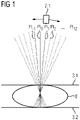
- the 1 shows an exemplary embodiment of the mammography system according to the invention in a first embodiment.
- a plurality of projection data records are recorded at a plurality of projection angles PI -1,0,1,2,...,12 .
- the X-ray source 2.1 is in particular along of a radius around a point in the breast 8, with a projection data set being recorded at each of the projection angles PI ⁇ 1,0,1,2,...,12 .
- the breast 8 of a patient, as the object to be examined is arranged between an upper compression element 3.1 and a lower compression element 3.2.
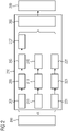
- the 2 shows an exemplary embodiment of the method according to the invention for generating a synthetic mammogram, comprising the steps of recording 200 and generating 210.
- FIG 2 shows in particular a preferred order of steps.
- the recording step a plurality of projection data sets are recorded at a plurality of projection angles.
- the generation step at least one synthetic mammogram is generated with image properties that are essentially equivalent to a conventional digital full-field mammography recording based on a plurality of projection data sets.
- multiple synthetic mammograms are generated for different projection angles.
- an average intensity projection (AIP) based on the plurality of projection data sets is determined as the first image component.
- the average projection corresponds in particular to a projection, particularly preferably the mean projection PI 0 .
- a number of synthetic mammograms are generated, then a number of projections are each used as an average projection, so that a synthetic mammogram is generated for each of these projections.
- the maximum number of synthetic mammograms generated can equal the total number of projections.
- a synthetic mammogram can preferably be generated for 17 of 25 projections, for example.
- At least one of the following steps is applied to the average intensity projection: Intensity adjustment 203, gray value distribution adjustment 205 and/or scattered radiation correction, and lime-preserving noise filter 207.
- the step of generating includes determining 211, 221 a maximum intensity projection (MIP) based on the plurality of projection data sets as the second image component.
- MIP maximum intensity projection
- the maximum intensity projection is broken down into at least two different frequency components.
- the at least two different frequency components include high-frequency components in step 213 and medium-frequency components in step 223 .
- the at least two different frequency components are scaled in steps 215,225.
- the first image component and the second image component are recombined to form the synthetic mammogram.
- the synthetic mammogram has image properties that are essentially equivalent to a conventional digital full-field mammography recording, wherein an imprinting step 209 can include a predefined image impression setting by means of a multi-frequency adjustment, a lookup table application, and/or windowing. An additional post-processing step can also be applied to the synthetic mammogram.
- the 3 shows an exemplary embodiment of the synthetic mammograms according to the invention in comparison to conventional digital full-field mammography recordings, each with a selected predefined image impression setting in a first embodiment.
- Representation 31 shows a conventional digital full-field mammography recording without a predefined image impression setting.
- the illustration 33 shows a synthetic mammogram with image properties that are essentially equivalent to a conventional digital full-field mammography recording without a predefined image impression setting.
- Representation 35 shows a conventional digital full-field mammography recording with a predefined image impression setting the first embodiment, in particular the so-called flavor 0.
- the representation 37 shows a synthetic mammogram with a predefined image impression setting of the first embodiment, in particular the so-called flavor 0.
- the 4 shows an exemplary embodiment of the synthetic mammograms according to the invention in comparison to conventional digital full-field mammography recordings, each with a selected predefined image impression setting in a second embodiment.
- Representation 41 shows a conventional digital full-field mammography recording without a predefined image impression setting.
- the illustration 43 shows a synthetic mammogram with image properties that are essentially equivalent to a conventional digital full-field mammography recording without a predefined image impression setting.
- Representation 45 shows a conventional digital full-field mammography recording with a predefined image impression setting of the second embodiment, in particular the so-called flavor 1.
- Representation 47 shows a synthetic mammogram with a predefined image impression setting of the second embodiment, in particular the so-called flavor 1.
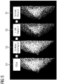
- the figure 5 shows an exemplary embodiment of the synthetic mammograms according to the invention in comparison to conventional digital full-field mammography recordings, each with a selected predefined image impression setting in a third embodiment.
- Representation 51 shows a conventional digital full-field mammography recording without a predefined image impression setting.
- the illustration 53 shows a synthetic mammogram with image properties that are essentially equivalent to a conventional digital full-field mammography recording without a predefined image impression setting.
- the illustration 55 shows a conventional digital full-field mammography recording with a predefined image impression setting of the third embodiment, in particular the so-called Flavor 6.
- Representation 57 shows a synthetic mammogram with a predefined image impression setting of the third embodiment, in particular the so-called Flavor 6.
- the 6 shows an exemplary embodiment of the mammography system 1 according to the invention in a second embodiment.
- the mammography system 1 includes a stand 1.1, on which the x-ray housing 2 having the x-ray source 2.1 and the x-ray detector 5 together with a compression unit 3 are arranged.
- the X-ray housing 2 is rotatably mounted on the stand 1.1 with respect to the stand 1.1 and the X-ray detector 5 and the compression unit 3.
- the compression unit 3 comprises an upper compression element 3.1 and a lower compression element 3.2, between which the breast 8 of a patient is arranged.
- the mammography system 1 is connected to a data processing unit 10 .
- the data processing unit 10 comprises at least one processor unit 10.1, a visualization unit 10.2 and an input unit 10.3.
- the mammography system 1 can be controlled at least partially via a foot switch 1.2.
Landscapes
- Health & Medical Sciences (AREA)
- Life Sciences & Earth Sciences (AREA)
- Engineering & Computer Science (AREA)
- Medical Informatics (AREA)
- Surgery (AREA)
- Physics & Mathematics (AREA)
- Pathology (AREA)
- General Health & Medical Sciences (AREA)
- Veterinary Medicine (AREA)
- Public Health (AREA)
- Nuclear Medicine, Radiotherapy & Molecular Imaging (AREA)
- Animal Behavior & Ethology (AREA)
- Molecular Biology (AREA)
- Heart & Thoracic Surgery (AREA)
- Biomedical Technology (AREA)
- Radiology & Medical Imaging (AREA)
- Optics & Photonics (AREA)
- Biophysics (AREA)
- High Energy & Nuclear Physics (AREA)
- Oral & Maxillofacial Surgery (AREA)
- Theoretical Computer Science (AREA)
- General Physics & Mathematics (AREA)
- Computer Vision & Pattern Recognition (AREA)
- Dentistry (AREA)
- Computer Graphics (AREA)
- Apparatus For Radiation Diagnosis (AREA)
Description
- Die Erfindung betrifft die Erzeugung eines synthetischen Mammogramms mit aufgeprägtem Bildeindruck.
- Digitale Brusttomosynthese (DBT) erlaubt eine dreidimensionale Bildgebung der Brust. Mehrere Schichten in unterschiedlichen Positionen, insbesondere Höhen, der Brust werden aus einer Vielzahl von Projektionsdatensätzen, beispielsweise 25, rekonstruiert. Die Aufnahme eines Projektionsdatensatzes findet unter einem Projektionswinkel statt. Die Projektionsdatensätze werden für verschiedene Projektionswinkel aufgenommen. Die verschiedenen Projektionswinkel können insbesondere in einem beschränkten Winkelbereich von beispielsweise 50 Grad aufgenommen werden.
- Ein großer Vorteil von digitaler Brusttomosynthese gegenüber einer konventionellen digitalen Vollfeld-Mammographie (engl.: full-field digital mammography, FFDM) ist die Möglichkeit überlappende Gewebe aufzulösen. Dies ist insbesondere vorteilhaft für die Identifikation sogenannter Massen mit spikulierte Läsionen (engl.: spiculated lesions) in bestimmten Schichten, welche in einer digitalen Vollfeld-Mammographieaufnahme durch überlappende Gewebestrukturen oder Gefäßen anderer Schichten überlagert sein können und damit nur schwer erkennbar sind. Dennoch hat die digitale Vollfeld-Mammographie einen Vorteil, wenn es um die Geschwindigkeit bei der Bildbetrachtung und der Darstellung von Mikrokalzifikationsclustern geht. Daher umfassen klinische Protokolle derzeit die digitale Brusttomosynthese und die digitale Vollfeld-Mammographie, um die Vorteile beider Bildgebungstechniken zu kombinieren. Nachdem für die digitale Brusttomosynthese und die digitale Vollfeld-Mammographie jeweils eine ähnliche Patientendosis verwendet wird, wird für die Kombination beider Bildgebungstechniken in etwa die doppelte Dosis im Vergleich zur alleinigen digitalen Vollfeld-Mammographie nötig. Daher ist es wünschenswert aus den Aufnahmedatensätzen bzw. Projektionsdatensätzen der digitalen Brusttomosynthese ein synthetisches Mammogramm zu berechnen, wobei die zusätzliche Dosis vermieden werden kann und die Vorteile der zweidimensionalen digitalen Vollfeld-Mammographie erhalten bleiben. Eine spezielle Anwendung für eine derartige zweidimensionale Darstellung ist der Vergleich mit früheren Aufnahmen. Diese früheren Aufnahmen sind digitale Vollfeld-Mammographieaufnahmen früherer Screenings oder Untersuchungen, welche für den betreffenden Patienten bzw. die betreffende Patientin gespeichert wurden. Nun ist es die Aufgabe, Unterschiede zwischen den aktuellen und vorherigen Gegebenheiten bzw. Auffälligkeiten und deren Größenverhältnissen innerhalb der Brust zu finden. Beispielsweise wird die Verteilung dichten Brustgewebes oder das Wachstum spezifischer Massen betrachtet und verglichen. Kunden bzw. Benutzer können für die Nachverarbeitung der digitalen Vollfeld-Mammographieaufnahme zwischen unterschiedlichen vordefinierten Bildeindruckseinstellungen, sogenannten Flavours, wählen.
- Die Berechnung des synthetischen Mammogramms bzw. einer synthetischen Projektion auf Basis von Tomosynthesedatensätzen bringt jedoch einige technische Hürden mit sich. Nachdem die Projektionsdatensätze nur mit einem Bruchteil der Dosis für eine digitale Vollfeld-Mammographieaufnahme aufgenommen werden, beispielsweise 1/25 der Dosis bei 25 Projektionen, leidet das Kontrast-zu-Rausch-Verhältnis enorm für jede einzelne Projektion. Zudem kann mit einer Verschmierung der Information aufgrund der Bewegung der Röntgenquelle gerechnet werden. Daher erfüllt die Verwendung einer einzigen Projektionsaufnahme nicht die Anforderungen an eine Aufnahme in zufriedenstellender Qualität. Rückprojektionen der rekonstruierten Volumendaten mit einer Durchschnitts-Intensitäts-Projektion (engl.: average intensity projection, AIP), welche einer physikalischen Linienintegration ähnelt leidet ebenfalls unter einer Verschmierung von Informationen. Dies kann Rekonstruktionsartefakten zugeschrieben werden, wie es beispielsweise in den Druckschriften
EP 2 998 936 B1 undUS 9 569 864 B2 - Aus der Druckschrift
EP 2 998 936 B1 ist ein Verfahren zum Erzeugen eines kombinierten Projektionsbildes von einem medizinischen Inspektionsobjekt bekannt, welches die folgenden Schritte umfasst: - Erfassen einer Menge von Anfangsprojektionsbildern aus verschiedenen Projektionsrichtungen;
- Rekonstruieren eines ersten und eines zweiten dreidimensionalen Volumens aus einer reduzierten Teilmenge der Anfangsprojektionsbilder, welche eine Anzahl von Anfangsprojektionsbildern aus Blickpositionen umfasst, die einer Reprojektions-Blickposition benachbart sind;
- Erzeugen eines ersten Reprojektionsbildes aus dem ersten dreidimensionalen Volumen und eines zweiten Reprojektionsbildes aus dem zweiten dreidimensionalen Volumen, wobei das erste und das zweite Reprojektionsbild unter Verwendung der Reprojektions-Blickposition erzeugt werden;
- Gewichten des ersten Reprojektionsbildes und des zweiten Reprojektionsbildes; und
- Kombinieren des gewichteten ersten Reprojektionsbildes und zweiten Reprojektionsbildes zum Erzeugen des kombinierten Projektionsbildes.
- Aus der Druckschrift
US 9 569 864 B2 - Aufnahme einer Mehrzahl von gestapelten zweidimensionalen tomographischen Bildern, welche ein tomographisches Volumen darstellen;
- Wichtung der Pixel der gestapelten zweidimensionalen tomographischen Bilder entlang einer Anzahl von Projektionsstrahlen durch das tomographische Volumen mit Wichtungsfaktoren, welche mit der Einschränkung gewählt wurden, dass die Summe aller quadrierten Wichtungsfaktoren für jede individuelle Projektion zwischen einer gegebenen unteren Grenze und einer gegebenen oberen Grenze liegen, wobei die gegebene untere Grenze gleich der gegebenen oberen Grenze gesetzt ist; und
- Erzeugen eines zweidimensionalen Projektionsbildes durch Aufsummieren der gewichteten Pixel entlang der Anzahl an Projektionen.
- Aus der Druckschrift
US 2018/185000 A1 ist eine Steuereinheit bekannt, welche einen Einfallswinkel von Strahlung, die auf eine Detektionsfläche eines Strahlungsdetektors einfällt, in Bezug auf die Detektionsfläche auf eine Vielzahl von voneinander verschiedenen Winkeln einstellt, einschließlich eines Winkels (0 Grad) in einer senkrechten Richtung der Detektionsfläche. Diese erfasst mindestens eines einer Vielzahl von Projektionsbildern, die von dem Strahlungsdetektor in Übereinstimmung mit dem Einfallswinkel erfasst werden, oder rekonstruierte Bilder, die unter Verwendung der Projektionsbilder rekonstruiert werden. Zusätzlich erzeugt die Steuereinheit auf der Grundlage des einen Bildes ein erstes synthetisches Mammogrammbild. Zusätzlich erzeugt die Steuereinheit ein zweites synthetisches Mammogrammbild, indem sie ein Hochfrequenzbild mit einer Hochfrequenzkomponente, die höher als eine vorbestimmte Frequenz des ersten synthetischen Mammogrammbildes ist, und ein Niederfrequenzbild mit einer Niederfrequenzkomponente, die gleich oder niedriger als eine vorbestimmte Frequenz eines Null-Grad-Projektionsbildes ist, synthetisiert. - Aus der Druckschrift
WO 2016/078958 A1 ist ein Verfahren und eine zugehörige Vorrichtung zum Synthetisieren eines Projektionsbildes, insbesondere zur Verwendung in der Mammographie, bekannt. Es wird vorgeschlagen, aus einem Bildvolumen eine Gewichtsfunktion zu berechnen, die dann dazu verwendet wird, eine gewichtete Vorwärtsprojektion durch einen anderen Bildvolumenblock zu implementieren, um ein synthetisiertes Projektionsbild über den Block zu berechnen. - Der Erfindung liegt das Problem zugrunde, dass synthetische Mammogramme jedoch in einem von der digitalen Vollfeld-Mammographieaufnahme verschiedenen Dynamikbereich berechnet werden und mittels einer von der digitalen Vollfeld-Mammographieaufnahme verschiedenen Nachbearbeitung bearbeitet werden. Die Verschiedenheit des Dynamikbereichs ergibt sich aus den Berechnungsmethoden des synthetischen Mammogramms. Dadurch wird bisher nur eine einzige Bildeindruckseinstellung bzw. ein einziger Bildeindruck für alle Benutzer bereitgestellt, welche sich vom Bildeindruck bzw. hinsichtlich mindestens einer Bildeigenschaft einer digitalen Vollfeld-Mammographieaufnahme unterscheidet. Der Vergleich früherer digitaler Vollfeld-Mammographieaufnahmen mit aktuellen synthetischen Mammogrammen wird dadurch erheblich erschwert, insbesondere für Benutzer mit sehr speziellen bzw. einzigartigen Bildeindruckseinstellungen der digitalen Vollfeld-Mammographieaufnahmen.
- Es ist Aufgabe der Erfindung, ein Verfahren und eine Vorrichtung anzugeben, welche einen Bildeindruck entsprechend einer früheren digitalen Vollfeld-Mammographieaufnahme ermöglichen.
- Die Aufgabe wird erfindungsgemäß gelöst durch ein Verfahren zum Erzeugen eines synthetischen Mammogramms nach Anspruch 1 und ein Mammographiesystem nach Anspruch 11.
- Die Erfindung betrifft ein Verfahren zum Erzeugen eines synthetischen Mammogramms aufweisend die Schritte des Aufnehmens und des Erzeugens. Ein synthetisches Mammogramm kann insbesondere einen zweidimensionalen Bilddatensatz bezeichnen, welcher eine der konventionellen digitalen Vollfeld-Mammographieaufnahme im Wesentlichen entsprechende Ansicht des Untersuchungsobjekts umfasst. Das synthetische Mammogramm wird dabei insbesondere aus einem im Wesentlichen dreidimensionalen Datensatz, insbesondere mehreren Projektionsdatensätzen erzeugt. Im Schritt des Aufnehmens wird eine Mehrzahl von Projektionsdatensätzen unter einer Mehrzahl von Projektionswinkeln aufgenommen. Die Projektionswinkel liegen in einem begrenzten Winkelbereich. Der Winkelbereich kann im Bereich zwischen 40 und 90 Grad liegen, bevorzugt entspricht der Winkelbereich in etwa 50 Grad. Die Anzahl der Projektionswinkel kann im Bereich zwischen 15 und 40 liegen, bevorzugt werden 25 Projektionen aufgenommen. Die Mehrzahl der Projektionsdatensätze kann insbesondere mit einem Röntgenspektrum aufgenommen werden. Die verwendete Dosis bzw. der verwendete Röhrenstrom kann für die Mehrzahl der Projektionsdatensätze im Wesentlichen gleich sein. Im Schritt des Erzeugens wird mindestens ein synthetisches Mammogramm mit zu einer konventionellen digitalen Vollfeld-Mammographieaufnahme im Wesentlichen äquivalenten Bildeigenschaft basierend auf mehreren Projektionsdatensätzen erzeugt. Im Schritt des Erzeugens kann eine Auswahl aus der Mehrzahl von Projektionsdatensätzen oder alle Projektionsdatensätze verwendet werden.
- Die äquivalente Bildeigenschaft kann insbesondere dazu führen, dass der Bildeindruck des synthetischen Mammogramms im Wesentlichen dem Bildeindruck einer konventionellen digitalen Vollfeld-Mammographieaufnahme entspricht. Zudem kann der Dynamikbereich des synthetischen Mammogramms im Wesentlichen dem Dynamikbereich, dem Kontrast oder/und der Helligkeit einer konventionellen digitalen Vollfeld-Mammographieaufnahme entsprechen. Die Bildeigenschaft kann den Dynamikbereich, den Kontrast oder die Helligkeit umfassen. Die äquivalente Bildeigenschaft kann bedeuten, dass die Bildeigenschaft des synthetischen Mammogramms und die Bildeigenschaft der konventionellen digitalen Voll-Mammographieaufnahme im Wesentlichen gleich ist. Die Bildeigenschaft kann sich beispielsweise um wenige Prozent, beispielsweise weniger als 10 Prozent, bevorzugt weniger als 5 Prozent unterscheiden. Vorteilhaft kann ein Vergleich des synthetischen Mammogramms mit einer früheren konventionellen digitalen Vollfeld-Mammographieaufnahme erheblich vereinfacht werden. Beispielsweise kann das synthetische Mammogramm eine aufgeprägte, ausgewählte vordefinierte Bildeindruckseinstellung, sogenannter Flavour, umfassen, wobei die Bildeindruckseinstellung derart ausgewählt ist, dass sie der vordefinierten Bildeindruckseinstellung der früheren konventionellen digitalen Vollfeld-Mammographieaufnahme entspricht.
- Im Schritt des Erzeugens wird ein synthetisches Mammogramm erzeugt oder es werden mehrere synthetische Mammogramme erzeugt. Bevorzugt wird zumindest ein synthetisches Mammogramm entsprechend des mittleren Projektionsdatensatzes, insbesondere bei einem Projektionswinkel von 0 Grad, erzeugt. Der mittlere Projektionswinkel kann dabei insbesondere den Winkel bezeichnen, bei dem der Zentralstrahl der Röntgenquelle im Wesentlichen senkrecht auf die komprimierte Brust bzw. senkrecht auf das obere Kompressionselement einfällt. Bevorzugt können mehrere synthetische Mammogramme erzeugt werden. Ein synthetisches Mammogramm kann jeweils einem Projektionswinkel zugeordnet werden. Es kann für einen Projektionswinkel ein synthetisches Mammogramm erzeugt werden. Die maximale Anzahl der erzeugten synthetischen Mammogramme kann beispielsweise der Anzahl der Projektionswinkel entsprechen. Bevorzugt wird ein synthetisches Mammogramm für den Projektionswinkel 0 Grad erzeugt. Weiterhin werden 2 bis zur maximalen Anzahl der aufgenommenen Projektionswinkel, bevorzugt 10 bis 20, besonders bevorzugt 17, synthetische Mammogramme erzeugt. Das synthetische Mammogramm kann insbesondere auf der Projektionsaufnahme des zugeordneten Projektionswinkels basieren, beispielsweise unter Verwendung dieser Projektionsaufnahme in Form einer Durchschnittsintensitätsprojektion. Beispielsweise werden 17 synthetische Mammogramme basierend auf 25 Projektionsdatensätzen erzeugt.
- Die Erfinder haben erkannt, dass es für das Ermöglichen einer Verwendung einer Standard-Nachverarbeitung für digitale Vollfeld-Mammographieaufnahmen nötig ist, ein neuartiges synthetisches Mammogramm zu berechnen, welches äquivalent zu einer Detektorausgabe ist. Das synthetische Mammogramm, welches äquivalent zu einer Detektorausgabe ist, weist mindestens eine, bevorzugt mehrere, im Wesentlichen gleiche Bildeigenschaft auf. Dieses synthetische Mammogramm muss daher idealerweise im gleichen dynamischen Bereich von konventionellen digitalen Vollfeld-Mammographieaufnahmen liegen. Durch das erfindungsgemäße Verfahren und dem Zusammenwirken der davon umfassten Schritte kann dies erreicht werden. Nach der Rekombination der unterschiedlichen Bildkomponenten der unterschiedlichen Schritte zu einem synthetischen Mammogramm kann vorteilhaft die gleiche Nachverarbeitung wie für digitale Vollfeld-Mammographieaufnahme für das synthetische Mammogramm verwendet werden.
- Das Mammographiesystem umfasst eine Röntgenquelle und einen Röntgendetektor sowie eine Kompressionseinheit. Insbesondere die Röntgenquelle ist bezüglich des Röntgendetektors sowie der Kompressionseinheit rotierbar gelagert, dadurch sind verschiedene Projektionswinkel einstellbar. Die Kompressionseinheit umfasst ein oberes Kompressionselement und ein unteres Kompressionselement, zwischen denen die Brust eines Patienten angeordnet wird. Im Schritt des Aufnehmens werden Projektionsdatensätze mit der Brust als Untersuchungsobjekt unter verschiedenen Projektionswinkeln aufgenommen, während die Brust zwischen dem oberen und dem unteren Kompressionselement komprimiert wird und im während des Schrittes des Aufnehmens im Wesentlichen unverändert komprimiert bleibt.
- Vorteilhaft kann das synthetische Mammogramm mit einer früheren digitalen Vollfeld-Mammographieaufnahme verglichen werden. Der Vergleich findet bevorzugt optisch statt. Beispielsweise können das synthetische Mammogramm und die frühere digitale Vollfeld-Mammographieaufnahme nebeneinander angezeigt werden. Ferner kann ein Links-Rechts-Vergleich von synthetischen Mammogrammen und der früheren digitale Vollfeld-Mammographieaufnahmen nebeneinander ermöglicht werden. Vorteilhaft kann die Kombination bzw. die kombinierte Aufnahme von digitaler Vollfeld-Mammographieaufnahme und digitaler Brusttomosynthese nacheinander vermieden werden, damit kann vorteilhaft die Patientendosis reduziert werden. Vorteilhaft kann die Brustdichte besonders einfach erfasst werden. Vorteilhaft kann die Patientendosis reduziert werden. Vorteilhaft kann die Zeit zur Bewertung der Aufnahmen verkürzt werden, da eine zusätzliche aktuelle konventionelle digitale Vollfeld-Mammographieaufnahme vermieden werden kann.
- Gemäß einem Aspekt der Erfindung weist das ferner den Schritt des Aufprägens auf, wobei eine ausgewählte vordefinierte Bildeindruckseinstellung auf das synthetische Mammogramm aufgeprägt wird. Mittels der Aufprägung der ausgewählten vordefinierten Bildeindruckseinstellung kann der Bildeindruck des synthetischen Mammogramms angepasst bzw. verändert werden. Die Bildeindruckseinstellung kann als sogenannter Flavour bezeichnet werden. Die Bildeindruckseinstellung kann beispielsweise aus einer der folgenden Varianten ausgewählt werden: Standard, geglättet, Kontrast-verstärkt, Kanten-verstärkt, oder Kontrast-und-Kanten-verstärkt. Vorteilhaft kann der Bildeindruck vom Benutzer angepasst werden.
- Gemäß einem Aspekt der Erfindung umfasst die vordefinierte Bildeindruckseinstellung eine Multifrequenzanpassung, eine Lookup-Tabellen-Anwendung, oder/und eine Fensterung. Vorteilhaft kann der Bildeindruck reproduzierbar aufgeprägt werden.
- Erfindungsgemäß umfasst der Schritt des Erzeugens ein Ermitteln einer Durchschnittsintensitätsprojektion (AIP) basierend auf der Mehrzahl von Projektionsdatensätzen als erste Bildkomponente. Bevorzugt kann ein Projektionsdatensatz eines Projektionswinkels, insbesondere entsprechend der Zuordnung eines Projektionswinkels zum synthetischen Mammogramm, als Durchschnittsintensitätsprojektion oder als Basis für die Durchschnittsintensitätsprojektion verwendet werden. Insbesondere kann eine limited-angle-AIP (LARIP) als Durchschnittsintensitätsprojektion verwendet werden. Es kann ein Projektionsdatensatz oder es können mehrere Projektionsdatensätze ausgewählt werden, auf deren Basis die Durchschnittsintensitätsprojektion ermittelt wird. Vorteilhaft umfasst die Durchschnittsintensitätsprojektion als erste Bildkomponente wesentliche Informationen über das Untersuchungsobjekt in der zweidimensionalen Draufsicht entsprechend des Projektionswinkels. Die Durchschnittsintensitätsprojektion kann alleine jedoch kaum den Anforderungen für eine umfassende Beurteilung der Brust genügen, da sie aufgrund der nur anteilig verwendeten Dosis ein relativ hohes Rauschen aufweist.
- Erfindungsgemäß wird auf die Durchschnittsintensitätsprojektion zumindest einer der folgenden Schritte angewendet: Intensitätsanpassung, Grauwertverteilungsanpassung, Streustrahlenkorrektur, und kalkerhaltender Rauschfilter. Die Durchschnittsintensitätsprojektion kann derart bearbeitet werden, dass die bearbeitete Durchschnittsintensitätsprojektion als rauschige Basis bzw. erste Bildkomponente für das synthetische Mammogramm verwendet werden kann. Vorteilhaft kann die Information dieser Projektion als Grundlage für das synthetische Mammogramm dienen. Auf die erste Bildkomponente aufbauend kann durch Hinzufügen von Kanteninformationen und Kontrastinformationen einer zweiten Bildkomponente ein zu einer konventionellen digitalen Vollfeld-Mammographieaufnahme im Wesentlichen qualitativ äquivalentes synthetisches Mammogramm erzeugt werden.
- Erfindungsgemäß umfasst der Schritt des Erzeugens ein Ermitteln einer Maximumsintensitätsprojektion (MIP) basierend auf der Mehrzahl von Projektionsdatensätzen als zweite Bildkomponente. Es können insbesondere mehrere Projektionsdatensätze zum Ermitteln der Maximumsintensitätsprojektion verwendet werden. Es können insbesondere Projektionsdatensätze verwendet werden, welche benachbart sind zu dem synthetischen Mammogramm zugeordneten Projektionsdatensatz bzw. deren Projektionswinkel. Beispielsweise können Projektionsdatensätze eines geeigneten Winkelbereichs um den zugeordneten Projektionswinkel verwendet werden. Es können insbesondere alle Projektionsdatensätze zum Ermitteln der Maximumsintensitätsprojektion verwendet werden. Vorteilhaft können Kanteninformationen und Kontrastinformationen aus mehreren Projektionsdatensätzen als zweite Bildkomponente genutzt werden, um diese der ersten Bildkomponente hinzuzufügen. Vorteilhaft kann die zweite Bildkomponente zum Entrauschen verwendet werden.
- Gemäß einem Aspekt der Erfindung wird die Maximumsintensitätsprojektion in mindestens zwei verschiedene Frequenzkomponenten zerlegt. Es kann eine Frequenzbandzerlegung durchgeführt werden. Gemäß einem Aspekt der Erfindung umfassen die mindestens zwei verschiedenen Frequenzkomponenten hochfrequente Komponenten und mittelfrequente Komponenten. Die mindestens zwei Frequenzkomponenten können mit oder ohne Überlapp zwischen einander ausgebildet sein. Mittels der hochfrequenten Komponente können die Kanten besonders hervorgehoben werden. Mittels der mittelfrequenten Komponente kann der Kontrast bzw. das Kontrast-zu-Rausch-Verhältnis verbessert werden.
- Gemäß einem Aspekt der Erfindung ist werden die mindestens zwei verschiedenen Frequenzkomponenten skaliert. Die mindestens zwei verschiedenen Frequenzkomponenten können gleich oder verschieden skaliert werden. Die skalierten Frequenzkomponenten können als zweite Bildkomponente verwendet werden. Vorteilhaft können die mindestens zwei Frequenzkomponenten aneinander angepasst skaliert werden. Vorteilhaft können die mindestens zwei Frequenzkomponenten an die erste Bildkomponente angepasst skaliert werden.
- Erfindungsgemäß werden die erste Bildkomponente und die zweite Bildkomponente zum synthetischen Mammogramm rekombiniert. Durch Zusammenfügen bzw. Rekombinieren der ersten Bildkomponente und der zweiten Bildkomponente kann das synthetische Mammogramm erzeugt werden. Vorteilhaft wird besonders betont die Information des zugeordneten Projektionsdatensatzes in Form der ersten Bildkomponente gemeinsam mit zusätzlichen Informationen aus mehreren bzw. weiteren Projektionsdatensätzen in Form der zweiten Bildkomponente genutzt. Dadurch kann die Information basierend auf der verwendeten Patientendosis vorteilhaft für das Erzeugen des synthetischen Mammogramms verwendet werden.
- Gemäß einem Aspekt der Erfindung wird ein Schritt der Nachverarbeitung auf das synthetische Mammogramm angewendet. Die Nachverarbeitung kann eine zusätzliche Filterung, Fensterung bzw. im Allgemeinen eine Anpassung der Darstellung umfassen. Vorteilhaft können von konventionellen digitalen Vollfeld-Mammographieaufnahmen bekannte Nachverarbeitungen auf das synthetische Mammogramm angewendet werden. Damit kann die Vergleichbarkeit weiterhin verbessert werden.
- Gemäß einem Aspekt der Erfindung wird das synthetische Mammogramm mit einer früheren digitalen Vollfeld-Mammographieaufnahme, insbesondere mit gleichem Flavour, verglichen. Dies wird vorteilhaft durch das erfindungsgemäße synthetische Mammogramm ermöglicht. Der Vergleich findet insbesondere optisch durch einen Benutzer statt. Insbesondere können das synthetische Mammogramm und die frühere digitale Vollfeld-Mammographieaufnahme, insbesondere mit gleichem Flavour, nebeneinander dargestellt werden, entweder für eine Brustseite oder beide Brustseiten gleichzeitig. Dadurch kann der Vergleich vorteilhaft vereinfacht durchgeführt werden.
- Gemäß einem Aspekt der Erfindung wird das synthetische Mammogramm aus mehreren erzeugten synthetischen Mammogrammen zum Vergleich mit einer früheren digitalen Vollfeld-Mammographieaufnahme ausgewählt. Werden mehrere synthetische Mammogramme erzeugt, so kann vom Anwender oder mittels eines Algorithmus ein für den Vergleich bevorzugt geeignetes synthetisches Mammogramm ausgewählt werden. Vorteilhaft kann der Vergleich einer früheren digitalen Vollfeld-Mammographieaufnahme mit einem synthetischen Mammogramm einer aktuellen Aufnahme vereinfacht werden. Die mehreren synthetischen Mammogramme können als sogenannte rotierende Mammogramme dargestellt werden, wobei durch das sogenannte Rotieren eine dreidimensionale Betrachtung möglich ist.
- Gemäß einem Aspekt der Erfindung umfasst das synthetische Mammogramm eine CAD-Markierung bzw. CAD-Markierungen. Rechnergestützte Diagnoseunterstützung bzw. Bildanalyse kann in Form von sogenannten CAD-Markierungen (engl.: CAD marks, computer-aided diagnosis marks) bereitgestellt werden. Mittels Maschinenlernverfahren, insbesondere Deep-Learning-Verfahren, können Läsionen, besonders dichte Brustdichten oder Kalizfikationen markiert bzw. hervorgehoben werden. Die CAD-Markierung kann insbesondere innerhalb eines dreidimensionalen Tomosynthesevolumens bestimmt werden. Die CAD-Markierung kann im synthetischen Mammogramm, welches das betroffene Voxel der CAD-Markierung in der Projektion umfasst, angezeigt werden. Vorteilhaft ermöglicht die CAD-Markierung im synthetischen Mammogramm einen umfassenden Überblick über potentiell näher zu betrachtende Regionen innerhalb der Brust.
- Die Erfindung betrifft ferner ein Mammographiesystem zum Durchführen eines Verfahrens nach einem der vorangehenden Ansprüche, aufweisend eine Aufnahmeeinheit und eine Erzeugungseinheit. Die Erzeugungseinheit kann bevorzugt von der Datenverarbeitungseinheit umfasst sein. Das Mammographiesystem kann ferner eine Einheit zum Ermitteln einer Durchschnittsintensitätsprojektion, eine Einheit zur Intensitätsanpassung, eine Einheit zur Grauwertverteilungsanpassung oder/und Streustrahlenkorrektur, eine Einheit zur kalkerhaltender Rauschfilterung, eine Einheit zum Ermitteln der Maximumsintensitätsprojektion, eine Einheit zur Frequenzzerlegung, eine Einheit zum Skalieren, eine Rekombinationseinheit und eine Aufprägeeinheit umfassen. Das Mammographiesystem kann vorteilhaft alle Schritte des erfindungsgemäßen Verfahrens durchführen. Das Mammographiesystem ist ein medizinisches Gerät. Es können alternativ auch andere medizinische Geräte, welche für Tomosyntheseverfahren geeignet sind, das erfindungsgemäße Verfahren anwenden.
- Die Erfindung betrifft ferner ein Computerprogrammprodukt mit einem Computerprogramm, welches direkt in eine Speichereinrichtung einer Steuereinrichtung eines Mammographiesystems ladbar ist, mit Programmabschnitten, um alle Schritte des erfindungsgemäßen Verfahrens auszuführen, wenn das Computerprogramm in der Steuereinrichtung des Mammographiesystems ausgeführt wird.
- Die Erfindung betrifft ferner ein computerlesbares Medium, auf welchem von einer Rechnereinheit einlesbare und ausführbare Programmabschnitte gespeichert sind, um alle Schritte des erfindungsgemäßen Verfahrens auszuführen, wenn die Programmabschnitte von der Recheneinheit ausgeführt werden. Die Recheneinheit kann bevorzugt von der Datenverarbeitungseinheit bzw. einer Prozessoreinheit umfasst sein.
- Nachfolgend werden Ausführungsbeispiele der Erfindung anhand von Zeichnungen näher erläutert. Hierbei zeigt:
-
FIG 1 eine schematische Darstellung des erfindungsgemäßen Mammographiesystems in einer ersten Ausführungsform; -
FIG 2 eine schematische Darstellung des erfindungsgemäßen Verfahrens; -
FIG 3 eine schematische Darstellung der erfindungsgemäßen synthetischen Mammogramme in Gegenüberstellung zu konventionellen digitalen Vollfeld-Mammographieaufnahmen jeweils mit einer ausgewählten vordefinierten Bildeindruckseinstellung in einer ersten Ausführungsform; -
FIG 4 eine schematische Darstellung der erfindungsgemäßen synthetischen Mammogramme in Gegenüberstellung zu konventionellen digitalen Vollfeld-Mammographieaufnahmen jeweils mit einer ausgewählten vordefinierten Bildeindruckseinstellung in einer zweiten Ausführungsform; -
FIG 5 eine schematische Darstellung der erfindungsgemäßen synthetischen Mammogramme in Gegenüberstellung zu konventionellen digitalen Vollfeld-Mammographieaufnahmen jeweils mit einer ausgewählten vordefinierten Bildeindruckseinstellung in einer dritten Ausführungsform; und -
FIG 6 eine schematische Darstellung des erfindungsgemäßen Mammographiesystems in einer zweiten Ausführungsform. - Die
Fig. 1 zeigt eine beispielhafte Ausführung des erfindungsgemäßen Mammographiesystems in einer ersten Ausführungsform. Es wird eine Mehrzahl von Projektionsdatensätzen unter einer Mehrzahl von Projektionswinkeln PI-1,0,1,2,..., 12 aufgenommen. Die Röntgenquelle 2.1 wird dabei insbesondere entlang eines Radius um einen Punkt in der Brust 8 verfahren, wobei unter den Proj ektionswinkeln PI-1,0,1,2,..., 12 jeweils ein Projektionsdatensatz aufgenommen wird. Während der Aufnahme ist die Brust 8 eines Patienten als Untersuchungsobjekt zwischen einem oberen Kompressionselement 3.1 und einem unteren Kompressionselement 3.2 angeordnet. - Die
Fig. 2 zeigt eine beispielhafte Ausführung des erfindungsgemäßen Verfahrens zum Erzeugen eines synthetischen Mammogramms aufweisend die Schritte des Aufnehmens 200 und des Erzeugens 210. DieFig. 2 zeigt insbesondere eine bevorzugte Reihenfolge der Schritte. Im Schritt des Aufnehmens wird eine Mehrzahl von Projektionsdatensätzen unter einer Mehrzahl von Projektionswinkeln aufgenommen. Im Schritt des Erzeugens wird mindestens ein synthetisches Mammogramm mit zu einer konventionellen digitalen Vollfeld-Mammographieaufnahme im Wesentlichen äquivalenten Bildeigenschaften basierend auf mehreren Projektionsdatensätzen erzeugt. In einer besonderen Ausführungsform werden mehrere synthetische Mammogramme für unterschiedliche Projektionswinkel erzeugt. - Im Schritt 201 wird eine Durchschnittsintensitätsprojektion (AIP) basierend auf der Mehrzahl von Projektionsdatensätzen als erste Bildkomponente ermittelt. Die Durchschnittsprojektion entspricht insbesondere einer Projektion, besonders bevorzugt der mittleren Projektion PI0.
- Werden mehrere synthetische Mammogramme erzeugt, so werden mehrere Projektionen jeweils als Durchschnittsprojektion verwendet, so dass für jede dieser Projektionen ein synthetisches Mammogramm erzeugt wird. Die maximale Anzahl der erzeugten synthetischen Mammogramme kann der Gesamtzahl der Projektionen entsprechen. Bevorzugt können beispielsweise für 17 von 25 Projektionen jeweils ein synthetisches Mammogramm erzeugt werden.
- Auf die Durchschnittsintensitätsprojektion wird zumindest einer der folgenden Schritte angewendet: Intensitätsanpassung 203, Grauwertverteilungsanpassung 205 oder/und Streustrahlenkorrektur, und kalkerhaltender Rauschfilter 207.
- Der Schritt des Erzeugens umfasst ein Ermitteln 211, 221 einer Maximumsintensitätsprojektion (MIP) basierend auf der Mehrzahl von Projektionsdatensätzen als zweite Bildkomponente. Die Maximumsintensitätsprojektion wird im Schritt 213 bzw. 223 in mindestens zwei verschiedene Frequenzkomponenten zerlegt. Die mindestens zwei verschiedenen Frequenzkomponenten umfassen im Schritt 213 hochfrequente Komponenten und im Schritt 223 mittelfrequente Komponenten. Die mindestens zwei verschiedenen Frequenzkomponenten werden in den Schritten 215, 225 skaliert.
- Im Schritt des Rekombinierens 208 werden die erste Bildkomponente und die zweite Bildkomponente zum synthetischen Mammogramm rekombiniert. Das synthetische Mammogramm weist zu einer konventionellen digitalen Vollfeld-Mammographieaufnahme im Wesentlichen äquivalente Bildeigenschaften auf, wobei ein Schritt des Aufprägens 209 eine vordefinierte Bildeindruckseinstellung mittels einer Multifrequenzanpassung, einer Lookup-Tabellen-Anwendung, oder/und einer Fensterung umfassen kann. Es kann ferner ein weiterer Schritt der Nachverarbeitung auf das synthetische Mammogramm angewendet werden.
- Die
Fig. 3 zeigt eine beispielhafte Ausführung der erfindungsgemäßen synthetischen Mammogramme in Gegenüberstellung zu konventionellen digitalen Vollfeld-Mammographieaufnahmen jeweils mit einer ausgewählten vordefinierten Bildeindruckseinstellung in einer ersten Ausführungsform. Die Darstellung 31 zeigt eine konventionelle digitale Vollfeld-Mammographieaufnahme ohne vordefinierte Bildeindruckseinstellung. Die Darstellung 33 zeigt ein synthetisches Mammogramm mit zu einer konventionellen digitalen Vollfeld-Mammographieaufnahme im Wesentlichen äquivalenten Bildeigenschaften ohne vordefinierte Bildeindruckseinstellung. Die Darstellung 35 zeigt eine konventionelle digitale Vollfeld-Mammographieaufnahme mit vordefinierter Bildeindruckseinstellung der ersten Ausführungsform, insbesondere dem sogenannten Flavour 0. Die Darstellung 37 zeigt ein synthetisches Mammogramm mit vordefinierter Bildeindruckseinstellung der ersten Ausführungsform, insbesondere dem sogenannten Flavour 0. - Die
Fig. 4 zeigt eine beispielhafte Ausführung der erfindungsgemäßen synthetischen Mammogramme in Gegenüberstellung zu konventionellen digitalen Vollfeld-Mammographieaufnahmen jeweils mit einer ausgewählten vordefinierten Bildeindruckseinstellung in einer zweiten Ausführungsform. Die Darstellung 41 zeigt eine konventionelle digitale Vollfeld-Mammographieaufnahme ohne vordefinierte Bildeindruckseinstellung. Die Darstellung 43 zeigt ein synthetisches Mammogramm mit zu einer konventionellen digitalen Vollfeld-Mammographieaufnahme im Wesentlichen äquivalenten Bildeigenschaften ohne vordefinierte Bildeindruckseinstellung. Die Darstellung 45 zeigt eine konventionelle digitale Vollfeld-Mammographieaufnahme mit vordefinierter Bildeindruckseinstellung der zweiten Ausführungsform, insbesondere dem sogenannten Flavour 1. Die Darstellung 47 zeigt ein synthetisches Mammogramm mit vordefinierter Bildeindruckseinstellung der zweiten Ausführungsform, insbesondere dem sogenannten Flavour 1. - Die
Fig. 5 zeigt eine beispielhafte Ausführung der erfindungsgemäßen synthetischen Mammogramme in Gegenüberstellung zu konventionellen digitalen Vollfeld-Mammographieaufnahmen jeweils mit einer ausgewählten vordefinierten Bildeindruckseinstellung in einer dritten Ausführungsform. Die Darstellung 51 zeigt eine konventionelle digitale Vollfeld-Mammographieaufnahme ohne vordefinierte Bildeindruckseinstellung. Die Darstellung 53 zeigt ein synthetisches Mammogramm mit zu einer konventionellen digitalen Vollfeld-Mammographieaufnahme im Wesentlichen äquivalenten Bildeigenschaften ohne vordefinierte Bildeindruckseinstellung. Die Darstellung 55 zeigt eine konventionelle digitale Vollfeld-Mammographieaufnahme mit vordefinierter Bildeindruckseinstellung der dritten Ausführungsform, insbesondere dem sogenannten Flavour 6. Die Darstellung 57 zeigt ein synthetisches Mammogramm mit vordefinierter Bildeindruckseinstellung der dritten Ausführungsform, insbesondere dem sogenannten Flavour 6. - Die
Fig. 6 zeigt eine beispielhafte Ausführung des erfindungsgemäßen Mammographiesystems 1 in einer zweiten Ausführungsform. Das Mammographiesystem 1 umfasst ein Stativ 1.1, an welchem das Röntgengehäuse 2 aufweisend die Röntgenquelle 2.1 und der Röntgendetektor 5 gemeinsam mit einer Kompressionseinheit 3 angeordnet sind. Insbesondere das Röntgengehäuse 2 ist bezüglich des Stativs 1.1 und des Röntgendetektors 5 sowie der Kompressionseinheit 3 rotierbar am Stativ 1.1 gelagert. Die Kompressionseinheit 3 umfasst ein oberes Kompressionselement 3.1 und ein unteres Kompressionselement 3.2, zwischen denen die Brust 8 eines Patienten angeordnet wird. Das Mammographiesystem 1 ist mit einer Datenverarbeitungseinheit 10 verbunden. Die Datenverarbeitungseinheit 10 umfasst mindestens eine Prozessoreinheit 10.1, eine Visualisierungseinheit 10.2 und eine Eingabeeinheit 10.3. Das Mammographiesystem 1 ist zumindest teilweise über einen Fußschalter 1.2 steuerbar. - Obwohl die Erfindung im Detail durch das bevorzugte Ausführungsbeispiel näher illustriert wurde, so ist die Erfindung nicht durch die offenbarten Beispiele eingeschränkt und andere Variationen können vom Fachmann hieraus abgeleitet werden, ohne den Schutzumfang der Erfindung, wie in den folgenden Ansprüchen definiert, zu verlassen.
Claims (13)
- Verfahren zum Erzeugen eines synthetischen Mammogramms aufweisend die Schritte:- Aufnehmen (200) einer Mehrzahl von Projektionsdatensätzen unter einer Mehrzahl von Projektionswinkeln (PI-1,0,1,2,...,12),- Erzeugen (210) mindestens eines synthetischen Mammogramms mit zu einer konventionellen digitalen Vollfeld-Mammographieaufnahme im Wesentlichen äquivalenter Bildeigenschaft basierend auf mehreren Projektionsdatensätzen,- wobei der Schritt des Erzeugens (210) ein Ermitteln (201) einer Durchschnittsintensitätsprojektion basierend auf der Mehrzahl von Projektionsdatensätzen als erste Bildkomponente umfasst, wobei ein Projektionsdatensatz oder mehrere Projektionsdatensätze ausgewählt werden,- auf die Durchschnittsintensitätsprojektion zumindest einer der folgenden Schritte angewendet wird:- Intensitätsanpassung (203),- Grauwertverteilungsanpassung (205),- Streustrahlenkorrektur, und- kalkerhaltender Rauschfilter (207),- der Schritt des Erzeugens (210) ein Ermitteln (211, 221) einer Maximumsintensitätsprojektion basierend auf der Mehrzahl von Projektionsdatensätzen als zweite Bildkomponente umfasst, und- die erste Bildkomponente und die zweite Bildkomponente zum synthetischen Mammogramm rekombiniert werden.
- Verfahren nach Anspruch 1, ferner aufweisend den Schritt:- Aufprägen (209) einer ausgewählten vordefinierten Bildeindruckseinstellung auf das synthetische Mammogramm.
- Verfahren nach Anspruch 2, wobei die vordefinierte Bildeindruckseinstellung eine Multifrequenzanpassung, eine Lookup-Tabellen-Anwendung, oder/und eine Fensterung umfasst.
- Verfahren nach einem der vorangehenden Ansprüche, wobei die Maximumsintensitätsprojektion in mindestens zwei verschiedene Frequenzkomponenten (213, 223) zerlegt wird.
- Verfahren nach einem der vorangehenden Ansprüche, wobei die mindestens zwei verschiedenen Frequenzkomponenten hochfrequente Komponenten und mittelfrequente Komponenten umfassen.
- Verfahren nach einem der Ansprüche 4 oder 5, wobei die mindestens zwei verschiedenen Frequenzkomponenten skaliert (215, 225) werden.
- Verfahren nach einem der vorangehenden Ansprüche, wobei ein Schritt der Nachverarbeitung auf das synthetische Mammogramm angewendet wird.
- Verfahren nach einem der vorangehenden Ansprüche, wobei das synthetische Mammogramm mit einer früheren digitalen Vollfeld-Mammographieaufnahme verglichen wird.
- Verfahren nach Anspruch 8, wobei das synthetische Mammogramm aus mehreren erzeugten synthetischen Mammogrammen zum Vergleich mit einer früheren digitalen Vollfeld-Mammographieaufnahme ausgewählt wird.
- Verfahren nach einem der vorangehenden Ansprüche, wobei das synthetische Mammogramm CAD-Markierungen umfasst.
- Mammographiesystem (1) zum Durchführen eines Verfahrens nach einem der vorangehenden Ansprüche, aufweisend eine Aufnahmeeinheit (1.1, 2.1, 3, 5) und eine Erzeugungseinheit.
- Computerprogrammprodukt mit einem Computerprogramm, welches direkt in eine Speichereinrichtung einer Steuereinrichtung eines Mammographiesystems (1) ladbar ist, mit Programmabschnitten, um alle Schritte eines Verfahrens nach einem der Ansprüche 1 bis 10 auszuführen, wenn das Computerprogramm in der Steuereinrichtung des Mammographiesystems (1) ausgeführt wird.
- Computerlesbares Medium, auf welchem von einer Rechnereinheit einlesbare und ausführbare Programmabschnitte gespeichert sind, um alle Schritte eines Verfahrens nach einem der Ansprüche 1 bis 10 auszuführen, wenn die Programmabschnitte von der Recheneinheit ausgeführt werden.
Priority Applications (1)
| Application Number | Priority Date | Filing Date | Title |
|---|---|---|---|
| US16/682,388 US11877879B2 (en) | 2018-11-23 | 2019-11-13 | Synthetic mammogram with imprinted image impression |
Applications Claiming Priority (1)
| Application Number | Priority Date | Filing Date | Title |
|---|---|---|---|
| EP18208120 | 2018-11-23 |
Publications (2)
| Publication Number | Publication Date |
|---|---|
| EP3657442A1 EP3657442A1 (de) | 2020-05-27 |
| EP3657442B1 true EP3657442B1 (de) | 2022-10-26 |
Family
ID=64476946
Family Applications (1)
| Application Number | Title | Priority Date | Filing Date |
|---|---|---|---|
| EP18214794.2A Active EP3657442B1 (de) | 2018-11-23 | 2018-12-20 | Synthetisches mammogramm mit aufgeprägtem bildeindruck |
Country Status (2)
| Country | Link |
|---|---|
| US (1) | US11877879B2 (de) |
| EP (1) | EP3657442B1 (de) |
Families Citing this family (4)
| Publication number | Priority date | Publication date | Assignee | Title |
|---|---|---|---|---|
| DE102020212089B3 (de) | 2020-09-25 | 2021-09-16 | Siemens Healthcare Gmbh | Verfahren und Vorrichtung zur Bildentrauschung sowie eine Steuereinrichtung, ein bildgebendes System, ein Computerprogrammprodukt und ein computerlesbares Medium dazu |
| DE102020212382B3 (de) * | 2020-09-30 | 2022-01-20 | Siemens Healthcare Gmbh | Rekonstruktion von Bildern mit Filtermatrizen |
| JP7785494B2 (ja) * | 2021-09-30 | 2025-12-15 | 富士フイルム株式会社 | 学習装置、画像生成装置、学習方法、画像生成方法、学習プログラム、及び画像生成プログラム |
| EP4344644A1 (de) * | 2022-09-29 | 2024-04-03 | Siemens Healthineers AG | Computerimplementiertes verfahren zur bereitstellung eines ersten röntgenbildes und eines durch ein röntgenbildgebungssystem erfassten zweiten röntgenbildes |
Family Cites Families (11)
| Publication number | Priority date | Publication date | Assignee | Title |
|---|---|---|---|---|
| US5704355A (en) * | 1994-07-01 | 1998-01-06 | Bridges; Jack E. | Non-invasive system for breast cancer detection |
| US7760924B2 (en) * | 2002-11-27 | 2010-07-20 | Hologic, Inc. | System and method for generating a 2D image from a tomosynthesis data set |
| JP5528518B2 (ja) * | 2012-09-28 | 2014-06-25 | 富士フイルム株式会社 | 放射線画像生成装置および方法 |
| AU2015222981B2 (en) * | 2014-02-28 | 2019-01-31 | Hologic, Inc. | System and method for generating and displaying tomosynthesis image slabs |
| US9569864B2 (en) | 2014-09-12 | 2017-02-14 | Siemens Aktiengesellschaft | Method and apparatus for projection image generation from tomographic images |
| US9836858B2 (en) * | 2014-09-19 | 2017-12-05 | Siemens Aktiengesellschaft | Method for generating a combined projection image and imaging device |
| CN107004283B (zh) * | 2014-11-20 | 2021-07-27 | 皇家飞利浦有限公司 | 用于根据断层合成数据生成合成乳房摄影图的方法 |
| US9792703B2 (en) * | 2015-07-06 | 2017-10-17 | Siemens Healthcare Gmbh | Generating a synthetic two-dimensional mammogram |
| EP3348195B1 (de) * | 2015-09-10 | 2019-07-10 | FUJIFILM Corporation | Bildverarbeitungsvorrichtung, strahlungsbildaufnahmesystem, bildverarbeitungsverfahren und bildverarbeitungsprogramm |
| ES2987687T3 (es) * | 2017-03-30 | 2024-11-15 | Hologic Inc | Método para sintetizar datos de imagen de baja dimensión a partir de datos de imagen de alta dimensión usando una potenciación de la cuadrícula de objetos |
| EP3856033B1 (de) * | 2018-09-28 | 2023-06-07 | Hologic, Inc. | Verfahren zur erzeugung eines synthetischen brustgewebes durch unterdrückung von hochdichtem element |
-
2018
- 2018-12-20 EP EP18214794.2A patent/EP3657442B1/de active Active
-
2019
- 2019-11-13 US US16/682,388 patent/US11877879B2/en active Active
Also Published As
| Publication number | Publication date |
|---|---|
| US20200163638A1 (en) | 2020-05-28 |
| US11877879B2 (en) | 2024-01-23 |
| EP3657442A1 (de) | 2020-05-27 |
Similar Documents
| Publication | Publication Date | Title |
|---|---|---|
| DE102009053471B4 (de) | Verfahren und Vorrichtung zur Identifizierung und Zuordnung von Koronarkalk zu einem Herzkranzgefäß sowie Computerprogrammprodukt | |
| DE102008028387B4 (de) | Tomographisches Bildrekonstruktionsverfahren zum Erzeugen eines Bildes von einem Untersuchungsobjekt und nach diesem Verfahren arbeitende bildgebende Einrichtung | |
| EP3657442B1 (de) | Synthetisches mammogramm mit aufgeprägtem bildeindruck | |
| DE102011003135B4 (de) | Bildgebungsverfahren zum Rotieren eines Gewebebereichs | |
| DE102007053511A1 (de) | Röntgentomographie-Bildgebungsgerät | |
| DE102007041976A1 (de) | Verfahren zur Erzeugung eines Tomosynthesebildes | |
| DE10353882A1 (de) | Verfahren und Einrichtung zur Weichgewebevolumen-Sichtbarmachung | |
| DE102011003137A1 (de) | Bildgebungsverfahren mit einer verbesserten Darstellung eines Gewebebereichs | |
| DE102016219887A1 (de) | Verfahren und System zur Nutzung von Messdaten | |
| DE10229113A1 (de) | Verfahren zur Grauwert-basierten Bildfilterung in der Computer-Tomographie | |
| DE102006043743A1 (de) | Verfahren und Vorrichtung zur Kombination von Bildern | |
| DE10238322A1 (de) | Retrospektive bzw. fenstergesteuerte Filterung von Bildern zur Adaption von Schärfe und Rauschen in der Computer-Tomographie | |
| DE10356174A1 (de) | Verfahren und Einrichtung zur Tomosynthese-Bildverbesserung unter Verwendung von Querfilterung | |
| EP3797698B1 (de) | Verfahren zum erzeugen eines synthetischen mammogramms basierend auf einer dual-energy-tomosyntheseaufnahme | |
| EP3327673B1 (de) | Erzeugen von hochaufgelösten ct-bildern mit spektraler information | |
| DE102011075917A1 (de) | Verfahren zum Bereitstellen eines 3D-Bilddatensatzes mit unterdrückten Messfeldüberschreitungsartefakten und Computertomograph | |
| DE102016211766A1 (de) | Erzeugung einer Bildsequenz | |
| WO2008052854A1 (de) | Verfahren und einrichtung zur anzeige eines im rahmen einer mammographie aufgenommenen röntgenbildes | |
| DE102009019840A1 (de) | Kontrastverstärkung von CT-Bildern mittels eines Multibandfilters | |
| DE102011003138B4 (de) | Bildgebungsverfahren mit optimierter Grauwertfensterbestimmung | |
| EP2101648B1 (de) | Verfahren und einrichtung zum erzeugen eines tomosynthetischen 3d-röntgenbildes | |
| DE102020212382B3 (de) | Rekonstruktion von Bildern mit Filtermatrizen | |
| DE102021210289A1 (de) | Verfahren zum Erzeugen von Ergebnisschichtbildern mit zumindest teilweise verschiedener Schichtdicke | |
| DE102020209706A1 (de) | Synthetisches Mammogramm mit reduzierter Überlagerung von Gewebeveränderungen | |
| DE112021007285T5 (de) | Röntgenbildgebungsvorrichtung und röntgenbildverarbeitungsverfahren |
Legal Events
| Date | Code | Title | Description |
|---|---|---|---|
| PUAI | Public reference made under article 153(3) epc to a published international application that has entered the european phase |
Free format text: ORIGINAL CODE: 0009012 |
|
| STAA | Information on the status of an ep patent application or granted ep patent |
Free format text: STATUS: THE APPLICATION HAS BEEN PUBLISHED |
|
| AK | Designated contracting states |
Kind code of ref document: A1 Designated state(s): AL AT BE BG CH CY CZ DE DK EE ES FI FR GB GR HR HU IE IS IT LI LT LU LV MC MK MT NL NO PL PT RO RS SE SI SK SM TR |
|
| AX | Request for extension of the european patent |
Extension state: BA ME |
|
| STAA | Information on the status of an ep patent application or granted ep patent |
Free format text: STATUS: REQUEST FOR EXAMINATION WAS MADE |
|
| 17P | Request for examination filed |
Effective date: 20201126 |
|
| RBV | Designated contracting states (corrected) |
Designated state(s): AL AT BE BG CH CY CZ DE DK EE ES FI FR GB GR HR HU IE IS IT LI LT LU LV MC MK MT NL NO PL PT RO RS SE SI SK SM TR |
|
| STAA | Information on the status of an ep patent application or granted ep patent |
Free format text: STATUS: EXAMINATION IS IN PROGRESS |
|
| 17Q | First examination report despatched |
Effective date: 20210226 |
|
| GRAP | Despatch of communication of intention to grant a patent |
Free format text: ORIGINAL CODE: EPIDOSNIGR1 |
|
| STAA | Information on the status of an ep patent application or granted ep patent |
Free format text: STATUS: GRANT OF PATENT IS INTENDED |
|
| INTG | Intention to grant announced |
Effective date: 20220623 |
|
| GRAS | Grant fee paid |
Free format text: ORIGINAL CODE: EPIDOSNIGR3 |
|
| GRAA | (expected) grant |
Free format text: ORIGINAL CODE: 0009210 |
|
| STAA | Information on the status of an ep patent application or granted ep patent |
Free format text: STATUS: THE PATENT HAS BEEN GRANTED |
|
| AK | Designated contracting states |
Kind code of ref document: B1 Designated state(s): AL AT BE BG CH CY CZ DE DK EE ES FI FR GB GR HR HU IE IS IT LI LT LU LV MC MK MT NL NO PL PT RO RS SE SI SK SM TR |
|
| REG | Reference to a national code |
Ref country code: GB Ref legal event code: FG4D Free format text: NOT ENGLISH |
|
| REG | Reference to a national code |
Ref country code: CH Ref legal event code: EP |
|
| REG | Reference to a national code |
Ref country code: AT Ref legal event code: REF Ref document number: 1527610 Country of ref document: AT Kind code of ref document: T Effective date: 20221115 |
|
| REG | Reference to a national code |
Ref country code: DE Ref legal event code: R096 Ref document number: 502018010909 Country of ref document: DE |
|
| REG | Reference to a national code |
Ref country code: IE Ref legal event code: FG4D Free format text: LANGUAGE OF EP DOCUMENT: GERMAN |
|
| REG | Reference to a national code |
Ref country code: LT Ref legal event code: MG9D |
|
| REG | Reference to a national code |
Ref country code: NL Ref legal event code: MP Effective date: 20221026 |
|
| PG25 | Lapsed in a contracting state [announced via postgrant information from national office to epo] |
Ref country code: NL Free format text: LAPSE BECAUSE OF FAILURE TO SUBMIT A TRANSLATION OF THE DESCRIPTION OR TO PAY THE FEE WITHIN THE PRESCRIBED TIME-LIMIT Effective date: 20221026 |
|
| PG25 | Lapsed in a contracting state [announced via postgrant information from national office to epo] |
Ref country code: SE Free format text: LAPSE BECAUSE OF FAILURE TO SUBMIT A TRANSLATION OF THE DESCRIPTION OR TO PAY THE FEE WITHIN THE PRESCRIBED TIME-LIMIT Effective date: 20221026 Ref country code: PT Free format text: LAPSE BECAUSE OF FAILURE TO SUBMIT A TRANSLATION OF THE DESCRIPTION OR TO PAY THE FEE WITHIN THE PRESCRIBED TIME-LIMIT Effective date: 20230227 Ref country code: NO Free format text: LAPSE BECAUSE OF FAILURE TO SUBMIT A TRANSLATION OF THE DESCRIPTION OR TO PAY THE FEE WITHIN THE PRESCRIBED TIME-LIMIT Effective date: 20230126 Ref country code: LT Free format text: LAPSE BECAUSE OF FAILURE TO SUBMIT A TRANSLATION OF THE DESCRIPTION OR TO PAY THE FEE WITHIN THE PRESCRIBED TIME-LIMIT Effective date: 20221026 Ref country code: FI Free format text: LAPSE BECAUSE OF FAILURE TO SUBMIT A TRANSLATION OF THE DESCRIPTION OR TO PAY THE FEE WITHIN THE PRESCRIBED TIME-LIMIT Effective date: 20221026 Ref country code: ES Free format text: LAPSE BECAUSE OF FAILURE TO SUBMIT A TRANSLATION OF THE DESCRIPTION OR TO PAY THE FEE WITHIN THE PRESCRIBED TIME-LIMIT Effective date: 20221026 |
|
| PG25 | Lapsed in a contracting state [announced via postgrant information from national office to epo] |
Ref country code: RS Free format text: LAPSE BECAUSE OF FAILURE TO SUBMIT A TRANSLATION OF THE DESCRIPTION OR TO PAY THE FEE WITHIN THE PRESCRIBED TIME-LIMIT Effective date: 20221026 Ref country code: PL Free format text: LAPSE BECAUSE OF FAILURE TO SUBMIT A TRANSLATION OF THE DESCRIPTION OR TO PAY THE FEE WITHIN THE PRESCRIBED TIME-LIMIT Effective date: 20221026 Ref country code: LV Free format text: LAPSE BECAUSE OF FAILURE TO SUBMIT A TRANSLATION OF THE DESCRIPTION OR TO PAY THE FEE WITHIN THE PRESCRIBED TIME-LIMIT Effective date: 20221026 Ref country code: IS Free format text: LAPSE BECAUSE OF FAILURE TO SUBMIT A TRANSLATION OF THE DESCRIPTION OR TO PAY THE FEE WITHIN THE PRESCRIBED TIME-LIMIT Effective date: 20230226 Ref country code: HR Free format text: LAPSE BECAUSE OF FAILURE TO SUBMIT A TRANSLATION OF THE DESCRIPTION OR TO PAY THE FEE WITHIN THE PRESCRIBED TIME-LIMIT Effective date: 20221026 Ref country code: GR Free format text: LAPSE BECAUSE OF FAILURE TO SUBMIT A TRANSLATION OF THE DESCRIPTION OR TO PAY THE FEE WITHIN THE PRESCRIBED TIME-LIMIT Effective date: 20230127 |
|
| REG | Reference to a national code |
Ref country code: DE Ref legal event code: R097 Ref document number: 502018010909 Country of ref document: DE |
|
| PG25 | Lapsed in a contracting state [announced via postgrant information from national office to epo] |
Ref country code: SM Free format text: LAPSE BECAUSE OF FAILURE TO SUBMIT A TRANSLATION OF THE DESCRIPTION OR TO PAY THE FEE WITHIN THE PRESCRIBED TIME-LIMIT Effective date: 20221026 Ref country code: RO Free format text: LAPSE BECAUSE OF FAILURE TO SUBMIT A TRANSLATION OF THE DESCRIPTION OR TO PAY THE FEE WITHIN THE PRESCRIBED TIME-LIMIT Effective date: 20221026 Ref country code: EE Free format text: LAPSE BECAUSE OF FAILURE TO SUBMIT A TRANSLATION OF THE DESCRIPTION OR TO PAY THE FEE WITHIN THE PRESCRIBED TIME-LIMIT Effective date: 20221026 Ref country code: DK Free format text: LAPSE BECAUSE OF FAILURE TO SUBMIT A TRANSLATION OF THE DESCRIPTION OR TO PAY THE FEE WITHIN THE PRESCRIBED TIME-LIMIT Effective date: 20221026 Ref country code: CZ Free format text: LAPSE BECAUSE OF FAILURE TO SUBMIT A TRANSLATION OF THE DESCRIPTION OR TO PAY THE FEE WITHIN THE PRESCRIBED TIME-LIMIT Effective date: 20221026 |
|
| REG | Reference to a national code |
Ref country code: CH Ref legal event code: PL |
|
| REG | Reference to a national code |
Ref country code: BE Ref legal event code: MM Effective date: 20221231 |
|
| PG25 | Lapsed in a contracting state [announced via postgrant information from national office to epo] |
Ref country code: SK Free format text: LAPSE BECAUSE OF FAILURE TO SUBMIT A TRANSLATION OF THE DESCRIPTION OR TO PAY THE FEE WITHIN THE PRESCRIBED TIME-LIMIT Effective date: 20221026 Ref country code: LU Free format text: LAPSE BECAUSE OF NON-PAYMENT OF DUE FEES Effective date: 20221220 Ref country code: AL Free format text: LAPSE BECAUSE OF FAILURE TO SUBMIT A TRANSLATION OF THE DESCRIPTION OR TO PAY THE FEE WITHIN THE PRESCRIBED TIME-LIMIT Effective date: 20221026 |
|
| PLBE | No opposition filed within time limit |
Free format text: ORIGINAL CODE: 0009261 |
|
| STAA | Information on the status of an ep patent application or granted ep patent |
Free format text: STATUS: NO OPPOSITION FILED WITHIN TIME LIMIT |
|
| 26N | No opposition filed |
Effective date: 20230727 |
|
| PG25 | Lapsed in a contracting state [announced via postgrant information from national office to epo] |
Ref country code: LI Free format text: LAPSE BECAUSE OF NON-PAYMENT OF DUE FEES Effective date: 20221231 Ref country code: IE Free format text: LAPSE BECAUSE OF NON-PAYMENT OF DUE FEES Effective date: 20221220 Ref country code: CH Free format text: LAPSE BECAUSE OF NON-PAYMENT OF DUE FEES Effective date: 20221231 |
|
| PG25 | Lapsed in a contracting state [announced via postgrant information from national office to epo] |
Ref country code: SI Free format text: LAPSE BECAUSE OF FAILURE TO SUBMIT A TRANSLATION OF THE DESCRIPTION OR TO PAY THE FEE WITHIN THE PRESCRIBED TIME-LIMIT Effective date: 20221026 Ref country code: BE Free format text: LAPSE BECAUSE OF NON-PAYMENT OF DUE FEES Effective date: 20221231 |
|
| REG | Reference to a national code |
Ref country code: DE Ref legal event code: R081 Ref document number: 502018010909 Country of ref document: DE Owner name: SIEMENS HEALTHINEERS AG, DE Free format text: FORMER OWNER: SIEMENS HEALTHCARE GMBH, MUENCHEN, DE |
|
| PG25 | Lapsed in a contracting state [announced via postgrant information from national office to epo] |
Ref country code: HU Free format text: LAPSE BECAUSE OF FAILURE TO SUBMIT A TRANSLATION OF THE DESCRIPTION OR TO PAY THE FEE WITHIN THE PRESCRIBED TIME-LIMIT; INVALID AB INITIO Effective date: 20181220 |
|
| PG25 | Lapsed in a contracting state [announced via postgrant information from national office to epo] |
Ref country code: CY Free format text: LAPSE BECAUSE OF FAILURE TO SUBMIT A TRANSLATION OF THE DESCRIPTION OR TO PAY THE FEE WITHIN THE PRESCRIBED TIME-LIMIT Effective date: 20221026 |
|
| PG25 | Lapsed in a contracting state [announced via postgrant information from national office to epo] |
Ref country code: MK Free format text: LAPSE BECAUSE OF FAILURE TO SUBMIT A TRANSLATION OF THE DESCRIPTION OR TO PAY THE FEE WITHIN THE PRESCRIBED TIME-LIMIT Effective date: 20221026 Ref country code: IT Free format text: LAPSE BECAUSE OF FAILURE TO SUBMIT A TRANSLATION OF THE DESCRIPTION OR TO PAY THE FEE WITHIN THE PRESCRIBED TIME-LIMIT Effective date: 20221026 |
|
| PG25 | Lapsed in a contracting state [announced via postgrant information from national office to epo] |
Ref country code: MC Free format text: LAPSE BECAUSE OF FAILURE TO SUBMIT A TRANSLATION OF THE DESCRIPTION OR TO PAY THE FEE WITHIN THE PRESCRIBED TIME-LIMIT Effective date: 20221026 |
|
| PG25 | Lapsed in a contracting state [announced via postgrant information from national office to epo] |
Ref country code: TR Free format text: LAPSE BECAUSE OF FAILURE TO SUBMIT A TRANSLATION OF THE DESCRIPTION OR TO PAY THE FEE WITHIN THE PRESCRIBED TIME-LIMIT Effective date: 20221026 Ref country code: MC Free format text: LAPSE BECAUSE OF FAILURE TO SUBMIT A TRANSLATION OF THE DESCRIPTION OR TO PAY THE FEE WITHIN THE PRESCRIBED TIME-LIMIT Effective date: 20221026 |
|
| PG25 | Lapsed in a contracting state [announced via postgrant information from national office to epo] |
Ref country code: BG Free format text: LAPSE BECAUSE OF FAILURE TO SUBMIT A TRANSLATION OF THE DESCRIPTION OR TO PAY THE FEE WITHIN THE PRESCRIBED TIME-LIMIT Effective date: 20221026 |
|
| PG25 | Lapsed in a contracting state [announced via postgrant information from national office to epo] |
Ref country code: MT Free format text: LAPSE BECAUSE OF FAILURE TO SUBMIT A TRANSLATION OF THE DESCRIPTION OR TO PAY THE FEE WITHIN THE PRESCRIBED TIME-LIMIT Effective date: 20221026 |
|
| REG | Reference to a national code |
Ref country code: AT Ref legal event code: MM01 Ref document number: 1527610 Country of ref document: AT Kind code of ref document: T Effective date: 20231220 |
|
| PGFP | Annual fee paid to national office [announced via postgrant information from national office to epo] |
Ref country code: DE Payment date: 20250220 Year of fee payment: 7 |
|
| PG25 | Lapsed in a contracting state [announced via postgrant information from national office to epo] |
Ref country code: AT Free format text: LAPSE BECAUSE OF NON-PAYMENT OF DUE FEES Effective date: 20231220 |
|
| PGFP | Annual fee paid to national office [announced via postgrant information from national office to epo] |
Ref country code: GB Payment date: 20250115 Year of fee payment: 7 |
|
| PGFP | Annual fee paid to national office [announced via postgrant information from national office to epo] |
Ref country code: FR Payment date: 20251215 Year of fee payment: 8 |