EP3622918A1 - Assemblage pour le remplacement de la valve atrio-ventriculaire tricuspide - Google Patents
Assemblage pour le remplacement de la valve atrio-ventriculaire tricuspide Download PDFInfo
- Publication number
- EP3622918A1 EP3622918A1 EP19192702.9A EP19192702A EP3622918A1 EP 3622918 A1 EP3622918 A1 EP 3622918A1 EP 19192702 A EP19192702 A EP 19192702A EP 3622918 A1 EP3622918 A1 EP 3622918A1
- Authority
- EP
- European Patent Office
- Prior art keywords
- assembly
- stent
- frame
- ring
- native
- Prior art date
- Legal status (The legal status is an assumption and is not a legal conclusion. Google has not performed a legal analysis and makes no representation as to the accuracy of the status listed.)
- Pending
Links
Images


Classifications
-
- A—HUMAN NECESSITIES
- A61—MEDICAL OR VETERINARY SCIENCE; HYGIENE
- A61F—FILTERS IMPLANTABLE INTO BLOOD VESSELS; PROSTHESES; DEVICES PROVIDING PATENCY TO, OR PREVENTING COLLAPSING OF, TUBULAR STRUCTURES OF THE BODY, e.g. STENTS; ORTHOPAEDIC, NURSING OR CONTRACEPTIVE DEVICES; FOMENTATION; TREATMENT OR PROTECTION OF EYES OR EARS; BANDAGES, DRESSINGS OR ABSORBENT PADS; FIRST-AID KITS
- A61F2/00—Filters implantable into blood vessels; Prostheses, i.e. artificial substitutes or replacements for parts of the body; Appliances for connecting them with the body; Devices providing patency to, or preventing collapsing of, tubular structures of the body, e.g. stents
- A61F2/02—Prostheses implantable into the body
- A61F2/24—Heart valves ; Vascular valves, e.g. venous valves; Heart implants, e.g. passive devices for improving the function of the native valve or the heart muscle; Transmyocardial revascularisation [TMR] devices; Valves implantable in the body
- A61F2/2412—Heart valves ; Vascular valves, e.g. venous valves; Heart implants, e.g. passive devices for improving the function of the native valve or the heart muscle; Transmyocardial revascularisation [TMR] devices; Valves implantable in the body with soft flexible valve members, e.g. tissue valves shaped like natural valves
- A61F2/2418—Scaffolds therefor, e.g. support stents
-
- A—HUMAN NECESSITIES
- A61—MEDICAL OR VETERINARY SCIENCE; HYGIENE
- A61F—FILTERS IMPLANTABLE INTO BLOOD VESSELS; PROSTHESES; DEVICES PROVIDING PATENCY TO, OR PREVENTING COLLAPSING OF, TUBULAR STRUCTURES OF THE BODY, e.g. STENTS; ORTHOPAEDIC, NURSING OR CONTRACEPTIVE DEVICES; FOMENTATION; TREATMENT OR PROTECTION OF EYES OR EARS; BANDAGES, DRESSINGS OR ABSORBENT PADS; FIRST-AID KITS
- A61F2/00—Filters implantable into blood vessels; Prostheses, i.e. artificial substitutes or replacements for parts of the body; Appliances for connecting them with the body; Devices providing patency to, or preventing collapsing of, tubular structures of the body, e.g. stents
- A61F2/02—Prostheses implantable into the body
- A61F2/24—Heart valves ; Vascular valves, e.g. venous valves; Heart implants, e.g. passive devices for improving the function of the native valve or the heart muscle; Transmyocardial revascularisation [TMR] devices; Valves implantable in the body
- A61F2/2427—Devices for manipulating or deploying heart valves during implantation
-
- A—HUMAN NECESSITIES
- A61—MEDICAL OR VETERINARY SCIENCE; HYGIENE
- A61F—FILTERS IMPLANTABLE INTO BLOOD VESSELS; PROSTHESES; DEVICES PROVIDING PATENCY TO, OR PREVENTING COLLAPSING OF, TUBULAR STRUCTURES OF THE BODY, e.g. STENTS; ORTHOPAEDIC, NURSING OR CONTRACEPTIVE DEVICES; FOMENTATION; TREATMENT OR PROTECTION OF EYES OR EARS; BANDAGES, DRESSINGS OR ABSORBENT PADS; FIRST-AID KITS
- A61F2210/00—Particular material properties of prostheses classified in groups A61F2/00 - A61F2/26 or A61F2/82 or A61F9/00 or A61F11/00 or subgroups thereof
- A61F2210/0014—Particular material properties of prostheses classified in groups A61F2/00 - A61F2/26 or A61F2/82 or A61F9/00 or A61F11/00 or subgroups thereof using shape memory or superelastic materials, e.g. nitinol
-
- A—HUMAN NECESSITIES
- A61—MEDICAL OR VETERINARY SCIENCE; HYGIENE
- A61F—FILTERS IMPLANTABLE INTO BLOOD VESSELS; PROSTHESES; DEVICES PROVIDING PATENCY TO, OR PREVENTING COLLAPSING OF, TUBULAR STRUCTURES OF THE BODY, e.g. STENTS; ORTHOPAEDIC, NURSING OR CONTRACEPTIVE DEVICES; FOMENTATION; TREATMENT OR PROTECTION OF EYES OR EARS; BANDAGES, DRESSINGS OR ABSORBENT PADS; FIRST-AID KITS
- A61F2220/00—Fixations or connections for prostheses classified in groups A61F2/00 - A61F2/26 or A61F2/82 or A61F9/00 or A61F11/00 or subgroups thereof
- A61F2220/0008—Fixation appliances for connecting prostheses to the body
-
- A—HUMAN NECESSITIES
- A61—MEDICAL OR VETERINARY SCIENCE; HYGIENE
- A61F—FILTERS IMPLANTABLE INTO BLOOD VESSELS; PROSTHESES; DEVICES PROVIDING PATENCY TO, OR PREVENTING COLLAPSING OF, TUBULAR STRUCTURES OF THE BODY, e.g. STENTS; ORTHOPAEDIC, NURSING OR CONTRACEPTIVE DEVICES; FOMENTATION; TREATMENT OR PROTECTION OF EYES OR EARS; BANDAGES, DRESSINGS OR ABSORBENT PADS; FIRST-AID KITS
- A61F2220/00—Fixations or connections for prostheses classified in groups A61F2/00 - A61F2/26 or A61F2/82 or A61F9/00 or A61F11/00 or subgroups thereof
- A61F2220/0025—Connections or couplings between prosthetic parts, e.g. between modular parts; Connecting elements
- A61F2220/0075—Connections or couplings between prosthetic parts, e.g. between modular parts; Connecting elements sutured, ligatured or stitched, retained or tied with a rope, string, thread, wire or cable
-
- A—HUMAN NECESSITIES
- A61—MEDICAL OR VETERINARY SCIENCE; HYGIENE
- A61F—FILTERS IMPLANTABLE INTO BLOOD VESSELS; PROSTHESES; DEVICES PROVIDING PATENCY TO, OR PREVENTING COLLAPSING OF, TUBULAR STRUCTURES OF THE BODY, e.g. STENTS; ORTHOPAEDIC, NURSING OR CONTRACEPTIVE DEVICES; FOMENTATION; TREATMENT OR PROTECTION OF EYES OR EARS; BANDAGES, DRESSINGS OR ABSORBENT PADS; FIRST-AID KITS
- A61F2230/00—Geometry of prostheses classified in groups A61F2/00 - A61F2/26 or A61F2/82 or A61F9/00 or A61F11/00 or subgroups thereof
- A61F2230/0002—Two-dimensional shapes, e.g. cross-sections
- A61F2230/0004—Rounded shapes, e.g. with rounded corners
- A61F2230/0008—Rounded shapes, e.g. with rounded corners elliptical or oval
-
- A—HUMAN NECESSITIES
- A61—MEDICAL OR VETERINARY SCIENCE; HYGIENE
- A61F—FILTERS IMPLANTABLE INTO BLOOD VESSELS; PROSTHESES; DEVICES PROVIDING PATENCY TO, OR PREVENTING COLLAPSING OF, TUBULAR STRUCTURES OF THE BODY, e.g. STENTS; ORTHOPAEDIC, NURSING OR CONTRACEPTIVE DEVICES; FOMENTATION; TREATMENT OR PROTECTION OF EYES OR EARS; BANDAGES, DRESSINGS OR ABSORBENT PADS; FIRST-AID KITS
- A61F2230/00—Geometry of prostheses classified in groups A61F2/00 - A61F2/26 or A61F2/82 or A61F9/00 or A61F11/00 or subgroups thereof
- A61F2230/0002—Two-dimensional shapes, e.g. cross-sections
- A61F2230/0004—Rounded shapes, e.g. with rounded corners
- A61F2230/0013—Horseshoe-shaped, e.g. crescent-shaped, C-shaped, U-shaped
-
- A—HUMAN NECESSITIES
- A61—MEDICAL OR VETERINARY SCIENCE; HYGIENE
- A61F—FILTERS IMPLANTABLE INTO BLOOD VESSELS; PROSTHESES; DEVICES PROVIDING PATENCY TO, OR PREVENTING COLLAPSING OF, TUBULAR STRUCTURES OF THE BODY, e.g. STENTS; ORTHOPAEDIC, NURSING OR CONTRACEPTIVE DEVICES; FOMENTATION; TREATMENT OR PROTECTION OF EYES OR EARS; BANDAGES, DRESSINGS OR ABSORBENT PADS; FIRST-AID KITS
- A61F2250/00—Special features of prostheses classified in groups A61F2/00 - A61F2/26 or A61F2/82 or A61F9/00 or A61F11/00 or subgroups thereof
- A61F2250/0058—Additional features; Implant or prostheses properties not otherwise provided for
- A61F2250/0096—Markers and sensors for detecting a position or changes of a position of an implant, e.g. RF sensors, ultrasound markers
- A61F2250/0098—Markers and sensors for detecting a position or changes of a position of an implant, e.g. RF sensors, ultrasound markers radio-opaque, e.g. radio-opaque markers
Definitions
- the invention relates to the field of methods and systems for the replacement of a defective atrioventricular heart valve or tricuspid valve.
- the human heart has four heart valves. Two of these valves are the so - called atrioventricular valves .
- the tricuspid valve is located between the right atrium (AD) and the right ventricle (VD).
- the mitral valve is located between the left atrium (AG) and the left ventricle (LV).
- the other two valves are located between the ventricles and the vascular system.
- the aortic valve separates the left ventricle from the aorta and the pulmonary valve separates the right ventricle from the pulmonary artery.
- a functional pathology of the tricuspid valve which can be leaky, in a severe way, secondary to a significant dilation of the tricuspid ring. More rarely this pathology is due to a rheumatic or infectious valve disease or even to a degeneration of a stenosing or leaky bioprosthesis.
- the tricuspid valve is significantly larger than the mitral valve. In pathological situations, the tricuspid ring expands exceeding 40 mm in diameter when the mitral pathological ring measures approximately 30 to 35 mm. This difference has several consequences, notably mechanical (e.g. maintenance, stability and in terms of peri-prosthetic leaks).
- Patent documents US2014 / 0172070 or US8657872 are examples of such documents, for which the technical lessons described have limitations and / or drawbacks and / or inadequacies for applications for replacing the tricuspid valve.
- the aortic valve and the pulmonary valve indeed present characteristics requiring different modes of treatment and / or replacement, if only by their anatomical situation in the heart.
- the invention is part of this latest development.
- an armature comprises a stent which itself contains the bioprosthesis of the valve. tricuspid.
- the diameter of the frame is therefore greater than that of the stent, which carries the bioprosthesis.
- the diameter of the frame is slightly greater than that of the stent, itself adapted to the diameter of the bioprosthesis.
- the present invention also discloses methods for replacement of the tricuspid atrioventricular valve.
- the route is usually a percutaneous trans-catheter route. After a percutaneous approach, the system contained in a catheter is put in place by the vascular route.
- an assembly for a tricuspid orifice of a human heart comprising an outer frame connected to an inner stent carrying a tricuspid valve bioprosthesis and a sealing skirt, and in which assembly the outer frame is adapted to be maintained in the '' native tricuspid ring (potentially and / or when deployed in the heart of a patient); the inner stent is connected to the outer frame by one or more attachment strands; and the sealing skirt covers the interstitial space existing between the external frame and the internal stent (in packaging and / or when deployed in the heart of a patient).
- the sealing skirt also covers the (contact) space existing between the external reinforcement and the location of the native tissue (ie potentially, ie in a folded or conditioned state) and / or the native tissue (ie in a deployed situation in the heart of a patient) in the area of the ring.
- the sealing skirt performs a fold or a return, making it possible both to obstruct the contact space with the native tissue and to cover the interstitial space between the frame and the upstream stent of the ring.
- the skirt covers only this interstitial space and other means are used to prevent peri-prosthetic leaks (e.g. sealing strip or flange, biocompatible glue or filling material, etc.)
- peri-prosthetic leaks e.g. sealing strip or flange, biocompatible glue or filling material, etc.
- the reinforcement is prestressed or exerts, due to its structure, a radial force (directed radially outwards) which makes it possible to maintain it in the native ring.
- the frame carries or comprises or is associated with a stent, itself carrying or comprising or being associated with a bioprosthesis.
- the sealing skirt makes it possible to channel the blood flow from the right atrium to the right ventricle through the bioprosthesis (by covering the "interstitial" space (which is always non-zero) existing between the frame and the stent wearing the bioprosthesis with a diameter chosen from a limited choice).
- the sealing skirt also makes it possible to minimize (or else entirely avoid) peri-prosthetic leaks between the framework and the native tissue (by covering and / or plugging and / or obstructing in the area of the ring the "contact" space existing between the outer frame pressed against the native tissue and the native tissue.
- the blood flow is obstructed (or stopped or blocked or obstructed or prevented) over the entire circumference over the width of the space " contact ”by the waterproof skirt.
- the armature has a deformable zone in the zone corresponding to the native ring
- the frame covered by the sealing skirt further has upstream of the area of the native ring a shape reinforcing the maintenance of the assembly in the native ring.
- the shape can be convex or flared in order to optimize the capture of blood flow, which helps to stabilize or maintain the maintenance of the assembly in the tricuspid ring (an appropriate capture minimizes the mechanical effects of torsion, ie of rotation / translation of the assembly that the pressure of the blood could cause due to the presence of the waterproof skirt).
- the framework further has downstream of the area of the native ring a shape minimizing the disruption of the flow of blood flow.
- the part of the armature which penetrates the area of the right ventricle is limited in its dimensions (confer the orders of magnitude given below).
- the function of the shape of this part of the frame can minimize turbulence (e.g. particular shapes can in particular involve drag effects tending towards a more laminar flow of blood flow)
- the frame includes a plurality of sub-parts.
- the frame is in one piece, that is to say made in one piece (for example, the frame is 3D printed in a deformable material).
- the frame consists of two sub-parts (for example complementary or symmetrical).
- the frame is made up of three parts (in an economical and robust arrangement).
- the frame consists of four sub-parts (symmetrical arrangement, advantageous in terms of stability).
- the armature consists of a large number of sub-parts or armature wires.
- the different sub-parts of the frame are integral with one another. In one embodiment, the different sub-parts of the frame are at least partially independent of each other. In certain embodiments, at least one sub-part of the armature has a physical property chosen from properties comprising rigidity, elasticity, plasticity, shape memory, heat-sensitive, actuable, instrumented, configurable and spring-loaded.
- the outer frame is connected to the inner stent by one or more strands.
- the frame includes four sub-parts, each of the sub-parts being connected to the interior stent by a nitinol strand.
- the assembly further comprises at least one element for fixing to the native tissue.
- the element for fixing to the native tissue improves in particular the maintenance in translation and / or in rotation of the assembly, being as close as possible to the area of the native ring.
- the arrangement of the sealing skirt is configurable or reconfigurable or repositionable, for example by means of an actuator.
- the arrangement in the space of the skirt can be adapted to variations in the space existing between the stent of the bioprosthesis and the frame.
- the skirt can to a certain extent be actuated or repositioned, for example from a distance.
- the frame and / or the stent and / or a fixing strand and / or a fixing element are made of a heat-sensitive and / or shape memory material.
- the assembly further comprises at least one sensor and / or a marker.
- the sensor can in particular be a position, displacement, pressure sensor or a chemical and / or biological sensor ("biomarker").
- the marker can in particular be a radiopaque marker.
- the assembly further comprises an actuator adapted to modify the structure of the assembly and / or to adjust the positioning of a part of the assembly relative to the native ring.
- the assembly can be articulated or articulated.
- the repositioning can be relative (relative to the native fabric) and / or absolute (modification of the shape of the structure of the assembly itself). This reconfiguration can be manual and / or automatic.
- the assembly is connected to a stent suitable for positioning in the lower vena cava, this stent being connected to the frame of the assembly by a strand of nitinol.
- the assembly is folded back into a conditioned state in a catheter for percutaneous introduction (eg by vascular or trans-cardiac).
- the interstitial space or "gap" (between the frame of variable diameter and the stent of standardized size, that is to say according to discrete sizes and located inside the frame) is of dimension variable.
- the stent is fixed to the frame (for example on the ear side) by one or more strands, for example made of nitinol.
- the shape of the frame comprises three zones, one of the zones of which is situated in the zone of the native ring; the armature is applied against the native cardiac tissue of the tricuspid ring and holds the assembly by its radial (radial) force.
- the stent containing or carrying the bioprosthesis is positioned asymmetrically with respect to the ring (mainly in the auricle and little present in the right ventricle).
- intrusions into the right ventricle are minimized, in particular so as not to disturb the flow of blood circulation.
- certain optional embodiments of the invention comprise a system for maintaining in translation (presence of a stent in the mouth of the inferior vena cava, presence of one or more several fixing elements on the native tissue), in particular not disturbing the flow of blood flow.
- different embodiments of the invention allow the installation of a sealing skirt.
- the presence of this sealing skirt makes it possible in particular to minimize or avoid peri-prosthetic leaks between the assembly and the native tissue.
- the sealing skirt can be made of PET (polyethylene terephthalate) or another blood-tight material, covering the height of the ring and covering the base of the frame on the ear side.
- the embodiments of the invention make it possible to rationalize clinical practice, with multiple proven or potential induced effects (economies of scale, standardization of operations, increased security, etc.).
- the valve bioprostheses that are currently on the market are most often "standardized", i.e. the different ranges of existing valves are limited and the valves are of discrete dimensions (generally 35 mm, 40 mm and 45 mm).
- Customization of the bioprosthesis
- the embodiments of the invention make it possible to avoid having to adapt the dimensions of the valve itself to the exact dimensions of the tricuspid ring.
- the dimensions of the frame on the one hand and of the stent / bioprosthesis on the other hand define the dimensions of the interstitial space ("gap") existing between the frame and the stent / bioprosthesis device.
- This space can be more or less extended, generally a few millimeters, depending on the configurations and / or needs.
- the diameter of the stent can be of the order of 20% less than the diameter of the frame at the level of the native ring (in this configuration, the sealing skirt plays a central role in channeling the blood flow and eliminate leaks between the stent and the frame.
- the diameter of the stent may be closer to the diameter of the frame at the native ring, the sealing skirt covering the space between the frame and the stent.
- adjustment variables exist according to the embodiments of the invention, in particular the diameters of the frame and the diameter of the stent carrying the bioprosthesis measured at the native ring.
- An adjustment variable resides in the shapes and / or dimensions of the frame itself.
- the reinforcement may in fact comprise geometries (rebounds or loops or flanges or attachment points or anchors or reliefs, for example in the area of the ring) allowing in particular an additional adjustment of the interstitial space existing between the reinforcement and the stent / bioprosthesis at the level of the native ring.
- Another adjustment variable is the diameter of the stent.
- Another adjustment variable is the diameter of the bioprosthesis itself.
- the covering by the sealing skirt to fill the interstitial space can itself be optimized (e.g. tension, folding or return, skirt comprising different sub-parts, etc.).
- the sealing skirt between the stent / bioprosthesis and the frame can in particular retain a certain elasticity (or tolerance) in order to allow the frame to apply exactly to the morphology of the tricuspid ring, and this without exerting constraint.
- the tricuspid valve has specificities compared to the mitral valve, which are detailed below, in particular in terms of its own structure (e.g. geometry) and the setting environment.
- the tricuspid atrioventricular opening communicates the right ventricle with the right homologous atrium. It is provided with an atrioventricular valve, formed by three cusps, anterior, septal and posterior, of different extent.
- the mitral valve meanwhile, is made up of two valves or cusps (anterior and posterior).
- the tricuspid valve is the largest of the four heart valves, its surface varies from 5 to 8 cm2 (while the surface of the mitral valve is between 4 and 6 cm2).
- the ring of the tricuspid valve has a more elliptical or ovoid than circular shape.
- the tricuspid valve has a circumference of approximately 120 mm in men and approximately 105 mm in women. It is made up of three elements: the valve veil (the 3 cusps), the tricuspid ring and the subvalvular apparatus (muscle pillars and tendon ropes).
- the mitral valve has a diameter of 28 mm / m2 in diastole. It is conical in shape, around 30mm at the ring and 26mm at the top of the valves (cusps). Its circumference is between 90 and 100 mm in women, between 100 and 110mm in men.
- the environments of the mitral and tricuspid valves are very different.
- the right atrium is the confluence of venous blood loaded with CO2, coming from the two cava veins.
- the inferior vena cava has a diameter of approximately 30 millimeters (mm).
- the right atrium is more spacious than the left atrium, its capacity (or volume) is around 160ml, while the left atrium is around 140ml.
- the length of the right atrium is about 4.5 centimeters, the length of the length of the OG 3.5 centimeters.
- the structural aspects of the right and left ventricles are contrasted.
- the VD is 5 to 6 millimeters thick while the VG is 12 to 14 millimeters thick.
- the average pressure of the VD is 15 millimeters Hg while it is 100 millimeters Hg in the VG. Blood pressure is about five times higher in the left ventricle than in the right ventricle.
- the diastole pressure gradient between AD and VD is less than 2mmHg.
- the tricuspid valve corresponds to the atrioventricular orifice located in the circulatory system operating at low pressures.
- the weight of the VD is around 70g, the weight of the VG is around 150g.
- the wall of the VD is therefore significantly more fragile than that of the VG. If a bioprosthesis valve system is installed between the AD and the VD, it is advantageous to provide relatively little anchoring means on the VD side (filling chamber of the VD) and rather to favor the fixing on the side.
- AD auricular (in terms of mitral valve, the anchors are generally placed under the mitral ring on the LV side).
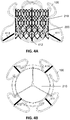
- the figure 1A shows an example of reinforcement according to the invention.
- the frame 100 comprises several parts formed from a metal alloy (for example heat-sensitive or shape memory such as a nickel-titanium or nitinol alloy).
- a metal alloy for example heat-sensitive or shape memory such as a nickel-titanium or nitinol alloy.
- Nitinol has the particular capacity to change physical state due to a temperature variation, which makes it a material of choice in the clinic (conditioned in an ice-cold or refreshed liquid, the nitinol then retains its final form after its release in the bloodstream at 37 ° C)
- the frame is of generally cylindrical shape having three parts of different diameters, allowing it to be positioned and to be maintained through the ring of the tricuspid valve without hampering the blood flow flowing through the tricuspid valve.
- the frame comprises at least four elements (101, 102, 103, 104).
- the frame has a total height of between 32 mm and 45 mm (i.e. adapted to the dimensions of the bioprosthesis).
- a bioprosthesis 110 is placed inside the frame
- the four main parts of the frame 101, 102, 103 and 104 are interconnected in the zone of the valve ring, which is the narrowest, by a flexible and deformable junction or articulation.
- the main parts are made of nickel-titanium alloy with shape memory.
- Each of the main parts is schematically in the form of a capital S.
- the flexible and deformable joint measures between 5 and 7mm in its height corresponding to the height of the valve ring.
- the figure 1B illustrates an example of interconnection of the sub-parts of the armature 100 according to one embodiment.
- a junction wire 110 (single) connects the various main parts of the frame, for example by welds.
- the main parts of the frame are connected by a plurality of junctions or articulations (this latter embodiment makes the structure according to the invention more flexible).
- the embodiments of these articulations allow an adaptation of the diameter of the reinforcement considered overall, by making this diameter variable and / or configurable.
- the number of direct contact points can in particular be adjusted (for example increased), so as to increase the contact surface with the native ring and improve the maintenance of the replacement structure of the tricuspid valve.
- the example shown in figure 1B has twelve points of contact. This type of configuration advantageously makes it possible to reduce the risk of peri-prosthetic leakage between the framework and the native tissue of the tricuspid ring.
- the figure 1A shows that the frame according to an embodiment of the invention comprises three parts or zones.
- the direction of blood flow is illustrated by arrow 199.
- the frame 100 is extensible and / or deformable. By its radial force (for example prestressed or resulting from the mechanical play of the four interconnected parts), the reinforcement 100 adapts to the morphology of the tricuspid ring (which is not entirely circular, often according to an ellipse) .
- the native ring or armature in its narrowest part measures between 40 and 42 millimeters. This diameter can reach 45 millimeters, or even more than 50 millimeters in the event of significant dilation, corresponding to the anatomical zone of the tricuspid ring delimiting the passage or the orifice, between the AD and the VD.
- the annular zone (of the native tricuspid ring of the armature according to the invention) measures from 5 to 7 mm in height.
- the reinforcement continues downstream in the right ventricle VD in the direction of blood flow.
- the armature extends in the right ventricle over a height of between 10 and 12mm, flaring on the side of the ventricular VD (ie has a convex shape).
- the maximum diameter 1321, in the right intra ventricular portion VD is slightly greater than the diameter of the ring area.
- the end of the armature ends in a diameter of value substantially equal to the diameter of the bioprosthesis (for example 35mm or 40mm).
- this mechanical configuration ie convex shape and selection of diameters
- this mechanical configuration makes it possible to maintain the three sheets composing the native tricuspid valve in the open position without creating any obstacle in the filling chamber of the right ventricle VD for the flow of blood flow ( " No right ventricular out flow obstruction" in English).
- the frame In the intra-AD 133 area, located upstream of the ring area, is the proximal part of the armature, positioned in the right atrium AD.
- the frame has a convex flared shape.
- the largest diameter 1332 is 6 to 8 mm larger than that of the ring area.
- the diameter value can be 50 mm if the ring measures 42 mm.
- the frame extends in this intra-AD 133 area over a variable distance depending on the dimensions of the bioprosthesis. For example, this distance can be of the order of 15 mm.
- This area of the intra-AD frame corresponds to the area of attachment to the stent which contains the bioprosthesis.
- the figure 2A represents a stent 200 according to the state of the art.
- the stent 200 used is a self-expanding stent.
- the stent 200 is cylindrical, formed of multiple cells 201 of metal alloy, for example of an alloy identical to that of the frame 100 (titanium-nickel).
- the diameter of the stent 200 may be slightly less than that of the frame in the area of the ring.
- the dimensions of the stent 200 are generally discreet: the standard dimensions are 30, 35 and 40 mm.
- the dimensions of the stent correspond to the dimensions of the bioprosthesis 210 (the stent 200 carries the bioprosthesis 210).
- the figure 2B represents a bioprosthesis 210 of tricuspid valve according to the state of the art.
- a tricuspid bioprosthesis is formed of three cusps (211, 212, 213) produced from animal tissue (for example a bovine or porcine pericardium) and / or a synthetic tissue.
- the three cusps or "sheets" forming the bioprosthesis are connected (eg associated or fixed or attached or welded or sewn) in or on or by or via the stent.
- the bioprosthesis works in the physiological direction of the blood flow arriving in the right atrium AD and injected in systole in the filling chamber of the right ventricle VD.
- Different technologies can be used to connect the stent and the bioprosthesis to the different contact points 220 (e.g. welding, glue, fixed or spring link, flexible or partially rotary contact points, etc.).
- three contact points 220 are used, allowing a more secure attachment of the tricuspid valve.
- a plurality of contact points (greater than or equal to four) hold the bioprosthesis in the stent. The probability of failure generally decreases when the number of contact points increases.
- the figure 3A is an illustration of the relative positioning of the frame 100 relative to the stent 200 carrying the bioprosthesis 210.
- the stent containing the bioprosthesis is positioned at inside the frame.
- the proximal end 301 (ie the base) of the stent is located in the area AD 133 of the frame.
- the bioprosthesis 210 is therefore mainly in the sub-annular position.
- the distal end of the stent 302 containing the bioprosthesis 210 is located in the intra-VD area 132 but over a few millimeters only.
- the stent is therefore positioned in an asymmetrical position relative to the zone of the ring, which is the narrowest part of the frame.
- the figure 3B shows a horizontal sectional view at the height of the ring area.
- the figure showing the presence of the frame 100, the stent 200 and the bioprosthesis 210.
- the figure 4A shows an example of assembly according to the invention.
- the frame 100 is connected to the stent 200 comprising the prosthesis 210 by one or more strands.
- the number, location and nature (e.g. materials) of the strands are variable depending on the embodiments (more or less flexible assembly, more or less stable or maintained, more or less disruptive to circulation, etc.).
- a single fixing strand may be sufficient (the transmission of mechanical forces via this single strand, possibly reinforced, can be modeled and optimized).
- the blood flow is minimally disturbed .
- the four strands of the example shown in the figure associate the stent 200 (in its intra AD 132 area) with the base of the frame 100 (in its widest area, ie in intra AD 132 area). ).
- This configuration where the attachment is made upstream of the circulation of the blood flow has the advantage of causing torsional stability of the stent downstream.
- one or more additional and optional attachments of the stent to the frame can be made in the area of the 'ring, or even by the distal part of the stent (even if the latter penetrates little in the DV area).
- one or more strands are made of nitinol.
- heat-sensitive and / or shape memory materials can be used.
- one or more strands are rigid. In embodiments, one or more strands are elastic or flexible or deformable. Other embodiments combine elastic strands and rigid strands (for example as a function of the dynamic behavior of the structure of the assembly). Other advanced embodiments provide for the use of actuable or configurable strands.
- the figure 4B shows a top view of an example of assembly according to the invention.
- the figure shows in particular the positioning of the bioprosthesis 210 within the frame 100.
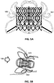
- the figure 5A illustrates an alternative embodiment of the invention.
- the assembly includes one or more optional fixing elements (ie holding in the heart).
- an element of fixing takes the form of a "racket” or a "loop” or a “tab” (for example 501).
- a fastener is made of a nitinol alloy.
- a fixing element has a height of the order of 8 to 10 mm and a length of between 10 and 12 mm.
- one or more of these optional fixing elements are associated with the armature 100 in the region of the ring 131 and open only on the ear side AD.
- no fastening element is used.
- a single fastening element is used.
- two fastening elements are positioned symmetrically.
- a second fastening element can indeed be positioned (for example symmetrically, eg towards the inter atrial septal wall).
- this configuration in certain cases can increase the risk of electrical conduction disorder (including an atrioventricular block).
- a plurality of fasteners is used, decreasing the risk of conduction disturbance.
- attachment elements are in particular to stabilize and fix the reinforcement 100 in the heart.
- these fastening elements improve the stability in rotation and / or in torsion and / or in translation of the assembly according to the invention.
- the figure 5B specifies the anatomical positioning of the optional fixing elements.
- the holding loops are placed upstream of the flow of blood flow 199.
- the assembly comprises two fixing elements 501 and 502. These elements are arranged in diametrically opposite relation to the frame. Each element is arranged substantially perpendicular to the reinforcement and is pressed onto the wall of the native fabric.
- One of the fixing elements is positioned on the side of the inter-auricular septum 501, the other fixing element 502 is positioned towards the external wall of the AD (called pectineal muscle).
- the technologies for attaching the fastening elements to the frame are variable.
- the attachment points can for example be welded.
- the plurality of fastening elements deploys spontaneously or automatically towards the outside during the release of the armature during the withdrawal of the carrying catheter from the whole system,
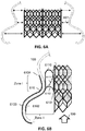
- the figure 6A illustrates the circumferential spacing or interstitial space between the stent and the frame.
- a circumferential free space 601 of variable amplitude between the stent 200 and the frame 100.
- the function of the frame 100 is in particular to maintain and stiffen the whole of the system while applying radial force against the native ring for stable placement in the heart.
- the stent and bioprosthesis are cylindrical in shape (discrete and standardized sizes).
- the diameter of the stent and of the bioprosthesis are substantially equal, and are less than the diameter of the frame: there is therefore an interstitial space (a "gap") between the frame 100 and the stent 200.
- the figure 6B illustrates an alternative embodiment of the invention comprising a sealing skirt (sectional view).
- the function of the sealing skirt is to seal (in particular in the area of the atrioventricular ring) the assembly according to the invention (ie the system formed by the frame and the stent carrying the bioprosthesis); in particular the sealing skirt minimizes the risk of peri-prosthetic leaks (point 6104 below).
- the sealing skirt 610 generally bears on the frame and at least partially covers the stent 200.
- the starting point 6101 for fixing the sealing skirt 610 is sewn (or glued) to the external wall of the stent over a height that largely covers the area of the ring. Then, the fabric of the sealing skirt is folded (for example according to a 180 ° return towards the outside) to descend down along the internal wall of the frame 6102. Finally, the fabric of the sealing skirt wraps by its intra-auricular base the frame 100, going up on the external wall of the frame 6103 to end above the zone of the ring, sewn or glued on the joints in capital S and / or the elements of the frame 100 thus reinforcing this frame in the narrowest area pressed against the native tissue 6104.
- This point 6104 plays in particular a decisive role in preventing peri-prosthetic leaks. As regards the alternative embodiments, it is not necessary for the sealing skirt to completely envelop the frame.
- the sealing skirt is formed from a synthetic material (for example polyethylene terephthalate or PET, which material is flexible and waterproof against water or blood).
- a waterproof material such as dacron can be advantageously used.
- the skirt may consist of a plurality of materials, for example arranged in layers and / or in strips (ie with reinforced zones for example and / or with zones consisting of a single layer and / or openwork spaces ( "Openings", "holes”, porous or non-waterproof sub-parts, etc.) upstream of the ring area, ear slope, in order to minimize the thickness of assembly in packaging).
- the structure of the skirt (eg materials, arrangement of the sub-parts, layers, distribution of the openwork spaces, etc.) can in particular be carried out so as to optimize (ie minimize the thickness of the assembly in packaging and / or to ensure the resistance to the blood flow of the skirt thus structured and / or to contribute to strengthening the maintenance of the assembly put in place (eg stability in rotation and / or in translation).
- the blood flow is slightly modified by the assembly according to the invention.
- Upstream on the auricular side, the convex structure of the armature implies certain disturbances of turbulence in the blood flow, but this modification is acceptable or of no medical and mechanical importance.
- the patient is on anticoagulant treatment to avoid the formation of clots caused by the presence of the bioprosthesis and its frame. As a result, the blood rheology is changed.
- zone I on the figure 6B area or space of "contact"
- the blood flow is "blocked” (or stopped or blocked or obstructed or prevented) or at least substantially minimized in the area of the native ring (ie around the entire circumference and at least partially on the height of the corresponding cylinder of the heart valve ring.
- the skirt captures blood flow and channels it through the bioprosthesis, due to its tightness.
- the shape of the fold 6110 ( figure 6B ) may incidentally influence the recirculation towards the bioprosthesis.
- Some advantageous embodiments consist in "stretching" or “loosening” more or less this fold of the skirt so as to optimize the flow dynamics of blood flow.
- There is a slight obstruction to the blood flow around the periphery of the valve itself because the diameter of the bioprosthesis is actually very slightly less than the diameter of the native ring. The latter is however dilated by the pathology justifying the replacement of the tricuspid valve, which makes this particular obstruction negligible in proportion.
- the reinforcement is applied (in contact) on the native tissue. Leaks between the frame and the native fabric are in practice non-existent or not significant (ie due to the absence of calcification of the tricuspid ring which does not allow the development of interstitial spaces between the frame and the fabric native, and also from the fact that the fabric of the skirt folds in the zone of the ring which improves the tightness in this zone).
- a triple thickness (of material (eg PET) seals the prosthetic system advantageously eliminates para-prosthetic leaks between the prosthesis and the native tissue but can increase the thickness of the assembly in its folded configuration. allow these requirements to be reconciled by moving the folding zones or by arranging the various components of the assembly so as to optimize its geometric folding.
- Certain embodiments of the invention provide for the use of strands and / or meshes of the stent having a tubular or cylindrical or elliptical shape, so as to minimize the impacts caused to red blood cells.
- the sections of the strands and / or meshes of the stent are elliptical in shape and are oriented (like “aeronautical flaps”), so as to improve the rheology and / or optimize the blood flow. local and / or global.
- the flow is partly channeled by the stent without causing any medically harmful consequence (in particular this does not create obstruction of the blood flow entering the right ventricle VD).
- the reinforcement Downstream of the ring, the reinforcement is wider (convex shape oriented towards outside) but with a diameter smaller than the upstream area, so as not to obstruct the route of ejection to the pulmonary valve located in the DV at about 10mm.
- the figure 7A illustrates an entirely optional variant embodiment of the invention comprising an additional attachment to a stent positioned in the inferior vena cava.
- a self-expanding stent 701 (for example made of nitinol) is placed in the inferior vena cava VCI, at a distance from the frame.
- the stent 701 is of variable diameter, generally of the order of 30mm, adapted to the dimension of the IVC at its abutment in the AD.
- the stent placed in the IVC must be shorter so as not to impede circulation in the hepatic veins.
- the stowage arrangement in the case of the tricuspid valve is also specific when compared to the arrangement for the mitral valve.
- the stent 701 is connected to the armature 100 in various ways (for example by a wire link 702).
- the wire link 702 is connected to the stent 701 in its upper part, flush with the mouth of the IVC.
- the wire link 702 is connected to the main frame at its base intra AD in its lower part.
- This wire link 702 can be made visible by fluoroscopy by a radio-opaque marker.
- This flexible and deformable wire link can be straight or corrugated. It can be adapted in its length (more or less 30 mm, depending on the anatomical distance between the tricuspid ring and the IVC).
- the figure 8 illustrates the method of setting up the system or assembly according to the invention.
- the delivery catheter 800 (“ delivery system” in English) contains the frame, the stent and the bioprosthesis (as well as the elements of fixing and sealing skirt according to the variant embodiments if necessary).
- the assembly according to the invention is deployed in the patient's heart (by manipulation of the operator).
- the bioprosthesis is "folded” in the stent, itself “folded” (with the sealing skirt) in the frame in a state of packaging.
- the delivery system and the delivery system include: a) a 0.35 inch guide of suitable length (length close to 280 mm) curved "snail" at its non-traumatic distal end, intended to be placed in the cavity of the DV; b) a catheter of a length adapted to the distance from the common femoral vein (to the inguinal cavity) to the AD and the VD.
- This catheter or sheath formed by a coaxial tube called OTW is focused on the 0.35.inch guide.
- This catheter has a retractable sheath at its distal part. Its diameter from 18 to 24 French is suitable for assembly according to the invention.
- the sheath is preceded at its distal end by a non-traumatic conical tip, adapted to receive the 0.35 coaxial guide provided for crossing the tricuspid valve and placing itself in the tip of the VD; c) a handle which contains the bioprosthesis release mechanism.
- This handle is welded to the coaxial tube. It controls the deployment of the assembly.
- the handle has a screw wheel controlling the deployment and release of the assembly.
- the device is also provided with a system making it possible to bend and orient the tube at its distality, before the sheath which contains the assembly according to the invention.
- the sheath (18 to 24 Fr) is retractable and contains the assembly according to the invention (according to its possible variants).
- percutaneous placement is carried out, in the common femoral vein, of a 24 Fr desilet adapted to receive the delivery system.
- the 0.35 guide is placed in the tip of the DV.
- the delivery system containing the frame and the bioprosthesis, is mounted through the mesh, then pushed through the Lower Cave Vein (VCI) to the AD, on the 0.35 guide.
- VCI Lower Cave Vein
- the sheath upon arrival in the AD, the sheath is bent at its end towards the tricuspid orifice using the mechanical handle.
- a fifth step the assembly is then pushed to cross the tricuspid valve.
- the progressive deployment of the armature is done under fluoroscopic control (radio-opaque markers are located on the armature at the level of the ring area) and ETO and / or 3D echocardiographic.
- the screw knob on the handle rotating clockwise gradually releases the frame containing the bioprosthesis under ETO echocardiographic and fluoroscopy control.
- a seventh step after the ring area, in the right intra-auricular part AD, the deployment continues, releasing one or more fastening elements (“hooks”, “rackets”, “loops”) which come press against the outer wall of the atrium and the interatrial septum.
- the bioprosthesis is released and begins to function as soon as the intra AD part of the frame is fully released.
- the sheath is gradually withdrawn in the abutment orifice of the VCI, releasing the stent fixed to the frame by a strand of nitinol. This operation is carried out under ETO and fluoroscopic control and produces a radiopaque contrast.
- an angiography and TEE step checks the seal, that is to say the absence of leakage of the bioprosthesis in place in the native tricuspid valve.
- the assembly can be instrumented by including sensors (“ sensors” in English, for example active sensors of and / or passive markers) and / or actuators (“ actuators” in English).
- the assembly according to the invention may in particular include radio-opaque markers making it possible to quantify to measure, to verify the correct positioning of the assembly in the patient's heart.
- the assembly according to the invention may also include devices allowing movements or readjustments in the space of the assembly, the position of which can change or even drift over time.
- the assembly according to the invention can be static in certain embodiments and / or dynamic or adaptive in other embodiments.
- the assembly according to the invention can in particular comprise one or more MEMS.
- An electromechanical microsystem or MEM is a microsystem of generally micrometric dimensions comprising one or more mechanical elements, using electricity as a source of energy, in order to perform a sensor and / or actuator function.
- bio-MEMS are used.
- the actuators can be placed in or on different parts of the frame and / or at the attachment zones of the different sub-parts of the frame and / or in or on the strands of attachment of the frame to the stent.
- the actuators can in particular, for example, serve to reconfigure the shape of the reinforcement (for example its convexity) and / or to adjust the overlap of the sealing skirt (eg curvature, return, tension of the surface of the skirt to certain locations, etc.) and / or to adjust the attachment of the stent to the frame.
- Spatial displacements or readjustments are generally carried out over short distances. They can be reversible or irreversible (e.g. mechanical pawls). They can be configured and / or configurable. They can be at least partially determined by an external device (controlled by the operating doctor), i.e. in open loop. Logical and / or physical devices can secure spatial modifications, if necessary.
- the structural modifications made to the structure can also be regulated according to a closed loop, for example as a function of the measurements of static position and of dynamic behavior of the assembly inserted into the blood flow.
- the assembly for the invention is modified so as to allow replacement of the mitral valve.
- the terms "tricuspid” and “mitral” are not generally interchangeable.
- the assembly is modified so as to allow replacement of the mitral valve, taking into account the fact that the diameter of the pathological mitral valve is significantly less than the diameter of the tricuspid valve pathological.
- preference will be given to the use, in addition to a mitral bioprosthesis, of a more limited number of sub-parts making up the armature (eg mechanical stability able be obtained more easily) and if possible a smaller number of sealing skirt thicknesses in the conditioned state.
Landscapes
- Health & Medical Sciences (AREA)
- Engineering & Computer Science (AREA)
- Biomedical Technology (AREA)
- Cardiology (AREA)
- Oral & Maxillofacial Surgery (AREA)
- Transplantation (AREA)
- Heart & Thoracic Surgery (AREA)
- Vascular Medicine (AREA)
- Life Sciences & Earth Sciences (AREA)
- Animal Behavior & Ethology (AREA)
- General Health & Medical Sciences (AREA)
- Public Health (AREA)
- Veterinary Medicine (AREA)
- Prostheses (AREA)
Applications Claiming Priority (3)
| Application Number | Priority Date | Filing Date | Title |
|---|---|---|---|
| FR1561217A FR3043907A1 (fr) | 2015-11-23 | 2015-11-23 | Assemblage pour le remplacement de la valve atrio-ventriculaire tricuspide |
| EP16795096.3A EP3380042B1 (fr) | 2015-11-23 | 2016-11-15 | Assemblage pour le remplacement de la valve atrio-ventriculaire tricuspide |
| PCT/EP2016/077715 WO2017089179A1 (fr) | 2015-11-23 | 2016-11-15 | Assemblage pour le remplacement de la valve atrio-ventriculaire tricuspide |
Related Parent Applications (2)
| Application Number | Title | Priority Date | Filing Date |
|---|---|---|---|
| EP16795096.3A Division EP3380042B1 (fr) | 2015-11-23 | 2016-11-15 | Assemblage pour le remplacement de la valve atrio-ventriculaire tricuspide |
| EP16795096.3A Division-Into EP3380042B1 (fr) | 2015-11-23 | 2016-11-15 | Assemblage pour le remplacement de la valve atrio-ventriculaire tricuspide |
Publications (1)
| Publication Number | Publication Date |
|---|---|
| EP3622918A1 true EP3622918A1 (fr) | 2020-03-18 |
Family
ID=55589952
Family Applications (2)
| Application Number | Title | Priority Date | Filing Date |
|---|---|---|---|
| EP19192702.9A Pending EP3622918A1 (fr) | 2015-11-23 | 2016-11-15 | Assemblage pour le remplacement de la valve atrio-ventriculaire tricuspide |
| EP16795096.3A Active EP3380042B1 (fr) | 2015-11-23 | 2016-11-15 | Assemblage pour le remplacement de la valve atrio-ventriculaire tricuspide |
Family Applications After (1)
| Application Number | Title | Priority Date | Filing Date |
|---|---|---|---|
| EP16795096.3A Active EP3380042B1 (fr) | 2015-11-23 | 2016-11-15 | Assemblage pour le remplacement de la valve atrio-ventriculaire tricuspide |
Country Status (9)
| Country | Link |
|---|---|
| US (2) | US11154396B2 (enExample) |
| EP (2) | EP3622918A1 (enExample) |
| JP (2) | JP7158721B2 (enExample) |
| CN (1) | CN108495600B (enExample) |
| AU (1) | AU2016360524B2 (enExample) |
| BR (1) | BR112018010342B1 (enExample) |
| ES (1) | ES2775700T3 (enExample) |
| FR (1) | FR3043907A1 (enExample) |
| WO (1) | WO2017089179A1 (enExample) |
Families Citing this family (106)
| Publication number | Priority date | Publication date | Assignee | Title |
|---|---|---|---|---|
| US8652202B2 (en) | 2008-08-22 | 2014-02-18 | Edwards Lifesciences Corporation | Prosthetic heart valve and delivery apparatus |
| US10517719B2 (en) | 2008-12-22 | 2019-12-31 | Valtech Cardio, Ltd. | Implantation of repair devices in the heart |
| US9968452B2 (en) | 2009-05-04 | 2018-05-15 | Valtech Cardio, Ltd. | Annuloplasty ring delivery cathethers |
| US8449599B2 (en) | 2009-12-04 | 2013-05-28 | Edwards Lifesciences Corporation | Prosthetic valve for replacing mitral valve |
| WO2012127309A1 (en) * | 2011-03-21 | 2012-09-27 | Ontorfano Matteo | Disk-based valve apparatus and method for the treatment of valve dysfunction |
| ES2664243T3 (es) | 2011-06-23 | 2018-04-18 | Valtech Cardio, Ltd. | Elemento de cierre para su uso con una estructura de anuloplastia |
| US9439763B2 (en) | 2013-02-04 | 2016-09-13 | Edwards Lifesciences Corporation | Prosthetic valve for replacing mitral valve |
| US9622863B2 (en) | 2013-11-22 | 2017-04-18 | Edwards Lifesciences Corporation | Aortic insufficiency repair device and method |
| CN111437068B (zh) | 2014-12-04 | 2023-01-17 | 爱德华兹生命科学公司 | 用于修复心脏瓣膜的经皮夹具 |
| US10231827B2 (en) * | 2015-03-18 | 2019-03-19 | Medtronic Vascular, Inc. | Valve prostheses having an integral centering mechanism and methods of use thereof |
| WO2016183485A1 (en) | 2015-05-14 | 2016-11-17 | Edwards Lifesciences Corporation | Heart valve sealing devices and delivery devices therefor |
| EP4335415A3 (en) | 2015-05-14 | 2024-05-29 | Cephea Valve Technologies, Inc. | Replacement mitral valves |
| FR3043907A1 (fr) * | 2015-11-23 | 2017-05-26 | Alain Dibie | Assemblage pour le remplacement de la valve atrio-ventriculaire tricuspide |
| US11833034B2 (en) | 2016-01-13 | 2023-12-05 | Shifamed Holdings, Llc | Prosthetic cardiac valve devices, systems, and methods |
| US10799675B2 (en) | 2016-03-21 | 2020-10-13 | Edwards Lifesciences Corporation | Cam controlled multi-direction steerable handles |
| US10835714B2 (en) | 2016-03-21 | 2020-11-17 | Edwards Lifesciences Corporation | Multi-direction steerable handles for steering catheters |
| US11219746B2 (en) | 2016-03-21 | 2022-01-11 | Edwards Lifesciences Corporation | Multi-direction steerable handles for steering catheters |
| US10973638B2 (en) | 2016-07-07 | 2021-04-13 | Edwards Lifesciences Corporation | Device and method for treating vascular insufficiency |
| US10653862B2 (en) | 2016-11-07 | 2020-05-19 | Edwards Lifesciences Corporation | Apparatus for the introduction and manipulation of multiple telescoping catheters |
| US10905554B2 (en) | 2017-01-05 | 2021-02-02 | Edwards Lifesciences Corporation | Heart valve coaptation device |
| US10653523B2 (en) | 2017-01-19 | 2020-05-19 | 4C Medical Technologies, Inc. | Systems, methods and devices for delivery systems, methods and devices for implanting prosthetic heart valves |
| EP3570779B2 (en) | 2017-01-23 | 2025-11-12 | Cephea Valve Technologies, Inc. | Replacement mitral valves |
| EP4209196A1 (en) | 2017-01-23 | 2023-07-12 | Cephea Valve Technologies, Inc. | Replacement mitral valves |
| US10561495B2 (en) | 2017-01-24 | 2020-02-18 | 4C Medical Technologies, Inc. | Systems, methods and devices for two-step delivery and implantation of prosthetic heart valve |
| US12029647B2 (en) | 2017-03-07 | 2024-07-09 | 4C Medical Technologies, Inc. | Systems, methods and devices for prosthetic heart valve with single valve leaflet |
| ES2991801T3 (es) | 2017-04-18 | 2024-12-04 | Edwards Lifesciences Corp | Dispositivos de sellado de válvula cardíaca y dispositivos de suministro para los mismos |
| US11224511B2 (en) | 2017-04-18 | 2022-01-18 | Edwards Lifesciences Corporation | Heart valve sealing devices and delivery devices therefor |
| US10799312B2 (en) | 2017-04-28 | 2020-10-13 | Edwards Lifesciences Corporation | Medical device stabilizing apparatus and method of use |
| US10959846B2 (en) | 2017-05-10 | 2021-03-30 | Edwards Lifesciences Corporation | Mitral valve spacer device |
| US12036113B2 (en) | 2017-06-14 | 2024-07-16 | 4C Medical Technologies, Inc. | Delivery of heart chamber prosthetic valve implant |
| US11051940B2 (en) | 2017-09-07 | 2021-07-06 | Edwards Lifesciences Corporation | Prosthetic spacer device for heart valve |
| US11065117B2 (en) | 2017-09-08 | 2021-07-20 | Edwards Lifesciences Corporation | Axisymmetric adjustable device for treating mitral regurgitation |
| US11040174B2 (en) | 2017-09-19 | 2021-06-22 | Edwards Lifesciences Corporation | Multi-direction steerable handles for steering catheters |
| US10722349B2 (en) * | 2017-12-07 | 2020-07-28 | Medtronic Vascular, Inc. | Adjustable prosthetic heart valve |
| US10159570B1 (en) | 2018-01-09 | 2018-12-25 | Edwards Lifesciences Corporation | Native valve repair devices and procedures |
| US10238493B1 (en) | 2018-01-09 | 2019-03-26 | Edwards Lifesciences Corporation | Native valve repair devices and procedures |
| US10245144B1 (en) | 2018-01-09 | 2019-04-02 | Edwards Lifesciences Corporation | Native valve repair devices and procedures |
| US10123873B1 (en) | 2018-01-09 | 2018-11-13 | Edwards Lifesciences Corporation | Native valve repair devices and procedures |
| US10076415B1 (en) | 2018-01-09 | 2018-09-18 | Edwards Lifesciences Corporation | Native valve repair devices and procedures |
| FI3964175T3 (fi) | 2018-01-09 | 2024-12-03 | Edwards Lifesciences Corp | Natiivin läpän korjauslaitteita |
| US10231837B1 (en) | 2018-01-09 | 2019-03-19 | Edwards Lifesciences Corporation | Native valve repair devices and procedures |
| US10136993B1 (en) | 2018-01-09 | 2018-11-27 | Edwards Lifesciences Corporation | Native valve repair devices and procedures |
| US10111751B1 (en) | 2018-01-09 | 2018-10-30 | Edwards Lifesciences Corporation | Native valve repair devices and procedures |
| US10105222B1 (en) | 2018-01-09 | 2018-10-23 | Edwards Lifesciences Corporation | Native valve repair devices and procedures |
| US10973639B2 (en) | 2018-01-09 | 2021-04-13 | Edwards Lifesciences Corporation | Native valve repair devices and procedures |
| US11458287B2 (en) * | 2018-01-20 | 2022-10-04 | V-Wave Ltd. | Devices with dimensions that can be reduced and increased in vivo, and methods of making and using the same |
| WO2019147846A2 (en) | 2018-01-25 | 2019-08-01 | Edwards Lifesciences Corporation | Delivery system for aided replacement valve recapture and repositioning post- deployment |
| WO2019195860A2 (en) | 2018-04-04 | 2019-10-10 | Vdyne, Llc | Devices and methods for anchoring transcatheter heart valve |
| US11389297B2 (en) | 2018-04-12 | 2022-07-19 | Edwards Lifesciences Corporation | Mitral valve spacer device |
| US11207181B2 (en) | 2018-04-18 | 2021-12-28 | Edwards Lifesciences Corporation | Heart valve sealing devices and delivery devices therefor |
| CN108578016B (zh) * | 2018-04-26 | 2020-09-08 | 赛诺医疗科学技术股份有限公司 | 一种经心尖植入式二尖瓣瓣膜装置 |
| CN119564382A (zh) | 2018-08-21 | 2025-03-07 | 施菲姆德控股有限责任公司 | 人工心脏瓣膜装置、系统和方法 |
| US11857441B2 (en) | 2018-09-04 | 2024-01-02 | 4C Medical Technologies, Inc. | Stent loading device |
| US11071627B2 (en) | 2018-10-18 | 2021-07-27 | Vdyne, Inc. | Orthogonally delivered transcatheter heart valve frame for valve in valve prosthesis |
| US10595994B1 (en) | 2018-09-20 | 2020-03-24 | Vdyne, Llc | Side-delivered transcatheter heart valve replacement |
| US12186187B2 (en) | 2018-09-20 | 2025-01-07 | Vdyne, Inc. | Transcatheter deliverable prosthetic heart valves and methods of delivery |
| US11278437B2 (en) | 2018-12-08 | 2022-03-22 | Vdyne, Inc. | Compression capable annular frames for side delivery of transcatheter heart valve replacement |
| US11344413B2 (en) | 2018-09-20 | 2022-05-31 | Vdyne, Inc. | Transcatheter deliverable prosthetic heart valves and methods of delivery |
| US10321995B1 (en) | 2018-09-20 | 2019-06-18 | Vdyne, Llc | Orthogonally delivered transcatheter heart valve replacement |
| US12310850B2 (en) | 2018-09-20 | 2025-05-27 | Vdyne, Inc. | Transcatheter deliverable prosthetic heart valves and methods of delivery |
| EP3860519A4 (en) | 2018-10-05 | 2022-07-06 | Shifamed Holdings, LLC | HEART VALVE PROSTHESIS, SYSTEMS AND PROCEDURES |
| US10945844B2 (en) | 2018-10-10 | 2021-03-16 | Edwards Lifesciences Corporation | Heart valve sealing devices and delivery devices therefor |
| AU2019362078B2 (en) | 2018-10-19 | 2025-08-07 | Shifamed Holdings, Llc | Adjustable medical device |
| US11109969B2 (en) | 2018-10-22 | 2021-09-07 | Vdyne, Inc. | Guidewire delivery of transcatheter heart valve |
| EP3883500B1 (en) | 2018-11-20 | 2024-11-06 | Edwards Lifesciences Corporation | Deployment tools for delivering a device to a native heart valve |
| AU2019384540B2 (en) | 2018-11-21 | 2025-01-02 | Edwards Lifesciences Corporation | Heart valve sealing devices, delivery devices therefor, and retrieval devices |
| KR102815760B1 (ko) | 2018-11-29 | 2025-06-04 | 에드워즈 라이프사이언시스 코포레이션 | 카테터 삽입 방법 및 장치 |
| EP3659558B1 (de) * | 2018-11-30 | 2022-01-19 | Saphenus Medical Technology GmbH | Sensorträger zum anordnen an einer prothese |
| US11253359B2 (en) | 2018-12-20 | 2022-02-22 | Vdyne, Inc. | Proximal tab for side-delivered transcatheter heart valves and methods of delivery |
| WO2020146842A1 (en) | 2019-01-10 | 2020-07-16 | Vdyne, Llc | Anchor hook for side-delivery transcatheter heart valve prosthesis |
| CN111437064B (zh) * | 2019-01-17 | 2025-09-19 | 上海微创心通医疗科技有限公司 | 一种假体心脏瓣膜 |
| US11273032B2 (en) * | 2019-01-26 | 2022-03-15 | Vdyne, Inc. | Collapsible inner flow control component for side-deliverable transcatheter heart valve prosthesis |
| US11185409B2 (en) | 2019-01-26 | 2021-11-30 | Vdyne, Inc. | Collapsible inner flow control component for side-delivered transcatheter heart valve prosthesis |
| JP7322155B2 (ja) * | 2019-01-28 | 2023-08-07 | トライケアス | 第2世代の3パーツステント |
| PL3923867T4 (pl) | 2019-02-14 | 2024-05-27 | Edwards Lifesciences Corporation | Urządzenia do uszczelniania zastawek serca i urządzenia do ich wprowadzania |
| CA3131522A1 (en) | 2019-02-25 | 2020-09-03 | Edwards Lifesciences Corporation | Heart valve sealing devices |
| AU2020231221B2 (en) | 2019-03-05 | 2025-07-31 | Vdyne, Inc. | Tricuspid regurgitation control devices for orthogonal transcatheter heart valve prosthesis |
| US11173027B2 (en) | 2019-03-14 | 2021-11-16 | Vdyne, Inc. | Side-deliverable transcatheter prosthetic valves and methods for delivering and anchoring the same |
| US11076956B2 (en) | 2019-03-14 | 2021-08-03 | Vdyne, Inc. | Proximal, distal, and anterior anchoring tabs for side-delivered transcatheter mitral valve prosthesis |
| US11471282B2 (en) | 2019-03-19 | 2022-10-18 | Shifamed Holdings, Llc | Prosthetic cardiac valve devices, systems, and methods |
| US11452628B2 (en) | 2019-04-15 | 2022-09-27 | 4C Medical Technologies, Inc. | Loading systems for collapsible prosthetic heart valve devices and methods thereof |
| CA3138875A1 (en) | 2019-05-04 | 2020-11-12 | Vdyne, Inc. | Cinch device and method for deployment of a side-delivered prosthetic heart valve in a native annulus |
| WO2020233775A1 (en) * | 2019-05-17 | 2020-11-26 | T-Heart SAS | Stent device for a prosthetic heart valve |
| CN110279495B (zh) * | 2019-06-25 | 2022-08-26 | 陈翔 | 一种自膨胀心脏瓣膜假体 |
| CN121101808A (zh) | 2019-08-20 | 2025-12-12 | 维迪内股份有限公司 | 用于可侧面递送经导管人工瓣膜的递送和取回装置和方法 |
| CA3152632A1 (en) | 2019-08-26 | 2021-03-04 | Vdyne, Inc. | Side-deliverable transcatheter prosthetic valves and methods for delivering and anchoring the same |
| US11801131B2 (en) * | 2019-12-20 | 2023-10-31 | Medtronic Vascular, Inc. | Elliptical heart valve prostheses, delivery systems, and methods of use |
| US11234813B2 (en) | 2020-01-17 | 2022-02-01 | Vdyne, Inc. | Ventricular stability elements for side-deliverable prosthetic heart valves and methods of delivery |
| US11931253B2 (en) | 2020-01-31 | 2024-03-19 | 4C Medical Technologies, Inc. | Prosthetic heart valve delivery system: ball-slide attachment |
| US12133797B2 (en) | 2020-01-31 | 2024-11-05 | 4C Medical Technologies, Inc. | Prosthetic heart valve delivery system: paddle attachment feature |
| US12053375B2 (en) | 2020-03-05 | 2024-08-06 | 4C Medical Technologies, Inc. | Prosthetic mitral valve with improved atrial and/or annular apposition and paravalvular leakage mitigation |
| US11992403B2 (en) | 2020-03-06 | 2024-05-28 | 4C Medical Technologies, Inc. | Devices, systems and methods for improving recapture of prosthetic heart valve device with stent frame having valve support with inwardly stent cells |
| CN116456937A (zh) | 2020-08-31 | 2023-07-18 | 施菲姆德控股有限责任公司 | 假体瓣膜递送系统 |
| US11197755B1 (en) * | 2020-10-28 | 2021-12-14 | Occam Labs LLC | Systems, devices and methods for folded unibody heart valve stents |
| WO2022103734A1 (en) * | 2020-11-10 | 2022-05-19 | Edwards Lifesciences Corporation | Docking station for a transcatheter heart valve |
| US12329635B2 (en) | 2020-12-04 | 2025-06-17 | Shifamed Holdings, Llc | Flared prosthetic cardiac valve delivery devices and systems |
| US11246726B1 (en) | 2021-02-10 | 2022-02-15 | Occam Labs LLC | Systems, devices and methods for delivery systems |
| US12171678B2 (en) * | 2021-02-24 | 2024-12-24 | Medtronic, Inc. | Skirt-reinforcement members for prosthetic valve devices |
| US12201521B2 (en) | 2021-03-22 | 2025-01-21 | Shifamed Holdings, Llc | Anchor position verification for prosthetic cardiac valve devices |
| CN113730034B (zh) * | 2021-09-27 | 2023-07-21 | 启晨(上海)医疗器械有限公司 | 经导管植入的二尖瓣瓣膜装置 |
| US20230149157A1 (en) * | 2021-11-18 | 2023-05-18 | St. Jude Medical, Cardiology Division, Inc. | Transcatheter Prosthetic Atrioventricular Valve |
| CN115040290B (zh) * | 2022-07-06 | 2025-01-24 | 昆山茵络医疗器械有限公司 | 一种瓣环环缩装置 |
| US20240091000A1 (en) * | 2022-09-21 | 2024-03-21 | St. Jude Medical, Cardiology Division, Inc. | Prosthetic Tricuspid Heart Valve |
| WO2024132115A1 (en) | 2022-12-20 | 2024-06-27 | T-Heart SAS | Loading system for a stent device |
| WO2024192413A2 (en) | 2023-03-15 | 2024-09-19 | Capstan Medical Inc. | Hooked spool delivery systems |
| USD1071198S1 (en) | 2023-06-28 | 2025-04-15 | Edwards Lifesciences Corporation | Cradle |
Citations (6)
| Publication number | Priority date | Publication date | Assignee | Title |
|---|---|---|---|---|
| WO2009044082A2 (fr) * | 2007-09-11 | 2009-04-09 | Laboratoires Perouse | Dispositif de traitement d'un conduit de circulation du sang |
| US8657872B2 (en) | 2010-07-19 | 2014-02-25 | Jacques Seguin | Cardiac valve repair system and methods of use |
| US20140194983A1 (en) * | 2013-01-08 | 2014-07-10 | Medtronic, Inc. | Valve Prosthesis and Method for Delivery |
| WO2014144937A2 (en) * | 2013-03-15 | 2014-09-18 | Twelve, Inc. | Prosthetic heart valve devices, prosthetic mitral valves and associated systems and methods |
| WO2015055052A1 (zh) * | 2013-10-17 | 2015-04-23 | 杭州启明医疗器械有限公司 | 提高安全性的肺动脉支架及肺动脉瓣膜置换装置 |
| WO2016130913A1 (en) * | 2015-02-12 | 2016-08-18 | Medtronic Inc. | Integrated valve assembly and method of delivering and deploying an integrated valve assembly |
Family Cites Families (33)
| Publication number | Priority date | Publication date | Assignee | Title |
|---|---|---|---|---|
| NL9500147A (nl) * | 1995-01-26 | 1996-09-02 | Industrial Res Bv | Werkwijze voor het uit foliemateriaal vervaardigen van een hulsvormige stent, en stent verkregen onder toepassing van deze werkwijze. |
| EP0850607A1 (en) * | 1996-12-31 | 1998-07-01 | Cordis Corporation | Valve prosthesis for implantation in body channels |
| US9579194B2 (en) * | 2003-10-06 | 2017-02-28 | Medtronic ATS Medical, Inc. | Anchoring structure with concave landing zone |
| US20060052867A1 (en) * | 2004-09-07 | 2006-03-09 | Medtronic, Inc | Replacement prosthetic heart valve, system and method of implant |
| ES2558534T3 (es) * | 2005-02-18 | 2016-02-05 | The Cleveland Clinic Foundation | Aparato para sustituir una válvula cardíaca |
| SE531468C2 (sv) | 2005-04-21 | 2009-04-14 | Edwards Lifesciences Ag | En anordning för styrning av blodflöde |
| EP3311779B1 (en) * | 2007-10-25 | 2024-04-24 | Boston Scientific Medical Device Limited | Cardiac valve |
| US20090171456A1 (en) * | 2007-12-28 | 2009-07-02 | Kveen Graig L | Percutaneous heart valve, system, and method |
| EP3572044B1 (en) * | 2008-01-24 | 2021-07-28 | Medtronic, Inc. | Stents for prosthetic heart valves |
| DE102008015781B4 (de) * | 2008-03-26 | 2011-09-29 | Malte Neuss | Vorrichtung zum Verschluss von Defekten im Gefäßsystem |
| US20100217382A1 (en) * | 2009-02-25 | 2010-08-26 | Edwards Lifesciences | Mitral valve replacement with atrial anchoring |
| NZ596179A (en) | 2009-04-29 | 2014-05-30 | Cleveland Clinic Foundation | Apparatus and method for replacing a diseased cardiac valve |
| US8845722B2 (en) | 2009-08-03 | 2014-09-30 | Shlomo Gabbay | Heart valve prosthesis and method of implantation thereof |
| WO2011143263A2 (en) | 2010-05-10 | 2011-11-17 | Heart Leaflet Technologies, Inc. | Stentless support structure |
| EP2613737B2 (en) * | 2010-09-10 | 2023-03-15 | Symetis SA | Valve replacement devices, delivery device for a valve replacement device and method of production of a valve replacement device |
| AU2011305153A1 (en) * | 2010-09-23 | 2013-05-02 | Colibri Heart Valve Llc | Percutaneously deliverable heart or blood vessel valve with frame having abluminally situated tissue membrane |
| US9554897B2 (en) * | 2011-04-28 | 2017-01-31 | Neovasc Tiara Inc. | Methods and apparatus for engaging a valve prosthesis with tissue |
| WO2012178115A2 (en) | 2011-06-24 | 2012-12-27 | Rosenbluth, Robert | Percutaneously implantable artificial heart valve system and associated methods and devices |
| CA2849030C (en) * | 2011-10-19 | 2020-10-27 | Twelve, Inc. | Prosthetic heart valve devices, prosthetic mitral valves and associated systems and methods |
| US9039757B2 (en) * | 2011-10-19 | 2015-05-26 | Twelve, Inc. | Prosthetic heart valve devices, prosthetic mitral valves and associated systems and methods |
| FR2982763B1 (fr) * | 2011-11-17 | 2015-07-17 | Ct Hospitalier Regional Universitaire D Amiens | Implant destine a etre place dans un passage de circulation du sang et dispositif de traitement associe |
| DK2852354T3 (da) * | 2012-05-20 | 2020-08-24 | Tel Hashomer Medical Res Infrastructure & Services Ltd | Kunstig mitralklap |
| US9345573B2 (en) * | 2012-05-30 | 2016-05-24 | Neovasc Tiara Inc. | Methods and apparatus for loading a prosthesis onto a delivery system |
| US10206775B2 (en) * | 2012-08-13 | 2019-02-19 | Medtronic, Inc. | Heart valve prosthesis |
| US20140277427A1 (en) * | 2013-03-14 | 2014-09-18 | Cardiaq Valve Technologies, Inc. | Prosthesis for atraumatically grasping intralumenal tissue and methods of delivery |
| EP2896387A1 (en) | 2014-01-20 | 2015-07-22 | Mitricares | Heart valve anchoring device |
| US9757230B2 (en) * | 2014-05-16 | 2017-09-12 | St. Jude Medical, Cardiology Division, Inc. | Stent assembly for use in prosthetic heart valves |
| WO2015179423A1 (en) * | 2014-05-19 | 2015-11-26 | Cardiaq Valve Technologies, Inc. | Replacement mitral valve with annular flap |
| US9532870B2 (en) | 2014-06-06 | 2017-01-03 | Edwards Lifesciences Corporation | Prosthetic valve for replacing a mitral valve |
| US9974647B2 (en) | 2014-06-12 | 2018-05-22 | Caisson Interventional, LLC | Two stage anchor and mitral valve assembly |
| US9782256B2 (en) * | 2015-04-27 | 2017-10-10 | Venus Medtech (Hangzhou) Inc | Heart valve assembly |
| US10016273B2 (en) * | 2015-06-05 | 2018-07-10 | Medtronic, Inc. | Filtered sealing components for a transcatheter valve prosthesis |
| FR3043907A1 (fr) * | 2015-11-23 | 2017-05-26 | Alain Dibie | Assemblage pour le remplacement de la valve atrio-ventriculaire tricuspide |
-
2015
- 2015-11-23 FR FR1561217A patent/FR3043907A1/fr not_active Withdrawn
-
2016
- 2016-11-15 BR BR112018010342-0A patent/BR112018010342B1/pt active IP Right Grant
- 2016-11-15 AU AU2016360524A patent/AU2016360524B2/en active Active
- 2016-11-15 EP EP19192702.9A patent/EP3622918A1/fr active Pending
- 2016-11-15 US US15/777,177 patent/US11154396B2/en active Active
- 2016-11-15 EP EP16795096.3A patent/EP3380042B1/fr active Active
- 2016-11-15 CN CN201680079758.6A patent/CN108495600B/zh active Active
- 2016-11-15 JP JP2018545550A patent/JP7158721B2/ja active Active
- 2016-11-15 ES ES16795096T patent/ES2775700T3/es active Active
- 2016-11-15 WO PCT/EP2016/077715 patent/WO2017089179A1/fr not_active Ceased
-
2021
- 2021-10-01 US US17/491,642 patent/US20220015901A1/en active Pending
-
2022
- 2022-03-01 JP JP2022030709A patent/JP7563767B2/ja active Active
Patent Citations (7)
| Publication number | Priority date | Publication date | Assignee | Title |
|---|---|---|---|---|
| WO2009044082A2 (fr) * | 2007-09-11 | 2009-04-09 | Laboratoires Perouse | Dispositif de traitement d'un conduit de circulation du sang |
| US8657872B2 (en) | 2010-07-19 | 2014-02-25 | Jacques Seguin | Cardiac valve repair system and methods of use |
| US20140172070A1 (en) | 2010-07-19 | 2014-06-19 | Jacques Seguin | Cardiac valve repair system and methods of use |
| US20140194983A1 (en) * | 2013-01-08 | 2014-07-10 | Medtronic, Inc. | Valve Prosthesis and Method for Delivery |
| WO2014144937A2 (en) * | 2013-03-15 | 2014-09-18 | Twelve, Inc. | Prosthetic heart valve devices, prosthetic mitral valves and associated systems and methods |
| WO2015055052A1 (zh) * | 2013-10-17 | 2015-04-23 | 杭州启明医疗器械有限公司 | 提高安全性的肺动脉支架及肺动脉瓣膜置换装置 |
| WO2016130913A1 (en) * | 2015-02-12 | 2016-08-18 | Medtronic Inc. | Integrated valve assembly and method of delivering and deploying an integrated valve assembly |
Also Published As
| Publication number | Publication date |
|---|---|
| AU2016360524B2 (en) | 2021-05-13 |
| US20220015901A1 (en) | 2022-01-20 |
| EP3380042A1 (fr) | 2018-10-03 |
| BR112018010342A2 (pt) | 2018-12-04 |
| JP7563767B2 (ja) | 2024-10-08 |
| US20180333259A1 (en) | 2018-11-22 |
| AU2016360524A1 (en) | 2018-06-21 |
| JP2018535074A (ja) | 2018-11-29 |
| US11154396B2 (en) | 2021-10-26 |
| CN108495600A (zh) | 2018-09-04 |
| ES2775700T3 (es) | 2020-07-28 |
| FR3043907A1 (fr) | 2017-05-26 |
| CN108495600B (zh) | 2020-10-30 |
| EP3380042B1 (fr) | 2019-12-11 |
| JP2022088374A (ja) | 2022-06-14 |
| WO2017089179A1 (fr) | 2017-06-01 |
| JP7158721B2 (ja) | 2022-10-24 |
| BR112018010342B1 (pt) | 2023-01-31 |
Similar Documents
| Publication | Publication Date | Title |
|---|---|---|
| EP3380042B1 (fr) | Assemblage pour le remplacement de la valve atrio-ventriculaire tricuspide | |
| US11406496B2 (en) | Stented prosthetic heart valve having a paravalvular sealing wrap | |
| US20200246142A1 (en) | Modular valve prosthesis with anchor stent and valve component | |
| EP3512466B1 (en) | Prosthetic heart valve with paravalvular leak mitigation features | |
| US9974649B2 (en) | Stented prosthetic heart valve having wrap and methods of delivery and deployment | |
| EP2943157B1 (en) | Valve prosthesis and method for delivery | |
| RU2631410C2 (ru) | Протез сердечного клапана | |
| TW201936133A (zh) | 用於可植入人造心臟瓣膜裝置的可擴張支架及瓣膜周圍滲漏減緩系統 | |
| US20170079786A1 (en) | Self-expanding heart valves for coronary perfusion and sealing | |
| CN106999273A (zh) | 具有无支撑瓣膜区段的分段式经导管瓣膜假体 | |
| US20140243965A1 (en) | Multi-layer stent | |
| FR3058631A1 (fr) | Implant de traitement d'une valve biologique | |
| CN103781439A (zh) | 用于改善的小叶附着的人工心脏瓣膜框架的连合改进 | |
| TW202017538A (zh) | 具有流體儲槽之支架裝載裝置 |
Legal Events
| Date | Code | Title | Description |
|---|---|---|---|
| PUAI | Public reference made under article 153(3) epc to a published international application that has entered the european phase |
Free format text: ORIGINAL CODE: 0009012 |
|
| STAA | Information on the status of an ep patent application or granted ep patent |
Free format text: STATUS: REQUEST FOR EXAMINATION WAS MADE |
|
| 17P | Request for examination filed |
Effective date: 20190820 |
|
| AC | Divisional application: reference to earlier application |
Ref document number: 3380042 Country of ref document: EP Kind code of ref document: P |
|
| AK | Designated contracting states |
Kind code of ref document: A1 Designated state(s): AL AT BE BG CH CY CZ DE DK EE ES FI FR GB GR HR HU IE IS IT LI LT LU LV MC MK MT NL NO PL PT RO RS SE SI SK SM TR |
|
| P01 | Opt-out of the competence of the unified patent court (upc) registered |
Effective date: 20230526 |
|
| STAA | Information on the status of an ep patent application or granted ep patent |
Free format text: STATUS: EXAMINATION IS IN PROGRESS |
|
| 17Q | First examination report despatched |
Effective date: 20250904 |