EP3594830B1 - Programm zum suchen von bildern eines ähnlichen falls, vorrichtung zum suchen von bildern eines ähnlichen falls und verfahren zum suchen von bildern eines ähnlichen falls - Google Patents
Programm zum suchen von bildern eines ähnlichen falls, vorrichtung zum suchen von bildern eines ähnlichen falls und verfahren zum suchen von bildern eines ähnlichen falls Download PDFInfo
- Publication number
- EP3594830B1 EP3594830B1 EP18763395.3A EP18763395A EP3594830B1 EP 3594830 B1 EP3594830 B1 EP 3594830B1 EP 18763395 A EP18763395 A EP 18763395A EP 3594830 B1 EP3594830 B1 EP 3594830B1
- Authority
- EP
- European Patent Office
- Prior art keywords
- image
- lung field
- area
- similar case
- unit
- Prior art date
- Legal status (The legal status is an assumption and is not a legal conclusion. Google has not performed a legal analysis and makes no representation as to the accuracy of the status listed.)
- Active
Links
Images







Classifications
-
- A—HUMAN NECESSITIES
- A61—MEDICAL OR VETERINARY SCIENCE; HYGIENE
- A61B—DIAGNOSIS; SURGERY; IDENTIFICATION
- A61B6/00—Apparatus or devices for radiation diagnosis; Apparatus or devices for radiation diagnosis combined with radiation therapy equipment
- A61B6/50—Apparatus or devices for radiation diagnosis; Apparatus or devices for radiation diagnosis combined with radiation therapy equipment specially adapted for specific body parts; specially adapted for specific clinical applications
-
- A—HUMAN NECESSITIES
- A61—MEDICAL OR VETERINARY SCIENCE; HYGIENE
- A61B—DIAGNOSIS; SURGERY; IDENTIFICATION
- A61B6/00—Apparatus or devices for radiation diagnosis; Apparatus or devices for radiation diagnosis combined with radiation therapy equipment
- A61B6/52—Devices using data or image processing specially adapted for radiation diagnosis
- A61B6/5211—Devices using data or image processing specially adapted for radiation diagnosis involving processing of medical diagnostic data
- A61B6/5217—Devices using data or image processing specially adapted for radiation diagnosis involving processing of medical diagnostic data extracting a diagnostic or physiological parameter from medical diagnostic data
-
- A—HUMAN NECESSITIES
- A61—MEDICAL OR VETERINARY SCIENCE; HYGIENE
- A61B—DIAGNOSIS; SURGERY; IDENTIFICATION
- A61B6/00—Apparatus or devices for radiation diagnosis; Apparatus or devices for radiation diagnosis combined with radiation therapy equipment
- A61B6/52—Devices using data or image processing specially adapted for radiation diagnosis
- A61B6/5211—Devices using data or image processing specially adapted for radiation diagnosis involving processing of medical diagnostic data
- A61B6/5229—Devices using data or image processing specially adapted for radiation diagnosis involving processing of medical diagnostic data combining image data of a patient, e.g. combining a functional image with an anatomical image
-
- G—PHYSICS
- G06—COMPUTING OR CALCULATING; COUNTING
- G06F—ELECTRIC DIGITAL DATA PROCESSING
- G06F16/00—Information retrieval; Database structures therefor; File system structures therefor
-
- G—PHYSICS
- G06—COMPUTING OR CALCULATING; COUNTING
- G06F—ELECTRIC DIGITAL DATA PROCESSING
- G06F16/00—Information retrieval; Database structures therefor; File system structures therefor
- G06F16/50—Information retrieval; Database structures therefor; File system structures therefor of still image data
- G06F16/58—Retrieval characterised by using metadata, e.g. metadata not derived from the content or metadata generated manually
- G06F16/583—Retrieval characterised by using metadata, e.g. metadata not derived from the content or metadata generated manually using metadata automatically derived from the content
- G06F16/5854—Retrieval characterised by using metadata, e.g. metadata not derived from the content or metadata generated manually using metadata automatically derived from the content using shape and object relationship
-
- G—PHYSICS
- G06—COMPUTING OR CALCULATING; COUNTING
- G06F—ELECTRIC DIGITAL DATA PROCESSING
- G06F18/00—Pattern recognition
- G06F18/20—Analysing
- G06F18/22—Matching criteria, e.g. proximity measures
-
- G—PHYSICS
- G06—COMPUTING OR CALCULATING; COUNTING
- G06T—IMAGE DATA PROCESSING OR GENERATION, IN GENERAL
- G06T7/00—Image analysis
- G06T7/0002—Inspection of images, e.g. flaw detection
- G06T7/0012—Biomedical image inspection
-
- G—PHYSICS
- G06—COMPUTING OR CALCULATING; COUNTING
- G06T—IMAGE DATA PROCESSING OR GENERATION, IN GENERAL
- G06T7/00—Image analysis
- G06T7/0002—Inspection of images, e.g. flaw detection
- G06T7/0012—Biomedical image inspection
- G06T7/0014—Biomedical image inspection using an image reference approach
-
- G—PHYSICS
- G06—COMPUTING OR CALCULATING; COUNTING
- G06T—IMAGE DATA PROCESSING OR GENERATION, IN GENERAL
- G06T7/00—Image analysis
- G06T7/10—Segmentation; Edge detection
- G06T7/11—Region-based segmentation
-
- G—PHYSICS
- G06—COMPUTING OR CALCULATING; COUNTING
- G06T—IMAGE DATA PROCESSING OR GENERATION, IN GENERAL
- G06T7/00—Image analysis
- G06T7/10—Segmentation; Edge detection
- G06T7/12—Edge-based segmentation
-
- G—PHYSICS
- G06—COMPUTING OR CALCULATING; COUNTING
- G06T—IMAGE DATA PROCESSING OR GENERATION, IN GENERAL
- G06T7/00—Image analysis
- G06T7/30—Determination of transform parameters for the alignment of images, i.e. image registration
-
- G—PHYSICS
- G06—COMPUTING OR CALCULATING; COUNTING
- G06V—IMAGE OR VIDEO RECOGNITION OR UNDERSTANDING
- G06V10/00—Arrangements for image or video recognition or understanding
- G06V10/40—Extraction of image or video features
- G06V10/42—Global feature extraction by analysis of the whole pattern, e.g. using frequency domain transformations or autocorrelation
- G06V10/421—Global feature extraction by analysis of the whole pattern, e.g. using frequency domain transformations or autocorrelation by analysing segments intersecting the pattern
-
- G—PHYSICS
- G16—INFORMATION AND COMMUNICATION TECHNOLOGY [ICT] SPECIALLY ADAPTED FOR SPECIFIC APPLICATION FIELDS
- G16H—HEALTHCARE INFORMATICS, i.e. INFORMATION AND COMMUNICATION TECHNOLOGY [ICT] SPECIALLY ADAPTED FOR THE HANDLING OR PROCESSING OF MEDICAL OR HEALTHCARE DATA
- G16H30/00—ICT specially adapted for the handling or processing of medical images
- G16H30/40—ICT specially adapted for the handling or processing of medical images for processing medical images, e.g. editing
-
- G—PHYSICS
- G16—INFORMATION AND COMMUNICATION TECHNOLOGY [ICT] SPECIALLY ADAPTED FOR SPECIFIC APPLICATION FIELDS
- G16H—HEALTHCARE INFORMATICS, i.e. INFORMATION AND COMMUNICATION TECHNOLOGY [ICT] SPECIALLY ADAPTED FOR THE HANDLING OR PROCESSING OF MEDICAL OR HEALTHCARE DATA
- G16H50/00—ICT specially adapted for medical diagnosis, medical simulation or medical data mining; ICT specially adapted for detecting, monitoring or modelling epidemics or pandemics
- G16H50/50—ICT specially adapted for medical diagnosis, medical simulation or medical data mining; ICT specially adapted for detecting, monitoring or modelling epidemics or pandemics for simulation or modelling of medical disorders
-
- G—PHYSICS
- G16—INFORMATION AND COMMUNICATION TECHNOLOGY [ICT] SPECIALLY ADAPTED FOR SPECIFIC APPLICATION FIELDS
- G16H—HEALTHCARE INFORMATICS, i.e. INFORMATION AND COMMUNICATION TECHNOLOGY [ICT] SPECIALLY ADAPTED FOR THE HANDLING OR PROCESSING OF MEDICAL OR HEALTHCARE DATA
- G16H50/00—ICT specially adapted for medical diagnosis, medical simulation or medical data mining; ICT specially adapted for detecting, monitoring or modelling epidemics or pandemics
- G16H50/70—ICT specially adapted for medical diagnosis, medical simulation or medical data mining; ICT specially adapted for detecting, monitoring or modelling epidemics or pandemics for mining of medical data, e.g. analysing previous cases of other patients
-
- G—PHYSICS
- G16—INFORMATION AND COMMUNICATION TECHNOLOGY [ICT] SPECIALLY ADAPTED FOR SPECIFIC APPLICATION FIELDS
- G16H—HEALTHCARE INFORMATICS, i.e. INFORMATION AND COMMUNICATION TECHNOLOGY [ICT] SPECIALLY ADAPTED FOR THE HANDLING OR PROCESSING OF MEDICAL OR HEALTHCARE DATA
- G16H70/00—ICT specially adapted for the handling or processing of medical references
- G16H70/60—ICT specially adapted for the handling or processing of medical references relating to pathologies
-
- A—HUMAN NECESSITIES
- A61—MEDICAL OR VETERINARY SCIENCE; HYGIENE
- A61B—DIAGNOSIS; SURGERY; IDENTIFICATION
- A61B6/00—Apparatus or devices for radiation diagnosis; Apparatus or devices for radiation diagnosis combined with radiation therapy equipment
- A61B6/02—Arrangements for diagnosis sequentially in different planes; Stereoscopic radiation diagnosis
- A61B6/03—Computed tomography [CT]
- A61B6/032—Transmission computed tomography [CT]
-
- A—HUMAN NECESSITIES
- A61—MEDICAL OR VETERINARY SCIENCE; HYGIENE
- A61B—DIAGNOSIS; SURGERY; IDENTIFICATION
- A61B6/00—Apparatus or devices for radiation diagnosis; Apparatus or devices for radiation diagnosis combined with radiation therapy equipment
- A61B6/52—Devices using data or image processing specially adapted for radiation diagnosis
- A61B6/5211—Devices using data or image processing specially adapted for radiation diagnosis involving processing of medical diagnostic data
- A61B6/5229—Devices using data or image processing specially adapted for radiation diagnosis involving processing of medical diagnostic data combining image data of a patient, e.g. combining a functional image with an anatomical image
- A61B6/5235—Devices using data or image processing specially adapted for radiation diagnosis involving processing of medical diagnostic data combining image data of a patient, e.g. combining a functional image with an anatomical image combining images from the same or different ionising radiation imaging techniques, e.g. PET and CT
-
- G—PHYSICS
- G06—COMPUTING OR CALCULATING; COUNTING
- G06T—IMAGE DATA PROCESSING OR GENERATION, IN GENERAL
- G06T2207/00—Indexing scheme for image analysis or image enhancement
- G06T2207/30—Subject of image; Context of image processing
- G06T2207/30004—Biomedical image processing
- G06T2207/30061—Lung
-
- G—PHYSICS
- G06—COMPUTING OR CALCULATING; COUNTING
- G06T—IMAGE DATA PROCESSING OR GENERATION, IN GENERAL
- G06T2207/00—Indexing scheme for image analysis or image enhancement
- G06T2207/30—Subject of image; Context of image processing
- G06T2207/30004—Biomedical image processing
- G06T2207/30096—Tumor; Lesion
-
- G—PHYSICS
- G06—COMPUTING OR CALCULATING; COUNTING
- G06T—IMAGE DATA PROCESSING OR GENERATION, IN GENERAL
- G06T2207/00—Indexing scheme for image analysis or image enhancement
- G06T2207/30—Subject of image; Context of image processing
- G06T2207/30242—Counting objects in image
Definitions
- the embodiments discussed herein are related to a similar case image search program, a similar case image search apparatus, and a similar case image search method.
- the diffuse lung disease is a disease, lesions of which are distributed over a wide range of a lung field area.
- image diagnosis of diseases, lesions of which are distributed in organs, such as the diffuse lung disease it is important to identify which site of the lung field area includes the lesions.
- the similar case image search apparatus preferably divides the lung field area in the medical image of the diagnosis subject into areas suitable for the image diagnosis (for example, central area, peripheral area, and so on).
- Patent Document 1 proposes a division method of extracting the center of the patient's body from the medical image of the diagnosis subject, and dividing a particular area based on the distance from the extracted center.
- Patent Document 1 Japanese Laid-open Patent Publication No. 2009-90054
- US7206462 B1 may be considered to disclose the use of regions of the lung at various distances from the centre and mentions the use of matching/registration of similar images by interpolation of images segmented using template matching.
- the lung field area has a complicated shape, and is not symmetric about the center in the right-and-left direction as well as the fore-and-aft direction. For this reason, according to the division method, the lung field area may not be divided into areas suitable for the image diagnosis, and in an effort to determine the area where the lesions are distributed and to search for the similar case, it is difficult to accurately search for the similar case.
- an object of the present disclosure is to search for the similar case based on the distribution of lesions in diseases, lesions of which are distributed in organs, such as the diffuse lung disease.
- a similar case image search method includes: extracting a lung field area from a medical image and identifying a contour of the lung field area including a chest wall and a mediastinum; identifying a position at which the chest wall and the mediastinum are internally divided and dividing the lung field area into a central area and a peripheral area based on a shape of the lung field area; counting the number of pixels indicating lesions in each of the divided central area and peripheral area; and identifying a similar case image corresponding to similarity level of the number of pixels indicating lesions by referring to a storage unit that stores the number of pixels indicating lesions in each of the areas.
- FIG. 1 is a view illustrating an example of the CT image processing system.
- a CT image processing system 100 has a computed tomography (CT) device 110, a similar case image search apparatus 120, and an image database (database will be hereinafter abbreviated as DB) 130.
- CT computed tomography
- DB image database
- the CT device 110 and the similar case image search apparatus 120 are connected to each other via wiring 111, and exchange various data.
- the similar case image search apparatus 120 and the image DB 130 are connected to each other via wiring 112, and exchange various data.
- the CT device 110 scans the inside of the patient's body by radiation or the like, and executes processing by use of a computer to generate a CT image including a plurality of slice images of the patient, as a medical image (such processing will be hereinafter referred to as "capture the CT image").
- the CT device 110 transmits the captured CT image to the similar case image search apparatus 120.
- the similar case image search apparatus 120 installs a similar case image search program therein, and the similar case image search program is executed by the computer, causing the similar case image search apparatus 120 to function as a similar case image search unit 140.
- the similar case image search unit 140 has a registration unit 141, a search unit 142, and a display control unit 143.
- the registration unit 141 stores the CT image captured by the CT device 110 in the image DB 130.
- the registration unit 141 counts the number of pixels representing lesions (the number of lesion pixels) in each of lung fields, generating a histogram, and stores the histogram in a features information DB 150.
- the search unit 142 For each of the slice images of the CT image of a diagnosis subject captured by the CT device 110, the search unit 142 counts the number of lesion pixels in each of the lung fields to generate histograms. The search unit 142 searches for a histogram that is similar to the histogram generated for the CT image of the diagnosis subject among histograms for CT images of search subjects, which are stored in the features information DB 150. In this manner, the search unit 142 searches for the CT image of a similar case similar to the CT image of the diagnosis subject. The search unit 142 notifies a search result to the display control unit 143.
- the display control unit 143 displays a display screen for a radiologist or the like to compare and interpret the CT image of the diagnosis subject.
- the display screen includes a display function of displaying the CT image of the diagnosis subject.
- the display screen also includes an instruction function of searching for the CT image of the similar case similar to the case of the CT image of the diagnosis subject of the radiologist or the like.
- the display screen also includes a comparison display function of comparing the CT image read from the image DB 130 based on the search results of the search unit 142, as the CT image of the similar case, with the CT image of the diagnosis subject.
- the image DB 130 stores the CT image captured by the CT device 110. In response to an instruction from the similar case image search apparatus 120, the image DB 130 transmits the stored CT image to the similar case image search apparatus 120.
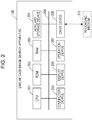
- FIG. 2 is a view illustrating the hardware configuration of the similar case image search apparatus.
- the similar case image search apparatus 120 includes a central processing unit (CPU) 201, a read only memory (ROM) 202, and a random access memory (RAM) 203.
- the CPU 201, the ROM 202, and the RAM 203 constitute a so-called computer.
- the similar case image search apparatus 120 includes an auxiliary storage device 204, a connection device 205, a display device 206, an operation device 207, and a drive device 208.
- the hardware constituents of the similar case image search apparatus 120 are interconnected via a bus 209.
- the CPU 201 executes various programs (for example, the similar case image search program) stored in the auxiliary storage device 204.
- the ROM 202 is a nonvolatile memory.
- the ROM 202 functions as a main storage unit, which stores various programs and data for the CPU 201 to execute the various programs stored in the auxiliary storage device 204.
- the ROM 202 stores boot programs including a basic input/output system (BIOS) and an extensible firmware interface (EFI).
- BIOS basic input/output system
- EFI extensible firmware interface
- the RAM 203 is a volatile memory, and includes a dynamic random access memory (DRAM) and a static random access memory (SRAM).
- the RAM 203 functions as a main storage unit, which provides a work area expanded when the CPU 201 executes the various programs stored in the auxiliary storage device 204.
- the auxiliary storage device 204 functions as a computer-readable auxiliary storage unit, which stores the various programs installed to the similar case image search apparatus 120, and data generated by executing the various programs.
- the features information DB 150 is implemented in the auxiliary storage device 204.
- the connection device 205 is connected to the CT device 110 and the image DB 130, and transmits and receives various data to and from the CT device 110 and the image DB 130.
- the display device 206 displays the display screen used when the radiologist or the like compares and interprets the CT image of the diagnosis subject.
- the operation device 207 accepts various operations of the radiologist or the like with respect to the similar case image search apparatus 120.
- the drive device 208 is a device for setting a recording medium 210.
- the recording medium 210 described herein includes media that record data optically, electrically, or magnetically, for example, CD-ROMs, flexible discs, and magneto-optical discs.
- the recording medium 210 may include semiconductor memories that electrically record data, for example, ROMs and flash memories.
- the various programs stored in the auxiliary storage device 204 are installed, for example, by setting the distributed recording medium 210 to the drive device 208 and reading various programs stored in the recording medium 210 using the drive device 208.
- the various programs stored in the auxiliary storage device 204 may be installed by downloading from a network via the connection device 205.
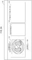
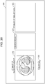
- FIGs. 3A to FIGs. 4B are first views and second views illustrating an example of the display screen of the similar case image search apparatus, respectively.
- a display screen 300 includes a diagnosis subject image display area 310, which displays a CT image of the diagnosis subject, which is captured by the CT device 110.
- the display screen 300 includes a search button 330.
- the search button 330 is a button for instructing the search unit 142 to perform a search.
- the display screen 300 includes a search result display area 340.
- the search result display area 340 displays search results acquired by causing the search unit 142 to search for the histogram similar to the histogram generated for the CT image of the diagnosis subject, among the histograms stored in the features information DB 150 for the CT images of the search subjects.
- the display screen 300 includes a similar case search result display area 350.
- the similar case search result display area 350 displays a CT image designated by the radiologist or the like in the search results displayed in the search result display area 340.
- FIG. 3A illustrates the case where the CT image of the diagnosis subject, which is captured by the CT device 110, is displayed in the diagnosis subject image display area 310 of the display screen 300.
- FIG. 3B illustrates the case where the radiologist or the like presses the search button 330 in the state in which the CT image of the diagnosis subject is displayed in the diagnosis subject image display area 310 of the display screen 300.
- the search button 330 is pressed, the display screen shifts to the display screen 300 illustrated in FIGs. 4A and 4B .
- FIG. 4A illustrates the case where the search button 330 is pressed, causing the search unit 142 performs search and search results are displayed in the search result display area 340.
- information items in the search results displayed in the search result display area 340 include “ID”, “thumbnail”, and “similarity level”.
- the “ID” stores identifiers for identifying CT images searched by the search unit 142.
- the “thumbnail” displays thumbnail images of the CT images identified based on the "ID”.
- the “similarity level” stores the similarity level between the histogram for each CT image searched by the search unit 142 and the histogram for the CT image of the diagnosis subject.
- FIG. 4B illustrates the case where a prescribed search result is selected from the search results displayed in the search result display area 340 by the radiologist or the like, and the CT image corresponding to the selected search result is displayed in the similar case search result display area 350.
- ID "ID001" is selected, and the corresponding CT image is displayed as the CT image of the similar case in the similar case search result display area 350.
- the radiologist or the like may diagnose the CT image of the diagnosis subject by comparative interpretation while referring to the CT image of the similar case similar to the CT image of the diagnosis subject.
- Display contents of display screen 300 are not limited to those illustrated in FIGs. 3A to 4B , and may display patient information of the patient to be diagnosed. Alternatively, the display screen may display various information that is associated with the CT image displayed on the similar case search result display area 350 and stored in the image DB 130.
- FIGs. 5A and 5B are first views illustrating an example of the features information stored in the features information DB.
- features information 500 includes "ID”, “slice number”, “thumbnail image”, “right lung field”, and “left lung field” as information items.
- the "ID” stores identifiers for identifying CT images stored in the image DB 130.
- the "slice number” stores numbers for identifying a plurality of slice images included in each CT image.
- the "thumbnail image” stores thumbnail images of the plurality of slice images included in the CT image, which are associated with the respective slice number.
- the "right lung field” further includes “number of lesion pixels in peripheral area” and “number of lesion pixels in central area”.
- the "number of lesion pixels in peripheral area” stores the number of lesion pixels of lesions distributed in a peripheral area of the right lung field.
- the “number of lesion pixels in central area” stores the number of lesion pixels of lesions distributed in a central area of the right lung field.
- the "left lung field” further includes “number of lesion pixels in peripheral area” and “number of lesion pixels in central area”.
- the "number of lesion pixels in peripheral area” stores the number of lesion pixels of lesions distributed in a peripheral area of the left lung field.
- the “number of lesion pixels in central area” stores the number of lesion pixels of lesions distributed in a central area of the left lung field.
- FIG. 5B illustrates histograms of the number of lesion pixels generated based on the features information 500, a horizontal axis represents the number of lesion pixels, and a vertical axis represents the slice number.
- a histogram 501 is a histogram of the number of lesion pixels in peripheral area of the right lung field
- a histogram 502 is a histogram of the number of lesion pixels in central area of the right lung field
- a histogram 503 is a histogram of the number of lesion pixels in peripheral area of the left lung field
- a histogram 504 is a histogram of the number of lesion pixels in central area of the left lung field.
- the histograms 501 to 504 are associated with the "ID" in the features information 500, and stored in the features information DB 150.
- FIG. 6 is a view illustrating the CT image information stored in the image DB.
- a CT image information 600 includes "ID”, “image”, “patient information”, “diagnosis result”, “diagnostician” as information items.
- the "ID” stores identifiers for identifying CT images stores in the image DB 130.
- the “image” includes file names of the CT images stored in the image DB 130.
- the "patient information” stores detailed information (patient ID, name, age, and sex) of the patient whose CT image is captured.
- the “diagnosis result” stores diagnosis results of the CT images, which are acquired by the radiologists or the like.
- the “diagnostician” stores IDs of the radiologists or the like who diagnose respective CT images.
- the diagnosis of the CT image stored in the image DB 130 may be made at taking of the CT image.
- the CT image information 600 may store, in addition to the diagnosis result, various information including contents of a procedure for the patient, and the state after the procedure, which is associated with the diagnosis result.
- FIG. 7 is a first view illustrating an example of the functional configuration of the registration unit of the similar case image search apparatus.
- the registration unit 141 has a slice image acquisition unit 710, a lesion detection unit 720, a contour identification unit 730, a central area model acquisition unit 740, a division curve generation unit 750, and a histogram generation unit 760.
- the CT image information 600 stores the ID for identifying the stored CT image, the file name, the patient information, the diagnosis result, and the ID for identifying the radiologist who diagnoses the CT image, which are associated with the CT image.
- each unit of the registration unit 141 in FIG. 7 are activated.
- the slice image acquisition unit 710 reads each of slice images included of the CT image newly stored in the image DB 130, and transmits the slice image to the lesion detection unit 720 and the contour identification unit 730.
- the lesion detection unit 720 executes lesion detection processing. Specifically, the lesion detection unit 720 divides each slice image into grids each having a designated size to generate partial images (hereinafter referred to as "block"). The lesion detection unit 720 statistically processes the brightness value of each pixel of each of the generated blocks, thereby calculating a multidimensional vector. Further, based on the calculated multidimensional vector, the lesion detection unit 720 determines whether or not each block corresponds to a lesion.
- each block corresponds to the lesion based on the calculated multidimensional vector
- the lesion detection unit 720 previously holds a representative vector indicating the lesion.
- the lesion detection unit 720 calculates a distance between the multidimensional vector calculated from each block and the previously-held representative vector indicating the lesion, thereby determining whether or not each block corresponds to the lesion to detect the lesion.
- the lesion detection unit 720 notifies lesion information indicating the position of the detected lesion to the histogram generation unit 760.
- the contour identification unit 730 executes contour identification processing for each slice image. Specifically, the contour identification unit 730 extracts the lung field area from each slice image, and identifies the contour of the extracted lung field area. The contour identification unit 730 notifies the identified contour of the lung field area of each slice image to the central area model acquisition unit 740 and the division curve generation unit 750.
- the central area model acquisition unit 740 executes central area model acquisition processing. Specifically, the central area model acquisition unit 740 reads a central area model previously stored in a model information storage unit 770.
- the central area model is a model defining the proportion of the central area and the peripheral area in the lung field area at each slice position.
- the central area model acquisition unit 740 acquires the central area model to identify the proportion of the central area and the peripheral area in the lung field area of each slice image, and notifies the proportion to the division curve generation unit 750.
- the division curve generation unit 750 is an example of the division unit, and executes division curve generation processing. Specifically, the division curve generation unit 750 internally divides the contour of the lung field area of each slice image, which is notified from the contour identification unit 730, based on the proportion notified from the central area model acquisition unit 740 for each slice image, thereby calculating division points. The division curve generation unit 750 connects the calculated division points to each other to generate a division curve dividing the lung field area into the central area and the peripheral area. Further, the division curve generation unit 750 notifies the division curve generated for each slice image to the histogram generation unit 760.
- the histogram generation unit 760 is an example of a counting unit, and compares the lesion information about each slice image, which is notified from the lesion detection unit 720, with the division curve in each slice image, which is notified from the division curve generation unit 750. Thereby, the histogram generation unit 760 may determine whether each lesion in each slice image is distributed in the central area or the peripheral area.
- the histogram generation unit 760 For each slice image in the right lung field and the left lung field, the histogram generation unit 760 counts the number of lesion pixels of lesions distributed in the central area and the number of lesion pixels of lesions distributed in the peripheral area. The histogram generation unit 760 stores the counted number of lesion pixels in the features information 500 of the features information DB 150.
- the histogram generation unit 760 generates a histogram based on the counted number of lesion pixels, and stores the histogram in association with "ID" in the features information 500.
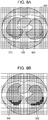
- FIGs. 8A and 8B are views illustrating the specific example of the lesion detection processing of the lesion detection unit.
- FIG. 8A illustrates an example of the slice image.
- a slice image 800 includes a lung field area 810 of a right lung of a patient, and a lung field area 820 of a left lung of the patient.
- the blocks (for example, block 830) in the slice image 800 are blocks generated by the lesion detection unit 720.
- FIG. 8B illustrates the case of determining whether or not each block generated by the lesion detection unit 720 corresponds to the lesion.
- blocks represented by thick lines for example, blocks 840 and 850
- blocks other than the blocks represented by the thick lines for example, blocks 840 and 850
- FIGs. 9A and 9B are views illustrating the specific example of the contour identification processing of the contour identification unit.
- FIG. 9A illustrates the slice image 800 acquired before the contour identification processing of the contour identification unit 730.
- FIG. 9B illustrates the case where the contour identification unit 730 extracts the lung field areas 810, 820 from the slice image 800, and identifies contours 911, 912 of the lung field areas 810, 820.
- central area model acquisition processing of the central area model acquisition unit 740 will be described.
- lung tissues three-dimensionally spread from a site called the hilum of lung, and following medical findings about the central area in each slice position are obtained.
- FIG. 10 is a view illustrating an example of the central area model, and illustrates the case where a central area model 1050 is superimposed on the lung field areas 1010, 1020 when viewing the patient's lung from the front.
- a slice position 1034 passes the hilum of lung, the area of the central area becomes maximum at the slice position 1034.
- a slice position 1031 passes the upper ends of the lung field areas 1010, 1020, and a slice position 1032 passes the upper end of a central area 1040.
- a gap ⁇ is present between the slice position 1031 and the slice position 1032.
- a slice position 1036 passes the lower ends of the lung field areas 1010, 1020, and a slice position 1035 passes the lower end of the central area 1040.
- a gap y is present between the slice position 1036 and the slice position 1035.
- the central area model 1050 is linearly approximated such that the area of the central area becomes maximum at the slice position 1034, and becomes 0 at the slice position 1032 and the slice position 1035.
- the central area model 1050 defines, at each slice position, a proportion of a length from the center to a boundary of the central area model 1050 and a length from the boundary of the central area model 1050 to each of boundaries of the lung field areas 1010, 1020.
- the central area model 1050 defines, at a slice position 1033, the proportion of the length from the center to the boundary of the central area model 1050 and the length from the boundary of the central area model 1050 to the boundary of the lung field area 1010 as ⁇ :(1- a).
- the central area model acquisition unit 740 executes the central area model acquisition processing to acquire the central area model 1050. Based on the central area model 1050, the central area model acquisition unit 740 determines the proportion of the length from the center of each slice position to the boundary of the central area model 1050 and the length from the boundary of the central area model 1050 to the boundary of each of the lung field areas 1010, 1020.
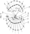
- FIG. 11 is a view illustrating the specific example of the division curve generation processing of the division curve generation unit.
- FIG. 11 illustrates the case where division curves 1111, 1121 dividing the lung field area into the central area and the peripheral area are generated in the slice image at the slice position 1033.
- the division curve generation unit 750 extracts a central position 1130 of the patient's body from the slice image.
- the division curve generation unit 750 radially extends straight lines from the central position 1130, and extracts intersections between the straight lines and the contours of the lung field areas 1110, 1120, which are notified from the contour identification unit 730 (inner (mediastinum) intersections and outer (chest wall) intersections).
- the division curve generation unit 750 extends a straight line 1140 from the central position 1130, and extracts intersections between the straight line and the contour of the lung field area 1110 (an inner (mediastinum) intersection 1150 and an outer (chest wall) intersection 1160).
- the division curve generation unit 750 internally divides line segments between the inner (mediastinum) intersections and the outer (chest wall) intersections based on the proportion notified from the central area model acquisition unit 740, to identify the division points.
- the division curve generation unit 750 internally divides the line segment between the inner (mediastinum) intersection 1150 and the outer (chest wall) intersection 1160 based on the proportion ⁇ :(1- a) to identify a division point 1170.
- the division curve generation unit 750 identifies each straight line radially extending from the central position 1130 to identify the division point, and connects the identified division points to each other, thereby generating division curves based on the shape of the lung field areas 1110, 1120. Thereby, the division curve generation unit 750 may generate the division curve 1111 for the lung field area 1110 and the division curve 1121 for the lung field area 1120. That is, the lung field areas 1110, 1120 each may be divided into the central area and the peripheral area based on medical findings.
- the search unit 142 will be described in detail.
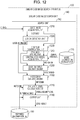
- FIG. 12 is a view illustrating an example of the functional configuration of the search unit of the similar case image search apparatus.
- the search unit 142 has a slice image acquisition unit 1210, a lesion detection unit 1220, a contour identification unit 1230, a central area model acquisition unit 1240, a central area model acquisition unit 1250, a histogram generation unit 1260, a similarity level calculation unit 1270, and an output unit 1280.
- the display control unit 143 displays the captured CT image as the CT image of the diagnosis subject on the display screen 300.
- the display control unit 143 displays the display screen 300 and the radiologist or the like inputs a search instruction, activating each of the units of the search unit 142 in FIG. 12 .
- the units except for the similarity level calculation unit 1270 and the output unit 1280 execute processing for the CT image of the diagnosis subject on the display screen 300, as well as the same processing as the processing of the registration unit 141 in FIG. 7 . Thus, description of the same processing is omitted.
- the similarity level calculation unit 1270 is an example of the image identification unit.
- the similarity level calculation unit 1270 calculates the similarity level between histograms for the CT image of the diagnosis subject and histograms for the CT image of the search subject, which is read from the features information DB 150.
- the similarity level calculation unit 1270 calculates the similarity level about each of the histograms of the central area and the peripheral area in the right lung field, and the similarity level about each of the histograms of the central area and the peripheral area in the left lung field.
- the similarity level calculation unit 1270 sums the calculated similarity level to calculate a total value (overall similarity level).
- the similarity level calculation unit 1270 notifies the overall similarity level calculated between the CT image of the diagnosis subject and the CT image of each search subject to the output unit 1280.
- the output unit 1280 sorts the overall similarity level notified from the similarity level calculation unit 1270 and rearranges the overall similarity level in decreasing order.
- the output unit 1280 reads the IDs and the thumbnail images that correspond to a predetermined number of search subjects having high overall similarity level from the features information DB 150, and notifies the IDs and the thumbnail images along with the overall similarity level as the search results to the display control unit 143.
- FIG. 13 is a view illustrating a specific example of the similarity level calculation processing of the similarity level calculation unit.
- histograms 1311 to 1314 are histograms of the CT image of the diagnosis subject.
- the histogram 1311 is the histogram for the peripheral area in the right lung field
- the histogram 1312 is the histogram for the central area in the right lung field
- the histogram 1313 is the histogram for the peripheral area in the left lung field
- the histogram 1314 is the histogram for the central area in the left lung field.
- Histograms 1321 to 1324 are histograms of the CT image of the diagnosis subject.
- the histogram 1321 is the histogram for the peripheral area in the right lung field
- the histogram 1322 is the histogram for the central area in the right lung field
- the histogram 1323 is the histogram for the peripheral area in the left lung field
- the histogram 1324 is the histogram for the central area in the left lung field.
- the similarity level calculation unit 1270 calculates the similarity level of mutual histograms, and sums the similarity level to calculate the overall similarity level.
- the similarity level for the histograms for the peripheral area in the right lung field is S 1
- the similarity level for the histograms of the central area in the right lung field is S 2
- the similarity level for the histograms of the peripheral area in the left lung field is S 3
- FIG. 14 is a sequence diagram of the similar case image search processing in the CT image processing system.
- the similar case image search processing in the CT image processing system 100 may be roughly divided into a registration phase and a search phase.
- registration phase will be described.
- Step S1401 the CT device 110 a patient's CT image.
- patient information is inputted to the similar case image search apparatus 120.
- Step S1402 the CT device 110 transmits the captured CT image as the CT image of the search subject to the registration unit 141 of the similar case image search apparatus 120.
- the registration unit 141 receives the CT image of the search subject from the CT device 110, adds an identifier to the CT image, and stores the CT image along the patient information in the image DB 130.
- the registration unit 141 stores the CT image in the image DB 130, the radiologist or the like diagnoses the CT image.
- the registration unit 141 associates a diagnosis result with the CT image, and stores them in the image DB 130.
- Step S1404 the registration unit 141 applies the lesion detection processing to each slice image of the stored CT image of the search subject, and detects lesions.
- Step S1405 the registration unit 141 applies the contour identification processing, the central area model acquisition processing, and the division curve generation processing to each slice image of the stored CT image of the search subject, thereby dividing the lung field area into the central area and the peripheral area.
- Step S1406 the registration unit 141 counts the number of lesion pixels of lesions distributed in the central area and the number of lesion pixels of lesions distributed in the peripheral area to generate the histograms 1321 to 1324.
- Step S1407 the registration unit 141 stores the counted number of lesion pixels and the generated histograms in the features information 500 of the features information DB 150.
- FIG. 14 illustrates the example of the processing for one patient in the registration phase. In fact, however, a plurality of patients are subjected to the processing in the registration phase.
- Step S1411 the CT device 110 captures a CT image of a patient to be diagnosed.
- patient information on the patient to be diagnosed is inputted to the similar case image search apparatus 120.
- Step S1412 the CT device 110 transmits the captured CT image as the CT image of the diagnosis subject to the search unit 142 of the similar case image search apparatus 120.
- Step S1413 the search unit 142 receives the CT image of the diagnosis subject from the CT device 110.
- the search unit 142 notifies the acquired CT image of the diagnosis subject to the display control unit 143.
- Step S1414 the display control unit 143 displays the display screen 300 on the display device 206, and displays any slice image inclined in the CT image notified from the search unit 142 on the diagnosis subject image display area 310 of the display screen 300.
- Step S1415 the display control unit 143 accepts a search instruction inputted via the search button 330 of the display screen 300.
- Step S1416 the display control unit 143 notifies the accepted search instruction to the search unit 142.
- Step S1417 when receiving the search instruction from the display control unit 143, the search unit 142 executes the lesion detection processing for each slice image of the CT image of the diagnosis subject, and detects lesions.
- Step S1418 the search unit 142 executes the contour identification processing, the central area model acquisition processing, and the division curve generation processing for each slice image of the CT image of the diagnosis subject, thereby dividing the lung field area into the central area and the peripheral area.
- Step S1419 the search unit 142 counts the number of lesion pixels of lesions distributed in the central area and the number of lesion pixels of lesions distributed in the peripheral area, and generates the histograms 1311 to 1314.
- Step S1420 the search unit 142 reads the features information 500 from the features information DB 150, and acquires histograms (for example, histogram 1321 to 1324) of the CT image of each search subject.
- Step S1421 the search unit 142 calculates the similarity level for each area between the acquired histograms 1321 to 1324 for the CT image of the search subject and the generated histograms 1311 to 1314 for the CT image of the diagnosis subject, respectively.
- the search unit 142 sums the similarity level calculated for each area to calculate the overall similarity level, and sorts the calculated overall similarity level in descending order.
- Step S1422 the search unit 142 notifies the IDs and the thumbnail images which are associated with the CT images of a predetermined number of search subjects having high overall similarity level, along with the overall similarity level, as the search results to the display control unit 143.
- Step S1423 the display control unit 143 displays the search result notified from the search unit 142 on the search result display area 340.
- Step S1424 when the radiologist or the like selects a prescribed search result in the search result display area 340, the display control unit 143 accepts it as an instruction to display the CT image.
- Step S1425 the display control unit 143 identifies the ID included in the selected search result, and reads the CT image determined based on the identified ID from the image DB 130.
- Step S1426 the display control unit 143 displays any slice image of the read CT image on the similar case search result display area 350.
- the radiologist or the like may diagnose the CT image of the diagnosis subject by comparative interpretation while referring to the slice image of the CT image of the similar case similar to the case of the CT image of the diagnosis subject.
- the lesions are distributed over a wide range of the lung field area.
- the lung field area may be properly divided into the central area and the peripheral area. For this reason, when it is attempted to determine the distributed area of lesions and search for a similar case, it is difficult to accurately search for the similar case.
- the similar case image search apparatus 120 identifies the position at which the chest wall and the mediastinum are internally divided in each slice image, based on medical findings, to divide the lung field area into the central area and the peripheral area. For this reason, the similar case image search apparatus 120 may divide the lung field area into areas suitable for diagnosis.
- the similar case image search apparatus 120 For the lesions detected in each slice image, the similar case image search apparatus 120 counts the number of lesion pixels of lesions distributed in the central area and the number of lesion pixels of lesions distributed in the peripheral area, and generates histograms. Further, the similar case image search apparatus 120 calculates the similarity level using the generated histograms to identify the similar case image. Therefore, the similar case image search apparatus 120 may determine the distributed area of lesions to search for the similar case.
- the similar case image search apparatus 120 may accurately search for the similar case based on the distribution of lesions in diseases, lesions of which are distributed in organs, such as the diffuse lung disease.
- the division curve is generated to divide the lung field area into the central area and the peripheral area.
- the method of dividing the lung field area into the central area and the peripheral area is not limited to this.
- the lung field area is divided into the central area and the peripheral area by a method that is different from the method in the first embodiment. A difference between this embodiment and the first embodiment will be mainly described below.
- FIG. 15 is a second view illustrating an example of the functional configuration of the registration unit of the similar case image search apparatus.
- the difference between the functional configuration and the functional configuration illustrated in FIG. 7 is that the registration unit 141 has a lung field integrated image generation unit 1510 and an integrated image reduction unit 1520.
- the lung field integrated image generation unit 1510 executes integrated image generation processing. Specifically, the lung field integrated image generation unit 1510 calculates a reference line segment including the chest wall and the mediastinum as endpoints, based on the contours of the lung field areas of the left and right lungs in each slice image, which are notified from the contour identification unit 730. The lung field integrated image generation unit 1510 generates an image formed by integrating the both lung field areas (integrated image), based on the contours of the lung field areas of the left and right lungs in each slice image, which are notified from the contour identification unit 730. Further, the lung field integrated image generation unit 1510 notifies the calculated reference line segment and the generated integrated image to the integrated image reduction unit 1520.
- the integrated image reduction unit 1520 is an example of a division unit, and executes the integrated image reduction processing. Specifically, the integrated image reduction unit 1520 reduces the integrated image notified from the lung field integrated image generation unit 1510, and internally divides the reference line segment in a prescribed proportion to divide the lung field area into the central area and the peripheral area. The integrated image reduction unit 1520 determines the proportion in which the reference line segment is internally divided based on the central area model acquired from the model information storage unit 770, and identifies the division point based on the determined proportion.
- the integrated image reduction unit 1520 notifies the divided central area and peripheral area to the histogram generation unit 760.
- FIGs. 16A and 16B are views illustrating the specific example of the integrated image generation processing of the lung field integrated image generation unit.
- FIGs. 16A and 16B illustrate the case where a reference line segment for the slice image at the slice position 1033 is calculated to generate an integrated image.
- the lung field integrated image generation unit 1510 calculates centroid positions 1611, 1621 of lung field areas 1610, 1620 based on the contours notified from the contour identification unit 730. Subsequently, the lung field integrated image generation unit 1510 calculates a horizontal line 1612 passing the calculated centroid position 1611 and a horizontal line 1622 passing the calculated centroid position 1621.
- the lung field integrated image generation unit 1510 extracts an intersection 1613 between the horizontal line 1612 and the outer contour (chest wall) of the lung field area 1610 and an intersection 1623 between the horizontal line 1622 and the outer contour (chest wall) of the lung field area 1620.
- the lung field integrated image generation unit 1510 extracts a point 1614 on the horizontal line 1612, which is located on the opposite side to the intersection 1613 across the centroid position 1611 away from the centroid position 1611 with the same distance as the distance between the intersection 1613 and the centroid position 1611. Similarly, the lung field integrated image generation unit 1510 extracts a point 1624 on the horizontal line 1622, which is located on the opposite side to the intersection 1623 across the centroid position 1621 away from the centroid position 1621 with the same distance as the distance between the intersection 1623 and the centroid position 1621.
- the processing of the lung field integrated image generation unit 1510 is processing of extracting the mediastinum by using the fact that the distance between the chest wall and the centroid position of the lung field area is anatomically equal to the distance between the centroid position of the lung field area and the mediastinum.
- the lung field integrated image generation unit 1510 may calculate reference line segments 1615, 1625 having the chest wall (the intersection 1613, 1623) and the mediastinum (the points 1614, 1624) as endpoints.
- the lung field integrated image generation unit 1510 generates an integrated image 1630 formed by integrating the lung field areas 1610, 1620 based on the shape of the contours (chest wall) of the lung field area 1610, 1620.
- the lung field integrated image generation unit 1510 notifies the generated integrated image 1630 and the reference line segments 1615, 1625 to the integrated image reduction unit 1520.
- FIGs. 17A and 17B are views illustrating the specific example of the integrated image reduction processing of the integrated image reduction unit.
- the integrated image reduction unit 1520 reduces the size of the integrated image 1630 notified from the lung field integrated image generation unit 1510, and generates a reduced integrated image 1701.
- the integrated image reduction unit 1520 generates the reduced integrated image 1701 such that an intersection 1711 between the reduced integrated image 1701 and the reference line segment 1615 (that is, between the chest wall and the mediastinum) becomes a division point at which the reference line segment 1615 is internally divided in a proportion of ⁇ :(1- a).
- the integrated image reduction unit 1520 may calculate a boundary 1721 of the central area in the lung field area of the right lung.
- the integrated image reduction unit 1520 notifies the inner side of the boundary 1721 of the central area as the central area, and the outer side of the boundary 1721 of the central area as the peripheral area to the histogram generation unit 760.
- the integrated image reduction unit 1520 reduces the size of the integrated image 1630 notified from the lung field integrated image generation unit 1510, and generates a reduced integrated image 1702.
- the integrated image reduction unit 1520 generates the reduced integrated image 1702 such that an intersection 1712 between the reduced integrated image 1702 and the reference line segment 1625 (that is, between the chest wall and the mediastinum) becomes a division point at which the reference line segment 1625 is internally divided in a proportion of ⁇ :(1- a).
- the integrated image reduction unit 1520 may calculate a boundary 1722 of the central area in the lung field area of the left lung.
- the integrated image reduction unit 1520 notifies the inner side of the boundary 1722 of the central area as the central area, and the outer side of the boundary 1722 of the central area as the peripheral area to the histogram generation unit 760.
- the lung field area of each slice image in each lung field area of each slice image, one point on each of the chest wall and the mediastinum is extracted to calculate the reference line segment, and the size of the integrated image is reduced so as to internally divide the reference line segment in a proportion based on medical findings.
- the boundary of the central area is calculated based on the reduced integrated image to divide the lung field area into the central area and the peripheral area. For this reason, in the second embodiment, the lung field area may be divided into areas suitable for diagnosis.
- the contour of the boundary of the lung field area on the side of the chest wall, and the centroid position of the lung field area are used without using the contour of the boundary of the lung field area on the side of the mediastinum, which is difficult to accurately extract the mediastinum.
- the accuracy of calculation of the boundary of the central area may be improved, properly dividing the lung field area into the central area and the peripheral area.
- the lung field area is divided into the central area and the peripheral area.
- the number of divided areas of the lung field area is not limited to two, and the central area and the peripheral area may be subdivided. This may count the number of lesion pixels of detected lesions in each of the subdivided areas.
- FIG. 18 is a second view illustrating an example of the features information stored in the features information DB.
- a difference between FIG. 18 and FIG. 5A is that, in the features information 1800, each of the right lung field and the left lung field are divided into three or more areas, and the number of lesion pixels of lesions counted in each of the areas is stored.
- the lung field area is divided into a plurality of areas, and histograms are generated based on the number of lesion pixels counted in each of the areas, thereby improving the accuracy of search for the similar case.
- the search unit second embodiment fails to mention the functional configuration of the search unit.
- the search unit second embodiment is implemented by adding the similarity level calculation unit 1270 and the output unit 1280 to the registration unit ( FIG. 15 ).
- the histograms are generated to calculate similarity level.
- the method of calculating the similarity level is not limited to this, and may be any method of calculating the similarity level by using the number of lesion pixels in each area.
- the features information 500 and the CT image information 600 are stored in different DBs.
- the features information 500 and the CT image information 600 may be stored in the same DB.
- the histograms for the CT image of the diagnosis subject are generated after acceptance of the search instruction.
- the histograms for the CT image of the diagnosis subject may be generated before acceptance of the search instruction.
- the CT image is searched as the medical image.
- medical images other than the CT image for example, magnetic resonance imaging (MRI) image
- MRI magnetic resonance imaging
- present disclosure is not limited to the configuration described herein, and may be any combination of the configuration in the embodiments described above and other constituents. In this connection, such embodiments may be modified within the scope of the subject matter of the present disclosure, and may be properly adapted to practical applications.
Landscapes
- Engineering & Computer Science (AREA)
- Health & Medical Sciences (AREA)
- Medical Informatics (AREA)
- Physics & Mathematics (AREA)
- Life Sciences & Earth Sciences (AREA)
- Theoretical Computer Science (AREA)
- Computer Vision & Pattern Recognition (AREA)
- General Physics & Mathematics (AREA)
- General Health & Medical Sciences (AREA)
- Public Health (AREA)
- Nuclear Medicine, Radiotherapy & Molecular Imaging (AREA)
- Radiology & Medical Imaging (AREA)
- Pathology (AREA)
- Biomedical Technology (AREA)
- Data Mining & Analysis (AREA)
- Veterinary Medicine (AREA)
- Surgery (AREA)
- Animal Behavior & Ethology (AREA)
- Molecular Biology (AREA)
- Heart & Thoracic Surgery (AREA)
- Optics & Photonics (AREA)
- High Energy & Nuclear Physics (AREA)
- Biophysics (AREA)
- Databases & Information Systems (AREA)
- Epidemiology (AREA)
- Primary Health Care (AREA)
- Quality & Reliability (AREA)
- General Engineering & Computer Science (AREA)
- Oral & Maxillofacial Surgery (AREA)
- Physiology (AREA)
- Dentistry (AREA)
- Multimedia (AREA)
- Library & Information Science (AREA)
- Bioinformatics & Cheminformatics (AREA)
- Bioinformatics & Computational Biology (AREA)
- Evolutionary Biology (AREA)
- Evolutionary Computation (AREA)
- Artificial Intelligence (AREA)
- Apparatus For Radiation Diagnosis (AREA)
- Information Retrieval, Db Structures And Fs Structures Therefor (AREA)
Claims (9)
- Vergleichsfallbildsuchprogramm, das einen Computer veranlasst, einen Prozess auszuführen, umfassend:Extrahieren eines Lungenfeldbereichs aus einem medizinischen Bild und Identifizieren (S1405) einer Kontur des Lungenfeldbereichs, der eine Brustwand und ein Mittelfell beinhaltet;Identifizieren einer Position, an der die Brustwand und das Mittelfell internal getrennt sind und den Lungenfeldbereich in einen Mittelbereich und einen Randbereich trennen, basierend auf einer Form des Lungenfeldbereichs;Zählen (S1406) der Anzahl von Pixeln, die Läsionen in sowohl dem getrennten Mittelbereich als auch dem Randbereich anzeigen; undIdentifizieren eines Vergleichsfallbilds, das einem Ähnlichkeitsgrad der gezählten Anzahl von Pixeln entspricht, die Läsionen anzeigen, indem Bezug auf eine Speichereinheit genommen wird, die die Anzahl von Pixeln speichert, die Läsionen in jedem der Bereiche anzeigen.
- Vergleichsfallbildsuchprogramm nach Anspruch 1, wobei
für jedes einer Vielzahl von Teilbildern, die in dem medizinischen Bild beinhaltet sind, ein Histogramm basierend auf der gezählten Anzahl von Pixeln, die Läsionen bei jeder Teilposition anzeigen, erzeugt wird, und
Identifizieren eines Vergleichsfallbilds entsprechend einem Ähnlichkeitsgrad des erzeugten Histogramms in jedem der Bereiche, indem Bezug auf die Speichereinheit genommen wird, die das Histogramm in jedem der Bereiche speichert. - Vergleichsfallbildsuchprogramm nach Anspruch 2, wobei
für jedes der Vielzahl von Teilbildern, die in dem medizinischen Bild beinhaltet sind, eine Position, an der die Brustwand und das Mittelfell internal getrennt sind, in jedem der Vielzahl von Teilbildern, die in dem medizinischen Bild beinhaltet sind, identifiziert wird, indem Bezug auf ein Modell genommen wird, das eine Proportion definiert, in der die Brustwand und das Mittelfell internal getrennt sind. - Vergleichsfallbildsuchprogramm nach Anspruch 3, wobei
für jedes der Vielzahl von Teilbildern, die in dem medizinischen Bild beinhaltet sind, eine Mittelposition eines Körpers extrahiert wird, und Schnittpunkte zwischen einer Vielzahl von geraden Linien, die sich radial von der Mittelposition erstrecken, und einer Kontur des identifizierten Lungenfeldbereichs als die Brustwand oder das Mittelfell extrahiert werden. - Vergleichsfallbildsuchprogramm nach Anspruch 4, wobei
für jedes der Vielzahl von Teilbildern, die in dem medizinischen Bild beinhaltet sind, eine Trennkurve, die die Vielzahl von Positionen verbindet, an denen die Brustwand und das Mittelfell internal getrennt sind, erzeugt wird und der Lungenfeldbereich in einen Mittelbereich und einen Randbereich getrennt werden. - Vergleichsfallbildsuchprogramm nach Anspruch 3, wobei
für jedes der Vielzahl von Teilbildern, die in dem medizinischen Bild beinhaltet sind, ein Schnittpunkt zwischen einer horizontalen Linie, die durch eine Schwerpunktposition des Lungenfeldbereichs geht, und der Kontur des identifizierten Lungenfeldbereichs als die Brustwand extrahiert wird und ein Punkt auf der horizontalen Linie, der an einer gegenüberliegenden Seite zu der Brustwand über die Schwerpunktposition liegt, mit demselben Abstand von der Schwerpunktposition wie einem Abstand zwischen der Brustwand und der Schwerpunktposition, als das Mittelfell extrahiert wird. - Vergleichsfallbildsuchprogramm nach Anspruch 6, wobei
für jedes der Vielzahl von Teilbildern, die in dem medizinischen Bild beinhaltet sind, ein integriertes Bild, das gebildet wird, indem die zwei Lungenfelbereiche integriert werden, erzeugt wird, das erzeugte integrierte Bild verringert wird, um durch eine Position zu gehen, bei der die Brustwand und das Mittelfell internal getrennt sind, und basierend auf dem verringerten integrierten Bild der Lungenfeldbereich in einen Mittelbereich und einen Randbereich getrennt wird. - Vergleichsfallbildsucheinrichtung, umfassend:eine Konturidentifikationseinheit (730; 1230), die konfiguriert ist, einen Lungenfeldbereich aus einem medizinischen Bild zu extrahieren und eine Kontur des Lungenfeldbereichs zu identifizieren, der eine Brustwand und ein Mittelfell beinhaltet;eine Trenneinheit (750), die konfiguriert ist, eine Position zu identifizieren, an der die Brustwand und das Mittelfell internal getrennt sind, und den Lungenfeldbereich in einen Mittelbereich und einen Randbereich trennen, basierend auf einer Form des Lungenfeldbereichs;eine Zähleinheit, die konfiguriert ist, die Anzahl von Pixeln zu zählen, die Läsionen in sowohl dem getrennten Mittelbereich als auch dem Randbereich anzeigen; undeine Bildidentifikationseinheit (1270), die konfiguriert ist, sich auf eine Speichereinheit zu beziehen, die die Anzahl von Pixeln speichert, die Läsionen in jedem der Bereiche anzeigen, und ein Vergleichsfallbild entsprechend einem Ähnlichkeitsgrad der gezählten Anzahl von Pixeln, die Läsionen anzeigen, zu identifizieren.
- Vergleichsfallbildsuchverfahren, das von einem Computer durchgeführt wird, wobei das Verfahren umfasst:Extrahieren eines Lungenfeldbereichs aus einem medizinischen Bild und Identifizieren einer Kontur des Lungenfeldbereichs, der eine Brustwand und ein Mittelfell beinhaltet;Identifizieren (S1405) einer Position, an der die Brustwand und das Mittelfell internal getrennt sind und den Lungenfeldbereich in einen Mittelbereich und einen Randbereich trennen, basierend auf einer Form des Lungenfeldbereichs;Zählen (S1406) der Anzahl von Pixeln, die Läsionen in sowohl dem getrennten Mittelbereich als auch dem Randbereich anzeigen; undIdentifizieren eines Vergleichsfallbilds entsprechend einem Ähnlichkeitsgrad der gezählten Anzahl von Pixeln, die Läsionen anzeigen, indem Bezug auf eine Speichereinheit genommen wird, die die Anzahl von Pixeln speichert, die Läsionen in jedem der Bereiche anzeigen.
Applications Claiming Priority (2)
| Application Number | Priority Date | Filing Date | Title |
|---|---|---|---|
| JP2017046736A JP6855850B2 (ja) | 2017-03-10 | 2017-03-10 | 類似症例画像検索プログラム、類似症例画像検索装置及び類似症例画像検索方法 |
| PCT/JP2018/002089 WO2018163633A1 (ja) | 2017-03-10 | 2018-01-24 | 類似症例画像検索プログラム、類似症例画像検索装置及び類似症例画像検索方法 |
Publications (3)
| Publication Number | Publication Date |
|---|---|
| EP3594830A1 EP3594830A1 (de) | 2020-01-15 |
| EP3594830A4 EP3594830A4 (de) | 2020-03-18 |
| EP3594830B1 true EP3594830B1 (de) | 2021-05-26 |
Family
ID=63447603
Family Applications (1)
| Application Number | Title | Priority Date | Filing Date |
|---|---|---|---|
| EP18763395.3A Active EP3594830B1 (de) | 2017-03-10 | 2018-01-24 | Programm zum suchen von bildern eines ähnlichen falls, vorrichtung zum suchen von bildern eines ähnlichen falls und verfahren zum suchen von bildern eines ähnlichen falls |
Country Status (6)
| Country | Link |
|---|---|
| US (1) | US10964020B2 (de) |
| EP (1) | EP3594830B1 (de) |
| JP (1) | JP6855850B2 (de) |
| CN (1) | CN109716445B (de) |
| ES (1) | ES2875919T3 (de) |
| WO (1) | WO2018163633A1 (de) |
Families Citing this family (16)
| Publication number | Priority date | Publication date | Assignee | Title |
|---|---|---|---|---|
| JP7034306B2 (ja) * | 2018-08-31 | 2022-03-11 | 富士フイルム株式会社 | 領域分割装置、方法およびプログラム、類似度決定装置、方法およびプログラム、並びに特徴量導出装置、方法およびプログラム |
| JPWO2020044736A1 (ja) * | 2018-08-31 | 2021-08-10 | 富士フイルム株式会社 | 類似度決定装置、方法およびプログラム |
| JP7236239B2 (ja) * | 2018-10-05 | 2023-03-09 | キヤノンメディカルシステムズ株式会社 | X線ctシステム及びctスキャン実行プログラム |
| JP7082287B2 (ja) * | 2018-10-11 | 2022-06-08 | 富士通株式会社 | 画像検索プログラム、画像検索装置、及び画像検索方法 |
| JP7151464B2 (ja) * | 2018-12-21 | 2022-10-12 | 富士通株式会社 | 肺画像処理プログラム、肺画像処理方法および肺画像処理システム |
| JP7348469B2 (ja) * | 2019-04-11 | 2023-09-21 | 富士通株式会社 | 異常陰影特定プログラム、異常陰影特定装置及び異常陰影特定方法 |
| CN110706794B (zh) * | 2019-09-26 | 2022-10-25 | 中国科学院深圳先进技术研究院 | 一种医学图像处理系统和医学图像处理方法 |
| KR102516154B1 (ko) * | 2019-12-31 | 2023-03-30 | 주식회사 코어라인소프트 | 정량화 파라미터를 이용하는 의료 영상 분석 시스템 및 유사 증례 검색 시스템, 및 그 시스템에서 실행되는 방법 |
| JP2021149145A (ja) | 2020-03-16 | 2021-09-27 | 富士通株式会社 | 画像処理プログラム、画像処理装置、及び画像処理方法 |
| CN111445463A (zh) * | 2020-03-31 | 2020-07-24 | 上海联影智能医疗科技有限公司 | 相似肺部疾病病例的检索方法、装置和计算机设备 |
| CN111507956B (zh) * | 2020-04-15 | 2023-04-07 | 广西科技大学 | 一种纳米线数量统计方法及系统 |
| WO2021251885A1 (en) * | 2020-06-10 | 2021-12-16 | Corsmed Ab | A method directed to magnetic resonance imaging simulation |
| JP2023071437A (ja) * | 2021-11-11 | 2023-05-23 | キヤノン株式会社 | 情報処理装置、情報処理方法およびプログラム |
| CN114443708A (zh) * | 2021-12-28 | 2022-05-06 | 杭州迪英加科技有限公司 | 一种病理切片判读结果展示方法、系统及可读存储介质 |
| KR20250020174A (ko) * | 2023-08-03 | 2025-02-11 | 메디컬아이피 주식회사 | 폐 영역 분할 방법 및 그 장치 |
| CN117576321B (zh) * | 2023-12-05 | 2024-07-19 | 上海长征医院 | 一种辅助肝脏手术规划建模系统 |
Family Cites Families (26)
| Publication number | Priority date | Publication date | Assignee | Title |
|---|---|---|---|---|
| US5881124A (en) * | 1994-03-31 | 1999-03-09 | Arch Development Corporation | Automated method and system for the detection of lesions in medical computed tomographic scans |
| JPH08320923A (ja) * | 1995-05-26 | 1996-12-03 | Toshiba Medical Eng Co Ltd | 異常陰影自動検出システム |
| JP4035216B2 (ja) * | 1997-12-25 | 2008-01-16 | キヤノン株式会社 | 画像処理方法及び画像処理装置 |
| US7206462B1 (en) * | 2000-03-17 | 2007-04-17 | The General Hospital Corporation | Method and system for the detection, comparison and volumetric quantification of pulmonary nodules on medical computed tomography scans |
| AU2001251539A1 (en) * | 2000-04-11 | 2001-10-23 | Cornell Research Foundation Inc. | System and method for three-dimensional image rendering and analysis |
| US6577752B2 (en) * | 2001-06-15 | 2003-06-10 | Arch Development Corporation | Automated method and system for the delineation of the chest wall in computed tomography scans for the assessment of pleural disease |
| AU2003216295A1 (en) * | 2002-02-15 | 2003-09-09 | The Regents Of The University Of Michigan | Lung nodule detection and classification |
| JP2006006359A (ja) | 2004-06-22 | 2006-01-12 | Fuji Photo Film Co Ltd | 画像生成装置、画像生成装置方法、およびそのプログラム |
| JP4832300B2 (ja) | 2004-07-30 | 2011-12-07 | 株式会社日立メディコ | 医用画像診断支援装置の作動方法、医用画像診断支援装置、及び画像処理プログラム |
| JP2006181025A (ja) | 2004-12-27 | 2006-07-13 | Fuji Photo Film Co Ltd | 異常陰影検出方法および装置並びにプログラム |
| JP2008200368A (ja) | 2007-02-21 | 2008-09-04 | Osaka Univ | 間質性肺炎の定量的評価を自動的に実行する装置およびプログラム |
| JP5224768B2 (ja) | 2007-10-12 | 2013-07-03 | インフォコム株式会社 | 胸部正面x線画像の検像方法及び検像装置 |
| CN102053963B (zh) * | 2009-10-28 | 2012-08-08 | 沈阳工业大学 | 一种胸部x线图像检索方法 |
| JP2011118543A (ja) | 2009-12-01 | 2011-06-16 | Shizuoka Prefecture | 症例画像検索装置、方法およびプログラム |
| CN102429679A (zh) * | 2011-09-09 | 2012-05-02 | 华南理工大学 | 基于胸部ct图像的肺气肿计算机辅助诊断系统 |
| WO2013076927A1 (ja) | 2011-11-24 | 2013-05-30 | パナソニック株式会社 | 診断支援装置および診断支援方法 |
| JP2014085851A (ja) * | 2012-10-24 | 2014-05-12 | Nippon Telegr & Teleph Corp <Ntt> | 画像類似度算出装置およびその動作方法 |
| JP2015136480A (ja) | 2014-01-22 | 2015-07-30 | 富士フイルム株式会社 | 3次元医用画像表示制御装置およびその作動方法並びに3次元医用画像表示制御プログラム |
| CN104036109A (zh) * | 2014-03-14 | 2014-09-10 | 上海大图医疗科技有限公司 | 基于图像的病例检索、勾画及治疗计划系统和方法 |
| JP6109778B2 (ja) * | 2014-03-27 | 2017-04-05 | 富士フイルム株式会社 | 類似症例検索装置、類似症例検索方法、及び類似症例検索プログラム |
| JP6099593B2 (ja) * | 2014-03-27 | 2017-03-22 | 富士フイルム株式会社 | 類似症例検索装置、類似症例検索方法、及び類似症例検索プログラム |
| JP2015203920A (ja) * | 2014-04-11 | 2015-11-16 | キヤノン株式会社 | 類似症例検索システム、類似症例検索方法及びプログラム |
| US10599810B2 (en) * | 2014-06-04 | 2020-03-24 | Panasonic Corporation | Control method and recording system |
| US10748661B2 (en) * | 2014-10-30 | 2020-08-18 | Panasonic Corporation | Method for controlling information terminal, and recording medium |
| CN105957094B (zh) * | 2016-06-15 | 2018-09-25 | 深圳维影医疗科技有限公司 | 一种基于轮廓检测的胸片肺野快速自动分割方法及其系统 |
| WO2018107371A1 (zh) * | 2016-12-13 | 2018-06-21 | 上海联影医疗科技有限公司 | 图像搜索系统及方法 |
-
2017
- 2017-03-10 JP JP2017046736A patent/JP6855850B2/ja active Active
-
2018
- 2018-01-24 ES ES18763395T patent/ES2875919T3/es active Active
- 2018-01-24 WO PCT/JP2018/002089 patent/WO2018163633A1/ja not_active Ceased
- 2018-01-24 EP EP18763395.3A patent/EP3594830B1/de active Active
- 2018-01-24 CN CN201880003543.5A patent/CN109716445B/zh active Active
-
2019
- 2019-03-04 US US16/291,016 patent/US10964020B2/en active Active
Non-Patent Citations (1)
| Title |
|---|
| None * |
Also Published As
| Publication number | Publication date |
|---|---|
| CN109716445A (zh) | 2019-05-03 |
| WO2018163633A1 (ja) | 2018-09-13 |
| JP2018151791A (ja) | 2018-09-27 |
| ES2875919T3 (es) | 2021-11-11 |
| CN109716445B (zh) | 2023-03-24 |
| EP3594830A4 (de) | 2020-03-18 |
| EP3594830A1 (de) | 2020-01-15 |
| US20190197688A1 (en) | 2019-06-27 |
| US10964020B2 (en) | 2021-03-30 |
| JP6855850B2 (ja) | 2021-04-07 |
Similar Documents
| Publication | Publication Date | Title |
|---|---|---|
| US10964020B2 (en) | Similar case image search program, similar case image search apparatus, and similar case image search method | |
| CN113711271B (zh) | 用于通过正电子发射断层摄影进行肿瘤分割的深度卷积神经网络 | |
| US8483467B2 (en) | Medical image diagnosis assisting apparatus and method, and computer readable recording medium on which is recorded program for the same | |
| US8417009B2 (en) | Apparatus, method, and computer readable medium for assisting medical image diagnosis using 3-D images representing internal structure | |
| EP2888686B1 (de) | Automatische detektion und abfrage von vorhergehenden relevanten anmerkungen für eine bildgebungsstudie für effiziente betrachtung und protokollierung | |
| JP4640845B2 (ja) | 画像処理装置およびそのプログラム | |
| WO2011093921A1 (en) | Automated vascular region separation in medical imaging | |
| US20160125162A1 (en) | Method for controlling information terminal, and recording medium | |
| JP2008259622A (ja) | レポート作成支援装置およびそのプログラム | |
| US20170221204A1 (en) | Overlay Of Findings On Image Data | |
| Bi et al. | Multi-stage thresholded region classification for whole-body PET-CT lymphoma studies | |
| EP3373194B1 (de) | Bildabrufgerät und bilderabrufverfahren | |
| US11200443B2 (en) | Image processing apparatus, image processing method, and image processing system | |
| US8306354B2 (en) | Image processing apparatus, method, and program | |
| US8331635B2 (en) | Cartesian human morpho-informatic system | |
| US9483705B2 (en) | Image processing device, image processing method, and image processing program | |
| US20150379365A1 (en) | System and method for retrieval of similar findings from a hybrid image dataset | |
| US20060078184A1 (en) | Intelligent splitting of volume data | |
| CN117952991A (zh) | Spcet骨显像图像的肾脏区域分割方法、系统及设备 | |
| US20180308244A1 (en) | Registration apparatus, registration method, and registration program | |
| US12608801B2 (en) | Information processing apparatus, information processing method, and information processing program for supporting interpretation of medical image | |
| Huang et al. | A Channel Fusion Convolutional Neural Network for Lung Nodule Detection | |
| WO2006123272A2 (en) | Automatic organ mapping method an device | |
| Shao et al. | Thyroid Classification in Ultraound by Deep Multimodal Learning | |
| CN115880306A (zh) | 一种基于三维体数据的肝脏分割和重建方法 |
Legal Events
| Date | Code | Title | Description |
|---|---|---|---|
| STAA | Information on the status of an ep patent application or granted ep patent |
Free format text: STATUS: THE INTERNATIONAL PUBLICATION HAS BEEN MADE |
|
| PUAI | Public reference made under article 153(3) epc to a published international application that has entered the european phase |
Free format text: ORIGINAL CODE: 0009012 |
|
| STAA | Information on the status of an ep patent application or granted ep patent |
Free format text: STATUS: REQUEST FOR EXAMINATION WAS MADE |
|
| 17P | Request for examination filed |
Effective date: 20190301 |
|
| AK | Designated contracting states |
Kind code of ref document: A1 Designated state(s): AL AT BE BG CH CY CZ DE DK EE ES FI FR GB GR HR HU IE IS IT LI LT LU LV MC MK MT NL NO PL PT RO RS SE SI SK SM TR |
|
| AX | Request for extension of the european patent |
Extension state: BA ME |
|
| A4 | Supplementary search report drawn up and despatched |
Effective date: 20200217 |
|
| RIC1 | Information provided on ipc code assigned before grant |
Ipc: G06T 1/00 20060101ALI20200211BHEP Ipc: A61B 6/03 20060101AFI20200211BHEP Ipc: G06T 7/12 20170101ALI20200211BHEP Ipc: G06T 7/11 20170101ALI20200211BHEP Ipc: G06K 9/62 20060101ALI20200211BHEP Ipc: G06T 7/10 20170101ALI20200211BHEP Ipc: G16H 50/70 20180101ALI20200211BHEP Ipc: G06F 16/00 20190101ALI20200211BHEP Ipc: G06F 16/583 20190101ALI20200211BHEP Ipc: A61B 6/00 20060101ALI20200211BHEP Ipc: G06F 16/53 20190101ALI20200211BHEP Ipc: G16H 30/00 20180101ALI20200211BHEP Ipc: G06F 16/532 20190101ALI20200211BHEP Ipc: G06T 7/00 20170101ALI20200211BHEP |
|
| DAV | Request for validation of the european patent (deleted) | ||
| DAX | Request for extension of the european patent (deleted) | ||
| REG | Reference to a national code |
Ref country code: DE Ref legal event code: R079 Ref document number: 602018017780 Country of ref document: DE Free format text: PREVIOUS MAIN CLASS: G06F0017300000 Ipc: A61B0006030000 |
|
| GRAP | Despatch of communication of intention to grant a patent |
Free format text: ORIGINAL CODE: EPIDOSNIGR1 |
|
| STAA | Information on the status of an ep patent application or granted ep patent |
Free format text: STATUS: GRANT OF PATENT IS INTENDED |
|
| RIC1 | Information provided on ipc code assigned before grant |
Ipc: A61B 6/00 20060101ALI20200915BHEP Ipc: G06T 7/00 20170101ALI20200915BHEP Ipc: G06F 16/583 20190101ALI20200915BHEP Ipc: G06T 7/11 20170101ALI20200915BHEP Ipc: G06T 1/00 20060101ALI20200915BHEP Ipc: A61B 6/03 20060101AFI20200915BHEP Ipc: G16H 50/70 20180101ALI20200915BHEP Ipc: G06T 7/10 20170101ALI20200915BHEP Ipc: G06K 9/62 20060101ALI20200915BHEP Ipc: G06T 7/12 20170101ALI20200915BHEP Ipc: G06F 16/00 20190101ALI20200915BHEP Ipc: G06F 16/532 20190101ALI20200915BHEP Ipc: G16H 30/00 20180101ALI20200915BHEP Ipc: G06F 16/53 20190101ALI20200915BHEP |
|
| INTG | Intention to grant announced |
Effective date: 20201008 |
|
| GRAJ | Information related to disapproval of communication of intention to grant by the applicant or resumption of examination proceedings by the epo deleted |
Free format text: ORIGINAL CODE: EPIDOSDIGR1 |
|
| STAA | Information on the status of an ep patent application or granted ep patent |
Free format text: STATUS: REQUEST FOR EXAMINATION WAS MADE |
|
| INTC | Intention to grant announced (deleted) | ||
| GRAP | Despatch of communication of intention to grant a patent |
Free format text: ORIGINAL CODE: EPIDOSNIGR1 |
|
| STAA | Information on the status of an ep patent application or granted ep patent |
Free format text: STATUS: GRANT OF PATENT IS INTENDED |
|
| INTG | Intention to grant announced |
Effective date: 20210304 |
|
| GRAS | Grant fee paid |
Free format text: ORIGINAL CODE: EPIDOSNIGR3 |
|
| GRAA | (expected) grant |
Free format text: ORIGINAL CODE: 0009210 |
|
| STAA | Information on the status of an ep patent application or granted ep patent |
Free format text: STATUS: THE PATENT HAS BEEN GRANTED |
|
| AK | Designated contracting states |
Kind code of ref document: B1 Designated state(s): AL AT BE BG CH CY CZ DE DK EE ES FI FR GB GR HR HU IE IS IT LI LT LU LV MC MK MT NL NO PL PT RO RS SE SI SK SM TR |
|
| REG | Reference to a national code |
Ref country code: GB Ref legal event code: FG4D |
|
| REG | Reference to a national code |
Ref country code: CH Ref legal event code: EP |
|
| REG | Reference to a national code |
Ref country code: DE Ref legal event code: R096 Ref document number: 602018017780 Country of ref document: DE |
|
| REG | Reference to a national code |
Ref country code: AT Ref legal event code: REF Ref document number: 1395395 Country of ref document: AT Kind code of ref document: T Effective date: 20210615 |
|
| REG | Reference to a national code |
Ref country code: IE Ref legal event code: FG4D |
|
| REG | Reference to a national code |
Ref country code: LT Ref legal event code: MG9D |
|
| REG | Reference to a national code |
Ref country code: AT Ref legal event code: MK05 Ref document number: 1395395 Country of ref document: AT Kind code of ref document: T Effective date: 20210526 |
|
| PG25 | Lapsed in a contracting state [announced via postgrant information from national office to epo] |
Ref country code: AT Free format text: LAPSE BECAUSE OF FAILURE TO SUBMIT A TRANSLATION OF THE DESCRIPTION OR TO PAY THE FEE WITHIN THE PRESCRIBED TIME-LIMIT Effective date: 20210526 Ref country code: BG Free format text: LAPSE BECAUSE OF FAILURE TO SUBMIT A TRANSLATION OF THE DESCRIPTION OR TO PAY THE FEE WITHIN THE PRESCRIBED TIME-LIMIT Effective date: 20210826 Ref country code: LT Free format text: LAPSE BECAUSE OF FAILURE TO SUBMIT A TRANSLATION OF THE DESCRIPTION OR TO PAY THE FEE WITHIN THE PRESCRIBED TIME-LIMIT Effective date: 20210526 Ref country code: HR Free format text: LAPSE BECAUSE OF FAILURE TO SUBMIT A TRANSLATION OF THE DESCRIPTION OR TO PAY THE FEE WITHIN THE PRESCRIBED TIME-LIMIT Effective date: 20210526 Ref country code: FI Free format text: LAPSE BECAUSE OF FAILURE TO SUBMIT A TRANSLATION OF THE DESCRIPTION OR TO PAY THE FEE WITHIN THE PRESCRIBED TIME-LIMIT Effective date: 20210526 |
|
| REG | Reference to a national code |
Ref country code: NL Ref legal event code: MP Effective date: 20210526 |
|
| REG | Reference to a national code |
Ref country code: ES Ref legal event code: FG2A Ref document number: 2875919 Country of ref document: ES Kind code of ref document: T3 Effective date: 20211111 |
|
| PG25 | Lapsed in a contracting state [announced via postgrant information from national office to epo] |
Ref country code: SE Free format text: LAPSE BECAUSE OF FAILURE TO SUBMIT A TRANSLATION OF THE DESCRIPTION OR TO PAY THE FEE WITHIN THE PRESCRIBED TIME-LIMIT Effective date: 20210526 Ref country code: RS Free format text: LAPSE BECAUSE OF FAILURE TO SUBMIT A TRANSLATION OF THE DESCRIPTION OR TO PAY THE FEE WITHIN THE PRESCRIBED TIME-LIMIT Effective date: 20210526 Ref country code: PT Free format text: LAPSE BECAUSE OF FAILURE TO SUBMIT A TRANSLATION OF THE DESCRIPTION OR TO PAY THE FEE WITHIN THE PRESCRIBED TIME-LIMIT Effective date: 20210927 Ref country code: PL Free format text: LAPSE BECAUSE OF FAILURE TO SUBMIT A TRANSLATION OF THE DESCRIPTION OR TO PAY THE FEE WITHIN THE PRESCRIBED TIME-LIMIT Effective date: 20210526 Ref country code: NO Free format text: LAPSE BECAUSE OF FAILURE TO SUBMIT A TRANSLATION OF THE DESCRIPTION OR TO PAY THE FEE WITHIN THE PRESCRIBED TIME-LIMIT Effective date: 20210826 Ref country code: LV Free format text: LAPSE BECAUSE OF FAILURE TO SUBMIT A TRANSLATION OF THE DESCRIPTION OR TO PAY THE FEE WITHIN THE PRESCRIBED TIME-LIMIT Effective date: 20210526 Ref country code: GR Free format text: LAPSE BECAUSE OF FAILURE TO SUBMIT A TRANSLATION OF THE DESCRIPTION OR TO PAY THE FEE WITHIN THE PRESCRIBED TIME-LIMIT Effective date: 20210827 Ref country code: IS Free format text: LAPSE BECAUSE OF FAILURE TO SUBMIT A TRANSLATION OF THE DESCRIPTION OR TO PAY THE FEE WITHIN THE PRESCRIBED TIME-LIMIT Effective date: 20210926 |
|
| PG25 | Lapsed in a contracting state [announced via postgrant information from national office to epo] |
Ref country code: NL Free format text: LAPSE BECAUSE OF FAILURE TO SUBMIT A TRANSLATION OF THE DESCRIPTION OR TO PAY THE FEE WITHIN THE PRESCRIBED TIME-LIMIT Effective date: 20210526 |
|
| PG25 | Lapsed in a contracting state [announced via postgrant information from national office to epo] |
Ref country code: SK Free format text: LAPSE BECAUSE OF FAILURE TO SUBMIT A TRANSLATION OF THE DESCRIPTION OR TO PAY THE FEE WITHIN THE PRESCRIBED TIME-LIMIT Effective date: 20210526 Ref country code: EE Free format text: LAPSE BECAUSE OF FAILURE TO SUBMIT A TRANSLATION OF THE DESCRIPTION OR TO PAY THE FEE WITHIN THE PRESCRIBED TIME-LIMIT Effective date: 20210526 Ref country code: SM Free format text: LAPSE BECAUSE OF FAILURE TO SUBMIT A TRANSLATION OF THE DESCRIPTION OR TO PAY THE FEE WITHIN THE PRESCRIBED TIME-LIMIT Effective date: 20210526 Ref country code: RO Free format text: LAPSE BECAUSE OF FAILURE TO SUBMIT A TRANSLATION OF THE DESCRIPTION OR TO PAY THE FEE WITHIN THE PRESCRIBED TIME-LIMIT Effective date: 20210526 Ref country code: CZ Free format text: LAPSE BECAUSE OF FAILURE TO SUBMIT A TRANSLATION OF THE DESCRIPTION OR TO PAY THE FEE WITHIN THE PRESCRIBED TIME-LIMIT Effective date: 20210526 Ref country code: DK Free format text: LAPSE BECAUSE OF FAILURE TO SUBMIT A TRANSLATION OF THE DESCRIPTION OR TO PAY THE FEE WITHIN THE PRESCRIBED TIME-LIMIT Effective date: 20210526 |
|
| REG | Reference to a national code |
Ref country code: DE Ref legal event code: R097 Ref document number: 602018017780 Country of ref document: DE |
|
| PLBE | No opposition filed within time limit |
Free format text: ORIGINAL CODE: 0009261 |
|
| STAA | Information on the status of an ep patent application or granted ep patent |
Free format text: STATUS: NO OPPOSITION FILED WITHIN TIME LIMIT |
|
| 26N | No opposition filed |
Effective date: 20220301 |
|
| PG25 | Lapsed in a contracting state [announced via postgrant information from national office to epo] |
Ref country code: IS Free format text: LAPSE BECAUSE OF FAILURE TO SUBMIT A TRANSLATION OF THE DESCRIPTION OR TO PAY THE FEE WITHIN THE PRESCRIBED TIME-LIMIT Effective date: 20210926 Ref country code: AL Free format text: LAPSE BECAUSE OF FAILURE TO SUBMIT A TRANSLATION OF THE DESCRIPTION OR TO PAY THE FEE WITHIN THE PRESCRIBED TIME-LIMIT Effective date: 20210526 |
|
| PG25 | Lapsed in a contracting state [announced via postgrant information from national office to epo] |
Ref country code: IT Free format text: LAPSE BECAUSE OF FAILURE TO SUBMIT A TRANSLATION OF THE DESCRIPTION OR TO PAY THE FEE WITHIN THE PRESCRIBED TIME-LIMIT Effective date: 20210526 |
|
| PG25 | Lapsed in a contracting state [announced via postgrant information from national office to epo] |
Ref country code: MC Free format text: LAPSE BECAUSE OF FAILURE TO SUBMIT A TRANSLATION OF THE DESCRIPTION OR TO PAY THE FEE WITHIN THE PRESCRIBED TIME-LIMIT Effective date: 20210526 |
|
| REG | Reference to a national code |
Ref country code: CH Ref legal event code: PL |
|
| REG | Reference to a national code |
Ref country code: BE Ref legal event code: MM Effective date: 20220131 |
|
| PG25 | Lapsed in a contracting state [announced via postgrant information from national office to epo] |
Ref country code: LU Free format text: LAPSE BECAUSE OF NON-PAYMENT OF DUE FEES Effective date: 20220124 |
|
| PG25 | Lapsed in a contracting state [announced via postgrant information from national office to epo] |
Ref country code: BE Free format text: LAPSE BECAUSE OF NON-PAYMENT OF DUE FEES Effective date: 20220131 |
|
| PG25 | Lapsed in a contracting state [announced via postgrant information from national office to epo] |
Ref country code: LI Free format text: LAPSE BECAUSE OF NON-PAYMENT OF DUE FEES Effective date: 20220131 Ref country code: CH Free format text: LAPSE BECAUSE OF NON-PAYMENT OF DUE FEES Effective date: 20220131 |
|
| PG25 | Lapsed in a contracting state [announced via postgrant information from national office to epo] |
Ref country code: IE Free format text: LAPSE BECAUSE OF NON-PAYMENT OF DUE FEES Effective date: 20220124 |
|
| PG25 | Lapsed in a contracting state [announced via postgrant information from national office to epo] |
Ref country code: MK Free format text: LAPSE BECAUSE OF FAILURE TO SUBMIT A TRANSLATION OF THE DESCRIPTION OR TO PAY THE FEE WITHIN THE PRESCRIBED TIME-LIMIT Effective date: 20210526 Ref country code: CY Free format text: LAPSE BECAUSE OF FAILURE TO SUBMIT A TRANSLATION OF THE DESCRIPTION OR TO PAY THE FEE WITHIN THE PRESCRIBED TIME-LIMIT Effective date: 20210526 |
|
| PG25 | Lapsed in a contracting state [announced via postgrant information from national office to epo] |
Ref country code: HU Free format text: LAPSE BECAUSE OF FAILURE TO SUBMIT A TRANSLATION OF THE DESCRIPTION OR TO PAY THE FEE WITHIN THE PRESCRIBED TIME-LIMIT; INVALID AB INITIO Effective date: 20180124 |
|
| PG25 | Lapsed in a contracting state [announced via postgrant information from national office to epo] |
Ref country code: MT Free format text: LAPSE BECAUSE OF FAILURE TO SUBMIT A TRANSLATION OF THE DESCRIPTION OR TO PAY THE FEE WITHIN THE PRESCRIBED TIME-LIMIT Effective date: 20210526 |
|
| PG25 | Lapsed in a contracting state [announced via postgrant information from national office to epo] |
Ref country code: TR Free format text: LAPSE BECAUSE OF FAILURE TO SUBMIT A TRANSLATION OF THE DESCRIPTION OR TO PAY THE FEE WITHIN THE PRESCRIBED TIME-LIMIT Effective date: 20210526 |
|
| PGFP | Annual fee paid to national office [announced via postgrant information from national office to epo] |
Ref country code: GB Payment date: 20251219 Year of fee payment: 9 |
|
| PGFP | Annual fee paid to national office [announced via postgrant information from national office to epo] |
Ref country code: FR Payment date: 20251217 Year of fee payment: 9 |
|
| PGFP | Annual fee paid to national office [announced via postgrant information from national office to epo] |
Ref country code: ES Payment date: 20260202 Year of fee payment: 9 |
|
| PGFP | Annual fee paid to national office [announced via postgrant information from national office to epo] |
Ref country code: DE Payment date: 20251217 Year of fee payment: 9 |