EP3589729B1 - Verfahren zur kultivierung von t-zellen und verwendungen davon - Google Patents
Verfahren zur kultivierung von t-zellen und verwendungen davon Download PDFInfo
- Publication number
- EP3589729B1 EP3589729B1 EP18712421.9A EP18712421A EP3589729B1 EP 3589729 B1 EP3589729 B1 EP 3589729B1 EP 18712421 A EP18712421 A EP 18712421A EP 3589729 B1 EP3589729 B1 EP 3589729B1
- Authority
- EP
- European Patent Office
- Prior art keywords
- cells
- cell
- ccl21
- icam1
- figures
- Prior art date
- Legal status (The legal status is an assumption and is not a legal conclusion. Google has not performed a legal analysis and makes no representation as to the accuracy of the status listed.)
- Active
Links
Images












Classifications
-
- C—CHEMISTRY; METALLURGY
- C12—BIOCHEMISTRY; BEER; SPIRITS; WINE; VINEGAR; MICROBIOLOGY; ENZYMOLOGY; MUTATION OR GENETIC ENGINEERING
- C12N—MICROORGANISMS OR ENZYMES; COMPOSITIONS THEREOF; PROPAGATING, PRESERVING, OR MAINTAINING MICROORGANISMS; MUTATION OR GENETIC ENGINEERING; CULTURE MEDIA
- C12N5/00—Undifferentiated human, animal or plant cells, e.g. cell lines; Tissues; Cultivation or maintenance thereof; Culture media therefor
- C12N5/06—Animal cells or tissues; Human cells or tissues
- C12N5/0602—Vertebrate cells
- C12N5/0634—Cells from the blood or the immune system
- C12N5/0636—T lymphocytes
-
- A—HUMAN NECESSITIES
- A61—MEDICAL OR VETERINARY SCIENCE; HYGIENE
- A61K—PREPARATIONS FOR MEDICAL, DENTAL OR TOILETRY PURPOSES
- A61K40/00—Cellular immunotherapy
- A61K40/10—Cellular immunotherapy characterised by the cell type used
- A61K40/11—T-cells, e.g. tumour infiltrating lymphocytes [TIL] or regulatory T [Treg] cells; Lymphokine-activated killer [LAK] cells
-
- A—HUMAN NECESSITIES
- A61—MEDICAL OR VETERINARY SCIENCE; HYGIENE
- A61K—PREPARATIONS FOR MEDICAL, DENTAL OR TOILETRY PURPOSES
- A61K40/00—Cellular immunotherapy
- A61K40/10—Cellular immunotherapy characterised by the cell type used
- A61K40/19—Dendritic cells
-
- A—HUMAN NECESSITIES
- A61—MEDICAL OR VETERINARY SCIENCE; HYGIENE
- A61K—PREPARATIONS FOR MEDICAL, DENTAL OR TOILETRY PURPOSES
- A61K40/00—Cellular immunotherapy
- A61K40/20—Cellular immunotherapy characterised by the effect or the function of the cells
- A61K40/24—Antigen-presenting cells [APC]
-
- A—HUMAN NECESSITIES
- A61—MEDICAL OR VETERINARY SCIENCE; HYGIENE
- A61K—PREPARATIONS FOR MEDICAL, DENTAL OR TOILETRY PURPOSES
- A61K40/00—Cellular immunotherapy
- A61K40/30—Cellular immunotherapy characterised by the recombinant expression of specific molecules in the cells of the immune system
- A61K40/31—Chimeric antigen receptors [CAR]
-
- A—HUMAN NECESSITIES
- A61—MEDICAL OR VETERINARY SCIENCE; HYGIENE
- A61K—PREPARATIONS FOR MEDICAL, DENTAL OR TOILETRY PURPOSES
- A61K40/00—Cellular immunotherapy
- A61K40/40—Cellular immunotherapy characterised by antigens that are targeted or presented by cells of the immune system
- A61K40/41—Vertebrate antigens
- A61K40/42—Cancer antigens
-
- A—HUMAN NECESSITIES
- A61—MEDICAL OR VETERINARY SCIENCE; HYGIENE
- A61K—PREPARATIONS FOR MEDICAL, DENTAL OR TOILETRY PURPOSES
- A61K40/00—Cellular immunotherapy
- A61K40/40—Cellular immunotherapy characterised by antigens that are targeted or presented by cells of the immune system
- A61K40/41—Vertebrate antigens
- A61K40/42—Cancer antigens
- A61K40/4202—Receptors, cell surface antigens or cell surface determinants
- A61K40/4203—Receptors for growth factors
- A61K40/4205—Her-2/neu/ErbB2, Her-3/ErbB3 or Her 4/ ErbB4
-
- A—HUMAN NECESSITIES
- A61—MEDICAL OR VETERINARY SCIENCE; HYGIENE
- A61K—PREPARATIONS FOR MEDICAL, DENTAL OR TOILETRY PURPOSES
- A61K2239/00—Indexing codes associated with cellular immunotherapy of group A61K40/00
- A61K2239/46—Indexing codes associated with cellular immunotherapy of group A61K40/00 characterised by the cancer treated
- A61K2239/57—Skin; melanoma
-
- C—CHEMISTRY; METALLURGY
- C07—ORGANIC CHEMISTRY
- C07K—PEPTIDES
- C07K14/00—Peptides having more than 20 amino acids; Gastrins; Somatostatins; Melanotropins; Derivatives thereof
- C07K14/435—Peptides having more than 20 amino acids; Gastrins; Somatostatins; Melanotropins; Derivatives thereof from animals; from humans
- C07K14/705—Receptors; Cell surface antigens; Cell surface determinants
- C07K14/70503—Immunoglobulin superfamily
- C07K14/7051—T-cell receptor (TcR)-CD3 complex
-
- C—CHEMISTRY; METALLURGY
- C07—ORGANIC CHEMISTRY
- C07K—PEPTIDES
- C07K2319/00—Fusion polypeptide
- C07K2319/01—Fusion polypeptide containing a localisation/targetting motif
- C07K2319/03—Fusion polypeptide containing a localisation/targetting motif containing a transmembrane segment
-
- C—CHEMISTRY; METALLURGY
- C12—BIOCHEMISTRY; BEER; SPIRITS; WINE; VINEGAR; MICROBIOLOGY; ENZYMOLOGY; MUTATION OR GENETIC ENGINEERING
- C12N—MICROORGANISMS OR ENZYMES; COMPOSITIONS THEREOF; PROPAGATING, PRESERVING, OR MAINTAINING MICROORGANISMS; MUTATION OR GENETIC ENGINEERING; CULTURE MEDIA
- C12N2501/00—Active agents used in cell culture processes, e.g. differentation
- C12N2501/20—Cytokines; Chemokines
- C12N2501/21—Chemokines, e.g. MIP-1, MIP-2, RANTES, MCP, PF-4
-
- C—CHEMISTRY; METALLURGY
- C12—BIOCHEMISTRY; BEER; SPIRITS; WINE; VINEGAR; MICROBIOLOGY; ENZYMOLOGY; MUTATION OR GENETIC ENGINEERING
- C12N—MICROORGANISMS OR ENZYMES; COMPOSITIONS THEREOF; PROPAGATING, PRESERVING, OR MAINTAINING MICROORGANISMS; MUTATION OR GENETIC ENGINEERING; CULTURE MEDIA
- C12N2501/00—Active agents used in cell culture processes, e.g. differentation
- C12N2501/20—Cytokines; Chemokines
- C12N2501/23—Interleukins [IL]
- C12N2501/2306—Interleukin-6 (IL-6)
-
- C—CHEMISTRY; METALLURGY
- C12—BIOCHEMISTRY; BEER; SPIRITS; WINE; VINEGAR; MICROBIOLOGY; ENZYMOLOGY; MUTATION OR GENETIC ENGINEERING
- C12N—MICROORGANISMS OR ENZYMES; COMPOSITIONS THEREOF; PROPAGATING, PRESERVING, OR MAINTAINING MICROORGANISMS; MUTATION OR GENETIC ENGINEERING; CULTURE MEDIA
- C12N2501/00—Active agents used in cell culture processes, e.g. differentation
- C12N2501/50—Cell markers; Cell surface determinants
- C12N2501/515—CD3, T-cell receptor complex
-
- C—CHEMISTRY; METALLURGY
- C12—BIOCHEMISTRY; BEER; SPIRITS; WINE; VINEGAR; MICROBIOLOGY; ENZYMOLOGY; MUTATION OR GENETIC ENGINEERING
- C12N—MICROORGANISMS OR ENZYMES; COMPOSITIONS THEREOF; PROPAGATING, PRESERVING, OR MAINTAINING MICROORGANISMS; MUTATION OR GENETIC ENGINEERING; CULTURE MEDIA
- C12N2501/00—Active agents used in cell culture processes, e.g. differentation
- C12N2501/50—Cell markers; Cell surface determinants
- C12N2501/58—Adhesion molecules, e.g. ICAM, VCAM, CD18 (ligand), CD11 (ligand), CD49 (ligand)
-
- C—CHEMISTRY; METALLURGY
- C12—BIOCHEMISTRY; BEER; SPIRITS; WINE; VINEGAR; MICROBIOLOGY; ENZYMOLOGY; MUTATION OR GENETIC ENGINEERING
- C12N—MICROORGANISMS OR ENZYMES; COMPOSITIONS THEREOF; PROPAGATING, PRESERVING, OR MAINTAINING MICROORGANISMS; MUTATION OR GENETIC ENGINEERING; CULTURE MEDIA
- C12N2502/00—Coculture with; Conditioned medium produced by
- C12N2502/11—Coculture with; Conditioned medium produced by blood or immune system cells
- C12N2502/1121—Dendritic cells
-
- C—CHEMISTRY; METALLURGY
- C12—BIOCHEMISTRY; BEER; SPIRITS; WINE; VINEGAR; MICROBIOLOGY; ENZYMOLOGY; MUTATION OR GENETIC ENGINEERING
- C12N—MICROORGANISMS OR ENZYMES; COMPOSITIONS THEREOF; PROPAGATING, PRESERVING, OR MAINTAINING MICROORGANISMS; MUTATION OR GENETIC ENGINEERING; CULTURE MEDIA
- C12N2510/00—Genetically modified cells
-
- C—CHEMISTRY; METALLURGY
- C12—BIOCHEMISTRY; BEER; SPIRITS; WINE; VINEGAR; MICROBIOLOGY; ENZYMOLOGY; MUTATION OR GENETIC ENGINEERING
- C12N—MICROORGANISMS OR ENZYMES; COMPOSITIONS THEREOF; PROPAGATING, PRESERVING, OR MAINTAINING MICROORGANISMS; MUTATION OR GENETIC ENGINEERING; CULTURE MEDIA
- C12N2533/00—Supports or coatings for cell culture, characterised by material
- C12N2533/50—Proteins
Definitions
- the present invention relates to methods of culturing T cells.
- T cell-based adoptive immune therapies involving in-vitro activation and expansion of T cells hold promise for the treatment of various diseases.
- adoptive transfer of antigen-specific CD8 + T cells can be used for the treatment of malignancies and infections, while specific regulatory T cells (Tregs) can be harnessed for suppression of autoimmune processes.
- Tregs specific regulatory T cells
- a major challenge for T cell based immune therapies is the necessity to expand T cells in large quantities (e.g., 40 population doublings), while maintaining their functionality at the time of re-infusion.
- T cell activation is initiated by the engagement of the T cell receptor/CD3 complex (TCR/CD3) by a peptide-antigen bound to a major histocompatibility complex (MHC) molecule on the surface of an antigen-presenting cell (APC). While this is the primary signal in T cell activation, other receptor-ligand interactions between APCs and T cells are required for complete activation.
- T cell activation involves complex cellular processes, intercellular interactions and paracrine stimulations that occur in specific sites within the lymphatic system, commonly referred to as "immune niches". Mimicry of such niches by engineering artificial lymphoid tissues or synthetic immune niches (SIN) is an emerging field, with important implications for adoptive therapies.
- SINs for the selective stimulation of specific T cells is a particularly challenging mission, as it must encompass the broad diversity of natural immune niches, and the complex interplay between the stromal and immune cell types that reside within them.
- Doh et al. discloses immunological synapse arrays, specifically patterned protein surfaces that modulate immunological synapse structure formation in T cells ( Proceedings of the National Academy of Sciences, vol. 103, no. 15, 2006, XP055472387, DOI: 10.1073/pnas.0509404103 ).
- the present invention provides a method of culturing T cells, the method comprising culturing T cells in the presence of a T cell stimulator, an exogenous CCL21 and an exogenous ICAM1, thereby culturing the T cells, wherein the culturing is effected to allow expansion of the T cells, wherein the CCL21 and the ICAM1 are immobilized.
- the T cells comprise engineered T cells transduced with a nucleic acid encoding an expression product of interest.
- the present invention also provides a method of producing engineered T cells, the method comprising transducing T cells with a nucleic acid encoding an expression product of interest in the presence of an exogenous immobilized CCL21 and an exogenous immobilized ICAM1, thereby producing the engineered T cells.
- the expression product of interest is a T cell receptor (TCR) or a chimeric antigen receptor (CAR).
- TCR T cell receptor
- CAR chimeric antigen receptor
- the culturing is effected for at least 3 days.
- the culturing is effected for 3-7 days.
- the method further comprises adding IL-6 to the culture.
- the T cells comprise CD4 + T cells.
- the T cells comprise CD8 + T cells.
- the present invention relates to methods of culturing T cells.
- T cell-based adoptive immune therapies involving in-vitro activation and expansion of T cells, can be used for the treatment of various diseases including malignancies infections and autoimmune diseases.
- T cell activation involves complex cellular processes, intercellular interactions and paracrine stimulations that occur in specific sites within the lymphatic system, commonly referred to as "immune niches".
- Mimicry of such niches by engineering artificial lymphoid tissues or synthetic immune niches (SIN) is an emerging field, with important implications for adoptive therapies.
- SIN synthetic immune niches
- the present inventors Whilst reducing the present invention to practice, the present inventors have now devised an ex-vivo environment for the efficient expansion of functional CD4 + and/or CD8 + T cells by activating and culturing the T cells in the presence of the chemokine CCL21 and the adhesion molecule ICAM1.
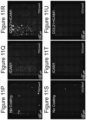
- the present inventors further demonstrate that in-vitro activation of the cells on the CCL21+ICAM1 coated surface has an even greater effect on cell density in the culture when the number of T cells (CD8 + T cells) seeded is low (e.g. 3250 cells/ml and 7500 cells/ml) (Example 2, Figures 11P-U ), an effect clinically significant when only a low number of e.g. tumor infiltrating T cells are isolated from a given tumor for adoptive cell therapy.
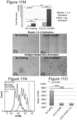
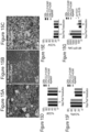
- CCL21+ICAM1 coating did not affect CD4 + T cell sub-differentiation and function (Example 1, Figures 9A-D ); while it had a combined improved effect on CD8 + T cells in-vitro killing efficiency (Examples 3-4, Figures 12A-K , 13A1-G3 and 16A-J ) and in-vivo tumor suppression (Example 3, Figures 14A-D ).
- the results indicate that the effects induced by the CCL21+ICAM1 coated surface on T cell proliferation and function last several days following the end of exposure to the coating, indicating that a continuous CCL21 and ICAM1 signal at the e.g. tumor site is unnecessary.
- both antigen-specific and non-specific activation of T cells in this newly developed synthetic ex-vivo environment induces a dramatic shift in the organization and morphology of the T cells, as well as greatly improves their expansion and in the case of CD8 + T cells also the in-vitro killing efficiency and in-vivo tumor suppression. Consequently, the present teachings further suggest the use of this developed synthetic in-vitro activation and expansion system in the production of cells used for adoptive T cells therapies for e.g. malignancies, infections, autoimmune and graft rejection diseases.
- the synthetic in-vitro activation and expansion system can provide a novel tool for basic research into the mechanisms underlying immunological processes, by enabling the controlled regulation and perturbation of specific factors potentially involved in cell-cell or cell-matrix interactions.
- the present invention thus provides a method of culturing T cells, the method comprising culturing T cells in the presence of a T cell stimulator, an exogenous CCL21 and an exogenous ICAM1, thereby culturing the T cells, wherein the culturing is effected to allow expansion of the T cells, wherein the CCL21 and the ICAM1 are immobilized.
- CCL21 also known as Chemokine (C-C motif) ligand 21, 6Ckine, exodus-2 and secondary lymphoid-tissue chemokine (SLC).
- CCL21 can bind the chemokine receptor CCR7.
- the CCL21 protein refers to the human protein, such as provided in the following GenBank Number NP_002980 (SEQ ID NO: 1).
- the term also encompasses functional homologues (naturally occurring or synthetically/recombinantly produced), orthologs (from other species) which exhibit the desired activity (i . e ., binding CCR7, increasing expansion and at least maintaining activity of T cells such as in combination with ICAM1).
- Such homologues can be, for example, at least 80 %, at least 81 %, at least 82 %, at least 83 %, at least 84 %, at least 85 %, at least 86 %, at least 87 %, at least 88 %, at least 89 %, at least 90 %, at least 91 %, at least 92 %, at least 93 %, at least 94 %, at least 95 %, at least 96 %, at least 97 %, at least 98 %, at least 99 % or 100 % identical or homologous to the polypeptide SEQ ID NO: 1 or 80 %, at least 81 %, at least 82 %, at least 83 %, at least 84 %, at least 85 %, at least 86 %, at least 87 %, at least 88 %, at least 89 %, at least 90 %, at least 91 %, at least 92 %, at least
- Sequence identity or homology can be determined using any protein or nucleic acid sequence alignment algorithm such as Blast, ClustalW, and MUSCLE.
- the functional homologs also refer to functional portions of CCL21 which maintain the activity of the full length protein (i.e. binding CCR7, increasing expansion and at least maintaining activity of T cells such as in combination with ICAM1).
- the CCL21 is a recombinant CCL21.
- CCL21 can be commercially obtained from e.g. R&D Systems.
- the level of CCL21 in the methods of the present invention is above the level obtained in a cell culture comprising T cells of the same origin under the same culture conditions without the addition of CCL21.
- the concentration of CCL21 in the methods of the present invention is 100 ng / ml culture medium - 1000 ⁇ g / ml culture medium, 100 ng / ml culture medium - 100 ⁇ g / ml culture medium, 500 ng / ml culture medium - 500 ⁇ g / ml culture medium, 1 ⁇ g / ml culture medium - 100 ⁇ g / ml culture medium or 1 ⁇ g / ml culture medium - 10 ⁇ g / ml culture medium.
- the concentration of CCL21 in the methods of the present invention is about 5 ⁇ g / ml culture medium.
- the CCL21 is immobilized to a solid support.
- ICAM1 also known as Intercellular Adhesion Molecule 1 and CD54.
- ICAM1 can bind the CD11a / CD18 or CD11b / CD18 integrins.
- the ICAM1 protein refers to the human protein, such as provided in the following GenBank Number NP_000192 (SEQ ID NO: 2).
- the term also encompasses functional homologues (naturally occurring or synthetically/recombinantly produced), orthologs (from other species) which exhibit the desired activity (i.e ., binding CDlla / CD18 or CD11b / CD18 integrins, increasing expansion and at least maintaining activity of T cells such as in combination with CCL21).
- Such homologues can be, for example, at least 80 %, at least 81 %, at least 82 %, at least 83 %, at least 84 %, at least 85 %, at least 86 %, at least 87 %, at least 88 %, at least 89 %, at least 90 %, at least 91 %, at least 92 %, at least 93 %, at least 94 %, at least 95 %, at least 96 %, at least 97 %, at least 98 %, at least 99 % or 100 % identical or homologous to the polypeptide SEQ ID NO: 2 or 80 %, at least 81 %, at least 82 %, at least 83 %, at least 84 %, at least 85 %, at least 86 %, at least 87 %, at least 88 %, at least 89 %, at least 90 %, at least 91 %, at least 92 %, at least
- the functional homologs also refer to functional portions of ICAM1 which maintain the activity of the full length protein (i.e. binding CDlla / CD18 or CD11b / CD18 integrins, increasing expansion and at least maintaining activity of T cells such as in combination with CCL21).
- the ICAM1 is a recombinant ICAM1.
- ICAM1 can be commercially obtained from e.g. R&D Systems.
- the level of ICAM1 in the methods of the present invention is above the level obtained in a cell culture comprising T cells of the same origin under the same culture conditions without the addition of ICAM1.
- the concentration of ICAM1 in the methods of the present invention is 100 ng / ml culture medium - 1000 ⁇ g / ml culture medium, 100 ng / ml culture medium - 100 ⁇ g / ml culture medium, 500 ng / ml culture medium - 500 ⁇ g / ml culture medium, 1 ⁇ g / ml culture medium - 500 ⁇ g / ml culture medium or 1 ⁇ g / ml culture medium - 100 ⁇ g / ml culture medium.
- the concentration of ICAM1 in the methods of the present invention is about 50 ⁇ g / ml culture medium.
- the CCL21 is immobilized to a solid support.
- the ICAM1 and CCL21 are both human.
- An immobilized molecule or agent may be attached or coupled to the culture vessel or to a matrix within the culture vessel by a variety of chemistries known and available in the art.
- the attachment may be covalent or noncovalent, electrostatic, or hydrophobic and may be accomplished by a variety of attachment means, including for example, chemical, mechanical, enzymatic, or other means whereby the molecule or the agent is capable of stimulating the T cells.
- cell culture or “culture” refers to at least T cells and medium in an ex-vivo , in-vitro environment.
- the cell culture is maintained under conditions necessary to support growth and survival, for example, an appropriate temperature (e.g., 37 °C) and atmosphere (e.g., air plus 5 % CO 2 ).
- an appropriate temperature e.g., 37 °C
- atmosphere e.g., air plus 5 % CO 2 .
- the culture may be in a glass, plastic or metal vessel that can provide an aseptic environment for cell culturing.
- the culture vessel includes dishes, plates, flasks, bottles, vials, bags, bioreactors or any device that can be used to grow cells.
- the culture vessel is coated with a synthetic matrix and/or a biological matrix that can increase stimulation and proliferation, such as, a synthetic hydrogel, polycarbonate fibers, synthetic spongers and polymers, synthetic ECM, PLGA, PGA, natural ECM materials, agarose, gelatin, laminin, fibrin, collagen chitosan, protosan, and mixtures thereof.
- a synthetic matrix and/or a biological matrix that can increase stimulation and proliferation
- a synthetic hydrogel, polycarbonate fibers, synthetic spongers and polymers synthetic ECM, PLGA, PGA, natural ECM materials, agarose, gelatin, laminin, fibrin, collagen chitosan, protosan, and mixtures thereof.
- the culture vessel is pre-coated (i.e. prior to culturing and/or transducting) with the T cells stimulator, the chemokine (e.g. CCL21) and/or the adhesion molecule (e.g. ICAM1). Coating can be done simultaneously or in a sequential manner as long as the cells are subjected to the factors for a sufficient time and concentration to allow expansion while maintaining the desired phenotype.
- the chemokine e.g. CCL21
- the adhesion molecule e.g. ICAM1
- the culture vessel is a commercially available culture vessel coated with the T cells stimulator, such as anti-CD3/anti-CD28 coated culture vessel such as can be obtained from e.g. Miltenyi Biotech.
- the culture vessel is pre-coated (i.e. prior to culturing and/or transducing) with the chemokine (e.g. CCL21) and/or the adhesion molecule (e.g. ICAM1).
- the chemokine e.g. CCL21
- the adhesion molecule e.g. ICAM1.
- the culture medium used in the method of the present invention can be a water-based medium which includes a combination of substances such as salts, nutrients, minerals, vitamins, amino acids, nucleic acids and/or proteins such as cytokines, growth factors and hormones, all of which are needed for cell proliferation and survival.
- a culture medium can be a synthetic tissue culture medium such as RPMI 1640 medium (can be obtained from e.g. Biological Industries), DMEM (can be obtained from e.g. Biological Industries), MEM (can be obtained from e.g. Biological Industries), AIM-V (can be obtained from e.g. ThermoFisher Scientific), X-Vivo 15 (can be obtained from e.g.
- X-Vivo 20 can be obtained from e.g. Lonza, supplemented with the necessary additives as is further described hereinunder.
- all ingredients included in the culture medium of the present invention are substantially pure, with a tissue culture grade.
- the culture medium is devoid of any animal contaminants, i.e ., animal cells, fluid or pathogens (e.g., viruses infecting animal cells), i.e., being xeno-free.
- animal contaminants i.e ., animal cells, fluid or pathogens (e.g., viruses infecting animal cells), i.e., being xeno-free.
- the culture medium comprises antibiotics (e.g., penicillin, streptomycin, gentamicin), anti-fungal agents (e.g. amphotericin B), L-glutamine or NEAA (non-essential amino acids).
- antibiotics e.g., penicillin, streptomycin, gentamicin
- anti-fungal agents e.g. amphotericin B
- L-glutamine or NEAA non-essential amino acids
- the medium comprises serum and antibiotics.
- the medium comprises RPMI 1640 medium supplemented with serum, penicillin, streptomycin, glutamine, HEPES, sodium pyruvate and ⁇ -mercaptoethanol.
- the culture medium may be periodically refreshed to maintain sufficient levels of supplements and to remove metabolic waste products that can damage the cells. According to specific embodiments, the culture medium is refreshed every 12-96 hours, every 24-72 hours or every 24-48 hours.
- the T cells seeding concentration in the culture is 10 3 - 10 6 cells/ml culture medium, 10 3 - 5 ⁇ 10 5 cells/ml culture medium or 10 3 - 10 5 cells/ml culture medium.
- the T cells seeding concentration in the culture is less than 5 ⁇ 10 4 cells / ml culture medium.
- the T cells seeding concentration in the culture is 10 3 - 5 ⁇ 10 4 cells/ml culture medium.
- the T cells seeding concentration in the culture is 2 ⁇ 10 3 - 10 4 cells/ml culture medium.
- the T cells seeding concentration in the culture is about 7500 cells/ml culture medium.
- the T cells seeding concentration in the culture is about 3250 cells/ml culture medium.
- the T cells cultured according to the methods of the present invention proliferate during culturing, according to specific embodiments the T cells are serially splitted so as to maintain the desired cell concentration.
- the T cells concentration in the culture medium is 10 3 - 10 7 cells/ml culture medium, 10 3 - 5 ⁇ 10 6 cells/ml culture medium or 10 3 - 1 ⁇ 10 6 cells/ml culture medium.
- T cell refers to a differentiated lymphocyte with a CD3 + , T cell receptor (TCR) + having either CD4 + or CD8 + phenotype.
- the T cell may be a naive, activated or memory T cell.
- the T cell may be either an effector or a regulatory T cell.
- the T cells comprise effector T cells.
- effector T cell refers to a T cell that activates or directs other immune cells e.g. by producing cytokines or has a cytotoxic activity e.g., CD4 + , Th1/Th2, CD8 + cytotoxic T lymphocyte.
- the T cells comprise regulatory T cells.
- the term "regulatory T cell” or “Treg” refers to a T cell that negatively regulates the activation of other T cells, including effector T cells, as well as innate immune system cells. Treg cells are characterized by sustained suppression of effector T cell responses. According to a specific embodiment, the Treg is a CD4 + CD25 + Foxp3 + T cell.
- the T cells comprise CD4 + T cells. According to specific embodiments, the T cells comprise CD8 + T cells.
- the T cells are memory T cells.
- memory T cells include effector memory CD4 + T cells with a CD3 + /CD4 + /CD45RA - /CCR7 - phenotype, central memory CD4 + T cells with a CD3 + /CD4 + /CD45RA - /CCR7 + phenotype, effector memory CD8 + T cells with a CD3 + /CD8 + /CD45RA - /CCR7 - phenotype and central memory CD8 + T cells with a CD3 + /CD8 + /CD45RA -/ CCR7 + phenotype.
- the T cells are human T cells.
- the T cells are mouse or rat T cells.
- T cells can be obtained from a number of sources, including peripheral blood mononuclear cells (PBMCs), bone marrow, body tissue (such as lymph node tissue, spleen tissue, and tumors) or cells from tissue fluids.
- PBMCs peripheral blood mononuclear cells
- body tissue such as lymph node tissue, spleen tissue, and tumors
- T cells can be obtained from a unit of blood collected from a subject using any number of techniques known to the skilled artisan, as further disclosed hereinbelow.
- the T cells are obtained from a tumor tissue obtained from a subject.
- T cell lines are also available in the art. Thus, according to specific embodiments, any number of T cell lines available in the art, may be used.
- subject refers to a mammal (e.g., human being) at any age or of any gender.
- the subject is a healthy subject.
- the subject is diagnosed with a disease or is at risk of to develop a disease.
- the subject is a donor subject (syngeneic or non-syngeneic e.g., allogeneic).
- the subject is a recipient subject.
- the T cells are obtained from a healthy subject.
- the T cells are obtained from a subject suffering from a pathology.
- a peripheral blood sample is collected from a subject by methods well known in the art such as drawing whole blood from the subject and collection in a container containing an anti-coagulant (e.g. heparin or citrate); and apheresis.
- an anti-coagulant e.g. heparin or citrate
- apheresis e.g. heparin or citrate
- at least one type of T cell is purified from the peripheral blood.
- There are several methods and reagents known to those skilled in the art for purifying T cells from whole blood such as leukapheresis, sedimentation, density gradient centrifugation (e.g. ficoll), centrifugal elutriation, fractionation, chemical lysis of e.g. red blood cells (e.g. by ACK), selection of specific cell types using cell surface markers (using e.g.
- FACS sorter or magnetic cell separation techniques such as are commercially available e.g. from Invitrogen, Stemcell Technologies, Cellpro, Advanced Magnetics, or Miltenyi Biotec.), and depletion of specific cell types by methods such as eradication (e.g. killing) with specific antibodies or by affinity based purification based on negative selection (using e.g. magnetic cell separation techniques, FACS sorter and/or capture ELISA labeling).
- eradication e.g. killing
- affinity based purification based on negative selection using e.g. magnetic cell separation techniques, FACS sorter and/or capture ELISA labeling.
- the cell culture comprises at least 50 % at least 60 %, at least 70 %, at least 80 %, at least 90 % or at least 95 % T cells.
- the T cells comprise more than 5 %, more than 10 %, more than 20 %, more than 30 %, more than 40 %, more than 50 %, more than 60 %, more than 70 %, more than 80 %, more than 90 % or more than 95 % T cells with specific antigen-specificity.
- antigen-specific T cells can be isolated by contacting the T cells with antibodies specific for T cell activation markers.
- the T cells comprise engineered T cells transduced with a nucleic acid sequence encoding an expression product of interest.
- the expression product of interest is a T cell receptor (TCR) or a chimeric antigen receptor (CAR).
- TCR T cell receptor
- CAR chimeric antigen receptor
- transduced with a nucleic acid sequence encoding a TCR or “transducing with a nucleic acid sequence encoding a TCR” refers to cloning of variable ⁇ - and ⁇ -chains from T cells with specificity against a desired antigen presented in the context of MHC.
- Methods of transducing with a TCR are known in the art and are disclosed e.g. in Nicholson et al. Adv Hematol. 2012; 2012:404081 ; Wang and Rivière Cancer Gene Ther. 2015 Mar;22(2):85-94 ); and Lamers et al, Cancer Gene Therapy (2002) 9, 613-623 .
- the phrase "transduced with a nucleic acid sequence encoding a CAR" or “transducing with a nucleic acid sequence encoding a CAR” refers to cloning of a nucleic acid sequence encoding a chimeric antigen receptor (CAR), wherein the CAR comprises an antigen recognition moiety and a T-cell activation moiety.
- a chimeric antigen receptor (CAR) is an artificially constructed hybrid protein or polypeptide containing an antigen binding domain of an antibody (e.g., a single chain variable fragment (scFv)) linked to T-cell signaling or T-cell activation domains.
- scFv single chain variable fragment
- the T cells are transduced with a suicide gene.
- the present invention also provides a method of producing engineered T cells, the method comprising transducing T cells with a nucleic acid sequence encoding an expression product of interest (e.g. TCR or a CAR) in the presence of an exogenous immobilized CCL21 and an exogenous immobilized ICAM1, thereby producing the engineered T cells.
- a nucleic acid sequence encoding an expression product of interest (e.g. TCR or a CAR) in the presence of an exogenous immobilized CCL21 and an exogenous immobilized ICAM1, thereby producing the engineered T cells.
- an exogenous expression product of interest e.g. TCR or CAR
- the T cells are transduced with a nucleic acid expression vector (i.e. construct) encoding the expression product of interest suitable for mammalian cell expression.
- a nucleic acid expression vector i.e. construct
- Such an expression vector includes a promoter sequence for directing transcription of the polynucleotide sequence in the cell in a constitutive or inducible manner.
- the promoter utilized by the expression vector is active in the specific cell population transformed. Examples of cell type-specific promoters include promoters such as lymphoid specific promoters [ Calame et al., (1988) Adv. Immunol. 43:235-275 ]; in particular promoters of T-cell receptors [ Winoto et al., (1989) EMBO J. 8:729-733 ].
- the expression vector of some embodiments of the invention includes additional sequences which render this vector suitable for replication and integration in prokaryotes, eukaryotes, or preferably both (e.g., shuttle vectors).
- a cloning vector may also contain a transcription and translation initiation sequence, transcription and translation terminator, a polyadenylation signal, an enhancer element, restriction element, integration element, a eukaryotic replicon and/or internal ribosome entry site (IRES).
- IRS internal ribosome entry site
- such expression vector typically includes a 5' LTR, a tRNA binding site, a packaging signal, an origin of second-strand DNA synthesis, and a 3' LTR or a portion thereof.
- mammalian expression vectors examples include pcDNA3, pcDNA3.1(+/-), pGL3, pZeoSV2(+/-), pSecTag2, pDisplay, pEF/myc/cyto, pCMV/myc/cyto, pCR3.1, pSinRep5, DH26S, DHBB, pNMT1, pNMT41, pNMT81, which are available from Invitrogen, pCI which is available from Promega, pMbac, pPbac, pBK-RSV and pBK-CMV which are available from Strategene, pTRES which is available from Clontech, and their derivatives.
- Expression vectors containing regulatory elements from eukaryotic viruses such as retroviruses can be also used.
- SV40 vectors include pSVT7 and pMT2.
- Vectors derived from bovine papilloma virus include pBV-1MTHA, and vectors derived from Epstein Bar virus include pHEBO, and p2O5.
- exemplary vectors include pMSG, pAV009/A + , pMTO10/A + , pMAMneo-5, baculovirus pDSVE, and any other vector allowing expression of proteins under the direction of the SV-40 early promoter, SV-40 later promoter, metallothionein promoter, murine mammary tumor virus promoter, Rous sarcoma virus promoter, polyhedrin promoter, or other promoters shown effective for expression in eukaryotic cells.
- the expression vector is the retrovector pBullet/pMSGV plasmids.
- nucleic acids by viral infection offers several advantages over other methods such as lipofection and electroporation, since higher transfection efficiency can be obtained due to the infectious nature of viruses.
- the transduction is effected with a viral vector (i.e. by incubating the T cells with the viral vector).
- the transduction step can be effected for a couple of hours and up to a couple of days.
- the transduction is effected for at least 12 hours, at least 24 hours, at least 36 hours or at least 48 hours.
- the transduction is effected for 1-3 days.
- the transduction is effected for about 2 days.
- the transduction is effected in the presence of a RetroNectin reagent (e.g. using RetroNectin coated-plates, such as commercially available from Takara Bio, Mountain View, CA, USA).
- a RetroNectin reagent e.g. using RetroNectin coated-plates, such as commercially available from Takara Bio, Mountain View, CA, USA.
- the transduction is effected in the presence of exogenous IL-2.
- the transduction is effected on activated T cells.
- the transduction is effected on in-vitro activated T cells.
- the transduction is effected on T cells pre-cultured in the presence of a T cell stimulator, CCL21 and ICAM1.
- the method of producing engineered T cells further comprising culturing the T cells in the presence of a T cell stimulator, an exogenous chemokine and an exogenous adhesion molecule prior to the transducing.
- the culturing is effected for 1-7 days, 2-7 days, 1-5 days, 2-5 days or 1-3 days.
- the culturing is effected for about 2 days.
- chemokine refers to a chemotactic cytokine which is secreted by cells and induces direct chemotaxis of nearby responsive cells (e.g. T cells).
- chemokines are classified into four main subfamilies: CXC, CC, CX3C and XC.
- Non-limiting eExamples of chemokines include CCL14, CCL19, CCL20, CCL21, CCL25, CCL27, CXCL12, CXCL13, CXCL-8, CCL2, CCL3, CCL4, CCL5, CCL11 and CXCL10.
- adheresion molecule refers to a protein capable of mediating cell-cell or cell-substrate binding.
- cell adhesion molecules are divided into two groups: molecules involved in cell-cell adhesion (i.e. intercellular adhesion) and molecules involved in cell-extracellular matrix adhesion (i.e. cell-substrate adhesion).
- adhesion molecules examples include LFA-1, mac-1, CD11c/CD18, ICAM1, ICAM2, ICAM3, VCAM1, VLA1, VLA4, CD2, LFA3, CD44, CD62 (E, L and P), CD106, other integrin-family members, extracellular matrix molecules like fibronectin, and laminin, fibrinogen, and cytohesin-1.
- the cells may be further cultured, activated and expanded.
- the engineered T cells produced according to the method of the present invention may be further cultured in the presence of a T cell stimulator, an exogenous chemokine and an exogenous adhesion molecule following the transducing.
- T cell stimulator refers to an agent capable of activating a T cell resulting in cellular proliferation, maturation, cytokine production and/or induction of regulatory or effector functions.
- T cell activation or function Methods of evaluating T cell activation or function are well known in the art and also disclosed in the Examples section which follow and include proliferation assays such as CFSE, BRDU and thymidine incorporation, cytotoxicity assays such as chromium release and evaluating cell killing by microscopic evaluation, cytokine secretion assays such as intracellular cytokine staining, ELISPOT and ELISA, expression of activation markers such as CD25, CD69 and CD44 using flow cytometry.
- proliferation assays such as CFSE, BRDU and thymidine incorporation
- cytotoxicity assays such as chromium release and evaluating cell killing by microscopic evaluation
- cytokine secretion assays such as intracellular cytokine staining, ELISPOT and ELISA
- expression of activation markers such as CD25, CD69 and CD44 using flow cytometry.
- the T cell stimulator can activate the T cells in an antigen-dependent or -independent (i.e. polyclonal) manner.
- the T cell stimulator is an antigen-specific stimulator.
- antigen specific T cell stimulators include antigen-loaded antigen presenting cell [APC, e.g. dendritic cell] and peptide loaded recombinant MHC.
- APC antigen-loaded antigen presenting cell
- peptide loaded recombinant MHC e.g. peptide loaded recombinant MHC.
- the T cells stimulator can be a dendritic cell preloaded with a desired antigen (e.g. a tumor antigen) or transfected with mRNA coding for the desired antigen.
- a desired antigen e.g. a tumor antigen
- mRNA coding for the desired antigen e.g. a tumor antigen
- the antigen-specific stimulator comprises a dendritic cell loaded with the antigen.
- the antigen is a cancer antigen.
- cancer antigen refers to an antigen is overexpressed or solely expressed by a cancerous cell as compared to a non-cancerous cell.
- a cancer antigen may be a known cancer antigen or a new specific antigen that develops in a cancer cell (i.e. neoantigens).
- cancer antigens examples include MAGE-AI, MAGE-A2, MAGE- A3, MAGE-A4, MAGE-AS, MAGE-A6, MAGE-A7, MAGE-AS, MAGE-A9, MAGE-AIO, MAGE-All, MAGE-A12, GAGE-I, GAGE-2, GAGE-3, GAGE-4, GAGE-5, GAGE-6, GAGE-7, GAGE-8, BAGE-l, RAGE- 1, LB33/MUM-1, PRAME, NAG, MAGE-Xp2 (MAGE-B2), MAGE-Xp3 (MAGE-B3), MAGE-Xp4 (MAGE-B4), MAGE- Cl/CT7, MAGE-C2, NY-ES0-1, LAGE-1, SSX-1, SSX-2(HOM-MEL-40), SSX-3, SSX-4, SSX-5, SCP-1 and XAGE, melanocyte differentiation antigens, p53, ras, CEA, MUCI, PMSA
- the tumor antigen is a HER2 peptide.
- tumor antigens that may be expressed include out-of-frame peptide-MHC complexes generated by the non-AUG translation initiation mechanisms employed by "stressed" cancer cells ( Malarkannan et al. Immunity 1999 ).
- tumor antigens that may be expressed are well-known in the art (see for example WO00/20581 ; Cancer Vaccines and Immunotherapy (2000) Eds Stern, Beverley and Carroll, Cambridge University Press, Cambridge ). The sequences of these tumor antigens are readily available from public databases but are also found in WO 1992/020356 A1 , WO 1994/005304 A1 , WO 1994/023031 A1 , WO 1995/020974 A1 , WO 1995/023874 A1 & WO 1996/026214 A1 .
- EBV-associated lymphoma EBV-associated lymphoma
- EBV-specific antigens can be used as the cancer antigen.
- a tumor antigen may be identified using cancer cells obtained from a subject by e.g. biopsy.
- the T cell stimulator is an antigen non-specific stimulator.
- An antigen non-specific stimulator is an agent capable of binding to a T cell surface structure and induce the polyclonal stimulation of the T cell, such as anti-CD3 antibody in combination with a co-stimulatory protein such as anti-CD28 antibody.
- anti-CD2 anti-CD137, anti-CD134, Notch-ligands, e.g. Delta-like 1/4, Jaggedl/2 either alone or in various combinations with anti-CD3.
- Other agents that can induce polyclonal stimulation of T cells include mitogens, PHA, PMA-ionomycin, CEB and CytoStim (Miltenyi Biotech).
- the antigen non-specific stimulator comprises anti-CD3 and anti-CD28 antibodies.
- the T cell stimulator comprises anti-CD3 and anti-CD28 coated beads, such as the CD3CD28 MACSiBeads obtained from Miltenyi Biotec.
- the method comprises adding the cytokine IL-6 to the culture.
- the IL-6 is added in a level above the level obtained by culturing T cells of the same origin under the same culture conditions without addition of the cytokine.
- the cell culture comprises Il-6 in a level above the level obtained in the cell cultures described herein without addition of the IL-6.
- the concentration of IL-6 in the cultures is 1- 500 ng/ml, 5 - 250 ng/ml, 10 - 200 ng/ml or 20 - 100 ng/ml.
- cytokines added to the cell cultures induce differential effects on CD4 + and CD8 + T cells (Table 1, in the Examples section which follows).
- the T cells comprise CD4 + T cells and the cytokine is IL-6.
- the cytokine is soluble.
- the cytokine is immobilized to a solid support.
- the cytokine is added to the culture within 24 hours of cell seeding.
- the cytokine is added to the culture at the time of cell seeding.
- the culture does not comprise more than 10, more than 8, more than 6, more than 5, or more than 4 recombinant factors.
- the T cell culture prepared by the method of the present invention has a morphological appearance, which can be determined by e.g. microscopic evaluation, that is characterized by at least one of:
- the T cell culture prepared by the method of the present invention may be characterized by one, two, three, four, five or all of (i) - (vi).
- the T cell culture prepared by the method of the present invention may be characterized by at least (i) + (ii), (ii) + (iii), (iii) + (iv), (ii) + (iii) + (iv), (v) + (vi) and/or (i) + (iv)/
- the T cell culture prepared by the method of the present invention may be characterized by (i) + (ii) + (iii) + (iv) + (v) + (vi).
- the microscopic evaluation is effected following labeling of the cells by e.g. a membrane-permeable dye.
- the T cell culture prepared by the method of the present invention may present different morphological appearance as compared to T cells of the same origin under the same culture conditions without addition of the chemokine (e.g. CCL21) and the adhesion molecule (e.g. ICAM1), wherein the combination of the chemokine (e.g. CCL21) and the adhesion molecule (e.g. ICAM1) has at least an additive effect as compared to the addition of only one of the chemokine (e.g. CCL21) and the adhesion molecule (e.g. ICAM1), as further discussed in the details in the Examples section which follows.
- the chemokine e.g. CCL21
- the adhesion molecule e.g. ICAM1
- the combination of the chemokine (e.g. CCL21) and the adhesion molecule (e.g. ICAM1) has a synergistic effect as compared to the addition of only one of the chemokine (e.g. CCL21) and the adhesion molecule (e.g. ICAM1).
- the chemokine (e.g. CCL21) and the adhesion molecule (e.g. ICAM1) used in the methods of the present invention may improve the in-vitro stimulation of T-cells by increasing expansion of the cells while at least maintaining their functionality.
- expansion and “proliferation” are interchangeably used and refer to an increase in the number of cells in a population by means of cell division.
- Methods of evaluating expansion are well known in the art and include proliferation assays such as CFSE and BrDU and determining cell number by direct cell counting and microscopic evaluation.
- the increase refers to an increase of at least 5 % in T cell expansion, as compared to same in the absence of the exogenous chemokine (e.g. CCL21) and the adhesion molecule (e.g. ICAM1), as may be manifested e.g. in cell proliferation or cell number.
- the increase is in at least 10%, 20 %, 30 %, 40 % or even higher say, 50 %, 60 %, 70 %, 80 %, 90 % or more than 100 %.
- the increase is at least 1.5 fold, at least 2 fold, at least 3 fold, at least 5 fold, at least 10 fold, or at least 20 fold as compared to same in the absence of the exogenous chemokine (e.g. CCL21) and the adhesion molecule (e.g. ICAM1).
- the exogenous chemokine e.g. CCL21
- the adhesion molecule e.g. ICAM1
- a "functionality of a T cell” refers to the ability of a T cell to proliferate, mature, produce and/or secrete cytokine, induce regulatory functions and/or induce effector functions such as cytotoxic activity. Methods of evaluating functionality of a T cell are well known in the art and also disclosed hereinabove and in the Examples section which follow.
- the chemokine (e.g. CCL21) and the adhesion molecule (e.g. ICAM1) used in the methods of the present invention improve T cells functionality, e.g. in-vitro killing efficiency, expression of granzyme B, in-vivo activity against e.g. a tumor, wherein the combination of the chemokine (e.g. CCL21) and the adhesion molecule (e.g. ICAM1) has at least an additive effect as compared to the presence of only one of the chemokine (e.g. CCL21) and the adhesion molecule (e.g. ICAM1).
- improves T cells functionality refers to an increase of at least 5 % in T cell function, as compared to same in the absence of the exogenous chemokine (e.g. CCL21) and the adhesion molecule (e.g. ICAM1), as may be manifested e.g. in cytokine secretion and/or cytotoxic activity.
- the increase may be in at least 10%, 20 %, 30 %, 40 % or even higher say, 50 %, 60 %, 70 %, 80 %, 90 % or more than 100 %.
- the increase may be at least 1.5 fold, at least 2 fold, at least 3 fold, at least 5 fold, at least 10 fold, or at least 20 fold as compared to same in the absence of the exogenous chemokine (e.g. CCL21) and the adhesion molecule (e.g. ICAM1).
- exogenous chemokine e.g. CCL21
- adhesion molecule e.g. ICAM1
- the morphology of the cultures, T cells expansion and functionality may be culture time dependent.
- the culturing is effected for 3-10, 3-7, 3-6, 4-7, 5-7, 4-6 or 5-6 days.
- the culture is up to 7 days old.
- the culture is up to 6 days old.
- the culturing is effected for at least 3 days.
- the culturing is effected for 3-7 days.
- the culturing is effected for 3-6 days.
- the T cells are expanded by 2-200 fold following 3-7 days of culture.
- a specific population of cells can be further subjected to purification.
- Methods of purifying a desired population of cells are well known in the art and are further described hereinabove.
- the T cells may be purified following the culturing.
- the T cells may be purified following the transducing.
- the T cells may be purified from the T cell stimulator.
- the T cells may be purified from the chemokine (e.g. CCL21) and the adhesion molecule (e.g. ICAM1).
- chemokine e.g. CCL21
- adhesion molecule e.g. ICAM1
- a specific population of T cells effective for the treatment of a disease may be purified.
- the cells obtained according to the present invention can be freshly isolated, stored e.g., cryopresereved (i.e. frozen) at e.g. liquid nitrogen temperature at any stage (e.g. following their retrieval, following culturing, following transduction or following purification) for long periods of time (e.g., months, years) for future use; and cell lines.
- cryopresereved i.e. frozen
- liquid nitrogen temperature e.g. liquid nitrogen temperature
- any stage e.g. following their retrieval, following culturing, following transduction or following purification
- long periods of time e.g., months, years
- the cells obtained according to the present invention can be stored in a cell bank or a depository or storage facility.
- the cells may be freshly isolated (i.e., not more than 24 hours after retrieval, culturing, transduction and/or purification and not subjected to preservation processes).
- T cells obtainable according to the methods of the present invention may be administered as adoptive T cells transfer to a subject in need thereof, thereby adoptively transferring the T cells to the subject.
- T cells obtainable according to the methods of the present invention may be useful for the manufacture of a medicament identified for adoptive T cell therapy.
- the cells may be autologous or non-autologous; they can be syngeneic or non-syngeneic: allogeneic or xenogeneic to the subject.
- autologous means that the donor subject is the recipient subject.
- the cells have been removed and re-introduced e.g., re-infused to the subject.
- non-autologous means that the donor subject is not the recipient subject.
- syngeneic means that the donor subject is essentially genetically identical with the recipient subject.
- Examples of syngeneic transplantation include transplantation of cells derived from the subject (also referred to in the art as “autologous”), a clone of the subject, or a homozygotic twin of the subject.
- allogeneic means that the donor is of the same species as the recipient, but which is substantially non-clonal with the recipient. Typically, outbred, non-zygotic twin mammals of the same species are allogeneic with each other. It will be appreciated that an allogeneic donor may be HLA identical or HLA non-identical with respect to the subject.
- xenogeneic means that the donor subject is from a different species relative to the recipient subject.
- treating refers to inhibiting, preventing or arresting the development of a pathology (disease, disorder, or condition) and/or causing the reduction, remission, or regression of a pathology.
- pathology disease, disorder, or condition
- Those of skill in the art will understand that various methodologies and assays can be used to assess the development of a pathology, and similarly, various methodologies and assays may be used to assess the reduction, remission or regression of a pathology.
- the teachings described herein are relevant for obtaining cells effective for the treatment of diseases such as cancer, infectious diseases, allergies and graft rejection disease.
- Cancers can be any solid or non-solid cancer and/or cancer metastasis.
- cancer include carcinoma, lymphoma, blastoma, sarcoma, and leukemia. More particular examples of such cancers include squamous cell cancer, lung cancer (including small-cell lung cancer, non-small-cell lung cancer, adenocarcinoma of the lung, and squamous carcinoma of the lung), cancer of the peritoneum, hepatocellular cancer, gastric or stomach cancer (including gastrointestinal cancer), pancreatic cancer, glioblastoma, cervical cancer, ovarian cancer, liver cancer, bladder cancer, hepatoma, breast cancer, colon cancer, colorectal cancer, endometrial or uterine carcinoma, salivary gland carcinoma, kidney or renal cancer, liver cancer, prostate cancer, vulval cancer, thyroid cancer, hepatic carcinoma and various types of head and neck cancer, as well as B-cell lymphoma (including low grade/follicular non-Hodgkin's lymphoma (
- the cancer is selected from breast cancer, colorectal cancer, rectal cancer, non-small cell lung cancer, non-Hodgkins lymphoma (NHL), renal cell cancer, prostate cancer, liver cancer, pancreatic cancer, soft-tissue sarcoma, Kaposi's sarcoma, carcinoid carcinoma, head and neck cancer, melanoma, ovarian cancer, mesothelioma, or multiple myeloma.
- the cancerous conditions amenable for treatment include metastatic cancers.
- infectious disease refers to a disease induced by a pathogen.
- pathogens include, viral pathogens, bacterial pathogens e.g., intracellular mycobacterial pathogens (such as, for example, Mycobacterium tuberculosis ), intracellular bacterial pathogens (such as, for example, Listeria monocytogenes ), or intracellular protozoan pathogens (such as, for example, Leishmania and Trypanosoma ).
- viral pathogens causing infectious diseases include retroviruses, circoviruses, parvoviruses, papovaviruses, adenoviruses, herpesviruses, iridoviruses, poxviruses, hepadnaviruses, picornaviruses, caliciviruses, togaviruses, flaviviruses, reoviruses, orthomyxoviruses, paramyxoviruses, rhabdoviruses, bunyaviruses, coronaviruses, arenaviruses, and filoviruses.
- viral infections include human immunodeficiency virus (HIV)-induced acquired immunodeficiency syndrome (AIDS), influenza, rhinoviral infection, viral meningitis, Epstein-Barr virus (EBV) infection, hepatitis A, B or C virus infection, measles, papilloma virus infection/warts, cytomegalovirus (CMV) infection, Herpes simplex virus infection, yellow fever, Ebola virus infection, rabies, etc.
- HBV human immunodeficiency virus
- AIDS human immunodeficiency virus
- EBV Epstein-Barr virus
- CMV cytomegalovirus
- Herpes simplex virus infection Herpes simplex virus infection
- yellow fever Ebola virus infection
- Ebola virus infection rabies, etc.
- autoimmune diseases which may be treated include rheumatoid diseases, rheumatoid autoimmune diseases, rheumatoid arthritis ( Krenn V. et al., Histol Histopathol 2000 Jul;15 (3):791 ), spondylitis, ankylosing spondylitis ( Jan Voswinkel et al., Arthritis Res 2001; 3 (3): 189 ), systemic diseases, systemic autoimmune diseases, systemic lupus erythematosus ( Erikson J. et al., Immunol Res 1998;17 (1-2):49 ), sclerosis, systemic sclerosis ( Renaudineau Y. et al., Clin Diagn Lab Immunol.
- vasculitises necrotizing small vessel vasculitises, microscopic polyangiitis, Churg and Strauss syndrome, glomerulonephritis, pauci-immune focal necrotizing glomerulonephritis, crescentic glomerulonephritis ( Noel LH. Ann Med Interne (Paris). 2000 May;151 (3):178 ); antiphospholipid syndrome ( Flamholz R. et al., J Clin Apheresis 1999;14 (4):171 ); heart failure, agonist-like beta-adrenoceptor antibodies in heart failure ( Wallukat G. et al., Am J Cardiol.
- allergic diseases which may be treated include asthma, hives, urticaria, pollen allergy, dust mite allergy, venom allergy, cosmetics allergy, latex allergy, chemical allergy, drug allergy, insect bite allergy, animal dander allergy, stinging plant allergy, poison ivy allergy and food allergy.
- graft rejection diseases which may be treated include host vs. graft disease, chronic graft rejection, subacute graft rejection, hyperacute graft rejection, acute graft rejection, allograft rejection, xenograft rejection and graft-versus-host disease (GVHD).
- host vs. graft disease chronic graft rejection, subacute graft rejection, hyperacute graft rejection, acute graft rejection, allograft rejection, xenograft rejection and graft-versus-host disease (GVHD).
- GVHD graft-versus-host disease
- the graft may be a cell, a tissue or a whole organ graft.
- the origin of the graft may be embryonic, fetal, post natal or adult.
- the term "graft" refers to a bodily cell (e.g. a single cell or a group of cells) or tissue (e.g. solid tissues or soft tissues, which may be transplanted in full or in part).
- tissue and whole organs which may be transplanted include liver, pancreas, spleen, kidney, heart, lung, skin, intestine and lymphoid/hematopoietic tissues (e.g. lymph node, Peyer's patches, thymus or bone marrow).
- the cells may be stem cells, progenitors (e.g. immature HSCs) or differentiated cells.
- Exemplary cells which may be transplanted include hematopoietic stem cells.
- the graft can be autologous or non-autologous; it can be syngeneic or non-syngeneic: allogeneic or xenogeneic to the subject.
- the graft can be autologous or non-autologous; it can be syngeneic or non-syngeneic: allogeneic or xenogeneic to the adoptively transferred T cells.
- the T cells obtained by the methods of the invention can be administered to a subject in combination with other established or experimental therapeutic regimen to treat a disease (e.g. cancer) including analgetics, chemotherapeutic agents, radiotherapeutic agents, cytotoxic therapies (conditioning), hormonal therapy and other treatment regimens (e.g., surgery) which are well known in the art.
- a disease e.g. cancer
- analgetics chemotherapeutic agents, radiotherapeutic agents, cytotoxic therapies (conditioning), hormonal therapy and other treatment regimens (e.g., surgery) which are well known in the art.
- the T cells obtained by the method of the present invention may be transferred in a therapeutically effective amount.
- a therapeutically effective amount means an amount of active ingredients effective to prevent, alleviate or ameliorate symptoms of a disorder or prolong the survival of the subject being treated.
- Dosage amount and interval may be adjusted individually to provide sufficient amount of T cells to induce or suppress the biological effect.
- the amount of a T cells to be administered will, of course, be dependent on the subject being treated, the severity of the affliction, the manner of administration, the judgment of the prescribing physician, etc.
- dosing can be of a single or multiple rounds of adoptive transfer of the cells obtained according to the methods of the present invention, with course of treatment lasting from several days to several weeks or until cure is effected or diminution of the disease state is achieved.
- the T cells obtained via the method of the present invention may be administered either alone, or as a pharmaceutical composition in combination with diluents and/or with other components such as established or experimental therapeutic regimen to treat a disease, cytokines or other cell populations.
- Pharmaceutical compositions may comprise the T cells prepared as described herein, in combination with one or more pharmaceutically or physiologically acceptable carriers, diluents or excipients.
- compositions may be formulated for intravenous administration.
- a compound or “at least one compound” may include a plurality of compounds, including mixtures thereof.
- range format is merely for convenience and brevity and should not be construed as an inflexible limitation on the scope of the invention. Accordingly, the description of a range should be considered to have specifically disclosed all the possible subranges as well as individual numerical values within that range. For example, description of a range such as from 1 to 6 should be considered to have specifically disclosed subranges such as from 1 to 3, from 1 to 4, from 1 to 5, from 2 to 4, from 2 to 6, from 3 to 6 etc., as well as individual numbers within that range, for example, 1, 2, 3, 4, 5, and 6. This applies regardless of the breadth of the range.
- method refers to manners, means, techniques and procedures for accomplishing a given task including, but not limited to, those manners, means, techniques and procedures either known to, or readily developed from known manners, means, techniques and procedures by practitioners of the chemical, pharmacological, biological, biochemical and medical arts.
- sequences that substantially correspond to its complementary sequence as including minor sequence variations, resulting from, e.g., sequencing errors, cloning errors, or other alterations resulting in base substitution, base deletion or base addition, provided that the frequency of such variations is less than 1 in 50 nucleotides, alternatively, less than 1 in 100 nucleotides, alternatively, less than 1 in 200 nucleotides, alternatively, less than 1 in 500 nucleotides, alternatively, less than 1 in 1000 nucleotides, alternatively, less than 1 in 5,000 nucleotides, alternatively, less than 1 in 10,000 nucleotides.
- mice - 5-20 weeks old C57BL/6 mice were obtained from Harlan Laboratories (Rehovot, Israel) and 5-20 weeks old OT-I and OT-II mice were obtained from Jackson Laboratories (Bar Harbor, ME, USA). Mice were maintained at the Weizmann Institute's Lorry Lokey Pre-Clinical Research Facility and were cared for in accordance with national and institutional guidelines. All experiments were approved by the Institutional Animal Care and Use Committee.
- Cell isolation - Spleens were harvested from the indicated mouse strain.
- T-cells isolation the spleen was cut into small 1 mm pieces, crushed and passed through 70 micron mesh and washed with PBS. Red blood cells were excluded from cell pellet by 1 minute incubation with RBC lysis solution (Biological Industries, 01-888-1B).
- RBC lysis solution Biological Industries, 01-888-1B
- DCs isolation the spleen was cut into small 1 mm pieces and incubated in spleen dissociation medium (StemCell, 079-15C) for 30 minutes, then added with 0.1M EDTA for additional 5 minutes. Following, the pieces were crushed and passed through 70 ⁇ m mesh and washed with PBS. Red blood cells were excluded from cell pellet by 1 minute incubation with RBC lysis solution (Biological Industries, 01-888-1B).
- Naive CD4 + T cells were purified (> 95 %) from a cell suspension harvested from spleens of OT-II mice, using a MACS CD4 + CD62L + T Cell Isolation Kit II (Miltenyi Biotec, Bergisch Gladbach, Germany), according to the manufacturer's instructions.
- CD8 + T cells were purified (> 95 %) from a cell suspension harvested from spleens of OT-I mice, using a CD8a + T Cell Isolation Kit and magnetic associated cell sorting (MACS), according to the manufacturer's instructions (Miltenyi Biotec, Bergisch Gladbach, Germany).
- DCs dendritic cells
- Engineered T cells CARS
- Engineered human T cells expressing a chimeric receptor against a human HER2 protein were produced as described in Globerson-Levin et al. Mol Ther. 2014;22(5):1029-1038 .
- the culture medium used was RPMI 1640, supplemented with 10 % serum, 100 U / ml of penicillin, 100 mg / ml of streptomycin, 2 mM glutamine, 1 mM sodium pyruvate, and 50 mM ⁇ -mercaptoethanol (Biological Industries, Beit Haemek, Israel).
- the isolated cells were seeded at the indicated concentration (between 3250 - 10 6 cells/ml ) on the culture plates in complete RPMI 1640 medium [RPMI 1640 w/o phenol red, supplemented with 10 % serum, 100 U / ml penicillin, 100 mg / ml streptomycin, 2 mM glutamine, 10 mM HEPES, 1 mM sodium pyruvate, 50 mM ⁇ -mercaptoethanol (Biological Industries, Beit Haemek, Israel] containing 1 ⁇ g / ml ovalbumin peptide (OVA323-339, InvivoGen, San Diego, CA, USA) and cultured for 24 hours to 7 days in 37 °C, 5 % CO 2 .
- RPMI 1640 w/o phenol red supplemented with 10 % serum, 100 U / ml penicillin, 100 mg / ml streptomycin, 2 mM glutamine, 10 mM HEPES, 1 mM
- FIG. 1R An illustration of the model is shown in Figure 1R .
- the T cells were activated for 24 hours with antigen-loaded DCs and then reseeded on different surface coatings for an additional incubation time (e.g. 48 hours).
- T cells activation was effected using DCs
- the freshly isolated T cells and DCs were seeded in a ratio of 3 : 1, respectively.
- CD3/CD28 beads were used for activation, the freshly isolated T cells were incubated 1 : 1 with activation beads freshly prepared according to manufacturer's instruction (Miltenyi Biotec, Bergisch Gladbach, Germany) and IL-2 30 Units / ml (BioLegend, San Diego, CA, USA).
- soluble IL-6 BioLegend
- 2-200 ng/ml of soluble IL-2, IL-12, IL-7, TNF- ⁇ , IFN ⁇ , TGF- ⁇ , IL-4 and/or GM-CSF All from BioLegend
- 5 ⁇ g / ml of blocking antibody was added 24 hours following cell seeding.
- Different coating solutions were placed in each chamber, to form separate strips containing: no coating, ICAM1 coating, CCL21 coating, and CCL21+ICAM1 coating on the glass surface.
- the coating solutions and SILICON molds were removed and the surfaces were thoroughly rinsed.
- OT-II CD4 + T cells were then co-cultured with ovalbumin-loaded DCs and imaged by phase contrast microscopy at the indicated time points.
- naive CD4 + T cells were labeled with either 10 ⁇ M CPDE-450 (Cell Proliferation Dye eFluor-450), 5 ⁇ M CPDE-670 (eBioscience, San Diego, CA, USA) or 2 ⁇ M CFSE (Invitrogen, Carlsbad, CA, USA) for 10 minutes at 37 °C. Excess dye was removed by three cold washes with 7 volumes of RPMI 1640, prior to cell seeding.
- 10 ⁇ M CPDE-450 Cell Proliferation Dye eFluor-450
- 5 ⁇ M CPDE-670 eBioscience, San Diego, CA, USA
- 2 ⁇ M CFSE Invitrogen, Carlsbad, CA, USA
- Cell proliferation assay - T cells were stained prior to seeding according to manufacturer's instructions with 5 ⁇ M CFSE (Biolegend) for 20 minutes or with 10 ⁇ M CPDE-450 (Cell Proliferation Dye eFluor-450; eBioscience, San Diego, CA, USA) for 10 minutes at 37 °C. Excess dye was removed by washing with 5-7 volumes of RPMI 1640. Following, 90 ⁇ 10 3 T cells were seeded with 30 ⁇ 10 3 DCs in a 96 wells plate with 250 ⁇ l complete RPMI 1640 medium containing 1 ⁇ g / ml ovalbumin peptide. Five days later, cells were detached from the surface by 10 minutes incubation with PBS without calcium and magnesium, and pipetting.
- PI Propidium iodide 1 ⁇ g / ml was added to each well for cell death staining and the single cell suspensions were analyzed by flow cytometry (BD Bioscoence and the software analysis FlowJo, Ashland, OR, USA). Since CFSE is diluted by approximately half with each cell division, live single cells (PI negative cells) were gated and the mean fluorescent intensity of CFSE was used to evaluate the level of proliferation.
- Cell viability assays A microscopic viability assay was performed by staining the cell nuclei with 1 ⁇ g / ml Hoechst (33342, ImmunoChemistry Technologies, Bloomington, MN, USA) and with 250 ng / ml propidium iodide (PI, Sigma Aldrich, St. Louis, MO, USA), for identification of dead cells. Cells were pipetted in order to breakdown clusters and spun down. Fluorescent images were taken using a Hermes microscope (IDEA Bio-Medical Ltd.) and the image analysis software (WiSoft, IDEA Bio-Medical Ltd.) was used to quantify viable cell numbers.
- Hoechst 33342, ImmunoChemistry Technologies, Bloomington, MN, USA
- PI propidium iodide
- PI propidium iodide
- a metabolic viability assay was performed by adding 20 ⁇ l CellTiter-Blue (Promega Corporation, Madison, WI, USA) per 100 ⁇ l culture medium for 3 hours. Results were quantified using a fluorescent plate reader (excitation 560nm; emission 590nm), and a linear equation of a calibration column, according to manufacturer's instructions.
- Microscopy and image analysis Wide-range phase-contrast images were acquired and stitched, using the 30/x objective of a Ti-eclipse microscope (Nikon Instruments, Konan, Minato-ku, Tokyo, Japan) equipped with an automated stage, and an incubator with a 5 % CO 2 atmosphere.
- Time-lapse movies were acquired using a DeltaVision Elite ® microscope (Applied Precision, GE Healthcare, Issaquah, WA, USA) mounted on an inverted IX71 microscope (Olympus, Center Valley, PA, USA) connected to a Photometrics CoolSNAP HQ2 camera (Roper Scientific, Martinsried, Germany).
- the primary image processing software used was SoftWorX 6.0.
- This microscope was also used for the production of deconvolution-based 3D image reconstructions, rendering supported by BITPlan software (Willich-Schiefbahn, Germany). Wide-range phase-contrast images and high content/high throughput microscopy was conducted on 96-wells plates using a Hermes ® microscope (IDEA BioMedical Ltd., Rehovot, Israel) equipped with automated scanning optics, high-precision autofocus, and a closed environmental chamber.
- the WiSoft ® software (IDEA Bio-Medical Ltd.) was utilized for cell counting and cell death analysis.
- Cluster analysis was carried out using Fiji software: Background was subtracted by means of a rolling ball method, and textured cluster areas were enhanced by a standard deviation filter, prior to thresholding using fixed value. Morphological filtering was applied to fill holes and connect broken clusters; finally, single cells were discarded by size and circularity. Areas of individual clusters and the total area of all clusters in a given image were measured for each condition.
- Epon ["hard", EMS (electron microscopy sciences) Ltd.] was gradually infiltrated into the cultures in increasing concentrations (30 %, 50 %, and 70 % in propylene oxide) for 2 hours each and then with 100 % Epon for 16 hours following by 100% Epon for three times for 2 hours each.
- samples were transferred in Epon to backing molds, and baked overnight in an oven.
- the Epon blocks were disconnected from the coverslip by cooling using liquid nitrogen followed by heating with a hot plate.
- CD4 + T cells Sub-differentiations induction - The following differentiation cytokines and neutralizing antibodies were added to the culture media upon cell seeding: for Th1 differentiation IL-12 (10 ng / ml) + anti-IL-4 (10 ⁇ g / ml); for Th2 differentiation IL-4 (20 ng / ml) + anti-IPN ⁇ (10 ⁇ g / ml) + anti-IL-12 (10 ⁇ g / ml); for Treg differentiation TGF ⁇ (10 ng / ml) + IL-2 (5 ng / ml) + anti-IL-4 (10 ⁇ g / ml) + anti-IFN ⁇ (10 ⁇ g / ml) + anti-IL-12 (10 ⁇ g / ml).
- Re-stimulation and IFN ⁇ analysis - CD4 + T cells were activated and induced to sub-differentiate to Th1 as described above. Following 7 days of incubation, cells were spun down and the medium was replaced with a medium containing PMA and ionomycin (BioLegend, BLG-423301, 1:500) for 4 hours of re-stimulation. Brefeldin (BioLegend, BLG420601) and Monensin (BioLegend, BLG420701) were then added 1 : 1000 each for 2 hours, for prevention of proteins secretion. Following, cells were collected, fixated, permeablized, stained with anti-CD4 and anti-IPN ⁇ (BioLegend) and analyzed by flow cytometry analysis (BD Biosciense and Flowjo).
- RPMI 1640 complete medium [RPMI 1640 w/o phenol red, supplemented with 10 % serum, 100 U/ml of penicillin, 100 mg / ml of streptomycin, 2 mM glutamine, 10 mM HEPES, 1 mM sodium pyruvate, and 50 mM ⁇ -mercaptoethanol (Biological Industries, Beit Haemek, Israel)], seeded 1000 cells per 384-plate well, and incubated for 2-3 hours to allow attachment to the surface.
- RPMI 1640 complete medium [RPMI 1640 w/o phenol red, supplemented with 10 % serum, 100 U/ml of penicillin, 100 mg / ml of streptomycin, 2 mM glutamine, 10 mM HEPES, 1 mM sodium pyruvate, and 50 mM ⁇ -mercaptoethanol (Biological Industries, Beit Haemek, Israel)]
- the entire well was imaged every 6 hours using Hermes ® microscope (IDEA BioMedical Ltd., Rehovot, Israel) equipped with automated scanning optics, high precision autofocus and a closed environmental chamber.
- the number of live (e.g. GFP expressing) B16 cells was counted from the image analysis using the WiSoft ® software (IDEA Bio-Medical Ltd.).
- In-vivo cytotoxic T cells tumor suppression assay - B16 melanoma cell line expressing ovalbumin (a courtesy of the Lea Eisenach laboratory at the Weizmann institute) were grown in DMEM complete medium [DMEM supplemented with 10 % serum, 100 U / ml of penicillin, 100 mg / ml of streptomycin, 2 mM glutamine, 10m M HEPES, 1 mM sodium pyruvate, and 50 mM ⁇ -mercaptoethanol (Biological Industries, Beit Haemek, Israel)]. Cells were harvested using trypsin and washed twice and suspended in PBS (Biological Industries).
- OT-I CD8 + T cells activated and cultured for 8-9 days as described above, were washed twice and suspended in PBS.
- 100 ⁇ l containing 2 ⁇ 10 6 or 4 ⁇ 10 6 CD8 + T cells were injected intravenously.
- CANCER RESEARCH (1998) 58: 2661-2666 ) were suspended in a RPMI 1640 complete medium without phenol red, supplemented with 10% serum, 0.48mg/ml G418, 100U/ml of penicillin, 100mg/ml of streptomycin, 2mM glutamine, 10mM HEPES, 1mM sodium pyruvate, and 50mM ⁇ -mercaptoethanol, (Biological Industries, Beit Haemek, Israel).
- the cells were seeded 50,000 cells per 96-wells plate and co-cultured with 150,000 human CD8 + T cells (3 : 1 T cell : target) of which 40-60 % were HER2-CAR cells (3 or 6 days following retroviral transduction).
- Luciferin Promega, Madison, WI, US
- luminescence total photon flux/sec
- CD4 + T cells expressing ovalbumin-specific T cell receptors isolated from TCR transgenic OT-II mice and stimulated with dendritic cells (DCs) loaded with the corresponding cognate ovalbumin peptide were used as a model for antigen-specific T cell stimulation.
- DCs dendritic cells
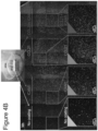
- the DCs and the T cells were pre-labeled with different cell-permeable dyes.
- cell clusters formed following stimulation consisted mainly of proliferating CD4 + T cells with only 1-2 DCs per cluster, most of which were dead following 24 hours of culture.
- T cell clusters continuously expanded in size for ⁇ 120 hours ( Figures 1G-H and 1Q ); and from that point onwards, they disassembled dramatically, and scattered into single cells. Notably, a similar phenomenon is also observed in the natural niche, when T cells detach from the clusters, leave the lymph node, and translocate into the circulation. 42
- clustering may support the activation of many T cells by a small number of DCs, as well as the interplay between T cells residing within the same cluster.
- Clustered T cells secrete various regulatory factors with autocrine and paracrine activity, and the close proximity of cells within the cluster facilitates intercellular communication in a regulated microenvironment. 3,40,43-45
- large T cell clusters may limit or even down-regulate T cell proliferation, by restricting the cells' access to essential signals or nutrients, or even by creating inhibitory environmental conditions. As CCL21 coating induced the formation of very large, yet short-lived 3-dimensional clusters, it was assumed that the conditions within these clusters might be less favorable.
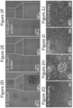
- Coating the surface with ICAM1 and CCL21 also exerted a strong effect on the morphology of single T cells ( Figures 2G-J ): cells interacting with uncoated surfaces were mostly round, with hardly any stable cell contacts while on the CCL21 coated surface, they developed an elongated morphology; in addition, ICAM1 coating, with or without CCL21, induced massive cell spreading on the surface, and multiple cell-cell contacts, some by means of long, thin tethers.
- T cells on a patterned surface characterized by separate uncoated areas and areas coated with ICAM1-1, CCL21 or both verified that the observed differences in T cell clustering, cell spreading and cell density are indeed local, confined to the coated area, and unaffected by both the relative order of the coatings on the surface, as well as by the number of seeded cells ( Figures 4A-E ).
- T cells were found mainly within the coated areas, which display a higher cell density compared to that of the uncoated area; the induction of large clusters by CCL21 coating and the cell spreading and culture flattening induced by ICAM1 were confined to the coated area and did not affect the morphology of T cells in other areas that share the same original cell pool and culture medium.
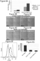
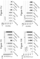
- cytokines included: IL-2, IL-12, IL-6, IL-7, TNF- ⁇ , IFN ⁇ , TGF- ⁇ , IL-4 and GM-CSF. Most of the cytokines tested had no significant effect on T cell expansion, either alone, or in combination with CCL21 and/or ICAM1 coatings (data not shown). However, as shown in Figure 7A-H , IL-6 decreased the percentage of dead cells by nearly a factor of 2 on uncoated surfaces and increased expansion by over 3-fold.
- the antigen-loaded DCs were chosen as the primary model for T cell activation, as this model has several important advantages over activation with non-specific activation using e.g. commercially available anti-CD3 anti-CD28 coated beads.
- activation with DCs was found to provide a greater T cell yield while requiring a minimal number of T cells, smaller by 7-fold than that needed with beads ( Figures 8A-E ).
- T cells activation with beads on CCL21+ICAM1 coated surface significantly augmented T cell proliferation and yield ( Figures 8A-G ) compared to activation on an uncoated surface, indicating surface-bound CCL21 and ICAM1 has a synergistic effect on T cells' expansion with other forms of activation.
- CD8 + T cells expressing ovalbumin-specific T cell receptors isolated from TCR transgenic OT-I mice and stimulated with dendritic cells (DCs) loaded with the corresponding cognate ovalbumin peptide were used as a model for antigen-specific T cell stimulation.
- DCs dendritic cells
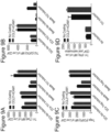
- CCL21+ICAM1 had an effect on CD8 + in-vitro activation.
- CCL21+ICAM1 effect was a result of augmenting CD8 + T cells proliferation rather than cell survival ( Figures 11J and 11L ).
- addition of soluble IL-6, collectively with CCL21+ICAM1 increased viable CD8 + T cell numbers by up to 7 times at 72 hours and 12 times at 7 days of culture, as compared to activation on an uncoated surface with no IL-6 ( Figures 11I and 11K ), possibly by decreasing the percentage of dead T cells by up to 25 % and 55 %, respectively ( Figures 11J and 11L ).
- CCL21+ICAM1 Given the effect of CCL21+ICAM1 for increasing T cells yield and use in adoptive therapy, in the next step the yielded pool was evaluated for functional activity.
- a microscopic killing assay was established, comprising co-cultures of B16 target cells, expressing ovalbumin coupled with GFP and OT-I CD8 + T cells pre-cultured on an uncoated surface or on a surface coated with CCL21+ICAM1.
- the number of live (e.g. GFP expressing) B16 cells was counted from the image analysis of each entire well of the co-cultures and used to assess the killing efficiency of the T cells.
- T cells pre-cultured for 72 hours on a CCL21+ICAM1 coated surface were able to kill more target cells by 24 hours of co-culturing compared to those pre-cultured on an uncoated surface or those pre-cultured on a CCL21+ICAM1 coated surface with addition of IL-6 ( Figures 12A-D and 12I ).
- this advantage was lost at 36 hours and onward ( Figure 12I ).
- T cells pre-cultured for 7 days on a CCL21+ICAM1 coated surface killed target cells more rapidly, with up to 2.5 times less live target cells by 24-72 hours of co-culturing, compared to those cultured on an uncoated surface ( Figures 12E-G , 12J and 13A1-G3 ).
- T cells pre-cultured on a CCL21+ ICAM1 coated surface not only killed more target cells more rapidly ( Figures 13C1-C5 and 13G1-G3), but formed larger clusters (which could indicate increased proliferation), compared to T cells grown on an uncoated surface ( Figures 13B1-B5 and 13F1-F3 ).
- B16-OVA tumor-bearing mice were injected, intravenously, with T cells activated and pre-cultured for 7-9 days activated on an uncoated surface, or on a CCL21+ICAM1 coated surface (a schematic representation of the experimental design is depicted in Figure 14A ).
- the in-vivo results correlated with the in-vitro results ( Figure 13A1-G3 ).
- OT-I CD8 + T cells cultured on both an uncoated surface or on a CCL2+ICAM1 coated surface suppressed tumor growth, in a dose dependent manner compared to non-treated group ( Figures 14B-C ).
- engineered T cells expressing a chimeric antigen receptor (CAR) (for an overview on CAR T cells see Almasbak H, et al. J Immunol Res. 2016;2016:5474602 ) were used as an additional model.
- CAR chimeric antigen receptor
- the specificity of a monoclonal antibody is grafted onto autologous T cells from the patient's blood, along with a signaling domain taken from activating and co-stimulatory immune receptors.
- these engineered T cells could be both activated and co-stimulated through their CAR when binding a desired tumor associated antigen.
- the model used was a HER2-CAR (see Globerson-Levin A et al. Mol Ther.
- T cells comprising both CD4 + and CD8 + T cells
- all T cells are isolated from a donated human blood, activated using anti-CD3 and anti-CD28 antibodies for 48 hours and then virally transduced with a chimeric receptor against the human oncogene HER2, for additional 48 hours.
- This CAR is composed of a single chain variable fragment (scFv) of the anti-ErbB-2 (HER2) monoclonal antibody (N29), linked to a variable fragment heavy (VH) chain through a flexible linker.
- scFv single chain variable fragment
- HER2 anti-ErbB-2
- VH variable fragment heavy
- it is ligated with the cytoplasmic domain of the co-stimulatory CD28 gene, followed by a signaling FcyR.
- HER2 known as human epidermal growth factor receptor 2
- HER2 known as human epidermal growth factor receptor 2
- HER2 is one of four tyrosine kinases in the ErbB family and a preferred dimerization partner of the other ErbB receptors. Its dimerization results in the auto-phosphorylation of its cytoplasmic tyrosine residues and several signaling pathways, whose dysregulation is strongly associated with a variety of cancers and poor prognosis ( Iqbal N. Mol Biol Int. 2014;2014:852748 ).
- immobilized CCL21+ICAM1 augmented the killing efficiency of HER2-CAR T cells ( Figures 16A-J ). Since the target cell line (RENCA-ErbB2) was of a murine origin and the T cells were of human origin, a group of T cells which were not transduced with the CAR antigen was used as a negative control to eliminate non-HER2 specific killing (grey column in Figures 16E-J ).
- CCL21+ICAM1 shortened the killing time, with up to 5 times less live target cells in the group of HER2-CAR T cells produced and expanded on a CCL21+ICAM1 coated surface, compared to those produced and expanded on an uncoated surface ( Figure 16G ). Furthermore, a smaller, yet significant increase in killing, of up to 2 times less live target cells, was seen in the group of HER2-CAR T cells produced without coating but expanded on a CCL21+ICAM1 coated surface, compared to those produced and expanded on an uncoated surface ( Figure 16G ).
- CCL21 - Cell Yield/Clustering/Cell spreading Increases cluster number, size and cell yield ICAM1 - Decreases cluster size and increases cell spreading and yield CCL21 + ICAM1 - Spread cells, increased proliferation and cell yield compared to CCL21 only or ICAM1 only Fibronectin - Clustering/Cell spreading No effect Poly Lysine - No effect Anti-LFA1 - Cell Yield/Clustering/Cell spreading Cell spreading, few clustering, low yield Anti-LFA1 + CCL21 Cell spreading, few clustering, low yield - IL-2 Inconsistent changes in cell yield CCL21 + ICAM1 Did not add to cell yield compared to CCL21+ICAM1 or CCL21+ICAM1+IL-6 - IL-12 Slightly bigger clusters CCL21 + ICAM1 Did not add to cell yield compared to CCL21+ICAM1 - IL-6 Higher cell yield CCL21 + ICAM1 Higher cell yield CCL21 + ICAM1 Higher cell yield
Landscapes
- Health & Medical Sciences (AREA)
- Life Sciences & Earth Sciences (AREA)
- General Health & Medical Sciences (AREA)
- Veterinary Medicine (AREA)
- Animal Behavior & Ethology (AREA)
- Public Health (AREA)
- Epidemiology (AREA)
- Engineering & Computer Science (AREA)
- Biomedical Technology (AREA)
- Immunology (AREA)
- Chemical & Material Sciences (AREA)
- Biotechnology (AREA)
- Zoology (AREA)
- Genetics & Genomics (AREA)
- Bioinformatics & Cheminformatics (AREA)
- Organic Chemistry (AREA)
- Wood Science & Technology (AREA)
- Cell Biology (AREA)
- Hematology (AREA)
- General Engineering & Computer Science (AREA)
- Biochemistry (AREA)
- Microbiology (AREA)
- Micro-Organisms Or Cultivation Processes Thereof (AREA)
- Developmental Biology & Embryology (AREA)
- Virology (AREA)
- Medicinal Chemistry (AREA)
- Pharmacology & Pharmacy (AREA)
- Medicines Containing Material From Animals Or Micro-Organisms (AREA)
Claims (9)
- Verfahren zum Züchten von T-Zellen, wobei das Verfahren Züchten von T-Zellen in Anwesenheit eines T-Zellstimulators, eines exogenen CCL2l und eines exogenen ICAM1 umfasst, wodurch die T-Zellen gezüchtet werden, wobei das Züchten durchgeführt wird, um eine Expansion der T-Zellen zuzulassen, wobei das CCL21 und das ICAM1 immobilisiert sind.
- Verfahren nach Anspruch 1, wobei die T-Zellen gentechnisch modifizierte T-Zellen umfassen, die mit einer Aminosäure transduziert sind, die für ein Expressionsprodukt, das von Interesse ist, codiert.
- Verfahren zum Produzieren gentechnisch modifizierter T-Zellen, wobei das Verfahren Transduzieren von T-Zellen mit einer Aminosäure, die für ein Expressionsprodukt, das von Interesse ist, in Anwesenheit eines exogenen immobilisierten CCL2l und eines exogenen immobilisierten ICAM1codiert, wodurch die gentechnisch modifizierten T-Zellen produziert werden.
- Verfahren nach Anspruch 2 oder 3, wobei das Expressionsprodukt, das von Interesse ist, ein T-Zellrezeptor (TCR) oder ein chimärer Antigenrezeptor (CAR) ist.
- Verfahren nach Anspruch 1 oder 2, wobei das Züchten während mindestens 3 Tagen durchgeführt wird.
- Verfahren nach Anspruch 1 oder 2, wobei das Züchten während mindestens 3-7 Tagen durchgeführt wird.
- Verfahren nach Anspruch 1 oder 2, wobei das Verfahren ferner Zusetzen von IL-6 zu der Kultur umfasst.
- Verfahren nach einem der Ansprüche 1-7, wobei die T-Zellen CD4+ T-Zellen umfassen.
- Verfahren nach einem der Ansprüche 1-8, wobei die T-Zellen CD8+ T-Zellen umfassen.
Applications Claiming Priority (2)
| Application Number | Priority Date | Filing Date | Title |
|---|---|---|---|
| IL250916A IL250916A0 (en) | 2017-03-02 | 2017-03-02 | Methods for growing t cells in culture and their use |
| PCT/IL2018/050236 WO2018158775A1 (en) | 2017-03-02 | 2018-03-02 | Methods of culturing t cells and uses of same |
Publications (2)
| Publication Number | Publication Date |
|---|---|
| EP3589729A1 EP3589729A1 (de) | 2020-01-08 |
| EP3589729B1 true EP3589729B1 (de) | 2024-05-29 |
Family
ID=61692206
Family Applications (1)
| Application Number | Title | Priority Date | Filing Date |
|---|---|---|---|
| EP18712421.9A Active EP3589729B1 (de) | 2017-03-02 | 2018-03-02 | Verfahren zur kultivierung von t-zellen und verwendungen davon |
Country Status (4)
| Country | Link |
|---|---|
| US (1) | US11932871B2 (de) |
| EP (1) | EP3589729B1 (de) |
| IL (1) | IL250916A0 (de) |
| WO (1) | WO2018158775A1 (de) |
Families Citing this family (1)
| Publication number | Priority date | Publication date | Assignee | Title |
|---|---|---|---|---|
| IL250916A0 (en) | 2017-03-02 | 2017-06-29 | Geiger Benjamin | Methods for growing t cells in culture and their use |
Family Cites Families (41)
| Publication number | Priority date | Publication date | Assignee | Title |
|---|---|---|---|---|
| NL154600B (nl) | 1971-02-10 | 1977-09-15 | Organon Nv | Werkwijze voor het aantonen en bepalen van specifiek bindende eiwitten en hun corresponderende bindbare stoffen. |
| NL154598B (nl) | 1970-11-10 | 1977-09-15 | Organon Nv | Werkwijze voor het aantonen en bepalen van laagmoleculire verbindingen en van eiwitten die deze verbindingen specifiek kunnen binden, alsmede testverpakking. |
| NL154599B (nl) | 1970-12-28 | 1977-09-15 | Organon Nv | Werkwijze voor het aantonen en bepalen van specifiek bindende eiwitten en hun corresponderende bindbare stoffen, alsmede testverpakking. |
| US3901654A (en) | 1971-06-21 | 1975-08-26 | Biological Developments | Receptor assays of biologically active compounds employing biologically specific receptors |
| US3853987A (en) | 1971-09-01 | 1974-12-10 | W Dreyer | Immunological reagent and radioimmuno assay |
| US3867517A (en) | 1971-12-21 | 1975-02-18 | Abbott Lab | Direct radioimmunoassay for antigens and their antibodies |
| NL171930C (nl) | 1972-05-11 | 1983-06-01 | Akzo Nv | Werkwijze voor het aantonen en bepalen van haptenen, alsmede testverpakkingen. |
| US3850578A (en) | 1973-03-12 | 1974-11-26 | H Mcconnell | Process for assaying for biologically active molecules |
| US3935074A (en) | 1973-12-17 | 1976-01-27 | Syva Company | Antibody steric hindrance immunoassay with two antibodies |
| US3996345A (en) | 1974-08-12 | 1976-12-07 | Syva Company | Fluorescence quenching with immunological pairs in immunoassays |
| US4034074A (en) | 1974-09-19 | 1977-07-05 | The Board Of Trustees Of Leland Stanford Junior University | Universal reagent 2-site immunoradiometric assay using labelled anti (IgG) |
| US3984533A (en) | 1975-11-13 | 1976-10-05 | General Electric Company | Electrophoretic method of detecting antigen-antibody reaction |
| US4098876A (en) | 1976-10-26 | 1978-07-04 | Corning Glass Works | Reverse sandwich immunoassay |
| US4879219A (en) | 1980-09-19 | 1989-11-07 | General Hospital Corporation | Immunoassay utilizing monoclonal high affinity IgM antibodies |
| US5011771A (en) | 1984-04-12 | 1991-04-30 | The General Hospital Corporation | Multiepitopic immunometric assay |
| US4666828A (en) | 1984-08-15 | 1987-05-19 | The General Hospital Corporation | Test for Huntington's disease |
| US4683202A (en) | 1985-03-28 | 1987-07-28 | Cetus Corporation | Process for amplifying nucleic acid sequences |
| US4801531A (en) | 1985-04-17 | 1989-01-31 | Biotechnology Research Partners, Ltd. | Apo AI/CIII genomic polymorphisms predictive of atherosclerosis |
| US5272057A (en) | 1988-10-14 | 1993-12-21 | Georgetown University | Method of detecting a predisposition to cancer by the use of restriction fragment length polymorphism of the gene for human poly (ADP-ribose) polymerase |
| US5464764A (en) | 1989-08-22 | 1995-11-07 | University Of Utah Research Foundation | Positive-negative selection methods and vectors |
| US5192659A (en) | 1989-08-25 | 1993-03-09 | Genetype Ag | Intron sequence analysis method for detection of adjacent and remote locus alleles as haplotypes |
| US5541104A (en) | 1991-05-23 | 1996-07-30 | Ludwig Institute For Cancer Research | Monoclonal antibodies which bind to tumor rejection antigen precursor mage-1 |
| US6235525B1 (en) | 1991-05-23 | 2001-05-22 | Ludwig Institute For Cancer Research | Isolated nucleic acid molecules coding for tumor rejection antigen precursor MAGE-3 and uses thereof |
| US5342774A (en) | 1991-05-23 | 1994-08-30 | Ludwig Institute For Cancer Research | Nucleotide sequence encoding the tumor rejection antigen precursor, MAGE-1 |
| US5731160A (en) | 1992-05-26 | 1998-03-24 | Rijksuniversiteit Leiden | Induction of antigen specific T-lymphocyte responses by stimulation with peptide loaded MHC class I molecules on antigen processing defective mammalian cell lines |
| US5281521A (en) | 1992-07-20 | 1994-01-25 | The Trustees Of The University Of Pennsylvania | Modified avidin-biotin technique |
| US6222012B1 (en) | 1992-08-31 | 2001-04-24 | Ludwig Institute For Cancer Research | Isolated nonapeptides presented by HLA molecules, and uses thereof |
| AU668772B2 (en) | 1992-08-31 | 1996-05-16 | Ludwig Institute For Cancer Research | Isolated nonapeptide derived from mage-3 gene and presented by HLA-A1, and uses thereof |
| NZ282536A (en) | 1994-03-01 | 1998-07-28 | Ludwig Inst Cancer Res | Nucleic acid molecule useful as a primer in determining expression of a mage tumour rejections antigen precursor |
| US6291430B1 (en) | 1997-09-12 | 2001-09-18 | Ludwig Institute For Cancer Research | Mage-3 peptides presented by HLA class II molecules |
| WO2001039594A2 (en) | 1999-12-03 | 2001-06-07 | Dendreon Corporation | Cryopreservation of antigen-loaded dendritic cells and their precursors in serum-free media |
| US6797514B2 (en) * | 2000-02-24 | 2004-09-28 | Xcyte Therapies, Inc. | Simultaneous stimulation and concentration of cells |
| US7745140B2 (en) | 2002-01-03 | 2010-06-29 | The Trustees Of The University Of Pennsylvania | Activation and expansion of T-cells using an engineered multivalent signaling platform as a research tool |
| US7592431B2 (en) * | 2004-02-26 | 2009-09-22 | Immunovative Therapies, Ltd. | Biodegradable T-cell Activation device |
| US8030070B2 (en) | 2004-04-28 | 2011-10-04 | Sanofi Pasteur Vaxdesign Corp. | Artificial lymphoid tissue equivalent |
| EP1787512A1 (de) | 2005-11-09 | 2007-05-23 | Paul-Ehrlich-Institut Bundesamt für Sera und Impfstoffe | Verfahren zur Kryokonservierung menschlicher Vollblut |
| US20120149108A1 (en) | 2009-08-19 | 2012-06-14 | Masashige Tanabe | Cell preservation method |
| WO2013036585A1 (en) * | 2011-09-06 | 2013-03-14 | The Trustees Of The University Of Pennsylvania | Activation and expansion of t cell subsets using biocompatible solid substrates with tunable rigidity |
| EP2711418B1 (de) | 2012-09-25 | 2017-08-23 | Miltenyi Biotec GmbH | Verfahren zur polyklonalen Stimulation von T-Zellen durch flexible Nanomatricen |
| US20170209492A1 (en) * | 2014-07-31 | 2017-07-27 | Novartis Ag | Subset-optimized chimeric antigen receptor-containing t-cells |
| IL250916A0 (en) | 2017-03-02 | 2017-06-29 | Geiger Benjamin | Methods for growing t cells in culture and their use |
-
2017
- 2017-03-02 IL IL250916A patent/IL250916A0/en unknown
-
2018
- 2018-03-02 WO PCT/IL2018/050236 patent/WO2018158775A1/en not_active Ceased
- 2018-03-02 EP EP18712421.9A patent/EP3589729B1/de active Active
- 2018-03-02 US US16/490,568 patent/US11932871B2/en active Active
Also Published As
| Publication number | Publication date |
|---|---|
| EP3589729A1 (de) | 2020-01-08 |
| IL250916A0 (en) | 2017-06-29 |
| US20200071669A1 (en) | 2020-03-05 |
| WO2018158775A1 (en) | 2018-09-07 |
| WO2018158775A9 (en) | 2019-05-23 |
| US11932871B2 (en) | 2024-03-19 |
Similar Documents
| Publication | Publication Date | Title |
|---|---|---|
| CN105051184B (zh) | 通过给定的因子将人内皮细胞重编程为造血多系祖细胞 | |
| US20130115617A1 (en) | Methods of cell culture for adoptive cell therapy | |
| CN111918963B (zh) | 表达趋化因子受体和细胞粘附分子的cd3阴性细胞群体及其用途和制备方法 | |
| CN103492406A (zh) | 嵌合抗原受体-修饰的t细胞治疗癌症的用途 | |
| CN104411819A (zh) | 用于过继细胞疗法的改进的细胞培养方法 | |
| JPH10507627A (ja) | Tリンパ球のインビトロ増殖の急速拡大方法(「rem」) | |
| US11723922B2 (en) | CXCR6-transduced T cells for targeted tumor therapy | |
| US11524988B2 (en) | Artificial antigen presenting cells for genetic engineering of immune cells | |
| WO2020112815A1 (en) | Anti-lmp2 tcr-t cell therapy for the treatment of ebv-associated cancers | |
| US20230048361A1 (en) | Methods of culturing t cells and uses of same | |
| JP2024045306A (ja) | 胚性間葉系始原細胞の製造方法及び使用方法 | |
| JP5433825B2 (ja) | ウイルス特異的ctlの製造方法 | |
| CN105343894A (zh) | 高效分泌前列腺素e2(pge2)的重组间充质干细胞及其应用 | |
| US11932871B2 (en) | Methods of culturing t cells and uses of same | |
| CN117106061A (zh) | 靶向巨细胞病毒抗原的tcr和表达其的t细胞及应用 | |
| JP7385230B2 (ja) | 移植用培養細胞または培養組織の調製方法 | |
| US20230048719A1 (en) | Methods of culturing t cells with a 4-1bbl fusion polypeptide and uses of same | |
| Adutler-Lieber et al. | Substrate-bound CCL21 and ICAM1 combined with soluble IL-6 collectively augment the expansion of antigen-specific murine CD4+ T cells | |
| Karabekian et al. | Downregulation of beta-microglobulin to diminish T-lymphocyte lysis of non-syngeneic cell sources of engineered heart tissue constructs | |
| JP2025516264A (ja) | 多重化t細胞受容体組成物、その併用療法、及び使用 | |
| KR20120054018A (ko) | 일과성 생착 ctl을 포함하는 의약품 조성물 | |
| US20180371413A1 (en) | Products and methods for activating and/or expanding t cells | |
| WO2017200017A1 (ja) | 細胞調製方法、及び細胞培養容器 | |
| Zhang | Materials to enable and enhance T-cell immunotherapies | |
| EP4602171A1 (de) | Transduktion von gammadelta t-zellen mit pseudotypisierten retroviralen vektoren |
Legal Events
| Date | Code | Title | Description |
|---|---|---|---|
| STAA | Information on the status of an ep patent application or granted ep patent |
Free format text: STATUS: UNKNOWN |
|
| STAA | Information on the status of an ep patent application or granted ep patent |
Free format text: STATUS: THE INTERNATIONAL PUBLICATION HAS BEEN MADE |
|
| PUAI | Public reference made under article 153(3) epc to a published international application that has entered the european phase |
Free format text: ORIGINAL CODE: 0009012 |
|
| STAA | Information on the status of an ep patent application or granted ep patent |
Free format text: STATUS: REQUEST FOR EXAMINATION WAS MADE |
|
| 17P | Request for examination filed |
Effective date: 20190930 |
|
| AK | Designated contracting states |
Kind code of ref document: A1 Designated state(s): AL AT BE BG CH CY CZ DE DK EE ES FI FR GB GR HR HU IE IS IT LI LT LU LV MC MK MT NL NO PL PT RO RS SE SI SK SM TR |
|
| AX | Request for extension of the european patent |
Extension state: BA ME |
|
| DAV | Request for validation of the european patent (deleted) | ||
| DAX | Request for extension of the european patent (deleted) | ||
| STAA | Information on the status of an ep patent application or granted ep patent |
Free format text: STATUS: EXAMINATION IS IN PROGRESS |
|
| 17Q | First examination report despatched |
Effective date: 20210419 |
|
| P01 | Opt-out of the competence of the unified patent court (upc) registered |
Effective date: 20230524 |
|
| GRAP | Despatch of communication of intention to grant a patent |
Free format text: ORIGINAL CODE: EPIDOSNIGR1 |
|
| STAA | Information on the status of an ep patent application or granted ep patent |
Free format text: STATUS: GRANT OF PATENT IS INTENDED |
|
| RIC1 | Information provided on ipc code assigned before grant |
Ipc: C07K 14/725 20060101ALI20240216BHEP Ipc: A61K 39/00 20060101ALI20240216BHEP Ipc: A61K 35/26 20150101ALI20240216BHEP Ipc: C12N 5/0783 20100101AFI20240216BHEP |
|
| INTG | Intention to grant announced |
Effective date: 20240314 |
|
| GRAS | Grant fee paid |
Free format text: ORIGINAL CODE: EPIDOSNIGR3 |
|
| GRAA | (expected) grant |
Free format text: ORIGINAL CODE: 0009210 |
|
| STAA | Information on the status of an ep patent application or granted ep patent |
Free format text: STATUS: THE PATENT HAS BEEN GRANTED |
|
| AK | Designated contracting states |
Kind code of ref document: B1 Designated state(s): AL AT BE BG CH CY CZ DE DK EE ES FI FR GB GR HR HU IE IS IT LI LT LU LV MC MK MT NL NO PL PT RO RS SE SI SK SM TR |
|
| REG | Reference to a national code |
Ref country code: CH Ref legal event code: EP |
|
| REG | Reference to a national code |
Ref country code: IE Ref legal event code: FG4D |
|
| REG | Reference to a national code |
Ref country code: DE Ref legal event code: R096 Ref document number: 602018070012 Country of ref document: DE |
|
| REG | Reference to a national code |
Ref country code: LT Ref legal event code: MG9D |
|
| REG | Reference to a national code |
Ref country code: NL Ref legal event code: MP Effective date: 20240529 |
|
| PG25 | Lapsed in a contracting state [announced via postgrant information from national office to epo] |
Ref country code: IS Free format text: LAPSE BECAUSE OF FAILURE TO SUBMIT A TRANSLATION OF THE DESCRIPTION OR TO PAY THE FEE WITHIN THE PRESCRIBED TIME-LIMIT Effective date: 20240929 |
|
| PG25 | Lapsed in a contracting state [announced via postgrant information from national office to epo] |
Ref country code: BG Free format text: LAPSE BECAUSE OF FAILURE TO SUBMIT A TRANSLATION OF THE DESCRIPTION OR TO PAY THE FEE WITHIN THE PRESCRIBED TIME-LIMIT Effective date: 20240529 |
|
| PG25 | Lapsed in a contracting state [announced via postgrant information from national office to epo] |
Ref country code: HR Free format text: LAPSE BECAUSE OF FAILURE TO SUBMIT A TRANSLATION OF THE DESCRIPTION OR TO PAY THE FEE WITHIN THE PRESCRIBED TIME-LIMIT Effective date: 20240529 Ref country code: FI Free format text: LAPSE BECAUSE OF FAILURE TO SUBMIT A TRANSLATION OF THE DESCRIPTION OR TO PAY THE FEE WITHIN THE PRESCRIBED TIME-LIMIT Effective date: 20240529 |
|
| PG25 | Lapsed in a contracting state [announced via postgrant information from national office to epo] |
Ref country code: GR Free format text: LAPSE BECAUSE OF FAILURE TO SUBMIT A TRANSLATION OF THE DESCRIPTION OR TO PAY THE FEE WITHIN THE PRESCRIBED TIME-LIMIT Effective date: 20240830 |
|
| REG | Reference to a national code |
Ref country code: AT Ref legal event code: MK05 Ref document number: 1690661 Country of ref document: AT Kind code of ref document: T Effective date: 20240529 |
|
| PG25 | Lapsed in a contracting state [announced via postgrant information from national office to epo] |
Ref country code: ES Free format text: LAPSE BECAUSE OF FAILURE TO SUBMIT A TRANSLATION OF THE DESCRIPTION OR TO PAY THE FEE WITHIN THE PRESCRIBED TIME-LIMIT Effective date: 20240529 |
|
| PG25 | Lapsed in a contracting state [announced via postgrant information from national office to epo] |
Ref country code: AT Free format text: LAPSE BECAUSE OF FAILURE TO SUBMIT A TRANSLATION OF THE DESCRIPTION OR TO PAY THE FEE WITHIN THE PRESCRIBED TIME-LIMIT Effective date: 20240529 |
|
| PG25 | Lapsed in a contracting state [announced via postgrant information from national office to epo] |
Ref country code: PL Free format text: LAPSE BECAUSE OF FAILURE TO SUBMIT A TRANSLATION OF THE DESCRIPTION OR TO PAY THE FEE WITHIN THE PRESCRIBED TIME-LIMIT Effective date: 20240529 |
|
| PG25 | Lapsed in a contracting state [announced via postgrant information from national office to epo] |
Ref country code: LV Free format text: LAPSE BECAUSE OF FAILURE TO SUBMIT A TRANSLATION OF THE DESCRIPTION OR TO PAY THE FEE WITHIN THE PRESCRIBED TIME-LIMIT Effective date: 20240529 |
|
| PG25 | Lapsed in a contracting state [announced via postgrant information from national office to epo] |
Ref country code: PL Free format text: LAPSE BECAUSE OF FAILURE TO SUBMIT A TRANSLATION OF THE DESCRIPTION OR TO PAY THE FEE WITHIN THE PRESCRIBED TIME-LIMIT Effective date: 20240529 Ref country code: NO Free format text: LAPSE BECAUSE OF FAILURE TO SUBMIT A TRANSLATION OF THE DESCRIPTION OR TO PAY THE FEE WITHIN THE PRESCRIBED TIME-LIMIT Effective date: 20240829 Ref country code: LV Free format text: LAPSE BECAUSE OF FAILURE TO SUBMIT A TRANSLATION OF THE DESCRIPTION OR TO PAY THE FEE WITHIN THE PRESCRIBED TIME-LIMIT Effective date: 20240529 Ref country code: IS Free format text: LAPSE BECAUSE OF FAILURE TO SUBMIT A TRANSLATION OF THE DESCRIPTION OR TO PAY THE FEE WITHIN THE PRESCRIBED TIME-LIMIT Effective date: 20240929 Ref country code: HR Free format text: LAPSE BECAUSE OF FAILURE TO SUBMIT A TRANSLATION OF THE DESCRIPTION OR TO PAY THE FEE WITHIN THE PRESCRIBED TIME-LIMIT Effective date: 20240529 Ref country code: GR Free format text: LAPSE BECAUSE OF FAILURE TO SUBMIT A TRANSLATION OF THE DESCRIPTION OR TO PAY THE FEE WITHIN THE PRESCRIBED TIME-LIMIT Effective date: 20240830 Ref country code: FI Free format text: LAPSE BECAUSE OF FAILURE TO SUBMIT A TRANSLATION OF THE DESCRIPTION OR TO PAY THE FEE WITHIN THE PRESCRIBED TIME-LIMIT Effective date: 20240529 Ref country code: ES Free format text: LAPSE BECAUSE OF FAILURE TO SUBMIT A TRANSLATION OF THE DESCRIPTION OR TO PAY THE FEE WITHIN THE PRESCRIBED TIME-LIMIT Effective date: 20240529 Ref country code: BG Free format text: LAPSE BECAUSE OF FAILURE TO SUBMIT A TRANSLATION OF THE DESCRIPTION OR TO PAY THE FEE WITHIN THE PRESCRIBED TIME-LIMIT Effective date: 20240529 Ref country code: AT Free format text: LAPSE BECAUSE OF FAILURE TO SUBMIT A TRANSLATION OF THE DESCRIPTION OR TO PAY THE FEE WITHIN THE PRESCRIBED TIME-LIMIT Effective date: 20240529 Ref country code: RS Free format text: LAPSE BECAUSE OF FAILURE TO SUBMIT A TRANSLATION OF THE DESCRIPTION OR TO PAY THE FEE WITHIN THE PRESCRIBED TIME-LIMIT Effective date: 20240829 |
|
| PG25 | Lapsed in a contracting state [announced via postgrant information from national office to epo] |
Ref country code: NL Free format text: LAPSE BECAUSE OF FAILURE TO SUBMIT A TRANSLATION OF THE DESCRIPTION OR TO PAY THE FEE WITHIN THE PRESCRIBED TIME-LIMIT Effective date: 20240529 |
|
| PG25 | Lapsed in a contracting state [announced via postgrant information from national office to epo] |
Ref country code: NL Free format text: LAPSE BECAUSE OF FAILURE TO SUBMIT A TRANSLATION OF THE DESCRIPTION OR TO PAY THE FEE WITHIN THE PRESCRIBED TIME-LIMIT Effective date: 20240529 |
|
| PG25 | Lapsed in a contracting state [announced via postgrant information from national office to epo] |
Ref country code: DK Free format text: LAPSE BECAUSE OF FAILURE TO SUBMIT A TRANSLATION OF THE DESCRIPTION OR TO PAY THE FEE WITHIN THE PRESCRIBED TIME-LIMIT Effective date: 20240529 |
|
| PG25 | Lapsed in a contracting state [announced via postgrant information from national office to epo] |
Ref country code: EE Free format text: LAPSE BECAUSE OF FAILURE TO SUBMIT A TRANSLATION OF THE DESCRIPTION OR TO PAY THE FEE WITHIN THE PRESCRIBED TIME-LIMIT Effective date: 20240529 |
|
| PG25 | Lapsed in a contracting state [announced via postgrant information from national office to epo] |
Ref country code: CZ Free format text: LAPSE BECAUSE OF FAILURE TO SUBMIT A TRANSLATION OF THE DESCRIPTION OR TO PAY THE FEE WITHIN THE PRESCRIBED TIME-LIMIT Effective date: 20240529 |
|
| PG25 | Lapsed in a contracting state [announced via postgrant information from national office to epo] |
Ref country code: RO Free format text: LAPSE BECAUSE OF FAILURE TO SUBMIT A TRANSLATION OF THE DESCRIPTION OR TO PAY THE FEE WITHIN THE PRESCRIBED TIME-LIMIT Effective date: 20240529 Ref country code: SK Free format text: LAPSE BECAUSE OF FAILURE TO SUBMIT A TRANSLATION OF THE DESCRIPTION OR TO PAY THE FEE WITHIN THE PRESCRIBED TIME-LIMIT Effective date: 20240529 |
|
| PG25 | Lapsed in a contracting state [announced via postgrant information from national office to epo] |
Ref country code: SM Free format text: LAPSE BECAUSE OF FAILURE TO SUBMIT A TRANSLATION OF THE DESCRIPTION OR TO PAY THE FEE WITHIN THE PRESCRIBED TIME-LIMIT Effective date: 20240529 |
|
| PG25 | Lapsed in a contracting state [announced via postgrant information from national office to epo] |
Ref country code: SM Free format text: LAPSE BECAUSE OF FAILURE TO SUBMIT A TRANSLATION OF THE DESCRIPTION OR TO PAY THE FEE WITHIN THE PRESCRIBED TIME-LIMIT Effective date: 20240529 Ref country code: SK Free format text: LAPSE BECAUSE OF FAILURE TO SUBMIT A TRANSLATION OF THE DESCRIPTION OR TO PAY THE FEE WITHIN THE PRESCRIBED TIME-LIMIT Effective date: 20240529 Ref country code: RO Free format text: LAPSE BECAUSE OF FAILURE TO SUBMIT A TRANSLATION OF THE DESCRIPTION OR TO PAY THE FEE WITHIN THE PRESCRIBED TIME-LIMIT Effective date: 20240529 Ref country code: EE Free format text: LAPSE BECAUSE OF FAILURE TO SUBMIT A TRANSLATION OF THE DESCRIPTION OR TO PAY THE FEE WITHIN THE PRESCRIBED TIME-LIMIT Effective date: 20240529 Ref country code: DK Free format text: LAPSE BECAUSE OF FAILURE TO SUBMIT A TRANSLATION OF THE DESCRIPTION OR TO PAY THE FEE WITHIN THE PRESCRIBED TIME-LIMIT Effective date: 20240529 Ref country code: CZ Free format text: LAPSE BECAUSE OF FAILURE TO SUBMIT A TRANSLATION OF THE DESCRIPTION OR TO PAY THE FEE WITHIN THE PRESCRIBED TIME-LIMIT Effective date: 20240529 |
|
| PG25 | Lapsed in a contracting state [announced via postgrant information from national office to epo] |
Ref country code: IT Free format text: LAPSE BECAUSE OF FAILURE TO SUBMIT A TRANSLATION OF THE DESCRIPTION OR TO PAY THE FEE WITHIN THE PRESCRIBED TIME-LIMIT Effective date: 20240529 |
|
| REG | Reference to a national code |
Ref country code: DE Ref legal event code: R097 Ref document number: 602018070012 Country of ref document: DE |
|
| PLBE | No opposition filed within time limit |
Free format text: ORIGINAL CODE: 0009261 |
|
| STAA | Information on the status of an ep patent application or granted ep patent |
Free format text: STATUS: NO OPPOSITION FILED WITHIN TIME LIMIT |
|
| PGFP | Annual fee paid to national office [announced via postgrant information from national office to epo] |
Ref country code: DE Payment date: 20250121 Year of fee payment: 8 |
|
| PG25 | Lapsed in a contracting state [announced via postgrant information from national office to epo] |
Ref country code: SI Free format text: LAPSE BECAUSE OF FAILURE TO SUBMIT A TRANSLATION OF THE DESCRIPTION OR TO PAY THE FEE WITHIN THE PRESCRIBED TIME-LIMIT Effective date: 20240529 |
|
| 26N | No opposition filed |
Effective date: 20250303 |
|
| PG25 | Lapsed in a contracting state [announced via postgrant information from national office to epo] |
Ref country code: SE Free format text: LAPSE BECAUSE OF FAILURE TO SUBMIT A TRANSLATION OF THE DESCRIPTION OR TO PAY THE FEE WITHIN THE PRESCRIBED TIME-LIMIT Effective date: 20240529 |
|
| PG25 | Lapsed in a contracting state [announced via postgrant information from national office to epo] |
Ref country code: MC Free format text: LAPSE BECAUSE OF FAILURE TO SUBMIT A TRANSLATION OF THE DESCRIPTION OR TO PAY THE FEE WITHIN THE PRESCRIBED TIME-LIMIT Effective date: 20240529 |
|
| REG | Reference to a national code |
Ref country code: CH Ref legal event code: H13 Free format text: ST27 STATUS EVENT CODE: U-0-0-H10-H13 (AS PROVIDED BY THE NATIONAL OFFICE) Effective date: 20251023 |
|
| PG25 | Lapsed in a contracting state [announced via postgrant information from national office to epo] |
Ref country code: LU Free format text: LAPSE BECAUSE OF NON-PAYMENT OF DUE FEES Effective date: 20250302 |
|
| GBPC | Gb: european patent ceased through non-payment of renewal fee |
Effective date: 20250302 |
|
| REG | Reference to a national code |
Ref country code: BE Ref legal event code: MM Effective date: 20250331 |
|
| PG25 | Lapsed in a contracting state [announced via postgrant information from national office to epo] |
Ref country code: GB Free format text: LAPSE BECAUSE OF NON-PAYMENT OF DUE FEES Effective date: 20250302 |
|
| PG25 | Lapsed in a contracting state [announced via postgrant information from national office to epo] |
Ref country code: FR Free format text: LAPSE BECAUSE OF NON-PAYMENT OF DUE FEES Effective date: 20250331 |
|
| PG25 | Lapsed in a contracting state [announced via postgrant information from national office to epo] |
Ref country code: BE Free format text: LAPSE BECAUSE OF NON-PAYMENT OF DUE FEES Effective date: 20250331 |
|
| PG25 | Lapsed in a contracting state [announced via postgrant information from national office to epo] |
Ref country code: CH Free format text: LAPSE BECAUSE OF NON-PAYMENT OF DUE FEES Effective date: 20250331 |
|
| PG25 | Lapsed in a contracting state [announced via postgrant information from national office to epo] |
Ref country code: IE Free format text: LAPSE BECAUSE OF NON-PAYMENT OF DUE FEES Effective date: 20250302 |