EP3554485B9 - Treatment of a disease of the gastrointestinal tract with a jak inhibitor and devices - Google Patents
Treatment of a disease of the gastrointestinal tract with a jak inhibitor and devices Download PDFInfo
- Publication number
- EP3554485B9 EP3554485B9 EP17826643.3A EP17826643A EP3554485B9 EP 3554485 B9 EP3554485 B9 EP 3554485B9 EP 17826643 A EP17826643 A EP 17826643A EP 3554485 B9 EP3554485 B9 EP 3554485B9
- Authority
- EP
- European Patent Office
- Prior art keywords
- jak inhibitor
- ingestible device
- cases
- disease
- reservoir
- Prior art date
- Legal status (The legal status is an assumption and is not a legal conclusion. Google has not performed a legal analysis and makes no representation as to the accuracy of the status listed.)
- Active
Links
- 229940122245 Janus kinase inhibitor Drugs 0.000 title claims description 323
- 208000037265 diseases, disorders, signs and symptoms Diseases 0.000 title claims description 166
- 201000010099 disease Diseases 0.000 title claims description 152
- 210000001035 gastrointestinal tract Anatomy 0.000 title claims description 145
- 238000011282 treatment Methods 0.000 title claims description 106
- 238000000034 method Methods 0.000 claims description 204
- 210000004534 cecum Anatomy 0.000 claims description 136
- 210000001072 colon Anatomy 0.000 claims description 114
- 230000007246 mechanism Effects 0.000 claims description 61
- 210000003405 ileum Anatomy 0.000 claims description 45
- 230000007704 transition Effects 0.000 claims description 35
- 206010009900 Colitis ulcerative Diseases 0.000 claims description 30
- 201000006704 Ulcerative Colitis Diseases 0.000 claims description 30
- 230000003287 optical effect Effects 0.000 claims description 12
- 239000004012 Tofacitinib Substances 0.000 claims description 11
- UJLAWZDWDVHWOW-YPMHNXCESA-N tofacitinib Chemical compound C[C@@H]1CCN(C(=O)CC#N)C[C@@H]1N(C)C1=NC=NC2=C1C=CN2 UJLAWZDWDVHWOW-YPMHNXCESA-N 0.000 claims description 11
- 229960001350 tofacitinib Drugs 0.000 claims description 10
- 239000002144 L01XE18 - Ruxolitinib Substances 0.000 claims description 7
- HFNKQEVNSGCOJV-OAHLLOKOSA-N ruxolitinib Chemical compound C1([C@@H](CC#N)N2N=CC(=C2)C=2C=3C=CNC=3N=CN=2)CCCC1 HFNKQEVNSGCOJV-OAHLLOKOSA-N 0.000 claims description 6
- RIJLVEAXPNLDTC-UHFFFAOYSA-N n-[5-[4-[(1,1-dioxo-1,4-thiazinan-4-yl)methyl]phenyl]-[1,2,4]triazolo[1,5-a]pyridin-2-yl]cyclopropanecarboxamide Chemical compound C1CC1C(=O)NC(=NN12)N=C1C=CC=C2C(C=C1)=CC=C1CN1CCS(=O)(=O)CC1 RIJLVEAXPNLDTC-UHFFFAOYSA-N 0.000 claims description 5
- 229960000215 ruxolitinib Drugs 0.000 claims description 5
- 238000007910 systemic administration Methods 0.000 claims description 5
- WYQFJHHDOKWSHR-MNOVXSKESA-N (3S,4R)-3-ethyl-4-(1,5,7,10-tetrazatricyclo[7.3.0.02,6]dodeca-2(6),3,7,9,11-pentaen-12-yl)-N-(2,2,2-trifluoroethyl)pyrrolidine-1-carboxamide Chemical compound CC[C@@H]1CN(C(=O)NCC(F)(F)F)C[C@@H]1C1=CN=C2N1C(C=CN1)=C1N=C2 WYQFJHHDOKWSHR-MNOVXSKESA-N 0.000 claims description 3
- DADAEARVGOQWHV-OSYLJGHBSA-N CC1=CC(NC2=NC(N[C@H]3C[C@@H]4CC[C@H](C3)N4CCC#N)=C3C=CC=NC3=C2)=NN1 Chemical compound CC1=CC(NC2=NC(N[C@H]3C[C@@H]4CC[C@H](C3)N4CCC#N)=C3C=CC=NC3=C2)=NN1 DADAEARVGOQWHV-OSYLJGHBSA-N 0.000 claims description 2
- 229950006663 filgotinib Drugs 0.000 claims description 2
- 229950000088 upadacitinib Drugs 0.000 claims 1
- 210000001519 tissue Anatomy 0.000 description 157
- 239000003814 drug Substances 0.000 description 123
- 241001465754 Metazoa Species 0.000 description 109
- BLFLLBZGZJTVJG-UHFFFAOYSA-N benzocaine Chemical compound CCOC(=O)C1=CC=C(N)C=C1 BLFLLBZGZJTVJG-UHFFFAOYSA-N 0.000 description 95
- 102000039446 nucleic acids Human genes 0.000 description 94
- 108020004707 nucleic acids Proteins 0.000 description 94
- 150000007523 nucleic acids Chemical class 0.000 description 94
- 241000699670 Mus sp. Species 0.000 description 89
- 229940079593 drug Drugs 0.000 description 89
- 239000000203 mixture Substances 0.000 description 87
- 239000000126 substance Substances 0.000 description 72
- 239000007789 gas Substances 0.000 description 71
- 239000008194 pharmaceutical composition Substances 0.000 description 66
- 238000003860 storage Methods 0.000 description 65
- 210000004027 cell Anatomy 0.000 description 64
- 239000003795 chemical substances by application Substances 0.000 description 64
- 206010009887 colitis Diseases 0.000 description 64
- 229960002964 adalimumab Drugs 0.000 description 60
- 230000002401 inhibitory effect Effects 0.000 description 59
- 210000002381 plasma Anatomy 0.000 description 58
- 125000003729 nucleotide group Chemical group 0.000 description 54
- 208000011231 Crohn disease Diseases 0.000 description 53
- 239000002773 nucleotide Substances 0.000 description 53
- 238000009472 formulation Methods 0.000 description 50
- 101000997832 Homo sapiens Tyrosine-protein kinase JAK2 Proteins 0.000 description 48
- 230000002496 gastric effect Effects 0.000 description 48
- 101000934996 Homo sapiens Tyrosine-protein kinase JAK3 Proteins 0.000 description 47
- 210000004369 blood Anatomy 0.000 description 46
- 239000008280 blood Substances 0.000 description 46
- 239000003981 vehicle Substances 0.000 description 45
- 239000000427 antigen Substances 0.000 description 41
- 239000002775 capsule Substances 0.000 description 41
- 108091007433 antigens Proteins 0.000 description 40
- 102000036639 antigens Human genes 0.000 description 40
- 208000022559 Inflammatory bowel disease Diseases 0.000 description 39
- 230000004044 response Effects 0.000 description 39
- 108010036949 Cyclosporine Proteins 0.000 description 38
- 238000012360 testing method Methods 0.000 description 38
- PMATZTZNYRCHOR-CGLBZJNRSA-N Cyclosporin A Chemical compound CC[C@@H]1NC(=O)[C@H]([C@H](O)[C@H](C)C\C=C\C)N(C)C(=O)[C@H](C(C)C)N(C)C(=O)[C@H](CC(C)C)N(C)C(=O)[C@H](CC(C)C)N(C)C(=O)[C@@H](C)NC(=O)[C@H](C)NC(=O)[C@H](CC(C)C)N(C)C(=O)[C@H](C(C)C)NC(=O)[C@H](CC(C)C)N(C)C(=O)CN(C)C1=O PMATZTZNYRCHOR-CGLBZJNRSA-N 0.000 description 37
- FAPWRFPIFSIZLT-UHFFFAOYSA-M Sodium chloride Chemical compound [Na+].[Cl-] FAPWRFPIFSIZLT-UHFFFAOYSA-M 0.000 description 37
- 230000002354 daily effect Effects 0.000 description 37
- 230000007423 decrease Effects 0.000 description 37
- 238000001839 endoscopy Methods 0.000 description 36
- 108020004999 messenger RNA Proteins 0.000 description 35
- 108010000837 Janus Kinase 1 Proteins 0.000 description 34
- 102100033438 Tyrosine-protein kinase JAK1 Human genes 0.000 description 34
- 102100025387 Tyrosine-protein kinase JAK3 Human genes 0.000 description 34
- 230000000694 effects Effects 0.000 description 34
- 229930105110 Cyclosporin A Natural products 0.000 description 33
- 102100033444 Tyrosine-protein kinase JAK2 Human genes 0.000 description 33
- 230000006698 induction Effects 0.000 description 33
- 239000011780 sodium chloride Substances 0.000 description 33
- 108060008682 Tumor Necrosis Factor Proteins 0.000 description 31
- 102000000852 Tumor Necrosis Factor-alpha Human genes 0.000 description 31
- 102100032028 Non-receptor tyrosine-protein kinase TYK2 Human genes 0.000 description 30
- 108010010057 TYK2 Kinase Proteins 0.000 description 30
- 230000008569 process Effects 0.000 description 30
- 210000001630 jejunum Anatomy 0.000 description 28
- 210000001198 duodenum Anatomy 0.000 description 27
- 230000007613 environmental effect Effects 0.000 description 27
- IJGRMHOSHXDMSA-UHFFFAOYSA-N Atomic nitrogen Chemical compound N#N IJGRMHOSHXDMSA-UHFFFAOYSA-N 0.000 description 26
- 238000004458 analytical method Methods 0.000 description 26
- 230000009885 systemic effect Effects 0.000 description 26
- 238000004891 communication Methods 0.000 description 24
- 239000012634 fragment Substances 0.000 description 24
- 210000000813 small intestine Anatomy 0.000 description 24
- 206010012735 Diarrhoea Diseases 0.000 description 23
- LFQSCWFLJHTTHZ-UHFFFAOYSA-N Ethanol Chemical compound CCO LFQSCWFLJHTTHZ-UHFFFAOYSA-N 0.000 description 23
- 102000004889 Interleukin-6 Human genes 0.000 description 23
- 108090001005 Interleukin-6 Proteins 0.000 description 23
- 108010021625 Immunoglobulin Fragments Proteins 0.000 description 22
- 102000008394 Immunoglobulin Fragments Human genes 0.000 description 22
- 239000000463 material Substances 0.000 description 22
- 230000014509 gene expression Effects 0.000 description 21
- 238000005286 illumination Methods 0.000 description 21
- 239000007788 liquid Substances 0.000 description 21
- 238000003556 assay Methods 0.000 description 20
- 230000033001 locomotion Effects 0.000 description 20
- 230000001960 triggered effect Effects 0.000 description 20
- 230000000692 anti-sense effect Effects 0.000 description 19
- 238000003384 imaging method Methods 0.000 description 19
- 239000003112 inhibitor Substances 0.000 description 19
- 230000004118 muscle contraction Effects 0.000 description 19
- 108050003558 Interleukin-17 Proteins 0.000 description 18
- 102000013691 Interleukin-17 Human genes 0.000 description 18
- 108010002350 Interleukin-2 Proteins 0.000 description 18
- 102000000588 Interleukin-2 Human genes 0.000 description 18
- 238000000692 Student's t-test Methods 0.000 description 18
- -1 hydroxyl propyl Chemical group 0.000 description 18
- 150000003384 small molecules Chemical class 0.000 description 18
- NHJVRSWLHSJWIN-UHFFFAOYSA-N 2,4,6-trinitrobenzenesulfonic acid Chemical compound OS(=O)(=O)C1=C([N+]([O-])=O)C=C([N+]([O-])=O)C=C1[N+]([O-])=O NHJVRSWLHSJWIN-UHFFFAOYSA-N 0.000 description 17
- 102000004127 Cytokines Human genes 0.000 description 17
- 230000001154 acute effect Effects 0.000 description 17
- 210000001815 ascending colon Anatomy 0.000 description 17
- LOKCTEFSRHRXRJ-UHFFFAOYSA-I dipotassium trisodium dihydrogen phosphate hydrogen phosphate dichloride Chemical compound P(=O)(O)(O)[O-].[K+].P(=O)(O)([O-])[O-].[Na+].[Na+].[Cl-].[K+].[Cl-].[Na+] LOKCTEFSRHRXRJ-UHFFFAOYSA-I 0.000 description 17
- 239000002953 phosphate buffered saline Substances 0.000 description 17
- 238000007619 statistical method Methods 0.000 description 17
- 210000003384 transverse colon Anatomy 0.000 description 17
- XLYOFNOQVPJJNP-UHFFFAOYSA-N water Chemical compound O XLYOFNOQVPJJNP-UHFFFAOYSA-N 0.000 description 17
- 108090000695 Cytokines Proteins 0.000 description 16
- 230000036765 blood level Effects 0.000 description 16
- 230000028993 immune response Effects 0.000 description 16
- 230000001965 increasing effect Effects 0.000 description 16
- 108090000623 proteins and genes Proteins 0.000 description 16
- 210000002966 serum Anatomy 0.000 description 16
- 239000000243 solution Substances 0.000 description 16
- 108020004414 DNA Proteins 0.000 description 15
- 102100030703 Interleukin-22 Human genes 0.000 description 15
- 108020004459 Small interfering RNA Proteins 0.000 description 15
- 230000003247 decreasing effect Effects 0.000 description 15
- 210000003736 gastrointestinal content Anatomy 0.000 description 15
- 102000049921 human JAK2 Human genes 0.000 description 15
- 108010074109 interleukin-22 Proteins 0.000 description 15
- 239000007928 intraperitoneal injection Substances 0.000 description 15
- 230000004807 localization Effects 0.000 description 15
- 210000002784 stomach Anatomy 0.000 description 15
- 108091032973 (ribonucleotides)n+m Proteins 0.000 description 14
- 108091000080 Phosphotransferase Proteins 0.000 description 14
- 208000035475 disorder Diseases 0.000 description 14
- 230000006870 function Effects 0.000 description 14
- 230000002757 inflammatory effect Effects 0.000 description 14
- 229910052757 nitrogen Inorganic materials 0.000 description 14
- 102000020233 phosphotransferase Human genes 0.000 description 14
- 208000024891 symptom Diseases 0.000 description 14
- 229940124597 therapeutic agent Drugs 0.000 description 14
- HJWLJNBZVZDLAQ-HAQNSBGRSA-N chembl2103874 Chemical compound C1C[C@@H](CS(=O)(=O)NC)CC[C@@H]1N(C)C1=NC=NC2=C1C=CN2 HJWLJNBZVZDLAQ-HAQNSBGRSA-N 0.000 description 13
- 238000002474 experimental method Methods 0.000 description 13
- 102000049912 human JAK3 Human genes 0.000 description 13
- 230000003902 lesion Effects 0.000 description 13
- 210000004400 mucous membrane Anatomy 0.000 description 13
- 230000009467 reduction Effects 0.000 description 13
- 102000015617 Janus Kinases Human genes 0.000 description 12
- 108010024121 Janus Kinases Proteins 0.000 description 12
- 206010028980 Neoplasm Diseases 0.000 description 12
- 210000001731 descending colon Anatomy 0.000 description 12
- 239000012528 membrane Substances 0.000 description 12
- 230000002829 reductive effect Effects 0.000 description 12
- 239000000523 sample Substances 0.000 description 12
- 210000001599 sigmoid colon Anatomy 0.000 description 12
- 238000007920 subcutaneous administration Methods 0.000 description 12
- ZLGYJAIAVPVCNF-UHFFFAOYSA-N 1,2,4-trichloro-5-(3,5-dichlorophenyl)benzene Chemical compound ClC1=CC(Cl)=CC(C=2C(=CC(Cl)=C(Cl)C=2)Cl)=C1 ZLGYJAIAVPVCNF-UHFFFAOYSA-N 0.000 description 11
- 208000025865 Ulcer Diseases 0.000 description 11
- XUZMWHLSFXCVMG-UHFFFAOYSA-N baricitinib Chemical compound C1N(S(=O)(=O)CC)CC1(CC#N)N1N=CC(C=2C=3C=CNC=3N=CN=2)=C1 XUZMWHLSFXCVMG-UHFFFAOYSA-N 0.000 description 11
- 230000005540 biological transmission Effects 0.000 description 11
- 201000011510 cancer Diseases 0.000 description 11
- 230000001684 chronic effect Effects 0.000 description 11
- 238000012377 drug delivery Methods 0.000 description 11
- 239000012530 fluid Substances 0.000 description 11
- 230000028709 inflammatory response Effects 0.000 description 11
- 238000012423 maintenance Methods 0.000 description 11
- 210000004379 membrane Anatomy 0.000 description 11
- 239000006187 pill Substances 0.000 description 11
- 238000001356 surgical procedure Methods 0.000 description 11
- 229950000971 baricitinib Drugs 0.000 description 10
- 230000037396 body weight Effects 0.000 description 10
- 238000013461 design Methods 0.000 description 10
- 239000007924 injection Substances 0.000 description 10
- 238000002347 injection Methods 0.000 description 10
- 229940090044 injection Drugs 0.000 description 10
- 210000000936 intestine Anatomy 0.000 description 10
- 210000002429 large intestine Anatomy 0.000 description 10
- 238000005259 measurement Methods 0.000 description 10
- 210000004877 mucosa Anatomy 0.000 description 10
- 239000008188 pellet Substances 0.000 description 10
- 239000004800 polyvinyl chloride Substances 0.000 description 10
- 229920000915 polyvinyl chloride Polymers 0.000 description 10
- 239000007787 solid Substances 0.000 description 10
- 230000001629 suppression Effects 0.000 description 10
- 208000018522 Gastrointestinal disease Diseases 0.000 description 9
- 230000008901 benefit Effects 0.000 description 9
- 230000000295 complement effect Effects 0.000 description 9
- 238000005516 engineering process Methods 0.000 description 9
- 230000003203 everyday effect Effects 0.000 description 9
- 230000036541 health Effects 0.000 description 9
- 238000007912 intraperitoneal administration Methods 0.000 description 9
- 230000036470 plasma concentration Effects 0.000 description 9
- 229920001296 polysiloxane Polymers 0.000 description 9
- 210000000664 rectum Anatomy 0.000 description 9
- 229940126585 therapeutic drug Drugs 0.000 description 9
- 229940046728 tumor necrosis factor alpha inhibitor Drugs 0.000 description 9
- CIWBSHSKHKDKBQ-JLAZNSOCSA-N Ascorbic acid Chemical compound OC[C@H](O)[C@H]1OC(=O)C(O)=C1O CIWBSHSKHKDKBQ-JLAZNSOCSA-N 0.000 description 8
- 108090000994 Catalytic RNA Proteins 0.000 description 8
- 102000053642 Catalytic RNA Human genes 0.000 description 8
- KCXVZYZYPLLWCC-UHFFFAOYSA-N EDTA Chemical compound OC(=O)CN(CC(O)=O)CCN(CC(O)=O)CC(O)=O KCXVZYZYPLLWCC-UHFFFAOYSA-N 0.000 description 8
- 206010015548 Euthanasia Diseases 0.000 description 8
- WSFSSNUMVMOOMR-UHFFFAOYSA-N Formaldehyde Chemical compound O=C WSFSSNUMVMOOMR-UHFFFAOYSA-N 0.000 description 8
- UFHFLCQGNIYNRP-UHFFFAOYSA-N Hydrogen Chemical compound [H][H] UFHFLCQGNIYNRP-UHFFFAOYSA-N 0.000 description 8
- 229940123241 Janus kinase 3 inhibitor Drugs 0.000 description 8
- 241000124008 Mammalia Species 0.000 description 8
- 241000282898 Sus scrofa Species 0.000 description 8
- 210000003815 abdominal wall Anatomy 0.000 description 8
- 238000007876 drug discovery Methods 0.000 description 8
- 210000003608 fece Anatomy 0.000 description 8
- 239000001257 hydrogen Substances 0.000 description 8
- 229910052739 hydrogen Inorganic materials 0.000 description 8
- 239000002502 liposome Substances 0.000 description 8
- GLVAUDGFNGKCSF-UHFFFAOYSA-N mercaptopurine Chemical compound S=C1NC=NC2=C1NC=N2 GLVAUDGFNGKCSF-UHFFFAOYSA-N 0.000 description 8
- 235000018102 proteins Nutrition 0.000 description 8
- 102000004169 proteins and genes Human genes 0.000 description 8
- 108091092562 ribozyme Proteins 0.000 description 8
- 230000000699 topical effect Effects 0.000 description 8
- 239000002451 tumor necrosis factor inhibitor Substances 0.000 description 8
- 206010061218 Inflammation Diseases 0.000 description 7
- 230000002411 adverse Effects 0.000 description 7
- 206010003246 arthritis Diseases 0.000 description 7
- 230000008859 change Effects 0.000 description 7
- 239000003246 corticosteroid Substances 0.000 description 7
- 238000011161 development Methods 0.000 description 7
- 230000018109 developmental process Effects 0.000 description 7
- 238000010586 diagram Methods 0.000 description 7
- 238000009826 distribution Methods 0.000 description 7
- 238000011156 evaluation Methods 0.000 description 7
- 230000009368 gene silencing by RNA Effects 0.000 description 7
- 230000005847 immunogenicity Effects 0.000 description 7
- 230000004054 inflammatory process Effects 0.000 description 7
- UQSXHKLRYXJYBZ-UHFFFAOYSA-N iron oxide Inorganic materials [Fe]=O UQSXHKLRYXJYBZ-UHFFFAOYSA-N 0.000 description 7
- 229940068196 placebo Drugs 0.000 description 7
- 239000000902 placebo Substances 0.000 description 7
- 229920001223 polyethylene glycol Polymers 0.000 description 7
- 230000000717 retained effect Effects 0.000 description 7
- 150000003839 salts Chemical class 0.000 description 7
- 239000007929 subcutaneous injection Substances 0.000 description 7
- 238000010254 subcutaneous injection Methods 0.000 description 7
- 230000001225 therapeutic effect Effects 0.000 description 7
- 206010002091 Anaesthesia Diseases 0.000 description 6
- WVDDGKGOMKODPV-UHFFFAOYSA-N Benzyl alcohol Chemical compound OCC1=CC=CC=C1 WVDDGKGOMKODPV-UHFFFAOYSA-N 0.000 description 6
- 206010012741 Diarrhoea haemorrhagic Diseases 0.000 description 6
- 241000792859 Enema Species 0.000 description 6
- 241000699666 Mus <mouse, genus> Species 0.000 description 6
- JOOXLOJCABQBSG-UHFFFAOYSA-N N-tert-butyl-3-[[5-methyl-2-[4-[2-(1-pyrrolidinyl)ethoxy]anilino]-4-pyrimidinyl]amino]benzenesulfonamide Chemical compound N1=C(NC=2C=C(C=CC=2)S(=O)(=O)NC(C)(C)C)C(C)=CN=C1NC(C=C1)=CC=C1OCCN1CCCC1 JOOXLOJCABQBSG-UHFFFAOYSA-N 0.000 description 6
- 108091034117 Oligonucleotide Proteins 0.000 description 6
- 108091093037 Peptide nucleic acid Proteins 0.000 description 6
- 239000002202 Polyethylene glycol Substances 0.000 description 6
- DNIAPMSPPWPWGF-UHFFFAOYSA-N Propylene glycol Chemical compound CC(O)CO DNIAPMSPPWPWGF-UHFFFAOYSA-N 0.000 description 6
- 238000012228 RNA interference-mediated gene silencing Methods 0.000 description 6
- 241000219061 Rheum Species 0.000 description 6
- 102000018594 Tumour necrosis factor Human genes 0.000 description 6
- 108050007852 Tumour necrosis factor Proteins 0.000 description 6
- 230000037005 anaesthesia Effects 0.000 description 6
- 239000005557 antagonist Substances 0.000 description 6
- 208000027503 bloody stool Diseases 0.000 description 6
- 239000000872 buffer Substances 0.000 description 6
- 239000011248 coating agent Substances 0.000 description 6
- 238000000576 coating method Methods 0.000 description 6
- 239000007920 enema Substances 0.000 description 6
- 229940095399 enema Drugs 0.000 description 6
- 208000035861 hematochezia Diseases 0.000 description 6
- 238000009396 hybridization Methods 0.000 description 6
- 230000000670 limiting effect Effects 0.000 description 6
- 150000002632 lipids Chemical group 0.000 description 6
- 210000004962 mammalian cell Anatomy 0.000 description 6
- 239000002105 nanoparticle Substances 0.000 description 6
- 230000035515 penetration Effects 0.000 description 6
- 238000001228 spectrum Methods 0.000 description 6
- 210000004876 tela submucosa Anatomy 0.000 description 6
- 230000036269 ulceration Effects 0.000 description 6
- 208000016261 weight loss Diseases 0.000 description 6
- 230000004580 weight loss Effects 0.000 description 6
- MZOFCQQQCNRIBI-VMXHOPILSA-N (3s)-4-[[(2s)-1-[[(2s)-1-[[(1s)-1-carboxy-2-hydroxyethyl]amino]-4-methyl-1-oxopentan-2-yl]amino]-5-(diaminomethylideneamino)-1-oxopentan-2-yl]amino]-3-[[2-[[(2s)-2,6-diaminohexanoyl]amino]acetyl]amino]-4-oxobutanoic acid Chemical compound OC[C@@H](C(O)=O)NC(=O)[C@H](CC(C)C)NC(=O)[C@H](CCCN=C(N)N)NC(=O)[C@H](CC(O)=O)NC(=O)CNC(=O)[C@@H](N)CCCCN MZOFCQQQCNRIBI-VMXHOPILSA-N 0.000 description 5
- 238000012815 AlphaLISA Methods 0.000 description 5
- 102100032912 CD44 antigen Human genes 0.000 description 5
- 238000012413 Fluorescence activated cell sorting analysis Methods 0.000 description 5
- 101000868273 Homo sapiens CD44 antigen Proteins 0.000 description 5
- 101000617830 Homo sapiens Sterol O-acyltransferase 1 Proteins 0.000 description 5
- 101000611183 Homo sapiens Tumor necrosis factor Proteins 0.000 description 5
- 229940116839 Janus kinase 1 inhibitor Drugs 0.000 description 5
- FBOZXECLQNJBKD-ZDUSSCGKSA-N L-methotrexate Chemical compound C=1N=C2N=C(N)N=C(N)C2=NC=1CN(C)C1=CC=C(C(=O)N[C@@H](CCC(O)=O)C(O)=O)C=C1 FBOZXECLQNJBKD-ZDUSSCGKSA-N 0.000 description 5
- 238000011887 Necropsy Methods 0.000 description 5
- 102100021993 Sterol O-acyltransferase 1 Human genes 0.000 description 5
- 101000697584 Streptomyces lavendulae Streptothricin acetyltransferase Proteins 0.000 description 5
- 208000027418 Wounds and injury Diseases 0.000 description 5
- 230000002159 abnormal effect Effects 0.000 description 5
- LMEKQMALGUDUQG-UHFFFAOYSA-N azathioprine Chemical compound CN1C=NC([N+]([O-])=O)=C1SC1=NC=NC2=C1NC=N2 LMEKQMALGUDUQG-UHFFFAOYSA-N 0.000 description 5
- 229960002170 azathioprine Drugs 0.000 description 5
- 230000015572 biosynthetic process Effects 0.000 description 5
- 210000000601 blood cell Anatomy 0.000 description 5
- 229960001334 corticosteroids Drugs 0.000 description 5
- 230000001419 dependent effect Effects 0.000 description 5
- 238000003745 diagnosis Methods 0.000 description 5
- 230000005670 electromagnetic radiation Effects 0.000 description 5
- 229960004756 ethanol Drugs 0.000 description 5
- 230000002550 fecal effect Effects 0.000 description 5
- 238000001943 fluorescence-activated cell sorting Methods 0.000 description 5
- 230000035876 healing Effects 0.000 description 5
- 238000001727 in vivo Methods 0.000 description 5
- 208000027866 inflammatory disease Diseases 0.000 description 5
- 208000032839 leukemia Diseases 0.000 description 5
- 229950008814 momelotinib Drugs 0.000 description 5
- ZVHNDZWQTBEVRY-UHFFFAOYSA-N momelotinib Chemical compound C1=CC(C(NCC#N)=O)=CC=C1C1=CC=NC(NC=2C=CC(=CC=2)N2CCOCC2)=N1 ZVHNDZWQTBEVRY-UHFFFAOYSA-N 0.000 description 5
- 239000000546 pharmaceutical excipient Substances 0.000 description 5
- 230000003285 pharmacodynamic effect Effects 0.000 description 5
- 239000000843 powder Substances 0.000 description 5
- 238000005086 pumping Methods 0.000 description 5
- 238000012552 review Methods 0.000 description 5
- 238000005070 sampling Methods 0.000 description 5
- 239000007921 spray Substances 0.000 description 5
- 235000000346 sugar Nutrition 0.000 description 5
- 239000000725 suspension Substances 0.000 description 5
- 238000002560 therapeutic procedure Methods 0.000 description 5
- 238000011269 treatment regimen Methods 0.000 description 5
- 231100000397 ulcer Toxicity 0.000 description 5
- 102000040650 (ribonucleotides)n+m Human genes 0.000 description 4
- SPFYMRJSYKOXGV-UHFFFAOYSA-N Baytril Chemical compound C1CN(CC)CCN1C(C(=C1)F)=CC2=C1C(=O)C(C(O)=O)=CN2C1CC1 SPFYMRJSYKOXGV-UHFFFAOYSA-N 0.000 description 4
- 238000002965 ELISA Methods 0.000 description 4
- 208000007882 Gastritis Diseases 0.000 description 4
- PEDCQBHIVMGVHV-UHFFFAOYSA-N Glycerine Chemical compound OCC(O)CO PEDCQBHIVMGVHV-UHFFFAOYSA-N 0.000 description 4
- DHMQDGOQFOQNFH-UHFFFAOYSA-N Glycine Chemical compound NCC(O)=O DHMQDGOQFOQNFH-UHFFFAOYSA-N 0.000 description 4
- 208000032843 Hemorrhage Diseases 0.000 description 4
- 101001012157 Homo sapiens Receptor tyrosine-protein kinase erbB-2 Proteins 0.000 description 4
- DGAQECJNVWCQMB-PUAWFVPOSA-M Ilexoside XXIX Chemical class C[C@@H]1CC[C@@]2(CC[C@@]3(C(=CC[C@H]4[C@]3(CC[C@@H]5[C@@]4(CC[C@@H](C5(C)C)OS(=O)(=O)[O-])C)C)[C@@H]2[C@]1(C)O)C)C(=O)O[C@H]6[C@@H]([C@H]([C@@H]([C@H](O6)CO)O)O)O.[Na+] DGAQECJNVWCQMB-PUAWFVPOSA-M 0.000 description 4
- 108060003951 Immunoglobulin Proteins 0.000 description 4
- PIWKPBJCKXDKJR-UHFFFAOYSA-N Isoflurane Chemical compound FC(F)OC(Cl)C(F)(F)F PIWKPBJCKXDKJR-UHFFFAOYSA-N 0.000 description 4
- UIARLYUEJFELEN-LROUJFHJSA-N LSM-1231 Chemical compound C12=C3N4C5=CC=CC=C5C3=C3C(=O)NCC3=C2C2=CC=CC=C2N1[C@]1(C)[C@](CO)(O)C[C@H]4O1 UIARLYUEJFELEN-LROUJFHJSA-N 0.000 description 4
- 208000002193 Pain Diseases 0.000 description 4
- ISWSIDIOOBJBQZ-UHFFFAOYSA-N Phenol Natural products OC1=CC=CC=C1 ISWSIDIOOBJBQZ-UHFFFAOYSA-N 0.000 description 4
- 102100030086 Receptor tyrosine-protein kinase erbB-2 Human genes 0.000 description 4
- VYPSYNLAJGMNEJ-UHFFFAOYSA-N Silicium dioxide Chemical compound O=[Si]=O VYPSYNLAJGMNEJ-UHFFFAOYSA-N 0.000 description 4
- 210000001744 T-lymphocyte Anatomy 0.000 description 4
- QJJXYPPXXYFBGM-LFZNUXCKSA-N Tacrolimus Chemical compound C1C[C@@H](O)[C@H](OC)C[C@@H]1\C=C(/C)[C@@H]1[C@H](C)[C@@H](O)CC(=O)[C@H](CC=C)/C=C(C)/C[C@H](C)C[C@H](OC)[C@H]([C@H](C[C@H]2C)OC)O[C@@]2(O)C(=O)C(=O)N2CCCC[C@H]2C(=O)O1 QJJXYPPXXYFBGM-LFZNUXCKSA-N 0.000 description 4
- 210000001015 abdomen Anatomy 0.000 description 4
- 239000000556 agonist Substances 0.000 description 4
- 239000003963 antioxidant agent Substances 0.000 description 4
- 235000006708 antioxidants Nutrition 0.000 description 4
- 230000002358 autolytic effect Effects 0.000 description 4
- 229940105596 baytril Drugs 0.000 description 4
- 230000003542 behavioural effect Effects 0.000 description 4
- 230000009286 beneficial effect Effects 0.000 description 4
- 208000034158 bleeding Diseases 0.000 description 4
- 230000000740 bleeding effect Effects 0.000 description 4
- RMRJXGBAOAMLHD-IHFGGWKQSA-N buprenorphine Chemical compound C([C@]12[C@H]3OC=4C(O)=CC=C(C2=4)C[C@@H]2[C@]11CC[C@]3([C@H](C1)[C@](C)(O)C(C)(C)C)OC)CN2CC1CC1 RMRJXGBAOAMLHD-IHFGGWKQSA-N 0.000 description 4
- 229960001736 buprenorphine Drugs 0.000 description 4
- 239000000969 carrier Substances 0.000 description 4
- DREIJXJRTLTGJC-ZLBJMMTISA-N chembl3137308 Chemical compound C([C@H]1C[C@@](O)(C2)C3)C2C[C@H]3[C@H]1NC1=C2C=CNC2=NC=C1C(=O)N DREIJXJRTLTGJC-ZLBJMMTISA-N 0.000 description 4
- 229960001265 ciclosporin Drugs 0.000 description 4
- 150000001875 compounds Chemical class 0.000 description 4
- 238000004590 computer program Methods 0.000 description 4
- 239000006184 cosolvent Substances 0.000 description 4
- NISPVUDLMHQFRQ-ILFSFOJUSA-N cucurbitacin I Natural products CC(C)(O)C=CC(=O)[C@](C)(O)[C@H]1[C@H](O)C[C@@]2(C)[C@@H]3CC=C4[C@@H](C=C(O)C(=O)C4(C)C)[C@]3(C)C(=O)C[C@]12C NISPVUDLMHQFRQ-ILFSFOJUSA-N 0.000 description 4
- NISPVUDLMHQFRQ-MKIKIEMVSA-N cucurbitacin I Chemical group C([C@H]1[C@]2(C)C[C@@H](O)[C@@H]([C@]2(CC(=O)[C@]11C)C)[C@@](C)(O)C(=O)/C=C/C(C)(O)C)C=C2[C@H]1C=C(O)C(=O)C2(C)C NISPVUDLMHQFRQ-MKIKIEMVSA-N 0.000 description 4
- 229930182912 cyclosporin Natural products 0.000 description 4
- 238000010790 dilution Methods 0.000 description 4
- 239000012895 dilution Substances 0.000 description 4
- 239000003651 drinking water Substances 0.000 description 4
- 235000020188 drinking water Nutrition 0.000 description 4
- 239000000839 emulsion Substances 0.000 description 4
- 102000057041 human TNF Human genes 0.000 description 4
- 102000018358 immunoglobulin Human genes 0.000 description 4
- 238000002513 implantation Methods 0.000 description 4
- 230000003993 interaction Effects 0.000 description 4
- 230000000968 intestinal effect Effects 0.000 description 4
- 229960002725 isoflurane Drugs 0.000 description 4
- 230000007108 local immune response Effects 0.000 description 4
- 210000001165 lymph node Anatomy 0.000 description 4
- HQKMJHAJHXVSDF-UHFFFAOYSA-L magnesium stearate Chemical compound [Mg+2].CCCCCCCCCCCCCCCCCC([O-])=O.CCCCCCCCCCCCCCCCCC([O-])=O HQKMJHAJHXVSDF-UHFFFAOYSA-L 0.000 description 4
- 229960001428 mercaptopurine Drugs 0.000 description 4
- 229960000485 methotrexate Drugs 0.000 description 4
- 125000002496 methyl group Chemical group [H]C([H])([H])* 0.000 description 4
- 239000003094 microcapsule Substances 0.000 description 4
- 238000002156 mixing Methods 0.000 description 4
- 230000003232 mucoadhesive effect Effects 0.000 description 4
- 230000007935 neutral effect Effects 0.000 description 4
- 230000036407 pain Effects 0.000 description 4
- 230000036961 partial effect Effects 0.000 description 4
- 210000005259 peripheral blood Anatomy 0.000 description 4
- 239000011886 peripheral blood Substances 0.000 description 4
- 108090000765 processed proteins & peptides Proteins 0.000 description 4
- 238000012545 processing Methods 0.000 description 4
- 230000000770 proinflammatory effect Effects 0.000 description 4
- 230000004043 responsiveness Effects 0.000 description 4
- 230000002441 reversible effect Effects 0.000 description 4
- 229910001285 shape-memory alloy Inorganic materials 0.000 description 4
- 229910052708 sodium Inorganic materials 0.000 description 4
- 239000011734 sodium Substances 0.000 description 4
- 239000004094 surface-active agent Substances 0.000 description 4
- 230000002459 sustained effect Effects 0.000 description 4
- 238000003786 synthesis reaction Methods 0.000 description 4
- 230000001839 systemic circulation Effects 0.000 description 4
- QJJXYPPXXYFBGM-SHYZHZOCSA-N tacrolimus Natural products CO[C@H]1C[C@H](CC[C@@H]1O)C=C(C)[C@H]2OC(=O)[C@H]3CCCCN3C(=O)C(=O)[C@@]4(O)O[C@@H]([C@H](C[C@H]4C)OC)[C@@H](C[C@H](C)CC(=C[C@@H](CC=C)C(=O)C[C@H](O)[C@H]2C)C)OC QJJXYPPXXYFBGM-SHYZHZOCSA-N 0.000 description 4
- 230000008685 targeting Effects 0.000 description 4
- 239000003106 tissue adhesive Substances 0.000 description 4
- 238000013519 translation Methods 0.000 description 4
- 229910052720 vanadium Inorganic materials 0.000 description 4
- 238000001429 visible spectrum Methods 0.000 description 4
- 239000001993 wax Substances 0.000 description 4
- 238000005303 weighing Methods 0.000 description 4
- HEDPDFHTQKEORT-UHFFFAOYSA-N 2-(2-chloro-6-fluorophenyl)-5-[4-(morpholine-4-carbonyl)anilino]-1,3-oxazole-4-carboxamide Chemical compound NC(=O)C=1N=C(C=2C(=CC=CC=2F)Cl)OC=1NC(C=C1)=CC=C1C(=O)N1CCOCC1 HEDPDFHTQKEORT-UHFFFAOYSA-N 0.000 description 3
- FWXVGKSWZJEPQI-UHFFFAOYSA-N 3-[[4-[4-[[5-tert-butyl-3-(methanesulfonamido)-2-methoxyphenyl]carbamoylamino]naphthalen-1-yl]oxypyrimidin-2-yl]amino]-5-ethynyl-n-[2-[2-(2-methoxyethoxy)ethoxy]ethyl]benzamide Chemical compound C#CC1=CC(C(=O)NCCOCCOCCOC)=CC(NC=2N=C(OC=3C4=CC=CC=C4C(NC(=O)NC=4C(=C(NS(C)(=O)=O)C=C(C=4)C(C)(C)C)OC)=CC=3)C=CN=2)=C1 FWXVGKSWZJEPQI-UHFFFAOYSA-N 0.000 description 3
- BGLPECHZZQDNCD-UHFFFAOYSA-N 4-(cyclopropylamino)-2-[4-(4-ethylsulfonylpiperazin-1-yl)anilino]pyrimidine-5-carboxamide Chemical compound C1CN(S(=O)(=O)CC)CCN1C(C=C1)=CC=C1NC1=NC=C(C(N)=O)C(NC2CC2)=N1 BGLPECHZZQDNCD-UHFFFAOYSA-N 0.000 description 3
- MJZJYWCQPMNPRM-UHFFFAOYSA-N 6,6-dimethyl-1-[3-(2,4,5-trichlorophenoxy)propoxy]-1,6-dihydro-1,3,5-triazine-2,4-diamine Chemical compound CC1(C)N=C(N)N=C(N)N1OCCCOC1=CC(Cl)=C(Cl)C=C1Cl MJZJYWCQPMNPRM-UHFFFAOYSA-N 0.000 description 3
- 208000004998 Abdominal Pain Diseases 0.000 description 3
- 101100137546 Arabidopsis thaliana PRF2 gene Proteins 0.000 description 3
- 108091003079 Bovine Serum Albumin Proteins 0.000 description 3
- CURLTUGMZLYLDI-UHFFFAOYSA-N Carbon dioxide Chemical compound O=C=O CURLTUGMZLYLDI-UHFFFAOYSA-N 0.000 description 3
- FBPFZTCFMRRESA-KVTDHHQDSA-N D-Mannitol Chemical compound OC[C@@H](O)[C@@H](O)[C@H](O)[C@H](O)CO FBPFZTCFMRRESA-KVTDHHQDSA-N 0.000 description 3
- SQSZANZGUXWJEA-UHFFFAOYSA-N Gandotinib Chemical compound N1C(C)=CC(NC2=NN3C(CC=4C(=CC(Cl)=CC=4)F)=C(C)N=C3C(CN3CCOCC3)=C2)=N1 SQSZANZGUXWJEA-UHFFFAOYSA-N 0.000 description 3
- 108010010803 Gelatin Proteins 0.000 description 3
- WQZGKKKJIJFFOK-GASJEMHNSA-N Glucose Natural products OC[C@H]1OC(O)[C@H](O)[C@@H](O)[C@@H]1O WQZGKKKJIJFFOK-GASJEMHNSA-N 0.000 description 3
- XEEYBQQBJWHFJM-UHFFFAOYSA-N Iron Chemical compound [Fe] XEEYBQQBJWHFJM-UHFFFAOYSA-N 0.000 description 3
- UETNIIAIRMUTSM-UHFFFAOYSA-N Jacareubin Natural products CC1(C)OC2=CC3Oc4c(O)c(O)ccc4C(=O)C3C(=C2C=C1)O UETNIIAIRMUTSM-UHFFFAOYSA-N 0.000 description 3
- 229940121730 Janus kinase 2 inhibitor Drugs 0.000 description 3
- GUBGYTABKSRVRQ-QKKXKWKRSA-N Lactose Natural products OC[C@H]1O[C@@H](O[C@H]2[C@H](O)[C@@H](O)C(O)O[C@@H]2CO)[C@H](O)[C@@H](O)[C@H]1O GUBGYTABKSRVRQ-QKKXKWKRSA-N 0.000 description 3
- 229930195725 Mannitol Natural products 0.000 description 3
- FQISKWAFAHGMGT-SGJOWKDISA-M Methylprednisolone sodium succinate Chemical compound [Na+].C([C@@]12C)=CC(=O)C=C1[C@@H](C)C[C@@H]1[C@@H]2[C@@H](O)C[C@]2(C)[C@@](O)(C(=O)COC(=O)CCC([O-])=O)CC[C@H]21 FQISKWAFAHGMGT-SGJOWKDISA-M 0.000 description 3
- 206010028116 Mucosal inflammation Diseases 0.000 description 3
- 206010028851 Necrosis Diseases 0.000 description 3
- 101150004094 PRO2 gene Proteins 0.000 description 3
- 208000031481 Pathologic Constriction Diseases 0.000 description 3
- 239000004698 Polyethylene Substances 0.000 description 3
- 206010038063 Rectal haemorrhage Diseases 0.000 description 3
- 241000283984 Rodentia Species 0.000 description 3
- 108091027967 Small hairpin RNA Proteins 0.000 description 3
- 108091008874 T cell receptors Proteins 0.000 description 3
- 102000016266 T-Cell Antigen Receptors Human genes 0.000 description 3
- 102000004887 Transforming Growth Factor beta Human genes 0.000 description 3
- 108090001012 Transforming Growth Factor beta Proteins 0.000 description 3
- 206010000269 abscess Diseases 0.000 description 3
- 238000010521 absorption reaction Methods 0.000 description 3
- 230000009471 action Effects 0.000 description 3
- 239000004480 active ingredient Substances 0.000 description 3
- 230000004075 alteration Effects 0.000 description 3
- 230000003110 anti-inflammatory effect Effects 0.000 description 3
- 235000010323 ascorbic acid Nutrition 0.000 description 3
- 229960005070 ascorbic acid Drugs 0.000 description 3
- 239000011668 ascorbic acid Substances 0.000 description 3
- 238000011953 bioanalysis Methods 0.000 description 3
- 230000004071 biological effect Effects 0.000 description 3
- DQXBYHZEEUGOBF-UHFFFAOYSA-N but-3-enoic acid;ethene Chemical compound C=C.OC(=O)CC=C DQXBYHZEEUGOBF-UHFFFAOYSA-N 0.000 description 3
- 238000004422 calculation algorithm Methods 0.000 description 3
- 238000012754 cardiac puncture Methods 0.000 description 3
- 230000015556 catabolic process Effects 0.000 description 3
- 239000002738 chelating agent Substances 0.000 description 3
- 238000006243 chemical reaction Methods 0.000 description 3
- KRKNYBCHXYNGOX-UHFFFAOYSA-N citric acid Chemical compound OC(=O)CC(O)(C(O)=O)CC(O)=O KRKNYBCHXYNGOX-UHFFFAOYSA-N 0.000 description 3
- 238000013270 controlled release Methods 0.000 description 3
- 230000001276 controlling effect Effects 0.000 description 3
- 229920001577 copolymer Polymers 0.000 description 3
- 239000007857 degradation product Substances 0.000 description 3
- 238000006731 degradation reaction Methods 0.000 description 3
- 238000001514 detection method Methods 0.000 description 3
- 229920003045 dextran sodium sulfate Polymers 0.000 description 3
- 230000009266 disease activity Effects 0.000 description 3
- 210000004921 distal colon Anatomy 0.000 description 3
- 239000002552 dosage form Substances 0.000 description 3
- 229920001971 elastomer Polymers 0.000 description 3
- 230000002255 enzymatic effect Effects 0.000 description 3
- 230000003628 erosive effect Effects 0.000 description 3
- 210000003238 esophagus Anatomy 0.000 description 3
- 239000005038 ethylene vinyl acetate Substances 0.000 description 3
- 239000012894 fetal calf serum Substances 0.000 description 3
- 235000013305 food Nutrition 0.000 description 3
- 229950008908 gandotinib Drugs 0.000 description 3
- 239000008273 gelatin Substances 0.000 description 3
- 229920000159 gelatin Polymers 0.000 description 3
- 229940014259 gelatin Drugs 0.000 description 3
- 235000019322 gelatine Nutrition 0.000 description 3
- 235000011852 gelatine desserts Nutrition 0.000 description 3
- 238000005534 hematocrit Methods 0.000 description 3
- 229920003088 hydroxypropyl methyl cellulose Polymers 0.000 description 3
- 239000003018 immunosuppressive agent Substances 0.000 description 3
- 238000000338 in vitro Methods 0.000 description 3
- 208000027138 indeterminate colitis Diseases 0.000 description 3
- 208000015181 infectious disease Diseases 0.000 description 3
- 230000002458 infectious effect Effects 0.000 description 3
- 229960000598 infliximab Drugs 0.000 description 3
- 238000002329 infrared spectrum Methods 0.000 description 3
- 230000005764 inhibitory process Effects 0.000 description 3
- 235000013980 iron oxide Nutrition 0.000 description 3
- 208000002551 irritable bowel syndrome Diseases 0.000 description 3
- 239000008101 lactose Substances 0.000 description 3
- 239000008297 liquid dosage form Substances 0.000 description 3
- 238000011068 loading method Methods 0.000 description 3
- 239000006166 lysate Substances 0.000 description 3
- 238000002595 magnetic resonance imaging Methods 0.000 description 3
- 239000000594 mannitol Substances 0.000 description 3
- 235000010355 mannitol Nutrition 0.000 description 3
- 239000011159 matrix material Substances 0.000 description 3
- 235000010270 methyl p-hydroxybenzoate Nutrition 0.000 description 3
- 210000003205 muscle Anatomy 0.000 description 3
- RTGDFNSFWBGLEC-SYZQJQIISA-N mycophenolate mofetil Chemical compound COC1=C(C)C=2COC(=O)C=2C(O)=C1C\C=C(/C)CCC(=O)OCCN1CCOCC1 RTGDFNSFWBGLEC-SYZQJQIISA-N 0.000 description 3
- 229960004866 mycophenolate mofetil Drugs 0.000 description 3
- 230000017074 necrotic cell death Effects 0.000 description 3
- 231100000956 nontoxicity Toxicity 0.000 description 3
- 229960004955 oclacitinib Drugs 0.000 description 3
- LXCFILQKKLGQFO-UHFFFAOYSA-N p-hydroxybenzoic acid methyl ester Natural products COC(=O)C1=CC=C(O)C=C1 LXCFILQKKLGQFO-UHFFFAOYSA-N 0.000 description 3
- 239000002245 particle Substances 0.000 description 3
- 239000012071 phase Substances 0.000 description 3
- WVDDGKGOMKODPV-ZQBYOMGUSA-N phenyl(114C)methanol Chemical compound O[14CH2]C1=CC=CC=C1 WVDDGKGOMKODPV-ZQBYOMGUSA-N 0.000 description 3
- 229920001200 poly(ethylene-vinyl acetate) Polymers 0.000 description 3
- 229920000642 polymer Polymers 0.000 description 3
- 239000000244 polyoxyethylene sorbitan monooleate Substances 0.000 description 3
- 229920000036 polyvinylpyrrolidone Polymers 0.000 description 3
- 239000001267 polyvinylpyrrolidone Substances 0.000 description 3
- 235000013855 polyvinylpyrrolidone Nutrition 0.000 description 3
- 102000005962 receptors Human genes 0.000 description 3
- 108020003175 receptors Proteins 0.000 description 3
- 238000011084 recovery Methods 0.000 description 3
- 230000001105 regulatory effect Effects 0.000 description 3
- 230000011664 signaling Effects 0.000 description 3
- 239000007909 solid dosage form Substances 0.000 description 3
- 239000002904 solvent Substances 0.000 description 3
- 239000003381 stabilizer Substances 0.000 description 3
- 239000008223 sterile water Substances 0.000 description 3
- 150000008163 sugars Chemical class 0.000 description 3
- 229960001940 sulfasalazine Drugs 0.000 description 3
- NCEXYHBECQHGNR-QZQOTICOSA-N sulfasalazine Chemical compound C1=C(O)C(C(=O)O)=CC(\N=N\C=2C=CC(=CC=2)S(=O)(=O)NC=2N=CC=CC=2)=C1 NCEXYHBECQHGNR-QZQOTICOSA-N 0.000 description 3
- NCEXYHBECQHGNR-UHFFFAOYSA-N sulfasalazine Natural products C1=C(O)C(C(=O)O)=CC(N=NC=2C=CC(=CC=2)S(=O)(=O)NC=2N=CC=CC=2)=C1 NCEXYHBECQHGNR-UHFFFAOYSA-N 0.000 description 3
- 229960001967 tacrolimus Drugs 0.000 description 3
- 230000008719 thickening Effects 0.000 description 3
- SYIKUFDOYJFGBQ-YLAFAASESA-N tofacitinib citrate Chemical compound OC(=O)CC(O)(C(O)=O)CC(O)=O.C[C@@H]1CCN(C(=O)CC#N)C[C@@H]1N(C)C1=NC=NC2=C1C=CN2 SYIKUFDOYJFGBQ-YLAFAASESA-N 0.000 description 3
- 238000013518 transcription Methods 0.000 description 3
- 230000035897 transcription Effects 0.000 description 3
- 238000002604 ultrasonography Methods 0.000 description 3
- 229960003824 ustekinumab Drugs 0.000 description 3
- 230000036642 wellbeing Effects 0.000 description 3
- 229940039916 xeljanz Drugs 0.000 description 3
- RFLVMTUMFYRZCB-UHFFFAOYSA-N 1-methylguanine Chemical compound O=C1N(C)C(N)=NC2=C1N=CN2 RFLVMTUMFYRZCB-UHFFFAOYSA-N 0.000 description 2
- VBICKXHEKHSIBG-UHFFFAOYSA-N 1-monostearoylglycerol Chemical compound CCCCCCCCCCCCCCCCCC(=O)OCC(O)CO VBICKXHEKHSIBG-UHFFFAOYSA-N 0.000 description 2
- PTCAIPUXGKZZBJ-UHFFFAOYSA-N 11-deoxocucurbitacin I Natural products CC12CCC3(C)C(C(C)(O)C(=O)C=CC(C)(O)C)C(O)CC3(C)C1CC=C1C2C=C(O)C(=O)C1(C)C PTCAIPUXGKZZBJ-UHFFFAOYSA-N 0.000 description 2
- HVAUUPRFYPCOCA-AREMUKBSSA-N 2-O-acetyl-1-O-hexadecyl-sn-glycero-3-phosphocholine Chemical compound CCCCCCCCCCCCCCCCOC[C@@H](OC(C)=O)COP([O-])(=O)OCC[N+](C)(C)C HVAUUPRFYPCOCA-AREMUKBSSA-N 0.000 description 2
- YSAJFXWTVFGPAX-UHFFFAOYSA-N 2-[(2,4-dioxo-1h-pyrimidin-5-yl)oxy]acetic acid Chemical compound OC(=O)COC1=CNC(=O)NC1=O YSAJFXWTVFGPAX-UHFFFAOYSA-N 0.000 description 2
- KTBSXLIQKWEBRB-UHFFFAOYSA-N 2-[1-[1-[3-fluoro-2-(trifluoromethyl)pyridine-4-carbonyl]piperidin-4-yl]-3-[4-(7h-pyrrolo[2,3-d]pyrimidin-4-yl)pyrazol-1-yl]azetidin-3-yl]acetonitrile Chemical compound C1=CN=C(C(F)(F)F)C(F)=C1C(=O)N1CCC(N2CC(CC#N)(C2)N2N=CC(=C2)C=2C=3C=CNC=3N=CN=2)CC1 KTBSXLIQKWEBRB-UHFFFAOYSA-N 0.000 description 2
- FZWGECJQACGGTI-UHFFFAOYSA-N 2-amino-7-methyl-1,7-dihydro-6H-purin-6-one Chemical compound NC1=NC(O)=C2N(C)C=NC2=N1 FZWGECJQACGGTI-UHFFFAOYSA-N 0.000 description 2
- IBPVXAOOVUAOKJ-UHFFFAOYSA-N 4-[[2,6-difluoro-4-[3-(1-piperidin-4-ylpyrazol-4-yl)quinoxalin-5-yl]phenyl]methyl]morpholine Chemical group FC1=CC(C=2C3=NC(=CN=C3C=CC=2)C2=CN(N=C2)C2CCNCC2)=CC(F)=C1CN1CCOCC1 IBPVXAOOVUAOKJ-UHFFFAOYSA-N 0.000 description 2
- OVONXEQGWXGFJD-UHFFFAOYSA-N 4-sulfanylidene-1h-pyrimidin-2-one Chemical compound SC=1C=CNC(=O)N=1 OVONXEQGWXGFJD-UHFFFAOYSA-N 0.000 description 2
- OIVLITBTBDPEFK-UHFFFAOYSA-N 5,6-dihydrouracil Chemical compound O=C1CCNC(=O)N1 OIVLITBTBDPEFK-UHFFFAOYSA-N 0.000 description 2
- PDOQBOJDRPLBQU-QMMMGPOBSA-N 5-chloro-2-n-[(1s)-1-(5-fluoropyrimidin-2-yl)ethyl]-4-n-(5-methyl-1h-pyrazol-3-yl)pyrimidine-2,4-diamine Chemical compound N([C@@H](C)C=1N=CC(F)=CN=1)C(N=1)=NC=C(Cl)C=1NC=1C=C(C)NN=1 PDOQBOJDRPLBQU-QMMMGPOBSA-N 0.000 description 2
- ZLAQATDNGLKIEV-UHFFFAOYSA-N 5-methyl-2-sulfanylidene-1h-pyrimidin-4-one Chemical compound CC1=CNC(=S)NC1=O ZLAQATDNGLKIEV-UHFFFAOYSA-N 0.000 description 2
- BZZKEPGENYLQSC-FIBGUPNXSA-N 6-(cyclopropanecarbonylamino)-4-[2-methoxy-3-(1-methyl-1,2,4-triazol-3-yl)anilino]-N-(trideuteriomethyl)pyridazine-3-carboxamide Chemical compound C1(CC1)C(=O)NC1=CC(=C(N=N1)C(=O)NC([2H])([2H])[2H])NC1=C(C(=CC=C1)C1=NN(C=N1)C)OC BZZKEPGENYLQSC-FIBGUPNXSA-N 0.000 description 2
- LRFVTYWOQMYALW-UHFFFAOYSA-N 9H-xanthine Chemical compound O=C1NC(=O)NC2=C1NC=N2 LRFVTYWOQMYALW-UHFFFAOYSA-N 0.000 description 2
- 206010000077 Abdominal mass Diseases 0.000 description 2
- GUBGYTABKSRVRQ-XLOQQCSPSA-N Alpha-Lactose Chemical compound O[C@@H]1[C@@H](O)[C@@H](O)[C@@H](CO)O[C@H]1O[C@@H]1[C@@H](CO)O[C@H](O)[C@H](O)[C@H]1O GUBGYTABKSRVRQ-XLOQQCSPSA-N 0.000 description 2
- 208000023275 Autoimmune disease Diseases 0.000 description 2
- 229940124282 BMS-986165 Drugs 0.000 description 2
- QFOHBWFCKVYLES-UHFFFAOYSA-N Butylparaben Chemical compound CCCCOC(=O)C1=CC=C(O)C=C1 QFOHBWFCKVYLES-UHFFFAOYSA-N 0.000 description 2
- VTYYLEPIZMXCLO-UHFFFAOYSA-L Calcium carbonate Chemical compound [Ca+2].[O-]C([O-])=O VTYYLEPIZMXCLO-UHFFFAOYSA-L 0.000 description 2
- 208000017667 Chronic Disease Diseases 0.000 description 2
- 208000015943 Coeliac disease Diseases 0.000 description 2
- FBPFZTCFMRRESA-FSIIMWSLSA-N D-Glucitol Natural products OC[C@H](O)[C@H](O)[C@@H](O)[C@H](O)CO FBPFZTCFMRRESA-FSIIMWSLSA-N 0.000 description 2
- FBPFZTCFMRRESA-JGWLITMVSA-N D-glucitol Chemical compound OC[C@H](O)[C@@H](O)[C@H](O)[C@H](O)CO FBPFZTCFMRRESA-JGWLITMVSA-N 0.000 description 2
- 239000004803 Di-2ethylhexylphthalate Substances 0.000 description 2
- IAZDPXIOMUYVGZ-UHFFFAOYSA-N Dimethylsulphoxide Chemical compound CS(C)=O IAZDPXIOMUYVGZ-UHFFFAOYSA-N 0.000 description 2
- 206010058838 Enterocolitis infectious Diseases 0.000 description 2
- ZHNUHDYFZUAESO-UHFFFAOYSA-N Formamide Chemical compound NC=O ZHNUHDYFZUAESO-UHFFFAOYSA-N 0.000 description 2
- 206010064147 Gastrointestinal inflammation Diseases 0.000 description 2
- 208000017189 Gastrointestinal inflammatory disease Diseases 0.000 description 2
- 239000004471 Glycine Substances 0.000 description 2
- VEXZGXHMUGYJMC-UHFFFAOYSA-N Hydrochloric acid Chemical compound Cl VEXZGXHMUGYJMC-UHFFFAOYSA-N 0.000 description 2
- 102000008070 Interferon-gamma Human genes 0.000 description 2
- 108010074328 Interferon-gamma Proteins 0.000 description 2
- 102000019223 Interleukin-1 receptor Human genes 0.000 description 2
- 108050006617 Interleukin-1 receptor Proteins 0.000 description 2
- WHUUTDBJXJRKMK-VKHMYHEASA-N L-glutamic acid Chemical compound OC(=O)[C@@H](N)CCC(O)=O WHUUTDBJXJRKMK-VKHMYHEASA-N 0.000 description 2
- 108010000817 Leuprolide Proteins 0.000 description 2
- 206010025323 Lymphomas Diseases 0.000 description 2
- 201000010927 Mucositis Diseases 0.000 description 2
- HYVABZIGRDEKCD-UHFFFAOYSA-N N(6)-dimethylallyladenine Chemical compound CC(C)=CCNC1=NC=NC2=C1N=CN2 HYVABZIGRDEKCD-UHFFFAOYSA-N 0.000 description 2
- 101710163270 Nuclease Proteins 0.000 description 2
- 229910019142 PO4 Inorganic materials 0.000 description 2
- NBIIXXVUZAFLBC-UHFFFAOYSA-N Phosphoric acid Chemical compound OP(O)(O)=O NBIIXXVUZAFLBC-UHFFFAOYSA-N 0.000 description 2
- 108010003541 Platelet Activating Factor Proteins 0.000 description 2
- 108010029485 Protein Isoforms Proteins 0.000 description 2
- 102000001708 Protein Isoforms Human genes 0.000 description 2
- 102000006382 Ribonucleases Human genes 0.000 description 2
- 108010083644 Ribonucleases Proteins 0.000 description 2
- 108010071390 Serum Albumin Proteins 0.000 description 2
- 102000007562 Serum Albumin Human genes 0.000 description 2
- XUIMIQQOPSSXEZ-UHFFFAOYSA-N Silicon Chemical compound [Si] XUIMIQQOPSSXEZ-UHFFFAOYSA-N 0.000 description 2
- CDBYLPFSWZWCQE-UHFFFAOYSA-L Sodium Carbonate Chemical compound [Na+].[Na+].[O-]C([O-])=O CDBYLPFSWZWCQE-UHFFFAOYSA-L 0.000 description 2
- 229920002472 Starch Polymers 0.000 description 2
- 108010090804 Streptavidin Proteins 0.000 description 2
- 229930006000 Sucrose Natural products 0.000 description 2
- CZMRCDWAGMRECN-UGDNZRGBSA-N Sucrose Chemical compound O[C@H]1[C@H](O)[C@@H](CO)O[C@@]1(CO)O[C@@H]1[C@H](O)[C@@H](O)[C@H](O)[C@@H](CO)O1 CZMRCDWAGMRECN-UGDNZRGBSA-N 0.000 description 2
- RYYWUUFWQRZTIU-UHFFFAOYSA-N Thiophosphoric acid Chemical group OP(O)(S)=O RYYWUUFWQRZTIU-UHFFFAOYSA-N 0.000 description 2
- IQFYYKKMVGJFEH-XLPZGREQSA-N Thymidine Chemical compound O=C1NC(=O)C(C)=CN1[C@@H]1O[C@H](CO)[C@@H](O)C1 IQFYYKKMVGJFEH-XLPZGREQSA-N 0.000 description 2
- GWEVSGVZZGPLCZ-UHFFFAOYSA-N Titan oxide Chemical compound O=[Ti]=O GWEVSGVZZGPLCZ-UHFFFAOYSA-N 0.000 description 2
- 229940123371 Tyrosine kinase 2 inhibitor Drugs 0.000 description 2
- ISAKRJDGNUQOIC-UHFFFAOYSA-N Uracil Chemical compound O=C1C=CNC(=O)N1 ISAKRJDGNUQOIC-UHFFFAOYSA-N 0.000 description 2
- DRTQHJPVMGBUCF-XVFCMESISA-N Uridine Chemical class O[C@@H]1[C@H](O)[C@@H](CO)O[C@H]1N1C(=O)NC(=O)C=C1 DRTQHJPVMGBUCF-XVFCMESISA-N 0.000 description 2
- DPXJVFZANSGRMM-UHFFFAOYSA-N acetic acid;2,3,4,5,6-pentahydroxyhexanal;sodium Chemical compound [Na].CC(O)=O.OCC(O)C(O)C(O)C(O)C=O DPXJVFZANSGRMM-UHFFFAOYSA-N 0.000 description 2
- 230000003213 activating effect Effects 0.000 description 2
- 208000038016 acute inflammation Diseases 0.000 description 2
- 230000006022 acute inflammation Effects 0.000 description 2
- 230000006978 adaptation Effects 0.000 description 2
- OIRDTQYFTABQOQ-KQYNXXCUSA-N adenosine Chemical compound C1=NC=2C(N)=NC=NC=2N1[C@@H]1O[C@H](CO)[C@@H](O)[C@H]1O OIRDTQYFTABQOQ-KQYNXXCUSA-N 0.000 description 2
- 235000010443 alginic acid Nutrition 0.000 description 2
- 229920000615 alginic acid Polymers 0.000 description 2
- 229940024606 amino acid Drugs 0.000 description 2
- 235000001014 amino acid Nutrition 0.000 description 2
- 150000001413 amino acids Chemical class 0.000 description 2
- 230000003872 anastomosis Effects 0.000 description 2
- 229940121363 anti-inflammatory agent Drugs 0.000 description 2
- 239000002260 anti-inflammatory agent Substances 0.000 description 2
- 239000003146 anticoagulant agent Substances 0.000 description 2
- 229940127219 anticoagulant drug Drugs 0.000 description 2
- 239000003793 antidiarrheal agent Substances 0.000 description 2
- 239000002246 antineoplastic agent Substances 0.000 description 2
- 229940009192 apoquel Drugs 0.000 description 2
- 239000007864 aqueous solution Substances 0.000 description 2
- 230000001363 autoimmune Effects 0.000 description 2
- 230000001580 bacterial effect Effects 0.000 description 2
- 229910052788 barium Inorganic materials 0.000 description 2
- DSAJWYNOEDNPEQ-UHFFFAOYSA-N barium atom Chemical compound [Ba] DSAJWYNOEDNPEQ-UHFFFAOYSA-N 0.000 description 2
- 239000011324 bead Substances 0.000 description 2
- 235000019445 benzyl alcohol Nutrition 0.000 description 2
- 229960004217 benzyl alcohol Drugs 0.000 description 2
- 229920000249 biocompatible polymer Polymers 0.000 description 2
- 229960000074 biopharmaceutical Drugs 0.000 description 2
- BJQHLKABXJIVAM-UHFFFAOYSA-N bis(2-ethylhexyl) phthalate Chemical compound CCCCC(CC)COC(=O)C1=CC=CC=C1C(=O)OCC(CC)CCCC BJQHLKABXJIVAM-UHFFFAOYSA-N 0.000 description 2
- JCINBYQJBYJGDM-UHFFFAOYSA-N bms-911543 Chemical compound CCN1C(C(=O)N(C2CC2)C2CC2)=CC(C=2N(C)C=NC=22)=C1N=C2NC=1C=C(C)N(C)N=1 JCINBYQJBYJGDM-UHFFFAOYSA-N 0.000 description 2
- 229940010849 brepocitinib Drugs 0.000 description 2
- XAAHAAMILDNBPS-UHFFFAOYSA-L calcium hydrogenphosphate dihydrate Chemical compound O.O.[Ca+2].OP([O-])([O-])=O XAAHAAMILDNBPS-UHFFFAOYSA-L 0.000 description 2
- 229910052799 carbon Inorganic materials 0.000 description 2
- YCIMNLLNPGFGHC-UHFFFAOYSA-N catechol Chemical compound OC1=CC=CC=C1O YCIMNLLNPGFGHC-UHFFFAOYSA-N 0.000 description 2
- 125000002091 cationic group Chemical group 0.000 description 2
- 235000010980 cellulose Nutrition 0.000 description 2
- 229920002678 cellulose Polymers 0.000 description 2
- 239000001913 cellulose Substances 0.000 description 2
- 229950006295 cerdulatinib Drugs 0.000 description 2
- VQIGDTLRBSNOBV-VQIYXBGXSA-N chembl2105739 Chemical compound OC(=O)\C=C/C(O)=O.C1C[C@@H](CS(=O)(=O)NC)CC[C@@H]1N(C)C1=NC=NC2=C1C=CN2 VQIGDTLRBSNOBV-VQIYXBGXSA-N 0.000 description 2
- 239000003153 chemical reaction reagent Substances 0.000 description 2
- OSASVXMJTNOKOY-UHFFFAOYSA-N chlorobutanol Chemical compound CC(C)(O)C(Cl)(Cl)Cl OSASVXMJTNOKOY-UHFFFAOYSA-N 0.000 description 2
- MYSWGUAQZAJSOK-UHFFFAOYSA-N ciprofloxacin Chemical compound C12=CC(N3CCNCC3)=C(F)C=C2C(=O)C(C(=O)O)=CN1C1CC1 MYSWGUAQZAJSOK-UHFFFAOYSA-N 0.000 description 2
- 238000003776 cleavage reaction Methods 0.000 description 2
- 238000012321 colectomy Methods 0.000 description 2
- 230000000112 colonic effect Effects 0.000 description 2
- 230000008878 coupling Effects 0.000 description 2
- 238000010168 coupling process Methods 0.000 description 2
- 238000005859 coupling reaction Methods 0.000 description 2
- 108010057085 cytokine receptors Proteins 0.000 description 2
- 210000001151 cytotoxic T lymphocyte Anatomy 0.000 description 2
- 238000007405 data analysis Methods 0.000 description 2
- 238000013500 data storage Methods 0.000 description 2
- 239000003405 delayed action preparation Substances 0.000 description 2
- 230000003111 delayed effect Effects 0.000 description 2
- 229960003957 dexamethasone Drugs 0.000 description 2
- UREBDLICKHMUKA-CXSFZGCWSA-N dexamethasone Chemical compound C1CC2=CC(=O)C=C[C@]2(C)[C@]2(F)[C@@H]1[C@@H]1C[C@@H](C)[C@@](C(=O)CO)(O)[C@@]1(C)C[C@@H]2O UREBDLICKHMUKA-CXSFZGCWSA-N 0.000 description 2
- 239000008121 dextrose Substances 0.000 description 2
- 208000010643 digestive system disease Diseases 0.000 description 2
- 239000003085 diluting agent Substances 0.000 description 2
- 239000006185 dispersion Substances 0.000 description 2
- 239000003937 drug carrier Substances 0.000 description 2
- 239000003596 drug target Substances 0.000 description 2
- 238000012063 dual-affinity re-targeting Methods 0.000 description 2
- 150000002148 esters Chemical class 0.000 description 2
- 229950003487 fedratinib Drugs 0.000 description 2
- MHMNJMPURVTYEJ-UHFFFAOYSA-N fluorescein-5-isothiocyanate Chemical compound O1C(=O)C2=CC(N=C=S)=CC=C2C21C1=CC=C(O)C=C1OC1=CC(O)=CC=C21 MHMNJMPURVTYEJ-UHFFFAOYSA-N 0.000 description 2
- 230000037406 food intake Effects 0.000 description 2
- 208000018685 gastrointestinal system disease Diseases 0.000 description 2
- 230000002068 genetic effect Effects 0.000 description 2
- 239000003862 glucocorticoid Substances 0.000 description 2
- 235000011187 glycerol Nutrition 0.000 description 2
- 239000008187 granular material Substances 0.000 description 2
- 238000010438 heat treatment Methods 0.000 description 2
- 229910001385 heavy metal Inorganic materials 0.000 description 2
- 230000002962 histologic effect Effects 0.000 description 2
- JYGXADMDTFJGBT-VWUMJDOOSA-N hydrocortisone Chemical compound O=C1CC[C@]2(C)[C@H]3[C@@H](O)C[C@](C)([C@@](CC4)(O)C(=O)CO)[C@@H]4[C@@H]3CCC2=C1 JYGXADMDTFJGBT-VWUMJDOOSA-N 0.000 description 2
- 239000000017 hydrogel Substances 0.000 description 2
- 230000002209 hydrophobic effect Effects 0.000 description 2
- 229920003063 hydroxymethyl cellulose Polymers 0.000 description 2
- 229940031574 hydroxymethyl cellulose Drugs 0.000 description 2
- 235000010979 hydroxypropyl methyl cellulose Nutrition 0.000 description 2
- FDGQSTZJBFJUBT-UHFFFAOYSA-N hypoxanthine Chemical compound O=C1NC=NC2=C1NC=N2 FDGQSTZJBFJUBT-UHFFFAOYSA-N 0.000 description 2
- 230000036039 immunity Effects 0.000 description 2
- 238000003364 immunohistochemistry Methods 0.000 description 2
- 239000002955 immunomodulating agent Substances 0.000 description 2
- 229940121354 immunomodulator Drugs 0.000 description 2
- 229960003444 immunosuppressant agent Drugs 0.000 description 2
- 230000001976 improved effect Effects 0.000 description 2
- 208000027139 infectious colitis Diseases 0.000 description 2
- 230000008595 infiltration Effects 0.000 description 2
- 238000001764 infiltration Methods 0.000 description 2
- 239000004615 ingredient Substances 0.000 description 2
- 230000000977 initiatory effect Effects 0.000 description 2
- 229940125798 integrin inhibitor Drugs 0.000 description 2
- 229960003130 interferon gamma Drugs 0.000 description 2
- 229940100601 interleukin-6 Drugs 0.000 description 2
- 238000001990 intravenous administration Methods 0.000 description 2
- 229940045773 jakafi Drugs 0.000 description 2
- 229940043355 kinase inhibitor Drugs 0.000 description 2
- 229950001845 lestaurtinib Drugs 0.000 description 2
- RGLRXNKKBLIBQS-XNHQSDQCSA-N leuprolide acetate Chemical compound CC(O)=O.CCNC(=O)[C@@H]1CCCN1C(=O)[C@H](CCCNC(N)=N)NC(=O)[C@H](CC(C)C)NC(=O)[C@@H](CC(C)C)NC(=O)[C@@H](NC(=O)[C@H](CO)NC(=O)[C@H](CC=1C2=CC=CC=C2NC=1)NC(=O)[C@H](CC=1N=CNC=1)NC(=O)[C@H]1NC(=O)CC1)CC1=CC=C(O)C=C1 RGLRXNKKBLIBQS-XNHQSDQCSA-N 0.000 description 2
- 239000003446 ligand Substances 0.000 description 2
- 239000012669 liquid formulation Substances 0.000 description 2
- 239000000891 luminescent agent Substances 0.000 description 2
- 206010025135 lupus erythematosus Diseases 0.000 description 2
- RLSSMJSEOOYNOY-UHFFFAOYSA-N m-cresol Chemical compound CC1=CC=CC(O)=C1 RLSSMJSEOOYNOY-UHFFFAOYSA-N 0.000 description 2
- 210000002540 macrophage Anatomy 0.000 description 2
- 235000019359 magnesium stearate Nutrition 0.000 description 2
- 230000014759 maintenance of location Effects 0.000 description 2
- 238000013507 mapping Methods 0.000 description 2
- KBOPZPXVLCULAV-UHFFFAOYSA-N mesalamine Chemical compound NC1=CC=C(O)C(C(O)=O)=C1 KBOPZPXVLCULAV-UHFFFAOYSA-N 0.000 description 2
- 229960004963 mesalazine Drugs 0.000 description 2
- 229910052751 metal Inorganic materials 0.000 description 2
- 239000002184 metal Chemical class 0.000 description 2
- 229920000609 methyl cellulose Polymers 0.000 description 2
- 239000004292 methyl p-hydroxybenzoate Substances 0.000 description 2
- 235000010981 methylcellulose Nutrition 0.000 description 2
- 239000001923 methylcellulose Substances 0.000 description 2
- 229960002216 methylparaben Drugs 0.000 description 2
- 239000000693 micelle Substances 0.000 description 2
- 239000004530 micro-emulsion Substances 0.000 description 2
- 208000008275 microscopic colitis Diseases 0.000 description 2
- 239000004005 microsphere Substances 0.000 description 2
- 230000004048 modification Effects 0.000 description 2
- 238000012986 modification Methods 0.000 description 2
- 238000012544 monitoring process Methods 0.000 description 2
- MPYACSQFXVMWNO-UHFFFAOYSA-N n-[5-[4-(3,3-dimethylazetidine-1-carbonyl)phenyl]-[1,2,4]triazolo[1,5-a]pyridin-2-yl]cyclopropanecarboxamide Chemical compound C1C(C)(C)CN1C(=O)C1=CC=C(C=2N3N=C(NC(=O)C4CC4)N=C3C=CC=2)C=C1 MPYACSQFXVMWNO-UHFFFAOYSA-N 0.000 description 2
- 229960005027 natalizumab Drugs 0.000 description 2
- 210000000440 neutrophil Anatomy 0.000 description 2
- 229940021182 non-steroidal anti-inflammatory drug Drugs 0.000 description 2
- 239000002777 nucleoside Substances 0.000 description 2
- QIQXTHQIDYTFRH-UHFFFAOYSA-N octadecanoic acid Chemical compound CCCCCCCCCCCCCCCCCC(O)=O QIQXTHQIDYTFRH-UHFFFAOYSA-N 0.000 description 2
- 238000003305 oral gavage Methods 0.000 description 2
- 210000000056 organ Anatomy 0.000 description 2
- NDLPOXTZKUMGOV-UHFFFAOYSA-N oxo(oxoferriooxy)iron hydrate Chemical compound O.O=[Fe]O[Fe]=O NDLPOXTZKUMGOV-UHFFFAOYSA-N 0.000 description 2
- HWXVIOGONBBTBY-ONEGZZNKSA-N pacritinib Chemical compound C=1C=C(C=2)NC(N=3)=NC=CC=3C(C=3)=CC=CC=3COC\C=C\COCC=2C=1OCCN1CCCC1 HWXVIOGONBBTBY-ONEGZZNKSA-N 0.000 description 2
- 230000008506 pathogenesis Effects 0.000 description 2
- 229950005157 peficitinib Drugs 0.000 description 2
- AQIXEPGDORPWBJ-UHFFFAOYSA-N pentan-3-ol Chemical compound CCC(O)CC AQIXEPGDORPWBJ-UHFFFAOYSA-N 0.000 description 2
- 229960003742 phenol Drugs 0.000 description 2
- 235000021317 phosphate Nutrition 0.000 description 2
- 239000003757 phosphotransferase inhibitor Substances 0.000 description 2
- 239000013612 plasmid Substances 0.000 description 2
- 210000004180 plasmocyte Anatomy 0.000 description 2
- 229920003023 plastic Polymers 0.000 description 2
- 239000004033 plastic Substances 0.000 description 2
- 229920000747 poly(lactic acid) Polymers 0.000 description 2
- 238000002360 preparation method Methods 0.000 description 2
- 239000003755 preservative agent Substances 0.000 description 2
- 102000004196 processed proteins & peptides Human genes 0.000 description 2
- 229940002612 prodrug Drugs 0.000 description 2
- 239000000651 prodrug Substances 0.000 description 2
- QELSKZZBTMNZEB-UHFFFAOYSA-N propylparaben Chemical compound CCCOC(=O)C1=CC=C(O)C=C1 QELSKZZBTMNZEB-UHFFFAOYSA-N 0.000 description 2
- 239000002213 purine nucleotide Substances 0.000 description 2
- 239000002719 pyrimidine nucleotide Substances 0.000 description 2
- 150000003230 pyrimidines Chemical class 0.000 description 2
- 238000002271 resection Methods 0.000 description 2
- GHMLBKRAJCXXBS-UHFFFAOYSA-N resorcinol Chemical compound OC1=CC=CC(O)=C1 GHMLBKRAJCXXBS-UHFFFAOYSA-N 0.000 description 2
- 230000000452 restraining effect Effects 0.000 description 2
- 229940018036 ritlecitinib Drugs 0.000 description 2
- 239000005060 rubber Substances 0.000 description 2
- JFMWPOCYMYGEDM-XFULWGLBSA-N ruxolitinib phosphate Chemical compound OP(O)(O)=O.C1([C@@H](CC#N)N2N=CC(=C2)C=2C=3C=CNC=3N=CN=2)CCCC1 JFMWPOCYMYGEDM-XFULWGLBSA-N 0.000 description 2
- 230000007017 scission Effects 0.000 description 2
- 229910052710 silicon Inorganic materials 0.000 description 2
- 239000010703 silicon Substances 0.000 description 2
- 235000012239 silicon dioxide Nutrition 0.000 description 2
- 229960001866 silicon dioxide Drugs 0.000 description 2
- 239000004055 small Interfering RNA Substances 0.000 description 2
- 239000000600 sorbitol Substances 0.000 description 2
- 241000894007 species Species 0.000 description 2
- 230000006641 stabilisation Effects 0.000 description 2
- 238000011105 stabilization Methods 0.000 description 2
- 238000010186 staining Methods 0.000 description 2
- 239000010935 stainless steel Substances 0.000 description 2
- 229910001220 stainless steel Inorganic materials 0.000 description 2
- 239000008107 starch Substances 0.000 description 2
- 235000019698 starch Nutrition 0.000 description 2
- 229940032147 starch Drugs 0.000 description 2
- 230000036262 stenosis Effects 0.000 description 2
- 208000037804 stenosis Diseases 0.000 description 2
- 150000003431 steroids Chemical class 0.000 description 2
- 239000005720 sucrose Substances 0.000 description 2
- 239000013589 supplement Substances 0.000 description 2
- 239000000829 suppository Substances 0.000 description 2
- 230000004083 survival effect Effects 0.000 description 2
- 238000013268 sustained release Methods 0.000 description 2
- 239000012730 sustained-release form Substances 0.000 description 2
- 239000003826 tablet Substances 0.000 description 2
- 239000000454 talc Substances 0.000 description 2
- 229910052623 talc Inorganic materials 0.000 description 2
- 229940033134 talc Drugs 0.000 description 2
- 235000012222 talc Nutrition 0.000 description 2
- 238000010998 test method Methods 0.000 description 2
- ZRKFYGHZFMAOKI-QMGMOQQFSA-N tgfbeta Chemical compound C([C@H](NC(=O)[C@H](C(C)C)NC(=O)CNC(=O)[C@H](CCC(O)=O)NC(=O)[C@H](CCCNC(N)=N)NC(=O)[C@H](CC(N)=O)NC(=O)[C@H](CC(C)C)NC(=O)[C@H]([C@@H](C)O)NC(=O)[C@H](CCC(O)=O)NC(=O)[C@H]([C@@H](C)O)NC(=O)[C@H](CC(C)C)NC(=O)CNC(=O)[C@H](C)NC(=O)[C@H](CO)NC(=O)[C@H](CCC(N)=O)NC(=O)[C@@H](NC(=O)[C@H](C)NC(=O)[C@H](C)NC(=O)[C@@H](NC(=O)[C@H](CC(C)C)NC(=O)[C@@H](N)CCSC)C(C)C)[C@@H](C)CC)C(=O)N[C@@H]([C@@H](C)O)C(=O)N[C@@H](C(C)C)C(=O)N[C@@H](CC=1C=CC=CC=1)C(=O)N[C@@H](C)C(=O)N1[C@@H](CCC1)C(=O)N[C@@H]([C@@H](C)O)C(=O)N[C@@H](CC(N)=O)C(=O)N[C@@H](CCC(O)=O)C(=O)N[C@@H](C)C(=O)N[C@@H](CC=1C=CC=CC=1)C(=O)N[C@@H](CCCNC(N)=N)C(=O)N[C@@H](C)C(=O)N[C@@H](CC(C)C)C(=O)N1[C@@H](CCC1)C(=O)N1[C@@H](CCC1)C(=O)N[C@@H](CCCNC(N)=N)C(=O)N[C@@H](CCC(O)=O)C(=O)N[C@@H](CCCNC(N)=N)C(=O)N[C@@H](CO)C(=O)N[C@@H](CCCNC(N)=N)C(=O)N[C@@H](CC(C)C)C(=O)N[C@@H](CC(C)C)C(O)=O)C1=CC=C(O)C=C1 ZRKFYGHZFMAOKI-QMGMOQQFSA-N 0.000 description 2
- 238000001931 thermography Methods 0.000 description 2
- RWQNBRDOKXIBIV-UHFFFAOYSA-N thymine Chemical compound CC1=CNC(=O)NC1=O RWQNBRDOKXIBIV-UHFFFAOYSA-N 0.000 description 2
- 238000011200 topical administration Methods 0.000 description 2
- 201000002516 toxic megacolon Diseases 0.000 description 2
- 238000002054 transplantation Methods 0.000 description 2
- URAYPUMNDPQOKB-UHFFFAOYSA-N triacetin Chemical compound CC(=O)OCC(OC(C)=O)COC(C)=O URAYPUMNDPQOKB-UHFFFAOYSA-N 0.000 description 2
- OVCXRBARSPBVMC-UHFFFAOYSA-N triazolopyridine Chemical compound C=1N2C(C(C)C)=NN=C2C=CC=1C=1OC=NC=1C1=CC=C(F)C=C1 OVCXRBARSPBVMC-UHFFFAOYSA-N 0.000 description 2
- 229940121358 tyrosine kinase inhibitor Drugs 0.000 description 2
- 238000012285 ultrasound imaging Methods 0.000 description 2
- 239000013598 vector Substances 0.000 description 2
- 229960004914 vedolizumab Drugs 0.000 description 2
- 238000012800 visualization Methods 0.000 description 2
- 229910000859 α-Fe Inorganic materials 0.000 description 2
- HDTRYLNUVZCQOY-UHFFFAOYSA-N α-D-glucopyranosyl-α-D-glucopyranoside Natural products OC1C(O)C(O)C(CO)OC1OC1C(O)C(O)C(O)C(CO)O1 HDTRYLNUVZCQOY-UHFFFAOYSA-N 0.000 description 1
- FTLYMKDSHNWQKD-UHFFFAOYSA-N (2,4,5-trichlorophenyl)boronic acid Chemical compound OB(O)C1=CC(Cl)=C(Cl)C=C1Cl FTLYMKDSHNWQKD-UHFFFAOYSA-N 0.000 description 1
- LNAZSHAWQACDHT-XIYTZBAFSA-N (2r,3r,4s,5r,6s)-4,5-dimethoxy-2-(methoxymethyl)-3-[(2s,3r,4s,5r,6r)-3,4,5-trimethoxy-6-(methoxymethyl)oxan-2-yl]oxy-6-[(2r,3r,4s,5r,6r)-4,5,6-trimethoxy-2-(methoxymethyl)oxan-3-yl]oxyoxane Chemical compound CO[C@@H]1[C@@H](OC)[C@H](OC)[C@@H](COC)O[C@H]1O[C@H]1[C@H](OC)[C@@H](OC)[C@H](O[C@H]2[C@@H]([C@@H](OC)[C@H](OC)O[C@@H]2COC)OC)O[C@@H]1COC LNAZSHAWQACDHT-XIYTZBAFSA-N 0.000 description 1
- XMQUEQJCYRFIQS-YFKPBYRVSA-N (2s)-2-amino-5-ethoxy-5-oxopentanoic acid Chemical compound CCOC(=O)CC[C@H](N)C(O)=O XMQUEQJCYRFIQS-YFKPBYRVSA-N 0.000 description 1
- WHTVZRBIWZFKQO-AWEZNQCLSA-N (S)-chloroquine Chemical compound ClC1=CC=C2C(N[C@@H](C)CCCN(CC)CC)=CC=NC2=C1 WHTVZRBIWZFKQO-AWEZNQCLSA-N 0.000 description 1
- OKMWKBLSFKFYGZ-UHFFFAOYSA-N 1-behenoylglycerol Chemical compound CCCCCCCCCCCCCCCCCCCCCC(=O)OCC(O)CO OKMWKBLSFKFYGZ-UHFFFAOYSA-N 0.000 description 1
- IXPNQXFRVYWDDI-UHFFFAOYSA-N 1-methyl-2,4-dioxo-1,3-diazinane-5-carboximidamide Chemical compound CN1CC(C(N)=N)C(=O)NC1=O IXPNQXFRVYWDDI-UHFFFAOYSA-N 0.000 description 1
- WJNGQIYEQLPJMN-IOSLPCCCSA-N 1-methylinosine Chemical compound C1=NC=2C(=O)N(C)C=NC=2N1[C@@H]1O[C@H](CO)[C@@H](O)[C@H]1O WJNGQIYEQLPJMN-IOSLPCCCSA-N 0.000 description 1
- IIZPXYDJLKNOIY-JXPKJXOSSA-N 1-palmitoyl-2-arachidonoyl-sn-glycero-3-phosphocholine Chemical compound CCCCCCCCCCCCCCCC(=O)OC[C@H](COP([O-])(=O)OCC[N+](C)(C)C)OC(=O)CCC\C=C/C\C=C/C\C=C/C\C=C/CCCCC IIZPXYDJLKNOIY-JXPKJXOSSA-N 0.000 description 1
- IQFYYKKMVGJFEH-BIIVOSGPSA-N 2'-deoxythymidine Natural products O=C1NC(=O)C(C)=CN1[C@@H]1O[C@@H](CO)[C@@H](O)C1 IQFYYKKMVGJFEH-BIIVOSGPSA-N 0.000 description 1
- 239000000263 2,3-dihydroxypropyl (Z)-octadec-9-enoate Substances 0.000 description 1
- CHHHXKFHOYLYRE-UHFFFAOYSA-M 2,4-Hexadienoic acid, potassium salt (1:1), (2E,4E)- Chemical compound [K+].CC=CC=CC([O-])=O CHHHXKFHOYLYRE-UHFFFAOYSA-M 0.000 description 1
- SZXUTTGMFUSMCE-UHFFFAOYSA-N 2-(1h-imidazol-2-yl)pyridine Chemical class C1=CNC(C=2N=CC=CC=2)=N1 SZXUTTGMFUSMCE-UHFFFAOYSA-N 0.000 description 1
- HLYBTPMYFWWNJN-UHFFFAOYSA-N 2-(2,4-dioxo-1h-pyrimidin-5-yl)-2-hydroxyacetic acid Chemical compound OC(=O)C(O)C1=CNC(=O)NC1=O HLYBTPMYFWWNJN-UHFFFAOYSA-N 0.000 description 1
- SGAKLDIYNFXTCK-UHFFFAOYSA-N 2-[(2,4-dioxo-1h-pyrimidin-5-yl)methylamino]acetic acid Chemical compound OC(=O)CNCC1=CNC(=O)NC1=O SGAKLDIYNFXTCK-UHFFFAOYSA-N 0.000 description 1
- NBGAYCYFNGPNPV-UHFFFAOYSA-N 2-aminooxybenzoic acid Chemical class NOC1=CC=CC=C1C(O)=O NBGAYCYFNGPNPV-UHFFFAOYSA-N 0.000 description 1
- XMSMHKMPBNTBOD-UHFFFAOYSA-N 2-dimethylamino-6-hydroxypurine Chemical compound N1C(N(C)C)=NC(=O)C2=C1N=CN2 XMSMHKMPBNTBOD-UHFFFAOYSA-N 0.000 description 1
- XBBVURRQGJPTHH-UHFFFAOYSA-N 2-hydroxyacetic acid;2-hydroxypropanoic acid Chemical compound OCC(O)=O.CC(O)C(O)=O XBBVURRQGJPTHH-UHFFFAOYSA-N 0.000 description 1
- SMADWRYCYBUIKH-UHFFFAOYSA-N 2-methyl-7h-purin-6-amine Chemical compound CC1=NC(N)=C2NC=NC2=N1 SMADWRYCYBUIKH-UHFFFAOYSA-N 0.000 description 1
- VNDWQCSOSCCWIP-UHFFFAOYSA-N 2-tert-butyl-9-fluoro-1,6-dihydrobenzo[h]imidazo[4,5-f]isoquinolin-7-one Chemical compound C1=2C=CNC(=O)C=2C2=CC(F)=CC=C2C2=C1NC(C(C)(C)C)=N2 VNDWQCSOSCCWIP-UHFFFAOYSA-N 0.000 description 1
- OBFSQMXGZIYMMN-UHFFFAOYSA-N 3-chloro-2-hexadecylpyridine Chemical compound CCCCCCCCCCCCCCCCC1=NC=CC=C1Cl OBFSQMXGZIYMMN-UHFFFAOYSA-N 0.000 description 1
- KOLPWZCZXAMXKS-UHFFFAOYSA-N 3-methylcytosine Chemical compound CN1C(N)=CC=NC1=O KOLPWZCZXAMXKS-UHFFFAOYSA-N 0.000 description 1
- RZRNAYUHWVFMIP-GDCKJWNLSA-N 3-oleoyl-sn-glycerol Chemical compound CCCCCCCC\C=C/CCCCCCCC(=O)OC[C@H](O)CO RZRNAYUHWVFMIP-GDCKJWNLSA-N 0.000 description 1
- DREIJXJRTLTGJC-UHFFFAOYSA-N 4-[(5-hydroxy-2-adamantyl)amino]-1h-pyrrolo[2,3-b]pyridine-5-carboxamide Chemical group C1C(O)(C2)CC3CC2CC1C3NC1=C2C=CNC2=NC=C1C(=O)N DREIJXJRTLTGJC-UHFFFAOYSA-N 0.000 description 1
- DREIJXJRTLTGJC-JQCLMNFQSA-N 4-[[(1r,3s)-5-hydroxy-2-adamantyl]amino]-1h-pyrrolo[2,3-b]pyridine-5-carboxamide Chemical compound C([C@H]1CC(O)(C2)C3)C2C[C@H]3C1NC1=C2C=CNC2=NC=C1C(=O)N DREIJXJRTLTGJC-JQCLMNFQSA-N 0.000 description 1
- GJAKJCICANKRFD-UHFFFAOYSA-N 4-acetyl-4-amino-1,3-dihydropyrimidin-2-one Chemical compound CC(=O)C1(N)NC(=O)NC=C1 GJAKJCICANKRFD-UHFFFAOYSA-N 0.000 description 1
- FWMNVWWHGCHHJJ-SKKKGAJSSA-N 4-amino-1-[(2r)-6-amino-2-[[(2r)-2-[[(2r)-2-[[(2r)-2-amino-3-phenylpropanoyl]amino]-3-phenylpropanoyl]amino]-4-methylpentanoyl]amino]hexanoyl]piperidine-4-carboxylic acid Chemical compound C([C@H](C(=O)N[C@H](CC(C)C)C(=O)N[C@H](CCCCN)C(=O)N1CCC(N)(CC1)C(O)=O)NC(=O)[C@H](N)CC=1C=CC=CC=1)C1=CC=CC=C1 FWMNVWWHGCHHJJ-SKKKGAJSSA-N 0.000 description 1
- 229940124125 5 Lipoxygenase inhibitor Drugs 0.000 description 1
- MQJSSLBGAQJNER-UHFFFAOYSA-N 5-(methylaminomethyl)-1h-pyrimidine-2,4-dione Chemical compound CNCC1=CNC(=O)NC1=O MQJSSLBGAQJNER-UHFFFAOYSA-N 0.000 description 1
- WPYRHVXCOQLYLY-UHFFFAOYSA-N 5-[(methoxyamino)methyl]-2-sulfanylidene-1h-pyrimidin-4-one Chemical compound CONCC1=CNC(=S)NC1=O WPYRHVXCOQLYLY-UHFFFAOYSA-N 0.000 description 1
- LQLQRFGHAALLLE-UHFFFAOYSA-N 5-bromouracil Chemical compound BrC1=CNC(=O)NC1=O LQLQRFGHAALLLE-UHFFFAOYSA-N 0.000 description 1
- VKLFQTYNHLDMDP-PNHWDRBUSA-N 5-carboxymethylaminomethyl-2-thiouridine Chemical compound O[C@@H]1[C@H](O)[C@@H](CO)O[C@H]1N1C(=S)NC(=O)C(CNCC(O)=O)=C1 VKLFQTYNHLDMDP-PNHWDRBUSA-N 0.000 description 1
- ZFTBZKVVGZNMJR-UHFFFAOYSA-N 5-chlorouracil Chemical compound ClC1=CNC(=O)NC1=O ZFTBZKVVGZNMJR-UHFFFAOYSA-N 0.000 description 1
- KSNXJLQDQOIRIP-UHFFFAOYSA-N 5-iodouracil Chemical compound IC1=CNC(=O)NC1=O KSNXJLQDQOIRIP-UHFFFAOYSA-N 0.000 description 1
- KELXHQACBIUYSE-UHFFFAOYSA-N 5-methoxy-1h-pyrimidine-2,4-dione Chemical compound COC1=CNC(=O)NC1=O KELXHQACBIUYSE-UHFFFAOYSA-N 0.000 description 1
- LRSASMSXMSNRBT-UHFFFAOYSA-N 5-methylcytosine Chemical compound CC1=CNC(=O)N=C1N LRSASMSXMSNRBT-UHFFFAOYSA-N 0.000 description 1
- DCPSTSVLRXOYGS-UHFFFAOYSA-N 6-amino-1h-pyrimidine-2-thione Chemical compound NC1=CC=NC(S)=N1 DCPSTSVLRXOYGS-UHFFFAOYSA-N 0.000 description 1
- FHVDTGUDJYJELY-UHFFFAOYSA-N 6-{[2-carboxy-4,5-dihydroxy-6-(phosphanyloxy)oxan-3-yl]oxy}-4,5-dihydroxy-3-phosphanyloxane-2-carboxylic acid Chemical compound O1C(C(O)=O)C(P)C(O)C(O)C1OC1C(C(O)=O)OC(OP)C(O)C1O FHVDTGUDJYJELY-UHFFFAOYSA-N 0.000 description 1
- DVCPYUTZIIXGFE-UHFFFAOYSA-N 649nj54i9p Chemical compound ClC1=CC=CC(Cl)=C1C1=NC(C2=CC=CN=C2NC2=CN=CC=C22)=C2N1 DVCPYUTZIIXGFE-UHFFFAOYSA-N 0.000 description 1
- MSSXOMSJDRHRMC-UHFFFAOYSA-N 9H-purine-2,6-diamine Chemical compound NC1=NC(N)=C2NC=NC2=N1 MSSXOMSJDRHRMC-UHFFFAOYSA-N 0.000 description 1
- 206010000084 Abdominal pain lower Diseases 0.000 description 1
- 208000016557 Acute basophilic leukemia Diseases 0.000 description 1
- 108010088751 Albumins Proteins 0.000 description 1
- 102000009027 Albumins Human genes 0.000 description 1
- PQSUYGKTWSAVDQ-ZVIOFETBSA-N Aldosterone Chemical compound C([C@@]1([C@@H](C(=O)CO)CC[C@H]1[C@@H]1CC2)C=O)[C@H](O)[C@@H]1[C@]1(C)C2=CC(=O)CC1 PQSUYGKTWSAVDQ-ZVIOFETBSA-N 0.000 description 1
- PQSUYGKTWSAVDQ-UHFFFAOYSA-N Aldosterone Natural products C1CC2C3CCC(C(=O)CO)C3(C=O)CC(O)C2C2(C)C1=CC(=O)CC2 PQSUYGKTWSAVDQ-UHFFFAOYSA-N 0.000 description 1
- 239000004382 Amylase Substances 0.000 description 1
- 102000013142 Amylases Human genes 0.000 description 1
- 108010065511 Amylases Proteins 0.000 description 1
- 101100137548 Arabidopsis thaliana PRF4 gene Proteins 0.000 description 1
- 239000004475 Arginine Substances 0.000 description 1
- DCXYFEDJOCDNAF-UHFFFAOYSA-N Asparagine Natural products OC(=O)C(N)CC(N)=O DCXYFEDJOCDNAF-UHFFFAOYSA-N 0.000 description 1
- 208000035143 Bacterial infection Diseases 0.000 description 1
- 208000031648 Body Weight Changes Diseases 0.000 description 1
- VOVIALXJUBGFJZ-KWVAZRHASA-N Budesonide Chemical compound C1CC2=CC(=O)C=C[C@]2(C)[C@@H]2[C@@H]1[C@@H]1C[C@H]3OC(CCC)O[C@@]3(C(=O)CO)[C@@]1(C)C[C@@H]2O VOVIALXJUBGFJZ-KWVAZRHASA-N 0.000 description 1
- 239000004322 Butylated hydroxytoluene Substances 0.000 description 1
- NLZUEZXRPGMBCV-UHFFFAOYSA-N Butylhydroxytoluene Chemical compound CC1=CC(C(C)(C)C)=C(O)C(C(C)(C)C)=C1 NLZUEZXRPGMBCV-UHFFFAOYSA-N 0.000 description 1
- 239000002126 C01EB10 - Adenosine Substances 0.000 description 1
- 101150013553 CD40 gene Proteins 0.000 description 1
- 108010084313 CD58 Antigens Proteins 0.000 description 1
- KQQLBXFPTDVFAJ-UHFFFAOYSA-N CHZ868 Chemical group CC(=O)Nc1cc(Oc2ccc3n(C)c(Nc4ccc(F)cc4F)nc3c2C)ccn1 KQQLBXFPTDVFAJ-UHFFFAOYSA-N 0.000 description 1
- 241000282826 Camelus Species 0.000 description 1
- 102100033377 Carbohydrate sulfotransferase 15 Human genes 0.000 description 1
- 229920002134 Carboxymethyl cellulose Polymers 0.000 description 1
- 229920000298 Cellophane Polymers 0.000 description 1
- 102000009410 Chemokine receptor Human genes 0.000 description 1
- 108050000299 Chemokine receptor Proteins 0.000 description 1
- 102000019034 Chemokines Human genes 0.000 description 1
- 108010012236 Chemokines Proteins 0.000 description 1
- 229920001661 Chitosan Polymers 0.000 description 1
- 102000011413 Chondroitinases and Chondroitin Lyases Human genes 0.000 description 1
- 108010023736 Chondroitinases and Chondroitin Lyases Proteins 0.000 description 1
- KRKNYBCHXYNGOX-UHFFFAOYSA-K Citrate Chemical compound [O-]C(=O)CC(O)(CC([O-])=O)C([O-])=O KRKNYBCHXYNGOX-UHFFFAOYSA-K 0.000 description 1
- 206010009657 Clostridium difficile colitis Diseases 0.000 description 1
- 108091026890 Coding region Proteins 0.000 description 1
- 108091033380 Coding strand Proteins 0.000 description 1
- 206010009895 Colitis ischaemic Diseases 0.000 description 1
- 102000008186 Collagen Human genes 0.000 description 1
- 108010035532 Collagen Proteins 0.000 description 1
- 108020004394 Complementary RNA Proteins 0.000 description 1
- 229920002261 Corn starch Polymers 0.000 description 1
- 208000014997 Crohn colitis Diseases 0.000 description 1
- 229920002785 Croscarmellose sodium Polymers 0.000 description 1
- 229940122204 Cyclooxygenase inhibitor Drugs 0.000 description 1
- CMSMOCZEIVJLDB-UHFFFAOYSA-N Cyclophosphamide Chemical compound ClCCN(CCCl)P1(=O)NCCCO1 CMSMOCZEIVJLDB-UHFFFAOYSA-N 0.000 description 1
- 102100025621 Cytochrome b-245 heavy chain Human genes 0.000 description 1
- 206010050685 Cytokine storm Diseases 0.000 description 1
- ZZZCUOFIHGPKAK-UHFFFAOYSA-N D-erythro-ascorbic acid Natural products OCC1OC(=O)C(O)=C1O ZZZCUOFIHGPKAK-UHFFFAOYSA-N 0.000 description 1
- WQZGKKKJIJFFOK-QTVWNMPRSA-N D-mannopyranose Chemical compound OC[C@H]1OC(O)[C@@H](O)[C@@H](O)[C@@H]1O WQZGKKKJIJFFOK-QTVWNMPRSA-N 0.000 description 1
- 102000016928 DNA-directed DNA polymerase Human genes 0.000 description 1
- 108010014303 DNA-directed DNA polymerase Proteins 0.000 description 1
- MQJKPEGWNLWLTK-UHFFFAOYSA-N Dapsone Chemical compound C1=CC(N)=CC=C1S(=O)(=O)C1=CC=C(N)C=C1 MQJKPEGWNLWLTK-UHFFFAOYSA-N 0.000 description 1
- 206010011906 Death Diseases 0.000 description 1
- 208000016192 Demyelinating disease Diseases 0.000 description 1
- 206010012305 Demyelination Diseases 0.000 description 1
- 229920002307 Dextran Polymers 0.000 description 1
- 239000004375 Dextrin Substances 0.000 description 1
- 229920001353 Dextrin Polymers 0.000 description 1
- 235000017274 Diospyros sandwicensis Nutrition 0.000 description 1
- 206010061818 Disease progression Diseases 0.000 description 1
- LVGKNOAMLMIIKO-UHFFFAOYSA-N Elaidinsaeure-aethylester Natural products CCCCCCCCC=CCCCCCCCC(=O)OCC LVGKNOAMLMIIKO-UHFFFAOYSA-N 0.000 description 1
- 206010014896 Enterocolitis haemorrhagic Diseases 0.000 description 1
- 102000004190 Enzymes Human genes 0.000 description 1
- 108090000790 Enzymes Proteins 0.000 description 1
- 102400001368 Epidermal growth factor Human genes 0.000 description 1
- 101800003838 Epidermal growth factor Proteins 0.000 description 1
- 206010015226 Erythema nodosum Diseases 0.000 description 1
- 108010008165 Etanercept Proteins 0.000 description 1
- 239000001856 Ethyl cellulose Substances 0.000 description 1
- ZZSNKZQZMQGXPY-UHFFFAOYSA-N Ethyl cellulose Chemical compound CCOCC1OC(OC)C(OCC)C(OCC)C1OC1C(O)C(O)C(OC)C(CO)O1 ZZSNKZQZMQGXPY-UHFFFAOYSA-N 0.000 description 1
- 206010016717 Fistula Diseases 0.000 description 1
- 108010040721 Flagellin Proteins 0.000 description 1
- GHASVSINZRGABV-UHFFFAOYSA-N Fluorouracil Chemical compound FC1=CNC(=O)NC1=O GHASVSINZRGABV-UHFFFAOYSA-N 0.000 description 1
- 208000004262 Food Hypersensitivity Diseases 0.000 description 1
- 108010072051 Glatiramer Acetate Proteins 0.000 description 1
- 102000006395 Globulins Human genes 0.000 description 1
- 108010044091 Globulins Proteins 0.000 description 1
- SXRSQZLOMIGNAQ-UHFFFAOYSA-N Glutaraldehyde Chemical compound O=CCCCC=O SXRSQZLOMIGNAQ-UHFFFAOYSA-N 0.000 description 1
- 102000003886 Glycoproteins Human genes 0.000 description 1
- 108090000288 Glycoproteins Proteins 0.000 description 1
- 206010018691 Granuloma Diseases 0.000 description 1
- NYHBQMYGNKIUIF-UUOKFMHZSA-N Guanosine Chemical class C1=NC=2C(=O)NC(N)=NC=2N1[C@@H]1O[C@H](CO)[C@@H](O)[C@H]1O NYHBQMYGNKIUIF-UUOKFMHZSA-N 0.000 description 1
- 241000589989 Helicobacter Species 0.000 description 1
- 102000001554 Hemoglobins Human genes 0.000 description 1
- 108010054147 Hemoglobins Proteins 0.000 description 1
- 208000032759 Hemolytic-Uremic Syndrome Diseases 0.000 description 1
- 101000943842 Homo sapiens Carbohydrate sulfotransferase 15 Proteins 0.000 description 1
- 101001041117 Homo sapiens Hyaluronidase PH-20 Proteins 0.000 description 1
- 101000844245 Homo sapiens Non-receptor tyrosine-protein kinase TYK2 Proteins 0.000 description 1
- 108010003272 Hyaluronate lyase Proteins 0.000 description 1
- 102000001974 Hyaluronidases Human genes 0.000 description 1
- 229920002153 Hydroxypropyl cellulose Polymers 0.000 description 1
- 206010020601 Hyperchlorhydria Diseases 0.000 description 1
- 206010020751 Hypersensitivity Diseases 0.000 description 1
- UGQMRVRMYYASKQ-UHFFFAOYSA-N Hypoxanthine nucleoside Natural products OC1C(O)C(CO)OC1N1C(NC=NC2=O)=C2N=C1 UGQMRVRMYYASKQ-UHFFFAOYSA-N 0.000 description 1
- 101150083678 IL2 gene Proteins 0.000 description 1
- 101150101999 IL6 gene Proteins 0.000 description 1
- 102100026120 IgG receptor FcRn large subunit p51 Human genes 0.000 description 1
- 101710177940 IgG receptor FcRn large subunit p51 Proteins 0.000 description 1
- 102000001706 Immunoglobulin Fab Fragments Human genes 0.000 description 1
- 108010054477 Immunoglobulin Fab Fragments Proteins 0.000 description 1
- 102000017727 Immunoglobulin Variable Region Human genes 0.000 description 1
- 108010067060 Immunoglobulin Variable Region Proteins 0.000 description 1
- 206010021518 Impaired gastric emptying Diseases 0.000 description 1
- 229930010555 Inosine Natural products 0.000 description 1
- UGQMRVRMYYASKQ-KQYNXXCUSA-N Inosine Chemical compound O[C@@H]1[C@H](O)[C@@H](CO)O[C@H]1N1C2=NC=NC(O)=C2N=C1 UGQMRVRMYYASKQ-KQYNXXCUSA-N 0.000 description 1
- 238000012695 Interfacial polymerization Methods 0.000 description 1
- 102000000589 Interleukin-1 Human genes 0.000 description 1
- 108010002352 Interleukin-1 Proteins 0.000 description 1
- 108090000978 Interleukin-4 Proteins 0.000 description 1
- 102000010781 Interleukin-6 Receptors Human genes 0.000 description 1
- 108010038501 Interleukin-6 Receptors Proteins 0.000 description 1
- 230000004163 JAK-STAT signaling pathway Effects 0.000 description 1
- ODKSFYDXXFIFQN-BYPYZUCNSA-P L-argininium(2+) Chemical compound NC(=[NH2+])NCCC[C@H]([NH3+])C(O)=O ODKSFYDXXFIFQN-BYPYZUCNSA-P 0.000 description 1
- DCXYFEDJOCDNAF-REOHCLBHSA-N L-asparagine Chemical compound OC(=O)[C@@H](N)CC(N)=O DCXYFEDJOCDNAF-REOHCLBHSA-N 0.000 description 1
- QIJRTFXNRTXDIP-JIZZDEOASA-N L-cysteine hydrochloride hydrate Chemical compound O.Cl.SC[C@H](N)C(O)=O QIJRTFXNRTXDIP-JIZZDEOASA-N 0.000 description 1
- ZDXPYRJPNDTMRX-VKHMYHEASA-N L-glutamine Chemical compound OC(=O)[C@@H](N)CCC(N)=O ZDXPYRJPNDTMRX-VKHMYHEASA-N 0.000 description 1
- HNDVDQJCIGZPNO-YFKPBYRVSA-N L-histidine Chemical compound OC(=O)[C@@H](N)CC1=CN=CN1 HNDVDQJCIGZPNO-YFKPBYRVSA-N 0.000 description 1
- KDXKERNSBIXSRK-YFKPBYRVSA-N L-lysine Chemical compound NCCCC[C@H](N)C(O)=O KDXKERNSBIXSRK-YFKPBYRVSA-N 0.000 description 1
- FFEARJCKVFRZRR-BYPYZUCNSA-N L-methionine Chemical compound CSCC[C@H](N)C(O)=O FFEARJCKVFRZRR-BYPYZUCNSA-N 0.000 description 1
- 241000282838 Lama Species 0.000 description 1
- 108091026898 Leader sequence (mRNA) Proteins 0.000 description 1
- 241000713666 Lentivirus Species 0.000 description 1
- 235000010643 Leucaena leucocephala Nutrition 0.000 description 1
- 240000007472 Leucaena leucocephala Species 0.000 description 1
- NNJVILVZKWQKPM-UHFFFAOYSA-N Lidocaine Chemical compound CCN(CC)CC(=O)NC1=C(C)C=CC=C1C NNJVILVZKWQKPM-UHFFFAOYSA-N 0.000 description 1
- 239000000867 Lipoxygenase Inhibitor Substances 0.000 description 1
- KDXKERNSBIXSRK-UHFFFAOYSA-N Lysine Natural products NCCCCC(N)C(O)=O KDXKERNSBIXSRK-UHFFFAOYSA-N 0.000 description 1
- 239000004472 Lysine Substances 0.000 description 1
- 239000004907 Macro-emulsion Substances 0.000 description 1
- 206010025476 Malabsorption Diseases 0.000 description 1
- 208000004155 Malabsorption Syndromes Diseases 0.000 description 1
- 208000002720 Malnutrition Diseases 0.000 description 1
- 241000512015 Menetes Species 0.000 description 1
- VVQNEPGJFQJSBK-UHFFFAOYSA-N Methyl methacrylate Chemical class COC(=O)C(C)=C VVQNEPGJFQJSBK-UHFFFAOYSA-N 0.000 description 1
- 108700011259 MicroRNAs Proteins 0.000 description 1
- 229920000168 Microcrystalline cellulose Polymers 0.000 description 1
- 108010085220 Multiprotein Complexes Proteins 0.000 description 1
- 102000007474 Multiprotein Complexes Human genes 0.000 description 1
- 241001529936 Murinae Species 0.000 description 1
- SGSSKEDGVONRGC-UHFFFAOYSA-N N(2)-methylguanine Chemical compound O=C1NC(NC)=NC2=C1N=CN2 SGSSKEDGVONRGC-UHFFFAOYSA-N 0.000 description 1
- 206010051606 Necrotising colitis Diseases 0.000 description 1
- 206010055668 Necrotising oesophagitis Diseases 0.000 description 1
- 108091092724 Noncoding DNA Proteins 0.000 description 1
- 108091028043 Nucleic acid sequence Proteins 0.000 description 1
- 101150066369 PRO3 gene Proteins 0.000 description 1
- 239000002033 PVDF binder Substances 0.000 description 1
- 241000599931 Paris quadrifolia Species 0.000 description 1
- 208000008469 Peptic Ulcer Diseases 0.000 description 1
- 229920003171 Poly (ethylene oxide) Polymers 0.000 description 1
- 229920002732 Polyanhydride Polymers 0.000 description 1
- 229920000954 Polyglycolide Polymers 0.000 description 1
- 229920001710 Polyorthoester Polymers 0.000 description 1
- 208000002389 Pouchitis Diseases 0.000 description 1
- 206010036774 Proctitis Diseases 0.000 description 1
- 108010001267 Protein Subunits Proteins 0.000 description 1
- 208000003100 Pseudomembranous Enterocolitis Diseases 0.000 description 1
- 206010037128 Pseudomembranous colitis Diseases 0.000 description 1
- 208000006311 Pyoderma Diseases 0.000 description 1
- CZPWVGJYEJSRLH-UHFFFAOYSA-N Pyrimidine Chemical compound C1=CN=CN=C1 CZPWVGJYEJSRLH-UHFFFAOYSA-N 0.000 description 1
- RWRDLPDLKQPQOW-UHFFFAOYSA-N Pyrrolidine Chemical compound C1CCNC1 RWRDLPDLKQPQOW-UHFFFAOYSA-N 0.000 description 1
- 102000000574 RNA-Induced Silencing Complex Human genes 0.000 description 1
- 108010016790 RNA-Induced Silencing Complex Proteins 0.000 description 1
- 102000004278 Receptor Protein-Tyrosine Kinases Human genes 0.000 description 1
- 108090000873 Receptor Protein-Tyrosine Kinases Proteins 0.000 description 1
- 206010057071 Rectal tenesmus Diseases 0.000 description 1
- 229940123231 SMAD7 inhibitor Drugs 0.000 description 1
- 108010072819 STAT Transcription Factors Proteins 0.000 description 1
- 102000007078 STAT Transcription Factors Human genes 0.000 description 1
- 108010044012 STAT1 Transcription Factor Proteins 0.000 description 1
- 102000006381 STAT1 Transcription Factor Human genes 0.000 description 1
- 102000004265 STAT2 Transcription Factor Human genes 0.000 description 1
- 108010081691 STAT2 Transcription Factor Proteins 0.000 description 1
- 108010017324 STAT3 Transcription Factor Proteins 0.000 description 1
- 102000005886 STAT4 Transcription Factor Human genes 0.000 description 1
- 108010019992 STAT4 Transcription Factor Proteins 0.000 description 1
- 101150058731 STAT5A gene Proteins 0.000 description 1
- 101150063267 STAT5B gene Proteins 0.000 description 1
- 102000013968 STAT6 Transcription Factor Human genes 0.000 description 1
- 108010011005 STAT6 Transcription Factor Proteins 0.000 description 1
- 206010039897 Sedation Diseases 0.000 description 1
- 206010062164 Seronegative arthritis Diseases 0.000 description 1
- 102100024040 Signal transducer and activator of transcription 3 Human genes 0.000 description 1
- 102100024481 Signal transducer and activator of transcription 5A Human genes 0.000 description 1
- 102100024474 Signal transducer and activator of transcription 5B Human genes 0.000 description 1
- DWAQJAXMDSEUJJ-UHFFFAOYSA-M Sodium bisulfite Chemical compound [Na+].OS([O-])=O DWAQJAXMDSEUJJ-UHFFFAOYSA-M 0.000 description 1
- DBMJMQXJHONAFJ-UHFFFAOYSA-M Sodium laurylsulphate Chemical compound [Na+].CCCCCCCCCCCCOS([O-])(=O)=O DBMJMQXJHONAFJ-UHFFFAOYSA-M 0.000 description 1
- 229920002125 Sokalan® Polymers 0.000 description 1
- 241000251131 Sphyrna Species 0.000 description 1
- 235000021355 Stearic acid Nutrition 0.000 description 1
- 108010023197 Streptokinase Proteins 0.000 description 1
- 206010042220 Stress ulcer Diseases 0.000 description 1
- KDYFGRWQOYBRFD-UHFFFAOYSA-N Succinic acid Natural products OC(=O)CCC(O)=O KDYFGRWQOYBRFD-UHFFFAOYSA-N 0.000 description 1
- 208000033809 Suppuration Diseases 0.000 description 1
- 201000008736 Systemic mastocytosis Diseases 0.000 description 1
- 108700012920 TNF Proteins 0.000 description 1
- 241000223892 Tetrahymena Species 0.000 description 1
- JZRWCGZRTZMZEH-UHFFFAOYSA-N Thiamine Natural products CC1=C(CCO)SC=[N+]1CC1=CN=C(C)N=C1N JZRWCGZRTZMZEH-UHFFFAOYSA-N 0.000 description 1
- 229920002807 Thiomer Polymers 0.000 description 1
- 108091036066 Three prime untranslated region Proteins 0.000 description 1
- HDTRYLNUVZCQOY-WSWWMNSNSA-N Trehalose Natural products O[C@@H]1[C@@H](O)[C@@H](O)[C@@H](CO)O[C@@H]1O[C@@H]1[C@H](O)[C@@H](O)[C@@H](O)[C@@H](CO)O1 HDTRYLNUVZCQOY-WSWWMNSNSA-N 0.000 description 1
- DOOTYTYQINUNNV-UHFFFAOYSA-N Triethyl citrate Chemical compound CCOC(=O)CC(O)(C(=O)OCC)CC(=O)OCC DOOTYTYQINUNNV-UHFFFAOYSA-N 0.000 description 1
- 102100040247 Tumor necrosis factor Human genes 0.000 description 1
- 101710187743 Tumor necrosis factor receptor superfamily member 1A Proteins 0.000 description 1
- 102100033732 Tumor necrosis factor receptor superfamily member 1A Human genes 0.000 description 1
- 102100040245 Tumor necrosis factor receptor superfamily member 5 Human genes 0.000 description 1
- 206010054094 Tumour necrosis Diseases 0.000 description 1
- 229920000690 Tyvek Polymers 0.000 description 1
- 239000004775 Tyvek Substances 0.000 description 1
- 229910052770 Uranium Inorganic materials 0.000 description 1
- 206010046851 Uveitis Diseases 0.000 description 1
- 102000005789 Vascular Endothelial Growth Factors Human genes 0.000 description 1
- 108010019530 Vascular Endothelial Growth Factors Proteins 0.000 description 1
- 229930003268 Vitamin C Natural products 0.000 description 1
- 235000018936 Vitellaria paradoxa Nutrition 0.000 description 1
- 201000008629 Zollinger-Ellison syndrome Diseases 0.000 description 1
- DLYSYXOOYVHCJN-UDWGBEOPSA-N [(2r,3s,5r)-2-[[[(4-methoxyphenyl)-diphenylmethyl]amino]methyl]-5-(5-methyl-2,4-dioxopyrimidin-1-yl)oxolan-3-yl]oxyphosphonamidous acid Chemical compound C1=CC(OC)=CC=C1C(C=1C=CC=CC=1)(C=1C=CC=CC=1)NC[C@@H]1[C@@H](OP(N)O)C[C@H](N2C(NC(=O)C(C)=C2)=O)O1 DLYSYXOOYVHCJN-UDWGBEOPSA-N 0.000 description 1
- JLCPHMBAVCMARE-UHFFFAOYSA-N [3-[[3-[[3-[[3-[[3-[[3-[[3-[[3-[[3-[[3-[[3-[[5-(2-amino-6-oxo-1H-purin-9-yl)-3-[[3-[[3-[[3-[[3-[[3-[[5-(2-amino-6-oxo-1H-purin-9-yl)-3-[[5-(2-amino-6-oxo-1H-purin-9-yl)-3-hydroxyoxolan-2-yl]methoxy-hydroxyphosphoryl]oxyoxolan-2-yl]methoxy-hydroxyphosphoryl]oxy-5-(5-methyl-2,4-dioxopyrimidin-1-yl)oxolan-2-yl]methoxy-hydroxyphosphoryl]oxy-5-(6-aminopurin-9-yl)oxolan-2-yl]methoxy-hydroxyphosphoryl]oxy-5-(6-aminopurin-9-yl)oxolan-2-yl]methoxy-hydroxyphosphoryl]oxy-5-(6-aminopurin-9-yl)oxolan-2-yl]methoxy-hydroxyphosphoryl]oxy-5-(6-aminopurin-9-yl)oxolan-2-yl]methoxy-hydroxyphosphoryl]oxyoxolan-2-yl]methoxy-hydroxyphosphoryl]oxy-5-(5-methyl-2,4-dioxopyrimidin-1-yl)oxolan-2-yl]methoxy-hydroxyphosphoryl]oxy-5-(4-amino-2-oxopyrimidin-1-yl)oxolan-2-yl]methoxy-hydroxyphosphoryl]oxy-5-(5-methyl-2,4-dioxopyrimidin-1-yl)oxolan-2-yl]methoxy-hydroxyphosphoryl]oxy-5-(5-methyl-2,4-dioxopyrimidin-1-yl)oxolan-2-yl]methoxy-hydroxyphosphoryl]oxy-5-(6-aminopurin-9-yl)oxolan-2-yl]methoxy-hydroxyphosphoryl]oxy-5-(6-aminopurin-9-yl)oxolan-2-yl]methoxy-hydroxyphosphoryl]oxy-5-(4-amino-2-oxopyrimidin-1-yl)oxolan-2-yl]methoxy-hydroxyphosphoryl]oxy-5-(4-amino-2-oxopyrimidin-1-yl)oxolan-2-yl]methoxy-hydroxyphosphoryl]oxy-5-(4-amino-2-oxopyrimidin-1-yl)oxolan-2-yl]methoxy-hydroxyphosphoryl]oxy-5-(6-aminopurin-9-yl)oxolan-2-yl]methoxy-hydroxyphosphoryl]oxy-5-(4-amino-2-oxopyrimidin-1-yl)oxolan-2-yl]methyl [5-(6-aminopurin-9-yl)-2-(hydroxymethyl)oxolan-3-yl] hydrogen phosphate Polymers Cc1cn(C2CC(OP(O)(=O)OCC3OC(CC3OP(O)(=O)OCC3OC(CC3O)n3cnc4c3nc(N)[nH]c4=O)n3cnc4c3nc(N)[nH]c4=O)C(COP(O)(=O)OC3CC(OC3COP(O)(=O)OC3CC(OC3COP(O)(=O)OC3CC(OC3COP(O)(=O)OC3CC(OC3COP(O)(=O)OC3CC(OC3COP(O)(=O)OC3CC(OC3COP(O)(=O)OC3CC(OC3COP(O)(=O)OC3CC(OC3COP(O)(=O)OC3CC(OC3COP(O)(=O)OC3CC(OC3COP(O)(=O)OC3CC(OC3COP(O)(=O)OC3CC(OC3COP(O)(=O)OC3CC(OC3COP(O)(=O)OC3CC(OC3COP(O)(=O)OC3CC(OC3COP(O)(=O)OC3CC(OC3COP(O)(=O)OC3CC(OC3CO)n3cnc4c(N)ncnc34)n3ccc(N)nc3=O)n3cnc4c(N)ncnc34)n3ccc(N)nc3=O)n3ccc(N)nc3=O)n3ccc(N)nc3=O)n3cnc4c(N)ncnc34)n3cnc4c(N)ncnc34)n3cc(C)c(=O)[nH]c3=O)n3cc(C)c(=O)[nH]c3=O)n3ccc(N)nc3=O)n3cc(C)c(=O)[nH]c3=O)n3cnc4c3nc(N)[nH]c4=O)n3cnc4c(N)ncnc34)n3cnc4c(N)ncnc34)n3cnc4c(N)ncnc34)n3cnc4c(N)ncnc34)O2)c(=O)[nH]c1=O JLCPHMBAVCMARE-UHFFFAOYSA-N 0.000 description 1
- 238000002679 ablation Methods 0.000 description 1
- 238000009825 accumulation Methods 0.000 description 1
- 239000008351 acetate buffer Substances 0.000 description 1
- 150000001242 acetic acid derivatives Chemical class 0.000 description 1
- HDYRYUINDGQKMC-UHFFFAOYSA-M acetyloxyaluminum;dihydrate Chemical compound O.O.CC(=O)O[Al] HDYRYUINDGQKMC-UHFFFAOYSA-M 0.000 description 1
- 239000003929 acidic solution Substances 0.000 description 1
- 125000000641 acridinyl group Chemical group C1(=CC=CC2=NC3=CC=CC=C3C=C12)* 0.000 description 1
- 230000004913 activation Effects 0.000 description 1
- 239000008186 active pharmaceutical agent Substances 0.000 description 1
- 239000013543 active substance Substances 0.000 description 1
- 239000000654 additive Substances 0.000 description 1
- 229960000643 adenine Drugs 0.000 description 1
- 229960005305 adenosine Drugs 0.000 description 1
- 230000001919 adrenal effect Effects 0.000 description 1
- 230000002776 aggregation Effects 0.000 description 1
- 238000004220 aggregation Methods 0.000 description 1
- 229960002478 aldosterone Drugs 0.000 description 1
- 229940072056 alginate Drugs 0.000 description 1
- 239000000783 alginic acid Substances 0.000 description 1
- 229960001126 alginic acid Drugs 0.000 description 1
- 150000004781 alginic acids Chemical class 0.000 description 1
- JMLGXYWHNOKLBE-HOTXNYTESA-A alicaforsen sodium Chemical compound [Na+].[Na+].[Na+].[Na+].[Na+].[Na+].[Na+].[Na+].[Na+].[Na+].[Na+].[Na+].[Na+].[Na+].[Na+].[Na+].[Na+].[Na+].[Na+].O=C1NC(=O)C(C)=CN1[C@@H]1O[C@H](COP([O-])(=S)O[C@@H]2[C@H](O[C@H](C2)N2C(N=C(N)C=C2)=O)COP([O-])(=S)O[C@@H]2[C@H](O[C@H](C2)N2C3=C(C(NC(N)=N3)=O)N=C2)COP([O-])(=S)O[C@@H]2[C@H](O[C@H](C2)N2C3=NC=NC(N)=C3N=C2)COP([O-])(=S)O[C@@H]2[C@H](O[C@H](C2)N2C3=NC=NC(N)=C3N=C2)COP([O-])(=S)O[C@@H]2[C@H](O[C@H](C2)N2C(N=C(N)C=C2)=O)COP([O-])(=S)O[C@@H]2[C@H](O[C@H](C2)N2C(N=C(N)C=C2)=O)COP([O-])(=S)O[C@@H]2[C@H](O[C@H](C2)N2C(N=C(N)C=C2)=O)COP([O-])(=S)O[C@@H]2[C@H](O[C@H](C2)N2C3=C(C(NC(N)=N3)=O)N=C2)CO)[C@@H](OP([O-])(=S)OC[C@@H]2[C@H](C[C@@H](O2)N2C3=C(C(NC(N)=N3)=O)N=C2)OP([O-])(=S)OC[C@@H]2[C@H](C[C@@H](O2)N2C3=C(C(NC(N)=N3)=O)N=C2)OP([O-])(=S)OC[C@@H]2[C@H](C[C@@H](O2)N2C(N=C(N)C=C2)=O)OP([O-])(=S)OC[C@@H]2[C@H](C[C@@H](O2)N2C3=NC=NC(N)=C3N=C2)OP([O-])(=S)OC[C@@H]2[C@H](C[C@@H](O2)N2C(NC(=O)C(C)=C2)=O)OP([O-])(=S)OC[C@@H]2[C@H](C[C@@H](O2)N2C(N=C(N)C=C2)=O)OP([O-])(=S)OC[C@@H]2[C@H](C[C@@H](O2)N2C(N=C(N)C=C2)=O)OP([O-])(=S)OC[C@@H]2[C@H](C[C@@H](O2)N2C3=C(C(NC(N)=N3)=O)N=C2)OP([O-])(=S)OC[C@@H]2[C@H](C[C@@H](O2)N2C(NC(=O)C(C)=C2)=O)OP([O-])(=S)OC[C@@H]2[C@H](C[C@@H](O2)N2C(N=C(N)C=C2)=O)OP([O-])(=S)OC[C@@H]2[C@H](C[C@@H](O2)N2C3=NC=NC(N)=C3N=C2)O)C1 JMLGXYWHNOKLBE-HOTXNYTESA-A 0.000 description 1
- 125000000217 alkyl group Chemical group 0.000 description 1
- 229940100198 alkylating agent Drugs 0.000 description 1
- 239000002168 alkylating agent Substances 0.000 description 1
- 208000026935 allergic disease Diseases 0.000 description 1
- 230000007815 allergy Effects 0.000 description 1
- 229910045601 alloy Inorganic materials 0.000 description 1
- 239000000956 alloy Substances 0.000 description 1
- HDTRYLNUVZCQOY-LIZSDCNHSA-N alpha,alpha-trehalose Chemical compound O[C@@H]1[C@@H](O)[C@H](O)[C@@H](CO)O[C@@H]1O[C@@H]1[C@H](O)[C@@H](O)[C@H](O)[C@@H](CO)O1 HDTRYLNUVZCQOY-LIZSDCNHSA-N 0.000 description 1
- 229910000147 aluminium phosphate Inorganic materials 0.000 description 1
- 229940009827 aluminum acetate Drugs 0.000 description 1
- 239000003708 ampul Substances 0.000 description 1
- 235000019418 amylase Nutrition 0.000 description 1
- 210000002255 anal canal Anatomy 0.000 description 1
- 238000004873 anchoring Methods 0.000 description 1
- 239000003242 anti bacterial agent Substances 0.000 description 1
- 230000000844 anti-bacterial effect Effects 0.000 description 1
- 230000000781 anti-lymphocytic effect Effects 0.000 description 1
- 230000000845 anti-microbial effect Effects 0.000 description 1
- 230000002421 anti-septic effect Effects 0.000 description 1
- 230000000259 anti-tumor effect Effects 0.000 description 1
- 230000010056 antibody-dependent cellular cytotoxicity Effects 0.000 description 1
- 229940125714 antidiarrheal agent Drugs 0.000 description 1
- 229940121375 antifungal agent Drugs 0.000 description 1
- 239000003429 antifungal agent Substances 0.000 description 1
- 230000000890 antigenic effect Effects 0.000 description 1
- 239000003430 antimalarial agent Substances 0.000 description 1
- 239000004599 antimicrobial Substances 0.000 description 1
- 229940041181 antineoplastic drug Drugs 0.000 description 1
- 230000003078 antioxidant effect Effects 0.000 description 1
- 239000000074 antisense oligonucleotide Substances 0.000 description 1
- 238000012230 antisense oligonucleotides Methods 0.000 description 1
- 210000000436 anus Anatomy 0.000 description 1
- 208000002399 aphthous stomatitis Diseases 0.000 description 1
- 230000036528 appetite Effects 0.000 description 1
- 235000019789 appetite Nutrition 0.000 description 1
- 239000013011 aqueous formulation Substances 0.000 description 1
- 239000008346 aqueous phase Substances 0.000 description 1
- 239000008135 aqueous vehicle Substances 0.000 description 1
- ODKSFYDXXFIFQN-UHFFFAOYSA-N arginine Natural products OC(=O)C(N)CCCNC(N)=N ODKSFYDXXFIFQN-UHFFFAOYSA-N 0.000 description 1
- 229960001230 asparagine Drugs 0.000 description 1
- 235000009582 asparagine Nutrition 0.000 description 1
- 238000003149 assay kit Methods 0.000 description 1
- 230000006472 autoimmune response Effects 0.000 description 1
- 210000003719 b-lymphocyte Anatomy 0.000 description 1
- 208000022362 bacterial infectious disease Diseases 0.000 description 1
- 229960004168 balsalazide Drugs 0.000 description 1
- IPOKCKJONYRRHP-FMQUCBEESA-N balsalazide Chemical compound C1=CC(C(=O)NCCC(=O)O)=CC=C1\N=N\C1=CC=C(O)C(C(O)=O)=C1 IPOKCKJONYRRHP-FMQUCBEESA-N 0.000 description 1
- 230000006399 behavior Effects 0.000 description 1
- 229960000686 benzalkonium chloride Drugs 0.000 description 1
- 229960001950 benzethonium chloride Drugs 0.000 description 1
- UREZNYTWGJKWBI-UHFFFAOYSA-M benzethonium chloride Chemical compound [Cl-].C1=CC(C(C)(C)CC(C)(C)C)=CC=C1OCCOCC[N+](C)(C)CC1=CC=CC=C1 UREZNYTWGJKWBI-UHFFFAOYSA-M 0.000 description 1
- DZBUGLKDJFMEHC-UHFFFAOYSA-N benzoquinolinylidene Chemical group C1=CC=CC2=CC3=CC=CC=C3N=C21 DZBUGLKDJFMEHC-UHFFFAOYSA-N 0.000 description 1
- CADWTSSKOVRVJC-UHFFFAOYSA-N benzyl(dimethyl)azanium;chloride Chemical compound [Cl-].C[NH+](C)CC1=CC=CC=C1 CADWTSSKOVRVJC-UHFFFAOYSA-N 0.000 description 1
- WQZGKKKJIJFFOK-VFUOTHLCSA-N beta-D-glucose Chemical compound OC[C@H]1O[C@@H](O)[C@H](O)[C@@H](O)[C@@H]1O WQZGKKKJIJFFOK-VFUOTHLCSA-N 0.000 description 1
- IQFYYKKMVGJFEH-UHFFFAOYSA-N beta-L-thymidine Natural products O=C1NC(=O)C(C)=CN1C1OC(CO)C(O)C1 IQFYYKKMVGJFEH-UHFFFAOYSA-N 0.000 description 1
- DRTQHJPVMGBUCF-PSQAKQOGSA-N beta-L-uridine Natural products O[C@H]1[C@@H](O)[C@H](CO)O[C@@H]1N1C(=O)NC(=O)C=C1 DRTQHJPVMGBUCF-PSQAKQOGSA-N 0.000 description 1
- 230000002457 bidirectional effect Effects 0.000 description 1
- 239000011230 binding agent Substances 0.000 description 1
- 230000000975 bioactive effect Effects 0.000 description 1
- 238000004166 bioassay Methods 0.000 description 1
- 239000003124 biologic agent Substances 0.000 description 1
- 230000008827 biological function Effects 0.000 description 1
- 239000012620 biological material Substances 0.000 description 1
- 230000008512 biological response Effects 0.000 description 1
- 238000001574 biopsy Methods 0.000 description 1
- 229960000106 biosimilars Drugs 0.000 description 1
- 238000001815 biotherapy Methods 0.000 description 1
- HUTDDBSSHVOYJR-UHFFFAOYSA-H bis[(2-oxo-1,3,2$l^{5},4$l^{2}-dioxaphosphaplumbetan-2-yl)oxy]lead Chemical compound [Pb+2].[Pb+2].[Pb+2].[O-]P([O-])([O-])=O.[O-]P([O-])([O-])=O HUTDDBSSHVOYJR-UHFFFAOYSA-H 0.000 description 1
- 230000000903 blocking effect Effects 0.000 description 1
- 230000036770 blood supply Effects 0.000 description 1
- 230000004579 body weight change Effects 0.000 description 1
- 238000006664 bond formation reaction Methods 0.000 description 1
- 229950009342 brazikumab Drugs 0.000 description 1
- 229960002874 briakinumab Drugs 0.000 description 1
- OZVBMTJYIDMWIL-AYFBDAFISA-N bromocriptine Chemical compound C1=CC(C=2[C@H](N(C)C[C@@H](C=2)C(=O)N[C@]2(C(=O)N3[C@H](C(N4CCC[C@H]4[C@]3(O)O2)=O)CC(C)C)C(C)C)C2)=C3C2=C(Br)NC3=C1 OZVBMTJYIDMWIL-AYFBDAFISA-N 0.000 description 1
- 229960002802 bromocriptine Drugs 0.000 description 1
- 229960004436 budesonide Drugs 0.000 description 1
- KDYFGRWQOYBRFD-NUQCWPJISA-N butanedioic acid Chemical compound O[14C](=O)CC[14C](O)=O KDYFGRWQOYBRFD-NUQCWPJISA-N 0.000 description 1
- LRHPLDYGYMQRHN-UHFFFAOYSA-N butyl alcohol Substances CCCCO LRHPLDYGYMQRHN-UHFFFAOYSA-N 0.000 description 1
- 125000000484 butyl group Chemical group [H]C([*])([H])C([H])([H])C([H])([H])C([H])([H])[H] 0.000 description 1
- 235000010354 butylated hydroxytoluene Nutrition 0.000 description 1
- 229940095259 butylated hydroxytoluene Drugs 0.000 description 1
- 229940067596 butylparaben Drugs 0.000 description 1
- 239000006227 byproduct Substances 0.000 description 1
- 229910000019 calcium carbonate Inorganic materials 0.000 description 1
- 239000001201 calcium disodium ethylene diamine tetra-acetate Substances 0.000 description 1
- 235000011188 calcium disodium ethylene diamine tetraacetate Nutrition 0.000 description 1
- CJZGTCYPCWQAJB-UHFFFAOYSA-L calcium stearate Chemical compound [Ca+2].CCCCCCCCCCCCCCCCCC([O-])=O.CCCCCCCCCCCCCCCCCC([O-])=O CJZGTCYPCWQAJB-UHFFFAOYSA-L 0.000 description 1
- 239000008116 calcium stearate Substances 0.000 description 1
- 235000013539 calcium stearate Nutrition 0.000 description 1
- SHWNNYZBHZIQQV-UHFFFAOYSA-L calcium;disodium;2-[2-[bis(carboxylatomethyl)azaniumyl]ethyl-(carboxylatomethyl)azaniumyl]acetate Chemical compound [Na+].[Na+].[Ca+2].[O-]C(=O)C[NH+](CC([O-])=O)CC[NH+](CC([O-])=O)CC([O-])=O SHWNNYZBHZIQQV-UHFFFAOYSA-L 0.000 description 1
- 239000004204 candelilla wax Substances 0.000 description 1
- 229940073532 candelilla wax Drugs 0.000 description 1
- 235000013868 candelilla wax Nutrition 0.000 description 1
- 239000003990 capacitor Substances 0.000 description 1
- 150000001720 carbohydrates Chemical class 0.000 description 1
- 235000014633 carbohydrates Nutrition 0.000 description 1
- 239000001768 carboxy methyl cellulose Substances 0.000 description 1
- 229940084030 carboxymethylcellulose calcium Drugs 0.000 description 1
- 239000004359 castor oil Substances 0.000 description 1
- 235000019438 castor oil Nutrition 0.000 description 1
- 238000004113 cell culture Methods 0.000 description 1
- 210000000170 cell membrane Anatomy 0.000 description 1
- 230000001413 cellular effect Effects 0.000 description 1
- 230000036755 cellular response Effects 0.000 description 1
- 230000004700 cellular uptake Effects 0.000 description 1
- 238000000701 chemical imaging Methods 0.000 description 1
- 238000002512 chemotherapy Methods 0.000 description 1
- 229960004630 chlorambucil Drugs 0.000 description 1
- JCKYGMPEJWAADB-UHFFFAOYSA-N chlorambucil Chemical compound OC(=O)CCCC1=CC=C(N(CCCl)CCCl)C=C1 JCKYGMPEJWAADB-UHFFFAOYSA-N 0.000 description 1
- 229960004926 chlorobutanol Drugs 0.000 description 1
- 239000001752 chlorophylls and chlorophyllins Substances 0.000 description 1
- 229960003677 chloroquine Drugs 0.000 description 1
- WHTVZRBIWZFKQO-UHFFFAOYSA-N chloroquine Natural products ClC1=CC=C2C(NC(C)CCCN(CC)CC)=CC=NC2=C1 WHTVZRBIWZFKQO-UHFFFAOYSA-N 0.000 description 1
- 208000016532 chronic granulomatous disease Diseases 0.000 description 1
- 208000037976 chronic inflammation Diseases 0.000 description 1
- 230000006020 chronic inflammation Effects 0.000 description 1
- 229960003405 ciprofloxacin Drugs 0.000 description 1
- 150000001860 citric acid derivatives Chemical class 0.000 description 1
- 238000005354 coacervation Methods 0.000 description 1
- 238000004581 coalescence Methods 0.000 description 1
- 229920001436 collagen Polymers 0.000 description 1
- 208000008609 collagenous colitis Diseases 0.000 description 1
- 210000004922 colonic epithelial cell Anatomy 0.000 description 1
- 238000002052 colonoscopy Methods 0.000 description 1
- 238000002648 combination therapy Methods 0.000 description 1
- 230000006957 competitive inhibition Effects 0.000 description 1
- 108010047295 complement receptors Proteins 0.000 description 1
- 102000006834 complement receptors Human genes 0.000 description 1
- 239000003184 complementary RNA Substances 0.000 description 1
- 239000002131 composite material Substances 0.000 description 1
- 238000007906 compression Methods 0.000 description 1
- 230000006835 compression Effects 0.000 description 1
- 230000001010 compromised effect Effects 0.000 description 1
- 238000002591 computed tomography Methods 0.000 description 1
- 238000013170 computed tomography imaging Methods 0.000 description 1
- 238000011443 conventional therapy Methods 0.000 description 1
- 229940038717 copaxone Drugs 0.000 description 1
- 239000008120 corn starch Substances 0.000 description 1
- 229960001681 croscarmellose sodium Drugs 0.000 description 1
- 239000003431 cross linking reagent Substances 0.000 description 1
- 235000010947 crosslinked sodium carboxy methyl cellulose Nutrition 0.000 description 1
- 238000011461 current therapy Methods 0.000 description 1
- 125000004122 cyclic group Chemical group 0.000 description 1
- HPXRVTGHNJAIIH-UHFFFAOYSA-N cyclohexanol Chemical compound OC1CCCCC1 HPXRVTGHNJAIIH-UHFFFAOYSA-N 0.000 description 1
- 229960004397 cyclophosphamide Drugs 0.000 description 1
- 229960002433 cysteine Drugs 0.000 description 1
- 230000016396 cytokine production Effects 0.000 description 1
- 102000003675 cytokine receptors Human genes 0.000 description 1
- 206010052015 cytokine release syndrome Diseases 0.000 description 1
- 229940127089 cytotoxic agent Drugs 0.000 description 1
- 230000006378 damage Effects 0.000 description 1
- POZRVZJJTULAOH-LHZXLZLDSA-N danazol Chemical compound C1[C@]2(C)[C@H]3CC[C@](C)([C@](CC4)(O)C#C)[C@@H]4[C@@H]3CCC2=CC2=C1C=NO2 POZRVZJJTULAOH-LHZXLZLDSA-N 0.000 description 1
- 229960000766 danazol Drugs 0.000 description 1
- 229960000860 dapsone Drugs 0.000 description 1
- 230000034994 death Effects 0.000 description 1
- 230000007123 defense Effects 0.000 description 1
- 230000018044 dehydration Effects 0.000 description 1
- 238000006297 dehydration reaction Methods 0.000 description 1
- 230000000368 destabilizing effect Effects 0.000 description 1
- 229960002086 dextran Drugs 0.000 description 1
- 235000019425 dextrin Nutrition 0.000 description 1
- BABWHSBPEIVBBZ-UHFFFAOYSA-N diazete Chemical compound C1=CN=N1 BABWHSBPEIVBBZ-UHFFFAOYSA-N 0.000 description 1
- 235000019700 dicalcium phosphate Nutrition 0.000 description 1
- 229940095079 dicalcium phosphate anhydrous Drugs 0.000 description 1
- 235000014113 dietary fatty acids Nutrition 0.000 description 1
- 239000003166 dihydrofolate reductase inhibitor Substances 0.000 description 1
- 229940008099 dimethicone Drugs 0.000 description 1
- 239000004205 dimethyl polysiloxane Substances 0.000 description 1
- 235000013870 dimethyl polysiloxane Nutrition 0.000 description 1
- 150000002016 disaccharides Chemical class 0.000 description 1
- 230000008034 disappearance Effects 0.000 description 1
- 230000005750 disease progression Effects 0.000 description 1
- BNIILDVGGAEEIG-UHFFFAOYSA-L disodium hydrogen phosphate Chemical compound [Na+].[Na+].OP([O-])([O-])=O BNIILDVGGAEEIG-UHFFFAOYSA-L 0.000 description 1
- BFMYDTVEBKDAKJ-UHFFFAOYSA-L disodium;(2',7'-dibromo-3',6'-dioxido-3-oxospiro[2-benzofuran-1,9'-xanthene]-4'-yl)mercury;hydrate Chemical compound O.[Na+].[Na+].O1C(=O)C2=CC=CC=C2C21C1=CC(Br)=C([O-])C([Hg])=C1OC1=C2C=C(Br)C([O-])=C1 BFMYDTVEBKDAKJ-UHFFFAOYSA-L 0.000 description 1
- IVKWXPBUMQZFCW-UHFFFAOYSA-L disodium;2-(2,4,5,7-tetraiodo-3-oxido-6-oxoxanthen-9-yl)benzoate;hydrate Chemical compound O.[Na+].[Na+].[O-]C(=O)C1=CC=CC=C1C1=C2C=C(I)C(=O)C(I)=C2OC2=C(I)C([O-])=C(I)C=C21 IVKWXPBUMQZFCW-UHFFFAOYSA-L 0.000 description 1
- 239000002270 dispersing agent Substances 0.000 description 1
- 201000008243 diversion colitis Diseases 0.000 description 1
- 208000007784 diverticulitis Diseases 0.000 description 1
- 229940088679 drug related substance Drugs 0.000 description 1
- 230000009977 dual effect Effects 0.000 description 1
- 208000000718 duodenal ulcer Diseases 0.000 description 1
- 201000006549 dyspepsia Diseases 0.000 description 1
- 230000002497 edematous effect Effects 0.000 description 1
- 239000003602 elastase inhibitor Substances 0.000 description 1
- 239000000806 elastomer Substances 0.000 description 1
- 238000010828 elution Methods 0.000 description 1
- 238000004146 energy storage Methods 0.000 description 1
- 239000003623 enhancer Substances 0.000 description 1
- 239000002702 enteric coating Substances 0.000 description 1
- 238000009505 enteric coating Methods 0.000 description 1
- 208000010227 enterocolitis Diseases 0.000 description 1
- 208000037902 enteropathy Diseases 0.000 description 1
- 229940088598 enzyme Drugs 0.000 description 1
- 201000001564 eosinophilic gastroenteritis Diseases 0.000 description 1
- 229940116977 epidermal growth factor Drugs 0.000 description 1
- 210000003743 erythrocyte Anatomy 0.000 description 1
- 229940080322 erythrosine sodium Drugs 0.000 description 1
- 229960000403 etanercept Drugs 0.000 description 1
- BEFDCLMNVWHSGT-UHFFFAOYSA-N ethenylcyclopentane Chemical compound C=CC1CCCC1 BEFDCLMNVWHSGT-UHFFFAOYSA-N 0.000 description 1
- MVPICKVDHDWCJQ-UHFFFAOYSA-N ethyl 3-pyrrolidin-1-ylpropanoate Chemical compound CCOC(=O)CCN1CCCC1 MVPICKVDHDWCJQ-UHFFFAOYSA-N 0.000 description 1
- 235000019325 ethyl cellulose Nutrition 0.000 description 1
- 229920001249 ethyl cellulose Polymers 0.000 description 1
- 229960004667 ethyl cellulose Drugs 0.000 description 1
- LVGKNOAMLMIIKO-QXMHVHEDSA-N ethyl oleate Chemical compound CCCCCCCC\C=C/CCCCCCCC(=O)OCC LVGKNOAMLMIIKO-QXMHVHEDSA-N 0.000 description 1
- 229940093471 ethyl oleate Drugs 0.000 description 1
- 230000001747 exhibiting effect Effects 0.000 description 1
- 238000013401 experimental design Methods 0.000 description 1
- 238000004880 explosion Methods 0.000 description 1
- 239000013604 expression vector Substances 0.000 description 1
- 239000000194 fatty acid Substances 0.000 description 1
- 229930195729 fatty acid Natural products 0.000 description 1
- 239000010685 fatty oil Substances 0.000 description 1
- 238000001914 filtration Methods 0.000 description 1
- 230000003890 fistula Effects 0.000 description 1
- 229920002313 fluoropolymer Polymers 0.000 description 1
- 239000004811 fluoropolymer Substances 0.000 description 1
- 229960002949 fluorouracil Drugs 0.000 description 1
- 239000011888 foil Substances 0.000 description 1
- 235000020932 food allergy Nutrition 0.000 description 1
- 108020001507 fusion proteins Proteins 0.000 description 1
- 102000037865 fusion proteins Human genes 0.000 description 1
- IRSCQMHQWWYFCW-UHFFFAOYSA-N ganciclovir Chemical compound O=C1NC(N)=NC2=C1N=CN2COC(CO)CO IRSCQMHQWWYFCW-UHFFFAOYSA-N 0.000 description 1
- 229960002963 ganciclovir Drugs 0.000 description 1
- 201000000052 gastrinoma Diseases 0.000 description 1
- 208000021302 gastroesophageal reflux disease Diseases 0.000 description 1
- 208000018925 gastrointestinal mucositis Diseases 0.000 description 1
- 208000001288 gastroparesis Diseases 0.000 description 1
- 238000003304 gavage Methods 0.000 description 1
- 239000000499 gel Substances 0.000 description 1
- 239000011521 glass Substances 0.000 description 1
- 239000008103 glucose Substances 0.000 description 1
- 229960002989 glutamic acid Drugs 0.000 description 1
- ZDXPYRJPNDTMRX-UHFFFAOYSA-N glutamine Natural products OC(=O)C(N)CCC(N)=O ZDXPYRJPNDTMRX-UHFFFAOYSA-N 0.000 description 1
- 229960005150 glycerol Drugs 0.000 description 1
- ZEMPKEQAKRGZGQ-XOQCFJPHSA-N glycerol triricinoleate Natural products CCCCCC[C@@H](O)CC=CCCCCCCCC(=O)OC[C@@H](COC(=O)CCCCCCCC=CC[C@@H](O)CCCCCC)OC(=O)CCCCCCCC=CC[C@H](O)CCCCCC ZEMPKEQAKRGZGQ-XOQCFJPHSA-N 0.000 description 1
- 229940049654 glyceryl behenate Drugs 0.000 description 1
- 229940075507 glyceryl monostearate Drugs 0.000 description 1
- 239000001087 glyceryl triacetate Substances 0.000 description 1
- 235000013773 glyceryl triacetate Nutrition 0.000 description 1
- 229960002449 glycine Drugs 0.000 description 1
- 239000003979 granulating agent Substances 0.000 description 1
- 244000144993 groups of animals Species 0.000 description 1
- 239000003102 growth factor Substances 0.000 description 1
- 229950010864 guselkumab Drugs 0.000 description 1
- 229960002706 gusperimus Drugs 0.000 description 1
- 229940093915 gynecological organic acid Drugs 0.000 description 1
- 210000002443 helper t lymphocyte Anatomy 0.000 description 1
- 201000005787 hematologic cancer Diseases 0.000 description 1
- 208000024200 hematopoietic and lymphoid system neoplasm Diseases 0.000 description 1
- IUJAMGNYPWYUPM-UHFFFAOYSA-N hentriacontane Chemical compound CCCCCCCCCCCCCCCCCCCCCCCCCCCCCCC IUJAMGNYPWYUPM-UHFFFAOYSA-N 0.000 description 1
- HNDVDQJCIGZPNO-UHFFFAOYSA-N histidine Natural products OC(=O)C(N)CC1=CN=CN1 HNDVDQJCIGZPNO-UHFFFAOYSA-N 0.000 description 1
- 108091008039 hormone receptors Proteins 0.000 description 1
- 230000007540 host microbe interaction Effects 0.000 description 1
- 102000047536 human TYK2 Human genes 0.000 description 1
- 229960002773 hyaluronidase Drugs 0.000 description 1
- 229960000890 hydrocortisone Drugs 0.000 description 1
- 229920001477 hydrophilic polymer Polymers 0.000 description 1
- 229920001600 hydrophobic polymer Polymers 0.000 description 1
- 125000002887 hydroxy group Chemical group [H]O* 0.000 description 1
- XXSMGPRMXLTPCZ-UHFFFAOYSA-N hydroxychloroquine Chemical compound ClC1=CC=C2C(NC(C)CCCN(CCO)CC)=CC=NC2=C1 XXSMGPRMXLTPCZ-UHFFFAOYSA-N 0.000 description 1
- 229960004171 hydroxychloroquine Drugs 0.000 description 1
- 235000010977 hydroxypropyl cellulose Nutrition 0.000 description 1
- 239000001863 hydroxypropyl cellulose Substances 0.000 description 1
- 239000001866 hydroxypropyl methyl cellulose Substances 0.000 description 1
- 229940071676 hydroxypropylcellulose Drugs 0.000 description 1
- 229940044700 hylenex Drugs 0.000 description 1
- 229960003943 hypromellose Drugs 0.000 description 1
- 210000003692 ilium Anatomy 0.000 description 1
- 230000001900 immune effect Effects 0.000 description 1
- 230000008629 immune suppression Effects 0.000 description 1
- 210000000987 immune system Anatomy 0.000 description 1
- 229940072221 immunoglobulins Drugs 0.000 description 1
- 230000002584 immunomodulator Effects 0.000 description 1
- 230000007365 immunoregulation Effects 0.000 description 1
- 230000001506 immunosuppresive effect Effects 0.000 description 1
- 230000001861 immunosuppressant effect Effects 0.000 description 1
- 229940125721 immunosuppressive agent Drugs 0.000 description 1
- 238000002650 immunosuppressive therapy Methods 0.000 description 1
- 239000007943 implant Substances 0.000 description 1
- 230000006872 improvement Effects 0.000 description 1
- 238000011065 in-situ storage Methods 0.000 description 1
- 238000011534 incubation Methods 0.000 description 1
- 238000011419 induction treatment Methods 0.000 description 1
- 230000001939 inductive effect Effects 0.000 description 1
- 239000012678 infectious agent Substances 0.000 description 1
- 230000004968 inflammatory condition Effects 0.000 description 1
- 238000001802 infusion Methods 0.000 description 1
- 208000014674 injury Diseases 0.000 description 1
- 230000015788 innate immune response Effects 0.000 description 1
- 229960003786 inosine Drugs 0.000 description 1
- 238000007689 inspection Methods 0.000 description 1
- 239000000138 intercalating agent Substances 0.000 description 1
- 208000028774 intestinal disease Diseases 0.000 description 1
- 230000003834 intracellular effect Effects 0.000 description 1
- 238000007918 intramuscular administration Methods 0.000 description 1
- 238000011835 investigation Methods 0.000 description 1
- 229910052742 iron Inorganic materials 0.000 description 1
- VBMVTYDPPZVILR-UHFFFAOYSA-N iron(2+);oxygen(2-) Chemical class [O-2].[Fe+2] VBMVTYDPPZVILR-UHFFFAOYSA-N 0.000 description 1
- 201000008222 ischemic colitis Diseases 0.000 description 1
- 239000007951 isotonicity adjuster Substances 0.000 description 1
- 238000009533 lab test Methods 0.000 description 1
- 235000010445 lecithin Nutrition 0.000 description 1
- 239000000787 lecithin Substances 0.000 description 1
- 229940067606 lecithin Drugs 0.000 description 1
- 229960000681 leflunomide Drugs 0.000 description 1
- VHOGYURTWQBHIL-UHFFFAOYSA-N leflunomide Chemical compound O1N=CC(C(=O)NC=2C=CC(=CC=2)C(F)(F)F)=C1C VHOGYURTWQBHIL-UHFFFAOYSA-N 0.000 description 1
- 210000000265 leukocyte Anatomy 0.000 description 1
- 201000008105 leukocyte adhesion deficiency 1 Diseases 0.000 description 1
- 239000003199 leukotriene receptor blocking agent Substances 0.000 description 1
- 229960004338 leuprorelin Drugs 0.000 description 1
- 229960004194 lidocaine Drugs 0.000 description 1
- 125000005647 linker group Chemical group 0.000 description 1
- 229960001571 loperamide Drugs 0.000 description 1
- RDOIQAHITMMDAJ-UHFFFAOYSA-N loperamide Chemical compound C=1C=CC=CC=1C(C=1C=CC=CC=1)(C(=O)N(C)C)CCN(CC1)CCC1(O)C1=CC=C(Cl)C=C1 RDOIQAHITMMDAJ-UHFFFAOYSA-N 0.000 description 1
- 239000007937 lozenge Substances 0.000 description 1
- 210000004072 lung Anatomy 0.000 description 1
- 229940087857 lupron Drugs 0.000 description 1
- 210000004698 lymphocyte Anatomy 0.000 description 1
- ZLNQQNXFFQJAID-UHFFFAOYSA-L magnesium carbonate Chemical compound [Mg+2].[O-]C([O-])=O ZLNQQNXFFQJAID-UHFFFAOYSA-L 0.000 description 1
- 239000001095 magnesium carbonate Substances 0.000 description 1
- 229910000021 magnesium carbonate Inorganic materials 0.000 description 1
- 229960001708 magnesium carbonate Drugs 0.000 description 1
- 239000000395 magnesium oxide Substances 0.000 description 1
- CPLXHLVBOLITMK-UHFFFAOYSA-N magnesium oxide Inorganic materials [Mg]=O CPLXHLVBOLITMK-UHFFFAOYSA-N 0.000 description 1
- 229960000869 magnesium oxide Drugs 0.000 description 1
- AXZKOIWUVFPNLO-UHFFFAOYSA-N magnesium;oxygen(2-) Chemical compound [O-2].[Mg+2] AXZKOIWUVFPNLO-UHFFFAOYSA-N 0.000 description 1
- 238000009115 maintenance therapy Methods 0.000 description 1
- 230000036210 malignancy Effects 0.000 description 1
- 230000001071 malnutrition Effects 0.000 description 1
- 235000000824 malnutrition Nutrition 0.000 description 1
- 238000007726 management method Methods 0.000 description 1
- 238000004519 manufacturing process Methods 0.000 description 1
- 230000001404 mediated effect Effects 0.000 description 1
- 235000015875 medicated confectionery Nutrition 0.000 description 1
- 229940126601 medicinal product Drugs 0.000 description 1
- 238000002844 melting Methods 0.000 description 1
- 230000008018 melting Effects 0.000 description 1
- 150000002739 metals Chemical class 0.000 description 1
- 229940117841 methacrylic acid copolymer Drugs 0.000 description 1
- 229920003145 methacrylic acid copolymer Polymers 0.000 description 1
- 229930182817 methionine Natural products 0.000 description 1
- IZAGSTRIDUNNOY-UHFFFAOYSA-N methyl 2-[(2,4-dioxo-1h-pyrimidin-5-yl)oxy]acetate Chemical compound COC(=O)COC1=CNC(=O)NC1=O IZAGSTRIDUNNOY-UHFFFAOYSA-N 0.000 description 1
- 229960004584 methylprednisolone Drugs 0.000 description 1
- 229960000334 methylprednisolone sodium succinate Drugs 0.000 description 1
- 229960000282 metronidazole Drugs 0.000 description 1
- VAOCPAMSLUNLGC-UHFFFAOYSA-N metronidazole Chemical compound CC1=NC=C([N+]([O-])=O)N1CCO VAOCPAMSLUNLGC-UHFFFAOYSA-N 0.000 description 1
- 239000002679 microRNA Substances 0.000 description 1
- 230000000813 microbial effect Effects 0.000 description 1
- 235000019813 microcrystalline cellulose Nutrition 0.000 description 1
- 239000008108 microcrystalline cellulose Substances 0.000 description 1
- 229940016286 microcrystalline cellulose Drugs 0.000 description 1
- 239000002480 mineral oil Substances 0.000 description 1
- 235000010446 mineral oil Nutrition 0.000 description 1
- 229940042472 mineral oil Drugs 0.000 description 1
- 239000002991 molded plastic Substances 0.000 description 1
- 238000001823 molecular biology technique Methods 0.000 description 1
- 239000001788 mono and diglycerides of fatty acids Substances 0.000 description 1
- RZRNAYUHWVFMIP-UHFFFAOYSA-N monoelaidin Natural products CCCCCCCCC=CCCCCCCCC(=O)OCC(O)CO RZRNAYUHWVFMIP-UHFFFAOYSA-N 0.000 description 1
- 150000004682 monohydrates Chemical class 0.000 description 1
- 239000000178 monomer Substances 0.000 description 1
- 150000002772 monosaccharides Chemical class 0.000 description 1
- 230000002969 morbid Effects 0.000 description 1
- 210000003097 mucus Anatomy 0.000 description 1
- 230000035772 mutation Effects 0.000 description 1
- XJVXMWNLQRTRGH-UHFFFAOYSA-N n-(3-methylbut-3-enyl)-2-methylsulfanyl-7h-purin-6-amine Chemical compound CSC1=NC(NCCC(C)=C)=C2NC=NC2=N1 XJVXMWNLQRTRGH-UHFFFAOYSA-N 0.000 description 1
- IDINUJSAMVOPCM-INIZCTEOSA-N n-[(1s)-2-[4-(3-aminopropylamino)butylamino]-1-hydroxy-2-oxoethyl]-7-(diaminomethylideneamino)heptanamide Chemical compound NCCCNCCCCNC(=O)[C@H](O)NC(=O)CCCCCCN=C(N)N IDINUJSAMVOPCM-INIZCTEOSA-N 0.000 description 1
- RFZKSQIFOZZIAQ-UHFFFAOYSA-N n-[3-(4-methylpiperazin-1-yl)phenyl]-8-(4-methylsulfonylphenyl)-[1,2,4]triazolo[1,5-a]pyridin-2-amine Chemical compound C1CN(C)CCN1C1=CC=CC(NC2=NN3C=CC=C(C3=N2)C=2C=CC(=CC=2)S(C)(=O)=O)=C1 RFZKSQIFOZZIAQ-UHFFFAOYSA-N 0.000 description 1
- 239000002088 nanocapsule Substances 0.000 description 1
- 239000007908 nanoemulsion Substances 0.000 description 1
- 208000004995 necrotizing enterocolitis Diseases 0.000 description 1
- 230000009826 neoplastic cell growth Effects 0.000 description 1
- 102000037979 non-receptor tyrosine kinases Human genes 0.000 description 1
- 108091008046 non-receptor tyrosine kinases Proteins 0.000 description 1
- 239000002736 nonionic surfactant Substances 0.000 description 1
- 231100000252 nontoxic Toxicity 0.000 description 1
- 230000003000 nontoxic effect Effects 0.000 description 1
- 239000000346 nonvolatile oil Substances 0.000 description 1
- 150000003833 nucleoside derivatives Chemical class 0.000 description 1
- 125000003835 nucleoside group Chemical group 0.000 description 1
- 208000015380 nutritional deficiency disease Diseases 0.000 description 1
- OQCDKBAXFALNLD-UHFFFAOYSA-N octadecanoic acid Natural products CCCCCCCC(C)CCCCCCCCC(O)=O OQCDKBAXFALNLD-UHFFFAOYSA-N 0.000 description 1
- 229940046166 oligodeoxynucleotide Drugs 0.000 description 1
- 229960004110 olsalazine Drugs 0.000 description 1
- QQBDLJCYGRGAKP-FOCLMDBBSA-N olsalazine Chemical compound C1=C(O)C(C(=O)O)=CC(\N=N\C=2C=C(C(O)=CC=2)C(O)=O)=C1 QQBDLJCYGRGAKP-FOCLMDBBSA-N 0.000 description 1
- 230000000174 oncolytic effect Effects 0.000 description 1
- 229940127240 opiate Drugs 0.000 description 1
- 229940126701 oral medication Drugs 0.000 description 1
- 229940100692 oral suspension Drugs 0.000 description 1
- 150000007524 organic acids Chemical class 0.000 description 1
- 235000005985 organic acids Nutrition 0.000 description 1
- 229920000620 organic polymer Polymers 0.000 description 1
- 239000003960 organic solvent Substances 0.000 description 1
- 230000003204 osmotic effect Effects 0.000 description 1
- 239000012188 paraffin wax Substances 0.000 description 1
- 244000052769 pathogen Species 0.000 description 1
- 230000007170 pathology Effects 0.000 description 1
- 230000007310 pathophysiology Effects 0.000 description 1
- 230000000149 penetrating effect Effects 0.000 description 1
- 238000010647 peptide synthesis reaction Methods 0.000 description 1
- 201000006195 perinatal necrotizing enterocolitis Diseases 0.000 description 1
- 210000003819 peripheral blood mononuclear cell Anatomy 0.000 description 1
- 230000002093 peripheral effect Effects 0.000 description 1
- 230000002572 peristaltic effect Effects 0.000 description 1
- 210000001986 peyer's patch Anatomy 0.000 description 1
- 230000000144 pharmacologic effect Effects 0.000 description 1
- 239000010452 phosphate Substances 0.000 description 1
- NBIIXXVUZAFLBC-UHFFFAOYSA-K phosphate Chemical compound [O-]P([O-])([O-])=O NBIIXXVUZAFLBC-UHFFFAOYSA-K 0.000 description 1
- 125000002467 phosphate group Chemical group [H]OP(=O)(O[H])O[*] 0.000 description 1
- 150000008300 phosphoramidites Chemical class 0.000 description 1
- 150000003013 phosphoric acid derivatives Chemical class 0.000 description 1
- 239000002504 physiological saline solution Substances 0.000 description 1
- 239000006223 plastic coating Substances 0.000 description 1
- 229920000435 poly(dimethylsiloxane) Polymers 0.000 description 1
- 229920000058 polyacrylate Polymers 0.000 description 1
- 239000004417 polycarbonate Substances 0.000 description 1
- 229920000515 polycarbonate Polymers 0.000 description 1
- 229920000728 polyester Polymers 0.000 description 1
- 229920006267 polyester film Polymers 0.000 description 1
- 229920000573 polyethylene Polymers 0.000 description 1
- 229940068886 polyethylene glycol 300 Drugs 0.000 description 1
- 239000004633 polyglycolic acid Substances 0.000 description 1
- 229920002338 polyhydroxyethylmethacrylate Polymers 0.000 description 1
- 239000004626 polylactic acid Substances 0.000 description 1
- 102000054765 polymorphisms of proteins Human genes 0.000 description 1
- 229920000098 polyolefin Polymers 0.000 description 1
- 235000010482 polyoxyethylene sorbitan monooleate Nutrition 0.000 description 1
- 229920001184 polypeptide Polymers 0.000 description 1
- 229920001282 polysaccharide Polymers 0.000 description 1
- 239000005017 polysaccharide Substances 0.000 description 1
- 150000004804 polysaccharides Chemical class 0.000 description 1
- 229920000053 polysorbate 80 Polymers 0.000 description 1
- 229940068968 polysorbate 80 Drugs 0.000 description 1
- 239000004814 polyurethane Substances 0.000 description 1
- 229920002635 polyurethane Polymers 0.000 description 1
- 229920002451 polyvinyl alcohol Polymers 0.000 description 1
- 229920002981 polyvinylidene fluoride Polymers 0.000 description 1
- 231100000683 possible toxicity Toxicity 0.000 description 1
- 235000015497 potassium bicarbonate Nutrition 0.000 description 1
- 239000011736 potassium bicarbonate Substances 0.000 description 1
- 229910000028 potassium bicarbonate Inorganic materials 0.000 description 1
- TYJJADVDDVDEDZ-UHFFFAOYSA-M potassium hydrogencarbonate Chemical compound [K+].OC([O-])=O TYJJADVDDVDEDZ-UHFFFAOYSA-M 0.000 description 1
- 159000000001 potassium salts Chemical class 0.000 description 1
- 235000010241 potassium sorbate Nutrition 0.000 description 1
- 239000004302 potassium sorbate Substances 0.000 description 1
- 229940069338 potassium sorbate Drugs 0.000 description 1
- 229920001592 potato starch Polymers 0.000 description 1
- 238000005381 potential energy Methods 0.000 description 1
- 229960005205 prednisolone Drugs 0.000 description 1
- OIGNJSKKLXVSLS-VWUMJDOOSA-N prednisolone Chemical compound O=C1C=C[C@]2(C)[C@H]3[C@@H](O)C[C@](C)([C@@](CC4)(O)C(=O)CO)[C@@H]4[C@@H]3CCC2=C1 OIGNJSKKLXVSLS-VWUMJDOOSA-N 0.000 description 1
- XOFYZVNMUHMLCC-ZPOLXVRWSA-N prednisone Chemical compound O=C1C=C[C@]2(C)[C@H]3C(=O)C[C@](C)([C@@](CC4)(O)C(=O)CO)[C@@H]4[C@@H]3CCC2=C1 XOFYZVNMUHMLCC-ZPOLXVRWSA-N 0.000 description 1
- 229960004618 prednisone Drugs 0.000 description 1
- 230000000750 progressive effect Effects 0.000 description 1
- 230000000644 propagated effect Effects 0.000 description 1
- 235000010232 propyl p-hydroxybenzoate Nutrition 0.000 description 1
- 239000004405 propyl p-hydroxybenzoate Substances 0.000 description 1
- QQONPFPTGQHPMA-UHFFFAOYSA-N propylene Natural products CC=C QQONPFPTGQHPMA-UHFFFAOYSA-N 0.000 description 1
- 229960003415 propylparaben Drugs 0.000 description 1
- 239000002599 prostaglandin synthase inhibitor Substances 0.000 description 1
- 239000000649 purine antagonist Substances 0.000 description 1
- 150000003212 purines Chemical class 0.000 description 1
- 210000004915 pus Anatomy 0.000 description 1
- 230000005855 radiation Effects 0.000 description 1
- 238000001959 radiotherapy Methods 0.000 description 1
- ZAHRKKWIAAJSAO-UHFFFAOYSA-N rapamycin Natural products COCC(O)C(=C/C(C)C(=O)CC(OC(=O)C1CCCCN1C(=O)C(=O)C2(O)OC(CC(OC)C(=CC=CC=CC(C)CC(C)C(=O)C)C)CCC2C)C(C)CC3CCC(O)C(C3)OC)C ZAHRKKWIAAJSAO-UHFFFAOYSA-N 0.000 description 1
- 239000002464 receptor antagonist Substances 0.000 description 1
- 229940044551 receptor antagonist Drugs 0.000 description 1
- 238000001454 recorded image Methods 0.000 description 1
- 230000007115 recruitment Effects 0.000 description 1
- 238000007670 refining Methods 0.000 description 1
- 238000000611 regression analysis Methods 0.000 description 1
- 229940116176 remicade Drugs 0.000 description 1
- 230000010076 replication Effects 0.000 description 1
- 230000008672 reprogramming Effects 0.000 description 1
- 238000011160 research Methods 0.000 description 1
- 229940085605 saccharin sodium Drugs 0.000 description 1
- YGSDEFSMJLZEOE-UHFFFAOYSA-M salicylate Chemical compound OC1=CC=CC=C1C([O-])=O YGSDEFSMJLZEOE-UHFFFAOYSA-M 0.000 description 1
- 229960001860 salicylate Drugs 0.000 description 1
- 150000003873 salicylate salts Chemical class 0.000 description 1
- 239000012266 salt solution Substances 0.000 description 1
- 238000012216 screening Methods 0.000 description 1
- 230000003248 secreting effect Effects 0.000 description 1
- 230000036280 sedation Effects 0.000 description 1
- 238000004062 sedimentation Methods 0.000 description 1
- 230000035807 sensation Effects 0.000 description 1
- 230000035945 sensitivity Effects 0.000 description 1
- 239000008159 sesame oil Substances 0.000 description 1
- 235000011803 sesame oil Nutrition 0.000 description 1
- 239000000741 silica gel Substances 0.000 description 1
- 229910002027 silica gel Inorganic materials 0.000 description 1
- RMAQACBXLXPBSY-UHFFFAOYSA-N silicic acid Chemical compound O[Si](O)(O)O RMAQACBXLXPBSY-UHFFFAOYSA-N 0.000 description 1
- 239000000377 silicon dioxide Substances 0.000 description 1
- 229960002930 sirolimus Drugs 0.000 description 1
- QFJCIRLUMZQUOT-HPLJOQBZSA-N sirolimus Chemical compound C1C[C@@H](O)[C@H](OC)C[C@@H]1C[C@@H](C)[C@H]1OC(=O)[C@@H]2CCCCN2C(=O)C(=O)[C@](O)(O2)[C@H](C)CC[C@H]2C[C@H](OC)/C(C)=C/C=C/C=C/[C@@H](C)C[C@@H](C)C(=O)[C@H](OC)[C@H](O)/C(C)=C/[C@@H](C)C(=O)C1 QFJCIRLUMZQUOT-HPLJOQBZSA-N 0.000 description 1
- 235000010413 sodium alginate Nutrition 0.000 description 1
- 239000000661 sodium alginate Substances 0.000 description 1
- 229940005550 sodium alginate Drugs 0.000 description 1
- WXMKPNITSTVMEF-UHFFFAOYSA-M sodium benzoate Chemical compound [Na+].[O-]C(=O)C1=CC=CC=C1 WXMKPNITSTVMEF-UHFFFAOYSA-M 0.000 description 1
- 235000010234 sodium benzoate Nutrition 0.000 description 1
- 239000004299 sodium benzoate Substances 0.000 description 1
- 229960003885 sodium benzoate Drugs 0.000 description 1
- 229910000029 sodium carbonate Inorganic materials 0.000 description 1
- 229940001593 sodium carbonate Drugs 0.000 description 1
- 235000017550 sodium carbonate Nutrition 0.000 description 1
- 235000019812 sodium carboxymethyl cellulose Nutrition 0.000 description 1
- 229920001027 sodium carboxymethylcellulose Polymers 0.000 description 1
- 229960002668 sodium chloride Drugs 0.000 description 1
- 239000001509 sodium citrate Substances 0.000 description 1
- NLJMYIDDQXHKNR-UHFFFAOYSA-K sodium citrate Chemical compound O.O.[Na+].[Na+].[Na+].[O-]C(=O)CC(O)(CC([O-])=O)C([O-])=O NLJMYIDDQXHKNR-UHFFFAOYSA-K 0.000 description 1
- 229960001790 sodium citrate Drugs 0.000 description 1
- HRZFUMHJMZEROT-UHFFFAOYSA-L sodium disulfite Chemical compound [Na+].[Na+].[O-]S(=O)S([O-])(=O)=O HRZFUMHJMZEROT-UHFFFAOYSA-L 0.000 description 1
- 235000010267 sodium hydrogen sulphite Nutrition 0.000 description 1
- 229910001415 sodium ion Inorganic materials 0.000 description 1
- 235000019333 sodium laurylsulphate Nutrition 0.000 description 1
- 229940001584 sodium metabisulfite Drugs 0.000 description 1
- 235000010262 sodium metabisulphite Nutrition 0.000 description 1
- JXKPEJDQGNYQSM-UHFFFAOYSA-M sodium propionate Chemical compound [Na+].CCC([O-])=O JXKPEJDQGNYQSM-UHFFFAOYSA-M 0.000 description 1
- 235000010334 sodium propionate Nutrition 0.000 description 1
- 239000004324 sodium propionate Substances 0.000 description 1
- 229960003212 sodium propionate Drugs 0.000 description 1
- 229940080313 sodium starch Drugs 0.000 description 1
- 229920003109 sodium starch glycolate Polymers 0.000 description 1
- 229940079832 sodium starch glycolate Drugs 0.000 description 1
- 239000008109 sodium starch glycolate Substances 0.000 description 1
- 229940045902 sodium stearyl fumarate Drugs 0.000 description 1
- 239000002047 solid lipid nanoparticle Substances 0.000 description 1
- 239000007790 solid phase Substances 0.000 description 1
- 229940087854 solu-medrol Drugs 0.000 description 1
- 235000010199 sorbic acid Nutrition 0.000 description 1
- 229940075582 sorbic acid Drugs 0.000 description 1
- 239000004334 sorbic acid Substances 0.000 description 1
- 230000003595 spectral effect Effects 0.000 description 1
- 238000004611 spectroscopical analysis Methods 0.000 description 1
- 230000000087 stabilizing effect Effects 0.000 description 1
- SFVFIFLLYFPGHH-UHFFFAOYSA-M stearalkonium chloride Chemical compound [Cl-].CCCCCCCCCCCCCCCCCC[N+](C)(C)CC1=CC=CC=C1 SFVFIFLLYFPGHH-UHFFFAOYSA-M 0.000 description 1
- 229940114926 stearate Drugs 0.000 description 1
- 239000008117 stearic acid Substances 0.000 description 1
- 210000000130 stem cell Anatomy 0.000 description 1
- 238000011146 sterile filtration Methods 0.000 description 1
- 230000004936 stimulating effect Effects 0.000 description 1
- 208000003265 stomatitis Diseases 0.000 description 1
- 239000010902 straw Substances 0.000 description 1
- 229960005202 streptokinase Drugs 0.000 description 1
- 238000006467 substitution reaction Methods 0.000 description 1
- 150000005846 sugar alcohols Polymers 0.000 description 1
- 230000003319 supportive effect Effects 0.000 description 1
- 239000000375 suspending agent Substances 0.000 description 1
- 230000009747 swallowing Effects 0.000 description 1
- 230000009182 swimming Effects 0.000 description 1
- 208000011580 syndromic disease Diseases 0.000 description 1
- 229920001059 synthetic polymer Polymers 0.000 description 1
- 239000006188 syrup Substances 0.000 description 1
- 235000020357 syrup Nutrition 0.000 description 1
- 229940037128 systemic glucocorticoids Drugs 0.000 description 1
- 208000012271 tenesmus Diseases 0.000 description 1
- 229920001169 thermoplastic Polymers 0.000 description 1
- 239000004416 thermosoftening plastic Substances 0.000 description 1
- 235000019157 thiamine Nutrition 0.000 description 1
- KYMBYSLLVAOCFI-UHFFFAOYSA-N thiamine Chemical compound CC1=C(CCO)SCN1CC1=CN=C(C)N=C1N KYMBYSLLVAOCFI-UHFFFAOYSA-N 0.000 description 1
- 229960003495 thiamine Drugs 0.000 description 1
- 239000011721 thiamine Substances 0.000 description 1
- RTKIYNMVFMVABJ-UHFFFAOYSA-L thimerosal Chemical compound [Na+].CC[Hg]SC1=CC=CC=C1C([O-])=O RTKIYNMVFMVABJ-UHFFFAOYSA-L 0.000 description 1
- 229940033663 thimerosal Drugs 0.000 description 1
- 125000003396 thiol group Chemical group [H]S* 0.000 description 1
- RYYWUUFWQRZTIU-UHFFFAOYSA-K thiophosphate Chemical compound [O-]P([O-])([O-])=S RYYWUUFWQRZTIU-UHFFFAOYSA-K 0.000 description 1
- RZWIIPASKMUIAC-VQTJNVASSA-N thromboxane Chemical compound CCCCCCCC[C@H]1OCCC[C@@H]1CCCCCCC RZWIIPASKMUIAC-VQTJNVASSA-N 0.000 description 1
- OEKWJQXRCDYSHL-FNOIDJSQSA-N ticagrelor Chemical compound C1([C@@H]2C[C@H]2NC=2N=C(N=C3N([C@H]4[C@@H]([C@H](O)[C@@H](OCCO)C4)O)N=NC3=2)SCCC)=CC=C(F)C(F)=C1 OEKWJQXRCDYSHL-FNOIDJSQSA-N 0.000 description 1
- 229960002528 ticagrelor Drugs 0.000 description 1
- 229950005515 tildrakizumab Drugs 0.000 description 1
- 239000004408 titanium dioxide Substances 0.000 description 1
- 229960005196 titanium dioxide Drugs 0.000 description 1
- 235000010215 titanium dioxide Nutrition 0.000 description 1
- 231100000331 toxic Toxicity 0.000 description 1
- 230000002588 toxic effect Effects 0.000 description 1
- 230000001988 toxicity Effects 0.000 description 1
- 231100000419 toxicity Toxicity 0.000 description 1
- 230000005026 transcription initiation Effects 0.000 description 1
- 238000001890 transfection Methods 0.000 description 1
- 229960002622 triacetin Drugs 0.000 description 1
- 150000003626 triacylglycerols Chemical class 0.000 description 1
- 239000001069 triethyl citrate Substances 0.000 description 1
- 235000013769 triethyl citrate Nutrition 0.000 description 1
- VMYFZRTXGLUXMZ-UHFFFAOYSA-N triethyl citrate Natural products CCOC(=O)C(O)(C(=O)OCC)C(=O)OCC VMYFZRTXGLUXMZ-UHFFFAOYSA-N 0.000 description 1
- 201000008827 tuberculosis Diseases 0.000 description 1
- 239000002452 tumor necrosis factor alpha inhibitor Substances 0.000 description 1
- 238000002211 ultraviolet spectrum Methods 0.000 description 1
- 241000701161 unidentified adenovirus Species 0.000 description 1
- 241001430294 unidentified retrovirus Species 0.000 description 1
- 238000011870 unpaired t-test Methods 0.000 description 1
- 238000011144 upstream manufacturing Methods 0.000 description 1
- 229940035893 uracil Drugs 0.000 description 1
- DRTQHJPVMGBUCF-UHFFFAOYSA-N uracil arabinoside Natural products OC1C(O)C(CO)OC1N1C(=O)NC(=O)C=C1 DRTQHJPVMGBUCF-UHFFFAOYSA-N 0.000 description 1
- 229940045145 uridine Drugs 0.000 description 1
- VBEQCZHXXJYVRD-GACYYNSASA-N uroanthelone Chemical compound C([C@@H](C(=O)N[C@H](C(=O)N[C@@H](CS)C(=O)N[C@@H](CC(N)=O)C(=O)N[C@@H](CS)C(=O)N[C@H](C(=O)N[C@@H]([C@@H](C)CC)C(=O)NCC(=O)N[C@@H](CC=1C=CC(O)=CC=1)C(=O)N[C@@H](CO)C(=O)NCC(=O)N[C@@H](CC(O)=O)C(=O)N[C@@H](CCCNC(N)=N)C(=O)N[C@@H](CS)C(=O)N[C@@H](CCC(N)=O)C(=O)N[C@@H]([C@@H](C)O)C(=O)N[C@@H](CCCNC(N)=N)C(=O)N[C@@H](CC(O)=O)C(=O)N[C@@H](CC(C)C)C(=O)N[C@@H](CCCNC(N)=N)C(=O)N[C@@H](CC=1C2=CC=CC=C2NC=1)C(=O)N[C@@H](CC=1C2=CC=CC=C2NC=1)C(=O)N[C@@H](CCC(O)=O)C(=O)N[C@@H](CC(C)C)C(=O)N[C@@H](CCCNC(N)=N)C(O)=O)C(C)C)[C@@H](C)O)NC(=O)[C@H](CO)NC(=O)[C@H](CC(O)=O)NC(=O)[C@H](CC(C)C)NC(=O)[C@H](CO)NC(=O)[C@H](CCC(O)=O)NC(=O)[C@@H](NC(=O)[C@H](CC=1NC=NC=1)NC(=O)[C@H](CCSC)NC(=O)[C@H](CS)NC(=O)[C@@H](NC(=O)CNC(=O)CNC(=O)[C@H](CC(N)=O)NC(=O)[C@H](CC(C)C)NC(=O)[C@H](CS)NC(=O)[C@H](CC=1C=CC(O)=CC=1)NC(=O)CNC(=O)[C@H](CC(O)=O)NC(=O)[C@H](CC=1C=CC(O)=CC=1)NC(=O)[C@H](CO)NC(=O)[C@H](CO)NC(=O)[C@H]1N(CCC1)C(=O)[C@H](CS)NC(=O)CNC(=O)[C@H]1N(CCC1)C(=O)[C@H](CC=1C=CC(O)=CC=1)NC(=O)[C@H](CO)NC(=O)[C@@H](N)CC(N)=O)C(C)C)[C@@H](C)CC)C1=CC=C(O)C=C1 VBEQCZHXXJYVRD-GACYYNSASA-N 0.000 description 1
- 229920002554 vinyl polymer Polymers 0.000 description 1
- 235000019154 vitamin C Nutrition 0.000 description 1
- 239000011718 vitamin C Substances 0.000 description 1
- WCNMEQDMUYVWMJ-JPZHCBQBSA-N wybutoxosine Chemical compound C1=NC=2C(=O)N3C(CC([C@H](NC(=O)OC)C(=O)OC)OO)=C(C)N=C3N(C)C=2N1[C@@H]1O[C@H](CO)[C@@H](O)[C@H]1O WCNMEQDMUYVWMJ-JPZHCBQBSA-N 0.000 description 1
- 229940075420 xanthine Drugs 0.000 description 1
Images
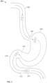
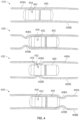

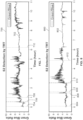


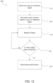


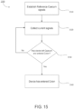
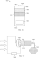

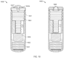
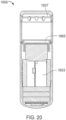


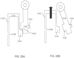


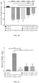


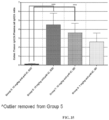
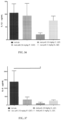
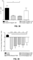


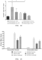


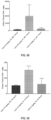

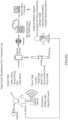
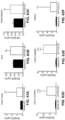
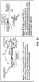
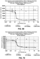


Classifications
-
- A—HUMAN NECESSITIES
- A61—MEDICAL OR VETERINARY SCIENCE; HYGIENE
- A61K—PREPARATIONS FOR MEDICAL, DENTAL OR TOILETRY PURPOSES
- A61K45/00—Medicinal preparations containing active ingredients not provided for in groups A61K31/00 - A61K41/00
-
- A—HUMAN NECESSITIES
- A61—MEDICAL OR VETERINARY SCIENCE; HYGIENE
- A61K—PREPARATIONS FOR MEDICAL, DENTAL OR TOILETRY PURPOSES
- A61K9/00—Medicinal preparations characterised by special physical form
- A61K9/0002—Galenical forms characterised by the drug release technique; Application systems commanded by energy
- A61K9/0009—Galenical forms characterised by the drug release technique; Application systems commanded by energy involving or responsive to electricity, magnetism or acoustic waves; Galenical aspects of sonophoresis, iontophoresis, electroporation or electroosmosis
-
- A—HUMAN NECESSITIES
- A61—MEDICAL OR VETERINARY SCIENCE; HYGIENE
- A61K—PREPARATIONS FOR MEDICAL, DENTAL OR TOILETRY PURPOSES
- A61K31/00—Medicinal preparations containing organic active ingredients
- A61K31/33—Heterocyclic compounds
- A61K31/395—Heterocyclic compounds having nitrogen as a ring hetero atom, e.g. guanethidine or rifamycins
- A61K31/495—Heterocyclic compounds having nitrogen as a ring hetero atom, e.g. guanethidine or rifamycins having six-membered rings with two or more nitrogen atoms as the only ring heteroatoms, e.g. piperazine or tetrazines
- A61K31/505—Pyrimidines; Hydrogenated pyrimidines, e.g. trimethoprim
- A61K31/519—Pyrimidines; Hydrogenated pyrimidines, e.g. trimethoprim ortho- or peri-condensed with heterocyclic rings
-
- A—HUMAN NECESSITIES
- A61—MEDICAL OR VETERINARY SCIENCE; HYGIENE
- A61B—DIAGNOSIS; SURGERY; IDENTIFICATION
- A61B1/00—Instruments for performing medical examinations of the interior of cavities or tubes of the body by visual or photographical inspection, e.g. endoscopes; Illuminating arrangements therefor
- A61B1/00002—Operational features of endoscopes
- A61B1/00004—Operational features of endoscopes characterised by electronic signal processing
- A61B1/00009—Operational features of endoscopes characterised by electronic signal processing of image signals during a use of endoscope
-
- A—HUMAN NECESSITIES
- A61—MEDICAL OR VETERINARY SCIENCE; HYGIENE
- A61B—DIAGNOSIS; SURGERY; IDENTIFICATION
- A61B1/00—Instruments for performing medical examinations of the interior of cavities or tubes of the body by visual or photographical inspection, e.g. endoscopes; Illuminating arrangements therefor
- A61B1/00064—Constructional details of the endoscope body
- A61B1/00071—Insertion part of the endoscope body
- A61B1/0008—Insertion part of the endoscope body characterised by distal tip features
- A61B1/00097—Sensors
-
- A—HUMAN NECESSITIES
- A61—MEDICAL OR VETERINARY SCIENCE; HYGIENE
- A61B—DIAGNOSIS; SURGERY; IDENTIFICATION
- A61B1/00—Instruments for performing medical examinations of the interior of cavities or tubes of the body by visual or photographical inspection, e.g. endoscopes; Illuminating arrangements therefor
- A61B1/00131—Accessories for endoscopes
-
- A—HUMAN NECESSITIES
- A61—MEDICAL OR VETERINARY SCIENCE; HYGIENE
- A61B—DIAGNOSIS; SURGERY; IDENTIFICATION
- A61B1/00—Instruments for performing medical examinations of the interior of cavities or tubes of the body by visual or photographical inspection, e.g. endoscopes; Illuminating arrangements therefor
- A61B1/012—Instruments for performing medical examinations of the interior of cavities or tubes of the body by visual or photographical inspection, e.g. endoscopes; Illuminating arrangements therefor characterised by internal passages or accessories therefor
- A61B1/015—Control of fluid supply or evacuation
-
- A—HUMAN NECESSITIES
- A61—MEDICAL OR VETERINARY SCIENCE; HYGIENE
- A61B—DIAGNOSIS; SURGERY; IDENTIFICATION
- A61B1/00—Instruments for performing medical examinations of the interior of cavities or tubes of the body by visual or photographical inspection, e.g. endoscopes; Illuminating arrangements therefor
- A61B1/04—Instruments for performing medical examinations of the interior of cavities or tubes of the body by visual or photographical inspection, e.g. endoscopes; Illuminating arrangements therefor combined with photographic or television appliances
- A61B1/041—Capsule endoscopes for imaging
-
- A—HUMAN NECESSITIES
- A61—MEDICAL OR VETERINARY SCIENCE; HYGIENE
- A61B—DIAGNOSIS; SURGERY; IDENTIFICATION
- A61B1/00—Instruments for performing medical examinations of the interior of cavities or tubes of the body by visual or photographical inspection, e.g. endoscopes; Illuminating arrangements therefor
- A61B1/04—Instruments for performing medical examinations of the interior of cavities or tubes of the body by visual or photographical inspection, e.g. endoscopes; Illuminating arrangements therefor combined with photographic or television appliances
- A61B1/045—Control thereof
-
- A—HUMAN NECESSITIES
- A61—MEDICAL OR VETERINARY SCIENCE; HYGIENE
- A61B—DIAGNOSIS; SURGERY; IDENTIFICATION
- A61B1/00—Instruments for performing medical examinations of the interior of cavities or tubes of the body by visual or photographical inspection, e.g. endoscopes; Illuminating arrangements therefor
- A61B1/273—Instruments for performing medical examinations of the interior of cavities or tubes of the body by visual or photographical inspection, e.g. endoscopes; Illuminating arrangements therefor for the upper alimentary canal, e.g. oesophagoscopes, gastroscopes
- A61B1/2736—Gastroscopes
-
- A—HUMAN NECESSITIES
- A61—MEDICAL OR VETERINARY SCIENCE; HYGIENE
- A61K—PREPARATIONS FOR MEDICAL, DENTAL OR TOILETRY PURPOSES
- A61K31/00—Medicinal preparations containing organic active ingredients
-
- A—HUMAN NECESSITIES
- A61—MEDICAL OR VETERINARY SCIENCE; HYGIENE
- A61K—PREPARATIONS FOR MEDICAL, DENTAL OR TOILETRY PURPOSES
- A61K31/00—Medicinal preparations containing organic active ingredients
- A61K31/33—Heterocyclic compounds
- A61K31/395—Heterocyclic compounds having nitrogen as a ring hetero atom, e.g. guanethidine or rifamycins
- A61K31/54—Heterocyclic compounds having nitrogen as a ring hetero atom, e.g. guanethidine or rifamycins having six-membered rings with at least one nitrogen and one sulfur as the ring hetero atoms, e.g. sulthiame
- A61K31/542—Heterocyclic compounds having nitrogen as a ring hetero atom, e.g. guanethidine or rifamycins having six-membered rings with at least one nitrogen and one sulfur as the ring hetero atoms, e.g. sulthiame ortho- or peri-condensed with heterocyclic ring systems
- A61K31/545—Compounds containing 5-thia-1-azabicyclo [4.2.0] octane ring systems, i.e. compounds containing a ring system of the formula:, e.g. cephalosporins, cefaclor, or cephalexine
- A61K31/546—Compounds containing 5-thia-1-azabicyclo [4.2.0] octane ring systems, i.e. compounds containing a ring system of the formula:, e.g. cephalosporins, cefaclor, or cephalexine containing further heterocyclic rings, e.g. cephalothin
-
- A—HUMAN NECESSITIES
- A61—MEDICAL OR VETERINARY SCIENCE; HYGIENE
- A61K—PREPARATIONS FOR MEDICAL, DENTAL OR TOILETRY PURPOSES
- A61K39/00—Medicinal preparations containing antigens or antibodies
- A61K39/395—Antibodies; Immunoglobulins; Immune serum, e.g. antilymphocytic serum
-
- A—HUMAN NECESSITIES
- A61—MEDICAL OR VETERINARY SCIENCE; HYGIENE
- A61K—PREPARATIONS FOR MEDICAL, DENTAL OR TOILETRY PURPOSES
- A61K45/00—Medicinal preparations containing active ingredients not provided for in groups A61K31/00 - A61K41/00
- A61K45/06—Mixtures of active ingredients without chemical characterisation, e.g. antiphlogistics and cardiaca
-
- A—HUMAN NECESSITIES
- A61—MEDICAL OR VETERINARY SCIENCE; HYGIENE
- A61K—PREPARATIONS FOR MEDICAL, DENTAL OR TOILETRY PURPOSES
- A61K9/00—Medicinal preparations characterised by special physical form
- A61K9/0002—Galenical forms characterised by the drug release technique; Application systems commanded by energy
- A61K9/0004—Osmotic delivery systems; Sustained release driven by osmosis, thermal energy or gas
-
- A—HUMAN NECESSITIES
- A61—MEDICAL OR VETERINARY SCIENCE; HYGIENE
- A61K—PREPARATIONS FOR MEDICAL, DENTAL OR TOILETRY PURPOSES
- A61K9/00—Medicinal preparations characterised by special physical form
- A61K9/0012—Galenical forms characterised by the site of application
- A61K9/0053—Mouth and digestive tract, i.e. intraoral and peroral administration
- A61K9/0056—Mouth soluble or dispersible forms; Suckable, eatable, chewable coherent forms; Forms rapidly disintegrating in the mouth; Lozenges; Lollipops; Bite capsules; Baked products; Baits or other oral forms for animals
-
- A—HUMAN NECESSITIES
- A61—MEDICAL OR VETERINARY SCIENCE; HYGIENE
- A61K—PREPARATIONS FOR MEDICAL, DENTAL OR TOILETRY PURPOSES
- A61K9/00—Medicinal preparations characterised by special physical form
- A61K9/0087—Galenical forms not covered by A61K9/02 - A61K9/7023
- A61K9/0097—Micromachined devices; Microelectromechanical systems [MEMS]; Devices obtained by lithographic treatment of silicon; Devices comprising chips
-
- A—HUMAN NECESSITIES
- A61—MEDICAL OR VETERINARY SCIENCE; HYGIENE
- A61K—PREPARATIONS FOR MEDICAL, DENTAL OR TOILETRY PURPOSES
- A61K9/00—Medicinal preparations characterised by special physical form
- A61K9/48—Preparations in capsules, e.g. of gelatin, of chocolate
- A61K9/4808—Preparations in capsules, e.g. of gelatin, of chocolate characterised by the form of the capsule or the structure of the filling; Capsules containing small tablets; Capsules with outer layer for immediate drug release
-
- A—HUMAN NECESSITIES
- A61—MEDICAL OR VETERINARY SCIENCE; HYGIENE
- A61P—SPECIFIC THERAPEUTIC ACTIVITY OF CHEMICAL COMPOUNDS OR MEDICINAL PREPARATIONS
- A61P1/00—Drugs for disorders of the alimentary tract or the digestive system
-
- A—HUMAN NECESSITIES
- A61—MEDICAL OR VETERINARY SCIENCE; HYGIENE
- A61P—SPECIFIC THERAPEUTIC ACTIVITY OF CHEMICAL COMPOUNDS OR MEDICINAL PREPARATIONS
- A61P1/00—Drugs for disorders of the alimentary tract or the digestive system
- A61P1/04—Drugs for disorders of the alimentary tract or the digestive system for ulcers, gastritis or reflux esophagitis, e.g. antacids, inhibitors of acid secretion, mucosal protectants
-
- A—HUMAN NECESSITIES
- A61—MEDICAL OR VETERINARY SCIENCE; HYGIENE
- A61P—SPECIFIC THERAPEUTIC ACTIVITY OF CHEMICAL COMPOUNDS OR MEDICINAL PREPARATIONS
- A61P29/00—Non-central analgesic, antipyretic or antiinflammatory agents, e.g. antirheumatic agents; Non-steroidal antiinflammatory drugs [NSAID]
-
- A—HUMAN NECESSITIES
- A61—MEDICAL OR VETERINARY SCIENCE; HYGIENE
- A61P—SPECIFIC THERAPEUTIC ACTIVITY OF CHEMICAL COMPOUNDS OR MEDICINAL PREPARATIONS
- A61P35/00—Antineoplastic agents
-
- G—PHYSICS
- G01—MEASURING; TESTING
- G01N—INVESTIGATING OR ANALYSING MATERIALS BY DETERMINING THEIR CHEMICAL OR PHYSICAL PROPERTIES
- G01N33/00—Investigating or analysing materials by specific methods not covered by groups G01N1/00 - G01N31/00
- G01N33/48—Biological material, e.g. blood, urine; Haemocytometers
- G01N33/50—Chemical analysis of biological material, e.g. blood, urine; Testing involving biospecific ligand binding methods; Immunological testing
- G01N33/68—Chemical analysis of biological material, e.g. blood, urine; Testing involving biospecific ligand binding methods; Immunological testing involving proteins, peptides or amino acids
- G01N33/6863—Cytokines, i.e. immune system proteins modifying a biological response such as cell growth proliferation or differentiation, e.g. TNF, CNF, GM-CSF, lymphotoxin, MIF or their receptors
-
- A—HUMAN NECESSITIES
- A61—MEDICAL OR VETERINARY SCIENCE; HYGIENE
- A61K—PREPARATIONS FOR MEDICAL, DENTAL OR TOILETRY PURPOSES
- A61K39/00—Medicinal preparations containing antigens or antibodies
- A61K2039/505—Medicinal preparations containing antigens or antibodies comprising antibodies
-
- A—HUMAN NECESSITIES
- A61—MEDICAL OR VETERINARY SCIENCE; HYGIENE
- A61K—PREPARATIONS FOR MEDICAL, DENTAL OR TOILETRY PURPOSES
- A61K9/00—Medicinal preparations characterised by special physical form
- A61K9/08—Solutions
-
- A—HUMAN NECESSITIES
- A61—MEDICAL OR VETERINARY SCIENCE; HYGIENE
- A61K—PREPARATIONS FOR MEDICAL, DENTAL OR TOILETRY PURPOSES
- A61K9/00—Medicinal preparations characterised by special physical form
- A61K9/10—Dispersions; Emulsions
-
- A—HUMAN NECESSITIES
- A61—MEDICAL OR VETERINARY SCIENCE; HYGIENE
- A61M—DEVICES FOR INTRODUCING MEDIA INTO, OR ONTO, THE BODY; DEVICES FOR TRANSDUCING BODY MEDIA OR FOR TAKING MEDIA FROM THE BODY; DEVICES FOR PRODUCING OR ENDING SLEEP OR STUPOR
- A61M2210/00—Anatomical parts of the body
- A61M2210/10—Trunk
- A61M2210/1042—Alimentary tract
- A61M2210/1053—Stomach
- A61M2210/1057—Duodenum
-
- A—HUMAN NECESSITIES
- A61—MEDICAL OR VETERINARY SCIENCE; HYGIENE
- A61M—DEVICES FOR INTRODUCING MEDIA INTO, OR ONTO, THE BODY; DEVICES FOR TRANSDUCING BODY MEDIA OR FOR TAKING MEDIA FROM THE BODY; DEVICES FOR PRODUCING OR ENDING SLEEP OR STUPOR
- A61M2210/00—Anatomical parts of the body
- A61M2210/10—Trunk
- A61M2210/1042—Alimentary tract
- A61M2210/106—Small intestine
-
- A—HUMAN NECESSITIES
- A61—MEDICAL OR VETERINARY SCIENCE; HYGIENE
- A61M—DEVICES FOR INTRODUCING MEDIA INTO, OR ONTO, THE BODY; DEVICES FOR TRANSDUCING BODY MEDIA OR FOR TAKING MEDIA FROM THE BODY; DEVICES FOR PRODUCING OR ENDING SLEEP OR STUPOR
- A61M31/00—Devices for introducing or retaining media, e.g. remedies, in cavities of the body
-
- G—PHYSICS
- G01—MEASURING; TESTING
- G01N—INVESTIGATING OR ANALYSING MATERIALS BY DETERMINING THEIR CHEMICAL OR PHYSICAL PROPERTIES
- G01N2333/00—Assays involving biological materials from specific organisms or of a specific nature
- G01N2333/435—Assays involving biological materials from specific organisms or of a specific nature from animals; from humans
- G01N2333/52—Assays involving cytokines
- G01N2333/525—Tumor necrosis factor [TNF]
-
- G—PHYSICS
- G01—MEASURING; TESTING
- G01N—INVESTIGATING OR ANALYSING MATERIALS BY DETERMINING THEIR CHEMICAL OR PHYSICAL PROPERTIES
- G01N2333/00—Assays involving biological materials from specific organisms or of a specific nature
- G01N2333/435—Assays involving biological materials from specific organisms or of a specific nature from animals; from humans
- G01N2333/52—Assays involving cytokines
- G01N2333/54—Interleukins [IL]
- G01N2333/5412—IL-6
-
- G—PHYSICS
- G01—MEASURING; TESTING
- G01N—INVESTIGATING OR ANALYSING MATERIALS BY DETERMINING THEIR CHEMICAL OR PHYSICAL PROPERTIES
- G01N2333/00—Assays involving biological materials from specific organisms or of a specific nature
- G01N2333/435—Assays involving biological materials from specific organisms or of a specific nature from animals; from humans
- G01N2333/52—Assays involving cytokines
- G01N2333/54—Interleukins [IL]
- G01N2333/545—IL-1
-
- G—PHYSICS
- G01—MEASURING; TESTING
- G01N—INVESTIGATING OR ANALYSING MATERIALS BY DETERMINING THEIR CHEMICAL OR PHYSICAL PROPERTIES
- G01N2333/00—Assays involving biological materials from specific organisms or of a specific nature
- G01N2333/435—Assays involving biological materials from specific organisms or of a specific nature from animals; from humans
- G01N2333/52—Assays involving cytokines
- G01N2333/54—Interleukins [IL]
- G01N2333/55—IL-2
-
- G—PHYSICS
- G01—MEASURING; TESTING
- G01N—INVESTIGATING OR ANALYSING MATERIALS BY DETERMINING THEIR CHEMICAL OR PHYSICAL PROPERTIES
- G01N2333/00—Assays involving biological materials from specific organisms or of a specific nature
- G01N2333/435—Assays involving biological materials from specific organisms or of a specific nature from animals; from humans
- G01N2333/52—Assays involving cytokines
- G01N2333/555—Interferons [IFN]
- G01N2333/57—IFN-gamma
Definitions
- compositions for treating diseases of the gastrointestinal tract with a JAK inhibitor This disclosure features compositions for treating diseases of the gastrointestinal tract with a JAK inhibitor.
- the Janus kinase (JAK) proteins are a family of non-receptor tyrosine kinases that possess a highly conserved kinase domain responsible for its enzymatic activity. Mammals have four members of this family, JAK1, JAK2, JAK3 and Tyrosine kinase 2 (TYK2). These kinases associate with the intracellular portion of cytokine or hormone receptors, and transduce signals through seven members of the STAT transcription factors - STAT1, STAT2, STAT3, STAT4, STAT5A, STAT5B, and STAT6 in various combinations.
- JAK1 promotes signaling of multiple cytokines (e.g., certain type I and type II cytokines), including pro-inflammatory cytokines involved in the pathogenesis of autoimmune diseases.
- cytokines e.g., certain type I and type II cytokines
- pro-inflammatory cytokines involved in the pathogenesis of autoimmune diseases Single-nucleotide polymorphisms within the JAK/STAT pathway that confer susceptibility to IBD have been identified ( Jostins et al., Host-microbe interactions have shaped the genetic architecture of inflammatory bowel disease, Nature 491:119-124, 2012 ). Both in viva and in vitro studies have confirmed the role of JAK/STAT signaling in regulating immune responses ( O'Shea and Plenge, JAK and STAT signaling molecules in immunoregulation and immune-mediated disease, Immunity 36:542-550, 2012 ).
- Tofacitinib selective oral JAK inhibitor (including JAK1 and JAK3) has been tested in clinical trials for both ulcerative colitis ( Sandborn et al., Tofacitinib, an Oral Janus Kinase Inhibitor, in Active Ulcerative Colitis, New England J. Med 367:616-24, 2012 ; Panes et al., Randomized trial of tofacitinib in active ulcerative colitis: analysis of efficacy base on patient-reported outcomes, BMC Gastroenterology 15:14, doi: 10.1186/sl2876-015-0239-9, 2015 ) and Crohn's disease.
- the gastrointestinal (GI) tract generally provides a therapeutic medium for an individual's body.
- therapeutic drugs may need to be dispensed to specified locations within the small intestine or large intestine, which is more effective than oral administration of the therapeutic drugs to cure or alleviate the symptoms of some medical conditions.
- therapeutic drugs dispensed directly within the small intestine would not be contaminated, digested or otherwise compromised in the stomach, and thus allow a higher dose to be delivered at a specific location within the small intestine.
- dispensing therapeutic drugs directly within the small intestine inside a human body can be difficult, because a device or mechanism (e.g., special formulation) would be needed to transport a therapeutically effective dose of drug to a desired location within the small intestine and then automatically deliver the therapeutic drug at the desired location.
- Dispensing therapeutic drugs directly within other locations in the GI tract of the human body can be similarly difficult.
- Such a device or mechanism also would also need to be operated in a safe manner in that the device or mechanism needs to physically enter the human body.
- Ingestible devices for delivery of drugs in the gastrointestinal tract have been described in prior art ( J. Drug Delivery & Ther. 2(3):1-8, 2012 ; Intl. J. Pharm. Ind. 6(3):72-74 ; Gastrointest. Endosc. 78(3);520-528 ; WO2016049602 ; WO2004/066903 ).
- IBD inflammatory bowel disease
- the present disclosure provides novel treatment paradigms for inflammatory conditions of the gastrointestinal tract.
- the methods and compositions described herein allow for the regio-specific release of therapeutic drugs in the cecum, wherein the site of disease is in the colon.
- the bioavailability of the drug can be increased at the site of injury and/or decreased in the systemic circulation, thereby resulting in improved overall safety and/or efficacy and fewer adverse side effects.
- Advantages may include one or more of increased drug engagement at the target, leading to new and more efficacious treatment regimens, and/or lower systemic drug levels, which can translate to reduced toxicity and reduced immunogenicity, e.g., in the case of biologics.
- releasing a therapeutic drug locally also provides for new modes of action that may be unique to local delivery in the cecum as opposed to systemic administration. For patients, clinicians and payors, this can mean an easier or simpler route of administration, fewer co-medicaments (e.g., immunomodulators), fewer side effects, and/or better outcomes.
- co-medicaments e.g., immunomodulators
- the methods can include one or more of:
- the present disclosure accordingly provides patients and physicians more personalized treatment options for GI disorders by facilitating regimens which can release a therapeutic agent according to desired (e.g., customized or optimized) dosage, timing, and/or location parameters.
- the treatment methods can employ one or more ingestible devices to achieve the benefits disclosed herein.
- a method of treating a disease of the gastrointestinal tract in a subject comprising:
- the pharmaceutical formulation is administered in an ingestible device.
- the pharmaceutical formulation is released from an ingestible device.
- the ingestible device comprises a housing, a reservoir containing the pharmaceutical formulation, and a release mechanism for releasing the pharmaceutical formulation from the device, wherein the reservoir is releasably or permanently attached to the exterior of the housing or internal to the housing.
- a disease of the gastrointestinal tract in a subject comprising:
- the housing is non-biodegradable in the GI tract. In some embodiments, the release of the formulation is triggered autonomously. In some embodiments, the location of one or more sites of disease is predetermined.
- the reservoir is made of a material that allows the formulation to leave the reservoir, such as a biodegradable material.
- the release of the formulation is triggered by a preprogrammed algorithm. In some embodiments, the release of the formulation is triggered by data from a sensor or detector to identify the location of the device. In some more particular embodiments, the data is not based solely on a physiological parameter (such as pH, temperature, and/or transit time).
- a physiological parameter such as pH, temperature, and/or transit time
- the device comprises a detector configured to detect light reflectance from an environment external to the housing.
- the release is triggered autonomously or based on the detected reflectance.
- the device releases the formulation at substantially the same time as one or more sites of disease are detected.
- the one or more sites of disease are detected by the device (e.g., by imaging the GI tract).
- the release mechanism is an actuation system. In some embodiments, the release mechanism is a chemical actuation system. In some embodiments, the release mechanism is a mechanical actuation system. In some embodiments, the release mechanism is an electrical actuation system. In some embodiments, the actuation system comprises a pump and releasing the formulation comprises pumping the formulation out of the reservoir. In some embodiments, the actuation system comprises a gas generating cell. In some embodiments, the formulation comprises a therapeutically effective amount of the JAK inhibitor. In some embodiments, the formulation comprises a human equivalent dose (HED) of the JAK inhibitor.
- HED human equivalent dose
- the device is a device capable of releasing a solid JAK inhibitor or a solid formulation comprising the JAK inhibitor. In some embodiments, the device is a device capable of releasing a liquid JAK inhibitor or a liquid formulation comprising the JAK inhibitor. Accordingly, in some embodiments, the pharmaceutical formulation released from the device is a solid formulation. In other embodiments, the pharmaceutical formulation release from the device is a liquid formulation.
- the devices disclosed herein are capable of releasing a JAK inhibitor or a formulation comprising the JAK inhibitor irrespective of the particular type of JAK inhibitor.
- the JAK inhibitor may be a small molecule, a biological, a nucleic acid, an antibody, a fusion protein, and so on.
- a method of releasing an JAK inhibitor into the cecum of a subject for treating one or more sites of disease within the colon comprising: administering to the subject a therapeutically effective amount of the JAK inhibitor housed in an ingestible device, wherein the ingestible device comprises:
- Also provided herein is a method of releasing an JAK inhibitor into the cecum of a subject for treating one or more pre-determined sites of disease within the colon, the method comprising: administering to the subject a therapeutically effective amount of the JAK inhibitor contained in an ingestible device, wherein the ingestible device comprises:
- a JAK inhibitor into the cecum of a subject for treating one or more sites of disease within the colon, the method comprising:
- a method of releasing an JAK inhibitor into the cecum of a subject for treating one or more sites of disease within the colon comprising:
- a method of treating a disease of the gastrointestinal tract in a subject comprising: delivering a JAK inhibitor to the cecum of the subject, wherein the method comprises administering to the subject a pharmaceutical composition comprising a therapeutically effective amount of the JAK inhibitor.
- a method of treating a disease of the gastrointestinal tract in a subject comprising: releasing a JAK inhibitor in the cecum of the subject, wherein the method comprises administering to the subject a pharmaceutical composition comprising a therapeutically effective amount of the JAK inhibitor.
- a method of treating a disease of the gastrointestinal tract in a subject comprising: releasing a JAK inhibitor in the cecum of the subject wherein the method comprises administering to the subject a pharmaceutical composition comprising a therapeutically effective amount of the JAK inhibitor, wherein the pharmaceutical composition is an ingestible device. and the method comprises administering orally to the subject the pharmaceutical composition.
- a method of treating a disease of the gastrointestinal tract in a subject comprising: releasing a JAK inhibitor in the cecum of the subject, wherein the method comprises administering to the subject a pharmaceutical composition comprising a therapeutically effective amount of the JAK inhibitor, wherein the method provides a concentration of the JAK inhibitor in the plasma of the subject that is less than 3 ⁇ g/ml.
- a JAK inhibitor for use in a method of treating a disease of the gastrointestinal tract in a subject according to claim 1.
- the present disclosure provides a composition comprising or consisting of an ingestible device loaded with a therapeutically effective amount of a JAK inhibitor, for use in a method of treatment, wherein the method comprises orally administering the composition to the subject, wherein the JAK inhibitor is released by the device in the cecum.
- the present disclosure provides an ingestible device loaded with a therapeutically effective amount of a JAK inhibitor, wherein the device is controllable to release the JAK inhibitor in the cecum of the subject.
- the device may be for use in a method of treatment of the human or animal body, for example, any method as described herein.
- the present disclosure provides an ingestible device for use in a method of treating a disease of the gastrointestinal tract in a subject, wherein the method comprises orally administering to the subject the ingestible device loaded with a therapeutically effective amount of a JAK inhibitor, wherein the JAK inhibitor is released by the device in the cecum of the subject.
- An ingestible device as used in the present disclosure may comprise one or more mechanical and/or electrical mechanisms which actively control release of the JAK inhibitor.
- the ingestible device as used in the present invention may comprise a release mechanism for release of the JAK inhibitor (e.g., from a reservoir comprising the JAK inhibitor) and an actuator controlling the release mechanism.
- the ingestible device comprises:
- the ingestible device comprises
- the exit valve can be considered as the release mechanism having a closed state which retains the JAK inhibitor in the reservoir and an open state which releases the JAK inhibitor from the reservoir to the exterior of the device, and the mechanism for releasing the JAK inhibitor from the reservoir can be considered as the actuator.
- the one or more disease sites may have been pre-determined (e.g., determined in a step preceding the administration of the composition of the present disclosure).
- the disease site(s) may have been determined by imaging the gastrointestinal tract.
- the disease site(s) may have been pre-determined by endoscopy (e.g., a step of colonoscopy, enteroscopy, or using a capsule endoscope).
- the location of the device in the gut may be detected by tracking the device.
- the device may comprise a localization mechanism which may be a communication system for transmitting localization data, e.g., by radiofrequency transmission.
- the device may additionally or alternatively comprise a communication system for receiving a signal remotely triggering the actuator and thus causing release of the JAK inhibitor. The signal may be sent when it is determined that the device is in the correct location in the gut.
- the ingestible device may comprise:
- the ingestible device as used in the present invention may comprise an environmental sensor for detecting the location of the device in the gut
- the environment sensor may be an image sensor for obtaining images in vivo.
- actuation of the release mechanism may be triggered by a processor or controller communicably coupled to the environmental sensor.
- the device may not require any external signal or control in order to release the drug.
- the ingestible device comprises:
- detection of a transition from the ileum to the cecum may be based on environmental data indicating the location of the device in the GI tract (and reference to a pre-determined disease site).
- the device may further comprise a communication system adapted to transmit the environment data to an external receiver (e.g., outside of the body).
- This data may be used, for example, for diagnostic purposes.
- the external receiver may comprise means for displaying the data.
- an external signal may then be sent to the device to trigger release of the drug.
- the communication system may further be adapted to receive a signal remotely triggering the actuator and thus causing release of the JAK inhibitor.
- the signal may be sent from an external transmitter in response to receipt/analysis and/or assessment of the environmental data, e.g., data indicating that the device has reached the desired location of the gut (where the location of the diseased tissue has been pre-determined) and/or data indicating the presence of diseased tissue.
- “External” may be "outside of the body”.
- the ingestible device may comprise:
- a system comprising:
- disease of the GI tract may be an inflammatory bowel disease.
- the disease of the GI tract is ulcerative colitis.
- the disease of the GI tract is Crohn's disease.
- gastrointestinal tract diseases that can be treated include, without limitation, inflammatory bowel disease (IBD), Crohn's disease (e.g., active Crohn's disease, refractory Crohn's disease, or fistulizing Crohn's disease), ulcerative colitis, indeterminate colitis, microscopic colitis, infectious colitis, drug or chemical-induced colitis, diverticulitis, and ischemic colitis, gastritis, peptic ulcers, stress ulcers, bleeding ulcers, gastric hyperacidity, dyspepsia, gastroparesis, Zollinger-Ellison syndrome, gastroesophageal reflux disease, short-bowel (anastomosis) syndrome, a hypersecretory state associated with systemic mastocytosis or basophilic leukemia or hyperhistaminemia, Celiac disease (e.g., nontropical Sprue), enteropathy associated with IBD
- Crohn's disease e.g., active Crohn's disease, refractory Crohn'
- apparatuses, compositions, and methods disclosed herein are for use to treat one gastrointestinal disease. In some embodiments, apparatuses, compositions, and methods disclosed herein are for use to treat more than one gastrointestinal disease. In some embodiments, apparatuses, compositions, and methods disclosed herein are for use to treat multiple gastrointestinal diseases that occur in the same area of the gastrointestinal tract (the colon, or any sub-region thereof). In some disclosures, apparatuses, compositions, and methods disclosed herein are used to treat multiple gastrointestinal diseases that occur in different areas of the gastrointestinal tract.
- administration e.g., local administration to the cecum
- JAK inhibitor is useful in the treatment of gastrointestinal diseases including, but not limited to, inflammatory bowel disease (IBD), ulcerative colitis, Crohn's disease, or any of the other gastrointestinal diseases described herein.
- IBD inflammatory bowel disease
- ulcerative colitis Crohn's disease
- Crohn's disease or any of the other gastrointestinal diseases described herein.
- administration e.g., local administration to the gastrointestinal tract
- JAK inhibitor is useful in the treatment of gastrointestinal diseases including, but not limited to, inflammatory bowel disease (IBD), ulcerative colitis, Crohn's disease, or any of the other gastrointestinal diseases described herein.
- IBD inflammatory bowel disease
- ulcerative colitis Crohn's disease
- Crohn's disease or any of the other gastrointestinal diseases described herein.
- any details described herein for methods of treatment apply equally to a JAK inhibitor, composition or ingestible device for use in said treatment.
- Any details described for a device apply equally to methods of treatment using the device, or to a JAK inhibitor or composition for use in a method of treatment involving the device.
- a method of treating a disease of the gastrointestinal tract in a subject comprises administering to the subject a pharmaceutical formulation comprising a JAK inhibitor wherein the pharmaceutical formulation is released in the subject's cecum.
- the pharmaceutical formulation comprises a therapeutically effective amount of a JAK inhibitor.
- the formulation is contained in an ingestible device, and the device releases the formulation in the cecum.
- the location of the site of disease may be predetermined.
- the release of the formulation may be triggered autonomously, as further described herein.
- a "formulation" of a JAK inhibitor may refer to either the JAK inhibitor in pure form, such as, for example, a lyophilized JAK inhibitor, or a mixture of the JAK inhibitor with one or more physiologically acceptable carriers, excipients or stabilizers.
- therapeutic formulations or medicaments can be prepared by mixing the JAK inhibitor having the desired degree of purity with optional physiologically acceptable carriers, excipients or stabilizers ( Remington's Pharmaceutical Sciences 16th edition, Osol, A. Ed. (1980 )), in the form of lyophilized formulations or aqueous solutions.
- Acceptable carriers, excipients, or stabilizers are nontoxic to recipients at the dosages and concentrations employed, and include buffers such as phosphate, citrate, and other organic acids; antioxidants including ascorbic acid and methionine; preservatives (such as octadecyldimethylbenzyl ammonium chloride; hexamethonium chloride; benzalkonium chloride, benzethonium chloride; phenol, butyl or benzyl alcohol; alkyl parabens such as methyl or propyl paraben; catechol; resorcinol; cyclohexanol; 3-pentanol; and m-cresol); low molecular weight (less than about 10 residues) antibody; proteins, such as serum albumin, gelatin, or immunoglobulins; hydrophilic polymers such as polyvinylpyrrolidone; amino acids such as glycine, glutamine, asparagine, histidine, arginine
- Exemplary pharmaceutically acceptable carriers herein further include insterstitial drug dispersion agents such as soluble neutral-active hyaluronidase glycoproteins (sHASEGP), for example, human soluble PH-20 hyaluronidase glycoproteins, such as rHuPH20 (HYLENEX ⁇ ® >, Baxter International, Inc.).
- sHASEGP soluble neutral-active hyaluronidase glycoproteins
- rHuPH20 HYLENEX ⁇ ® >, Baxter International, Inc.
- Certain exemplary sHASEGPs and methods of use, including rHuPH20 are described in US Patent Publication Nos. 2005/0260186 and 2006/0104968 .
- a sHASEGP is combined with one or more additional glycosaminoglycanases such as chondroitinases.
- Exemplary lyophilized formulations are described in US Patent No. 6,267,958 .
- Aqueous formulations include those described in US
- a formulation of a JAK inhibitor as disclosed herein, e.g., sustained-release formulations, can further include a mucoadhesive agent, e.g., one or more of polyvinyl pyrolidine, methyl cellulose, sodium carboxyl methyl cellulose, hydroxyl propyl cellulose, carbopol, a polyacrylate, chitosan, a eudragit analogue, a polymer, and a thiomer.
- a mucoadhesive agent e.g., one or more of polyvinyl pyrolidine, methyl cellulose, sodium carboxyl methyl cellulose, hydroxyl propyl cellulose, carbopol, a polyacrylate, chitosan, a eudragit analogue, a polymer, and a thiomer.
- mucoadhesive agents that can be included in a formulation with a JAK inhibitor are described in, e.g., Peppas et al., Biomaterials 17(16): 1553-1561, 1996 ; Kharenko et al., Pharmaceutical Chemistry J. 43(4):200-208, 2009 ; Salamat-Miller et al., Adv. Drug Deliv. Reviews 57(11):1666-1691, 2005 ; Bernkop-Schnurch, Adv. Drug Deliv. Rev. 57(11):1569-1582, 2005 ; and Harding et al., Biotechnol. Genet. Eng. News 16(1):41-86, 1999 .
- components of a formulation may include any one of the following components, or any combination thereof: Acacia, Alginate, Alginic Acid, Aluminum Acetate, an antiseptic, Benzyl Alcohol, Butyl Paraben, Butylated Hydroxy Toluene, an antioxidant.
- the method comprises administering to the subject a pharmaceutical composition that is a formulation as disclosed herein.
- the formulation is a dosage form, which may be, as an example, a solid form such as, for example, a capsule, a tablet, a sachet, or a lozenge; or which may be, as an example, a liquid form such as, for example, a solution, a suspension, an emulsion, or a syrup.
- the formulation is comprised in an ingestible device as defined in claim 1.
- the formulation may be suitable for oral administration.
- the formulation may be, for example, a solid dosage form or a liquid dosage form as disclosed herein.
- the formulation is suitable for introduction and optionally for storage in the device.
- the formulation is suitable for introduction and optionally for storage in a reservoir comprised in the device.
- the formulation is suitable for introduction and optionally for storage in a reservoir comprised in the device.
- a reservoir comprising a therapeutically effective amount of a JAK inhibitor, wherein the reservoir is configured to fit into an ingestible device.
- the reservoir comprising a therapeutically effective amount of an JAK inhibitor is attachable to an ingestible device
- the formulation is suitable for introduction in a spray catheter, as disclosed herein.
- the formulation herein may also contain more than one active compound as necessary for the particular indication being treated, for example, those with complementary activities that do not adversely affect each other.
- the formulation may further comprise another JAK inhibitor or a chemotherapeutic agent.
- Such molecules are suitably present in combination in amounts that are effective for the purpose intended.
- the active ingredients may also be entrapped in microcapsules prepared, for example, by coacervation techniques or by interfacial polymerization, for example, hydroxymethylcellulose or gelatin-microcapsule and poly-(methylmethacylate) microcapsule, respectively, in colloidal drug delivery systems (for example, liposomes, albumin microspheres, microemulsions, nano-particles and nanocapsules) or in macroemulsions.
- colloidal drug delivery systems for example, liposomes, albumin microspheres, microemulsions, nano-particles and nanocapsules
- macroemulsions for example, liposomes, albumin microspheres, microemulsions, nano-particles and nanocapsules
- the formulations to be used for in vivo administration must be sterile. This is readily accomplished by filtration through sterile filtration membranes. Sustained-release preparations may be prepared.
- sustained-release preparations include semipermeable matrices of solid hydrophobic polymers containing the JAK inhibitor, which matrices are in the form of shaped articles, e.g., films, or microcapsule.
- sustained-release matrices include polyesters, hydrogels (for example, poly(2- hydroxyethylmethacrylate), or poly(vinylalcohol)), polylactides ( U.S. Pat. No.
- copolymers of L-glutamic acid and ⁇ ethyl-L-glutamate copolymers of L-glutamic acid and ⁇ ethyl-L-glutamate, non-degradable ethylene-vinyl acetate, degradable lactic acid-glycolic acid copolymers such as the LUPRON DEPOT TM (injectable microspheres composed of lactic acid-glycolic acid copolymer and leuprolide acetate), and poly-D-(-)-3-hydroxybutyric acid. While polymers such as ethylene-vinyl acetate and lactic acid-glycolic acid enable release of molecules for over 100 days, certain hydrogels release proteins for shorter time periods.
- encapsulated JAK inhibitors When encapsulated JAK inhibitors remain in the body for a long time, they may denature or aggregate as a result of exposure to moisture at 37°C, resulting in a loss of biological activity and possible changes in immunogenicity. Rational strategies can be devised for stabilization depending on the mechanism involved. For example, if the aggregation mechanism is discovered to be intermolecular S-S bond formation through thio-disulfide interchange, stabilization may be achieved by modifying sulfhydryl residues, lyophilizing from acidic solutions, controlling moisture content, using appropriate additives, and developing specific polymer matrix compositions.
- compositions may contain one or more JAK inhibitors.
- the pharmaceutical formulations may be formulated in any manner known in the art.
- the formulations include one or more of the following components: a sterile diluent (e.g., sterile water or saline), a fixed oil, polyethylene glycol, glycerin, propylene glycol, or other synthetic solvents, antibacterial or antifungal agents, such as benzyl alcohol or methyl parabens, chlorobutanol, phenol, ascorbic acid, thimerosal, and the like, antioxidants, such as ascorbic acid or sodium bisulfite, chelating agents, such as ethylenediaminetetraacetic acid, buffers, such as acetates, citrates, or phosphates, and isotonic agents, such as sugars (e.g., dextrose), polyalcohols (e.g., mannitol or sorbitol), or salts (e.g.
- Liposomal suspensions can also be used as pharmaceutically acceptable carriers (see, e.g., U.S. Patent No. 4,522,811 ).
- the formulations can be formulated and enclosed in ampules, disposable syringes, or multiple dose vials. Where required, proper fluidity can be maintained by, for example, the use of a coating, such as lecithin, or a surfactant.
- Controlled release of the JAK inhibitor can be achieved by implants and microencapsulated delivery systems, which can include biodegradable, biocompatible polymers (e.g., ethylene vinyl acetate, polyanhydrides, polyglycolic acid, collagen, polyorthoesters, and polylactic acid; Alza Corporation and Nova Pharmaceutical, Inc.).
- the JAK inhibitor is present in a pharmaceutical formulation within the device.
- the JAK inhibitor is present in solution within the device.
- the JAK inhibitor is present in a suspension in a liquid medium within the device.
- the JAK inhibitor is present as a pure, powder (e.g., lyophilized) form of the JAK inhibitor.
- Gastrointestinal inflammatory disorders are a group of chronic disorders that cause inflammation and/or ulceration in the mucous membrane. These disorders include, for example, inflammatory bowel disease (e.g., Crohn's disease, ulcerative colitis, indeterminate colitis and infectious colitis), mucositis (e.g., oral mucositis, gastrointestinal mucositis, nasal mucositis and proctitis), necrotizing enterocolitis and esophagitis.
- IBD is a chronic inflammatory autoimmune condition of the gastrointestinal (GI) tract. The GI tract can be divided into four main different sections, the oesophagus, stomach, small intestine and large intestine or colon.
- the small intestine possesses three main subcompartments: the duodenum, jejunum and ileum.
- the large intestine consists of six sections: the cecum, ascending colon, transverse colon, ascending colon, sigmoid colon, and the rectum.
- the small intestine is about 6 m long, its diameter is 2.5 to 3 cm and the transit time through it is typically 3 hours.
- the duodenum has a C-shape, and is 30 cm long. Due to its direct connection with the stomach, it is physically more stable than the jejunum and ileum, which are sections that can freely move.
- the jejunum is 2.4 m in length and the ileum is 3.6 m in length and their surface areas are 180 m2 and 280 m2 respectively.
- the large intestine is 1.5 m long, its diameter is between 6.3 and 6.5 cm, the transit time though this section is 20 hours and has a reduced surface area of approximately 150 m2.
- the higher surface area of the small intestine enhances its capacity for systemic drug absorption.
- the etiology of IBD is complex, and many aspects of the pathogenesis remain unclear.
- the treatment of moderate to severe IBD poses significant challenges to treating physicians, because conventional therapy with corticosteroids and immunomodulator therapy (e.g., azathioprine, 6 mercaptopurine, and methotrexate administered via traditional routes such as tablet form, oral suspension, or intravenously) is associated with side effects and intolerance and has not shown proven benefit in maintenance therapy (steroids).
- corticosteroids and immunomodulator therapy e.g., azathioprine, 6 mercaptopurine, and methotrexate administered via traditional routes such as tablet form, oral suspension, or intravenously
- TNF-a tumor necrosis factor alpha
- infliximab a chimeric antibody
- adalimumab a fully human antibody
- AEs adverse events associated with anti TNFs
- Other adverse events (AEs) associated with anti TNFs include elevated rates of bacterial infection, including tuberculosis, and, more rarely, lymphoma and demyelination ( Chang et al., Nat Clin Pract Gastroenterol Hepatology 3:220 (2006 ); Hoentjen et al., World J. Gastroenterol. 15(17):2067 (2009 )).
- No currently available therapy achieves sustained remission in more than 20%-30% of IBD patients with chronic disease ( Hanauer et al, Lancet 359: 1541-49 (2002 ); Sandborn et al, N Engl J Med 353: 1912-25 (2005 )).
- most patients do not achieve sustained steroid-free remission and mucosal healing, clinical outcomes that correlate with true disease modification.
- IBD Inflammatory bowel syndrome
- GI gastrointestinal
- UC ulcerative colitis
- CD Crohn's disease
- CD Crohn's disease
- Crohn's disease may be continuous or relapsing, mild or severe, but unlike ulcerative colitis, Crohn's disease is not curable by resection of the involved segment of bowel. Most patients with Crohn's disease require surgery at some point, but subsequent relapse is common and continuous medical treatment is usual. Crohn's disease may involve any part of the alimentary tract from the mouth to the anus, although typically it appears in the ileocolic, small-intestinal or colonic-anorectal regions.
- the disease manifests by discontinuous granulomatomas, crypt abscesses, fissures and aphthous ulcers.
- the inflammatory infiltrate is mixed, consisting of lymphocytes (both T and B cells), plasma cells, macrophages, and neutrophils.
- lymphocytes both T and B cells
- plasma cells plasma cells
- macrophages and neutrophils.
- IgM- and IgG-secreting plasma cells macrophages and neutrophils.
- CDAI Crohn's Disease Activity Index
- the CDAI was developed by regressing clinician global assessment of disease activity on eighteen potential items representing patient reported outcomes (PROs) (i.e. abdominal pain, pain awakening patient from sleep, appetite), physical signs (i.e. average daily temperature, abdominal mass), medication use (i.e. loperamide or opiate use for diarrhea) and a laboratory test (i.e. hematocrit).
- PROs patient reported outcomes
- physical signs i.e. average daily temperature, abdominal mass
- medication use i.e. loperamide or opiate use for diarrhea
- a laboratory test i.e. hematocrit
- Backward stepwise regression analysis identified eight independent predictors which are the number of liquid or soft stools, severity of abdominal pain, general well-being, occurrence of extra-intestinal symptoms, need for anti diarrheal drugs, presence of an abdominal mass, hematocrit, and body weight.
- the final score is a composite of these eight items, adjusted using regression coefficients and standardization to construct an overall CDAI score, ranging from 0 to 600 with higher score indicating greater disease activity.
- Widely used benchmarks are: CDAI ⁇ 150 is defined as clinical remission, 150 to 219 is defined as mildly active disease, 220 to 450 is defined as moderately active disease, and above 450 is defined as very severe disease ( Best WR, et al., Gastroenterology 77:843-6, 1979 ).
- Vedolizumab and natalizumab have been approved on the basis of demonstrated clinical remission, i.e. CDAI ⁇ 150.
- CDAI has been in use for over 40 years, and has served as the basis for drug approval, it has several limitations as an outcome measure for clinical trials. For example, most of the overall score comes from the patient diary card items (pain, number of liquid bowel movements, and general well-being), which are vaguely defined and not standardized terms ( Sandler et al., J. Clin. Epidemiol 41 :451-8, 1988 ; Thia et al., Inflamm Bowel Dis 17: 105-11, 2011 ). In addition, measurement of pain is based on a four- point scale rather than an updated seven-point scale. The remaining 5 index items contribute very little to identifying an efficacy signal and may be a source of measurement noise.
- the PRO2 and PRO3 tools are such adaptations of the CDAI and have been recently described in Khanna et al., Aliment Pharmacol. Ther. 41: 77-86, 2015 .
- the PRO2 evaluates the frequency of loose/liquid stools and abdominal pain ⁇ Id). These items are derived and weighted accordingly from the CDAI and are the CDAI diary card items, along with general well-being, that contribute most to the observed clinical benefit measured by CDAI ( Sandler et al., J. Clin.
- the remission score of ⁇ 11 is the CDAI-weighted sum of the average stool frequency and pain scores in a 7-day period, which yielded optimum sensitivity and specificity for identification of CDAI remission (score of ⁇ 150) in a retrospective data analysis of ustekinumab induction treatment for moderate to severe CD in a Phase II clinical study ( Gasink C, et al., abstract, ACG Annual Meeting 2014 ).
- the PRO2 was shown to be sensitive and responsive when used as a continuous outcome measure in a retrospective data analysis of MTX treatment in active CD ( Khanna R, et al., Inflamm Bowel Dis 20: 1850-61, 2014 ) measured by CDAI. Additional outcome measures include the Mayo Clinic Score, the Crohn disease endoscopic index of severity (CDEIS), and the Ulcerative colitis endoscopic index of severity (UCEIS). Additional outcome measures include Clinical remission, Mucosal healing, Histological healing (transmural), MRI or ultrasound for measurement or evaluation of bowel wall thickness, abscesses, fistula and histology.
- SES- CD Simplified Endoscopic Activity Score for Crohn's Disease
- the current therapy of IBD usually involves the administration of antiinflammatory or immunosuppressive agents, such as sulfasalazine, corticosteroids, 6- mercaptopurine/azathioprine, or cyclosporine, all of which are not typically delivered by localized release of a drug at the site or location of disease.
- antiinflammatory or immunosuppressive agents such as sulfasalazine, corticosteroids, 6- mercaptopurine/azathioprine, or cyclosporine, all of which are not typically delivered by localized release of a drug at the site or location of disease.
- Refining a diagnosis of inflammatory bowel disease involves evaluating the progression status of the diseases using standard classification criteria.
- the classification systems used in IBD include the Truelove and Witts Index ( Truelove S. C. and Witts, L.J. Br Med J. 1955;2: 1041-1048 ), which classifies colitis as mild, moderate, or severe, as well as Lennard- Jones. ( Lennard-Jones JE. Scand J Gastroenterol Suppl 1989; 170:2-6 ) and the simple clinical colitis activity index (SCCAI). ( Walmsley et. al. Gut. 1998;43:29-32 ) These systems track such variables as daily bowel movements, rectal bleeding, temperature, heart rate, hemoglobin levels, erythrocyte sedimentation rate, weight, hematocrit score, and the level of serum albumin.
- UC ulcerative colitis
- CD can appear anywhere in the bowel, with occasional involvement of stomach, esophagus and duodenum, and the lesions are usually described as extensive linear fissures.
- pANCA perinuclear anti-neutrophil antibody
- ASCA anti-Saccharomyces cervisiae antibody
- a third test which measures the presence and accumulation of circulating anti-microbial antibodies - particularly flagellin antibodies, has proven to be useful for detecting susceptibility to Crohn's Disease before disease development. See Choung, R. S., et al. "Serologic microbial associated markers can predict Crohn's disease behaviour years before disease diagnosis.” Alimentary pharmacology & therapeutics 43.12 (2016): 1300-1310 .
- Ulcerative colitis afflicts the large intestine.
- the course of the disease may be continuous or relapsing, mild or severe.
- the earliest lesion is an inflammatory infiltration with abscess formation at the base of the crypts of Lieberkuhn. Coalescence of these distended and ruptured crypts tends to separate the overlying mucosa from its blood supply, leading to ulceration.
- Symptoms of the disease include cramping, lower abdominal pain, rectal bleeding, and frequent, loose discharges consisting mainly of blood, pus and mucus with scanty fecal particles.
- a total colectomy may be required for acute, severe or chronic, unremitting ulcerative colitis.
- UC ulcerative colitis
- antibody and “immunoglobulin” are used interchangeably in the broadest sense and include monoclonal antibodies (for example, full length or intact monoclonal antibodies), polyclonal antibodies, multivalent antibodies, multispecific antibodies (e.g., bispecific, trispecific etc. antibodies so long as they exhibit the desired biological activity) and may also include certain antibody fragments (as described in greater detail herein).
- An antibody can be human, humanized and/or affinity matured.
- Antibody fragments comprise only a portion of an intact antibody, where in certain embodiments, the portion retains at least one, and typically most or all, of the functions normally associated with that portion when present in an intact antibody.
- an antibody fragment comprises an antigen binding site of the intact antibody and thus retains the ability to bind antigen.
- an antibody fragment for example one that comprises the Fc region, retains at least one of the biological functions normally associated with the Fc region when present in an intact antibody, such as FcRn binding, antibody half-life modulation, ADCC function and complement binding.
- an antibody fragment is a monovalent antibody that has an in vivo half-life substantially similar to an intact antibody.
- such an antibody fragment may comprise on antigen binding arm linked to an Fc sequence capable of conferring in vivo stability to the fragment.
- monoclonal antibody refers to an antibody obtained from a population of substantially homogeneous antibodies, i.e., the individual antibodies comprising the population are identical except for possible naturally occurring mutations that may be present in minor amounts. Monoclonal antibodies are highly specific, being directed against a single antigen. Furthermore, in contrast to polyclonal antibody preparations that typically include different antibodies directed against different determinants (epitopes), each monoclonal antibody is directed against a single determinant on the antigen.
- the monoclonal antibodies herein specifically include "chimeric" antibodies in which a portion of the heavy and/or light chain is identical with or homologous to corresponding sequences in antibodies derived from a particular species or belonging to a particular antibody class or subclass, while the remainder of the chain(s) is identical with or homologous to corresponding sequences in antibodies derived from another species or belonging to another antibody class or subclass, as well as fragments of such antibodies, so long as they exhibit the desired biological activity ( U.S. Patent No. 4,816,567 ; and Morrison et al, Proc. Natl. Acad. Sci. USA 81 :6851-6855 (1984 )).
- Treatment regimen refers to a combination of dosage, frequency of administration, or duration of treatment, with or without addition of a second medication.
- Effective treatment regimen refers to a treatment regimen that will offer beneficial response to a patient receiving the treatment.
- Effective amount refers to an amount of drug that offers beneficial response to a patient receiving the treatment.
- an effective amount may be a Human Equivalent Dose (HED).
- Dispensable refers to any substance that may be released from an ingestible device as disclosed herein, or from a component of the device such as a reservoir.
- a dispensable substance may be an JAK inhibitor, and/or a formulation comprising an JAK inhibitor.
- Patient response or “patient responsiveness” can be assessed using any endpoint indicating a benefit to the patient, including, without limitation, (1) inhibition, to some extent, of disease progression, including slowing down and complete arrest; (2) reduction in the number of disease episodes and/or symptoms; (3) reduction in lesional size; (4) inhibition (i.e., reduction, slowing down or complete stopping) of disease cell infiltration into adjacent peripheral organs and/or tissues; (5) inhibition (i.e., reduction, slowing down or complete stopping) of disease spread; (6) decrease of auto-immune response, which may, but does not have to, result in the regression or ablation of the disease lesion; (7) relief, to some extent, of one or more symptoms associated with the disorder; (8) increase in the length of disease-free presentation following treatment; and/or (9) decreased mortality at a given point of time following treatment.
- responsiveness refers to a measurable response, including complete response (CR) and partial response (PR).
- Partial response refers to a decrease of at least 50% in the severity of inflammation, in response to treatment.
- a "beneficial response" of a patient to treatment with a therapeutic agent and similar wording refers to the clinical or therapeutic benefit imparted to a patient at risk for or suffering from a gastrointestinal inflammatory disorder from or as a result of the treatment with the agent.
- Such benefit includes cellular or biological responses, a complete response, a partial response, a stable disease (without progression or relapse), or a response with a later relapse of the patient from or as a result of the treatment with the agent.
- non-response or “lack of response” or similar wording means an absence of a complete response, a partial response, or a beneficial response to treatment with a therapeutic agent.
- a patient maintains responsiveness to a treatment" when the patient's responsiveness does not decrease with time during the course of a treatment.
- a "symptom" of a disease or disorder is any morbid phenomenon or departure from the normal in structure, function, or sensation, experienced by a subject and indicative of disease.
- JAK inhibitor refers to an agent which decreases the expression of Janus kinase 1 (JAK1), JAK2, JAK3, or non-receptor protein tyrosine kinase 2 (TYK-2) and/or the kinase activity of at least one of JAK1, JAK2, JAK3, and TYK-2.
- the JAK inhibitor decreases the expression of JAKI.
- the JAK inhibitor decreases the expression of JAK2.
- the JAK inhibitor decreases the expression of JAK3.
- the JAK inhibitor decreases the expression of TYK-2.
- the JAK inhibitor decreases the kinase activity of JAKI. In some embodiments, the JAK inhibitor decreases the kinase activity of JAK2. In some embodiments, the JAK inhibitor decreases the kinase activity of JAK3. In some embodiments, the JAK inhibitor decreases the kinase activity of TYK-2. In some embodiments, the JAK inhibitor is a decreases the kinase activity of JAK1, JAK2, JAK3, and TYK2. In some embodiments, the JAK inhibitor decreases the kinase activity of two or more (e.g., 3 or 4) of: JAK1, JAK2, JAK3 and TYK2. In some embodiments, the JAK inhibitor decreases the kinase activity of a single JAK isoform (e.g., JAK1, JAK2, JAK3, or TYK2).
- a single JAK isoform e.g., JAK1, JAK2, JAK3, or TYK2
- the JAK inhibitor decreases the kinase activity of JAK1 and JAK2. In some embodiments, the JAK inhibitor decreases the kinase activity of JAK1 and JAK3. In some embodiments, the JAK inhibitor decreases the kinase activity of JAK2 and JAK3. In some embodiments, the JAK inhibitor decreases the kinase activity of JAK1, JAK2 and JAK3.
- a JAK inhibitory agent is an inhibitory nucleic acid or a small molecule.
- the inhibitory nucleic acid is an antisense nucleic acid, a ribozyme, a small interfering RNA, a small hairpin RNA, or a microRNA. Examples of aspects of these different inhibitory nucleic acids are described below. Any of the examples of inhibitory nucleic acids that can decrease expression of a JAK1, JAK2, JAK3, or TYK2 mRNA in a mammalian cell can be synthesized in vitro.
- Inhibitory nucleic acids that can decrease the expression of JAKl, JAK2, JAK3, or TYK2 mRNA expression in a mammalian cell include antisense nucleic acid molecules, i.e., nucleic acid molecules whose nucleotide sequence is complementary to all or part of a JAKl, JAK2, JAK3, or TYK2 mRNA (e.g., complementary to all or a part of any one of SEQ ID NOs: 1-15).
- An antisense nucleic acid molecule can be complementary to all or part of a non-coding region of the coding strand of a nucleotide sequence encoding a JAK1, JAK2, JAK3, or TYK2 protein.
- Non-coding regions (5' and 3' untranslated regions) are the 5' and 3' sequences that flank the coding region in a gene and are not translated into amino acids.
- Antisense nucleic acids targeting a nucleic acid encoding a JAK1, JAK2, JAK3, or TYK2 protein can be designed using the software available at the Integrated DNA Technologies website.
- An antisense nucleic acid can be, for example, about 5, 10, 15, 20, 25, 30, 35, 40, 45, or 50 nucleotides or more in length.
- An antisense oligonucleotide can be constructed using chemical synthesis and enzymatic ligation reactions using procedures known in the art.
- an antisense nucleic acid can be chemically synthesized using naturally occurring nucleotides or variously modified nucleotides designed to increase the biological stability of the molecules or to increase the physical stability of the duplex formed between the antisense and sense nucleic acids, e.g., phosphorothioate derivatives and acridine substituted nucleotides can be used.
- modified nucleotides which can be used to generate an antisense nucleic acid include 5-fluorouracil, 5-bromouracil, 5-chlorouracil, 5-iodouracil, hypoxanthine, xanthine, 4-acetylcytosine, 5-(carboxyhydroxylmethyl) uracil, 5-carboxymethylaminomethyl-2-thiouridine, 5-carboxymethylaminomethyluracil, dihydrouracil, beta-D-galactosylqueosine, inosine, N6-isopentenyladenine, 1-methylguanine, 1-methylinosine, 2,2-dimethylguanine, 2-methyladenine, 2-methylguanine, 3-methylcytosine, 5-methylcytosine, N6-adenine, 7-methylguanine, 5-methylaminomethyluracil, 5-methoxyaminomethyl-2-thiouracil, beta-D-mannosylqueosine, 5'-methoxycar
- the antisense nucleic acid can be produced biologically using an expression vector into which a nucleic acid has been subcloned in an antisense orientation (i.e., RNA transcribed from the inserted nucleic acid will be of an antisense orientation to a target nucleic acid of interest).
- the antisense nucleic acid molecules described herein can be prepared in vitro and administered to a mammal, e.g., a human. Alternatively, they can be generated in situ such that they hybridize with or bind to cellular mRNA and/or genomic DNA encoding a JAK1, JAK2, JAK3, or TYK2 protein to thereby inhibit expression, e.g., by inhibiting transcription and/or translation.
- the hybridization can be by conventional nucleotide complementarities to form a stable duplex, or, for example, in the case of an antisense nucleic acid molecule that binds to DNA duplexes, through specific interactions in the major groove of the double helix.
- the antisense nucleic acid molecules can be delivered to a mammalian cell using a vector (e.g., a lentivirus, a retrovirus, or an adenovirus vector).
- An antisense nucleic acid can be an ⁇ -anomeric nucleic acid molecule.
- An ⁇ -anomeric nucleic acid molecule forms specific double-stranded hybrids with complementary RNA in which, contrary to the usual, ⁇ -units, the strands run parallel to each other ( Gaultier et al., Nucleic Acids Res. 15:6625-6641, 1987 ).
- the antisense nucleic acid can also comprise a 2'-O-methylribonucleotide ( Inoue et al., Nucleic Acids Res. 15:6131-6148, 1987 ) or a chimeric RNA-DNA analog ( Inoue et al., FEbS Lett. 215:327-330, 1987 ).
- an inhibitory nucleic acid is a ribozyme that has specificity for a nucleic acid encoding a JAKl, JAK2, JAK3, or TYK2 protein (e.g., specificity for a JAKl, JAK2, JAK3, or TYK2 mRNA, e.g., specificity for any one of SEQ ID NOs: 1-15).
- Ribozymes are catalytic RNA molecules with ribonuclease activity that are capable of cleaving a single-stranded nucleic acid, such as an mRNA, to which they have a complementary region.
- ribozymes e.g., hammerhead ribozymes (described in Haselhoff and Gerlach, Nature 334:585-591, 1988 )
- ribozymes can be used to catalytically cleave mRNA transcripts to thereby inhibit translation of the protein encoded by the mRNA.
- a ribozyme having specificity for a JAK1, JAK2, JAK3, or TYK2 mRNA can be designed based upon the nucleotide sequence of any of the JAK1, JAK2, JAK3, or TYK2 mRNA sequences disclosed herein.
- a derivative of a Tetrahymena L-19 IVS RNA can be constructed in which the nucleotide sequence of the active site is complementary to the nucleotide sequence to be cleaved in a JAK1, JAK2, JAK3, or TYK2 mRNA (see, e.g., U.S. Patent. Nos. 4,987,071 and 5,116,742 ).
- a JAK1, JAK2, JAK3, or TYK2 mRNA can be used to select a catalytic RNA having a specific ribonuclease activity from a pool ofRNA molecules. See, e.g., Bartel et al., Science 261:1411-1418, 1993 .
- An inhibitory nucleic acid can also be a nucleic acid molecule that forms triple helical structures.
- expression of a JAK1, JAK2, JAK3, or JAK4 polypeptide can be inhibited by targeting nucleotide sequences complementary to the regulatory region of the gene encoding the JAK1, JAK2, JAK3, or TYK2 polypeptide (e.g., the promoter and/or enhancer, e.g., a sequence that is at least 1 kb, 2 kb, 3 kb, 4 kb, or 5 kb upstream of the transcription initiation start state) to form triple helical structures that prevent transcription of the gene in target cells.
- the promoter and/or enhancer e.g., a sequence that is at least 1 kb, 2 kb, 3 kb, 4 kb, or 5 kb upstream of the transcription initiation start state
- inhibitory nucleic acids can be modified at the base moiety, sugar moiety, or phosphate backbone to improve, e.g., the stability, hybridization, or solubility of the molecule.
- the deoxyribose phosphate backbone of the nucleic acids can be modified to generate peptide nucleic acids (see, e.g., Hyrup et al., Bioorganic Medicinal Chem. 4(1):5-23, 1996 ).
- Peptide nucleic acids PNAs are nucleic acid mimics, e.g., DNA mimics, in which the deoxyribose phosphate backbone is replaced by a pseudopeptide backbone and only the four natural nucleobases are retained.
- PNAs The neutral backbone of PNAs allows for specific hybridization to DNA and RNA under conditions of low ionic strength.
- the synthesis of PNA oligomers can be performed using standard solid phase peptide synthesis protocols (see, e.g., Perry-O'Keefe et al., Proc. Natl. Acad. Sci. U.S.A. 93:14670-675, 1996 ).
- PNAs can be used as antisense or antigene agents for sequence-specific modulation of gene expression by, e.g., inducing transcription or translation arrest or inhibiting replication.
- PNAs can be modified, e.g., to enhance their stability or cellular uptake, by attaching lipophilic or other helper groups to PNA, by the formation of PNA-DNA chimeras, or by the use of liposomes or other techniques of drug delivery known in the art.
- PNA-DNA chimeras can be generated which may combine the advantageous properties of PNA and DNA.
- Such chimeras allow DNA recognition enzymes, e.g., RNAse H and DNA polymerases, to interact with the DNA portion while the PNA portion would provide high binding affinity and specificity.
- PNA-DNA chimeras can be linked using linkers of appropriate lengths selected in terms of base stacking, number of bonds between the nucleobases, and orientation.
- PNA-DNA chimeras can be performed as described in Finn et al., Nucleic Acids Res. 24:3357-63, 1996 .
- a DNA chain can be synthesized on a solid support using standard phosphoramidite coupling chemistry and modified nucleoside analogs.
- Compounds such as 5'-(4-methoxytrityl)amino-5'-deoxy-thymidine phosphoramidite can be used as a link between the PNA and the 5' end of DNA ( Mag et al., Nucleic Acids Res. 17:5973-88, 1989 ).
- PNA monomers are then coupled in a stepwise manner to produce a chimeric molecule with a 5' PNA segment and a 3' DNA segment ( Finn et al., Nucleic Acids Res. 24:3357-63, 1996 ).
- chimeric molecules can be synthesized with a 5' DNA segment and a 3' PNA segment ( Peterser et al., Bioorganic Med. Chem. Lett. 5:1119-11124, 1975 ).
- the inhibitory nucleic acids can include other appended groups such as peptides, or agents facilitating transport across the cell membrane (see, Letsinger et al., Proc. Natl. Acad. Sci. U.S.A. 86:6553-6556, 1989 ; Lemaitre et al., Proc. Natl. Acad. Sci. U.S.A. 84:648-652, 1989 ; and WO 88/09810 ).
- other appended groups such as peptides, or agents facilitating transport across the cell membrane
- inhibitory nucleic acids can be modified with hybridization-triggered cleavage agents (see, e.g., Krol et al., Bio/Techniques 6:958-976, 1988 ) or intercalating agents (see, e.g., Zon, Pharm. Res. 5:539-549, 1988 ).
- the oligonucleotide may be conjugated to another molecule, e.g., a peptide, hybridization triggered cross-linking agent, transport agent, hybridization-triggered cleavage agent, etc.
- RNAi RNA interference
- dsRNA double-stranded RNA
- siRNAs short interfering RNAs
- RISC RNA-induced silencing complex
- RNA-mediated gene silencing can be induced in a mammalian cell in many ways, e.g., by enforcing endogenous expression of RNA hairpins (see, Paddison et al., Proc. Natl. Acad. Sci. U.S.A. 99: 1443-1448, 2002 ) or, as noted above, by transfection of small (21-23 nt) dsRNA (reviewed in Caplen, Trends Biotech. 20:49-51, 2002 ). Methods for modulating gene expression with RNAi are described, e.g., in U.S. Patent No. 6,506,559 and US 2003/0056235A1 .
- Standard molecular biology techniques can be used to generate siRNAs.
- Short interfering RNAs can be chemically synthesized, recombinantly produced, e.g., by expressing RNA from a template DNA, such as a plasmid, or obtained from commercial vendors, such as Dharmacon.
- the RNA used to mediate RNAi can include synthetic or modified nucleotides, such as phosphorothioate nucleotides.
- the siRNA molecules used to decrease expression of a JAK1, JAK2, JAK3, or TYK2 mRNA can vary in a number of ways. For example, they can include a 3' hydroxyl group and strands of 21, 22, or 23 consecutive nucleotides. They can be blunt ended or include an overhanging end at either the 3' end, the 5' end, or both ends. For example, at least one strand of the RNA molecule can have a 3' overhang from about 1 to about 6 nucleotides (e.g., 1-5, 1-3, 2-4, or 3-5 nucleotides (whether pyrimidine or purine nucleotides) in length.
- nucleotides e.g., 1-5, 1-3, 2-4, or 3-5 nucleotides (whether pyrimidine or purine nucleotides) in length.
- the length of the overhangs may be the same or different for each strand.
- the 3' overhangs can be stabilized against degradation (by, e.g., including purine nucleotides, such as adenosine or guanosine nucleotides or replacing pyrimidine nucleotides by modified analogues (e.g., substitution of uridine 2-nucleotide 3' overhangs by 2'-deoxythymidine is tolerated and does not affect the efficiency of RNAi).
- siRNA can be used in the methods of decreasing a JAK1, JAK2, JAK3, or TYK2 mRNA, provided it has sufficient homology to the target of interest (e.g., a sequence present in any one of SEQ ID NOs: 1-15, e.g., a target sequence encompassing the translation start site or the first exon of the mRNA).
- the target of interest e.g., a sequence present in any one of SEQ ID NOs: 1-15, e.g., a target sequence encompassing the translation start site or the first exon of the mRNA.
- the siRNA can range from about 21 base pairs of the gene to the full length of the gene or more (e.g., about 20 to about 30 base pairs, about 50 to about 60 base pairs, about 60 to about 70 base pairs, about 70 to about 80 base pairs, about 80 to about 90 base pairs, or about 90 to about 100 base pairs).
- JAK inhibitors that are short interfering RNAs (siRNAs) are described in Cook et al., Blood 123:2826-2837, 2014 .
- JAK inhibitors that are short hairpin RNAs (shRNAs) are described in Koppikar et al., Nature 489(7414): 155-159, 2012 ).
- a therapeutically effective amount of an inhibitory nucleic acid targeting a nucleic acid encoding a JAK1, JAK2, JAK3, or TYK2 protein can be administered to a subject (e.g., a human subject) in need thereof.
- the inhibitory nucleic acid can be about 10 nucleotides to about 40 nucleotides (e.g., about 10 to about 30 nucleotides, about 10 to about 25 nucleotides, about 10 to about 20 nucleotides, about 10 to about 15 nucleotides, 10 nucleotides, 11 nucleotides, 12 nucleotides, 13 nucleotides, 14 nucleotides, 15 nucleotides, 16 nucleotides, 17 nucleotides, 18 nucleotides, 19 nucleotides, 20 nucleotides, 21 nucleotides, 22 nucleotides, 23 nucleotides, 24 nucleotides, 25 nucleotides, 26 nucleotides, 27 nucleotides, 28 nucleotides, 29 nucleotides, 30 nucleotides, 31 nucleotides, 32 nucleotides, 33 nucleotides, 34 nucleotides, 35 nucleot
- thermal melting point refers to the temperature, under defined ionic strength, pH, and inhibitory nucleic acid concentration, at which 50% of the inhibitory nucleic acids complementary to the target sequence hybridize to the target sequence at equilibrium.
- an inhibitory nucleic acid can bind specifically to a target nucleic acid under stingent conditions, e.g., those in which the salt concentration is at least about 0.01 to 1.0 M Na ion concentration (or other salts) at pH 7.0 to 8.3 and the temperature is at least about 30 °C. for short oligonucleotides (e.g., 10 to 50 nucleotide). Stringent conditions can also be achieved with the addition of destabilizing agents such as formamide.
- the inhibitory nucleic acid binds to a target nucleic acid (e.g., a nucleic acid encoding JAKI, JAK2, JAK3, or TYK2) with a T m of greater than 20 °C, greater than 22 °C, greater than 24 °C, greater than 26 °C, greater than 28 °C, greater than 30 °C, greater than 32 °C, greater than 34 °C, greater than 36 °C, greater than 38 °C, greater than 40 °C, greater than 42 °C, greater than 44 °C, greater than 46 °C, greater than 48 °C, greater than 50 °C, greater than 52 °C, greater than 54 °C, greater than 56 °C, greater than 58 °C, greater than 60 °C, greater than 62 °C, greater than 64 °C, greater than 66 °C, greater than 68 °C, greater than 70 °C
- a target nucleic acid e.g.
- the inhibitory nucleic acid binds to a target nucleic acid (e.g., a nucleic acid encoding a JAKI, JAK2, JAK3, or TYK2) with a Tm of about 20 °C to about 80 °C, about 78 °C, about 76 °C, about 74 °C, about 72 °C, about 70 °C, about 68 °C, about 66 °C, about 64 °C, about 62 °C, about 60 °C, about 58 °C, about 56 °C, about 54 °C, about 52 °C, about 50 °C, about 48 °C, about 46 °C, about 44 °C, about 42 °C, about 40 °C, about 38 °C, about 36 °C, about 34 °C, about 32 °C, about 30 °C, about 28 °C, about 26 °C, about 24 °
- the inhibitory nucleic acid can be formulated in a nanoparticle (e.g., a nanoparticle including one or more synthetic polymers, e.g., Patil et al., Pharmaceutical Nanotechnol. 367:195-203, 2009 ; Yang et al., ACS Appl. Mater. Interfaces, doi: 10.1021/acsami.6b 16556, 2017 ; Perepelyuk et al., Mol. Ther. Nucleic Acids 6:259-268, 2017 ).
- a nanoparticle e.g., a nanoparticle including one or more synthetic polymers, e.g., Patil et al., Pharmaceutical Nanotechnol. 367:195-203, 2009 ; Yang et al., ACS Appl. Mater. Interfaces, doi: 10.1021/acsami.6b 16556, 2017 ; Perepelyuk et al., Mol. Ther. Nucleic Acids 6:259
- the nanoparticle can be a mucoadhesive particle (e.g., nanoparticles having a positively-charged exterior surface) ( Andersen et al., Methods Mol. Biol. 555:77-86, 2009 ). In some cases, the nanoparticle can have a neutrally-charged exterior surface.
- a mucoadhesive particle e.g., nanoparticles having a positively-charged exterior surface
- the nanoparticle can have a neutrally-charged exterior surface.
- the inhibitory nucleic acid can be formulated, e.g., as a liposome ( Buyens et al., J. Control Release 158(3): 362-370, 2012 ; Scarabel et al., Expert Opin. Drug Deliv.
- a pharmaceutical composition can include a sterile saline solution and one or more inhibitory nucleic acid (e.g., any of the inhibitory nucleic acids described herein).
- a pharmaceutical composition consists of a sterile saline solution and one or more inhibitory nucleic acid (e.g., any of the inhibitory nucleic acids described herein).
- the sterile saline is a pharmaceutical grade saline.
- a pharmaceutical composition can include one or more inhibitory nucleic acid (e.g., any of the inhibitory nucleic acids described herein) and sterile water.
- a pharmaceutical composition consists of one or more inhibitory nucleic acid (e.g., any of the inhibitory nucleic acids described herein) and sterile water.
- a pharmaceutical composition includes one or more inhibitory nucleic acid (e.g., any of the inhibitory nucleic acids described herein) and phosphate-buffered saline (PBS).
- a pharmaceutical composition consists of one or more inhibitory nucleic acids (e.g., any of the inhibitory nucleic acids described herein) and sterile phosphate-buffered saline (PBS).
- the sterile saline is a pharmaceutical grade PBS.
- one or more inhibitory nucleic acids may be admixed with pharmaceutically acceptable active and/or inert substances for the preparation of pharmaceutical compositions or formulations.
- Compositions and methods for the formulation of pharmaceutical compositions depend on a number of criteria, including, but not limited to, route of administration, extent of disease, or dose to be administered.
- compositions including one or more inhibitory nucleic acids encompass any pharmaceutically acceptable salts, esters, or salts of such esters.
- Non-limiting examples of pharmaceutical compositions include pharmaceutically acceptable salts of inhibitory nucleic acids.
- Suitable pharmaceutically acceptable salts include, but are not limited to, sodium and potassium salts.
- prodrugs that can include additional nucleosides at one or both ends of an inhibitory nucleic acid which are cleaved by endogenous nucleases within the body, to form the active inhibitory nucleic acid.
- Lipid moieties can be used to formulate an inhibitory nucleic acid.
- the inhibitory nucleic acid is introduced into preformed liposomes or lipoplexes made of mixtures of cationic lipids and neutral lipids.
- inhibitory nucleic acid complexes with mono- or poly-cationic lipids are formed without the presence of a neutral lipid.
- a lipid moiety is selected to increase distribution of an inhibitory nucleic acid to a particular cell or tissue in a mammal.
- a lipid moiety is selected to increase distribution of an inhibitory nucleic acid to fat tissue in a mammal.
- a lipid moiety is selected to increase distribution of an inhibitory nucleic acid to muscle tissue.
- compositions provided herein comprise one or more inhibitory nucleic acid and one or more excipients.
- excipients are selected from water, salt solutions, alcohol, polyethylene glycols, gelatin, lactose, amylase, magnesium stearate, talc, silicic acid, viscous paraffin, hydroxymethylcellulose and polyvinylpyrrolidone.
- a pharmaceutical composition provided herein includes liposomes and emulsions. Liposomes and emulsions can be used to formulate hydrophobic compounds. In some examples, certain organic solvents such as dimethylsulfoxide are used.
- a pharmaceutical composition provided herein includes one or more tissue-specific delivery molecules designed to deliver one or more inhibitory nucleic acids to specific tissues or cell types in a mammal.
- a pharmaceutical composition can include liposomes coated with a tissue-specific antibody.
- a pharmaceutical composition provided herein can include a co-solvent system.
- co-solvent systems include benzyl alcohol, a nonpolar surfactant, a water-miscible organic polymer, and an aqueous phase.
- VPD co-solvent system is a solution of absolute ethanol comprising 3% w/v benzyl alcohol, 8% w/v of the nonpolar surfactant Polysorbate 80 TM and 65% w/v polyethylene glycol 300.
- polyethylene glycol may be varied; other biocompatible polymers may replace polyethylene glycol, e.g., polyvinyl pyrrolidone; and other sugars or polysaccharides may substitute for dextrose.
- a pharmaceutical composition can be formulated for oral administration.
- pharmaceutical compositions are formulated for buccal administration.
- a pharmaceutical composition is formulated for administration by injection (e.g., intravenous, subcutaneous, intramuscular, etc.).
- a pharmaceutical composition includes a carrier and is formulated in aqueous solution, such as water or physiologically compatible buffers such as Hanks's solution, Ringer's solution, or physiological saline buffer.
- other ingredients are included (e.g., ingredients that aid in solubility or serve as preservatives).
- injectable suspensions are prepared using appropriate liquid carriers, suspending agents, and the like.
- compositions for injection are formulated in unit dosage form, e.g., in ampoules or in multi-dose containers.
- Some pharmaceutical compositions for injection are suspensions, solutions, or emulsions in oily or aqueous vehicles, and may contain formulatory agents such as suspending, stabilizing, and/or dispersing agents.
- Solvents suitable for use in pharmaceutical compositions for injection include, but are not limited to, lipophilic solvents and fatty oils, such as sesame oil, synthetic fatty acid esters, such as ethyl oleate or triglycerides, and liposomes.
- the JAK inhibitor is a small molecule.
- the JAK inhibitory agent is a pan-JAK inhibitor (e.g., 3-O-methylthespesilactam ( Li et al., Biochem. Pharmacol. 86(10):1411-8, 2013 )).
- the JAK inhibitor is a JAKI and JAK2 inhibitor.
- the JAKI and JAK2 inhibitor is ruxolitinib (Jakafi ® , Jakavi ® , INCB018424) ( Harrison et al., N Engl. J Med 366:787-798, 2012 ; Pieri et al., Am. J Hematol.
- the JAK inhibitory agent is a JAKl inhibitor (e.g., GSK2586184 ( Kahl et al., Lupus 25(13): 1420-1430, 2016 ; Ludbrook et al., Er. J Dermatol. 174(5):985-995, 2016 ; van Vollenhoven et al., Lupus 24(6): 648-649, 2015 ), oclacitinib (PF03394197, Apoquel ® ) ( Gonzales et al., J Vet. Pharmacol. Ther. 37(4):317-324, 2014 ; Collard et al., J Vet. Pharmacol. Ther.
- a JAKl inhibitor e.g., GSK2586184 ( Kahl et al., Lupus 25(13): 1420-1430, 2016 ; Ludbrook et al., Er. J Dermatol. 174(5):985-995, 2016 ; van Vollenhoven et al., Lupus
- the JAK inhibitory agent is a JAK2 inhibitor (e.g., CEP-33779 ( Dugan et al., J Med Chem. 55(11):5243-5254, 2012 ; Seavey et al., Mol. Cancer Ther. 11(4):984-993, 2012 ; Stump et al., Arthritis Res. Ther. 13(2):R68, 2011 ), fedratinib (TG101348, SAR302503) ( Pardanani et al., J Clin. Oneal. 29:789-796, 2011 ; Jamieson et al., J Trans!.
- a JAK2 inhibitor e.g., CEP-33779 ( Dugan et al., J Med Chem. 55(11):5243-5254, 2012 ; Seavey et al., Mol. Cancer Ther. 11(4):984-993, 2012 ; Stump et al., Arthritis Res. Ther. 13(2):R
- gandotinib ® (LY- 2784544) ( Ma et al., Blood Cancer J 3:el09, 2013 ; Verstovsek et al., Blood 122: 665, 2013 ; Mitchell et al., Org. Process Res. Dev. 16(1):70-81. 2012 ); R723 ( Shide et al., Blood 117(25): 6866-6875, 2011 )); Z3 ( Sayyah et al., Mol. Cancer. Ther. 7(8):2308-2318, 2008 )) or a variant thereof.
- the JAK inhibitory agent is a JAK3 inhibitor (e.g., decemotinib (VX-509) ( Elwood et al., J Pharmacol. Exp. Ther. 2017 ; Genovese et al., Ann Rheum Dis. 75(11):1979-1983, 2016 ; Gadina et al., Arthritis Rheumatol. 68(1):31-34, 2016 ; Farmer et al., J Med Chem. 58(18):7195-7216, 2015 ; Fleischmann et al., Arthritis Rheumatol. 67(2):334-343, 2015 ; Mahajan et al., J Pharmacol.
- a JAK3 inhibitor e.g., decemotinib (VX-509) ( Elwood et al., J Pharmacol. Exp. Ther. 2017 ; Genovese et al., Ann Rheum Dis. 75(11):1979-1983
- the small molecule is R256 or a variant thereof ( Ashino et al., J Allergy Clin. Immunol. 133(4):1162-1174, 2014 ). In some embodiments, the small molecule is R333 or a variant thereof. In some embodiments, the small molecule is INCB047986 or a variant thereof ( Norman, Exp. Opin. Investig. Drugs 23(8):1067-1077, 2014 ).
- the small molecule is INCB 16562 or a variant thereof ( Koppikar et al., Blood 115(4):2919-2927, 2010 ; Li et al., Neoplasia 12(1):28- 38, 2010 ).
- the small molecule is NVP-BSK805 or a variant thereof ( Ringel et al., Acta Haematol. 132(1):75-86, 2014 ; Baffert et al., Mol. Cancer. Ther. 9(7):1945-1955, 2010 ).
- the small molecule is peficitinib (ASP015K, JNJ-54781532) or a variant thereof ( Genovese et al., Arthritis Rheumatol., 2017 ; Ito et al., J Pharmacol. Sci. 133(1):25-33, 2017 ; Cao et al. (2016) Clin. Pharmacol. Drug Dev. 5(6):435- 449, 2016 ; Takeuchi et al., Ann. Rheum. Dis. 75(6):1057-1064, 2016 ).
- the small molecule is tofacitinib (Xeljanz ® , Jakvinus ® , CP-690, 500) or a variant thereof ( Ghoreschi et al., J Immunol. 186(7):4234-4243, 2011 ; Yoshida et al., Biochem. Biophys. Res. Commun 418(2):234-240, 2012 ; Calama et al., Pulm. Pharmacol. Ther. S1094-5539(16):30060-30068, 2017 ; Cutolo et al., J Inflamm. Res. 6:129-137, 2013 ).
- the small molecule is cucurbitacin I (JSI-124) or a variant thereof ( Oi et al., Int. J Oneal. 49(6):2275-2284, 2016 ; Qi et al., Am. J Chin. Med 43(2):337-347, 2015 ; Seo et al., Food Chem. Toxicol. 64:217-224, 2014 ).
- the small molecule is CHZ868 or a variant thereof ( Wu et al., Cancer Cell 28(1):29-41, 2015 ; Meyer et al., Cancer Cell 28(1):15-28, 2015 ).
- the small molecule is a TYK2 inhibitor (e.g., Masse et al., J Immunol. 194(1):67, 2015 ; Menet, Pharm. Pat. Anal. 3(4):449-466, 2014 ; Liang et al., Euro. J Med Chem. 67: 175-187, 2013 ; Jang et al., Bioorg. Med Chem. Lett. 25(18):3947-3952, 2015 ); U.S. Patent Nos. 9,296,725 and 9,309,240 ; US 2013/0231340 ; and US 2016/0251376 ).
- TYK2 inhibitor e.g., Masse et al., J Immunol. 194(1):67, 2015 ; Menet, Pharm. Pat. Anal. 3(4):449-466, 2014 ; Liang et al., Euro. J Med Chem. 67: 175-187, 2013 ; Jang et al., Bioorg. Med
- the TYK2 inhibitor is Ndi-031301 ( Akahane et al., Blood 128:1596, 2016 ); BMS-986165 ( Gillooly et al., 2016ACRIARHP Annual Meeting, Abstract 1 IL, 2016 ); SAR-20347 ( Works et al., J Immunol. 193(7):3278-3287, 2014 ); tyrphostin Al ( Ishizaki et al., Int. Immunol. 26(5):257-267, 2014 ); a triazolopyridine ( US 2013/0143915 ); or a variant thereof.
- JAK inhibitors that are small molecules are described in, e.g., Furomoto et al., BioDrugs 27(5):431-438, 2013 ; O' Shea et al., Ann. Rheum. Dis. 72(2):iil 11-ii-115, 2013 ; Sonbol et al., Ther. Adv. Hematol. 4(1):15-35, 2013 ; and Tanaka et al. (2015) J Biochem. 158(3): 173-179, 2015 .
- the JAK inhibitor is a pan-JAK inhibitor.
- pan-JAK inhibitor is an agent that has an ICso of about 500 nM to 4 ⁇ M (e.g., about 500 nM to about 2 ⁇ M) for each of human JAKI, human JAK2, and human JAK3 isoforms, when the ICso is determined for each of wildtype human JAKI, wildtype human JAK2, and wildtype human JAK3 using similar assay conditions (e.g., the same assay conditions).
- a pan-JAK inhibitor can be an agent that has an ICso for wildtype human JAKI, wildtype human JAK2, and wildtype human JAK3 that are within ⁇ 10% of each other, when each of the ICso values is assays under similar assay conditions (e.g., the same assay, e.g., the human wildtype JAKI, wildtype human JAK2, and wildtype human JAK3 assay described in Kim et al., J Med Chem. 58(18):7596-5602, 2015 ).
- the pan-JAK inhibitor is tofacitinib (Xeljanz ® , Jakvinus ® , tasocitinib, CP-690550; Yokoyama et al., J Clin. Immunol. 33(3):586-594, 2013 ; and Thoma et al., J Med Chem. 54(1):284-288, 2011 ); cerdulatinib (PRT2070; Coffey et al. (2014) J Pharmacol. Exp. Ther.
- JAK inhibitor 1 (CAS 457081-03-07 ; JAKi; Wang et al., Antimicrob. Agents Chemother. 60(5):2834-48, 2016 ; Bordonaro et al., PLoS One 9:el 15068, 2014 ; and Osorio et al., PLoS Pathogens 10(6):el004165, 2014 ); or baricitinib (Olumiant; LY3009104; INCB-28050; and Hsu and Armstrong, J Immunol. Res. Article ID 283617, 2014 ).
- the JAK inhibitor is a selective JAK1/JAK3 inhibitor.
- selective JAK1/JAK3 inhibitor means an agent that has an ICso for wildtype human JAKl and wildtype human JAK3, that are each at least 5-fold (e.g., at least 10-fold or at least 20-fold) lowerthan the ICso for wildtype human JAK2, when the ICso is determined for each of wild type human JAKl, wildtype human JAK2, and wildtype human JAK3 using similar assay conditions (e.g., the same assay, e.g., the human wildtype JAKl, wildtype human JAK2, and wildtype human JAK3 assay described in Kim et al., J Med Chem. 58(18):7596-5602,2015 ).
- the JAK inhibitor is a selective JAKl inhibitor.
- selective JAKl inhibitor means an agent that has an ICso for wildtype human JAKl that is at least 10-fold (e.g., at least 20-fold) lower than each of the ICso for wildtype human JAK2 and the ICso for wildtype human JAK3 when measured using similar assay conditions (e.g., the same assay, e.g., the human wildtype JAKl, wildtype human JAK2, and wildtype human JAK3 assay described in Kim et al., J Med Chem. 58(18):7596- 5602, 2015 ).
- the JAKl inhibitor is (31S,4R)-3-ethyl-4-(3H-imidazol[1,2-a ]pyrrolo[2,3- e]pyrazin-8-yl)-N-(2,2,2-trifluoroethyl)pyrrolidine-1-carboxamide as disclosed in international patent application PCT/US2014/062145 , incorporated by reference herein in its entirety.
- the JAK inhibitor is a selective JAK3 inhibitor.
- selective JAK3 inhibitor means an agent that has an ICso for wildtype human JAK3 that is at least 10-fold (e.g., at least 20-fold) lower than each of the ICso for wildtype human JAK2 and the ICso for wildtype human JAKl when measured using similar assay conditions (e.g., the same assay, e.g., the human wildtype JAK1, wildtype human JAK2, and wildtype human JAK3 assay described in Kim et al., J Med Chem. 58(18):7596-5602, 2015 ).
- the JAK inhibitor is a JAK1 and JAK 3 inhibitor (e.g., a selective JAK1/JAK3 inhibitor).
- the selective JAK1/JAK3 inhibitor is ZM 39923 ( Brown et al., Bioorg. Med Chem. Lett. 10(6):575-579, 2000 ; and Lai et al., Chem. Biol. 15(9):969-978, 2008 ); or peficitinib (ASP015K; JNJ-54781532; Ito et al., J Pharmacol. Sci. 133(1):25-33, 2017 ; Cao et al., Clin. Pharmacol. Drug Dev.
- the JAK inhibitor is PF-06700841. In some embodiments, the JAK inhibitor is PF-06651600.
- the inhibitor is one of the following: Common Name Brand name Company Tofacitinib Xelj anz Pfizer peficitinib (ASP015K or JNJ-54781532) JNJ TD-1473 Therassemble Biopharma PF-06263276 Pfizer Cerdulatinib (PRT-062070) upadacitinib (ABT494) Abbvie Baricitinib (INCB028050, LY3009104) Olumiant Incyte & Eli Lilly Filgotinib (GLPG0634) TBD Gilead, Galapagos NV Jakafi ® , Jakavi ® ruxolitinib (INCB018424) AZD1480 AstraZeneca momelotinib (GS-0387) GSK2586184 GSK oclacitinib (PF03394197) Apoquel ® Pfizer oclacitinib (PF
- the kinase inhibitor is TOP-1288 from TopiVert Pharma Ltd., which is described in " The Pharmacological Profile of TOP1288, a Narrow Spectrum Kinase Inhibitor (NSKI) in Clinical Development as an AntiInflammatory Treatment for Ulcerative Colitis" Foster, Martyn et al. Gastroenterology, Volume 152, Issue 5, S766 .
- a method of treating a disease of the gastrointestinal tract comprises administering to the subject a pharmaceutical formulation wherein the pharmaceutical formulation is delivered to the cecum by one of various methods.
- the pharmaceutical formulation may be delivered via an ingestible device; the pharmaceutical formulation may be a solid dosage form, a liquid dosage form, a suppository or an enema for rectal administration with different types of release such as sustained or delayed release.
- the pharmaceutical formulation is delivered to the cecum by an ingestible device containing the pharmaceutical formulation.
- the GI tract can be imaged using endoscopes, or more recently, by ingestible devices that are swallowed. Direct visualization of the GI mucosa is useful to detect subtle mucosal alterations, as in inflammatory bowel diseases, as well as any flat or sessile lesions.
- the method of treating a disease of the gastrointestinal tract comprises administering to the subject a pharmaceutical formulation.
- the pharmaceutical formulation is delivered to the cecum by one of various methods.
- the pharmaceutical formulation may be delivered via a medical device such as an ingestible device; the pharmaceutical formulation may be a solid dosage form, a liquid dosage form, a suppository or an enema for rectal administration with different types of release such as sustained or delayed release.
- the pharmaceutical formulation is delivered to the cecum by an ingestible device containing the pharmaceutical formulation .
- Ingestible devices are advantageous over spray catheters in that they are less invasive, thereby allowing for regular dosing more frequently than spray catheters. Another advantage of ingestible devices is the greater ease with which they can access, relative to a catheter, certain sections of the GI tract such as the ascending colon, the cecum, and all portions of the small intestine.
- one or more different mechanisms can be used to determine the location of an ingestible device within the GI tract.
- Various implementations may be used for localization of ingestible devices within the GI tract.
- certain implementations can include one or more electromagnetic sensor coils, magnetic fields, electromagnetic waves, electric potential values, ultrasound positioning systems, gamma scintigraphy techniques or other radio-tracker technology have been described by others.
- imaging can be used to localize, for example, using anatomical landmarks or more complex algorithms for 3D reconstruction based on multiple images.
- Other technologies rely on radio frequency, which relies on sensors placed externally on the body to receive the strength of signals emitted by the capsule.
- Ingestible devices may also be localized based on reflected light in the medium surrounding the device; pH; temperature; time following ingestion; and/or acoustic signals.
- the disclosure provides an ingestible device, as well as related systems and methods that provide for determining the position of the ingestible device within the GI tract of a subject with very high accuracy.
- the ingestible device can autonomously determine its position within the GI tract of the subject.
- the ingestible device includes one or more processing devices, and one more machine readable hardware storage devices.
- the one or more machine readable hardware storage devices store instructions that are executable by the one or more processing devices to determine the location of the ingestible device in a portion of a GI tract of the subject.
- the one or more machine readable hardware storage devices store instructions that are executable by the one or more processing devices to transmit data to an external device (e.g., a base station external to the subject, such as a base station carried on an article worn by the subject) capable of implementing the data to determine the location of the device within the GI tract of the subject.
- an external device e.g., a base station external to the subject, such as a base station carried on an article worn by the subject
- the location of the ingestible device within the cecum of the subject can be determined to an accuracy of at least 85%, e.g., at least 90%, at least 95%, at least 97%, at least 98%, at least 99%, 100%.
- An exemplary and non-limiting disclosure is provided below in Example 13.
- the term "reflectance” refers to a value derived from light emitted by the device, reflected back to the device, and received by a detector in or on the device. For example, in some cases this refers to light emitted by the device, wherein a portion of the light is reflected by a surface external to the device, and the light is received by a detector located in or on the device.
- an illumination refers to any electromagnetic emission.
- an illumination may be within the range of Infrared Light (IR), the visible spectrum and ultraviolet light (UV), and an illumination may have a majority of its power centered at a particular wavelength in the range of 100nm to 1000nm.
- IR Infrared Light
- UV ultraviolet light
- an illumination may have a majority of its power limited to one of the infrared (750nm-1000nm), red (600nm-750nm), green (495nm-600nm), blue (400nm-495nm), or ultraviolet (100nm-400nm) spectrums.
- a plurality of illuminations with different wavelengths may be used.
- One option is to use green or blue spectrums of light.
- any suitable light having a wavelength that is substantially or approximately within the green or blue spectra defined above, and the localization systems and methods described herein may use any suitable spectra of light.
- an ingestible device 100 which may be used to identify a location within a gastrointestinal (GI) tract.
- the ingestible device 100 may be configured to autonomously determine whether it is located in the stomach, a particular portion of the small intestine such as a duodenum, jejunum, or ileum, or the large intestine by utilizing sensors operating with different wavelengths of light. Additionally, ingestible device 100 may be configured to autonomously determine whether it is located within certain portions of the small intestine or large intestine, such as the duodenum, the jejunum, the cecum, or the colon.
- Ingestible device 100 may have a housing 102 shaped similar to a pill or capsule.
- the housing 102 of ingestible device 100 may have a first end portion 104, and a second end portion 106.
- the first end portion 104 may include a first wall portion 108
- second end portion 106 may include a second wall portion 110.
- the first end portion 104 and second end portion 106 of ingestible device 100 may be manufactured separately, and may be affixed together by a connecting portion 112.
- the ingestible device 100 may include an optically transparent window 114.
- Optically transparent window 114 may be transparent to various types of illumination in the visible spectrum, infrared spectrum, or ultraviolet light spectrum, and ingestible device 100 may have various sensors and illuminators located within the housing 102, and behind the transparent window 114. This may allow ingestible device 100 to be configured to transmit illumination at different wavelengths through transparent window 114 to an environment external to housing 102 of ingestible device 100, and to detect a reflectance from a portion of the illumination that is reflected back through transparent window 114 from the environment external to housing 102. Ingestible device 100 may then use the detected level of reflectance in order to determine a location of ingestible device 100 within a GI tract.
- the optically transparent window 114 may be of any shape and size, and may wrap around the circumference of ingestible device 100.
- ingestible device 100 may have multiple sets of sensors and illuminators positioned at different locations azimuthally behind window 114.
- the ingestible device 100 may optionally include an opening 116 in the second wall portion 110.
- the second wall portion 110 may be configured to rotate around the longitudinal axis of ingestible device 100 (e.g., by means of a suitable motor or other actuator housed within ingestible device 100). This may allow ingestible device 100 to release a substance into the GI tract, through opening 116.
- FIG. 2 shows an exploded view of ingestible device 100.
- the ingestible device 100 may optionally include a rotation assembly 118.
- Optional rotation assembly 118 may include a motor 118-1 driven by a microcontroller (e.g., a microcontroller coupled to printed circuit board 120), a rotation position sensing ring 118-2, and a storage sub-unit 118-3 configured to fit snugly within the second end portion 104.
- the rotation assembly 118 may cause second end portion 104, and opening 116, to rotate relative to the storage sub-unit 118-3.
- the cavity on the side of the storage sub-unit 118-3 may be exposed to the environment external to the housing 102 of ingestible device 100.
- the storage sub-unit 118-3 may be loaded with a medicament or other substance prior to the ingestible device 100 being administered to a subject.
- the medicament or other substance may be released from the ingestible device 100 by aligning opening 116 with the cavity within storage sub-unit 118-3.
- FIG. Other ingestible devices capable of releasing substances are discussed in commonly-assigned PCT Application WO/2013/120184 filed February 15, 2013 , commonly-assigned U.S.
- Ingestible device 100 may include a printed circuit board (PCB) 120, and a battery 128 configured to power PCB 120.
- PCB 120 may include a programmable microcontroller, and control and memory circuitry for holding and executing firmware or software for coordinating the operation of ingestible device 100, and the various components of ingestible device 100.
- PCB 120 may include memory circuitry for storing data, such as data sets of measurements collected by sensing sub-unit 126, or instructions to be executed by control circuitry to implement a localization process, such as, for example, one or more of the processes, discussed herein, including those discussed below in connection with one or more of the associated flow charts.
- PCB 120 may include a detector 122 and an illuminator 124, which together form sensing sub-unit 126.
- the control circuitry within PCB 120 may include processing units, communication circuitry, or any other suitable type of circuitry for operating ingestible device 100.
- a single detector 122 and a single illuminator 124 forming a single sensing sub-unit 126 are shown. However, it is understood that in some cases there may be multiple sensing sub-units, each with a separate illuminator and detector, within ingestible device 100.
- sensing sub-units there may be several sensing sub-units spaced azimuthally around the circumference of the PCB 120, which may enable ingestible device 100 to transmit illumination and detect reflectances or ambient light in all directions around the circumference of the device.
- the sensing sub-unit 126 may be configured to generate an illumination using illuminator 124, which is directed through the window 114 in a radial direction away from ingestible device 100. This illumination may reflect off of the environment external to ingestible device 100, and the reflected light coming back into ingestible device 100 through window 114 may be detected as a reflectance by detector 122.
- the window 114 may be of any suitable shape and size.
- window 114 may extend around a full circumference of ingestible device 100.
- there may be a plurality of sensing sub-units e.g., similar to sensing sub-unit 126) located at different positions behind the window.
- three sensing sub-units may be positioned behind the window at the same longitudinal location, but spaced 120 degrees apart azimuthally. This may enable ingestible device 100 to transmit illuminations in all directions radially around ingestible device 100, and to measure each of the corresponding reflectances.
- the illuminator 124 may be capable of producing illumination at a variety of different wavelengths in the ultraviolet, infrared, or visible spectrum.
- illuminator 124 may be implemented by using Red-Green-Blue Light-Emitting diode packages (RGB-LED). These types of RGB-LED packages are able to transmit red, blue, or green illumination, or combinations of red, blue, or green illumination.
- detector 122 may be configured to sense reflected light of the same wavelengths as the illumination produced by illuminator 124.
- detector 122 may be configured to detect different reflectances produced by red, blue, or green illumination (e.g., through the use of an appropriately configured photodiode). These detected reflectances may be stored by ingestible device 100 (e.g., within memory circuitry of PCB 120), and may then be used by ingestible device 100 in determining a location of ingestible device 100 within the GI tract (e.g., through the use of process 500 ( FIG. 5 ), process 600 ( FIG. 6 ), or process 900 ( FIG. 9 )).
- ingestible device 100 is intended to be illustrative, and not limiting. It will be understood that modifications to the general shape and structure of the various devices and mechanisms described in relation to FIG. 1 and FIG. 2 may be made without significantly changing the functions and operations of the devices and mechanisms.
- ingestible device 100 may have a housing formed from a single piece of molded plastic, rather than being divided into a first end portion 104 and a second end portion 106.
- the location of window 114 within ingestible device 100 may be moved to some other location, such as the center of ingestible device 100, or to one of the ends of ingestible device 100.
- ingestible device 1-10 may be implemented on any suitable type of ingestible device, provided that the ingestible device is capable of detecting reflectances or levels of illumination in some capacity.
- the ingestible device 100 may be modified to replace detector 122 with an image sensor, and the ingestible device may be configured to measure relative levels of red, blue, or green light by decomposing a recorded image into its individual spectral components.
- Other examples of ingestible devices with localization capabilities which may be utilized in order to implement the systems and methods discussed in relation to FIG. 1-11 , are discussed in co-owned PCT Application WO/2017/049602filed on September 25, 2015 .
- FIG. 3 is a diagram of an ingestible device during an example transit through a gastrointestinal (GI) tract, in accordance with some aspects of the disclosure.
- Ingestible device 300 may include any portion of any other ingestible device discussed in this disclosure (e.g., ingestible device 100 ( FIG. 1 )), and may be any suitable type of ingestible device with localization capabilities.
- ingestible device 300 may be one of an ingestible device 100 without the optional opening 116 ( FIG. 1 ) or optional rotation assembly 118 ( FIG. 2 )).
- the ingestible device 300 may be ingested by a subject, and as ingestible device 300 traverses the GI tract, ingestible device 300 may be configured to determine its location within the GI tract. For example, the movement of ingestible device 300 and the amount of light detected by ingestible device 300 (e.g., via detector 122 ( FIG. 2 )) may vary substantially depending on the location of ingestible device 300 within the GI tract, and ingestible device 300 may be configured to use this information to determine a location of ingestible device 300 within the GI tract.
- ingestible device 300 may detect ambient light from the surrounding environment, or reflectances based on illumination generated by ingestible device 300 (e.g., generated by illuminator 124 ( FIG. 1 )), and use this information to determine a location of ingestible device 300 through processes, such as described herein.
- the current location of ingestible device 300, and the time that ingestible device 300 detected each transition between the various portions of the GI tract, may then be stored by ingestible device 300 (e.g., in memory circuitry of PCB 120 ( FIG. 2 )), and may be used for any suitable purpose.
- ingestible device 300 Shortly after ingestible device 300 is ingested, ingestible device will traverse the esophagus 302, which may connect the subject's mouth to a stomach 306.
- the ingestible device 300 may be configured to determine that it has entered the esophagus portion GI tract by measuring the amount and type of light (e.g., via detector 122 ( FIG. 2 )) in the environment surrounding the ingestible device 300. For instance, ingestible device 300 may detect higher levels of light in the visible spectrum (e.g., via detector 122 ( FIG. 2 )) while outside the subject's body, as compared to the levels of light detected while within the GI tract.
- the ingestible device 300 may have previously stored data (e.g., on memory circuitry of PCB 120 ( FIG. 2 )) indicating a typical level of light detected when outside of the body, and the ingestible device 300 may be configured to determine that entry to the body has occurred when a detected level of light (e.g., detected via detector 122 ( FIG. 2 )) has been reduced beyond a threshold level (e.g., at least a 20-30% reduction) for a sufficient period of time (e.g., 5.0 seconds).
- the ingestible device 100 may also be configured to monitor for one or more transitions. Such transitions can include an ileoceacal transition from the ileum to the cecum, a transition from the cecum to the colon, or detect exit from the body (e.g., by measuring reflectances, temperature, or levels of ambient light).
- FIG. 7 is a plot illustrating data collected during an example operation of an ingestible device (e.g., ingestible device 100, 300, or 400), which may be used when determining a location of an ingestible device as it transits through a gastrointestinal (GI) tract, in accordance with some aspects of the disclosure.
- an ingestible device e.g., ingestible device 100, 300, or 400
- GI gastrointestinal
- plot 700 depicts the ratios of the measured green reflectance levels to the measured blue reflectance levels over time.
- ingestible device 100 may have computed the value for each point in the data set 702 by transmitting green and blue illumination at a given time (e.g., via illuminator 124 ( FIG. 2 )), measuring the resulting green and blue reflectances (e.g., via detector 122 ( FIG. 2 )), calculating the ratio of the resulting reflectances, and storing the ratio in the data set along with a timestamp indicating the time that the reflectances were gathered.
- ingestible device 100 determines that it has reached at least the stomach (e.g., as a result of making a determination similar to the determination discussed in relation to 506 in process 500 ( FIG. 5 )). Ingestible device 100 continues to gather additional measurements of green and blue reflectance levels, and at 706 ingestible device 100 determines that a pyloric transition has occurred from the stomach to the duodenum (e.g., as a result of making a determination similar to the determinations discussed in relation to 616-624 of process 600 ( FIG. 6 )). Notably, the values in data set 702 around 706 jump up precipitously, which is indicative of the higher ratios of measured green reflectance levels to measured blue reflectance levels typical of the duodenum.
- the remainder of the data set 702 depicts the ratios of the measured green reflectance levels to the measured blue reflectance levels throughout the remainder of the GI tract.
- ingestible device 100 has reached the jejunum (e.g., as determined through measurements of muscle contractions, as discussed in relation to FIG. 9 ), and by 710, ingestible device 100 has reached the cecum. It is understood that, in some cases, the overall character and appearance of data set 702 changes within the small intestine (i.e., the duodenum, jejunum, and ileum) versus the cecum.
- the cecum ingestible device 100 may measure a relatively stable ratio of the measured green reflectance levels to the measured blue reflectance levels.
- the ingestible device 100 may be configured to determine transitions from the small intestine to the cecum based on these differences. For example, ingestible device 100 may compare recent windows of data to past windows of data, and detect a transition to the cecum in response to determining that the standard deviation of the ratios in the recent window of data is substantially less than the standard deviation of the ratios in the past window of data.
- FIG. 8 is another plot illustrating data collected during an example operation of an ingestible device, which may be used when determining a location of an ingestible device as it transits through a gastrointestinal (GI) tract, in accordance with some aspects of the disclosure. Similar to FIG. 7, FIG. 8 may be described in connection with the ingestible device 100 for illustrative purposes. However, this is not intended to be limiting, and plot 800 and data set 802 may be typical of data gathered by any device discussed in this application.
- GI gastrointestinal
- ingestible device 100 determines that it has reached at least the stomach (e.g., as a result of making a determination similar to the determination discussed in relation to 506 in process 500 ( FIG. 5 )). Ingestible device 100 continues to gather additional measurements of green and blue reflectance levels (e.g., via sensing sub-unit 126 ( FIG. 2 )), and at 806 ingestible device 100 determines that a pyloric transition has occurred from the stomach to the duodenum (e.g., as a result of making a determination similar to the determinations discussed in relation to 616-624 of process 600 ( FIG. 6 )).
- ingestible device 100 determines that a reverse pyloric transition has occurred from the duodenum back to the stomach at 808 (e.g., as a result of making a determination similar to the determinations discussed in relation to 610-612 of process 600 ( FIG. 6 )).
- ingestible device 100 determines that another pyloric transition has occurred from the stomach to the duodenum, and shortly thereafter ingestible device 100 proceeds onwards to the jejunum, ileum, and cecum.
- the remainder of the data set 802 depicts the ratios of the measured green reflectance levels to the measured blue reflectance levels throughout the remainder of the GI tract.
- ingestible device reaches the transition point between the ileum and the cecum.
- the transition to the cecum is marked by a reduced standard deviation in the ratios of measured green reflectances and measured blue reflectances over time, and ingestible device 100 may be configured to detect a transition to the cecum based on determining that the standard deviation of a recent set of measurements is substantially smaller than the standard deviation of past measurements taken from the jejunum or ileum.
- the ingestible device may be configured to deliver a dispensable substance that is pre-stored within the ingestible device from the ingestible device into the cecum in response to identifying the change in the location of the ingestible device.
- ingestible device 100 may have a dispensable substance pre-stored within the ingestible device 100 (e.g., within a storage chamber or cavity on optional storage sub-unit 118-3 ( FIG. 2 )), and ingestible device 100 may be configured to dispense the substance into the cecum (e.g., through the use of optional opening 116 and optional rotating assembly 118 ( FIG. 2 )) when the ingestible device 100 detects that the ingestible device 100 is located within the cecum.
- the ingestible device may be configured to perform an action based on the total number of detected muscle contractions.
- ingestible device 100 may be configured to retrieve data indicative of the total number of muscle contractions (e.g., from memory circuitry of PCB 120 ( FIG. 2 )), and compare that to an expected number of muscle contractions in a healthy individual.
- the ingestible device may dispense a substance into the cecum (e.g., through the use of optional opening 116 and optional rotating assembly 118 ( FIG. 2 ))
- ingestible device 100 may be configured to deliver a substance into the cecum (such as a medicament), in response to determining that the detected muscle contractions are inconsistent with a functioning GI tract in a healthy individual.
- the steps and descriptions of the flowcharts of this disclosure are merely illustrative. Any of the steps and descriptions of the flowcharts, , may be modified, omitted, rearranged, performed in alternate orders or in parallel, two or more of the steps may be combined, or any additional steps may be added, without departing from the scope of the present disclosure.
- the ingestible device 100 may calculate the mean and the standard deviation of multiple data sets in parallel (e.g., multiple data sets, each one corresponding to a different wavelength of reflectance or different sensing sub-unit used to detect the reflectance) in order to speed up the overall computation time.
- FIG.11 is a plot illustrating muscle contractions detected by an ingestible device over time, which may be used when determining a location of an ingestible device as it transits through a gastrointestinal (GI) tract, in accordance with some aspects of the disclosure.
- the ingestible device 100 may be configured to detect muscle contractions, and store data indicative of when each muscle contraction is.
- Plot 1100 depicts the detected muscle contractions 1106 over time, with each muscle contraction being represented by a vertical line reaching from "0" to "1" on the y-axis.
- ingestible device 100 first enters the duodenum. Shortly thereafter, at 1108, ingestible device 100 begins to detect several muscle contractions 1106 in quick succession, which may be indicative of the strong peristaltic waves that form in the jejunum (e.g., jejunum 314 ( FIG. 3 )). Later, around 1110, ingestible device 100 continues to detect intermittent muscle contractions, which may be consistent with an ingestible device 100 within the ileum. Finally, at 1104, ingestible device 100 transitions out of the small intestine, and into the cecum.
- jejunum e.g., jejunum 314 ( FIG. 3 )
- ingestible device 100 detects more frequent muscle contractions in the jejunum portion of the small intestine as compared to the ileum portion of the small intestine, and ingestible device 100 does not measure any muscle contractions after having exited the small intestine.
- the ingestible device 100 may incorporate this information into a localization process.
- ingestible device 100 may be configured to detect a transition from a jejunum to an ileum in response to determining that a frequency of detected muscle contractions (e.g., the number of muscle contractions measured in a given 10-minute window) has fallen below a threshold number.
- ingestible device 100 may be configured to detect a transition from an ileum to a cecum in response to determining that no muscle contractions have been detected for a threshold period of time. It is understood that these examples are intended to be illustrative, and not limiting, and that measurements of muscle contractions may be combined with any of the other processes, systems, or methods discussed in this disclosure.
- FIG 14 is a flowchart 1400 for an exemplary process for determining a transition of the device from the ileum to the cecum. In general, the process involves detecting changes in the reflected optical signal (e.g., red light, blue light, green light, ratio of red light to green light, ratio of red light to blue light, and/or ratio of green light to blue light).
- the reflected optical signal e.g., red light, blue light, green light, ratio of red light to green light, ratio of red light to blue light, and/or ratio of green light to blue light.
- the process includes detecting changes in the ratio of reflected red light to reflected green light, and also detecting changes in the ratio of reflected green light to reflected blue light.
- the sliding window analysis first and second windows discussed with respect to process 600 is continued.
- Step 1410 includes setting a first threshold in a detected signal, e.g., ratio of detected red light to detected green light, and setting a second threshold for the coefficient of variation for a detected signal, e.g., the coefficient of variation for the ratio of detected green light to detected blue light.
- the first threshold can be set to a fraction (e.g., from 0.5 to 0.9, from 0.6 to 0.8) of the average signal (e.g., ratio of detected red light to detected green light) in the first window, or a fraction (e.g., from 0.4 to 0.8, from 0.5 to 0.7) of the mean difference between the detected signal (e.g., ratio of detected red light to detected green light) in the two windows.
- the second threshold can be set to 0.1 (e.g., 0.05, 0.02).
- Step 1420 includes detecting the signals in the first and second windows that are to be used for comparing to the first and second thresholds.
- Step 1430 includes comparing the detected signals to the first and second thresholds. If the corresponding value is not below the first threshold or the corresponding value is not below the second threshold, then it is determined that the device has not left the ileum and entered the cecum, and the process returns to step 1420. If the corresponding value is below the first threshold and the corresponding value is below the second threshold, then it is determined that the device has left the ileum and entered the cecum, and the proceeds to step 1440.
- Step 1450 includes determining whether it is the first time that that the device was determined to leave the ileum and enter the cecum. If it is the first time that the device was determined to leave the ileum and enter the cecum, then the process proceeds to step 1460. If it is not the first time that the device has left the ileum and entered the cecum, then the process proceeds to step 1470.
- Step 1460 includes setting a reference signal.
- the optical signal e.g., ratio of detected red light to detected green light
- the optical signal e.g., ratio of detected red light to detected green light
- Step 1470 includes determining whether the device may have left the cecum and returned to the ileum.
- the device is determined to have left the cecum and returned to the ileum if the corresponding detected signal (e.g., ratio of detected red light to detected green light) is statistically comparable to the reference signal (determined in step 1460) and the coefficient of variation for the corresponding detected signal (e.g., ratio of detected green light to detected blue light) exceeds the second threshold. If it is determined that the device may have left the cecum and returned to the ileum, the process proceeds to step 1480.
- the corresponding detected signal e.g., ratio of detected red light to detected green light
- the coefficient of variation for the corresponding detected signal e.g., ratio of detected green light to detected blue light
- Step 1480 includes continuing to detect the relevant optical signals for a period of time (e.g., at least one minute, from five minutes to 15 minutes).
- a period of time e.g., at least one minute, from five minutes to 15 minutes.
- Step 1490 includes determining whether the signals determined in step 1480 indicate (using the methodology discussed in step 1470) that the device re-entered the ileum. If the signals indicate that the device re-entered the ileum, the process proceeds to step 1420. If the signals indicate that the device is in the cecum, the process proceeds to step 1492.
- Step 1492 includes continuing to monitor the relevant optical signals for a period of time (e.g., at least 30 minutes, at least one hour, at least two hours).
- a period of time e.g., at least 30 minutes, at least one hour, at least two hours.
- Step 1494 includes determining whether the signals determined in step 1492 indicate (using the methodology discussed in step 1470) that the device re-entered the ileum. If the signals indicate that the device re-entered the ileum, the process proceeds to step 1420. If the signals indicate that the device is in the cecum, the process proceeds to step 1496.
- the process determines that the device is in the cecum.
- the disclosure focuses primarily on a number of different examples of an ingestible device, and methods for determining a location of an ingestible device within a GI tract.
- the possible ingestible devices that may be constructed are not limited to these examples, and variations in the shape and design may be made without significantly changing the functions and operations of the device.
- the possible procedures for determining a location of the ingestible device within the GI tract are not limited to the specific procedures and disclosed (e.g., process 1400 ( FIG. 14 ))
- At least some of the elements of the ingestible device described herein which are implemented via software (e.g., software executed by control circuitry within PCB 120 ( FIG.
- the program code may be written in C, C ++ or any other suitable programming language and may comprise modules or classes, as is known to those skilled in object oriented programming.
- the elements of the of the ingestible device described herein that are implemented via software may be written in assembly language, machine language or firmware as needed. In either case, the language may be a compiled or an interpreted language.
- At least some of the program code used to implement the ingestible device can be stored on a storage media or on a computer readable medium that is readable by a general or special purpose programmable computing device having a processor, an operating system and the associated hardware and software that is necessary to implement the functionality of at least one of the elements described herein.
- the program code when read by the computing device, configures the computing device to operate in a new, specific and predefined manner in order to perform at least one of the methods described herein.
- the programs associated with the systems, devices, and methods described herein are capable of being distributed in a computer program product comprising a computer readable medium that bears computer usable instructions for one or more processors.
- the medium may be provided in various forms, including non-transitory forms such as, but not limited to, one or more diskettes, compact disks, tapes, chips, and magnetic and electronic storage.
- the medium may be transitory in nature such as, but not limited to, wire-line transmissions, satellite transmissions, internet transmissions (e.g. downloads), media, digital and analog signals, and the like.
- the computer useable instructions may also be in various formats, including compiled and non-compiled code.
- the techniques described above can be implemented using software for execution on a computer.
- the software forms procedures in one or more computer programs that execute on one or more programmed or programmable computer systems (which may be of various architectures such as distributed, client/server, or grid) each including at least one processor, at least one data storage system (including volatile and non-volatile memory and/or storage elements), at least one input device or port, and at least one output device or port.
- programmed or programmable computer systems which may be of various architectures such as distributed, client/server, or grid
- the software may be provided on a storage medium, such as a CD-ROM, readable by a general or special purpose programmable computer or delivered (encoded in a propagated signal) over a communication medium of a network to the computer where it is executed. All of the functions may be performed on a special purpose computer, or using special-purpose hardware, such as coprocessors.
- the software may be implemented in a distributed manner in which different parts of the computation specified by the software are performed by different computers.
- Each such computer program is preferably stored on or downloaded to a storage media or device (e.g., solid state memory or media, or magnetic or optical media) readable by a general or special purpose programmable computer, for configuring and operating the computer when the storage media or device is read by the computer system to perform the procedures described herein.
- a storage media or device e.g., solid state memory or media, or magnetic or optical media
- the inventive system may also be considered to be implemented as a computer-readable storage medium, configured with a computer program, where the storage medium so configured causes a computer system to operate in a specific and predefined manner to perform the functions described herein.
- FIG. 16 provides an example mock-up diagram illustrating aspects of a structure of an ingestible device 1600 for delivering a dispensable substance, such as a formulation of a therapeutic agent described herein, according to some cases described herein.
- the ingestible device 1600 may generally be in the shape of a capsule, a pill or any swallowable form that may be orally consumed by an individual. In this way, the ingestible device 1600 may be ingested by a patient and may be prescribed by healthcare practitioners and patients.
- the ingestible device 1600 includes a housing 1601 that may take a shape similar to a capsule, a pill, and/or the like, which may include two ends 1602a-b.
- the housing 1601 may be designed to withstand the chemical and mechanical environment of the GI tract (e.g., effects of muscle contractile forces and concentrated hydrochloric acid in the stomach).
- the wall of the housing 1601 may have a thickness of 0.5mm-1mm, which is sufficient to sustain an internal explosion (e.g., caused by hydrogen ignition or over pressure inside the housing).
- the housing 1601 may or may not have a pH-sensitive enteric coating to detect or otherwise be sensitive to a pH level of the environment external to the ingestible device.
- the ingestible device 1600 may additionally or alternatively include one more sensors, e.g., temperature sensor, optical sense.
- the housing 1601 may be formed by coupling two enclosure portions together.
- the ingestible device 1600 may include an electronic component within the housing 1600.
- the electronic component may be placed proximally to an end 1602b of the housing, and includes a printed circuit board (PCB), a battery, an optical sensing unit, and/or the like.
- PCB printed circuit board
- the ingestible device 1600 further includes a gas generating cell 1603 that is configured to generate gas and thus cause an internal pressure within the housing 1601.
- the gas generating cell may include or be connected to a separate channel or valve of the ingestible device such that gas may be release through the channel or valve to create a motion to alter the position of the ingestible device within the GI tract. Such gas release can also be used to position the ingestible device relative to the intestinal lining.
- gas may be released through the separate channel or valve to alter the surface orientation of the intestinal tissue prior to delivery of the dispensable substance.
- a traveling plunger 1604 may be placed on top of the gas generating cell 1603 within the housing 1601.
- the traveling plunger 1604 is a membrane that separates the gas generating cell 1603 and a storage reservoir that stores the dispensable substance 1605.
- the traveling plunger 1604 may be a movable piston.
- the traveling plunger 1604 may instead be a flexible membrane such as but not limited to a diaphragm.
- the traveling plunger 1604, which may have the form of a flexible diaphragm may be placed along an axial direction of the housing 1601, instead of being placed on top of the gas generating cell 1603.
- the traveling plunger or the membrane 1604 may move (when the membrane 1604 is a piston) or deform (when the membrane 1604 is a diaphragm) towards a direction of the end 1602a of the housing, when the gas generating cell 1603 generates gas to create an internal pressure that pushes the membrane 1604. In this way, the membrane or traveling plunger 1604 may push the dispensable substance 1605 out of the housing via a dispensing outlet 1607.
- the housing 1601 may include a storage reservoir storing one or more dispensable substances 1605 adjacent to the traveling plunger 1604.
- the dispensable substance 1605 may be a therapeutic or medical agent that may take a form of a powder, a compressed powder, a fluid, a semi-liquid gel, or any other dispensable or deliverable form.
- the delivery of the dispensable substance 1605 may take a form such as but not limited to bolus, semi-bolus, continuous, burst drug delivery, and/or the like. In some cases, a single bolus is delivered to the cecum. In some cases, more than one bolus is released to the cecum. In some cases the release of more than one bolus is triggered according to a pre-programmed algorithm.
- the release profile is continuous. In some cases the release profile is time-based. In some disclosures the release profile is location-based. In some cases, the amount delivered is based on the severity and/or extent of the disease in the following manner.
- the JAK inhibitor is ustekinumab. In some embodiments, the JAK inhibitor is briakinumab. In some embodiments, the JAK inhibitor is guselkumab. In some embodiments, the JAK inhibitor is tildrakizumab. In some embodiments, the JAK inhibitor is brazikumab. In some embodiments, the JAK inhibitor is ustekinumab.
- the dispensable substance is a small molecule therapeutic that is released in the cecum.
- Small molecules that are administerered by typical oral routes are primarily absorbed in the small intestine, with much lower absorption taking place in the large intestine (outside of the rectum). Accordingly, an ingestible device that is capable of releasing a small molecule selectively in the cecum with resulting low systemic levels (even when high doses are used) is attractive for subjects with inflammatory bowel disease in the large intestine.
- the storage reservoir may include multiple chambers, and each chamber stores a different dispensable substance.
- the different dispensable substances can be released at the same time via the dispensing outlet 1607.
- the multiple chambers may take a form of different layers within the storage reservoir such that the different dispensable substance from each chamber is delivered sequentially in an order.
- each of the multiple chambers is controlled by a separate traveling plunger, which may be propelled by gas generation.
- the electronic component may control the gas generating cell 1603 to generate gas to propel a specific traveling plunger, e.g., via a separate gas generation chamber, etc., to delivery the respective substance.
- the content of the multiple chambers may be mixed or combined prior to release, for example, to activate the drug.
- the ingestible device 1600 may include a dispensing outlet 1607 at one end 1602a of the housing 1601 to direct the dispensable substance 105 out of the housing.
- the dispensing outlet 1607 may include an exit valve, a slit or a hole, a jet injection nozzle with a syringe, and/or the like.
- a pressure relief device 1606 may be placed within the housing 1601, e.g., at the end 1602a of the housing 1601.
- the housing 1601 may include small holes (e.g., with a diameter smaller than 2 mm), e.g., on the side of the housing 1601, or at the end 1602a to facilitate loading the dispensable substance into the storage reservoir.
- small holes e.g., with a diameter smaller than 2 mm
- a feedback control circuit e.g., a feedback resistor, etc.
- a feedback resistor may be added to send feedback from the gas generating cell 1603 to the electronic component such that when the internal pressure reaches a threshold level, the electronic component may control the gas generating cell 1603 to turn off gas generation, or to activate other safety mechanism (e.g., feedback-controlled release valve, etc.).
- an internal pressure sensor may be used to measure the internal pressure within the ingestible device and generate feedback to the feedback control circuit.
- FIG. 17 provides an example diagram illustrating aspects of a mechanism for a gas generating cell 1603 configured to generate a gas to dispense a substance, according to some cases described herein.
- the gas generating cell 1603 generates a gas 1611 which can propel the dispensable substance 1605 out of the dispensing outlet 1607.
- a variable resistor 1608 may be connected to a circuit with the gas generating cell 1603 such that the variable resistor 1608 may be used to control an intensity and/or an amount of gas 1611 (e.g., hydrogen) generated by the cell 1603.
- the gas generating cell 1603 may be a battery form factor cell that is capable of generating hydrogen when a resistor is applied.
- the gas generating cell 1603 may be integrated into an ingestible device such as a capsule with limited energy/power available.
- the gas generating cell 1603 may be compatible with a capsule at a size of 26mm x 13mm or smaller.
- the time required may be 30 seconds or longer.
- the time to generate a volume of hydrogen equivalent to 500 ⁇ L of fluid would be approximately 5 minutes.
- a longer period of time may be needed based upon non-ideal conditions within the ingestible device, such as friction, etc.
- gas generation may need to start prior to the ingestible device arriving at the site of delivery to build pressure up within the device. The ingestible device may then need to know when it is approaching the site of delivery.
- the device may start producing gas on an "entry transition," which is determined by temperature, so as to produce enough gas to be close to the pressure high enough to deliver the dispensable substance.
- the ingestible device may then only start producing gas again when it arrives at the site of delivery, which will cause the internal pressure within the ingestible device to reach a level required by the dispensing outlet to release the dispensable substance.
- the ingestible device may estimate the time it takes to build up enough pressure to deliver the dispensable substance before the ingestible device arrives at a specific location, to activate gas generation.
- the gas 1611 that may be generated for a continuous delivery of drug may equate to 1 cc hydrogen in approximately 2000L of exhaled air, or approximately 0.5 ppm H2, which is below physiologic values of exhaled hydrogen. Reducing this time to 10 minutes equates to approximately 13 ppm hydrogen.
- the ingestible device may possess a higher localized value than physiologic.
- FIGs. 18 and 19 illustrated in US patent publication US2018/0070857A1 , illustrates an example of an ingestible device for localized delivery of pharmaceutical compositions disclosed herein, in accordance with particular implementations.
- the ingestible device 1600 includes a piston or drive element 1634 to push for drug delivery, in accordance with particular implementations described herein.
- the ingestible device 1600 may have one or more batteries 1631 placed at one end 1602a of a housing 1601 to provide power for the ingestible device 1600.
- a printed circuit board (PCB) 1632 may be placed adjacent to a battery or other power source 1631, and a gas generating cell 1603 may be mounted on or above the PCB 1632.
- PCB printed circuit board
- the gas generating cell 1603 may be sealed from the bottom chamber (e.g., space including 1631 and 1632) of the ingestible device 1600.
- a movable piston 1634 may be placed adjacent to the gas generating cell 1603. In this way, gas generation from the gas generating cell 1603 may propel a piston 1634 to move towards another end 1602b of the housing 1601 such that the dispensable substance in a reservoir compartment 1635 can be pushed out of the housing through a dispensing outlet 1607, e.g., the movement is shown at 1636, with the piston 1634 at a position after dispensing the substance.
- the dispensing outlet 1607 may comprise a plug.
- the reservoir compartment 1635 can store the dispensable substance (e.g., drug substance), or alternatively the reservoir compartment can house a storage reservoir 1661 which comprises the dispensable substance.
- the reservoir compartment 1635 or storage reservoir 1661 may have a volume of approximately 600 ⁇ L or even more dispensable substance, which may be dispensed in a single bolus, or gradually over a period of time.
- the battery cells 1631 may have a height of 1.65 mm each, and one to three batteries may be used.
- the height of the piston may be reduced with custom molded part for around 1.5mm to save space.
- the gas generating cell 1603 is integrated with the piston 1634, the overall height of the PCB, batteries and gas generating cell in total can be reduced to around 5 mm, thus providing more space for drug storage.
- a reservoir compartment 1635 or a storage reservoir 1661 of approximately 600 ⁇ L may be used for drug delivery.
- a reservoir compartment 1635 or a storage reservoir 1661 of approximately 1300 ⁇ L may be used for drug release.
- the reservoir 1635 or 1661 for storing a therapeutically effective amount of the JAK inhibitor forms at least a portion of the device housing 1601.
- the therapeutically effective amount of the JAK inhibitor can be stored in the reservoir 1635 or 1661 at a particular pressure, for example, determined to be higher than a pressure inside the GI tract so that once the reservoir 1635 or 1661 is in fluid communication with the GI tract, the JAK inhibitor is automatically released.
- the reservoir compartment 1635 includes a plurality of chambers, and each of the plurality of the chambers stores a different dispensable substance or a different storage reservoir 1661.
- the storage reservoir 1661 is a compressible component or has compressible side walls.
- the compressible component can be composed, at least in part, or coated (e.g., internally) with polyvinyl chloride (PVC), silicone, DEHP (di-2-ethylhexyl phthalate), Tyvek, polyester film, polyolefin, polyethylene, polyurethane, or other materials that inhibit the JAK inhibitor from sticking to the reservoir and provide a sterile reservoir environment for the JAK inhibitor.
- PVC polyvinyl chloride
- silicone silicone
- DEHP di-2-ethylhexyl phthalate
- Tyvek polyester film
- polyolefin polyethylene
- polyurethane polyurethane
- the reservoir compartment 1635 or storage reservoir 1661 can be configured to store JAK inhibitor in quantities in the range of 0.01 mL - 2 mL, such as 0.05 mL - 2 mL, such as 0.05 mL - 2 mL, such as 0.6mL - 2 mL.
- the storage reservoir 1661 is attachable to the device housing 1601, for example, in the reservoir compartment. Accordingly, the storage reservoir 1635 can be loaded with the JAK inhibitor prior to being positioned in and/or coupled to the ingestible device housing 1601.
- the ingestible device housing 1601 includes one or more openings configured as a loading port to load the dispensable substance into the reservoir compartment. In another case, the ingestible device housing 1601 includes one or more openings configured as a vent.
- a storage reservoir (optionally, containing a JAK inhibitor, such as a therapeutically effective amount of JAK inhibitor) is attachable to an ingestible device.
- the storage reservoir and ingestible device can be designed in any appropriate fashion so that the storage reservoir can attach to the ingestible device when desired. Examples of designs include a storage reservoir that fits entirely within the ingestible device (e.g., in the ingestible device so that the storage reservoir is sealed within the device at the time the device is ingested by a subject), a storage reservoir that fits partially within the ingestible device, and a storage reservoir that is carried by the housing of the device. In some cases, the storage reservoir snap fits with the ingestible device.
- the storage reservoir is friction fit with the ingestible device.
- the storage reservoir is held together with the ingestible device via a biasing mechanism, such as one or more springs, one or more latches, one or more hooks, one or more magnets, and/or electromagnetic radiation.
- the storage reservoir can be a piercable member.
- the ingestible device has a sleeve into which the storage reservoir securely fits.
- the storage reservoir is disposed in/on a slidable track/groove so that it can move onto a piercing needle when delivery of the therapeutic agent is desired.
- the storage reservoir is made of a soft plastic coating, which is contacted with a needle at any orientation to deliver the therapeutic agent when desired.
- the storage reservoir can be made of one or more appropriate materials, such as, for example, one or more plastics and/or one or more metals or alloys. Exemplary materials include silicone, polyvinyl chloride, polycarbonate and stainless steel.
- the design may be such that the storage reservoir carries some or all of the electrical componentry to be used by the ingestible device.
- an ingestible device can be designed to carry any desired number (e.g., two, three, four, five) storage reservoirs. Different storage reservoirs can have the same or different designs.
- the ingestible device (when fully assembled and packaged) satisfies the regulatory requirements for marketing a medical device in one or more jurisdictions selected from the United States of America, the European Union or any member state thereof, Japan, China, Brazil, Canada, Mexico, Colombia, Chile, Peru, Russia, the UK, Switzerland, Norway, Turkey, Israel, any member state of the Gulf Cooperative Council, South Africa, India, Australia, New Zealand, South Korea, Singapore, Thailand, the Philippines, Malaysia, Viet Nam, Indonesia, Taiwan and Hong Kong.
- the ingestible device housing 1601 includes one or more actuation systems (e.g., gas generating cell 1603) for pumping the JAK inhibitor from the reservoir 1635.
- the actuation system can include a mechanical, electrical, electromechanical, hydraulic, and/or fluid actuation system.
- a chemical actuation means may use chemical reaction of mixing one or more reagents to generate a sufficient volume of gas to propel the piston or drive element 1634 for drug release.
- the actuation system can be integrated into the reservoir compartment 1635 or can be an auxiliary system acting on or outside of the reservoir compartment 1635.
- the actuation system can include pumping system for pushing/pulling the JAK inhibitor out of the reservoir compartment 1635 or the actuation system can be configured to cause the reservoir compartment 1635 to change structurally so that the volume inside of the reservoir compartment 1635 changes, thereby dispensing the JAK inhibitor from the reservoir compartment 1635.
- the actuation system can include an energy storage component such as a battery or a capacitor for powering the actuation system.
- the actuation system can be actuated via gas pressure or a system storing potential energy, such as energy from an elastic reservoir component being expanded during loading of the reservoir and after being positioned in the ingestible device housing 1601 being subsequently released from the expanded state when the ingestible device housing is at the location for release within the cecum.
- the reservoir compartment 1635 can include a membrane portion, whereby the JAK inhibitor is dispensed from the reservoir compartment 1635 or storage reservoir 1661 via osmotic pressure.
- the storage reservoir 1661 is in a form of a bellow that is configured to be compressed via a pressure from the gas generating cell.
- the JAK inhibitor may be loaded into the bellow, which may be compressed by gas generation from the gas generating cell or other actuation means to dispense the dispensable substance through the dispensing outlet 1607 and out of the housing 1601.
- the ingestible device includes a capillary plate placed between the gas generating cell and the first end of the housing, and a wax seal between the gas generating cell and the reservoir, wherein the wax seal is configured to melt and the dispensable substance is pushed through the capillary plate by a pressure from the gas generating cell.
- the shape of the bellow may aid in controlled delivery.
- the reservoir compartment 1635 includes a dispensing outlet, such as a valve or dome slit 1662 extending out of an end of the housing 1601, in accordance with particular implementations.
- a dispensing outlet such as a valve or dome slit 1662 extending out of an end of the housing 1601, in accordance with particular implementations.
- the reservoir compartment 1635 includes one or more valves (e.g. a valve in the dispensing outlet 1607) that are configured to move or open to fluidly couple the reservoir compartment 1635 to the GI tract.
- a housing wall of the housing 1601 can form a portion of the reservoir compartment 1635.
- the housing walls of the reservoir serve as a gasket.
- One or more of the one or more valves are positioned in the housing wall of the device housing 1601, in accordance with particular implementations.
- One or more conduits may extend from the reservoir 1635 to the one or more valves, in certain implementations.
- a housing wall of the housing 1601 can be formed of a material that is configured to dissolve, for example, in response to contact at the disease site. In certain cases, a housing wall of the housing 1601 can be configured to dissolve in response to a chemical reaction or an electrical signal.
- the one or more valves and/or the signals for causing the housing wall of the housing 1601 to dissolve or dissipate can be controlled by one or more processors or controllers positioned on PCB 1632 in the device housing 1601.
- the controller is communicably coupled to one or more sensors or detectors configured to determine when the device housing 1601 is in the cecum.
- the sensors or detectors comprise a plurality of electrodes comprising a coating, in certain implementations.
- the one or more sensors can include a chemical sensor, an electrical sensor, an optical sensor, an electromagnetic sensor, a light sensor, and/or a radiofrequency sensor.
- the device housing 1601 can include one or more pumps configured to pump the therapeutically effective amount of the JAK inhibitor from the reservoir compartment 1635.
- the pump is communicably coupled to the one or more controllers.
- the controller is configured to activate the pump in response to detection by the one or more detectors of the disease site and activation of the valves to allow the reservoir 1635 to be in fluid communication with the GI tract.
- the pump can include a fluid actuated pump, an electrical pump, or a mechanical pump.
- FIG. 20 provides an example structural diagram having a flexible diaphragm 1665 that may deform towards the dispensing outlet 1607 when the gas generating cell 1603 generates gas. The dispensable substance may then be propelled by the deformed diaphragm out of the housing through the dispensing outlet 1607.
- the dispensing outlet 1607 shown at FIG. 20 is in the form of a ring valve, however, any outlet design can be applied.
- an ingestible device can have an umbrella-shaped exit valve structure as a dispensing outlet of the ingestible device.
- an ingestible device can have a flexible diaphragm to deform for drug delivery, and/or an integrated piston and gas generating cell such that the gas generating cell is movable with the piston to push for drug delivery.
- the gas generating cell when the gas generating cell generates gas to propel the piston to move towards the nozzle such that the dispensable substance can be pushed under the pressure to break a burst disc to be injected via the nozzle.
- an ingestible device has a jet delivery mechanism with enhanced usable volume of dispensable substance.
- the nozzle may be placed at the center of the ingestible device, and gas channels may be placed longitudinally along the wall of the ingestible device to transport gas from the gas generating cell to propel the piston, which is placed at an end of the ingestible device.
- the reservoir is attachable to an ingestible device.
- the ingestible device comprises a housing and the reservoir is attachable to the housing.
- a JAK inhibitor for use in a method of treating a disease of the gastrointestinal tract as disclosed herein, wherein the JAK inhibitor is contained in a reservoir suitable for attachment to a device housing, and wherein the method comprises attaching the reservoir to the device housing to form the ingestible device, prior to orally administering the ingestible device to the subject.
- an attachable reservoir containing a JAK inhibitor for use in a method of treating a disease of the gastrointestinal tract, wherein the method comprises attaching the reservoir to a device housing to form an ingestible device and orally administering the ingestible device to a subject, wherein the JAK inhibitor is released by the device in the cecum of the subject wherein the site of disease is in the colon.
- an attachable reservoir containing a JAK inhibitor wherein the reservoir is attachable to a device housing to form an ingestible device that is suitable for oral administration to a subject and that is capable of releasing the JAK inhibitor in the cecum of the subject wherein the site of disease is in the colon.
- the ingestible device includes cameras (e.g., video cameras) that affords inspection of the entire GI tract without discomfort or the need for sedation, thus avoiding many of the potential risks of conventional endoscopy.
- Video imaging can be used to help determine one or more characteristics of the GI tract, including the location of disease (e.g., presence or location of inflamed tissue and/or lesions associated with inflammatory bowel disease).
- the ingestible device 101 may comprise a camera for generating video imaging data of the GI tract which can be used to determine, among other things, the location of the device.
- video imaging capsules examples include Medtronic's PillCam TM , Olympus' Endocapsule ® , and IntroMedic's MicroCam TM .
- imaging capsules see Basar et al. "Ingestible Wireless Capsule Technology: A Review of Development and Future Indication” International Journal of Antennas and Propagation (2012); 1-14 ).
- Other imaging technologies implemented with the device 101 can include thermal imaging cameras, and those that employ ultrasound or Doppler principles to generate different images (see Chinese patent application CN104473611 : "Capsule endoscope system having ultrasonic positioning function".
- Ingestible devices can be equipped with sources for generating reflected light, including light in the Ultraviolet, Visible, Near-infrared and/or Mid-infrared spectrum, and the corresponding detectors for spectroscopy and hyperspectral imaging.
- autofluorescense may be used to characterize GI tissue (e.g., subsurface vessel information), or low-dose radiation (see Check-Cap TM ) can be used to obtain 3D reconstructed images.
- An ingestible device may comprise a component made of a non-digestible material and contain the JAK inhibitor.
- the material is plastic.
- the device is single-use.
- the device is loaded with a drug prior to the time of administration.
- a medicinal product comprising the device pre-filled with the drug.
- Ingestible devices can be active or passive, depending on whether they have controlled or non-controlled locomotion. Passive (non-controlled) locomotion is more commonly used among ingestible devices given the challenges of implementing a locomotion module. Active (controlled) locomotion is more common in endoscopic ingestible capsules.
- a capsule may comprise a miniaturized locomotion system (internal locomotion).
- Internal locomotion mechanisms may employ independent miniaturized propellers actuated by DC brushed motors, or the use of water jets.
- a mechanism may comprise flagellar or flap-based swimming mechanisms.
- a mechanism may comprise cyclic compression/extension shape-memory alloy (SMA) spring actuators and anchoring systems based on directional micro-needles.
- SMA shape-memory alloy
- a mechanism may comprise six SMA actuated units, each provided with two SMA actuators for enabling bidirectional motion.
- a mechanism may comprise a motor adapted to electrically stimulating the GI muscles to generate a temporary restriction in the bowel.
- a capsule may comprise a magnet and motion of the capsule is caused by an external magnetic field.
- a locomotion system may comprise an ingestible capsule and an external magnetic field source.
- the system may comprise an ingestible capsule and magnetic guidance equipment such as, for example, magnetic resonance imaging and computer tomography, coupled to a dedicated control interface.
- drug release mechanisms may also be triggered by an external condition, such as temperature, pH, movement, acoustics, or combinations thereof.
- An ingestible device can include an outlet port connected to the volume within housing of the ingestible device.
- the outlet port may provide a path for the gas to exit the ingestible device and be released into the environment surrounding the ingestible device. This may prevent pressure from building up within the housing of the ingestible device.
- an ingestible device does not include an outlet port, and the gas stays inside the volume of the ingestible device.
- the outlet port may contain a gas permeable membrane, a one-way valve, a hydrophobic channel, or some other mechanism to avoid unwanted material, (e.g., fluids and solid particulates from within the GI tract), from entering the ingestible device through the outlet port.
- An ingestible device may be equipped with a communication system adapted to transmit and/or receive data, including imaging and/or localization data.
- a communication system may employ radiofrequency transmission. Ingestible devices using radiofrequency communication are attractive because of their efficient transmission through the layers of the skin. This is especially true for low frequency transmission (UHF-433 ISM and lower, including the Medical Device Radio Communication Service band (MDRS) band 402-406MHz).
- MDRS Medical Device Radio Communication Service band
- acoustics are used for communications, including the transmission of data.
- an ingestible capsule may be able to transmit information by applying one or more base voltages to an electromechanical transducer or piezoelectric (e.g., PZT, PVDF, etc.) device to cause the piezoelectric device to ring at particular frequencies, resulting in an acoustic transmission.
- a multi-sensor array for receiving the acoustic transmission may include a plurality of acoustic transducers that receive the acoustic transmission from a movable device such as an ingestible capsule as described in US Patent US8615284B2 filed September 6, 2007 .
- a communication system may employ human body communication technology.
- Human body communication technology uses the human body as a conductive medium, which generally requires a large number of sensor electrodes on the skin.
- a communication system may integrate a data storage system.
- the device may comprise environmental sensors to measure pH, temperature, transit times, or combinations thereof.
- environmental sensors include, but are not limited to a capacitance sensor, an impedance sensor, a heart rate sensor, acoustic sensor such as a microphone or hydrophone, image sensor, and/or a movement sensor.
- the ingestible device comprises a plurality of different environmental sensors for generating different kinds of environmental data.
- the patient is given a patency capsule a few days before swallowing an ingestible device.
- Any dissolvable non-endoscopic capsule may be used to determine the patency of the GI tract.
- the patency capsule is usually the same size as the ingestible device and can be made of cellophane.
- the patency capsule contains a mixture of barium and lactose, which allows visualization by x-ray.
- the patency capsule may also include a radiotag or other label, which allows for it to be detected by radio-scanner externally.
- the patency capsule may comprise wax plugs, which allow for intestinal fluid to enter and dissolve the content, thereby dividing the capsule into small particles.
- the methods herein comprise (a) identifying a subject having a disease of the gastrointestinal tract and (b) evaluating the subject for suitability to treatment.
- the methods herein comprise evaluating for suitability to treatment a subject identified as having a disease of the gastrointestinal tract.
- evaluating the subject for suitability to treatment comprises determining the patency of the subject's GI tract.
- the ingestible device contains an electric energy emitting means, a radio signal transmitting means, a medicament storage means and a remote actuatable medicament releasing means.
- the capsule signals a remote receiver as it progresses through the alimentary tract in a previously mapped route and upon reaching the cecum is remotely triggered to release a dosage of medicament. Accordingly, in some cases, releasing the JAK inhibitor is triggered by a remote electromagnetic signal.
- the ingestible device includes a housing introducible into a body cavity and of a material insoluble in the body cavity fluids, but formed with an opening covered by a material which is soluble in body cavity fluids.
- a diaphragm divides the interior of the housing into a medication chamber including the opening, and a control chamber.
- An electrolytic cell in the control chamber generates a gas when electrical current is passed therethrough to deliver medication from the medication chamber through the opening into the body cavity at a rate controlled by the electrical current. Accordingly, in some cases, releasing the JAK inhibitor is triggered by generation in the composition of a gas in an amount sufficient to expel the JAK inhibitor.
- the ingestible device includes an oral drug delivery device having a housing with walls of water permeable material and having at least two chambers separated by a displaceable membrane.
- the first chamber receives drug and has an orifice through which the drug is expelled under pressure.
- the second chamber contains at least one of two spaced apart electrodes forming part of an electric circuit which is closed by the ingress of an aqueous ionic solution into the second chamber. When current flows through the circuit, gas is generated and acts on the displaceable membrane to compress the first chamber and expel the active ingredient through the orifice for progressive delivery to the gastrointestinal tract.
- the ingestible device includes an ingestible device for delivering a substance to a chosen location in the GI tract of a mammal includes a receiver of electromagnetic radiation for powering an openable part of the device to an opened position for dispensing of the substance.
- the receiver includes a coiled wire that couples the energy field, the wire having an air or ferrite core.
- an apparatus for generating the electromagnetic radiation the apparatus including one or more pairs of field coils supported in a housing.
- the device optionally includes a latch defined by a heating resistor and a fusible restraint.
- the device may also include a flexible member that may serve one or both the functions of activating a transmitter circuit to indicate dispensing of the substance; and restraining of a piston used for expelling the substance.
- the ingestible device includes an ingestible device for delivering a substance to the cecum of a mammal includes a receiver of electromagnetic radiation for powering an openable part of the device to an opened position for dispensing of the substance.
- the receiver includes a coiled wire that couples the energy field, the wire having an air or ferrite core.
- a further aspect of the disclosure includes an apparatus for generating the electromagnetic radiation, the apparatus including one or more pairs of field coils supported in a housing.
- the device optionally includes a latch defined by a heating resistor and a fusible restraint.
- the device may also include a flexible member that may serve one or both the functions of activating a transmitter circuit to indicate dispensing of the substance; and restraining of a piston used for expelling the substance.
- the ingestible device is a device a swallowable capsule.
- a sensing module is disposed in the capsule.
- a bioactive substance dispenser is disposed in the capsule.
- a memory and logic component is disposed in the capsule and in communication with the sensing module and the dispenser.
- the ingestible device includes electronic control means for dispensing the drug substantially to the cecum, according to a pre-determined drug release profile obtained prior to administration from the specific mammal. Accordingly, in some cases, releasing the JAK inhibitor is triggered by an electromagnetic signal generated within the device. The releasing may occur according to a pre-determined drug release profile.
- the ingestible device can include at least one guide tube, one or more tissue penetrating members positioned in the guide tube, a delivery member, an actuating mechanism and a release element.
- the release element degrades upon exposure to various conditions in the intestine so as to release and actuate the actuating mechanism. These are particularly useful for the delivery of drugs which are poorly absorbed, tolerated and/or degraded within the GI tract.
- the ingestible device includes an electronic pill comprising at least one reservoir with a solid powder or granulate medicament or formulation, a discharge opening and an actuator responsive to control circuitry for displacing medicine from the reservoir to the discharge opening.
- the medicament or formulation comprises a dispersion of one or more active ingredients--e.g., solids in powder or granulate form--in an inert carrier matrix.
- the active ingredients are dispersed using intestinal moisture absorbed into the pill via a semipermeable wall section.
- the ingestible device includes a sensor comprising a plurality of electrodes having a miniature size and a lower power consumption and a coating exterior to the electrodes, wherein the coating interacts with a target condition thereby producing a change in an electrical property of the electrodes, wherein the change is transduced into an electrical signal by the electrodes. Accordingly, in some cases, releasing the JAK inhibitor is triggered by an electric signal by the electrodes resulting from the interaction of the coating with the one or more sites of disease. Further provided herein is a system for medication delivery comprising such sensor and a pill.
- the ingestible device includes an electronic pill comprising a plurality of reservoirs, each of the reservoirs comprising a discharge opening covered by a removable cover.
- the pill comprises at least one actuator responsive to control circuitry for removing the cover from the discharge opening.
- the actuator can for example be a spring loaded piston breaking a foil cover when dispensing the medicament.
- the cover can be a rotatable disk or cylinder with an opening which can be brought in line with the discharge opening of a reservoir under the action of the actuator.
- the ingestible device includes an electronically and remotely controlled pill or medicament delivery system.
- the pill includes a housing; a reservoir for storing a medicament; an electronically controlled release valve or hatch for dispensing one or more medicaments stored in the reservoir while traversing the gastrointestinal tract; control and timing circuitry for opening and closing the valve; and a battery.
- the control and timing circuitry opens and closes the valve throughout a dispensing time period in accordance with a preset dispensing timing pattern which is programmed within the control and timing circuitry.
- RF communication circuitry receives control signals for remotely overriding the preset dispensing timing pattern, reprogramming the control and timing circuitry or terminating the dispensing of the medicament within the body.
- the pill includes an RFID tag for tracking, identification, inventory and other purposes.
- the ingestible device includes an electronic capsule which has a discrete drive element comprising: a housing, electronics for making the electronic capsule operable, a pumping mechanism for dosing and displacing a substance, a power source for powering the electronic capsule and enabling the electronics and the pumping mechanism to operate, and a locking mechanism; and a discrete payload element comprising: a housing, a reservoir for storing the substance, one or more openings in the housing for releasing the substance from the reservoir and a locking mechanism for engaging the drive element locking mechanism. Engagement of the drive element locking mechanism with the payload element locking mechanism secures the drive element to the payload element, thereby making the electronic capsule operable and specific.
- the ingestible device may be a mucoadhesive device configured for release of an active agent, i.e. an anti-JAK inhibitor.
- the ingestible device may also include one or more environmental sensors.
- Environmental sensor may be used to generate environmental data for the environment external to device in the gastrointestinal (GI) tract of the subject. In some cases, environmental data is generated at or near the location within the GI tract of the subject where a drug is delivered. Examples of environmental sensor include, but are not limited to a capacitance sensor, a temperature sensor, an impedance sensor, a pH sensor, a heart rate sensor, acoustic sensor, image sensor (e.g., a hydrophone), and/or a movement sensor (e.g., an accelerometer).
- the ingestible device comprises a plurality of different environmental sensors for generating different kinds of environmental data.
- the image sensor is a video camera suitable for obtaining images in vivo of the tissues forming the GI tract of the subject.
- the environmental data is used to help determine one or more characteristics of the GI tract, including the location of disease (e.g., presence or location of inflamed tissue and/or lesions associated with inflammatory bowel disease).
- the ingestible device may comprise a camera for generating video imaging data of the GI tract which can be used to determine, among other things, the location of the device.
- the ingestible device described herein may be localized using a gamma scintigraphy technique or other radio-tracker technology as employed by Phaeton Research's Enterion TM capsule (See Teng, Renli, and Juan Maya. "Absolute bioavailability and regional absorption of ticagrelor in healthy volunteers.” Journal of Drug Assessment 3.1 (2014): 43-50 ), or monitoring the magnetic field strength of permanent magnet in the ingestible device (see T. D. Than, et al., "A review of localization systems for robotic endoscopic capsules," IEEE Trans. Biomed. Eng., vol. 59, no. 9, pp. 2387-2399, Sep. 2012 ).
- the one or more environmental sensors measure pH, temperature, transit times, or combinations thereof.
- releasing the JAK inhibitor is dependent on the pH at or in the vicinity of the location.
- releasing the JAK inhibitor is not dependent on the pH at or in the vicinity of the location. In some disclosures, releasing the JAK inhibitor is triggered by degradation of a release component located in the capsule. In some cases, the JAK inhibitor is not triggered by degradation of a release component located in the capsule. In some disclosures, wherein releasing the JAK inhibitor is not dependent on enzymatic activity at or in the vicinity of the location. In some cases, releasing the JAK inhibitor is not dependent on bacterial activity at or in the vicinity of the location.
- the JAK inhibitor is present in a therapeutically effective amount.
- the JAK inhibitor is delivered at a location in the cecum of the subject. In some embodiments, the location is in the proximal portion of the cecum. In some embodiments, the location is in the distal portion of the cecum.
- the JAK inhibitor is delivered at a location in the cecum of the subject and is not delivered at other locations in the gastrointestinal tract. In some embodiments, the JAK inhibitor is delivered at a location in the distal portion of the cecum of the subject and is not delivered at other locations in the gastrointestinal tract, wherein a site of disease is in the cecum and/or ascending colon, and no site of disease is present at other locations in the gastrointestinal tract.
- the site of disease is in the colon and the JAK inhibitor is released in the cecum.
- a site of disease is in the ascending colon and the JAK inhibitor is released in the cecum.
- the subject is diagnosed with colitis throughout the colon and the JAK inhibitor is released (a) in the cecum .
- the JAK inhibitor is delivered 150 cm or less from the one or more sites of disease.
- the JAK inhibitor is delivered 125 cm or less from the one or more sites of disease.
- the JAK inhihitor is delivered 100 cm or less from the one or more sites of disease.
- the JAK inhibitor is delivered 50 cm or less from the one or more sites of disease.
- the JAK inhibitor is delivered 40 cm or less from the one or more sites of disease.
- the JAK inhibitor is delivered 30 cm or less from the one or more sites of disease.
- the JAK inhibitor is delivered 20 cm or less from the one or more sites of disease. In some cases, the JAK inhibitor is delivered 10 cm or less from the one or more sites of disease. In some cases, the JAK inhibitor is delivered 5 cm or less from the one or more sites of disease. In some cases, the JAK inhibitor is delivered 2 cm or less from the one or more sites of disease. In some cases, the method further comprises using an ingestible device to deliver the JAK inhibitor and using localization methods disclosed herein (e.g., such as discussed in Example 13 below) to determine the location of the ingestible device within the GI tract (e.g., relative to the site of disease).
- localization methods disclosed herein e.g., such as discussed in Example 13 below
- the method further comprises using an ingestible device to deliver the JAK inhibitor and determining the period of time since the ingestible device was ingested to determine the location of the ingestible device within the GI tract (e.g., relative to the site of disease).
- the method further comprises identifying the one or more sites of disease by a method comprising imaging of the gastrointestinal tract.
- imaging of the gastrointestinal tract comprises video imaging.
- imaging of the gastrointestinal tract comprises thermal imaging.
- imaging of the gastrointestinal tract comprises ultrasound imaging.
- imaging of the gastrointestinal tract comprises Doppler imaging.
- the at least 80%, at least 90%, at least 95%, at least 96%, at least 97%, or at least 98% of the JAK inhibitor is delivered 150 cm or less from the one or more sites of disease. In some cases, the at least 80%, at least 90%, at least 95%, at least 96%, at least 97%, or at least 98% of the JAK inhibitor is delivered 125 cm or less from the one or more sites of disease. In some cases, the at least 80%, at least 90%, at least 95%, at least 96%, at least 97%, or at least 98% of the JAK inhibitor is delivered 100 cm or less from the one or more sites of disease.
- the at least 80%, at least 90%, at least 95%, at least 96%, at least 97%, or at least 98% of the JAK inhibitor is delivered 50 cm or less from the one or more sites of disease. In some cases, the at least 80%, at least 90%, at least 95%, at least 96%, at least 97%, or at least 98% of the JAK inhibitor is delivered 40 cm or less from the one or more sites of disease. In some cases, the at least 80%, at least 90%, at least 95%, at least 96%, at least 97%, or at least 98% of the JAK inhibitor is delivered 30 cm or less from the one or more sites of disease.
- the at least 80%, at least 90%, at least 95%, at least 96%, at least 97%, or at least 98% of the JAK inhibitor is delivered 20 cm or less from the one or more sites of disease. In some cases, the at least 80%, at least 90%, at least 95%, at least 96%, at least 97%, or at least 98% of the JAK inhibitor is delivered I 0 cm or less from the one or more sites of disease. In some cases, the at least 80%, at least 90%, at least 95%, at least 96%, at least 97%, or at least 98% of the JAK inhibitor is delivered 5 cm or less from the one or more sites of disease.
- the at least 80%, at least 90%, at least 95%, at least 96%, at least 97%, or at least 98% of the JAK inhibitor is delivered 2 cm or less from the one or more sites of disease.
- the method further comprises using an ingestible device to deliver the JAK inhibitor and using localization methods disclosed herein (e.g., such as discussed in Example 13 below) to determine the location of the ingestible device within the GI tract (e.g., relative to the site of disease).
- the amount of JAK inhibitor that is delivered is a Human Equivalent Dose.
- the JAK inhibitor is delivered to the location by mucosal contact.
- GI tissue refers to tissue in the gastrointestinal (GI) tract, such as tissue in one or more of duodenum, jejunum, ileum, cecum, ascending colon, transverse colon, descending colon, sigmoid colon, and rectum.
- GI tissue refers to tissue in the proximal portion of one or more of duodenum, jejunum, ileum, cecum, ascending colon, transverse colon, descending colon, and sigmoid colon. In one particular cases, GI tissue refers to tissue in the distal portion of one or more of duodenum, jejunum, ileum, cecum, ascending colon, transverse colon, descending colon, and sigmoid colon.
- the GI tissue may be, for example, GI tissue proximate to one or more sites of disease. In some cases the antibody or antigen-binding antibody fragment can penetrate the cecum tissue proximate to one or more sites of disease.
- an antibody or antigen-binding fragment thereof e.g., a F(ab') 2 , a Fv, or a scFv
- a F(ab') 2 , a Fv, or a scFv can penetrate one or more (e.g., two, three, or four) of the lumen/superficial mucosa, the lamina basement, the submucosa, and the tunica muscularis/serosa.
- any of the devices or compositions described herein can release a recombinant antibody (e.g., a humanized or fully human antibody, e.g., human or humanized IgG1, human or humanized IgG2, human or humanized IgG3, human or humanized IgG4, human or humanized IgA1, human or humanized IgA2, human or humanized IgD, human or humanized IgE, or human or humanized IgM), which is degraded into an antigen-binding antibody fragment (e.g., a Fab, a Fv, or a F(ab') 2 ), which in turn is able to penetrate GI tissue (e.g., one or more (e.g., two, three, or four) of the lumen/superficial mucosa, the lamina intestinal, the submucosa, and the tunica muscularis/serosa) of the subject.
- the device releases an antigen-binding antibody fragment (e.g., a humanized
- administering results in penetration (e.g., a detectable level of penetration) of GI tissue (e.g., one or more (e.g., two, three, or four) of the lumen/superficial mucosa, the lamina intestinal, the submucosa, and the tunica muscularis/serosa) within a time period of about 10 minutes to about 10 hours.
- penetration of GI tissue by an antibody or an antigen-binding antibody fragment can be detected by administering a labeled antibody or labeled antigen-binding antibody fragment, and performing imaging on the subject (e.g., ultrasound, computed tomography, or magnetic resonance imaging).
- the label can be a radioisotope, a heavy metal, a fluorophore, or a luminescent agent (e.g., any suitable radioisotopes, heavy metals, fluorophores, or luminescent agents used for imaging known in the art).
- a radioisotope e.g., a radioisotope, a heavy metal, a fluorophore, or a luminescent agent (e.g., any suitable radioisotopes, heavy metals, fluorophores, or luminescent agents used for imaging known in the art).
- a concentration gradient of the JAK inhibitor is generated in the mucosa, and that administration of an JAK inhibitor using a device as described herein advantageously results in a "reverse" concentration gradient when compared to the concentration gradient resulting from systemic administration.
- the drug concentration is highest from superficial to deep with respect to the mucosal surface.
- Systemic administration instead typically results in concentrations of the drug being highest from deep to superficial.
- a "reverse" concentration gradient as described above aligns more favorably with the pathophysiology of IBD.
- administration of an antibody or an antigen-binding antibody fragment can provide for treatment (e.g., a reduction in the number, severity, and/or duration of one or more symptoms of any of the disorders described herein in a subject) for a time period of between about 1 hour to about 30 days in a subject following first administration of an antibody or antigen-binding antibody fragment using any of the compositions or devices described herein.
- treatment e.g., a reduction in the number, severity, and/or duration of one or more symptoms of any of the disorders described herein in a subject
- symptoms of a disease described herein are described below.
- treatment can result in a decrease (e.g., about 1% to about 99% decrease) in one or more (e.g., two, three, four, five, six, seven, eight, or nine) of: the level of interferon- ⁇ in GI tissue, the level of IL-1 ⁇ in GI tissue, the level of IL-6 in GI tissue, the level of IL-22 in GI tissue, the level of IL-17A in the GI tissue, the level of TNF ⁇ in GI tissue, the level of IL-2 in GI tissue, and endoscopy score in a subject (e.g., as compared to the level in the subject prior to treatment or compared to a subject or population of subjects having a similar disease but receiving a placebo or a different treatment) (e.g., for a time period of between about 1 hour to about 30 days (e.g., or any of the subranges herein) following the first administration of an antibody or antigen-binding antibody fragment using any of the compositions or devices described herein.
- Exemplary methods for determining the endoscopy score are described herein and other methods for determining the endoscopy score are known in the art. Exemplary methods for determining the levels of interferon-y, IL-1 , IL-6, IL-22, IL-17A, TNFa, and IL-2 are described herein. Additional methods for determining the levels of these cytokines are known in the art.
- treatment can result in an increase (e.g., about I% to about 500% increase) in one or both of stool consistency score and weight of a subject (e.g., as compared to the level in the subject prior to treatment or compared to a subject or population of subjects having a similar disease but receiving a placebo or a different treatment) (e.g., for a time period of between about 1 hour to about 30 days (e.g., or any of the subranges herein) following the first administration of an antibody or antigen-binding antibody fragment using any of the compositions or devices described herein.
- Exemplary methods for determining stool consistency score are described herein. Additional methods for determining a stool consistency score are known in the art.
- administration of an antibody or an antigen-binding antibody fragment using any of the devices or compositions described herein can result in a ratio of GI tissue concentration of the antibody or the antigen-binding antibody fragment to the blood, serum, or plasma concentration of the antibody or the antigen-binding antibody fragment of, e.g., about 2.8 to about 6.0.
- GI tissue may be, for example, GI tissue proximate to one or more sites of disease.
- GI content refers to the content of the gastrointestinal (GI) tract, such as the content of one or more of duodenum, jejunum, ileum, cecum, ascending colon, transverse colon, descending colon, sigmoid colon, and rectum, more particularly of the proximal portion of one or more of duodenum, jejunum, ileum, cecum, ascending colon, transverse colon, descending colon, and sigmoid colon, or of the distal portion of one or more of duodenum, jejunum, ileum, cecum, ascending colon, transverse colon, descending colon, and sigmoid colon.
- the methods described herein can result in a selective suppression of the inflammatory response in the dudodenum tissue proximate to one or more sites of disease, while maintaining the systemic immune response.
- the methods described herein can result in a selective suppression of the inflammatory response in the jejunum tissue proximate to one or more sites of disease, while maintaining the systemic immune response.
- the methods described herein can result in a selective suppression of the inflammatory response in the ileum tissue proximate to one or more sites of disease, while maintaining the systemic immune response.
- the methods described herein can result in a selective suppression of the inflammatory response in the cecum tissue proximate to one or more sites of disease, while maintaining the systemic immune response.
- the methods described herein can result in a selective suppression of the inflammatory response in the ascending colon tissue proximate to one or more sites of disease, while maintaining the systemic immune response. In some cases, the methods described herein can result in a selective suppression of the inflammatory response in the transverse colon tissue proximate to one or more sites of disease, while maintaining the systemic immune response. In some cases, the methods described herein can result in a selective suppression of the inflammatory response in the descending colon tissue proximate to one or more sites of disease, while maintaining the systemic immune response. In some cases, the methods described herein can result in a selective suppression of the inflammatory response in the sigmoid colon tissue proximate to one or more sites of disease, while maintaining the systemic immune response.
- the methods described herein can result in a 1% increase to 500% in one or more (e.g., two, three, four, five, six, seven, eight, nine, or ten) of: the plasma, serum, or blood level of IL-6; the plasma, serum, or blood level of IL-2; the plasma, serum, or blood level of IL-1 ⁇ ; the plasma, serum, or blood level of TNF ⁇ ; the plasma, serum, or blood level of IL-17A; the plasma, serum, or blood level of IL-22; the plasma, serum, or blood level of interferon-y; the level of blood Th memory cells (CD44 + CD45RB - CD4 + cells); and the level of ⁇ 4 ⁇ 7 expression in blood cells; e.g., each as compared to the corresponding level in a subject systemically administered the same dose of the same JAK inhibitor.
- the plasma, serum, or blood level of IL-6 the plasma, serum, or blood level of IL-2
- the plasma, serum, or blood level of IL-1 ⁇ the plasma,
- any of the methods described herein can result, e.g., in a 1% to 99% decrease (or any of the subranges of this range described herein) in one or more (e.g., two, three, four, five, six, or seven) of: the level of interferon- ⁇ in GI tissue or GI content; the level of IL-1 ⁇ in GI tissue or GI content; the level of IL-6 in GI tissue or GI content; the level of IL-22 in GI tissue or GI content; the level of IL-17A in GI tissue or GI content; the level of TNF ⁇ in GI tissue or GI content; and the level of IL-2 in GI tissue or GI content, e.g., as compared to the corresponding level in a subject not administered a treatment, or not administered a JAK inhibitor locally as disclosed herein.
- the methods described herein can result, e.g., in a 1% to 99% decrease (or any of the subranges of this range described herein) in one or more (e.g., two, three, four, five, six, or seven) of the level of interferon- y; the level of IL-1 ⁇ ; the level of IL-6; the level of IL-22; the level of IL-17A; the level of TNF ⁇ ; and the level of IL-2, in the duodenum tissue proximate to one or more sites of disease.
- the methods described herein can result, e.g., in a 1% to 99% decrease (or any of the subranges of this range described herein) in one or more (e.g., two, three, four, five, six, or seven) of the level of interferon- y; the level of IL-1 ⁇ ; the level of IL-6; the level of IL-22; the level of IL-17A; the level of TNF ⁇ ; and the level of IL-2, in the ileum tissue proximate to one or more sites of disease.
- the methods described herein can result, e.g., in a 1% to 99% decrease (or any of the subranges of this range described herein) in one or more (e.g., two, three, four, five, six, or seven) of the level of interferon- y; the level of IL-1 ⁇ ; the level of IL-6; the level of IL-22; the level of IL-17A; the level of TNF ⁇ ; and the level of IL-2, in the jejunum tissue proximate to one or more sites of disease.
- the methods described herein can result, e.g., in a 1% to 99% decrease (or any of the subranges of this range described herein) in one or more (e.g., two, three, four, five, six, or seven) of the level of interferon- y; the level of IL-1 ⁇ ; the level of IL-6; the level of IL-22; the level of IL-17A; the level of TNF ⁇ ; and the level of IL-2, in the cecum tissue proximate to one or more sites of disease.
- the methods described herein can result, e.g., in a 1% to 99% decrease (or any of the subranges of this range described herein) in one or more (e.g., two, three, four, five, six, or seven) of the level of interferon- y; the level of IL-1 ⁇ ; the level of IL-6; the level of IL-22; the level of IL-17A; the level of TNF ⁇ ; and the level of IL-2, in the ascending colon tissue proximate to one or more sites of disease.
- the methods described herein can result, e.g., in a 1% to 99% decrease (or any of the subranges of this range described herein) in one or more (e.g., two, three, four, five, six, or seven) of the level of interferon- y; the level of IL-1 ⁇ ; the level of IL-6; the level of IL-22; the level of IL-17A; the level of TNF ⁇ ; and the level of IL-2, in the transverse colon tissue proximate to one or more sites of disease.
- the methods described herein can result, e.g., in a 1% to 99% decrease (or any of the subranges of this range described herein) in one or more (e.g., two, three, four, five, six, or seven) of the level of interferon- y; the level of IL-1 ⁇ ; the level of IL-6; the level of IL-22; the level of IL-17A; the level of TNF ⁇ ; and the level of IL-2, in the decending colon tissue proximate to one or more sites of disease.
- the methods described herein can result, e.g., in a 1% to 99% decrease (or any of the subranges of this range described herein) in one or more (e.g., two, three, four, five, six, or seven) of the level of interferon- y; the level of IL-1 ⁇ ; the level of IL-6; the level of IL-22; the level of IL-17A; the level of TNF ⁇ ; and the level of IL-2, in the sigmoid colon tissue proximate to one or more sites of disease.
- the amount of the JAK inhibitor that is administered is from about 1 mg to about 500 mg. In some embodiments, the amount of the JAK inhibitor that is administered is from about 1 mg to about 100 mg. In some cases, the amount of the JAK inhibitor that is administered is from about 10 mg to about 300 mg. In some cases, the amount of the JAK inhibitor that is administered is from about 5 mg to about 50 mg. In some cases, the amount of the JAK inhibitor that is administered from about I 0 mg to about 50 mg. In some cases, the amount of the JAK inhibitor that is administered is from about 5 mg to about 40 mg.
- the amount of the JAK inhibitor that is administered is less than an amount that is effective when the JAK inhibitor is delivered systemically.
- the amount of the JAK inhibitor that is administered is an induction dose.
- such induction dose is effective to induce remission of the TNF and cytokine storm and healing of acute inflammation and lesions.
- the induction dose is administered once a day. In some cases, the induction dose is administered once every three days. In some cases, the induction dose is administered once a week. In some cases, the induction dose is administered once a day, once every three days, or once a week, over a period of about 6-8 weeks.
- the method comprises administering (i) an amount of the JAK inhibitor that is an induction dose, and (ii) an amount of the JAK inhibitor that is a maintenance dose, in this order.
- step (ii) is repeated one or more times.
- the induction dose is equal to the maintenance dose.
- the induction dose is greater than the maintenance dose.
- the induction dose is five times greater than the maintenance dose.
- the induction dose is two times greater than the maintenance dose.
- the induction dose is the same as or higher than an induction dose administered systemically for treatment of the same disorder to a subject.
- the induction dose is the same as or higher than an induction dose administered systemically for treatment of the same disorder to a subject, and the maintenance dose is lower than the maintenance dose administered systemically for treatment of the same disorder to a subject.
- the induction dose is the same as or higher than an induction dose administered systemically for treatment of the same disorder to a subject, and the maintenance dose is higher than the maintenance dose administered systemically for treatment of the same disorder to a subject.
- an induction dose of JAK inhibitor and a maintenance dose JAK inhibitor are each administered to the subject by administering a pharmaceutical composition comprising a therapeutically effective amount of the JAK inhibitor inhibitor, wherein the pharmaceutical composition is a device.
- an induction dose of JAK inhibitor is administered to the subject in a different manner from the maintenance dose.
- the induction dose may be administered systemically.
- the induction dose may be administered other than orally.
- the induction dose may be administered rectally.
- the induction dose may be administered intravenously.
- the induction dose may be administered subcutaneously.
- the induction dose may be administered by spray catheter.
- the concentration of the JAK inhibitor delivered at the location in the gastrointestinal tract is 10%, 25%, 50%, 75%, 100%, 200%, 300%, 400%, 500%, 1000%, 2000% greater than the concentration of JAK inhibitor in plasma.
- the method provides a concentration of the JAK inhibitor at a location that is a site of disease or proximate to a site of disease that is 2-100 times greater than at a location that is not a site of disease or proximate to a site of disease.
- the method comprises delivering the JAK inhibitor at the location in the gastrointestinal tract as a single bolus.
- the method comprises delivering the JAK inhibitor at the location in the gastrointestinal tract as more than one bolus.
- the method comprises delivering the JAK inhibitor at the location in the gastrointestinal tract in a continuous manner.
- the method comprises delivering the JAK inhibitor at the location in the gastrointestinal tract over a time period of 20 or more minutes.
- the method provides a concentration of the JAK inhibitor in the plasma of the subject that is less than 10 ⁇ g/ml. In some cases, the method provides a concentration of the JAK inhibitor in the plasma of the subject that is less than 3 ⁇ g/ml. In some cases, the method provides a concentration of the JAK inhibitor in the plasma of the subject that is less than 1 ⁇ g/ml. In some cases, the method provides a concentration of the JAK inhibitor in the plasma of the subject that is less than 0.3 ⁇ g/ml. In some cases, the method provides a concentration of the JAK inhibitor in the plasma of the subject that is less than 0.1 ⁇ g/ml.
- the method provides a concentration of the JAK inhibitor in the plasma of the subject that is less than 0.01 ⁇ g/ml.
- the values of the concentration of the JAK inhibitor in the plasma of the subject provided herein refer to C trough , that is, the lowest value of the concentration prior to administration of the next dose.
- the method provides a concentration C max of the JAK inhibitor in the plasma of the subject that is less than 10 ⁇ g/ml. In some disclosures, the method provides a concentration C max of the JAK inhibitor in the plasma of the subject that is less than 3 ⁇ g/ml. In some cases, the method provides a concentration C max of the JAK inhibitor in the plasma of the subject that is less than 1 ⁇ g/ml. In some cases, the method provides a concentration C max of the JAK inhibitor in the plasma of the subject that is less than 0.3 ⁇ g/ml. In some cases, the method provides a concentration C max of the JAK inhibitor in the plasma of the subject that is less than 0.1 ⁇ g/ml. In some cases, the method provides a concentration C max of the JAK inhibitor in the plasma of the subject that is less than 0.01 ⁇ g/ml.
- the methods disclosed herein comprise producing a therapeutically effective degradation product of the JAK inhibitor in the gastrointestinal tract.
- the degradation product is a therapeutic antibody fragment.
- a therapeutically effective amount of the degradation product is produced.
- the antibody can be a humanized antibody, a chimeric antibody, a multivalent antibody, or a fragment thereof.
- an antibody can be a scFv-Fc ( Sokolowska-Wedzina et al., Mol. Cancer Res. 15(8):1040-1050, 2017 ), a VHH domain ( Li et al., Immunol. Lett. 188:89-95, 2017 ), a VNAR domain ( Hasler et al., Mol. Immunol. 75:28-37, 2016 ), a (scFv) 2 , a minibody ( Kim et al., PLoS One 10(1):e113442, 2014 ), or a BiTE.
- an antibody can be a DVD-Ig ( Wu et al., Nat. Biotechnol. 25(11):1290-1297, 2007 ; WO 08/024188 ; WO 07/024715 ), and a dual-affinity re-targeting antibody (DART) ( Tsai et al., Mol. Ther. Oncolytics 3:15024, 2016 ), a triomab ( Chelius et al., MAbs 2(3):309-319, 2010 ), kih IgG with a common LC ( Kontermann et al., Drug Discovery Today 20(7):838-847, 2015 ), a crossmab ( Regula et al., EMBO Mol.
- Patent No. 5,759,808 Stijlemans et al., J. Biol. Chem. 279:1256-1261, 2004 ; Dumoulin et al., Nature 424:783-788, 2003 ; and Pleschberger et al., Bioconjugate Chem. 14:440-448, 2003 ), nanobody-HSA, a diabody (e.g., Poljak, Structure 2(12):1121-1123, 1994 ; Hudson et al., J. Immunol.
- a diabody e.g., Poljak, Structure 2(12):1121-1123, 1994 ; Hudson et al., J. Immunol.
- Diabody-CH3 (Guo et al., , Triple Body, miniantibody, minibody, TriBi minibody, scFv-CH3 KIH, Fab-scFv, scFv-CH-CL-scFv, F(ab')2-scFV2, scFv-KIH, Fab-scFv-Fc, tetravalent HCAb, scDiabody-Fc, diabody-Fc, tandem scFv-Fc, intrabody ( Huston et al., Human Antibodies 10(3-4):127-142, 2001 ; Wheeler et al., Mol. Ther.
- Non-limiting examples of an antigen-binding fragment of an antibody include an Fv fragment, a Fab fragment, a F(ab') 2 fragment, and a Fab' fragment.
- Additional examples of an antigen-binding fragment of an antibody is an antigen-binding fragment of an IgG (e.g., an antigen-binding fragment of IgG1, IgG2, IgG3, or IgG4) (e.g., an antigen-binding fragment of a human or humanized IgG, e.g., human or humanized IgG1, IgG2, IgG3, or IgG4); an antigen-binding fragment of an IgA (e.g., an antigen-binding fragment of IgA1 or IgA2) (e.g., an antigen-binding fragment of a human or humanized IgA, e.g., a human or humanized IgA1 or IgA2); an antigen-binding fragment of an IgD (e.g
- an antibody can be an IgNAR, a bispecific antibody ( Milstein and Cuello, Nature 305:537-539, 1983 ; Suresh et al., Methods in Enzymology 121:210, 1986 ; WO 96/27011 ; Brennan et al., Science 229:81, 1985 ; Shalaby et al., J. Exp. Med. 175:217-225, 1992 ; Kolstelny et al., J. Immunol. 148(5):1547-1553, 1992 ; Hollinger et al., Proc. Natl. Acad. Sci. U.S.A. 90:6444-6448, 1993 ; Gruber et al., J.
- an intrabody e.g., a monoclonal antibody (e.g., a human or humanized monoclonal antibody), a heteroconjugate antibody (e.g., U.S. Patent No. 4,676,980 ), a linear antibody ( Zapata et al., Protein Eng. 8(10:1057-1062, 1995 ), a trispecific antibody ( Tutt et al., J. Immunol. 147:60, 1991 ), a Fabs-in-Tandem immunoglobulin ( WO 15/103072 ), or a humanized camelid antibody.
- a monoclonal antibody e.g., a human or humanized monoclonal antibody
- a heteroconjugate antibody e.g., U.S. Patent No. 4,676,980
- a linear antibody Zapata et al., Protein Eng. 8(10:1057-1062, 1995
- a trispecific antibody Tutt et al., J. Immunol
- the methods comprising administering the JAK inhibitor in the manner disclosed herein result in a reduced immunosuppressive properties relative to methods of administration of the JAK inhibitor systemically.
- the methods comprising administering the JAK inhibitor in the manner disclosed herein result in reduced immunogenicity relative to methods of administration of the JAK inhibitor systemically.
- data obtained from cell culture assays and animal studies can be used in formulating an appropriate dosage of any given JAK inhibitor.
- the effectiveness and dosing of any JAK inhibitor can be determined by a health care professional or veterinary professional using methods known in the art, as well as by the observation of one or more disease symptoms in a subject (e.g., a human). Certain factors may influence the dosage and timing required to effectively treat a subject (e.g., the severity of the disease or disorder, previous treatments, the general health and/or age of the subject, and the presence of other diseases).
- the subject is further administered an additional therapeutic agent (e.g., any of the additional therapeutic agents described herein).
- the additional therapeutic agent can be administered to the subject at substantially the same time as the JAK inhibitor or pharmaceutical composition comprising it is administered and/or at one or more other time points.
- the additional therapeutic agent is formulated together with the JAK inhibitor (e.g., using any of the examples of formulations described herein).
- the subject is administered a dose of the JAK inhibitor at least once a month (e.g., at least twice a month, at least three times a month, at least four times a month, at least once a week, at least twice a week, three times a week, once a day, or twice a day).
- the JAK inhibitor may be administered to a subject chronically.
- Chronic treatments include any form of repeated administration for an extended period of time, such as repeated administrations for one or more months, between a month and a year, one or more years, more than five years, more than 10 years, more than 15 years, more than 20 years, more than 25 years, more than 30 years, more than 35 years, more than 40 years, more than 45 years, or longer.
- chronic treatments may be administered.
- Chronic treatments can involve regular administrations, for example one or more times a day, one or more times a week, or one or more times a month.
- chronic treatment can include administration (e.g., intravenous administration) about every two weeks (e.g., between about every 10 to 18 days).
- a suitable dose may be the amount that is the lowest dose effective to produce a desired therapeutic effect. Such an effective dose will generally depend upon the factors described herein. If desired, an effective daily dose of JAK inhibitor can be administered as two, three, four, five, or six or more sub-doses administered separately at appropriate intervals throughout the day, optionally, in unit dosage forms.
- administration of a JAK inhibitor using any of the compositions or devices described herein can result in the onset of treatment (e.g., a reduction in the number, severity, or duration of one or more symptoms and/or markers of any of the diseases described herein) or drug-target engagement in a subject within a time period of about 10 minutes to about 10 hoursof administration of a dose of an JAK inhibitor using any of the devices or compositions described herein.
- Drug-target engagement may be determined, for example, as disclosed in Simon GM, Niphakis MJ, Cravatt BF, Nature chemical biology. 2013;9(4):200-205 .
- administration of an JAK inhibitor using any of the devices or compositions described herein can provide for treatment (e.g., a reduction in the number, severity, and/or duration of one or more symptoms and/or markers of any of the disorders described herein in a subject) for a time period of between about 1 hour to about 30 daysin a subject following first administration of an JAK inhibitor using any of the compositions or devices described herein.
- treatment e.g., a reduction in the number, severity, and/or duration of one or more symptoms and/or markers of any of the disorders described herein in a subject
- a time period of between about 1 hour to about 30 daysin a subject following first administration of an JAK inhibitor using any of the compositions or devices described herein.
- symptoms and/or markers of a disease described herein are described below.
- treatment can result in a decrease (e.g., about 1% to about 99% decrease ) in one or more (e.g., two, three, four, five, six, seven, eight, or nine) of: the level of interferon-K in GI tissue, the level of IL-1 ⁇ in GI tissue, the level of IL-6 in GI tissue, the level of IL-22 in GI tissue, the level of IL-17A in the GI tissue, the level of TNFI in GI tissue, the level of IL-2 in GI tissue, and endoscopy score in a subject (e.g., as compared to the level in the subject prior to treatment or compared to a subject or population of subjects having a similar disease but receiving a placebo or a different treatment) (e.g., for a time period of between about 1 hour to about 30 days (e.g., or any of the subranges herein) following the first administration of an JAK inhibitor using any of the compositions or devices described herein.
- a decrease e.
- GI tissue refers to tissue in the gastrointestinal (GI) tract, such as tissue in one or more of duodenum, jejunum, ileum, cecum, ascending colon, transverse colon, descending colon, sigmoid colon, and rectum, more particularly in the proximal portion of one or more of duodenum, jejunum, ileum, cecum, ascending colon, transverse colon, descending colon, and sigmoid colon, or in the distal portion of one or more of duodenum, jejunum, ileum, cecum, ascending colon, transverse colon, descending colon, and sigmoid colon.
- the GI tissue may be, for example, GI tissue proximate to one or more sites of disease.
- Exemplary methods for determining the endoscopy score are described herein and other methods for determining the endoscopy score are known in the art. Exemplary methods for determining the levels of interferon-y, IL-1 ⁇ , IL-6, IL-22, IL-17A, TNF ⁇ , and IL-2 are described herein. Additional methods for determining the levels of these cytokines are known in the art.
- treatment can result in an increase (e.g., about 1% to about 500% increase) in one or both of stool consistency score and weight of a subject (e.g., as compared to the level in the subject prior to treatment or compared to a subject or population of subjects having a similar disease but receiving a placebo or a different treatment) (e.g., for a time period of between about 1 hour to about 30 days (e.g., or any of the subranges herein) following the first administration of an JAK inhibitor using any of the compositions or devices described herein.
- Exemplary methods for determining stool consistency score are described herein. Additional methods for determining a stool consistency score are known in the art.
- the JAK inhibitor inhibitors disclosed herein may be optionally be used with additional agents in the treatment of the diseases disclosed herein.
- agents for treating or preventing inflammatory bowel disease in such adjunct therapy include substances that suppress cytokine production, down-regulate or suppress self-antigen expression, or mask the MHC antigens.
- agents include 2-amino-6-aryl-5 -substituted pyrimidines (see U.S. Patent No.
- non-steroidal antiinflammatory drugs NSAIDs
- ganciclovir tacrolimus
- lucocorticoids such as Cortisol or aldosterone
- anti-inflammatory agents such as a cyclooxygenase inhibitor; a 5 -lipoxygenase inhibitor; or a leukotriene receptor antagonist
- purine antagonists such as azathioprine or mycophenolate mofetil (MMF)
- alkylating agents such as cyclophosphamide; bromocryptine; danazol; dapsone; glutaraldehyde (which masks the MHC antigens, as described in U.S. Patent No.
- anti-idiotypic antibodies for MHC antigens and MHC fragments include cyclosporine; 6-mercaptopurine; steroids such as corticosteroids or glucocorticosteroids or glucocorticoid analogs, e.g., prednisone, methylprednisolone, including SOLU-MEDROL ® , methylprednisolone sodium succinate, and dexamethasone; dihydrofolate reductase inhibitors such as methotrexate (oral or subcutaneous); anti-malarial agents such as chloroquine and hydroxychloroquine; sulfasalazine; leflunomide; cytokine or cytokine receptor antibodies or antagonists including anti-interferon-alpha, -beta, or -gamma antibodies, anti-tumor necrosis factor(TNF)-alpha antibodies (infliximab (REMICADE ® ) or ad
- streptokinase transforming growth factor-beta (TGF-beta); streptodomase; RNA or DNA from the host; FK506; RS-61443; chlorambucil; deoxyspergualin; rapamycin; T-cell receptor ( Cohen et al, U.S. Patent No.
- T-cell receptor fragments Offner et al, Science, 251 : 430-432 (1991 ); WO 90/11294 ; Ianeway, Nature, 341 : 482 (1989 ); and WO 91/01133 ); BAFF antagonists such as BAFF or BR3 antibodies or immunoadhesins and zTNF4 antagonists (for review, see Mackay and Mackay, Trends Immunol, 23: 113-5 (2002 ) and see also definition below); biologic agents that interfere with T cell helper signals, such as anti-CD40 receptor or anti-CD40 ligand (CD 154), including blocking antibodies to CD40-CD40 ligand.(e.g., Durie et al, Science, 261 : 1328-30 (1993 ); Mohan et al, J.
- adjunct agents also include the following: budenoside; epidermal growth factor; aminosalicylates; metronidazole; mesalamine; olsalazine; balsalazide; antioxidants; thromboxane inhibitors; IL-1 receptor antagonists; anti-IL-1 monoclonal antibodies; growth factors; elastase inhibitors; pyridinyl-imidazole compounds; TNF antagonists; IL-4, IL-10, IL-13 and/or TGF ⁇ cytokines or agonists thereof (e.g., agonist antibodies); IL-11; glucuronide- or dextran-conjugated prodrugs of prednisolone, dexamethasone or budesonide; ICAM-I antisense phosphorothioate oligodeoxynucleotides (ISIS 2302; Isis Pharmaceuticals, Inc.); soluble complement receptor 1 (TPlO; T Cell Sciences, Inc.); slow-
- agents for UC are sulfasalazine and related salicylate-containing drugs for mild cases and corticosteroid drugs in severe cases.
- Topical administration of either salicylates or corticosteroids is sometimes effective, particularly when the disease is limited to the distal bowel, and is associated with decreased side effects compared with systemic use.
- Supportive measures such as administration of iron and antidiarrheal agents are sometimes indicated.
- Azathioprine, 6-mercaptopurine and methotrexate are sometimes also prescribed for use in refractory corticosteroid-dependent cases.
- a JAK inhibitor as described herein may be administered with one or more of: a CHST15 inhibitor, a IL-6 receptor inhibitor, a TNF inhibitor, an integrin inhibitor a JAK inhibitor, a SMAD7 inhibitor, a IL-13 inhibitor, an IL-1 receptor inhibitor, a TLR agonist, an immunosuppressant, a live biotherapeutic such as a stem cell, IL-10 or an IL-10 agonist, copaxone, a CD40 inhibitor, an S1P-inhibitor, or a chemokine/chemokine receptor inhibitor.
- a JAK inhibitor as described herein may be administered with a vitamin C infusion, one or more corticosteroids, and optionally thiamine.
- the first component (component (1)) is administered in an ingestible device
- the second component (component (2)) is administered either in an ingestible device, which may be the same or different ingestible device as the first component, or by another form of administration.
- the methods disclosed herein comprise administering (i) the JAK inhibitor as disclosed herein, and (ii) a second agent orally, intravenously or subcutaneously, wherein the second agent in (ii) is the same JAK inhibitor in (i); a different JAK inhibitor; or an agent having a different biological target from the JAK inhibitor.
- the methods disclosed herein comprise administering (i) the JAK inhibitor in the manner disclosed herein, and (ii) a second agent orally, intravenously or subcutaneously, wherein the second agent in (ii) is an agent suitable for treating an inflammatory bowel disease.
- the JAK inhibitor is administered prior to the second agent. In some cases, the JAK inhibitor is administered after the second agent. In some cases, the JAK inhibitor and the second agent are administered substantially at the same time. In some cases, the JAK inhibitor is delivered prior to the second agent. In some cases, the JAK inhibitor is delivered after the second agent. In some cases, the JAK inhibitor and the second agent are delivered substantially at the same time.
- the second agent is an agent suitable for the treatment of a disease of the gastrointestinal tract. In some cases, the second agent is an agent suitable for the treatment of an inflammatory bowel disease. In some cases, the second agent is administered intravenously. In some cases, the second agent is administered subcutaneously. In some cases, the second agent is methotrexate.
- delivery of the JAK inhibitor to the cecum results in systemic immunogenicity levels at or below systemic immunogenicity levels resulting from administration of the JAK inhibitor systemically.
- delivery of the JAK inhibitor to the cecum results in systemic immunogenicity levels at or below systemic immunogenicity levels resulting from administration of the JAK inhibitor systemically and the second agent systemically.
- the method comprises administering the JAK inhibitor in the manner disclosed herein and a second agent, wherein the amount of the second agent is less than the amount of the second agent when the JAK inhibitor and the second agent are both administered systemically.
- the second agent is a JAK inhibitor.
- the method comprises administering the JAK inhibitor in the manner disclosed herein and does not comprise administering a second agent.
- DSS dextran sulfate sodium
- mice are allocated to one of seven cohorts, depending on the agent that is administered:
- control or agent is applied to a damaged mucosal surface of the bowel via administration through a cecal catheter at the dose levels described above.
- the animals are separated into two groups. One group receives a single dose of the control or agent on day 10 or 12. The other group receives daily (or similar) dosing of the control or agent.
- cytotoxic T-cell levels are determined in blood, feces, and tissue (tissue levels are determined after animal sacrifice).
- tissue samples levels are additionally determined, and the level of cytotoxic T cells is normalized to the level of HER2.
- other cytokine levels are determined in tissue (e.g., phospho STAT 1, STAT 3 and STAT 5), in plasma (e.g., VEGF, VCAM, ICAM, IL-6), or both.
- Pharmacokinetics are determined both systemically (e.g., in the plasma) and locally (e.g., in colon tissue).
- blood and/or feces is collected from the animals at one or more timepoints after administration (e.g., plasma samples are collected at 15 minutes, 30 minutes, 1 hour, 2 hours, 4 hours, and/or 8 hours after administration).
- Local/colon tissue samples are collected once after animal sacrifice.
- TNBS trinitrobenzene sulfonic acid
- the TNBS is retained at the dose site for 12 minutes by use of two Foley catheters with 60-ml balloons placed in the mid-section of the descending colon below the dose site. A second animal is similarly treated, but with a solution containing 10 grams of TNBS. An Endoscope is employed to positively identify the dose site in both animals prior to TNBS administration. Dosing and endoscopy are performed by a veterinary surgeon
- the dose site and mucosal tissues above and below the dose site are evaluated by the veterinary surgeon using an endoscope.
- Pinch Biopsies are obtained necessary, as determined by the surgeon.
- the animals may be euthanized for tissue collection on that day, or may proceed on study pending the results of subsequent endoscopy exams for 1 to 4 more days. Macroscopic and microscopic alterations of colonic architecture, possible necrosis, thickening of the colon, and substantial histologic changes are observed at the proper TNBS dose.
- Clinical signs e.g., ill health, behavioral changes, etc.
- Clinical signs are recorded at least daily during acclimation and throughout the study. Additional pen-side observations are conducted twice daily (once-daily on weekends). Body weight is measured for both animals Days 1 and 7 (and on the day of euthanasia if after Day 7).
- the animals are euthanized via injection of a veterinarian-approved euthanasia solution.
- colon tissues are collected, opened, rinsed with saline, and a detailed macroscopic examination of the colon is performed to identify macroscopic finings related to TNBS-damage. Photos are taken. Tissue samples are taken from the proximal, mid, and distal transverse colon; the dose site; the distal colon; the rectum; and the anal canal. Samples are placed into NBF and evaluated by a board certified veterinary pathologist.
- test article On Day 0, the test article is applied to a damaged mucosal surface of the bowel via intra-rectal administration or subcutaneous injection by a veterinary surgeon at the dose levels and volume described above.
- Clinical observations are conducted at least once daily.
- Clinical signs e.g., ill health, behavioral changes, etc.
- Clinical signs are recorded on all appropriate animals at least daily prior to the initiation of experiment and throughout the study until termination. Additional clinical observations may be performed if deemed necessary.
- Animals whose health condition warrants further evaluation are examined by a Clinical Veterinarian. Body weight is measured for all animals Days -6, 0, and after the last blood collections.
- Blood is collected (cephalic, jugular, and/or catheter) into EDTA tubes during acclimation on Day-7, just prior to dose on Day 0, and 0.5, 1, 2, 4, 6, 8, 12, 24, and 48 hours post-dose.
- the EDTA samples are split into two aliquots and one is centrifuged for pharmacokinetic plasma and either analyzed immediately, or stored frozen (-80°C) for later pharmacokinetic analyses. The remaining sample of whole blood is used for pharmacodynamic analyses.
- Feces is collected Day -7, 0 and 0.5, 1, 2, 4, 6, 8, 12, 24 and 48 hours post-dose, and either analyzed immediately, or flash-frozen on liquid nitrogen and stored frozen at -70°C pending later analysis of drug levels and inflammatory cytokines.
- colon tissues are collected, opened, rinsed with saline, and a detailed macroscopic examination of the colon is performed to identify macroscopic finings related to TNBS-damage.
- Triplicate samples of normal and damaged tissues are either analyzed immediately, or are flash-frozen on liquid nitrogen and stored frozen at -70°C pending later analysis of drug concentration, inflammatory cytokines and histology.
- Samples are analyzed for adalimumab levels (local mucosal tissue levels and systemic circulation levels), and for levels of inflammatory cytokines including TNF-alpha.
- Animals are euthanized as per the schedule in Table AA, where one animal each of Vehicle and Treated Control groups is euthanized at 6 and 48 hours post-dose, and one animal of each the adalimumab groups are euthanized at 6, 12, 24 and 48 hours post-dose. Animals are discarded after the last blood collection unless retained for a subsequent study.
- the test article is applied to a damaged mucosal surface of the bowel via intra-rectal administration or subcutaneous injection by a veterinary surgeon at the dose levels and volume described above.
- Clinical signs e.g., ill health, behavioral changes, etc.
- Clinical signs are recorded on all appropriate animals at least daily prior to the initiation of experiment and throughout the study until termination. Additional clinical observations may be performed if deemed necessary. Animals whose health condition warrants further evaluation are examined by a Clinical Veterinarian.
- Blood is collected (cephalic, jugular, and/or catheter) into EDTA tubes during acclimation on Day-7, just prior to dose on Day 0, and 12 hours post-dose.
- the EDTA samples are split into two aliquots and one is centrifuged for pharmacokinetic plasma and either analyzed immediately, or stored frozen (-80°C) for later pharmacokinetic analyses. The remaining sample of whole blood is used for pharmacodynamic analyses.
- Feces is collected Day -7, 0 and 12 hours post-dose, and either analyzed immediately, or flash-frozen on liquid nitrogen and stored frozen at -70°C pending later analysis of drug levels and inflammatory cytokines.
- colon tissues are collected, opened, rinsed with saline, and a detailed macroscopic examination of the colon is performed to identify macroscopic finings related to DSS-damage.
- Triplicate samples of normal and damaged tissues are either analyzed immediately, or are flash-frozen on liquid nitrogen and stored frozen at -70°C pending later analysis of drug concentration, inflammatory cytokines and histology.
- Samples are analyzed for adalimumab levels (local mucosal tissue levels and systemic circulation levels), and for levels of inflammatory cytokines including TNF-alpha.
- the objective of this study was to compare the efficacy of an IL-12 inhibitor (anti-IL-12 p40; anti-p40 mAb; BioXCell (Cat#: BE0051)), when dosed systemically versus intracecally, to the treat dextran sulfate sodium salt (DSS)-induced colitis in male C57B1/6 mice.
- an IL-12 inhibitor anti-IL-12 p40; anti-p40 mAb; BioXCell (Cat#: BE0051
- DSS dextran sulfate sodium salt
- mice normal male C57B1/6 mice between the ages of 6-8 weeks old, weighing 20-24 g, were obtained from Charles River Laboratories. The mice were randomized into thirteen groups of twelve animals and two groups of eight animals, and housed in groups of 6-8 per cage, and acclimatized for at least three days prior to entering the study. Animal rooms were set to maintain a minimum of 12 to 15 air changes per hour, with an automatic timer for a light/dark cycle of 12 hours on/off, and fed with Labdiet 5053 sterile rodent chow, with water administered ad libitum.
- mice All animals were weighed daily and visually assessed for the presence of diarrhea and/or bloody stool at the time of dosing.
- Endoscopy Scoring Score Description of Endoscopy Score 0 Normal 1 Loss of vascularity 2 Loss of vascularity and friability 3 Friability and erosions 4 Ulcerations and bleeding Table 1.2.
- Stool Consistency Score Score Description of Stool Consistency 0 Normal, well-formed pellet 1 Loose stool, soft, staying in shape 2 Loose stool, abnormal form with excess moisture 3 Watery or diarrhea 4 Bloody diarrhea
- mice were treated with anti-IL-12 p40 during the acute phase of colitis due to its efficacy in the treatment of DSS-induced colitis.
- the test article was dosed at a volume of 0.1 mL/20 g from days 0 to 14.
- Anti-IL-12 p40 was administered intraperitoneally at a dose of 10 mg/kg every 3 days, and intracecally at a dose of 10 mg/kg, either every 3 days or every day. There was also a lower dose of 1 mg/kg given every day intracecally.
- the control groups were not administered drugs, and the vehicles (sterile PBS) were administered the placebo drug intraperitoneally and intracecally every day. These drugs were given from days 5-14, which is 9 days of administration.
- Table 1.3 A more detailed explanation of dosing and groups can be seen in Table 1.3.
- mice were euthanized by CO 2 inhalation immediately following endoscopy on day 14.
- the blood was collected via cardiac puncture into K 2 EDTA-coated tubes and centrifuged at 4000 x g for 10 minutes.
- the blood cell pellet was retained and snapped frozen.
- the resulting plasma was then split into two separate cryotubes, with 100 ⁇ L in one tube and the remainder in the second. Plasma and cell pellet were also collected, flash frozen, and stored at -80 degrees Celsius.
- the cecum and colon were removed from each animal and contents were collected, weighed, and snap frozen in separate cryovials.
- the colon was excised, rinsed, measured, weighed, and then trimmed to 6 cm in length and divided into 5 pieces.
- the most proximal 1 cm of colon was snapped frozen for subsequent bioanalysis of test article levels.
- the most distal and proximal 1.5-cm sections was placed in formalin for 24 hours then transferred to 70% ethanol for subsequent histological evaluation.
- the middle 2-cm portion was bisected longitudinally and placed into two separate cryotubes, weighed, and snap frozen in liquid nitrogen.
- the data in Figure 30 show that the DSS mice that were intracecally administered an anti-IL-12 p40 (IgG2A) antibody had decreased weight loss as compared to DSS mice that were intraperitoneally administered the anti-IL-12 p40 antibody.
- IgG2A anti-IL-12 p40
- the data in Figure 31 show that the plasma concentration of the anti-IL-12 p40 antibody was decreased in DSS mice that were intracecally administered the anti-IL-12 p40 antibody as compared to DSS mice that were intraperitoneally administered the anti-IL-12 p40 antibody.
- the data in Figure 32 show that the cecum and colon concentration of the anti-IL-12 p40 antibody is increased in DSS mice that were intracecally administered the anti-IL-12 p40 antibody as compared to the DSS mice that were intraperitoneally administered the anti-IL-12 p40 antibody.
- the data in Figures 33 and 34 show that the anti-IL-12 p40 antibody is able to penetrate colon tissues (the lumen superficial, lamina intestinal, submucosa, and tunica muscularis/serosa) in DSS mice intracecally administered the anti-IL-12 p40 antibody, while the anti-IL-12 p40 antibody did not detectably penetrate the colon tissues of DSS mice intraperitoneally administered the anti-IL-12 p40 antibody.
- the data in Figure 35 also show that the ratio of the concentration of anti-IL-12 p40 antibody in colon tissue to the concentration of the anti-IL-12 p40 antibody in plasma is increased in DSS mice intracecally administered the anti-IL-12 p40 antibody as compared to the ratio in DSS mice intraperitoneally administered the anti-IL-12 p40 antibody.
- the data in Figure 36 show that the concentration of IL-1 ⁇ in colon tissue is decreased in DSS mice intracecally administered the anti-IL-12 p40 antibody as compared to the concentration of IL-1 ⁇ in colon tissue in DSS mice intraperitoneally administered the anti-IL-12 p40 antibody.
- the data in Figure 37 show that the concentration of IL-6 in colon tissue is decreased in DSS mice intracecally administered the anti-IL-12 p40 antibody as compared to the concentration of IL-6 in colon tissue in DSS mice intraperitoneally administered the anti-IL-12 p40 antibody.
- the data in Figure 38 show that the concentration of IL-17A in colon tissue is decreased in DSS mice intracecally administered the anti-IL-12 p40 antibody as compared to the concentration of IL-17A in colon tissue in DSS mice intraperitoneally administered the anti-IL-12 p40 antibody.
- AUC body weight-loss
- anti-IL-12 p40 antibody (10 mg/kg, QD) treatment groups showed penetration of the antibody in all layers of colon tissue, including lumen mucosa, lamina intestinal, submucosa, tunica muscularis, via intra-cecal delivery.
- the distribution of anti-IL-12 p40 antibody was found in all segments of the colon, however, higher levels were detected in the proximal region.
- a significantly higher mean concentration of anti-IL-12 p40 antibody was found in the gastrointestinal contents and colon tissues when delivered via intra-cecal administration (Anti-p40: 10 mg/kg and 1mg/kg, QD) compared with intraperitoneal administration (anti-p40: 10 mg/kg, Q3D).
- the blood level of anti-IL-12 p40 antibody was significantly higher when delivered via intraperitoneal administration (Q3D) as compared to intra-cecal administration (Q3D & QD).
- the concentrations of inflammatory cytokines, including IL-1 ⁇ , IL-6, and IL-17, were significantly reduced by anti-IL-12 p40 antibody (10 mg/kg, QD) treatment when delivered via intra-cecal administration as compared to vehicle controls.
- compositions and devices provided herein can suppress the local immune response in the intestine, while having less of a suppressive effect on the systemic immune response of an animal.
- compositions and devices will provide for treatment of colitis and other pro-inflammatory disorders of the intestine.
- the objective of this study was to compare the efficacy of an integrin inhibitor (anti-integrin ⁇ 4 ⁇ 7; anti-LPAM1; DATK-32 mAb; BioXCell (Cat#: BE0034)) when dosed systemically versus intracecally for treating dextran sulfate sodium salt (DSS)-induced colitis in male C57B1/6 mice.
- an integrin inhibitor anti-integrin ⁇ 4 ⁇ 7; anti-LPAM1; DATK-32 mAb; BioXCell (Cat#: BE0034)
- DSS dextran sulfate sodium salt
- mice normal male C57B1/6 mice between the ages of 6-8 weeks old, weighing 20-24 g, were obtained from Charles River Laboratories. The mice were randomized into thirteen groups of twelve animals and two groups of eight animals, and housed in groups of 6-8 per cage, and acclimatized for at least three days prior to entering the study. Animal rooms were set to maintain a minimum of 12 to 15 air changes per hour, with an automatic timer for a light/dark cycle of 12 hours on/off, and fed with Labdiet 5053 sterile rodent chow, with water administered ad libitum.
- the animals were placed under isoflurane anesthesia, with the cecum exposed via a midline incision in the abdomen. A small point incision was made in the distal cecum where 1-2 cm of the cannula was inserted. The incision was closed with a purse string suture using 5-0 silk. An incision was then made in the left abdominal wall through which the distal end of the cannula was inserted and pushed subcutaneously to the dorsal aspect of the back. The site was then washed copiously with warmed saline prior to closing the abdominal wall. A small incision was also made in the skin of the back between the shoulder blades, exposing the tip of the cannula. The cannula was secured in place using suture, wound clips, and tissue glue.
- mice were treated with DATK32 during the acute phase of colitis due to its efficacy in the treatment of DSS-induced colitis.
- the test article was dosed at a volume of 0.1 mL/20 g from days 0 to 14.
- DATK32 was administered intraperitoneally at a dose of 25 mg/kg every 3 days, and intracecally at a dose of 25 mg/kg, either every 3 days or every day. There was also a lower dose of 5 mg/kg given every day intracecally.
- the control groups were not administered drugs, and the vehicle (sterile PBS) was administered as the placebo drug intraperitoneally and intracecally every day. These drugs were given from days 5-14, which is 9 days of administration.
- mice were euthanized by CO 2 inhalation immediately following endoscopy on day 14.
- the blood was collected via cardiac puncture into K 2 EDTA-coated tubes and centrifuged at 4000 x g for 10 minutes.
- the blood cell pellet was retained and snapped frozen.
- the resulting plasma was then split into two separate cryotubes, with 100 ⁇ L in one tube and the remainder in the second. Plasma and the cell pellet were also collected, flash frozen, and stored at -80 degrees Celsius.
- An ELISA was used to determine the level of rat IgG2A.
- the cecum and colon were removed from each animal and contents were collected, weighed, and snap frozen in separate cryovials.
- the colon was excised, rinsed, measured, weighed, and then trimmed to 6 cm in length and divided into 5 pieces.
- the most proximal 1 cm of colon was snapped frozen for subsequent bioanalysis of anti-DATK32 levels.
- the most distal and proximal 1.5-cm sections was placed in formalin for 24 hours then transferred to 70% ethanol for subsequent histological evaluation.
- the middle 2-cm portion was bisected longitudinally and placed into two separate cryotubes, weighed, and snap frozen in liquid nitrogen.
- APC-Vio770 Defines T-Helper Cells CD44 VioBlue Memory/Naive Discrimination CD45RB FITC Memory/Naive Discrimination ⁇ 4 APC Defines T-helper memory subset of interest ⁇ 7 PE Defines T-helper memory subset of interest CD 16/32 - Fc Block
- the data in Figure 39 show decreased weight loss in DSS mice intracecally administered DATK antibody as compared to DSS mice that were intraperitoneally administered the DATK antibody.
- the data in Figure 40 show that DSS mice intracecally administered DATK antibody have a decreased plasma concentration of DATK antibody as compared to DSS mice that were intraperitoneally administered DATK antibody.
- the data in Figures 41 and 42 show that DSS mice intracecally administered DATK antibody have an increased concentration of DATK antibody in the cecum and colon content as compared to DSS mice intraperitoneally administered DATK antibody.
- the data in Figures 43 and 44 show that DSS mice intracecally administered DATK antibody have an increased concentration of DATK antibody in colon tissue as compared to DSS mice intraperitoneally administered DATK antibody.
- the data in Figures 45 and 46 show an increased level of penetration of DATK antibody into colon tissue in DSS mice intracecally administered the DATK antibody as compared to an intracecal vehicle control (PBS).
- the data in Figure 47 show that DSS mice intracecally administered DATK antibody have an increased ratio of the concentration of DATK antibody in colon tissue to the plasma concentration of the DATK antibody, as compared to the same ratio in DSS mice intraperitoneally administered the DATK antibody.
- the data in Figure 48 show that DSS mice intracecally administered the DATK antibody have an increased percentage of blood Th memory cells as compared to DSS mice intraperitoneally administered the DATK antibody.
- DATK32 (5 mg/kg, QD) treatment (IC) when compared to vehicle control at the endpoint (day 14).
- the immunohistochemistry staining in DATK32 (25 mg/kg, QD) treatment groups showed penetration of DATK32 in all layers of colon tissue, including lumen mucosa, lamina intestinal, submucosa, tunica muscularis, via intra-cecal delivery.
- DATK32 The distribution of DATK32 was found in all segments of the colon, however, higher levels were detected in the proximal region.
- a significantly higher mean concentration of DATK32 was found in gastrointestinal contents and colon tissues when delivered via intra-cecal administration (DATK32: 25 mg/kg and 5 mg/kg, QD) as compared to intraperitoneal administration (DATK32: 25 mg/kg, Q3D).
- the blood level of DATK32 was significantly higher when delivered via intraperitoneal administration (Q3D) as compared to intra-cecal administration (Q3D & QD).
- the pharmacokinetics of DATK32 (25 mg/kg, QD) showed significantly higher mean concentrations of DATK32 when delivered via intra-cecal administration at 1, 2, and 4 h post-dose in the gastrointestinal contents, and 1, 2, 4 and 24 h in colon tissue as compared with the mean concentrations of DATK32 following intraperitoneal administration.
- the mean number of gut-homing T cells (Th memory cells) was significantly higher in the blood of groups treated with DATK32 via intra-cecal administration (QD 25 mg/kg and QD 5 mg/kg) as compared to the groups treated with DATK32 via intraperitoneal administration (Q3D 25 mg/kg).
- Th memory cells The mean number of Th memory cells was significantly lower in the Peyer's Patches of groups treated with DATK32 via intra-cecal administration (QD 25 mg/kg and 5 mg/kg) as compared to the groups treated with DATK32 via intraperitoneal administration (Q3D 25 mg/kg).
- the mean number of Th memory cells in mesenteric lymph nodes (MLN) was significantly lower in groups treated with DATK32 via intra-cecal administratoin (QD and Q3D 25 mg/kg and QD 5 mg/kg) as compared to the groups treated with DATK32 via intraperitoneal administration (Q3D 25 mg/kg).
- compositions and devices provided herein can suppress the local immune response in the intestine, while having less of a suppressive effect on the systemic immune response of an animal.
- release of DATK-32 antibody in the colon can result in a suppression of leukocyte recruitment and may provide for the treatment of colitis and other pro-inflammatory diseases of the intestine.
- the objective of this study is to assess DATK32 bio-distribution when dosed intracecally in male C57B1/6 mice.
- a minimum of 10 days prior to the start of the experiment a cohort of animals will undergo surgical implantation of a cecal cannula.
- a sufficient number of animals will undergo implantation to allow for 24 cannulated animals to be enrolled in the main study (e.g., 31 animals).
- Animals were dosed with vehicle or test article via intracecal injection (IC) on Day 0 as indicated in Table 3. Animals from all groups were sacrificed for terminal sample collection three hours following test article administration.
- mice normal male C57B1/6 mice between the ages of 6-8 weeks old, weighing 20-24 g, were obtained from Charles River Laboratories. The mice were randomized into two groups of twelve animals, and housed in groups of 12 per cage, and acclimatized for at least three days prior to entering the study. Animal rooms were set to maintain a minimum of 12 to 15 air changes per hour, with an automatic timer for a light/dark cycle of 12 hours on/off, and fed with Labdiet 5053 sterile rodent chow, with water administered ad libitum.
- the animals were placed under isoflurane anesthesia, with the cecum exposed via a midline incision in the abdomen. A small point incision was made in the distal cecum where 1-2 cm of the cannula was inserted. The incision was closed with a purse string suture using 5-0 silk. An incision was then made in the left abdominal wall through which the distal end of the cannula was inserted and pushed subcutaneously to the dorsal aspect of the back. The site was then washed copiously with warmed saline prior to closing the abdominal wall. A small incision was also made in the skin of the back between the shoulder blades, exposing the tip of the cannula. The cannula was secured in place using suture, wound clips, and tissue glue.
- Terminal blood was collected and prepared for plasma using K 2 EDTA as the anti-coagulant.
- the plasma will be split into two cryotubes, with 50 ⁇ L in one tube (PK analysis) and the remainder in another (other). Both samples were flash-frozen in liquid nitrogen.
- Plasma was stored at -80°C for downstream analysis.
- Mesenteric lymph nodes (mLN) were collected, weighed, and flash-frozen in liquid nitrogen. Mesenteric lymph nodes were stored at -80°C for downstream analysis. The small intestine was excised and rinsed, and the most distal 1 cm of ilium was dissected, weighed, and flash-frozen in liquid nitrogen. The samples were stored at - 80°C for downstream analysis.
- FIGs. 63A-F show no significant differences in clinical observations. No gastrointestinal-specific or adverse effects were found in the group administered DATK32 via intra-cecal administration as compared to the group administered a vehicle control. No toxicity resulting from the treatments was reported. The level of DATK32 in the group intra-cecally administered DATK32 was significantly higher in cecum and colon content, and colon tissue compared to the group administered a vehicle control at 3h post-dose. A small amount of DATK32 was also detected in plasma, small intestine, and mesenteric lymph node in the group intra-cecally administered DATK32.
- GLP Good Laboratory Practice
- the TNBS-induced colitis model was validated when a single administration on Day 1 of 40 mL of 100% ethanol (EtOH) mixed with 5 grams of TNBS diluted in 10 mL of water via an enema using a rubber catheter resulted in the intended reproducible induction of damaged mucosal surface (induced colitis) in England-Cross farm swine.
- This study investigated whether topical delivery of adalimumab would result in increased local mucosal tissue levels with limited drug reaching systemic circulation, as compared to subcutaneous administration; whether local mucosal tissue levels of drug would be greater in damaged tissues when compared to normal tissues; whether increasing the dose of drug would result in increased mucosal tissue levels in local and distal TNBS-damaged tissues; and whether topical delivery of adalimumab would result in reductions in inflammatory cytokines such as TNF- ⁇ in damaged tissues, feces, and possibly blood.
- TNBS trinitrobenzene sulfonic acid
- the test drug (adalimumab) was administered to all groups on study day 1.
- the intra-rectal administrations (Groups 2-5) were applied to damaged mucosal surface of the bowel vial intra-rectal administration by a veterinary surgeon.
- the EDTA samples were split into two aliquots, and one was centrifuged for PK plasma, and stored frozen (-80°C) for PK analyses and reporting.
- Fecal samples were collected for the same time-points (87 fecal collections). Fecal samples were flash-frozen in liquid nitrogen and then stored at -80°C for analysis of drug levels and inflammatory cytokines. Groups 2, 3, 4, and 5 were euthanized and subjected to gross necropsy and tissue collection 6, 12, 24, and 48 hours post-dose, respectively. Group 1 was similarly euthanized and necropsied 48 hours post-dose. The animals were euthanized via injection of a veterinarian-approved euthanasia solution as per the schedule.
- colon tissues were collected, opened, rinsed with saline, and a detailed macroscopic examination of the colon were performed to identify macroscopic findings related to TNBS-damage.
- Tissue samples were taken from the proximal, mid, and distal transverse colon; the dose site; and the distal colon. Each tissue sample was divided into two approximate halves; one tissue section was placed into 10% neutral buffered formalin (NBF) and evaluated by a Board certified veterinary pathologist, and the remaining tissue section was flash frozen in liquid nitrogen and stored frozen at -80 °C.
- Clinical signs (ill health, behavioral changes, etc.) were recorded daily beginning on day -3. Additional pen-side observations were conducted once or twice daily. Animals observed to be in ill health were examined by a veterinarian. Body weight was measured for all animals on day -3, and prior to scheduled euthanasia. Table 4.1, depicted below, shows the study design.
- Adalimumab (EXEMPTIA TM ) is a Tumour Necrosis Factor (TNF) inhibitor.
- TNF Tumour Necrosis Factor
- Table 4.1. Study Design Table Days Hours -3 -2 -1 1 0.5 1 2 4 6 8 12 24 48 General Sample size Dose Route Fast • Food/Water ad libidum oral • • • • • • • • • • Observations clinical observations • • • • • • • • • • • • • • • Observations clinical observations • • • • • • • • body weight • • • • • Treatments (groups) TNBS (all animals) intra rectal • 1.
- adalimumab Either subcutaneous or intra-rectal administration of adalimumab was well tolerated and did not result in death, morbidity, adverse clinical observations, or body weight changes. A decreased level of total TNBS-related inflammatory response was observed by adalimumab treatment via intra-rectal administration when applied to the damaged mucosal surface of the bowel when compared to subcutaneous delivery. A significantly higher concentration of adalimumab was measured in blood following subcutaneous delivery as compared to the blood concentration following intra-rectal administration.
- Intra-rectal administration of adalimumab decreased the total and normalized TNF ⁇ concentration over time (6 ⁇ 48h) and was more effective at reducing TNF ⁇ at the endpoint (48h) as compared to groups administered adalimumab subcutaneously.
- compositions and devices provided herein can suppress the local immune response in the intestine, while having less of a suppressive effect on the systemic immune response of an animal.
- these data show that intracecal administration of adalimumab using a device as described herein can provide for local delivery of adalimumab to the site of disease, without suppressing the systemic immune response.
- These data also show that local administration of adalimumab using a device as described herein can result in a significant reduction of the levels of TNF ⁇ in diseases animals.
- the objective of this study was to compare the efficacy of an immunosuppressant agent (cyclosporine A; CsA) when dosed systemically versus intracecally to treat dextran sulfate sodium salt (DSS)-induced colitis in male C57Bl/6 mice.
- CsA immunosuppressant agent
- DSS dextran sulfate sodium salt
- a minimum of 10 days prior to the start of the experiment a cohort of animals underwent surgical implantation of a cecal cannula. A sufficient number of animals underwent implantation to allow for 44 cannulated animals to be enrolled in the main study (e.g., 76 animals). Colitis was induced in 60 male C5Bl/6 mice by exposure to 3% DSS-treated drinking water from day 0 to day 5. Two groups of eight additional animals (cannulated and non-cannulated) served as no-disease controls (Groups 1 and 2). Animals were dosed with cyclosporine A via intraperitoneal injection (IP), oral gavage (PO), or intracecal injection (IC) from day 0 to 14 as indicated in Table 5.1.
- IP intraperitoneal injection
- PO oral gavage
- IC intracecal injection
- Terminal blood was collected via cardiac puncture and prepared for plasma using K 2 EDTA as the anti-coagulant.
- the blood cell pellet was retained and snap frozen while the resulting plasma was split into two separate cryotubes, with 100 ⁇ L in one tube and the remainder in the second.
- the cecum and colon were removed from all animals; the contents were collected, weighed, and snap frozen in separate cyrovials. The colon was then rinsed, measured, weighed, and then trimmed to 6 cm in length and divided into five pieces.
- Colitis was induced on day 0 via addition of 3% DSS (MP Biomedicals, Cat #0260110) to the drinking water. Fresh DSS/water solutions were made on day 3 and any of the remaining original DSS solution was discarded.
- DSS MP Biomedicals, Cat #0260110
- Animals were dosed by oral gavage (PO), intraperitoneal injection (IP), or intracecal injection (IC) at a volume of 0.1 mL/20 g on days 0 to 14 as indicated in Table 5.1.
- PO oral gavage
- IP intraperitoneal injection
- IC intracecal injection
- FACS Antibody Panel Antibody Target Fluorochrome Purpose CD4 APC-Vio770 Defines T H cells CD44 VioBlue Memory/ Naive discrimination CD45RB FITC Memory/ Naive discrimination ⁇ 4 APC Defines T H -memory subset of interest ⁇ 7 PE Defines T H -memory subset of interest CD 16/32 - Fc block
- the data in Figure 55 show a decrease in weight loss is observed in DSS mice intracecally administered cyclosporine A as compared to DSS mice orally administered cyclosporine A.
- the data in Figure 56 show a decrease in plasma concentration of cyclosporine A in DSS mice intracecally administered cyclosporine A as compared to DSS mice orally administered cyclosporine A.
- the data in Figures 57-59 show an increased concentration of cyclosporine A in the colon tissue of DSS mice intracecally administered cyclosporine A as compared to the concentration of cyclosporine A in the colon tissue of DSS mice orally administered cyclosporine A.
- the data in Figure 60 show that DSS mice intracecally administered cyclosporine A have an increased concentration of IL-2 in colon tissue as compared to DSS mice orally administered cyclosporine A.
- the data in Figure 61 show that DSS mice intracecally administered cyclosporine A have a decreased concentration of IL-6 in colon tissue as compared to DSS mice orally administered cyclosporine A.
- compositions and devices provided herein can suppress the local immune response in the intestine, while having less of a suppressive effect on the systemic immune response of an animal.
- these data demonstrate that the present compositions and devices can be used to release cyclosporine A to the intestine and that this results in a selective immune suppression in the colon, while having less of an effect on the immune system outside of the intesting.
- the present compositions and devices will provide for the treatment of colitis and other pro-inflammatory disorders of the intestine.
- FIG. 64 illustrates the tapered silicone bellows
- FIG. 65 illustrates the tapered silicone bellows in the simulated device jig
- FIG. 66 illustrates the smooth PVC bellows
- FIG. 67 illustrates the smooth PVC in the simulated device jig.
- the drug was subsequently extracted using the respective dispensing systems and tested by a competitive inhibition assay.
- the test method has been developed from the literature ( Velayudhan et al., "Demonstration of functional similarity of proposed biosimilar ABP501 to adalimumab” BioDrugs 30:339-351 (2016 ) and Barbeauet et al., "Application Note: Screening for inhibitors of TNF ⁇ /s TNFR1 Binding using AlphaScreenTM Technology”. PerkinElmer Technical Note ASC-016. (2002 )), as well as pre-testing development work using control drug and experiments using the provided AlphaLISA test kits.
- FIG. 68 demonstrates the principle of the competition assay performed in the experiment.
- the bellows were loaded as follows: aseptically wiped the dispensing port of the simulated ingestible device jig with 70% ethanol; allowed to air dry for one minute; used an adalimumab delivery syringe to load each set of bellows with 200 ⁇ L of drug; took a photo of the loaded device; gently rotated the device such that the drug is allowed to come in contact with all bellows surfaces; protected the bellows from light; and incubate at room temperature for the predetermined time period to allow full contact of the drug with all bellows' surfaces.
- the drug was extracted as follows: after completion of the incubation period; the device jig was inverted such that the dispensing port was positioned over a sterile collection microfuge tube and petri dish below; five cubic centimeters of air was drawn into an appropriate syringe; the lure lock was attached to the device jig; the syringe was used to gently apply positive pressure to the bellow with air such that the drug was recovered in the collection microfuge tube; where possible, a video of drug dispensing was taken; samples were collected from each bellows type; a control drug sample was collected by directly dispensing 200 ⁇ L of drug from the commercial dispensing syringe into a sterile microfuge tube; the control drug-free sample was collected by directly dispensing 200 ⁇ L of PBS using a sterile pipette into a sterile microfuge tube; the collected drug was protected from light; and the drug was diluted over the following dilution range (250, 125, 25, 2.5, 0.
- the drug stored either in the syringe, silicon bellows, PVC bellows
- the drug was stored at room temperature while protected from light for 24 hours and 72 hours. Samples were then extracted and the steps in the preceding paragraph were repeated.
- the AlphaLISA (LOCI TM ) test method was used. Human TNF ⁇ standard dilution ranges were prepared as described in Table 6. Table 6 Tube Vol. of human TNF ⁇ ( ⁇ L) Vol.
- test was performed as follows: the above standard dilution ranges were in a separate 96-well plate; to ensure consistent mixing, samples were mixed up and down gently with a pipette five times; a 384-well test plate was prepared according to the test layout diagram depicted Table 7; five microliters of 10,000 pg/mL TNF ⁇ standard from the previously made dilution plate was added to each corresponding concentration as shown in Table 6; five microliters of recovered drug (directly from the commercial syringe (A), from the silicone bellows (B Si), from the PVC bellows (B PVC), or from the PBS control (C) was added into the corresponding wells described in Table 5; the test plate was incubated for one hour at room temperature while protected from light; 10 microliters of acceptor beads were added to each previously accessed well; the wells were incubated for 30 minutes at room temperature while protected from light; 10 ⁇ L of biotinylated antibody was added to each previously accessed well; the wells were incubated
- FIGs. 69-71 The data demonstrate that the bellows do not negatively impact the drug function after shelf lives of 4 hours, 24 hours, or 336 hours.
- the IC 50 values of the drug dispensed from the bellows were comparable to the IC 50 values of the standard dispensation method (Table 6).
- a slight right shift was noted in the bellows curves after 24 hours ( FIG. 70 ), but this shift was well within the error bars of the curves.
- Example 9 A Comparison Study of Systemic vs Intracecal Delivery of SMAD7 Bio-Distribution in DSS-Induced Colitis in Male C57Bl/6 Mice
- the objective of this study was to compare the efficacy of novel test articles, e.g., fluorescent SMAD7 antisense oligonucleotides (SMAD7 AS), when dosed systemically versus intracecally in the treatment of DSS-induced colitis, in male C57Bl/6 mice.
- novel test articles e.g., fluorescent SMAD7 antisense oligonucleotides (SMAD7 AS)
- SMAD7 AS fluorescent SMAD7 antisense oligonucleotides
- Animals were dosed with test-article via oral gavage (PO) or intracecal injection (IC) once on Day 9 as indicated in Table 12.
- PO oral gavage
- IC intracecal injection
- This 2-cm portion was bisected longitudinally; each piece was separately weighed and flash-frozen in liquid nitrogen. Snap-frozen blood pellet, cecum/colon contents, and tissue samples were used for downstream fluoremetry or RP-HPLC. The details of the study design are shown in Table 12.
- Table 12 Study design Group No Animals Cecal Cannula Colitis Induction Treatment Route Schedule Terminal Collections Day 10 1 6 NO -- -- -- -- Whole blood, plasma, cecal contents, colon contents, cecal tissue, colon tissue 2 6 NO Fluorescently labeled SMAD7 antisense 50 ⁇ g* PO QD Day 9** 3 6 YES IC 4 6 NO 3% DSS Days 0-5 PO 5 6 YES IC ⁇ Per mouse. TA is administered in 0.075 mL/animal. ⁇ Animals are dosed on Day 9 and collections are performed 12 hours later.
- mice normal male C57Bl/6 mice between the ages of 6-8 weeks old, weighing 20-24 g, were obtained from Charles River Laboratories. The mice were randomized into five groups of six mice each, and housed in groups of 8-15 per cage, and acclimatized for at least three days prior to entering the study. Animal rooms were set to maintain a minimum of 12 to 15 air changes per hour, with an automatic timer for a light/dark cycle of 12 hours on/off, and fed with Labdiet 5053 sterile rodent chow, with water administered ad libitum.
- the animals were placed under isoflurane anesthesia, with the cecum exposed via a midline incision in the abdomen. A small point incision was made in the distal cecum, where 1-2 cm of the cannula was inserted. The incision was closed with a purse string suture using 5-0 silk. An incision was then made in the left abdominal wall through which the distal end of the cannula was inserted and pushed subcutaneously to the dorsal aspect of the back. The site was then washed copiously with warmed saline prior to closing the abdominal wall. A small incision was also made in the skin of the back between the shoulder blades, exposing the tip of the cannula. The cannula was secured in place using suture, wound clips, and tissue glue.
- All animals were administered 1 mL of warm sterile saline (subcutaneous injection) and were monitored closely until recovery before returning to their cage. All animals were administered 0.6 mg/kg BID buprenorphine for the first 3 days, and Baytril ® at 10mg/Kg every day for the first 5 days post-surgery.
- Colitis was induced on Day 0 via addition of 3% DSS (MP Biomedicals, Cat #0260110) to the drinking water. Fresh DSS/water solutions was provided on Day 3 and any of the remaining original DSS solution is discarded.
- DSS MP Biomedicals, Cat #0260110
- Animals were dosed with test-article via oral gavage (PO) or intracecal injection (IC) once on Day 9 as indicated in Table 12. Animals in Group 0 were not dosed. Animals in Groups 2 and 4 were dosed PO with SMAD7 antisense. Animals in Groups 3 and 5 were dosed IC with SMAD7 antisense.
- Terminal blood was collected into two K 2 EDTA tubes and processed for plasma. The approximate volume of each blood sample was recorded prior to centrifugation. Both plasma and pellet samples were snap-frozen in liquid nitrogen and stored at -80 °C. The first pellet sample (sample 1) was used for fluorometry. The second pellet sample (sample 2) was used for RP-HPLC.
- Example 1 Cecum contents was removed and contents were split into two aliquots. Both aliquots were weighed and snap frozen in separate cryovials in liquid nitrogen. The first sample (sample 1) was used for fluorometry. The second sample (sample 2) was used for RP-HPLC.
- the cecum was excised and bisected longitudinally; each piece was separately weighed and snap-frozen.
- the first sample (sample 1) was used for fluorometry.
- the second sample (sample 2) was used for RP-HPLC.
- Colon contents were removed and contents were split into two aliquots. Both aliquots were weighed and snap frozen in separate cryovials in liquid nitrogen. The first sample (sample 1) was used for fluorometry. The second sample (sample 2) was used for RP-HPLC.
- the colon was rinsed, and the most proximal 2 cm of colon was collected and bisected longitudinally. Each piece was separately weighed and flash-frozen in liquid nitrogen. The first sample (sample 1) was used for fluorometry. The second sample (sample 2) was used for RP-HPLC.
- Samples flash-frozen for fluoremetry were homogenized in 0.5 mL buffer RLT+ (Qiagen). Homogenate was centrifuged (4000 ⁇ g; 10 minutes), and supernatant was collected. Forty microliters of the sample was diluted 1:6 in 200 ⁇ L of bicarbonate solution and 100 ⁇ L of diluted supernatant was analyzed on a fluorescent plate reader (485 excitation; 535 emission) in duplicate.
- samples were harvested from a naive animal and flash-frozen. Samples were then homogenized in 0.5 mL buffer RLT+, homogenate was centrifuged (4000 x g; 10 minutes) and supernatant was collected and diluted 1:6 with bicarbonate solution (i.e., 0.5 mL supernatant was added to 2.5 mL of PBS). An aliquot (0.200 mL (90 ⁇ L for each duplicate) of each diluted sample was pipetted into 15 (14 dilution of FAM-AS-SAMD7+ blank control) Eppendorf tubes. One tube was set-aside to be used as a blank sample.
- Ten microliters of fluorescently-labeled SMAD7 antisense was then spiked into all other sample to achieve final concentrations of 50 ⁇ g/mL, 16.67 ⁇ g/mL, 5.56 ⁇ g/mL, 1.85 ⁇ g/mL, 0.62 ⁇ g/mL, 0.21 ⁇ g/mL, 0.069 ⁇ g/mL, 0.023 ⁇ g/mL, 7.6 ng/mL, 2.5 ng/mL, 0.847 ng/mL, 0.282 ng/mL, 0.094 ng/mL, and 0.024 ng/mL respectively.
- the fluorescently-labeled SMAD7 antisense was prepared and serially diluted such that the volume added to each organ homogenate sample was the same for each of the above concentrations. These samples were analyzed on a fluorescent plate reader (485 excitation; 535 emission) in duplicate.
- Samples flash-frozen for RP-HPLC were homogenized in buffer RLT+ (Qiagen). Homogenate was centrifuged (4000 x g; 10 minutes), and supernatant was used to perform RP-HPLC analysis.
- the data in Figures 73 and 74 show that significantly more SMAD7 anstisense oligonucleotide was present in cecum tissue and colon tissue for mice with or without DSS treatment that were intra-cecally administered the SMAD7 antisense oligonucleotide as compared to mice with or without DSS treatment that were orally administered the SMAD7 antisense oligonucleotide.
- the data in Figure 75 show that there is about the same level of SMAD7 antisense oligonucleotide in the cecum contents of mice with or without DSS treatment that were orally or intra-cecally administered the SMAD7 antisense oligonucleotide. No SMAD7 antisense oligonucleotide was found in the plasma or white blood cell pellet of SMAD7 antisense oligonucleotide treated mice.
- Example 10 Comparison of the Tissue, Plasma, and GI Content Pharmacokinetics of Tacrolimus through Oral vs. Intra-Cecal Ingestible Device Delivery in Yorkshire-Cross Farm Swine
- the primary objective of this study was to compare the tissue, plasma, rectal sample, and GI content pharmacokinetics of tacrolimus through oral versus intra-cecal ingestible device delivery in normal Yorkshire-Cross farm swine.
- This study compares the effects of administration of: a single intra-cecal administration of an ingestible device containing 0.8 mL sterile vehicle solution (80% alcohol, 20% castor oil (HCO-60)); a single oral dose of tacrolimus at 4 mg/0.8 mL (in sterile vehicle solution); and a single intra-cecal administration of an ingestible device containing either 1 mg/0.8 mL (in sterile vehicle solution), 2 mg/0.8 mL (in sterile vehicle solution), or 4 mg /0.8 mL (in sterile vehicle solution).
- a single intra-cecal administration of an ingestible device containing either 1 mg/0.8 mL (in sterile vehicle solution), 2 mg/0.8 mL (in sterile vehicle solution), or 4 mg /0.8 mL (in sterile vehicle solution).
- Animals in Group 1 received an ingestible device containing 0.8 mL of vehicle solution (80% alcohol, 20% HCO-60).
- Animals in Group 2 received orally 4 mL liquid formulation of tacrolimus at 4 mg/0.8 mL per animal (Prograf: 5 mg/mL).
- Animals in Group 3 received intra-cecally an ingestible device containing tacrolimus at 1 mg in 0.8 mL per ingestible device.
- Animals in Group 4 received intra-cecally an ingestible device containing tacrolimus at 2 mg in 0.8 mL per ingestible device.
- Animals in Group 5 received intra-cecally an ingestible device containing tacrolimus at 4 mg in 0.8 mL per ingestible device.
- Animals in Group 1 were administered a single intra-cecal ingestible device containing 0.8 mL Vehicle solution (80% alcohol, 20% castor oil (HCO-60) on Day 1.
- Vehicle solution 80% alcohol, 20% castor oil (HCO-60)
- the animals were anesthetized, and a veterinary surgeon surgically placed an intra-cecal port in each animal.
- each animal was placed into a sling then a single intra-cecal ingestible device containing 0.8 mL vehicle solution (80% alcohol, 20% castor oil (HCO-60)) is introduced by the veterinary surgeon into the cecum via the cecal port in each animal.
- HCO-60 80% alcohol, 20% castor oil
- fecal swab Copan Diagnostics Nylon Flocked Dry Swabs, 502CS01.
- the fecal swab was pre-weighed and weighed after collection in the collection tube (Sterile Tube and Cap No Media, PFPM913S), and the sample weight was recorded.
- the fecal swab was broken via the breakpoint, and was stored in the collection tube, and immediately frozen at -70 °C.
- Whole blood (2 mL) was collected into K 2 EDTA coated tubes for pharmacokinetics at each time-point of pre-dose and 1, 2, 3, 4, 6 and 12 hours post-dose. Immediately following euthanasia, tissue was collected. A total of 105 samples were collected.
- tissue necropsy small intestine fluid and cecal fluid were collected separately from all the animals into two separate square plastic bottles, and stored at -20 °C. The length and diameter of the cecum and the colon was measured from one animal in each group and recorded for reference. Tissues were collected for pharmacokinetic analyses and include mesenteric lymph nodes, a Peyer's Patch, and five gastrointestinal sections, including cecum, proximal colon, transverse colon, distal colon, and rectum. All samples were weighed, and the tissue sample weights were recorded. In each of the five gastrointestinal sections, tissue samples were collected in three different areas where the mucosal surface was visible and not covered by luminal content by using an 8.0-mm punch biopsy tool.
- Luminal contents were collected for pharmacokinetic analyses from the surface of the tissue from each of five gastrointestinal sections: cecum, proximal colon, transverse colon, distal colon, and rectum (total of 75). The contents were collected in pre-weighed 15-mL conical tubes and the sample weights were recorded. Samples were snap-frozen in liquid nitrogen stored frozen at approximately -70 °C or below.
- tissue samples were collected via an 8.0-mm punch biopsy in each section of the five tissue gastrointestinal sections described above. Around 3 grams of the total punched sample were collected into a pre-weighed 15-mL conical tube, and the tissue weight was recorded (total of 75). Tissues were snap-frozen in liquid nitrogen and stored frozen at approximately -70 °C or below.
- Group 2 animals were administered a single oral dose of tacrolimus at 1 mg/0.8 mL (in the vehicle solution) on Day 1.
- Plasma, rectal content sample, tissue collection, GI content collection and related procedures/storage/shipments was the same as those employed in Group 1.
- Group 3 animals were administered a single intra-cecal ingestible device containing tacrolimus at 0.5 mg/0.8 mL (in the vehicle solution) on Day 1 by a veterinary surgeon.
- Plasma, rectal content sample, tissue collection, GI content collection and related procedures/storage/shipments was the same as those employed in Group 1. All samples wereanalyzed for tacrolimus.
- Group 4 animals were administered a single intra-cecal ingestible device of tacrolimus at 2 mg/0.8 mL (in sterile vehicle solution) on Day 1 by a veterinary surgeon.
- Plasma, rectal content sample, tissue collection, GI content collection and related procedures/storage/shipments were the same as those employed in Group 1. All samples were analyzed for tacrolimus.
- Group 5 animals are administered a single intra-cecal ingestible device containing tacrolimus at 4 mg /0.8 mL (in the vehicle solution) on Day 1 by a veterinary surgeon.
- Plasma, rectal content sample, tissue collection, GI content collection and related procedures/storage/shipments were the same as those employed in Group 1. All samples were analyzed for tacrolimus.
- Tacrolimus is macrolide immunosuppressant and the active ingredient of Prograf.
- 0.8 mL of Prograf (5 mg/mL) was administrated through oral gavage per animal in group 2.
- Prograf (5 mg/mL) was diluted 2x folds (2.5 mg/mL) and 4x folds (1.25 mg/mL) by using vehicle solution.
- Each mL contained polyoxyl 60 hydrogenated castor oil (HCO-60), 200 mg, and dehydrated alcohol, USP, 80.0% v/v.
- HCO-60 polyoxyl 60 hydrogenated castor oil
- USP dehydrated alcohol
- the data in Figure 76 show that the mean concentration of tacrolimus in the cecum tissue and the proximate colon tissue were higher in swine that were inta-cecally administered tacrolimus as compared to swine that were orally administered tacrolimus.
- Example 11 Comparison of the Tissue, Plasma, and GI Content Pharmacokinetics of Adalimumab through SC vs. Intra-Cecal Ingestible Device Delivery in Indiana-Cross Farm Swine in DSS-induced Colitis
- GLP Good Laboratory Practice
- the concentration of adalimumab and TNF ⁇ is measured in plasma at 1, 2, 3, 4, 6, and 12 hours post-dose.
- the concentration of adalimumab is measured in rectal contents at 1, 3, 6, and 12 hours post-dose and in luminal content at 12 hours post-dose.
- Concentration of adalimumab and TNF ⁇ , HER2, and total protein is measured in gastrointestinal tissue, e.g., cecum sample (CAC), proximal colon sample (PCN), transverse colon sample (TCN), distal colon sample (DCNi) inflamed, distal colon non-inflamed sample (DCNn), and rectum sample (RTM), at 12 hours post-dose.
- CAC cecum sample
- PCN proximal colon sample
- TCN transverse colon sample
- DCNi distal colon sample
- DCNn distal colon non-inflamed sample
- RTM rectum sample
- the patient population of this study is patients that (1) have moderate to severe ulcerative colitis, regardless of extent, and (2) have had an insufficient response to a previous treatment, e.g., a conventional therapy (e.g., 5-ASA, corticosteroid, and/or immunosuppressant) or a FDA-approved treatment.
- a conventional therapy e.g., 5-ASA, corticosteroid, and/or immunosuppressant
- FDA-approved treatment e.g., a FDA-approved treatment.
- patients are randomized. All patient undergo a colonoscopy at the start of the study (baseline) and at week 8.
- Patients enrolled in the study are assessed for clinical status of disease by stool frequency, rectal bleeding, abdominal pain, physician's global assessment, and biomarker levels such as fecal calprotectin and hsCRP.
- the primary endpoint is a shift in endoscopy scores from Baseline to Week 8.
- Secondary and exploratory endpoints include safety and tolerability, change in rectal bleeding score, change in abdominal pain score, change in stool frequency, change in partial Mayo score, change in Mayo score, proportion of subjects achieving endoscopy remission, proportion of subjects achieving clinical remission, change in histology score, change in biomarkers of disease such as fecal calprotectin and hsCRP, level of adalimumab in the blood/tissue/stool, change in cytokine levels (e.g., TNF ⁇ , IL-6) in the blood and tissue.
- cytokine levels e.g., TNF ⁇ , IL-6
- Figure 72 describes an exemplary process of what would occur in clinical practice, and when, where, and how the ingestible device will be used.
- a patient displays symptoms of ulcerative colitis, including but not limited to: diarrhea, bloody stool, abdominal pain, high c-reactive protein (CRP), and/or high fecal calprotectin.
- a patient may or may not have undergone a colonoscopy with diagnosis of ulcerative colitis at this time.
- the patient's primary care physician refers the patient.
- the patient undergoes a colonoscopy with a biopsy, CT scan, and/or MRI. Based on this testing, the patient is diagnosed with ulcerative colitis. Most patients are diagnosed with ulcerative colitis by colonoscopy with biopsy.
- the severity based on clinical symptoms and endoscopic appearance, and the extent, based on the area of involvement on colonoscopy with or without CT/MRI is documented. Treatment is determined based on diagnosis, severity and extent.
- treatment for a patient that is diagnosed with ulcerative colitis is an ingestible device programmed to release a single bolus of a therapeutic agent, e.g., 40 mg adalimumab, in the cecum.
- a therapeutic agent e.g. 40 mg adalimumab
- the patient Prior to administration of the treatment, the patient is fasted overnight and is allowed to drink clear fluids. Four hours after swallowing the ingestible device, the patient can resume a normal diet. An ingestible device is swallowed at the same time each day. The ingestible device is not recovered.
- the ingestible device can include a mapping tool, which can be used after 8 to 12 weeks of induction therapy, to assess the response status (e.g., based on one or more of the following: drug level, drug antibody level, biomarker level, and mucosal healing status).
- a subject may continue to receive an induction regimen or maintenance regimen of adalimumab.
- TLC1 An ingestible medical device according to the disclosure
- TLC1 was a biocompatible polycarbonate ingestible device that contained a power supply, electronics and software.
- An onboard software algorithm used time, temperature and reflected light spectral data to determine the location of the ingestible device as it traveled the GI tract.
- the ingestible device is 0.51 x 1.22 inches which is larger than a vitamin pill which is 0.4 x 0.85 inches.
- the subjects fasted overnight before participating in the study.
- Computerized tomography (“CT”) were used as a basis for determining the accuracy of the localization data collected with TLC1.
- CT data was lacking for another one of the 20 subjects.
- TLC1 sampled RGB data (radially transmitted) every 15 seconds for the first 14 hours after it entered the subject's stomach, and then samples every five minutes after that until battery dies. TLC1 did not start to record optical data until it reached the subject's stomach. Thus, there was no RGB-based data for the mouth-esophagus transition for any of the subjects.
- PillCam ® SB Garnier Imaging
- CT Computerized tomography
- the subjects fasted overnight before joining the study. PillCam videos were recorded within each subject.
- the sampling frequency of PillCam is velocity dependent. The faster PillCam travels, the faster it would sample data.
- Each video is about seven to eight hours long, starting from when the ingestible device was administrated into the subject's mouth. RGB optical data were recorded in a table.
- a physician provided notes on where stomach-duodenum transition and ileum-cecum transition occured in each video.
- Computerized tomography was used as a basis for determining the accuracy of the localization data collected with PillCam.
- the algorithm used a dumbbell shape two-sliding-window approach with a two-minute gap between the front (first) and back (second) windows.
- the two-minute gap was designed, at least in part, to skip the rapid transition from stomach to small intestine and capture the small intestine signal after ingestible device settles down in small intestine.
- the algorithm was as follows:
- TLC1 For TLC1, one of the 18 subjects had too few samples ( ⁇ 3 minutes) taken in the stomach due to the delayed esophagus-stomach transition identification by previously developed localization algorithm. Thus, this subject was excluded from the stomach-duodenum transition algorithm test.
- PillCam For one of the PillCam subjects, the ingestible device stayed in the subject's stomach all the time before the video ended. For another two of the PillCam subjects, too few samples were taken in the stomach to run the localization algorithm. These three PillCam subjects were excluded from the stomach-duodenum transition localization algorithm performance test.
- the performance summary of pyloric transition localization algorithm for PillCam was as follows:
- jejunum is redder and more vascular than ileum, and that the jejenum has a thicker intestine wall with more mesentery fat. These differences can cause various optical responses between jejunum and ileum, particularly for the reflected red light signal.
- TLC1 and PillCam two different approaches were explored to track the change of red signal at the jejunum-ileum transition. The first approach was a single-slidingwindow analysis, where the window is 10 minutes long, and the mean signal was compared with a threshold value while the window was moving along. The second approach was a two-sliding-window analysis, where each window was 10 minutes long with a 20 minute spacing between the two windows.
- the algorithm for the jejunum-ileum transition localization was as follows:
- the flag setting and location reversal criteria particularly designed for TLC1 device were as follows:
- PillCam endoscopy videos For three subjects the endoscopy video ended before PillCam reached cecum, and another two subjects had only very limited video data (less than five minutes) in the large intestine. These five subjects were excluded from ileum-cecum transition localization algorithm performance test. The performance summary of ileum-cecum transition detection for PillCam is listed below:
- the threshold values above were chosen based on a statistical analysis of data taken by TLC1.
Landscapes
- Health & Medical Sciences (AREA)
- Life Sciences & Earth Sciences (AREA)
- Veterinary Medicine (AREA)
- General Health & Medical Sciences (AREA)
- Animal Behavior & Ethology (AREA)
- Public Health (AREA)
- Chemical & Material Sciences (AREA)
- Medicinal Chemistry (AREA)
- Engineering & Computer Science (AREA)
- Pharmacology & Pharmacy (AREA)
- Surgery (AREA)
- Epidemiology (AREA)
- Nuclear Medicine, Radiotherapy & Molecular Imaging (AREA)
- Biomedical Technology (AREA)
- Molecular Biology (AREA)
- Heart & Thoracic Surgery (AREA)
- Pathology (AREA)
- Physics & Mathematics (AREA)
- Medical Informatics (AREA)
- Radiology & Medical Imaging (AREA)
- Optics & Photonics (AREA)
- Biophysics (AREA)
- Bioinformatics & Cheminformatics (AREA)
- Organic Chemistry (AREA)
- General Chemical & Material Sciences (AREA)
- Chemical Kinetics & Catalysis (AREA)
- Immunology (AREA)
- Physiology (AREA)
- Nutrition Science (AREA)
- Hematology (AREA)
- Microbiology (AREA)
- Signal Processing (AREA)
- Gastroenterology & Hepatology (AREA)
- Urology & Nephrology (AREA)
- Cell Biology (AREA)
- Zoology (AREA)
- Dermatology (AREA)
- Mycology (AREA)
- Analytical Chemistry (AREA)
- General Physics & Mathematics (AREA)
Priority Applications (1)
| Application Number | Priority Date | Filing Date | Title |
|---|---|---|---|
| EP22209435.1A EP4190318A1 (en) | 2016-12-14 | 2017-12-14 | Treatment of a disease of the gastrointestinal tract with a jak inhibitor and devices |
Applications Claiming Priority (5)
| Application Number | Priority Date | Filing Date | Title |
|---|---|---|---|
| US201662434374P | 2016-12-14 | 2016-12-14 | |
| US201762478919P | 2017-03-30 | 2017-03-30 | |
| US201762545380P | 2017-08-14 | 2017-08-14 | |
| US201762583832P | 2017-11-09 | 2017-11-09 | |
| PCT/US2017/066492 WO2018112245A1 (en) | 2016-12-14 | 2017-12-14 | Treatment of a disease of the gastrointestinal tract with a jak inhibitor and devices |
Related Child Applications (2)
| Application Number | Title | Priority Date | Filing Date |
|---|---|---|---|
| EP22209435.1A Division-Into EP4190318A1 (en) | 2016-12-14 | 2017-12-14 | Treatment of a disease of the gastrointestinal tract with a jak inhibitor and devices |
| EP22209435.1A Division EP4190318A1 (en) | 2016-12-14 | 2017-12-14 | Treatment of a disease of the gastrointestinal tract with a jak inhibitor and devices |
Publications (4)
| Publication Number | Publication Date |
|---|---|
| EP3554485A1 EP3554485A1 (en) | 2019-10-23 |
| EP3554485C0 EP3554485C0 (en) | 2023-06-07 |
| EP3554485B1 EP3554485B1 (en) | 2023-06-07 |
| EP3554485B9 true EP3554485B9 (en) | 2023-09-27 |
Family
ID=60953958
Family Applications (2)
| Application Number | Title | Priority Date | Filing Date |
|---|---|---|---|
| EP17826643.3A Active EP3554485B9 (en) | 2016-12-14 | 2017-12-14 | Treatment of a disease of the gastrointestinal tract with a jak inhibitor and devices |
| EP22209435.1A Pending EP4190318A1 (en) | 2016-12-14 | 2017-12-14 | Treatment of a disease of the gastrointestinal tract with a jak inhibitor and devices |
Family Applications After (1)
| Application Number | Title | Priority Date | Filing Date |
|---|---|---|---|
| EP22209435.1A Pending EP4190318A1 (en) | 2016-12-14 | 2017-12-14 | Treatment of a disease of the gastrointestinal tract with a jak inhibitor and devices |
Country Status (10)
| Country | Link |
|---|---|
| US (2) | US11033490B2 (zh) |
| EP (2) | EP3554485B9 (zh) |
| JP (2) | JP2020502126A (zh) |
| KR (1) | KR20190097107A (zh) |
| CN (2) | CN116869457A (zh) |
| AU (2) | AU2017378398B2 (zh) |
| BR (1) | BR112019012062A2 (zh) |
| CA (1) | CA3045666A1 (zh) |
| MX (1) | MX2019006821A (zh) |
| WO (1) | WO2018112245A1 (zh) |
Families Citing this family (15)
| Publication number | Priority date | Publication date | Assignee | Title |
|---|---|---|---|---|
| BR112019018576A2 (pt) * | 2017-03-09 | 2020-04-14 | Abbvie Inc | métodos de tratamento de doença de crohn e colite ulcerativa |
| US11564922B2 (en) | 2017-03-09 | 2023-01-31 | Abbvie Inc. | Methods of treating crohn's disease and ulcerative colitis |
| JP7219723B2 (ja) | 2017-05-17 | 2023-02-08 | マサチューセッツ インスティテュート オブ テクノロジー | 自己復元システムならびに関連構成要素および方法 |
| US11541015B2 (en) | 2017-05-17 | 2023-01-03 | Massachusetts Institute Of Technology | Self-righting systems, methods, and related components |
| AU2019269636A1 (en) | 2018-05-17 | 2020-11-19 | Massachusetts Institute Of Technology | Systems for electrical stimulation |
| EP3810094A1 (en) * | 2018-06-20 | 2021-04-28 | Progenity, Inc. | Treatment of a disease of the gastrointestinal tract with a jak or other kinase inhibitor |
| US11771829B2 (en) | 2019-02-01 | 2023-10-03 | Massachusetts Institute Of Technology | Systems and methods for liquid injection |
| CN112393827A (zh) * | 2019-08-12 | 2021-02-23 | 南京理工大学 | 一种可生物降解的压电力传感器的制备方法 |
| CN112578123B (zh) * | 2019-09-27 | 2022-10-25 | 成都中医药大学 | 检测粪钙卫蛋白含量的试剂在制备子宫病变筛查试剂盒中的用途 |
| US11541216B2 (en) | 2019-11-21 | 2023-01-03 | Massachusetts Institute Of Technology | Methods for manufacturing tissue interfacing components |
| WO2021174024A1 (en) | 2020-02-28 | 2021-09-02 | First Wave Bio, Inc. | Methods of treating iatrogenic autoimmune colitis |
| JP2023528239A (ja) * | 2020-05-14 | 2023-07-04 | テラバンス バイオファーマ アールアンドディー アイピー リミテッド ライアビリティ カンパニー | 腸管選択的jak3阻害剤の投与 |
| CN111920812B (zh) * | 2020-07-10 | 2022-09-20 | 深圳市臻质医疗科技有限公司 | 一种钙通道抑制剂氧海罂粟碱在骨关节炎中的应用 |
| CN113081075B (zh) * | 2021-03-09 | 2022-03-04 | 武汉大学 | 一种具有主动式活检与施药功能的磁控胶囊 |
| WO2022211820A1 (en) * | 2021-03-31 | 2022-10-06 | Kulia Labs, Inc. | Portable hyperspectral system |
Family Cites Families (110)
| Publication number | Priority date | Publication date | Assignee | Title |
|---|---|---|---|---|
| US3773919A (en) | 1969-10-23 | 1973-11-20 | Du Pont | Polylactide-drug mixtures |
| IL47062A (en) | 1975-04-10 | 1979-07-25 | Yeda Res & Dev | Process for diminishing antigenicity of tissues to be usedas transplants by treatment with glutaraldehyde |
| US4281645A (en) | 1977-06-28 | 1981-08-04 | Duke University, Inc. | Method and apparatus for monitoring metabolism in body organs |
| US4190328A (en) | 1978-12-01 | 1980-02-26 | Levine Robert A | Process for detection of blood-borne parasites |
| JPS5588732A (en) | 1978-12-26 | 1980-07-04 | Olympus Optical Co | Endoscope |
| US4665077A (en) | 1979-03-19 | 1987-05-12 | The Upjohn Company | Method for treating rejection of organ or skin grafts with 6-aryl pyrimidine compounds |
| US4522811A (en) | 1982-07-08 | 1985-06-11 | Syntex (U.S.A.) Inc. | Serial injection of muramyldipeptides and liposomes enhances the anti-infective activity of muramyldipeptides |
| US4816567A (en) | 1983-04-08 | 1989-03-28 | Genentech, Inc. | Recombinant immunoglobin preparations |
| US4676980A (en) | 1985-09-23 | 1987-06-30 | The United States Of America As Represented By The Secretary Of The Department Of Health And Human Services | Target specific cross-linked heteroantibodies |
| US5116742A (en) | 1986-12-03 | 1992-05-26 | University Patents, Inc. | RNA ribozyme restriction endoribonucleases and methods |
| US4987071A (en) | 1986-12-03 | 1991-01-22 | University Patents, Inc. | RNA ribozyme polymerases, dephosphorylases, restriction endoribonucleases and methods |
| US4904582A (en) | 1987-06-11 | 1990-02-27 | Synthetic Genetics | Novel amphiphilic nucleic acid conjugates |
| IL85746A (en) | 1988-03-15 | 1994-05-30 | Yeda Res & Dev | Preparations comprising t-lymphocyte cells treated with 8-methoxypsoralen or cell membranes separated therefrom for preventing or treating autoimmune diseases |
| FI891226A (fi) | 1988-04-28 | 1989-10-29 | Univ Leland Stanford Junior | Reseptordeterminanter i anti-t-celler foer behandling av autoimmunsjukdom. |
| JP2681061B2 (ja) | 1988-06-08 | 1997-11-19 | ロンドン・ダイアグノスティック・インコーポレーテッド | 検出可能なシグナルのセンシタイザー誘導発生を利用したアッセイ |
| WO1990008187A1 (en) | 1989-01-19 | 1990-07-26 | Dana Farber Cancer Institute | Soluble two domain cd2 protein |
| RU2138512C1 (ru) | 1989-03-21 | 1999-09-27 | Дзе Иммюн Риспонз Корпорейшн | Вакцина для профилактики или лечения опосредованной т-клетками патологии или нерегулируемой репликации клонами т-клеток, способ выделения вакцины, способ диагностирования или прогнозирования восприимчивости к ревматоидному артриту или рассеянному склерозу, способ профилактики или лечения ревматоидного артрита или рассеянного склероза и содержащий последовательность sgdqggne пептид, являющийся агентом для обнаружения, профилактики или лечения рассеянного склероза |
| AU652540B2 (en) | 1989-07-19 | 1994-09-01 | Xoma Corporation | T cell receptor peptides as therapeutics for autoimmune and malignant disease |
| US6251581B1 (en) | 1991-05-22 | 2001-06-26 | Dade Behring Marburg Gmbh | Assay method utilizing induced luminescence |
| US5279607A (en) | 1991-05-30 | 1994-01-18 | The State University Of New York | Telemetry capsule and process |
| US5314805A (en) | 1991-10-28 | 1994-05-24 | Molecular Probes, Inc. | Dual-fluorescence cell viability assay using ethidium homodimer and calcein AM |
| US5318557A (en) | 1992-07-13 | 1994-06-07 | Elan Medical Technologies Limited | Medication administering device |
| PT1498427E (pt) | 1992-08-21 | 2010-03-22 | Univ Bruxelles | Imunoglobulinas desprovidas de cadeias leves |
| US5731168A (en) | 1995-03-01 | 1998-03-24 | Genentech, Inc. | Method for making heteromultimeric polypeptides |
| US6267958B1 (en) | 1995-07-27 | 2001-07-31 | Genentech, Inc. | Protein formulation |
| US5763602A (en) | 1996-10-01 | 1998-06-09 | Li; Ying-Syi | Methods of syntheses of phthalocyanine compounds |
| US6171586B1 (en) | 1997-06-13 | 2001-01-09 | Genentech, Inc. | Antibody formulation |
| UA67760C2 (uk) | 1997-12-11 | 2004-07-15 | Міннесота Майнінг Енд Мануфакчурінг Компані | Імідазонафтиридин та тетрагідроімідазонафтиридин, фармацевтична композиція, спосіб індукування біосинтезу цитокінів та спосіб лікування вірусної інфекції, проміжні сполуки |
| US6506559B1 (en) | 1997-12-23 | 2003-01-14 | Carnegie Institute Of Washington | Genetic inhibition by double-stranded RNA |
| WO1999042838A1 (en) | 1998-02-18 | 1999-08-26 | Dade Behring Inc. | Chemiluminescent compositions for use in detection of multiple analytes |
| US6331530B1 (en) | 1999-07-13 | 2001-12-18 | The Trustees Of Columbia University In The City Of New York | Hydrophilic carrier for photosensitizers that cleaves when they catalyze the formation of singlet oxygen |
| US6703248B1 (en) | 1999-12-15 | 2004-03-09 | Dade Behring Marburg Gmbh | Particles for diagnostic and therapeutic use |
| GB9930000D0 (en) * | 1999-12-21 | 2000-02-09 | Phaeton Research Ltd | An ingestible device |
| US20030117491A1 (en) | 2001-07-26 | 2003-06-26 | Dov Avni | Apparatus and method for controlling illumination in an in-vivo imaging device |
| AU2003222253A1 (en) | 2002-03-05 | 2003-09-22 | Monogram Biosciences, Inc. | Multiplex analysis using membrane-bound sensitizers |
| US7797033B2 (en) | 2002-04-08 | 2010-09-14 | Smart Pill Corporation | Method of using, and determining location of, an ingestible capsule |
| EP1519717B1 (en) * | 2002-07-05 | 2010-09-29 | Temrel Limited | Controlled release composition |
| US9211259B2 (en) | 2002-11-29 | 2015-12-15 | Foamix Pharmaceuticals Ltd. | Antibiotic kit and composition and uses thereof |
| RU2334506C2 (ru) | 2003-01-29 | 2008-09-27 | И-Пилл Фарма Лтд. | Активная доставка лекарственного средства в желудочно-кишечном тракте |
| US20060104968A1 (en) | 2003-03-05 | 2006-05-18 | Halozyme, Inc. | Soluble glycosaminoglycanases and methods of preparing and using soluble glycosaminogly ycanases |
| US7871607B2 (en) | 2003-03-05 | 2011-01-18 | Halozyme, Inc. | Soluble glycosaminoglycanases and methods of preparing and using soluble glycosaminoglycanases |
| WO2005000101A2 (en) | 2003-06-12 | 2005-01-06 | University Of Utah Research Foundation | Apparatus, systems and methods for diagnosing carpal tunnel syndrome |
| US7402398B2 (en) | 2003-07-17 | 2008-07-22 | Monogram Biosciences, Inc. | Measuring receptor homodimerization |
| JP4445732B2 (ja) | 2003-08-29 | 2010-04-07 | オリンパス株式会社 | 被検体内導入装置および無線型被検体内情報取得システム |
| US20050065441A1 (en) | 2003-08-29 | 2005-03-24 | Arkady Glukhovsky | System, apparatus and method for measurement of motion parameters of an in-vivo device |
| JP2005074031A (ja) | 2003-09-01 | 2005-03-24 | Pentax Corp | カプセル内視鏡 |
| AU2004275804A1 (en) | 2003-09-23 | 2005-04-07 | Light Sciences Oncology, Inc. | Conjugates for photodynamic therapy |
| US8206285B2 (en) | 2003-12-31 | 2012-06-26 | Given Imaging Ltd. | Apparatus, system and method to indicate in-vivo device location |
| CA2559685A1 (en) | 2004-03-26 | 2005-10-06 | Lek Pharmaceuticals D.D. | Gastroresistant pharmaceutical dosage form comprising n-(2-(2-phthalimidoethoxy)-acetyl)-l-alanyl-d-glutamic acid (lk-423) |
| US7970455B2 (en) * | 2004-05-20 | 2011-06-28 | Spectrum Dynamics Llc | Ingestible device platform for the colon |
| AU2005245224A1 (en) | 2004-05-21 | 2005-12-01 | Given Imaging Ltd. | Device, system and method for in-vivo sampling |
| JO3000B1 (ar) | 2004-10-20 | 2016-09-05 | Genentech Inc | مركبات أجسام مضادة . |
| BRPI0611765B1 (pt) | 2005-06-07 | 2022-09-27 | Esbatech Ag | Anticorpo ou fragmento de anticorpo estável e solúvel que se liga especificamente ao tnf-alfa seus usos e composição diagnóstica ou terapêutica |
| US20070027362A1 (en) | 2005-07-27 | 2007-02-01 | Olympus Medical Systems Corp. | Infrared observation system |
| US7612181B2 (en) | 2005-08-19 | 2009-11-03 | Abbott Laboratories | Dual variable domain immunoglobulin and uses thereof |
| EP2500352A1 (en) | 2005-08-19 | 2012-09-19 | Abbott Laboratories | Dual variable domain immunoglobulin and uses thereof |
| US8338416B2 (en) * | 2006-03-16 | 2012-12-25 | Pharmacylics, Inc. | Indole derivatives as inhibitors of histone deacetylase |
| WO2008030482A2 (en) | 2006-09-06 | 2008-03-13 | Innurvation Inc | System and method for acoustic information exchange involving an ingestible low power capsule |
| US8597279B2 (en) * | 2006-10-31 | 2013-12-03 | Medimetrics Personalized Drug Delivery, Inc. | Swallowable multi-nozzle dosing device for releasing medicines in the gastrointestinal tract |
| CA2675827A1 (en) | 2007-02-26 | 2008-09-04 | Duocure, Inc. | Spray administration of compositions including active agents such as peptides to the gastrointestinal tract |
| JP4932588B2 (ja) | 2007-05-08 | 2012-05-16 | オリンパス株式会社 | 画像処理装置および画像処理プログラム |
| CA2734139C (en) | 2007-10-02 | 2019-12-24 | Avaxia Biologics, Inc. | Antibody therapy for use in the digestive tract |
| JP6230772B2 (ja) | 2008-02-18 | 2017-11-15 | シュトコ・ツェーン・ゲゼルシャフト・ミト・ベシュレンクテル・ハフツング | 患者に対する薬物の投与 |
| KR100931946B1 (ko) | 2009-06-10 | 2009-12-15 | 주식회사 인트로메딕 | 캡슐 내시경에 의해 촬영된 이미지 데이터를 저장하고 있는 서버로부터 관심있는 데이터를 우선하여 단말기에 수신하여 확인할 수 있도록 하는 서버-클라이언트 시스템의 이미지 데이터 전송 프로세싱 방법 |
| WO2011004395A1 (en) | 2009-07-06 | 2011-01-13 | Intas Biopharmaceuticals Limited | Microemulsion formulation for biologicals |
| JP6030956B2 (ja) | 2009-08-05 | 2016-11-24 | テル ハショマー メディカル リサーチ インフラストラクチャー アンド サーヴィシーズ リミテッド | 胃腸管の異常の診断に使える情報を提供する方法と機器 |
| US9026192B2 (en) | 2009-11-24 | 2015-05-05 | Given Imaging Ltd | Device and method for in vivo imaging |
| US20130129675A1 (en) * | 2009-12-04 | 2013-05-23 | Board Of Regents, The University Of Texas System | Interferon therapies in combination with blockade of stat3 activation |
| US20130013031A1 (en) | 2010-03-17 | 2013-01-10 | Photopill Medical Ltd. | Capsule phototherapy |
| WO2012000970A1 (en) | 2010-07-01 | 2012-01-05 | Cellzome Limited | Triazolopyridines as tyk2 inhibitors |
| BR112013011520A2 (pt) | 2010-11-19 | 2019-09-24 | Hoffmann La Roche | pirazolo piridinas e pirazolo piridinas e seu uso como inibidores de tyk2 |
| WO2012137705A1 (ja) | 2011-04-01 | 2012-10-11 | オリンパスメディカルシステムズ株式会社 | 受信装置及びカプセル型内視鏡システム |
| US9527925B2 (en) * | 2011-04-01 | 2016-12-27 | Boehringer Ingelheim International Gmbh | Bispecific binding molecules binding to VEGF and ANG2 |
| WO2012158648A1 (en) * | 2011-05-13 | 2012-11-22 | Massachusetts Institute Of Technology | Method and apparatus for delivering a substance |
| US8907081B2 (en) | 2011-05-27 | 2014-12-09 | Sharp Laboratories Of America, Inc. | Long wavelength absorbing porphyrin photosensitizers for dye-sensitized solar cells |
| EP2786156A2 (en) | 2011-11-30 | 2014-10-08 | AbbVie Deutschland GmbH & Co KG | Methods and compositions for determining responsiveness to treatment with a tnf-alpha inhibitor |
| CN104271028B (zh) | 2011-12-15 | 2017-11-17 | 基文影像公司 | 确定患者的胃肠道随时间的出血曲线的类型的系统 |
| US9795330B2 (en) | 2011-12-15 | 2017-10-24 | Given Imaging Ltd. | Device, system and method for in-vivo detection of bleeding in the gastrointestinal tract |
| WO2013113326A1 (en) | 2012-01-31 | 2013-08-08 | Curevac Gmbh | Pharmaceutical composition comprising a polymeric carrier cargo complex and at least one protein or peptide antigen |
| FR2986432B1 (fr) | 2012-02-06 | 2014-03-14 | Univ Lille Ii Droit & Sante | Proteines 28kda gst provenant de schistosomes pour leur utilisation dans le traitement des maladies inflammatoires auto-immunes engendrant une reponse de type th1 et/ou th17 |
| EP2819584B1 (en) | 2012-02-17 | 2020-04-08 | Progenity, Inc. | Ingestible medical device |
| US20130231340A1 (en) | 2012-03-02 | 2013-09-05 | Sareum Limited | Pharmaceutical compounds |
| WO2013174895A1 (en) | 2012-05-24 | 2013-11-28 | Cellzome Limited | Heterocyclyl pyrimidine analogues as tyk2 inhibitors |
| IN2014DN10670A (zh) | 2012-06-15 | 2015-08-28 | Concert Pharmaceuticals Inc | |
| TWI527811B (zh) * | 2013-05-09 | 2016-04-01 | 吉李德科學股份有限公司 | 作爲溴結構域抑制劑的苯並咪唑衍生物 |
| MX367046B (es) | 2013-05-24 | 2019-08-02 | Soc Des Produits Nestle S A Star | Marcadores especificos de via para diagnosticar sindrome del intestino irritable. |
| US9324145B1 (en) | 2013-08-08 | 2016-04-26 | Given Imaging Ltd. | System and method for detection of transitions in an image stream of the gastrointestinal tract |
| CN111097097A (zh) * | 2013-09-26 | 2020-05-05 | 宝珍那提公司 | 具有阈值释放装置的传送胶囊 |
| AU2014339897A1 (en) | 2013-10-24 | 2016-04-21 | Abbvie Inc. | JAK1 selective inhibitor and uses thereof |
| US20160249793A1 (en) | 2013-12-27 | 2016-09-01 | Kang-Huai Wang | Capsule Camera Device with Multi-Spectral Light Sources |
| KR102056963B1 (ko) | 2013-12-30 | 2019-12-17 | 에피맙 바이오테라퓨틱스 인코포레이티드 | Fabs-인-탠덤 면역글로불린 및 이의 용도 |
| WO2015112575A1 (en) | 2014-01-22 | 2015-07-30 | The Board Of Regents Of The University Of Nebraska | Gastrointestinal sensor implantation system |
| CN106715968B (zh) | 2014-05-30 | 2020-04-17 | 舍弗勒技术股份两合公司 | 包括球形离合器的扭矩变换器 |
| EP3179983A4 (en) * | 2014-08-13 | 2018-01-10 | Cedars-Sinai Medical Center | Anti-methanogenic compositions and uses thereof |
| SG11201702308TA (en) * | 2014-09-25 | 2017-04-27 | Progenity Inc | Electromechanical pill device with localization capabilities |
| US9714288B2 (en) | 2014-09-30 | 2017-07-25 | The Regents Of The University Of California | Antisense compounds and uses thereof |
| CN104473611B (zh) | 2015-01-19 | 2018-01-30 | 吉林大学 | 具有超声波定位功能的胶囊内窥镜系统 |
| US10064544B2 (en) * | 2015-01-26 | 2018-09-04 | The Chinese University Of Hong Kong | Endoscopic capsule and endoscopic system |
| TWI788655B (zh) | 2015-02-27 | 2023-01-01 | 美商林伯士拉克許米公司 | 酪胺酸蛋白質激酶2(tyk2)抑制劑及其用途 |
| AU2016229976B2 (en) * | 2015-03-06 | 2021-11-11 | Theriva Biologics, Inc. | Safe and effective beta-lactamase dosing for microbiome protection |
| WO2016193964A1 (en) * | 2015-05-31 | 2016-12-08 | Check-Cap Ltd. | Drug delivery capsule |
| JP2017012395A (ja) | 2015-06-30 | 2017-01-19 | 富士フイルム株式会社 | 内視鏡システム及び内視鏡システムの作動方法 |
| CA3034263A1 (en) | 2016-08-18 | 2018-02-22 | Mitchell Lawrence Jones | Sampling systems and related materials and methods |
| KR20230042759A (ko) | 2016-09-09 | 2023-03-29 | 비오라 쎄라퓨틱스, 인크. | 분배가능한 물질의 전달을 위한 전자기계식 섭취가능한 디바이스 |
| US20180070928A1 (en) | 2016-09-09 | 2018-03-15 | Progenity Inc. | Systems and Methods for Extracting a Sample from an Ingestible Device |
| WO2018106959A1 (en) | 2016-12-07 | 2018-06-14 | Progenity Inc. | Gastrointestinal tract detection methods, devices and systems |
| CA3047241A1 (en) | 2016-12-14 | 2018-06-21 | Progenity, Inc. | Ingestible device recovery system |
| CA3046975A1 (en) | 2016-12-15 | 2018-06-21 | Progenity, Inc. | Ingestible device and associated methods |
| EP3900613B1 (en) | 2017-03-31 | 2023-12-27 | Biora Therapeutics, Inc. | Localization methods for an ingestible device |
| US11851214B2 (en) | 2021-11-17 | 2023-12-26 | H2 Clipper, Inc. | System, method and apparatus for airship manufacture using robotics |
-
2017
- 2017-12-14 US US16/468,158 patent/US11033490B2/en active Active
- 2017-12-14 AU AU2017378398A patent/AU2017378398B2/en active Active
- 2017-12-14 BR BR112019012062-9A patent/BR112019012062A2/pt not_active IP Right Cessation
- 2017-12-14 KR KR1020197019708A patent/KR20190097107A/ko not_active IP Right Cessation
- 2017-12-14 EP EP17826643.3A patent/EP3554485B9/en active Active
- 2017-12-14 CN CN202310685573.1A patent/CN116869457A/zh active Pending
- 2017-12-14 CN CN201780077484.1A patent/CN110072519A/zh active Pending
- 2017-12-14 CA CA3045666A patent/CA3045666A1/en active Pending
- 2017-12-14 JP JP2019531761A patent/JP2020502126A/ja active Pending
- 2017-12-14 WO PCT/US2017/066492 patent/WO2018112245A1/en unknown
- 2017-12-14 MX MX2019006821A patent/MX2019006821A/es unknown
- 2017-12-14 EP EP22209435.1A patent/EP4190318A1/en active Pending
-
2021
- 2021-05-06 US US17/313,339 patent/US11857669B2/en active Active
-
2023
- 2023-01-04 JP JP2023000128A patent/JP2023052188A/ja active Pending
- 2023-03-03 AU AU2023201348A patent/AU2023201348A1/en active Pending
Also Published As
| Publication number | Publication date |
|---|---|
| AU2017378398B2 (en) | 2023-02-02 |
| WO2018112245A1 (en) | 2018-06-21 |
| EP3554485A1 (en) | 2019-10-23 |
| AU2017378398A1 (en) | 2019-06-13 |
| BR112019012062A2 (pt) | 2019-11-12 |
| EP3554485C0 (en) | 2023-06-07 |
| CN116869457A (zh) | 2023-10-13 |
| US20200197295A1 (en) | 2020-06-25 |
| US11033490B2 (en) | 2021-06-15 |
| KR20190097107A (ko) | 2019-08-20 |
| JP2023052188A (ja) | 2023-04-11 |
| MX2019006821A (es) | 2019-10-21 |
| US11857669B2 (en) | 2024-01-02 |
| EP4190318A1 (en) | 2023-06-07 |
| AU2023201348A1 (en) | 2023-04-06 |
| EP3554485B1 (en) | 2023-06-07 |
| US20220105026A1 (en) | 2022-04-07 |
| JP2020502126A (ja) | 2020-01-23 |
| CN110072519A (zh) | 2019-07-30 |
| CA3045666A1 (en) | 2018-06-21 |
Similar Documents
| Publication | Publication Date | Title |
|---|---|---|
| EP3554485B9 (en) | Treatment of a disease of the gastrointestinal tract with a jak inhibitor and devices | |
| EP3554540B1 (en) | Treatment of a disease of the gastrointestinal tract with an il-12/il-23 inhibitor released using an ingestible device | |
| EP3554346B1 (en) | Treatment of a disease of the gastrointestinal tract with an immunosuppressant | |
| EP3554541B1 (en) | Treatment of a disease of the gastrointestinal tract with a chemokine/chemokine receptor inhibitor | |
| EP3554539B1 (en) | Treatment of a disease of the gastrointestinal tract with an integrin inhibitor | |
| US20190343425A1 (en) | Treatment of a disease of the gastrointestinal tract with a tnf inhibitor | |
| CN111225686A (zh) | 使用可摄入装置释放免疫调节剂治疗炎性疾病 | |
| JP2022177262A5 (zh) | ||
| EP3600416B1 (en) | Treatment of a disease of the gastrointestinal tract with an immune modulatory agent released using an ingestible device | |
| US20200094031A1 (en) | Treatment of a disease of the gastrointestinal tract with a chst15 inhibitor | |
| US20230033021A1 (en) | Treatment of a disease of the gastrointestinal tract with an integrin inhibitor | |
| US11134889B2 (en) | Treatment of a disease of the gastrointestinal tract with a SMAD7 inhibitor | |
| EP3723801B1 (en) | Treatment of a disease of the gastrointestinal tract with a s1p modulator | |
| WO2018182641A1 (en) | Methods and ingestible devices for the regio-specific release of il-13 inhibitors at the site of gastrointestinal tract disease | |
| WO2018182623A1 (en) | Methods and ingestible devices for the regio-specific release of chst15 inhibitors at the site of gastrointestinal tract disease | |
| WO2018183932A1 (en) | Treatment of a disease of the gastrointestinal tract with a il-13 inhibitor | |
| WO2018112237A1 (en) | Treatment of a disease of the gastrointestinal tract with an il-6r inhibitor | |
| WO2018111321A1 (en) | Methods and ingestible devices for the regio-specific release of il-12/il-23 inhibitors at the site of gastrointestinal tract disease | |
| WO2018111325A1 (en) | Methods and ingestible devices for the regio-specific release of il-6r inhibitors at the site of gastrointestinal tract disease | |
| WO2018111328A1 (en) | Methods and ingestible devices for the regio-specific release of tnf inhibitors at the site of gastrointestinal tract disease |
Legal Events
| Date | Code | Title | Description |
|---|---|---|---|
| STAA | Information on the status of an ep patent application or granted ep patent |
Free format text: STATUS: UNKNOWN |
|
| STAA | Information on the status of an ep patent application or granted ep patent |
Free format text: STATUS: THE INTERNATIONAL PUBLICATION HAS BEEN MADE |
|
| PUAI | Public reference made under article 153(3) epc to a published international application that has entered the european phase |
Free format text: ORIGINAL CODE: 0009012 |
|
| STAA | Information on the status of an ep patent application or granted ep patent |
Free format text: STATUS: REQUEST FOR EXAMINATION WAS MADE |
|
| 17P | Request for examination filed |
Effective date: 20190701 |
|
| AK | Designated contracting states |
Kind code of ref document: A1 Designated state(s): AL AT BE BG CH CY CZ DE DK EE ES FI FR GB GR HR HU IE IS IT LI LT LU LV MC MK MT NL NO PL PT RO RS SE SI SK SM TR |
|
| AX | Request for extension of the european patent |
Extension state: BA ME |
|
| RIN1 | Information on inventor provided before grant (corrected) |
Inventor name: STYLLI, HARRY Inventor name: JONES, MITCHELL LAWRENCE Inventor name: SINGH, SHARAT Inventor name: WAHL, CHRISTOPHER LOREN |
|
| DAV | Request for validation of the european patent (deleted) | ||
| DAX | Request for extension of the european patent (deleted) | ||
| REG | Reference to a national code |
Ref country code: HK Ref legal event code: DE Ref document number: 40011907 Country of ref document: HK |
|
| PUAG | Search results despatched under rule 164(2) epc together with communication from examining division |
Free format text: ORIGINAL CODE: 0009017 |
|
| STAA | Information on the status of an ep patent application or granted ep patent |
Free format text: STATUS: EXAMINATION IS IN PROGRESS |
|
| 17Q | First examination report despatched |
Effective date: 20210303 |
|
| B565 | Issuance of search results under rule 164(2) epc |
Effective date: 20210303 |
|
| RIC1 | Information provided on ipc code assigned before grant |
Ipc: A61K 31/519 20060101ALI20210226BHEP Ipc: A61P 35/00 20060101ALI20210226BHEP Ipc: A61K 9/00 20060101ALI20210226BHEP Ipc: A61K 31/00 20060101AFI20210226BHEP |
|
| STAA | Information on the status of an ep patent application or granted ep patent |
Free format text: STATUS: EXAMINATION IS IN PROGRESS |
|
| RAP3 | Party data changed (applicant data changed or rights of an application transferred) |
Owner name: BIORA THERAPEUTICS, INC. |
|
| RIC1 | Information provided on ipc code assigned before grant |
Ipc: A61M 31/00 20060101ALI20220714BHEP Ipc: A61P 35/00 20060101ALI20220714BHEP Ipc: A61K 31/519 20060101ALI20220714BHEP Ipc: A61K 9/00 20060101ALI20220714BHEP Ipc: A61K 31/00 20060101AFI20220714BHEP |
|
| GRAP | Despatch of communication of intention to grant a patent |
Free format text: ORIGINAL CODE: EPIDOSNIGR1 |
|
| STAA | Information on the status of an ep patent application or granted ep patent |
Free format text: STATUS: GRANT OF PATENT IS INTENDED |
|
| INTG | Intention to grant announced |
Effective date: 20220906 |
|
| GRAS | Grant fee paid |
Free format text: ORIGINAL CODE: EPIDOSNIGR3 |
|
| GRAA | (expected) grant |
Free format text: ORIGINAL CODE: 0009210 |
|
| STAA | Information on the status of an ep patent application or granted ep patent |
Free format text: STATUS: THE PATENT HAS BEEN GRANTED |
|
| AK | Designated contracting states |
Kind code of ref document: B1 Designated state(s): AL AT BE BG CH CY CZ DE DK EE ES FI FR GB GR HR HU IE IS IT LI LT LU LV MC MK MT NL NO PL PT RO RS SE SI SK SM TR |
|
| REG | Reference to a national code |
Ref country code: GB Ref legal event code: FG4D |
|
| REG | Reference to a national code |
Ref country code: CH Ref legal event code: EP Ref country code: AT Ref legal event code: REF Ref document number: 1572784 Country of ref document: AT Kind code of ref document: T Effective date: 20230615 |
|
| REG | Reference to a national code |
Ref country code: DE Ref legal event code: R096 Ref document number: 602017069850 Country of ref document: DE |
|
| U01 | Request for unitary effect filed |
Effective date: 20230621 |
|
| U07 | Unitary effect registered |
Designated state(s): AT BE BG DE DK EE FI FR IT LT LU LV MT NL PT SE SI Effective date: 20230719 |
|
| REG | Reference to a national code |
Ref country code: CH Ref legal event code: PK Free format text: BERICHTIGUNG B9 |
|
| REG | Reference to a national code |
Ref country code: LT Ref legal event code: MG9D |
|
| PG25 | Lapsed in a contracting state [announced via postgrant information from national office to epo] |
Ref country code: NO Free format text: LAPSE BECAUSE OF FAILURE TO SUBMIT A TRANSLATION OF THE DESCRIPTION OR TO PAY THE FEE WITHIN THE PRESCRIBED TIME-LIMIT Effective date: 20230907 Ref country code: ES Free format text: LAPSE BECAUSE OF FAILURE TO SUBMIT A TRANSLATION OF THE DESCRIPTION OR TO PAY THE FEE WITHIN THE PRESCRIBED TIME-LIMIT Effective date: 20230607 |
|
| PG25 | Lapsed in a contracting state [announced via postgrant information from national office to epo] |
Ref country code: RS Free format text: LAPSE BECAUSE OF FAILURE TO SUBMIT A TRANSLATION OF THE DESCRIPTION OR TO PAY THE FEE WITHIN THE PRESCRIBED TIME-LIMIT Effective date: 20230607 Ref country code: HR Free format text: LAPSE BECAUSE OF FAILURE TO SUBMIT A TRANSLATION OF THE DESCRIPTION OR TO PAY THE FEE WITHIN THE PRESCRIBED TIME-LIMIT Effective date: 20230607 Ref country code: GR Free format text: LAPSE BECAUSE OF FAILURE TO SUBMIT A TRANSLATION OF THE DESCRIPTION OR TO PAY THE FEE WITHIN THE PRESCRIBED TIME-LIMIT Effective date: 20230908 |
|
| PG25 | Lapsed in a contracting state [announced via postgrant information from national office to epo] |
Ref country code: SK Free format text: LAPSE BECAUSE OF FAILURE TO SUBMIT A TRANSLATION OF THE DESCRIPTION OR TO PAY THE FEE WITHIN THE PRESCRIBED TIME-LIMIT Effective date: 20230607 |
|
| PGFP | Annual fee paid to national office [announced via postgrant information from national office to epo] |
Ref country code: GB Payment date: 20231229 Year of fee payment: 7 |
|
| PG25 | Lapsed in a contracting state [announced via postgrant information from national office to epo] |
Ref country code: IS Free format text: LAPSE BECAUSE OF FAILURE TO SUBMIT A TRANSLATION OF THE DESCRIPTION OR TO PAY THE FEE WITHIN THE PRESCRIBED TIME-LIMIT Effective date: 20231007 |
|
| PG25 | Lapsed in a contracting state [announced via postgrant information from national office to epo] |
Ref country code: SM Free format text: LAPSE BECAUSE OF FAILURE TO SUBMIT A TRANSLATION OF THE DESCRIPTION OR TO PAY THE FEE WITHIN THE PRESCRIBED TIME-LIMIT Effective date: 20230607 Ref country code: SK Free format text: LAPSE BECAUSE OF FAILURE TO SUBMIT A TRANSLATION OF THE DESCRIPTION OR TO PAY THE FEE WITHIN THE PRESCRIBED TIME-LIMIT Effective date: 20230607 Ref country code: RO Free format text: LAPSE BECAUSE OF FAILURE TO SUBMIT A TRANSLATION OF THE DESCRIPTION OR TO PAY THE FEE WITHIN THE PRESCRIBED TIME-LIMIT Effective date: 20230607 Ref country code: IS Free format text: LAPSE BECAUSE OF FAILURE TO SUBMIT A TRANSLATION OF THE DESCRIPTION OR TO PAY THE FEE WITHIN THE PRESCRIBED TIME-LIMIT Effective date: 20231007 Ref country code: CZ Free format text: LAPSE BECAUSE OF FAILURE TO SUBMIT A TRANSLATION OF THE DESCRIPTION OR TO PAY THE FEE WITHIN THE PRESCRIBED TIME-LIMIT Effective date: 20230607 |
|
| U20 | Renewal fee paid [unitary effect] |
Year of fee payment: 7 Effective date: 20231222 |
|
| PG25 | Lapsed in a contracting state [announced via postgrant information from national office to epo] |
Ref country code: PL Free format text: LAPSE BECAUSE OF FAILURE TO SUBMIT A TRANSLATION OF THE DESCRIPTION OR TO PAY THE FEE WITHIN THE PRESCRIBED TIME-LIMIT Effective date: 20230607 |
|
| REG | Reference to a national code |
Ref country code: DE Ref legal event code: R097 Ref document number: 602017069850 Country of ref document: DE |
|
| PLBE | No opposition filed within time limit |
Free format text: ORIGINAL CODE: 0009261 |
|
| STAA | Information on the status of an ep patent application or granted ep patent |
Free format text: STATUS: NO OPPOSITION FILED WITHIN TIME LIMIT |
|
| REG | Reference to a national code |
Ref country code: GB Ref legal event code: 732E Free format text: REGISTERED BETWEEN 20240321 AND 20240327 |
|
| PGFP | Annual fee paid to national office [announced via postgrant information from national office to epo] |
Ref country code: CH Payment date: 20240101 Year of fee payment: 7 |
|
| 26N | No opposition filed |
Effective date: 20240308 |