EP3512554B1 - Platelet compositions and methods for the delivery of therapeutic agents - Google Patents
Platelet compositions and methods for the delivery of therapeutic agents Download PDFInfo
- Publication number
- EP3512554B1 EP3512554B1 EP17851460.0A EP17851460A EP3512554B1 EP 3512554 B1 EP3512554 B1 EP 3512554B1 EP 17851460 A EP17851460 A EP 17851460A EP 3512554 B1 EP3512554 B1 EP 3512554B1
- Authority
- EP
- European Patent Office
- Prior art keywords
- apdl1
- platelets
- platelet
- composition
- tumor
- Prior art date
- Legal status (The legal status is an assumption and is not a legal conclusion. Google has not performed a legal analysis and makes no representation as to the accuracy of the status listed.)
- Active
Links
- 239000000203 mixture Substances 0.000 title claims description 111
- 239000003814 drug Substances 0.000 title claims description 78
- 229940124597 therapeutic agent Drugs 0.000 title claims description 57
- 238000000034 method Methods 0.000 title claims description 52
- 206010028980 Neoplasm Diseases 0.000 claims description 157
- 201000011510 cancer Diseases 0.000 claims description 53
- 239000000126 substance Substances 0.000 claims description 41
- 125000005647 linker group Chemical group 0.000 claims description 39
- 239000002955 immunomodulating agent Substances 0.000 claims description 38
- 230000004913 activation Effects 0.000 claims description 25
- 206010027476 Metastases Diseases 0.000 claims description 21
- 206010052428 Wound Diseases 0.000 claims description 21
- 208000027418 Wounds and injury Diseases 0.000 claims description 21
- 230000009401 metastasis Effects 0.000 claims description 21
- 238000002271 resection Methods 0.000 claims description 16
- 208000019553 vascular disease Diseases 0.000 claims description 11
- 201000001441 melanoma Diseases 0.000 claims description 10
- 239000011859 microparticle Substances 0.000 claims description 8
- 229940034982 antineoplastic agent Drugs 0.000 claims description 7
- 239000002246 antineoplastic agent Substances 0.000 claims description 7
- 206010006187 Breast cancer Diseases 0.000 claims description 5
- 208000026310 Breast neoplasm Diseases 0.000 claims description 5
- 206010061218 Inflammation Diseases 0.000 claims description 4
- 230000004054 inflammatory process Effects 0.000 claims description 4
- 208000024172 Cardiovascular disease Diseases 0.000 claims description 2
- 208000037816 tissue injury Diseases 0.000 claims description 2
- 241000699670 Mus sp. Species 0.000 description 80
- -1 isomers Substances 0.000 description 67
- 229920000642 polymer Polymers 0.000 description 66
- 210000004027 cell Anatomy 0.000 description 46
- 238000011282 treatment Methods 0.000 description 36
- 239000002904 solvent Substances 0.000 description 32
- 238000001356 surgical procedure Methods 0.000 description 31
- 239000000243 solution Substances 0.000 description 25
- 229920001223 polyethylene glycol Polymers 0.000 description 23
- 235000002639 sodium chloride Nutrition 0.000 description 23
- 238000002560 therapeutic procedure Methods 0.000 description 23
- 238000001727 in vivo Methods 0.000 description 19
- 239000002105 nanoparticle Substances 0.000 description 19
- 150000003839 salts Chemical class 0.000 description 19
- 239000002202 Polyethylene glycol Substances 0.000 description 18
- 208000007660 Residual Neoplasm Diseases 0.000 description 18
- 239000003795 chemical substances by application Substances 0.000 description 18
- 239000000546 pharmaceutical excipient Substances 0.000 description 18
- LFQSCWFLJHTTHZ-UHFFFAOYSA-N Ethanol Chemical compound CCO LFQSCWFLJHTTHZ-UHFFFAOYSA-N 0.000 description 17
- 238000003384 imaging method Methods 0.000 description 17
- 210000004072 lung Anatomy 0.000 description 17
- 239000012071 phase Substances 0.000 description 17
- 239000002245 particle Substances 0.000 description 16
- 239000003755 preservative agent Substances 0.000 description 16
- 108010074708 B7-H1 Antigen Proteins 0.000 description 15
- 102100024216 Programmed cell death 1 ligand 1 Human genes 0.000 description 15
- 239000013543 active substance Substances 0.000 description 15
- FBOZXECLQNJBKD-ZDUSSCGKSA-N L-methotrexate Chemical compound C=1N=C2N=C(N)N=C(N)C2=NC=1CN(C)C1=CC=C(C(=O)N[C@@H](CCC(O)=O)C(O)=O)C=C1 FBOZXECLQNJBKD-ZDUSSCGKSA-N 0.000 description 14
- 210000001744 T-lymphocyte Anatomy 0.000 description 14
- 239000000412 dendrimer Substances 0.000 description 14
- 229920000736 dendritic polymer Polymers 0.000 description 14
- 208000037265 diseases, disorders, signs and symptoms Diseases 0.000 description 14
- 229940079593 drug Drugs 0.000 description 14
- 239000004005 microsphere Substances 0.000 description 14
- XLYOFNOQVPJJNP-UHFFFAOYSA-N water Substances O XLYOFNOQVPJJNP-UHFFFAOYSA-N 0.000 description 14
- 208000005443 Circulating Neoplastic Cells Diseases 0.000 description 13
- 102000004127 Cytokines Human genes 0.000 description 13
- 108090000695 Cytokines Proteins 0.000 description 13
- 229920002472 Starch Polymers 0.000 description 13
- RGLRXNKKBLIBQS-XNHQSDQCSA-N leuprolide acetate Chemical compound CC(O)=O.CCNC(=O)[C@@H]1CCCN1C(=O)[C@H](CCCNC(N)=N)NC(=O)[C@H](CC(C)C)NC(=O)[C@@H](CC(C)C)NC(=O)[C@@H](NC(=O)[C@H](CO)NC(=O)[C@H](CC=1C2=CC=CC=C2NC=1)NC(=O)[C@H](CC=1N=CNC=1)NC(=O)[C@H]1NC(=O)CC1)CC1=CC=C(O)C=C1 RGLRXNKKBLIBQS-XNHQSDQCSA-N 0.000 description 13
- 235000019698 starch Nutrition 0.000 description 13
- DLGOEMSEDOSKAD-UHFFFAOYSA-N Carmustine Chemical compound ClCCNC(=O)N(N=O)CCCl DLGOEMSEDOSKAD-UHFFFAOYSA-N 0.000 description 12
- 241000701806 Human papillomavirus Species 0.000 description 12
- 241001465754 Metazoa Species 0.000 description 12
- 238000005415 bioluminescence Methods 0.000 description 12
- 230000029918 bioluminescence Effects 0.000 description 12
- 150000001875 compounds Chemical class 0.000 description 12
- 239000000839 emulsion Substances 0.000 description 12
- 235000019441 ethanol Nutrition 0.000 description 12
- 230000001225 therapeutic effect Effects 0.000 description 12
- GUBGYTABKSRVRQ-QKKXKWKRSA-N Lactose Natural products OC[C@H]1O[C@@H](O[C@H]2[C@H](O)[C@@H](O)C(O)O[C@@H]2CO)[C@H](O)[C@@H](O)[C@H]1O GUBGYTABKSRVRQ-QKKXKWKRSA-N 0.000 description 11
- 229920003171 Poly (ethylene oxide) Polymers 0.000 description 11
- FAPWRFPIFSIZLT-UHFFFAOYSA-M Sodium chloride Chemical compound [Na+].[Cl-] FAPWRFPIFSIZLT-UHFFFAOYSA-M 0.000 description 11
- 229920002125 Sokalan® Polymers 0.000 description 11
- 235000010443 alginic acid Nutrition 0.000 description 11
- 229920000615 alginic acid Polymers 0.000 description 11
- 239000008107 starch Substances 0.000 description 11
- 229940032147 starch Drugs 0.000 description 11
- 230000004083 survival effect Effects 0.000 description 11
- 210000001519 tissue Anatomy 0.000 description 11
- GMVPRGQOIOIIMI-UHFFFAOYSA-N (8R,11R,12R,13E,15S)-11,15-Dihydroxy-9-oxo-13-prostenoic acid Natural products CCCCCC(O)C=CC1C(O)CC(=O)C1CCCCCCC(O)=O GMVPRGQOIOIIMI-UHFFFAOYSA-N 0.000 description 10
- 229960000711 alprostadil Drugs 0.000 description 10
- 239000000872 buffer Substances 0.000 description 10
- HVYWMOMLDIMFJA-DPAQBDIFSA-N cholesterol Chemical compound C1C=C2C[C@@H](O)CC[C@]2(C)[C@@H]2[C@@H]1[C@@H]1CC[C@H]([C@H](C)CCCC(C)C)[C@@]1(C)CC2 HVYWMOMLDIMFJA-DPAQBDIFSA-N 0.000 description 10
- 238000009472 formulation Methods 0.000 description 10
- 230000010118 platelet activation Effects 0.000 description 10
- GMVPRGQOIOIIMI-DWKJAMRDSA-N prostaglandin E1 Chemical compound CCCCC[C@H](O)\C=C\[C@H]1[C@H](O)CC(=O)[C@@H]1CCCCCCC(O)=O GMVPRGQOIOIIMI-DWKJAMRDSA-N 0.000 description 10
- 210000004881 tumor cell Anatomy 0.000 description 10
- 229910001868 water Inorganic materials 0.000 description 10
- UHDGCWIWMRVCDJ-CCXZUQQUSA-N Cytarabine Chemical compound O=C1N=C(N)C=CN1[C@H]1[C@@H](O)[C@H](O)[C@@H](CO)O1 UHDGCWIWMRVCDJ-CCXZUQQUSA-N 0.000 description 9
- YMWUJEATGCHHMB-UHFFFAOYSA-N Dichloromethane Chemical compound ClCCl YMWUJEATGCHHMB-UHFFFAOYSA-N 0.000 description 9
- MWWSFMDVAYGXBV-RUELKSSGSA-N Doxorubicin hydrochloride Chemical compound Cl.O([C@H]1C[C@@](O)(CC=2C(O)=C3C(=O)C=4C=CC=C(C=4C(=O)C3=C(O)C=21)OC)C(=O)CO)[C@H]1C[C@H](N)[C@H](O)[C@H](C)O1 MWWSFMDVAYGXBV-RUELKSSGSA-N 0.000 description 9
- 238000002965 ELISA Methods 0.000 description 9
- NWIBSHFKIJFRCO-WUDYKRTCSA-N Mytomycin Chemical compound C1N2C(C(C(C)=C(N)C3=O)=O)=C3[C@@H](COC(N)=O)[C@@]2(OC)[C@@H]2[C@H]1N2 NWIBSHFKIJFRCO-WUDYKRTCSA-N 0.000 description 9
- DNIAPMSPPWPWGF-UHFFFAOYSA-N Propylene glycol Chemical compound CC(O)CO DNIAPMSPPWPWGF-UHFFFAOYSA-N 0.000 description 9
- 230000009286 beneficial effect Effects 0.000 description 9
- 230000008901 benefit Effects 0.000 description 9
- JCKYGMPEJWAADB-UHFFFAOYSA-N chlorambucil Chemical compound OC(=O)CCCC1=CC=C(N(CCCl)CCCl)C=C1 JCKYGMPEJWAADB-UHFFFAOYSA-N 0.000 description 9
- KRKNYBCHXYNGOX-UHFFFAOYSA-N citric acid Chemical compound OC(=O)CC(O)(C(O)=O)CC(O)=O KRKNYBCHXYNGOX-UHFFFAOYSA-N 0.000 description 9
- 239000003995 emulsifying agent Substances 0.000 description 9
- 238000010166 immunofluorescence Methods 0.000 description 9
- 238000002347 injection Methods 0.000 description 9
- 239000007924 injection Substances 0.000 description 9
- 239000000454 talc Substances 0.000 description 9
- 229910052623 talc Inorganic materials 0.000 description 9
- 235000012222 talc Nutrition 0.000 description 9
- VTYYLEPIZMXCLO-UHFFFAOYSA-L Calcium carbonate Chemical compound [Ca+2].[O-]C([O-])=O VTYYLEPIZMXCLO-UHFFFAOYSA-L 0.000 description 8
- PEDCQBHIVMGVHV-UHFFFAOYSA-N Glycerine Chemical compound OCC(O)CO PEDCQBHIVMGVHV-UHFFFAOYSA-N 0.000 description 8
- 108010000817 Leuprolide Proteins 0.000 description 8
- 241000699666 Mus <mouse, genus> Species 0.000 description 8
- DBMJMQXJHONAFJ-UHFFFAOYSA-M Sodium laurylsulphate Chemical compound [Na+].CCCCCCCCCCCCOS([O-])(=O)=O DBMJMQXJHONAFJ-UHFFFAOYSA-M 0.000 description 8
- 238000004458 analytical method Methods 0.000 description 8
- 230000000740 bleeding effect Effects 0.000 description 8
- 210000004369 blood Anatomy 0.000 description 8
- 239000008280 blood Substances 0.000 description 8
- 229960005243 carmustine Drugs 0.000 description 8
- 235000014113 dietary fatty acids Nutrition 0.000 description 8
- 208000035475 disorder Diseases 0.000 description 8
- 230000000694 effects Effects 0.000 description 8
- 238000005538 encapsulation Methods 0.000 description 8
- 239000000194 fatty acid Substances 0.000 description 8
- 229930195729 fatty acid Natural products 0.000 description 8
- 239000003921 oil Substances 0.000 description 8
- 235000019198 oils Nutrition 0.000 description 8
- 229960001592 paclitaxel Drugs 0.000 description 8
- 235000019333 sodium laurylsulphate Nutrition 0.000 description 8
- RCINICONZNJXQF-MZXODVADSA-N taxol Chemical compound O([C@@H]1[C@@]2(C[C@@H](C(C)=C(C2(C)C)[C@H](C([C@]2(C)[C@@H](O)C[C@H]3OC[C@]3([C@H]21)OC(C)=O)=O)OC(=O)C)OC(=O)[C@H](O)[C@@H](NC(=O)C=1C=CC=CC=1)C=1C=CC=CC=1)O)C(=O)C1=CC=CC=C1 RCINICONZNJXQF-MZXODVADSA-N 0.000 description 8
- GUBGYTABKSRVRQ-XLOQQCSPSA-N Alpha-Lactose Chemical compound O[C@@H]1[C@@H](O)[C@@H](O)[C@@H](CO)O[C@H]1O[C@@H]1[C@@H](CO)O[C@H](O)[C@H](O)[C@H]1O GUBGYTABKSRVRQ-XLOQQCSPSA-N 0.000 description 7
- CMSMOCZEIVJLDB-UHFFFAOYSA-N Cyclophosphamide Chemical compound ClCCN(CCCl)P1(=O)NCCCO1 CMSMOCZEIVJLDB-UHFFFAOYSA-N 0.000 description 7
- 108010010803 Gelatin Proteins 0.000 description 7
- 229930012538 Paclitaxel Natural products 0.000 description 7
- 229930006000 Sucrose Natural products 0.000 description 7
- 239000000783 alginic acid Substances 0.000 description 7
- 229960001126 alginic acid Drugs 0.000 description 7
- 150000004781 alginic acids Chemical class 0.000 description 7
- 238000002619 cancer immunotherapy Methods 0.000 description 7
- 238000005119 centrifugation Methods 0.000 description 7
- 229960004630 chlorambucil Drugs 0.000 description 7
- 238000005354 coacervation Methods 0.000 description 7
- 230000021615 conjugation Effects 0.000 description 7
- 229960000684 cytarabine Drugs 0.000 description 7
- 238000000799 fluorescence microscopy Methods 0.000 description 7
- 229920000159 gelatin Polymers 0.000 description 7
- 239000008273 gelatin Substances 0.000 description 7
- 229940014259 gelatin Drugs 0.000 description 7
- 235000019322 gelatine Nutrition 0.000 description 7
- 235000011852 gelatine desserts Nutrition 0.000 description 7
- HOMGKSMUEGBAAB-UHFFFAOYSA-N ifosfamide Chemical compound ClCCNP1(=O)OCCCN1CCCl HOMGKSMUEGBAAB-UHFFFAOYSA-N 0.000 description 7
- 210000002865 immune cell Anatomy 0.000 description 7
- 239000008101 lactose Substances 0.000 description 7
- 229960004338 leuprorelin Drugs 0.000 description 7
- 239000002609 medium Substances 0.000 description 7
- 206010061289 metastatic neoplasm Diseases 0.000 description 7
- 229960000485 methotrexate Drugs 0.000 description 7
- 239000005720 sucrose Substances 0.000 description 7
- 239000006228 supernatant Substances 0.000 description 7
- 239000000725 suspension Substances 0.000 description 7
- VBICKXHEKHSIBG-UHFFFAOYSA-N 1-monostearoylglycerol Chemical compound CCCCCCCCCCCCCCCCCC(=O)OCC(O)CO VBICKXHEKHSIBG-UHFFFAOYSA-N 0.000 description 6
- CIWBSHSKHKDKBQ-JLAZNSOCSA-N Ascorbic acid Chemical compound OC[C@H](O)[C@H]1OC(=O)C(O)=C1O CIWBSHSKHKDKBQ-JLAZNSOCSA-N 0.000 description 6
- 210000001266 CD8-positive T-lymphocyte Anatomy 0.000 description 6
- GHASVSINZRGABV-UHFFFAOYSA-N Fluorouracil Chemical compound FC1=CNC(=O)NC1=O GHASVSINZRGABV-UHFFFAOYSA-N 0.000 description 6
- 229920002153 Hydroxypropyl cellulose Polymers 0.000 description 6
- 102000008394 Immunoglobulin Fragments Human genes 0.000 description 6
- 108010021625 Immunoglobulin Fragments Proteins 0.000 description 6
- CDBYLPFSWZWCQE-UHFFFAOYSA-L Sodium Carbonate Chemical compound [Na+].[Na+].[O-]C([O-])=O CDBYLPFSWZWCQE-UHFFFAOYSA-L 0.000 description 6
- 108010008038 Synthetic Vaccines Proteins 0.000 description 6
- 238000007792 addition Methods 0.000 description 6
- 230000001093 anti-cancer Effects 0.000 description 6
- 239000001768 carboxy methyl cellulose Substances 0.000 description 6
- 210000000170 cell membrane Anatomy 0.000 description 6
- 239000001913 cellulose Substances 0.000 description 6
- 201000010099 disease Diseases 0.000 description 6
- 150000002148 esters Chemical class 0.000 description 6
- MMXKVMNBHPAILY-UHFFFAOYSA-N ethyl laurate Chemical compound CCCCCCCCCCCC(=O)OCC MMXKVMNBHPAILY-UHFFFAOYSA-N 0.000 description 6
- 238000002474 experimental method Methods 0.000 description 6
- 239000012634 fragment Substances 0.000 description 6
- 150000004677 hydrates Chemical class 0.000 description 6
- 235000010977 hydroxypropyl cellulose Nutrition 0.000 description 6
- 239000001863 hydroxypropyl cellulose Substances 0.000 description 6
- 230000005746 immune checkpoint blockade Effects 0.000 description 6
- 238000010253 intravenous injection Methods 0.000 description 6
- 239000007788 liquid Substances 0.000 description 6
- HQKMJHAJHXVSDF-UHFFFAOYSA-L magnesium stearate Chemical compound [Mg+2].CCCCCCCCCCCCCCCCCC([O-])=O.CCCCCCCCCCCCCCCCCC([O-])=O HQKMJHAJHXVSDF-UHFFFAOYSA-L 0.000 description 6
- 229920000609 methyl cellulose Polymers 0.000 description 6
- 235000010981 methylcellulose Nutrition 0.000 description 6
- 239000001923 methylcellulose Substances 0.000 description 6
- 210000000056 organ Anatomy 0.000 description 6
- 235000010482 polyoxyethylene sorbitan monooleate Nutrition 0.000 description 6
- 229920000053 polysorbate 80 Polymers 0.000 description 6
- 239000000843 powder Substances 0.000 description 6
- 238000002360 preparation method Methods 0.000 description 6
- 230000002265 prevention Effects 0.000 description 6
- 230000000306 recurrent effect Effects 0.000 description 6
- 239000007787 solid Substances 0.000 description 6
- 239000008247 solid mixture Substances 0.000 description 6
- 238000012360 testing method Methods 0.000 description 6
- 238000004627 transmission electron microscopy Methods 0.000 description 6
- 239000001993 wax Substances 0.000 description 6
- MZOFCQQQCNRIBI-VMXHOPILSA-N (3s)-4-[[(2s)-1-[[(2s)-1-[[(1s)-1-carboxy-2-hydroxyethyl]amino]-4-methyl-1-oxopentan-2-yl]amino]-5-(diaminomethylideneamino)-1-oxopentan-2-yl]amino]-3-[[2-[[(2s)-2,6-diaminohexanoyl]amino]acetyl]amino]-4-oxobutanoic acid Chemical compound OC[C@@H](C(O)=O)NC(=O)[C@H](CC(C)C)NC(=O)[C@H](CCCN=C(N)N)NC(=O)[C@H](CC(O)=O)NC(=O)CNC(=O)[C@@H](N)CCCCN MZOFCQQQCNRIBI-VMXHOPILSA-N 0.000 description 5
- UEJJHQNACJXSKW-UHFFFAOYSA-N 2-(2,6-dioxopiperidin-3-yl)-1H-isoindole-1,3(2H)-dione Chemical compound O=C1C2=CC=CC=C2C(=O)N1C1CCC(=O)NC1=O UEJJHQNACJXSKW-UHFFFAOYSA-N 0.000 description 5
- FWBHETKCLVMNFS-UHFFFAOYSA-N 4',6-Diamino-2-phenylindol Chemical compound C1=CC(C(=N)N)=CC=C1C1=CC2=CC=C(C(N)=N)C=C2N1 FWBHETKCLVMNFS-UHFFFAOYSA-N 0.000 description 5
- NMUSYJAQQFHJEW-KVTDHHQDSA-N 5-azacytidine Chemical compound O=C1N=C(N)N=CN1[C@H]1[C@H](O)[C@H](O)[C@@H](CO)O1 NMUSYJAQQFHJEW-KVTDHHQDSA-N 0.000 description 5
- QTBSBXVTEAMEQO-UHFFFAOYSA-N Acetic acid Chemical compound CC(O)=O QTBSBXVTEAMEQO-UHFFFAOYSA-N 0.000 description 5
- 229920001817 Agar Polymers 0.000 description 5
- 241000416162 Astragalus gummifer Species 0.000 description 5
- COVZYZSDYWQREU-UHFFFAOYSA-N Busulfan Chemical compound CS(=O)(=O)OCCCCOS(C)(=O)=O COVZYZSDYWQREU-UHFFFAOYSA-N 0.000 description 5
- 241000283707 Capra Species 0.000 description 5
- 229920002134 Carboxymethyl cellulose Polymers 0.000 description 5
- WQZGKKKJIJFFOK-GASJEMHNSA-N Glucose Natural products OC[C@H]1OC(O)[C@H](O)[C@@H](O)[C@@H]1O WQZGKKKJIJFFOK-GASJEMHNSA-N 0.000 description 5
- RVGRUAULSDPKGF-UHFFFAOYSA-N Poloxamer Chemical compound C1CO1.CC1CO1 RVGRUAULSDPKGF-UHFFFAOYSA-N 0.000 description 5
- 102100040678 Programmed cell death protein 1 Human genes 0.000 description 5
- 101710089372 Programmed cell death protein 1 Proteins 0.000 description 5
- CZMRCDWAGMRECN-UGDNZRGBSA-N Sucrose Chemical compound O[C@H]1[C@H](O)[C@@H](CO)O[C@@]1(CO)O[C@@H]1[C@H](O)[C@@H](O)[C@H](O)[C@@H](CO)O1 CZMRCDWAGMRECN-UGDNZRGBSA-N 0.000 description 5
- BPEGJWRSRHCHSN-UHFFFAOYSA-N Temozolomide Chemical compound O=C1N(C)N=NC2=C(C(N)=O)N=CN21 BPEGJWRSRHCHSN-UHFFFAOYSA-N 0.000 description 5
- 108090000190 Thrombin Proteins 0.000 description 5
- 229920001615 Tragacanth Polymers 0.000 description 5
- 108060008682 Tumor Necrosis Factor Proteins 0.000 description 5
- 102100040247 Tumor necrosis factor Human genes 0.000 description 5
- RJURFGZVJUQBHK-UHFFFAOYSA-N actinomycin-C1 Natural products CC1OC(=O)C(C(C)C)N(C)C(=O)CN(C)C(=O)C2CCCN2C(=O)C(C(C)C)NC(=O)C1NC(=O)C1=C(N)C(=O)C(C)=C2OC(C(C)=CC=C3C(=O)NC4C(=O)NC(C(N5CCCC5C(=O)N(C)CC(=O)N(C)C(C(C)C)C(=O)OC4C)=O)C(C)C)=C3N=C21 RJURFGZVJUQBHK-UHFFFAOYSA-N 0.000 description 5
- 235000010419 agar Nutrition 0.000 description 5
- 235000012216 bentonite Nutrition 0.000 description 5
- WQZGKKKJIJFFOK-VFUOTHLCSA-N beta-D-glucose Chemical compound OC[C@H]1O[C@@H](O)[C@H](O)[C@@H](O)[C@@H]1O WQZGKKKJIJFFOK-VFUOTHLCSA-N 0.000 description 5
- 230000027455 binding Effects 0.000 description 5
- 230000017531 blood circulation Effects 0.000 description 5
- 235000010980 cellulose Nutrition 0.000 description 5
- 229920002678 cellulose Polymers 0.000 description 5
- 235000012000 cholesterol Nutrition 0.000 description 5
- WDDPHFBMKLOVOX-AYQXTPAHSA-N clofarabine Chemical compound C1=NC=2C(N)=NC(Cl)=NC=2N1[C@@H]1O[C@H](CO)[C@@H](O)[C@@H]1F WDDPHFBMKLOVOX-AYQXTPAHSA-N 0.000 description 5
- 229920001577 copolymer Polymers 0.000 description 5
- BIFMNMPSIYHKDN-FJXQXJEOSA-N dexrazoxane hydrochloride Chemical compound [H+].[Cl-].C([C@H](C)N1CC(=O)NC(=O)C1)N1CC(=O)NC(=O)C1 BIFMNMPSIYHKDN-FJXQXJEOSA-N 0.000 description 5
- 229940063519 doxorubicin hydrochloride liposome Drugs 0.000 description 5
- VJJPUSNTGOMMGY-MRVIYFEKSA-N etoposide Chemical compound COC1=C(O)C(OC)=CC([C@@H]2C3=CC=4OCOC=4C=C3[C@@H](O[C@H]3[C@@H]([C@@H](O)[C@@H]4O[C@H](C)OC[C@H]4O3)O)[C@@H]3[C@@H]2C(OC3)=O)=C1 VJJPUSNTGOMMGY-MRVIYFEKSA-N 0.000 description 5
- 239000000945 filler Substances 0.000 description 5
- 238000000684 flow cytometry Methods 0.000 description 5
- BXWNKGSJHAJOGX-UHFFFAOYSA-N hexadecan-1-ol Chemical compound CCCCCCCCCCCCCCCCO BXWNKGSJHAJOGX-UHFFFAOYSA-N 0.000 description 5
- 235000010979 hydroxypropyl methyl cellulose Nutrition 0.000 description 5
- 229920003088 hydroxypropyl methyl cellulose Polymers 0.000 description 5
- 239000001866 hydroxypropyl methyl cellulose Substances 0.000 description 5
- UFVKGYZPFZQRLF-UHFFFAOYSA-N hydroxypropyl methyl cellulose Chemical compound OC1C(O)C(OC)OC(CO)C1OC1C(O)C(O)C(OC2C(C(O)C(OC3C(C(O)C(O)C(CO)O3)O)C(CO)O2)O)C(CO)O1 UFVKGYZPFZQRLF-UHFFFAOYSA-N 0.000 description 5
- 230000006872 improvement Effects 0.000 description 5
- 238000011065 in-situ storage Methods 0.000 description 5
- 210000004185 liver Anatomy 0.000 description 5
- 239000000314 lubricant Substances 0.000 description 5
- GLVAUDGFNGKCSF-UHFFFAOYSA-N mercaptopurine Chemical compound S=C1NC=NC2=C1NC=N2 GLVAUDGFNGKCSF-UHFFFAOYSA-N 0.000 description 5
- 239000000178 monomer Substances 0.000 description 5
- 229960003301 nivolumab Drugs 0.000 description 5
- 231100000252 nontoxic Toxicity 0.000 description 5
- 230000003000 nontoxic effect Effects 0.000 description 5
- 239000003960 organic solvent Substances 0.000 description 5
- 229960003359 palonosetron hydrochloride Drugs 0.000 description 5
- 229960002621 pembrolizumab Drugs 0.000 description 5
- 229920002451 polyvinyl alcohol Polymers 0.000 description 5
- 230000004044 response Effects 0.000 description 5
- 208000024891 symptom Diseases 0.000 description 5
- 229960004072 thrombin Drugs 0.000 description 5
- KDQAABAKXDWYSZ-PNYVAJAMSA-N vinblastine sulfate Chemical compound OS(O)(=O)=O.C([C@H](C[C@]1(C(=O)OC)C=2C(=CC3=C([C@]45[C@H]([C@@]([C@H](OC(C)=O)[C@]6(CC)C=CCN([C@H]56)CC4)(O)C(=O)OC)N3C)C=2)OC)C[C@@](C2)(O)CC)N2CCC2=C1NC1=CC=CC=C21 KDQAABAKXDWYSZ-PNYVAJAMSA-N 0.000 description 5
- AQTQHPDCURKLKT-JKDPCDLQSA-N vincristine sulfate Chemical compound OS(O)(=O)=O.C([C@@H](C[C@]1(C(=O)OC)C=2C(=CC3=C([C@]45[C@H]([C@@]([C@H](OC(C)=O)[C@]6(CC)C=CCN([C@H]56)CC4)(O)C(=O)OC)N3C=O)C=2)OC)C[C@@](C2)(O)CC)N2CCC2=C1NC1=CC=CC=C21 AQTQHPDCURKLKT-JKDPCDLQSA-N 0.000 description 5
- PUPZLCDOIYMWBV-UHFFFAOYSA-N (+/-)-1,3-Butanediol Chemical compound CC(O)CCO PUPZLCDOIYMWBV-UHFFFAOYSA-N 0.000 description 4
- WRMNZCZEMHIOCP-UHFFFAOYSA-N 2-phenylethanol Chemical compound OCCC1=CC=CC=C1 WRMNZCZEMHIOCP-UHFFFAOYSA-N 0.000 description 4
- 244000215068 Acacia senegal Species 0.000 description 4
- NIXOWILDQLNWCW-UHFFFAOYSA-N Acrylic acid Chemical group OC(=O)C=C NIXOWILDQLNWCW-UHFFFAOYSA-N 0.000 description 4
- LZZYPRNAOMGNLH-UHFFFAOYSA-M Cetrimonium bromide Chemical compound [Br-].CCCCCCCCCCCCCCCC[N+](C)(C)C LZZYPRNAOMGNLH-UHFFFAOYSA-M 0.000 description 4
- 229920002261 Corn starch Polymers 0.000 description 4
- KCXVZYZYPLLWCC-UHFFFAOYSA-N EDTA Chemical compound OC(=O)CN(CC(O)=O)CCN(CC(O)=O)CC(O)=O KCXVZYZYPLLWCC-UHFFFAOYSA-N 0.000 description 4
- 101000622137 Homo sapiens P-selectin Proteins 0.000 description 4
- 238000012695 Interfacial polymerization Methods 0.000 description 4
- 108090001005 Interleukin-6 Proteins 0.000 description 4
- 241000124008 Mammalia Species 0.000 description 4
- 102100023472 P-selectin Human genes 0.000 description 4
- ISWSIDIOOBJBQZ-UHFFFAOYSA-N Phenol Chemical compound OC1=CC=CC=C1 ISWSIDIOOBJBQZ-UHFFFAOYSA-N 0.000 description 4
- NBIIXXVUZAFLBC-UHFFFAOYSA-N Phosphoric acid Chemical compound OP(O)(O)=O NBIIXXVUZAFLBC-UHFFFAOYSA-N 0.000 description 4
- 229920002732 Polyanhydride Polymers 0.000 description 4
- 229920001213 Polysorbate 20 Polymers 0.000 description 4
- 229920001214 Polysorbate 60 Polymers 0.000 description 4
- 239000004372 Polyvinyl alcohol Substances 0.000 description 4
- WCUXLLCKKVVCTQ-UHFFFAOYSA-M Potassium chloride Chemical compound [Cl-].[K+] WCUXLLCKKVVCTQ-UHFFFAOYSA-M 0.000 description 4
- IYFATESGLOUGBX-YVNJGZBMSA-N Sorbitan monopalmitate Chemical compound CCCCCCCCCCCCCCCC(=O)OC[C@@H](O)[C@H]1OC[C@H](O)[C@H]1O IYFATESGLOUGBX-YVNJGZBMSA-N 0.000 description 4
- 208000002847 Surgical Wound Diseases 0.000 description 4
- IJCWFDPJFXGQBN-RYNSOKOISA-N [(2R)-2-[(2R,3R,4S)-4-hydroxy-3-octadecanoyloxyoxolan-2-yl]-2-octadecanoyloxyethyl] octadecanoate Chemical compound CCCCCCCCCCCCCCCCCC(=O)OC[C@@H](OC(=O)CCCCCCCCCCCCCCCCC)[C@H]1OC[C@H](O)[C@H]1OC(=O)CCCCCCCCCCCCCCCCC IJCWFDPJFXGQBN-RYNSOKOISA-N 0.000 description 4
- 239000002253 acid Substances 0.000 description 4
- 239000008272 agar Substances 0.000 description 4
- 229940023476 agar Drugs 0.000 description 4
- SNAAJJQQZSMGQD-UHFFFAOYSA-N aluminum magnesium Chemical compound [Mg].[Al] SNAAJJQQZSMGQD-UHFFFAOYSA-N 0.000 description 4
- 150000001408 amides Chemical class 0.000 description 4
- 239000000427 antigen Substances 0.000 description 4
- 210000000612 antigen-presenting cell Anatomy 0.000 description 4
- 102000036639 antigens Human genes 0.000 description 4
- 108091007433 antigens Proteins 0.000 description 4
- 239000003963 antioxidant agent Substances 0.000 description 4
- 235000006708 antioxidants Nutrition 0.000 description 4
- 229960003852 atezolizumab Drugs 0.000 description 4
- VSRXQHXAPYXROS-UHFFFAOYSA-N azanide;cyclobutane-1,1-dicarboxylic acid;platinum(2+) Chemical compound [NH2-].[NH2-].[Pt+2].OC(=O)C1(C(O)=O)CCC1 VSRXQHXAPYXROS-UHFFFAOYSA-N 0.000 description 4
- 239000000440 bentonite Substances 0.000 description 4
- 229910000278 bentonite Inorganic materials 0.000 description 4
- SVPXDRXYRYOSEX-UHFFFAOYSA-N bentoquatam Chemical compound O.O=[Si]=O.O=[Al]O[Al]=O SVPXDRXYRYOSEX-UHFFFAOYSA-N 0.000 description 4
- 239000011230 binding agent Substances 0.000 description 4
- 229910000019 calcium carbonate Inorganic materials 0.000 description 4
- 229960003563 calcium carbonate Drugs 0.000 description 4
- 235000010216 calcium carbonate Nutrition 0.000 description 4
- 235000010948 carboxy methyl cellulose Nutrition 0.000 description 4
- 239000008112 carboxymethyl-cellulose Substances 0.000 description 4
- 229940105329 carboxymethylcellulose Drugs 0.000 description 4
- 239000004359 castor oil Substances 0.000 description 4
- OSASVXMJTNOKOY-UHFFFAOYSA-N chlorobutanol Chemical compound CC(C)(O)C(Cl)(Cl)Cl OSASVXMJTNOKOY-UHFFFAOYSA-N 0.000 description 4
- 229960004316 cisplatin Drugs 0.000 description 4
- 229960000928 clofarabine Drugs 0.000 description 4
- 239000008120 corn starch Substances 0.000 description 4
- 229940099112 cornstarch Drugs 0.000 description 4
- 239000006071 cream Substances 0.000 description 4
- 229960004397 cyclophosphamide Drugs 0.000 description 4
- 239000003085 diluting agent Substances 0.000 description 4
- 229950009791 durvalumab Drugs 0.000 description 4
- 239000008187 granular material Substances 0.000 description 4
- 206010073071 hepatocellular carcinoma Diseases 0.000 description 4
- 229960001101 ifosfamide Drugs 0.000 description 4
- 238000009169 immunotherapy Methods 0.000 description 4
- 238000001764 infiltration Methods 0.000 description 4
- 210000004698 lymphocyte Anatomy 0.000 description 4
- 238000004519 manufacturing process Methods 0.000 description 4
- 239000000463 material Substances 0.000 description 4
- 239000012528 membrane Substances 0.000 description 4
- LXCFILQKKLGQFO-UHFFFAOYSA-N methylparaben Chemical compound COC(=O)C1=CC=C(O)C=C1 LXCFILQKKLGQFO-UHFFFAOYSA-N 0.000 description 4
- 229960004857 mitomycin Drugs 0.000 description 4
- GLDOVTGHNKAZLK-UHFFFAOYSA-N octadecan-1-ol Chemical compound CCCCCCCCCCCCCCCCCCO GLDOVTGHNKAZLK-UHFFFAOYSA-N 0.000 description 4
- 238000001543 one-way ANOVA Methods 0.000 description 4
- OLDRWYVIKMSFFB-SSPJITILSA-N palonosetron hydrochloride Chemical compound Cl.C1N(CC2)CCC2[C@@H]1N1C(=O)C(C=CC=C2CCC3)=C2[C@H]3C1 OLDRWYVIKMSFFB-SSPJITILSA-N 0.000 description 4
- 108010092851 peginterferon alfa-2b Proteins 0.000 description 4
- 239000008194 pharmaceutical composition Substances 0.000 description 4
- 238000005191 phase separation Methods 0.000 description 4
- 210000004623 platelet-rich plasma Anatomy 0.000 description 4
- 229920001993 poloxamer 188 Polymers 0.000 description 4
- 239000008389 polyethoxylated castor oil Substances 0.000 description 4
- 235000010486 polyoxyethylene sorbitan monolaurate Nutrition 0.000 description 4
- 239000000256 polyoxyethylene sorbitan monolaurate Substances 0.000 description 4
- 229940068984 polyvinyl alcohol Drugs 0.000 description 4
- 235000013855 polyvinylpyrrolidone Nutrition 0.000 description 4
- 229920000036 polyvinylpyrrolidone Polymers 0.000 description 4
- 239000001267 polyvinylpyrrolidone Substances 0.000 description 4
- 230000000770 proinflammatory effect Effects 0.000 description 4
- 235000018102 proteins Nutrition 0.000 description 4
- 102000004169 proteins and genes Human genes 0.000 description 4
- 108090000623 proteins and genes Proteins 0.000 description 4
- BKXVVCILCIUCLG-UHFFFAOYSA-N raloxifene hydrochloride Chemical compound [H+].[Cl-].C1=CC(O)=CC=C1C1=C(C(=O)C=2C=CC(OCCN3CCCCC3)=CC=2)C2=CC=C(O)C=C2S1 BKXVVCILCIUCLG-UHFFFAOYSA-N 0.000 description 4
- RMAQACBXLXPBSY-UHFFFAOYSA-N silicic acid Chemical compound O[Si](O)(O)O RMAQACBXLXPBSY-UHFFFAOYSA-N 0.000 description 4
- 235000012239 silicon dioxide Nutrition 0.000 description 4
- 239000011780 sodium chloride Substances 0.000 description 4
- 235000011078 sorbitan tristearate Nutrition 0.000 description 4
- 206010041823 squamous cell carcinoma Diseases 0.000 description 4
- 238000003756 stirring Methods 0.000 description 4
- UCSJYZPVAKXKNQ-HZYVHMACSA-N streptomycin Chemical compound CN[C@H]1[C@H](O)[C@@H](O)[C@H](CO)O[C@H]1O[C@@H]1[C@](C=O)(O)[C@H](C)O[C@H]1O[C@@H]1[C@@H](NC(N)=N)[C@H](O)[C@@H](NC(N)=N)[C@H](O)[C@H]1O UCSJYZPVAKXKNQ-HZYVHMACSA-N 0.000 description 4
- 235000000346 sugar Nutrition 0.000 description 4
- 229960004964 temozolomide Drugs 0.000 description 4
- 229960005267 tositumomab Drugs 0.000 description 4
- 235000010487 tragacanth Nutrition 0.000 description 4
- 239000000196 tragacanth Substances 0.000 description 4
- 229940116362 tragacanth Drugs 0.000 description 4
- URAYPUMNDPQOKB-UHFFFAOYSA-N triacetin Chemical compound CC(=O)OCC(OC(C)=O)COC(C)=O URAYPUMNDPQOKB-UHFFFAOYSA-N 0.000 description 4
- 230000004614 tumor growth Effects 0.000 description 4
- 239000003981 vehicle Substances 0.000 description 4
- RWRDJVNMSZYMDV-SIUYXFDKSA-L (223)RaCl2 Chemical compound Cl[223Ra]Cl RWRDJVNMSZYMDV-SIUYXFDKSA-L 0.000 description 3
- NAALWFYYHHJEFQ-ZASNTINBSA-N (2s,5r,6r)-6-[[(2r)-2-[[6-[4-[bis(2-hydroxyethyl)sulfamoyl]phenyl]-2-oxo-1h-pyridine-3-carbonyl]amino]-2-(4-hydroxyphenyl)acetyl]amino]-3,3-dimethyl-7-oxo-4-thia-1-azabicyclo[3.2.0]heptane-2-carboxylic acid Chemical compound N([C@@H](C(=O)N[C@H]1[C@H]2SC([C@@H](N2C1=O)C(O)=O)(C)C)C=1C=CC(O)=CC=1)C(=O)C(C(N1)=O)=CC=C1C1=CC=C(S(=O)(=O)N(CCO)CCO)C=C1 NAALWFYYHHJEFQ-ZASNTINBSA-N 0.000 description 3
- MWWSFMDVAYGXBV-FGBSZODSSA-N (7s,9s)-7-[(2r,4s,5r,6s)-4-amino-5-hydroxy-6-methyloxan-2-yl]oxy-6,9,11-trihydroxy-9-(2-hydroxyacetyl)-4-methoxy-8,10-dihydro-7h-tetracene-5,12-dione;hydron;chloride Chemical compound Cl.O([C@H]1C[C@@](O)(CC=2C(O)=C3C(=O)C=4C=CC=C(C=4C(=O)C3=C(O)C=21)OC)C(=O)CO)[C@H]1C[C@H](N)[C@@H](O)[C@H](C)O1 MWWSFMDVAYGXBV-FGBSZODSSA-N 0.000 description 3
- ALSTYHKOOCGGFT-KTKRTIGZSA-N (9Z)-octadecen-1-ol Chemical compound CCCCCCCC\C=C/CCCCCCCCO ALSTYHKOOCGGFT-KTKRTIGZSA-N 0.000 description 3
- WRIDQFICGBMAFQ-UHFFFAOYSA-N (E)-8-Octadecenoic acid Natural products CCCCCCCCCC=CCCCCCCC(O)=O WRIDQFICGBMAFQ-UHFFFAOYSA-N 0.000 description 3
- FDKXTQMXEQVLRF-ZHACJKMWSA-N (E)-dacarbazine Chemical compound CN(C)\N=N\c1[nH]cnc1C(N)=O FDKXTQMXEQVLRF-ZHACJKMWSA-N 0.000 description 3
- GVJHHUAWPYXKBD-UHFFFAOYSA-N (±)-α-Tocopherol Chemical compound OC1=C(C)C(C)=C2OC(CCCC(C)CCCC(C)CCCC(C)C)(C)CCC2=C1C GVJHHUAWPYXKBD-UHFFFAOYSA-N 0.000 description 3
- IXPNQXFRVYWDDI-UHFFFAOYSA-N 1-methyl-2,4-dioxo-1,3-diazinane-5-carboximidamide Chemical compound CN1CC(C(N)=N)C(=O)NC1=O IXPNQXFRVYWDDI-UHFFFAOYSA-N 0.000 description 3
- RTQWWZBSTRGEAV-PKHIMPSTSA-N 2-[[(2s)-2-[bis(carboxymethyl)amino]-3-[4-(methylcarbamoylamino)phenyl]propyl]-[2-[bis(carboxymethyl)amino]propyl]amino]acetic acid Chemical compound CNC(=O)NC1=CC=C(C[C@@H](CN(CC(C)N(CC(O)=O)CC(O)=O)CC(O)=O)N(CC(O)=O)CC(O)=O)C=C1 RTQWWZBSTRGEAV-PKHIMPSTSA-N 0.000 description 3
- RFVNOJDQRGSOEL-UHFFFAOYSA-N 2-hydroxyethyl octadecanoate Chemical compound CCCCCCCCCCCCCCCCCC(=O)OCCO RFVNOJDQRGSOEL-UHFFFAOYSA-N 0.000 description 3
- LQJBNNIYVWPHFW-UHFFFAOYSA-N 20:1omega9c fatty acid Natural products CCCCCCCCCCC=CCCCCCCCC(O)=O LQJBNNIYVWPHFW-UHFFFAOYSA-N 0.000 description 3
- ZHSKUOZOLHMKEA-UHFFFAOYSA-N 4-[5-[bis(2-chloroethyl)amino]-1-methylbenzimidazol-2-yl]butanoic acid;hydron;chloride Chemical compound Cl.ClCCN(CCCl)C1=CC=C2N(C)C(CCCC(O)=O)=NC2=C1 ZHSKUOZOLHMKEA-UHFFFAOYSA-N 0.000 description 3
- XAUDJQYHKZQPEU-KVQBGUIXSA-N 5-aza-2'-deoxycytidine Chemical compound O=C1N=C(N)N=CN1[C@@H]1O[C@H](CO)[C@@H](O)C1 XAUDJQYHKZQPEU-KVQBGUIXSA-N 0.000 description 3
- ZGXJTSGNIOSYLO-UHFFFAOYSA-N 88755TAZ87 Chemical compound NCC(=O)CCC(O)=O ZGXJTSGNIOSYLO-UHFFFAOYSA-N 0.000 description 3
- QSBYPNXLFMSGKH-UHFFFAOYSA-N 9-Heptadecensaeure Natural products CCCCCCCC=CCCCCCCCC(O)=O QSBYPNXLFMSGKH-UHFFFAOYSA-N 0.000 description 3
- MKBLHFILKIKSQM-UHFFFAOYSA-N 9-methyl-3-[(2-methyl-1h-imidazol-3-ium-3-yl)methyl]-2,3-dihydro-1h-carbazol-4-one;chloride Chemical compound Cl.CC1=NC=CN1CC1C(=O)C(C=2C(=CC=CC=2)N2C)=C2CC1 MKBLHFILKIKSQM-UHFFFAOYSA-N 0.000 description 3
- CSCPPACGZOOCGX-UHFFFAOYSA-N Acetone Chemical compound CC(C)=O CSCPPACGZOOCGX-UHFFFAOYSA-N 0.000 description 3
- BFYIZQONLCFLEV-DAELLWKTSA-N Aromasine Chemical compound O=C1C=C[C@]2(C)[C@H]3CC[C@](C)(C(CC4)=O)[C@@H]4[C@@H]3CC(=C)C2=C1 BFYIZQONLCFLEV-DAELLWKTSA-N 0.000 description 3
- 108010029697 CD40 Ligand Proteins 0.000 description 3
- 102100032937 CD40 ligand Human genes 0.000 description 3
- GAGWJHPBXLXJQN-UORFTKCHSA-N Capecitabine Chemical compound C1=C(F)C(NC(=O)OCCCCC)=NC(=O)N1[C@H]1[C@H](O)[C@H](O)[C@@H](C)O1 GAGWJHPBXLXJQN-UORFTKCHSA-N 0.000 description 3
- 201000009030 Carcinoma Diseases 0.000 description 3
- 102000019034 Chemokines Human genes 0.000 description 3
- 108010012236 Chemokines Proteins 0.000 description 3
- FBPFZTCFMRRESA-KVTDHHQDSA-N D-Mannitol Chemical compound OC[C@@H](O)[C@@H](O)[C@H](O)[C@H](O)CO FBPFZTCFMRRESA-KVTDHHQDSA-N 0.000 description 3
- 108010092160 Dactinomycin Proteins 0.000 description 3
- ZBNZXTGUTAYRHI-UHFFFAOYSA-N Dasatinib Chemical compound C=1C(N2CCN(CCO)CC2)=NC(C)=NC=1NC(S1)=NC=C1C(=O)NC1=C(C)C=CC=C1Cl ZBNZXTGUTAYRHI-UHFFFAOYSA-N 0.000 description 3
- LVGKNOAMLMIIKO-UHFFFAOYSA-N Elaidinsaeure-aethylester Natural products CCCCCCCCC=CCCCCCCCC(=O)OCC LVGKNOAMLMIIKO-UHFFFAOYSA-N 0.000 description 3
- XEKOWRVHYACXOJ-UHFFFAOYSA-N Ethyl acetate Chemical compound CCOC(C)=O XEKOWRVHYACXOJ-UHFFFAOYSA-N 0.000 description 3
- HKVAMNSJSFKALM-GKUWKFKPSA-N Everolimus Chemical compound C1C[C@@H](OCCO)[C@H](OC)C[C@@H]1C[C@@H](C)[C@H]1OC(=O)[C@@H]2CCCCN2C(=O)C(=O)[C@](O)(O2)[C@H](C)CC[C@H]2C[C@H](OC)/C(C)=C/C=C/C=C/[C@@H](C)C[C@@H](C)C(=O)[C@H](OC)[C@H](O)/C(C)=C/[C@@H](C)C(=O)C1 HKVAMNSJSFKALM-GKUWKFKPSA-N 0.000 description 3
- 108010029961 Filgrastim Proteins 0.000 description 3
- WSFSSNUMVMOOMR-UHFFFAOYSA-N Formaldehyde Chemical compound O=C WSFSSNUMVMOOMR-UHFFFAOYSA-N 0.000 description 3
- VWUXBMIQPBEWFH-WCCTWKNTSA-N Fulvestrant Chemical compound OC1=CC=C2[C@H]3CC[C@](C)([C@H](CC4)O)[C@@H]4[C@@H]3[C@H](CCCCCCCCCS(=O)CCCC(F)(F)C(F)(F)F)CC2=C1 VWUXBMIQPBEWFH-WCCTWKNTSA-N 0.000 description 3
- 108010069236 Goserelin Proteins 0.000 description 3
- 229920000663 Hydroxyethyl cellulose Polymers 0.000 description 3
- 239000004354 Hydroxyethyl cellulose Substances 0.000 description 3
- XDXDZDZNSLXDNA-TZNDIEGXSA-N Idarubicin Chemical compound C1[C@H](N)[C@H](O)[C@H](C)O[C@H]1O[C@@H]1C2=C(O)C(C(=O)C3=CC=CC=C3C3=O)=C3C(O)=C2C[C@@](O)(C(C)=O)C1 XDXDZDZNSLXDNA-TZNDIEGXSA-N 0.000 description 3
- 229940076838 Immune checkpoint inhibitor Drugs 0.000 description 3
- KFZMGEQAYNKOFK-UHFFFAOYSA-N Isopropanol Chemical compound CC(C)O KFZMGEQAYNKOFK-UHFFFAOYSA-N 0.000 description 3
- 239000005411 L01XE02 - Gefitinib Substances 0.000 description 3
- 239000002067 L01XE06 - Dasatinib Substances 0.000 description 3
- 239000005536 L01XE08 - Nilotinib Substances 0.000 description 3
- 239000002145 L01XE14 - Bosutinib Substances 0.000 description 3
- 239000002146 L01XE16 - Crizotinib Substances 0.000 description 3
- 239000002177 L01XE27 - Ibrutinib Substances 0.000 description 3
- GQYIWUVLTXOXAJ-UHFFFAOYSA-N Lomustine Chemical compound ClCCN(N=O)C(=O)NC1CCCCC1 GQYIWUVLTXOXAJ-UHFFFAOYSA-N 0.000 description 3
- 229930195725 Mannitol Natural products 0.000 description 3
- XOGTZOOQQBDUSI-UHFFFAOYSA-M Mesna Chemical compound [Na+].[O-]S(=O)(=O)CCS XOGTZOOQQBDUSI-UHFFFAOYSA-M 0.000 description 3
- ZMXDDKWLCZADIW-UHFFFAOYSA-N N,N-Dimethylformamide Chemical compound CN(C)C=O ZMXDDKWLCZADIW-UHFFFAOYSA-N 0.000 description 3
- ZDZOTLJHXYCWBA-VCVYQWHSSA-N N-debenzoyl-N-(tert-butoxycarbonyl)-10-deacetyltaxol Chemical compound O([C@H]1[C@H]2[C@@](C([C@H](O)C3=C(C)[C@@H](OC(=O)[C@H](O)[C@@H](NC(=O)OC(C)(C)C)C=4C=CC=CC=4)C[C@]1(O)C3(C)C)=O)(C)[C@@H](O)C[C@H]1OC[C@]12OC(=O)C)C(=O)C1=CC=CC=C1 ZDZOTLJHXYCWBA-VCVYQWHSSA-N 0.000 description 3
- 239000005642 Oleic acid Substances 0.000 description 3
- ZQPPMHVWECSIRJ-UHFFFAOYSA-N Oleic acid Natural products CCCCCCCCC=CCCCCCCCC(O)=O ZQPPMHVWECSIRJ-UHFFFAOYSA-N 0.000 description 3
- CZYWHNTUXNGDGR-UHFFFAOYSA-L Pamidronate disodium Chemical compound O.O.O.O.O.[Na+].[Na+].NCCC(O)(P(O)([O-])=O)P(O)([O-])=O CZYWHNTUXNGDGR-UHFFFAOYSA-L 0.000 description 3
- 208000006265 Renal cell carcinoma Diseases 0.000 description 3
- NAVMQTYZDKMPEU-UHFFFAOYSA-N Targretin Chemical compound CC1=CC(C(CCC2(C)C)(C)C)=C2C=C1C(=C)C1=CC=C(C(O)=O)C=C1 NAVMQTYZDKMPEU-UHFFFAOYSA-N 0.000 description 3
- CBPNZQVSJQDFBE-FUXHJELOSA-N Temsirolimus Chemical compound C1C[C@@H](OC(=O)C(C)(CO)CO)[C@H](OC)C[C@@H]1C[C@@H](C)[C@H]1OC(=O)[C@@H]2CCCCN2C(=O)C(=O)[C@](O)(O2)[C@H](C)CC[C@H]2C[C@H](OC)/C(C)=C/C=C/C=C/[C@@H](C)C[C@@H](C)C(=O)[C@H](OC)[C@H](O)/C(C)=C/[C@@H](C)C(=O)C1 CBPNZQVSJQDFBE-FUXHJELOSA-N 0.000 description 3
- JXLYSJRDGCGARV-WWYNWVTFSA-N Vinblastine Natural products O=C(O[C@H]1[C@](O)(C(=O)OC)[C@@H]2N(C)c3c(cc(c(OC)c3)[C@]3(C(=O)OC)c4[nH]c5c(c4CCN4C[C@](O)(CC)C[C@H](C3)C4)cccc5)[C@@]32[C@H]2[C@@]1(CC)C=CCN2CC3)C JXLYSJRDGCGARV-WWYNWVTFSA-N 0.000 description 3
- UVIQSJCZCSLXRZ-UBUQANBQSA-N abiraterone acetate Chemical compound C([C@@H]1[C@]2(C)CC[C@@H]3[C@@]4(C)CC[C@@H](CC4=CC[C@H]31)OC(=O)C)C=C2C1=CC=CN=C1 UVIQSJCZCSLXRZ-UBUQANBQSA-N 0.000 description 3
- VJHCJDRQFCCTHL-UHFFFAOYSA-N acetic acid 2,3,4,5,6-pentahydroxyhexanal Chemical compound CC(O)=O.OCC(O)C(O)C(O)C(O)C=O VJHCJDRQFCCTHL-UHFFFAOYSA-N 0.000 description 3
- RJURFGZVJUQBHK-IIXSONLDSA-N actinomycin D Chemical compound C[C@H]1OC(=O)[C@H](C(C)C)N(C)C(=O)CN(C)C(=O)[C@@H]2CCCN2C(=O)[C@@H](C(C)C)NC(=O)[C@H]1NC(=O)C1=C(N)C(=O)C(C)=C2OC(C(C)=CC=C3C(=O)N[C@@H]4C(=O)N[C@@H](C(N5CCC[C@H]5C(=O)N(C)CC(=O)N(C)[C@@H](C(C)C)C(=O)O[C@@H]4C)=O)C(C)C)=C3N=C21 RJURFGZVJUQBHK-IIXSONLDSA-N 0.000 description 3
- 239000004480 active ingredient Substances 0.000 description 3
- 108010081667 aflibercept Proteins 0.000 description 3
- 108700025316 aldesleukin Proteins 0.000 description 3
- WNROFYMDJYEPJX-UHFFFAOYSA-K aluminium hydroxide Chemical compound [OH-].[OH-].[OH-].[Al+3] WNROFYMDJYEPJX-UHFFFAOYSA-K 0.000 description 3
- YBBLVLTVTVSKRW-UHFFFAOYSA-N anastrozole Chemical compound N#CC(C)(C)C1=CC(C(C)(C#N)C)=CC(CN2N=CN=C2)=C1 YBBLVLTVTVSKRW-UHFFFAOYSA-N 0.000 description 3
- 238000013459 approach Methods 0.000 description 3
- GOLCXWYRSKYTSP-UHFFFAOYSA-N arsenic trioxide Inorganic materials O1[As]2O[As]1O2 GOLCXWYRSKYTSP-UHFFFAOYSA-N 0.000 description 3
- RITAVMQDGBJQJZ-FMIVXFBMSA-N axitinib Chemical compound CNC(=O)C1=CC=CC=C1SC1=CC=C(C(\C=C\C=2N=CC=CC=2)=NN2)C2=C1 RITAVMQDGBJQJZ-FMIVXFBMSA-N 0.000 description 3
- 229960002756 azacitidine Drugs 0.000 description 3
- NCNRHFGMJRPRSK-MDZDMXLPSA-N belinostat Chemical compound ONC(=O)\C=C\C1=CC=CC(S(=O)(=O)NC=2C=CC=CC=2)=C1 NCNRHFGMJRPRSK-MDZDMXLPSA-N 0.000 description 3
- 229960000686 benzalkonium chloride Drugs 0.000 description 3
- CADWTSSKOVRVJC-UHFFFAOYSA-N benzyl(dimethyl)azanium;chloride Chemical compound [Cl-].C[NH+](C)CC1=CC=CC=C1 CADWTSSKOVRVJC-UHFFFAOYSA-N 0.000 description 3
- 229960000397 bevacizumab Drugs 0.000 description 3
- 230000001588 bifunctional effect Effects 0.000 description 3
- 230000004071 biological effect Effects 0.000 description 3
- 229940031416 bivalent vaccine Drugs 0.000 description 3
- GXJABQQUPOEUTA-RDJZCZTQSA-N bortezomib Chemical compound C([C@@H](C(=O)N[C@@H](CC(C)C)B(O)O)NC(=O)C=1N=CC=NC=1)C1=CC=CC=C1 GXJABQQUPOEUTA-RDJZCZTQSA-N 0.000 description 3
- UBPYILGKFZZVDX-UHFFFAOYSA-N bosutinib Chemical compound C1=C(Cl)C(OC)=CC(NC=2C3=CC(OC)=C(OCCCN4CCN(C)CC4)C=C3N=CC=2C#N)=C1Cl UBPYILGKFZZVDX-UHFFFAOYSA-N 0.000 description 3
- 201000008275 breast carcinoma Diseases 0.000 description 3
- 229960000455 brentuximab vedotin Drugs 0.000 description 3
- 239000006172 buffering agent Substances 0.000 description 3
- 229960002092 busulfan Drugs 0.000 description 3
- BMQGVNUXMIRLCK-OAGWZNDDSA-N cabazitaxel Chemical compound O([C@H]1[C@@H]2[C@]3(OC(C)=O)CO[C@@H]3C[C@@H]([C@]2(C(=O)[C@H](OC)C2=C(C)[C@@H](OC(=O)[C@H](O)[C@@H](NC(=O)OC(C)(C)C)C=3C=CC=CC=3)C[C@]1(O)C2(C)C)C)OC)C(=O)C1=CC=CC=C1 BMQGVNUXMIRLCK-OAGWZNDDSA-N 0.000 description 3
- HFCFMRYTXDINDK-WNQIDUERSA-N cabozantinib malate Chemical compound OC(=O)[C@@H](O)CC(O)=O.C=12C=C(OC)C(OC)=CC2=NC=CC=1OC(C=C1)=CC=C1NC(=O)C1(C(=O)NC=2C=CC(F)=CC=2)CC1 HFCFMRYTXDINDK-WNQIDUERSA-N 0.000 description 3
- KVUAALJSMIVURS-ZEDZUCNESA-L calcium folinate Chemical compound [Ca+2].C1NC=2NC(N)=NC(=O)C=2N(C=O)C1CNC1=CC=C(C(=O)N[C@@H](CCC([O-])=O)C([O-])=O)C=C1 KVUAALJSMIVURS-ZEDZUCNESA-L 0.000 description 3
- 239000002775 capsule Substances 0.000 description 3
- 229960004562 carboplatin Drugs 0.000 description 3
- 208000002458 carcinoid tumor Diseases 0.000 description 3
- BLMPQMFVWMYDKT-NZTKNTHTSA-N carfilzomib Chemical compound C([C@@H](C(=O)N[C@@H](CC(C)C)C(=O)N[C@@H](CC=1C=CC=CC=1)C(=O)N[C@@H](CC(C)C)C(=O)[C@]1(C)OC1)NC(=O)CN1CCOCC1)CC1=CC=CC=C1 BLMPQMFVWMYDKT-NZTKNTHTSA-N 0.000 description 3
- 108010021331 carfilzomib Proteins 0.000 description 3
- 229920001525 carrageenan Polymers 0.000 description 3
- 239000000969 carrier Substances 0.000 description 3
- VERWOWGGCGHDQE-UHFFFAOYSA-N ceritinib Chemical compound CC=1C=C(NC=2N=C(NC=3C(=CC=CC=3)S(=O)(=O)C(C)C)C(Cl)=CN=2)C(OC(C)C)=CC=1C1CCNCC1 VERWOWGGCGHDQE-UHFFFAOYSA-N 0.000 description 3
- 229960005395 cetuximab Drugs 0.000 description 3
- 229960000541 cetyl alcohol Drugs 0.000 description 3
- 229960001927 cetylpyridinium chloride Drugs 0.000 description 3
- NFCRBQADEGXVDL-UHFFFAOYSA-M cetylpyridinium chloride monohydrate Chemical compound O.[Cl-].CCCCCCCCCCCCCCCC[N+]1=CC=CC=C1 NFCRBQADEGXVDL-UHFFFAOYSA-M 0.000 description 3
- 239000002738 chelating agent Substances 0.000 description 3
- DQLATGHUWYMOKM-UHFFFAOYSA-L cisplatin Chemical compound N[Pt](N)(Cl)Cl DQLATGHUWYMOKM-UHFFFAOYSA-L 0.000 description 3
- 229960004106 citric acid Drugs 0.000 description 3
- 235000015165 citric acid Nutrition 0.000 description 3
- 208000009060 clear cell adenocarcinoma Diseases 0.000 description 3
- 230000008878 coupling Effects 0.000 description 3
- 238000010168 coupling process Methods 0.000 description 3
- 238000005859 coupling reaction Methods 0.000 description 3
- KTEIFNKAUNYNJU-GFCCVEGCSA-N crizotinib Chemical compound O([C@H](C)C=1C(=C(F)C=CC=1Cl)Cl)C(C(=NC=1)N)=CC=1C(=C1)C=NN1C1CCNCC1 KTEIFNKAUNYNJU-GFCCVEGCSA-N 0.000 description 3
- BFSMGDJOXZAERB-UHFFFAOYSA-N dabrafenib Chemical compound S1C(C(C)(C)C)=NC(C=2C(=C(NS(=O)(=O)C=3C(=CC=CC=3F)F)C=CC=2)F)=C1C1=CC=NC(N)=N1 BFSMGDJOXZAERB-UHFFFAOYSA-N 0.000 description 3
- 229960003109 daunorubicin hydrochloride Drugs 0.000 description 3
- 108010017271 denileukin diftitox Proteins 0.000 description 3
- 229960001251 denosumab Drugs 0.000 description 3
- 229960004102 dexrazoxane hydrochloride Drugs 0.000 description 3
- XBDQKXXYIPTUBI-UHFFFAOYSA-N dimethylselenoniopropionate Natural products CCC(O)=O XBDQKXXYIPTUBI-UHFFFAOYSA-N 0.000 description 3
- 239000002552 dosage form Substances 0.000 description 3
- 239000003937 drug carrier Substances 0.000 description 3
- 239000008393 encapsulating agent Substances 0.000 description 3
- WXCXUHSOUPDCQV-UHFFFAOYSA-N enzalutamide Chemical compound C1=C(F)C(C(=O)NC)=CC=C1N1C(C)(C)C(=O)N(C=2C=C(C(C#N)=CC=2)C(F)(F)F)C1=S WXCXUHSOUPDCQV-UHFFFAOYSA-N 0.000 description 3
- UFNVPOGXISZXJD-JBQZKEIOSA-N eribulin Chemical compound C([C@H]1CC[C@@H]2O[C@@H]3[C@H]4O[C@@H]5C[C@](O[C@H]4[C@H]2O1)(O[C@@H]53)CC[C@@H]1O[C@H](C(C1)=C)CC1)C(=O)C[C@@H]2[C@@H](OC)[C@@H](C[C@H](O)CN)O[C@H]2C[C@@H]2C(=C)[C@H](C)C[C@H]1O2 UFNVPOGXISZXJD-JBQZKEIOSA-N 0.000 description 3
- LVGKNOAMLMIIKO-QXMHVHEDSA-N ethyl oleate Chemical compound CCCCCCCC\C=C/CCCCCCCC(=O)OCC LVGKNOAMLMIIKO-QXMHVHEDSA-N 0.000 description 3
- 229940093471 ethyl oleate Drugs 0.000 description 3
- 229960005420 etoposide Drugs 0.000 description 3
- LIQODXNTTZAGID-OCBXBXKTSA-N etoposide phosphate Chemical compound COC1=C(OP(O)(O)=O)C(OC)=CC([C@@H]2C3=CC=4OCOC=4C=C3[C@@H](O[C@H]3[C@@H]([C@@H](O)[C@@H]4O[C@H](C)OC[C@H]4O3)O)[C@@H]3[C@@H]2C(OC3)=O)=C1 LIQODXNTTZAGID-OCBXBXKTSA-N 0.000 description 3
- 229960000752 etoposide phosphate Drugs 0.000 description 3
- GIUYCYHIANZCFB-FJFJXFQQSA-N fludarabine phosphate Chemical compound C1=NC=2C(N)=NC(F)=NC=2N1[C@@H]1O[C@H](COP(O)(O)=O)[C@@H](O)[C@@H]1O GIUYCYHIANZCFB-FJFJXFQQSA-N 0.000 description 3
- 229960005304 fludarabine phosphate Drugs 0.000 description 3
- 238000001943 fluorescence-activated cell sorting Methods 0.000 description 3
- 229960002949 fluorouracil Drugs 0.000 description 3
- XGALLCVXEZPNRQ-UHFFFAOYSA-N gefitinib Chemical compound C=12C=C(OCCCN3CCOCC3)C(OC)=CC2=NC=NC=1NC1=CC=C(F)C(Cl)=C1 XGALLCVXEZPNRQ-UHFFFAOYSA-N 0.000 description 3
- 239000000499 gel Substances 0.000 description 3
- SDUQYLNIPVEERB-QPPQHZFASA-N gemcitabine Chemical compound O=C1N=C(N)C=CN1[C@H]1C(F)(F)[C@H](O)[C@@H](CO)O1 SDUQYLNIPVEERB-QPPQHZFASA-N 0.000 description 3
- 229960003297 gemtuzumab ozogamicin Drugs 0.000 description 3
- 108010049491 glucarpidase Proteins 0.000 description 3
- 239000008103 glucose Substances 0.000 description 3
- 235000011187 glycerol Nutrition 0.000 description 3
- 238000007490 hematoxylin and eosin (H&E) staining Methods 0.000 description 3
- 239000012943 hotmelt Substances 0.000 description 3
- 235000019447 hydroxyethyl cellulose Nutrition 0.000 description 3
- 229920003063 hydroxymethyl cellulose Polymers 0.000 description 3
- 229940031574 hydroxymethyl cellulose Drugs 0.000 description 3
- 229960001001 ibritumomab tiuxetan Drugs 0.000 description 3
- XYFPWWZEPKGCCK-GOSISDBHSA-N ibrutinib Chemical compound C1=2C(N)=NC=NC=2N([C@H]2CN(CCC2)C(=O)C=C)N=C1C(C=C1)=CC=C1OC1=CC=CC=C1 XYFPWWZEPKGCCK-GOSISDBHSA-N 0.000 description 3
- 229960001507 ibrutinib Drugs 0.000 description 3
- IFSDAJWBUCMOAH-HNNXBMFYSA-N idelalisib Chemical compound C1([C@@H](NC=2C=3N=CNC=3N=CN=2)CC)=NC2=CC=CC(F)=C2C(=O)N1C1=CC=CC=C1 IFSDAJWBUCMOAH-HNNXBMFYSA-N 0.000 description 3
- DOUYETYNHWVLEO-UHFFFAOYSA-N imiquimod Chemical compound C1=CC=CC2=C3N(CC(C)C)C=NC3=C(N)N=C21 DOUYETYNHWVLEO-UHFFFAOYSA-N 0.000 description 3
- 210000000987 immune system Anatomy 0.000 description 3
- 239000012274 immune-checkpoint protein inhibitor Substances 0.000 description 3
- 239000007943 implant Substances 0.000 description 3
- 230000008595 infiltration Effects 0.000 description 3
- 229940036646 iodine-131-tositumomab Drugs 0.000 description 3
- 229960005386 ipilimumab Drugs 0.000 description 3
- GURKHSYORGJETM-WAQYZQTGSA-N irinotecan hydrochloride (anhydrous) Chemical compound Cl.C1=C2C(CC)=C3CN(C(C4=C([C@@](C(=O)OC4)(O)CC)C=4)=O)C=4C3=NC2=CC=C1OC(=O)N(CC1)CCC1N1CCCCC1 GURKHSYORGJETM-WAQYZQTGSA-N 0.000 description 3
- QXJSBBXBKPUZAA-UHFFFAOYSA-N isooleic acid Natural products CCCCCCCC=CCCCCCCCCC(O)=O QXJSBBXBKPUZAA-UHFFFAOYSA-N 0.000 description 3
- FABUFPQFXZVHFB-PVYNADRNSA-N ixabepilone Chemical compound C/C([C@@H]1C[C@@H]2O[C@]2(C)CCC[C@@H]([C@@H]([C@@H](C)C(=O)C(C)(C)[C@@H](O)CC(=O)N1)O)C)=C\C1=CSC(C)=N1 FABUFPQFXZVHFB-PVYNADRNSA-N 0.000 description 3
- HWLFIUUAYLEFCT-UHFFFAOYSA-N lenvatinib mesylate Chemical compound CS(O)(=O)=O.C=12C=C(C(N)=O)C(OC)=CC2=NC=CC=1OC(C=C1Cl)=CC=C1NC(=O)NC1CC1 HWLFIUUAYLEFCT-UHFFFAOYSA-N 0.000 description 3
- HPJKCIUCZWXJDR-UHFFFAOYSA-N letrozole Chemical compound C1=CC(C#N)=CC=C1C(N1N=CN=C1)C1=CC=C(C#N)C=C1 HPJKCIUCZWXJDR-UHFFFAOYSA-N 0.000 description 3
- 235000019359 magnesium stearate Nutrition 0.000 description 3
- 230000003211 malignant effect Effects 0.000 description 3
- 239000000594 mannitol Substances 0.000 description 3
- 235000010355 mannitol Nutrition 0.000 description 3
- QZIQJVCYUQZDIR-UHFFFAOYSA-N mechlorethamine hydrochloride Chemical compound Cl.ClCCN(C)CCCl QZIQJVCYUQZDIR-UHFFFAOYSA-N 0.000 description 3
- RQZAXGRLVPAYTJ-GQFGMJRRSA-N megestrol acetate Chemical compound C1=C(C)C2=CC(=O)CC[C@]2(C)[C@@H]2[C@@H]1[C@@H]1CC[C@@](C(C)=O)(OC(=O)C)[C@@]1(C)CC2 RQZAXGRLVPAYTJ-GQFGMJRRSA-N 0.000 description 3
- 229960001428 mercaptopurine Drugs 0.000 description 3
- 239000002207 metabolite Substances 0.000 description 3
- 230000001394 metastastic effect Effects 0.000 description 3
- 125000000956 methoxy group Chemical group [H]C([H])([H])O* 0.000 description 3
- 210000001616 monocyte Anatomy 0.000 description 3
- LBWFXVZLPYTWQI-IPOVEDGCSA-N n-[2-(diethylamino)ethyl]-5-[(z)-(5-fluoro-2-oxo-1h-indol-3-ylidene)methyl]-2,4-dimethyl-1h-pyrrole-3-carboxamide;(2s)-2-hydroxybutanedioic acid Chemical compound OC(=O)[C@@H](O)CC(O)=O.CCN(CC)CCNC(=O)C1=C(C)NC(\C=C/2C3=CC(F)=CC=C3NC\2=O)=C1C LBWFXVZLPYTWQI-IPOVEDGCSA-N 0.000 description 3
- 239000002077 nanosphere Substances 0.000 description 3
- IXOXBSCIXZEQEQ-UHTZMRCNSA-N nelarabine Chemical compound C1=NC=2C(OC)=NC(N)=NC=2N1[C@@H]1O[C@H](CO)[C@@H](O)[C@@H]1O IXOXBSCIXZEQEQ-UHTZMRCNSA-N 0.000 description 3
- HHZIURLSWUIHRB-UHFFFAOYSA-N nilotinib Chemical compound C1=NC(C)=CN1C1=CC(NC(=O)C=2C=C(NC=3N=C(C=CN=3)C=3C=NC=CC=3)C(C)=CC=2)=CC(C(F)(F)F)=C1 HHZIURLSWUIHRB-UHFFFAOYSA-N 0.000 description 3
- 229940030960 nonavalent vaccine Drugs 0.000 description 3
- 229960003347 obinutuzumab Drugs 0.000 description 3
- 229960002450 ofatumumab Drugs 0.000 description 3
- 239000002674 ointment Substances 0.000 description 3
- FDLYAMZZIXQODN-UHFFFAOYSA-N olaparib Chemical compound FC1=CC=C(CC=2C3=CC=CC=C3C(=O)NN=2)C=C1C(=O)N(CC1)CCN1C(=O)C1CC1 FDLYAMZZIXQODN-UHFFFAOYSA-N 0.000 description 3
- ZQPPMHVWECSIRJ-KTKRTIGZSA-N oleic acid Chemical compound CCCCCCCC\C=C/CCCCCCCC(O)=O ZQPPMHVWECSIRJ-KTKRTIGZSA-N 0.000 description 3
- 229940055577 oleyl alcohol Drugs 0.000 description 3
- XMLQWXUVTXCDDL-UHFFFAOYSA-N oleyl alcohol Natural products CCCCCCC=CCCCCCCCCCCO XMLQWXUVTXCDDL-UHFFFAOYSA-N 0.000 description 3
- HYFHYPWGAURHIV-JFIAXGOJSA-N omacetaxine mepesuccinate Chemical compound C1=C2CCN3CCC[C@]43C=C(OC)[C@@H](OC(=O)[C@@](O)(CCCC(C)(C)O)CC(=O)OC)[C@H]4C2=CC2=C1OCO2 HYFHYPWGAURHIV-JFIAXGOJSA-N 0.000 description 3
- 229960001756 oxaliplatin Drugs 0.000 description 3
- AHJRHEGDXFFMBM-UHFFFAOYSA-N palbociclib Chemical compound N1=C2N(C3CCCC3)C(=O)C(C(=O)C)=C(C)C2=CN=C1NC(N=C1)=CC=C1N1CCNCC1 AHJRHEGDXFFMBM-UHFFFAOYSA-N 0.000 description 3
- 229960001972 panitumumab Drugs 0.000 description 3
- 229960005184 panobinostat Drugs 0.000 description 3
- FPOHNWQLNRZRFC-ZHACJKMWSA-N panobinostat Chemical compound CC=1NC2=CC=CC=C2C=1CCNCC1=CC=C(\C=C\C(=O)NO)C=C1 FPOHNWQLNRZRFC-ZHACJKMWSA-N 0.000 description 3
- MQHIQUBXFFAOMK-UHFFFAOYSA-N pazopanib hydrochloride Chemical compound Cl.C1=CC2=C(C)N(C)N=C2C=C1N(C)C(N=1)=CC=NC=1NC1=CC=C(C)C(S(N)(=O)=O)=C1 MQHIQUBXFFAOMK-UHFFFAOYSA-N 0.000 description 3
- 108010001564 pegaspargase Proteins 0.000 description 3
- 229960003931 peginterferon alfa-2b Drugs 0.000 description 3
- 229960002087 pertuzumab Drugs 0.000 description 3
- 230000004962 physiological condition Effects 0.000 description 3
- 239000006187 pill Substances 0.000 description 3
- 210000002381 plasma Anatomy 0.000 description 3
- YIQPUIGJQJDJOS-UHFFFAOYSA-N plerixafor Chemical compound C=1C=C(CN2CCNCCCNCCNCCC2)C=CC=1CN1CCCNCCNCCCNCC1 YIQPUIGJQJDJOS-UHFFFAOYSA-N 0.000 description 3
- 239000004584 polyacrylic acid Substances 0.000 description 3
- 229920000193 polymethacrylate Polymers 0.000 description 3
- 229920001184 polypeptide Polymers 0.000 description 3
- 229920001282 polysaccharide Chemical class 0.000 description 3
- 239000005017 polysaccharide Chemical class 0.000 description 3
- 150000004804 polysaccharides Chemical class 0.000 description 3
- UVSMNLNDYGZFPF-UHFFFAOYSA-N pomalidomide Chemical compound O=C1C=2C(N)=CC=CC=2C(=O)N1C1CCC(=O)NC1=O UVSMNLNDYGZFPF-UHFFFAOYSA-N 0.000 description 3
- BWTNNZPNKQIADY-UHFFFAOYSA-N ponatinib hydrochloride Chemical compound Cl.C1CN(C)CCN1CC(C(=C1)C(F)(F)F)=CC=C1NC(=O)C1=CC=C(C)C(C#CC=2N3N=CC=CC3=NC=2)=C1 BWTNNZPNKQIADY-UHFFFAOYSA-N 0.000 description 3
- 239000011148 porous material Substances 0.000 description 3
- OGSBUKJUDHAQEA-WMCAAGNKSA-N pralatrexate Chemical compound C1=NC2=NC(N)=NC(N)=C2N=C1CC(CC#C)C1=CC=C(C(=O)N[C@@H](CCC(O)=O)C(O)=O)C=C1 OGSBUKJUDHAQEA-WMCAAGNKSA-N 0.000 description 3
- 239000002244 precipitate Substances 0.000 description 3
- 230000008569 process Effects 0.000 description 3
- 102000004196 processed proteins & peptides Human genes 0.000 description 3
- 108090000765 processed proteins & peptides Proteins 0.000 description 3
- 229960004063 propylene glycol Drugs 0.000 description 3
- 238000011002 quantification Methods 0.000 description 3
- 229960002633 ramucirumab Drugs 0.000 description 3
- 108010084837 rasburicase Proteins 0.000 description 3
- FNHKPVJBJVTLMP-UHFFFAOYSA-N regorafenib Chemical compound C1=NC(C(=O)NC)=CC(OC=2C=C(F)C(NC(=O)NC=3C=C(C(Cl)=CC=3)C(F)(F)F)=CC=2)=C1 FNHKPVJBJVTLMP-UHFFFAOYSA-N 0.000 description 3
- 229960004641 rituximab Drugs 0.000 description 3
- OHRURASPPZQGQM-GCCNXGTGSA-N romidepsin Chemical compound O1C(=O)[C@H](C(C)C)NC(=O)C(=C/C)/NC(=O)[C@H]2CSSCC\C=C\[C@@H]1CC(=O)N[C@H](C(C)C)C(=O)N2 OHRURASPPZQGQM-GCCNXGTGSA-N 0.000 description 3
- 108010091666 romidepsin Proteins 0.000 description 3
- OHRURASPPZQGQM-UHFFFAOYSA-N romidepsin Natural products O1C(=O)C(C(C)C)NC(=O)C(=CC)NC(=O)C2CSSCCC=CC1CC(=O)NC(C(C)C)C(=O)N2 OHRURASPPZQGQM-UHFFFAOYSA-N 0.000 description 3
- 108010017584 romiplostim Proteins 0.000 description 3
- JFMWPOCYMYGEDM-XFULWGLBSA-N ruxolitinib phosphate Chemical compound OP(O)(O)=O.C1([C@@H](CC#N)N2N=CC(=C2)C=2C=3C=CNC=3N=CN=2)CCCC1 JFMWPOCYMYGEDM-XFULWGLBSA-N 0.000 description 3
- 210000003491 skin Anatomy 0.000 description 3
- 235000010413 sodium alginate Nutrition 0.000 description 3
- 239000000661 sodium alginate Substances 0.000 description 3
- 229940005550 sodium alginate Drugs 0.000 description 3
- 229910000029 sodium carbonate Inorganic materials 0.000 description 3
- 235000017550 sodium carbonate Nutrition 0.000 description 3
- 229910000162 sodium phosphate Inorganic materials 0.000 description 3
- MIXCUJKCXRNYFM-UHFFFAOYSA-M sodium;diiodomethanesulfonate;n-propyl-n-[2-(2,4,6-trichlorophenoxy)ethyl]imidazole-1-carboxamide Chemical compound [Na+].[O-]S(=O)(=O)C(I)I.C1=CN=CN1C(=O)N(CCC)CCOC1=C(Cl)C=C(Cl)C=C1Cl MIXCUJKCXRNYFM-UHFFFAOYSA-M 0.000 description 3
- 238000000935 solvent evaporation Methods 0.000 description 3
- VZZJRYRQSPEMTK-CALCHBBNSA-N sonidegib Chemical compound C1[C@@H](C)O[C@@H](C)CN1C(N=C1)=CC=C1NC(=O)C1=CC=CC(C=2C=CC(OC(F)(F)F)=CC=2)=C1C VZZJRYRQSPEMTK-CALCHBBNSA-N 0.000 description 3
- IVDHYUQIDRJSTI-UHFFFAOYSA-N sorafenib tosylate Chemical compound [H+].CC1=CC=C(S([O-])(=O)=O)C=C1.C1=NC(C(=O)NC)=CC(OC=2C=CC(NC(=O)NC=3C=C(C(Cl)=CC=3)C(F)(F)F)=CC=2)=C1 IVDHYUQIDRJSTI-UHFFFAOYSA-N 0.000 description 3
- 230000002269 spontaneous effect Effects 0.000 description 3
- 239000007921 spray Substances 0.000 description 3
- 238000001694 spray drying Methods 0.000 description 3
- 238000007619 statistical method Methods 0.000 description 3
- 150000008163 sugars Chemical class 0.000 description 3
- 239000004094 surface-active agent Substances 0.000 description 3
- 239000003826 tablet Substances 0.000 description 3
- FQZYTYWMLGAPFJ-OQKDUQJOSA-N tamoxifen citrate Chemical compound [H+].[H+].[H+].[O-]C(=O)CC(O)(CC([O-])=O)C([O-])=O.C=1C=CC=CC=1C(/CC)=C(C=1C=CC(OCCN(C)C)=CC=1)/C1=CC=CC=C1 FQZYTYWMLGAPFJ-OQKDUQJOSA-N 0.000 description 3
- 229940031351 tetravalent vaccine Drugs 0.000 description 3
- 229960003433 thalidomide Drugs 0.000 description 3
- UCFGDBYHRUNTLO-QHCPKHFHSA-N topotecan Chemical compound C1=C(O)C(CN(C)C)=C2C=C(CN3C4=CC5=C(C3=O)COC(=O)[C@]5(O)CC)C4=NC2=C1 UCFGDBYHRUNTLO-QHCPKHFHSA-N 0.000 description 3
- LIRYPHYGHXZJBZ-UHFFFAOYSA-N trametinib Chemical compound CC(=O)NC1=CC=CC(N2C(N(C3CC3)C(=O)C3=C(NC=4C(=CC(I)=CC=4)F)N(C)C(=O)C(C)=C32)=O)=C1 LIRYPHYGHXZJBZ-UHFFFAOYSA-N 0.000 description 3
- 229960001612 trastuzumab emtansine Drugs 0.000 description 3
- 210000003171 tumor-infiltrating lymphocyte Anatomy 0.000 description 3
- 238000002604 ultrasonography Methods 0.000 description 3
- GPXBXXGIAQBQNI-UHFFFAOYSA-N vemurafenib Chemical compound CCCS(=O)(=O)NC1=CC=C(F)C(C(=O)C=2C3=CC(=CN=C3NC=2)C=2C=CC(Cl)=CC=2)=C1F GPXBXXGIAQBQNI-UHFFFAOYSA-N 0.000 description 3
- 229960004982 vinblastine sulfate Drugs 0.000 description 3
- BPQMGSKTAYIVFO-UHFFFAOYSA-N vismodegib Chemical compound ClC1=CC(S(=O)(=O)C)=CC=C1C(=O)NC1=CC=C(Cl)C(C=2N=CC=CC=2)=C1 BPQMGSKTAYIVFO-UHFFFAOYSA-N 0.000 description 3
- WAEXFXRVDQXREF-UHFFFAOYSA-N vorinostat Chemical compound ONC(=O)CCCCCCC(=O)NC1=CC=CC=C1 WAEXFXRVDQXREF-UHFFFAOYSA-N 0.000 description 3
- 239000000080 wetting agent Substances 0.000 description 3
- 229920001285 xanthan gum Polymers 0.000 description 3
- XRASPMIURGNCCH-UHFFFAOYSA-N zoledronic acid Chemical compound OP(=O)(O)C(P(O)(O)=O)(O)CN1C=CN=C1 XRASPMIURGNCCH-UHFFFAOYSA-N 0.000 description 3
- GVJHHUAWPYXKBD-IEOSBIPESA-N α-tocopherol Chemical compound OC1=C(C)C(C)=C2O[C@@](CCC[C@H](C)CCC[C@H](C)CCCC(C)C)(C)CCC2=C1C GVJHHUAWPYXKBD-IEOSBIPESA-N 0.000 description 3
- KIUKXJAPPMFGSW-DNGZLQJQSA-N (2S,3S,4S,5R,6R)-6-[(2S,3R,4R,5S,6R)-3-Acetamido-2-[(2S,3S,4R,5R,6R)-6-[(2R,3R,4R,5S,6R)-3-acetamido-2,5-dihydroxy-6-(hydroxymethyl)oxan-4-yl]oxy-2-carboxy-4,5-dihydroxyoxan-3-yl]oxy-5-hydroxy-6-(hydroxymethyl)oxan-4-yl]oxy-3,4,5-trihydroxyoxane-2-carboxylic acid Chemical compound CC(=O)N[C@H]1[C@H](O)O[C@H](CO)[C@@H](O)[C@@H]1O[C@H]1[C@H](O)[C@@H](O)[C@H](O[C@H]2[C@@H]([C@@H](O[C@H]3[C@@H]([C@@H](O)[C@H](O)[C@H](O3)C(O)=O)O)[C@H](O)[C@@H](CO)O2)NC(C)=O)[C@@H](C(O)=O)O1 KIUKXJAPPMFGSW-DNGZLQJQSA-N 0.000 description 2
- QBADKJRRVGKRHP-JLXQGRKUSA-N (3as)-2-[(3s)-1-azabicyclo[2.2.2]octan-3-yl]-3a,4,5,6-tetrahydro-3h-benzo[de]isoquinolin-1-one;2-[3,5-bis(trifluoromethyl)phenyl]-n,2-dimethyl-n-[6-(4-methylpiperazin-1-yl)-4-[(3z)-penta-1,3-dien-3-yl]pyridin-3-yl]propanamide Chemical compound C1N(CC2)CCC2[C@@H]1N1C(=O)C(C=CC=C2CCC3)=C2[C@H]3C1.C\C=C(\C=C)C1=CC(N2CCN(C)CC2)=NC=C1N(C)C(=O)C(C)(C)C1=CC(C(F)(F)F)=CC(C(F)(F)F)=C1 QBADKJRRVGKRHP-JLXQGRKUSA-N 0.000 description 2
- LKJPYSCBVHEWIU-KRWDZBQOSA-N (R)-bicalutamide Chemical compound C([C@@](O)(C)C(=O)NC=1C=C(C(C#N)=CC=1)C(F)(F)F)S(=O)(=O)C1=CC=C(F)C=C1 LKJPYSCBVHEWIU-KRWDZBQOSA-N 0.000 description 2
- ICLYJLBTOGPLMC-KVVVOXFISA-N (z)-octadec-9-enoate;tris(2-hydroxyethyl)azanium Chemical compound OCCN(CCO)CCO.CCCCCCCC\C=C/CCCCCCCC(O)=O ICLYJLBTOGPLMC-KVVVOXFISA-N 0.000 description 2
- ZORQXIQZAOLNGE-UHFFFAOYSA-N 1,1-difluorocyclohexane Chemical compound FC1(F)CCCCC1 ZORQXIQZAOLNGE-UHFFFAOYSA-N 0.000 description 2
- HJTAZXHBEBIQQX-UHFFFAOYSA-N 1,5-bis(chloromethyl)naphthalene Chemical compound C1=CC=C2C(CCl)=CC=CC2=C1CCl HJTAZXHBEBIQQX-UHFFFAOYSA-N 0.000 description 2
- IIZPXYDJLKNOIY-JXPKJXOSSA-N 1-palmitoyl-2-arachidonoyl-sn-glycero-3-phosphocholine Chemical compound CCCCCCCCCCCCCCCC(=O)OC[C@H](COP([O-])(=O)OCC[N+](C)(C)C)OC(=O)CCC\C=C/C\C=C/C\C=C/C\C=C/CCCCC IIZPXYDJLKNOIY-JXPKJXOSSA-N 0.000 description 2
- 239000000263 2,3-dihydroxypropyl (Z)-octadec-9-enoate Substances 0.000 description 2
- WGIMXKDCVCTHGW-UHFFFAOYSA-N 2-(2-hydroxyethoxy)ethyl dodecanoate Chemical compound CCCCCCCCCCCC(=O)OCCOCCO WGIMXKDCVCTHGW-UHFFFAOYSA-N 0.000 description 2
- FKOKUHFZNIUSLW-UHFFFAOYSA-N 2-Hydroxypropyl stearate Chemical compound CCCCCCCCCCCCCCCCCC(=O)OCC(C)O FKOKUHFZNIUSLW-UHFFFAOYSA-N 0.000 description 2
- QXLQZLBNPTZMRK-UHFFFAOYSA-N 2-[(dimethylamino)methyl]-1-(2,4-dimethylphenyl)prop-2-en-1-one Chemical compound CN(C)CC(=C)C(=O)C1=CC=C(C)C=C1C QXLQZLBNPTZMRK-UHFFFAOYSA-N 0.000 description 2
- DJMJHIKGMVJYCW-UHFFFAOYSA-N 2-aminoethanol 3-[3-[[2-(3,4-dimethylphenyl)-5-methyl-3-oxo-1H-pyrazol-4-yl]diazenyl]-2-hydroxyphenyl]benzoic acid Chemical compound CC1=C(C=C(C=C1)N2C(=O)C(=C(N2)C)N=NC3=CC=CC(=C3O)C4=CC(=CC=C4)C(=O)O)C.C(CO)N.C(CO)N DJMJHIKGMVJYCW-UHFFFAOYSA-N 0.000 description 2
- RZRNAYUHWVFMIP-GDCKJWNLSA-N 3-oleoyl-sn-glycerol Chemical compound CCCCCCCC\C=C/CCCCCCCC(=O)OC[C@H](O)CO RZRNAYUHWVFMIP-GDCKJWNLSA-N 0.000 description 2
- CFKMVGJGLGKFKI-UHFFFAOYSA-N 4-chloro-m-cresol Chemical compound CC1=CC(O)=CC=C1Cl CFKMVGJGLGKFKI-UHFFFAOYSA-N 0.000 description 2
- HIQIXEFWDLTDED-UHFFFAOYSA-N 4-hydroxy-1-piperidin-4-ylpyrrolidin-2-one Chemical compound O=C1CC(O)CN1C1CCNCC1 HIQIXEFWDLTDED-UHFFFAOYSA-N 0.000 description 2
- FHVDTGUDJYJELY-UHFFFAOYSA-N 6-{[2-carboxy-4,5-dihydroxy-6-(phosphanyloxy)oxan-3-yl]oxy}-4,5-dihydroxy-3-phosphanyloxane-2-carboxylic acid Chemical compound O1C(C(O)=O)C(P)C(O)C(O)C1OC1C(C(O)=O)OC(OP)C(O)C1O FHVDTGUDJYJELY-UHFFFAOYSA-N 0.000 description 2
- GJCOSYZMQJWQCA-UHFFFAOYSA-N 9H-xanthene Chemical compound C1=CC=C2CC3=CC=CC=C3OC2=C1 GJCOSYZMQJWQCA-UHFFFAOYSA-N 0.000 description 2
- 241000251468 Actinopterygii Species 0.000 description 2
- 239000005995 Aluminium silicate Substances 0.000 description 2
- NLXLAEXVIDQMFP-UHFFFAOYSA-N Ammonia chloride Chemical compound [NH4+].[Cl-] NLXLAEXVIDQMFP-UHFFFAOYSA-N 0.000 description 2
- 244000105624 Arachis hypogaea Species 0.000 description 2
- 235000010777 Arachis hypogaea Nutrition 0.000 description 2
- 102100022005 B-lymphocyte antigen CD20 Human genes 0.000 description 2
- 206010005003 Bladder cancer Diseases 0.000 description 2
- 108091003079 Bovine Serum Albumin Proteins 0.000 description 2
- 235000004977 Brassica sinapistrum Nutrition 0.000 description 2
- QFOHBWFCKVYLES-UHFFFAOYSA-N Butylparaben Chemical compound CCCCOC(=O)C1=CC=C(O)C=C1 QFOHBWFCKVYLES-UHFFFAOYSA-N 0.000 description 2
- 238000011740 C57BL/6 mouse Methods 0.000 description 2
- GAGWJHPBXLXJQN-UHFFFAOYSA-N Capecitabine Natural products C1=C(F)C(NC(=O)OCCCCC)=NC(=O)N1C1C(O)C(O)C(C)O1 GAGWJHPBXLXJQN-UHFFFAOYSA-N 0.000 description 2
- 108010076119 Caseins Proteins 0.000 description 2
- 229920001661 Chitosan Polymers 0.000 description 2
- 206010053567 Coagulopathies Diseases 0.000 description 2
- 108010035532 Collagen Proteins 0.000 description 2
- 102000008186 Collagen Human genes 0.000 description 2
- 206010009944 Colon cancer Diseases 0.000 description 2
- 201000000054 Coronary Restenosis Diseases 0.000 description 2
- 206010056489 Coronary artery restenosis Diseases 0.000 description 2
- 229920002785 Croscarmellose sodium Polymers 0.000 description 2
- 229920002307 Dextran Polymers 0.000 description 2
- RTZKZFJDLAIYFH-UHFFFAOYSA-N Diethyl ether Chemical compound CCOCC RTZKZFJDLAIYFH-UHFFFAOYSA-N 0.000 description 2
- AOJJSUZBOXZQNB-TZSSRYMLSA-N Doxorubicin Chemical compound O([C@H]1C[C@@](O)(CC=2C(O)=C3C(=O)C=4C=CC=C(C=4C(=O)C3=C(O)C=21)OC)C(=O)CO)[C@H]1C[C@H](N)[C@H](O)[C@H](C)O1 AOJJSUZBOXZQNB-TZSSRYMLSA-N 0.000 description 2
- 238000012286 ELISA Assay Methods 0.000 description 2
- 238000008157 ELISA kit Methods 0.000 description 2
- 102000002322 Egg Proteins Human genes 0.000 description 2
- 108010000912 Egg Proteins Proteins 0.000 description 2
- 239000001856 Ethyl cellulose Substances 0.000 description 2
- ZZSNKZQZMQGXPY-UHFFFAOYSA-N Ethyl cellulose Chemical compound CCOCC1OC(OC)C(OCC)C(OCC)C1OC1C(O)C(O)C(OC)C(CO)O1 ZZSNKZQZMQGXPY-UHFFFAOYSA-N 0.000 description 2
- FPVVYTCTZKCSOJ-UHFFFAOYSA-N Ethylene glycol distearate Chemical compound CCCCCCCCCCCCCCCCCC(=O)OCCOC(=O)CCCCCCCCCCCCCCCCC FPVVYTCTZKCSOJ-UHFFFAOYSA-N 0.000 description 2
- 102000003972 Fibroblast growth factor 7 Human genes 0.000 description 2
- 108090000385 Fibroblast growth factor 7 Proteins 0.000 description 2
- VZCYOOQTPOCHFL-OWOJBTEDSA-N Fumaric acid Chemical compound OC(=O)\C=C\C(O)=O VZCYOOQTPOCHFL-OWOJBTEDSA-N 0.000 description 2
- GLZPCOQZEFWAFX-UHFFFAOYSA-N Geraniol Chemical compound CC(C)=CCCC(C)=CCO GLZPCOQZEFWAFX-UHFFFAOYSA-N 0.000 description 2
- 208000032612 Glial tumor Diseases 0.000 description 2
- 206010018338 Glioma Diseases 0.000 description 2
- 102100039619 Granulocyte colony-stimulating factor Human genes 0.000 description 2
- 229920002907 Guar gum Polymers 0.000 description 2
- 239000004705 High-molecular-weight polyethylene Substances 0.000 description 2
- 241000282412 Homo Species 0.000 description 2
- 101000897405 Homo sapiens B-lymphocyte antigen CD20 Proteins 0.000 description 2
- 101001046686 Homo sapiens Integrin alpha-M Proteins 0.000 description 2
- UFHFLCQGNIYNRP-UHFFFAOYSA-N Hydrogen Chemical compound [H][H] UFHFLCQGNIYNRP-UHFFFAOYSA-N 0.000 description 2
- 229920001479 Hydroxyethyl methyl cellulose Polymers 0.000 description 2
- 108060003951 Immunoglobulin Proteins 0.000 description 2
- 102100022338 Integrin alpha-M Human genes 0.000 description 2
- 102100022297 Integrin alpha-X Human genes 0.000 description 2
- 108010078049 Interferon alpha-2 Proteins 0.000 description 2
- 241000581650 Ivesia Species 0.000 description 2
- 239000002138 L01XE21 - Regorafenib Substances 0.000 description 2
- TWRXJAOTZQYOKJ-UHFFFAOYSA-L Magnesium chloride Chemical compound [Mg+2].[Cl-].[Cl-] TWRXJAOTZQYOKJ-UHFFFAOYSA-L 0.000 description 2
- 240000003183 Manihot esculenta Species 0.000 description 2
- 235000016735 Manihot esculenta subsp esculenta Nutrition 0.000 description 2
- 208000000172 Medulloblastoma Diseases 0.000 description 2
- 102000018697 Membrane Proteins Human genes 0.000 description 2
- 108010052285 Membrane Proteins Proteins 0.000 description 2
- 206010027406 Mesothelioma Diseases 0.000 description 2
- 206010027458 Metastases to lung Diseases 0.000 description 2
- 229920000168 Microcrystalline cellulose Polymers 0.000 description 2
- GXCLVBGFBYZDAG-UHFFFAOYSA-N N-[2-(1H-indol-3-yl)ethyl]-N-methylprop-2-en-1-amine Chemical compound CN(CCC1=CNC2=C1C=CC=C2)CC=C GXCLVBGFBYZDAG-UHFFFAOYSA-N 0.000 description 2
- 240000007817 Olea europaea Species 0.000 description 2
- 241000283973 Oryctolagus cuniculus Species 0.000 description 2
- 239000012271 PD-L1 inhibitor Substances 0.000 description 2
- 229930182555 Penicillin Natural products 0.000 description 2
- JGSARLDLIJGVTE-MBNYWOFBSA-N Penicillin G Chemical compound N([C@H]1[C@H]2SC([C@@H](N2C1=O)C(O)=O)(C)C)C(=O)CC1=CC=CC=C1 JGSARLDLIJGVTE-MBNYWOFBSA-N 0.000 description 2
- 229920002873 Polyethylenimine Polymers 0.000 description 2
- 229920000954 Polyglycolide Polymers 0.000 description 2
- ZTHYODDOHIVTJV-UHFFFAOYSA-N Propyl gallate Chemical compound CCCOC(=O)C1=CC(O)=C(O)C(O)=C1 ZTHYODDOHIVTJV-UHFFFAOYSA-N 0.000 description 2
- 206010060862 Prostate cancer Diseases 0.000 description 2
- 208000000236 Prostatic Neoplasms Diseases 0.000 description 2
- 206010038111 Recurrent cancer Diseases 0.000 description 2
- 235000004443 Ricinus communis Nutrition 0.000 description 2
- VYPSYNLAJGMNEJ-UHFFFAOYSA-N Silicium dioxide Chemical compound O=[Si]=O VYPSYNLAJGMNEJ-UHFFFAOYSA-N 0.000 description 2
- XUIMIQQOPSSXEZ-UHFFFAOYSA-N Silicon Chemical compound [Si] XUIMIQQOPSSXEZ-UHFFFAOYSA-N 0.000 description 2
- VMHLLURERBWHNL-UHFFFAOYSA-M Sodium acetate Chemical compound [Na+].CC([O-])=O VMHLLURERBWHNL-UHFFFAOYSA-M 0.000 description 2
- UIIMBOGNXHQVGW-UHFFFAOYSA-M Sodium bicarbonate Chemical compound [Na+].OC([O-])=O UIIMBOGNXHQVGW-UHFFFAOYSA-M 0.000 description 2
- DWAQJAXMDSEUJJ-UHFFFAOYSA-M Sodium bisulfite Chemical compound [Na+].OS([O-])=O DWAQJAXMDSEUJJ-UHFFFAOYSA-M 0.000 description 2
- BCKXLBQYZLBQEK-KVVVOXFISA-M Sodium oleate Chemical compound [Na+].CCCCCCCC\C=C/CCCCCCCC([O-])=O BCKXLBQYZLBQEK-KVVVOXFISA-M 0.000 description 2
- NWGKJDSIEKMTRX-AAZCQSIUSA-N Sorbitan monooleate Chemical compound CCCCCCCC\C=C/CCCCCCCC(=O)OC[C@@H](O)[C@H]1OC[C@H](O)[C@H]1O NWGKJDSIEKMTRX-AAZCQSIUSA-N 0.000 description 2
- 208000000102 Squamous Cell Carcinoma of Head and Neck Diseases 0.000 description 2
- 208000005718 Stomach Neoplasms Diseases 0.000 description 2
- 238000000692 Student's t-test Methods 0.000 description 2
- 238000003917 TEM image Methods 0.000 description 2
- WPMWEFXCIYCJSA-UHFFFAOYSA-N Tetraethylene glycol monododecyl ether Chemical compound CCCCCCCCCCCCOCCOCCOCCOCCO WPMWEFXCIYCJSA-UHFFFAOYSA-N 0.000 description 2
- 208000003721 Triple Negative Breast Neoplasms Diseases 0.000 description 2
- 240000008042 Zea mays Species 0.000 description 2
- 235000005824 Zea mays ssp. parviglumis Nutrition 0.000 description 2
- 235000002017 Zea mays subsp mays Nutrition 0.000 description 2
- XLOMVQKBTHCTTD-UHFFFAOYSA-N Zinc monoxide Chemical compound [Zn]=O XLOMVQKBTHCTTD-UHFFFAOYSA-N 0.000 description 2
- HVUMOYIDDBPOLL-XGKPLOKHSA-N [2-[(2r,3r,4s)-3,4-dihydroxyoxolan-2-yl]-2-hydroxyethyl] octadecanoate Chemical compound CCCCCCCCCCCCCCCCCC(=O)OCC(O)[C@H]1OC[C@H](O)[C@H]1O HVUMOYIDDBPOLL-XGKPLOKHSA-N 0.000 description 2
- YKTSYUJCYHOUJP-UHFFFAOYSA-N [O--].[Al+3].[Al+3].[O-][Si]([O-])([O-])[O-] Chemical compound [O--].[Al+3].[Al+3].[O-][Si]([O-])([O-])[O-] YKTSYUJCYHOUJP-UHFFFAOYSA-N 0.000 description 2
- 229960004103 abiraterone acetate Drugs 0.000 description 2
- 238000009825 accumulation Methods 0.000 description 2
- 229960000583 acetic acid Drugs 0.000 description 2
- RUGAHXUZHWYHNG-NLGNTGLNSA-N acetic acid;(4r,7s,10s,13r,16s,19r)-10-(4-aminobutyl)-n-[(2s,3r)-1-amino-3-hydroxy-1-oxobutan-2-yl]-19-[[(2r)-2-amino-3-naphthalen-2-ylpropanoyl]amino]-16-[(4-hydroxyphenyl)methyl]-13-(1h-indol-3-ylmethyl)-6,9,12,15,18-pentaoxo-7-propan-2-yl-1,2-dithia-5, Chemical compound CC(O)=O.CC(O)=O.CC(O)=O.CC(O)=O.CC(O)=O.C([C@H]1C(=O)N[C@H](CC=2C3=CC=CC=C3NC=2)C(=O)N[C@@H](CCCCN)C(=O)N[C@H](C(N[C@@H](CSSC[C@@H](C(=O)N1)NC(=O)[C@H](N)CC=1C=C2C=CC=CC2=CC=1)C(=O)N[C@@H]([C@@H](C)O)C(N)=O)=O)C(C)C)C1=CC=C(O)C=C1.C([C@H]1C(=O)N[C@H](CC=2C3=CC=CC=C3NC=2)C(=O)N[C@@H](CCCCN)C(=O)N[C@H](C(N[C@@H](CSSC[C@@H](C(=O)N1)NC(=O)[C@H](N)CC=1C=C2C=CC=CC2=CC=1)C(=O)N[C@@H]([C@@H](C)O)C(N)=O)=O)C(C)C)C1=CC=C(O)C=C1 RUGAHXUZHWYHNG-NLGNTGLNSA-N 0.000 description 2
- DPXJVFZANSGRMM-UHFFFAOYSA-N acetic acid;2,3,4,5,6-pentahydroxyhexanal;sodium Chemical compound [Na].CC(O)=O.OCC(O)C(O)C(O)C(O)C=O DPXJVFZANSGRMM-UHFFFAOYSA-N 0.000 description 2
- 230000002378 acidificating effect Effects 0.000 description 2
- 208000009956 adenocarcinoma Diseases 0.000 description 2
- 230000002411 adverse Effects 0.000 description 2
- 239000000443 aerosol Substances 0.000 description 2
- USNRYVNRPYXCSP-JUGPPOIOSA-N afatinib dimaleate Chemical compound OC(=O)\C=C/C(O)=O.OC(=O)\C=C/C(O)=O.N1=CN=C2C=C(O[C@@H]3COCC3)C(NC(=O)/C=C/CN(C)C)=CC2=C1NC1=CC=C(F)C(Cl)=C1 USNRYVNRPYXCSP-JUGPPOIOSA-N 0.000 description 2
- 229960002736 afatinib dimaleate Drugs 0.000 description 2
- 150000001298 alcohols Chemical class 0.000 description 2
- 229960005310 aldesleukin Drugs 0.000 description 2
- 229960000548 alemtuzumab Drugs 0.000 description 2
- 229940072056 alginate Drugs 0.000 description 2
- 229910000147 aluminium phosphate Inorganic materials 0.000 description 2
- 235000012211 aluminium silicate Nutrition 0.000 description 2
- 150000003862 amino acid derivatives Chemical class 0.000 description 2
- 150000001413 amino acids Chemical class 0.000 description 2
- 229960002749 aminolevulinic acid Drugs 0.000 description 2
- 229960002932 anastrozole Drugs 0.000 description 2
- 230000000843 anti-fungal effect Effects 0.000 description 2
- 230000000845 anti-microbial effect Effects 0.000 description 2
- 230000006023 anti-tumor response Effects 0.000 description 2
- 229940121375 antifungal agent Drugs 0.000 description 2
- 229940027983 antiseptic and disinfectant quaternary ammonium compound Drugs 0.000 description 2
- 230000005975 antitumor immune response Effects 0.000 description 2
- ATALOFNDEOCMKK-OITMNORJSA-N aprepitant Chemical compound O([C@@H]([C@@H]1C=2C=CC(F)=CC=2)O[C@H](C)C=2C=C(C=C(C=2)C(F)(F)F)C(F)(F)F)CCN1CC1=NNC(=O)N1 ATALOFNDEOCMKK-OITMNORJSA-N 0.000 description 2
- 229960001372 aprepitant Drugs 0.000 description 2
- 235000010323 ascorbic acid Nutrition 0.000 description 2
- 229960005070 ascorbic acid Drugs 0.000 description 2
- 239000011668 ascorbic acid Substances 0.000 description 2
- 229940102797 asparaginase erwinia chrysanthemi Drugs 0.000 description 2
- 229950002916 avelumab Drugs 0.000 description 2
- 229960003005 axitinib Drugs 0.000 description 2
- 210000003719 b-lymphocyte Anatomy 0.000 description 2
- 229960003094 belinostat Drugs 0.000 description 2
- 229960001215 bendamustine hydrochloride Drugs 0.000 description 2
- WPYMKLBDIGXBTP-UHFFFAOYSA-N benzoic acid Chemical compound OC(=O)C1=CC=CC=C1 WPYMKLBDIGXBTP-UHFFFAOYSA-N 0.000 description 2
- MMIMIFULGMZVPO-UHFFFAOYSA-N benzyl 3-bromo-2,6-dinitro-5-phenylmethoxybenzoate Chemical compound [O-][N+](=O)C1=C(C(=O)OCC=2C=CC=CC=2)C([N+](=O)[O-])=C(Br)C=C1OCC1=CC=CC=C1 MMIMIFULGMZVPO-UHFFFAOYSA-N 0.000 description 2
- SESFRYSPDFLNCH-UHFFFAOYSA-N benzyl benzoate Chemical compound C=1C=CC=CC=1C(=O)OCC1=CC=CC=C1 SESFRYSPDFLNCH-UHFFFAOYSA-N 0.000 description 2
- 229960002938 bexarotene Drugs 0.000 description 2
- 229960000997 bicalutamide Drugs 0.000 description 2
- 229960003008 blinatumomab Drugs 0.000 description 2
- 208000015294 blood coagulation disease Diseases 0.000 description 2
- 229960001467 bortezomib Drugs 0.000 description 2
- 229960003736 bosutinib Drugs 0.000 description 2
- 210000004556 brain Anatomy 0.000 description 2
- 235000019437 butane-1,3-diol Nutrition 0.000 description 2
- 229960001573 cabazitaxel Drugs 0.000 description 2
- 229960002865 cabozantinib s-malate Drugs 0.000 description 2
- DEGAKNSWVGKMLS-UHFFFAOYSA-N calcein Chemical compound O1C(=O)C2=CC=CC=C2C21C1=CC(CN(CC(O)=O)CC(O)=O)=C(O)C=C1OC1=C2C=C(CN(CC(O)=O)CC(=O)O)C(O)=C1 DEGAKNSWVGKMLS-UHFFFAOYSA-N 0.000 description 2
- 235000008207 calcium folinate Nutrition 0.000 description 2
- 239000011687 calcium folinate Substances 0.000 description 2
- FUFJGUQYACFECW-UHFFFAOYSA-L calcium hydrogenphosphate Chemical compound [Ca+2].OP([O-])([O-])=O FUFJGUQYACFECW-UHFFFAOYSA-L 0.000 description 2
- 239000001506 calcium phosphate Substances 0.000 description 2
- CJZGTCYPCWQAJB-UHFFFAOYSA-L calcium stearate Chemical compound [Ca+2].CCCCCCCCCCCCCCCCCC([O-])=O.CCCCCCCCCCCCCCCCCC([O-])=O CJZGTCYPCWQAJB-UHFFFAOYSA-L 0.000 description 2
- 235000013539 calcium stearate Nutrition 0.000 description 2
- 239000008116 calcium stearate Substances 0.000 description 2
- OSGAYBCDTDRGGQ-UHFFFAOYSA-L calcium sulfate Chemical compound [Ca+2].[O-]S([O-])(=O)=O OSGAYBCDTDRGGQ-UHFFFAOYSA-L 0.000 description 2
- 229960004117 capecitabine Drugs 0.000 description 2
- 229920003123 carboxymethyl cellulose sodium Polymers 0.000 description 2
- 229920003090 carboxymethyl hydroxyethyl cellulose Polymers 0.000 description 2
- 229940063834 carboxymethylcellulose sodium Drugs 0.000 description 2
- 229940096529 carboxypolymethylene Drugs 0.000 description 2
- 229960002438 carfilzomib Drugs 0.000 description 2
- 235000010418 carrageenan Nutrition 0.000 description 2
- 239000000679 carrageenan Substances 0.000 description 2
- 229940113118 carrageenan Drugs 0.000 description 2
- 239000005018 casein Substances 0.000 description 2
- BECPQYXYKAMYBN-UHFFFAOYSA-N casein, tech. Chemical compound NCCCCC(C(O)=O)N=C(O)C(CC(O)=O)N=C(O)C(CCC(O)=N)N=C(O)C(CC(C)C)N=C(O)C(CCC(O)=O)N=C(O)C(CC(O)=O)N=C(O)C(CCC(O)=O)N=C(O)C(C(C)O)N=C(O)C(CCC(O)=N)N=C(O)C(CCC(O)=N)N=C(O)C(CCC(O)=N)N=C(O)C(CCC(O)=O)N=C(O)C(CCC(O)=O)N=C(O)C(COP(O)(O)=O)N=C(O)C(CCC(O)=N)N=C(O)C(N)CC1=CC=CC=C1 BECPQYXYKAMYBN-UHFFFAOYSA-N 0.000 description 2
- 235000021240 caseins Nutrition 0.000 description 2
- 238000005266 casting Methods 0.000 description 2
- 235000019438 castor oil Nutrition 0.000 description 2
- 125000002091 cationic group Chemical group 0.000 description 2
- 230000006037 cell lysis Effects 0.000 description 2
- 230000001413 cellular effect Effects 0.000 description 2
- 229920002301 cellulose acetate Polymers 0.000 description 2
- 229960001602 ceritinib Drugs 0.000 description 2
- 229960002798 cetrimide Drugs 0.000 description 2
- 229960000800 cetrimonium bromide Drugs 0.000 description 2
- 238000006243 chemical reaction Methods 0.000 description 2
- 229960004926 chlorobutanol Drugs 0.000 description 2
- 208000006990 cholangiocarcinoma Diseases 0.000 description 2
- 230000009852 coagulant defect Effects 0.000 description 2
- 238000000576 coating method Methods 0.000 description 2
- 235000019868 cocoa butter Nutrition 0.000 description 2
- 229940110456 cocoa butter Drugs 0.000 description 2
- 229920001436 collagen Polymers 0.000 description 2
- 239000003086 colorant Substances 0.000 description 2
- 238000013270 controlled release Methods 0.000 description 2
- 235000005822 corn Nutrition 0.000 description 2
- 235000012343 cottonseed oil Nutrition 0.000 description 2
- 229960005061 crizotinib Drugs 0.000 description 2
- 239000001767 crosslinked sodium carboxy methyl cellulose Substances 0.000 description 2
- 229960002465 dabrafenib Drugs 0.000 description 2
- 229960003901 dacarbazine Drugs 0.000 description 2
- 229960000640 dactinomycin Drugs 0.000 description 2
- 230000006378 damage Effects 0.000 description 2
- 229960002448 dasatinib Drugs 0.000 description 2
- STQGQHZAVUOBTE-VGBVRHCVSA-N daunorubicin Chemical compound O([C@H]1C[C@@](O)(CC=2C(O)=C3C(=O)C=4C=CC=C(C=4C(=O)C3=C(O)C=21)OC)C(C)=O)[C@H]1C[C@H](N)[C@H](O)[C@H](C)O1 STQGQHZAVUOBTE-VGBVRHCVSA-N 0.000 description 2
- 230000034994 death Effects 0.000 description 2
- 229960003603 decitabine Drugs 0.000 description 2
- 230000007423 decrease Effects 0.000 description 2
- 239000008367 deionised water Substances 0.000 description 2
- 229910021641 deionized water Inorganic materials 0.000 description 2
- 229960002923 denileukin diftitox Drugs 0.000 description 2
- 230000008021 deposition Effects 0.000 description 2
- 229960000605 dexrazoxane Drugs 0.000 description 2
- 239000008121 dextrose Substances 0.000 description 2
- 235000019700 dicalcium phosphate Nutrition 0.000 description 2
- 229960004497 dinutuximab Drugs 0.000 description 2
- 235000019329 dioctyl sodium sulphosuccinate Nutrition 0.000 description 2
- FPAFDBFIGPHWGO-UHFFFAOYSA-N dioxosilane;oxomagnesium;hydrate Chemical compound O.[Mg]=O.[Mg]=O.[Mg]=O.O=[Si]=O.O=[Si]=O.O=[Si]=O.O=[Si]=O FPAFDBFIGPHWGO-UHFFFAOYSA-N 0.000 description 2
- 230000006806 disease prevention Effects 0.000 description 2
- BNIILDVGGAEEIG-UHFFFAOYSA-L disodium hydrogen phosphate Chemical compound [Na+].[Na+].OP([O-])([O-])=O BNIILDVGGAEEIG-UHFFFAOYSA-L 0.000 description 2
- SMVRDGHCVNAOIN-UHFFFAOYSA-L disodium;1-dodecoxydodecane;sulfate Chemical compound [Na+].[Na+].[O-]S([O-])(=O)=O.CCCCCCCCCCCCOCCCCCCCCCCCC SMVRDGHCVNAOIN-UHFFFAOYSA-L 0.000 description 2
- NYDXNILOWQXUOF-UHFFFAOYSA-L disodium;2-[[4-[2-(2-amino-4-oxo-1,7-dihydropyrrolo[2,3-d]pyrimidin-5-yl)ethyl]benzoyl]amino]pentanedioate Chemical compound [Na+].[Na+].C=1NC=2NC(N)=NC(=O)C=2C=1CCC1=CC=C(C(=O)NC(CCC([O-])=O)C([O-])=O)C=C1 NYDXNILOWQXUOF-UHFFFAOYSA-L 0.000 description 2
- 239000002270 dispersing agent Substances 0.000 description 2
- 239000006185 dispersion Substances 0.000 description 2
- 229960003668 docetaxel Drugs 0.000 description 2
- 229960000878 docusate sodium Drugs 0.000 description 2
- 239000013583 drug formulation Substances 0.000 description 2
- 210000003162 effector t lymphocyte Anatomy 0.000 description 2
- 235000013345 egg yolk Nutrition 0.000 description 2
- 210000002969 egg yolk Anatomy 0.000 description 2
- 229960001827 eltrombopag olamine Drugs 0.000 description 2
- 238000004945 emulsification Methods 0.000 description 2
- 229960004671 enzalutamide Drugs 0.000 description 2
- 229960003265 epirubicin hydrochloride Drugs 0.000 description 2
- 229960000439 eribulin mesylate Drugs 0.000 description 2
- GTTBEUCJPZQMDZ-UHFFFAOYSA-N erlotinib hydrochloride Chemical compound [H+].[Cl-].C=12C=C(OCCOC)C(OCCOC)=CC2=NC=NC=1NC1=CC=CC(C#C)=C1 GTTBEUCJPZQMDZ-UHFFFAOYSA-N 0.000 description 2
- BEFDCLMNVWHSGT-UHFFFAOYSA-N ethenylcyclopentane Chemical compound C=CC1CCCC1 BEFDCLMNVWHSGT-UHFFFAOYSA-N 0.000 description 2
- 235000019325 ethyl cellulose Nutrition 0.000 description 2
- 229920001249 ethyl cellulose Polymers 0.000 description 2
- 229960005167 everolimus Drugs 0.000 description 2
- 230000005284 excitation Effects 0.000 description 2
- 229960000255 exemestane Drugs 0.000 description 2
- 239000003925 fat Substances 0.000 description 2
- 235000019197 fats Nutrition 0.000 description 2
- 239000012091 fetal bovine serum Substances 0.000 description 2
- 229960004177 filgrastim Drugs 0.000 description 2
- 239000000796 flavoring agent Substances 0.000 description 2
- MHMNJMPURVTYEJ-UHFFFAOYSA-N fluorescein-5-isothiocyanate Chemical compound O1C(=O)C2=CC(N=C=S)=CC=C2C21C1=CC=C(O)C=C1OC1=CC(O)=CC=C21 MHMNJMPURVTYEJ-UHFFFAOYSA-N 0.000 description 2
- 229960002258 fulvestrant Drugs 0.000 description 2
- 230000006870 function Effects 0.000 description 2
- 125000000524 functional group Chemical group 0.000 description 2
- 210000001035 gastrointestinal tract Anatomy 0.000 description 2
- 229960002584 gefitinib Drugs 0.000 description 2
- 239000007903 gelatin capsule Substances 0.000 description 2
- 229960005144 gemcitabine hydrochloride Drugs 0.000 description 2
- 208000005017 glioblastoma Diseases 0.000 description 2
- 229960004859 glucarpidase Drugs 0.000 description 2
- ZEMPKEQAKRGZGQ-XOQCFJPHSA-N glycerol triricinoleate Natural products CCCCCC[C@@H](O)CC=CCCCCCCCC(=O)OC[C@@H](COC(=O)CCCCCCCC=CC[C@@H](O)CCCCCC)OC(=O)CCCCCCCC=CC[C@H](O)CCCCCC ZEMPKEQAKRGZGQ-XOQCFJPHSA-N 0.000 description 2
- 229940075507 glyceryl monostearate Drugs 0.000 description 2
- 239000001087 glyceryl triacetate Substances 0.000 description 2
- 235000013773 glyceryl triacetate Nutrition 0.000 description 2
- 229960003690 goserelin acetate Drugs 0.000 description 2
- 230000012010 growth Effects 0.000 description 2
- 235000010417 guar gum Nutrition 0.000 description 2
- 239000000665 guar gum Substances 0.000 description 2
- 229960002154 guar gum Drugs 0.000 description 2
- 235000019314 gum ghatti Nutrition 0.000 description 2
- 201000010536 head and neck cancer Diseases 0.000 description 2
- 208000014829 head and neck neoplasm Diseases 0.000 description 2
- 201000011066 hemangioma Diseases 0.000 description 2
- 230000023597 hemostasis Effects 0.000 description 2
- 230000002439 hemostatic effect Effects 0.000 description 2
- 231100000844 hepatocellular carcinoma Toxicity 0.000 description 2
- HYFHYPWGAURHIV-UHFFFAOYSA-N homoharringtonine Natural products C1=C2CCN3CCCC43C=C(OC)C(OC(=O)C(O)(CCCC(C)(C)O)CC(=O)OC)C4C2=CC2=C1OCO2 HYFHYPWGAURHIV-UHFFFAOYSA-N 0.000 description 2
- 229920002674 hyaluronan Polymers 0.000 description 2
- 229960003160 hyaluronic acid Drugs 0.000 description 2
- 239000000017 hydrogel Substances 0.000 description 2
- 229960001176 idarubicin hydrochloride Drugs 0.000 description 2
- 229960003445 idelalisib Drugs 0.000 description 2
- YLMAHDNUQAMNNX-UHFFFAOYSA-N imatinib methanesulfonate Chemical compound CS(O)(=O)=O.C1CN(C)CCN1CC1=CC=C(C(=O)NC=2C=C(NC=3N=C(C=CN=3)C=3C=NC=CC=3)C(C)=CC=2)C=C1 YLMAHDNUQAMNNX-UHFFFAOYSA-N 0.000 description 2
- ZCTXEAQXZGPWFG-UHFFFAOYSA-N imidurea Chemical compound O=C1NC(=O)N(CO)C1NC(=O)NCNC(=O)NC1C(=O)NC(=O)N1CO ZCTXEAQXZGPWFG-UHFFFAOYSA-N 0.000 description 2
- 229960002751 imiquimod Drugs 0.000 description 2
- 238000010569 immunofluorescence imaging Methods 0.000 description 2
- 238000003125 immunofluorescent labeling Methods 0.000 description 2
- 230000005847 immunogenicity Effects 0.000 description 2
- 102000018358 immunoglobulin Human genes 0.000 description 2
- 238000012744 immunostaining Methods 0.000 description 2
- 230000006698 induction Effects 0.000 description 2
- 239000003701 inert diluent Substances 0.000 description 2
- 230000002757 inflammatory effect Effects 0.000 description 2
- 239000003112 inhibitor Substances 0.000 description 2
- 239000007972 injectable composition Substances 0.000 description 2
- 238000011081 inoculation Methods 0.000 description 2
- 230000003993 interaction Effects 0.000 description 2
- 229960003507 interferon alfa-2b Drugs 0.000 description 2
- 230000003834 intracellular effect Effects 0.000 description 2
- 229960000779 irinotecan hydrochloride Drugs 0.000 description 2
- 229960002014 ixabepilone Drugs 0.000 description 2
- NLYAJNPCOHFWQQ-UHFFFAOYSA-N kaolin Chemical compound O.O.O=[Al]O[Si](=O)O[Si](=O)O[Al]=O NLYAJNPCOHFWQQ-UHFFFAOYSA-N 0.000 description 2
- 108010021336 lanreotide Proteins 0.000 description 2
- 229960001739 lanreotide acetate Drugs 0.000 description 2
- 229960001320 lapatinib ditosylate Drugs 0.000 description 2
- 235000010445 lecithin Nutrition 0.000 description 2
- 239000000787 lecithin Substances 0.000 description 2
- 229940067606 lecithin Drugs 0.000 description 2
- GOTYRUGSSMKFNF-UHFFFAOYSA-N lenalidomide Chemical compound C1C=2C(N)=CC=CC=2C(=O)N1C1CCC(=O)NC1=O GOTYRUGSSMKFNF-UHFFFAOYSA-N 0.000 description 2
- 229960001429 lenvatinib mesylate Drugs 0.000 description 2
- 229960003881 letrozole Drugs 0.000 description 2
- 229960002293 leucovorin calcium Drugs 0.000 description 2
- 229960002247 lomustine Drugs 0.000 description 2
- 230000007774 longterm Effects 0.000 description 2
- 208000020816 lung neoplasm Diseases 0.000 description 2
- 208000037841 lung tumor Diseases 0.000 description 2
- VTHJTEIRLNZDEV-UHFFFAOYSA-L magnesium dihydroxide Chemical compound [OH-].[OH-].[Mg+2] VTHJTEIRLNZDEV-UHFFFAOYSA-L 0.000 description 2
- 239000000347 magnesium hydroxide Substances 0.000 description 2
- 229910001862 magnesium hydroxide Inorganic materials 0.000 description 2
- 201000009020 malignant peripheral nerve sheath tumor Diseases 0.000 description 2
- 229960002868 mechlorethamine hydrochloride Drugs 0.000 description 2
- 230000001404 mediated effect Effects 0.000 description 2
- 210000003593 megakaryocyte Anatomy 0.000 description 2
- 229960004296 megestrol acetate Drugs 0.000 description 2
- 229960004635 mesna Drugs 0.000 description 2
- 208000037819 metastatic cancer Diseases 0.000 description 2
- 208000011575 metastatic malignant neoplasm Diseases 0.000 description 2
- 235000010270 methyl p-hydroxybenzoate Nutrition 0.000 description 2
- 239000004292 methyl p-hydroxybenzoate Substances 0.000 description 2
- 229960002216 methylparaben Drugs 0.000 description 2
- 235000019813 microcrystalline cellulose Nutrition 0.000 description 2
- 239000008108 microcrystalline cellulose Substances 0.000 description 2
- 229940016286 microcrystalline cellulose Drugs 0.000 description 2
- 238000013508 migration Methods 0.000 description 2
- 230000003278 mimic effect Effects 0.000 description 2
- 230000004048 modification Effects 0.000 description 2
- 238000012986 modification Methods 0.000 description 2
- 239000001788 mono and diglycerides of fatty acids Substances 0.000 description 2
- RZRNAYUHWVFMIP-UHFFFAOYSA-N monoelaidin Natural products CCCCCCCCC=CCCCCCCCC(=O)OCC(O)CO RZRNAYUHWVFMIP-UHFFFAOYSA-N 0.000 description 2
- AZBFJBJXUQUQLF-UHFFFAOYSA-N n-(1,5-dimethylpyrrolidin-3-yl)pyrrolidine-1-carboxamide Chemical compound C1N(C)C(C)CC1NC(=O)N1CCCC1 AZBFJBJXUQUQLF-UHFFFAOYSA-N 0.000 description 2
- BLCLNMBMMGCOAS-UHFFFAOYSA-N n-[1-[[1-[[1-[[1-[[1-[[1-[[1-[2-[(carbamoylamino)carbamoyl]pyrrolidin-1-yl]-5-(diaminomethylideneamino)-1-oxopentan-2-yl]amino]-4-methyl-1-oxopentan-2-yl]amino]-3-[(2-methylpropan-2-yl)oxy]-1-oxopropan-2-yl]amino]-3-(4-hydroxyphenyl)-1-oxopropan-2-yl]amin Chemical compound C1CCC(C(=O)NNC(N)=O)N1C(=O)C(CCCN=C(N)N)NC(=O)C(CC(C)C)NC(=O)C(COC(C)(C)C)NC(=O)C(NC(=O)C(CO)NC(=O)C(CC=1C2=CC=CC=C2NC=1)NC(=O)C(CC=1NC=NC=1)NC(=O)C1NC(=O)CC1)CC1=CC=C(O)C=C1 BLCLNMBMMGCOAS-UHFFFAOYSA-N 0.000 description 2
- GOQYKNQRPGWPLP-UHFFFAOYSA-N n-heptadecyl alcohol Natural products CCCCCCCCCCCCCCCCCO GOQYKNQRPGWPLP-UHFFFAOYSA-N 0.000 description 2
- 229960000801 nelarabine Drugs 0.000 description 2
- 201000011519 neuroendocrine tumor Diseases 0.000 description 2
- 208000029974 neurofibrosarcoma Diseases 0.000 description 2
- 229960001346 nilotinib Drugs 0.000 description 2
- 230000009871 nonspecific binding Effects 0.000 description 2
- 239000000346 nonvolatile oil Substances 0.000 description 2
- 235000014571 nuts Nutrition 0.000 description 2
- QIQXTHQIDYTFRH-UHFFFAOYSA-N octadecanoic acid Chemical compound CCCCCCCCCCCCCCCCCC(O)=O QIQXTHQIDYTFRH-UHFFFAOYSA-N 0.000 description 2
- 229960002378 oftasceine Drugs 0.000 description 2
- 229960000572 olaparib Drugs 0.000 description 2
- 229960002969 oleic acid Drugs 0.000 description 2
- 239000004006 olive oil Substances 0.000 description 2
- 229960002230 omacetaxine mepesuccinate Drugs 0.000 description 2
- 229960000770 ondansetron hydrochloride Drugs 0.000 description 2
- 201000008968 osteosarcoma Diseases 0.000 description 2
- DWAFYCQODLXJNR-BNTLRKBRSA-L oxaliplatin Chemical compound O1C(=O)C(=O)O[Pt]11N[C@@H]2CCCC[C@H]2N1 DWAFYCQODLXJNR-BNTLRKBRSA-L 0.000 description 2
- 229960004390 palbociclib Drugs 0.000 description 2
- 229960002404 palifermin Drugs 0.000 description 2
- 229960003978 pamidronic acid Drugs 0.000 description 2
- 201000002530 pancreatic endocrine carcinoma Diseases 0.000 description 2
- 239000006072 paste Substances 0.000 description 2
- 229960005492 pazopanib hydrochloride Drugs 0.000 description 2
- 229940121656 pd-l1 inhibitor Drugs 0.000 description 2
- 229920001277 pectin Polymers 0.000 description 2
- 235000010987 pectin Nutrition 0.000 description 2
- 239000001814 pectin Substances 0.000 description 2
- 229960001744 pegaspargase Drugs 0.000 description 2
- 229960003349 pemetrexed disodium Drugs 0.000 description 2
- 229940049954 penicillin Drugs 0.000 description 2
- 239000002304 perfume Substances 0.000 description 2
- 229960003742 phenol Drugs 0.000 description 2
- WVDDGKGOMKODPV-ZQBYOMGUSA-N phenyl(114C)methanol Chemical compound O[14CH2]C1=CC=CC=C1 WVDDGKGOMKODPV-ZQBYOMGUSA-N 0.000 description 2
- 229940067107 phenylethyl alcohol Drugs 0.000 description 2
- 239000008363 phosphate buffer Substances 0.000 description 2
- 229960002169 plerixafor Drugs 0.000 description 2
- 229920001983 poloxamer Polymers 0.000 description 2
- 229940044519 poloxamer 188 Drugs 0.000 description 2
- 229920000962 poly(amidoamine) Polymers 0.000 description 2
- 229920000747 poly(lactic acid) Polymers 0.000 description 2
- 229920000728 polyester Polymers 0.000 description 2
- 229920000056 polyoxyethylene ether Polymers 0.000 description 2
- 229920000259 polyoxyethylene lauryl ether Polymers 0.000 description 2
- 239000000244 polyoxyethylene sorbitan monooleate Substances 0.000 description 2
- 229920001451 polypropylene glycol Polymers 0.000 description 2
- 229920001296 polysiloxane Polymers 0.000 description 2
- 229960000688 pomalidomide Drugs 0.000 description 2
- 229960002183 ponatinib hydrochloride Drugs 0.000 description 2
- SCVFZCLFOSHCOH-UHFFFAOYSA-M potassium acetate Chemical compound [K+].CC([O-])=O SCVFZCLFOSHCOH-UHFFFAOYSA-M 0.000 description 2
- 239000001103 potassium chloride Substances 0.000 description 2
- 235000011164 potassium chloride Nutrition 0.000 description 2
- RWPGFSMJFRPDDP-UHFFFAOYSA-L potassium metabisulfite Chemical compound [K+].[K+].[O-]S(=O)S([O-])(=O)=O RWPGFSMJFRPDDP-UHFFFAOYSA-L 0.000 description 2
- 229940043349 potassium metabisulfite Drugs 0.000 description 2
- 235000010263 potassium metabisulphite Nutrition 0.000 description 2
- 229940096992 potassium oleate Drugs 0.000 description 2
- MLICVSDCCDDWMD-KVVVOXFISA-M potassium;(z)-octadec-9-enoate Chemical compound [K+].CCCCCCCC\C=C/CCCCCCCC([O-])=O MLICVSDCCDDWMD-KVVVOXFISA-M 0.000 description 2
- 229920001592 potato starch Polymers 0.000 description 2
- 229920003124 powdered cellulose Polymers 0.000 description 2
- 235000019814 powdered cellulose Nutrition 0.000 description 2
- 229960000214 pralatrexate Drugs 0.000 description 2
- 229960004618 prednisone Drugs 0.000 description 2
- 230000002335 preservative effect Effects 0.000 description 2
- 229960001586 procarbazine hydrochloride Drugs 0.000 description 2
- 229940002612 prodrug Drugs 0.000 description 2
- 239000000651 prodrug Substances 0.000 description 2
- 235000019260 propionic acid Nutrition 0.000 description 2
- 229940093625 propylene glycol monostearate Drugs 0.000 description 2
- QELSKZZBTMNZEB-UHFFFAOYSA-N propylparaben Chemical compound CCCOC(=O)C1=CC=C(O)C=C1 QELSKZZBTMNZEB-UHFFFAOYSA-N 0.000 description 2
- 150000003856 quaternary ammonium compounds Chemical class 0.000 description 2
- 229940092814 radium (223ra) dichloride Drugs 0.000 description 2
- 229960002119 raloxifene hydrochloride Drugs 0.000 description 2
- 229960000424 rasburicase Drugs 0.000 description 2
- 230000009467 reduction Effects 0.000 description 2
- 230000002829 reductive effect Effects 0.000 description 2
- 229960004836 regorafenib Drugs 0.000 description 2
- 210000003289 regulatory T cell Anatomy 0.000 description 2
- 230000001105 regulatory effect Effects 0.000 description 2
- 229960003452 romidepsin Drugs 0.000 description 2
- 229960004262 romiplostim Drugs 0.000 description 2
- 229960002539 ruxolitinib phosphate Drugs 0.000 description 2
- WUWDLXZGHZSWQZ-WQLSENKSSA-N semaxanib Chemical compound N1C(C)=CC(C)=C1\C=C/1C2=CC=CC=C2NC\1=O WUWDLXZGHZSWQZ-WQLSENKSSA-N 0.000 description 2
- 238000000926 separation method Methods 0.000 description 2
- 210000002966 serum Anatomy 0.000 description 2
- 239000008159 sesame oil Substances 0.000 description 2
- 235000011803 sesame oil Nutrition 0.000 description 2
- 150000004760 silicates Chemical class 0.000 description 2
- 229910052710 silicon Inorganic materials 0.000 description 2
- 239000010703 silicon Substances 0.000 description 2
- 229960003323 siltuximab Drugs 0.000 description 2
- 229960000714 sipuleucel-t Drugs 0.000 description 2
- 208000000649 small cell carcinoma Diseases 0.000 description 2
- 239000001632 sodium acetate Substances 0.000 description 2
- 235000017281 sodium acetate Nutrition 0.000 description 2
- WXMKPNITSTVMEF-UHFFFAOYSA-M sodium benzoate Chemical compound [Na+].[O-]C(=O)C1=CC=CC=C1 WXMKPNITSTVMEF-UHFFFAOYSA-M 0.000 description 2
- 235000010234 sodium benzoate Nutrition 0.000 description 2
- 239000004299 sodium benzoate Substances 0.000 description 2
- 229940001607 sodium bisulfite Drugs 0.000 description 2
- 235000019812 sodium carboxymethyl cellulose Nutrition 0.000 description 2
- 229920001027 sodium carboxymethylcellulose Polymers 0.000 description 2
- 239000001509 sodium citrate Substances 0.000 description 2
- NLJMYIDDQXHKNR-UHFFFAOYSA-K sodium citrate Chemical compound O.O.[Na+].[Na+].[Na+].[O-]C(=O)CC(O)(CC([O-])=O)C([O-])=O NLJMYIDDQXHKNR-UHFFFAOYSA-K 0.000 description 2
- 235000011083 sodium citrates Nutrition 0.000 description 2
- AJPJDKMHJJGVTQ-UHFFFAOYSA-M sodium dihydrogen phosphate Chemical compound [Na+].OP(O)([O-])=O AJPJDKMHJJGVTQ-UHFFFAOYSA-M 0.000 description 2
- HRZFUMHJMZEROT-UHFFFAOYSA-L sodium disulfite Chemical compound [Na+].[Na+].[O-]S(=O)S([O-])(=O)=O HRZFUMHJMZEROT-UHFFFAOYSA-L 0.000 description 2
- APSBXTVYXVQYAB-UHFFFAOYSA-M sodium docusate Chemical compound [Na+].CCCCC(CC)COC(=O)CC(S([O-])(=O)=O)C(=O)OCC(CC)CCCC APSBXTVYXVQYAB-UHFFFAOYSA-M 0.000 description 2
- 235000010267 sodium hydrogen sulphite Nutrition 0.000 description 2
- 229940001584 sodium metabisulfite Drugs 0.000 description 2
- 235000010262 sodium metabisulphite Nutrition 0.000 description 2
- 239000001488 sodium phosphate Substances 0.000 description 2
- 235000011008 sodium phosphates Nutrition 0.000 description 2
- 229920003109 sodium starch glycolate Polymers 0.000 description 2
- 239000008109 sodium starch glycolate Substances 0.000 description 2
- 229940079832 sodium starch glycolate Drugs 0.000 description 2
- GEHJYWRUCIMESM-UHFFFAOYSA-L sodium sulfite Chemical compound [Na+].[Na+].[O-]S([O-])=O GEHJYWRUCIMESM-UHFFFAOYSA-L 0.000 description 2
- VUFNRPJNRFOTGK-UHFFFAOYSA-M sodium;1-[4-[(2,5-dioxopyrrol-1-yl)methyl]cyclohexanecarbonyl]oxy-2,5-dioxopyrrolidine-3-sulfonate Chemical compound [Na+].O=C1C(S(=O)(=O)[O-])CC(=O)N1OC(=O)C1CCC(CN2C(C=CC2=O)=O)CC1 VUFNRPJNRFOTGK-UHFFFAOYSA-M 0.000 description 2
- 229960005325 sonidegib Drugs 0.000 description 2
- 229960000487 sorafenib tosylate Drugs 0.000 description 2
- 235000010199 sorbic acid Nutrition 0.000 description 2
- 239000004334 sorbic acid Substances 0.000 description 2
- 229940075582 sorbic acid Drugs 0.000 description 2
- 235000011069 sorbitan monooleate Nutrition 0.000 description 2
- 239000001593 sorbitan monooleate Substances 0.000 description 2
- 229940035049 sorbitan monooleate Drugs 0.000 description 2
- 235000011071 sorbitan monopalmitate Nutrition 0.000 description 2
- 239000001570 sorbitan monopalmitate Substances 0.000 description 2
- 229940031953 sorbitan monopalmitate Drugs 0.000 description 2
- 239000001589 sorbitan tristearate Substances 0.000 description 2
- 229960004129 sorbitan tristearate Drugs 0.000 description 2
- 241000894007 species Species 0.000 description 2
- 229940012831 stearyl alcohol Drugs 0.000 description 2
- 229960005322 streptomycin Drugs 0.000 description 2
- 229960002812 sunitinib malate Drugs 0.000 description 2
- 239000002344 surface layer Substances 0.000 description 2
- 239000000375 suspending agent Substances 0.000 description 2
- 239000003765 sweetening agent Substances 0.000 description 2
- 230000009885 systemic effect Effects 0.000 description 2
- 238000012353 t test Methods 0.000 description 2
- 229960003454 tamoxifen citrate Drugs 0.000 description 2
- 229960000235 temsirolimus Drugs 0.000 description 2
- QFJCIRLUMZQUOT-UHFFFAOYSA-N temsirolimus Natural products C1CC(O)C(OC)CC1CC(C)C1OC(=O)C2CCCCN2C(=O)C(=O)C(O)(O2)C(C)CCC2CC(OC)C(C)=CC=CC=CC(C)CC(C)C(=O)C(OC)C(O)C(C)=CC(C)C(=O)C1 QFJCIRLUMZQUOT-UHFFFAOYSA-N 0.000 description 2
- ATGUDZODTABURZ-UHFFFAOYSA-N thiolan-2-ylideneazanium;chloride Chemical compound Cl.N=C1CCCS1 ATGUDZODTABURZ-UHFFFAOYSA-N 0.000 description 2
- 229960002190 topotecan hydrochloride Drugs 0.000 description 2
- XFCLJVABOIYOMF-QPLCGJKRSA-N toremifene Chemical compound C1=CC(OCCN(C)C)=CC=C1C(\C=1C=CC=CC=1)=C(\CCCl)C1=CC=CC=C1 XFCLJVABOIYOMF-QPLCGJKRSA-N 0.000 description 2
- 229960005026 toremifene Drugs 0.000 description 2
- 229960004066 trametinib Drugs 0.000 description 2
- 229960000575 trastuzumab Drugs 0.000 description 2
- 229960002622 triacetin Drugs 0.000 description 2
- QORWJWZARLRLPR-UHFFFAOYSA-H tricalcium bis(phosphate) Chemical compound [Ca+2].[Ca+2].[Ca+2].[O-]P([O-])([O-])=O.[O-]P([O-])([O-])=O QORWJWZARLRLPR-UHFFFAOYSA-H 0.000 description 2
- 229940117013 triethanolamine oleate Drugs 0.000 description 2
- 230000001960 triggered effect Effects 0.000 description 2
- 208000022679 triple-negative breast carcinoma Diseases 0.000 description 2
- LWIHDJKSTIGBAC-UHFFFAOYSA-K tripotassium phosphate Chemical compound [K+].[K+].[K+].[O-]P([O-])([O-])=O LWIHDJKSTIGBAC-UHFFFAOYSA-K 0.000 description 2
- RYFMWSXOAZQYPI-UHFFFAOYSA-K trisodium phosphate Chemical compound [Na+].[Na+].[Na+].[O-]P([O-])([O-])=O RYFMWSXOAZQYPI-UHFFFAOYSA-K 0.000 description 2
- NQPDZGIKBAWPEJ-UHFFFAOYSA-N valeric acid Chemical compound CCCCC(O)=O NQPDZGIKBAWPEJ-UHFFFAOYSA-N 0.000 description 2
- 210000005166 vasculature Anatomy 0.000 description 2
- 229960003862 vemurafenib Drugs 0.000 description 2
- 229960002110 vincristine sulfate Drugs 0.000 description 2
- 229940034332 vincristine sulfate liposome Drugs 0.000 description 2
- 229960002166 vinorelbine tartrate Drugs 0.000 description 2
- GBABOYUKABKIAF-IWWDSPBFSA-N vinorelbinetartrate Chemical compound C1N(CC=2C3=CC=CC=C3NC=22)CC(CC)=C[C@H]1C[C@]2(C(=O)OC)C1=CC(C23[C@H]([C@@]([C@H](OC(C)=O)[C@]4(CC)C=CCN([C@H]34)CC2)(O)C(=O)OC)N2C)=C2C=C1OC GBABOYUKABKIAF-IWWDSPBFSA-N 0.000 description 2
- 229960004449 vismodegib Drugs 0.000 description 2
- 229960000237 vorinostat Drugs 0.000 description 2
- 210000002268 wool Anatomy 0.000 description 2
- UHVMMEOXYDMDKI-JKYCWFKZSA-L zinc;1-(5-cyanopyridin-2-yl)-3-[(1s,2s)-2-(6-fluoro-2-hydroxy-3-propanoylphenyl)cyclopropyl]urea;diacetate Chemical compound [Zn+2].CC([O-])=O.CC([O-])=O.CCC(=O)C1=CC=C(F)C([C@H]2[C@H](C2)NC(=O)NC=2N=CC(=CC=2)C#N)=C1O UHVMMEOXYDMDKI-JKYCWFKZSA-L 0.000 description 2
- 229960002760 ziv-aflibercept Drugs 0.000 description 2
- 229960004276 zoledronic acid Drugs 0.000 description 2
- OZFAFGSSMRRTDW-UHFFFAOYSA-N (2,4-dichlorophenyl) benzenesulfonate Chemical compound ClC1=CC(Cl)=CC=C1OS(=O)(=O)C1=CC=CC=C1 OZFAFGSSMRRTDW-UHFFFAOYSA-N 0.000 description 1
- MJYQFWSXKFLTAY-OVEQLNGDSA-N (2r,3r)-2,3-bis[(4-hydroxy-3-methoxyphenyl)methyl]butane-1,4-diol;(2r,3r,4s,5s,6r)-6-(hydroxymethyl)oxane-2,3,4,5-tetrol Chemical compound OC[C@H]1O[C@@H](O)[C@H](O)[C@@H](O)[C@@H]1O.C1=C(O)C(OC)=CC(C[C@@H](CO)[C@H](CO)CC=2C=C(OC)C(O)=CC=2)=C1 MJYQFWSXKFLTAY-OVEQLNGDSA-N 0.000 description 1
- JNYAEWCLZODPBN-JGWLITMVSA-N (2r,3r,4s)-2-[(1r)-1,2-dihydroxyethyl]oxolane-3,4-diol Chemical compound OC[C@@H](O)[C@H]1OC[C@H](O)[C@H]1O JNYAEWCLZODPBN-JGWLITMVSA-N 0.000 description 1
- YXTKHLHCVFUPPT-YYFJYKOTSA-N (2s)-2-[[4-[(2-amino-5-formyl-4-oxo-1,6,7,8-tetrahydropteridin-6-yl)methylamino]benzoyl]amino]pentanedioic acid;(1r,2r)-1,2-dimethanidylcyclohexane;5-fluoro-1h-pyrimidine-2,4-dione;oxalic acid;platinum(2+) Chemical compound [Pt+2].OC(=O)C(O)=O.[CH2-][C@@H]1CCCC[C@H]1[CH2-].FC1=CNC(=O)NC1=O.C1NC=2NC(N)=NC(=O)C=2N(C=O)C1CNC1=CC=C(C(=O)N[C@@H](CCC(O)=O)C(O)=O)C=C1 YXTKHLHCVFUPPT-YYFJYKOTSA-N 0.000 description 1
- BJEPYKJPYRNKOW-REOHCLBHSA-N (S)-malic acid Chemical compound OC(=O)[C@@H](O)CC(O)=O BJEPYKJPYRNKOW-REOHCLBHSA-N 0.000 description 1
- QMMJWQMCMRUYTG-UHFFFAOYSA-N 1,2,4,5-tetrachloro-3-(trifluoromethyl)benzene Chemical compound FC(F)(F)C1=C(Cl)C(Cl)=CC(Cl)=C1Cl QMMJWQMCMRUYTG-UHFFFAOYSA-N 0.000 description 1
- DTOUUUZOYKYHEP-UHFFFAOYSA-N 1,3-bis(2-ethylhexyl)-5-methyl-1,3-diazinan-5-amine Chemical compound CCCCC(CC)CN1CN(CC(CC)CCCC)CC(C)(N)C1 DTOUUUZOYKYHEP-UHFFFAOYSA-N 0.000 description 1
- 229940058015 1,3-butylene glycol Drugs 0.000 description 1
- FPKVOQKZMBDBKP-UHFFFAOYSA-N 1-[4-[(2,5-dioxopyrrol-1-yl)methyl]cyclohexanecarbonyl]oxy-2,5-dioxopyrrolidine-3-sulfonic acid Chemical compound O=C1C(S(=O)(=O)O)CC(=O)N1OC(=O)C1CCC(CN2C(C=CC2=O)=O)CC1 FPKVOQKZMBDBKP-UHFFFAOYSA-N 0.000 description 1
- FPIPGXGPPPQFEQ-UHFFFAOYSA-N 13-cis retinol Natural products OCC=C(C)C=CC=C(C)C=CC1=C(C)CCCC1(C)C FPIPGXGPPPQFEQ-UHFFFAOYSA-N 0.000 description 1
- NHBKXEKEPDILRR-UHFFFAOYSA-N 2,3-bis(butanoylsulfanyl)propyl butanoate Chemical compound CCCC(=O)OCC(SC(=O)CCC)CSC(=O)CCC NHBKXEKEPDILRR-UHFFFAOYSA-N 0.000 description 1
- CHHHXKFHOYLYRE-UHFFFAOYSA-M 2,4-Hexadienoic acid, potassium salt (1:1), (2E,4E)- Chemical compound [K+].CC=CC=CC([O-])=O CHHHXKFHOYLYRE-UHFFFAOYSA-M 0.000 description 1
- SMZOUWXMTYCWNB-UHFFFAOYSA-N 2-(2-methoxy-5-methylphenyl)ethanamine Chemical compound COC1=CC=C(C)C=C1CCN SMZOUWXMTYCWNB-UHFFFAOYSA-N 0.000 description 1
- JKMHFZQWWAIEOD-UHFFFAOYSA-N 2-[4-(2-hydroxyethyl)piperazin-1-yl]ethanesulfonic acid Chemical compound OCC[NH+]1CCN(CCS([O-])(=O)=O)CC1 JKMHFZQWWAIEOD-UHFFFAOYSA-N 0.000 description 1
- DSKYSDCYIODJPC-UHFFFAOYSA-N 2-butyl-2-ethylpropane-1,3-diol Chemical compound CCCCC(CC)(CO)CO DSKYSDCYIODJPC-UHFFFAOYSA-N 0.000 description 1
- RSGFPIWWSCWCFJ-VAXZQHAWSA-N 2-hydroxypropane-1,2,3-tricarboxylic acid;(2r,3s,4r,5r)-2,3,4,5,6-pentahydroxyhexanal;phosphoric acid Chemical compound OP(O)(O)=O.OC[C@@H](O)[C@@H](O)[C@H](O)[C@@H](O)C=O.OC(=O)CC(O)(C(O)=O)CC(O)=O RSGFPIWWSCWCFJ-VAXZQHAWSA-N 0.000 description 1
- QTWJRLJHJPIABL-UHFFFAOYSA-N 2-methylphenol;3-methylphenol;4-methylphenol Chemical compound CC1=CC=C(O)C=C1.CC1=CC=CC(O)=C1.CC1=CC=CC=C1O QTWJRLJHJPIABL-UHFFFAOYSA-N 0.000 description 1
- LEACJMVNYZDSKR-UHFFFAOYSA-N 2-octyldodecan-1-ol Chemical compound CCCCCCCCCCC(CO)CCCCCCCC LEACJMVNYZDSKR-UHFFFAOYSA-N 0.000 description 1
- QCDWFXQBSFUVSP-UHFFFAOYSA-N 2-phenoxyethanol Chemical compound OCCOC1=CC=CC=C1 QCDWFXQBSFUVSP-UHFFFAOYSA-N 0.000 description 1
- UBLAMKHIFZBBSS-UHFFFAOYSA-N 3-Methylbutyl pentanoate Chemical compound CCCCC(=O)OCCC(C)C UBLAMKHIFZBBSS-UHFFFAOYSA-N 0.000 description 1
- CYDQOEWLBCCFJZ-UHFFFAOYSA-N 4-(4-fluorophenyl)oxane-4-carboxylic acid Chemical compound C=1C=C(F)C=CC=1C1(C(=O)O)CCOCC1 CYDQOEWLBCCFJZ-UHFFFAOYSA-N 0.000 description 1
- UHDGCWIWMRVCDJ-STUHELBRSA-N 4-amino-1-[(3s,4s,5r)-3,4-dihydroxy-5-(hydroxymethyl)oxolan-2-yl]pyrimidin-2-one Chemical compound O=C1N=C(N)C=CN1C1[C@@H](O)[C@H](O)[C@@H](CO)O1 UHDGCWIWMRVCDJ-STUHELBRSA-N 0.000 description 1
- OSDLLIBGSJNGJE-UHFFFAOYSA-N 4-chloro-3,5-dimethylphenol Chemical compound CC1=CC(O)=CC(C)=C1Cl OSDLLIBGSJNGJE-UHFFFAOYSA-N 0.000 description 1
- FJKROLUGYXJWQN-UHFFFAOYSA-N 4-hydroxybenzoic acid Chemical compound OC(=O)C1=CC=C(O)C=C1 FJKROLUGYXJWQN-UHFFFAOYSA-N 0.000 description 1
- VHSRMPMVGSQIGB-UHFFFAOYSA-N 6-amino-7h-purine-2-carbaldehyde Chemical compound NC1=NC(C=O)=NC2=C1NC=N2 VHSRMPMVGSQIGB-UHFFFAOYSA-N 0.000 description 1
- NIXOWILDQLNWCW-UHFFFAOYSA-M Acrylate Chemical compound [O-]C(=O)C=C NIXOWILDQLNWCW-UHFFFAOYSA-M 0.000 description 1
- 208000010507 Adenocarcinoma of Lung Diseases 0.000 description 1
- 208000036764 Adenocarcinoma of the esophagus Diseases 0.000 description 1
- 206010052747 Adenocarcinoma pancreas Diseases 0.000 description 1
- ULXXDDBFHOBEHA-ONEGZZNKSA-N Afatinib Chemical compound N1=CN=C2C=C(OC3COCC3)C(NC(=O)/C=C/CN(C)C)=CC2=C1NC1=CC=C(F)C(Cl)=C1 ULXXDDBFHOBEHA-ONEGZZNKSA-N 0.000 description 1
- 240000006054 Agastache cana Species 0.000 description 1
- 108010012934 Albumin-Bound Paclitaxel Proteins 0.000 description 1
- 108010088751 Albumins Proteins 0.000 description 1
- 102000009027 Albumins Human genes 0.000 description 1
- 235000006667 Aleurites moluccana Nutrition 0.000 description 1
- 244000136475 Aleurites moluccana Species 0.000 description 1
- 239000012103 Alexa Fluor 488 Substances 0.000 description 1
- 239000012099 Alexa Fluor family Substances 0.000 description 1
- VHUUQVKOLVNVRT-UHFFFAOYSA-N Ammonium hydroxide Chemical compound [NH4+].[OH-] VHUUQVKOLVNVRT-UHFFFAOYSA-N 0.000 description 1
- 244000247812 Amorphophallus rivieri Species 0.000 description 1
- 235000001206 Amorphophallus rivieri Nutrition 0.000 description 1
- 244000144725 Amygdalus communis Species 0.000 description 1
- 235000011437 Amygdalus communis Nutrition 0.000 description 1
- 244000144730 Amygdalus persica Species 0.000 description 1
- 206010002091 Anaesthesia Diseases 0.000 description 1
- 206010061424 Anal cancer Diseases 0.000 description 1
- 201000003076 Angiosarcoma Diseases 0.000 description 1
- 235000011514 Anogeissus latifolia Nutrition 0.000 description 1
- 244000106483 Anogeissus latifolia Species 0.000 description 1
- 208000007860 Anus Neoplasms Diseases 0.000 description 1
- 235000003276 Apios tuberosa Nutrition 0.000 description 1
- 235000017060 Arachis glabrata Nutrition 0.000 description 1
- 235000018262 Arachis monticola Nutrition 0.000 description 1
- 235000010744 Arachis villosulicarpa Nutrition 0.000 description 1
- 208000023275 Autoimmune disease Diseases 0.000 description 1
- 238000011725 BALB/c mouse Methods 0.000 description 1
- 206010004146 Basal cell carcinoma Diseases 0.000 description 1
- 239000005711 Benzoic acid Substances 0.000 description 1
- 206010004593 Bile duct cancer Diseases 0.000 description 1
- 229930185605 Bisphenol Natural products 0.000 description 1
- 108010006654 Bleomycin Proteins 0.000 description 1
- 235000007689 Borago officinalis Nutrition 0.000 description 1
- 240000004355 Borago officinalis Species 0.000 description 1
- 241000283690 Bos taurus Species 0.000 description 1
- 235000014698 Brassica juncea var multisecta Nutrition 0.000 description 1
- 240000002791 Brassica napus Species 0.000 description 1
- 235000006008 Brassica napus var napus Nutrition 0.000 description 1
- 235000006618 Brassica rapa subsp oleifera Nutrition 0.000 description 1
- 244000188595 Brassica sinapistrum Species 0.000 description 1
- 235000004936 Bromus mango Nutrition 0.000 description 1
- 208000003170 Bronchiolo-Alveolar Adenocarcinoma Diseases 0.000 description 1
- 206010058354 Bronchioloalveolar carcinoma Diseases 0.000 description 1
- LVDKZNITIUWNER-UHFFFAOYSA-N Bronopol Chemical compound OCC(Br)(CO)[N+]([O-])=O LVDKZNITIUWNER-UHFFFAOYSA-N 0.000 description 1
- 239000004255 Butylated hydroxyanisole Substances 0.000 description 1
- 239000004322 Butylated hydroxytoluene Substances 0.000 description 1
- NLZUEZXRPGMBCV-UHFFFAOYSA-N Butylhydroxytoluene Chemical compound CC1=CC(C(C)(C)C)=C(O)C(C(C)(C)C)=C1 NLZUEZXRPGMBCV-UHFFFAOYSA-N 0.000 description 1
- OYPRJOBELJOOCE-UHFFFAOYSA-N Calcium Chemical compound [Ca] OYPRJOBELJOOCE-UHFFFAOYSA-N 0.000 description 1
- UXVMQQNJUSDDNG-UHFFFAOYSA-L Calcium chloride Chemical compound [Cl-].[Cl-].[Ca+2] UXVMQQNJUSDDNG-UHFFFAOYSA-L 0.000 description 1
- 239000001736 Calcium glycerylphosphate Substances 0.000 description 1
- 241000282472 Canis lupus familiaris Species 0.000 description 1
- 208000017897 Carcinoma of esophagus Diseases 0.000 description 1
- 208000010667 Carcinoma of liver and intrahepatic biliary tract Diseases 0.000 description 1
- 235000003255 Carthamus tinctorius Nutrition 0.000 description 1
- 244000020518 Carthamus tinctorius Species 0.000 description 1
- 235000005747 Carum carvi Nutrition 0.000 description 1
- 240000000467 Carum carvi Species 0.000 description 1
- 241000700199 Cavia porcellus Species 0.000 description 1
- 235000009024 Ceanothus sanguineus Nutrition 0.000 description 1
- PTHCMJGKKRQCBF-UHFFFAOYSA-N Cellulose, microcrystalline Chemical compound OC1C(O)C(OC)OC(CO)C1OC1C(O)C(O)C(OC)C(CO)O1 PTHCMJGKKRQCBF-UHFFFAOYSA-N 0.000 description 1
- 229940124957 Cervarix Drugs 0.000 description 1
- 206010008342 Cervix carcinoma Diseases 0.000 description 1
- GHXZTYHSJHQHIJ-UHFFFAOYSA-N Chlorhexidine Chemical compound C=1C=C(Cl)C=CC=1NC(N)=NC(N)=NCCCCCCN=C(N)N=C(N)NC1=CC=C(Cl)C=C1 GHXZTYHSJHQHIJ-UHFFFAOYSA-N 0.000 description 1
- 241000251730 Chondrichthyes Species 0.000 description 1
- 208000005243 Chondrosarcoma Diseases 0.000 description 1
- 241000206575 Chondrus crispus Species 0.000 description 1
- 201000009047 Chordoma Diseases 0.000 description 1
- 208000006332 Choriocarcinoma Diseases 0.000 description 1
- 244000223760 Cinnamomum zeylanicum Species 0.000 description 1
- 241000132536 Cirsium Species 0.000 description 1
- YASYEJJMZJALEJ-UHFFFAOYSA-N Citric acid monohydrate Chemical compound O.OC(=O)CC(O)(C(O)=O)CC(O)=O YASYEJJMZJALEJ-UHFFFAOYSA-N 0.000 description 1
- 241000207199 Citrus Species 0.000 description 1
- 235000005979 Citrus limon Nutrition 0.000 description 1
- 244000131522 Citrus pyriformis Species 0.000 description 1
- 235000013162 Cocos nucifera Nutrition 0.000 description 1
- 244000060011 Cocos nucifera Species 0.000 description 1
- 208000001333 Colorectal Neoplasms Diseases 0.000 description 1
- 206010052360 Colorectal adenocarcinoma Diseases 0.000 description 1
- 235000010919 Copernicia prunifera Nutrition 0.000 description 1
- 244000180278 Copernicia prunifera Species 0.000 description 1
- RYGMFSIKBFXOCR-UHFFFAOYSA-N Copper Chemical compound [Cu] RYGMFSIKBFXOCR-UHFFFAOYSA-N 0.000 description 1
- 240000009226 Corylus americana Species 0.000 description 1
- 235000001543 Corylus americana Nutrition 0.000 description 1
- 235000007466 Corylus avellana Nutrition 0.000 description 1
- 229920000742 Cotton Polymers 0.000 description 1
- 240000001980 Cucurbita pepo Species 0.000 description 1
- 235000009852 Cucurbita pepo Nutrition 0.000 description 1
- FBPFZTCFMRRESA-FSIIMWSLSA-N D-Glucitol Natural products OC[C@H](O)[C@H](O)[C@@H](O)[C@H](O)CO FBPFZTCFMRRESA-FSIIMWSLSA-N 0.000 description 1
- ZAKOWWREFLAJOT-CEFNRUSXSA-N D-alpha-tocopherylacetate Chemical compound CC(=O)OC1=C(C)C(C)=C2O[C@@](CCC[C@H](C)CCC[C@H](C)CCCC(C)C)(C)CCC2=C1C ZAKOWWREFLAJOT-CEFNRUSXSA-N 0.000 description 1
- ZZZCUOFIHGPKAK-UHFFFAOYSA-N D-erythro-ascorbic acid Natural products OCC1OC(=O)C(O)=C1O ZZZCUOFIHGPKAK-UHFFFAOYSA-N 0.000 description 1
- FBPFZTCFMRRESA-JGWLITMVSA-N D-glucitol Chemical compound OC[C@H](O)[C@@H](O)[C@H](O)[C@H](O)CO FBPFZTCFMRRESA-JGWLITMVSA-N 0.000 description 1
- RGHNJXZEOKUKBD-SQOUGZDYSA-N D-gluconic acid Chemical compound OC[C@@H](O)[C@@H](O)[C@H](O)[C@@H](O)C(O)=O RGHNJXZEOKUKBD-SQOUGZDYSA-N 0.000 description 1
- RGHNJXZEOKUKBD-UHFFFAOYSA-N D-gluconic acid Natural products OCC(O)C(O)C(O)C(O)C(O)=O RGHNJXZEOKUKBD-UHFFFAOYSA-N 0.000 description 1
- XMSXQFUHVRWGNA-UHFFFAOYSA-N Decamethylcyclopentasiloxane Chemical compound C[Si]1(C)O[Si](C)(C)O[Si](C)(C)O[Si](C)(C)O[Si](C)(C)O1 XMSXQFUHVRWGNA-UHFFFAOYSA-N 0.000 description 1
- 239000004287 Dehydroacetic acid Substances 0.000 description 1
- 239000004375 Dextrin Substances 0.000 description 1
- 229920001353 Dextrin Polymers 0.000 description 1
- FEWJPZIEWOKRBE-JCYAYHJZSA-N Dextrotartaric acid Chemical compound OC(=O)[C@H](O)[C@@H](O)C(O)=O FEWJPZIEWOKRBE-JCYAYHJZSA-N 0.000 description 1
- 235000019739 Dicalciumphosphate Nutrition 0.000 description 1
- IAZDPXIOMUYVGZ-UHFFFAOYSA-N Dimethylsulphoxide Chemical compound CS(C)=O IAZDPXIOMUYVGZ-UHFFFAOYSA-N 0.000 description 1
- 241000271571 Dromaius novaehollandiae Species 0.000 description 1
- 208000006402 Ductal Carcinoma Diseases 0.000 description 1
- 239000012591 Dulbecco’s Phosphate Buffered Saline Substances 0.000 description 1
- 239000006144 Dulbecco’s modified Eagle's medium Substances 0.000 description 1
- OVBJJZOQPCKUOR-UHFFFAOYSA-L EDTA disodium salt dihydrate Chemical compound O.O.[Na+].[Na+].[O-]C(=O)C[NH+](CC([O-])=O)CC[NH+](CC([O-])=O)CC([O-])=O OVBJJZOQPCKUOR-UHFFFAOYSA-L 0.000 description 1
- QZKRHPLGUJDVAR-UHFFFAOYSA-K EDTA trisodium salt Chemical compound [Na+].[Na+].[Na+].OC(=O)CN(CC([O-])=O)CCN(CC([O-])=O)CC([O-])=O QZKRHPLGUJDVAR-UHFFFAOYSA-K 0.000 description 1
- 241000283073 Equus caballus Species 0.000 description 1
- PIICEJLVQHRZGT-UHFFFAOYSA-N Ethylenediamine Chemical compound NCCN PIICEJLVQHRZGT-UHFFFAOYSA-N 0.000 description 1
- 244000004281 Eucalyptus maculata Species 0.000 description 1
- 208000001382 Experimental Melanoma Diseases 0.000 description 1
- 241000282326 Felis catus Species 0.000 description 1
- 108010073385 Fibrin Proteins 0.000 description 1
- 102000009123 Fibrin Human genes 0.000 description 1
- BWGVNKXGVNDBDI-UHFFFAOYSA-N Fibrin monomer Chemical compound CNC(=O)CNC(=O)CN BWGVNKXGVNDBDI-UHFFFAOYSA-N 0.000 description 1
- 201000008808 Fibrosarcoma Diseases 0.000 description 1
- 229920001917 Ficoll Polymers 0.000 description 1
- 239000004606 Fillers/Extenders Substances 0.000 description 1
- 241000287828 Gallus gallus Species 0.000 description 1
- 229940124897 Gardasil Drugs 0.000 description 1
- 206010051066 Gastrointestinal stromal tumour Diseases 0.000 description 1
- 241000206672 Gelidium Species 0.000 description 1
- 239000005792 Geraniol Substances 0.000 description 1
- GLZPCOQZEFWAFX-YFHOEESVSA-N Geraniol Natural products CC(C)=CCC\C(C)=C/CO GLZPCOQZEFWAFX-YFHOEESVSA-N 0.000 description 1
- 208000021309 Germ cell tumor Diseases 0.000 description 1
- 201000010915 Glioblastoma multiforme Diseases 0.000 description 1
- SXRSQZLOMIGNAQ-UHFFFAOYSA-N Glutaraldehyde Chemical compound O=CCCCC=O SXRSQZLOMIGNAQ-UHFFFAOYSA-N 0.000 description 1
- 235000010469 Glycine max Nutrition 0.000 description 1
- 244000068988 Glycine max Species 0.000 description 1
- AEMRFAOFKBGASW-UHFFFAOYSA-N Glycolic acid Polymers OCC(O)=O AEMRFAOFKBGASW-UHFFFAOYSA-N 0.000 description 1
- 229920002683 Glycosaminoglycan Polymers 0.000 description 1
- BLCLNMBMMGCOAS-URPVMXJPSA-N Goserelin Chemical compound C([C@@H](C(=O)N[C@H](COC(C)(C)C)C(=O)N[C@@H](CC(C)C)C(=O)N[C@@H](CCCN=C(N)N)C(=O)N1[C@@H](CCC1)C(=O)NNC(N)=O)NC(=O)[C@H](CO)NC(=O)[C@H](CC=1C2=CC=CC=C2NC=1)NC(=O)[C@H](CC=1NC=NC=1)NC(=O)[C@H]1NC(=O)CC1)C1=CC=C(O)C=C1 BLCLNMBMMGCOAS-URPVMXJPSA-N 0.000 description 1
- 206010060980 Granular cell tumour Diseases 0.000 description 1
- 229920000084 Gum arabic Polymers 0.000 description 1
- 239000001922 Gum ghatti Substances 0.000 description 1
- 229920000569 Gum karaya Polymers 0.000 description 1
- 239000007995 HEPES buffer Substances 0.000 description 1
- 244000020551 Helianthus annuus Species 0.000 description 1
- 235000003222 Helianthus annuus Nutrition 0.000 description 1
- 208000006050 Hemangiopericytoma Diseases 0.000 description 1
- 208000001258 Hemangiosarcoma Diseases 0.000 description 1
- HTTJABKRGRZYRN-UHFFFAOYSA-N Heparin Chemical compound OC1C(NC(=O)C)C(O)OC(COS(O)(=O)=O)C1OC1C(OS(O)(=O)=O)C(O)C(OC2C(C(OS(O)(=O)=O)C(OC3C(C(O)C(O)C(O3)C(O)=O)OS(O)(=O)=O)C(CO)O2)NS(O)(=O)=O)C(C(O)=O)O1 HTTJABKRGRZYRN-UHFFFAOYSA-N 0.000 description 1
- 206010073069 Hepatic cancer Diseases 0.000 description 1
- SQUHHTBVTRBESD-UHFFFAOYSA-N Hexa-Ac-myo-Inositol Natural products CC(=O)OC1C(OC(C)=O)C(OC(C)=O)C(OC(C)=O)C(OC(C)=O)C1OC(C)=O SQUHHTBVTRBESD-UHFFFAOYSA-N 0.000 description 1
- 102100028999 High mobility group protein HMGI-C Human genes 0.000 description 1
- 240000000950 Hippophae rhamnoides Species 0.000 description 1
- 235000003145 Hippophae rhamnoides Nutrition 0.000 description 1
- 101000945318 Homo sapiens Calponin-1 Proteins 0.000 description 1
- 101000986379 Homo sapiens High mobility group protein HMGI-C Proteins 0.000 description 1
- 101001078143 Homo sapiens Integrin alpha-IIb Proteins 0.000 description 1
- 101001015004 Homo sapiens Integrin beta-3 Proteins 0.000 description 1
- 101001117312 Homo sapiens Programmed cell death 1 ligand 2 Proteins 0.000 description 1
- 101000652736 Homo sapiens Transgelin Proteins 0.000 description 1
- 241000384508 Hoplostethus atlanticus Species 0.000 description 1
- 235000010650 Hyssopus officinalis Nutrition 0.000 description 1
- 102000026633 IL6 Human genes 0.000 description 1
- 208000005726 Inflammatory Breast Neoplasms Diseases 0.000 description 1
- 206010021980 Inflammatory carcinoma of the breast Diseases 0.000 description 1
- 201000003803 Inflammatory myofibroblastic tumor Diseases 0.000 description 1
- 206010067917 Inflammatory myofibroblastic tumour Diseases 0.000 description 1
- IMQLKJBTEOYOSI-GPIVLXJGSA-N Inositol-hexakisphosphate Chemical compound OP(O)(=O)O[C@H]1[C@H](OP(O)(O)=O)[C@@H](OP(O)(O)=O)[C@H](OP(O)(O)=O)[C@H](OP(O)(O)=O)[C@@H]1OP(O)(O)=O IMQLKJBTEOYOSI-GPIVLXJGSA-N 0.000 description 1
- 102100025306 Integrin alpha-IIb Human genes 0.000 description 1
- 102100032999 Integrin beta-3 Human genes 0.000 description 1
- PIWKPBJCKXDKJR-UHFFFAOYSA-N Isoflurane Chemical compound FC(F)OC(Cl)C(F)(F)F PIWKPBJCKXDKJR-UHFFFAOYSA-N 0.000 description 1
- 240000007049 Juglans regia Species 0.000 description 1
- 235000009496 Juglans regia Nutrition 0.000 description 1
- 208000008839 Kidney Neoplasms Diseases 0.000 description 1
- 229920002752 Konjac Polymers 0.000 description 1
- ROHFNLRQFUQHCH-YFKPBYRVSA-N L-leucine Chemical compound CC(C)C[C@H](N)C(O)=O ROHFNLRQFUQHCH-YFKPBYRVSA-N 0.000 description 1
- 239000002147 L01XE04 - Sunitinib Substances 0.000 description 1
- 239000005511 L01XE05 - Sorafenib Substances 0.000 description 1
- 239000002136 L01XE07 - Lapatinib Substances 0.000 description 1
- 239000003798 L01XE11 - Pazopanib Substances 0.000 description 1
- 239000002118 L01XE12 - Vandetanib Substances 0.000 description 1
- 239000002144 L01XE18 - Ruxolitinib Substances 0.000 description 1
- 239000002137 L01XE24 - Ponatinib Substances 0.000 description 1
- 239000002176 L01XE26 - Cabozantinib Substances 0.000 description 1
- XNRVGTHNYCNCFF-UHFFFAOYSA-N Lapatinib ditosylate monohydrate Chemical compound O.CC1=CC=C(S(O)(=O)=O)C=C1.CC1=CC=C(S(O)(=O)=O)C=C1.O1C(CNCCS(=O)(=O)C)=CC=C1C1=CC=C(N=CN=C2NC=3C=C(Cl)C(OCC=4C=C(F)C=CC=4)=CC=3)C2=C1 XNRVGTHNYCNCFF-UHFFFAOYSA-N 0.000 description 1
- 241000218652 Larix Species 0.000 description 1
- 235000005590 Larix decidua Nutrition 0.000 description 1
- 244000165082 Lavanda vera Species 0.000 description 1
- 235000010663 Lavandula angustifolia Nutrition 0.000 description 1
- 208000018142 Leiomyosarcoma Diseases 0.000 description 1
- 241000408747 Lepomis gibbosus Species 0.000 description 1
- 240000003553 Leptospermum scoparium Species 0.000 description 1
- 235000010643 Leucaena leucocephala Nutrition 0.000 description 1
- 240000007472 Leucaena leucocephala Species 0.000 description 1
- ROHFNLRQFUQHCH-UHFFFAOYSA-N Leucine Natural products CC(C)CC(N)C(O)=O ROHFNLRQFUQHCH-UHFFFAOYSA-N 0.000 description 1
- 241001072282 Limnanthes Species 0.000 description 1
- 206010024612 Lipoma Diseases 0.000 description 1
- 235000012854 Litsea cubeba Nutrition 0.000 description 1
- 240000002262 Litsea cubeba Species 0.000 description 1
- 208000000265 Lobular Carcinoma Diseases 0.000 description 1
- 206010058467 Lung neoplasm malignant Diseases 0.000 description 1
- 235000015459 Lycium barbarum Nutrition 0.000 description 1
- 206010062049 Lymphocytic infiltration Diseases 0.000 description 1
- 241000282567 Macaca fascicularis Species 0.000 description 1
- 241000282560 Macaca mulatta Species 0.000 description 1
- PEEHTFAAVSWFBL-UHFFFAOYSA-N Maleimide Chemical compound O=C1NC(=O)C=C1 PEEHTFAAVSWFBL-UHFFFAOYSA-N 0.000 description 1
- 208000006644 Malignant Fibrous Histiocytoma Diseases 0.000 description 1
- 240000000982 Malva neglecta Species 0.000 description 1
- 235000000060 Malva neglecta Nutrition 0.000 description 1
- 235000014826 Mangifera indica Nutrition 0.000 description 1
- 240000007228 Mangifera indica Species 0.000 description 1
- 208000009018 Medullary thyroid cancer Diseases 0.000 description 1
- 206010027193 Meningioma malignant Diseases 0.000 description 1
- AFVFQIVMOAPDHO-UHFFFAOYSA-N Methanesulfonic acid Chemical compound CS(O)(=O)=O AFVFQIVMOAPDHO-UHFFFAOYSA-N 0.000 description 1
- 229920000881 Modified starch Polymers 0.000 description 1
- 244000179970 Monarda didyma Species 0.000 description 1
- 235000010672 Monarda didyma Nutrition 0.000 description 1
- 229920000715 Mucilage Polymers 0.000 description 1
- 241001529936 Murinae Species 0.000 description 1
- 208000007727 Muscle Tissue Neoplasms Diseases 0.000 description 1
- 241000204031 Mycoplasma Species 0.000 description 1
- 235000009421 Myristica fragrans Nutrition 0.000 description 1
- 244000270834 Myristica fragrans Species 0.000 description 1
- WHNWPMSKXPGLAX-UHFFFAOYSA-N N-Vinyl-2-pyrrolidone Chemical compound C=CN1CCCC1=O WHNWPMSKXPGLAX-UHFFFAOYSA-N 0.000 description 1
- LKJPYSCBVHEWIU-UHFFFAOYSA-N N-[4-cyano-3-(trifluoromethyl)phenyl]-3-[(4-fluorophenyl)sulfonyl]-2-hydroxy-2-methylpropanamide Chemical compound C=1C=C(C#N)C(C(F)(F)F)=CC=1NC(=O)C(O)(C)CS(=O)(=O)C1=CC=C(F)C=C1 LKJPYSCBVHEWIU-UHFFFAOYSA-N 0.000 description 1
- PLILLUUXAVKBPY-SBIAVEDLSA-N NCCO.NCCO.CC1=NN(C=2C=C(C)C(C)=CC=2)C(=O)\C1=N/NC(C=1O)=CC=CC=1C1=CC=CC(C(O)=O)=C1 Chemical compound NCCO.NCCO.CC1=NN(C=2C=C(C)C(C)=CC=2)C(=O)\C1=N/NC(C=1O)=CC=CC=1C1=CC=CC(C(O)=O)=C1 PLILLUUXAVKBPY-SBIAVEDLSA-N 0.000 description 1
- 208000003788 Neoplasm Micrometastasis Diseases 0.000 description 1
- 208000034176 Neoplasms, Germ Cell and Embryonal Diseases 0.000 description 1
- 241000772415 Neovison vison Species 0.000 description 1
- 206010029260 Neuroblastoma Diseases 0.000 description 1
- 206010052399 Neuroendocrine tumour Diseases 0.000 description 1
- 201000004404 Neurofibroma Diseases 0.000 description 1
- 229920002292 Nylon 6 Polymers 0.000 description 1
- 241000219925 Oenothera Species 0.000 description 1
- 235000004496 Oenothera biennis Nutrition 0.000 description 1
- 206010030137 Oesophageal adenocarcinoma Diseases 0.000 description 1
- 206010030155 Oesophageal carcinoma Diseases 0.000 description 1
- 206010061534 Oesophageal squamous cell carcinoma Diseases 0.000 description 1
- 235000014643 Orbignya martiana Nutrition 0.000 description 1
- 244000021150 Orbignya martiana Species 0.000 description 1
- 206010031112 Oropharyngeal squamous cell carcinoma Diseases 0.000 description 1
- 240000007594 Oryza sativa Species 0.000 description 1
- 235000007164 Oryza sativa Nutrition 0.000 description 1
- 208000007571 Ovarian Epithelial Carcinoma Diseases 0.000 description 1
- 206010033128 Ovarian cancer Diseases 0.000 description 1
- 108010035766 P-Selectin Proteins 0.000 description 1
- 102000008212 P-Selectin Human genes 0.000 description 1
- 239000012270 PD-1 inhibitor Substances 0.000 description 1
- 239000012668 PD-1-inhibitor Substances 0.000 description 1
- 241000282577 Pan troglodytes Species 0.000 description 1
- 206010061902 Pancreatic neoplasm Diseases 0.000 description 1
- 235000008753 Papaver somniferum Nutrition 0.000 description 1
- 241001504519 Papio ursinus Species 0.000 description 1
- 206010061332 Paraganglion neoplasm Diseases 0.000 description 1
- 208000000821 Parathyroid Neoplasms Diseases 0.000 description 1
- 208000037273 Pathologic Processes Diseases 0.000 description 1
- 235000019483 Peanut oil Nutrition 0.000 description 1
- 244000025272 Persea americana Species 0.000 description 1
- 235000008673 Persea americana Nutrition 0.000 description 1
- 208000002163 Phyllodes Tumor Diseases 0.000 description 1
- 206010071776 Phyllodes tumour Diseases 0.000 description 1
- IMQLKJBTEOYOSI-UHFFFAOYSA-N Phytic acid Natural products OP(O)(=O)OC1C(OP(O)(O)=O)C(OP(O)(O)=O)C(OP(O)(O)=O)C(OP(O)(O)=O)C1OP(O)(O)=O IMQLKJBTEOYOSI-UHFFFAOYSA-N 0.000 description 1
- 229920002845 Poly(methacrylic acid) Polymers 0.000 description 1
- 239000004952 Polyamide Substances 0.000 description 1
- 229920001273 Polyhydroxy acid Polymers 0.000 description 1
- 239000004793 Polystyrene Substances 0.000 description 1
- ZLMJMSJWJFRBEC-UHFFFAOYSA-N Potassium Chemical compound [K] ZLMJMSJWJFRBEC-UHFFFAOYSA-N 0.000 description 1
- HLCFGWHYROZGBI-JJKGCWMISA-M Potassium gluconate Chemical compound [K+].OC[C@@H](O)[C@@H](O)[C@H](O)[C@@H](O)C([O-])=O HLCFGWHYROZGBI-JJKGCWMISA-M 0.000 description 1
- 102100024213 Programmed cell death 1 ligand 2 Human genes 0.000 description 1
- 108010067787 Proteoglycans Proteins 0.000 description 1
- 102000016611 Proteoglycans Human genes 0.000 description 1
- 235000009827 Prunus armeniaca Nutrition 0.000 description 1
- 244000018633 Prunus armeniaca Species 0.000 description 1
- 235000006040 Prunus persica var persica Nutrition 0.000 description 1
- 239000012980 RPMI-1640 medium Substances 0.000 description 1
- 241000700159 Rattus Species 0.000 description 1
- 208000015634 Rectal Neoplasms Diseases 0.000 description 1
- 201000000582 Retinoblastoma Diseases 0.000 description 1
- 241000283984 Rodentia Species 0.000 description 1
- 244000178231 Rosmarinus officinalis Species 0.000 description 1
- 235000019485 Safflower oil Nutrition 0.000 description 1
- 240000000513 Santalum album Species 0.000 description 1
- 235000008632 Santalum album Nutrition 0.000 description 1
- 206010039491 Sarcoma Diseases 0.000 description 1
- 201000010208 Seminoma Diseases 0.000 description 1
- 235000003434 Sesamum indicum Nutrition 0.000 description 1
- 244000040738 Sesamum orientale Species 0.000 description 1
- 208000002669 Sex Cord-Gonadal Stromal Tumors Diseases 0.000 description 1
- 208000003252 Signet Ring Cell Carcinoma Diseases 0.000 description 1
- 244000044822 Simmondsia californica Species 0.000 description 1
- 235000004433 Simmondsia californica Nutrition 0.000 description 1
- UIIMBOGNXHQVGW-DEQYMQKBSA-M Sodium bicarbonate-14C Chemical compound [Na+].O[14C]([O-])=O UIIMBOGNXHQVGW-DEQYMQKBSA-M 0.000 description 1
- 235000002595 Solanum tuberosum Nutrition 0.000 description 1
- 244000061456 Solanum tuberosum Species 0.000 description 1
- 235000009184 Spondias indica Nutrition 0.000 description 1
- 208000036765 Squamous cell carcinoma of the esophagus Diseases 0.000 description 1
- 235000021355 Stearic acid Nutrition 0.000 description 1
- SSZBUIDZHHWXNJ-UHFFFAOYSA-N Stearinsaeure-hexadecylester Natural products CCCCCCCCCCCCCCCCCC(=O)OCCCCCCCCCCCCCCCC SSZBUIDZHHWXNJ-UHFFFAOYSA-N 0.000 description 1
- 230000006052 T cell proliferation Effects 0.000 description 1
- FEWJPZIEWOKRBE-UHFFFAOYSA-N Tartaric acid Natural products [H+].[H+].[O-]C(=O)C(O)C(O)C([O-])=O FEWJPZIEWOKRBE-UHFFFAOYSA-N 0.000 description 1
- 208000024313 Testicular Neoplasms Diseases 0.000 description 1
- 206010057644 Testis cancer Diseases 0.000 description 1
- FOCVUCIESVLUNU-UHFFFAOYSA-N Thiotepa Chemical compound C1CN1P(N1CC1)(=S)N1CC1 FOCVUCIESVLUNU-UHFFFAOYSA-N 0.000 description 1
- 208000007536 Thrombosis Diseases 0.000 description 1
- 208000000728 Thymus Neoplasms Diseases 0.000 description 1
- IWEQQRMGNVVKQW-OQKDUQJOSA-N Toremifene citrate Chemical compound OC(=O)CC(O)(C(O)=O)CC(O)=O.C1=CC(OCCN(C)C)=CC=C1C(\C=1C=CC=CC=1)=C(\CCCl)C1=CC=CC=C1 IWEQQRMGNVVKQW-OQKDUQJOSA-N 0.000 description 1
- 102100031013 Transgelin Human genes 0.000 description 1
- 238000010162 Tukey test Methods 0.000 description 1
- 208000015778 Undifferentiated pleomorphic sarcoma Diseases 0.000 description 1
- COQLPRJCUIATTQ-UHFFFAOYSA-N Uranyl acetate Chemical compound O.O.O=[U]=O.CC(O)=O.CC(O)=O COQLPRJCUIATTQ-UHFFFAOYSA-N 0.000 description 1
- 208000007097 Urinary Bladder Neoplasms Diseases 0.000 description 1
- 208000006105 Uterine Cervical Neoplasms Diseases 0.000 description 1
- 235000007769 Vetiveria zizanioides Nutrition 0.000 description 1
- 244000284012 Vetiveria zizanioides Species 0.000 description 1
- FPIPGXGPPPQFEQ-BOOMUCAASA-N Vitamin A Natural products OC/C=C(/C)\C=C\C=C(\C)/C=C/C1=C(C)CCCC1(C)C FPIPGXGPPPQFEQ-BOOMUCAASA-N 0.000 description 1
- 229930003268 Vitamin C Natural products 0.000 description 1
- 229930003427 Vitamin E Natural products 0.000 description 1
- 235000018936 Vitellaria paradoxa Nutrition 0.000 description 1
- 241001135917 Vitellaria paradoxa Species 0.000 description 1
- 108010046516 Wheat Germ Agglutinins Proteins 0.000 description 1
- 208000008383 Wilms tumor Diseases 0.000 description 1
- ZSTCHQOKNUXHLZ-PIRIXANTSA-L [(1r,2r)-2-azanidylcyclohexyl]azanide;oxalate;pentyl n-[1-[(2r,3r,4s,5r)-3,4-dihydroxy-5-methyloxolan-2-yl]-5-fluoro-2-oxopyrimidin-4-yl]carbamate;platinum(4+) Chemical compound [Pt+4].[O-]C(=O)C([O-])=O.[NH-][C@@H]1CCCC[C@H]1[NH-].C1=C(F)C(NC(=O)OCCCCC)=NC(=O)N1[C@H]1[C@H](O)[C@H](O)[C@@H](C)O1 ZSTCHQOKNUXHLZ-PIRIXANTSA-L 0.000 description 1
- 239000001089 [(2R)-oxolan-2-yl]methanol Substances 0.000 description 1
- 229940028652 abraxane Drugs 0.000 description 1
- 238000002835 absorbance Methods 0.000 description 1
- 239000002250 absorbent Substances 0.000 description 1
- 230000002745 absorbent Effects 0.000 description 1
- 239000003655 absorption accelerator Substances 0.000 description 1
- 238000010521 absorption reaction Methods 0.000 description 1
- 235000010489 acacia gum Nutrition 0.000 description 1
- 239000000205 acacia gum Substances 0.000 description 1
- 239000008351 acetate buffer Substances 0.000 description 1
- 235000011054 acetic acid Nutrition 0.000 description 1
- DEXPIBGCLCPUHE-UISHROKMSA-N acetic acid;(4r,7s,10s,13r,16s,19r)-10-(4-aminobutyl)-n-[(2s,3r)-1-amino-3-hydroxy-1-oxobutan-2-yl]-19-[[(2r)-2-amino-3-naphthalen-2-ylpropanoyl]amino]-16-[(4-hydroxyphenyl)methyl]-13-(1h-indol-3-ylmethyl)-6,9,12,15,18-pentaoxo-7-propan-2-yl-1,2-dithia-5, Chemical compound CC(O)=O.C([C@H]1C(=O)N[C@H](CC=2C3=CC=CC=C3NC=2)C(=O)N[C@@H](CCCCN)C(=O)N[C@H](C(N[C@@H](CSSC[C@@H](C(=O)N1)NC(=O)[C@H](N)CC=1C=C2C=CC=CC2=CC=1)C(=O)N[C@@H]([C@@H](C)O)C(N)=O)=O)C(C)C)C1=CC=C(O)C=C1 DEXPIBGCLCPUHE-UISHROKMSA-N 0.000 description 1
- 108010052004 acetyl-2-naphthylalanyl-3-chlorophenylalanyl-1-oxohexadecyl-seryl-4-aminophenylalanyl(hydroorotyl)-4-aminophenylalanyl(carbamoyl)-leucyl-ILys-prolyl-alaninamide Proteins 0.000 description 1
- 150000007513 acids Chemical class 0.000 description 1
- 229920006322 acrylamide copolymer Polymers 0.000 description 1
- 239000000853 adhesive Substances 0.000 description 1
- 239000002671 adjuvant Substances 0.000 description 1
- 229940042992 afinitor Drugs 0.000 description 1
- 238000013019 agitation Methods 0.000 description 1
- 229940029184 akynzeo Drugs 0.000 description 1
- 229940060265 aldara Drugs 0.000 description 1
- 229940110282 alimta Drugs 0.000 description 1
- 150000005215 alkyl ethers Chemical class 0.000 description 1
- OENHQHLEOONYIE-UKMVMLAPSA-N all-trans beta-carotene Natural products CC=1CCCC(C)(C)C=1/C=C/C(/C)=C/C=C/C(/C)=C/C=C/C=C(C)C=CC=C(C)C=CC1=C(C)CCCC1(C)C OENHQHLEOONYIE-UKMVMLAPSA-N 0.000 description 1
- FPIPGXGPPPQFEQ-OVSJKPMPSA-N all-trans-retinol Chemical compound OC\C=C(/C)\C=C\C=C(/C)\C=C\C1=C(C)CCCC1(C)C FPIPGXGPPPQFEQ-OVSJKPMPSA-N 0.000 description 1
- 235000020224 almond Nutrition 0.000 description 1
- 229940014175 aloxi Drugs 0.000 description 1
- 229940087168 alpha tocopherol Drugs 0.000 description 1
- BJEPYKJPYRNKOW-UHFFFAOYSA-N alpha-hydroxysuccinic acid Natural products OC(=O)C(O)CC(O)=O BJEPYKJPYRNKOW-UHFFFAOYSA-N 0.000 description 1
- 230000004075 alteration Effects 0.000 description 1
- 229940024545 aluminum hydroxide Drugs 0.000 description 1
- 150000001414 amino alcohols Chemical group 0.000 description 1
- 125000005365 aminothiol group Chemical group 0.000 description 1
- 235000019270 ammonium chloride Nutrition 0.000 description 1
- 229960001040 ammonium chloride Drugs 0.000 description 1
- 235000011114 ammonium hydroxide Nutrition 0.000 description 1
- 230000037005 anaesthesia Effects 0.000 description 1
- 238000000540 analysis of variance Methods 0.000 description 1
- 208000009887 angiolipoma Diseases 0.000 description 1
- 208000000252 angiomatosis Diseases 0.000 description 1
- 239000000420 anogeissus latifolia wall. gum Substances 0.000 description 1
- 230000000259 anti-tumor effect Effects 0.000 description 1
- 230000010100 anticoagulation Effects 0.000 description 1
- 230000003078 antioxidant effect Effects 0.000 description 1
- 201000011165 anus cancer Diseases 0.000 description 1
- 239000008346 aqueous phase Substances 0.000 description 1
- 239000007864 aqueous solution Substances 0.000 description 1
- BTFJIXJJCSYFAL-UHFFFAOYSA-N arachidyl alcohol Natural products CCCCCCCCCCCCCCCCCCCCO BTFJIXJJCSYFAL-UHFFFAOYSA-N 0.000 description 1
- 229940078010 arimidex Drugs 0.000 description 1
- 229940087620 aromasin Drugs 0.000 description 1
- 229940014583 arranon Drugs 0.000 description 1
- 238000003556 assay Methods 0.000 description 1
- 230000001363 autoimmune Effects 0.000 description 1
- 229940120638 avastin Drugs 0.000 description 1
- 230000001580 bacterial effect Effects 0.000 description 1
- 235000001053 badasse Nutrition 0.000 description 1
- FUKOGSUFTZDYOI-BMANNDLBSA-O beacopp protocol Chemical compound ClCCN(CCCl)P1(=O)NCCCO1.CNNCC1=CC=C(C(=O)NC(C)C)C=C1.O=C1C=C[C@]2(C)[C@H]3C(=O)C[C@](C)([C@@](CC4)(O)C(=O)CO)[C@@H]4[C@@H]3CCC2=C1.O([C@H]1C[C@@](O)(CC=2C(O)=C3C(=O)C=4C=CC=C(C=4C(=O)C3=C(O)C=21)OC)C(=O)CO)[C@H]1C[C@H](N)[C@H](O)[C@H](C)O1.COC1=C(O)C(OC)=CC([C@@H]2C3=CC=4OCOC=4C=C3C(O[C@H]3[C@@H]([C@@H](O)[C@@H]4O[C@H](C)OC[C@H]4O3)O)[C@@H]3[C@@H]2C(OC3)=O)=C1.C([C@H](C[C@]1(C(=O)OC)C=2C(=C3C([C@]45[C@H]([C@@]([C@H](OC(C)=O)[C@]6(CC)C=CCN([C@H]56)CC4)(O)C(=O)OC)N3C=O)=CC=2)OC)C[C@@](C2)(O)CC)N2CCC2=C1NC1=CC=CC=C21.N([C@H](C(=O)N[C@H](C)[C@@H](O)[C@H](C)C(=O)N[C@@H]([C@H](O)C)C(=O)NCCC=1SC=C(N=1)C=1SC=C(N=1)C(=O)NCCC[S+](C)C)C(O[C@H]1[C@H]([C@@H](O)[C@H](O)[C@H](CO)O1)O[C@@H]1[C@@H]([C@@H](OC(N)=O)[C@H](O)[C@@H](CO)O1)O)C=1NC=NC=1)C(=O)C1=NC([C@H](CC(N)=O)NC[C@H](N)C(N)=O)=NC(N)=C1C FUKOGSUFTZDYOI-BMANNDLBSA-O 0.000 description 1
- 229940077840 beleodaq Drugs 0.000 description 1
- 229960001950 benzethonium chloride Drugs 0.000 description 1
- UREZNYTWGJKWBI-UHFFFAOYSA-M benzethonium chloride Chemical compound [Cl-].C1=CC(C(C)(C)CC(C)(C)C)=CC=C1OCCOCC[N+](C)(C)CC1=CC=CC=C1 UREZNYTWGJKWBI-UHFFFAOYSA-M 0.000 description 1
- 235000010233 benzoic acid Nutrition 0.000 description 1
- 229960004365 benzoic acid Drugs 0.000 description 1
- 229960002903 benzyl benzoate Drugs 0.000 description 1
- 235000013734 beta-carotene Nutrition 0.000 description 1
- 239000011648 beta-carotene Substances 0.000 description 1
- TUPZEYHYWIEDIH-WAIFQNFQSA-N beta-carotene Natural products CC(=C/C=C/C=C(C)/C=C/C=C(C)/C=C/C1=C(C)CCCC1(C)C)C=CC=C(/C)C=CC2=CCCCC2(C)C TUPZEYHYWIEDIH-WAIFQNFQSA-N 0.000 description 1
- WHGYBXFWUBPSRW-FOUAGVGXSA-N beta-cyclodextrin Chemical compound OC[C@H]([C@H]([C@@H]([C@H]1O)O)O[C@H]2O[C@@H]([C@@H](O[C@H]3O[C@H](CO)[C@H]([C@@H]([C@H]3O)O)O[C@H]3O[C@H](CO)[C@H]([C@@H]([C@H]3O)O)O[C@H]3O[C@H](CO)[C@H]([C@@H]([C@H]3O)O)O[C@H]3O[C@H](CO)[C@H]([C@@H]([C@H]3O)O)O3)[C@H](O)[C@H]2O)CO)O[C@@H]1O[C@H]1[C@H](O)[C@@H](O)[C@@H]3O[C@@H]1CO WHGYBXFWUBPSRW-FOUAGVGXSA-N 0.000 description 1
- 229960002747 betacarotene Drugs 0.000 description 1
- 229940108502 bicnu Drugs 0.000 description 1
- 201000007180 bile duct carcinoma Diseases 0.000 description 1
- 229960000074 biopharmaceutical Drugs 0.000 description 1
- 230000015572 biosynthetic process Effects 0.000 description 1
- HOQPTLCRWVZIQZ-UHFFFAOYSA-H bis[[2-(5-hydroxy-4,7-dioxo-1,3,2$l^{2}-dioxaplumbepan-5-yl)acetyl]oxy]lead Chemical compound [Pb+2].[Pb+2].[Pb+2].[O-]C(=O)CC(O)(CC([O-])=O)C([O-])=O.[O-]C(=O)CC(O)(CC([O-])=O)C([O-])=O HOQPTLCRWVZIQZ-UHFFFAOYSA-H 0.000 description 1
- IISBACLAFKSPIT-UHFFFAOYSA-N bisphenol A Chemical compound C=1C=C(O)C=CC=1C(C)(C)C1=CC=C(O)C=C1 IISBACLAFKSPIT-UHFFFAOYSA-N 0.000 description 1
- 201000001531 bladder carcinoma Diseases 0.000 description 1
- 229960001561 bleomycin Drugs 0.000 description 1
- OYVAGSVQBOHSSS-UAPAGMARSA-O bleomycin A2 Chemical compound N([C@H](C(=O)N[C@H](C)[C@@H](O)[C@H](C)C(=O)N[C@@H]([C@H](O)C)C(=O)NCCC=1SC=C(N=1)C=1SC=C(N=1)C(=O)NCCC[S+](C)C)[C@@H](O[C@H]1[C@H]([C@@H](O)[C@H](O)[C@H](CO)O1)O[C@@H]1[C@H]([C@@H](OC(N)=O)[C@H](O)[C@@H](CO)O1)O)C=1N=CNC=1)C(=O)C1=NC([C@H](CC(N)=O)NC[C@H](N)C(N)=O)=NC(N)=C1C OYVAGSVQBOHSSS-UAPAGMARSA-O 0.000 description 1
- 229940101815 blincyto Drugs 0.000 description 1
- 230000000903 blocking effect Effects 0.000 description 1
- 230000037396 body weight Effects 0.000 description 1
- 229940083476 bosulif Drugs 0.000 description 1
- 210000000481 breast Anatomy 0.000 description 1
- 201000003714 breast lobular carcinoma Diseases 0.000 description 1
- 208000003362 bronchogenic carcinoma Diseases 0.000 description 1
- 229960003168 bronopol Drugs 0.000 description 1
- 229940112133 busulfex Drugs 0.000 description 1
- 235000019282 butylated hydroxyanisole Nutrition 0.000 description 1
- 229940043253 butylated hydroxyanisole Drugs 0.000 description 1
- CZBZUDVBLSSABA-UHFFFAOYSA-N butylated hydroxyanisole Chemical compound COC1=CC=C(O)C(C(C)(C)C)=C1.COC1=CC=C(O)C=C1C(C)(C)C CZBZUDVBLSSABA-UHFFFAOYSA-N 0.000 description 1
- 235000010354 butylated hydroxytoluene Nutrition 0.000 description 1
- 229940095259 butylated hydroxytoluene Drugs 0.000 description 1
- 229940067596 butylparaben Drugs 0.000 description 1
- 239000007978 cacodylate buffer Substances 0.000 description 1
- 239000011575 calcium Substances 0.000 description 1
- 229960005069 calcium Drugs 0.000 description 1
- 229910052791 calcium Inorganic materials 0.000 description 1
- 239000001110 calcium chloride Substances 0.000 description 1
- 229910001628 calcium chloride Inorganic materials 0.000 description 1
- 229960002713 calcium chloride Drugs 0.000 description 1
- 235000011148 calcium chloride Nutrition 0.000 description 1
- FNAQSUUGMSOBHW-UHFFFAOYSA-H calcium citrate Chemical compound [Ca+2].[Ca+2].[Ca+2].[O-]C(=O)CC(O)(CC([O-])=O)C([O-])=O.[O-]C(=O)CC(O)(CC([O-])=O)C([O-])=O FNAQSUUGMSOBHW-UHFFFAOYSA-H 0.000 description 1
- 239000001354 calcium citrate Substances 0.000 description 1
- 229960004256 calcium citrate Drugs 0.000 description 1
- 229960002283 calcium glubionate Drugs 0.000 description 1
- 229940078512 calcium gluceptate Drugs 0.000 description 1
- 235000013927 calcium gluconate Nutrition 0.000 description 1
- 239000004227 calcium gluconate Substances 0.000 description 1
- 229960004494 calcium gluconate Drugs 0.000 description 1
- UHHRFSOMMCWGSO-UHFFFAOYSA-L calcium glycerophosphate Chemical compound [Ca+2].OCC(CO)OP([O-])([O-])=O UHHRFSOMMCWGSO-UHFFFAOYSA-L 0.000 description 1
- 229940095618 calcium glycerophosphate Drugs 0.000 description 1
- 235000019299 calcium glycerylphosphate Nutrition 0.000 description 1
- MKJXYGKVIBWPFZ-UHFFFAOYSA-L calcium lactate Chemical compound [Ca+2].CC(O)C([O-])=O.CC(O)C([O-])=O MKJXYGKVIBWPFZ-UHFFFAOYSA-L 0.000 description 1
- 239000001527 calcium lactate Substances 0.000 description 1
- 235000011086 calcium lactate Nutrition 0.000 description 1
- 229960002401 calcium lactate Drugs 0.000 description 1
- 229940078480 calcium levulinate Drugs 0.000 description 1
- 229910000389 calcium phosphate Inorganic materials 0.000 description 1
- 235000011010 calcium phosphates Nutrition 0.000 description 1
- 159000000007 calcium salts Chemical class 0.000 description 1
- 235000012241 calcium silicate Nutrition 0.000 description 1
- 235000011132 calcium sulphate Nutrition 0.000 description 1
- FATUQANACHZLRT-XBQZYUPDSA-L calcium;(2r,3r,4s,5r,6r)-2,3,4,5,6,7-hexahydroxyheptanoate Chemical compound [Ca+2].OC[C@@H](O)[C@@H](O)[C@H](O)[C@@H](O)[C@@H](O)C([O-])=O.OC[C@@H](O)[C@@H](O)[C@H](O)[C@@H](O)[C@@H](O)C([O-])=O FATUQANACHZLRT-XBQZYUPDSA-L 0.000 description 1
- OKRXSXDSNLJCRS-NLOQLBMISA-L calcium;(2r,3s,4r,5r)-2,3,4,5,6-pentahydroxyhexanoate;(2r,3r,4r,5r)-2,3,5,6-tetrahydroxy-4-[(2s,3r,4s,5r,6r)-3,4,5-trihydroxy-6-(hydroxymethyl)oxan-2-yl]oxyhexanoate;hydrate Chemical compound O.[Ca+2].OC[C@@H](O)[C@@H](O)[C@H](O)[C@@H](O)C([O-])=O.[O-]C(=O)[C@H](O)[C@@H](O)[C@@H]([C@H](O)CO)O[C@@H]1O[C@H](CO)[C@H](O)[C@H](O)[C@H]1O OKRXSXDSNLJCRS-NLOQLBMISA-L 0.000 description 1
- NEEHYRZPVYRGPP-UHFFFAOYSA-L calcium;2,3,4,5,6-pentahydroxyhexanoate Chemical compound [Ca+2].OCC(O)C(O)C(O)C(O)C([O-])=O.OCC(O)C(O)C(O)C(O)C([O-])=O NEEHYRZPVYRGPP-UHFFFAOYSA-L 0.000 description 1
- JHECKPXUCKQCSH-UHFFFAOYSA-J calcium;disodium;2-[2-[bis(carboxylatomethyl)amino]ethyl-(carboxylatomethyl)amino]acetate;hydrate Chemical compound O.[Na+].[Na+].[Ca+2].[O-]C(=O)CN(CC([O-])=O)CCN(CC([O-])=O)CC([O-])=O JHECKPXUCKQCSH-UHFFFAOYSA-J 0.000 description 1
- 229940112129 campath Drugs 0.000 description 1
- 229940088954 camptosar Drugs 0.000 description 1
- 208000035269 cancer or benign tumor Diseases 0.000 description 1
- PGMBSCDPACPRSG-SCSDYSBLSA-N capiri Chemical compound C1=C(F)C(NC(=O)OCCCCC)=NC(=O)N1[C@H]1[C@H](O)[C@H](O)[C@@H](C)O1.C1=C2C(CC)=C3CN(C(C4=C([C@@](C(=O)OC4)(O)CC)C=4)=O)C=4C3=NC2=CC=C1OC(=O)N(CC1)CCC1N1CCCCC1 PGMBSCDPACPRSG-SCSDYSBLSA-N 0.000 description 1
- 229960001631 carbomer Drugs 0.000 description 1
- 150000001244 carboxylic acid anhydrides Chemical class 0.000 description 1
- 229940097647 casodex Drugs 0.000 description 1
- 239000003729 cation exchange resin Substances 0.000 description 1
- 229940023913 cation exchange resins Drugs 0.000 description 1
- 230000021164 cell adhesion Effects 0.000 description 1
- 230000022131 cell cycle Effects 0.000 description 1
- 230000012292 cell migration Effects 0.000 description 1
- 230000007969 cellular immunity Effects 0.000 description 1
- 229920003086 cellulose ether Polymers 0.000 description 1
- 201000010881 cervical cancer Diseases 0.000 description 1
- 230000008859 change Effects 0.000 description 1
- 238000010382 chemical cross-linking Methods 0.000 description 1
- 229940045110 chitosan Drugs 0.000 description 1
- WIIZWVCIJKGZOK-RKDXNWHRSA-N chloramphenicol Chemical compound ClC(Cl)C(=O)N[C@H](CO)[C@H](O)C1=CC=C([N+]([O-])=O)C=C1 WIIZWVCIJKGZOK-RKDXNWHRSA-N 0.000 description 1
- 229960003260 chlorhexidine Drugs 0.000 description 1
- 229960002242 chlorocresol Drugs 0.000 description 1
- 150000005827 chlorofluoro hydrocarbons Chemical class 0.000 description 1
- 229960005443 chloroxylenol Drugs 0.000 description 1
- 235000017803 cinnamon Nutrition 0.000 description 1
- 239000007979 citrate buffer Substances 0.000 description 1
- 229960002303 citric acid monohydrate Drugs 0.000 description 1
- 235000020971 citrus fruits Nutrition 0.000 description 1
- 239000004927 clay Substances 0.000 description 1
- 229940103380 clolar Drugs 0.000 description 1
- 239000011248 coating agent Substances 0.000 description 1
- 235000019516 cod Nutrition 0.000 description 1
- 229960005188 collagen Drugs 0.000 description 1
- 208000029742 colonic neoplasm Diseases 0.000 description 1
- 229940034568 cometriq Drugs 0.000 description 1
- 230000001010 compromised effect Effects 0.000 description 1
- 238000004624 confocal microscopy Methods 0.000 description 1
- 230000001276 controlling effect Effects 0.000 description 1
- 229910052802 copper Inorganic materials 0.000 description 1
- 239000010949 copper Substances 0.000 description 1
- 239000002285 corn oil Substances 0.000 description 1
- 235000005687 corn oil Nutrition 0.000 description 1
- 230000002596 correlated effect Effects 0.000 description 1
- 229940088547 cosmegen Drugs 0.000 description 1
- 239000002537 cosmetic Substances 0.000 description 1
- 239000002385 cottonseed oil Substances 0.000 description 1
- 229930003836 cresol Natural products 0.000 description 1
- 229940013361 cresol Drugs 0.000 description 1
- 229960005168 croscarmellose Drugs 0.000 description 1
- 229960000913 crospovidone Drugs 0.000 description 1
- 238000004132 cross linking Methods 0.000 description 1
- 239000003431 cross linking reagent Substances 0.000 description 1
- 235000010947 crosslinked sodium carboxy methyl cellulose Nutrition 0.000 description 1
- 238000002425 crystallisation Methods 0.000 description 1
- 230000008025 crystallization Effects 0.000 description 1
- 238000005520 cutting process Methods 0.000 description 1
- IMBXRZKCLVBLBH-OGYJWPHRSA-N cvp protocol Chemical compound ClCCN(CCCl)P1(=O)NCCCO1.O=C1C=C[C@]2(C)[C@H]3C(=O)C[C@](C)([C@@](CC4)(O)C(=O)CO)[C@@H]4[C@@H]3CCC2=C1.C([C@H](C[C@]1(C(=O)OC)C=2C(=C3C([C@]45[C@H]([C@@]([C@H](OC(C)=O)[C@]6(CC)C=CCN([C@H]56)CC4)(O)C(=O)OC)N3C=O)=CC=2)OC)C[C@@](C2)(O)CC)N2CCC2=C1NC1=CC=CC=C21 IMBXRZKCLVBLBH-OGYJWPHRSA-N 0.000 description 1
- HCAJEUSONLESMK-UHFFFAOYSA-N cyclohexylsulfamic acid Chemical compound OS(=O)(=O)NC1CCCCC1 HCAJEUSONLESMK-UHFFFAOYSA-N 0.000 description 1
- 229940086555 cyclomethicone Drugs 0.000 description 1
- 230000009089 cytolysis Effects 0.000 description 1
- 229940059359 dacogen Drugs 0.000 description 1
- 229960000975 daunorubicin Drugs 0.000 description 1
- 230000003247 decreasing effect Effects 0.000 description 1
- 229960002272 degarelix Drugs 0.000 description 1
- MEUCPCLKGZSHTA-XYAYPHGZSA-N degarelix Chemical compound C([C@H](C(=O)N[C@@H](CC(C)C)C(=O)N[C@@H](CCCCNC(C)C)C(=O)N1[C@@H](CCC1)C(=O)N[C@H](C)C(N)=O)NC(=O)[C@H](CC=1C=CC(NC(=O)[C@H]2NC(=O)NC(=O)C2)=CC=1)NC(=O)[C@H](CO)NC(=O)[C@@H](CC=1C=NC=CC=1)NC(=O)[C@@H](CC=1C=CC(Cl)=CC=1)NC(=O)[C@@H](CC=1C=C2C=CC=CC2=CC=1)NC(C)=O)C1=CC=C(NC(N)=O)C=C1 MEUCPCLKGZSHTA-XYAYPHGZSA-N 0.000 description 1
- 235000019258 dehydroacetic acid Nutrition 0.000 description 1
- 229940061632 dehydroacetic acid Drugs 0.000 description 1
- PGRHXDWITVMQBC-UHFFFAOYSA-N dehydroacetic acid Natural products CC(=O)C1C(=O)OC(C)=CC1=O PGRHXDWITVMQBC-UHFFFAOYSA-N 0.000 description 1
- JEQRBTDTEKWZBW-UHFFFAOYSA-N dehydroacetic acid Chemical compound CC(=O)C1=C(O)OC(C)=CC1=O JEQRBTDTEKWZBW-UHFFFAOYSA-N 0.000 description 1
- 230000003111 delayed effect Effects 0.000 description 1
- 230000002939 deleterious effect Effects 0.000 description 1
- 238000002716 delivery method Methods 0.000 description 1
- 210000004443 dendritic cell Anatomy 0.000 description 1
- 230000002951 depilatory effect Effects 0.000 description 1
- 229940070968 depocyt Drugs 0.000 description 1
- 238000013461 design Methods 0.000 description 1
- 238000001514 detection method Methods 0.000 description 1
- 239000003599 detergent Substances 0.000 description 1
- 235000019425 dextrin Nutrition 0.000 description 1
- 229940111685 dibasic potassium phosphate Drugs 0.000 description 1
- 229940061607 dibasic sodium phosphate Drugs 0.000 description 1
- CGMRCMMOCQYHAD-UHFFFAOYSA-J dicalcium hydroxide phosphate Chemical compound [OH-].[Ca++].[Ca++].[O-]P([O-])([O-])=O CGMRCMMOCQYHAD-UHFFFAOYSA-J 0.000 description 1
- NEFBYIFKOOEVPA-UHFFFAOYSA-K dicalcium phosphate Chemical compound [Ca+2].[Ca+2].[O-]P([O-])([O-])=O NEFBYIFKOOEVPA-UHFFFAOYSA-K 0.000 description 1
- 229910000390 dicalcium phosphate Inorganic materials 0.000 description 1
- 229940038472 dicalcium phosphate Drugs 0.000 description 1
- 229940095079 dicalcium phosphate anhydrous Drugs 0.000 description 1
- 230000004069 differentiation Effects 0.000 description 1
- 229940008099 dimethicone Drugs 0.000 description 1
- 239000004205 dimethyl polysiloxane Substances 0.000 description 1
- 235000013870 dimethyl polysiloxane Nutrition 0.000 description 1
- ZPWVASYFFYYZEW-UHFFFAOYSA-L dipotassium hydrogen phosphate Chemical compound [K+].[K+].OP([O-])([O-])=O ZPWVASYFFYYZEW-UHFFFAOYSA-L 0.000 description 1
- KCIDZIIHRGYJAE-YGFYJFDDSA-L dipotassium;[(2r,3r,4s,5r,6r)-3,4,5-trihydroxy-6-(hydroxymethyl)oxan-2-yl] phosphate Chemical compound [K+].[K+].OC[C@H]1O[C@H](OP([O-])([O-])=O)[C@H](O)[C@@H](O)[C@H]1O KCIDZIIHRGYJAE-YGFYJFDDSA-L 0.000 description 1
- 239000007884 disintegrant Substances 0.000 description 1
- 229910000397 disodium phosphate Inorganic materials 0.000 description 1
- 238000009826 distribution Methods 0.000 description 1
- WSDISUOETYTPRL-UHFFFAOYSA-N dmdm hydantoin Chemical compound CC1(C)N(CO)C(=O)N(CO)C1=O WSDISUOETYTPRL-UHFFFAOYSA-N 0.000 description 1
- 229940115080 doxil Drugs 0.000 description 1
- 229960004679 doxorubicin Drugs 0.000 description 1
- 229960002918 doxorubicin hydrochloride Drugs 0.000 description 1
- 239000006196 drop Substances 0.000 description 1
- 229940009662 edetate Drugs 0.000 description 1
- 229960001484 edetic acid Drugs 0.000 description 1
- 239000012636 effector Substances 0.000 description 1
- 229940099302 efudex Drugs 0.000 description 1
- 238000001493 electron microscopy Methods 0.000 description 1
- 230000008030 elimination Effects 0.000 description 1
- 238000003379 elimination reaction Methods 0.000 description 1
- 229940053603 elitek Drugs 0.000 description 1
- 229940087477 ellence Drugs 0.000 description 1
- 229940120655 eloxatin Drugs 0.000 description 1
- 229940108890 emend Drugs 0.000 description 1
- 230000001804 emulsifying effect Effects 0.000 description 1
- 201000003908 endometrial adenocarcinoma Diseases 0.000 description 1
- 208000018463 endometrial serous adenocarcinoma Diseases 0.000 description 1
- 208000027858 endometrioid tumor Diseases 0.000 description 1
- 208000029382 endometrium adenocarcinoma Diseases 0.000 description 1
- 238000005516 engineering process Methods 0.000 description 1
- 239000003623 enhancer Substances 0.000 description 1
- 230000002708 enhancing effect Effects 0.000 description 1
- 239000002702 enteric coating Substances 0.000 description 1
- 238000009505 enteric coating Methods 0.000 description 1
- 229940082789 erbitux Drugs 0.000 description 1
- 229940014684 erivedge Drugs 0.000 description 1
- 229960005073 erlotinib hydrochloride Drugs 0.000 description 1
- 229940051398 erwinaze Drugs 0.000 description 1
- 210000003743 erythrocyte Anatomy 0.000 description 1
- 208000028653 esophageal adenocarcinoma Diseases 0.000 description 1
- 201000005619 esophageal carcinoma Diseases 0.000 description 1
- 208000007276 esophageal squamous cell carcinoma Diseases 0.000 description 1
- 229960004756 ethanol Drugs 0.000 description 1
- 229940093499 ethyl acetate Drugs 0.000 description 1
- 229960001617 ethyl hydroxybenzoate Drugs 0.000 description 1
- 235000010228 ethyl p-hydroxybenzoate Nutrition 0.000 description 1
- 239000004403 ethyl p-hydroxybenzoate Substances 0.000 description 1
- 125000003916 ethylene diamine group Chemical group 0.000 description 1
- NUVBSKCKDOMJSU-UHFFFAOYSA-N ethylparaben Chemical compound CCOC(=O)C1=CC=C(O)C=C1 NUVBSKCKDOMJSU-UHFFFAOYSA-N 0.000 description 1
- 238000001704 evaporation Methods 0.000 description 1
- 230000001747 exhibiting effect Effects 0.000 description 1
- 229940043168 fareston Drugs 0.000 description 1
- 229940087861 faslodex Drugs 0.000 description 1
- 150000004665 fatty acids Chemical class 0.000 description 1
- 230000002349 favourable effect Effects 0.000 description 1
- 201000010972 female reproductive endometrioid cancer Diseases 0.000 description 1
- 229940087476 femara Drugs 0.000 description 1
- 229950003499 fibrin Drugs 0.000 description 1
- 206010016629 fibroma Diseases 0.000 description 1
- 206010049444 fibromatosis Diseases 0.000 description 1
- 238000001914 filtration Methods 0.000 description 1
- 239000010419 fine particle Substances 0.000 description 1
- 235000019688 fish Nutrition 0.000 description 1
- 235000004426 flaxseed Nutrition 0.000 description 1
- 239000012530 fluid Substances 0.000 description 1
- 238000002073 fluorescence micrograph Methods 0.000 description 1
- 238000002189 fluorescence spectrum Methods 0.000 description 1
- 229940064300 fluoroplex Drugs 0.000 description 1
- 230000004907 flux Effects 0.000 description 1
- JYEFSHLLTQIXIO-SMNQTINBSA-N folfiri regimen Chemical compound FC1=CNC(=O)NC1=O.C1NC=2NC(N)=NC(=O)C=2N(C=O)C1CNC1=CC=C(C(=O)N[C@@H](CCC(O)=O)C(O)=O)C=C1.C1=C2C(CC)=C3CN(C(C4=C([C@@](C(=O)OC4)(O)CC)C=4)=O)C=4C3=NC2=CC=C1OC(=O)N(CC1)CCC1N1CCCCC1 JYEFSHLLTQIXIO-SMNQTINBSA-N 0.000 description 1
- PJZDLZXMGBOJRF-CXOZILEQSA-L folfirinox Chemical compound [Pt+4].[O-]C(=O)C([O-])=O.[NH-][C@H]1CCCC[C@@H]1[NH-].FC1=CNC(=O)NC1=O.C1NC=2NC(N)=NC(=O)C=2N(C=O)C1CNC1=CC=C(C(=O)N[C@@H](CCC(O)=O)C(O)=O)C=C1.C1=C2C(CC)=C3CN(C(C4=C([C@@](C(=O)OC4)(O)CC)C=4)=O)C=4C3=NC2=CC=C1OC(=O)N(CC1)CCC1N1CCCCC1 PJZDLZXMGBOJRF-CXOZILEQSA-L 0.000 description 1
- 229940039573 folotyn Drugs 0.000 description 1
- 239000008098 formaldehyde solution Substances 0.000 description 1
- 230000008014 freezing Effects 0.000 description 1
- 238000007710 freezing Methods 0.000 description 1
- 239000001530 fumaric acid Substances 0.000 description 1
- WIGCFUFOHFEKBI-UHFFFAOYSA-N gamma-tocopherol Natural products CC(C)CCCC(C)CCCC(C)CCCC1CCC2C(C)C(O)C(C)C(C)C2O1 WIGCFUFOHFEKBI-UHFFFAOYSA-N 0.000 description 1
- 229940102767 gardasil 9 Drugs 0.000 description 1
- 239000007789 gas Substances 0.000 description 1
- 201000006585 gastric adenocarcinoma Diseases 0.000 description 1
- 201000011243 gastrointestinal stromal tumor Diseases 0.000 description 1
- 229940020967 gemzar Drugs 0.000 description 1
- 238000012239 gene modification Methods 0.000 description 1
- 230000002068 genetic effect Effects 0.000 description 1
- 230000005017 genetic modification Effects 0.000 description 1
- 235000013617 genetically modified food Nutrition 0.000 description 1
- 229940113087 geraniol Drugs 0.000 description 1
- 229940087158 gilotrif Drugs 0.000 description 1
- 239000012362 glacial acetic acid Substances 0.000 description 1
- 210000004907 gland Anatomy 0.000 description 1
- 229940084910 gliadel Drugs 0.000 description 1
- 235000012208 gluconic acid Nutrition 0.000 description 1
- 229950006191 gluconic acid Drugs 0.000 description 1
- 235000001727 glucose Nutrition 0.000 description 1
- YQEMORVAKMFKLG-UHFFFAOYSA-N glycerine monostearate Natural products CCCCCCCCCCCCCCCCCC(=O)OC(CO)CO YQEMORVAKMFKLG-UHFFFAOYSA-N 0.000 description 1
- 229960005150 glycerol Drugs 0.000 description 1
- SVUQHVRAGMNPLW-UHFFFAOYSA-N glycerol monostearate Natural products CCCCCCCCCCCCCCCCC(=O)OCC(O)CO SVUQHVRAGMNPLW-UHFFFAOYSA-N 0.000 description 1
- 125000003976 glyceryl group Chemical group [H]C([*])([H])C(O[H])([H])C(O[H])([H])[H] 0.000 description 1
- 150000002334 glycols Chemical class 0.000 description 1
- 208000030316 grade III meningioma Diseases 0.000 description 1
- 201000006604 granular cell tumor Diseases 0.000 description 1
- 239000003979 granulating agent Substances 0.000 description 1
- 229940087559 grape seed Drugs 0.000 description 1
- LHGVFZTZFXWLCP-UHFFFAOYSA-N guaiacol Chemical class COC1=CC=CC=C1O LHGVFZTZFXWLCP-UHFFFAOYSA-N 0.000 description 1
- 229940118951 halaven Drugs 0.000 description 1
- 230000035876 healing Effects 0.000 description 1
- 230000036541 health Effects 0.000 description 1
- 229960002897 heparin Drugs 0.000 description 1
- 229920000669 heparin Polymers 0.000 description 1
- 229940022353 herceptin Drugs 0.000 description 1
- IPCSVZSSVZVIGE-UHFFFAOYSA-M hexadecanoate Chemical compound CCCCCCCCCCCCCCCC([O-])=O IPCSVZSSVZVIGE-UHFFFAOYSA-M 0.000 description 1
- 229960004867 hexetidine Drugs 0.000 description 1
- 239000003906 humectant Substances 0.000 description 1
- 239000010903 husk Substances 0.000 description 1
- 229940088013 hycamtin Drugs 0.000 description 1
- 239000008172 hydrogenated vegetable oil Substances 0.000 description 1
- 229920001477 hydrophilic polymer Polymers 0.000 description 1
- 206010020718 hyperplasia Diseases 0.000 description 1
- 229940061301 ibrance Drugs 0.000 description 1
- 229940049235 iclusig Drugs 0.000 description 1
- 229940099279 idamycin Drugs 0.000 description 1
- 229940090411 ifex Drugs 0.000 description 1
- 239000012216 imaging agent Substances 0.000 description 1
- 229960003685 imatinib mesylate Drugs 0.000 description 1
- 229940113174 imidurea Drugs 0.000 description 1
- 230000036737 immune function Effects 0.000 description 1
- 230000028993 immune response Effects 0.000 description 1
- 229940027941 immunoglobulin g Drugs 0.000 description 1
- 230000001771 impaired effect Effects 0.000 description 1
- 238000000338 in vitro Methods 0.000 description 1
- 239000005414 inactive ingredient Substances 0.000 description 1
- 201000004653 inflammatory breast carcinoma Diseases 0.000 description 1
- 230000004968 inflammatory condition Effects 0.000 description 1
- 208000027866 inflammatory disease Diseases 0.000 description 1
- 239000003999 initiator Substances 0.000 description 1
- 229940102223 injectable solution Drugs 0.000 description 1
- 229940102213 injectable suspension Drugs 0.000 description 1
- 229940005319 inlyta Drugs 0.000 description 1
- CDAISMWEOUEBRE-GPIVLXJGSA-N inositol Chemical compound O[C@H]1[C@H](O)[C@@H](O)[C@H](O)[C@H](O)[C@@H]1O CDAISMWEOUEBRE-GPIVLXJGSA-N 0.000 description 1
- 229960000367 inositol Drugs 0.000 description 1
- 239000007928 intraperitoneal injection Substances 0.000 description 1
- 230000002601 intratumoral effect Effects 0.000 description 1
- 229940065638 intron a Drugs 0.000 description 1
- 206010073096 invasive lobular breast carcinoma Diseases 0.000 description 1
- VBUWHHLIZKOSMS-RIWXPGAOSA-N invicorp Chemical compound C([C@@H](C(=O)N[C@@H](CC(C)C)C(=O)N[C@@H](CC(N)=O)C(=O)N[C@@H](CO)C(=O)N[C@@H]([C@@H](C)CC)C(=O)N[C@@H](CC(C)C)C(=O)N[C@@H](CC(N)=O)C(O)=O)NC(=O)[C@H](CCCCN)NC(=O)[C@H](CCCCN)NC(=O)[C@@H](NC(=O)[C@H](C)NC(=O)[C@H](CCSC)NC(=O)[C@H](CCC(N)=O)NC(=O)[C@H](CCCCN)NC(=O)[C@H](CCCNC(N)=N)NC(=O)[C@H](CC(C)C)NC(=O)[C@H](CCCNC(N)=N)NC(=O)[C@@H](NC(=O)[C@H](CC=1C=CC(O)=CC=1)NC(=O)[C@H](CC(N)=O)NC(=O)[C@H](CC(O)=O)NC(=O)[C@@H](NC(=O)[C@H](CC=1C=CC=CC=1)NC(=O)[C@@H](NC(=O)[C@H](C)NC(=O)[C@H](CC(O)=O)NC(=O)[C@H](CO)NC(=O)[C@@H](N)CC=1NC=NC=1)C(C)C)[C@@H](C)O)[C@@H](C)O)C(C)C)C1=CC=C(O)C=C1 VBUWHHLIZKOSMS-RIWXPGAOSA-N 0.000 description 1
- 229940084651 iressa Drugs 0.000 description 1
- 229960002725 isoflurane Drugs 0.000 description 1
- 239000007951 isotonicity adjuster Substances 0.000 description 1
- 229940011083 istodax Drugs 0.000 description 1
- 229940111707 ixempra Drugs 0.000 description 1
- 229940045773 jakafi Drugs 0.000 description 1
- 229940025735 jevtana Drugs 0.000 description 1
- 235000010494 karaya gum Nutrition 0.000 description 1
- 229940065223 kepivance Drugs 0.000 description 1
- 210000003734 kidney Anatomy 0.000 description 1
- 235000010485 konjac Nutrition 0.000 description 1
- 239000000252 konjac Substances 0.000 description 1
- 229940000764 kyprolis Drugs 0.000 description 1
- 239000000832 lactitol Substances 0.000 description 1
- VQHSOMBJVWLPSR-JVCRWLNRSA-N lactitol Chemical compound OC[C@H](O)[C@@H](O)[C@@H]([C@H](O)CO)O[C@@H]1O[C@H](CO)[C@H](O)[C@H](O)[C@H]1O VQHSOMBJVWLPSR-JVCRWLNRSA-N 0.000 description 1
- 235000010448 lactitol Nutrition 0.000 description 1
- 229960003451 lactitol Drugs 0.000 description 1
- 208000003849 large cell carcinoma Diseases 0.000 description 1
- 244000056931 lavandin Species 0.000 description 1
- 235000009606 lavandin Nutrition 0.000 description 1
- 239000001102 lavandula vera Substances 0.000 description 1
- 235000018219 lavender Nutrition 0.000 description 1
- 239000010410 layer Substances 0.000 description 1
- 201000010260 leiomyoma Diseases 0.000 description 1
- 229960004942 lenalidomide Drugs 0.000 description 1
- 229940064847 lenvima Drugs 0.000 description 1
- 230000003902 lesion Effects 0.000 description 1
- 235000005772 leucine Nutrition 0.000 description 1
- 229960001691 leucovorin Drugs 0.000 description 1
- 229940063725 leukeran Drugs 0.000 description 1
- 230000021633 leukocyte mediated immunity Effects 0.000 description 1
- 229940118199 levulan Drugs 0.000 description 1
- 239000003446 ligand Substances 0.000 description 1
- 230000000670 limiting effect Effects 0.000 description 1
- 150000002632 lipids Chemical class 0.000 description 1
- 229940103064 lipodox Drugs 0.000 description 1
- 206010024627 liposarcoma Diseases 0.000 description 1
- 239000002502 liposome Substances 0.000 description 1
- 239000007791 liquid phase Substances 0.000 description 1
- 201000002250 liver carcinoma Diseases 0.000 description 1
- 244000144972 livestock Species 0.000 description 1
- 238000001325 log-rank test Methods 0.000 description 1
- 239000006210 lotion Substances 0.000 description 1
- 201000005249 lung adenocarcinoma Diseases 0.000 description 1
- 208000016992 lung adenocarcinoma in situ Diseases 0.000 description 1
- 201000005202 lung cancer Diseases 0.000 description 1
- 229940087857 lupron Drugs 0.000 description 1
- 208000012804 lymphangiosarcoma Diseases 0.000 description 1
- 229940100352 lynparza Drugs 0.000 description 1
- 210000002540 macrophage Anatomy 0.000 description 1
- 229910001629 magnesium chloride Inorganic materials 0.000 description 1
- 229960000816 magnesium hydroxide Drugs 0.000 description 1
- 229940037627 magnesium lauryl sulfate Drugs 0.000 description 1
- HBNDBUATLJAUQM-UHFFFAOYSA-L magnesium;dodecyl sulfate Chemical compound [Mg+2].CCCCCCCCCCCCOS([O-])(=O)=O.CCCCCCCCCCCCOS([O-])(=O)=O HBNDBUATLJAUQM-UHFFFAOYSA-L 0.000 description 1
- 238000012423 maintenance Methods 0.000 description 1
- FPYJFEHAWHCUMM-UHFFFAOYSA-N maleic anhydride Chemical compound O=C1OC(=O)C=C1 FPYJFEHAWHCUMM-UHFFFAOYSA-N 0.000 description 1
- 239000001630 malic acid Substances 0.000 description 1
- 235000011090 malic acid Nutrition 0.000 description 1
- 208000015486 malignant pancreatic neoplasm Diseases 0.000 description 1
- 239000003550 marker Substances 0.000 description 1
- 229940034322 marqibo Drugs 0.000 description 1
- 239000011159 matrix material Substances 0.000 description 1
- 229940087732 matulane Drugs 0.000 description 1
- 230000035800 maturation Effects 0.000 description 1
- 238000005259 measurement Methods 0.000 description 1
- 230000007246 mechanism Effects 0.000 description 1
- 208000023356 medullary thyroid gland carcinoma Diseases 0.000 description 1
- 229940090004 megace Drugs 0.000 description 1
- 229940083118 mekinist Drugs 0.000 description 1
- 230000008018 melting Effects 0.000 description 1
- 238000002844 melting Methods 0.000 description 1
- 206010027191 meningioma Diseases 0.000 description 1
- 210000000716 merkel cell Anatomy 0.000 description 1
- 201000008806 mesenchymal cell neoplasm Diseases 0.000 description 1
- 229940101533 mesnex Drugs 0.000 description 1
- 229910052751 metal Inorganic materials 0.000 description 1
- 239000002184 metal Substances 0.000 description 1
- 150000002736 metal compounds Chemical class 0.000 description 1
- 125000002496 methyl group Chemical group [H]C([H])([H])* 0.000 description 1
- BEGLCMHJXHIJLR-UHFFFAOYSA-N methylisothiazolinone Chemical compound CN1SC=CC1=O BEGLCMHJXHIJLR-UHFFFAOYSA-N 0.000 description 1
- 239000004530 micro-emulsion Substances 0.000 description 1
- 230000005012 migration Effects 0.000 description 1
- 239000002480 mineral oil Substances 0.000 description 1
- 235000010446 mineral oil Nutrition 0.000 description 1
- 208000024191 minimally invasive lung adenocarcinoma Diseases 0.000 description 1
- ZAHQPTJLOCWVPG-UHFFFAOYSA-N mitoxantrone dihydrochloride Chemical compound Cl.Cl.O=C1C2=C(O)C=CC(O)=C2C(=O)C2=C1C(NCCNCCO)=CC=C2NCCNCCO ZAHQPTJLOCWVPG-UHFFFAOYSA-N 0.000 description 1
- 229960004169 mitoxantrone hydrochloride Drugs 0.000 description 1
- 238000002156 mixing Methods 0.000 description 1
- 235000013379 molasses Nutrition 0.000 description 1
- 229940111688 monobasic potassium phosphate Drugs 0.000 description 1
- 229940045641 monobasic sodium phosphate Drugs 0.000 description 1
- CQDGTJPVBWZJAZ-UHFFFAOYSA-N monoethyl carbonate Chemical compound CCOC(O)=O CQDGTJPVBWZJAZ-UHFFFAOYSA-N 0.000 description 1
- 235000019796 monopotassium phosphate Nutrition 0.000 description 1
- 235000019799 monosodium phosphate Nutrition 0.000 description 1
- 229910000403 monosodium phosphate Inorganic materials 0.000 description 1
- PJUIMOJAAPLTRJ-UHFFFAOYSA-N monothioglycerol Chemical compound OCC(O)CS PJUIMOJAAPLTRJ-UHFFFAOYSA-N 0.000 description 1
- 229940074923 mozobil Drugs 0.000 description 1
- 201000010879 mucinous adenocarcinoma Diseases 0.000 description 1
- 208000010492 mucinous cystadenocarcinoma Diseases 0.000 description 1
- 229940087004 mustargen Drugs 0.000 description 1
- 229940090009 myleran Drugs 0.000 description 1
- 208000024305 myofibroblastoma Diseases 0.000 description 1
- 208000001611 myxosarcoma Diseases 0.000 description 1
- DUWWHGPELOTTOE-UHFFFAOYSA-N n-(5-chloro-2,4-dimethoxyphenyl)-3-oxobutanamide Chemical compound COC1=CC(OC)=C(NC(=O)CC(C)=O)C=C1Cl DUWWHGPELOTTOE-UHFFFAOYSA-N 0.000 description 1
- 229940097496 nasal spray Drugs 0.000 description 1
- 239000007922 nasal spray Substances 0.000 description 1
- 229920005615 natural polymer Polymers 0.000 description 1
- 229940086322 navelbine Drugs 0.000 description 1
- WAXQNWCZJDTGBU-UHFFFAOYSA-N netupitant Chemical compound C=1N=C(N2CCN(C)CC2)C=C(C=2C(=CC=CC=2)C)C=1N(C)C(=O)C(C)(C)C1=CC(C(F)(F)F)=CC(C(F)(F)F)=C1 WAXQNWCZJDTGBU-UHFFFAOYSA-N 0.000 description 1
- 229960005163 netupitant Drugs 0.000 description 1
- 229940029345 neupogen Drugs 0.000 description 1
- 208000007538 neurilemmoma Diseases 0.000 description 1
- 208000016065 neuroendocrine neoplasm Diseases 0.000 description 1
- 230000007935 neutral effect Effects 0.000 description 1
- 229940080607 nexavar Drugs 0.000 description 1
- 229940085033 nolvadex Drugs 0.000 description 1
- 239000002736 nonionic surfactant Substances 0.000 description 1
- 239000001702 nutmeg Substances 0.000 description 1
- 235000015097 nutrients Nutrition 0.000 description 1
- OQCDKBAXFALNLD-UHFFFAOYSA-N octadecanoic acid Natural products CCCCCCCC(C)CCCCCCCCC(O)=O OQCDKBAXFALNLD-UHFFFAOYSA-N 0.000 description 1
- KSCKTBJJRVPGKM-UHFFFAOYSA-N octan-1-olate;titanium(4+) Chemical compound [Ti+4].CCCCCCCC[O-].CCCCCCCC[O-].CCCCCCCC[O-].CCCCCCCC[O-] KSCKTBJJRVPGKM-UHFFFAOYSA-N 0.000 description 1
- JPMIIZHYYWMHDT-UHFFFAOYSA-N octhilinone Chemical compound CCCCCCCCN1SC=CC1=O JPMIIZHYYWMHDT-UHFFFAOYSA-N 0.000 description 1
- 229940024847 odomzo Drugs 0.000 description 1
- 230000009437 off-target effect Effects 0.000 description 1
- 235000008390 olive oil Nutrition 0.000 description 1
- 229940099216 oncaspar Drugs 0.000 description 1
- 238000010915 one-step procedure Methods 0.000 description 1
- 229940100027 ontak Drugs 0.000 description 1
- 229940041678 oral spray Drugs 0.000 description 1
- 239000000668 oral spray Substances 0.000 description 1
- 201000002740 oral squamous cell carcinoma Diseases 0.000 description 1
- 208000022698 oropharynx squamous cell carcinoma Diseases 0.000 description 1
- 208000011937 ovarian epithelial tumor Diseases 0.000 description 1
- 201000008033 ovary epithelial cancer Diseases 0.000 description 1
- 201000002094 pancreatic adenocarcinoma Diseases 0.000 description 1
- 201000002528 pancreatic cancer Diseases 0.000 description 1
- 208000008443 pancreatic carcinoma Diseases 0.000 description 1
- 239000012188 paraffin wax Substances 0.000 description 1
- 208000007312 paraganglioma Diseases 0.000 description 1
- 201000003913 parathyroid carcinoma Diseases 0.000 description 1
- 208000017954 parathyroid gland carcinoma Diseases 0.000 description 1
- 230000036961 partial effect Effects 0.000 description 1
- 230000009054 pathological process Effects 0.000 description 1
- 229940121655 pd-1 inhibitor Drugs 0.000 description 1
- 235000020232 peanut Nutrition 0.000 description 1
- 239000000312 peanut oil Substances 0.000 description 1
- 229940106366 pegintron Drugs 0.000 description 1
- NYDXNILOWQXUOF-GXKRWWSZSA-L pemetrexed disodium Chemical compound [Na+].[Na+].C=1NC=2NC(N)=NC(=O)C=2C=1CCC1=CC=C(C(=O)N[C@@H](CCC([O-])=O)C([O-])=O)C=C1 NYDXNILOWQXUOF-GXKRWWSZSA-L 0.000 description 1
- 210000003899 penis Anatomy 0.000 description 1
- 239000000575 pesticide Substances 0.000 description 1
- 239000003208 petroleum Substances 0.000 description 1
- 230000000144 pharmacologic effect Effects 0.000 description 1
- 150000002989 phenols Chemical class 0.000 description 1
- 229960005323 phenoxyethanol Drugs 0.000 description 1
- PDTFCHSETJBPTR-UHFFFAOYSA-N phenylmercuric nitrate Chemical compound [O-][N+](=O)O[Hg]C1=CC=CC=C1 PDTFCHSETJBPTR-UHFFFAOYSA-N 0.000 description 1
- 208000028591 pheochromocytoma Diseases 0.000 description 1
- 235000011007 phosphoric acid Nutrition 0.000 description 1
- 230000000704 physical effect Effects 0.000 description 1
- 230000035790 physiological processes and functions Effects 0.000 description 1
- 235000002949 phytic acid Nutrition 0.000 description 1
- 239000000467 phytic acid Substances 0.000 description 1
- 229940068041 phytic acid Drugs 0.000 description 1
- 229950010773 pidilizumab Drugs 0.000 description 1
- 230000036470 plasma concentration Effects 0.000 description 1
- 229920003023 plastic Polymers 0.000 description 1
- 239000004033 plastic Substances 0.000 description 1
- 229940063179 platinol Drugs 0.000 description 1
- 229960000502 poloxamer Drugs 0.000 description 1
- 229920000191 poly(N-vinyl pyrrolidone) Polymers 0.000 description 1
- 229920000233 poly(alkylene oxides) Polymers 0.000 description 1
- 229920000090 poly(aryl ether) Polymers 0.000 description 1
- 229920000435 poly(dimethylsiloxane) Polymers 0.000 description 1
- 229920000333 poly(propyleneimine) Polymers 0.000 description 1
- 229940074982 poly(vinylpyrrolidone-co-vinyl-acetate) Drugs 0.000 description 1
- 229920002401 polyacrylamide Polymers 0.000 description 1
- 229920000058 polyacrylate Polymers 0.000 description 1
- 229920002647 polyamide Polymers 0.000 description 1
- 239000004633 polyglycolic acid Substances 0.000 description 1
- 239000004626 polylactic acid Substances 0.000 description 1
- 229920002223 polystyrene Polymers 0.000 description 1
- 229920002689 polyvinyl acetate Polymers 0.000 description 1
- 239000011118 polyvinyl acetate Substances 0.000 description 1
- 235000013809 polyvinylpolypyrrolidone Nutrition 0.000 description 1
- 229920000523 polyvinylpolypyrrolidone Polymers 0.000 description 1
- 229940008606 pomalyst Drugs 0.000 description 1
- 239000011591 potassium Substances 0.000 description 1
- 229960003975 potassium Drugs 0.000 description 1
- 229910052700 potassium Inorganic materials 0.000 description 1
- 235000007686 potassium Nutrition 0.000 description 1
- 235000011056 potassium acetate Nutrition 0.000 description 1
- 229960004109 potassium acetate Drugs 0.000 description 1
- XAEFZNCEHLXOMS-UHFFFAOYSA-M potassium benzoate Chemical compound [K+].[O-]C(=O)C1=CC=CC=C1 XAEFZNCEHLXOMS-UHFFFAOYSA-M 0.000 description 1
- 235000010235 potassium benzoate Nutrition 0.000 description 1
- 239000004300 potassium benzoate Substances 0.000 description 1
- 229940103091 potassium benzoate Drugs 0.000 description 1
- 229960002816 potassium chloride Drugs 0.000 description 1
- GNSKLFRGEWLPPA-UHFFFAOYSA-M potassium dihydrogen phosphate Chemical compound [K+].OP(O)([O-])=O GNSKLFRGEWLPPA-UHFFFAOYSA-M 0.000 description 1
- 239000004224 potassium gluconate Substances 0.000 description 1
- 235000013926 potassium gluconate Nutrition 0.000 description 1
- 229960003189 potassium gluconate Drugs 0.000 description 1
- 229940093916 potassium phosphate Drugs 0.000 description 1
- 229910000160 potassium phosphate Inorganic materials 0.000 description 1
- 235000011009 potassium phosphates Nutrition 0.000 description 1
- 235000010241 potassium sorbate Nutrition 0.000 description 1
- 239000004302 potassium sorbate Substances 0.000 description 1
- 229940069338 potassium sorbate Drugs 0.000 description 1
- BHZRJJOHZFYXTO-UHFFFAOYSA-L potassium sulfite Chemical compound [K+].[K+].[O-]S([O-])=O BHZRJJOHZFYXTO-UHFFFAOYSA-L 0.000 description 1
- 235000019252 potassium sulphite Nutrition 0.000 description 1
- 238000001556 precipitation Methods 0.000 description 1
- 239000002243 precursor Substances 0.000 description 1
- XOFYZVNMUHMLCC-ZPOLXVRWSA-N prednisone Chemical compound O=C1C=C[C@]2(C)[C@H]3C(=O)C[C@](C)([C@@](CC4)(O)C(=O)CO)[C@@H]4[C@@H]3CCC2=C1 XOFYZVNMUHMLCC-ZPOLXVRWSA-N 0.000 description 1
- 150000003141 primary amines Chemical class 0.000 description 1
- CPTBDICYNRMXFX-UHFFFAOYSA-N procarbazine Chemical compound CNNCC1=CC=C(C(=O)NC(C)C)C=C1 CPTBDICYNRMXFX-UHFFFAOYSA-N 0.000 description 1
- 239000000047 product Substances 0.000 description 1
- 229940087463 proleukin Drugs 0.000 description 1
- 229940092597 prolia Drugs 0.000 description 1
- 230000035755 proliferation Effects 0.000 description 1
- 230000002035 prolonged effect Effects 0.000 description 1
- 229940021945 promacta Drugs 0.000 description 1
- 239000003380 propellant Substances 0.000 description 1
- 230000000069 prophylactic effect Effects 0.000 description 1
- 229940095574 propionic acid Drugs 0.000 description 1
- 239000000473 propyl gallate Substances 0.000 description 1
- 235000010388 propyl gallate Nutrition 0.000 description 1
- 229940075579 propyl gallate Drugs 0.000 description 1
- 235000010232 propyl p-hydroxybenzoate Nutrition 0.000 description 1
- 239000004405 propyl p-hydroxybenzoate Substances 0.000 description 1
- 229960003415 propylparaben Drugs 0.000 description 1
- 201000005825 prostate adenocarcinoma Diseases 0.000 description 1
- 229940034080 provenge Drugs 0.000 description 1
- 235000020236 pumpkin seed Nutrition 0.000 description 1
- 229940117820 purinethol Drugs 0.000 description 1
- 229940069591 purixan Drugs 0.000 description 1
- IUVKMZGDUIUOCP-BTNSXGMBSA-N quinbolone Chemical compound O([C@H]1CC[C@H]2[C@H]3[C@@H]([C@]4(C=CC(=O)C=C4CC3)C)CC[C@@]21C)C1=CCCC1 IUVKMZGDUIUOCP-BTNSXGMBSA-N 0.000 description 1
- 229960004622 raloxifene Drugs 0.000 description 1
- 229920013730 reactive polymer Polymers 0.000 description 1
- 238000011084 recovery Methods 0.000 description 1
- 206010038038 rectal cancer Diseases 0.000 description 1
- 201000001275 rectum cancer Diseases 0.000 description 1
- 238000005067 remediation Methods 0.000 description 1
- 230000003252 repetitive effect Effects 0.000 description 1
- 238000011160 research Methods 0.000 description 1
- 230000000717 retained effect Effects 0.000 description 1
- 239000003340 retarding agent Substances 0.000 description 1
- 201000009410 rhabdomyosarcoma Diseases 0.000 description 1
- 229940061969 rheumatrex Drugs 0.000 description 1
- 235000009566 rice Nutrition 0.000 description 1
- 239000003813 safflower oil Substances 0.000 description 1
- 235000005713 safflower oil Nutrition 0.000 description 1
- YGSDEFSMJLZEOE-UHFFFAOYSA-M salicylate Chemical compound OC1=CC=CC=C1C([O-])=O YGSDEFSMJLZEOE-UHFFFAOYSA-M 0.000 description 1
- 201000007416 salivary gland adenoid cystic carcinoma Diseases 0.000 description 1
- 210000003752 saphenous vein Anatomy 0.000 description 1
- 238000004621 scanning probe microscopy Methods 0.000 description 1
- 206010039667 schwannoma Diseases 0.000 description 1
- 229940053186 sclerosol Drugs 0.000 description 1
- CDAISMWEOUEBRE-UHFFFAOYSA-N scyllo-inosotol Natural products OC1C(O)C(O)C(O)C(O)C1O CDAISMWEOUEBRE-UHFFFAOYSA-N 0.000 description 1
- 230000028327 secretion Effects 0.000 description 1
- 208000028467 sex cord-stromal tumor Diseases 0.000 description 1
- 229940057910 shea butter Drugs 0.000 description 1
- 201000008123 signet ring cell adenocarcinoma Diseases 0.000 description 1
- 239000000377 silicon dioxide Substances 0.000 description 1
- 229920002545 silicone oil Polymers 0.000 description 1
- 150000003384 small molecules Chemical class 0.000 description 1
- 239000011734 sodium Substances 0.000 description 1
- 229910052708 sodium Inorganic materials 0.000 description 1
- 235000010378 sodium ascorbate Nutrition 0.000 description 1
- PPASLZSBLFJQEF-RKJRWTFHSA-M sodium ascorbate Substances [Na+].OC[C@@H](O)[C@H]1OC(=O)C(O)=C1[O-] PPASLZSBLFJQEF-RKJRWTFHSA-M 0.000 description 1
- 229960005055 sodium ascorbate Drugs 0.000 description 1
- 229960003885 sodium benzoate Drugs 0.000 description 1
- 229910000030 sodium bicarbonate Inorganic materials 0.000 description 1
- 229940037001 sodium edetate Drugs 0.000 description 1
- 239000001540 sodium lactate Substances 0.000 description 1
- 235000011088 sodium lactate Nutrition 0.000 description 1
- 229940005581 sodium lactate Drugs 0.000 description 1
- JXKPEJDQGNYQSM-UHFFFAOYSA-M sodium propionate Chemical compound [Na+].CCC([O-])=O JXKPEJDQGNYQSM-UHFFFAOYSA-M 0.000 description 1
- 235000010334 sodium propionate Nutrition 0.000 description 1
- 239000004324 sodium propionate Substances 0.000 description 1
- 229960003212 sodium propionate Drugs 0.000 description 1
- 159000000000 sodium salts Chemical class 0.000 description 1
- 229940001482 sodium sulfite Drugs 0.000 description 1
- 235000010265 sodium sulphite Nutrition 0.000 description 1
- PPASLZSBLFJQEF-RXSVEWSESA-M sodium-L-ascorbate Chemical compound [Na+].OC[C@H](O)[C@H]1OC(=O)C(O)=C1[O-] PPASLZSBLFJQEF-RXSVEWSESA-M 0.000 description 1
- 210000004872 soft tissue Anatomy 0.000 description 1
- 239000000600 sorbitol Substances 0.000 description 1
- 235000010356 sorbitol Nutrition 0.000 description 1
- 239000003549 soybean oil Substances 0.000 description 1
- 235000012424 soybean oil Nutrition 0.000 description 1
- 238000001228 spectrum Methods 0.000 description 1
- 238000009987 spinning Methods 0.000 description 1
- 210000000952 spleen Anatomy 0.000 description 1
- 229940068117 sprycel Drugs 0.000 description 1
- 230000000087 stabilizing effect Effects 0.000 description 1
- 238000010186 staining Methods 0.000 description 1
- 239000008117 stearic acid Substances 0.000 description 1
- 210000000130 stem cell Anatomy 0.000 description 1
- 239000008223 sterile water Substances 0.000 description 1
- 238000004659 sterilization and disinfection Methods 0.000 description 1
- 239000003206 sterilizing agent Substances 0.000 description 1
- 230000000638 stimulation Effects 0.000 description 1
- 229940090374 stivarga Drugs 0.000 description 1
- 201000000498 stomach carcinoma Diseases 0.000 description 1
- JJAHTWIKCUJRDK-UHFFFAOYSA-N succinimidyl 4-(N-maleimidomethyl)cyclohexane-1-carboxylate Chemical compound C1CC(CN2C(C=CC2=O)=O)CCC1C(=O)ON1C(=O)CCC1=O JJAHTWIKCUJRDK-UHFFFAOYSA-N 0.000 description 1
- 239000010414 supernatant solution Substances 0.000 description 1
- 239000000829 suppository Substances 0.000 description 1
- 238000011477 surgical intervention Methods 0.000 description 1
- 229940034785 sutent Drugs 0.000 description 1
- 229940110546 sylatron Drugs 0.000 description 1
- 229940053017 sylvant Drugs 0.000 description 1
- 229940022873 synribo Drugs 0.000 description 1
- 239000006188 syrup Substances 0.000 description 1
- 235000020357 syrup Nutrition 0.000 description 1
- 230000009897 systematic effect Effects 0.000 description 1
- 238000007910 systemic administration Methods 0.000 description 1
- 229940081616 tafinlar Drugs 0.000 description 1
- 229940099419 targretin Drugs 0.000 description 1
- 235000002906 tartaric acid Nutrition 0.000 description 1
- 239000011975 tartaric acid Substances 0.000 description 1
- 229940069905 tasigna Drugs 0.000 description 1
- 229940063683 taxotere Drugs 0.000 description 1
- 229940061353 temodar Drugs 0.000 description 1
- 230000002381 testicular Effects 0.000 description 1
- 201000003120 testicular cancer Diseases 0.000 description 1
- BSYVTEYKTMYBMK-UHFFFAOYSA-N tetrahydrofurfuryl alcohol Chemical compound OCC1CCCO1 BSYVTEYKTMYBMK-UHFFFAOYSA-N 0.000 description 1
- 229940034915 thalomid Drugs 0.000 description 1
- 230000004797 therapeutic response Effects 0.000 description 1
- 229920001169 thermoplastic Polymers 0.000 description 1
- 230000008719 thickening Effects 0.000 description 1
- 239000002562 thickening agent Substances 0.000 description 1
- RTKIYNMVFMVABJ-UHFFFAOYSA-L thimerosal Chemical compound [Na+].CC[Hg]SC1=CC=CC=C1C([O-])=O RTKIYNMVFMVABJ-UHFFFAOYSA-L 0.000 description 1
- 229940033663 thimerosal Drugs 0.000 description 1
- CNHYKKNIIGEXAY-UHFFFAOYSA-N thiolan-2-imine Chemical compound N=C1CCCS1 CNHYKKNIIGEXAY-UHFFFAOYSA-N 0.000 description 1
- 229960001196 thiotepa Drugs 0.000 description 1
- 239000003634 thrombocyte concentrate Substances 0.000 description 1
- 208000008732 thymoma Diseases 0.000 description 1
- 210000001685 thyroid gland Anatomy 0.000 description 1
- 229960000984 tocofersolan Drugs 0.000 description 1
- 235000010384 tocopherol Nutrition 0.000 description 1
- 229930003799 tocopherol Natural products 0.000 description 1
- 239000011732 tocopherol Substances 0.000 description 1
- 229960001295 tocopherol Drugs 0.000 description 1
- 229940042585 tocopherol acetate Drugs 0.000 description 1
- 230000000699 topical effect Effects 0.000 description 1
- 229940035307 toposar Drugs 0.000 description 1
- 229940100411 torisel Drugs 0.000 description 1
- 231100000419 toxicity Toxicity 0.000 description 1
- 230000001988 toxicity Effects 0.000 description 1
- 230000002110 toxicologic effect Effects 0.000 description 1
- 231100000027 toxicology Toxicity 0.000 description 1
- VZCYOOQTPOCHFL-UHFFFAOYSA-N trans-butenedioic acid Natural products OC(=O)C=CC(O)=O VZCYOOQTPOCHFL-UHFFFAOYSA-N 0.000 description 1
- 238000012546 transfer Methods 0.000 description 1
- 206010044412 transitional cell carcinoma Diseases 0.000 description 1
- 230000032258 transport Effects 0.000 description 1
- 229940066958 treanda Drugs 0.000 description 1
- 235000013337 tricalcium citrate Nutrition 0.000 description 1
- 235000019731 tricalcium phosphate Nutrition 0.000 description 1
- LADGBHLMCUINGV-UHFFFAOYSA-N tricaprin Chemical compound CCCCCCCCCC(=O)OCC(OC(=O)CCCCCCCCC)COC(=O)CCCCCCCCC LADGBHLMCUINGV-UHFFFAOYSA-N 0.000 description 1
- VLPFTAMPNXLGLX-UHFFFAOYSA-N trioctanoin Chemical compound CCCCCCCC(=O)OCC(OC(=O)CCCCCCC)COC(=O)CCCCCCC VLPFTAMPNXLGLX-UHFFFAOYSA-N 0.000 description 1
- LENZDBCJOHFCAS-UHFFFAOYSA-N tris Chemical compound OCC(N)(CO)CO LENZDBCJOHFCAS-UHFFFAOYSA-N 0.000 description 1
- 229940086984 trisenox Drugs 0.000 description 1
- 229960005066 trisodium edetate Drugs 0.000 description 1
- 229960000281 trometamol Drugs 0.000 description 1
- 229940094060 tykerb Drugs 0.000 description 1
- 229940022919 unituxin Drugs 0.000 description 1
- 210000003708 urethra Anatomy 0.000 description 1
- 201000005112 urinary bladder cancer Diseases 0.000 description 1
- 208000010570 urinary bladder carcinoma Diseases 0.000 description 1
- 208000037965 uterine sarcoma Diseases 0.000 description 1
- 210000004291 uterus Anatomy 0.000 description 1
- 210000001215 vagina Anatomy 0.000 description 1
- ATCJTYORYKLVIA-SRXJVYAUSA-N vamp regimen Chemical compound O=C1C=C[C@]2(C)[C@H]3[C@@H](O)C[C@](C)([C@@](CC4)(O)C(=O)CO)[C@@H]4[C@@H]3CCC2=C1.C=1N=C2N=C(N)N=C(N)C2=NC=1CN(C)C1=CC=C(C(=O)N[C@@H](CCC(O)=O)C(O)=O)C=C1.O([C@H]1C[C@@](O)(CC=2C(O)=C3C(=O)C=4C=CC=C(C=4C(=O)C3=C(O)C=21)OC)C(=O)CO)[C@H]1C[C@H](N)[C@H](O)[C@H](C)O1.C([C@H](C[C@]1(C(=O)OC)C=2C(=CC3=C(C45[C@H]([C@@]([C@H](OC(C)=O)[C@]6(CC)C=CCN([C@H]56)CC4)(O)C(=O)OC)N3C=O)C=2)OC)C[C@@](C2)(O)CC)N2CCC2=C1NC1=CC=CC=C21 ATCJTYORYKLVIA-SRXJVYAUSA-N 0.000 description 1
- 229960000241 vandetanib Drugs 0.000 description 1
- UHTHHESEBZOYNR-UHFFFAOYSA-N vandetanib Chemical compound COC1=CC(C(/N=CN2)=N/C=3C(=CC(Br)=CC=3)F)=C2C=C1OCC1CCN(C)CC1 UHTHHESEBZOYNR-UHFFFAOYSA-N 0.000 description 1
- 235000019871 vegetable fat Nutrition 0.000 description 1
- 210000003462 vein Anatomy 0.000 description 1
- 229940099039 velcade Drugs 0.000 description 1
- 239000010679 vetiver oil Substances 0.000 description 1
- 230000035899 viability Effects 0.000 description 1
- 229940061389 viadur Drugs 0.000 description 1
- 229940065658 vidaza Drugs 0.000 description 1
- AQTQHPDCURKLKT-PNYVAJAMSA-N vincristine sulfate Chemical compound OS(O)(=O)=O.C([C@H](C[C@]1(C(=O)OC)C=2C(=CC3=C([C@]45[C@H]([C@@]([C@H](OC(C)=O)[C@]6(CC)C=CCN([C@H]56)CC4)(O)C(=O)OC)N3C=O)C=2)OC)C[C@@](C2)(O)CC)N2CCC2=C1NC1=CC=CC=C21 AQTQHPDCURKLKT-PNYVAJAMSA-N 0.000 description 1
- CILBMBUYJCWATM-PYGJLNRPSA-N vinorelbine ditartrate Chemical compound OC(=O)[C@H](O)[C@@H](O)C(O)=O.OC(=O)[C@H](O)[C@@H](O)C(O)=O.C1N(CC=2C3=CC=CC=C3NC=22)CC(CC)=C[C@H]1C[C@]2(C(=O)OC)C1=CC([C@]23[C@H]([C@@]([C@H](OC(C)=O)[C@]4(CC)C=CCN([C@H]34)CC2)(O)C(=O)OC)N2C)=C2C=C1OC CILBMBUYJCWATM-PYGJLNRPSA-N 0.000 description 1
- 229920002554 vinyl polymer Polymers 0.000 description 1
- 235000019155 vitamin A Nutrition 0.000 description 1
- 239000011719 vitamin A Substances 0.000 description 1
- 235000019154 vitamin C Nutrition 0.000 description 1
- 239000011718 vitamin C Substances 0.000 description 1
- 235000019165 vitamin E Nutrition 0.000 description 1
- 229940046009 vitamin E Drugs 0.000 description 1
- 239000011709 vitamin E Substances 0.000 description 1
- 229940045997 vitamin a Drugs 0.000 description 1
- 229940110059 voraxaze Drugs 0.000 description 1
- 229940069559 votrient Drugs 0.000 description 1
- 235000020234 walnut Nutrition 0.000 description 1
- 239000008170 walnut oil Substances 0.000 description 1
- 239000010497 wheat germ oil Substances 0.000 description 1
- 239000002023 wood Substances 0.000 description 1
- 229940049068 xalkori Drugs 0.000 description 1
- 235000010493 xanthan gum Nutrition 0.000 description 1
- 239000000230 xanthan gum Substances 0.000 description 1
- 229940082509 xanthan gum Drugs 0.000 description 1
- 229940053867 xeloda Drugs 0.000 description 1
- 229940014556 xgeva Drugs 0.000 description 1
- 229940066799 xofigo Drugs 0.000 description 1
- 229940085728 xtandi Drugs 0.000 description 1
- 229940055760 yervoy Drugs 0.000 description 1
- 229940036061 zaltrap Drugs 0.000 description 1
- 229940034727 zelboraf Drugs 0.000 description 1
- 239000011787 zinc oxide Substances 0.000 description 1
- 235000014692 zinc oxide Nutrition 0.000 description 1
- 229940072018 zofran Drugs 0.000 description 1
- 229940033942 zoladex Drugs 0.000 description 1
- 229940061261 zolinza Drugs 0.000 description 1
- 229940002005 zometa Drugs 0.000 description 1
- 229940095188 zydelig Drugs 0.000 description 1
- 229940052129 zykadia Drugs 0.000 description 1
- 229940051084 zytiga Drugs 0.000 description 1
- 239000002076 α-tocopherol Substances 0.000 description 1
- 235000004835 α-tocopherol Nutrition 0.000 description 1
- OENHQHLEOONYIE-JLTXGRSLSA-N β-Carotene Chemical compound CC=1CCCC(C)(C)C=1\C=C\C(\C)=C\C=C\C(\C)=C\C=C\C=C(/C)\C=C\C=C(/C)\C=C\C1=C(C)CCCC1(C)C OENHQHLEOONYIE-JLTXGRSLSA-N 0.000 description 1
Images

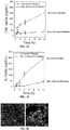

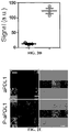

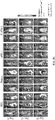

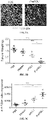


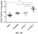


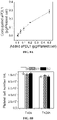
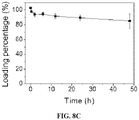


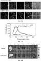

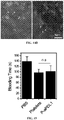


Classifications
-
- A—HUMAN NECESSITIES
- A61—MEDICAL OR VETERINARY SCIENCE; HYGIENE
- A61K—PREPARATIONS FOR MEDICAL, DENTAL OR TOILETRY PURPOSES
- A61K35/00—Medicinal preparations containing materials or reaction products thereof with undetermined constitution
- A61K35/12—Materials from mammals; Compositions comprising non-specified tissues or cells; Compositions comprising non-embryonic stem cells; Genetically modified cells
- A61K35/14—Blood; Artificial blood
- A61K35/19—Platelets; Megacaryocytes
-
- A—HUMAN NECESSITIES
- A61—MEDICAL OR VETERINARY SCIENCE; HYGIENE
- A61P—SPECIFIC THERAPEUTIC ACTIVITY OF CHEMICAL COMPOUNDS OR MEDICINAL PREPARATIONS
- A61P9/00—Drugs for disorders of the cardiovascular system
- A61P9/10—Drugs for disorders of the cardiovascular system for treating ischaemic or atherosclerotic diseases, e.g. antianginal drugs, coronary vasodilators, drugs for myocardial infarction, retinopathy, cerebrovascula insufficiency, renal arteriosclerosis
-
- A—HUMAN NECESSITIES
- A61—MEDICAL OR VETERINARY SCIENCE; HYGIENE
- A61K—PREPARATIONS FOR MEDICAL, DENTAL OR TOILETRY PURPOSES
- A61K39/00—Medicinal preparations containing antigens or antibodies
- A61K39/395—Antibodies; Immunoglobulins; Immune serum, e.g. antilymphocytic serum
- A61K39/39533—Antibodies; Immunoglobulins; Immune serum, e.g. antilymphocytic serum against materials from animals
- A61K39/3955—Antibodies; Immunoglobulins; Immune serum, e.g. antilymphocytic serum against materials from animals against proteinaceous materials, e.g. enzymes, hormones, lymphokines
-
- A—HUMAN NECESSITIES
- A61—MEDICAL OR VETERINARY SCIENCE; HYGIENE
- A61K—PREPARATIONS FOR MEDICAL, DENTAL OR TOILETRY PURPOSES
- A61K39/00—Medicinal preparations containing antigens or antibodies
- A61K39/395—Antibodies; Immunoglobulins; Immune serum, e.g. antilymphocytic serum
- A61K39/40—Antibodies; Immunoglobulins; Immune serum, e.g. antilymphocytic serum bacterial
-
- A—HUMAN NECESSITIES
- A61—MEDICAL OR VETERINARY SCIENCE; HYGIENE
- A61K—PREPARATIONS FOR MEDICAL, DENTAL OR TOILETRY PURPOSES
- A61K45/00—Medicinal preparations containing active ingredients not provided for in groups A61K31/00 - A61K41/00
- A61K45/06—Mixtures of active ingredients without chemical characterisation, e.g. antiphlogistics and cardiaca
-
- A—HUMAN NECESSITIES
- A61—MEDICAL OR VETERINARY SCIENCE; HYGIENE
- A61K—PREPARATIONS FOR MEDICAL, DENTAL OR TOILETRY PURPOSES
- A61K47/00—Medicinal preparations characterised by the non-active ingredients used, e.g. carriers or inert additives; Targeting or modifying agents chemically bound to the active ingredient
- A61K47/06—Organic compounds, e.g. natural or synthetic hydrocarbons, polyolefins, mineral oil, petrolatum or ozokerite
- A61K47/08—Organic compounds, e.g. natural or synthetic hydrocarbons, polyolefins, mineral oil, petrolatum or ozokerite containing oxygen, e.g. ethers, acetals, ketones, quinones, aldehydes, peroxides
- A61K47/10—Alcohols; Phenols; Salts thereof, e.g. glycerol; Polyethylene glycols [PEG]; Poloxamers; PEG/POE alkyl ethers
-
- A—HUMAN NECESSITIES
- A61—MEDICAL OR VETERINARY SCIENCE; HYGIENE
- A61K—PREPARATIONS FOR MEDICAL, DENTAL OR TOILETRY PURPOSES
- A61K47/00—Medicinal preparations characterised by the non-active ingredients used, e.g. carriers or inert additives; Targeting or modifying agents chemically bound to the active ingredient
- A61K47/50—Medicinal preparations characterised by the non-active ingredients used, e.g. carriers or inert additives; Targeting or modifying agents chemically bound to the active ingredient the non-active ingredient being chemically bound to the active ingredient, e.g. polymer-drug conjugates
- A61K47/51—Medicinal preparations characterised by the non-active ingredients used, e.g. carriers or inert additives; Targeting or modifying agents chemically bound to the active ingredient the non-active ingredient being chemically bound to the active ingredient, e.g. polymer-drug conjugates the non-active ingredient being a modifying agent
- A61K47/68—Medicinal preparations characterised by the non-active ingredients used, e.g. carriers or inert additives; Targeting or modifying agents chemically bound to the active ingredient the non-active ingredient being chemically bound to the active ingredient, e.g. polymer-drug conjugates the non-active ingredient being a modifying agent the modifying agent being an antibody, an immunoglobulin or a fragment thereof, e.g. an Fc-fragment
- A61K47/6835—Medicinal preparations characterised by the non-active ingredients used, e.g. carriers or inert additives; Targeting or modifying agents chemically bound to the active ingredient the non-active ingredient being chemically bound to the active ingredient, e.g. polymer-drug conjugates the non-active ingredient being a modifying agent the modifying agent being an antibody, an immunoglobulin or a fragment thereof, e.g. an Fc-fragment the modifying agent being an antibody or an immunoglobulin bearing at least one antigen-binding site
- A61K47/6851—Medicinal preparations characterised by the non-active ingredients used, e.g. carriers or inert additives; Targeting or modifying agents chemically bound to the active ingredient the non-active ingredient being chemically bound to the active ingredient, e.g. polymer-drug conjugates the non-active ingredient being a modifying agent the modifying agent being an antibody, an immunoglobulin or a fragment thereof, e.g. an Fc-fragment the modifying agent being an antibody or an immunoglobulin bearing at least one antigen-binding site the antibody targeting a determinant of a tumour cell
-
- A—HUMAN NECESSITIES
- A61—MEDICAL OR VETERINARY SCIENCE; HYGIENE
- A61K—PREPARATIONS FOR MEDICAL, DENTAL OR TOILETRY PURPOSES
- A61K47/00—Medicinal preparations characterised by the non-active ingredients used, e.g. carriers or inert additives; Targeting or modifying agents chemically bound to the active ingredient
- A61K47/50—Medicinal preparations characterised by the non-active ingredients used, e.g. carriers or inert additives; Targeting or modifying agents chemically bound to the active ingredient the non-active ingredient being chemically bound to the active ingredient, e.g. polymer-drug conjugates
- A61K47/69—Medicinal preparations characterised by the non-active ingredients used, e.g. carriers or inert additives; Targeting or modifying agents chemically bound to the active ingredient the non-active ingredient being chemically bound to the active ingredient, e.g. polymer-drug conjugates the conjugate being characterised by physical or galenical forms, e.g. emulsion, particle, inclusion complex, stent or kit
- A61K47/6901—Conjugates being cells, cell fragments, viruses, ghosts, red blood cells or viral vectors
-
- A—HUMAN NECESSITIES
- A61—MEDICAL OR VETERINARY SCIENCE; HYGIENE
- A61P—SPECIFIC THERAPEUTIC ACTIVITY OF CHEMICAL COMPOUNDS OR MEDICINAL PREPARATIONS
- A61P35/00—Antineoplastic agents
-
- A—HUMAN NECESSITIES
- A61—MEDICAL OR VETERINARY SCIENCE; HYGIENE
- A61P—SPECIFIC THERAPEUTIC ACTIVITY OF CHEMICAL COMPOUNDS OR MEDICINAL PREPARATIONS
- A61P35/00—Antineoplastic agents
- A61P35/04—Antineoplastic agents specific for metastasis
-
- C—CHEMISTRY; METALLURGY
- C07—ORGANIC CHEMISTRY
- C07K—PEPTIDES
- C07K16/00—Immunoglobulins [IGs], e.g. monoclonal or polyclonal antibodies
- C07K16/18—Immunoglobulins [IGs], e.g. monoclonal or polyclonal antibodies against material from animals or humans
- C07K16/28—Immunoglobulins [IGs], e.g. monoclonal or polyclonal antibodies against material from animals or humans against receptors, cell surface antigens or cell surface determinants
- C07K16/2803—Immunoglobulins [IGs], e.g. monoclonal or polyclonal antibodies against material from animals or humans against receptors, cell surface antigens or cell surface determinants against the immunoglobulin superfamily
- C07K16/2818—Immunoglobulins [IGs], e.g. monoclonal or polyclonal antibodies against material from animals or humans against receptors, cell surface antigens or cell surface determinants against the immunoglobulin superfamily against CD28 or CD152
-
- C—CHEMISTRY; METALLURGY
- C07—ORGANIC CHEMISTRY
- C07K—PEPTIDES
- C07K16/00—Immunoglobulins [IGs], e.g. monoclonal or polyclonal antibodies
- C07K16/18—Immunoglobulins [IGs], e.g. monoclonal or polyclonal antibodies against material from animals or humans
- C07K16/28—Immunoglobulins [IGs], e.g. monoclonal or polyclonal antibodies against material from animals or humans against receptors, cell surface antigens or cell surface determinants
- C07K16/2803—Immunoglobulins [IGs], e.g. monoclonal or polyclonal antibodies against material from animals or humans against receptors, cell surface antigens or cell surface determinants against the immunoglobulin superfamily
- C07K16/2827—Immunoglobulins [IGs], e.g. monoclonal or polyclonal antibodies against material from animals or humans against receptors, cell surface antigens or cell surface determinants against the immunoglobulin superfamily against B7 molecules, e.g. CD80, CD86
-
- C—CHEMISTRY; METALLURGY
- C12—BIOCHEMISTRY; BEER; SPIRITS; WINE; VINEGAR; MICROBIOLOGY; ENZYMOLOGY; MUTATION OR GENETIC ENGINEERING
- C12N—MICROORGANISMS OR ENZYMES; COMPOSITIONS THEREOF; PROPAGATING, PRESERVING, OR MAINTAINING MICROORGANISMS; MUTATION OR GENETIC ENGINEERING; CULTURE MEDIA
- C12N5/00—Undifferentiated human, animal or plant cells, e.g. cell lines; Tissues; Cultivation or maintenance thereof; Culture media therefor
- C12N5/06—Animal cells or tissues; Human cells or tissues
- C12N5/0602—Vertebrate cells
- C12N5/0634—Cells from the blood or the immune system
- C12N5/0644—Platelets; Megakaryocytes
-
- A—HUMAN NECESSITIES
- A61—MEDICAL OR VETERINARY SCIENCE; HYGIENE
- A61K—PREPARATIONS FOR MEDICAL, DENTAL OR TOILETRY PURPOSES
- A61K39/00—Medicinal preparations containing antigens or antibodies
- A61K2039/505—Medicinal preparations containing antigens or antibodies comprising antibodies
Definitions
- compositions suitable for targeted delivery of therapeutic agents The present disclosure further provides compositions for use in treating or preventing the metastasis or recurrence of a cancer using platelets loaded with an immunotherapeutic agent.
- the checkpoint inhibitors block the interaction with programmed cell death protein 1 (PD1) on lymphocytes and programmed cell death 1 ligand 1 (PDL1) on antigen presenting cells (APCs) and tumor cells have shown exciting results in treating various types of cancer.
- the first PD-L1 inhibitor, atezolizumab has been granted accelerated approval by FDA recently.
- current methods of checkpoint blockade therapy limit the therapeutic benefits in many patients.
- the largest limitation observed in clinical trials may be the severity of the side effects, such as autoimmune disorders.
- the grade 3/4 adverse events have sometimes occurred when treated with immune checkpoint blockade therapy. Meanwhile, a large fraction of patients failed to response to these agents.
- the objective response rate (ORR) of cancer immunotherapy still needs improvement. How to enhance cancer immunotherapy has become one of the central themes in the field of cancer immunology and immunotherapy.
- the compositions and methods disclosed herein address these and other needs.
- an immunotherapeutic agent selected from an anti-PDLl antibody, an anti-PDl antibody, an anti-CTLA4 antibody, an anti-CD47 antibody, or a combination thereof, is covalently linked to a platelet cell.
- said immunotherapeutic agent for example, anti-PDL1 conjugated to the surface of a platelet cell reduces the recurrence of a cancer (and/or reduces metastasis) after resection of a primary tumor.
- composition comprising:
- Another aspect of the invention is the aforementioned composition for use in a method of preventing metastasis or recurrence of a cancer in a subject in need thereof.
- a further aspect of the invention is the aforementioned composition for use in a method for the targeted delivery of a therapeutic agent to a subject, wherein the composition is targeted to a wound or surgical resection site through activation of the platelet cell.
- An additional aspect of the invention is the aforementioned composition for use in a method of treating or preventing vascular disease in a subject in need thereof.
- FIGS. 1A-1F In situ activation of platelets with anti-PDLl (P-aPDL1) promotes release of therapeutics.
- FIGS. 1a-1F In situ activation of platelets with anti-PDLl (P-aPDL1) promotes release of therapeutics.
- Figure 1a Schematic illustration of delivery of anti-PDLl antibody (aPDL1) toward the primary tumor resection site by platelets.
- Figure 1b TEM imaging of P- aPDL1 before (i) and after (ii and iii) activation. The red arrows indicated the PMPs released from platelet. A large amount of generated PMPs particles were observed under the electron microscopy. Scale bar, 0.5 ⁇ m.
- FIG. 1c Percentage of released aPDL1 from nonactivated and activated platelets at different time points
- Figure 1d- Figure 1e Amount of TNF- ⁇ (f) and IL-1 ⁇
- Figure 1g released from nonactivated and activated platelets at different time points.
- Figure 1f Confocal immunofluorescence images of B16 cancer cells co-incubated with nonactivated (left) and activated (right) P-aPDL1 in a transwell system (pore size, 1 ⁇ m). P-aPDL1 and B16 cancer cells were cultured in upper and lower compartments, respectively.
- Red colors represent aPDL1 signals
- blue and green fluorescence represent nucleus and plasma membrane from DAPI and Alexa fluor 488 conjugate wheat germ agglutinin, respectively.
- Scale bar 20 ⁇ m. The error bars are based on the standard deviations (SD) of triplicated samples.
- FIGS. 2A-2J In vivo reduction of the recurrent melanoma tumors in the surgical bed by P-aPDL1 therapy.
- Figure 2b Fluorescence imaging (aPDL1-Cy5.5) of the mice after i.v. injection of P-aPDL1 or an equivalent dose of free aPDL1 2 hours post injection.
- FIG. 2c Ex vivo imaging of wounds with residual tumors 2 h after i.v. injection of P-aPDL1 or free aPDL1.
- Figure 2d The mean aPDL1-Cy5.5 fluorescent signal intensities in the different wounds of mice shown in (c). The error bars are based on the standard error of the mean (s.e.m.) of triplicated samples.
- Figure 2e Confocal images of the residual tumor slices taken from the mice as showed in (b), where blue and red represent nucleus and aPDL1 signals from DAPI and Cy5.5, respectively. Scale bar, 20 ⁇ m.
- Figure 2f Schematic illustration of P-aPDL1 therapy for treatment of an incomplete surgery tumor model.
- FIG. 2g In vivo bioluminescence imaging of the B16F10 tumors of the different groups after removal of the primary tumor. Shown were 3 representative mice per treatment group.
- Figure 2h Quantified tumor signals of the different groups of mice after various treatments indicated.
- Figure 2i- Figure 2j Tumor growth curves (i) and survival curves (j) of the treated and control mice. Shown were 8 mice per treatment group for survival study. The error bars are based on the standard error of the mean (s.e.m.). P value: ⁇ , P ⁇ 0.05.
- FIGS. 3A-3H P-aPDL1 triggered a robust, T cell-mediated anti-tumor immune response.
- Figure 3a Immunofluorescence of residual tumors showed CD4+ T cells and CD8+ T cells infiltration. Scale bar, 50 ⁇ m.
- Figure 3b Tumor weights and
- Figure 3d Percentages of CD4+ and CD8+ T cells of total CD3+ cells, and representative dot plots in residual tumor of mice treated as indicated.
- FIGS. 4A-4F P-aPDL1 therapy affected the growth of the local recurrent melanoma tumor and metastatic disease.
- Figure 4a Schematic illustration of P-aPDL1 therapy for treatment of an incomplete surgery and metastatic tumor model of mouse.
- Figure 4b In vivo bioluminescence imaging of the B16F10 metastasis of different groups after removal of the primary tumors at different time points. Shown were 3 representative mice per treatment group.
- Figure 4c Representative lung photographs and
- Figure 4d H&E-stained lung slices collected from the mice post different treatments indicated. The black arrows indicated the metastatic tumors in the lung. Scale bar, 500 ⁇ m.
- FIGS. 5A-5E P-aPDL1 to treat 4T1 recurrent cancer after surgery.
- Figure 5a In vivo bioluminescence imaging of the 4T1 metastasis of the different groups after removing of the primary tumors at different time points.
- Figure 5b Representative mice photographs 2 weeks after surgery. Black arrows indicate the recurrent tumor in surgical bed.
- Figure 5c Representative lung photographs collected from the mice post different treatments indicated. Red arrows indicated the tumor nodules in the lung.
- Figure 5e Survival curves for treated and control mice. Shown are 8 mice per each treatment group for survival study. P value: ⁇ , P ⁇ 0.05.
- FIG. 6 Confocal immunofluorescence images of the aPDL1-coupled platelets. FITC-conjugated secondary antibody was used to detect image aPDL1 on platelet. Scale bar, 20 ⁇ m.
- FIG. 7 Flow cytometry assay of aPDL1-coupled platelets. Platelets were incubated with aPDL1 under different experimental settings and analyzed by flow cytometry using the goat anti-rat IgG (H+L) secondary antibody, FITC. The successful attachment of aPDL1 on the surface of the platelet was verified via the sulfydryl and maleimide binding.
- H+L goat anti-rat IgG
- FIGS. 8A-8C Conjugation and stability of aPDL1-coupled platelets.
- Figure 8a Various amounts of aPDL1 were added into platelets for conjugation. The efficiency of aPDL1 conjugation (added aPDL1/conjugated aPDL1) to platelets was about 75% when 0.2pg aPDL1 per platelet was added.
- Figure 8b Stability of platelets after aPDL1 modification. The number of P-aPDL1 was measured based on the platelet count analysis at the 0h and 24h.
- SD standard deviations
- FIG. 9 Surface protein expression on naive and aPDL1 conjugated platelets (CD62P and CD40L were examined after activation).
- FIGS. 10A-10B Transmission electron microscopy (TEM) of P-aPDL1 after activation.
- Figure 10a Platelet microparticles (PMPs) shed from activated platelets.
- Figure 10b A number of PMPs can be found under TEM after P-aPDL1 activation. Size bars, 100 nm in Figure 10a and 2 ⁇ m in Figure 10b .
- FIG. 11 Immunofluorescence imaging of P-aPDL1 after activation. Platelets were loaded with calcein and visualized by green fluorescence. aPDL1 were labeled with Cy3 Goat anti-rat IgG secondary antibody and visualized by the red fluorescence. The white arrows indicated the PMPs released from platelet.
- FIGS. 12A-12B Confocal immunofluorescence images of B16 cancer cells co-incubated with P-aPDL1 in a transwell system.
- Figure 12a A schematic of the transwell system. P-aPDL1 and B16 cancer cells were cultured in the upper and lower compartments, respectively.
- Figure 12b Confocal immunofluorescence images of B16 cancer cells co-incubated with nonactivated and activated P-aPDL1 in a transwell system (pore size, 1 ⁇ m). Red (Cy3) fluorescence represents the aPDL1 signal, blue (DAPI) and green (Alexa Fluor ® 488) fluorescence represent the signals from nucleus and cell membrane, respectively. Scale bar, 20 ⁇ m.
- FIGS. 13A-13C Biodistrubtion of aPDL1-coupled platelets in mice.
- Figure 13a Fluorescence spectra of free Cy5.5-aPDL1 and P-aPDL1-Cy5.5. The intensity of Cy5.5 was not found affected significantly after aPDL1 conjugated to platelets.
- Figure 13b Ex vivo Cy5.5-aPDL1 fluorescence imaging in different organs of mice 2 h after i.v. injection of P-aPDL1 and free aPDL1. L: liver, Lu: lung, Sp: spleen, H: heart, K: kidney.
- Figure 13c The mean Cy5.5 fluorescence signal intensities in different organs of mice shown in (b). Shown are 3 mice per each treatment group. Results were presented as the mean ⁇ SD. Statistical significance was calculated by t-test. P value: ⁇ , P ⁇ 0.005.
- FIGS. 14A-14B Confocal images of the residual tumor slices.
- Figure 14a Confocal images of the residual tumor slices, where blue and red represent nucleus and aPDL1 signals from DAPI and Cy5.5.
- Figure 14b High magnification images of residual tumor slices in P-aPDL1 of (a). The white arrows indicated the PMPs and aPDL1 released from platelets.
- FIGS. 16A-16D Cytokine levels of mice after treatments.
- Figure 16a Local and
- Figure 16b systematic cytokine levels of mice after treatments indicated.
- Figure 16c The representative flow cytometric analysis images and
- Figure 16d The representative flow cytometric analysis images and
- Figure 16d the immunofluorescence image (up, W/O platelets; down, W/ platelets) of PD-L1 analyses.
- the error bars are based on the s.e.m. of triplicated samples.
- Statistical significance was calculated by one-way ANOVA using the Tukey post-test. P value: ⁇ , P ⁇ 0.05, n.s., non-significant.
- FIG. 17 Immunofluorescence of residual tumors showed CD4+ T cells and CD8+ T cells infiltration. Scale bar, 50 ⁇ m.
- FIG. 18 Percentage of CD8+, CD4+Foxp3, and CD4+Foxp3+ T cells expressing Ki67 with different treatments indicated. Statistical significance was calculated by one-way ANOVA using the Tukey post-test. P value: ⁇ , P ⁇ 0.05; ⁇ , P ⁇ 0.01; ⁇ P ⁇ 0.005.
- FIGS. 19A-19C Anti-tumor efficiency of PMP-aPDL1 and high dose of aPDL1 after removal of the primary tumor.
- Figure 19a In vivo bioluminescence imaging of the B16F10 tumors of the different groups after removal of the primary tumor.
- Figure 19b Tumor growth curves of the different groups of mice after various treatments indicated.
- Figure 19c The survival curves of mice in 60 days after various treatments indicated. Shown are 5 mice per each treatment group.
- FIG. 20 Fluorescence imaging of lung tissue. Blue and green fluorescence represent nucleus from DAPI and GFP cancer cells, respectively. Scale bar, 100 ⁇ m.
- an immunotherapeutic agent selected from an anti-PDLl antibody, an anti-PDl antibody, an anti-CTLA4 antibody, an anti-CD47 antibody, or a combination thereof, is covalently linked to a platelet cell.
- said immunotherapeutic agent for example, anti-PDL1 conjugated to the surface of a platelet cell reduces the recurrence of a cancer (and/or reduces metastasis) after resection of a primary tumor.
- a cell includes a plurality of cells, including mixtures thereof.
- the terms “may,” “optionally,” and “may optionally” are used interchangeably and are meant to include cases in which the condition occurs as well as cases in which the condition does not occur.
- the statement that a formulation "may include an excipient” is meant to include cases in which the formulation includes an excipient as well as cases in which the formulation does not include an excipient.
- beneficial agent and “active agent” are used interchangeably herein to refer to a chemical compound or composition that has a beneficial biological effect.
- beneficial biological effects include both therapeutic effects, i.e., treatment of a disorder or other undesirable physiological condition, and prophylactic effects, i.e., prevention of a disorder or other undesirable physiological condition (e.g., cancer).
- prophylactic effects i.e., prevention of a disorder or other undesirable physiological condition (e.g., cancer).
- the terms also encompass pharmaceutically acceptable, pharmacologically active derivatives of beneficial agents specifically mentioned herein, including, but not limited to, salts, esters, amides, prodrugs, active metabolites, isomers, fragments, analogs, and the like.
- treating or “treatment” of a subject includes the administration of a drug to a subject with the purpose of preventing, curing, healing, alleviating, relieving, altering, remedying, ameliorating, improving, stabilizing or affecting a disease or disorder, or a symptom of a disease or disorder.
- the terms “treating” and “treatment” can also refer to reduction in severity and/or frequency of symptoms, elimination of symptoms and/or underlying cause, prevention of the occurrence of symptoms and/or their underlying cause, and improvement or remediation of damage.
- the term "preventing" a disorder or unwanted physiological event in a subject refers specifically to the prevention of the occurrence of symptoms and/or their underlying cause, wherein the subject may or may not exhibit heightened susceptibility to the disorder or event.
- an “effective amount” of a therapeutic agent is meant a nontoxic but sufficient amount of a beneficial agent to provide the desired effect.
- the amount of beneficial agent that is “effective” will vary from subject to subject, depending on the age and general condition of the subject, the particular beneficial agent or agents, and the like. Thus, it is not always possible to specify an exact “effective amount.” However, an appropriate “effective” amount in any subject case may be determined by one of ordinary skill in the art using routine experimentation. Also, as used herein, and unless specifically stated otherwise, an "effective amount” of a beneficial can also refer to an amount covering both therapeutically effective amounts and prophylactically effective amounts.
- an "effective amount" of a drug necessary to achieve a therapeutic effect may vary according to factors such as the age, sex, and weight of the subject. Dosage regimens can be adjusted to provide the optimum therapeutic response. For example, several divided doses may be administered daily or the dose may be proportionally reduced as indicated by the exigencies of the therapeutic situation.
- a "therapeutically effective amount” of a therapeutic agent refers to an amount that is effective to achieve a desired therapeutic result
- a “prophylactically effective amount” of a therapeutic agent refers to an amount that is effective to prevent an unwanted physiological condition.
- Therapeutically effective and prophylactically effective amounts of a given therapeutic agent will typically vary with respect to factors such as the type and severity of the disorder or disease being treated and the age, gender, and weight of the subject.
- terapéuticaally effective amount can also refer to an amount of a therapeutic agent, or a rate of delivery of a therapeutic agent (e.g., amount over time), effective to facilitate a desired therapeutic effect.
- the precise desired therapeutic effect will vary according to the condition to be treated, the tolerance of the subject, the drug and/or drug formulation to be administered (e.g., the potency of the therapeutic agent (drug), the concentration of drug in the formulation, and the like), and a variety of other factors that are appreciated by those of ordinary skill in the art.
- the term "pharmaceutically acceptable” component can refer to a component that is not biologically or otherwise undesirable, i.e., the component may be incorporated into a pharmaceutical formulation of the invention and administered to a subject as described herein without causing any significant undesirable biological effects or interacting in a deleterious manner with any of the other components of the formulation in which it is contained.
- pharmaceutically acceptable refers to an excipient, it is generally implied that the component has met the required standards of toxicological and manufacturing testing or that it is included on the Inactive Ingredient Guide prepared by the U.S. Food and Drug Administration.
- the term "pharmacologically active” can refer to a derivative or analog (e.g., a salt, ester, amide, conjugate, metabolite, isomer, fragment, etc.) having the same type of pharmacological activity as the parent compound and approximately equivalent in degree.
- mixture can include solutions in which the components of the mixture are completely miscible, as well as suspensions and emulsions, in which the components of the mixture are not completely miscible.
- the term "subject" can refer to living organisms such as mammals, including, but not limited to humans, livestock, dogs, cats, and other mammals. Administration of the therapeutic agents can be carried out at dosages and for periods of time effective for treatment of a subject. In some embodiments, the subject is a human. In some embodiments, the pharmacokinetic profiles of the systems of the present invention are similar for male and female subjects.
- antibody refers to immunoglobulin molecules comprised of four polypeptide chains, two heavy (H) chains and two light (L) chains interconnected by disulfide bonds.
- antibody or “antibodies” as used herein includes but is not limited to recombinant antibodies that are generated by recombinant technologies as known in the art.
- An “antibody” or “antibodies” can be of any origin including from mammalian species such as human, non-human primate (e.g. human such as from chimpanzee, baboon, rhesus or cynomolgus monkey), rodent (e.g.
- Antibody or “antibodies” include antibodies of any isotype, including human isotypes IgA 1 IgA 2 , IgD, IgG 1 , IgG 2a , IgG 2b , IgG 3 , IgG 4 , IgE and IgM and modified variants thereof.
- the antibody herein is directed against an "antigen" of interest.
- the antigen is a biologically important polypeptide and administration of the antibody to a mammal suffering from a disease or disorder can result in a therapeutic benefit in that mammal.
- antibodies directed against non-polypeptide antigens are also contemplated.
- antibody fragment refers to a naturally occurring antibody which lacks one or more domains or one or more amino acids. Typically, an antibody fragment contains the entire antigen binding or variable region thereof of such naturally occurring antibody. Examples of antibody fragments include any antibody that lacks the Fc portion. Examples of antibody fragments include also Fab, Fab', F(ab') 2 , Fv and scFv fragments; diabodies; triabodies; tetrabodies; minibodies; antibodies consisting essentially of a single, two or three immunoglobulin domain(s) such as Domain antibodies TM ; single-chain antibodies; bispecific, trispecific, tetraspecific or multispecific variants of any of the above.
- composition comprising:
- the chemical linker moiety is selected from a maleimide linker, a PEG linker, PASylation, and HESylation. In one embodiment, the chemical linker moiety is a maleimide linker.
- the platelet cell is a human platelet cell. In one embodiment, the platelet cell is an autologous platelet cell.
- composition comprising:
- compositions containing therapeutically effective amounts of one or more of the disclosed platelet compositions and a pharmaceutically acceptable carrier.
- Pharmaceutical carriers suitable for administration of the compositions provided herein include any such carriers known to those skilled in the art to be suitable for the particular mode of administration.
- the compositions may be formulated as the sole pharmaceutically active ingredient in the pharmaceutical composition or may be combined with other active ingredients.
- compositions as described herein, comprising an immunotherapeutic agent as defined above conjugated to a platelet cell and an excipient of some sort may be useful in a variety of applications.
- Excipients include any and all solvents, diluents or other liquid vehicles, dispersion or suspension aids, surface active agents, isotonic agents, thickening or emulsifying agents, preservatives, solid binders, lubricants and the like, as suited to the particular dosage form desired.
- General considerations in formulation and/or manufacture can be found, for example, in Remington's Pharmaceutical Sciences, Sixteenth Edition, E. W. Martin (Mack Publishing Co., Easton, Pa., 1980 ), and Remington: The Science and Practice of Pharmacy, 21st Edition (Lippincott Williams & Wilkins, 2005 ).
- the pharmaceutically acceptable excipients may also include one or more of fillers, binders, lubricants, glidants, disintegrants, and the like.
- excipients include, but are not limited to, any non-toxic, inert solid, semi-solid or liquid filler, diluent, encapsulating material or formulation auxiliary of any type.
- materials which can serve as excipients include, but are not limited to, sugars such as lactose, glucose, and sucrose; starches such as corn starch and potato starch; cellulose and its derivatives such as sodium carboxymethyl cellulose, ethyl cellulose, and cellulose acetate; powdered tragacanth; malt; gelatin; talc; excipients such as cocoa butter and suppository waxes; oils such as peanut oil, cottonseed oil; safflower oil; sesame oil; olive oil; corn oil and soybean oil; glycols such as propylene glycol; esters such as ethyl oleate and ethyl laurate; agar; detergents such as Tween 80; buffering agents such as magnesium hydroxide and aluminum
- the excipients may be chosen based on what the composition is useful for.
- the choice of the excipient will depend on the route of administration, the agent being delivered, time course of delivery of the agent, etc., and can be administered to humans and/or to animals, orally, rectally, parenterally, intracisternally, intravaginally, intranasally, intraperitoneally, topically (as by powders, creams, ointments, or drops), bucally, or as an oral or nasal spray.
- the composition is injected at or near the resection site of a tumor.
- Exemplary diluents include calcium carbonate, sodium carbonate, calcium phosphate, dicalcium phosphate, calcium sulfate, calcium hydrogen phosphate, sodium phosphate lactose, sucrose, cellulose, microcrystalline cellulose, kaolin, mannitol, sorbitol, inositol, sodium chloride, dry starch, cornstarch, powdered sugar, etc., and combinations thereof.
- Exemplary granulating and/or dispersing agents include potato starch, corn starch, tapioca starch, sodium starch glycolate, clays, alginic acid, guar gum, citrus pulp, agar, bentonite, cellulose and wood products, natural sponge, cation-exchange resins, calcium carbonate, silicates, sodium carbonate, cross-linked poly(vinyl-pyrrolidone) (crospovidone), sodium carboxymethyl starch (sodium starch glycolate), carboxymethyl cellulose, cross-linked sodium carboxymethyl cellulose (croscarmellose), methylcellulose, pregelatinized starch (starch 1500), microcrystalline starch, water insoluble starch, calcium carboxymethyl cellulose, magnesium aluminum silicate (Veegum), sodium lauryl sulfate, quaternary ammonium compounds, etc., and combinations thereof.
- cross-linked poly(vinyl-pyrrolidone) crospovidone
- sodium carboxymethyl starch sodium starch glycolate
- Exemplary surface active agents and/or emulsifiers include natural emulsifiers (e.g. acacia, agar, alginic acid, sodium alginate, tragacanth, chondrux, cholesterol, xanthan, pectin, gelatin, egg yolk, casein, wool fat, cholesterol, wax, and lecithin), colloidal clays (e.g. bentonite [aluminum silicate] and Veegum [magnesium aluminum silicate]), long chain amino acid derivatives, high molecular weight alcohols (e.g.
- stearyl alcohol cetyl alcohol, oleyl alcohol, triacetin monostearate, ethylene glycol distearate, glyceryl monostearate, and propylene glycol monostearate, polyvinyl alcohol), carbomers (e.g. carboxy polymethylene, polyacrylic acid, acrylic acid polymer, and carboxyvinyl polymer), carrageenan, cellulosic derivatives (e.g. carboxymethylcellulose sodium, powdered cellulose, hydroxymethyl cellulose, hydroxypropyl cellulose, hydroxypropyl methylcellulose, methylcellulose), sorbitan fatty acid esters (e.g.
- Cremophor polyoxyethylene ethers, (e.g. polyoxyethylene lauryl ether [Brij 30]), poly(vinyl-pyrrolidone), diethylene glycol monolaurate, triethanolamine oleate, sodium oleate, potassium oleate, ethyl oleate, oleic acid, ethyl laurate, sodium lauryl sulfate, Pluronic F 68, Poloxamer 188, cetrimonium bromide, cetylpyridinium chloride, benzalkonium chloride, docusate sodium, etc. and/or combinations thereof.
- polyoxyethylene ethers e.g. polyoxyethylene lauryl ether [Brij 30]
- poly(vinyl-pyrrolidone) diethylene glycol monolaurate
- triethanolamine oleate sodium oleate
- potassium oleate ethyl oleate
- oleic acid ethyl laurate
- Exemplary binding agents include starch (e.g. cornstarch and starch paste), gelatin, sugars (e.g. sucrose, glucose, dextrose, dextrin, molasses, lactose, lactitol, mannitol, etc.), natural and synthetic gums (e.g.
- acacia sodium alginate, extract of Irish moss, panwar gum, ghatti gum, mucilage of isapol husks, carboxymethylcellulose, methylcellulose, ethylcellulose, hydroxyethylcellulose, hydroxypropyl cellulose, hydroxypropyl methylcellulose, microcrystalline cellulose, cellulose acetate, poly(vinyl-pyrrolidone), magnesium aluminum silicate (Veegum), and larch arabogalactan), alginates, polyethylene oxide, polyethylene glycol, inorganic calcium salts, silicic acid, polymethacrylates, waxes, water, alcohol, etc., and/or combinations thereof.
- Exemplary preservatives include antioxidants, chelating agents, antimicrobial preservatives, antifungal preservatives, alcohol preservatives, acidic preservatives, and other preservatives.
- antioxidants include alpha tocopherol, ascorbic acid, acorbyl palmitate, butylated hydroxyanisole, butylated hydroxytoluene, monothioglycerol, potassium metabisulfite, propionic acid, propyl gallate, sodium ascorbate, sodium bisulfite, sodium metabisulfite, and sodium sulfite.
- Exemplary chelating agents include ethylenediaminetetraacetic acid (EDTA) and salts and hydrates thereof (e.g., sodium edetate, disodium edetate, trisodium edetate, calcium disodium edetate, dipotassium edetate, and the like), citric acid and salts and hydrates thereof (e.g., citric acid monohydrate), fumaric acid and salts and hydrates thereof, malic acid and salts and hydrates thereof, phosphoric acid and salts and hydrates thereof, and tartaric acid and salts and hydrates thereof.
- EDTA ethylenediaminetetraacetic acid
- salts and hydrates thereof e.g., sodium edetate, disodium edetate, trisodium edetate, calcium disodium edetate, dipotassium edetate, and the like
- citric acid and salts and hydrates thereof e.g., citric acid mono
- antimicrobial preservatives include benzalkonium chloride, benzethonium chloride, benzyl alcohol, bronopol, cetrimide, cetylpyridinium chloride, chlorhexidine, chlorobutanol, chlorocresol, chloroxylenol, cresol, ethyl alcohol, glycerin, hexetidine, imidurea, phenol, phenoxyethanol, phenylethyl alcohol, phenylmercuric nitrate, propylene glycol, and thimerosal.
- antifungal preservatives include butyl paraben, methyl paraben, ethyl paraben, propyl paraben, benzoic acid, hydroxybenzoic acid, potassium benzoate, potassium sorbate, sodium benzoate, sodium propionate, and sorbic acid.
- Exemplary alcohol preservatives include ethanol, polyethylene glycol, phenol, phenolic compounds, bisphenol, chlorobutanol, hydroxybenzoate, and phenylethyl alcohol.
- Exemplary acidic preservatives include vitamin A, vitamin C, vitamin E, beta-carotene, citric acid, acetic acid, dehydroacetic acid, ascorbic acid, sorbic acid, and phytic acid.
- preservatives include tocopherol, tocopherol acetate, deteroxime mesylate, cetrimide, butylated hydroxyanisol (BHA), butylated hydroxytoluened (BHT), ethylenediamine, sodium lauryl sulfate (SLS), sodium lauryl ether sulfate (SLES), sodium bisulfite, sodium metabisulfite, potassium sulfite, potassium metabisulfite, Glydant Plus, Phenonip, methylparaben, Germall 115, Germaben II, Neolone, Kathon, and Euxyl.
- the preservative is an anti-oxidant.
- the preservative is a chelating agent.
- Exemplary buffering agents include citrate buffer solutions, acetate buffer solutions, phosphate buffer solutions, ammonium chloride, calcium carbonate, calcium chloride, calcium citrate, calcium glubionate, calcium gluceptate, calcium gluconate, D-gluconic acid, calcium glycerophosphate, calcium lactate, propanoic acid, calcium levulinate, pentanoic acid, dibasic calcium phosphate, phosphoric acid, tribasic calcium phosphate, calcium hydroxide phosphate, potassium acetate, potassium chloride, potassium gluconate, potassium mixtures, dibasic potassium phosphate, monobasic potassium phosphate, potassium phosphate mixtures, sodium acetate, sodium bicarbonate, sodium chloride, sodium citrate, sodium lactate, dibasic sodium phosphate, monobasic sodium phosphate, sodium phosphate mixtures, tromethamine, magnesium hydroxide, aluminum hydroxide, alginic acid, pyrogen-free water, isotonic saline, Ringer
- Exemplary lubricating agents include magnesium stearate, calcium stearate, stearic acid, silica, talc, malt, glyceryl behanate, hydrogenated vegetable oils, polyethylene glycol, sodium benzoate, sodium acetate, sodium chloride, leucine, magnesium lauryl sulfate, sodium lauryl sulfate, etc., and combinations thereof.
- Exemplary natural oils include almond, apricot kernel, avocado, babassu, bergamot, black current seed, borage, cade, camomile, canola, caraway, carnauba, castor, cinnamon, cocoa butter, coconut, cod liver, coffee, corn, cotton seed, emu, eucalyptus, evening primrose, fish, flaxseed, geraniol, gourd, grape seed, hazel nut, hyssop, isopropyl myristate, jojoba, kukui nut, lavandin, lavender, lemon, litsea cubeba, macademia nut, mallow, mango seed, meadowfoam seed, mink, nutmeg, olive, orange, orange roughy, palm, palm kernel, peach kernel, peanut, poppy seed, pumpkin seed, rapeseed, rice bran, rosemary, safflower, sandalwood, sasquana, savoury, sea buckt
- Exemplary synthetic oils include, but are not limited to, butyl stearate, caprylic triglyceride, capric triglyceride, cyclomethicone, diethyl sebacate, dimethicone 360, isopropyl myristate, mineral oil, octyldodecanol, oleyl alcohol, silicone oil, and combinations thereof.
- composition may further comprise a polymer.
- exemplary polymers contemplated herein include, but are not limited to, cellulosic polymers and copolymers, for example, cellulose ethers such as methylcellulose (MC), hydroxyethylcellulose (HEC), hydroxypropyl cellulose (HPC), hydroxypropyl methyl cellulose (HPMC), methylhydroxyethylcellulose (MHEC), methylhydroxypropylcellulose (MHPC), carboxymethyl cellulose (CMC) and its various salts, including, e.g., the sodium salt, hydroxyethylcarboxymethylcellulose (HECMC) and its various salts, carboxymethylhydroxyethylcellulose (CMHEC) and its various salts, other polysaccharides and polysaccharide derivatives such as starch, dextran, dextran derivatives, chitosan, and alginic acid and its various salts, carageenan, various gums, including xanthan gum, guar gum, gum arab
- composition may further comprise an emulsifying agent.
- emulsifying agents include, but are not limited to, a polyethylene glycol (PEG), a polypropylene glycol, a polyvinyl alcohol, a poly-N-vinyl pyrrolidone and copolymers thereof, poloxamer nonionic surfactants, neutral water-soluble polysaccharides (e.g., dextran, Ficoll, celluloses), non-cationic poly(meth)acrylates, non-cationic polyacrylates, such as poly(meth)acrylic acid, and esters amide and hydroxyalkyl amides thereof, natural emulsifiers (e.g.
- acacia agar, alginic acid, sodium alginate, tragacanth, chondrux, cholesterol, xanthan, pectin, gelatin, egg yolk, casein, wool fat, cholesterol, wax, and lecithin), colloidal clays (e.g. bentonite [aluminum silicate] and Veegum [magnesium aluminum silicate]), long chain amino acid derivatives, high molecular weight alcohols (e.g. stearyl alcohol, cetyl alcohol, oleyl alcohol, triacetin monostearate, ethylene glycol distearate, glyceryl monostearate, and propylene glycol monostearate, polyvinyl alcohol), carbomers (e.g.
- carboxy polymethylene polyacrylic acid, acrylic acid polymer, and carboxyvinyl polymer
- carrageenan cellulosic derivatives (e.g. carboxymethylcellulose sodium, powdered cellulose, hydroxymethyl cellulose, hydroxypropyl cellulose, hydroxypropyl methylcellulose, methylcellulose), sorbitan fatty acid esters (e.g.
- Cremophor polyoxyethylene ethers, (e.g. polyoxyethylene lauryl ether [Brij 30]), poly(vinyl-pyrrolidone), diethylene glycol monolaurate, triethanolamine oleate, sodium oleate, potassium oleate, ethyl oleate, oleic acid, ethyl laurate, sodium lauryl sulfate, Pluronic F 68, Poloxamer 188, cetrimonium bromide, cetylpyridinium chloride, benzalkonium chloride, docusate sodium, etc. and/or combinations thereof.
- the emulsifying agent is cholesterol.
- Liquid compositions include emulsions, microemulsions, solutions, suspensions, syrups, and elixirs.
- the liquid composition may contain inert diluents commonly used in the art such as, for example, water or other solvents, solubilizing agents and emulsifiers such as ethyl alcohol, isopropyl alcohol, ethyl carbonate, ethyl acetate, benzyl alcohol, benzyl benzoate, propylene glycol, 1,3-butylene glycol, dimethylformamide, oils (in particular, cottonseed, groundnut, corn, germ, olive, castor, and sesame oils), glycerol, tetrahydrofurfuryl alcohol, polyethylene glycols and fatty acid esters of sorbitan, and mixtures thereof.
- inert diluents commonly used in the art such as, for example, water or other solvents, solubilizing agents and emulsifiers such
- injectable compositions for example, injectable aqueous or oleaginous suspensions may be formulated according to the known art using suitable dispersing or wetting agents and suspending agents.
- the sterile injectable preparation may also be an injectable solution, suspension, or emulsion in a nontoxic parenterally acceptable diluent or solvent, for example, as a solution in 1,3-butanediol.
- acceptable vehicles and solvents for pharmaceutical or cosmetic compositions that may be employed are water, Ringer's solution, U.S.P. and isotonic sodium chloride solution.
- sterile, fixed oils are conventionally employed as a solvent or suspending medium. Any bland fixed oil can be employed including synthetic mono- or diglycerides.
- fatty acids such as oleic acid are used in the preparation of injectables.
- the particles are suspended in a carrier fluid comprising 1% (w/v) sodium carboxymethyl cellulose and 0.1% (v/v) Tween 80.
- the injectable composition can be sterilized, for example, by filtration through a bacteria-retaining filter, or by incorporating sterilizing agents in the form of sterile solid compositions which can be dissolved or dispersed in sterile water or other sterile injectable medium prior to use.
- compositions or the therapeutic agents can be administered in a solid composition.
- Solid compositions include capsules, tablets, pills, powders, and granules.
- the particles are mixed with at least one excipient and/or a) fillers or extenders such as starches, lactose, sucrose, glucose, mannitol, and silicic acid, b) binders such as, for example, carboxymethylcellulose, alginates, gelatin, polyvinylpyrrolidinone, sucrose, and acacia, c) humectants such as glycerol, d) disintegrating agents such as agar-agar, calcium carbonate, potato or tapioca starch, alginic acid, certain silicates, and sodium carbonate, e) solution retarding agents such as paraffin, f) absorption accelerators such as quaternary ammonium compounds, g) wetting
- the dosage form may also comprise buffering agents.
- Solid compositions of a similar type may also be employed as fillers in soft and hard-filled gelatin capsules using such excipients as lactose or milk sugar as well as high molecular weight polyethylene glycols and the like.
- Tablets, capsules, pills, and granules can be prepared with coatings and shells such as enteric coatings and other coatings well known in the pharmaceutical formulating art. They may optionally contain opacifying agents and can also be of a composition that they release the active ingredient(s) only, or preferentially, in a certain part of the intestinal tract, optionally, in a delayed manner. Examples of embedding compositions which can be used include polymeric substances and waxes.
- compositions of a similar type may also be employed as fillers in soft and hard-filled gelatin capsules using such excipients as lactose or milk sugar as well as high molecular weight polyethylene glycols and the like.
- compositions for topical or transdermal administration include ointments, pastes, creams, lotions, gels, powders, solutions, sprays, inhalants, or patches.
- the active compound for example, an immunotherapeutic agent conjugated to a platelet cell
- the active compound is admixed with an excipient and any needed preservatives or buffers as may be required.
- the ointments, pastes, creams, and gels may contain, in addition to the active compound (for example, an immunotherapeutic agent conjugated to a platelet cell), excipients such as animal and vegetable fats, oils, waxes, paraffins, starch, tragacanth, cellulose derivatives, polyethylene glycols, silicones, bentonites, silicic acid, talc, and zinc oxide, or mixtures thereof.
- active compound for example, an immunotherapeutic agent conjugated to a platelet cell
- excipients such as animal and vegetable fats, oils, waxes, paraffins, starch, tragacanth, cellulose derivatives, polyethylene glycols, silicones, bentonites, silicic acid, talc, and zinc oxide, or mixtures thereof.
- Powders and sprays can contain, in addition to the active compound/composition, excipients such as lactose, talc, silicic acid, aluminum hydroxide, calcium silicates, and polyamide powder, or mixtures of these substances.
- Sprays can additionally contain customary propellants such as chlorofluorohydrocarbons.
- Transdermal patches have the added advantage of providing controlled delivery of a compound to the body.
- dosage forms can be made by dissolving or dispensing the nanoparticles in a proper medium.
- Absorption enhancers can also be used to increase the flux of the compound across the skin.
- the rate can be controlled by either providing a rate controlling membrane or by dispersing the particles in a polymer matrix or gel.
- the therapeutic agent can be encapsulated in a nanoparticle.
- the nanoparticle is linked covalently through the chemical linker moiety.
- the chemical linker moiety is optional and the nanoparticles are not covalently linked to the platelet cell.
- the nanoparticles can be made from one or more polymers.
- the polymer includes copolymers of polycarboxylic acids or salts thereof, carboxylic anhydrides (for example, maleic anhydride) with other monomers (for example, methyl (meth)acrylate, acrylic acid and the like), hydrophilic vinyl polymers, such as polyvinyl acetate, polyvinyl alcohol, polyvinyl pyrrolidone (PVP), polyethyleneoxide (PEO), poly(vinyl pyrrolidone-co-vinyl acetate), polymethacrylates, polyoxyethylene alkyl ethers, polyoxyethylene castor oils, polycaprolactam, polylactic acid, polyglycolic acid, poly(lactic-glycolic) acid, poly(lactic co-glycolic) acid (PLGA), cellulose derivatives, such as hydroxymethylcellulose, hydroxypropylcellulose and the like.
- carboxylic anhydrides for example, maleic anhydride
- other monomers for example,
- the polymer is 50:50 PLGA copolymer.
- the polymer includes a natural polymer, such as chitosan, collagen, alginate, gelatin, hyaluronic acid, and nontoxic metal salts thereof.
- the polymer is a hydrogel, for example an alginate hydrogel. Hydrophilic polymers and other vehicles can be used alone or in combination, and enhanced structural integrity can be imparted to the vehicle by partial crystallization, ionic bonding, cross-linking and the like.
- encapsulation techniques include, but are not limited to, spray drying, interfacial polymerization, hot melt encapsulation, phase separation encapsulation (spontaneous emulsion microencapsulation, solvent evaporation microencapsulation, and solvent removal microencapsulation), coacervation, low temperature microsphere formation, and phase inversion nanoencapsulation (PIN).
- the nanoparticles incorporated in the compositions discussed herein are multi-walled nanoparticles.
- Multi-walled nanoparticles useful in the compositions disclosed herein can be prepared, for example, using "sequential phase inversion nanoencapsulation" (sPIN).
- microspheres/nanospheres using spray drying techniques are described in U.S. Pat. No. 6,620,617, to Mathiowitz et al.
- the polymer is dissolved in an organic solvent such as methylene chloride or in water.
- a known amount of one or more active agents to be incorporated in the particles is suspended (in the case of an insoluble active agent) or co-dissolved (in the case of a soluble active agent) in the polymer solution.
- the solution or dispersion is pumped through a micronizing nozzle driven by a flow of compressed gas, and the resulting aerosol is suspended in a heated cyclone of air, allowing the solvent to evaporate from the microdroplets, forming particles.
- Microspheres/nanospheres ranging between 0.1-10 microns can be obtained using this method.
- Interfacial polymerization can also be used to encapsulate one or more active agents.
- a monomer and the active agent(s) are dissolved in a solvent.
- a second monomer is dissolved in a second solvent (typically aqueous) which is immiscible with the first.
- An emulsion is formed by suspending the first solution through stirring in the second solution. Once the emulsion is stabilized, an initiator is added to the aqueous phase causing interfacial polymerization at the interface of each droplet of emulsion.
- Microspheres can be formed from polymers such as polyesters and polyanhydrides using hot melt microencapsulation methods as described in Mathiowitz et al., Reactive Polymers, 6:275 (1987 ). In this method, the use of polymers with molecular weights between 3-75,000 daltons is preferred.
- the polymer first is melted and then mixed with the solid particles of one or more active agents to be incorporated that have been sieved to less than 50 microns. The mixture is suspended in a non-miscible solvent (like silicon oil), and, with continuous stirring, heated to 5° C. above the melting point of the polymer. Once the emulsion is stabilized, it is cooled until the polymer particles solidify. The resulting microspheres are washed by decanting with petroleum ether to give a free-flowing powder.
- a non-miscible solvent like silicon oil
- phase separation microencapsulation techniques a polymer solution is stirred, optionally in the presence of one or more active agents to be encapsulated. While continuing to uniformly suspend the material through stirring, a nonsolvent for the polymer is slowly added to the solution to decrease the polymer's solubility. Depending on the solubility of the polymer in the solvent and nonsolvent, the polymer either precipitates or phase separates into a polymer rich and a polymer poor phase. Under proper conditions, the polymer in the polymer rich phase will migrate to the interface with the continuous phase, encapsulating the active agent(s) in a droplet with an outer polymer shell.
- Spontaneous emulsification involves solidifying emulsified liquid polymer droplets formed above by changing temperature, evaporating solvent, or adding chemical cross-linking agents.
- One or more active agents to be incorporated are optionally added to the solution, and the mixture is suspended in an aqueous solution that contains a surface active agent such as poly(vinyl alcohol).
- a surface active agent such as poly(vinyl alcohol).
- the resulting emulsion is stirred until most of the organic solvent evaporated, leaving solid microspheres/nanospheres.
- This method is useful for relatively stable polymers, such as polyesters and polystyrene.
- labile polymers such as polyanhydrides, may degrade during the fabrication process due to the presence of water. For these polymers, some of the following methods performed in completely anhydrous organic solvents are more useful.
- the solvent removal microencapsulation technique is primarily designed for polyanhydrides and is described, for example, in WO 93/21906 to Brown University Research Foundation .
- the substance to be incorporated is dispersed or dissolved in a solution of the selected polymer in a volatile organic solvent, such as methylene chloride.
- a volatile organic solvent such as methylene chloride.
- This mixture is suspended by stirring in an organic oil, such as silicon oil, to form an emulsion.
- Microspheres that range between 1-300 microns can be obtained by this procedure.
- Substances which can be incorporated in the microspheres include pharmaceuticals, pesticides, nutrients, imaging agents, and metal compounds.
- Coacervation procedures for various substances using coacervation techniques are known in the art, for example, in GB-B-929 406; GB-B-929 40 1; and U.S. Pat. Nos. 3,266,987 , 4,794,000 , and 4,460,563 .
- Coacervation involves the separation of a macromolecular solution into two immiscible liquid phases.
- One phase is a dense coacervate phase, which contains a high concentration of the polymer encapsulant (and optionally one or more active agents), while the second phase contains a low concentration of the polymer.
- the polymer encapsulant forms nanoscale or microscale droplets.
- Coacervation may be induced by a temperature change, addition of a non-solvent or addition of a micro-salt (simple coacervation), or by the addition of another polymer thereby forming an interpolymer complex (complex coacervation).
- Nanoparticles can also be formed using the phase inversion nanoencapsulation (PIN) method, wherein a polymer is dissolved in a "good” solvent, fine particles of a substance to be incorporated, such as a drug, are mixed or dissolved in the polymer solution, and the mixture is poured into a strong non-solvent for the polymer, to spontaneously produce, under favorable conditions, polymeric microspheres, wherein the polymer is either coated with the particles or the particles are dispersed in the polymer.
- PIN phase inversion nanoencapsulation
- an emulsion need not be formed prior to precipitation.
- the process can be used to form microspheres from thermoplastic polymers.
- Multi-walled nanoparticles can also be formed by a process referred to herein as "sequential phase inversion nanoencapsulation" (sPIN).
- sPIN is particularly suited for forming monodisperse populations of nanoparticles, avoiding the need for an additional separations step to achieve a monodisperse population of nanoparticles.
- a core polymer is dissolved in a first solvent.
- the active agent is dissolved or dispersed in a core polymer solvent.
- the core polymer, core polymer solvent, and agent to be encapsulated form a mixture having a continuous phase, in which the core polymer solvent is the continuous phase.
- the shell polymer is dissolved in a shell polymer solvent, which is a non-solvent for the core polymer.
- the solutions of the core polymer and shell polymer are mixed together. The resulting decreases the solubility of the core polymer at its cloud point due to the presence of the shell polymer solvent results in the preferential phase separation of the core polymer and, optionally, encapsulation of the agent.
- the shell polymer engulfs the core polymer as phase inversion is completed to form a double-walled nanoparticle.
- sPIN provides a one-step procedure for the preparation of multi-walled particles, such as double-walled nanoparticles, which is nearly instantaneous, and does not require emulsification of the solvent.
- Methods for forming multi-walled particles are disclosed in U.S. Publication No. 2012-0009267 to Cho, et al.
- the particle can be a dendrimer particle.
- Dendrimers are three-dimensional polymers that are grown by the successive addition of shells or layers of branched molecules to a central core. Dendrimers have several advantages over linear polymers, since they have controllable structure, a single molecular weight rather than a distribution of molecular weights, and a large number of controllable surface functionalities, and an inclination to adopt a globular conformation once a certain size is reached. They are prepared by reacting highly branched monomers together to produce monodisperse, tree-like and/or generational structure polymeric structures. Individual dendrimers consist of a central core molecule, with a dendritic wedge attached to each functional site.
- the dendrimeric surface layer can have a variety of functional groups disposed thereon, according to the assembly monomers used during the preparation.
- the dendrimer functional groups dictate the properties of the individual dendrimer types.
- dendrimer cores are spacious, and by modifying the chemical properties of the core, shells, and especially the surface layer, their physical properties can be finely tuned. Tunable properties include solubility, toxicity, immunogenicity and bioattachment capability.
- Polyamidoamine, polypropyleneimine, polyarylether and polyethyleneimine are examples of dendrimers that have been investigated for biopharmaceutical applications.
- Polyamidoamine dendrimers are based on an ethylenediamine core and an amidoamine repeat branching structure. They can be synthesized in a variety of well-defined molecular weights. Their size and surface functionality (primary amine) is defined by the number of controlled repetitive additions of monomeric units, giving rise to different half or full generations. They are water-soluble and they have been reported to be the only class of dendrimer that are mono-dispersed. Furthermore, they show high charge densities that are restricted to the surface of the molecules.
- Dendrimers have been used as carriers for therapeutic compounds, either by entrapment of a drug in cavities within the dendrimer, or by covalently linking drug molecules to the surface. This is reviewed in Svenson, S., Eur J Pharm Biopharm (2009) 71:445-462 and Cheng, Y., J. Pharm. Sci. (2007) 97:123-143 . Entrapment within dendrimer cavities is limited to small molecules, and covalent attachment approaches have thus far been limited to systems in which a small drug is hydrolytically or enzymatically cleaved from the dendrimer surface.
- composition as defined above for use in a method of preventing metastasis or recurrence of a cancer, the method comprising:
- the immunotherapeutic agent is an anti-PDLl antibody.
- the chemical linker moiety is selected from a maleimide linker, a PEG linker, PASylation, and HESylation. In one embodiment, the chemical linker moiety is a maleimide linker.
- the platelet cell is a human platelet cell. In one embodiment, the platelet cell is an autologous platelet cell.
- the cancer is a solid tumor. In one embodiment, the cancer is melanoma. In one embodiment, the cancer is breast cancer. In one embodiment, the composition is administered in combination with an additional therapeutic agent. In one embodiment, the additional therapeutic agent is an antineoplastic agent.
- composition as defined above for use in a method of treating or preventing a cancer comprising:
- composition for use in a method for the targeted delivery of a therapeutic agent comprising:
- the method provides the therapeutic agent is further released in platelet-derived microparticles at the wound or resection site upon activation of the platelet cell.
- the immunotherapeutic agent is an anti-PDLl antibody.
- the chemical linker moiety is selected from a maleimide linker, a PEG linker, PASylation, and HESylation. In one embodiment, the chemical linker moiety is a maleimide linker.
- the platelet cell is a human platelet cell. In one embodiment, the platelet cell is an autologous platelet cell.
- the platelet compositions described herein are useful for treating or preventing metastasis or recurrence of a cancer. In some embodiments, the platelet compositions described herein are useful for the prevention of recurrence of excised solid tumors. In some embodiments, the compositions described herein are useful for the prevention of metastasis of excised solid tumors. In another embodiment, the cancer recurrence to be prevented is melanoma. In another embodiment, the cancer recurrence to be prevented is breast cancer. In a further embodiment, the cancer recurrence to be prevented is prostate cancer.
- compositions described herein are used to prevent solid tumor recurrence or metastasis, for example, melanoma, lung cancer (including lung adenocarcinoma, basal cell carcinoma, squamous cell carcinoma, large cell carcinoma, bronchioloalveolar carcinoma, bronchogenic carcinoma, non-small-cell carcinoma, small cell carcinoma, mesothelioma); breast cancer (including ductal carcinoma, lobular carcinoma, inflammatory breast cancer, clear cell carcinoma, mucinous carcinoma, serosal cavities breast carcinoma); colorectal cancer (colon cancer, rectal cancer, colorectal adenocarcinoma); anal cancer; pancreatic cancer (including pancreatic adenocarcinoma, islet cell carcinoma, neuroendocrine tumors); prostate cancer; prostate adenocarcinoma; ovarian carcinoma (ovarian epithelial carcinoma or surface epithelial-stromal tumor including serous tumor, endometrioid tumor and mucinous cystadenocarcino
- lung cancer
- the platelet compositions described herein are useful in treating or preventing a cancer.
- the cancer is a circulating cancer cell (circulating tumor cell).
- the cancer is a metastatic cancer cell.
- the platelet compositions described herein can eliminate or reduce the circulating tumor cells (CTCs) in vivo. In some embodiments, the platelet compositions described herein can significantly inhibit the tumor metastasis. In some embodiments, the platelet compositions described herein can significantly inhibit the recurrence of a cancer (for example, after resection of a solid tumor).
- CTCs circulating tumor cells
- Platelets are anucleated cellular fragments released from megakaryocytes and are best known for their function in hemostasis.
- the average life span of circulating platelets is 8 to 9 days, which could greatly improve pharmacokinetics of intravenously injected therapeutics.
- transfused platelets could migrate to the site of surgical wounds, where residual tumors may survive after the surgery.
- CTCs circulating tumor cells
- platelets can also be considered as immune "cells" that initiate and improve many inflammatory conditions.
- the platelet composition is autologous, i.e., produced from a platelet obtained from the subject. In some cases, the platelet composition is heterologous, i.e., produced from a platelet obtained from a source other than the subject being treated.
- the whole platelet cell can be conjugated to the therapeutic agent.
- the therapeutic agent can be delivered in platelet-derived microparticles.
- Platelet-derived microparticles are derived from the plasma membrane upon platelet activation; activated platelets release PMPs carrying adhesion molecules and chemokines, facilitating monocyte trap at the site of PMP deposition. These PMPs are released from the platelets following activation of the platelets (for example, by thrombin).
- the therapeutic agent is an immunotherapeutic agent, wherein the therapeutic agent is an antibody or an antibody fragment as defined below.
- the therapeutic agent is an anti-neoplastic agent.
- the platelet compositions can target delivery of a therapeutic agent for treating vascular disease.
- the immunotherapeutic agent is selected from an anti-PDLl antibody, an anti-PDl antibody, an anti-CTLA4 antibody, an anti-CD47 antibody, or a combination thereof.
- the immunotherapeutic agent is an anti-PDLl antibody.
- the anti-PDLl antibody is selected from atezolizumab, durvalumab, or avelumab.
- the anti-PDLl antibody is atezolizumab (MPDL3280A)(Roche).
- the anti-PDLl antibody is durvalumab (MEDI4736).
- the anti-PDL1 antibody is avelumab (MS0010718C).
- the immunotherapeutic agent is a programmed death protein 1 (PD-1) inhibitor or programmed death protein ligand 1 or 2 inhibitor.
- PD-1 inhibitors are known in the art, and include, for example, nivolumab (BMS), pembrolizumab (Merck), pidilizumab (CureTech/Teva), AMP-244 (Amplimmune/GSK), BMS-936559 (BMS), and MEDI4736 (Roche/Genentech) .
- the immunotherapeutic agent is an anti-PDl antibody.
- the anti-PDl antibody is nivolumab.
- the anti-PDl antibody is pembrolizumab.
- the immunotherapeutic agent is an anti-CTLA4 antibody.
- the anti-CTLA4 antibody is ipilimumab.
- the immunotherapeutic agent is administered in combination with an additional therapeutic agent. In some embodiments, the immunotherapeutic agent is administered in combination with an anti-neoplastic agent.
- the therapeutic agent is an anti-neoplastic agent.
- the anti-neoplastic agent can be selected from the group consisting of Abiraterone Acetate, Abitrexate (Methotrexate), Abraxane (Paclitaxel Albumin-stabilized Nanoparticle Formulation), ABVD, ABVE, ABVE-PC, AC, AC-T, Adcetris (Brentuximab Vedotin), ADE, Ado-Trastuzumab Emtansine, Adriamycin (Doxorubicin Hydrochloride), Adrucil (Fluorouracil), Afatinib Dimaleate, Afinitor (Everolimus), Akynzeo (Netupitant and Palonosetron Hydrochloride), Aldara (Imiquimod), Aldesleukin, Alemtuzumab, Alimta (Pemetrexed Disodium), Aloxi (Palonosetron Hydrochloride), Amboch
- platelets also play a key role in several physiologic and pathologic processes such as hemostasis and thrombosis by forming the plugs that seal injured vessels and arrest bleeding, this platform can also be used to treat relevant vascular diseases.
- the platelet delivers a drug to treat vascular disease, e.g., coagulation disorders or coronary restenosis.
- the drug can be heparin or doxorubicin.
- composition for use in a method of treating or preventing vascular disease comprising:
- the chemical linker moiety is selected from a maleimide linker, a PEG linker, PASylation, and HESylation. In one embodiment, the chemical linker moiety is a maleimide linker.
- the platelet cell is a human platelet cell. In one embodiment, the platelet cell is an autologous platelet cell.
- the vascular disease is tissue injury, inflammation, or cardiovascular disease.
- the composition is administered in combination with an additional therapeutic agent.
- the platelet compositions can target delivery of a therapeutic agent for treating or preventing vascular disease, e.g., coagulation disorders or coronary restenosis.
- vascular disease e.g., coagulation disorders or coronary restenosis.
- a covalent approach can be employed to modify platelets via a chemical linker.
- a chemical linker One of the benefits of using a chemical linker is that it bypasses the need for genetic changes.
- Such a bifunctional chemical linker can link the therapeutic agent to the platelet cell.
- bifunctional linker groups include, but are not limited to, moieties such as sugars, amino acids, amino alcohols, carboxy alcohols, amino thiols, and the like.
- the chemical linker moiety is selected from a maleimide linker, a PEG linker, PASylation, and HESylation. In one embodiment, the chemical linker moiety is a maleimide linker.
- the immune checkpoint blockade has elicited durable antitumor responses and long-term remissions in a subset of patients 8-10 .
- the checkpoint inhibitors block the interaction with programmed cell death protein 1 (PD1) on lymphocytes and programmed cell death 1 ligand 1 (PDL1) on antigen presenting cells (APCs) and tumor cells have shown exciting results in treating various types of cancer 11-14 .
- the first PD-L1 inhibitor, atezolizumab has been granted accelerated approval by U. S. Food and Drug Administration (FDA) recently 15 .
- FDA U. S. Food and Drug Administration
- the largest limitation observed in clinical trials may be the severity of the side effects, such as autoimmune disorders 16-18 .
- Grade 3/4 adverse events have sometimes occurred when treated with immune checkpoint blockade therapy 15,19 .
- the objective response rate of anti-PD therapy still needs improvement. How to enhance anti-PD therapy has become one of the central themes in the field of cancer immunology and immunotherapy 6,11 .
- One mechanism that may account for the compromised treatment efficacy of anti-PD therapy is the off-target antibodies to normal tissues when intravenously infused 16,18,20,21 .
- the limited efficacy may also be due to the insufficient tumor lymphocytic infiltration and T cell-inflamed tumor microenvironment, which contains a higher expression of PDL1 that is positively associated with clinical benefit from anti-PD therapy 22-25 .
- Platelets are anucleated cellular fragments released from megakaryocytes and are best known for their function in hemostasis 28-31 .
- the average life span of circulating platelets is 8 to 9 days 27, 32 , which could greatly improve the pharmacokinetics of intravenously injected therapeutics.
- transfused platelets could migrate to the site of surgical wounds 33 , where residual tumors may survive after surgery.
- platelets have the capability to recognize and interact with CTCs 34-36 , which have shed into the vasculature from the primary tumor and led to metastasis.
- aPDL1 can be targeted to the cancer cells after surgery, while reducing off-target effects of therapeutics.
- platelets are also considered as immune "cells" that initiate and improve many inflammatory conditions 37-39 . Platelet-derived chemokines recruit and awaken T cells as well as other immune cells.
- sCD40L soluble CD40L
- platelets can boost T cell immunity and are necessary to induce dendritic cell (DCs) maturation and B-cell isotype switching for production of immunoglobulin G 40 .
- DCs dendritic cell
- PDL1 and PDL2 are upregulated in response to inflammation, 41,42 which results in PDL1-positive tumors, making the tumor more sensitive to anti-PD therapy and improving the ORR potentially.
- anti-PDLl (aPDL1) was loaded to the surface of platelets for preventing post-surgical tumor recurrence ( Figure 1a ).
- the inventors found that the binding of aPDL1 to unactivated platelets was highly stable, while release of aPDL1 could be significantly promoted upon the activation of platelets.
- the aPDL1 release can result from the platelet-derived microparticles (PMPs), which are generated from the plasma membrane of activated platelets 43 .
- PMPs platelet-derived microparticles
- aPDL1 conjugated platelets P-aPD1 conjugated platelets
- P-aPD1 aPDL1 conjugated platelets
- the T cell-inflamed tumor microenvironment was also created by platelets upon activation, together with the increased PDL1 expression on tumor site.
- aPDL1 can be released after platelet activation to block the PDL1 on immune and tumor cells.
- a covalent approach was employed to modify platelets with aPDL1 conveniently via a bifunctional maleimide linker, which bypasses the need for genetic modification 44 .
- the binding of aPDL1 to platelets was examined by immunofluorescence ( Fig. 6 ). Additionally, the maleimide-free aPDL1 showed minimal nonspecific binding to the platelets ( Fig. 7 ).
- the enzyme-linked immunosorbent assay (ELISA) showed that platelets could be readily coupled with aPDL1 up to -0.3 pg per platelet ( Fig. 8a ). It was also found that coupling 0.1 and 0.2 pg of aPDL1 per platelet had no significant effect on their viability ( Fig. 8b ).
- Platelet activation happens after adhesion occurs 45 .
- a platelet contains about 60 granules that contain many molecules with immune functions 38 . Upon activation, these granules release their inside cargo into the extracellular environment. Many of these contents play roles as immune molecules. For example, they could recruit and activate other immune cells, induce T-cell migration, and increase monocyte differentiation into DCs.
- PMPs are derived from the plasma membrane upon platelet activation 43 ; activated platelets release PMPs carrying adhesion molecules and chemokines, facilitating monocyte traps at the site of PMP deposition 46 .
- thrombin was used to activate P-aPDL1 in vitro.
- aPDL1 conjugated platelets were cultured in the upper compartment of the transwell culture system, while B16 cancer cells were grown in the lower compartment ( Fig. 12 ).
- the micro-pores on the insert membrane had a diameter of 1 ⁇ m, allowing PMPs to cross the membrane freely.
- B16 cancer cells were co-incubated with nonactivated and activated platelets 12 hours before immunostaining. As shown in Fig. If and Fig.
- aPDL1 was found to bind to the membrane of cancer cells when platelets were activated. As for the cancer cells incubated with nonactivated platelets, few signals could be detected on the cancer cells in the lower compartment. Collectively, the data shows that aPDL1 can be released from activated platelets and bind to cancer cells.
- aPDL1 Coupling of aPDL1 to the surface of platelets has the potential to alter their in vivo behavior. Therefore, the in vivo pharmacokinetics of P-aPDL1 following the systemic administration was evaluated in healthy mice.
- aPDL1, P-aPDL1 and co-injected unconjugated platelets + a-PDL1 were intravenously injected into mice with equivalent aPDL1 doses.
- ELISA analysis of aPDL1 from blood showed that a greatly prolonged blood-circulation half-life (34.8 hours) was achieved for the aPDL1 linked to platelets compared with free aPDL1 (5.2 hours) and mixture (5.5 hours) ( Fig. 2a ).
- aPDL1 was labeled by Cy5.5 and then conjugated to platelets.
- In vivo fluorescence imaging was conducted two hours post injection of the P-aPDL1 or free aPDL1, with major organs collected for ex vivo fluorescence imaging ( Fig. 13 ). It was observed that aPDL1 was enriched around the surgical wound with residual tumors when they were conjugated to the platelets, whereas insignificant fluorescence signal was detected at the wound site for the free aPDL1 ( Fig. 2b ).
- the cytokine level at the operative site was assessed. After surgically removing the primary tumor, the mice were i.v. injected with platelets. Two hours later, the wound tissues were collected and cultured in medium for 24 hours. Medium from the wound tissues was then tested for the activity of cytokines. It was found that the levels of IL-1 ⁇ , IL-6, TNF-a, and sCD40L all exhibited apparent elevation compared with that from the untreated control and wound-bearing mice ( Fig. 16a ). The cytokines from the serum were also tested. Interestingly, platelets did not induce the secretion of these pro-inflammatory cytokines in the serum ( Fig. 16b ).
- the local inflammatory environment rather than systemic inflammation, could be induced by platelets injection.
- the local pro-inflammatory environment is conducive to the aPDL1 immunotherapy by converting quiescent precursor lymphocytes into activated lymphocytes at the tumor site 47 .
- the PDL1 expression in the tumor site was also investigated. It was found that platelets induced inflammatory burden could upregulate PDL1 expression of tumor-infiltration immune cells and tumor cells ( Fig. 16c-d ). The increase of the PDL1 positive cells within tumor can further enhance the anti-PDLl immunotherapy and increase the ORR. 41, 42
- the B16F10 mouse melanoma incomplete tumor resection model was used to mimic the local recurrences post-surgically ( FIG. 2f ).
- mice showed a modest delay of tumor growth but did not prevent tumor recurrence in the surgical bed.
- the prevention of tumor recurrence in mice treated with only platelets showed a similar effect to that of the PBS control ( Fig. 2g-h ).
- the tumor size in mice also correlated with their survival. About 75% of mice survived 60 days after treatment with P-aPDL1. In contrast, none of the mice survived in all control groups after 60 days ( Fig. 2i ).
- TILs tumor-infiltrating lymphocytes
- the absolute number of CD8+ T cells/gram of tumor increased by almost 10-fold in the P-aPDL1 treated mice compared with the PBS control and 3-fold over the free aPDL1 treated mice ( Fig. 3d-f ).
- the tumor-infiltrating CD4+ FoxP3+ T cells were studied ( Fig. 3e ).
- Intratumoral ratios of T effector cells to regulatory T (Treg) cells were significantly enhanced in mice after P-aPDL1 therapy ( Fig. 3f-h ).
- a high CD8+ and CD4+ effector T cell proliferation within the tumors of P-aPDL1 mice was observed, as measured by the expression of the cell cycle associated protein Ki67.
- P-aPDL1 was first tested in an experimental metastasis tumor model by challenging mice with i.v. injection of the B16F10 cells after surgery, mimicking the CTCs escape from the primary tumor to the blood circulation 48 ( Fig. 4a , Fig. 19 ).
- TNBC triple-negative breast cancer
- this example utilized in situ activation of platelets for promoted delivery of aPDL1, which can substantially eradicate residual tumor cells after surgery and prevent cancer recurrence.
- the P-aPDL1 therapy can maximize the effectiveness of surgical interventions and reduce the risk of cancer recurrence and metastasis after resection of the primary tumor.
- this delivery method is also applied to other therapeutic agents and treatments that apply bio-particulates for targeted delivery and bio-responsive release of therapeutics. 51
- the mouse melanoma cell line B 16F10 and mouse mammary carcinoma cell line 4T1 were purchased from the American Type Culture Collection.
- B16F10-luc-GFP and 4T1-luc-GFP cells were gifts from Dr. Leaf Huang at The University of North Carolina at Chapel Hill.
- the B16F10 cells were maintained in Dulbecco's Modified Eagle Medium (Gibco, Invitrogen) supplemented with 10% fetal bovine serum (Invitrogen, Carlsbad, CA), 100 U/mL penicillin (Invitrogen), and 100 U/mL streptomycin (Invitrogen).
- the 4T1 cells were maintained in RPMI-1640 Medium (Gibco, Invitrogen) supplemented with 10% fetal bovine serum (Invitrogen, Carlsbad, CA), 100 U/mL penicillin (Invitrogen), and 100 U/mL streptomycin (Invitrogen). Master and working cell banks were generated immediately upon receipt. The third and fourth passages were used for tumor experiments. Cells were tested every three months for potential mycoplasma. Reauthentication of cells was not performed after receipt.
- mice and BALB/c mice were purchased from Jackson Lab (USA). Age-matched (6-10 weeks) female animals were used throughout all experiments. All mouse studies were performed in the context of the animal protocol approved by the Institutional Animal Care and Use Committee at the University of North Carolina at Chapel Hill and North Carolina State University. Experimental group sizes were approved by the regulatory authorities for animal welfare after being defined to balance statistical power, feasibility, and ethical aspects. All mice were kept in accordance with federal and state policies on animal research at the University of North Carolina at Chapel Hill and North Carolina State University.
- Anti-PDLl antibody (aPDL1) used in vivo was purchased from Biolegend Inc (Cat. # 124329, Clone: 10F.9G2). Staining antibodies included CD3 (Thermo Fisher Scientific, Cat. #A18644), CD4 (Thermo Fisher Scientific, Cat. #A18667), CD8 (Thermo Fisher Scientific, Cat. #A18609), PD1 (Biolegend, Cat. # 135227), CD11c (Biolegend, Cat. # 117309), PDL1 (Biolegend, Cat. # 124311), CD20 (Biolegend, Cat. # 150411), CD11b (Biolegend, Cat. # 101211), CD9 (Biolegend, Cat.
- Murine platelets were isolated as described. 52 In brief, whole blood was collected from the C57BL/6 (or BALB/c) mice (nonterminal blood collection, from the orbital sinus or saphenous vein, 20 mice were used for blood collection) into a plastic syringe containing 1.0 mL citrate-phosphate-dextrose (16 mM citric acid, 90 mM sodium citrate, 16 mM NaH 2 PO 4 , 142 mM dextrose, pH 7.4.) and spun at 100 g for 20 min at room temperature with no brake. The platelet-rich plasma (PRP) was transferred to a separate tube using a transfer pipette (wide orifice), and PGE1 was added to a final concentration of 1 ⁇ M to each tube.
- C57BL/6 or BALB/c mice
- the surface of the platelets was functionalized with aPDL-1 in three steps.
- the excess Traut's Reagent was removed by centrifugation at 800 g for 10 minutes and washed with the Tyrode's buffer (including PGE1 (1 ⁇ M)) three times (without resuspension in order to avoid unnecessary platelet activation).
- platelets and antibodies were mixed in the Tyrode's buffer (including PGE1 (1 ⁇ M)).
- aPDL1-platelet was stored in Tyrode's buffer (including PGE1 (1 ⁇ M)) at room temperature until use. Unconjugated platelets were not separated from conjugated platelets. The final conjugated amount of aPDL1 to platelet was measured by ELISA (Rat IgG total ELISA kit, eBioscience, Cat. No. 88-50490-22). Freshly isolated platelets were used within 6 hours.
- Platelet activation marker CD62P was used for evaluating platelet activation. All platelet manipulations were performed at room temperature. To study the conjugation efficiency, various amounts of aPDL1 were added into platelets for conjugation. Unconjugated aPDL1 were removed by centrifugation in supernatant (800 g for 10 minutes). The precipitate P-aPDL1 were then washed by the Tyrode's buffer (including PGE1 (1 ⁇ M)) twice by centrifugation (800 g for 10 minutes). After that, P-aPDL1 were dissolved in 100 ⁇ l deionized water and treated with ultrasound for cell lysis to release aPDL1. The conjugated amount of aPDL1 to platelet was measured by ELISA analysis.
- the efficiency of aPDL1 conjugation (added aPDL1/conjugated aPDL1) to platelets was about 75% when 0.2pg aPDL1 per platelet was added.
- the number of P-aPDL1 was measured based on the platelet count analysis at the 0h and 24h.
- P-aPDL1 were stored in Tyrode's buffer (including PGE1 (1 ⁇ M)) at RT. 50 ⁇ l of P-aPDL1 were extracted at different time. Released aPDL1 in supernatant were removed by centrifugation (800 g for 10 minutes).
- the P-aPDL1 were dissolved in 100 ⁇ l deionized water and treated with ultrasound for lysis to release aPDL1.
- the conjugated amount of aPDL1 to platelet was measured by ELISA analysis.
- To activate the platelets 0.5U thrombin/mL was added to the platelet suspension. PGE1 was removed prior to platelet activation. Platelets were activated for 30 minutes at 37°C.
- PMPs were prepared from platelets as described earlier. 53 Before the experiments, platelet concentrates were activated by thrombin (2 U/mL) for 30 minutes and centrifuged at 800g for 10 minutes, and the supernatants enriched in PMPs were collected. Supernatant was examined by flow cytometry analysis using anti-mouse aIIbb3 antibody and anti-mouse CD62P (P selectin) antibody. Next, PMPs were coupled with aPDL1 as described above. The coupling efficiency and stability were examined by ELISA.
- Pre-fixation of platelets was carried out by mixing platelets with 10% buffered formaldehyde solution in order to avoid shape changes of platelets by the subsequent preparation steps. Immediately after pre-fixation, the samples were centrifuged at 800 g for 10 min at room temperature. After discarding the supernatant, platelets were fixed with 2.5% glutaraldehyde in the cacodylate buffer, pH 7.2, for 90 min at 4 °C (for activated platelets, the supernatant were also collected for TEM imaging). After fixation, platelets were washed twice by centrifugation at 800 g for 10 min at 4 °C. Then the platelets were stained with 2% uranyl acetate and lead citrate in sequential steps for 5 min then transferred to the copper grids. 54 The TEM images of platelets were obtained via a JEOL 2000FX TEM instrument at 80 kV.
- mice Three mice were i.v. injected with free aPDL1, P-aPDL1 or unconjugated platelets+a-PDL1 mixture (aPDL1, 2mg/kg, platelets, 2 ⁇ 10 8 , in 200 ⁇ L PBS for each mouse). 10 ⁇ L of blood was extracted from the tail at different time points using an anticoagulation tube. The each sample was dissolved in 100 ⁇ l water (Sigma, Cat. No. W4502) and treated with ultrasound for cell lysis to release the conjugated aPDL1. The aPDL1 was measured by Rat IgG total ELISA kit (eBioscience, Cat. No. 88-50490-22).
- mice were i.v. injected with Cy5.5 labeled free aPDL1 or P-aPDL1.
- IVIS system with supplied excitation / emission filters for Cy5.5, exposure time, Is.
- treated mice were sacrificed at 2 h post injection.
- Major organs and tissues were collected and imaged under an IVIS imaging system (with supplied excitation / emission filters for Cy5.5, exposure time, Is) (Perkin Elmer Ltd).
- B16F10 or 4T1
- luciferase-tagged B16F10 or 4T1
- tumor cells were transplanted into the right flank of mice (the tumor reaches ⁇ 300 mm 3 ); the tumors were resected leaving about 1% residual tissue behind to mimic the residual microtumors in surgical bed 3 .
- animals were anesthetized with isoflurane (1-3% for maintenance; up to 5% for induction) anesthesia via chamber induction and maintained via nose cone.
- the tumor area was clipped and aseptically prepped.
- Sterile instruments were used to remove approximately 99% of the tumor.
- the wound was closed by Autoclip Wound Clip System.
- mice 1 ⁇ 10 5 luciferase-tagged B16F10 (or 4T1) tumor cells in 200 ⁇ L PBS were intravenously infused into mice via the tail vein after resection of primary tumor.
- the tumor burden was monitored by the bioluminescence signal of cancer cells.
- the mice were clipped and shaved using a depilatory cream before imaging.
- the tumors were dissected from the mice after treatment and snap frozen in optimal cutting medium (O.C.T.) for immunofluorescence staining. Fluorescence-labeled secondary antibody was used to detect aPDL1 and platelets or PMPs.
- O.C.T. optimal cutting medium
- Fluorescence-labeled secondary antibody was used to detect aPDL1 and platelets or PMPs.
- H&E staining animals with lung tumors were sacrificed for analysis. The lung tissue sections were stained with H&E following the standard protocol. All H&E staining sections were examined under a Leica microscope (Leica DM5500 B).
- IL-1 ⁇ , IL-6, TNF- ⁇ , and sCD40L were measured by ELISA (eBioscience). To determine the concentration of different cytokines at the wound, six hours after platelets injection, the wound tissue was collected and cultured at 37 °C for 12 hours. 100 ⁇ L of medium was removed and frozen at -80 °C for analysis. To determine the cytokine levels in plasma, six hours after platelets injection, plasma samples were isolated from the mice after various treatments and diluted for analysis. IL-1 ⁇ , IL-6, TNF- ⁇ , and sCD40L concentration was determined by ELISA (Thermo Scientific). All measurements were carried out in triplicate.
- the tumors were dissected from the mice and snap frozen in O.C.T.
- Several micrometer sections were cut using a cryotome and mounted on slides. Sections were fixed in ice-cold acetone for 10 minutes prior to rehydration with PBS. After blocking with BSA (3%), sections were stained with primary antibodies overnight at 4 °C. Following the addition of fluorescence-labeled secondary antibody, the slides were analyzed using a confocal microscope (Zeiss LSM 710).
- Bioluminescence images were collected with an IVIS Spectrum Imaging System (Perkin Elmer Ltd). Living Image software (Perkin Elmer Ltd) was used to acquire the data 10 min after intraperitoneal injection of d-luciferin (Thermo Scientific TM Pierce TM , Cat# PI88291) in DPBS (15 mg/mL) into the animals (10 ⁇ L/g of body weight). Exposure time for bioluminescence imaging was 5min. (To optimize reading time, bioluminescence intensity was acquired for 30 min with 1 min exposure time using the IVIS Imaging System.) Regions of interest (ROI) were quantified as average radiance (photons s -1 cm -2 sr -1 , represented by color bars) (IVIS Living Image 4.2).
- Tail bleeding time was determined by removing 3 mm from the tip of the distal mouse tail and immediately immersing the tail in 37 °C PBS. A complete cessation of bleeding was defined as the end point of bleeding time.
Applications Claiming Priority (2)
| Application Number | Priority Date | Filing Date | Title |
|---|---|---|---|
| US201662393839P | 2016-09-13 | 2016-09-13 | |
| PCT/US2017/051376 WO2018053010A1 (en) | 2016-09-13 | 2017-09-13 | Platelet compositions and methods for the delivery of therapeutic agents |
Publications (3)
| Publication Number | Publication Date |
|---|---|
| EP3512554A1 EP3512554A1 (en) | 2019-07-24 |
| EP3512554A4 EP3512554A4 (en) | 2020-06-17 |
| EP3512554B1 true EP3512554B1 (en) | 2022-07-27 |
Family
ID=61620161
Family Applications (1)
| Application Number | Title | Priority Date | Filing Date |
|---|---|---|---|
| EP17851460.0A Active EP3512554B1 (en) | 2016-09-13 | 2017-09-13 | Platelet compositions and methods for the delivery of therapeutic agents |
Country Status (8)
| Country | Link |
|---|---|
| US (1) | US11730765B2 (zh) |
| EP (1) | EP3512554B1 (zh) |
| JP (2) | JP7262774B2 (zh) |
| KR (2) | KR20230109771A (zh) |
| CN (1) | CN109890420B (zh) |
| AU (1) | AU2017327360A1 (zh) |
| CA (1) | CA3036693A1 (zh) |
| WO (1) | WO2018053010A1 (zh) |
Families Citing this family (8)
| Publication number | Priority date | Publication date | Assignee | Title |
|---|---|---|---|---|
| US11073462B2 (en) | 2017-12-06 | 2021-07-27 | Lightintegra Technology Inc. | Non-activated platelet transfusion methods for enhancement of patient treatment outcomes |
| CN112135639B (zh) * | 2018-04-06 | 2023-10-20 | 北卡罗莱纳州立大学 | 细胞组装介导的癌症免疫治疗检查点抑制剂的递送 |
| WO2020006539A1 (en) * | 2018-06-29 | 2020-01-02 | Platelet Biogenesis, Inc. | Compositions for drug delivery and methods of use thereof |
| AU2019321429B2 (en) * | 2018-08-15 | 2022-12-08 | Aiviva Biopharma, Inc. | Multi-kinase inhibitors of VEGF and TGF beta and uses thereof |
| JP2022512658A (ja) * | 2018-10-10 | 2022-02-07 | ノース カロライナ ステート ユニバーシティ | Pd-l1提示血小板は、新たに発症する1型糖尿病を逆転させる |
| US20230310504A1 (en) * | 2020-07-01 | 2023-10-05 | Cello Therapeutics, Inc. | Platelet membrane coated nanoparticles and uses thereof |
| CN112826941A (zh) * | 2020-08-14 | 2021-05-25 | 南京邮电大学 | 一种用于递送蛋白药物的磁性血小板复合物的制备方法 |
| CN112891317A (zh) * | 2021-02-05 | 2021-06-04 | 东南大学 | 一种血小板载药系统的制备方法 |
Family Cites Families (25)
| Publication number | Priority date | Publication date | Assignee | Title |
|---|---|---|---|---|
| GB929401A (en) | 1958-12-22 | 1963-06-19 | Upjohn Co | Encapsulated emulsions and processes for their preparation |
| GB929405A (en) | 1958-12-22 | 1963-06-19 | Upjohn Co | Processes for the encapsulation of particles |
| GB969808A (en) | 1962-06-08 | 1964-09-16 | Boots Pure Drug Co Ltd | Anthelmintic compositions and compounds |
| GB1413186A (en) | 1973-06-27 | 1975-11-12 | Toyo Jozo Kk | Process for encapsulation of medicaments |
| IT1148784B (it) | 1980-04-09 | 1986-12-03 | Eurand Spa | Procedimento per la preparazione di microcapsule in un veicolo liquido |
| US4794000A (en) | 1987-01-08 | 1988-12-27 | Synthetic Blood Corporation | Coacervate-based oral delivery system for medically useful compositions |
| US5019400A (en) | 1989-05-01 | 1991-05-28 | Enzytech, Inc. | Very low temperature casting of controlled release microspheres |
| US6197346B1 (en) | 1992-04-24 | 2001-03-06 | Brown Universtiy Research Foundation | Bioadhesive microspheres and their use as drug delivery and imaging systems |
| WO1995024929A2 (en) | 1994-03-15 | 1995-09-21 | Brown University Research Foundation | Polymeric gene delivery system |
| JPH08109142A (ja) | 1994-08-16 | 1996-04-30 | S R L:Kk | 血小板薬物送達システム |
| US6143211A (en) | 1995-07-21 | 2000-11-07 | Brown University Foundation | Process for preparing microparticles through phase inversion phenomena |
| US7294455B2 (en) * | 2003-05-16 | 2007-11-13 | The University Of North Carolina At Chapel Hill | Fixed-dried platelets cross-linked to protein |
| US20060141482A1 (en) * | 2004-12-23 | 2006-06-29 | Awdalla Essam T | Method and multicomponent conjugates for treating cancer |
| US20080317761A1 (en) | 2004-04-28 | 2008-12-25 | The Trustees Of The University Of Pennsylvania | Peptide-Mediated Protein Transduction Into Cells of the Hematopoietic Lineage |
| CA2970873C (en) | 2005-05-09 | 2022-05-17 | E. R. Squibb & Sons, L.L.C. | Human monoclonal antibodies to programmed death 1 (pd-1) and methods for treating cancer using anti-pd-1 antibodies alone or in combination with other immunotherapeutics |
| US20070178107A1 (en) * | 2006-01-31 | 2007-08-02 | Awdalla Essam T | Method and means for treating solid tumors |
| EP2018436A2 (en) * | 2006-04-25 | 2009-01-28 | Immune Disease Institute Inc. | Targeted delivery to leukocytes using non-protein carriers |
| US8512697B2 (en) * | 2007-05-16 | 2013-08-20 | The University Of North Carolina At Chapel Hill | Delivery of micro- and nanoparticles with blood platelets |
| EP2111869A1 (en) * | 2008-04-23 | 2009-10-28 | Stichting Sanquin Bloedvoorziening | Compositions and methods to enhance the immune system |
| WO2010014784A2 (en) | 2008-08-01 | 2010-02-04 | Bristol-Myers Squibb Company | Combination of anti-ctla4 antibody with diverse therapeutic regimens for the synergistic treatment of proliferative diseases |
| PE20120341A1 (es) * | 2008-12-09 | 2012-04-24 | Genentech Inc | Anticuerpos anti-pd-l1 y su uso para mejorar la funcion de celulas t |
| US8673359B2 (en) | 2010-07-08 | 2014-03-18 | Brown University | Nanoparticle compositions and methods for improved oral delivery of active agents |
| US9856320B2 (en) * | 2012-05-15 | 2018-01-02 | Bristol-Myers Squibb Company | Cancer immunotherapy by disrupting PD-1/PD-L1 signaling |
| DE112013006579T5 (de) * | 2013-02-02 | 2016-03-10 | Duke University | Verfahren zum Isolieren zirkulierender Tumorzellen |
| KR20170072244A (ko) | 2014-10-10 | 2017-06-26 | 이데라 파마슈티칼즈, 인코포레이티드 | 관문 억제제를 가지는 tlr9 효능제를 이용한 암 치료 |
-
2017
- 2017-09-13 AU AU2017327360A patent/AU2017327360A1/en active Pending
- 2017-09-13 WO PCT/US2017/051376 patent/WO2018053010A1/en unknown
- 2017-09-13 KR KR1020237022734A patent/KR20230109771A/ko not_active Application Discontinuation
- 2017-09-13 EP EP17851460.0A patent/EP3512554B1/en active Active
- 2017-09-13 US US16/332,939 patent/US11730765B2/en active Active
- 2017-09-13 KR KR1020197009451A patent/KR102557336B1/ko active IP Right Grant
- 2017-09-13 JP JP2019513929A patent/JP7262774B2/ja active Active
- 2017-09-13 CN CN201780066511.5A patent/CN109890420B/zh active Active
- 2017-09-13 CA CA3036693A patent/CA3036693A1/en active Pending
-
2023
- 2023-04-05 JP JP2023061666A patent/JP2023085470A/ja active Pending
Also Published As
| Publication number | Publication date |
|---|---|
| US20190247438A1 (en) | 2019-08-15 |
| JP2023085470A (ja) | 2023-06-20 |
| CN109890420B (zh) | 2024-04-09 |
| US11730765B2 (en) | 2023-08-22 |
| EP3512554A4 (en) | 2020-06-17 |
| CA3036693A1 (en) | 2018-03-22 |
| EP3512554A1 (en) | 2019-07-24 |
| AU2017327360A1 (en) | 2019-03-28 |
| JP2019530673A (ja) | 2019-10-24 |
| JP7262774B2 (ja) | 2023-04-24 |
| WO2018053010A1 (en) | 2018-03-22 |
| KR20190053206A (ko) | 2019-05-17 |
| KR20230109771A (ko) | 2023-07-20 |
| CN109890420A (zh) | 2019-06-14 |
| KR102557336B1 (ko) | 2023-07-18 |
Similar Documents
| Publication | Publication Date | Title |
|---|---|---|
| EP3512554B1 (en) | Platelet compositions and methods for the delivery of therapeutic agents | |
| US20210000750A1 (en) | Engineered nanovesicles as checkpoint blockade for cancer immunotherapy | |
| US20210077620A1 (en) | Compositions comprising chemotherapeutic agents and checkpoint inhibitors and methods of use | |
| RU2734930C2 (ru) | Режимы дозирования мелфлуфена для раковых заболеваний | |
| CN112135639B (zh) | 细胞组装介导的癌症免疫治疗检查点抑制剂的递送 | |
| CN114306622A (zh) | 含有包载阿霉素的血小板外泌体和pd-l1单克隆抗体的纤维蛋白凝胶及制备方法和应用 | |
| JP7470988B2 (ja) | 生物応答性ヒドロゲルマトリックス及び使用方法 | |
| Zhao et al. | Remodeling the hepatic fibrotic microenvironment with emerging nanotherapeutics: a comprehensive review | |
| WO2020180904A1 (en) | Drug delivery for combination of epigenetic modulation and immune checkpoint blockade | |
| US20220169725A1 (en) | Bio-responsive antibody complexes for enhanced immunotherapy | |
| US20210260187A1 (en) | In situ sprayed bioresponsive immunotherapeutic gel for post-surgical treatment | |
| Li et al. | Triptolide Self-Assembling Nanoparticles Engineering with Modified Erythrocyte Membranes for Targeting and Remodeling Inflammatory Microenvironment in Arthritis | |
| WO2022235941A1 (en) | Double sided chimeric antigen receptor (car) engineered cell membrane based drug delivery systems | |
| WO2022015723A1 (en) | In situ thiol-maleimide crosslinked hydrogel for immune checkpoint blockade delivery |
Legal Events
| Date | Code | Title | Description |
|---|---|---|---|
| STAA | Information on the status of an ep patent application or granted ep patent |
Free format text: STATUS: THE INTERNATIONAL PUBLICATION HAS BEEN MADE |
|
| PUAI | Public reference made under article 153(3) epc to a published international application that has entered the european phase |
Free format text: ORIGINAL CODE: 0009012 |
|
| STAA | Information on the status of an ep patent application or granted ep patent |
Free format text: STATUS: REQUEST FOR EXAMINATION WAS MADE |
|
| 17P | Request for examination filed |
Effective date: 20190412 |
|
| AK | Designated contracting states |
Kind code of ref document: A1 Designated state(s): AL AT BE BG CH CY CZ DE DK EE ES FI FR GB GR HR HU IE IS IT LI LT LU LV MC MK MT NL NO PL PT RO RS SE SI SK SM TR |
|
| AX | Request for extension of the european patent |
Extension state: BA ME |
|
| DAV | Request for validation of the european patent (deleted) | ||
| DAX | Request for extension of the european patent (deleted) | ||
| A4 | Supplementary search report drawn up and despatched |
Effective date: 20200518 |
|
| RIC1 | Information provided on ipc code assigned before grant |
Ipc: A61P 35/00 20060101ALI20200512BHEP Ipc: A61K 47/69 20170101ALI20200512BHEP Ipc: A61P 9/10 20060101ALI20200512BHEP Ipc: A61K 39/40 20060101AFI20200512BHEP |
|
| STAA | Information on the status of an ep patent application or granted ep patent |
Free format text: STATUS: EXAMINATION IS IN PROGRESS |
|
| 17Q | First examination report despatched |
Effective date: 20210406 |
|
| STAA | Information on the status of an ep patent application or granted ep patent |
Free format text: STATUS: EXAMINATION IS IN PROGRESS |
|
| GRAP | Despatch of communication of intention to grant a patent |
Free format text: ORIGINAL CODE: EPIDOSNIGR1 |
|
| STAA | Information on the status of an ep patent application or granted ep patent |
Free format text: STATUS: GRANT OF PATENT IS INTENDED |
|
| RIC1 | Information provided on ipc code assigned before grant |
Ipc: C07K 16/28 20060101ALI20220131BHEP Ipc: A61K 45/06 20060101ALI20220131BHEP Ipc: C12N 5/078 20100101ALI20220131BHEP Ipc: A61P 35/00 20060101ALI20220131BHEP Ipc: A61P 35/04 20060101ALI20220131BHEP Ipc: A61K 39/00 20060101ALI20220131BHEP Ipc: A61P 9/10 20060101ALI20220131BHEP Ipc: A61K 47/69 20170101ALI20220131BHEP Ipc: A61K 39/40 20060101AFI20220131BHEP |
|
| INTG | Intention to grant announced |
Effective date: 20220218 |
|
| GRAS | Grant fee paid |
Free format text: ORIGINAL CODE: EPIDOSNIGR3 |
|
| GRAA | (expected) grant |
Free format text: ORIGINAL CODE: 0009210 |
|
| STAA | Information on the status of an ep patent application or granted ep patent |
Free format text: STATUS: THE PATENT HAS BEEN GRANTED |
|
| AK | Designated contracting states |
Kind code of ref document: B1 Designated state(s): AL AT BE BG CH CY CZ DE DK EE ES FI FR GB GR HR HU IE IS IT LI LT LU LV MC MK MT NL NO PL PT RO RS SE SI SK SM TR |
|
| REG | Reference to a national code |
Ref country code: CH Ref legal event code: EP |
|
| REG | Reference to a national code |
Ref country code: DE Ref legal event code: R096 Ref document number: 602017059999 Country of ref document: DE |
|
| REG | Reference to a national code |
Ref country code: AT Ref legal event code: REF Ref document number: 1506627 Country of ref document: AT Kind code of ref document: T Effective date: 20220815 |
|
| REG | Reference to a national code |
Ref country code: IE Ref legal event code: FG4D |
|
| REG | Reference to a national code |
Ref country code: LT Ref legal event code: MG9D |
|
| REG | Reference to a national code |
Ref country code: NL Ref legal event code: MP Effective date: 20220727 |
|
| PG25 | Lapsed in a contracting state [announced via postgrant information from national office to epo] |
Ref country code: SE Free format text: LAPSE BECAUSE OF FAILURE TO SUBMIT A TRANSLATION OF THE DESCRIPTION OR TO PAY THE FEE WITHIN THE PRESCRIBED TIME-LIMIT Effective date: 20220727 Ref country code: RS Free format text: LAPSE BECAUSE OF FAILURE TO SUBMIT A TRANSLATION OF THE DESCRIPTION OR TO PAY THE FEE WITHIN THE PRESCRIBED TIME-LIMIT Effective date: 20220727 Ref country code: PT Free format text: LAPSE BECAUSE OF FAILURE TO SUBMIT A TRANSLATION OF THE DESCRIPTION OR TO PAY THE FEE WITHIN THE PRESCRIBED TIME-LIMIT Effective date: 20221128 Ref country code: NO Free format text: LAPSE BECAUSE OF FAILURE TO SUBMIT A TRANSLATION OF THE DESCRIPTION OR TO PAY THE FEE WITHIN THE PRESCRIBED TIME-LIMIT Effective date: 20221027 Ref country code: NL Free format text: LAPSE BECAUSE OF FAILURE TO SUBMIT A TRANSLATION OF THE DESCRIPTION OR TO PAY THE FEE WITHIN THE PRESCRIBED TIME-LIMIT Effective date: 20220727 Ref country code: LV Free format text: LAPSE BECAUSE OF FAILURE TO SUBMIT A TRANSLATION OF THE DESCRIPTION OR TO PAY THE FEE WITHIN THE PRESCRIBED TIME-LIMIT Effective date: 20220727 Ref country code: LT Free format text: LAPSE BECAUSE OF FAILURE TO SUBMIT A TRANSLATION OF THE DESCRIPTION OR TO PAY THE FEE WITHIN THE PRESCRIBED TIME-LIMIT Effective date: 20220727 Ref country code: FI Free format text: LAPSE BECAUSE OF FAILURE TO SUBMIT A TRANSLATION OF THE DESCRIPTION OR TO PAY THE FEE WITHIN THE PRESCRIBED TIME-LIMIT Effective date: 20220727 Ref country code: ES Free format text: LAPSE BECAUSE OF FAILURE TO SUBMIT A TRANSLATION OF THE DESCRIPTION OR TO PAY THE FEE WITHIN THE PRESCRIBED TIME-LIMIT Effective date: 20220727 |
|
| REG | Reference to a national code |
Ref country code: AT Ref legal event code: MK05 Ref document number: 1506627 Country of ref document: AT Kind code of ref document: T Effective date: 20220727 |
|
| PG25 | Lapsed in a contracting state [announced via postgrant information from national office to epo] |
Ref country code: PL Free format text: LAPSE BECAUSE OF FAILURE TO SUBMIT A TRANSLATION OF THE DESCRIPTION OR TO PAY THE FEE WITHIN THE PRESCRIBED TIME-LIMIT Effective date: 20220727 Ref country code: IS Free format text: LAPSE BECAUSE OF FAILURE TO SUBMIT A TRANSLATION OF THE DESCRIPTION OR TO PAY THE FEE WITHIN THE PRESCRIBED TIME-LIMIT Effective date: 20221127 Ref country code: HR Free format text: LAPSE BECAUSE OF FAILURE TO SUBMIT A TRANSLATION OF THE DESCRIPTION OR TO PAY THE FEE WITHIN THE PRESCRIBED TIME-LIMIT Effective date: 20220727 Ref country code: GR Free format text: LAPSE BECAUSE OF FAILURE TO SUBMIT A TRANSLATION OF THE DESCRIPTION OR TO PAY THE FEE WITHIN THE PRESCRIBED TIME-LIMIT Effective date: 20221028 |
|
| PG25 | Lapsed in a contracting state [announced via postgrant information from national office to epo] |
Ref country code: SM Free format text: LAPSE BECAUSE OF FAILURE TO SUBMIT A TRANSLATION OF THE DESCRIPTION OR TO PAY THE FEE WITHIN THE PRESCRIBED TIME-LIMIT Effective date: 20220727 Ref country code: RO Free format text: LAPSE BECAUSE OF FAILURE TO SUBMIT A TRANSLATION OF THE DESCRIPTION OR TO PAY THE FEE WITHIN THE PRESCRIBED TIME-LIMIT Effective date: 20220727 Ref country code: MC Free format text: LAPSE BECAUSE OF FAILURE TO SUBMIT A TRANSLATION OF THE DESCRIPTION OR TO PAY THE FEE WITHIN THE PRESCRIBED TIME-LIMIT Effective date: 20220727 Ref country code: DK Free format text: LAPSE BECAUSE OF FAILURE TO SUBMIT A TRANSLATION OF THE DESCRIPTION OR TO PAY THE FEE WITHIN THE PRESCRIBED TIME-LIMIT Effective date: 20220727 Ref country code: CZ Free format text: LAPSE BECAUSE OF FAILURE TO SUBMIT A TRANSLATION OF THE DESCRIPTION OR TO PAY THE FEE WITHIN THE PRESCRIBED TIME-LIMIT Effective date: 20220727 Ref country code: AT Free format text: LAPSE BECAUSE OF FAILURE TO SUBMIT A TRANSLATION OF THE DESCRIPTION OR TO PAY THE FEE WITHIN THE PRESCRIBED TIME-LIMIT Effective date: 20220727 |
|
| REG | Reference to a national code |
Ref country code: CH Ref legal event code: PL |
|
| REG | Reference to a national code |
Ref country code: DE Ref legal event code: R097 Ref document number: 602017059999 Country of ref document: DE |
|
| REG | Reference to a national code |
Ref country code: BE Ref legal event code: MM Effective date: 20220930 |
|
| PG25 | Lapsed in a contracting state [announced via postgrant information from national office to epo] |
Ref country code: SK Free format text: LAPSE BECAUSE OF FAILURE TO SUBMIT A TRANSLATION OF THE DESCRIPTION OR TO PAY THE FEE WITHIN THE PRESCRIBED TIME-LIMIT Effective date: 20220727 Ref country code: EE Free format text: LAPSE BECAUSE OF FAILURE TO SUBMIT A TRANSLATION OF THE DESCRIPTION OR TO PAY THE FEE WITHIN THE PRESCRIBED TIME-LIMIT Effective date: 20220727 |
|
| PLBE | No opposition filed within time limit |
Free format text: ORIGINAL CODE: 0009261 |
|
| STAA | Information on the status of an ep patent application or granted ep patent |
Free format text: STATUS: NO OPPOSITION FILED WITHIN TIME LIMIT |
|
| PG25 | Lapsed in a contracting state [announced via postgrant information from national office to epo] |
Ref country code: LU Free format text: LAPSE BECAUSE OF NON-PAYMENT OF DUE FEES Effective date: 20220913 Ref country code: AL Free format text: LAPSE BECAUSE OF FAILURE TO SUBMIT A TRANSLATION OF THE DESCRIPTION OR TO PAY THE FEE WITHIN THE PRESCRIBED TIME-LIMIT Effective date: 20220727 |
|
| 26N | No opposition filed |
Effective date: 20230502 |
|
| P01 | Opt-out of the competence of the unified patent court (upc) registered |
Effective date: 20230526 |
|
| PG25 | Lapsed in a contracting state [announced via postgrant information from national office to epo] |
Ref country code: LI Free format text: LAPSE BECAUSE OF NON-PAYMENT OF DUE FEES Effective date: 20220930 Ref country code: IE Free format text: LAPSE BECAUSE OF NON-PAYMENT OF DUE FEES Effective date: 20220913 Ref country code: CH Free format text: LAPSE BECAUSE OF NON-PAYMENT OF DUE FEES Effective date: 20220930 |
|
| REG | Reference to a national code |
Ref country code: DE Ref legal event code: R082 Ref document number: 602017059999 Country of ref document: DE Representative=s name: KRAMER BARSKE SCHMIDTCHEN PATENTANWAELTE PARTG, DE Ref country code: DE Ref legal event code: R082 Ref document number: 602017059999 Country of ref document: DE Representative=s name: ALPSPITZ IP ALLGAYER UND PARTNER PATENTANWAELT, DE |
|
| PG25 | Lapsed in a contracting state [announced via postgrant information from national office to epo] |
Ref country code: SI Free format text: LAPSE BECAUSE OF FAILURE TO SUBMIT A TRANSLATION OF THE DESCRIPTION OR TO PAY THE FEE WITHIN THE PRESCRIBED TIME-LIMIT Effective date: 20220727 |
|
| REG | Reference to a national code |
Ref country code: DE Ref legal event code: R082 Ref document number: 602017059999 Country of ref document: DE Representative=s name: KRAMER BARSKE SCHMIDTCHEN PATENTANWAELTE PARTG, DE |
|
| PG25 | Lapsed in a contracting state [announced via postgrant information from national office to epo] |
Ref country code: BE Free format text: LAPSE BECAUSE OF NON-PAYMENT OF DUE FEES Effective date: 20220930 |
|
| PGFP | Annual fee paid to national office [announced via postgrant information from national office to epo] |
Ref country code: GB Payment date: 20230720 Year of fee payment: 7 |
|
| PGFP | Annual fee paid to national office [announced via postgrant information from national office to epo] |
Ref country code: FR Payment date: 20230710 Year of fee payment: 7 Ref country code: DE Payment date: 20230718 Year of fee payment: 7 |
|
| PG25 | Lapsed in a contracting state [announced via postgrant information from national office to epo] |
Ref country code: HU Free format text: LAPSE BECAUSE OF FAILURE TO SUBMIT A TRANSLATION OF THE DESCRIPTION OR TO PAY THE FEE WITHIN THE PRESCRIBED TIME-LIMIT; INVALID AB INITIO Effective date: 20170913 |
|
| PG25 | Lapsed in a contracting state [announced via postgrant information from national office to epo] |
Ref country code: CY Free format text: LAPSE BECAUSE OF FAILURE TO SUBMIT A TRANSLATION OF THE DESCRIPTION OR TO PAY THE FEE WITHIN THE PRESCRIBED TIME-LIMIT Effective date: 20220727 |