EP3480320A1 - Sonde de signal d'acide nucléique bicaténaire et procédé de détection de molécule cible utilisant celle-ci - Google Patents
Sonde de signal d'acide nucléique bicaténaire et procédé de détection de molécule cible utilisant celle-ci Download PDFInfo
- Publication number
- EP3480320A1 EP3480320A1 EP17820585.2A EP17820585A EP3480320A1 EP 3480320 A1 EP3480320 A1 EP 3480320A1 EP 17820585 A EP17820585 A EP 17820585A EP 3480320 A1 EP3480320 A1 EP 3480320A1
- Authority
- EP
- European Patent Office
- Prior art keywords
- signal
- signal probe
- probe
- region
- detecting
- Prior art date
- Legal status (The legal status is an assumption and is not a legal conclusion. Google has not performed a legal analysis and makes no representation as to the accuracy of the status listed.)
- Withdrawn
Links
- 239000000523 sample Substances 0.000 title claims abstract description 404
- 150000007523 nucleic acids Chemical group 0.000 title claims abstract description 165
- 102000039446 nucleic acids Human genes 0.000 title claims abstract description 154
- 108020004707 nucleic acids Proteins 0.000 title claims abstract description 154
- 238000000034 method Methods 0.000 title claims abstract description 59
- 238000001514 detection method Methods 0.000 claims abstract description 74
- 230000027455 binding Effects 0.000 claims description 144
- 108090000623 proteins and genes Proteins 0.000 claims description 55
- 230000009870 specific binding Effects 0.000 claims description 52
- 230000002860 competitive effect Effects 0.000 claims description 50
- 239000000203 mixture Substances 0.000 claims description 44
- 102000004169 proteins and genes Human genes 0.000 claims description 43
- YBJHBAHKTGYVGT-ZKWXMUAHSA-N (+)-Biotin Chemical compound N1C(=O)N[C@@H]2[C@H](CCCCC(=O)O)SC[C@@H]21 YBJHBAHKTGYVGT-ZKWXMUAHSA-N 0.000 claims description 36
- 239000000126 substance Substances 0.000 claims description 36
- 108091023037 Aptamer Proteins 0.000 claims description 34
- 239000012528 membrane Substances 0.000 claims description 29
- 238000005406 washing Methods 0.000 claims description 22
- 239000000020 Nitrocellulose Substances 0.000 claims description 21
- 229920001220 nitrocellulos Polymers 0.000 claims description 21
- 108010090804 Streptavidin Proteins 0.000 claims description 18
- 229960002685 biotin Drugs 0.000 claims description 18
- 235000020958 biotin Nutrition 0.000 claims description 18
- 239000011616 biotin Substances 0.000 claims description 18
- 239000000427 antigen Substances 0.000 claims description 17
- 108091007433 antigens Proteins 0.000 claims description 17
- 102000036639 antigens Human genes 0.000 claims description 17
- 230000000295 complement effect Effects 0.000 claims description 17
- 230000009918 complex formation Effects 0.000 claims description 14
- 239000006249 magnetic particle Substances 0.000 claims description 13
- 238000004132 cross linking Methods 0.000 claims description 12
- 239000000463 material Substances 0.000 claims description 12
- 125000006850 spacer group Chemical group 0.000 claims description 11
- 108091028043 Nucleic acid sequence Proteins 0.000 claims description 8
- 230000003993 interaction Effects 0.000 claims description 8
- 239000003446 ligand Substances 0.000 claims description 8
- 239000002981 blocking agent Substances 0.000 claims description 2
- 238000010438 heat treatment Methods 0.000 abstract description 10
- 239000000243 solution Substances 0.000 description 67
- 238000006243 chemical reaction Methods 0.000 description 37
- 108020004414 DNA Proteins 0.000 description 31
- 125000003729 nucleotide group Chemical group 0.000 description 30
- 239000002773 nucleotide Substances 0.000 description 22
- -1 mRNA or the like Chemical class 0.000 description 20
- 238000002360 preparation method Methods 0.000 description 20
- 238000005516 engineering process Methods 0.000 description 19
- 239000007850 fluorescent dye Substances 0.000 description 18
- 108020004999 messenger RNA Proteins 0.000 description 18
- FAPWRFPIFSIZLT-UHFFFAOYSA-M Sodium chloride Chemical compound [Na+].[Cl-] FAPWRFPIFSIZLT-UHFFFAOYSA-M 0.000 description 17
- 239000011324 bead Substances 0.000 description 17
- 102000003960 Ligases Human genes 0.000 description 15
- 108090000364 Ligases Proteins 0.000 description 15
- TWRXJAOTZQYOKJ-UHFFFAOYSA-L Magnesium chloride Chemical compound [Mg+2].[Cl-].[Cl-] TWRXJAOTZQYOKJ-UHFFFAOYSA-L 0.000 description 14
- 229920001213 Polysorbate 20 Polymers 0.000 description 14
- 238000003752 polymerase chain reaction Methods 0.000 description 14
- 239000000256 polyoxyethylene sorbitan monolaurate Substances 0.000 description 14
- 235000010486 polyoxyethylene sorbitan monolaurate Nutrition 0.000 description 14
- 241000894006 Bacteria Species 0.000 description 12
- 239000012634 fragment Substances 0.000 description 12
- SCVFZCLFOSHCOH-UHFFFAOYSA-M potassium acetate Chemical compound [K+].CC([O-])=O SCVFZCLFOSHCOH-UHFFFAOYSA-M 0.000 description 12
- 238000002331 protein detection Methods 0.000 description 12
- 208000037065 Subacute sclerosing leukoencephalitis Diseases 0.000 description 11
- 206010042297 Subacute sclerosing panencephalitis Diseases 0.000 description 11
- 239000000872 buffer Substances 0.000 description 11
- 235000000346 sugar Nutrition 0.000 description 11
- 239000006228 supernatant Substances 0.000 description 11
- 239000011541 reaction mixture Substances 0.000 description 10
- 229910019142 PO4 Inorganic materials 0.000 description 9
- 210000004027 cell Anatomy 0.000 description 9
- 230000014509 gene expression Effects 0.000 description 9
- 235000021317 phosphate Nutrition 0.000 description 9
- 239000011780 sodium chloride Substances 0.000 description 9
- 238000009396 hybridization Methods 0.000 description 8
- NBIIXXVUZAFLBC-UHFFFAOYSA-K phosphate Chemical group [O-]P([O-])([O-])=O NBIIXXVUZAFLBC-UHFFFAOYSA-K 0.000 description 8
- 239000010452 phosphate Substances 0.000 description 8
- 239000000047 product Substances 0.000 description 8
- 241000700605 Viruses Species 0.000 description 7
- 239000012472 biological sample Substances 0.000 description 7
- 230000002068 genetic effect Effects 0.000 description 7
- 238000002372 labelling Methods 0.000 description 7
- 229910001629 magnesium chloride Inorganic materials 0.000 description 7
- 230000008569 process Effects 0.000 description 7
- QKNYBSVHEMOAJP-UHFFFAOYSA-N 2-amino-2-(hydroxymethyl)propane-1,3-diol;hydron;chloride Chemical compound Cl.OCC(N)(CO)CO QKNYBSVHEMOAJP-UHFFFAOYSA-N 0.000 description 6
- 108091003079 Bovine Serum Albumin Proteins 0.000 description 6
- 241000588724 Escherichia coli Species 0.000 description 6
- 108091093037 Peptide nucleic acid Proteins 0.000 description 6
- DBMJMQXJHONAFJ-UHFFFAOYSA-M Sodium laurylsulphate Chemical compound [Na+].CCCCCCCCCCCCOS([O-])(=O)=O DBMJMQXJHONAFJ-UHFFFAOYSA-M 0.000 description 6
- 229940098773 bovine serum albumin Drugs 0.000 description 6
- 239000011521 glass Substances 0.000 description 6
- UEGPKNKPLBYCNK-UHFFFAOYSA-L magnesium acetate Chemical compound [Mg+2].CC([O-])=O.CC([O-])=O UEGPKNKPLBYCNK-UHFFFAOYSA-L 0.000 description 6
- 239000011654 magnesium acetate Substances 0.000 description 6
- 235000011285 magnesium acetate Nutrition 0.000 description 6
- 229940069446 magnesium acetate Drugs 0.000 description 6
- 235000011056 potassium acetate Nutrition 0.000 description 6
- 239000010421 standard material Substances 0.000 description 6
- 210000001519 tissue Anatomy 0.000 description 6
- PIEPQKCYPFFYMG-UHFFFAOYSA-N tris acetate Chemical compound CC(O)=O.OCC(N)(CO)CO PIEPQKCYPFFYMG-UHFFFAOYSA-N 0.000 description 6
- HMUNWXXNJPVALC-UHFFFAOYSA-N 1-[4-[2-(2,3-dihydro-1H-inden-2-ylamino)pyrimidin-5-yl]piperazin-1-yl]-2-(2,4,6,7-tetrahydrotriazolo[4,5-c]pyridin-5-yl)ethanone Chemical compound C1C(CC2=CC=CC=C12)NC1=NC=C(C=N1)N1CCN(CC1)C(CN1CC2=C(CC1)NN=N2)=O HMUNWXXNJPVALC-UHFFFAOYSA-N 0.000 description 5
- 102100022704 Amyloid-beta precursor protein Human genes 0.000 description 5
- 101710151993 Amyloid-beta precursor protein Proteins 0.000 description 5
- 108010074051 C-Reactive Protein Proteins 0.000 description 5
- 102100032752 C-reactive protein Human genes 0.000 description 5
- 101001019598 Homo sapiens Interleukin-17 receptor A Proteins 0.000 description 5
- 101000998146 Homo sapiens Interleukin-17A Proteins 0.000 description 5
- 102100035018 Interleukin-17 receptor A Human genes 0.000 description 5
- 102100033461 Interleukin-17A Human genes 0.000 description 5
- 230000000903 blocking effect Effects 0.000 description 5
- 239000003153 chemical reaction reagent Substances 0.000 description 5
- 201000010099 disease Diseases 0.000 description 5
- 208000037265 diseases, disorders, signs and symptoms Diseases 0.000 description 5
- 108091008146 restriction endonucleases Proteins 0.000 description 5
- 238000010561 standard procedure Methods 0.000 description 5
- 102000007469 Actins Human genes 0.000 description 4
- 108010085238 Actins Proteins 0.000 description 4
- 239000012103 Alexa Fluor 488 Substances 0.000 description 4
- 239000012099 Alexa Fluor family Substances 0.000 description 4
- 108091029845 Aminoallyl nucleotide Proteins 0.000 description 4
- 102000053602 DNA Human genes 0.000 description 4
- 108010008286 DNA nucleotidylexotransferase Proteins 0.000 description 4
- 102100033215 DNA nucleotidylexotransferase Human genes 0.000 description 4
- 101710198510 Enoyl-[acyl-carrier-protein] reductase [NADH] Proteins 0.000 description 4
- LFQSCWFLJHTTHZ-UHFFFAOYSA-N Ethanol Chemical compound CCO LFQSCWFLJHTTHZ-UHFFFAOYSA-N 0.000 description 4
- 241001465754 Metazoa Species 0.000 description 4
- QXKHYNVANLEOEG-UHFFFAOYSA-N Methoxsalen Chemical compound C1=CC(=O)OC2=C1C=C1C=COC1=C2OC QXKHYNVANLEOEG-UHFFFAOYSA-N 0.000 description 4
- 101000860173 Myxococcus xanthus C-factor Proteins 0.000 description 4
- 241000293869 Salmonella enterica subsp. enterica serovar Typhimurium Species 0.000 description 4
- VYPSYNLAJGMNEJ-UHFFFAOYSA-N Silicium dioxide Chemical compound O=[Si]=O VYPSYNLAJGMNEJ-UHFFFAOYSA-N 0.000 description 4
- 102000013529 alpha-Fetoproteins Human genes 0.000 description 4
- 108010026331 alpha-Fetoproteins Proteins 0.000 description 4
- 238000004458 analytical method Methods 0.000 description 4
- 239000000975 dye Substances 0.000 description 4
- 230000006870 function Effects 0.000 description 4
- 238000003306 harvesting Methods 0.000 description 4
- 230000001965 increasing effect Effects 0.000 description 4
- 238000011534 incubation Methods 0.000 description 4
- 238000012545 processing Methods 0.000 description 4
- 150000008163 sugars Chemical class 0.000 description 4
- 239000004094 surface-active agent Substances 0.000 description 4
- ANRHNWWPFJCPAZ-UHFFFAOYSA-M thionine Chemical compound [Cl-].C1=CC(N)=CC2=[S+]C3=CC(N)=CC=C3N=C21 ANRHNWWPFJCPAZ-UHFFFAOYSA-M 0.000 description 4
- 125000002088 tosyl group Chemical group [H]C1=C([H])C(=C([H])C([H])=C1C([H])([H])[H])S(*)(=O)=O 0.000 description 4
- XLYOFNOQVPJJNP-UHFFFAOYSA-N water Substances O XLYOFNOQVPJJNP-UHFFFAOYSA-N 0.000 description 4
- BUNGCZLFHHXKBX-UHFFFAOYSA-N 8-methoxypsoralen Natural products C1=CC(=O)OC2=C1C=C1CCOC1=C2OC BUNGCZLFHHXKBX-UHFFFAOYSA-N 0.000 description 3
- 102100025475 Carcinoembryonic antigen-related cell adhesion molecule 5 Human genes 0.000 description 3
- 238000000018 DNA microarray Methods 0.000 description 3
- IAZDPXIOMUYVGZ-UHFFFAOYSA-N Dimethylsulphoxide Chemical compound CS(C)=O IAZDPXIOMUYVGZ-UHFFFAOYSA-N 0.000 description 3
- 101000914324 Homo sapiens Carcinoembryonic antigen-related cell adhesion molecule 5 Proteins 0.000 description 3
- 102000008394 Immunoglobulin Fragments Human genes 0.000 description 3
- 108010021625 Immunoglobulin Fragments Proteins 0.000 description 3
- 101710112185 Phototropin-1 Proteins 0.000 description 3
- 108700028909 Serum Amyloid A Proteins 0.000 description 3
- 102000054727 Serum Amyloid A Human genes 0.000 description 3
- 125000003277 amino group Chemical group 0.000 description 3
- 230000015572 biosynthetic process Effects 0.000 description 3
- 230000008859 change Effects 0.000 description 3
- 230000000052 comparative effect Effects 0.000 description 3
- 239000012153 distilled water Substances 0.000 description 3
- 230000000694 effects Effects 0.000 description 3
- 125000000524 functional group Chemical group 0.000 description 3
- 125000002887 hydroxy group Chemical group [H]O* 0.000 description 3
- SZVJSHCCFOBDDC-UHFFFAOYSA-N iron(II,III) oxide Inorganic materials O=[Fe]O[Fe]O[Fe]=O SZVJSHCCFOBDDC-UHFFFAOYSA-N 0.000 description 3
- 238000004519 manufacturing process Methods 0.000 description 3
- 229960004469 methoxsalen Drugs 0.000 description 3
- SQBBOVROCFXYBN-UHFFFAOYSA-N methoxypsoralen Natural products C1=C2OC(=O)C(OC)=CC2=CC2=C1OC=C2 SQBBOVROCFXYBN-UHFFFAOYSA-N 0.000 description 3
- 230000009871 nonspecific binding Effects 0.000 description 3
- ZCCUUQDIBDJBTK-UHFFFAOYSA-N psoralen Chemical compound C1=C2OC(=O)C=CC2=CC2=C1OC=C2 ZCCUUQDIBDJBTK-UHFFFAOYSA-N 0.000 description 3
- 238000000746 purification Methods 0.000 description 3
- 230000002285 radioactive effect Effects 0.000 description 3
- 150000003839 salts Chemical class 0.000 description 3
- 238000012360 testing method Methods 0.000 description 3
- IKYJCHYORFJFRR-UHFFFAOYSA-N Alexa Fluor 350 Chemical compound O=C1OC=2C=C(N)C(S(O)(=O)=O)=CC=2C(C)=C1CC(=O)ON1C(=O)CCC1=O IKYJCHYORFJFRR-UHFFFAOYSA-N 0.000 description 2
- 108090001008 Avidin Proteins 0.000 description 2
- HEDRZPFGACZZDS-UHFFFAOYSA-N Chloroform Chemical compound ClC(Cl)Cl HEDRZPFGACZZDS-UHFFFAOYSA-N 0.000 description 2
- KRKNYBCHXYNGOX-UHFFFAOYSA-K Citrate Chemical compound [O-]C(=O)CC(O)(CC([O-])=O)C([O-])=O KRKNYBCHXYNGOX-UHFFFAOYSA-K 0.000 description 2
- HMFHBZSHGGEWLO-SOOFDHNKSA-N D-ribofuranose Chemical compound OC[C@H]1OC(O)[C@H](O)[C@@H]1O HMFHBZSHGGEWLO-SOOFDHNKSA-N 0.000 description 2
- AOJJSUZBOXZQNB-TZSSRYMLSA-N Doxorubicin Chemical compound O([C@H]1C[C@@](O)(CC=2C(O)=C3C(=O)C=4C=CC=C(C=4C(=O)C3=C(O)C=21)OC)C(=O)CO)[C@H]1C[C@H](N)[C@H](O)[C@H](C)O1 AOJJSUZBOXZQNB-TZSSRYMLSA-N 0.000 description 2
- KCXVZYZYPLLWCC-UHFFFAOYSA-N EDTA Chemical compound OC(=O)CN(CC(O)=O)CCN(CC(O)=O)CC(O)=O KCXVZYZYPLLWCC-UHFFFAOYSA-N 0.000 description 2
- LLQPHQFNMLZJMP-UHFFFAOYSA-N Fentrazamide Chemical compound N1=NN(C=2C(=CC=CC=2)Cl)C(=O)N1C(=O)N(CC)C1CCCCC1 LLQPHQFNMLZJMP-UHFFFAOYSA-N 0.000 description 2
- 241000186781 Listeria Species 0.000 description 2
- NWIBSHFKIJFRCO-WUDYKRTCSA-N Mytomycin Chemical compound C1N2C(C(C(C)=C(N)C3=O)=O)=C3[C@@H](COC(N)=O)[C@@]2(OC)[C@@H]2[C@H]1N2 NWIBSHFKIJFRCO-WUDYKRTCSA-N 0.000 description 2
- NQTADLQHYWFPDB-UHFFFAOYSA-N N-Hydroxysuccinimide Chemical compound ON1C(=O)CCC1=O NQTADLQHYWFPDB-UHFFFAOYSA-N 0.000 description 2
- 108091008103 RNA aptamers Proteins 0.000 description 2
- 108091081062 Repeated sequence (DNA) Proteins 0.000 description 2
- PYMYPHUHKUWMLA-LMVFSUKVSA-N Ribose Natural products OC[C@@H](O)[C@@H](O)[C@@H](O)C=O PYMYPHUHKUWMLA-LMVFSUKVSA-N 0.000 description 2
- 241000607142 Salmonella Species 0.000 description 2
- 108020004682 Single-Stranded DNA Proteins 0.000 description 2
- PXIPVTKHYLBLMZ-UHFFFAOYSA-N Sodium azide Chemical compound [Na+].[N-]=[N+]=[N-] PXIPVTKHYLBLMZ-UHFFFAOYSA-N 0.000 description 2
- UIIMBOGNXHQVGW-UHFFFAOYSA-M Sodium bicarbonate Chemical compound [Na+].OC([O-])=O UIIMBOGNXHQVGW-UHFFFAOYSA-M 0.000 description 2
- 239000002253 acid Substances 0.000 description 2
- HMFHBZSHGGEWLO-UHFFFAOYSA-N alpha-D-Furanose-Ribose Natural products OCC1OC(O)C(O)C1O HMFHBZSHGGEWLO-UHFFFAOYSA-N 0.000 description 2
- 150000001413 amino acids Chemical class 0.000 description 2
- 230000003321 amplification Effects 0.000 description 2
- 238000003556 assay Methods 0.000 description 2
- 230000001580 bacterial effect Effects 0.000 description 2
- 239000000090 biomarker Substances 0.000 description 2
- 150000001615 biotins Chemical class 0.000 description 2
- 125000003178 carboxy group Chemical group [H]OC(*)=O 0.000 description 2
- 210000000845 cartilage Anatomy 0.000 description 2
- 125000001309 chloro group Chemical group Cl* 0.000 description 2
- HVYWMOMLDIMFJA-DPAQBDIFSA-N cholesterol Chemical compound C1C=C2C[C@@H](O)CC[C@]2(C)[C@@H]2[C@@H]1[C@@H]1CC[C@H]([C@H](C)CCCC(C)C)[C@@]1(C)CC2 HVYWMOMLDIMFJA-DPAQBDIFSA-N 0.000 description 2
- 150000001875 compounds Chemical class 0.000 description 2
- 238000010168 coupling process Methods 0.000 description 2
- 238000005859 coupling reaction Methods 0.000 description 2
- RGWHQCVHVJXOKC-SHYZEUOFSA-J dCTP(4-) Chemical group O=C1N=C(N)C=CN1[C@@H]1O[C@H](COP([O-])(=O)OP([O-])(=O)OP([O-])([O-])=O)[C@@H](O)C1 RGWHQCVHVJXOKC-SHYZEUOFSA-J 0.000 description 2
- 230000014670 detection of bacterium Effects 0.000 description 2
- 238000010586 diagram Methods 0.000 description 2
- 239000003814 drug Substances 0.000 description 2
- 229940079593 drug Drugs 0.000 description 2
- JEIPFZHSYJVQDO-UHFFFAOYSA-N ferric oxide Chemical compound O=[Fe]O[Fe]=O JEIPFZHSYJVQDO-UHFFFAOYSA-N 0.000 description 2
- 239000012530 fluid Substances 0.000 description 2
- 150000004676 glycans Chemical class 0.000 description 2
- 238000003384 imaging method Methods 0.000 description 2
- 238000007654 immersion Methods 0.000 description 2
- 230000003100 immobilizing effect Effects 0.000 description 2
- 239000000138 intercalating agent Substances 0.000 description 2
- 238000002955 isolation Methods 0.000 description 2
- 150000002632 lipids Chemical class 0.000 description 2
- 239000002207 metabolite Substances 0.000 description 2
- 229910052751 metal Inorganic materials 0.000 description 2
- 239000002184 metal Substances 0.000 description 2
- 229910001507 metal halide Inorganic materials 0.000 description 2
- 150000005309 metal halides Chemical class 0.000 description 2
- 150000002739 metals Chemical class 0.000 description 2
- 238000003199 nucleic acid amplification method Methods 0.000 description 2
- 229920001282 polysaccharide Polymers 0.000 description 2
- 239000005017 polysaccharide Substances 0.000 description 2
- 229920000136 polysorbate Polymers 0.000 description 2
- 102000004196 processed proteins & peptides Human genes 0.000 description 2
- 108090000765 processed proteins & peptides Proteins 0.000 description 2
- 238000004445 quantitative analysis Methods 0.000 description 2
- 238000011897 real-time detection Methods 0.000 description 2
- 230000035945 sensitivity Effects 0.000 description 2
- 238000000926 separation method Methods 0.000 description 2
- 239000000377 silicon dioxide Substances 0.000 description 2
- 239000003053 toxin Substances 0.000 description 2
- 231100000765 toxin Toxicity 0.000 description 2
- 108700012359 toxins Proteins 0.000 description 2
- FLCQLSRLQIPNLM-UHFFFAOYSA-N (2,5-dioxopyrrolidin-1-yl) 2-acetylsulfanylacetate Chemical compound CC(=O)SCC(=O)ON1C(=O)CCC1=O FLCQLSRLQIPNLM-UHFFFAOYSA-N 0.000 description 1
- AUTOLBMXDDTRRT-JGVFFNPUSA-N (4R,5S)-dethiobiotin Chemical compound C[C@@H]1NC(=O)N[C@@H]1CCCCCC(O)=O AUTOLBMXDDTRRT-JGVFFNPUSA-N 0.000 description 1
- UEJJHQNACJXSKW-UHFFFAOYSA-N 2-(2,6-dioxopiperidin-3-yl)-1H-isoindole-1,3(2H)-dione Chemical compound O=C1C2=CC=CC=C2C(=O)N1C1CCC(=O)NC1=O UEJJHQNACJXSKW-UHFFFAOYSA-N 0.000 description 1
- OBYNJKLOYWCXEP-UHFFFAOYSA-N 2-[3-(dimethylamino)-6-dimethylazaniumylidenexanthen-9-yl]-4-isothiocyanatobenzoate Chemical compound C=12C=CC(=[N+](C)C)C=C2OC2=CC(N(C)C)=CC=C2C=1C1=CC(N=C=S)=CC=C1C([O-])=O OBYNJKLOYWCXEP-UHFFFAOYSA-N 0.000 description 1
- UMTZYGZMIJRCFG-UHFFFAOYSA-N 3-phenyldioxetane Chemical compound C1OOC1C1=CC=CC=C1 UMTZYGZMIJRCFG-UHFFFAOYSA-N 0.000 description 1
- VXGRJERITKFWPL-UHFFFAOYSA-N 4',5'-Dihydropsoralen Natural products C1=C2OC(=O)C=CC2=CC2=C1OCC2 VXGRJERITKFWPL-UHFFFAOYSA-N 0.000 description 1
- FWMNVWWHGCHHJJ-SKKKGAJSSA-N 4-amino-1-[(2r)-6-amino-2-[[(2r)-2-[[(2r)-2-[[(2r)-2-amino-3-phenylpropanoyl]amino]-3-phenylpropanoyl]amino]-4-methylpentanoyl]amino]hexanoyl]piperidine-4-carboxylic acid Chemical compound C([C@H](C(=O)N[C@H](CC(C)C)C(=O)N[C@H](CCCCN)C(=O)N1CCC(N)(CC1)C(O)=O)NC(=O)[C@H](N)CC=1C=CC=CC=1)C1=CC=CC=C1 FWMNVWWHGCHHJJ-SKKKGAJSSA-N 0.000 description 1
- HUDPLKWXRLNSPC-UHFFFAOYSA-N 4-aminophthalhydrazide Chemical compound O=C1NNC(=O)C=2C1=CC(N)=CC=2 HUDPLKWXRLNSPC-UHFFFAOYSA-N 0.000 description 1
- DEQPBRIACBATHE-FXQIFTODSA-N 5-[(3as,4s,6ar)-2-oxo-1,3,3a,4,6,6a-hexahydrothieno[3,4-d]imidazol-4-yl]-2-iminopentanoic acid Chemical compound N1C(=O)N[C@@H]2[C@H](CCCC(=N)C(=O)O)SC[C@@H]21 DEQPBRIACBATHE-FXQIFTODSA-N 0.000 description 1
- STQGQHZAVUOBTE-UHFFFAOYSA-N 7-Cyan-hept-2t-en-4,6-diinsaeure Natural products C1=2C(O)=C3C(=O)C=4C(OC)=CC=CC=4C(=O)C3=C(O)C=2CC(O)(C(C)=O)CC1OC1CC(N)C(O)C(C)O1 STQGQHZAVUOBTE-UHFFFAOYSA-N 0.000 description 1
- ZCYVEMRRCGMTRW-UHFFFAOYSA-N 7553-56-2 Chemical compound [I] ZCYVEMRRCGMTRW-UHFFFAOYSA-N 0.000 description 1
- 229940027041 8-mop Drugs 0.000 description 1
- JLDSMZIBHYTPPR-UHFFFAOYSA-N Alexa Fluor 405 Substances CC[NH+](CC)CC.CC[NH+](CC)CC.CC[NH+](CC)CC.C12=C3C=4C=CC2=C(S([O-])(=O)=O)C=C(S([O-])(=O)=O)C1=CC=C3C(S(=O)(=O)[O-])=CC=4OCC(=O)N(CC1)CCC1C(=O)ON1C(=O)CCC1=O JLDSMZIBHYTPPR-UHFFFAOYSA-N 0.000 description 1
- WEJVZSAYICGDCK-UHFFFAOYSA-N Alexa Fluor 430 Substances CC[NH+](CC)CC.CC1(C)C=C(CS([O-])(=O)=O)C2=CC=3C(C(F)(F)F)=CC(=O)OC=3C=C2N1CCCCCC(=O)ON1C(=O)CCC1=O WEJVZSAYICGDCK-UHFFFAOYSA-N 0.000 description 1
- 239000012105 Alexa Fluor 514 Substances 0.000 description 1
- 239000012110 Alexa Fluor 594 Substances 0.000 description 1
- 239000012111 Alexa Fluor 610 Substances 0.000 description 1
- 239000012114 Alexa Fluor 647 Substances 0.000 description 1
- QGZKDVFQNNGYKY-UHFFFAOYSA-O Ammonium Chemical class [NH4+] QGZKDVFQNNGYKY-UHFFFAOYSA-O 0.000 description 1
- 238000012935 Averaging Methods 0.000 description 1
- BTBUEUYNUDRHOZ-UHFFFAOYSA-N Borate Chemical compound [O-]B([O-])[O-] BTBUEUYNUDRHOZ-UHFFFAOYSA-N 0.000 description 1
- UXVMQQNJUSDDNG-UHFFFAOYSA-L Calcium chloride Chemical compound [Cl-].[Cl-].[Ca+2] UXVMQQNJUSDDNG-UHFFFAOYSA-L 0.000 description 1
- OKTJSMMVPCPJKN-UHFFFAOYSA-N Carbon Chemical compound [C] OKTJSMMVPCPJKN-UHFFFAOYSA-N 0.000 description 1
- 206010008805 Chromosomal abnormalities Diseases 0.000 description 1
- 208000031404 Chromosome Aberrations Diseases 0.000 description 1
- KILAVHULBYCSCB-UHFFFAOYSA-N ClC1=CC(=NN=N1)Cl.N=C=S Chemical class ClC1=CC(=NN=N1)Cl.N=C=S KILAVHULBYCSCB-UHFFFAOYSA-N 0.000 description 1
- 108020004635 Complementary DNA Proteins 0.000 description 1
- 235000005956 Cosmos caudatus Nutrition 0.000 description 1
- CMSMOCZEIVJLDB-UHFFFAOYSA-N Cyclophosphamide Chemical compound ClCCN(CCCl)P1(=O)NCCCO1 CMSMOCZEIVJLDB-UHFFFAOYSA-N 0.000 description 1
- IGXWBGJHJZYPQS-SSDOTTSWSA-N D-Luciferin Chemical compound OC(=O)[C@H]1CSC(C=2SC3=CC=C(O)C=C3N=2)=N1 IGXWBGJHJZYPQS-SSDOTTSWSA-N 0.000 description 1
- 230000007067 DNA methylation Effects 0.000 description 1
- 230000004543 DNA replication Effects 0.000 description 1
- WEAHRLBPCANXCN-UHFFFAOYSA-N Daunomycin Natural products CCC1(O)CC(OC2CC(N)C(O)C(C)O2)c3cc4C(=O)c5c(OC)cccc5C(=O)c4c(O)c3C1 WEAHRLBPCANXCN-UHFFFAOYSA-N 0.000 description 1
- CYCGRDQQIOGCKX-UHFFFAOYSA-N Dehydro-luciferin Natural products OC(=O)C1=CSC(C=2SC3=CC(O)=CC=C3N=2)=N1 CYCGRDQQIOGCKX-UHFFFAOYSA-N 0.000 description 1
- QXNVGIXVLWOKEQ-UHFFFAOYSA-N Disodium Chemical compound [Na][Na] QXNVGIXVLWOKEQ-UHFFFAOYSA-N 0.000 description 1
- 102000004190 Enzymes Human genes 0.000 description 1
- 108090000790 Enzymes Proteins 0.000 description 1
- 239000004593 Epoxy Substances 0.000 description 1
- BJGNCJDXODQBOB-UHFFFAOYSA-N Fivefly Luciferin Natural products OC(=O)C1CSC(C=2SC3=CC(O)=CC=C3N=2)=N1 BJGNCJDXODQBOB-UHFFFAOYSA-N 0.000 description 1
- 206010016952 Food poisoning Diseases 0.000 description 1
- 208000019331 Foodborne disease Diseases 0.000 description 1
- 108090000288 Glycoproteins Proteins 0.000 description 1
- 102000003886 Glycoproteins Human genes 0.000 description 1
- DGAQECJNVWCQMB-PUAWFVPOSA-M Ilexoside XXIX Chemical class C[C@@H]1CC[C@@]2(CC[C@@]3(C(=CC[C@H]4[C@]3(CC[C@@H]5[C@@]4(CC[C@@H](C5(C)C)OS(=O)(=O)[O-])C)C)[C@@H]2[C@]1(C)O)C)C(=O)O[C@H]6[C@@H]([C@H]([C@@H]([C@H](O6)CO)O)O)O.[Na+] DGAQECJNVWCQMB-PUAWFVPOSA-M 0.000 description 1
- 102000018071 Immunoglobulin Fc Fragments Human genes 0.000 description 1
- 108010091135 Immunoglobulin Fc Fragments Proteins 0.000 description 1
- JVTAAEKCZFNVCJ-UHFFFAOYSA-M Lactate Chemical compound CC(O)C([O-])=O JVTAAEKCZFNVCJ-UHFFFAOYSA-M 0.000 description 1
- WHXSMMKQMYFTQS-UHFFFAOYSA-N Lithium Chemical class [Li] WHXSMMKQMYFTQS-UHFFFAOYSA-N 0.000 description 1
- DDWFXDSYGUXRAY-UHFFFAOYSA-N Luciferin Natural products CCc1c(C)c(CC2NC(=O)C(=C2C=C)C)[nH]c1Cc3[nH]c4C(=C5/NC(CC(=O)O)C(C)C5CC(=O)O)CC(=O)c4c3C DDWFXDSYGUXRAY-UHFFFAOYSA-N 0.000 description 1
- 229930195725 Mannitol Natural products 0.000 description 1
- 108010052285 Membrane Proteins Proteins 0.000 description 1
- 102000018697 Membrane Proteins Human genes 0.000 description 1
- 229910002651 NO3 Inorganic materials 0.000 description 1
- NHNBFGGVMKEFGY-UHFFFAOYSA-N Nitrate Chemical compound [O-][N+]([O-])=O NHNBFGGVMKEFGY-UHFFFAOYSA-N 0.000 description 1
- 108020004711 Nucleic Acid Probes Proteins 0.000 description 1
- 108091005461 Nucleic proteins Proteins 0.000 description 1
- 239000004677 Nylon Substances 0.000 description 1
- 238000012408 PCR amplification Methods 0.000 description 1
- 108090000526 Papain Proteins 0.000 description 1
- 102000057297 Pepsin A Human genes 0.000 description 1
- 108090000284 Pepsin A Proteins 0.000 description 1
- OAICVXFJPJFONN-OUBTZVSYSA-N Phosphorus-32 Chemical compound [32P] OAICVXFJPJFONN-OUBTZVSYSA-N 0.000 description 1
- 239000004698 Polyethylene Substances 0.000 description 1
- 229920002596 Polyethylene Glycol 900 Polymers 0.000 description 1
- 239000002202 Polyethylene glycol Substances 0.000 description 1
- 239000004743 Polypropylene Substances 0.000 description 1
- 229920001214 Polysorbate 60 Polymers 0.000 description 1
- 239000004793 Polystyrene Substances 0.000 description 1
- ZLMJMSJWJFRBEC-UHFFFAOYSA-N Potassium Chemical class [K] ZLMJMSJWJFRBEC-UHFFFAOYSA-N 0.000 description 1
- 206010036790 Productive cough Diseases 0.000 description 1
- WDVSHHCDHLJJJR-UHFFFAOYSA-N Proflavine Chemical compound C1=CC(N)=CC2=NC3=CC(N)=CC=C3C=C21 WDVSHHCDHLJJJR-UHFFFAOYSA-N 0.000 description 1
- 239000004365 Protease Substances 0.000 description 1
- IYFATESGLOUGBX-YVNJGZBMSA-N Sorbitan monopalmitate Chemical compound CCCCCCCCCCCCCCCC(=O)OC[C@@H](O)[C@H]1OC[C@H](O)[C@H]1O IYFATESGLOUGBX-YVNJGZBMSA-N 0.000 description 1
- PRXRUNOAOLTIEF-ADSICKODSA-N Sorbitan trioleate Chemical compound CCCCCCCC\C=C/CCCCCCCC(=O)OC[C@@H](OC(=O)CCCCCCC\C=C/CCCCCCCC)[C@H]1OC[C@H](O)[C@H]1OC(=O)CCCCCCC\C=C/CCCCCCCC PRXRUNOAOLTIEF-ADSICKODSA-N 0.000 description 1
- 241000191940 Staphylococcus Species 0.000 description 1
- 241000191967 Staphylococcus aureus Species 0.000 description 1
- 241000186983 Streptomyces avidinii Species 0.000 description 1
- QAOWNCQODCNURD-UHFFFAOYSA-L Sulfate Chemical compound [O-]S([O-])(=O)=O QAOWNCQODCNURD-UHFFFAOYSA-L 0.000 description 1
- NINIDFKCEFEMDL-AKLPVKDBSA-N Sulfur-35 Chemical compound [35S] NINIDFKCEFEMDL-AKLPVKDBSA-N 0.000 description 1
- 101710137500 T7 RNA polymerase Proteins 0.000 description 1
- 239000006180 TBST buffer Substances 0.000 description 1
- 108010006785 Taq Polymerase Proteins 0.000 description 1
- 229920002359 Tetronic® Polymers 0.000 description 1
- 239000007983 Tris buffer Substances 0.000 description 1
- YZCKVEUIGOORGS-NJFSPNSNSA-N Tritium Chemical compound [3H] YZCKVEUIGOORGS-NJFSPNSNSA-N 0.000 description 1
- 239000013504 Triton X-100 Substances 0.000 description 1
- 229920004890 Triton X-100 Polymers 0.000 description 1
- 229920004896 Triton X-405 Polymers 0.000 description 1
- LWZFANDGMFTDAV-BURFUSLBSA-N [(2r)-2-[(2r,3r,4s)-3,4-dihydroxyoxolan-2-yl]-2-hydroxyethyl] dodecanoate Chemical compound CCCCCCCCCCCC(=O)OC[C@@H](O)[C@H]1OC[C@H](O)[C@H]1O LWZFANDGMFTDAV-BURFUSLBSA-N 0.000 description 1
- QWXOJIDBSHLIFI-UHFFFAOYSA-N [3-(1-chloro-3'-methoxyspiro[adamantane-4,4'-dioxetane]-3'-yl)phenyl] dihydrogen phosphate Chemical compound O1OC2(C3CC4CC2CC(Cl)(C4)C3)C1(OC)C1=CC=CC(OP(O)(O)=O)=C1 QWXOJIDBSHLIFI-UHFFFAOYSA-N 0.000 description 1
- XYIPYISRNJUPBA-UHFFFAOYSA-N [3-(3'-methoxyspiro[adamantane-2,4'-dioxetane]-3'-yl)phenyl] dihydrogen phosphate Chemical compound O1OC2(C3CC4CC(C3)CC2C4)C1(OC)C1=CC=CC(OP(O)(O)=O)=C1 XYIPYISRNJUPBA-UHFFFAOYSA-N 0.000 description 1
- 238000002835 absorbance Methods 0.000 description 1
- 238000010521 absorption reaction Methods 0.000 description 1
- QPEJOQWVBDJCKB-NNFSRNKVSA-N ac1l8w4m Chemical compound C1([C@H]2[C@H]3C(=O)OC[C@@H]3[C@@H](C3=CC=4OCOC=4C=C32)NC2=CC=C(C=C2)N=CC=2C3=CC=CC=C3N=C3C4=CC5=C(C(N4CC3=2)=O)COC(=O)[C@]5(O)CC)=CC(OC)=C(O)C(OC)=C1 QPEJOQWVBDJCKB-NNFSRNKVSA-N 0.000 description 1
- 239000007825 activation reagent Substances 0.000 description 1
- 230000002776 aggregation Effects 0.000 description 1
- 238000004220 aggregation Methods 0.000 description 1
- 125000001931 aliphatic group Chemical group 0.000 description 1
- 125000000217 alkyl group Chemical group 0.000 description 1
- 229940059260 amidate Drugs 0.000 description 1
- 210000004381 amniotic fluid Anatomy 0.000 description 1
- 239000003242 anti bacterial agent Substances 0.000 description 1
- PYMYPHUHKUWMLA-WDCZJNDASA-N arabinose Chemical compound OC[C@@H](O)[C@@H](O)[C@H](O)C=O PYMYPHUHKUWMLA-WDCZJNDASA-N 0.000 description 1
- PYMYPHUHKUWMLA-UHFFFAOYSA-N arabinose Natural products OCC(O)C(O)C(O)C=O PYMYPHUHKUWMLA-UHFFFAOYSA-N 0.000 description 1
- SRBFZHDQGSBBOR-UHFFFAOYSA-N beta-D-Pyranose-Lyxose Natural products OC1COC(O)C(O)C1O SRBFZHDQGSBBOR-UHFFFAOYSA-N 0.000 description 1
- 230000003115 biocidal effect Effects 0.000 description 1
- 230000005540 biological transmission Effects 0.000 description 1
- 210000004369 blood Anatomy 0.000 description 1
- 239000008280 blood Substances 0.000 description 1
- 229920005549 butyl rubber Polymers 0.000 description 1
- 238000010804 cDNA synthesis Methods 0.000 description 1
- 239000001110 calcium chloride Substances 0.000 description 1
- 229910001628 calcium chloride Inorganic materials 0.000 description 1
- 150000001720 carbohydrates Chemical class 0.000 description 1
- 235000014633 carbohydrates Nutrition 0.000 description 1
- 229910052799 carbon Inorganic materials 0.000 description 1
- 125000002915 carbonyl group Chemical group [*:2]C([*:1])=O 0.000 description 1
- 238000004113 cell culture Methods 0.000 description 1
- 239000013592 cell lysate Substances 0.000 description 1
- 108091092356 cellular DNA Proteins 0.000 description 1
- 238000005119 centrifugation Methods 0.000 description 1
- 239000007795 chemical reaction product Substances 0.000 description 1
- 239000003795 chemical substances by application Substances 0.000 description 1
- 150000003841 chloride salts Chemical class 0.000 description 1
- 235000012000 cholesterol Nutrition 0.000 description 1
- 210000000349 chromosome Anatomy 0.000 description 1
- 230000009137 competitive binding Effects 0.000 description 1
- 239000002299 complementary DNA Substances 0.000 description 1
- 238000012790 confirmation Methods 0.000 description 1
- 230000021615 conjugation Effects 0.000 description 1
- 239000000470 constituent Substances 0.000 description 1
- 230000008878 coupling Effects 0.000 description 1
- 230000037029 cross reaction Effects 0.000 description 1
- 229960004397 cyclophosphamide Drugs 0.000 description 1
- SUYVUBYJARFZHO-RRKCRQDMSA-N dATP Chemical compound C1=NC=2C(N)=NC=NC=2N1[C@H]1C[C@H](O)[C@@H](COP(O)(=O)OP(O)(=O)OP(O)(O)=O)O1 SUYVUBYJARFZHO-RRKCRQDMSA-N 0.000 description 1
- SUYVUBYJARFZHO-UHFFFAOYSA-N dATP Natural products C1=NC=2C(N)=NC=NC=2N1C1CC(O)C(COP(O)(=O)OP(O)(=O)OP(O)(O)=O)O1 SUYVUBYJARFZHO-UHFFFAOYSA-N 0.000 description 1
- HAAZLUGHYHWQIW-KVQBGUIXSA-N dGTP Chemical compound C1=NC=2C(=O)NC(N)=NC=2N1[C@H]1C[C@H](O)[C@@H](COP(O)(=O)OP(O)(=O)OP(O)(O)=O)O1 HAAZLUGHYHWQIW-KVQBGUIXSA-N 0.000 description 1
- NHVNXKFIZYSCEB-XLPZGREQSA-N dTTP Chemical compound O=C1NC(=O)C(C)=CN1[C@@H]1O[C@H](COP(O)(=O)OP(O)(=O)OP(O)(O)=O)[C@@H](O)C1 NHVNXKFIZYSCEB-XLPZGREQSA-N 0.000 description 1
- STQGQHZAVUOBTE-VGBVRHCVSA-N daunorubicin Chemical compound O([C@H]1C[C@@](O)(CC=2C(O)=C3C(=O)C=4C=CC=C(C=4C(=O)C3=C(O)C=21)OC)C(C)=O)[C@H]1C[C@H](N)[C@H](O)[C@H](C)O1 STQGQHZAVUOBTE-VGBVRHCVSA-N 0.000 description 1
- DIOQZVSQGTUSAI-UHFFFAOYSA-N decane Chemical compound CCCCCCCCCC DIOQZVSQGTUSAI-UHFFFAOYSA-N 0.000 description 1
- 230000003247 decreasing effect Effects 0.000 description 1
- 238000012217 deletion Methods 0.000 description 1
- 230000037430 deletion Effects 0.000 description 1
- UQLDLKMNUJERMK-UHFFFAOYSA-L di(octadecanoyloxy)lead Chemical compound [Pb+2].CCCCCCCCCCCCCCCCCC([O-])=O.CCCCCCCCCCCCCCCCCC([O-])=O UQLDLKMNUJERMK-UHFFFAOYSA-L 0.000 description 1
- 238000003745 diagnosis Methods 0.000 description 1
- 239000003085 diluting agent Substances 0.000 description 1
- 238000010790 dilution Methods 0.000 description 1
- 239000012895 dilution Substances 0.000 description 1
- BNDVOTOSPYZMRG-UHFFFAOYSA-N dioxetane;phenyl dihydrogen phosphate Chemical compound C1COO1.OP(O)(=O)OC1=CC=CC=C1 BNDVOTOSPYZMRG-UHFFFAOYSA-N 0.000 description 1
- 229960004679 doxorubicin Drugs 0.000 description 1
- 239000012636 effector Substances 0.000 description 1
- 238000001962 electrophoresis Methods 0.000 description 1
- 239000003344 environmental pollutant Substances 0.000 description 1
- 229940088598 enzyme Drugs 0.000 description 1
- 125000001033 ether group Chemical group 0.000 description 1
- ZMMJGEGLRURXTF-UHFFFAOYSA-N ethidium bromide Chemical compound [Br-].C12=CC(N)=CC=C2C2=CC=C(N)C=C2[N+](CC)=C1C1=CC=CC=C1 ZMMJGEGLRURXTF-UHFFFAOYSA-N 0.000 description 1
- 229960005542 ethidium bromide Drugs 0.000 description 1
- NPUKDXXFDDZOKR-LLVKDONJSA-N etomidate Chemical compound CCOC(=O)C1=CN=CN1[C@H](C)C1=CC=CC=C1 NPUKDXXFDDZOKR-LLVKDONJSA-N 0.000 description 1
- 230000005284 excitation Effects 0.000 description 1
- 239000000284 extract Substances 0.000 description 1
- 238000000605 extraction Methods 0.000 description 1
- MHMNJMPURVTYEJ-UHFFFAOYSA-N fluorescein-5-isothiocyanate Chemical compound O1C(=O)C2=CC(N=C=S)=CC=C2C21C1=CC=C(O)C=C1OC1=CC(O)=CC=C21 MHMNJMPURVTYEJ-UHFFFAOYSA-N 0.000 description 1
- 235000013305 food Nutrition 0.000 description 1
- 239000010794 food waste Substances 0.000 description 1
- 108020001507 fusion proteins Proteins 0.000 description 1
- 102000037865 fusion proteins Human genes 0.000 description 1
- 230000007614 genetic variation Effects 0.000 description 1
- PCHJSUWPFVWCPO-UHFFFAOYSA-N gold Chemical compound [Au] PCHJSUWPFVWCPO-UHFFFAOYSA-N 0.000 description 1
- 229910052737 gold Inorganic materials 0.000 description 1
- 239000010931 gold Substances 0.000 description 1
- 239000003102 growth factor Substances 0.000 description 1
- 125000005843 halogen group Chemical group 0.000 description 1
- 238000013090 high-throughput technology Methods 0.000 description 1
- 239000005556 hormone Substances 0.000 description 1
- 229940088597 hormone Drugs 0.000 description 1
- 229910052739 hydrogen Inorganic materials 0.000 description 1
- 239000001257 hydrogen Substances 0.000 description 1
- 230000006698 induction Effects 0.000 description 1
- 230000001939 inductive effect Effects 0.000 description 1
- 239000010842 industrial wastewater Substances 0.000 description 1
- 208000015181 infectious disease Diseases 0.000 description 1
- 239000003112 inhibitor Substances 0.000 description 1
- 230000002401 inhibitory effect Effects 0.000 description 1
- 238000003780 insertion Methods 0.000 description 1
- 230000037431 insertion Effects 0.000 description 1
- 229910052740 iodine Inorganic materials 0.000 description 1
- 239000011630 iodine Substances 0.000 description 1
- UQSXHKLRYXJYBZ-UHFFFAOYSA-N iron oxide Inorganic materials [Fe]=O UQSXHKLRYXJYBZ-UHFFFAOYSA-N 0.000 description 1
- LIKBJVNGSGBSGK-UHFFFAOYSA-N iron(3+);oxygen(2-) Chemical compound [O-2].[O-2].[O-2].[Fe+3].[Fe+3] LIKBJVNGSGBSGK-UHFFFAOYSA-N 0.000 description 1
- 230000000670 limiting effect Effects 0.000 description 1
- 238000012417 linear regression Methods 0.000 description 1
- 229910052744 lithium Inorganic materials 0.000 description 1
- KNJDBYZZKAZQNG-UHFFFAOYSA-N lucigenin Chemical compound [O-][N+]([O-])=O.[O-][N+]([O-])=O.C12=CC=CC=C2[N+](C)=C(C=CC=C2)C2=C1C1=C(C=CC=C2)C2=[N+](C)C2=CC=CC=C12 KNJDBYZZKAZQNG-UHFFFAOYSA-N 0.000 description 1
- HWYHZTIRURJOHG-UHFFFAOYSA-N luminol Chemical compound O=C1NNC(=O)C2=C1C(N)=CC=C2 HWYHZTIRURJOHG-UHFFFAOYSA-N 0.000 description 1
- 230000001926 lymphatic effect Effects 0.000 description 1
- 150000002671 lyxoses Chemical class 0.000 description 1
- 239000000696 magnetic material Substances 0.000 description 1
- 239000000594 mannitol Substances 0.000 description 1
- 235000010355 mannitol Nutrition 0.000 description 1
- 238000005259 measurement Methods 0.000 description 1
- SGDBTWWWUNNDEQ-LBPRGKRZSA-N melphalan Chemical compound OC(=O)[C@@H](N)CC1=CC=C(N(CCCl)CCCl)C=C1 SGDBTWWWUNNDEQ-LBPRGKRZSA-N 0.000 description 1
- 229960001924 melphalan Drugs 0.000 description 1
- 238000002844 melting Methods 0.000 description 1
- 230000008018 melting Effects 0.000 description 1
- 238000002493 microarray Methods 0.000 description 1
- 239000011859 microparticle Substances 0.000 description 1
- 239000004005 microsphere Substances 0.000 description 1
- 229960004857 mitomycin Drugs 0.000 description 1
- 238000002156 mixing Methods 0.000 description 1
- 230000004048 modification Effects 0.000 description 1
- 238000012986 modification Methods 0.000 description 1
- 230000009149 molecular binding Effects 0.000 description 1
- 239000000025 natural resin Substances 0.000 description 1
- 108010087904 neutravidin Proteins 0.000 description 1
- 238000010606 normalization Methods 0.000 description 1
- 239000002853 nucleic acid probe Substances 0.000 description 1
- 235000015097 nutrients Nutrition 0.000 description 1
- 229920001778 nylon Polymers 0.000 description 1
- 229940055729 papain Drugs 0.000 description 1
- 235000019834 papain Nutrition 0.000 description 1
- 244000052769 pathogen Species 0.000 description 1
- 229940111202 pepsin Drugs 0.000 description 1
- 239000000816 peptidomimetic Substances 0.000 description 1
- 239000008363 phosphate buffer Substances 0.000 description 1
- 125000002467 phosphate group Chemical group [H]OP(=O)(O[H])O[*] 0.000 description 1
- 150000003013 phosphoric acid derivatives Chemical class 0.000 description 1
- 229940097886 phosphorus 32 Drugs 0.000 description 1
- 229920003023 plastic Polymers 0.000 description 1
- 239000004033 plastic Substances 0.000 description 1
- 229920003229 poly(methyl methacrylate) Polymers 0.000 description 1
- 229920002239 polyacrylonitrile Polymers 0.000 description 1
- 239000004417 polycarbonate Substances 0.000 description 1
- 229920000515 polycarbonate Polymers 0.000 description 1
- 229920000573 polyethylene Polymers 0.000 description 1
- 229920001223 polyethylene glycol Polymers 0.000 description 1
- 229920000642 polymer Polymers 0.000 description 1
- 239000004926 polymethyl methacrylate Substances 0.000 description 1
- 239000011116 polymethylpentene Substances 0.000 description 1
- 229920000306 polymethylpentene Polymers 0.000 description 1
- 102000054765 polymorphisms of proteins Human genes 0.000 description 1
- 229940051841 polyoxyethylene ether Drugs 0.000 description 1
- 229920000056 polyoxyethylene ether Polymers 0.000 description 1
- 235000010482 polyoxyethylene sorbitan monooleate Nutrition 0.000 description 1
- 229920001155 polypropylene Polymers 0.000 description 1
- 229920000053 polysorbate 80 Polymers 0.000 description 1
- 229920002223 polystyrene Polymers 0.000 description 1
- 229920001343 polytetrafluoroethylene Polymers 0.000 description 1
- 239000004810 polytetrafluoroethylene Substances 0.000 description 1
- 229920002689 polyvinyl acetate Polymers 0.000 description 1
- 239000011118 polyvinyl acetate Substances 0.000 description 1
- 239000004800 polyvinyl chloride Substances 0.000 description 1
- 229920000915 polyvinyl chloride Polymers 0.000 description 1
- 229920002981 polyvinylidene fluoride Polymers 0.000 description 1
- 229920000036 polyvinylpyrrolidone Polymers 0.000 description 1
- 239000001267 polyvinylpyrrolidone Substances 0.000 description 1
- 235000013855 polyvinylpyrrolidone Nutrition 0.000 description 1
- 229910052700 potassium Inorganic materials 0.000 description 1
- 239000011591 potassium Chemical class 0.000 description 1
- 239000002243 precursor Substances 0.000 description 1
- 229960000286 proflavine Drugs 0.000 description 1
- 230000035755 proliferation Effects 0.000 description 1
- 238000000751 protein extraction Methods 0.000 description 1
- 230000006337 proteolytic cleavage Effects 0.000 description 1
- YUOCYTRGANSSRY-UHFFFAOYSA-N pyrrolo[2,3-i][1,2]benzodiazepine Chemical compound C1=CN=NC2=C3C=CN=C3C=CC2=C1 YUOCYTRGANSSRY-UHFFFAOYSA-N 0.000 description 1
- 238000004451 qualitative analysis Methods 0.000 description 1
- 238000011002 quantification Methods 0.000 description 1
- 239000011535 reaction buffer Substances 0.000 description 1
- 230000008707 rearrangement Effects 0.000 description 1
- 108020003175 receptors Proteins 0.000 description 1
- 239000012925 reference material Substances 0.000 description 1
- 229920005989 resin Polymers 0.000 description 1
- 239000011347 resin Substances 0.000 description 1
- 230000004044 response Effects 0.000 description 1
- 238000003757 reverse transcription PCR Methods 0.000 description 1
- 230000002441 reversible effect Effects 0.000 description 1
- 210000003296 saliva Anatomy 0.000 description 1
- 238000012216 screening Methods 0.000 description 1
- 210000000582 semen Anatomy 0.000 description 1
- 238000013207 serial dilution Methods 0.000 description 1
- 210000002966 serum Anatomy 0.000 description 1
- 150000003376 silicon Chemical class 0.000 description 1
- 229910052709 silver Inorganic materials 0.000 description 1
- 239000004332 silver Substances 0.000 description 1
- 229910052708 sodium Inorganic materials 0.000 description 1
- 239000011734 sodium Chemical class 0.000 description 1
- 229910000030 sodium bicarbonate Inorganic materials 0.000 description 1
- 235000011067 sorbitan monolaureate Nutrition 0.000 description 1
- 238000001179 sorption measurement Methods 0.000 description 1
- 210000003802 sputum Anatomy 0.000 description 1
- 208000024794 sputum Diseases 0.000 description 1
- 229920003048 styrene butadiene rubber Polymers 0.000 description 1
- 239000000758 substrate Substances 0.000 description 1
- 230000004083 survival effect Effects 0.000 description 1
- 230000002194 synthesizing effect Effects 0.000 description 1
- 230000009897 systematic effect Effects 0.000 description 1
- BFKJFAAPBSQJPD-UHFFFAOYSA-N tetrafluoroethene Chemical group FC(F)=C(F)F BFKJFAAPBSQJPD-UHFFFAOYSA-N 0.000 description 1
- 229960003433 thalidomide Drugs 0.000 description 1
- 125000003396 thiol group Chemical group [H]S* 0.000 description 1
- 108010081020 traptavidin Proteins 0.000 description 1
- LENZDBCJOHFCAS-UHFFFAOYSA-N tris Chemical compound OCC(N)(CO)CO LENZDBCJOHFCAS-UHFFFAOYSA-N 0.000 description 1
- 229910052722 tritium Inorganic materials 0.000 description 1
- 238000009281 ultraviolet germicidal irradiation Methods 0.000 description 1
- 210000002700 urine Anatomy 0.000 description 1
- 230000003612 virological effect Effects 0.000 description 1
- 150000003742 xyloses Chemical class 0.000 description 1
Images


Classifications
-
- C—CHEMISTRY; METALLURGY
- C12—BIOCHEMISTRY; BEER; SPIRITS; WINE; VINEGAR; MICROBIOLOGY; ENZYMOLOGY; MUTATION OR GENETIC ENGINEERING
- C12Q—MEASURING OR TESTING PROCESSES INVOLVING ENZYMES, NUCLEIC ACIDS OR MICROORGANISMS; COMPOSITIONS OR TEST PAPERS THEREFOR; PROCESSES OF PREPARING SUCH COMPOSITIONS; CONDITION-RESPONSIVE CONTROL IN MICROBIOLOGICAL OR ENZYMOLOGICAL PROCESSES
- C12Q1/00—Measuring or testing processes involving enzymes, nucleic acids or microorganisms; Compositions therefor; Processes of preparing such compositions
- C12Q1/68—Measuring or testing processes involving enzymes, nucleic acids or microorganisms; Compositions therefor; Processes of preparing such compositions involving nucleic acids
- C12Q1/6876—Nucleic acid products used in the analysis of nucleic acids, e.g. primers or probes
- C12Q1/6888—Nucleic acid products used in the analysis of nucleic acids, e.g. primers or probes for detection or identification of organisms
- C12Q1/689—Nucleic acid products used in the analysis of nucleic acids, e.g. primers or probes for detection or identification of organisms for bacteria
-
- C—CHEMISTRY; METALLURGY
- C12—BIOCHEMISTRY; BEER; SPIRITS; WINE; VINEGAR; MICROBIOLOGY; ENZYMOLOGY; MUTATION OR GENETIC ENGINEERING
- C12Q—MEASURING OR TESTING PROCESSES INVOLVING ENZYMES, NUCLEIC ACIDS OR MICROORGANISMS; COMPOSITIONS OR TEST PAPERS THEREFOR; PROCESSES OF PREPARING SUCH COMPOSITIONS; CONDITION-RESPONSIVE CONTROL IN MICROBIOLOGICAL OR ENZYMOLOGICAL PROCESSES
- C12Q1/00—Measuring or testing processes involving enzymes, nucleic acids or microorganisms; Compositions therefor; Processes of preparing such compositions
- C12Q1/68—Measuring or testing processes involving enzymes, nucleic acids or microorganisms; Compositions therefor; Processes of preparing such compositions involving nucleic acids
- C12Q1/6876—Nucleic acid products used in the analysis of nucleic acids, e.g. primers or probes
- C12Q1/6881—Nucleic acid products used in the analysis of nucleic acids, e.g. primers or probes for tissue or cell typing, e.g. human leukocyte antigen [HLA] probes
-
- C—CHEMISTRY; METALLURGY
- C12—BIOCHEMISTRY; BEER; SPIRITS; WINE; VINEGAR; MICROBIOLOGY; ENZYMOLOGY; MUTATION OR GENETIC ENGINEERING
- C12Q—MEASURING OR TESTING PROCESSES INVOLVING ENZYMES, NUCLEIC ACIDS OR MICROORGANISMS; COMPOSITIONS OR TEST PAPERS THEREFOR; PROCESSES OF PREPARING SUCH COMPOSITIONS; CONDITION-RESPONSIVE CONTROL IN MICROBIOLOGICAL OR ENZYMOLOGICAL PROCESSES
- C12Q1/00—Measuring or testing processes involving enzymes, nucleic acids or microorganisms; Compositions therefor; Processes of preparing such compositions
- C12Q1/68—Measuring or testing processes involving enzymes, nucleic acids or microorganisms; Compositions therefor; Processes of preparing such compositions involving nucleic acids
- C12Q1/6804—Nucleic acid analysis using immunogens
-
- C—CHEMISTRY; METALLURGY
- C12—BIOCHEMISTRY; BEER; SPIRITS; WINE; VINEGAR; MICROBIOLOGY; ENZYMOLOGY; MUTATION OR GENETIC ENGINEERING
- C12Q—MEASURING OR TESTING PROCESSES INVOLVING ENZYMES, NUCLEIC ACIDS OR MICROORGANISMS; COMPOSITIONS OR TEST PAPERS THEREFOR; PROCESSES OF PREPARING SUCH COMPOSITIONS; CONDITION-RESPONSIVE CONTROL IN MICROBIOLOGICAL OR ENZYMOLOGICAL PROCESSES
- C12Q1/00—Measuring or testing processes involving enzymes, nucleic acids or microorganisms; Compositions therefor; Processes of preparing such compositions
- C12Q1/68—Measuring or testing processes involving enzymes, nucleic acids or microorganisms; Compositions therefor; Processes of preparing such compositions involving nucleic acids
- C12Q1/6813—Hybridisation assays
- C12Q1/6816—Hybridisation assays characterised by the detection means
-
- C—CHEMISTRY; METALLURGY
- C12—BIOCHEMISTRY; BEER; SPIRITS; WINE; VINEGAR; MICROBIOLOGY; ENZYMOLOGY; MUTATION OR GENETIC ENGINEERING
- C12Q—MEASURING OR TESTING PROCESSES INVOLVING ENZYMES, NUCLEIC ACIDS OR MICROORGANISMS; COMPOSITIONS OR TEST PAPERS THEREFOR; PROCESSES OF PREPARING SUCH COMPOSITIONS; CONDITION-RESPONSIVE CONTROL IN MICROBIOLOGICAL OR ENZYMOLOGICAL PROCESSES
- C12Q1/00—Measuring or testing processes involving enzymes, nucleic acids or microorganisms; Compositions therefor; Processes of preparing such compositions
- C12Q1/70—Measuring or testing processes involving enzymes, nucleic acids or microorganisms; Compositions therefor; Processes of preparing such compositions involving virus or bacteriophage
- C12Q1/701—Specific hybridization probes
-
- G—PHYSICS
- G01—MEASURING; TESTING
- G01N—INVESTIGATING OR ANALYSING MATERIALS BY DETERMINING THEIR CHEMICAL OR PHYSICAL PROPERTIES
- G01N21/00—Investigating or analysing materials by the use of optical means, i.e. using sub-millimetre waves, infrared, visible or ultraviolet light
- G01N21/62—Systems in which the material investigated is excited whereby it emits light or causes a change in wavelength of the incident light
- G01N21/63—Systems in which the material investigated is excited whereby it emits light or causes a change in wavelength of the incident light optically excited
- G01N21/64—Fluorescence; Phosphorescence
- G01N21/6486—Measuring fluorescence of biological material, e.g. DNA, RNA, cells
-
- C—CHEMISTRY; METALLURGY
- C12—BIOCHEMISTRY; BEER; SPIRITS; WINE; VINEGAR; MICROBIOLOGY; ENZYMOLOGY; MUTATION OR GENETIC ENGINEERING
- C12Q—MEASURING OR TESTING PROCESSES INVOLVING ENZYMES, NUCLEIC ACIDS OR MICROORGANISMS; COMPOSITIONS OR TEST PAPERS THEREFOR; PROCESSES OF PREPARING SUCH COMPOSITIONS; CONDITION-RESPONSIVE CONTROL IN MICROBIOLOGICAL OR ENZYMOLOGICAL PROCESSES
- C12Q2563/00—Nucleic acid detection characterized by the use of physical, structural and functional properties
- C12Q2563/107—Nucleic acid detection characterized by the use of physical, structural and functional properties fluorescence
Definitions
- the present invention relates to a double-stranded nucleic acid signal probe and a method of detecting a target molecule using the same.
- Genetic information encoded in cellular DNA, is passed from one generation to the next through DNA replication. The genetic information is transferred through DNA to RNA and then to protein. All types of cells are composed of biomolecules, which are associated with transmission to offspring or expression of genetic information, and the expression patterns of biomolecules have large effects on the functions of organisms.
- Target molecules are biomolecules that are clinically detectable and have been chosen for particular uses. Biomolecules, depending on the onset or progress of diseases, can exhibit an increased or decreased expression within cells or can be expressed by being secreted, and also can easily spread through the bloodstream from specific tissues into terminal tissues. Also, altered expression of biomolecules is detected when cells are destroyed.
- biomolecules are nucleic acids and amino acids.
- Nucleic acids are composed of DNA, carrying genetic information, and RNA, serving as an intermediator for the flow of genetic information, and the expression of genetic information is affected by chromosomal abnormalities, DNA methylation, gene variations, or the like.
- common gene variations are single nucleotide polymorphisms (SNPs) and structural variations.
- SNPs are DNA sequence changes or variations that occur when a single nucleotide (A, T, G or C) in the genome sequence is altered.
- Structural variations are the genetic variations in the structure of chromosomes with different types of rearrangements, such as deletions, inversions, insertions, and duplications.
- Gene variations are known to contribute to individual differences, including variability in phenotypes and differences in susceptibility to diseases and in response to drug treatment. In particular, variations involved in the onset and progress of diseases are referred to as disease-associated genetic variants.
- the gene is a unit of hereditary information that is a factor determining phenotypic traits.
- Each cell contains about 50,000 to 100,000 genes, but genes are selectively used in each cell. Most of them are genes that are needed for the survival of the cell itself or daily activities.
- Endogenous reference genes are commonly used to normalize expression levels of other specific or multiple genes and are based on the level of messenger RNA (hereinafter referred to as "mRNA").
- mRNA messenger RNA
- a representative method for detecting gene expression is DNA microarray analysis, which is a high-throughput technology.
- DNA microarrays which are adapted to detect target sequences only through hybridization, can yield false positive results due to cross reactions, and thus the reliability of hybridization signals needs to be improved.
- conventional DNA microarrays which are designed to detect nucleic acids in a sample through hybridization, require stringent post-hybridization washing steps, and a target sequence needs to be denatured to a single strand prior to hybridization.
- on-chip PCR which has been recently developed as a heterogeneous assay system, like conventional microarrays, and which is based on detection through hybridization or probe extension, does not enable real-time detection and is difficult to use to perform accurate quantitation.
- Biomolecules include, as well as nucleic acids, amino acids, peptides, proteins, carbohydrates, lipids, polysaccharides, glycoproteins, hormones, receptors, antigens, antibodies, viruses, pathogens, toxins, substrates, metabolites, transition-state analogs, cofactors, inhibitors, drugs, dyes, nutrients, growth factors, cells, and tissues, but are theoretically not limited thereto. Biomolecules encompass almost all chemical or biological effectors and have a variety of sizes. There is a need for effective real-time detection and accurate quantitation of such biomolecules.
- the fluorescent barcode probe comprises a signal-generating region, which comprises seven RNA segments that are annealed to a complementary DNA backbone and are arranged in a continuous linear combination; and a target molecule-binding region, which is a single-stranded nucleic acid that is hybridized in a specific manner to a target nucleic acid.
- the fluorescent barcode probe binds to a target nucleic acid to form a complex and generates a detectable signal specific to a target molecule, and thus enables the complex (i.e., the target molecule) to be individually counted.
- the fluorescent barcode probe is a DNA-RNA double-stranded nucleic acid in which the two strands are joined together via hydrogen bonds between base pairs, its stability is affected by temperature, pH, salinity, ionic strength, and the like. That is, when the double-stranded nucleic acids of the probe are separated due to temperature or the like, the fluorescent barcode probe cannot provide accurate information on a target molecule. Therefore, the method of detecting a target molecule disclosed in US Pat. No. 8,519,115 has an inherent limitation in that temperature, pH, salinity, ionic strength, etc. have to be taken into consideration.
- the present invention has been made to overcome this limitation.
- the present invention provides a double-stranded nucleic acid probe, in which single-stranded nucleic acids are complementarily joined to form a double-stranded nucleic acid through complementary covalent bonds to thus not be affected by temperature, pH, salinity, ionic strength, etc.
- the present invention has been made keeping in mind the above problems occurring in the related art, and it is therefore an object of the present invention to provide a stable double-stranded nucleic acid signal probe, in which single-stranded nucleic acids are complementarily joined together to form a double-stranded nucleic acid through cross-linking (or covalent bonding) to thus not be affected by temperature, pH, salinity, ionic strength, or the like.
- a double-stranded nucleic acid signal probe In an aspect of the present invention, there is provided a double-stranded nucleic acid signal probe.
- the double-stranded nucleic acid signal probe as described above, is characterized in that single-stranded nucleic acids are complementarily joined together to form a double-stranded nucleic acid through cross-linking or covalent bonding.
- the double-stranded nucleic acid signal probe of the present invention comprises (i) a region that binds specifically to a target molecule (in some cases, the "region” may be also represented as a "moiety” for clear distinction of the same or similar terms in other portions); and (ii) a signal-generating region, which is linked to the target-molecule-specific binding region and generates an innate (or specific) signal of the signal probe, wherein the signal-generating region is a double-stranded nucleic acid formed via complementary cross-linking (or covalent bonding), and the double-stranded nucleic acid contains two or more regions, each of which is labelled with the same or a different signal-generating substance (in some cases, the "region” may be also represented as a "segment” for clear distinction of the same or similar terms in other portions).
- the region specifically binding to a target molecule may be, as representative examples, a single-stranded nucleic acid, an antibody, or an aptamer.
- its target molecule may be a nucleic acid such as mRNA or the like.
- its target molecule may be an antigen having an epitope.
- its target molecule may be a protein, such as an antigen or the like, which the aptamer specifically recognizes and binds to.
- the single-stranded nucleic acid serving as the region specifically binding to a target molecule, may be, as well as single-stranded DNA or single-stranded RNA, nucleic acid analogs, as long as it has the ability to bind specifically to its target molecule, nucleic acids such as mRNA or the like, and exemplary nucleic acid analogs include Peptide Nucleic Acids (PNA), Locked Nucleic Acids (LNA), Hexitol Nucleic Acids (HNA), Altritol Nucleic Acids (ANA) and Mannitol Nucleic Acids (MNA).
- PNA Peptide Nucleic Acids
- LNA Locked Nucleic Acids
- HNA Hexitol Nucleic Acids
- ANA Altritol Nucleic Acids
- MNA Mannitol Nucleic Acids
- the single-stranded nucleic acid is not limited to natural nucleotides and may include modified nucleotides.
- modified nucleotides may be modified at the sugar, phosphate and/or base moiety.
- the nucleotides modified at the sugar, phosphate and/or base moiety, including their manufacturing methods, are known in detail in the art.
- the modifications at the sugar moiety for example, comprise modified sugars in which a hydroxyl group (OH group) is modified with a halogen group, an aliphatic group, an ether group, an amine group, and the like.
- Nucleotides can also contain analogous forms of ribose or deoxyribose sugars, wherein the ribose or deoxyribose sugars are substituted with, for example, ⁇ -anomeric sugars, epimeric sugars such as arabinose, xyloses or lyxoses, pyranose sugars and furanose sugars.
- nucleotides can be modified at the phosphate moiety in which phosphate is replaced by, for example, P(O)S (thioate), P(S)S (dithioate), P(O)NR'2 (amidate), P(O)R, P(O)OR', CO or CH2 (formacetal), in which each R or R' is independently H or a substituted or unsubstituted alkyl.
- adjacent nucleotides are linked to each other through an -O-, -N-, -S-or -C- linkage.
- nucleic acid analogs or modified nucleotides, the single-stranded nucleic acid containing such modified nucleotides and the like are known in the art and described in numerous references, for example, in Sproat, et al., Nucl. Acid Res. 19:733-738 (1991 ), Cotten, et al., Nucl. Acid Res. 19:2629-2635 (1991 ), and Hobbs, et al., Biochemistry 12:5138-5145 (1973 ), and reference is to be made thereto for more details. Including these references, all of the references cited throughout this application are to be taken as a part of this application.
- the single-stranded nucleic acid serving as the target-molecule-specific binding region is not specifically limited in length as long as it can retain its specific ability to bind to a target molecule.
- the nucleic acid generally comprises from 5 to 70 nucleotides. However, since longer or shorter probes can cause non-specific binding, the length may preferably range from 15 to 50 bases.
- the antibody serving as the target-molecule-specific binding region may be, as well as a monoclonal antibody or a polyclonal antibody, a multispecific antibody (that is, an antibody that recognizes two or more antigens or epitopes, such as a bispecific antibody), or may be an antibody fragment, a chemically modified antibody, a humanized antibody, a recombinant antibody, and the like, as long as it retains the ability to bind specifically to a target molecule.
- antibody fragments and chemically modified antibodies include, for example, Fab, F(ab') 2 , scFv (an antibody in which Fv regions of the heavy chain and the light chain are connected by an appropriate linker), Fv and Fab/c (consisting of one Fab arm and the complete Fc region), as well as antibody fragments, which are produced from intact antibodies, for example by proteolytic cleavage with enzymes such as papain or pepsin.
- the aptamer serving as the target-molecule-specific binding region may be a single-stranded DNA aptamer or a single-stranded RNA aptamer.
- the aptamer like the antibody, refers to a nucleic acid ligand that is capable of binding specifically to a target molecule, such as a target protein. As long as it retains the ability to bind specifically to a target molecule, the aptamer may be a double-stranded DNA or RNA aptamer. Methods for manufacturing and selecting such an aptamer having the ability to bind specifically to a target molecule are known in the art, and, in particular, SELEX technology may be used.
- SELEX is an acronym for Systematic Evolution of Ligands by EXponential enrichment, which is described in Science 249 (4968):505-510, 1990 , U.S. Pat. Nos. 5,475,096 and 5,270,163 , International PCT Publication No. WO 91/19813 , and the like.
- the use of detailed methods or appropriate reagents, materials and others for aptamer selection are found, for example, in Methods Enzymol. 267:275-301, 1996 , and Methods Enzymol. 318:193-214, 2000 .
- the aptamer may be a modified form from sugars, phosphates and/or bases thereof.
- the target-molecule-specific binding region is representatively a single-stranded nucleic acid, an antibody or an aptamer, but may be an antigen for detecting an antibody, as a target molecule, to an invading antigen in a biological sample.
- the invading antigen is derived from bacteria, viruses, etc., which have invaded the body, etc., and the detection of antibodies to such invading antigens may be used for detecting the infection of bacteria and viruses.
- Fc fragments of antibodies, peptides, peptidomimetics, gapmers and the like may be used as the target-molecule-specific binding region without particular limitation as long as they have the ability to bind specifically to a target molecule.
- the target molecule that can be detected by the signal probe of the present invention is a certain molecule that can be specifically recognized and bound by the target-molecule-specific binding region of the signal probe, such as the aforementioned single-stranded nucleic acid, antibody, aptamer, antigen or the like, which is capable of binding specifically the target molecule.
- the molecule is preferably a biological molecule from, for example, humans or animals, and is more preferably a molecule from humans, among biological molecules. Further preferred may be a biomarker that is useful for the diagnosis of diseases.
- Representative examples of the molecule include nucleic acids such as mRNA, antigens derived from bacteria, viruses, etc. or antibodies to such antigens.
- the target molecule may be a surface protein of a bacteria or viruses, which is capable of binding specifically to the target-molecule-specific binding region of the signal probe of the present invention, thereby allowing bacterial or viral detection.
- the signal-generating region is a double-stranded nucleic acid, in which two strands are cross-linked to each other and are thus stably kept in the double-stranded form even at a high temperature and not affected by pH, salinity, ionic strength, etc.
- This overcomes a drawback in which a double-stranded nucleic acid dissociates into single strands by a change such as a temperature change during a process of detecting a target molecule, thus causing the loss in a single-strand nucleic acid, which generates a detectable signal, and thereby reducing the detection accuracy for the target molecule.
- the stable double-stranded form makes it possible to detect a target molecule when the signal probe of the present invention is used for detecting a target molecule as described below by forming a complex of the signal probe of the present invention with the target molecule, separating the complex, heating the complex, and detecting only the signal probe separated from the complex.
- the cross-linking between two strands of the signal-generating region may be induced using a method known in the art.
- the cross-linking may be achieved by reacting the hybridized duplex with psoralen, known as an intercalator between double strands of nucleic acid, psoralen derivatives, such as or 8-methoxypsoralen, mitomycin C, pyrrolobenzodiazepine, melphalan, cyclophosphamide, ethidium bromide, proflavine, daunomycin, doxorubicin, thalidomide, and the like, and exposing the same to UV light at an appropriate wavelength.
- psoralen known as an intercalator between double strands of nucleic acid
- psoralen derivatives such as or 8-methoxypsoralen
- mitomycin C pyrrolobenzodiazepine
- melphalan cyclophosphamide
- ethidium bromide proflavine
- the duplex when the interstrand cross-linking is induced using 8-methoxypsoralen as an intercalator following by UV irradiation at 365 nm, the duplex was found to be stably maintained even at 95°C.
- the signal-generating substance (i.e., a detectable label) of the signal-generating region is a certain material that generates a detectable signal and is known in the art.
- a material covalently or non-covalently binds to the single strand or double strands of a nucleic acid fragment (or a nucleotide, which is a basic unit that constitutes the nucleic acid fragment), constituting the signal-generating region.
- the signal-generating substance may be any one of various known materials including fluorescent substances, chemiluminescent compounds and radioactive isotopes.
- fluorescent materials include cyanine fluorescent dyes (Cy2, Cy3, or Cy5), Alexa Fluor fluorescent dyes (Alexa Fluor 350, 405, 430, 488, 514, 594, 610 or 647, ThermoFisher Scientific, USA), fluorescein isothiocyanate, tetramethyl rhodamine isothiocyanate, substituted rhodamine isothiocyanate, dichlorotriazine isothiocyanate, and the like.
- chemiluminescent compounds include luminol, isoluminol, luciferin, lucigenin, disodium 3-(4-methoxyspiro ⁇ 1,2-dioxetane-3,2'-(5'-chloro)tricyclo[3.3.1.13,7]decan ⁇ -4-yl) phenyl phosphate dioxetane (CSPD), 3-(2'-Spiroadamantane)-4-methoxy-4-(3"-phosphoryloxy)phenyl-1,2-dioxetane (AMPPD), and the like.
- CSPD phenyl phosphate dioxetane
- AMPPD 3-(2'-Spiroadamantane)-4-methoxy-4-(3"-phosphoryloxy)phenyl-1,2-dioxetane
- radioactive isotopes examples include tritium, iodine isotopes ( 131 I, 125 I, 123 I, 121 I), Phosphorus-32 ( 32 P), Sulfur-35 ( 35 S), radioactive metals (e.g., 68 Ga, 67 Ga, 68 Ge, 54 Mn, 99 Mo, 99 Tc, and 133 Xe), and the like.
- Other examples of the signal-generating substances (detectable labels) including fluorescent materials are described in, for example, International PCT Publications WO 92/15673 , WO 95/07463 , WO 98/14605 , WO 98/26277 and WO 99/49019 , as well as in U.S. Pat. Nos. 5,292,658 , 5,418,155 , 5,683,888 , 5,741,668 , 5,777,079 and 5,876,995 .
- cyanine fluorescent dyes or Alexa Fluor fluorescent dyes each of which has a different color.
- the nucleic acid is amplified with any particular one among four aminoallyl nucleotides (any one of four nucleotides, e.g., aminoallyl-dCTP or aminoallyl-UTP) or more aminoallyl nucleotides so that one or more aminoallyl nucleotides are incorporated into the nucleic acid strand at a constant interval (e.g., at the dCTP position when aminoallyl-dCTP is used), followed by labeling of an amine group of the particular one or more aminoallyl nucleotides through coupling (covalent bonding) with a fluorescent dye.
- a nucleic acid is amplified using aminoallyl-dCTP, and the aminoallyl-dCTP is then coupled to four different fluorescent dyes including Alexa Fluor 488 (blue) and Cy3 (green), thereby labeling the nucleic acid.
- the signal-generating region produces a signal unique to the signal probe containing the signal-generating region. Because the signal probe has the target-molecule-specific binding region, this unique signal that the signal-generating region produces is a signal unique to the target molecule.
- the signal-generating region consists of only one region assigned by a single nucleic acid fragment and becomes thus labeled with only one signal-generating substance (detectable label)
- the unique signal generated from the signal-generating substance is extremely limited depending on the number of available types of signal-generating substances. For this reason, it is preferred that the signal-generating region consist of at least two or more regions.
- the linear order of signals produced from each region of a signal-generating region may be quantified by collecting images of each signal using an nCounter digital analyzer (Nanostring technology, USA), which is used in an embodiment of the present invention, Nikon Eclipse TE2000E (Perfect Focus, 1.4 NA Plan Apo VC 60X oil-immersion lens, Nikon, Japan), which is used in an embodiment of U.S. Pat. No.
- fluorescent dyes capable of generating three different signals
- Alexa Fluor 488 blue
- Cy3 green
- Cy5 red
- the signal-generating region when the signal-generating region is designed to contain two or more regions, it may be understood that two or more regions are labeled with the same signal-generating substance or that one or more regions are labeled with a detectable dye producing a different signal from that of the other regions.
- the length (the number of nucleotides linked to each other) of each region constituting the signal-generating region may be determined taking into consideration the extent of labeling with the signal-generating substance (that is, the intensity of the signal that is produced).
- the length of each region may typically range from 0.1 kb (kilobases) to 1.5 kb.
- nucleic acid amplification is carried out using 100% aminoallyl-dCTP (aminoallyl-dCTP is completely substituted for dCTP) and the other three natural nucleotides (i.e., dATP, dTTP and dGTP), and obtained is a nucleic acid fragment of 903 bp, into which aminoallyl-dCTP is incorporated in one of four nucleotides, and is labeled with a signal-generating substance.
- amplification is performed using two types of aminoallyl-NTP and the nucleic acid thus obtained is labeled, it is possible for said region to range from 0.2 kb to 0.5 kb in length.
- the embodiment of the present invention employs 100% aminoallyl-dCTP, but the use of 30% to 70% aminoallyl-dCTP can provide sufficient dye-coupling to produce sufficient signals in the length of about 0.9 bp.
- the signal-generating region is a double-stranded nucleic acid in which two strands are fully or partially double-stranded.
- double-stranded nucleic acid fragments to constitute a signal-generating region are prepared and each labeled and then the labeled double-stranded nucleic acid fragments are linked to each other, for example, by T4 ligase, two single strands of the double-stranded nucleic acid form a fully double-stranded nucleic acid in which the two strands have the same length.
- T4 ligase two single strands of the double-stranded nucleic acid form a fully double-stranded nucleic acid in which the two strands have the same length.
- U.S. Pat. No. 8,519,115 its related published article: Nat. Biotechnol.
- RNA transcripts when a single-stranded nucleic acid is made by PCR amplification and isolation, the single-stranded nucleic acid is used as a template with T7 RNA polymerase, T3 polymerase, SP6 polymerase, or the like so as to prepare RNA transcript fragments shorter than the template, and the RNA transcripts are labeled and then complementarily hybridized to the template to constitute a signal-generating region, a partially double-stranded nucleic acid can be formed.
- the signal-generating region of the present invention is a double-stranded nucleic acid that may be a nucleic acid analog, such as PNA, and may contain a modified nucleotide.
- the target-molecule-specific binding region and the signal-generating region when both regions are nucleic acids, may be linked to each other using a ligase known in the art, such as T4 ligase.
- a ligase known in the art, such as T4 ligase.
- the target-molecule-specific binding region is an antibody or an aptamer, it may be covalently linked to the signal-generating region by introducing a functional group (a reactive group), such as an amino group, a hydroxyl group, a carboxyl group, a carbonyl group or a thiol group, and inducing amide bonding, ester bonding, thioester bonding, and the like.
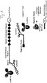
- the signal probe may have a spacer and/or an immobilization region between the target-molecule-specific binding region and the signal-generating region.
- FIG. 1 is a schematic view illustrating the signal probe of the present invention and major components used in the detection method of the present invention for target molecules based on the use of the signal probe.
- the spacer is introduced to separate the target-molecule-specific binding region and the signal-generating region. This aims to prevent complexes, formed by the binding of the target-molecule-specific binding region to a target molecule, from acting as obstacles for the detection of signals from the signal-generating part, thereby facilitating the detection of signals from the signal-generating part.
- the spacer may be a single-stranded nucleic acid, a double-stranded nucleic acid, RNA, DNA, etc., and may contain a nucleic acid analog, such as PNA, or a modified nucleotide.
- the spacer is not particularly limited in length as long as it facilitates the detection of signals from the signal-generating part. The length may typically range from 20 bp to 50 bp.
- the immobilization region is a region to which one or more immobilization nucleic acid molecules (or immobilization molecules), which will be described below, are complementarily bound in order to immobilize and stretch the signal probe of the present invention on an analytical support, as described below, and is composed of a previously known sequence.
- the immobilization region is composed of a single-stranded nucleic acid having a sequence complementary to the immobilization nucleic acid molecules, and the previously known sequence may be repeated two or more times in order to allow the binding of two or more immobilization nucleic acid molecules.
- the immobilization region may be RNA, DNA, or the like, as long as it can bind complementarily to the immobilization nucleic acid molecule, and may also be a nucleic acid analog, such as PNA, or may contain a modified nucleotide.
- the immobilization region may have a certain length, as long as it can exhibit sufficient binding affinity to the immobilization nucleic acid molecule, and may typically consist of 2 to 20 repetitions of a previously known sequence of 20 to 50 bp in length.
- both the spacer and the immobilization region are covalently linked to an adjacent region, and they can be arranged in a certain order, but it is preferred that the immobilization region be adjacent to the signal-generating region, that is, be present at a 5' end or a 3' end of the signal-generating region. This is because, when the immobilization region further immobilizes the signal probe of the present invention onto an analytical support, the immobilization region should be adjacent to the signal-generating region in order to allow the signal-generating region to be immobilized in a linear or stretched form on the analytical support and to thus facilitate the detection of signals from the signal-generating region.
- the signal probe of the present invention may contain a binding tag that is attached to an opposite side of the target-molecule-specific binding region (the opposite side refers to, for example, a 3'-end when the target-molecule-specific binding region is present at a 5'-end).
- the binding tag serves to immobilize the signal probe of the present invention on an analytical support, coated with a binding partner capable of binding to the binding tag, through the specific binding to the binding partner.
- a binding partner capable of binding to the binding tag
- An available representative example of the binding tag is biotin
- biotin is an antibiotic isolated from Streptomyces avidinii , belonging to the avidin family, which is a tetrameric protein that can bind to a maximum of four biotin molecules.
- biotin analogs such as desthiobiotin and iminobiotin may be used.
- streptavidin analogs may be used, which include avidin, traptavidin, NeutrAvidin (ThermoFisher Scientific, USA), and the like.
- the present invention relates to a method of detecting a target molecule in a sample using the aforementioned signal probe.
- the detection method of the present invention comprises the steps of (a) treating a sample intended to be analyzed with a signal probe to form a complex of the signal probe with a target molecule in the sample; and (b) detecting the signal probe of the complex.
- the sample to be analyzed may be a certain mixture or solution that contains or is suspected to contain a target molecule intended to be detected and thus needs to be detected.
- the sample may include biological samples obtained from humans or animals as well as processed samples having an increased concentration of a target molecule through processing of the biological samples.
- the sample may further include samples containing a target molecule or suspected of containing a target molecule, such as water, food, and industrial waste water, and samples intended to be tested, such as environmental pollutants and toxins.
- Such samples may contain a suitable diluent and a buffer, and can be a culture of bacteria or viruses, containing a medium or a component thereof, in order to detect the presence of bacteria or viruses.
- the sample to be analyzed may preferably be a biological sample taken from humans or animals or a processed sample thereof.
- the biological sample may be any one of substances taken from humans or animals, which contains or is suspected of containing a target molecule intended to be detected and is thus necessary to test, such as blood, urine, saliva, semen, amniotic fluid, lymphatic fluid, sputum, tissues, etc.
- the processed sample may be selected from samples having improved protein concentrations using a protein extraction kit, for example, plasma, serum or biological samples, as well as samples having increased concentrations of nucleic acids (DNA or RNA), tissue extracts, cells derived from tissues, cell lysates, cell cultures, and the like.
- DNA or RNA nucleic acids
- the present method at the step of forming a complex of the signal probe with a target molecule in a sample, when the sample contains a variety of target molecules, it is possible to form multiple different complexes and to detect the complexes using a plurality of signal probes each specific to the target molecules.
- detection will be understood to refer to qualitative analysis for the presence of a target molecule, and further includes quantitative analysis of the amount and concentration of the target molecule.
- the present method when the target molecule intended to be detected is a protein, may further include, prior to the step (a), the steps of applying the sample onto a nitrocellulose membrane having a high binding affinity to proteins to allow proteins contained in the sample including the target molecule to be adsorbed to the nitrocellulose membrane; and eliminating molecules other than the proteins absorbed to the nitrocellulose membrane, such as lipids, nucleic acids, metabolites, etc., using a suitable washing solution.
- the washing solution may be used by purchasing any one widely used in the art, or can be manufactured as appropriate, and includes a surfactant and a salt.
- surfactants include SDS, Tween 20, Tween 40, Tween 60, Tween 80, Triton X-405, Triton X-100, Tetronic 908, Cholesterol PEG 900, Polyoxyethylene Ether W-1, Span 20, Span 40, Span 85, and mixtures thereof.
- salts include acetic salts, lactate, citrate, phosphate, nitrate, sulfate and chloride salts of lithium, sodium, potassium and ammonium, and mixtures thereof (SSC, SSPE, etc.).
- washing solution can be used TBST solution (10 mM Tris-Cl, pH 8.0, 150 mM NaCl, 0.05% Tween 20), PBST solution (PBS, pH 7.0, 0.05% Tween 20), Tween 20, SSPE solution containing Tween 20 (0.2 M phosphate buffer, 2.98 M NaCl, 20 mM EDTA, pH 7.4), and the like.
- the method may further include the step of applying a blocking solution containing a suitable blocking agent, such as bovine serum albumin, to the nitrocellulose membrane to block the protein-unoccupied sites on the membrane surface.
- a blocking solution containing a suitable blocking agent such as bovine serum albumin
- This blocking step when used as the signal probe in which the target-molecule-specific binding region is an antibody in order to form a complex of the signal probe with a target molecule at the next step, serves to prevent the adsorption of the antibody of the signal probe from reducing the detection accuracy.
- the signal probe is applied to the membrane in order to perform the step (a) of forming a complex of the signal probe and the target molecule.
- the blocking step is not essentially required when the target-molecule-specific binding region is not an antibody but an aptamer or the like.
- another washing step may be carried out using, for example, the aforementioned washing solution in order to remove non-specifically bound signal probes.
- the detection of the signal probe at the step (b) may be achieved through the steps of (b-1) separating the signal probe from the complex bound to the nitrocellulose membrane and (b-2) detecting the separated signal probe.
- the separation of the signal probe from the complex is possible by heating the complex-bound nitrocellulose membrane (heating through a suitable buffer or a reaction solution) or by treating with a surfactant during heating.
- the signal probe of the present invention even when heated, is kept in a stable state without the loss of the signal-generating region, thereby not affecting the accuracy of detection of a target molecule.
- the step (b-1) of detecting the separated signal probe may include the steps of (b-1-i) applying the separated signal probe to an analytical support, which is coated with a binding partner to a binding tag of the signal probe, to thus immobilize the signal probe via the binding tag onto the analytical support through the specific interaction between the binding tag and the binding partner; and (b-1-ii) detecting a signal of the signal probe immobilized onto the support.
- the signal of the signal probe which is a signal that is produced from the signal-generating substance labeled to the signal probe, can be detected using a suitable instrument capable of detecting the signal depending on the signal-generating substance that is used.
- the instrument may be a spectrophotometer, absorption spectrometer, fluorometer, luminometer, chemiluminometer, actinometer, or the like.
- nCounter digital analyzer (Nanostring technology, USA), which is used in an embodiment of the present invention, a Nikon Eclipse TE2000E (Perfect Focus, 1.4 NA Plan Apo VC 60X oil-immersion lens, Nikon, Japan), which is used in an embodiment of U.S. Pat. No. 8,519,115 and its related published article: Nat. Biotechnol. 2008, 26:317-325 , an X-cite 120 metal halide light source (Exfo, USA), an automated H117 stage (Prior Scientific, UK), or a Smart Shutter (Sutter Instrument, USA).
- the nCounter digital analyzer or the like makes it possible to sense the linear order of a specific signal of the signal-generating region and to count and quantify a target molecule from the sensed signal.
- the linear order of signals is detected using the nCounter digital analyzer or the like, in order to facilitate such signal detection, it is preferred to immobilize the signal-generating region of the signal probe onto an analytical support in a linear or stretched form.
- immobilization molecule or an immobilization nucleic acid molecule
- the immobilization molecule has a single-stranded nucleic acid sequence complementary to the immobilization region of the signal probe and a binding tag that is linked to the complementary single-stranded nucleic acid sequence and is able to bind to its binding partner coated onto the analytical support.
- the binding tag of the immobilization molecule may also be biotin or a biotin analog, as described above with regard to the signal probe.
- an analytical support available in the method of this invention is not particularly limited in its form or shape as long as a binding partner (e.g., streptavidin) to the binding tag of the signal probe can be coated onto the surface of the support through covalent or non-covalent bonding.
- a binding partner e.g., streptavidin
- the support may be in the form of membranes, beads, microparticles, plates (e.g., glass slides), columns, wells, well plates, and the like, and may be made up of plastic, resin, polysaccharides, silica, functionalized glass, modified silicon, carbon, metals, inorganic glasses, membrane, nylon, natural resin, and the like.
- the materials for polymeric supports may include polystyrene, polyethylene glycol tetraphthalate, polyvinyl acetate, polyvinyl chloride, polyvinyl pyrrolidone, polyacrylonitrile, polymethyl methacrylate, polytetrafluoroethylene, butyl rubber, styrene butadiene rubber, polyethylene, polypropylene, (poly)tetrafluoroethylene, (poly)vinylidenefluoride, polycarbonate, polymethylpentene, and the like.
- FIG. 1 illustrates the signal probe and major components including the immobilization molecule used in the method of the present invention.
- FIGS. 2 and 3 show the overall process for detecting a target molecule, a protein, using the aforementioned nitrocellulose membrane, wherein the target-molecule-specific binding region of the signal probe is an antibody or an aptamer, respectively.
- the detection method for a target molecule according to the present invention may, in addition to the aforementioned signal probe, further employ a capture probe.
- Such a method of the present invention using a capture probe comprises the steps of (a) treating a sample intended to be analyzed with a signal probe and a capture probe, each of which binds specifically to a target molecule to form a complex of the target molecule, the signal probe and the capture probe, and obtaining a mixture containing the complex as well as a portion of each of the signal probe and the capture probe not participating in the complex formation; and (b) detecting the signal probe of the complex in the mixture.
- the capture probe comprises a region that binds specifically to a different portion from a portion (i.e., an epitope) of a target molecule to which the signal probe specifically binds, wherein a magnetic particle (or a magnetic bead) is connected to the region, as illustrated in FIG. 1 .
- the magnetic particle has been widely used in the art to isolate proteins, nucleic acids, cells, bacteria, and the like, as disclosed, for example, in U.S. Pat. Nos. 5,665,554 , 6,027,945 and 7,078,224 , Korean Patent Application Publication No. 2006-0061494 , and Korean Pat. No. 0541282 , and has a micro- or nano-scale size (i.e. several microns or tens to hundreds of nanometers in size) .
- the representative magnetic particles that have been employed for isolating biomolecules are iron oxide magnetic particles, including magnetite (Fe 3 O 4 ), maghemite (Fe 2 O 3 ), and hematite (Fe 2 O 3 ).
- the magnetic particles be modified with non-magnetic materials, such as silica, polymers, gold, and silver, to prevent the interaction and aggregation between magnetic particles in order to be used in the isolation of biomolecules, such as proteins or nucleic acids, and that, in order to allow binding to biomolecules such as proteins or nucleic acids, the magnetic particles be introduced with a functional group, such as carboxyl, epoxy, tosyl, amino, siloxane, etc., or a binding material, such as streptavidin or biotin.
- a functional group such as carboxyl, epoxy, tosyl, amino, siloxane, etc.
- a binding material such as streptavidin or biotin.
- a target-molecule-specific binding region may be connected to such a magnetic particle by covalent bonding via a functional group or by non-covalent bonding between biotin and streptavidin.
- the target-molecule-specific binding region of the capture probe is designed to recognize and bind to a different portion from a portion recognized and bound by a target-molecule-specific binding region of the signal probe so as to prevent competitive binding for the target molecule with the target-molecule-specific binding region of the signal probe.
- the target-molecule-specific binding region of the capture probe may be an antibody, an aptamer, a single-stranded nucleic acid, an antigen if the target molecule is an antibody, and the like. If the region is a single-stranded nucleic acid, it may contain a nucleic acid analog, such as PNA or a modified nucleotide.
- the step (a) is performed by treating a sample intended to be analyzed with a signal probe and a capture probe, in which a complex is formed among a target molecule, the signal probe and the capture probe to thus give a mixture containing the resulting complex, a portion of the signal probe not participating in the complex formation, a portion of the capture probe not participating in the complex formation, and non-target molecules.
- the next step (b) for the detection of a target molecule serves to detect the signal probe of the complex in the mixture.
- the detection of the signal probe is based on the capture probe containing magnetic particles, wherein the mixture is applied to a magnet and only the complex containing the capture probe and a portion of the capture probe not participating in the complex formation are adhered to the magnet.
- eliminated are a portion of the signal probe not participating in the complex formation, non-target molecules, and others.
- a washing step using a suitable washing solution may be desirably performed as exemplified above in order to effective removal of the signal probe not participating in the complex formation, non-target molecules and others.
- the signal probe is separated from the complex. This, as described above, is possible, for example, by heating or by treating with a surfactant with heating.
- the signal probe, as described above, is maintained in a stable state even under heating or the like because double-stranded nucleic acids constituting the signal probe are cross-linked to each other.
- the step (b) of detecting the signal probe may include the steps of (b-1) applying the mixture to a magnet to adhere only both the complex and a portion of the capture probe not participating in the complex formation to the magnet so as to eliminate other remaining components in the mixture; (b-2) separating the signal probe from the complex adhered to the magnet; and (b-3) detecting the separated signal probe.
- step (b-3) of detecting only the separated signal probe may be achieved by performing the steps of (b-3-i) applying the separated signal probe to an analytic support, which is coated with a binding partner to a binding tag of the signal probe, to immobilize the signal probe via the binding tag onto the analytic support through the specific interaction between the binding tag and the binding partner; and (b-3-ii) detecting a signal of the signal probe immobilized onto the support.
- steps (b-3-i) of immobilizing the signal probe onto the analytic support and the detection step (b-3-ii) descriptions with respect to the protein detection method using a nitrocellulose membrane can be referred to in their entireties.
- the present invention in addition to the signal probe, may further employ a capture competitive molecule as illustrated in FIGS. 6 and 7 .
- the present method employing the capture competitive molecule comprises the steps of (a) treating a sample intended to be analyzed with a signal probe and a capture competitive molecule, each of which binds specifically to a target molecule to form a complex of the capture competitive molecule and the signal probe, and obtaining a mixture containing the complex; and (b) detecting the signal probe of the complex in the mixture.
- the mixture contains, in addition to the complex of the capture competitive molecule and the signal probe, a complex of the target molecule and the signal probe, a portion of the signal probe participating in the complex formation, a portion of the competitive molecule participating in the complex formation, a portion of the target molecule participating in the complex formation, and non-target molecules.
- the capture competitive molecule has a portion the same as the portion (i.e., an epitope) of a target molecule to which the signal probe specifically binds, wherein a magnetic particle is connected to the region.
- the capture competitive molecule may typically be the same molecule as the target molecule but can be an analogous molecule as long as it can bind to the same portion as a portion (i.e., an epitope) of a target molecule to which the signal probe specifically binds, and can thus compete for binding to the signal probe.
- Examples of such analogous molecules may include a fusion protein with a protein (Fc of antibody) having a different target protein.
- the capture competitive molecule Since the capture competitive molecule has the same binding region for the signal probe as a target molecule, it competes with the target molecule for binding to the signal probe. This allows, when the signal probe is applied in a limited amount to a sample and the capture competitive molecule is used in a predetermined amount, the detection of the capture competitive molecule to provide information about the target molecule in a sample intended to be analyzed (such as the presence or absence of target molecules or the concentrations thereof).
- the signal probe is applied in a limited amount to a sample
- the signal probe is used in an amount less than an amount that enables detection both of the capture competitive molecule and the target molecule
- theoretically means that it is applied at a concentration allowing the detection of an amount less than the sum of the quantified concentration (quantitative value) of the capture competitive molecule and the estimated concentration (quantitative value) of the target molecule in a sample of interest, but can be applied at a concentration higher than the sum because not all of the applied signal probe binds to the capture competitive molecule or to the target molecule.
- the limited amount of the signal may be generally a concentration that enables detection of less than the quantified concentration of the capture competitive molecule.
- the detection and quantification of a target molecule in the sample of interest may be achieved by performing several repeated tests with various concentrations of the signal probe and the capture competitive molecule under the same detection condition, such as using the same reaction solutions, and generating a standard curve that shows the relationship between the concentrations of the applied signal probe and the concentrations of the applied capture competitive molecule.
- the detected and quantified value of the capture competitive molecule is in inverse proportion to the concentration of the target molecule.
- the step (b) of detecting the signal probe of the complex in the mixture can be performed in the same way as in the capture detection method.
- the step (b) may include the steps of (b-1) applying the mixture to a magnet to adhere the complex of the capture competitive molecule and the signal probe to the magnet; (b-2) separating only the signal probe from the complex adhered to the magnet; and (b-3) detecting the separated signal probe.
- the capture competitive molecule not forming the complex is also adhered to the magnet, but heating makes it possible to separate only the signal probe.
- the above step (b-3) of detecting only the separated signal probe, as in the protein detection method using a nitrocellulose membrane and the capture detection method as described above, may be achieved by performing the steps of (b-3-i) applying the separated signal probe to an analytic support coated with a binding partner to a binding tag of the signal probe in order to immobilize the signal probe via the binding tag onto the analytic support through the specific binding between the binding tag and the binding partner; and (b-3-ii) detecting a signal of the signal probe immobilized onto the support.
- steps of the step (b-3-i) of immobilizing the signal probe onto the analytic support and the detection step (b-3-ii) descriptions of the protein detection method using a nitrocellulose membrane can be referred to in their entireties.
- the present invention can provide a stable double-stranded nucleic acid signal probe, in which single-stranded nucleic acids are complementarily joined together to form a double-stranded nucleic acid through cross-linking (or covalent bonding) to thus not be affected by temperature, pH, salinity, ionic strength, or the like.
- the present invention can provide a method of detecting a target molecule using the stable double-stranded nucleic acid signal probe by forming a complex of the signal probe with the target molecule, separating only the signal probe from the complex by heating or the like, and detecting the signal probe.
- the term “signal probe”, as used herein, is used interchangeably with “the first probe”, the term “signal-generating region” is used interchangeably with “the color-coded barcode”, and the term “target-molecule-specific binding region” of the signal probe is used interchangeably with “the first binding region” or "the first ligand”.
- capture probe is used interchangeably with “the second probe” or “the capture probe”
- target-molecule-specific binding region of the capture probe
- Each region labeled with a signal-generating substance, constituting the signal-generating region of the signal probe, is designated as "a signal tag".
- immobilization nucleic acid molecule is used interchangeably with “the immobilization molecule” in the following examples.
- the first probe functions to bind to a target molecule and generate a signal unique to a target molecule.
- the first probe basically consists of a signal-generating region, assigning a unique signal to a target molecule, and the first binding region linked thereto, specifically binding to the target molecule.
- the first probe may further include an immobilization region and a binding tag.
- the signal tag indicates the basic unit constituting the signal-generating region and is composed of a double-stranded nucleic acid fragment labeled with a signal-generating substance.
- the double-stranded nucleic acid was prepared by performing a polymerase chain reaction (PCR) using M13 DNA as a template and labeling the PCR product of about 900 bp with a signal-generating substance (a detectable label).
- PCR polymerase chain reaction
- the nucleotide sequence of M13 DNA which is the backbone of the signal tag, was obtained from the GenBank Database system (http://www.ncbi.nlm.nih.gov; Accession No. NC_003287).
- PCR was carried out using 100 ng of M13 DNA (NEB, USA) as a template with 0.1 ⁇ M of each of a primer pair below (Bioneer, South Korea) and 1.25 units of Taq Polymerase (Promega, USA) according to a standard PCR method, thereby giving a DNA fragment (903 bp).
- a restriction enzyme map was prepared for the above PCR product to select restriction enzymes not capable of digesting the PCR product, and the adaptors containing recognition sites for the selected restriction enzymes, as listed in Table 1, were purchased (Gene Link, USA).
- the adaptors were linked to the aminoallyl-dCTP-incorporated PCR product according to the standard method recommended by the manufacturer to give the combinations described below.
- a ligation reaction was conducted by mixing 3 nmol of the adaptor with 0.3 nmol of the PCR product DNA fragment and adding 1 ⁇ l of T4 ligase (5 units/ ⁇ l; NEB, USA) to 19 ⁇ l of the resulting mixture.
- the products thus ligated were purified to obtain adaptor-attached, aminoallyl-dCTP-incorporated DNA fragments. These DNA fragments were then treated with the above restriction enzymes and purified.
- N-hydroxysuccinimide (NHS)-ester fluorescent dyes Alexa 488, Alexa 594, Alexa 647 (Invitrogen, USA) and Cy3 (GE Healthcare, USA).
- 16-20 ⁇ g of the adaptor-attached, aminoallyl-dCTP-incorporated DNA fragment for each domain was dissolved in 20 ⁇ l of sterile distilled water (DW) and mixed with 12 ⁇ l of a labeling solution (25 mg of NaHCO 3 in 1 ml of sterile DW) to give a reaction solution.
- a labeling solution 25 mg of NaHCO 3 in 1 ml of sterile DW
- each of the signal-generating substances was dissolved in DMSO at a final concentration of 30 ⁇ g/L to give a signal-generating substance solution.
- the reaction solution was aliquoted into four tubes with 5 ⁇ l per tube, and to each tube was added 2 ⁇ l of any one of the signal-generating substance solutions, followed by incubation for 1 hr at room temperature in the dark.
- the DNA fragments were labeled with the signal-generating substances in this way, they were purified using the QIAquick PCR Purification Kit (Qiagen, USA), thus giving adaptor-attached DNA fragments, each of which was labeled with the signal-generating substance.
- a four-fold volume of chloroform was added to the resulting reaction mixture to obtain a non-reactive psoralen-removed supernatant through an extraction process.
- To the supernatant was added 5 M NaCl at a final concentration of 0.2 M and a three-fold volume of ethanol so as to recover the covalent-bond-induced H signal tag.
- the covalent-bond-induced H signal tag thus obtained was designated a "C signal tag" for convenience.
- the double strands of the H signal tag began to dissociate and the duplex was converted to single strands, wherein it was found that the melting temperature (Tm) was about 85°C and the duplex was completely denatured at 95°C to the single-stranded state. In contrast, the C signal tag was found to maintain its double-stranded structure even at 95°C.
- the C signal tags thus prepared were used for preparing color-coded barcodes as the signal-generating regions.
- one signal tag was selected from among the four signal tags for the first domain and placed into each of four tubes at a final concentration of 10 ⁇ g/ml.
- each of the four signal tags for the second domain was added at a final concentration of 10 ⁇ g/ml.
- a reaction solution of T4 ligase 50 mM Tris-HCl, 10 mM MgCl 2 , 1 mM ATP, 10 mM DTT, pH 7.5
- the other three types of signal tags for the first domain were reacted with each signal tag for the second domain according to the same method as described above.
- Each of the four signal tags for the first domain was mixed with each of the four signal tags for the second domain, and the resulting 16 reaction mixtures were designated as the first-second domain mixture solutions.
- one signal tag was selected from among the four signal tags for the third domain and placed into each of four tubes at a final concentration of 10 ⁇ g/ml.
- each of the four signal tags for the fourth domain was added at a final concentration of 10 ⁇ g/ml.
- a reaction solution of T4 ligase 50 mM Tris-HCl, 10 mM MgCl 2 , 1 mM ATP, 10 mM DTT, pH 7.5
- the other three types of signal tags for the third domain were reacted with each signal tag for the fourth domain according to the same method as described above.
- Each of the four signal tags for the third domain was mixed with each of the four signal tags for the fourth domain, and the resulting 16 reaction mixtures were designated as the third-fourth domain mixture solutions.
- one signal tag was selected from among the four signal tags for the fifth domain and placed into each of four tubes at a final concentration of 10 ⁇ g/ml.
- each of the four signal tags for the sixth domain was added at a final concentration of 10 ug/ml.
- a reaction solution of T4 ligase 50 mM Tris-HCl, 10 mM MgCl 2 , 1 mM ATP, 10 mM DTT, pH 7.5
- the other three types of signal tags for the fifth domain were reacted with each signal tag for the sixth domain according to the same method as described above.
- Each of the four signal tags for the fifth domain was mixed with each of the four signal tags for the sixth domain, and the resulting 16 reaction mixtures were designated as the fifth-sixth domain mixture solutions.
- one tube selected from among the 16 tubes of the first-second domain mixture solutions, one tube selected from among the 16 tubes of the third-fourth domain mixture solutions and one tube selected from among the 16 tubes of the fifth-sixth domain mixture solutions were mixed at an equal concentration.
- To the resulting reaction solution of 95 ⁇ l was added 5 ⁇ l of T4 ligase (5 units/ ⁇ l; NEB, USA).
- reaction products at each reaction step were electrophoresed and the result is shown in FIG. 9 .
- These color-coded barcodes form a spot of 300 nm in size and are imaged using an epi-fluorescent microscope ( US 8,519,115 B2 ; Nat. Biotechnol. 2008 Mar 26(3) : 293-4 ).
- the first probe is a construct that recognizes and binds to a target molecule and generates a unique signal to detect the target molecule, wherein the target-molecule-specific binding region, that is, the first binding region, is connected with the signal-generating region.
- This first probe was prepared in two types, namely a form not immobilized to a support and a form immobilized thereto, depending on the presence or absence of a binding material (e.g., biotin).
- a binding material e.g., biotin
- As the first binding region used were a single-stranded nucleic acid for nucleic acid detection and an antibody and an aptamer for protein detection.
- a non-immobilizable first probe was designed to have a structure of 5'-color-coded barcode-spacer-the first binding region-3'.
- the 5' and 3' ends, used here and below, are intended to indicate the direction or order of the arrangement of the nucleic acid when a nucleic acid such as the color-coded barcode or the spacer is used as a constituent factor of the probe.
- a single-stranded nucleic acid was synthesized to have the following sequence by a commercial supplier.
- a non-immobilizable first probe was prepared using a single-stranded nucleic acid as the first binding region as follows. First, a single-stranded nucleic acid was synthesized to have a construct of 5'-spacer-first binding region-3' (Integrated DNA Technologies, USA). Then, 10 ⁇ l of the 5'-spacer-first binding region-3' construct (1 mg/ml) was mixed with 10 ⁇ l of a color-coded barcode (1 mg/ml) in a reaction solution (50 mM Tris-HCl, 10 mM MgCl 2 , 1 mM ATP, 10 mM DTT, pH 7.5).
- a reaction solution 50 mM Tris-HCl, 10 mM MgCl 2 , 1 mM ATP, 10 mM DTT, pH 7.5.
- a non-immobilizable first probe was prepared using an antibody as the first binding region as follows. First, to an antibody was added a reactive group (SH group obtained by reducing disulfide bonds of the antibody using DTT) using the Thunder-Link oligo conjugation system (Innova Biosciences, England) according to the manufacturer's protocol, thereby giving an antibody having a reactive group. In detail, according to the manufacturer's standard method, 100 ⁇ l of an antibody (1 mg/ml) was added to the Antibody Activation Reagent vial and incubated for 1 hr at room temperature, thereby generating an activated antibody having a reactive group.
- a reactive group SH group obtained by reducing disulfide bonds of the antibody using DTT
- Thunder-Link oligo conjugation system Innova Biosciences, England
- a spacer having a reactive group (NH 2 ), 5'-spacer-NH 2 was synthesized in the form of a single-stranded nucleic acid (Integrated DNA Technologies, USA). Thereafter, the activated antibody was reacted with the spacer having the reactive group in a solution of N-succinimidyl-S-acetyl-thioacetate to obtain a 5'-spacer-antibody-3' construct.
- a non-immobilizable first probe was prepared using an aptamer as the first binding region as follows. First, a construct of 5'-spacer-aptamer-3' was synthesized (Integrated DNA Technologies, USA). Then, 10 ⁇ l of the 5'-spacer-aptamer-3' construct (1 ⁇ mg/ml) was mixed with 10 ⁇ l of a color-coded barcode (1 mg/ml) in a reaction solution (50 mM Tris-HCl, 10 mM MgCl 2 , 1 mM ATP, 10 mM DTT, pH 7.5).
- An immobilizable first probe was designed to have a construct of 5'-first binding region-spacer-immobilization region-color-coded barcode-biotin-3'.
- the immobilization region which consists of repetitions of a previously known sequence, was used to immobilize the first probe onto a support through complementary binding between the immobilization region and an immobilization molecule (that is, an immobilization nucleic acid molecule) containing a sequence complementary to the immobilization region.
- the immobilization region was a single-stranded nucleic acid having the following sequence.
- the immobilization region 5'-(GCCACAGCACCTTGGTGCGTAGGATCTG) 10 -3' (SEQ ID NO: 12)
- the number 10 which is an integer, refers to the repeated number of the above sequence.
- An immobilizable first probe was prepared using a single-stranded nucleic acid as the first binding region as follows. First, a single-stranded nucleic acid was synthesized to have a construct of 5'-spacer-immobilization region-3' (Integrated DNA Technologies, USA). Separately, a color-coded barcode was biotinylated using the Thermo Scientific Biotin 3' End DNA Labeling Kit (Thermo Scientific, USA), thereby generating a 5'-color-coded barcode-biotin-3' construct.
- a color-coded barcode (1 mg/ml) was mixed with 5 ⁇ l of 5 ⁇ M Biotin-11-UTP (Biotin-11-Uridine-5'-triphosphate) in a reaction solution (50 mM Potassium Acetate, 20 mM Tris-acetate, 10 mM Magnesium Acetate, pH 7.9).
- a reaction solution 50 mM Potassium Acetate, 20 mM Tris-acetate, 10 mM Magnesium Acetate, pH 7.9
- TdT terminal deoxynucleotidyl transferase
- An immobilizable first probe was prepared using an antibody as the first binding region as follows. First, a 5'-color-coded barcode-biotin-3' construct and an antibody having a reactive group (SH group) were prepared according to the same method as described above, while a single-stranded nucleic acid having a reactive group (NH 2 ), 5'-NH 2 -spacer-immobilization region-3', was synthesized by a commercial supplier (Integrated DNA Technologies, USA).
- the antibody having the reactive group was reacted with the above construct for 12 to 24 hrs at room temperature.
- the Conjugate Clean Up Reagent (Innova Biosciences, England), and the resulting mixture was allowed to react for 10 min and then centrifuged at 15,000 ⁇ g for 5 min. This step, the reaction in the Conjugate Clean Up Reagent, followed by centrifugation, was repeated twice to obtain a purified immobilizable first probe containing an antibody as the first binding region in the construct of 5'-first binding region-spacer-immobilization region-color-coded barcode-biotin-3'.
- An immobilizable first probe was prepared using an aptamer as the first binding region as follows. First, a 5'-color-coded barcode-biotin-3' construct was prepared according to the same method as described above, while a single-stranded nucleic acid, 5'-aptamer-spacer-immobilization region-3', was synthesized by a commercial supplier (Integrated DNA Technologies, USA).
- the second probe which is a capture probe, was prepared by binding the second binding region, serving as a target-molecule-specific binding region, to magnetic beads serving as a harvesting material.
- the competitive molecule was prepared by binding a target molecule (or a target molecule analog capable of competing with the target molecule) to magnetic beads.
- a second probe containing a single-stranded nucleic acid or an aptamer as the second binding region, was prepared by attaching the second binding region to magnetic beads through biotin-streptavidin interaction.
- a single-stranded nucleic acid or an aptamer prepared in advance, was biotinylated using the Thermo Scientific Biotin 3 End DNA Labeling Kit (Thermo Scientific, USA) according to the same method as described above in order to obtain 5'-second binding region-biotin-3'.
- the above-prepared 5'-second binding region-biotin-3' (50 ng/L) was added to an equal volume of the Streptavidin-coupled Dynabeads (5 ⁇ g/L) (Dynal Biotech ASA, Norway) and allowed to react by continuous rocking according to the manufacturer's protocol, thereby obtaining a second probe containing a single-stranded nucleic acid as the second binding region.
- the second probe containing an antibody as the second binding region was prepared by directly binding an antibody to magnetic beads, serving as a harvesting material. First, 100 ⁇ g of an antibody was mixed with 5 mg of magnetic beads having a tosyl group (M-280 Tosyl activated magnetic beads, Dynal Biotech ASA, Norway) in a buffer (0.1 M borate buffer, pH 9.5) and incubated for 48 hrs at room temperature. The antibody-bound magnetic beads were separated using a magnet via the tosyl group and washed with the same buffer to remove unbound antibodies.
- M-280 Tosyl activated magnetic beads Dynal Biotech ASA, Norway
- the antibody-bound magnetic beads (hereinafter, referred to as "capture probe") were reacted with 0.1% bovine serum albumin (BSA) (Sigma, USA) for 4 hrs at 37°C in order to inactivate tosyl groups unbound to molecules. After several washings, the magnetic beads were stored at 4°C in a buffer (0.1 M PBS, pH 7.4).
- BSA bovine serum albumin
- the capture competitive molecule using a single-stranded nucleic acid as the competitive molecule was intended to be used for detecting a nucleic acid as a target molecule.
- This capture competitive molecule was prepared according to the same method as in the preparation of the second probe containing a single-stranded nucleic acid or an aptamer as the second binding region.
- the capture competitive molecule using a protein as the competitive molecule was intended to be used for detecting a protein as a target molecule.
- This capture competitive molecule was prepared according to the same method as in the preparation of the second probe containing a single-stranded nucleic acid or an aptamer as the second binding region. The same protein as a target protein was used as the competitive molecule.
- An immobilization molecule was designed to have a nucleotide sequence complementary to the repeated sequence constituting the immobilization region of the first probe, and was prepared by synthesizing a single-stranded nucleic acid biotinylated at its 5' end (Integrated DNA Technologies, USA).
- Typical food-poisoning bacteria such as E. coli, Salmonella, Listeria and Staphylococcus, were detected using the immobilizable first probe, prepared using the antibodies of Table 2 and the aptamers of Table 3 as the first binding region (ligand).
- E. coli Salmonella
- Listeria Listeria
- Staphylococcus Typical food-poisoning bacteria, such as E. coli, Salmonella, Listeria and Staphylococcus
- reaction solution 10 mM PBS buffer (137 mM NaCl, 10 mM Phosphate, 2.7 mM KCl, pH 7.4) for the antibody ligand and SELEX buffer (20 mM Tris-Cl, pH 7.6, containing 100 mM NaCl, 2 mM MgCl 2 , 5 mM KCl, 1 mM CaCl 2 and 0.02% Tween) for the aptamer ligand.
- the reaction solution was supplemented with 0.05% to 0.2% (w/v) of sodium azide for suppressing bacterial proliferation and 0.1% to 0.3% (w/v) of bovine serum albumin for inhibiting non-specific binding.
- the reaction was carried out at 20°C to 30°C.
- the nucleic acid detection was conducted with an immobilizable first probe, containing a single-stranded nucleic acid of Table 4 below as the first binding region, and a second probe, containing a single-stranded nucleic acid of Table 4 below as the second binding region, using ⁇ -actin, IL17 and IL17RA mRNA molecules as a target molecule.
- the single-stranded nucleic acids, listed in Table 4 below, for ⁇ -actin, IL17 and IL17RA mRNA molecules were synthesized (Bioneer, South Korea) and used in the preparation of the first probe and the second probe.
- nucleotide sequences of the target molecules, ⁇ -actin, IL17 and IL17RA mRNA, were obtained from the GenBank database system (http:// www.ncbi.nlm.nih.gov) (IL17: Accession number NM_002190; and IL17RA: Accession number NM_014339).
- RNA Human cartilage tissue (Promocell, Germany) was used as a sample to be analyzed.
- mRNA as the target molecule, was isolated from the sample using the AllPrep DNA/RNA/Protein Mini kit (Qiagen, USA).
- the cartilage tissue was dissolved in a buffer provided by the manufacturer, loaded onto a column and centrifuged to bind RNA to the column membrane.
- the RNA bound to the column membrane was recovered by washing the column, eluting RNA from the column membrane and centrifuging the column.
- the isolated RNA (100 ng/ ⁇ l) was mixed with the first probe (200 ng/ ⁇ l) and the second probe (200 ng/ ⁇ l) with rocking to form a complex of the first probe, the target mRNA and the second probe through hybridization.
- Hybridization was carried out at 65°C in a reaction solution containing 0.5 M NaHPO 4 , 7% sodium dodecyl sulfate (SDS) and 1 mM EDTA. Then, the reaction mixture was applied to a magnet to immobilize the complex onto the magnet through the magnetic beads of the second probe, followed by washing with a washing solution in order to remove the unbound first probe and non-target RNA.
- the washing was performed three times: once with 6X SSC (standard saline citrate)/0.1% SDS at 8°C, once with 0.1X SSC/0.1% SDS at 68°C, and once 0.2X SSC/0.1% SDS at 42°C.
- 6X SSC standard saline citrate
- the magnet to which the complex of the first probe, the target mRNA and the second probe was bound, was incubated in a solution containing 0.1X SSPE and 0.1% Tween 20 for 5 min at 95°C in order to separate the first probe from the complex, thereby obtaining a supernatant containing the first probe.
- the first probe was immobilized onto a streptavidin-coated coverslip using the nCounter digital analyzer (Nanostring technology, USA), which enables molecular counting, according to the manufacturer's protocol, and the immobilized spots were imaged to thus be detected and quantified.
- nCounter digital analyzer Nemostring technology, USA
- 3 ⁇ l of the above-obtained supernatant was mixed with 1 ⁇ l of a 1:5,000 dilution of 0.1% TetraSpeck Fluorescent Microspheres, loaded into a fluid device in the above analyzer equipped with a streptavidin-coated coverslip (Optichem, Accelr8 Technology Corporation) in order to induce attachment of the first probe onto the coverslip as a support, through the interaction between biotin in the first probe and streptavidin in the coverslip. Thereafter, the surface was washed once with 90 ⁇ l of 1X TAE and then treated with 40 ⁇ l of 1X TAE to stretch the first probe, followed by applying 160 V/cm for 1 min.
- the obtained four images were integrated (spatial clustering), and an image for the first probe was obtained as shown in the schematic diagram of FIG. 11 .
- the resolution of the above analyzer was set to 600 FOV (field of view).
- Data were downloaded from the above analyzer and analyzed in Excel according to the manufacturer's instructions.
- Data were normalized for a standard material and a target molecule in a sample using an initially provided standard curve of a positive test control.
- the normalized values were used for overall average normalization of target molecule values using the values of the reference materials contained in a sample.
- the quantitative values of target molecules were calculated by averaging the analysis results of three repetitions.
- Quantitative results were obtained as described above using the above analyzer through molecular counting for the target molecule mRNA. The results thus obtained were found to be similar to the RT-PCR result for the target mRNA.
- Target molecules of Table 5 below were detected using only an immobilizable first probe containing a polyclonal antibody as listed in Table 5 below as the first binding region for a target molecule.
- the overall process of protein detection can be seen in FIGS. 2 and 3 .
- each target molecule of Table 5 was added at a concentration of 100 ⁇ g/ml to 10 mM PBS buffer (137 mM NaCl, 10 mM Phosphate, 2.7 mM KCl pH 7.4) and serially diluted to give final concentrations of 1,000 pg/ml, 100 pg/ml, 10 pg/ml, 0.1 pg/ml and 0 pg/ml.
- the resulting serial dilutions for the thirteen biological molecules were mixed at various concentrations to give mixture samples to be analyzed.
- the first probe was added at a concentration of 200 ng/ml to 10 mM PBS buffer (137 mM NaCl, 10 mM Phosphate, 2.7 mM KCl pH 7.4) to give an immobilizable first probe solution.
- a nitrocellulose membrane disc was added to the reaction solution containing the sample to be analyzed and allowed to react for 30 min with rocking to attach proteins in the sample onto the membrane. Then, the disc was washed with 150 ⁇ l of 0.1X SSPE/0.1% Tween 20 and allowed to react in a blocking solution (2% (w/v) bovine serum albumin (BSA)). 70 ⁇ l of the immobilizable first probe solution for each target molecule was added and the solution was rocked for 1 hr to induce the formation of a complex between the target molecule and the first probe. The disc was then washed with 150 ⁇ l of 0.1X SSPE/0.1% Tween 20 three times so as to eliminate excess first probes, not forming the complex, and the first probe forming a non-specific complex.
- a blocking solution 2% (w/v) bovine serum albumin
- the washed disc was heated in 0.1X SSPE/0.1% Tween 20 for 5 min at 95°C to thus induce separation of the first probe from the complex, thereby obtaining a supernatant containing the first probe.
- the first probe was immobilized onto a streptavidin-coated coverslip using the nCounter digital analyzer (Nanostring technology, USA), which enables molecular counting, according to the manufacturer's protocol, and the immobilized spots were imaged to thus be detected and quantified.
- nCounter digital analyzer Nemostring technology, USA
- the target molecules of Table 5 were detected using a second probe containing a polyclonal antibody as listed in Table 5 as the second binding region for a target molecule.
- the overall process of protein detection can be seen in FIGS. 4 and 5 .
- a sample to be analyzed was prepared according to the same method as in the above Example 3-1. 25 ⁇ l of the sample to be analyzed was mixed with 70 ⁇ l of an immobilizable first probe solution for each target molecule and 70 ⁇ l of a second probe and rocked for 1 hr to induce the formation of a complex of the first probe, the target molecule and the second probe. Then, the reaction mixture was applied to a magnet to attach the complex onto the magnet through the magnetic beads of the second probe. The beads bound to the magnet were then washed with a washing solution so as to eliminate unbound first probes and non-target molecules. The washing was carried out three times using 150 ⁇ l of 0.1X SSPE/0.1% Tween 20.
- the complex-adhered magnet was heated in 0.1X SSPE/0.1% Tween 20 for 5 min at 95°C in order to separate the first probe from the complex, thereby obtaining a supernatant containing the first probe.
- the first probe was immobilized onto a streptavidin-coated coverslip using the nCounter digital analyzer (Nanostring technology, USA), which enables molecular counting, according to the manufacturer's protocol, and the immobilized spots were imaged to thus be detected and quantified.
- nCounter digital analyzer Nemostring technology, USA
- the thirteen biological molecules of Table 6 were detected in two independent reactions, and the normalized results are shown in a log-log scale in FIG. 13 , in which the deviation for each signal ranged from 0.7 to 35.4.
- the target molecules of Table 5 were detected using a first probe containing an antibody listed in Table 6 as the second binding region and a capture competitive molecule using each protein of Table 5.
- the overall process of protein detection can be seen in FIGS. 6 and 7 .
- a sample to be analyzed was prepared according to the same method as in the above Example 3-1. 25 ⁇ l of the sample to be analyzed was mixed with 70 ⁇ l of an immobilizable first probe solution for each target molecule and 70 ⁇ l of a capture competitive molecule (30 ⁇ g/ ⁇ l) and rocked for 1 hr to induce the formation of a complex of the first probe, the target molecule and the capture competitive molecule. Then, the reaction mixture was applied to a magnet to attach the complex onto the magnet through the magnetic beads of the capture competitive molecule.
- the bead bound to the magnet was then washed with a washing solution so as to eliminate unbound first probes, non-target molecules and the first probe-target molecule complex, thereby harvesting only the complex between the first probe and the capture competitive molecule.
- the washing was carried out three times using 150 ⁇ l of 0.1X SSPE/0.1% Tween 20.
- the magnet to which the complex of the first probe and the capture competitive molecule was adhered, was heated in 0.1X SSPE/0.1% Tween 20 for 5 min at 95°C in order to separate the first probe from the complex, thereby obtaining a supernatant containing the first probe.
- the first probe was immobilized onto a streptavidin-coated coverslip using the nCounter digital analyzer (Nanostring technology, USA), which enables molecular counting, according to the manufacturer's protocol, and the immobilized spots were imaged to thus detect and quantify the same.
- a standard curve was generated from the quantitative results of the capture competitive molecule, and quantitative results of target molecules were obtained from the standard curve.
Landscapes
- Chemical & Material Sciences (AREA)
- Life Sciences & Earth Sciences (AREA)
- Health & Medical Sciences (AREA)
- Organic Chemistry (AREA)
- Proteomics, Peptides & Aminoacids (AREA)
- Engineering & Computer Science (AREA)
- Zoology (AREA)
- Wood Science & Technology (AREA)
- Analytical Chemistry (AREA)
- Immunology (AREA)
- Physics & Mathematics (AREA)
- Molecular Biology (AREA)
- Biochemistry (AREA)
- General Health & Medical Sciences (AREA)
- Biotechnology (AREA)
- Biophysics (AREA)
- Microbiology (AREA)
- Bioinformatics & Cheminformatics (AREA)
- General Engineering & Computer Science (AREA)
- Genetics & Genomics (AREA)
- Pathology (AREA)
- Cell Biology (AREA)
- Virology (AREA)
- Nuclear Medicine, Radiotherapy & Molecular Imaging (AREA)
- General Physics & Mathematics (AREA)
- Biomedical Technology (AREA)
- Measuring Or Testing Involving Enzymes Or Micro-Organisms (AREA)
- Peptides Or Proteins (AREA)
Applications Claiming Priority (2)
| Application Number | Priority Date | Filing Date | Title |
|---|---|---|---|
| KR20160082612 | 2016-06-30 | ||
| PCT/KR2017/006987 WO2018004309A1 (fr) | 2016-06-30 | 2017-06-30 | Sonde de signal d'acide nucléique bicaténaire et procédé de détection de molécule cible utilisant celle-ci |
Publications (2)
| Publication Number | Publication Date |
|---|---|
| EP3480320A1 true EP3480320A1 (fr) | 2019-05-08 |
| EP3480320A4 EP3480320A4 (fr) | 2020-03-11 |
Family
ID=60787015
Family Applications (1)
| Application Number | Title | Priority Date | Filing Date |
|---|---|---|---|
| EP17820585.2A Withdrawn EP3480320A4 (fr) | 2016-06-30 | 2017-06-30 | Sonde de signal d'acide nucléique bicaténaire et procédé de détection de molécule cible utilisant celle-ci |
Country Status (7)
| Country | Link |
|---|---|
| US (1) | US20200308631A1 (fr) |
| EP (1) | EP3480320A4 (fr) |
| JP (1) | JP6940532B2 (fr) |
| KR (1) | KR101913163B1 (fr) |
| CN (1) | CN110312808A (fr) |
| CA (1) | CA3029178C (fr) |
| WO (1) | WO2018004309A1 (fr) |
Cited By (1)
| Publication number | Priority date | Publication date | Assignee | Title |
|---|---|---|---|---|
| CN108444966A (zh) * | 2018-04-16 | 2018-08-24 | 成都博奥晶芯生物科技有限公司 | 一种激光微阵列芯片扫描仪的浓度梯度荧光校准片 |
Families Citing this family (2)
| Publication number | Priority date | Publication date | Assignee | Title |
|---|---|---|---|---|
| CN111458506B (zh) * | 2020-03-08 | 2023-01-06 | 复旦大学 | 一种基于TdT信号放大的结直肠癌外泌体检测方法和体系 |
| CN114544937B (zh) * | 2020-11-11 | 2023-04-07 | 艾克发(北京)生物技术有限公司 | 一种多重信号放大系统及其在免疫吸附间接法检测中的应用 |
Family Cites Families (36)
| Publication number | Priority date | Publication date | Assignee | Title |
|---|---|---|---|---|
| US5683888A (en) | 1989-07-22 | 1997-11-04 | University Of Wales College Of Medicine | Modified bioluminescent proteins and their use |
| US5292658A (en) | 1989-12-29 | 1994-03-08 | University Of Georgia Research Foundation, Inc. Boyd Graduate Studies Research Center | Cloning and expressions of Renilla luciferase |
| ATE318832T1 (de) | 1990-06-11 | 2006-03-15 | Gilead Sciences Inc | Verfahren zur vervendung von nukleinsäureliganden |
| US5270163A (en) | 1990-06-11 | 1993-12-14 | University Research Corporation | Methods for identifying nucleic acid ligands |
| WO1992015673A1 (fr) | 1991-03-11 | 1992-09-17 | The University Of Georgia Research Foundation, Inc. | Clonage et expression de renilla luciferase |
| ATE400651T1 (de) | 1993-09-10 | 2008-07-15 | Univ Columbia | Verwendung von grünem fluoreszenzprotein |
| WO1995021191A1 (fr) | 1994-02-04 | 1995-08-10 | William Ward | Indicateur bioluminescent fonde sur l'expression d'un gene codant pour une proteine modifiee a fluorescence verte |
| GB9411572D0 (en) | 1994-06-09 | 1994-08-03 | Amersham Int Plc | Magnetic bead precipitation method |
| US5777079A (en) | 1994-11-10 | 1998-07-07 | The Regents Of The University Of California | Modified green fluorescent proteins |
| US5876995A (en) | 1996-02-06 | 1999-03-02 | Bryan; Bruce | Bioluminescent novelty items |
| DE19623895C2 (de) * | 1996-06-07 | 1999-05-06 | Hunke & Jochheim | Halter für Wendetafeln od. dgl. |
| US5976796A (en) | 1996-10-04 | 1999-11-02 | Loma Linda University | Construction and expression of renilla luciferase and green fluorescent protein fusion genes |
| US6458547B1 (en) | 1996-12-12 | 2002-10-01 | Prolume, Ltd. | Apparatus and method for detecting and identifying infectious agents |
| US6027945A (en) | 1997-01-21 | 2000-02-22 | Promega Corporation | Methods of isolating biological target materials using silica magnetic particles |
| JP3262531B2 (ja) * | 1997-10-02 | 2002-03-04 | インターナショナル・ビジネス・マシーンズ・コーポレーション | 曲げられたフライング・リード・ワイヤ・ボンデイング・プロセス |
| AU765703B2 (en) | 1998-03-27 | 2003-09-25 | Bruce J. Bryan | Luciferases, fluorescent proteins, nucleic acids encoding the luciferases and fluorescent proteins and the use thereof in diagnostics, high throughput screening and novelty items |
| US7078224B1 (en) | 1999-05-14 | 2006-07-18 | Promega Corporation | Cell concentration and lysate clearance using paramagnetic particles |
| WO2000004192A1 (fr) * | 1998-07-17 | 2000-01-27 | Emory University | Procedes de detection et de mappage de genes, de mutations et de sequences de polynucleotides du type variant |
| CA2381732A1 (fr) * | 1999-08-21 | 2001-03-01 | John S. Fox | Detection a grande sensibilite de biomolecules, au moyen de particules magnetiques |
| JP3463098B2 (ja) * | 1999-10-08 | 2003-11-05 | 独立行政法人産業技術総合研究所 | モジュレートアプタマー及びこれを用いた標的タンパク質の検出方法 |
| JP4214255B2 (ja) | 1999-11-12 | 2009-01-28 | 東洋紡績株式会社 | 粒子担体を使用する改良された核酸の抽出方法 |
| ATE419629T1 (de) | 2000-03-24 | 2009-01-15 | Qiagen Gmbh | Poröse ferro- oder ferrimagnetische glasteilchen für molekültrennung |
| EP2801624B1 (fr) * | 2001-03-16 | 2019-03-06 | Singular Bio, Inc | Puces et procédés d'utilisation |
| KR100541282B1 (ko) | 2004-06-29 | 2006-01-10 | 경북대학교 산학협력단 | 초상자성 산화철계 나노입자를 이용한 간 조영제 및 그제조방법 |
| KR20060061494A (ko) | 2004-12-02 | 2006-06-08 | 요업기술원 | 핵산(dna/rna) 분리정제용 기능성 실리카자성나노입자 및 그 제조방법 |
| AU2008237018B2 (en) | 2007-04-10 | 2014-04-03 | Bruker Spatial Biology, Inc. | Methods and computer systems for identifying target-specific sequences for use in nanoreporters |
| EA201000836A1 (ru) * | 2007-11-21 | 2010-12-30 | Эразмус Юниверсити Медикал Сентер Роттердам | Улучшенные зонды для fret и их применение |
| KR101053023B1 (ko) | 2008-02-14 | 2011-08-01 | (주)바이오니아 | 구형 실리카 자성입자 및 이의 제조방법 |
| JP5836803B2 (ja) * | 2008-08-14 | 2015-12-24 | ナノストリング テクノロジーズ, インコーポレイテッド | 安定したナノレポーター |
| KR20100023117A (ko) * | 2008-08-21 | 2010-03-04 | 경북대학교 산학협력단 | 뇌 손상 치료를 위한 리포칼린 2의 신규한 용도 |
| US8309306B2 (en) * | 2008-11-12 | 2012-11-13 | Nodality, Inc. | Detection composition |
| US8877438B2 (en) | 2010-07-20 | 2014-11-04 | California Institute Of Technology | Self-assembled polynucleotide structure |
| WO2012058638A2 (fr) | 2010-10-29 | 2012-05-03 | President And Fellows Of Harvard College | Sondes code à barres à nanostructure d'acide nucléique |
| KR20140068758A (ko) * | 2012-11-28 | 2014-06-09 | 서울대학교산학협력단 | 미세 유체칩을 이용한 나노입자 분리 및 이를 이용한 생체물질분석방법 |
| WO2014084545A1 (fr) * | 2012-11-28 | 2014-06-05 | 서울대학교산학협력단 | Séparation de nanoparticules au moyen d'une puce microfluidique et procédé d'analyse de biomatériau l'utilisant |
| WO2014200767A1 (fr) * | 2013-06-12 | 2014-12-18 | The General Hospital Corporation | Procédés, nécessaires et systèmes pour détection multiplexe de molécules cibles et leurs utilisations |
-
2017
- 2017-06-30 KR KR1020170083788A patent/KR101913163B1/ko active IP Right Grant
- 2017-06-30 WO PCT/KR2017/006987 patent/WO2018004309A1/fr unknown
- 2017-06-30 CN CN201780040968.9A patent/CN110312808A/zh active Pending
- 2017-06-30 US US16/313,670 patent/US20200308631A1/en not_active Abandoned
- 2017-06-30 CA CA3029178A patent/CA3029178C/fr active Active
- 2017-06-30 JP JP2018569145A patent/JP6940532B2/ja active Active
- 2017-06-30 EP EP17820585.2A patent/EP3480320A4/fr not_active Withdrawn
Cited By (1)
| Publication number | Priority date | Publication date | Assignee | Title |
|---|---|---|---|---|
| CN108444966A (zh) * | 2018-04-16 | 2018-08-24 | 成都博奥晶芯生物科技有限公司 | 一种激光微阵列芯片扫描仪的浓度梯度荧光校准片 |
Also Published As
| Publication number | Publication date |
|---|---|
| CA3029178C (fr) | 2022-07-19 |
| KR101913163B1 (ko) | 2018-11-01 |
| US20200308631A1 (en) | 2020-10-01 |
| WO2018004309A1 (fr) | 2018-01-04 |
| JP6940532B2 (ja) | 2021-09-29 |
| KR20180003479A (ko) | 2018-01-09 |
| CN110312808A (zh) | 2019-10-08 |
| EP3480320A4 (fr) | 2020-03-11 |
| CA3029178A1 (fr) | 2018-01-04 |
| JP2019526231A (ja) | 2019-09-19 |
Similar Documents
| Publication | Publication Date | Title |
|---|---|---|
| US9404919B2 (en) | Multiplexed analyses of test samples | |
| US7855054B2 (en) | Multiplexed analyses of test samples | |
| CA2693453C (fr) | Analyses multiplexees d'echantillons d'essai | |
| CA2775613C (fr) | Detection de proteine par l'intermediaire de nanorapporteurs | |
| US8697435B2 (en) | Integrated sample preparation and analyte detection | |
| CA3029178C (fr) | Sonde de signal d'acide nucleique bicatenaire et procede de detection de molecule cible utilisant celle-ci | |
| EP3995575A1 (fr) | Procédé de sélection d'aptamères et procédé d'analyse d'immunité utilisant un aptamère | |
| WO2017180911A1 (fr) | Procédés et systèmes permettant de détecter ou de surveiller l'agrégation associée à une sclérose latérale amyotrophique | |
| US20160258938A1 (en) | Method of detecting an analyte in a sample | |
| KR101911859B1 (ko) | 이중가닥 핵산 신호 프로브 및 이를 이용한 표적분자의 검출방법 | |
| KR102403628B1 (ko) | 시료의 표적분자 집단에 대한 프로파일을 얻는 방법 | |
| AU2017202493B2 (en) | Multiplexed analyses of test samples | |
| JPH05505526A (ja) | 多発光団蛍光プローブ | |
| AU2013203360B2 (en) | Multiplexed analyses of test samples | |
| CN118048438A (zh) | 一种dna探针组合、荧光传感器及其在肿瘤亚型外泌体分离与检测中的应用 |
Legal Events
| Date | Code | Title | Description |
|---|---|---|---|
| STAA | Information on the status of an ep patent application or granted ep patent |
Free format text: STATUS: THE INTERNATIONAL PUBLICATION HAS BEEN MADE |
|
| PUAI | Public reference made under article 153(3) epc to a published international application that has entered the european phase |
Free format text: ORIGINAL CODE: 0009012 |
|
| STAA | Information on the status of an ep patent application or granted ep patent |
Free format text: STATUS: REQUEST FOR EXAMINATION WAS MADE |
|
| STAA | Information on the status of an ep patent application or granted ep patent |
Free format text: STATUS: REQUEST FOR EXAMINATION WAS MADE |
|
| 17P | Request for examination filed |
Effective date: 20190130 |
|
| AK | Designated contracting states |
Kind code of ref document: A1 Designated state(s): AL AT BE BG CH CY CZ DE DK EE ES FI FR GB GR HR HU IE IS IT LI LT LU LV MC MK MT NL NO PL PT RO RS SE SI SK SM TR |
|
| AX | Request for extension of the european patent |
Extension state: BA ME |
|
| DAV | Request for validation of the european patent (deleted) | ||
| DAX | Request for extension of the european patent (deleted) | ||
| A4 | Supplementary search report drawn up and despatched |
Effective date: 20200212 |
|
| RIC1 | Information provided on ipc code assigned before grant |
Ipc: C12Q 1/68 20180101AFI20200206BHEP Ipc: C12Q 1/6804 20180101ALI20200206BHEP Ipc: C12Q 1/70 20060101ALI20200206BHEP |
|
| STAA | Information on the status of an ep patent application or granted ep patent |
Free format text: STATUS: EXAMINATION IS IN PROGRESS |
|
| 17Q | First examination report despatched |
Effective date: 20210713 |
|
| STAA | Information on the status of an ep patent application or granted ep patent |
Free format text: STATUS: EXAMINATION IS IN PROGRESS |
|
| STAA | Information on the status of an ep patent application or granted ep patent |
Free format text: STATUS: THE APPLICATION IS DEEMED TO BE WITHDRAWN |
|
| 18D | Application deemed to be withdrawn |
Effective date: 20240103 |