EP3393366B1 - Ultrasound imaging apparatus and ultrasound imaging method for inspecting a volume of subject - Google Patents
Ultrasound imaging apparatus and ultrasound imaging method for inspecting a volume of subject Download PDFInfo
- Publication number
- EP3393366B1 EP3393366B1 EP16816666.8A EP16816666A EP3393366B1 EP 3393366 B1 EP3393366 B1 EP 3393366B1 EP 16816666 A EP16816666 A EP 16816666A EP 3393366 B1 EP3393366 B1 EP 3393366B1
- Authority
- EP
- European Patent Office
- Prior art keywords
- ultrasound
- image
- anatomical feature
- image data
- dimensional
- Prior art date
- Legal status (The legal status is an assumption and is not a legal conclusion. Google has not performed a legal analysis and makes no representation as to the accuracy of the status listed.)
- Active
Links
Images


Classifications
-
- A—HUMAN NECESSITIES
- A61—MEDICAL OR VETERINARY SCIENCE; HYGIENE
- A61B—DIAGNOSIS; SURGERY; IDENTIFICATION
- A61B8/00—Diagnosis using ultrasonic, sonic or infrasonic waves
- A61B8/48—Diagnostic techniques
- A61B8/483—Diagnostic techniques involving the acquisition of a 3D volume of data
-
- A—HUMAN NECESSITIES
- A61—MEDICAL OR VETERINARY SCIENCE; HYGIENE
- A61B—DIAGNOSIS; SURGERY; IDENTIFICATION
- A61B8/00—Diagnosis using ultrasonic, sonic or infrasonic waves
- A61B8/42—Details of probe positioning or probe attachment to the patient
- A61B8/4245—Details of probe positioning or probe attachment to the patient involving determining the position of the probe, e.g. with respect to an external reference frame or to the patient
-
- A—HUMAN NECESSITIES
- A61—MEDICAL OR VETERINARY SCIENCE; HYGIENE
- A61B—DIAGNOSIS; SURGERY; IDENTIFICATION
- A61B8/00—Diagnosis using ultrasonic, sonic or infrasonic waves
- A61B8/52—Devices using data or image processing specially adapted for diagnosis using ultrasonic, sonic or infrasonic waves
- A61B8/5215—Devices using data or image processing specially adapted for diagnosis using ultrasonic, sonic or infrasonic waves involving processing of medical diagnostic data
- A61B8/5223—Devices using data or image processing specially adapted for diagnosis using ultrasonic, sonic or infrasonic waves involving processing of medical diagnostic data for extracting a diagnostic or physiological parameter from medical diagnostic data
-
- A—HUMAN NECESSITIES
- A61—MEDICAL OR VETERINARY SCIENCE; HYGIENE
- A61B—DIAGNOSIS; SURGERY; IDENTIFICATION
- A61B8/00—Diagnosis using ultrasonic, sonic or infrasonic waves
- A61B8/52—Devices using data or image processing specially adapted for diagnosis using ultrasonic, sonic or infrasonic waves
- A61B8/5215—Devices using data or image processing specially adapted for diagnosis using ultrasonic, sonic or infrasonic waves involving processing of medical diagnostic data
- A61B8/523—Devices using data or image processing specially adapted for diagnosis using ultrasonic, sonic or infrasonic waves involving processing of medical diagnostic data for generating planar views from image data in a user selectable plane not corresponding to the acquisition plane
-
- A—HUMAN NECESSITIES
- A61—MEDICAL OR VETERINARY SCIENCE; HYGIENE
- A61B—DIAGNOSIS; SURGERY; IDENTIFICATION
- A61B8/00—Diagnosis using ultrasonic, sonic or infrasonic waves
- A61B8/52—Devices using data or image processing specially adapted for diagnosis using ultrasonic, sonic or infrasonic waves
- A61B8/5269—Devices using data or image processing specially adapted for diagnosis using ultrasonic, sonic or infrasonic waves involving detection or reduction of artifacts
-
- G—PHYSICS
- G16—INFORMATION AND COMMUNICATION TECHNOLOGY [ICT] SPECIALLY ADAPTED FOR SPECIFIC APPLICATION FIELDS
- G16H—HEALTHCARE INFORMATICS, i.e. INFORMATION AND COMMUNICATION TECHNOLOGY [ICT] SPECIALLY ADAPTED FOR THE HANDLING OR PROCESSING OF MEDICAL OR HEALTHCARE DATA
- G16H50/00—ICT specially adapted for medical diagnosis, medical simulation or medical data mining; ICT specially adapted for detecting, monitoring or modelling epidemics or pandemics
- G16H50/30—ICT specially adapted for medical diagnosis, medical simulation or medical data mining; ICT specially adapted for detecting, monitoring or modelling epidemics or pandemics for calculating health indices; for individual health risk assessment
Definitions
- the present invention relates to an ultrasound imaging apparatus for inspecting a volume of a subject.
- the present invention further relates to an ultrasound imaging method for inspecting a volume of a subject.
- the present invention in particular relates to real-time user guidance and optimization of image quality during ultrasound inspection of a volume of a subject in order to provide a size measurement of an anatomical object of the subject based on ultrasound data.
- a corresponding ultrasound apparatus is e.g. known from WO 2014/097090 A1 .
- the three-dimensional ultrasound systems have due to the huge amount of measurement data in the field of view a reduced spatial resolution and a reduced image quality in real-time so that the three-dimensional ultrasound systems have a limited usability for precise quantification analysis or size measurements. Further, since the usability of ultrasound measurements for quantification analysis or size measurements is highly dependent on the image quality of the ultrasound data and since the image quality is highly dependent on the viewing direction of the ultrasound probe and the direction of the ultrasound beams with respect to the anatomical structures to be measured, extensive user experiences are necessary to achieve high quality ultrasound images for a precise ultrasound quantification or size measurements.
- EP 2 612 599 A1 discloses a method and device for evaluating the quality of elasticity volume data in order to improve the quality of a three-dimensional elasticity image.
- US 2014/343428 A1 discloses an ultrasound diagnosis apparatus comprising, a setting unit, a virtual endoscopic image generating unit and a display controlling unit.
- the setting unit configured to, on a basis of information about a lumen rendered in a two-dimensional tomographic image generated by using three-dimensional image data, set a value of an image quality adjusting parameter used for generating a virtual endoscopic image obtained by projecting an inside of the lumen from a predetermined viewpoint.
- US 2015/359512 A1 discloses a synthetic aperture ultrasound system includes an ultrasound probe, and an ultrasound signal processor configured to communicate with the ultrasound probe to receive phase and amplitude information from a plurality of ultrasonic echo signals from a corresponding plurality of ultrasound pulses.
- EP 1 685 799 A1 discloses an ultrasonic diagnostic apparatus capable of acquiring a three-dimensional image, and more particularly to an ultrasonic diagnostic apparatus for extracting a three-dimensional image of a region of interest.
- EP 2 807 978 A1 discloses methods and systems used in ultrasound (US) imaging of biological soft tissues. More specifically, it relates to an US-acquisition protocol with an interactive real-time feedback to the user.
- US ultrasound
- US 2012/071758 A1 discloses techniques and supporting systems that facilitate real-time or near-real-time ultrasound tracking for the purpose of calculating changes in anatomical features during a medical procedure.
- an ultrasound imaging apparatus for inspecting a volume of a subject according to claim 1 is disclosed.
- an ultrasound imaging method for inspecting a volume of a subjec according to claim 12 is disclosed.
- the present invention is based on the idea to acquire ultrasound data in a field of view by means of an ultrasound probe and to provide two-dimensional ultrasound image data in two different image planes on the basis of the ultrasound data and to determine a quality parameter on the basis of the positional relation of an anatomical feature with respect to the image planes of the two-dimensional ultrasound image data in the field of view.
- the two-dimensional ultrasound image data in the field of view is provided as real-time image data in bi-planar mode in the two different image planes.
- the image planes or the field of view can be adapted in order to improve the viewing direction of the ultrasound probe so that ultrasound images can be provided with the highest quality for quantification purposes or for size measurements. Since the determination of the anatomical feature is based on the two-dimensional ultrasound image data, the image quality of the ultrasound image data can be improved, in particular in real-time, and the image planes can be aligned or an alignment can be indicated. Hence, 3D ultrasound image data can be acquired based on the aligned probe position having an improved image quality and a precise quantification and / or measurement of anatomical features can be achieved.
- the ultrasound probe is adapted to acquire the two-dimensional ultrasound image data in the two different image planes in the field of view simultaneously. This is a possibility to provide different two-dimensional image data in real-time and to guide the user precisely in real-time to cover the region of interest and to achieve high quality image data.
- the two different image planes are disposed perpendicular to each other.
- the ultrasound probe is in particular adapted to use a bi-planar mode in order to provide different two-dimensional images in different image planes. This is a possibility to determine the two-dimensional ultrasound image data in a larger field of view so that the ultrasound data acquisition and the alignment of the ultrasound probe with respect to the anatomical feature can be improved.
- the ultrasound imaging apparatus further comprises a display unit for displaying an ultrasound image on the basis of the two-dimensional ultrasound image data. This is a possibility to align the image plane of the ultrasound probe on the basis of a visual inspection of the ultrasound image by a user.
- the alignment unit is adapted to indicate a movement direction of the ultrasound probe on a display unit based on the positional relation and the quality parameter. This is a possibility to provide a user guidance as a feedback so that the user can perform a manual alignment of the ultrasound probe in order to improve the quality of the ultrasound image data by adapting the positional relation between the anatomical feature and the ultrasound probe.
- the alignment unit is adapted to indicate the quality parameter with respect to predefined quality limits on the display unit. This is a possibility to quantify the positional relation and the quality of the image data with low technical effort.
- the movement direction is indicated within the ultrasound image. This is a possibility to provide a simple feedback and a comfortable user guidance for manual alignment of the ultrasound probe.
- the ultrasound imaging apparatus further comprises a control unit for controlling a steering direction of the ultrasound probe based on the positional relation and the quality parameter. This is a possibility to provide an automatic alignment of the image plane based on the positional relation and the quality parameter so that the best image quality can be provided automatically without a manual alignment by the user.
- the positional relation is a distance of a center of the anatomical feature from a center of the two-dimensional ultrasound image in the image plane. This is a possibility to determine the position of the ultrasound probe with respect to the anatomical feature with low technical effort on the basis of the two-dimensional ultrasound image data.
- the positional relation is an angle between a longitudinal axis of the anatomical feature with respect to a horizontal axis of the two-dimensional ultrasound image in the image plane.
- the positional relation is an outline of the anatomical feature with respect to the field of view in the image plane.
- the positional relation is in particular a size of an outline of the anatomical feature with respect to a size of the field of view in the image plane. This is a possibility to determine the quality parameter based on the positional relation of the anatomical feature with respect to the borders of the two-dimensional ultrasound image and to determine whether the anatomical feature is displayed within or partially outside the two-dimensional ultrasound image.
- the positional relation is a position of the anatomical feature with respect to an image depth of the ultrasound image data. This is a possibility to determine whether the image depth of the ultrasound image data is adapted to the position of the anatomical feature in the beaming direction of the ultrasound waves or whether the image depth is too large or too small.
- the image processing unit is adapted to receive the two-dimensional ultrasound image data as a continuous data stream and to determine the quality parameter based on the positional relation of the anatomical feature with respect to the image plane in real time. This is a possibility to continuously align the anatomical feature to the ultrasound probe so that a quantification and/or a size measurement can be performed comfortable with low time consumption.
- the image processing unit comprises a measurement unit for measuring a size of the anatomical feature on the basis of the two-dimensional image data. This is a possibility to further reduce the time consumption for the measurement of the anatomical feature, since the measuring process is performed based on the two-dimensional image data.
- the image processing unit comprises a measurement unit for measuring a size of the anatomical feature on the basis of the three-dimensional ultrasound image data. This is a possibility to determine the size of the anatomical feature with high precision based on the three-dimensional ultrasound image data.
- the measurement unit comprises a segmentation unit for providing segmentation data of the three-dimensional ultrasound image data or the two-dimensional ultrasound image data and for measuring a size of the anatomical feature on the basis of the segmentation data. This is a possibility to measure the size of the anatomical feature with high precision based on the three-dimensional ultrasound image data or the two-dimensional ultrasound image data.
- the evaluation unit comprises a segmentation unit for providing segmentation data of the anatomical feature in the two-dimensional ultrasound image data and for determining the positional relation on the basis of the segmentation data. This is a possibility to precisely determine the positional relation of the anatomical feature with respect to the image plane and the ultrasound probe and to precisely determine the quality parameter.
- the evaluation unit is adapted to determine the quality parameter based on a positional relation of the anatomical feature with respect to the two different image planes. This is a possibility to improve the measurement of the anatomical feature, since the alignment of the anatomical feature with respect to the ultrasound probe is based on two different image planes.
- the anatomical feature is a vessel of the subject. This is a possibility to provide a quantification and/or a measurement of the vessel with an improved quality based on the determined quality parameter.
- the present invention can provide a high quality ultrasound image for measurement of the anatomical feature, since the quality parameter is determined based on the positional relation of the anatomical feature with respect to the different image planes of the two-dimensional ultrasound image data and since the image planes are correspondingly aligned. Since the alignment of the anatomical feature with respect to the image planes is performed based on the two-dimensional ultrasound image data, the alignment can be performed in real time based on a continuous data stream from the ultrasound probe so that a precise measurement with low time consumption can be provided.
- Fig. 1 shows a schematic illustration of an ultrasound imaging apparatus generally denoted by 10.
- the ultrasound imaging apparatus 10 is applied to inspect a volume of an anatomical site, in particular an anatomical site of a patient 12.
- the ultrasound imaging apparatus 10 comprises an ultrasound probe 14 having at least one transducer array including a multitude of transducer elements for transmitting and receiving ultrasound waves.
- the transducer elements are preferably arranged in a 2D array for providing multi-dimensional image data, in particular three-dimensional ultrasound image data and bi-plane image data.
- Bi-plane image data can be acquired by sweeping two intersecting 2D image planes. Generally in bi-pane imaging the two 2D planes are orthogonal to an emitting surface of the array and can intersect under a different angle.
- the ultrasound imaging apparatus 10 comprises in general a control unit 16 connected to the ultrasound probe 14 for controlling the ultrasound probe 14 and for evaluating the ultrasound data received from the ultrasound probe 14.
- the ultrasound probe 14 is adapted to provide three-dimensional and two-dimensional ultrasound image data in a field of view of the anatomical site of the patient 12, wherein the two-dimensional ultrasound image data is provided in two or more image planes parallel to a propagation direction of the ultrasound waves emitted by the ultrasound probe 14.
- the ultrasound probe 14 is in particular adapted to provide two-dimensional ultrasound images in real time in a bi-planar mode, wherein the image planes of the two-dimensional ultrasound images can be disposed (intersect) either perpendicular (orthogonal bi-planes) or under a different angle to each other.
- the two-dimensional ultrasound images in the two different image planes are acquired simultaneously in real-time and displayed in real-time.
- the control unit 16 comprises an image processing unit 18 coupled to the ultrasound probe 14 for receiving the three-dimensional ultrasound image data and the two-dimensional ultrasound image data from the ultrasound probe 14, wherein the image processing unit 18 is adapted to determine an anatomical feature in the ultrasound image data.
- the image processing unit 18 determines an outline of the anatomical feature in the two-dimensional ultrasound image data based on pattern detection or edge detection and may perform a segmentation of the anatomical feature in order to provide corresponding segmentation data of the anatomical feature. In other words, the image processing unit 18 may determine main characteristics of an anatomical feature or main features of an anatomical structure.
- the control unit 16 further comprises an evaluation unit 20 for evaluating the two-dimensional ultrasound image data.
- the evaluation unit 20 determines a positional relation of the anatomical feature with respect to the field of view of the ultrasound probe 14 and with respect to the image plane of the two-dimensional ultrasound image data and determines a quality parameter based on the positional relation of the anatomical feature in the two-dimensional ultrasound image data.
- the ultrasound emitting apparatus 10 further comprises a display unit 22 for displaying image data received from the control unit 16.
- the display unit 22 receives the image data in general from the image processing unit 18 and is adapted to display the two-dimensional ultrasound image data and/or the three-dimensional ultrasound image data detected by the ultrasound probe 14.
- the ultrasound imaging apparatus 10 further comprises an input device 24 which may be connected to the display unit 22 or to the control unit 16 in order to control the image acquisition in general.
- the evaluation unit 20 determines the quality parameter based on the positional relation of the anatomical feature with respect to the image plane and with respect to the viewing direction of the ultrasound probe 14 and provides a corresponding feedback to the user via the display unit 22.
- the evaluation unit 20 determines whether the anatomical feature is well aligned to the image planes and to the viewing direction of the ultrasound probe 14 in order to provide high quality image data so that high quality volume and/or size measurements of the anatomical feature can be achieved.
- the evaluation unit compares the quality parameter with predefined quality limits and provides a corresponding feedback to the user via the display unit 22.
- a size and/or volume measurement of the anatomical feature can be performed by means of a measurement unit of the image processing unit 18 based on the two-dimensional ultrasound image data or the three-dimensional ultrasound image data received from the ultrasound probe 14 of the respective anatomical feature.
- an alignment unit 28 or a user guidance unit 28 of the control unit 16 indicates an alignment or an adaption of the position of the ultrasound probe 14 with respect to the anatomical feature.
- control unit 16 is adapted to control a steering direction of the ultrasound probe 14 based on the positional relation and the respective quality parameter received from the alignment unit 28 in order to align the image planes and/or the viewing direction of the ultrasound probe 14 electronically.
- the alignment may include varying an intersection angle of the image planes or their steering direction with repect to the probe (wherein the 2D planes are steered under a different than orthogonal angle with respect to the emitting surface of the array).
- the size and/or volume measurement of the anatomical feature can be performed based on the two-dimensional ultrasound image data or the three-dimensional ultrasound image data after a separate scan.
- Fig. 2a, b show schematic two-dimensional ultrasound images provided by the ultrasound probe 14 and including the anatomical feature, which is generally denoted by 30.
- the ultrasound image data is acquired in a field of view 32 of the ultrasound probe 14.
- the evaluation unit 20 determines an outline 34 of the anatomical feature 30 in order to determine the positional relation of the anatomical feature 30 with respect to the field of view 32 and/or the image plane of the two-dimensional images.
- the evaluation unit 20 determines the quality parameter based on the positional relation of the anatomical feature 30 with respect to the image plane and/or the field of view 32.
- the positional relation on the basis on which the quality parameter is determined may be a distance of the anatomical feature 30 from an image center 36 as shown in Fig. 2a or an angle 38 of a main axis of the anatomical feature 30 with respect to a horizontal axis 40 of the two-dimensional ultrasound image as shown in Fig. 2b .
- the quality parameter may be determined based on a depth of the two-dimensional image with respect to the anatomical feature 30 or whether the anatomical feature is entirely included in the field of view 32.
- a size and/or volume measurement of the anatomical feature 30 can be performed based on the two-dimensional ultrasound image data and/or the three-dimensional ultrasound image data e.g. after a separate scan received from the ultrasound probe 14.
- Fig. 3a-c show two intersecting image planes 42, 44 of the two-dimensional image data with respect to the anatomical feature 34 in different viewing directions and schematic sectional views of the anatomical feature 34 in the respective image planes 42, 44.
- the two-dimensional image data in the two image planes 42, 44 are acquired simultaneously and preferably displayed in real-time.
- two image planes 42, 44 are disposed with respect to the anatomical feature 30, which is a vessel of the patient 12, wherein the respective image plane 42, 44 are well aligned, i.e. the image plane 42 is disposed orthogonal to a longitudinal axis of the vessel 30 and the image plane 44 is aligned parallel to the longitudinal axis of the vessel 30 and centered with respect to the vessel 30. Consequently, the captured two-dimensional images in the image planes 42, 44 are centered with respect to the image center 36 and with respect to the horizontal axis 40 of the two-dimensional image data.
- Fig. 3b merely the image plane 42 is shown with respect to the anatomical feature 34, wherein the image plane 42 is not disposed orthogonally to the longitudinal axis of the vessel 34 so that a misalignment of the image plane 42 is present and a correct volume and/or size measurement of the anatomical feature 30 is not possible.
- the respective outlines 34 of the anatomical feature 30 are schematically shown in Fig. 3b with respect to the image plane 42.
- Fig. 3c merely the image plane 44 aligned in parallel with the longitudinal axis of the anatomical feature 30 is shown, wherein the image plane 44 is not disposed in the center of the anatomical feature 30 so that the anatomical feature 30 is misaligned with respect to the center 36 and a precise measurement of the size and the volume of the anatomical feature 30 cannot be achieved as shown in Fig. 3c .
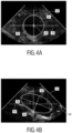
- Fig. 4 two different two-dimensional ultrasound images in the field of view 32 are schematically shown.
- Fig. 4a shows a two-dimensional ultrasound image including the anatomical feature 30, which is centered with respect to the center 36 of the field of view 32 and, therefore well aligned to perform a precise measurement of the size and/or the volume of the anatomical feature 30.
- the identified quality parameter is relatively high for this probes position with respect to the anatomical feature 30.
- Fig. 4b shows a two-dimensional ultrasound image including the anatomical feature 30 which is misaligned with respect to the image plane 42, 44 and the field of view 32.
- the longitudinal axis of the anatomical feature 30 is tilted with respect to the horizontal axis 40 by the angle 38.
- the identified quality parameter is relatively low for this probes position and planes orientation with respect to the anatomical feature 30. Measurements of the anatomical feature 30 performed using the image illustrated in Fig.4b may have an increased error with respect to a real size of the anatomical feature.
- the alignment unit 28 is arranged to indicate an improved alignment of the image planes with respect to the anatomical feature. In order to align the field of view 32 and the image plane 42, 44 with respect to the anatomical feature 34 an indication 46 is shown in the two-dimensional ultrasound image which indicates a rotation and/or a translation of the image planes 42, 44 or the position of the ultrasound probe 14 to align the anatomical feature 30 with respect to the field of view 32.
- the indication 46 (an arrow) is shown within the two-dimensional ultrasound image displayed to the user so that the user can align the image plane and/or the field of view 32 by moving the ultrasound probe 14, respectively.
- This invention may be beneficially implemented in vessel quantification, wherein the ultrasound planes alignment with respect to the quantified vessel may play an important role in ultrasound assisted diagnostic. Alternativly,
- a size and/or volume measurement of the anatomical feature 30 can be performed either on the basis of the two-dimensional ultrasound image data or a full three-dimensional ultrasound scan can be performed by the ultrasound probe 14 to achieve a precise size or volume measurement.
- the size and/or volume measurement can be combined with an automatic segmentation of the three-dimensional ultrasound data.
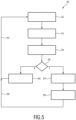
- Fig. 5 shows a schematic flow diagram of an ultrasound imaging method for inspecting a volume of the subject 12. The method is in Fig. 5 generally denoted by 50.
- the method 50 starts with the acquisition of the two-dimensional ultrasound image data simultaneously in the two images planes 42, 44 as shown at step 52.
- the anatomical feature 30 is automatically segmented in the axial and the longitudinal direction based on the two-dimensional ultrasound data in the two different image planes 42, 44 as shown at step 54.
- At least the orientation angle of the anatomical feature with respect to the two-dimensional image planes is estimated. This can reduce the technical effort in general, since an estimation can be simpler than a full segmentation to provide feedback for user guidance.
- the quality parameter is determined based on the positional relation of the anatomical feature 30 with respect to the respective image planes 42, 44.
- the quality parameter is compared to predefined quality limits in order to evaluate the alignment quality. If the quality parameter is not within predetermined quality limit, the indication 46 is displayed on the display screen 22 as a user guidance in order to propose a rotation and/or a translation movement of the ultrasound probe 14 or an adaption of the image depth to improve the quality parameter (step 60). After the alignment, the method 50 returns to step 52 as shown by the feedback loop 62.
- the user gets a feedback via the display unit 22 as shown at step 64 and two-dimensional ultrasound image data or the three-dimensional ultrasound image data are acquired in the aligned position of the ultrasound probe 14 and the measurement unit may optionally determine the size and/or the volume of the anatomical feature 30 based on the two-dimensional ultrasound image data or the three-dimensional ultrasound image data as shown at step 66.
- the user may optionally confirm the measurement and returns to step 52 as shown by the feedback loop 68.
- a computer program may be stored/distributed on a suitable medium, such as an optical storage medium or a solid-state medium supplied together with or as part of other hardware, but may also be distributed in other forms, such as via the Internet or other wired or wireless telecommunication systems.
- a suitable medium such as an optical storage medium or a solid-state medium supplied together with or as part of other hardware, but may also be distributed in other forms, such as via the Internet or other wired or wireless telecommunication systems.
Landscapes
- Health & Medical Sciences (AREA)
- Life Sciences & Earth Sciences (AREA)
- Engineering & Computer Science (AREA)
- Public Health (AREA)
- Medical Informatics (AREA)
- Biomedical Technology (AREA)
- General Health & Medical Sciences (AREA)
- Pathology (AREA)
- Molecular Biology (AREA)
- Physics & Mathematics (AREA)
- Heart & Thoracic Surgery (AREA)
- Nuclear Medicine, Radiotherapy & Molecular Imaging (AREA)
- Biophysics (AREA)
- Surgery (AREA)
- Animal Behavior & Ethology (AREA)
- Radiology & Medical Imaging (AREA)
- Veterinary Medicine (AREA)
- Computer Vision & Pattern Recognition (AREA)
- Physiology (AREA)
- Data Mining & Analysis (AREA)
- Databases & Information Systems (AREA)
- Epidemiology (AREA)
- Primary Health Care (AREA)
- Ultra Sonic Daignosis Equipment (AREA)
Applications Claiming Priority (2)
| Application Number | Priority Date | Filing Date | Title |
|---|---|---|---|
| EP15307070 | 2015-12-21 | ||
| PCT/EP2016/081684 WO2017108667A1 (en) | 2015-12-21 | 2016-12-19 | Ultrasound imaging apparatus and ultrasound imaging method for inspecting a volume of subject |
Publications (2)
| Publication Number | Publication Date |
|---|---|
| EP3393366A1 EP3393366A1 (en) | 2018-10-31 |
| EP3393366B1 true EP3393366B1 (en) | 2025-02-12 |
Family
ID=55070813
Family Applications (1)
| Application Number | Title | Priority Date | Filing Date |
|---|---|---|---|
| EP16816666.8A Active EP3393366B1 (en) | 2015-12-21 | 2016-12-19 | Ultrasound imaging apparatus and ultrasound imaging method for inspecting a volume of subject |
Country Status (5)
| Country | Link |
|---|---|
| US (1) | US20190015076A1 (enExample) |
| EP (1) | EP3393366B1 (enExample) |
| JP (1) | JP6960922B2 (enExample) |
| CN (1) | CN108430334B (enExample) |
| WO (1) | WO2017108667A1 (enExample) |
Families Citing this family (14)
| Publication number | Priority date | Publication date | Assignee | Title |
|---|---|---|---|---|
| TW201923776A (zh) * | 2017-10-27 | 2019-06-16 | 美商蝴蝶網路公司 | 超音波影像上的自動化測量及用於自動化測量的收集的品質指示器 |
| EP3569154A1 (en) | 2018-05-15 | 2019-11-20 | Koninklijke Philips N.V. | Ultrasound processing unit and method, and imaging system |
| US20200245970A1 (en) * | 2019-01-31 | 2020-08-06 | Bay Labs, Inc. | Prescriptive guidance for ultrasound diagnostics |
| EP3711673A1 (en) * | 2019-03-18 | 2020-09-23 | Koninklijke Philips N.V. | Methods and systems for adjusting the field of view of an ultrasound probe |
| GB201908052D0 (en) * | 2019-06-06 | 2019-07-24 | Nisonic As | Alignment of ultrasound image |
| KR102693899B1 (ko) | 2019-07-12 | 2024-08-08 | 베라톤 인코포레이티드 | 초음파 탐침 조준 중 표적의 표현 |
| US11844654B2 (en) | 2019-08-19 | 2023-12-19 | Caption Health, Inc. | Mid-procedure view change for ultrasound diagnostics |
| CN114502078A (zh) * | 2019-09-30 | 2022-05-13 | 皇家飞利浦有限公司 | 记录超声图像 |
| GB201914638D0 (en) * | 2019-10-10 | 2019-11-27 | Rolls Royce Plc | Inspection system |
| US20210196237A1 (en) * | 2019-12-31 | 2021-07-01 | Butterfly Network, Inc. | Methods and apparatuses for modifying the location of an ultrasound imaging plane |
| EP3868303A1 (en) * | 2020-02-18 | 2021-08-25 | Koninklijke Philips N.V. | Ultrasound guidance method and system |
| US11593933B2 (en) * | 2020-03-16 | 2023-02-28 | GE Precision Healthcare LLC | Systems and methods for ultrasound image quality determination |
| JP7722842B2 (ja) | 2020-05-13 | 2025-08-13 | キヤノンメディカルシステムズ株式会社 | 超音波診断装置、医用画像処理装置、医用画像処理方法、及び、プログラム |
| EP4094695A1 (en) | 2021-05-28 | 2022-11-30 | Koninklijke Philips N.V. | Ultrasound imaging system |
Citations (1)
| Publication number | Priority date | Publication date | Assignee | Title |
|---|---|---|---|---|
| US20120071758A1 (en) * | 2010-01-12 | 2012-03-22 | Martin Lachaine | Feature Tracking Using Ultrasound |
Family Cites Families (49)
| Publication number | Priority date | Publication date | Assignee | Title |
|---|---|---|---|---|
| AU1294995A (en) * | 1993-11-29 | 1995-06-19 | Perception, Inc. | Pc based ultrasound device with virtual control user interface |
| US5871019A (en) * | 1996-09-23 | 1999-02-16 | Mayo Foundation For Medical Education And Research | Fast cardiac boundary imaging |
| AUPP227898A0 (en) * | 1998-03-11 | 1998-04-09 | Commonwealth Scientific And Industrial Research Organisation | Improvements in ultrasound techniques |
| JP2001212144A (ja) * | 2000-01-31 | 2001-08-07 | Toshiba Corp | 超音波診断装置及び超音波画像化方法 |
| US6761689B2 (en) * | 2000-08-17 | 2004-07-13 | Koninklijke Philips Electronics N.V. | Biplane ultrasonic imaging |
| US20060100530A1 (en) * | 2000-11-28 | 2006-05-11 | Allez Physionix Limited | Systems and methods for non-invasive detection and monitoring of cardiac and blood parameters |
| US6537220B1 (en) * | 2001-08-31 | 2003-03-25 | Siemens Medical Solutions Usa, Inc. | Ultrasound imaging with acquisition of imaging data in perpendicular scan planes |
| KR20050059245A (ko) * | 2002-10-07 | 2005-06-17 | 노모스 코포레이션 | 타겟 위치 확인 방법 및 장치 |
| US6835177B2 (en) * | 2002-11-06 | 2004-12-28 | Sonosite, Inc. | Ultrasonic blood vessel measurement apparatus and method |
| JP3905470B2 (ja) * | 2002-12-26 | 2007-04-18 | アロカ株式会社 | 超音波診断装置 |
| JP2005319199A (ja) * | 2004-05-11 | 2005-11-17 | Toshiba Corp | 超音波診断装置 |
| JP4555619B2 (ja) * | 2004-06-28 | 2010-10-06 | アロカ株式会社 | 超音波診断装置 |
| JP4868843B2 (ja) * | 2005-01-26 | 2012-02-01 | 株式会社東芝 | 超音波診断装置及び超音波診断装置の制御プログラム |
| EP1685799B1 (en) * | 2005-01-26 | 2013-02-27 | Kabushiki Kaisha Toshiba | Ultrasonic diagnostic apparatus and ultrasonic image acquiring method |
| JP4750429B2 (ja) * | 2005-02-08 | 2011-08-17 | 株式会社日立メディコ | 画像表示装置 |
| CN101141920B (zh) * | 2005-03-15 | 2011-12-14 | 株式会社东芝 | 超声波诊断装置及其控制方法 |
| EP1884197B1 (en) * | 2005-05-20 | 2013-01-02 | Hitachi Medical Corporation | Image diagnosing device |
| CN101404931A (zh) * | 2006-03-20 | 2009-04-08 | 皇家飞利浦电子股份有限公司 | 借助心肌机能的量化的超声诊断 |
| JP5014051B2 (ja) * | 2007-10-09 | 2012-08-29 | 株式会社ユネクス | 血管超音波画像測定方法 |
| JP5134932B2 (ja) * | 2007-12-03 | 2013-01-30 | 株式会社東芝 | 超音波診断装置及び超音波診断装置の制御プログラム |
| JP5508401B2 (ja) * | 2008-06-05 | 2014-05-28 | コーニンクレッカ フィリップス エヌ ヴェ | 誘導efov走査による拡張視野の超音波イメージング |
| RU2507535C2 (ru) * | 2008-06-05 | 2014-02-20 | Конинклейке Филипс Электроникс Н.В. | Визуализация ультразвуковых изображений с расширенным полем обзора с помощью двумерного матричного зонда |
| JP5404141B2 (ja) * | 2008-06-13 | 2014-01-29 | キヤノン株式会社 | 超音波装置及びその制御方法 |
| WO2010017508A1 (en) * | 2008-08-07 | 2010-02-11 | Verathon Inc. | Device, system, and method to measure abdominal aortic aneurysm diameter |
| US8600133B2 (en) * | 2008-10-01 | 2013-12-03 | Koninklijke Philips N.V. | Selection of snapshots of a medical image sequence |
| JP5620666B2 (ja) * | 2008-10-16 | 2014-11-05 | 株式会社東芝 | 超音波診断装置、超音波画像処理装置 |
| US8265363B2 (en) * | 2009-02-04 | 2012-09-11 | General Electric Company | Method and apparatus for automatically identifying image views in a 3D dataset |
| US8355554B2 (en) * | 2009-04-14 | 2013-01-15 | Sonosite, Inc. | Systems and methods for adaptive volume imaging |
| KR101116925B1 (ko) * | 2009-04-27 | 2012-05-30 | 삼성메디슨 주식회사 | 초음파 영상을 정렬시키는 초음파 시스템 및 방법 |
| CN102469988B (zh) * | 2009-07-16 | 2014-12-03 | 尤奈克斯公司 | 超声波血管检查装置 |
| WO2011074271A1 (ja) * | 2009-12-18 | 2011-06-23 | パナソニック株式会社 | 超音波診断装置およびそれを用いた検出対象部位の画像表示方法と計測方法 |
| JP5462076B2 (ja) * | 2010-06-01 | 2014-04-02 | 株式会社東芝 | 超音波診断装置および画像情報管理装置 |
| EP2612599A1 (en) | 2010-08-31 | 2013-07-10 | Hitachi Medical Corporation | Ultrasound diagnostic device and evaluation calculation method |
| US9119559B2 (en) * | 2011-06-16 | 2015-09-01 | Salient Imaging, Inc. | Method and system of generating a 3D visualization from 2D images |
| CN104066380B (zh) * | 2012-02-01 | 2016-11-09 | 东芝医疗系统株式会社 | 超声波诊断装置、图像处理装置 |
| WO2013161277A1 (ja) * | 2012-04-23 | 2013-10-31 | パナソニック株式会社 | 超音波診断装置およびその制御方法 |
| KR20140024190A (ko) * | 2012-08-20 | 2014-02-28 | 삼성메디슨 주식회사 | 초음파 영상 관리 방법, 표시 방법 및 그 장치 |
| GB2507987A (en) * | 2012-11-15 | 2014-05-21 | Imp Innovations Ltd | Method of automatically processing an ultrasound image |
| US10424044B2 (en) * | 2012-12-21 | 2019-09-24 | Koninklijke Philips N.V. | Anatomically intelligent echocardiography for point-of-care |
| CN105101881A (zh) * | 2013-03-29 | 2015-11-25 | 日立阿洛卡医疗株式会社 | 图像位置对准显示方法及超声波诊断装置 |
| EP2807978A1 (en) * | 2013-05-28 | 2014-12-03 | Universität Bern | Method and system for 3D acquisition of ultrasound images |
| EP3080778B1 (en) * | 2013-12-09 | 2019-03-27 | Koninklijke Philips N.V. | Imaging view steering using model-based segmentation |
| EP3089670B1 (en) * | 2014-01-02 | 2024-11-06 | Metritrack, Inc. | System and method for tracking completeness of co-registered medical image data |
| KR20150082945A (ko) * | 2014-01-08 | 2015-07-16 | 삼성메디슨 주식회사 | 초음파 진단 장치 및 그 동작방법 |
| KR102258800B1 (ko) * | 2014-05-15 | 2021-05-31 | 삼성메디슨 주식회사 | 초음파 진단장치 및 그에 따른 초음파 진단 방법 |
| US10349917B2 (en) * | 2014-06-11 | 2019-07-16 | The Johns Hopkins University | Synthetic aperture ultrasound system |
| US20160038125A1 (en) * | 2014-08-06 | 2016-02-11 | General Electric Company | Guided semiautomatic alignment of ultrasound volumes |
| CN108024789B (zh) * | 2015-07-02 | 2024-03-19 | 西门子保健有限责任公司 | 容积间病变检测和图像准备 |
| KR20170068944A (ko) * | 2015-12-10 | 2017-06-20 | 삼성메디슨 주식회사 | 초음파 영상을 디스플레이하는 방법 및 이를 위한 초음파 장치 |
-
2016
- 2016-12-19 WO PCT/EP2016/081684 patent/WO2017108667A1/en not_active Ceased
- 2016-12-19 US US16/061,803 patent/US20190015076A1/en not_active Abandoned
- 2016-12-19 JP JP2018532321A patent/JP6960922B2/ja active Active
- 2016-12-19 CN CN201680075157.8A patent/CN108430334B/zh active Active
- 2016-12-19 EP EP16816666.8A patent/EP3393366B1/en active Active
Patent Citations (1)
| Publication number | Priority date | Publication date | Assignee | Title |
|---|---|---|---|---|
| US20120071758A1 (en) * | 2010-01-12 | 2012-03-22 | Martin Lachaine | Feature Tracking Using Ultrasound |
Also Published As
| Publication number | Publication date |
|---|---|
| US20190015076A1 (en) | 2019-01-17 |
| JP6960922B2 (ja) | 2021-11-05 |
| EP3393366A1 (en) | 2018-10-31 |
| WO2017108667A1 (en) | 2017-06-29 |
| JP2019503748A (ja) | 2019-02-14 |
| CN108430334B (zh) | 2021-06-01 |
| CN108430334A (zh) | 2018-08-21 |
Similar Documents
| Publication | Publication Date | Title |
|---|---|---|
| EP3393366B1 (en) | Ultrasound imaging apparatus and ultrasound imaging method for inspecting a volume of subject | |
| EP3013243B1 (en) | Elastography measurement system and method | |
| JP6430498B2 (ja) | 超音波剪断波エラストグラフィ測定のマッピングのためのシステムおよび方法 | |
| US9612142B2 (en) | Method and system for measuring flow through a heart valve | |
| CN110072465B (zh) | 用于肺部超声的目标探头放置 | |
| JP5784607B2 (ja) | 超音波画像装置と三次元画像表示方法 | |
| EP3193727A1 (en) | Ultrasound imaging apparatus | |
| US20200237337A1 (en) | Rib blockage delineation in anatomically intelligent echocardiography | |
| CN111698947B (zh) | 多参数组织硬度量化 | |
| CN104822323A (zh) | 超声和x射线模态的整合 | |
| JP6865695B2 (ja) | 超音波撮像装置 | |
| US20150182198A1 (en) | System and method for displaying ultrasound images | |
| KR102615722B1 (ko) | 초음파 스캐너 및 초음파 스캐너에서의 조준 가이드 방법 | |
| De Lorenzo et al. | Accurate calibration method for 3D freehand ultrasound probe using virtual plane | |
| KR101024857B1 (ko) | 3차원 초음파 영상에 컬러 모델링 처리를 수행하는 초음파 시스템 및 방법 | |
| US12527548B2 (en) | Ultrasound system and methods for smart shear wave elastography | |
| WO2025140888A1 (en) | Configuring protocol-based ultrasound imaging data systems and methods |
Legal Events
| Date | Code | Title | Description |
|---|---|---|---|
| STAA | Information on the status of an ep patent application or granted ep patent |
Free format text: STATUS: UNKNOWN |
|
| STAA | Information on the status of an ep patent application or granted ep patent |
Free format text: STATUS: THE INTERNATIONAL PUBLICATION HAS BEEN MADE |
|
| PUAI | Public reference made under article 153(3) epc to a published international application that has entered the european phase |
Free format text: ORIGINAL CODE: 0009012 |
|
| STAA | Information on the status of an ep patent application or granted ep patent |
Free format text: STATUS: REQUEST FOR EXAMINATION WAS MADE |
|
| 17P | Request for examination filed |
Effective date: 20180723 |
|
| AK | Designated contracting states |
Kind code of ref document: A1 Designated state(s): AL AT BE BG CH CY CZ DE DK EE ES FI FR GB GR HR HU IE IS IT LI LT LU LV MC MK MT NL NO PL PT RO RS SE SI SK SM TR |
|
| AX | Request for extension of the european patent |
Extension state: BA ME |
|
| DAV | Request for validation of the european patent (deleted) | ||
| DAX | Request for extension of the european patent (deleted) | ||
| RAP1 | Party data changed (applicant data changed or rights of an application transferred) |
Owner name: KONINKLIJKE PHILIPS N.V. |
|
| STAA | Information on the status of an ep patent application or granted ep patent |
Free format text: STATUS: EXAMINATION IS IN PROGRESS |
|
| 17Q | First examination report despatched |
Effective date: 20210617 |
|
| REG | Reference to a national code |
Ref country code: DE Ref legal event code: R079 Free format text: PREVIOUS MAIN CLASS: A61B0008080000 Ipc: A61B0008000000 Ref document number: 602016091211 Country of ref document: DE |
|
| GRAP | Despatch of communication of intention to grant a patent |
Free format text: ORIGINAL CODE: EPIDOSNIGR1 |
|
| STAA | Information on the status of an ep patent application or granted ep patent |
Free format text: STATUS: GRANT OF PATENT IS INTENDED |
|
| RIC1 | Information provided on ipc code assigned before grant |
Ipc: G16H 50/30 20180101ALI20240325BHEP Ipc: A61B 8/08 20060101ALI20240325BHEP Ipc: A61B 8/00 20060101AFI20240325BHEP |
|
| INTG | Intention to grant announced |
Effective date: 20240416 |
|
| RIN1 | Information on inventor provided before grant (corrected) |
Inventor name: ENTREKIN, ROBERT RANDALL Inventor name: COLLET-BILLON, ANTOINE Inventor name: ROUET, LAURENCE |
|
| GRAJ | Information related to disapproval of communication of intention to grant by the applicant or resumption of examination proceedings by the epo deleted |
Free format text: ORIGINAL CODE: EPIDOSDIGR1 |
|
| STAA | Information on the status of an ep patent application or granted ep patent |
Free format text: STATUS: EXAMINATION IS IN PROGRESS |
|
| GRAP | Despatch of communication of intention to grant a patent |
Free format text: ORIGINAL CODE: EPIDOSNIGR1 |
|
| STAA | Information on the status of an ep patent application or granted ep patent |
Free format text: STATUS: GRANT OF PATENT IS INTENDED |
|
| INTC | Intention to grant announced (deleted) | ||
| INTG | Intention to grant announced |
Effective date: 20240730 |
|
| GRAS | Grant fee paid |
Free format text: ORIGINAL CODE: EPIDOSNIGR3 |
|
| GRAA | (expected) grant |
Free format text: ORIGINAL CODE: 0009210 |
|
| STAA | Information on the status of an ep patent application or granted ep patent |
Free format text: STATUS: THE PATENT HAS BEEN GRANTED |
|
| AK | Designated contracting states |
Kind code of ref document: B1 Designated state(s): AL AT BE BG CH CY CZ DE DK EE ES FI FR GB GR HR HU IE IS IT LI LT LU LV MC MK MT NL NO PL PT RO RS SE SI SK SM TR |
|
| REG | Reference to a national code |
Ref country code: GB Ref legal event code: FG4D |
|
| REG | Reference to a national code |
Ref country code: CH Ref legal event code: EP |
|
| REG | Reference to a national code |
Ref country code: DE Ref legal event code: R096 Ref document number: 602016091211 Country of ref document: DE |
|
| REG | Reference to a national code |
Ref country code: IE Ref legal event code: FG4D |
|
| REG | Reference to a national code |
Ref country code: DE Ref legal event code: R084 Ref document number: 602016091211 Country of ref document: DE |
|
| REG | Reference to a national code |
Ref country code: NL Ref legal event code: MP Effective date: 20250212 |
|
| PG25 | Lapsed in a contracting state [announced via postgrant information from national office to epo] |
Ref country code: RS Free format text: LAPSE BECAUSE OF FAILURE TO SUBMIT A TRANSLATION OF THE DESCRIPTION OR TO PAY THE FEE WITHIN THE PRESCRIBED TIME-LIMIT Effective date: 20250512 |
|
| PG25 | Lapsed in a contracting state [announced via postgrant information from national office to epo] |
Ref country code: FI Free format text: LAPSE BECAUSE OF FAILURE TO SUBMIT A TRANSLATION OF THE DESCRIPTION OR TO PAY THE FEE WITHIN THE PRESCRIBED TIME-LIMIT Effective date: 20250212 |
|
| PG25 | Lapsed in a contracting state [announced via postgrant information from national office to epo] |
Ref country code: PL Free format text: LAPSE BECAUSE OF FAILURE TO SUBMIT A TRANSLATION OF THE DESCRIPTION OR TO PAY THE FEE WITHIN THE PRESCRIBED TIME-LIMIT Effective date: 20250212 |
|
| PG25 | Lapsed in a contracting state [announced via postgrant information from national office to epo] |
Ref country code: ES Free format text: LAPSE BECAUSE OF FAILURE TO SUBMIT A TRANSLATION OF THE DESCRIPTION OR TO PAY THE FEE WITHIN THE PRESCRIBED TIME-LIMIT Effective date: 20250212 |
|
| REG | Reference to a national code |
Ref country code: LT Ref legal event code: MG9D |
|
| PG25 | Lapsed in a contracting state [announced via postgrant information from national office to epo] |
Ref country code: IS Free format text: LAPSE BECAUSE OF FAILURE TO SUBMIT A TRANSLATION OF THE DESCRIPTION OR TO PAY THE FEE WITHIN THE PRESCRIBED TIME-LIMIT Effective date: 20250612 Ref country code: NO Free format text: LAPSE BECAUSE OF FAILURE TO SUBMIT A TRANSLATION OF THE DESCRIPTION OR TO PAY THE FEE WITHIN THE PRESCRIBED TIME-LIMIT Effective date: 20250512 |
|
| PG25 | Lapsed in a contracting state [announced via postgrant information from national office to epo] |
Ref country code: NL Free format text: LAPSE BECAUSE OF FAILURE TO SUBMIT A TRANSLATION OF THE DESCRIPTION OR TO PAY THE FEE WITHIN THE PRESCRIBED TIME-LIMIT Effective date: 20250212 |
|
| PG25 | Lapsed in a contracting state [announced via postgrant information from national office to epo] |
Ref country code: HR Free format text: LAPSE BECAUSE OF FAILURE TO SUBMIT A TRANSLATION OF THE DESCRIPTION OR TO PAY THE FEE WITHIN THE PRESCRIBED TIME-LIMIT Effective date: 20250212 |
|
| PG25 | Lapsed in a contracting state [announced via postgrant information from national office to epo] |
Ref country code: LV Free format text: LAPSE BECAUSE OF FAILURE TO SUBMIT A TRANSLATION OF THE DESCRIPTION OR TO PAY THE FEE WITHIN THE PRESCRIBED TIME-LIMIT Effective date: 20250212 Ref country code: PT Free format text: LAPSE BECAUSE OF FAILURE TO SUBMIT A TRANSLATION OF THE DESCRIPTION OR TO PAY THE FEE WITHIN THE PRESCRIBED TIME-LIMIT Effective date: 20250612 |
|
| PG25 | Lapsed in a contracting state [announced via postgrant information from national office to epo] |
Ref country code: BG Free format text: LAPSE BECAUSE OF FAILURE TO SUBMIT A TRANSLATION OF THE DESCRIPTION OR TO PAY THE FEE WITHIN THE PRESCRIBED TIME-LIMIT Effective date: 20250212 Ref country code: GR Free format text: LAPSE BECAUSE OF FAILURE TO SUBMIT A TRANSLATION OF THE DESCRIPTION OR TO PAY THE FEE WITHIN THE PRESCRIBED TIME-LIMIT Effective date: 20250513 |
|
| REG | Reference to a national code |
Ref country code: AT Ref legal event code: MK05 Ref document number: 1765291 Country of ref document: AT Kind code of ref document: T Effective date: 20250212 |
|
| PG25 | Lapsed in a contracting state [announced via postgrant information from national office to epo] |
Ref country code: SE Free format text: LAPSE BECAUSE OF FAILURE TO SUBMIT A TRANSLATION OF THE DESCRIPTION OR TO PAY THE FEE WITHIN THE PRESCRIBED TIME-LIMIT Effective date: 20250212 |
|
| PG25 | Lapsed in a contracting state [announced via postgrant information from national office to epo] |
Ref country code: SM Free format text: LAPSE BECAUSE OF FAILURE TO SUBMIT A TRANSLATION OF THE DESCRIPTION OR TO PAY THE FEE WITHIN THE PRESCRIBED TIME-LIMIT Effective date: 20250212 |
|
| PG25 | Lapsed in a contracting state [announced via postgrant information from national office to epo] |
Ref country code: DK Free format text: LAPSE BECAUSE OF FAILURE TO SUBMIT A TRANSLATION OF THE DESCRIPTION OR TO PAY THE FEE WITHIN THE PRESCRIBED TIME-LIMIT Effective date: 20250212 |
|
| PG25 | Lapsed in a contracting state [announced via postgrant information from national office to epo] |
Ref country code: IT Free format text: LAPSE BECAUSE OF FAILURE TO SUBMIT A TRANSLATION OF THE DESCRIPTION OR TO PAY THE FEE WITHIN THE PRESCRIBED TIME-LIMIT Effective date: 20250212 |
|
| PG25 | Lapsed in a contracting state [announced via postgrant information from national office to epo] |
Ref country code: AT Free format text: LAPSE BECAUSE OF FAILURE TO SUBMIT A TRANSLATION OF THE DESCRIPTION OR TO PAY THE FEE WITHIN THE PRESCRIBED TIME-LIMIT Effective date: 20250212 |
|
| PG25 | Lapsed in a contracting state [announced via postgrant information from national office to epo] |
Ref country code: EE Free format text: LAPSE BECAUSE OF FAILURE TO SUBMIT A TRANSLATION OF THE DESCRIPTION OR TO PAY THE FEE WITHIN THE PRESCRIBED TIME-LIMIT Effective date: 20250212 Ref country code: CZ Free format text: LAPSE BECAUSE OF FAILURE TO SUBMIT A TRANSLATION OF THE DESCRIPTION OR TO PAY THE FEE WITHIN THE PRESCRIBED TIME-LIMIT Effective date: 20250212 |
|
| PG25 | Lapsed in a contracting state [announced via postgrant information from national office to epo] |
Ref country code: RO Free format text: LAPSE BECAUSE OF FAILURE TO SUBMIT A TRANSLATION OF THE DESCRIPTION OR TO PAY THE FEE WITHIN THE PRESCRIBED TIME-LIMIT Effective date: 20250212 |
|
| PG25 | Lapsed in a contracting state [announced via postgrant information from national office to epo] |
Ref country code: SK Free format text: LAPSE BECAUSE OF FAILURE TO SUBMIT A TRANSLATION OF THE DESCRIPTION OR TO PAY THE FEE WITHIN THE PRESCRIBED TIME-LIMIT Effective date: 20250212 |
|
| REG | Reference to a national code |
Ref country code: DE Ref legal event code: R097 Ref document number: 602016091211 Country of ref document: DE |
|
| PLBE | No opposition filed within time limit |
Free format text: ORIGINAL CODE: 0009261 |
|
| STAA | Information on the status of an ep patent application or granted ep patent |
Free format text: STATUS: NO OPPOSITION FILED WITHIN TIME LIMIT |
|
| PGFP | Annual fee paid to national office [announced via postgrant information from national office to epo] |
Ref country code: GB Payment date: 20251223 Year of fee payment: 10 |