EP3374495B1 - Verbessertes verfahren für die herstellung von gewebe - Google Patents
Verbessertes verfahren für die herstellung von gewebe Download PDFInfo
- Publication number
- EP3374495B1 EP3374495B1 EP16864922.6A EP16864922A EP3374495B1 EP 3374495 B1 EP3374495 B1 EP 3374495B1 EP 16864922 A EP16864922 A EP 16864922A EP 3374495 B1 EP3374495 B1 EP 3374495B1
- Authority
- EP
- European Patent Office
- Prior art keywords
- bio
- ink
- tissue
- cells
- certain embodiments
- Prior art date
- Legal status (The legal status is an assumption and is not a legal conclusion. Google has not performed a legal analysis and makes no representation as to the accuracy of the status listed.)
- Active
Links
Images


Classifications
-
- C—CHEMISTRY; METALLURGY
- C12—BIOCHEMISTRY; BEER; SPIRITS; WINE; VINEGAR; MICROBIOLOGY; ENZYMOLOGY; MUTATION OR GENETIC ENGINEERING
- C12N—MICROORGANISMS OR ENZYMES; COMPOSITIONS THEREOF; PROPAGATING, PRESERVING, OR MAINTAINING MICROORGANISMS; MUTATION OR GENETIC ENGINEERING; CULTURE MEDIA
- C12N5/00—Undifferentiated human, animal or plant cells, e.g. cell lines; Tissues; Cultivation or maintenance thereof; Culture media therefor
- C12N5/06—Animal cells or tissues; Human cells or tissues
- C12N5/0697—Artificial constructs associating cells of different lineages, e.g. tissue equivalents
- C12N5/0698—Skin equivalents
-
- C—CHEMISTRY; METALLURGY
- C12—BIOCHEMISTRY; BEER; SPIRITS; WINE; VINEGAR; MICROBIOLOGY; ENZYMOLOGY; MUTATION OR GENETIC ENGINEERING
- C12N—MICROORGANISMS OR ENZYMES; COMPOSITIONS THEREOF; PROPAGATING, PRESERVING, OR MAINTAINING MICROORGANISMS; MUTATION OR GENETIC ENGINEERING; CULTURE MEDIA
- C12N5/00—Undifferentiated human, animal or plant cells, e.g. cell lines; Tissues; Cultivation or maintenance thereof; Culture media therefor
- C12N5/0068—General culture methods using substrates
-
- B—PERFORMING OPERATIONS; TRANSPORTING
- B29—WORKING OF PLASTICS; WORKING OF SUBSTANCES IN A PLASTIC STATE IN GENERAL
- B29C—SHAPING OR JOINING OF PLASTICS; SHAPING OF MATERIAL IN A PLASTIC STATE, NOT OTHERWISE PROVIDED FOR; AFTER-TREATMENT OF THE SHAPED PRODUCTS, e.g. REPAIRING
- B29C64/00—Additive manufacturing, i.e. manufacturing of three-dimensional [3D] objects by additive deposition, additive agglomeration or additive layering, e.g. by 3D printing, stereolithography or selective laser sintering
- B29C64/10—Processes of additive manufacturing
- B29C64/106—Processes of additive manufacturing using only liquids or viscous materials, e.g. depositing a continuous bead of viscous material
- B29C64/112—Processes of additive manufacturing using only liquids or viscous materials, e.g. depositing a continuous bead of viscous material using individual droplets, e.g. from jetting heads
-
- B—PERFORMING OPERATIONS; TRANSPORTING
- B33—ADDITIVE MANUFACTURING TECHNOLOGY
- B33Y—ADDITIVE MANUFACTURING, i.e. MANUFACTURING OF THREE-DIMENSIONAL [3D] OBJECTS BY ADDITIVE DEPOSITION, ADDITIVE AGGLOMERATION OR ADDITIVE LAYERING, e.g. BY 3D PRINTING, STEREOLITHOGRAPHY OR SELECTIVE LASER SINTERING
- B33Y70/00—Materials specially adapted for additive manufacturing
-
- B—PERFORMING OPERATIONS; TRANSPORTING
- B33—ADDITIVE MANUFACTURING TECHNOLOGY
- B33Y—ADDITIVE MANUFACTURING, i.e. MANUFACTURING OF THREE-DIMENSIONAL [3D] OBJECTS BY ADDITIVE DEPOSITION, ADDITIVE AGGLOMERATION OR ADDITIVE LAYERING, e.g. BY 3D PRINTING, STEREOLITHOGRAPHY OR SELECTIVE LASER SINTERING
- B33Y80/00—Products made by additive manufacturing
-
- B—PERFORMING OPERATIONS; TRANSPORTING
- B41—PRINTING; LINING MACHINES; TYPEWRITERS; STAMPS
- B41J—TYPEWRITERS; SELECTIVE PRINTING MECHANISMS, i.e. MECHANISMS PRINTING OTHERWISE THAN FROM A FORME; CORRECTION OF TYPOGRAPHICAL ERRORS
- B41J2/00—Typewriters or selective printing mechanisms characterised by the printing or marking process for which they are designed
- B41J2/005—Typewriters or selective printing mechanisms characterised by the printing or marking process for which they are designed characterised by bringing liquid or particles selectively into contact with a printing material
- B41J2/01—Ink jet
-
- C—CHEMISTRY; METALLURGY
- C12—BIOCHEMISTRY; BEER; SPIRITS; WINE; VINEGAR; MICROBIOLOGY; ENZYMOLOGY; MUTATION OR GENETIC ENGINEERING
- C12N—MICROORGANISMS OR ENZYMES; COMPOSITIONS THEREOF; PROPAGATING, PRESERVING, OR MAINTAINING MICROORGANISMS; MUTATION OR GENETIC ENGINEERING; CULTURE MEDIA
- C12N5/00—Undifferentiated human, animal or plant cells, e.g. cell lines; Tissues; Cultivation or maintenance thereof; Culture media therefor
- C12N5/0062—General methods for three-dimensional culture
-
- C—CHEMISTRY; METALLURGY
- C12—BIOCHEMISTRY; BEER; SPIRITS; WINE; VINEGAR; MICROBIOLOGY; ENZYMOLOGY; MUTATION OR GENETIC ENGINEERING
- C12N—MICROORGANISMS OR ENZYMES; COMPOSITIONS THEREOF; PROPAGATING, PRESERVING, OR MAINTAINING MICROORGANISMS; MUTATION OR GENETIC ENGINEERING; CULTURE MEDIA
- C12N5/00—Undifferentiated human, animal or plant cells, e.g. cell lines; Tissues; Cultivation or maintenance thereof; Culture media therefor
- C12N5/06—Animal cells or tissues; Human cells or tissues
- C12N5/0602—Vertebrate cells
- C12N5/0652—Cells of skeletal and connective tissues; Mesenchyme
- C12N5/0656—Adult fibroblasts
-
- C—CHEMISTRY; METALLURGY
- C12—BIOCHEMISTRY; BEER; SPIRITS; WINE; VINEGAR; MICROBIOLOGY; ENZYMOLOGY; MUTATION OR GENETIC ENGINEERING
- C12N—MICROORGANISMS OR ENZYMES; COMPOSITIONS THEREOF; PROPAGATING, PRESERVING, OR MAINTAINING MICROORGANISMS; MUTATION OR GENETIC ENGINEERING; CULTURE MEDIA
- C12N5/00—Undifferentiated human, animal or plant cells, e.g. cell lines; Tissues; Cultivation or maintenance thereof; Culture media therefor
- C12N5/06—Animal cells or tissues; Human cells or tissues
- C12N5/0697—Artificial constructs associating cells of different lineages, e.g. tissue equivalents
-
- B—PERFORMING OPERATIONS; TRANSPORTING
- B29—WORKING OF PLASTICS; WORKING OF SUBSTANCES IN A PLASTIC STATE IN GENERAL
- B29L—INDEXING SCHEME ASSOCIATED WITH SUBCLASS B29C, RELATING TO PARTICULAR ARTICLES
- B29L2031/00—Other particular articles
- B29L2031/753—Medical equipment; Accessories therefor
- B29L2031/7532—Artificial members, protheses
-
- C—CHEMISTRY; METALLURGY
- C12—BIOCHEMISTRY; BEER; SPIRITS; WINE; VINEGAR; MICROBIOLOGY; ENZYMOLOGY; MUTATION OR GENETIC ENGINEERING
- C12N—MICROORGANISMS OR ENZYMES; COMPOSITIONS THEREOF; PROPAGATING, PRESERVING, OR MAINTAINING MICROORGANISMS; MUTATION OR GENETIC ENGINEERING; CULTURE MEDIA
- C12N2502/00—Coculture with; Conditioned medium produced by
- C12N2502/09—Coculture with; Conditioned medium produced by epidermal cells, skin cells, oral mucosa cells
- C12N2502/091—Coculture with; Conditioned medium produced by epidermal cells, skin cells, oral mucosa cells melanocytes
-
- C—CHEMISTRY; METALLURGY
- C12—BIOCHEMISTRY; BEER; SPIRITS; WINE; VINEGAR; MICROBIOLOGY; ENZYMOLOGY; MUTATION OR GENETIC ENGINEERING
- C12N—MICROORGANISMS OR ENZYMES; COMPOSITIONS THEREOF; PROPAGATING, PRESERVING, OR MAINTAINING MICROORGANISMS; MUTATION OR GENETIC ENGINEERING; CULTURE MEDIA
- C12N2502/00—Coculture with; Conditioned medium produced by
- C12N2502/09—Coculture with; Conditioned medium produced by epidermal cells, skin cells, oral mucosa cells
- C12N2502/094—Coculture with; Conditioned medium produced by epidermal cells, skin cells, oral mucosa cells keratinocytes
-
- C—CHEMISTRY; METALLURGY
- C12—BIOCHEMISTRY; BEER; SPIRITS; WINE; VINEGAR; MICROBIOLOGY; ENZYMOLOGY; MUTATION OR GENETIC ENGINEERING
- C12N—MICROORGANISMS OR ENZYMES; COMPOSITIONS THEREOF; PROPAGATING, PRESERVING, OR MAINTAINING MICROORGANISMS; MUTATION OR GENETIC ENGINEERING; CULTURE MEDIA
- C12N2523/00—Culture process characterised by temperature
-
- C—CHEMISTRY; METALLURGY
- C12—BIOCHEMISTRY; BEER; SPIRITS; WINE; VINEGAR; MICROBIOLOGY; ENZYMOLOGY; MUTATION OR GENETIC ENGINEERING
- C12N—MICROORGANISMS OR ENZYMES; COMPOSITIONS THEREOF; PROPAGATING, PRESERVING, OR MAINTAINING MICROORGANISMS; MUTATION OR GENETIC ENGINEERING; CULTURE MEDIA
- C12N2533/00—Supports or coatings for cell culture, characterised by material
- C12N2533/50—Proteins
- C12N2533/54—Collagen; Gelatin
-
- C—CHEMISTRY; METALLURGY
- C12—BIOCHEMISTRY; BEER; SPIRITS; WINE; VINEGAR; MICROBIOLOGY; ENZYMOLOGY; MUTATION OR GENETIC ENGINEERING
- C12N—MICROORGANISMS OR ENZYMES; COMPOSITIONS THEREOF; PROPAGATING, PRESERVING, OR MAINTAINING MICROORGANISMS; MUTATION OR GENETIC ENGINEERING; CULTURE MEDIA
- C12N2539/00—Supports and/or coatings for cell culture characterised by properties
Definitions
- Tissue engineering and regenerative medicine is a field with great promise from both a therapeutic and a research standpoint.
- Engineered tissues are at the center of many different avenues of tissue engineering research. Methods that can improve the fabrication and formation of these tissues, can also improve their function both in vitro and in vivo, and are needed in order to facilitate the advancement of this field.
- tissue engineering holds great potential for centuries, many problems must be overcome before the full extent of these advantages can be realized.
- One of the problems in tissue engineering is achieving and maintaining compartmentalization of cell types within a tissue. While bioprinting overcomes some of those challenges in the initial fabrication step, new methods are needed that are broadly applicable and support achievement and maintenance of cellular compartments post fabrication without compromising cell viability and function.
- one way to induce compartmentalization is to utilize calcium-cross-linked hydrogels as a component of bio-ink. Disadvantages of this method are that very high concentrations of divalent cross-linking compounds such as calcium ions can negatively impact viability in the tissue, and removal of the hydrogel components requires treatment with enzymes or chemicals that may further damage the cells.
- a method of compartmentalized tissue fabrication has been developed that can be applied broadly and maintains cellular compartments and avoids ionic cross-linkers and enzymatic hydrogel removal post-fabrication.
- this disclosure allows for the fabrication of engineered tissues without the necessity of cross-linking for tissue formation.
- Described herein is a method of fabricating a three-dimensional, engineered, biological tissue, the method comprising: preparing a bio-ink comprising living cells; depositing the bio-ink onto a surface by extrusion bioprinting; incubating the bio-ink at a temperature of greater than or equal to 18°C, but less than 37°C; wherein the bio-ink is not exposed to any ionic, chemical, photo or physical cross-linker during the incubation at a temperature of greater than or equal to 18°C, but less than 37°C.
- apoptosis in a bioprinted tissue is reduced by fabrication using said method, in comparison to an engineered tissue not fabricated by said method.
- the method further comprises exposing the bio-ink to a hypothermic hold of greater than or equal to 2°C, but less than 10°C during or after bioprinting.
- the bio-ink is not exposed to any ionic, chemical, photo or physical cross-linker during the hypothermic hold of greater than or equal to 2°C, but less than 10°C during or after bioprinting.
- apoptosis in a bioprinted tissue is reduced by fabrication using said method, in comparison to an engineered tissue not fabricated by said method.
- the bio-ink further comprises a test substance, the test substance a substance under evaluation for its ability to elicit a change in a tissue compared to a tissue not treated with said substance.
- the bio-ink is not deposited by aerosol spray technology.
- the bio-ink consists essentially of a single human cell-type.
- Also described herein is a method of fabricating a three-dimensional, engineered, biological tissue, the method comprising: preparing a plurality of bio-inks comprising living cells; depositing a first bio-ink onto a surface by extrusion bioprinting; incubating the first bio-ink at a temperature of greater than or equal to 18°C, but less than 37°C; depositing a second bio-ink onto a surface by extrusion bioprinting; and incubating the plurality of bio-inks at a temperature of greater than or equal to 18°C, but less than 37°C; wherein the bio-ink is not exposed to any ionic, chemical, photo or physical cross-linker during the exposure of the first bio-ink, the second bio ink, or both bio-inks at a temperature of greater than or equal to 18°C, but less than 37°C.
- apoptosis in a bioprinted tissue is reduced by fabrication using said method, in comparison to an engineered tissue not fabricated by said method.
- the method further comprises exposing the first bio-ink, the second bio-ink, or both bio-inks to a temperature of greater than or equal to 2°C, but less than 10°C during or after bioprinting.
- the bio-ink is not exposed to any ionic, chemical, photo or physical cross-linker during the hypothermic hold of greater than or equal to 2°C, but less than 10°C during or after bioprinting.
- apoptosis in a bioprinted tissue is reduced by fabrication using said method, in comparison to an engineered tissue not fabricated by said method.
- At least one of the first or second bio-inks or both bio-inks comprise a test substance, wherein the test substance is a substance under evaluation for its ability to elicit a change in a tissue compared to a tissue not treated with said substance.
- the first or second bio-ink is not deposited by aerosol spray technology.
- tissue engineered by: preparing a bio-ink comprising living cells; depositing the bio-ink onto a surface by extrusion bioprinting; incubating the bio-ink at a temperature of greater than or equal to 18°C, but less than 37°C; not exposing the tissue to any ionic, chemical, photo or physical cross-linker during the incubation at a temperature of greater than or equal to 18°C, but less than 37°C; and wherein the tissue exhibits lower levels of apoptosis than a tissue that has been incubated at 37°C or above.
- the bio-ink is exposed to a temperature of greater than or equal to 2°C, but less than 10°C during or after bioprinting. In certain embodiments, the bio-ink was not exposed to any ionic, chemical, photo or physical cross-linker during the hypothermic hold of greater than or equal to 2°C, but less than 10°C during or after bioprinting.
- the bio-ink further comprises a test substance, wherein a test substance is a substance under evaluation for its ability to elicit a change in a tissue compared to a tissue not treated with said substance.
- the bio-ink was not deposited by aerosol spray technology.
- the bio-ink consists essentially of a single human cell type. In certain embodiments, the tissue has no perfusable vasculature. In certain embodiments, the tissue consists essentially of a single human cell-type.
- tissue engineered by: preparing a plurality of bio-inks comprising living cells; depositing a first bio-ink onto a surface by extrusion bioprinting; incubating the first bio-ink at a temperature of greater than or equal to 18°C, but less than 37°C; depositing a second bio-ink onto a surface by extrusion bioprinting; incubating the plurality of bio-inks at a temperature of greater than or equal to 18°C, but less than 37°C; and not exposing any bio-ink to any ionic, chemical, photo or physical cross-linker during the exposure of the first bio-ink, the second bio ink, or both bio-inks at a temperature of greater than or equal to 18°C, but less than 37°C; wherein the tissue exhibits lower levels of apoptosis than a tissue that has been incubated at 37°C or above.
- the bio-ink was exposed to a temperature of greater than or equal to 2°C, but less than 10°C during or after bioprinting. In certain embodiments, the bio-ink was not exposed to any ionic, chemical, photo or physical cross-linker during the hypothermic hold of greater than or equal to 2°C, but less than 10°C during or after bioprinting. In certain embodiments, at least one of the plurality of bio-inks further comprises a test substance, wherein a test substance is a substance under evaluation for its ability to elicit a change in a tissue compared to a tissue not treated with said substance. In certain embodiments, the first or second bio-ink was not deposited by aerosol spray technology. In certain embodiments, the tissue has no perfusable vasculature. In certain embodiments, any of the first or second bio-inks consist essentially of a single human cell-type.
- bio-ink comprising: a bio-ink, wherein the bioink comprises a concentration of between 0.1 and 50 million cells per mL, and a concentration of Novogel between 2 and 20%, wherein the bioink is held at a temperature of between 2-10°C for between 30 seconds and 1 hour.
- bio-ink comprising: a bio-ink, wherein the bioink comprises a concentration of between 0.1 and 50 million cells per mL, and a concentration of Novogel between 2 and 20%, wherein the bioink is held at a temperature of between 18-37°C for between 1 hour and 15 days.
- Disclosed herein are methods of fabricating a three-dimensional, engineered, biological tissue the method comprising, preparing a bio-ink comprising living cells; depositing the bio-ink onto a surface by bioprinting; incubating the bio-ink at a temperature of greater than or equal to 18°C, but less than 37°C; wherein the bio-ink is not exposed to any ionic, chemical, photo or physical cross-linker during the incubation at a temperature of greater than or equal to 18°C, but less than 37°C.
- Disclosed herein are methods of fabricating a three-dimensional, engineered, biological tissue comprising: preparing a plurality of bio-inks comprising living cells; depositing a first bio-ink onto a surface by bioprinting; incubating the first bio-ink at a temperature of greater than or equal to 18°C, but less than 37°C; depositing a second bio-ink onto the surface by bioprinting; and incubating the plurality of bio-inks at a temperature of greater than or equal to 18°C, but less than 37°C; and wherein the bio-ink is not exposed to any ionic, chemical, photo or physical cross-linker during the exposure of the first bio-ink, the second bioink, or both bio-inks at a temperature of greater than or equal to 18°C, but less than 37°C.
- tissue engineered by: preparing a bio-ink comprising living cells; depositing the bio-ink onto a surface by bioprinting; incubating the bio-ink at a temperature of greater than or equal to 18°C, but less than 37°C; wherein the bio-ink is not exposed to any ionic, chemical, photo or physical cross-linker during the incubation at a temperature of greater than or equal to 18°C, but less than 37°C.
- tissue engineered by: preparing a plurality of bio-inks comprising living cells; depositing a first bio-ink onto a surface by bioprinting; incubating the first bio-ink at a temperature of greater than or equal to 18°C, but less than 37°C; depositing a second bio-ink onto the surface by bioprinting; and incubating the plurality of bio-inks at a temperature of greater than or equal to 18°C, but less than 37°C; and wherein the bio-ink is not exposed to any ionic, chemical, photo or physical cross-linker during the exposure of the first bio-ink, the second bioink, or both bio-inks at a temperature of greater than or equal to 18°C, but less than 37°C.
- An advantage of the engineered tissues and methodologies described herein is that they allow retention of the shape of the structure without compromising the functionality of the original cell types, and that they require no use of potentially toxic cross-linkers such as high ion levels, enzymes or UV light.
- the shape of the bioprinted structure is advantageously maintained by multiple approaches.
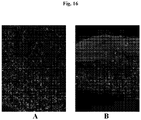
- a cold exposure step and/or an incubation step at below 37°C are advantageous in that they allow for printed bio-inks to better maintain their shape during maturation, and limit the bioprinted cells exposure to cross-linkers that may damage the cells, such as supraphysiological levels of calcium, and reduces apoptosis in the resulting bioprinted tissues ( Fig. 16 ).
- the invention also incorporates a novel aerosol spray printing method into a 3D tissue model.
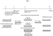
- the aerosol spray approach provides a for a unique discontinuous method compared to a continuous deposition method in that it allows the creation of a thinner layer, and allows for deposition of material onto an existing tissue layer after a period of maturation. This is advantageous because it may produce a tissue that better mimics native tissue in vivo.
- This aerosol spray method can be applied to create multiple layers at multiple time points. For example, this method could be used for constructing a skin tissue by spraying first with undifferentiated keratinocytes followed by spraying with differentiated keratinocytes this could better mimic native skin.
- bio-ink may contain other non-cellular material including but not limited to extrusion compounds, hydrogels, extracellular matrix components, nutritive and media components, inorganic and organic salts, acids and bases, buffer compounds, and other non-cellular components that promote cell survival, adhesion, growth, or that facilitate bioprinting.
- cross-linker or “cross-linking” agent means non-physiological levels of the specific cross-linker or cross-linking agent, or levels of the specific cross-linking agent that are not sufficient to result in significant crosslinking of a given cross-linkable substance.
- Significant crosslinking is greater than 1%, 2%, 3%, 4% or 5% crosslinking.
- tissue means an aggregate of cells.
- layer means an association of cells in X and Y planes that is multiple cells thick.
- the engineered tissues describe herein include one layer.
- the engineered tissues describe herein include a plurality of layers.
- a layer forms a contiguous, substantially contiguous, or non-contiguous sheet of cells.
- each layer of an engineered tissue described herein comprises multiple cells in the X, Y, and Z axes.
- bio-ink means a liquid, semi-solid, or solid composition for use in bioprinting.
- bio-ink comprises cell solutions, cell aggregates, cell-comprising gels, multicellular bodies, cellular pastes or tissues.
- the bio-ink additionally comprises non-cellular materials that provide specific biomechanical properties that enable bioprinting.
- the bio-ink comprises an extrusion compound.
- the extrusion compound is engineered to be removed after the bioprinting process. In other embodiments, at least some portion of the extrusion compound remains entrained with the cells post-printing and is not removed.
- bio-compatible liquid means any liquid capable of contacting or completely covering cells without damage to the cells, examples include but are not limited to growth media and physiological buffers disclosed in this application.
- bioprinting means utilizing three-dimensional, precise deposition of cells (e.g., cell solutions, cell-containing gels, cell suspensions, cell concentrations, multicellular aggregates, multicellular bodies, etc.) via methodology that is compatible with an automated or semi-automated, computer-aided, three-dimensional prototyping device (e.g., a bioprinter).
- Suitable bioprinters include the Novogen Bioprinter ® from Organovo, Inc. (San Diego, CA).
- Some bioprinting methods are extrusion methods which comprise forcing a high viscosity bio-ink through an opening for deposition to a surface. Extrusion methods can be continuous or discontinuous.
- bioprinting methods are ejection methods which comprise spraying an aerosol, droplets, or a mist onto a surface.
- This type of method requires a low-viscosity bio-ink.
- An example of this method is the technique of inkjetting. These methods are incompatible with high viscosity bio-inks.
- tissue scaffold refers to synthetic scaffolds such as polymer scaffolds and porous hydrogels, non-synthetic scaffolds such as pre-formed extracellular matrix layers, dead cell layers, and decellularized tissues, and any other type of pre-formed scaffold that is integral to the physical structure of the engineered tissue and not able to be removed from the tissue without damage/destruction of said tissue.
- decellularized tissue scaffolds include decellularized native tissues or decellularized cellular material generated by cultured cells in any manner; for example, cell layers that are allowed to die or are decellularized, leaving behind the ECM they produced while living.
- subject means any individual, which is a human, a non-human animal, any mammal, or any vertebrate.
- patient means any individual, which is a human, a non-human animal, any mammal, or any vertebrate.
- the term is interchangeable with “patient,” “recipient” and “donor.”
- test substance refers to any biological, chemical or physical substance under evaluation for its ability to elicit a change in said skin tissue compared to skin tissue not treated with said substance.
- a non-limiting example of a change in skin tissue could be an allergic reaction, a toxic reaction, an irritation reaction; a change that is measured by a defined molecular state such as a change in mRNA levels or activity, changes in protein levels, changes in protein modification or epigenetic changes; or a change that results in a measurable cellular outcome such as a change in proliferation, apoptosis, cell viability, cell division, cell motility, cytoskeletal rearrangements, chromosomal number or composition.
- Test substances include, but are not limited to; chemical compositions containing an active or inactive ingredient, either in whole, in part, isolated, or purified; physical stressors such as light, UV light, mechanical stress, heat, or cold; biological agents such as bacteria, viruses, parasites, or fungi. "Test substance” also refers to a plurality of substances mixed or applied separately.
- use encompasses a variety of possible uses of the tissue which will be appreciated by one skilled in the art. These uses include by way of non-limiting example; implantation or engraftment of the engineered tissue into or onto a subject; inclusion of the tissue in a biological assay for the purposes of biological, biotechnological or pharmacological discovery; toxicology testing, including teratogen testing; pharmacology testing, including testing to determine pharmacokinetics and drug metabolism and absorption and penetration, cosmetic testing, including testing to determine sensitization, potential to cause irritation or corrosion of any layer of the dermis, to any test chemical or non-chemical agent including ultraviolet light.
- “Use” can also refer to the process of maturation, or tissue cohesion, in vitro after bioprinting.
- tissue with the bioprinting platform disclosed herein compared to current tissue models and natural tissue is that the process is automated. This allows for greater reproducibility and scalability. For example, it is possible to miniaturize the tissue geometry in order to print bio-ink into well plate formats such as 6, 12, 24, 48, 96, 384 or 1536-well plates for use in screening applications including high-throughput screening applications.
- Another major advantage of an automated platform is that it can be utilized to administer substances for toxicity testing in addition to bioprinting tissue. Current testing in tissue models is limited by the manual approaches necessary both to fabricate the tissue and to apply a test material to that tissue, limiting the application to topical administration.
- the flexibility of the printing platform allows for a variety of methods for application, deposition, and incorporation into tissues not possible with a manual approach. For example, test substances could be sprayed in a fine mist using the aerosol spray technology, or injected into the dermal layer utilizing the continuous deposition module.
- a third major advantage of bioprinting in a tissue toxicology model is the time frame in which a layered structure can be generated and tested. Bioprinting approaches can overlay sheets of cells simultaneously or with a delay to create multiple layers which can then be allowed to mature and differentiate for a defined period of time.
- the bioprinting platform allows for longitudinal studies not possible with manual approaches because test or therapeutic substances can be exposed to or incorporated into tissues during printing or administered to mature tissues at later time points.
- the three-dimensional, engineered, biological tissues described herein include one or more cellular layers. In further embodiments, the layers are stratified. In some embodiments, the engineered tissues include a basal layer. In some embodiments, the tissues described herein are skin tissues. In some embodiments, the tissues described herein are kidney tissues. In some embodiments, the tissues described herein are liver tissues. In some embodiments, the tissues described herein are lung tissues. In some embodiments, the tissues described herein are gut tissues. In some embodiments, the tissues described herein are intestinal tissues.
- the cells are bioprinted.
- the bioprinted cells are cohered to form the engineered tissues.
- the engineered tissues are free or substantially free of pre-formed scaffold at the time of fabrication or the time of use. In some cases, bioprinting allows fabrication of tissues that mimic the appropriate cellularity of native tissue.
- the three-dimensional, engineered tissues described herein are distinguished from tissues fabricated by prior technologies by virtue of the fact that they are three-dimensional, free of pre-formed scaffolds, consist essentially of cells, have a high cell density.
- the engineered tissues are greater than 30% cellular, greater than 40% cellular, greater than 50% cellular, greater than 60% cellular, greater than 70% cellular, greater than 80% cellular, or greater than 90% cellular.
- the engineered tissues have been exposed to incubations at different non-physiological temperatures at various times. For mammalian cells physiological temperature is defined as the normal body temperature of about 37°C.
- the three-dimensional, engineered tissues described herein are distinguished from native (e.g., non-engineered) tissues by virtue of the fact that they are non-innervated (e.g., substantially free of nervous tissue), substantially free of mature vasculature, and/or substantially free of blood components.
- the three-dimensional, engineered tissues are free of plasma, red blood cells, platelets, and the like and/or endogenously-generated plasma, red blood cells, platelets, and the like.
- the tissues lack hemoglobin.
- the tissues lack innervation or neurons.
- the tissue lack neuronal markers such as any of: Beat III tubulin, MAP2, NeuN and neuron specific enolase.
- the engineered tissues are species chimeras, wherein at least one cell or cell-type of the tissue is from a different mammalian species then another cell or cell-type of the tissue.
- the tissues described herein are marked by an increased basal metabolic rate then tissue in vivo or ex vivo. In some embodiments, the tissues described herein are marked by an increased proliferative rate then tissue in vivo or in ex vivo culture. In some embodiments, the tissues described herein are marked by an increased cell size when compared to cells in tissue in vivo or in ex vivo culture.
- the tissues of the current disclosure are marked by extended viability in culture. Tissue explants exhibit low viability in in vitro culture.
- the three-dimensional, engineered tissues described herein are viable after 7 days in culture. In certain embodiments, the three-dimensional, engineered tissues described herein are viable after 10 days in culture. In certain embodiments, the three-dimensional, engineered tissues described herein are viable after 14 days in culture. In certain embodiments, the three-dimensional, engineered tissues described herein are viable after 21 days in culture.
- the engineered tissues are species chimeras, wherein at least one cell or cell-type of the tissue is from a different mammalian species than another cell or cell-type of the tissue.
- the dermal bio-ink contains a cell of mouse, rat, or primate origin and the epidermal bio-ink contains a cell of human origin.
- the engineered tissues are genetic chimeras, wherein at least one cell or cell-type is from a different genetic background than the genetic background of any other cell or cell-type of the tissue.
- the dermal fibroblasts of the dermal bio-ink may be from a certain donor and the keratinocytes or melanocytes of the epidermal bio-ink may be from a different donor, creating a genetic chimera.
- the engineered tissues are chimeras of other types.
- the dermal bio-ink may comprise a transformed dermal fibroblast, and the epidermal bio-ink may comprise a primary untransformed keratinocyte or melanocyte.
- the dermal bio-ink may contain fibroblasts of non-dermal origin.
- the tissues are free of immune cells.
- the tissues are free of Langerhans cells.
- the tissues are free of T-cells. In certain embodiments, the tissues are substantially free of any of the immune cells marked by expression of the following proteins: CD11c, DC-SIGN, CD11b, CD4, CD8, CD28, CD3, CD19 CD80 or CD86.
- one or more components of the engineered tissues described herein are bioprinted, which comprises an additive fabrication process. Therefore, in such embodiments, through the methods of fabrication, the fabricator exerts significant control over the composition of the resulting engineered tissues described herein.
- the engineered tissues described herein optionally comprise any of the layers, structures, compartments, and/or cells of native tissue.
- the engineered skin tissues described herein optionally lack any of the layers, structures, compartments, and/or cells of native tissue.
- the tissues and methods described herein involve bio-ink formulations and bioprinting methods to create 3D tissue structures containing compositions of living cells.
- the printing methods utilize bio-ink to create geometries which produce layers to mimic native tissue.
- the printing methods utilize a variety of printing surfaces with a variety of pore sizes that are optionally coated with matrix support material such as collagen.
- the printing surface can be static or flexible.
- the flexible printing surface allows printed tissue to be subjected to flexing and other non-static conditions post fabrication.
- hydrogels are optionally added to support biomaterials or to constitute space-saving regions in which there are no cells.
- the engineered tissues, arrays, and methods described herein incorporate continuous deposition printing into a 3D tissue model. Continuous deposition is optionally utilized to produce single or multiple layers.
- a bio-ink comprised of fibroblasts is printed to produce a tissue mimicking the dermis.
- bio-ink comprised of keratinocytes or a mixture of keratinocytes and melanocytes is printed to produce a tissue to mimic the epidermis.
- a third embodiment combines bio-inks to simultaneously deposit the epidermal bio-ink on top of the dermal bio-ink.
- Continuous deposition printing provides an advantage to current 3D tissue models in that it enables cells to be placed within a precise geometry and enables the use of multiple bio-ink formulations including, but not limited to, inert gels such as Novogel ® 2.0 and Novogel ® 3.0, and cell paste. Continuous deposition allows optional incorporation of various biomaterials into the Novogel ® formulation and various printing surfaces to promote extracellular matrix production and differentiation.
- Aerosol spray bioprinting techniques allow for the spray of materials that include, for example, a cell suspension, media, bio-ink, biosupport material, or a combination thereof.
- the engineered tissues and methodologies described herein highlight the ability to aerosol spray (e.g., spray) single cells at a resolution of one cell layer thickness and the ability to spray cell aggregates.
- the sprayed layer could, however, also be modified by changing parameters including but not limited to spray material velocity, distance, time, volume, and viscosity.
- cells are optionally sprayed onto other bioprinted layers to result in a full-thickness model, or directly onto transwell or other matrix coated surfaces to specifically generate an epidermal model.
- the spray method is optionally utilized to embed sprayed material into a soft surface such as biosupport material or Novogel ® .
- a dermal layer could be created by spraying fibroblasts into a collagen gel.
- the aerosol spray method is unique when compared to continuous deposition printing in that it does not require a flat printing surface, such as a transwell membrane, to zero the initial printing position in the x, y, and z-axes.
- the aerosol spray method is optionally used to apply a layer to an uneven surface such as a structure previously printed by continuous deposition.
- tissue constructs are optionally modified to promote proliferation and/or differentiation of bioprinted tissue cells.
- dermal media, epidermal media, or a combination of dermal and epidermal media is added to the tissue constructs.
- the media composition is optionally changed at different points in the tissue lifetime to promote the desired biology.
- the tissue constructs are optionally moved to an air liquid interface or subjected to atmospheric changes such as modification of humidity or CO 2 .
- a hypothetical experimental design combining both printing approaches is shown in Fig. 1 .
- bioprinted constructs are made with a method that utilizes a rapid prototyping technology based on three-dimensional, automated, computer-aided deposition of cells, including cell solutions, cell suspensions, cell-comprising gels or pastes, cell concentrations, multicellular bodies (e.g., cylinders, spheroids, ribbons, etc.), and, optionally, confinement material onto a biocompatible support surface (e.g., composed of hydrogel and/or a porous membrane) by a three-dimensional delivery device (e.g., a bioprinter).
- a biocompatible support surface e.g., composed of hydrogel and/or a porous membrane
- the surface can be a layer of cells previously printed.
- the surface can be a cell-free biocompatible surface.
- the term "engineered,” when used to refer to tissues and/or organs means that cells, cell solutions, cell suspensions, cell-comprising gels or pastes, cell concentrates, multicellular aggregates, and layers thereof are positioned to form three-dimensional structures by a computer-aided device (e.g., a bioprinter) according to a computer script.
- the computer script is, for example, one or more computer programs, computer applications, or computer modules.
- three-dimensional tissue structures form through the post-printing fusion of cells or multicellular bodies which, in some cases, is similar to self-assembly phenomena in early morphogenesis.
- the method of bioprinting is continuous and/or substantially continuous.
- a non-limiting example of a continuous bioprinting method is to dispense bio-ink (i.e., cells, cells combined with an excipient or extrusion compound, or aggregates of cells) from a bioprinter via a dispense tip (e.g., a syringe, needle, capillary tube, etc.) connected to a reservoir of bio-ink.
- a continuous bioprinting method is to dispense bio-ink in a repeating pattern of functional units.
- a repeating functional unit has any suitable geometry, including, for example, circles, squares, rectangles, triangles, polygons, and irregular geometries, thereby resulting in one or more tissue layers with planar geometry achieved via spatial patterning of distinct bio-inks and/or void spaces.
- a repeating pattern of bioprinted function units comprises a layer and a plurality of layers are bioprinted adjacently (e.g., stacked) to form an engineered tissue or organ with laminar geometry.
- 2, 3, 4, 5, 6, 7, 8, 9, 10, 11, 12, 13, 14, 15, or more layers are bioprinted adjacently (e.g., stacked) to form an engineered tissue or organ.
- one or more layers of a tissue with laminar geometry also has planar geometry.
- the method of bioprinting is discontinuous.
- a non-limiting example of discontinuous bioprinting is when bio-ink or cells are dispensed, and then the flow of bio-ink or cells is stopped, paused for a certain amount of time, and then started again. This can allow for different bio-inks or cells, or the same bio-inks or cells to be layered with a delay in printing of the layers.
- the discontinuous bioprinting is achieved using an aerosol spray type of bioprinting, wherein cells are applied to an existing tissue layer or surface using an aerosol spray technology.
- a single layer or plurality of layers of cells including dermal cells and cell matrix components or bio-inks are deposited, followed by a temporal delay in deposition of a single layer or plurality of layers epidermal cells or bio-inks.
- the deposition of the epidermal cells is by an aerosol spray.
- deposition of a second bio-ink occurs after deposition of a first bio-ink.
- deposition of the second bio-ink is temporally delayed before it is deposited on the first bio-ink.
- the delay is greater than 1, 2, 3, 4, 5, 6, 7, 8, 9, milliseconds.
- the delay is greater than 10 milliseconds.
- the delay is greater than 20, 30, 40, 50, 60, 70, 80, 90 or 100, milliseconds.
- the delay is greater than 200, 300, 400, 500, 600, 700, 800, 900 or 1000, milliseconds.
- the delay is greater than 1, 2, 3, 4, 5, 6, 7, 8, 9, 10 seconds.
- the delay is greater than 10, 20, 30, 40, 50, or 60 seconds. In certain embodiments, the delay is greater than 1, 2, 3, 4, 5, 6, 7, 8, 9, or 10 minutes. In certain embodiments, the delay is greater than 10, 20, 30, 40, 50, or 60 minutes. In certain embodiments, the delay is greater than 1, 2, 3, 4, 5, 6, 7, 8, 9, 10, 11, 12, 13, 14, 15, 16, 17, 18, 19, 20, 21, 22, 23, or 24 hours. In certain embodiments, the delay is greater than 1, 2, 3, 4, 5, 6, or 7 days. In certain embodiments, the delay is greater than 1, 2, 3, or 4 weeks. In certain embodiments, the delay is less than 1, 2, 3, 4, 5, 6, 7, 8, 9, milliseconds. In certain embodiments, the delay is less than 10 milliseconds.
- the delay is less than 20, 30, 40, 50, 60, 70, 80, 90 or 100, milliseconds. In certain embodiments, the delay is less than 200, 300, 400, 500, 600, 700, 800, 900 or 1000, milliseconds. In certain embodiments, the delay is less than 1, 2, 3, 4, 5, 6, 7, 8, 9, 10 seconds. In certain embodiments, the delay is less than 10, 20, 30, 40, 50, or 60 seconds. In certain embodiments, the delay is less than 1, 2, 3, 4, 5, 6, 7, 8, 9, or 10 minutes. In certain embodiments, the delay is less than 10, 20, 30, 40, 50, or 60 minutes. In certain embodiments, the delay is less than 1, 2, 3, 4, 5, 6, 7, 8, 9, 10, 11, 12, 13, 14, 15, 16, 17, 18, 19, 20, 21, 22, 23, or 24 hours. In certain embodiments, the delay is less than 1, 2, 3, 4, 5, 6, or 7 days. In certain embodiments, the delay is less than 1, 2, 3, or 4 weeks.
- bio-ink includes liquid, semi-solid, or solid compositions comprising a plurality of cells.
- bio-ink comprises liquid or semi-solid cell solutions, cell suspensions, or cell concentrations.
- a cell solution, suspension, or concentration comprises a liquid or semi-solid (e.g., viscous) carrier and a plurality of cells.
- the carrier is a suitable cell nutrient media, such as those described herein.
- bio-ink comprises a plurality of cells that optionally cohere into multicellular aggregates prior to bioprinting.
- bio-ink comprises a plurality of cells and is bioprinted to produce a specific planar and/or laminar geometry; wherein cohesion of the individual cells within the bio-ink takes place before, during and/or after bioprinting.
- the bio-ink is produced by 1) collecting a plurality of cells in a fixed volume; wherein the cellular component(s) represent at least about 30, 40, 50, 60, 70, 80, 90 % or 100% of the total volume.
- bio-ink comprises semi-solid or solid multicellular aggregates or multicellular bodies.
- the bio-ink is produced by 1) mixing a plurality of cells or cell aggregates and a biocompatible liquid or gel in a pre-determined ratio to result in bio-ink, and 2) compacting the bio-ink to produce the bio-ink with a desired cell density and viscosity.
- the compacting of the bio-ink is achieved by centrifugation, tangential flow filtration ("TFF"), or a combination thereof.
- the compacting of the bio-ink results in a composition that is extrudable, allowing formation of multicellular aggregates or multicellular bodies.
- "extrudable” means able to be shaped by forcing (e.g., under pressure) through a nozzle or orifice (e.g., one or more holes or tubes).
- the compacting of the bio-ink results from growing the cells to a suitable density.
- the cell density necessary for the bio-ink will vary with the cells being used and the tissue or organ being produced.
- the cells of the bio-ink are cohered and/or adhered.
- "cohere,” “cohered,” and “cohesion” refer to cell-cell adhesion properties that bind cells, multicellular aggregates, multicellular bodies, and/or layers thereof.
- the terms are used interchangeably with “fuse,” “fused,” and “fusion.”
- the bio-ink additionally comprises support material, cell culture medium (or supplements thereof), extracellular matrix (or components thereof), cell adhesion agents, cell death inhibitors, anti-apoptotic agents, anti-oxidants, extrusion compounds, and combinations thereof.
- the cells are any suitable cell.
- the cells are vertebrate cells, mammalian cells, human cells, or combinations thereof.
- the type of cell used in a method disclosed herein depends on the type of construct or tissue being produced.
- the bio-ink comprises one type of cell (also referred to as a "homogeneous” or “monotypic” bio-ink).
- the bio-ink comprises more than one type of cell (also referred to as a "heterogeneous” or "polytypic” bio-ink).
- the bio-ink comprises a cell culture medium.
- the cell culture medium is any suitable medium.
- suitable cell culture media include, by way of non-limiting examples, Dulbecco's Phosphate Buffered Saline, Earle's Balanced Salts, Hanks' Balanced Salts, Tyrode's Salts, Alsever's Solution, Gey's Balanced Salt Solution, Kreb's-Henseleit Buffer Modified, Kreb's-Ringer Bicarbonate Buffer, Puck's Saline, Dulbecco's Modified Eagle's Medium, Dulbecco's Modified Eagle's Medium/Nutrient F-12 Ham, Nutrient Mixture F-10 Ham (Ham's F-10), Medium 199, Minimum Essential Medium Eagle, RPMI-1640 Medium, Ames' Media, BGJb Medium (Fitton-Jackson Modification), Click's Medium, CMRL-1066 Medium, Fischer's Medium, Glascow Minimum Essential Medium (GMEM), Isco
- the cell culture medium is modified or supplemented.
- the cell culture medium further comprises albumin, selenium, transferrins, fetuins, sugars, amino acids, vitamins, growth factors, cytokines, hormones, antibiotics, lipids, lipid carriers, cyclodextrins, platelet-rich plasma, or a combination thereof.
- the bio-ink further comprises one or more components of an extracellular matrix or derivatives thereof.
- extracellular matrix includes proteins that are produced by cells and transported out of the cells into the extracellular space, where they serve as a support to hold tissues together, to provide tensile strength, and/or to facilitate cell signaling.
- extracellular matrix components include, but are not limited to, collagens, fibronectin, laminins, hyaluronates, elastin, and proteoglycans.
- the multicellular aggregates contain various ECM proteins (e.g., gelatin, fibrinogen, fibrin, collagens, fibronectin, laminins, elastin, and/or proteoglycans).
- ECM components or derivatives of ECM components are optionally added to the cell paste used to form the multicellular aggregate.
- the ECM components or derivatives of ECM components added to the cell paste are optionally purified from a human or animal source, or produced by recombinant methods known in the art.
- the ECM components or derivatives of ECM components are naturally secreted by the cells in the elongate cellular body, or the cells used to make the elongate cellular body are optionally genetically manipulated by any suitable method known in the art to vary the expression level of one or more ECM components or derivatives of ECM components and/or one or more cell adhesion molecules or cell-substrate adhesion molecules (e.g., selectins, integrins, immunoglobulins, and adherins).
- the ECM components or derivatives of ECM components promote cohesion of the cells in the multicellular aggregates.
- gelatin and/or fibrinogen is suitably added to the cell paste, which is used to form multicellular aggregates. The fibrinogen is converted to fibrin by the addition of thrombin.
- the bio-ink further comprises an agent that inhibits cell death (e.g., necrosis, apoptosis, or autophagocytosis). In some embodiments, the bio-ink further comprises an anti-apoptotic agent. Agents that inhibit cell death include, but are not limited to, small molecules, antibodies, peptides, peptibodies, or combination thereof.
- the agent that inhibits cell death is selected from: anti-TNF agents, agents that inhibit the activity of an interleukin, agents that inhibit the activity of an interferon, agents that inhibit the activity of an GCSF (granulocyte colony-stimulating factor), agents that inhibit the activity of a macrophage inflammatory protein, agents that inhibit the activity of TGF-B (transforming growth factor B), agents that inhibit the activity of an MMP (matrix metalloproteinase), agents that inhibit the activity of a caspase, agents that inhibit the activity of the MAPK/JNK signaling cascade, agents that inhibit the activity of a Src kinase, agents that inhibit the activity of a JAK (Janus kinase), or a combination thereof.
- the bio-ink comprises an anti-oxidant.
- the bio-ink comprises oxygen-carriers or other cell-specific nutrients.
- the bio-ink further comprises an extrusion compound (i.e., a compound that modifies the extrusion properties of the bio-ink).
- extrusion compounds include, but are not limited to gels, hydrogels, peptide hydrogels, amino acid-based gels, surfactant polyols (e.g., Pluronic F-127 or PF-127), thermo-responsive polymers, hyaluronates, alginates, extracellular matrix components (and derivatives thereof), collagens, gelatin, other biocompatible natural or synthetic polymers, nanofibers, and self-assembling nanofibers.
- extrusion compounds are removed after bioprinting by physical, chemical, or enzymatic means.
- the bio-ink comprises between 50 million and 1 billion cells per milliliter. In certain embodiments, the bio-ink comprises between 50 million and 900 million cells per milliliter. In certain embodiments, the bio-ink comprises between 50 million and 800 million cells per milliliter. In certain embodiments, the bio ink comprises between 50 million and 700 million cells per milliliter. In certain embodiments, the bio ink comprises between 50 million and 600 million cells per milliliter. In certain embodiments, the bio ink comprises between 50 million and 500 million cells per milliliter. In certain embodiments, the bio ink comprises between 50 million and 400 million cells per milliliter. In certain embodiments, the bio ink comprises between 50 million and 300 million cells per milliliter.
- the bio ink comprises at least 1 million cells per milliliter. In certain embodiments, the bio ink comprises at least 10 million cells per milliliter. In certain embodiments, the bio ink comprises at least 50 million cells per milliliter. In certain embodiments, the bio ink comprises at least 100 million cells per milliliter. In certain embodiments, the bio ink comprises less than 100 million cells per milliliter. In certain embodiments, the bio ink comprises less than 10 million cells per milliliter. In certain embodiments, the bio ink comprises less than 5 million cells per milliliter. In certain embodiments, the bio ink comprises less than 1 million cells per milliliter.
- the bio-ink is a viscous liquid. In certain embodiments, the bio-ink is a semi-solid. In certain embodiments, the bio-ink is a solid. In certain embodiments, the viscosity of the bio-ink is greater than 100 centipoise. In certain embodiments, the viscosity of the bio-ink is greater than 200 centipoise. In certain embodiments, the viscosity of the bio-ink is greater than 500 centipoise. In certain embodiments, the viscosity of the bio-ink is greater than 1,000 centipoise. In certain embodiments, the viscosity of the bio-ink is greater than 2,000 centipoise.
- the viscosity of the bio-ink is greater than 5,000 centipoise. In certain embodiments, the viscosity of the bio-ink is greater than 10,000 centipoise. In certain embodiments, the viscosity of the bio-ink is greater than 20,000 centipoise. In certain embodiments, the viscosity of the bio-ink is greater than 50,000 centipoise. In certain embodiments, the viscosity of the bio-ink is greater than 100,000 centipoise. In certain embodiments, the viscosity of the bio-ink is less than 100 centipoise. In certain embodiments, the viscosity of the bio-ink is less than 200 centipoise.
- the viscosity of the bio-ink is less than 500 centipoise. In certain embodiments, the viscosity of the bio-ink is less than 1,000 centipoise. In certain embodiments, the viscosity of the bio-ink is less than 2,000 centipoise. In certain embodiments, the viscosity of the bio-ink is less than 5,000 centipoise. In certain embodiments, the viscosity of the bio-ink is less than 10,000 centipoise. In certain embodiments, the viscosity of the bio-ink is less than 20,000 centipoise. In certain embodiments, the viscosity of the bio-ink is less than 50,000 centipoise. In certain embodiments, the viscosity of the bio-ink is less than 100,000 centipoise.
- any vertebrate cell is suitable for inclusion in bio-ink and the three dimensional engineered tissues.
- the cells are, by way of non-limiting examples, contractile or muscle cells (e.g., skeletal muscle cells, cardiomyocytes, smooth muscle cells, and myoblasts), connective tissue cells (e.g., bone cells, cartilage cells, fibroblasts, and cells differentiating into bone forming cells, chondrocytes, or lymph tissues), bone marrow cells, endothelial cells, skin cells, epithelial cells, breast cells, vascular cells, blood cells, lymph cells, neural cells, Schwann cells, gut cells, gastrointestinal cells, liver cells, pancreatic cells, lung cells, tracheal cells, corneal cells, genitourinary cells, kidney cells, reproductive cells, adipose cells, parenchymal cells, pericytes, mesothelial cells, stromal cells, undifferentiated cells (e.g., embryonic cells, stem cells, and pro
- contractile or muscle cells
- the bio-ink or tissues comprise fibroblasts. In certain embodiments, the bio-ink or tissues comprise fibroblasts of dermal origin. In certain embodiments, the bio-ink or tissues comprise fibroblasts of renal origin. In certain embodiments, the bio-ink or tissues comprise fibroblasts of vascular origin. In certain embodiments, the bio-ink or tissues comprise endothelial cells. In certain embodiments, the bio-ink or tissues comprise fibroblasts and endothelial cells. In certain embodiments, the bio-ink or tissues comprise keratinocytes. In certain embodiments, the bio-ink or tissues comprise melanocytes. In certain embodiments, the bio-ink or tissues comprise hepatocytes.
- the bio-ink or tissues comprise stellate cells. In certain embodiments, the bio-ink or tissues comprise epidermal cells. In certain embodiments, the bio-ink or tissues comprise dermal cells. In certain embodiments, the bio-ink or tissues comprise epithelial cells. In certain embodiments, the bio-ink or tissues comprise renal tubular epithelial cells. In additional embodiments, the bio-inks or tissues consist essentially of a single cell type. In additional embodiments, the bio-inks or tissues consist essentially of two cell types. In additional embodiments, the bio-inks or tissues consist essentially of three cell types. In additional embodiments, the bio-inks or tissues consist essentially of four cell types. In additional embodiments, the bio-inks or tissues consist essentially of human cells. In additional embodiments, the bio-inks or tissues consist essentially of human primary cells.
- the cells have been modified biologically, chemically or physically.
- Biological modifications include genetic modifications such as transfection, transduction, or infection with a transgene that encodes wild-type, dominant negative, truncated or mutant protein.
- the transgene can also encode an miRNA, siRNA, shRNA or an antisense RNA.
- the transgene can be maintained transiently or stably integrated into the cellular genome. Transfection can be achieved by cationic lipids, calcium phosphate, and electroporation, or through uptake of DNA without a specific transfection means.
- the cells can be virally transduced with any viral vector commonly used for these purposes such as a retrovirus, lentivirus, adenovirus, adeno associated virus, or vaccinia virus.
- the modification can be chemical such as treatment with a mutagen, antibiotic, antifungal, antiviral, HDAC inhibitor, chemotherapeutic, fluorescent labeling or tracking dyes, cell permanent or cell impermanent dyes.
- the modifications can be physical such as radiation, electromagnetic radiation, X-rays, and hot and cold shocks.
- the cells are adult, differentiated cells.
- differentiated cells are cells with a tissue-specific phenotype consistent with, for example, a muscle cell, a fibroblast, or an endothelial cell at the time of isolation, wherein tissue-specific phenotype (or the potential to display the phenotype) is maintained from the time of isolation to the time of use.
- the cells are adult, non-differentiated cells.
- non-differentiated cells are cells that do not have, or have lost, the definitive tissue-specific traits of for example, muscle cells, fibroblasts, or endothelial cells.
- non-differentiated cells include stem cells.
- stem cells are cells that exhibit potency and self-renewal.
- Stem cells include, but are not limited to, totipotent cells, pluripotent cells, multipotent cells, oligopotent cells, unipotent cells, and progenitor cells.
- stem cells are embryonic stem cells, adult stem cells, amniotic stem cells, and induced pluripotent stem cells.
- the cells are a mixture of adult, differentiated cells and adult, non-differentiated cells.
- tissue scaffolds that are free or substantially free of any pre-formed scaffold.
- "scaffold” refers to synthetic scaffolds such as polymer scaffolds and porous hydrogels, non-synthetic scaffolds such as pre-formed extracellular matrix layers, dead cell layers, and decellularized tissues, and any other type of pre-formed scaffold that is integral to the physical structure of the engineered tissue and/or organ and not removed from the tissue and/or organ.
- decellularized tissue scaffolds include decellularized native tissues or decellularized cellular material generated by cultured cells in any manner; for example, cell layers that are allowed to die or are decellularized, leaving behind the ECM they produced while living.
- the engineered tissues/constructs and arrays thereof do not utilize any pre-formed scaffold, e.g., for the formation of the tissue, any layer of the tissue, or formation of the tissue's shape.
- the engineered tissues of the present invention do not utilize any pre-formed, synthetic scaffolds such as polymer scaffolds, pre-formed extracellular matrix layers, or any other type of pre-formed scaffold at the time of manufacture or at the time of use.
- the engineered tissues are substantially free of any pre-formed scaffolds.
- the cellular components of the tissues contain a detectable, but trace or trivial amount of scaffold, e.g., less than 2.0%, less than 1.0%, or less than 0.5% of the total composition.
- scaffold components are removed post-printing, by physical, chemical, or enzymatic methods, yielding an engineered tissue that is free or substantially-free of scaffold components.
- the engineered tissues free, or substantially free, of pre-formed scaffold disclosed herein are in stark contrast to those developed with certain other methods of tissue engineering in which a scaffolding material is first formed, and then cells are seeded onto the scaffold, and subsequently the cells proliferate to fill and take the shape of the scaffold for example.
- the methods of bioprinting described herein allow production of viable and useful tissues that are free or substantially free of pre-formed scaffold.
- the cells of the invention are, in some embodiments, held in a desired three-dimensional shape using a confinement material.
- the confinement material is distinct from a scaffold at least in the fact that the confinement material is temporary and/or removable from the cells and/or tissue.
- tissues are incubated or placed in a "hypothermic hold" at temperature below 24°C for a certain time period after bioprinting.
- this temperature is greater than 0°C. In certain embodiments, this temperature is less than 24° C. In certain embodiments, this temperature is greater than 0°C and less than 24°C. In certain embodiments, this temperature is greater than 0°C and less than 20°C. In certain embodiments, this temperature is greater than 0°C and less than 18°C. In certain embodiments, this temperature is greater than 0°C and less than 16°C. In certain embodiments, this temperature is greater than 0°C and less than 14°C.
- this temperature is greater than 0°C and less than 12°C. In certain embodiments, this temperature is greater than 0°C and less than 10°C. In certain embodiments, this temperature is greater than 0°C and less than 8°C. In certain embodiments, this temperature is greater than 2°C and less than 8°C. In certain embodiments, this temperature is greater than 2°C and less than 10°C. In certain embodiments, this temperature is greater than 2°C and less than 12°C. In certain embodiments, this temperature is greater than 2°C and less than 8°C. In certain embodiments, this temperature is greater than 2°C and less than 14°C. In certain embodiments, this temperature is greater than 2°C and less than 16°C. In certain embodiments, this temperature is greater than 2°C and less than 6°C. In certain embodiments, this temperature is about 1, 2, 3, 4, 5, 6, 7, 8, 9, 10, 11, 12, 13, 14, 15, 16, 17, 18, 19, 20, 21, 22, 23, or 24°C.
- tissues and bio-inks are incubated at the hypothermic hold temperature for a certain amount of time.
- the time period of this incubation can be for at least .020, .05, 1, 2, 3, 4, 5, 6, 7, 8, 9, or 10 seconds.
- the time period of this incubation is at least 10, 20, 30, 40, 50, or 60, seconds.
- the time period of this incubation is at least 1, 2, 3, 4, 5, 6, 7, 8, 9, or 10 minutes.
- the time period of this incubation is at least 10, 20, 30, 40, 50, or 60 minutes.
- the time period of this incubation is at least 1, 2, 3, 4, 5, 6, 7, 8, 9, 10, 11, 12, 13, 14, 15, 16, 17, or 18 hours.
- the time period of this incubation is no more than 10, 20, 30, 40, 50, or 60, seconds. In certain embodiments, the time period of this incubation is no more than 1, 2, 3, 4, 5, 6, 7, 8, 9, or 10 minutes. In certain embodiments, the time period of this incubation is no more than 10, 20, 30, 40, 50, or 60 minutes. In certain embodiments, the time period of this incubation is no more than 1, 2, 3, 4, 5, 6, 7, 8, 9, 10, 11, 12, 13, 14, 15, 16, 17, or 18 hours.
- the hypothermic hold is repeated. In certain embodiments, the hypothermic hold is repeated 1, 2, 3, 4, 5, 6, 7, 8, 9, 10, or more times. In certain embodiments, the hypothermic hold is repeated 10, 20, 30, 40, 50, 60, 70, 80, 90, or 100, or more times. In certain embodiments, the hypothermic hold is achieved by contacting the bio-ink to a bio-compatible liquid that is at a hypothermic temperature. In certain embodiments, the hold is achieved by contacting the bio-ink to a temperature controlled or "chilled" surface.
- the tissues are "matured" or incubated with a single low temperature of a combination at a low temperatures after bioprinting.
- a low-temperature is any temperature below 37°C.
- this step is independent of the aforementioned hypothermic hold. In certain embodiments, this step occurs after the aforementioned hypothermic hold. In certain embodiments, this step occurs without the aforementioned hypothermic hold.
- this temperature can be greater than 18°C but less than 37°C. In certain embodiments, this temperature is about 24, 25, 26, 27, 28, 29, 30, 31, 32, 33, 34, 35, or 36°C. In certain embodiments, this temperature is greater than about 18°C, but less than 37° C.
- this temperature is greater than about 19°C, but less than 37°C. In certain embodiments, this temperature is greater than about 20°C, but less than 37°C. In certain embodiments, this temperature is greater than about 21°C, but less than 37°C. In certain embodiments, this temperature is greater than about 22°C, but less than 37°C. In certain embodiments, this temperature is greater than about 23°C, but less than 37°C. In certain embodiments, this temperature is greater than about 24°C, but less than 37°C. In certain embodiments, this temperature is greater than about 18°C, but less than 36°C. In certain embodiments, this temperature is greater than about 18°C, but less than 35°C.
- this temperature is greater than about 18°C, but less than 34°C. In certain embodiments, this temperature is greater than about 18°C, but less than 33°C. In certain embodiments, this temperature is greater than about 18°C, but less than 32°C. In certain embodiments, this temperature is greater than about 28°C, but less than 32°C.
- the amount of time the tissue is incubated is for greater than 1 hour but less than 20 days. In certain embodiments, the amount of time the tissue is incubated is 1, 2, 3, 4, 5, 6, 7, 8, 9, 10, 11, 12, 13, 14, 15, 16, 17, 18, 19, 20, 21, 22, 23, or 24 hours. In certain embodiments, the amount of time is for 1, 2, 3, 4, 5, 6, 7, 8, 9, 10, 11, 12, 13, 14, 15, 18, 19, or 20 days. In certain embodiments, the amount of time the tissue is incubated is for greater than 2 hours. In certain embodiments, the amount of time the tissue is incubated is for greater than 4 hours. In certain embodiments, the amount of time the tissue is incubated is for greater than 8 hours. In certain embodiments, the amount of time the tissue is incubated is for greater than 12 hours.
- the amount of time the tissue is incubated is for greater than 16 hours. In certain embodiments, the amount of time the tissue is incubated is for greater than 24 hours. In certain embodiments, the amount of time the tissue is incubated is for greater than 3 days. In certain embodiments, the amount of time the tissue is incubated is for greater than 7 days.
- the tissues fabricated by the methods of this disclosure utilize hydrogels, examples include, but are not limited to, those derived from collagen, hyaluronate, hyaluronan, fibrin, alginate, agarose, chitosan, chitin, cellulose, pectin, starch, polysaccharides, fibrinogen/thrombin, fibrillin, elastin, gum, cellulose, agar, gluten, casein, albumin, vitronectin, tenascin, entactin/nidogen, glycoproteins, glycosaminoglycans (GAGs) and proteoglycans which may contain for example chrondroitin sulfate, fibronectin, keratin sulfate, laminin, heparan sulfate proteoglycan, decorin, aggrecan, perlecan and combinations thereof.
- hydrogels examples include, but are not limited to, those derived from collagen, hyal
- suitable hydrogels are synthetic polymers.
- suitable hydrogels include those derived from poly(acrylic acid) and derivatives thereof, poly(ethylene oxide) and copolymers thereof, poly(vinyl alcohol), polyphosphazene, and combinations thereof.
- hydrogel, NovoGel TM agarose, alginate, gelatin, Matrigel TM , hyaluronan, poloxamer, peptide hydrogel, poly(isopropyl n-polyacrylamide), polyethylene glycol diacrylate (PEG-DA), hydroxyethyl methacrylate, polydimethylsiloxane, polyacrylamide, poly(lactic acid), silicon, silk, or combinations thereof.
- the tissues fabricated by the method of the disclosure do not require any cross-linking to form compartmentalized tissues.
- the tissues do not require chemical cross-linking, gluteraldehyde or bis-epoxide by way of non-limiting example.
- the tissues do not require cross-linking by ionic cross-linkers.
- the tissues do not require cross-linking by any cationic or anionic cross-linkers.
- the tissues do not require cross-linking by calcium, magnesium, sodium, chloride, alginate, or any combination thereof.
- the tissues do not require cross-linking by enzymatic cross-linkers.
- the cells do not require physical cross-linking.
- the tissues do not require photo cross-linking.
- the tissues do not require photo cross-linking or, radiation cross-linking, including ultraviolet or visible light.
- the bioprinted tissues, bio-inks, and cells are exposed to divalent cationic cross-linking compounds such as calcium that do not exceed physiological levels.
- Physiological levels for this purpose are between 1 and 1.5 mM for ionic calcium, and between 2 and 3 mM for total calcium.
- the tissues or bio-inks are exposed to more than 1, 2, 3, 4, 5, 6, 7, 8, 9, 10 mM total calcium.
- the tissues or bio-inks are exposed to more than 5, 10, 15, 20, 25, 30, 35, 40, 45, or 50 mM total calcium.
- Calcium comprises all ionic species of calcium including, but not limited to, calcium chloride or calcium phosphate.
- the tissues or bio-inks are not exposed to other divalent cationic cross-linking compounds such as strontium, magnesium, barium, or other multivalent ions that may include copper, aluminum, or iron.
- the tissues or bio-inks are exposed to more than 1, 2, 3, 4, 5, 6, 7, 8, 9, or 10 mM of an ionic cross-linker.
- the tissues or bio-inks are exposed to more than 10, 20, 30, 40, 50, 60, 70, 80, 90, or 100 mM of an ionic cross-linker.
- the bioprinted tissues fabricated by the methods of this disclosure reduce apoptosis in the bioprinted tissues when compared to bioprinted tissues that are not bioprinted by the methods of this disclosure.
- apoptosis is reduced by 5%, 10%, 15%, 20%, 25%, 30%, 35%, 40%, 45%, 50%, 55%, 60%, 65%, 70%, 75%, 80%, 85%, 90%, or more.
- apoptosis is reduced by 10% or more.
- apoptosis is reduced by 25% or more.
- apoptosis is reduced by 30% or more.
- apoptosis is reduced by 50% or more.
- apoptosis is reduced by 70% or more. In certain embodiments, apoptosis is reduced by 90% or more. In certain embodiments, the reduction in apoptosis is measured by changes in molecular markers of apoptosis; non-limiting examples include DNA fragmentation, TUNEL staining, changes in activated caspase, changes in caspase cleavage products, changes in total caspase, changes in mRNA levels consistent with a reduction or increase in apoptosis, changes in cell surface markers of apoptosis (Anexin V, by way of non-limiting example).
- the changes in molecular markers can be measured by immunohistochemistry (IHC), flow cytometry, western blot, PCR including real-time PCR, or any homogenous caspase assay including ones that are commercially available.
- a system of tissues with improved viability and utility for in vitro and therapeutic applications In certain embodiments, enclosed herein are arrays of tissues with improved viability and utility for in vitro and therapeutic applications.
- An array is an in vitro association of multiple tissues that allows for enhanced screening of compounds for potential use as therapeutics.
- the arrays or systems allow for at least 20 ⁇ m of space between tissues. In certain embodiments, the array or systems allow for at least 100 ⁇ m of space between tissues. In certain embodiments, the array or systems allow for at least 1000 ⁇ m of space between tissues.
- the tissues for use in the arrays or systems are compositionally the same. In certain embodiments, the tissues for use in the arrays or systems are compositionally different.
- the tissues for use in the arrays or systems are attached to a biocompatible surface.
- the tissues for use in the arrays or systems are anchored to a biocompatible surface. Attachment can be through physical means or by cell-adhesion molecules coated on the biocompatible surface. In certain embodiments, attachment is through secretion of cell adhesion proteins or molecules by the cells that are deposited.
- the disclosure herein includes business methods.
- the speed and scalability of the techniques and methods disclosed herein are utilized to design, build, and operate industrial and/or commercial facilities for production of engineered tissues and/or organs for engraftment or use in generation of cell-based tools for research and development, such as in vitro assays.
- the engineered tissues and/or organs and arrays thereof are produced, stored, distributed, marketed, advertised, and sold as, for example, cellular arrays (e.g., microarrays or chips), tissue arrays (e.g., microarrays or chips), and kits for biological assays, high-throughput drug screening, toxicology and toxicity testing.
- the engineered tissues and/or organs and arrays thereof are produced and utilized to conduct biological assays and/or drug screening as a service.
- the business methods involve on demand bioprinting of tissues for use in transplantation.
- Examples 1, 3 and 5 are not according to the invention and are present for illustration and comparison purposes only.
- Example 1 Incubation below 37°C improves skin tissue formation
- Bio-ink was generated by a cellular mixture of 100% primary adult human dermal fibroblasts (HDFa) in 6% gelatin (Novogel ® 2.0) in a concentration of 150 million cells per milliliter.
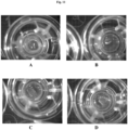
- Three-dimensional bio-ink constructs were printed by continuous deposition using the Novogen Bioprinter ® platform in a 4 mm ⁇ 4 mm ⁇ 0.5 mm base sheet with a 1mm wall bordering the top to create a dermal structure resembling a cup.
- One tissue construct was printed per transwell in a 6 well plate.
- the transwell printing surface contained a polytetrafluoroethylene (PTFE) membrane coated with equimolar mixture of types I and III collagen (bovine) with pores 3 ⁇ m in size.
- PTFE polytetrafluoroethylene
- Epidermal cell paste containing a mixture of 95% primary adult human epidermal keratinocytes (HEKa) and 5% primary adult human epidermal melanocytes (HEMa) was then printed on top of the dermal bio-ink immediately or between .020 seconds and several hours or several days.
- Cell paste was measured post print at 90.5% viable by trypan exclusion assay.
- Cell number in deposited epidermal layer was estimated at 160,000 cells by cell counting on a Cell-O-Meter.
- Media was then added to the outer well of the transwell in a volume of 2 ml.
- the media used for subsequent growth and maintenance of the skin tissue was a 50:40:10 ratio of HDFa: HEKa: HEMa media.
- the volume added was sufficient to collect at the base of the printed structure but not to submerge the structure.
- Media was changed 48 hours later and subsequently changed daily after that.
- Printed constructs were placed into a non-humidified incubator at 30°C for 5 days. This is a key step that enables maintenance of tissue shape for a period of maturation. After 5 days, the temperature was raised to 37°C for an additional 7 days.
- constructs were either lysed for RNA analysis or fixed in 2% PFA for histological analysis.
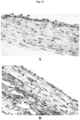
- H&E staining of skin tissues at day 12 shows a distinct layered architecture ( Figs . 2 and 3 ). Fibroblasts in a dermal layer are observed at the base (purple) and differentiated keratinocytes in an epidermal layer (pink) on top.
- An unexpected finding with this approach is the extent of the layered architecture observed. In particular, there is a layer of cells with distinct morphology can be observed at the interface (arrows). This layer stains specifically for CK14, indicating that the keratinocyte cells in the deposited paste have arranged into a basal layer.
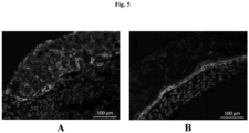
- Distinct layers of differentiated keratinocytes are visualized by simultaneously staining for a basal cell marker CK5 and involucrin (IVL), a later stage differentiation marker of granular and cornified keratinocytes. Similar to normal human skin, differences in morphology are seen as basal cells appear to have a distinct cuboidal morphology, while differentiated keratinocytes on top appear flatter.
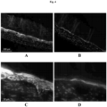
- the layered architecture also includes CK10-positive spinous and granular keratinocytes in mid stages differentiation ( Fig. 4 ).
- Gene expression analysis supports histological findings. Data shows an increase in epidermal differentiation markers CK1, CK10, and especially late marker FLG over time. Gene expression also shows that collagen 4 levels increase over time, suggesting formation of a basement membrane. Collagen I levels are maintained over the time course of the experiment suggesting dermal layer remains viable ( Fig. 6 ).
- Example 2 Transient exposure at 4°C and incubation below 37°C improves skin tissue formation
- Bio-ink was generated by a cellular mixture of 100% primary adult human dermal fibroblasts (HDFa) in 8% gelatin (Novogel ® ) in a concentration of 100 million cells per milliliter. The cell: gelatin ratio was altered to reduce the cellular density of the dermal sheet to better mimic dermal tissue in native skin.
- Three-dimensional bio-ink constructs were printed by continuous deposition using the Novogen Bioprinter ® platform in a 4 mm ⁇ 4 mm ⁇ 0.5 mm base sheet to create a dermal structure resembling a sheet.
- One tissue construct was printed per transwell-in a 6 well plate.
- the transwell printing surface contained a polytetrafluoroethylene (PTFE) membrane coated with equimolar mixture of types I and III collagen (bovine) with pores 3 ⁇ m in-size.
- PTFE polytetrafluoroethylene
- Epidermal cell paste containing a mixture of 100% primary neonatal human epidermal keratinocytes (HEKn) was then printed on top of the dermal bio-ink.
- a separate but identical epidermal paste structure was simultaneously deposited next to the dermal sheet directly onto the transwell printing surface. This structure was only comprised of epidermal keratinocyte paste and contained no dermal tissue.
- Printed constructs were placed into a non-humidified incubator at 30°C for 5 days. This is a key step that enables maintenance of tissue shape for a period of maturation. After 5 days, the temperature was raised to 37°C for an additional 7 days.
- constructs were either lysed for RNA analysis or fixed in 2% PFA for histological analysis.
- Figs. 7A and B H&E staining of skin tissues at day 12 shows a distinct layered architecture only in structures with epidermal paste printed on top of a dermal layer ( Figs. 7 C and D versus F and G , Fig. 8A ). Fibroblasts in a dermal layer are observed at the base (purple) and differentiated keratinocytes in an epidermal layer (pink) on top. In particular, there is a layer of cells with distinct morphology that can be observed at the interface.
- Distinct layers of differentiated keratinocytes are visualized by simultaneously staining for a basal cell marker CK5 (green) and involucrin (IVL, red), a later stage differentiation marker of granular and cornified keratinocytes ( Fig. 7E versus H , Fig. 8B ).
- the distinct green layer indicates that the keratinocyte cells in the deposited paste have arranged into a basal layer with a layer of more differentiated IVL positive cells on top. Similar to normal human skin, differences in morphology are seen as basal cells that appear to have a distinct cuboidal morphology, while differentiated keratinocytes on top appear flatter.
- Staining for the proliferation marker PCNA indicates that proliferation is high in both dermal fibroblasts and basal layer keratinocytes but not in differentiating keratinocytes. This pattern is similar to that which is found in native skin. Staining for apoptosis by TUNEL ( Fig. 8F ) also low showing very few positive staining cells in either dermal or epidermal layer. Collectively PCNA and TUNEL staining demonstrate that both dermal and epidermal compartments of the full thickness tissue are viable at day 12.
- Gene expression analysis supports histological findings. Data shows an increase in mid epidermal differentiation markers CK1, CK10, and later markers IVL, Loricrin, and at day 12 compared to day 0. Gene expression also shows that collagen I and 4 levels are maintained over the time course of the experiment, while collagen 3 levels increase suggesting the dermal layer remains viable and functional ( Fig. 9 ).
- the dermal layer directs differentiation and/or stratification of the epidermal keratinocytes, and that there is a uniqueness to the combination of dermal and epidermal cells that is not present in the epidermal cells alone.
- the extent of the layered architecture observed in the tissues comprised of both epidermal and dermal cells including the staining of the CK5-positive basal layer which is limited to a defined region at the base of the epidermal layer similar to native human skin.
- the layered architecture also includes a CK10 positive ( Fig.
- H&E staining shows that the dermal fibroblasts do not form a thin sheet as in earlier examples 1 and 2, but a thicker structure.
- Collagen deposition which is a key indicator of normal fibroblast function in the dermis, can be seen by both trichrome staining (blue color) and by immunofluorescent staining for collagen 3 (red) in between dermal cells ( Fig. 8 ).
- Example 3 Incubation below 37°C improves kidney tissue formation
- the interstitial layer of the renal proximal tubule model is composed of renal fibroblasts and HUVECs in Novogel ® .
- the cell ratio was changed to 50% fibroblasts / 50% HUVEC the concentration of the cells was 125 million cells/mL. Attempts to fabricate tissues using these cell ratios were hampered by a propensity of the tissues to "ball up," preventing the sort of thin, spread out interstitial layer that is ideal.
- tissues were incubated at 30°C for 3 days following printing to slow the rate of Novogel ® dissipation.
- tissue that better retains its overall dimensions ( Fig. 11C and D ) after 30°C incubation, when compared to 37°C incubation ( Fig. 11A and B ), and allows the cells to proliferate and secrete ECM to replace the Novogel ® material as a binding agent ( Fig. 11B ).
- tissues can be shifted to 37°C for the addition of epithelial cells to the structure, with no loss of tissue features or viability as a result of the culture time at 30°C.
- H&E staining is shown ( Fig. 12A ), and brush borders are indicated ( Fig. 13 A , arrows). Trichrome staining indicates collagen secretion ( Fig.
- Bioprinted tissues demonstrated ⁇ -glutamyl-transferase activity which increases over time in culture, which is indicative of a functioning epithelial layer ( Fig. 15 ). Considering that the kidney is a fully internal structure normally kept at 37°C, it is unexpected that the culture time at 30°C would result in a tissue with the desired structural and functional characteristics.
- Example 4 Comparison of skin tissue fabricated using a 4°C hypothermic hold and a 30°C Incubation
- Tissues that are fabricated using the methods disclosed here display quantitative and qualitative improvements.
- skin tissue fabricated using a 4°C hold with a 30°C incubation step for 5 days exhibit reduced apoptosis ( Fig . 16B ), as indicated by TUNEL staining, when compared to tissue fabricated without these techniques ( Fig . 16A ).
- tissue is assayed for gene expression differentiation markers IVL, CK10, CK5 and Col1a1 are elevated in tissue fabricated using a 4°C hold with a 30°C incubation step for 5 days ( Fig. 17E , F, G, and H ), when compared to tissue fabricated without these techniques ( Fig . 17A , B , C, and D ).
- Fig . 16 and Fig . 17 show that tissue fabricated using a hypothermic hold and incubation below 37°C results in tissues with increased viability and differentiation.
- Liver tissues comprising primary human hepatocytes, endothelial cells, and stellate cells were bioprinted and immediately placed in an incubator under varying temperatures (18.2 °C, 30.2 °C, 32.0 °C, and 36.9 °C) and imaged.
- Fig. 18A-D are photographs showing the temperature dependence of the viscosity of cellular bio-ink at 18.2 °C ( A ), 30.2 °C ( B ), 32.0 °C ( C ), and 36.9 °C ( D ).
- Bio-ink that forms and is distinctly visible on the outer edge of liver tissue (clear diamond bordering the dark triangles indicated by an arrow) at 18.2 °C ( A ) is maintained at 30.2 °C ( B ).
- the tissue edges break down at 32.0 °C ( C ) and completely disappear at 36.9 °C ( D ). Therefore, incubation at 36.9 °C does not produce liver tissue having the desired structural and functional characteristics.
Landscapes
- Engineering & Computer Science (AREA)
- Health & Medical Sciences (AREA)
- Biomedical Technology (AREA)
- Life Sciences & Earth Sciences (AREA)
- Chemical & Material Sciences (AREA)
- Biotechnology (AREA)
- Organic Chemistry (AREA)
- Bioinformatics & Cheminformatics (AREA)
- Genetics & Genomics (AREA)
- Wood Science & Technology (AREA)
- Zoology (AREA)
- Biochemistry (AREA)
- Microbiology (AREA)
- General Engineering & Computer Science (AREA)
- General Health & Medical Sciences (AREA)
- Cell Biology (AREA)
- Materials Engineering (AREA)
- Manufacturing & Machinery (AREA)
- Rheumatology (AREA)
- Physics & Mathematics (AREA)
- Mechanical Engineering (AREA)
- Optics & Photonics (AREA)
- Micro-Organisms Or Cultivation Processes Thereof (AREA)
- Materials For Medical Uses (AREA)
- Apparatus Associated With Microorganisms And Enzymes (AREA)
Claims (15)
- Verfahren zum Fertigen eines dreidimensionalen, künstlich hergestellten, biologischen Gewebes, das Verfahren umfassend:a. Herstellen einer Biotinte, die lebende Zellen umfasst, wobei die Biotinte eine viskose Flüssigkeit, ein Halbfeststoff oder ein Feststoff ist;b. Aufbringen der Biotinte auf eine Oberfläche durch Extrusions-Bioprinting, um eine Struktur zu bilden;c. Aussetzen der Biotinte nach Bioprinting einer hypothermischen Haltung von mehr als oder gleich wie 2 °C, aber weniger als 10 °C;d. Inkubation der Biotinte bei einer Temperatur von mehr als oder gleich wie 18 °C, aber weniger als 37 °C nach hypothermischer Haltung;wobei die Biotinte während der Inkubation bei einer Temperatur von mehr als oder gleich wie 18 °C, aber weniger als 37 °C, keinem ionischen, chemischen, fotografischen oder physikalischen Vernetzer ausgesetzt wird und wobei die Form der Struktur während der hypothermischen Haltung und der Inkubation erhalten bleibt.
- Verfahren nach Anspruch 1, wobei Apoptose in einem biologisch gedruckten Gewebe durch das Fertigen unter Verwendung dieses Verfahrens im Vergleich zu einem nicht nach diesem Verfahren gefertigtem künstlich hergestellten Gewebe reduziert wird.
- Verfahren nach Anspruch 1, wobei die Biotinte während der hypothermischen Haltung von mehr als oder gleich wie 2 °C, aber weniger als 10 °C nach Bioprinting keinem ionischen, chemischen, fotografischen oder physikalischen Vernetzer ausgesetzt wird.
- Verfahren nach Anspruch 3, wobei Apoptose in einem biologisch gedruckten Gewebe durch das Fertigen unter Verwendung dieses Verfahrens im Vergleich zu einem nicht nach diesem Verfahren gefertigtem künstlich hergestellten Gewebe reduziert wird.
- Verfahren nach Anspruch 1, wobei die Biotinte ferner eine Testsubstanz umfasst, wobei die Testsubstanz eine Substanz ist, die auf ihre Fähigkeit untersucht wird, eine Veränderung in einem Gewebe im Vergleich zu einem nicht mit der Substanz behandelten Gewebe hervorzurufen.
- Verfahren nach Anspruch 1, wobei die Biotinte im Wesentlichen aus einem einzigen menschlichen Zelltyp besteht.
- Verfahren zum Fertigen eines dreidimensionalen, künstlich hergestellten, biologischen Gewebes, das Verfahren umfassend:a. Herstellen einer Vielzahl von Biotinten, die lebende Zellen umfassen, wobei die Biotinte eine viskose Flüssigkeit, ein Halbfeststoff oder ein Feststoff ist;b. Aufbringen einer ersten Biotinte auf eine Oberfläche durch Extrusions-Bioprinting;c. Inkubieren der ersten Biotinte bei einer Temperatur von mehr als oder gleich wie 18 °C, aber weniger als 37 °C;d. Aufbringen einer zweiten Biotinte auf eine Oberfläche durch Extrusions-Bioprinting; unde. Inkubieren der Vielzahl von Biotinten bei einer Temperatur von mehr als oder gleich wie 18 °C, aber weniger als 37 °C;ferner umfassend ein Aussetzen der ersten Biotinte, der zweiten Biotinte oder beider Biotinten einer hypothermischen Haltung von mehr als oder gleich wie 2 °C, aber weniger als 10 °C nach Bioprinting und vor Inkubation der ersten Biotinte, der zweiten Biotinte oder beider Biotinten, wobei die Vielzahl der Biotinten während der Inkubation der ersten photochemischen oder physikalischen Vernetzer während der Inkubation der ersten Biotinte, der zweiten Biotinte oder beider Biotinten bei einer Temperatur von mehr als oder gleich wie 18 °C, aber weniger als 37°C ausgesetzt sind, wobei das Aufbringen der ersten Biotinte und der zweiten Biotinte eine Struktur bildet und wobei die Form der Struktur während der hypothermischen Haltung und der Inkubation beibehalten wird.
- Verfahren nach Anspruch 7, wobei Apoptose in einem biologisch gedruckten Gewebe durch das Fertigen unter Verwendung dieses Verfahrens im Vergleich zu einem nicht nach diesem Verfahren gefertigtem künstlich hergestellten Gewebe reduziert wird.
- Verfahren nach Anspruch 7, wobei die Vielzahl von Biotinten während der hypothermischen Haltung von mehr als oder gleich wie 2 °C, aber weniger als 10 °C nach Bioprinting keinem ionischen, chemischen, fotografischen oder physikalischen Vernetzer ausgesetzt wird.
- Verfahren nach Anspruch 9, wobei Apoptose in einem biologisch gedruckten Gewebe durch das Fertigen unter Verwendung dieses Verfahrens im Vergleich zu einem nicht nach diesem Verfahren gefertigtem künstlich hergestellten Gewebe reduziert wird.
- Verfahren nach Anspruch 7, wobei mindestens eine von der ersten oder der zweiten Biotinte oder beide Biotinten eine Testsubstanz umfassen, wobei die Testsubstanz eine Substanz ist, die auf ihre Fähigkeit untersucht wird, eine Veränderung in einem Gewebe im Vergleich zu einem nicht mit dieser Substanz behandelten Gewebe hervorzurufen.
- Verfahren nach Anspruch 1, wobei das biologische Gewebe frei von einem vorgeformten Gerüst ist.
- Verfahren nach Anspruch 7, wobei das biologische Gewebe frei von einem vorgeformten Gerüst ist.
- Verfahren nach Anspruch 1, wobei das Verfahren nicht die Verwendung eines ionischen, chemischen, fotografischen oder physikalischen Vernetzungsmittels umfasst.
- Verfahren nach Anspruch 7, wobei das Verfahren nicht die Verwendung eines ionischen, chemischen, fotografischen oder physikalischen Vernetzungsmittels umfasst.
Applications Claiming Priority (3)
| Application Number | Priority Date | Filing Date | Title |
|---|---|---|---|
| US201562253064P | 2015-11-09 | 2015-11-09 | |
| US14/936,580 US20170130192A1 (en) | 2015-11-09 | 2015-11-09 | Methods for tissue fabrication |
| PCT/US2016/061156 WO2017083402A1 (en) | 2015-11-09 | 2016-11-09 | Improved methods for tissue fabrication |
Publications (3)
| Publication Number | Publication Date |
|---|---|
| EP3374495A1 EP3374495A1 (de) | 2018-09-19 |
| EP3374495A4 EP3374495A4 (de) | 2019-04-17 |
| EP3374495B1 true EP3374495B1 (de) | 2023-02-15 |
Family
ID=58695226
Family Applications (1)
| Application Number | Title | Priority Date | Filing Date |
|---|---|---|---|
| EP16864922.6A Active EP3374495B1 (de) | 2015-11-09 | 2016-11-09 | Verbessertes verfahren für die herstellung von gewebe |
Country Status (6)
| Country | Link |
|---|---|
| US (1) | US12037603B2 (de) |
| EP (1) | EP3374495B1 (de) |
| JP (1) | JP7007267B2 (de) |
| AU (1) | AU2016352873B2 (de) |
| DK (1) | DK3374495T3 (de) |
| WO (1) | WO2017083402A1 (de) |
Families Citing this family (9)
| Publication number | Priority date | Publication date | Assignee | Title |
|---|---|---|---|---|
| WO2018089750A1 (en) | 2016-11-10 | 2018-05-17 | Organovo, Inc. | Bioprinted hair follicles and uses thereof |
| JP7300986B2 (ja) | 2016-11-10 | 2023-06-30 | オルガノボ インコーポレイテッド | 人工腸組織およびその使用 |
| WO2018187380A1 (en) | 2017-04-03 | 2018-10-11 | Greene Nguyen Deborah Lynn | Use of engineered liver tissue constructs for modeling liver disorders |
| US20240158727A1 (en) * | 2019-03-06 | 2024-05-16 | Osaka University | Cell tissue production method, cell tissue production set, and cultivation container containing cell tissue produced by said production method |
| JP7256494B2 (ja) * | 2019-03-06 | 2023-04-12 | 国立大学法人大阪大学 | 細胞組織作製方法および細胞組織作製セット |
| KR20220090495A (ko) * | 2019-08-23 | 2022-06-29 | 머독 칠드런스 리서치 인스티튜트 | 바이오-프린팅된 신장 조직 |
| CN112848296A (zh) * | 2021-01-14 | 2021-05-28 | 中国医科大学 | 复合多喷头3d打印成型系统 |
| US12474238B2 (en) * | 2022-09-16 | 2025-11-18 | Lawrence Livermore National Security, Llc | High throughput materials screening |
| US20240096454A1 (en) * | 2022-09-16 | 2024-03-21 | Lawrence Livermore National Security, Llc | High Throughput Materials Screening |
Family Cites Families (117)
| Publication number | Priority date | Publication date | Assignee | Title |
|---|---|---|---|---|
| US4759764A (en) | 1985-05-24 | 1988-07-26 | Clayton Foundation For Research | Peripheral nerve regeneration |
| US4808435A (en) | 1987-04-06 | 1989-02-28 | International Business Machines Corporation | Screen printing method for producing lines of uniform width and height |
| US5099090A (en) | 1988-05-11 | 1992-03-24 | Ariel Electronics, Inc. | Circuit writer |
| EP2075015B1 (de) | 1997-07-03 | 2015-03-11 | Massachusetts Institute of Technology | Gewebegezüchtete Konstrukte |
| US6401795B1 (en) | 1997-10-28 | 2002-06-11 | Sandia Corporation | Method for freeforming objects with low-binder slurry |
| US6316153B1 (en) | 1998-04-21 | 2001-11-13 | The University Of Connecticut | Free-form fabricaton using multi-photon excitation |
| US6697694B2 (en) | 1998-08-26 | 2004-02-24 | Electronic Materials, L.L.C. | Apparatus and method for creating flexible circuits |
| US7338798B2 (en) | 1998-09-15 | 2008-03-04 | The Regents Of The University Of Michigan | System and method for forming a cardiac muscle construct |
| DE69907397T2 (de) | 1999-02-17 | 2004-01-22 | Hewlett-Packard Co. (N.D.Ges.D.Staates Delaware), Palo Alto | Druckvorrichtung |
| US6979670B1 (en) | 1999-03-10 | 2005-12-27 | Biora Bioex Ab | Matrix protein compositions for grafting |
| US6642243B1 (en) | 1999-07-22 | 2003-11-04 | Ashkan Imanzahrai | Migraine medicine and method for treating same |
| JP2001145956A (ja) | 1999-11-19 | 2001-05-29 | Meiko:Kk | 光硬化性樹脂三次元造形物の積層造形装置及びその積層造形方法 |
| US6503273B1 (en) | 1999-11-22 | 2003-01-07 | Cyograft Tissue Engineering, Inc. | Tissue engineered blood vessels and methods and apparatus for their manufacture |
| US6454972B1 (en) | 1999-11-24 | 2002-09-24 | Sandia Corporation | Solid freeform fabrication using chemically reactive suspensions |
| US6520997B1 (en) | 1999-12-08 | 2003-02-18 | Baxter International Inc. | Porous three dimensional structure |
| US6315469B1 (en) | 2000-01-19 | 2001-11-13 | Hewlett-Packard Company | Tool and method for adjustment of printhead to platen spacing in a printer |
| DE10013223C2 (de) | 2000-03-13 | 2002-07-18 | Co Don Ag | Verfahren zur in vitro-Herstellung von dreidimensionalem, vitalem Knorpel- oder Knochengewebe und dessen Verwendung als Transplantationsmaterial |
| US20050091576A1 (en) | 2003-10-24 | 2005-04-28 | Microsoft Corporation | Programming interface for a computer platform |
| DE10018987A1 (de) | 2000-04-17 | 2001-10-31 | Envision Technologies Gmbh | Vorrichtung und Verfahren zum Herstellen von dreidimensionalen Objekten |
| AU2001283492A1 (en) | 2000-07-11 | 2002-01-21 | The Johns Hopkins University School Of Medicine | Methods of patterning protein and cell adhesivity |
| US7998735B2 (en) | 2000-08-21 | 2011-08-16 | Victorian Tissue Engineering Centre Pty. Ltd. | Vascularized tissue graft |
| US6561607B1 (en) | 2000-10-05 | 2003-05-13 | Eastman Kodak Company | Apparatus and method for maintaining a substantially constant closely spaced working distance between an inkjet printhead and a printing receiver |
| US6939489B2 (en) | 2001-03-23 | 2005-09-06 | Ivoclar Vivadent Ag | Desktop process for producing dental products by means of 3-dimensional plotting |
| US6849223B2 (en) | 2001-04-19 | 2005-02-01 | Case Western Reserve University | Fabrication of a polymeric prosthetic implant |
| DE10127225A1 (de) | 2001-05-22 | 2002-11-28 | Zeiss Carl | Ultraviolettlicht-Abschwächungsfilter |
| US20030049839A1 (en) | 2001-08-01 | 2003-03-13 | The University Of Texas System | Transparent multi-channel cell scaffold that creates a cellular and/or molecular gradient |
| AU2002324775A1 (en) | 2001-08-23 | 2003-03-10 | Sciperio, Inc. | Architecture tool and methods of use |
| US6934600B2 (en) | 2002-03-14 | 2005-08-23 | Auburn University | Nanotube fiber reinforced composite materials and method of producing fiber reinforced composites |
| US20100254900A1 (en) | 2002-03-18 | 2010-10-07 | Campbell Phil G | Biocompatible polymers and Methods of use |
| AU2003224710A1 (en) | 2002-03-18 | 2003-10-08 | Carnegie Mellon University | Method and apparatus for preparing biomimetic scaffold |
| US6907307B2 (en) | 2002-07-02 | 2005-06-14 | 3D Systems, Inc. | Support volume calculation for a CAD model |
| AU2003251874A1 (en) | 2002-07-12 | 2004-02-02 | Dirk R. Albrecht | Three dimensional cell patterned bioploymer scaffolds and method of making the same |
| US7641898B2 (en) | 2003-03-21 | 2010-01-05 | Materials Evolution And Development Usa, Inc. | Keratinocyte-fibrocyte concomitant grafting for wound healing |
| US20040197375A1 (en) | 2003-04-02 | 2004-10-07 | Alireza Rezania | Composite scaffolds seeded with mammalian cells |
| US7051654B2 (en) | 2003-05-30 | 2006-05-30 | Clemson University | Ink-jet printing of viable cells |
| CA2528134C (en) | 2003-06-04 | 2015-05-12 | University Of South Carolina | Tissue scaffold having aligned fibrils, apparatus and method for producing the same, and artificial tissue and methods of use thereof |
| JP4474858B2 (ja) | 2003-07-07 | 2010-06-09 | セイコーエプソン株式会社 | ギャップ調整装置、これに使用されるキャリブレーション用治具、およびギャップ調整装置を備えた液滴吐出装置、並びに電気光学装置の製造方法 |
| US20050074877A1 (en) | 2003-07-28 | 2005-04-07 | Mao Jeremy Jian | Biological engineering of articular structures containing both cartilage and bone |
| US8043614B2 (en) | 2004-03-09 | 2011-10-25 | Ahlfors Jan-Eric W | Autogenic living scaffolds and living tissue matrices: methods and uses thereof |
| FR2859128B1 (fr) | 2003-08-29 | 2006-03-10 | Centre Nat Rech Scient | Procede et dispositif de fabrication d'un composant multimateriaux tridimensionnel par impression du type jet d'encre |
| DE10353281A1 (de) | 2003-11-14 | 2005-06-16 | Bayer Cropscience Ag | Wirkstoffkombination mit insektiziden und akariziden Eigenschaften |
| US20050276791A1 (en) | 2004-02-20 | 2005-12-15 | The Ohio State University | Multi-layer polymer scaffolds |
| US8241905B2 (en) | 2004-02-24 | 2012-08-14 | The Curators Of The University Of Missouri | Self-assembling cell aggregates and methods of making engineered tissue using the same |
| US20090142307A1 (en) | 2004-07-09 | 2009-06-04 | Athanasiou Kyriacos A | Shape-Based Approach for Scaffoldless Tissue Engineering |
| WO2007115337A2 (en) | 2006-04-05 | 2007-10-11 | William Marsh Rice University | Chondrocyte differentiation from human embryonic stem cells and their use in tissue engineering |
| WO2006020685A2 (en) | 2004-08-11 | 2006-02-23 | Cornell Research Foundation, Inc. | Modular fabrication systems and methods |
| US7651665B2 (en) | 2004-09-07 | 2010-01-26 | Hewlett-Packard Development Company, L.P. | Microtray for handling biosubstances |
| US7767446B2 (en) | 2004-09-16 | 2010-08-03 | Becton, Dickinson And Company | Perfusion bioreactors for culturing cells |
| KR100606457B1 (ko) | 2004-11-11 | 2006-11-23 | 한국기계연구원 | 3차원 프린팅 조형시스템 |
| JP4539316B2 (ja) | 2004-12-08 | 2010-09-08 | セイコーエプソン株式会社 | ヘッド位置補正方法およびヘッド位置補正装置、並びに液滴吐出装置、電気光学装置の製造方法 |
| US7568904B2 (en) | 2005-03-03 | 2009-08-04 | Laser Solutions Co., Ltd. | Stereolithography apparatus |
| JP4729948B2 (ja) | 2005-03-09 | 2011-07-20 | ブラザー工業株式会社 | 液体供給装置、この液体供給装置を備えたインクジェット記録装置 |
| US7780897B2 (en) | 2005-04-22 | 2010-08-24 | Board Of Regents, The University Of Texas System | Hydrogel constructs using stereolithography |
| EP2495310A3 (de) | 2005-05-24 | 2013-03-27 | The Regents Of the University of California | Mikroskaliges manipuliertes In-vitro-Gewebe mit Mikrostruktur |
| JP5475283B2 (ja) | 2005-07-28 | 2014-04-16 | カーネギー メロン ユニバーシティ | 生体適合性ポリマーおよび使用方法 |
| US8690957B2 (en) | 2005-12-21 | 2014-04-08 | Warsaw Orthopedic, Inc. | Bone graft composition, method and implant |
| US20070200276A1 (en) | 2006-02-24 | 2007-08-30 | Micron Technology, Inc. | Method for rapid printing of near-field and imprint lithographic features |
| US7680555B2 (en) | 2006-04-03 | 2010-03-16 | Stratasys, Inc. | Auto tip calibration in an extrusion apparatus |
| US20070231787A1 (en) | 2006-04-04 | 2007-10-04 | Voelker Mark A | Methods and devices for imaging and manipulating biological samples |
| US20090208466A1 (en) | 2006-04-21 | 2009-08-20 | James Yoo | Ink-jet printing of tissues |
| JP4051077B2 (ja) | 2006-04-28 | 2008-02-20 | シャープ株式会社 | 液滴塗布装置、液滴吐出部のギャップ測定方法、および、液滴吐出部のギャップ調整方法 |
| FR2901143B1 (fr) | 2006-05-16 | 2008-10-03 | Centre Nat Rech Scient | "methode de construction de materiaux vivants fonctionnels, materiaux obtenus et applications" |
| US7959942B2 (en) | 2006-10-20 | 2011-06-14 | Orbusneich Medical, Inc. | Bioabsorbable medical device with coating |
| DE102006053121B3 (de) | 2006-11-10 | 2007-12-27 | Eos Gmbh Electro Optical Systems | Vorrichtung und Verfahren zum Herstellen eines dreidimensionalen Objektes mittels eines Beschichters für pulverförmiges Aufbaumaterial |
| WO2008097906A2 (en) | 2007-02-02 | 2008-08-14 | The Regents Of The University Of Michigan | System and method for forming bone, ligament and bone-ligament constructs |
| EP2114476A4 (de) * | 2007-02-07 | 2012-12-26 | Univ Mcgill | Biokeramische implantate mit bioaktiver substanz |
| US8343740B2 (en) | 2007-03-29 | 2013-01-01 | The United States Of America As Represented By The Administrator Of The National Aeronautics And Space Administration | Micro-organ device |
| EP2171054B1 (de) | 2007-06-07 | 2014-08-20 | Wake Forest University Health Sciences | Tintenstrahl-gen-druck |
| WO2009013751A2 (en) | 2007-07-25 | 2009-01-29 | Objet Geometries Ltd. | Solid freeform fabrication using a plurality of modeling materials |
| DK2195131T3 (da) | 2007-08-29 | 2011-12-12 | Vito Nv | Fremgangmåde til fremstilling af en tre-dimensionel makroporøs filamentkonstruktion baseret på faseinversion og konstruktion opnået derved |
| US20090076531A1 (en) | 2007-09-18 | 2009-03-19 | Richardson Charles L | Method and apparatus for bypass graft |
| AU2008311957A1 (en) | 2007-10-15 | 2009-04-23 | Wake Forest University | Methods and compositions for printing biologically compatible nanotube composites of autologous tissue |
| US7923846B2 (en) | 2007-11-16 | 2011-04-12 | Stats Chippac Ltd. | Integrated circuit package-in-package system with wire-in-film encapsulant |
| EP2090584A1 (de) | 2008-02-13 | 2009-08-19 | Max-Planck-Gesellschaft zur Förderung der Wissenschaften e.V. | Identifikation eines neuartigen Zystein-reichen Peptids zur Zellenpenetration |
| KR100922232B1 (ko) | 2008-02-13 | 2009-10-16 | 연세대학교 산학협력단 | 전기수력학 프린팅 기법을 이용한 살아있는 세포의 직접패터닝 방법 및 장치 |
| US8691274B2 (en) | 2008-02-14 | 2014-04-08 | Wake Forest University Health Sciences | Inkjet printing of tissues and cells |
| US20090248145A1 (en) | 2008-03-28 | 2009-10-01 | Nanyang Technological University | Method of forming a three-dimensional structure of unidirectionally aligned cells |
| US8876513B2 (en) | 2008-04-25 | 2014-11-04 | 3D Systems, Inc. | Selective deposition modeling using CW UV LED curing |
| SI2310491T1 (en) | 2008-06-20 | 2018-07-31 | Universiteit Maastricht | Only composite tissue modules |
| KR101711354B1 (ko) | 2008-06-24 | 2017-02-28 | 더 큐레이터스 오브 더 유니버시티 오브 미주리 | 자가-조립가능 다세포체 및 이들을 이용하여 3차원 생물학적 구조체를 생산하는 방법 |
| US9097702B2 (en) | 2008-07-29 | 2015-08-04 | Cornell University | Pathologically relevant tumor microenvironments for high-throughput drug screening |
| US20110212501A1 (en) | 2008-09-12 | 2011-09-01 | The Brigham And Women's Hospital, Inc. | 3-dimensional multi-layered hydrogels and methods of making the same |
| WO2010060080A1 (en) | 2008-11-24 | 2010-05-27 | Immunotrex Corporation | Three dimensional tissue generation |
| EP2450707B1 (de) | 2010-11-04 | 2016-04-27 | University of Pécs | Lungengewebemodell |
| WO2011038373A2 (en) | 2009-09-28 | 2011-03-31 | Virginia Tech Intellectual Properties, Inc. | Three-dimensional bioprinting of biosynthetic cellulose (bc) implants and scaffolds for tissue engineering |
| US8409603B2 (en) | 2009-10-01 | 2013-04-02 | University Of South Carolina | Compositions and methods for tissue engineering, tissue regeneration and wound healing |
| EP2521587B1 (de) | 2010-01-08 | 2020-04-08 | Wake Forest University Health Sciences | Freisetzungssystem |
| WO2011088213A1 (en) | 2010-01-15 | 2011-07-21 | University Of Utah Research Foundation | Crosslinked hydrogels and methods of making and using thereof |
| JP2013518591A (ja) | 2010-02-02 | 2013-05-23 | ザ キュレーターズ オブ ザ ユニバーシティ オブ ミズーリ | 改変された生物学的神経移植片の製造およびその用途 |
| WO2011107599A1 (en) | 2010-03-04 | 2011-09-09 | INSERM (Institut National de la Santé et de la Recherche Médicale) | Bioprinting station, assembly comprising such bioprinting station and bioprinting method |
| GB2489081B (en) | 2010-03-16 | 2013-03-27 | Organovo Inc | Multilayered vascular tubes |
| EP2552406B1 (de) | 2010-03-26 | 2018-01-24 | Stemmatters, Biotecnologia e Medicina Regenerativa, S.A. | Photovernetzte gellangummihydrogele: herstellungsverfahren und verwendungen davon |
| WO2012003465A2 (en) | 2010-07-01 | 2012-01-05 | The Regents Of The University Of Michigan | Scaffold-free three dimensional nerve fibroblast constructs |
| US20120089238A1 (en) | 2010-10-06 | 2012-04-12 | Hyun-Wook Kang | Integrated organ and tissue printing methods, system and apparatus |
| DK2629975T3 (da) | 2010-10-21 | 2022-05-09 | Organovo Inc | Anordninger til fremstilling af væv |
| DE202011003443U1 (de) | 2011-03-02 | 2011-12-23 | Bego Medical Gmbh | Vorrichtung zur generativen Herstellung dreidimensionaler Bauteile |
| WO2012122105A1 (en) | 2011-03-07 | 2012-09-13 | Wake Forest University Health Sciences | Delivery system |
| GB201105226D0 (en) | 2011-03-29 | 2011-05-11 | Univ Leiden | Methods |
| JP5599356B2 (ja) | 2011-03-31 | 2014-10-01 | 富士フイルム株式会社 | シミュレーション方法、プログラムおよびそれを記録した記録媒体、並びに、それらを利用した液滴配置パターンの作成方法、ナノインプリント方法、パターン化基板の製造方法およびインクジェット装置。 |
| RU2014113641A (ru) | 2011-09-12 | 2015-10-20 | Органово, Инк. | Сконструированные ткани для применения в исследованиях in vitro, их массивы и способы их изготовления |
| CA2848044C (en) | 2011-09-12 | 2021-01-19 | Organovo, Inc. | Platform for engineered implantable tissues and organs and methods of making the same |
| US10059056B2 (en) | 2012-02-01 | 2018-08-28 | Nscrypt, Inc. | Micro-dispensing multi-layered 3D objects with curing steps |
| US10072257B2 (en) | 2012-02-29 | 2018-09-11 | Massachusetts Institute Of Technology | Inverse patterning process for three-dimensional multi-compartmental micro-organization of multiple cell types |
| AU2013230025A1 (en) | 2012-03-06 | 2014-09-25 | Southern Research Institute | Three-dimesional, prevascularized, engineered tissue constructs, methods of making and methods of using the tissue constructs |
| US9499779B2 (en) | 2012-04-20 | 2016-11-22 | Organovo, Inc. | Devices, systems, and methods for the fabrication of tissue utilizing UV cross-linking |
| US20140099709A1 (en) | 2012-06-19 | 2014-04-10 | Organovo, Inc. | Engineered three-dimensional connective tissue constructs and methods of making the same |
| EP2679669A1 (de) | 2012-06-26 | 2014-01-01 | ETH Zurich | Herstellung dreidimensionaler Hydrogelobjekte |
| US20140042657A1 (en) | 2012-08-08 | 2014-02-13 | Makerbot Industries, Llc | Printed circuit board with integrated temperature sensing |
| JP2016522312A (ja) | 2013-03-15 | 2016-07-28 | マターファブ, コーポレイションMatterfab Corp. | 添加剤製造装置及び方法のためのカートリッジ |
| US9442105B2 (en) | 2013-03-15 | 2016-09-13 | Organovo, Inc. | Engineered liver tissues, arrays thereof, and methods of making the same |
| US9802360B2 (en) | 2013-06-04 | 2017-10-31 | Stratsys, Inc. | Platen planarizing process for additive manufacturing system |
| CN105659089B (zh) | 2013-07-31 | 2019-02-15 | 奥加诺沃公司 | 用于组织的制造的自动化设备、系统和方法 |
| US11903612B2 (en) | 2013-11-04 | 2024-02-20 | University Of Iowa Research Foundation | Bioprinter and methods of using same |
| AU2014346959B2 (en) | 2013-11-05 | 2018-07-19 | President And Fellows Of Harvard College | Method of printing a tissue construct with embedded vasculature |
| JP2017518070A (ja) | 2014-04-04 | 2017-07-06 | オルガノボ インコーポレイテッド | 人工的な三次元の乳房組織、脂肪組織、および腫瘍疾患モデル |
| JP2017529877A (ja) | 2014-10-06 | 2017-10-12 | オルガノボ インコーポレイテッド | 人工の腎臓組織、そのアレイ、およびその作製方法 |
| US20160122723A1 (en) | 2014-11-05 | 2016-05-05 | Organovo, Inc. | Engineered three-dimensional skin tissues, arrays thereof, and methods of making the same |
-
2016
- 2016-11-09 EP EP16864922.6A patent/EP3374495B1/de active Active
- 2016-11-09 AU AU2016352873A patent/AU2016352873B2/en active Active
- 2016-11-09 DK DK16864922.6T patent/DK3374495T3/da active
- 2016-11-09 WO PCT/US2016/061156 patent/WO2017083402A1/en not_active Ceased
- 2016-11-09 JP JP2018523470A patent/JP7007267B2/ja active Active
-
2021
- 2021-10-01 US US17/491,862 patent/US12037603B2/en active Active
Also Published As
| Publication number | Publication date |
|---|---|
| WO2017083402A1 (en) | 2017-05-18 |
| JP7007267B2 (ja) | 2022-01-24 |
| US20220195380A1 (en) | 2022-06-23 |
| US12037603B2 (en) | 2024-07-16 |
| AU2016352873B2 (en) | 2023-01-05 |
| DK3374495T3 (da) | 2023-05-22 |
| JP2018532418A (ja) | 2018-11-08 |
| EP3374495A1 (de) | 2018-09-19 |
| EP3374495A4 (de) | 2019-04-17 |
| AU2016352873A1 (en) | 2018-05-24 |
Similar Documents
| Publication | Publication Date | Title |
|---|---|---|
| US12037603B2 (en) | Methods for tissue fabrication | |
| US20170130192A1 (en) | Methods for tissue fabrication | |
| US20180265839A1 (en) | Improved Methods for Tissue Fabrication | |
| US20260016465A1 (en) | Engineered tissues for in vitro research uses, arrays thereof, and methods of making the same | |
| US12576188B2 (en) | Engineered three-dimensional skin tissues, arrays thereof, and methods of making the same | |
| AU2013277275B2 (en) | Engineered three-dimensional connective tissue constructs and methods of making the same | |
| EP3326661B1 (de) | Biotintenzusammensetzung mit verbesserten physikalischen und biologischen eigenschaften | |
| EP2755599B1 (de) | Plattform für veränderte implantierbare gewebe und organe sowie herstellungsverfahren dafür | |
| US20180369451A1 (en) | Rotating device and method for continuous fabrication of long biological tubes | |
| CA2876659C (en) | Engineered three-dimensional connective tissue constructs and methods of making the same |
Legal Events
| Date | Code | Title | Description |
|---|---|---|---|
| STAA | Information on the status of an ep patent application or granted ep patent |
Free format text: STATUS: THE INTERNATIONAL PUBLICATION HAS BEEN MADE |
|
| PUAI | Public reference made under article 153(3) epc to a published international application that has entered the european phase |
Free format text: ORIGINAL CODE: 0009012 |
|
| STAA | Information on the status of an ep patent application or granted ep patent |
Free format text: STATUS: REQUEST FOR EXAMINATION WAS MADE |
|
| 17P | Request for examination filed |
Effective date: 20180525 |
|
| AK | Designated contracting states |
Kind code of ref document: A1 Designated state(s): AL AT BE BG CH CY CZ DE DK EE ES FI FR GB GR HR HU IE IS IT LI LT LU LV MC MK MT NL NO PL PT RO RS SE SI SK SM TR |
|
| AX | Request for extension of the european patent |
Extension state: BA ME |
|
| DAV | Request for validation of the european patent (deleted) | ||
| DAX | Request for extension of the european patent (deleted) | ||
| REG | Reference to a national code |
Ref country code: DE Ref legal event code: R079 Ref document number: 602016077878 Country of ref document: DE Free format text: PREVIOUS MAIN CLASS: C12N0005070000 Ipc: C12N0005077000 |
|
| A4 | Supplementary search report drawn up and despatched |
Effective date: 20190319 |
|
| RIC1 | Information provided on ipc code assigned before grant |
Ipc: C12N 5/02 20060101ALI20190313BHEP Ipc: C12N 5/071 20100101ALI20190313BHEP Ipc: C12N 5/07 20100101ALI20190313BHEP Ipc: B41J 2/01 20060101ALI20190313BHEP Ipc: C12N 5/077 20100101AFI20190313BHEP |
|
| RIN1 | Information on inventor provided before grant (corrected) |
Inventor name: RETTING, KELSEY NICOLE Inventor name: NGUYEN, DEBORAH LYNN GREENE Inventor name: PRESNELL, SHARON C. Inventor name: KING, SHELBY MARIE |
|
| STAA | Information on the status of an ep patent application or granted ep patent |
Free format text: STATUS: EXAMINATION IS IN PROGRESS |
|
| 17Q | First examination report despatched |
Effective date: 20210416 |
|
| GRAP | Despatch of communication of intention to grant a patent |
Free format text: ORIGINAL CODE: EPIDOSNIGR1 |
|
| STAA | Information on the status of an ep patent application or granted ep patent |
Free format text: STATUS: GRANT OF PATENT IS INTENDED |
|
| INTG | Intention to grant announced |
Effective date: 20220905 |
|
| GRAS | Grant fee paid |
Free format text: ORIGINAL CODE: EPIDOSNIGR3 |
|
| GRAA | (expected) grant |
Free format text: ORIGINAL CODE: 0009210 |
|
| STAA | Information on the status of an ep patent application or granted ep patent |
Free format text: STATUS: THE PATENT HAS BEEN GRANTED |
|
| AK | Designated contracting states |
Kind code of ref document: B1 Designated state(s): AL AT BE BG CH CY CZ DE DK EE ES FI FR GB GR HR HU IE IS IT LI LT LU LV MC MK MT NL NO PL PT RO RS SE SI SK SM TR |
|
| REG | Reference to a national code |
Ref country code: CH Ref legal event code: EP Ref country code: GB Ref legal event code: FG4D |
|
| REG | Reference to a national code |
Ref country code: DE Ref legal event code: R096 Ref document number: 602016077878 Country of ref document: DE |
|
| REG | Reference to a national code |
Ref country code: AT Ref legal event code: REF Ref document number: 1548226 Country of ref document: AT Kind code of ref document: T Effective date: 20230315 Ref country code: IE Ref legal event code: FG4D |
|
| REG | Reference to a national code |
Ref country code: NL Ref legal event code: FP |
|
| REG | Reference to a national code |
Ref country code: DK Ref legal event code: T3 Effective date: 20230516 |
|
| REG | Reference to a national code |
Ref country code: SE Ref legal event code: TRGR |
|
| REG | Reference to a national code |
Ref country code: LT Ref legal event code: MG9D |
|
| P01 | Opt-out of the competence of the unified patent court (upc) registered |
Effective date: 20230417 |
|
| REG | Reference to a national code |
Ref country code: AT Ref legal event code: MK05 Ref document number: 1548226 Country of ref document: AT Kind code of ref document: T Effective date: 20230215 |
|
| PG25 | Lapsed in a contracting state [announced via postgrant information from national office to epo] |
Ref country code: RS Free format text: LAPSE BECAUSE OF FAILURE TO SUBMIT A TRANSLATION OF THE DESCRIPTION OR TO PAY THE FEE WITHIN THE PRESCRIBED TIME-LIMIT Effective date: 20230215 Ref country code: PT Free format text: LAPSE BECAUSE OF FAILURE TO SUBMIT A TRANSLATION OF THE DESCRIPTION OR TO PAY THE FEE WITHIN THE PRESCRIBED TIME-LIMIT Effective date: 20230615 Ref country code: NO Free format text: LAPSE BECAUSE OF FAILURE TO SUBMIT A TRANSLATION OF THE DESCRIPTION OR TO PAY THE FEE WITHIN THE PRESCRIBED TIME-LIMIT Effective date: 20230515 Ref country code: LV Free format text: LAPSE BECAUSE OF FAILURE TO SUBMIT A TRANSLATION OF THE DESCRIPTION OR TO PAY THE FEE WITHIN THE PRESCRIBED TIME-LIMIT Effective date: 20230215 Ref country code: LT Free format text: LAPSE BECAUSE OF FAILURE TO SUBMIT A TRANSLATION OF THE DESCRIPTION OR TO PAY THE FEE WITHIN THE PRESCRIBED TIME-LIMIT Effective date: 20230215 Ref country code: HR Free format text: LAPSE BECAUSE OF FAILURE TO SUBMIT A TRANSLATION OF THE DESCRIPTION OR TO PAY THE FEE WITHIN THE PRESCRIBED TIME-LIMIT Effective date: 20230215 Ref country code: ES Free format text: LAPSE BECAUSE OF FAILURE TO SUBMIT A TRANSLATION OF THE DESCRIPTION OR TO PAY THE FEE WITHIN THE PRESCRIBED TIME-LIMIT Effective date: 20230215 Ref country code: AT Free format text: LAPSE BECAUSE OF FAILURE TO SUBMIT A TRANSLATION OF THE DESCRIPTION OR TO PAY THE FEE WITHIN THE PRESCRIBED TIME-LIMIT Effective date: 20230215 |
|
| PG25 | Lapsed in a contracting state [announced via postgrant information from national office to epo] |
Ref country code: PL Free format text: LAPSE BECAUSE OF FAILURE TO SUBMIT A TRANSLATION OF THE DESCRIPTION OR TO PAY THE FEE WITHIN THE PRESCRIBED TIME-LIMIT Effective date: 20230215 Ref country code: IS Free format text: LAPSE BECAUSE OF FAILURE TO SUBMIT A TRANSLATION OF THE DESCRIPTION OR TO PAY THE FEE WITHIN THE PRESCRIBED TIME-LIMIT Effective date: 20230615 Ref country code: GR Free format text: LAPSE BECAUSE OF FAILURE TO SUBMIT A TRANSLATION OF THE DESCRIPTION OR TO PAY THE FEE WITHIN THE PRESCRIBED TIME-LIMIT Effective date: 20230516 Ref country code: FI Free format text: LAPSE BECAUSE OF FAILURE TO SUBMIT A TRANSLATION OF THE DESCRIPTION OR TO PAY THE FEE WITHIN THE PRESCRIBED TIME-LIMIT Effective date: 20230215 |
|
| PG25 | Lapsed in a contracting state [announced via postgrant information from national office to epo] |
Ref country code: SM Free format text: LAPSE BECAUSE OF FAILURE TO SUBMIT A TRANSLATION OF THE DESCRIPTION OR TO PAY THE FEE WITHIN THE PRESCRIBED TIME-LIMIT Effective date: 20230215 Ref country code: RO Free format text: LAPSE BECAUSE OF FAILURE TO SUBMIT A TRANSLATION OF THE DESCRIPTION OR TO PAY THE FEE WITHIN THE PRESCRIBED TIME-LIMIT Effective date: 20230215 Ref country code: EE Free format text: LAPSE BECAUSE OF FAILURE TO SUBMIT A TRANSLATION OF THE DESCRIPTION OR TO PAY THE FEE WITHIN THE PRESCRIBED TIME-LIMIT Effective date: 20230215 Ref country code: CZ Free format text: LAPSE BECAUSE OF FAILURE TO SUBMIT A TRANSLATION OF THE DESCRIPTION OR TO PAY THE FEE WITHIN THE PRESCRIBED TIME-LIMIT Effective date: 20230215 |
|
| REG | Reference to a national code |
Ref country code: DE Ref legal event code: R097 Ref document number: 602016077878 Country of ref document: DE |
|
| PG25 | Lapsed in a contracting state [announced via postgrant information from national office to epo] |
Ref country code: SK Free format text: LAPSE BECAUSE OF FAILURE TO SUBMIT A TRANSLATION OF THE DESCRIPTION OR TO PAY THE FEE WITHIN THE PRESCRIBED TIME-LIMIT Effective date: 20230215 |
|
| PLBE | No opposition filed within time limit |
Free format text: ORIGINAL CODE: 0009261 |
|
| STAA | Information on the status of an ep patent application or granted ep patent |
Free format text: STATUS: NO OPPOSITION FILED WITHIN TIME LIMIT |
|
| 26N | No opposition filed |
Effective date: 20231116 |
|
| PG25 | Lapsed in a contracting state [announced via postgrant information from national office to epo] |
Ref country code: SI Free format text: LAPSE BECAUSE OF FAILURE TO SUBMIT A TRANSLATION OF THE DESCRIPTION OR TO PAY THE FEE WITHIN THE PRESCRIBED TIME-LIMIT Effective date: 20230215 |
|
| PG25 | Lapsed in a contracting state [announced via postgrant information from national office to epo] |
Ref country code: IT Free format text: LAPSE BECAUSE OF FAILURE TO SUBMIT A TRANSLATION OF THE DESCRIPTION OR TO PAY THE FEE WITHIN THE PRESCRIBED TIME-LIMIT Effective date: 20230215 |
|
| PG25 | Lapsed in a contracting state [announced via postgrant information from national office to epo] |
Ref country code: MC Free format text: LAPSE BECAUSE OF FAILURE TO SUBMIT A TRANSLATION OF THE DESCRIPTION OR TO PAY THE FEE WITHIN THE PRESCRIBED TIME-LIMIT Effective date: 20230215 |
|
| PG25 | Lapsed in a contracting state [announced via postgrant information from national office to epo] |
Ref country code: LU Free format text: LAPSE BECAUSE OF NON-PAYMENT OF DUE FEES Effective date: 20231109 |
|
| PG25 | Lapsed in a contracting state [announced via postgrant information from national office to epo] |
Ref country code: MC Free format text: LAPSE BECAUSE OF FAILURE TO SUBMIT A TRANSLATION OF THE DESCRIPTION OR TO PAY THE FEE WITHIN THE PRESCRIBED TIME-LIMIT Effective date: 20230215 Ref country code: LU Free format text: LAPSE BECAUSE OF NON-PAYMENT OF DUE FEES Effective date: 20231109 |
|
| REG | Reference to a national code |
Ref country code: BE Ref legal event code: MM Effective date: 20231130 |
|
| PG25 | Lapsed in a contracting state [announced via postgrant information from national office to epo] |
Ref country code: BE Free format text: LAPSE BECAUSE OF NON-PAYMENT OF DUE FEES Effective date: 20231130 |
|
| PG25 | Lapsed in a contracting state [announced via postgrant information from national office to epo] |
Ref country code: BE Free format text: LAPSE BECAUSE OF NON-PAYMENT OF DUE FEES Effective date: 20231130 |
|
| PG25 | Lapsed in a contracting state [announced via postgrant information from national office to epo] |
Ref country code: BG Free format text: LAPSE BECAUSE OF FAILURE TO SUBMIT A TRANSLATION OF THE DESCRIPTION OR TO PAY THE FEE WITHIN THE PRESCRIBED TIME-LIMIT Effective date: 20230215 |
|
| PG25 | Lapsed in a contracting state [announced via postgrant information from national office to epo] |
Ref country code: BG Free format text: LAPSE BECAUSE OF FAILURE TO SUBMIT A TRANSLATION OF THE DESCRIPTION OR TO PAY THE FEE WITHIN THE PRESCRIBED TIME-LIMIT Effective date: 20230215 |
|
| PG25 | Lapsed in a contracting state [announced via postgrant information from national office to epo] |
Ref country code: CY Free format text: LAPSE BECAUSE OF FAILURE TO SUBMIT A TRANSLATION OF THE DESCRIPTION OR TO PAY THE FEE WITHIN THE PRESCRIBED TIME-LIMIT; INVALID AB INITIO Effective date: 20161109 |
|
| PG25 | Lapsed in a contracting state [announced via postgrant information from national office to epo] |
Ref country code: HU Free format text: LAPSE BECAUSE OF FAILURE TO SUBMIT A TRANSLATION OF THE DESCRIPTION OR TO PAY THE FEE WITHIN THE PRESCRIBED TIME-LIMIT; INVALID AB INITIO Effective date: 20161109 |
|
| PGFP | Annual fee paid to national office [announced via postgrant information from national office to epo] |
Ref country code: NL Payment date: 20250912 Year of fee payment: 10 |
|
| PGFP | Annual fee paid to national office [announced via postgrant information from national office to epo] |
Ref country code: GB Payment date: 20250918 Year of fee payment: 10 |
|
| PGFP | Annual fee paid to national office [announced via postgrant information from national office to epo] |
Ref country code: FR Payment date: 20250908 Year of fee payment: 10 |
|
| PGFP | Annual fee paid to national office [announced via postgrant information from national office to epo] |
Ref country code: SE Payment date: 20250910 Year of fee payment: 10 |
|
| PGFP | Annual fee paid to national office [announced via postgrant information from national office to epo] |
Ref country code: IE Payment date: 20250909 Year of fee payment: 10 |
|
| REG | Reference to a national code |
Ref country code: CH Ref legal event code: U11 Free format text: ST27 STATUS EVENT CODE: U-0-0-U10-U11 (AS PROVIDED BY THE NATIONAL OFFICE) Effective date: 20251201 |
|
| PG25 | Lapsed in a contracting state [announced via postgrant information from national office to epo] |
Ref country code: TR Free format text: LAPSE BECAUSE OF FAILURE TO SUBMIT A TRANSLATION OF THE DESCRIPTION OR TO PAY THE FEE WITHIN THE PRESCRIBED TIME-LIMIT Effective date: 20230215 |
|
| PGFP | Annual fee paid to national office [announced via postgrant information from national office to epo] |
Ref country code: DE Payment date: 20250916 Year of fee payment: 10 |
|
| PGFP | Annual fee paid to national office [announced via postgrant information from national office to epo] |
Ref country code: DK Payment date: 20251112 Year of fee payment: 10 |
|
| PGFP | Annual fee paid to national office [announced via postgrant information from national office to epo] |
Ref country code: CH Payment date: 20251201 Year of fee payment: 10 |