EP3361237A1 - Procédé de tomographie - Google Patents
Procédé de tomographie Download PDFInfo
- Publication number
- EP3361237A1 EP3361237A1 EP17865883.7A EP17865883A EP3361237A1 EP 3361237 A1 EP3361237 A1 EP 3361237A1 EP 17865883 A EP17865883 A EP 17865883A EP 3361237 A1 EP3361237 A1 EP 3361237A1
- Authority
- EP
- European Patent Office
- Prior art keywords
- fluorescence
- biological tissue
- sample
- tissue sample
- surface layer
- Prior art date
- Legal status (The legal status is an assumption and is not a legal conclusion. Google has not performed a legal analysis and makes no representation as to the accuracy of the status listed.)
- Withdrawn
Links
Images

Classifications
-
- G—PHYSICS
- G01—MEASURING; TESTING
- G01N—INVESTIGATING OR ANALYSING MATERIALS BY DETERMINING THEIR CHEMICAL OR PHYSICAL PROPERTIES
- G01N21/00—Investigating or analysing materials by the use of optical means, i.e. using sub-millimetre waves, infrared, visible or ultraviolet light
- G01N21/62—Systems in which the material investigated is excited whereby it emits light or causes a change in wavelength of the incident light
- G01N21/63—Systems in which the material investigated is excited whereby it emits light or causes a change in wavelength of the incident light optically excited
- G01N21/64—Fluorescence; Phosphorescence
- G01N21/6428—Measuring fluorescence of fluorescent products of reactions or of fluorochrome labelled reactive substances, e.g. measuring quenching effects, using measuring "optrodes"
-
- G—PHYSICS
- G01—MEASURING; TESTING
- G01N—INVESTIGATING OR ANALYSING MATERIALS BY DETERMINING THEIR CHEMICAL OR PHYSICAL PROPERTIES
- G01N21/00—Investigating or analysing materials by the use of optical means, i.e. using sub-millimetre waves, infrared, visible or ultraviolet light
- G01N21/62—Systems in which the material investigated is excited whereby it emits light or causes a change in wavelength of the incident light
- G01N21/63—Systems in which the material investigated is excited whereby it emits light or causes a change in wavelength of the incident light optically excited
- G01N21/64—Fluorescence; Phosphorescence
- G01N21/645—Specially adapted constructive features of fluorimeters
- G01N21/6456—Spatial resolved fluorescence measurements; Imaging
-
- G—PHYSICS
- G01—MEASURING; TESTING
- G01N—INVESTIGATING OR ANALYSING MATERIALS BY DETERMINING THEIR CHEMICAL OR PHYSICAL PROPERTIES
- G01N21/00—Investigating or analysing materials by the use of optical means, i.e. using sub-millimetre waves, infrared, visible or ultraviolet light
- G01N21/62—Systems in which the material investigated is excited whereby it emits light or causes a change in wavelength of the incident light
- G01N21/63—Systems in which the material investigated is excited whereby it emits light or causes a change in wavelength of the incident light optically excited
- G01N21/64—Fluorescence; Phosphorescence
- G01N21/6486—Measuring fluorescence of biological material, e.g. DNA, RNA, cells
-
- G—PHYSICS
- G01—MEASURING; TESTING
- G01N—INVESTIGATING OR ANALYSING MATERIALS BY DETERMINING THEIR CHEMICAL OR PHYSICAL PROPERTIES
- G01N21/00—Investigating or analysing materials by the use of optical means, i.e. using sub-millimetre waves, infrared, visible or ultraviolet light
- G01N21/62—Systems in which the material investigated is excited whereby it emits light or causes a change in wavelength of the incident light
- G01N21/63—Systems in which the material investigated is excited whereby it emits light or causes a change in wavelength of the incident light optically excited
- G01N21/64—Fluorescence; Phosphorescence
- G01N21/6428—Measuring fluorescence of fluorescent products of reactions or of fluorochrome labelled reactive substances, e.g. measuring quenching effects, using measuring "optrodes"
- G01N2021/6432—Quenching
Definitions
- the present invention relates to the field of bioluminescence micro-imaging, and in particular relates to a chemically tomographic imaging method, and more particularly, to a rapid imaging method with a tomographic capability by a chemical means.
- Fluorescence microimaging technology is an indispensable technology in current life science studies. With the developments of molecular biology and the development of fluorescent marking technology, imaging methods for fluorescent biological samples have also been continuously developed.
- the commonly used tomographic technologies were based on physical principles, which mainly include two types: one is mechanical tomography, in which the sample is cut into a number of slices, and only one layer is imaged at a time, so as to acquire information of each layer.
- mechanical tomography in which the sample is cut into a number of slices, and only one layer is imaged at a time, so as to acquire information of each layer.
- optical tomography which uses optical principles to suppress interference from the outside of an imaging focus plane.
- a focal-plane imaging mode When acquiring data, a focal-plane imaging mode can be used, which is more rapid in speed and smaller in fluorescence quenching while being compared with spot imaging, but the tomographic capability of most light-sheet illumination imaging technologies can only reach to the degree of several micrometers, and the light-sheet illumination technology has higher requirements on sample transparency, and is greatly limited when imaging large samples, especially non-transparent large samples.
- the present invention provides a tomographic imaging method, the objective of which is to perform fluorescence quenching and reactivation control of some specific protein-marked or fluorescent dye-marked biological tissue samples at a preparing stage and an imaging stage of the samples by a chemical means, and acquire complete three-dimensional structures of the biological samples by combining mechanical cutting and data reconstruction, thereby solving the technical problems that the existing light-sheet illumination tomographic imaging technology has, of low tomographic capability, low axial resolution, slow imaging speed, and the inability to solve three-dimensional stereoscopic imaging of large non-transparent biological samples.
- a tomographic imaging method wherein a structure change of a fluorescent chromophoric group in an imaging sample is controlled to quench or activate fluorescence, so that only a fluorescent group in the surface layer of the sample can be excited during imaging, so that only imaging the surface layer of the sample is performed.
- the tomographic imaging method includes the following steps:
- the original biological tissue sample not emitting fluorescence includes 1) the protein-marked or fluorescent dye-marked biological tissue sample not emitting fluorescence per se or 2) the protein-marked or fluorescent dye-marked biological tissue sample with fluorescence being reversibly quenched.
- an axial resolution of the fluorescence image of the biological tissue sample acquired through the tomographic imaging method is of a submicron order.
- the protein includes a fluorescent protein and/or a protein which can excite fluorescence by combining with a ligand.
- the method of reversibly quenching the fluorescence includes a method of reversibly quenching a fluorescent group of the fluorescent protein or fluorescent dye by steeping in a chemical reagent.
- the method of reversibly quenching the fluorescence is to use an acid chemical reagent for quenching processing, use a transition metal ion compound solution for quenching processing, or use a hydrogen ion and a transition metal ion for synergetic quenching processing.
- the activating processing method comprises chemical reagent processing or photochemical processing, so that the protein or the fluorescent dye in the surface layer of the biological tissue sample is activated, while the protein or the fluorescent dye under the surface layer of the sample is not activated.
- the chemical reagent processing method comprises steeping the protein-marked or fluorescent dye-marked biological tissue sample in the chemical reagent with the surface layer of the sample being permeated by the chemical reagent, so that only the fluorescence in the surface layer of the sample is activated.
- the chemical reagent processing method is alkaline solution activating processing, metal ion chelating agent activating processing, or synergetic activating processing of an alkaline solution and a metal ion chelating agent.
- the photochemical processing method comprises using light with a specific waveband to activate and the activating light can only penetrate the surface layer of the protein-marked or fluorescent dye-marked biological tissue sample, so that only the fluorescence in the surface layer of the sample is activated.
- the present invention provides a tomographic imaging method, wherein a structure change of a fluorescent chromophoric group in an imaging sample is controlled to quench or activate fluorescence, so that only a fluorescent group in a surface layer of the sample can be excited during imaging, with the result of only imaging the surface layer of the sample.
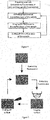
- Fig. 1 is a flow chart of a tomographic imaging method according to the present invention, and the tomographic imaging method provided by the present invention comprises the following steps:
- the protein includes a fluorescent protein and/or a protein which can be combined with a ligand to excite out fluorescence
- the fluorescent protein is preferably a pH-sensitive fluorescent protein, a pH-stable fluorescent protein and/or a photo-controlled fluorescent protein
- the fluorescent dye is preferably an organic fluorescent dye molecule.
- the pH-sensitive fluorescent protein includes a jellyfish-derived green fluorescent protein and derivatives, mOrange and derivatives, or mApple and derivatives; the green fluorescent protein and derivatives may be BFP, EBFP, EBFP2, CFP, ECFP, GFP, EGFP, YFP or EYFP, and preferably EGFP or EYFP; and the mApple and derivatives may be a red fluorescent protein pHuji.
- the photo-controlled fluorescent protein includes one or more of photoactivatable FPs (Photoactivatable FPs, PAFPs), such as PAGFP, PAmCherry1, etc.; or one or more of photoswitchable FPs (photoswitchable FPs, rsFPs), such as Dendra2, mEos3.1, etc.
- photoactivatable FPs Photoactivatable FPs, PAFPs
- PAGFP such as PAGFP, PAmCherry1, etc.
- photoswitchable FPs photoswitchable FPs, rsFPs
- Dendra2, mEos3.1 etc.
- the organic fluorescent dye molecule is preferably an Alexa series fluorescent dye molecule, and is preferably one or more of Alexa 488, Alexa 514, Alexa 532 and/or Alexa 546.
- the pH-stable fluorescent protein is preferably a red fluorescent protein DsRed.
- the protein or fluorescent dye used for marking the biological tissue is a protein or fluorescent dye not emitting fluorescence or only emitting fluorescence with a specific waveband, or a protein or fluorescent dye with fluorescence being reversibly quenched.
- the photo-controlled fluorescent protein not emitting fluorescence or only emitting fluorescence with a specific waveband may be directly subjected to surface layer activating step without being processed.
- the fluorescence being reversibly quenched includes a method of reversibly quenching a fluorescent group of the fluorescent protein or fluorescent dye by steeping in a chemical reagent, and preferably uses an acid chemical reagent for processing, use a transition metal ion compound solution for processing, or use a hydrogen ion and a transition metal ion for synergetic quenching.
- Fig. 2 is a flow chart of a quenching-imaging-cutting off tomographic imaging method according to the present invention.
- the fluorescence of a protein or fluorescent dye of which needs to be firstly quenched as shown in Fig. 2 , the fluorescence of the sample is firstly quenched while preparing the sample to make the sample not emit fluorescence, then a surface layer is activated, imaged and cut off, and then the procedures of activating the surface layer-imaging the surface layer-cutting are circulated as such, till the sample imaging is completed, to acquire a complete image of the biological tissue sample.
- the activating processing method includes chemical reagent processing or photochemical processing, so that the fluorescent protein or the fluorescent dye in the surface layer of the biological tissue sample is activated, while the protein or the fluorescent dye under the surface layer of the sample is not activated.
- the chemical reagent processing method includes steeping the protein-marked or fluorescent dye-marked biological tissue sample in the chemical reagent with the surface layer of the sample being permeated by the chemical reagent only, so that only the fluorescence in the surface layer of the sample is activated.
- the activating chemical reagent is preferably alkaline solution activating, metal ion chelating agent activating or synergetic activating of alkaline solution and metal ion chelating agent.
- the photochemical method includes using light with a specific waveband to activate and the activating light can only penetrate through the surface layer of the protein-marked or fluorescent dye-marked biological tissue sample, so that only the fluorescence in the surface layer of the sample is activated.
- the protein or the fluorescent dye may not emit fluorescence or may only emit fluorescence with a certain color originally, and may be excited by light with another wavelength to emit fluorescence with another color after being activated by the activating light with a specific wavelength.
- the photo-controlled fluorescent protein in a surface layer of a biological tissue-embedded sample may be activated by the activating light on a non-imaging light path, and then the fluorescent protein may be excited and imaged by using exciting light with a corresponding wavelength on an imaging light path.
- the imaging light path receives a light path emitting a spectrum (i.e., fluorescence signal), and the non-imaging light path refers to other light path excluding the imaging light path.
- activating may be described as follows: the photo-controlled fluorescent protein scarcely emits fluorescence with a certain waveband under the excitation of the exciting light with a specific wavelength, and only after being activated by the "activating light” with another certain wavelength, the fluorescent protein is enabled and excited by the exciting light with a specific wavelength so as to emit fluorescence.
- exciting refers to a process by which a fluorescent protein absorbs light with a certain wavelength to emit fluorescence.
- the photoactivatable fluorescent protein does not emit fluorescence per se, and, only when the surface layer of the sample is activated by the activating light with a specific wavelength, the activated fluorescence in the surface layer can perform fluorescence imaging under the excitation of the exciting light with another specific wavelength; while the photoswitchable fluorescent protein only emits fluorescence with a certain color itself, after the surface layer of the sample is activated by the activating light with a specific wavelength, the activated fluorescence in the surface layer performs fluorescence imaging under the excitation of the exciting light with another wavelength.
- the activating light can only activate the surface layer of the sample; compared with the exciting light, the wavelength of the activating light is generally shorter, and the capability of penetrating the tissue is poorer; therefore, the activating light can only penetrate through and activate the fluorescent protein in the surface layer; the activation of the activating light can control an angle between the activating light and the activated sample: the smaller the angle is, the stronger the surface reflection is; in this way, it is not only the case that little activating light enters a deep layer, but also that the energy density of light entering the sample becomes low.
- the photo-controlled fluorescent protein in the surface layer of the sample is activated by the activating light on the non-imaging light path of the sample according to the present invention, and then the activating light with a corresponding wavelength is used to activate the fluorescent protein on the imaging light path and perform fluorescence imaging, thus realizing imaging the surface layer of the sample.
- the activating light is an activating light with a specific wavelength matched with the photo-controlled fluorescent protein.
- the exciting light is an exciting light with a specific wavelength matched with the photo-controlled fluorescent protein.
- An included angle between a direction of the activating light and a direction of the exciting light is 0 to 90°, and is preferably 60 to 75°.
- the activated thickness of the surface layer of the sample may be controlled by optimizing the included angle between the activating light and the exciting light and optimizing the wavelength of the activating light, i.e., selecting a proper activating direction, and the activated thickness of the surface layer of the sample is an axial resolution of the fluorescence image of the sample.
- an acid liquor or chemical reagent may be added to make the pH of the solution range from 3 to 5, so as to quench the pH-sensitive fluorescent protein. Then the sample with quenched fluorescence may be steeped in an alkaline solution during activating, thus chemically reactivating the fluorescence of the pH-sensitive fluorescent protein in the surface layer of the sample.
- a pH value of the alkaline solution ranges from 8 to 13, and preferably ranges from 11 to 12, and the alkaline solution is preferably a sodium carbonate solution, an organic amine solution, ammonium hydroxide or a mixture thereof.
- a cut plane of the sample i.e., a surface layer of a mechanically cut surface
- a hydrophobic substance coating may further be added into the non-mechanically cut surfaces to prevent the alkaline solution from permeating into the sample from the non-mechanically cut surfaces.
- a type and a viscosity of the alkaline solution, a type of an embedding medium, and a ratio of each component and other factors all affect a permeability speed of the alkaline solution on the mechanically cut surfaces of the embedded sample.
- An axial resolution of the fluorescence imaging is controlled by controlling a proper permeability speed of the alkaline solution to realize high-resolution rapid imaging.
- an organic fluorescent dye molecule When an organic fluorescent dye molecule is used to mark a biological tissue, a transition metal ion and the organic fluorescent dye molecule are used to form a hexatomic ring to reduce a conjugate ⁇ electron density of the organic fluorescent molecule, thus quenching the fluorescence marked by the organic fluorescent dye molecule on the biological tissue, wherein the quenching is controllable and reversible.
- a transition metal ion compound is preferably one or more compounds of Cr 2+ , Mn 2+ , Fe 2+ , Fe 3+ , Co 2+ , Ni 2+ , Cu + and/or Cu 2+ .
- the metal ion chelating agent is one or more of EDTA-Na 4 , 8-hydroxyquinoline, N-oleoylsarcosine, 2-ethylenediamine and/or desferrioxamine chelating agent, and is preferably desferrioxamine or EDTA-Na 4 .
- a pH-insensitive fluorescent protein for example, a red fluorescent protein DsRed is used to mark the biological tissue
- a synergistic effect of the transition metal ion and a hydrogen ion is used to quench the fluorescence of the DsRed-marked biological tissue
- a synergistic effect of the metal ion chelating agent and a hydroxyl ion is used to reactivate the fluorescence of the biological tissue with quenched fluorescence, thus accurately controlling the fluorescence of the DsRed-marked biological tissue.
- An activated thickness of the surface layer of the biological tissue sample corresponds to an axial resolution during tomographic imaging, and a chemical reagent is used for reactivation, wherein a type and a viscosity of the chemical reagent, a type of an embedding medium while embedding the biological tissue, and a ratio of each component and other factors all affect a permeability speed of the chemical reagent on the mechanically cut surfaces of the embedded sample.
- the activating time is controlled by controlling a permeability speed of the chemical reagent in the surface of the sample to control an axial resolution of the fluorescence imaging, thus allowing high-resolution rapid imaging.
- a laser corresponding to an exciting wavelength of the sample is used as an exciting light source to irradiate on the surface of the sample through an objective lens after beam shaping, and excited fluorescence is collected by using the same objective lens. Since the fluorescence in a deep layer of the sample cannot be excited, interference of an axial background is avoided during imaging; therefore, an optical system without tomographic capability can be used for imaging, such as wide-field imaging, time-delay integral imaging, etc.
- the tomographic imaging method provided by the present invention may be deemed as a chemical tomographic imaging method, wherein the structure change of the fluorescent chromophoric group in the imaging sample is controlled to quench or activate fluorescence, so that only the fluorescent group in the surface layer of the sample can be excited during imaging, thus providing for imaging of only the surface layer of the sample.
- PAGFP Photoactivatable Green Fluorescent Protein
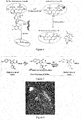
- the protein does not emit fluorescence basically while being irradiated by exciting light (with a wavelength of 488-515 nm in general); after being activated by ultraviolet light (with a wavelength of 405 nm in general), a chromophoric group is deprotonated and enters a state capable of emitting fluorescence, a fluorescence intensity of the state is enhanced by about 100 times before being activated, and this process is as shown in Fig. 3 .
- mEosFP is a typical photoswitchable fluorescent protein; taking a mEos 3.1 as an example, under an inactivated state, the mEos 3.1 can be excited to emit green fluorescence, after being irradiated by ultraviolet light (with a wavelength of 405 nm in general), a specific chemical bond in the fluorescent molecule will crack, which causes irreversible photoswitching, and the green fluorescence is switched into red fluorescence, and this process is as shown in Fig. 4 .
- EGFP Enhanced Green Fluorescent Protein
- a GFP chromophoric group in an acid environment is protonated under the effect of a hydrogen ion, and the GFP in this state is hardly to emit fluorescence under the excitation of the exciting light; however, when the protonated GFP is placed in an alkaline environment, the chromophoric group will be deprotonated due to the existence of a hydroxyl ion, and at the moment, the GFP may be excited to emit fluorescence normally. This process is a reversible process, which is as shown in Fig. 5 .
- a molecule configuration of a chromophoric group of an Alexa 488 fluorescent dye molecule may be changed into a form incapable of emitting fluorescence by excitation under the effect of a transition metal ion; while when a metal ion chelating agent is used to process the fluorescent dye molecule under an inactivated state, a metal ion will be seized by the chelating agent, which enables the chromophoric group to be restored to the form capable of emitting fluorescence.
- the process is as shown in Fig. 6 .
- a DsRed is a pH-stable fluorescent protein, a chromophoric group of the DsRed is reversibly changed under the common effect of a transition metal ion and a hydrogen ion, and cannot be excited to emit fluorescence under this form; when being processed by an alkalinous metal chelating agent, the chromophoric group may be restored to return to the form capable of emitting fluorescence, and this process is as shown in Fig. 7 .
- the tomographic imaging method of the present invention is intended to eliminate background fluorescence during imaging by a certain means before imaging, for example, the fluorescent protein-marked or the fluorescent dye-marked biological tissue sample is steeped by a chemical reagent; the fluorescent chromophoric group of the sample is changed and quenched, wherein the quenching is reversible, and the quenched fluorescence can be reactivated during activating. Only the surface layer is activated by using the activating chemical reagent during activating, after the fluorescence of the surface layer is activated, the surface layer imaging is free of background interference during the activation and fluorescence imaging since the fluorescence under the surface layer is not activated, and is still in a quenched state.
- the surface layer is activated during the activation step since the photo-controlled fluorescent protein does not emit fluorescence itself or only emits fluorescence with a certain wavelength; when the surface layer is activated and fluorescence imaging is performed, after the surface layer only emitting fluorescence with a specific wavelength is activated, and then the activated surface layer is excited by the exciting light with another specific wavelength, the wavelength of the fluorescence under the surface layer has a different scope from that of the wavelength of the fluorescence in the surface layer; therefore, the background interference problem does not exist.
- some proteins such as a photo-controlled fluorescent protein
- the tomographic imaging method of the present invention acquires the high-resolution fluorescence image by controlling the activated thickness of the surface layer of the sample, wherein an axial resolution thereof is of a submicron order, and a resolution scope ranges from 500 to 2000 nm.
- the tomographic imaging method of the present invention can perform focal-plane imaging, acquire high-throughput fluorescence imaging data, has a rapid imaging speed, has no limitation to the volume and transparency of the biological tissue sample, and the high-throughput advantage of the method is more apparent when used in imaging of a biological tissue sample, which shortens the imaging time. Since the tomographic imaging method of the present invention reversibly quenches the fluorescent group in the sample in advance and only activates the fluorescence in the surface layer of the sample during imaging, the inactivated fluorescence is protected from being quenched by the illumination light during imaging, and the imaging quality cannot be affected by the light quenching effect during long-time imaging.
- the biological tissue sample of the present invention may be a biological tissue-embedded sample which is embedded with a resin.
- Cutting off the surface layer of the sample may be performed by cutting off the surface layer of the sample in a mechanically cut manner.
- the cutting system is a surface cutting means capable of performing super thin-cutting, i.e., capable of cutting within a thickness of 10 ⁇ m, and is preferably vibration cutting or diamond cutter cutting.
- the imaging system of the present invention may be any imaging system used in three-dimensional imaging, and is preferably a wide-field imaging system to image a movable sample.
- the embodiments are as follows.
- Chemically tomographic imaging for a whole brain of a Thyl-EGFP-M mouse includes the following steps:
- the whole brain of a Thyl-EGFP-M mouse was fixed by a chemical fixation means to acquire a fixed biological brain tissue of the mouse.
- the specific steps were as follows: The whole brain that had been dissected from the mouse was steeped in a PFA solution with a mass fraction of 4% for 12h after heart perfusion at 4 °C. The dosage of the PFA solution was 20 ml per brain. Then the brain was rinsed by a PBS solution three times; 40 ml PBS solution per brain was used to rinse for 4h each time.
- the fixed brain tissue of the mouse was replaced by ethyl alcohol to dehydrate the biological tissue, so as to acquire the dehydrated EGFP-marked whole brain of the mouse.
- the specific steps were as follows.
- the fixed whole brain of the mouse was steeped in 20 ml gradient ethyl alcohol double distilled water solution in sequence for 2h at 4 °C to perform dehydration.
- Concentration gradients of the ethyl alcohol double distilled water solution were 50%, 75%, 95%, 100% and 100% by volume percent of ethyl alcohol.
- the dehydrated whole brain of the mouse was permeated by an HM20 embedding medium to acquire the whole brain of the mouse filled by a working solution of the HM20 embedding medium.
- the specific steps were as follows.
- the dehydrated whole brain of the mouse was permeated by the embedding medium by passing through more than 5 ml xylene solution with a gradient of HM20 in sequence at 4 °C. Gradients of the HM20 in xylene solution were 50%, 75%, 100%, 100%, 100% and 100% by volume percent of HM20.
- the brain was steeped for 2h in each of the former three gradients, steeped for 24h respectively in the fourth group and the fifth group, and steeped for 14h in the sixth group (before the brain was steeped in the sixth group, a resin working solution was added with acetic acid for mixing, wherein each 5 ml resin working solution was added with 25 to 30 ⁇ L acetic acid), so that the pH of the resin working solution was 3.5 to 4.0.
- the HM20 embedding medium was subjected to a polymerization reaction to acquire a resin-embedded sample of the EGFP-marked biological tissue.
- the specific steps were as follows: 1.1 mL working solution of the HM20 embedding medium mixed with acetic anhydride was injected into a gelatin capsule with a caliber of 9 mm installed in a base, then the whole brain of the mouse (filled by the working solution of the HM20 embedding medium) was placed in the capsule, the position of the whole brain was adjusted and a cover of the capsule was closed, then the capsule was brought into a vacuum drying box to perform gradient heating aggregation for 12h at 37 °C, 3h at 42 °C, 12h at 45 °C and 3h at 50 °C.
- the resin-embedded sample of the whole brain of the mouse was steeped in 0.05 mol/L sodium carbonate solution with a pH value of 11.2.
- the permeability speed of the alkaline solution in the resin-embedded sample of the whole brain of the mouse was about 1 ⁇ m/min to acquire an activated surface layer.
- Fluorescence imaging of the activated surface layer was excited, the activated surface layer imaged was mechanically cut, a new surface layer was exposed to contact with the alkaline solution, then the fluorescence of the new surface layer was reactivated and imaging was excited, then the new surface layer was repeatedly cut, activated and imaged, for repeating tomographic imaging as such until all the two-dimensional images of the entire sample were acquired.
- the acquired two-dimensional images were automatically registered to acquire a three-dimensional image of the sample (as shown in Fig. 8 ). From Fig.
- a spine, an axon bouton (axon bouton) and other submicrometer structures of a neure could be clearly distinguished, a single axon could be distinguished from a dense neuron axon fiber group, and a complete projection form of the neure could be traced.
- a chemically tomographic imaging method of a whole brain of a mouse over-expressing a pH-sensitive fluorescent protein pHuji included the following steps.
- the whole brain of a mouse over-expressing pHuji was fixed by a chemical fixation means to acquire a fixed pHuji-marked biological tissue.
- the specific steps were as follows: the whole brain that had been dissected from the mouse was steeped in a PFA solution with a mass fraction of 4% for 12h after heart perfusion at 4 °C. The dosage of the PFA solution was 20 ml per brain. Then the brain was rinsed by a PBS solution three times. 40 ml PBS solution per brain was used to rinse for 4h each time.
- the fixed pHuji-marked whole brain of the mouse was replaced by ethyl alcohol to dehydrate the biological tissue, so as to acquire the dehydrated pHuji-marked whole brain of the mouse.
- the specific steps were as follows: the fixed pHuji-marked whole brain of the mouse was steeped in 20 ml gradient ethyl alcohol double distilled water solution in sequence for 2h at 4 °C to perform dehydration. Concentration gradients of the ethyl alcohol double distilled water solution were 50%, 75%, 95%, 100% and 100%by volume percent of ethyl alcohol.
- the dehydrated pHuji-marked whole brain of the mouse was permeated by an HM20 embedding medium to acquire the pHuji-marked whole brain of the mouse filled by a working solution of the HM20 embedding medium.
- the specific steps were as follows: the dehydrated pHuji-marked whole brain of the mouse was permeated by the embedding medium by passing through more than 5 ml xylene solution with a gradient of HM20 in sequence at 4 °C.
- the HM20 embedding medium was subjected to a polymerization reaction to acquire a resin-embedded sample of the pHuji-marked biological tissue.
- the specific steps were as follows: 1.1 mL working solution of the HM20 embedding medium mixed with acetic anhydride was injected into a gelatin capsule with a caliber of 9 mm installed in a base, then the pHuji-marked whole brain of the mouse filled by the working solution of the HM20 embedding medium was placed in the capsule, the position of the whole brain was adjusted and a cover of the capsule was closed, then the capsule was brought into a vacuum drying box to perform gradient heating aggregation for 12h at 37 °C, 3h at 42 °C, 12h at 45 °C and 3h at 50 °C.
- the resin-embedded sample of the whole brain of the mouse was steeped in 0.1 mol/L sodium carbonate solution with a pH value of 11.6, the permeability speed of the alkaline solution in the resin-embedded sample of the whole brain of the mouse was about 1 ⁇ m/min to acquire an activated surface layer, and the thickness of the activated surface layer was 0.5 to 1 ⁇ m.
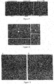
- Fig. 9 was a comparison diagram of fluorescence intensities before and after being activated by the alkaline solution in Embodiment 2; Fig. 9A was a fluorescence intensity of Embodiment 2 before being embedded by a resin; Fig. 9B was a fluorescence intensity of Embodiment 2 after being embedded by the resin and before being activated by the alkaline solution; and Fig. 9C was a fluorescence intensity after being embedded by the resin and after being activated by the alkaline solution. It could be seen that the resin-embedded sample after quenching processing hardly emits fluorescence, and after the sample was activated by the alkaline solution, the fluorescence intensity was well restored.
- Fluorescence imaging of the activated surface layer was excited, the activated surface layer imaged was mechanically cut, a new surface layer was exposed to contact with the alkaline solution, then the fluorescence of the new surface layer was reactivated and imaging was performed, then the new surface layer was repeatedly cut, activated and imaged, for repeating tomographic imaging as such until all the two-dimensional images of the entire sample were acquired.
- the acquired two-dimensional images were automatically registered to acquire a three-dimensional image of the sample, and a z-direction resolution was 0.5 to 1 ⁇ m.
- a chemically tomographic imaging method of a whole brain of a mouse over-expressing EYFP included the following steps.
- the whole brain of a mouse over-expressing EYFP was fixed by a chemical fixation means to acquire a fixed biological brain tissue of the mouse.
- the specific steps were as follows: the whole brain that had been dissected from the mouse was steeped in a PFA solution with a mass fraction of 4% for 12h after heart perfusion at 4 °C. A dosage of the PFA solution was 20 ml per brain. Then the brain was rinsed by a PBS solution three times; 40 ml PBS solution per brain was used to rinse for 4h each time.
- the fixed brain tissue of the mouse was replaced by ethyl alcohol to dehydrate the biological tissue, so as to acquire the dehydrated EGFP-marked whole brain of the mouse.
- the specific steps were as follows: the fixed whole brain of the mouse was steeped in 20ml gradient ethyl alcohol double distilled water solution in sequence for 2h at 4 °C to perform dehydration. Concentration gradients of the ethyl alcohol double distilled water solution were 50%, 75%, 95%, 100% and 100% by volume percent of ethyl alcohol.
- the dehydrated whole brain of the mouse was permeated by an HM20 embedding medium to acquire the whole brain of the mouse filled by a working solution of the HM20 embedding medium.
- the specific steps were as follows: the dehydrated whole brain of the mouse was permeated by the embedding medium by passing through more than 5 ml xylene solution with a gradient of HM20 in sequence at 4 °C.
- the HM20 embedding medium was subjected to a polymerization reaction to acquire a resin-embedded sample of the EYFP-marked biological tissue.
- the specific steps were as follows: 1.1 mL working solution of the HM20 embedding medium mixed with acetic anhydride was injected into a gelatin capsule with a caliber of 9 mm installed in a base, then the whole brain of the mouse (filled by the working solution of the HM20 embedding medium) was placed in the capsule, the position of the whole brain was adjusted and a cover of the capsule was closed, then the capsule was brought into a vacuum drying box to perform gradient heating aggregation for 12h at 37 °C, 3h at 42 °C, 12h at 45 °C and 3h at 50 °C.
- the resin-embedded sample of the whole brain of the mouse was steeped in a mixed solution of sodium carbonate and glycerinum with a pH value of 11.2, wherein the concentration of the sodium carbonate was 0.05 mol/L, and the volume percentage of the glycerinum was 20%.
- the permeability speed of this kind of mixed alkaline solution in the resin-embedded sample of the whole brain of the mouse was about 0.5 ⁇ m/min to acquire an activated surface layer, and the thickness of the activated surface layer was 0.5 to 1 ⁇ m.
- Fluorescence imaging of the activated surface layer was excited, then the activated surface layer imaged was mechanically cut, a new surface layer was exposed to contact with the alkaline solution, then the fluorescence of the new surface layer was reactivated and imaging was excited, then the new surface layer was repeatedly cut, activated and imaged, for repeating tomographic imaging as such until all the two-dimensional images of the entire sample were acquired.
- the acquired two-dimensional images ( Fig. 10B ) were automatically registered to acquire a three-dimensional image of the sample (as shown in Fig. 10A ). From Fig. 10 , a spine ( Fig. 10D ), an axon bouton ( Fig. 10C ) and other submicrometer structures of a neure could be clearly distinguished, and a z-direction resolution was about 0.5 to 1 ⁇ m.
- mEos3.1 is a common photoswitchable fluorescent protein.
- the photochemical property thereof was as follows: before being activated by a 405 nm ultraviolet wavelength, only green fluorescence (488 nm activating wavelength) was emitted and no red fluorescence was emitted; and after being activated by 405 nm ultraviolet wavelength for a short time, and under 561 nm exciting light, red fluorescence (an emission spectrum was above 550 nm) was emitted.
- a chemically tomographic imaging method of the whole brain of the mouse with the Rv-dg-mEos3.1 injected in the cerebral cortex included the following steps.
- the whole brain of the Rv-dg-mEos3.1-infected mouse was fixed by a chemical fixation means to acquire a fixed mEos3.1-marked biological tissue.
- the specific steps were as follows: the whole brain that had been dissected from the mouse was steeped in a PFA solution with a mass fraction of 4% for 12h after heart perfusion at 4 °C, a dosage of the PFA solution was 20 ml per brain, then the brain was rinsed by a PBS solution for three times, 40 ml PBS solution per brain was used to rinse for 4h each time.
- the fixed mEos3.1-marked whole brain of the mouse was replaced by ethyl alcohol to dehydrate the biological tissue, so as to acquire the dehydrated mEos3.1-marked whole brain of the mouse.
- the specific steps were as follows: the fixed mEos3.1-marked whole brain of the mouse was steeped in 20 ml gradient ethyl alcohol double distilled water solution in sequence for 2h at 4 °C to perform dehydration. Concentration gradients of the ethyl alcohol double distilled water solution were 50%, 75%, 95%, 100% and 100% by volume percent of ethyl alcohol.
- the dehydrated mEos3.1-marked whole brain of the mouse was permeated by an HM20 embedding medium to acquire the mEos3.1-marked whole brain of the mouse filled by a working solution of the HM20 embedding medium.
- the specific steps were as follows: the dehydrated mEos3.1-marked whole brain of the mouse was permeated by the embedding medium by passing through more than 5 ml xylene solution with a gradient of HM20 in sequence at 4 °C.
- the HM20 embedding medium was subjected to a polymerization reaction to acquire a resin-embedded sample of the mEos3.1-marked biological tissue.
- the specific steps were as follows: 1.1 mL working solution of the HM20 embedding medium was injected into a gelatin capsule with a caliber of 9 mm installed in a base, then the mEos3.1-marked whole brain of the mouse filled by the working solution of the HM20 embedding medium was placed in the capsule, the position of the whole brain was adjusted and a cover of the capsule was closed, then the capsule was brought into a vacuum drying box to perform gradient heating aggregation for 12h at 37 °C, 3h at 42 °C, 12h at 45 °C and 3h at 50°C.
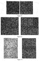
- Fig. 11 is a comparison diagram of fluorescence intensities before and after being activated and excited by the photo-controlled fluorescent protein; Fig. 11A is the excited fluorescence intensity before being activated; and Fig.
- 11B is the excited fluorescence intensity after being activated. It could be seen that the sample before being activated had no fluorescence signal basically, after being activated, a neure body can be observed, which indicated that the fluorescent protein in the surface layer of the sample was activated after the illumination of the activating light, and the thickness of the activated surface layer was 1 to 2 ⁇ m.
- 561 nm exciting light was used to excite fluorescence and perform imaging, and an included angle between the direction of the activating light and the direction of the exciting light was 60°.
- the sample surface was imaged by a wide-field imaging mode, and the sample was moved to acquire the image of the entire surface of the sample.
- the sample was moved below the diamond cutter and subjected to cutting, the imaged surface was cut off to expose the new surface layer, then the photo-controlled fluorescent protein of the new surface layer was reactivated and fluorescence imaging was excited, then the new surface layer was repeatedly cut, activated and imaged, for repeating tomographic imaging as such until all the two-dimensional images of the entire sample were acquired.
- the acquired two-dimensional images were automatically registered to acquire a three-dimensional image of the sample, and a z-direction resolution was 1 to 2 ⁇ m.
- a post-embedment chemically tomographic imaging method of Hela cells over-expressing PAGFP included the following steps.
- Hela cells over-expressing PAGFP were fixed by a chemical fixation means to acquire a fixed PAGFP-marked biological tissue.
- the specific steps were as follows: the Hela cell was steeped in a PFA solution with a mass fraction of 4% for 12h at 4 °C, then the Hela cell was rinsed by a PBS solution three times; 40ml PBS solution per tissue was used to rinse for 1h each time.
- the fixed PAGFP-marked Hela cell was replaced by ethyl alcohol to dehydrate the sample, so as to acquire the dehydrated PAGFP-marked cell sample.
- the specific steps were as follows: the fixed PAGFP-marked Hela cell was steeped in 20 ml gradient ethyl alcohol double distilled water solution in sequence for 2h at 4 °C to perform dehydration. Concentration gradients of the ethyl alcohol double distilled water solution were 50%, 75%, 95%, 100% and 100% by volume percent of ethyl alcohol.
- a dehydrated PAGFP-marked whole brain of a mouse was permeated by an HM20 embedding medium to acquire the PAGFP-marked cell sample filled by a working solution of the HM20 embedding medium.
- the specific steps were as follows: the dehydrated PAGFP-marked Hela cell was permeated by the embedding medium by passing through more than 5ml xylene solution with a gradient of HM20 in sequence at 4 °C. Gradients of the HM20 in xylene solution were 50%, 75%, 100%, 100%, 100% and 100% by volume percent of HM20, the cell sample was steeped for 2h in each of the former three gradients, steeped for 24h respectively in the fourth group and the fifth group, and steeped for 14h in the sixth group.
- the HM20 embedding medium was subjected to a polymerization reaction to acquire a PAGFP-marked and resin-embedded cell sample.
- the specific steps were as follows: the working solution of the HM20 embedding medium was dropped on a slide growing with the Hela cell, then a cover glass was covered on the slide to be brought into a vacuum drying box to perform gradient heating aggregation for 12h at 37 °C, 3h at 42 °C, 12h at 45 °C and 3h at 50 °C.
- the surface layer of the resin-embedded Hela cell sample was activated by the laser having 405 nm activating light, then the laser having 504 nm exciting light excited the fluorescence and performed the imaging, and an included angle between the direction of the activating light and the direction of the exciting light was 75°. That was, the included angle between the direction of the activating light and the direction of the upper surface of the sample was 15°, and the brightness of the fluorescence before and after activating processing by the activating was as shown in Fig. 12 (Fig. 12A and Fig. 12B ). After the PAGFP was activated and excited by a laser device, the brightness of the fluorescence was obviously increased from little fluorescence to where all the PAGFP-expressed cells were very bright, and the thickness of the activated surface layer was 1 to 2 um.
- the sample was moved below the diamond and cut, the imaged surface was cut to expose the new surface layer, then the photo-controlled fluorescent protein of the new surface layer was activated and fluorescence imaging was excited, then the new surface layer was repeatedly cut, activated and imaged, for repeating tomographic imaging as such until all the two-dimensional images of the entire sample were acquired.
- the acquired two-dimensional images were automatically registered to acquire a three-dimensional image of the sample, and an axial resolution was 1 to 2 ⁇ m.
- the brightness of fluorescence after fluorescence quenching and activating processing was as shown in Fig. 13 respectively.
- a contrast ratio of the image after fluorescence quenching was amplified by 10 times; as shown in Fig. 13a , after the activating processing, the fluorescence intensity ( Fig. 13b ) was equal to the effect after the contrast ratio of the fluorescence intensity before being activated was amplified by 10 times ( Fig. 13a ). That was, the activating processing enhanced the fluorescence intensity by 10 times or above, which guaranteed that the fluorescence could be used for further imaging.
- the brightness of the fluorescence after fluorescence quenching and activating processing was as shown in Fig. 14 respectively.
- a contrast ratio of the image after fluorescence quenching was amplified by 100 times; as shown in Fig. 14a , after the activating processing, the fluorescence intensity ( Fig. 13b ) was equal to the effect after the contrast ratio of the fluorescence intensity before being activated was amplified by 10 times ( Fig. 13a ). That was, the activating processing enhanced the fluorescence intensity by 10 times or above, which guaranteed that the fluorescence could be used for further imaging.
- the contrast ratio of the image after fluorescence quenching was amplified about 100 times, the processed image was shown in Fig.
- a fluorescence signal of the sample after being quenched could only be indistinctly seen after being amplified by 100 times, while a fluorescence signal of the sample reactivated ( Fig. 14b ) could be easily displayed in a background which was very dark; the fluorescence had a high contrast ratio, and could be used for further imaging.
Landscapes
- Health & Medical Sciences (AREA)
- Chemical & Material Sciences (AREA)
- Immunology (AREA)
- Physics & Mathematics (AREA)
- Life Sciences & Earth Sciences (AREA)
- General Physics & Mathematics (AREA)
- Pathology (AREA)
- Nuclear Medicine, Radiotherapy & Molecular Imaging (AREA)
- Analytical Chemistry (AREA)
- Biochemistry (AREA)
- General Health & Medical Sciences (AREA)
- Optics & Photonics (AREA)
- Chemical Kinetics & Catalysis (AREA)
- Engineering & Computer Science (AREA)
- Biomedical Technology (AREA)
- Molecular Biology (AREA)
- Investigating, Analyzing Materials By Fluorescence Or Luminescence (AREA)
- Measuring Or Testing Involving Enzymes Or Micro-Organisms (AREA)
- Investigating Or Analysing Biological Materials (AREA)
- Investigating Or Analysing Materials By The Use Of Chemical Reactions (AREA)
- Microscoopes, Condenser (AREA)
Applications Claiming Priority (2)
| Application Number | Priority Date | Filing Date | Title |
|---|---|---|---|
| CN201610967373.5A CN106501228B (zh) | 2016-10-31 | 2016-10-31 | 一种层析成像方法 |
| PCT/CN2017/106964 WO2018077114A1 (fr) | 2016-10-31 | 2017-10-20 | Procédé de tomographie |
Publications (2)
| Publication Number | Publication Date |
|---|---|
| EP3361237A1 true EP3361237A1 (fr) | 2018-08-15 |
| EP3361237A4 EP3361237A4 (fr) | 2019-09-04 |
Family
ID=58321738
Family Applications (1)
| Application Number | Title | Priority Date | Filing Date |
|---|---|---|---|
| EP17865883.7A Withdrawn EP3361237A4 (fr) | 2016-10-31 | 2017-10-20 | Procédé de tomographie |
Country Status (6)
| Country | Link |
|---|---|
| US (1) | US11346782B2 (fr) |
| EP (1) | EP3361237A4 (fr) |
| JP (1) | JP6966435B2 (fr) |
| CN (1) | CN106501228B (fr) |
| CA (1) | CA3004374A1 (fr) |
| WO (1) | WO2018077114A1 (fr) |
Families Citing this family (6)
| Publication number | Priority date | Publication date | Assignee | Title |
|---|---|---|---|---|
| CN106501228B (zh) * | 2016-10-31 | 2020-06-26 | 华中科技大学 | 一种层析成像方法 |
| CN110458923B (zh) * | 2018-11-01 | 2022-11-04 | 华中科技大学苏州脑空间信息研究院 | 一种快速且精准地获取组织样本神经元胞体位置的方法 |
| US11397179B2 (en) * | 2018-12-31 | 2022-07-26 | Robert Bosch Gmbh | PH-modulated imaging of targets close to a solid surface |
| CN117405636B (zh) * | 2023-07-25 | 2025-04-04 | 中国科学院生物物理研究所 | 超分辨光片荧光显微成像系统和方法 |
| CN119438272B (zh) * | 2024-11-04 | 2026-03-31 | 中国人民解放军空军军医大学 | 一种深部组织氧分压监测方法及系统 |
| CN120213918B (zh) * | 2025-03-25 | 2026-01-06 | 海南大学 | 同时获取单神经元完整形态和组学分子信息的方法及系统 |
Family Cites Families (12)
| Publication number | Priority date | Publication date | Assignee | Title |
|---|---|---|---|---|
| US7372985B2 (en) * | 2003-08-15 | 2008-05-13 | Massachusetts Institute Of Technology | Systems and methods for volumetric tissue scanning microscopy |
| US7767414B1 (en) * | 2005-04-20 | 2010-08-03 | The Board Of Trustees Of The Leland Stanford Junior University | Optical imaging of molecular characteristics of biological specimen |
| CN100523789C (zh) | 2006-03-21 | 2009-08-05 | 中国科学院化学研究所 | 一种用于检测抗氧化剂清除自由基的反应的配合物及其检测方法 |
| US20090041316A1 (en) * | 2007-08-07 | 2009-02-12 | California Institute Of Technology | Vibratome assisted subsurface imaging microscopy (vibra-ssim) |
| US20100000383A1 (en) * | 2007-08-07 | 2010-01-07 | Koos David S | Microscope coupled tissue sectioning system |
| WO2009098079A1 (fr) * | 2008-02-06 | 2009-08-13 | Ludwig-Maximilians-Universität München | Caractérisation thermo-optique de molécules d'acide nucléique |
| CN102373267A (zh) * | 2010-08-09 | 2012-03-14 | 北京泰格瑞分子检验有限公司 | 一种减少与引物聚合的水解探针实时荧光pcr |
| CN102928970B (zh) * | 2012-10-19 | 2014-10-29 | 华中科技大学 | 一种大样本快速三维显微成像的方法和系统 |
| CN105021431A (zh) * | 2014-04-24 | 2015-11-04 | 华中科技大学 | 荧光蛋白标记的生物组织树脂包埋方法及碱性溶液的应用 |
| EP3268715B1 (fr) * | 2015-03-11 | 2024-09-25 | TissueVision, Inc. | Système et méthodes de coloration et d'imagerie en série |
| CN105866079B (zh) * | 2016-03-31 | 2019-07-05 | 同济大学 | 基于荧光染料nmm、g-四链体dna、冠醚和金属离子相互作用的分子逻辑门构建方法 |
| CN106501228B (zh) * | 2016-10-31 | 2020-06-26 | 华中科技大学 | 一种层析成像方法 |
-
2016
- 2016-10-31 CN CN201610967373.5A patent/CN106501228B/zh active Active
-
2017
- 2017-10-20 JP JP2018522059A patent/JP6966435B2/ja active Active
- 2017-10-20 EP EP17865883.7A patent/EP3361237A4/fr not_active Withdrawn
- 2017-10-20 WO PCT/CN2017/106964 patent/WO2018077114A1/fr not_active Ceased
- 2017-10-20 US US15/771,713 patent/US11346782B2/en active Active
- 2017-10-20 CA CA3004374A patent/CA3004374A1/fr not_active Abandoned
Also Published As
| Publication number | Publication date |
|---|---|
| US20200309697A1 (en) | 2020-10-01 |
| CN106501228B (zh) | 2020-06-26 |
| WO2018077114A1 (fr) | 2018-05-03 |
| JP2019505758A (ja) | 2019-02-28 |
| US11346782B2 (en) | 2022-05-31 |
| EP3361237A4 (fr) | 2019-09-04 |
| JP6966435B2 (ja) | 2021-11-17 |
| CA3004374A1 (fr) | 2018-05-03 |
| CN106501228A (zh) | 2017-03-15 |
Similar Documents
| Publication | Publication Date | Title |
|---|---|---|
| US11346782B2 (en) | Tomographic imaging method | |
| US20230213415A1 (en) | Method and System for Imaging and Analysis of a Biological Specimen | |
| Watanabe et al. | Protein localization in electron micrographs using fluorescence nanoscopy | |
| Turkowyd et al. | From single molecules to life: microscopy at the nanoscale | |
| JP6112169B2 (ja) | 生体物質検出用の蛍光標識体 | |
| EP2833140B1 (fr) | Procédé de détection d'une matière biologique | |
| JP7565621B2 (ja) | 蛍光模倣明視野イメージング | |
| Petriella et al. | Superresolution imaging with switchable fluorophores based on oxazine auxochromes | |
| JP4804363B2 (ja) | 蛍光顕微鏡装置 | |
| JP2007522502A5 (fr) | ||
| JP6950922B2 (ja) | 染色方法、染色剤、及び染色キット | |
| JP2008083047A (ja) | 検体の蛍光発生物質で標識された構造の空間的に高解像度の調査方法 | |
| CN115437131A (zh) | 对生物样品进行三维成像的方法及光片显微镜系统 | |
| JP6583011B2 (ja) | 酸性水溶液を用いた免疫染色スライドの洗浄方法 | |
| Franzkoch et al. | Rapid in-EPON CLEM: Combining fast and efficient labeling of self-labeling enzyme tags with EM-resistant Janelia Fluor dyes and StayGold | |
| CN114057604A (zh) | 一种细胞核荧光染料及其染色方法 | |
| Douma et al. | Two‐photon lifetime imaging of fluorescent probes in intact blood vessels: A window to sub‐cellular structural information and binding status | |
| CA3156802C (fr) | Imagerie en fond clair imitant la fluorescence | |
| Farias et al. | Semiconductor nanocrystals and fluorescence microscopy in biological labeling | |
| Tashlieva | Visualization of surface antigens by correlative microscopy | |
| CN106568753A (zh) | 一种有机荧光染料分子标记生物组织的荧光控制方法 | |
| Cloin et al. | Single Molecule Localization Microscopy to study neuronal microtubule organization | |
| Endesfelder | Photoswitching fluorophores in super-Resolution fluorescence microscopy | |
| JP2023126996A (ja) | 免疫染色方法及び免疫染色組織標本の作製方法 | |
| Zhao | Exploring unique optical properties of upconversion nanocrystals towards a new library of molecular probes |
Legal Events
| Date | Code | Title | Description |
|---|---|---|---|
| STAA | Information on the status of an ep patent application or granted ep patent |
Free format text: STATUS: THE INTERNATIONAL PUBLICATION HAS BEEN MADE |
|
| PUAI | Public reference made under article 153(3) epc to a published international application that has entered the european phase |
Free format text: ORIGINAL CODE: 0009012 |
|
| STAA | Information on the status of an ep patent application or granted ep patent |
Free format text: STATUS: REQUEST FOR EXAMINATION WAS MADE |
|
| 17P | Request for examination filed |
Effective date: 20180507 |
|
| AK | Designated contracting states |
Kind code of ref document: A1 Designated state(s): AL AT BE BG CH CY CZ DE DK EE ES FI FR GB GR HR HU IE IS IT LI LT LU LV MC MK MT NL NO PL PT RO RS SE SI SK SM TR |
|
| AX | Request for extension of the european patent |
Extension state: BA ME |
|
| A4 | Supplementary search report drawn up and despatched |
Effective date: 20190802 |
|
| RIC1 | Information provided on ipc code assigned before grant |
Ipc: G01N 21/64 20060101AFI20190729BHEP Ipc: G01N 1/30 20060101ALI20190729BHEP |
|
| DAV | Request for validation of the european patent (deleted) | ||
| DAX | Request for extension of the european patent (deleted) | ||
| STAA | Information on the status of an ep patent application or granted ep patent |
Free format text: STATUS: EXAMINATION IS IN PROGRESS |
|
| 17Q | First examination report despatched |
Effective date: 20220523 |
|
| STAA | Information on the status of an ep patent application or granted ep patent |
Free format text: STATUS: THE APPLICATION IS DEEMED TO BE WITHDRAWN |
|
| 18D | Application deemed to be withdrawn |
Effective date: 20230503 |