EP3325665B1 - Methods of amplifying nucleic acid sequences - Google Patents
Methods of amplifying nucleic acid sequences Download PDFInfo
- Publication number
- EP3325665B1 EP3325665B1 EP16828282.0A EP16828282A EP3325665B1 EP 3325665 B1 EP3325665 B1 EP 3325665B1 EP 16828282 A EP16828282 A EP 16828282A EP 3325665 B1 EP3325665 B1 EP 3325665B1
- Authority
- EP
- European Patent Office
- Prior art keywords
- dna
- double stranded
- cell
- amplification
- nucleic acid
- Prior art date
- Legal status (The legal status is an assumption and is not a legal conclusion. Google has not performed a legal analysis and makes no representation as to the accuracy of the status listed.)
- Active
Links
- 238000000034 method Methods 0.000 title claims description 210
- 150000007523 nucleic acids Chemical group 0.000 title claims description 109
- 108020004414 DNA Proteins 0.000 claims description 363
- 210000004027 cell Anatomy 0.000 claims description 189
- 230000003321 amplification Effects 0.000 claims description 138
- 238000003199 nucleic acid amplification method Methods 0.000 claims description 138
- 239000012634 fragment Substances 0.000 claims description 97
- 102000039446 nucleic acids Human genes 0.000 claims description 91
- 108020004707 nucleic acids Proteins 0.000 claims description 91
- 108091032973 (ribonucleotides)n+m Proteins 0.000 claims description 72
- 125000003729 nucleotide group Chemical group 0.000 claims description 65
- 206010028980 Neoplasm Diseases 0.000 claims description 62
- 239000002773 nucleotide Substances 0.000 claims description 57
- 238000012163 sequencing technique Methods 0.000 claims description 50
- 201000011510 cancer Diseases 0.000 claims description 43
- 108010020764 Transposases Proteins 0.000 claims description 41
- 102000008579 Transposases Human genes 0.000 claims description 41
- 108091093088 Amplicon Proteins 0.000 claims description 34
- 230000027455 binding Effects 0.000 claims description 27
- 108090000626 DNA-directed RNA polymerases Proteins 0.000 claims description 26
- 102000004163 DNA-directed RNA polymerases Human genes 0.000 claims description 26
- 230000000295 complement effect Effects 0.000 claims description 20
- 208000005443 Circulating Neoplastic Cells Diseases 0.000 claims description 18
- 101710137500 T7 RNA polymerase Proteins 0.000 claims description 13
- 102000040650 (ribonucleotides)n+m Human genes 0.000 claims description 10
- 230000002441 reversible effect Effects 0.000 claims description 10
- 230000037452 priming Effects 0.000 claims description 9
- 108010012306 Tn5 transposase Proteins 0.000 claims description 5
- 239000000523 sample Substances 0.000 description 74
- 239000013615 primer Substances 0.000 description 64
- 102000053602 DNA Human genes 0.000 description 57
- 238000003752 polymerase chain reaction Methods 0.000 description 54
- 239000000047 product Substances 0.000 description 47
- 238000006243 chemical reaction Methods 0.000 description 41
- 230000017105 transposition Effects 0.000 description 34
- 102000016928 DNA-directed DNA polymerase Human genes 0.000 description 33
- 108010014303 DNA-directed DNA polymerase Proteins 0.000 description 33
- 238000004458 analytical method Methods 0.000 description 33
- 238000009396 hybridization Methods 0.000 description 32
- 238000001514 detection method Methods 0.000 description 31
- 238000013518 transcription Methods 0.000 description 29
- 230000035897 transcription Effects 0.000 description 29
- 102000040430 polynucleotide Human genes 0.000 description 28
- 108091033319 polynucleotide Proteins 0.000 description 28
- 239000002157 polynucleotide Substances 0.000 description 28
- 239000000203 mixture Substances 0.000 description 26
- 238000000338 in vitro Methods 0.000 description 25
- 239000004365 Protease Substances 0.000 description 22
- 238000000926 separation method Methods 0.000 description 22
- 108091005804 Peptidases Proteins 0.000 description 21
- 102100037486 Reverse transcriptase/ribonuclease H Human genes 0.000 description 21
- 235000019419 proteases Nutrition 0.000 description 21
- 208000037265 diseases, disorders, signs and symptoms Diseases 0.000 description 20
- 238000003556 assay Methods 0.000 description 19
- 230000015572 biosynthetic process Effects 0.000 description 19
- 210000000349 chromosome Anatomy 0.000 description 19
- 238000005516 engineering process Methods 0.000 description 19
- 230000001605 fetal effect Effects 0.000 description 18
- 108090000623 proteins and genes Proteins 0.000 description 18
- 210000001519 tissue Anatomy 0.000 description 18
- 102100034343 Integrase Human genes 0.000 description 17
- UYTPUPDQBNUYGX-UHFFFAOYSA-N guanine Chemical compound O=C1NC(N)=NC2=C1N=CN2 UYTPUPDQBNUYGX-UHFFFAOYSA-N 0.000 description 17
- 238000012546 transfer Methods 0.000 description 17
- 108091028043 Nucleic acid sequence Proteins 0.000 description 16
- 238000013412 genome amplification Methods 0.000 description 16
- 238000003786 synthesis reaction Methods 0.000 description 16
- 241000282414 Homo sapiens Species 0.000 description 15
- 210000004369 blood Anatomy 0.000 description 15
- 239000008280 blood Substances 0.000 description 15
- 238000004949 mass spectrometry Methods 0.000 description 15
- 210000001161 mammalian embryo Anatomy 0.000 description 14
- 238000011160 research Methods 0.000 description 14
- 108020004682 Single-Stranded DNA Proteins 0.000 description 13
- 239000003153 chemical reaction reagent Substances 0.000 description 13
- 208000035475 disorder Diseases 0.000 description 13
- 239000000975 dye Substances 0.000 description 13
- 230000008569 process Effects 0.000 description 13
- -1 dCTP or dATP Chemical class 0.000 description 12
- KCXVZYZYPLLWCC-UHFFFAOYSA-N EDTA Chemical compound OC(=O)CN(CC(O)=O)CCN(CC(O)=O)CC(O)=O KCXVZYZYPLLWCC-UHFFFAOYSA-N 0.000 description 11
- 108091034117 Oligonucleotide Proteins 0.000 description 11
- 102000006382 Ribonucleases Human genes 0.000 description 11
- 108010083644 Ribonucleases Proteins 0.000 description 11
- 230000006037 cell lysis Effects 0.000 description 11
- 230000007614 genetic variation Effects 0.000 description 11
- YBJHBAHKTGYVGT-ZKWXMUAHSA-N (+)-Biotin Chemical compound N1C(=O)N[C@@H]2[C@H](CCCCC(=O)O)SC[C@@H]21 YBJHBAHKTGYVGT-ZKWXMUAHSA-N 0.000 description 10
- GUAHPAJOXVYFON-ZETCQYMHSA-N (8S)-8-amino-7-oxononanoic acid zwitterion Chemical compound C[C@H](N)C(=O)CCCCCC(O)=O GUAHPAJOXVYFON-ZETCQYMHSA-N 0.000 description 10
- 108010092799 RNA-directed DNA polymerase Proteins 0.000 description 10
- 229940088598 enzyme Drugs 0.000 description 10
- 238000003205 genotyping method Methods 0.000 description 10
- 102000004190 Enzymes Human genes 0.000 description 9
- 108090000790 Enzymes Proteins 0.000 description 9
- HEMHJVSKTPXQMS-UHFFFAOYSA-M Sodium hydroxide Chemical compound [OH-].[Na+] HEMHJVSKTPXQMS-UHFFFAOYSA-M 0.000 description 9
- 238000000137 annealing Methods 0.000 description 9
- 239000007853 buffer solution Substances 0.000 description 9
- 239000002299 complementary DNA Substances 0.000 description 9
- 230000009089 cytolysis Effects 0.000 description 9
- OPTASPLRGRRNAP-UHFFFAOYSA-N cytosine Chemical compound NC=1C=CNC(=O)N=1 OPTASPLRGRRNAP-UHFFFAOYSA-N 0.000 description 9
- 238000012217 deletion Methods 0.000 description 9
- 230000037430 deletion Effects 0.000 description 9
- 238000003780 insertion Methods 0.000 description 9
- 230000037431 insertion Effects 0.000 description 9
- 108020004999 messenger RNA Proteins 0.000 description 9
- 238000000746 purification Methods 0.000 description 9
- 238000010791 quenching Methods 0.000 description 9
- 230000000171 quenching effect Effects 0.000 description 9
- 239000000243 solution Substances 0.000 description 9
- 230000004544 DNA amplification Effects 0.000 description 8
- 101710203526 Integrase Proteins 0.000 description 8
- FAPWRFPIFSIZLT-UHFFFAOYSA-M Sodium chloride Chemical compound [Na+].[Cl-] FAPWRFPIFSIZLT-UHFFFAOYSA-M 0.000 description 8
- 230000005856 abnormality Effects 0.000 description 8
- 239000000872 buffer Substances 0.000 description 8
- 238000003745 diagnosis Methods 0.000 description 8
- MHMNJMPURVTYEJ-UHFFFAOYSA-N fluorescein-5-isothiocyanate Chemical compound O1C(=O)C2=CC(N=C=S)=CC=C2C21C1=CC=C(O)C=C1OC1=CC(O)=CC=C21 MHMNJMPURVTYEJ-UHFFFAOYSA-N 0.000 description 8
- 208000027205 Congenital disease Diseases 0.000 description 7
- 208000029767 Congenital, Hereditary, and Neonatal Diseases and Abnormalities Diseases 0.000 description 7
- ISAKRJDGNUQOIC-UHFFFAOYSA-N Uracil Chemical compound O=C1C=CNC(=O)N1 ISAKRJDGNUQOIC-UHFFFAOYSA-N 0.000 description 7
- 239000011324 bead Substances 0.000 description 7
- 201000010099 disease Diseases 0.000 description 7
- 230000014509 gene expression Effects 0.000 description 7
- 238000012252 genetic analysis Methods 0.000 description 7
- 230000008774 maternal effect Effects 0.000 description 7
- 238000003793 prenatal diagnosis Methods 0.000 description 7
- 238000002360 preparation method Methods 0.000 description 7
- RWQNBRDOKXIBIV-UHFFFAOYSA-N thymine Chemical compound CC1=CNC(=O)NC1=O RWQNBRDOKXIBIV-UHFFFAOYSA-N 0.000 description 7
- 229930024421 Adenine Natural products 0.000 description 6
- 108020004711 Nucleic Acid Probes Proteins 0.000 description 6
- 208000037065 Subacute sclerosing leukoencephalitis Diseases 0.000 description 6
- 206010042297 Subacute sclerosing panencephalitis Diseases 0.000 description 6
- 229960000643 adenine Drugs 0.000 description 6
- 238000001574 biopsy Methods 0.000 description 6
- 239000013592 cell lysate Substances 0.000 description 6
- 230000029087 digestion Effects 0.000 description 6
- 238000006073 displacement reaction Methods 0.000 description 6
- 239000003814 drug Substances 0.000 description 6
- 230000000694 effects Effects 0.000 description 6
- 238000001962 electrophoresis Methods 0.000 description 6
- 210000003754 fetus Anatomy 0.000 description 6
- 238000013467 fragmentation Methods 0.000 description 6
- 238000006062 fragmentation reaction Methods 0.000 description 6
- 238000010438 heat treatment Methods 0.000 description 6
- 238000012165 high-throughput sequencing Methods 0.000 description 6
- 150000002500 ions Chemical class 0.000 description 6
- 239000012139 lysis buffer Substances 0.000 description 6
- 238000007481 next generation sequencing Methods 0.000 description 6
- 239000002853 nucleic acid probe Substances 0.000 description 6
- 239000002777 nucleoside Substances 0.000 description 6
- 238000012545 processing Methods 0.000 description 6
- 239000011541 reaction mixture Substances 0.000 description 6
- 238000012552 review Methods 0.000 description 6
- 239000007787 solid Substances 0.000 description 6
- 241000894007 species Species 0.000 description 6
- 239000000758 substrate Substances 0.000 description 6
- GFFGJBXGBJISGV-UHFFFAOYSA-N Adenine Chemical compound NC1=NC=NC2=C1N=CN2 GFFGJBXGBJISGV-UHFFFAOYSA-N 0.000 description 5
- 239000003155 DNA primer Substances 0.000 description 5
- 201000010374 Down Syndrome Diseases 0.000 description 5
- 206010018338 Glioma Diseases 0.000 description 5
- 206010044688 Trisomy 21 Diseases 0.000 description 5
- DRTQHJPVMGBUCF-XVFCMESISA-N Uridine Chemical compound O[C@@H]1[C@H](O)[C@@H](CO)O[C@H]1N1C(=O)NC(=O)C=C1 DRTQHJPVMGBUCF-XVFCMESISA-N 0.000 description 5
- JLCPHMBAVCMARE-UHFFFAOYSA-N [3-[[3-[[3-[[3-[[3-[[3-[[3-[[3-[[3-[[3-[[3-[[5-(2-amino-6-oxo-1H-purin-9-yl)-3-[[3-[[3-[[3-[[3-[[3-[[5-(2-amino-6-oxo-1H-purin-9-yl)-3-[[5-(2-amino-6-oxo-1H-purin-9-yl)-3-hydroxyoxolan-2-yl]methoxy-hydroxyphosphoryl]oxyoxolan-2-yl]methoxy-hydroxyphosphoryl]oxy-5-(5-methyl-2,4-dioxopyrimidin-1-yl)oxolan-2-yl]methoxy-hydroxyphosphoryl]oxy-5-(6-aminopurin-9-yl)oxolan-2-yl]methoxy-hydroxyphosphoryl]oxy-5-(6-aminopurin-9-yl)oxolan-2-yl]methoxy-hydroxyphosphoryl]oxy-5-(6-aminopurin-9-yl)oxolan-2-yl]methoxy-hydroxyphosphoryl]oxy-5-(6-aminopurin-9-yl)oxolan-2-yl]methoxy-hydroxyphosphoryl]oxyoxolan-2-yl]methoxy-hydroxyphosphoryl]oxy-5-(5-methyl-2,4-dioxopyrimidin-1-yl)oxolan-2-yl]methoxy-hydroxyphosphoryl]oxy-5-(4-amino-2-oxopyrimidin-1-yl)oxolan-2-yl]methoxy-hydroxyphosphoryl]oxy-5-(5-methyl-2,4-dioxopyrimidin-1-yl)oxolan-2-yl]methoxy-hydroxyphosphoryl]oxy-5-(5-methyl-2,4-dioxopyrimidin-1-yl)oxolan-2-yl]methoxy-hydroxyphosphoryl]oxy-5-(6-aminopurin-9-yl)oxolan-2-yl]methoxy-hydroxyphosphoryl]oxy-5-(6-aminopurin-9-yl)oxolan-2-yl]methoxy-hydroxyphosphoryl]oxy-5-(4-amino-2-oxopyrimidin-1-yl)oxolan-2-yl]methoxy-hydroxyphosphoryl]oxy-5-(4-amino-2-oxopyrimidin-1-yl)oxolan-2-yl]methoxy-hydroxyphosphoryl]oxy-5-(4-amino-2-oxopyrimidin-1-yl)oxolan-2-yl]methoxy-hydroxyphosphoryl]oxy-5-(6-aminopurin-9-yl)oxolan-2-yl]methoxy-hydroxyphosphoryl]oxy-5-(4-amino-2-oxopyrimidin-1-yl)oxolan-2-yl]methyl [5-(6-aminopurin-9-yl)-2-(hydroxymethyl)oxolan-3-yl] hydrogen phosphate Polymers Cc1cn(C2CC(OP(O)(=O)OCC3OC(CC3OP(O)(=O)OCC3OC(CC3O)n3cnc4c3nc(N)[nH]c4=O)n3cnc4c3nc(N)[nH]c4=O)C(COP(O)(=O)OC3CC(OC3COP(O)(=O)OC3CC(OC3COP(O)(=O)OC3CC(OC3COP(O)(=O)OC3CC(OC3COP(O)(=O)OC3CC(OC3COP(O)(=O)OC3CC(OC3COP(O)(=O)OC3CC(OC3COP(O)(=O)OC3CC(OC3COP(O)(=O)OC3CC(OC3COP(O)(=O)OC3CC(OC3COP(O)(=O)OC3CC(OC3COP(O)(=O)OC3CC(OC3COP(O)(=O)OC3CC(OC3COP(O)(=O)OC3CC(OC3COP(O)(=O)OC3CC(OC3COP(O)(=O)OC3CC(OC3COP(O)(=O)OC3CC(OC3CO)n3cnc4c(N)ncnc34)n3ccc(N)nc3=O)n3cnc4c(N)ncnc34)n3ccc(N)nc3=O)n3ccc(N)nc3=O)n3ccc(N)nc3=O)n3cnc4c(N)ncnc34)n3cnc4c(N)ncnc34)n3cc(C)c(=O)[nH]c3=O)n3cc(C)c(=O)[nH]c3=O)n3ccc(N)nc3=O)n3cc(C)c(=O)[nH]c3=O)n3cnc4c3nc(N)[nH]c4=O)n3cnc4c(N)ncnc34)n3cnc4c(N)ncnc34)n3cnc4c(N)ncnc34)n3cnc4c(N)ncnc34)O2)c(=O)[nH]c1=O JLCPHMBAVCMARE-UHFFFAOYSA-N 0.000 description 5
- 230000000692 anti-sense effect Effects 0.000 description 5
- 230000008901 benefit Effects 0.000 description 5
- 229960002685 biotin Drugs 0.000 description 5
- 235000020958 biotin Nutrition 0.000 description 5
- 239000011616 biotin Substances 0.000 description 5
- 150000001793 charged compounds Chemical class 0.000 description 5
- 238000003776 cleavage reaction Methods 0.000 description 5
- 229940104302 cytosine Drugs 0.000 description 5
- 238000009826 distribution Methods 0.000 description 5
- 239000000839 emulsion Substances 0.000 description 5
- 238000000605 extraction Methods 0.000 description 5
- 239000007850 fluorescent dye Substances 0.000 description 5
- 230000007246 mechanism Effects 0.000 description 5
- 238000012986 modification Methods 0.000 description 5
- 230000004048 modification Effects 0.000 description 5
- 238000007899 nucleic acid hybridization Methods 0.000 description 5
- 230000003287 optical effect Effects 0.000 description 5
- 238000006116 polymerization reaction Methods 0.000 description 5
- 238000003753 real-time PCR Methods 0.000 description 5
- 238000005096 rolling process Methods 0.000 description 5
- 230000007017 scission Effects 0.000 description 5
- 230000035945 sensitivity Effects 0.000 description 5
- 238000003860 storage Methods 0.000 description 5
- 210000004881 tumor cell Anatomy 0.000 description 5
- 229940035893 uracil Drugs 0.000 description 5
- 206010003571 Astrocytoma Diseases 0.000 description 4
- 108020004635 Complementary DNA Proteins 0.000 description 4
- 238000012408 PCR amplification Methods 0.000 description 4
- 208000007641 Pinealoma Diseases 0.000 description 4
- 240000004808 Saccharomyces cerevisiae Species 0.000 description 4
- 239000013504 Triton X-100 Substances 0.000 description 4
- 229920004890 Triton X-100 Polymers 0.000 description 4
- 230000001154 acute effect Effects 0.000 description 4
- OIRDTQYFTABQOQ-KQYNXXCUSA-N adenosine Chemical compound C1=NC=2C(N)=NC=NC=2N1[C@@H]1O[C@H](CO)[C@@H](O)[C@H]1O OIRDTQYFTABQOQ-KQYNXXCUSA-N 0.000 description 4
- 208000036878 aneuploidy Diseases 0.000 description 4
- 231100001075 aneuploidy Toxicity 0.000 description 4
- 238000003491 array Methods 0.000 description 4
- 229910052799 carbon Inorganic materials 0.000 description 4
- 230000001413 cellular effect Effects 0.000 description 4
- 210000003169 central nervous system Anatomy 0.000 description 4
- 230000001684 chronic effect Effects 0.000 description 4
- 238000001360 collision-induced dissociation Methods 0.000 description 4
- 238000005520 cutting process Methods 0.000 description 4
- 238000004925 denaturation Methods 0.000 description 4
- 230000036425 denaturation Effects 0.000 description 4
- 239000005547 deoxyribonucleotide Substances 0.000 description 4
- 238000001943 fluorescence-activated cell sorting Methods 0.000 description 4
- 238000012268 genome sequencing Methods 0.000 description 4
- 238000003384 imaging method Methods 0.000 description 4
- 230000003993 interaction Effects 0.000 description 4
- DRAVOWXCEBXPTN-UHFFFAOYSA-N isoguanine Chemical compound NC1=NC(=O)NC2=C1NC=N2 DRAVOWXCEBXPTN-UHFFFAOYSA-N 0.000 description 4
- 238000002955 isolation Methods 0.000 description 4
- 238000002372 labelling Methods 0.000 description 4
- 239000000463 material Substances 0.000 description 4
- 230000035772 mutation Effects 0.000 description 4
- YBYRMVIVWMBXKQ-UHFFFAOYSA-N phenylmethanesulfonyl fluoride Chemical compound FS(=O)(=O)CC1=CC=CC=C1 YBYRMVIVWMBXKQ-UHFFFAOYSA-N 0.000 description 4
- 125000002467 phosphate group Chemical group [H]OP(=O)(O[H])O[*] 0.000 description 4
- 208000029340 primitive neuroectodermal tumor Diseases 0.000 description 4
- 102000004169 proteins and genes Human genes 0.000 description 4
- 238000010839 reverse transcription Methods 0.000 description 4
- 239000003161 ribonuclease inhibitor Substances 0.000 description 4
- 239000011780 sodium chloride Substances 0.000 description 4
- 229940113082 thymine Drugs 0.000 description 4
- 201000008271 Atypical teratoid rhabdoid tumor Diseases 0.000 description 3
- 241000972773 Aulopiformes Species 0.000 description 3
- 201000009030 Carcinoma Diseases 0.000 description 3
- 108010077544 Chromatin Proteins 0.000 description 3
- 206010009944 Colon cancer Diseases 0.000 description 3
- 238000001712 DNA sequencing Methods 0.000 description 3
- 208000032612 Glial tumor Diseases 0.000 description 3
- PEDCQBHIVMGVHV-UHFFFAOYSA-N Glycerine Chemical compound OCC(O)CO PEDCQBHIVMGVHV-UHFFFAOYSA-N 0.000 description 3
- NYHBQMYGNKIUIF-UUOKFMHZSA-N Guanosine Chemical compound C1=NC=2C(=O)NC(N)=NC=2N1[C@@H]1O[C@H](CO)[C@@H](O)[C@H]1O NYHBQMYGNKIUIF-UUOKFMHZSA-N 0.000 description 3
- 206010025323 Lymphomas Diseases 0.000 description 3
- 208000034176 Neoplasms, Germ Cell and Embryonal Diseases 0.000 description 3
- 108091028664 Ribonucleotide Proteins 0.000 description 3
- 206010039491 Sarcoma Diseases 0.000 description 3
- VYPSYNLAJGMNEJ-UHFFFAOYSA-N Silicium dioxide Chemical compound O=[Si]=O VYPSYNLAJGMNEJ-UHFFFAOYSA-N 0.000 description 3
- XUIMIQQOPSSXEZ-UHFFFAOYSA-N Silicon Chemical compound [Si] XUIMIQQOPSSXEZ-UHFFFAOYSA-N 0.000 description 3
- 238000013459 approach Methods 0.000 description 3
- 210000004436 artificial bacterial chromosome Anatomy 0.000 description 3
- 210000001106 artificial yeast chromosome Anatomy 0.000 description 3
- DRTQHJPVMGBUCF-PSQAKQOGSA-N beta-L-uridine Natural products O[C@H]1[C@@H](O)[C@H](CO)O[C@@H]1N1C(=O)NC(=O)C=C1 DRTQHJPVMGBUCF-PSQAKQOGSA-N 0.000 description 3
- 208000030239 cerebral astrocytoma Diseases 0.000 description 3
- 210000003483 chromatin Anatomy 0.000 description 3
- 230000002759 chromosomal effect Effects 0.000 description 3
- 238000010367 cloning Methods 0.000 description 3
- 230000000052 comparative effect Effects 0.000 description 3
- 238000010276 construction Methods 0.000 description 3
- 125000002637 deoxyribonucleotide group Chemical group 0.000 description 3
- 230000001419 dependent effect Effects 0.000 description 3
- 238000003795 desorption Methods 0.000 description 3
- 238000011161 development Methods 0.000 description 3
- 230000018109 developmental process Effects 0.000 description 3
- 238000010790 dilution Methods 0.000 description 3
- 239000012895 dilution Substances 0.000 description 3
- 239000000539 dimer Substances 0.000 description 3
- 238000004945 emulsification Methods 0.000 description 3
- ZMMJGEGLRURXTF-UHFFFAOYSA-N ethidium bromide Chemical compound [Br-].C12=CC(N)=CC=C2C2=CC=C(N)C=C2[N+](CC)=C1C1=CC=CC=C1 ZMMJGEGLRURXTF-UHFFFAOYSA-N 0.000 description 3
- 229960005542 ethidium bromide Drugs 0.000 description 3
- 238000011156 evaluation Methods 0.000 description 3
- 238000011049 filling Methods 0.000 description 3
- 239000012530 fluid Substances 0.000 description 3
- 238000001502 gel electrophoresis Methods 0.000 description 3
- 230000036541 health Effects 0.000 description 3
- 238000013537 high throughput screening Methods 0.000 description 3
- 230000002267 hypothalamic effect Effects 0.000 description 3
- 238000011534 incubation Methods 0.000 description 3
- 238000007403 mPCR Methods 0.000 description 3
- 208000030883 malignant astrocytoma Diseases 0.000 description 3
- 239000011159 matrix material Substances 0.000 description 3
- 201000001441 melanoma Diseases 0.000 description 3
- 239000004005 microsphere Substances 0.000 description 3
- 238000012544 monitoring process Methods 0.000 description 3
- 210000000214 mouth Anatomy 0.000 description 3
- 125000003835 nucleoside group Chemical group 0.000 description 3
- 239000000137 peptide hydrolase inhibitor Substances 0.000 description 3
- 239000013612 plasmid Substances 0.000 description 3
- 229920000642 polymer Polymers 0.000 description 3
- 239000002987 primer (paints) Substances 0.000 description 3
- 238000003757 reverse transcription PCR Methods 0.000 description 3
- 239000002336 ribonucleotide Substances 0.000 description 3
- 125000002652 ribonucleotide group Chemical group 0.000 description 3
- 235000019515 salmon Nutrition 0.000 description 3
- 150000003839 salts Chemical class 0.000 description 3
- 238000012216 screening Methods 0.000 description 3
- 229910052710 silicon Inorganic materials 0.000 description 3
- 239000010703 silicon Substances 0.000 description 3
- AJPJDKMHJJGVTQ-UHFFFAOYSA-M sodium dihydrogen phosphate Chemical compound [Na+].OP(O)([O-])=O AJPJDKMHJJGVTQ-UHFFFAOYSA-M 0.000 description 3
- 229910000162 sodium phosphate Inorganic materials 0.000 description 3
- 238000010561 standard procedure Methods 0.000 description 3
- 239000000126 substance Substances 0.000 description 3
- 201000008205 supratentorial primitive neuroectodermal tumor Diseases 0.000 description 3
- 239000010409 thin film Substances 0.000 description 3
- 230000005945 translocation Effects 0.000 description 3
- 239000001226 triphosphate Substances 0.000 description 3
- 235000011178 triphosphate Nutrition 0.000 description 3
- DRTQHJPVMGBUCF-UHFFFAOYSA-N uracil arabinoside Natural products OC1C(O)C(CO)OC1N1C(=O)NC(=O)C=C1 DRTQHJPVMGBUCF-UHFFFAOYSA-N 0.000 description 3
- 229940045145 uridine Drugs 0.000 description 3
- 239000013598 vector Substances 0.000 description 3
- 238000005406 washing Methods 0.000 description 3
- 238000012070 whole genome sequencing analysis Methods 0.000 description 3
- GZEFTKHSACGIBG-UGKPPGOTSA-N 1-[(2r,3r,4s,5r)-3,4-dihydroxy-5-(hydroxymethyl)-2-propyloxolan-2-yl]pyrimidine-2,4-dione Chemical compound C1=CC(=O)NC(=O)N1[C@]1(CCC)O[C@H](CO)[C@@H](O)[C@H]1O GZEFTKHSACGIBG-UGKPPGOTSA-N 0.000 description 2
- OBYNJKLOYWCXEP-UHFFFAOYSA-N 2-[3-(dimethylamino)-6-dimethylazaniumylidenexanthen-9-yl]-4-isothiocyanatobenzoate Chemical compound C=12C=CC(=[N+](C)C)C=C2OC2=CC(N(C)C)=CC=C2C=1C1=CC(N=C=S)=CC=C1C([O-])=O OBYNJKLOYWCXEP-UHFFFAOYSA-N 0.000 description 2
- XQCZBXHVTFVIFE-UHFFFAOYSA-N 2-amino-4-hydroxypyrimidine Chemical compound NC1=NC=CC(O)=N1 XQCZBXHVTFVIFE-UHFFFAOYSA-N 0.000 description 2
- FZWGECJQACGGTI-UHFFFAOYSA-N 2-amino-7-methyl-1,7-dihydro-6H-purin-6-one Chemical class NC1=NC(O)=C2N(C)C=NC2=N1 FZWGECJQACGGTI-UHFFFAOYSA-N 0.000 description 2
- QXYRRCOJHNZVDJ-UHFFFAOYSA-N 4-pyren-1-ylbutanoic acid Chemical compound C1=C2C(CCCC(=O)O)=CC=C(C=C3)C2=C2C3=CC=CC2=C1 QXYRRCOJHNZVDJ-UHFFFAOYSA-N 0.000 description 2
- RYVNIFSIEDRLSJ-UHFFFAOYSA-N 5-(hydroxymethyl)cytosine Chemical compound NC=1NC(=O)N=CC=1CO RYVNIFSIEDRLSJ-UHFFFAOYSA-N 0.000 description 2
- PEHVGBZKEYRQSX-UHFFFAOYSA-N 7-deaza-adenine Chemical class NC1=NC=NC2=C1C=CN2 PEHVGBZKEYRQSX-UHFFFAOYSA-N 0.000 description 2
- HCGHYQLFMPXSDU-UHFFFAOYSA-N 7-methyladenine Chemical class C1=NC(N)=C2N(C)C=NC2=N1 HCGHYQLFMPXSDU-UHFFFAOYSA-N 0.000 description 2
- WRDABNWSWOHGMS-UHFFFAOYSA-N AEBSF hydrochloride Chemical compound Cl.NCCC1=CC=C(S(F)(=O)=O)C=C1 WRDABNWSWOHGMS-UHFFFAOYSA-N 0.000 description 2
- 208000030507 AIDS Diseases 0.000 description 2
- 206010069754 Acquired gene mutation Diseases 0.000 description 2
- 241000251468 Actinopterygii Species 0.000 description 2
- 229920000936 Agarose Polymers 0.000 description 2
- 108700028369 Alleles Proteins 0.000 description 2
- 206010060971 Astrocytoma malignant Diseases 0.000 description 2
- 108090001008 Avidin Proteins 0.000 description 2
- 241000894006 Bacteria Species 0.000 description 2
- DWRXFEITVBNRMK-UHFFFAOYSA-N Beta-D-1-Arabinofuranosylthymine Natural products O=C1NC(=O)C(C)=CN1C1C(O)C(O)C(CO)O1 DWRXFEITVBNRMK-UHFFFAOYSA-N 0.000 description 2
- 206010004593 Bile duct cancer Diseases 0.000 description 2
- 208000003174 Brain Neoplasms Diseases 0.000 description 2
- 206010006187 Breast cancer Diseases 0.000 description 2
- 208000026310 Breast neoplasm Diseases 0.000 description 2
- 239000002126 C01EB10 - Adenosine Substances 0.000 description 2
- 206010007275 Carcinoid tumour Diseases 0.000 description 2
- HEDRZPFGACZZDS-UHFFFAOYSA-N Chloroform Chemical compound ClC(Cl)Cl HEDRZPFGACZZDS-UHFFFAOYSA-N 0.000 description 2
- 208000011359 Chromosome disease Diseases 0.000 description 2
- 108020004705 Codon Proteins 0.000 description 2
- 208000009798 Craniopharyngioma Diseases 0.000 description 2
- 230000004543 DNA replication Effects 0.000 description 2
- 241000196324 Embryophyta Species 0.000 description 2
- 201000008228 Ependymoblastoma Diseases 0.000 description 2
- 206010014967 Ependymoma Diseases 0.000 description 2
- 206010014968 Ependymoma malignant Diseases 0.000 description 2
- 208000012468 Ewing sarcoma/peripheral primitive neuroectodermal tumor Diseases 0.000 description 2
- 208000021309 Germ cell tumor Diseases 0.000 description 2
- 229930010555 Inosine Natural products 0.000 description 2
- 206010061252 Intraocular melanoma Diseases 0.000 description 2
- 208000009164 Islet Cell Adenoma Diseases 0.000 description 2
- 208000008839 Kidney Neoplasms Diseases 0.000 description 2
- 206010061523 Lip and/or oral cavity cancer Diseases 0.000 description 2
- 206010058467 Lung neoplasm malignant Diseases 0.000 description 2
- 206010025557 Malignant fibrous histiocytoma of bone Diseases 0.000 description 2
- 208000000172 Medulloblastoma Diseases 0.000 description 2
- 206010027476 Metastases Diseases 0.000 description 2
- 208000003445 Mouth Neoplasms Diseases 0.000 description 2
- 208000034578 Multiple myelomas Diseases 0.000 description 2
- 201000007224 Myeloproliferative neoplasm Diseases 0.000 description 2
- 206010028729 Nasal cavity cancer Diseases 0.000 description 2
- 206010028767 Nasal sinus cancer Diseases 0.000 description 2
- 240000007019 Oxalis corniculata Species 0.000 description 2
- 208000003937 Paranasal Sinus Neoplasms Diseases 0.000 description 2
- ISWSIDIOOBJBQZ-UHFFFAOYSA-N Phenol Chemical compound OC1=CC=CC=C1 ISWSIDIOOBJBQZ-UHFFFAOYSA-N 0.000 description 2
- 206010050487 Pinealoblastoma Diseases 0.000 description 2
- 206010035226 Plasma cell myeloma Diseases 0.000 description 2
- 206010060862 Prostate cancer Diseases 0.000 description 2
- 208000000236 Prostatic Neoplasms Diseases 0.000 description 2
- 229940124158 Protease/peptidase inhibitor Drugs 0.000 description 2
- 108010065868 RNA polymerase SP6 Proteins 0.000 description 2
- 238000003559 RNA-seq method Methods 0.000 description 2
- 201000000582 Retinoblastoma Diseases 0.000 description 2
- 208000005718 Stomach Neoplasms Diseases 0.000 description 2
- 206010043391 Thalassaemia beta Diseases 0.000 description 2
- IQFYYKKMVGJFEH-XLPZGREQSA-N Thymidine Chemical compound O=C1NC(=O)C(C)=CN1[C@@H]1O[C@H](CO)[C@@H](O)C1 IQFYYKKMVGJFEH-XLPZGREQSA-N 0.000 description 2
- XSQUKJJJFZCRTK-UHFFFAOYSA-N Urea Chemical compound NC(N)=O XSQUKJJJFZCRTK-UHFFFAOYSA-N 0.000 description 2
- 201000005969 Uveal melanoma Diseases 0.000 description 2
- 208000033559 Waldenström macroglobulinemia Diseases 0.000 description 2
- 210000001766 X chromosome Anatomy 0.000 description 2
- 230000002159 abnormal effect Effects 0.000 description 2
- 229960005305 adenosine Drugs 0.000 description 2
- 239000000427 antigen Substances 0.000 description 2
- 108091007433 antigens Proteins 0.000 description 2
- 102000036639 antigens Human genes 0.000 description 2
- 108010028263 bacteriophage T3 RNA polymerase Proteins 0.000 description 2
- WPYMKLBDIGXBTP-UHFFFAOYSA-N benzoic acid Chemical compound OC(=O)C1=CC=CC=C1 WPYMKLBDIGXBTP-UHFFFAOYSA-N 0.000 description 2
- 210000001109 blastomere Anatomy 0.000 description 2
- 201000008873 bone osteosarcoma Diseases 0.000 description 2
- 210000004556 brain Anatomy 0.000 description 2
- 229910052794 bromium Inorganic materials 0.000 description 2
- 238000010804 cDNA synthesis Methods 0.000 description 2
- 238000004364 calculation method Methods 0.000 description 2
- 238000005251 capillar electrophoresis Methods 0.000 description 2
- 208000002458 carcinoid tumor Diseases 0.000 description 2
- 238000004113 cell culture Methods 0.000 description 2
- 238000005119 centrifugation Methods 0.000 description 2
- 201000007335 cerebellar astrocytoma Diseases 0.000 description 2
- 230000002490 cerebral effect Effects 0.000 description 2
- 230000008859 change Effects 0.000 description 2
- 230000003196 chaotropic effect Effects 0.000 description 2
- 239000003795 chemical substances by application Substances 0.000 description 2
- 238000004587 chromatography analysis Methods 0.000 description 2
- 208000024971 chromosomal disease Diseases 0.000 description 2
- 239000011248 coating agent Substances 0.000 description 2
- 238000000576 coating method Methods 0.000 description 2
- 208000029742 colonic neoplasm Diseases 0.000 description 2
- 150000001875 compounds Chemical class 0.000 description 2
- 125000004122 cyclic group Chemical group 0.000 description 2
- 230000004069 differentiation Effects 0.000 description 2
- 238000002224 dissection Methods 0.000 description 2
- 238000013399 early diagnosis Methods 0.000 description 2
- 208000014616 embryonal neoplasm Diseases 0.000 description 2
- 210000002257 embryonic structure Anatomy 0.000 description 2
- 230000007613 environmental effect Effects 0.000 description 2
- 238000006911 enzymatic reaction Methods 0.000 description 2
- XHXYXYGSUXANME-UHFFFAOYSA-N eosin 5-isothiocyanate Chemical compound O1C(=O)C2=CC(N=C=S)=CC=C2C21C1=CC(Br)=C(O)C(Br)=C1OC1=C(Br)C(O)=C(Br)C=C21 XHXYXYGSUXANME-UHFFFAOYSA-N 0.000 description 2
- 210000003743 erythrocyte Anatomy 0.000 description 2
- 238000002474 experimental method Methods 0.000 description 2
- 239000000284 extract Substances 0.000 description 2
- 230000004720 fertilization Effects 0.000 description 2
- 235000019688 fish Nutrition 0.000 description 2
- 238000000684 flow cytometry Methods 0.000 description 2
- 238000002866 fluorescence resonance energy transfer Methods 0.000 description 2
- 206010017758 gastric cancer Diseases 0.000 description 2
- 230000002068 genetic effect Effects 0.000 description 2
- 238000010448 genetic screening Methods 0.000 description 2
- 229910052737 gold Inorganic materials 0.000 description 2
- 239000010931 gold Substances 0.000 description 2
- 125000001475 halogen functional group Chemical group 0.000 description 2
- 210000003958 hematopoietic stem cell Anatomy 0.000 description 2
- 208000029824 high grade glioma Diseases 0.000 description 2
- 238000004128 high performance liquid chromatography Methods 0.000 description 2
- 210000005260 human cell Anatomy 0.000 description 2
- 238000002513 implantation Methods 0.000 description 2
- 238000001727 in vivo Methods 0.000 description 2
- 238000010348 incorporation Methods 0.000 description 2
- 230000000977 initiatory effect Effects 0.000 description 2
- 229960003786 inosine Drugs 0.000 description 2
- 229910052740 iodine Inorganic materials 0.000 description 2
- 208000032839 leukemia Diseases 0.000 description 2
- 208000014018 liver neoplasm Diseases 0.000 description 2
- 201000005202 lung cancer Diseases 0.000 description 2
- 208000020816 lung neoplasm Diseases 0.000 description 2
- 230000036210 malignancy Effects 0.000 description 2
- 201000011614 malignant glioma Diseases 0.000 description 2
- 238000001819 mass spectrum Methods 0.000 description 2
- 238000001840 matrix-assisted laser desorption--ionisation time-of-flight mass spectrometry Methods 0.000 description 2
- 201000008203 medulloepithelioma Diseases 0.000 description 2
- 238000002844 melting Methods 0.000 description 2
- 230000008018 melting Effects 0.000 description 2
- 239000012528 membrane Substances 0.000 description 2
- 230000009401 metastasis Effects 0.000 description 2
- 230000001394 metastastic effect Effects 0.000 description 2
- 206010061289 metastatic neoplasm Diseases 0.000 description 2
- 238000002493 microarray Methods 0.000 description 2
- 238000010369 molecular cloning Methods 0.000 description 2
- 201000005962 mycosis fungoides Diseases 0.000 description 2
- 208000025113 myeloid leukemia Diseases 0.000 description 2
- 210000002569 neuron Anatomy 0.000 description 2
- 150000003833 nucleoside derivatives Chemical class 0.000 description 2
- 201000002575 ocular melanoma Diseases 0.000 description 2
- 238000002515 oligonucleotide synthesis Methods 0.000 description 2
- 210000003463 organelle Anatomy 0.000 description 2
- 208000022102 pancreatic neuroendocrine neoplasm Diseases 0.000 description 2
- 201000007052 paranasal sinus cancer Diseases 0.000 description 2
- 210000004197 pelvis Anatomy 0.000 description 2
- 230000002974 pharmacogenomic effect Effects 0.000 description 2
- 201000003113 pineoblastoma Diseases 0.000 description 2
- 210000002381 plasma Anatomy 0.000 description 2
- 208000010626 plasma cell neoplasm Diseases 0.000 description 2
- 210000004508 polar body Anatomy 0.000 description 2
- 229920002401 polyacrylamide Polymers 0.000 description 2
- 238000009609 prenatal screening Methods 0.000 description 2
- DLOBKMWCBFOUHP-UHFFFAOYSA-M pyrene-1-sulfonate Chemical compound C1=C2C(S(=O)(=O)[O-])=CC=C(C=C3)C2=C2C3=CC=CC2=C1 DLOBKMWCBFOUHP-UHFFFAOYSA-M 0.000 description 2
- 239000000376 reactant Substances 0.000 description 2
- 108091008146 restriction endonucleases Proteins 0.000 description 2
- 238000012340 reverse transcriptase PCR Methods 0.000 description 2
- 229920002477 rna polymer Polymers 0.000 description 2
- 239000012146 running buffer Substances 0.000 description 2
- 238000007841 sequencing by ligation Methods 0.000 description 2
- 208000007056 sickle cell anemia Diseases 0.000 description 2
- 238000007873 sieving Methods 0.000 description 2
- 210000001626 skin fibroblast Anatomy 0.000 description 2
- 230000037439 somatic mutation Effects 0.000 description 2
- 208000002320 spinal muscular atrophy Diseases 0.000 description 2
- 239000007858 starting material Substances 0.000 description 2
- 210000000130 stem cell Anatomy 0.000 description 2
- 201000011549 stomach cancer Diseases 0.000 description 2
- 229910052717 sulfur Inorganic materials 0.000 description 2
- 238000004885 tandem mass spectrometry Methods 0.000 description 2
- 238000012360 testing method Methods 0.000 description 2
- WGTODYJZXSJIAG-UHFFFAOYSA-N tetramethylrhodamine chloride Chemical compound [Cl-].C=12C=CC(N(C)C)=CC2=[O+]C2=CC(N(C)C)=CC=C2C=1C1=CC=CC=C1C(O)=O WGTODYJZXSJIAG-UHFFFAOYSA-N 0.000 description 2
- 208000008732 thymoma Diseases 0.000 description 2
- 206010044412 transitional cell carcinoma Diseases 0.000 description 2
- 125000002264 triphosphate group Chemical class [H]OP(=O)(O[H])OP(=O)(O[H])OP(=O)(O[H])O* 0.000 description 2
- 208000018417 undifferentiated high grade pleomorphic sarcoma of bone Diseases 0.000 description 2
- 238000011144 upstream manufacturing Methods 0.000 description 2
- 210000000626 ureter Anatomy 0.000 description 2
- 210000000239 visual pathway Anatomy 0.000 description 2
- 230000004400 visual pathway Effects 0.000 description 2
- 238000012800 visualization Methods 0.000 description 2
- XLYOFNOQVPJJNP-UHFFFAOYSA-N water Substances O XLYOFNOQVPJJNP-UHFFFAOYSA-N 0.000 description 2
- 238000007482 whole exome sequencing Methods 0.000 description 2
- 125000004169 (C1-C6) alkyl group Chemical group 0.000 description 1
- UHDGCWIWMRVCDJ-UHFFFAOYSA-N 1-beta-D-Xylofuranosyl-NH-Cytosine Natural products O=C1N=C(N)C=CN1C1C(O)C(O)C(CO)O1 UHDGCWIWMRVCDJ-UHFFFAOYSA-N 0.000 description 1
- WJNGQIYEQLPJMN-IOSLPCCCSA-N 1-methylinosine Chemical group C1=NC=2C(=O)N(C)C=NC=2N1[C@@H]1O[C@H](CO)[C@@H](O)[C@H]1O WJNGQIYEQLPJMN-IOSLPCCCSA-N 0.000 description 1
- UHUHBFMZVCOEOV-UHFFFAOYSA-N 1h-imidazo[4,5-c]pyridin-4-amine Chemical class NC1=NC=CC2=C1N=CN2 UHUHBFMZVCOEOV-UHFFFAOYSA-N 0.000 description 1
- VLEIUWBSEKKKFX-UHFFFAOYSA-N 2-amino-2-(hydroxymethyl)propane-1,3-diol;2-[2-[bis(carboxymethyl)amino]ethyl-(carboxymethyl)amino]acetic acid Chemical compound OCC(N)(CO)CO.OC(=O)CN(CC(O)=O)CCN(CC(O)=O)CC(O)=O VLEIUWBSEKKKFX-UHFFFAOYSA-N 0.000 description 1
- HTOVHZGIBCAAJU-UHFFFAOYSA-N 2-amino-2-propyl-1h-purin-6-one Chemical compound CCCC1(N)NC(=O)C2=NC=NC2=N1 HTOVHZGIBCAAJU-UHFFFAOYSA-N 0.000 description 1
- KZEYUNCYYKKCIX-UMMCILCDSA-N 2-amino-8-chloro-9-[(2r,3r,4s,5r)-3,4-dihydroxy-5-(hydroxymethyl)oxolan-2-yl]-3h-purin-6-one Chemical compound C1=2NC(N)=NC(=O)C=2N=C(Cl)N1[C@@H]1O[C@H](CO)[C@@H](O)[C@H]1O KZEYUNCYYKKCIX-UMMCILCDSA-N 0.000 description 1
- GNYDOLMQTIJBOP-UMMCILCDSA-N 2-amino-9-[(2r,3r,4s,5r)-3,4-dihydroxy-5-(hydroxymethyl)oxolan-2-yl]-8-fluoro-3h-purin-6-one Chemical compound FC1=NC=2C(=O)NC(N)=NC=2N1[C@@H]1O[C@H](CO)[C@@H](O)[C@H]1O GNYDOLMQTIJBOP-UMMCILCDSA-N 0.000 description 1
- ASJSAQIRZKANQN-CRCLSJGQSA-N 2-deoxy-D-ribose Chemical compound OC[C@@H](O)[C@@H](O)CC=O ASJSAQIRZKANQN-CRCLSJGQSA-N 0.000 description 1
- USCCECGPGBGFOM-UHFFFAOYSA-N 2-propyl-7h-purin-6-amine Chemical compound CCCC1=NC(N)=C2NC=NC2=N1 USCCECGPGBGFOM-UHFFFAOYSA-N 0.000 description 1
- WCKQPPQRFNHPRJ-UHFFFAOYSA-N 4-[[4-(dimethylamino)phenyl]diazenyl]benzoic acid Chemical compound C1=CC(N(C)C)=CC=C1N=NC1=CC=C(C(O)=O)C=C1 WCKQPPQRFNHPRJ-UHFFFAOYSA-N 0.000 description 1
- OVONXEQGWXGFJD-UHFFFAOYSA-N 4-sulfanylidene-1h-pyrimidin-2-one Chemical compound SC=1C=CNC(=O)N=1 OVONXEQGWXGFJD-UHFFFAOYSA-N 0.000 description 1
- UMOWNVWFJIUDIB-UHFFFAOYSA-N 5-(2-aminoethyl)naphthalen-1-amine Chemical compound C1=CC=C2C(CCN)=CC=CC2=C1N UMOWNVWFJIUDIB-UHFFFAOYSA-N 0.000 description 1
- SJQRQOKXQKVJGJ-UHFFFAOYSA-N 5-(2-aminoethylamino)naphthalene-1-sulfonic acid Chemical compound C1=CC=C2C(NCCN)=CC=CC2=C1S(O)(=O)=O SJQRQOKXQKVJGJ-UHFFFAOYSA-N 0.000 description 1
- AGFIRQJZCNVMCW-UAKXSSHOSA-N 5-bromouridine Chemical compound O[C@@H]1[C@H](O)[C@@H](CO)O[C@H]1N1C(=O)NC(=O)C(Br)=C1 AGFIRQJZCNVMCW-UAKXSSHOSA-N 0.000 description 1
- ZLAQATDNGLKIEV-UHFFFAOYSA-N 5-methyl-2-sulfanylidene-1h-pyrimidin-4-one Chemical compound CC1=CNC(=S)NC1=O ZLAQATDNGLKIEV-UHFFFAOYSA-N 0.000 description 1
- LRSASMSXMSNRBT-UHFFFAOYSA-N 5-methylcytosine Chemical compound CC1=CNC(=O)N=C1N LRSASMSXMSNRBT-UHFFFAOYSA-N 0.000 description 1
- UJBCLAXPPIDQEE-UHFFFAOYSA-N 5-prop-1-ynyl-1h-pyrimidine-2,4-dione Chemical compound CC#CC1=CNC(=O)NC1=O UJBCLAXPPIDQEE-UHFFFAOYSA-N 0.000 description 1
- KXBCLNRMQPRVTP-UHFFFAOYSA-N 6-amino-1,5-dihydroimidazo[4,5-c]pyridin-4-one Chemical class O=C1NC(N)=CC2=C1N=CN2 KXBCLNRMQPRVTP-UHFFFAOYSA-N 0.000 description 1
- DCPSTSVLRXOYGS-UHFFFAOYSA-N 6-amino-1h-pyrimidine-2-thione Chemical compound NC1=CC=NC(S)=N1 DCPSTSVLRXOYGS-UHFFFAOYSA-N 0.000 description 1
- QNNARSZPGNJZIX-UHFFFAOYSA-N 6-amino-5-prop-1-ynyl-1h-pyrimidin-2-one Chemical compound CC#CC1=CNC(=O)N=C1N QNNARSZPGNJZIX-UHFFFAOYSA-N 0.000 description 1
- CKOMXBHMKXXTNW-UHFFFAOYSA-N 6-methyladenine Chemical compound CNC1=NC=NC2=C1N=CN2 CKOMXBHMKXXTNW-UHFFFAOYSA-N 0.000 description 1
- LOSIULRWFAEMFL-UHFFFAOYSA-N 7-deazaguanine Chemical class O=C1NC(N)=NC2=C1CC=N2 LOSIULRWFAEMFL-UHFFFAOYSA-N 0.000 description 1
- PFUVOLUPRFCPMN-UHFFFAOYSA-N 7h-purine-6,8-diamine Chemical compound C1=NC(N)=C2NC(N)=NC2=N1 PFUVOLUPRFCPMN-UHFFFAOYSA-N 0.000 description 1
- ASUCSHXLTWZYBA-UMMCILCDSA-N 8-Bromoguanosine Chemical compound C1=2NC(N)=NC(=O)C=2N=C(Br)N1[C@@H]1O[C@H](CO)[C@@H](O)[C@H]1O ASUCSHXLTWZYBA-UMMCILCDSA-N 0.000 description 1
- HRYKDUPGBWLLHO-UHFFFAOYSA-N 8-azaadenine Chemical class NC1=NC=NC2=NNN=C12 HRYKDUPGBWLLHO-UHFFFAOYSA-N 0.000 description 1
- LPXQRXLUHJKZIE-UHFFFAOYSA-N 8-azaguanine Chemical class NC1=NC(O)=C2NN=NC2=N1 LPXQRXLUHJKZIE-UHFFFAOYSA-N 0.000 description 1
- 229960005508 8-azaguanine Drugs 0.000 description 1
- RGKBRPAAQSHTED-UHFFFAOYSA-N 8-oxoadenine Chemical compound NC1=NC=NC2=C1NC(=O)N2 RGKBRPAAQSHTED-UHFFFAOYSA-N 0.000 description 1
- MSSXOMSJDRHRMC-UHFFFAOYSA-N 9H-purine-2,6-diamine Chemical compound NC1=NC(N)=C2NC=NC2=N1 MSSXOMSJDRHRMC-UHFFFAOYSA-N 0.000 description 1
- HDZZVAMISRMYHH-UHFFFAOYSA-N 9beta-Ribofuranosyl-7-deazaadenin Natural products C1=CC=2C(N)=NC=NC=2N1C1OC(CO)C(O)C1O HDZZVAMISRMYHH-UHFFFAOYSA-N 0.000 description 1
- 208000002008 AIDS-Related Lymphoma Diseases 0.000 description 1
- 208000037068 Abnormal Karyotype Diseases 0.000 description 1
- 208000024893 Acute lymphoblastic leukemia Diseases 0.000 description 1
- 208000014697 Acute lymphocytic leukaemia Diseases 0.000 description 1
- 208000031261 Acute myeloid leukaemia Diseases 0.000 description 1
- 206010061424 Anal cancer Diseases 0.000 description 1
- 208000007860 Anus Neoplasms Diseases 0.000 description 1
- 206010073360 Appendix cancer Diseases 0.000 description 1
- 241000271566 Aves Species 0.000 description 1
- 208000010839 B-cell chronic lymphocytic leukemia Diseases 0.000 description 1
- 208000032791 BCR-ABL1 positive chronic myelogenous leukemia Diseases 0.000 description 1
- 206010004146 Basal cell carcinoma Diseases 0.000 description 1
- 239000005711 Benzoic acid Substances 0.000 description 1
- 102100022548 Beta-hexosaminidase subunit alpha Human genes 0.000 description 1
- 206010005003 Bladder cancer Diseases 0.000 description 1
- 206010005949 Bone cancer Diseases 0.000 description 1
- 208000018084 Bone neoplasm Diseases 0.000 description 1
- 241000283690 Bos taurus Species 0.000 description 1
- 206010006143 Brain stem glioma Diseases 0.000 description 1
- 208000011691 Burkitt lymphomas Diseases 0.000 description 1
- 108090000994 Catalytic RNA Proteins 0.000 description 1
- 102000053642 Catalytic RNA Human genes 0.000 description 1
- 208000037138 Central nervous system embryonal tumor Diseases 0.000 description 1
- 206010007953 Central nervous system lymphoma Diseases 0.000 description 1
- 206010008342 Cervix carcinoma Diseases 0.000 description 1
- 201000009047 Chordoma Diseases 0.000 description 1
- 208000031404 Chromosome Aberrations Diseases 0.000 description 1
- 208000010833 Chronic myeloid leukaemia Diseases 0.000 description 1
- 208000001333 Colorectal Neoplasms Diseases 0.000 description 1
- 208000002330 Congenital Heart Defects Diseases 0.000 description 1
- MIKUYHXYGGJMLM-GIMIYPNGSA-N Crotonoside Natural products C1=NC2=C(N)NC(=O)N=C2N1[C@H]1O[C@@H](CO)[C@H](O)[C@@H]1O MIKUYHXYGGJMLM-GIMIYPNGSA-N 0.000 description 1
- 201000003883 Cystic fibrosis Diseases 0.000 description 1
- UHDGCWIWMRVCDJ-PSQAKQOGSA-N Cytidine Natural products O=C1N=C(N)C=CN1[C@@H]1[C@@H](O)[C@@H](O)[C@H](CO)O1 UHDGCWIWMRVCDJ-PSQAKQOGSA-N 0.000 description 1
- NYHBQMYGNKIUIF-UHFFFAOYSA-N D-guanosine Natural products C1=2NC(N)=NC(=O)C=2N=CN1C1OC(CO)C(O)C1O NYHBQMYGNKIUIF-UHFFFAOYSA-N 0.000 description 1
- HMFHBZSHGGEWLO-SOOFDHNKSA-N D-ribofuranose Chemical compound OC[C@H]1OC(O)[C@H](O)[C@@H]1O HMFHBZSHGGEWLO-SOOFDHNKSA-N 0.000 description 1
- 108010017826 DNA Polymerase I Proteins 0.000 description 1
- 102000004594 DNA Polymerase I Human genes 0.000 description 1
- 102000052510 DNA-Binding Proteins Human genes 0.000 description 1
- 108700020911 DNA-Binding Proteins Proteins 0.000 description 1
- 241000238557 Decapoda Species 0.000 description 1
- 102000007260 Deoxyribonuclease I Human genes 0.000 description 1
- 108010008532 Deoxyribonuclease I Proteins 0.000 description 1
- 208000002699 Digestive System Neoplasms Diseases 0.000 description 1
- 206010014733 Endometrial cancer Diseases 0.000 description 1
- 206010014759 Endometrial neoplasm Diseases 0.000 description 1
- 108010067770 Endopeptidase K Proteins 0.000 description 1
- 241000701832 Enterobacteria phage T3 Species 0.000 description 1
- 241000283073 Equus caballus Species 0.000 description 1
- 241000588724 Escherichia coli Species 0.000 description 1
- 208000000461 Esophageal Neoplasms Diseases 0.000 description 1
- 108700024394 Exon Proteins 0.000 description 1
- 108060002716 Exonuclease Proteins 0.000 description 1
- 208000017259 Extragonadal germ cell tumor Diseases 0.000 description 1
- 229920001917 Ficoll Polymers 0.000 description 1
- 208000001914 Fragile X syndrome Diseases 0.000 description 1
- 208000022072 Gallbladder Neoplasms Diseases 0.000 description 1
- 208000017604 Hodgkin disease Diseases 0.000 description 1
- 208000021519 Hodgkin lymphoma Diseases 0.000 description 1
- 208000010747 Hodgkins lymphoma Diseases 0.000 description 1
- 239000004354 Hydroxyethyl cellulose Substances 0.000 description 1
- 229920000663 Hydroxyethyl cellulose Polymers 0.000 description 1
- 206010021042 Hypopharyngeal cancer Diseases 0.000 description 1
- 206010056305 Hypopharyngeal neoplasm Diseases 0.000 description 1
- 208000026350 Inborn Genetic disease Diseases 0.000 description 1
- 102000009617 Inorganic Pyrophosphatase Human genes 0.000 description 1
- 108010009595 Inorganic Pyrophosphatase Proteins 0.000 description 1
- UGQMRVRMYYASKQ-KQYNXXCUSA-N Inosine Chemical compound O[C@@H]1[C@H](O)[C@@H](CO)O[C@H]1N1C2=NC=NC(O)=C2N=C1 UGQMRVRMYYASKQ-KQYNXXCUSA-N 0.000 description 1
- 108091092195 Intron Proteins 0.000 description 1
- 208000007766 Kaposi sarcoma Diseases 0.000 description 1
- 238000007397 LAMP assay Methods 0.000 description 1
- 206010023825 Laryngeal cancer Diseases 0.000 description 1
- 108090001090 Lectins Proteins 0.000 description 1
- 102000004856 Lectins Human genes 0.000 description 1
- 208000031422 Lymphocytic Chronic B-Cell Leukemia Diseases 0.000 description 1
- 206010025312 Lymphoma AIDS related Diseases 0.000 description 1
- 208000006644 Malignant Fibrous Histiocytoma Diseases 0.000 description 1
- 208000030070 Malignant epithelial tumor of ovary Diseases 0.000 description 1
- 206010073059 Malignant neoplasm of unknown primary site Diseases 0.000 description 1
- 208000032271 Malignant tumor of penis Diseases 0.000 description 1
- 208000002030 Merkel cell carcinoma Diseases 0.000 description 1
- 206010027406 Mesothelioma Diseases 0.000 description 1
- 241001465754 Metazoa Species 0.000 description 1
- 201000003793 Myelodysplastic syndrome Diseases 0.000 description 1
- 208000033761 Myelogenous Chronic BCR-ABL Positive Leukemia Diseases 0.000 description 1
- 208000014767 Myeloproliferative disease Diseases 0.000 description 1
- VQAYFKKCNSOZKM-IOSLPCCCSA-N N(6)-methyladenosine Chemical compound C1=NC=2C(NC)=NC=NC=2N1[C@@H]1O[C@H](CO)[C@@H](O)[C@H]1O VQAYFKKCNSOZKM-IOSLPCCCSA-N 0.000 description 1
- RSPURTUNRHNVGF-UHFFFAOYSA-N N2,N2-Dimethylguanosine (incomplete stereochemisrty) Chemical compound C1=2NC(N(C)C)=NC(=O)C=2N=CN1C1OC(CO)C(O)C1O RSPURTUNRHNVGF-UHFFFAOYSA-N 0.000 description 1
- VQAYFKKCNSOZKM-UHFFFAOYSA-N NSC 29409 Natural products C1=NC=2C(NC)=NC=NC=2N1C1OC(CO)C(O)C1O VQAYFKKCNSOZKM-UHFFFAOYSA-N 0.000 description 1
- 208000001894 Nasopharyngeal Neoplasms Diseases 0.000 description 1
- 206010061306 Nasopharyngeal cancer Diseases 0.000 description 1
- 206010029260 Neuroblastoma Diseases 0.000 description 1
- 206010029266 Neuroendocrine carcinoma of the skin Diseases 0.000 description 1
- 208000015914 Non-Hodgkin lymphomas Diseases 0.000 description 1
- 238000000636 Northern blotting Methods 0.000 description 1
- 108010047956 Nucleosomes Proteins 0.000 description 1
- 208000008589 Obesity Diseases 0.000 description 1
- 206010030155 Oesophageal carcinoma Diseases 0.000 description 1
- 108020005187 Oligonucleotide Probes Proteins 0.000 description 1
- 206010031096 Oropharyngeal cancer Diseases 0.000 description 1
- 206010057444 Oropharyngeal neoplasm Diseases 0.000 description 1
- 208000007571 Ovarian Epithelial Carcinoma Diseases 0.000 description 1
- 206010033128 Ovarian cancer Diseases 0.000 description 1
- 206010061328 Ovarian epithelial cancer Diseases 0.000 description 1
- 206010033268 Ovarian low malignant potential tumour Diseases 0.000 description 1
- 206010061535 Ovarian neoplasm Diseases 0.000 description 1
- 108020002230 Pancreatic Ribonuclease Proteins 0.000 description 1
- 102000005891 Pancreatic ribonuclease Human genes 0.000 description 1
- 108090000526 Papain Proteins 0.000 description 1
- 208000000821 Parathyroid Neoplasms Diseases 0.000 description 1
- 208000002471 Penile Neoplasms Diseases 0.000 description 1
- 206010034299 Penile cancer Diseases 0.000 description 1
- 208000009565 Pharyngeal Neoplasms Diseases 0.000 description 1
- 206010034811 Pharyngeal cancer Diseases 0.000 description 1
- 208000007913 Pituitary Neoplasms Diseases 0.000 description 1
- 108020005120 Plant DNA Proteins 0.000 description 1
- 201000008199 Pleuropulmonary blastoma Diseases 0.000 description 1
- 229920001213 Polysorbate 20 Polymers 0.000 description 1
- 241000605862 Porphyromonas gingivalis Species 0.000 description 1
- 208000006664 Precursor Cell Lymphoblastic Leukemia-Lymphoma Diseases 0.000 description 1
- 229930185560 Pseudouridine Natural products 0.000 description 1
- PTJWIQPHWPFNBW-UHFFFAOYSA-N Pseudouridine C Natural products OC1C(O)C(CO)OC1C1=CNC(=O)NC1=O PTJWIQPHWPFNBW-UHFFFAOYSA-N 0.000 description 1
- KDCGOANMDULRCW-UHFFFAOYSA-N Purine Natural products N1=CNC2=NC=NC2=C1 KDCGOANMDULRCW-UHFFFAOYSA-N 0.000 description 1
- CZPWVGJYEJSRLH-UHFFFAOYSA-N Pyrimidine Chemical group C1=CN=CN=C1 CZPWVGJYEJSRLH-UHFFFAOYSA-N 0.000 description 1
- 239000013614 RNA sample Substances 0.000 description 1
- 108020004511 Recombinant DNA Proteins 0.000 description 1
- 208000015634 Rectal Neoplasms Diseases 0.000 description 1
- 206010038389 Renal cancer Diseases 0.000 description 1
- 208000006265 Renal cell carcinoma Diseases 0.000 description 1
- 108700008625 Reporter Genes Proteins 0.000 description 1
- PYMYPHUHKUWMLA-LMVFSUKVSA-N Ribose Natural products OC[C@@H](O)[C@@H](O)[C@@H](O)C=O PYMYPHUHKUWMLA-LMVFSUKVSA-N 0.000 description 1
- 241000283984 Rodentia Species 0.000 description 1
- 208000004337 Salivary Gland Neoplasms Diseases 0.000 description 1
- 206010061934 Salivary gland cancer Diseases 0.000 description 1
- 241001468001 Salmonella virus SP6 Species 0.000 description 1
- 208000009359 Sezary Syndrome Diseases 0.000 description 1
- 208000000453 Skin Neoplasms Diseases 0.000 description 1
- 206010041067 Small cell lung cancer Diseases 0.000 description 1
- 208000021712 Soft tissue sarcoma Diseases 0.000 description 1
- 238000002105 Southern blotting Methods 0.000 description 1
- 201000010829 Spina bifida Diseases 0.000 description 1
- 208000006097 Spinal Dysraphism Diseases 0.000 description 1
- 108010090804 Streptavidin Proteins 0.000 description 1
- NINIDFKCEFEMDL-UHFFFAOYSA-N Sulfur Chemical compound [S] NINIDFKCEFEMDL-UHFFFAOYSA-N 0.000 description 1
- 208000031673 T-Cell Cutaneous Lymphoma Diseases 0.000 description 1
- 206010042971 T-cell lymphoma Diseases 0.000 description 1
- 208000027585 T-cell non-Hodgkin lymphoma Diseases 0.000 description 1
- 108010006785 Taq Polymerase Proteins 0.000 description 1
- 208000022292 Tay-Sachs disease Diseases 0.000 description 1
- 208000024313 Testicular Neoplasms Diseases 0.000 description 1
- 206010057644 Testis cancer Diseases 0.000 description 1
- 206010043395 Thalassaemia sickle cell Diseases 0.000 description 1
- RYYWUUFWQRZTIU-UHFFFAOYSA-N Thiophosphoric acid Chemical class OP(O)(S)=O RYYWUUFWQRZTIU-UHFFFAOYSA-N 0.000 description 1
- 206010043515 Throat cancer Diseases 0.000 description 1
- 201000009365 Thymic carcinoma Diseases 0.000 description 1
- 208000024770 Thyroid neoplasm Diseases 0.000 description 1
- 108020004566 Transfer RNA Proteins 0.000 description 1
- 108090000631 Trypsin Proteins 0.000 description 1
- 102000004142 Trypsin Human genes 0.000 description 1
- 208000015778 Undifferentiated pleomorphic sarcoma Diseases 0.000 description 1
- 206010046431 Urethral cancer Diseases 0.000 description 1
- 206010046458 Urethral neoplasms Diseases 0.000 description 1
- 208000007097 Urinary Bladder Neoplasms Diseases 0.000 description 1
- 208000006105 Uterine Cervical Neoplasms Diseases 0.000 description 1
- 208000002495 Uterine Neoplasms Diseases 0.000 description 1
- 241000251539 Vertebrata <Metazoa> Species 0.000 description 1
- 108020005202 Viral DNA Proteins 0.000 description 1
- 241000700605 Viruses Species 0.000 description 1
- 206010047741 Vulval cancer Diseases 0.000 description 1
- 208000004354 Vulvar Neoplasms Diseases 0.000 description 1
- 208000016025 Waldenstroem macroglobulinemia Diseases 0.000 description 1
- 208000008383 Wilms tumor Diseases 0.000 description 1
- 230000032912 absorption of UV light Effects 0.000 description 1
- 238000000862 absorption spectrum Methods 0.000 description 1
- 239000002253 acid Substances 0.000 description 1
- 150000007513 acids Chemical class 0.000 description 1
- 230000009471 action Effects 0.000 description 1
- 230000001464 adherent effect Effects 0.000 description 1
- 208000020990 adrenal cortex carcinoma Diseases 0.000 description 1
- 208000007128 adrenocortical carcinoma Diseases 0.000 description 1
- 238000001042 affinity chromatography Methods 0.000 description 1
- 125000003342 alkenyl group Chemical group 0.000 description 1
- 125000000304 alkynyl group Chemical group 0.000 description 1
- 201000006288 alpha thalassemia Diseases 0.000 description 1
- HMFHBZSHGGEWLO-UHFFFAOYSA-N alpha-D-Furanose-Ribose Natural products OCC1OC(O)C(O)C1O HMFHBZSHGGEWLO-UHFFFAOYSA-N 0.000 description 1
- 210000003484 anatomy Anatomy 0.000 description 1
- 206010002320 anencephaly Diseases 0.000 description 1
- 210000004102 animal cell Anatomy 0.000 description 1
- 230000002421 anti-septic effect Effects 0.000 description 1
- 201000011165 anus cancer Diseases 0.000 description 1
- 208000021780 appendiceal neoplasm Diseases 0.000 description 1
- 239000007864 aqueous solution Substances 0.000 description 1
- XKRFYHLGVUSROY-UHFFFAOYSA-N argon Substances [Ar] XKRFYHLGVUSROY-UHFFFAOYSA-N 0.000 description 1
- 229910052786 argon Inorganic materials 0.000 description 1
- 210000004507 artificial chromosome Anatomy 0.000 description 1
- 125000003118 aryl group Chemical group 0.000 description 1
- 238000002820 assay format Methods 0.000 description 1
- 210000001130 astrocyte Anatomy 0.000 description 1
- 235000010233 benzoic acid Nutrition 0.000 description 1
- IQFYYKKMVGJFEH-UHFFFAOYSA-N beta-L-thymidine Natural products O=C1NC(=O)C(C)=CN1C1OC(CO)C(O)C1 IQFYYKKMVGJFEH-UHFFFAOYSA-N 0.000 description 1
- WGDUUQDYDIIBKT-UHFFFAOYSA-N beta-Pseudouridine Natural products OC1OC(CN2C=CC(=O)NC2=O)C(O)C1O WGDUUQDYDIIBKT-UHFFFAOYSA-N 0.000 description 1
- 208000026900 bile duct neoplasm Diseases 0.000 description 1
- 239000013060 biological fluid Substances 0.000 description 1
- 239000012472 biological sample Substances 0.000 description 1
- 229920001222 biopolymer Polymers 0.000 description 1
- 238000001369 bisulfite sequencing Methods 0.000 description 1
- 210000002459 blastocyst Anatomy 0.000 description 1
- 210000000601 blood cell Anatomy 0.000 description 1
- 210000004204 blood vessel Anatomy 0.000 description 1
- 208000012172 borderline epithelial tumor of ovary Diseases 0.000 description 1
- 210000000133 brain stem Anatomy 0.000 description 1
- 210000000481 breast Anatomy 0.000 description 1
- 238000000339 bright-field microscopy Methods 0.000 description 1
- 208000035269 cancer or benign tumor Diseases 0.000 description 1
- 239000004202 carbamide Substances 0.000 description 1
- 125000004432 carbon atom Chemical group C* 0.000 description 1
- 239000003054 catalyst Substances 0.000 description 1
- 239000008004 cell lysis buffer Substances 0.000 description 1
- 230000004663 cell proliferation Effects 0.000 description 1
- 239000006285 cell suspension Substances 0.000 description 1
- 210000004671 cell-free system Anatomy 0.000 description 1
- 108091092356 cellular DNA Proteins 0.000 description 1
- 210000001175 cerebrospinal fluid Anatomy 0.000 description 1
- 201000010881 cervical cancer Diseases 0.000 description 1
- 238000012512 characterization method Methods 0.000 description 1
- 210000003467 cheek Anatomy 0.000 description 1
- 239000002738 chelating agent Substances 0.000 description 1
- 239000007795 chemical reaction product Substances 0.000 description 1
- 208000006990 cholangiocarcinoma Diseases 0.000 description 1
- 231100000005 chromosome aberration Toxicity 0.000 description 1
- 208000032852 chronic lymphocytic leukemia Diseases 0.000 description 1
- 238000004440 column chromatography Methods 0.000 description 1
- 238000010226 confocal imaging Methods 0.000 description 1
- 208000028831 congenital heart disease Diseases 0.000 description 1
- 210000002808 connective tissue Anatomy 0.000 description 1
- 238000007796 conventional method Methods 0.000 description 1
- 201000007241 cutaneous T cell lymphoma Diseases 0.000 description 1
- 208000017763 cutaneous neuroendocrine carcinoma Diseases 0.000 description 1
- UHDGCWIWMRVCDJ-ZAKLUEHWSA-N cytidine Chemical compound O=C1N=C(N)C=CN1[C@H]1[C@H](O)[C@@H](O)[C@H](CO)O1 UHDGCWIWMRVCDJ-ZAKLUEHWSA-N 0.000 description 1
- 230000002559 cytogenic effect Effects 0.000 description 1
- SUYVUBYJARFZHO-RRKCRQDMSA-N dATP Chemical compound C1=NC=2C(N)=NC=NC=2N1[C@H]1C[C@H](O)[C@@H](COP(O)(=O)OP(O)(=O)OP(O)(O)=O)O1 SUYVUBYJARFZHO-RRKCRQDMSA-N 0.000 description 1
- SUYVUBYJARFZHO-UHFFFAOYSA-N dATP Natural products C1=NC=2C(N)=NC=NC=2N1C1CC(O)C(COP(O)(=O)OP(O)(=O)OP(O)(O)=O)O1 SUYVUBYJARFZHO-UHFFFAOYSA-N 0.000 description 1
- RGWHQCVHVJXOKC-SHYZEUOFSA-J dCTP(4-) Chemical compound O=C1N=C(N)C=CN1[C@@H]1O[C@H](COP([O-])(=O)OP([O-])(=O)OP([O-])([O-])=O)[C@@H](O)C1 RGWHQCVHVJXOKC-SHYZEUOFSA-J 0.000 description 1
- 239000003398 denaturant Substances 0.000 description 1
- 238000009795 derivation Methods 0.000 description 1
- 239000003599 detergent Substances 0.000 description 1
- 206010012601 diabetes mellitus Diseases 0.000 description 1
- 238000002405 diagnostic procedure Methods 0.000 description 1
- ZPTBLXKRQACLCR-XVFCMESISA-N dihydrouridine Chemical compound O[C@@H]1[C@H](O)[C@@H](CO)O[C@H]1N1C(=O)NC(=O)CC1 ZPTBLXKRQACLCR-XVFCMESISA-N 0.000 description 1
- 239000001177 diphosphate Substances 0.000 description 1
- XPPKVPWEQAFLFU-UHFFFAOYSA-J diphosphate(4-) Chemical class [O-]P([O-])(=O)OP([O-])([O-])=O XPPKVPWEQAFLFU-UHFFFAOYSA-J 0.000 description 1
- 235000011180 diphosphates Nutrition 0.000 description 1
- 229940042399 direct acting antivirals protease inhibitors Drugs 0.000 description 1
- 102000022788 double-stranded DNA binding proteins Human genes 0.000 description 1
- 108091013637 double-stranded DNA binding proteins Proteins 0.000 description 1
- 230000005684 electric field Effects 0.000 description 1
- 238000004520 electroporation Methods 0.000 description 1
- 238000002330 electrospray ionisation mass spectrometry Methods 0.000 description 1
- 238000010828 elution Methods 0.000 description 1
- 230000013020 embryo development Effects 0.000 description 1
- 210000002308 embryonic cell Anatomy 0.000 description 1
- 210000001671 embryonic stem cell Anatomy 0.000 description 1
- 238000000295 emission spectrum Methods 0.000 description 1
- 230000002357 endometrial effect Effects 0.000 description 1
- 230000002255 enzymatic effect Effects 0.000 description 1
- 238000001976 enzyme digestion Methods 0.000 description 1
- 210000002919 epithelial cell Anatomy 0.000 description 1
- 201000004101 esophageal cancer Diseases 0.000 description 1
- 150000002148 esters Chemical class 0.000 description 1
- 238000005530 etching Methods 0.000 description 1
- DEFVIWRASFVYLL-UHFFFAOYSA-N ethylene glycol bis(2-aminoethyl)tetraacetic acid Chemical compound OC(=O)CN(CC(O)=O)CCOCCOCCN(CC(O)=O)CC(O)=O DEFVIWRASFVYLL-UHFFFAOYSA-N 0.000 description 1
- 230000005284 excitation Effects 0.000 description 1
- 230000005281 excited state Effects 0.000 description 1
- 102000013165 exonuclease Human genes 0.000 description 1
- 238000010195 expression analysis Methods 0.000 description 1
- 201000008819 extrahepatic bile duct carcinoma Diseases 0.000 description 1
- 208000024519 eye neoplasm Diseases 0.000 description 1
- 210000003608 fece Anatomy 0.000 description 1
- 239000010408 film Substances 0.000 description 1
- 238000001914 filtration Methods 0.000 description 1
- GNBHRKFJIUUOQI-UHFFFAOYSA-N fluorescein Chemical compound O1C(=O)C2=CC=CC=C2C21C1=CC=C(O)C=C1OC1=CC(O)=CC=C21 GNBHRKFJIUUOQI-UHFFFAOYSA-N 0.000 description 1
- 238000007421 fluorometric assay Methods 0.000 description 1
- 239000005350 fused silica glass Substances 0.000 description 1
- 230000005021 gait Effects 0.000 description 1
- 201000010175 gallbladder cancer Diseases 0.000 description 1
- 238000004817 gas chromatography Methods 0.000 description 1
- 230000002496 gastric effect Effects 0.000 description 1
- 238000002523 gelfiltration Methods 0.000 description 1
- 238000012215 gene cloning Methods 0.000 description 1
- 230000004077 genetic alteration Effects 0.000 description 1
- 231100000118 genetic alteration Toxicity 0.000 description 1
- 208000016361 genetic disease Diseases 0.000 description 1
- 102000054766 genetic haplotypes Human genes 0.000 description 1
- 238000010362 genome editing Methods 0.000 description 1
- 238000011331 genomic analysis Methods 0.000 description 1
- 210000004602 germ cell Anatomy 0.000 description 1
- 201000007116 gestational trophoblastic neoplasm Diseases 0.000 description 1
- 239000011521 glass Substances 0.000 description 1
- PCHJSUWPFVWCPO-UHFFFAOYSA-N gold Chemical compound [Au] PCHJSUWPFVWCPO-UHFFFAOYSA-N 0.000 description 1
- YQOKLYTXVFAUCW-UHFFFAOYSA-N guanidine;isothiocyanic acid Chemical compound N=C=S.NC(N)=N YQOKLYTXVFAUCW-UHFFFAOYSA-N 0.000 description 1
- PJJJBBJSCAKJQF-UHFFFAOYSA-N guanidinium chloride Chemical compound [Cl-].NC(N)=[NH2+] PJJJBBJSCAKJQF-UHFFFAOYSA-N 0.000 description 1
- 229940029575 guanosine Drugs 0.000 description 1
- 210000003780 hair follicle Anatomy 0.000 description 1
- 201000009277 hairy cell leukemia Diseases 0.000 description 1
- 201000010536 head and neck cancer Diseases 0.000 description 1
- 208000014829 head and neck neoplasm Diseases 0.000 description 1
- 238000003505 heat denaturation Methods 0.000 description 1
- 230000017525 heat dissipation Effects 0.000 description 1
- 208000034737 hemoglobinopathy Diseases 0.000 description 1
- 206010073071 hepatocellular carcinoma Diseases 0.000 description 1
- 210000003494 hepatocyte Anatomy 0.000 description 1
- 239000001257 hydrogen Substances 0.000 description 1
- 229910052739 hydrogen Inorganic materials 0.000 description 1
- 230000007062 hydrolysis Effects 0.000 description 1
- 238000006460 hydrolysis reaction Methods 0.000 description 1
- 125000001165 hydrophobic group Chemical group 0.000 description 1
- 125000002887 hydroxy group Chemical group [H]O* 0.000 description 1
- 235000019447 hydroxyethyl cellulose Nutrition 0.000 description 1
- 201000006866 hypopharynx cancer Diseases 0.000 description 1
- 230000036039 immunity Effects 0.000 description 1
- 238000007901 in situ hybridization Methods 0.000 description 1
- 238000011065 in-situ storage Methods 0.000 description 1
- 230000002779 inactivation Effects 0.000 description 1
- 239000003112 inhibitor Substances 0.000 description 1
- 238000002347 injection Methods 0.000 description 1
- 239000007924 injection Substances 0.000 description 1
- 238000005342 ion exchange Methods 0.000 description 1
- 201000010982 kidney cancer Diseases 0.000 description 1
- 210000003292 kidney cell Anatomy 0.000 description 1
- 206010023841 laryngeal neoplasm Diseases 0.000 description 1
- 239000002523 lectin Substances 0.000 description 1
- 239000003446 ligand Substances 0.000 description 1
- 238000007834 ligase chain reaction Methods 0.000 description 1
- 208000012987 lip and oral cavity carcinoma Diseases 0.000 description 1
- 238000004811 liquid chromatography Methods 0.000 description 1
- 201000007270 liver cancer Diseases 0.000 description 1
- HWYHZTIRURJOHG-UHFFFAOYSA-N luminol Chemical compound O=C1NNC(=O)C2=C1C(N)=CC=C2 HWYHZTIRURJOHG-UHFFFAOYSA-N 0.000 description 1
- 210000004072 lung Anatomy 0.000 description 1
- 210000004698 lymphocyte Anatomy 0.000 description 1
- 230000000527 lymphocytic effect Effects 0.000 description 1
- 239000006166 lysate Substances 0.000 description 1
- 230000002934 lysing effect Effects 0.000 description 1
- 201000000564 macroglobulinemia Diseases 0.000 description 1
- 238000002826 magnetic-activated cell sorting Methods 0.000 description 1
- 230000003211 malignant effect Effects 0.000 description 1
- 208000026045 malignant tumor of parathyroid gland Diseases 0.000 description 1
- 210000004962 mammalian cell Anatomy 0.000 description 1
- 238000013507 mapping Methods 0.000 description 1
- 239000003550 marker Substances 0.000 description 1
- 238000005259 measurement Methods 0.000 description 1
- 230000001404 mediated effect Effects 0.000 description 1
- 210000002752 melanocyte Anatomy 0.000 description 1
- 210000000716 merkel cell Anatomy 0.000 description 1
- 208000037970 metastatic squamous neck cancer Diseases 0.000 description 1
- 238000001531 micro-dissection Methods 0.000 description 1
- 238000010208 microarray analysis Methods 0.000 description 1
- 239000002991 molded plastic Substances 0.000 description 1
- 239000003068 molecular probe Substances 0.000 description 1
- 239000002808 molecular sieve Substances 0.000 description 1
- 206010051747 multiple endocrine neoplasia Diseases 0.000 description 1
- 238000002703 mutagenesis Methods 0.000 description 1
- 231100000350 mutagenesis Toxicity 0.000 description 1
- 208000017869 myelodysplastic/myeloproliferative disease Diseases 0.000 description 1
- 210000003928 nasal cavity Anatomy 0.000 description 1
- 201000008026 nephroblastoma Diseases 0.000 description 1
- 230000001537 neural effect Effects 0.000 description 1
- 210000002445 nipple Anatomy 0.000 description 1
- 208000002154 non-small cell lung carcinoma Diseases 0.000 description 1
- 210000003924 normoblast Anatomy 0.000 description 1
- 238000001668 nucleic acid synthesis Methods 0.000 description 1
- 210000001623 nucleosome Anatomy 0.000 description 1
- 230000005257 nucleotidylation Effects 0.000 description 1
- 235000020824 obesity Nutrition 0.000 description 1
- 201000008106 ocular cancer Diseases 0.000 description 1
- 239000003921 oil Substances 0.000 description 1
- 210000004248 oligodendroglia Anatomy 0.000 description 1
- 229940046166 oligodeoxynucleotide Drugs 0.000 description 1
- 239000002751 oligonucleotide probe Substances 0.000 description 1
- 210000000287 oocyte Anatomy 0.000 description 1
- 208000022982 optic pathway glioma Diseases 0.000 description 1
- 238000005457 optimization Methods 0.000 description 1
- 201000005443 oral cavity cancer Diseases 0.000 description 1
- 210000000056 organ Anatomy 0.000 description 1
- 239000003960 organic solvent Substances 0.000 description 1
- 230000008520 organization Effects 0.000 description 1
- 201000006958 oropharynx cancer Diseases 0.000 description 1
- 201000008968 osteosarcoma Diseases 0.000 description 1
- 230000002611 ovarian Effects 0.000 description 1
- 208000021284 ovarian germ cell tumor Diseases 0.000 description 1
- 210000001672 ovary Anatomy 0.000 description 1
- 238000010422 painting Methods 0.000 description 1
- 238000002559 palpation Methods 0.000 description 1
- 235000019834 papain Nutrition 0.000 description 1
- 229940055729 papain Drugs 0.000 description 1
- 238000004816 paper chromatography Methods 0.000 description 1
- 208000003154 papilloma Diseases 0.000 description 1
- 208000029211 papillomatosis Diseases 0.000 description 1
- 210000003695 paranasal sinus Anatomy 0.000 description 1
- 239000002245 particle Substances 0.000 description 1
- 238000005192 partition Methods 0.000 description 1
- 244000052769 pathogen Species 0.000 description 1
- 230000001717 pathogenic effect Effects 0.000 description 1
- 230000037361 pathway Effects 0.000 description 1
- 239000012071 phase Substances 0.000 description 1
- 208000028591 pheochromocytoma Diseases 0.000 description 1
- 150000004713 phosphodiesters Chemical class 0.000 description 1
- 208000010916 pituitary tumor Diseases 0.000 description 1
- 229910052697 platinum Inorganic materials 0.000 description 1
- 238000002264 polyacrylamide gel electrophoresis Methods 0.000 description 1
- 102000054765 polymorphisms of proteins Human genes 0.000 description 1
- 239000000256 polyoxyethylene sorbitan monolaurate Substances 0.000 description 1
- 235000010486 polyoxyethylene sorbitan monolaurate Nutrition 0.000 description 1
- 230000035935 pregnancy Effects 0.000 description 1
- 208000016800 primary central nervous system lymphoma Diseases 0.000 description 1
- 208000025638 primary cutaneous T-cell non-Hodgkin lymphoma Diseases 0.000 description 1
- 230000035755 proliferation Effects 0.000 description 1
- 230000001737 promoting effect Effects 0.000 description 1
- 210000002307 prostate Anatomy 0.000 description 1
- PTJWIQPHWPFNBW-GBNDHIKLSA-N pseudouridine Chemical compound O[C@@H]1[C@H](O)[C@@H](CO)O[C@H]1C1=CNC(=O)NC1=O PTJWIQPHWPFNBW-GBNDHIKLSA-N 0.000 description 1
- IGFXRKMLLMBKSA-UHFFFAOYSA-N purine Chemical group N1=C[N]C2=NC=NC2=C1 IGFXRKMLLMBKSA-UHFFFAOYSA-N 0.000 description 1
- 238000012175 pyrosequencing Methods 0.000 description 1
- 238000003908 quality control method Methods 0.000 description 1
- 238000004445 quantitative analysis Methods 0.000 description 1
- 230000002285 radioactive effect Effects 0.000 description 1
- 230000006798 recombination Effects 0.000 description 1
- 238000005215 recombination Methods 0.000 description 1
- 206010038038 rectal cancer Diseases 0.000 description 1
- 201000001275 rectum cancer Diseases 0.000 description 1
- 208000015347 renal cell adenocarcinoma Diseases 0.000 description 1
- 230000003252 repetitive effect Effects 0.000 description 1
- 230000010076 replication Effects 0.000 description 1
- 125000006853 reporter group Chemical group 0.000 description 1
- 230000008672 reprogramming Effects 0.000 description 1
- 210000002345 respiratory system Anatomy 0.000 description 1
- 239000011369 resultant mixture Substances 0.000 description 1
- 230000000717 retained effect Effects 0.000 description 1
- 201000009410 rhabdomyosarcoma Diseases 0.000 description 1
- 208000004124 rheumatic heart disease Diseases 0.000 description 1
- PYWVYCXTNDRMGF-UHFFFAOYSA-N rhodamine B Chemical compound [Cl-].C=12C=CC(=[N+](CC)CC)C=C2OC2=CC(N(CC)CC)=CC=C2C=1C1=CC=CC=C1C(O)=O PYWVYCXTNDRMGF-UHFFFAOYSA-N 0.000 description 1
- 108020004418 ribosomal RNA Proteins 0.000 description 1
- DWRXFEITVBNRMK-JXOAFFINSA-N ribothymidine Chemical compound O=C1NC(=O)C(C)=CN1[C@H]1[C@H](O)[C@H](O)[C@@H](CO)O1 DWRXFEITVBNRMK-JXOAFFINSA-N 0.000 description 1
- 108091092562 ribozyme Proteins 0.000 description 1
- 235000002020 sage Nutrition 0.000 description 1
- 210000003296 saliva Anatomy 0.000 description 1
- 238000007790 scraping Methods 0.000 description 1
- 210000000582 semen Anatomy 0.000 description 1
- 210000002966 serum Anatomy 0.000 description 1
- 238000010008 shearing Methods 0.000 description 1
- 239000000377 silicon dioxide Substances 0.000 description 1
- 229910052709 silver Inorganic materials 0.000 description 1
- 238000004513 sizing Methods 0.000 description 1
- 210000003491 skin Anatomy 0.000 description 1
- 201000000849 skin cancer Diseases 0.000 description 1
- 208000000587 small cell lung carcinoma Diseases 0.000 description 1
- 201000002314 small intestine cancer Diseases 0.000 description 1
- URGAHOPLAPQHLN-UHFFFAOYSA-N sodium aluminosilicate Chemical compound [Na+].[Al+3].[O-][Si]([O-])=O.[O-][Si]([O-])=O URGAHOPLAPQHLN-UHFFFAOYSA-N 0.000 description 1
- 210000004872 soft tissue Anatomy 0.000 description 1
- 239000007790 solid phase Substances 0.000 description 1
- 238000000527 sonication Methods 0.000 description 1
- 238000001179 sorption measurement Methods 0.000 description 1
- 230000003595 spectral effect Effects 0.000 description 1
- 238000004611 spectroscopical analysis Methods 0.000 description 1
- 238000001228 spectrum Methods 0.000 description 1
- 206010062261 spinal cord neoplasm Diseases 0.000 description 1
- 206010041823 squamous cell carcinoma Diseases 0.000 description 1
- 208000037969 squamous neck cancer Diseases 0.000 description 1
- 238000010186 staining Methods 0.000 description 1
- 230000004936 stimulating effect Effects 0.000 description 1
- 238000003756 stirring Methods 0.000 description 1
- 210000002536 stromal cell Anatomy 0.000 description 1
- 238000012916 structural analysis Methods 0.000 description 1
- 238000006467 substitution reaction Methods 0.000 description 1
- 239000011593 sulfur Substances 0.000 description 1
- 238000001356 surgical procedure Methods 0.000 description 1
- 210000004243 sweat Anatomy 0.000 description 1
- 230000000946 synaptic effect Effects 0.000 description 1
- 230000002194 synthesizing effect Effects 0.000 description 1
- 201000003120 testicular cancer Diseases 0.000 description 1
- 210000001550 testis Anatomy 0.000 description 1
- MPLHNVLQVRSVEE-UHFFFAOYSA-N texas red Chemical compound [O-]S(=O)(=O)C1=CC(S(Cl)(=O)=O)=CC=C1C(C1=CC=2CCCN3CCCC(C=23)=C1O1)=C2C1=C(CCC1)C3=[N+]1CCCC3=C2 MPLHNVLQVRSVEE-UHFFFAOYSA-N 0.000 description 1
- 238000005382 thermal cycling Methods 0.000 description 1
- 238000004809 thin layer chromatography Methods 0.000 description 1
- 229940104230 thymidine Drugs 0.000 description 1
- 201000002510 thyroid cancer Diseases 0.000 description 1
- 238000011222 transcriptome analysis Methods 0.000 description 1
- 238000013519 translation Methods 0.000 description 1
- 230000018412 transposition, RNA-mediated Effects 0.000 description 1
- 210000002993 trophoblast Anatomy 0.000 description 1
- 208000029387 trophoblastic neoplasm Diseases 0.000 description 1
- 239000012588 trypsin Substances 0.000 description 1
- HDZZVAMISRMYHH-KCGFPETGSA-N tubercidin Chemical compound C1=CC=2C(N)=NC=NC=2N1[C@@H]1O[C@H](CO)[C@@H](O)[C@H]1O HDZZVAMISRMYHH-KCGFPETGSA-N 0.000 description 1
- 230000004614 tumor growth Effects 0.000 description 1
- 208000029729 tumor suppressor gene on chromosome 11 Diseases 0.000 description 1
- 201000005112 urinary bladder cancer Diseases 0.000 description 1
- 210000002700 urine Anatomy 0.000 description 1
- 206010046766 uterine cancer Diseases 0.000 description 1
- 208000037965 uterine sarcoma Diseases 0.000 description 1
- 206010046885 vaginal cancer Diseases 0.000 description 1
- 208000013139 vaginal neoplasm Diseases 0.000 description 1
- 230000003612 virological effect Effects 0.000 description 1
- 238000007794 visualization technique Methods 0.000 description 1
- 201000005102 vulva cancer Diseases 0.000 description 1
- 238000005303 weighing Methods 0.000 description 1
Images
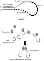


Classifications
-
- C—CHEMISTRY; METALLURGY
- C12—BIOCHEMISTRY; BEER; SPIRITS; WINE; VINEGAR; MICROBIOLOGY; ENZYMOLOGY; MUTATION OR GENETIC ENGINEERING
- C12Q—MEASURING OR TESTING PROCESSES INVOLVING ENZYMES, NUCLEIC ACIDS OR MICROORGANISMS; COMPOSITIONS OR TEST PAPERS THEREFOR; PROCESSES OF PREPARING SUCH COMPOSITIONS; CONDITION-RESPONSIVE CONTROL IN MICROBIOLOGICAL OR ENZYMOLOGICAL PROCESSES
- C12Q1/00—Measuring or testing processes involving enzymes, nucleic acids or microorganisms; Compositions therefor; Processes of preparing such compositions
- C12Q1/68—Measuring or testing processes involving enzymes, nucleic acids or microorganisms; Compositions therefor; Processes of preparing such compositions involving nucleic acids
- C12Q1/6844—Nucleic acid amplification reactions
- C12Q1/6865—Promoter-based amplification, e.g. nucleic acid sequence amplification [NASBA], self-sustained sequence replication [3SR] or transcription-based amplification system [TAS]
Definitions
- Embodiments of the present invention relate in general to methods and compositions for amplifying trace amount of DNA, such as DNA from a single cell, in order to determine its genetic sequences, particularly the entire genome.
- Single cell genome sequencing is important in studies where cell-to-cell variation and population heterogeneity play a key role, such as tumor growth, stem cell reprogramming, embryonic development, etc.
- Single cell genome sequencing is also important when the cell samples subject to sequencing are precious or rare or in minute amounts.
- Important to accurate single-cell genome sequencing is the initial amplification of the genomic DNA which can be in minute amounts.
- MDA Multiple displacement amplification
- US2013/196860 discloses a method for preparing nucleic acids for high-throughput sequencing by tagmentation using transposase and a transposon.
- Embodiments of the present disclosure are directed to a method of amplifying DNA such as a small amount of genomic DNA or a limited amount of DNA such as a genomic sequence or genomic sequences obtained from a single cell or a plurality of cells of the same cell type or from a tissue, fluid or blood sample obtained from an individual or a substrate.
- the methods described herein can be performed in a single tube with a single reaction mixture.
- the nucleic acid sample can be within an unpurified or unprocessed lysate from a single cell.
- Nucleic acids to be subjected to the methods disclosed herein need not be purified, such as by column purification, prior to being contacted with the various reagents and under the various conditions as described herein.
- the methods described herein can provide substantial and uniform coverage of the entire genome of a single cell producing amplified DNA for high-throughput sequencing.
- Embodiments of the present invention relate in general to methods and compositions for making DNA fragments, for example, DNA fragments from the whole genome of a single cell which may then be subjected to amplification methods known to those of skill in the art.
- a transposase as part of a transposome is used to create a set of double stranded genomic DNA fragments.
- Each of the double stranded genomic DNA fragments may then be amplified using methods known to those of skill in the art, such as PCR amplification, and as described herein.
- each double stranded DNA (dsDNA) fragment is then used as a template for making multiple copies of a corresponding RNA.
- the original dsDNA template is used to make each of the multiple copies of the corresponding RNA.
- the multiple copies of the RNA corresponding to the original dsDNA fragment template are then reverse transcribed to create multiple copies of single stranded DNA corresponding to the original dsDNA fragment template.
- Complementary strands are then made for the multiple copies of the ssDNA creating multiple copies of dsDNA corresponding to the original dsDNA fragment template. In this manner, the original dsDNA template fragment has been linearly amplified.
- the dsDNA template fragment is the original source template for the dsDNA amplicons corresponding to the dsDNA template fragment. Amplicons of the dsDNA template fragment are not themselves amplified to create amplicons from amplicons.
- the dsDNA amplicons of the present disclosure are linearly amplified from an original dsDNA fragment template.
- the double stranded genomic DNA fragments produced by the transposases which have the transposon DNA attached at each end of the double stranded genomic DNA fragments are then gap filled and extended using the transposon DNA as a template. Accordingly, a double stranded nucleic acid extension product is produced which includes the double stranded genomic DNA and a double stranded transposon DNA at each end of the double stranded genomic DNA.
- the double stranded nucleic acid extension products may then be amplified using methods known to those of skill in the art, such as PCR.
- methods of making nucleic acid fragments described herein utilize a transposase and an RNA polymerase.
- the transposase is complexed with a transposon DNA including a double stranded transposase binding site and a first nucleic acid sequence including one or more of a barcode sequence, a priming site and an RNA polymerase promoter sequence to form a transposase/transposon DNA complex.
- the first nucleic acid sequence may be in the form of a single stranded extension or the first nucleic acid sequence may be in the form of a loop with each end connected to a corresponding strand of the double stranded transposase binding site.
- the transposases have the capability to bind to the transposon DNA and dimerize when contacted together, such as when being placed within a reaction vessel, forming a transposase/transposon DNA complex dimer called transposome.
- the transposome have the capability to bind to target locations along double stranded nucleic acids, such as double stranded genomic DNA, forming a complex including the transposome and the double stranded genomic DNA.
- the transposases in the transposome cleave the double stranded genomic DNA, with one transposase cleaving the upper strand and one transposase cleaving the lower strand.
- the transposon DNA in the transposome is attached to the double stranded genomic DNA at the cut site.
- a plurality of transposase/transposon DNA complexe dimers bind to a corresponding plurality of target locations along a double stranded genomic DNA, for example, and then cleave the double stranded genomic DNA into a plurality of double stranded fragments with each fragment having transposon DNA attached at each end of the double stranded fragment.
- the transposon DNA is attached to the double stranded genomic DNA and a single stranded gap exists between one strand of the genomic DNA and one strand of the transposon DNA.
- gap extension is carried out to fill the gap and create a double stranded connection between the double stranded genomic DNA and the double stranded transposon DNA.
- the transposase binding site of the transposon DNA is attached at each end of the double stranded fragment.
- the transposase is attached to the transposon DNA which is attached at each end of the double stranded fragment.
- the transposases are removed from the transposon DNA which is attached at each end of the double stranded genomic DNA fragments.
- the double stranded genomic DNA fragments produced by the transposases which have the transposon DNA attached at each end of the double stranded genomic DNA fragments are then gap filled and extended using the transposon DNA as a template. Accordingly, a double stranded nucleic acid extension product is produced which includes the double stranded genomic DNA and a double stranded transposon DNA at each end of the double stranded genomic DNA.
- the double stranded transposon DNA may include an active double stranded RNA polymerase promoter sequence
- an RNA polymerase can then be used to make many RNA transcripts of the double stranded nucleic acid extension product using the double stranded nucleic acid extension product as a template for each of the many or plurality of corresponding RNA transcripts as amplicons.
- the RNA transcripts are amplicons linearly amplified from the corresponding original double stranded nucleic acid extension product template.
- the original double stranded nucleic acid extension product is the only template for the corresponding RNA transcript amplicons.
- the RNA transcripts are not exponential amplicons.
- the RNA transcripts are then reverse transcribed into single stranded DNA.
- Complementary strands to the single stranded DNA are then made forming double stranded DNA including the genomic DNA sequence and having barcodes at both ends of the upper and lower strands.
- the double stranded DNA including the genomic DNA sequence and having barcodes at both ends of the upper and lower strands are then sequenced using methods known to those of skill in the art.
- the present invention is directed to a method of amplifying DNA using the methods described herein such as a small amount of genomic DNA or a limited amount of DNA such as a genomic sequence or genomic sequences obtained from a single cell or a plurality of cells of the same cell type or from a tissue, fluid or blood sample obtained from an individual or a substrate.
- the methods described herein can be performed in a single tube with reactants added thereto under suitable conditions to create DNA amplicons linearly amplified from those genomic DNA fragments.
- the methods described herein can provide substantial coverage of the entire genome of a single cell producing amplified DNA for high-throughput sequencing.
- methods are provided herein for performing whole genome amplification of single cells with high fidelity and amplification uniformity or coverage across different loci in the genome which is useful for further sequencing or analysis using high throughput sequencing platforms known to those of skill in the art.
- Methods provided herein minimize amplification bias and provide substantially complete or complete genome coverage of DNA sequencing of genomic DNA from a single cell.
- Methods described herein can amplify greater than 90 percent of genomic DNA from a single cell while greater than 70 percent or 75 percent of the genomic DNA can be sequenced with a sequencing depth of 7x or 10x or 15x with little, substantially few or no chimera sequences.
- Methods described herein reduce or eliminate creation of sequencing artifacts and facilitate advanced genomic analysis of single cell single nucleotide polymorphisms, copy number variations and structural variations. Methods described herein have particular application in biological systems or tissue samples characterized by highly heterogeneous cell populations such as tumor and neural masses. Methods described herein to amplify genomic DNA facilitate the analysis of such amplified DNA using next generation sequencing techniques known to those of skill in the art and described herein.
- the DNA amplification methods of the present disclosure will be useful for amplifying small or limited amounts of DNA, which will allow multiple sites in the DNA sample to be genotyped for high-throughput screening. Additionally, the present method will allow for the rapid construction of band specific painting probes for any chromosomal region, and can also be used to micro dissect and amplify unidentifiable chromosomal regions or marker chromosomes in abnormal karyotypes. The presently disclosed method will also allow for the rapid cloning of amplified DNA for sequencing or generating DNA libraries. Thus, the method will not only be a valuable tool for genotype analysis and high-throughput screening, it should also be a valuable tool in cytogenetic diagnosis.
- the methods described herein can utilize varied sources of DNA materials, including genetically heterogeneous tissues (e.g. cancers), rare and precious samples (e.g. embryonic stem cells), and non-dividing cells (e.g. neurons) and the like, as well as, sequencing platforms and genotyping methods known to those of skill in the art.
- genetically heterogeneous tissues e.g. cancers
- rare and precious samples e.g. embryonic stem cells
- non-dividing cells e.g. neurons
- the present invention is based in part on the discovery of methods for making DNA fragment templates, such as from genomic DNA, using a transposase or transposome.
- the genomic DNA fragment templates may then be amplified and sequenced.
- DNA fragment templates made using the transposase methods described herein can be amplified using methods known to those of skill in the art.
- amplification is achieved using PCR.
- the term "polymerase chain reaction" (“PCR") of Mullis (U.S. Pat. Nos. 4,683,195 , 4,683,202 , and 4,965,188 ) refers to a method for increasing the concentration of a segment of a target sequence in a mixture of nucleic acid sequences without cloning or purification.
- This process for amplifying the target sequence consists of introducing a large excess of two oligonucleotide primers to the nucleic acid sequence mixture containing the desired target sequence, followed by a precise sequence of thermal cycling in the presence of a polymerase (e.g., DNA polymerase).
- the two primers are complementary to their respective strands of the double stranded target sequence.
- the mixture is denatured and the primers then annealed to their complementary sequences within the target molecule.
- the primers are extended with a polymerase so as to form a new pair of complementary strands.
- the steps of denaturation, primer annealing, and polymerase extension can be repeated many times (i.e., denaturation, annealing and extension constitute one "cycle;” there can be numerous “cycles”) to obtain a high concentration of an amplified segment of the desired target sequence.
- the length of the amplified segment of the desired target sequence is determined by the relative positions of the primers with respect to each other, and therefore, this length is a controllable parameter.
- the method is referred to as the “polymerase chain reaction” (hereinafter "PCR”). Because the desired amplified segments of the target sequence become the predominant sequences (in terms of concentration) in the mixture, they are said to be “PCR amplified.”
- PCR it is possible to amplify a single copy of a specific target sequence in genomic DNA to a level detectable by several different methodologies (e.g., hybridization with a labeled probe; incorporation of biotinylated primers followed by avidin-enzyme conjugate detection; incorporation of 32P-labeled deoxynucleotide triphosphates, such as dCTP or dATP, into the amplified segment).
- any oligonucleotide or polynucleotide sequence can be amplified with the appropriate set of primer molecules.
- the amplified segments created by the PCR process itself are, themselves, efficient templates for subsequent PCR amplifications.
- PCR is a reaction in which replicate copies are made of a target polynucleotide using a pair of primers or a set of primers consisting of an upstream and a downstream primer, and a catalyst of polymerization, such as a DNA polymerase, and typically a thermally-stable polymerase enzyme.
- Methods for PCR are well known in the art, and taught, for example in MacPherson et al. (1991) PCR 1: A Practical Approach (IRL Press at Oxford University Press ). All processes of producing replicate copies of a polynucleotide, such as PCR or gene cloning, are collectively referred to herein as replication.
- a primer can also be used as a probe in hybridization reactions, such as Southern or Northern blot analyses.
- Amplification refers to a process by which extra or multiple copies of a particular polynucleotide are formed.
- Amplification includes methods such as PCR, ligation amplification (or ligase chain reaction, LCR) and other amplification methods. These methods are known and widely practiced in the art. See, e.g., U.S. Patent Nos. 4,683,195 and 4,683,202 and Innis et al., "PCR protocols: a guide to method and applications” Academic Press, Incorporated (1990 ) (for PCR); and Wu et al. (1989) Genomics 4:560-569 (for LCR).
- the PCR procedure describes a method of gene amplification which is comprised of (i) sequence-specific hybridization of primers to specific genes within a DNA sample (or library), (ii) subsequent amplification involving multiple rounds of annealing, elongation, and denaturation using a DNA polymerase, and (iii) screening the PCR products for a band of the correct size.
- the primers used are oligonucleotides of sufficient length and appropriate sequence to provide initiation of polymerization, i.e. each primer is specifically designed to be complementary to each strand of the genomic locus to be amplified.
- Primers useful to amplify sequences from a particular gene region are preferably complementary to, and hybridize specifically to sequences in the target region or in its flanking regions and can he prepared using the polynucleotide sequences provided herein. Nucleic acid sequences generated by amplification can be sequenced directly.
- a double-stranded polynucleotide can be complementary or homologous to another polynucleotide, if hybridization can occur between one of the strands of the first polynucleotide and the second.
- Complementarity or homology is quantifiable in terms of the proportion of bases in opposing strands that are expected to form hydrogen bonding with each other, according to generally accepted base-pairing rules.
- reverse-transcriptase PCR and "RT-PCR” refer to a type of PCR where the starting material is mRNA.
- the starting mRNA is enzymatically converted to complementary DNA or "cDNA” using a reverse transcriptase enzyme.
- the cDNA is then used as a template for a PCR reaction.
- PCR product refers to the resultant mixture of compounds after two or more cycles of the PCR steps of denaturation, annealing and extension are complete. These terms encompass the case where there has been amplification of one or more segments of one or more target sequences.
- amplification reagents refers to those reagents (deoxyribonucleotide triphosphates, buffer, etc.), needed for amplification except for primers, nucleic acid template, and the amplification enzyme.
- amplification reagents along with other reaction components are placed and contained in a reaction vessel (test tube, microwell, etc.).
- Amplification methods include PCR methods known to those of skill in the art and also include rolling circle amplification ( Blanco et al., J. Biol. Chem., 264, 8935-8940, 1989 ), hyperbranched rolling circle amplification ( Lizard et al., Nat. Genetics, 19, 225-232, 1998 ), and loop-mediated isothermal amplification ( Notomi et al., Nuc. Acids Res., 28, e63, 2000 ).
- an emulsion PCR reaction is created by vigorously shaking or stirring a "water in oil" mix to generate millions of micron-sized aqueous compartments.
- the DNA library is mixed in a limiting dilution either with the beads prior to emulsification or directly into the emulsion mix.
- the combination of compartment size and limiting dilution of beads and target molecules is used to generate compartments containing, on average, just one DNA molecule and bead (at the optimal dilution many compartments will have beads without any target)
- an upstream (low concentration, matches primer sequence on bead) and downstream PCR primers (high concentration) are included in the reaction mix.
- each little compartment in the emulsion forms a micro PCR reactor.
- the average size of a compartment in an emulsion ranges from submicron in diameter to over a 100 microns, depending on the emulsification conditions.
- nucleic acid amplification procedures specifically contemplated in the context of the present disclosure include transcription-based amplification systems (rAS), including nucleic acid sequence based amplification (NASBA) and 3SR, Kwoh et al., Proc Natl Acad Sci USA, 86:1173-77, 1989 ; PCT Patent Application WO 88/10315 et al., 1989.
- rAS transcription-based amplification systems
- NASBA nucleic acid sequence based amplification
- 3SR Kwoh et al., Proc Natl Acad Sci USA, 86:1173-77, 1989
- PCT Patent Application WO 88/10315 et al., 1989 the nucleic acids can be prepared for amplification by standard phenol/chloroform extraction, heat denaturation of a clinical sample, treatment with lysis buffer, and minispin columns for isolation of DNA and RNA or guanidinium chloride extraction of RNA.
- amplification techniques involve anne
- DNA/RNA hybrids are digested with RNase H while double stranded DNA molecules are heat denatured again. In either case the single stranded DNA is made fully double stranded by addition of second target specific primer, followed by polymerization.
- the double-stranded DNA molecules are then multiply transcribed by a polymerase such as T7 or SP6.
- a polymerase such as T7 or SP6.
- the RNAs are reverse transcribed into double stranded DNA, and transcribed once again with a polymerase such as T7 or SP6.
- the resulting products whether truncated or complete, indicate target specific sequences.
- modified primers are used in a PCR-like template and enzyme dependent synthesis.
- the primers may be modified by labeling with a capture moiety (e.g., biotin) and/or a detector moiety (e.g., enzyme).
- a capture moiety e.g., biotin
- a detector moiety e.g., enzyme
- an excess of labeled probes are added to a sample.
- the probe binds and is cleaved catalytically. After cleavage, the target sequence is released intact to be bound by excess probe. Cleavage of the labeled probe signals the presence of the target sequence.
- ssRNA single-stranded RNA
- dsDNA double-stranded DNA
- the ssRNA is a first template for a first primer oligonucleotide, which is elongated by reverse transcriptase (RNA-dependent DNA polymerase).
- RNA-dependent DNA polymerase reverse transcriptase
- the RNA is then removed from the resulting DNA:RNA duplex by the action of ribonuclease H (RNase H, an RNase specific for RNA in duplex with either DNA or RNA).
- RNase H ribonuclease H
- the resultant ssDNA is a second template for a second primer, which also includes the sequences of an RNA polymerase promoter (exemplified by T7 RNA polymerase) 5' of its homology to the template.
- This primer is then extended by DNA polymerase (exemplified by the large "Klenow" fragment of E.coli DNA polymerase I), resulting in a double-stranded DNA molecule, having a sequence identical to that of the original RNA between the primers and having additionally, at one end, a promoter sequence.
- This promoter sequence can be used by the appropriate RNA polymerase to make many RNA copies of the DNA. These copies can then reenter the cycle leading to very swift amplification. With proper choice of enzymes, this amplification can be done isothermally without adding enzymes at each cycle. Because of the cyclical nature of this process, the starting nucleic acid sequence can be either DNA or RNA.
- Miller et al., PCT Patent Application WO 89/06700 disclose a nucleic acid sequence amplification scheme based on the hybridization of a promoter/primer sequence to a target single-stranded DNA followed by transcription of many RNA copies of the sequence. This scheme is not cyclic, i.e., new templates are not produced from the resultant RNA transcripts.
- Suitable amplification methods include “race and "one-sided PCR.”. ( Frohman, In: PCR Protocols: A Guide To Methods And Applications, Academic Press, N.Y., 1990 ). Methods based on ligation of two (or more) oligonucleotides in the presence of nucleic acid having the sequence of the resulting "di-oligonucleotide,” thereby amplifying the di-oligonucleotide, also may be used to amplify DNA in accordance with the present disclosure ( Wu et al., Genomics 4:560-569, 1989 ).
- the DNA fragment templates include an RNA polymerase promoter sequence and an RNA polymerase is used to make RNA amplicons from the DNA fragment templates and a reverse transcriptase to make single stranded DNA from the RNA amplicons. Complements are then made to the single stranded DNA to make double stranded DNA resulting in amplicons linearly amplified from the original DNA fragment template.
- an exemplary transposon system is a Tn5 transposon system.
- Other useful transposon systems are known to those of skill in the art and include Tn3 transposon system (see Maekawa, T., Yanagihara, K., and Ohtsubo, E. (1996), A cell-free system of Tn3 transposition and transposition immunity, Genes Cells 1, 1007-1016 ), Tn7 transposon system (see Craig, N.L. (1991), Tn7: a target site-specific transposon, Mol. Microbiol. 5, 2569-2573 ), Tn10 tranposon system (see Chalmers, R., Sewitz, S., Lipkow, K., and Crellin, P.
- an exemplary RNA polymerase is T7 RNA polymerase.
- Other useful RNA polymerases are known to those of skill in the art and include T3 RNA polymerase (see Jorgensen, E.D., Durbin, R.K., Risman, S.S., and McAllister, W.T. (1991) Specific contacts between the bacteriophage T3, T7, and SP6 RNA polymerases and their promoters, J. Biol. Chem. 266, 645-651 ), and SP6 RNA polymerase (see Melton, D.A., Krieg, P.A., Rebagliati, M.R., Maniatis, T., Zinn, K., and Green, M.R. (1984) Efficient in vitro synthesis of biologically active RNA and RNA hybridization probes from plasmids containing a bacteriophage SP6 promoter, Nucleic Acids Res. 12, 7035-7056 .)
- DNA to be amplified may be obtained from a single cell or a small population of cells. Methods described herein allow DNA to be amplified from any species or organism in a reaction mixture, such as a single reaction mixture carried out in a single reaction vessel. In one aspect, methods described herein include sequence independent amplification of DNA from any source including but not limited to human, animal, plant, yeast, viral, eukaryotic and prokaryotic DNA.
- embodiments are directed to methods for the amplification of substantially the entire genome without loss of representation of specific sites (herein defined as "whole genome amplification").
- whole genome amplification comprises simultaneous amplification of substantially all fragments of a genomic library.
- substantially entire or substantially all refers to about 80%, about 85%, about 90%, about 95%, about 97%, or about 99% of all sequence in a genome.
- amplification of the whole genome will, in some embodiments, comprise non-equivalent amplification of particular sequences over others.
- the DNA sample is genomic DNA, micro dissected chromosome DNA, yeast artificial chromosome (YAC) DNA, cosmid DNA, phage DNA, P1 derived artificial chromosome (PAC) DNA, or bacterial artificial chromosome (BAC) DNA.
- the DNA sample is mammalian DNA, plant DNA, yeast DNA, viral DNA, or prokaryotic DNA.
- the DNA sample is obtained from a human, bovine, porcine, ovine, equine, rodent, avian, fish, shrimp, plant, yeast, virus, or bacteria.
- the DNA sample is genomic DNA.
- a transposition system is used to make nucleic acid fragments for amplification and sequencing as desired.
- a transposition system is combined with an RNA polymerase for single cell genome amplification.
- a transposition system is used to fragment genomic DNA into double stranded genomic DNA fragments.
- An RNA polymerase is used to make RNA amplicons which are then reverse transcribed into DNA.
- Complements to the DNA are made and double stranded genomic DNA sequences are formed which are amplicons linearly amplified from the original double stranded genomic DNA fragments.
- the use of an RNA polymerase to make amplicons in a linear manner advantageously achieves high quality amplification of the single-cell genomic DNA (gDNA) reducing or avoiding (1) amplification bias, leading to the noisy single-cell sequencing data that further affect the genome coverage, as well as the low resolution detection of copy number variations (CNVs); (2) amplification errors, causing a high false positive rate in the single-cell sequencing data that further prevents the accurate detection of single nucleotide variations (SNVs); and (3) chimera formation during amplification, which overwhelms the signal of structural variations (SVs) from the original single-cell genomic DNA sample.
- gDNA single-cell genomic DNA
- CNVs copy number variations
- SNVs single nucleotide variations
- chimera formation during amplification which overwhelms the signal of structural variations (SVs) from the original single-cell genomic DNA sample.
- SVs structural variations
- PCR by definition is an exponential amplification method, i.e. new copies are made based on the copies from the previous rounds of amplification.
- the slight amplification efficiency difference between amplicons may accumulate, leading to amplification bias between different amplicons after many cycles.
- errors made during the early rounds of PCR may propagate further into more copies during the later PCR rounds, which may reduce the overall amplification accuracy after many cycles of amplification.
- a DNA column purification step is not carried out so as to maximize the small amount ( ⁇ 6 pg) of genomic DNA that can be obtained from within a single cell prior to amplification.
- the DNA can be amplified directly from a cell lysate or other impure condition. Accordingly, the DNA sample may be impure, unpurified, not isolated. Accordingly, aspects of the present method allow one to maximize genomic DNA for amplification and reduce loss due to purification.
- methods described herein may utilize amplification methods other than PCR, such as an RNA polymerase based amplification method.
- RNA polymerase-based amplification method as described herein can advantageously reduce the amplification bias and improve the amplification accuracy.
- a Tn5 transposon DNA is designed to contain a strong T7 promoter sequence. After the transposition reaction, single-cell genomic DNA will be fragmented with each fragment tagged by a strong T7 promoter on both ends, enabling linear amplification afterwards through in vitro transcription.
- transposon DNA is designed to contain a double-stranded 19 bp Tn5 transposase (Tnp) binding site at one end, linked or connected, such as by covalent bond, to a long single-stranded overhang including a barcode region, a priming site, and a strong T7 promoter sequence as shown in Fig. 1 .
- the strong T7 promoter sequence is at the end of the overhang.
- transposomes are numbered 1-3. Then, the transposases in the transposome cut the genomic DNA with one transposase cutting an upper strand and one transposase cutting a lower strand to create a genomic DNA fragment. The transposon DNA is thus inserted randomly into the single-cell genomic DNA, leaving a 9-bp gap on both ends of the transposition/insertion site. The result is a genomic DNA fragment with a transposon DNA Tnp binding site attached to the 5' position of an upper strand and a transposon DNA Tnp binding site attached to the 5' position of a lower strand. Gaps resulting from the insertion of the transposon DNA are shown.
- gap extension is performed to fill the 9 bp gap and complement the single-stranded overhang originally designed in the transposon DNA.
- active double-stranded T7 promoter sequences are attached to both ends of each genomic DNA fragment as shown in Fig. 3 .
- T7 RNA polymerase is added, and in vitro transcription is used to linearly amplify the single-cell genomic DNA fragments, generating many RNAs that contain the same sequence as the original genomic double stranded DNA template.
- the amplified RNAs are converted back into double-stranded DNA molecules, with the barcode region originally designed in the transposon DNA attached on both ends of each fragment.
- the DNA fragments may then be further processed for standard library preparation and sequencing.
- Tn5 transposition systems are described and are available to those of skill in the art. See Goryshin, I.Y. and W.S. Reznikoff, Tn5 in vitro transposition. The Journal of biological chemistry, 1998. 273(13): p. 7367-74 ; Davies, D.R., et al., Three-dimensional structure of the Tn5 synaptic complex transposition intermediate. Science, 2000. 289(5476): p. 77-85 ; Goryshin, I.Y., et al., Insertional transposon mutagenesis by electroporation of released Tn5 transposition complexes. Nature biotechnology, 2000. 18(1): p. 97-100 and Stier-White, M., I.
- Kits utilizing a Tn5 transposition system for DNA library preparation and other uses are known. See Adey, A., et al., Rapid, low-input, low-bias construction of shotgun fragment libraries by high-density in vitro transposition. Genome biology, 2010. 11(12): p. R119 ; Marine, R., et al., Evaluation of a transposase protocol for rapid generation of shotgun high-throughput sequencing libraries from nanogram quantities of DNA. Applied and environmental microbiology, 2011. 77(22): p.
- In vitro transcription (IVT) by T7 RNA polymerase is useful to create RNA amplicons.
- IVT In vitro transcription
- IVT advantageously provides linear amplification, with all the copies generated from the original DNA template.
- RNA molecules can be reverse transcribed into single stranded DNA followed by complementary strand formation resulting in double stranded DNA, which are amplicons linearly amplified from the original DNA template.
- amplification bias is much smaller between different amplicons.
- amplification accuracy is higher since in linear amplification the amplification errors cannot propagate into later stages.
- an RNA promoter sequence is attached to target DNA to advantageously use an RNA polymerase, such as T7 RNA polymerase, to create RNA amplicons which are then used to create DNA amplicons of the original DNA template.
- the method of amplifying DNA further includes genotype analysis of the amplified DNA product.
- the method of amplifying DNA preferably further includes identifying a polymorphism such as a single nucleotide polymorphism (SNP) in the amplified DNA product.
- SNP single nucleotide polymorphism
- a SNP may be identified in the DNA of an organism by a number of methods well known to those of skill in the art, including but not limited to identifying the SNP by DNA sequencing, by amplifying a PCR product and sequencing the PCR product, by Oligonucleotide Ligation Assay (OLA), by Doublecode OLA, by Single Base Extension Assay, by allele specific primer extension, or by mismatch hybridization.
- OLA Oligonucleotide Ligation Assay
- the identified SNP is associated with a phenotype, including disease phenotypes and desirable phenotypic traits.
- the amplified DNA generated by using the disclosed method of DNA amplification may also preferably be used to generate a DNA library, including but not limited to genomic DNA libraries, microdissected chromosome DNA libraries, BAC libraries, YAC libraries, PAC libraries, cDNA libraries, phage libraries, and cosmid libraries.
- genomic as used herein is defined as the collective gene set carried by an individual, cell, or organelle.
- genomic DNA as used herein is defined as DNA material comprising the partial or full collective gene set carried by an individual, cell, or organelle.
- nucleoside refers to a molecule having a purine or pyrimidine base covalently linked to a ribose or deoxyribose sugar.
- exemplary nucleosides include adenosine, guanosine, cytidine, uridine and thymidine.
- Additional exemplary nucleosides include inosine, 1-methyl inosine, pseudouridine, 5,6-dihydrouridine, ribothymidine, 2N-methylguanosine and 2,2N,N-dimethylguanosine (also referred to as "rare" nucleosides).
- nucleotide refers to a nucleoside having one or more phosphate groups joined in ester linkages to the sugar moiety.
- exemplary nucleotides include nucleoside monophosphates, diphosphates and triphosphates.
- polynucleotide oligonucleotide
- nucleic acid molecule are used interchangeably herein and refer to a polymer of nucleotides, either deoxyribonucleotides or ribonucleotides, of any length joined together by a phosphodiester linkage between 5' and 3' carbon atoms.
- Polynucleotides can have any three-dimensional structure and can perform any function, known or unknown.
- polynucleotides a gene or gene fragment (for example, a probe, primer, EST or SAGE tag), exons, introns, messenger RNA (mRNA), transfer RNA, ribosomal RNA, ribozymes, cDNA, recombinant polynucleotides, branched polynucleotides, plasmids, vectors, isolated DNA of any sequence, isolated RNA of any sequence, nucleic acid probes and primers.
- a polynucleotide can comprise modified nucleotides, such as methylated nucleotides and nucleotide analogs. The term also refers to both double- and single-stranded molecules.
- any embodiment of this invention that comprises a polynucleotide encompasses both the double-stranded form and each of two complementary single-stranded forms known or predicted to make up the double-stranded form.
- a polynucleotide is composed of a specific sequence of four nucleotide bases: adenine (A); cytosine (C); guanine (G); thymine (T); and uracil (U) for thymine when the polynucleotide is RNA.
- A adenine
- C cytosine
- G guanine
- T thymine
- U uracil
- polynucleotide sequence is the alphabetical representation of a polynucleotide molecule. This alphabetical representation can be input into databases in a computer having a central processing unit and used for bioinformatics applications such as functional genomics and homology searching.
- RNA RNA molecule
- ribonucleic acid molecule refers to a polymer of ribonucleotides.
- DNA DNA molecule
- deoxyribonucleic acid molecule refer to a polymer of deoxyribonucleotides.
- DNA and RNA can be synthesized naturally (e.g., by DNA replication or transcription of DNA, respectively). RNA can be post-transcriptionally modified. DNA and RNA can also be chemically synthesized. DNA and RNA can be single-stranded (i.e., ssRNA and ssDNA, respectively) or multi-stranded (e.g., double stranded, i.e., dsRNA and dsDNA, respectively).
- nucleotide analog refers to a non-standard nucleotide, including non-naturally occurring ribonucleotides or deoxyribonucleotides.
- nucleotide analogs are modified at any position so as to alter certain chemical properties of the nucleotide yet retain the ability of the nucleotide analog to perform its intended function.
- positions of the nucleotide which may be derivitized include the 5 position, e.g., 5-(2-amino)propyl uridine, 5-bromo uridine, 5-propyne uridine, 5-propenyl uridine, etc.; the 6 position, e.g., 6-(2-amino) propyl uridine; the 8-position for adenosine and/or guanosines, e.g., 8-bromo guanosine, 8-chloro guanosine, 8-fluoroguanosine, etc.
- 5 position e.g., 5-(2-amino)propyl uridine, 5-bromo uridine, 5-propyne uridine, 5-propenyl uridine, etc.
- the 6 position e.g., 6-(2-amino) propyl uridine
- the 8-position for adenosine and/or guanosines e.g
- Nucleotide analogs also include deaza nucleotides, e.g., 7-deaza-adenosine; O- and N-modified (e.g., alkylated, e.g., N6-methyl adenosine, or as otherwise known in the art) nucleotides; and other heterocyclically modified nucleotide analogs such as those described in Herdewijn, Antisense Nucleic Acid Drug Dev., 2000 Aug. 10(4):297-310 .
- Nucleotide analogs may also comprise modifications to the sugar portion of the nucleotides.
- the 2' OH-group may be replaced by a group selected from H, OR, R, F, Cl, Br, I, SH, SR, NH 2 , NHR, NR 2 , COOR, or OR, wherein R is substituted or unsubstituted C 1 -C 6 alkyl, alkenyl, alkynyl, aryl, etc.Other possible modifications include those described in U.S. Pat. Nos. 5,858,988 , and 6,291,438 .
- the phosphate group of the nucleotide may also be modified, e.g., by substituting one or more of the oxygens of the phosphate group with sulfur (e.g., phosphorothioates), or by making other substitutions which allow the nucleotide to perform its intended function such as described in, for example, Eckstein, Antisense Nucleic Acid Drug Dev. 2000 Apr. 10(2):117-21 , Rusckowski et al. Antisense Nucleic Acid Drug Dev. 2000 Oct. 10(5):333-45 , Stein, Antisense Nucleic Acid Drug Dev. 2001 Oct. 11(5): 317-25 , Vorobjev et al. Antisense Nucleic Acid Drug Dev. 2001 Apr.
- in vitro has its art recognized meaning, e.g., involving purified reagents or extracts, e.g., cell extracts.
- in vivo also has its art recognized meaning, e.g., involving living cells, e.g., immortalized cells, primary cells, cell lines, and/or cells in an organism.
- the terms “complementary” and “complementarity” are used in reference to nucleotide sequences related by the base-pairing rules.
- sequence 5'-AGT-3' is complementary to the sequence 5'-ACT-3'.
- Complementarity can be partial or total. Partial complementarity occurs when one or more nucleic acid bases is not matched according to the base pairing rules. Total or complete complementarity between nucleic acids occurs when each and every nucleic acid base is matched with another base under the base pairing rules. The degree of complementarity between nucleic acid strands has significant effects on the efficiency and strength of hybridization between nucleic acid strands.
- hybridization refers to the pairing of complementary nucleic acids. Hybridization and the strength of hybridization (i.e., the strength of the association between the nucleic acids) is impacted by such factors as the degree of complementary between the nucleic acids, stringency of the conditions involved, the T m of the formed hybrid, and the G:C ratio within the nucleic acids. A single molecule that contains pairing of complementary nucleic acids within its structure is said to be “self-hybridized.”
- T m refers to the melting temperature of a nucleic acid.
- the melting temperature is the temperature at which a population of double-stranded nucleic acid molecules becomes half dissociated into single strands.
- stringency refers to the conditions of temperature, ionic strength, and the presence of other compounds such as organic solvents, under which nucleic acid hybridizations are conducted.
- Low stringency conditions when used in reference to nucleic acid hybridization, comprise conditions equivalent to binding or hybridization at 42 °C in a solution consisting of 5x SSPE (43.8 g/l NaCl, 6.9 g/l NaH 2 PO 4 (H 2 O) and 1.85 g/l EDTA, pH adjusted to 7.4 with NaOH), 0.1% SDS, 5x Denhardt's reagent (50x Denhardt's contains per 500 ml: 5 g Ficoll (Type 400, Pharmacia), 5 g BSA (Fraction V; Sigma)) and 100 ⁇ g/ml denatured salmon sperm DNA followed by washing in a solution comprising 5x SSPE, 0.1% SDS at 42 °C when a probe of about 500 nucleotides in length is employed.
- 5x SSPE 43.8 g/l NaCl, 6.9 g/l NaH 2 PO 4 (H 2 O) and 1.85 g/l EDTA, pH adjusted to 7.4 with NaOH
- “Medium stringency conditions,” when used in reference to nucleic acid hybridization, comprise conditions equivalent to binding or hybridization at 42 °C in a solution consisting of 5x SSPE (43.8 g/l NaCl, 6.9 g/l NaH 2 PO 4 (H 2 O) and 1.85 g/l EDTA, pH adjusted to 7.4 with NaOH), 0.5% SDS, 5x Denhardt's reagent and 100 ⁇ g/ml denatured salmon sperm DNA followed by washing in a solution comprising 1.0x SSPE, 1.0% SDS at 42 °C when a probe of about 500 nucleotides in length is employed.
- High stringency conditions when used in reference to nucleic acid hybridization, comprise conditions equivalent to binding or hybridization at 42 °C in a solution consisting of 5x SSPE (43.8 g/l NaCl, 6.9 g/l NaH 2 PO 4 (H 2 O) and 1.85 g/l EDTA, pH adjusted to 7.4 with NaOH), 0.5% SDS, 5x Denhardt's reagent and 100 ⁇ g/ml denatured salmon sperm DNA followed by washing in a solution comprising 0.1x SSPE, 1.0% SDS at 42 °C when a probe of about 500 nucleotides in length is employed.
- cells are identified and then a single cell or a plurality of cells are isolated.
- Cells within the scope of the present disclosure include any type of cell where understanding the DNA content is considered by those of skill in the art to be useful.
- a cell according to the present disclosure includes a cancer cell of any type, hepatocyte, oocyte, embryo, stem cell, iPS cell, ES cell, neuron, erythrocyte, melanocyte, astrocyte, germ cell, oligodendrocyte, kidney cell and the like.
- the methods of the present invention are practiced with the cellular DNA from a single cell.
- a plurality of cells includes from about 2 to about 1,000,000 cells, about 2 to about 10 cells, about 2 to about 100 cells, about 2 to about 1,000 cells, about 2 to about 10,000 cells, about 2 to about 100,000 cells, about 2 to about 10 cells or about 2 to about 5 cells.
- Nucleic acids processed by methods described herein may be DNA and they may be obtained from any useful source, such as, for example, a human sample.
- a double stranded DNA molecule is further defined as comprising a genome, such as, for example, one obtained from a sample from a human.
- the sample may be any sample from a human, such as blood, serum, plasma, cerebrospinal fluid, cheek scrapings, nipple aspirate, biopsy, semen (which may be referred to as ejaculate), urine, feces, hair follicle, saliva, sweat, immunoprecipitated or physically isolated chromatin, and so forth.
- the sample comprises a single cell.
- the amplified nucleic acid molecule from the sample provides diagnostic or prognostic information.
- the prepared nucleic acid molecule from the sample may provide genomic copy number and/or sequence information, allelic variation information, cancer diagnosis, prenatal diagnosis, paternity information, disease diagnosis, detection, monitoring, and/or treatment information, sequence information, and so forth.
- a "single cell” refers to one cell.
- Single cells useful in the methods described herein can be obtained from a tissue of interest, or from a biopsy, blood sample, or cell culture. Additionally, cells from specific organs, tissues, tumors, neoplasms, or the like can be obtained and used in the methods described herein. Furthermore, in general, cells from any population can be used in the methods, such as a population of prokaryotic or eukaryotic single celled organisms including bacteria or yeast.
- a single cell suspension can be obtained using standard methods known in the art including, for example, enzymatically using trypsin or papain to digest proteins connecting cells in tissue samples or releasing adherent cells in culture, or mechanically separating cells in a sample.
- Single cells can be placed in any suitable reaction vessel in which single cells can be treated individually. For example a 96-well plate, such that each single cell is placed in a single well.
- FACS fluorescence activated cell sorting
- flow cytometry cytometry
- micromanipulation e.g. the QuixellTM cell transfer system from Stoelting Co.
- Semi-automated cell pickers e.g. the QuixellTM cell transfer system from Stoelting Co.
- Individual cells can, for example, be individually selected based on features detectable by microscopic observation, such as location, morphology, or reporter gene expression.
- a combination of gradient centrifugation and flow cytometry can also be used to increase isolation or sorting efficiency.
- the cell is lysed to release cellular contents including DNA, using methods known to those of skill in the art.
- the cellular contents are contained within a vessel.
- cellular contents such as genomic DNA
- Lysis can be achieved by, for example, heating the cells, or by the use of detergents or other chemical methods, or by a combination of these.
- any suitable lysis method known in the art can be used. For example, heating the cells at 72°C for 2 minutes in the presence of Tween-20 is sufficient to lyse the cells.
- cells can be heated to 65°C for 10 minutes in water ( Esumi et al., Neurosci Res 60(4):439-51 (2008 )); or 70°C for 90 seconds in PCR buffer II (Applied Biosystems) supplemented with 0.5% NP-40 ( Kurimoto et al., Nucleic Acids Res 34(5):e42 (2006 )); or lysis can be achieved with a protease such as Proteinase K or by the use of chaotropic salts such as guanidine isothiocyanate ( U.S. Publication No. 2007/0281313 ).
- a protease such as Proteinase K
- chaotropic salts such as guanidine isothiocyanate
- Amplification of genomic DNA according to methods described herein can be performed directly on cell lysates, such that a reaction mix can be added to the cell lysates.
- the cell lysate can be separated into two or more volumes such as into two or more containers, tubes or regions using methods known to those of skill in the art with a portion of the cell lysate contained in each volume container, tube or region.
- Genomic DNA contained in each container, tube or region may then be amplified by methods described herein or methods known to those of skill in the art.
- a nucleic acid used in the invention can also include native or non-native bases.
- a native deoxyribonucleic acid can have one or more bases selected from the group consisting of adenine, thymine, cytosine or guanine and a ribonucleic acid can have one or more bases selected from the group consisting of uracil, adenine, cytosine or guanine.
- Exemplary non-native bases that can be included in a nucleic acid, whether having a native backbone or analog structure include, without limitation, inosine, xathanine, hypoxathanine, isocytosine, isoguanine, 5-methylcytosine, 5-hydroxymethyl cytosine, 2-aminoadenine, 6-methyl adenine, 6-methyl guanine, 2-propyl guanine, 2-propyl adenine, 2-thioLiracil, 2-thiothymine, 2- thiocytosine, 15 -halouracil, 15 -halocytosine, 5-propynyl uracil, 5-propynyl cytosine, 6-azo uracil, 6-azo cytosine, 6-azo thymine, 5-uracil, 4-thiouracil, 8-halo adenine or guanine, 8- amino adenine or guanine, 8-thiol adenine or guanine, 8
- the term "primer” generally includes an oligonucleotide, either natural or synthetic, that is capable, upon forming a duplex with a polynucleotide template, of acting as a point of initiation of nucleic acid synthesis, such as a sequencing primer, and being extended from its 3' end along the template so that an extended duplex is formed.
- the sequence of nucleotides added during the extension process is determined by the sequence of the template polynucleotide.
- primers are extended by a DNA polymerase. Primers usually have a length in the range of between 3 to 36 nucleotides, also 5 to 24 nucleotides, also from 14 to 36 nucleotides.
- Primers within the scope of the invention include orthogonal primers, amplification primers, constructions primers and the like. Pairs of primers can flank a sequence of interest or a set of sequences of interest. Primers and probes can be degenerate or quasi-degenerate in sequence. Primers within the scope of the present invention bind adjacent to a target sequence.
- a "primer” may be considered a short polynucleotide, generally with a free 3' -OH group that binds to a target or template potentially present in a sample of interest by hybridizing with the target, and thereafter promoting polymerization of a polynucleotide complementary to the target.
- Primers of the instant invention are comprised of nucleotides ranging from 17 to 30 nucleotides.
- the primer is at least 17 nucleotides, or alternatively, at least 18 nucleotides, or alternatively, at least 19 nucleotides, or alternatively, at least 20 nucleotides, or alternatively, at least 21 nucleotides, or alternatively, at least 22 nucleotides, or alternatively, at least 23 nucleotides, or alternatively, at least 24 nucleotides, or alternatively, at least 25 nucleotides, or alternatively, at least 26 nucleotides, or alternatively, at least 27 nucleotides, or alternatively, at least 28 nucleotides, or alternatively, at least 29 nucleotides, or alternatively, at least 30 nucleotides, or alternatively at least 50 nucleotides, or alternatively at least 75 nucleotides or alternatively at least 100 nucleotides.
- amplification or “amplifying” refers to a process by which extra or multiple copies of a particular polynucleotide are formed.
- reverse-transcriptase PCR and "RT-PCR” refer to a type of PCR where the starting material is mRNA.
- the starting mRNA is enzymatically converted to complementary DNA or "cDNA” using a reverse transcriptase enzyme.
- the DNA amplified according to the methods described herein may be sequenced and analyzed using methods known to those of skill in the art. Determination of the sequence of a nucleic acid sequence of interest can be performed using a variety of sequencing methods known in the art including, but not limited to, sequencing by hybridization (SBH), sequencing by ligation (SBL) ( Shendure et al. (2005) Science 309:1728 ), quantitative incremental fluorescent nucleotide addition sequencing (QIFNAS), stepwise ligation and cleavage, fluorescence resonance energy transfer (FRET), molecular beacons, TaqMan reporter probe digestion, pyrosequencing, fluorescent in situ sequencing (FISSEQ), FISSEQ beads ( U.S. Pat. No.
- SBH sequencing by hybridization
- SBL sequencing by ligation
- QIFNAS quantitative incremental fluorescent nucleotide addition sequencing
- FRET fluorescence resonance energy transfer
- molecular beacons TaqMan reporter probe digestion, pyrosequencing, fluorescent in situ sequencing (FISSEQ), F
- allele-specific oligo ligation assays e.g., oligo ligation assay (OLA), single template molecule OLA using a ligated linear probe and a rolling circle amplification (RCA) readout, ligated padlock probes, and/or single template molecule OLA using a ligated circular padlock probe and a rolling circle amplification (RCA) readout
- OLA oligo ligation assay
- RCA rolling circle amplification
- ligated padlock probes ligated padlock probes
- RCA rolling circle amplification
- Polonator platforms can also be utilized.
- a variety of light-based sequencing technologies are known in the art ( Landegren et al. (1998) Genome Res. 8:769-76 ; Kwok (2000) Pharmacogenomics 1:95-100 ; and Shi (2001) Clin. Chem. 47:164-172 ).
- the amplified DNA can be sequenced by any suitable method.
- the amplified DNA can be sequenced using a high-throughput screening method, such as Applied Biosystems' SOLiD sequencing technology, or Illumina's Genome Analyzer.
- the amplified DNA can be shotgun sequenced.
- the number of reads can be at least 10,000, at least 1 million, at least 10 million, at least 100 million, or at least 1000 million.
- the number of reads can be from 10,000 to 100,000, or alternatively from 100,000 to 1 million, or alternatively from 1 million to 10 million, or alternatively from 10 million to 100 million, or alternatively from 100 million to 1000 million.
- a "read” is a length of continuous nucleic acid sequence obtained by a sequencing reaction.
- “Shotgun sequencing” refers to a method used to sequence very large amount of DNA (such as the entire genome).
- the DNA to be sequenced is first shredded into smaller fragments which can be sequenced individually.
- the sequences of these fragments are then reassembled into their original order based on their overlapping sequences, thus yielding a complete sequence.
- “Shredding" of the DNA can be done using a number of difference techniques including restriction enzyme digestion or mechanical shearing. Overlapping sequences are typically aligned by a computer suitably programmed. Methods and programs for shotgun sequencing a cDNA library are well known in the art.
- one aspect of the present invention relates to diagnostic assays for determining the genomic DNA in order to determine whether an individual is at risk of developing a disorder and/or disease. Such assays can be used for prognostic or predictive purposes to thereby prophylactically treat an individual prior to the onset of the disorder and/or disease. Accordingly, in certain exemplary embodiments, methods of diagnosing and/or prognosing one or more diseases and/or disorders using one or more of expression profiling methods described herein are provided.
- biological sample is intended to include, but is not limited to, tissues, cells, biological fluids and isolates thereof, isolated from a subject, as well as tissues, cells and fluids present within a subject.
- electronic apparatus readable media comprising one or more genomic DNA sequences described herein.
- electronic apparatus readable media refers to any suitable medium for storing, holding or containing data or information that can be read and accessed directly by an electronic apparatus.
- Such media can include, but are not limited to: magnetic storage media, such as floppy discs, hard disc storage medium, and magnetic tape; optical storage media such as compact disc; electronic storage media such as RAM, ROM, EPROM, EEPROM and the like; general hard disks and hybrids of these categories such as magnetic/optical storage media.
- the medium is adapted or configured for having recorded thereon one or more expression profiles described herein.
- the term "electronic apparatus” is intended to include any suitable computing or processing apparatus or other device configured or adapted for storing data or information.
- Examples of electronic apparatuses suitable for use with the present invention include stand-alone computing apparatus; networks, including a local area network (LAN), a wide area network (WAN) Internet, Intranet, and Extranet; electronic appliances such as a personal digital assistants (PDAs), cellular phone, pager and the like; and local and distributed processing systems.
- recorded refers to a process for storing or encoding information on the electronic apparatus readable medium.
- Those skilled in the art can readily adopt any of the presently known methods for recording information on known media to generate manufactures comprising one or more expression profiles described herein.
- a variety of software programs and formats can be used to store the genomic DNA information of the present invention on the electronic apparatus readable medium.
- the nucleic acid sequence can be represented in a word processing text file, formatted in commercially-available software such as WordPerfect and MicroSoft Word, or represented in the form of an ASCII file, stored in a database application, such as DB2, Sybase, Oracle, or the like, as well as in other forms.
- Any number of data processor structuring formats e.g., text file or database
- Tn5 transposase (Epicentre) is mixed with transposon DNA in equal molar number in a buffer containing EDTA and incubated at room temperature for 10 - 60 minutes. The final transposome concentration is 0.1 - 10 ⁇ M.
- the transposon DNA construct could be linear or looped form, with a double stranded 19 bp transposase binding site on one end, and single stranded T7 promoter on the other end. In the linear form, the single-stranded T7 promoter sequence forms a 5' protruding end; while in the looped form, the single-stranded T7 promoter sequence forms a loop and connects both strands of the 19 bp binding site.
- Barcode sequences with variable length and sequence complexity could be designed as needed between the 19 bp binding site and the T7 promoter sequence, together with a specific priming region between the barcodes and the T7 promoter.
- the transposome may be diluted by many folds in 50% Tris-EDTA and 50% glycerol solution and preserved at -20°C.
- a cell is selected, cut from a culture dish, and dispensed in a tube using a laser dissection microscope (LMD-6500, Leica) as follows.
- LMD-6500 laser dissection microscope
- the cells are plated onto a membrane-coated culture dish and observed using bright field microscopy with a 10x objective (Leica).
- a UV laser is then used to cut the membrane around an individually selected cell such that it falls into the cap of a PCR tube.
- the tube is briefly centrifuged to bring the cell down to the bottom of the tube.
- 3 - 5 ⁇ l lysis buffer (30mM Tris-Cl PH 7.8, 2mM EDTA, 20mM KCl, 0.2% Triton X-100, 500 ⁇ g/ml Qiagen Protease) is added to the side of the PCR tube and span down.
- the captured cell is then thermally lysed using the using following temperature schedule on PCR machine: 50°C 3 hours, 75°C 30 minutes.
- mouth pipette a single cell into a low salt lysis buffer containing EDTA and protease such as QIAGEN protease (QIAGEN) at a concentration of 10 - 5000 ⁇ g/mL.
- QIAGEN QIAGEN protease
- the incubation would be 37-55°C for 1 - 4 hrs.
- the protease is then heat inactivated up to 80°C and further inactivated by specific protease inhibitors such as 4-(2-Aminoethyl) benzenesulfonyl fluoride hydrochloride (AEBSF) or phenylmethanesulfonyl fluoride (PMSF) (Sigma Aldrich).
- AEBSF 4-(2-Aminoethyl) benzenesulfonyl fluoride hydrochloride
- PMSF phenylmethanesulfonyl fluoride
- the single cell lysis and the transposome are mixed in a buffer system containing 1 - 100 mM Mg 2+ and optionally 1 - 100 mM Mn 2+ or Co 2+ or Ca 2+ as well. Mix well and incubate at 37 - 55°C for 5 - 240 minutes.
- the reaction volume varies depending on the cell lysis volume.
- the amount of transposome added in the reaction could be readily tuned depending on the desired fragmentation size.
- the transposition reaction is stopped by chelating Mg 2+ using EDTA and optionally EGTA or other chelating agents for ions.
- the residue transposome is inactivated by protease digestion such as QIAGEN protease at a final concentration 1 - 500 ⁇ g/mL at 37 - 55°C for 10 - 60 minutes.
- protease digestion such as QIAGEN protease at a final concentration 1 - 500 ⁇ g/mL at 37 - 55°C for 10 - 60 minutes.
- the protease is then inactivated by heat and/or protease inhibitor.
- dNTP mix and DNA polymerase such as Bst 2.0 Warm Start DNA polymerase (New England Biolabs) are added to fill the 9 bp gap left by the transposition reaction and to extend all the way down to both ends of each fragment to make the T7 promoter double-stranded and thus active.
- the gap filling incubation temperature and time depends on the specific DNA polymerase used.
- the DNA polymerase is optionally inactivated by heating and/or protease treatment such as QIAGEN protease. The protease, if used, is then inactivated by heat and/or protease inhibitor.
- the in vitro transcription assay is assembled by adding in vitro transcription assay components to the gap filling mixture, including T7 RNA polymerase (New England Biolabs or Epicentre), NTP mixture, and T7 transcription buffer containing Mg 2+ and DTT.
- T7 RNA polymerase New England Biolabs or Epicentre
- NTP mixture NTP mixture
- T7 transcription buffer containing Mg 2+ and DTT T7 transcription buffer containing Mg 2+ and DTT.
- RNase Inhibitors such as Superase (Life Technologies) and optionally inorganic pyrophosphatase (New England Biolabs) could be added.
- the T7 in vitro transcription linear amplification reaction could be 1 hr up to 16 hrs at 37°C, in a volume between 20 uL to 200 uL.
- RNA is column purified (Zymo Research or QIAGEN).
- DNase I New England Biolabs
- Reverse transcription is performed by reverse transcriptase such as SuperScript IV (Life Technologies), in the presence of the corresponding buffer system.
- RNase digestion such as RNase H, RNase If and/or RNase A (RNase from New England Biolabs and Thermo Scientific)
- second strand synthesis is carried out by DNA polymerase such as Bst 2.0 DNA polymerase, KAPA DNA polymerase, Q5 DNA polymerase, etc (New England Biolabs or KAPA Biosystems).
- a specific primer is used to initiate second strand synthesis, which could be single stranded DNA or RNA.
- Double stranded DNA after second strand synthesis is then column purified (Zymo Research or QIAGEN).
- the purified double stranded DNA is the linearly amplified product of the genomic DNA from a single cell or from other sources.
- the DNA may then be subjected to various analysis methods for quality control, including DNA BioAnalyzer and quantitative PCR analysis.
- the DNA is then ready for sequencing library preparation. During library preparation, the sonication, fragmentation and/or size selection step could be optionally skipped.
- the Tn5 transposome preparation and the transposition reaction conditions can be varied to result in different DNA fragment sizes.
- the Tn5 transposition efficiency and the insertion density could be tuned at will within a large range.
- a post-transposition DNA fragment size of ⁇ 300 bp on average could be readily achieved.
- the x-axis is the DNA fragment size
- the y-axis is the accumulated percentage of DNA fragments smaller than a given size.
- the bottom figure is a zoom-in on the x-axis of the top figure, showing finer details of the small-fragment-size region.
- the transposition efficiency and the insertion density could be tuned between the high efficiency and the low efficiency conditions.
- the average DNA fragment size is ⁇ 300 bp, with >90% of the DNA fragments ⁇ 1 kb, which is a desirable distribution for IVT amplification and sequencing library preparation.
- the product size distribution was probed by a DNA BioAnalyzer, the results of which are shown in Fig. 6 .
- the x-axis is the fragment size
- the y-axis is the relative amount reflected by the fluorescence intensity with an arbitrary unit.
- the two sharp peaks at both sides of the image are the two spike-in DNA fragments of 35 bp and 10380 bp, respectively.
- the majority of the DNA fragments were several hundred bp in size, suggesting an efficient transposition reaction on the single-cell gDNA templates, followed by the linear IVT amplification.
- Fig. 7 An accumulative plot shown in Fig. 7 was drawn based on the DNA BioAnalyzer image.
- the x-axis is the DNA fragment size
- the y-axis is the accumulated percentage of the DNA fragments smaller than a given size. More than 80% of all the DNA fragments were shorter than 1.2 kb, and more than 95% of all the DNA fragments were shorter than 2 kb.
- Quantitative PCR results of 8 randomly picked genomic loci from the human genome showed a more or less equal representation of all the 8 loci compared with a bulk genomic DNA sample, suggesting a relatively even amplification using the methods described herein.
- Single nucleotide variations (SNV) in single cells may be analyzed using amplified DNA generated from the methods disclosed herein.
- SNV Single nucleotide variations
- each amplicon is a direct copy from the original DNA template, and the errors made during amplification are located randomly in all positions.
- no amplification errors would dominate at a specific locus as illustrated in Fig. 8 , and the true sequence of the original DNA template will be read out at a higher confidence level compared with that from exponential amplification, such as with PCR amplification, thereby enabling accurate single-cell SNV detection.
- the original single-cell DNA template is faithfully amplified and sequenced, enabling single-cell SNV detection with a high confidence level.
- CNVs Copy number variations
- Fig. 9 After amplification and sequencing of all the genomic DNA fragments, single-cell CNVs could be called and digitally counted at a resolution comparable with the transposition density ( ⁇ 1 kb), simply by counting the number of different barcodes associated to the same sequence pattern. This resolution for single-cell CNV detection is much higher than the current standard, which is 0.5-1 Mb fundamentally limited by the single-cell amplification noise.
- transposome is prepared with two identical barcodes on both ends. This allows de novo assembly of a single-cell genome by matching the barcodes on both sides of each fragment, without the aid from the reference genome, as depicted in Figure 10 . After transposition, amplification and DNA sequencing, the fragments are readily assembled into each other by matching the barcodes on both ends of each fragment, enabling de novo assembly without the aid from the reference genome. This will not only enable de novo assembly of unknown genomes, but also greatly facilitate the mapping and assembly of the gaps full of the repetitive sequences in the current human reference genome.
- the methods described herein were performed to amplify the genomic DNA in a single cell picked from a normal human skin fibroblast cell line (BJ cell line).
- BJ cell line normal human skin fibroblast cell line
- a single cell was picked by mouth pipetting into cell lysis buffer containing EDTA and QIAGEN Protease.
- the single cell lysis reaction was incubated at 55°C for 3 hrs followed by 75°C protease heat inactivation.
- the single cell lysate was then mixed with Tn5 transposome in a buffer system containing 10 mM Mg 2+ and incubated at 55°C for 15 min.
- dNTP mix and Bst 2.0 Warm Start DNA polymerase (New England Biolabs) was then added to fill the 9 bp gap on both ends of each fragment generated during transposition reaction and extend further downstream to form double stranded T7 promoters on both ends of each fragment. After the reaction, Bst 2.0 Warm Start DNA polymerase was heat inactivated at 80°C for 20 minutes.
- the in vitro transcription assay was then assembled by adding essential components to the mixture, including T7 RNA polymerase (New England Biolabs), NTP mix, and T7 transcription buffer containing Mg 2+ , DTT and Superase Inhibitor (Life Technologies). The T7 in vitro transcription assay was performed at 37°C for 16 hours.
- CNV copy number variation
- the parameters of the sequencing run are summarized in Figure 12 .
- the sequencing data demonstrated high coverage given the sequencing depth, high accuracy as suggested by the low false positive rate, and low chimera rate.
- the high accuracy during single-cell genome amplification is a key requisite of single nucleotide variation (SNV) detection
- the low chimera rate is a key requisite of structural variation (SV) detection, both are very important to many applications based on single cell genomics.
- amplification products of several different lengths may be desirable to separate the amplification products of several different lengths from each other, from the template, and from excess primers for the purpose of analysis or more specifically for determining whether specific amplification has occurred.
- amplification products are separated by agarose, agarose-acrylamide or polyacrylamide gel electrophoresis using standard methods ( Sambrook et al., "Molecular Cloning," A Laboratory Manual, 2d Ed., Cold Spring Harbor Laboratory Press, New York, 13.7-13.9:1989 ). Gel electrophoresis techniques are well known in the art.
- chromatographic techniques may be employed to effect separation.
- chromatography There are many kinds of chromatography which may be used in the present disclosure: adsorption, partition, ion-exchange, and molecular sieve, as well as many specialized techniques for using them including column, paper, thin-layer and gas chromatography ( Freifelder, Physical Biochemstry Applications to Biochemistry and Molecular Biology, 2nd ed. Wm. Freeman and Co., New York, N.Y., 1982 ).
- Yet another alternative is to capture nucleic acid products labeled with, for example, biotin or antigen with beads bearing avidin or antibody, respectively.
- Microfluidic techniques include separation on a platform such as microcapillaries, including by way of example those designed by ACLARA BioSciences Inc., or the LabChip.TM. by Caliper Technologies Inc. These microfluidic platforms require only nanoliter volumes of sample, in contrast to the microliter volumes required by other separation technologies. Miniaturizing some of the processes involved in genetic analysis has been achieved using microfluidic devices. For example, published PCT Application No. WO 94/05414 , to Northrup and White, reports an integrated micro-PCR.TM. apparatus for collection and amplification of nucleic acids from a specimen. U.S. Pat. Nos. 5,304,487 , 5,296,375 , and 5,856,174 describe apparatus and methods incorporating the various processing and analytical operations involved in nucleic acid analysis.
- microcapillary arrays are contemplated to be used for the analysis.
- Microcapillary array electrophoresis generally involves the use of a thin capillary or channel that may or may not be filled with a particular separation medium. Electrophoresis of a sample through the capillary provides a size based separation profile for the sample. Microcapillary array electrophoresis generally provides a rapid method for size-based sequencing, PCR.TM. product analysis, and restriction fragment sizing. The high surface to volume ratio of these capillaries allows for the application of higher electric fields across the capillary without substantial thermal variation across the capillary, consequently allowing for more rapid separations.
- microfluidic devices including microcapillary electrophoretic devices
- Jacobson et al. Anal Chem, 66:1107-1113, 1994 ; Effenhauser et al., Anal Chem, 66:2949-2953, 1994 ; Harrison et al., Science, 261:895-897, 1993 ; Effenhauser et al., Anal Chem, 65:2637-2642, 1993 ; Manz et al., J.
- these methods comprise photolithographic etching of micron scale channels on a silica, silicon, or other crystalline substrate or chip, and can be readily adapted for use in the present disclosure.
- Tsuda et al. (Anal Chem, 62:2149-2152, 1990 ) describes rectangular capillaries, an alternative to the cylindrical capillary glass tubes.
- Some advantages of these systems are their efficient heat dissipation due to the large height-to-width ratio and, hence, their high surface-to-volume ratio and their high detection sensitivity for optical on-column detection modes.
- These flat separation channels have the ability to perform two-dimensional separations, with one force being applied across the separation channel, and with the sample zones detected by the use of a multi-channel array detector.
- the capillaries e.g., fused silica capillaries or channels etched, machined, or molded into planar substrates, are filled with an appropriate separation/sieving matrix.
- sieving matrices known in the art may be used in the microcapillary arrays. Examples of such matrices include, e.g., hydroxyethyl cellulose, polyacrylamide, agarose, and the like.
- the specific gel matrix, running buffers, and running conditions are selected to maximize the separation characteristics of the particular application, e.g., the size of the nucleic acid fragments, the required resolution, and the presence of native or undenatured nucleic acid molecules.
- running buffers may include denaturants, chaotropic agents such as urea to denature nucleic acids in the sample.
- Mass spectrometry provides a means of "weighing" individual molecules by ionizing the molecules in vacuo and making them “fly” by volatilization. Under the influence of combinations of electric and magnetic fields, the ions follow trajectories depending on their individual mass (m) and charge (z). For low molecular weight molecules, mass spectrometry has been part of the routine physical-organic repertoire for analysis and characterization of organic molecules by the determination of the mass of the parent molecular ion. In addition, by arranging collisions of this parent molecular ion with other particles (e.g., argon atoms), the molecular ion is fragmented forming secondary ions by the so-called collision induced dissociation (CID).
- CID collision induced dissociation
- “sequencing” had been limited to low molecular weight synthetic oligonucleotides by determining the mass of the parent molecular ion and through this, confirming the already known sequence, or alternatively, confirming the known sequence through the generation of secondary ions (fragment ions) via CID in an MS/MS configuration utilizing, in particular, for the ionization and volatilization, the method of fast atomic bombardment (FAB mass spectrometry) or plasma desorption (PD mass spectrometry).
- FAB mass spectrometry fast atomic bombardment
- PD mass spectrometry plasma desorption
- ES mass spectrometry was introduced by Fenn et al., J. Phys. Chem. 88;4451-59,1984 ; PCT Application No. WO 90/14148 and its applications are summarized in review articles, for example, Smith et al., Anal Chem 62:882-89, 1990 , and Ardrey, Electrospray Mass Spectrometry, Spectroscopy Europe, 4:10-18, 1992 .
- a quadrupole is most frequently used. The determination of molecular weights in femtomole amounts of sample is very accurate due to the presence of multiple ion peaks that can be used for the mass calculation.
- MALDI mass spectrometry in contrast, can be particularly attractive when a time-of-flight (TOF) configuration is used as a mass analyzer.
- TOF time-of-flight
- the MALDI-TOF mass spectrometry was introduced by ( Hillenkamp et al., Biological Mass Spectrometry eds. Burlingame and McCloskey, Elsevier Science Publishers, Amsterdam, pp. 49-60, 1990 ). Since, in most cases, no multiple molecular ion peaks are produced with this technique, the mass spectra, in principle, look simpler compared to ES mass spectrometry.
- Labeling hybridization oligonucleotide probes with fluorescent labels is a well known technique in the art and is a sensitive, nonradioactive method for facilitating detection of probe hybridization. More recently developed detection methods employ the process of fluorescence energy transfer (FET) rather than direct detection of fluorescence intensity for detection of probe hybridization. FET occurs between a donor fluorophore and an acceptor dye (which may or may not be a fluorophore) when the absorption spectrum of one (the acceptor) overlaps the emission spectrum of the other (the donor) and the two dyes are in close proximity. Dyes with these properties are referred to as donor/acceptor dye pairs or energy transfer dye pairs.
- FET fluorescence energy transfer
- the excited-state energy of the donor fluorophore is transferred by a resonance dipole-induced dipole interaction to the neighboring acceptor. This results in quenching of donor fluorescence.
- the acceptor is also a fluorophore, the intensity of its fluorescence may be enhanced.
- the efficiency of energy transfer is highly dependent on the distance between the donor and acceptor, and equations predicting these relationships have been developed by Forster, Ann Phys 2:55-75, 1948 .
- the distance between donor and acceptor dyes at which energy transfer efficiency is 50% is referred to as the Forster distance (Ro).
- Other mechanisms of fluorescence quenching are also known in the art including, for example, charge transfer and collisional quenching.
- Homogeneous assay formats differ from conventional probe hybridization assays that rely on the detection of the fluorescence of a single fluorophore label because heterogeneous assays generally require additional steps to separate hybridized label from free label.
- FET hybridization assays are reviewed in Nonisotopic DNA Probe Techniques (Academic Press, Inc., pgs. 311-352, 1992 ).
- Higuchi et al. Biotechnology 10:413-417, 1992 ), discloses methods for detecting DNA amplification in real-time by monitoring increased fluorescence of ethidium bromide as it binds to double-stranded DNA. The sensitivity of this method is limited because binding of the ethidium bromide is not target specific and background amplification products are also detected.
- Signal primers or detector probes that hybridize to the target sequence downstream of the hybridization site of the amplification primers have been described for use in detection of nucleic acid amplification ( U.S. Pat. No. 5,547,861 ).
- the signal primer is extended by the polymerase in a manner similar to extension of the amplification primers. Extension of the amplification primer displaces the extension product of the signal primer in a target amplification-dependent manner, producing a double-stranded secondary amplification product that may be detected as an indication of target amplification.
- the secondary amplification products generated from signal primers may be detected by means of a variety of labels and reporter groups, restriction sites in the signal primer that are cleaved to produce fragments of a characteristic size, capture groups, and structural features such as triple helices and recognition sites for double-stranded DNA binding proteins.
- FITC fluorescein isothiocyanate
- TALIC tetramethylrhodamine isothiocyanate
- TAMI FITC/Texas Red.TM.
- Molecular Probes FITC/N-hydroxysuccmimidyl 1-pyrenebutyrate (PYB), FITC/eosin isothiocyanate (EITC), N-hydroxysuccinimidyl 1-pyrenesulfonate (PYS)/FITC, FITC/Rhodamine X, FITC/tetramethylrhodamine (TAMRA), and others.
- DABYL P-(dimethyl aminophenylazo) benzoic acid
- EDANS 5-(2'-aminoethyl) aminonaphthalene
- Any dye pairs that produce fluorescence quenching in the detector nucleic acids are suitable for use in the methods of the disclosure, regardless of the mechanism by which quenching occurs.
- Terminal and internal labeling methods are both known in the art and may be routinely used to link the donor and acceptor dyes at their respective sites in the detector nucleic acid.
- RNA technologies such as those described by ( Hacia et al., Nature Genet, 14:441-449, 1996 ) and ( Shoemaker et al., Nature Genetics, 14:450-456, 1996 ). These techniques involve quantitative methods for analyzing large numbers of genes rapidly and accurately.
- chip technology can be employed to segregate target molecules as high density arrays and screen these molecules on the basis of hybridization ( Pease et al., Proc Natl Acad Sci USA, 91:5022-5026, 1994 ; Fodor et al, Nature, 364:555-556, 1993 ).
- OIA uses the mirror-like surface of a silicon wafer as a substrate.
- a thin film optical coating and capture antibody is attached to the silicon wafer.
- White light reflected through the coating appears as a golden background color. This color does not change until the thickness of the optical molecular thin film is changed.
- Amplified RNA or DNA may be quantitated using the Real-Time PCR technique ( Higuchi et al., Biotechnology 10:413-417, 1992 ).
- the concentration of the amplified products that have completed the same number of cycles and are in their linear ranges, it is possible to determine the relative concentrations of the specific target sequence in the original DNA mixture.
- the DNA mixtures are cDNAs synthesized from RNAs isolated from different tissues or cells
- the relative abundance of the specific mRNA from which the target sequence was derived can be determined for the respective tissues or cells. This direct proportionality between the concentration of the amplification products and the relative mRNA abundance is only true in the linear range of the amplification reaction.
- the final concentration of the target DNA in the plateau portion of the curve is determined by the availability of reagents in the reaction mixture and is independent of the original concentration of target DNA. Therefore, the first condition that must be met before the relative abundance of a RNA or DNA species can be determined by Real-Time PCR for a collection of RNA or DNA populations is that the concentrations of the amplified products must be sampled when the reaction products are in the linear portion of their curves.
- the second condition that must be met for an RT-PCR experiment to successfully determine the relative abundance of a particular mRNA species is that relative concentrations of the amplifiable cDNAs must be normalized to some independent standard.
- the goal of a Real-Time PCR experiment is to determine the abundance of a particular RNA or DNA species relative to the average abundance of all RNA or DNA species in the sample.
- the Luminex technology allows the quantitation of nucleic acid products immobilized on color coded microspheres.
- the magnitude of the biomolecular reaction is measured using a second molecule called a reporter.
- the reporter molecule signals the extent of the reaction by attaching to the molecules on the microspheres.
- digital signal processing allows the translation of signals into real-time, quantitative data for each reaction.
- the standard technique is described in U.S. Pat. Nos. 5,736,303 and 6,057,107 .
- Amplification products may be visualized in order to confirm amplification of the target-gene(s) sequences.
- One typical visualization method involves staining of a gel with a flourescent dye, such as ethidium bromide or Vistra Green, and visualization under UV light.
- a flourescent dye such as ethidium bromide or Vistra Green
- the amplification products can be exposed to x-ray film or visualized under the appropriate stimulating spectra following separation.
- visualization is achieved indirectly, using a nucleic acid probe.
- a labeled, nucleic acid probe is brought into contact with the amplified products.
- the probe preferably is conjugated to a chromophore but may be radiolabeled.
- the probe is conjugated to a binding partner, such as an antibody or biotin, where the other member of the binding pair carries a detectable moiety.
- the probe incorporates a fluorescent dye or label.
- the probe has a mass label that can be used to detect the molecule amplified.
- Other embodiments also contemplate the use of Taqman.TM. and Molecular Beacon.TM. probes.
- solid-phase capture methods combined with a standard probe may be used.
- the type of label incorporated in DNA amplification products is dictated by the method used for analysis.
- capillary electrophoresis microfluidic electrophoresis, HPLC, or LC separations, either incorporated or intercalated fluorescent dyes are used to label and detect the amplification products.
- Samples are detected dynamically, in that fluorescence is quantitated as a labeled species moves past the detector. If any electrophoretic method, HPLC, or LC is used for separation, products can be detected by absorption of UV light, a property inherent to DNA and therefore not requiring addition of a label.
- primers for the amplification reactions can be labeled with a fluorophore, a chromophore or a radioisotope, or by associated enzymatic reaction.
- Enzymatic detection involves binding an enzyme to a primer, e.g., via a biotin:avidin interaction, following separation of the amplification products on a gel, then detection by chemical reaction, such as chemiluminescence generated with luminol. A fluorescent signal can be monitored dynamically.
- Detection with a radioisotope or enzymatic reaction requires an initial separation by gel electrophoresis, followed by transfer of DNA molecules to a solid support (blot) prior to analysis. If blots are made, they can be analyzed more than once by probing, stripping the blot, and then reprobing. If amplification products are separated using a mass spectrometer no label is required because nucleic acids are detected directly.
- a number of the above separation platforms can be coupled to achieve separations based on two different properties.
- some of the PCR primers can be coupled with a moiety that allows affinity capture, while some primers remain unmodified. Modifications can include a sugar (for binding to a lectin column), a hydrophobic group (for binding to a reverse-phase column), biotin (for binding to a streptavidin column), or an antigen (for binding to an antibody column).
- Samples are run through an affinity chromatography column. The flow-through fraction is collected, and the bound fraction eluted (by chemical cleavage, salt elution, etc.). Each sample is then further fractionated based on a property, such as mass, to identify individual components.
- kits of the present disclosure generally will include at least the transposome (consists of transposase enzyme and transposon DNA), RNA polymerase, nucleotides, reverse transcriptase, and DNA polymerase necessary to carry out the claimed method along with primer sets as needed.
- the kit will also contain directions for amplifying DNA from DNA samples. Exemplary kits are those suitable for use in amplifying whole genomic DNA.
- the kits will preferably have distinct containers for each individual reagent, enzyme or reactant. Each agent will generally be suitably aliquoted in their respective containers.
- the container means of the kits will generally include at least one vial or test tube.
- Flasks, bottles, and other container means into which the reagents are placed and aliquoted are also possible.
- the individual containers of the kit will preferably be maintained in close confinement for commercial sale. Suitable larger containers may include injection or blow-molded plastic containers into which the desired vials are retained. Instructions are preferably provided with the kit.
- methods are provided for non-invasive prenatal diagnosis by amplifying genetic material, such as DNA, from a single fetal cell or group of fetal cells or circulating fetal DNA in maternal blood and then analyzing the genetic material.
- genetic material such as DNA
- the entire genome or significant portions of the genome of the fetus can be obtained and analyzed, for example, for fetal abnormalities, anomalies and disorders.
- PGS preimplantation genetic screening
- PPD diagnosis
- IVF in vitro fertilization
- FISH fluorescence in situ hybridization
- PCR polymerase chain reactions
- a single cell or a few cells from the embryo are extracted and then the genetic material, such as DNA, is amplified according to the methods described herein whereby the entire genome of the embryo can be obtained and analyzed, for example, for fetal abnormalities, anomalies and disorders.
- nucleated fetal cells from maternal blood are isolated.
- Nucleic acids such as DNA
- the nucleic acids are then amplified by the methods described herein using a transposase and an RNA polymerase to provide, for example, the entire genome of the fetus or specific genome loci for analysis. Genetic variations are then identified and associated with congenital disorders or with known phenotypical consequences.
- Nucleated fetal cells are obtained as follows. Maternal blood is obtained by conventional venipuncture or finger pricking. About 0.1ml to 100ml of maternal blood is obtained. Nucleated fetal cells (including fetal nucleated red blood cells, lymphocytes, trophoblasts, etc.) can be isolated as early as ⁇ 8 weeks after gestation. Nucleated fetal cells from maternal circulation can be isolated by multiple different methods, including: fluorescence-activated cell sorting (FACS) by scattering and surface markers, magnetic-activated cell sorting, microdissection, cell size separation methods (e.g. by microfluidic devices), cell density separation methods (e.g. by centrifugation), etc.
- FACS fluorescence-activated cell sorting
- Nucleic acids such as DNA is extracted from nucleated fetal cells as follows using protease assisted cell lysis. Single cells are lysed by 3 ul lysis buffer (30mM Tris-Cl pH 7.8, 2mM EDTA, 20mM KCl, 0.3% Triton X-100, 30mM dTT, 12.5ug/ml Qiagen Protease). See, Bianchi et al., Isolation of fetal DNA from nucleated erythrocytes in maternal blood, Proc. Natl. Acad. Sci. USA Vol. 87, pp. 3279-3283, May 1990 and Wachtel et al., Clin. Genet. 2001: 59; 74-79 (2001 ). Other methods such as alkaline lysis or freeze-thaw lysis can also be applied for nucleic acid extraction or using other methods described herein or known to those of skill in the art.
- the single cell lysis was then mixed with Tn5 transposome in a buffer system containing 10 mM Mg 2+ and incubated at 55°C for 15 min.
- dNTP mix and DNA polymerase such as Bst 2.0 Warm Start DNA polymerase (New England Biolabs) was added to fill the 9 bp gap left by transposition reaction and extend all the way down to both ends of each fragment to generate double stranded T7 promoter sequence.
- Bst 2.0 Warm Start DNA polymerase could be optionally inactivated by 80°C heating.
- the in vitro transcription assay was then assembled by adding essential components to the gap extension mixture, including T7 RNA polymerase (New England Biolabs), NTP mixture, and T7 transcription buffer containing Mg 2+ and DTT. Optionally RNase Inhibitors such as Superase (Life Technologies) could be added.
- T7 in vitro transcription linear amplification reaction was performed up to 16 hrs at 37°C, in a volume between 30 ⁇ L to 100 ⁇ L, which could vary as necessary.
- RNA was column purified (Zymo Research), reverse transcribed by reverse transcriptase such as Superscript IV (Life Technologies) in the presence of the corresponding buffer system.
- reverse transcriptase such as Superscript IV (Life Technologies)
- second strand synthesis was carried out by DNA polymerase such as KAPA DNA polymerase (KAPA Biosystems).
- KAPA Biosystems KAPA DNA polymerase
- a specific single stranded DNA primer was used to initiate second strand synthesis followed by column purification (Zymo Research) of the resulting double stranded DNA.
- the purified double stranded DNA would be products linearly amplified from the minute amount of genomic DNA originally in the single cell.
- the genetic analyses to be performed on the resulting amplified nucleic acids can be either on the scale of the whole genome, on selected but significant portions of the whole genome or on specific genome loci known to cause abnormalities.
- whole genome analyses includes whole genome sequencing by next generation sequencing methods (Illumina, SoliD, etc.), hybridization-based whole genome genotyping techniques such as Single Nucleotide Polymorphism (SNP) array, comparative genomic hybridization array, etc.
- SNP Single Nucleotide Polymorphism
- Examples of analyzing significant portions of the whole genome includes targeted resequencing and genotyping on specific genome regions such as exome, specific chromosomes, etc.
- Examples of analyzing specific genome loci includes hybridizing nucleic acid probes to the resulting whole genome before imaging or sequencing the probes; as well as using PCR or multiplex PCR to amplify specific region(s) of the whole genome before further sequencing or genotyping these regions.
- SNVs single nucleotide variations
- Indels small insertions and deletions
- CNVs copy number variations
- LH loss of heterzygosity
- Examples of “congenital disorders or known phenotypical consequences” include known disorders associated with the above-mentioned genetic variations, such as beta-thalassaemia caused by 4bp deletion in codons 41 and 42 of the haemoglobin-beta (HBB) gene, and Down's syndrome caused by duplication of chromosome 21 (Trisomy 21).
- "Known phenotypical consequences” mentioned above include potential health conditions or physical status that are not recognized as congenital disorders, such as potential risks or disposition to certain diseases such as cancer, gender of the fetus, etc. For particular conditions see Cheung et al., Nature Genetics, Vol. 14, pp. 264-268 (1996 ) (sickle cell anemia and thalassaemia), Belroud, et al., The Lancet, Vol. 361, pp. 1013-1014 (2003 ) (spinal muscular atrophy)
- one or more cells are biopsied or otherwise isolated from an IVF embryo.
- Nucleic acids such as DNA
- the nucleic acids are then amplified by the methods described herein using a transposase and an RNA polymerase to provide, for example, the entire genome of the embryo or specific genome loci for analysis. Genetic variations are then identified and associated with congenital disorders or with known phenotypical consequences.
- One or more cells from an IVF embryo can be isolated by embryo puncture by micromanipulator.
- the biopsy or isolation is of a polar body from the embryo; is of trophectoderm from the embryo; is of blastomeres from the embryo, etc.
- the biopsy is taken from the embryo at day 0 to day 6 of development after fertilization.
- Nucleic acids such as DNA is extracted from the embryonic cells as follows using protease assisted cell lysis. Single cells are lysed by 3 ul lysis buffer (30mM Tris-Cl pH 7.8, 2mM EDTA, 20mM KCl, 0.3% Triton X-100, 30mM dTT, 12.5ug/ml Qiagen Protease). Other methods such as alkaline lysis or freeze-thaw lysis can also be applied for nucleic acid extraction or using other methods described herein or known to those of skill in the art.
- the single cell lysis was then mixed with Tn5 transposome in a buffer system containing 10 mM Mg 2+ and incubated at 55°C for 15 min.
- dNTP mix and DNA polymerase such as Bst 2.0 Warm Start DNA polymerase (New England Biolabs) was added to fill the 9 bp gap left by transposition reaction and extend all the way down to both ends of each fragment to generate double stranded T7 promoter sequence.
- Bst 2.0 Warm Start DNA polymerase could be optionally inactivated by 80°C heating.
- the in vitro transcription assay was then assembled by adding essential components to the gap extension mixture, including T7 RNA polymerase (New England Biolabs), NTP mixture, and T7 transcription buffer containing Mg 2+ and DTT. Optionally RNase Inhibitors such as Superase (Life Technologies) could be added.
- T7 in vitro transcription linear amplification reaction was performed up to 16 hrs at 37°C, in a volume between 30 ⁇ L to 100 ⁇ L, which could vary as necessary.
- RNA was column purified (Zymo Research), reverse transcribed by reverse transcriptase such as Superscript IV (Life Technologies) in the presence of the corresponding buffer system.
- reverse transcriptase such as Superscript IV (Life Technologies)
- second strand synthesis was carried out by DNA polymerase such as KAPA DNA polymerase (KAPA Biosystems).
- KAPA Biosystems KAPA DNA polymerase
- a specific single stranded DNA primer was used to initiate second strand synthesis followed by column purification (Zymo Research) of the resulting double stranded DNA.
- the purified double stranded DNA would be products linearly amplified from the minute amount of genomic DNA originally in the single cell.
- the genetic analyses to be performed on the resulting amplified nucleic acids can be either on the scale of the whole genome, on selected but significant portions of the whole genome or on specific genome loci known to cause abnormalities.
- whole genome analyses includes whole genome sequencing by next generation sequencing methods (Illumina, SoliD, etc.), hybridization-based whole genome genotyping techniques such as Single Nucleotide Polymorphism (SNP) array, comparative genomic hybridization array, etc.
- SNP Single Nucleotide Polymorphism
- Examples of analyzing significant portions of the whole genome includes targeted resequencing and genotyping on specific genome regions such as exome, specific chromosomes, etc.
- Examples of analyzing specific genome loci includes hybridizing nucleic acid probes to the resulting whole genome before imaging or sequencing the probes; as well as using PCR or multiplex PCR to amplify specific region(s) of the whole genome before further sequencing or genotyping these regions.
- SNVs single nucleotide variations
- Indels small insertions and deletions
- CNVs copy number variations
- LH loss of heterzygosity
- Examples of "congenital disorders or known phenotypical consequences" subject to prenatal diagnosis include known disorders associated with the above-mentioned genetic variations, such as beta-thalassaemia caused by 4bp deletion in codons 41 and 42 of the haemoglobin-beta (HBB) gene, and chromosomal disorders such as Down's syndrome caused by duplication of chromosome 21 (Trisomy 21).
- HBB haemoglobin-beta
- chromosomal disorders such as Down's syndrome caused by duplication of chromosome 21 (Trisomy 21).
- Known phenotypical consequences” mentioned above include potential health conditions or physical status that are not recognized as congenital disorders, such as potential risks or disposition to certain diseases such as cancer, gender of the fetus, etc.
- Additional disorders which can be diagnosed for a fetus (prenatal diagnosis) or embryo using the methods described herein include cystic fibrosis, sickle cell disease, tay-sachs disease, fragile X syndrome, spinal muscular atrophy, haemoglobinopathies, alpha-thalassemia, X-linked disorders (disorders determined by genes on the X chromosome), spina bifida, anencephaly, congenital heart defects, obesity, diabetes, cancer, fetal gender, fetal RHD, fetal HLA haplotype, paternally derived mutations, chromosomal aneuploidy, etc.
- methods are provided for performing whole genome genetic analysis on a single cancer cell, a few cancer cells, a plurality of cancer cells or a minimal amount of cancer cell material.
- the present methods are particularly useful where only small amounts of cancer cells are present, i.e. a rare amount, or are able to be obtained or isolated.
- cancer cells include circulating tumor cells (CTCs) in the blood of an individual. During the process of interaction of tumor cells with blood vessels, as well as, metastasis, cancer cells such as tumor cells will invade into the bloodstream.
- Current diagnostic methods using circulating tumor cells rely on counting of enriched CTC cells.
- CTCs Due to the rare amount of CTC cells in the blood (as rare as 1 in 109 blood cells) and the heterogeneity of CTCs, the enrichment efficiency varies case by case. Counting of CTCs is less reliable and the method is currently under evaluation in clinical trials. CellSearch is the only FDA approved machine for CTC enrichment and counting. The conventional genetic diagnosis with the requirement of ⁇ 1 million cells may not be applied to CTCs.
- Circulating tumor cells provide a course of cells derived from primary or metastatic sites allowing detection and analysis of cancer cells providing an early diagnosis.
- Methods described herein allow analysis of DNA from circulating tumor cells, which are usually few or rare in number, as a method of diagnosing an individual with cancer without invasive techniques such as obtaining a sample of tumor tissue by surgical means.
- Methods described herein allow analysis of DNA from circulating tumor cells, which are usually few or rare in number, as a method of early diagnosis of cancer in an individual where the cancer may be at a very early stage, but nonetheless at a point where cancer cells enter the bloodstream of the individual.
- Methods described herein provide reliable whole genome amplification that can uniformly amplify the whole genome of single cells, as well as about 10 to about 100 cells, without introducing significant amplification bias and allelic drop-outs. Since the methods described herein can amplify the whole genome or near whole genome of a single cell, the methods described herein have particular utility with circulating tumor cells which are relatively few in number, but can still provide an important early detection of cancer.
- the methods described herein using a transposase and an RNA polymerase are capable of whole genome amplification of a single cancer cell, such as a tumor cell such as a circulating tumor cell for further analysis.
- circulating tumor cells can be enriched from a patient's bloodstream.
- Tumor cells can also be obtained from a primary site or metastasis by non-open surgery such as fine-needle aspiration (FNA) to provide a sample for minimum sample mass diagnosis (MSMD). In this manner, the obtaining of one or more tumor cells can be considered non-invasive. While the methods described herein have particular application to situations where only a rare amount of cancer cells are available, one of skill in the art will readily understand that the methods also have application where a large amount of cancer cells are available, but where single cell genetic analysis is desired.
- FNA fine-needle aspiration
- cancer refers to various types of malignant neoplasms, most of which can invade surrounding tissues, and may metastasize to different sites (see, for example, PDR Medical Dictionary 1st edition (1995 )).
- neoplasm and tumor refer to an abnormal tissue that grows by cellular proliferation more rapidly than normal and continues to grow after the stimuli that initiated proliferation is removed. Id. Such abnormal tissue shows partial or complete lack of structural organization and functional coordination with the normal tissue which may be either benign (i.e., benign tumor) or malignant (i.e., malignant tumor).
- carcinomas i.e., malignant tumors derived from epithelial cells such as, for example, common forms of breast, prostate, lung and colon cancer
- sarcomas i.e., malignant tumors derived from connective tissue or mesenchymal cells
- lymphomas i.e., malignancies derived from hematopoietic cells
- leukemias i.e., malignancies derived from hematopoietic cells
- germ cell tumors i.e., tumors derived from totipotent cells; in adults most often found in the testicle or ovary; in fetuses, babies and young children, most often found on the body midline, particularly at the tip of the tailbone
- blastic tumors i.e., a typically malignant tumor which resembles an immature or embryonic tissue
- neoplasms intended to be encompassed by the present invention include, but are not limited to, acute lymphoblastic leukemia; myeloid leukemia, acute myeloid leukemia, childhood; adrenocortical carcinoma; AIDS-related cancers; AIDS-related lymphoma; anal cancer; appendix cancer; astrocytoma (e.g., cerebellar, cerebral); atypical teratoid/rhabdoid tumor; basal cell carcinoma; bile duct cancer, extrahepatic; bladder cancer; bone cancer, osteosarcoma and malignant fibrous histiocytoma; brain tumor (e.g., brain stem glioma, central nervous system atypical teratoid/rhabdoid tumors, central nervous system embryonal tumors, cerebellar astrocytoma, cerebral astrocytoma/malignant glioma, craniopharyngioma, ependymoblastoma,
- circulating tumor cells from the blood of an individual are isolated.
- Nucleic acids are extracted from one or more circulating tumor cells.
- the nucleic acids are then amplified by the amplification methods described herein, to provide, for example, the entire genome of the cell or specific genome loci for analysis.
- the genome is then analyzed for genetic variations that are associate with genomic disorders in cancer.
- Circulating tumor cells are obtained as follows. Patient blood is obtained by conventional venipuncture. About 10ml of blood is obtained. Circulating tumor cells can be isolated by multiple different methods, including: commercial CellSearch system (see Clin. Cancer Res. 2004, 10, 6897-6904 and Clin. Cancer Res. 2010, 16, 2634-2645 ), size based filtration device (see Am. J. Pathol. 2000, 156, 57 - 63 and Cancer Res. 2010, 70, 6420-6428 ), wild-field imaging with fiber-optic array scanning technology (see Proc. Natl. Acad. Sci.
- one or more cells are obtained by fine needle aspiration to isolate cells from a lump or a tissue mass.
- Nucleic acids such as DNA
- the nucleic acids are then amplified by the methods described herein using a transposase and an RNA polymerase to provide, for example, the entire genome of the embryo or specific genome loci for analysis. Genetic variations are then identified and associated with congenital disorders or with known phenotypical consequences.
- Cells are biopsied and isolated using fine needle aspiration as follows.
- the skin above the area to be biopsied is swabbed with an antiseptic solution and draped with sterile surgical towels.
- a special needle of very fine diameter 22 or 25 gauge is passed into the mass.
- cells are withdrawn by aspiration with a syringe and transferred to a single tube. Cells are preserved and labeled by markers.
- Single cancer cells are isolated under fluorescent microscope with mouth pipetting or laser dissection.
- Nucleic acids such as DNA are extracted from CTC cells or cells obtained by fine needle aspiration as follows using protease assisted cell lysis. Single cells are lysed by 3 ul lysis buffer (30mM Tris-Cl pH 7.8, 2mM EDTA, 20mM KCl, 0.3% Triton X-100, 30mM dTT, 12.5ug/ml Qiagen Protease). Other methods such as alkaline lysis or freeze-thaw lysis can also be applied for nucleic acid extraction or using other methods described herein or known to those of skill in the art.
- the single cell lysis was then mixed with Tn5 transposome in a buffer system containing 10 mM Mg 2+ and incubated at 55°C for 15 min.
- dNTP mix and DNA polymerase such as Bst 2.0 Warm Start DNA polymerase (New England Biolabs) was added to fill the 9 bp gap left by transposition reaction and extend all the way down to both ends of each fragment to generate double stranded T7 promoter sequence.
- Bst 2.0 Warm Start DNA polymerase could be optionally inactivated by 80°C heating.
- the in vitro transcription assay was then assembled by adding essential components to the gap extension mixture, including T7 RNA polymerase (New England Biolabs), NTP mixture, and T7 transcription buffer containing Mg 2+ and DTT. Optionally RNase Inhibitors such as Superase (Life Technologies) could be added.
- T7 in vitro transcription linear amplification reaction was performed up to 16 hrs at 37°C, in a volume between 30 ⁇ L to 100 ⁇ L, which could vary as necessary.
- RNA was column purified (Zymo Research), reverse transcribed by reverse transcriptase such as Superscript IV (Life Technologies) in the presence of the corresponding buffer system.
- reverse transcriptase such as Superscript IV (Life Technologies)
- second strand synthesis was carried out by DNA polymerase such as KAPA DNA polymerase (KAPA Biosystems).
- KAPA Biosystems KAPA DNA polymerase
- a specific single stranded DNA primer was used to initiate second strand synthesis followed by column purification (Zymo Research) of the resulting double stranded DNA.
- the purified double stranded DNA would be products linearly amplified from the minute amount of genomic DNA originally in the single cell.
- the genetic analyses to be performed on the resulting amplified nucleic acids can be either on the scale of the whole genome, on selected but significant portions of the whole genome or on specific genome loci known to cause abnormalities.
- whole genome analyses includes whole genome sequencing by next generation sequencing methods (Illumina, SoliD, etc.), hybridization-based whole genome genotyping techniques such as Single Nucleotide Polymorphism (SNP) array, comparative genomic hybridization array, etc.
- SNP Single Nucleotide Polymorphism
- Examples of analyzing significant portions of the whole genome includes targeted re-sequencing and genotyping on specific genome regions such as exome, specific chromosomes, etc.
- Examples of analyzing specific genome loci includes hybridizing nucleic acid probes to the resulting whole genome amplification product before imaging or sequencing the probes; as well as using PCR or multiplex PCR to amplify specific region(s) of the whole genome before further sequencing or genotyping these regions.
- SNVs single nucleotide variations
- Indels small insertions and deletions
- CNVs copy number variations
- LH loss of heterozygosity
- CGAP Cancer Genome Anatomy Project
- COSMIC Catalogue of Somatic Mutations in Cancer
Applications Claiming Priority (2)
| Application Number | Priority Date | Filing Date | Title |
|---|---|---|---|
| US201562193733P | 2015-07-17 | 2015-07-17 | |
| PCT/US2016/042394 WO2017015075A1 (en) | 2015-07-17 | 2016-07-15 | Methods of amplifying nucleic acid sequences |
Publications (3)
| Publication Number | Publication Date |
|---|---|
| EP3325665A1 EP3325665A1 (en) | 2018-05-30 |
| EP3325665A4 EP3325665A4 (en) | 2019-02-27 |
| EP3325665B1 true EP3325665B1 (en) | 2020-09-02 |
Family
ID=57834769
Family Applications (1)
| Application Number | Title | Priority Date | Filing Date |
|---|---|---|---|
| EP16828282.0A Active EP3325665B1 (en) | 2015-07-17 | 2016-07-15 | Methods of amplifying nucleic acid sequences |
Country Status (10)
| Country | Link |
|---|---|
| US (1) | US10894980B2 (zh) |
| EP (1) | EP3325665B1 (zh) |
| JP (1) | JP2018527947A (zh) |
| CN (1) | CN108026575B (zh) |
| AU (1) | AU2016297510B2 (zh) |
| CA (1) | CA2992717A1 (zh) |
| IL (1) | IL256905A (zh) |
| MX (1) | MX2018000729A (zh) |
| RU (1) | RU2018105835A (zh) |
| WO (1) | WO2017015075A1 (zh) |
Families Citing this family (68)
| Publication number | Priority date | Publication date | Assignee | Title |
|---|---|---|---|---|
| US10323279B2 (en) | 2012-08-14 | 2019-06-18 | 10X Genomics, Inc. | Methods and systems for processing polynucleotides |
| US20150376609A1 (en) | 2014-06-26 | 2015-12-31 | 10X Genomics, Inc. | Methods of Analyzing Nucleic Acids from Individual Cells or Cell Populations |
| US10400280B2 (en) | 2012-08-14 | 2019-09-03 | 10X Genomics, Inc. | Methods and systems for processing polynucleotides |
| US9701998B2 (en) | 2012-12-14 | 2017-07-11 | 10X Genomics, Inc. | Methods and systems for processing polynucleotides |
| MX364957B (es) | 2012-08-14 | 2019-05-15 | 10X Genomics Inc | Composiciones y metodos para microcapsulas. |
| US10752949B2 (en) | 2012-08-14 | 2020-08-25 | 10X Genomics, Inc. | Methods and systems for processing polynucleotides |
| US9951386B2 (en) | 2014-06-26 | 2018-04-24 | 10X Genomics, Inc. | Methods and systems for processing polynucleotides |
| US11591637B2 (en) | 2012-08-14 | 2023-02-28 | 10X Genomics, Inc. | Compositions and methods for sample processing |
| US10221442B2 (en) | 2012-08-14 | 2019-03-05 | 10X Genomics, Inc. | Compositions and methods for sample processing |
| US10273541B2 (en) | 2012-08-14 | 2019-04-30 | 10X Genomics, Inc. | Methods and systems for processing polynucleotides |
| CA2894694C (en) | 2012-12-14 | 2023-04-25 | 10X Genomics, Inc. | Methods and systems for processing polynucleotides |
| US10533221B2 (en) | 2012-12-14 | 2020-01-14 | 10X Genomics, Inc. | Methods and systems for processing polynucleotides |
| CA2900481A1 (en) | 2013-02-08 | 2014-08-14 | 10X Genomics, Inc. | Polynucleotide barcode generation |
| AU2014268710B2 (en) | 2013-05-23 | 2018-10-18 | The Board Of Trustees Of The Leland Stanford Junior University | Transposition into native chromatin for personal epigenomics |
| US9824068B2 (en) | 2013-12-16 | 2017-11-21 | 10X Genomics, Inc. | Methods and apparatus for sorting data |
| EP3795697A1 (en) | 2014-02-04 | 2021-03-24 | Jumpcode Genomics, Inc. | Genome fractioning |
| AU2015243445B2 (en) | 2014-04-10 | 2020-05-28 | 10X Genomics, Inc. | Fluidic devices, systems, and methods for encapsulating and partitioning reagents, and applications of same |
| US20160122817A1 (en) | 2014-10-29 | 2016-05-05 | 10X Genomics, Inc. | Methods and compositions for targeted nucleic acid sequencing |
| US9975122B2 (en) | 2014-11-05 | 2018-05-22 | 10X Genomics, Inc. | Instrument systems for integrated sample processing |
| US10900065B2 (en) | 2014-11-14 | 2021-01-26 | University Of Washington | Methods and kits for labeling cellular molecules |
| SG11201705615UA (en) | 2015-01-12 | 2017-08-30 | 10X Genomics Inc | Processes and systems for preparing nucleic acid sequencing libraries and libraries prepared using same |
| EP3262188B1 (en) | 2015-02-24 | 2021-05-05 | 10X Genomics, Inc. | Methods for targeted nucleic acid sequence coverage |
| EP3262407B1 (en) | 2015-02-24 | 2023-08-30 | 10X Genomics, Inc. | Partition processing methods and systems |
| US10968536B2 (en) | 2015-02-25 | 2021-04-06 | Jumpcode Genomics, Inc. | Methods and compositions for sequencing |
| US11371094B2 (en) | 2015-11-19 | 2022-06-28 | 10X Genomics, Inc. | Systems and methods for nucleic acid processing using degenerate nucleotides |
| SG11201804086VA (en) | 2015-12-04 | 2018-06-28 | 10X Genomics Inc | Methods and compositions for nucleic acid analysis |
| SG11201806757XA (en) | 2016-02-11 | 2018-09-27 | 10X Genomics Inc | Systems, methods, and media for de novo assembly of whole genome sequence data |
| US11339427B2 (en) | 2016-02-12 | 2022-05-24 | Jumpcode Genomics, Inc. | Method for target specific RNA transcription of DNA sequences |
| WO2017197338A1 (en) | 2016-05-13 | 2017-11-16 | 10X Genomics, Inc. | Microfluidic systems and methods of use |
| EP3507379A4 (en) * | 2016-08-31 | 2020-05-13 | President and Fellows of Harvard College | METHOD FOR DIGITAL TOTAL GENOM AMPLIFICATION |
| US11845960B2 (en) | 2016-09-12 | 2023-12-19 | President And Fellows Of Harvard College | Transcription factors controlling differentiation of stem cells |
| US10815525B2 (en) | 2016-12-22 | 2020-10-27 | 10X Genomics, Inc. | Methods and systems for processing polynucleotides |
| US10011872B1 (en) | 2016-12-22 | 2018-07-03 | 10X Genomics, Inc. | Methods and systems for processing polynucleotides |
| US10550429B2 (en) | 2016-12-22 | 2020-02-04 | 10X Genomics, Inc. | Methods and systems for processing polynucleotides |
| EP4029939B1 (en) | 2017-01-30 | 2023-06-28 | 10X Genomics, Inc. | Methods and systems for droplet-based single cell barcoding |
| US10995333B2 (en) | 2017-02-06 | 2021-05-04 | 10X Genomics, Inc. | Systems and methods for nucleic acid preparation |
| EP3583112A4 (en) * | 2017-02-14 | 2021-04-07 | Seqwell, Inc. | COMPOSITIONS AND METHODS FOR THE SEQUENCING OF NUCLEIC ACIDS |
| US11530436B2 (en) | 2017-05-23 | 2022-12-20 | President And Fellows Of Harvard College | Multiplex end-tagging amplification of nucleic acids |
| CN116064732A (zh) | 2017-05-26 | 2023-05-05 | 10X基因组学有限公司 | 转座酶可接近性染色质的单细胞分析 |
| US10400235B2 (en) | 2017-05-26 | 2019-09-03 | 10X Genomics, Inc. | Single cell analysis of transposase accessible chromatin |
| US10837047B2 (en) | 2017-10-04 | 2020-11-17 | 10X Genomics, Inc. | Compositions, methods, and systems for bead formation using improved polymers |
| WO2019084043A1 (en) | 2017-10-26 | 2019-05-02 | 10X Genomics, Inc. | METHODS AND SYSTEMS FOR NUCLEIC ACID PREPARATION AND CHROMATIN ANALYSIS |
| EP3700672B1 (en) | 2017-10-27 | 2022-12-28 | 10X Genomics, Inc. | Methods for sample preparation and analysis |
| SG11201913654QA (en) | 2017-11-15 | 2020-01-30 | 10X Genomics Inc | Functionalized gel beads |
| US10829815B2 (en) | 2017-11-17 | 2020-11-10 | 10X Genomics, Inc. | Methods and systems for associating physical and genetic properties of biological particles |
| CN108052798B (zh) * | 2017-11-22 | 2020-08-07 | 辽宁科骏生物有限公司 | 处理高通量测序数据的方法、装置、存储介质及处理器 |
| WO2019108851A1 (en) | 2017-11-30 | 2019-06-06 | 10X Genomics, Inc. | Systems and methods for nucleic acid preparation and analysis |
| WO2019157529A1 (en) | 2018-02-12 | 2019-08-15 | 10X Genomics, Inc. | Methods characterizing multiple analytes from individual cells or cell populations |
| US11639928B2 (en) | 2018-02-22 | 2023-05-02 | 10X Genomics, Inc. | Methods and systems for characterizing analytes from individual cells or cell populations |
| SG11202009889VA (en) | 2018-04-06 | 2020-11-27 | 10X Genomics Inc | Systems and methods for quality control in single cell processing |
| WO2019195675A1 (en) * | 2018-04-06 | 2019-10-10 | President And Fellows Of Harvard College | Methods of identifying combinations of transcription factors |
| US11773441B2 (en) | 2018-05-03 | 2023-10-03 | Becton, Dickinson And Company | High throughput multiomics sample analysis |
| US11932899B2 (en) | 2018-06-07 | 2024-03-19 | 10X Genomics, Inc. | Methods and systems for characterizing nucleic acid molecules |
| US11703427B2 (en) | 2018-06-25 | 2023-07-18 | 10X Genomics, Inc. | Methods and systems for cell and bead processing |
| SG11202011240YA (en) * | 2018-06-26 | 2020-12-30 | Broad Inst Inc | Crispr/cas and transposase based amplification compositions, systems and methods |
| US20200032335A1 (en) | 2018-07-27 | 2020-01-30 | 10X Genomics, Inc. | Systems and methods for metabolome analysis |
| US11459607B1 (en) | 2018-12-10 | 2022-10-04 | 10X Genomics, Inc. | Systems and methods for processing-nucleic acid molecules from a single cell using sequential co-partitioning and composite barcodes |
| US11845983B1 (en) | 2019-01-09 | 2023-12-19 | 10X Genomics, Inc. | Methods and systems for multiplexing of droplet based assays |
| US11467153B2 (en) | 2019-02-12 | 2022-10-11 | 10X Genomics, Inc. | Methods for processing nucleic acid molecules |
| US11851683B1 (en) | 2019-02-12 | 2023-12-26 | 10X Genomics, Inc. | Methods and systems for selective analysis of cellular samples |
| EP3924505A1 (en) | 2019-02-12 | 2021-12-22 | 10X Genomics, Inc. | Methods for processing nucleic acid molecules |
| US11655499B1 (en) | 2019-02-25 | 2023-05-23 | 10X Genomics, Inc. | Detection of sequence elements in nucleic acid molecules |
| SG11202111242PA (en) | 2019-03-11 | 2021-11-29 | 10X Genomics Inc | Systems and methods for processing optically tagged beads |
| CN113025608A (zh) * | 2019-12-09 | 2021-06-25 | 深圳市真迈生物科技有限公司 | 细胞裂解液、试剂盒及应用 |
| US11851700B1 (en) | 2020-05-13 | 2023-12-26 | 10X Genomics, Inc. | Methods, kits, and compositions for processing extracellular molecules |
| RU2752840C1 (ru) * | 2020-12-17 | 2021-08-09 | Общество с ограниченной ответственностью «ДНК-дисплей» | Способ штрихкодирования ампликонов при подготовке таргетных библиотек ДНК к массовому параллельному секвенированию |
| WO2022182682A1 (en) | 2021-02-23 | 2022-09-01 | 10X Genomics, Inc. | Probe-based analysis of nucleic acids and proteins |
| WO2024003332A1 (en) | 2022-06-30 | 2024-01-04 | F. Hoffmann-La Roche Ag | Controlling for tagmentation sequencing library insert size using archaeal histone-like proteins |
Family Cites Families (49)
| Publication number | Priority date | Publication date | Assignee | Title |
|---|---|---|---|---|
| US2703908A (en) | 1954-05-12 | 1955-03-15 | Joseph J Stracker | Heat deflector for pot handle |
| JPS59131909A (ja) | 1983-01-19 | 1984-07-28 | Nec Corp | 光記録ヘツド |
| US4683202A (en) | 1985-03-28 | 1987-07-28 | Cetus Corporation | Process for amplifying nucleic acid sequences |
| US4965188A (en) | 1986-08-22 | 1990-10-23 | Cetus Corporation | Process for amplifying, detecting, and/or cloning nucleic acid sequences using a thermostable enzyme |
| US4683195A (en) | 1986-01-30 | 1987-07-28 | Cetus Corporation | Process for amplifying, detecting, and/or-cloning nucleic acid sequences |
| AU622104B2 (en) | 1987-03-11 | 1992-04-02 | Sangtec Molecular Diagnostics Ab | Method of assaying of nucleic acids, a reagent combination and kit therefore |
| IL86724A (en) | 1987-06-19 | 1995-01-24 | Siska Diagnostics Inc | Methods and kits for amplification and testing of nucleic acid sequences |
| JP2846018B2 (ja) | 1988-01-21 | 1999-01-13 | ジェネンテク,インコーポレイテッド | 核酸配列の増幅および検出 |
| CA1340807C (en) | 1988-02-24 | 1999-11-02 | Lawrence T. Malek | Nucleic acid amplification process |
| EP0434792A4 (en) | 1989-05-19 | 1992-05-20 | John B. Fenn | Multiply charged ions and a method for determining the molecular weight of large molecules |
| US5541057A (en) | 1989-09-18 | 1996-07-30 | Biostar, Inc. | Methods for detection of an analyte |
| US5304487A (en) | 1992-05-01 | 1994-04-19 | Trustees Of The University Of Pennsylvania | Fluid handling in mesoscale analytical devices |
| US5296375A (en) | 1992-05-01 | 1994-03-22 | Trustees Of The University Of Pennsylvania | Mesoscale sperm handling devices |
| US5639423A (en) | 1992-08-31 | 1997-06-17 | The Regents Of The University Of Calfornia | Microfabricated reactor |
| US6291438B1 (en) | 1993-02-24 | 2001-09-18 | Jui H. Wang | Antiviral anticancer poly-substituted phenyl derivatized oligoribonucleotides and methods for their use |
| US5858988A (en) | 1993-02-24 | 1999-01-12 | Wang; Jui H. | Poly-substituted-phenyl-oligoribo nucleotides having enhanced stability and membrane permeability and methods of use |
| US5410808A (en) | 1993-02-24 | 1995-05-02 | G.P. Industries, Inc. | Method of making a double wall twist tube |
| US5547861A (en) | 1994-04-18 | 1996-08-20 | Becton, Dickinson And Company | Detection of nucleic acid amplification |
| US5681702A (en) | 1994-08-30 | 1997-10-28 | Chiron Corporation | Reduction of nonspecific hybridization by using novel base-pairing schemes |
| WO1996021144A1 (en) | 1994-12-30 | 1996-07-11 | Georgetown University | Fluorometric assay for detecting nucleic acid cleavage |
| US5856174A (en) | 1995-06-29 | 1999-01-05 | Affymetrix, Inc. | Integrated nucleic acid diagnostic device |
| US5736330A (en) | 1995-10-11 | 1998-04-07 | Luminex Corporation | Method and compositions for flow cytometric determination of DNA sequences |
| US5684143A (en) | 1996-02-21 | 1997-11-04 | Lynx Therapeutics, Inc. | Oligo-2'-fluoronucleotide N3'->P5' phosphoramidates |
| US5736303A (en) | 1996-06-07 | 1998-04-07 | Eastman Kodak Company | Color photographic paper with reduced interlayer effects |
| US5925545A (en) | 1996-09-09 | 1999-07-20 | Wisconsin Alumni Research Foundation | System for in vitro transposition |
| US5904824A (en) | 1997-03-07 | 1999-05-18 | Beckman Instruments, Inc. | Microfluidic electrophoresis device |
| US6485944B1 (en) | 1997-10-10 | 2002-11-26 | President And Fellows Of Harvard College | Replica amplification of nucleic acid arrays |
| JP2001519538A (ja) | 1997-10-10 | 2001-10-23 | プレジデント・アンド・フェローズ・オブ・ハーバード・カレッジ | 核酸アレイのレプリカ増幅 |
| US6511803B1 (en) | 1997-10-10 | 2003-01-28 | President And Fellows Of Harvard College | Replica amplification of nucleic acid arrays |
| US6638722B2 (en) | 2001-06-13 | 2003-10-28 | Invitrogen Corporation | Method for rapid amplification of DNA |
| DK2374900T3 (en) | 2003-03-07 | 2016-10-17 | Rubicon Genomics Inc | Polynucleotides for amplification and analysis of the total genomic and total transcription libraries generated by a DNA polymerization |
| EP1613778B1 (en) | 2003-04-14 | 2014-11-26 | Nugen Technologies, Inc. | Global amplification using a randomly primed composite primer |
| CN1950519A (zh) | 2004-02-27 | 2007-04-18 | 哈佛大学的校长及成员们 | 聚合酶群落荧光原位测序珠子 |
| US7727744B2 (en) * | 2004-03-30 | 2010-06-01 | Epicentre Technologies Corporation | Methods for obtaining directionally truncated polypeptides |
| EP1924704B1 (en) * | 2005-08-02 | 2011-05-25 | Rubicon Genomics, Inc. | Compositions and methods for processing and amplification of dna, including using multiple enzymes in a single reaction |
| JP5073967B2 (ja) | 2006-05-30 | 2012-11-14 | 株式会社日立製作所 | 単一細胞の遺伝子発現定量方法 |
| DK2376517T3 (da) | 2008-10-24 | 2013-02-11 | Epict Technologies Corp | Transposon-ende-sammensætninger og fremgangsmåder til at modificere nukleinsyrer |
| US9080211B2 (en) * | 2008-10-24 | 2015-07-14 | Epicentre Technologies Corporation | Transposon end compositions and methods for modifying nucleic acids |
| FR2957821B1 (fr) | 2010-03-24 | 2014-08-29 | Inst Francais Du Petrole | Nouvelle zone de regeneration du catalyseur divisee en secteurs pour unites catalytiques regeneratives |
| EP2580378A4 (en) * | 2010-06-08 | 2014-01-01 | Nugen Technologies Inc | MULTIPLEX SEQUENCING METHODS AND COMPOSITION |
| EP2635679B1 (en) * | 2010-11-05 | 2017-04-19 | Illumina, Inc. | Linking sequence reads using paired code tags |
| US8829171B2 (en) * | 2011-02-10 | 2014-09-09 | Illumina, Inc. | Linking sequence reads using paired code tags |
| US9074251B2 (en) * | 2011-02-10 | 2015-07-07 | Illumina, Inc. | Linking sequence reads using paired code tags |
| US20120258892A1 (en) * | 2011-04-08 | 2012-10-11 | Yan Wang | Methods, Compositions, and Kits for Making Targeted Nucleic Acid Libraries |
| US9005935B2 (en) | 2011-05-23 | 2015-04-14 | Agilent Technologies, Inc. | Methods and compositions for DNA fragmentation and tagging by transposases |
| EP2714938B1 (en) | 2011-05-27 | 2017-11-15 | President and Fellows of Harvard College | Methods of amplifying whole genome of a single cell |
| WO2014143158A1 (en) | 2013-03-13 | 2014-09-18 | The Broad Institute, Inc. | Compositions and methods for labeling of agents |
| US10968536B2 (en) * | 2015-02-25 | 2021-04-06 | Jumpcode Genomics, Inc. | Methods and compositions for sequencing |
| USD883506S1 (en) | 2018-09-28 | 2020-05-05 | Jiro Takashima | Massage device for body cavities |
-
2016
- 2016-07-15 JP JP2018521483A patent/JP2018527947A/ja active Pending
- 2016-07-15 WO PCT/US2016/042394 patent/WO2017015075A1/en active Application Filing
- 2016-07-15 RU RU2018105835A patent/RU2018105835A/ru not_active Application Discontinuation
- 2016-07-15 CA CA2992717A patent/CA2992717A1/en not_active Abandoned
- 2016-07-15 AU AU2016297510A patent/AU2016297510B2/en not_active Ceased
- 2016-07-15 US US15/745,251 patent/US10894980B2/en active Active
- 2016-07-15 CN CN201680054169.2A patent/CN108026575B/zh active Active
- 2016-07-15 MX MX2018000729A patent/MX2018000729A/es unknown
- 2016-07-15 EP EP16828282.0A patent/EP3325665B1/en active Active
-
2018
- 2018-01-14 IL IL256905A patent/IL256905A/en unknown
Non-Patent Citations (1)
| Title |
|---|
| None * |
Also Published As
| Publication number | Publication date |
|---|---|
| JP2018527947A (ja) | 2018-09-27 |
| EP3325665A4 (en) | 2019-02-27 |
| US20190002967A1 (en) | 2019-01-03 |
| RU2018105835A (ru) | 2019-08-19 |
| IL256905A (en) | 2018-03-29 |
| AU2016297510B2 (en) | 2021-09-09 |
| MX2018000729A (es) | 2018-09-06 |
| CN108026575B (zh) | 2022-08-19 |
| US10894980B2 (en) | 2021-01-19 |
| AU2016297510A1 (en) | 2018-02-15 |
| WO2017015075A1 (en) | 2017-01-26 |
| CN108026575A (zh) | 2018-05-11 |
| EP3325665A1 (en) | 2018-05-30 |
| CA2992717A1 (en) | 2017-01-26 |
Similar Documents
| Publication | Publication Date | Title |
|---|---|---|
| EP3325665B1 (en) | Methods of amplifying nucleic acid sequences | |
| JP6882453B2 (ja) | 全ゲノムデジタル増幅方法 | |
| US20230203563A1 (en) | Multiplex End-Tagging Amplification of Nucleic Acids | |
| EP2714938B1 (en) | Methods of amplifying whole genome of a single cell | |
| US20190203204A1 (en) | Methods of De Novo Assembly of Barcoded Genomic DNA Fragments | |
| CN112041459A (zh) | 核酸扩增方法 | |
| US20200063213A1 (en) | Methods of Amplifying DNA to Maintain Methylation Status | |
| JP2022543375A (ja) | 遺伝子変異分析 | |
| WO2021077415A1 (en) | Methylation detection and analysis of mammalian dna | |
| CN111356795B (zh) | 核酸的多重末端标记扩增 |
Legal Events
| Date | Code | Title | Description |
|---|---|---|---|
| STAA | Information on the status of an ep patent application or granted ep patent |
Free format text: STATUS: THE INTERNATIONAL PUBLICATION HAS BEEN MADE |
|
| PUAI | Public reference made under article 153(3) epc to a published international application that has entered the european phase |
Free format text: ORIGINAL CODE: 0009012 |
|
| STAA | Information on the status of an ep patent application or granted ep patent |
Free format text: STATUS: REQUEST FOR EXAMINATION WAS MADE |
|
| 17P | Request for examination filed |
Effective date: 20180209 |
|
| AK | Designated contracting states |
Kind code of ref document: A1 Designated state(s): AL AT BE BG CH CY CZ DE DK EE ES FI FR GB GR HR HU IE IS IT LI LT LU LV MC MK MT NL NO PL PT RO RS SE SI SK SM TR |
|
| AX | Request for extension of the european patent |
Extension state: BA ME |
|
| DAV | Request for validation of the european patent (deleted) | ||
| DAX | Request for extension of the european patent (deleted) | ||
| A4 | Supplementary search report drawn up and despatched |
Effective date: 20190125 |
|
| RIC1 | Information provided on ipc code assigned before grant |
Ipc: C40B 50/06 20060101ALI20190121BHEP Ipc: C12Q 1/6865 20180101AFI20190121BHEP Ipc: C40B 50/10 20060101ALI20190121BHEP Ipc: C40B 40/06 20060101ALI20190121BHEP Ipc: C40B 50/00 20060101ALI20190121BHEP |
|
| REG | Reference to a national code |
Ref country code: DE Ref legal event code: R079 Ref document number: 602016043408 Country of ref document: DE Free format text: PREVIOUS MAIN CLASS: C12Q0001680000 Ipc: C12Q0001686500 |
|
| RIC1 | Information provided on ipc code assigned before grant |
Ipc: C12Q 1/6865 20180101AFI20200114BHEP |
|
| GRAP | Despatch of communication of intention to grant a patent |
Free format text: ORIGINAL CODE: EPIDOSNIGR1 |
|
| STAA | Information on the status of an ep patent application or granted ep patent |
Free format text: STATUS: GRANT OF PATENT IS INTENDED |
|
| INTG | Intention to grant announced |
Effective date: 20200324 |
|
| GRAS | Grant fee paid |
Free format text: ORIGINAL CODE: EPIDOSNIGR3 |
|
| GRAA | (expected) grant |
Free format text: ORIGINAL CODE: 0009210 |
|
| STAA | Information on the status of an ep patent application or granted ep patent |
Free format text: STATUS: THE PATENT HAS BEEN GRANTED |
|
| AK | Designated contracting states |
Kind code of ref document: B1 Designated state(s): AL AT BE BG CH CY CZ DE DK EE ES FI FR GB GR HR HU IE IS IT LI LT LU LV MC MK MT NL NO PL PT RO RS SE SI SK SM TR |
|
| REG | Reference to a national code |
Ref country code: GB Ref legal event code: FG4D |
|
| REG | Reference to a national code |
Ref country code: AT Ref legal event code: REF Ref document number: 1308858 Country of ref document: AT Kind code of ref document: T Effective date: 20200915 Ref country code: CH Ref legal event code: EP |
|
| REG | Reference to a national code |
Ref country code: DE Ref legal event code: R096 Ref document number: 602016043408 Country of ref document: DE |
|
| REG | Reference to a national code |
Ref country code: IE Ref legal event code: FG4D |
|
| REG | Reference to a national code |
Ref country code: LT Ref legal event code: MG4D |
|
| PG25 | Lapsed in a contracting state [announced via postgrant information from national office to epo] |
Ref country code: FI Free format text: LAPSE BECAUSE OF FAILURE TO SUBMIT A TRANSLATION OF THE DESCRIPTION OR TO PAY THE FEE WITHIN THE PRESCRIBED TIME-LIMIT Effective date: 20200902 Ref country code: GR Free format text: LAPSE BECAUSE OF FAILURE TO SUBMIT A TRANSLATION OF THE DESCRIPTION OR TO PAY THE FEE WITHIN THE PRESCRIBED TIME-LIMIT Effective date: 20201203 Ref country code: SE Free format text: LAPSE BECAUSE OF FAILURE TO SUBMIT A TRANSLATION OF THE DESCRIPTION OR TO PAY THE FEE WITHIN THE PRESCRIBED TIME-LIMIT Effective date: 20200902 Ref country code: HR Free format text: LAPSE BECAUSE OF FAILURE TO SUBMIT A TRANSLATION OF THE DESCRIPTION OR TO PAY THE FEE WITHIN THE PRESCRIBED TIME-LIMIT Effective date: 20200902 Ref country code: LT Free format text: LAPSE BECAUSE OF FAILURE TO SUBMIT A TRANSLATION OF THE DESCRIPTION OR TO PAY THE FEE WITHIN THE PRESCRIBED TIME-LIMIT Effective date: 20200902 Ref country code: NO Free format text: LAPSE BECAUSE OF FAILURE TO SUBMIT A TRANSLATION OF THE DESCRIPTION OR TO PAY THE FEE WITHIN THE PRESCRIBED TIME-LIMIT Effective date: 20201202 Ref country code: BG Free format text: LAPSE BECAUSE OF FAILURE TO SUBMIT A TRANSLATION OF THE DESCRIPTION OR TO PAY THE FEE WITHIN THE PRESCRIBED TIME-LIMIT Effective date: 20201202 |
|
| REG | Reference to a national code |
Ref country code: NL Ref legal event code: MP Effective date: 20200902 |
|
| REG | Reference to a national code |
Ref country code: AT Ref legal event code: MK05 Ref document number: 1308858 Country of ref document: AT Kind code of ref document: T Effective date: 20200902 |
|
| PG25 | Lapsed in a contracting state [announced via postgrant information from national office to epo] |
Ref country code: LV Free format text: LAPSE BECAUSE OF FAILURE TO SUBMIT A TRANSLATION OF THE DESCRIPTION OR TO PAY THE FEE WITHIN THE PRESCRIBED TIME-LIMIT Effective date: 20200902 Ref country code: PL Free format text: LAPSE BECAUSE OF FAILURE TO SUBMIT A TRANSLATION OF THE DESCRIPTION OR TO PAY THE FEE WITHIN THE PRESCRIBED TIME-LIMIT Effective date: 20200902 Ref country code: RS Free format text: LAPSE BECAUSE OF FAILURE TO SUBMIT A TRANSLATION OF THE DESCRIPTION OR TO PAY THE FEE WITHIN THE PRESCRIBED TIME-LIMIT Effective date: 20200902 |
|
| PG25 | Lapsed in a contracting state [announced via postgrant information from national office to epo] |
Ref country code: EE Free format text: LAPSE BECAUSE OF FAILURE TO SUBMIT A TRANSLATION OF THE DESCRIPTION OR TO PAY THE FEE WITHIN THE PRESCRIBED TIME-LIMIT Effective date: 20200902 Ref country code: SM Free format text: LAPSE BECAUSE OF FAILURE TO SUBMIT A TRANSLATION OF THE DESCRIPTION OR TO PAY THE FEE WITHIN THE PRESCRIBED TIME-LIMIT Effective date: 20200902 Ref country code: CZ Free format text: LAPSE BECAUSE OF FAILURE TO SUBMIT A TRANSLATION OF THE DESCRIPTION OR TO PAY THE FEE WITHIN THE PRESCRIBED TIME-LIMIT Effective date: 20200902 Ref country code: PT Free format text: LAPSE BECAUSE OF FAILURE TO SUBMIT A TRANSLATION OF THE DESCRIPTION OR TO PAY THE FEE WITHIN THE PRESCRIBED TIME-LIMIT Effective date: 20210104 Ref country code: RO Free format text: LAPSE BECAUSE OF FAILURE TO SUBMIT A TRANSLATION OF THE DESCRIPTION OR TO PAY THE FEE WITHIN THE PRESCRIBED TIME-LIMIT Effective date: 20200902 |
|
| PG25 | Lapsed in a contracting state [announced via postgrant information from national office to epo] |
Ref country code: ES Free format text: LAPSE BECAUSE OF FAILURE TO SUBMIT A TRANSLATION OF THE DESCRIPTION OR TO PAY THE FEE WITHIN THE PRESCRIBED TIME-LIMIT Effective date: 20200902 Ref country code: AL Free format text: LAPSE BECAUSE OF FAILURE TO SUBMIT A TRANSLATION OF THE DESCRIPTION OR TO PAY THE FEE WITHIN THE PRESCRIBED TIME-LIMIT Effective date: 20200902 Ref country code: AT Free format text: LAPSE BECAUSE OF FAILURE TO SUBMIT A TRANSLATION OF THE DESCRIPTION OR TO PAY THE FEE WITHIN THE PRESCRIBED TIME-LIMIT Effective date: 20200902 Ref country code: IS Free format text: LAPSE BECAUSE OF FAILURE TO SUBMIT A TRANSLATION OF THE DESCRIPTION OR TO PAY THE FEE WITHIN THE PRESCRIBED TIME-LIMIT Effective date: 20210102 |
|
| REG | Reference to a national code |
Ref country code: DE Ref legal event code: R097 Ref document number: 602016043408 Country of ref document: DE |
|
| PG25 | Lapsed in a contracting state [announced via postgrant information from national office to epo] |
Ref country code: SK Free format text: LAPSE BECAUSE OF FAILURE TO SUBMIT A TRANSLATION OF THE DESCRIPTION OR TO PAY THE FEE WITHIN THE PRESCRIBED TIME-LIMIT Effective date: 20200902 |
|
| PLBE | No opposition filed within time limit |
Free format text: ORIGINAL CODE: 0009261 |
|
| STAA | Information on the status of an ep patent application or granted ep patent |
Free format text: STATUS: NO OPPOSITION FILED WITHIN TIME LIMIT |
|
| 26N | No opposition filed |
Effective date: 20210603 |
|
| PG25 | Lapsed in a contracting state [announced via postgrant information from national office to epo] |
Ref country code: DK Free format text: LAPSE BECAUSE OF FAILURE TO SUBMIT A TRANSLATION OF THE DESCRIPTION OR TO PAY THE FEE WITHIN THE PRESCRIBED TIME-LIMIT Effective date: 20200902 Ref country code: SI Free format text: LAPSE BECAUSE OF FAILURE TO SUBMIT A TRANSLATION OF THE DESCRIPTION OR TO PAY THE FEE WITHIN THE PRESCRIBED TIME-LIMIT Effective date: 20200902 |
|
| PG25 | Lapsed in a contracting state [announced via postgrant information from national office to epo] |
Ref country code: IT Free format text: LAPSE BECAUSE OF FAILURE TO SUBMIT A TRANSLATION OF THE DESCRIPTION OR TO PAY THE FEE WITHIN THE PRESCRIBED TIME-LIMIT Effective date: 20200902 |
|
| REG | Reference to a national code |
Ref country code: CH Ref legal event code: PL |
|
| PG25 | Lapsed in a contracting state [announced via postgrant information from national office to epo] |
Ref country code: MC Free format text: LAPSE BECAUSE OF FAILURE TO SUBMIT A TRANSLATION OF THE DESCRIPTION OR TO PAY THE FEE WITHIN THE PRESCRIBED TIME-LIMIT Effective date: 20200902 |
|
| REG | Reference to a national code |
Ref country code: BE Ref legal event code: MM Effective date: 20210731 |
|
| PG25 | Lapsed in a contracting state [announced via postgrant information from national office to epo] |
Ref country code: LI Free format text: LAPSE BECAUSE OF NON-PAYMENT OF DUE FEES Effective date: 20210731 Ref country code: CH Free format text: LAPSE BECAUSE OF NON-PAYMENT OF DUE FEES Effective date: 20210731 |
|
| PG25 | Lapsed in a contracting state [announced via postgrant information from national office to epo] |
Ref country code: LU Free format text: LAPSE BECAUSE OF NON-PAYMENT OF DUE FEES Effective date: 20210715 |
|
| PG25 | Lapsed in a contracting state [announced via postgrant information from national office to epo] |
Ref country code: IE Free format text: LAPSE BECAUSE OF NON-PAYMENT OF DUE FEES Effective date: 20210715 Ref country code: BE Free format text: LAPSE BECAUSE OF NON-PAYMENT OF DUE FEES Effective date: 20210731 |
|
| PG25 | Lapsed in a contracting state [announced via postgrant information from national office to epo] |
Ref country code: HU Free format text: LAPSE BECAUSE OF FAILURE TO SUBMIT A TRANSLATION OF THE DESCRIPTION OR TO PAY THE FEE WITHIN THE PRESCRIBED TIME-LIMIT; INVALID AB INITIO Effective date: 20160715 |
|
| P01 | Opt-out of the competence of the unified patent court (upc) registered |
Effective date: 20230509 |
|
| PG25 | Lapsed in a contracting state [announced via postgrant information from national office to epo] |
Ref country code: NL Free format text: LAPSE BECAUSE OF NON-PAYMENT OF DUE FEES Effective date: 20200923 Ref country code: CY Free format text: LAPSE BECAUSE OF FAILURE TO SUBMIT A TRANSLATION OF THE DESCRIPTION OR TO PAY THE FEE WITHIN THE PRESCRIBED TIME-LIMIT Effective date: 20200902 |
|
| PGFP | Annual fee paid to national office [announced via postgrant information from national office to epo] |
Ref country code: GB Payment date: 20230727 Year of fee payment: 8 |
|
| PGFP | Annual fee paid to national office [announced via postgrant information from national office to epo] |
Ref country code: FR Payment date: 20230725 Year of fee payment: 8 Ref country code: DE Payment date: 20230727 Year of fee payment: 8 |