EP3265557B1 - Dsba et dsbc ultrapurifiés et leurs procédés de fabrication et d'utilisation - Google Patents
Dsba et dsbc ultrapurifiés et leurs procédés de fabrication et d'utilisation Download PDFInfo
- Publication number
- EP3265557B1 EP3265557B1 EP16712126.8A EP16712126A EP3265557B1 EP 3265557 B1 EP3265557 B1 EP 3265557B1 EP 16712126 A EP16712126 A EP 16712126A EP 3265557 B1 EP3265557 B1 EP 3265557B1
- Authority
- EP
- European Patent Office
- Prior art keywords
- dsba
- polypeptide
- antibody
- dsbc
- antibodies
- Prior art date
- Legal status (The legal status is an assumption and is not a legal conclusion. Google has not performed a legal analysis and makes no representation as to the accuracy of the status listed.)
- Active
Links
- 238000000034 method Methods 0.000 title claims description 264
- 108090000765 processed proteins & peptides Proteins 0.000 claims description 475
- 102000004196 processed proteins & peptides Human genes 0.000 claims description 472
- 229920001184 polypeptide Polymers 0.000 claims description 469
- 239000000463 material Substances 0.000 claims description 223
- 239000000203 mixture Substances 0.000 claims description 137
- 238000004587 chromatography analysis Methods 0.000 claims description 110
- 238000001542 size-exclusion chromatography Methods 0.000 claims description 90
- 241000588724 Escherichia coli Species 0.000 claims description 86
- FAPWRFPIFSIZLT-UHFFFAOYSA-M Sodium chloride Chemical compound [Na+].[Cl-] FAPWRFPIFSIZLT-UHFFFAOYSA-M 0.000 claims description 86
- 238000002360 preparation method Methods 0.000 claims description 83
- 238000001514 detection method Methods 0.000 claims description 78
- 238000003556 assay Methods 0.000 claims description 69
- 238000000746 purification Methods 0.000 claims description 66
- 125000003275 alpha amino acid group Chemical group 0.000 claims description 65
- 238000003018 immunoassay Methods 0.000 claims description 52
- 238000005571 anion exchange chromatography Methods 0.000 claims description 51
- 238000005349 anion exchange Methods 0.000 claims description 49
- 239000013592 cell lysate Substances 0.000 claims description 47
- 238000010828 elution Methods 0.000 claims description 46
- 239000011780 sodium chloride Substances 0.000 claims description 43
- 241001465754 Metazoa Species 0.000 claims description 42
- 230000001580 bacterial effect Effects 0.000 claims description 40
- 229920002873 Polyethylenimine Polymers 0.000 claims description 37
- -1 sulphopropyl moiety Chemical group 0.000 claims description 36
- 102000008394 Immunoglobulin Fragments Human genes 0.000 claims description 34
- 108010021625 Immunoglobulin Fragments Proteins 0.000 claims description 34
- 150000003839 salts Chemical class 0.000 claims description 30
- 238000005277 cation exchange chromatography Methods 0.000 claims description 27
- 230000014509 gene expression Effects 0.000 claims description 27
- 239000008194 pharmaceutical composition Substances 0.000 claims description 27
- 239000007983 Tris buffer Substances 0.000 claims description 23
- LENZDBCJOHFCAS-UHFFFAOYSA-N tris Chemical compound OCC(N)(CO)CO LENZDBCJOHFCAS-UHFFFAOYSA-N 0.000 claims description 23
- 238000005341 cation exchange Methods 0.000 claims description 21
- 239000007993 MOPS buffer Substances 0.000 claims description 19
- 239000006166 lysate Substances 0.000 claims description 19
- 229920000936 Agarose Polymers 0.000 claims description 18
- 150000001875 compounds Chemical class 0.000 claims description 15
- 239000012264 purified product Substances 0.000 claims description 13
- 150000001450 anions Chemical class 0.000 claims description 12
- 238000005406 washing Methods 0.000 claims description 12
- 150000001412 amines Chemical class 0.000 claims description 11
- 238000005119 centrifugation Methods 0.000 claims description 10
- 238000000954 titration curve Methods 0.000 claims description 8
- 239000013641 positive control Substances 0.000 claims description 7
- 239000011159 matrix material Substances 0.000 claims description 4
- 101000874039 Bacillus phage SPbeta Disulfide bond formation protein A Proteins 0.000 claims description 2
- 101000874036 Bacillus subtilis (strain 168) SPbeta prophage-derived disulfide bond formation protein A Proteins 0.000 claims description 2
- 210000004027 cell Anatomy 0.000 description 194
- 108090000623 proteins and genes Proteins 0.000 description 138
- 102000004169 proteins and genes Human genes 0.000 description 127
- 235000018102 proteins Nutrition 0.000 description 120
- 239000000872 buffer Substances 0.000 description 92
- 238000004191 hydrophobic interaction chromatography Methods 0.000 description 77
- 230000027455 binding Effects 0.000 description 75
- 238000009739 binding Methods 0.000 description 74
- 239000012535 impurity Substances 0.000 description 74
- 239000000523 sample Substances 0.000 description 69
- 239000000427 antigen Substances 0.000 description 68
- 235000002639 sodium chloride Nutrition 0.000 description 68
- 108091007433 antigens Proteins 0.000 description 67
- 102000036639 antigens Human genes 0.000 description 67
- 150000007523 nucleic acids Chemical class 0.000 description 60
- 239000012149 elution buffer Substances 0.000 description 56
- 102000039446 nucleic acids Human genes 0.000 description 48
- 108020004707 nucleic acids Proteins 0.000 description 48
- 238000002415 sodium dodecyl sulfate polyacrylamide gel electrophoresis Methods 0.000 description 48
- 239000012634 fragment Substances 0.000 description 43
- 125000000539 amino acid group Chemical group 0.000 description 41
- 230000008569 process Effects 0.000 description 38
- 239000006167 equilibration buffer Substances 0.000 description 36
- 239000000047 product Substances 0.000 description 36
- 241000894007 species Species 0.000 description 36
- 235000001014 amino acid Nutrition 0.000 description 35
- 108060003951 Immunoglobulin Proteins 0.000 description 33
- 102000018358 immunoglobulin Human genes 0.000 description 33
- 238000004519 manufacturing process Methods 0.000 description 33
- 239000011347 resin Substances 0.000 description 32
- 229920005989 resin Polymers 0.000 description 32
- 229920002684 Sepharose Polymers 0.000 description 31
- 229940024606 amino acid Drugs 0.000 description 31
- 239000012160 loading buffer Substances 0.000 description 31
- 239000013598 vector Substances 0.000 description 31
- 150000001413 amino acids Chemical class 0.000 description 30
- 238000006467 substitution reaction Methods 0.000 description 30
- PMZURENOXWZQFD-UHFFFAOYSA-L Sodium Sulfate Chemical compound [Na+].[Na+].[O-]S([O-])(=O)=O PMZURENOXWZQFD-UHFFFAOYSA-L 0.000 description 29
- 238000011068 loading method Methods 0.000 description 29
- 229910052938 sodium sulfate Inorganic materials 0.000 description 29
- 235000011152 sodium sulphate Nutrition 0.000 description 29
- 239000011534 wash buffer Substances 0.000 description 28
- 241000283973 Oryctolagus cuniculus Species 0.000 description 27
- 230000008676 import Effects 0.000 description 27
- 125000001997 phenyl group Chemical group [H]C1=C([H])C([H])=C(*)C([H])=C1[H] 0.000 description 26
- HEMHJVSKTPXQMS-UHFFFAOYSA-M Sodium hydroxide Chemical compound [OH-].[Na+] HEMHJVSKTPXQMS-UHFFFAOYSA-M 0.000 description 25
- 108020004414 DNA Proteins 0.000 description 24
- 239000003153 chemical reaction reagent Substances 0.000 description 23
- 230000001143 conditioned effect Effects 0.000 description 21
- LOKCTEFSRHRXRJ-UHFFFAOYSA-I dipotassium trisodium dihydrogen phosphate hydrogen phosphate dichloride Chemical group P(=O)(O)(O)[O-].[K+].P(=O)(O)([O-])[O-].[Na+].[Na+].[Cl-].[K+].[Cl-].[Na+] LOKCTEFSRHRXRJ-UHFFFAOYSA-I 0.000 description 21
- 239000002953 phosphate buffered saline Substances 0.000 description 21
- 239000012505 Superdex™ Substances 0.000 description 20
- 238000002965 ELISA Methods 0.000 description 19
- 229910019142 PO4 Inorganic materials 0.000 description 19
- 238000004458 analytical method Methods 0.000 description 19
- NBIIXXVUZAFLBC-UHFFFAOYSA-K phosphate Chemical compound [O-]P([O-])([O-])=O NBIIXXVUZAFLBC-UHFFFAOYSA-K 0.000 description 19
- 108091033319 polynucleotide Proteins 0.000 description 19
- 102000040430 polynucleotide Human genes 0.000 description 19
- 239000002157 polynucleotide Substances 0.000 description 19
- 239000000243 solution Substances 0.000 description 19
- 239000010452 phosphate Substances 0.000 description 18
- 239000007790 solid phase Substances 0.000 description 18
- XLYOFNOQVPJJNP-UHFFFAOYSA-N water Substances O XLYOFNOQVPJJNP-UHFFFAOYSA-N 0.000 description 18
- 239000004473 Threonine Substances 0.000 description 17
- 241000894006 Bacteria Species 0.000 description 16
- AYFVYJQAPQTCCC-UHFFFAOYSA-N Threonine Natural products CC(O)C(N)C(O)=O AYFVYJQAPQTCCC-UHFFFAOYSA-N 0.000 description 16
- 239000000562 conjugate Substances 0.000 description 16
- 210000004408 hybridoma Anatomy 0.000 description 16
- 102000005962 receptors Human genes 0.000 description 16
- 108020003175 receptors Proteins 0.000 description 16
- 102000004190 Enzymes Human genes 0.000 description 15
- 108090000790 Enzymes Proteins 0.000 description 15
- 108010001336 Horseradish Peroxidase Proteins 0.000 description 15
- 230000004071 biological effect Effects 0.000 description 15
- 239000006143 cell culture medium Substances 0.000 description 15
- 229940088598 enzyme Drugs 0.000 description 15
- 239000012528 membrane Substances 0.000 description 15
- 210000004379 membrane Anatomy 0.000 description 15
- 238000001262 western blot Methods 0.000 description 15
- MTCFGRXMJLQNBG-REOHCLBHSA-N (2S)-2-Amino-3-hydroxypropansäure Chemical compound OC[C@H](N)C(O)=O MTCFGRXMJLQNBG-REOHCLBHSA-N 0.000 description 14
- AYFVYJQAPQTCCC-GBXIJSLDSA-N L-threonine Chemical compound C[C@@H](O)[C@H](N)C(O)=O AYFVYJQAPQTCCC-GBXIJSLDSA-N 0.000 description 14
- OUYCCCASQSFEME-QMMMGPOBSA-N L-tyrosine Chemical compound OC(=O)[C@@H](N)CC1=CC=C(O)C=C1 OUYCCCASQSFEME-QMMMGPOBSA-N 0.000 description 14
- KZSNJWFQEVHDMF-UHFFFAOYSA-N Valine Chemical compound CC(C)C(N)C(O)=O KZSNJWFQEVHDMF-UHFFFAOYSA-N 0.000 description 14
- 230000010056 antibody-dependent cellular cytotoxicity Effects 0.000 description 14
- 239000003795 chemical substances by application Substances 0.000 description 14
- 238000012360 testing method Methods 0.000 description 14
- 108010076504 Protein Sorting Signals Proteins 0.000 description 13
- 238000011067 equilibration Methods 0.000 description 13
- 238000000855 fermentation Methods 0.000 description 13
- 230000004151 fermentation Effects 0.000 description 13
- 230000002829 reductive effect Effects 0.000 description 13
- 239000000126 substance Substances 0.000 description 13
- QTBSBXVTEAMEQO-UHFFFAOYSA-N Acetic acid Chemical compound CC(O)=O QTBSBXVTEAMEQO-UHFFFAOYSA-N 0.000 description 12
- ROHFNLRQFUQHCH-YFKPBYRVSA-N L-leucine Chemical compound CC(C)C[C@H](N)C(O)=O ROHFNLRQFUQHCH-YFKPBYRVSA-N 0.000 description 12
- 239000013604 expression vector Substances 0.000 description 12
- 239000000499 gel Substances 0.000 description 12
- OUYCCCASQSFEME-UHFFFAOYSA-N tyrosine Natural products OC(=O)C(N)CC1=CC=C(O)C=C1 OUYCCCASQSFEME-UHFFFAOYSA-N 0.000 description 12
- BFSVOASYOCHEOV-UHFFFAOYSA-N 2-diethylaminoethanol Chemical compound CCN(CC)CCO BFSVOASYOCHEOV-UHFFFAOYSA-N 0.000 description 11
- 108091003079 Bovine Serum Albumin Proteins 0.000 description 11
- COLNVLDHVKWLRT-QMMMGPOBSA-N L-phenylalanine Chemical compound OC(=O)[C@@H](N)CC1=CC=CC=C1 COLNVLDHVKWLRT-QMMMGPOBSA-N 0.000 description 11
- MTCFGRXMJLQNBG-UHFFFAOYSA-N Serine Natural products OCC(N)C(O)=O MTCFGRXMJLQNBG-UHFFFAOYSA-N 0.000 description 11
- VMHLLURERBWHNL-UHFFFAOYSA-M Sodium acetate Chemical compound [Na+].CC([O-])=O VMHLLURERBWHNL-UHFFFAOYSA-M 0.000 description 11
- 238000001042 affinity chromatography Methods 0.000 description 11
- 125000003295 alanine group Chemical group N[C@@H](C)C(=O)* 0.000 description 11
- 229940098773 bovine serum albumin Drugs 0.000 description 11
- 125000000151 cysteine group Chemical group N[C@@H](CS)C(=O)* 0.000 description 11
- 230000000694 effects Effects 0.000 description 11
- 230000004927 fusion Effects 0.000 description 11
- 239000013628 high molecular weight specie Substances 0.000 description 11
- 230000004048 modification Effects 0.000 description 11
- 238000012986 modification Methods 0.000 description 11
- 239000008213 purified water Substances 0.000 description 11
- 238000000926 separation method Methods 0.000 description 11
- 239000001488 sodium phosphate Substances 0.000 description 11
- 229910000162 sodium phosphate Inorganic materials 0.000 description 11
- RYFMWSXOAZQYPI-UHFFFAOYSA-K trisodium phosphate Chemical compound [Na+].[Na+].[Na+].[O-]P([O-])([O-])=O RYFMWSXOAZQYPI-UHFFFAOYSA-K 0.000 description 11
- 229920002271 DEAE-Sepharose Polymers 0.000 description 10
- KDXKERNSBIXSRK-YFKPBYRVSA-N L-lysine Chemical compound NCCCC[C@H](N)C(O)=O KDXKERNSBIXSRK-YFKPBYRVSA-N 0.000 description 10
- KDXKERNSBIXSRK-UHFFFAOYSA-N Lysine Natural products NCCCCC(N)C(O)=O KDXKERNSBIXSRK-UHFFFAOYSA-N 0.000 description 10
- 241000699666 Mus <mouse, genus> Species 0.000 description 10
- 108091028043 Nucleic acid sequence Proteins 0.000 description 10
- PXIPVTKHYLBLMZ-UHFFFAOYSA-N Sodium azide Chemical compound [Na+].[N-]=[N+]=[N-] PXIPVTKHYLBLMZ-UHFFFAOYSA-N 0.000 description 10
- 235000004279 alanine Nutrition 0.000 description 10
- 230000015572 biosynthetic process Effects 0.000 description 10
- 235000018417 cysteine Nutrition 0.000 description 10
- 239000012636 effector Substances 0.000 description 10
- 230000006870 function Effects 0.000 description 10
- 230000002163 immunogen Effects 0.000 description 10
- 239000013627 low molecular weight specie Substances 0.000 description 10
- 239000002609 medium Substances 0.000 description 10
- 108091007491 NSP3 Papain-like protease domains Proteins 0.000 description 9
- QIVBCDIJIAJPQS-UHFFFAOYSA-N Tryptophan Natural products C1=CC=C2C(CC(N)C(O)=O)=CNC2=C1 QIVBCDIJIAJPQS-UHFFFAOYSA-N 0.000 description 9
- BFNBIHQBYMNNAN-UHFFFAOYSA-N ammonium sulfate Chemical compound N.N.OS(O)(=O)=O BFNBIHQBYMNNAN-UHFFFAOYSA-N 0.000 description 9
- 229910052921 ammonium sulfate Inorganic materials 0.000 description 9
- 235000011130 ammonium sulphate Nutrition 0.000 description 9
- 238000004113 cell culture Methods 0.000 description 9
- 238000011097 chromatography purification Methods 0.000 description 9
- 238000009472 formulation Methods 0.000 description 9
- 230000013595 glycosylation Effects 0.000 description 9
- 238000006206 glycosylation reaction Methods 0.000 description 9
- 239000001963 growth medium Substances 0.000 description 9
- 230000001965 increasing effect Effects 0.000 description 9
- YBJHBAHKTGYVGT-ZKWXMUAHSA-N (+)-Biotin Chemical compound N1C(=O)N[C@@H]2[C@H](CCCCC(=O)O)SC[C@@H]21 YBJHBAHKTGYVGT-ZKWXMUAHSA-N 0.000 description 8
- CKLJMWTZIZZHCS-REOHCLBHSA-N L-aspartic acid Chemical compound OC(=O)[C@@H](N)CC(O)=O CKLJMWTZIZZHCS-REOHCLBHSA-N 0.000 description 8
- ZDXPYRJPNDTMRX-VKHMYHEASA-N L-glutamine Chemical compound OC(=O)[C@@H](N)CCC(N)=O ZDXPYRJPNDTMRX-VKHMYHEASA-N 0.000 description 8
- QIVBCDIJIAJPQS-VIFPVBQESA-N L-tryptophane Chemical compound C1=CC=C2C(C[C@H](N)C(O)=O)=CNC2=C1 QIVBCDIJIAJPQS-VIFPVBQESA-N 0.000 description 8
- 206010035226 Plasma cell myeloma Diseases 0.000 description 8
- WCUXLLCKKVVCTQ-UHFFFAOYSA-M Potassium chloride Chemical compound [Cl-].[K+] WCUXLLCKKVVCTQ-UHFFFAOYSA-M 0.000 description 8
- 150000001720 carbohydrates Chemical group 0.000 description 8
- 238000011210 chromatographic step Methods 0.000 description 8
- 238000003780 insertion Methods 0.000 description 8
- 230000037431 insertion Effects 0.000 description 8
- 239000003446 ligand Substances 0.000 description 8
- 201000000050 myeloid neoplasm Diseases 0.000 description 8
- 210000001322 periplasm Anatomy 0.000 description 8
- 238000011176 pooling Methods 0.000 description 8
- 230000012846 protein folding Effects 0.000 description 8
- 239000000725 suspension Substances 0.000 description 8
- 125000000341 threoninyl group Chemical group [H]OC([H])(C([H])([H])[H])C([H])(N([H])[H])C(*)=O 0.000 description 8
- BWGNESOTFCXPMA-UHFFFAOYSA-N Dihydrogen disulfide Chemical compound SS BWGNESOTFCXPMA-UHFFFAOYSA-N 0.000 description 7
- 108010087819 Fc receptors Proteins 0.000 description 7
- 102000009109 Fc receptors Human genes 0.000 description 7
- DHMQDGOQFOQNFH-UHFFFAOYSA-N Glycine Chemical compound NCC(O)=O DHMQDGOQFOQNFH-UHFFFAOYSA-N 0.000 description 7
- XUJNEKJLAYXESH-REOHCLBHSA-N L-Cysteine Chemical compound SC[C@H](N)C(O)=O XUJNEKJLAYXESH-REOHCLBHSA-N 0.000 description 7
- QNAYBMKLOCPYGJ-REOHCLBHSA-N L-alanine Chemical compound C[C@H](N)C(O)=O QNAYBMKLOCPYGJ-REOHCLBHSA-N 0.000 description 7
- DCXYFEDJOCDNAF-REOHCLBHSA-N L-asparagine Chemical compound OC(=O)[C@@H](N)CC(N)=O DCXYFEDJOCDNAF-REOHCLBHSA-N 0.000 description 7
- AGPKZVBTJJNPAG-WHFBIAKZSA-N L-isoleucine Chemical compound CC[C@H](C)[C@H](N)C(O)=O AGPKZVBTJJNPAG-WHFBIAKZSA-N 0.000 description 7
- 102000005431 Molecular Chaperones Human genes 0.000 description 7
- 108010006519 Molecular Chaperones Proteins 0.000 description 7
- 102000035195 Peptidases Human genes 0.000 description 7
- 108091005804 Peptidases Proteins 0.000 description 7
- 239000012518 Poros HS 50 resin Substances 0.000 description 7
- 239000002671 adjuvant Substances 0.000 description 7
- 239000007864 aqueous solution Substances 0.000 description 7
- 238000011161 development Methods 0.000 description 7
- 230000018109 developmental process Effects 0.000 description 7
- 238000004128 high performance liquid chromatography Methods 0.000 description 7
- 230000001900 immune effect Effects 0.000 description 7
- 229940072221 immunoglobulins Drugs 0.000 description 7
- 230000035772 mutation Effects 0.000 description 7
- 239000008188 pellet Substances 0.000 description 7
- 230000001225 therapeutic effect Effects 0.000 description 7
- 239000004474 valine Substances 0.000 description 7
- WHUUTDBJXJRKMK-UHFFFAOYSA-N Glutamic acid Natural products OC(=O)C(N)CCC(O)=O WHUUTDBJXJRKMK-UHFFFAOYSA-N 0.000 description 6
- PEDCQBHIVMGVHV-UHFFFAOYSA-N Glycerine Chemical compound OCC(O)CO PEDCQBHIVMGVHV-UHFFFAOYSA-N 0.000 description 6
- 108010054477 Immunoglobulin Fab Fragments Proteins 0.000 description 6
- 102000001706 Immunoglobulin Fab Fragments Human genes 0.000 description 6
- KZSNJWFQEVHDMF-BYPYZUCNSA-N L-valine Chemical compound CC(C)[C@H](N)C(O)=O KZSNJWFQEVHDMF-BYPYZUCNSA-N 0.000 description 6
- ROHFNLRQFUQHCH-UHFFFAOYSA-N Leucine Natural products CC(C)CC(N)C(O)=O ROHFNLRQFUQHCH-UHFFFAOYSA-N 0.000 description 6
- 238000013459 approach Methods 0.000 description 6
- 230000021615 conjugation Effects 0.000 description 6
- 239000000356 contaminant Substances 0.000 description 6
- 238000005194 fractionation Methods 0.000 description 6
- 230000012010 growth Effects 0.000 description 6
- 238000002347 injection Methods 0.000 description 6
- 239000007924 injection Substances 0.000 description 6
- 230000003993 interaction Effects 0.000 description 6
- 150000002500 ions Chemical class 0.000 description 6
- 125000001360 methionine group Chemical group N[C@@H](CCSC)C(=O)* 0.000 description 6
- 230000001590 oxidative effect Effects 0.000 description 6
- 239000002245 particle Substances 0.000 description 6
- YBYRMVIVWMBXKQ-UHFFFAOYSA-N phenylmethanesulfonyl fluoride Chemical compound FS(=O)(=O)CC1=CC=CC=C1 YBYRMVIVWMBXKQ-UHFFFAOYSA-N 0.000 description 6
- 239000013612 plasmid Substances 0.000 description 6
- 238000003752 polymerase chain reaction Methods 0.000 description 6
- 210000001236 prokaryotic cell Anatomy 0.000 description 6
- 238000010188 recombinant method Methods 0.000 description 6
- 239000004475 Arginine Substances 0.000 description 5
- 241000283707 Capra Species 0.000 description 5
- 241000700199 Cavia porcellus Species 0.000 description 5
- 108020004705 Codon Proteins 0.000 description 5
- 241000699800 Cricetinae Species 0.000 description 5
- KCXVZYZYPLLWCC-UHFFFAOYSA-N EDTA Chemical compound OC(=O)CN(CC(O)=O)CCN(CC(O)=O)CC(O)=O KCXVZYZYPLLWCC-UHFFFAOYSA-N 0.000 description 5
- 241000283074 Equus asinus Species 0.000 description 5
- 241000287828 Gallus gallus Species 0.000 description 5
- LRQKBLKVPFOOQJ-YFKPBYRVSA-N L-norleucine Chemical compound CCCC[C@H]([NH3+])C([O-])=O LRQKBLKVPFOOQJ-YFKPBYRVSA-N 0.000 description 5
- 241001529936 Murinae Species 0.000 description 5
- 239000004365 Protease Substances 0.000 description 5
- 238000002835 absorbance Methods 0.000 description 5
- 239000000556 agonist Substances 0.000 description 5
- 239000005557 antagonist Substances 0.000 description 5
- ODKSFYDXXFIFQN-UHFFFAOYSA-N arginine Natural products OC(=O)C(N)CCCNC(N)=N ODKSFYDXXFIFQN-UHFFFAOYSA-N 0.000 description 5
- 210000004899 c-terminal region Anatomy 0.000 description 5
- 239000012560 cell impurity Substances 0.000 description 5
- 230000006037 cell lysis Effects 0.000 description 5
- XUJNEKJLAYXESH-UHFFFAOYSA-N cysteine Natural products SCC(N)C(O)=O XUJNEKJLAYXESH-UHFFFAOYSA-N 0.000 description 5
- 229960002433 cysteine Drugs 0.000 description 5
- 238000012217 deletion Methods 0.000 description 5
- 230000037430 deletion Effects 0.000 description 5
- 239000003085 diluting agent Substances 0.000 description 5
- 238000010790 dilution Methods 0.000 description 5
- 239000012895 dilution Substances 0.000 description 5
- 238000009826 distribution Methods 0.000 description 5
- 238000005516 engineering process Methods 0.000 description 5
- 238000001502 gel electrophoresis Methods 0.000 description 5
- 238000000338 in vitro Methods 0.000 description 5
- 238000001727 in vivo Methods 0.000 description 5
- 210000003819 peripheral blood mononuclear cell Anatomy 0.000 description 5
- 239000012071 phase Substances 0.000 description 5
- 238000011002 quantification Methods 0.000 description 5
- 230000009467 reduction Effects 0.000 description 5
- 238000006722 reduction reaction Methods 0.000 description 5
- 210000002966 serum Anatomy 0.000 description 5
- 239000001632 sodium acetate Substances 0.000 description 5
- 235000017281 sodium acetate Nutrition 0.000 description 5
- 238000003860 storage Methods 0.000 description 5
- 239000000758 substrate Substances 0.000 description 5
- 101150065732 tir gene Proteins 0.000 description 5
- CHRJZRDFSQHIFI-UHFFFAOYSA-N 1,2-bis(ethenyl)benzene;styrene Chemical compound C=CC1=CC=CC=C1.C=CC1=CC=CC=C1C=C CHRJZRDFSQHIFI-UHFFFAOYSA-N 0.000 description 4
- IJGRMHOSHXDMSA-UHFFFAOYSA-N Atomic nitrogen Chemical compound N#N IJGRMHOSHXDMSA-UHFFFAOYSA-N 0.000 description 4
- 229920002307 Dextran Polymers 0.000 description 4
- 108010010803 Gelatin Proteins 0.000 description 4
- 108010091358 Hypoxanthine Phosphoribosyltransferase Proteins 0.000 description 4
- ODKSFYDXXFIFQN-BYPYZUCNSA-P L-argininium(2+) Chemical compound NC(=[NH2+])NCCC[C@H]([NH3+])C(O)=O ODKSFYDXXFIFQN-BYPYZUCNSA-P 0.000 description 4
- 206010028980 Neoplasm Diseases 0.000 description 4
- 101100407828 Neurospora crassa (strain ATCC 24698 / 74-OR23-1A / CBS 708.71 / DSM 1257 / FGSC 987) ptr-3 gene Proteins 0.000 description 4
- 102100029268 Neurotrophin-3 Human genes 0.000 description 4
- 108090000099 Neurotrophin-4 Proteins 0.000 description 4
- 239000000020 Nitrocellulose Substances 0.000 description 4
- 239000004695 Polyether sulfone Substances 0.000 description 4
- 229920001213 Polysorbate 20 Polymers 0.000 description 4
- 239000012614 Q-Sepharose Substances 0.000 description 4
- 108020004511 Recombinant DNA Proteins 0.000 description 4
- 229920005654 Sephadex Polymers 0.000 description 4
- 239000012507 Sephadex™ Substances 0.000 description 4
- VYPSYNLAJGMNEJ-UHFFFAOYSA-N Silicium dioxide Chemical compound O=[Si]=O VYPSYNLAJGMNEJ-UHFFFAOYSA-N 0.000 description 4
- 241000700605 Viruses Species 0.000 description 4
- 229960002685 biotin Drugs 0.000 description 4
- 235000020958 biotin Nutrition 0.000 description 4
- 239000011616 biotin Substances 0.000 description 4
- 238000006664 bond formation reaction Methods 0.000 description 4
- 150000001768 cations Chemical class 0.000 description 4
- 229920006037 cross link polymer Polymers 0.000 description 4
- 239000003431 cross linking reagent Substances 0.000 description 4
- 230000002950 deficient Effects 0.000 description 4
- 238000011156 evaluation Methods 0.000 description 4
- 238000000605 extraction Methods 0.000 description 4
- 108091006047 fluorescent proteins Proteins 0.000 description 4
- 102000034287 fluorescent proteins Human genes 0.000 description 4
- 125000000524 functional group Chemical group 0.000 description 4
- 239000008273 gelatin Substances 0.000 description 4
- 229920000159 gelatin Polymers 0.000 description 4
- 235000019322 gelatine Nutrition 0.000 description 4
- 235000011852 gelatine desserts Nutrition 0.000 description 4
- RWSXRVCMGQZWBV-WDSKDSINSA-N glutathione Chemical compound OC(=O)[C@@H](N)CCC(=O)N[C@@H](CS)C(=O)NCC(O)=O RWSXRVCMGQZWBV-WDSKDSINSA-N 0.000 description 4
- 108010037896 heparin-binding hemagglutinin Proteins 0.000 description 4
- 238000001597 immobilized metal affinity chromatography Methods 0.000 description 4
- 230000003053 immunization Effects 0.000 description 4
- 229940127121 immunoconjugate Drugs 0.000 description 4
- 230000006698 induction Effects 0.000 description 4
- 230000001939 inductive effect Effects 0.000 description 4
- 230000002401 inhibitory effect Effects 0.000 description 4
- 239000012139 lysis buffer Substances 0.000 description 4
- 230000000813 microbial effect Effects 0.000 description 4
- 239000000178 monomer Substances 0.000 description 4
- 238000002703 mutagenesis Methods 0.000 description 4
- 231100000350 mutagenesis Toxicity 0.000 description 4
- 229920001220 nitrocellulos Polymers 0.000 description 4
- 239000002773 nucleotide Substances 0.000 description 4
- 125000003729 nucleotide group Chemical group 0.000 description 4
- 238000000955 peptide mass fingerprinting Methods 0.000 description 4
- 229920006393 polyether sulfone Polymers 0.000 description 4
- 229920000642 polymer Polymers 0.000 description 4
- 239000000256 polyoxyethylene sorbitan monolaurate Substances 0.000 description 4
- 235000010486 polyoxyethylene sorbitan monolaurate Nutrition 0.000 description 4
- 239000001103 potassium chloride Substances 0.000 description 4
- 235000011164 potassium chloride Nutrition 0.000 description 4
- 238000011084 recovery Methods 0.000 description 4
- 238000012216 screening Methods 0.000 description 4
- 230000035945 sensitivity Effects 0.000 description 4
- 238000003998 size exclusion chromatography high performance liquid chromatography Methods 0.000 description 4
- 230000009870 specific binding Effects 0.000 description 4
- 239000011550 stock solution Substances 0.000 description 4
- 239000006228 supernatant Substances 0.000 description 4
- FYSNRJHAOHDILO-UHFFFAOYSA-N thionyl chloride Chemical compound ClS(Cl)=O FYSNRJHAOHDILO-UHFFFAOYSA-N 0.000 description 4
- 238000013518 transcription Methods 0.000 description 4
- 230000035897 transcription Effects 0.000 description 4
- LWIHDJKSTIGBAC-UHFFFAOYSA-K tripotassium phosphate Chemical compound [K+].[K+].[K+].[O-]P([O-])([O-])=O LWIHDJKSTIGBAC-UHFFFAOYSA-K 0.000 description 4
- UMCMPZBLKLEWAF-BCTGSCMUSA-N 3-[(3-cholamidopropyl)dimethylammonio]propane-1-sulfonate Chemical compound C([C@H]1C[C@H]2O)[C@H](O)CC[C@]1(C)[C@@H]1[C@@H]2[C@@H]2CC[C@H]([C@@H](CCC(=O)NCCC[N+](C)(C)CCCS([O-])(=O)=O)C)[C@@]2(C)[C@@H](O)C1 UMCMPZBLKLEWAF-BCTGSCMUSA-N 0.000 description 3
- GUPXYSSGJWIURR-UHFFFAOYSA-N 3-octoxypropane-1,2-diol Chemical compound CCCCCCCCOCC(O)CO GUPXYSSGJWIURR-UHFFFAOYSA-N 0.000 description 3
- 241000251468 Actinopterygii Species 0.000 description 3
- 108090001008 Avidin Proteins 0.000 description 3
- 108091026890 Coding region Proteins 0.000 description 3
- IAZDPXIOMUYVGZ-UHFFFAOYSA-N Dimethylsulphoxide Chemical compound CS(C)=O IAZDPXIOMUYVGZ-UHFFFAOYSA-N 0.000 description 3
- 210000000712 G cell Anatomy 0.000 description 3
- 102100029098 Hypoxanthine-guanine phosphoribosyltransferase Human genes 0.000 description 3
- 108010073807 IgG Receptors Proteins 0.000 description 3
- 102000006496 Immunoglobulin Heavy Chains Human genes 0.000 description 3
- 108010019476 Immunoglobulin Heavy Chains Proteins 0.000 description 3
- 108010067060 Immunoglobulin Variable Region Proteins 0.000 description 3
- 102000017727 Immunoglobulin Variable Region Human genes 0.000 description 3
- 108090000723 Insulin-Like Growth Factor I Proteins 0.000 description 3
- 102000004218 Insulin-Like Growth Factor I Human genes 0.000 description 3
- 108010063738 Interleukins Proteins 0.000 description 3
- 102000015696 Interleukins Human genes 0.000 description 3
- 102000004195 Isomerases Human genes 0.000 description 3
- 108090000769 Isomerases Proteins 0.000 description 3
- 102100029185 Low affinity immunoglobulin gamma Fc region receptor III-B Human genes 0.000 description 3
- 239000004472 Lysine Substances 0.000 description 3
- 108010025020 Nerve Growth Factor Proteins 0.000 description 3
- ONIBWKKTOPOVIA-UHFFFAOYSA-N Proline Natural products OC(=O)C1CCCN1 ONIBWKKTOPOVIA-UHFFFAOYSA-N 0.000 description 3
- DBMJMQXJHONAFJ-UHFFFAOYSA-M Sodium laurylsulphate Chemical compound [Na+].CCCCCCCCCCCCOS([O-])(=O)=O DBMJMQXJHONAFJ-UHFFFAOYSA-M 0.000 description 3
- 108010073929 Vascular Endothelial Growth Factor A Proteins 0.000 description 3
- 102000005789 Vascular Endothelial Growth Factors Human genes 0.000 description 3
- 108010019530 Vascular Endothelial Growth Factors Proteins 0.000 description 3
- 230000002378 acidificating effect Effects 0.000 description 3
- 230000003213 activating effect Effects 0.000 description 3
- 230000004913 activation Effects 0.000 description 3
- 230000002776 aggregation Effects 0.000 description 3
- 238000004220 aggregation Methods 0.000 description 3
- 230000004075 alteration Effects 0.000 description 3
- AVKUERGKIZMTKX-NJBDSQKTSA-N ampicillin Chemical compound C1([C@@H](N)C(=O)N[C@H]2[C@H]3SC([C@@H](N3C2=O)C(O)=O)(C)C)=CC=CC=C1 AVKUERGKIZMTKX-NJBDSQKTSA-N 0.000 description 3
- 229960000723 ampicillin Drugs 0.000 description 3
- 230000003321 amplification Effects 0.000 description 3
- 239000011324 bead Substances 0.000 description 3
- 229960000074 biopharmaceutical Drugs 0.000 description 3
- 239000003054 catalyst Substances 0.000 description 3
- 230000008859 change Effects 0.000 description 3
- 238000012512 characterization method Methods 0.000 description 3
- 238000012412 chemical coupling Methods 0.000 description 3
- 238000006243 chemical reaction Methods 0.000 description 3
- 230000002759 chromosomal effect Effects 0.000 description 3
- 230000000295 complement effect Effects 0.000 description 3
- 238000004590 computer program Methods 0.000 description 3
- 238000010276 construction Methods 0.000 description 3
- 239000005289 controlled pore glass Substances 0.000 description 3
- 238000004132 cross linking Methods 0.000 description 3
- 230000001086 cytosolic effect Effects 0.000 description 3
- 231100000433 cytotoxic Toxicity 0.000 description 3
- 229940127089 cytotoxic agent Drugs 0.000 description 3
- 230000001472 cytotoxic effect Effects 0.000 description 3
- 239000000539 dimer Substances 0.000 description 3
- 238000007812 electrochemical assay Methods 0.000 description 3
- 239000002158 endotoxin Substances 0.000 description 3
- 230000002255 enzymatic effect Effects 0.000 description 3
- 108020001507 fusion proteins Proteins 0.000 description 3
- 102000037865 fusion proteins Human genes 0.000 description 3
- 239000003102 growth factor Substances 0.000 description 3
- 230000036541 health Effects 0.000 description 3
- 229940088597 hormone Drugs 0.000 description 3
- 239000005556 hormone Substances 0.000 description 3
- 101150020087 ilvG gene Proteins 0.000 description 3
- 230000028993 immune response Effects 0.000 description 3
- 238000003119 immunoblot Methods 0.000 description 3
- 230000016784 immunoglobulin production Effects 0.000 description 3
- 229940047122 interleukins Drugs 0.000 description 3
- 238000005342 ion exchange Methods 0.000 description 3
- 230000000670 limiting effect Effects 0.000 description 3
- 125000005647 linker group Chemical group 0.000 description 3
- 150000002632 lipids Chemical class 0.000 description 3
- 210000004698 lymphocyte Anatomy 0.000 description 3
- 239000003607 modifier Substances 0.000 description 3
- 230000007935 neutral effect Effects 0.000 description 3
- 229910052757 nitrogen Inorganic materials 0.000 description 3
- 238000003199 nucleic acid amplification method Methods 0.000 description 3
- 235000015097 nutrients Nutrition 0.000 description 3
- 230000002018 overexpression Effects 0.000 description 3
- 238000010647 peptide synthesis reaction Methods 0.000 description 3
- COLNVLDHVKWLRT-UHFFFAOYSA-N phenylalanine Natural products OC(=O)C(N)CC1=CC=CC=C1 COLNVLDHVKWLRT-UHFFFAOYSA-N 0.000 description 3
- 229940068977 polysorbate 20 Drugs 0.000 description 3
- 125000002924 primary amino group Chemical group [H]N([H])* 0.000 description 3
- 235000019419 proteases Nutrition 0.000 description 3
- 229940024999 proteolytic enzymes for treatment of wounds and ulcers Drugs 0.000 description 3
- 230000005180 public health Effects 0.000 description 3
- 230000002285 radioactive effect Effects 0.000 description 3
- 230000028327 secretion Effects 0.000 description 3
- 230000009466 transformation Effects 0.000 description 3
- 125000001493 tyrosinyl group Chemical group [H]OC1=C([H])C([H])=C(C([H])=C1[H])C([H])([H])C([H])(N([H])[H])C(*)=O 0.000 description 3
- 241001515965 unidentified phage Species 0.000 description 3
- FALRKNHUBBKYCC-UHFFFAOYSA-N 2-(chloromethyl)pyridine-3-carbonitrile Chemical compound ClCC1=NC=CC=C1C#N FALRKNHUBBKYCC-UHFFFAOYSA-N 0.000 description 2
- 101150090724 3 gene Proteins 0.000 description 2
- 102000002260 Alkaline Phosphatase Human genes 0.000 description 2
- 108020004774 Alkaline Phosphatase Proteins 0.000 description 2
- QGZKDVFQNNGYKY-UHFFFAOYSA-O Ammonium Chemical group [NH4+] QGZKDVFQNNGYKY-UHFFFAOYSA-O 0.000 description 2
- 208000002109 Argyria Diseases 0.000 description 2
- 206010003445 Ascites Diseases 0.000 description 2
- DCXYFEDJOCDNAF-UHFFFAOYSA-N Asparagine Natural products OC(=O)C(N)CC(N)=O DCXYFEDJOCDNAF-UHFFFAOYSA-N 0.000 description 2
- 102100024222 B-lymphocyte antigen CD19 Human genes 0.000 description 2
- 102100022005 B-lymphocyte antigen CD20 Human genes 0.000 description 2
- 102000007350 Bone Morphogenetic Proteins Human genes 0.000 description 2
- 108010007726 Bone Morphogenetic Proteins Proteins 0.000 description 2
- 108050005493 CD3 protein, epsilon/gamma/delta subunit Proteins 0.000 description 2
- 102000017420 CD3 protein, epsilon/gamma/delta subunit Human genes 0.000 description 2
- 108010021064 CTLA-4 Antigen Proteins 0.000 description 2
- 229940045513 CTLA4 antagonist Drugs 0.000 description 2
- OKTJSMMVPCPJKN-UHFFFAOYSA-N Carbon Chemical compound [C] OKTJSMMVPCPJKN-UHFFFAOYSA-N 0.000 description 2
- 102000007644 Colony-Stimulating Factors Human genes 0.000 description 2
- 108010071942 Colony-Stimulating Factors Proteins 0.000 description 2
- 108010047041 Complementarity Determining Regions Proteins 0.000 description 2
- 102000004127 Cytokines Human genes 0.000 description 2
- 108090000695 Cytokines Proteins 0.000 description 2
- 102100039498 Cytotoxic T-lymphocyte protein 4 Human genes 0.000 description 2
- SRBFZHDQGSBBOR-IOVATXLUSA-N D-xylopyranose Chemical compound O[C@@H]1COC(O)[C@H](O)[C@H]1O SRBFZHDQGSBBOR-IOVATXLUSA-N 0.000 description 2
- RTZKZFJDLAIYFH-UHFFFAOYSA-N Diethyl ether Chemical compound CCOCC RTZKZFJDLAIYFH-UHFFFAOYSA-N 0.000 description 2
- 101710106383 Disulfide bond formation protein B Proteins 0.000 description 2
- 102000001301 EGF receptor Human genes 0.000 description 2
- 108060006698 EGF receptor Proteins 0.000 description 2
- 241000305071 Enterobacterales Species 0.000 description 2
- 108010021468 Fc gamma receptor IIA Proteins 0.000 description 2
- 108010021472 Fc gamma receptor IIB Proteins 0.000 description 2
- CEAZRRDELHUEMR-URQXQFDESA-N Gentamicin Chemical compound O1[C@H](C(C)NC)CC[C@@H](N)[C@H]1O[C@H]1[C@H](O)[C@@H](O[C@@H]2[C@@H]([C@@H](NC)[C@@](C)(O)CO2)O)[C@H](N)C[C@@H]1N CEAZRRDELHUEMR-URQXQFDESA-N 0.000 description 2
- 229930182566 Gentamicin Natural products 0.000 description 2
- 102000006395 Globulins Human genes 0.000 description 2
- 108010044091 Globulins Proteins 0.000 description 2
- SXRSQZLOMIGNAQ-UHFFFAOYSA-N Glutaraldehyde Chemical compound O=CCCCC=O SXRSQZLOMIGNAQ-UHFFFAOYSA-N 0.000 description 2
- 108010024636 Glutathione Proteins 0.000 description 2
- 239000004471 Glycine Substances 0.000 description 2
- 244000068988 Glycine max Species 0.000 description 2
- 235000010469 Glycine max Nutrition 0.000 description 2
- 108010031186 Glycoside Hydrolases Proteins 0.000 description 2
- 102000005744 Glycoside Hydrolases Human genes 0.000 description 2
- 101000980825 Homo sapiens B-lymphocyte antigen CD19 Proteins 0.000 description 2
- 101000897405 Homo sapiens B-lymphocyte antigen CD20 Proteins 0.000 description 2
- 101001012157 Homo sapiens Receptor tyrosine-protein kinase erbB-2 Proteins 0.000 description 2
- 101000716102 Homo sapiens T-cell surface glycoprotein CD4 Proteins 0.000 description 2
- 101000946843 Homo sapiens T-cell surface glycoprotein CD8 alpha chain Proteins 0.000 description 2
- MHAJPDPJQMAIIY-UHFFFAOYSA-N Hydrogen peroxide Chemical compound OO MHAJPDPJQMAIIY-UHFFFAOYSA-N 0.000 description 2
- 102000013463 Immunoglobulin Light Chains Human genes 0.000 description 2
- 108010065825 Immunoglobulin Light Chains Proteins 0.000 description 2
- 102000004877 Insulin Human genes 0.000 description 2
- 108090001061 Insulin Proteins 0.000 description 2
- 108090001117 Insulin-Like Growth Factor II Proteins 0.000 description 2
- 102000048143 Insulin-Like Growth Factor II Human genes 0.000 description 2
- 108010008212 Integrin alpha4beta1 Proteins 0.000 description 2
- 102100025390 Integrin beta-2 Human genes 0.000 description 2
- 125000000510 L-tryptophano group Chemical group [H]C1=C([H])C([H])=C2N([H])C([H])=C(C([H])([H])[C@@]([H])(C(O[H])=O)N([H])[*])C2=C1[H] 0.000 description 2
- 239000006137 Luria-Bertani broth Substances 0.000 description 2
- 101100335081 Mus musculus Flt3 gene Proteins 0.000 description 2
- 241000699670 Mus sp. Species 0.000 description 2
- NQTADLQHYWFPDB-UHFFFAOYSA-N N-Hydroxysuccinimide Chemical compound ON1C(=O)CCC1=O NQTADLQHYWFPDB-UHFFFAOYSA-N 0.000 description 2
- 238000005481 NMR spectroscopy Methods 0.000 description 2
- 108090000742 Neurotrophin 3 Proteins 0.000 description 2
- 102000003683 Neurotrophin-4 Human genes 0.000 description 2
- 102100033857 Neurotrophin-4 Human genes 0.000 description 2
- 230000004989 O-glycosylation Effects 0.000 description 2
- 208000008589 Obesity Diseases 0.000 description 2
- 108020005187 Oligonucleotide Probes Proteins 0.000 description 2
- 102000004316 Oxidoreductases Human genes 0.000 description 2
- 108090000854 Oxidoreductases Proteins 0.000 description 2
- 108010090127 Periplasmic Proteins Proteins 0.000 description 2
- 108010038512 Platelet-Derived Growth Factor Proteins 0.000 description 2
- 102000010780 Platelet-Derived Growth Factor Human genes 0.000 description 2
- 239000002202 Polyethylene glycol Substances 0.000 description 2
- 101710116318 Probable disulfide formation protein Proteins 0.000 description 2
- 101800004937 Protein C Proteins 0.000 description 2
- 102000017975 Protein C Human genes 0.000 description 2
- 102000006010 Protein Disulfide-Isomerase Human genes 0.000 description 2
- 241000589516 Pseudomonas Species 0.000 description 2
- 102100030086 Receptor tyrosine-protein kinase erbB-2 Human genes 0.000 description 2
- 102100029986 Receptor tyrosine-protein kinase erbB-3 Human genes 0.000 description 2
- 101710100969 Receptor tyrosine-protein kinase erbB-3 Proteins 0.000 description 2
- 102100029981 Receptor tyrosine-protein kinase erbB-4 Human genes 0.000 description 2
- 101710100963 Receptor tyrosine-protein kinase erbB-4 Proteins 0.000 description 2
- 229910006124 SOCl2 Inorganic materials 0.000 description 2
- 241000607142 Salmonella Species 0.000 description 2
- 101800001700 Saposin-D Proteins 0.000 description 2
- 238000012300 Sequence Analysis Methods 0.000 description 2
- 241000607720 Serratia Species 0.000 description 2
- 241000607768 Shigella Species 0.000 description 2
- BQCADISMDOOEFD-UHFFFAOYSA-N Silver Chemical compound [Ag] BQCADISMDOOEFD-UHFFFAOYSA-N 0.000 description 2
- 108010003723 Single-Domain Antibodies Proteins 0.000 description 2
- CDBYLPFSWZWCQE-UHFFFAOYSA-L Sodium Carbonate Chemical compound [Na+].[Na+].[O-]C([O-])=O CDBYLPFSWZWCQE-UHFFFAOYSA-L 0.000 description 2
- 102100036011 T-cell surface glycoprotein CD4 Human genes 0.000 description 2
- 102100034922 T-cell surface glycoprotein CD8 alpha chain Human genes 0.000 description 2
- 210000001744 T-lymphocyte Anatomy 0.000 description 2
- 108010000499 Thromboplastin Proteins 0.000 description 2
- 102000002262 Thromboplastin Human genes 0.000 description 2
- IQFYYKKMVGJFEH-XLPZGREQSA-N Thymidine Chemical compound O=C1NC(=O)C(C)=CN1[C@@H]1O[C@H](CO)[C@@H](O)C1 IQFYYKKMVGJFEH-XLPZGREQSA-N 0.000 description 2
- 108010034949 Thyroglobulin Proteins 0.000 description 2
- 102000009843 Thyroglobulin Human genes 0.000 description 2
- 102100033571 Tissue-type plasminogen activator Human genes 0.000 description 2
- 108010009583 Transforming Growth Factors Proteins 0.000 description 2
- 102000009618 Transforming Growth Factors Human genes 0.000 description 2
- 229940122618 Trypsin inhibitor Drugs 0.000 description 2
- 101710162629 Trypsin inhibitor Proteins 0.000 description 2
- 230000021736 acetylation Effects 0.000 description 2
- 238000006640 acetylation reaction Methods 0.000 description 2
- 239000002253 acid Substances 0.000 description 2
- 230000004931 aggregating effect Effects 0.000 description 2
- 238000012867 alanine scanning Methods 0.000 description 2
- 125000000217 alkyl group Chemical group 0.000 description 2
- 229940037003 alum Drugs 0.000 description 2
- 238000012870 ammonium sulfate precipitation Methods 0.000 description 2
- 229940049595 antibody-drug conjugate Drugs 0.000 description 2
- 210000000628 antibody-producing cell Anatomy 0.000 description 2
- 230000000890 antigenic effect Effects 0.000 description 2
- 235000009582 asparagine Nutrition 0.000 description 2
- 229960001230 asparagine Drugs 0.000 description 2
- 125000000613 asparagine group Chemical group N[C@@H](CC(N)=O)C(=O)* 0.000 description 2
- 235000003704 aspartic acid Nutrition 0.000 description 2
- OQFSQFPPLPISGP-UHFFFAOYSA-N beta-carboxyaspartic acid Natural products OC(=O)C(N)C(C(O)=O)C(O)=O OQFSQFPPLPISGP-UHFFFAOYSA-N 0.000 description 2
- 230000001588 bifunctional effect Effects 0.000 description 2
- 238000013357 binding ELISA Methods 0.000 description 2
- 239000002981 blocking agent Substances 0.000 description 2
- 230000000903 blocking effect Effects 0.000 description 2
- 210000004369 blood Anatomy 0.000 description 2
- 239000008280 blood Substances 0.000 description 2
- 239000003114 blood coagulation factor Substances 0.000 description 2
- 229940112869 bone morphogenetic protein Drugs 0.000 description 2
- 239000006172 buffering agent Substances 0.000 description 2
- 235000014633 carbohydrates Nutrition 0.000 description 2
- 229910052799 carbon Inorganic materials 0.000 description 2
- 125000003178 carboxy group Chemical group [H]OC(*)=O 0.000 description 2
- 210000002421 cell wall Anatomy 0.000 description 2
- 238000010367 cloning Methods 0.000 description 2
- 230000004186 co-expression Effects 0.000 description 2
- 239000011248 coating agent Substances 0.000 description 2
- 238000000576 coating method Methods 0.000 description 2
- 229940047120 colony stimulating factors Drugs 0.000 description 2
- 230000001447 compensatory effect Effects 0.000 description 2
- 230000024203 complement activation Effects 0.000 description 2
- 230000004540 complement-dependent cytotoxicity Effects 0.000 description 2
- 238000007796 conventional method Methods 0.000 description 2
- 230000009089 cytolysis Effects 0.000 description 2
- 210000001151 cytotoxic T lymphocyte Anatomy 0.000 description 2
- 239000002254 cytotoxic agent Substances 0.000 description 2
- 231100000599 cytotoxic agent Toxicity 0.000 description 2
- 230000029087 digestion Effects 0.000 description 2
- 238000010494 dissociation reaction Methods 0.000 description 2
- 230000005593 dissociations Effects 0.000 description 2
- 229940079593 drug Drugs 0.000 description 2
- 239000003814 drug Substances 0.000 description 2
- 101150036810 eco gene Proteins 0.000 description 2
- 238000001962 electrophoresis Methods 0.000 description 2
- 230000007717 exclusion Effects 0.000 description 2
- 239000013020 final formulation Substances 0.000 description 2
- 238000005227 gel permeation chromatography Methods 0.000 description 2
- 230000002068 genetic effect Effects 0.000 description 2
- 229960002518 gentamicin Drugs 0.000 description 2
- 239000011521 glass Substances 0.000 description 2
- 229960003180 glutathione Drugs 0.000 description 2
- 125000003055 glycidyl group Chemical group C(C1CO1)* 0.000 description 2
- 230000005484 gravity Effects 0.000 description 2
- FDGQSTZJBFJUBT-UHFFFAOYSA-N hypoxanthine Chemical compound O=C1NC=NC2=C1NC=N2 FDGQSTZJBFJUBT-UHFFFAOYSA-N 0.000 description 2
- 238000002649 immunization Methods 0.000 description 2
- 239000000411 inducer Substances 0.000 description 2
- 102000028416 insulin-like growth factor binding Human genes 0.000 description 2
- 108091022911 insulin-like growth factor binding Proteins 0.000 description 2
- 238000007912 intraperitoneal administration Methods 0.000 description 2
- 230000037427 ion transport Effects 0.000 description 2
- 238000002955 isolation Methods 0.000 description 2
- 238000006317 isomerization reaction Methods 0.000 description 2
- 108010045069 keyhole-limpet hemocyanin Proteins 0.000 description 2
- 238000002372 labelling Methods 0.000 description 2
- 125000001909 leucine group Chemical group [H]N(*)C(C(*)=O)C([H])([H])C(C([H])([H])[H])C([H])([H])[H] 0.000 description 2
- 210000000265 leukocyte Anatomy 0.000 description 2
- 238000004811 liquid chromatography Methods 0.000 description 2
- 125000003588 lysine group Chemical group [H]N([H])C([H])([H])C([H])([H])C([H])([H])C([H])([H])C([H])(N([H])[H])C(*)=O 0.000 description 2
- 238000005259 measurement Methods 0.000 description 2
- 230000007246 mechanism Effects 0.000 description 2
- TWXDDNPPQUTEOV-FVGYRXGTSA-N methamphetamine hydrochloride Chemical compound Cl.CN[C@@H](C)CC1=CC=CC=C1 TWXDDNPPQUTEOV-FVGYRXGTSA-N 0.000 description 2
- 244000005700 microbiome Species 0.000 description 2
- 210000001616 monocyte Anatomy 0.000 description 2
- 210000000822 natural killer cell Anatomy 0.000 description 2
- 239000003900 neurotrophic factor Substances 0.000 description 2
- 210000000440 neutrophil Anatomy 0.000 description 2
- 235000020824 obesity Nutrition 0.000 description 2
- 239000002751 oligonucleotide probe Substances 0.000 description 2
- 239000007800 oxidant agent Substances 0.000 description 2
- 239000000137 peptide hydrolase inhibitor Substances 0.000 description 2
- 238000002823 phage display Methods 0.000 description 2
- 230000026731 phosphorylation Effects 0.000 description 2
- 238000006366 phosphorylation reaction Methods 0.000 description 2
- 238000005424 photoluminescence Methods 0.000 description 2
- 239000013600 plasmid vector Substances 0.000 description 2
- 229920003023 plastic Polymers 0.000 description 2
- 239000004033 plastic Substances 0.000 description 2
- 238000002264 polyacrylamide gel electrophoresis Methods 0.000 description 2
- 229920000768 polyamine Polymers 0.000 description 2
- 229920001223 polyethylene glycol Polymers 0.000 description 2
- 229910000160 potassium phosphate Inorganic materials 0.000 description 2
- 235000011009 potassium phosphates Nutrition 0.000 description 2
- 238000001556 precipitation Methods 0.000 description 2
- 150000003141 primary amines Chemical class 0.000 description 2
- 125000001436 propyl group Chemical group [H]C([*])([H])C([H])([H])C([H])([H])[H] 0.000 description 2
- 229960000856 protein c Drugs 0.000 description 2
- 230000017854 proteolysis Effects 0.000 description 2
- 238000003127 radioimmunoassay Methods 0.000 description 2
- 230000001105 regulatory effect Effects 0.000 description 2
- 230000010076 replication Effects 0.000 description 2
- 238000012552 review Methods 0.000 description 2
- 238000011012 sanitization Methods 0.000 description 2
- 150000003335 secondary amines Chemical class 0.000 description 2
- 125000003607 serino group Chemical group [H]N([H])[C@]([H])(C(=O)[*])C(O[H])([H])[H] 0.000 description 2
- 239000000377 silicon dioxide Substances 0.000 description 2
- 229910052709 silver Inorganic materials 0.000 description 2
- 239000004332 silver Substances 0.000 description 2
- 239000007974 sodium acetate buffer Substances 0.000 description 2
- 239000012475 sodium chloride buffer Substances 0.000 description 2
- 238000000527 sonication Methods 0.000 description 2
- 238000010186 staining Methods 0.000 description 2
- 238000010561 standard procedure Methods 0.000 description 2
- 238000012799 strong cation exchange Methods 0.000 description 2
- 238000007920 subcutaneous administration Methods 0.000 description 2
- 238000010254 subcutaneous injection Methods 0.000 description 2
- 239000007929 subcutaneous injection Substances 0.000 description 2
- 229940014800 succinic anhydride Drugs 0.000 description 2
- 239000013589 supplement Substances 0.000 description 2
- 150000003512 tertiary amines Chemical class 0.000 description 2
- 238000010257 thawing Methods 0.000 description 2
- 125000003396 thiol group Chemical group [H]S* 0.000 description 2
- 229960002175 thyroglobulin Drugs 0.000 description 2
- 239000003053 toxin Substances 0.000 description 2
- 231100000765 toxin Toxicity 0.000 description 2
- 238000012546 transfer Methods 0.000 description 2
- 238000013519 translation Methods 0.000 description 2
- 230000014621 translational initiation Effects 0.000 description 2
- 239000002753 trypsin inhibitor Substances 0.000 description 2
- 238000000108 ultra-filtration Methods 0.000 description 2
- 238000000870 ultraviolet spectroscopy Methods 0.000 description 2
- 238000011144 upstream manufacturing Methods 0.000 description 2
- 230000003612 virological effect Effects 0.000 description 2
- KYBXNPIASYUWLN-WUCPZUCCSA-N (2s)-5-hydroxypyrrolidine-2-carboxylic acid Chemical compound OC1CC[C@@H](C(O)=O)N1 KYBXNPIASYUWLN-WUCPZUCCSA-N 0.000 description 1
- 108091032973 (ribonucleotides)n+m Proteins 0.000 description 1
- UKAUYVFTDYCKQA-UHFFFAOYSA-N -2-Amino-4-hydroxybutanoic acid Natural products OC(=O)C(N)CCO UKAUYVFTDYCKQA-UHFFFAOYSA-N 0.000 description 1
- NWUYHJFMYQTDRP-UHFFFAOYSA-N 1,2-bis(ethenyl)benzene;1-ethenyl-2-ethylbenzene;styrene Chemical compound C=CC1=CC=CC=C1.CCC1=CC=CC=C1C=C.C=CC1=CC=CC=C1C=C NWUYHJFMYQTDRP-UHFFFAOYSA-N 0.000 description 1
- 125000003287 1H-imidazol-4-ylmethyl group Chemical group [H]N1C([H])=NC(C([H])([H])[*])=C1[H] 0.000 description 1
- KGLPWQKSKUVKMJ-UHFFFAOYSA-N 2,3-dihydrophthalazine-1,4-dione Chemical class C1=CC=C2C(=O)NNC(=O)C2=C1 KGLPWQKSKUVKMJ-UHFFFAOYSA-N 0.000 description 1
- 125000000979 2-amino-2-oxoethyl group Chemical group [H]C([*])([H])C(=O)N([H])[H] 0.000 description 1
- AXAVXPMQTGXXJZ-UHFFFAOYSA-N 2-aminoacetic acid;2-amino-2-(hydroxymethyl)propane-1,3-diol Chemical compound NCC(O)=O.OCC(N)(CO)CO AXAVXPMQTGXXJZ-UHFFFAOYSA-N 0.000 description 1
- HJBUBXIDMQBSQW-UHFFFAOYSA-N 4-(4-diazoniophenyl)benzenediazonium Chemical compound C1=CC([N+]#N)=CC=C1C1=CC=C([N+]#N)C=C1 HJBUBXIDMQBSQW-UHFFFAOYSA-N 0.000 description 1
- QFVHZQCOUORWEI-UHFFFAOYSA-N 4-[(4-anilino-5-sulfonaphthalen-1-yl)diazenyl]-5-hydroxynaphthalene-2,7-disulfonic acid Chemical compound C=12C(O)=CC(S(O)(=O)=O)=CC2=CC(S(O)(=O)=O)=CC=1N=NC(C1=CC=CC(=C11)S(O)(=O)=O)=CC=C1NC1=CC=CC=C1 QFVHZQCOUORWEI-UHFFFAOYSA-N 0.000 description 1
- TVZGACDUOSZQKY-LBPRGKRZSA-N 4-aminofolic acid Chemical compound C1=NC2=NC(N)=NC(N)=C2N=C1CNC1=CC=C(C(=O)N[C@@H](CCC(O)=O)C(O)=O)C=C1 TVZGACDUOSZQKY-LBPRGKRZSA-N 0.000 description 1
- 229940117976 5-hydroxylysine Drugs 0.000 description 1
- 102100031126 6-phosphogluconolactonase Human genes 0.000 description 1
- 108010029731 6-phosphogluconolactonase Proteins 0.000 description 1
- CJIJXIFQYOPWTF-UHFFFAOYSA-N 7-hydroxycoumarin Natural products O1C(=O)C=CC2=CC(O)=CC=C21 CJIJXIFQYOPWTF-UHFFFAOYSA-N 0.000 description 1
- 208000030507 AIDS Diseases 0.000 description 1
- HRPVXLWXLXDGHG-UHFFFAOYSA-N Acrylamide Chemical compound NC(=O)C=C HRPVXLWXLXDGHG-UHFFFAOYSA-N 0.000 description 1
- 108010059616 Activins Proteins 0.000 description 1
- 102000005606 Activins Human genes 0.000 description 1
- WQVFQXXBNHHPLX-ZKWXMUAHSA-N Ala-Ala-His Chemical compound C[C@H](N)C(=O)N[C@@H](C)C(=O)N[C@@H](Cc1cnc[nH]1)C(O)=O WQVFQXXBNHHPLX-ZKWXMUAHSA-N 0.000 description 1
- YYSWCHMLFJLLBJ-ZLUOBGJFSA-N Ala-Ala-Ser Chemical compound C[C@H](N)C(=O)N[C@@H](C)C(=O)N[C@@H](CO)C(O)=O YYSWCHMLFJLLBJ-ZLUOBGJFSA-N 0.000 description 1
- YYAVDNKUWLAFCV-ACZMJKKPSA-N Ala-Ser-Gln Chemical compound [H]N[C@@H](C)C(=O)N[C@@H](CO)C(=O)N[C@@H](CCC(N)=O)C(O)=O YYAVDNKUWLAFCV-ACZMJKKPSA-N 0.000 description 1
- 108010032595 Antibody Binding Sites Proteins 0.000 description 1
- 241000203069 Archaea Species 0.000 description 1
- PTVGLOCPAVYPFG-CIUDSAMLSA-N Arg-Gln-Asp Chemical compound [H]N[C@@H](CCCNC(N)=N)C(=O)N[C@@H](CCC(N)=O)C(=O)N[C@@H](CC(O)=O)C(O)=O PTVGLOCPAVYPFG-CIUDSAMLSA-N 0.000 description 1
- PTNFNTOBUDWHNZ-GUBZILKMSA-N Asn-Arg-Met Chemical compound [H]N[C@@H](CC(N)=O)C(=O)N[C@@H](CCCNC(N)=N)C(=O)N[C@@H](CCSC)C(O)=O PTNFNTOBUDWHNZ-GUBZILKMSA-N 0.000 description 1
- MECFLTFREHAZLH-ACZMJKKPSA-N Asn-Glu-Cys Chemical compound C(CC(=O)O)[C@@H](C(=O)N[C@@H](CS)C(=O)O)NC(=O)[C@H](CC(=O)N)N MECFLTFREHAZLH-ACZMJKKPSA-N 0.000 description 1
- KHCNTVRVAYCPQE-CIUDSAMLSA-N Asn-Lys-Asn Chemical compound [H]N[C@@H](CC(N)=O)C(=O)N[C@@H](CCCCN)C(=O)N[C@@H](CC(N)=O)C(O)=O KHCNTVRVAYCPQE-CIUDSAMLSA-N 0.000 description 1
- 101800001288 Atrial natriuretic factor Proteins 0.000 description 1
- 102400001282 Atrial natriuretic peptide Human genes 0.000 description 1
- 101800001890 Atrial natriuretic peptide Proteins 0.000 description 1
- 241000726110 Azoarcus Species 0.000 description 1
- 102100038080 B-cell receptor CD22 Human genes 0.000 description 1
- 235000014469 Bacillus subtilis Nutrition 0.000 description 1
- 108090000363 Bacterial Luciferases Proteins 0.000 description 1
- DWRXFEITVBNRMK-UHFFFAOYSA-N Beta-D-1-Arabinofuranosylthymine Natural products O=C1NC(=O)C(C)=CN1C1C(O)C(O)C(CO)O1 DWRXFEITVBNRMK-UHFFFAOYSA-N 0.000 description 1
- 102100026189 Beta-galactosidase Human genes 0.000 description 1
- 102000015081 Blood Coagulation Factors Human genes 0.000 description 1
- 108010039209 Blood Coagulation Factors Proteins 0.000 description 1
- 102000013585 Bombesin Human genes 0.000 description 1
- 108010051479 Bombesin Proteins 0.000 description 1
- 241001453698 Buchnera <proteobacteria> Species 0.000 description 1
- 239000012619 Butyl Sepharose® Substances 0.000 description 1
- 239000012617 Butyl Sepharose™ 4 Fast Flow Substances 0.000 description 1
- 102100031092 C-C motif chemokine 3 Human genes 0.000 description 1
- 101710155856 C-C motif chemokine 3 Proteins 0.000 description 1
- 101150013553 CD40 gene Proteins 0.000 description 1
- 108010009575 CD55 Antigens Proteins 0.000 description 1
- 102400000113 Calcitonin Human genes 0.000 description 1
- 108060001064 Calcitonin Proteins 0.000 description 1
- OYPRJOBELJOOCE-UHFFFAOYSA-N Calcium Chemical compound [Ca] OYPRJOBELJOOCE-UHFFFAOYSA-N 0.000 description 1
- UXVMQQNJUSDDNG-UHFFFAOYSA-L Calcium chloride Chemical compound [Cl-].[Cl-].[Ca+2] UXVMQQNJUSDDNG-UHFFFAOYSA-L 0.000 description 1
- 241000282832 Camelidae Species 0.000 description 1
- 241000282836 Camelus dromedarius Species 0.000 description 1
- 241000589876 Campylobacter Species 0.000 description 1
- 101710205625 Capsid protein p24 Proteins 0.000 description 1
- 102000014914 Carrier Proteins Human genes 0.000 description 1
- 108010078791 Carrier Proteins Proteins 0.000 description 1
- 108010076119 Caseins Proteins 0.000 description 1
- 108010067225 Cell Adhesion Molecules Proteins 0.000 description 1
- 102000016289 Cell Adhesion Molecules Human genes 0.000 description 1
- 102000000844 Cell Surface Receptors Human genes 0.000 description 1
- 108010001857 Cell Surface Receptors Proteins 0.000 description 1
- 241000282693 Cercopithecidae Species 0.000 description 1
- 241000251730 Chondrichthyes Species 0.000 description 1
- 102100022641 Coagulation factor IX Human genes 0.000 description 1
- 241000699802 Cricetulus griseus Species 0.000 description 1
- IGXWBGJHJZYPQS-SSDOTTSWSA-N D-Luciferin Chemical compound OC(=O)[C@H]1CSC(C=2SC3=CC=C(O)C=C3N=2)=N1 IGXWBGJHJZYPQS-SSDOTTSWSA-N 0.000 description 1
- CYCGRDQQIOGCKX-UHFFFAOYSA-N Dehydro-luciferin Natural products OC(=O)C1=CSC(C=2SC3=CC(O)=CC=C3N=2)=N1 CYCGRDQQIOGCKX-UHFFFAOYSA-N 0.000 description 1
- 102000016911 Deoxyribonucleases Human genes 0.000 description 1
- 108010053770 Deoxyribonucleases Proteins 0.000 description 1
- 235000017274 Diospyros sandwicensis Nutrition 0.000 description 1
- 108090000204 Dipeptidase 1 Proteins 0.000 description 1
- 206010059866 Drug resistance Diseases 0.000 description 1
- 241000196324 Embryophyta Species 0.000 description 1
- 241000588698 Erwinia Species 0.000 description 1
- 102000003951 Erythropoietin Human genes 0.000 description 1
- 108090000394 Erythropoietin Proteins 0.000 description 1
- 241000588722 Escherichia Species 0.000 description 1
- 101100409165 Escherichia coli (strain K12) prc gene Proteins 0.000 description 1
- 241001522878 Escherichia coli B Species 0.000 description 1
- 101000867232 Escherichia coli Heat-stable enterotoxin II Proteins 0.000 description 1
- 108010076282 Factor IX Proteins 0.000 description 1
- 108010054218 Factor VIII Proteins 0.000 description 1
- 102000001690 Factor VIII Human genes 0.000 description 1
- 102000018233 Fibroblast Growth Factor Human genes 0.000 description 1
- 108050007372 Fibroblast Growth Factor Proteins 0.000 description 1
- 108090000386 Fibroblast Growth Factor 1 Proteins 0.000 description 1
- 102100031706 Fibroblast growth factor 1 Human genes 0.000 description 1
- 102100024785 Fibroblast growth factor 2 Human genes 0.000 description 1
- 108090000379 Fibroblast growth factor 2 Proteins 0.000 description 1
- 241000724791 Filamentous phage Species 0.000 description 1
- 108090000331 Firefly luciferases Proteins 0.000 description 1
- BJGNCJDXODQBOB-UHFFFAOYSA-N Fivefly Luciferin Natural products OC(=O)C1CSC(C=2SC3=CC(O)=CC=C3N=2)=N1 BJGNCJDXODQBOB-UHFFFAOYSA-N 0.000 description 1
- 102000012673 Follicle Stimulating Hormone Human genes 0.000 description 1
- 108010079345 Follicle Stimulating Hormone Proteins 0.000 description 1
- 229920006010 Fractogel SO3 Polymers 0.000 description 1
- 230000005526 G1 to G0 transition Effects 0.000 description 1
- 108010015133 Galactose oxidase Proteins 0.000 description 1
- 241001416183 Ginglymostomatidae Species 0.000 description 1
- WQWMZOIPXWSZNE-WDSKDSINSA-N Gln-Asp-Gly Chemical compound [H]N[C@@H](CCC(N)=O)C(=O)N[C@@H](CC(O)=O)C(=O)NCC(O)=O WQWMZOIPXWSZNE-WDSKDSINSA-N 0.000 description 1
- YYOBUPFZLKQUAX-FXQIFTODSA-N Glu-Asn-Glu Chemical compound [H]N[C@@H](CCC(O)=O)C(=O)N[C@@H](CC(N)=O)C(=O)N[C@@H](CCC(O)=O)C(O)=O YYOBUPFZLKQUAX-FXQIFTODSA-N 0.000 description 1
- 102400000321 Glucagon Human genes 0.000 description 1
- 108060003199 Glucagon Proteins 0.000 description 1
- 108010073178 Glucan 1,4-alpha-Glucosidase Proteins 0.000 description 1
- 102100022624 Glucoamylase Human genes 0.000 description 1
- WQZGKKKJIJFFOK-GASJEMHNSA-N Glucose Natural products OC[C@H]1OC(O)[C@H](O)[C@@H](O)[C@@H]1O WQZGKKKJIJFFOK-GASJEMHNSA-N 0.000 description 1
- 108010015776 Glucose oxidase Proteins 0.000 description 1
- 239000004366 Glucose oxidase Substances 0.000 description 1
- 108010018962 Glucosephosphate Dehydrogenase Proteins 0.000 description 1
- 102000005720 Glutathione transferase Human genes 0.000 description 1
- 108010070675 Glutathione transferase Proteins 0.000 description 1
- 102000003886 Glycoproteins Human genes 0.000 description 1
- 108090000288 Glycoproteins Proteins 0.000 description 1
- 102000006771 Gonadotropins Human genes 0.000 description 1
- 108010086677 Gonadotropins Proteins 0.000 description 1
- 108010017080 Granulocyte Colony-Stimulating Factor Proteins 0.000 description 1
- 102000004269 Granulocyte Colony-Stimulating Factor Human genes 0.000 description 1
- 108010017213 Granulocyte-Macrophage Colony-Stimulating Factor Proteins 0.000 description 1
- 102100039620 Granulocyte-macrophage colony-stimulating factor Human genes 0.000 description 1
- 108010051696 Growth Hormone Proteins 0.000 description 1
- 239000000095 Growth Hormone-Releasing Hormone Substances 0.000 description 1
- 101100082540 Haemophilus influenzae (strain ATCC 51907 / DSM 11121 / KW20 / Rd) pcp gene Proteins 0.000 description 1
- 102100031573 Hematopoietic progenitor cell antigen CD34 Human genes 0.000 description 1
- 241000282412 Homo Species 0.000 description 1
- 101000884305 Homo sapiens B-cell receptor CD22 Proteins 0.000 description 1
- 101000777663 Homo sapiens Hematopoietic progenitor cell antigen CD34 Proteins 0.000 description 1
- 101001046686 Homo sapiens Integrin alpha-M Proteins 0.000 description 1
- 101000935040 Homo sapiens Integrin beta-2 Proteins 0.000 description 1
- 101000878605 Homo sapiens Low affinity immunoglobulin epsilon Fc receptor Proteins 0.000 description 1
- 101000738771 Homo sapiens Receptor-type tyrosine-protein phosphatase C Proteins 0.000 description 1
- 108010000521 Human Growth Hormone Proteins 0.000 description 1
- 102000002265 Human Growth Hormone Human genes 0.000 description 1
- 239000000854 Human Growth Hormone Substances 0.000 description 1
- 108091006905 Human Serum Albumin Proteins 0.000 description 1
- 102000008100 Human Serum Albumin Human genes 0.000 description 1
- UGQMRVRMYYASKQ-UHFFFAOYSA-N Hypoxanthine nucleoside Natural products OC1C(O)C(CO)OC1N1C(NC=NC2=O)=C2N=C1 UGQMRVRMYYASKQ-UHFFFAOYSA-N 0.000 description 1
- 102100026120 IgG receptor FcRn large subunit p51 Human genes 0.000 description 1
- 101710177940 IgG receptor FcRn large subunit p51 Proteins 0.000 description 1
- IOVUXUSIGXCREV-DKIMLUQUSA-N Ile-Leu-Phe Chemical compound CC[C@H](C)[C@H](N)C(=O)N[C@@H](CC(C)C)C(=O)N[C@H](C(O)=O)CC1=CC=CC=C1 IOVUXUSIGXCREV-DKIMLUQUSA-N 0.000 description 1
- 102000009786 Immunoglobulin Constant Regions Human genes 0.000 description 1
- 108010009817 Immunoglobulin Constant Regions Proteins 0.000 description 1
- 102100022338 Integrin alpha-M Human genes 0.000 description 1
- 102100022297 Integrin alpha-X Human genes 0.000 description 1
- 102000008607 Integrin beta3 Human genes 0.000 description 1
- 108010020950 Integrin beta3 Proteins 0.000 description 1
- 108010064593 Intercellular Adhesion Molecule-1 Proteins 0.000 description 1
- 102100037877 Intercellular adhesion molecule 1 Human genes 0.000 description 1
- 102000006992 Interferon-alpha Human genes 0.000 description 1
- 108010047761 Interferon-alpha Proteins 0.000 description 1
- 102000003996 Interferon-beta Human genes 0.000 description 1
- 108090000467 Interferon-beta Proteins 0.000 description 1
- 102000008070 Interferon-gamma Human genes 0.000 description 1
- 108010074328 Interferon-gamma Proteins 0.000 description 1
- 102000014150 Interferons Human genes 0.000 description 1
- 108010050904 Interferons Proteins 0.000 description 1
- 108010002352 Interleukin-1 Proteins 0.000 description 1
- 241000588748 Klebsiella Species 0.000 description 1
- SNDPXSYFESPGGJ-BYPYZUCNSA-N L-2-aminopentanoic acid Chemical compound CCC[C@H](N)C(O)=O SNDPXSYFESPGGJ-BYPYZUCNSA-N 0.000 description 1
- AHLPHDHHMVZTML-BYPYZUCNSA-N L-Ornithine Chemical compound NCCC[C@H](N)C(O)=O AHLPHDHHMVZTML-BYPYZUCNSA-N 0.000 description 1
- UKAUYVFTDYCKQA-VKHMYHEASA-N L-homoserine Chemical compound OC(=O)[C@@H](N)CCO UKAUYVFTDYCKQA-VKHMYHEASA-N 0.000 description 1
- FFEARJCKVFRZRR-BYPYZUCNSA-N L-methionine Chemical compound CSCC[C@H](N)C(O)=O FFEARJCKVFRZRR-BYPYZUCNSA-N 0.000 description 1
- SNDPXSYFESPGGJ-UHFFFAOYSA-N L-norVal-OH Natural products CCCC(N)C(O)=O SNDPXSYFESPGGJ-UHFFFAOYSA-N 0.000 description 1
- 102100038609 Lactoperoxidase Human genes 0.000 description 1
- 108010023244 Lactoperoxidase Proteins 0.000 description 1
- GUBGYTABKSRVRQ-QKKXKWKRSA-N Lactose Natural products OC[C@H]1O[C@@H](O[C@H]2[C@H](O)[C@@H](O)C(O)O[C@@H]2CO)[C@H](O)[C@@H](O)[C@H]1O GUBGYTABKSRVRQ-QKKXKWKRSA-N 0.000 description 1
- 241000282838 Lama Species 0.000 description 1
- 235000019687 Lamb Nutrition 0.000 description 1
- 108090001060 Lipase Proteins 0.000 description 1
- 102000004882 Lipase Human genes 0.000 description 1
- 239000004367 Lipase Substances 0.000 description 1
- 108090001030 Lipoproteins Proteins 0.000 description 1
- 102000004895 Lipoproteins Human genes 0.000 description 1
- 102100038007 Low affinity immunoglobulin epsilon Fc receptor Human genes 0.000 description 1
- 102100029204 Low affinity immunoglobulin gamma Fc region receptor II-a Human genes 0.000 description 1
- 102100029205 Low affinity immunoglobulin gamma Fc region receptor II-b Human genes 0.000 description 1
- DDWFXDSYGUXRAY-UHFFFAOYSA-N Luciferin Natural products CCc1c(C)c(CC2NC(=O)C(=C2C=C)C)[nH]c1Cc3[nH]c4C(=C5/NC(CC(=O)O)C(C)C5CC(=O)O)CC(=O)c4c3C DDWFXDSYGUXRAY-UHFFFAOYSA-N 0.000 description 1
- 102000009151 Luteinizing Hormone Human genes 0.000 description 1
- 108010073521 Luteinizing Hormone Proteins 0.000 description 1
- 108010064548 Lymphocyte Function-Associated Antigen-1 Proteins 0.000 description 1
- 108090000542 Lymphotoxin-alpha Proteins 0.000 description 1
- 102000004083 Lymphotoxin-alpha Human genes 0.000 description 1
- 241000282567 Macaca fascicularis Species 0.000 description 1
- 241000282560 Macaca mulatta Species 0.000 description 1
- 108010046938 Macrophage Colony-Stimulating Factor Proteins 0.000 description 1
- 102000007651 Macrophage Colony-Stimulating Factor Human genes 0.000 description 1
- 102000009571 Macrophage Inflammatory Proteins Human genes 0.000 description 1
- 108010009474 Macrophage Inflammatory Proteins Proteins 0.000 description 1
- 108010052285 Membrane Proteins Proteins 0.000 description 1
- 102100030173 Muellerian-inhibiting factor Human genes 0.000 description 1
- 101710122877 Muellerian-inhibiting factor Proteins 0.000 description 1
- 102000016943 Muramidase Human genes 0.000 description 1
- 108010014251 Muramidase Proteins 0.000 description 1
- 108010062010 N-Acetylmuramoyl-L-alanine Amidase Proteins 0.000 description 1
- 230000004988 N-glycosylation Effects 0.000 description 1
- 241000588653 Neisseria Species 0.000 description 1
- 108090000028 Neprilysin Proteins 0.000 description 1
- 102000003729 Neprilysin Human genes 0.000 description 1
- 102000015336 Nerve Growth Factor Human genes 0.000 description 1
- 108090000095 Neurotrophin-6 Proteins 0.000 description 1
- 108091060545 Nonsense suppressor Proteins 0.000 description 1
- 108010079246 OMPA outer membrane proteins Proteins 0.000 description 1
- 241000238413 Octopus Species 0.000 description 1
- 108091034117 Oligonucleotide Proteins 0.000 description 1
- AHLPHDHHMVZTML-UHFFFAOYSA-N Orn-delta-NH2 Natural products NCCCC(N)C(O)=O AHLPHDHHMVZTML-UHFFFAOYSA-N 0.000 description 1
- UTJLXEIPEHZYQJ-UHFFFAOYSA-N Ornithine Natural products OC(=O)C(C)CCCN UTJLXEIPEHZYQJ-UHFFFAOYSA-N 0.000 description 1
- 108010058846 Ovalbumin Proteins 0.000 description 1
- 206010033128 Ovarian cancer Diseases 0.000 description 1
- 206010061535 Ovarian neoplasm Diseases 0.000 description 1
- 108020002230 Pancreatic Ribonuclease Proteins 0.000 description 1
- 102000005891 Pancreatic ribonuclease Human genes 0.000 description 1
- 108090000526 Papain Proteins 0.000 description 1
- 241001504519 Papio ursinus Species 0.000 description 1
- 241001057811 Paracoccus <mealybug> Species 0.000 description 1
- 102000003982 Parathyroid hormone Human genes 0.000 description 1
- 108090000445 Parathyroid hormone Proteins 0.000 description 1
- 241001494479 Pecora Species 0.000 description 1
- 108010087702 Penicillinase Proteins 0.000 description 1
- 108090000284 Pepsin A Proteins 0.000 description 1
- 102000057297 Pepsin A Human genes 0.000 description 1
- 206010057249 Phagocytosis Diseases 0.000 description 1
- WEMYTDDMDBLPMI-DKIMLUQUSA-N Phe-Ile-Lys Chemical compound CC[C@H](C)[C@@H](C(=O)N[C@@H](CCCCN)C(=O)O)NC(=O)[C@H](CC1=CC=CC=C1)N WEMYTDDMDBLPMI-DKIMLUQUSA-N 0.000 description 1
- KIQUCMUULDXTAZ-HJOGWXRNSA-N Phe-Tyr-Tyr Chemical compound N[C@@H](Cc1ccccc1)C(=O)N[C@@H](Cc1ccc(O)cc1)C(=O)N[C@@H](Cc1ccc(O)cc1)C(O)=O KIQUCMUULDXTAZ-HJOGWXRNSA-N 0.000 description 1
- 101710177166 Phosphoprotein Proteins 0.000 description 1
- 108090000316 Pitrilysin Proteins 0.000 description 1
- 108010001014 Plasminogen Activators Proteins 0.000 description 1
- 102000001938 Plasminogen Activators Human genes 0.000 description 1
- 239000004698 Polyethylene Substances 0.000 description 1
- 239000004743 Polypropylene Substances 0.000 description 1
- 239000004793 Polystyrene Substances 0.000 description 1
- 241000288906 Primates Species 0.000 description 1
- 108010076181 Proinsulin Proteins 0.000 description 1
- 101710127332 Protease I Proteins 0.000 description 1
- 229940124158 Protease/peptidase inhibitor Drugs 0.000 description 1
- 241000588769 Proteus <enterobacteria> Species 0.000 description 1
- 241000589517 Pseudomonas aeruginosa Species 0.000 description 1
- 239000012980 RPMI-1640 medium Substances 0.000 description 1
- 241000700159 Rattus Species 0.000 description 1
- 102100037422 Receptor-type tyrosine-protein phosphatase C Human genes 0.000 description 1
- 102400000834 Relaxin A chain Human genes 0.000 description 1
- 101800000074 Relaxin A chain Proteins 0.000 description 1
- 102400000610 Relaxin B chain Human genes 0.000 description 1
- 101710109558 Relaxin B chain Proteins 0.000 description 1
- 108090000783 Renin Proteins 0.000 description 1
- 102100028255 Renin Human genes 0.000 description 1
- 108700008625 Reporter Genes Proteins 0.000 description 1
- 201000000582 Retinoblastoma Diseases 0.000 description 1
- KJTLSVCANCCWHF-UHFFFAOYSA-N Ruthenium Chemical compound [Ru] KJTLSVCANCCWHF-UHFFFAOYSA-N 0.000 description 1
- 241000293869 Salmonella enterica subsp. enterica serovar Typhimurium Species 0.000 description 1
- 239000012506 Sephacryl® Substances 0.000 description 1
- QMCDMHWAKMUGJE-IHRRRGAJSA-N Ser-Phe-Val Chemical compound [H]N[C@@H](CO)C(=O)N[C@@H](CC1=CC=CC=C1)C(=O)N[C@@H](C(C)C)C(O)=O QMCDMHWAKMUGJE-IHRRRGAJSA-N 0.000 description 1
- DKGRNFUXVTYRAS-UBHSHLNASA-N Ser-Ser-Trp Chemical compound [H]N[C@@H](CO)C(=O)N[C@@H](CO)C(=O)N[C@@H](CC1=CNC2=C1C=CC=C2)C(O)=O DKGRNFUXVTYRAS-UBHSHLNASA-N 0.000 description 1
- 108010071390 Serum Albumin Proteins 0.000 description 1
- 102000007562 Serum Albumin Human genes 0.000 description 1
- 101710149279 Small delta antigen Proteins 0.000 description 1
- 102100022831 Somatoliberin Human genes 0.000 description 1
- 101710142969 Somatoliberin Proteins 0.000 description 1
- 102100038803 Somatotropin Human genes 0.000 description 1
- 241000191940 Staphylococcus Species 0.000 description 1
- 108091081024 Start codon Proteins 0.000 description 1
- 108010090804 Streptavidin Proteins 0.000 description 1
- QAOWNCQODCNURD-UHFFFAOYSA-N Sulfuric acid Chemical compound OS(O)(=O)=O QAOWNCQODCNURD-UHFFFAOYSA-N 0.000 description 1
- RAHZWNYVWXNFOC-UHFFFAOYSA-N Sulphur dioxide Chemical group O=S=O RAHZWNYVWXNFOC-UHFFFAOYSA-N 0.000 description 1
- 102000019197 Superoxide Dismutase Human genes 0.000 description 1
- 108010012715 Superoxide dismutase Proteins 0.000 description 1
- 108091008874 T cell receptors Proteins 0.000 description 1
- 102000016266 T-Cell Antigen Receptors Human genes 0.000 description 1
- 239000004098 Tetracycline Substances 0.000 description 1
- 101710137710 Thioesterase 1/protease 1/lysophospholipase L1 Proteins 0.000 description 1
- COYHRQWNJDJCNA-NUJDXYNKSA-N Thr-Thr-Thr Chemical compound C[C@@H](O)[C@H](N)C(=O)N[C@@H]([C@@H](C)O)C(=O)N[C@@H]([C@@H](C)O)C(O)=O COYHRQWNJDJCNA-NUJDXYNKSA-N 0.000 description 1
- 108090000190 Thrombin Proteins 0.000 description 1
- 102000011923 Thyrotropin Human genes 0.000 description 1
- 108010061174 Thyrotropin Proteins 0.000 description 1
- 108090000373 Tissue Plasminogen Activator Proteins 0.000 description 1
- 108050006955 Tissue-type plasminogen activator Proteins 0.000 description 1
- 108090001012 Transforming Growth Factor beta Proteins 0.000 description 1
- 102000004887 Transforming Growth Factor beta Human genes 0.000 description 1
- 102000046299 Transforming Growth Factor beta1 Human genes 0.000 description 1
- 102000011117 Transforming Growth Factor beta2 Human genes 0.000 description 1
- 102400001320 Transforming growth factor alpha Human genes 0.000 description 1
- 101800004564 Transforming growth factor alpha Proteins 0.000 description 1
- 101800002279 Transforming growth factor beta-1 Proteins 0.000 description 1
- 101800000304 Transforming growth factor beta-2 Proteins 0.000 description 1
- 108090000097 Transforming growth factor beta-3 Proteins 0.000 description 1
- 102000056172 Transforming growth factor beta-3 Human genes 0.000 description 1
- 102100022563 Tubulin polymerization-promoting protein Human genes 0.000 description 1
- 108060008682 Tumor Necrosis Factor Proteins 0.000 description 1
- 102000000852 Tumor Necrosis Factor-alpha Human genes 0.000 description 1
- 102100040245 Tumor necrosis factor receptor superfamily member 5 Human genes 0.000 description 1
- 206010053613 Type IV hypersensitivity reaction Diseases 0.000 description 1
- KHPLUFDSWGDRHD-SLFFLAALSA-N Tyr-Tyr-Pro Chemical compound C1C[C@@H](N(C1)C(=O)[C@H](CC2=CC=C(C=C2)O)NC(=O)[C@H](CC3=CC=C(C=C3)O)N)C(=O)O KHPLUFDSWGDRHD-SLFFLAALSA-N 0.000 description 1
- 108010092464 Urate Oxidase Proteins 0.000 description 1
- 102000003990 Urokinase-type plasminogen activator Human genes 0.000 description 1
- 108090000435 Urokinase-type plasminogen activator Proteins 0.000 description 1
- 241000251539 Vertebrata <Metazoa> Species 0.000 description 1
- 240000004922 Vigna radiata Species 0.000 description 1
- 241000863000 Vitreoscilla Species 0.000 description 1
- 102100033220 Xanthine oxidase Human genes 0.000 description 1
- 108010093894 Xanthine oxidase Proteins 0.000 description 1
- 241000204366 Xylella Species 0.000 description 1
- JLCPHMBAVCMARE-UHFFFAOYSA-N [3-[[3-[[3-[[3-[[3-[[3-[[3-[[3-[[3-[[3-[[3-[[5-(2-amino-6-oxo-1H-purin-9-yl)-3-[[3-[[3-[[3-[[3-[[3-[[5-(2-amino-6-oxo-1H-purin-9-yl)-3-[[5-(2-amino-6-oxo-1H-purin-9-yl)-3-hydroxyoxolan-2-yl]methoxy-hydroxyphosphoryl]oxyoxolan-2-yl]methoxy-hydroxyphosphoryl]oxy-5-(5-methyl-2,4-dioxopyrimidin-1-yl)oxolan-2-yl]methoxy-hydroxyphosphoryl]oxy-5-(6-aminopurin-9-yl)oxolan-2-yl]methoxy-hydroxyphosphoryl]oxy-5-(6-aminopurin-9-yl)oxolan-2-yl]methoxy-hydroxyphosphoryl]oxy-5-(6-aminopurin-9-yl)oxolan-2-yl]methoxy-hydroxyphosphoryl]oxy-5-(6-aminopurin-9-yl)oxolan-2-yl]methoxy-hydroxyphosphoryl]oxyoxolan-2-yl]methoxy-hydroxyphosphoryl]oxy-5-(5-methyl-2,4-dioxopyrimidin-1-yl)oxolan-2-yl]methoxy-hydroxyphosphoryl]oxy-5-(4-amino-2-oxopyrimidin-1-yl)oxolan-2-yl]methoxy-hydroxyphosphoryl]oxy-5-(5-methyl-2,4-dioxopyrimidin-1-yl)oxolan-2-yl]methoxy-hydroxyphosphoryl]oxy-5-(5-methyl-2,4-dioxopyrimidin-1-yl)oxolan-2-yl]methoxy-hydroxyphosphoryl]oxy-5-(6-aminopurin-9-yl)oxolan-2-yl]methoxy-hydroxyphosphoryl]oxy-5-(6-aminopurin-9-yl)oxolan-2-yl]methoxy-hydroxyphosphoryl]oxy-5-(4-amino-2-oxopyrimidin-1-yl)oxolan-2-yl]methoxy-hydroxyphosphoryl]oxy-5-(4-amino-2-oxopyrimidin-1-yl)oxolan-2-yl]methoxy-hydroxyphosphoryl]oxy-5-(4-amino-2-oxopyrimidin-1-yl)oxolan-2-yl]methoxy-hydroxyphosphoryl]oxy-5-(6-aminopurin-9-yl)oxolan-2-yl]methoxy-hydroxyphosphoryl]oxy-5-(4-amino-2-oxopyrimidin-1-yl)oxolan-2-yl]methyl [5-(6-aminopurin-9-yl)-2-(hydroxymethyl)oxolan-3-yl] hydrogen phosphate Polymers Cc1cn(C2CC(OP(O)(=O)OCC3OC(CC3OP(O)(=O)OCC3OC(CC3O)n3cnc4c3nc(N)[nH]c4=O)n3cnc4c3nc(N)[nH]c4=O)C(COP(O)(=O)OC3CC(OC3COP(O)(=O)OC3CC(OC3COP(O)(=O)OC3CC(OC3COP(O)(=O)OC3CC(OC3COP(O)(=O)OC3CC(OC3COP(O)(=O)OC3CC(OC3COP(O)(=O)OC3CC(OC3COP(O)(=O)OC3CC(OC3COP(O)(=O)OC3CC(OC3COP(O)(=O)OC3CC(OC3COP(O)(=O)OC3CC(OC3COP(O)(=O)OC3CC(OC3COP(O)(=O)OC3CC(OC3COP(O)(=O)OC3CC(OC3COP(O)(=O)OC3CC(OC3COP(O)(=O)OC3CC(OC3COP(O)(=O)OC3CC(OC3CO)n3cnc4c(N)ncnc34)n3ccc(N)nc3=O)n3cnc4c(N)ncnc34)n3ccc(N)nc3=O)n3ccc(N)nc3=O)n3ccc(N)nc3=O)n3cnc4c(N)ncnc34)n3cnc4c(N)ncnc34)n3cc(C)c(=O)[nH]c3=O)n3cc(C)c(=O)[nH]c3=O)n3ccc(N)nc3=O)n3cc(C)c(=O)[nH]c3=O)n3cnc4c3nc(N)[nH]c4=O)n3cnc4c(N)ncnc34)n3cnc4c(N)ncnc34)n3cnc4c(N)ncnc34)n3cnc4c(N)ncnc34)O2)c(=O)[nH]c1=O JLCPHMBAVCMARE-UHFFFAOYSA-N 0.000 description 1
- 230000001594 aberrant effect Effects 0.000 description 1
- 238000010521 absorption reaction Methods 0.000 description 1
- 150000007513 acids Chemical class 0.000 description 1
- 239000008186 active pharmaceutical agent Substances 0.000 description 1
- 239000000488 activin Substances 0.000 description 1
- 230000002411 adverse Effects 0.000 description 1
- 230000009824 affinity maturation Effects 0.000 description 1
- 238000001261 affinity purification Methods 0.000 description 1
- 108010050122 alpha 1-Antitrypsin Proteins 0.000 description 1
- 102000015395 alpha 1-Antitrypsin Human genes 0.000 description 1
- 229940024142 alpha 1-antitrypsin Drugs 0.000 description 1
- WQZGKKKJIJFFOK-PHYPRBDBSA-N alpha-D-galactose Chemical compound OC[C@H]1O[C@H](O)[C@H](O)[C@@H](O)[C@H]1O WQZGKKKJIJFFOK-PHYPRBDBSA-N 0.000 description 1
- 230000009435 amidation Effects 0.000 description 1
- 238000007112 amidation reaction Methods 0.000 description 1
- 229960003896 aminopterin Drugs 0.000 description 1
- 239000012491 analyte Substances 0.000 description 1
- 238000010171 animal model Methods 0.000 description 1
- 239000003242 anti bacterial agent Substances 0.000 description 1
- 230000002494 anti-cea effect Effects 0.000 description 1
- 230000001455 anti-clotting effect Effects 0.000 description 1
- 229940046836 anti-estrogen Drugs 0.000 description 1
- 230000001833 anti-estrogenic effect Effects 0.000 description 1
- 229940088710 antibiotic agent Drugs 0.000 description 1
- 239000000611 antibody drug conjugate Substances 0.000 description 1
- 238000011091 antibody purification Methods 0.000 description 1
- 239000002246 antineoplastic agent Substances 0.000 description 1
- 239000003418 antiprogestin Substances 0.000 description 1
- PYMYPHUHKUWMLA-UHFFFAOYSA-N arabinose Natural products OCC(O)C(O)C(O)C=O PYMYPHUHKUWMLA-UHFFFAOYSA-N 0.000 description 1
- 125000000637 arginyl group Chemical group N[C@@H](CCCNC(N)=N)C(=O)* 0.000 description 1
- 125000003118 aryl group Chemical group 0.000 description 1
- 239000013584 assay control Substances 0.000 description 1
- QVGXLLKOCUKJST-UHFFFAOYSA-N atomic oxygen Chemical compound [O] QVGXLLKOCUKJST-UHFFFAOYSA-N 0.000 description 1
- 229940090047 auto-injector Drugs 0.000 description 1
- 244000052616 bacterial pathogen Species 0.000 description 1
- 230000004888 barrier function Effects 0.000 description 1
- 230000009286 beneficial effect Effects 0.000 description 1
- 230000008901 benefit Effects 0.000 description 1
- SRBFZHDQGSBBOR-UHFFFAOYSA-N beta-D-Pyranose-Lyxose Natural products OC1COC(O)C(O)C1O SRBFZHDQGSBBOR-UHFFFAOYSA-N 0.000 description 1
- 108010005774 beta-Galactosidase Proteins 0.000 description 1
- IQFYYKKMVGJFEH-UHFFFAOYSA-N beta-L-thymidine Natural products O=C1NC(=O)C(C)=CN1C1OC(CO)C(O)C1 IQFYYKKMVGJFEH-UHFFFAOYSA-N 0.000 description 1
- 102000006635 beta-lactamase Human genes 0.000 description 1
- 238000002306 biochemical method Methods 0.000 description 1
- 230000003115 biocidal effect Effects 0.000 description 1
- 230000008033 biological extinction Effects 0.000 description 1
- 230000008827 biological function Effects 0.000 description 1
- 238000005460 biophysical method Methods 0.000 description 1
- 230000006287 biotinylation Effects 0.000 description 1
- 238000007413 biotinylation Methods 0.000 description 1
- OWMVSZAMULFTJU-UHFFFAOYSA-N bis-tris Chemical compound OCCN(CCO)C(CO)(CO)CO OWMVSZAMULFTJU-UHFFFAOYSA-N 0.000 description 1
- DNDCVAGJPBKION-DOPDSADYSA-N bombesin Chemical compound C([C@@H](C(=O)N[C@@H](CC(C)C)C(=O)N[C@@H](CCSC)C(N)=O)NC(=O)CNC(=O)[C@@H](NC(=O)[C@H](C)NC(=O)[C@H](CC=1NC2=CC=CC=C2C=1)NC(=O)[C@H](CCC(N)=O)NC(=O)[C@H](CC(N)=O)NC(=O)CNC(=O)[C@H](CC(C)C)NC(=O)[C@H](CCCNC(N)=N)NC(=O)[C@H](CCC(N)=O)NC(=O)[C@H]1NC(=O)CC1)C(C)C)C1=CN=CN1 DNDCVAGJPBKION-DOPDSADYSA-N 0.000 description 1
- 210000000988 bone and bone Anatomy 0.000 description 1
- 108010006025 bovine growth hormone Proteins 0.000 description 1
- 210000004556 brain Anatomy 0.000 description 1
- 125000000484 butyl group Chemical group [H]C([*])([H])C([H])([H])C([H])([H])C([H])([H])[H] 0.000 description 1
- BBBFJLBPOGFECG-VJVYQDLKSA-N calcitonin Chemical compound N([C@H](C(=O)N[C@@H](CC(C)C)C(=O)NCC(=O)N[C@@H](CCCCN)C(=O)N[C@@H](CC(C)C)C(=O)N[C@@H](CO)C(=O)N[C@@H](CCC(N)=O)C(=O)N[C@@H](CCC(O)=O)C(=O)N[C@@H](CC(C)C)C(=O)N[C@@H](CC=1NC=NC=1)C(=O)N[C@@H](CCCCN)C(=O)N[C@@H](CC(C)C)C(=O)N[C@@H](CCC(N)=O)C(=O)N[C@@H]([C@@H](C)O)C(=O)N[C@@H](CC=1C=CC(O)=CC=1)C(=O)N1[C@@H](CCC1)C(=O)N[C@@H](CCCNC(N)=N)C(=O)N[C@@H]([C@@H](C)O)C(=O)N[C@@H](CC(N)=O)C(=O)N[C@@H]([C@@H](C)O)C(=O)NCC(=O)N[C@@H](CO)C(=O)NCC(=O)N[C@@H]([C@@H](C)O)C(=O)N1[C@@H](CCC1)C(N)=O)C(C)C)C(=O)[C@@H]1CSSC[C@H](N)C(=O)N[C@@H](CO)C(=O)N[C@@H](CC(N)=O)C(=O)N[C@@H](CC(C)C)C(=O)N[C@@H](CO)C(=O)N[C@@H]([C@@H](C)O)C(=O)N1 BBBFJLBPOGFECG-VJVYQDLKSA-N 0.000 description 1
- 229960004015 calcitonin Drugs 0.000 description 1
- 239000011575 calcium Substances 0.000 description 1
- 229910052791 calcium Inorganic materials 0.000 description 1
- 239000001110 calcium chloride Substances 0.000 description 1
- 229910001628 calcium chloride Inorganic materials 0.000 description 1
- 150000001718 carbodiimides Chemical class 0.000 description 1
- 150000001732 carboxylic acid derivatives Chemical group 0.000 description 1
- NSQLIUXCMFBZME-MPVJKSABSA-N carperitide Chemical compound C([C@H]1C(=O)NCC(=O)NCC(=O)N[C@@H](CCCNC(N)=N)C(=O)N[C@@H](CCSC)C(=O)N[C@@H](CC(O)=O)C(=O)N[C@@H](CCCNC(N)=N)C(=O)N[C@H](C(NCC(=O)N[C@@H](C)C(=O)N[C@@H](CCC(N)=O)C(=O)N[C@@H](CO)C(=O)NCC(=O)N[C@@H](CC(C)C)C(=O)NCC(=O)N[C@@H](CSSC[C@@H](C(=O)N1)NC(=O)[C@H](CO)NC(=O)[C@H](CO)NC(=O)[C@H](CCCNC(N)=N)NC(=O)[C@H](CCCNC(N)=N)NC(=O)[C@H](CC(C)C)NC(=O)[C@@H](N)CO)C(=O)N[C@@H](CC(N)=O)C(=O)N[C@@H](CO)C(=O)N[C@@H](CC=1C=CC=CC=1)C(=O)N[C@@H](CCCNC(N)=N)C(=O)N[C@@H](CC=1C=CC(O)=CC=1)C(O)=O)=O)[C@@H](C)CC)C1=CC=CC=C1 NSQLIUXCMFBZME-MPVJKSABSA-N 0.000 description 1
- 239000005018 casein Substances 0.000 description 1
- BECPQYXYKAMYBN-UHFFFAOYSA-N casein, tech. Chemical compound NCCCCC(C(O)=O)N=C(O)C(CC(O)=O)N=C(O)C(CCC(O)=N)N=C(O)C(CC(C)C)N=C(O)C(CCC(O)=O)N=C(O)C(CC(O)=O)N=C(O)C(CCC(O)=O)N=C(O)C(C(C)O)N=C(O)C(CCC(O)=N)N=C(O)C(CCC(O)=N)N=C(O)C(CCC(O)=N)N=C(O)C(CCC(O)=O)N=C(O)C(CCC(O)=O)N=C(O)C(COP(O)(O)=O)N=C(O)C(CCC(O)=N)N=C(O)C(N)CC1=CC=CC=C1 BECPQYXYKAMYBN-UHFFFAOYSA-N 0.000 description 1
- 235000021240 caseins Nutrition 0.000 description 1
- 238000012219 cassette mutagenesis Methods 0.000 description 1
- 239000003729 cation exchange resin Substances 0.000 description 1
- 230000010261 cell growth Effects 0.000 description 1
- 230000001413 cellular effect Effects 0.000 description 1
- 239000000919 ceramic Substances 0.000 description 1
- 125000003636 chemical group Chemical group 0.000 description 1
- 239000007795 chemical reaction product Substances 0.000 description 1
- 239000003638 chemical reducing agent Substances 0.000 description 1
- 238000011098 chromatofocusing Methods 0.000 description 1
- 239000012501 chromatography medium Substances 0.000 description 1
- 238000003776 cleavage reaction Methods 0.000 description 1
- 238000004440 column chromatography Methods 0.000 description 1
- 230000004154 complement system Effects 0.000 description 1
- 239000002131 composite material Substances 0.000 description 1
- 230000003750 conditioning effect Effects 0.000 description 1
- 238000012790 confirmation Methods 0.000 description 1
- 230000001276 controlling effect Effects 0.000 description 1
- 239000007822 coupling agent Substances 0.000 description 1
- 239000013581 critical reagent Substances 0.000 description 1
- 239000013078 crystal Substances 0.000 description 1
- 239000012228 culture supernatant Substances 0.000 description 1
- ATDGTVJJHBUTRL-UHFFFAOYSA-N cyanogen bromide Chemical compound BrC#N ATDGTVJJHBUTRL-UHFFFAOYSA-N 0.000 description 1
- 230000001351 cycling effect Effects 0.000 description 1
- OOTFVKOQINZBBF-UHFFFAOYSA-N cystamine Chemical compound CCSSCCN OOTFVKOQINZBBF-UHFFFAOYSA-N 0.000 description 1
- 229940099500 cystamine Drugs 0.000 description 1
- 125000001295 dansyl group Chemical group [H]C1=C([H])C(N(C([H])([H])[H])C([H])([H])[H])=C2C([H])=C([H])C([H])=C(C2=C1[H])S(*)(=O)=O 0.000 description 1
- 230000006240 deamidation Effects 0.000 description 1
- 230000003247 decreasing effect Effects 0.000 description 1
- YSMODUONRAFBET-UHFFFAOYSA-N delta-DL-hydroxylysine Natural products NCC(O)CCC(N)C(O)=O YSMODUONRAFBET-UHFFFAOYSA-N 0.000 description 1
- 108700001680 des-(1-3)- insulin-like growth factor 1 Proteins 0.000 description 1
- 239000003599 detergent Substances 0.000 description 1
- 238000000502 dialysis Methods 0.000 description 1
- 235000015872 dietary supplement Nutrition 0.000 description 1
- 229940042399 direct acting antivirals protease inhibitors Drugs 0.000 description 1
- VHJLVAABSRFDPM-ZXZARUISSA-N dithioerythritol Chemical compound SC[C@H](O)[C@H](O)CS VHJLVAABSRFDPM-ZXZARUISSA-N 0.000 description 1
- VHJLVAABSRFDPM-QWWZWVQMSA-N dithiothreitol Chemical compound SC[C@@H](O)[C@H](O)CS VHJLVAABSRFDPM-QWWZWVQMSA-N 0.000 description 1
- 231100000673 dose–response relationship Toxicity 0.000 description 1
- 230000003828 downregulation Effects 0.000 description 1
- 229940088679 drug related substance Drugs 0.000 description 1
- 101150009558 dsbA gene Proteins 0.000 description 1
- 101150015101 dsbC gene Proteins 0.000 description 1
- 230000009977 dual effect Effects 0.000 description 1
- 230000005670 electromagnetic radiation Effects 0.000 description 1
- 238000004520 electroporation Methods 0.000 description 1
- 238000006911 enzymatic reaction Methods 0.000 description 1
- 238000001976 enzyme digestion Methods 0.000 description 1
- 102000052116 epidermal growth factor receptor activity proteins Human genes 0.000 description 1
- 108700015053 epidermal growth factor receptor activity proteins Proteins 0.000 description 1
- YSMODUONRAFBET-UHNVWZDZSA-N erythro-5-hydroxy-L-lysine Chemical compound NC[C@H](O)CC[C@H](N)C(O)=O YSMODUONRAFBET-UHNVWZDZSA-N 0.000 description 1
- 229940105423 erythropoietin Drugs 0.000 description 1
- 239000000328 estrogen antagonist Substances 0.000 description 1
- 238000012869 ethanol precipitation Methods 0.000 description 1
- 238000002474 experimental method Methods 0.000 description 1
- 239000000284 extract Substances 0.000 description 1
- 229960004222 factor ix Drugs 0.000 description 1
- 235000013861 fat-free Nutrition 0.000 description 1
- 230000002349 favourable effect Effects 0.000 description 1
- 210000003754 fetus Anatomy 0.000 description 1
- 229950003499 fibrin Drugs 0.000 description 1
- 229940126864 fibroblast growth factor Drugs 0.000 description 1
- 238000001914 filtration Methods 0.000 description 1
- 239000012530 fluid Substances 0.000 description 1
- GNBHRKFJIUUOQI-UHFFFAOYSA-N fluorescein Chemical compound O1C(=O)C2=CC=CC=C2C21C1=CC=C(O)C=C1OC1=CC(O)=CC=C21 GNBHRKFJIUUOQI-UHFFFAOYSA-N 0.000 description 1
- 229940028334 follicle stimulating hormone Drugs 0.000 description 1
- 230000005714 functional activity Effects 0.000 description 1
- 230000002538 fungal effect Effects 0.000 description 1
- 229930182830 galactose Natural products 0.000 description 1
- 238000002523 gelfiltration Methods 0.000 description 1
- 102000034356 gene-regulatory proteins Human genes 0.000 description 1
- 108091006104 gene-regulatory proteins Proteins 0.000 description 1
- 238000012252 genetic analysis Methods 0.000 description 1
- MASNOZXLGMXCHN-ZLPAWPGGSA-N glucagon Chemical compound C([C@@H](C(=O)N[C@H](C(=O)N[C@@H](CCC(N)=O)C(=O)N[C@@H](CC=1C2=CC=CC=C2NC=1)C(=O)N[C@@H](CC(C)C)C(=O)N[C@@H](CCSC)C(=O)N[C@@H](CC(N)=O)C(=O)N[C@@H]([C@@H](C)O)C(O)=O)C(C)C)NC(=O)[C@H](CC(O)=O)NC(=O)[C@H](CCC(N)=O)NC(=O)[C@H](C)NC(=O)[C@H](CCCNC(N)=N)NC(=O)[C@H](CCCNC(N)=N)NC(=O)[C@H](CO)NC(=O)[C@H](CC(O)=O)NC(=O)[C@H](CC(C)C)NC(=O)[C@H](CC=1C=CC(O)=CC=1)NC(=O)[C@H](CCCCN)NC(=O)[C@H](CO)NC(=O)[C@H](CC=1C=CC(O)=CC=1)NC(=O)[C@H](CC(O)=O)NC(=O)[C@H](CO)NC(=O)[C@@H](NC(=O)[C@H](CC=1C=CC=CC=1)NC(=O)[C@@H](NC(=O)CNC(=O)[C@H](CCC(N)=O)NC(=O)[C@H](CO)NC(=O)[C@@H](N)CC=1NC=NC=1)[C@@H](C)O)[C@@H](C)O)C1=CC=CC=C1 MASNOZXLGMXCHN-ZLPAWPGGSA-N 0.000 description 1
- 229960004666 glucagon Drugs 0.000 description 1
- 239000008103 glucose Substances 0.000 description 1
- 229940116332 glucose oxidase Drugs 0.000 description 1
- 235000019420 glucose oxidase Nutrition 0.000 description 1
- 235000013922 glutamic acid Nutrition 0.000 description 1
- 239000004220 glutamic acid Substances 0.000 description 1
- 125000000291 glutamic acid group Chemical group N[C@@H](CCC(O)=O)C(=O)* 0.000 description 1
- ZDXPYRJPNDTMRX-UHFFFAOYSA-N glutamine Natural products OC(=O)C(N)CCC(N)=O ZDXPYRJPNDTMRX-UHFFFAOYSA-N 0.000 description 1
- 125000000404 glutamine group Chemical group N[C@@H](CCC(N)=O)C(=O)* 0.000 description 1
- 150000004676 glycans Chemical class 0.000 description 1
- 125000003976 glyceryl group Chemical group [H]C([*])([H])C(O[H])([H])C(O[H])([H])[H] 0.000 description 1
- 239000002622 gonadotropin Substances 0.000 description 1
- 210000005256 gram-negative cell Anatomy 0.000 description 1
- 239000000122 growth hormone Substances 0.000 description 1
- 239000012561 harvest cell culture fluid Substances 0.000 description 1
- 238000003306 harvesting Methods 0.000 description 1
- 108010067006 heat stable toxin (E coli) Proteins 0.000 description 1
- 210000003958 hematopoietic stem cell Anatomy 0.000 description 1
- 230000002607 hemopoietic effect Effects 0.000 description 1
- 125000000623 heterocyclic group Chemical group 0.000 description 1
- 239000000833 heterodimer Substances 0.000 description 1
- 125000004051 hexyl group Chemical group [H]C([H])([H])C([H])([H])C([H])([H])C([H])([H])C([H])([H])C([H])([H])* 0.000 description 1
- HNDVDQJCIGZPNO-UHFFFAOYSA-N histidine Natural products OC(=O)C(N)CC1=CN=CN1 HNDVDQJCIGZPNO-UHFFFAOYSA-N 0.000 description 1
- 238000000265 homogenisation Methods 0.000 description 1
- 230000002209 hydrophobic effect Effects 0.000 description 1
- 125000002887 hydroxy group Chemical group [H]O* 0.000 description 1
- 125000002349 hydroxyamino group Chemical group [H]ON([H])[*] 0.000 description 1
- 238000012872 hydroxylapatite chromatography Methods 0.000 description 1
- 230000033444 hydroxylation Effects 0.000 description 1
- 238000005805 hydroxylation reaction Methods 0.000 description 1
- 239000012642 immune effector Substances 0.000 description 1
- 238000013198 immunometric assay Methods 0.000 description 1
- 229940121354 immunomodulator Drugs 0.000 description 1
- 238000001114 immunoprecipitation Methods 0.000 description 1
- 229940051026 immunotoxin Drugs 0.000 description 1
- 239000002596 immunotoxin Substances 0.000 description 1
- 231100000608 immunotoxin Toxicity 0.000 description 1
- 230000002637 immunotoxin Effects 0.000 description 1
- 230000001976 improved effect Effects 0.000 description 1
- 238000011065 in-situ storage Methods 0.000 description 1
- 238000011534 incubation Methods 0.000 description 1
- 208000015181 infectious disease Diseases 0.000 description 1
- 239000000893 inhibin Substances 0.000 description 1
- 230000005764 inhibitory process Effects 0.000 description 1
- ZPNFWUPYTFPOJU-LPYSRVMUSA-N iniprol Chemical compound C([C@H]1C(=O)NCC(=O)NCC(=O)N[C@H]2CSSC[C@H]3C(=O)N[C@@H](CCCCN)C(=O)N[C@@H](C)C(=O)N[C@@H](CCCNC(N)=N)C(=O)N[C@H](C(N[C@H](C(=O)N[C@@H](CCCNC(N)=N)C(=O)N[C@@H](CC=4C=CC(O)=CC=4)C(=O)N[C@@H](CC=4C=CC=CC=4)C(=O)N[C@@H](CC=4C=CC(O)=CC=4)C(=O)N[C@@H](CC(N)=O)C(=O)N[C@@H](C)C(=O)N[C@@H](CCCCN)C(=O)N[C@@H](C)C(=O)NCC(=O)N[C@@H](CC(C)C)C(=O)N[C@@H](CSSC[C@H](NC(=O)[C@H](CC(O)=O)NC(=O)[C@H](CCC(O)=O)NC(=O)[C@H](C)NC(=O)[C@H](CO)NC(=O)[C@H](CCCCN)NC(=O)[C@H](CC=4C=CC=CC=4)NC(=O)[C@H](CC(N)=O)NC(=O)[C@H](CC(N)=O)NC(=O)[C@H](CCCNC(N)=N)NC(=O)[C@H](CCCCN)NC(=O)[C@H](C)NC(=O)[C@H](CCCNC(N)=N)NC2=O)C(=O)N[C@@H](CCSC)C(=O)N[C@@H](CCCNC(N)=N)C(=O)N[C@@H]([C@@H](C)O)C(=O)N[C@@H](CSSC[C@H](NC(=O)[C@H](CC=2C=CC=CC=2)NC(=O)[C@H](CC(O)=O)NC(=O)[C@H]2N(CCC2)C(=O)[C@@H](N)CCCNC(N)=N)C(=O)N[C@@H](CC(C)C)C(=O)N[C@@H](CCC(O)=O)C(=O)N2[C@@H](CCC2)C(=O)N2[C@@H](CCC2)C(=O)N[C@@H](CC=2C=CC(O)=CC=2)C(=O)N[C@@H]([C@@H](C)O)C(=O)NCC(=O)N2[C@@H](CCC2)C(=O)N3)C(=O)NCC(=O)NCC(=O)N[C@@H](C)C(O)=O)C(=O)N[C@@H](CCC(N)=O)C(=O)N[C@H](C(=O)N[C@@H](CC=2C=CC=CC=2)C(=O)N[C@H](C(=O)N1)C(C)C)[C@@H](C)O)[C@@H](C)CC)=O)[C@@H](C)CC)C1=CC=C(O)C=C1 ZPNFWUPYTFPOJU-LPYSRVMUSA-N 0.000 description 1
- 229910052816 inorganic phosphate Inorganic materials 0.000 description 1
- 102000006495 integrins Human genes 0.000 description 1
- 108010044426 integrins Proteins 0.000 description 1
- 229940079322 interferon Drugs 0.000 description 1
- 238000001990 intravenous administration Methods 0.000 description 1
- AGPKZVBTJJNPAG-UHFFFAOYSA-N isoleucine Natural products CCC(C)C(N)C(O)=O AGPKZVBTJJNPAG-UHFFFAOYSA-N 0.000 description 1
- 229960000310 isoleucine Drugs 0.000 description 1
- 238000005304 joining Methods 0.000 description 1
- 210000003734 kidney Anatomy 0.000 description 1
- 229940057428 lactoperoxidase Drugs 0.000 description 1
- 239000008101 lactose Substances 0.000 description 1
- 235000019421 lipase Nutrition 0.000 description 1
- 230000029226 lipidation Effects 0.000 description 1
- 239000007788 liquid Substances 0.000 description 1
- 101150074251 lpp gene Proteins 0.000 description 1
- 229940066294 lung surfactant Drugs 0.000 description 1
- 239000003580 lung surfactant Substances 0.000 description 1
- 229940040129 luteinizing hormone Drugs 0.000 description 1
- 230000002934 lysing effect Effects 0.000 description 1
- 229960000274 lysozyme Drugs 0.000 description 1
- 239000004325 lysozyme Substances 0.000 description 1
- 235000010335 lysozyme Nutrition 0.000 description 1
- 210000002540 macrophage Anatomy 0.000 description 1
- 239000003550 marker Substances 0.000 description 1
- 230000008774 maternal effect Effects 0.000 description 1
- 229930182817 methionine Natural products 0.000 description 1
- 230000011987 methylation Effects 0.000 description 1
- 238000007069 methylation reaction Methods 0.000 description 1
- 108010029942 microperoxidase Proteins 0.000 description 1
- 235000013336 milk Nutrition 0.000 description 1
- 239000008267 milk Substances 0.000 description 1
- 210000004080 milk Anatomy 0.000 description 1
- 230000004001 molecular interaction Effects 0.000 description 1
- 239000003068 molecular probe Substances 0.000 description 1
- 230000000869 mutational effect Effects 0.000 description 1
- YOHYSYJDKVYCJI-UHFFFAOYSA-N n-[3-[[6-[3-(trifluoromethyl)anilino]pyrimidin-4-yl]amino]phenyl]cyclopropanecarboxamide Chemical compound FC(F)(F)C1=CC=CC(NC=2N=CN=C(NC=3C=C(NC(=O)C4CC4)C=CC=3)C=2)=C1 YOHYSYJDKVYCJI-UHFFFAOYSA-N 0.000 description 1
- 239000013642 negative control Substances 0.000 description 1
- 229940053128 nerve growth factor Drugs 0.000 description 1
- 229940032018 neurotrophin 3 Drugs 0.000 description 1
- MGFYIUFZLHCRTH-UHFFFAOYSA-N nitrilotriacetic acid Chemical compound OC(=O)CN(CC(O)=O)CC(O)=O MGFYIUFZLHCRTH-UHFFFAOYSA-N 0.000 description 1
- 102000027450 oncoproteins Human genes 0.000 description 1
- 108091008819 oncoproteins Proteins 0.000 description 1
- 229960003104 ornithine Drugs 0.000 description 1
- 230000003204 osmotic effect Effects 0.000 description 1
- 230000002138 osteoinductive effect Effects 0.000 description 1
- 229940043515 other immunoglobulins in atc Drugs 0.000 description 1
- 210000001672 ovary Anatomy 0.000 description 1
- 230000003647 oxidation Effects 0.000 description 1
- 238000007254 oxidation reaction Methods 0.000 description 1
- 239000001301 oxygen Substances 0.000 description 1
- 229910052760 oxygen Inorganic materials 0.000 description 1
- 229940055729 papain Drugs 0.000 description 1
- 235000019834 papain Nutrition 0.000 description 1
- 239000000199 parathyroid hormone Substances 0.000 description 1
- 229960001319 parathyroid hormone Drugs 0.000 description 1
- 230000036961 partial effect Effects 0.000 description 1
- 239000011236 particulate material Substances 0.000 description 1
- 230000037361 pathway Effects 0.000 description 1
- 229950009506 penicillinase Drugs 0.000 description 1
- 229940111202 pepsin Drugs 0.000 description 1
- 102000013415 peroxidase activity proteins Human genes 0.000 description 1
- 108040007629 peroxidase activity proteins Proteins 0.000 description 1
- 230000003094 perturbing effect Effects 0.000 description 1
- 230000008782 phagocytosis Effects 0.000 description 1
- 239000000546 pharmaceutical excipient Substances 0.000 description 1
- 239000008363 phosphate buffer Substances 0.000 description 1
- 229940127126 plasminogen activator Drugs 0.000 description 1
- 108010054442 polyalanine Proteins 0.000 description 1
- 229920000573 polyethylene Polymers 0.000 description 1
- 229920001155 polypropylene Polymers 0.000 description 1
- 229920001282 polysaccharide Polymers 0.000 description 1
- 239000005017 polysaccharide Substances 0.000 description 1
- 229920002223 polystyrene Polymers 0.000 description 1
- 229920000915 polyvinyl chloride Polymers 0.000 description 1
- 239000004800 polyvinyl chloride Substances 0.000 description 1
- 239000011148 porous material Substances 0.000 description 1
- 230000004481 post-translational protein modification Effects 0.000 description 1
- OXCMYAYHXIHQOA-UHFFFAOYSA-N potassium;[2-butyl-5-chloro-3-[[4-[2-(1,2,4-triaza-3-azanidacyclopenta-1,4-dien-5-yl)phenyl]phenyl]methyl]imidazol-4-yl]methanol Chemical compound [K+].CCCCC1=NC(Cl)=C(CO)N1CC1=CC=C(C=2C(=CC=CC=2)C2=N[N-]N=N2)C=C1 OXCMYAYHXIHQOA-UHFFFAOYSA-N 0.000 description 1
- 239000002243 precursor Substances 0.000 description 1
- 230000003623 progesteronic effect Effects 0.000 description 1
- 230000001902 propagating effect Effects 0.000 description 1
- 108010087851 prorelaxin Proteins 0.000 description 1
- 108010043383 protease V Proteins 0.000 description 1
- 238000000159 protein binding assay Methods 0.000 description 1
- 238000001742 protein purification Methods 0.000 description 1
- 230000002797 proteolythic effect Effects 0.000 description 1
- 150000003254 radicals Chemical class 0.000 description 1
- 238000002708 random mutagenesis Methods 0.000 description 1
- 229910052761 rare earth metal Inorganic materials 0.000 description 1
- 150000002910 rare earth metals Chemical class 0.000 description 1
- 230000009257 reactivity Effects 0.000 description 1
- 238000003259 recombinant expression Methods 0.000 description 1
- 230000006798 recombination Effects 0.000 description 1
- 238000005215 recombination Methods 0.000 description 1
- 238000006268 reductive amination reaction Methods 0.000 description 1
- 230000008929 regeneration Effects 0.000 description 1
- 238000011069 regeneration method Methods 0.000 description 1
- 230000003362 replicative effect Effects 0.000 description 1
- 238000011160 research Methods 0.000 description 1
- 230000004044 response Effects 0.000 description 1
- 108091008146 restriction endonucleases Proteins 0.000 description 1
- 238000004007 reversed phase HPLC Methods 0.000 description 1
- PYWVYCXTNDRMGF-UHFFFAOYSA-N rhodamine B Chemical compound [Cl-].C=12C=CC(=[N+](CC)CC)C=C2OC2=CC(N(CC)CC)=CC=C2C=1C1=CC=CC=C1C(O)=O PYWVYCXTNDRMGF-UHFFFAOYSA-N 0.000 description 1
- 239000010979 ruby Substances 0.000 description 1
- 229910001750 ruby Inorganic materials 0.000 description 1
- 229910052707 ruthenium Inorganic materials 0.000 description 1
- 238000003118 sandwich ELISA Methods 0.000 description 1
- 239000013017 sartobind Substances 0.000 description 1
- 238000013391 scatchard analysis Methods 0.000 description 1
- 230000007017 scission Effects 0.000 description 1
- 238000011896 sensitive detection Methods 0.000 description 1
- 230000035939 shock Effects 0.000 description 1
- RMAQACBXLXPBSY-UHFFFAOYSA-N silicic acid Chemical compound O[Si](O)(O)O RMAQACBXLXPBSY-UHFFFAOYSA-N 0.000 description 1
- 235000012239 silicon dioxide Nutrition 0.000 description 1
- 238000002741 site-directed mutagenesis Methods 0.000 description 1
- 229910000029 sodium carbonate Inorganic materials 0.000 description 1
- 239000007787 solid Substances 0.000 description 1
- 239000002904 solvent Substances 0.000 description 1
- 238000007447 staining method Methods 0.000 description 1
- 239000010421 standard material Substances 0.000 description 1
- 230000004936 stimulating effect Effects 0.000 description 1
- 238000003756 stirring Methods 0.000 description 1
- 239000012089 stop solution Substances 0.000 description 1
- 239000012536 storage buffer Substances 0.000 description 1
- 238000012437 strong cation exchange chromatography Methods 0.000 description 1
- 238000002305 strong-anion-exchange chromatography Methods 0.000 description 1
- 235000000346 sugar Nutrition 0.000 description 1
- 150000008163 sugars Chemical class 0.000 description 1
- AGGIJOLULBJGTQ-UHFFFAOYSA-N sulfoacetic acid Chemical compound OC(=O)CS(O)(=O)=O AGGIJOLULBJGTQ-UHFFFAOYSA-N 0.000 description 1
- BDHFUVZGWQCTTF-UHFFFAOYSA-M sulfonate Chemical compound [O-]S(=O)=O BDHFUVZGWQCTTF-UHFFFAOYSA-M 0.000 description 1
- 125000000542 sulfonic acid group Chemical group 0.000 description 1
- 239000004094 surface-active agent Substances 0.000 description 1
- 230000004083 survival effect Effects 0.000 description 1
- 238000003786 synthesis reaction Methods 0.000 description 1
- 238000010998 test method Methods 0.000 description 1
- 229960002180 tetracycline Drugs 0.000 description 1
- 229930101283 tetracycline Natural products 0.000 description 1
- 235000019364 tetracycline Nutrition 0.000 description 1
- 150000003522 tetracyclines Chemical class 0.000 description 1
- ZRKFYGHZFMAOKI-QMGMOQQFSA-N tgfbeta Chemical compound C([C@H](NC(=O)[C@H](C(C)C)NC(=O)CNC(=O)[C@H](CCC(O)=O)NC(=O)[C@H](CCCNC(N)=N)NC(=O)[C@H](CC(N)=O)NC(=O)[C@H](CC(C)C)NC(=O)[C@H]([C@@H](C)O)NC(=O)[C@H](CCC(O)=O)NC(=O)[C@H]([C@@H](C)O)NC(=O)[C@H](CC(C)C)NC(=O)CNC(=O)[C@H](C)NC(=O)[C@H](CO)NC(=O)[C@H](CCC(N)=O)NC(=O)[C@@H](NC(=O)[C@H](C)NC(=O)[C@H](C)NC(=O)[C@@H](NC(=O)[C@H](CC(C)C)NC(=O)[C@@H](N)CCSC)C(C)C)[C@@H](C)CC)C(=O)N[C@@H]([C@@H](C)O)C(=O)N[C@@H](C(C)C)C(=O)N[C@@H](CC=1C=CC=CC=1)C(=O)N[C@@H](C)C(=O)N1[C@@H](CCC1)C(=O)N[C@@H]([C@@H](C)O)C(=O)N[C@@H](CC(N)=O)C(=O)N[C@@H](CCC(O)=O)C(=O)N[C@@H](C)C(=O)N[C@@H](CC=1C=CC=CC=1)C(=O)N[C@@H](CCCNC(N)=N)C(=O)N[C@@H](C)C(=O)N[C@@H](CC(C)C)C(=O)N1[C@@H](CCC1)C(=O)N1[C@@H](CCC1)C(=O)N[C@@H](CCCNC(N)=N)C(=O)N[C@@H](CCC(O)=O)C(=O)N[C@@H](CCCNC(N)=N)C(=O)N[C@@H](CO)C(=O)N[C@@H](CCCNC(N)=N)C(=O)N[C@@H](CC(C)C)C(=O)N[C@@H](CC(C)C)C(O)=O)C1=CC=C(O)C=C1 ZRKFYGHZFMAOKI-QMGMOQQFSA-N 0.000 description 1
- CWERGRDVMFNCDR-UHFFFAOYSA-M thioglycolate(1-) Chemical compound [O-]C(=O)CS CWERGRDVMFNCDR-UHFFFAOYSA-M 0.000 description 1
- 230000034005 thiol-disulfide exchange Effects 0.000 description 1
- 150000003573 thiols Chemical class 0.000 description 1
- 229960004072 thrombin Drugs 0.000 description 1
- 229940104230 thymidine Drugs 0.000 description 1
- 230000005030 transcription termination Effects 0.000 description 1
- 230000001131 transforming effect Effects 0.000 description 1
- 108010042974 transforming growth factor beta4 Proteins 0.000 description 1
- 230000005945 translocation Effects 0.000 description 1
- 125000000430 tryptophan group Chemical group [H]N([H])C(C(=O)O*)C([H])([H])C1=C([H])N([H])C2=C([H])C([H])=C([H])C([H])=C12 0.000 description 1
- 108010087967 type I signal peptidase Proteins 0.000 description 1
- ORHBXUUXSCNDEV-UHFFFAOYSA-N umbelliferone Chemical compound C1=CC(=O)OC2=CC(O)=CC=C21 ORHBXUUXSCNDEV-UHFFFAOYSA-N 0.000 description 1
- HFTAFOQKODTIJY-UHFFFAOYSA-N umbelliferone Natural products Cc1cc2C=CC(=O)Oc2cc1OCC=CC(C)(C)O HFTAFOQKODTIJY-UHFFFAOYSA-N 0.000 description 1
- 210000002700 urine Anatomy 0.000 description 1
- VBEQCZHXXJYVRD-GACYYNSASA-N uroanthelone Chemical compound C([C@@H](C(=O)N[C@H](C(=O)N[C@@H](CS)C(=O)N[C@@H](CC(N)=O)C(=O)N[C@@H](CS)C(=O)N[C@H](C(=O)N[C@@H]([C@@H](C)CC)C(=O)NCC(=O)N[C@@H](CC=1C=CC(O)=CC=1)C(=O)N[C@@H](CO)C(=O)NCC(=O)N[C@@H](CC(O)=O)C(=O)N[C@@H](CCCNC(N)=N)C(=O)N[C@@H](CS)C(=O)N[C@@H](CCC(N)=O)C(=O)N[C@@H]([C@@H](C)O)C(=O)N[C@@H](CCCNC(N)=N)C(=O)N[C@@H](CC(O)=O)C(=O)N[C@@H](CC(C)C)C(=O)N[C@@H](CCCNC(N)=N)C(=O)N[C@@H](CC=1C2=CC=CC=C2NC=1)C(=O)N[C@@H](CC=1C2=CC=CC=C2NC=1)C(=O)N[C@@H](CCC(O)=O)C(=O)N[C@@H](CC(C)C)C(=O)N[C@@H](CCCNC(N)=N)C(O)=O)C(C)C)[C@@H](C)O)NC(=O)[C@H](CO)NC(=O)[C@H](CC(O)=O)NC(=O)[C@H](CC(C)C)NC(=O)[C@H](CO)NC(=O)[C@H](CCC(O)=O)NC(=O)[C@@H](NC(=O)[C@H](CC=1NC=NC=1)NC(=O)[C@H](CCSC)NC(=O)[C@H](CS)NC(=O)[C@@H](NC(=O)CNC(=O)CNC(=O)[C@H](CC(N)=O)NC(=O)[C@H](CC(C)C)NC(=O)[C@H](CS)NC(=O)[C@H](CC=1C=CC(O)=CC=1)NC(=O)CNC(=O)[C@H](CC(O)=O)NC(=O)[C@H](CC=1C=CC(O)=CC=1)NC(=O)[C@H](CO)NC(=O)[C@H](CO)NC(=O)[C@H]1N(CCC1)C(=O)[C@H](CS)NC(=O)CNC(=O)[C@H]1N(CCC1)C(=O)[C@H](CC=1C=CC(O)=CC=1)NC(=O)[C@H](CO)NC(=O)[C@@H](N)CC(N)=O)C(C)C)[C@@H](C)CC)C1=CC=C(O)C=C1 VBEQCZHXXJYVRD-GACYYNSASA-N 0.000 description 1
- 229960005356 urokinase Drugs 0.000 description 1
- 125000002987 valine group Chemical group [H]N([H])C([H])(C(*)=O)C([H])(C([H])([H])[H])C([H])([H])[H] 0.000 description 1
- 108700026220 vif Genes Proteins 0.000 description 1
- 238000012784 weak cation exchange Methods 0.000 description 1
- 238000002424 x-ray crystallography Methods 0.000 description 1
- 229910052727 yttrium Inorganic materials 0.000 description 1
Images


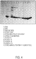
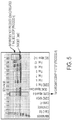
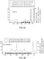


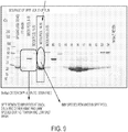
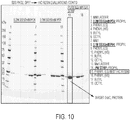


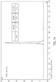


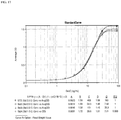
Classifications
-
- G—PHYSICS
- G01—MEASURING; TESTING
- G01N—INVESTIGATING OR ANALYSING MATERIALS BY DETERMINING THEIR CHEMICAL OR PHYSICAL PROPERTIES
- G01N33/00—Investigating or analysing materials by specific methods not covered by groups G01N1/00 - G01N31/00
- G01N33/48—Biological material, e.g. blood, urine; Haemocytometers
- G01N33/50—Chemical analysis of biological material, e.g. blood, urine; Testing involving biospecific ligand binding methods; Immunological testing
- G01N33/53—Immunoassay; Biospecific binding assay; Materials therefor
- G01N33/573—Immunoassay; Biospecific binding assay; Materials therefor for enzymes or isoenzymes
-
- C—CHEMISTRY; METALLURGY
- C07—ORGANIC CHEMISTRY
- C07K—PEPTIDES
- C07K1/00—General methods for the preparation of peptides, i.e. processes for the organic chemical preparation of peptides or proteins of any length
- C07K1/14—Extraction; Separation; Purification
- C07K1/16—Extraction; Separation; Purification by chromatography
- C07K1/22—Affinity chromatography or related techniques based upon selective absorption processes
-
- C—CHEMISTRY; METALLURGY
- C07—ORGANIC CHEMISTRY
- C07K—PEPTIDES
- C07K16/00—Immunoglobulins [IGs], e.g. monoclonal or polyclonal antibodies
- C07K16/40—Immunoglobulins [IGs], e.g. monoclonal or polyclonal antibodies against enzymes
-
- C—CHEMISTRY; METALLURGY
- C12—BIOCHEMISTRY; BEER; SPIRITS; WINE; VINEGAR; MICROBIOLOGY; ENZYMOLOGY; MUTATION OR GENETIC ENGINEERING
- C12N—MICROORGANISMS OR ENZYMES; COMPOSITIONS THEREOF; PROPAGATING, PRESERVING, OR MAINTAINING MICROORGANISMS; MUTATION OR GENETIC ENGINEERING; CULTURE MEDIA
- C12N9/00—Enzymes; Proenzymes; Compositions thereof; Processes for preparing, activating, inhibiting, separating or purifying enzymes
- C12N9/0004—Oxidoreductases (1.)
- C12N9/0051—Oxidoreductases (1.) acting on a sulfur group of donors (1.8)
-
- C—CHEMISTRY; METALLURGY
- C12—BIOCHEMISTRY; BEER; SPIRITS; WINE; VINEGAR; MICROBIOLOGY; ENZYMOLOGY; MUTATION OR GENETIC ENGINEERING
- C12N—MICROORGANISMS OR ENZYMES; COMPOSITIONS THEREOF; PROPAGATING, PRESERVING, OR MAINTAINING MICROORGANISMS; MUTATION OR GENETIC ENGINEERING; CULTURE MEDIA
- C12N9/00—Enzymes; Proenzymes; Compositions thereof; Processes for preparing, activating, inhibiting, separating or purifying enzymes
- C12N9/90—Isomerases (5.)
-
- C—CHEMISTRY; METALLURGY
- C12—BIOCHEMISTRY; BEER; SPIRITS; WINE; VINEGAR; MICROBIOLOGY; ENZYMOLOGY; MUTATION OR GENETIC ENGINEERING
- C12Y—ENZYMES
- C12Y108/00—Oxidoreductases acting on sulfur groups as donors (1.8)
- C12Y108/04—Oxidoreductases acting on sulfur groups as donors (1.8) with a disulfide as acceptor (1.8.4)
- C12Y108/04002—Protein-disulfide reductase (glutathione) (1.8.4.2), i.e. BdbC or BdbD
-
- C—CHEMISTRY; METALLURGY
- C12—BIOCHEMISTRY; BEER; SPIRITS; WINE; VINEGAR; MICROBIOLOGY; ENZYMOLOGY; MUTATION OR GENETIC ENGINEERING
- C12Y—ENZYMES
- C12Y503/00—Intramolecular oxidoreductases (5.3)
- C12Y503/04—Intramolecular oxidoreductases (5.3) transposing S-S bonds (5.3.4)
- C12Y503/04001—Protein disulfide-isomerase (5.3.4.1), i.e. disufide bond-forming enzyme
-
- A—HUMAN NECESSITIES
- A61—MEDICAL OR VETERINARY SCIENCE; HYGIENE
- A61K—PREPARATIONS FOR MEDICAL, DENTAL OR TOILETRY PURPOSES
- A61K38/00—Medicinal preparations containing peptides
-
- G—PHYSICS
- G01—MEASURING; TESTING
- G01N—INVESTIGATING OR ANALYSING MATERIALS BY DETERMINING THEIR CHEMICAL OR PHYSICAL PROPERTIES
- G01N2333/00—Assays involving biological materials from specific organisms or of a specific nature
- G01N2333/90—Enzymes; Proenzymes
- G01N2333/902—Oxidoreductases (1.)
- G01N2333/90212—Oxidoreductases (1.) acting on a sulfur group of donors (1.8)
-
- G—PHYSICS
- G01—MEASURING; TESTING
- G01N—INVESTIGATING OR ANALYSING MATERIALS BY DETERMINING THEIR CHEMICAL OR PHYSICAL PROPERTIES
- G01N2333/00—Assays involving biological materials from specific organisms or of a specific nature
- G01N2333/90—Enzymes; Proenzymes
- G01N2333/99—Isomerases (5.)
-
- Y—GENERAL TAGGING OF NEW TECHNOLOGICAL DEVELOPMENTS; GENERAL TAGGING OF CROSS-SECTIONAL TECHNOLOGIES SPANNING OVER SEVERAL SECTIONS OF THE IPC; TECHNICAL SUBJECTS COVERED BY FORMER USPC CROSS-REFERENCE ART COLLECTIONS [XRACs] AND DIGESTS
- Y02—TECHNOLOGIES OR APPLICATIONS FOR MITIGATION OR ADAPTATION AGAINST CLIMATE CHANGE
- Y02A—TECHNOLOGIES FOR ADAPTATION TO CLIMATE CHANGE
- Y02A50/00—TECHNOLOGIES FOR ADAPTATION TO CLIMATE CHANGE in human health protection, e.g. against extreme weather
- Y02A50/30—Against vector-borne diseases, e.g. mosquito-borne, fly-borne, tick-borne or waterborne diseases whose impact is exacerbated by climate change
Definitions
- the present invention provides methods for producing disulfide oxidoreductase A (DsbA) polypeptides at very high levels of purity. Also provided are ultrapure DsbA and methods of using same, e.g., for use in immunoassays to show removal of DsbA from biologics produced in bacteria.
- DsbA disulfide oxidoreductase A
- DsbA Disulfide oxidoreducatase
- DsbC catalyzes the isomerization of disulfide bonds and can shuffle misfolded disulfide bonds.
- Dsb proteins are a primary catalyst of disulfide bond formation and isomerization in bacteria and promote the correct protein folding of protein.
- Biol. Chem. 282(43):31302-31307 discloses DsbA as a sufficient catalyst for disulfide formation in RNase I.
- a purification scheme for histidine-tagged DsbA using immobilized metal affinity chromatography is provided.
- Urban, A. et al. (2001) J. Bacteriol. 183(2):587-596 discloses DsbA from Pseudomonas aeruginosa that stabilizes a periplasmic lipase.
- a purification scheme for histidine-tagged DsbA using immobilized metal affinity chromatography is provided.
- 59:266-273 discloses cloning, expression, purification and characterization of a DsbA-like protein from Walbachia pipientis.
- a purification scheme for histidine-tagged DsbA using immobilized metal affinity chromatography is provided.
- Zapun, A. et al. (1993) Biochem. 32:5083-5092 discloses that a reactive and destabilizied disulfide bond of DsbA is required for protein disulfide bond formation in vivo.
- Native DsbA protein was purified by anion exchange chromatography.
- Zhang, Y. et al. (2008) Hybridoma 27(2):131-134 discloses generation of monoclonal antibodies against Escherichia coli DsbA.
- a purification scheme for histidine-tagged DsbA using immobilized metal affinity chromatography is provided. Purification of a DsbA fusion protein using affinity columns containing the generated monoclonal antibodies is also disclosed.
- Brown, DM and Pietrzyk, DJ (1989) J. Chromat. 466:291-300 discloses mixed bed ionexchange (MBIE) chromatography for the simultaneous separation of anion and cation analytes.
- Zhan, X et al. (2004) J. Bacter. 186(3):654-660 disclosed genetic analysis of DsbC expression in Eschericia coli. DsbC expression was measured by western blot analysis.
- HCPs process-specific host cell proteins
- HCPs are typically present in the final drug substance in small quantities (in parts-per-million or nanograms per milligram of the intended recombinant polypeptide), it is recognized that HCPs are undesirable and their quantities should be minimized.
- FDA Food and Drug Administration
- biopharmaceuticals intended for in vivo human use should be as free as possible of extraneous impurities, and requires tests for detection and quantitation of potential impurities, such as HCPs.
- ICH International Conference on Harmonization
- Assays and reagents to detect immunoglobulins, DNA, endotoxins, viruses, and total HCPs, e.g., total E. coli proteins (ECP) have been developed but such assays and reagents to not accurately detect accessory proteins such as DsbA or DsbC.
- ECP E. coli proteins
- Reagents, methods and kits for the detection of DsbA and DsbC are particularly needed where there are no existing assays and reagents of sufficient consistency, sensitivity, specificity or efficiency.
- the invention described herein meets certain of the above-described needs and provides other benefits.
- the invention provides methods for purifying a DsbA polypeptide from a cell lysate comprising the DsbA polypeptide, comprising a) adding polyethyleneimine (PEI) to a final concentration of about 0.01% to about 1.0% to a cell lysate comprising the DsbA polypeptide, b) clarifying the cell lysate by centrifugation, c) applying the clarified cell lysate comprising the DsbA polypeptide to an anion exchange chromatography material, d) eluting the DsbA polypeptide from the anion exchange chromatography material to generate an anion exchange eluate comprising the DsbA polypeptide, e) applying the anion exchange eluate comprising the DsbA polypeptide to a cation exchange chromatography material, f) eluting the DsbA polypeptide from the cation exchange chromatography material to generate a cation exchange eluate comprising the purified Ds
- the cell lysate comprising the DsbA polypeptide is held in the PEI for at least about 16 hours prior to anion exchange chromatography. In some embodiments, the final concentration of PEI in the lysate is about 0.1%. In some embodiments, the lysate comprising the DsbA polypeptide and the PEI is at a pH of about 7.0.
- the anion exchange chromatography material is a strong anion exchanger.
- the strong anion exchanger comprises a quarternary amine.
- the quarternary amine is linked to crosslinked agarose.
- the anion exchanger is QSFF anion exchanger.
- the DsbA is eluted from the anion chromatography material using a salt gradient.
- the salt gradient is a step gradient.
- the clarified lysate comprises 10 mM MOPS, pH 7.1.
- the DsbA is eluted from the anion exchange chromatography material with the following steps: about 15% of about 25 mM Tris and about 250 mM NaCl at about pH 9.2 for about four column volumes, about 20% of about 25 mM Tris and about 250 mM NaCl at about pH 9.2 for about four column volumes, about 25% of about 25 mM Tris and about 250 mM NaCl at about pH 9.2 until DsbA elutes from the column.
- the clarified lysate comprising the DsbA polypeptide of step b) is passed through a 0.22 ⁇ m filter prior to anion exchange chromatography. In some embodiments, the clarified lysate comprising the DsbA polypeptide of step b) is adjusted to pH about 9.0 prior to anion exchange chromatography.
- the anion exchange eluate is collected in fractions.
- the fractions are about 0.3 to about 1.0 column volumes (CV).
- the fractions are analyzed by size exclusion chromatography prior to cation exchange chromatography.
- fractions comprising at least about 55% DsbA are selected for further purification.
- the cation exchange material comprises a sulphopropyl moiety.
- the sulfopropyl moiety is linked to a crosslinked poly(styrene-divinylbenzene) matrix.
- the cation exchange media is POROS HS 50 or equivalent.
- the anion exchange eluate of step d) is adjusted to pH about 5.0 prior to cation exchange chromatography.
- the DsbA is eluted from the cation chromatography material using a salt gradient.
- the cation chromatography material is washed with 5 column volumes of 12.5 mM MES.
- the salt gradient is a gradient from about 0% to about 60% 12.5 mM MES and 1 M NaCl over 15 column volumes.
- the cation exchange eluate is collected in fractions.
- the fractions are analyzed by size exclusion chromatography. In some embodiments, fractions comprising at least about 95% DsbA are pooled.
- the DsbA polypeptide is an Escherichia coli DsbA polypeptide.
- the DsbA polypeptide comprises the amino acid sequence of SEQ ID NO:1.
- the amino acid sequence of the DsbA polypeptide at least about 80% identical to the amino acid sequence of SEQ ID NO:1.
- the DsbA is expressed in cell.
- the cell is a prokaryotic cell.
- the cell is an E. coli cell.
- the cell is engineered to express DsbA at levels greater than endogenous expression of DsbA.
- the cell is lysed using a microfluidizer.
- the invention provides a composition comprising a DsbA polypeptide purified by the method of any one of the above embodiments. In some aspects, the invention provides a composition comprising a purified DsbA polypeptide, where the composition comprises at least about 95% monomeric DsbA polypeptide. In some aspects, the invention provides a composition comprising a purified DsbA polypeptide, where the composition comprises at least about 98% monomeric DsbA polypeptide. In some embodiments, the composition comprises less that about 2% low molecular weight species. In some embodiments, the composition comprises less than about 1% high molecular weight species. In some embodiments, the percentage of monomeric DsbA polypeptide is detected by size exclusion chromatography.
- the composition comprises less than about 5% impurities.
- the impurities are high molecular weight and/or low molecular weight polypeptide species relative to native or desired DsbA.
- the impurities are one or more of an E. coli protein (ECP), aggregates of DsbA, fragments of DsbA, a nucleic acid or a cell culture media component.
- ECP E. coli protein
- the DsbA is stable to one or more freeze-thaw cycles.
- the DsbA is stable to one, two, three, four, five, six, seven, eight, nine, ten or greater than ten freeze-thaw cycles.
- the ultrapure DsbA is used as a reference standard; for example, to determine the amount or concentration of DsbA in a test sample. In some embodiments, the ultrapure DsbA is used as a positive control; for example, in an assay to determine the presence and/or quantity of DsbA in a sample.
- the purity of the DsbA polypeptide in the composition is measured by chromatography, SDS polyacrylamide gel electrophoresis or western blot analysis. In some embodiments, the purity of the DsbA polypeptide in the composition is measured by high performance liquid chromatography (HPLC). In some embodiments, the chromatography is size exclusion chromatography (e.g ., SEC-HPLC). In some embodiments, the purity of the DsbA polypeptide in the composition is measured by SDS gel electrophoresis using a fluorescent protein stain or a silver stain.
- the presence of non-DsbA polypeptides in the composition are identified by the presence of species identified by gel electrophoresis that are not immunoreactive with anti-DsbA antibodies as shown by western blot analysis.
- the presence of aggregates of the DsbA polypeptide in the composition are identified by the presence of species with a molecular weight greater than the native DsbA by western blot analysis.
- the presence of fragments of the DsbA polypeptide in the composition are identified by the presence of species with a molecular weight less than the native DsbA by western blot analysis.
- the invention provides methods for generating antibodies that specifically bind DsbA, comprising exposing an animal to the ultrapure DsbA, purified as described above.
- the method comprises collecting sera from the animal wherein the sera comprises antibodies that specifically bind DsbA.
- the sera comprises polyclonal antibodies that specifically bind DsbA.
- the invention provides one or more monoclonal antibodies are isolated from the sera.
- the animal is a goat, a rabbit, a mouse, a guinea pig, a hamster, a rat, a donkey or a chicken.
- the invention provides methods for purifying antibodies that specifically bind DsbA, comprising contacting a composition comprising anti-DsbA antibodies to chromatography material comprising ultrapure DsbA attached to a support material, washing the chromatography material to remove unbound compounds, and eluting the anti-DsbA antibodies.
- the ultrapure DsbA comprises at least about 95% monomeric DsbA polypeptide.
- the ultrapure DsbA comprises less than about 5% impurities, less than about 1% impurities, or less than about 0.1% impurities.
- the ultrapure DsbA is prepared by any of the methods described herein.
- the antibodies are polyclonal antibodies.
- the antibodies are prepared according to any of the methods described herein. In some embodiments, less than 1% of the antibodies specifically bind non-DsbA compounds.
- the invention provides compositions comprising polyclonal antibodies that specifically bind DsbA, wherein the polyclonal antibodies are generated by exposing an animal to any of the DsbA compositions described above. In some embodiments, the polyclonal antibodies are collected from the sera of the animal. In some embodiments, the invention provides compositions comprising monoclonal antibodies that specifically bind DsbA, wherein the monoclonal antibodies are generated by exposing an animal to any of the DsbA compositions described above. In some embodiments, the animal is a goat, a rabbit, a mouse, a guinea pig, a hamster, a rat, a donkey or a chicken.
- the invention provides a method for analyzing a recombinant polypeptide sample for the presence of and/or quantity of DsbA, comprising detecting DsbA in the sample using an immunoassay and comparing the amount of DsbA detected in the sample with the detection of one or more concentrations of an ultrapure DsbA reference standard.
- a number of different concentrations of the reference standard DsbA are tested in order to establish a correlation between the level of detection and the concentration of DsbA in the reference standard.
- the concentration of DsbA in a test sample can be determined by comparing the level of detection of DsbA in the test sample with the detection of the known concentrations of DsbA in the reference standard.
- the preparation comprises less than about any of 1% of impurities.
- the ultrapure DsbA reference standard is prepared by the methods described herein.
- the immunoassay comprises antibodies that specifically bind ultrapure DsbA. In some embodiments, the antibodies that specifically bind ultrapure DsbA bind less than about any of 1% non-DsbA compounds. In some embodiments, the antibodies that specifically bind ultrapure DsbA are polyclonal antibodies. In other embodiments, the antibodies that specifically bind ultrapure DsbA are monoclonal antibodies. In some embodiments, the antibodies that specifically bind ultrapure DsbA are used as capture antibodies in the immunoassay.
- the antibodies that specifically bind ultrapure DsbA are used as detection antibodies.
- the detection antibodies are conjugated to a detection agent (e.g., a horseradish peroxidase).
- the DsbA is an E. coli DsbA.
- the recombinant polypeptide is prepared in a host cell ( e.g., an E. coli host cell).
- the host cell overexpresses DsbA (e.g., an E. coli host cell that overexpressed DsbA).
- the sample is cell lysate or is obtained from a recombinant polypeptide preparation and wherein the recombinant polypeptide preparation has been subjected to one or more chromatographic purification steps.
- the recombinant polypeptide preparation is a final purified product.
- the invention provides immunoassay methods for detecting DsbA in a sample, for example, wherein the sample is obtained from a recombinant polypeptide preparation or a host cell line, the method comprising: (a) contacting a capture antibody that binds DsbA with the sample thereby generating a sample-capture antibody combination material; (b) contacting a detection antibody that binds DsbA with the sample-capture antibody combination material; and (c) detecting the antibody bound to the sample-capture antibody combination material.
- the method comprises quantifying the level of the detection antibody bound using a standard titration curve.
- the method comprises calculating an amount of DsbA present in the sample based on the level of the detection antibody bound.
- the amount of DsbA present in the sample is determined by comparing the standard titration curve with a standard titration curve generated with an ultrapure DsbA composition.
- the ultrapure DsbA composition comprises at least about 95% monomeric DsbA polypeptide.
- the ultrapure DsbA composition comprises less than about 5% impurities, less than about 1% impurities, or less than about 0.1 % impurities.
- the ultrapure DsbA in the composition is prepared by any of the methods described herein.
- the capture antibody specifically binds ultrapure DsbA. In some embodiments, the detection antibody specifically binds ultrapure DsbA. In some embodiments, the antibody that specifically binds ultrapure DsbA is a polyclonal antibody. In some embodiments, the detection antibody that binds DsbA is conjugated to a horseradish peroxidase. In some embodiments, the immunoassay is a sandwich assay. In further embodiments, the sandwich assay is an enzyme-linked immunosorbent assay (ELISA). In some embodiments, the DsbA is an E. coli DsbA. In some embodiments, the recombinant polypeptide preparation or the host cell line is obtained from E. coli.
- the host cell line overexpresses DsbA (e.g., an E. coli host cell that overexpresses DsbA).
- the sample is cell lysate.
- the sample is obtained from the recombinant polypeptide preparation and wherein the recombinant polypeptide preparation has been subjected to one or more chromatographic purification steps.
- the recombinant polypeptide preparation is final purified product.
- the recombinant polypeptide contained in the recombinant polypeptide preparation is an antibody or an immunoadhesin.
- the antibody is a multispecific antibody, a bispecific antibody, a half antibody or an antibody fragment.
- the recombinant polypeptide is an IgG1, an IgG2, an IgG3, or an IgG4.
- the invention provides a quality assay for a pharmaceutical composition comprising a recombinant polypeptide prepared from a bacterial cell, the release assay comprising subjecting the pharmaceutical composition to an immunoassay as described herein, wherein detection of DsbA in the immunoassay indicates that the pharmaceutical composition is not suitable for therapeutic administration to an animal.
- an amount of DsbA in the pharmaceutical composition of less than about 1 ppm indicates that the pharmaceutical composition is suitable for administration to the animal.
- the recombinant polypeptide is prepared from an E. coli cell.
- the bacterial cell overexpresses DsbA.
- the sample is cell lysate.
- the sample is obtained from the recombinant polypeptide preparation and wherein the recombinant polypeptide preparation has been subjected to one or more chromatographic purification steps.
- the recombinant polypeptide preparation is final purified product.
- the recombinant polypeptide contained in the recombinant polypeptide preparation is an antibody or an immunoadhesin.
- the antibody is a multispecific antibody, a bispecific antibody, a half antibody or an antibody fragment.
- the recombinant polypeptide is an IgG1, an IgG2, an IgG3, or an IgG4.
- the disclosure provides methods for purifying a DsbC polypeptide from a cell lysate comprising the DsbC polypeptide comprising a) adding polyethyleneimine (PEI) to a final concentration of about 0.01% to about 1.0% to a cell lysate comprising the DsbC polypeptide, b) clarifying the cell lysate by centrifugation, c) applying the clarified cell lysate comprising the DsbC polypeptide to an anion exchange chromatography material, d) eluting the DsbC polypeptide from the anion exchange chromatography material to generate an anion exchange eluate comprising the DsbC polypeptide, e) applying the anion exchange eluate comprising the DsbC polypeptide to a hydrophobic interaction chromatography (HIC) material, f) eluting the DsbC polypeptide from the HIC material to generate a HIC eluate, g) applying the HIC eluate
- the cell lysate comprising the DsbC polypeptide is held in the PEI for at least about 16 hours prior to anion exchange chromatography. In some embodiments, the final concentration of PEI in the lysate is about 0.1%. In some embodiments, the lysate comprising the DsbC polypeptide and the PEI is at a pH of about 7.0.
- the anion exchange chromatography material is a weak anion exchanger.
- the weak anion exchanger comprises a quarternary amine.
- the quarternary amine is linked to crosslinked agarose.
- the DsbC is eluted from the cation chromatography material using a salt gradient.
- the salt gradient is a linear gradient.
- the anion exchange material is washed in 10 mM MOPS.
- the salt gradient is a gradient from about 0% to about 60% 10 mM MOPS and 250 mM NaCl over 15 column volumes.
- the clarified lysate comprising the DsbC polypeptide of step b) is passed through a 0.22 ⁇ m filter prior to anion exchange chromatography. In some embodiments, the clarified lysate comprising the DsbC polypeptide of step b) is adjusted to pH about 8.0 prior to anion exchange chromatography.
- the anion exchange eluate is collected in fractions.
- the fractions are analyzed by size exclusion chromatography prior to hydrophobic interaction chromatography.
- fractions comprising at least about 25% DsbC are selected for further purification.
- the HIC material comprises a phenyl moiety.
- the phenyl moiety is linked to a crosslinked agarose.
- the anion exchange eluate is conditioned to contain about 0.54 M sodium sulfate and about 50 mM PO 4 , about pH 7 before HIC chromatography.
- the DsbC is eluted from the HIC material using water.
- the HIC eluate is collected in fractions. In further embodiments, fractions comprising DsbC are pooled.
- the size exclusion chromatography material comprises a spherical composite of cross-linked agarose and dextran.
- the size exclusion flow through is collected in fractions.
- the HIC eluate is ultrafiltered prior to size exclusion chromatography.
- fractions comprising DsbC are pooled.
- the DsbC polypeptide is an Escherichia coli DsbC polypeptide.
- the DsbC polypeptide comprises the amino acid sequence of SEQ ID NO:3.
- the amino acid sequence of the DsbC polypeptide at least about 80% identical to the amino acid sequence of SEQ ID NO:3.
- the DsbC is expressed in cell.
- the cell is a prokaryotic cell.
- the cell is an E. coli cell.
- the cell is engineered to express DsbC at levels greater than endogenous expression of DsbC.
- the cell is lysed using a microfluidizer.
- the disclosure provides a composition comprising a DsbC polypeptide purified by the method of any one of the above embodiments.
- the invention provides a composition comprising a purified DsbC polypeptide, where the composition comprises at least about 95% monomeric DsbC polypeptide. In some embodiments, the composition comprises at least about 98% monomeric DsbC polypeptide. In some embodiments, the composition comprises at least about 99% monomeric DsbC polypeptide. In some embodiments, the composition comprises at least about 99.5% monomeric DsbC polypeptide. In some embodiments, the composition comprises less that about 2% low molecular weight species. In some embodiments, the composition comprises less than about 1% high molecular weight species.
- the percentage of monomeric DsbC polypeptide is detected by size exclusion chromatography.
- the impurities are high molecular weight and/or low molecular weight polypeptide species relative to native or desired DsbC.
- the impurities are one or more of an E. coli protein (ECP), aggregates of DsbC, fragments of DsbC, a nucleic acid or a cell culture media component.
- ECP E. coli protein
- the DsbC is stable to one or more freeze-thaw cycles.
- the DsbC is stable to one, two, three, four, five, six, seven, eight, nine, ten or greater than ten freeze-thaw cycles.
- the ultrapure DsbC is used as a reference standard; for example, to determine the amount or concentration of DsbC in a test sample. In some embodiments, the ultrapure DsbC is used as a positive control; for example, in an assay to determine the presence and/or quantity of DsbC in a sample.
- the purity of the DsbC polypeptide in the composition is measured by chromatography, SDS polyacrylamide gel electrophoresis or western blot analysis. In some embodiments, the purity of the DsbC polypeptide in the composition is measured by high performance liquid chromatography (HPLC). In some embodiments, the chromatography is size exclusion chromatography (e.g ., SEC-HPLC). In some embodiments, the purity of the DsbC polypeptide in the composition is measured by SDS gel electrophoresis using a fluorescent protein stain or a silver stain.
- the presence of non-DsbC polypeptides in the composition are identified by the presence of species identified by gel electrophoresis that are not immunoreactive with anti-DsbC antibodies as shown by western blot analysis.
- the presence of aggregates of the DsbC polypeptide in the composition are identified by the presence of species with a molecular weight greater than the native DsbC by western blot analysis.
- the presence of fragments of the DsbC polypeptide in the composition are identified by the presence of species with a molecular weight less than the native DsbC by western blot analysis.
- the disclosure provides methods for generating antibodies that specifically bind DsbC, comprising exposing an animal to the ultrapure DsbC, purified as described above.
- the method comprises collecting sera from the animal wherein the sera comprises antibodies that specifically bind DsbC.
- the sera comprises polyclonal antibodies that specifically bind DsbC.
- the invention provides one or more monoclonal antibodies are isolated from the sera.
- the animal is a goat, a rabbit, a mouse, a guinea pig, a hamster, a rat, a donkey or a chicken.
- the disclosure provides methods for purifying antibodies that specifically bind DsbC, comprising contacting a composition comprising anti-DsbC antibodies to chromatography material comprising ultrapure DsbC attached to a support material, washing the chromatography material to remove unbound compounds, and eluting the anti-DsbC antibodies.
- the ultrapure DsbC comprises more than about 95% monomeric DsbC polypeptide.
- the ultrapure DsbC comprises less than about 5% impurities, less than about 1% impurities, or less than about 0.1% impurities.
- the ultrapure DsbC is prepared by any of the methods described herein.
- the antibodies are polyclonal antibodies.
- the antibodies are prepared according to any of the methods described herein. In some embodiments, less than 1% of the antibodies specifically bind non-DsbC compounds.
- the disclosure provides compositions comprising polyclonal antibodies that specifically bind DsbC, wherein the polyclonal antibodies are generated by exposing an animal to any of the DsbC compositions described above.
- the polyclonal antibodies are collected from the sera of the animal.
- the invention provides compositions comprising monoclonal antibodies that specifically bind DsbC, wherein the monoclonal antibodies are generated by exposing an animal to any of the DsbC compositions described above.
- the animal is a goat, a rabbit, a mouse, a guinea pig, a hamster, a rat, a donkey or a chicken.
- the disclosure provides a method for analyzing a recombinant polypeptide sample for the presence of and/or quantity of DsbC, comprising detecting DsbC in the sample using an immunoassay and comparing the amount of DsbC detected in the sample with the detection of one or more concentrations of an ultrapure DsbC reference standard.
- a number of different concentrations of the reference standard DsbC are tested in order to establish a correlation between the level of detection and the concentration of DsbC in the reference standard.
- the concentration of DsbC in a test sample can be determined by comparing the level of detection of DsbC in the test sample with the detection of the known concentrations of DsbC in the reference standard.
- the preparation comprises less than about any of 1% of impurities.
- the ultrapure DsbC reference standard is prepared by the methods described herein.
- the immunoassay comprises antibodies that specifically bind ultrapure DsbC. In some embodiments, the antibodies that specifically bind ultrapure DsbC bind less than about any of 1% non-DsbC compounds. In some embodiments, the antibodies that specifically bind ultrapure DsbC are polyclonal antibodies. In other embodiments, the antibodies that specifically bind ultrapure DsbC are monoclonal antibodies. In some embodiments, the antibodies that specifically bind ultrapure DsbC are used as capture antibodies in the immunoassay.
- the antibodies that specifically bind ultrapure DsbC are used as detection antibodies.
- the detection antibodies are conjugated to a detection agent (e.g., a horseradish peroxidase).
- the DsbC is an E. coli DsbC.
- the recombinant polypeptide is prepared in a host cell ( e.g., an E. coli host cell).
- the host cell overexpresses DsbC (e.g., an E. coli host cell that overexpressed DsbC).
- the sample is cell lysate or is obtained from a recombinant polypeptide preparation and wherein the recombinant polypeptide preparation has been subjected to one or more chromatographic purification steps.
- the recombinant polypeptide preparation is a final purified product.
- the disclosure provides immunoassay methods for detecting DsbC in a sample, for example, wherein the sample is obtained from a recombinant polypeptide preparation or a host cell line, the method comprising: (a) contacting a capture antibody that binds DsbC with the sample thereby generating a sample-capture antibody combination material; (b) contacting a detection antibody that binds DsbC with the sample-capture antibody combination material; and (c) detecting the antibody bound to the sample-capture antibody combination material.
- the method comprises quantifying the level of the detection antibody bound using a standard titration curve.
- the method comprises calculating an amount of DsbC present in the sample based on the level of the detection antibody bound.
- the amount of DsbC present in the sample is determined by comparing the standard titration curve with a standard titration curve generated with an ultrapure DsbC composition.
- the ultrapure DsbC composition comprises at least about 95% monomeric DsbC polypeptide.
- the ultrapure DsbC composition comprises less than about 5% impurities, less than about 1% impurities, or less than about 0.1% impurities.
- the ultrapure DsbC in the composition is prepared by any of the methods described herein.
- the capture antibody specifically binds ultrapure DsbC. In some embodiments, the detection antibody specifically binds ultrapure DsbC. In some embodiments, the antibody that specifically binds ultrapure DsbC is a polyclonal antibody. In some embodiments, the detection antibody that binds DsbC is conjugated to a horseradish peroxidase. In some embodiments, the immunoassay is a sandwich assay. In further embodiments, the sandwich assay is an enzyme-linked immunosorbent assay (ELISA). In some embodiments, the DsbC is an E. coli DsbC. In some embodiments, the recombinant polypeptide preparation or the host cell line is obtained from E. coli.
- the host cell line overexpresses DsbC (e.g., an E. coli host cell that overexpresses DsbC).
- the sample is cell lysate.
- the sample is obtained from the recombinant polypeptide preparation and wherein the recombinant polypeptide preparation has been subjected to one or more chromatographic purification steps.
- the recombinant polypeptide preparation is final purified product.
- the recombinant polypeptide contained in the recombinant polypeptide preparation is an antibody or an immunoadhesin.
- the antibody is a multispecific antibody, a bispecific antibody, a half antibody or an antibody fragment.
- the recombinant polypeptide is an IgG1, an IgG2, an IgG3, or an IgG4.
- the disclosure provides a quality assay for a pharmaceutical composition comprising a recombinant polypeptide prepared from a bacterial cell, the release assay comprising subjecting the pharmaceutical composition to an immunoassay as described herein, wherein detection of DsbC in the immunoassay indicates that the pharmaceutical composition is not suitable for therapeutic administration to an animal.
- an amount of DsbC in the pharmaceutical composition of less than about 1 ppm indicates that the pharmaceutical composition is suitable for administration to the animal.
- the recombinant polypeptide is prepared from an E. coli cell.
- the bacterial cell overexpresses DsbC.
- the sample is cell lysate.
- the sample is obtained from the recombinant polypeptide preparation and wherein the recombinant polypeptide preparation has been subjected to one or more chromatographic purification steps.
- the recombinant polypeptide preparation is final purified product.
- the recombinant polypeptide contained in the recombinant polypeptide preparation is an antibody or an immunoadhesin.
- the antibody is a multispecific antibody, a bispecific antibody, a half antibody or an antibody fragment.
- the recombinant polypeptide is an IgG1, an IgG2, an IgG3, or an IgG4.
- kits for the detection of DsbA in a pharmaceutical composition comprising a recombinant polypeptide prepared from a bacterial cell, said kit comprising anti-DsbA antibodies prepared by any of the methods described herein or a composition of any of the anti-DsbA antibodies described herein.
- kits for the detection of DsbC in a pharmaceutical composition comprising a recombinant polypeptide prepared from a bacterial cell, said kit comprising anti-DsbC antibodies prepared by any of the methods described herein or a composition of any of the anti-DsbC antibodies described herein.
- kits for the detection of DsbA and the disclosure provides kits for the detection of DsbC in a pharmaceutical composition comprising a recombinant polypeptide prepared from a bacterial cell, said kit comprising anti-DsbA antibodies and anti-DsbC antibodies prepared by any of the methods described herein or a composition of any of the anti-DsbA antibodies and any of the anti-DsbC antibodies described herein.
- the kits further comprise ultrapure DsbA and/or DsbC for use as a reference standard in generating standard curves for quantitating DsbA and/or DsbC in a sample.
- the kits further comprise ultrapure DsbA and/or DsbC for use as positive controls in an assay to detect DsbA and/or DsbC in a sample.
- the invention provides methods to generate ultrapure compositions of DsbA and the disclosure provides methods to generate ultrapure compositions of DsbC.
- Such compositions are used to generate antibodies that are highly specific to DsbA or DsbC.
- the antibodies in turn are useful for the detection of DsbA and DsbC in recombinant polypeptides prepared in bacterial fermentation cultures where DsbA and DsbC are overexpressed to facilitate recombinant polypeptide folding and assembly.
- the antibodies may be useful in release assays in the development of pharmaceutical formulation of the recombinant polypeptides such as antibodies.
- detecting is used herein in the broadest sense to include both qualitative and quantitative measurements of a target molecule. Detecting includes identifying the mere presence of the target molecule in a sample as well as determining whether the target molecule is present in the sample at detectable levels.
- polypeptide and protein are used interchangeably herein to refer to polymers of amino acids of any length.
- the polymer may be linear or branched, it may comprise modified amino acids, and it may be interrupted by non-amino acids.
- the terms also encompass an amino acid polymer that has been modified naturally or by intervention; for example, disulfide bond formation, glycosylation, lipidation, acetylation, phosphorylation, or any other manipulation or modification, such as conjugation with a labeling component.
- polypeptides containing one or more analogs of an amino acid including, for example, unnatural amino acids, etc .
- the terms "polypeptide” and “protein” as used herein specifically encompass antibodies.
- Polypeptide e.g ., antibody or immunoadhesin
- Purity is a relative term and does not necessarily mean absolute purity.
- ultrapure DsbA and ultrapure DsbC refer to DsbA or DsbC compositions in which at least about 95% of the protein in the composition is the desired protein, monomeric DsbA or monomeric DsbC.
- ultrapure DsbA and ultrapure DsbC comprises at least about 96%, 97%, 98%, 99%, or 99.5% monomeric DsbA or monomeric DsbC.
- monomeric DsbA or monomeric DsbC is determined by SEC.
- Dsb proteins When used in reference to a polypeptide, "Dsb" proteins refer to bacterial disulfide oxidoreductases. Bacterial disulfide oxireductases are members of the disulfide bond family of enzymes. Members of the Dsb family include DsbA, DsbB, DsbC, DsbD and DsbG. DsbA forms intrachain disulfide bonds as peptides emerge into the cell's periplasm and DsbC serves as a disulfide bond isomerase during oxidative protein-folding in cell's periplasm.
- DsbA may also be known as periplasmic protein disulfide isomerase I.
- An exemplary DsbA protein is E. coli DsbA.
- the amino acid sequence of E. coli DsbA is provided by NCBI Accession No. NP_418297 (SEQ ID NO:1) and the nucleic acid sequence of E. coli dsbA gene is provided by NCBI Accession No. NC_000913.3 or EcoGene:EG11297 (SEQ ID NO:2).
- DsbC may also be known as periplasmic protein disulfide isomerase II.
- An exemplary DsbC protein is E. coli DsbC.
- the amino acid sequence of E. coli DsbC is provided by NCBI Accession No. NP_417369.1 (SEQ ID NO:3) and the nucleic acid sequence of E. coli dsbC gene is provided by NCBI Accession No. NC_000913.3 or EcoGene:EG11070 (SEQ ID NO:4).
- sample refers to a small portion of a larger quantity of material. Generally, testing according to the methods described herein is performed on a sample.
- the sample is typically obtained from a recombinant polypeptide preparation obtained, for example, from cultured recombinant polypeptide-expressing cell lines, also referred to herein as "product cell lines," or from cultured host cells.
- host cells do not contain genes for the expression of recombinant polypeptides of interest or products.
- a sample may be obtained from, for example but not limited to, harvested cell culture fluid, from an in-process pool at a certain step in a purification process, or from the final purified product.
- a “capture antibody” refers to an antibody that specifically binds a target molecule in a sample. Under certain conditions, the capture antibody forms a complex with the target molecule such that the antibody-target molecule complex can be separated from the rest of the sample. In certain embodiments, such separation may include washing away substances or material in the sample that did not bind the capture antibody. In certain embodiments, a capture antibody may be attached to a solid support surface, such as, for example but not limited to, a plate or a bead.
- a “detection antibody” refers to an antibody that specifically binds a target molecule in a sample or in a sample-capture antibody combination material. Under certain conditions, the detection antibody forms a complex with the target molecule or with a target molecule-capture antibody complex.
- a detection antibody is capable of being detected either directly through a label, which may be amplified, or indirectly, e.g., through use of another antibody that is labeled and that binds the detection antibody.
- the detection antibody is typically conjugated to a moiety that is detectable by some means, for example, including but not limited to, biotin or ruthenium.
- label refers to any chemical group or moiety that can be linked to a substance that is to be detected or quantitated, e.g., an antibody.
- a label is a detectable label that is suitable for the sensitive detection or quantification of a substance.
- detectable labels include, but are not limited to, luminescent labels, e.g., fluorescent, phosphorescent, chemiluminescent, bioluminescent and electrochemiluminescent labels, radioactive labels, enzymes, particles, magnetic substances, electroactive species and the like.
- a detectable label may signal its presence by participating in specific binding reactions. Examples of such labels include haptens, antibodies, biotin, streptavidin, his-tag, nitrilotriacetic acid, glutathione S-transferase, glutathione and the like.
- detection means refers to a moiety or technique used to detect the presence of the detectable antibody through signal reporting that is then read out in an assay.
- detection means employ reagents that amplify an immobilized label such as the label captured onto a microtiter plate, e.g., avidin or streptavidin-HRP.
- Photoluminescence refers to a process whereby a material luminesces subsequent to the absorption by that material of light (alternatively termed electromagnetic radiation). Fluorescence and phosphorescence are two different types of photoluminescence. “Chemiluminescent” processes involve the creation of the luminescent species by a chemical reaction. “Electro-chemiluminescence” or “ECL” is a process whereby a species, e.g., an antibody, luminesces upon the exposure of that species to electrochemical energy in an appropriate surrounding chemical environment.
- an antibody "which binds" an antigen of interest e.g. a host cell protein, is one that binds the antigen with sufficient affinity such that the antibody is useful as an assay reagent, e.g., as a capture antibody or as a detection antibody. Typically, such an antibody does not significantly cross-react with other polypeptides.
- the term “specific binding” or “specifically binds to” or is “specific for” a particular polypeptide or an epitope on a particular polypeptide target means binding that is measurably different from a non-specific interaction. Specific binding can be measured, for example, by determining binding of a target molecule compared to binding of a control molecule, which generally is a molecule of similar structure that does not have binding activity.
- a preparation of polyclonal antibodies specifically binds DsbA or DsbC. For example, at least about 99% of the antibodies in the polyclonal antibody preparation bind the desired polypeptide ( e.g ., DsbA or DsbC).
- Bind refers to the strength of the sum total of noncovalent interactions between a single binding site of a molecule (e.g., an antibody) and its binding partner (e.g., an antigen). Unless indicated otherwise, as used herein, "binding affinity” refers to intrinsic binding affinity which reflects a 1:1 interaction between members of a binding pair ( e.g ., antibody and antigen).
- the affinity of a molecule X for its partner Y can generally be represented by the dissociation constant (Kd). Affinity can be measured by common methods known in the art, including those described herein.
- Activity refers to form(s) of a polypeptide which retain a biological and/or an immunological activity of native or naturally-occurring polypeptide
- biological activity refers to a biological function (either inhibitory or stimulatory) caused by a native or naturally-occurring polypeptide other than the ability to induce the production of an antibody against an antigenic epitope possessed by a native or naturally-occurring polypeptide
- immunological activity refers to the ability to induce the production of an antibody against an antigenic epitope possessed by a native or naturally-occurring polypeptide.
- antagonist is used in the broadest sense, and includes any molecule that partially or fully blocks, inhibits, or neutralizes a biological activity of a native polypeptide.
- agonist is used in the broadest sense and includes any molecule that mimics a biological activity of a native polypeptide. Suitable agonist or antagonist molecules specifically include agonist or antagonist antibodies or antibody fragments, fragments or amino acid sequence variants of native polypeptides, etc.
- Methods for identifying agonists or antagonists of a polypeptide may comprise contacting a polypeptide with a candidate agonist or antagonist molecule and measuring a detectable change in one or more biological activities normally associated with the polypeptide.
- antibody herein is used in the broadest sense and specifically covers monoclonal antibodies, polyclonal antibodies, multispecific antibodies (e.g . bispecific antibodies) formed from at least two intact antibodies, and antibody fragments so long as they exhibit the desired biological activity.
- immunoglobulin Ig is used interchangeable with antibody herein.
- Antibodies are naturally occurring immunoglobulin molecules which have varying structures, all based upon the immunoglobulin fold.
- IgG antibodies have two "heavy” chains and two "light” chains that are disulphide-bonded to form a functional antibody.
- Each heavy and light chain itself comprises a “constant” (C) and a “variable” (V) region.
- the V regions determine the antigen binding specificity of the antibody, whilst the C regions provide structural support and function in non-antigen-specific interactions with immune effectors.
- the antigen binding specificity of an antibody or antigen-binding fragment of an antibody is the ability of an antibody to specifically bind to a particular antigen.
- the antigen binding specificity of an antibody is determined by the structural characteristics of the V region.
- the variability is not evenly distributed across the 110-amino acid span of the variable domains.
- the V regions consist of relatively invariant stretches called framework regions (FRs) of 15-30 amino acids separated by shorter regions of extreme variability called "hypervariable regions” that are each 9-12 amino acids long.
- FRs framework regions
- hypervariable regions regions of extreme variability
- the variable domains of native heavy and light chains each comprise four FRs, largely adopting a ⁇ -sheet configuration, connected by three hypervariable regions, which form loops connecting, and in some cases forming part of, the ⁇ -sheet structure.
- the hypervariable regions in each chain are held together in close proximity by the FRs and, with the hypervariable regions from the other chain, contribute to the formation of the antigen-binding site of antibodies ( see Kabat et al., Sequences of Proteins of Immunological Interest, 5th Ed. Public Health Service, National Institutes of Health, Bethesda, Md. (1991 )).
- the constant domains are not involved directly in binding an antibody to an antigen, but exhibit various effector functions, such as participation of the antibody in antibody dependent cellular cytotoxicity (ADCC).
- Each V region typically comprises three complementarity determining regions ("CDRs", each of which contains a "hypervariable loop"), and four framework regions.
- An antibody binding site the minimal structural unit required to bind with substantial affinity to a particular desired antigen, will therefore typically include the three CDRs, and at least three, preferably four, framework regions interspersed there between to hold and present the CDRs in the appropriate conformation.
- Classical four chain antibodies have antigen binding sites which are defined by V H and V L domains in cooperation. Certain antibodies, such as camel and shark antibodies, lack light chains and rely on binding sites formed by heavy chains only. Single domain engineered immunoglobulins can be prepared in which the binding sites are formed by heavy chains or light chains alone, in absence of cooperation between V H and V L .
- variable refers to the fact that certain portions of the variable domains differ extensively in sequence among antibodies and are used in the binding and specificity of each particular antibody for its particular antigen. However, the variability is not evenly distributed throughout the variable domains of antibodies. It is concentrated in three segments called hypervariable regions both in the light chain and the heavy chain variable domains. The more highly conserved portions of variable domains are called the framework regions (FRs).
- the variable domains of native heavy and light chains each comprise four FRs, largely adopting a ⁇ -sheet configuration, connected by three hypervariable regions, which form loops connecting, and in some cases forming part of, the ⁇ -sheet structure.
- the hypervariable regions in each chain are held together in close proximity by the FRs and, with the hypervariable regions from the other chain, contribute to the formation of the antigen-binding site of antibodies ( see Kabat et al., Sequences of Proteins of Immunological Interest, 5th Ed. Public Health Service, National Institutes of Health, Bethesda, MD. (1991 )).
- the constant domains are not involved directly in binding an antibody to an antigen, but exhibit various effector functions, such as participation of the antibody in antibody dependent cellular cytotoxicity (ADCC).
- hypervariable region when used herein refers to the amino acid residues of an antibody that are responsible for antigen binding.
- the hypervariable region may comprise amino acid residues from a "complementarity determining region" or "CDR" (e.g ., around about residues 24-34 (L1), 50-56 (L2) and 89-97 (L3) in the V L , and around about 31-35B (HI), 50-65 (H2) and 95-102 (H3) in the V H ( Kabat et al., Sequences of Proteins of Immunological Interest, 5th Ed. Public Health Service, National Institutes of Health, Bethesda, Md.
- CDR complementarity determining region
- Framework or "FR” residues are those variable domain residues other than the hypervariable region residues as herein defined.
- Hinge region in the context of an antibody or half-antibody is generally defined as stretching from Glu216 to Pro230 of human IgG1 ( Burton, Molec. Immunol.22:161-206 (1985 )). Hinge regions of other IgG isotypes may be aligned with the IgG1 sequence by placing the first and last cysteine residues forming inter-heavy chain S-S bonds in the same positions.
- the "lower hinge region" of an Fc region is normally defined as the stretch of residues immediately C-terminal to the hinge region, i.e. residues 233 to 239 of the Fc region. Prior to the present invention, FcyR binding was generally attributed to amino acid residues in the lower hinge region of an IgG Fc region.
- the "CH2 domain" of a human IgG Fc region usually extends from about residues 231 to about 340 of the IgG.
- the CH2 domain is unique in that it is not closely paired with another domain. Rather, two N-linked branched carbohydrate chains are interposed between the two CH2 domains of an intact native IgG molecule. It has been speculated that the carbohydrate may provide a substitute for the domain-domain pairing and help stabilize the CH2 domain.
- the "CH3 domain” comprises the stretch of residues C-terminal to a CH2 domain in an Fc region (i.e. from about amino acid residue 341 to about amino acid residue 447 of an IgG).
- Antibody fragments comprise a portion of an intact antibody, preferably comprising the antigen binding region thereof.
- antibody fragments include Fab, Fab', F(ab') 2 , and Fv fragments; diabodies; tandem diabodies (taDb), linear antibodies( e.g ., U.S. Patent No. 5,641,870 , Example 2; Zapata et al., Protein Eng.
- Papain digestion of antibodies produces two identical antigen-binding fragments, called “Fab” fragments, and a residual "Fc” fragment, a designation reflecting the ability to crystallize readily.
- the Fab fragment consists of an entire L chain along with the variable region domain of the H chain (VH), and the first constant domain of one heavy chain (CH1).
- Pepsin treatment of an antibody yields a single large F(ab')2 fragment which roughly corresponds to two disulfide linked Fab fragments having divalent antigen-binding activity and is still capable of cross-linking antigen.
- Fab' fragments differ from Fab fragments by having additional few residues at the carboxy terminus of the CH1 domain including one or more cysteines from the antibody hinge region.
- Fab'-SH is the designation herein for Fab' in which the cysteine residue(s) of the constant domains bear a free thiol group.
- F(ab')2 antibody fragments originally were produced as pairs of Fab' fragments which have hinge cysteines between them. Other chemical couplings of antibody fragments are also known.
- Fv is the minimum antibody fragment that contains a complete antigen-recognition and antigen-binding site. This region consists of a dimer of one heavy chain and one light chain variable domain in tight, non-covalent association. It is in this configuration that the three hypervariable regions of each variable domain interact to define an antigen-binding site on the surface of the V H -V L dimer. Collectively, the six hypervariable regions confer antigen-binding specificity to the antibody. However, even a single variable domain (or half of an Fv comprising only three hypervariable regions specific for an antigen) has the ability to recognize and bind antigen, although at a lower affinity than the entire binding site.
- the Fab fragment also contains the constant domain of the light chain and the first constant domain (CH1) of the heavy chain.
- Fab' fragments differ from Fab fragments by the addition of a few residues at the carboxy terminus of the heavy chain CH1 domain including one or more cysteines from the antibody hinge region.
- Fab'-SH is the designation herein for Fab' in which the cysteine residue(s) of the constant domains bear at least one free thiol group.
- F(ab') 2 antibody fragments originally were produced as pairs of Fab' fragments that have hinge cysteines between them. Other chemical couplings of antibody fragments are also known.
- the "light chains" of antibodies (immunoglobulins) from any vertebrate species can be assigned to one of two clearly distinct types, called kappa ( ⁇ ) and lambda ( ⁇ ), based on the amino acid sequences of their constant domains.
- antibodies can be assigned to different classes. There are five major classes of intact antibodies: IgA, IgD, IgE, IgG, and IgM, and several of these may be further divided into subclasses (isotypes), e.g., IgG1, IgG2, IgG3, IgG4, IgA, and IgA2.
- the heavy chain constant domains that correspond to the different classes of antibodies are called ⁇ , ⁇ , ⁇ , ⁇ , and ⁇ , respectively.
- the subunit structures and three-dimensional configurations of different classes of immunoglobulins are well known.
- Single-chain Fv or “scFv” antibody fragments comprise the V H and V L domains of antibody, wherein these domains are present in a single polypeptide chain.
- the Fv polypeptide further comprises a polypeptide linker between the V H and V L domains that enables the scFv to form the desired structure for antigen binding.
- diabodies refers to small antibody fragments with two antigen-binding sites, which fragments comprise a heavy chain variable domain (V H ) connected to a light chain variable domain (V L ) in the same polypeptide chain (V H - V L ).
- V H heavy chain variable domain
- V L light chain variable domain
- the domains are forced to pair with the complementary domains of another chain and create two antigen-binding sites.
- Diabodies are described more fully in, for example, EP 404,097 ; WO 93/11161 ; and Hollinger et al., Proc. Natl. Acad. Sci. USA, 90:6444-6448 (1993 ).
- multispecific antibody is used in the broadest sense and specifically covers an antibody that has polyepitopic specificity.
- Such multispecific antibodies include, but are not limited to, an antibody comprising a heavy chain variable domain (V H ) and a light chain variable domain (V L ), where the V H V L unit has polyepitopic specificity, antibodies having two or more V L and V H domains with each V H V L unit binding to a different epitope, antibodies having two or more single variable domains with each single variable domain binding to a different epitope, full length antibodies, antibody fragments such as Fab, Fv, dsFv, scFv, diabodies, bispecific diabodies, triabodies, tri-functional antibodies, antibody fragments that have been linked covalently or non-covalently.
- Polyepitopic specificity refers to the ability to specifically bind to two or more different epitopes on the same or different target(s).
- “Monospecific” refers to the ability to bind only one epitope.
- the multispecific antibody is an IgG antibody that binds to each epitope with an affinity of 5 ⁇ M to 0.001 pM, 3 ⁇ M to 0.001 pM, 1 ⁇ M to 0.001 pM, 0.5 ⁇ M to 0.001 pM, or 0.1 ⁇ M to 0.001 pM.
- single domain antibodies or “single variable domain (SVD) antibodies” generally refers to antibodies in which a single variable domain (VH or VL) can confer antigen binding. In other words, the single variable domain does not need to interact with another variable domain in order to recognize the target antigen.
- single domain antibodies include those derived from camelids (lamas and camels) and cartilaginous fish ( e.g., nurse sharks) and those derived from recombinant methods from humans and mouse antibodies ( Nature (1989) 341:544-546 ; Dev Comp Immunol (2006) 30:43-56 ; Trend Biochem Sci (2001) 26:230-235 ; Trends Biotechnol (2003):21:484-490 ; WO 2005/035572 ; WO 03/035694 ; FEBS Lett (1994) 339:285-290 ; WO00/29004 ; WO 02/051870 ).
- the term "monoclonal antibody” as used herein refers to an antibody obtained from a population of substantially homogeneous antibodies, i.e., the individual antibodies comprising the population are identical and/or bind the same epitope, except for possible variants that may arise during production of the monoclonal antibody, such variants generally being present in minor amounts.
- each monoclonal antibody is directed against a single determinant on the antigen.
- the monoclonal antibodies are advantageous in that they are uncontaminated by other immunoglobulins.
- the modifier "monoclonal” indicates the character of the antibody as being obtained from a substantially homogeneous population of antibodies, and is not to be construed as requiring production of the antibody by any particular method.
- the monoclonal antibodies to be used in accordance with the methods provided herein may be made by the hybridoma method first described by Kohler et al., Nature 256:495 (1975 ), or may be made by recombinant DNA methods ( see, e.g., U.S. Patent No. 4,816,567 ).
- the "monoclonal antibodies” may also be isolated from phage antibody libraries using the techniques described in Clackson et al., Nature 352:624-628 (1991 ) and Marks et al., J. Mol. Biol. 222:581-597 (1991 ), for example.
- the monoclonal antibodies herein specifically include "chimeric" antibodies (immunoglobulins) in which a portion of the heavy and/or light chain is identical with or homologous to corresponding sequences in antibodies derived from a particular species or belonging to a particular antibody class or subclass, while the remainder of the chain(s) is identical with or homologous to corresponding sequences in antibodies derived from another species or belonging to another antibody class or subclass, as well as fragments of such antibodies, so long as they exhibit the desired biological activity ( U.S. Patent No. 4,816,567 ; Morrison et al., Proc. Natl. Acad. Sci. USA 81:6851-6855 (1984 )).
- chimeric antibodies immunoglobulins in which a portion of the heavy and/or light chain is identical with or homologous to corresponding sequences in antibodies derived from a particular species or belonging to a particular antibody class or subclass, while the remainder of the chain(s) is identical with or homologous to corresponding sequences
- Chimeric antibodies of interest herein include "primatized" antibodies comprising variable domain antigen-binding sequences derived from a non-human primate (e.g . Old World Monkey, such as baboon, rhesus or cynomolgus monkey) and human constant region sequences ( US Pat No. 5,693,780 ).
- a non-human primate e.g . Old World Monkey, such as baboon, rhesus or cynomolgus monkey
- human constant region sequences US Pat No. 5,693,780
- Humanized forms of non-human (e.g ., murine) antibodies are chimeric antibodies that contain minimal sequence derived from non-human immunoglobulin.
- humanized antibodies are human immunoglobulins (recipient antibody) in which residues from a hypervariable region of the recipient are replaced by residues from a hypervariable region of a non-human species (donor antibody) such as mouse, rat, rabbit or nonhuman primate having the desired specificity, affinity, and capacity.
- donor antibody such as mouse, rat, rabbit or nonhuman primate having the desired specificity, affinity, and capacity.
- framework region (FR) residues of the human immunoglobulin are replaced by corresponding non-human residues.
- humanized antibodies may comprise residues that are not found in the recipient antibody or in the donor antibody. These modifications are made to further refine antibody performance.
- the humanized antibody will comprise substantially all of at least one, and typically two, variable domains, in which all or substantially all of the hypervariable loops correspond to those of a non-human immunoglobulin and all or substantially all of the FRs are those of a human immunoglobulin sequence, except for FR substitution(s) as noted above.
- the humanized antibody optionally also will comprise at least a portion of an immunoglobulin constant region, typically that of a human immunoglobulin. For further details, see Jones et al., Nature 321:522-525 (1986 ); Riechmann et al., Nature 332:323-329 (1988 ); and Presta, Curr. Op. Struct. Biol. 2:593-596 (1992 ).
- an "intact antibody” is one comprising heavy and light variable domains as well as an Fc region.
- the constant domains may be native sequence constant domains (e.g . human native sequence constant domains) or amino acid sequence variant thereof.
- the intact antibody has one or more effector functions.
- “Native antibodies” are usually heterotetrameric glycoproteins of about 150,000 daltons, composed of two identical light (L) chains and two identical heavy (H) chains. Each light chain is linked to a heavy chain by one covalent disulfide bond, while the number of disulfide linkages varies among the heavy chains of different immunoglobulin isotypes. Each heavy and light chain also has regularly spaced intrachain disulfide bridges. Each heavy chain has at one end a variable domain (V H ) followed by a number of constant domains.
- V H variable domain
- Each light chain has a variable domain at one end (V L ) and a constant domain at its other end; the constant domain of the light chain is aligned with the first constant domain of the heavy chain, and the light chain variable domain is aligned with the variable domain of the heavy chain. Particular amino acid residues are believed to form an interface between the light chain and heavy chain variable domains.
- naked antibody is an antibody (as herein defined) that is not conjugated to a heterologous molecule, such as a cytotoxic moiety or radiolabel.
- the term "immunoadhesin” designates molecules which combine the binding specificity of a heterologous protein (an “adhesin”) with the effector functions of immunoglobulin constant domains.
- the immunoadhesins comprise a fusion of an amino acid sequence with a desired binding specificity, which amino acid sequence is other than the antigen recognition and binding site of an antibody (i.e ., is “heterologous” compared to a constant region of an antibody), and an immunoglobulin constant domain sequence (e.g ., CH2 and/or CH3 sequence of an IgG).
- adhesin sequences include contiguous amino acid sequences that comprise a portion of a receptor or a ligand that binds to a protein of interest.
- Adhesin sequences can also be sequences that bind a protein of interest, but are not receptor or ligand sequences ( e.g ., adhesin sequences in peptibodies).
- Such polypeptide sequences can be selected or identified by various methods, include phage display techniques and high throughput sorting methods.
- the immunoglobulin constant domain sequence in the immunoadhesin can be obtained from any immunoglobulin, such as IgG-1, IgG-2, IgG-3, or IgG-4 subtypes, IgA (including IgA-1 and IgA-2), IgE, IgD, or IgM.
- immunoglobulin such as IgG-1, IgG-2, IgG-3, or IgG-4 subtypes, IgA (including IgA-1 and IgA-2), IgE, IgD, or IgM.
- antibody effector functions refer to those biological activities attributable to the Fc region (a native sequence Fc region or amino acid sequence variant Fc region) of an antibody, and vary with the antibody isotype.
- Examples of antibody effector functions include: C1q binding and complement dependent cytotoxicity; Fc receptor binding; antibody-dependent cell-mediated cytotoxicity (ADCC); phagocytosis; down regulation of cell surface receptors.
- “Complement dependent cytotoxicity” or “CDC” refers to the ability of a molecule to lyse a target in the presence of complement.
- the complement activation pathway is initiated by the binding of the first component of the complement system (C1q) to a molecule (e.g. polypeptide ( e.g., an antibody)) complexed with a cognate antigen.
- a CDC assay e.g. as described in Gazzano-Santoro et al., J. Immunol. Methods 202:163 (1996 ), may be performed.
- Antibody-dependent cell-mediated cytotoxicity and “ADCC” refer to a cell-mediated reaction in which nonspecific cytotoxic cells that express Fc receptors (FcRs) (e.g . Natural Killer (NK) cells, neutrophils, and macrophages) recognize bound antibody on a target cell and subsequently cause lysis of the target cell.
- FcRs Fc receptors
- FcR expression on hematopoietic cells in summarized is Table 3 on page 464 of Ravetch and Kinet, Annu. Rev. Immunol 9:457-92 (1991 ).
- ADCC activity of a molecule of interest may be assessed in vitro, such as that described in US Patent No. 5,500,362 or 5,821,337.
- Useful effector cells for such assays include peripheral blood mononuclear cells (PBMC) and Natural Killer (NK) cells.
- PBMC peripheral blood mononuclear cells
- NK Natural Killer
- ADCC activity of the molecule of interest may be assessed in vivo, e.g., in an animal model such as that disclosed in Clynes et al., Proc. Natl. Acad. Sci. (USA) 95:652-656 (1998 ).
- Human effector cells are leukocytes that express one or more FcRs and perform effector functions. In some embodiments, the cells express at least FcyRIII and carry out ADCC effector function. Examples of human leukocytes that mediate ADCC include peripheral blood mononuclear cells (PBMC), natural killer (NK) cells, monocytes, cytotoxic T cells and neutrophils; with PBMCs and NK cells being preferred.
- PBMC peripheral blood mononuclear cells
- NK natural killer cells
- monocytes monocytes
- cytotoxic T cells and neutrophils cytotoxic T cells and neutrophils
- Fc receptor or “FcR” are used to describe a receptor that binds to the Fc region of an antibody.
- the FcR is a native sequence human FcR.
- a preferred FcR is one that binds an IgG antibody (a gamma receptor) and includes receptors of the Fc ⁇ RI, Fc ⁇ RII, and Fc ⁇ RIII subclasses, including allelic variants and alternatively spliced forms of these receptors.
- Fc ⁇ RII receptors include Fc ⁇ RIIA (an "activating receptor") and Fc ⁇ RIIB (an “inhibiting receptor”), which have similar amino acid sequences that differ primarily in the cytoplasmic domains thereof.
- Activating receptor Fc ⁇ RIIA contains an immunoreceptor tyrosine-based activation motif (ITAM) in its cytoplasmic domain.
- Inhibiting receptor Fc ⁇ RIIB contains an immunoreceptor tyrosine-based inhibition motif (ITIM) in its cytoplasmic domain.
- ITAM immunoreceptor tyrosine-based activation motif
- ITIM immunoreceptor tyrosine-based inhibition motif
- FcR FcR
- FcRn neonatal receptor
- host cell refers to cells into which exogenous nucleic acid has been introduced, including the progeny of such cells.
- Host cells include “transformants” and “transformed cells,” which include the primary transformed cell and progeny derived therefrom without regard to the number of passages. Progeny may not be completely identical in nucleic acid content to a parent cell, but may contain mutations. Mutant progeny that have the same function or biological activity as screened or selected for in the originally transformed cell are included herein.
- impurities refers to materials or substances that are different from the desired polypeptide product.
- the impurities include, without limitation: host cell materials, such as E. coli host cell protein (ECP); leached Protein A; nucleic acid; a variant, fragment, aggregate or derivative of the desired polypeptide; another polypeptide; endotoxin; viral contaminant; cell culture media component, etc.
- the impurity may be a host cell protein (HCP) from, for example but not limited to, a bacterial cell such as an E. coli cell (e.g., ECP).
- HCP host cell protein
- the impurity may be clipped DsbA or DsbC and/or aggregates of DsbA or DsbC.
- nucleic acid refers to a nucleic acid molecule that has been separated from a component of its natural environment.
- An isolated nucleic acid includes a nucleic acid molecule contained in cells that ordinarily contain the nucleic acid molecule, but the nucleic acid molecule is present extrachromosomally or at a chromosomal location that is different from its natural chromosomal location.
- Percent (%) amino acid sequence identity with respect to a reference polypeptide sequence is defined as the percentage of amino acid residues in a candidate sequence that are identical with the amino acid residues in the reference polypeptide sequence, after aligning the sequences and introducing gaps, if necessary, to achieve the maximum percent sequence identity, and not considering any conservative substitutions as part of the sequence identity. Alignment for purposes of determining percent amino acid sequence identity can be achieved in various ways that are within the skill in the art, for instance, using publicly available computer software such as BLAST, BLAST-2, ALIGN or Megalign (DNASTAR) software. Those skilled in the art can determine appropriate parameters for aligning sequences, including any algorithms needed to achieve maximal alignment over the full length of the sequences being compared.
- % amino acid sequence identity values are generated using the sequence comparison computer program ALIGN-2.
- the ALIGN-2 sequence comparison computer program was authored by Genentech, Inc., and the source code has been filed with user documentation in the U.S. Copyright Office, Washington D.C., 20559, where it is registered under U.S. Copyright Registration No. TXU510087.
- the ALIGN-2 program is publicly available from Genentech, Inc., South San Francisco, California, or may be compiled from the source code.
- the ALIGN-2 program should be compiled for use on a UNIX operating system, including digital UNIX V4.0D. All sequence comparison parameters are set by the ALIGN-2 program and do not vary.
- the % amino acid sequence identity of a given amino acid sequence A to, with, or against a given amino acid sequence B is calculated as follows: 100 times the fraction X/Y where X is the number of amino acid residues scored as identical matches by the sequence alignment program ALIGN-2 in that program's alignment of A and B, and where Y is the total number of amino acid residues in B.
- variable region refers to the domain of an antibody heavy or light chain that is involved in binding the antibody to antigen.
- the variable domains of the heavy chain and light chain (VH and VL, respectively) of a native antibody generally have similar structures, with each domain comprising four conserved framework regions (FRs) and three hypervariable regions (HVRs).
- FRs conserved framework regions
- HVRs hypervariable regions
- antibodies that bind a particular antigen may be isolated using a VH or VL domain from an antibody that binds the antigen to screen a library of complementary VL or VH domains, respectively. See, e.g., Portolano et al., J. Immunol. 150:880-887 (1993 ); Clarkson et al., Nature 352:624-628 (1991 ).
- vector refers to a nucleic acid molecule capable of propagating another nucleic acid to which it is linked.
- the term includes the vector as a self-replicating nucleic acid structure as well as the vector incorporated into the genome of a host cell into which it has been introduced.
- Certain vectors are capable of directing the expression of nucleic acids to which they are operatively linked. Such vectors are referred to herein as "expression vectors.”
- chromatography refers to having a first chromatography followed by a second chromatography. Additional steps may be included between the first chromatography and the second chromatography.
- continuous refers to having a first chromatography material and a second chromatography material either directly connected or some other mechanism which allows for continuous flow between the two chromatography materials.
- Loading density refers to the amount, e.g. grams, of composition put in contact with a volume of chromatography material, e.g. liters. In some examples, loading density is expressed in g/L.
- references to "about” a value or parameter herein includes (and describes) variations that are directed to that value or parameter per se. For example, description referring to "about X” includes description of "X”.
- the preparations comprise more than about any of 95.0%, 96.0%, 97.0%, 98.0%, 99.0%, 99.1%, 99.2%, 99.3%, 99.4%, 99.5%, 99.6%, 99.7%, 99.8%, 99.9% pure DsbA or DsbC.
- a preparation that is 99% pure DsbA or 99% pure DsbC indicates that less than 1% of the material in the preparation are substances present in the cells or cell lysates (e.g ., protein, nucleic acids, lipids, etc .) present during the production of the DsbA or DsbC.
- a 99% pure preparation of DsbA or DsbC may contain buffers, salts or other excipients used in the purification or formulation of DsbA or DsbC.
- the invention provides methods for generating ultrapure preparations of DsbA.
- the DsbA is produced by overexpressing DsbA in a bacterial fermentation culture such as an E. coli culture. Following fermentation, bacterial cells are collected and centrifuged. The resultant cell paste is resuspended in a lysis buffer (e.g., 10 mM MOPS pH 7.1) and lysed ( e.g., by using a microfluidizer).
- a lysis buffer e.g. 10 mM MOPS pH 7.1
- lysed e.g., by using a microfluidizer
- the cell lysate is conditioned with polyethyleneimine (PEI).
- the cell lysate is conditioned with PEI at a final concentration of more than about any of 0.01%, 0.05%, 0.1%, 0.2%, 0.3%, 0.4%, 0.5%, 0.6%, 0.7%, 0.8%, 0.9%, or 1.0%. In some embodiments, the cell lysate is conditioned with PEI for more than about any of 15 min., 30 min., 45 min., 1 hr., 2 hr., 3 hr., 4 hr., 5 hr., 6 hr., 8 hr., 12 hr., 16 hr., 20 hr., or 24 hr.
- the cell lysate is conditioned with PEI at more than about any of 0 °C, 4 °C, or 21 °C. In some embodiments, the cell lysate is conditioned with PEI at ambient temperature ( ⁇ 21 °C). In some embodiments, the PEI-conditioned lysate is centrifuged to remove particulates. In some embodiments, the PEI-conditioned lysate is filtered prior to chromatography. In some embodiments, the PEI-conditioned lysate is filtered through a 22 ⁇ m filter prior to chromatography.
- the method comprises methods for purifying a DsbA polypeptide from a cell lysate comprising the DsbA polypeptide, the method comprising a) adding polyethyleneimine (PEI) to a final concentration of about 0.01% to about 1.0% to a cell lysate comprising the DsbA polypeptide, b) clarifying the cell lysate by centrifugation, c) applying the clarified cell lysate comprising the DsbA polypeptide to an anion exchange chromatography material, d) eluting the DsbA polypeptide from the anion exchange chromatography material to generate an anion exchange eluate comprising the DsbA polypeptide, e) applying the anion exchange eluate comprising the DsbA polypeptide to a cation exchange chromatography material, f) eluting the DsbA polypeptide from the cation exchange chromatography material to generate a cation exchange eluate comprising the pur
- PEI
- the anion exchange chromatography material is a solid phase that is positively charged and has free anions for exchange with anions in an aqueous solution passed over or through the solid phase.
- the anion exchange material may be a membrane, a monolith, or resin.
- the anion exchange material may be a resin.
- the anion exchange material may comprise a primary amine, a secondary amine, a tertiary amine or a quarternary ammonium ion functional group, a polyamine functional group, or a diethylaminoaethyl functional group.
- the anion exchange chromatography material is an anion exchange chromatography column.
- the anion exchange chromatography material is an anion exchange chromatography membrane.
- the cation exchange material is a solid phase that is negatively charged and has free cations for exchange with cations in an aqueous solution passed over or through the solid phase.
- the cation exchange material may be a membrane, a monolith, or resin. In some embodiments, the cation exchange material may be a resin.
- the cation exchange material may comprise a carboxylic acid functional group or a sulfonic acid functional group such as, but not limited to, sulfonate, carboxylic, carboxymethyl sulfonic acid, sulfoisobutyl, sulfoethyl, carboxyl, sulphopropyl, sulphonyl, sulphoxyethyl, or orthophosphate.
- the cation exchange chromatography material is a cation exchange chromatography column.
- the cation exchange chromatography material is a cation exchange chromatography membrane.
- the chromatography material is not a cation exchange chromatography material.
- the ion exchange material may utilize a conventional chromatography material or a convective chromatography material.
- the conventional chromatography materials include, for example, perfusive materials (e.g ., poly (styrene-divinylbenzene) resin) and diffusive materials (e.g ., cross-linked agarose resin).
- the poly (styrene-divinylbenzene) resin can be Poros resin.
- the cross-linked agarose resin may be sulphopropyl-Sepharose® Fast Flow (“SPSFF”) resin.
- the convective chromatography material may be a membrane (e.g., polyethersulfone) or monolith material (e.g. cross-linked polymer).
- the polyethersulfone membrane may be Mustang.
- the cross-linked polymer monolith material may be cross-linked poly(glycidyl methacrylate-co-ethylene dimethacrylate).
- anion exchange materials include, but are not limited to Poros HQ 50, Poros PI 50, Poros D, Mustang Q, Q Sepharose® FF (QSFF), and DEAE Sepharose® or equivalents thereof.
- the anion exchange material is a weak anion exchange material; e.g. DEAE.
- the anion exchange material is a strong anion exchange material; e.g., QSFF.
- cation exchange materials examples include, but are not limited to Mustang S, Sartobind S, SO3 Monolith, S Ceramic HyperD, Poros XS, Poros HS50, Poros HS20, SPSFF, SP-Sepharose® XL (SPXL), CM Sepharose® Fast Flow, Capto S, Fractogel Se HiCap, Fractogel SO3, or Fractogel COO or equivalents thereof.
- the cation exchange material is Poros HS50.
- the Poros HS resin may be Poros HS 50 ⁇ m or Poros HS 20 ⁇ m particles.
- the cation exchange material is a weak cation exchange material; e.g., CM sepharose® or equivalents thereof. In other embodiments, the cation exchange material is a strong cation exchange material; e.g., POROS HS 50 or equivalents thereof.
- Loading of the composition comprising DsbA on the chromatography material may be optimized for separation of the DsbA product from impurities.
- loading on the composition onto the chromatography material is optimized for binding of the impurities to the chromatography material.
- the composition may be loaded onto the chromatography material, e.g. a chromatography column, in a load buffer at a number of different pH while the conductivity of the load buffer is constant.
- the composition may be loaded onto the chromatography material in a load buffer at a number of different conductivities while the pH of the load buffer is constant.
- the amount of contaminant in the pool fraction provides information regarding the separation of the product from the impurities for a given pH or conductivity.
- the composition comprising DsbA is loaded onto an anion exchange chromatography material at a loading density of the polypeptide of less than or equal to about any of 10 mg/mL, 9 mg/mL, 8 mg/mL, 7 mg/mL, 6 mg/mL, 5 mg/mL, 4 mg/mL, 3 mg/mL, 2 mg/mL, or 1 mg/mL of the anion exchange chromatography material.
- the composition may be loaded onto an anion chromatography material at a loading density of the polypeptide of between about any of 1 mg/mL and 2 mg/mL, 2 mg/mL and 3 mg/mL, 3 mg/mL and 4 mg/mL, 4 mg/mL and 5 mg/mL, 5 mg/mL and 6 mg/mL, 6 mg/mL and 7 mg/mL, 7 mg/mL and 8 mg/mL, 8 mg/mL and 9 mg/mL, or 9 mg/mL and 10 mg/mL of anion exchange chromatography material.
- the anion exchange chromatography material a quarternary amine crosslinked to agarose; e.g., QSFF.
- the composition comprising DsbA is loaded onto a cation exchange chromatography material at a loading density of the polypeptide of less than or equal to about any of 10 mg/mL, 9 mg/mL, 8 mg/mL, 7 mg/mL, 6 mg/mL, 5 mg/mL, 4 mg/mL, 3 mg/mL, 2 mg/mL, or 1 mg/mL of the anion exchange chromatography material.
- composition may be loaded onto an cation chromatography material at a loading density of the polypeptide of between about any of 1 mg/mL and 2 mg/mL, 2 mg/mL and 3 mg/mL, 3 mg/mL and 4 mg/mL, 4 mg/mL and 5 mg/mL, 5 mg/mL and 6 mg/mL, 6 mg/mL and 7 mg/mL, 7 mg/mL and 8 mg/mL, 8 mg/mL and 9 mg/mL, or 9 mg/mL and 10 mg/mL of cation exchange chromatography material.
- the cation exchange chromatography material is a sulfopropyl moiety crosslinked to a poly(styrene-divinylbenzene) matrix; e.g., POROS HS50 or equivalents thereof.
- Elution of the DsbA from the chromatography material may be optimized for yield of DsbA with minimal impurities and at minimal pool volume.
- the composition may be loaded onto the chromatography material, e.g. a chromatography column, in a load buffer.
- the product is eluted with buffers at a number of different pH while the conductivity of the elution buffer is constant.
- the product may be eluted from the chromatography material in an elution buffer at a number of different conductivities while the pH of the elution buffer is constant.
- the amount of contaminant in the pool fraction provides information regarding the separation of the product from the impurities for a given pH or conductivity.
- Elution of the product in a high number of fractions indicates "tailing" of the elution profile. In some embodiments of the invention, tailing of the elution is minimized.
- the methods comprise using a buffer.
- the buffer can be a loading buffer, an equilibration buffer, or a wash buffer.
- one or more of the loading buffer, the equilibration buffer, and/or the wash buffer are the same.
- the loading buffer, the equilibration buffer, and/or the wash buffer are different.
- the buffer comprises a salt.
- the loading buffer may comprise sodium chloride, sodium acetate, or a mixture thereof.
- the loading buffer is a sodium chloride buffer.
- the loading buffer is a sodium acetate buffer.
- Load is the composition loaded onto a chromatography material.
- Loading buffer is the buffer used to load the composition comprising DsbA onto a chromatography material.
- the chromatography material may be equilibrated with an equilibration buffer prior to loading the composition which is to be purified.
- the wash buffer is used after loading the composition onto a chromatography material and before elution of the polypeptide of interest from the solid phase.
- Elution is the removal of the product, e.g. DsbA, from the chromatography material.
- Elution buffer is the buffer used to elute the polypeptide or other product of interest from a chromatography material.
- an elution buffer has a different physical characteristic than the load buffer.
- the elution buffer may have a different conductivity than load buffer or a different pH than the load buffer.
- the elution buffer has a lower conductivity than the load buffer.
- the elution buffer has a higher conductivity than the load buffer.
- the elution buffer has a lower pH than the load buffer.
- the elution buffer has a higher pH than the load buffer. In some embodiments the elution buffer has a different conductivity and a different pH than the load buffer.
- the elution buffer can have any combination of higher or lower conductivity and higher or lower pH.
- Conductivity refers to the ability of an aqueous solution to conduct an electric current between two electrodes. In solution, the current flows by ion transport. Therefore, with an increasing amount of ions present in the aqueous solution, the solution will have a higher conductivity.
- the basic unit of measure for conductivity is the Siemen (or mho), mho (mS/cm), and can be measured using a conductivity meter, such as various models of Orion conductivity meters.
- electrolytic conductivity is the capacity of ions in a solution to carry electrical current
- the conductivity of a solution may be altered by changing the concentration of ions therein.
- the concentration of a buffering agent and/or the concentration of a salt e.g . sodium chloride, sodium acetate, or potassium chloride
- the salt concentration of the various buffers is modified to achieve the desired conductivity.
- the load buffer has a conductivity of greater than about any of 4.0 mS/cm, 4.5 mS/cm, 5.0 mS/cm, 5.5 mS/cm, 6.0 mS/cm, 6.5 mS/cm, 7.0 mS/cm, 7.5 mS/cm, 8.0 mS/cm, 8.5 mS/cm, 9.0 mS/cm, 9.5 mS/cm, or 10 mS/cm.
- the conductivity may be between about any of 4 mS/cm and 17 mS/cm, 4 mS/cm and 10 mS/cm, 4 mS/cm and 7 mS/cm, 5 mS/cm and 17 mS/cm, 5 mS/cm and 10 mS/cm, or 5 mS/cm and 7 mS/cm.
- the conductivity is about any of 4 mS/cm, 4.5 mS/cm, 5.0 mS/cm, 5.5 mS/cm, 6.0 mS/cm, 6.5 mS/cm, 7.0 mS/cm, 7.5 mS/cm, 8.0 mS/cm, 8.5 mS/cm, 9.0 mS/cm, 9.5 mS/cm, or 10 mS/cm.
- the conductivity is the conductivity of the loading buffer, the equilibration buffer, and/or the wash buffer.
- the conductivity of one or more of the loading buffer, the equilibration buffer, and the wash buffer are the same.
- the conductivity of the loading buffer is different from the conductivity of the wash buffer and/or equilibration buffer.
- the elution buffer has a conductivity greater than the conductivity of the load buffer. In some embodiments of any of the methods described herein, the elution buffer has a conductivity of greater than about any of 5 mS/cm, 10 mS/cm, 15 mS/cm, 20 mS/cm, 25 mS/cm, 30 mS/cm, 35 mS/cm, 40 mS/cm, 45 mS/cm, 50 mS/cm, 55 mS/cm, 60 mS/cm, 65 mS/cm, 70 mS/cm, 75 mS/cm, 80 mS/cm, 85 mS/cm, 90 mS/cm, 95 mS/cm, or 100 mS/cm. In some embodiments, the elution buffers described above are used in anion exchange or cation exchange chromatography. In some embodiments, the conductivity of
- the conductivity of the elution buffer changed from the load and/or wash buffer isocratically, by step gradient or by linear gradient.
- DsbA is eluted from the anion exchange chromatography material with the following steps: 1) about 15% of about 25 mM Tris and about 250 mM NaCl at about pH 9.2 for about four column volumes, 2) about 20% of about 25 mM Tris and about 250 mM NaCl at about pH 9.2 for about four column volumes, and 3) about 25% of about 25 mM Tris and about 250 mM NaCl at about pH 9.2 until DsbA elutes from the column.
- DsbA is eluted from the cation exchange chromatography using a salt gradient from about 0% to about 60% 12 mM MES and 1 M NaCl over 15 column volumes.
- the anion exchange load buffer has a pH of less than about any of 10, 9, 8, 7, 6, or 5. In some embodiments of any of the methods described herein, the load buffer has a pH of greater than about any of 4, 5, 6, 7, 8, or 9. In some embodiments, the pH of one or more of the loading buffer, the equilibration buffer, and/or the wash buffer are the same. In some embodiments, the pH of the loading buffer is different from the pH of the equilibration buffer and/or the wash buffer.
- the elution buffer has a pH less than the pH of the load buffer. In some embodiments of any of the methods described herein, the elution buffer has a pH of less than about any of 8, 7, 6, 5, 4, 3 or 2.
- the pH of the elution buffer may be between about any of 4 and 9, 4 and 8, 4 and 7, 4 and 6, 4 and 5, 5 and 9, 5 and 8, 5 and 7, 5 and 6, 6 and 9, 6 and 8, 6 and 7.
- the pH of the elution buffer is about any of 4.0, 4.5, 5.0, 5.5, 6.0, 6.5, 7/0, 7.5, 8.0, 8.5 or 9.0.
- the cation exchange load buffer has a pH of less than about any of 4, 5, 6, or 7. In some embodiments of any of the methods described herein, the load buffer has a pH of greater than about any of 4, 5, 6, or 7. In some embodiments, the pH of one or more of the loading buffer, the equilibration buffer, and/or the wash buffer are the same. In some embodiments, the pH of the loading buffer is different from the pH of the equilibration buffer and/or the wash buffer.
- the elution buffer has a pH greater than the pH of the load buffer. In some embodiments of any of the methods described herein, the elution buffer has a pH of greater than about any of 5, 6, 7, 8, or 9. The pH of the elution buffer may be between about any of 4 and 9, 5 and 9, 6 and 9, 7 and 9, 8 and 9, 4 and 8, 5 and 8, 6 and 8, 7 and 8, 4 and 7, 5 and 7, and 6 and 7.
- the flow rate is less than about any of 50 CV/hr, 40 CV/hr, or 30 CV/hr.
- the flow rate may be between about any of 5 CV/hr and 50 CV/hr, 10 CV/hr and 40 CV/hr, or 18 CV/hr and 36 CV/hr. In some embodiments, the flow rate is about any of 9 CV/hr, 18 CV/hr, 25 CV/hr, 30 CV/hr, 36 CV/hr, or 40 CV/hr. In some embodiments of any of the methods described herein, the flow rate is less than about any of 200 cm/hr, 150 cm/hr, 100 cm/hr, 75 cm/hr, or 50 cm/hr.
- the flow rate may be between about any of 25 cm/hr and 200 cm/hr, 25 cm/hr and 175 cm/hr, 25 cm/hr and 150 cm/hr, 25 cm/hr and 100 cm/hr, 50 cm/hr and 100 cm/hr, or 65 cm/hr and 85 cm/hr.
- Bed height is the height of chromatography material used. In some embodiments of any of the method described herein, the bed height is greater than about any of 3 cm, 10 cm, 15 cm, 20 cm, 25 cm, 30 cm, 35 cm, 40 cm, 45 cm, or 50 cm. The bed height may be between about any of 3 cm and 50 cm, 5 cm and 35 cm, 3 cm and 35 cm, or 5 cm and 50 cm. In some embodiments, bed height is determined based on the amount of polypeptide or impurities in the load.
- the chromatography is in a column of vessel with a volume of greater than about 1 mL, 2 mL, 3 mL, 4 mL, 5 mL, 6 mL, 7 mL, 8 mL, 9 mL, 10 mL, 15 mL, 20 mL, 25 mL, 30 mL, 40 mL, 50 mL, 75 mL, 100 mL, or 200 mL.
- fractions are collected from the chromatography. In some embodiments, fractions collected are greater than about 0.01 CV, 0.02 CV, 0.03 CV, 0.04 CV, 0.05 CV, 0.06 CV, 0.07 CV, 0.08 CV, 0.09 CV, 0.1 CV, 0.2 CV, 0.3 CV, 0.4 CV, 0.5 CV, 0.6 CV, 0.7 CV, 0.8 CV, 0.9 CV, 1.0 CV, 2.0 CV, 3.0 CV, 4.0 CV, 5.0 CV, 6.0 CV, 7.0 CV, 8.0 CV, 9.0 CV, or 10.0 CV. In some embodiments, fractions containing the product, e.g. polypeptide, are pooled.
- fractions containing the polypeptide from the load fractions and from the elution fractions are pooled.
- the amount of polypeptide in a fraction can be determined by one skilled in the art; for example, the amount of polypeptide in a fraction can be determined by UV spectroscopy.
- fractions containing detectable polypeptide fragment are pooled.
- the presence and purity of DsbA in a fraction is determined by size exclusion chromatography. Exemplary embodiment
- the invention provides the following exemplary but nonlimiting methods for producing an ultrapure preparation of DsbA.
- DsbA is expressed in E. coli.
- the cell paste is suspended (50 g cell paste/1 L) in 10 mM MOPS pH 7.1 and mixed until the suspension is homogenous.
- Cell lysis is performed using a Microfluidizer 110F with 3 passes at house pressure ( ⁇ 7000 psi).
- the homogenate is conditioned to 0.1% PEI (using a 10% PEI stock solution) and mixed for 30 min at ambient temperature ( ⁇ 21°C).
- the suspension is centrifuged in a Sorval RC-5B+ centrifuge using a "GSA" rotor at 8500 rpm for 30 min.
- the centrate is collected and filtered through a 0.22um Durapore filter
- the cell lysate is applied to a Q-Sepharose® FF (GE) in a bind and elute mode.
- the column Height is about 20-30 cm in height, the flow rate is 150 cm/h and the load density is ⁇ 6 mg/mL.
- the column is pre-equilibrated with 25mM Tris, 1M NaCl, pH 9.2, 86 mS/cm with 4 column volumes (CV).
- the column is equilibrated with 25mM Tris, pH 9.1, 0.3 mS/cm with 4 CV.
- the centrate comprising DsbA is diluted with water (1:1), then pH-adjust to 9.0 with 1.5M Tris Base pH 9.0, conductivity ⁇ 1.0 mS/cm, ⁇ 6 mg/mL.
- the column is then washed with 6 CV of Equilibration Buffer.
- DsbA is eluted from the column using a step gradient increase in salt concentration.
- Buffer B is 25mM Tris, 250mM NaCl, pH 9.2, 26 mS/cm.
- DsbA is eluted from the column by first applying 15% buffer B for 4 CV, then 20% buffer B for 4 CV and finally 25% buffer B for remainder of elution phase.
- the pools are assayed by SEC and pooled based on the fractions containing the largest amount of DsbA.
- the pooled QSFF fraction is then applied to a POROS HS50 column in bind and elute mode.
- the QSFF fraction is adjusted to pH 5.0 with 2.0 M Acetic Acid.
- the column height is about 20-30 cm, the flow rate is about 150 cm/h, and the load density is about ⁇ 6 mg/mL.
- the POROS HS50 column is first equilibrated with 12.5 mM MES, pH 5.5, 0.4 mS/cm for 4 CV.
- the QSFF Pool pH, adjusted to 5.0 with 2M Acetic Acid, and then diluted with water (1:2) pH 5.0, 0.4 mS/cm is loaded on the POROS HS50 column.
- the column is then washed with 5 CV Equilibration Buffer.
- the DsbA is eluted from the column with a salt gradient.
- Buffer B is 12.5mM MES, 250mM NaCl pH 5.5, 25 mS/cm.
- the gradient is 0 to 60% B over 15 CV. 1 CV fractions are collected peak. Fractions are analyzed and pooled based on purity by SDS-PAGE Gel and SEC.
- the POROS pool is concentrated to ⁇ 3.0 mg/mL using 10kD Centricon membranes (Millipore) that are centrifuged for ⁇ 30 min at 3000 rpm with Sorval RC-3B centrifuge.
- the disclosure provides methods for generating ultrapure preparations of DsbC.
- the DsbC is produced by overexpressing DsbC in a bacterial fermentation culture such as an E. coli culture. Following fermentation, bacterial cells are collected and centrifuged. The resultant cell paste is resuspended in a lysis buffer (e.g., 10 mM MOPS pH 7.1) and lysed ( e.g., by using a microfluidizer).
- a lysis buffer e.g. 10 mM MOPS pH 7.1
- lysed e.g., by using a microfluidizer
- the cell lysate is conditioned with polyethyleneimine (PEI).
- the cell lysate is conditioned with PEI at a final concentration of more than about any of 0.01%, 0.05%, 0.1%, 0.2%, 0.3%, 0.4%, 0.5%, 0.6%, 0.7%, 0.8%, 0.9%, or 1.0%. In some embodiments, the cell lysate is conditioned with PEI for more than about any of 15 min., 30 min., 45 min., 1 hr., 2 hr., 3 hr., 4 hr., 5 hr., 6 hr., 8 hr., 12 hr., 16 hr., 20 hr., or 24 hr.
- the cell lysate is conditioned with PEI at more than about any of 0 °C, 4 °C, or 21 °C. In some embodiments, the cell lysate is conditioned with PEI at ambient temperature ( ⁇ 21 °C). In some embodiments, the PEI-conditioned lysate is centrifuged to remove particulates. In some embodiments, the PEI-conditioned lysate is filtered prior to chromatography. In some embodiments, the PEI-conditioned lysate is filtered through a 22 ⁇ m filter prior to chromatography.
- the method comprises methods for purifying a DsbC polypeptide from a cell lysate comprising the DsbC polypeptide, the method comprising a) adding polyethyleneimine (PEI) to a final concentration of about 0.01% to about 1.0% to a cell lysate comprising the DsbC polypeptide, b) clarifying the cell lysate by centrifugation, c) applying the clarified cell lysate comprising the DsbC polypeptide to an anion exchange chromatography material, d) eluting the DsbC polypeptide from the anion exchange chromatography material to generate an anion exchange eluate comprising the DsbC polypeptide, e) applying the anion exchange eluate comprising the DsbC polypeptide to a hydrophobic interaction chromatography (HIC) material, f) eluting the DsbC polypeptide from the HIC material to generate a HIC eluate, g) applying the H
- PEI
- the anion exchange chromatography material is a solid phase that is positively charged and has free anions for exchange with anions in an aqueous solution passed over or through the solid phase.
- the anion exchange material may be a membrane, a monolith, or resin.
- the anion exchange material may be a resin.
- the anion exchange material may comprise a primary amine, a secondary amine, a tertiary amine or a quarternary ammonium ion functional group, a polyamine functional group, or a diethylaminoaethyl functional group.
- the anion exchange chromatography material is an anion exchange chromatography column.
- the anion exchange chromatography material is an anion exchange chromatography membrane.
- the anion exchange material may utilize a conventional chromatography material or a convective chromatography material.
- the conventional chromatography materials include, for example, perfusive materials (e.g ., poly (styrene-divinylbenzene) resin) and diffusive materials (e.g ., cross-linked agarose resin).
- the poly (styrene-divinylbenzene) resin can be Poros resin.
- the cross-linked agarose resin may be sulphopropyl-Sepharose® Fast Flow (“SPSFF”) resin.
- the convective chromatography material may be a membrane (e.g., polyethersulfone) or monolith material (e.g. cross-linked polymer).
- the polyethersulfone membrane may be Mustang.
- the cross-linked polymer monolith material may be cross-linked poly(glycidyl methacrylate-co-ethylene dimethacrylate).
- anion exchange materials include, but are not limited to Poros HQ 50, Poros PI 50, Poros D, Mustang Q, Q Sepharose® FF (QSFF), and DEAE Sepharose® or equivalents thereof.
- the anion exchange material is a weak anion exchange material; e.g. DEAE.
- the anion exchange material is a strong anion exchange material; e.g., QSFF.
- the anion exchange material used for the purification of DsbC is a weak anion exchange material.
- the weak anion exchange material comprises a quarternary amine.
- the quarternary amine is linked to crosslinked agarose.
- the anion exchange material used for the purification of DsbC is DEAE Sepharose®.
- the hydrophobic interaction chromatography is a liquid chromatography technique that separates biomolecules according to hydrophobicity.
- HIC chromatography materials include, but are not limited to, Toyopearl hexyl 650, Toyopear butyl 650, Toyopearl phenyl 650, Toyopearl ether 650, Source, Resource, Sepharose® Hi-Trap, Octyl Sepharose®, phenyl sepharose®.
- the HIC chromatography material is a HIC chromatography column.
- the HIC chromatography material is a HIC chromatography membrane.
- the HIC material comprises a phenyl moiety.
- the phenyl moiety is linked to crosslinked agarose.
- the HIC material used for the purification of DsbC is phenyl Sepharose®.
- the size exclusion chromatography is a liquid chromatography technique that separates biomolecules according to their size and shape.
- Size exclusion chromatography materials come in different pore size for efficient separation of molecules of particular weight ranges. Examples of size exclusion chromatography materials include, but are not limited to, dextran, porous agarose particles, crosslinked agarose, crosslinked agarose and dextran, and crosslinked acrylamide. Examples of size exclusion chromatography materials include, but are not limited to Sephadex, Superdex, Sephacryl, TSKgel, and Bio-Gel. In some embodiments, Superdex 75 size exclusion chromatography is used in the purification of DsbC.
- Loading of the composition comprising DsbC on the chromatography material may be optimized for separation of the DsbC product from impurities.
- loading on the composition onto the chromatography material is optimized for binding of the impurities to the chromatography material.
- the composition may be loaded onto the chromatography material, e.g. a chromatography column, in a load buffer at a number of different pH while the conductivity of the load buffer is constant.
- the composition may be loaded onto the chromatography material in a load buffer at a number of different conductivities while the pH of the load buffer is constant.
- the amount of contaminant in the pool fraction provides information regarding the separation of the product from the impurities for a given pH or conductivity.
- the composition comprising DsbC is loaded onto an anion exchange chromatography material at a loading density of the polypeptide of less than or equal to about any of 10 mg/mL, 9 mg/mL, 8 mg/mL, 7 mg/mL, 6 mg/mL, 5 mg/mL, 4 mg/mL, 3 mg/mL, 2 mg/mL, or 1 mg/mL of the anion exchange chromatography material.
- the composition may be loaded onto an anion chromatography material at a loading density of the polypeptide of between about any of 1 mg/mL and 2 mg/mL, 2 mg/mL and 3 mg/mL, 3 mg/mL and 4 mg/mL, 4 mg/mL and 5 mg/mL, 5 mg/mL and 6 mg/mL, 6 mg/mL and 7 mg/mL, 7 mg/mL and 8 mg/mL, 8 mg/mL and 9 mg/mL, or 9 mg/mL and 10 mg/mL of anion exchange chromatography material.
- the anion exchange chromatography material a quarternary amine crosslinked to agarose; e.g., DEAE Sepharose®.
- the composition comprising DsbC is loaded onto a HIC material at a loading density of the polypeptide of less than or equal to about any of 10 mg/mL, 9 mg/mL, 8 mg/mL, 7 mg/mL, 6 mg/mL, 5 mg/mL, 4 mg/mL, 3 mg/mL, 2 mg/mL, or 1 mg/mL of HIC material.
- the composition may be loaded onto HIC material at a loading density of the polypeptide of between about any of 1 mg/mL and 2 mg/mL, 2 mg/mL and 3 mg/mL, 3 mg/mL and 4 mg/mL, 4 mg/mL and 5 mg/mL, 5 mg/mL and 6 mg/mL, 6 mg/mL and 7 mg/mL, 7 mg/mL and 8 mg/mL, 8 mg/mL and 9 mg/mL, or 9 mg/mL and 10 mg/mL of HIC material.
- the HIC material is a phenyl moiety crosslinked to crosslinked agarose; e.g., Phenyl Sepharose®.
- Elution of the DsbC from the chromatography material may be optimized for yield of DsbC with minimal impurities and at minimal pool volume.
- the composition may be loaded onto the chromatography material, e.g. a chromatography column, in a load buffer.
- the product is eluted with buffers at a number of different pH while the conductivity of the elution buffer is constant.
- the product may be eluted from the chromatography material in an elution buffer at a number of different conductivities while the pH of the elution buffer is constant.
- the amount of contaminant in the pool fraction provides information regarding the separation of the product from the impurities for a given pH or conductivity.
- Elution of the product in a high number of fractions indicates "tailing" of the elution profile. In some embodiments of the invention, tailing of the elution is minimized.
- the methods comprise using a buffer.
- the buffer can be a loading buffer, an equilibration buffer, or a wash buffer.
- one or more of the loading buffer, the equilibration buffer, and/or the wash buffer are the same.
- the loading buffer, the equilibration buffer, and/or the wash buffer are different.
- the buffer comprises a salt.
- the loading buffer may comprise sodium chloride, sodium acetate, or a mixture thereof.
- the loading buffer is a sodium chloride buffer.
- the loading buffer is a sodium acetate buffer.
- Load is the composition loaded onto a chromatography material.
- Loading buffer is the buffer used to load the composition comprising DsbC onto a chromatography material.
- the chromatography material may be equilibrated with an equilibration buffer prior to loading the composition which is to be purified.
- the wash buffer is used after loading the composition onto a chromatography material and before elution of the polypeptide of interest from the solid phase.
- Elution is the removal of the product, e.g. DsbC, from the chromatography material.
- Elution buffer is the buffer used to elute the polypeptide or other product of interest from a chromatography material.
- an elution buffer has a different physical characteristic than the load buffer.
- the elution buffer may have a different conductivity than load buffer or a different pH than the load buffer.
- the elution buffer has a lower conductivity than the load buffer.
- the elution buffer has a higher conductivity than the load buffer.
- the elution buffer has a lower pH than the load buffer.
- the elution buffer has a higher pH than the load buffer. In some embodiments the elution buffer has a different conductivity and a different pH than the load buffer.
- the elution buffer can have any combination of higher or lower conductivity and higher or lower pH.
- Conductivity refers to the ability of an aqueous solution to conduct an electric current between two electrodes. In solution, the current flows by ion transport. Therefore, with an increasing amount of ions present in the aqueous solution, the solution will have a higher conductivity.
- the basic unit of measure for conductivity is the Siemen (or mho), mho (mS/cm), and can be measured using a conductivity meter, such as various models of Orion conductivity meters.
- electrolytic conductivity is the capacity of ions in a solution to carry electrical current
- the conductivity of a solution may be altered by changing the concentration of ions therein.
- the concentration of a buffering agent and/or the concentration of a salt e.g . sodium chloride, sodium acetate, or potassium chloride
- the salt concentration of the various buffers is modified to achieve the desired conductivity.
- the anion exchange load buffer has a conductivity of greater than about any of 4.0 mS/cm, 4.5 mS/cm, 5.0 mS/cm, 5.5 mS/cm, 6.0 mS/cm, 6.5 mS/cm, 7.0 mS/cm, 7.5 mS/cm, 8.0 mS/cm, 8.5 mS/cm, 9.0 mS/cm, 9.5 mS/cm, or 10 mS/cm.
- the conductivity may be between about any of 4 mS/cm and 17 mS/cm, 4 mS/cm and 10 mS/cm, 4 mS/cm and 7 mS/cm, 5 mS/cm and 17 mS/cm, 5 mS/cm and 10 mS/cm, or 5 mS/cm and 7 mS/cm.
- the conductivity is about any of 4 mS/cm, 4.5 mS/cm, 5.0 mS/cm, 5.5 mS/cm, 6.0 mS/cm, 6.5 mS/cm, 7.0 mS/cm, 7.5 mS/cm, 8.0 mS/cm, 8.5 mS/cm, 9.0 mS/cm, 9.5 mS/cm, or 10 mS/cm.
- the conductivity is the conductivity of the loading buffer, the equilibration buffer, and/or the wash buffer.
- the conductivity of one or more of the loading buffer, the equilibration buffer, and the wash buffer are the same.
- the conductivity of the loading buffer is different from the conductivity of the wash buffer and/or equilibration buffer.
- the anion exchange elution buffer has a conductivity greater than the conductivity of the load buffer. In some embodiments of any of the methods described herein, the elution buffer has a conductivity of greater than about any of 5 mS/cm, 10 mS/cm, 15 mS/cm, 20 mS/cm, 25 mS/cm, 30 mS/cm, 35 mS/cm, 40 mS/cm, 45 mS/cm, 50 mS/cm, 55 mS/cm, 60 mS/cm, 65 mS/cm, 70 mS/cm, 75 mS/cm, 80 mS/cm, 85 mS/cm, 90 mS/cm, 95 mS/cm, or 100 mS/cm. In some embodiments, the conductivity of the elution buffer is altered by altering the salt concentration of the elution buffer.
- the HIC loading buffer has a conductivity greater than the conductivity of the elution buffer. In some embodiments of any of the methods described herein, the HIC loading buffer has a conductivity of greater than about any of 5 mS/cm, 10 mS/cm, 15 mS/cm, 20 mS/cm, 25 mS/cm, 30 mS/cm, 35 mS/cm, 40 mS/cm, 45 mS/cm, 50 mS/cm, 55 mS/cm, 60 mS/cm, 65 mS/cm, 70 mS/cm, 75 mS/cm, 80 mS/cm, 85 mS/cm, 90 mS/cm, 95 mS/cm, or 100 mS/cm. In some embodiments, the conductivity of the elution buffer is altered by altering the salt concentration of the elution buffer.
- the HIC elution buffer has a conductivity of less than about any of 4.0 mS/cm, 4.5 mS/cm, 5.0 mS/cm, 5.5 mS/cm, 6.0 mS/cm, 6.5 mS/cm, 7.0 mS/cm, 7.5 mS/cm, 8.0 mS/cm, 8.5 mS/cm, 9.0 mS/cm, 9.5 mS/cm, or 10 mS/cm.
- the conductivity of the elution buffer changed from the load and/or wash buffer isocratically, by step gradient or by linear gradient.
- DsbC is eluted from the anion exchange chromatography material with a salt gradient from about 0% to about 60% 10 mM MOPS and 250 mM NaCl over 15 column volumes.
- the salt gradient is from about 0% to about 60% 10 mM MOPS and 250 mM NaCl over 10 column volumes.
- DsbC is eluted from the HIC material using purified water until the DsbC elutes from the HIC material.
- the anion exchange load buffer has a pH of less than about any of 10, 9, 8, 7, 6, or 5. In some embodiments of any of the methods described herein, the load buffer has a pH of greater than about any of 4, 5, 6, 7, 8, or 9. In some embodiments, the pH of one or more of the loading buffer, the equilibration buffer, and/or the wash buffer are the same. In some embodiments, the pH of the loading buffer is different from the pH of the equilibration buffer and/or the wash buffer.
- the elution buffer has a pH less than the pH of the load buffer. In some embodiments of any of the methods described herein, the elution buffer has a pH of less than about any of 8, 7, 6, 5, 4, 3 or 2.
- the pH of the elution buffer may be between about any of 4 and 9, 4 and 8, 4 and 7, 4 and 6, 4 and 5, 5 and 9, 5 and 8, 5 and 7, 5 and 6, 6 and 9, 6 and 8, 6 and 7.
- the pH of the elution buffer is about any of 4.0, 4.5, 5.0, 5.5, 6.0, 6.5, 7/0, 7.5, 8.0, 8.5 or 9.0.
- the composition comprising DsbC is loaded onto the anion exchange material in loading buffer at pH 8 and eluted from the anion exchange material in elution buffer at pH about 7.0 or 7.1.
- the HIC load buffer has a pH of less than about any of 6, 7, or 8. In some embodiments of any of the methods described herein, the load buffer has a pH of greater than about any of 6, 7, or 8. In some embodiments, the pH of one or more of the loading buffer, the equilibration buffer, and/or the wash buffer are the same. In some embodiments, the pH of the loading buffer is different from the pH of the equilibration buffer and/or the wash buffer.
- size exclusion chromatography is used in a flow-through mode.
- the buffer used for size exclusion chromatography is PBS pH 7.0 ⁇ 0.4.
- the flow rate is less than about any of 50 CV/hr, 40 CV/hr, or 30 CV/hr.
- the flow rate may be between about any of 5 CV/hr and 50 CV/hr, 10 CV/hr and 40 CV/hr, or 18 CV/hr and 36 CV/hr. In some embodiments, the flow rate is about any of 9 CV/hr, 18 CV/hr, 25 CV/hr, 30 CV/hr, 36 CV/hr, or 40 CV/hr. In some embodiments of any of the methods described herein, the flow rate is less than about any of 200 cm/hr, 150 cm/hr, 100 cm/hr, 75 cm/hr, or 50 cm/hr.
- the flow rate may be between about any of 25 cm/hr and 200 cm/hr, 25 cm/hr and 175 cm/hr, 25 cm/hr and 150 cm/hr, 25 cm/hr and 100 cm/hr, 50 cm/hr and 100 cm/hr, or 65 cm/hr and 85 cm/hr.
- the flow rate is about more than about 0.1 mL/min, 0.25 mL/min, 0.5 mL/min, 0.75 mL/min, 1 mL/min, 2 mL/min, 3 mL/min, 4 mL/min, 5 mL/min, 6 mL/min, 7 mL/min, 8 mL/min, 9 mL/min, 10 mL/min, 11 mL/min, 12 mL/min, 13 mL/min, 14 mL/min, 15 mL/min, 20 mL/min, 25 mL/min, and 50 mL/min.
- the flow rate is between about 0.1 mL/min and 1 mL/min, 1 mL/min and 5 mL/min, 1 mL/min and 10 mL/min, 5 mL/min and 10 mL/min, 10 mL/min and 15 mL/min, 10 mL/min and 25 mL/min, and 15 mL/min and 25 mL/min.
- the flow rate for anion exchange chromatography of DsbC is about 13.3 ml/min.
- the flow rate for hydrophobic interaction chromatography of DsbC is about 13.2 ml/min.
- the flow rate for size exclusion chromatography of DsbC is about 1 ml/min.
- Bed height is the height of chromatography material used. In some embodiments of any of the method described herein, the bed height is greater than about any of 3 cm, 10 cm, 15 cm, 20 cm, 25 cm, 30 cm, 35 cm, 40 cm, 45 cm, 50 cm, 60 cm, 70 cm, 80 cm, 90 cm or 100 cm.
- the bed height may be between about any of 3 cm and 50 cm, 5 cm and 35 cm, 3 cm and 35 cm, or 5 cm and 50 cm. In some embodiments, bed height is determined based on the amount of polypeptide or impurities in the load.
- the chromatography is in a column of vessel with a volume of greater than about 1 mL, 2 mL, 3 mL, 4 mL, 5 mL, 6 mL, 7 mL, 8 mL, 9 mL, 10 mL, 15 mL, 20 mL, 25 mL, 30 mL, 40 mL, 50 mL, 75 mL, 100 mL, or 200 mL.
- fractions are collected from the chromatography. In some embodiments, fractions collected are greater than about 0.01 CV, 0.02 CV, 0.03 CV, 0.04 CV, 0.05 CV, 0.06 CV, 0.07 CV, 0.08 CV, 0.09 CV, 0.1 CV, 0.2 CV, 0.3 CV, 0.4 CV, 0.5 CV, 0.6 CV, 0.7 CV, 0.8 CV, 0.9 CV, 1.0 CV, 2.0 CV, 3.0 CV, 4.0 CV, 5.0 CV, 6.0 CV, 7.0 CV, 8.0 CV, 9.0 CV, or 10.0 CV. In some embodiments, fractions containing the product, e.g. polypeptide, are pooled.
- fractions containing the polypeptide from the load fractions and from the elution fractions are pooled.
- the amount of polypeptide in a fraction can be determined by one skilled in the art; for example, the amount of polypeptide in a fraction can be determined by UV spectroscopy.
- fractions containing detectable polypeptide fragment are pooled.
- the presence and purity of DsbC in a fraction is determined by size exclusion chromatography. Exemplary embodiment
- the disclosure provides the following exemplary but nonlimiting method for producing an ultrapure preparation of DsbC.
- DsbC is expressed in E. coli.
- the cell paste is suspended (50 g cell paste/1 L) in 10 mM MOPS pH 7.1 and mixed until the suspension is homogenous.
- Cell lysis is performed using a Microfluidizer 110F with 3 passes at house pressure ( ⁇ 7000 psi).
- the homogenate is conditioned to 0.1% PEI (using a 10% PEI stock solution) and mixed for 30 min at ambient temperature ( ⁇ 21°C).
- the suspension is centrifuged in a Sorval RC-5B+ centrifuge using a "GSA" rotor at 8500 rpm for 30 min. The centrate is collected and filtered through a 0.22um Durapore filter.
- the clarified centrate (conditioned with 1.5 M Tris base to pH 8.0) is loaded to a column containing DEAE Sepharose® Fast Flow (GE Healthcare) in bind and elute mode.
- the column has the following properties: column was 28.4 cm ⁇ 2.6 cm diameter, volume 151 mL, protein capacity ⁇ 50 mg/mL resin.
- the column is pre-equilibrated with 3 CV 250 mM MOPS pH 7.1; cond. 6.2 mS/cm and equilibrated with 10 mM MOPS, pH 7.1; cond. 0.3 mS/cm.
- the column is washed with 12 CV of equilibration buffer.
- the DsbC is eluted using a 15 CV gradient of 0 - 60% buffer B. Elution is a linear gradient of 0-60% buffer B into elution buffer over 15 CV. Fractions are analyzed by SDS-PAGE.
- the pooled DEAE fractions are then applied to Phenyl Sepharose® chromatography in bind and elute mode.
- the column has the following properties: column was 20 cm ⁇ 2.6 cm diameter, volume 106 mL, protein capacity ⁇ 20 mg/mL resin.
- the column is equilibrated with 0.6 M sodium sulfate, 50 mM sodium phosphate, pH 7.0.
- the conditioned DEAE pool comprising DsbC is loaded onto the Phenyl Sepharose column following dilution of 1:1 with 1.2 M sodium sulfate, 1:20 with 1 M sodium phosphate and adjusted to pH 7.0, ⁇ 68 mS/cm prior to loading.
- the column is then washed with 7 CV 0.6 M sodium sulfate, 50 mM sodium phosphate, pH 7.0 and then 7 CV 0.6 M sodium sulfate, 50 mM sodium phosphate, pH 7.0.
- DsbC is eluted from the column with purified water. Fractions are collected and analyzed by SDS-PAGE analysis of the column fractions.
- the pooled Phenyl Sepharose fractions are then subjected to size exclusion chromatography, using Superdex 75. This step is used to remove any residual high MW and low MW species and to formulate DsbC.
- Superdex 75 has fractionation range (5 kDa to 70 kDa) is better suited to a smaller protein ( ⁇ 24 kDa) like DsbC.
- the column is a Superdex 75 and has the following properties: column was 60 cm ⁇ 2.6 cm diameter, volume 320 mL, load volume ⁇ 16mL.
- the HIC pool (fractions 7-11) is concentrated to a volume of ⁇ 16 mL ( ⁇ 5% of the SEC CV) using centrifugal filters.
- the units are centrifuged at 4000 rpm using a clinical centrifuge for 20 min intervals until the target volume is reached.
- the Superdex 75 size exclusion column is equilibrated with 3 CV PBS, pH 7.0 ⁇ 0.4.
- the Phenyl sepharose fraction is loaded on the column and eluted with PBS, pH 7.0 ⁇ 0.4 at a flow rate of 1 mL/min.
- the purity DsbA or DsbC in a preparation is determined by size exclusion chromatography (SEC). In some embodiments, the purity DsbA or DsbC in a preparation is determined by high performance liquid chromatography size exclusion chromatography (HPLC SEC). In some embodiments, the purity DsbA or DsbC in a preparation is determined by sodium dodecyl sulfate polyacrylamide gel electrophoresis (SDS-PAGE). In some embodiments, protein on an SDS PAGE gel is identified using a fluorescent protein stain.
- the fluorescent protein stain is a Sypro®Ruby stain.
- proteins on the SDS-PAGE are visualized using Heukeshoven Silver staining.
- the identity of the DsbA or DsbC molecule is confirmed using characterization assays including but not limited to N-terminal sequence analysis, Peptide Mass Fingerprinting (PMF), intact/reduced Mass by CHIP TOF, and western blot analysis.
- the invention provides ultrapure preparations of DsbA.
- the invention provides a preparation of DsbA wherein monomeric DsbA makes up at least about any of 95%, 96%, 97%, 98%, 99%, or 99.5% of the preparation.
- the monomeric DsbA is about any of 95%, 96%, 97%, 98%, 99%, and/or 99.5% of the preparation.
- the preparation comprises less than about any of 1%, 0.9%, 0.8%, 0.7%, 0.6%, 0.5%, 0.4%, 0.3%, 0.2%, 0.1%, 0.09%, 0.08%, 0.07%, 0.06%, 0.05%, 0.04%, 0.03%, 0.02%, or 0.01%, of impurities.
- Impurities may include, but are not limited to host cell proteins such as E. coli host cell proteins, nucleic acids, viruses, cell culture components such as cell culture media components as well as aggregates of DsbA or fragments of DsbA (e.g., non-functional fragments of DsbA).
- the preparation comprises less than about any of 2%, 1.5%, or 1% low molecular weight species.
- the preparation comprises less than about 1%, 0.5%, or 0.1% high molecular weight species.
- the high molecular weight species are undetectable.
- the presence of monomeric DsbA, low molecular weight species, and/or high molecular weight species are detected by SEC.
- the disclosure provides ultrapure preparations of DsbC.
- the disclosure provides a preparation of DsbC wherein monomeric DsbC makes up at least about any of 95%, 96%, 97%, 98%, 99%, or 99.5% of the preparation.
- the monomeric DsbC is about any of 95%, 96%, 97%, 98%, 99%, and/or 99.5% of the preparation.
- the preparation comprises less than about any of 1%, 0.9%, 0.8%, 0.7%, 0.6%, 0.5%, 0.4%, 0.3%, 0.2%, 0.1%, 0.09%, 0.08%, 0.07%, 0.06%, 0.05%, 0.04%, 0.03%, 0.02%, or 0.01%, of impurities.
- Impurities may include, but are not limited to host cell proteins such as E. coli host cell proteins, nucleic acids, viruses, cell culture components such as cell culture media components as well as aggregates of DsbC or fragments of DsbC ( e.g ., non-functional fragments of DsbC).
- the preparation comprises less than about any of 1%, 0.5%, or 0.1% low molecular weight species.
- the preparation comprises less than about 1%, 0.5%, or 0.1% high molecular weight species. In some embodiments, the presence of monomeric DsbC, low molecular weight species, and/or high molecular weight species are detected by SEC.
- the amount of DNA is reduced by greater than about any of 10 %, 20 %, 30 %, 40 %, 50 %, 60 %, 70 %, 80 %, or 90 %.
- the amount of DNA may be reduced by between about any of 10 % and 99 %, 30% and 95%, 30 % and 99 %, 50% and 95%, 50 % and 99 %, 75 % and 99 %, or 85 % and 99 %.
- the amount of DNA may be reduced by about any of 10 %, 20 %, 30 %, 40 %, 50 %, 60 %, 70 %, 80 %, 90 %, 95 %, or 99 %. In some embodiments, the reduction is determined by comparing the amount of DNA in the composition recovered from a purification step(s) to the amount of DNA in the composition before the purification step(s).
- Cell culture media component refers to a component present in a cell culture media.
- a cell culture media may be a cell culture media at the time of harvesting cells.
- the cell culture media component is gentamicin.
- the amount of gentamicin may be measured by ELISA.
- the amount of cell culture media component is reduced by greater than about any of 10 %, 20 %, 30 %, 40 %, 50 %, 60 %, 70 %, 80 %, or 90 %.
- the amount of cell culture media component may be reduced by between about any of 10 % and 99 %, 30% and 95%, 30 % and 99 %, 50% and 95%, 50 % and 99 %, 75 % and 99 %, or 85 % and 99 %. In some embodiments, the amount of cell culture media component is reduced by about any of 10 %, 20 %, 30 %, 40 %, 50 %, 60 %, 70 %, 80 %, 90 %, 95 %, or 98 %. In some embodiments, the reduction is determined by comparing the amount of cell culture media component in the composition recovered from a purification step(s) to the amount of cell culture media component in the composition before the purification step(s).
- the invention provides methods to generate ultrapure preparations of DsbA and the disclosure provides methods to generate ultrapure preparations of DsbC.
- Dsb proteins refer to bacterial disulfide oxidoreductases. DsbA forms intrachain disulfide bonds as peptides emerge into the cell's periplasm and DsbC serves as a disulfide bond isomerase during oxidative protein-folding in cell's periplasm.
- DsbA and DsbC are derived from bacteria.
- the DsbA and DsbC polypeptides are E. coli DsbA and DsbC polypeptides.
- the DsbA and DsbC polypeptides are derived from any species of Enterobacteria, Actinetobacter, Azoarcus, Salmonella, Buchnera, Xylella, Xanthmonas, Campylobacter, Shigella, Pseudomonas, Yersina, Erwinia, and Neisseria.
- the DsbA polypeptide comprises the amino acid sequence of SEQ ID NO:1.
- the DsbA polypeptide comprises an amino acid sequence that has at least about any of 70%, 75%, 80%, 85%, 90%, 95%, or 99% identity to the amino acid sequence of SEQ ID NO:1.
- the DsbC polypeptide comprises the amino acid sequence of SEQ ID NO:3. In some embodiments, the DsbC polypeptide comprises an amino acid sequence that has at least about any of 70%, 75%, 80%, 85%, 90%, 95%, or 99% identity to the amino acid sequence of SEQ ID NO:3.
- the DsbA and DsbC polypeptides to be purified using the methods described herein is generally produced using recombinant techniques. Methods for producing recombinant polypeptides in bacteria are known in the art. When using recombinant techniques, the polypeptides can be produced intracellularly, in the periplasmic space, or directly secreted into the medium.
- nucleic acid encoding DsbA or DsbC is introduced to the host cell for overexpression of the polypeptide. In some embodiments, the nucleic acid encoding DsbA or DsbC is expressed from an expression vector; e.g., a plasmid.
- the nucleic acid encoding the DsbA polypeptide comprises the nucleic acid sequence of SEQ ID NO:2. In some embodiments, the nucleic acid encoding DsbA comprises a nucleic acid sequence that has at least about any of 70%, 75%, 80%, 85%, 90%, 95%, or 99% identity to the nucleic acid sequence of SEQ ID NO:2. In some embodiments, the nucleic acid encoding DsbC comprises the nucleic acid sequence of SEQ ID NO:4.
- the nucleic acid encoding DsbC comprises a nucleic acid sequence that has at least about any of 70%, 75%, 80%, 85%, 90%, 95%, or 99% identity to the nucleic acid sequence of SEQ ID NO:3.
- the polypeptides may be recovered from culture medium or from host cell lysates.
- Cells employed in expression of the polypeptides can be disrupted by various physical or chemical means, such as freeze-thaw cycling, sonication, mechanical disruption, or cell lysing agents. If the polypeptide is produced intracellularly, as a first step, the particulate debris, either host cells or lysed fragments, are removed, for example, by centrifugation or ultrafiltration.
- Carter et al., Bio/Technology 10: 163-167 (1992 ) describe a procedure for isolating polypeptides which are secreted to the periplasmic space of E. coli.
- cell paste is thawed in the presence of sodium acetate (pH 3.5), EDTA, and phenylmethylsulfonylfluoride (PMSF) over about 30 min.
- PMSF phenylmethylsulfonylfluoride
- Cell debris can be removed by centrifugation.
- supernatants from such expression systems are generally first concentrated using a commercially available polypeptide concentration filter, for example, an Amicon or Millipore Pellicon ultrafiltration unit.
- a protease inhibitor such as PMSF may be included in any of the foregoing steps to inhibit proteolysis and antibiotics may be included to prevent the growth of adventitious contaminants.
- polypeptides that may be produced in bacteria (e.g . E coli) where protein folding and assembly may be aided by the overexpression of DsbA and/or DsbC include but are not limited to immunoglobulins, immunoadhesins, antibodies, enzymes, hormones, fusion proteins, Fc-containing proteins, immunoconjugates, cytokines and interleukins.
- polypeptide examples include, but are not limited to, mammalian proteins, such as, e.g., renin; a hormone; a growth hormone, including human growth hormone and bovine growth hormone; growth hormone releasing factor; parathyroid hormone; thyroid stimulating hormone; lipoproteins; alpha-1-antitrypsin; insulin A-chain; insulin B-chain; proinsulin; follicle stimulating hormone; calcitonin; luteinizing hormone; glucagon; clotting factors such as factor VIIIC, factor IX, tissue factor, and von Willebrands factor; anti-clotting factors such as Protein C; atrial natriuretic factor; lung surfactant; a plasminogen activator, such as urokinase or human urine or tissue-type plasminogen activator (t-PA); bombesin; thrombin; hemopoietic growth factor; tumor necrosis factor-alpha and -beta; enkephalinase; RANTES (regulated on activation normally
- a polypeptide comprised on two or more heterologous polypeptides or fragments thereof and encoded by a recombinant nucleic acid; an Fc-containing polypeptide, for example, a fusion protein comprising an immunoglobulin Fc region, or fragment thereof, fused to a second polypeptide; an immunoconjugate; a bone morphogenetic protein (BMP); an interferon such as interferon-alpha, -beta, and -gamma; colony stimulating factors (CSFs), e.g., M-CSF, GM-CSF, and G-CSF; interleukins (ILs), e.g., IL-1 to IL-10; superoxide dismutase; T-cell receptors; surface membrane proteins; decay accelerating factor; viral antigen such as, for example, a portion of the AIDS envelope; transport proteins; homing receptors; addressins; regulatory proteins; integrins such as CD11a, CD11b, CD11c,
- the polypeptide preparation for use in any of the assay methods described herein contains an antibody of interest, i.e. the recombinant polypeptide produced by a host cell is an antibody.
- CD proteins and their ligands such as, but not limited to: (i) CD3, CD4, CD8, CD19, CD11a, CD20, CD22, CD34, CD40, CD79? (CD79a), and CD79?
- CD79b members of the ErbB receptor family such as the EGF receptor, HER2, HER3 or HER4 receptor;
- cell adhesion molecules such as LFA-1, Macl, p150,95, VLA-4, ICAM-1, VCAM and ⁇ v/ ⁇ 3 integrin, including either alpha or beta subunits thereof (e.g., anti-CD11a, anti-CD18 or anti-CD11b antibodies);
- growth factors such as VEGF; IgE; blood group antigens; flk2/flt3 receptor; obesity (OB) receptor; mpl receptor; CTLA-4; protein C, BR3, c-met, tissue factor, ⁇ 7 etc ; and
- TAA tumor-associated antigens
- exemplary antibodies include those selected from, and without limitation, anti-estrogen receptor antibody, anti-progesterone receptor antibody, anti-p53 antibody, anti-HER-2/neu antibody, anti-EGFR antibody, anti-cathepsin D antibody, anti-Bcl-2 antibody, anti-E-cadherin antibody, anti-CA125 antibody, anti-CA15-3 antibody, anti-CA19-9 antibody, anti-c-erbB-2 antibody, anti-P-glycoprotein antibody, anti-CEA antibody, anti-retinoblastoma protein antibody, anti-ras oncoprotein antibody, anti-Lewis X antibody, anti-Ki-67 antibody, anti-PCNA antibody, anti-CD3 antibody, anti-CD4 antibody, anti-CD5 antibody, anti-CD7 antibody, anti-CD8 antibody, anti-CD9/p24 antibody, anti-CD 10 antibody, anti-CD11a antibody, anti-CD 11c antibody, anti-CD 13 antibody, anti-CD 14 antibody, anti-CD 15 antibody, anti-CD19 antibody, anti-CD
- antibodies are polyclonal antibodies.
- a bifunctional or derivatizing agent for example, maleimidobenzoyl sulfosucc
- Animals are immunized against the antigen, immunogenic conjugates, or derivatives by combining, e.g., 100 ⁇ g or 5 ⁇ g of the polypeptide or conjugate (for rabbits or mice, respectively) with 3 volumes of Freund's complete adjuvant and injecting the solution intradermally at multiple sites.
- the animals are boosted with 1/5 to 1/10 the original amount of peptide or conjugate in Freund's complete adjuvant by subcutaneous injection at multiple sites.
- Seven to 14 days later the animals are bled and the serum is assayed for antibody titer. Animals are boosted until the titer plateaus.
- the animal is boosted with the conjugate of the same antigen, but conjugated to a different polypeptide and/or through a different cross-linking reagent.
- Conjugates also can be made in recombinant cell culture as polypeptide fusions.
- aggregating agents such as alum are suitably used to enhance the immune response.
- antibodies are monoclonal antibodies.
- Monoclonal antibodies are obtained from a population of substantially homogeneous antibodies, i.e., the individual antibodies comprising the population are identical and/or bind the same epitope except for possible variants that arise during production of the monoclonal antibody, such variants generally being present in minor amounts.
- the modifier "monoclonal" indicates the character of the antibody as not being a mixture of discrete or polyclonal antibodies.
- the monoclonal antibodies may be made using the hybridoma method first described by Kohler et al., Nature 256:495 (1975 ), or may be made by recombinant DNA methods ( U.S. Patent No. 4,816,567 ).
- lymphocytes that produce or are capable of producing antibodies that will specifically bind to the polypeptide used for immunization.
- lymphocytes may be immunized in vitro. Lymphocytes then are fused with myeloma cells using a suitable fusing agent, such as polyethylene glycol, to form a hybridoma cell ( Goding, Monoclonal Antibodies: Principles and Practice, pp. 59-103 (Academic Press, 1986 )).
- the hybridoma cells thus prepared are seeded and grown in a suitable culture medium that preferably contains one or more substances that inhibit the growth or survival of the unfused, parental myeloma cells.
- a suitable culture medium that preferably contains one or more substances that inhibit the growth or survival of the unfused, parental myeloma cells.
- the culture medium for the hybridomas typically will include hypoxanthine, aminopterin, and thymidine (HAT medium), which substances prevent the growth of HGPRT-deficient cells.
- the myeloma cells are those that fuse efficiently, support stable high-level production of antibody by the selected antibody-producing cells, and are sensitive to a medium such as HAT medium.
- the myeloma cell lines are murine myeloma lines, such as those derived from MOPC-21 and MPC-11 mouse tumors available from the Salk Institute Cell Distribution Center, San Diego, California USA, and SP-2 or X63-Ag8-653 cells available from the American Type Culture Collection, Rockville, Maryland USA.
- Human myeloma and mouse-human heteromyeloma cell lines also have been described for the production of human monoclonal antibodies ( Kozbor, J. Immunol. 133:3001 (1984 ); Brodeur et al., Monoclonal Antibody Production Techniques and Applications pp. 51-63 (Marcel Dekker, Inc., New York, 1987 )).
- Culture medium in which hybridoma cells are growing is assayed for production of monoclonal antibodies directed against the antigen.
- the binding specificity of monoclonal antibodies produced by hybridoma cells is determined by immunoprecipitation or by an in vitro binding assay, such as radioimmunoassay (RIA) or enzyme-linked immunoabsorbent assay (ELISA).
- RIA radioimmunoassay
- ELISA enzyme-linked immunoabsorbent assay
- the binding affinity of the monoclonal antibody can, for example, be determined by the Scatchard analysis of Munson et al., Anal. Biochem. 107:220 (1980 ).
- the clones may be subcloned by limiting dilution procedures and grown by standard methods ( Goding, Monoclonal Antibodies: Principles and Practice pp. 59-103 (Academic Press, 1986 )). Suitable culture media for this purpose include, for example, D-MEM or RPMI-1640 medium.
- the hybridoma cells may be grown in vivo as ascites tumors in an animal.
- the monoclonal antibodies secreted by the subclones are suitably separated from the culture medium, ascites fluid, or serum by conventional immunoglobulin purification procedures such as, for example, polypeptide A-Sepharose, hydroxylapatite chromatography, gel electrophoresis, dialysis, or affinity chromatography.
- DNA encoding the monoclonal antibodies is readily isolated and sequenced using conventional procedures (e.g. , by using oligonucleotide probes that are capable of binding specifically to genes encoding the heavy and light chains of murine antibodies).
- the hybridoma cells serve as a source of such DNA.
- the DNA may be placed into expression vectors, which are then transfected into host cells such as E. coli cells, simian COS cells, human embryonic kidney (HEK) 293 cells, Chinese Hamster Ovary (CHO) cells, or myeloma cells that do not otherwise produce immunoglobulin polypeptide, to obtain the synthesis of monoclonal antibodies in the recombinant host cells.
- antibodies or antibody fragments can be isolated from antibody phage libraries generated using the techniques described in McCafferty et al., Nature 348:552-554 (1990 ). Clackson et al., Nature 352:624-628 (1991 ) and Marks et al., J. Mol. Biol. 222:581-597 (1991 ) describe the isolation of murine and human antibodies, respectively, using phage libraries.
- the DNA also may be modified, for example, by substituting the coding sequence for human heavy- and light chain constant domains in place of the homologous murine sequences ( U.S. Patent No. 4,816,567 ; Morrison et al., Proc. Natl Acad. Sci. USA 81:6851 (1984 )), or by covalently joining to the immunoglobulin coding sequence all or part of the coding sequence for a non-immunoglobulin polypeptide.
- non-immunoglobulin polypeptides are substituted for the constant domains of an antibody, or they are substituted for the variable domains of one antigen-combining site of an antibody to create a chimeric bivalent antibody comprising one antigen-combining site having specificity for an antigen and another antigen-combining site having specificity for a different antigen.
- the antibody is IgA, IgD, IgE, IgG, or IgM. In some embodiments, the antibody is an IgG monoclonal antibody.
- an antibody is an antibody fragment.
- Various techniques have been developed for the production of antibody fragments. Traditionally, these fragments were derived via proteolytic digestion of intact antibodies (see, e.g., Morimoto et al., Journal of Biochemical and Biophysical Methods 24:107-117 (1992 ) and Brennan et al., Science 229:81 (1985 )). However, these fragments can now be produced directly by recombinant host cells. For example, the antibody fragments can be isolated from the antibody phage libraries discussed above. Alternatively, Fab'-SH fragments can be directly recovered from E.
- F(ab')2 fragments can be isolated directly from recombinant host cell culture.
- the antibody of choice is a single chain Fv fragment (scFv). See WO 93/16185 ; US Patent No. 5,571,894 ; and US Patent No. 5,587,458 .
- the antibody fragment may also be a "linear antibody," e.g., as described in US Patent 5,641,870 for example. Such linear antibody fragments may be monospecific or bispecific.
- fragments of the antibodies described herein are provided.
- the antibody fragment is an antigen binding fragment.
- the antigen binding fragment is selected from the group consisting of a Fab fragment, a Fab' fragment, a F(ab')2 fragment, a scFv, a Fv, and a diabody.
- amino acid sequence variants of the proteins herein are contemplated.
- Amino acid sequence variants of a protein may be prepared by introducing appropriate modifications into the nucleotide sequence encoding the protein, or by peptide synthesis. Such modifications include, for example, deletions from, and/or insertions into and/or substitutions of residues within the amino acid sequences of the protein. Any combination of deletion, insertion, and substitution can be made to arrive at the final construct, provided that the final construct possesses the desired characteristics.
- Polypeptide variant means a polypeptide, for example, an active polypeptide, as defined herein having at least about 80% amino acid sequence identity with a full-length native sequence of the polypeptide, a polypeptide sequence lacking the signal peptide, an extracellular domain of a polypeptide, with or without the signal peptide.
- Such polypeptide variants include, for instance, polypeptides wherein one or more amino acid residues are added, or deleted, at the N or C-terminus of the full-length native amino acid sequence.
- a polypeptide variant will have at least about 80% amino acid sequence identity, alternatively at least about any of 85%, 90%, 95%, 96%, 97%, 98%, or 99% amino acid sequence identity, to a full-length native sequence polypeptide sequence, a polypeptide sequence lacking the signal peptide, an extracellular domain of a polypeptide, with or without the signal peptide.
- variant polypeptides will have no more than one conservative amino acid substitution as compared to the native polypeptide sequence, alternatively no more than about any of 2, 3, 4, 5, 6, 7, 8, 9, or 10 conservative amino acid substitution as compared to the native polypeptide sequence.
- the variant polypeptide may be truncated at the N-terminus or C-terminus, or may lack internal residues, for example, when compared with a full length native polypeptide. Certain variant polypeptides may lack amino acid residues that are not essential for a desired biological activity. These variant polypeptides with truncations, deletions, and insertions may be prepared by any of a number of conventional techniques. Desired variant polypeptides may be chemically synthesized. Another suitable technique involves isolating and amplifying a nucleic acid fragment encoding a desired variant polypeptide, by polymerase chain reaction (PCR).
- PCR polymerase chain reaction
- Oligonucleotides that define the desired termini of the nucleic acid fragment are employed at the 5' and 3' primers in the PCR.
- variant polypeptides share at least one biological and/or immunological activity with the native polypeptide disclosed herein.
- Amino acid sequence insertions include amino- and/or carboxyl-terminal fusions ranging in length from one residue to polypeptides containing a hundred or more residues, as well as intrasequence insertions of single or multiple amino acid residues.
- terminal insertions include an antibody with an N-terminal methionyl residue or the antibody fused to a cytotoxic polypeptide.
- Other insertional variants of the antibody molecule include the fusion to the N- or C-terminus of the antibody to an enzyme or a polypeptide which increases the serum half-life of the antibody.
- Amino acid sequence variants of the polypeptide are prepared by introducing appropriate nucleotide changes into the antibody nucleic acid, or by peptide synthesis. Such modifications include, for example, deletions from, and/or insertions into and/or substitutions of, residues within the amino acid sequences of the polypeptide. Any combination of deletion, insertion, and substitution is made to arrive at the final construct, provided that the final construct possesses the desired characteristics.
- the amino acid changes also may alter post-translational processes of the polypeptide (e.g., antibody), such as changing the number or position of glycosylation sites.
- Guidance in determining which amino acid residue may be inserted, substituted or deleted without adversely affecting the desired activity may be found by comparing the sequence of the polypeptide with that of homologous known polypeptide molecules and minimizing the number of amino acid sequence changes made in regions of high homology.
- a useful method for identification of certain residues or regions of the polypeptide (e.g ., antibody) that are preferred locations for mutagenesis is called "alanine scanning mutagenesis" as described by Cunningham and Wells, Science 244:1081-1085 (1989 ).
- a residue or group of target residues are identified (e.g ., charged residues such as Arg, Asp, His, Lys, and Glu) and replaced by a neutral or negatively charged amino acid (most preferably Alanine or Polyalanine) to affect the interaction of the amino acids with antigen.
- Those amino acid locations demonstrating functional sensitivity to the substitutions then are refined by introducing further or other variants at, or for, the sites of substitution.
- the site for introducing an amino acid sequence variation is predetermined, the nature of the mutation per se need not be predetermined. For example, to analyze the performance of a mutation at a given site, ala scanning or random mutagenesis is conducted at the target codon or region and the expressed antibody variants are screened for the desired activity.
- variants are an amino acid substitution variant. These variants have at least one amino acid residue in the antibody molecule replaced by a different residue.
- the sites of greatest interest for substitutional mutagenesis include the hypervariable regions, but FR alterations are also contemplated. If such substitutions result in a change in biological activity, then more substantial changes, denominated "exemplary substitutions" in the Table 1, or as further described below in reference to amino acid classes, may be introduced and the products screened. Table 1.
- Substantial modifications in the biological properties of the polypeptide are accomplished by selecting substitutions that differ significantly in their effect on maintaining (a) the structure of the polypeptide backbone in the area of the substitution, for example, as a sheet or helical conformation, (b) the charge or hydrophobicity of the molecule at the target site, or (c) the bulk of the side chain.
- Amino acids may be grouped according to similarities in the properties of their side chains (in A. L. Lehninger, Biochemistry second ed., pp. 73-75, Worth Publishers, New York (1975 )):
- Naturally occurring residues may be divided into groups based on common side-chain properties:
- Non-conservative substitutions will entail exchanging a member of one of these classes for another class.
- cysteine residues not involved in maintaining the proper conformation of the antibody also may be substituted, generally with serine, to improve the oxidative stability of the molecule and prevent aberrant crosslinking.
- cysteine bond(s) may be added to the polypeptide to improve its stability (particularly where the antibody is an antibody fragment such as an Fv fragment).
- substitutional variant involves substituting one or more hypervariable region residues of a parent antibody (e.g., a humanized antibody).
- a parent antibody e.g., a humanized antibody
- the resulting variant(s) selected for further development will have improved biological properties relative to the parent antibody from which they are generated.
- a convenient way for generating such substitutional variants involves affinity maturation using phage display. Briefly, several hypervariable region sites (e.g., 6-7 sites) are mutated to generate all possible amino substitutions at each site.
- the antibody variants thus generated are displayed in a monovalent fashion from filamentous phage particles as fusions to the gene III product of M13 packaged within each particle.
- the phage-displayed variants are then screened for their biological activity (e.g ., binding affinity) as herein disclosed.
- biological activity e.g ., binding affinity
- alanine scanning mutagenesis can be performed to identify hypervariable region residues contributing significantly to antigen binding.
- the panel of variants is subjected to screening as described herein and antibodies with superior properties in one or more relevant assays may be selected for further development.
- polypeptide may comprise non-amino acid moieties.
- the polypeptide may be glycosylated. Such glycosylation may occur naturally during expression of the polypeptide in the host cell or host organism, or may be a deliberate modification arising from human intervention.
- altering is meant deleting one or more carbohydrate moieties found in the polypeptide, and/or adding one or more glycosylation sites that are not present in the polypeptide.
- N-linked refers to the attachment of the carbohydrate moiety to the side chain of an asparagine residue.
- the tripeptide sequences asparagine-X-serine and asparagine-X-threonine, where X is any amino acid except proline, are the recognition sequences for enzymatic attachment of the carbohydrate moiety to the asparagine side chain.
- O-linked glycosylation refers to the attachment of one of the sugars N-aceylgalactosamine, galactose, or xylose to a hydroxyamino acid, most commonly serine or threonine, although 5-hydroxyproline or 5-hydroxylysine may also be used.
- glycosylation sites to the polypeptide is conveniently accomplished by altering the amino acid sequence such that it contains one or more of the above-described tripeptide sequences (for N-linked glycosylation sites).
- the alteration may also be made by the addition of, or substitution by, one or more serine or threonine residues to the sequence of the original antibody (for O-linked glycosylation sites).
- Removal of carbohydrate moieties present on the polypeptide may be accomplished chemically or enzymatically or by mutational substitution of codons encoding for amino acid residues that serve as targets for glycosylation. Enzymatic cleavage of carbohydrate moieties on polypeptides can be achieved by the use of a variety of endo- and exo-glycosidases.
- a chimeric molecule comprises a fusion of the polypeptide with a tag polypeptide which provides an epitope to which an anti-tag antibody can selectively bind.
- the epitope tag is generally placed at the amino- or carboxyl-terminus of the polypeptide. The presence of such epitope-tagged forms of the polypeptide can be detected using an antibody against the tag polypeptide. Also, provision of the epitope tag enables the polypeptide to be readily purified by affinity purification using an anti-tag antibody or another type of affinity matrix that binds to the epitope tag.
- an antibody provided herein is a multispecific antibody, e.g. a bispecific antibody.
- Multispecific antibodies are monoclonal antibodies that have binding specificities for at least two different sites. In certain embodiments, one of the binding specificities is for c-met and the other is for any other antigen.
- bispecific antibodies may bind to two different epitopes of c-met. Bispecific antibodies may also be used to localize cytotoxic agents to cells which express c-met. Bispecific antibodies can be prepared as full length antibodies or antibody fragments.
- Multispecific antibodies include, but are not limited to, recombinant co-expression of two immunoglobulin heavy chain-light chain pairs having different specificities (see Milstein and Cuello, Nature 305: 537 (1983 ), WO 93/08829 , and Traunecker et al., EMBO J. 10: 3655 (1991 )), and "knob-in-hole” engineering (see, e.g., U.S. Patent No. 5,731,168 ). Multi-specific antibodies may also be made by engineering electrostatic steering effects for making antibody Fc-heterodimeric molecules ( WO 2009/089004A1 ); cross-linking two or more antibodies or fragments (see, e.g., US Patent No.
- the antibody or fragment herein also includes a "Dual Acting FAb” or “DAF” comprising an antigen binding site that binds to c-met as well as another, different antigen, such as EGFR (see, US 2008/0069820 , for example).
- bispecific antibodies are known in the art. Traditionally, the recombinant production of bispecific antibodies is based on the co-expression of two immunoglobulin heavy chain-light chain pairs, where the two heavy chains have different specificities ( Milstein and Cuello, Nature, 305: 537 (1983 )). Because of the random assortment of immunoglobulin heavy and light chains, these hybridomas (quadromas) produce a potential mixture of 10 different antibody molecules, of which only one has the correct bispecific structure. The purification of the correct molecule, which is usually done by affinity chromatography steps, is rather cumbersome, and the product yields are low. Similar procedures are disclosed in WO 93/08829 published May 13, 1993 , and in Traunecker et al., EMBO J., 10: 3655 (1991 ).
- antibody variable domains with the desired binding specificities are fused to immunoglobulin constant domain sequences.
- the fusion preferably is with an immunoglobulin heavy chain constant domain, comprising at least part of the hinge, CH2, and CH3 regions. It is preferred to have the first heavy-chain constant region (CH1), containing the site necessary for light chain binding, present in at least one of the fusions.
- DNAs encoding the immunoglobulin heavy chain fusions and, if desired, the immunoglobulin light chain are inserted into separate expression vectors, and are co-transfected into a suitable host organism.
- the bispecific antibodies are composed of a hybrid immunoglobulin heavy chain with a first binding specificity in one arm, and a hybrid immunoglobulin heavy chain-light chain pair (providing a second binding specificity) in the other arm. It was found that this asymmetric structure facilitates the separation of the desired bispecific compound from unwanted immunoglobulin chain combinations, as the presence of an immunoglobulin light chain in only one half of the bispecific molecule provides for a facile way of separation. This approach is disclosed in WO 94/04690 . For further details of generating bispecific antibodies see, for example, Suresh et al., Methods in Enzymology, 121:210 (1986 ).
- the interface between a pair of antibody molecules can be engineered to maximize the percentage of heterodimers which are recovered from recombinant cell culture.
- the preferred interface comprises at least a part of the CH3 domain of an antibody constant domain.
- one or more small amino acid side chains from the interface of the first antibody molecule are replaced with larger side chains (e.g., tyrosine or tryptophan) (knobs or protuberances).
- Compensatory "cavities" (holes) of identical or similar size to the large side chain(s) are created on the interface of the second antibody molecule by replacing large amino acid side chains with smaller ones (e.g., alanine or threonine). This provides a mechanism for increasing the yield of the heterodimer over other unwanted end-products such as homodimers. Knobs and holes are further described herein.
- knobs into holes as a method of producing multispecific antibodies and/or one-armed antibodies and/or immunoadhesins is well known in the art. See US Pat. No. 5,731,168 granted 24 March 1998 assigned to Genentech, PCT Pub. No. WO2009089004 published 16 July 2009 and assigned to Amgen, and US Pat. Pub. No. 20090182127 published 16 July 2009 and assigned to Novo Nordisk A/S. See also Marvin and Zhu, Acta Pharmacologica Sincia (2005) 26(6):649-658 and Kontermann (2005) Acta Pharacol. Sin., 26:1-9 . A brief discussion is provided here.
- a “protuberance” refers to at least one amino acid side chain which projects from the interface of a first polypeptide and is therefore positionable in a compensatory cavity in the adjacent interface ( i.e. the interface of a second polypeptide) so as to stabilize the heteromultimer, and thereby favor heteromultimer formation over homomultimer formation, for example.
- the protuberance may exist in the original interface or may be introduced synthetically (e.g. by altering nucleic acid encoding the interface). Normally, nucleic acid encoding the interface of the first polypeptide is altered to encode the protuberance.
- nucleic acid encoding at least one "original” amino acid residue in the interface of the first polypeptide is replaced with nucleic acid encoding at least one "import” amino acid residue which has a larger side chain volume than the original amino acid residue. It will be appreciated that there can be more than one original and corresponding import residue.
- the upper limit for the number of original residues which are replaced is the total number of residues in the interface of the first polypeptide.
- the side chain volumes of the various amino residues are shown in the following table. Table 2.
- the preferred import residues for the formation of a protuberance are generally naturally occurring amino acid residues and are preferably selected from arginine (R), phenylalanine (F), tyrosine (Y) and tryptophan (W). Most preferred are tryptophan and tyrosine.
- the original residue for the formation of the protuberance has a small side chain volume, such as alanine, asparagine, aspartic acid, glycine, serine, threonine or valine.
- Exemplary amino acid substitutions in the CH3 domain for forming the protuberance include without limitation the T366W substitution.
- a “cavity” refers to at least one amino acid side chain which is recessed from the interface of a second polypeptide and therefore accommodates a corresponding protuberance on the adjacent interface of a first polypeptide.
- the cavity may exist in the original interface or may be introduced synthetically ( e.g. by altering nucleic acid encoding the interface). Normally, nucleic acid encoding the interface of the second polypeptide is altered to encode the cavity. To achieve this, the nucleic acid encoding at least one "original” amino acid residue in the interface of the second polypeptide is replaced with DNA encoding at least one "import” amino acid residue which has a smaller side chain volume than the original amino acid residue. It will be appreciated that there can be more than one original and corresponding import residue.
- the upper limit for the number of original residues which are replaced is the total number of residues in the interface of the second polypeptide.
- the side chain volumes of the various amino residues are shown in Table 2 above.
- the preferred import residues for the formation of a cavity are usually naturally occurring amino acid residues and are preferably selected from alanine (A), serine (S), threonine (T) and valine (V). Most preferred are serine, alanine or threonine.
- the original residue for the formation of the cavity has a large side chain volume, such as tyrosine, arginine, phenylalanine or tryptophan.
- Exemplary amino acid substitutions in the CH3 domain for generating the cavity include without limitation the T366S, L368A and Y407A substitutions.
- an “original” amino acid residue is one which is replaced by an "import” residue which can have a smaller or larger side chain volume than the original residue.
- the import amino acid residue can be a naturally occurring or non-naturally occurring amino acid residue, but preferably is the former.
- “Naturally occurring” amino acid residues are those residues encoded by the genetic code and listed in Table 2 above.
- non-naturally occurring amino acid residue is meant a residue which is not encoded by the genetic code, but which is able to covalently bind adjacent amino acid residue(s) in the polypeptide chain.
- non-naturally occurring amino acid residues are norleucine, ornithine, norvaline, homoserine and other amino acid residue analogues such as those described in Ellman et al., Meth. Enzym. 202:301-336 (1991 ), for example.
- the procedures of Noren et al. Science 244: 182 (1989 ) and Ellman et al., supra can be used. Briefly, this involves chemically activating a suppressor tRNA with a non-naturally occurring amino acid residue followed by in vitro transcription and translation of the RNA.
- the method of the instant invention involves replacing at least one original amino acid residue, but more than one original residue can be replaced.
- no more than the total residues in the interface of the first or second polypeptide will comprise original amino acid residues which are replaced.
- original residues for replacement are "buried".
- buried is meant that the residue is essentially inaccessible to solvent.
- the import residue is not cysteine to prevent possible oxidation or mispairing of disulfide bonds.
- the protuberance is "positionable" in the cavity which means that the spatial location of the protuberance and cavity on the interface of a first polypeptide and second polypeptide respectively and the sizes of the protuberance and cavity are such that the protuberance can be located in the cavity without significantly perturbing the normal association of the first and second polypeptides at the interface.
- protuberances such as Tyr, Phe and Trp do not typically extend perpendicularly from the axis of the interface and have preferred conformations
- the alignment of a protuberance with a corresponding cavity relies on modeling the protuberance/cavity pair based upon a three-dimensional structure such as that obtained by X-ray crystallography or nuclear magnetic resonance (NMR). This can be achieved using widely accepted techniques in the art.
- original or template nucleic acid is meant the nucleic acid encoding a polypeptide of interest which can be “altered” (i.e. genetically engineered or mutated) to encode a protuberance or cavity.
- the original or starting nucleic acid may be a naturally occurring nucleic acid or may comprise a nucleic acid which has been subjected to prior alteration (e.g. a humanized antibody fragment).
- altering the nucleic acid is meant that the original nucleic acid is mutated by inserting, deleting or replacing at least one codon encoding an amino acid residue of interest. Normally, a codon encoding an original residue is replaced by a codon encoding an import residue.
- the protuberance or cavity can be "introduced" into the interface of a first or second polypeptide by synthetic means, e.g. by recombinant techniques, in vitro peptide synthesis, those techniques for introducing non-naturally occurring amino acid residues previously described, by enzymatic or chemical coupling of peptides or some combination of these techniques. Accordingly, the protuberance or cavity which is "introduced” is "non-naturally occurring” or “non-native", which means that it does not exist in nature or in the original polypeptide ( e.g. a humanized monoclonal antibody).
- the import amino acid residue for forming the protuberance has a relatively small number of “rotamers” (e.g . about 3-6).
- a “rotamer” is an energetically favorable conformation of an amino acid side chain. The number of rotamers of the various amino acid residues is reviewed in Ponders and Richards, J. Mol. Biol. 193: 775-791 (1987 ).
- a first Fc polypeptide and a second Fc polypeptide meet/interact at an interface.
- the interface of the second Fc polypeptide comprises a protuberance (also termed a "knob") which is positionable in a cavity (also termed a "hole") in the interface of the first Fc polypeptide (sequence).
- the first Fc polypeptide has been altered from a template/original polypeptide to encode the cavity or the second Fc polypeptide has been altered from a template/original polypeptide to encode the protuberance, or both.
- the first Fc polypeptide has been altered from a template/original polypeptide to encode the cavity and the second Fc polypeptide has been altered from a template/original polypeptide to encode the protuberance.
- the interface of the second Fc polypeptide comprises a protuberance which is positionable in a cavity in the interface of the first Fc polypeptide, wherein the cavity or protuberance, or both, have been introduced into the interface of the first and second Fc polypeptides, respectively.
- the interface of the first Fc polypeptide comprises a protuberance which is positionable in a cavity in the interface of the second Fc polypeptide (sequence).
- the second Fc polypeptide has been altered from a template/original polypeptide to encode the cavity or the first Fc polypeptide has been altered from a template/original polypeptide to encode the protuberance, or both.
- the second Fc polypeptide has been altered from a template/original polypeptide to encode the cavity and the first Fc polypeptide has been altered from a template/original polypeptide to encode the protuberance.
- the interface of the first Fc polypeptide comprises a protuberance which is positionable in a cavity in the interface of the second Fc polypeptide, wherein the protuberance or cavity, or both, have been introduced into the interface of the first and second Fc polypeptides, respectively.
- the protuberance and cavity each comprise a naturally occurring amino acid residue.
- the Fc polypeptide comprising the protuberance is generated by replacing an original residue from the interface of a template/original polypeptide with an import residue having a larger side chain volume than the original residue.
- the Fc polypeptide comprising the protuberance is generated by a method comprising a step wherein polynucleotide encoding an original residue from the interface of said polypeptide is replaced with polynucleotide encoding an import residue having a larger side chain volume than the original.
- the original residue is threonine.
- the original residue is T366.
- the import residue is arginine (R).
- the import residue is phenylalanine (F). In one embodiment, the import residue is tyrosine (Y). In one embodiment, the import residue is tryptophan (W). In one embodiment, the import residue is R, F, Y or W.
- a protuberance is generated by replacing two or more residues in a template/original polypeptide. In one embodiment, the Fc polypeptide comprising a protuberance comprises replacement of threonine at position 366 with tryptophan, amino acid numbering according to the EU numbering scheme of Kabat et al. (pp. 688-696 in Sequences of proteins of immunological interest, 5th ed., Vol. 1 (1991; NIH, Bethesda, MD )).
- the Fc polypeptide comprising a cavity is generated by replacing an original residue in the interface of a template/original polypeptide with an import residue having a smaller side chain volume than the original residue.
- the Fc polypeptide comprising the cavity may be generated by a method comprising a step wherein polynucleotide encoding an original residue from the interface of said polypeptide is replaced with polynucleotide encoding an import residue having a smaller side chain volume than the original.
- the original residue is threonine.
- the original residue is leucine.
- the original residue is tyrosine.
- the import residue is not cysteine (C).
- the import residue is alanine (A). In one embodiment, the import residue is serine (S). In one embodiment, the import residue is threonine (T). In one embodiment, the import residue is valine (V).
- a cavity can be generated by replacing one or more original residues of a template/original polypeptide.
- the Fc polypeptide comprising a cavity comprises replacement of two or more original amino acids selected from the group consisting of threonine, leucine and tyrosine.
- the Fc polypeptide comprising a cavity comprises two or more import residues selected from the group consisting of alanine, serine, threonine and valine.
- the Fc polypeptide comprising a cavity comprises replacement of two or more original amino acids selected from the group consisting of threonine, leucine and tyrosine, and wherein said original amino acids are replaced with import residues selected from the group consisting of alanine, serine, threonine and valine.
- an original amino acid that is replaced is T366, L368 and/or Y407.
- the Fc polypeptide comprising a cavity comprises replacement of threonine at position 366 with serine, amino acid numbering according to the EU numbering scheme of Kabat et al. supra.
- the Fc polypeptide comprising a cavity comprises replacement of leucine at position 368 with alanine, amino acid numbering according to the EU numbering scheme of Kabat et al. supra. In one embodiment, the Fc polypeptide comprising a cavity comprises replacement of tyrosine at position 407 with valine, amino acid numbering according to the EU numbering scheme of Kabat et al. supra. In one embodiment, the Fc polypeptide comprising a cavity comprises two or more amino acid replacements selected from the group consisting of T366S, L368A and Y407V, amino acid numbering according to the EU numbering scheme of Kabat et al. supra. In some embodiments of these antibody fragments, the Fc polypeptide comprising the protuberance comprises replacement of threonine at position 366 with tryptophan, amino acid numbering according to the EU numbering scheme of Kabat et al. supra.
- the antibody comprises Fc mutations constituting "knobs” and "holes” as described in WO2005/063816 .
- a hole mutation can be one or more of T366A, L368A and/or Y407V in an Fc polypeptide, and a knob mutation can be T366W.
- a heterologous polypeptide e.g. , an antibody
- DsbA and DsbC the nucleic acid encoding it is isolated and inserted into a replicable vector for further cloning (amplification of the DNA) or for expression.
- DNA encoding the polypeptide e.g ., antibody
- DNA encoding the polypeptide is readily isolated and sequenced using conventional procedures (e.g., by using oligonucleotide probes that are capable of binding specifically to genes encoding the heavy and light chains of the antibody).
- Many vectors are available. The choice of vector depends in part on the host cell to be used. Generally, preferred host cells are of either prokaryotic origin. It will be appreciated that constant regions of any isotype can be used for this purpose, including IgG, IgM, IgA, IgD, and IgE constant regions, and that such constant regions can be obtained from any human or animal species.
- Polynucleotide sequences encoding polypeptide components of the polypeptide (e.g., antibody) of the invention can be obtained using standard recombinant techniques. Desired polynucleotide sequences may be isolated and sequenced from antibody producing cells such as hybridoma cells. Alternatively, polynucleotides can be synthesized using nucleotide synthesizer or PCR techniques. Once obtained, sequences encoding the polypeptides are inserted into a recombinant vector capable of replicating and expressing heterologous polynucleotides in prokaryotic hosts. Many vectors that are available and known in the art can be used for the purpose of the present invention.
- Selection of an appropriate vector will depend mainly on the size of the nucleic acids to be inserted into the vector and the particular host cell to be transformed with the vector.
- Each vector contains various components, depending on its function (amplification or expression of heterologous polynucleotide, or both) and its compatibility with the particular host cell in which it resides.
- the vector components generally include, but are not limited to: an origin of replication, a selection marker gene, a promoter, a ribosome binding site (RBS), a signal sequence, the heterologous nucleic acid insert and a transcription termination sequence.
- plasmid vectors containing replicon and control sequences which are derived from species compatible with the host cell are used in connection with these hosts.
- the vector ordinarily carries a replication site, as well as marking sequences which are capable of providing phenotypic selection in transformed cells.
- E. coli is typically transformed using pBR322, a plasmid derived from an E. coli species.
- pBR322 contains genes encoding ampicillin (Amp) and tetracycline (Tet) resistance and thus provides easy means for identifying transformed cells.
- pBR322 its derivatives, or other microbial plasmids or bacteriophage may also contain, or be modified to contain, promoters which can be used by the microbial organism for expression of endogenous proteins.
- promoters which can be used by the microbial organism for expression of endogenous proteins. Examples of pBR322 derivatives used for expression of particular antibodies are described in detail in Carter et al., U.S. Patent No. 5,648,237 .
- phage vectors containing replicon and control sequences that are compatible with the host microorganism can be used as transforming vectors in connection with these hosts.
- bacteriophage such as GEMTM-11 may be utilized in making a recombinant vector which can be used to transform susceptible host cells such as E. coli LE392.
- the expression vector of the invention may comprise two or more promoter-cistron pairs, encoding each of the polypeptide components.
- a promoter is an untranslated regulatory sequence located upstream (5') to a cistron that modulates its expression.
- Prokaryotic promoters typically fall into two classes, inducible and constitutive. Inducible promoter is a promoter that initiates increased levels of transcription of the cistron under its control in response to changes in the culture condition, e.g., the presence or absence of a nutrient or a change in temperature.
- the selected promoter can be operably linked to cistron DNA encoding the light or heavy chain by removing the promoter from the source DNA via restriction enzyme digestion and inserting the isolated promoter sequence into the vector of the invention.
- Both the native promoter sequence and many heterologous promoters may be used to direct amplification and/or expression of the target genes.
- heterologous promoters are utilized, as they generally permit greater transcription and higher yields of expressed target gene as compared to the native target polypeptide promoter.
- Promoters suitable for use with prokaryotic hosts include the PhoA promoter, the -lactamase and lactose promoter systems, a tryptophan (trp) promoter system and hybrid promoters such as the tac or the trc promoter.
- trp tryptophan
- other promoters that are functional in bacteria such as other known bacterial or phage promoters
- Their nucleotide sequences have been published, thereby enabling a skilled worker operably to ligate them to cistrons encoding the target light and heavy chains ( Siebenlist et al., (1980) Cell 20: 269 ) using linkers or adaptors to supply any required restriction sites.
- the translational initiation region is a major determinant of the overall translation level of a protein.
- the TIR includes the polynucleotide that encodes the signal sequence, and extends from immediately upstream of the Shine-Delgarno sequence to approximately twenty nucleotides downstream of the initiation codon.
- the vector will comprise a TIR, TIRs and variant TIRs are known in the art and methods for generating TIRs are known in in the art
- a series of nucleic acid sequence variants can be created with a range of translational strengths, thereby providing a convenient means by which to adjust this factor for the optimal secretion of many different polypeptides.
- variant or mutant TIRs can be provided in the background of a plasmid vector thereby providing a set of plasmids into which a gene of interest may be inserted and its expression measured, so as to establish an optimum range of translational strengths for maximal expression of mature polypeptide.
- variant TIRs are disclosed in USP 8,241,901 .
- each cistron within the recombinant vector comprises a secretion signal sequence component that directs translocation of the expressed polypeptides across a membrane.
- the signal sequence may be a component of the vector, or it may be a part of the target polypeptide DNA that is inserted into the vector.
- the signal sequence selected for the purpose of this invention should be one that is recognized and processed (i.e., cleaved by a signal peptidase) by the host cell.
- the signal sequence is substituted by a prokaryotic signal sequence selected, for example, from the signal polypeptides of the present invention.
- the vector may comprise a signal sequence selected from the group consisting of alkaline phosphatase, penicillinase, Lpp, or heat-stable enterotoxin II (STII) leaders, LamB, PhoE, PelB, OmpA, and MBP.
- STII heat-stable enterotoxin II
- one or more polynucleotides collectively encode an antibody.
- a single polynucleotide encodes the light chain of the antibody and a separate polynucleotide encodes the heavy chain of the antibody.
- a single polynucleotide encodes the light chain and heavy chain of the antibody.
- one or more polynucleotides collectively encode a one-armed antibody.
- a single polynucleotide encodes (a) the light and heavy chain of the one armed antibody, and (b) the Fc polypeptide.
- a single polynucleotide encodes the light and heavy chain of the one armed antibody, and a separate polynucleotide encodes the Fc polypeptide.
- separate polynucleotides encode the light chain component of the one-armed antibody, the heavy chain component of the one-armed antibody and the Fc polypeptide, respectively. Production of a one-armed antibody is described in, for example, in WO2005063816 .
- Prokaryotic host cells suitable for expressing antibodies of the invention include Archaebacteria and Eubacteria, such as Gram-negative or Gram-positive organisms.
- useful bacteria include Escherichia (e.g., E. coli ) , Bacilli ( e.g., B. subtilis ) , Enterobacteria, Pseudomonas species ( e.g., P. aeruginosa ) , Salmonella typhimurium, Serratia marcescans, Klebsiella, Proteus, Shigella, Rhizobia, Vitreoscilla, or Paracoccus.
- gram-negative cells are used.
- E. coli cells are used as hosts for the invention.
- E. coli strains include strain W3110 ( Bachmann, Cellular and Molecular Biology, vol. 2 (Washington, D.C.: American Society for Microbiology, 1987), pp. 1190-1219 ; ATCC Deposit No. 27,325) and derivatives thereof, including strain 33D3 having genotype W3110 ⁇ fhuA ( ⁇ tonA) ptr3 lac Iq lacL8 ⁇ ompT ⁇ (nmpc-fepE) degP41 kanR ( U.S. Pat. No. 5,639,635 ) and strains 63C1 and 64B4.
- coli strain is a W3110 derivative named 62A7 ( ⁇ fhuA ( ⁇ tonA) ptr3, lacIq, lacL8, ompT ⁇ (nmpc-fepE) ⁇ degP ilvG repaired).
- Other strains and derivatives thereof such as E. coli 294 (ATCC 31,446), E. coli B, E. coli ⁇ 1776 (ATCC 31,537) and E . coli RV308(ATCC 31,608) are also suitable. These examples are illustrative rather than limiting.
- the bacterial cells can be modified.
- the bacteria host cell may comprise additional vectors overexpressing chaperone proteins, such as Dsb proteins (DsbA, DsbB, DsbC, DsbD, and/or DsbG) can be used to co-transform the host prokaryotic cells.
- the chaperone proteins have been demonstrated to facilitate the proper folding and solubility of heterologous proteins produced in bacterial host cells.
- Host cells are transformed with the above-described expression vectors and cultured in conventional nutrient media modified as appropriate for inducing promoters, selecting transformants, or amplifying the genes encoding the desired sequences.
- Transformation means introducing DNA into the prokaryotic host so that the DNA is replicable, either as an extrachromosomal element or by chromosomal integrant.
- transformation is done using standard techniques appropriate to such cells.
- the calcium treatment employing calcium chloride is generally used for bacterial cells that contain substantial cell-wall barriers.
- Another method for transformation employs polyethylene glycol/DMSO.
- Yet another technique used is electroporation.
- Prokaryotic cells used to produce the polypeptides of the invention are grown in media known in the art and suitable for culture of the selected host cells.
- suitable media include Luria broth (LB) plus necessary nutrient supplements.
- the media also contains a selection agent, chosen based on the construction of the expression vector, to selectively permit growth of prokaryotic cells containing the expression vector. For example, ampicillin is added to media for growth of cells expressing ampicillin resistant gene.
- any necessary supplements besides carbon, nitrogen, and inorganic phosphate sources may also be included at appropriate concentrations introduced alone or as a mixture with another supplement or medium such as a complex nitrogen source.
- the culture medium may contain one or more reducing agents selected from the group consisting of glutathione, cysteine, cystamine, thioglycollate, dithioerythritol and dithiothreitol.
- the prokaryotic host cells are cultured at suitable temperatures.
- the preferred temperature ranges from about 20 °C to about 39 °C, more preferably from about 25 °C to about 37 °C, even more preferably at about 30 °C.
- the pH of the medium may be any pH ranging from about 5 to about 9, depending mainly on the host organism.
- the pH is preferably from about 6.8 to about 7.4, and more preferably about 7.0.
- an inducible promoter is used in the expression vector of the invention, protein expression is induced under conditions suitable for the activation of the promoter.
- PhoA promoters are used for controlling transcription of the polypeptides.
- the transformed host cells are cultured in a phosphate-limiting medium for induction.
- the phosphate-limiting medium is the C.R.A.P medium (see, e.g., Simmons et al., J. Immunol. Methods (2002), 263:133-147 ) or media described in WO2002/061090 .
- a variety of other inducers may be used, according to the vector construct employed, as is known in the art.
- the expressed polypeptides of the present invention are secreted into and recovered from the periplasm of the host cells.
- Protein recovery typically involves disrupting the microorganism, generally by such means as osmotic shock, sonication or lysis. Once cells are disrupted, cell debris or whole cells may be removed by centrifugation or filtration. The proteins may be further purified, for example, by affinity resin chromatography. Alternatively, proteins can be transported into the culture media and isolated therein. Cells may be removed from the culture and the culture supernatant being filtered and concentrated for further purification of the proteins produced. The expressed polypeptides can be further isolated and identified using commonly known methods such as polyacrylamide gel electrophoresis (PAGE) and Western blot assay.
- PAGE polyacrylamide gel electrophoresis
- antibody production is conducted in large quantity by a fermentation process.
- Various large-scale fed-batch fermentation procedures are available for production of recombinant polypeptides.
- Large-scale fermentations have at least 1000 liters of capacity, preferably about 1,000 to 100,000 liters of capacity. These fermentors use agitator impellers to distribute oxygen and nutrients, especially glucose (the preferred carbon/energy source).
- Small scale fermentation refers generally to fermentation in a fermenter that is no more than approximately 100 liters in volumetric capacity, and can range from about 1 liter to about 100 liters.
- induction of protein expression is typically initiated after the cells have been grown under suitable conditions to a desired density, e.g., an OD550 of about 180-220, at which stage the cells are in the early stationary phase.
- a desired density e.g., an OD550 of about 180-220
- inducers may be used, according to the vector construct employed, as is known in the art and described above. Cells may be grown for shorter periods prior to induction. Cells are usually induced for about 12-50 hours, although longer or shorter induction time may be used.
- various fermentation conditions can be modified.
- additional vectors overexpressing chaperone proteins such as Dsb proteins (DsbA, DsbB, DsbC, DsbD, and/or DsbG) can be used to co-transform the host prokaryotic cells.
- the chaperone proteins have been demonstrated to facilitate the proper folding and solubility of heterologous proteins produced in bacterial host cells. Chen et al., (1999) J. Biol. Chem. 274:19601-19605 ; Georgiou et al., U.S. Patent No.
- DsbA and C are expressed in the bacterial host cell.
- certain host strains deficient for proteolytic enzymes can be used for the present invention.
- host cell strains may be modified to effect genetic mutation(s) in the genes encoding known bacterial proteases such as Protease III, OmpT, DegP, Tsp, Protease I, Protease Mi, Protease V, Protease VI, and combinations thereof.
- E. coli protease-deficient strains are available and described in, for example, Joly et al., (1998), supra ; Georgiou et al., U.S. Patent No. 5,264,365 ; Georgiou et al., U.S. Patent No. 5,508,192 ; Hara et al., Microbial Drug Resistance, 2:63-72 (1996 ).
- E. coli strains deficient for proteolytic enzymes and transformed with plasmids overexpressing one or more chaperone proteins are used as host cells in the expression system of the invention.
- Standard protein purification methods known in the art can be employed.
- the following procedures are exemplary of suitable purification procedures: fractionation on immunoaffinity or ionexchange columns, ethanol precipitation, reverse phase HPLC, chromatography on silica or on a cation-exchange resin such as DEAE, chromatofocusing, SDS-PAGE, ammonium sulfate precipitation, and gel filtration using, for example, Sephadex G-75.
- Protein A immobilized on a solid phase is used for immunoaffinity purification of the antibody products of the invention.
- Protein A is a 41kD cell wall protein from Staphylococcus aureas which binds with a high affinity to the Fc region of antibodies. Lindmark et al., (1983) J. Immunol. Meth. 62:1-13 .
- the solid phase to which Protein A is immobilized is preferably a column comprising a glass or silica surface, more preferably a controlled pore glass column or a silicic acid column. In some applications, the column has been coated with a reagent, such as glycerol, in an attempt to prevent nonspecific adherence of impurities.
- the preparation derived from the cell culture as described above is applied onto the Protein A immobilized solid phase to allow specific binding of the antibody of interest to Protein A.
- the solid phase is then washed to remove impurities non-specifically bound to the solid phase.
- the antibody of interest is recovered from the solid phase by elution.
- fractions derived from one or more of the purification steps are analyzed for the removal of DsbA and/or DsbC.
- the removal of DsbA and/or DsbC is determined by immunoassay; for example, by the immunoassays described herein.
- the invention also provides immunoconjugates (interchangeably termed “antibody-drug conjugates” or "ADC”), comprising any of the antibodies described herein conjugated to, e.g., a cytotoxic agent such as a chemotherapeutic agent, a drug, a growth inhibitory agent, a toxin (e.g., an enzymatically active toxin of bacterial, fungal, plant, or animal origin, or fragments thereof), or a radioactive isotope (i.e., a radioconjugate).
- a cytotoxic agent such as a chemotherapeutic agent
- a drug e.g., a growth inhibitory agent
- a toxin e.g., an enzymatically active toxin of bacterial, fungal, plant, or animal origin, or fragments thereof
- a radioactive isotope i.e., a radioconjugate
- the invention provides compositions comprising ultrapure DsbA and/or the disclosure provides compositions comprising ultrapure DsbC.
- the composition comprises DsbA wherein the DsbA is more than about any of 95.0%, 96.0%, 97.0%, 98.0%, 99.0%, or 99.5% monomericDsbA.
- a composition comprising 95% monomeric DsbA is a composition where less than 5% of the material in the preparation are other substances that were present in the cells or cell lysates ( e.g., protein, nucleic acids, lipids, etc. ) present during the production of the DsbA.
- the percentage of monomeric DsbA polypeptide is measured by size exclusion chromatography (e.g ., SEC-HPLC).
- the composition comprising ultrapure DsbA comprises less than about any of 5%, 4%, 3%, 2%, 1%, 0.5% or 0.1% low molecular weight species (e.g., species with a molecular weight less than monomeric DsbA as measured by SEC or SDS-PAGE). In some embodiments, the composition comprising ultrapure DsbA comprises less than about 2% low molecular weight species.
- the composition comprising ultrapure DsbA comprises less than about any of 5%, 4%, 3%, 2%, 1%, 0.5% or 0.1% high molecular weight species (e.g., species with a molecular weight greater than monomeric DsbA as measured by SEC or SDS-PAGE). In some embodiments, the composition comprising ultrapure DsbA comprises less than about 1% high molecular weight species.
- the composition comprises DsbC wherein the DsbC is more than about any of 95.0%, 96.0%, 97.0%, 98.0%, 99.0%, or 99.5% monomeric DsbC.
- a composition comprising 95% monomeric DsbC is a composition where less than 5% of the material in the preparation are other substances that were present in the cells or cell lysates (e.g., protein, nucleic acids, lipids, etc. ) present during the production of the DsbC.
- the percentage of monomeric DsbC polypeptide is measured by size exclusion chromatography (e.g ., SEC-HPLC).
- the composition comprising ultrapure DsbC comprises less than about any of 5%, 4%, 3%, 2%, 1%, 0.5% or 0.1% low molecular weight species (e.g., species with a molecular weight less than monomeric DsbC as measured by SEC or SDS-PAGE). In some embodiments, the composition comprising ultrapure DsbC comprises less than about 2% low molecular weight species.
- the composition comprising ultrapure DsbC comprises less than about any of 5%, 4%, 3%, 2%, 1%, 0.5% or 0.1% high molecular weight species (e.g., species with a molecular weight greater than monomeric DsbC as measured by SEC or SDS-PAGE). In some embodiments, the composition comprising ultrapure DsbC comprises less than about 1% high molecular weight species.
- the composition comprising highy purified DsbA or DsbC is formulated for use.
- the composition comprising ultrapure DsbA or DsbC is formulated for immunizing animals to generate antibodies to DsbA or DsbC; for example, by adding adjuvants to facilitate the generation of an immune response.
- the composition comprising ultrapure DsbA or DsbC is formulated for use in immunoassays; for example, as a positive control or as a standard curve.
- the invention provides ultrapure DsbA and the disclosure provides ultrapure DsbC to use as an immunogen to generate antibodies (e.g ., polyclonal antibodies and/or monoclonal antibodies) for use in immunoassays for analyzing recombinant polypeptide samples for the presence of DsbA and/or DsbC.
- the antibodies may be used in immunoassays to detect and/or quantitate DsbA and or DsbC in recombinant polypeptide samples where the recombinant polypeptide was produced in bacterial cells that overexpress DsbA and/or DsbC.
- the invention provides polyclonal antibodies that specifically bind ultrapure DsbA and/or polyclonal antibodies that specifically bind ultrapure DsbC.
- polyclonal antibodies specifically bind different epitopes of DsbA or DsbC and therefore provide utility to detect DsbA or DsbC fragments, variant, misfolded protein, etc.
- animals e.g ., rabbits are immunized with ultrapure DsbA or DsbC.
- antibodies that specifically bind DsbA and the disclosure provides antibodies that specifically bind DsbC.
- antibodies are polyclonal antibodies.
- Polyclonal antibodies are preferably raised in animals by multiple subcutaneous (sc) or intraperitoneal (ip) injections of the relevant antigen and an adjuvant.
- a polypeptide that is immunogenic in the species to be immunized e.g ., keyhole limpet hemocyanin, serum albumin, bovine thyroglobulin, or soybean trypsin inhibitor
- Animals are immunized against the antigen, immunogenic conjugates, or derivatives by combining, e.g ., 100 ⁇ g or 5 ⁇ g of the polypeptide or conjugate (for rabbits or mice, respectively) with 3 volumes of Freund's complete adjuvant and injecting the solution intradermally at multiple sites.
- the animals are boosted with 1/5 to 1/10 the original amount of peptide or conjugate in Freund's complete adjuvant by subcutaneous injection at multiple sites.
- Seven to 14 days later the animals are bled and the serum is assayed for antibody titer. Animals are boosted until the titer plateaus.
- the animal is boosted with the conjugate of the same antigen, but conjugated to a different polypeptide and/or through a different cross-linking reagent.
- Conjugates also can be made in recombinant cell culture as polypeptide fusions.
- aggregating agents such as alum are suitably used to enhance the immune response.
- the animal immunized with DsbA or DsbC is a goat, a sheep, a rabbit, a mouse, a guinea pig, a hamster, a rat, a donkey or a chicken.
- the antibodies that specifically bind DsbA or DsbC are monoclonal antibodies.
- Monoclonal antibodies are obtained from a population of substantially homogeneous antibodies, i.e. , the individual antibodies comprising the population are identical and/or bind the same epitope except for possible variants that arise during production of the monoclonal antibody, such variants generally being present in minor amounts.
- the modifier "monoclonal” indicates the character of the antibody as not being a mixture of discrete or polyclonal antibodies.
- the monoclonal antibodies may be made using the hybridoma method first described by Kohler et al., Nature 256:495 (1975 ), or may be made by recombinant DNA methods ( U.S. Patent No. 4,816,567 ).
- the invention provides methods for purifying antibodies that specifically bind DsbA, comprising contacting a composition comprising anti-DsbA antibodies to chromatography material comprising ultrapure DsbA attached to a support material, washing the chromatography material to remove unbound compounds, and eluting the anti-DsbA antibodies.
- the ultrapure DsbA that is attached to the support material comprises less than about any of 1%, 0.9%, 0.8%, 0.7%, 0.6%, 0.5%, 0.4%, 0.3%, 0.2%, 0.1%, 0.05%, 0.01% impurities.
- the ultrapure DsbA is prepared by the methods described herein.
- the invention provides methods to purify polyclonal antibodies that specifically bind DsbA. In some embodiments, less than about any of 1%, 0.9%, 0.8%, 0.7%, 0.6%, 0.5%, 0.4%, 0.3%, 0.2%, 0.1%, 0.05%, 0.01% of the polyclonal antibodies specifically bind non-DsbA compounds.
- the disclosure provides methods for purifying antibodies that specifically bind DsbC, comprising contacting a composition comprising anti-DsbC antibodies to chromatography material comprising ultrapure DsbC attached to a support material, washing the chromatography material to remove unbound compounds, and eluting the anti-DsbC antibodies.
- the ultrapure DsbA that is attached to the support material comprises less than about any of 1%, 0.9%, 0.8%, 0.7%, 0.6%, 0.5%, 0.4%, 0.3%, 0.2%, 0.1%, 0.05%, 0.01% impurities.
- the ultrapure DsbC is prepared by the methods described herein.
- the invention provides methods to purify polyclonal antibodies that specifically bind DsbC. In some embodiments, less than about any of 1%, 0.9%, 0.8%, 0.7%, 0.6%, 0.5%, 0.4%, 0.3%, 0.2%, 0.1%, 0.05%, 0.01% of the polyclonal antibodies specifically bind non-DsbC compounds.
- anti-DsbA and/or anti-DsbC antibodies are purified by contacting the antibodies to ultrapure DsbA or DsbC immobilized on a chromatography material (e.g., ultrapure DsbA and or DsbC immobilized to activated glyceryl-controlled pore glass).
- the antibodies are concentrated, for example, by ammonium sulfate precipitation prior to contact with the immobilized DsbA or DsbC.
- the antibodies are bound to the immobilized DsbA or DsbC and then the immobilized DsbA or DsbC-antibody complexes are washed to remove non-bound impurities.
- the antibodies are eluted from the immobilized DsbA or DsbC by eluting with a buffer at a different pH as the load buffer; for example, the antibodies are bound to the immobilized DsbA or DsbC ate neutral pH (e.g., pH 7.2) and eluted at acidic pH ( e.g., pH 2.0).
- the anti-DsbA or anti-DsbC antibodies are subject to further chromatography; for example size exclusion chromatography.
- the purified anti-DsbA or anti-DsbC antibodies are assayed for their binding to ultrapure DsbA or DsbC, respectively.
- the invention provides a method for analyzing a recombinant polypeptide sample for the presence of and/or quantity of DsbA, comprising detecting DsbA in the sample using an immunoassay and comparing the amount of DsbA detected in the sample with the detection of one or more concentrations of an ultrapure DsbA reference standard.
- the preparation comprises less than about any of 1%, 0.9%, 0.8%, 0.7%, 0.6%, 0.5%, 0.4%, 0.3%, 0.2%, 0.1%, 0.09%, 0.08%, 0.07%, 0.06%, 0.05%, 0.04%, 0.03%, 0.02%, or 0.01% of impurities.
- the ultrapure DsbA reference standard is prepared by the methods described herein.
- the immunoassay comprises antibodies that specifically bind ultrapure DsbA.
- the antibodies that specifically bind ultrapure DsbA bind less than about any of 1%, 0.9%, 0.8%, 0.7%, 0.6%, 0.5%, 0.4%, 0.3%, 0.2%, 0.1%, 0.09%, 0.08%, 0.07%, 0.06%, 0.05%, 0.04%, 0.03%, 0.02%, or 0.01% of non-DsbA compounds.
- the antibodies that specifically bind ultrapure DsbA are polyclonal antibodies.
- the antibodies that specifically bind ultrapure DsbA are monoclonal antibodies. In some embodiments, the antibodies that specifically bind ultrapure DsbA are used as capture antibodies in the immunoassay. In some embodiments, the antibodies that specifically bind ultrapure DsbA are used as detection antibodies. In some embodiments, the detection antibodies are conjugated to a detection agent (e.g., a horseradish peroxidase). In some embodiments, the DsbA is an E. coli DsbA. In some embodiments, the recombinant polypeptide is prepared in a host cell ( e.g., an E. coli host cell).
- a host cell e.g., an E. coli host cell
- the host cell overexpresses DsbA (e.g., an E. coli host cell that overexpressed DsbA).
- the sample is cell lysate or is obtained from a recombinant polypeptide preparation and wherein the recombinant polypeptide preparation has been subjected to one or more chromatographic purification steps.
- the recombinant polypeptide preparation is a final purified product.
- the antibodies that specifically bind ultrapure DsbA are capable of detecting less than about and/or about any of 50 ng/mL, 25 ng/mL, 15 ng/mL, 10 ng/mL, 5 ng/mL, 2.5 ng/mL, and/or 1.5 ng/mL of DsbA in an immunoassay.
- the disclosure provides a method for analyzing a recombinant polypeptide sample for the presence of and/or quantity of DsbC, comprising detecting DsbC in the sample using an immunoassay and comparing the amount of DsbC detected in the sample with the detection of one or more concentrations of an ultrapure DsbC reference standard.
- the preparation comprises less than about any of 1%, 0.9%, 0.8%, 0.7%, 0.6%, 0.5%, 0.4%, 0.3%, 0.2%, 0.1%, 0.09%, 0.08%, 0.07%, 0.06%, 0.05%, 0.04%, 0.03%, 0.02%, or 0.01% of impurities.
- the ultrapure DsbC reference standard is prepared by the methods described herein.
- the immunoassay comprises antibodies that specifically bind ultrapure DsbC.
- the antibodies that specifically bind ultrapure DsbC bind less than about any of 1%, 0.9%, 0.8%, 0.7%, 0.6%, 0.5%, 0.4%, 0.3%, 0.2%, 0.1%, 0.09%, 0.08%, 0.07%, 0.06%, 0.05%, 0.04%, 0.03%, 0.02%, or 0.01% of non-DsbC compounds.
- the antibodies that specifically bind ultrapure DsbC are polyclonal antibodies.
- the antibodies that specifically bind ultrapure DsbC are monoclonal antibodies. In some embodiments, the antibodies that specifically bind ultrapure DsbC are used as capture antibodies in the immunoassay. In some embodiments, the antibodies that specifically bind ultrapure DsbC are capable of detecting less than about and/or about any of 50 ng/mL, 35 ng/mL, 25 ng/mL, 15 ng/mL, 10 ng/mL, 5 ng/mL, 2.5 ng/mL, 1.5 ng/mL, and/or 1 ng/mL of DsbC in an immunoassay. In some embodiments, the antibodies that specifically bind ultrapure DsbC are used as detection antibodies.
- the detection antibodies are conjugated to a detection agent (e.g., a horseradish peroxidase).
- the DsbC is an E. coli DsbC.
- the recombinant polypeptide is prepared in a host cell (e.g., an E. coli host cell).
- the host cell overexpresses DsbC (e.g., an E. coli host cell that overexpressed DsbC).
- the sample is cell lysate or is obtained from a recombinant polypeptide preparation and wherein the recombinant polypeptide preparation has been subjected to one or more chromatographic purification steps.
- the recombinant polypeptide preparation is a final purified product.
- the invention provides immunoassay methods for detection and quantification of DsbA and DsbC. Such methods may be used for the detection and quantification of DsbA and DsbC in recombinant polypeptide preparations produced in host cells, for example E. coli where DsbA and/or DsbC are overexpressed to facilitate polypeptide folding and assembly.
- the immunoassay methods use capture and detection anti-DsbA or DsbC antibodies described herein.
- the antibodies are used in any immunoassay method known in the art, including but not limited to, sandwich assay, enzyme-linked immunosorbent assay (ELISA) assay, electrochemical assay (ECL) assay, magnetic immunoassay.
- the method comprises contacting a sample of the recombinant polypeptide preparation with an anti-DsbA or anti-DsbC antibody as described herein under conditions permissive for binding of the anti-DsbA or anti-DsbC antibody to DsbA or DsbC, and detecting whether a complex is formed between the anti-DsbA or anti-DsbC antibody and DsbA or DsbC, respectively.
- labeled anti-DsbA and/or anti-DsbC antibodies are provided.
- Labels include, but are not limited to, labels or moieties that are detected directly (such as fluorescent, chromophoric, electron-dense, chemiluminescent, and radioactive labels), as well as moieties, such as enzymes or ligands, that are detected indirectly, e.g., through an enzymatic reaction or molecular interaction.
- Exemplary labels include, but are not limited to, the radioisotopes 32 P, 14 C, 125 I, 3 H, and 131 I, fluorophores such as rare earth chelates or fluorescein and its derivatives, rhodamine and its derivatives, dansyl, umbelliferone, luceriferases, e.g. , firefly luciferase and bacterial luciferase ( U.S. Patent No.
- luciferin 2,3-dihydrophthalazinediones, horseradish peroxidase (HRP), alkaline phosphatase, ⁇ -galactosidase, glucoamylase, lysozyme, saccharide oxidases, e.g., glucose oxidase, galactose oxidase, and glucose-6-phosphate dehydrogenase, heterocyclic oxidases such as uricase and xanthine oxidase, coupled with an enzyme that employs hydrogen peroxide to oxidize a dye precursor such as HRP, lactoperoxidase, or microperoxidase, biotin/avidin, spin labels, bacteriophage labels, stable free radicals, and the like.
- HRP horseradish peroxidase
- lactoperoxidase lactoperoxidase
- microperoxidase biotin/avidin
- spin labels bacteriophage labels,
- a capture anti-DsbA or anti-DsbC antibody is immobilized on a solid phase.
- the solid phase used for immobilization is any inert support or carrier that is essentially water insoluble and useful in immunometric assays, including supports in the form of, e.g., surfaces, particles, porous matrices, beads and the like.
- Examples of commonly used supports include small sheets, SEPHADEX®, gels, polyvinyl chloride, plastic beads, and assay plates or test tubes manufactured from polyethylene, polypropylene, polystyrene, and the like, including 96-well microtiter plates, as well as particulate materials such as filter paper, agarose, cross-linked dextran, and other polysaccharides.
- reactive water-insoluble matrices such as cyanogen-bromide-activated carbohydrates and the reactive substrates described in U.S. Pat. Nos.
- the immobilized capture reagents are coated on a microtiter plate that can be used to analyze several samples at one time.
- exemplary microtiter plates include, but are not limited to, MICROTEST®, MAXISORP®, NUNC MAXISORB®, and IMMULON®.
- the solid phase is coated with the capture reagents as defined above, which may be linked by a non-covalent or covalent interaction or physical linkage as desired. Techniques for attachment include those described in U.S. Pat. No.
- the plate or other solid phase is incubated with a cross-linking agent together with the capture reagent under conditions well known in the art such as for one hour at room temperature.
- the plates are stacked and coated long in advance of the assay itself, and then the assay is carried out simultaneously on several samples in a manual, semiautomatic, or automatic fashion, such as by using robotics.
- the coated plates are treated with a blocking agent that binds non-specifically to and saturates the binding sites to prevent unwanted binding of the free ligand to the excess sites on the wells of the plate.
- a blocking agent that binds non-specifically to and saturates the binding sites to prevent unwanted binding of the free ligand to the excess sites on the wells of the plate.
- appropriate blocking agents for this purpose include but are not limited to, e.g. , gelatin, bovine serum albumin, egg albumin, casein, and non-fat milk.
- the blocking treatment typically takes place under conditions of ambient temperatures for a period of time, typically about 1-4 hours.
- the sample to be analyzed, appropriately diluted is added to the immobilized phase.
- exemplary buffers that may be used for dilution for this purpose include, but are not limited to, (a) phosphate-buffered saline (PBS) containing 0.5% BSA, 0.05% TWEEN 20® detergent (P20), 0.05% PROCLIN® 300 antibiotic, 5 mM EDTA, 0.25% 3-((3-cholamidopropyl)dimethylammonio)-1-propanesulphonate (CHAPS) surfactant, 0.2% beta-gamma globulin, and 0.35M NaCl; (b) PBS containing 0.5% bovine serum albumin (BSA), 0.05% P20, and 0.05% PROCLIN® 300, pH 7; (c) PBS containing 0.5% BSA, 0.05% P20, 0.05% PROCLIN® 300, 5 mM EDTA, and 0.35 M NaCl, pH 6.35; (d
- the conditions for incubation of sample and immobilized capture reagent are selected to maximize sensitivity of the assay and to minimize dissociation, and to ensure that any analyte of interest present in the sample (such as DsbA or DsbC) binds to the immobilized capture reagent.
- the sample is separated (for example, by washing) from the immobilized capture reagents to remove uncaptured material.
- the solution used for washing is generally a buffer (e.g ., "washing buffer”).
- a cross-linking agent or other suitable agent may also be added at this stage to allow the now-bound material of interest (e.g. , DsbA or DsbC) to be covalently attached to the capture reagents if there is any concern that the captured material of interest may dissociate to some extent in the subsequent steps.
- the immobilized capture reagents with any bound material of interest present are contacted with a detection anti-DsbA or anti-DsbC antibody.
- the detection antibody is biotinylated.
- the detection means for the biotinylated label is avidin or streptavidin-HRP.
- the readout of the detection means is fluorimetric or colorimetric.
- the level of any free material of interest from the sample (e.g., DsbA or DsbC) that is now bound to the capture reagents is measured or quantified using a detection means for the detection antibody.
- the measuring or quantifying comprises comparing the reaction that occurs as a result of the above steps with a standard curve to determine the level of material of interest (e.g., DsbA or DsbC) compared to a known amount.
- the antibody added to the immobilized capture reagents will be either directly labeled, or detected indirectly by addition, after washing off of excess first antibody, of a molar excess of a second, labeled antibody directed against IgG of the animal species of the first antibody.
- indirect assay, labeled antisera against the first antibody are added to the sample so as to produce the labeled antibody in situ.
- the label used for either the first or second antibody is any detectable functionality that does not interfere with the binding of free material of interest (e.g. , DsbA or DsbC) to the first or second antibodies.
- suitable labels include those known for use in immunoassay, such as those enumerated above.
- the label is biotin using streptavidin-HRP for detection means.
- the amount of bound antibody is determined by removing excess unbound labeled antibody through washing and then measuring or quantifying the amount of the attached label using a detection method appropriate to the label, and correlating the measured amount with the amount of the antibody of interest in the biological sample.
- a detection method appropriate to the label for example, in the case of enzymes, the amount of color developed and measured will be a direct measurement allowing quantification of the amount of the antibody of interest present.
- HRP is the label and the color is detected using the substrate OPD at 490-nm absorbance.
- color or chemiluminiscence is developed and measured by incubating the immobilized capture reagent with a substrate of the enzyme. Then the concentration of the material of interest (e.g. , DsbA or DsbC) is calculated by comparing with the color or chemiluminescence generated by the standard run in parallel.
- concentration of the material of interest e.g. , DsbA or DsbC
- the invention provides quality assays for pharmaceutical compositions comprising a recombinant polypeptide prepared in bacteria comprising DsbA and/or DsbC to facilitate protein folding and assembly.
- the bacterial cell overexpresses DsbA and/or DsbC.
- the bacterial cell is an E. coli cell.
- the bacterial cell is an E. coli cell that overexpressed DsbA and/or DsbC.
- the quality assay comprises subjecting a sample of the composition comprising the recombinant polypeptide to immunoassays to detect DsbA or DsbC wherein the detection of DsbA or DsbC above certain amounts indicates that the pharmaceutical composition of the therapeutic polypeptide is not suitable for administration to an animal.
- the sample of the composition comprising the recombinant polypeptide is a cell lysate.
- the sample is obtained from the composition comprising the recombinant polypeptide wherein the recombinant polypeptide has been subjected to one or more chromatographic purification steps.
- the composition comprising the recombinant polypeptide is the final purified product.
- the polypeptides purified by the methods described herein and/or formulations comprising the polypeptides purified by the methods described herein may be contained within an article of manufacture.
- the article of manufacture comprises antibodies generated using the ultrapure DsbA and /or DsbC polypeptides described herein.
- the article of manufacture may comprise a container containing the polypeptide, antibody, polypeptide formulation, and/or the antibody formulation.
- the article of manufacture comprises:(a) a container comprising a composition comprising the polypeptide, antibody, polypeptide formulation, and/or the antibody formulation described herein within the container; and (b) a package insert with instructions for using the polypeptides and/or antibodies.
- the article of manufacture comprises a container and a label or package insert on or associated with the container.
- Suitable containers include, for example, bottles, vials, syringes, etc.
- the containers may be formed from a variety of materials such as glass or plastic.
- the container holds or contains a formulation and may have a sterile access port (for example the container may be an intravenous solution bag or a vial having a stopper pierceable by a hypodermic injection needle).
- the article of manufacture may further include other materials desirable from a commercial and user standpoint, including other buffers, diluents, filters, needles, and syringes.
- the container is a syringe.
- the syringe is further contained within an injection device.
- the injection device is an autoinjector.
- a "package insert” is used to refer to instructions customarily included in commercial packages of therapeutic products, that contain information about the uses of the polypeptide, antibody, polypeptide formulation, and/or the antibody formulations.
- kits for the detection of DsbA in a pharmaceutical composition comprising a recombinant polypeptide prepared from a bacterial cell, wherein the kit comprises an anti-DsbA antibodies as described herein.
- the invention provides kits for the detection of DsbC in a pharmaceutical composition comprising a recombinant polypeptide prepared from a bacterial cell, wherein the kit comprises anti-DsbC antibodies as described herein.
- kits for the detection of DsbA and DsbC in a pharmaceutical composition comprising a recombinant polypeptide prepared from a bacterial cell, wherein the kits comprise anti-DsbA antibodies as described herein and anti-DsbC antibodies as described herein.
- the invention provides kits for the detection of DsbA in a pharmaceutical composition comprising a recombinant polypeptide prepared from a bacterial cell, wherein the bacterial cell (e.g., an E. coli cell) overexpresses DsbA and/or DsbC.
- the kits include instructions for use.
- kits further comprise ultrapure DsbA and/or DsbC for use as a reference standard in generating standard curves for quantitating DsbA and/or DsbC in a sample. In some embodiments, the kits further comprise ultrapure DsbA and/or DsbC for use as positive controls in an assay to detect DsbA and/or DsbC in a sample.
- Example 1 Assay for E. coli protein does not adequately measure DsbA or DsbC.
- Disulfide oxidoreducatase is a strong oxidant that can oxidize the cysteines in a protein by a thiol disulfide exchange reaction to form disulfide bonds. It is a primary catalyst of disulfide bond formation in bacteria and promotes the correct protein folding of protein.
- Disulfide oxidoreducatase is a disulfide bond isomerase during oxidative protein-folding in cell's periplasm.
- DsbA and DsbC are typically not expressed at high levels in E. coli, but E. coli overexpressing DsbA and/or DsbC have been used to improve the proper assembly and folding of heteromultimeric eukaryotic proteins including antibodies (e.g., multispecific antibodies).
- ECP E. coli protein
- samples of purified DsbA or DsbC were added to assay diluent (0.15M NaCl/0.1 M NaPO 4 /0.1% fish gelatin/0.05% Polysorbate 20/0.05% Proclin 300) at different concentrations and tested in the ECP assay ( Zhu-Shimoni, J. et al., 2014, Biotech and Bioeng. 111:2367-2379 ).
- Bovine serum albumin was used as a negative control as an assay for E. coli protein should not detect this mammalian protein. Results are shown in Table 3. Table 3. ECP assay detection.
- ECP assay does not adequately detect and quantitate DsbA or DsbC.
- commercial assays to detect E. coli remnants e.g., E. coli proteins and nucleic acids
- E. coli remnants e.g., E. coli proteins and nucleic acids
- these assays are typically generated by making polyclonal antibodies to E. coli cell extracts where DsbA and/or DsbC expression is minimal.
- ultrapure DsbA and/or DsbC to generate polyclonal antibodies that are highly specific to Dsb A and/or DsbC with minimal reactivity to other E.
- ultrapure preparations of DsbA and/or DsbC are useful in the generation of DsbA and/or DsbC standards for accurate detection of DsbA and/or DsbC in a therapeutic polypeptide preparation and in the generation of DsbA and/or DsbC positive controls to insure assay quality for research and commercial polypeptide production.
- DsbA was expressed in E. coli (W3110 derivative named 62A7 ⁇ fhuA ( ⁇ tonA) ptr3, lacIq, lacL8, ompT ⁇ (nmpc-fepE) ⁇ degP ilvG repaired) ( Joly, JC and Swartz JR 1994, Biochem. 33:4231-4236 ; Joly, JC and Swartz JR 1997, Biochem. 36:10067-10072 ; US Pat. No. 5,789,199 ).
- the cell paste was suspended (50 g cell paste/1 L) in 10 mM MOPS pH 7.1 and mixed until the suspension was homogenous. Cell lysis was performed using a Microfluidizer 110F at 7000 psi.
- the homogenate was conditioned to 0.1% PEI (using a 10% PEI stock solution) and mixed for 30 min at ambient temperature ( ⁇ 21°C). The suspension was centrifuged at 8500 rpm for 30 min. The centrate was collected and filtered through a 0.22um Durapore filter.
- the pool was concentrated to ⁇ 3.0 mg/mL using 10kD Centricon membranes (Millipore) that were centrifuged for ⁇ 30 min at 3000 rpm.
- DsbA centrate titer was quantified with a HPLC (Agilent 1100) assay using a Superdex 200 10/300 SEC column from GE Healthcare (ID# 0619081). The column was run at 0.5 mL/min at ambient temperature. The column was equilibrated at 0.5 mL/min for 60 minutes with 0.20 M potassium phosphate, 0.25 M potassium chloride, pH 6.2 ⁇ 0.1. Absorbance was monitored at 280 nm and elution peak area was quantified. The absorbance was measured at 280 nm minus background absorbance at 320 nm using an extinction coefficient of 1.15 (mg/ml)-1 ⁇ cm -1 .
- DsbA heterogeneity, percent high molecular weight, monomer, and fragments were determined using a Superdex 200 10/300 SEC column from GE Healthcare (ID# 0619081). The column was run at ambient temperature on an HPLC (Agilent 1100) at 0.5 mL/min for 60 minutes in 0.20 M potassium phosphate, 0.25 M potassium chloride, pH 6.2 ⁇ 0.1. The target injection volume was 50 ⁇ g and absorbance was monitored at 280 nm. The peak areas were calculated using Chemstation Software from Agilent.
- SDS-PAGE was performed with 4-12% Bis-Tris precast gels (Invitrogen Cat. #NP0322). The gels were stained using the Heukeshoven Silver Staining method.
- DsbA protein Purified DsbA protein was generated. Rabbits were immunized and the final bleeds were pooled and affinity purified as described below.
- proteins were transferred from SDS gels onto nitrocellulose membranes with a semi-dry transfer system (iBlot Invitrogen cat. #SD1000).
- the nitrocellulose was blocked using a IX NET (A3017), 0.5% Gelatin (Bio-Rad Catalog No. 170-6539) solution for 30 minutes at room temperature.
- a primary anti-DsbA antibody was diluted 1:700K in 50 mL of 1 ⁇ NET and added to the blocked nitrocellulose and probed overnight.
- Anti-DsbA antibody was produced by immunizing rabbits with purified DsbA. The rabbit antisera were pooled, affinity purified and stored in PBS, pH 7.5, containing 0.1% sodium azide].
- a secondary anti-rabbit-HRP (GE Healthcare cat#NA934V) antibody was diluted 1:100K and added to washed nitrocellulose for two hours.
- DsbA was able to bind to the column using pH 9.0 buffers and load.
- the QSFF elution phase had three distinct elution peaks. Selected QSFF pool fractions across the elution peak were analyzed using a Superdex 200 10/300 GL SEC column ( Figure 1 ).
- the LMW impurities elute in the first peak (Fraction 3).
- the second peak is comprised predominantly of HMW species (Fraction 7), and the third peak contained the target DsbA protein (Fractions 10 and 11).
- the tail-eluting shoulder contained additional LMW species and trace amounts of DsbA (Fraction 15).
- the fractions containing the DsbA protein were pooled based on size exclusion chromatography data.
- the QSFF step provided partial reduction of HMW and LMW species; however, an additional downstream purification step was implemented to further improve the purity of the DsbA.
- the Poros 50 HS resin was implemented to purify the QSFF pool.
- SDS-PAGE analysis of the POROS 50 HS pool (Fractions 4 and 5) showed that the main peak contains the DsbA protein ( Fig.2 , Lanes 6 and 7).
- the single band representing the purified DsbA protein has a molecular weight consistent with a previously purified pool of DsbA (Lane 2.
- the HMW and LMW species eluted later in the gradient and the high salt wash (Lanes 9 and 10).
- the SEC data from a representative Poros 50 HS run is shown in Figure 3 .
- the pooled Poros 50HS fractions (Fractions 4 and 5) showed that HMW and LMW were removed from the load and the fractions consisted primarily of DsbA protein. No aggregation issues were observed with the purified DsbA material.
- the chromatography process resulted in ultrapure DsbA.
- the DsbA was > 98% as shown in Figure 4 -98.2% main peak as analyzed by SEC with 0.0% HMW species and 1.8% LMW species and 99.2% main peak as analyzed by SEC with 0.0% HMW species and 0.8% LMW.
- Impurities were quantitated by SEC shown with the Poros Load (Q Pool) in Figure 3 .
- DsbC was previously purified using a two-step chromatography process (Table 6; Process A).
- the protein was needed in order to generate a reference standard for ELISA (enzyme-linked immunosorbent assay) development and as an immunogen to generate antibodies against DsbC (in rabbits) for use as molecular probes.
- the purified DsbC bulk from the two-step process was analyzed by Western Blot against a commercially available rabbit anti-DsbC. SDS-PAGE analysis revealed that the DsbC contained several higher molecular weight (HMW) host cell-related proteins ( Figure 5 ). Because of these impurities, new DsbC material was generated to provide a higher level of DsbC purity, in order to minimize the generation of rabbit antibodies against host cell protein impurities, which might result in an over-quantitation of the level of DsbC in samples being measured in the ELISA.
- HMW molecular weight
- E. coli (a W3110 derivative named 62A7 ⁇ fhuA ( ⁇ tonA) ptr3, lacIq, lacL8, ompT ⁇ (nmpc-fepE) ⁇ degP ilvG repaired) ( Joly, JC and Swartz JR 1994, Biochem. 33:4231-4236 ; Joly, JC and Swartz JR 1997, Biochem. 36:10067-10072 ; US Pat. No. 5,789,199 ) cell paste (containing DsbC) was suspended in lysis buffer (10 mM MOPS, pH 7.0; 1 gram of cell paste per 10 mL of lysis buffer).
- the elution profile for the DEAE Sepharose column® is shown in Figure 6 .
- SDS-PAGE analysis ( Figure 7 ) of the peaks labelled as 1, 2 and 3 indicates that the fractions in Peak 1 contain a prominent band corresponding to the predicted molar mass ( ⁇ 24 kDa) of DsbC.
- the Peak 1 fractions were pooled and then immuno-blotted against rabbit anti-DsbC.
- the results of this immunoblot ( Figure 8A ) confirmed the presence of DsbC.
- the SDS-PAGE analysis reveals a number of higher molecular weight and lower molecular weight bands that do not light up in the immunoblot, indicating that they are not related to DsbC and are host-cell related impurities ( Figure 8B ).
- SEC size-exclusion chromatography
- SP Sepharose® Fast Flow The strong cation exchange chromatography media, SP Sepharose® Fast Flow, (SP-FF; GE Healthcare), was evaluated for the removal of the host cell proteins from the DEAE Sepharose® Fast Flow chromatography pool.
- the screening of SP Sepharose® FF was carried out at pH values of 5.0, 5.5 and 6.0. Loading density was 5 mg/mL. Buffers were as follows: A. Equilibration/wash buffer: pH 5 was 50 mM NaOAc, 3.1 mS/cm; pH 5.5 was 50 mM NaOAc, 3.05 mS/cm; pH 6 was 20 mM phosphate, 3.18 mS/cm; B. Elution Buffer: pH 5 was 1 M NaCl in Equil A, 94 mS/cm; pH 5.5 was 1 M NaCl in Equil A, 92 mS/cm; pH 6 was 1 M NaCl in Equil A, 87 mS/cm.
- the column fractions for each of the pH values tested were evaluated by SDS-PAGE. The best binding condition was observed at pH 5.0. Increasing pH values resulted in some-to-all of the buffer-exchanged DEAE-FF pool flowing through the columns. Some, but not all, of the host cell impurities were removed at pH 5.0: a single lower molecular weight band of ⁇ 20 kDa was removed during the column loading.
- HIC Hydrophobic interaction chromatography
- the loading density for each resin was 3 mg/mL Wash 7 CV (wash 1 (4CV), wash 2 (3CV)) Elution (3 CV of each) 0.5 M Sodium sulfate + 50 mM phosphate, pH 7 0.4 M Sodium sulfate + 50 mM phosphate, pH 7 0.3 M Sodium sulfate + 50 mM phosphate, pH 7 0.2 M Sodium sulfate + 50 mM phosphate, pH 7 0.1 M Sodium sulfate + 50 mM phosphate, pH 7 Purified water (pw) Collect fraction and perform SDS PAGE to assess impurities removal
- HIC at position 2 took 5 mg DEAE pool and diluted it to contain a final concentration of 0.6 M sodium sulfate/50 mM phosphate, pH 7. Adjusted to final pH 7; ⁇ 68 mS/cm.
- HIC at position 3 took 5 mg SPFF pool and diluted it to contain a final concentration of 0.6 M sodium sulfate/50 mM phosphate, pH 7. Adjusted to final pH 7; ⁇ 68 mS/cm.
- the two step process was just as effective in removing the undesired impurities from DsbC as was the three step process ( Figure 11 ).
- the column was Phenyl Sepharose® (LS) in bind and elute mode.
- the mass loaded to the column was 536 mg.
- Table 10 Phenyl Sepharose Fast Flow chromatography (low sub) Step Buffer Volume used Flow Rate (CVs) (mL/min) Equilibration 0.6 M sodium sulfate, 50 mM sodium phosphate, pH 7.0 10 13.2 Load Conditioned DEAE pool, diluted 1:1 with 1.2 M sodium sulfate, 1:20 with 1 M sodium phosphate and adjusted to pH 7.0, ⁇ 68 mS/cm prior to loading on HIC column.
- CVs Phenyl Sepharose Fast Flow chromatography
- CVs Buffer Volume used Flow Rate
- SEC Size Exclusion Chromatography
- Superdex 75 was chosen as its fractionation range (5 kDa to 70 kDa) is better suited to a smaller protein ( ⁇ 24 kDa) like DsbC. Operating parameters for the SEC column are shown in Table 11.
- the HIC pool (fractions 7-11) was concentrated to a volume of ⁇ 16 mL ( ⁇ 5% of the SEC CV) using Amicon Ultra-3 centrifugal filters (Millipore). The units were centrifuged at 4000 rpm using a clinical centrifuge (Eppendorf) for 20 min intervals until the target volume was reached. Table 11. Superdex 75 size-exclusion chromatography Step Buffer Volume used (CVs) Flow Rate (mL/min) Equilibration PBS, pH 7.0 ⁇ 0.4 3 1 Load Concentrated HIC pool ⁇ 16 mL 1 ⁇ 5% of 1 CV Elution PBS, pH 7.0 ⁇ 0.4 1 1
- the concentrated HIC pool was loaded to the SEC column, and the load was chased with equilibration buffer.
- the column was developed with equilibration buffer and fractions representing 2% of the SEC CV (6.4 mL) were collected. A total of two size-exclusion runs were performed.
- the main fractions (47-56) were analyzed by SDS-PAGE ( Figure 13 ) and showed 1 major band corresponding to DsbC ( ⁇ 24 kDa).
- the same fractions were also tested in the HPLC-SEC assay.
- the final formulated DsbC bulk shows a single, highly symmetrical peak with slight tailing on the backside ( Figure 14 ).
- the preparative SEC step was successful in further removing impurities and buffer exchanging the DsbC bulk into the final formulation buffer; using HPLC-SEC showed a distribution of 99.85% main peak, 0.10% low MW and 0.05% high MW.
- the stability of the formulated bulk was evaluated by subjecting it to three freeze-thaw cycles at a temperature of ⁇ -80°C. Analysis of the freeze-thaw samples was done using SDS-PAGE and HPLC-SEC.
- a total of 600 ⁇ L of the formulated bulk was used for this study, divided as four ⁇ 150 ⁇ L aliquots for each time point.
- One aliquot was placed in cold room (2-8°C) ("0" freeze-thaws).
- the remaining three aliquots were placed in the ⁇ -80°C freezer.
- the samples were thawed for ⁇ 1 hr at room temperature and gently mixed.
- One of the samples was transferred to the coldroom ("1" freeze-thaw) and the remaining samples were replaced in the ⁇ -80°C freezer. This process was repeated, generating the "2" freeze-thaws sample.
- the remaining sample (“3" freeze-thaws) was stored overnight in the ⁇ -80°C freezer.
- the identity of the DsbC molecule was confirmed by a set of characterization assays (N-terminal sequence analysis, Peptide Mass Fingerprinting (PMF), intact/reduced Mass by CHIP TOF). The assays confirmed the correct identity of the molecule.
- Polyclonal antibodies generated against ultrapure DsbA and DsbC were generated for use in assays to measure the removal from DsbA and DsbC in the preparation of therapeutic polypeptides.
- the following Example describes the purification methods used to generate the polyclonal DsbA and DsbC antibodies. These critical reagents, along with the DsbA and DsbC immunogens, were required for the development of the specific DsbA and DsbC ELISAs.
- Sodium dodecyl sulfate polyacrylamide gel electrophoresis was used to determine the relative purity of the DsbA and DsbC antibodies as well as to confirm the molar mass of the proteins. Electrophoresis was performed with and without disulfide bond reduction to assess for covalent aggregation of the antibodies.
- HPLC-SEC High Performance Liquid Chromatography-Size Exclusion Chromatography
- a 60% ammonium sulfate (AS) fractionation step was performed on the antisera.
- the 60% ammonium sulfate pellets are dissolved in a phosphate buffer prior to affinity chromatography.
- Immobilized (immunogen) affinity chromatography the affinity chromatography step was designed to separate the target antibodies from the remaining impurities in the 60% ammonium sulfate pellet.
- Purified DsbA and DsbC the same lots of reagents that were used to immunize the rabbits were used as ligands to construct separate affinity supports.
- Each affinity support was made by immobilization of the specific immunogen to activated Glyceryl-CPG (Controlled Pore Glass; Millipore) via reductive amination chemistry. This type of affinity chromatography gave a high degree of selectivity for the target antibodies.
- Size-Exclusion Chromatography SEC: preparative SEC was used to separate higher Molecular Weight (MW) and lower Molecular Weight (MW) species from the target antibodies and to formulate them into a suitable storage buffer. Functionality of the antibody pools was subsequently determined by their respective ELISAs to confirm the target assays performance.
- the rabbit anti-DsbA and anti-DsbC antibodies show molar masses of ⁇ 130 kDa. Upon reduction, the main bands at ⁇ 130 kDa reduced to heavy chains ( ⁇ 50 kDa) and light chains ( ⁇ 25 kDa).
- the anti-DsbA and anti-DsbC 60% ammonium sulfate pellets were reconstituted in phosphate buffered saline (PBS), pH 7.2, prior to loading onto their respective DsbA-CPG or DsbC-CPG affinity columns.
- PBS phosphate buffered saline
- the resin was DsbA-CPG or DsbC-CPG.
- the chromatography was in bind and elute mode.
- the bed height was 6.0 cm (DsbA-CPG) or 5.0 cm (DsbC-CPG).
- the diameters were 1.6 cm and the volumes were 12.0 or 10.0 mL. Table 12.
- the bound DsbA and DsbC antibodies from each column were eluted with 5 CV of elution buffer, collecting the antibodies into a concentrated Tris buffer (in order to maintain a pH of 7.0 - 7.5, which will minimize aggregation of the antibodies due to the very low elution pH).
- the elution pool was collected into a beaker that contained 17 mL of 1.0 M Tris, pH 7.5.
- HPLC-SEC analysis of the anti-DsbA pool showed a distribution of 79% main peak, 19% higher MW species, and 2% lower MW species.
- For the anti-DsbC pool 78% main peak, 16% higher MW species and 6% lower MW species was observed.
- An overall purity of 79% (main peak) was achieved for this step.
- Prior to functionally testing the pools an aliquot of the pool be further fractionated to enrich the amount of main peak. Assay performance of the pools, before and after size-exclusion chromatography, was evaluated in the ELISA.
- SEC size-exclusion chromatography
- the column was developed with PBS, pH 7.2 for 1 CV at 0.5 mL/min.
- the pH adjusted affinity pool from the rabbit serum was concentrated using an Amicon Ultra 10 kDa filter to a final volume of ⁇ 6.0 mL.
- a 10 mg aliquot of the anti-DsbC affinity pool was concentrated to a volume of ⁇ 6.0 mL ( ⁇ 5% of 1 CV) using Amicon Ultra-3 centrifugal filters (Millipore).
- the concentrated anti-DsbC affinity pool was loaded onto the SEC column, and the load was chased with equilibration buffer.
- the column was developed with equilibration buffer and 2.4 mL fractions (2% of the SEC CV) were collected.
- the main peak pool fractions were 34-37.
- Concentrations are predicted based on mock pooling.
- the SEC step increased the amount of monomer from 79% (at the affinity step) to 94%. Functional testing results
- the anti-DsbC affinity pools (un-fractionated and fractionated) were evaluated in the direct binding ELISA format to determine if a higher purity was required to give better assay performance. Comparable dose response curves were achieved among the two pools indicating removal of the high MW species was not needed ( Figure 16 ).
- the low aggregate affinity pool was conjugated to HRP.
- the low aggregate affinity pool as the coat and the low aggregate affinity pool conjugated to HRP for detection, the low aggregate anti-DsbC pool was considered suitable for use in the sandwich ELISA to detect DsbC ( Figure 17 ).
- the final decision was to use the Superdex 200 anti-DsbC pool (lower in aggregate, higher enriched in main peak). With this decision, the remaining half of the anti-DsbC pool and the entire anti-DsbA pool were further fractionation using SEC.
- the SEC step was added to the final purification process (B) as the final chromatographic step to ensure a higher degree of purity with low levels of aggregates.
- the final pooling cuts for anti-DsbA were fractions 35-38, and factions 34-37 for anti-DsbC. These pooling cuts were made to obtain high levels of purity (main peak) and maximum product yield.
- the final fractionated pools assayed by HPLC-SEC contained 87.3% main peak (anti-DsbA) and 92.5% main peak (anti-DsbC).
- the SEC step was successful in enriching the main peak by removing the high and low MW species and buffer exchanging the pools into the final formulation buffer (PBS, pH 7.0 ⁇ 0.2).
- Coat antibodies (polyclonal anti-DsbA or polyclonal anti-DsbC) were diluted to about 1.2 mg/mL with PBS and stored at -60 °C or below. After thawing, coat antibodies were stored at 2-8 °C for up to one week from the date of thaw. Standard material (DsbA or DsbC) were diluted to 100 ⁇ g/mL and stored at -60 °C or below. Assay control source was diluted with assay diluent to fall within the low and high areas to the standard curve and stored at -60 °C or below. HRP-conjugate (antibody to DsbA or DsbC conjugated to horseradish peroxidase).
- HRP-conjugate antibodies were stored at 2-8 °C for up to one week from the date of thaw.
- HRP-conjugate anti-DsbA stock I was diluted approximately 1:1 with glycerol for storage at -10 °C to -30 °C.
- HRP-conjugate anti-DsbC stock I was diluted approximately 1:20 with assay diluent for storage at -60 °C.
- Assay Diluent 0.15M Sodium chloride [NaCl]/0.1M Sodium phosphate [NaPO4]/0.1% fish gelatin/0.05% Polysorbate 20/0.05% Proclin 300.
- Coating Buffer 0.05M Sodium carbonate buffer.
- Substrate Solution SureBlue ReserveTM TMB Microwell Peroxidase Substrate (Kirkegaard & Perry Labs [KPL], catalog # 53-00-00 or equivalent).
- a standard curve was prepared (1000, 100, 50, 25, 12.5, 6.25, 3.13, 1.56, 0.781, 0 ng/mL).An aliquot of the control preparation of each control was thawed on the day of use. Unused thawed control was discarded. Test samples with were diluted with Assay Diluent to fall within the range of the standard curve.
- Coat Antibody Stock I was diluted with Coating Buffer to yield the standard curve concentrations and desired response ranges. 100 ⁇ L of diluted Coat Antibody Stock I was pipetted into each well of microtiter plate and incubated at 2-8°C for 12 to 72 hours. Each well was washed and aspirated with approximately 400 ⁇ L of Wash Buffer 3 times using platewasher and blotted thoroughly. Solution was not removed from wells by inverting plate over waste reservoir. Approximately 200 ⁇ L of Assay Diluent was added to each well and incubated for 1 to 2 hours at ambient temperature with agitation. The wash step was repeated.
- the HRP-Conjugate Stock I was diluted with Assay Diluent to yield a significant OD range between the highest and lowest standards, targeting a maximum OD value of 1.5 - 2.0 OD. 100 ⁇ L of diluted HRP-Conjugate Stock I was pipetted to each well and incubated at ambient temperature for 2 hours ⁇ 10 minutes with agitation. Wells were washed as above, the plate was rotated, and the wash step was repeated.
- ODs were read using a platereader using two filters, 450 nm for detection absorbance and 620-630 nm for reference absorbance (reference wavelength was optional).
- Sample concentrations were determined by using data-processing software with a minimum 5-parameter logistic curve-fitting program.
- the ELISA was used to evaluate DsbA and DsbC removal during the purification of an antibody as described in Example 4.
- Example 5 Evaluation of DsbA and DsbC removal during purification of a monoclonal antibody.
- MAb1 was produced in E. coli cells that overexpressed DsbA and DsbC to aid in proper folding and generation of disulfide bonds. Briefly, E. coli cells expressing MAb1, DsbA and DsbC were lysed and centrifuged to clarify the lysate. The centrate was then applied to a MabSelect Sure protein A column (MSS). The eluted MSS fractions containing MAb 1 were subsequently applied to CaptoAdhere (Capto) mixed mode chromatography, Poros 50 HS cation exchange chromatography and QSFF anion exchange chromatography, all in bind and elute mode. The pooled QSFF fractions containing MAb1 were then concentrated and formulated using ultrafiltration and diafiltration.
- MSS MabSelect Sure protein A column
- the recovery process used for the purification of antibodies has been evaluated for its ability to reduce the level of host-cell proteins ( E. coli proteins or ECP). Quantitation of ECP was performed on in-process and filtered bulk samples using the ELISA assay described below. These data demonstrate that ECPs in the clinical antibody materials are significantly reduced by the recovery process to levels ⁇ 50 ng/mg antibody.
- ECP E. coli proteins
- the multi-product sandwich ELISA for E. coli proteins was used to quantitate the levels of ECP in the pool samples.
- Affinity-purified goat anti-whole ECP antibodies were immobilized on microtiter plate wells. Dilutions of the pool samples were incubated in the wells, followed by incubation with affinity-purified goat anti-whole ECP conjugated to horseradish peroxidase. The horseradish peroxidase enzymatic activity was detected with o-phenylenediamine dihydrochloride.
- the ECP was quantitated by reading absorbance at 490 nm in a microtiter plate reader.
- a four-parameter computer curve-fitting program was used to generate the standard curve and automatically calculate the sample concentration.
- the assay range for the ELISA was typically 1.56 ng/mL to 50 ng/mL for DsbA and 1.09 ng/mL to 35 ng/mL for DsbC.
- E. coli protein Removal of E. coli protein is shown in Table 15.
- Table 15. Total E. coli protein (ng ECP/mg total protein) of purification fractions. Pool Run 1 Run 2 Run 3 Run 5 Run 6 Centrate 3761682.4 3829524 4510806 4610109.9 3362492.1 MSS 893.9 928.6 1116 1385 969.7 Capto 63.7 67.6 79.80 77.2 55.9 Poros 14.7 18.3 29.5 20.6 13.7 QSFF 5.1 6.9 10 8 5.8 UFDF 8.7 9.6 13.2 11.1 8.4
- the relative amount of DsbA in each fraction was determined by normalizing the total DsbA in each fraction (Table 16) for the total protein in each fraction (Table 14). Results of the normalized DsbA content is shown in Table 17. The results are presented as ng/ml DsbA/protein concentration. Table 17. Total relative DsbA content (ng DsbA/mg total protein) of purification fractions.
- the relative amount of DsbC in each fraction was determined by normalizing the total DsbC in each fraction (Table 18) for the total protein in each fraction (Table 14). Results of the normalized DsbC content is shown in Table 19. The results are presented as ng/ml DsbA/protein concentration. Table 19. Total relative DsbC content (ng DsbC/mg total protein) of purification fractions.
- DsbA and DsbC assays show that both DsbA and DsbC are cleared prior to the ultrafiltration/diafiltration step.
- DsbA is cleared after the CaptoAdhere step and DsbC is cleared after the MabSelect Sure step.
Claims (38)
- Procédé de purification d'un polypeptide de disulfure oxydoréductase A (DsbA) à partir d'un lysat de cellules comprenant le polypeptide de DsbA, le procédé comprenanta) l'ajout de polyéthylèneimine (PEI) à une concentration finale de 0,01 % à 1,0 %, de préférence de 0,1 %, à un lysat de cellules comprenant le polypeptide de DsbA,b) la clarification du lysat de cellules par centrifugation,c) l'application du lysat de cellules clarifié comprenant le polypeptide de DsbA sur un matériau de chromatographie d'échange d'anions,d) l'élution du polypeptide de DsbA à partir du matériau de chromatographie d'échange d'anions pour générer un éluat d'échange d'anions comprenant le polypeptide de DsbA,e) l'application de l'éluat d'échange d'anions comprenant le polypeptide de DsbA sur un matériau de chromatographie d'échange de cations,f) l'élution du polypeptide de DsbA à partir du matériau de chromatographie d'échange de cations pour générer un éluat d'échange de cations comprenant la DsbA purifiée, dans lequel la DsbA comprend au moins 95 % de polypeptide de DsbA monomère.
- Procédé selon la revendication 1, dans lequel le lysat clarifié comprend du MOPS à 10 mM, pH 7,1.
- Procédé selon l'une quelconque de la revendication 1 ou 2, dans lequel le matériau de chromatographie d'échange d'anions est un échangeur d'anions fort, de préférence une amine quaternaire, plus préférablement une amine quaternaire liée à de l'agarose réticulé.
- Procédé selon l'une quelconque des revendications 1 à 3, dans lequel le procédé comprend un ou plusieurs de ce qui suit :i) la DsbA est éluée du matériau de chromatographie d'échange d'anions à l'aide des étapes suivantes :15 % de Tris à 25 mM et de NaCl à 250 mM à pH 9,2 pour quatre volumes de colonne,20% de Tris à 25 mM et de NaCl à 250 mM à pH 9,2 pour quatre volumes de colonne,25 % de Tris à 25 mM et de NaCl à 250 mM à pH 9,2 jusqu'à l'élution de la DsbA de la colonne,ii) le lysat clarifié comprenant le polypeptide de DsbA de l'étape b) est passé à travers un filtre de 0,22 µm avant la chromatographie d'échange d'anions,iii) le lysat clarifié comprenant le polypeptide de DsbA de l'étape b) est ajusté à pH 9,0 avant la chromatographie d'échange d'anions,iv) l'éluat d'échange d'anions est collecté sous forme de fractions,v) les fractions d'échange d'anions comprenant au moins 55 % de DsbA sont choisies pour une purification supplémentaire,vi) l'éluat d'échange d'anions de l'étape d) est ajusté à pH 5,0 avant la chromatographie d'échange de cations,vii) le matériau de chromatographie cationique de l'étape e) est lavé avec 5 volumes de colonne de MES à 12,5 mM avant l'élution,viii) la DsbA est éluée du matériau de chromatographie cationique de l'étape e) en utilisant un gradient de sel de 0 % à 60 % de MES à 12,5 M et de NaCl à 1 M sur 15 volumes de colonne,ix) l'éluat d'échange de cations est collecté par fractions, oux) les fractions d'échange de cations comprenant au moins 95 % de DsbA sont regroupées.
- Procédé selon la revendication 4, dans lequel les fractions de l'éluat d'échange d'anions de l'étape iv) sont analysées par chromatographie d'exclusion diffusion avant la chromatographie d'échange de cations.
- Procédé selon la revendication 4 ou 5, dans lequel les fractions de l'éluat d'échange de cations de l'étape ix) sont analysées par chromatographie d'exclusion diffusion.
- Procédé selon l'une quelconque des revendications 1 à 6, dans lequel le matériau d'échange de cations comprend une fraction sulfopropyle.
- Procédé selon la revendication 7, dans lequel la fraction sulfopropyle est liée à une matrice de poly(styrène-divinylbenzène) réticulé ou des équivalents de celle-ci.
- Procédé selon l'une quelconque des revendications 1 à 8, dans lequel le polypeptide de DsbA est un polypeptide de DsbA d'Escherichia coli.
- Procédé selon la revendication 9, dans lequel le polypeptide de DsbA comprend la séquence d'acides aminés de SEQ ID NO : 1, ou est au moins 80 % identique à la séquence d'acides aminés de SEQ ID NO : 1.
- Procédé selon l'une quelconque des revendications 1 à 10, dans lequel la DsbA est exprimée dans une cellule d'E. coli.
- Procédé selon la revendication 11, dans lequel la DsbA est exprimée dans une cellule d'E. coli. modifiée pour exprimer la DsbA à des niveaux supérieurs à l'expression endogène de la DsbA.
- Procédé de purification d'anticorps qui se lient spécifiquement à la DsbA, comprenant la mise en contact d'une composition comprenant des anticorps anti-DsbA avec un matériau de chromatographie comprenant la DsbA, dans lequel la DsbA comprend au moins 95 % de polypeptide de DsbA monomère, lié à un matériau de support, le lavage du matériau de chromatographie pour éliminer les composés non liés, et l'élution des anticorps anti-DsbA, dans lequel la DsbA est préparée par le procédé selon l'une quelconque des revendications 1 à 12.
- Procédé selon la revendication 13, dans lequel moins de 1 % des anticorps se lie spécifiquement à des composés autres que la DsbA.
- Procédé de quantification de la DsbA dans un échantillon, comprenant la détection de la DsbA dans l'échantillon en utilisant un système de détection et la comparaison de la quantité de DsbA détectée dans l'échantillon avec la détection d'une ou plusieurs concentrations d'un standard de référence de DsbA, dans lequel le standard de référence de DsbA comprend au moins 95 % de polypeptide de DsbA monomère.
- Procédé selon la revendication 15, dans lequel le standard de référence de DsbA est préparé par le procédé selon l'une quelconque des revendications 1 à 12.
- Procédé selon la revendication 15 ou 16, dans lequel le système de détection est un immunodosage.
- Procédé selon la revendication 17, dans lequel l'immunodosage comprend des anticorps qui se lient spécifiquement à la DsbA, dans lequel la DsbA comprend au moins 95 % de polypeptide de DsbA monomère.
- Procédé d'analyse d'un échantillon de polypeptide recombinant pour la présence de et/ou la quantité de DsbA, comprenant la détection de la DsbA dans l'échantillon en utilisant un immunodosage et la comparaison de la quantité de DsbA détectée dans l'échantillon avec la détection d'une ou plusieurs concentrations d'un standard de référence de DsbA, dans lequel le standard de référence de DsbA comprend au moins 95 % de polypeptide de DsbA monomère.
- Procédé selon la revendication 19, dans lequel le standard de référence de DsbA est préparé par le procédé selon l'une quelconque des revendications 1 à 12, dans lequel le standard de référence de DsbA comprend au moins 95 % de polypeptide de DsbA monomère.
- Procédé selon les revendications 19 ou 20, dans lequel l'immunodosage comprend des anticorps qui se lient spécifiquement à la DsbA, dans lequel la DsbA comprend au moins 95 % de polypeptide de DsbA monomère.
- Procédé selon la revendication 21, dans lequel les anticorps qui se lient spécifiquement à la DsbA, dans lequel la DsbA comprend au moins 95 % de polypeptide de DsbA monomère, sont des anticorps polyclonaux, dans lequel les anticorps sont utilisés comme anticorps de capture dans l'immunodosage et/ou comme anticorps de détection.
- Procédé selon la revendication 21 ou 22, dans lequel les anticorps qui se lient spécifiquement à la DsbA sont préparés selon le procédé selon la revendication 13 ou 14.
- Procédé selon l'une quelconque des revendications 19 à 23, dans lequel le polypeptide recombinant est préparé dans une cellule hôte.
- Procédé selon la revendication 24, dans lequel la cellule hôte est une cellule d'E.coli.
- Procédé selon la revendication 25, dans lequel la cellule d'E.coli surexprime la DsbA.
- Procédé selon la revendication 19, dans lequel l'échantillon est un produit final purifié de polypeptide recombinant.
- Procédé selon la revendication 27, dans lequel le produit final purifié de polypeptide recombinant est un anticorps ou une immunoadhésine.
- Immunodosage pour la détection de la DsbA dans un échantillon, dans lequel l'échantillon est obtenu à partir d'une préparation de polypeptide recombinant ou d'une lignée de cellules hôtes, le procédé comprenant :(a) la mise en contact d'un anticorps de capture qui se lie à la DsbA avec l'échantillon générant ainsi un matériau de combinaison d'anticorps de capture-échantillon ;(b) la mise en contact d'un anticorps de détection qui se lie à la DsbA avec le matériau de combinaison d'échantillon-anticorps de capture ; et(c) la détection de l'anticorps de détection lié au matériau de combinaison d'échantillon-anticorps de capture, et(d) la détermination de la quantité de DsbA présente dans l'échantillon par comparaison de la courbe de titrage standard avec une courbe de titrage standard générée avec une composition de DsbA, dans lequel la composition de DsbA comprend au moins 95 % de polypeptide de DsbA monomère.
- Dosage de la qualité pour une composition pharmaceutique comprenant un polypeptide recombinant préparé à partir d'une cellule bactérienne, le dosage de libération comprenant la soumission d'un échantillon de la composition pharmaceutique au procédé d'immunodosage selon la revendication 29, dans lequel la quantité de DsbA détectée dans la composition détermine si la composition pharmaceutique est appropriée pour une administration à un animal.
- Dosage de la qualité selon la revendication 30, dans lequel une quantité de DsbA dans la composition pharmaceutique inférieure à environ 1 ppm indique que la composition pharmaceutique est appropriée pour une administration à l'animal.
- Immunodosage selon la revendication 29 ou dosage de la qualité selon la revendication 30, dans lequel la cellule bactérienne est une cellule d'E. coli.
- Immunodosage selon la revendication 32, dans lequel la cellule d'E.coli surexprime la DsbA.
- Immunodosage ou dosage de la qualité selon la revendication 32 ou 33, dans lequel l'échantillon est un lysat de cellules.
- Immunodosage ou dosage de la qualité selon les revendications 32 à 34, dans lequel la préparation de polypeptide recombinant est le produit purifié final.
- Immunodosage ou dosage de la qualité selon l'une quelconque des revendications 32 à 34, dans lequel le polypeptide recombinant contenu dans la préparation de polypeptide recombinant est un anticorps ou une immunoadhésine.
- Immunodosage ou dosage de la qualité selon la revendication 36, dans lequel l'anticorps est un anticorps multispécifique, un anticorps bispécifique, un demi-anticorps ou un fragment d'anticorps.
- Kit de détection de DsbA dans une composition pharmaceutique comprenant un polypeptide recombinant préparé à partir d'une cellule bactérienne, ledit kit comprenant une composition d'anticorps anti-DsbA selon la revendication 13 ou 14 et une composition comprenant la DsbA pour une utilisation comme standard de référence, dans lequel la DsbA comprend au moins 95 % de DsbA monomère, dans la génération de courbes standard pour la quantification de DsbA dans un échantillon et/ou pour une utilisation comme témoins positifs, dans lequel la composition comprenant la DsbA est préparée selon les procédés selon l'une quelconque des revendications 1 à 12.
Priority Applications (4)
| Application Number | Priority Date | Filing Date | Title |
|---|---|---|---|
| SI201630544T SI3265557T1 (sl) | 2015-03-06 | 2016-03-04 | Ultraprečiščena DsbA in DsbC ter postopki njune izdelave in uporabe |
| EP19195777.8A EP3636749B1 (fr) | 2015-03-06 | 2016-03-04 | Dsbc ultrapurifié et leurs procédés de fabrication et d'utilisation |
| PL16712126T PL3265557T3 (pl) | 2015-03-06 | 2016-03-04 | Ultraoczyszczone DsbA i DsbC oraz sposoby ich przygotowywania i stosowania |
| HRP20192217TT HRP20192217T1 (hr) | 2015-03-06 | 2019-12-10 | ULTRA-PROČIŠĆENI DsbA I DsbC TE POSTUPCI NJIHOVE IZRADE I UPORABE |
Applications Claiming Priority (2)
| Application Number | Priority Date | Filing Date | Title |
|---|---|---|---|
| US201562129701P | 2015-03-06 | 2015-03-06 | |
| PCT/US2016/021059 WO2016144824A1 (fr) | 2015-03-06 | 2016-03-04 | Dsba et dsbc ultrapurifiés et leurs procédés de fabrication et d'utilisation |
Related Child Applications (2)
| Application Number | Title | Priority Date | Filing Date |
|---|---|---|---|
| EP19195777.8A Division-Into EP3636749B1 (fr) | 2015-03-06 | 2016-03-04 | Dsbc ultrapurifié et leurs procédés de fabrication et d'utilisation |
| EP19195777.8A Division EP3636749B1 (fr) | 2015-03-06 | 2016-03-04 | Dsbc ultrapurifié et leurs procédés de fabrication et d'utilisation |
Publications (2)
| Publication Number | Publication Date |
|---|---|
| EP3265557A1 EP3265557A1 (fr) | 2018-01-10 |
| EP3265557B1 true EP3265557B1 (fr) | 2019-10-16 |
Family
ID=55637452
Family Applications (2)
| Application Number | Title | Priority Date | Filing Date |
|---|---|---|---|
| EP16712126.8A Active EP3265557B1 (fr) | 2015-03-06 | 2016-03-04 | Dsba et dsbc ultrapurifiés et leurs procédés de fabrication et d'utilisation |
| EP19195777.8A Active EP3636749B1 (fr) | 2015-03-06 | 2016-03-04 | Dsbc ultrapurifié et leurs procédés de fabrication et d'utilisation |
Family Applications After (1)
| Application Number | Title | Priority Date | Filing Date |
|---|---|---|---|
| EP19195777.8A Active EP3636749B1 (fr) | 2015-03-06 | 2016-03-04 | Dsbc ultrapurifié et leurs procédés de fabrication et d'utilisation |
Country Status (17)
| Country | Link |
|---|---|
| US (2) | US10073098B2 (fr) |
| EP (2) | EP3265557B1 (fr) |
| JP (2) | JP6865689B2 (fr) |
| KR (1) | KR20170122216A (fr) |
| CN (1) | CN107531749A (fr) |
| AR (1) | AR103852A1 (fr) |
| AU (1) | AU2016230018A1 (fr) |
| CA (1) | CA2978256A1 (fr) |
| ES (1) | ES2761726T3 (fr) |
| HK (1) | HK1247936A1 (fr) |
| HR (1) | HRP20192217T1 (fr) |
| IL (1) | IL254166A0 (fr) |
| MX (2) | MX2021000153A (fr) |
| PL (1) | PL3265557T3 (fr) |
| SG (2) | SG11201707186YA (fr) |
| SI (1) | SI3265557T1 (fr) |
| WO (1) | WO2016144824A1 (fr) |
Families Citing this family (9)
| Publication number | Priority date | Publication date | Assignee | Title |
|---|---|---|---|---|
| US10704021B2 (en) | 2012-03-15 | 2020-07-07 | Flodesign Sonics, Inc. | Acoustic perfusion devices |
| US9725710B2 (en) | 2014-01-08 | 2017-08-08 | Flodesign Sonics, Inc. | Acoustophoresis device with dual acoustophoretic chamber |
| US10073098B2 (en) | 2015-03-06 | 2018-09-11 | Genentech, Inc. | Ultrapurified DsbA and DsbC and methods of making and using the same |
| US11377651B2 (en) | 2016-10-19 | 2022-07-05 | Flodesign Sonics, Inc. | Cell therapy processes utilizing acoustophoresis |
| US11708572B2 (en) | 2015-04-29 | 2023-07-25 | Flodesign Sonics, Inc. | Acoustic cell separation techniques and processes |
| US11214789B2 (en) | 2016-05-03 | 2022-01-04 | Flodesign Sonics, Inc. | Concentration and washing of particles with acoustics |
| CA3085784A1 (fr) | 2017-12-14 | 2019-06-20 | Flodesign Sonics, Inc. | Circuit d'attaque et regulateur de transducteur acoustique |
| TW202112799A (zh) * | 2019-06-05 | 2021-04-01 | 美商建南德克公司 | 過載層析管柱之再生方法 |
| EP4132656A1 (fr) * | 2020-04-09 | 2023-02-15 | CytomX Therapeutics, Inc. | Compositions contenant des anticorps activables |
Family Cites Families (56)
| Publication number | Priority date | Publication date | Assignee | Title |
|---|---|---|---|---|
| GB1319315A (en) | 1969-06-19 | 1973-06-06 | Citizen Watch Co Ltd | Calendar timepiece |
| US3691016A (en) | 1970-04-17 | 1972-09-12 | Monsanto Co | Process for the preparation of insoluble enzymes |
| US3940475A (en) | 1970-06-11 | 1976-02-24 | Biological Developments, Inc. | Radioimmune method of assaying quantitatively for a hapten |
| CA1023287A (fr) | 1972-12-08 | 1977-12-27 | Boehringer Mannheim G.M.B.H. | Procede de fixation d'une proteine sur un substrat |
| US4195128A (en) | 1976-05-03 | 1980-03-25 | Bayer Aktiengesellschaft | Polymeric carrier bound ligands |
| US4330440A (en) | 1977-02-08 | 1982-05-18 | Development Finance Corporation Of New Zealand | Activated matrix and method of activation |
| CA1093991A (fr) | 1977-02-17 | 1981-01-20 | Hideo Hirohara | Traduction non-disponible |
| US4229537A (en) | 1978-02-09 | 1980-10-21 | New York University | Preparation of trichloro-s-triazine activated supports for coupling ligands |
| US4376110A (en) | 1980-08-04 | 1983-03-08 | Hybritech, Incorporated | Immunometric assays using monoclonal antibodies |
| US4816567A (en) | 1983-04-08 | 1989-03-28 | Genentech, Inc. | Recombinant immunoglobin preparations |
| US4737456A (en) | 1985-05-09 | 1988-04-12 | Syntex (U.S.A.) Inc. | Reducing interference in ligand-receptor binding assays |
| US4676980A (en) | 1985-09-23 | 1987-06-30 | The United States Of America As Represented By The Secretary Of The Department Of Health And Human Services | Target specific cross-linked heteroantibodies |
| IL85035A0 (en) | 1987-01-08 | 1988-06-30 | Int Genetic Eng | Polynucleotide molecule,a chimeric antibody with specificity for human b cell surface antigen,a process for the preparation and methods utilizing the same |
| DE3920358A1 (de) | 1989-06-22 | 1991-01-17 | Behringwerke Ag | Bispezifische und oligospezifische, mono- und oligovalente antikoerperkonstrukte, ihre herstellung und verwendung |
| US5508192A (en) | 1990-11-09 | 1996-04-16 | Board Of Regents, The University Of Texas System | Bacterial host strains for producing proteolytically sensitive polypeptides |
| US5264365A (en) | 1990-11-09 | 1993-11-23 | Board Of Regents, The University Of Texas System | Protease-deficient bacterial strains for production of proteolytically sensitive polypeptides |
| US5571894A (en) | 1991-02-05 | 1996-11-05 | Ciba-Geigy Corporation | Recombinant antibodies specific for a growth factor receptor |
| EP0590058B1 (fr) | 1991-06-14 | 2003-11-26 | Genentech, Inc. | ANTICORP HUMANISE specifique pour heregulin |
| IE922437A1 (en) | 1991-07-25 | 1993-01-27 | Idec Pharma Corp | Recombinant antibodies for human therapy |
| JP3951062B2 (ja) | 1991-09-19 | 2007-08-01 | ジェネンテック・インコーポレーテッド | 少なくとも遊離のチオールとして存在するシステインを有する抗体フラグメントの大腸菌での発現、2官能性F(ab’)2抗体の産生のための使用 |
| US5587458A (en) | 1991-10-07 | 1996-12-24 | Aronex Pharmaceuticals, Inc. | Anti-erbB-2 antibodies, combinations thereof, and therapeutic and diagnostic uses thereof |
| WO1993008829A1 (fr) | 1991-11-04 | 1993-05-13 | The Regents Of The University Of California | Compositions induisant la destruction de cellules infectees par l'hiv |
| AU3178993A (en) | 1991-11-25 | 1993-06-28 | Enzon, Inc. | Multivalent antigen-binding proteins |
| ATE419355T1 (de) | 1992-02-06 | 2009-01-15 | Novartis Vaccines & Diagnostic | Marker für krebs und biosynthetisches bindeprotein dafür |
| JPH08500017A (ja) | 1992-08-17 | 1996-01-09 | ジェネンテク,インコーポレイテッド | 二特異的免疫アドヘジン |
| GB9325718D0 (en) * | 1993-12-16 | 1994-02-16 | Ars Holding 89 Nv | Sensor device for sandwich assay |
| US5789199A (en) * | 1994-11-03 | 1998-08-04 | Genentech, Inc. | Process for bacterial production of polypeptides |
| US5639635A (en) * | 1994-11-03 | 1997-06-17 | Genentech, Inc. | Process for bacterial production of polypeptides |
| US5731168A (en) | 1995-03-01 | 1998-03-24 | Genentech, Inc. | Method for making heteromultimeric polypeptides |
| US5641870A (en) | 1995-04-20 | 1997-06-24 | Genentech, Inc. | Low pH hydrophobic interaction chromatography for antibody purification |
| AU2660397A (en) | 1996-04-05 | 1997-10-29 | Board Of Regents, The University Of Texas System | Methods for producing soluble, biologically-active disulfide bond-containing eukaryotic proteins in bacterial cells |
| US6083715A (en) | 1997-06-09 | 2000-07-04 | Board Of Regents, The University Of Texas System | Methods for producing heterologous disulfide bond-containing polypeptides in bacterial cells |
| JP2000083670A (ja) * | 1998-09-09 | 2000-03-28 | Hsp Kenkyusho:Kk | DsbA/DsbB/DsbC/DsbD発現プラスミド |
| IL127127A0 (en) | 1998-11-18 | 1999-09-22 | Peptor Ltd | Small functional units of antibody heavy chain variable regions |
| JP2002541073A (ja) * | 1999-03-17 | 2002-12-03 | メディカル リサーチ カウンシル | Igドメイン含有ポリペプチドの分子リフォールディング方法 |
| US6872563B1 (en) * | 1999-10-05 | 2005-03-29 | President And Fellows Of Harvard College | Compositions and methods for production of disulfide bond containing proteins in host cells |
| JP2003531588A (ja) | 2000-04-11 | 2003-10-28 | ジェネンテック・インコーポレーテッド | 多価抗体とその用途 |
| KR100879194B1 (ko) | 2000-12-14 | 2009-01-16 | 제넨테크, 인크. | 원핵생물에 의해 제조된 항체 및 이의 용도 |
| WO2002051870A2 (fr) | 2000-12-22 | 2002-07-04 | GRAD, Carole Legal Representative of KAPLAN, Howard | Bibliotheques d'affichage de phages de fragments vh humains |
| JP2005289809A (ja) | 2001-10-24 | 2005-10-20 | Vlaams Interuniversitair Inst Voor Biotechnologie Vzw (Vib Vzw) | 突然変異重鎖抗体 |
| CA2529819A1 (fr) | 2003-06-30 | 2004-09-23 | Domantis Limited | Anticorps a domaine unique pegyles |
| CA2548757A1 (fr) | 2003-12-19 | 2005-07-14 | Genentech, Inc. | Fragments d'anticorps univalents utiles en tant qu'agents therapeutiques |
| DK1791565T3 (en) | 2004-09-23 | 2016-08-01 | Genentech Inc | Cysteingensplejsede antibodies and conjugates |
| CN100475965C (zh) | 2005-07-22 | 2009-04-08 | 上海高科联合生物技术研发有限公司 | 一种大肠杆菌高效外分泌表达溶葡萄球菌酶的方法 |
| WO2007147901A1 (fr) | 2006-06-22 | 2007-12-27 | Novo Nordisk A/S | Production d'anticorps bispécifiques |
| DK2059533T3 (da) | 2006-08-30 | 2013-02-25 | Genentech Inc | Multispecifikke antistoffer |
| US7947813B2 (en) * | 2007-01-22 | 2011-05-24 | Genentech, Inc. | Polyelectrolyte precipitation and purification of proteins |
| CA2709847C (fr) | 2008-01-07 | 2018-07-10 | Amgen Inc. | Methode de fabrication de molecules heterodimeres fc d'anticorps utilisant les effets de conduite electrostatique |
| US8361744B2 (en) * | 2009-11-05 | 2013-01-29 | Genentech, Inc. | Methods and composition for secretion of heterologous polypeptides |
| AU2010201410B2 (en) * | 2010-03-30 | 2015-04-30 | Pelican Technology Holdings, Inc. | High level expression of recombinant CRM197 |
| GB201012519D0 (en) | 2010-07-26 | 2010-09-08 | Ucl Business Plc | Method and system for anomaly detection in data sets |
| GB201012599D0 (en) * | 2010-07-27 | 2010-09-08 | Ucb Pharma Sa | Process for purifying proteins |
| SI2773438T2 (sl) * | 2011-11-02 | 2022-05-31 | F.Hoffmann-La Roche Ag | Izpiralna kromatografija s preobremenitvijo |
| CN105283556B (zh) * | 2013-04-19 | 2020-07-14 | 苏特罗生物制药公司 | 在细菌无细胞合成系统中表达生物学活性蛋白 |
| PL3044323T3 (pl) * | 2013-09-13 | 2022-06-27 | F.Hoffmann-La Roche Ag | Sposoby wykrywania i ilościowego oznaczania białka komórki gospodarza w liniach komórkowych |
| US10073098B2 (en) | 2015-03-06 | 2018-09-11 | Genentech, Inc. | Ultrapurified DsbA and DsbC and methods of making and using the same |
-
2016
- 2016-03-04 US US15/061,943 patent/US10073098B2/en active Active
- 2016-03-04 SI SI201630544T patent/SI3265557T1/sl unknown
- 2016-03-04 SG SG11201707186YA patent/SG11201707186YA/en unknown
- 2016-03-04 WO PCT/US2016/021059 patent/WO2016144824A1/fr active Application Filing
- 2016-03-04 MX MX2021000153A patent/MX2021000153A/es unknown
- 2016-03-04 SG SG10202005917SA patent/SG10202005917SA/en unknown
- 2016-03-04 EP EP16712126.8A patent/EP3265557B1/fr active Active
- 2016-03-04 AR ARP160100573A patent/AR103852A1/es unknown
- 2016-03-04 CA CA2978256A patent/CA2978256A1/fr not_active Abandoned
- 2016-03-04 EP EP19195777.8A patent/EP3636749B1/fr active Active
- 2016-03-04 CN CN201680026013.3A patent/CN107531749A/zh active Pending
- 2016-03-04 AU AU2016230018A patent/AU2016230018A1/en not_active Abandoned
- 2016-03-04 PL PL16712126T patent/PL3265557T3/pl unknown
- 2016-03-04 KR KR1020177026459A patent/KR20170122216A/ko not_active Application Discontinuation
- 2016-03-04 MX MX2017011189A patent/MX2017011189A/es unknown
- 2016-03-04 JP JP2017546779A patent/JP6865689B2/ja active Active
- 2016-03-04 ES ES16712126T patent/ES2761726T3/es active Active
-
2017
- 2017-08-27 IL IL254166A patent/IL254166A0/en unknown
-
2018
- 2018-06-04 HK HK18107289.5A patent/HK1247936A1/zh unknown
- 2018-07-24 US US16/044,360 patent/US10690671B2/en active Active
-
2019
- 2019-12-10 HR HRP20192217TT patent/HRP20192217T1/hr unknown
-
2021
- 2021-01-07 JP JP2021001186A patent/JP7191132B2/ja active Active
Non-Patent Citations (1)
| Title |
|---|
| None * |
Also Published As
| Publication number | Publication date |
|---|---|
| SG11201707186YA (en) | 2017-10-30 |
| CA2978256A1 (fr) | 2016-09-15 |
| SI3265557T1 (sl) | 2020-02-28 |
| JP2021078502A (ja) | 2021-05-27 |
| EP3265557A1 (fr) | 2018-01-10 |
| WO2016144824A1 (fr) | 2016-09-15 |
| JP7191132B2 (ja) | 2022-12-16 |
| US10690671B2 (en) | 2020-06-23 |
| IL254166A0 (en) | 2017-10-31 |
| US20160370369A1 (en) | 2016-12-22 |
| JP6865689B2 (ja) | 2021-04-28 |
| AR103852A1 (es) | 2017-06-07 |
| ES2761726T3 (es) | 2020-05-20 |
| AU2016230018A1 (en) | 2017-09-21 |
| KR20170122216A (ko) | 2017-11-03 |
| EP3636749A1 (fr) | 2020-04-15 |
| PL3265557T3 (pl) | 2020-03-31 |
| MX2017011189A (es) | 2018-01-23 |
| CN107531749A (zh) | 2018-01-02 |
| HRP20192217T1 (hr) | 2020-02-21 |
| US10073098B2 (en) | 2018-09-11 |
| SG10202005917SA (en) | 2020-07-29 |
| EP3636749B1 (fr) | 2022-04-27 |
| JP2018516229A (ja) | 2018-06-21 |
| HK1247936A1 (zh) | 2018-10-05 |
| US20190056399A1 (en) | 2019-02-21 |
| MX2021000153A (es) | 2021-10-28 |
Similar Documents
| Publication | Publication Date | Title |
|---|---|---|
| US10690671B2 (en) | Ultrapurified DsbA and DsbC and methods of making and using the same | |
| EP3337819B1 (fr) | Méthode de purification pour la production de polypeptides recombinants utilisant fkpa | |
| JP7203928B2 (ja) | オーバーロード/溶出クロマトグラフィー | |
| US11519912B2 (en) | Compositions and methods for detecting and quantifying host cell protein in cell lines and recombinant polypeptide products | |
| JP2022535526A (ja) | 過負荷クロマトグラフィーカラムの再生のための方法 |
Legal Events
| Date | Code | Title | Description |
|---|---|---|---|
| STAA | Information on the status of an ep patent application or granted ep patent |
Free format text: STATUS: THE INTERNATIONAL PUBLICATION HAS BEEN MADE |
|
| PUAI | Public reference made under article 153(3) epc to a published international application that has entered the european phase |
Free format text: ORIGINAL CODE: 0009012 |
|
| STAA | Information on the status of an ep patent application or granted ep patent |
Free format text: STATUS: REQUEST FOR EXAMINATION WAS MADE |
|
| 17P | Request for examination filed |
Effective date: 20171006 |
|
| AK | Designated contracting states |
Kind code of ref document: A1 Designated state(s): AL AT BE BG CH CY CZ DE DK EE ES FI FR GB GR HR HU IE IS IT LI LT LU LV MC MK MT NL NO PL PT RO RS SE SI SK SM TR |
|
| AX | Request for extension of the european patent |
Extension state: BA ME |
|
| DAV | Request for validation of the european patent (deleted) | ||
| DAX | Request for extension of the european patent (deleted) | ||
| STAA | Information on the status of an ep patent application or granted ep patent |
Free format text: STATUS: EXAMINATION IS IN PROGRESS |
|
| 17Q | First examination report despatched |
Effective date: 20181113 |
|
| GRAP | Despatch of communication of intention to grant a patent |
Free format text: ORIGINAL CODE: EPIDOSNIGR1 |
|
| STAA | Information on the status of an ep patent application or granted ep patent |
Free format text: STATUS: GRANT OF PATENT IS INTENDED |
|
| INTG | Intention to grant announced |
Effective date: 20190426 |
|
| GRAS | Grant fee paid |
Free format text: ORIGINAL CODE: EPIDOSNIGR3 |
|
| GRAA | (expected) grant |
Free format text: ORIGINAL CODE: 0009210 |
|
| STAA | Information on the status of an ep patent application or granted ep patent |
Free format text: STATUS: THE PATENT HAS BEEN GRANTED |
|
| AK | Designated contracting states |
Kind code of ref document: B1 Designated state(s): AL AT BE BG CH CY CZ DE DK EE ES FI FR GB GR HR HU IE IS IT LI LT LU LV MC MK MT NL NO PL PT RO RS SE SI SK SM TR |
|
| REG | Reference to a national code |
Ref country code: GB Ref legal event code: FG4D |
|
| REG | Reference to a national code |
Ref country code: CH Ref legal event code: EP |
|
| REG | Reference to a national code |
Ref country code: DE Ref legal event code: R096 Ref document number: 602016022525 Country of ref document: DE |
|
| REG | Reference to a national code |
Ref country code: IE Ref legal event code: FG4D |
|
| REG | Reference to a national code |
Ref country code: AT Ref legal event code: REF Ref document number: 1191275 Country of ref document: AT Kind code of ref document: T Effective date: 20191115 |
|
| REG | Reference to a national code |
Ref country code: RO Ref legal event code: EPE |
|
| REG | Reference to a national code |
Ref country code: NL Ref legal event code: FP |
|
| REG | Reference to a national code |
Ref country code: SE Ref legal event code: TRGR |
|
| REG | Reference to a national code |
Ref country code: HR Ref legal event code: T1PR Ref document number: P20192217 Country of ref document: HR |
|
| REG | Reference to a national code |
Ref country code: HR Ref legal event code: ODRP Ref document number: P20192217 Country of ref document: HR Payment date: 20200224 Year of fee payment: 5 |
|
| REG | Reference to a national code |
Ref country code: LT Ref legal event code: MG4D |
|
| REG | Reference to a national code |
Ref country code: GR Ref legal event code: EP Ref document number: 20190403870 Country of ref document: GR Effective date: 20200318 |
|
| PG25 | Lapsed in a contracting state [announced via postgrant information from national office to epo] |
Ref country code: LV Free format text: LAPSE BECAUSE OF FAILURE TO SUBMIT A TRANSLATION OF THE DESCRIPTION OR TO PAY THE FEE WITHIN THE PRESCRIBED TIME-LIMIT Effective date: 20191016 Ref country code: BG Free format text: LAPSE BECAUSE OF FAILURE TO SUBMIT A TRANSLATION OF THE DESCRIPTION OR TO PAY THE FEE WITHIN THE PRESCRIBED TIME-LIMIT Effective date: 20200116 Ref country code: FI Free format text: LAPSE BECAUSE OF FAILURE TO SUBMIT A TRANSLATION OF THE DESCRIPTION OR TO PAY THE FEE WITHIN THE PRESCRIBED TIME-LIMIT Effective date: 20191016 Ref country code: PT Free format text: LAPSE BECAUSE OF FAILURE TO SUBMIT A TRANSLATION OF THE DESCRIPTION OR TO PAY THE FEE WITHIN THE PRESCRIBED TIME-LIMIT Effective date: 20200217 Ref country code: NO Free format text: LAPSE BECAUSE OF FAILURE TO SUBMIT A TRANSLATION OF THE DESCRIPTION OR TO PAY THE FEE WITHIN THE PRESCRIBED TIME-LIMIT Effective date: 20200116 Ref country code: LT Free format text: LAPSE BECAUSE OF FAILURE TO SUBMIT A TRANSLATION OF THE DESCRIPTION OR TO PAY THE FEE WITHIN THE PRESCRIBED TIME-LIMIT Effective date: 20191016 |
|
| REG | Reference to a national code |
Ref country code: ES Ref legal event code: FG2A Ref document number: 2761726 Country of ref document: ES Kind code of ref document: T3 Effective date: 20200520 |
|
| PG25 | Lapsed in a contracting state [announced via postgrant information from national office to epo] |
Ref country code: IS Free format text: LAPSE BECAUSE OF FAILURE TO SUBMIT A TRANSLATION OF THE DESCRIPTION OR TO PAY THE FEE WITHIN THE PRESCRIBED TIME-LIMIT Effective date: 20200224 Ref country code: RS Free format text: LAPSE BECAUSE OF FAILURE TO SUBMIT A TRANSLATION OF THE DESCRIPTION OR TO PAY THE FEE WITHIN THE PRESCRIBED TIME-LIMIT Effective date: 20191016 |
|
| PG25 | Lapsed in a contracting state [announced via postgrant information from national office to epo] |
Ref country code: AL Free format text: LAPSE BECAUSE OF FAILURE TO SUBMIT A TRANSLATION OF THE DESCRIPTION OR TO PAY THE FEE WITHIN THE PRESCRIBED TIME-LIMIT Effective date: 20191016 |
|
| REG | Reference to a national code |
Ref country code: DE Ref legal event code: R097 Ref document number: 602016022525 Country of ref document: DE |
|
| PG2D | Information on lapse in contracting state deleted |
Ref country code: IS |
|
| PG25 | Lapsed in a contracting state [announced via postgrant information from national office to epo] |
Ref country code: CZ Free format text: LAPSE BECAUSE OF FAILURE TO SUBMIT A TRANSLATION OF THE DESCRIPTION OR TO PAY THE FEE WITHIN THE PRESCRIBED TIME-LIMIT Effective date: 20191016 Ref country code: EE Free format text: LAPSE BECAUSE OF FAILURE TO SUBMIT A TRANSLATION OF THE DESCRIPTION OR TO PAY THE FEE WITHIN THE PRESCRIBED TIME-LIMIT Effective date: 20191016 Ref country code: DK Free format text: LAPSE BECAUSE OF FAILURE TO SUBMIT A TRANSLATION OF THE DESCRIPTION OR TO PAY THE FEE WITHIN THE PRESCRIBED TIME-LIMIT Effective date: 20191016 Ref country code: IS Free format text: LAPSE BECAUSE OF FAILURE TO SUBMIT A TRANSLATION OF THE DESCRIPTION OR TO PAY THE FEE WITHIN THE PRESCRIBED TIME-LIMIT Effective date: 20200216 |
|
| PLBE | No opposition filed within time limit |
Free format text: ORIGINAL CODE: 0009261 |
|
| STAA | Information on the status of an ep patent application or granted ep patent |
Free format text: STATUS: NO OPPOSITION FILED WITHIN TIME LIMIT |
|
| PG25 | Lapsed in a contracting state [announced via postgrant information from national office to epo] |
Ref country code: SK Free format text: LAPSE BECAUSE OF FAILURE TO SUBMIT A TRANSLATION OF THE DESCRIPTION OR TO PAY THE FEE WITHIN THE PRESCRIBED TIME-LIMIT Effective date: 20191016 Ref country code: SM Free format text: LAPSE BECAUSE OF FAILURE TO SUBMIT A TRANSLATION OF THE DESCRIPTION OR TO PAY THE FEE WITHIN THE PRESCRIBED TIME-LIMIT Effective date: 20191016 |
|
| 26N | No opposition filed |
Effective date: 20200717 |
|
| PG25 | Lapsed in a contracting state [announced via postgrant information from national office to epo] |
Ref country code: MC Free format text: LAPSE BECAUSE OF FAILURE TO SUBMIT A TRANSLATION OF THE DESCRIPTION OR TO PAY THE FEE WITHIN THE PRESCRIBED TIME-LIMIT Effective date: 20191016 |
|
| PG25 | Lapsed in a contracting state [announced via postgrant information from national office to epo] |
Ref country code: LU Free format text: LAPSE BECAUSE OF NON-PAYMENT OF DUE FEES Effective date: 20200304 |
|
| REG | Reference to a national code |
Ref country code: HR Ref legal event code: ODRP Ref document number: P20192217 Country of ref document: HR Payment date: 20210218 Year of fee payment: 6 |
|
| REG | Reference to a national code |
Ref country code: HR Ref legal event code: ODRP Ref document number: P20192217 Country of ref document: HR Payment date: 20220303 Year of fee payment: 7 |
|
| PG25 | Lapsed in a contracting state [announced via postgrant information from national office to epo] |
Ref country code: MT Free format text: LAPSE BECAUSE OF FAILURE TO SUBMIT A TRANSLATION OF THE DESCRIPTION OR TO PAY THE FEE WITHIN THE PRESCRIBED TIME-LIMIT Effective date: 20191016 Ref country code: CY Free format text: LAPSE BECAUSE OF FAILURE TO SUBMIT A TRANSLATION OF THE DESCRIPTION OR TO PAY THE FEE WITHIN THE PRESCRIBED TIME-LIMIT Effective date: 20191016 |
|
| PG25 | Lapsed in a contracting state [announced via postgrant information from national office to epo] |
Ref country code: MK Free format text: LAPSE BECAUSE OF FAILURE TO SUBMIT A TRANSLATION OF THE DESCRIPTION OR TO PAY THE FEE WITHIN THE PRESCRIBED TIME-LIMIT Effective date: 20191016 |
|
| REG | Reference to a national code |
Ref country code: AT Ref legal event code: UEP Ref document number: 1191275 Country of ref document: AT Kind code of ref document: T Effective date: 20191016 |
|
| PGFP | Annual fee paid to national office [announced via postgrant information from national office to epo] |
Ref country code: PL Payment date: 20221220 Year of fee payment: 8 Ref country code: HR Payment date: 20221230 Year of fee payment: 8 |
|
| REG | Reference to a national code |
Ref country code: HR Ref legal event code: ODRP Ref document number: P20192217 Country of ref document: HR Payment date: 20221230 Year of fee payment: 8 |
|
| PGFP | Annual fee paid to national office [announced via postgrant information from national office to epo] |
Ref country code: NL Payment date: 20230215 Year of fee payment: 8 |
|
| PGFP | Annual fee paid to national office [announced via postgrant information from national office to epo] |
Ref country code: RO Payment date: 20230228 Year of fee payment: 8 Ref country code: IE Payment date: 20230223 Year of fee payment: 8 Ref country code: FR Payment date: 20230209 Year of fee payment: 8 Ref country code: AT Payment date: 20230223 Year of fee payment: 8 |
|
| PGFP | Annual fee paid to national office [announced via postgrant information from national office to epo] |
Ref country code: TR Payment date: 20230302 Year of fee payment: 8 Ref country code: SI Payment date: 20230213 Year of fee payment: 8 Ref country code: SE Payment date: 20230307 Year of fee payment: 8 Ref country code: IT Payment date: 20230314 Year of fee payment: 8 Ref country code: GR Payment date: 20230223 Year of fee payment: 8 Ref country code: GB Payment date: 20230208 Year of fee payment: 8 Ref country code: DE Payment date: 20230210 Year of fee payment: 8 Ref country code: BE Payment date: 20230213 Year of fee payment: 8 |
|
| PGFP | Annual fee paid to national office [announced via postgrant information from national office to epo] |
Ref country code: ES Payment date: 20230403 Year of fee payment: 8 Ref country code: CH Payment date: 20230401 Year of fee payment: 8 |
|
| REG | Reference to a national code |
Ref country code: DE Ref legal event code: R082 Ref document number: 602016022525 Country of ref document: DE Representative=s name: KRAUS & LEDERER PARTGMBB, DE |
|
| REG | Reference to a national code |
Ref country code: HR Ref legal event code: ODRP Ref document number: P20192217 Country of ref document: HR Payment date: 20240223 Year of fee payment: 9 |
|
| PGFP | Annual fee paid to national office [announced via postgrant information from national office to epo] |
Ref country code: GR Payment date: 20240222 Year of fee payment: 9 |