EP3265096B9 - Compositions ophtalmiques et leurs procédés d'utilisation - Google Patents
Compositions ophtalmiques et leurs procédés d'utilisation Download PDFInfo
- Publication number
- EP3265096B9 EP3265096B9 EP16759200.5A EP16759200A EP3265096B9 EP 3265096 B9 EP3265096 B9 EP 3265096B9 EP 16759200 A EP16759200 A EP 16759200A EP 3265096 B9 EP3265096 B9 EP 3265096B9
- Authority
- EP
- European Patent Office
- Prior art keywords
- corneal
- composition
- dexamethasone
- eye
- cornea
- Prior art date
- Legal status (The legal status is an assumption and is not a legal conclusion. Google has not performed a legal analysis and makes no representation as to the accuracy of the status listed.)
- Active
Links
- 239000000203 mixture Substances 0.000 title claims description 214
- 238000000034 method Methods 0.000 title claims description 52
- 210000004087 cornea Anatomy 0.000 claims description 235
- UREBDLICKHMUKA-CXSFZGCWSA-N dexamethasone Chemical compound C1CC2=CC(=O)C=C[C@]2(C)[C@]2(F)[C@@H]1[C@@H]1C[C@@H](C)[C@@](C(=O)CO)(O)[C@@]1(C)C[C@@H]2O UREBDLICKHMUKA-CXSFZGCWSA-N 0.000 claims description 125
- 229960003957 dexamethasone Drugs 0.000 claims description 114
- 102000004196 processed proteins & peptides Human genes 0.000 claims description 106
- 108090000765 processed proteins & peptides Proteins 0.000 claims description 106
- 229920001184 polypeptide Polymers 0.000 claims description 105
- 238000011282 treatment Methods 0.000 claims description 85
- 150000001413 amino acids Chemical class 0.000 claims description 54
- 239000003795 chemical substances by application Substances 0.000 claims description 43
- 102000008186 Collagen Human genes 0.000 claims description 41
- 108010035532 Collagen Proteins 0.000 claims description 41
- 229920001436 collagen Polymers 0.000 claims description 41
- 239000007943 implant Substances 0.000 claims description 40
- 238000001727 in vivo Methods 0.000 claims description 37
- 201000002287 Keratoconus Diseases 0.000 claims description 34
- 150000002148 esters Chemical class 0.000 claims description 30
- 238000000465 moulding Methods 0.000 claims description 30
- 150000003839 salts Chemical class 0.000 claims description 30
- 150000004678 hydrides Chemical class 0.000 claims description 29
- 238000009472 formulation Methods 0.000 claims description 27
- 208000014733 refractive error Diseases 0.000 claims description 26
- 238000004132 cross linking Methods 0.000 claims description 25
- 239000000243 solution Substances 0.000 claims description 21
- 208000001491 myopia Diseases 0.000 claims description 19
- 230000004379 myopia Effects 0.000 claims description 19
- 238000001356 surgical procedure Methods 0.000 claims description 13
- 229940124326 anaesthetic agent Drugs 0.000 claims description 11
- 201000009310 astigmatism Diseases 0.000 claims description 11
- 239000000499 gel Substances 0.000 claims description 11
- 239000002260 anti-inflammatory agent Substances 0.000 claims description 10
- 229940121363 anti-inflammatory agent Drugs 0.000 claims description 10
- 201000010041 presbyopia Diseases 0.000 claims description 9
- 239000004599 antimicrobial Substances 0.000 claims description 8
- 239000000314 lubricant Substances 0.000 claims description 7
- 206010020675 Hypermetropia Diseases 0.000 claims description 6
- 229960004833 dexamethasone phosphate Drugs 0.000 claims description 6
- VQODGRNSFPNSQE-CXSFZGCWSA-N dexamethasone phosphate Chemical group C1CC2=CC(=O)C=C[C@]2(C)[C@]2(F)[C@@H]1[C@@H]1C[C@@H](C)[C@@](C(=O)COP(O)(O)=O)(O)[C@@]1(C)C[C@@H]2O VQODGRNSFPNSQE-CXSFZGCWSA-N 0.000 claims description 6
- 239000003193 general anesthetic agent Substances 0.000 claims description 6
- 201000006318 hyperopia Diseases 0.000 claims description 6
- 230000004305 hyperopia Effects 0.000 claims description 6
- 238000011260 co-administration Methods 0.000 claims description 4
- 239000000839 emulsion Substances 0.000 claims description 4
- 239000006071 cream Substances 0.000 claims description 3
- 239000000882 contact lens solution Substances 0.000 claims description 2
- 125000003275 alpha amino acid group Chemical group 0.000 claims 12
- 108010041390 Collagen Type II Proteins 0.000 description 87
- 102000000503 Collagen Type II Human genes 0.000 description 87
- 210000004027 cell Anatomy 0.000 description 82
- 239000002609 medium Substances 0.000 description 61
- 210000001519 tissue Anatomy 0.000 description 56
- 230000009816 chondrogenic differentiation Effects 0.000 description 37
- 102000010834 Extracellular Matrix Proteins Human genes 0.000 description 35
- 108010037362 Extracellular Matrix Proteins Proteins 0.000 description 35
- 239000003889 eye drop Substances 0.000 description 35
- 238000000151 deposition Methods 0.000 description 34
- 230000008021 deposition Effects 0.000 description 34
- 230000014509 gene expression Effects 0.000 description 31
- 210000002744 extracellular matrix Anatomy 0.000 description 29
- 241000700159 Rattus Species 0.000 description 28
- 210000000845 cartilage Anatomy 0.000 description 22
- 229940012356 eye drops Drugs 0.000 description 22
- 239000000523 sample Substances 0.000 description 22
- 241001494479 Pecora Species 0.000 description 20
- 239000003102 growth factor Substances 0.000 description 20
- 238000002372 labelling Methods 0.000 description 20
- 230000008672 reprogramming Effects 0.000 description 20
- 210000002950 fibroblast Anatomy 0.000 description 19
- 102000012422 Collagen Type I Human genes 0.000 description 18
- 108010022452 Collagen Type I Proteins 0.000 description 18
- 238000005259 measurement Methods 0.000 description 18
- -1 for example Substances 0.000 description 16
- 239000010410 layer Substances 0.000 description 16
- 238000012360 testing method Methods 0.000 description 16
- 230000001965 increasing effect Effects 0.000 description 15
- 230000000694 effects Effects 0.000 description 14
- 239000001963 growth medium Substances 0.000 description 14
- JYGXADMDTFJGBT-VWUMJDOOSA-N hydrocortisone Chemical compound O=C1CC[C@]2(C)[C@H]3[C@@H](O)C[C@](C)([C@@](CC4)(O)C(=O)CO)[C@@H]4[C@@H]3CCC2=C1 JYGXADMDTFJGBT-VWUMJDOOSA-N 0.000 description 14
- 241001465754 Metazoa Species 0.000 description 13
- 238000002474 experimental method Methods 0.000 description 13
- 238000003384 imaging method Methods 0.000 description 13
- 108090000623 proteins and genes Proteins 0.000 description 13
- 210000003239 corneal fibroblast Anatomy 0.000 description 12
- 210000003683 corneal stroma Anatomy 0.000 description 12
- 238000000338 in vitro Methods 0.000 description 12
- 229920002148 Gellan gum Polymers 0.000 description 11
- 229940096422 collagen type i Drugs 0.000 description 11
- 102000004190 Enzymes Human genes 0.000 description 10
- 108090000790 Enzymes Proteins 0.000 description 10
- 230000002648 chondrogenic effect Effects 0.000 description 10
- 150000001875 compounds Chemical class 0.000 description 10
- 230000007423 decrease Effects 0.000 description 10
- 238000005516 engineering process Methods 0.000 description 10
- 229940088598 enzyme Drugs 0.000 description 10
- 239000000216 gellan gum Substances 0.000 description 10
- 235000010492 gellan gum Nutrition 0.000 description 10
- 206010023332 keratitis Diseases 0.000 description 10
- 239000002953 phosphate buffered saline Substances 0.000 description 10
- 210000001612 chondrocyte Anatomy 0.000 description 9
- 102000004169 proteins and genes Human genes 0.000 description 9
- 230000037390 scarring Effects 0.000 description 9
- CURLTUGMZLYLDI-UHFFFAOYSA-N Carbon dioxide Chemical compound O=C=O CURLTUGMZLYLDI-UHFFFAOYSA-N 0.000 description 8
- 230000007547 defect Effects 0.000 description 8
- 239000012634 fragment Substances 0.000 description 8
- 239000008188 pellet Substances 0.000 description 8
- 230000035755 proliferation Effects 0.000 description 8
- 230000002829 reductive effect Effects 0.000 description 8
- 238000011069 regeneration method Methods 0.000 description 8
- 208000006069 Corneal Opacity Diseases 0.000 description 7
- AUNGANRZJHBGPY-SCRDCRAPSA-N Riboflavin Chemical compound OC[C@@H](O)[C@@H](O)[C@@H](O)CN1C=2C=C(C)C(C)=CC=2N=C2C1=NC(=O)NC2=O AUNGANRZJHBGPY-SCRDCRAPSA-N 0.000 description 7
- 229930006000 Sucrose Natural products 0.000 description 7
- CZMRCDWAGMRECN-UGDNZRGBSA-N Sucrose Chemical compound O[C@H]1[C@H](O)[C@@H](CO)O[C@@]1(CO)O[C@@H]1[C@H](O)[C@@H](O)[C@H](O)[C@@H](CO)O1 CZMRCDWAGMRECN-UGDNZRGBSA-N 0.000 description 7
- 206010064996 Ulcerative keratitis Diseases 0.000 description 7
- 230000002776 aggregation Effects 0.000 description 7
- 238000004220 aggregation Methods 0.000 description 7
- 230000008859 change Effects 0.000 description 7
- 230000006378 damage Effects 0.000 description 7
- 208000037265 diseases, disorders, signs and symptoms Diseases 0.000 description 7
- 239000006196 drop Substances 0.000 description 7
- 239000003814 drug Substances 0.000 description 7
- 229960000890 hydrocortisone Drugs 0.000 description 7
- 238000011065 in-situ storage Methods 0.000 description 7
- 230000008569 process Effects 0.000 description 7
- 230000008929 regeneration Effects 0.000 description 7
- 239000002356 single layer Substances 0.000 description 7
- 230000003637 steroidlike Effects 0.000 description 7
- 150000003431 steroids Chemical class 0.000 description 7
- 239000000126 substance Substances 0.000 description 7
- 239000005720 sucrose Substances 0.000 description 7
- 208000024891 symptom Diseases 0.000 description 7
- 108091032973 (ribonucleotides)n+m Proteins 0.000 description 6
- IJGRMHOSHXDMSA-UHFFFAOYSA-N Atomic nitrogen Chemical compound N#N IJGRMHOSHXDMSA-UHFFFAOYSA-N 0.000 description 6
- 102100022525 Bone morphogenetic protein 6 Human genes 0.000 description 6
- 241000283707 Capra Species 0.000 description 6
- 206010054760 Corneal thinning Diseases 0.000 description 6
- 239000006144 Dulbecco’s modified Eagle's medium Substances 0.000 description 6
- 101000899390 Homo sapiens Bone morphogenetic protein 6 Proteins 0.000 description 6
- 241000283984 Rodentia Species 0.000 description 6
- 230000004075 alteration Effects 0.000 description 6
- 230000015572 biosynthetic process Effects 0.000 description 6
- 229910002092 carbon dioxide Inorganic materials 0.000 description 6
- 238000004113 cell culture Methods 0.000 description 6
- 206010011005 corneal dystrophy Diseases 0.000 description 6
- 230000004438 eyesight Effects 0.000 description 6
- 230000002055 immunohistochemical effect Effects 0.000 description 6
- 238000011534 incubation Methods 0.000 description 6
- 238000007373 indentation Methods 0.000 description 6
- 210000002966 serum Anatomy 0.000 description 6
- 210000000130 stem cell Anatomy 0.000 description 6
- 238000002560 therapeutic procedure Methods 0.000 description 6
- 230000004304 visual acuity Effects 0.000 description 6
- XLYOFNOQVPJJNP-UHFFFAOYSA-N water Substances O XLYOFNOQVPJJNP-UHFFFAOYSA-N 0.000 description 6
- 102000001187 Collagen Type III Human genes 0.000 description 5
- 108010069502 Collagen Type III Proteins 0.000 description 5
- LFQSCWFLJHTTHZ-UHFFFAOYSA-N Ethanol Chemical compound CCO LFQSCWFLJHTTHZ-UHFFFAOYSA-N 0.000 description 5
- 206010016654 Fibrosis Diseases 0.000 description 5
- 229930040373 Paraformaldehyde Natural products 0.000 description 5
- 230000003444 anaesthetic effect Effects 0.000 description 5
- 238000004458 analytical method Methods 0.000 description 5
- 230000003416 augmentation Effects 0.000 description 5
- 231100000269 corneal opacity Toxicity 0.000 description 5
- 230000007850 degeneration Effects 0.000 description 5
- 230000004069 differentiation Effects 0.000 description 5
- 201000010099 disease Diseases 0.000 description 5
- 210000000981 epithelium Anatomy 0.000 description 5
- 230000004761 fibrosis Effects 0.000 description 5
- 230000008014 freezing Effects 0.000 description 5
- 238000007710 freezing Methods 0.000 description 5
- 238000003364 immunohistochemistry Methods 0.000 description 5
- 230000006698 induction Effects 0.000 description 5
- 208000014674 injury Diseases 0.000 description 5
- 239000007788 liquid Substances 0.000 description 5
- 210000000651 myofibroblast Anatomy 0.000 description 5
- 229920002866 paraformaldehyde Polymers 0.000 description 5
- 229920000642 polymer Polymers 0.000 description 5
- 230000002062 proliferating effect Effects 0.000 description 5
- 230000001225 therapeutic effect Effects 0.000 description 5
- FWBHETKCLVMNFS-UHFFFAOYSA-N 4',6-Diamino-2-phenylindol Chemical compound C1=CC(C(=N)N)=CC=C1C1=CC2=CC=C(C(N)=N)C=C2N1 FWBHETKCLVMNFS-UHFFFAOYSA-N 0.000 description 4
- 102100029136 Collagen alpha-1(II) chain Human genes 0.000 description 4
- 101000771163 Homo sapiens Collagen alpha-1(II) chain Proteins 0.000 description 4
- 108010088225 Nestin Proteins 0.000 description 4
- 102000008730 Nestin Human genes 0.000 description 4
- 241000233805 Phoenix Species 0.000 description 4
- 241000700157 Rattus norvegicus Species 0.000 description 4
- XSQUKJJJFZCRTK-UHFFFAOYSA-N Urea Chemical compound NC(N)=O XSQUKJJJFZCRTK-UHFFFAOYSA-N 0.000 description 4
- 102000013127 Vimentin Human genes 0.000 description 4
- 108010065472 Vimentin Proteins 0.000 description 4
- 208000027418 Wounds and injury Diseases 0.000 description 4
- 238000010171 animal model Methods 0.000 description 4
- 230000003367 anti-collagen effect Effects 0.000 description 4
- 238000013459 approach Methods 0.000 description 4
- 238000003556 assay Methods 0.000 description 4
- 230000003190 augmentative effect Effects 0.000 description 4
- 230000004071 biological effect Effects 0.000 description 4
- ZPUCINDJVBIVPJ-LJISPDSOSA-N cocaine Chemical compound O([C@H]1C[C@@H]2CC[C@@H](N2C)[C@H]1C(=O)OC)C(=O)C1=CC=CC=C1 ZPUCINDJVBIVPJ-LJISPDSOSA-N 0.000 description 4
- 239000002299 complementary DNA Substances 0.000 description 4
- 201000007717 corneal ulcer Diseases 0.000 description 4
- 238000012937 correction Methods 0.000 description 4
- 238000011161 development Methods 0.000 description 4
- 230000018109 developmental process Effects 0.000 description 4
- PLCQGRYPOISRTQ-FCJDYXGNSA-L dexamethasone sodium phosphate Chemical compound [Na+].[Na+].C1CC2=CC(=O)C=C[C@]2(C)[C@]2(F)[C@@H]1[C@@H]1C[C@@H](C)[C@@](C(=O)COP([O-])([O-])=O)(O)[C@@]1(C)C[C@@H]2O PLCQGRYPOISRTQ-FCJDYXGNSA-L 0.000 description 4
- 230000029087 digestion Effects 0.000 description 4
- 238000010790 dilution Methods 0.000 description 4
- 239000012895 dilution Substances 0.000 description 4
- 229940079593 drug Drugs 0.000 description 4
- FAOZLTXFLGPHNG-KNAQIMQKSA-N fluorometholone Chemical compound C([C@@]12C)=CC(=O)C=C1[C@@H](C)C[C@@H]1[C@]2(F)[C@@H](O)C[C@]2(C)[C@@](O)(C(C)=O)CC[C@H]21 FAOZLTXFLGPHNG-KNAQIMQKSA-N 0.000 description 4
- 239000011521 glass Substances 0.000 description 4
- 230000012010 growth Effects 0.000 description 4
- 238000004519 manufacturing process Methods 0.000 description 4
- 239000000463 material Substances 0.000 description 4
- 239000003068 molecular probe Substances 0.000 description 4
- 210000005055 nestin Anatomy 0.000 description 4
- 229940021182 non-steroidal anti-inflammatory drug Drugs 0.000 description 4
- 230000003287 optical effect Effects 0.000 description 4
- 230000002265 prevention Effects 0.000 description 4
- WVYADZUPLLSGPU-UHFFFAOYSA-N salsalate Chemical compound OC(=O)C1=CC=CC=C1OC(=O)C1=CC=CC=C1O WVYADZUPLLSGPU-UHFFFAOYSA-N 0.000 description 4
- 210000003786 sclera Anatomy 0.000 description 4
- 238000002864 sequence alignment Methods 0.000 description 4
- 210000002536 stromal cell Anatomy 0.000 description 4
- 238000002054 transplantation Methods 0.000 description 4
- 210000005048 vimentin Anatomy 0.000 description 4
- 102000007469 Actins Human genes 0.000 description 3
- 108010085238 Actins Proteins 0.000 description 3
- CIWBSHSKHKDKBQ-JLAZNSOCSA-N Ascorbic acid Chemical compound OC[C@H](O)[C@H]1OC(=O)C(O)=C1O CIWBSHSKHKDKBQ-JLAZNSOCSA-N 0.000 description 3
- AUNGANRZJHBGPY-UHFFFAOYSA-N D-Lyxoflavin Natural products OCC(O)C(O)C(O)CN1C=2C=C(C)C(C)=CC=2N=C2C1=NC(=O)NC2=O AUNGANRZJHBGPY-UHFFFAOYSA-N 0.000 description 3
- KCXVZYZYPLLWCC-UHFFFAOYSA-N EDTA Chemical compound OC(=O)CN(CC(O)=O)CCN(CC(O)=O)CC(O)=O KCXVZYZYPLLWCC-UHFFFAOYSA-N 0.000 description 3
- 208000003923 Hereditary Corneal Dystrophies Diseases 0.000 description 3
- 102000006382 Ribonucleases Human genes 0.000 description 3
- 108010083644 Ribonucleases Proteins 0.000 description 3
- 102000056172 Transforming growth factor beta-3 Human genes 0.000 description 3
- 108090000097 Transforming growth factor beta-3 Proteins 0.000 description 3
- 230000006978 adaptation Effects 0.000 description 3
- 230000006907 apoptotic process Effects 0.000 description 3
- 230000008901 benefit Effects 0.000 description 3
- 239000000872 buffer Substances 0.000 description 3
- 239000006143 cell culture medium Substances 0.000 description 3
- 230000004453 corneal transparency Effects 0.000 description 3
- 230000003247 decreasing effect Effects 0.000 description 3
- 238000006073 displacement reaction Methods 0.000 description 3
- 238000009826 distribution Methods 0.000 description 3
- 210000003560 epithelium corneal Anatomy 0.000 description 3
- 102000013373 fibrillar collagen Human genes 0.000 description 3
- 108060002894 fibrillar collagen Proteins 0.000 description 3
- 230000003176 fibrotic effect Effects 0.000 description 3
- 239000007789 gas Substances 0.000 description 3
- 230000001939 inductive effect Effects 0.000 description 3
- 208000015181 infectious disease Diseases 0.000 description 3
- 229910052740 iodine Inorganic materials 0.000 description 3
- 230000001050 lubricating effect Effects 0.000 description 3
- 239000003550 marker Substances 0.000 description 3
- 239000011159 matrix material Substances 0.000 description 3
- 108020004999 messenger RNA Proteins 0.000 description 3
- 230000001537 neural effect Effects 0.000 description 3
- 229910052757 nitrogen Inorganic materials 0.000 description 3
- 238000003752 polymerase chain reaction Methods 0.000 description 3
- 229960004618 prednisone Drugs 0.000 description 3
- XOFYZVNMUHMLCC-ZPOLXVRWSA-N prednisone Chemical compound O=C1C=C[C@]2(C)[C@H]3C(=O)C[C@](C)([C@@](CC4)(O)C(=O)CO)[C@@H]4[C@@H]3CCC2=C1 XOFYZVNMUHMLCC-ZPOLXVRWSA-N 0.000 description 3
- 239000003755 preservative agent Substances 0.000 description 3
- 230000000750 progressive effect Effects 0.000 description 3
- 238000003753 real-time PCR Methods 0.000 description 3
- 238000011160 research Methods 0.000 description 3
- 230000004044 response Effects 0.000 description 3
- 210000001525 retina Anatomy 0.000 description 3
- 229960002477 riboflavin Drugs 0.000 description 3
- 235000019192 riboflavin Nutrition 0.000 description 3
- 239000002151 riboflavin Substances 0.000 description 3
- 239000004094 surface-active agent Substances 0.000 description 3
- 239000005526 vasoconstrictor agent Substances 0.000 description 3
- MDKGKXOCJGEUJW-VIFPVBQESA-N (2s)-2-[4-(thiophene-2-carbonyl)phenyl]propanoic acid Chemical compound C1=CC([C@@H](C(O)=O)C)=CC=C1C(=O)C1=CC=CS1 MDKGKXOCJGEUJW-VIFPVBQESA-N 0.000 description 2
- RUDATBOHQWOJDD-UHFFFAOYSA-N (3beta,5beta,7alpha)-3,7-Dihydroxycholan-24-oic acid Natural products OC1CC2CC(O)CCC2(C)C2C1C1CCC(C(CCC(O)=O)C)C1(C)CC2 RUDATBOHQWOJDD-UHFFFAOYSA-N 0.000 description 2
- CBHWNVHRXOVHJV-URWONLMNSA-N (8S,9R,10S,11S,13S,14S,16R,17R)-9-fluoro-11,17-dihydroxy-10,13,16-trimethyl-17-[2-[(2R,3R,4S,5R,6R)-3,4,5-trihydroxy-6-(hydroxymethyl)oxan-2-yl]oxyacetyl]-6,7,8,11,12,14,15,16-octahydrocyclopenta[a]phenanthren-3-one Chemical compound O=C([C@]1(O)[C@@]2(C)C[C@H](O)[C@]3(F)[C@@]4(C)C=CC(=O)C=C4CC[C@H]3[C@@H]2C[C@H]1C)CO[C@@H]1O[C@H](CO)[C@H](O)[C@H](O)[C@H]1O CBHWNVHRXOVHJV-URWONLMNSA-N 0.000 description 2
- GVJHHUAWPYXKBD-UHFFFAOYSA-N (±)-α-Tocopherol Chemical compound OC1=C(C)C(C)=C2OC(CCCC(C)CCCC(C)CCCC(C)C)(C)CCC2=C1C GVJHHUAWPYXKBD-UHFFFAOYSA-N 0.000 description 2
- CNIIGCLFLJGOGP-UHFFFAOYSA-N 2-(1-naphthalenylmethyl)-4,5-dihydro-1H-imidazole Chemical compound C=1C=CC2=CC=CC=C2C=1CC1=NCCN1 CNIIGCLFLJGOGP-UHFFFAOYSA-N 0.000 description 2
- WRMNZCZEMHIOCP-UHFFFAOYSA-N 2-phenylethanol Chemical compound OCCC1=CC=CC=C1 WRMNZCZEMHIOCP-UHFFFAOYSA-N 0.000 description 2
- GIKNHHRFLCDOEU-UHFFFAOYSA-N 4-(2-aminopropyl)phenol Chemical compound CC(N)CC1=CC=C(O)C=C1 GIKNHHRFLCDOEU-UHFFFAOYSA-N 0.000 description 2
- RZVAJINKPMORJF-UHFFFAOYSA-N Acetaminophen Chemical compound CC(=O)NC1=CC=C(O)C=C1 RZVAJINKPMORJF-UHFFFAOYSA-N 0.000 description 2
- 238000006037 Brook Silaketone rearrangement reaction Methods 0.000 description 2
- 101150082216 COL2A1 gene Proteins 0.000 description 2
- 229920002134 Carboxymethyl cellulose Polymers 0.000 description 2
- HEDRZPFGACZZDS-UHFFFAOYSA-N Chloroform Chemical compound ClC(Cl)Cl HEDRZPFGACZZDS-UHFFFAOYSA-N 0.000 description 2
- 108060005980 Collagenase Proteins 0.000 description 2
- 102000029816 Collagenase Human genes 0.000 description 2
- 208000028006 Corneal injury Diseases 0.000 description 2
- 102100033270 Cyclin-dependent kinase inhibitor 1 Human genes 0.000 description 2
- 102000004127 Cytokines Human genes 0.000 description 2
- 108090000695 Cytokines Proteins 0.000 description 2
- 102000016911 Deoxyribonucleases Human genes 0.000 description 2
- 108010053770 Deoxyribonucleases Proteins 0.000 description 2
- BQTXJHAJMDGOFI-NJLPOHDGSA-N Dexamethasone 21-(4-Pyridinecarboxylate) Chemical compound O=C([C@]1(O)[C@@]2(C)C[C@H](O)[C@]3(F)[C@@]4(C)C=CC(=O)C=C4CC[C@H]3[C@@H]2C[C@H]1C)COC(=O)C1=CC=NC=C1 BQTXJHAJMDGOFI-NJLPOHDGSA-N 0.000 description 2
- ULGZDMOVFRHVEP-RWJQBGPGSA-N Erythromycin Chemical compound O([C@@H]1[C@@H](C)C(=O)O[C@@H]([C@@]([C@H](O)[C@@H](C)C(=O)[C@H](C)C[C@@](C)(O)[C@H](O[C@H]2[C@@H]([C@H](C[C@@H](C)O2)N(C)C)O)[C@H]1C)(C)O)CC)[C@H]1C[C@@](C)(OC)[C@@H](O)[C@H](C)O1 ULGZDMOVFRHVEP-RWJQBGPGSA-N 0.000 description 2
- 108700039887 Essential Genes Proteins 0.000 description 2
- 208000010412 Glaucoma Diseases 0.000 description 2
- 206010019233 Headaches Diseases 0.000 description 2
- 229920000663 Hydroxyethyl cellulose Polymers 0.000 description 2
- 239000004354 Hydroxyethyl cellulose Substances 0.000 description 2
- XQFRJNBWHJMXHO-RRKCRQDMSA-N IDUR Chemical compound C1[C@H](O)[C@@H](CO)O[C@H]1N1C(=O)NC(=O)C(I)=C1 XQFRJNBWHJMXHO-RRKCRQDMSA-N 0.000 description 2
- 206010061218 Inflammation Diseases 0.000 description 2
- 102100021497 Keratocan Human genes 0.000 description 2
- 101710153980 Keratocan Proteins 0.000 description 2
- NNJVILVZKWQKPM-UHFFFAOYSA-N Lidocaine Chemical compound CCN(CC)CC(=O)NC1=C(C)C=CC=C1C NNJVILVZKWQKPM-UHFFFAOYSA-N 0.000 description 2
- CMWTZPSULFXXJA-UHFFFAOYSA-N Naproxen Natural products C1=C(C(C)C(O)=O)C=CC2=CC(OC)=CC=C21 CMWTZPSULFXXJA-UHFFFAOYSA-N 0.000 description 2
- 229920001213 Polysorbate 20 Polymers 0.000 description 2
- 239000004372 Polyvinyl alcohol Substances 0.000 description 2
- KCLANYCVBBTKTO-UHFFFAOYSA-N Proparacaine Chemical compound CCCOC1=CC=C(C(=O)OCCN(CC)CC)C=C1N KCLANYCVBBTKTO-UHFFFAOYSA-N 0.000 description 2
- FAPWRFPIFSIZLT-UHFFFAOYSA-M Sodium chloride Chemical compound [Na+].[Cl-] FAPWRFPIFSIZLT-UHFFFAOYSA-M 0.000 description 2
- 239000004133 Sodium thiosulphate Substances 0.000 description 2
- GLNADSQYFUSGOU-GPTZEZBUSA-J Trypan blue Chemical compound [Na+].[Na+].[Na+].[Na+].C1=C(S([O-])(=O)=O)C=C2C=C(S([O-])(=O)=O)C(/N=N/C3=CC=C(C=C3C)C=3C=C(C(=CC=3)\N=N\C=3C(=CC4=CC(=CC(N)=C4C=3O)S([O-])(=O)=O)S([O-])(=O)=O)C)=C(O)C2=C1N GLNADSQYFUSGOU-GPTZEZBUSA-J 0.000 description 2
- 208000025865 Ulcer Diseases 0.000 description 2
- HCHKCACWOHOZIP-UHFFFAOYSA-N Zinc Chemical compound [Zn] HCHKCACWOHOZIP-UHFFFAOYSA-N 0.000 description 2
- 238000005299 abrasion Methods 0.000 description 2
- 206010002537 anisometropia Diseases 0.000 description 2
- 208000003464 asthenopia Diseases 0.000 description 2
- HSWPZIDYAHLZDD-UHFFFAOYSA-N atipamezole Chemical compound C1C2=CC=CC=C2CC1(CC)C1=CN=CN1 HSWPZIDYAHLZDD-UHFFFAOYSA-N 0.000 description 2
- 230000001580 bacterial effect Effects 0.000 description 2
- 230000009286 beneficial effect Effects 0.000 description 2
- 229960000686 benzalkonium chloride Drugs 0.000 description 2
- BLFLLBZGZJTVJG-UHFFFAOYSA-N benzocaine Chemical compound CCOC(=O)C1=CC=C(N)C=C1 BLFLLBZGZJTVJG-UHFFFAOYSA-N 0.000 description 2
- CADWTSSKOVRVJC-UHFFFAOYSA-N benzyl(dimethyl)azanium;chloride Chemical compound [Cl-].C[NH+](C)CC1=CC=CC=C1 CADWTSSKOVRVJC-UHFFFAOYSA-N 0.000 description 2
- 239000000227 bioadhesive Substances 0.000 description 2
- 238000001574 biopsy Methods 0.000 description 2
- 230000004397 blinking Effects 0.000 description 2
- 230000000903 blocking effect Effects 0.000 description 2
- 210000004204 blood vessel Anatomy 0.000 description 2
- 238000004364 calculation method Methods 0.000 description 2
- 239000004202 carbamide Substances 0.000 description 2
- 235000013877 carbamide Nutrition 0.000 description 2
- 239000001569 carbon dioxide Substances 0.000 description 2
- 239000001768 carboxy methyl cellulose Substances 0.000 description 2
- 235000010948 carboxy methyl cellulose Nutrition 0.000 description 2
- 239000008112 carboxymethyl-cellulose Substances 0.000 description 2
- 230000022159 cartilage development Effects 0.000 description 2
- 230000015556 catabolic process Effects 0.000 description 2
- RZEKVGVHFLEQIL-UHFFFAOYSA-N celecoxib Chemical compound C1=CC(C)=CC=C1C1=CC(C(F)(F)F)=NN1C1=CC=C(S(N)(=O)=O)C=C1 RZEKVGVHFLEQIL-UHFFFAOYSA-N 0.000 description 2
- 230000005779 cell damage Effects 0.000 description 2
- 238000006243 chemical reaction Methods 0.000 description 2
- OSASVXMJTNOKOY-UHFFFAOYSA-N chlorobutanol Chemical compound CC(C)(O)C(Cl)(Cl)Cl OSASVXMJTNOKOY-UHFFFAOYSA-N 0.000 description 2
- MYSWGUAQZAJSOK-UHFFFAOYSA-N ciprofloxacin Chemical compound C12=CC(N3CCNCC3)=C(F)C=C2C(=O)C(C(=O)O)=CN1C1CC1 MYSWGUAQZAJSOK-UHFFFAOYSA-N 0.000 description 2
- 229960003920 cocaine Drugs 0.000 description 2
- 230000000052 comparative effect Effects 0.000 description 2
- 238000011109 contamination Methods 0.000 description 2
- 208000021921 corneal disease Diseases 0.000 description 2
- 239000006184 cosolvent Substances 0.000 description 2
- 238000005520 cutting process Methods 0.000 description 2
- GHVNFZFCNZKVNT-UHFFFAOYSA-N decanoic acid Chemical compound CCCCCCCCCC(O)=O GHVNFZFCNZKVNT-UHFFFAOYSA-N 0.000 description 2
- 230000006735 deficit Effects 0.000 description 2
- 238000006731 degradation reaction Methods 0.000 description 2
- DDIWRLSEGOVQQD-BJRLRHTOSA-N dexamethasone acefurate Chemical compound O([C@]1([C@@]2(C)C[C@H](O)[C@]3(F)[C@@]4(C)C=CC(=O)C=C4CC[C@H]3[C@@H]2C[C@H]1C)C(=O)COC(C)=O)C(=O)C1=CC=CO1 DDIWRLSEGOVQQD-BJRLRHTOSA-N 0.000 description 2
- CIWBQSYVNNPZIQ-PKWREOPISA-N dexamethasone dipropionate Chemical compound C1CC2=CC(=O)C=C[C@]2(C)[C@]2(F)[C@@H]1[C@@H]1C[C@@H](C)[C@@](C(=O)COC(=O)CC)(OC(=O)CC)[C@@]1(C)C[C@@H]2O CIWBQSYVNNPZIQ-PKWREOPISA-N 0.000 description 2
- 229960002344 dexamethasone sodium phosphate Drugs 0.000 description 2
- 238000003745 diagnosis Methods 0.000 description 2
- LOKCTEFSRHRXRJ-UHFFFAOYSA-I dipotassium trisodium dihydrogen phosphate hydrogen phosphate dichloride Chemical compound P(=O)(O)(O)[O-].[K+].P(=O)(O)([O-])[O-].[Na+].[Na+].[Cl-].[K+].[Cl-].[Na+] LOKCTEFSRHRXRJ-UHFFFAOYSA-I 0.000 description 2
- 208000035475 disorder Diseases 0.000 description 2
- 231100000673 dose–response relationship Toxicity 0.000 description 2
- 238000011124 ex vivo culture Methods 0.000 description 2
- 238000010195 expression analysis Methods 0.000 description 2
- 239000012530 fluid Substances 0.000 description 2
- GNBHRKFJIUUOQI-UHFFFAOYSA-N fluorescein Chemical compound O1C(=O)C2=CC=CC=C2C21C1=CC=C(O)C=C1OC1=CC(O)=CC=C21 GNBHRKFJIUUOQI-UHFFFAOYSA-N 0.000 description 2
- 238000003633 gene expression assay Methods 0.000 description 2
- 150000004676 glycans Chemical class 0.000 description 2
- 231100000869 headache Toxicity 0.000 description 2
- 235000019447 hydroxyethyl cellulose Nutrition 0.000 description 2
- CGIGDMFJXJATDK-UHFFFAOYSA-N indomethacin Chemical compound CC1=C(CC(O)=O)C2=CC(OC)=CC=C2N1C(=O)C1=CC=C(Cl)C=C1 CGIGDMFJXJATDK-UHFFFAOYSA-N 0.000 description 2
- 230000004054 inflammatory process Effects 0.000 description 2
- 238000003780 insertion Methods 0.000 description 2
- 230000037431 insertion Effects 0.000 description 2
- 230000016507 interphase Effects 0.000 description 2
- 238000011835 investigation Methods 0.000 description 2
- 201000000766 irregular astigmatism Diseases 0.000 description 2
- 229960004194 lidocaine Drugs 0.000 description 2
- 238000010841 mRNA extraction Methods 0.000 description 2
- 229920000609 methyl cellulose Polymers 0.000 description 2
- 239000001923 methylcellulose Substances 0.000 description 2
- 235000010981 methylcellulose Nutrition 0.000 description 2
- LXCFILQKKLGQFO-UHFFFAOYSA-N methylparaben Chemical compound COC(=O)C1=CC=C(O)C=C1 LXCFILQKKLGQFO-UHFFFAOYSA-N 0.000 description 2
- 238000012986 modification Methods 0.000 description 2
- 230000004048 modification Effects 0.000 description 2
- 229960002009 naproxen Drugs 0.000 description 2
- CMWTZPSULFXXJA-VIFPVBQESA-N naproxen Chemical compound C1=C([C@H](C)C(O)=O)C=CC2=CC(OC)=CC=C21 CMWTZPSULFXXJA-VIFPVBQESA-N 0.000 description 2
- 235000015097 nutrients Nutrition 0.000 description 2
- 239000002674 ointment Substances 0.000 description 2
- 230000037361 pathway Effects 0.000 description 2
- 230000000149 penetrating effect Effects 0.000 description 2
- 230000002093 peripheral effect Effects 0.000 description 2
- 230000035479 physiological effects, processes and functions Effects 0.000 description 2
- 229920003023 plastic Polymers 0.000 description 2
- 239000004033 plastic Substances 0.000 description 2
- 239000000256 polyoxyethylene sorbitan monolaurate Substances 0.000 description 2
- 235000010486 polyoxyethylene sorbitan monolaurate Nutrition 0.000 description 2
- 229920001282 polysaccharide Polymers 0.000 description 2
- 239000005017 polysaccharide Substances 0.000 description 2
- 229920002451 polyvinyl alcohol Polymers 0.000 description 2
- 239000011148 porous material Substances 0.000 description 2
- 229960005205 prednisolone Drugs 0.000 description 2
- OIGNJSKKLXVSLS-VWUMJDOOSA-N prednisolone Chemical compound O=C1C=C[C@]2(C)[C@H]3[C@@H](O)C[C@](C)([C@@](CC4)(O)C(=O)CO)[C@@H]4[C@@H]3CCC2=C1 OIGNJSKKLXVSLS-VWUMJDOOSA-N 0.000 description 2
- VJZLQIPZNBPASX-OJJGEMKLSA-L prednisolone sodium phosphate Chemical compound [Na+].[Na+].O=C1C=C[C@]2(C)[C@H]3[C@@H](O)C[C@](C)([C@@](CC4)(O)C(=O)COP([O-])([O-])=O)[C@@H]4[C@@H]3CCC2=C1 VJZLQIPZNBPASX-OJJGEMKLSA-L 0.000 description 2
- 238000002360 preparation method Methods 0.000 description 2
- 229960003981 proparacaine Drugs 0.000 description 2
- QELSKZZBTMNZEB-UHFFFAOYSA-N propylparaben Chemical compound CCCOC(=O)C1=CC=C(O)C=C1 QELSKZZBTMNZEB-UHFFFAOYSA-N 0.000 description 2
- 238000000746 purification Methods 0.000 description 2
- 239000011541 reaction mixture Substances 0.000 description 2
- 230000009467 reduction Effects 0.000 description 2
- 230000001172 regenerating effect Effects 0.000 description 2
- 230000008439 repair process Effects 0.000 description 2
- 206010039073 rheumatoid arthritis Diseases 0.000 description 2
- 229960000953 salsalate Drugs 0.000 description 2
- 229930182490 saponin Natural products 0.000 description 2
- 150000007949 saponins Chemical class 0.000 description 2
- 235000017709 saponins Nutrition 0.000 description 2
- 238000004621 scanning probe microscopy Methods 0.000 description 2
- 210000001423 scleral cell Anatomy 0.000 description 2
- 230000035807 sensation Effects 0.000 description 2
- 210000002460 smooth muscle Anatomy 0.000 description 2
- AKHNMLFCWUSKQB-UHFFFAOYSA-L sodium thiosulfate Chemical compound [Na+].[Na+].[O-]S([O-])(=O)=S AKHNMLFCWUSKQB-UHFFFAOYSA-L 0.000 description 2
- 235000019345 sodium thiosulphate Nutrition 0.000 description 2
- 239000002904 solvent Substances 0.000 description 2
- 238000010561 standard procedure Methods 0.000 description 2
- 238000003756 stirring Methods 0.000 description 2
- 201000004181 stromal dystrophy Diseases 0.000 description 2
- 238000006467 substitution reaction Methods 0.000 description 2
- 235000000346 sugar Nutrition 0.000 description 2
- 229960004492 suprofen Drugs 0.000 description 2
- 239000000725 suspension Substances 0.000 description 2
- 238000003786 synthesis reaction Methods 0.000 description 2
- 230000009885 systemic effect Effects 0.000 description 2
- 229960002372 tetracaine Drugs 0.000 description 2
- GKCBAIGFKIBETG-UHFFFAOYSA-N tetracaine Chemical compound CCCCNC1=CC=C(C(=O)OCCN(C)C)C=C1 GKCBAIGFKIBETG-UHFFFAOYSA-N 0.000 description 2
- BYJAVTDNIXVSPW-UHFFFAOYSA-N tetryzoline Chemical compound N1CCN=C1C1C2=CC=CC=C2CCC1 BYJAVTDNIXVSPW-UHFFFAOYSA-N 0.000 description 2
- 229940124597 therapeutic agent Drugs 0.000 description 2
- 230000017423 tissue regeneration Effects 0.000 description 2
- 238000011200 topical administration Methods 0.000 description 2
- 230000000699 topical effect Effects 0.000 description 2
- MDYZKJNTKZIUSK-UHFFFAOYSA-N tyloxapol Chemical compound O=C.C1CO1.CC(C)(C)CC(C)(C)C1=CC=C(O)C=C1 MDYZKJNTKZIUSK-UHFFFAOYSA-N 0.000 description 2
- 229920001664 tyloxapol Polymers 0.000 description 2
- 229960004224 tyloxapol Drugs 0.000 description 2
- 231100000397 ulcer Toxicity 0.000 description 2
- 239000003981 vehicle Substances 0.000 description 2
- 229940088594 vitamin Drugs 0.000 description 2
- 229930003231 vitamin Natural products 0.000 description 2
- 235000013343 vitamin Nutrition 0.000 description 2
- 239000011782 vitamin Substances 0.000 description 2
- 230000029663 wound healing Effects 0.000 description 2
- XMAYWYJOQHXEEK-OZXSUGGESA-N (2R,4S)-ketoconazole Chemical compound C1CN(C(=O)C)CCN1C(C=C1)=CC=C1OC[C@@H]1O[C@@](CN2C=NC=C2)(C=2C(=CC(Cl)=CC=2)Cl)OC1 XMAYWYJOQHXEEK-OZXSUGGESA-N 0.000 description 1
- LNAZSHAWQACDHT-XIYTZBAFSA-N (2r,3r,4s,5r,6s)-4,5-dimethoxy-2-(methoxymethyl)-3-[(2s,3r,4s,5r,6r)-3,4,5-trimethoxy-6-(methoxymethyl)oxan-2-yl]oxy-6-[(2r,3r,4s,5r,6r)-4,5,6-trimethoxy-2-(methoxymethyl)oxan-3-yl]oxyoxane Chemical compound CO[C@@H]1[C@@H](OC)[C@H](OC)[C@@H](COC)O[C@H]1O[C@H]1[C@H](OC)[C@@H](OC)[C@H](O[C@H]2[C@@H]([C@@H](OC)[C@H](OC)O[C@@H]2COC)OC)O[C@@H]1COC LNAZSHAWQACDHT-XIYTZBAFSA-N 0.000 description 1
- RDJGLLICXDHJDY-NSHDSACASA-N (2s)-2-(3-phenoxyphenyl)propanoic acid Chemical compound OC(=O)[C@@H](C)C1=CC=CC(OC=2C=CC=CC=2)=C1 RDJGLLICXDHJDY-NSHDSACASA-N 0.000 description 1
- UCTWMZQNUQWSLP-VIFPVBQESA-N (R)-adrenaline Chemical compound CNC[C@H](O)C1=CC=C(O)C(O)=C1 UCTWMZQNUQWSLP-VIFPVBQESA-N 0.000 description 1
- 229930182837 (R)-adrenaline Natural products 0.000 description 1
- TWBNMYSKRDRHAT-RCWTXCDDSA-N (S)-timolol hemihydrate Chemical compound O.CC(C)(C)NC[C@H](O)COC1=NSN=C1N1CCOCC1.CC(C)(C)NC[C@H](O)COC1=NSN=C1N1CCOCC1 TWBNMYSKRDRHAT-RCWTXCDDSA-N 0.000 description 1
- MXDYUONTWJFUOK-UHFFFAOYSA-N 1-(azepan-1-yl)dodecan-1-one Chemical compound CCCCCCCCCCCC(=O)N1CCCCCC1 MXDYUONTWJFUOK-UHFFFAOYSA-N 0.000 description 1
- AHPMSOZQRIEVLQ-UHFFFAOYSA-N 1-(azepan-1-yl)octan-1-one Chemical compound CCCCCCCC(=O)N1CCCCCC1 AHPMSOZQRIEVLQ-UHFFFAOYSA-N 0.000 description 1
- FAAHNQAYWKTLFD-UHFFFAOYSA-N 1-butan-2-ylpyrrolidin-2-one Chemical compound CCC(C)N1CCCC1=O FAAHNQAYWKTLFD-UHFFFAOYSA-N 0.000 description 1
- LEBVLXFERQHONN-UHFFFAOYSA-N 1-butyl-N-(2,6-dimethylphenyl)piperidine-2-carboxamide Chemical compound CCCCN1CCCCC1C(=O)NC1=C(C)C=CC=C1C LEBVLXFERQHONN-UHFFFAOYSA-N 0.000 description 1
- AXTGDCSMTYGJND-UHFFFAOYSA-N 1-dodecylazepan-2-one Chemical compound CCCCCCCCCCCCN1CCCCCC1=O AXTGDCSMTYGJND-UHFFFAOYSA-N 0.000 description 1
- CPKVUHPKYQGHMW-UHFFFAOYSA-N 1-ethenylpyrrolidin-2-one;molecular iodine Chemical compound II.C=CN1CCCC1=O CPKVUHPKYQGHMW-UHFFFAOYSA-N 0.000 description 1
- NZJXADCEESMBPW-UHFFFAOYSA-N 1-methylsulfinyldecane Chemical compound CCCCCCCCCCS(C)=O NZJXADCEESMBPW-UHFFFAOYSA-N 0.000 description 1
- LEZWWPYKPKIXLL-UHFFFAOYSA-N 1-{2-(4-chlorobenzyloxy)-2-(2,4-dichlorophenyl)ethyl}imidazole Chemical compound C1=CC(Cl)=CC=C1COC(C=1C(=CC(Cl)=CC=1)Cl)CN1C=NC=C1 LEZWWPYKPKIXLL-UHFFFAOYSA-N 0.000 description 1
- FPIPGXGPPPQFEQ-UHFFFAOYSA-N 13-cis retinol Natural products OCC=C(C)C=CC=C(C)C=CC1=C(C)CCCC1(C)C FPIPGXGPPPQFEQ-UHFFFAOYSA-N 0.000 description 1
- NHBKXEKEPDILRR-UHFFFAOYSA-N 2,3-bis(butanoylsulfanyl)propyl butanoate Chemical compound CCCC(=O)OCC(SC(=O)CCC)CSC(=O)CCC NHBKXEKEPDILRR-UHFFFAOYSA-N 0.000 description 1
- JKXYOQDLERSFPT-UHFFFAOYSA-N 2-[2-[2-[2-[2-[2-[2-[2-[2-[2-[2-[2-[2-[2-[2-[2-[2-[2-[2-(2-octadecoxyethoxy)ethoxy]ethoxy]ethoxy]ethoxy]ethoxy]ethoxy]ethoxy]ethoxy]ethoxy]ethoxy]ethoxy]ethoxy]ethoxy]ethoxy]ethoxy]ethoxy]ethoxy]ethoxy]ethanol Chemical compound CCCCCCCCCCCCCCCCCCOCCOCCOCCOCCOCCOCCOCCOCCOCCOCCOCCOCCOCCOCCOCCOCCOCCOCCOCCOCCO JKXYOQDLERSFPT-UHFFFAOYSA-N 0.000 description 1
- IEQAICDLOKRSRL-UHFFFAOYSA-N 2-[2-[2-[2-[2-[2-[2-[2-[2-[2-[2-[2-[2-[2-[2-[2-[2-[2-[2-[2-[2-[2-(2-dodecoxyethoxy)ethoxy]ethoxy]ethoxy]ethoxy]ethoxy]ethoxy]ethoxy]ethoxy]ethoxy]ethoxy]ethoxy]ethoxy]ethoxy]ethoxy]ethoxy]ethoxy]ethoxy]ethoxy]ethoxy]ethoxy]ethoxy]ethanol Chemical compound CCCCCCCCCCCCOCCOCCOCCOCCOCCOCCOCCOCCOCCOCCOCCOCCOCCOCCOCCOCCOCCOCCOCCOCCOCCOCCOCCO IEQAICDLOKRSRL-UHFFFAOYSA-N 0.000 description 1
- JJOFNSLZHKIJEV-UHFFFAOYSA-N 2-amino-2-(hydroxymethyl)propane-1,3-diol;2-[2-chloro-5-cyano-3-(oxaloamino)anilino]-2-oxoacetic acid Chemical compound OCC(N)(CO)CO.OCC(N)(CO)CO.OC(=O)C(=O)NC1=CC(C#N)=CC(NC(=O)C(O)=O)=C1Cl JJOFNSLZHKIJEV-UHFFFAOYSA-N 0.000 description 1
- WZRJTRPJURQBRM-UHFFFAOYSA-N 4-amino-n-(5-methyl-1,2-oxazol-3-yl)benzenesulfonamide;5-[(3,4,5-trimethoxyphenyl)methyl]pyrimidine-2,4-diamine Chemical compound O1C(C)=CC(NS(=O)(=O)C=2C=CC(N)=CC=2)=N1.COC1=C(OC)C(OC)=CC(CC=2C(=NC(N)=NC=2)N)=C1 WZRJTRPJURQBRM-UHFFFAOYSA-N 0.000 description 1
- VVIAGPKUTFNRDU-UHFFFAOYSA-N 6S-folinic acid Natural products C1NC=2NC(N)=NC(=O)C=2N(C=O)C1CNC1=CC=C(C(=O)NC(CCC(O)=O)C(O)=O)C=C1 VVIAGPKUTFNRDU-UHFFFAOYSA-N 0.000 description 1
- QTBSBXVTEAMEQO-UHFFFAOYSA-M Acetate Chemical compound CC([O-])=O QTBSBXVTEAMEQO-UHFFFAOYSA-M 0.000 description 1
- 102100036601 Aggrecan core protein Human genes 0.000 description 1
- 108010067219 Aggrecans Proteins 0.000 description 1
- 239000012099 Alexa Fluor family Substances 0.000 description 1
- APKFDSVGJQXUKY-KKGHZKTASA-N Amphotericin-B Natural products O[C@H]1[C@@H](N)[C@H](O)[C@@H](C)O[C@H]1O[C@H]1C=CC=CC=CC=CC=CC=CC=C[C@H](C)[C@@H](O)[C@@H](C)[C@H](C)OC(=O)C[C@H](O)C[C@H](O)CC[C@@H](O)[C@H](O)C[C@H](O)C[C@](O)(C[C@H](O)[C@H]2C(O)=O)O[C@H]2C1 APKFDSVGJQXUKY-KKGHZKTASA-N 0.000 description 1
- BSYNRYMUTXBXSQ-UHFFFAOYSA-N Aspirin Chemical compound CC(=O)OC1=CC=CC=C1C(O)=O BSYNRYMUTXBXSQ-UHFFFAOYSA-N 0.000 description 1
- 108010001478 Bacitracin Proteins 0.000 description 1
- 241000894006 Bacteria Species 0.000 description 1
- 102100024506 Bone morphogenetic protein 2 Human genes 0.000 description 1
- 108091003079 Bovine Serum Albumin Proteins 0.000 description 1
- 239000005632 Capric acid (CAS 334-48-5) Substances 0.000 description 1
- 241000579895 Chlorostilbon Species 0.000 description 1
- 108090000819 Chondroitin-sulfate-ABC endolyases Proteins 0.000 description 1
- 102000037716 Chondroitin-sulfate-ABC endolyases Human genes 0.000 description 1
- 101710106625 Chondroitinase-AC Proteins 0.000 description 1
- 208000017667 Chronic Disease Diseases 0.000 description 1
- 241000284156 Clerodendrum quadriloculare Species 0.000 description 1
- 241001550206 Colla Species 0.000 description 1
- 102100033601 Collagen alpha-1(I) chain Human genes 0.000 description 1
- 206010010071 Coma Diseases 0.000 description 1
- 208000032170 Congenital Abnormalities Diseases 0.000 description 1
- 201000004182 Congenital stromal corneal dystrophy Diseases 0.000 description 1
- 206010010741 Conjunctivitis Diseases 0.000 description 1
- 206010051559 Corneal defect Diseases 0.000 description 1
- 206010011033 Corneal oedema Diseases 0.000 description 1
- 201000009343 Cornelia de Lange syndrome Diseases 0.000 description 1
- 235000004035 Cryptotaenia japonica Nutrition 0.000 description 1
- 108010016788 Cyclin-Dependent Kinase Inhibitor p21 Proteins 0.000 description 1
- 101710157567 Cyclin-dependent kinase inhibitor 1 Proteins 0.000 description 1
- 229920000858 Cyclodextrin Polymers 0.000 description 1
- 208000003471 De Lange Syndrome Diseases 0.000 description 1
- 206010052122 Dellen Diseases 0.000 description 1
- WDPYZTKOEFDTCU-WDJQFAPHSA-N Dexamethasone palmitate Chemical compound C1CC2=CC(=O)C=C[C@]2(C)[C@]2(F)[C@@H]1[C@@H]1C[C@@H](C)[C@@](C(=O)COC(=O)CCCCCCCCCCCCCCC)(O)[C@@]1(C)C[C@@H]2O WDPYZTKOEFDTCU-WDJQFAPHSA-N 0.000 description 1
- 229920002307 Dextran Polymers 0.000 description 1
- BWGNESOTFCXPMA-UHFFFAOYSA-N Dihydrogen disulfide Chemical compound SS BWGNESOTFCXPMA-UHFFFAOYSA-N 0.000 description 1
- 208000003164 Diplopia Diseases 0.000 description 1
- 208000003556 Dry Eye Syndromes Diseases 0.000 description 1
- 206010013774 Dry eye Diseases 0.000 description 1
- 208000002197 Ehlers-Danlos syndrome Diseases 0.000 description 1
- 241000196324 Embryophyta Species 0.000 description 1
- 241001635598 Enicostema Species 0.000 description 1
- 206010015150 Erythema Diseases 0.000 description 1
- 241000588724 Escherichia coli Species 0.000 description 1
- VTUSIVBDOCDNHS-UHFFFAOYSA-N Etidocaine Chemical compound CCCN(CC)C(CC)C(=O)NC1=C(C)C=CC=C1C VTUSIVBDOCDNHS-UHFFFAOYSA-N 0.000 description 1
- 206010015946 Eye irritation Diseases 0.000 description 1
- 108010067306 Fibronectins Proteins 0.000 description 1
- 102000016359 Fibronectins Human genes 0.000 description 1
- 208000005417 Fleck corneal dystrophy Diseases 0.000 description 1
- 206010016845 Foetal alcohol syndrome Diseases 0.000 description 1
- MPJKWIXIYCLVCU-UHFFFAOYSA-N Folinic acid Natural products NC1=NC2=C(N(C=O)C(CNc3ccc(cc3)C(=O)NC(CCC(=O)O)CC(=O)O)CN2)C(=O)N1 MPJKWIXIYCLVCU-UHFFFAOYSA-N 0.000 description 1
- 201000001925 Fuchs' endothelial dystrophy Diseases 0.000 description 1
- 208000014260 Fungal keratitis Diseases 0.000 description 1
- IECPWNUMDGFDKC-UHFFFAOYSA-N Fusicsaeure Natural products C12C(O)CC3C(=C(CCC=C(C)C)C(O)=O)C(OC(C)=O)CC3(C)C1(C)CCC1C2(C)CCC(O)C1C IECPWNUMDGFDKC-UHFFFAOYSA-N 0.000 description 1
- 108010026132 Gelatinases Proteins 0.000 description 1
- 102000013382 Gelatinases Human genes 0.000 description 1
- 229930182566 Gentamicin Natural products 0.000 description 1
- CEAZRRDELHUEMR-URQXQFDESA-N Gentamicin Chemical compound O1[C@H](C(C)NC)CC[C@@H](N)[C@H]1O[C@H]1[C@H](O)[C@@H](O[C@@H]2[C@@H]([C@@H](NC)[C@@](C)(O)CO2)O)[C@H](N)C[C@@H]1N CEAZRRDELHUEMR-URQXQFDESA-N 0.000 description 1
- 206010072579 Granulomatosis with polyangiitis Diseases 0.000 description 1
- 239000012981 Hank's balanced salt solution Substances 0.000 description 1
- 241000282412 Homo Species 0.000 description 1
- 101000762366 Homo sapiens Bone morphogenetic protein 2 Proteins 0.000 description 1
- 101000945357 Homo sapiens Collagen alpha-1(I) chain Proteins 0.000 description 1
- 101001013150 Homo sapiens Interstitial collagenase Proteins 0.000 description 1
- 101500026551 Homo sapiens Transforming growth factor beta-3 Proteins 0.000 description 1
- 101000712663 Homo sapiens Transforming growth factor beta-3 proprotein Proteins 0.000 description 1
- 108010003272 Hyaluronate lyase Proteins 0.000 description 1
- 102000001974 Hyaluronidases Human genes 0.000 description 1
- 229920002153 Hydroxypropyl cellulose Polymers 0.000 description 1
- 206010020751 Hypersensitivity Diseases 0.000 description 1
- HEFNNWSXXWATRW-UHFFFAOYSA-N Ibuprofen Chemical compound CC(C)CC1=CC=C(C(C)C(O)=O)C=C1 HEFNNWSXXWATRW-UHFFFAOYSA-N 0.000 description 1
- 206010060833 Infectious crystalline keratopathy Diseases 0.000 description 1
- 108010002350 Interleukin-2 Proteins 0.000 description 1
- 102000004388 Interleukin-4 Human genes 0.000 description 1
- 108090000978 Interleukin-4 Proteins 0.000 description 1
- 208000032984 Intraoperative Complications Diseases 0.000 description 1
- 206010022998 Irritability Diseases 0.000 description 1
- UETNIIAIRMUTSM-UHFFFAOYSA-N Jacareubin Natural products CC1(C)OC2=CC3Oc4c(O)c(O)ccc4C(=O)C3C(=C2C=C1)O UETNIIAIRMUTSM-UHFFFAOYSA-N 0.000 description 1
- 206010023335 Keratitis interstitial Diseases 0.000 description 1
- YQEZLKZALYSWHR-UHFFFAOYSA-N Ketamine Chemical compound C=1C=CC=C(Cl)C=1C1(NC)CCCCC1=O YQEZLKZALYSWHR-UHFFFAOYSA-N 0.000 description 1
- ZDXPYRJPNDTMRX-VKHMYHEASA-N L-glutamine Chemical compound OC(=O)[C@@H](N)CCC(N)=O ZDXPYRJPNDTMRX-VKHMYHEASA-N 0.000 description 1
- 229930182816 L-glutamine Natural products 0.000 description 1
- 229930182504 Lasalocid Natural products 0.000 description 1
- 102000000380 Matrix Metalloproteinase 1 Human genes 0.000 description 1
- 102000000424 Matrix Metalloproteinase 2 Human genes 0.000 description 1
- 108010016165 Matrix Metalloproteinase 2 Proteins 0.000 description 1
- 102000000422 Matrix Metalloproteinase 3 Human genes 0.000 description 1
- SBDNJUWAMKYJOX-UHFFFAOYSA-N Meclofenamic Acid Chemical compound CC1=CC=C(Cl)C(NC=2C(=CC=CC=2)C(O)=O)=C1Cl SBDNJUWAMKYJOX-UHFFFAOYSA-N 0.000 description 1
- GZENKSODFLBBHQ-ILSZZQPISA-N Medrysone Chemical compound C([C@@]12C)CC(=O)C=C1[C@@H](C)C[C@@H]1[C@@H]2[C@@H](O)C[C@]2(C)[C@@H](C(C)=O)CC[C@H]21 GZENKSODFLBBHQ-ILSZZQPISA-N 0.000 description 1
- 102000005741 Metalloproteases Human genes 0.000 description 1
- 108010006035 Metalloproteases Proteins 0.000 description 1
- BYBLEWFAAKGYCD-UHFFFAOYSA-N Miconazole Chemical compound ClC1=CC(Cl)=CC=C1COC(C=1C(=CC(Cl)=CC=1)Cl)CN1C=NC=C1 BYBLEWFAAKGYCD-UHFFFAOYSA-N 0.000 description 1
- 206010027626 Milia Diseases 0.000 description 1
- 208000024599 Mooren ulcer Diseases 0.000 description 1
- 208000007101 Muscle Cramp Diseases 0.000 description 1
- HSHXDCVZWHOWCS-UHFFFAOYSA-N N'-hexadecylthiophene-2-carbohydrazide Chemical compound CCCCCCCCCCCCCCCCNNC(=O)c1cccs1 HSHXDCVZWHOWCS-UHFFFAOYSA-N 0.000 description 1
- IJHNSHDBIRRJRN-UHFFFAOYSA-N N,N-dimethyl-3-phenyl-3-(2-pyridinyl)-1-propanamine Chemical compound C=1C=CC=NC=1C(CCN(C)C)C1=CC=CC=C1 IJHNSHDBIRRJRN-UHFFFAOYSA-N 0.000 description 1
- BLXXJMDCKKHMKV-UHFFFAOYSA-N Nabumetone Chemical compound C1=C(CCC(C)=O)C=CC2=CC(OC)=CC=C21 BLXXJMDCKKHMKV-UHFFFAOYSA-N 0.000 description 1
- 208000008621 Nance-Horan syndrome Diseases 0.000 description 1
- 206010028813 Nausea Diseases 0.000 description 1
- 241000772415 Neovison vison Species 0.000 description 1
- 241000207746 Nicotiana benthamiana Species 0.000 description 1
- 206010030113 Oedema Diseases 0.000 description 1
- 241000283973 Oryctolagus cuniculus Species 0.000 description 1
- 101150092757 POP4 gene Proteins 0.000 description 1
- 208000002193 Pain Diseases 0.000 description 1
- 208000031816 Pathologic Dilatation Diseases 0.000 description 1
- 102000035195 Peptidases Human genes 0.000 description 1
- 108091005804 Peptidases Proteins 0.000 description 1
- 206010034960 Photophobia Diseases 0.000 description 1
- 229920003171 Poly (ethylene oxide) Polymers 0.000 description 1
- 229920000148 Polycarbophil calcium Polymers 0.000 description 1
- 206010065159 Polychondritis Diseases 0.000 description 1
- 108010093965 Polymyxin B Proteins 0.000 description 1
- 229920001214 Polysorbate 60 Polymers 0.000 description 1
- 239000004793 Polystyrene Substances 0.000 description 1
- 229920000153 Povidone-iodine Polymers 0.000 description 1
- LRJOMUJRLNCICJ-JZYPGELDSA-N Prednisolone acetate Chemical compound C1CC2=CC(=O)C=C[C@]2(C)[C@@H]2[C@@H]1[C@@H]1CC[C@@](C(=O)COC(=O)C)(O)[C@@]1(C)C[C@@H]2O LRJOMUJRLNCICJ-JZYPGELDSA-N 0.000 description 1
- 102100033237 Pro-epidermal growth factor Human genes 0.000 description 1
- 102000016611 Proteoglycans Human genes 0.000 description 1
- 108010067787 Proteoglycans Proteins 0.000 description 1
- 201000002154 Pterygium Diseases 0.000 description 1
- 238000002123 RNA extraction Methods 0.000 description 1
- 206010038848 Retinal detachment Diseases 0.000 description 1
- 102100033788 Ribonuclease P protein subunit p29 Human genes 0.000 description 1
- 101710188052 Ribonuclease P protein subunit p29 Proteins 0.000 description 1
- 241001303601 Rosacea Species 0.000 description 1
- 201000004224 Schnyder corneal dystrophy Diseases 0.000 description 1
- 102000054727 Serum Amyloid A Human genes 0.000 description 1
- 108700028909 Serum Amyloid A Proteins 0.000 description 1
- PMZURENOXWZQFD-UHFFFAOYSA-L Sodium Sulfate Chemical compound [Na+].[Na+].[O-]S([O-])(=O)=O PMZURENOXWZQFD-UHFFFAOYSA-L 0.000 description 1
- ABBQHOQBGMUPJH-UHFFFAOYSA-M Sodium salicylate Chemical compound [Na+].OC1=CC=CC=C1C([O-])=O ABBQHOQBGMUPJH-UHFFFAOYSA-M 0.000 description 1
- 229920002125 Sokalan® Polymers 0.000 description 1
- 208000005392 Spasm Diseases 0.000 description 1
- 241000790234 Sphingomonas elodea Species 0.000 description 1
- QAOWNCQODCNURD-UHFFFAOYSA-L Sulfate Chemical compound [O-]S([O-])(=O)=O QAOWNCQODCNURD-UHFFFAOYSA-L 0.000 description 1
- NHUHCSRWZMLRLA-UHFFFAOYSA-N Sulfisoxazole Chemical compound CC1=NOC(NS(=O)(=O)C=2C=CC(N)=CC=2)=C1C NHUHCSRWZMLRLA-UHFFFAOYSA-N 0.000 description 1
- 239000000150 Sympathomimetic Substances 0.000 description 1
- 102000043168 TGF-beta family Human genes 0.000 description 1
- 108091085018 TGF-beta family Proteins 0.000 description 1
- WBWWGRHZICKQGZ-UHFFFAOYSA-N Taurocholic acid Natural products OC1CC2CC(O)CCC2(C)C2C1C1CCC(C(CCC(=O)NCCS(O)(=O)=O)C)C1(C)C(O)C2 WBWWGRHZICKQGZ-UHFFFAOYSA-N 0.000 description 1
- 239000004098 Tetracycline Substances 0.000 description 1
- 102000004887 Transforming Growth Factor beta Human genes 0.000 description 1
- 108090001012 Transforming Growth Factor beta Proteins 0.000 description 1
- 102100033460 Transforming growth factor beta-3 proprotein Human genes 0.000 description 1
- 240000002913 Trifolium pratense Species 0.000 description 1
- 235000015724 Trifolium pratense Nutrition 0.000 description 1
- 102000018594 Tumour necrosis factor Human genes 0.000 description 1
- 108050007852 Tumour necrosis factor Proteins 0.000 description 1
- 208000026928 Turner syndrome Diseases 0.000 description 1
- OIRDTQYFTABQOQ-UHTZMRCNSA-N Vidarabine Chemical compound C1=NC=2C(N)=NC=NC=2N1[C@@H]1O[C@H](CO)[C@@H](O)[C@@H]1O OIRDTQYFTABQOQ-UHTZMRCNSA-N 0.000 description 1
- 206010047513 Vision blurred Diseases 0.000 description 1
- FPIPGXGPPPQFEQ-BOOMUCAASA-N Vitamin A Natural products OC/C=C(/C)\C=C\C=C(\C)/C=C/C1=C(C)CCCC1(C)C FPIPGXGPPPQFEQ-BOOMUCAASA-N 0.000 description 1
- 229930003427 Vitamin E Natural products 0.000 description 1
- 229930003448 Vitamin K Natural products 0.000 description 1
- 208000026724 Waardenburg syndrome Diseases 0.000 description 1
- 201000005928 Weill-Marchesani syndrome Diseases 0.000 description 1
- 230000001594 aberrant effect Effects 0.000 description 1
- 238000010521 absorption reaction Methods 0.000 description 1
- RXSUFCOOZSGWSW-UHFFFAOYSA-M acetyloxy-(4-aminophenyl)mercury Chemical compound CC(=O)O[Hg]C1=CC=C(N)C=C1 RXSUFCOOZSGWSW-UHFFFAOYSA-M 0.000 description 1
- 229960001138 acetylsalicylic acid Drugs 0.000 description 1
- 229960004150 aciclovir Drugs 0.000 description 1
- MKUXAQIIEYXACX-UHFFFAOYSA-N aciclovir Chemical compound N1C(N)=NC(=O)C2=C1N(COCCO)C=N2 MKUXAQIIEYXACX-UHFFFAOYSA-N 0.000 description 1
- 239000002253 acid Substances 0.000 description 1
- 239000013543 active substance Substances 0.000 description 1
- 230000001154 acute effect Effects 0.000 description 1
- 125000002252 acyl group Chemical group 0.000 description 1
- 239000002313 adhesive film Substances 0.000 description 1
- 230000002293 adipogenic effect Effects 0.000 description 1
- 239000003470 adrenal cortex hormone Substances 0.000 description 1
- 239000012574 advanced DMEM Substances 0.000 description 1
- 230000002411 adverse Effects 0.000 description 1
- 229940006546 albon Drugs 0.000 description 1
- FPIPGXGPPPQFEQ-OVSJKPMPSA-N all-trans-retinol Chemical compound OC\C=C(/C)\C=C\C=C(/C)\C=C\C1=C(C)CCCC1(C)C FPIPGXGPPPQFEQ-OVSJKPMPSA-N 0.000 description 1
- 208000026935 allergic disease Diseases 0.000 description 1
- 230000007815 allergy Effects 0.000 description 1
- 150000001408 amides Chemical class 0.000 description 1
- APKFDSVGJQXUKY-INPOYWNPSA-N amphotericin B Chemical compound O[C@H]1[C@@H](N)[C@H](O)[C@@H](C)O[C@H]1O[C@H]1/C=C/C=C/C=C/C=C/C=C/C=C/C=C/[C@H](C)[C@@H](O)[C@@H](C)[C@H](C)OC(=O)C[C@H](O)C[C@H](O)CC[C@@H](O)[C@H](O)C[C@H](O)C[C@](O)(C[C@H](O)[C@H]2C(O)=O)O[C@H]2C1 APKFDSVGJQXUKY-INPOYWNPSA-N 0.000 description 1
- 229960003942 amphotericin b Drugs 0.000 description 1
- 210000003484 anatomy Anatomy 0.000 description 1
- 229920006318 anionic polymer Polymers 0.000 description 1
- 229960002469 antazoline Drugs 0.000 description 1
- REYFJDPCWQRWAA-UHFFFAOYSA-N antazoline Chemical compound N=1CCNC=1CN(C=1C=CC=CC=1)CC1=CC=CC=C1 REYFJDPCWQRWAA-UHFFFAOYSA-N 0.000 description 1
- 210000002159 anterior chamber Anatomy 0.000 description 1
- 230000000844 anti-bacterial effect Effects 0.000 description 1
- 230000003110 anti-inflammatory effect Effects 0.000 description 1
- 230000000845 anti-microbial effect Effects 0.000 description 1
- 230000000840 anti-viral effect Effects 0.000 description 1
- 229940121375 antifungal agent Drugs 0.000 description 1
- 239000003429 antifungal agent Substances 0.000 description 1
- 229940125715 antihistaminic agent Drugs 0.000 description 1
- 239000000739 antihistaminic agent Substances 0.000 description 1
- 229940111133 antiinflammatory and antirheumatic drug oxicams Drugs 0.000 description 1
- 239000002543 antimycotic Substances 0.000 description 1
- 229940109449 antisedan Drugs 0.000 description 1
- 230000005735 apoptotic response Effects 0.000 description 1
- 229960002610 apraclonidine Drugs 0.000 description 1
- IEJXVRYNEISIKR-UHFFFAOYSA-N apraclonidine Chemical compound ClC1=CC(N)=CC(Cl)=C1NC1=NCCN1 IEJXVRYNEISIKR-UHFFFAOYSA-N 0.000 description 1
- 239000007864 aqueous solution Substances 0.000 description 1
- 235000010323 ascorbic acid Nutrition 0.000 description 1
- 239000011668 ascorbic acid Substances 0.000 description 1
- 229960005070 ascorbic acid Drugs 0.000 description 1
- 229960003002 atipamezole Drugs 0.000 description 1
- 239000012298 atmosphere Substances 0.000 description 1
- 229960001671 azapropazone Drugs 0.000 description 1
- WOIIIUDZSOLAIW-NSHDSACASA-N azapropazone Chemical compound C1=C(C)C=C2N3C(=O)[C@H](CC=C)C(=O)N3C(N(C)C)=NC2=C1 WOIIIUDZSOLAIW-NSHDSACASA-N 0.000 description 1
- 229960005364 bacitracin zinc Drugs 0.000 description 1
- 239000007640 basal medium Substances 0.000 description 1
- 229960005274 benzocaine Drugs 0.000 description 1
- 239000002876 beta blocker Substances 0.000 description 1
- 229960002537 betamethasone Drugs 0.000 description 1
- UREBDLICKHMUKA-DVTGEIKXSA-N betamethasone Chemical compound C1CC2=CC(=O)C=C[C@]2(C)[C@]2(F)[C@@H]1[C@@H]1C[C@H](C)[C@@](C(=O)CO)(O)[C@@]1(C)C[C@@H]2O UREBDLICKHMUKA-DVTGEIKXSA-N 0.000 description 1
- 229960005354 betamethasone sodium phosphate Drugs 0.000 description 1
- PLCQGRYPOISRTQ-LWCNAHDDSA-L betamethasone sodium phosphate Chemical compound [Na+].[Na+].C1CC2=CC(=O)C=C[C@]2(C)[C@]2(F)[C@@H]1[C@@H]1C[C@H](C)[C@@](C(=O)COP([O-])([O-])=O)(O)[C@@]1(C)C[C@@H]2O PLCQGRYPOISRTQ-LWCNAHDDSA-L 0.000 description 1
- 229960004324 betaxolol Drugs 0.000 description 1
- CHDPSNLJFOQTRK-UHFFFAOYSA-N betaxolol hydrochloride Chemical compound [Cl-].C1=CC(OCC(O)C[NH2+]C(C)C)=CC=C1CCOCC1CC1 CHDPSNLJFOQTRK-UHFFFAOYSA-N 0.000 description 1
- 239000003613 bile acid Substances 0.000 description 1
- 239000003833 bile salt Substances 0.000 description 1
- 229940093761 bile salts Drugs 0.000 description 1
- 230000003851 biochemical process Effects 0.000 description 1
- 230000003115 biocidal effect Effects 0.000 description 1
- 230000008436 biogenesis Effects 0.000 description 1
- 230000008827 biological function Effects 0.000 description 1
- 239000012620 biological material Substances 0.000 description 1
- 239000012472 biological sample Substances 0.000 description 1
- 239000008366 buffered solution Substances 0.000 description 1
- 229960003150 bupivacaine Drugs 0.000 description 1
- 238000010804 cDNA synthesis Methods 0.000 description 1
- 238000010805 cDNA synthesis kit Methods 0.000 description 1
- LWAFSWPYPHEXKX-UHFFFAOYSA-N carteolol Chemical compound N1C(=O)CCC2=C1C=CC=C2OCC(O)CNC(C)(C)C LWAFSWPYPHEXKX-UHFFFAOYSA-N 0.000 description 1
- 229960001222 carteolol Drugs 0.000 description 1
- 150000001768 cations Chemical class 0.000 description 1
- 229940047495 celebrex Drugs 0.000 description 1
- 229960000590 celecoxib Drugs 0.000 description 1
- 230000030833 cell death Effects 0.000 description 1
- 230000024245 cell differentiation Effects 0.000 description 1
- 208000037887 cell injury Diseases 0.000 description 1
- 230000006727 cell loss Effects 0.000 description 1
- 239000013553 cell monolayer Substances 0.000 description 1
- 230000004663 cell proliferation Effects 0.000 description 1
- 239000006285 cell suspension Substances 0.000 description 1
- 230000003833 cell viability Effects 0.000 description 1
- 230000001413 cellular effect Effects 0.000 description 1
- 230000008668 cellular reprogramming Effects 0.000 description 1
- 229920002678 cellulose Polymers 0.000 description 1
- 239000001913 cellulose Substances 0.000 description 1
- 238000005119 centrifugation Methods 0.000 description 1
- 229960001927 cetylpyridinium chloride Drugs 0.000 description 1
- YMKDRGPMQRFJGP-UHFFFAOYSA-M cetylpyridinium chloride Chemical compound [Cl-].CCCCCCCCCCCCCCCC[N+]1=CC=CC=C1 YMKDRGPMQRFJGP-UHFFFAOYSA-M 0.000 description 1
- 238000012512 characterization method Methods 0.000 description 1
- 238000003889 chemical engineering Methods 0.000 description 1
- RUDATBOHQWOJDD-BSWAIDMHSA-N chenodeoxycholic acid Chemical compound C([C@H]1C[C@H]2O)[C@H](O)CC[C@]1(C)[C@@H]1[C@@H]2[C@@H]2CC[C@H]([C@@H](CCC(O)=O)C)[C@@]2(C)CC1 RUDATBOHQWOJDD-BSWAIDMHSA-N 0.000 description 1
- 229960001091 chenodeoxycholic acid Drugs 0.000 description 1
- 210000003837 chick embryo Anatomy 0.000 description 1
- 210000004978 chinese hamster ovary cell Anatomy 0.000 description 1
- 229960005091 chloramphenicol Drugs 0.000 description 1
- WIIZWVCIJKGZOK-RKDXNWHRSA-N chloramphenicol Chemical compound ClC(Cl)C(=O)N[C@H](CO)[C@H](O)C1=CC=C([N+]([O-])=O)C=C1 WIIZWVCIJKGZOK-RKDXNWHRSA-N 0.000 description 1
- 229960004926 chlorobutanol Drugs 0.000 description 1
- 229960002023 chloroprocaine Drugs 0.000 description 1
- VDANGULDQQJODZ-UHFFFAOYSA-N chloroprocaine Chemical compound CCN(CC)CCOC(=O)C1=CC=C(N)C=C1Cl VDANGULDQQJODZ-UHFFFAOYSA-N 0.000 description 1
- CYDMQBQPVICBEU-UHFFFAOYSA-N chlorotetracycline Natural products C1=CC(Cl)=C2C(O)(C)C3CC4C(N(C)C)C(O)=C(C(N)=O)C(=O)C4(O)C(O)=C3C(=O)C2=C1O CYDMQBQPVICBEU-UHFFFAOYSA-N 0.000 description 1
- CYDMQBQPVICBEU-XRNKAMNCSA-N chlortetracycline Chemical compound C1=CC(Cl)=C2[C@](O)(C)[C@H]3C[C@H]4[C@H](N(C)C)C(O)=C(C(N)=O)C(=O)[C@@]4(O)C(O)=C3C(=O)C2=C1O CYDMQBQPVICBEU-XRNKAMNCSA-N 0.000 description 1
- 239000000812 cholinergic antagonist Substances 0.000 description 1
- 238000004587 chromatography analysis Methods 0.000 description 1
- 230000001684 chronic effect Effects 0.000 description 1
- 230000001886 ciliary effect Effects 0.000 description 1
- 229960003405 ciprofloxacin Drugs 0.000 description 1
- 238000004140 cleaning Methods 0.000 description 1
- 238000003776 cleavage reaction Methods 0.000 description 1
- 229960002227 clindamycin Drugs 0.000 description 1
- KDLRVYVGXIQJDK-AWPVFWJPSA-N clindamycin Chemical compound CN1C[C@H](CCC)C[C@H]1C(=O)N[C@H]([C@H](C)Cl)[C@@H]1[C@H](O)[C@H](O)[C@@H](O)[C@@H](SC)O1 KDLRVYVGXIQJDK-AWPVFWJPSA-N 0.000 description 1
- 229960004022 clotrimazole Drugs 0.000 description 1
- VNFPBHJOKIVQEB-UHFFFAOYSA-N clotrimazole Chemical compound ClC1=CC=CC=C1C(N1C=NC=C1)(C=1C=CC=CC=1)C1=CC=CC=C1 VNFPBHJOKIVQEB-UHFFFAOYSA-N 0.000 description 1
- 229940047766 co-trimoxazole Drugs 0.000 description 1
- FDJOLVPMNUYSCM-UVKKECPRSA-L cobalt(3+);[(2r,3s,4r,5s)-5-(5,6-dimethylbenzimidazol-1-yl)-4-hydroxy-2-(hydroxymethyl)oxolan-3-yl] [(2r)-1-[3-[(2r,3r,4z,7s,9z,12s,13s,14z,17s,18s,19r)-2,13,18-tris(2-amino-2-oxoethyl)-7,12,17-tris(3-amino-3-oxopropyl)-3,5,8,8,13,15,18,19-octamethyl-2,7, Chemical compound [Co+3].N#[C-].C1([C@H](CC(N)=O)[C@@]2(C)CCC(=O)NC[C@@H](C)OP([O-])(=O)O[C@H]3[C@H]([C@H](O[C@@H]3CO)N3C4=CC(C)=C(C)C=C4N=C3)O)[N-]\C2=C(C)/C([C@H](C\2(C)C)CCC(N)=O)=N/C/2=C\C([C@H]([C@@]/2(CC(N)=O)C)CCC(N)=O)=N\C\2=C(C)/C2=N[C@]1(C)[C@@](C)(CC(N)=O)[C@@H]2CCC(N)=O FDJOLVPMNUYSCM-UVKKECPRSA-L 0.000 description 1
- 229960002424 collagenase Drugs 0.000 description 1
- 229940089482 combination collagen Drugs 0.000 description 1
- 238000011284 combination treatment Methods 0.000 description 1
- 238000013270 controlled release Methods 0.000 description 1
- ORTQZVOHEJQUHG-UHFFFAOYSA-L copper(II) chloride Chemical compound Cl[Cu]Cl ORTQZVOHEJQUHG-UHFFFAOYSA-L 0.000 description 1
- 201000003046 cornea plana Diseases 0.000 description 1
- 201000004573 corneal ectasia Diseases 0.000 description 1
- 201000004778 corneal edema Diseases 0.000 description 1
- 201000004889 corneal granular dystrophy Diseases 0.000 description 1
- ALEXXDVDDISNDU-JZYPGELDSA-N cortisol 21-acetate Chemical compound C1CC2=CC(=O)CC[C@]2(C)[C@@H]2[C@@H]1[C@@H]1CC[C@@](C(=O)COC(=O)C)(O)[C@@]1(C)C[C@@H]2O ALEXXDVDDISNDU-JZYPGELDSA-N 0.000 description 1
- 229940111134 coxibs Drugs 0.000 description 1
- 239000013078 crystal Substances 0.000 description 1
- 238000012258 culturing Methods 0.000 description 1
- 239000003255 cyclooxygenase 2 inhibitor Substances 0.000 description 1
- RFWZESUMWJKKRN-UHFFFAOYSA-N dapiprazole Chemical compound CC1=CC=CC=C1N1CCN(CCC=2N3CCCCC3=NN=2)CC1 RFWZESUMWJKKRN-UHFFFAOYSA-N 0.000 description 1
- 229960002947 dapiprazole Drugs 0.000 description 1
- 238000013480 data collection Methods 0.000 description 1
- 229940026692 decadron Drugs 0.000 description 1
- 230000002950 deficient Effects 0.000 description 1
- 230000003412 degenerative effect Effects 0.000 description 1
- 230000003413 degradative effect Effects 0.000 description 1
- 230000002939 deleterious effect Effects 0.000 description 1
- 210000002555 descemet membrane Anatomy 0.000 description 1
- 238000013461 design Methods 0.000 description 1
- 229950002874 dexamethasone acefurate Drugs 0.000 description 1
- 229950000250 dexamethasone dipropionate Drugs 0.000 description 1
- 229950000812 dexamethasone palmitate Drugs 0.000 description 1
- 229950006825 dexamethasone valerate Drugs 0.000 description 1
- 229940087410 dexasone Drugs 0.000 description 1
- 238000010586 diagram Methods 0.000 description 1
- 229960001259 diclofenac Drugs 0.000 description 1
- DCOPUUMXTXDBNB-UHFFFAOYSA-N diclofenac Chemical compound OC(=O)CC1=CC=CC=C1NC1=C(Cl)C=CC=C1Cl DCOPUUMXTXDBNB-UHFFFAOYSA-N 0.000 description 1
- 229960001193 diclofenac sodium Drugs 0.000 description 1
- 238000009792 diffusion process Methods 0.000 description 1
- 229960000616 diflunisal Drugs 0.000 description 1
- HUPFGZXOMWLGNK-UHFFFAOYSA-N diflunisal Chemical compound C1=C(O)C(C(=O)O)=CC(C=2C(=CC(F)=CC=2)F)=C1 HUPFGZXOMWLGNK-UHFFFAOYSA-N 0.000 description 1
- OCUJLLGVOUDECM-UHFFFAOYSA-N dipivefrin Chemical compound CNCC(O)C1=CC=C(OC(=O)C(C)(C)C)C(OC(=O)C(C)(C)C)=C1 OCUJLLGVOUDECM-UHFFFAOYSA-N 0.000 description 1
- VKFAUCPBMAGVRG-UHFFFAOYSA-N dipivefrin hydrochloride Chemical compound [Cl-].C[NH2+]CC(O)C1=CC=C(OC(=O)C(C)(C)C)C(OC(=O)C(C)(C)C)=C1 VKFAUCPBMAGVRG-UHFFFAOYSA-N 0.000 description 1
- 229960000966 dipivefrine Drugs 0.000 description 1
- 239000006185 dispersion Substances 0.000 description 1
- 239000012153 distilled water Substances 0.000 description 1
- 208000029444 double vision Diseases 0.000 description 1
- 238000012377 drug delivery Methods 0.000 description 1
- 238000001035 drying Methods 0.000 description 1
- 230000004064 dysfunction Effects 0.000 description 1
- 229960003913 econazole Drugs 0.000 description 1
- 239000003792 electrolyte Substances 0.000 description 1
- 230000009881 electrostatic interaction Effects 0.000 description 1
- 230000008030 elimination Effects 0.000 description 1
- 238000003379 elimination reaction Methods 0.000 description 1
- 229910052876 emerald Inorganic materials 0.000 description 1
- 239000010976 emerald Substances 0.000 description 1
- 206010014801 endophthalmitis Diseases 0.000 description 1
- 210000002889 endothelial cell Anatomy 0.000 description 1
- 230000003511 endothelial effect Effects 0.000 description 1
- 210000000871 endothelium corneal Anatomy 0.000 description 1
- 238000004146 energy storage Methods 0.000 description 1
- 230000007515 enzymatic degradation Effects 0.000 description 1
- 239000002532 enzyme inhibitor Substances 0.000 description 1
- 229960005139 epinephrine Drugs 0.000 description 1
- 210000003743 erythrocyte Anatomy 0.000 description 1
- 229960003276 erythromycin Drugs 0.000 description 1
- 229940011871 estrogen Drugs 0.000 description 1
- 239000000262 estrogen Substances 0.000 description 1
- 229950009769 etabonate Drugs 0.000 description 1
- BEFDCLMNVWHSGT-UHFFFAOYSA-N ethenylcyclopentane Chemical compound C=CC1CCCC1 BEFDCLMNVWHSGT-UHFFFAOYSA-N 0.000 description 1
- 229960003976 etidocaine Drugs 0.000 description 1
- 229960005293 etodolac Drugs 0.000 description 1
- XFBVBWWRPKNWHW-UHFFFAOYSA-N etodolac Chemical compound C1COC(CC)(CC(O)=O)C2=N[C]3C(CC)=CC=CC3=C21 XFBVBWWRPKNWHW-UHFFFAOYSA-N 0.000 description 1
- 238000011156 evaluation Methods 0.000 description 1
- 201000009819 exposure keratitis Diseases 0.000 description 1
- 231100000013 eye irritation Toxicity 0.000 description 1
- 206010016256 fatigue Diseases 0.000 description 1
- 229960001419 fenoprofen Drugs 0.000 description 1
- 208000026934 fetal alcohol spectrum disease Diseases 0.000 description 1
- 201000007794 fetal alcohol syndrome Diseases 0.000 description 1
- 239000012894 fetal calf serum Substances 0.000 description 1
- 239000000835 fiber Substances 0.000 description 1
- 230000003619 fibrillary effect Effects 0.000 description 1
- 230000003328 fibroblastic effect Effects 0.000 description 1
- 201000008996 filamentary keratitis Diseases 0.000 description 1
- RFHAOTPXVQNOHP-UHFFFAOYSA-N fluconazole Chemical compound C1=NC=NN1CC(C=1C(=CC(F)=CC=1)F)(O)CN1C=NC=N1 RFHAOTPXVQNOHP-UHFFFAOYSA-N 0.000 description 1
- 229960004884 fluconazole Drugs 0.000 description 1
- XRECTZIEBJDKEO-UHFFFAOYSA-N flucytosine Chemical compound NC1=NC(=O)NC=C1F XRECTZIEBJDKEO-UHFFFAOYSA-N 0.000 description 1
- 229960004413 flucytosine Drugs 0.000 description 1
- 229960003973 fluocortolone Drugs 0.000 description 1
- GAKMQHDJQHZUTJ-ULHLPKEOSA-N fluocortolone Chemical compound C1([C@@H](F)C2)=CC(=O)C=C[C@]1(C)[C@@H]1[C@@H]2[C@@H]2C[C@@H](C)[C@H](C(=O)CO)[C@@]2(C)C[C@@H]1O GAKMQHDJQHZUTJ-ULHLPKEOSA-N 0.000 description 1
- 229960001048 fluorometholone Drugs 0.000 description 1
- 229960002390 flurbiprofen Drugs 0.000 description 1
- SYTBZMRGLBWNTM-UHFFFAOYSA-N flurbiprofen Chemical compound FC1=CC(C(C(O)=O)C)=CC=C1C1=CC=CC=C1 SYTBZMRGLBWNTM-UHFFFAOYSA-N 0.000 description 1
- 229960003898 flurbiprofen sodium Drugs 0.000 description 1
- VVIAGPKUTFNRDU-ABLWVSNPSA-N folinic acid Chemical compound C1NC=2NC(N)=NC(=O)C=2N(C=O)C1CNC1=CC=C(C(=O)N[C@@H](CCC(O)=O)C(O)=O)C=C1 VVIAGPKUTFNRDU-ABLWVSNPSA-N 0.000 description 1
- 235000008191 folinic acid Nutrition 0.000 description 1
- 239000011672 folinic acid Substances 0.000 description 1
- 230000006870 function Effects 0.000 description 1
- 125000000524 functional group Chemical group 0.000 description 1
- 230000002538 fungal effect Effects 0.000 description 1
- 229960004675 fusidic acid Drugs 0.000 description 1
- IECPWNUMDGFDKC-MZJAQBGESA-N fusidic acid Chemical compound O[C@@H]([C@@H]12)C[C@H]3\C(=C(/CCC=C(C)C)C(O)=O)[C@@H](OC(C)=O)C[C@]3(C)[C@@]2(C)CC[C@@H]2[C@]1(C)CC[C@@H](O)[C@H]2C IECPWNUMDGFDKC-MZJAQBGESA-N 0.000 description 1
- WIGCFUFOHFEKBI-UHFFFAOYSA-N gamma-tocopherol Natural products CC(C)CCCC(C)CCCC(C)CCCC1CCC2C(C)C(O)C(C)C(C)C2O1 WIGCFUFOHFEKBI-UHFFFAOYSA-N 0.000 description 1
- 229960002963 ganciclovir Drugs 0.000 description 1
- IRSCQMHQWWYFCW-UHFFFAOYSA-N ganciclovir Chemical compound O=C1NC(N)=NC2=C1N=CN2COC(CO)CO IRSCQMHQWWYFCW-UHFFFAOYSA-N 0.000 description 1
- 238000001879 gelation Methods 0.000 description 1
- 239000003349 gelling agent Substances 0.000 description 1
- 230000002068 genetic effect Effects 0.000 description 1
- 229960002518 gentamicin Drugs 0.000 description 1
- 230000004313 glare Effects 0.000 description 1
- 239000003862 glucocorticoid Substances 0.000 description 1
- 208000014706 granular corneal dystrophy Diseases 0.000 description 1
- 125000001475 halogen functional group Chemical group 0.000 description 1
- 238000003306 harvesting Methods 0.000 description 1
- 230000035876 healing Effects 0.000 description 1
- 238000010438 heat treatment Methods 0.000 description 1
- 229960002773 hyaluronidase Drugs 0.000 description 1
- 230000036571 hydration Effects 0.000 description 1
- 238000006703 hydration reaction Methods 0.000 description 1
- 239000000416 hydrocolloid Substances 0.000 description 1
- 229960001067 hydrocortisone acetate Drugs 0.000 description 1
- 239000000017 hydrogel Substances 0.000 description 1
- 229950005360 hydroxyamfetamine Drugs 0.000 description 1
- 239000001863 hydroxypropyl cellulose Substances 0.000 description 1
- 235000010977 hydroxypropyl cellulose Nutrition 0.000 description 1
- 239000001866 hydroxypropyl methyl cellulose Substances 0.000 description 1
- 235000010979 hydroxypropyl methyl cellulose Nutrition 0.000 description 1
- 229920003088 hydroxypropyl methyl cellulose Polymers 0.000 description 1
- 229940071676 hydroxypropylcellulose Drugs 0.000 description 1
- 229960001680 ibuprofen Drugs 0.000 description 1
- 229960004716 idoxuridine Drugs 0.000 description 1
- 238000002991 immunohistochemical analysis Methods 0.000 description 1
- 230000001771 impaired effect Effects 0.000 description 1
- 230000001976 improved effect Effects 0.000 description 1
- 238000012606 in vitro cell culture Methods 0.000 description 1
- 238000011503 in vivo imaging Methods 0.000 description 1
- 229960000905 indomethacin Drugs 0.000 description 1
- 239000000411 inducer Substances 0.000 description 1
- 230000002458 infectious effect Effects 0.000 description 1
- 208000027866 inflammatory disease Diseases 0.000 description 1
- 230000002757 inflammatory effect Effects 0.000 description 1
- 239000004615 ingredient Substances 0.000 description 1
- 230000005764 inhibitory process Effects 0.000 description 1
- 229910052500 inorganic mineral Inorganic materials 0.000 description 1
- 230000003993 interaction Effects 0.000 description 1
- 201000006904 interstitial keratitis Diseases 0.000 description 1
- 239000007928 intraperitoneal injection Substances 0.000 description 1
- 239000002555 ionophore Substances 0.000 description 1
- 230000000236 ionophoric effect Effects 0.000 description 1
- 210000000554 iris Anatomy 0.000 description 1
- 230000002427 irreversible effect Effects 0.000 description 1
- 239000002085 irritant Substances 0.000 description 1
- 231100000021 irritant Toxicity 0.000 description 1
- 230000007794 irritation Effects 0.000 description 1
- 238000002955 isolation Methods 0.000 description 1
- 229960003299 ketamine Drugs 0.000 description 1
- 229960004125 ketoconazole Drugs 0.000 description 1
- DKYWVDODHFEZIM-UHFFFAOYSA-N ketoprofen Chemical compound OC(=O)C(C)C1=CC=CC(C(=O)C=2C=CC=CC=2)=C1 DKYWVDODHFEZIM-UHFFFAOYSA-N 0.000 description 1
- 229960000991 ketoprofen Drugs 0.000 description 1
- 229960004752 ketorolac Drugs 0.000 description 1
- OZWKMVRBQXNZKK-UHFFFAOYSA-N ketorolac Chemical compound OC(=O)C1CCN2C1=CC=C2C(=O)C1=CC=CC=C1 OZWKMVRBQXNZKK-UHFFFAOYSA-N 0.000 description 1
- 229960003630 ketotifen fumarate Drugs 0.000 description 1
- YNQQEYBLVYAWNX-WLHGVMLRSA-N ketotifen fumarate Chemical compound OC(=O)\C=C\C(O)=O.C1CN(C)CCC1=C1C2=CC=CC=C2CC(=O)C2=C1C=CS2 YNQQEYBLVYAWNX-WLHGVMLRSA-N 0.000 description 1
- 230000002147 killing effect Effects 0.000 description 1
- BBMULGJBVDDDNI-OWKLGTHSSA-N lasalocid Chemical compound C([C@@H]1[C@@]2(CC)O[C@@H]([C@H](C2)C)[C@@H](CC)C(=O)[C@@H](C)[C@@H](O)[C@H](C)CCC=2C(=C(O)C(C)=CC=2)C(O)=O)C[C@](O)(CC)[C@H](C)O1 BBMULGJBVDDDNI-OWKLGTHSSA-N 0.000 description 1
- 229960000320 lasalocid Drugs 0.000 description 1
- 201000003775 lattice corneal dystrophy Diseases 0.000 description 1
- 229960001691 leucovorin Drugs 0.000 description 1
- IXHBTMCLRNMKHZ-LBPRGKRZSA-N levobunolol Chemical compound O=C1CCCC2=C1C=CC=C2OC[C@@H](O)CNC(C)(C)C IXHBTMCLRNMKHZ-LBPRGKRZSA-N 0.000 description 1
- 229960000831 levobunolol Drugs 0.000 description 1
- 230000000670 limiting effect Effects 0.000 description 1
- 239000002502 liposome Substances 0.000 description 1
- 230000004807 localization Effects 0.000 description 1
- 230000033001 locomotion Effects 0.000 description 1
- 229960004305 lodoxamide Drugs 0.000 description 1
- RVGLGHVJXCETIO-UHFFFAOYSA-N lodoxamide Chemical compound OC(=O)C(=O)NC1=CC(C#N)=CC(NC(=O)C(O)=O)=C1Cl RVGLGHVJXCETIO-UHFFFAOYSA-N 0.000 description 1
- 229960000558 lodoxamide tromethamine Drugs 0.000 description 1
- 238000011866 long-term treatment Methods 0.000 description 1
- 230000007774 longterm Effects 0.000 description 1
- 210000004072 lung Anatomy 0.000 description 1
- 201000005139 macular corneal dystrophy Diseases 0.000 description 1
- 208000002780 macular degeneration Diseases 0.000 description 1
- 238000007734 materials engineering Methods 0.000 description 1
- 229940087412 maxidex Drugs 0.000 description 1
- 235000013372 meat Nutrition 0.000 description 1
- 238000011326 mechanical measurement Methods 0.000 description 1
- 229960003803 meclofenamic acid Drugs 0.000 description 1
- VPNGEIHDPSLNMU-UHFFFAOYSA-N medetomidine hydrochloride Chemical compound Cl.C=1C=CC(C)=C(C)C=1C(C)C1=CNC=N1 VPNGEIHDPSLNMU-UHFFFAOYSA-N 0.000 description 1
- 229960001011 medrysone Drugs 0.000 description 1
- 229960003464 mefenamic acid Drugs 0.000 description 1
- HYYBABOKPJLUIN-UHFFFAOYSA-N mefenamic acid Chemical compound CC1=CC=CC(NC=2C(=CC=CC=2)C(O)=O)=C1C HYYBABOKPJLUIN-UHFFFAOYSA-N 0.000 description 1
- 229960002409 mepivacaine Drugs 0.000 description 1
- INWLQCZOYSRPNW-UHFFFAOYSA-N mepivacaine Chemical compound CN1CCCCC1C(=O)NC1=C(C)C=CC=C1C INWLQCZOYSRPNW-UHFFFAOYSA-N 0.000 description 1
- 239000004292 methyl p-hydroxybenzoate Substances 0.000 description 1
- 235000010270 methyl p-hydroxybenzoate Nutrition 0.000 description 1
- 229960002216 methylparaben Drugs 0.000 description 1
- 229960002704 metipranolol Drugs 0.000 description 1
- BLWNYSZZZWQCKO-UHFFFAOYSA-N metipranolol hydrochloride Chemical compound [Cl-].CC(C)[NH2+]CC(O)COC1=CC(C)=C(OC(C)=O)C(C)=C1C BLWNYSZZZWQCKO-UHFFFAOYSA-N 0.000 description 1
- 229960002509 miconazole Drugs 0.000 description 1
- 238000007431 microscopic evaluation Methods 0.000 description 1
- 238000000386 microscopy Methods 0.000 description 1
- 239000011707 mineral Substances 0.000 description 1
- 238000002156 mixing Methods 0.000 description 1
- 230000004342 moderate myopia Effects 0.000 description 1
- 230000000877 morphologic effect Effects 0.000 description 1
- 210000003205 muscle Anatomy 0.000 description 1
- 229960004270 nabumetone Drugs 0.000 description 1
- 239000002105 nanoparticle Substances 0.000 description 1
- 229960005016 naphazoline Drugs 0.000 description 1
- 229960003255 natamycin Drugs 0.000 description 1
- 239000004311 natamycin Substances 0.000 description 1
- 235000010298 natamycin Nutrition 0.000 description 1
- NCXMLFZGDNKEPB-FFPOYIOWSA-N natamycin Chemical compound O[C@H]1[C@@H](N)[C@H](O)[C@@H](C)O[C@H]1O[C@H]1/C=C/C=C/C=C/C=C/C[C@@H](C)OC(=O)/C=C/[C@H]2O[C@@H]2C[C@H](O)C[C@](O)(C[C@H](O)[C@H]2C(O)=O)O[C@H]2C1 NCXMLFZGDNKEPB-FFPOYIOWSA-N 0.000 description 1
- 230000008693 nausea Effects 0.000 description 1
- 230000001272 neurogenic effect Effects 0.000 description 1
- 210000002569 neuron Anatomy 0.000 description 1
- 206010069732 neurotrophic keratopathy Diseases 0.000 description 1
- 230000004297 night vision Effects 0.000 description 1
- 229960000965 nimesulide Drugs 0.000 description 1
- HYWYRSMBCFDLJT-UHFFFAOYSA-N nimesulide Chemical compound CS(=O)(=O)NC1=CC=C([N+]([O-])=O)C=C1OC1=CC=CC=C1 HYWYRSMBCFDLJT-UHFFFAOYSA-N 0.000 description 1
- 239000002353 niosome Substances 0.000 description 1
- 229960001180 norfloxacin Drugs 0.000 description 1
- OGJPXUAPXNRGGI-UHFFFAOYSA-N norfloxacin Chemical compound C1=C2N(CC)C=C(C(O)=O)C(=O)C2=CC(F)=C1N1CCNCC1 OGJPXUAPXNRGGI-UHFFFAOYSA-N 0.000 description 1
- 229960004110 olsalazine Drugs 0.000 description 1
- QQBDLJCYGRGAKP-FOCLMDBBSA-N olsalazine Chemical compound C1=C(O)C(C(=O)O)=CC(\N=N\C=2C=C(C(O)=CC=2)C(O)=O)=C1 QQBDLJCYGRGAKP-FOCLMDBBSA-N 0.000 description 1
- 239000002997 ophthalmic solution Substances 0.000 description 1
- 230000004792 oxidative damage Effects 0.000 description 1
- 229940094443 oxytocics prostaglandins Drugs 0.000 description 1
- 229960005489 paracetamol Drugs 0.000 description 1
- 230000002445 parasympatholytic effect Effects 0.000 description 1
- 239000000734 parasympathomimetic agent Substances 0.000 description 1
- 230000001499 parasympathomimetic effect Effects 0.000 description 1
- 229940005542 parasympathomimetics Drugs 0.000 description 1
- 239000002245 particle Substances 0.000 description 1
- 230000008506 pathogenesis Effects 0.000 description 1
- 208000025896 pellucid marginal degeneration Diseases 0.000 description 1
- 230000035515 penetration Effects 0.000 description 1
- 239000003961 penetration enhancing agent Substances 0.000 description 1
- 230000000737 periodic effect Effects 0.000 description 1
- 229960001190 pheniramine Drugs 0.000 description 1
- 229960002895 phenylbutazone Drugs 0.000 description 1
- VYMDGNCVAMGZFE-UHFFFAOYSA-N phenylbutazonum Chemical compound O=C1C(CCCC)C(=O)N(C=2C=CC=CC=2)N1C1=CC=CC=C1 VYMDGNCVAMGZFE-UHFFFAOYSA-N 0.000 description 1
- 229960001802 phenylephrine Drugs 0.000 description 1
- SONNWYBIRXJNDC-VIFPVBQESA-N phenylephrine Chemical compound CNC[C@H](O)C1=CC=CC(O)=C1 SONNWYBIRXJNDC-VIFPVBQESA-N 0.000 description 1
- 229940067107 phenylethyl alcohol Drugs 0.000 description 1
- 201000001757 phlyctenulosis Diseases 0.000 description 1
- SHUZOJHMOBOZST-UHFFFAOYSA-N phylloquinone Natural products CC(C)CCCCC(C)CCC(C)CCCC(=CCC1=C(C)C(=O)c2ccccc2C1=O)C SHUZOJHMOBOZST-UHFFFAOYSA-N 0.000 description 1
- 230000000704 physical effect Effects 0.000 description 1
- 230000035790 physiological processes and functions Effects 0.000 description 1
- 229960002702 piroxicam Drugs 0.000 description 1
- QYSPLQLAKJAUJT-UHFFFAOYSA-N piroxicam Chemical compound OC=1C2=CC=CC=C2S(=O)(=O)N(C)C=1C(=O)NC1=CC=CC=N1 QYSPLQLAKJAUJT-UHFFFAOYSA-N 0.000 description 1
- 239000013612 plasmid Substances 0.000 description 1
- 210000001778 pluripotent stem cell Anatomy 0.000 description 1
- 229920001983 poloxamer Polymers 0.000 description 1
- 201000006292 polyarteritis nodosa Diseases 0.000 description 1
- 229950005134 polycarbophil Drugs 0.000 description 1
- 229920000024 polymyxin B Polymers 0.000 description 1
- 229960005266 polymyxin b Drugs 0.000 description 1
- 239000000244 polyoxyethylene sorbitan monooleate Substances 0.000 description 1
- 235000010482 polyoxyethylene sorbitan monooleate Nutrition 0.000 description 1
- 239000001818 polyoxyethylene sorbitan monostearate Substances 0.000 description 1
- 235000010989 polyoxyethylene sorbitan monostearate Nutrition 0.000 description 1
- 229920001451 polypropylene glycol Polymers 0.000 description 1
- 229940068977 polysorbate 20 Drugs 0.000 description 1
- 229940113124 polysorbate 60 Drugs 0.000 description 1
- 229920000053 polysorbate 80 Polymers 0.000 description 1
- 229940068968 polysorbate 80 Drugs 0.000 description 1
- 229920002223 polystyrene Polymers 0.000 description 1
- 235000019422 polyvinyl alcohol Nutrition 0.000 description 1
- 235000013855 polyvinylpyrrolidone Nutrition 0.000 description 1
- 229920000036 polyvinylpyrrolidone Polymers 0.000 description 1
- 239000001267 polyvinylpyrrolidone Substances 0.000 description 1
- 230000002980 postoperative effect Effects 0.000 description 1
- 230000003389 potentiating effect Effects 0.000 description 1
- 229960001621 povidone-iodine Drugs 0.000 description 1
- 239000000843 powder Substances 0.000 description 1
- 229960001896 pramocaine Drugs 0.000 description 1
- DQKXQSGTHWVTAD-UHFFFAOYSA-N pramocaine Chemical compound C1=CC(OCCCC)=CC=C1OCCCN1CCOCC1 DQKXQSGTHWVTAD-UHFFFAOYSA-N 0.000 description 1
- 238000001556 precipitation Methods 0.000 description 1
- 239000002243 precursor Substances 0.000 description 1
- 229960002800 prednisolone acetate Drugs 0.000 description 1
- 229960002943 prednisolone sodium phosphate Drugs 0.000 description 1
- 229960004919 procaine Drugs 0.000 description 1
- MFDFERRIHVXMIY-UHFFFAOYSA-N procaine Chemical compound CCN(CC)CCOC(=O)C1=CC=C(N)C=C1 MFDFERRIHVXMIY-UHFFFAOYSA-N 0.000 description 1
- 230000002250 progressing effect Effects 0.000 description 1
- 230000002035 prolonged effect Effects 0.000 description 1
- 235000010232 propyl p-hydroxybenzoate Nutrition 0.000 description 1
- 239000004405 propyl p-hydroxybenzoate Substances 0.000 description 1
- 229960003415 propylparaben Drugs 0.000 description 1
- 150000003180 prostaglandins Chemical class 0.000 description 1
- 238000002731 protein assay Methods 0.000 description 1
- 230000005180 public health Effects 0.000 description 1
- 210000001747 pupil Anatomy 0.000 description 1
- CYMJPJKHCSDSRG-UHFFFAOYSA-N pyrazolidine-3,4-dione Chemical class O=C1CNNC1=O CYMJPJKHCSDSRG-UHFFFAOYSA-N 0.000 description 1
- WKSAUQYGYAYLPV-UHFFFAOYSA-N pyrimethamine Chemical compound CCC1=NC(N)=NC(N)=C1C1=CC=C(Cl)C=C1 WKSAUQYGYAYLPV-UHFFFAOYSA-N 0.000 description 1
- 229960000611 pyrimethamine Drugs 0.000 description 1
- 230000005855 radiation Effects 0.000 description 1
- 239000003642 reactive oxygen metabolite Substances 0.000 description 1
- 238000003259 recombinant expression Methods 0.000 description 1
- 201000009308 regular astigmatism Diseases 0.000 description 1
- 230000002787 reinforcement Effects 0.000 description 1
- 208000009169 relapsing polychondritis Diseases 0.000 description 1
- 230000002040 relaxant effect Effects 0.000 description 1
- 230000000246 remedial effect Effects 0.000 description 1
- 239000011347 resin Substances 0.000 description 1
- 229920005989 resin Polymers 0.000 description 1
- 230000004264 retinal detachment Effects 0.000 description 1
- 230000002207 retinal effect Effects 0.000 description 1
- 229960001487 rimexolone Drugs 0.000 description 1
- QTTRZHGPGKRAFB-OOKHYKNYSA-N rimexolone Chemical compound C1CC2=CC(=O)C=C[C@]2(C)[C@@H]2[C@@H]1[C@@H]1C[C@@H](C)[C@@](C(=O)CC)(C)[C@@]1(C)C[C@@H]2O QTTRZHGPGKRAFB-OOKHYKNYSA-N 0.000 description 1
- 229960000371 rofecoxib Drugs 0.000 description 1
- RZJQGNCSTQAWON-UHFFFAOYSA-N rofecoxib Chemical compound C1=CC(S(=O)(=O)C)=CC=C1C1=C(C=2C=CC=CC=2)C(=O)OC1 RZJQGNCSTQAWON-UHFFFAOYSA-N 0.000 description 1
- 201000004700 rosacea Diseases 0.000 description 1
- 231100000241 scar Toxicity 0.000 description 1
- HFHDHCJBZVLPGP-UHFFFAOYSA-N schardinger α-dextrin Chemical compound O1C(C(C2O)O)C(CO)OC2OC(C(C2O)O)C(CO)OC2OC(C(C2O)O)C(CO)OC2OC(C(O)C2O)C(CO)OC2OC(C(C2O)O)C(CO)OC2OC2C(O)C(O)C1OC2CO HFHDHCJBZVLPGP-UHFFFAOYSA-N 0.000 description 1
- 230000007017 scission Effects 0.000 description 1
- 238000007790 scraping Methods 0.000 description 1
- 239000000932 sedative agent Substances 0.000 description 1
- 230000001624 sedative effect Effects 0.000 description 1
- 238000000926 separation method Methods 0.000 description 1
- 239000011780 sodium chloride Substances 0.000 description 1
- NRHMKIHPTBHXPF-TUJRSCDTSA-M sodium cholate Chemical compound [Na+].C([C@H]1C[C@H]2O)[C@H](O)CC[C@]1(C)[C@@H]1[C@@H]2[C@@H]2CC[C@H]([C@@H](CCC([O-])=O)C)[C@@]2(C)[C@@H](O)C1 NRHMKIHPTBHXPF-TUJRSCDTSA-M 0.000 description 1
- VMSNAUAEKXEYGP-YEUHZSMFSA-M sodium glycodeoxycholate Chemical compound [Na+].C([C@H]1CC2)[C@H](O)CC[C@]1(C)[C@@H]1[C@@H]2[C@@H]2CC[C@H]([C@@H](CCC(=O)NCC([O-])=O)C)[C@@]2(C)[C@@H](O)C1 VMSNAUAEKXEYGP-YEUHZSMFSA-M 0.000 description 1
- 229960004025 sodium salicylate Drugs 0.000 description 1
- 229910052938 sodium sulfate Inorganic materials 0.000 description 1
- 235000011152 sodium sulphate Nutrition 0.000 description 1
- JAJWGJBVLPIOOH-IZYKLYLVSA-M sodium taurocholate Chemical compound [Na+].C([C@H]1C[C@H]2O)[C@H](O)CC[C@]1(C)[C@@H]1[C@@H]2[C@@H]2CC[C@H]([C@@H](CCC(=O)NCCS([O-])(=O)=O)C)[C@@]2(C)[C@@H](O)C1 JAJWGJBVLPIOOH-IZYKLYLVSA-M 0.000 description 1
- 229940045946 sodium taurodeoxycholate Drugs 0.000 description 1
- JGMJQSFLQWGYMQ-UHFFFAOYSA-M sodium;2,6-dichloro-n-phenylaniline;acetate Chemical compound [Na+].CC([O-])=O.ClC1=CC=CC(Cl)=C1NC1=CC=CC=C1 JGMJQSFLQWGYMQ-UHFFFAOYSA-M 0.000 description 1
- GNMBMOULKUXEQF-UHFFFAOYSA-M sodium;2-(3-fluoro-4-phenylphenyl)propanoate;dihydrate Chemical compound O.O.[Na+].FC1=CC(C(C([O-])=O)C)=CC=C1C1=CC=CC=C1 GNMBMOULKUXEQF-UHFFFAOYSA-M 0.000 description 1
- YXHRQQJFKOHLAP-FVCKGWAHSA-M sodium;2-[[(4r)-4-[(3r,5r,8r,9s,10s,12s,13r,14s,17r)-3,12-dihydroxy-10,13-dimethyl-2,3,4,5,6,7,8,9,11,12,14,15,16,17-tetradecahydro-1h-cyclopenta[a]phenanthren-17-yl]pentanoyl]amino]ethanesulfonate Chemical compound [Na+].C([C@H]1CC2)[C@H](O)CC[C@]1(C)[C@@H]1[C@@H]2[C@@H]2CC[C@H]([C@@H](CCC(=O)NCCS([O-])(=O)=O)C)[C@@]2(C)[C@@H](O)C1 YXHRQQJFKOHLAP-FVCKGWAHSA-M 0.000 description 1
- UCUGUQPGZNAZKD-UHFFFAOYSA-M sodium;6,8-dioxo-7,9-dihydro-3h-purin-2-olate;hydrate Chemical compound O.[Na+].N1C(=O)NC(=O)C2=C1NC(=O)[N-]2 UCUGUQPGZNAZKD-UHFFFAOYSA-M 0.000 description 1
- 239000007779 soft material Substances 0.000 description 1
- 235000010199 sorbic acid Nutrition 0.000 description 1
- 239000004334 sorbic acid Substances 0.000 description 1
- 229940075582 sorbic acid Drugs 0.000 description 1
- UNFWWIHTNXNPBV-WXKVUWSESA-N spectinomycin Chemical compound O([C@@H]1[C@@H](NC)[C@@H](O)[C@H]([C@@H]([C@H]1O1)O)NC)[C@]2(O)[C@H]1O[C@H](C)CC2=O UNFWWIHTNXNPBV-WXKVUWSESA-N 0.000 description 1
- 210000000952 spleen Anatomy 0.000 description 1
- 239000007921 spray Substances 0.000 description 1
- 230000007480 spreading Effects 0.000 description 1
- 238000003892 spreading Methods 0.000 description 1
- 230000003019 stabilising effect Effects 0.000 description 1
- 238000010186 staining Methods 0.000 description 1
- 210000001562 sternum Anatomy 0.000 description 1
- 208000025889 stromal keratitis Diseases 0.000 description 1
- 108091007196 stromelysin Proteins 0.000 description 1
- 150000008163 sugars Chemical class 0.000 description 1
- 229960002673 sulfacetamide Drugs 0.000 description 1
- SKIVFJLNDNKQPD-UHFFFAOYSA-N sulfacetamide Chemical compound CC(=O)NS(=O)(=O)C1=CC=C(N)C=C1 SKIVFJLNDNKQPD-UHFFFAOYSA-N 0.000 description 1
- SEEPANYCNGTZFQ-UHFFFAOYSA-N sulfadiazine Chemical compound C1=CC(N)=CC=C1S(=O)(=O)NC1=NC=CC=N1 SEEPANYCNGTZFQ-UHFFFAOYSA-N 0.000 description 1
- 229960004306 sulfadiazine Drugs 0.000 description 1
- 229960000654 sulfafurazole Drugs 0.000 description 1
- 229960001940 sulfasalazine Drugs 0.000 description 1
- NCEXYHBECQHGNR-QZQOTICOSA-N sulfasalazine Chemical compound C1=C(O)C(C(=O)O)=CC(\N=N\C=2C=CC(=CC=2)S(=O)(=O)NC=2N=CC=CC=2)=C1 NCEXYHBECQHGNR-QZQOTICOSA-N 0.000 description 1
- NCEXYHBECQHGNR-UHFFFAOYSA-N sulfasalazine Natural products C1=C(O)C(C(=O)O)=CC(N=NC=2C=CC(=CC=2)S(=O)(=O)NC=2N=CC=CC=2)=C1 NCEXYHBECQHGNR-UHFFFAOYSA-N 0.000 description 1
- 229960000894 sulindac Drugs 0.000 description 1
- MLKXDPUZXIRXEP-MFOYZWKCSA-N sulindac Chemical compound CC1=C(CC(O)=O)C2=CC(F)=CC=C2\C1=C/C1=CC=C(S(C)=O)C=C1 MLKXDPUZXIRXEP-MFOYZWKCSA-N 0.000 description 1
- 229910021653 sulphate ion Inorganic materials 0.000 description 1
- 230000008093 supporting effect Effects 0.000 description 1
- 230000001975 sympathomimetic effect Effects 0.000 description 1
- 229940064707 sympathomimetics Drugs 0.000 description 1
- 229940037128 systemic glucocorticoids Drugs 0.000 description 1
- 201000000596 systemic lupus erythematosus Diseases 0.000 description 1
- WBWWGRHZICKQGZ-GIHLXUJPSA-N taurocholic acid Chemical compound C([C@@H]1C[C@H]2O)[C@@H](O)CC[C@]1(C)[C@@H]1[C@@H]2[C@@H]2CC[C@@H]([C@@H](CCC(=O)NCCS(O)(=O)=O)C)[C@@]2(C)[C@H](O)C1 WBWWGRHZICKQGZ-GIHLXUJPSA-N 0.000 description 1
- 229960002871 tenoxicam Drugs 0.000 description 1
- LZNWYQJJBLGYLT-UHFFFAOYSA-N tenoxicam Chemical compound OC=1C=2SC=CC=2S(=O)(=O)N(C)C=1C(=O)NC1=CC=CC=N1 LZNWYQJJBLGYLT-UHFFFAOYSA-N 0.000 description 1
- 229960002180 tetracycline Drugs 0.000 description 1
- 229930101283 tetracycline Natural products 0.000 description 1
- 235000019364 tetracycline Nutrition 0.000 description 1
- 150000003522 tetracyclines Chemical class 0.000 description 1
- 229960000337 tetryzoline Drugs 0.000 description 1
- 238000005382 thermal cycling Methods 0.000 description 1
- RTKIYNMVFMVABJ-UHFFFAOYSA-L thimerosal Chemical compound [Na+].CC[Hg]SC1=CC=CC=C1C([O-])=O RTKIYNMVFMVABJ-UHFFFAOYSA-L 0.000 description 1
- 229940033663 thimerosal Drugs 0.000 description 1
- 229960004605 timolol Drugs 0.000 description 1
- 229960000707 tobramycin Drugs 0.000 description 1
- NLVFBUXFDBBNBW-PBSUHMDJSA-N tobramycin Chemical compound N[C@@H]1C[C@H](O)[C@@H](CN)O[C@@H]1O[C@H]1[C@H](O)[C@@H](O[C@@H]2[C@@H]([C@@H](N)[C@H](O)[C@@H](CO)O2)O)[C@H](N)C[C@@H]1N NLVFBUXFDBBNBW-PBSUHMDJSA-N 0.000 description 1
- 229960001017 tolmetin Drugs 0.000 description 1
- UPSPUYADGBWSHF-UHFFFAOYSA-N tolmetin Chemical compound C1=CC(C)=CC=C1C(=O)C1=CC=C(CC(O)=O)N1C UPSPUYADGBWSHF-UHFFFAOYSA-N 0.000 description 1
- 238000003325 tomography Methods 0.000 description 1
- 238000012876 topography Methods 0.000 description 1
- 238000013519 translation Methods 0.000 description 1
- 239000006163 transport media Substances 0.000 description 1
- 230000008733 trauma Effects 0.000 description 1
- 238000011269 treatment regimen Methods 0.000 description 1
- VSQQQLOSPVPRAZ-RRKCRQDMSA-N trifluridine Chemical compound C1[C@H](O)[C@@H](CO)O[C@H]1N1C(=O)NC(=O)C(C(F)(F)F)=C1 VSQQQLOSPVPRAZ-RRKCRQDMSA-N 0.000 description 1
- 229960003962 trifluridine Drugs 0.000 description 1
- 230000001960 triggered effect Effects 0.000 description 1
- LENZDBCJOHFCAS-UHFFFAOYSA-N tris Chemical compound OCC(N)(CO)CO LENZDBCJOHFCAS-UHFFFAOYSA-N 0.000 description 1
- FQCQGOZEWWPOKI-UHFFFAOYSA-K trisalicylate-choline Chemical compound [Mg+2].C[N+](C)(C)CCO.OC1=CC=CC=C1C([O-])=O.OC1=CC=CC=C1C([O-])=O.OC1=CC=CC=C1C([O-])=O FQCQGOZEWWPOKI-UHFFFAOYSA-K 0.000 description 1
- 229960000281 trometamol Drugs 0.000 description 1
- 230000036269 ulceration Effects 0.000 description 1
- 238000002604 ultrasonography Methods 0.000 description 1
- RUDATBOHQWOJDD-UZVSRGJWSA-N ursodeoxycholic acid Chemical compound C([C@H]1C[C@@H]2O)[C@H](O)CC[C@]1(C)[C@@H]1[C@@H]2[C@@H]2CC[C@H]([C@@H](CCC(O)=O)C)[C@@]2(C)CC1 RUDATBOHQWOJDD-UZVSRGJWSA-N 0.000 description 1
- 229960001661 ursodiol Drugs 0.000 description 1
- 208000024126 vascular type Ehlers-Danlos syndrome Diseases 0.000 description 1
- 239000013598 vector Substances 0.000 description 1
- 229960003636 vidarabine Drugs 0.000 description 1
- 230000003612 virological effect Effects 0.000 description 1
- 238000012800 visualization Methods 0.000 description 1
- 235000019155 vitamin A Nutrition 0.000 description 1
- 239000011719 vitamin A Substances 0.000 description 1
- 235000019165 vitamin E Nutrition 0.000 description 1
- 229940046009 vitamin E Drugs 0.000 description 1
- 239000011709 vitamin E Substances 0.000 description 1
- 235000019168 vitamin K Nutrition 0.000 description 1
- 239000011712 vitamin K Substances 0.000 description 1
- 150000003721 vitamin K derivatives Chemical class 0.000 description 1
- 229940045997 vitamin a Drugs 0.000 description 1
- 229940045999 vitamin b 12 Drugs 0.000 description 1
- 150000003722 vitamin derivatives Chemical class 0.000 description 1
- 229940046010 vitamin k Drugs 0.000 description 1
- 230000003442 weekly effect Effects 0.000 description 1
- 238000001262 western blot Methods 0.000 description 1
- 210000002417 xiphoid bone Anatomy 0.000 description 1
- 229960005332 zileuton Drugs 0.000 description 1
- MWLSOWXNZPKENC-SSDOTTSWSA-N zileuton Chemical compound C1=CC=C2SC([C@H](N(O)C(N)=O)C)=CC2=C1 MWLSOWXNZPKENC-SSDOTTSWSA-N 0.000 description 1
- 239000011701 zinc Substances 0.000 description 1
- 229910052725 zinc Inorganic materials 0.000 description 1
- 235000016804 zinc Nutrition 0.000 description 1
- UCRLQOPRDMGYOA-DFTDUNEMSA-L zinc;(4r)-4-[[(2s)-2-[[(4r)-2-[(1s,2s)-1-amino-2-methylbutyl]-4,5-dihydro-1,3-thiazole-4-carbonyl]amino]-4-methylpentanoyl]amino]-5-[[(2s,3s)-1-[[(3s,6r,9s,12r,15s,18r,21s)-3-(2-amino-2-oxoethyl)-18-(3-aminopropyl)-12-benzyl-15-[(2s)-butan-2-yl]-6-(carbox Chemical compound [Zn+2].C1SC([C@@H](N)[C@@H](C)CC)=N[C@@H]1C(=O)N[C@@H](CC(C)C)C(=O)N[C@H](CCC([O-])=O)C(=O)N[C@@H]([C@@H](C)CC)C(=O)N[C@@H]1C(=O)N[C@H](CCCN)C(=O)N[C@@H]([C@@H](C)CC)C(=O)N[C@H](CC=2C=CC=CC=2)C(=O)N[C@@H](CC=2NC=NC=2)C(=O)N[C@H](CC([O-])=O)C(=O)N[C@@H](CC(N)=O)C(=O)NCCCC1 UCRLQOPRDMGYOA-DFTDUNEMSA-L 0.000 description 1
Images
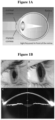
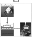


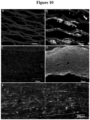

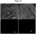

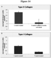
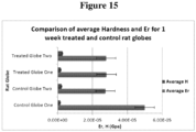
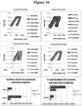
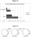

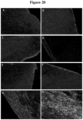

Classifications
-
- A—HUMAN NECESSITIES
- A61—MEDICAL OR VETERINARY SCIENCE; HYGIENE
- A61K—PREPARATIONS FOR MEDICAL, DENTAL OR TOILETRY PURPOSES
- A61K38/00—Medicinal preparations containing peptides
- A61K38/16—Peptides having more than 20 amino acids; Gastrins; Somatostatins; Melanotropins; Derivatives thereof
- A61K38/17—Peptides having more than 20 amino acids; Gastrins; Somatostatins; Melanotropins; Derivatives thereof from animals; from humans
- A61K38/18—Growth factors; Growth regulators
- A61K38/1841—Transforming growth factor [TGF]
-
- A—HUMAN NECESSITIES
- A61—MEDICAL OR VETERINARY SCIENCE; HYGIENE
- A61K—PREPARATIONS FOR MEDICAL, DENTAL OR TOILETRY PURPOSES
- A61K31/00—Medicinal preparations containing organic active ingredients
- A61K31/56—Compounds containing cyclopenta[a]hydrophenanthrene ring systems; Derivatives thereof, e.g. steroids
- A61K31/57—Compounds containing cyclopenta[a]hydrophenanthrene ring systems; Derivatives thereof, e.g. steroids substituted in position 17 beta by a chain of two carbon atoms, e.g. pregnane or progesterone
- A61K31/573—Compounds containing cyclopenta[a]hydrophenanthrene ring systems; Derivatives thereof, e.g. steroids substituted in position 17 beta by a chain of two carbon atoms, e.g. pregnane or progesterone substituted in position 21, e.g. cortisone, dexamethasone, prednisone or aldosterone
-
- A—HUMAN NECESSITIES
- A61—MEDICAL OR VETERINARY SCIENCE; HYGIENE
- A61K—PREPARATIONS FOR MEDICAL, DENTAL OR TOILETRY PURPOSES
- A61K45/00—Medicinal preparations containing active ingredients not provided for in groups A61K31/00 - A61K41/00
- A61K45/06—Mixtures of active ingredients without chemical characterisation, e.g. antiphlogistics and cardiaca
-
- A—HUMAN NECESSITIES
- A61—MEDICAL OR VETERINARY SCIENCE; HYGIENE
- A61P—SPECIFIC THERAPEUTIC ACTIVITY OF CHEMICAL COMPOUNDS OR MEDICINAL PREPARATIONS
- A61P27/00—Drugs for disorders of the senses
- A61P27/02—Ophthalmic agents
-
- A—HUMAN NECESSITIES
- A61—MEDICAL OR VETERINARY SCIENCE; HYGIENE
- A61P—SPECIFIC THERAPEUTIC ACTIVITY OF CHEMICAL COMPOUNDS OR MEDICINAL PREPARATIONS
- A61P27/00—Drugs for disorders of the senses
- A61P27/02—Ophthalmic agents
- A61P27/10—Ophthalmic agents for accommodation disorders, e.g. myopia
-
- A—HUMAN NECESSITIES
- A61—MEDICAL OR VETERINARY SCIENCE; HYGIENE
- A61P—SPECIFIC THERAPEUTIC ACTIVITY OF CHEMICAL COMPOUNDS OR MEDICINAL PREPARATIONS
- A61P43/00—Drugs for specific purposes, not provided for in groups A61P1/00-A61P41/00
-
- A—HUMAN NECESSITIES
- A61—MEDICAL OR VETERINARY SCIENCE; HYGIENE
- A61K—PREPARATIONS FOR MEDICAL, DENTAL OR TOILETRY PURPOSES
- A61K2300/00—Mixtures or combinations of active ingredients, wherein at least one active ingredient is fully defined in groups A61K31/00 - A61K41/00
-
- C—CHEMISTRY; METALLURGY
- C07—ORGANIC CHEMISTRY
- C07K—PEPTIDES
- C07K14/00—Peptides having more than 20 amino acids; Gastrins; Somatostatins; Melanotropins; Derivatives thereof
- C07K14/435—Peptides having more than 20 amino acids; Gastrins; Somatostatins; Melanotropins; Derivatives thereof from animals; from humans
- C07K14/475—Growth factors; Growth regulators
- C07K14/495—Transforming growth factor [TGF]
Definitions
- the present disclosure relates to compositions and methods useful for the treatment and/or prevention of conditions of the eye.
- the disclosure relates to compositions and methods that can be used in augmenting and regenerating the cornea, and in correcting refractive errors of the eye.
- the cornea of the eye accounts for more than two-thirds of the eye's total refractive power (focusing power). Even small changes in corneal shape can have a dramatic effect on the clarity with which an image is brought to focus on the retina.
- the stromal layer of the cornea (the clear front surface of the eye) comprises the majority of the corneal tissue and is composed of highly organised lamellae which are made up of tightly packed collagen fibrils, mostly of collagen types I and V (Marshall et al. 1993).
- the unique structure of the stromal layer as a result of the uniform alignment of the collagen fibrils confers the properties of toughness and transparency on the cornea (Funderburgh 2000).
- stromal cells the corneal keratocytes
- stromal cells the corneal keratocytes
- stromal cells the corneal keratocytes
- myofibroblast form Another commonly observed phenotype of keratocytes is the myofibroblast form that is seen in the cornea after injury (Jester et al. 1987). Changes in exogenous growth factors and cytokines are thought to bring about these phenotypic changes (Funderburgh et al. 2001).
- TGF ⁇ family of growth factors are known to be the most potent inducers of chondrogenic (cartilage) differentiation (Heng, Cao, & Lee 2004; Johnstone et al. 1998; Menetrey et al. 2000).
- TGF ⁇ 1 stimulates the synthesis of collagens and fibronectin by chick embryo fibroblasts (Ignotz and Massague 1986).
- TGF ⁇ 1 and TGF ⁇ 2 are known to cause ECM deposition associated with scarring, possibly due to conversion of keratocytes into the myofibroblast phenotype (Funderburgh, Mann, Funderburgh, Corpuz, & Roth 2001).
- TGF ⁇ 3 has been shown to induce corneal fibroblasts to produce ECM depositions made up of collagen type I without fibrosis or scarring (Karamichos, Hutcheon, & Zieske 2011).
- Certain non-proteinaceous chemical compounds such as dexamethasone (Johnstone, Hering, Caplan, Goldberg, & Yoo 1998), ascorbic acid (Farquharson, Berry, Barbara Mawer, Seawright, & Whitehead 1998), and ethanol (Kulyk & Hoffman 1996) are also known to promote chondrogenic differentiation in vitro.
- Myopia results from excessive curvature of the cornea so that light entering the eye focuses in front of the retina. It is the most prevalent vision impairment worldwide affecting the vision of 70 to 90% of people in some Asian countries and 30 to 40% in Europe and the United States (Frederick 2002). In most cases, myopia first occurs in school-age children and progresses until about the age of 20. It is also associated with increased prevalence of macular degeneration, retinal detachment, and glaucoma in adulthood (Ebenstein & Pruitt 2006).
- Myopia is most commonly corrected by the use of prescription eye glasses or contact lenses. However, these devices do not provide permanent treatment for the condition, and they are unsuitable for use during certain activities. Contact lenses are also associated with ophthalmic infections and more serious conditions, including corneal abrasions and ulcers. In certain circumstances, refractive surgery or orthokeratology is indicated for myopia. Still, these treatments provide only a temporarily correction for mild to moderate myopia; they are not permanent treatments, and they are unsuitable for severe cases.
- Keratoconus is an ecstatic corneal dystrophy associated with stromal thinning and disruption of the portion of the cornea known as Bowman's layer.
- the progressive thinning of the corneal stroma typically occurs over decades and results in the cornea developing a conical shape. This results in an impairment of vision due to irregular astigmatism and myopia.
- the pathogenesis of keratoconus is still unknown but has been associated with factors such as constant eye rubbing and contact lens wear (Krachmer, Feder, & Belin 1984; Sherwin & Brookes 2004). It can appear as early as puberty and continues to progress until the third or fourth decade of life.
- Keratoconus is a major indication for cornea transplantation in the Western world, determined by researchers to constitute 28.8% of corneal transplantation in France (vonis et al. 2001) and from 11.4% to 15.4% in the United States (Cosar et al. 2002; Dobbins et al. 2000).
- changes associated with keratoconus include a decrease in the number of lamellae and keratocytes (Ku, Niederer, Patel, Sherwin, & McGhee 2008; Sherwin & Brookes 2004), and changes in organisation of the lamellae and distribution of collagen fibrillary mass (Meek et al. 2005).
- Keratoconus corneas are known to have decreased levels of enzyme inhibitors and an increased level of degradative enzymes (Kenney & Brown 2003). Biomechanical factors include thinning and decreased rigidity of the cornea due to oxidative damage caused by ultraviolet radiation and mechanical trauma (Kenney & Brown 2003). Biomechanical investigation of keratoconic corneas has revealed a decrease in elasticity and stiffness; however the reasons for this remain unknown (Edmund 1988). It has been suggested that a reduction in collagen cross-links could be a cause (Wollensak & Buddecke 1990). Currently there is no satisfactory animal model for keratoconus and investigations have been largely limited to an ex vivo setting.
- keratoconus is the leading indication for corneal transplantation surgery worldwide, with about 12-20% of those affected by keratoconus requiring a corneal transplant (Pramanik, Musch, Sutphin, & Farjo 2006).
- CXL corneal collagen crosslinking
- the UV-A light activates the riboflavin thereby producing reactive oxygen species that induce the formation of covalent bonds between the collagen molecules in the corneal stroma (Spoerl, Huhle, & Seiler 1998; G. Wollensak et al. 2003).
- This procedure is not recommended for the treatment of corneas thinner than 400 pm due to the possibility of endothelial cell damage. Although this treatment leads to a stiffer cornea, it does not address the problem of corneal thinning.
- compositions and methods for modulating corneal cells to alter collagen expression and extracellular matrix formation in corneal tissue. These compositions and methods are useful for regenerating and/or augmenting the cornea, and thereby treating and/or preventing various conditions of the cornea and refractive errors of the eye.
- the invention provides a composition comprising a TGF ⁇ 3 polypeptide, and dexamethasone or any salts, esters, or hydrides thereof, for use in a method of: (i) treating or preventing thinning or irregularity of a cornea of a subject; or (ii) treating or preventing a refractive error of the eye of a subject,
- the invention provides a combination of:
- the invention provides a composition comprising a TGF ⁇ 3 polypeptide, and dexamethasone or any salts, esters, or hydrides thereof, for use in a method of treating or preventing thinning or irregularity of a cornea of a subject,
- the TGF ⁇ 3 polypeptide consists of the amino acid sequence of SEQ ID NO: 1.
- the dexamethasone is dexamethasone phosphate.
- composition comprises 10 to 100 ng/ml of the TGF ⁇ 3 polypeptide.
- the composition comprises 40 to 4000 ng/ml dexamethasone.
- composition is formulated as an eye drop.
- composition is formulated with gellan gum.
- composition is administered once daily or twice daily.
- composition is co-administered with one or more additional agents for the eye.
- the one or more additional agents for the eye are selected from the group consisting of: anaesthetic agents, anti-inflammatory agents, anti-microbial agents, and lubricants.
- composition for use is administered in conjunction with use of a contact lens, corneal insert, corneal implant, or intrastromal ring.
- the contact lens, corneal insert, corneal implant, or intrastromal ring is adapted for moulding or holding corneal shape during and/or following treatment with the composition for use.
- the contact lens, corneal insert, corneal implant, or intrastromal ring is adapted to act as a carrier for the composition for use or as a composition eluting device.
- composition for use is administered in conjunction with corneal collagen crosslinking.
- composition for use is prior to and/or subsequent to crosslinking.
- the condition is selected from the group consisting of: keratoconus, myopia, and astigmatism.
- the invention provides a combination of:
- the invention provides a composition comprising a TGF ⁇ 3 polypeptide, and dexamethasone or any salts, esters, or hydrides thereof, for use in a method of treating or preventing a refractive error of the eye of a subject, wherein:
- the TGF ⁇ 3 polypeptide consists of the amino acid sequence of SEQ ID NO: 1.
- the dexamethasone is dexamethasone phosphate.
- composition comprises 10 to 100 ng/ml of the TGF ⁇ 3 polypeptide.
- the composition comprises 40 to 4000 ng/ml dexamethasone.
- composition is formulated as an eye drop.
- composition is formulated with gellan gum.
- composition is administered once daily or twice daily.
- composition is co-administered with one or more additional agents for the eye.
- the one or more additional agents for the eye are selected from the group consisting of: anaesthetic agents, anti-inflammatory agents, anti-microbial agents, and lubricants.
- composition for use is administered for use in conjunction with a contact lens, corneal insert, corneal implant, or intrastromal ring.
- the contact lens, corneal insert, corneal implant, or intrastromal ring is adapted for moulding or holding corneal shape during and/or following treatment with the composition.
- the contact lens, corneal insert, corneal implant, or intrastromal ring is adapted to act as a carrier for the composition or as a composition eluting device.
- the method is performed preceding or following refractive surgery.
- the refractive error of the eye is associated with one or more of: myopia, hyperopia, astigmatism, and presbyopia.
- the invention provides a combination of:
- the invention provides a kit comprising: (i) a composition comprising a TGF ⁇ 3 polypeptide, and dexamethasone or any salts, esters, or hydrides thereof; and (ii) a contact lens, corneal insert, corneal implant, or intrastromal ring, wherein:
- the contact lens, corneal insert, corneal implant, or intrastromal ring is adapted for moulding or holding corneal shape during and/or following treatment with the composition.
- the contact lens, corneal insert, corneal implant, or intrastromal ring act as a carrier for the composition or as a composition eluting device.
- the TGF ⁇ 3 polypeptide consists of the amino acid sequence of SEQ ID NO: 1.
- the dexamethasone is dexamethasone phosphate.
- composition comprises 10 to 100 ng/ml of the TGF ⁇ 3 polypeptide.
- the composition comprises 40 to 4000 ng/ml dexamethasone.
- composition is formulated as an eye drop.
- composition is formulated with gellan gum.
- composition is formulated for administration once daily or twice daily.
- composition is co-formulated with one or more additional agents for the eye.
- the kit includes one or more additional agents for the eye.
- the one or more additional agents for the eye are selected from the group consisting of: anaesthetic agents, anti-inflammatory agents, anti-microbial agents, and lubricants.
- the kit includes a contact lens solution.
- the kit includes instructions for use.
- the kit is used for the treatment or prevention of a refractive error of the eye.
- the kit is used for the treatment or prevention of a corneal condition selected from the group consisting of: keratoconus, myopia, hyperopia, astigmatism, presbyopia, and stromal dystrophies.
- the kit is used for the treatment of a corneal condition selected from the group consisting of: an abrasion, tear, ulcer, bum, puncture, corneal melt, and surgical injury.
- Augmenting refers to methods of increasing one or more of the thickness, hardness, elastic modulus, tensile strength, and regularity of the cornea, including the corneal tissue (e.g., the stromal layer). Augmentation may be used to impose a particular shape to the cornea, i.e., corneal curvature. Augmentation methods may be performed in the presence or absence of a particular condition of the eye, or of the cornea. Augmentation may involve the increase in components in the extracellular matrix of the cornea (e.g., collagen type II). Augmentation may also involve increasing the number of cells (e.g., keratocytes) in the cornea. The number of cells may be increased, for example, by altering the proliferative state of such cells from quiescent to active.
- the corneal tissue e.g., the stromal layer
- Augmentation may be used to impose a particular shape to the cornea, i.e., corneal curvature. Augmentation methods may be performed in the presence or absence of a
- Co-administration refers to the combined use of agents, for example, therapeutic agents for the eye, and includes the administration of coformulations (i.e., combination formulations), as well as the simultaneous or sequential administration of separate formulations.
- coformulations i.e., combination formulations
- in conjunction refers to the combined use of a therapeutic composition and a therapeutic device/procedure. This can include use of the composition preceding use of the device/procedure, simultaneously with the device/procedure, and/or following use of the device/procedure.
- a “condition" of the cornea refers to a state of disease, defect, damage, injury, degeneration, or dysfunction of the cornea.
- the condition may affect the corneal tissue (e.g., the stromal layer) or corneal cells (e.g., keratocytes).
- the condition may be an acute condition, for example, an abrasion or ulceration, or may be a chronic condition, for example, keratoconus or myopia.
- the "cornea” as used herein refers to the transparent front part of the eye that covers the iris, pupil, and anterior chamber of the eye. It includes the corneal epithelium, Bowman's layer, corneal stroma, Descemet's membrane, and the corneal epithelium. Of particular interest is the stromal layer (also called the substantia intestinal ) of the cornea, which comprises an extracellular matrix of regularly arranged collagen fibres along with keratocytes,
- a “derivative”, as relating to a chemical derivative, refers to a compound that has been chemically modified.
- the present disclosure encompasses each of the chemical compounds described herein as well as any derivatives thereof, including chemically modified forms such as salts, hydrides, esters, and other modifications of the original compound.
- isolated refers to a molecule that is separated from its natural environment.
- An isolated molecule may be obtained by any method or combination of methods as known and used in the art, including biochemical, recombinant, and synthetic techniques.
- the polypeptides may be prepared by at least one purification or enrichment step.
- polypeptides produced by chemical synthesis or by plasmids, vectors, or other expression constructs that may be introduced into a cell or cell-free translation system.
- Such polypeptides are clearly distinguished from polypeptides as they naturally occur, without human intervention.
- protein or “polypeptide” (e.g., SEQ ID NO.T), and other such terms, for simplicity, refer to the molecules described herein. Such terms are not meant to provide the complete characterization of these molecules.
- a protein or polypeptide may be characterised herein as having a particular amino acid sequence, a particular 2-dimensional representation of the structure, but it is understood that the actual molecule claimed has other features, including 3-dimensional structure, mobility about certain bonds and other properties of the molecule as a whole. It is the molecules themselves and their properties as a whole that are encompassed by this disclosure.
- protein and “polypeptide” are used interchangeably herein.
- TGF ⁇ 3 refers to polypeptides obtained from any source, e.g., isolated naturally occurring polypeptides, recombinant polypeptides, and synthetic polypeptides, and to include polypeptides having the naturally occurring amino acid sequence as well as polypeptides having variant amino acid sequences, and fragments of such sequences, as described in detail herein.
- TGF ⁇ 3 may also be referred to in the art as transforming growth factor-beta3, TGFB3, ARVD, and FLJ16571.
- sequence identity refers to the amino acid to amino acid comparison of two or more polypeptides.
- a test sequence may be identical to a reference sequence (i.e., share 100% identity), or may include one or more amino acid substitutions. In preferred aspects, amino acid substitutions may possess similar chemical and/or physical properties such as charge or hydrophobicity, as compared to the reference amino acid.
- Sequence identity may be typically determined by sequence alignments at the regions of highest homology. Sequence alignment algorithms, for example BLAST ® sequence alignment programs, are well known and widely used in the art. Based on the sequence alignment, the percent identity can be determined between the compared polypeptide sequences.
- Refractive errors may include spherical errors and cylindrical errors. Both lower order aberrations and higher order aberrations are included.
- refractive errors are the conditions of the eye noted as myopia, hyperopia, astigmatism, anisometropia, and presbyopia.
- Regeneration in relation to the cornea, refers to the restoration of one or more of the shape, thickness, regularity, hardness, elastic modulus, and tensile strength of the cornea, including that of the corneal tissue (e.g., the stromal layer).
- Methods of regeneration may be used to impose a particular shape to the cornea, i.e., corneal curvature.
- Regeneration methods may be performed in the treatment of a particular condition of the eye, or of the cornea. Regeneration may involve the increase in components in the extracellular matrix of the cornea (e.g., collagen type II). Regeneration may also involve increasing the number of cells (e.g., keratocytes) in the cornea. The number of cells may be increased, for example, by altering the proliferative state of such cells from quiescent to active.
- Reprogramming of cells for example, for corneal cells (e.g., keratocytes) refers to changes in the state of differentiation. Reprogramming is associated with one or more changes in cell morphology, cellular gene expression (e.g., collagen expression, including collagen type I and/or type II expression), or the cells proliferative state (e.g., quiescent or active).
- corneal cells e.g., keratocytes
- Reprogramming refers to changes in the state of differentiation. Reprogramming is associated with one or more changes in cell morphology, cellular gene expression (e.g., collagen expression, including collagen type I and/or type II expression), or the cells proliferative state (e.g., quiescent or active).
- subject refers to a human or non-human animal.
- Preventing refers to stopping or delaying the onset of a condition, for example an eye condition, or particularly a corneal condition, such as a disorder or other defect of the cornea.
- a preventative measure will result in the stoppage or delay of one or more symptoms of the condition, or a lessening of symptoms if such do arise.
- Prevention of a corneal condition may involve augmenting the cornea, as described in detail herein.
- Treating refers to reducing, ameliorating, or resolving a condition, for example an eye condition, or particularly a corneal condition, such as a disorder or other defect of the cornea.
- a treatment will result in the reduction, amelioration, or elimination of one or more symptoms of the condition.
- Treatment of a corneal condition may involve regeneration of the cornea, as detailed herein.
- the compositions for use of the invention may be for treating various conditions, for preventing various conditions, or for both treating and preventing various conditions, as described in detail herein.
- compositions and methods for modulating cells using in situ cell reprogramming in order to affect collagen type II expression and extracellular matrix (ECM) deposition in corneal tissue This, in turn, is used to strengthen and/or augment the cornea of the eye.
- ECM extracellular matrix
- compositions, combinations and kits may be utilised in in vivo tissue engineering therapy for treating or preventing thinning or irregularity of the cornea associated with keratoconus or for treating or preventing a refractive error of the eye associated with myopia.
- myopia is characterised by the excessive curvature of the cornea ( Figure 1A ), whil e keratoconus is a progressive ectatic corneal dystrophy leading to a characteristic pattern of corneal thinning ( Figure IB; image adapted from Romero-Jimenez, Santodomingo-Rubido, & Wolffsohn 2010).
- Corneal keratocytes are relatively quiescent and normally only produce large amounts of extracellular matrix (ECM) when they switch to a fibroblast or myofibroblast phenotype. ECM deposition associated with those phenotypes usually leads to corneal fibrosis and loss of transparency (Kadler, Baldock, Bella, & Boot-Handford 2007). Chondrocytes, the cells that make up cartilage, secrete type II collagen which is a fibrillar collagen similar to type I found in the cornea. Type II collagen is also expressed by keratocytes during development of the chick cornea and it is only later replaced by type I in the mature chick stroma (Linsenmayer et al. 1990).
- ECM extracellular matrix
- stromal cells from adult human and rat corneas can be reprogrammed to produce neuron specific proteins when treated with neuronal lineage specifying growth factors (Greene et al. 2013). This data demonstrates that an adult cell population can be reprogrammed simply by the modulation of the growth factor environment both in vitro and in vivo.
- corneal stromal cells can be induced in vitro and ex vivo to produce cartilage specific fibrillar collagen, collagen type II, by treating the cells with transforming growth factor ⁇ 3 (TGF ⁇ 3) and dexamethasone (Examples 8 and 9).
- TGF ⁇ 3 transforming growth factor ⁇ 3
- dexamethasone examples 8 and 9
- the inventors have demonstrated that keratocytes in human keratoconic corneal biopsies express collagen type II when treated with these two compounds (Example 8).
- the inventors have demonstrated that the two compounds of TGF ⁇ 3 and dexamethasone can be delivered in vivo using eye drops to stimulate collagen II deposition (Example 9).
- the deposition of collagen type II was uniform, improving the biomechanics of the cornea, with no fibrosis or scarring, and no effect on corneal transparency (Examples 11 and 13).
- chondrocytes secrete type II collagen which is not only a fibrillar collagen similar to type I found in the cornea, but is also expressed during development of the chick cornea (Linsenmayer et al. 1990). It is only later replaced by collagen type I in the mature stroma (Linsenmayer et al. 1990).
- the disclosed treatment module aims not only to stabilise the cornea, but also to provide remedial aid for conditions of the eye, including various corneal conditions and refractive errors of the eye.
- the compositions for use of the invention may be, for example, for the treatment of keratoconic keratocytes in the ectatic cornea. Additionally, the compositions for use of the invention may be for the treatment of myopia and various other conditions of the cornea, as described in detail herein.
- compositions and combinations for use of the invention are for use in treating or preventing thinning or irregularity of a cornea of a subject, or treating or preventing a refractive error of the eye of a subject.
- exemplary conditions include keratoconus, as described in detail herein, and related conditions, which include corneal ectasias such as keratoglobus, pellucid marginal degeneration, and posterior keratoconus (see, e.g., Arffa 1997; Krachmer et al. 1984; Rabonitz 2004; Jinabhai et al. 2010).
- corneal ectasias such as keratoglobus, pellucid marginal degeneration, and posterior keratoconus
- Specifically included as defects are myopia, presbyopia, and also astigmatisms, which encompass regular and irregular astigmatisms.
- cornea plana and microcomea the latter of which may be associated with fetal alcohol syndrome, Turner syndrome, Ehlers-Danlos syndrome, Weill-Marchesani syndrome, Waardenburg's syndrome, Nance-Horan syndrome, and Cornelia de Lange's syndrome.
- keratoglobus (mentioned above) that may be associated with Ehlers-Danlos syndrome type IV.
- compositions for use of the invention may be to prevent corneal damage. Damage or degeneration may be associated with a particular condition of the cornea. Specifically included is corneal melt, for example, corneal melt associated with an inflammatory disorder, such as rheumatoid arthritis.
- keratitis such as marginal keratitis, stromal keratitis, exposure keratitis, neurotrophic keratitis, filamentary keratitis, rosacea keratitis, viral keratitis including herpes keratitis, fungal keratitis, protozoal keratitis, and other infectious keratitis, such as luetic interstitial keratitis, microsporidial keratitis, Thygeson's keratitis, and infectious crystalline keratopathy.
- keratitis such as marginal keratitis, stromal keratitis, exposure keratitis, neurotrophic keratitis, filamentary keratitis, rosacea keratitis, viral keratitis including herpes keratitis, fungal keratitis, protozoal keratitis, and other infectious keratitis, such as luetic
- ulcerative keratitis also called peripheral ulcerative keratitis (PUK), which includes ulcerative keratitis that is associated with a systemic disease, such as rheumatoid arthritis Wegener's granulomatosis, systemic lupus erythematosus, relapsing polychondritis, and polyarteritis nodosa. Endophthalmitis is also included. Included as well are chronic corneal edema, Mooren's ulcer, dellen, phlyctenulosis, Terrien's degeneration, Salzman's degeneration, spheroidal degeneration, and Fuch's dystrophy. Such conditions are well known and well characterised in the art.
- stromal dystrophies for example, lattice corneal dystrophy (e.g., type 1 and type 2), granular corneal dystrophy (e.g., type 1 and type 2), macular corneal dystrophy, Schnyder corneal dystrophy, congenital stromal corneal dystrophy, and fleck corneal dystrophy.
- the invention encompasses compositions for use in treating or preventing refractive errors of the eye.
- refractive errors may be associated with particular conditions, including myopia, hyperopia, presbyopia, anisometropia, higher order aberrations, and various astigmatisms.
- Higher order aberrations include, but are not limited to, comas, trefoils, quadrafoils, spherical aberrations, and aberrations identified by mathematical expressions (e.g., Zemike polynomials).
- Conditions of the cornea may be diagnosed by various methods, including fluorescein staining, which may include a Seidel's test, specular microscopy, corneal topography, isometric tomography, pachymetry, ultrasound, slit lamps, corneal scrapes, and biopsies. Diagnosis may also involve assessments for visual acuity and/or opacification. Corneal conditions may be associated with one or more symptoms of: pain, photophobia, foreign body sensation, reduced visual acuity, oedema, white cell infiltrate, fluorescein uptake, vascularisation, redness, and systemic symptoms such as headaches, nausea, and fatigue.
- fluorescein staining which may include a Seidel's test, specular microscopy, corneal topography, isometric tomography, pachymetry, ultrasound, slit lamps, corneal scrapes, and biopsies. Diagnosis may also involve assessments for visual acuity and/or opacification. Corneal conditions
- symptoms of refractive errors may include but are not limited to: reduced visual acuity as well as blurry vision, double vision, haziness of vision, visual fatigue, foreign body sensation, problematic glare or halos, starburst patterns, ghost images, impaired night vision, squinting, excessive staring, excessive blinking, headaches, eye rubbing, eye strain, eye surface dessication, eye irritation, redness, and spasms of the eye.
- composition for use may be formulated to include the noted combination of components (a TGF ⁇ 3 polypeptide plus dexamethasone (or any salts, esters, or hydrides thereof)), or may be formulated to include a first component (a TGF ⁇ 3 polypeptide or alternatively dexamethasone (or any salts, esters, or hydrides thereof)) with the second component to be added in prior to administration.
- a combination for use may be formulated to include a first component (a TGF ⁇ 3 polypeptide or alternatively dexamethasone (or any salts, esters, or hydrides thereof)), which is used in simultaneous or sequential administration with a formulation that includes the second component.
- the TGF ⁇ 3 polypeptide may include at least the following amino acid sequence: ALDTNYCFRN LEENCCVRPL YIDFRQDLGW KWVHEPKGYY ANFCSGPCPY LRSADTTHST VLGLYNTLNP EASASPCCVP QDLEPLTILY YVGRTPKVEQ LSNMVVKSCK CS (SEQ ID NO:1) (GenBank Reference CAR70088.1).
- the TGF ⁇ 3 polypeptide may include at least 112 amino acids shown above, and may have a molecular mass of 25.5 kDa.
- the TGF ⁇ 3 polypeptide may be derived from amino acids 644-850 (207 amino acids) of the precursor polypeptide sequence identified in GenBank Reference CAA33024.1; GenBank Accession No. CAA33024; or NCBI Reference Sequence NP_003230.1.
- the TGF ⁇ 3 polypeptide shares at least 90% sequence identity to the amino acid sequence of SEQ ID NO: 1
- the TGF ⁇ 3 polypeptide comprises at least 100 amino acids of the amino acid sequence of SEQ ID NO: 1.
- the TGF ⁇ 3 polypeptide or the variant or fragment thereof may be provided as a recombinant polypeptide.
- the polypeptide may be expressed in cell or cell-free expression systems as widely known and used in the art. Included amongst these are bacterial, fungal, plant, and mammalian expression systems. Expression systems using E. coli cells, CHO cells, HEK cells, and Nicotiana benthamiana cells are specifically included.
- the TGF ⁇ 3 polypeptide or the variant or fragment thereof may be provided as a human recombinant polypeptide expressed in human or non-human expression systems.
- the TGF ⁇ 3 polypeptide or the variant or fragment thereof may be provided as a disulfide-linked homodimeric, non-glycosylated, polypeptide chain, in accordance with known methods.
- the TGF ⁇ 3 polypeptide or the variant or fragment thereof may be isolated from recombinant expression systems by standard methods, including well known chromatographic techniques.
- the TGF ⁇ 3 polypeptide or the variant or fragment thereof may include a sequence tag to facilitate cleavage, isolation, and/or localisation of the polypeptide.
- the TGF ⁇ 3 polypeptide may be obtained from various commercial sources.
- recombinant human TGF ⁇ 3 may be obtained from R&D Systems (Catalogue Nos. 243-B3-002; 243-B3-010), BioVision, Inc. (Catalogue Nos. 4344-500; 4344-50; 4344-5), or Prospec Protein Specialists (Catalogue Nos. CYT-113; CYT-319).
- the biological activity of the TGF ⁇ 3 polypeptide or the variant or fragment thereof may be measured in accordance with widely known and used methods.
- biological activity may be measured in culture by the polypeptides ability to inhibit the mink lung epithelial (MvlLu) cells proliferation (see, e.g., Premaraj et al. 2006).
- Exemplary activity by this measurement is shown by an ED50 of ⁇ 50 ng/ml.
- biological activity may be measured by the dose-dependent inhibition of IL-4 induced proliferation of mouse HT-2 cells (BALB/c spleen activated by sheep erythrocytes in the presence of IL-2) (see, e.g., Tsang et al. 1995).
- Exemplary activity by this measurement is typically 0.1 to 0.5 ng/ml.
- the composition that includes the combination of agents described herein may be measured for biological activity using the methods noted below.
- induction of collagen type II e.g., collagen type II, alpha 1 in keratocytes can be assessed by one or more of: immunohistochemical assays, protein assays, Western blot analysis, polymerase chain reaction (PCR) analysis, and quantitative PCR technologies.
- composition may also include dexamethasone, or any salts, esters, or hydrides thereof.
- Dexamethasone is characterised as having the following chemical structure:
- dexamethasone examples include, for example, Decadron ® , Dexasone ® , Diodex ® , Hexadrol ® , Maxidex ® , and Minims ® .
- derivatives of dexamethasone may be used, including any esters and salts thereof.
- exemplary derivatives include but are not limited to: dexamethasone-17-acetate ( CAS RN: 1177-87-3 ), dexamethasone disodium phosphate ( CAS RN: 2392-39-4 ), dexamethasone valerate ( CAS RN: 14899-36-6 ), dexamethasone- 21-isonicotinate ( CAS RN: 2265-64-7 ), dexamethasone palmitate ( CAS RN: 33755-463 ), dexamethasone propionate ( CAS RN: 55541-30-5 ), dexamethasone acefurate ( CAS RN: 83880-70-0 ), dexamethasone-21-galactoside ( CAS RN: 92901-23-0 ), dexamethasone 21-thiopivalate, dexamethasone 21-thiopentanoate, dexa
- the composition may include related steroidal agents in addition to dexamethasone.
- other corticoid steroids may be utilised, along with dexamethasone.
- Preferred for use as related steroids are Group C steroids according to Coopman classification, which includes betamethasone-type steroids, such as dexamethasone, dexamethasone sodium phosphate, betamethasone, betamethasone sodium phosphate, and fluocortolone.
- Other related steroidal agents include but are not limited to: fluoromethalone, lotoprendol, medrysone, prednisolone, prednisone, rimexolone, hydrocortisone, lodoxamide, or any derivative or combination thereof.
- fluoromethalone acetate Specifically included are fluoromethalone acetate, fluoromethalone alcohol, prednisolone acetate, prednisolone sodium phosphate, lotoprendol etabonate, hydrocortisone acetate, and lodoxamide tromethamine.
- the composition may include, for example, 0.04 ng/ml to 4 ng/ml; or 0.04 ng/ml to 0.4 ng/ml; or 0.4 ng/ml to 4 ng/ml; or 4 to 40 ng/ml; or 40 ng/ml to 400 ng/ml, or 40 ng/ml to 4000 ng/ml dexamethasone, or derivative thereof or related steroidal agent; or about 0.04 ng/ml, about 0.08 ng/ml, about 0.12 ng/ml, about 0.4 ng/ml, about 0.8 ng/ml, about 1.2 ng/ml, about 4 ng/ml, about 12 ng/ml, about 24 ng/ml, about 40 ng/ml, about 80 ng/ml, about 120 ng/ml, about 240 ng/ml, about 400 ng/ml, about 800 ng/ml, about 1000 ng/ml, about 1600 ng/
- the composition may include 0.4 pg/ml to 40 pg/ml; or 0.4 p.g/ml to 4 pg/ml; or 4 pg/ml to 40 pg/ml dexamethasone, or derivative thereof or related steroidal agent; or about 0.4 pg/ml, about 0.8 pg/ml, about 1 pg/ml, about 1.2 pg/ml, about 2 pg/ml, about 4 pg/ml, about 8 pg/ml, about 12 pg/ml, about 20 Ug/ml, or about 40 pg/ml dexamethasone, or any salt, ester, or hydride thereof.
- the composition may include 0.1 mg/ml to 1 mg/ml; or 0.5 mg/ml to 5 mg/ml; or 1 mg/ml to 10 mg/ml; dexamethasone, or any salt, ester, or hydride thereof; or about 0.1 mg/ml, about 0.5 mg/ml, about 1 mg/ml, about 2 mg/ml, about 5 mg/ml, or about 10 mg/ml dexamethasone, or any salt, ester, or hydride thereof.
- the composition may include, for example, 1 ng/ml to 1 pg/ml; or 1 ng/ml to 10 ng/ml; or 10 ng/ml to 100 ng/ml; or 100 ng/ml to 1 pg/ml TGF ⁇ 3 polypeptide as defined herein, or about 1 ng/ml, about 5 ng/ml, about 10 ng/ml, about 20 ng/ml, about 50 ng/ml, about 100 ng/ml, about 200 ng/ml, about 500 ng/ml, about 800 ng/ml, or about 1 ug/ml TGF ⁇ 3 polypeptide.
- the composition may include at least 40 ng/ml dexamethasone, or derivative thereof or related steroidal agent, along with at least 4 ng/ml TGF ⁇ 3 polypeptide.
- the composition may also include one or more anti-inflammatory agents.
- anti-inflammatory agents include ketotifen fumarate, diclofenac sodium, flurbiprofen sodium, ketorlac tromethamine, suprofen, celecoxib, naproxen, rofecoxib, or any derivative or combination thereof. Particularly included are nonsteroidal anti-inflammatory drugs (NSAIDs).
- the composition may additionally include one or more anaesthetic agents.
- Exemplary anaesthetics include topical anaesthetics such as proparacaine, lidocaine, and tetracaine, and any derivative or combination thereof.
- Other agents for the eye may be selected for inclusion with the composition; these may be chosen by the skilled artisan based on the condition and needs of the subject under treatment.
- compositions as described herein may be formulated for topical administration, as described herein and in accordance with known methods. In certain circumstances, intraocular administration may be desirable.
- the composition may be provided in any form suitable for administration to the eye. Exemplary formulations include, at least, solutions, suspensions, emulsions (dispersions), gels, creams, or ointments in a suitable ophthalmic vehicle.
- the composition may be provided in the form of eye drops, a semisolid gel, or a spray.
- moulding contact lenses or other inserts/implants may be impregnated with the composition for use of the invention. In this manner, the composition can be delivered to the cornea continuously and in a time-release manner as the subject is wearing the contact lenses.
- the compositions may be formulated with a pH range of 5.0 to 8.0. This pH range may be achieved by the addition of buffers to the solution. It is preferred that the formulations are stable in buffered solutions. That is, there is no adverse interaction between the buffer and the active agents that would cause the composition to be unstable, e.g., by precipitation or aggregation.
- the composition may be hypertonic (5% to 40%, preferably approximately 10, 20, 30, or 40%) or hypotonic (0% to 5%, preferably approximately 1, 2, 3, or 4%) depending on the needs of the subject (e.g., working needs, rest hours, sleeping, etc.)
- a hypertonic composition (e.g., 40%) may be used when combined with moulding contact lenses, as described in detail herein.
- compositions may include one or more suitable preservatives as optional ingredients. Suitable preservatives may be added to prevent contamination, for example, bacterial contamination. Such agents may include, but are not limited to, benzalkonium chloride, thimerosal, chlorobutanol, methyl paraben, propyl paraben, phenylethyl alcohol, EDTA, sorbic acid, Onarner ® M, and other agents known to those skilled in the art, or any combination thereof. Such preservatives may be typically employed at a level of 0.001% to 1.0% by weight of the composition.
- compositions may contain an optional co-solvent.
- the solubility of the components of the present compositions may be enhanced by a surfactant or other appropriate co-solvent in the composition.
- co-solvents/surfactants include, for example, polysorbate 20, 60, and 80, polyoxyethylene/polyoxypropylene surfactants (e.g. Pluronic ® F-68, F-84, and P-103), cyclodextrin, tyloxapol, and other agents known to those skilled in the art, and any combination thereof.
- co-solvents may be typically employed at a level of 0.01% to 2% by weight of the composition.
- Penetration enhancing agents may be used to increase uptake of the composition into the eye.
- Exemplary agents include, at least, cetylpyridinium chloride, ionophores such as lasalocid, benzalkonium chloride, Parabens, Tween 20, saponins, Brij 35, Brij 78, Brij 98, ethylenediaminetetraacetic acid, bile salts, and bile acids (such as sodium cholate, sodium taurocholate, sodium glycodeoxycholate, sodium taurodeoxycholate, taurocholic acid, chenodeoxycholic acid, and ursodeoxycholic acid), capric acid, azone, fusidic acid, hexamethylene lauramide, saponins, hexamethylene octanamide, and decylmethyl sulfoxide.
- bioadhesive polymers may be used to adhere to the mucin coat covering the eye, to prolong contact of the composition with the eye.
- Bioadhesive polymers may be macromolecular hydrocolloids with numerous hydrophilic functional groups, such as carboxyl-, hydroxyl-, amide, and sulphate capable of establishing electrostatic interactions.
- Exemplary agents include, at least, polyarylic acid (e.g., carbopol, carbophil, and polycarbophil) and carboxymethyl cellulose.
- Controlled release systems may also be used; such systems may involve in situ gels, colloidal particles, nanoparticles, and/or niosomes.
- Other drug delivery systems include but are not limited to: non-erodible ocular inserts, erodible ocular inserts, hydrogels, collagen shields, liposomes, drug-loaded films (e.g., NOD ® ), and ionotophoresis.
- compositions may include, also, an optional agent to increase viscosity.
- Viscosity increased above that of simple aqueous solutions may be desirable to increase ocular absorption of the active compounds, to decrease variability in dispensing the formulation, to decrease physical separation of components of a suspension or emulsion of the formulation and/or to otherwise improve the ophthalmic formulation.
- Such viscosity builder agents include as examples polyvinyl alcohol, polyvinyl pyrrolidone, methyl cellulose, hydroxy propyl methylcellulose, hydroxyethyl cellulose, carboxymethyl cellulose, hydroxy propyl cellulose, other agents known to those skilled in the art, or a combination thereof. Such agents may be typically employed at a level of 0.01% to 2% by weight of the composition.
- the compositions include a gelling agent, for example, high molecular weight water-soluble polysaccharides such as gellan gum.
- Gellan gum may be obtained from various commercial sources, for example, as sold under the trade name Kelcogel ® .
- Kelcogel ® LT 100 may be used as a fine mesh, high acyl gellan, which forms soft, elastic, non-brittle gels.
- the gellan gum based composition is formulated as a 0.5% eye drop
- EDTA sodium chloride
- tyloxapol sodium sulfate
- hydroxyethylcellulose may have additional beneficial effects of further stabilising the composition.
- compositions for use are used to treat or prevent thinning or irregularity of a cornea of a subject; or treating or preventing a refractive error of the eye of a subject.
- the compositions described herein may be used to address various conditions of the cornea and correct refractive errors of the eye, and may be used as adjunct therapy with other eye treatments.
- the composition for use may be formulated in any suitable means for administration to the eye. Included as formulations are ophthalmic solutions, creams, emulsions, ointments, and gels. Specifically noted are formulations that are made as eye drops. In a particular aspect, the composition for use may be administered as an eye drop using any of the many types of eye drop dispensers on the market. As exemplifications, the container for the compositions for use of the invention may be clear, translucent, and opaque and may contain other properties or combination of properties such as being glass lined, tamper proof, packaged in single or few dose aliquots, and any combination thereof.
- the composition for use may be administered in therapeutically effective amounts to a subject to achieve a desired medical outcome.
- the composition for use may be administered in amounts to address an ophthalmic condition described herein, or at least mitigate one or more symptoms of such condition.
- the precise dosage of the composition for use i.e., amount and scheduling
- Exemplary formulations e.g., eye drops
- eye drops may be administered 1 to 24 times per day, or 1 to 12 times per day, or 1 to 6 times per day, or 1 to 4 times per day, or 1 to 3 times per day, or 1 to 2 times per day, or 1, 2, 3 4, 6, 8,12, 18, or 24 times per day.
- the composition for use may be topically applied as an eye drop by placing one drop in each eye to be treated. Alternatively, 2 to 3 drops may be applied to each eye.
- the dosage range may be, for example, 0.2 pg to 2.4 ng; or 2 pg to 2.4 ng of dexamethasone, or any salt, ester, or hydride thereof; or about 0.2 pg, about 0.4 pg, about 0.6 pg, about 0.8 pg, about 1.2 pg, about 2.4 pg, about 2 pg, about 4 pg, about 6 pg, about 8 pg, about 12 pg, about 18 pg, about 24 pg, about 0.2 ng, about 0.26 ng, about 0.4 ng, about 0.6 ng, about 0.8 ng, about 1.2 ng, about 1.8 ng, or about 2.4 ng of dexamethasone, or any salt, ester, or hydride thereof, per eye for one dose.
- the dosage range may be 12 ng to 1.3 pg; or 6 ng to 600 ng dexamethasone, or derivative thereof or related steroidal agent; or about 6 ng, about 8 ng, about 12 ng, about 16 ng, about 18 ng, about 24 ng, about 26 ng, about 30 ng, about 36 ng, about 40 ng, about 48 ng, about 52 ng, about 54 ng, about 60 ng, about 72 ng, about 78 ng, about 80 ng, about 90 ng, about 120 ng, about 130 ng, about 160 ng, about 180 ng, about 240 ng, about 260 ng, about 300 ng, about 360 ng, about 400 ng, about 480 ng, about 520 ng, about 540 ng, about 600 ng, about 720 ng, about 780 ng, about 900 ng, about 1.2 ⁇ g, about 1.3 ⁇ g of dexamethasone, or any salt, ester, or hydr
- the dosage range may be 1.5 pg to 150 pg; 2.6 pg to 260 pg; or 6.5 pg to 650 pg of dexamethasone, or derivative thereof or related steroidal agent; or about 1.5 pg, about 2 pg, about 3 pg, about 4.5 pg, about 6 pg, about 6.5 pg, about 7.5 pg, about 10 pg, about 15 pg, about 22.5 pg, about 32.5 pg, about 20 pg, about 26 pg, about 30 pg, about 40 pg, about 45 pg, about 60 pg, about 65 pg, about 75 pg, about 80 pg, about 90 pg, about 100 pg, about 120 pg, about 130 pg, about 150 pg, about 180 pg, about 225 pg, about 240 pg, about 260 pg, about 200 pg, about 300 p
- any of the above noted dosages of dexamethasone may be co-administered with a dosage range of, for example, 5 pg to 65 ng; or 0.5 ng to 65 ng of TGF ⁇ 3 polypeptide, or variants or fragments thereof; or about 5 pg, about 10 pg, about 15 pg, about 20 pg, about 30 pg, about 45 pg, about 60 pg, about 0.05 ng, about 0.1 ng, about 0.15 ng, about 0.2 ng, about 0.3 ng, about 0.45 ng, about 0.5 ng, about 0.6 ng, about 0.65 ng, about 1 ng, about 1.5 ng, about 2 ng, about 3 ng, about 4 ng, about 4.5 ng, about 6 ng, about 6.5 ng, about 7.5 ng, about 9 ng, about 10 ng, about 12 ng, about 13 ng, about 15 ng, about 16 ng, about 20 ng, about 22.5 ng,
- Dosage for one eye may be about one drop of the disclosed composition for use.
- One drop of composition may be 10 pl to 200 pl, 20 pl and 120 pl, or 50 pl to 80 pl or any values in between.
- dispensers such as pipettors can dispense drops from 1 pl to 300 pl and any value in between.
- the dispenser metes out about 15 pl, about 20 pl, about 30 pl, about 45 pl, about 60 pl, or about 65 pl per drop of the disclosed composition.
- the contact lens or insert/implant may include, for example, 0.01 mg to 10 mg of dexamethasone, or any salt, ester, or hydride thereof; or about 0.01 mg, about 0.1 mg, about 0.5 mg, about 0.7 mg, about 1 mg, about 5 mg, or about 10 mg of dexamethasone, or any salt, ester, or hydride thereof.
- the contact lens or insert/implant may include 10 ng to 100 ng of dexamethasone, or any salt, ester, or hydride thereof; or about 1 ng, about 5 ng, about 10 ng, about 20 ng, about 50 ng, about 80 ng, or about 100 ng dexamethasone, or any salt, ester, or hydride thereof.
- the contact lens or insert/implant may include about 10 ng to 1 pg of TGF ⁇ 3 polypeptide; or about 10 ng, about 50 ng, about 100 ng, about 200 ng, about 500 ng, about 800 ng, or about 1 pg TGF ⁇ 3 polypeptide as defined herein.
- compositions for use described herein may be used in conjunction with various surgical procedures or other treatments.
- the compositions for use can be used along with surgical and non-surgical methods for the refractive correction of the eye.
- Exemplary methods include but are not limited to: radial keratotomy (RK), including mini asymmetric radial keratotomy (MARK), hexagonal keratotomy (HK), photorefractive keratectomy (PRK), keratomilleusis, laser in situ keratomileusis (LASIK), e.g., intraLASIK ® , laser epithelial keratomileusis (LASEK), e.g., Epi- LASEK, automated lamellar keratoplasty (ALK), laser thermal keratoplasty (LTK), conductive keratoplasty (CK), limbal relaxing incisions (LRI), astigmatic keratotomy (AK), epikeratophakia, anterior ciliary sclerotomy (ACS), s
- compositions for use may be used in conjunction with surgical correction of non-refractive conditions, for example, surgical correction of a corneal tear.
- the compositions for use described herein may be used in conjunction with specific surgical methods performed on the cornea. Exemplary methods include but are not limited to: corneal transplant surgery, penetrating keratoplasty (PK), phototherapeutic keratectomy (PTK), pterygium excision, corneal tattooing, keratoprosthesis insertion (e.g., KPro or Dohlman-Doane), and osteo-odonto-keratoprosthesis insertion (OOKP).
- PK penetrating keratoplasty
- PTK phototherapeutic keratectomy
- pterygium excision corneal tattooing
- keratoprosthesis insertion e.g., KPro or Dohlman-Doane
- OKP osteo-odonto-keratoprosthesis insertion
- compositions for use may be used in conjunction with corneal collagen crosslinking.
- Corneal crosslinking typically involves the use of riboflavin solution activated by exposure to UV-A light.
- Noted crosslinking methods include but are not limited to: corneal crosslinking with the epithelium removed (Dresden protocol, or epi- off), transepithelial crosslinking (epi-on), and accelerated crosslinking.
- Crosslinking procedures are generally available, and marketed as CXL, C3-R ® CCL ® and KXL ® corneal crosslinking, amongst others.
- Administration of the composition may be prior to, and/or subsequent to, the crosslinking procedure.
- the disclosed compositions can be used to avoid or counter the deleterious effects of crosslinking procedures, such as stromal haze and cell loss (described in more detail, below).
- corneal regeneration with the disclosed compositions can allow crosslinking to be performed on subjects who were previously ineligible for such procedures, e.g., those with corneal thickness less than 400 pm.
- the disclosed compositions can be used to slow or halt progressive corneal thinning, which would not be addressed by the use of crosslinking on its own.
- compositions for use described herein may be co-administered with one or more additional agents for the eye.
- co-administration may be by simultaneous or subsequent administration with such agents, or by co-formulation with such agents.
- the compositions for use described herein may be co-administered with one or more agents, which include but are not limited to: antihistamines, sympathomimetics, beta receptor blockers, parasympathomimetics, parasympatholytics, prostaglandins, nutrients, vasoconstrictors, lubricants, anti-microbials, and anaesthetics.
- agents include but are not limited to: antihistamines, sympathomimetics, beta receptor blockers, parasympathomimetics, parasympatholytics, prostaglandins, nutrients, vasoconstrictors, lubricants, anti-microbials, and anaesthetics.
- antiinflammatory agents including nonsteroidal anti-inflammatory drugs (NSAIDs).
- Non-limiting examples of anaesthetics include: benzocaine, bupivacaine, cocaine, etidocaine, lidocaine, mepivacaine, pramoxine, prilocalne, chloroprocaine, procaine, proparacaine, ropicaine, and tetracaine.
- Non-limiting examples of antiinflammatory agents include: aspirin, acetaminophen, indomethacin, sulfasalazine, olsalazine, sodium salicylate, choline magnesium trisalicylate, salsalate, diflunisal, salicylsalicylic acid, sulindac, etodolac, tolmetin, diclofenac, ketorolac, ibuprofen, naproxen, flurbiprofen, ketoprofen, fenoprofen, suprofen, oxaproxin, mefenamic acid, meclofenamic acid, oxicams, piroxicam, tenoxicam, pyrazolidinediones, phenylbutazone, oxyphenthatrazone, pheniramine, antazoline, nabumetone, COX-2 inhibitors (Celebrex ® ), apazone, nimesulide, and zileu
- anti-microbial agents include but are not limited to: bacitracin zinc, chloramphenicol, chlorotetracycline, ciprofloxacin, erythromycin, gentamicin, norfloxacin, sulfacetamide, sulfisoxazole, polymyxin B, tetracycline, tobramycin, idoxuridine, trifluridine, vidarabine, acyclovir, foscamet, ganciclovir, natamycin, amphotericin B, clotrimazole, econazole, fluconazole, ketoconazole, miconazole, flucytosine, clindamycin, pyrimethamine, folinic acid, sulfadiazine, and trimethoprimsulfamethoxazole.
- vasoconstrictors include but are not limited to: dipivefrin (Propine ® ), epinephrine, phenylephrine, apraclonidine, cocaine, hydroxyamphetamine, naphazoline, tetrahydrozoline, dapiprazole, betaxolol, carteolol, levobunolol, metipranolol, and timolol.
- Nutrients include vitamins, minerals, and other beneficial agents such as vitamin A, vitamin Bi, vitamin Bö, vitamin B 12, vitamin C (ascorbic acid), vitamin E, vitamin K, and zinc.
- composition for use described herein is formulated as eye drops, and such eye drops are used in conjunction with other eye drop formulations.
- eye drops may include but are not limited to: rinse/lubricating eye drops, dry eye treatments, steroid and antibiotic eye drops, glaucoma eye drops, allergy/anti- inflammatory eye drops, and conjunctivitis eye drops.
- compositions for use may be used in conjunction with contact lenses, corneal inserts, corneal implants, or intrastromal rings, to assist in supporting or reshaping the subject's cornea.
- corneal inserts include corneal inlay and corneal onlay devices.
- contact lenses, intrastromal rings, or other inserts/implants may be used for moulding or holding corneal shape preceding, during, and/or following treatment with the composition.
- a corneal 'insert' typically refers to a temporary device inserted into the cornea
- a corneal 'implant' typically refers to a more permanent device.
- many well known devices are described interchangeably in the art as implants/inserts. Therefore, the terms 'insert/implant' as used herein are not to be deemed as strictly limiting based on time of usage.
- the contact lens, corneal insert, corneal implant, or intrastromal ring may be used as a kit with the disclosed composition in treating or preventing thinning or irregularity of a cornea of a subject; or treating or preventing a refractive error of the eye of a subject.
- the contact lens, corneal insert, corneal implant, or intrastromal ring may act as a carrier for the composition or as a composition eluting device.
- the contact lens, intrastromal ring, or other corneal insert/implant may be utilised with the composition that is suitable for administration to the eye, e.g., eye drops, as described in detail herein.
- computer software may be used to determine the contact lenses, corneal inserts, corneal implants, or intrastromal rings that are most suitable for the subject and/or to determine the formulation of the composition.
- treatment utilising contact lenses, corneal inserts, corneal implants, or intrastromal rings along with the composition described herein is used preceding or following eye surgery, e.g., refractive or transplant surgery.
- the treatment may involve assessing the subject (e.g., age, working needs of the subject, eye defect or disease, etc.), prescribing the use of moulding contact lenses, corneal inserts, corneal implants, or intrastromal rings to assist with the needed changes in the radius of curvature of the anterior surface of the cornea, and prescribing the composition described herein to be used in conjunction with the contact lenses or implants/inserts.
- the contact lenses or implants/inserts which are prescribed and utilised by the subject can be used exert a mechanical force on the cornea thereby inducing a change in shape, i.e., the refractive power, of the cornea.
- the cornea may be supported or shaped by use of a corneal insert, corneal implant, or intrastromal ring in conjunction with the disclosed composition.
- a corneal insert examples include INTACS ® and KeraRing intrastromal corneal rings.
- a moulding contact lens may be used in conjunction with the disclosed composition.
- the contact lens may be hard or rigid, or it may be a soft lens. Alternatively, the contact lens may comprise both hard and soft portions. If a soft contact lens is used, more positive or negative curvature can be induce in the cornea, and the discomfort in the subject's eyes will diminish as he or she adapts to the contact lenses. If a hard contact lens is used, more mechanical pressure can be exerted on the cornea.
- the contact lenses may be gas permeable.
- Moulding contact lenses may be obtained from commercial sources.
- Commercially available lenses include, at least, DreamLite, OK Lens, EyeDream, MiracLens, DreamLens, i-GO OVC, GOV, Wake and See, CRT, Fargo/iSee, Emerald and Wave Contact Lens System lenses.
- the composition described herein may be ad mini stered to the eye.
- the contact lenses, intrastromal rings, or other inserts/implants and the composition may be used in conjunction to produce a change in the shape, and thereby the refractive power, of the cornea.
- the composition may be administered more frequently to allow the cornea to adopt the desired change in shape.
- the composition is administered at least every 24,12, or 8 hours.
- the composition is administered every 6 hours.
- the composition is administered approximately every 3 hours.
- the composition is administered approximately every 2 hours.
- the composition is administered every hour.
- the combined use of contact lenses, corneal inserts, corneal implants, or intrastromal rings and the composition described herein may induce changes in the molecular structure of the cornea and may induce changes in the cells and proteins such as collagen (e.g., collagen type II) found in the corneal stroma.
- collagen e.g., collagen type II
- the surface of the cornea is thereby made more uniform. By reducing irregularities in the surface of the cornea, the quality and clearness of all images (i.e., visual acuity) is improved.
- the base curve of the moulding contact lens may be calculated based on the change in the refractive power for each eye separately.
- the base curve of the moulding contact lens may be calculated starting with one to four flatter or steeper diopters, more preferably one to three flatter or steeper diopters, even more preferably one to two flatter or steeper diopters, depending on the refractive error that is required.
- the peripheral base curve depends on the adaptation of the moulding contact lens and is calculated to be 0.5 mm of radius greater than the central zone, but can vary depending on the design.
- the diameter of the moulding contact lens used in accordance with the invention may be from 8.0 mm to 18.0 mm. Commercially available lenses are produced with such diameters.
- the moulding contact lens may be a hard contact lens with a diameter ranging from 8.0 mm to 12.0 mm.
- the moulding contact lens may be a soft contact lens with a diameter ranging from 13.0 mm to 15.0 mm. Soft contact lenses may cover the entire cornea and go from sclera to sclera.
- the moulding contact lens may be comprised of hard and soft materials.
- the contact lens may be hard in the centre, out to approximately 12.0 mm, 13.0 mm, 14.0 mm, or 15.0 mm, and then soft in the periphery out to 16.0 mm, 17.0 mm, and 18.0 mm.
- a larger contact lens, preferably a soft contact lens, may be used at night as a moulding contact lens.
- the power of the moulding contact lenses can be determined to the nearest possible refractive power that the subject requires to see comfortably.
- the subject is prescribed eyeglasses while the subject is undergoing treatment.
- various optometric measurements may be repeated to confirm that the treatment is progressing as planned and is adequate.
- Such measurements may include assessment of visual acuity for near and far vision, orthotypes, keratometry measurements, objective and subjective retinoscopy, diagrams of the adaptation of the moulding contact lens, movement of the moulding contact lens, and comfort of the moulding contact lens.
- changes may be made to the treatment program based on these measurements. With each evaluation, a decision may be made whether to continue with the same moulding contact lens or whether a new contact lens should be used. In addition, the same decision can be made with regard to the composition being used with the moulding contact lenses. Changes in the moulding contact lenses and/or in the composition can be made to induce the desired reshaping of the cornea over several weeks. In certain aspects, weekly periodic revisions are performed during the first 8 weeks after beginning treatment.
- the composition as described herein induces changes in the collagen content of the cornea (e.g., collagen type II). Other aspects of the anatomy, histology, and physiology of the cornea may also be affected by composition.
- the composition may be hypertonic or hypotonic to induce changes in corneal hydration.
- the composition may change the molecular structure of the cornea (e.g., the extracellular matrix) and in this way augment or repair the cornea, or reshape the cornea to the desired curvature.
- exemplary enzymes include but are not limited to hyaluronidase, chondroitinase ABC, chondroitinase AC, keratanse, and stromelysin, which have been shown to work on various proteoglycan components of the cornea. Included also are the enzyme collagenase, matrix metalloproteinase 1 (interstitial collagenase), and matrix metalloproteinase 2 (gelatinase).
- composition for use is coadministered with any such enzymes
- a vehicle such as a polymer (e.g., methylcellulose, polyvinyl alcohol, cellulose, etc.) in the composition for use to enhance the working of such enzymes.
- Additional agents may be included to activate metalloproteinase enzymes, e.g., interleukin-la, tumour necrosis factor a/ ⁇ and any subtypes thereof, monosodium urate monohydrate, 4-amino phenylmercuric acetate, human serum amyloid A, human B2 microglobin, and copper chloride.
- carbamide urea
- composition for use may also be co-administered with one or more enzymes that degrade other sugars or proteins found in the cornea.
- the composition for use may be co-administered with one or more anaesthetics used to reduce the irritation of the moulding contact lens or any corneal insert/implant to the cornea.
- the composition for use may be co-administered with one or more lubricants to improve the comfort of the subject during the treatment.
- the composition for use may be co-administered with one or more anti-microbial agents such as anti-bacterial, anti-viral, and/or anti-fungal agents.
- the composition for use may also be co-administered with one or more vasoconstrictors. The person of skill in the art can determine the appropriate agents for co-administration to the subject based on the condition being treated.
- the composition may be provided in a kit as defined in the claims.
- the kit may include one or more of; moulding contact lenses, lubricating eye drops, cleaning or other solutions for the contact lenses, a contact lens carrying case, an extra pair of contact lenses, and instructions for wearing the contact lenses and using the composition.
- the composition provided with the kit may be formulated to include the noted combination of components (a TGF ⁇ 3 polypeptide as defined herein plus dexamethasone (or any salt, esters, or hydrides thereof)).
- Cadaveric whole human corneas, keratoconic corneas obtained at the time of transplant surgery, human limbal rims and surgeon cut DSEK caps (excess stromal tissue from Descemets stripping endothelial keratoplasty) were obtained from donors sourced through the New Zealand National Eye Bank (Auckland, New Zealand). Human limbal rims were collected after the central corneal button had been removed for corneal transplantation surgery leaving a 2 mm corneal margin from the limbal junction. Prior to the use of tissue, research ethics approval and consent was obtained from the Northern X Regional Human Ethics Committee.
- Corneal and cartilage pieces (2 mm ⁇ 2 mm) were embedded in Optimal Cutting Temperature compound (OCT, Tissue-Tek, Sakura, The Netherlands) before being snap frozen in liquid nitrogen. Sections 10-15 ⁇ m thick were cut using a Microm HM550 Cryostat (Thermo-Scientific, USA) and mounted on SuperFrost TM Plus electrostatic slides (Menzel-Glenser, Germany). Cryosections were stored at -20°C until further use.
- OCT Optimal Cutting Temperature compound
- Limbal rims were dissected to isolate stroma from sclera in a class II laminar flow hood. Following this, the corneal epithelium and endothelium was gently scraped off with a keratome and discarded. DSEK caps also received gentle scraping with a keratome to remove the epithelium. Remaining stromal tissue was then digested in 0.4% type II collagenase (Sigma-Aldrich), in Hanks balanced salt solution (GIBCO ® , Life Technologies) at 37°C with gentle mixing on an orbital shaker. A variety of digestion times were used with 5 hours being the time required for optimal tissue digestion and cell viability.
- the cells were pelleted by centrifuging at 1200 rpm for seven minutes. The cells were then resuspended in a minimal amount of an appropriate cell culture medium and counted using a Leica DM IL bench top inverted microscope and a Neubauer hemocytometer. A 1:1 ratio of cell suspension added to trypan blue solution (0.04% trypan blue stock in PBS) was used with a minimum of three counts per sample and the average value taken.
- Isolated keratocytes were cultured in either 12 or 24 well cluster plates (Falcon) on plastic or glass coverslips in 2-3 ml of cell culture media. Cells were kept in a humidified incubator at 37°C with 5% CO 2 . Culture media was changed after 24 hours then every two days subsequently or more frequently if required. Cultures were viewed daily with a Leica DM IL bench top inverted microscope. For cell pellet culture, freshly obtained cells after tissue were pelleted by centrifuging at 300 g for 7 minutes at 20°C in a plastic conical tube. Appropriate culture media was added to the tubes.
- the cells After 24 hours of incubation at 37°C, the cells had contracted and formed a pellet which did not adhere to the walls of the tube.
- the pellets were cultured in 2 ml of media in a humidified atmosphere of 5% CO 2 at 37°C for three weeks. Media was changed every other day.
- Human and rat corneal and cartilage tissue was thin-sliced (1-2 mm) in an anteroposterior plane with a blade and the slices were placed in an organotypic air-liquid interphase culture system ( Figure 2 ). Briefly, the explants of healthy tissue were cultured on 0.4 ⁇ m pore size cell culture inserts (Millicell, France) at the interface between culture medium and a CO 2 rich environment. Corneal sections were placed epithelium side up on cell culture plate inserts with 3 ml of culture medium. The culture media was changed every other day.
- Table 1 Cell culture media used Name Medium Base Other Components Fibroblast proliferation medium Dulbecco's Modified Eagle Medium (DMEM) (Life Technologies, GIBCO ® ) 10% FBS, 1% Anti-anti (100X stock), 1% GlutaMAX TM (100X stock) Chondrogenic reprogramming medium Advanced DMEM (Life Technologies, GIBCO ® ) 10 ng/ml TGF ⁇ 3 (Abeam, ab52313), 10- 7 M dexamethasone (Abeam, ab120743), 1% GlutaMAX TM (Life Technologies, GIBCO ® ) (100X stock), 1% Anti-Anti (100X stock) (Life Technologies, GIBCO ® ) Control medium Dulbecco's Modified Eagle Medium (DMEM) 1% GlutaMAX TM (100X stock), 1% Anti-Anti (100X stock)
- DMEM Dulbecco's Modified Eagle Medium
- DMEM Dulbecco's Modified Eagle Medium
- Tissue slices were cultured in the chondrogenic differentiation medium for varying time intervals to determine the optimum time required for the growth factor treatment. Samples were collected for each time point (Table 2). For obtaining a monolayer of cells, keratocytes were seeded on glass coverslips at a density of 15 ⁇ 10 4 per cm 2 . The cells were allowed to attach to the coverslips for 24 hours and culture media was changed every other day. Cultures were maintained for up to 3 weeks.
- Table 2 Experimental time points for corneal tissue slice culture Time point 1 Week 1 Time point 1 Week 2 Time point 1 Week 3 Sample 1 chondrogenic differentiation medium control medium control medium Sample 2 chondrogenic differentiation medium control medium Sample 3 chondrogenic differentiation medium chondrogenic differentiation medium chondrogenic differentiation medium Sample 4 control medium control medium control medium
- Gellan gum which is a water soluble polysaccharide produced by the bacterium, Pseudomonas elodea.
- the use of gel base formulation allows a prolonged corneal residence time and increased ocular bioavailability of the therapeutic agent.
- polymeric gellan gum is an anionic polymer it undergoes in situ gelling in the presence of mono- and divalent cations such as Ca 2+ , Mg 2+ , K + , and Na + (Bakliwal, & Pawar 2010) .
- the electrolytes present in the tear fluid cause the gelation of the polymer when it is instilled in the eye and this in turn results in a longer residence time and increased bioavailability of the drug (Ludwig 2005).
- the polymer formulation is a non-irritant and safe for in vivo use (Rupenthal, Green, & Alany 2011).
- a 0.5% solution was prepared by first heating distilled water to 80°C followed by the addition of gellan gum (Kelcogel TM USA) with constant stirring. Once the powder was completely dissolved, the solution was cooled and stored at 4°C. The appropriate amounts of growth factors were added to the runny gel with constant stirring. A ten times higher concentration of growth factors than that used in the culture medium was used to make up for the drug lost through naso-lacrymal drainage and blinking.
- the eye drop gel included a final concentration of 100 ng/ml TGF ⁇ 3 and approximately 4 ⁇ g/ml dexamethasone.
- the animals were manually restrained and approximately 15 ⁇ L of the eye drops were instilled in the right eye ( Figure 3 ).
- the contra lateral eye was used as the control eye.
- Thrice daily eye drops were administered for up to 5 days for neuronal specification and for up to 8 weeks for chondrogenic specification.
- the animals were euthanised using a carbon dioxide chamber.
- the eyes were harvested and rinsed in phosphate buffered saline.
- the corneas were then dissected out carefully and fixed in 4% paraformaldehyde (PFA) for 1 hour and treated with sucrose solution in order to cryoprotect the tissue before freezing and sectioning.
- PFA paraformaldehyde
- sucrose as a cryoprotection is a dehydrant that prevents the formation of ice crystal artefact in frozen tissue sections. In the case of slow freezing of the tissue cryoprotection is particularly important.
- the corneas were immersed in 20% sucrose solution for 5 hours at 4°C and then moved to a 30% sucrose solution and kept at 4°C until the tissue sinks (usually overnight).
- the corneas were then embedded in OCT compound and immersed in liquid nitrogen to bring about rapid freezing.
- the frozen blocks of tissue were stored at -80°C until further use.
- Approximately 10-15 ⁇ m thick cryostat sections were mounted on SuperFrost TM Plus slides and the slides were stored at -80°C until needed.
- the cells cultured on coverslips were rinsed with PBS and fixed with 4% PFA for 15 minutes. Coverslips were immersed in PBS until further use.
- tissue cryosections before carrying out immunohistochemistry, the slides were kept at room temperature for 15-20 minutes.
- the OCT was washed off using PBS and the zone around the tissue demarcated using a wax pen.
- the tissue slices were first incubated with a blocking solution of 10% normal goat serum for 1 hour followed by overnight incubation with the appropriate dilution of primary antibody at 4°C.
- the slides were then rinsed three times in PBS before incubation with the appropriate dilution of secondary antibody.
- the secondary antibody was left on for 2 hours at room temperature.
- Slices were counterstained with the nuclear marker 4', 6'-diamidino-2-phenylindol (DAPI) and mounted in Citifluor antifade agent (ProSciTech, Australia).
- DAPI nuclear marker 4', 6'-diamidino-2-phenylindol
- tissue samples were mixed with 0.75 ml TRIzol ® and carrier RNA and homogenised using a hand held homogeniser (PRO Scientific, Inc.). The samples where then incubated with 0.2 ml of chloroform followed by centrifugation at 12000 rpm and 4°C for 15 minutes. The upper phase was separated and was mixed with ethanol and then transferred to the collection column tube.
- the SuperScript ® VILO TM cDNA Synthesis Kit (Invitrogen TM , Life Technologies) was used to prepare cDNA. Briefly, 100 ng of RNA was incubated at 25°C for 10 minutes with VILO TM Reaction Mix, SuperScript ® Enzyme Mix, and RNAse free water. The samples were then incubated at 42°C for 120 minutes followed by 85°C incubation for 5 minutes. The cDNA was stored at -20°C.
- TaqMan ® Gene Expression Assays for the genes of interest were obtained.
- 10 ⁇ L of TaqMan ® Universal Master Mix II was combined with 1 ⁇ L of the assay, approximately 25 ng of cDNA and 9 ⁇ L water to make up a volume of 20 ⁇ L.
- the tubes were vortexed and centrifuged briefly to spin down the contents.
- Each cDNA sample was prepared in triplicate and pipetted into a 3 84 well plate. 20 ⁇ L of each reaction mixture was loaded into each well of a MicroAmp ® Optical 384-Well Reaction Plate (Applied Biosystems).
- the plate was then covered with a MicroAmp ® Optical Adhesive Film (Applied Biosystems) and the plate was centrifuged briefly to eliminate air bubbles.
- the plate was transferred to the 7900HT Fast Real-Time PCR System and was run using the following thermal cycling parameters, 50°C for 2 min, 95°C for 10 minutes followed by 40 cycles of 95°C at 15 sec and 60°C at 1 minute. Results were analysed as described in the previous section.
- Table 4 TaqMan ® gene assays used for QPCR Gene symbol Gene name Assay ID Colla1 (Rat) Collagen, type I, alpha 1 Rn01463848_m1 Col2a1 (Rat) Collagen, type II, alpha 1 Rn01637085_m1 Col2a1 (Rat) Collagen, type II, alpha 1 Rn01637087_m1 Pop4 (Rat) Ribonuclease P protein subunit p29 (housekeeping gene) Rn02347225_m1 COL2A1 (Human) Collagen, type II, alpha 1 Hs00264051_m1 CDKN1A (Human) Cyclin-Dependent Kinase Inhibitor 1 (housekeeping gene) Hs00355782_m1
- Example 7 Testing of biomechanical and optical properties of corneas following in situ stromal ECM protein deposition
- Corneal biomechanics have been shown to be relevant in the diagnosis and treatment of various corneal diseases and provide insight into the structure of the cornea and its relation to corneal physiological function. Corneas that have undergone treatment to bring about the deposition of ECM protein also need to be tested for corneal opacity as reduced transparency would be undesirable.
- the Phoenix Micron IV Rodent eye Imaging System (Phoenix Research Labs) was used to examine the corneas of treated rats. Rats were first sedated using an intra-peritoneal injection of ketamine and Domitor ® (3:2). The slit-lamp attachment of the Micron IV imaging system was used to examine the layers of the cornea in detail and check corneal integrity and transparency. Retinal imaging was also done to check corneal transparency. Following imaging, the rats were administered Antisedan ® (atipamezole) for reversal of the sedative.
- Antisedan ® atipamezole
- Nanoindentation provides mechanical measurements of materials of interest through the application of ultra-small forces perpendicular to the sample plane of interest and measurement of the resultant sample indentation (Dias & Ziebarth 2013). Nanoindentation has recently emerged as a powerful tool for measuring nano- and microscale mechanical properties in tissues and other biomaterials (Ebenstein & Pruitt 2006). The more recent advancement of in situ scanning probe microscopy (SPM) imaging, where the nanoindenter tip is simultaneously used as a 3D imaging device combined with nanoindentation has enabled a new wave of novel materials research (Dickinson & Schirer 2009). Force, displacement, and time are recorded simultaneously while a nanoindentation tip is pushed into the corneal tissue under a controlled load.
- SPM in situ scanning probe microscopy
- Nanoindentation tests are output as a load-displacement curve which can be analysed using well defined equations to calculate the mechanical properties relating to rigidity, integrity, and elasticity of the cornea.
- Nanoindentation testing was carried out at the Chemical and Materials Engineering lab at the University of Auckland. In order to test the cornea in its natural position, a mould was required for nanoindentation. Previous studies have used polystyrene and blue tack to hold the corneas in place. The effect of the mould deforming under the load was a potential source of error so a hard mould was decided on for testing.
- the first material that was used to create a mould was conventional play dough. This was formed to the exact shape and curvature of human cornea samples ( Figure 6(A) ). The play dough was then left to harden over the next two days before being used in testing. The testing of the rat eyes was slightly different as the entire globe was used. To hold the globes in place a petri dish filled with a resin and having small indent to hold the globe was used ( Figure 6(B) ). PBS was used to keep the samples from drying out.
- the samples are very soft biological samples a conospherical fluid tip was used for all nanoindentation testing.
- the indent load used for the human samples was 50 ⁇ N.
- For the rat globes a range of loads between 3 and 5 ⁇ N were used.
- the fibre optic light was switched on and the sample placed directly under the stream of light from the microscope.
- the central section of the cornea was placed directly in the stream of light as accurately as possible ( Figure 6(C) ).
- the sample was focused by adjusting the Z slider until the surface of the cornea could be observed in good resolution. To ensure that the focus was on the central highest point of the cornea sample, the view was moved in the x and y directions to observe how the focus changed.
- the reduced elastic modulus tells us how elastic a sample is. Because the same indenter tip is used for each test the reduced elastic modulus can be used to compare the elasticity in each sample being tested.
- Example 8 Adult human corneal keratocytes produce cartilage specific collagen type II upon treatment with exogenous TGF ⁇ 3 and dexamethasone
- TGF ⁇ 3 and dexamethasone have been previously used to induce progenitor cells to differentiate into chondrocytes in vitro (Diekman, Rowland, Lennon, Caplan, & Guilak 2009; Johnstone et al. 1998; Kolambkar, Peister, Soker, Atala, & Guldberg 2007; Winter et al. 2003).
- dexamethasone a synthetic steroid drug has been used to treat inflammatory eye conditions. Therefore a combination of TGF ⁇ 3 and dexamethasone was used in the chondrogenic differentiation medium to drive the differentiation of keratocytes towards a chondrocyte phenotype.
- type I and type II collagen were specifically noted. It is known that fibrillar types of collagen such as types I and II self-assemble and crosslink to form highly crystalline fibres exhibit a very high stiffness, low extensibility and a remarkable elastic energy storage capacity (Wells 2003). It is the crosslinking which contributes towards the stiffness and tensile strength of the fibres.
- the corneal stromal extracellular matrix is composed of tightly packed heterotypic collagen fibrils made up mostly of collagen types I and V. Similar to corneal fibrils, cartilage fibrils are heterotypic (made up of types II and XI) and have a uniform diameter of 25 nm (slightly smaller than corneal fibrils) (Mendler, Eich-Bender, Vaughan, Winterhalter, & Bruckner 1989).
- Collagen II is the major fibril component of cartilage and is similar to collagen I in that the molecule essentially consists of a single uninterrupted helical domain 300 nm in length. Owing to their similarities, collagens II and XI are considered to be the cartilage analogues of collagens I and V (corneal stroma collagens) in other tissues.
- corneal keratocytes from adult corneas were seeded in either the chondrogenic differentiation medium containing TGF ⁇ 3 and dexamethasone or a standard fibroblast proliferation medium.
- the keratocytes seeded in the chondrogenic differentiation media formed cell aggregations/spheres ( Figure 7(A) ) approximately 50-100 ⁇ m in diameter.
- Figure 7(C) the fibroblast proliferation media cells from the spheres started spreading outwards ( Figure 7(C) ) to populate the culture dish thereby forming a cell monolayer.
- the regions where the cells had once been aggregated labelled for collagen type II whereas the cells in monolayer did not ( Figure 7(D) ).
- Keratocytes seeded in the fibroblast proliferating medium formed an even monolayer of fibroblast-like cells ( Figure 8(A) ) which did not label for either nestin or collagen type II ( Figure 8(B) ).
- Figure 8(A) Keratocytes seeded into fibroblast proliferation medium failed to form the necessary cell aggregations. Therefore, in order to form fibroblast clusters, the confluent fibroblasts were dissociated from the culture dish, pelleted, and grown as a pellet culture in chondrogenic differentiation medium for a further three weeks.
- Example 9 Keratocytes in adult human corneas and adult rat corneas secrete collagen type II containing ECM when treated with TGF ⁇ 3 and dexamethasone
- the amount and pattern of the native collagen type I appeared to be slightly altered in the treated corneas.
- the intensity of the labelling was similar but the distribution was more extensive and the amount of labelling was higher in the untreated corneas ( Figure 9(D) ).
- the newly produced type II collagen was laid evenly and in an ordered fashion in the ECM without forming any large masses or aggregates. The labelling was clearly seen along the pre-existing collagen framework of the corneal stroma and was distributed across the entire thickness of the stromal layer.
- the in vitro human corneal tissue experiment was then extended to an in vivo rodent study wherein the right corneas of male Wistar rats were treated for two weeks with a thrice daily administration of 15 ⁇ l of a gellan gum based eye drop formulation of TGFP3 and dexamethasone. After two weeks the rats were euthanised and the corneas processed for immunohistochemistry. Only the treated corneas labelled positive for collagen type II with a higher degree of deposition observed in the anterior part of the cornea ( Figure 10(D) and (E) ). Thus, only corneal slices cultured in the chondrogenic differentiation medium were positive for type II collagen. Furthermore, type II collagen was laid down in uniform layers along the pre-existing collagen framework of the stroma.
- Example 10 Induction of collagen type II deposition in keratoconic corneas
- Vimentin labelling revealed stark differences between keratocytes in the untreated and treated keratoconic corneas.
- the keratocyte density was lower in the untreated corneas with a scarcity of cells in the posterior part of the cornea ( Figure 11(C) ).
- the keratocytes in treated corneas appeared more filamentous and complete in morphology when compared to keratocytes in untreated corneas ( Figure 11(E) and(F)). Keratocytes in treated corneas were longer and had a larger number of cell processes which labelled strongly for Vimentin when compared to the keratocytes in the untreated corneas.
- Example 11 TGF ⁇ 3 and dexamethasone treatment does not induce deposition of fibrotic proteins or cause corneal opacity
- Example 12 Change in mRNA expression of collagen type II and type I upon treatment in vivo
- Rat corneas which were treated in vivo for 1 week, 7 weeks, and 3 weeks followed by a non-treatment period of 4 weeks were subjected to quantitative gene expression analysis. The aim was to determine whether type II collagen expression decreases again and/or permanently ceases after growth factor treatment is withdrawn. The effect of the treatment on native corneal collagen type II was also investigated.
- the 1 week treated corneas When compared to the 7 weeks treated corneas, the 1 week treated corneas expressed very high levels of type II collagen. The expression levels dropped considerably upon withdrawal of the treatment as indicated by the first graph in figure 35.
- type I collagen expression the 1 week and 7 week treated corneas were each compared to their untreated corneas. It was found that there was an initial spike in type I Collagen expression after 1 week treatment but by week 7 type I Collagen expression was significantly lower and comparable to its expression in the untreated cornea ( Figure 14 (B) ).
- Example 13 Change in biomechanical properties of in vitro and in vivo treated corneas
- Example 14 Comparative combinations of growth factors and steroids
- Fresh sheep eyes were obtained from Auckland Meat Processors. The corneas were immediately excised and washed with povidone-iodine (PVP-1) and sodium thiosulphate solution. Then, 8 mm discs of sheep corneal tissue were cut using a trephine. One sheep corneal disc was placed in each of the culture conditions (outlined in Table 5) for 3 weeks. The corneal discs were then placed in an organotypic air-liquid interphase culture system.
- PVP-1 povidone-iodine
- sodium thiosulphate solution 8 mm discs of sheep corneal tissue were cut using a trephine.
- One sheep corneal disc was placed in each of the culture conditions (outlined in Table 5) for 3 weeks. The corneal discs were then placed in an organotypic air-liquid interphase culture system.
- the explants of healthy tissue were cultured on 0.4 ⁇ m pore size cell culture inserts (Millicell, France) at the interface between culture medium and a CO 2 rich environment. Corneal sections were placed epithelium side up on cell culture plate inserts with 3 ml of culture medium. The culture media was changed every other day.
- the basal medium used was Dulbecco's Modified Eagle Medium (DMEM) supplemented with 1% Anti-Anti (antibiotic-antimycotic solution) and 1% GlutaMAX TM (GIBCO ® ).
- DMEM Dulbecco's Modified Eagle Medium
- Anti-Anti antibiotic-antimycotic solution
- GlutaMAX TM GlutaMAX TM
- the corneas were immersed in 20% sucrose solution for 5 hours at 4°C and then moved to a 30% sucrose solution and kept at 4°C until the tissue sank (usually overnight).
- the corneas were then embedded in OCT (optimal cutting temperature) compound and immersed in liquid nitrogen to bring about rapid freezing.
- the frozen blocks of tissue were stored at -80 °C until further use.
- Approximately 4-6 40 ⁇ m thick cryostat sections were mounted on SuperFrost TM Plus slides and the slides were stored at -80 °C until needed. The corneal sections were then labelled for collagen type II.
- the slides were kept at room temperature for 15-20 minutes.
- the OCT was washed off using PBS and the zone around the tissue was demarcated using a wax pen.
- the tissue slices were first incubated with a blocking solution of 10% normal goat serum for 1 hr followed by overnight incubation with mouse anti collagen II antibody (Millipore/MAB8887) at 4°C.
- the slides were then rinsed three times in PBS before incubation with the appropriate dilution of goat anti mouse Alexa Fluor ® 488 secondary antibody (Molecular Probes ® /A-11001). The secondary antibody was left on for 2 hours at room temperature.
- Table 5 depicts the findings from this study.
- Figure 20 shows representative images of collagen type II labelling in corneal sections, in each of the conditions.
- Table 5 Tested combinations of growth factors and steroids Growth factor-steroid combination Collagen type II deposition (Y/N) Representative image BMP6 N Figure 20A BMP6 + hydrocortisone N Figure 20B TGF ⁇ 3 + hydrocortisone N Figure 20C BMP6 + dexamethasone N Figure 20D TGF ⁇ 3 + prednisone N Figure 20E TGF ⁇ 3 + Triesense ® N Figure 20F TGF ⁇ 3 + dexamethasone Y Figure 20G-H
- TGF ⁇ 1 and TGF ⁇ 2 both produce fibrotic scarring (Carrington, Albon et al. 2006; Desmouliere, Chaponnier et al. 2005; Jester, Huang et al. 2002; Cowin et al. 2001; Shah et al. 1995).
- EGF negatively regulates chondrogenesis (Yoon 2000).
- Estrogen also negatively regulates chondrogenesis (Kato & Gospodarowicz 1985).
- Hydrocortisone has been shown to promote adipogenic rather than chondrogenic differentiation (Ghoniem et al. 2015; Lee, Kuo et al. 2004).
- Example 15 Comparative dosages for TGF ⁇ 3 and dexamethasone
- FIG. 21 shows representative images of collagen type II labelling in corneal sections in each of the conditions.
- TGF ⁇ 1 and dexamethasone have been previously used to induce progenitor cells to differentiate into chondrocytes in vitro (Diekman et al. 2009; Johnstone et al. 1998; Kolambkar et al. 2007; Winter et al. 2003).
- a side population of corneal stromal cells has been shown to produce a matrix made up of the cartilage specific collagen II under similar chondrogenic differentiation conditions (Du, Funderburgh, Mann, SundarRaj, & Funderburgh 2005). It has also been reported that scleral cells after four weeks in a chondrogenic differentiation medium containing TGF ⁇ 1 and BMP2 expressed cartilage specific markers including aggrecan, and collagen type II.
- human scleral cells have been shown to retain their chondrogenic potential in vivo after being transplanted into a rat cartilage defect (Seko et al. 2008). It is known that the fibroblastic cells of the sclera and the corneal stroma share a common embryological origin.
- keratocytes seeded in culture medium containing TGF ⁇ 3 and dexamethasone and in the absence of serum spontaneously formed cell spheroids within 2-3 days by cell aggregation and by three weeks these cell clusters labelled positive for cartilage specific type II collagen.
- type I collagen expression was also increased.
- the medium was changed to a control medium containing fetal calf serum the cell clusters dispersed into a monolayer of cells. Cells growing in the monolayer no longer expressed type II collagen.
- keratocytes which were first proliferated as fibroblasts in serum containing medium did not secrete collagen type II when the medium was changed to the TGF ⁇ 3 and dexamethasone containing chondrogenic differentiation medium. This suggests that once proliferated as fibroblasts the cells lose the ability to differentiate along a chondrogenic pathway. Further to this, fibroblasts grown in three-dimensional culture in chondrogenic differentiation medium as a pellet also failed to express cartilage specific collagen type II. These results suggest that the quiescent keratocyte phenotype and cell aggregation are important to chondrogenic differentiation.
- ex vivo culture of normal and keratoconic corneas in chondrogenic differentiation media revealed uniform deposition of type II collagen along the stromal lamellae. Every keratocyte within the corneal stroma was associated with the collagen type II labelling, once again suggesting that the reprogramming into a chondrogenic phenotype is stochastic and confirming that results obtained from the in vitro cell culture were not as a result of proliferation of a side population of progenitor cells.
- in vivo treatment of corneas in rats also caused the deposition of type II collagen in a manner similar to that seen in ex vivo culture. However, stronger immunolabelling of type II collagen was seen in the anterior part of the cornea when treated in vivo, most probably reflecting easier diffusion of growth factors into the anterior layers of the stroma from the ocular surface.
- the treated half of the keratoconic cornea which was cultured in the chondrogenic medium containing TGF ⁇ 3 and dexamethasone had an increased keratocyte density when compared to the control. Furthermore, the posterior region of the stroma appeared to be repopulated by keratocytes. The keratocytes in the treated half also appeared to look healthier with large prominent nuclei and several cell processes. This indicates that the treatment with the two factors have possibly caused keratocytes to proliferate and repopulate the stroma, in particular the posterior part which was devoid of keratocytes.
- Collagen crosslinking one of the current treatments for keratoconus, results in an initial period of keratocyte apoptosis in the anterior part of the stroma. This is then followed by a period of repopulation of the stroma by the keratocytes. Keratocyte cell death is generally seen in response to an injury and in the case of crosslinking is understood to be as a result of UVA-induced cellular damage. This apoptotic response is thought to have evolved in order to protect the cornea from further inflammation (Wilson, Netto, & Ambrosio 2003).
- Stromal haze which can last up to several months is also observed after the crosslinking treatment. It has been attributed to the increase in collagen diameter and spacing between the collagen fibrils which results in the modification of the corneal microstructure. Most studies have reported a decrease in corneal haze between 6-12 months after the treatment (Greenstein, Fry, Bhatt, & Hersh 2010; Mazzotta et al. 2008). Although there have been several clinical observations of the corneas carried out after the crosslinking treatment there is ambiguity regarding the cause of the corneal haze and other possible downstream effects of the treatment. The fact that it takes several months for corneas to be repopulated and become clear suggests that the crosslinking might be triggering a wound healing response within the stroma.
- type II collagen mRNA expression showed that its expression was significantly lowered upon withdrawal of TGF ⁇ 3 and dexamethasone. This suggests that the reprogramming of keratocytes is not irreversible and the subsequent deposition of type II collagen in the ECM can potentially be controlled. This is important for the development of therapeutic methods, as it would not be desirable to induce irrepressible ECM deposition.
- Nanoindentation has been employed in the assessment of postoperative therapeutic methods such as crosslinking for keratoconus (a corneal dystrophy) and post-LASIK ectasia in the eye.
- collagen crosslinking caused a two-fold increase in the elastic modulus in the anterior corneal stroma while the posterior stroma was unaffected by the treatment (Dias, Diakonis, Kankariya, Yoo, & Ziebarth 2013).
- anterior corneal elasticity was measured.
- the results in this study do indicate that posterior stroma keratocyte density was altered in the TGF ⁇ 3 and dexamethasone treated corneas.
- nanoindentation does not measure the properties of the individual collagen fibrils it can measure the changes in the inherent elastic property of the cornea which will be altered on collagen II deposition with a subsequent increase in collagen crosslinking. Structural differences within the stroma are reflected in the corresponding differences in biomechanical properties. The results here show that there was almost a three-fold increase in elastic modulus and hardness in the growth factor treated rat corneas. These results indicate that the treatment results in a stiffer cornea with higher elasticity.
- the elastic modulus is a measure of a substance's resistance to being deformed elastically and therefore a higher elastic modulus indicates that a material is more difficult to deform.
- the immunohistochemical labelling results coupled with the gene expression studies and biomechanical testing show that keratocytes within an intact cornea are amenable to reprogramming along a chondrogenic pathway by treatment with TGF ⁇ 3 and dexamethasone.
- the reprogramming by combined TGFP3 and dexamethasone treatment is stochastic and may be controlled via the modulation of the growth factor treatment period to result in stiffer, more elastic corneas.
- administration of both agents is required; when TGF ⁇ 3 and dexamethasone are tested separately, no collagen type II production in keratocytes is observed.
- a novel treatment is therefore proposed for keratoconus and other eye conditions using in vivo tissue engineering, by administration of TGF ⁇ 3 and dexamethasone, as described herein.
- Example 17 Large animal model to investigate reshaping of the cornea
- a large animal model is used to allow placement of prescription contact lenses. Sheep are used as a model animal, as their eyes are comparable in size and physiology to that of humans. In addition, housing facilities are available at Lincoln University, Wales. It is noted also that sheep have a mild temperament, and are amenable to handling.
- Sheep are sedated in accordance with standard operating procedure in the housing facility.
- the eye drop formulation with optimal TGF ⁇ 3 and dexamethasone concentrations (volume scaled) based upon the rodent dose optimisation studies are instilled in the right eye followed by the placing of corneal INTACS ® (or similar scleral rings) to hold the desired curvature of the cornea during collagen deposition ( Figure 18 ). Eye drops are continued to be administered either once or twice daily (as determined in rodent optimisation studies) for a period of three weeks. The INTACS ® are then removed and the animals are continued to be housed for a further three weeks or six months.
- corneal thickness and curvature measurements are taken before treatment and at the end of the treatment (when the INTACS ® are removed.
- the portable corneal pachymeter is used to detect changes in corneal thickness of treated versus control contralateral corneas in vivo.
- a portable Pentacam ® is used to measure corneal curvature as well as corneal thickness of the sheep eyes before and after treatment ( Figures 19(E) and (F) ). Corneal measurements are repeated again at three weeks after lenses removal with the final (most accurate) Pentacam ® measurements. These are taken after killing the animal but prior to eye removal for immunohistological and biomechanical analysis as described above for rodent corneas. In the unlikely event that the sheep are unable to tolerate a hard contact lens (signs of infection, inflammation or irritability), the study is continued without lenses, which allows completion of key parameters such as type II collagen deposition and distribution, and biomechanical properties.
Claims (14)
- Composition comprenant un polypeptide TGFβ3, et de la dexaméthasone ou tout sel, ester, ou hydrure de celle-ci, pour utilisation dans un procédé de : (i) traitement ou prévention d'un amincissement ou d'une irrégularité de la cornée d'un sujet ; ou (ii) traitement ou prévention d'un vice de réfraction de l'oeil d'un sujet,
dans laquelle :le polypeptide TGFβ3 comprend la séquence d'acides aminés de SEQ ID NO : 1 ; oule polypeptide TGFβ3 partage au moins 90 % d'identité de séquence avec la séquence d'acides aminés de SEQ ID NO : 1, oule polypeptide TGFβ3 comprend au moins 100 acides aminés de la séquence d'acides aminés de SEQ ID NO : 1. - Composition pour utilisation selon la revendication 1, dans laquelle :(a) le polypeptide TGFβ3 consiste en la séquence d'acides aminés de SEQ ID NO : 1 ; et/ou(b) la dexaméthasone est le phosphate de dexaméthasone.
- Composition pour utilisation selon la revendication 1 ou la revendication 2, dans laquelle :(a) la composition comprend 10 à 100 ng/ml du polypeptide TGFβ3 ; et/ou(b) la composition comprend 40 à 4 000 ng/ml de dexaméthasone.
- Composition pour utilisation selon l'une quelconque des revendications 1 à 3, dans laquelle :(a) la composition est formulée pour une administration à raison d'une prise par jour ou de deux prises par jour ;(b) la composition est formulée pour une administration concomitante avec un ou plusieurs agents supplémentaires pour l'oeil, et les un ou plusieurs agents supplémentaires pour l'oeil sont sélectionnés parmi le groupe consistant en : des agents anesthésiques, des agents anti-inflammatoires, des agents antimicrobiens, et des lubrifiants ; et/ou(c) la composition est formulée pour une administration conjointement avec l'utilisation d'une lentille de contact, d'un insert cornéen, d'un implant cornéen, ou d'un anneau intrastromal, qui est conçu pour mouler ou retenir la forme cornéenne pendant et/ou suite au traitement avec la composition ou est conçu pour agir en tant que support pour la composition ou en tant que dispositif d'élution de composition.
- Composition pour utilisation selon l'une quelconque des revendications 1 à 4, dans laquelle :(a) pour (i), la composition est formulée pour une administration avant et/ou suite à la réticulation du collagène cornéen ; ou(b) pour (ii), la composition est formulée pour une administration en amont ou en aval d'une chirurgie réfractive.
- Composition pour utilisation selon l'une quelconque des revendications 1 à 5, dans laquelle :(a) l'amincissement ou l'irrégularité de la cornée est associé à un kératocône ; ou(b) le vice de réfraction de l'oeil est associé à un ou plusieurs parmi : la myopie, l'hypermétropie, l'astigmatisme, et la presbytie.
- Kit comprenant : (i) une composition comprenant un polypeptide TGFβ3, et de la dexaméthasone ou tout sel, ester, ou hydrure de celle-ci ; et (ii) une lentille de contact, un insert cornéen, un implant cornéen, ou un anneau intrastromal,
dans lequel :le polypeptide TGFβ3 comprend la séquence d'acides aminés de SEQ ID NO : 1 ; oule polypeptide TGFβ3 partage au moins 90 % d'identité de séquence avec la séquence d'acides aminés de SEQ ID NO : 1, oule polypeptide TGFβ3 comprend au moins 100 acides aminés de la séquence d'acides aminés de SEQ ID NO : 1. - Kit selon la revendication 7, dans lequel :(a) la lentille de contact, l'insert cornéen, l'implant cornéen, ou l'anneau intrastromal est conçu pour mouler ou retenir la forme cornéenne pendant et/ou suite au traitement avec la composition ; et/ou(b) la lentille de contact, l'insert cornéen, l'implant cornéen, ou l'anneau intrastromal agissent en tant que support pour la composition ou en tant que dispositif d'élution de composition.
- Kit selon la revendication 7 ou la revendication 8, dans lequel :(a) le polypeptide TGFβ3 consiste en la séquence d'acides aminés de SEQ ID NO : 1 ; et/ou(b) la dexaméthasone est le phosphate de dexaméthasone.
- Kit selon l'une quelconque des revendications 7 à 9, dans lequel la composition comprend un ou plusieurs parmi :(a) 10 à 100 ng/ml du polypeptide TGFβ3 ;(b) 40 à 4 000 ng/ml de dexaméthasone ;(c) une formulation pour une administration à raison d'une prise par jour ou de deux prises par jour ; ou(d) une co-formulation avec un ou plusieurs agents supplémentaires pour l'oeil.
- Kit selon l'une quelconque des revendications 7 à 10, dans lequel :(a) le kit inclut un ou plusieurs agents supplémentaires pour l'oeil, et dans lequel les un ou plusieurs agents supplémentaires pour l'oeil sont sélectionnés parmi le groupe consistant en : des agents anesthésiques, des agents anti-inflammatoires, des agents antimicrobiens, et des lubrifiants ;(b) le kit inclut une solution pour lentilles de contact ; et/ou(c) le kit inclut des instructions en vue de l'utilisation.
- Combinaison de :une composition comprenant un polypeptide TGFβ3, etune composition comprenant de la dexaméthasone ou tout sel, ester, ou hydrure de celle-ci,pour utilisation dans un procédé de : (i) traitement ou prévention d'un amincissement ou d'une irrégularité d'une cornée ; ou (ii) traitement ou prévention d'un vice de réfraction de l'oeil,dans laquelle :le polypeptide TGFβ3 comprend la séquence d'acides aminés de SEQ ID NO : 1 ; oule polypeptide TGFβ3 partage au moins 90 % d'identité de séquence avec la séquence d'acides aminés de SEQ ID NO : 1, oule polypeptide TGFβ3 comprend au moins 100 acides aminés de la séquence d'acides aminés de SEQ ID NO : 1.
- Combinaison pour utilisation selon la revendication 12, dans laquelle :(a) le polypeptide TGFβ3 consiste en la séquence d'acides aminés de SEQ ID NO : 1 ; et/ou(b) la dexaméthasone est le phosphate de dexaméthasone, et/oul'amincissement ou l'irrégularité de la cornée est associé à un kératocône ; oule vice de réfraction de l'oeil est associé à un ou plusieurs parmi : la myopie, l'hypermétropie, l'astigmatisme, et la presbytie.
- Combinaison pour utilisation selon la revendication 12 ou la revendication 13, dans laquelle :(a) la composition comprend 10 à 100 ng/ml du polypeptide TGFβ3 ;(b) la composition comprend 40 à 4 000 ng/ml de dexaméthasone ;(c) la composition comprend une formulation pour une administration par l'intermédiaire d'une lentille de contact, d'un insert cornéen, d'un implant cornéen, ou d'un anneau intrastromal ;(d) la composition comprend une formulation pour une administration sous forme de solution, de gel, de crème, ou d'émulsion ; et/ou(e) la composition comprend une formulation pour une administration in vivo ou ex vivo.
Applications Claiming Priority (2)
| Application Number | Priority Date | Filing Date | Title |
|---|---|---|---|
| NZ70572715 | 2015-03-05 | ||
| PCT/NZ2016/050033 WO2016140581A1 (fr) | 2015-03-05 | 2016-03-04 | Compositions ophtalmiques et leurs procédés d'utilisation |
Publications (5)
| Publication Number | Publication Date |
|---|---|
| EP3265096A1 EP3265096A1 (fr) | 2018-01-10 |
| EP3265096A4 EP3265096A4 (fr) | 2018-10-24 |
| EP3265096C0 EP3265096C0 (fr) | 2023-06-07 |
| EP3265096B1 EP3265096B1 (fr) | 2023-06-07 |
| EP3265096B9 true EP3265096B9 (fr) | 2023-10-04 |
Family
ID=56848288
Family Applications (1)
| Application Number | Title | Priority Date | Filing Date |
|---|---|---|---|
| EP16759200.5A Active EP3265096B9 (fr) | 2015-03-05 | 2016-03-04 | Compositions ophtalmiques et leurs procédés d'utilisation |
Country Status (11)
| Country | Link |
|---|---|
| US (2) | US10842850B2 (fr) |
| EP (1) | EP3265096B9 (fr) |
| JP (3) | JP2018513117A (fr) |
| KR (1) | KR102450674B1 (fr) |
| CN (1) | CN107530360B (fr) |
| AU (1) | AU2016226699B2 (fr) |
| ES (1) | ES2952700T3 (fr) |
| MY (1) | MY186271A (fr) |
| SG (1) | SG11201708114SA (fr) |
| TW (1) | TWI747816B (fr) |
| WO (1) | WO2016140581A1 (fr) |
Families Citing this family (11)
| Publication number | Priority date | Publication date | Assignee | Title |
|---|---|---|---|---|
| US9622911B2 (en) | 2010-09-30 | 2017-04-18 | Cxl Ophthalmics, Llc | Ophthalmic treatment device, system, and method of use |
| EP2830637A4 (fr) | 2012-03-29 | 2016-03-16 | Cxl Ophthalmics Llc | Compositions et procédés de traitement ou de prévention de maladies associées au stress oxydatif |
| WO2013148896A1 (fr) | 2012-03-29 | 2013-10-03 | Cxl Ophthalmics, Llc | Solutions de traitement oculaire, dispositifs d'administration et procédés améliorant l'administration |
| TWI747816B (zh) | 2015-03-05 | 2021-12-01 | 紐西蘭商奧克蘭聯合服務有限公司 | 眼用組合物用於製備藥物的用途 |
| KR20180030892A (ko) | 2015-07-21 | 2018-03-26 | 아베드로 인코퍼레이티드 | 광증감제로 눈을 치료하기 위한 시스템 및 방법 |
| CN110092816B (zh) * | 2018-01-29 | 2023-08-01 | 上海市第一人民医院 | 预防和治疗纤维化的小分子多肽及其应用 |
| US20210060017A1 (en) * | 2018-02-27 | 2021-03-04 | Shilpa Medicare Ltd | Methotrexate ophthalmic solution |
| CN108728418A (zh) * | 2018-06-19 | 2018-11-02 | 新乡医学院 | 一种鸡胚中枢神经系统单个神经元动态研究模型的制备方法及应用 |
| CA3118592A1 (fr) * | 2018-11-02 | 2020-05-07 | Senseonics, Incorporated | Matrice d'elution de medicament sur indicateur d'analyte |
| GB201820021D0 (en) * | 2018-12-07 | 2019-01-23 | Univ Birmingham | Ocular hydrogel compositions |
| WO2024047230A1 (fr) | 2022-09-01 | 2024-03-07 | Optifye Therapeutics Ag | Compositions ophtalmiques |
Family Cites Families (23)
| Publication number | Priority date | Publication date | Assignee | Title |
|---|---|---|---|---|
| US3786812A (en) * | 1972-03-10 | 1974-01-22 | C Neefe | Contact lens for olar drug delivery |
| US4177268A (en) | 1973-05-30 | 1979-12-04 | Jouveinal S.A. | Method of alleviating inflammation by administration of dexamethasone derivatives |
| US5124392A (en) * | 1988-10-03 | 1992-06-23 | Alcon Laboratories, Inc. | Pharmaceutical compositions and methods of treatment to prevent and treat corneal scar formation produced by laser irradiation |
| ATE194169T1 (de) * | 1990-11-16 | 2000-07-15 | Celtrix Pharma | Beta-type ähnlicher transformierender wachstumfaktor |
| US5411940A (en) * | 1993-09-29 | 1995-05-02 | Alcon Laboratories, Inc. | Use of TGF-β3 to reduce the formation of scar tissue in response to corneal trauma |
| NZ542127A (en) | 2003-02-13 | 2008-04-30 | Anthrogenesis Corp | Use of umbilical cord blood to treat individuals having a disease, disorder or condition |
| MXPA03011987A (es) | 2003-12-19 | 2005-06-23 | Osio Sancho Alberto | Metodo para el tratamiento de la presbicia induciendo cambios en el poder y fisiologia corneal. |
| US20090232777A1 (en) | 2006-03-01 | 2009-09-17 | Evy Lundgren-Akerlund | Expansion and Differentiation of Mesenchymal Stem Cells |
| GB0604938D0 (en) | 2006-03-11 | 2006-04-19 | Renovo Ltd | Proteins, nucleic acids and medicaments |
| US7767217B2 (en) | 2006-03-14 | 2010-08-03 | Foresight Biotherapeutics | Ophthalmic compositions comprising povidone-iodine |
| WO2008067158A2 (fr) | 2006-11-13 | 2008-06-05 | Auxagen, Inc. | MÉTHODE DESTINÉE À FAVORISER LA POUSSE DES CHEVEUX ET/OU À RETARDER OU TRAITER LA CHUTE DES CHEVEUX PAR ADMINISTRATION D'UN ANTAGONISTE OU D'UN INHIBITEUR DU TGF-β |
| RU2542471C2 (ru) | 2006-11-15 | 2015-02-20 | Коуда Терапьютикс, Инк. | Улучшенные способы и композиции для заживления ран |
| WO2009071594A1 (fr) | 2007-12-04 | 2009-06-11 | Novagali Pharma Sa | Compositions comprenant un promédicament stéroïdien tel que le palmitate de dexaméthasone pour le traitement de troubles oculaires |
| US7795316B1 (en) | 2007-12-19 | 2010-09-14 | Alcon Research, Ltd. | Topical ophthalmic compositions containing tobramycin and dexamethasone |
| MX2010009157A (es) | 2008-02-25 | 2010-09-24 | Eyegate Pharma S A S | Suministro mejorado de un agente terapeutico para tejidos oculares a traves de iontoforesis. |
| JP2012511395A (ja) * | 2008-12-11 | 2012-05-24 | マサチューセッツ インスティテュート オブ テクノロジー | コンタクトレンズ薬物送達デバイス |
| US10201548B2 (en) | 2009-03-06 | 2019-02-12 | Sun Pharma Global Fze | Methods for treating ocular inflammatory diseases |
| WO2012088044A2 (fr) | 2010-12-20 | 2012-06-28 | James Mcmillan | Compositions et procédés pour améliorer la santé de la surface oculaire, la clarté de la cornée, la fonction optique et maintenir l'acuité visuelle |
| US20130165419A1 (en) | 2011-12-21 | 2013-06-27 | Insite Vision Incorporated | Combination anti-inflammatory ophthalmic compositions |
| KR101518370B1 (ko) | 2013-06-25 | 2015-05-07 | 가톨릭대학교 산학협력단 | Il-10 생성 조절 t세포로의 분화 유도용 조성물 |
| CN104548210A (zh) | 2014-12-13 | 2015-04-29 | 浙江大学 | 含地塞米松转化生长因子的可控缓释plga微球及制备 |
| TWI747816B (zh) | 2015-03-05 | 2021-12-01 | 紐西蘭商奧克蘭聯合服務有限公司 | 眼用組合物用於製備藥物的用途 |
| US10537585B2 (en) | 2017-12-18 | 2020-01-21 | Dexcel Pharma Technologies Ltd. | Compositions comprising dexamethasone |
-
2016
- 2016-03-04 TW TW105106605A patent/TWI747816B/zh active
- 2016-03-04 US US15/555,909 patent/US10842850B2/en active Active
- 2016-03-04 WO PCT/NZ2016/050033 patent/WO2016140581A1/fr active Application Filing
- 2016-03-04 CN CN201680026065.0A patent/CN107530360B/zh active Active
- 2016-03-04 EP EP16759200.5A patent/EP3265096B9/fr active Active
- 2016-03-04 JP JP2017546870A patent/JP2018513117A/ja active Pending
- 2016-03-04 MY MYPI2017703238A patent/MY186271A/en unknown
- 2016-03-04 SG SG11201708114SA patent/SG11201708114SA/en unknown
- 2016-03-04 ES ES16759200T patent/ES2952700T3/es active Active
- 2016-03-04 AU AU2016226699A patent/AU2016226699B2/en active Active
- 2016-03-04 KR KR1020177028161A patent/KR102450674B1/ko active IP Right Grant
-
2020
- 2020-10-23 US US17/078,949 patent/US11938168B2/en active Active
-
2021
- 2021-01-27 JP JP2021011148A patent/JP2021091688A/ja active Pending
-
2023
- 2023-03-01 JP JP2023030945A patent/JP2023066425A/ja active Pending
Also Published As
| Publication number | Publication date |
|---|---|
| ES2952700T3 (es) | 2023-11-03 |
| AU2016226699B2 (en) | 2020-10-22 |
| TWI747816B (zh) | 2021-12-01 |
| MY186271A (en) | 2021-07-01 |
| CN107530360A (zh) | 2018-01-02 |
| EP3265096C0 (fr) | 2023-06-07 |
| KR102450674B1 (ko) | 2022-10-04 |
| WO2016140581A1 (fr) | 2016-09-09 |
| US20220047676A1 (en) | 2022-02-17 |
| JP2023066425A (ja) | 2023-05-15 |
| EP3265096A4 (fr) | 2018-10-24 |
| US11938168B2 (en) | 2024-03-26 |
| TW201639592A (zh) | 2016-11-16 |
| AU2016226699A1 (en) | 2017-10-26 |
| EP3265096A1 (fr) | 2018-01-10 |
| US10842850B2 (en) | 2020-11-24 |
| JP2021091688A (ja) | 2021-06-17 |
| US20180050088A1 (en) | 2018-02-22 |
| EP3265096B1 (fr) | 2023-06-07 |
| CN107530360B (zh) | 2021-09-17 |
| KR20170132197A (ko) | 2017-12-01 |
| SG11201708114SA (en) | 2017-11-29 |
| NZ735959A (en) | 2021-10-29 |
| JP2018513117A (ja) | 2018-05-24 |
Similar Documents
| Publication | Publication Date | Title |
|---|---|---|
| US11938168B2 (en) | Ophthalmic compositions and methods of use therefor | |
| McBrien | Regulation of scleral metabolism in myopia and the role of transforming growth factor-beta | |
| Cejka et al. | The favorable effect of mesenchymal stem cell treatment on the antioxidant protective mechanism in the corneal epithelium and renewal of corneal optical properties changed after alkali burns | |
| Huxlin et al. | Topical rosiglitazone is an effective anti-scarring agent in the cornea | |
| TW201615187A (zh) | 用於舒緩及減輕近視之醫藥組合物及其製備方法與用途 | |
| US20160361388A1 (en) | Method and composition for the treatment of moderate to severe keratoconjunctivitis sicca | |
| Marino‑Martínez et al. | Human amniotic membrane intra‑articular injection prevents cartilage damage in an osteoarthritis model | |
| CN105555363B (zh) | 医药组成物及其用途 | |
| EP4241781A1 (fr) | Utilisation d'un inhibiteur de p55pik dans la préparation d'un médicament destiné au traitement de la maladie de la sècheresse oculaire | |
| Klintworth et al. | Corneal dystrophies and keratoconus | |
| EP3986430A1 (fr) | Méthodes de promotion de la vasculogenèse | |
| Sheikpranbabu et al. | RETRACTED: Pigment epithelium-derived factor inhibits advanced glycation end-product-induced angiogenesis and stimulates apoptosis in retinal endothelial cells | |
| Paley et al. | Corneal wound healing, recurrent corneal erosions, and persistent epithelial defects | |
| NZ735959B2 (en) | Ophthalmic compositions and methods of use therefor | |
| Yeh et al. | Pigment epithelial-derived factor peptide facilitates the regeneration of a functional limbus in rabbit partial limbal deficiency | |
| EP3808371B1 (fr) | Nouvelles larmes artificielles contenant un lysozyme humain recombinant et un facteur de croissance épidermique humain recombinant | |
| TWI491407B (zh) | 色素上皮衍生因子衍生之多胜肽於治療骨性關節炎之用途 | |
| Fan et al. | Effects of Recombinant Human Epidermal Growth Factor Eye Drops Combined with Phacoemulsification on Short-and Long-Term Visual Acuity Recovery and Related Dry Eye Complications in Patients with Senile Cataract | |
| Albé | Keratoconus and Corneal Noninflammatory Ectasias | |
| Chung et al. | Effectiveness of cultured human keratinocyte onlays on epithelial healing and clinical outcome after photorefractive keratectomy | |
| Ozek et al. | Efeito do agente bioprotetor trealose na cicatrização epitelial da córnea após reticulação corneana para ceratocone | |
| US20200038355A1 (en) | Antioxidants for use in ophthalmic surgery | |
| Mandić et al. | Modern approach in the treatment of recurrent corneal erosion | |
| Ziaei | Novel Options for the Treatment of Corneal Disease | |
| CN115721656A (zh) | 包含瑞巴派特或其盐的药物组合物及其制备方法和在制备眼用制剂中的用途 |
Legal Events
| Date | Code | Title | Description |
|---|---|---|---|
| STAA | Information on the status of an ep patent application or granted ep patent |
Free format text: STATUS: THE INTERNATIONAL PUBLICATION HAS BEEN MADE |
|
| PUAI | Public reference made under article 153(3) epc to a published international application that has entered the european phase |
Free format text: ORIGINAL CODE: 0009012 |
|
| STAA | Information on the status of an ep patent application or granted ep patent |
Free format text: STATUS: REQUEST FOR EXAMINATION WAS MADE |
|
| 17P | Request for examination filed |
Effective date: 20171002 |
|
| AK | Designated contracting states |
Kind code of ref document: A1 Designated state(s): AL AT BE BG CH CY CZ DE DK EE ES FI FR GB GR HR HU IE IS IT LI LT LU LV MC MK MT NL NO PL PT RO RS SE SI SK SM TR |
|
| AX | Request for extension of the european patent |
Extension state: BA ME |
|
| DAV | Request for validation of the european patent (deleted) | ||
| DAX | Request for extension of the european patent (deleted) | ||
| A4 | Supplementary search report drawn up and despatched |
Effective date: 20180925 |
|
| RIC1 | Information provided on ipc code assigned before grant |
Ipc: A61K 31/573 20060101AFI20180919BHEP Ipc: A61P 27/10 20060101ALI20180919BHEP Ipc: C07K 14/495 20060101ALI20180919BHEP Ipc: G02C 7/04 20060101ALI20180919BHEP Ipc: A61P 27/02 20060101ALI20180919BHEP |
|
| STAA | Information on the status of an ep patent application or granted ep patent |
Free format text: STATUS: EXAMINATION IS IN PROGRESS |
|
| 17Q | First examination report despatched |
Effective date: 20191017 |
|
| STAA | Information on the status of an ep patent application or granted ep patent |
Free format text: STATUS: EXAMINATION IS IN PROGRESS |
|
| REG | Reference to a national code |
Ref country code: DE Ref legal event code: R079 Ref document number: 602016079955 Country of ref document: DE Free format text: PREVIOUS MAIN CLASS: A61K0031573000 Ipc: A61K0038180000 |
|
| GRAP | Despatch of communication of intention to grant a patent |
Free format text: ORIGINAL CODE: EPIDOSNIGR1 |
|
| STAA | Information on the status of an ep patent application or granted ep patent |
Free format text: STATUS: GRANT OF PATENT IS INTENDED |
|
| RIC1 | Information provided on ipc code assigned before grant |
Ipc: A61K 38/18 20060101AFI20210623BHEP Ipc: A61K 31/573 20060101ALI20210623BHEP Ipc: A61P 27/02 20060101ALI20210623BHEP Ipc: A61P 27/10 20060101ALI20210623BHEP Ipc: A61P 43/00 20060101ALI20210623BHEP |
|
| INTG | Intention to grant announced |
Effective date: 20210713 |
|
| GRAJ | Information related to disapproval of communication of intention to grant by the applicant or resumption of examination proceedings by the epo deleted |
Free format text: ORIGINAL CODE: EPIDOSDIGR1 |
|
| STAA | Information on the status of an ep patent application or granted ep patent |
Free format text: STATUS: EXAMINATION IS IN PROGRESS |
|
| INTC | Intention to grant announced (deleted) | ||
| GRAP | Despatch of communication of intention to grant a patent |
Free format text: ORIGINAL CODE: EPIDOSNIGR1 |
|
| STAA | Information on the status of an ep patent application or granted ep patent |
Free format text: STATUS: GRANT OF PATENT IS INTENDED |
|
| INTG | Intention to grant announced |
Effective date: 20211126 |
|
| GRAJ | Information related to disapproval of communication of intention to grant by the applicant or resumption of examination proceedings by the epo deleted |
Free format text: ORIGINAL CODE: EPIDOSDIGR1 |
|
| STAA | Information on the status of an ep patent application or granted ep patent |
Free format text: STATUS: EXAMINATION IS IN PROGRESS |
|
| INTC | Intention to grant announced (deleted) | ||
| GRAP | Despatch of communication of intention to grant a patent |
Free format text: ORIGINAL CODE: EPIDOSNIGR1 |
|
| STAA | Information on the status of an ep patent application or granted ep patent |
Free format text: STATUS: GRANT OF PATENT IS INTENDED |
|
| INTG | Intention to grant announced |
Effective date: 20220915 |
|
| GRAS | Grant fee paid |
Free format text: ORIGINAL CODE: EPIDOSNIGR3 |
|
| GRAA | (expected) grant |
Free format text: ORIGINAL CODE: 0009210 |
|
| STAA | Information on the status of an ep patent application or granted ep patent |
Free format text: STATUS: THE PATENT HAS BEEN GRANTED |
|
| AK | Designated contracting states |
Kind code of ref document: B1 Designated state(s): AL AT BE BG CH CY CZ DE DK EE ES FI FR GB GR HR HU IE IS IT LI LT LU LV MC MK MT NL NO PL PT RO RS SE SI SK SM TR |
|
| REG | Reference to a national code |
Ref country code: GB Ref legal event code: FG4D |
|
| REG | Reference to a national code |
Ref country code: CH Ref legal event code: EP Ref country code: AT Ref legal event code: REF Ref document number: 1572904 Country of ref document: AT Kind code of ref document: T Effective date: 20230615 |
|
| REG | Reference to a national code |
Ref country code: DE Ref legal event code: R096 Ref document number: 602016079955 Country of ref document: DE |
|
| U01 | Request for unitary effect filed |
Effective date: 20230703 |
|
| RAP2 | Party data changed (patent owner data changed or rights of a patent transferred) |
Owner name: THEIANOVA LIMITED |
|
| U07 | Unitary effect registered |
Designated state(s): AT BE BG DE DK EE FI FR IT LT LU LV MT NL PT SE SI Effective date: 20230814 |
|
| REG | Reference to a national code |
Ref country code: CH Ref legal event code: PK Free format text: BERICHTIGUNG B9 |
|
| REG | Reference to a national code |
Ref country code: LT Ref legal event code: MG9D |
|
| PG25 | Lapsed in a contracting state [announced via postgrant information from national office to epo] |
Ref country code: NO Free format text: LAPSE BECAUSE OF FAILURE TO SUBMIT A TRANSLATION OF THE DESCRIPTION OR TO PAY THE FEE WITHIN THE PRESCRIBED TIME-LIMIT Effective date: 20230907 |
|
| REG | Reference to a national code |
Ref country code: ES Ref legal event code: FG2A Ref document number: 2952700 Country of ref document: ES Kind code of ref document: T3 Effective date: 20231103 |
|
| PG25 | Lapsed in a contracting state [announced via postgrant information from national office to epo] |
Ref country code: RS Free format text: LAPSE BECAUSE OF FAILURE TO SUBMIT A TRANSLATION OF THE DESCRIPTION OR TO PAY THE FEE WITHIN THE PRESCRIBED TIME-LIMIT Effective date: 20230607 Ref country code: HR Free format text: LAPSE BECAUSE OF FAILURE TO SUBMIT A TRANSLATION OF THE DESCRIPTION OR TO PAY THE FEE WITHIN THE PRESCRIBED TIME-LIMIT Effective date: 20230607 Ref country code: GR Free format text: LAPSE BECAUSE OF FAILURE TO SUBMIT A TRANSLATION OF THE DESCRIPTION OR TO PAY THE FEE WITHIN THE PRESCRIBED TIME-LIMIT Effective date: 20230908 |
|
| PG25 | Lapsed in a contracting state [announced via postgrant information from national office to epo] |
Ref country code: SK Free format text: LAPSE BECAUSE OF FAILURE TO SUBMIT A TRANSLATION OF THE DESCRIPTION OR TO PAY THE FEE WITHIN THE PRESCRIBED TIME-LIMIT Effective date: 20230607 |
|
| PG25 | Lapsed in a contracting state [announced via postgrant information from national office to epo] |
Ref country code: IS Free format text: LAPSE BECAUSE OF FAILURE TO SUBMIT A TRANSLATION OF THE DESCRIPTION OR TO PAY THE FEE WITHIN THE PRESCRIBED TIME-LIMIT Effective date: 20231007 |
|
| PG25 | Lapsed in a contracting state [announced via postgrant information from national office to epo] |
Ref country code: SM Free format text: LAPSE BECAUSE OF FAILURE TO SUBMIT A TRANSLATION OF THE DESCRIPTION OR TO PAY THE FEE WITHIN THE PRESCRIBED TIME-LIMIT Effective date: 20230607 Ref country code: SK Free format text: LAPSE BECAUSE OF FAILURE TO SUBMIT A TRANSLATION OF THE DESCRIPTION OR TO PAY THE FEE WITHIN THE PRESCRIBED TIME-LIMIT Effective date: 20230607 Ref country code: RO Free format text: LAPSE BECAUSE OF FAILURE TO SUBMIT A TRANSLATION OF THE DESCRIPTION OR TO PAY THE FEE WITHIN THE PRESCRIBED TIME-LIMIT Effective date: 20230607 Ref country code: IS Free format text: LAPSE BECAUSE OF FAILURE TO SUBMIT A TRANSLATION OF THE DESCRIPTION OR TO PAY THE FEE WITHIN THE PRESCRIBED TIME-LIMIT Effective date: 20231007 Ref country code: CZ Free format text: LAPSE BECAUSE OF FAILURE TO SUBMIT A TRANSLATION OF THE DESCRIPTION OR TO PAY THE FEE WITHIN THE PRESCRIBED TIME-LIMIT Effective date: 20230607 |
|
| PG25 | Lapsed in a contracting state [announced via postgrant information from national office to epo] |
Ref country code: PL Free format text: LAPSE BECAUSE OF FAILURE TO SUBMIT A TRANSLATION OF THE DESCRIPTION OR TO PAY THE FEE WITHIN THE PRESCRIBED TIME-LIMIT Effective date: 20230607 |
|
| U20 | Renewal fee paid [unitary effect] |
Year of fee payment: 9 Effective date: 20240227 |
|
| PLBE | No opposition filed within time limit |
Free format text: ORIGINAL CODE: 0009261 |
|
| STAA | Information on the status of an ep patent application or granted ep patent |
Free format text: STATUS: NO OPPOSITION FILED WITHIN TIME LIMIT |