EP3253315B1 - Synthetic tissue structures for electrosurgical training and simulation - Google Patents
Synthetic tissue structures for electrosurgical training and simulation Download PDFInfo
- Publication number
- EP3253315B1 EP3253315B1 EP16724246.0A EP16724246A EP3253315B1 EP 3253315 B1 EP3253315 B1 EP 3253315B1 EP 16724246 A EP16724246 A EP 16724246A EP 3253315 B1 EP3253315 B1 EP 3253315B1
- Authority
- EP
- European Patent Office
- Prior art keywords
- hydrogel
- acrylamide
- alginate
- tube
- linked
- Prior art date
- Legal status (The legal status is an assumption and is not a legal conclusion. Google has not performed a legal analysis and makes no representation as to the accuracy of the status listed.)
- Not-in-force
Links
- 238000004088 simulation Methods 0.000 title 1
- 239000000017 hydrogel Substances 0.000 claims description 122
- 210000001519 tissue Anatomy 0.000 claims description 65
- HRPVXLWXLXDGHG-UHFFFAOYSA-N Acrylamide Chemical compound NC(=O)C=C HRPVXLWXLXDGHG-UHFFFAOYSA-N 0.000 claims description 55
- FHVDTGUDJYJELY-UHFFFAOYSA-N 6-{[2-carboxy-4,5-dihydroxy-6-(phosphanyloxy)oxan-3-yl]oxy}-4,5-dihydroxy-3-phosphanyloxane-2-carboxylic acid Chemical compound O1C(C(O)=O)C(P)C(O)C(O)C1OC1C(C(O)=O)OC(OP)C(O)C1O FHVDTGUDJYJELY-UHFFFAOYSA-N 0.000 claims description 54
- 229940072056 alginate Drugs 0.000 claims description 54
- 229920000615 alginic acid Polymers 0.000 claims description 54
- 235000010443 alginic acid Nutrition 0.000 claims description 54
- XLYOFNOQVPJJNP-UHFFFAOYSA-N water Substances O XLYOFNOQVPJJNP-UHFFFAOYSA-N 0.000 claims description 48
- 210000000056 organ Anatomy 0.000 claims description 40
- 238000000034 method Methods 0.000 claims description 35
- ROOXNKNUYICQNP-UHFFFAOYSA-N ammonium persulfate Chemical compound [NH4+].[NH4+].[O-]S(=O)(=O)OOS([O-])(=O)=O ROOXNKNUYICQNP-UHFFFAOYSA-N 0.000 claims description 27
- XKRFYHLGVUSROY-UHFFFAOYSA-N Argon Chemical compound [Ar] XKRFYHLGVUSROY-UHFFFAOYSA-N 0.000 claims description 20
- OSGAYBCDTDRGGQ-UHFFFAOYSA-L calcium sulfate Chemical compound [Ca+2].[O-]S([O-])(=O)=O OSGAYBCDTDRGGQ-UHFFFAOYSA-L 0.000 claims description 19
- 210000002307 prostate Anatomy 0.000 claims description 19
- 229920000642 polymer Polymers 0.000 claims description 18
- 229910001870 ammonium persulfate Inorganic materials 0.000 claims description 14
- 210000004185 liver Anatomy 0.000 claims description 14
- 210000000232 gallbladder Anatomy 0.000 claims description 12
- 238000002156 mixing Methods 0.000 claims description 12
- 229910052786 argon Inorganic materials 0.000 claims description 10
- 239000003086 colorant Substances 0.000 claims description 10
- ZIUHHBKFKCYYJD-UHFFFAOYSA-N n,n'-methylenebisacrylamide Chemical compound C=CC(=O)NCNC(=O)C=C ZIUHHBKFKCYYJD-UHFFFAOYSA-N 0.000 claims description 10
- 239000011837 N,N-methylenebisacrylamide Substances 0.000 claims description 9
- 239000007789 gas Substances 0.000 claims description 9
- 238000004519 manufacturing process Methods 0.000 claims description 9
- KWYHDKDOAIKMQN-UHFFFAOYSA-N N,N,N',N'-tetramethylethylenediamine Chemical compound CN(C)CCN(C)C KWYHDKDOAIKMQN-UHFFFAOYSA-N 0.000 claims description 8
- 229920002401 polyacrylamide Polymers 0.000 claims description 8
- 238000005266 casting Methods 0.000 claims description 7
- 230000009977 dual effect Effects 0.000 claims description 6
- 210000005166 vasculature Anatomy 0.000 claims description 5
- 210000003484 anatomy Anatomy 0.000 claims description 4
- 210000003708 urethra Anatomy 0.000 claims description 4
- 210000001177 vas deferen Anatomy 0.000 claims description 4
- 238000007789 sealing Methods 0.000 claims description 3
- 210000001625 seminal vesicle Anatomy 0.000 claims description 3
- 210000003932 urinary bladder Anatomy 0.000 claims description 3
- 201000010653 vesiculitis Diseases 0.000 claims description 3
- 238000011010 flushing procedure Methods 0.000 claims description 2
- 238000007580 dry-mixing Methods 0.000 claims 1
- 210000005036 nerve Anatomy 0.000 claims 1
- 210000000626 ureter Anatomy 0.000 claims 1
- 239000000243 solution Substances 0.000 description 39
- 102000008186 Collagen Human genes 0.000 description 31
- 108010035532 Collagen Proteins 0.000 description 31
- 229920001436 collagen Polymers 0.000 description 31
- 239000000499 gel Substances 0.000 description 29
- 210000000664 rectum Anatomy 0.000 description 28
- 238000002224 dissection Methods 0.000 description 27
- 239000000463 material Substances 0.000 description 22
- 239000000203 mixture Substances 0.000 description 20
- 229920001296 polysiloxane Polymers 0.000 description 15
- 239000004971 Cross linker Substances 0.000 description 9
- 238000001356 surgical procedure Methods 0.000 description 9
- NIXOWILDQLNWCW-UHFFFAOYSA-N acrylic acid group Chemical group C(C=C)(=O)O NIXOWILDQLNWCW-UHFFFAOYSA-N 0.000 description 7
- 239000003292 glue Substances 0.000 description 7
- 210000004303 peritoneum Anatomy 0.000 description 7
- 239000002002 slurry Substances 0.000 description 7
- 238000003756 stirring Methods 0.000 description 7
- 229920001651 Cyanoacrylate Polymers 0.000 description 6
- MWCLLHOVUTZFKS-UHFFFAOYSA-N Methyl cyanoacrylate Chemical compound COC(=O)C(=C)C#N MWCLLHOVUTZFKS-UHFFFAOYSA-N 0.000 description 6
- 239000003973 paint Substances 0.000 description 6
- 230000037361 pathway Effects 0.000 description 6
- 239000007787 solid Substances 0.000 description 6
- 238000009472 formulation Methods 0.000 description 5
- 239000000654 additive Substances 0.000 description 4
- 229910052925 anhydrite Inorganic materials 0.000 description 4
- XLYOFNOQVPJJNP-ZSJDYOACSA-N heavy water Substances [2H]O[2H] XLYOFNOQVPJJNP-ZSJDYOACSA-N 0.000 description 4
- 210000002747 omentum Anatomy 0.000 description 4
- 230000008569 process Effects 0.000 description 4
- SMZOUWXMTYCWNB-UHFFFAOYSA-N 2-(2-methoxy-5-methylphenyl)ethanamine Chemical compound COC1=CC=C(C)C=C1CCN SMZOUWXMTYCWNB-UHFFFAOYSA-N 0.000 description 3
- JOYRKODLDBILNP-UHFFFAOYSA-N Ethyl urethane Chemical compound CCOC(N)=O JOYRKODLDBILNP-UHFFFAOYSA-N 0.000 description 3
- 206010028980 Neoplasm Diseases 0.000 description 3
- 208000037062 Polyps Diseases 0.000 description 3
- HEMHJVSKTPXQMS-UHFFFAOYSA-M Sodium hydroxide Chemical compound [OH-].[Na+] HEMHJVSKTPXQMS-UHFFFAOYSA-M 0.000 description 3
- 230000000996 additive effect Effects 0.000 description 3
- 210000001367 artery Anatomy 0.000 description 3
- 239000004927 clay Substances 0.000 description 3
- 239000003999 initiator Substances 0.000 description 3
- 239000000047 product Substances 0.000 description 3
- HRZFUMHJMZEROT-UHFFFAOYSA-L sodium disulfite Chemical compound [Na+].[Na+].[O-]S(=O)S([O-])(=O)=O HRZFUMHJMZEROT-UHFFFAOYSA-L 0.000 description 3
- 229940001584 sodium metabisulfite Drugs 0.000 description 3
- 235000010262 sodium metabisulphite Nutrition 0.000 description 3
- DGAQECJNVWCQMB-PUAWFVPOSA-M Ilexoside XXIX Chemical compound C[C@@H]1CC[C@@]2(CC[C@@]3(C(=CC[C@H]4[C@]3(CC[C@@H]5[C@@]4(CC[C@@H](C5(C)C)OS(=O)(=O)[O-])C)C)[C@@H]2[C@]1(C)O)C)C(=O)O[C@H]6[C@@H]([C@H]([C@@H]([C@H](O6)CO)O)O)O.[Na+] DGAQECJNVWCQMB-PUAWFVPOSA-M 0.000 description 2
- 229920002633 Kraton (polymer) Polymers 0.000 description 2
- 239000000853 adhesive Substances 0.000 description 2
- 230000001070 adhesive effect Effects 0.000 description 2
- 210000000436 anus Anatomy 0.000 description 2
- 210000000709 aorta Anatomy 0.000 description 2
- 239000012300 argon atmosphere Substances 0.000 description 2
- 239000011230 binding agent Substances 0.000 description 2
- 230000015572 biosynthetic process Effects 0.000 description 2
- 210000001072 colon Anatomy 0.000 description 2
- 229920006037 cross link polymer Polymers 0.000 description 2
- 238000004132 cross linking Methods 0.000 description 2
- 239000008367 deionised water Substances 0.000 description 2
- 229910021641 deionized water Inorganic materials 0.000 description 2
- 210000003195 fascia Anatomy 0.000 description 2
- 239000006260 foam Substances 0.000 description 2
- 230000004927 fusion Effects 0.000 description 2
- 239000004615 ingredient Substances 0.000 description 2
- 235000015110 jellies Nutrition 0.000 description 2
- 239000008274 jelly Substances 0.000 description 2
- 210000003734 kidney Anatomy 0.000 description 2
- 239000002923 metal particle Substances 0.000 description 2
- 238000012986 modification Methods 0.000 description 2
- 230000004048 modification Effects 0.000 description 2
- 238000000465 moulding Methods 0.000 description 2
- 210000001672 ovary Anatomy 0.000 description 2
- 238000006116 polymerization reaction Methods 0.000 description 2
- 239000000779 smoke Substances 0.000 description 2
- 229910052708 sodium Inorganic materials 0.000 description 2
- 239000011734 sodium Substances 0.000 description 2
- 210000003462 vein Anatomy 0.000 description 2
- 229910052782 aluminium Inorganic materials 0.000 description 1
- XAGFODPZIPBFFR-UHFFFAOYSA-N aluminium Chemical compound [Al] XAGFODPZIPBFFR-UHFFFAOYSA-N 0.000 description 1
- 238000007486 appendectomy Methods 0.000 description 1
- 239000007864 aqueous solution Substances 0.000 description 1
- 230000000712 assembly Effects 0.000 description 1
- 238000000429 assembly Methods 0.000 description 1
- 239000012298 atmosphere Substances 0.000 description 1
- 210000004204 blood vessel Anatomy 0.000 description 1
- 201000011510 cancer Diseases 0.000 description 1
- 210000003679 cervix uteri Anatomy 0.000 description 1
- 210000001096 cystic duct Anatomy 0.000 description 1
- 230000003247 decreasing effect Effects 0.000 description 1
- 230000005611 electricity Effects 0.000 description 1
- 238000005516 engineering process Methods 0.000 description 1
- 239000012467 final product Substances 0.000 description 1
- 239000011521 glass Substances 0.000 description 1
- 229910002804 graphite Inorganic materials 0.000 description 1
- 239000010439 graphite Substances 0.000 description 1
- 239000011261 inert gas Substances 0.000 description 1
- 210000004379 membrane Anatomy 0.000 description 1
- 239000012528 membrane Substances 0.000 description 1
- 210000000713 mesentery Anatomy 0.000 description 1
- 229910052751 metal Inorganic materials 0.000 description 1
- 239000002184 metal Substances 0.000 description 1
- 230000003278 mimic effect Effects 0.000 description 1
- 239000002135 nanosheet Substances 0.000 description 1
- 230000002611 ovarian Effects 0.000 description 1
- 210000003101 oviduct Anatomy 0.000 description 1
- 210000000496 pancreas Anatomy 0.000 description 1
- 239000002245 particle Substances 0.000 description 1
- 239000000049 pigment Substances 0.000 description 1
- 229920003023 plastic Polymers 0.000 description 1
- 239000004033 plastic Substances 0.000 description 1
- 229920001495 poly(sodium acrylate) polymer Polymers 0.000 description 1
- 229920000058 polyacrylate Polymers 0.000 description 1
- 239000000843 powder Substances 0.000 description 1
- 230000002035 prolonged effect Effects 0.000 description 1
- 238000006479 redox reaction Methods 0.000 description 1
- 230000004044 response Effects 0.000 description 1
- 229920002379 silicone rubber Polymers 0.000 description 1
- NNMHYFLPFNGQFZ-UHFFFAOYSA-M sodium polyacrylate Chemical compound [Na+].[O-]C(=O)C=C NNMHYFLPFNGQFZ-UHFFFAOYSA-M 0.000 description 1
- 210000000952 spleen Anatomy 0.000 description 1
- 238000003892 spreading Methods 0.000 description 1
- 230000007480 spreading Effects 0.000 description 1
- 210000002784 stomach Anatomy 0.000 description 1
- 229920001169 thermoplastic Polymers 0.000 description 1
- 239000004416 thermosoftening plastic Substances 0.000 description 1
- 238000013334 tissue model Methods 0.000 description 1
- 238000011247 total mesorectal excision Methods 0.000 description 1
- 210000004291 uterus Anatomy 0.000 description 1
- 210000001215 vagina Anatomy 0.000 description 1
- 230000000007 visual effect Effects 0.000 description 1
Images

Classifications
-
- C—CHEMISTRY; METALLURGY
- C08—ORGANIC MACROMOLECULAR COMPOUNDS; THEIR PREPARATION OR CHEMICAL WORKING-UP; COMPOSITIONS BASED THEREON
- C08L—COMPOSITIONS OF MACROMOLECULAR COMPOUNDS
- C08L33/00—Compositions of homopolymers or copolymers of compounds having one or more unsaturated aliphatic radicals, each having only one carbon-to-carbon double bond, and only one being terminated by only one carboxyl radical, or of salts, anhydrides, esters, amides, imides or nitriles thereof; Compositions of derivatives of such polymers
- C08L33/24—Homopolymers or copolymers of amides or imides
- C08L33/26—Homopolymers or copolymers of acrylamide or methacrylamide
-
- G—PHYSICS
- G09—EDUCATION; CRYPTOGRAPHY; DISPLAY; ADVERTISING; SEALS
- G09B—EDUCATIONAL OR DEMONSTRATION APPLIANCES; APPLIANCES FOR TEACHING, OR COMMUNICATING WITH, THE BLIND, DEAF OR MUTE; MODELS; PLANETARIA; GLOBES; MAPS; DIAGRAMS
- G09B23/00—Models for scientific, medical, or mathematical purposes, e.g. full-sized devices for demonstration purposes
- G09B23/28—Models for scientific, medical, or mathematical purposes, e.g. full-sized devices for demonstration purposes for medicine
- G09B23/30—Anatomical models
-
- B—PERFORMING OPERATIONS; TRANSPORTING
- B29—WORKING OF PLASTICS; WORKING OF SUBSTANCES IN A PLASTIC STATE IN GENERAL
- B29C—SHAPING OR JOINING OF PLASTICS; SHAPING OF MATERIAL IN A PLASTIC STATE, NOT OTHERWISE PROVIDED FOR; AFTER-TREATMENT OF THE SHAPED PRODUCTS, e.g. REPAIRING
- B29C39/00—Shaping by casting, i.e. introducing the moulding material into a mould or between confining surfaces without significant moulding pressure; Apparatus therefor
- B29C39/003—Shaping by casting, i.e. introducing the moulding material into a mould or between confining surfaces without significant moulding pressure; Apparatus therefor characterised by the choice of material
-
- C—CHEMISTRY; METALLURGY
- C08—ORGANIC MACROMOLECULAR COMPOUNDS; THEIR PREPARATION OR CHEMICAL WORKING-UP; COMPOSITIONS BASED THEREON
- C08J—WORKING-UP; GENERAL PROCESSES OF COMPOUNDING; AFTER-TREATMENT NOT COVERED BY SUBCLASSES C08B, C08C, C08F, C08G or C08H
- C08J5/00—Manufacture of articles or shaped materials containing macromolecular substances
- C08J5/12—Bonding of a preformed macromolecular material to the same or other solid material such as metal, glass, leather, e.g. using adhesives
- C08J5/122—Bonding of a preformed macromolecular material to the same or other solid material such as metal, glass, leather, e.g. using adhesives using low molecular chemically inert solvents, swelling or softening agents
-
- A—HUMAN NECESSITIES
- A61—MEDICAL OR VETERINARY SCIENCE; HYGIENE
- A61B—DIAGNOSIS; SURGERY; IDENTIFICATION
- A61B18/00—Surgical instruments, devices or methods for transferring non-mechanical forms of energy to or from the body
- A61B18/04—Surgical instruments, devices or methods for transferring non-mechanical forms of energy to or from the body by heating
- A61B18/12—Surgical instruments, devices or methods for transferring non-mechanical forms of energy to or from the body by heating by passing a current through the tissue to be heated, e.g. high-frequency current
-
- B—PERFORMING OPERATIONS; TRANSPORTING
- B29—WORKING OF PLASTICS; WORKING OF SUBSTANCES IN A PLASTIC STATE IN GENERAL
- B29K—INDEXING SCHEME ASSOCIATED WITH SUBCLASSES B29B, B29C OR B29D, RELATING TO MOULDING MATERIALS OR TO MATERIALS FOR MOULDS, REINFORCEMENTS, FILLERS OR PREFORMED PARTS, e.g. INSERTS
- B29K2005/00—Use of polysaccharides or derivatives as moulding material
-
- B—PERFORMING OPERATIONS; TRANSPORTING
- B29—WORKING OF PLASTICS; WORKING OF SUBSTANCES IN A PLASTIC STATE IN GENERAL
- B29K—INDEXING SCHEME ASSOCIATED WITH SUBCLASSES B29B, B29C OR B29D, RELATING TO MOULDING MATERIALS OR TO MATERIALS FOR MOULDS, REINFORCEMENTS, FILLERS OR PREFORMED PARTS, e.g. INSERTS
- B29K2033/00—Use of polymers of unsaturated acids or derivatives thereof as moulding material
- B29K2033/26—Polymers of acrylamide or methacrylamide
-
- C—CHEMISTRY; METALLURGY
- C08—ORGANIC MACROMOLECULAR COMPOUNDS; THEIR PREPARATION OR CHEMICAL WORKING-UP; COMPOSITIONS BASED THEREON
- C08J—WORKING-UP; GENERAL PROCESSES OF COMPOUNDING; AFTER-TREATMENT NOT COVERED BY SUBCLASSES C08B, C08C, C08F, C08G or C08H
- C08J2333/00—Characterised by the use of homopolymers or copolymers of compounds having one or more unsaturated aliphatic radicals, each having only one carbon-to-carbon double bond, and only one being terminated by only one carboxyl radical, or of salts, anhydrides, esters, amides, imides, or nitriles thereof; Derivatives of such polymers
- C08J2333/24—Homopolymers or copolymers of amides or imides
- C08J2333/26—Homopolymers or copolymers of acrylamide or methacrylamide
-
- C—CHEMISTRY; METALLURGY
- C08—ORGANIC MACROMOLECULAR COMPOUNDS; THEIR PREPARATION OR CHEMICAL WORKING-UP; COMPOSITIONS BASED THEREON
- C08J—WORKING-UP; GENERAL PROCESSES OF COMPOUNDING; AFTER-TREATMENT NOT COVERED BY SUBCLASSES C08B, C08C, C08F, C08G or C08H
- C08J2405/00—Characterised by the use of polysaccharides or of their derivatives not provided for in groups C08J2401/00 or C08J2403/00
- C08J2405/04—Alginic acid; Derivatives thereof
Definitions
- This application relates to synthetic tissue for practicing electrosurgical procedures and, in particular, to conductive synthetic tissue material made from a cross-linked hydrogel and methods of manufacturing such material and synthetic tissue models.
- International Patent Application, publication number WO2011/035410A1 discloses a surgical training aid in the form of a hydrogel designed to mimic the properties of real tissue.
- advances in technology have led to an increased use of energy devices in surgical procedures and there is a need for synthetic tissue that closely resembles the response of human tissue to electrosurgery.
- International Patent Application, publication number WO2013/103956A1 discloses an interpenetrating network with covalent crosslinks, wherein the first network comprises a polyacrylamide polymer and second network comprises a alginate polymer, where ammonium persulfate is used as a photo initiator for polyacrylamide, and N,N- methylenebisacrylamide is used as the crosslinker for polyacrylamide.
- a new synthetic tissue would be advantageous to surgeons and residents for training purposes.
- the synthetic tissue requires several characteristics to closely resemble human tissue including the ability to be cauterized, cut, and fused when manipulated with energy devices. Additionally, the tissue needs to emulate the mechanical properties of real tissue such as elasticity, toughness, suturability, tactility, color and texture. Furthermore, the material needs to be moldable into a structure that mimics various human organs or membranes for simulating human anatomy.
- the synthetic tissue may also need to be bondable to a variety of thermoplastics and silicones. The present invention addresses these needs.
- a surgical simulator for surgical training includes a synthetic tissue structure formed at least in part of a hydrogel including an ionically cross-linked alginate network cross-linked with a covalently cross-linked acrylamide network.
- the synthetic tissue structure includes at least one of an artificial liver or artificial gallbladder.
- the at least one of the artificial liver or artificial gallbladder includes at least one lumen substantially formed of a hydrogel including an ionically cross-linked alginate network cross-linked with a covalently cross-linked acrylamide network.
- a surgical simulator for surgical training includes a synthetic tissue structure substantially formed of a hydrogel including an ionically cross-linked alginate network cross-linked with a covalently cross-linked acrylamide network.
- the synthetic tissue structure includes a first layer formed of the hydrogel having a first ratio of acrylamide to alginate by weight and a second layer formed of the hydrogel having a second ratio of acrylamide to alginate by weight. The second layer is adjacent to the first layer.
- a surgical simulator for surgical training includes a simulated organ model.
- the simulated organ model includes a first tube having an outer surface and an inner surface defining a first lumen.
- the first tube is made of a hydrogel comprising a dual interpenetrated network of ionically cross-linked alginate and covalently cross-linked acrylamide having a first ratio of acrylamide to alginate.
- the simulated organ model includes a second tube having an outer surface and an inner surface defining a second lumen.
- the second tube is made of a hydrogel comprising a dual interpenetrated network of ionically cross-linked alginate and covalently cross-linked acrylamide having a second ratio of acrylamide to alginate.
- the first tube is coaxially located inside the second lumen such that the outer surface of the first tube is in contact with the inner surface of the second tube.
- a method of making a surgical simulator for the practice of electrosurgical techniques includes the steps of providing an acrylamide polymer, providing alginate polymer, providing water, mixing the water with the acrylamide and alginate to form a solution, adding ammonium persulfate to the solution, adding N,N-methylenebisacrylamide to the solution, adding calcium sulfate after the steps of adding ammonium persulfate and adding N,N-methylenebisacrylamide to the solution, casting the solution into a shape representative of an anatomical structure, and curing the solution to form a simulated electrosurgery model made of hydrogel for practicing and simulating electrosurgery.
- a method of making a surgical simulator for the practice of electrosurgical techniques includes the step of providing an uncured hydrogel including an ionically cross-linked alginate network cross-linked with a covalently cross-linked acrylamide network.
- the method includes the step of providing a polymer bag, pouring the uncured hydrogel into the polymer bag, sealing the polymer bag, curing the uncured hydrogel inside the polymer bag to form a cured hydrogel, and removing the cured hydrogel.
- the resulting structure is substantially planar sheet of hydrogel that can be used in building a larger procedural-based surgical training model.
- the material of the surgical simulator in accordance with one aspect of the present invention may be made from a dual interpenetrated cross-linked hydrogel network.
- the hydrogel is a mixture of two cross-linked polymers: an ionically cross-linked alginate network and a covalently cross-linked polyacrylamide network.
- the gel material is prepared by mixing an 8:3 ratio of acrylamide to alginate and water. In order to make the organ or tissue parts that are more realistic, color can be incorporated into the process. The colorant is added prior to deionized water being mixed with the acrylamide and alginate solids. Half the water being used to form the gel is used to make the colorant.
- a wash is created with the water and drops of acrylic paints.
- the amount and color of paint used varies depending on the organ See Table 1 below for organ color ratios that show how many parts of each color need to be mixed together for a particular organ and/or tissue part.
- the colored wash is then combined back with the other half of water and mixed with the acrylamide and alginate. Water content of the gel is approximately 86 weight percent.
- Ammonium persulfate (0.003 the weight of the acrylamide) and N,N-methylenebisacrylamide (0.006 the weight of acrylamide) are added to the solution as a photo initiator and a cross-linker respectively, for the acrylamide.
- the solution is flushed with argon gas and N,N,N'N'-tetramethylethylenediamine (0.003 the weight of acrylamide) is added under an argon atmosphere as a cross-linking accelerator for the acrylamide.
- the final additive, calcium sulfate (0.136 weight of alginate), is an ionic cross-linker for the alginate.
- the slurry is constantly stirred throughout each step until the solution is homogeneous.
- the gel solution is cast into organ shaped molds and placed in an 85°C oven for 30 minutes to cure. See the Example below for a specific hydrogel procedure example.
- the gel solution can be painted onto a mandrel and placed under a heat lamp to cure.
- the cured product is a tough, clear hydrogel or colored replica of the organ or tissue.
- the application of hydrogel organs makes the organ trays for surgical training more dynamic, the trays become more life-like as well as energy device compatible.
- the material is made from a dual interpenetrated cross-linked hydrogel network.
- the hydrogel is a mixture of two cross-linked polymers: an ionically cross-linked alginate network and a covalently cross-linked polyacrylamide network.
- the gel material is prepared by mixing an 8:3 ratio of acrylamide to alginate and water.
- a colorant solution is prepared separate from the acrylamide and alginate mixture to allow for accessibility of different pigments while molding various tissue or organs.
- the colorant solution is prepared by dissolving acrylic paints in deionized water. The amount and color of paint used varies depending on the organ.
- the solution is flushed with argon gas for approximately 10-15 minutes in order to displace the air with an inert gas, and then N,N,N'N'-tetramethylethylenediamine (approximately 0.3% the weight of acrylamide) is added under an argon atmosphere as a cross-linking accelerator for the acrylamide.
- the slurry is constantly stirred throughout each step until the solution is homogeneous.
- the gel solution is cast into organ shaped molds and placed in an 85°C oven for 60 minutes to cure. See Example below for a specific hydrogel procedure example.
- the gel solution can be painted onto a mandrel and placed under a heat lamp to cure.
- the cured product is a tough, clear hydrogel or colored replica of the organ or tissue.
- Organs and/or tissue made of the hydrogel closely resemble and react to manipulation with energy devices similar to the way human organs do.
- the synthetic tissue made of the above mentioned hydrogel can be cut, cauterized and fused. Two layers of the hydrogel tissue as described above can be separated along a plane using various monopolar and bipolar devices. Furthermore, vessels of the hydrogel can be fused and transected like real blood vessels. Mechanical devices such as scissors, graspers, and sutures can also be used on synthetic tissue made from the hydrogel described above .
- the tissue has the strength to accommodate sutures and can be further reinforced with mesh to allow additional strength to accommodate sutures in a manner used for actual surgeries without concern for the suture tearing through the synthetic tissue and coming undone.
- the material when wetted the material becomes lubricious and slick making for a life-like feel.
- the compatibility of the hydrogel with other materials becomes useful when making large assemblies, such as organ trays comprising multiple tissue components for simulators because the synthetic organs not only need to bond to each other, but also are able to bond to the plastic base of the tray.
- the synthetic organs and tissues made of the hydrogel material should be stored in closed containers with minimal exposure to the atmosphere until ready for use. Due to being predominantly water, the hydrogel material can dry out over time if not stored properly. However, advantageously, the hydrogel has the ability to reabsorb water allowing for it to rehydrate after losing moisture and to be used.
- synthetic tissue is made as follows.
- Sodium metabisulfite is added as an additive to the above mentioned hydrogel.
- the sodium metabisulfite is added to the solution prior to the calcium sulfate.
- the amount utilized is equivalent to the amount of ammonium persulfate present in the gel solution.
- the addition of the sodium metabisulfite allows the gel to be cured at room temperature. Once cast, the hydrogel begins to instantly cure, thus the need for a secondary oven cure is no longer necessary. This process shortens the time required for producing the gel. However, the resulting tissue lacks the same tear strength, elongation, and work time as its oven-cured counterpart.
- Another approach utilizes adjusting the ratios of ingredients already present in the hydrogel solution.
- the two polymers of the hybrid hydrogel are what allow the gel to be elastic and still hold its shape.
- the 8:3 polymer ratio of acrylamide to alginate in the gel can be adjusted to enhance different properties of the gel.
- the amount of acrylamide can be increased to increase flexibility and elasticity of the gel; inversely, if the amount of alginate is increased, brittleness is amplified and tear resistance is decreased.
- the cross-linkers are further responsible for certain characteristics. The cross-linkers essentially entangle the polymer strands together forming a polymer network.
- the amount of cross-linkers causes the hydrogel to cure faster and lack elasticity and an insufficient amount of cross-linkers causes the formation of a jelly rather than a gel.
- the amount of water can also be varied, with the amount of water being inversely proportional to hardness. Gel with higher water content will be softer and will have the formation of a jelly.
- the ingredients of the hybrid hydrogel can be utilized to enhance different physical and mechanical properties.
- Two other examples of replacement hydrogels are an acrylic acid based gel and a clay-based gel.
- acrylic acid hydrogel an acrylate polymer is created through the polymerization of acrylic acid in an aqueous solution neutralized by sodium hydroxide.
- a sodium metabisulfite-ammonium persulfate redox reaction acts as an initiator for the polymerization process.
- the clay based hydrogel is a solution of sodium polyacrylate and clay nanosheets.
- a dendritic molecular binder (G3-binder) is added to the solution to initiate bonding.
- the resulting product is a clear, moldable hydrogel.
- semiconductive silicones can be utilized to produce synthetic organs.
- Semiconductive silicones are silicone rubbers that have been doped with small particles of metal, commonly, nickel-graphite or aluminum. These metal particles essentially make a non-conductive silicone semiconductive by providing a medium for electricity to flow through. Semiconductive silicones are expensive and difficult to bond to other materials. In addition, the silicone needs to contain large amounts of metal particles to provide a short enough arcing distance for the electric current. The above materials and processes can similarly be engaged to manufacture organ trays that are energy compatible.
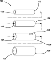
- the organ model is a simulated rectum model 100.
- the simulated rectum model 100 includes a first tube 102 made of any one of the hydrogel compositions described herein and dyed to have a pink color.
- the hydrogel is selected to have a ratio of approximately 8:1 acrylamide to alginate and approximately 86% water.
- the first tube 102 defines a first lumen 103 extending between a proximal end and a distal end.
- the simulated rectum model 100 further includes a second tube 104 defining a second lumen 105 and extending between a proximal end and a distal end.
- the second tube 104 is made of yellow dyed hydrogel of any one of the hydrogel compositions described herein. In one variation, the hydrogel is selected to have a ratio of approximately 8:1 acrylamide to alginate and approximately 86% water.
- the second lumen 105 is dimensioned to receive the first tube 102 inside the second lumen 105 in a concentric-like fashion.
- the second tube 104 is adhered to the first tube 102 using cyanoacrylate glue. Alternatively, the second tube 104 is cured onto the first tube 102 and no glue is employed.
- the yellow color of the second tube 104 is selected such that the second tube 104 represents the mesorectum of a human colon.
- the model 100 further includes a third tube 106.
- the third tube 106 defines a third lumen 107.
- the diameter of the third lumen 107 is dimensioned to receive the second tube 104 inside the third lumen 107 in a concentric fashion.
- the third tube 106 is adhered to the second tube 104 by being cured on top of the second tube 104.
- the third tube 106 is made of any one of the hydrogel compositions described herein and dyed to have a yellow and/or orange color to represent a presacral fat layer.
- the hydrogel is selected to have a ratio of approximately 8:1 acrylamide to alginate and approximately 86% water.
- the simulated rectum model 100 further includes a fourth tube 108.
- the fourth tube 108 defines a fourth lumen 109.
- the diameter of the fourth lumen 109 is dimensioned to receive the third tube 106 inside the fourth lumen 109 in a concentric-like fashion.
- the fourth tube 108 is made of any one of the hydrogel compositions described herein and dyed to have a pink color. In one variation, the hydrogel is selected to have a ratio of approximately 8:1 acrylamide to alginate and 86% water.
- the fourth tube 108 is adhered to the third tube 106 with adhesive such as cyanoacrylate glue such as LOCTITE® 401 or 4902 cyanoacrylate glue manufactured by LOCTITE® of Westlake, Ohio. Alternatively, the fourth tube 108 is cured onto the third tube 106 and no adhesive is employed.
- the simulated rectum model 100 further includes a simulated prostate system 110 located and embedded between the third tube 106 and the fourth tube 108.
- the simulated prostate system 110 is located and embedded inside the third tube 106.
- the simulated prostate system 110 is located at the anterior side of the model 100.
- the simulated prostate system 110 includes any one or more of the following simulated anatomical structures: simulated prostate, simulated seminal vesicles, simulated bladder, simulated urethra, and simulated vas deferens.
- the simulated urethra and simulated vas deferens are made of silicone formed into a solid tube or other polymer.
- the simulated seminal vesicles are made of urethane or other foam overmolded onto the simulated vas deferens.
- the simulated prostate is made of urethane or other foam overmolded onto the simulated urethra.
- the simulated rectum model 100 further includes one or more collagen layer (not shown) located in any one or more of the following locations: (1) between the second tube 104 and the first tube 102, (2) between the third tube 106 and the second tube 104.
- the collagen layer is wetted and placed onto the cured hydrogel tube which is then placed in an oven to adhere it.
- the second tube 104 is covered with a thin layer of collagen and the third tube 106 is covered with a thin layer of collagen and electrosurgical dissection takes places between the two adjacent layers of collagen.
- a thin collagen layer is applied to the third tube 106 only and dissection is between the second tube 104 and the collagen layer on the third tube 106.
- a thin first collagen layer is applied to the second tube 104, a thin second collagen layer is applied to the first collagen layer.
- the prostate system 110 is adhered to the second collagen layer and care is taken to dissect around the prostate system between the first collagen layer and the second collagen layer.
- a thin collagen layer is applied to the prostate system 110 and care is taken to dissect between the second tube 104 and the thin collagen layer to avoid the prostate system 110.
- the simulated rectum model 100 is fantastically suited for practicing transanal total mesorectal excision (TaTME) for cancer located in the lower rectum using electrosurgical devices and electrosurgery techniques.
- TaTME transanal total mesorectal excision
- the cancerous rectum is approached through the anus into the first lumen 103 via a sealable port that is connected to channel.
- a purse-string suture is tied to seal off the cancerous location of the rectum that includes the tumor.
- the first tube 102 is optionally provided with an embedded mesh layer so that sutures would be held in the first tube 102 and not tear through the hydrogel when pulled.
- the purse-string suture is pre-made during the manufacturing process so that the surgeon can visually locate the suture and only practice techniques subsequent to purse-string suture placement.
- the surgeon will commence to dissect in the posterior direction and electrosurgically cut down through first tube 102 and into the second tube 104 which represents the mesorectum and circumferentially around the second tube 104 between the second tube 104 and the third tube 106 being careful not to penetrate into the simulated prostate system 110 and not to penetrate into the fourth tube 108 as can be seen in FIG. 2 . Care is also taken not to enter the simulated mesorectum (second tube 104) nor enter into the first tube 102. The user carefully practices to dissect circumferentially around the first tube 102.
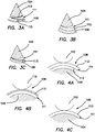
- FIG. 3A illustrates a posterior dissection location between the second tube 104 and the third tube 106 and a dissection plane 111 in between two collagen layers 113 if they are employed.
- FIG. 3B illustrates a posterior dissection location with a dissection pathway between the second tube 104 and the third tube 106, and in particular, between the second tube 104 and a thin hydrogel layer 112 located between the third tube 106 and the second tube 104.
- FIG. 3C illustrates a posterior dissection location with a dissection pathway 111 between the second tube 104 and a collagen layer 113 adhered to the third tube 106.
- anterior dissection begins by dissecting through the thinner layer of the second tube 104, visible in FIG. 2 , until the third tube 106 is reached. Dissection proceeds between the second tube 104 and the third tube 106 along a dissection plane 111 until the posterior dissection is encountered.
- Exemplary anterior dissection locations and dissection pathways 111 that correspond to posterior dissection pathways 111 of the models configured as shown in FIGs. 3A, 3B and 3C are illustrated in FIGs. 4A, 4B and 4C , respectively.
- FIG. 4A illustrates an anterior dissection location with a dissection plane 111 lying between two collagen layers 113 if they are provided.
- FIG. 4A illustrates an anterior dissection location with a dissection plane 111 lying between two collagen layers 113 if they are provided.
- FIG. 4B illustrates an anterior dissection location with a dissection plane 111 lying between the second tube 104 and the thin layer of hydrogel 112.
- FIG. 4C illustrates an anterior dissection location with a dissection plane 111 lying between the second tube 104 and collagen layer 113 if one is provided. Care is taken not to enter the third tube 106 to avoid risk damaging the prostate system 110.
- the proximal end of the simulated rectum model 100 may be attached to a transanal adapter.
- the transanal adapter is a support used to space apart the top cover from the base of a surgical trainer to provide access into the model from the side of the surgical trainer. (An example of a surgical trainer is described in U.S. Patent No. 8,764,452 ).
- the transanal adapter includes an opening that is connected to the first lumen of the first tube 102. Surrounding the opening of the transanal adapter, soft silicone is provided to simulate an anus. The practice of the surgical TaTME procedure is performed through the opening of the transanal adapter into the first lumen 103 as described above.
- the first tube 102 and the second tube 104 are made of hydrogel having a ratio of approximately 8:1 acrylamide to alginate and approximately 86% water and the third tube 106 and the fourth tube 108 are made of hydrogel having a ratio of approximately 8:3 acrylamide to alginate and approximately 86% water.
- the intersection of layers/tubes having the same ratio are substantially indistinguishable, the intersection of layers/tubes having different ratios are distinguishable making the intersection plane discernible and more easily separable, leading the practitioner along the correct dissection plane and making dissection easier than if the correct dissection plane was the intersection of layers/tubes having the same ratio.
- the simulated rectum model 100 is assembled by first casting the material into hollow tube-like molds that are provided with mandrels. The casting of layers may begin from the innermost layer and proceed to the outermost layer or vice versa. For example, if the casting is to start from the innermost layer, a small tube is filled with material and allowed to cure in an oven. When removed from the small tube mandrel, the cured innermost layer is inserted into a larger diameter tubular mandrel of the desired diameter and the next layer is poured and allowed to cure. The combination is then removed and placed into a tubular mandrel having a larger diameter and the next layer is poured and so forth. Similarly, the model 100 may be constructed beginning with the outer layer and sequentially proceeding to the inner layer.
- Tubing is placed inside of a larger hollow tubing and the outermost space in between is filled with material until the desired layers is achieved working progressively until the innermost layer is poured.
- Any layer can be offset from the longitudinal axis to achieve a thicker or thinner layer posteriorly or anteriorly as needed such as for the second tube. If a purse-string suture is to be pre-made, the outer-to-inner manufacturing process would be employed.
- material would be cast to completely fill in the rectum except for the top portion.
- a small mandrel would be placed allowing only the very top to be hollow.
- the mandrel could be designed to look like a purse-string, giving the user a visual cue that the purse-string suture has been already completed.
- synthetic or natural collagen casing is employed in the form of a sheet or cylinder. If provided in the form of a cylinder, it is cut into sheets. The collagen layer is then soaked in water and water is brushed onto the desired layer of application. The soaked collagen layer is then placed onto the layer of hydrogel. More layers are added as needed and the hydrogel layer and collagen layer are baked together in an oven to adhere the hydrogel to the collagen or the collagen to itself when multiple layers are employed side-by-side.
- the model 100 is held together by over molding the layers or with cyanoacrylate glue.
- Silicone components of the model 100 such as the prostate system 110 are adhered to the hydrogel or collagen using cyanoacrylate glue.
- Urethane molds are employed and the molds may be surface treated with in a variety of ways including but not limited to plasma treating and flame treating to make the mold hydrophilic and improve spreading of hydrogel material into the mold, especially for a hydrogel formulation that does not include sodium metabisulfide.
- Certain model organ parts, especially thin sheet-like parts such as a simulated peritoneum, are formed by polybag casting. In polybag casting, the hydrogel material is poured into a bag. Any air pockets are pressed out and the bag is sealed and placed between two flat trays.
- Weights of approximately 1134-2268 g (2.5-5.0 pounds) were laid on top of the trays and allowed to cure into a flat sheet to create an artificial peritoneum or omentum.
- Artificial vasculature also made of hydrogel may be embedded by arranging the artificial vasculature inside the polybag. Also, smaller hollow molds are utilized to manufacture simulated hollow vessels.
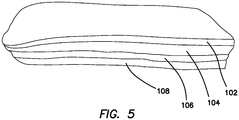
- the model 100 does not have a cylindrical shape to represent a rectum. Instead, the model 100 simply includes four layers 102, 104, 106, 108 from top to bottom in the shape of a rectangular or square block as if the cylinder were to be cut open and laid flat as shown in FIG. 5 .
- the block configuration of the layers permits the user to practice the procedures without being confined to a lumen configuration with the procedures performed transluminally.
- the block allows practitioners to simply practice the electrosurgical techniques in a laparoscopic environment such with the model 100 placed inside a cavity of a surgical trainer between a top cover and a base.
- the first layer 102 and the second layer 104 are made of hydrogel having a ratio of approximately 8:1 acrylamide to alginate and approximately 86% water and the third layer 106 and the fourth layer 108 are made of hydrogel having a ratio of approximately 8:3 acrylamide to alginate and approximately 86% water.
- any one of the hydrogels disclosed in this specification can be used to form at least part of a simulated tissue structure for the practice of surgical techniques, especially laparoscopic electro-surgical procedures wherein the simulated tissue structure is disposed inside an enclosure substantially enclosing the simulated tissue structure.
- An example of an enclosure includes a laparoscopic trainer in which a laparoscope is utilized to visualize the surgical field.
- the simulated tissue structure is not limited to artificial vessels, arteries, veins, one or more organs and tissues, hollow or solid, associated with the human lower rectum as described above and suitable for practicing a TaTME procedure.
- the TaTME model described above may be made with two layers of hydrogel instead of four layers.
- the two layers made of hydrogel include the rectum layer and mesorectum layer, the first tube 102 and the second tube 104, respectively, if the model is formed to have a tubular shape.
- a variation of such a TaTME model having two layers includes a mesh layer located between the two layers 102, 104.
- the TaTME model need not have a tubular shape.
- Any of the TaTME models may include artificial polyps to be practiced for removal using energy.
- a gallbladder model may include one or more of an artificial liver, artificial gallbladder, artificial peritoneum, artificial fascia, artificial duct(s), and one or more artificial artery.
- the artificial liver is excluded from being made of hydrogel and instead made of silicone or KRATON in order to localize the surge areas to the locations where a simulated procedure would be performed.
- a simulated tissue structure is substantially made of any one of the hydrogels described herein.
- the simulated tissue structure includes an artificial human ovarian organ that includes one or more of a simulated ovary portion, a uterine horn portion, uterus, ovary, fallopian tube, vagina, cervix, bladder, omentum, and peritoneum.
- the peritoneum and omentum may further include embedded simulated vasculature, hollow or solid, also made of hydrogel.
- simulated tissue structures include an artificial stomach, kidney, rectum, aorta, tumor, and polyp.
- Any of the simulated tissue structures made of hydrogel described herein may include a mesh layer.
- the simulated tissue structure may include two different hydrogels forming different parts of the simulated tissue.
- part of a simulated tissue structure may be made with a hydrogel having an 8:3 formulation and another part having an 8:1 formulation.
- part of a simulated tissue structure may be formed of a hydrogel of the above type and part made of silicone or other material and attached, connected, adjacent or in juxtaposition to the part made of hydrogel.
- an artificial colon is made of silicone and an artificial peritoneum and vessels are made of hydrogel having one or more formulation described herein.
- the artificial liver is made of silicone or KRATON and all other parts of the gallbladder model are made of hydrogel having one or more formulation described herein.
- an artificial rectum is made of silicone and artificial polyps of hydrogel described herein are adhered to the silicone rectum using cyanoacrylate glue.
- the simulated tissue structure is configured for use with electrosurgical units, including but not limited to monopolar, bipolar, harmonic or other devices employed in electrosurgery, in order to provide a realistic medium configured into an anatomical portion for the practice of using electrosurgical units, electrosurgical techniques, surgical procedures employing electrosurgical units alone and with other instruments encountered in surgery.
- electrosurgical units including but not limited to monopolar, bipolar, harmonic or other devices employed in electrosurgery, in order to provide a realistic medium configured into an anatomical portion for the practice of using electrosurgical units, electrosurgical techniques, surgical procedures employing electrosurgical units alone and with other instruments encountered in surgery.
- the handling of electrosurgical units requires practice as does employing surgical techniques and learning specific procedures performed with the electrosurgical units.
- heat is generated by the electrical current traveling between two polarities in a bipolar system or from one electrical polarity to a ground in a monopolar system.
- the artificial tissue structure is located above and in contact with a grounding plate/pad which is connected to a ground.
- the simulated tissue structure that portion of the structure that is composed of hydrogel is placed in direct contact with the grounding pad/plate or other conductive surface.
- the entirety of the simulated tissue structure is configured such that the hydrogel is not in direct contact with the grounding pad, a conductive pathway, such as a wire or the like, is provided to contact the hydrogel portion and then pass across non-conductive portions of the model to contact the grounding pad.
- a conductive pathway such as a wire or the like
- any one of the liver, peritoneum, gallbladder, vasculature, fascia, duct system or other component of the model is made of hydrogel
- a wire is passed into that portion and then fed to contact a metallic frame which is set inside the stand with the frame legs extending all the way through the stand to be exposed at the bottom surface of the stand which then can be place atop a grounding pad.
- the temperature of the hydrogel structure will increase to a temperature that begins to vaporize the water content of the hydrogel in the location of contact. Because the hydrogel contains approximately 86% water by weight of the hydrogel structure, the model will generate steam that mimics the smoke created during electrosurgery performed on human tissue.
- the water vapor of the hydrogel structure is not odiferous compared with the smoke produced by real tissue.
- the water content will be reduced in the location of contact advantageously creating a simulated fusion or seal of tissue typically encountered in real surgery.
- a surgical simulator of the present invention not only advantageously simulates the look and feel of tissue structures that would undergo procedures that employ electrosurgery, but also, responds in manner that mimics real electrosurgery when electrosurgery is applied to the simulated tissue structures.
- the hydrogel of a simulator of the present invention can be utilized to simulate dissection of tissue in addition to sealing and/or fusion via an electrosurgical unit.
- a large glass beaker add 33.75g alginate and 90g acrylamide. Dry mix the two solids until the mixture is uniform. Measure out 614 ml of deionized (DI) water. Add 307 ml (about half) of the 614 ml of DI water to the beaker with the powder mixture. Mix the solution to break apart any alginate adhered to the sides or bottom of the beaker. Once a homogenous solution is formed, maintain the mixing by placing the beaker under an overhead mixer or insert a stir bar and place on stir plate to continue mixing.
- DI deionized
- the remaining 307 ml of water are added to a different beaker and used to prepare the colorant.
- 4 drops of red acrylic paint and 1 drop of black acrylic paint are added to the second jar of DI water and stirred on a stir plate until the water is a uniform color.
- the now colored 307 ml of DI water is combined back with the other half in the beaker of gel solution.
- the beaker of gel solution remains mixing on the overhead mixer or stir plate to dissolve all solids and allow for uniform mixing of the colorant.
- APS ammonium persulfate
- the hose While on the overhead mixer or stir plate, insert a thin hose into the bottom of the beaker of gel solution, the hose should be connected to the argon gas tank. Bubble in a stream of argon gas into the beaker for approximately 15 minutes. Afterwards, remove the hose from the solution and allow hose to sit above the surface and blow a stream of argon gas on top of the gel solution for another 5 minutes. After flushing the solution with argon gas remove the thin hose from the jar. The following step is also completed under argon conditions. Flush the headspace of the N,N,N',N'-tetramethylethylenediamine (N,N,N',N'-TMEDA) bottle with argon.
- N,N,N',N'-TMEDA N,N,N',N'-TMEDA
Landscapes
- Chemical & Material Sciences (AREA)
- Engineering & Computer Science (AREA)
- Health & Medical Sciences (AREA)
- Medicinal Chemistry (AREA)
- Physics & Mathematics (AREA)
- General Physics & Mathematics (AREA)
- Chemical Kinetics & Catalysis (AREA)
- Organic Chemistry (AREA)
- Polymers & Plastics (AREA)
- General Health & Medical Sciences (AREA)
- Medical Informatics (AREA)
- Pure & Applied Mathematics (AREA)
- Mathematical Physics (AREA)
- Algebra (AREA)
- Business, Economics & Management (AREA)
- Educational Administration (AREA)
- Educational Technology (AREA)
- Theoretical Computer Science (AREA)
- Mathematical Optimization (AREA)
- Mathematical Analysis (AREA)
- Computational Mathematics (AREA)
- Manufacturing & Machinery (AREA)
- Materials Engineering (AREA)
- Surgery (AREA)
- Life Sciences & Earth Sciences (AREA)
- Biomedical Technology (AREA)
- Otolaryngology (AREA)
- Nuclear Medicine, Radiotherapy & Molecular Imaging (AREA)
- Plasma & Fusion (AREA)
- Heart & Thoracic Surgery (AREA)
- Molecular Biology (AREA)
- Animal Behavior & Ethology (AREA)
- Public Health (AREA)
- Veterinary Medicine (AREA)
- Instructional Devices (AREA)
- Materials For Medical Uses (AREA)
Description
- This application relates to synthetic tissue for practicing electrosurgical procedures and, in particular, to conductive synthetic tissue material made from a cross-linked hydrogel and methods of manufacturing such material and synthetic tissue models.
- International Patent Application, publication number
WO2011/035410A1 , discloses a surgical training aid in the form of a hydrogel designed to mimic the properties of real tissue. However, advances in technology have led to an increased use of energy devices in surgical procedures and there is a need for synthetic tissue that closely resembles the response of human tissue to electrosurgery. International Patent Application, publication numberWO2013/103956A1 discloses an interpenetrating network with covalent crosslinks, wherein the first network comprises a polyacrylamide polymer and second network comprises a alginate polymer, where ammonium persulfate is used as a photo initiator for polyacrylamide, and N,N- methylenebisacrylamide is used as the crosslinker for polyacrylamide. - A new synthetic tissue would be advantageous to surgeons and residents for training purposes. The synthetic tissue requires several characteristics to closely resemble human tissue including the ability to be cauterized, cut, and fused when manipulated with energy devices. Additionally, the tissue needs to emulate the mechanical properties of real tissue such as elasticity, toughness, suturability, tactility, color and texture. Furthermore, the material needs to be moldable into a structure that mimics various human organs or membranes for simulating human anatomy. The synthetic tissue may also need to be bondable to a variety of thermoplastics and silicones. The present invention addresses these needs.
- According to one aspect of the invention, a surgical simulator for surgical training is provided. The surgical simulator includes a synthetic tissue structure formed at least in part of a hydrogel including an ionically cross-linked alginate network cross-linked with a covalently cross-linked acrylamide network. The synthetic tissue structure includes at least one of an artificial liver or artificial gallbladder. The at least one of the artificial liver or artificial gallbladder includes at least one lumen substantially formed of a hydrogel including an ionically cross-linked alginate network cross-linked with a covalently cross-linked acrylamide network.
- According to another aspect of the invention, a surgical simulator for surgical training is provided. The surgical simulator includes a synthetic tissue structure substantially formed of a hydrogel including an ionically cross-linked alginate network cross-linked with a covalently cross-linked acrylamide network. The synthetic tissue structure includes a first layer formed of the hydrogel having a first ratio of acrylamide to alginate by weight and a second layer formed of the hydrogel having a second ratio of acrylamide to alginate by weight. The second layer is adjacent to the first layer.
- According to another aspect of the invention, a surgical simulator for surgical training is provided. The surgical simulator includes a simulated organ model. The simulated organ model includes a first tube having an outer surface and an inner surface defining a first lumen. The first tube is made of a hydrogel comprising a dual interpenetrated network of ionically cross-linked alginate and covalently cross-linked acrylamide having a first ratio of acrylamide to alginate. The simulated organ model includes a second tube having an outer surface and an inner surface defining a second lumen. The second tube is made of a hydrogel comprising a dual interpenetrated network of ionically cross-linked alginate and covalently cross-linked acrylamide having a second ratio of acrylamide to alginate. The first tube is coaxially located inside the second lumen such that the outer surface of the first tube is in contact with the inner surface of the second tube.
- According to another aspect of the invention, a method of making a surgical simulator for the practice of electrosurgical techniques is provided. The method includes the steps of providing an acrylamide polymer, providing alginate polymer, providing water, mixing the water with the acrylamide and alginate to form a solution, adding ammonium persulfate to the solution, adding N,N-methylenebisacrylamide to the solution, adding calcium sulfate after the steps of adding ammonium persulfate and adding N,N-methylenebisacrylamide to the solution, casting the solution into a shape representative of an anatomical structure, and curing the solution to form a simulated electrosurgery model made of hydrogel for practicing and simulating electrosurgery.
- According to another aspect of the invention, a method of making a surgical simulator for the practice of electrosurgical techniques is provided. The method includes the step of providing an uncured hydrogel including an ionically cross-linked alginate network cross-linked with a covalently cross-linked acrylamide network. The method includes the step of providing a polymer bag, pouring the uncured hydrogel into the polymer bag, sealing the polymer bag, curing the uncured hydrogel inside the polymer bag to form a cured hydrogel, and removing the cured hydrogel. The resulting structure is substantially planar sheet of hydrogel that can be used in building a larger procedural-based surgical training model.
-
-
FIG. 1 is an exploded, top perspective view of an organ model of a surgical simulator according to the present invention. -
FIG. 2 is a side, cross-sectional view of a rectum model with a simulated prostate system of a surgical simulator according to the present invention. -
FIG. 3A is posterior, partial, cross-sectional view of two collagen layers located between a second tube and a third tube of a rectum model of a surgical simulator according to the present invention. -
FIG. 3B is a posterior, partial, cross-sectional view of a second tube, third tube and a thin hydrogel layer of a rectum model of a surgical simulator according to the present invention. -
FIG. 3C is a posterior, partial, cross-sectional view of a second tube, third tube and a collagen layer of a rectum model of a surgical simulator according to the present invention. -
FIG. 4A is an anterior, partial, cross-sectional view of two collagen layers located between a second tube and simulated prostate system of a rectum model of a surgical simulator according to the present invention. -
FIG. 4B is an anterior, partial, cross-sectional view of a thin hydrogel layer located between a second tube and simulated prostate system of a rectum model of a surgical simulator according to the present invention. -
FIG. 4C is an anterior, partial, cross-sectional view of a collagen layer between a second tube and a simulated prostate system of a rectum model of a surgical simulator according to the present invention. - The material of the surgical simulator in accordance with one aspect of the present invention, or which may be produced by a method in accordance with another aspect of the present invention, may be made from a dual interpenetrated cross-linked hydrogel network. The hydrogel is a mixture of two cross-linked polymers: an ionically cross-linked alginate network and a covalently cross-linked polyacrylamide network. The gel material is prepared by mixing an 8:3 ratio of acrylamide to alginate and water. In order to make the organ or tissue parts that are more realistic, color can be incorporated into the process. The colorant is added prior to deionized water being mixed with the acrylamide and alginate solids. Half the water being used to form the gel is used to make the colorant. A wash is created with the water and drops of acrylic paints. The amount and color of paint used varies depending on the organ See Table 1 below for organ color ratios that show how many parts of each color need to be mixed together for a particular organ and/or tissue part. The colored wash is then combined back with the other half of water and mixed with the acrylamide and alginate. Water content of the gel is approximately 86 weight percent. Ammonium persulfate (0.003 the weight of the acrylamide) and N,N-methylenebisacrylamide (0.006 the weight of acrylamide) are added to the solution as a photo initiator and a cross-linker respectively, for the acrylamide. Further, the solution is flushed with argon gas and N,N,N'N'-tetramethylethylenediamine (0.003 the weight of acrylamide) is added under an argon atmosphere as a cross-linking accelerator for the acrylamide. The final additive, calcium sulfate (0.136 weight of alginate), is an ionic cross-linker for the alginate. The slurry is constantly stirred throughout each step until the solution is homogeneous. The gel solution is cast into organ shaped molds and placed in an 85°C oven for 30 minutes to cure. See the Example below for a specific hydrogel procedure example. To obtain hollow organs, the gel solution can be painted onto a mandrel and placed under a heat lamp to cure. The cured product is a tough, clear hydrogel or colored replica of the organ or tissue. The application of hydrogel organs makes the organ trays for surgical training more dynamic, the trays become more life-like as well as energy device compatible.
- In another variation, the material is made from a dual interpenetrated cross-linked hydrogel network. The hydrogel is a mixture of two cross-linked polymers: an ionically cross-linked alginate network and a covalently cross-linked polyacrylamide network. The gel material is prepared by mixing an 8:3 ratio of acrylamide to alginate and water. In order to make organs or tissue parts that are more realistic, color can be incorporated into the process. A colorant solution is prepared separate from the acrylamide and alginate mixture to allow for accessibility of different pigments while molding various tissue or organs. The colorant solution is prepared by dissolving acrylic paints in deionized water. The amount and color of paint used varies depending on the organ. See Table I for organ color ratios that show how many parts of each color need to be mixed together for a particular organ and/or tissue part. From the total amount of water used to create the hydrogel, half the water comes from the colorant solution. The colored solution is then combined back with the other half of water which is mixed with the acrylamide and alginate. The total water content of the gel is approximately 86 wt%. Ammonium persulfate (approximately 0.3% the weight of the acrylamide) and N,N-methylenebisacrylamide (approximately 0.6% the weight of acrylamide) are added to the solution as a photoinitiator and a cross-linker respectively, for the acrylamide. Further, the solution is flushed with argon gas for approximately 10-15 minutes in order to displace the air with an inert gas, and then N,N,N'N'-tetramethylethylenediamine (approximately 0.3% the weight of acrylamide) is added under an argon atmosphere as a cross-linking accelerator for the acrylamide. The final additive, calcium sulfate (approximately 13.6% weight of alginate), is an ionic cross-linker for the alginate. The slurry is constantly stirred throughout each step until the solution is homogeneous. The gel solution is cast into organ shaped molds and placed in an 85°C oven for 60 minutes to cure. See Example below for a specific hydrogel procedure example. To obtain hollow organs, the gel solution can be painted onto a mandrel and placed under a heat lamp to cure. The cured product is a tough, clear hydrogel or colored replica of the organ or tissue. The application of hydrogel organs makes the organ trays for surgical training more dynamic, the trays become more life-like as well as energy device compatible.
- Organs and/or tissue made of the hydrogel closely resemble and react to manipulation with energy devices similar to the way human organs do. The synthetic tissue made of the above mentioned hydrogel can be cut, cauterized and fused. Two layers of the hydrogel tissue as described above can be separated along a plane using various monopolar and bipolar devices. Furthermore, vessels of the hydrogel can be fused and transected like real blood vessels. Mechanical devices such as scissors, graspers, and sutures can also be used on synthetic tissue made from the hydrogel described above . The tissue has the strength to accommodate sutures and can be further reinforced with mesh to allow additional strength to accommodate sutures in a manner used for actual surgeries without concern for the suture tearing through the synthetic tissue and coming undone. In addition, when wetted the material becomes lubricious and slick making for a life-like feel. The compatibility of the hydrogel with other materials becomes useful when making large assemblies, such as organ trays comprising multiple tissue components for simulators because the synthetic organs not only need to bond to each other, but also are able to bond to the plastic base of the tray. The synthetic organs and tissues made of the hydrogel material should be stored in closed containers with minimal exposure to the atmosphere until ready for use. Due to being predominantly water, the hydrogel material can dry out over time if not stored properly. However, advantageously, the hydrogel has the ability to reabsorb water allowing for it to rehydrate after losing moisture and to be used.
- In another variation of the method of one aspect of the present invention, synthetic tissue is made as follows. Sodium metabisulfite is added as an additive to the above mentioned hydrogel. The sodium metabisulfite is added to the solution prior to the calcium sulfate. The amount utilized is equivalent to the amount of ammonium persulfate present in the gel solution. The addition of the sodium metabisulfite allows the gel to be cured at room temperature. Once cast, the hydrogel begins to instantly cure, thus the need for a secondary oven cure is no longer necessary. This process shortens the time required for producing the gel. However, the resulting tissue lacks the same tear strength, elongation, and work time as its oven-cured counterpart.
- Another approach utilizes adjusting the ratios of ingredients already present in the hydrogel solution. The two polymers of the hybrid hydrogel are what allow the gel to be elastic and still hold its shape. The 8:3 polymer ratio of acrylamide to alginate in the gel can be adjusted to enhance different properties of the gel. The amount of acrylamide can be increased to increase flexibility and elasticity of the gel; inversely, if the amount of alginate is increased, brittleness is amplified and tear resistance is decreased. The cross-linkers are further responsible for certain characteristics. The cross-linkers essentially entangle the polymer strands together forming a polymer network. Increasing the amount of cross-linkers causes the hydrogel to cure faster and lack elasticity and an insufficient amount of cross-linkers causes the formation of a jelly rather than a gel. The amount of water can also be varied, with the amount of water being inversely proportional to hardness. Gel with higher water content will be softer and will have the formation of a jelly. Ultimately, the ingredients of the hybrid hydrogel can be utilized to enhance different physical and mechanical properties.
- Two other examples of replacement hydrogels are an acrylic acid based gel and a clay-based gel. In the acrylic acid hydrogel, an acrylate polymer is created through the polymerization of acrylic acid in an aqueous solution neutralized by sodium hydroxide. A sodium metabisulfite-ammonium persulfate redox reaction acts as an initiator for the polymerization process. The clay based hydrogel is a solution of sodium polyacrylate and clay nanosheets. A dendritic molecular binder (G3-binder) is added to the solution to initiate bonding. The resulting product is a clear, moldable hydrogel.
- Besides hydrogel materials semiconductive silicones can be utilized to produce synthetic organs. Semiconductive silicones are silicone rubbers that have been doped with small particles of metal, commonly, nickel-graphite or aluminum. These metal particles essentially make a non-conductive silicone semiconductive by providing a medium for electricity to flow through. Semiconductive silicones are expensive and difficult to bond to other materials. In addition, the silicone needs to contain large amounts of metal particles to provide a short enough arcing distance for the electric current. The above materials and processes can similarly be engaged to manufacture organ trays that are energy compatible.
- An exemplary organ model made of hydrogel material compositions described in this specification is shown in
FIGs. 1-3 . The organ model is asimulated rectum model 100. Thesimulated rectum model 100 includes afirst tube 102 made of any one of the hydrogel compositions described herein and dyed to have a pink color. In one variation, the hydrogel is selected to have a ratio of approximately 8:1 acrylamide to alginate and approximately 86% water. Thefirst tube 102 defines afirst lumen 103 extending between a proximal end and a distal end. - The
simulated rectum model 100 further includes asecond tube 104 defining asecond lumen 105 and extending between a proximal end and a distal end. Thesecond tube 104 is made of yellow dyed hydrogel of any one of the hydrogel compositions described herein. In one variation, the hydrogel is selected to have a ratio of approximately 8:1 acrylamide to alginate and approximately 86% water. Thesecond lumen 105 is dimensioned to receive thefirst tube 102 inside thesecond lumen 105 in a concentric-like fashion. Thesecond tube 104 is adhered to thefirst tube 102 using cyanoacrylate glue. Alternatively, thesecond tube 104 is cured onto thefirst tube 102 and no glue is employed. The yellow color of thesecond tube 104 is selected such that thesecond tube 104 represents the mesorectum of a human colon. - The
model 100 further includes athird tube 106. Thethird tube 106 defines athird lumen 107. The diameter of thethird lumen 107 is dimensioned to receive thesecond tube 104 inside thethird lumen 107 in a concentric fashion. Thethird tube 106 is adhered to thesecond tube 104 by being cured on top of thesecond tube 104. Thethird tube 106 is made of any one of the hydrogel compositions described herein and dyed to have a yellow and/or orange color to represent a presacral fat layer. In one variation, the hydrogel is selected to have a ratio of approximately 8:1 acrylamide to alginate and approximately 86% water. - The
simulated rectum model 100 further includes afourth tube 108. Thefourth tube 108 defines afourth lumen 109. The diameter of thefourth lumen 109 is dimensioned to receive thethird tube 106 inside thefourth lumen 109 in a concentric-like fashion. Thefourth tube 108 is made of any one of the hydrogel compositions described herein and dyed to have a pink color. In one variation, the hydrogel is selected to have a ratio of approximately 8:1 acrylamide to alginate and 86% water. Thefourth tube 108 is adhered to thethird tube 106 with adhesive such as cyanoacrylate glue such as LOCTITE® 401 or 4902 cyanoacrylate glue manufactured by LOCTITE® of Westlake, Ohio. Alternatively, thefourth tube 108 is cured onto thethird tube 106 and no adhesive is employed. - In one variation of the
simulated rectum model 100, thesimulated rectum model 100 further includes asimulated prostate system 110 located and embedded between thethird tube 106 and thefourth tube 108. In one variation, thesimulated prostate system 110 is located and embedded inside thethird tube 106. Thesimulated prostate system 110 is located at the anterior side of themodel 100. Thesimulated prostate system 110 includes any one or more of the following simulated anatomical structures: simulated prostate, simulated seminal vesicles, simulated bladder, simulated urethra, and simulated vas deferens. The simulated urethra and simulated vas deferens are made of silicone formed into a solid tube or other polymer. The simulated seminal vesicles are made of urethane or other foam overmolded onto the simulated vas deferens. The simulated prostate is made of urethane or other foam overmolded onto the simulated urethra. - In one variation of the
simulated rectum model 100, thesimulated rectum model 100 further includes one or more collagen layer (not shown) located in any one or more of the following locations: (1) between thesecond tube 104 and thefirst tube 102, (2) between thethird tube 106 and thesecond tube 104. The collagen layer is wetted and placed onto the cured hydrogel tube which is then placed in an oven to adhere it. In one variation, thesecond tube 104 is covered with a thin layer of collagen and thethird tube 106 is covered with a thin layer of collagen and electrosurgical dissection takes places between the two adjacent layers of collagen. In another variation, a thin collagen layer is applied to thethird tube 106 only and dissection is between thesecond tube 104 and the collagen layer on thethird tube 106. In another variation, a thin first collagen layer is applied to thesecond tube 104, a thin second collagen layer is applied to the first collagen layer. Theprostate system 110 is adhered to the second collagen layer and care is taken to dissect around the prostate system between the first collagen layer and the second collagen layer. In another variation, a thin collagen layer is applied to theprostate system 110 and care is taken to dissect between thesecond tube 104 and the thin collagen layer to avoid theprostate system 110. - The
simulated rectum model 100 is fantastically suited for practicing transanal total mesorectal excision (TaTME) for cancer located in the lower rectum using electrosurgical devices and electrosurgery techniques. In such a surgical procedure, the cancerous rectum is approached through the anus into thefirst lumen 103 via a sealable port that is connected to channel. A purse-string suture is tied to seal off the cancerous location of the rectum that includes the tumor. In order to practice this suture technique, thefirst tube 102 is optionally provided with an embedded mesh layer so that sutures would be held in thefirst tube 102 and not tear through the hydrogel when pulled. In another variation, the purse-string suture is pre-made during the manufacturing process so that the surgeon can visually locate the suture and only practice techniques subsequent to purse-string suture placement. In the practice of the procedure, the surgeon will commence to dissect in the posterior direction and electrosurgically cut down throughfirst tube 102 and into thesecond tube 104 which represents the mesorectum and circumferentially around thesecond tube 104 between thesecond tube 104 and thethird tube 106 being careful not to penetrate into thesimulated prostate system 110 and not to penetrate into thefourth tube 108 as can be seen inFIG. 2 . Care is also taken not to enter the simulated mesorectum (second tube 104) nor enter into thefirst tube 102. The user carefully practices to dissect circumferentially around thefirst tube 102. Exemplary posterior dissection locations and dissection pathways are illustrated inFIGs. 3A-3C. FIG. 3A illustrates a posterior dissection location between thesecond tube 104 and thethird tube 106 and adissection plane 111 in between twocollagen layers 113 if they are employed.FIG. 3B illustrates a posterior dissection location with a dissection pathway between thesecond tube 104 and thethird tube 106, and in particular, between thesecond tube 104 and athin hydrogel layer 112 located between thethird tube 106 and thesecond tube 104.FIG. 3C illustrates a posterior dissection location with adissection pathway 111 between thesecond tube 104 and acollagen layer 113 adhered to thethird tube 106. After dissecting posteriorly, anterior dissection begins by dissecting through the thinner layer of thesecond tube 104, visible inFIG. 2 , until thethird tube 106 is reached. Dissection proceeds between thesecond tube 104 and thethird tube 106 along adissection plane 111 until the posterior dissection is encountered. Exemplary anterior dissection locations anddissection pathways 111 that correspond toposterior dissection pathways 111 of the models configured as shown inFIGs. 3A, 3B and 3C are illustrated inFIGs. 4A, 4B and 4C , respectively.FIG. 4A illustrates an anterior dissection location with adissection plane 111 lying between twocollagen layers 113 if they are provided.FIG. 4B illustrates an anterior dissection location with adissection plane 111 lying between thesecond tube 104 and the thin layer ofhydrogel 112.FIG. 4C illustrates an anterior dissection location with adissection plane 111 lying between thesecond tube 104 andcollagen layer 113 if one is provided. Care is taken not to enter thethird tube 106 to avoid risk damaging theprostate system 110. - The proximal end of the
simulated rectum model 100 may be attached to a transanal adapter. The transanal adapter is a support used to space apart the top cover from the base of a surgical trainer to provide access into the model from the side of the surgical trainer. (An example of a surgical trainer is described inU.S. Patent No. 8,764,452 ). The transanal adapter includes an opening that is connected to the first lumen of thefirst tube 102. Surrounding the opening of the transanal adapter, soft silicone is provided to simulate an anus. The practice of the surgical TaTME procedure is performed through the opening of the transanal adapter into thefirst lumen 103 as described above. - In one variation, the
first tube 102 and thesecond tube 104 are made of hydrogel having a ratio of approximately 8:1 acrylamide to alginate and approximately 86% water and thethird tube 106 and thefourth tube 108 are made of hydrogel having a ratio of approximately 8:3 acrylamide to alginate and approximately 86% water. Whereas the intersection of layers/tubes having the same ratio are substantially indistinguishable, the intersection of layers/tubes having different ratios are distinguishable making the intersection plane discernible and more easily separable, leading the practitioner along the correct dissection plane and making dissection easier than if the correct dissection plane was the intersection of layers/tubes having the same ratio. - The
simulated rectum model 100 is assembled by first casting the material into hollow tube-like molds that are provided with mandrels. The casting of layers may begin from the innermost layer and proceed to the outermost layer or vice versa. For example, if the casting is to start from the innermost layer, a small tube is filled with material and allowed to cure in an oven. When removed from the small tube mandrel, the cured innermost layer is inserted into a larger diameter tubular mandrel of the desired diameter and the next layer is poured and allowed to cure. The combination is then removed and placed into a tubular mandrel having a larger diameter and the next layer is poured and so forth. Similarly, themodel 100 may be constructed beginning with the outer layer and sequentially proceeding to the inner layer. Tubing is placed inside of a larger hollow tubing and the outermost space in between is filled with material until the desired layers is achieved working progressively until the innermost layer is poured. Any layer can be offset from the longitudinal axis to achieve a thicker or thinner layer posteriorly or anteriorly as needed such as for the second tube. If a purse-string suture is to be pre-made, the outer-to-inner manufacturing process would be employed. On the last innermost layer, instead of placing a mandrel in all of the way, material would be cast to completely fill in the rectum except for the top portion. On the top, a small mandrel would be placed allowing only the very top to be hollow. The mandrel could be designed to look like a purse-string, giving the user a visual cue that the purse-string suture has been already completed. To apply a collagen layer, synthetic or natural collagen casing is employed in the form of a sheet or cylinder. If provided in the form of a cylinder, it is cut into sheets. The collagen layer is then soaked in water and water is brushed onto the desired layer of application. The soaked collagen layer is then placed onto the layer of hydrogel. More layers are added as needed and the hydrogel layer and collagen layer are baked together in an oven to adhere the hydrogel to the collagen or the collagen to itself when multiple layers are employed side-by-side. Themodel 100 is held together by over molding the layers or with cyanoacrylate glue. Silicone components of themodel 100 such as theprostate system 110 are adhered to the hydrogel or collagen using cyanoacrylate glue. Urethane molds are employed and the molds may be surface treated with in a variety of ways including but not limited to plasma treating and flame treating to make the mold hydrophilic and improve spreading of hydrogel material into the mold, especially for a hydrogel formulation that does not include sodium metabisulfide. Certain model organ parts, especially thin sheet-like parts such as a simulated peritoneum, are formed by polybag casting. In polybag casting, the hydrogel material is poured into a bag. Any air pockets are pressed out and the bag is sealed and placed between two flat trays. Weights of approximately 1134-2268 g (2.5-5.0 pounds) were laid on top of the trays and allowed to cure into a flat sheet to create an artificial peritoneum or omentum. Artificial vasculature also made of hydrogel may be embedded by arranging the artificial vasculature inside the polybag. Also, smaller hollow molds are utilized to manufacture simulated hollow vessels. - In another variation, the
model 100 does not have a cylindrical shape to represent a rectum. Instead, themodel 100 simply includes fourlayers FIG. 5 . The block configuration of the layers permits the user to practice the procedures without being confined to a lumen configuration with the procedures performed transluminally. The block allows practitioners to simply practice the electrosurgical techniques in a laparoscopic environment such with themodel 100 placed inside a cavity of a surgical trainer between a top cover and a base. In such a variation, thefirst layer 102 and thesecond layer 104 are made of hydrogel having a ratio of approximately 8:1 acrylamide to alginate and approximately 86% water and thethird layer 106 and thefourth layer 108 are made of hydrogel having a ratio of approximately 8:3 acrylamide to alginate and approximately 86% water. - Any one of the hydrogels disclosed in this specification can be used to form at least part of a simulated tissue structure for the practice of surgical techniques, especially laparoscopic electro-surgical procedures wherein the simulated tissue structure is disposed inside an enclosure substantially enclosing the simulated tissue structure. An example of an enclosure includes a laparoscopic trainer in which a laparoscope is utilized to visualize the surgical field. The simulated tissue structure is not limited to artificial vessels, arteries, veins, one or more organs and tissues, hollow or solid, associated with the human lower rectum as described above and suitable for practicing a TaTME procedure. Also, the TaTME model described above may be made with two layers of hydrogel instead of four layers. In such a model the two layers made of hydrogel include the rectum layer and mesorectum layer, the
first tube 102 and thesecond tube 104, respectively, if the model is formed to have a tubular shape. A variation of such a TaTME model having two layers includes a mesh layer located between the twolayers - In use, the simulated tissue structure is configured for use with electrosurgical units, including but not limited to monopolar, bipolar, harmonic or other devices employed in electrosurgery, in order to provide a realistic medium configured into an anatomical portion for the practice of using electrosurgical units, electrosurgical techniques, surgical procedures employing electrosurgical units alone and with other instruments encountered in surgery. The handling of electrosurgical units requires practice as does employing surgical techniques and learning specific procedures performed with the electrosurgical units. When an electrosurgical unit is applied, heat is generated by the electrical current traveling between two polarities in a bipolar system or from one electrical polarity to a ground in a monopolar system. Typically, in a monopolar system, the artificial tissue structure is located above and in contact with a grounding plate/pad which is connected to a ground. In one variation of the simulated tissue structure , that portion of the structure that is composed of hydrogel is placed in direct contact with the grounding pad/plate or other conductive surface. In the event, the entirety of the simulated tissue structure is configured such that the hydrogel is not in direct contact with the grounding pad, a conductive pathway, such as a wire or the like, is provided to contact the hydrogel portion and then pass across non-conductive portions of the model to contact the grounding pad. For example, in a gallbladder model such as the model described in U.S. Patent Application Publication No.
US 2014/0370477 to Applied Medical Resources Corporation in California, the anatomical portion is connected to a support in order to permit the model to stand upright. If any one of the liver, peritoneum, gallbladder, vasculature, fascia, duct system or other component of the model is made of hydrogel, a wire is passed into that portion and then fed to contact a metallic frame which is set inside the stand with the frame legs extending all the way through the stand to be exposed at the bottom surface of the stand which then can be place atop a grounding pad. When the hydrogel structure is contacted with an electrosurgical unit, the temperature of the hydrogel structure will increase to a temperature that begins to vaporize the water content of the hydrogel in the location of contact. Because the hydrogel contains approximately 86% water by weight of the hydrogel structure, the model will generate steam that mimics the smoke created during electrosurgery performed on human tissue. Advantageously, the water vapor of the hydrogel structure is not odiferous compared with the smoke produced by real tissue. With prolonged contact with the electrosurgical unit, the water content will be reduced in the location of contact advantageously creating a simulated fusion or seal of tissue typically encountered in real surgery. Hence, a surgical simulator of the present invention not only advantageously simulates the look and feel of tissue structures that would undergo procedures that employ electrosurgery, but also, responds in manner that mimics real electrosurgery when electrosurgery is applied to the simulated tissue structures. The hydrogel of a simulator of the present invention can be utilized to simulate dissection of tissue in addition to sealing and/or fusion via an electrosurgical unit.Table 1 ORGAN COLOR RATIO Liver 4 red : 1 black Gallbladder 3 yellow : 1 blue Cystic duct 3 yellow : 1 blue Kidney 4 red : 1 blue Spleen 4 red : 1 blue Pancreas 4 yellow Omentum 4 yellow : 1 white Mesentery 4 yellow : 1 white (serial diluted 8 times) Veins 3 blue : 0.5 black Arteries 5 red : 0.25 black Aorta 4 red - The following is an example procedure according to the present invention for making a simulated hydrogel liver . In a large glass beaker, add 33.75g alginate and 90g acrylamide. Dry mix the two solids until the mixture is uniform. Measure out 614 ml of deionized (DI) water. Add 307 ml (about half) of the 614 ml of DI water to the beaker with the powder mixture. Mix the solution to break apart any alginate adhered to the sides or bottom of the beaker. Once a homogenous solution is formed, maintain the mixing by placing the beaker under an overhead mixer or insert a stir bar and place on stir plate to continue mixing. The remaining 307 ml of water are added to a different beaker and used to prepare the colorant. For a simulated liver, 4 drops of red acrylic paint and 1 drop of black acrylic paint are added to the second jar of DI water and stirred on a stir plate until the water is a uniform color. The now colored 307 ml of DI water is combined back with the other half in the beaker of gel solution. The beaker of gel solution remains mixing on the overhead mixer or stir plate to dissolve all solids and allow for uniform mixing of the colorant. Keep solution stirring and add 0.250g of ammonium persulfate (APS) and add 0.050g N,N'-methylenebisacrylamide. Allow the APS and N,N'-MBAA to dissolve in the gel solution prior to proceeding. Hand mix as necessary, since the solution is viscous and the lighter additives will not readily mix with the mixers.
- While on the overhead mixer or stir plate, insert a thin hose into the bottom of the beaker of gel solution, the hose should be connected to the argon gas tank. Bubble in a stream of argon gas into the beaker for approximately 15 minutes. Afterwards, remove the hose from the solution and allow hose to sit above the surface and blow a stream of argon gas on top of the gel solution for another 5 minutes. After flushing the solution with argon gas remove the thin hose from the jar. The following step is also completed under argon conditions. Flush the headspace of the N,N,N',N'-tetramethylethylenediamine (N,N,N',N'-TMEDA) bottle with argon. Using a micropipette, pipette 0.290 milliliters of argon gas from the N,N,N',N'-TMEDA bottle head space and eject the gas off to the side, this should be done twice in order to flush the interior of the micropipette. Now, extract 0.290 ml of N,N,N',N'-TMEDA from the bottle using the same micropipette tip and eject into the gel solution. The N,N,N',N'-TMEDA bottle should be sealed quickly after use and stored in a dark area, away from moisture.
- Continue stirring, make a slurry of calcium sulfate dihydrate (CaSO4•2H2O) and DI water. Add approximately 25ml of DI water to 4.59g of CaSO4•2H2O. Mix thoroughly and add slurry to the hydrogel solution. Wash the remains of the CaSO4•2H2O slurry with DI water and add to the hydrogel solution. Some white clouds may still remain from the addition of the CaSO4•2H2O. These clouds will disappear once hydrogel is cured. Allow gel slurry to mix at medium speed for approximately 1 minute. The gel slurry can now be poured into a liver mold and placed in an oven at 85 °C for 60 minutes to cure the gel. After 1 hour, the mold is removed from the oven and allowed to cool to room temperature. Once cool, the hydrogel liver can be removed from the mold. The final product is a life-like synthetic liver capable of being manipulated with energy devices in addition to mechanical devices.
- It is understood that various modifications may be made to the embodiments of the synthetic tissue disclosed herein. Therefore, the above description should not be construed as limiting, but merely as exemplifications of preferred embodiments. Those skilled in the art will envision other modifications within the scope of the invention, as defined by the following claims.
Claims (15)
- A method of making a surgical simulator (100), characterised in comprising the steps of:providing an acrylamide polymer;providing alginate polymer;providing water;mixing the water with the acrylamide and alginate to form a solution;adding ammonium persulfate to the solution;adding N,N-methylenebisacrylamide to the solution;adding calcium sulfate after the steps of adding ammonium persulfate and adding N,N-methylenebisacrylamide to the solution;casting the solution into a shape representative of an anatomical structure; and curing the solution to form a simulated electrosurgery model made of hydrogel for practicing and simulating electrosurgery.
- The method of claim 1 including the step of adding N,N, N', N'-tetramethylethylenediamine in the amount of approximately 0.3 percent acrylamide by weight to the solution;
wherein the step of adding ammonium persulfate includes adding ammonium persulfate in the amount of approximately 0.3 percent acrylamide by weight to the solution;
wherein the step of adding N,N-methylenebisacrylamide includes adding N,N-methylenebisacrylamide in the amount of approximately 0.06 percent acrylamide by weight to the solution; and
wherein the step of adding calcium sulfate includes adding calcium sulfate in the amount of approximately 13.6 percent alginate by weight to the solution. - The method of any one of the previous claims further including the step of flushing the solution with argon gas.
- The method of any one of the previous claims including the step of dry mixing the acrylamide polymer and alginate polymer prior to the step of mixing the water.
- The method of any one of the previous claims further including the steps of:forming a first layer of hydrogel having a first ratio of acrylamide to alginate by weight; andovermolding a second layer of hydrogel having a second ratio of acrylamide to alginate by weight.
- The method of any one of the previous claims further including the steps of:providing a colorant selected for a synthetic tissue structure; andmixing the colorant with the water;wherein the step of mixing the colorant with the water is performed prior to the step of mixing the water with the acrylamide and alginate to form a solution.
- A method of making a surgical simulator (100) for the practice of electrosurgical techniques, characterised in comprising the steps of:providing an uncured hydrogel including an ionically cross-linked alginate network cross-linked with a covalently cross-linked acrylamide network;providing a polymer bag;pouring the uncured hydrogel into the polymer bag;sealing the polymer bag;curing the uncured hydrogel inside the polymer bag to form a cured hydrogel; andremoving the cured hydrogel.
- A surgical simulator (100) for surgical training comprising:a synthetic tissue structure substantially formed of a hydrogel including an ionically cross-linked alginate network cross-linked with a covalently cross-linked acrylamide network; the synthetic tissue structure includes a first layer formed of the hydrogel having a first ratio of acrylamide to alginate by weight; and a second layer formed of the hydrogel having a second ratio of acrylamide to alginate by weight; the second layer being adjacent to the first layer.
- The surgical simulator of claim 8 further including a synthetic tissue structure embedded between the first layer and the second layer; wherein the embedded synthetic tissue structure is glued to the first layer.
- The surgical simulator of claim 8 or 9 further including a synthetic tissue structure embedded between the first layer and the second layer; wherein the embedded synthetic tissue structure includes at least one of a lumen, artificial vasculature, artificial nerves, and artificial ureter.
- A surgical simulator (100) for surgical training comprisinga simulated organ model (100); the simulated organ model includinga first tube (102) having an outer surface and an inner surface defining a first lumen (103) and characterised in that:a second tube (104) having an outer surface and an inner surface defining a second lumen (105)the first tube is made of a hydrogel comprising a dual interpenetrated network of ionically cross-linked alginate and covalently cross-linked acrylamide having a first ratio of acrylamide to alginate;the second tube (104) is made of a hydrogel comprising a dual interpenetrated network of ionically cross-linked alginate and covalently cross-linked acrylamide having a second ratio of acrylamide to alginate;wherein the first tube (103) is coaxially located inside the second lumen (105) such that the outer surface of the first tube (102) is in contact with the inner surface of the second tube (104).
- The surgical simulator of claim 11 wherein:the first ratio is approximately 8:3 and the second ratio is approximately 8:3;
the first ratio is approximately 8:1 and the second ratio is approximately 8:1; or
the first ratio is approximately 8:1 and the second ratio is approximately 8:3. - A surgical simulator (100) for surgical training chacterised in comprising:a synthetic tissue structure formed at least in part of a hydrogel including an ionically cross-linked alginate network cross-linked with a covalently cross-linked acrylamide network;wherein the synthetic tissue structure includes at least one of an artificial liver or artificial gallbladder; the at least one of the artificial liver or artificial gallbladder includes at least one lumen substantially formed of a hydrogel including an ionically cross-linked alginate network cross-linked with a covalently cross-linked acrylamide network.
- The surgical simulator (100) of any one of claims 8 to 13 further including a simulated prostate system including one or more of an artificial prostate (110), artificial seminal vesicles, simulated bladder, simulated urethra, and simulated vas deferens.
- The surgical simulator of any one of claims 8 to 14 further including an electrosurgical unit having a proximal end and an operable distal end configured to deliver energy at the distal end and controlled with a handle at the proximal end; wherein contact of the distal end of the electrosurgical unit with the hydrogel simulates electrosurgery.
Priority Applications (1)
| Application Number | Priority Date | Filing Date | Title |
|---|---|---|---|
| EP18207214.0A EP3476343B1 (en) | 2015-05-14 | 2016-05-13 | Synthetic tissue structures for electrosurgical training and simulation |
Applications Claiming Priority (3)
| Application Number | Priority Date | Filing Date | Title |
|---|---|---|---|
| US201562161322P | 2015-05-14 | 2015-05-14 | |
| US201562257877P | 2015-11-20 | 2015-11-20 | |
| PCT/US2016/032292 WO2016183412A1 (en) | 2015-05-14 | 2016-05-13 | Synthetic tissue structures for electrosurgical training and simulation |
Related Child Applications (2)
| Application Number | Title | Priority Date | Filing Date |
|---|---|---|---|
| EP18207214.0A Division EP3476343B1 (en) | 2015-05-14 | 2016-05-13 | Synthetic tissue structures for electrosurgical training and simulation |
| EP18207214.0A Division-Into EP3476343B1 (en) | 2015-05-14 | 2016-05-13 | Synthetic tissue structures for electrosurgical training and simulation |
Publications (2)
| Publication Number | Publication Date |
|---|---|
| EP3253315A1 EP3253315A1 (en) | 2017-12-13 |
| EP3253315B1 true EP3253315B1 (en) | 2019-01-02 |
Family
ID=56027249
Family Applications (2)
| Application Number | Title | Priority Date | Filing Date |
|---|---|---|---|
| EP18207214.0A Active EP3476343B1 (en) | 2015-05-14 | 2016-05-13 | Synthetic tissue structures for electrosurgical training and simulation |
| EP16724246.0A Not-in-force EP3253315B1 (en) | 2015-05-14 | 2016-05-13 | Synthetic tissue structures for electrosurgical training and simulation |
Family Applications Before (1)
| Application Number | Title | Priority Date | Filing Date |
|---|---|---|---|
| EP18207214.0A Active EP3476343B1 (en) | 2015-05-14 | 2016-05-13 | Synthetic tissue structures for electrosurgical training and simulation |
Country Status (8)
| Country | Link |
|---|---|
| US (2) | US10081727B2 (en) |
| EP (2) | EP3476343B1 (en) |
| JP (1) | JP2018514805A (en) |
| KR (1) | KR20180008417A (en) |
| AU (1) | AU2016260331A1 (en) |
| CA (1) | CA2980776A1 (en) |
| ES (1) | ES2716924T3 (en) |
| WO (1) | WO2016183412A1 (en) |
Families Citing this family (43)
| Publication number | Priority date | Publication date | Assignee | Title |
|---|---|---|---|---|
| AU2011308788B2 (en) | 2010-10-01 | 2015-05-07 | Applied Medical Resources Corporation | Surgical training device |
| JP6169088B2 (en) | 2011-10-21 | 2017-07-26 | アプライド メディカル リソーシーズ コーポレイション | Simulated tissue structure for surgical training |
| AU2012358851B2 (en) | 2011-12-20 | 2016-08-11 | Applied Medical Resources Corporation | Advanced surgical simulation |
| WO2014022815A1 (en) | 2012-08-03 | 2014-02-06 | Applied Medical Resources Corporation | Simulated stapling and energy based ligation for surgical training |
| EP2907125B1 (en) | 2012-09-26 | 2017-08-02 | Applied Medical Resources Corporation | Surgical training model for laparoscopic procedures |
| CA3159450A1 (en) | 2012-09-27 | 2014-04-03 | Applied Medical Resources Corporation | Surgical training model for laparoscopic procedures |
| US10679520B2 (en) | 2012-09-27 | 2020-06-09 | Applied Medical Resources Corporation | Surgical training model for laparoscopic procedures |
| AU2013323289B2 (en) | 2012-09-28 | 2017-03-16 | Applied Medical Resources Corporation | Surgical training model for transluminal laparoscopic procedures |
| EP3660816B1 (en) | 2013-03-01 | 2021-10-13 | Applied Medical Resources Corporation | Advanced surgical simulation constructions and methods |
| AU2014265412B2 (en) | 2013-05-15 | 2018-07-19 | Applied Medical Resources Corporation | Hernia model |
| AU2014281483B2 (en) | 2013-06-18 | 2018-04-12 | Applied Medical Resources Corporation | Gallbladder model for teaching and practicing surgical procedures |
| US10198966B2 (en) | 2013-07-24 | 2019-02-05 | Applied Medical Resources Corporation | Advanced first entry model for surgical simulation |
| EP3410421A1 (en) | 2013-07-24 | 2018-12-05 | Applied Medical Resources Corporation | First entry model |
| EP3913602B1 (en) | 2014-03-26 | 2025-08-27 | Applied Medical Resources Corporation | Simulated dissectible tissue |
| KR102665331B1 (en) | 2014-11-13 | 2024-05-13 | 어플라이드 메디컬 리소시스 코포레이션 | Simulated tissue models and methods |
| KR20250033336A (en) | 2015-02-19 | 2025-03-07 | 어플라이드 메디컬 리소시스 코포레이션 | Simulated tissue structures and methods |
| KR20180008417A (en) | 2015-05-14 | 2018-01-24 | 어플라이드 메디컬 리소시스 코포레이션 | Synthetic tissue structure for electrosurgical training and simulation |
| US12512017B2 (en) | 2015-05-27 | 2025-12-30 | Applied Medical Resources Corporation | Surgical training model for laparoscopic procedures |
| WO2016201085A1 (en) | 2015-06-09 | 2016-12-15 | Applied Medical Resources Corporation | Hysterectomy model |
| WO2017011436A1 (en) | 2015-07-16 | 2017-01-19 | Applied Medical Resources Corporation | Simulated dissectable tissue |
| AU2016329211A1 (en) | 2015-10-02 | 2018-04-26 | Applied Medical Resources Corporation | Hysterectomy model |
| US11776428B1 (en) * | 2015-10-21 | 2023-10-03 | University Of Rochester | Systems, models, and methods for simulating surgery on anatomical organs |
| EP3378053B1 (en) | 2015-11-20 | 2023-08-16 | Applied Medical Resources Corporation | Simulated dissectible tissue |
| AU2017291422B2 (en) | 2016-06-27 | 2023-04-06 | Applied Medical Resources Corporation | Simulated abdominal wall |
| US10127838B2 (en) * | 2016-11-22 | 2018-11-13 | PraxiCut, LLC | Surgical simulation systems, methods, and compositions |
| ES3004046T3 (en) | 2017-02-14 | 2025-03-11 | Applied Med Resources | Laparoscopic training system |
| US10847057B2 (en) | 2017-02-23 | 2020-11-24 | Applied Medical Resources Corporation | Synthetic tissue structures for electrosurgical training and simulation |
| WO2018156544A1 (en) * | 2017-02-23 | 2018-08-30 | Applied Medical Resources Corporation | Synthetic tissue structures for electrosurgical training and simulation |
| JP6961973B2 (en) * | 2017-03-28 | 2021-11-05 | 株式会社リコー | Complexes, practice kits, and methods of manufacturing complexes |
| US11814511B2 (en) * | 2017-08-21 | 2023-11-14 | Tohoku University | Hydrogel composition, tissue model, and manufacturing method for tissue model |
| JP7230027B2 (en) | 2017-12-19 | 2023-02-28 | アプライド メディカル リソーシーズ コーポレイション | Total Mesrectomy Surgery Simulator |
| CA3092351A1 (en) | 2018-03-12 | 2019-09-19 | John Alexander | Abdominal hernia simulation model for surgical training |
| US10410542B1 (en) | 2018-07-18 | 2019-09-10 | Simulated Inanimate Models, LLC | Surgical training apparatus, methods and systems |
| CN108938143A (en) * | 2018-08-15 | 2018-12-07 | 湖南工业大学 | A kind of small-bore bionical blood vessel of three-decker and preparation method thereof |
| EP3846867B1 (en) * | 2018-09-06 | 2025-02-19 | Biomodics APS | A medical tubular device |
| US11417241B2 (en) * | 2018-12-01 | 2022-08-16 | Syndaver Labs, Inc. | Artificial canine model |
| US12462709B2 (en) | 2018-12-01 | 2025-11-04 | Syndaver Labs, Inc. | Artificial canine model |
| US12499780B2 (en) * | 2020-09-16 | 2025-12-16 | University Of Florida Research Foundation, Inc. | Simulated cervical tissue and methods for making and using the same |
| US12451239B2 (en) * | 2020-12-03 | 2025-10-21 | Intuitive Surgical Operations, Inc. | Systems and methods for assessing surgical ability |
| KR102761446B1 (en) * | 2021-10-12 | 2025-02-04 | 한국과학기술원 | A hydrogel composition for connective tissue of an artificial organ, a hydrogel composition for an artificial organ mucosa, an artificial organ comprising a connective tissue or mucous membrane formed of the hydrogel composition, a method for producing the hydrogel composition for connective tissue of an artificial organ, and a method for manufacturing an artificial organ using the hydrogel composition for connective tissue of the artificial organ |
| AU2022370068A1 (en) * | 2021-10-22 | 2024-04-11 | Applied Medical Resources Corporation | Colpotomy model |
| JP2024541906A (en) | 2021-10-23 | 2024-11-13 | シミュレーテッド イナニメイト モデルズ エルエルシー | PROCEDURE GUIDANCE AND TRAINING DEVICE, METHOD, AND SYSTEM |
| US20230343243A1 (en) * | 2022-04-22 | 2023-10-26 | Covidien Lp | Regenerative artificial tissue for electrosurgical robotic assisted surgery training and testing |
Family Cites Families (450)
| Publication number | Priority date | Publication date | Assignee | Title |
|---|---|---|---|---|
| US184573A (en) | 1876-11-21 | Improvement in gas-cocks | ||
| US2127774A (en) | 1936-04-27 | 1938-08-23 | Jacobs Julian Bay | Apparatus for teaching obstetrics |
| US2324702A (en) | 1938-11-30 | 1943-07-20 | Karl F Hoffmann | Surgical simulacra and process of preparing same |
| US2284888A (en) | 1941-04-14 | 1942-06-02 | Arc Diaphragm & Drug Co | Demonstrating device for vaginal diaphragms |
| US2345489A (en) | 1943-04-10 | 1944-03-28 | Frederic P Lord | Anatomical model |
| US2495568A (en) | 1948-12-30 | 1950-01-24 | Holland Rantos Company Inc | Clinical model |
| US3766666A (en) | 1971-10-13 | 1973-10-23 | Robins Co Inc A H | Uterine simulator trainer |
| US3789518A (en) | 1972-04-12 | 1974-02-05 | Weatherby Nasco Inc | Simulated human limb |
| US3775865A (en) | 1972-07-24 | 1973-12-04 | R Rowan | Simulator for teaching suturing techniques |
| US3991490A (en) | 1973-04-09 | 1976-11-16 | Markman H David | Teaching aid for sigmoidoscope and the like |
| US3921311A (en) | 1975-01-06 | 1975-11-25 | Pathfinder Fund | Clinical demonstration model |
| US4001951A (en) | 1975-03-25 | 1977-01-11 | Fasse Wolfgang G | Breast cancer detection training device |
| US4001952A (en) | 1975-10-24 | 1977-01-11 | Kleppinger Trygve M | Hysteroscopy teaching aid |
| US4321047A (en) | 1980-06-05 | 1982-03-23 | Bradley Landis | Simulator and process for teaching surgical knot tying techniques |
| US4323350A (en) | 1980-09-22 | 1982-04-06 | Bowden Jr Robert L | Anatomical model |
| US4371345A (en) | 1980-10-17 | 1983-02-01 | National Research Development Corporation | Multi-dimensional display equipment |
| US4332569A (en) | 1981-03-16 | 1982-06-01 | University Of Kentucky Research Foundation | Instructional device for use of a bronchoscope |
| ES260340Y (en) | 1981-08-31 | 1982-10-16 | LEARNING DEVICE FOR ENDOSCOPES | |
| US4386917A (en) | 1981-09-16 | 1983-06-07 | Forrest Leonard E | Suturing training device and method |
| US4481001A (en) | 1983-05-26 | 1984-11-06 | Collagen Corporation | Human skin model for intradermal injection demonstration or training |
| US4596528A (en) | 1984-07-02 | 1986-06-24 | Lewis Leonard A | Simulated skin and method |
| US4726772A (en) | 1986-12-01 | 1988-02-23 | Kurt Amplatz | Medical simulator |
| US4737109A (en) | 1987-03-03 | 1988-04-12 | Abramson Daniel J | Breast cancer detection training device |
| US4832978A (en) | 1987-04-24 | 1989-05-23 | Lesser Jary M | Simulated connective tissue for construction of models and prostheses |
| US4789340A (en) | 1987-08-18 | 1988-12-06 | Zikria Bashir A | Surgical student teaching aid |
| ATE114490T1 (en) | 1987-08-24 | 1994-12-15 | Holdings Pty Ltd Merredin | PUPPET. |
| IL84752A (en) | 1987-12-08 | 1991-11-21 | Elscint Ltd | Anatomical models and methods for manufacturing such models |
| US4907973A (en) | 1988-11-14 | 1990-03-13 | Hon David C | Expert system simulator for modeling realistic internal environments and performance |
| US4867686A (en) | 1989-02-09 | 1989-09-19 | Goldstein Mark K | Breast cancer detection model and method for using same |
| US4938696A (en) | 1989-07-25 | 1990-07-03 | Foster-Pickard International, Inc. | Model demonstrating human organ systems |
| US5061187A (en) | 1990-04-12 | 1991-10-29 | Ravinder Jerath | Ultrasound training apparatus |
| US5104328A (en) | 1990-04-18 | 1992-04-14 | Lounsbury Katherine L | Anatomical model |
| US5149270A (en) | 1990-10-29 | 1992-09-22 | Mckeown M J | Apparatus for practicing surgical procedures |
| US5279547A (en) | 1991-01-03 | 1994-01-18 | Alcon Surgical Inc. | Computer controlled smart phacoemulsification method and apparatus |
| DE4105892A1 (en) | 1991-02-14 | 1992-08-27 | Arnold Dipl Ing Dr Med Pier | Simulation training appts. for surgery - comprises sealed housing and cover releasably connected to housing, excess pressure being formable in housing, and having flat insert in cover top side opening |
| DE9102218U1 (en) | 1991-02-14 | 1991-05-16 | Pier, Arnold, Dipl.-Ing. Dr.med., 5138 Heinsberg | Training device for laparoscopic surgical technique |
| JP2558584B2 (en) | 1991-04-05 | 1996-11-27 | メトカル・インコーポレーテッド | Instruments for cutting, coagulating and removing body tissue |
| US5403191A (en) | 1991-10-21 | 1995-04-04 | Tuason; Leo B. | Laparoscopic surgery simulator and method of use |
| US5318448A (en) | 1992-01-06 | 1994-06-07 | Garito Jon C | Demonstration model for gynecological procedure |
| US5180308A (en) | 1992-01-06 | 1993-01-19 | Garito Jon C | Medical demonstration model |
| US5775916A (en) | 1992-01-15 | 1998-07-07 | Limbs & Things Limited | Method of making a surgical and/or clinical apparatus |
| FR2691826A1 (en) | 1992-06-01 | 1993-12-03 | Allal Hossein | Coelio-surgery simulator for teaching and training of surgeons - uses semi-rigid envelope in form of human body, with coeloscope connected to video camera and to video screen |
| US5273435B1 (en) | 1992-07-16 | 1995-12-05 | Wisconsin Med College Inc | Tumor localization phantom |
| US5230630A (en) | 1992-07-20 | 1993-07-27 | Richard Burgett | Suture training device |
| US5368487A (en) | 1992-07-31 | 1994-11-29 | Medina; Marelyn | Laparoscopic training device and method of use |
| US5762458A (en) | 1996-02-20 | 1998-06-09 | Computer Motion, Inc. | Method and apparatus for performing minimally invasive cardiac procedures |
| EP0611469B1 (en) | 1992-09-07 | 1999-12-15 | Karl Storz GmbH & Co. | Medical training apparatus |
| US5310348A (en) | 1992-10-09 | 1994-05-10 | United States Surgical Corporation | Suture demonstration portfolio |
| US5295694A (en) | 1992-10-27 | 1994-03-22 | Levin John M | Laparoscopic surgery simulating game |
| US5769640A (en) | 1992-12-02 | 1998-06-23 | Cybernet Systems Corporation | Method and system for simulating medical procedures including virtual reality and control method and system for use therein |
| US5358408A (en) | 1993-03-09 | 1994-10-25 | Marelyn Medina | Tissue specimen suspension device |
| US5320537A (en) | 1993-03-16 | 1994-06-14 | Triangle Research And Development Corporation | Microsurgical training apparatus |
| US5472345A (en) | 1993-04-14 | 1995-12-05 | Gaumard Scientific Company, Inc. | Gynecological simulator |
| US5425644A (en) | 1993-05-13 | 1995-06-20 | Gerhard Szinicz | Surgical training apparatus and method |
| US5518407A (en) | 1993-11-02 | 1996-05-21 | Greenfield; Cathy L. | Anatomically correct artificial organ replicas for use as teaching aids |
| US5518406A (en) | 1993-11-24 | 1996-05-21 | Waters; Tammie C. | Percutaneous endoscopic gastrostomy teaching device |
| US5380207A (en) | 1993-12-27 | 1995-01-10 | Siepser; Steven B. | Slip suture practice kit |
| DE4414832A1 (en) | 1994-04-28 | 1995-11-02 | Laerdal Asmund S As | Teaching model for practising blood taking or injection of blood vessels |
| US5541304A (en) | 1994-05-02 | 1996-07-30 | Hercules Incorporated | Crosslinked hydrogel compositions with improved mechanical performance |
| US5623582A (en) | 1994-07-14 | 1997-04-22 | Immersion Human Interface Corporation | Computer interface or control input device for laparoscopic surgical instrument and other elongated mechanical objects |
| WO1996010725A1 (en) | 1994-09-30 | 1996-04-11 | Tovarischestvo S Ogranichennoi Otvetstvennostiu 'anter Ltd.' | Target |
| US5720742A (en) | 1994-10-11 | 1998-02-24 | Zacharias; Jaime | Controller and actuating system for surgical instrument |
| US6106524A (en) | 1995-03-03 | 2000-08-22 | Neothermia Corporation | Methods and apparatus for therapeutic cauterization of predetermined volumes of biological tissue |
| US5882206A (en) | 1995-03-29 | 1999-03-16 | Gillio; Robert G. | Virtual surgery system |
| US5814038A (en) | 1995-06-07 | 1998-09-29 | Sri International | Surgical manipulator for a telerobotic system |
| US5649956A (en) | 1995-06-07 | 1997-07-22 | Sri International | System and method for releasably holding a surgical instrument |
| US5620326A (en) | 1995-06-09 | 1997-04-15 | Simulab Corporation | Anatomical simulator for videoendoscopic surgical training |
| US5803746A (en) | 1996-01-23 | 1998-09-08 | Medisim Corporation | Body part model and method of making same |
| US5855583A (en) | 1996-02-20 | 1999-01-05 | Computer Motion, Inc. | Method and apparatus for performing minimally invasive cardiac procedures |
| US5743730A (en) | 1996-05-07 | 1998-04-28 | Clester; Kenneth E. | Dental porcelain shading guide and method of use therefore |
| US5722836A (en) | 1996-05-21 | 1998-03-03 | Simulab Corporation | Reflected-image videoendoscopic surgical trainer and method of training |
| US5785531A (en) | 1996-06-06 | 1998-07-28 | Wilson-Cook Medical Incorporated | Cuttable papilla and sphincterotomy training apparatus |
| US6929481B1 (en) | 1996-09-04 | 2005-08-16 | Immersion Medical, Inc. | Interface device and method for interfacing instruments to medical procedure simulation systems |
| US5727948A (en) | 1996-09-05 | 1998-03-17 | Jordan; Lynette S. | Syringe injection practice device |
| EP0948784B1 (en) | 1996-12-04 | 2002-03-20 | Erbe Elektromedizin GmbH | Artifical tissue |
| JP3679535B2 (en) | 1997-01-29 | 2005-08-03 | オリンパス株式会社 | Colonoscopy insertion practice device |
| DE19716341C2 (en) | 1997-03-19 | 2000-09-21 | Erbe Elektromedizin | Training model, especially torso |
| US6271278B1 (en) | 1997-05-13 | 2001-08-07 | Purdue Research Foundation | Hydrogel composites and superporous hydrogel composites having fast swelling, high mechanical strength, and superabsorbent properties |
| GB9712987D0 (en) | 1997-06-19 | 1997-08-27 | Limbs & Things Ltd | Surgical training apparatus |
| US5873863A (en) | 1997-08-29 | 1999-02-23 | United States Surgical Corporation | Vascular surgery demonstration/training kit |
| JP2893178B2 (en) | 1997-09-01 | 1999-05-17 | 工業技術院長 | Biological optical phantom and method of manufacturing the same |
| US5873732A (en) | 1997-09-26 | 1999-02-23 | Hasson; Harrith M. | Apparatus for training for the performance of a medical procedure |
| US5947743A (en) | 1997-09-26 | 1999-09-07 | Hasson; Harrith M. | Apparatus for training for the performance of a medical procedure |
| IL123073A0 (en) | 1998-01-26 | 1998-09-24 | Simbionix Ltd | Endoscopic tutorial system |
| JP4662622B2 (en) | 1998-01-28 | 2011-03-30 | イマージョン メディカル,インコーポレイティド | Interface apparatus and method for interfacing an instrument to a medical procedure simulation system |
| US8016823B2 (en) | 2003-01-18 | 2011-09-13 | Tsunami Medtech, Llc | Medical instrument and method of use |
| US6511325B1 (en) | 1998-05-04 | 2003-01-28 | Advanced Research & Technology Institute | Aortic stent-graft calibration and training model |
| US5908302A (en) | 1998-06-12 | 1999-06-01 | Goldfarb; Michael A. | Inguinal hernia model |
| US6113395A (en) | 1998-08-18 | 2000-09-05 | Hon; David C. | Selectable instruments with homing devices for haptic virtual reality medical simulation |
| GB9827496D0 (en) | 1998-12-14 | 1999-02-10 | Pharmabotics Limited | Simulated body tissue |
| US6169155B1 (en) | 1999-01-14 | 2001-01-02 | Dow Corning Corporation | Silicone gel composition and silicone gel produced therefrom |
| WO2000051485A2 (en) | 1999-03-02 | 2000-09-08 | Peter Yong | Thoracic training model for endoscopic cardiac surgery |
| JP2001005378A (en) | 1999-06-24 | 2001-01-12 | Yasuhisa Koki:Kk | Simulator for training of operation technique |
| US20030031993A1 (en) | 1999-08-30 | 2003-02-13 | Carla Pugh | Medical examination teaching and measurement system |
| US6398557B1 (en) | 1999-09-17 | 2002-06-04 | The University Of Iowa Research Foundation | Devices, methods and kits for training in surgical techniques |
| US6488507B1 (en) | 1999-11-29 | 2002-12-03 | Ethicon, Inc. | Portable surgical trainer |
| US6497902B1 (en) | 1999-12-01 | 2002-12-24 | The Regents Of The University Of Michigan | Ionically crosslinked hydrogels with adjustable gelation time |
| BR9906054A (en) | 1999-12-29 | 2001-09-18 | Thadeu Rezende Provenza | Female human mammary gland simulator device |
| US7803151B2 (en) | 2001-12-04 | 2010-09-28 | Power Medical Interventions, Llc | System and method for calibrating a surgical instrument |
| US7819799B2 (en) | 2000-03-16 | 2010-10-26 | Immersion Medical, Inc. | System and method for controlling force applied to and manipulation of medical instruments |
| US6817973B2 (en) | 2000-03-16 | 2004-11-16 | Immersion Medical, Inc. | Apparatus for controlling force for manipulation of medical instruments |
| US6939138B2 (en) | 2000-04-12 | 2005-09-06 | Simbionix Ltd. | Endoscopic tutorial system for urology |
| CN2421706Y (en) | 2000-04-26 | 2001-02-28 | 佟玉章 | Multifunctional exerciser for operating tying and suturing |
| US6969480B2 (en) | 2000-05-12 | 2005-11-29 | Matregen Corp. | Method of producing structures using centrifugal forces |
| US6887082B2 (en) | 2000-08-23 | 2005-05-03 | The Royal Alexandra Hospital For Children | Laparoscopic trainer |
| US6589057B1 (en) | 2000-09-27 | 2003-07-08 | Becton, Dickinson & Company | Incision trainer for ophthalmological surgery |
| US6780016B1 (en) | 2000-10-23 | 2004-08-24 | Christopher C. Toly | Human surgical trainer and methods for training |
| US8556635B2 (en) | 2000-10-23 | 2013-10-15 | Christopher C. Toly | Physiological simulator for use as a brachial plexus nerve block trainer |
| US7857626B2 (en) | 2000-10-23 | 2010-12-28 | Toly Christopher C | Medical physiological simulator including a conductive elastomer layer |
| US7850454B2 (en) | 2000-10-23 | 2010-12-14 | Toly Christopher C | Simulated anatomical structures incorporating an embedded image layer |
| WO2002037827A2 (en) | 2000-10-30 | 2002-05-10 | Naval Postgraduate School | Method and apparatus for motion tracking of an articulated rigid body |
| US6517354B1 (en) | 2000-11-17 | 2003-02-11 | David Levy | Medical simulation apparatus and related method |
| US6659776B1 (en) | 2000-12-28 | 2003-12-09 | 3-D Technical Services, Inc. | Portable laparoscopic trainer |
| US7526112B2 (en) | 2001-04-30 | 2009-04-28 | Chase Medical, L.P. | System and method for facilitating cardiac intervention |
| US6485308B1 (en) | 2001-07-09 | 2002-11-26 | Mark K. Goldstein | Training aid for needle biopsy |
| US7056123B2 (en) | 2001-07-16 | 2006-06-06 | Immersion Corporation | Interface apparatus with cable-driven force feedback and grounded actuators |
| NL1018874C2 (en) | 2001-09-03 | 2003-03-05 | Michel Petronella Hub Vleugels | Surgical instrument. |
| DE10148341A1 (en) | 2001-09-29 | 2003-04-24 | Friedhelm Brassel | Process for the production of a model system for vascular malformations |
| GB2380594B (en) | 2001-10-02 | 2004-02-04 | Keymed | Improvements in endoscopy training apparatus |
| US6773263B2 (en) | 2001-10-09 | 2004-08-10 | Robert J. Nicholls | Medical simulator |
| US7464847B2 (en) | 2005-06-03 | 2008-12-16 | Tyco Healthcare Group Lp | Surgical stapler with timer and feedback display |
| FR2838185B1 (en) | 2002-04-05 | 2004-08-06 | Commissariat Energie Atomique | DEVICE FOR CAPTURING ROTATIONAL MOVEMENTS OF A SOLID |
| WO2003089506A1 (en) | 2002-04-22 | 2003-10-30 | Purdue Research Foundation | Hydrogels having enhanced elasticity and mechanical strength properties |
| EP1496805B1 (en) | 2002-04-25 | 2012-01-11 | Tyco Healthcare Group LP | Surgical instruments including micro-electromechanical systems (mems) |
| US7080984B1 (en) | 2002-04-29 | 2006-07-25 | Bonnie Cohen | Simulated disposable foreskin for training surgical procedure of infant circumcision |
| MXPA02004422A (en) | 2002-05-03 | 2003-11-07 | Univ Mexico Nacional Autonoma | Training device for surgery of minimal invasion. |
| CA2484586C (en) | 2002-05-10 | 2011-06-14 | Haptica Limited | A surgical training simulator |
| US6950025B1 (en) | 2002-05-17 | 2005-09-27 | Li Nguyen | Medical surgery safety device |
| US6997719B2 (en) | 2002-06-26 | 2006-02-14 | Ethicon, Inc. | Training model for endoscopic vessel harvesting |
| US7018327B1 (en) | 2002-08-27 | 2006-03-28 | Conti James C | Test apparatus providing pulsatile flow service for test samples |
| WO2004032095A1 (en) | 2002-10-07 | 2004-04-15 | Xitact S.A. | Interactive medical training system and method |
| US6854976B1 (en) | 2002-11-02 | 2005-02-15 | John S. Suhr | Breast model teaching aid and method |
| US20070166682A1 (en) | 2003-01-22 | 2007-07-19 | Realsim Systems, Llc. | Medical training apparatus |
| US7997903B2 (en) | 2003-01-22 | 2011-08-16 | Realsim Systems, Llc | Medical training apparatus |
| US6866514B2 (en) | 2003-01-31 | 2005-03-15 | Von Enterprises, Inc. | Gel electrophoresis training aid and training kit |
| US20050142525A1 (en) | 2003-03-10 | 2005-06-30 | Stephane Cotin | Surgical training system for laparoscopic procedures |
| WO2004082486A1 (en) | 2003-03-18 | 2004-09-30 | Anke Gasche | Apparatus and method for colonoscopic appendectomy |
| US20050008997A1 (en) | 2003-07-08 | 2005-01-13 | Mayo Foundation For Medical Education And Research | Portable endoscopic training and research device and methods of use |
| US7850456B2 (en) | 2003-07-15 | 2010-12-14 | Simbionix Ltd. | Surgical simulation device, system and method |
| FR2858453B1 (en) | 2003-08-01 | 2006-04-28 | Centre Nat Rech Scient | FUNCTIONAL SIMULATOR AND ANATOMIC DELIVERY |
| US8007281B2 (en) | 2003-09-24 | 2011-08-30 | Toly Christopher C | Laparoscopic and endoscopic trainer including a digital camera with multiple camera angles |
| US7594815B2 (en) | 2003-09-24 | 2009-09-29 | Toly Christopher C | Laparoscopic and endoscopic trainer including a digital camera |
| WO2005038751A1 (en) | 2003-10-16 | 2005-04-28 | Nagoya Industrial Science Research Institute | Three-dimensional model |
| US10041822B2 (en) | 2007-10-05 | 2018-08-07 | Covidien Lp | Methods to shorten calibration times for powered devices |
| US7147650B2 (en) | 2003-10-30 | 2006-12-12 | Woojin Lee | Surgical instrument |
| WO2005058137A2 (en) | 2003-12-12 | 2005-06-30 | University Of Washington | Catheterscope 3d guidance and interface system |
| WO2005071639A1 (en) | 2004-01-09 | 2005-08-04 | Board Of Regents, The University Of Texas System | Models imitating internal organs and the real anatomy |
| US7802990B2 (en) | 2004-01-23 | 2010-09-28 | Korndorffer James R Jr | Laparoscopic camera navigation trainer |
| WO2005083653A1 (en) | 2004-02-24 | 2005-09-09 | Cedars-Sinai Medical Center | Laparoscopic surgery training device with adjustable instrument placement |
| CN1950862B (en) | 2004-03-08 | 2011-12-28 | 约翰·霍普金斯大学 | device and method for medical training and evaluation |
| US20050196739A1 (en) | 2004-03-08 | 2005-09-08 | Olympus Corporation | Endoscopic simulator system and training method for endoscopic manipulation using endoscopic simulator |
| US20050196740A1 (en) | 2004-03-08 | 2005-09-08 | Olympus Corporation | Simulator system and training method for endoscopic manipulation using simulator |
| US7255565B2 (en) | 2004-03-15 | 2007-08-14 | Brian Keegan | Anthropomorphic phantoms and method |
| US8403674B2 (en) | 2004-03-23 | 2013-03-26 | Laerdal Medical As | Vascular-access simulation system with ergonomic features |
| US20070275359A1 (en) | 2004-06-22 | 2007-11-29 | Rotnes Jan S | Kit, operating element and haptic device for use in surgical simulation systems |
| US7968085B2 (en) | 2004-07-05 | 2011-06-28 | Ascendis Pharma A/S | Hydrogel formulations |
| US8021162B2 (en) | 2004-08-06 | 2011-09-20 | The Chinese University Of Hong Kong | Navigation surgical training model, apparatus having the same and method thereof |
| US7465168B2 (en) | 2004-09-03 | 2008-12-16 | Birth Injury Prevention, Llc | Birthing simulator |
| US8128658B2 (en) | 2004-11-05 | 2012-03-06 | Biomet Sports Medicine, Llc | Method and apparatus for coupling soft tissue to bone |
| CN2751372Y (en) | 2004-12-15 | 2006-01-11 | 武彪 | Laparoscope simulated training table |
| US7467075B2 (en) | 2004-12-23 | 2008-12-16 | Covidien Ag | Three-dimensional finite-element code for electrosurgery and thermal ablation simulations |
| JP2006187566A (en) | 2005-01-05 | 2006-07-20 | Tamotsu Sato | Table for injection and blood collection |
| US7699615B2 (en) | 2005-02-03 | 2010-04-20 | Christopher Sakezles | Joint replica models and methods of using same for testing medical devices |
| US8137110B2 (en) | 2005-02-03 | 2012-03-20 | Christopher Sakezles | Dielectric properties models and methods of using same |
| US7272766B2 (en) | 2005-04-04 | 2007-09-18 | Christopher Sakezles | Method of making tissue simulating analog materials and models made from same |
| US7993140B2 (en) | 2005-02-03 | 2011-08-09 | Christopher Sakezles | Models and methods of using same for testing medical devices |
| US7427199B2 (en) | 2005-02-03 | 2008-09-23 | Christopher Sakezles | Models and methods of using same for testing medical devices |
| US8480408B2 (en) | 2005-02-09 | 2013-07-09 | Koken Co., Ltd. | Medical training model device |
| US9427496B2 (en) | 2005-02-18 | 2016-08-30 | Drexel University | Method for creating an internal transport system within tissue scaffolds using computer-aided tissue engineering |
| WO2006102756A1 (en) | 2005-03-30 | 2006-10-05 | University Western Ontario | Anisotropic hydrogels |
| US8945095B2 (en) | 2005-03-30 | 2015-02-03 | Intuitive Surgical Operations, Inc. | Force and torque sensing for surgical instruments |
| US20110020779A1 (en) | 2005-04-25 | 2011-01-27 | University Of Washington | Skill evaluation using spherical motion mechanism |
| US20060252019A1 (en) | 2005-05-06 | 2006-11-09 | David Burkitt | Knot tying training apparatus |
| US20060275741A1 (en) | 2005-06-02 | 2006-12-07 | Depuy Spine, Inc. | Spine simulator system |
| US7717312B2 (en) | 2005-06-03 | 2010-05-18 | Tyco Healthcare Group Lp | Surgical instruments employing sensors |
| US20070077544A1 (en) | 2005-06-16 | 2007-04-05 | Gottfried Lemperle | Life-like anatomic feature for testing injection of soft tissue fillers |
| US8573462B2 (en) | 2006-05-19 | 2013-11-05 | Ethicon Endo-Surgery, Inc. | Electrical surgical instrument with optimized power supply and drive |
| US7544062B1 (en) | 2005-08-02 | 2009-06-09 | Ams Research Corporation | Abdominopelvic region male anatomic model |
| US7775916B1 (en) | 2005-08-05 | 2010-08-17 | Thomas Henry Mahoney | Soccer goal structure |
| CN101305052B (en) | 2005-09-09 | 2012-10-10 | 渥太华健康研究所 | Interpenetrating networks, and related methods and compositions |
| US7648367B1 (en) | 2005-09-23 | 2010-01-19 | Acclarent, Inc. | Anatomical models and methods for training and demonstration of medical procedures |
| CA2520942C (en) | 2005-09-23 | 2013-03-19 | Queen's University At Kingston | Tactile amplification instrument and method of use |
| US20070078484A1 (en) | 2005-10-03 | 2007-04-05 | Joseph Talarico | Gentle touch surgical instrument and method of using same |
| US20070074584A1 (en) | 2005-10-03 | 2007-04-05 | Joseph Talarico | Gentle touch surgical instrument and method of using same |
| CN101326558B (en) | 2005-12-13 | 2011-01-12 | 爱尔伯电子医疗设备公司 | Training model for the endoscopic investigation and treatment of hollow organs |
| US7549866B2 (en) | 2005-12-15 | 2009-06-23 | Kimberly-Clark Worldwide, Inc. | Mannequin with more skin-like properties |
| DE602006017160D1 (en) | 2005-12-22 | 2010-11-11 | Zimmer Inc | Perfluorocyclobutane crosslinked hydrogels |
| DE102006001884A1 (en) | 2006-01-13 | 2007-07-19 | Siemens Ag | Medical instrument`s e.g. intracranial catheter, guidance visual aiding method, involves marking input point and/or target point in spatial representation of hollow organ section of examination object by medical personnel |
| WO2007082313A2 (en) | 2006-01-13 | 2007-07-19 | East Tennessee State University Research Foundation | Surgical simulator system |
| US7845537B2 (en) | 2006-01-31 | 2010-12-07 | Ethicon Endo-Surgery, Inc. | Surgical instrument having recording capabilities |
| US20120292367A1 (en) | 2006-01-31 | 2012-11-22 | Ethicon Endo-Surgery, Inc. | Robotically-controlled end effector |
| US8708213B2 (en) | 2006-01-31 | 2014-04-29 | Ethicon Endo-Surgery, Inc. | Surgical instrument having a feedback system |
| EP1815949A1 (en) | 2006-02-03 | 2007-08-08 | The European Atomic Energy Community (EURATOM), represented by the European Commission | Medical robotic system with manipulator arm of the cylindrical coordinate type |
| US20070197895A1 (en) | 2006-02-17 | 2007-08-23 | Sdgi Holdings, Inc. | Surgical instrument to assess tissue characteristics |
| CN2909427Y (en) | 2006-03-21 | 2007-06-06 | 南方医科大学珠江医院 | Surgical basic skill training box |
| US20070225734A1 (en) | 2006-03-22 | 2007-09-27 | Minos Medical | Systems and methods for less invasive resolution of maladies of tissue including the appendix, gall bladder, and hemorrhoids |
| US7837473B2 (en) | 2006-04-11 | 2010-11-23 | Koh Charles H | Surgical training device and method |
| CA2649915A1 (en) | 2006-04-20 | 2007-11-01 | University Of Utah Research Foundation | Polymeric compositions and methods of making and using thereof |
| US7621749B2 (en) | 2006-05-05 | 2009-11-24 | Wallcur, Inc. | Kit, injectable object, aids and a method of using them for practicing hypodermic needle insertion techniques |
| US20080076101A1 (en) | 2006-05-12 | 2008-03-27 | Abbott Laboratories | Forming vascular diseases within anatomical models |
| US7553159B1 (en) | 2006-05-12 | 2009-06-30 | Ams Research Corporation | Abdominopelvic region surgical training model |
| WO2007136784A2 (en) | 2006-05-17 | 2007-11-29 | Nuvasive, Inc. | Surgical trajectory monitoring system and related methods |
| US8403676B2 (en) | 2006-05-19 | 2013-03-26 | Olympus Endo Technology America Inc. | Anatomical model |
| US7854612B2 (en) | 2006-05-19 | 2010-12-21 | Spirus Medical, Inc. | Anatomical model |
| US20080032273A1 (en) | 2006-06-21 | 2008-02-07 | Boston Scientific Scimed, Inc. | Anatomical model |
| US20080097501A1 (en) | 2006-06-22 | 2008-04-24 | Tyco Healthcare Group Lp | Ultrasonic probe deflection sensor |
| CA2686927A1 (en) | 2006-07-06 | 2008-01-06 | Abbott Respiratory Llc | Superporous hydrogels for heavy-duty applications |
| US7575434B2 (en) | 2006-08-01 | 2009-08-18 | Palakodeti Ratna K | Surgery practice kit |
| US7431189B2 (en) | 2006-08-02 | 2008-10-07 | Ethicon Endo-Surgery, Inc. | Pneumatically powered surgical cutting and fastening instrument with mechanical linkage coupling end effector and trigger motion |
| US7419376B2 (en) | 2006-08-14 | 2008-09-02 | Artahn Laboratories, Inc. | Human tissue phantoms and methods for manufacturing thereof |
| US20080064017A1 (en) | 2006-08-29 | 2008-03-13 | Grundmeyer Ramond Iii | Suture training device |
| ITMI20061726A1 (en) | 2006-09-11 | 2008-03-12 | Fidia Farmaceutici | CROSSLINKATI DERIVATIVES BASED ON HYALURONIC ACID RETICULATED VIA CLICK CHEMISTRY |
| GB2437763B (en) | 2006-10-05 | 2008-05-07 | Hi Tec Medical Services Ltd | Endotrainer |
| US8807414B2 (en) | 2006-10-06 | 2014-08-19 | Covidien Lp | System and method for non-contact electronic articulation sensing |
| US8460002B2 (en) | 2006-10-18 | 2013-06-11 | Shyh-Jen Wang | Laparoscopic trainer and method of training |
| US8116847B2 (en) | 2006-10-19 | 2012-02-14 | Stryker Corporation | System and method for determining an optimal surgical trajectory |
| JP2010506669A (en) | 2006-10-20 | 2010-03-04 | フェムスイート, エルエルシー | Optical surgical device and method of use thereof |
| EP1915963A1 (en) | 2006-10-25 | 2008-04-30 | The European Atomic Energy Community (EURATOM), represented by the European Commission | Force estimation for a minimally invasive robotic surgery system |
| US7780451B2 (en) | 2006-11-07 | 2010-08-24 | Arthrex, Inc. | Shoulder model for shoulder arthroscopy |
| US8408920B2 (en) | 2006-11-10 | 2013-04-02 | Bayer Healthcare Llc | Training aid |
| RU2459273C2 (en) | 2006-12-21 | 2012-08-20 | Конинклейке Филипс Электроникс Н.В. | Anatomically and functionally accurate phantom soft tissues and method of formation thereof |
| US8439687B1 (en) | 2006-12-29 | 2013-05-14 | Acclarent, Inc. | Apparatus and method for simulated insertion and positioning of guidewares and other interventional devices |
| US8684253B2 (en) | 2007-01-10 | 2014-04-01 | Ethicon Endo-Surgery, Inc. | Surgical instrument with wireless communication between a control unit of a robotic system and remote sensor |
| US8459520B2 (en) | 2007-01-10 | 2013-06-11 | Ethicon Endo-Surgery, Inc. | Surgical instrument with wireless communication between control unit and remote sensor |
| US20110174861A1 (en) | 2007-01-10 | 2011-07-21 | Shelton Iv Frederick E | Surgical Instrument With Wireless Communication Between Control Unit and Remote Sensor |
| US20080188948A1 (en) | 2007-02-05 | 2008-08-07 | Flatt Terry J | Liner system and liner for prosthetics and method for using and making |
| US20080243142A1 (en) | 2007-02-20 | 2008-10-02 | Gildenberg Philip L | Videotactic and audiotactic assisted surgical methods and procedures |
| US8893946B2 (en) | 2007-03-28 | 2014-11-25 | Ethicon Endo-Surgery, Inc. | Laparoscopic tissue thickness and clamp load measuring devices |
| US8647125B2 (en) | 2007-03-30 | 2014-02-11 | Emory University | Apparatuses and methods for simulating microlaryngeal surgery |
| US7931471B2 (en) | 2007-05-24 | 2011-04-26 | Anthony Senagore | Surgical training aid apparatus |
| CN101313842A (en) | 2007-05-29 | 2008-12-03 | 高永东 | Peritoneoscope vermiform appendix ablation lancing retractor |
| US8157145B2 (en) | 2007-05-31 | 2012-04-17 | Ethicon Endo-Surgery, Inc. | Pneumatically powered surgical cutting and fastening instrument with electrical feedback |
| WO2009000939A1 (en) | 2007-06-22 | 2008-12-31 | Gmv, S.A. | Laparoscopic surgical simulator |
| FR2917876B1 (en) | 2007-06-22 | 2010-01-15 | Michel Bams | ANATOMICAL PEDAGOGICAL DEVICE OF THE VERTEBRAL COLUMN TYPE WHICH ENABLES TO DEMONSTRATE AND TEACH THE IMPORTANCE OF THE ROLE OF FLEXIBILITY |
| US20110046659A1 (en) | 2007-07-09 | 2011-02-24 | Immersion Corporation | Minimally Invasive Surgical Tools With Haptic Feedback |
| EP2181441A2 (en) | 2007-07-13 | 2010-05-05 | Koninklijke Philips Electronics N.V. | Phantom for ultrasound guided needle insertion and method for making the phantom |
| EP2170229A2 (en) | 2007-08-02 | 2010-04-07 | Össur HF | Liner for prosthetic and orthopedic systems |
| JP4866816B2 (en) | 2007-09-06 | 2012-02-01 | 株式会社八光 | Endoscopic surgical training instrument |
| US8469715B2 (en) | 2007-09-26 | 2013-06-25 | Rose Marie Ambrozio | Dynamic human model |
| US8197464B2 (en) | 2007-10-19 | 2012-06-12 | Cordis Corporation | Deflecting guide catheter for use in a minimally invasive medical procedure for the treatment of mitral valve regurgitation |
| US9119572B2 (en) | 2007-10-24 | 2015-09-01 | Josef Gorek | Monitoring trajectory of surgical instrument during the placement of a pedicle screw |
| US8454368B2 (en) | 2007-11-29 | 2013-06-04 | Cedars-Sinai Medical Center | Medical training methods and devices |
| JP4580973B2 (en) | 2007-11-29 | 2010-11-17 | オリンパスメディカルシステムズ株式会社 | Treatment instrument system |
| EP2068294A1 (en) | 2007-12-03 | 2009-06-10 | Endosim Limited | Laparoscopic training apparatus |
| WO2009089614A1 (en) | 2008-01-14 | 2009-07-23 | The University Of Western Ontario | Sensorized medical instrument |
| WO2009094476A1 (en) | 2008-01-22 | 2009-07-30 | Applied Medical Resources Corporation | Surgical instrument access device |
| CN101513554B (en) * | 2008-02-21 | 2011-09-07 | 重庆海扶(Hifu)技术有限公司 | An intelligent tissue imitation ultrasonic phantom and its manufacturing method |
| JP2009236963A (en) | 2008-03-25 | 2009-10-15 | Panasonic Electric Works Co Ltd | Training device for endoscopic surgery, and skill evaluation method for endoscopic surgery |
| US20090246747A1 (en) | 2008-03-25 | 2009-10-01 | Operative Experience, Inc. | Simulator for major surgical operations |
| EP2127604A1 (en) | 2008-05-30 | 2009-12-02 | Nederlandse Organisatie voor toegepast- natuurwetenschappelijk onderzoek TNO | An instrument for minimally invasive surgery |
| US8491309B2 (en) | 2008-06-03 | 2013-07-23 | Techline Technologies, Inc. | Wearable wound simulant |
| US8221129B2 (en) | 2008-06-03 | 2012-07-17 | Techline Technologies, Inc. | Wearable wound simulant |
| US9280917B2 (en) | 2008-06-03 | 2016-03-08 | Techline Technologies, Inc. | Simulant with vascular element mechanically responsive to a tourniquet |
| US20090314550A1 (en) | 2008-06-18 | 2009-12-24 | Layton Michael D | Touchpad designed in a planar configuration that can be molded to conform to a non-planar object |
| WO2010007801A1 (en) | 2008-07-16 | 2010-01-21 | 学校法人早稲田大学 | Mold for producing simulated blood vessel, method of producing simulated blood vessel and simulated blood vessel |
| CA2675217C (en) | 2008-08-13 | 2016-10-04 | National Research Council Of Canada | Tissue-mimicking phantom for prostate cancer brachytherapy |
| US9017080B1 (en) | 2008-08-29 | 2015-04-28 | Otto J. Placik | System and method for teaching injection techniques of the human head and face |
| US8342851B1 (en) | 2008-09-19 | 2013-01-01 | Devicor Medical Products, Inc. | Tissue model for testing biopsy needles |
| US20100248200A1 (en) | 2008-09-26 | 2010-09-30 | Ladak Hanif M | System, Method and Computer Program for Virtual Reality Simulation for Medical Procedure Skills Training |
| WO2010043043A2 (en) | 2008-10-14 | 2010-04-22 | Pyng Medical Corporation | Training device for medical procedures |
| FI8206U1 (en) | 2008-10-20 | 2009-02-27 | Stiftelsen Arcada | Kanyleringssimulant |
| US20100099067A1 (en) | 2008-10-21 | 2010-04-22 | Felice Eugenio Agro' | Mannequin for Medical Training |
| US8083691B2 (en) | 2008-11-12 | 2011-12-27 | Hansen Medical, Inc. | Apparatus and method for sensing force |
| CN201364679Y (en) | 2008-12-05 | 2009-12-16 | 天津市天堰医教科技开发有限公司 | Genital cutting demonstration model |
| US8535062B2 (en) | 2008-12-23 | 2013-09-17 | Simskin, Llc | Cutaneous surgical training model of the head, neck and shoulders |
| US20100167250A1 (en) | 2008-12-31 | 2010-07-01 | Haptica Ltd. | Surgical training simulator having multiple tracking systems |
| US20100167248A1 (en) | 2008-12-31 | 2010-07-01 | Haptica Ltd. | Tracking and training system for medical procedures |
| US20100167249A1 (en) | 2008-12-31 | 2010-07-01 | Haptica Ltd. | Surgical training simulator having augmented reality |
| US20100167253A1 (en) | 2008-12-31 | 2010-07-01 | Haptica Ltd. | Surgical training simulator |
| EP2218570A1 (en) | 2009-01-26 | 2010-08-18 | VKR Holding A/S | Roofing components having vacuum-formed thermoset materials and related manufacturing methods |
| FR2941556B1 (en) | 2009-01-27 | 2023-09-15 | Thierry Mousques | EDUCATIONAL DEVICE FOR INCISIONS AND SUTURES |
| US8459094B2 (en) | 2009-01-30 | 2013-06-11 | Research In Motion Limited | Method for calibrating an accelerometer of an electronic device, an accelerometer, and an electronic device having an accelerometer with improved calibration features |
| WO2010095519A1 (en) | 2009-02-17 | 2010-08-26 | テルモ株式会社 | Biological model for training and method for producing biological model for training |
| US8297982B2 (en) | 2009-02-18 | 2012-10-30 | University Of Maryland, Baltimore | Simulated abdominal wall |
| FR2942392B1 (en) | 2009-02-20 | 2011-04-22 | Commissariat Energie Atomique | DEVICE FOR CONNECTING SUTURE TO TWO HOLLOW BIOLOGICAL TISSUES. |
| EP2442735B1 (en) | 2009-02-27 | 2020-09-02 | Amir Belson | Improved apparatus for hybrid endoscopic and laparoscopic surgery |
| CN102362302B (en) | 2009-03-20 | 2016-01-06 | 约翰斯·霍普金斯大学 | For the method and system of quantifying technical skill |
| US20100273136A1 (en) | 2009-04-24 | 2010-10-28 | Sangampalyam Vedanayagam Kandasami | Svk's real time turp simulator |
| WO2010126018A1 (en) | 2009-04-28 | 2010-11-04 | 有限会社聖和デンタル | Organ model |
| US8360786B2 (en) | 2009-04-29 | 2013-01-29 | Scott Duryea | Polysomnography training apparatus |
| US20120134968A1 (en) | 2009-05-15 | 2012-05-31 | Nanyang Technological University | Composition for manufacturing a scaffold for tissue engineering, and a method of making it |
| US8888498B2 (en) | 2009-06-02 | 2014-11-18 | National Research Council Of Canada | Multilayered tissue phantoms, fabrication methods, and use |
| US8205779B2 (en) | 2009-07-23 | 2012-06-26 | Tyco Healthcare Group Lp | Surgical stapler with tactile feedback system |
| US8641423B2 (en) | 2009-08-14 | 2014-02-04 | Covidien Lp | Circumcision testing and training model |
| KR101231565B1 (en) | 2009-09-04 | 2013-02-08 | 한양대학교 산학협력단 | Preparation method of dna-carbon nanotube hydrogel fiber and dna-carbon nanotube hydrogel fiber thereof |
| KR101103423B1 (en) | 2009-09-04 | 2012-01-06 | 아주대학교산학협력단 | Bio-injectable tissue adhesive hydrogels and their biomedical uses |
| CN102473359B (en) | 2009-09-07 | 2014-08-20 | 学校法人昭和大学 | Small intestine endoscopy practice model |
| DE102009042438A1 (en) | 2009-09-22 | 2011-03-31 | Erbe Elektromedizin Gmbh | surgical device |
| US8870576B2 (en) | 2009-09-22 | 2014-10-28 | The University Of Western Ontario | Surgical training aids and methods of fabrication thereof |
| WO2011046606A1 (en) | 2009-10-15 | 2011-04-21 | Miyazaki Douglas W | Pelvic surgery training model |
| JP3162161U (en) | 2009-11-26 | 2010-08-26 | 株式会社ワインレッド | Endoscopic surgery / inspection training organ placement device and pulsation device |
| JP5505927B2 (en) | 2009-11-30 | 2014-05-28 | 株式会社 鹿児島Tlo | Endoscopic surgery simulation device |
| US20110200976A1 (en) | 2010-02-12 | 2011-08-18 | Mari Hou | Method and apparatus for in vitro testing for medical devices |
| US8678831B2 (en) | 2010-02-19 | 2014-03-25 | Gaumard Scientific Company, Inc. | Ultrasound phantom models, materials, and methods |
| US20110218550A1 (en) | 2010-03-08 | 2011-09-08 | Tyco Healthcare Group Lp | System and method for determining and adjusting positioning and orientation of a surgical device |
| US20110244436A1 (en) | 2010-04-01 | 2011-10-06 | Campo Theresa M | Incision and drainage simulator |
| WO2011127379A2 (en) | 2010-04-09 | 2011-10-13 | University Of Florida Research Foundation Inc. | Interactive mixed reality system and uses thereof |
| US8469716B2 (en) | 2010-04-19 | 2013-06-25 | Covidien Lp | Laparoscopic surgery simulator |
| FR2959409B1 (en) | 2010-05-03 | 2012-06-29 | Gen Electric | METHOD FOR DETERMINING A TOOL INSERTION PATH IN A DEFORMABLE TISSUE MATRIX AND ROBOTIC SYSTEM USING THE METHOD |
| HUE034644T2 (en) | 2010-05-12 | 2018-02-28 | Mondi Halle Gmbh | Standing pouch made of a heat sealable plastic film |
| US20130052626A1 (en) | 2010-05-31 | 2013-02-28 | Ian Hoskins | Iv training system |
| WO2011162753A1 (en) | 2010-06-23 | 2011-12-29 | Mako Sugical Corp. | Inertially tracked objects |
| US8613621B2 (en) | 2010-07-15 | 2013-12-24 | Colorado State University Research Foundation | Simulated tissue, body lumens and body wall and methods of making same |
| US9959785B2 (en) | 2010-08-24 | 2018-05-01 | Vti Medical, Inc. | Apparatus and method for laparoscopic skills training |
| AU2011308788B2 (en) | 2010-10-01 | 2015-05-07 | Applied Medical Resources Corporation | Surgical training device |
| US9345534B2 (en) | 2010-10-04 | 2016-05-24 | Covidien Lp | Vessel sealing instrument |
| US9299476B2 (en) | 2010-10-22 | 2016-03-29 | Newsouth Innovations Pty Limited | Polymeric material |
| US9072523B2 (en) | 2010-11-05 | 2015-07-07 | Ethicon Endo-Surgery, Inc. | Medical device with feature for sterile acceptance of non-sterile reusable component |
| US9782214B2 (en) | 2010-11-05 | 2017-10-10 | Ethicon Llc | Surgical instrument with sensor and powered control |
| US20120115118A1 (en) | 2010-11-08 | 2012-05-10 | Marshall M Blair | Suture training device |
| US20120115117A1 (en) | 2010-11-08 | 2012-05-10 | Marshall M Blair | Suture training device |
| US8679279B2 (en) | 2010-11-16 | 2014-03-25 | Allergan, Inc. | Methods for creating foam-like texture |
| CN201955979U (en) | 2010-11-17 | 2011-08-31 | 天津市医学堂科技有限公司 | Abdominal operation opening-suturing model |
| US8480703B2 (en) | 2010-11-19 | 2013-07-09 | Covidien Lp | Surgical device |
| JP5550050B2 (en) | 2010-12-14 | 2014-07-16 | 株式会社ティー・エム・シー | Partial model of human body |
| CA2819622C (en) | 2010-12-14 | 2021-02-09 | Khan, Aslam | Stylus and treatment head for use with a medical device |
| WO2012082987A1 (en) | 2010-12-15 | 2012-06-21 | Allergan, Inc. | Anatomical model |
| ES2384853B1 (en) | 2010-12-17 | 2013-05-20 | Jesus HERNANDEZ JUANPERA | AUTONOMOUS PORTABLE DEVICE, SPECIFICALLY APPLICABLE IN SURGERY, MICRO-COMPONENT AND SIMILAR HANDLING |
| GB2488994A (en) | 2011-03-14 | 2012-09-19 | Marek Stefan Cynk | Surgical Training Model |
| US9026247B2 (en) | 2011-03-30 | 2015-05-05 | University of Washington through its Center for Communication | Motion and video capture for tracking and evaluating robotic surgery and associated systems and methods |
| JP5239037B2 (en) | 2011-03-31 | 2013-07-17 | 国立大学法人神戸大学 | 3D modeling model creation method and medical / medical / research / educational support tool |
| US8932063B2 (en) | 2011-04-15 | 2015-01-13 | Ams Research Corporation | BPH laser ablation simulation |
| US10354555B2 (en) | 2011-05-02 | 2019-07-16 | Simbionix Ltd. | System and method for performing a hybrid simulation of a medical procedure |
| US20140378995A1 (en) | 2011-05-05 | 2014-12-25 | Intuitive Surgical Operations, Inc. | Method and system for analyzing a task trajectory |
| WO2012149606A1 (en) | 2011-05-05 | 2012-11-08 | University Of New England | Artificial bowel model |
| EP2706913A4 (en) | 2011-05-12 | 2015-03-18 | Beaumont Hospital William | SYSTEM AND METHOD FOR DETECTING CATHETER POSITIONING FOR SURGICAL INTERVENTIONS |
| WO2012168287A1 (en) | 2011-06-06 | 2012-12-13 | Lapskill Medical As | Artificial organs for surgical simulation training and method of producing artificial organs |
| JP6188685B2 (en) | 2011-06-10 | 2017-08-30 | コーニンクレッカ フィリップス エヌ ヴェKoninklijke Philips N.V. | Fiber optic sensing determines real-time changes in applicator placement for interventional therapy |
| GB2492115B (en) | 2011-06-22 | 2014-03-05 | Royal Brompton & Harefield Nhs Foundation Trust | Simulation apparatus |
| US9498231B2 (en) | 2011-06-27 | 2016-11-22 | Board Of Regents Of The University Of Nebraska | On-board tool tracking system and methods of computer assisted surgery |
| EP2760491A1 (en) | 2011-09-26 | 2014-08-06 | Allergan, Inc. | Silicone implant with imprinted texture |
| WO2013051918A1 (en) | 2011-10-06 | 2013-04-11 | Quirarte Catano Cesar | Tissue-simulation device for learning and training in basic techniques of laparoscopic, endoscopic or minimally-invasive surgery |
| CN103050040A (en) | 2011-10-11 | 2013-04-17 | 天津艾劢奇科技有限公司 | Surgical planar model for use in simulation teaching of laparoscope gynecological tumor surgery |
| JP6169088B2 (en) | 2011-10-21 | 2017-07-26 | アプライド メディカル リソーシーズ コーポレイション | Simulated tissue structure for surgical training |
| US10828030B2 (en) | 2011-10-26 | 2020-11-10 | Intuitive Surgical Operations, Inc. | Cartridge status and presence detection |
| TWI452999B (en) | 2011-10-31 | 2014-09-21 | Iner Aec Executive Yuan | Medical prostheses for medical imaging systems |
| JP5984104B2 (en) * | 2011-11-17 | 2016-09-06 | 学校法人関西医科大学 | Prostatic hypertrophy model and prostate surgery simulator |
| US8801438B2 (en) | 2011-11-23 | 2014-08-12 | Christopher Sakezles | Artificial anatomic model |
| US8911238B2 (en) | 2011-11-28 | 2014-12-16 | BrachyTech LLC | Prostate brachytherapy simulator |
| WO2013085832A1 (en) | 2011-12-06 | 2013-06-13 | Ohio University | Active colonoscopy training model and method of using the same |
| JP5865694B2 (en) | 2011-12-16 | 2016-02-17 | 株式会社 タナック | Simulated organ placement table and surgical training device |
| AU2012358851B2 (en) | 2011-12-20 | 2016-08-11 | Applied Medical Resources Corporation | Advanced surgical simulation |
| US20130171288A1 (en) | 2011-12-29 | 2013-07-04 | Allergan, Inc. | Device for facilitating molding of breast implant shells |
| WO2013103956A1 (en) * | 2012-01-05 | 2013-07-11 | President And Fellows Of Harvard College | Interpenetrating networks with covalent and ionic crosslinks |
| CN202443680U (en) | 2012-01-19 | 2012-09-19 | 德州学院 | Teaching model for abdominal operation |
| US9472123B2 (en) | 2012-01-27 | 2016-10-18 | Gaumard Scientific Company, Inc. | Human tissue models, materials, and methods |
| US9123261B2 (en) | 2012-01-28 | 2015-09-01 | Gaumard Scientific Company, Inc. | Surgical simulation models, materials, and methods |
| US12458462B2 (en) | 2012-02-21 | 2025-11-04 | Ranell Elmore | Surgical angulation measurement instrument for orthopedic instumentation system |
| US9489869B2 (en) | 2012-02-24 | 2016-11-08 | Arizona Board Of Regents, On Behalf Of The University Of Arizona | Portable low cost computer assisted surgical trainer and assessment system |
| CN202694651U (en) | 2012-03-15 | 2013-01-23 | 中国人民解放军第二军医大学 | Laparoscopic surgery puncture operation training device |
| US8740919B2 (en) | 2012-03-16 | 2014-06-03 | Ethicon, Inc. | Devices for dispensing surgical fasteners into tissue while simultaneously generating external marks that mirror the number and location of the dispensed surgical fasteners |
| US20130253480A1 (en) | 2012-03-22 | 2013-09-26 | Cory G. Kimball | Surgical instrument usage data management |
| PT106230A (en) | 2012-03-27 | 2013-09-27 | David Serrano Faustino Angelo | SURGICAL TRAINING PLATFORM |
| USD699297S1 (en) | 2012-03-30 | 2014-02-11 | Ali Nehme Bahsoun | Laparoscopic trainer |
| US20130302771A1 (en) | 2012-04-17 | 2013-11-14 | Suzanne Renee Alderete | Three-dimensional muscle and fascial pieces |
| US9788851B2 (en) | 2012-04-18 | 2017-10-17 | Ethicon Llc | Surgical instrument with tissue density sensing |
| CN104272366B (en) | 2012-04-30 | 2017-11-28 | 挪度环球医疗股份公司 | Postpartum model of uterus |
| US9008989B2 (en) | 2012-05-02 | 2015-04-14 | Microsoft Technology Licensing, Llc | Wireless controller |
| US10553130B2 (en) | 2012-05-03 | 2020-02-04 | Regents Of The University Of Minnesota | Systems and methods for analyzing surgical techniques |
| CN202601055U (en) | 2012-05-17 | 2012-12-12 | 谢梅芳 | Perineum cutting and suturing simulation teaching model |
| CN202563792U (en) | 2012-05-17 | 2012-11-28 | 北京日正华瑞科技发展有限公司 | Base laparoscope simulator |
| US9572592B2 (en) | 2012-05-31 | 2017-02-21 | Ethicon Endo-Surgery, Llc | Surgical instrument with orientation sensing |
| US9681884B2 (en) | 2012-05-31 | 2017-06-20 | Ethicon Endo-Surgery, Llc | Surgical instrument with stress sensor |
| EP2866649B1 (en) | 2012-06-28 | 2020-01-15 | Koninklijke Philips N.V. | Dedicated user interface for mr-guided interstitial interventions |
| US20140030682A1 (en) | 2012-07-26 | 2014-01-30 | William Jackson THILENIUS | Training device and method for spaying and/or suturing animals |
| WO2014022815A1 (en) | 2012-08-03 | 2014-02-06 | Applied Medical Resources Corporation | Simulated stapling and energy based ligation for surgical training |
| US9827321B2 (en) | 2012-08-14 | 2017-11-28 | The Trustees Of The University Of Pennsylvania | Stabilizing shear-thinning hydrogels |
| US20140051049A1 (en) | 2012-08-17 | 2014-02-20 | Intuitive Surgical Operations, Inc. | Anatomical model and method for surgical training |
| US20140081659A1 (en) | 2012-09-17 | 2014-03-20 | Depuy Orthopaedics, Inc. | Systems and methods for surgical and interventional planning, support, post-operative follow-up, and functional recovery tracking |
| EP2907125B1 (en) | 2012-09-26 | 2017-08-02 | Applied Medical Resources Corporation | Surgical training model for laparoscopic procedures |
| CA3159450A1 (en) | 2012-09-27 | 2014-04-03 | Applied Medical Resources Corporation | Surgical training model for laparoscopic procedures |
| ES2871473T3 (en) | 2012-09-27 | 2021-10-29 | Applied Med Resources | Surgical training model for laparoscopic procedures |
| AU2013323289B2 (en) | 2012-09-28 | 2017-03-16 | Applied Medical Resources Corporation | Surgical training model for transluminal laparoscopic procedures |
| EP2901439A1 (en) | 2012-09-28 | 2015-08-05 | Applied Medical Resources Corporation | Surgical training model for laparoscopic procedures |
| US9713564B2 (en) | 2012-10-05 | 2017-07-25 | Wsm Investment Llc | Model dolls and methods for making the same |
| US20140106328A1 (en) | 2012-10-17 | 2014-04-17 | The Cleveland Clinic Foundation | Surgical training apparatus |
| WO2014070799A1 (en) | 2012-10-30 | 2014-05-08 | Truinject Medical Corp. | System for injection training |
| US9070306B2 (en) | 2012-11-02 | 2015-06-30 | Digital Surgicals Pte. Ltd. | Apparatus, method and system for microsurgical suture training |
| JP5904110B2 (en) | 2012-12-06 | 2016-04-13 | ソニー株式会社 | Manufacturing method of shaped objects |
| CN203038549U (en) | 2012-12-12 | 2013-07-03 | 内蒙古自治区人民医院医学工程处 | Endoscopic surgery operation training device |
| AU2013359158B2 (en) | 2012-12-13 | 2018-08-02 | Allergan, Inc. | Device and method for making a variable surface breast implant |
| US20140170623A1 (en) | 2012-12-19 | 2014-06-19 | John S. Jarstad | Cataract surgical trainer |
| EP3848064A1 (en) | 2012-12-28 | 2021-07-14 | Boston Scientific Scimed, Inc. | Kits for surgical repair |
| CN203013103U (en) | 2013-01-16 | 2013-06-19 | 黄磊 | A uterus operation teaching and training model |
| US10265090B2 (en) | 2013-01-16 | 2019-04-23 | Covidien Lp | Hand held electromechanical surgical system including battery compartment diagnostic display |
| US20140212861A1 (en) | 2013-01-29 | 2014-07-31 | Peter Joseph Romano | Educational suturing apparatus |
| US9011158B2 (en) | 2013-02-07 | 2015-04-21 | The Johns Hopkins University | Human surrogate neck model |
| US20140220527A1 (en) | 2013-02-07 | 2014-08-07 | AZ Board of Regents, a body corporate of the State of AZ, acting for & on behalf of AZ State | Video-Based System for Improving Surgical Training by Providing Corrective Feedback on a Trainee's Movement |
| JP2014164029A (en) * | 2013-02-22 | 2014-09-08 | Nihon Univ | Vascular model production apparatus, vascular model production method and vascular model |
| EP3660816B1 (en) * | 2013-03-01 | 2021-10-13 | Applied Medical Resources Corporation | Advanced surgical simulation constructions and methods |
| US20140249557A1 (en) | 2013-03-01 | 2014-09-04 | Ethicon Endo-Surgery, Inc. | Thumbwheel switch arrangements for surgical instruments |
| US9675272B2 (en) | 2013-03-13 | 2017-06-13 | DePuy Synthes Products, Inc. | Methods, systems, and devices for guiding surgical instruments using radio frequency technology |
| AU2013381815B2 (en) | 2013-03-14 | 2017-04-13 | 7-Sigma, Inc. | Responsive device with sensors |
| US9888919B2 (en) | 2013-03-14 | 2018-02-13 | Ethicon Llc | Method and system for operating a surgical instrument |
| US9087458B2 (en) | 2013-03-15 | 2015-07-21 | Smartummy Llc | Dynamically-changeable abdominal simulator system |
| US10105149B2 (en) | 2013-03-15 | 2018-10-23 | Board Of Regents Of The University Of Nebraska | On-board tool tracking system and methods of computer assisted surgery |
| US9600138B2 (en) | 2013-03-15 | 2017-03-21 | Synaptive Medical (Barbados) Inc. | Planning, navigation and simulation systems and methods for minimally invasive therapy |
| US9117377B2 (en) | 2013-03-15 | 2015-08-25 | SmarTummy, LLC | Dynamically-changeable abdominal simulator system |
| GB201304947D0 (en) | 2013-03-18 | 2013-05-01 | Cass Anthony E G | Biomimetic collagen |
| CN105228535B (en) | 2013-03-29 | 2018-12-11 | 皇家飞利浦有限公司 | Force feedback gripping device with magneto-rheological based actuators |
| US20140303660A1 (en) | 2013-04-04 | 2014-10-09 | Elwha Llc | Active tremor control in surgical instruments |
| KR20140121581A (en) | 2013-04-08 | 2014-10-16 | 삼성전자주식회사 | Surgical robot system |
| AU2014265412B2 (en) | 2013-05-15 | 2018-07-19 | Applied Medical Resources Corporation | Hernia model |
| WO2014197793A1 (en) | 2013-06-06 | 2014-12-11 | The Board Of Regents Of The University Of Nebraska | Camera aided simulator for minimally invasive surgical training |
| EP2811479B1 (en) | 2013-06-07 | 2017-08-02 | Surgical Science Sweden AB | A user interface for a surgical simulation system |
| AU2014281483B2 (en) | 2013-06-18 | 2018-04-12 | Applied Medical Resources Corporation | Gallbladder model for teaching and practicing surgical procedures |
| CN103396562B (en) | 2013-07-09 | 2015-07-08 | 西安交通大学 | Preparation method for sodium alginate-acrylamide-based hydrogel |
| CN203338651U (en) | 2013-07-09 | 2013-12-11 | 金黑鹰 | Laparoscope exercising machine |
| US9666102B2 (en) | 2013-07-18 | 2017-05-30 | Biotras Holdings, Llc | Spinal injection trainer and methods therefor |
| US10198966B2 (en) | 2013-07-24 | 2019-02-05 | Applied Medical Resources Corporation | Advanced first entry model for surgical simulation |
| EP3410421A1 (en) | 2013-07-24 | 2018-12-05 | Applied Medical Resources Corporation | First entry model |
| JP6153222B2 (en) * | 2013-08-16 | 2017-06-28 | 有限会社 テクノ・キャスト | Conductive organ model |
| CN203397593U (en) | 2013-08-22 | 2014-01-15 | 马常兰 | Obstetric perineum cutting and stitching skill training model |
| WO2015047573A1 (en) | 2013-09-25 | 2015-04-02 | Covidien Lp | Surgical instrument with magnetic sensor |
| US9817019B2 (en) | 2013-11-13 | 2017-11-14 | Intuitive Surgical Operations, Inc. | Integrated fiber bragg grating accelerometer in a surgical instrument |
| CN203562128U (en) | 2013-11-29 | 2014-04-23 | 刘兰峰 | Porous laparoscope simulation teaching aid |
| CN103845757B (en) | 2013-12-13 | 2015-12-09 | 天津大学 | A kind of artificial articular cartilage material and preparation method thereof |
| US9802033B2 (en) | 2014-01-28 | 2017-10-31 | Ethicon Llc | Surgical devices having controlled tissue cutting and sealing |
| US9801679B2 (en) | 2014-01-28 | 2017-10-31 | Ethicon Llc | Methods and devices for controlling motorized surgical devices |
| US10342623B2 (en) | 2014-03-12 | 2019-07-09 | Proximed, Llc | Surgical guidance systems, devices, and methods |
| CN103886797B (en) | 2014-03-13 | 2017-06-20 | 西安交通大学 | A kind of height emulation laparoscopic surgery simulation trainer |
| US20150262511A1 (en) | 2014-03-17 | 2015-09-17 | Henry Lin | Systems and methods for medical device simulator scoring |
| EP3913602B1 (en) | 2014-03-26 | 2025-08-27 | Applied Medical Resources Corporation | Simulated dissectible tissue |
| US9690362B2 (en) | 2014-03-26 | 2017-06-27 | Ethicon Llc | Surgical instrument control circuit having a safety processor |
| US9743929B2 (en) | 2014-03-26 | 2017-08-29 | Ethicon Llc | Modular powered surgical instrument with detachable shaft assemblies |
| US10420577B2 (en) | 2014-03-31 | 2019-09-24 | Covidien Lp | Apparatus and method for tissue thickness sensing |
| US10653339B2 (en) | 2014-04-29 | 2020-05-19 | Nxp B.V. | Time and frequency domain based activity tracking system |
| US9613545B2 (en) | 2014-05-16 | 2017-04-04 | Kimberly Jean Alexander | Kit for simulated animal spaying |
| JP5759055B1 (en) * | 2014-05-26 | 2015-08-05 | サンアロー株式会社 | Organ model |
| US9987095B2 (en) | 2014-06-26 | 2018-06-05 | Covidien Lp | Adapter assemblies for interconnecting electromechanical handle assemblies and surgical loading units |
| US9947242B2 (en) * | 2014-07-22 | 2018-04-17 | Synaptive Medical (Barbados) Inc. | Method for producing anatomical phantoms with constituents having variable densities |
| US10369045B2 (en) | 2014-07-29 | 2019-08-06 | The Johns Hopkins University | Micromanipulation systems and methods |
| US10004586B2 (en) | 2014-08-27 | 2018-06-26 | The Cleveland Clinic Foundation | Biocompatible tissue graft |
| US10111679B2 (en) | 2014-09-05 | 2018-10-30 | Ethicon Llc | Circuitry and sensors for powered medical device |
| US10820939B2 (en) | 2014-09-15 | 2020-11-03 | Covidien Lp | Vessel-sealing device including force-balance interface and electrosurgical system including same |
| US9830834B2 (en) | 2014-10-09 | 2017-11-28 | Douglas Miyazaki | Pelvic model |
| US20160125762A1 (en) | 2014-11-05 | 2016-05-05 | Illinois Tool Works Inc. | System and method for welding system clamp assembly |
| KR102665331B1 (en) | 2014-11-13 | 2024-05-13 | 어플라이드 메디컬 리소시스 코포레이션 | Simulated tissue models and methods |
| US9734732B2 (en) | 2014-11-18 | 2017-08-15 | Ibrahim Ihsan Jabbour | Collapsible surgical training apparatus and method for laparoscopic procedures |
| KR20250033336A (en) | 2015-02-19 | 2025-03-07 | 어플라이드 메디컬 리소시스 코포레이션 | Simulated tissue structures and methods |
| US10596300B2 (en) | 2015-02-27 | 2020-03-24 | Wayne State University | Methods and compositions relating to biocompatible implants |
| US10052044B2 (en) | 2015-03-06 | 2018-08-21 | Ethicon Llc | Time dependent evaluation of sensor data to determine stability, creep, and viscoelastic elements of measures |
| KR20180008417A (en) | 2015-05-14 | 2018-01-24 | 어플라이드 메디컬 리소시스 코포레이션 | Synthetic tissue structure for electrosurgical training and simulation |
| WO2016201085A1 (en) | 2015-06-09 | 2016-12-15 | Applied Medical Resources Corporation | Hysterectomy model |
| EP3103485A1 (en) | 2015-06-11 | 2016-12-14 | Commissariat A L'energie Atomique Et Aux Energies Alternatives | Material comprising a polymer capable of forming a hydrogel and nanoparticles |
| WO2017031214A1 (en) | 2015-08-19 | 2017-02-23 | University Of Iowa Research Foundation | Preventative therapy for post-traumatic osteoarthritis |
| WO2017042301A1 (en) | 2015-09-09 | 2017-03-16 | ETH Zürich | Injectable macroporous hydrogels |
| CN105194740B (en) | 2015-09-20 | 2018-06-26 | 哈尔滨工业大学 | A kind of post-operation adhesion preventing hydrogel and preparation method thereof |
| CN105504166B (en) | 2016-01-20 | 2018-06-08 | 武汉理工大学 | A kind of sodium alginate-acrylamide composite hydrogel and its preparation method and application |
| US10847057B2 (en) * | 2017-02-23 | 2020-11-24 | Applied Medical Resources Corporation | Synthetic tissue structures for electrosurgical training and simulation |
-
2016
- 2016-05-13 KR KR1020177029698A patent/KR20180008417A/en not_active Withdrawn
- 2016-05-13 CA CA2980776A patent/CA2980776A1/en not_active Abandoned
- 2016-05-13 ES ES16724246T patent/ES2716924T3/en active Active
- 2016-05-13 WO PCT/US2016/032292 patent/WO2016183412A1/en not_active Ceased
- 2016-05-13 EP EP18207214.0A patent/EP3476343B1/en active Active
- 2016-05-13 JP JP2017554015A patent/JP2018514805A/en active Pending
- 2016-05-13 EP EP16724246.0A patent/EP3253315B1/en not_active Not-in-force
- 2016-05-13 AU AU2016260331A patent/AU2016260331A1/en not_active Abandoned
- 2016-07-13 US US15/208,873 patent/US10081727B2/en active Active
-
2018
- 2018-09-21 US US16/138,652 patent/US11034831B2/en active Active
Non-Patent Citations (1)
| Title |
|---|
| None * |
Also Published As
| Publication number | Publication date |
|---|---|
| EP3476343A1 (en) | 2019-05-01 |
| US11034831B2 (en) | 2021-06-15 |
| EP3253315A1 (en) | 2017-12-13 |
| US20190085162A1 (en) | 2019-03-21 |
| JP2018514805A (en) | 2018-06-07 |
| WO2016183412A1 (en) | 2016-11-17 |
| CA2980776A1 (en) | 2016-11-17 |
| EP3476343B1 (en) | 2022-12-07 |
| ES2716924T3 (en) | 2019-06-18 |
| AU2016260331A1 (en) | 2017-08-17 |
| US20160355676A1 (en) | 2016-12-08 |
| KR20180008417A (en) | 2018-01-24 |
| US10081727B2 (en) | 2018-09-25 |
Similar Documents
| Publication | Publication Date | Title |
|---|---|---|
| US11034831B2 (en) | Synthetic tissue structures for electrosurgical training and simulation | |
| US10847057B2 (en) | Synthetic tissue structures for electrosurgical training and simulation | |
| AU2020203427B2 (en) | Advanced surgical simulation constructions and methods | |
| US20210043115A1 (en) | Simulated tissue models and methods | |
| JP7324881B2 (en) | simulated incisable tissue | |
| EP0870292B1 (en) | Anatomical simulator for videoendoscopic surgical training | |
| JP6806684B2 (en) | Simulated tissue structure and method | |
| KR102438168B1 (en) | Simulated dissectable tissue | |
| JP2018514805A5 (en) | ||
| US20220238043A1 (en) | Total mesorectal excision surgical simulator | |
| KR20140107298A (en) | Advanced surgical simulation | |
| WO2018156544A1 (en) | Synthetic tissue structures for electrosurgical training and simulation | |
| US20230035446A1 (en) | Gyn pathology surgical simulation models and systems for surgical training |
Legal Events
| Date | Code | Title | Description |
|---|---|---|---|
| STAA | Information on the status of an ep patent application or granted ep patent |
Free format text: STATUS: THE INTERNATIONAL PUBLICATION HAS BEEN MADE |
|
| PUAI | Public reference made under article 153(3) epc to a published international application that has entered the european phase |
Free format text: ORIGINAL CODE: 0009012 |
|
| STAA | Information on the status of an ep patent application or granted ep patent |
Free format text: STATUS: REQUEST FOR EXAMINATION WAS MADE |
|
| 17P | Request for examination filed |
Effective date: 20170829 |
|
| AK | Designated contracting states |
Kind code of ref document: A1 Designated state(s): AL AT BE BG CH CY CZ DE DK EE ES FI FR GB GR HR HU IE IS IT LI LT LU LV MC MK MT NL NO PL PT RO RS SE SI SK SM TR |
|
| AX | Request for extension of the european patent |
Extension state: BA ME |
|
| GRAP | Despatch of communication of intention to grant a patent |
Free format text: ORIGINAL CODE: EPIDOSNIGR1 |
|
| STAA | Information on the status of an ep patent application or granted ep patent |
Free format text: STATUS: GRANT OF PATENT IS INTENDED |
|
| RIC1 | Information provided on ipc code assigned before grant |
Ipc: G09B 23/28 20060101ALI20180711BHEP Ipc: C08L 33/26 20060101ALI20180711BHEP Ipc: C08J 5/12 20060101ALI20180711BHEP Ipc: B29C 39/00 20060101ALI20180711BHEP Ipc: A61B 18/12 20060101AFI20180711BHEP Ipc: G09B 23/30 20060101ALI20180711BHEP Ipc: B29K 33/00 20060101ALI20180711BHEP |
|
| DAV | Request for validation of the european patent (deleted) | ||
| DAX | Request for extension of the european patent (deleted) | ||
| INTG | Intention to grant announced |
Effective date: 20180725 |
|
| GRAS | Grant fee paid |
Free format text: ORIGINAL CODE: EPIDOSNIGR3 |
|
| GRAA | (expected) grant |
Free format text: ORIGINAL CODE: 0009210 |
|
| STAA | Information on the status of an ep patent application or granted ep patent |
Free format text: STATUS: THE PATENT HAS BEEN GRANTED |
|
| AK | Designated contracting states |
Kind code of ref document: B1 Designated state(s): AL AT BE BG CH CY CZ DE DK EE ES FI FR GB GR HR HU IE IS IT LI LT LU LV MC MK MT NL NO PL PT RO RS SE SI SK SM TR |
|
| REG | Reference to a national code |
Ref country code: GB Ref legal event code: FG4D |
|
| REG | Reference to a national code |
Ref country code: AT Ref legal event code: REF Ref document number: 1083422 Country of ref document: AT Kind code of ref document: T Effective date: 20190115 Ref country code: CH Ref legal event code: EP |
|
| REG | Reference to a national code |
Ref country code: IE Ref legal event code: FG4D |
|
| REG | Reference to a national code |
Ref country code: DE Ref legal event code: R096 Ref document number: 602016009026 Country of ref document: DE |
|
| REG | Reference to a national code |
Ref country code: NL Ref legal event code: MP Effective date: 20190102 |
|
| REG | Reference to a national code |
Ref country code: LT Ref legal event code: MG4D |
|
| REG | Reference to a national code |
Ref country code: AT Ref legal event code: MK05 Ref document number: 1083422 Country of ref document: AT Kind code of ref document: T Effective date: 20190102 |
|
| REG | Reference to a national code |
Ref country code: ES Ref legal event code: FG2A Ref document number: 2716924 Country of ref document: ES Kind code of ref document: T3 Effective date: 20190618 |
|
| PG25 | Lapsed in a contracting state [announced via postgrant information from national office to epo] |
Ref country code: NL Free format text: LAPSE BECAUSE OF FAILURE TO SUBMIT A TRANSLATION OF THE DESCRIPTION OR TO PAY THE FEE WITHIN THE PRESCRIBED TIME-LIMIT Effective date: 20190102 |
|
| PG25 | Lapsed in a contracting state [announced via postgrant information from national office to epo] |
Ref country code: PL Free format text: LAPSE BECAUSE OF FAILURE TO SUBMIT A TRANSLATION OF THE DESCRIPTION OR TO PAY THE FEE WITHIN THE PRESCRIBED TIME-LIMIT Effective date: 20190102 Ref country code: LT Free format text: LAPSE BECAUSE OF FAILURE TO SUBMIT A TRANSLATION OF THE DESCRIPTION OR TO PAY THE FEE WITHIN THE PRESCRIBED TIME-LIMIT Effective date: 20190102 Ref country code: PT Free format text: LAPSE BECAUSE OF FAILURE TO SUBMIT A TRANSLATION OF THE DESCRIPTION OR TO PAY THE FEE WITHIN THE PRESCRIBED TIME-LIMIT Effective date: 20190502 Ref country code: SE Free format text: LAPSE BECAUSE OF FAILURE TO SUBMIT A TRANSLATION OF THE DESCRIPTION OR TO PAY THE FEE WITHIN THE PRESCRIBED TIME-LIMIT Effective date: 20190102 Ref country code: FI Free format text: LAPSE BECAUSE OF FAILURE TO SUBMIT A TRANSLATION OF THE DESCRIPTION OR TO PAY THE FEE WITHIN THE PRESCRIBED TIME-LIMIT Effective date: 20190102 Ref country code: NO Free format text: LAPSE BECAUSE OF FAILURE TO SUBMIT A TRANSLATION OF THE DESCRIPTION OR TO PAY THE FEE WITHIN THE PRESCRIBED TIME-LIMIT Effective date: 20190402 |
|
| PGFP | Annual fee paid to national office [announced via postgrant information from national office to epo] |
Ref country code: ES Payment date: 20190603 Year of fee payment: 4 Ref country code: IE Payment date: 20190528 Year of fee payment: 4 Ref country code: DE Payment date: 20190530 Year of fee payment: 4 Ref country code: IT Payment date: 20190531 Year of fee payment: 4 |
|
| PG25 | Lapsed in a contracting state [announced via postgrant information from national office to epo] |
Ref country code: RS Free format text: LAPSE BECAUSE OF FAILURE TO SUBMIT A TRANSLATION OF THE DESCRIPTION OR TO PAY THE FEE WITHIN THE PRESCRIBED TIME-LIMIT Effective date: 20190102 Ref country code: IS Free format text: LAPSE BECAUSE OF FAILURE TO SUBMIT A TRANSLATION OF THE DESCRIPTION OR TO PAY THE FEE WITHIN THE PRESCRIBED TIME-LIMIT Effective date: 20190502 Ref country code: GR Free format text: LAPSE BECAUSE OF FAILURE TO SUBMIT A TRANSLATION OF THE DESCRIPTION OR TO PAY THE FEE WITHIN THE PRESCRIBED TIME-LIMIT Effective date: 20190403 Ref country code: BG Free format text: LAPSE BECAUSE OF FAILURE TO SUBMIT A TRANSLATION OF THE DESCRIPTION OR TO PAY THE FEE WITHIN THE PRESCRIBED TIME-LIMIT Effective date: 20190402 Ref country code: HR Free format text: LAPSE BECAUSE OF FAILURE TO SUBMIT A TRANSLATION OF THE DESCRIPTION OR TO PAY THE FEE WITHIN THE PRESCRIBED TIME-LIMIT Effective date: 20190102 Ref country code: LV Free format text: LAPSE BECAUSE OF FAILURE TO SUBMIT A TRANSLATION OF THE DESCRIPTION OR TO PAY THE FEE WITHIN THE PRESCRIBED TIME-LIMIT Effective date: 20190102 |
|
| PGFP | Annual fee paid to national office [announced via postgrant information from national office to epo] |
Ref country code: FR Payment date: 20190527 Year of fee payment: 4 |
|
| REG | Reference to a national code |
Ref country code: DE Ref legal event code: R097 Ref document number: 602016009026 Country of ref document: DE |
|
| PG25 | Lapsed in a contracting state [announced via postgrant information from national office to epo] |
Ref country code: SK Free format text: LAPSE BECAUSE OF FAILURE TO SUBMIT A TRANSLATION OF THE DESCRIPTION OR TO PAY THE FEE WITHIN THE PRESCRIBED TIME-LIMIT Effective date: 20190102 Ref country code: AL Free format text: LAPSE BECAUSE OF FAILURE TO SUBMIT A TRANSLATION OF THE DESCRIPTION OR TO PAY THE FEE WITHIN THE PRESCRIBED TIME-LIMIT Effective date: 20190102 Ref country code: CZ Free format text: LAPSE BECAUSE OF FAILURE TO SUBMIT A TRANSLATION OF THE DESCRIPTION OR TO PAY THE FEE WITHIN THE PRESCRIBED TIME-LIMIT Effective date: 20190102 Ref country code: RO Free format text: LAPSE BECAUSE OF FAILURE TO SUBMIT A TRANSLATION OF THE DESCRIPTION OR TO PAY THE FEE WITHIN THE PRESCRIBED TIME-LIMIT Effective date: 20190102 Ref country code: EE Free format text: LAPSE BECAUSE OF FAILURE TO SUBMIT A TRANSLATION OF THE DESCRIPTION OR TO PAY THE FEE WITHIN THE PRESCRIBED TIME-LIMIT Effective date: 20190102 Ref country code: DK Free format text: LAPSE BECAUSE OF FAILURE TO SUBMIT A TRANSLATION OF THE DESCRIPTION OR TO PAY THE FEE WITHIN THE PRESCRIBED TIME-LIMIT Effective date: 20190102 Ref country code: AT Free format text: LAPSE BECAUSE OF FAILURE TO SUBMIT A TRANSLATION OF THE DESCRIPTION OR TO PAY THE FEE WITHIN THE PRESCRIBED TIME-LIMIT Effective date: 20190102 |
|
| PLBE | No opposition filed within time limit |
Free format text: ORIGINAL CODE: 0009261 |
|
| STAA | Information on the status of an ep patent application or granted ep patent |
Free format text: STATUS: NO OPPOSITION FILED WITHIN TIME LIMIT |
|
| PG25 | Lapsed in a contracting state [announced via postgrant information from national office to epo] |
Ref country code: SM Free format text: LAPSE BECAUSE OF FAILURE TO SUBMIT A TRANSLATION OF THE DESCRIPTION OR TO PAY THE FEE WITHIN THE PRESCRIBED TIME-LIMIT Effective date: 20190102 |
|
| 26N | No opposition filed |
Effective date: 20191003 |
|
| REG | Reference to a national code |
Ref country code: CH Ref legal event code: PL |
|
| PG25 | Lapsed in a contracting state [announced via postgrant information from national office to epo] |
Ref country code: CH Free format text: LAPSE BECAUSE OF NON-PAYMENT OF DUE FEES Effective date: 20190531 Ref country code: MC Free format text: LAPSE BECAUSE OF FAILURE TO SUBMIT A TRANSLATION OF THE DESCRIPTION OR TO PAY THE FEE WITHIN THE PRESCRIBED TIME-LIMIT Effective date: 20190102 Ref country code: LI Free format text: LAPSE BECAUSE OF NON-PAYMENT OF DUE FEES Effective date: 20190531 |
|
| REG | Reference to a national code |
Ref country code: BE Ref legal event code: MM Effective date: 20190531 |
|
| PG25 | Lapsed in a contracting state [announced via postgrant information from national office to epo] |
Ref country code: LU Free format text: LAPSE BECAUSE OF NON-PAYMENT OF DUE FEES Effective date: 20190513 Ref country code: SI Free format text: LAPSE BECAUSE OF FAILURE TO SUBMIT A TRANSLATION OF THE DESCRIPTION OR TO PAY THE FEE WITHIN THE PRESCRIBED TIME-LIMIT Effective date: 20190102 |
|
| PG25 | Lapsed in a contracting state [announced via postgrant information from national office to epo] |
Ref country code: TR Free format text: LAPSE BECAUSE OF FAILURE TO SUBMIT A TRANSLATION OF THE DESCRIPTION OR TO PAY THE FEE WITHIN THE PRESCRIBED TIME-LIMIT Effective date: 20190102 |
|
| PG25 | Lapsed in a contracting state [announced via postgrant information from national office to epo] |
Ref country code: BE Free format text: LAPSE BECAUSE OF NON-PAYMENT OF DUE FEES Effective date: 20190531 |
|
| REG | Reference to a national code |
Ref country code: DE Ref legal event code: R119 Ref document number: 602016009026 Country of ref document: DE |
|
| GBPC | Gb: european patent ceased through non-payment of renewal fee |
Effective date: 20200513 |
|
| PG25 | Lapsed in a contracting state [announced via postgrant information from national office to epo] |
Ref country code: FR Free format text: LAPSE BECAUSE OF NON-PAYMENT OF DUE FEES Effective date: 20200531 Ref country code: IE Free format text: LAPSE BECAUSE OF NON-PAYMENT OF DUE FEES Effective date: 20200513 Ref country code: GB Free format text: LAPSE BECAUSE OF NON-PAYMENT OF DUE FEES Effective date: 20200513 |
|
| PG25 | Lapsed in a contracting state [announced via postgrant information from national office to epo] |
Ref country code: DE Free format text: LAPSE BECAUSE OF NON-PAYMENT OF DUE FEES Effective date: 20201201 Ref country code: CY Free format text: LAPSE BECAUSE OF FAILURE TO SUBMIT A TRANSLATION OF THE DESCRIPTION OR TO PAY THE FEE WITHIN THE PRESCRIBED TIME-LIMIT Effective date: 20190102 |
|
| PG25 | Lapsed in a contracting state [announced via postgrant information from national office to epo] |
Ref country code: HU Free format text: LAPSE BECAUSE OF FAILURE TO SUBMIT A TRANSLATION OF THE DESCRIPTION OR TO PAY THE FEE WITHIN THE PRESCRIBED TIME-LIMIT; INVALID AB INITIO Effective date: 20160513 Ref country code: MT Free format text: LAPSE BECAUSE OF FAILURE TO SUBMIT A TRANSLATION OF THE DESCRIPTION OR TO PAY THE FEE WITHIN THE PRESCRIBED TIME-LIMIT Effective date: 20190102 |
|
| REG | Reference to a national code |
Ref country code: ES Ref legal event code: FD2A Effective date: 20210927 |
|
| PG25 | Lapsed in a contracting state [announced via postgrant information from national office to epo] |
Ref country code: IT Free format text: LAPSE BECAUSE OF NON-PAYMENT OF DUE FEES Effective date: 20200513 |
|
| PG25 | Lapsed in a contracting state [announced via postgrant information from national office to epo] |
Ref country code: ES Free format text: LAPSE BECAUSE OF NON-PAYMENT OF DUE FEES Effective date: 20200514 |
|
| PG25 | Lapsed in a contracting state [announced via postgrant information from national office to epo] |
Ref country code: MK Free format text: LAPSE BECAUSE OF FAILURE TO SUBMIT A TRANSLATION OF THE DESCRIPTION OR TO PAY THE FEE WITHIN THE PRESCRIBED TIME-LIMIT Effective date: 20190102 |
|
| P01 | Opt-out of the competence of the unified patent court (upc) registered |
Effective date: 20230526 |
|
| P02 | Opt-out of the competence of the unified patent court (upc) corrected |
Effective date: 20230526 |