EP3245219B1 - Glypican epitopes and uses thereof - Google Patents
Glypican epitopes and uses thereof Download PDFInfo
- Publication number
- EP3245219B1 EP3245219B1 EP15877381.2A EP15877381A EP3245219B1 EP 3245219 B1 EP3245219 B1 EP 3245219B1 EP 15877381 A EP15877381 A EP 15877381A EP 3245219 B1 EP3245219 B1 EP 3245219B1
- Authority
- EP
- European Patent Office
- Prior art keywords
- seq
- epitope
- variant
- fragment
- segment
- Prior art date
- Legal status (The legal status is an assumption and is not a legal conclusion. Google has not performed a legal analysis and makes no representation as to the accuracy of the status listed.)
- Active
Links
Images


Classifications
-
- C—CHEMISTRY; METALLURGY
- C07—ORGANIC CHEMISTRY
- C07K—PEPTIDES
- C07K14/00—Peptides having more than 20 amino acids; Gastrins; Somatostatins; Melanotropins; Derivatives thereof
- C07K14/435—Peptides having more than 20 amino acids; Gastrins; Somatostatins; Melanotropins; Derivatives thereof from animals; from humans
- C07K14/46—Peptides having more than 20 amino acids; Gastrins; Somatostatins; Melanotropins; Derivatives thereof from animals; from humans from vertebrates
- C07K14/47—Peptides having more than 20 amino acids; Gastrins; Somatostatins; Melanotropins; Derivatives thereof from animals; from humans from vertebrates from mammals
- C07K14/4701—Peptides having more than 20 amino acids; Gastrins; Somatostatins; Melanotropins; Derivatives thereof from animals; from humans from vertebrates from mammals not used
- C07K14/4748—Tumour specific antigens; Tumour rejection antigen precursors [TRAP], e.g. MAGE
-
- C—CHEMISTRY; METALLURGY
- C07—ORGANIC CHEMISTRY
- C07K—PEPTIDES
- C07K16/00—Immunoglobulins [IG], e.g. monoclonal or polyclonal antibodies
- C07K16/18—Immunoglobulins [IG], e.g. monoclonal or polyclonal antibodies against material from animals or humans
- C07K16/28—Immunoglobulins [IG], e.g. monoclonal or polyclonal antibodies against material from animals or humans against receptors, cell surface antigens or cell surface determinants
- C07K16/30—Immunoglobulins [IG], e.g. monoclonal or polyclonal antibodies against material from animals or humans against receptors, cell surface antigens or cell surface determinants from tumour cells
- C07K16/303—Liver or Pancreas
-
- G—PHYSICS
- G01—MEASURING; TESTING
- G01N—INVESTIGATING OR ANALYSING MATERIALS BY DETERMINING THEIR CHEMICAL OR PHYSICAL PROPERTIES
- G01N33/00—Investigating or analysing materials by specific methods not covered by groups G01N1/00 - G01N31/00
- G01N33/48—Biological material, e.g. blood, urine; Haemocytometers
- G01N33/50—Chemical analysis of biological material, e.g. blood, urine; Testing involving biospecific ligand binding methods; Immunological testing
- G01N33/53—Immunoassay; Biospecific binding assay; Materials therefor
- G01N33/575—Immunoassay; Biospecific binding assay; Materials therefor for cancer
- G01N33/57555—Immunoassay; Biospecific binding assay; Materials therefor for cancer of the prostate
-
- C—CHEMISTRY; METALLURGY
- C07—ORGANIC CHEMISTRY
- C07K—PEPTIDES
- C07K2317/00—Immunoglobulins specific features
- C07K2317/30—Immunoglobulins specific features characterized by aspects of specificity or valency
- C07K2317/34—Identification of a linear epitope shorter than 20 amino acid residues or of a conformational epitope defined by amino acid residues
-
- C—CHEMISTRY; METALLURGY
- C07—ORGANIC CHEMISTRY
- C07K—PEPTIDES
- C07K2317/00—Immunoglobulins specific features
- C07K2317/50—Immunoglobulins specific features characterized by immunoglobulin fragments
- C07K2317/56—Immunoglobulins specific features characterized by immunoglobulin fragments variable (Fv) region, i.e. VH and/or VL
- C07K2317/565—Complementarity determining region [CDR]
-
- G—PHYSICS
- G01—MEASURING; TESTING
- G01N—INVESTIGATING OR ANALYSING MATERIALS BY DETERMINING THEIR CHEMICAL OR PHYSICAL PROPERTIES
- G01N2333/00—Assays involving biological materials from specific organisms or of a specific nature
- G01N2333/435—Assays involving biological materials from specific organisms or of a specific nature from animals; from humans
- G01N2333/46—Assays involving biological materials from specific organisms or of a specific nature from animals; from humans from vertebrates
- G01N2333/47—Assays involving proteins of known structure or function as defined in the subgroups
- G01N2333/4701—Details
- G01N2333/4722—Proteoglycans, e.g. aggreccan
Definitions
- the present invention relates generally to the fields of immunology and medicine. More specifically, the present invention relates to epitopes of glypican-1 (GPC-1) and uses thereof.
- GPC-1 glypican-1
- Prostate cancer is the most commonly occurring cancer in men of all races and is second only to lung cancer in mortality among among white, black, American Indian/Alaska Native, and Hispanic men. In 2011, 209,292 men in the United States of America were diagnosed with prostate cancer and 27,970 of these died from the disease ( U.S. Cancer Statistics Working Group, "United States Cancer Statistics: 1999-2011 Incidence and Mortality Web-based Report", Atlanta (GA): Department of Health and Human Services, Centers for Disease Control and Prevention, and National Cancer Institute; 2014 ).
- Treatment with surgery and/or radiotherapy is successful in many patients if prostate cancer is diagnosed early. However, many patients with advanced disease and a sizeable proportion of all prostate cancer patients eventually develop metastatic disease following localised therapy.
- Glypican-1 is a cell surface heparan sulfate proteoglycan with a core protein that is anchored to the cytoplasmic membrane via a glycosyl phosphatidylinositol. It is a member of a larger family of glypicans.
- the sequence of GPC-1 and the encoding nucleic acid has been disclosed in e.g. WO 99/37764 .
- GPC-1 has been reported to be overexpressed in some forms of cancer (e.g. pancreatic cancer, breast cancer), but expression does not significantly differ in others.
- the present inventors have recently determined that GPC-1 is overexpressed by prostate cancer cells, and can be used as a means of diagnosing the disease ( US provisional patent application no.
- the present inventors have determined that a series of epitopes within the GPC-1 protein are preferably targeted by binding entities including, but not limited to, antibodies.
- binding entities including, but not limited to, antibodies.
- the positive control element when used in a detection method or detection assay may directly or indirectly provide a positive/affirmative signal, and thereby at least in part or wholly validate that the method or assay is capable of functioning correctly.
- the present inventors have identified specific epitopes within glypican-1 (GPC-1) advantageous for detecting and quantifying GPC-1 levels when using binding entities such as antibodies. Although useful for many purposes, these epitopes and combinations thereof can be targeted in diagnostic assays for prostate cancer seeking to quantify GPC-1 levels.
- GPC-1 glypican-1
- the present invention relates to GPC-1 epitopes and components thereof; combinations of said epitopes and/or epitope components; binding entities capable of specifically targeting the epitopes, components and/or combinations; compositions, kits and other entities comprising the epitopes, components and/or combinations; methods for generating binding entities capable of specifically targeting the epitopes, components and/or combinations; and diagnostic methods for prostate cancer requiring detection of the epitopes, components and/or combinations.
- Glypican-1 Glypican-1 (GPC-1)
- the present invention arises from a series of epitopes in glypican-1 heparan sulfate proteoglycan (GPC-1) which are targeted by specific binding entities (e.g. antibodies).
- GPC-1 glypican-1 heparan sulfate proteoglycan
- the epitopes may be present in a mammalian GPC-1 protein such as, for example, a bovine, equine, ovine, primate or rodent species.
- a mammalian GPC-1 protein such as, for example, a human or a non-human mammal (e.g. a dog GPC-1 protein).
- the epitopes may be present in a non-mammalian GPC-1 protein such as, for example, an avian GPC-1 protein.
- the epitopes may be present in a human GPC-1 protein (e.g. as defined by a sequence set forth in any one of: NCBI reference sequence accession no. NP_002072.2, GenBank accession no. AAH51279.1, GenBank accession no. AAA98132.1, GenBank accession no. EAW71184.1, UniProtKB/Swiss-Prot accession no. P35052.2, and/or SEQ ID NO: 14).
- the epitopes may be present in a GPC-1 variant (e.g. a GPC-1 isoform, splice variant, or allotype).
- the epitopes may be present in cell-surface bound and/or secreted forms of GPC-1.
- GPC-1 epitopes and components thereof Disclosed herein areGPC-1 epitopes and components thereof, and also combinations of said epitopes and/or epitope components.
- a GPC-1 epitope according to the present invention may comprise or consist of any one or more of the epitopes set out in Table 1 below.
- Table 1 Non-limiting examples of GPC-1 epitopes according to the present invention SEQ ID NO EPITOPE SEQUENCE 1 KVNPQGPGPEEK (or a variant or fragment thereof) 2 VNPQGPGPEEK (or a variant or fragment thereof) 3 VNPQGPGPEE (or a variant or fragment thereof) 4 NPQGPGPEE (or a variant or fragment thereof) 5 KVNPQGPGPE (or a variant or fragment thereof) 6 KVNPQGPGP (or a variant or fragment thereof) 7 TQNARAFRD (or a variant or fragment thereof) 8 TQNARA (or a variant or fragment thereof) 9 ALSTASDDR (or a variant or fragment thereof) 10 PRERPP (or a variant or fragment thereof) 11 QDASDDGSGS (or a variant or fragment thereof) 12 CGELYTQNARAFRDLCGN
- an epitope of the present invention comprises or consists of a plurality of segments. These epitopes may be linear or discontinuous.
- Non-limiting examples of epitopes comprising a plurality of segments are set out in Table 2 below.
- combinations of distinct epitopes that comprise or consist of a plurality of discrete epitopes. These discrete epitopes may be linear or discontinuous.
- Non-limiting examples of epitope combinations comprising a plurality of distinct epitopes are set out in Table 3 below.
- a combination of epitopes may comprise prostate-specific antigen (PSA), also known as gamma-seminoprotein or kallikrein-3 (KLK3). Accordingly, a combination of epitopes may comprise any one of the epitopes listed in Table 1 or Table 2 in combination with PSA, or any one of the epitope combinations listed in Table 3 further combined with PSA.
- PSA prostate-specific antigen
- KLK3 gamma-seminoprotein or kallikrein-3
- GPC-1 epitopes Provided herein are variants of the GPC-1 epitopes.
- the variants may comprise conservative or non-conservative amino acid substitution(s), as known to those of ordinary skill in the art.
- a variant of an epitope of the present invention may have a specified percentage of amino acid residues that are the same (percentage of "sequence identity"), over a specified region, or, when not specified, over the entire sequence. Accordingly, a "variant" of a GPC-1 epitope disclosed herein may share at least 40%, 45%, 50%, 55%, 60%, 65%, 70%, 75%, 80%, 83%, 85%, 88%, 90%, 93%, 95%, 96%, 97%, 98% or 99% sequence identity with the sequence of a GPC-1 epitope described herein.
- the variant may retain identical, substantially identical, or altered biological activity in comparison to the GPC-1 epitope sequence from which the variant arises.
- the variant may be a homologue GPC-1 epitope from a different family, genus or species having identical or substantially identical biological function or activity to the GPC-1 epitope sequence from which the variant arises (e.g. those derived from other species of mammals).
- a conservative amino acid substitution refers to a substitution or replacement of one amino acid for another amino acid with similar properties within a polypeptide chain, as well known to those of ordinary skill in the art.
- Glu charged amino acid glutamic acid
- Asp similarly charged amino acid aspartic acid
- the percentage of sequence identity between two sequences may be determined without difficulty using methods known to those of ordinary skill in the art.
- the percentage of sequence identity between two sequences may be determined by comparing two optimally aligned sequences over a comparison window.
- the portion of the sequence in the comparison window may, for example, comprise deletions or additions (i.e. gaps) in comparison to the reference sequence (for example, a GPC-1 epitope sequence as described herein), which does not comprise deletions or additions, in order to align the two sequences optimally.
- a percentage of sequence identity may then be calculated by determining the number of positions at which the identical nucleic acid base or amino acid residue occurs in both sequences to yield the number of matched positions, dividing the number of matched positions by the total number of positions in the window of comparison and multiplying the result by 100 to yield the percentage of sequence identity.
- sequence analysis software e.g., Sequence Analysis Software Package, Genetics Computer Group, University of Wisconsin Biotechnology Center, 1710 University Ave., Madison, Wis. 53705. This software matches similar sequences by assigning degrees of homology to various substitutions, deletions, and other modifications.
- suitable examples computer software for measuring the degree of sequence identity between two or more sequences include, but are not limited to, CLUSTAL in the PC/Gene program (available from Intelligenetics, Mountain View, California); the ALIGN program (Version 2.0) and GAP, BESTFIT, BLAST, FASTA, and TFASTA in the GCG Wisconsin Genetics Software Package, Version 10 (available from Accelrys Inc., 9685 Scranton Road, San Diego, California, USA).
- Table 4 below provides suitable and non-limiting examples of the variants referred to.
- a variant of K V NP QGP GPEEK may comprise V (val) at position 2, Q (gln) at position 5, G (gly) at position 6, and P (pro) at position 7.
- the variant may further comprise K (lys) at position 1, and/or N (asn) at position 3, and/or P (pro) at position 4.
- the variant may further comprise G (gly) at position 8, and/or P (pro) at position 9, and/or E (glu) at position 10.
- the variant may comprise one, two, three, four, five, six, seven or eight substituted residues.
- the substituted amino acid residue(s) may be at any one or more of positions 1, 3, 4, 8, 9, 10, 11, and/or 12 of SEQ ID NO: 1.
- a variant of KVNPQ G P G P E EK may comprise G (gly) at position 6, G (gly) at position 8, and E (glu) at position 10.
- the variant may further comprise N (asn) at position 3, and/or Q (gln) at position 5, and/or P (pro) at position 7.
- the variant may comprise one, two, three, four, five, six, seven, eight or nine substituted residues.
- the substituted amino acid residue(s) may be at any one or more of positions 1, 2, 3, 4, 5, 7, 9, 11, and/or 12 of SEQ ID NO: 1.
- a variant of KVNPQ GP GP E EK may comprise P (pro) at position 7, G (gly) at position 8, and E (glu) at position 10.
- the variant may further comprise Q (gln) at position 5, and/or G (gly) at position 6, and/or E (glu) at position 11.
- the variant may comprise one, two, three, four, five, six, seven, eight or nine substituted residues.
- the substituted amino acid residue(s) may be at any one or more of positions 1, 2, 3, 4, 5, 6, 9, 11, and/or 12 of SEQ ID NO: 1.
- a variant of V NP QGP GPEEK may comprise V (val) at position 1, Q (gln) at position 4, G (gly) at position 5, and P (pro) at position 6.
- the variant may further comprise N (asn) at position 2, and/or P (pro) at position 3.
- the variant may further comprise G (gly) at position 7, and/or P (pro) at position 8, and/or E (glu) at position 9.
- the variant may comprise one, two, three, four, five, six, or seven substituted residues.
- the substituted amino acid residue(s) may be at any one or more of positions 2, 3, 7, 8, 9, 10, and/or 11 of SEQ ID NO: 2.
- a variant of VNPQ G P G P E EK may comprise G (gly) at position 5, G (gly) at position 7, and E (glu) at position 9.
- the variant may further comprise N (asn) at position 2, and/or Q (gln) at position 4, and/or P (pro) at position 6.
- the variant may comprise one, two, three, four, five, six, seven, or eight substituted residues.
- the substituted amino acid residue(s) may be at any one or more of positions 1, 2, 3, 4, 6, 8, 10, and/or 11 of SEQ ID NO: 2.
- a variant of VNPQG PG P E EK may comprise P (pro) at position 6, G (gly) at position 7, and E (glu) at position 9.
- the variant may further comprise Q (gln) at position 4, and/or G (gly) at position 5, and/or E (glu) at position 10.
- the variant may comprise one, two, three, four, five, six, seven, or eight substituted residues.
- the substituted amino acid residue(s) may be at any one or more of positions 1, 2, 3, 4, 5, 8, 10, and/or 11of SEQ ID NO: 2.
- a variant of V NP QGP GPEE may comprise V (val) at position 1, Q (gln) at position 4, G (gly) at position 5, and P (pro) at position 6.
- the variant may further comprise N (asn) at position 2, and/or P (pro) at position 3.
- the variant may further comprise G (gly) at position 7, and/or P (pro) at position 8, and/or E (glu) at position 9.
- the variant may comprise one, two, three, four, five or six substituted residues.
- the substituted amino acid residue(s) may be at any one or more of positions 2, 3, 7, 8, 9, and/or 10 of SEQ ID NO: 3.
- a variant of VNPQ G P G P E E may comprise G (gly) at position 5, G (gly) at position 7, and E (glu) at position 9.
- the variant may further comprise N (asn) at position 2, and/or Q (gln) at position 4, and/or P (pro) at position 6.
- the variant may comprise one, two, three, four, five, six, or seven substituted residues.
- the substituted amino acid residue(s) may be at any one or more of positions 1, 2, 3, 4, 6, 8, and/or 10 of SEQ ID NO: 3.
- a variant of VNPQG PG P E E may comprise P (pro) at position 6, G (gly) at position 7, and E (glu) at position 9.
- the variant may further comprise Q (gln) at position 4, and/or G (gly) at position 5, and/or E (glu) at position 10.
- the variant may comprise one, two, three, four, five, six, or seven substituted residues.
- the substituted amino acid residue(s) may be at any one or more of positions 1, 2, 3, 4, 5, 8, and/or 10 of SEQ ID NO: 3 .
- a variant of NP QGP GPEE may comprise Q (gln) at position 3, G (gly) at position 4, and P (pro) at position 5.
- the variant may further comprise N (asn) at position 1, and/or P (pro) at position 2.
- the variant may further comprise G (gly) at position 6, and/or P (pro) at position 7, and/or E (glu) at position 8.
- the variant may comprise one, two, three, four, five, or six substituted residues.
- the substituted amino acid residue(s) may be at any one or more of positions 1, 2, 6, 7, 8, and/or 9 of SEQ ID NO: 4 .
- a variant of NPQ G P G P E E may comprise G (gly) at position 4, G (gly) at position 6, and E (glu) at position 8.
- the variant may further comprise N (asn) at position 1, and/or Q (gln) at position 3, and/or P (pro) at position 5.
- the variant may comprise one, two, three, four, five, or six substituted residues.
- the substituted amino acid residue(s) may be at any one or more of positions 1, 2, 3, 5, 7, and/or 9 of SEQ ID NO: 4.
- a variant of NPQG PG P E E may comprise P (pro) at position 5, G (gly) at position 6, and E (glu) at position 8.
- the variant may further comprise Q (gln) at position 3, and/or G (gly) at position 4, and/or E (glu) at position 9.
- the variant may comprise one, two, three, four, five, or six, substituted residues.
- the substituted amino acid residue(s) may be at any one or more of positions 1, 2, 3, 4, 7, and/or 9 of SEQ ID NO: 4.
- a variant of K V NP QGP GPE may comprise V (val) at position 2, Q (gln) at position 5, G (gly) at position 6, and P (pro) at position 7.
- the variant may further comprise K (lys) at position 1, and/or N (asn) at position 3, and/or P (pro) at position 4.
- the variant may further comprise G (gly) at position 8, and/or P (pro) at position 9, and/or E (glu) at position 10.
- the variant may comprise one, two, three, four, five or six substituted residues.
- the substituted amino acid residue(s) may be at any one or more of positions 1, 3, 4, 8, 9, and/or 10 of SEQ ID NO: 5.
- a variant of KVNPQ G P G P E may comprise G (gly) at position 6, G (gly) at position 8, and E (glu) at position 10.
- the variant may further comprise N (asn) at position 3, and/or Q (gln) at position 5, and/or P (pro) at position 7.
- the variant may comprise one, two, three, four, five, six, or seven substituted residues.
- the substituted amino acid residue(s) may be at any one or more of positions 1, 2, 3, 4, 5, 7, and/or 9 of SEQ ID NO: 5 .
- a variant of KVNPQG PG P E may comprise P (pro) at position 7, G (gly) at position 8, and E (glu) at position 10.
- the variant may further comprise Q (gln) at position 5, and/or G (gly) at position 6.
- the variant may comprise one, two, three, four, five, six, or seven substituted residues.
- the substituted amino acid residue(s) may be at any one or more of positions 1, 2, 3, 4, 5, 6, and/or 9 of SEQ ID NO: 5.
- a variant of K V NP QGP GP may comprise V (val) at position 2, Q (gln) at position 5, G (gly) at position 6, and and P (pro) at position 7.
- the variant may further comprise K (lys) at position 1, and/or N (asn) at position 3, and/or P (pro) at position 4.
- the variant may further comprise G (gly) at position 8, and/or P (pro) at position 9.
- the variant may comprise one, two, three, four, or five substituted residues.
- the substituted amino acid residue(s) may be at any one or more of positions 1, 3, 4, 8, and/or 9 of SEQ ID NO: 6.
- a variant of KVNPQ G P G P may comprise G (gly) at position 6, and G (gly) at position 8.
- the variant may further comprise N (asn) at position 3, and/or Q (gln) at position 5, and/or P (pro) at position 7.
- the variant may comprise one, two, three, four, five, six, or seven substituted residues.
- the substituted amino acid residue(s) may be at any one or more of positions 1, 2, 3, 4, 5, 7, and/or 9 of SEQ ID NO: 6.
- a variant of KVNPQG PG P may comprise P (pro) at position 7, and G (gly) at position 8.
- the variant may further comprise Q (gln) at position 5, and/or G (gly) at position 6.
- the variant may comprise one, two, three, four, five, six, or seven substituted residues.
- the substituted amino acid residue(s) may be at any one or more of positions 1, 2, 3, 4, 5, 6, and/or 9 of SEQ ID NO: 6 .
- a variant of any one of the epitopes of the present invention as set forth in SEQ ID NOs: 7-13 may comprise an amino acid substitution at any one or more position(s) of the epitope sequence.
- the amino acid substitution may be a conservative amino-acid substitution or a non-conservative amino acid substitution, as are known to those of ordinary skill in the art.
- the variants may comprise conservative substitution(s) only, non-conservative substitution(s) only, or a mixture of conservative substiution(s) and non-conservative substitution(s).
- a fragment of an epitope of the present invention may comprise or consist of 4, 5, 6, 7, 8, 9, 10, 11, 12, 13, 14, 15, 16, 17, 18, 19 or 20 amino acids. Accordingly, a fragment of an epitope according to the present invention may comprise or consist of, for example, between 5 and 10, between 5 and 15, between 5 and 20, between 10 and 20, between 10 and 15, between 7 and 15, between 7 and 13, between 8 and 12, between 8 and 10, between 11 and 19, between 12 and 18, or between 13 and 17 amino acids in length.
- a fragment of a full GPC-1 epitope disclosed herein may possess similar or in some cases improved immunological properties compared to the full GPC-1 epitope.
- Table 5 provides suitable and non-limiting examples of the fragments referred to.
- Table 5 Non-limiting examples of GPC-1 epitope fragments according to the present invention
- SEQ ID NO SEQUENCE EXEMPLARY FRAGMENTS SEQ ID NO: 1 KVNPQGPGPEEK (or a variant thereof as disclosed herein) residues 1-11, 1-10, 1-9, 1-8, 2-12,2-11, 2-10, 2-9, 2-8, 3-12, 3-11,3-10, 3-9, 3-8, 4-12, 4-11, 4-10, 4-9, or 4-8
- SEQ ID NO: 2 VNPQGPGPEEK or a variant thereof as disclosed herein residues 1-10, 1-9, 1-8, 1-7, 2-11, 2-10, 2-9, 2-8, 2-7, 3-11, 3-10, 3-9, 3-8 or 3-7
- SEQ ID NO: 3 VNPQGPGPEE or a variant thereof as disclosed herein residues 1-9, 1-8
- GPC-1 epitopes in the form of fusion polypeptides.
- two or more full epitopes selected from Table 1 two or more epitope segments selected from Table 2 , a combination of two or more full epitopes selected from Table 3 , or a combination of a full epitope from Table 1 and any one or more epitope segments selected from Table 2 , may be linked together to form the fusion polypeptides.
- the fusion polypeptides can be prepared using standard techniques known to those of ordinary skill in the art including, for example, chemical conjugation.
- the fusion polypeptides may also be expressed as a recombinant polypeptide in an expression system.
- DNA sequences encoding polypeptide components of the fusion polypeptide may be assembled separately, and ligated into an appropriate expression vector.
- the 3' terminus of the DNA sequence encoding one polypeptide component can be ligated, with or without a peptide linker, to the 5' terminus of a DNA sequence encoding the second polypeptide component so that the reading frames of the sequences are in frame. This can allow translation into a single fusion polypeptide retaining the biological activity of both polypeptide components.
- a linker sequence may be employed to separate first and second polypeptide components of the fusion protein by a distance sufficient to ensure that each polypeptide component folds into appropriate secondary and/or tertiary structures.
- suitable linkers include peptides/polypeptides, alkyl chains or other convenient spacer molecules as known to those of ordinary skill in the art.
- Suitable peptide linker sequences may be selected according to factors including an ability to adopt a flexible extended conformation, an inability to adopt a secondary structure that could interact with functional epitope/s in polypeptide component/s of the fusion polypeptides, and/or a lack of hydrophobic and/or charged residues that may react with functional epitope/s in polypeptide component/s of the fusion polypeptide.
- the peptide linker sequences may contain Gly, Asn and/or Ser residues. Additionally or alternatively, other near neutral amino acids such as Thr and Ala may also be used in the peptide linker sequences.
- Further examples of amino acid sequences that may be usefully employed as linkers include those disclosed in US patent no. 4,935,233 ; US patent no. 4,751,180 ; Murphy et al., Proc. Natl. Acad. Sci. USA 83:8258-8262, 1986 ; and Maratea et al., Gene 40:39-46, 1985 ).
- the linker sequence may generally be from 1 to about 50 amino acids in length.
- the linker sequences may be 5, 10, 15, 20, 30, 40, between 10 and 50, between 10 and 40, between 10 and 30, between 20 and 30, between 1 and 5, or between 5 and 10 amino acids in length.
- Fusion polypeptides according to the present invention may not require a linker sequence if the first and second polypeptide components of the fusion polypeptide have non-essential N-terminal amino acid regions that can be used to separate the functional domains and prevent steric interference.
- the present invention also provides polynucleotides encoding GPC-1 epitope/s of the present invention, segments of the GPC-1 epitopes, and fusion proteins comprising GPC-1 epitopes and/or segments thereof.
- the polynucleotides may be cloned into a vector.
- the vector may comprise, for example, a DNA, RNA or complementary DNA (cDNA) sequence encoding the GPC-1 epitopes, GPC-1 epitope segment/s, and/or fusion proteins.
- the vector may be a plasmid vector, a viral vector, or any other suitable vehicle adapted for the insertion of foreign sequences, their introduction into cells and the expression of the introduced sequences.
- the vector is an expression vector and may include expression control and processing sequences such as a promoter, an enhancer, ribosome binding sites, polyadenylation signals and transcription termination sequences.
- the polynucleotides of the invention may be cloned into a vector which is transformed into a bacterial host cell, for example E. coli.
- GPC-1 epitopes of the present invention segments of the GPC-1 epitopes, and fusion proteins comprising the GPC-1 epitopes and/or segments thereof can be manufactured according to standard methodologies well known to persons of ordinary skill in the art.
- the epitopes, segments and fusion proteins may be prepared using any of a variety of well-known synthetic and/or recombinant techniques.
- Polypeptides may, for example, be generated by synthetic means using methodologies well known to those of ordinary skill in the art.
- commercially available solid-phase techniques e.g. the Merrifield solid-phase synthesis method
- Equipment for automated synthesis of the epitopes, segments and fusion proteins disclosed herein is commercially available (e.g. Perkin Elmer/Applied BioSystems Division (Foster City, Calif.), and may be utilised according to the manufacturer's instructions.
- the epitopes, segments and fusion proteins may be produced by any other method available to one of skill in the art.
- recombinant means may be used in which nucleic acids encoding selected epitope/s, segment/s and/or fusion protein/s may be inserted into an expression vector using any of a variety of procedures known in the art (e.g. Sambrook et al., 1989, Molecular Cloning, A Laboratory Manual, 2d ed., Cold Spring Harbor Laboratory Press, Cold Spring Harbor, N.Y .; Sambrook and Russell, Molecular Cloning: A Laboratory Manual, 3rd edition (Jan.
- the ligated nucleic acid sequences can be operably linked to suitable transcriptional or translational regulatory elements that facilitate expression of the epitope/s, segment/s and/or fusion protein/s. Regulatory elements responsible for the expression of proteins may be located 5' to the coding region for the polypeptide. Stop codons that end translation and transcription termination signals may be present 3' to the nucleic acid sequence encoding the epitope/s, segment/s and/or fusion protein/s. After construction of a nucleic acid encoding the polypeptide/s of interest with the operably linked regulatory elements, the resultant expression cassette can be introduced into a host cell and the encoded polypeptide/s can be expressed.
- GPC-1 epitopes are “isolated”.
- An "isolated" polypeptide is one that is removed from its original environment.
- a naturally-occurring protein or polypeptide referred to herein is considered to be isolated if it is separated from some or all of the co-existing materials in the natural system.
- Isolated polypeptides referred to herein may also be purified.
- the isolated polypeptides may be at least about 90% pure, at least about 95% pure or at least about 99% pure.
- Polynucleotides and nucleic acids according to the present invention are generally known in the art, and are described, for example, in Ausubel F. M. et al. (Eds) Current Protocols in Molecular Biology (2007), John Wiley and Sons, Inc ; Sambrook et al. (1989) Molecular Cloning: A Laboratory Manual (2nd ed., Cold Spring Harbor Laboratory Press, Plainview, New York ; and Maniatis et al. Molecular Cloning (1982), 280-281 . Polynucleotides may be prepared, for example, by chemical synthesis techniques, for example, the phosphodiester and phosphotriester methods (see for example Narang S. A. et al. (1979) Meth. Enzymol.
- Epitopes and fusion polypeptides according to the present invention may be attached to a support.
- the support may, for example, be an insoluble material or a matrix which retains the epitope and excludes it from freely moving in the bulk of a reaction mixture. Suitable supports for immobilizing or localizing epitopes and fusion polypeptides are well known to those of ordinary skill in the art.
- the support can be selected from a wide variety of matrices, polymers, and the like in a variety of forms including beads convenient for use in microassays, plastic and glass plates with individual wells, as well as other materials compatible with the reaction conditions.
- the support can be a plastic material, such as plastic beads or wafers, or that of the well or tube in which a particular assay (e.g. an ELISA) is conducted.
- binding entities capable of binding specifically to the GPC-1 epitopes described herein.
- the binding entity may be any molecule capable of binding specifically to a GPC-1 epitope as described herein.
- binding entities include polypeptides, antibodies, antibody fragments, molecular imprints, lectins, and capture compounds.
- the binding entity may be an agent that can bind to a GPC-1 epitope of the present invention or alternatively a different region of GPC-1, and also modify a binding interaction between the epitope and another different binding entity such as, for example, an antibody as disclosed herein.
- Binding entities such as antibodies capable of binding specifically to GPC-1 epitopes of the present invention can bind to a given target epitope, combination of epitope segments, or epitope combination preferentially over other non-target molecules.
- the binding entity molecule A
- molecule B molecule A
- molecule A has the capacity to discriminate between molecule B and any other number of potential alternative binding partners. Accordingly, when exposed to a plurality of different but equally accessible molecules as potential binding partners, molecule A will selectively bind to molecule B and other alternative potential binding partners will remain substantially unbound by molecule A.
- molecule A will preferentially bind to molecule B at least 10-fold, preferably 50-fold, more preferably 100-fold, and most preferably greater than 100-fold more frequently than other potential binding partners.
- Molecule A may be capable of binding to molecules that are not molecule B at a weak, yet detectable level. This is commonly known as background binding and is readily discernible from molecule B-specific binding, for example, by use of an appropriate control.
- Immortal, antibody-producing cell lines can be created by techniques other than fusion, such as direct transformation of B lymphocytes with oncogenic DNA, or transfection with Epstein-Barr virus (see, for example, M. Schreier et al., (1980), “Hybridoma Techniques", Cold Spring Harbor Laboratory ; Hammerling et al., (1981), “Monoclonal Antibodies and T-cell Hybridomas", Elsevier/North-Holland Biochemical Press, Amsterd am; Kennett et al., (1980), “Monoclonal Antibodies”, Plenum Press ).
- a means of producing a hybridoma from which the monoclonal antibody is produced a myeloma or other self-perpetuating cell line can be fused with lymphocytes obtained from the spleen of a mammal hyperimmunised with a recognition factor-binding portion thereof, or recognition factor, or an origin-specific DNA-binding portion thereof.
- Hybridomas producing a monoclonal antibody capable of binding specifically to a GPC-1 epitope of the present invention are identified by their ability to immunoreact with the epitope/s presented.
- a monoclonal antibody useful in practicing the invention can be produced by initiating a monoclonal hybridoma culture comprising a nutrient medium containing a hybridoma that secretes antibody molecules of the appropriate antigen specificity.
- the culture is maintained under conditions and for a time period sufficient for the hybridoma to secrete the antibody molecules into the medium.
- the antibody-containing medium is then collected.
- the antibody molecules can then be further isolated by well known techniques.
- polyclonal antibodies there are various procedures known in the art which may be used for the production of polyclonal antibodies.
- various host animals can be immunised by injection with the epitopes, including, but not limited to, rabbits, chickens, mice, rats, sheep, goats, etc.
- the target molecule can be conjugated to an immunogenic carrier (e.g., bovine serum albumin (BSA) or keyhole limpet hemocyanin (KLH)).
- BSA bovine serum albumin
- KLH keyhole limpet hemocyanin
- various adjuvants may be used to increase the immunological response, including, but not limited to, Freund's (complete and incomplete), mineral gels such as aluminium hydroxide, surface active substances such as rysolecithin, pluronic polyols, polyanions, peptides, oil emulsions, keyhole limpet hemocyanins, dinitrophenol, and potentially useful human adjuvants such as BCG (bacille Calmette-Guerin) and Corynebacterium parvum.
- Freund's complete and incomplete
- mineral gels such as aluminium hydroxide
- surface active substances such as rysolecithin, pluronic polyols, polyanions, peptides, oil emulsions, keyhole limpet hemocyanins, dinitrophenol
- BCG Bacille Calmette-Guerin
- Corynebacterium parvum bacille Calmette-Guerin
- Suitable assays for immunospecific binding of antibodies include, but are not limited to, radioimmunoassays, ELISAs (enzyme-linked immunosorbent assay), sandwich immunoassays, immunoradiometric assays, gel diffusion precipitation reactions, immunodiffusion assays, in situ immunoassays, Western blots, precipitation reactions, agglutination assays, complement fixation assays, immunofluorescence assays, protein A assays, immunoelectrophoresis assays, and the like (see, for example, Ausubel et al., (1994), "Current Protocols in Molecular Biology", Vol.
- Antibody binding may be detected by virtue of a detectable label on the primary antibody.
- the antibody may be detected by virtue of its binding with a secondary antibody or reagent which is appropriately labelled.
- a variety of methods for the detection of binding in an immunoassay are known in the art and are included in the scope of the present invention.
- the antibodies (or fragments thereof) raised against specific GPC-1 epitope/s of interest have binding affinity for the epitope/s.
- the antibodies (or fragments thereof) have binding affinity or avidity greater than about 10 5 M -1 , more preferably greater than about 10 6 M -1 , still more preferably greater than about 10 7 M -1 and most preferably greater than about 10 8 M -1 .
- an antibody in terms of obtaining a suitable amount of an antibody according to the present invention, one may manufacture the antibody(s) using batch fermentation with serum free medium. After fermentation the antibody may be purified via a multistep procedure incorporating chromatography and viral inactivation/removal steps. For instance, the antibody may be first separated by Protein A affinity chromatography and then treated with solvent/detergent to inactivate any lipid enveloped viruses. Further purification, typically by anion and cation exchange chromatography may be used to remove residual proteins, solvents/detergents and nucleic acids. The purified antibody may be further purified and formulated into 0.9% saline using gel filtration columns. The formulated bulk preparation may then be sterilised and viral filtered and dispensed.
- the antibody may be purified via a multistep procedure incorporating chromatography and viral inactivation/removal steps. For instance, the antibody may be first separated by Protein A affinity chromatography and then treated with solvent/detergent to inactivate any lipid enveloped viruses. Further pur
- Non-limiting examples of antibodies capable to binding to one or multiple GPC-1 epitopes of the present invention include:
- compositions and kits disclosed herein comprise:
- compositions may additionally comprise an acceptable carrier, adjuvant and/or diluent.
- the carriers, diluents and adjuvants may be "acceptable” in terms of being compatible with the other ingredients of the composition, and/or “pharmaceutically acceptable” in generally being not deleterious to any recipient thereof.
- Suitable carriers, diluents and adjuvants are well known to those of ordinary skill in the art (see, for example, Gilman et al. (eds.) Goodman and Gilman's: the pharmacological basis of therapeutics, 8th Ed., Pergamon Press (1990 ); Remington's Pharmaceutical Sciences, Mack Publishing Co.: Easton, Pa., 17th Ed. (1985 )).
- the compositions may, in some embodiments, be used for diagnostic or research purposes.
- kits may be fragmented or combined kits.
- a "fragmented kit” refers to a delivery system comprising two or more separate containers that each contain a sub portion of the total kit components. Any delivery system comprising two or more separate containers that each contain a sub portion of the total kit components are included within the meaning of the term fragmented kit.
- a “combined kit” refers to a delivery system containing all of the kit components in a single container (e.g. in a single box housing each of the desired components). The kits may, in some embodiments, be used for diagnostic or research purposes.
- GPC-1 epitopes of the present invention can be used in diagnostic methods.
- the present inventors have discovered that glypican-1 is a new marker for prostate cancer ( US provisional patent application no. 61/928/776 entitled “Cell Surface Prostate Cancer Antigen for Diagnosis", Walsh et al. - unpublished).
- the present invention provides for methods for the diagnosis, prognosis, or likelihood of developing prostate cancer in subjects based on the detection of GPC-1 epitope/s, GPC-1 epitope segments, and/or variants and fragments of the epitopes/epitope segments as described herein. Additionally or alternatively, variants and fragments of the GPC-1 epitopes and/or GPC-1 epitope fragments may be detected in the diagnostic methods.
- the methods comprise determining the level of GPC-1 epitope/s and/or GPC-1 epitope segments in a biological sample from a subject to be tested. Additionally or alternatively, the level of variant/s and fragment/s of the GPC-1 epitopes and/or segments thereof in the biological sample may also be determined when carrying out the methods.
- GPC-1 epitopes, GPC-1 epitope segments, and variants and fragments include those referred to in the section entitled "Epitopes" (see in particular Tables 1-3 and associated description of variants and fragments).
- the methods may additionally comprise determining levels of prostate-specific antigen (PSA), also known as gamma-seminoprotein or kallikrein-3 (KLK3), in the biological sample, and optionally comparing the level of PSA detected with that of a control.
- PSA prostate-specific antigen
- KLK3 gamma-seminoprotein or kallikrein-3
- the level of GPC-1 epitope/s, GPC-1 epitope segments, and/or variants and fragments of the epitopes/epitope segments detected in the subject's biological sample may be compared to levels determined to be present in a control sample.
- the levels in the control sample may be determined before, during or after determining the level present in the subject's biological sample.
- the levels present in the control sample may be determined based on those present in an equivalent biological sample from an individual or based on mean levels present in biological samples from a population of individuals. The individual or individuals may have been determined not to have cancer, and/or determined not to have prostate cancer.
- detection of increased levels in the subject's biological sample compared to those of the control can be taken as indicative that the subject has prostate cancer, or an increased likelihood of developing prostate cancer.
- an increase of at least 10%, at least 20%, at least 30%, at least 40%, at least 50%, or at least 60% may be indicative that the subject has prostate cancer, or an increased likelihood of developing prostate cancer.
- detection of equivalent or decreased levels in the subject's biological sample compared to those of the control can be taken as indicative that the subject does not have prostate cancer, or does not have an increased likelihood of developing prostate cancer.
- GPC-1 epitope/s, GPC-1 epitope segments, and/or variants and fragments of the epitopes/epitope segments as described herein are used as positive controls in the diagnostic methods of the present invention.
- they may be used in a given assay to confirm that the assay as performed is capable of detecting the GPC-1 epitope/s, GPC-1 epitope segments, and/or variants and fragments.
- the diagnostic methods of the present invention utilise fusion protein/s of the present invention.
- suitable fusion proteins are provided in the subsection entitled "Fusion proteins”.
- the fusion protein/s may be used as a positive control in the detection methods.
- the biological sample may be a tissue sample (e.g. a biopsy sample of prostate tissue) or a body fluid sample.
- the body fluid sample may be a blood, serum, plasma or urine sample.
- Non-limiting examples of prostate cancers that may be detected with the present invention include prostatic intraepithelial neoplasia, adenocarcinoma, leiomyosarcoma, and rhabdomyosarcoma.
- the detection of the GPC-1 epitopes, GPC-1 epitope segments, and variants and fragments may be by way of standard assays known to those or ordinary skill in the art including, but not limited to Western blot analysis, Enzyme-linked immunosorbent assays (ELISAs), fluorescent activated cell sorting (FACS), a biofilm test, or an affinity ring test (see, for example, US application 2013/016,736 ).
- a binding entity according to the present invention may be used the assays.
- the binding entity is an antibody. In other embodiments, the biding entity is not an antibody In some embodiments, the antibody is selected from any one or more of:
- the antibody is not selected from any one or more of:
- the binding entity is an antibody population comprising MIL38 antibody (CBA20140026) and not comprising an anti-glypican 1 (GPC-1) antibody capable of binding to an epitope comprising an amino acid sequence selected from any one or a plurality of: TQNARA ( SEQ ID NO: 8 ), ALSTASDDR ( SEQ ID NO: 9), PRERPP ( SEQ ID NO: 10 ), QDASDDGSGS ( SEQ ID NO: 11 ), LGPECSRAVMK ( SEQ ID NO: 13 ), and TQNARAFRD ( SEQ ID NO: 7 ).
- TQNARA SEQ ID NO: 8
- ALSTASDDR SEQ ID NO: 9
- PRERPP SEQ ID NO: 10
- QDASDDGSGS SEQ ID NO: 11
- LGPECSRAVMK SEQ ID NO: 13
- TQNARAFRD SEQ ID NO: 7
- Empirical sets spanning the reference sequence with different conformational constraints were made, and the antibody was probed on the peptide array.
- Murine anti human glypican 1 monoclonal antibody MIL38-AM4 was provided as set out in Table 6 below. Table 6: description of antibody used Name Origin Concentration Storage Status MIL38-AM4* Mouse 4.6mg/ml(100 ⁇ l) -20 °C/ 73 OK * produced by hybridoma cells as deposited at Cellbank Australia under accession number CBA20140026
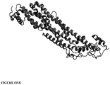
- Figure 1A shows the rendering of chain B as present under Protein Data Bank identifier (PBD ID) 4AD7 (http://www.ebi.ac.uk/pdbsum/4AD7).
- PBD ID Protein Data Bank identifier
- CLIPS technology structurally fixes peptides into defined three-dimensional structures. This results in functional mimics of even the most complex binding sites. CLIPS technology is now routinely used to shape peptide libraries into single, double or triple looped structures as well as sheet- and helix-like folds.
- the CLIPS reaction takes place between bromo groups of the CLIPS scaffold and thiol sidechains of cysteines. The reaction is fast and specific under mild conditions. Using this chemistry, native protein sequences are transformed into CLIPS constructs with a range of structures including single T2 loops, T3 double loops, conjugated T2+T3 loops, stabilized beta sheets, and stabilized alpha helixes ( Timmerman et al., J. Mol. Recognit. 2007; 20: 283-29 ).
- CLIPS library screening starts with the conversion of the target protein into a library of up to 10,000 overlapping peptide constructs, using a combinatorial matrix design.
- a matrix of linear peptides is synthesized, which are subsequently shaped into spatially defined CLIPS constructs.
- Constructs representing both parts of the discontinuous epitope in the correct conformation bind the antibody with high affinity, which is detected and quantified.
- Constructs presenting the incomplete epitope bind the antibody with lower affinity, whereas constructs not containing the epitope do not bind at all.
- Affinity information is used in iterative screens to define the sequence and conformation of epitopes in detail.
- the target protein containing a discontinuous conformational epitope is converted into a matrix library.
- Combinatorial peptides are synthesized on a proprietary minicard and chemically converted into spatially defined CLIPS constructs. Binding of antibodies is quantified.
- CLIPS Chemically Linked Peptides on Scaffolds
- a 0.5 mM solution of the T2 CLIPS template 1,3-bis (bromomethyl) benzene was dissolved in ammonium bicarbonate (20 mM, pH 7.9)/acetonitrile (1:1(v/v)). This solution was added onto the peptide arrays.
- the CLIPS template bound to side-chains of two cysteines as present in the solid-phase bound peptides of the peptide-arrays (455 wells plate with 3 ⁇ l wells). The peptide arrays were gently shaken in the solution for 30 to 60 minutes while completely covered in solution.
- peptide arrays were washed extensively with excess of H 2 O and sonicated in disrupt-buffer containing 1 percent SDS/0.1 percent betamercaptoethanol in PBS (pH 7.2) at 70°C for 30 minutes, followed by sonication in H 2 O for another 45 minutes.
- the T3 CLIPS carrying peptides were made in a similar way but with three cysteines.
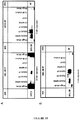
- Linear and CLIPS peptides were chemicall synthesized according to the following designs: Set 1 Label lin Mimic Type Linear peptides Description Linear peptides of length 15 with an overlap of 14 that cover reference sequence ( SEQ ID NO: 14 ). Set 2 Label loop Mimic Type Loop shaped peptides Description Peptides of length 17, with cysteine residues at positions 1 and 17. Positions 2 - 16 contain the linear 15mers of Set 1 in which cysteines are replaced by alanine. After synthesis, peptides are constrained by a P2 CLIPSTM linker to constrain the shape.
- Set 3 Label hel_i3 Mimic Type Helical peptides Description Peptides of length 15, with an overlap of 14 derived from the reference sequence ( SEQ ID NO: 14 ). Cysteine residues were replaced by Alanine. Then positions 3 and 7 were replaced by Cysteine, which were connected by a P2 CLIPSTM. This i, i+4 connection ('stapling') induced helix nucleation in sequences that were prone to helical folding.
- Set 4 Label hel_i7 Mimic Type Helical peptides Description Peptides of length 15, with an overlap of 14 derived from the reference sequence ( SEQ ID NO: 14 ). Cysteine residues were replaced by Alanine.
- the binding of antibody to each of the synthesized peptides was tested in a PEPSCAN-based ELISA.
- the peptide arrays were incubated with primary antibody solution (overnight at 4°C). After washing, the peptide arrays were incubated with a 1/1000 dilution of an antibody peroxidase conjugate (SBA, cat.nr. 2010-05) for one hour at 25°C. After washing, the peroxidase substrate 2,2'-azino-di-3-ethylbenzthiazoline sulfonate (ABTS) and 2 ⁇ l/ml of 3 percent H 2 O 2 were added. After one hour, the color development was measured. The color development was quantified with a charge coupled device (CCD) - camera and an image processing system.
- CCD charge coupled device
- the values obtained from the CCD camera ranged from 0 to 3000 mAU, similar to a standard 96-well plate ELISA-reader.
- the results were quantified and stored in the Peplab database. Occasionally a well contained an air-bubble resulting in a false-positive value.
- the cards were manually inspected and any values caused by an air-bubble were scored as 0.
- Table 7 summarises antibody binding conditions. For the Pepscan Buffer and Preconditioning (SQ), the numbers indicate the relative amount of competing protein (a combination of horse serum and ovalbumin). Table 7: screening conditions dilution samplebuffer preconditioning serum MIL38-AM4* 1 ⁇ g/ml 50%SQ 50%SQ MIL38-AM4* 10 ⁇ g/ml 50%SQ 50%SQ * produced by hybridoma cells as deposited at Cellbank Australia under accession number CBA20140026
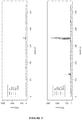
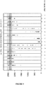
- FIG. 2 A graphical overview of the complete dataset of raw ELISA results generated by the screening is shown in Figure 2 .
- a box plot depicts each dataset and indicates the average ELISA signal, the distribution and the outliers within each dataset.
- experiment conditions amount of antibody, blocking strength etc
- different distributions of ELISA data were obtained. The data enabled identification of an epitope.
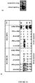
- the array was incubated with MIL38-AM4 at dilutions of 1 ⁇ g/ml and 10 ⁇ g/ml, under normal stringency conditions. A concentration dependent response was observed ( Figures 3A and 3B ). At 10 ⁇ g/ml saturation was not observed, indicating that none of the peptides represented the full epitope. Two clusters of responses that are not adjacent in the primary structure were identified. Two common cores emerged: 135 TQNARAFRD 143 ( SEQ ID NO: 7 ) and 348 VNPQGPGPEEK 358 ( SEQ ID NO: 2 ), with the latter stretch clearly dominant with respect to the former.
- Table 8 below provides information on the antibodies used in this study. The first three antibodies listed are commercially available. Table 8: description of antibodies used Name Origin Concentration Location Status Anti-Glypican 1 (GPC1) (AA 24-530) ⁇ goat 1mg/ml 4 °C/ 11 OK Anti Glypican 1/GPC1 antibody (ab137604)* rabbit 0.97mg/ml -20 °C/ 73 OK Mab2600 Merck Millipore ⁇ (anti-glypican-1 Ab, clone 4D1) mouse 1mg/ml 4 °C/ 11 OK MIL38-AM3 ⁇ mouse 1.25mg/ml -20 °C/ 73 used ⁇ Commercially available from: antibodies-online (http: // www.antibodies-online.com); Lifespan Biosciences, Inc.
- the human glypican-1 (GPC-1) sequence defined by SEQ ID NO: 14 was used as a basis to generate a library of structured peptides.
- Figure 1A shows the rendering of chain B as present under Protein Data Bank identifier (PBD ID) 4AD7 (http://www.ebi.ac.uk/pdbsum/4AD7).
- PBD ID Protein Data Bank identifier
- Example 1 Peptide synthesis was performed using the methods referred in Example 1. Chemically synthesized linear and CLIPS peptides were synthesized according to the designs shown in Example 1 (sets 1-5).
- Table 9 summarises antibody binding conditions.
- the numbers indicate the relative amount of competing protein (a combination of horse serum and ovalbumin).
- P/Tw PBS-Tween was also used to reduce stringency of binding.
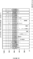
- FIG. 5 A graphical overview of the complete dataset of raw ELISA results generated by the screening is shown in Figure 5 .
- a box plot depicts each dataset and indicates the average ELISA signal, the distribution and the outliers within each dataset.
- experiment conditions amount of antibody, blocking strength etc
- different distributions of ELISA data are obtained.
- the data enabled identification of an epitope for the monoclonal and polyclonal sera, but not for MIL38-AM3.
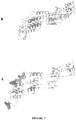
- Rabbit anti GPC1 yields 3 main signals, as can be seen in the corresponding panel in Figure 6 . These signals correspond to the stretches 348 VNPQGPGPEEK 358 (SEQ ID NO: 2), 366 PRERPP 371 (SEQ ID NO: 10), and 478 QDASDDGSGS 487 (SEQ ID NO: 11).
- Mouse antibody 2600 recognizes one clear peak in all peptide sets, sharing the common core 242 LGPECSRAVMK 252 (SEQ ID NO: 13). This can be seen in the corresponding panel in Figure 6 .
- Goat anti-GPC-1 yields 2 main signals, as can be seen in the corresponding panel in Figure 6 . These signals correspond to the stretches 348 VNPQGPGPEEK 358 (SEQ ID NO: 2), and 408 ALSTASDDR 414 (SEQ ID NO: 9).
- Rabbit anti GPC-1 recognizes at least three stretches in glypican 1, 348 VNPQGPGPEEK 358 (SEQ ID NO: 2), 366 PRERPP 371 (SEQ ID NO: 10), and 478Q DASDDGSGS 487 (SEQ ID NO: 11). Two of these stretches are resolved in coordinate file 4AD7.pbd, as depicted in Figure 7A . Since this is a polyclonal antibody preparation, it cannot be assessed whether the epitopes are linear or are part of a complex epitope.
- Mouse anti glypican mab 2600 recognizes the stretch 242 LGPECSRAVMK 252 (SEQ ID NO: 13) in all peptide sets of the array.
- the localization of epitope in coordinate file 4AD7.pdb is depicted in Figure 7B .
- Goat anti glypican 1 recognizes at least two stretches in glypican 1, 348 VNPQGPGPEEK 358 (SEQ ID NO: 2), and 408 ALSTASDDR 414 (SEQ ID NO: 9). Since this is a polyclonal antibody preparation, it cannot be assessed whether the epitopes are linear or are part of a complex epitope. Neither stretch is resolved in the available coordinate file.
- the rabbit and goat polyclonal preparations both recognize a stretch 348 VNPQGPGPEEK 358 (SEQ ID NO: 2) in Glypican 1, which also forms part of the (likely discontinuous) binding site of Mab MIL38-AM4. Both polyclonal preparations also recognize additional epitopes on Glypican 1, but it cannot be assessed if these epitopes form a discontinuous epitope, or are manifestations of the polyclonal nature of the sample. Neither pAb recognizes the stretch 135 TQNARA 140 , which is thought to contribute to MIL38-AM4 binding.
- Mouse Mab 2600 recognizes an epitope that is not shared by any other anti-GPC-1 antibody tested thus far.
- Table 10 below provides information on the antibodies used in this study. Three anti-glypican 1 antibodies were used, each having been used in earlier experiments as described in Example 1 and/or Example 2 above. Table 10: description of antibodies used Name Origin Concentration Location Status Anti-Glypican 1 (GPC1) (AA 24-530) ⁇ goat 1 mg/ml 4 °C/ 11 OK Anti Glypican 1/GPC1 antibody (ab137604)* rabbit 0.97mg/ml -20 °C/ 73 OK MIL38-AM4 ⁇ mouse 4.6 mg/ml -20 °C/ 73 OK ⁇ Commercially available from: antibodies-online (http: // www.antibodies-online.com); Lifespan Biosciences, Inc. (https: // www.lsbio.com) * Commercially available from abeam® (http: // www.abcam.com) ⁇ Produced by hybridoma cells as deposited at Cellbank Australia under accession number CBA20140026
- the human glypican-1 (GPC-1) sequence on which this study was based is defined in SEQ ID NO: 14. The following sequences of residues were used:
- Example 2 Peptide synthesis was performed using the methods referred in Example 1. Chemically synthesized linear and CLIPS peptides were synthesized according to the designs shown below: Set 1 Mimic Type Discontinuous epitope mimics Label MAT.A, MAT.B Description Constrained peptide mimics of varying length. From the two starting sequences ( SEQ ID NO: 15 and SEQ ID NO: 16 ) all 10 to 22, and 10 to 18 mer peptides with stepsize 4 were made, and these were been paired. At the termini and in between the two peptides cysteines were placed. These were linked by a T3 CLIPS.
- SEQ ID NO: 15 and SEQ ID NO: 16 From the two starting sequences ( SEQ ID NO: 15 and SEQ ID NO: 16 ) all 10 to 22, and 10 to 18 mer peptides with stepsize 4 were made, and these were been paired. At the termini and in between the two peptides cysteines were placed. These were linked by a T3 CLIPS.
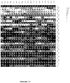
- a heat map is a graphical representation of data where the values taken by a variable in a two-dimensional map are represented as colours.
- a two-dimensional map can be derived from the independent sequences of the first and second loops.
- the sequences of a given series of 16 CLIPS peptides are effectively permutations of 4 unique sub-sequences in loop 1 and 4 unique sub-sequences in loop 2.
- the observed ELISA data can be plotted in a 4x4 matrix, where each X coordinate corresponds to the sequence of the first loop, and each Y coordinate corresponds to the sequence of the second loop.
- ELISA values can be replaced with a continuous gradient of shading.
- extremely low values are light coloured
- extremely high values are darker coloured
- average values are black coloured.
- Table 11 summarises antibody binding conditions. For the Pepscan Buffer and Preconditioning (SQ), the numbers indicate the relative amount of competing protein (a combination of horse serum and ovalbumin). Table 11: screening conditions serum dilution samplebuffer preconditioning Anti-Glypican 1 (GPC1) (AA 24-530) 10 ng/ml SQ SQ Anti Glypican 1/GPC1 antibody (ab137604) 1/2500 SQ SQ MIL-38 AM4 10 ⁇ g/ml 10%SQ 50%SQ
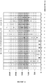
- FIG. 8 A graphical overview of the complete dataset of raw ELISA results generated by the screening is shown in Figure 8 .
- a box plot depicts each dataset and indicates the average ELISA signal, the distribution and the outliers within each dataset.
- experiment conditions amount of antibody, blocking strength etc
- Example 2 the stretch 348 VNPQGPGPEEK 358 (SEQ ID NO: 2) was found to suffice for binding some IgG from rabbit polyclonal Ab 137604. In this study all constructs containing 348 VNPQGPGPEE 357 (SEQ ID NO: 3) again were bound by IgG from this sample at 1/2500 dilution. In the matrices of Set1 there is no augmentation of signal in specific constructs.
- Example 2 the stretch 348 VNPQGPGPEEK 358 (SEQ ID NO: 2) also sufficed to bind some IgG from goat polyclonal antiGPC-1.
- This antibody recognizes the same loop as the rabbit polyclonal, but does so with a slightly different fine specificity.
- the most critical residues are G352, G354, and E356, and that limited substitution is allowed on positions N349, Q351, and P353.
- the stretch around residues 140 - 149 was not implied in the primary mapping of this antibody, distinct constructs including this stretch are better baits for binding by some IgG in this polyclone.
- MIL38-AM4 binds glypican on stretch 348 VNPQGPGPEEK 358 (SEQ ID NO: 2), and also binds to the stretch 135 TQNARA 140 (SEQ ID NO: 8), which was taken as an indication for a discontinuous epitope.
- the antibodies bind to or not bind to similar constructs, as can be seen in the scatter plots of Figure 13 . However, many constructs are exclusively seen by one of the preparations (points along the axes, or in the lower right hand and upper left hand corners).
- Example 2 The two leads obtained in Example 1 that point to a discontinuous epitope for MIL 38 - AM4 were used to generate a matrix array in which the loops have different lengths. In addition, full substitution analyses of the individual lead sequences were made. All arrays were probed with the three antibodies listed above.
- the epitope of (some IgG species present in) rabbit polyclonal Ab 137604 is the linear stretch 348 VNPQGPGPEE 357 (SEQ ID NO: 3). There is no indication for the presence of antibodies that recognize a conformational epitope.
- Table 13 represents a summary of substitutions tolerated in the epitope sequences analysed.
- Table 13 Summary of tolerated substitutions in epitope sequences Residue No. in SEQ ID NO: 14 347 348 349 350 351 352 353 354 355 356 E 357 E 358 K V N P Q G P G P K Tolerated substitutions in MIL38-AM4
- GPC1 Goat Anti-Glypican 1
- GPC1 Goat Anti-Glypican 1
- Example 4 MIL38 and anti-glypican-1 (anti-GPC-1) antibodies show overlapping reactivity on 2D western blots.
- Rabbit anti-GPC-1 antibody ab137604 showed reactivity with the glypican-1 core protein at a molecular weight of approximately 60 kDa - the same molecular weight as detected by MIL38.
- MIL38 recognized glypican-1
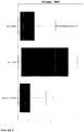
- prostate cancer DU-145 MPEK extracts were subjected to 2D electrophoresis and western blotting.
- Membrane protein extracts (MPEK) of DU-145 prostate cancer cells were separated on 2D gel (pI gradient-horizontal, and molecular mass vertical). Western blots using MIL38 antibody and commercial rGPC-1 rabbit polyclonal antibodies show overlapping reactivity marking a 60Kd protein (circled in Figure 14 ).
- Lane D ( Figure 14 ) is a one dimension separation for DU-145 extracts as a control.
- Lane M ( Figure 14 ) is a one dimension separation lane for molecular size markers as controls.
- MIL38 antibody and the anti-GPC-1 antibodies showed overlapping reactivity detecting a protein with 60 kDa molecular weight and isoelectric points ranging from 5 to 7.
- Example 5 MIL38 is detected in anti-GPC-1 immunoprecipitates and vice versa.
- MIL38 or rabbit anti-GPC-1 antibodies were used to immunoprecipitate their respective antigens from DU-145 or C3 (MIL38 negative) MPEK extracts.
- the immunoprecipitates (IPs) were western blotted with either MIL38 or anti-GPC-1 antibody ( Figure 15 ).
- a 60kDa GPC-1 reactive band was detected in MIL38 IPs blotted with anti-GPC-1, while a 60 kDa MIL38 reactive band was detected in anti-GPC-1 IPs blotted with MIL38. No reactivity was detected with the secondary only controls. Furthermore, immunoprecipitating with MIL38 antibody resulted in almost complete depletion of both MIL38 and anti-GPC-1 antigens, strongly suggesting that the MIL38 antigen is glypican-1.
- FIG. 15A depicts GPC1 detection of MIL38 immunoprecipitates (left) and MIL38 detection of GPC1 immunoprecipitates (right).
- Figure 15B depicts MIL38 detection of MIL38 immunoprecipitates as a control.
- Lanes are: Magic Mark-commercial protein marker as control; DU145 MPEK- prostate cancer membrane protein extract (not immunoprecipitated); DU145 FT- prostate cancer flow through from immune precipitation; DU145 IP- immunoprecipitate using antibody; C3 MPEK- (MIL38 negative) control membrane protein extract (not immunoprecipitated); C3 FT- flow through from immune precipitation; C3 IP elution- (MIL38 negative) immunoprecipitate using antibody.
- MIL38 can detect the immunoprecipitate from rGPC1 antibody and vice versa.
- MIL38 can also bind to all controls including DU145 MPEK and to IP conducted by MIL38.
- GPC-1 can be detected by MIL38 in prostate cancer plasma samples and in membrane extracts from prostate cancer patients.
- Plasma samples from one normal patient (042) and one prostate cancer patient (046) were immunoprecipitated with MIL38 antibody and the IP sample western blotted with MIL38 and anti-GPC-1 antibodies ( Figure 16A ).
- MIL38 antigen could be detected in membrane protein extracts from normal prostate and prostate cancer
- one sample of each was obtained from Novus Bio. Equivalent amounts of protein were western blotted using MIL38 antibody ( Figure 16B ).
- the prostate cancer extract demonstrated much higher expression of the MIL38 antigen than the normal prostate sample.
- Example 7 Detection of MIL38 antigen in patient urine.
- MIL38 can detect cells in the urine of prostate cancer patients. To test the sensitivity and specificity of this detection method, 125 age-matched urine samples were obtained. Cells were spun down from the urine and analyzed by the MIL38 indirect immunofluorescence assay. A total of 47 healthy controls, 37 benign prostatic hypertrophy (BPH) and 41 biopsy-confirmed prostate cancers were analyzed.
- BPH benign prostatic hypertrophy
- the MIL38 immunofluorescence assay (IFA) test demonstrated a sensitivity of 71% and a specificity of 73% in identifying prostate cancers within the cohort. The test showed 71% sensitivity and 76% specificity in identifying prostate cancers compared to BPH patients (Table 14). Table 14: Sensitivity and specificity calculations of prostate cancer detection in patient urine. Sensitivity and Specificity Calculations True Positive False Positive 29 12 False Negative True Negative 23 61 Sensitivity and Specificity Calculations for BPH only True Positive False Positive 29 12 False Negative True Negative 9 28
Landscapes
- Health & Medical Sciences (AREA)
- Chemical & Material Sciences (AREA)
- Life Sciences & Earth Sciences (AREA)
- Immunology (AREA)
- Organic Chemistry (AREA)
- Molecular Biology (AREA)
- Biochemistry (AREA)
- General Health & Medical Sciences (AREA)
- Medicinal Chemistry (AREA)
- Engineering & Computer Science (AREA)
- Biophysics (AREA)
- Genetics & Genomics (AREA)
- Proteomics, Peptides & Aminoacids (AREA)
- Cell Biology (AREA)
- Gastroenterology & Hepatology (AREA)
- Hematology (AREA)
- Urology & Nephrology (AREA)
- Biomedical Technology (AREA)
- Zoology (AREA)
- Toxicology (AREA)
- Biotechnology (AREA)
- Microbiology (AREA)
- Food Science & Technology (AREA)
- Physics & Mathematics (AREA)
- Analytical Chemistry (AREA)
- General Physics & Mathematics (AREA)
- Pathology (AREA)
- Peptides Or Proteins (AREA)
- Micro-Organisms Or Cultivation Processes Thereof (AREA)
- Medicines Containing Antibodies Or Antigens For Use As Internal Diagnostic Agents (AREA)
Applications Claiming Priority (1)
| Application Number | Priority Date | Filing Date | Title |
|---|---|---|---|
| PCT/AU2015/000019 WO2016112423A1 (en) | 2015-01-16 | 2015-01-16 | Glypican epitopes and uses thereof |
Publications (3)
| Publication Number | Publication Date |
|---|---|
| EP3245219A1 EP3245219A1 (en) | 2017-11-22 |
| EP3245219A4 EP3245219A4 (en) | 2018-06-06 |
| EP3245219B1 true EP3245219B1 (en) | 2020-04-08 |
Family
ID=56405046
Family Applications (1)
| Application Number | Title | Priority Date | Filing Date |
|---|---|---|---|
| EP15877381.2A Active EP3245219B1 (en) | 2015-01-16 | 2015-01-16 | Glypican epitopes and uses thereof |
Country Status (10)
| Country | Link |
|---|---|
| US (3) | US20180002391A1 (enExample) |
| EP (1) | EP3245219B1 (enExample) |
| JP (1) | JP6735005B2 (enExample) |
| KR (1) | KR102435383B1 (enExample) |
| CN (1) | CN107531755B (enExample) |
| AU (1) | AU2015376851B2 (enExample) |
| DK (1) | DK3245219T3 (enExample) |
| ES (1) | ES2805085T3 (enExample) |
| SG (1) | SG11201705808XA (enExample) |
| WO (1) | WO2016112423A1 (enExample) |
Families Citing this family (8)
| Publication number | Priority date | Publication date | Assignee | Title |
|---|---|---|---|---|
| CA2964179C (en) * | 2014-10-23 | 2023-04-11 | Minomic International Ltd. | Monoclonal anti-gpc-1 antibodies and uses thereof |
| CA2983311A1 (en) * | 2015-04-20 | 2016-10-27 | Minomic International Ltd. | Therapeutic antibodies and uses thereof |
| JPWO2018199318A1 (ja) * | 2017-04-28 | 2020-03-12 | 国立大学法人高知大学 | 抗gpc−1抗体 |
| US12122843B2 (en) | 2019-01-22 | 2024-10-22 | The United States Of America, As Represented By The Secretary, Department Of Health And Human Services | High affinity monoclonal antibodies targeting glypican-1 and methods of use |
| CN111187350A (zh) * | 2019-12-30 | 2020-05-22 | 西安英创生物技术有限公司 | 一种与磷脂酰肌醇蛋白聚糖-1结合的抗原结合蛋白 |
| IT202200021546A1 (it) | 2022-10-19 | 2024-04-19 | Centro Di Riferimento Oncologico Di Aviano | Anticorpo monoclonale anti-GPC1, suoi usi terapeutici e diagnostici |
| WO2025202361A1 (en) * | 2024-03-27 | 2025-10-02 | Adcendo Aps | Anti-gpc1 antibodies and uses thereof |
| WO2025202359A1 (en) * | 2024-03-27 | 2025-10-02 | Adcendo Aps | Anti-gpc1 antibodies and uses thereof |
Family Cites Families (13)
| Publication number | Priority date | Publication date | Assignee | Title |
|---|---|---|---|---|
| US4356270A (en) | 1977-11-08 | 1982-10-26 | Genentech, Inc. | Recombinant DNA cloning vehicle |
| US4458066A (en) | 1980-02-29 | 1984-07-03 | University Patents, Inc. | Process for preparing polynucleotides |
| US4751180A (en) | 1985-03-28 | 1988-06-14 | Chiron Corporation | Expression using fused genes providing for protein product |
| US4935233A (en) | 1985-12-02 | 1990-06-19 | G. D. Searle And Company | Covalently linked polypeptide cell modulators |
| US5840839A (en) * | 1996-02-09 | 1998-11-24 | The United States Of America As Represented By The Secretary Of The Department Of Health And Human Services | Alternative open reading frame DNA of a normal gene and a novel human cancer antigen encoded therein |
| AU2422999A (en) * | 1998-01-27 | 1999-08-09 | Vlaams Interuniversitair Instituut Voor Biotechnologie Vzw | New members of the glypican gene family |
| EP1146903A4 (en) * | 1998-10-16 | 2005-02-16 | Univ California | GLYPICANE FOR THE DETECTION AND TREATMENT OF HUMAN CARCINOMA |
| US20040226056A1 (en) * | 1998-12-22 | 2004-11-11 | Myriad Genetics, Incorporated | Compositions and methods for treating neurological disorders and diseases |
| KR100953520B1 (ko) * | 2001-06-22 | 2010-04-21 | 츄가이 세이야꾸 가부시키가이샤 | 항글리피칸 3항체를 포함하는 항암제 |
| SI1674111T1 (sl) * | 2004-07-09 | 2011-02-28 | Chugai Pharmaceutical Co Ltd | Protitelo proti glipikanu 3 |
| US20070087005A1 (en) * | 2005-10-14 | 2007-04-19 | Lazar Gregory A | Anti-glypican-3 antibody |
| US8270389B2 (en) | 2008-08-11 | 2012-09-18 | Marvell International Ltd. | Method of synchronization for low power idle |
| CN102180969B (zh) * | 2011-01-30 | 2013-04-10 | 中国人民解放军军事医学科学院微生物流行病研究所 | 抗肝癌活性单克隆抗体及其应用 |
-
2015
- 2015-01-16 SG SG11201705808XA patent/SG11201705808XA/en unknown
- 2015-01-16 WO PCT/AU2015/000019 patent/WO2016112423A1/en not_active Ceased
- 2015-01-16 CN CN201580076839.6A patent/CN107531755B/zh active Active
- 2015-01-16 DK DK15877381.2T patent/DK3245219T3/da active
- 2015-01-16 ES ES15877381T patent/ES2805085T3/es active Active
- 2015-01-16 AU AU2015376851A patent/AU2015376851B2/en active Active
- 2015-01-16 US US15/543,877 patent/US20180002391A1/en not_active Abandoned
- 2015-01-16 EP EP15877381.2A patent/EP3245219B1/en active Active
- 2015-01-16 KR KR1020177022547A patent/KR102435383B1/ko active Active
- 2015-01-16 JP JP2017537906A patent/JP6735005B2/ja active Active
-
2020
- 2020-07-27 US US16/939,729 patent/US20210107958A1/en not_active Abandoned
-
2023
- 2023-07-28 US US18/361,407 patent/US20240247037A1/en active Pending
Non-Patent Citations (1)
| Title |
|---|
| None * |
Also Published As
| Publication number | Publication date |
|---|---|
| AU2015376851A1 (en) | 2017-07-27 |
| US20180002391A1 (en) | 2018-01-04 |
| KR102435383B1 (ko) | 2022-08-24 |
| KR20170109584A (ko) | 2017-09-29 |
| ES2805085T3 (es) | 2021-02-10 |
| CN107531755B (zh) | 2022-02-25 |
| EP3245219A4 (en) | 2018-06-06 |
| WO2016112423A1 (en) | 2016-07-21 |
| AU2015376851B2 (en) | 2020-07-09 |
| DK3245219T3 (da) | 2020-06-29 |
| CA2973771A1 (en) | 2016-07-21 |
| US20210107958A1 (en) | 2021-04-15 |
| JP2018504903A (ja) | 2018-02-22 |
| EP3245219A1 (en) | 2017-11-22 |
| SG11201705808XA (en) | 2017-08-30 |
| JP6735005B2 (ja) | 2020-08-05 |
| US20240247037A1 (en) | 2024-07-25 |
| CN107531755A (zh) | 2018-01-02 |
Similar Documents
| Publication | Publication Date | Title |
|---|---|---|
| EP3245219B1 (en) | Glypican epitopes and uses thereof | |
| JPWO2004081047A1 (ja) | モノクローナル抗体及びこれを産生するハイブリドーマ | |
| US20230391886A1 (en) | Compositions and methods for muc18 targeting | |
| KR20140048318A (ko) | 췌장암의 검출 방법 | |
| KR20110000548A (ko) | 신장암의 진단 또는 검출을 위한 조성물 및 방법 | |
| US12559548B2 (en) | Antibodies to misfolded TDP-43 and methods of use | |
| EP3239176B1 (en) | Anti-active gip antibody | |
| JP6707660B2 (ja) | 抗ポドカリキシン様タンパク質前駆体サブタイプ2モノクローナル抗体とその生成方法及び使用 | |
| EP2726094B1 (en) | Therapeutic and diagnostic target | |
| JP5305259B2 (ja) | 抗シトルリン化gfapモノクローナル抗体及びその用途 | |
| CN102863530A (zh) | 一种脂肪细胞分化代谢产物igf-1抗体及包含该抗体的芯片以及应用 | |
| EP2918600B1 (en) | Novel peptide and use thereof | |
| HK1238267B (en) | Glypican epitopes and uses thereof | |
| US9056919B2 (en) | USP2a peptides and antibodies | |
| JP7053058B2 (ja) | グリピカンエピトープおよびその使用 | |
| CA2973771C (en) | Glypican epitopes and uses thereof | |
| JP6861349B2 (ja) | Cd81 lel特異的モノクローナル抗体 | |
| CN108727493B (zh) | 抗Stathmin单克隆抗体及其用途 | |
| KR20190107967A (ko) | 한국인 위암 연관 유전자 단클론 항체 및 이의 제조방법 | |
| HK1196092A (en) | Therapeutic and diagnostic target | |
| HK1196092B (en) | Therapeutic and diagnostic target |
Legal Events
| Date | Code | Title | Description |
|---|---|---|---|
| STAA | Information on the status of an ep patent application or granted ep patent |
Free format text: STATUS: THE INTERNATIONAL PUBLICATION HAS BEEN MADE |
|
| PUAI | Public reference made under article 153(3) epc to a published international application that has entered the european phase |
Free format text: ORIGINAL CODE: 0009012 |
|
| STAA | Information on the status of an ep patent application or granted ep patent |
Free format text: STATUS: REQUEST FOR EXAMINATION WAS MADE |
|
| 17P | Request for examination filed |
Effective date: 20170722 |
|
| AK | Designated contracting states |
Kind code of ref document: A1 Designated state(s): AL AT BE BG CH CY CZ DE DK EE ES FI FR GB GR HR HU IE IS IT LI LT LU LV MC MK MT NL NO PL PT RO RS SE SI SK SM TR |
|
| AX | Request for extension of the european patent |
Extension state: BA ME |
|
| DAX | Request for extension of the european patent (deleted) | ||
| REG | Reference to a national code |
Ref country code: HK Ref legal event code: DE Ref document number: 1238267 Country of ref document: HK |
|
| RIC1 | Information provided on ipc code assigned before grant |
Ipc: C07K 14/705 20060101ALI20180423BHEP Ipc: C07K 16/28 20060101ALI20180423BHEP Ipc: G01N 33/53 20060101ALI20180423BHEP Ipc: C07K 7/06 20060101AFI20180423BHEP Ipc: C07K 7/08 20060101ALI20180423BHEP |
|
| A4 | Supplementary search report drawn up and despatched |
Effective date: 20180507 |
|
| STAA | Information on the status of an ep patent application or granted ep patent |
Free format text: STATUS: EXAMINATION IS IN PROGRESS |
|
| 17Q | First examination report despatched |
Effective date: 20190121 |
|
| GRAP | Despatch of communication of intention to grant a patent |
Free format text: ORIGINAL CODE: EPIDOSNIGR1 |
|
| STAA | Information on the status of an ep patent application or granted ep patent |
Free format text: STATUS: GRANT OF PATENT IS INTENDED |
|
| INTG | Intention to grant announced |
Effective date: 20191031 |
|
| RAP1 | Party data changed (applicant data changed or rights of an application transferred) |
Owner name: GLYP HOLDINGS PTY LIMITED |
|
| RIN1 | Information on inventor provided before grant (corrected) |
Inventor name: CAMPBELL, DOUGLAS Inventor name: WISSMUELLER, SANDRA Inventor name: TRUONG, QUACH Inventor name: NOCON, ALINE Inventor name: JUSTINIANO FUENMAYOR, IRENE Inventor name: SOON, JULIE Inventor name: WALSH, BRADLEY |
|
| GRAS | Grant fee paid |
Free format text: ORIGINAL CODE: EPIDOSNIGR3 |
|
| GRAA | (expected) grant |
Free format text: ORIGINAL CODE: 0009210 |
|
| STAA | Information on the status of an ep patent application or granted ep patent |
Free format text: STATUS: THE PATENT HAS BEEN GRANTED |
|
| AK | Designated contracting states |
Kind code of ref document: B1 Designated state(s): AL AT BE BG CH CY CZ DE DK EE ES FI FR GB GR HR HU IE IS IT LI LT LU LV MC MK MT NL NO PL PT RO RS SE SI SK SM TR |
|
| REG | Reference to a national code |
Ref country code: AT Ref legal event code: REF Ref document number: 1254253 Country of ref document: AT Kind code of ref document: T Effective date: 20200415 Ref country code: CH Ref legal event code: EP |
|
| REG | Reference to a national code |
Ref country code: DE Ref legal event code: R096 Ref document number: 602015050532 Country of ref document: DE |
|
| REG | Reference to a national code |
Ref country code: IE Ref legal event code: FG4D |
|
| REG | Reference to a national code |
Ref country code: DK Ref legal event code: T3 Effective date: 20200625 |
|
| REG | Reference to a national code |
Ref country code: CH Ref legal event code: NV Representative=s name: NOVAGRAAF INTERNATIONAL SA, CH |
|
| REG | Reference to a national code |
Ref country code: SE Ref legal event code: TRGR |
|
| REG | Reference to a national code |
Ref country code: NL Ref legal event code: FP |
|
| REG | Reference to a national code |
Ref country code: NO Ref legal event code: T2 Effective date: 20200408 |
|
| REG | Reference to a national code |
Ref country code: LT Ref legal event code: MG4D |
|
| PG25 | Lapsed in a contracting state [announced via postgrant information from national office to epo] |
Ref country code: IS Free format text: LAPSE BECAUSE OF FAILURE TO SUBMIT A TRANSLATION OF THE DESCRIPTION OR TO PAY THE FEE WITHIN THE PRESCRIBED TIME-LIMIT Effective date: 20200808 Ref country code: FI Free format text: LAPSE BECAUSE OF FAILURE TO SUBMIT A TRANSLATION OF THE DESCRIPTION OR TO PAY THE FEE WITHIN THE PRESCRIBED TIME-LIMIT Effective date: 20200408 Ref country code: PT Free format text: LAPSE BECAUSE OF FAILURE TO SUBMIT A TRANSLATION OF THE DESCRIPTION OR TO PAY THE FEE WITHIN THE PRESCRIBED TIME-LIMIT Effective date: 20200817 Ref country code: GR Free format text: LAPSE BECAUSE OF FAILURE TO SUBMIT A TRANSLATION OF THE DESCRIPTION OR TO PAY THE FEE WITHIN THE PRESCRIBED TIME-LIMIT Effective date: 20200709 Ref country code: LT Free format text: LAPSE BECAUSE OF FAILURE TO SUBMIT A TRANSLATION OF THE DESCRIPTION OR TO PAY THE FEE WITHIN THE PRESCRIBED TIME-LIMIT Effective date: 20200408 |
|
| PG25 | Lapsed in a contracting state [announced via postgrant information from national office to epo] |
Ref country code: BG Free format text: LAPSE BECAUSE OF FAILURE TO SUBMIT A TRANSLATION OF THE DESCRIPTION OR TO PAY THE FEE WITHIN THE PRESCRIBED TIME-LIMIT Effective date: 20200708 Ref country code: HR Free format text: LAPSE BECAUSE OF FAILURE TO SUBMIT A TRANSLATION OF THE DESCRIPTION OR TO PAY THE FEE WITHIN THE PRESCRIBED TIME-LIMIT Effective date: 20200408 Ref country code: LV Free format text: LAPSE BECAUSE OF FAILURE TO SUBMIT A TRANSLATION OF THE DESCRIPTION OR TO PAY THE FEE WITHIN THE PRESCRIBED TIME-LIMIT Effective date: 20200408 Ref country code: RS Free format text: LAPSE BECAUSE OF FAILURE TO SUBMIT A TRANSLATION OF THE DESCRIPTION OR TO PAY THE FEE WITHIN THE PRESCRIBED TIME-LIMIT Effective date: 20200408 |
|
| PG25 | Lapsed in a contracting state [announced via postgrant information from national office to epo] |
Ref country code: AL Free format text: LAPSE BECAUSE OF FAILURE TO SUBMIT A TRANSLATION OF THE DESCRIPTION OR TO PAY THE FEE WITHIN THE PRESCRIBED TIME-LIMIT Effective date: 20200408 |
|
| REG | Reference to a national code |
Ref country code: DE Ref legal event code: R097 Ref document number: 602015050532 Country of ref document: DE |
|
| PG25 | Lapsed in a contracting state [announced via postgrant information from national office to epo] |
Ref country code: SM Free format text: LAPSE BECAUSE OF FAILURE TO SUBMIT A TRANSLATION OF THE DESCRIPTION OR TO PAY THE FEE WITHIN THE PRESCRIBED TIME-LIMIT Effective date: 20200408 Ref country code: EE Free format text: LAPSE BECAUSE OF FAILURE TO SUBMIT A TRANSLATION OF THE DESCRIPTION OR TO PAY THE FEE WITHIN THE PRESCRIBED TIME-LIMIT Effective date: 20200408 Ref country code: CZ Free format text: LAPSE BECAUSE OF FAILURE TO SUBMIT A TRANSLATION OF THE DESCRIPTION OR TO PAY THE FEE WITHIN THE PRESCRIBED TIME-LIMIT Effective date: 20200408 Ref country code: RO Free format text: LAPSE BECAUSE OF FAILURE TO SUBMIT A TRANSLATION OF THE DESCRIPTION OR TO PAY THE FEE WITHIN THE PRESCRIBED TIME-LIMIT Effective date: 20200408 |
|
| REG | Reference to a national code |
Ref country code: ES Ref legal event code: FG2A Ref document number: 2805085 Country of ref document: ES Kind code of ref document: T3 Effective date: 20210210 |
|
| PLBE | No opposition filed within time limit |
Free format text: ORIGINAL CODE: 0009261 |
|
| STAA | Information on the status of an ep patent application or granted ep patent |
Free format text: STATUS: NO OPPOSITION FILED WITHIN TIME LIMIT |
|
| PG25 | Lapsed in a contracting state [announced via postgrant information from national office to epo] |
Ref country code: PL Free format text: LAPSE BECAUSE OF FAILURE TO SUBMIT A TRANSLATION OF THE DESCRIPTION OR TO PAY THE FEE WITHIN THE PRESCRIBED TIME-LIMIT Effective date: 20200408 Ref country code: SK Free format text: LAPSE BECAUSE OF FAILURE TO SUBMIT A TRANSLATION OF THE DESCRIPTION OR TO PAY THE FEE WITHIN THE PRESCRIBED TIME-LIMIT Effective date: 20200408 |
|
| 26N | No opposition filed |
Effective date: 20210112 |
|
| PG25 | Lapsed in a contracting state [announced via postgrant information from national office to epo] |
Ref country code: SI Free format text: LAPSE BECAUSE OF FAILURE TO SUBMIT A TRANSLATION OF THE DESCRIPTION OR TO PAY THE FEE WITHIN THE PRESCRIBED TIME-LIMIT Effective date: 20200408 |
|
| PG25 | Lapsed in a contracting state [announced via postgrant information from national office to epo] |
Ref country code: MC Free format text: LAPSE BECAUSE OF FAILURE TO SUBMIT A TRANSLATION OF THE DESCRIPTION OR TO PAY THE FEE WITHIN THE PRESCRIBED TIME-LIMIT Effective date: 20200408 |
|
| PG25 | Lapsed in a contracting state [announced via postgrant information from national office to epo] |
Ref country code: LU Free format text: LAPSE BECAUSE OF NON-PAYMENT OF DUE FEES Effective date: 20210116 |
|
| REG | Reference to a national code |
Ref country code: AT Ref legal event code: UEP Ref document number: 1254253 Country of ref document: AT Kind code of ref document: T Effective date: 20200408 |
|
| PG25 | Lapsed in a contracting state [announced via postgrant information from national office to epo] |
Ref country code: HU Free format text: LAPSE BECAUSE OF FAILURE TO SUBMIT A TRANSLATION OF THE DESCRIPTION OR TO PAY THE FEE WITHIN THE PRESCRIBED TIME-LIMIT; INVALID AB INITIO Effective date: 20150116 |
|
| P01 | Opt-out of the competence of the unified patent court (upc) registered |
Effective date: 20230425 |
|
| PG25 | Lapsed in a contracting state [announced via postgrant information from national office to epo] |
Ref country code: CY Free format text: LAPSE BECAUSE OF FAILURE TO SUBMIT A TRANSLATION OF THE DESCRIPTION OR TO PAY THE FEE WITHIN THE PRESCRIBED TIME-LIMIT Effective date: 20200408 |
|
| PG25 | Lapsed in a contracting state [announced via postgrant information from national office to epo] |
Ref country code: MK Free format text: LAPSE BECAUSE OF FAILURE TO SUBMIT A TRANSLATION OF THE DESCRIPTION OR TO PAY THE FEE WITHIN THE PRESCRIBED TIME-LIMIT Effective date: 20200408 |
|
| PG25 | Lapsed in a contracting state [announced via postgrant information from national office to epo] |
Ref country code: TR Free format text: LAPSE BECAUSE OF FAILURE TO SUBMIT A TRANSLATION OF THE DESCRIPTION OR TO PAY THE FEE WITHIN THE PRESCRIBED TIME-LIMIT Effective date: 20200408 |
|
| PG25 | Lapsed in a contracting state [announced via postgrant information from national office to epo] |
Ref country code: MT Free format text: LAPSE BECAUSE OF FAILURE TO SUBMIT A TRANSLATION OF THE DESCRIPTION OR TO PAY THE FEE WITHIN THE PRESCRIBED TIME-LIMIT Effective date: 20200408 |
|
| PGFP | Annual fee paid to national office [announced via postgrant information from national office to epo] |
Ref country code: NL Payment date: 20250127 Year of fee payment: 11 |
|
| PGFP | Annual fee paid to national office [announced via postgrant information from national office to epo] |
Ref country code: DE Payment date: 20250121 Year of fee payment: 11 |
|
| PGFP | Annual fee paid to national office [announced via postgrant information from national office to epo] |
Ref country code: DK Payment date: 20250129 Year of fee payment: 11 |
|
| PGFP | Annual fee paid to national office [announced via postgrant information from national office to epo] |
Ref country code: ES Payment date: 20250210 Year of fee payment: 11 |
|
| PGFP | Annual fee paid to national office [announced via postgrant information from national office to epo] |
Ref country code: IE Payment date: 20250123 Year of fee payment: 11 Ref country code: SE Payment date: 20250128 Year of fee payment: 11 |
|
| PGFP | Annual fee paid to national office [announced via postgrant information from national office to epo] |
Ref country code: NO Payment date: 20250127 Year of fee payment: 11 |
|
| PGFP | Annual fee paid to national office [announced via postgrant information from national office to epo] |
Ref country code: CH Payment date: 20250205 Year of fee payment: 11 Ref country code: AT Payment date: 20250127 Year of fee payment: 11 Ref country code: BE Payment date: 20250123 Year of fee payment: 11 |
|
| PGFP | Annual fee paid to national office [announced via postgrant information from national office to epo] |
Ref country code: FR Payment date: 20250121 Year of fee payment: 11 |
|
| PGFP | Annual fee paid to national office [announced via postgrant information from national office to epo] |
Ref country code: IT Payment date: 20250129 Year of fee payment: 11 Ref country code: GB Payment date: 20250116 Year of fee payment: 11 |