EP3190193A1 - Nanopore-facilitated single molecule detection of nucleic acids - Google Patents
Nanopore-facilitated single molecule detection of nucleic acids Download PDFInfo
- Publication number
- EP3190193A1 EP3190193A1 EP17157476.7A EP17157476A EP3190193A1 EP 3190193 A1 EP3190193 A1 EP 3190193A1 EP 17157476 A EP17157476 A EP 17157476A EP 3190193 A1 EP3190193 A1 EP 3190193A1
- Authority
- EP
- European Patent Office
- Prior art keywords
- probe
- level
- nanopore
- mir
- target
- Prior art date
- Legal status (The legal status is an assumption and is not a legal conclusion. Google has not performed a legal analysis and makes no representation as to the accuracy of the status listed.)
- Withdrawn
Links
- 150000007523 nucleic acids Chemical class 0.000 title claims description 33
- 102000039446 nucleic acids Human genes 0.000 title claims description 28
- 108020004707 nucleic acids Proteins 0.000 title claims description 28
- 238000004557 single molecule detection Methods 0.000 title description 4
- 239000000523 sample Substances 0.000 claims abstract description 171
- 239000002679 microRNA Substances 0.000 claims abstract description 125
- 108091034117 Oligonucleotide Proteins 0.000 claims abstract description 77
- 230000000295 complement effect Effects 0.000 claims abstract description 18
- 238000000034 method Methods 0.000 claims description 55
- 108700011259 MicroRNAs Proteins 0.000 claims description 50
- 230000005945 translocation Effects 0.000 claims description 32
- 239000002773 nucleotide Substances 0.000 claims description 30
- 108090000765 processed proteins & peptides Proteins 0.000 claims description 30
- 125000003729 nucleotide group Chemical group 0.000 claims description 28
- 101710092462 Alpha-hemolysin Proteins 0.000 claims description 16
- 229920001184 polypeptide Polymers 0.000 claims description 16
- 102000004196 processed proteins & peptides Human genes 0.000 claims description 16
- 238000005192 partition Methods 0.000 claims description 9
- 230000003993 interaction Effects 0.000 claims description 8
- 238000010494 dissociation reaction Methods 0.000 claims description 7
- 230000005593 dissociations Effects 0.000 claims description 7
- 229920000642 polymer Polymers 0.000 claims description 5
- OKTJSMMVPCPJKN-UHFFFAOYSA-N Carbon Chemical compound [C] OKTJSMMVPCPJKN-UHFFFAOYSA-N 0.000 claims description 3
- 229910052581 Si3N4 Inorganic materials 0.000 claims description 3
- VCORFLZFSPUNDN-UAGCYRGNSA-N [(2r,3s,5r)-5-(6-aminopurin-9-yl)-3-[[(2r,3s,5r)-5-(6-aminopurin-9-yl)-3-hydroxyoxolan-2-yl]methoxy-hydroxyphosphoryl]oxyoxolan-2-yl]methyl [(2r,3s,5r)-5-(6-aminopurin-9-yl)-2-(phosphonooxymethyl)oxolan-3-yl] hydrogen phosphate Chemical compound C1=NC2=C(N)N=CN=C2N1[C@H](O[C@@H]1COP(O)(=O)O[C@@H]2[C@H](O[C@H](C2)N2C3=NC=NC(N)=C3N=C2)COP(O)(O)=O)C[C@@H]1OP(O)(=O)OC[C@@H](O1)[C@@H](O)C[C@@H]1N1C(N=CN=C2N)=C2N=C1 VCORFLZFSPUNDN-UAGCYRGNSA-N 0.000 claims description 3
- 229910021389 graphene Inorganic materials 0.000 claims description 3
- HQVNEWCFYHHQES-UHFFFAOYSA-N silicon nitride Chemical compound N12[Si]34N5[Si]62N3[Si]51N64 HQVNEWCFYHHQES-UHFFFAOYSA-N 0.000 claims description 3
- 239000012634 fragment Substances 0.000 claims description 2
- 108091070501 miRNA Proteins 0.000 abstract description 118
- 238000001514 detection method Methods 0.000 abstract description 47
- JLCPHMBAVCMARE-UHFFFAOYSA-N [3-[[3-[[3-[[3-[[3-[[3-[[3-[[3-[[3-[[3-[[3-[[5-(2-amino-6-oxo-1H-purin-9-yl)-3-[[3-[[3-[[3-[[3-[[3-[[5-(2-amino-6-oxo-1H-purin-9-yl)-3-[[5-(2-amino-6-oxo-1H-purin-9-yl)-3-hydroxyoxolan-2-yl]methoxy-hydroxyphosphoryl]oxyoxolan-2-yl]methoxy-hydroxyphosphoryl]oxy-5-(5-methyl-2,4-dioxopyrimidin-1-yl)oxolan-2-yl]methoxy-hydroxyphosphoryl]oxy-5-(6-aminopurin-9-yl)oxolan-2-yl]methoxy-hydroxyphosphoryl]oxy-5-(6-aminopurin-9-yl)oxolan-2-yl]methoxy-hydroxyphosphoryl]oxy-5-(6-aminopurin-9-yl)oxolan-2-yl]methoxy-hydroxyphosphoryl]oxy-5-(6-aminopurin-9-yl)oxolan-2-yl]methoxy-hydroxyphosphoryl]oxyoxolan-2-yl]methoxy-hydroxyphosphoryl]oxy-5-(5-methyl-2,4-dioxopyrimidin-1-yl)oxolan-2-yl]methoxy-hydroxyphosphoryl]oxy-5-(4-amino-2-oxopyrimidin-1-yl)oxolan-2-yl]methoxy-hydroxyphosphoryl]oxy-5-(5-methyl-2,4-dioxopyrimidin-1-yl)oxolan-2-yl]methoxy-hydroxyphosphoryl]oxy-5-(5-methyl-2,4-dioxopyrimidin-1-yl)oxolan-2-yl]methoxy-hydroxyphosphoryl]oxy-5-(6-aminopurin-9-yl)oxolan-2-yl]methoxy-hydroxyphosphoryl]oxy-5-(6-aminopurin-9-yl)oxolan-2-yl]methoxy-hydroxyphosphoryl]oxy-5-(4-amino-2-oxopyrimidin-1-yl)oxolan-2-yl]methoxy-hydroxyphosphoryl]oxy-5-(4-amino-2-oxopyrimidin-1-yl)oxolan-2-yl]methoxy-hydroxyphosphoryl]oxy-5-(4-amino-2-oxopyrimidin-1-yl)oxolan-2-yl]methoxy-hydroxyphosphoryl]oxy-5-(6-aminopurin-9-yl)oxolan-2-yl]methoxy-hydroxyphosphoryl]oxy-5-(4-amino-2-oxopyrimidin-1-yl)oxolan-2-yl]methyl [5-(6-aminopurin-9-yl)-2-(hydroxymethyl)oxolan-3-yl] hydrogen phosphate Polymers Cc1cn(C2CC(OP(O)(=O)OCC3OC(CC3OP(O)(=O)OCC3OC(CC3O)n3cnc4c3nc(N)[nH]c4=O)n3cnc4c3nc(N)[nH]c4=O)C(COP(O)(=O)OC3CC(OC3COP(O)(=O)OC3CC(OC3COP(O)(=O)OC3CC(OC3COP(O)(=O)OC3CC(OC3COP(O)(=O)OC3CC(OC3COP(O)(=O)OC3CC(OC3COP(O)(=O)OC3CC(OC3COP(O)(=O)OC3CC(OC3COP(O)(=O)OC3CC(OC3COP(O)(=O)OC3CC(OC3COP(O)(=O)OC3CC(OC3COP(O)(=O)OC3CC(OC3COP(O)(=O)OC3CC(OC3COP(O)(=O)OC3CC(OC3COP(O)(=O)OC3CC(OC3COP(O)(=O)OC3CC(OC3COP(O)(=O)OC3CC(OC3CO)n3cnc4c(N)ncnc34)n3ccc(N)nc3=O)n3cnc4c(N)ncnc34)n3ccc(N)nc3=O)n3ccc(N)nc3=O)n3ccc(N)nc3=O)n3cnc4c(N)ncnc34)n3cnc4c(N)ncnc34)n3cc(C)c(=O)[nH]c3=O)n3cc(C)c(=O)[nH]c3=O)n3ccc(N)nc3=O)n3cc(C)c(=O)[nH]c3=O)n3cnc4c3nc(N)[nH]c4=O)n3cnc4c(N)ncnc34)n3cnc4c(N)ncnc34)n3cnc4c(N)ncnc34)n3cnc4c(N)ncnc34)O2)c(=O)[nH]c1=O JLCPHMBAVCMARE-UHFFFAOYSA-N 0.000 abstract description 13
- 238000005516 engineering process Methods 0.000 abstract description 9
- 206010028980 Neoplasm Diseases 0.000 abstract description 8
- 230000001976 improved effect Effects 0.000 abstract description 7
- 210000004369 blood Anatomy 0.000 abstract description 6
- 239000008280 blood Substances 0.000 abstract description 6
- 201000011510 cancer Diseases 0.000 abstract description 5
- 230000004069 differentiation Effects 0.000 abstract description 5
- 238000011002 quantification Methods 0.000 abstract description 5
- 238000002405 diagnostic procedure Methods 0.000 abstract 1
- 108091033773 MiR-155 Proteins 0.000 description 94
- 239000011148 porous material Substances 0.000 description 59
- 108091007423 let-7b Proteins 0.000 description 31
- 239000000243 solution Substances 0.000 description 31
- 108091028376 let-7a-5 stem-loop Proteins 0.000 description 29
- 108091091807 let-7a stem-loop Proteins 0.000 description 27
- 108091057746 let-7a-4 stem-loop Proteins 0.000 description 27
- 108091024393 let-7a-6 stem-loop Proteins 0.000 description 27
- 108091091174 let-7a-7 stem-loop Proteins 0.000 description 27
- 206010058467 Lung neoplasm malignant Diseases 0.000 description 22
- 201000005202 lung cancer Diseases 0.000 description 20
- 208000020816 lung neoplasm Diseases 0.000 description 20
- 108091093037 Peptide nucleic acid Proteins 0.000 description 19
- 108091032973 (ribonucleotides)n+m Proteins 0.000 description 18
- 108020004414 DNA Proteins 0.000 description 17
- 239000000203 mixture Substances 0.000 description 15
- WCUXLLCKKVVCTQ-UHFFFAOYSA-M Potassium chloride Chemical compound [Cl-].[K+] WCUXLLCKKVVCTQ-UHFFFAOYSA-M 0.000 description 14
- 108091090568 miR-39 stem-loop Proteins 0.000 description 11
- 108091056739 miR-39-1 stem-loop Proteins 0.000 description 11
- 108091039160 miR-39-2 stem-loop Proteins 0.000 description 11
- 238000000926 separation method Methods 0.000 description 11
- 108091007772 MIRLET7C Proteins 0.000 description 10
- 235000001014 amino acid Nutrition 0.000 description 10
- 108090000623 proteins and genes Proteins 0.000 description 10
- 238000003757 reverse transcription PCR Methods 0.000 description 10
- 230000035945 sensitivity Effects 0.000 description 10
- 239000000232 Lipid Bilayer Substances 0.000 description 9
- 150000001413 amino acids Chemical class 0.000 description 9
- 102000004169 proteins and genes Human genes 0.000 description 9
- 102000040650 (ribonucleotides)n+m Human genes 0.000 description 8
- 238000011529 RT qPCR Methods 0.000 description 8
- 238000002512 chemotherapy Methods 0.000 description 8
- 235000018102 proteins Nutrition 0.000 description 8
- 208000000649 small cell carcinoma Diseases 0.000 description 8
- 239000003228 hemolysin Substances 0.000 description 6
- 238000012544 monitoring process Methods 0.000 description 6
- 230000008569 process Effects 0.000 description 6
- 238000002123 RNA extraction Methods 0.000 description 5
- 229920001577 copolymer Polymers 0.000 description 5
- 239000000463 material Substances 0.000 description 5
- -1 microRNAs Chemical class 0.000 description 5
- 210000001519 tissue Anatomy 0.000 description 5
- 108091023037 Aptamer Proteins 0.000 description 4
- 239000002202 Polyethylene glycol Substances 0.000 description 4
- 235000009697 arginine Nutrition 0.000 description 4
- 238000003556 assay Methods 0.000 description 4
- 238000010586 diagram Methods 0.000 description 4
- 238000002474 experimental method Methods 0.000 description 4
- 230000006870 function Effects 0.000 description 4
- 108020004999 messenger RNA Proteins 0.000 description 4
- 238000002156 mixing Methods 0.000 description 4
- 230000036961 partial effect Effects 0.000 description 4
- 229920001223 polyethylene glycol Polymers 0.000 description 4
- 108091005703 transmembrane proteins Proteins 0.000 description 4
- 102000035160 transmembrane proteins Human genes 0.000 description 4
- 239000004475 Arginine Substances 0.000 description 3
- 102000053602 DNA Human genes 0.000 description 3
- 239000003298 DNA probe Substances 0.000 description 3
- 108090000862 Ion Channels Proteins 0.000 description 3
- 102000004310 Ion Channels Human genes 0.000 description 3
- 239000013614 RNA sample Substances 0.000 description 3
- 101900127397 Staphylococcus aureus Alpha-hemolysin Proteins 0.000 description 3
- 108010090804 Streptavidin Proteins 0.000 description 3
- 230000003321 amplification Effects 0.000 description 3
- 238000004458 analytical method Methods 0.000 description 3
- ODKSFYDXXFIFQN-UHFFFAOYSA-N arginine Natural products OC(=O)C(N)CCCNC(N)=N ODKSFYDXXFIFQN-UHFFFAOYSA-N 0.000 description 3
- 230000008901 benefit Effects 0.000 description 3
- 210000004027 cell Anatomy 0.000 description 3
- 230000008859 change Effects 0.000 description 3
- 238000009826 distribution Methods 0.000 description 3
- 229940079593 drug Drugs 0.000 description 3
- 239000003814 drug Substances 0.000 description 3
- 230000014509 gene expression Effects 0.000 description 3
- UYTPUPDQBNUYGX-UHFFFAOYSA-N guanine Chemical compound O=C1NC(N)=NC2=C1N=CN2 UYTPUPDQBNUYGX-UHFFFAOYSA-N 0.000 description 3
- 238000009396 hybridization Methods 0.000 description 3
- 108091023663 let-7 stem-loop Proteins 0.000 description 3
- 108091063478 let-7-1 stem-loop Proteins 0.000 description 3
- 108091049777 let-7-2 stem-loop Proteins 0.000 description 3
- 238000011068 loading method Methods 0.000 description 3
- 235000018977 lysine Nutrition 0.000 description 3
- 238000003199 nucleic acid amplification method Methods 0.000 description 3
- 238000003753 real-time PCR Methods 0.000 description 3
- 238000012360 testing method Methods 0.000 description 3
- FZWGECJQACGGTI-UHFFFAOYSA-N 2-amino-7-methyl-1,7-dihydro-6H-purin-6-one Chemical compound NC1=NC(O)=C2N(C)C=NC2=N1 FZWGECJQACGGTI-UHFFFAOYSA-N 0.000 description 2
- OIVLITBTBDPEFK-UHFFFAOYSA-N 5,6-dihydrouracil Chemical compound O=C1CCNC(=O)N1 OIVLITBTBDPEFK-UHFFFAOYSA-N 0.000 description 2
- GFFGJBXGBJISGV-UHFFFAOYSA-N Adenine Chemical compound NC1=NC=NC2=C1N=CN2 GFFGJBXGBJISGV-UHFFFAOYSA-N 0.000 description 2
- 108091006146 Channels Proteins 0.000 description 2
- 238000000018 DNA microarray Methods 0.000 description 2
- 238000001712 DNA sequencing Methods 0.000 description 2
- 108010006464 Hemolysin Proteins Proteins 0.000 description 2
- ODKSFYDXXFIFQN-BYPYZUCNSA-P L-argininium(2+) Chemical compound NC(=[NH2+])NCCC[C@H]([NH3+])C(O)=O ODKSFYDXXFIFQN-BYPYZUCNSA-P 0.000 description 2
- UBORTCNDUKBEOP-UHFFFAOYSA-N L-xanthosine Natural products OC1C(O)C(CO)OC1N1C(NC(=O)NC2=O)=C2N=C1 UBORTCNDUKBEOP-UHFFFAOYSA-N 0.000 description 2
- KDXKERNSBIXSRK-UHFFFAOYSA-N Lysine Natural products NCCCCC(N)C(O)=O KDXKERNSBIXSRK-UHFFFAOYSA-N 0.000 description 2
- 239000004472 Lysine Substances 0.000 description 2
- 238000000692 Student's t-test Methods 0.000 description 2
- 239000007983 Tris buffer Substances 0.000 description 2
- ISAKRJDGNUQOIC-UHFFFAOYSA-N Uracil Chemical compound O=C1C=CNC(=O)N1 ISAKRJDGNUQOIC-UHFFFAOYSA-N 0.000 description 2
- UBORTCNDUKBEOP-HAVMAKPUSA-N Xanthosine Natural products O[C@@H]1[C@H](O)[C@H](CO)O[C@H]1N1C(NC(=O)NC2=O)=C2N=C1 UBORTCNDUKBEOP-HAVMAKPUSA-N 0.000 description 2
- 208000009956 adenocarcinoma Diseases 0.000 description 2
- 125000003118 aryl group Chemical group 0.000 description 2
- 230000031018 biological processes and functions Effects 0.000 description 2
- 229920001400 block copolymer Polymers 0.000 description 2
- 125000003178 carboxy group Chemical group [H]OC(*)=O 0.000 description 2
- 238000003776 cleavage reaction Methods 0.000 description 2
- OPTASPLRGRRNAP-UHFFFAOYSA-N cytosine Chemical compound NC=1C=CNC(=O)N=1 OPTASPLRGRRNAP-UHFFFAOYSA-N 0.000 description 2
- 238000013399 early diagnosis Methods 0.000 description 2
- 238000010828 elution Methods 0.000 description 2
- 229920000140 heteropolymer Polymers 0.000 description 2
- HNDVDQJCIGZPNO-UHFFFAOYSA-N histidine Natural products OC(=O)C(N)CC1=CN=CN1 HNDVDQJCIGZPNO-UHFFFAOYSA-N 0.000 description 2
- 235000014304 histidine Nutrition 0.000 description 2
- 229920001519 homopolymer Polymers 0.000 description 2
- FDGQSTZJBFJUBT-UHFFFAOYSA-N hypoxanthine Chemical compound O=C1NC=NC2=C1NC=N2 FDGQSTZJBFJUBT-UHFFFAOYSA-N 0.000 description 2
- 150000002500 ions Chemical class 0.000 description 2
- 230000000670 limiting effect Effects 0.000 description 2
- 230000007774 longterm Effects 0.000 description 2
- 201000005296 lung carcinoma Diseases 0.000 description 2
- 125000003588 lysine group Chemical group [H]N([H])C([H])([H])C([H])([H])C([H])([H])C([H])([H])C([H])(N([H])[H])C(*)=O 0.000 description 2
- 230000001394 metastastic effect Effects 0.000 description 2
- 206010061289 metastatic neoplasm Diseases 0.000 description 2
- 230000011987 methylation Effects 0.000 description 2
- 238000007069 methylation reaction Methods 0.000 description 2
- 230000035772 mutation Effects 0.000 description 2
- 108091027963 non-coding RNA Proteins 0.000 description 2
- 238000010606 normalization Methods 0.000 description 2
- 210000005259 peripheral blood Anatomy 0.000 description 2
- 239000011886 peripheral blood Substances 0.000 description 2
- 230000002028 premature Effects 0.000 description 2
- 230000003449 preventive effect Effects 0.000 description 2
- 230000002035 prolonged effect Effects 0.000 description 2
- 230000005855 radiation Effects 0.000 description 2
- 230000000306 recurrent effect Effects 0.000 description 2
- 238000002271 resection Methods 0.000 description 2
- 238000010839 reverse transcription Methods 0.000 description 2
- 230000002441 reversible effect Effects 0.000 description 2
- 150000003839 salts Chemical class 0.000 description 2
- 230000007017 scission Effects 0.000 description 2
- 238000004088 simulation Methods 0.000 description 2
- 238000006467 substitution reaction Methods 0.000 description 2
- RWQNBRDOKXIBIV-UHFFFAOYSA-N thymine Chemical compound CC1=CNC(=O)NC1=O RWQNBRDOKXIBIV-UHFFFAOYSA-N 0.000 description 2
- LENZDBCJOHFCAS-UHFFFAOYSA-N tris Chemical compound OCC(N)(CO)CO LENZDBCJOHFCAS-UHFFFAOYSA-N 0.000 description 2
- 239000000107 tumor biomarker Substances 0.000 description 2
- UBORTCNDUKBEOP-UUOKFMHZSA-N xanthosine Chemical compound O[C@@H]1[C@H](O)[C@@H](CO)O[C@H]1N1C(NC(=O)NC2=O)=C2N=C1 UBORTCNDUKBEOP-UUOKFMHZSA-N 0.000 description 2
- ZAYHVCMSTBRABG-UHFFFAOYSA-N 5-Methylcytidine Natural products O=C1N=C(N)C(C)=CN1C1C(O)C(O)C(CO)O1 ZAYHVCMSTBRABG-UHFFFAOYSA-N 0.000 description 1
- ZAYHVCMSTBRABG-JXOAFFINSA-N 5-methylcytidine Chemical compound O=C1N=C(N)C(C)=CN1[C@H]1[C@H](O)[C@H](O)[C@@H](CO)O1 ZAYHVCMSTBRABG-JXOAFFINSA-N 0.000 description 1
- LRSASMSXMSNRBT-UHFFFAOYSA-N 5-methylcytosine Chemical compound CC1=CNC(=O)N=C1N LRSASMSXMSNRBT-UHFFFAOYSA-N 0.000 description 1
- OGHAROSJZRTIOK-KQYNXXCUSA-O 7-methylguanosine Chemical compound C1=2N=C(N)NC(=O)C=2[N+](C)=CN1[C@@H]1O[C@H](CO)[C@@H](O)[C@H]1O OGHAROSJZRTIOK-KQYNXXCUSA-O 0.000 description 1
- 229930024421 Adenine Natural products 0.000 description 1
- 241000894006 Bacteria Species 0.000 description 1
- 108091026890 Coding region Proteins 0.000 description 1
- XUIIKFGFIJCVMT-GFCCVEGCSA-N D-thyroxine Chemical compound IC1=CC(C[C@@H](N)C(O)=O)=CC(I)=C1OC1=CC(I)=C(O)C(I)=C1 XUIIKFGFIJCVMT-GFCCVEGCSA-N 0.000 description 1
- 108020003215 DNA Probes Proteins 0.000 description 1
- KCXVZYZYPLLWCC-UHFFFAOYSA-N EDTA Chemical compound OC(=O)CN(CC(O)=O)CCN(CC(O)=O)CC(O)=O KCXVZYZYPLLWCC-UHFFFAOYSA-N 0.000 description 1
- 102000004190 Enzymes Human genes 0.000 description 1
- 108090000790 Enzymes Proteins 0.000 description 1
- 108091081406 G-quadruplex Proteins 0.000 description 1
- 108700000788 Human immunodeficiency virus 1 tat peptide (47-57) Proteins 0.000 description 1
- UGQMRVRMYYASKQ-UHFFFAOYSA-N Hypoxanthine nucleoside Natural products OC1C(O)C(CO)OC1N1C(NC=NC2=O)=C2N=C1 UGQMRVRMYYASKQ-UHFFFAOYSA-N 0.000 description 1
- 229930010555 Inosine Natural products 0.000 description 1
- UGQMRVRMYYASKQ-KQYNXXCUSA-N Inosine Chemical compound O[C@@H]1[C@H](O)[C@@H](CO)O[C@H]1N1C2=NC=NC(O)=C2N=C1 UGQMRVRMYYASKQ-KQYNXXCUSA-N 0.000 description 1
- 102100034343 Integrase Human genes 0.000 description 1
- HNDVDQJCIGZPNO-YFKPBYRVSA-N L-histidine Chemical compound OC(=O)[C@@H](N)CC1=CN=CN1 HNDVDQJCIGZPNO-YFKPBYRVSA-N 0.000 description 1
- KDXKERNSBIXSRK-YFKPBYRVSA-N L-lysine Chemical compound NCCCC[C@H](N)C(O)=O KDXKERNSBIXSRK-YFKPBYRVSA-N 0.000 description 1
- COLNVLDHVKWLRT-QMMMGPOBSA-N L-phenylalanine Chemical compound OC(=O)[C@@H](N)CC1=CC=CC=C1 COLNVLDHVKWLRT-QMMMGPOBSA-N 0.000 description 1
- QIVBCDIJIAJPQS-VIFPVBQESA-N L-tryptophane Chemical compound C1=CC=C2C(C[C@H](N)C(O)=O)=CNC2=C1 QIVBCDIJIAJPQS-VIFPVBQESA-N 0.000 description 1
- OUYCCCASQSFEME-QMMMGPOBSA-N L-tyrosine Chemical compound OC(=O)[C@@H](N)CC1=CC=C(O)C=C1 OUYCCCASQSFEME-QMMMGPOBSA-N 0.000 description 1
- 108091092724 Noncoding DNA Proteins 0.000 description 1
- 108091028043 Nucleic acid sequence Proteins 0.000 description 1
- 108020005187 Oligonucleotide Probes Proteins 0.000 description 1
- 108700026244 Open Reading Frames Proteins 0.000 description 1
- 229910019142 PO4 Inorganic materials 0.000 description 1
- 101710124239 Poly(A) polymerase Proteins 0.000 description 1
- 235000013929 Psidium pyriferum Nutrition 0.000 description 1
- 244000236580 Psidium pyriferum Species 0.000 description 1
- 108010092799 RNA-directed DNA polymerase Proteins 0.000 description 1
- 102000003661 Ribonuclease III Human genes 0.000 description 1
- 108010057163 Ribonuclease III Proteins 0.000 description 1
- 108020004682 Single-Stranded DNA Proteins 0.000 description 1
- 239000004809 Teflon Substances 0.000 description 1
- 229920006362 Teflon® Polymers 0.000 description 1
- 108020004566 Transfer RNA Proteins 0.000 description 1
- QIVBCDIJIAJPQS-UHFFFAOYSA-N Tryptophan Natural products C1=CC=C2C(CC(N)C(O)=O)=CNC2=C1 QIVBCDIJIAJPQS-UHFFFAOYSA-N 0.000 description 1
- 241000700605 Viruses Species 0.000 description 1
- 230000001594 aberrant effect Effects 0.000 description 1
- 230000002378 acidificating effect Effects 0.000 description 1
- 230000006978 adaptation Effects 0.000 description 1
- 229960000643 adenine Drugs 0.000 description 1
- 125000000539 amino acid group Chemical group 0.000 description 1
- 238000013459 approach Methods 0.000 description 1
- 150000001484 arginines Chemical class 0.000 description 1
- 238000005415 bioluminescence Methods 0.000 description 1
- 230000029918 bioluminescence Effects 0.000 description 1
- 239000000090 biomarker Substances 0.000 description 1
- 230000015572 biosynthetic process Effects 0.000 description 1
- 210000001124 body fluid Anatomy 0.000 description 1
- 239000010839 body fluid Substances 0.000 description 1
- 229910052799 carbon Inorganic materials 0.000 description 1
- 230000015556 catabolic process Effects 0.000 description 1
- 230000024245 cell differentiation Effects 0.000 description 1
- 238000007385 chemical modification Methods 0.000 description 1
- 238000006243 chemical reaction Methods 0.000 description 1
- 238000004737 colorimetric analysis Methods 0.000 description 1
- 239000002299 complementary DNA Substances 0.000 description 1
- 150000001875 compounds Chemical class 0.000 description 1
- 238000010276 construction Methods 0.000 description 1
- 229940104302 cytosine Drugs 0.000 description 1
- 238000006731 degradation reaction Methods 0.000 description 1
- 230000036425 denaturation Effects 0.000 description 1
- 238000004925 denaturation Methods 0.000 description 1
- 230000001419 dependent effect Effects 0.000 description 1
- 238000013461 design Methods 0.000 description 1
- 238000011161 development Methods 0.000 description 1
- 230000018109 developmental process Effects 0.000 description 1
- ZPTBLXKRQACLCR-XVFCMESISA-N dihydrouridine Chemical compound O[C@@H]1[C@H](O)[C@@H](CO)O[C@H]1N1C(=O)NC(=O)CC1 ZPTBLXKRQACLCR-XVFCMESISA-N 0.000 description 1
- 238000010790 dilution Methods 0.000 description 1
- 239000012895 dilution Substances 0.000 description 1
- 230000005684 electric field Effects 0.000 description 1
- 230000005518 electrochemistry Effects 0.000 description 1
- 238000002001 electrophysiology Methods 0.000 description 1
- 230000007831 electrophysiology Effects 0.000 description 1
- 230000002708 enhancing effect Effects 0.000 description 1
- 230000002255 enzymatic effect Effects 0.000 description 1
- 238000011049 filling Methods 0.000 description 1
- 238000001914 filtration Methods 0.000 description 1
- 239000012530 fluid Substances 0.000 description 1
- 230000036541 health Effects 0.000 description 1
- 230000001939 inductive effect Effects 0.000 description 1
- 230000002401 inhibitory effect Effects 0.000 description 1
- 229960003786 inosine Drugs 0.000 description 1
- 238000003780 insertion Methods 0.000 description 1
- 230000037431 insertion Effects 0.000 description 1
- 230000010354 integration Effects 0.000 description 1
- 238000002372 labelling Methods 0.000 description 1
- 150000002632 lipids Chemical class 0.000 description 1
- 239000003550 marker Substances 0.000 description 1
- 230000035800 maturation Effects 0.000 description 1
- 230000001404 mediated effect Effects 0.000 description 1
- 238000002844 melting Methods 0.000 description 1
- 230000008018 melting Effects 0.000 description 1
- 239000012528 membrane Substances 0.000 description 1
- 125000000325 methylidene group Chemical group [H]C([H])=* 0.000 description 1
- 238000002493 microarray Methods 0.000 description 1
- 230000004048 modification Effects 0.000 description 1
- 238000012986 modification Methods 0.000 description 1
- 230000009456 molecular mechanism Effects 0.000 description 1
- 239000002105 nanoparticle Substances 0.000 description 1
- 102000042567 non-coding RNA Human genes 0.000 description 1
- 102000044158 nucleic acid binding protein Human genes 0.000 description 1
- 108700020942 nucleic acid binding protein Proteins 0.000 description 1
- 239000002777 nucleoside Substances 0.000 description 1
- 125000003835 nucleoside group Chemical group 0.000 description 1
- 239000002751 oligonucleotide probe Substances 0.000 description 1
- 229910052760 oxygen Inorganic materials 0.000 description 1
- 239000001301 oxygen Substances 0.000 description 1
- 230000037361 pathway Effects 0.000 description 1
- COLNVLDHVKWLRT-UHFFFAOYSA-N phenylalanine Natural products OC(=O)C(N)CC1=CC=CC=C1 COLNVLDHVKWLRT-UHFFFAOYSA-N 0.000 description 1
- NBIIXXVUZAFLBC-UHFFFAOYSA-K phosphate Chemical compound [O-]P([O-])([O-])=O NBIIXXVUZAFLBC-UHFFFAOYSA-K 0.000 description 1
- 239000010452 phosphate Substances 0.000 description 1
- 230000026731 phosphorylation Effects 0.000 description 1
- 238000006366 phosphorylation reaction Methods 0.000 description 1
- 230000008488 polyadenylation Effects 0.000 description 1
- 102000054765 polymorphisms of proteins Human genes 0.000 description 1
- 230000001124 posttranscriptional effect Effects 0.000 description 1
- 239000002243 precursor Substances 0.000 description 1
- 239000003755 preservative agent Substances 0.000 description 1
- 230000002335 preservative effect Effects 0.000 description 1
- 238000012545 processing Methods 0.000 description 1
- 238000002331 protein detection Methods 0.000 description 1
- 238000003762 quantitative reverse transcription PCR Methods 0.000 description 1
- 230000002829 reductive effect Effects 0.000 description 1
- 230000025915 regulation of apoptotic process Effects 0.000 description 1
- 230000022983 regulation of cell cycle Effects 0.000 description 1
- 230000016515 regulation of signal transduction Effects 0.000 description 1
- 230000001105 regulatory effect Effects 0.000 description 1
- 238000012552 review Methods 0.000 description 1
- 125000000548 ribosyl group Chemical group C1([C@H](O)[C@H](O)[C@H](O1)CO)* 0.000 description 1
- 239000012266 salt solution Substances 0.000 description 1
- 238000005070 sampling Methods 0.000 description 1
- 229920006395 saturated elastomer Polymers 0.000 description 1
- 229910052710 silicon Inorganic materials 0.000 description 1
- 239000010703 silicon Substances 0.000 description 1
- 238000010561 standard procedure Methods 0.000 description 1
- 230000004960 subcellular localization Effects 0.000 description 1
- 230000008093 supporting effect Effects 0.000 description 1
- 238000003786 synthesis reaction Methods 0.000 description 1
- 238000012353 t test Methods 0.000 description 1
- 230000008685 targeting Effects 0.000 description 1
- 229940113082 thymine Drugs 0.000 description 1
- 229940034208 thyroxine Drugs 0.000 description 1
- XUIIKFGFIJCVMT-UHFFFAOYSA-N thyroxine-binding globulin Natural products IC1=CC(CC([NH3+])C([O-])=O)=CC(I)=C1OC1=CC(I)=C(O)C(I)=C1 XUIIKFGFIJCVMT-UHFFFAOYSA-N 0.000 description 1
- 230000007704 transition Effects 0.000 description 1
- 230000001960 triggered effect Effects 0.000 description 1
- 230000007306 turnover Effects 0.000 description 1
- OUYCCCASQSFEME-UHFFFAOYSA-N tyrosine Natural products OC(=O)C(N)CC1=CC=C(O)C=C1 OUYCCCASQSFEME-UHFFFAOYSA-N 0.000 description 1
- 229940035893 uracil Drugs 0.000 description 1
- XLYOFNOQVPJJNP-UHFFFAOYSA-N water Substances O XLYOFNOQVPJJNP-UHFFFAOYSA-N 0.000 description 1
Images


Classifications
-
- C—CHEMISTRY; METALLURGY
- C12—BIOCHEMISTRY; BEER; SPIRITS; WINE; VINEGAR; MICROBIOLOGY; ENZYMOLOGY; MUTATION OR GENETIC ENGINEERING
- C12Q—MEASURING OR TESTING PROCESSES INVOLVING ENZYMES, NUCLEIC ACIDS OR MICROORGANISMS; COMPOSITIONS OR TEST PAPERS THEREFOR; PROCESSES OF PREPARING SUCH COMPOSITIONS; CONDITION-RESPONSIVE CONTROL IN MICROBIOLOGICAL OR ENZYMOLOGICAL PROCESSES
- C12Q1/00—Measuring or testing processes involving enzymes, nucleic acids or microorganisms; Compositions therefor; Processes of preparing such compositions
- C12Q1/68—Measuring or testing processes involving enzymes, nucleic acids or microorganisms; Compositions therefor; Processes of preparing such compositions involving nucleic acids
- C12Q1/6813—Hybridisation assays
- C12Q1/6816—Hybridisation assays characterised by the detection means
- C12Q1/6825—Nucleic acid detection involving sensors
-
- C—CHEMISTRY; METALLURGY
- C12—BIOCHEMISTRY; BEER; SPIRITS; WINE; VINEGAR; MICROBIOLOGY; ENZYMOLOGY; MUTATION OR GENETIC ENGINEERING
- C12Q—MEASURING OR TESTING PROCESSES INVOLVING ENZYMES, NUCLEIC ACIDS OR MICROORGANISMS; COMPOSITIONS OR TEST PAPERS THEREFOR; PROCESSES OF PREPARING SUCH COMPOSITIONS; CONDITION-RESPONSIVE CONTROL IN MICROBIOLOGICAL OR ENZYMOLOGICAL PROCESSES
- C12Q1/00—Measuring or testing processes involving enzymes, nucleic acids or microorganisms; Compositions therefor; Processes of preparing such compositions
- C12Q1/68—Measuring or testing processes involving enzymes, nucleic acids or microorganisms; Compositions therefor; Processes of preparing such compositions involving nucleic acids
- C12Q1/6876—Nucleic acid products used in the analysis of nucleic acids, e.g. primers or probes
- C12Q1/6883—Nucleic acid products used in the analysis of nucleic acids, e.g. primers or probes for diseases caused by alterations of genetic material
- C12Q1/6886—Nucleic acid products used in the analysis of nucleic acids, e.g. primers or probes for diseases caused by alterations of genetic material for cancer
-
- G—PHYSICS
- G01—MEASURING; TESTING
- G01N—INVESTIGATING OR ANALYSING MATERIALS BY DETERMINING THEIR CHEMICAL OR PHYSICAL PROPERTIES
- G01N33/00—Investigating or analysing materials by specific methods not covered by groups G01N1/00 - G01N31/00
- G01N33/48—Biological material, e.g. blood, urine; Haemocytometers
- G01N33/483—Physical analysis of biological material
- G01N33/487—Physical analysis of biological material of liquid biological material
- G01N33/48707—Physical analysis of biological material of liquid biological material by electrical means
- G01N33/48721—Investigating individual macromolecules, e.g. by translocation through nanopores
-
- C—CHEMISTRY; METALLURGY
- C12—BIOCHEMISTRY; BEER; SPIRITS; WINE; VINEGAR; MICROBIOLOGY; ENZYMOLOGY; MUTATION OR GENETIC ENGINEERING
- C12Q—MEASURING OR TESTING PROCESSES INVOLVING ENZYMES, NUCLEIC ACIDS OR MICROORGANISMS; COMPOSITIONS OR TEST PAPERS THEREFOR; PROCESSES OF PREPARING SUCH COMPOSITIONS; CONDITION-RESPONSIVE CONTROL IN MICROBIOLOGICAL OR ENZYMOLOGICAL PROCESSES
- C12Q2600/00—Oligonucleotides characterized by their use
- C12Q2600/178—Oligonucleotides characterized by their use miRNA, siRNA or ncRNA
Definitions
- This product relates to a method/apparatus of single-molecule detection, more specifically, to a method/system for quantitative detection of single strand nucleic acids, such as microRNAs, employing an ultrasensitive, low noise nanopore-based single-molecule technology.
- MicroRNAs are a class of short ( ⁇ 18-24 nucleotides) noncoding RNAs that regulate gene expression at the post-transcriptional level 2 . Depending on the degree of homology to their target sequences, miRNA binding induces either translational repression or cleavage of target mRNAs 2 . As powerful gene regulators, miRNAs play important roles in development, cell differentiation, and regulation of cell cycle, apoptosis and signaling pathways 2,3 . Aberrant expression of miRNAs has been found in all types of tumors 4,5 ; the different cancer types have distinct miRNA expression profiles 6 .
- RT-qPCR reverse transcription real-time PCR
- microarray for miRNA detection have been developed 11-13 .
- Each technology has its own advantages, but limitations include requiring enzymatic amplification and semiquantitative results 14 .
- the short miRNA sequences make it difficult to selectively design the primers or probes, resulting in cross-hybridization and low selectivity. This is especially true when the miRNAs contain a few or a single nucleotide difference in a miRNA family.
- Emerging techniques based on colorimetry, bioluminescence, enzyme turnovers and electrochemistry have been proposed, and nanoparticles and molecular beacon have been applied to miRNA detection with high sensitivity and selectivity (review 14 ). But the intrinsic versatility needs to be improved.
- LNA lock-nucleic acids
- Nanopores have been developed as receptive single molecule detectors for broad biotechnological applications (previews 17-19 ). The nanopore is also recognized as one of the next generations of DNA sequencing technologies 20,21 .
- the 2-nm nanopore, ⁇ -hemolysin transmembrane protein pore allows rapid translocation of single-stranded oligonucleotide, which has been well characterized for DNA sequencing 22-27 .
- the molecular translocation-based sensing mode is not suitable for miRNA detection because the sequences of all mature miRNAs are short (18-24 nt), and when traversing the nanopore, the current signals by different miRNAs are indistinguishable.
- a new and improved nanopore-based sensing system for detection and differentiation of single strand oligonucleotides, such as miRNAs is described.
- the inventive system for detecting a target single strand oligonucleotide comprises 1) a nanopore, 2) a power source providing sufficient voltage to induce unzipping, 3) a probe with its center domain complementary to the target oligonucleotide, whereas the unzipping of the hybrid of target oligonucleotide and the probe in the nanopore induces certain identifiable current signal changes, and 4) means for detecting the current signal changes.
- the inventive probe further comprises at least one signal tag at its 3' or 5' terminal (or both).
- the signal tag may be of any charged single chain molecule with sufficient length to assist the unzipping translocation through the nanopore driven by the voltage.
- the signal tag may be oligonucleotides such as poly(dC) n , poly(dA) n , and poly(dT) n , or charged polypeptides.
- a new and improved method based on nanopore technology for detecting and differentiating single strand oligonucleotide detects the current changes triggered by the unzipping of the hybrid of the target oligonucleotide and its probe in a nanopore.
- the inventive method includes the step of 1) mixing the target oligonucleotide with a pre-designed probe, which has its central domain matching the target sequence and a charged single chain molecule tagged to at least one of its 3' and 5' terminals, to produce a sample mixture, 2) loading the mixture into the cis chamber of a nanopore system, and a voltage is added from the trans chamber, and 3) recording current output for a pre-determined time period.
- a new and improved method for detecting and monitoring cancer-related miRNAs in patients' blood sample includes the steps of 1) mixing the total plasma RNAs extracted from a patient's blood sample with the miRNA probe that contains the complementary sequence to the targeting miRNA and a signal tag at the probe's 3'-terminal, 5'-terminal, or both, 2) adding the mixture into a nanopore chamber with a preselected voltage, and 3) monitoring and analyzing the signature events in the output current traces that serves as an electrical marker for single miRNA molecule recognition.
- a probe molecule for detecting of a single strand oligonucleotide, such as miRNA, using a nanopore comprising: 1) a center domain with a complementary sequence to the target oligonucleotide, and 2) a terminal extension tagged to at least one of the center domain's 3' or 5' terminals is provided.
- the terminal extension is a charged chain molecule.
- the terminal extension is a charged polypeptide.
- terminal extension is a charged polymer.
- the invention provides a method of detecting single strand oligonucleotide with a dual-compartment nanopore system, whereas the system includes a cis compartment and a trans compartment divided by a partition with an opening at its center region, recording solution filling both compartments and a lipid bilayer formed at the opening on the partition, a nanopore plugging through the lipid bilayer bridging the cis and trans chamber, a voltage loaded upon the system via a pair of electrodes each extruding from the cis or trans compartment, and a current detector monitoring the current changes, includes the steps of 1) mixing the target oligonucleotide with a pre-designed probe, which has its central domain matching the target sequence and a charged single chain molecule tagged to at least one of its 3' and 5' terminals, to produce a sample mixture, 2) loading the mixture into the cis compartment, 3) providing the system with a pre-determined voltage, and 4) recording current output for a pre-determined time period.
- the system includes a
- the instant invention also includes probes, nanopores, kits comprising the probes and nanopores, and associated methods of use described in the following portions of the specification, drawings, and claims provided herewith.
- the invention provides a robust nanopore sensing system that enables sensitive, selective and direct detection, differentiation and quantification of single strand oligonucleotide, such as miRNAs. Additionally, the inventive sensing technology can also be employed to discriminate single nucleotide differences in miRNA family members. Furthermore, the inventive technology has the potential for non-invasive and cost-effective early diagnosis and continuous monitoring of cancer markers in patients' blood samples.
- the inventive nanopore sensing system for detecting a target single-strand oligonucleotide includes 1) a nanopore allowing rapid translocation of single-stranded oligonucleotide, 2) a power source providing a pre-determined voltage as driving force to induce unzipping of a double-stranded oligonucleotide, 3) a probe molecule to be mixed with the target oligonucleotide and loaded into the nanopore, and the unzipping of the hybrid of target oligonucleotide and the probe in the pore produces certain identifiable current signal changes, and 4) a means for detecting current changes.
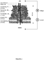
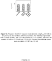
- the sensing chamber, 1 includes a cis compartment, 2 , and a trans compartment, 3 , which are divided by a partition, 4 . Both compartments are filled with a pre-selected recording solution such as 1 M KCl.
- the partition, 4 has an opening, 5 , in its center region, over which a lipid bilayer is formed, and the nanopore, 6 , is plugged through the lipid bilayer.
- the power, 7 provides a voltage that is loaded through a pair of electrodes in the two compartments; the current detector, such as a pico-Ampere amplifier, 8 , is connected to monitor the current changes.
- a mixture sample of the target oligonucleotide, 9 , and its complementary probe, 10 is loaded into the cis compartment, 2 .
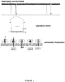
- FIG. 2 is a schematic amplified illustration of the nanopore, 6.
- the nanopore, 6 is in conical or funnel shape with two openings, the cis opening, 11 , at the wide end and the trans opening, 12 , down the narrow end.
- the paired oligonucleotides, 9 / 10 is captured into the nanocavity, 13 .
- the voltage then drive the oligonucleotides, 9 / 10 , to unzip at the constriction, 14 , with the probe, 10 , first traversing through the ⁇ -barrel, 15 , and out off the trans opening, 12 , and followed by the traversing of the target oligonucleotide, 9 .
- the nanopore may be any ion channel of cone-shape or any asymmetrical shape with a wide and a narrow opening plugged into the planar lipid bilayer that has a wider cavity followed by a narrow channel that can facilitate unzipping translocation events.
- the nanopore may be any existing protein ion channels, such as the ⁇ -hemolysin transmembrane protein pore adopted in the examples below, or various synthetic pores fabricated using fashion nanotechnologies with abiotic materials such as silicon.
- the inventive probe is a multi-domain single strand molecule, which comprises a central domain fully complementary to the target oligonucleotide and at least one terminal extension, i.e., signal tag, at its 3' or 5' terminal, with signal tags at both terminals as preferred.
- the invention suggests the 3'-tagged probe is preferred over the 5'-tagged probe.
- the probe directionality-dependence of the capture rate is possibly due to that the bases of ssDNA tilt collectively toward the 5' end of the strand 38 , and this asymmetric base orientation makes DNA move more easily from 3'-end than 5'-end.
- the terminal extension may be of any charged single chain molecule with sufficient length to assist the unzipping translocation through the nanopore driven by the voltage.
- the signal tag may be a charged polymer chain, which can be an oligonucleotide such as poly(dC) n , poly(dA) n , and or poly(dT) n , or a charged polypeptide.
- the poly(dC) tag is more preferred over poly(dA) or poly(dT) tags; furthermore, the poly(dC) 30 is much more efficient in generating signature events (discussed below) than that with a shorter tag such as poly(dC) 8 .
- the capture rate can be further enhanced once combined with other effective approaches, including detection at high voltage, use of engineered pores with designed charge profile in the lumen 33 , and detection in asymmetrical salt concentrations between both sides of the pore 39 .
- the invention also provides a method of detecting and differentiating single strand oligonucleotides by monitoring the current changes induced by the unzipping and translocation of the oligonucleotides through a nanopore.
- the current change induced by the unzipping and translocation of the hybrid of the target oligonucleotide and its complementary probe through the nanopore is a unique signature event, which is used to detect and differentiate the target oligonucleotide.
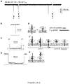
- FIG. 3 which includes an exemplary current trace recorded during an exemplary detection, an amplified electrical mark of the signature event, and a schematic illustration of the unzipping-translocation event.
- ROC curve refers to a Receiver Operating Characteristic Curve. An ROC curve used to analyze the relationship between selectivity and sensitivity. An ROC curve separates the plot into up and lower regions.
- AUC refers to the Area under the ROC curve.
- An AUC can range between 0.5-1.0. The higher the AUC value, the better the separation result.
- OCP refers to an Optimized Cutoff Point.
- an OCP can be calculated from ROC curves.
- an OCP is a cutoff duration at the maximal value of a Youden index.
- Youden index is defined as ⁇ sensitivity+selectivity-1 ⁇ .
- a Youden index is calculated from the ROC curve, and can range between 0 and 1.
- OCP optical cutoff point
- the instant invention is directed to probes, nanopores, kits comprising the probes and nanopores, and associated methods of use, that provide for "signature" current blockage events that distinguish those events arising from interactions with the probe and target from other events.
- the other events are referred to as "background” events. Background events include, but are not limited to, interactions of a probe with nucleic acid that is not a target, interactions of a probe with other components present in a nanopore detection system, free nucleic acids present in the nanopore detection system, and the like.
- Such features of such signature events include, but are not limited to, at least one of a: i) a current block of different duration than a background current block; ii) a different number of distinct current blockade levels than a background current block; iii) a different order of occurrence of current blockade levels than a background current block; iv) a different current amplitude at a blockade level than a background current block; v) a different current amplitude of each blockade level than a background current block; or any combination of (i), (ii), (ii), (iv), or (v).
- a signature blockage event can be distinguished from a background blockage event by differences in a characteristic background noise of each blockage event.
- the distinct durations, numbers, or amplitude(s) in the signature event are greater than those observed in the background event. In certain embodiments, the distinct durations, numbers, or amplitude(s) in the signature event are less than those observed in the background event. In certain embodiments, the distinct durations, numbers, orders, or amplitude(s) in a signature event are statistically distinguishable from those of a background event.
- the signature events are provided in nanopore systems comprising a protein nanopore formed by alpha-hemolysin ( ⁇ HL) or engineered variants thereof in a planar lipid bilayer system.
- the signature events can be provided in a biochip formed by hydrogel-encapsulated lipid bilayer with a single protein nanopore embedded therein or a micro-droplet bilayer system.
- Biochips and micro-droplet bilayer systems have been described ( Shim and Gu; Stochastic Sensing on a Modular Chip Containing a Single-Ion Channel Anal. Chem. 2007, 79, 2207-2213 ; Bayley,H. et al. Droplet interface bilayers. Mol. Biosyst. 4, 1191-1208 (2008 ).
- the signature events can be provided in a synthetic nanopore.
- Synthetic nanopores include, but are not limited to, nanopores comprising silicon nitride or graphene.
- Probe molecules provided herein comprise terminal extensions at one or both of their 5' and/or 3' termini. Without seeking to be limited by theory, it is believed that these terminal extensions provide useful functions that include, but are not limited to, trapping of the probe/target complex into the nanopore at a high rate (i.e. the number of signature events per unit target concentration per unit recording time). The trapping rate directly determines the sensitivity. In the same target concentration and the same recording time, a higher trapping rate gives a more precise sensing result. Without seeking to be limited by theory, it is also believed that these terminal extensions provide useful functions that include, but are not limited to, inducing the voltage-driven dissociation of the probe/target complex. This dissociation function generates a signature event that can be used to discriminate interactions of the probe with the target from other components in the mixture, thereby ensuring the selectivity or specificity.
- Probe terminal extensions can comprise a charged polymer of any length.
- the polymer can be a negatively charged single-stranded nucleic acid.
- Advantages of such nucleic acid terminal extensions include, but are not limited to, extremely low cost of synthesis and controllable charge by pH, salt concentration and temperature.
- Such nucleic acid extensions can comprise homopolymers, heteropolymers, copolymers or combinations thereof.
- the lengths of such nucleic acid terminal extensions can range from about 1 or 2 nucleotides to about 50 nucleotides.
- the nucleic acid extensions can range in length from about 5 to about 40 nucleotides, about 15 to about 35 nucleotides, or from about 20 to about 35 nucleotides.
- An exemplary terminal extension provided herewith is homopolymer poly(dC) 30 .
- a heteropolymeric sequence including but not limited to, di- or trinucleotide heteropolymers such as CTCTCTCT..., or CATCATCAT..., can also be used.
- co-polymers comprising abases or polyethylene glycol (PEG) can be used in the terminal extension. These co-polymers, or domains thereof in a terminal extension, can confer new functions on the terminal extension of the probe.
- An abase is a nucleotide without the base, but carries a negative charge provided by the phosphate.
- abase As the dimension of abase is narrower than normal nucleotides, it may generate a signature event signal different from that formed by the neighbor nucleotides. PEG is not charged. Without seeking to be limited by theory, it is believed that when the PEG domain in a nucleic acid sequence is trapped in the pore, it can reduce the driving force, thus precisely regulating the dissociation of the probe/target complex.
- Probe terminal extensions can also comprise a polypeptide.
- the richer choice of amino acids makes the sequence and functionality of the polypeptide terminal extension more programmable than an oligonucleotide terminal extension.
- polypeptide terminal extensions allow insertion of charged amino acids in the optimized positions to generate more distinguishable probe/target signature events. While not seeking to be limited by theory, it is believed that the probe/target complex can be selectively trapped using a probe comprising a positively charged polypeptide terminal extension under an appropriate voltage while all other negatively charged non-target oligonucleotides in the mixture are prevented from entering into the pore, resulting in ultra-selective detection.
- the polypeptide terminal extensions can comprise two, three, four, or more amino acid residues that can carry a positive charge (i.e. lysine and/or arginine and/or histidine).
- a positive charge i.e. lysine and/or arginine and/or histidine.
- sufficient numbers of positively charged residues are included in the polypeptide terminal extension to provide a net positive charge when said probe is hybridized to a target oligonucleotide.
- performance of the associated nanopore based detection methods can be enhanced under acidic conditions (i.e. when the pH value is less than 7) or conditions where the residue will be protonated.
- the lengths of such polypeptide terminal extensions can range from about 1 or 2 residues to about 30 residues.
- the polypeptide extensions can range in length from about 5 to about 20 residues, about 8 to about 20 residues, or from about 8 to about 15 residues.
- an HIV-TAT polypeptide comprising positively charged arginine and lysine residues can be used as the terminal extension.
- the center domain of the probe that is complementary to the target oligonucleotide can comprise a peptide nucleic acid that is covalently linked to a terminal extension comprising amino acids that carry a positive charge.
- a center domain comprising a peptide nucleic acid is used in conjunction with a terminal extension comprising amino acids that carry a positive charge to provide a net positive charge when said probe is hybridized to a target oligonucleotide.
- polypeptide terminal extensions comprising amino acids with aromatic side chains including, but not limited to, phenylalanine, tryptophan, tyrosine, thyroxine, and the like, can be incorporated into the polypeptide terminal extensions. While not seeking to be limited by theory, it is believed that such aromatic amino acids can interact with the pore through aromatic stacking and provide for useful changes in the signature obtained in nanopore based detection methods.
- any other free nucleic acids components will be repulsed from entering the pore due to the negative charge that is carried by the free, unhybridized nucleic acids. This significantly reduces signals by free nucleic acid components, such that the majority of the observed current blockage events are either due to the trapping of the oligonucleotide/probe complex or to the translocation of the probe.
- the oligonucleotide/probe complexes with a net positive charge can be directed to a nanopore with a negatively-charged ring at the trans- opening of the pore.
- a trans opening of a pore is understood to be that portion of the pore from which a molecule would emerge whereas a cis opening of a pore from which a molecule would enter.
- a negative charged ring at the trans- opening of the pore can be obtained by using any type of nanopore that has been suitably synthesize and/or derivatized so as to have a negative charged ring at the trans- opening of the pore.
- Such nanopores with a negatively charged ring at the trans opening of the pore include, but are not limited to, protein nanopores and synthetic nanopores.
- Protein nanopores with a negatively charged ring at the trans opening of the pore include, but are not limited to, engineered variants of an alpha-hemolysin protein.
- the engineered alpha hemolysin variant can comprise a Staphylococcus aureus alpha hemolysin containing a K131D, a K131E, or a K131H amino acid substitution.
- Exemplary and non-limiting Staphylococcus aureus alpha hemolysin wild type sequences are provided herein (SEQ ID NO:20, nucleic acid coding region; SEQ ID NO:21: protein coding region) and available elsewhere (National Center for Bioinformatics or GenBank Accession Numbers M90536 and AAA26598).
- An exemplary and non-limiting Staphylococcus aureus alpha hemolysin variant comprising a K131D substitution is provided as SEQ ID NO:22.
- the engineered alpha hemolysin variant can comprise a suitably derivatized variant that is derivatized with moieties that provide for a negatively charged ring at the trans opening of the pore.
- aureus alpha hemolysin protein that can be substituted or derivatized to provide for a protein nanopore with a negative charged ring at the trans- opening of the pore is provided herewith as SEQ ID NO: 21.
- variants of other hemolysins capable of forming pores can be substituted or derivatized to provide for a protein nanopore with a negative charged ring at the trans- opening of the pore.
- Synthetic nanopores with a negatively charged ring at the trans opening of the pore are also provided.
- such synthetic nanopores with a negatively charged ring at the trans opening of the pore include, but are not limited to, silicon nitride or graphene nanopores that have been suitably derivatized with moieties that provide for a negatively charged ring at the trans opening of the pore.
- the center domain of probes provided herein is used to capture the target molecule.
- the center domain can be fully complementary or partially complementary to the target sequence.
- a center domain can comprise an oligonucleotide comprising natural nucleotides (A, T, G, C (DNA) or a, u, g, c (RNA)), and/or artificial nucleotides including, but not limited to, nucleosides such as inosine, xanthosine, 7-methylguanosine, Dihydrouridine, and 5-methylcytidine.
- the center domain can comprise a locked nucleic acid (LNA) or a peptide nucleic acid (PNA).
- Locked nucleic acids comprise RNA derivatives where the ribose ring contains a methylene linkage between the 2'-oxygen and the 4'-carbon.
- Peptide nucleic acids comprise a peptide backbone with nucleobase side chains.
- a LNA or a PNA center domain can comprise natural nucleobases (adenine, guanine, thymine, cytosine or uracil) and/or artificial nucleobases including, but not limited to, hypoxanthine, xanthosine, 7-methylguanine, 5,6-dihydrouracil, and 5-methyl cytosine.
- probe center domains comprising co-polymers of oligonucleotides, LNA, or PNA are provided.
- a center domain of a probe will have at least about 4, 6, 8, 10, 12, 14, 15, 16, 17, 18, 19, 20, 21, 22, 23, 24, or 25 nucleotide or nucleobase residues that are complementary to the target nucleic acid.
- a central region of a probe will have at least about 4, 6, 8, 10, 12, 14, 15, 16, 17, 18, 19, 20, 21, 22, 23, 24, or 25 to any of about 30, 35, 40, or 50 nucleotide or nucleobase residues that are complementary to the target nucleic acid.
- synthetic nucleotides or nucleobases inserted in the sequence can precisely adjust the hybridization energy with the target, such that one can distinguish the characters of targets such as single-nucleotide polymorphism, methylation, or interaction between miRNA and its target messenger RNA.
- target nucleic acids or oligonucleotides that can be detected and distinguished from non-target nucleic acids by the probes, nanopores, kits comprising the probes and nanopores, and associated methods of use probes, provided herein.
- the target can be a nucleic acid or a fragment thereof from cells, body fluid, tissues, bacteria, or a virus.
- the target can be a PCR products or a synthetic oligonucleotide.
- a target can comprise a genomic DNA, an mRNA, a pre-mature or mature miRNA, an artificial miRNA, non-coding DNA or RNA, a nucleic acid biomarker, or a synthetic aptamer.
- a miRNA targets may come from the RNA extraction from bio-fluid from any tissues such as plasma and formalin-fixed and paraffin-embedded tissues.
- a target nucleic acid can comprise be a nucleic acid fragment complexed with any of a nucleic acid binding protein, an antibody, or an aptamer bound with a target protein.
- a target nucleic acid can comprise be a nucleic acid fragment complexed with a low molecule weight compound, including, but not limited to, a drug.
- targets can include sequences with mutations, with single-nucleotide polymorphisms, or with chemical modifications such as methylation and phosphorylation.
- Example 1 Detection of miR-155, a lung cancer biomarker
- the invention further provides an exemplary nanopore sensing system for detection of miR-155 , a lung cancer biomarker.
- the nanopore sensing system includes an ⁇ -hemolysin transmembrane protein pore and a pre-designed probe for miR-155.
- the probe is a DNA multiple-block copolymer with its central domain complementary to the target miR-155, and at least one poly(dC) 30 extension at 3'- , 5'-, or both terminals functioning as signal tags.
- Table 1 lists the sequences of miR-155 and the exemplary probes with the tri-block copolymer, P 155 as preferred. Table 1.
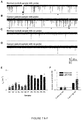
- a mixture of miR-155 /P 155 is added to the cis side of the pore, a current trace with a series of short- and long-lived current blocks can be recorded, as shown in FIG. 4a , while being monitored in 1 M KCl at +100 mV.
- the long blocks in the recording persist for 250 ⁇ 58 ms.
- This conductance level is consistent with a configuration that miR-155 •P 155 is trapped in the pore from the wider opening ( cis ), with either 3'- or 5'-signal tag of P 155 occupying the narrowest ⁇ -barrel ( Fig. 4c' level 1).
- the signal tag in the ⁇ -barrel can induce unzipping of miR - 155 •P 155 , driven by voltage.
- the unzipping time, or the duration of Level 1 is comparable to previously reported time scales for DNA unzipping in the pore, e.g. ⁇ 435 ms for unzipping a 50 bps dsDNA at +140 mV, and ⁇ 40 ms for a 10 bps hairpin DNA at +90 mV.
- the duration of Level 3 is 270 ⁇ 30 ⁇ s, close to the 220 ⁇ s for short blocks by mir-155 alone, and consistent with the time scale of ⁇ 400 ⁇ s for translocation of a 75 bases RNA at +120 mV, 35 and 800 ⁇ s for a 210 bases RNA at +120 mV. 36
- the duration of Level 3 becomes shortened as the voltage increases, further supporting the translocation of a single-stranded oligonucleotide for this conductance level.
- This type of long blocks may occur when the arrested miR-155 •P 155 exits the pore from the cis entry without unzipping.
- [ miR ] 0 and [P] 0 are the initial concentrations of miRNA and the probe, k on is the occurrence rate of signature events and K d is the dissociation constant for miR •P in the solution.
- k on is the occurrence rate of signature events
- K d is the dissociation constant for miR •P in the solution.
- the mean concentrations of circulating miRNAs were 158.6 ng/mL ( ⁇ 25 nM) for the lung cancer group versus 68.1 ng/mL ( ⁇ 10 nM) for the control group. Therefore we compared the f sig values at 10 nM and 25 nM mir - 155 ( FIG. 5b ). Analysis indicates that the two levels of miRNA concentration can be separated ( p ⁇ 0.005), suggesting that the inventive method has the potential to differentially detect miRNA levels in lung cancer patients.
- the invention also provides an exemplary process employing the inventive nanopore sensing system to differentiate highly similar miRNA sequences, let - 7a and let-7b.
- let - 7a and let-7b are members of the Let-7 tumor suppressing miRNA family 4-6 ; and the two Let-7 members only contain different nucleotides at the position 17 and 19, which are adenines in let - 7a and guanines in let-7b.
- the inventive probes P a and P b are designed for let - 7a and let-7b respectively with sequences listed in Table 2. Table 2.
- the inventive nanopore sensing system is able to differentiate single mismatches based on the unzipping time, thus demonstrating the potential to detect miRNAs with similar sequences and SNPs.
- the invention further provides an exemplary process of detecting plasma miR-155 in lung cancer patients with the inventive nanopore sensing system.
- the peripheral blood samples were obtained from six lung cancer patients and six normal volunteers with a local IRB approval.
- Total plasma RNAs containing miRNAs were extracted from 350 ⁇ l of each plasma sample using miRVana PARIS Kit (Ambion), with a final elution volume of 100 ⁇ l, which were than divided into two aliquots (50 ⁇ l) for the nanopore and RT-PCR assay 44 .
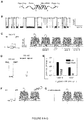
- One aliquot was pre-mixed with P 155 and directly added to the 2-ml recording solution in the nanopore chamber.
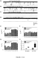
- the nanopore current retain a low level of noise even in the presence of plasma samples, and distinct short and long blocks (marked with red arrows) can be indentified in both the control group ( FIG. 7a ) and lung cancer group ( FIG. 7b ).
- the characteristic long blocks including both with multiple conductance and single conductance, features the same conductance profiles and similar properties to that for synthetic miR-155 RNA in Fig.1a .
- no such types of long blocks can be observed ( FIGs. 7c and d ), but short blocks were found for translocation of single-stranded oligonucleotides such as free miRNAs ( FIGs. 7c and d).
- the characteristic long blocks could be attributed to miR-155 •P 155 hybrids and serve as signature events for single miRNA molecules detection.
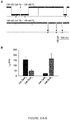
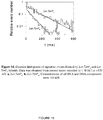
- the frequency of miR-155 signature events f sig for all samples in the lung cancer patient group varies between 1.15-1.51 min -1 , with a mean of 1.40 ⁇ 0.16 min -1 ( FIG. 7e ). This level was significantly higher than f sig in the control group that ranges between 0.32-0.70 min -1 with a mean of 0.48 ⁇ 0.14 min -1 ( FIG. 7e ). Since all samples were prepared following a standard procedure (Methods in Supplementary Materials), it should be valid to compare relative miRNA levels in two groups. When the mean f sig value in normal plasma was set as 1, the folds of miR-155 in lung cancer plasma were compared with the two methods.
- Fig.7f showed that the relative mir-155 level in lung cancer patients was 2.79 with the nanopore sensor ( p ⁇ 0.001). By comparison, the relative miR-155 level was 4.72 with RT-PCR method ( p ⁇ 0.02) with greater variability. Therefore, both nanopore and RT-PCR assay indicated a significant elevation of miR-155 in lung cancer patient plasma although there is a 1.69 fold difference. As the nanopore method does not require labeling and amplification, this may be a reason for smaller variability in the nanopore assay ( FIG. 7f ). Overall the nanopore sensor with engineered probes demonstrates the ability to detect circulating miRNAs in clinical lung cancer patients, which is verified by the independent RT-PCR method.
- Oligonucleotides including miRNAs and DNA probes were synthesized and electrophoresis-purified by Integrated DNA Technologies (Coralville, IA). Before testing, the mixtures of miRNA and DNA probe were heated to 90 °C for 5 minutes, then gradually cooled down to room temperature and stored at 4 °C. The RNase-free water was used to prepare RNA solution.
- Both cis and trans chambers were filled with symmetrical 1 M salt solutions (KCl) buffered with 10 mM Tris and titrated to pH 8.0. All solutions are filtered before use. Single ⁇ -hemolysin proteins were inserted into the bilayer from the cis side to form molecular pores in the membrane. All the oligonucleotides including miRNAs and DNA probes and clinical RNA samples were also added to the cis solution. To record the pore current, the cis solution was grounded and the voltage was given from the trans solution. In this convention, a positive voltage can drive the translocation of a negatively charged DNA through the pore from cis to trans. Single-channel currents were recorded with an Axopatch 200A amplifier (Molecular Device Inc.
- RNA extraction from plasma and miRNA quantification by qRT-PCR Peripheral blood samples were obtained at the University of Missouri Ellis Fischel Cancer Center with an IRB approval. Whole blood with EDTA preservative was centrifuged at 1,600 g for 10 min at room temperature and the plasma was transferred to new tubes. Total RNAs containing miRNAs was extracted from 350 ⁇ l of plasma using miRVana PARIS Kit (Ambion, Austin, TX, USA) according to the manufacturer's protocol. The final elution volume was 100 ⁇ l.
- RNA sample containing miRNAs was polyadenylated by poly(A) polymerase (Ambion) and reverse transcribed to cDNA using SuperScript III Reverse Transcriptase (Invitrogen) according to the manufacturer's instructions with a poly(T) adapter primer (5'-GCGAGCACAGAATTAATACGACTCACTATAGGTTTTTTTTTTTTTTTVN-3'; SEQ ID NO: 10)).
- Real-time PCR was performed using iQ SYBR Green Supermix (Bio-Rad, Hercules, CA, USA) with the miR-155 specific forward primer (5'-TTAATGCTAATCGTGATAGGGGT-3'; SEQ ID NO:11) and the sequence complementary to the poly(T) adapter as the reverse primer (5'-GCGAGCACAGAATTAATACGAC-3'; SEQ ID NO:12) in iQ5 Real-time PCR system (Bio-Rad, USA). The PCR was carried out as follows: after initial denaturation at 95 °C for 3 min, 40 cycles of 95 °C for 15 s and 60 °C for 1 min were followed.
- the relative level of miR-155 was calculated using 2 -delta Ct method where the level of normal plasma was normalized as 1. Data was presented as mean ⁇ SD of three independent experiments, and the differences were considered statistically significant at p ⁇ 0.05 by using the Student's t -test.
- the Denaturing Solution prevents RNAs from undergoing degradation by inhibiting endogenous plasma RNAases.
- both miR-155 and spiked-in miR-39 were measured using the nanopore sensor and SYBR green-based qRT-PCR.
- the nanopore data and normalization result were shown in Table 9.
- the probes for miR-155 and miR-39 were P 155 and P 39 .
- the variability of f 39 reflected the difference in miR-39 concentrations among samples after RNA extraction.
- miR represents miRNA
- P probe
- K d equilibrium dissociation constant for miR •P
- k on occurrence rate constant of miR •P signature events
- f sig frequency of signature events.
- the equilibrium can be established the reactors miR and P and the product miR •P
- Eq.S4 suggested that f sig is not in exact proportion to [ miR ] 0 , the total concentration of the target miRNA. However, when [ miR ] 0 is considerably smaller than [P] 0 , which is the case in our miRNA detection, Eq.S4 can be simplified as f sig ⁇ k on miR 0 In this condition, f sig is proportional to [ miR ] 0 . Eq.S4 also suggested that f sig will ultimately become saturated. This is because f sig measures the capture frequency of miR •P, and the maximal concentration of miR •P ([ miR •P]) can not be higher than that of the probe ([P]o).
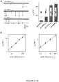
- the cis and trans miR-155 were measured separately.
- the peaks of melting curves for trans RNA samples were the same as synthesized miR-155.
- the miR-155 concentrations in trans solutions were 14, 34 and 63 aM (10 -18 M), indicating that a trace amount of miR-155 transported to the trans side of the pore.
- Precursor miRNAs are stem-loop RNAs of ⁇ 70 nucleotides bearing the 2 nucleotides 3'-overhang as a signature of RNase III-mediated cleavage ( Lee,Y., Jeon,K., Lee,J.T., Kim,S., & Kim,V.N. MicroRNA maturation: stepwise processing and subcellular localization. EMBO J 21, 4663-4670 (2002 )). It is not known whether the plasma total RNA extract contains pre-miRNAs. However, we have verified that the capture rate for a miR •P is very low if the signal tag in the probe is very short ( Fig.9 , Fig.16 and unpublished data). Therefore, we expected that, even though pre-miRNAs exist, the short overhang will prevent them from trapping in the nanopore.
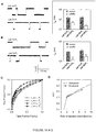
- AUC Areas under ROC curves (AUC) for separation of miRNAs with one nucleotide difference (let-7a and let-7c) and with two nucleotide difference (let-7a and let-7b)a let-7a • P a let-7b • P b let-7a • P a let-7c • P c let-7b • P a 0.75 n.a. let-7c • P a 0.73 n.a. let-7a • P b n.a. 0.83 let-7a • P c n.a.
- the receiver operating characteristic (ROC) curve is a plot of the true positive rate (sensitivity) against the false positive rate (1-selectivity) for the different possible cutoff points that separate the entire duration distribution into the positive and negative components.
- ROC receiver operating characteristic
- the separation accuracy was measured by the area under the ROC curve (AUC).
- An AUC of 1 represents a perfect separation; an area of 0.5 represents no separation ability.
- AUC was analyzed online using free software on the world wide web (internet address) rad.jhmi.edu/jeng/javarad/roc/JROCFITi.html. Table 8.
- AUC Areas under ROC curves (AUC) and optimal cutoff point (OCP) at various duration ratio and event number ratio a ⁇ P / ⁇ N (s/s) b 1/1 2/1 3/1 4/1 5/1 10/1 AUC 0.51 0.72 0.73 0.76 0.78 0.93 OCP n.a. 1.33 1.74 1.88 1.98 2.18 N P lN N c 200:800 200:400 200:200 200:150 200:100 200:50 AUC 0.83 0.81 0.76 0.76 0.79 0.78 OCP 1.88 1.88 1.85 1.79 1.81 1.97 a : Both AUC and OCP were calculated from the ROC curves shown in Fig.16A-B . OCP is a cutoff duration at the maximal value of Youden index.
- OCP optical cutoff point
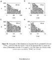
- Example 3 Peptide-guided selective detection of microRNAs.
- RNA extraction contains numerous and complicated nucleic acids components, at least including miRNAs (both pre-mature and mature miRNAs), mRNAs, tRNAs, other RNAs. All components commonly carry negative charges. Thus, if the target miRNA can be trapped in the pore at an applied voltage, any other component may also be driven to interact with the nanopore, generating non-specific current signals that interfere with the recognition of signature events generated by the target miRNA/probe complex.
- peptide-PNA peptide nucleic acid
- the PNA sequence in “PNA” bracket in Figure 18A ) has a peptide backbone with side chain nucleobases that are complementary to the entire or partial sequence of the target miRNA ( "miRNA ( Let-7b , - 7c )" in Figure 18A bracket), and thus serves as the center domain for capturing the target miRNA in the solution.
- NH2-AACCACACAA-COOH where the molecule comprises a peptide backbone with the indicated AACCACACAA nucleobases; (SEQ ID NO: 18).
- HIV-TAT HIV-TAT
- the reporter (or terminal extension) of the new probe is a peptide that carries a series of positively-charged amino acids ("Peptide reporter" in Figure 18A bracket) and the center domain is a peptide nucleic acid comprising nucleotides that are complementary to the target nucleic acid.
- Peptide reporter in Figure 18A bracket
- the center domain is a peptide nucleic acid comprising nucleotides that are complementary to the target nucleic acid.
- S. aureus alpha-hemolysin comprising a K131D mutation.
- the wild-type S. aureus alpha-hemolysin peptide sequence (National Center for Bioinformatics Accession NO. AAA26598.1); is:
- the variant S. aureus K131D alpha-hemolysin peptide sequence is:
- the positively-charged peptide domain of the probe dipole will be both pushed by the positive voltage (cis grounded) and attracted by the negative ring at the trans opening, guiding the trapping of the miRNA/complex into the ⁇ -barrel of the pore.
- any other free nucleic acids components will be repulsed from entering the pore due to the negative charge carried. This significantly reduces signals by free RNA components, and most observed events are either due to the trapping of the miRNA/probe complex or the translocation of the probe.
- the use of peptide-PNA probe enables selective detection of the target miRNA.
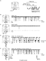
- Figure 18A shows the diagram of the miRNA/probe complex.
- the bracketed miRNA is target miRNA Let-7b.
- the bracketed probe P7b has a bracketed "Peptide Reporter” part and a bracketed "PNA” (peptide nucleic acid) part.
- the PNA is for capturing Let-7b
- the bracketed "Peptide reporter” is apositively-charged peptide corresponding the sequence of HIV-TAT, which contains +8e contributed by arginines and lysines.
- Figure 18B shows events for translocation of the peptide-PNA probe, P7b. The characteristic events last for 3 ms and reduce the current to 10 pA at +180 mV.
- Figure 18C shows no block events can be observed with free miRNA let-7b (without probe) in the solution at +180 mV.
- Figure 18D shows signature events for the trapping of the let-7b/P7b complex. These events characteristically last for 100 ms and reduce the current to 57 pA at +180 mV, completely different that for the probe.
- Figure 18E shows that Let-7c, which has two different nucleotides from Let-7b, cannot bind to PNA of the probe P7b, therefore does not generate signature events as in Figure 18C . Almost all observed events are due to the probe itself.
- Figure 18F compares the duration-amplitude property for P7b binding to Let-7b (fully match, two separate clusters without overlay) and Let-7c (2 mismatches, two clusters fully overlay). This suggests an accuracy of almost 100% in differentiating sequence-similar miRNAs with two different nucleotides.
- Level 1' can be interpreted by that although the short strand of SA-C30 moves into the ⁇ -barrel after unzipping, its translocation is prevented by the attached large streptavidin. This result suggested the potential of using signature events for protein detection.
- Figure 19C we demonstrated that the complex can be sequentially unzipped in the nanopore in two steps when using a short oligonucleotide to link two DNAs. The unzipping of the two DNAs can be clearly revealed by the two Level 2 states.
- the peptide-PNA probe enables 1) selective detection of the target miRNA, 2) greatly enhanced accuracy in differentiating sequence-similar miRNAs.
Landscapes
- Chemical & Material Sciences (AREA)
- Life Sciences & Earth Sciences (AREA)
- Health & Medical Sciences (AREA)
- Engineering & Computer Science (AREA)
- Organic Chemistry (AREA)
- Analytical Chemistry (AREA)
- Proteomics, Peptides & Aminoacids (AREA)
- Physics & Mathematics (AREA)
- Zoology (AREA)
- Wood Science & Technology (AREA)
- Immunology (AREA)
- Biophysics (AREA)
- Molecular Biology (AREA)
- Biochemistry (AREA)
- General Health & Medical Sciences (AREA)
- Genetics & Genomics (AREA)
- Biomedical Technology (AREA)
- Pathology (AREA)
- Bioinformatics & Cheminformatics (AREA)
- Microbiology (AREA)
- Biotechnology (AREA)
- General Engineering & Computer Science (AREA)
- General Physics & Mathematics (AREA)
- Hematology (AREA)
- Urology & Nephrology (AREA)
- Food Science & Technology (AREA)
- Medicinal Chemistry (AREA)
- Spectroscopy & Molecular Physics (AREA)
- Nanotechnology (AREA)
- Hospice & Palliative Care (AREA)
- Oncology (AREA)
- Measuring Or Testing Involving Enzymes Or Micro-Organisms (AREA)
- Investigating Or Analyzing Materials By The Use Of Electric Means (AREA)
- Apparatus Associated With Microorganisms And Enzymes (AREA)
- Investigating Or Analysing Biological Materials (AREA)
Applications Claiming Priority (2)
| Application Number | Priority Date | Filing Date | Title |
|---|---|---|---|
| US39957810P | 2010-07-14 | 2010-07-14 | |
| EP11807539.9A EP2593568B1 (en) | 2010-07-14 | 2011-07-14 | Nanopore-facilitated single molecule detection of nucleic acids |
Related Parent Applications (2)
| Application Number | Title | Priority Date | Filing Date |
|---|---|---|---|
| EP11807539.9A Division EP2593568B1 (en) | 2010-07-14 | 2011-07-14 | Nanopore-facilitated single molecule detection of nucleic acids |
| EP11807539.9A Division-Into EP2593568B1 (en) | 2010-07-14 | 2011-07-14 | Nanopore-facilitated single molecule detection of nucleic acids |
Publications (1)
| Publication Number | Publication Date |
|---|---|
| EP3190193A1 true EP3190193A1 (en) | 2017-07-12 |
Family
ID=45470088
Family Applications (2)
| Application Number | Title | Priority Date | Filing Date |
|---|---|---|---|
| EP17157476.7A Withdrawn EP3190193A1 (en) | 2010-07-14 | 2011-07-14 | Nanopore-facilitated single molecule detection of nucleic acids |
| EP11807539.9A Active EP2593568B1 (en) | 2010-07-14 | 2011-07-14 | Nanopore-facilitated single molecule detection of nucleic acids |
Family Applications After (1)
| Application Number | Title | Priority Date | Filing Date |
|---|---|---|---|
| EP11807539.9A Active EP2593568B1 (en) | 2010-07-14 | 2011-07-14 | Nanopore-facilitated single molecule detection of nucleic acids |
Country Status (6)
| Country | Link |
|---|---|
| US (3) | US9395353B2 (OSRAM) |
| EP (2) | EP3190193A1 (OSRAM) |
| JP (2) | JP6016792B2 (OSRAM) |
| AU (1) | AU2011279083B2 (OSRAM) |
| CA (1) | CA2805247C (OSRAM) |
| WO (1) | WO2012009578A2 (OSRAM) |
Families Citing this family (63)
| Publication number | Priority date | Publication date | Assignee | Title |
|---|---|---|---|---|
| WO2007146158A1 (en) | 2006-06-07 | 2007-12-21 | The Trustees Of Columbia University In The City Of New York | Dna sequencing by nanopore using modified nucleotides |
| US9678055B2 (en) | 2010-02-08 | 2017-06-13 | Genia Technologies, Inc. | Methods for forming a nanopore in a lipid bilayer |
| US9605307B2 (en) | 2010-02-08 | 2017-03-28 | Genia Technologies, Inc. | Systems and methods for forming a nanopore in a lipid bilayer |
| US8324914B2 (en) | 2010-02-08 | 2012-12-04 | Genia Technologies, Inc. | Systems and methods for characterizing a molecule |
| US9194838B2 (en) | 2010-03-03 | 2015-11-24 | Osaka University | Method and device for identifying nucleotide, and method and device for determining nucleotide sequence of polynucleotide |
| EP3190193A1 (en) * | 2010-07-14 | 2017-07-12 | The Curators Of The University Of Missouri | Nanopore-facilitated single molecule detection of nucleic acids |
| WO2012083249A2 (en) | 2010-12-17 | 2012-06-21 | The Trustees Of Columbia University In The City Of New York | Dna sequencing by synthesis using modified nucleotides and nanopore detection |
| US8845880B2 (en) | 2010-12-22 | 2014-09-30 | Genia Technologies, Inc. | Nanopore-based single DNA molecule characterization, identification and isolation using speed bumps |
| US8962242B2 (en) | 2011-01-24 | 2015-02-24 | Genia Technologies, Inc. | System for detecting electrical properties of a molecular complex |
| US9110478B2 (en) | 2011-01-27 | 2015-08-18 | Genia Technologies, Inc. | Temperature regulation of measurement arrays |
| WO2013109970A1 (en) * | 2012-01-20 | 2013-07-25 | Genia Technologies, Inc. | Nanopore based molecular detection and sequencing |
| IN2014DN06796A (OSRAM) | 2012-02-15 | 2015-05-22 | Oxford Nanopore Tech Ltd | |
| EP2814983B1 (en) * | 2012-02-16 | 2019-04-24 | Genia Technologies, Inc. | Methods for creating bilayers for use with nanopore sensors |
| US8986629B2 (en) | 2012-02-27 | 2015-03-24 | Genia Technologies, Inc. | Sensor circuit for controlling, detecting, and measuring a molecular complex |
| CN107082792A (zh) | 2012-04-09 | 2017-08-22 | 纽约哥伦比亚大学理事会 | 纳米孔的制备方法和其用途 |
| WO2013188841A1 (en) | 2012-06-15 | 2013-12-19 | Genia Technologies, Inc. | Chip set-up and high-accuracy nucleic acid sequencing |
| EP2880186B1 (en) | 2012-08-03 | 2019-02-06 | University Of Washington Through Its Center For Commercialization | Compositions and methods for improving nanopore sequencing |
| US9551023B2 (en) | 2012-09-14 | 2017-01-24 | Oxford Nanopore Technologies Ltd. | Sample preparation method |
| WO2014072703A1 (en) | 2012-11-06 | 2014-05-15 | Oxford Nanopore Technologies Limited | Quadruplex method |
| US9605309B2 (en) | 2012-11-09 | 2017-03-28 | Genia Technologies, Inc. | Nucleic acid sequencing using tags |
| US9759711B2 (en) | 2013-02-05 | 2017-09-12 | Genia Technologies, Inc. | Nanopore arrays |
| GB201318465D0 (en) | 2013-10-18 | 2013-12-04 | Oxford Nanopore Tech Ltd | Method |
| EP2971051A4 (en) | 2013-03-15 | 2017-03-01 | The Trustees of Columbia University in the City of New York | Method for detecting multiple predetermined compounds in a sample |
| US10648026B2 (en) | 2013-03-15 | 2020-05-12 | The Trustees Of Columbia University In The City Of New York | Raman cluster tagged molecules for biological imaging |
| CA2906007C (en) * | 2013-03-15 | 2022-06-21 | The Curators Of The University Of Missouri | Encoded nanopore sensor for multiplex nucleic acids detection |
| US10048245B2 (en) | 2013-06-25 | 2018-08-14 | Two Pore Guys, Inc. | Multiplexed biomarker quantitation by nanopore analysis of biomarker-polymer complexes |
| JP6334118B2 (ja) * | 2013-09-24 | 2018-05-30 | クオンタムバイオシステムズ株式会社 | 単分子識別方法、装置、及びプログラム |
| CA2929929A1 (en) | 2013-09-18 | 2015-03-26 | Quantum Biosystems Inc. | Biomolecule sequencing devices, systems and methods |
| JP2015077652A (ja) | 2013-10-16 | 2015-04-23 | クオンタムバイオシステムズ株式会社 | ナノギャップ電極およびその製造方法 |
| US9551697B2 (en) | 2013-10-17 | 2017-01-24 | Genia Technologies, Inc. | Non-faradaic, capacitively coupled measurement in a nanopore cell array |
| WO2015061509A1 (en) | 2013-10-23 | 2015-04-30 | Genia Technologies, Inc. | High speed molecular sensing with nanopores |
| US9567630B2 (en) | 2013-10-23 | 2017-02-14 | Genia Technologies, Inc. | Methods for forming lipid bilayers on biochips |
| CA2943952A1 (en) | 2014-03-24 | 2015-10-01 | The Trustees Of Columbia University In The City Of New York | Chemical methods for producing tagged nucleotides |
| US10438811B1 (en) | 2014-04-15 | 2019-10-08 | Quantum Biosystems Inc. | Methods for forming nano-gap electrodes for use in nanosensors |
| WO2015170782A1 (en) | 2014-05-08 | 2015-11-12 | Osaka University | Devices, systems and methods for linearization of polymers |
| SG10201809900PA (en) * | 2014-07-31 | 2018-12-28 | Illumina Inc | Hybrid Nanopore Sensors |
| EP3971300B1 (en) * | 2014-10-16 | 2024-06-05 | Oxford Nanopore Technologies plc | Sorting of polymers |
| KR102457147B1 (ko) | 2014-10-17 | 2022-10-19 | 옥스포드 나노포어 테크놀로지즈 피엘씨 | 나노 세공 rna 특징 규명을 위한 방법 |
| CN105842453B (zh) * | 2015-01-12 | 2018-12-25 | 南京理工大学 | 一种基于末端延伸、金纳米粒子和酶三级放大电化学传感器检测dna的方法 |
| US20180155768A1 (en) | 2015-06-02 | 2018-06-07 | Nanopore Diagnostics, Llc | Nucleic acid detection |
| JP6889701B2 (ja) | 2015-09-24 | 2021-06-18 | エフ.ホフマン−ラ ロシュ アーゲーF. Hoffmann−La Roche Aktiengesellschaft | アルファ溶血素バリアント |
| WO2017167811A1 (en) * | 2016-03-31 | 2017-10-05 | Genia Technologies, Inc. | Nanopore protein conjugates and uses thereof |
| JP2019515317A (ja) | 2016-04-27 | 2019-06-06 | クオンタムバイオシステムズ株式会社 | バイオ分子の測定およびシーケンシングのためのシステムおよび方法 |
| EP3475447B1 (en) | 2016-06-23 | 2021-04-21 | F. Hoffmann-La Roche AG | Formation and calibration of nanopore sequencing cells |
| US11124827B2 (en) | 2016-06-23 | 2021-09-21 | Roche Sequencing Solutions, Inc. | Period-to-period analysis of AC signals from nanopore sequencing |
| CN119753114A (zh) * | 2016-07-12 | 2025-04-04 | 格罗宁根大学 | 基于FraC actinoporin的用于生物聚合物传感和测序的生物纳米孔 |
| WO2018093976A1 (en) | 2016-11-16 | 2018-05-24 | Nanopore Diagnostics, Llc | Analysis of nucleic acids using probe with non-linear tag |
| EP4265733A1 (en) * | 2017-07-17 | 2023-10-25 | President and Fellows of Harvard College | Analysis of nanopore-matched protein shuttle for molecular characterization |
| EP3732484B1 (en) | 2017-12-28 | 2023-06-07 | F. Hoffmann-La Roche AG | Measuring and removing noise in stochastic signals from a nanopore dna sequencing system driven by an alternating signal |
| US11034996B2 (en) | 2018-03-02 | 2021-06-15 | The Curators Of The University Of Missouri | Nucleic acid sequence and capture by formation of an abasic site-derived cross-link |
| WO2019173799A1 (en) * | 2018-03-08 | 2019-09-12 | Caris Science, Inc. | Oligonucleotide probes and uses thereof |
| JP7239965B2 (ja) * | 2018-06-26 | 2023-03-15 | 国立大学法人東京農工大学 | 2種以上の特定オリゴヌクレオチドの有無を判定する判定方法、並びに、前記判定方法を用いて検査対象が小細胞肺がん、胆管がんに罹患している又はそのリスクを有することを判定する方法 |
| US20220162692A1 (en) * | 2018-07-30 | 2022-05-26 | Oxford University Innovation Limited | Assemblies |
| US11162935B2 (en) | 2018-08-24 | 2021-11-02 | University Of Notre Dame Du Lac | Systems and methods for separating, detecting, and quantifying a target polynucleotide |
| CA3111488A1 (en) | 2018-09-11 | 2020-03-19 | Rijksuniversiteit Groningen | Biological nanopores having tunable pore diameters and uses thereof as analytical tools |
| CN116814818A (zh) * | 2019-07-22 | 2023-09-29 | 四川大学华西医院 | 一种区分抗生素耐药的肺炎克雷伯菌的装置及方法 |
| KR102390925B1 (ko) * | 2019-11-01 | 2022-04-27 | 한국생명공학연구원 | 나노포어를 이용한 병원성 미생물의 검출 방법 및 검출키트 |
| WO2021231920A1 (en) * | 2020-05-15 | 2021-11-18 | The Curators Of The University Of Missouri | Nanopore unzipping-sequencing for dna data storage |
| JP7679055B2 (ja) * | 2020-08-24 | 2025-05-19 | 国立大学法人東京農工大学 | 極低濃度のオリゴヌクレオチドの存在の有無を判定する方法、及び検査対象が特定疾患に罹患しているかどうか又は特定疾患罹患のリスクを有するかどうかを判定する方法 |
| CN112795628B (zh) * | 2021-02-05 | 2024-02-23 | 南京邮电大学 | 基于固态纳米孔传感器的miRNA检测方法、检测探针及试剂盒 |
| CN112961906B (zh) * | 2021-02-05 | 2024-01-23 | 南京邮电大学 | 金纳米棒检测探针、制备方法、检测方法及其应用 |
| CN113189070B (zh) * | 2021-04-28 | 2024-01-16 | 香港理工大学 | 一种纳米探针及其制备方法和应用、循环肿瘤细胞的miRNAs的检测系统 |
| IL320502A (en) | 2022-10-28 | 2025-06-01 | Univ Groningen | Nanopore-based protein efflux |
Citations (3)
| Publication number | Priority date | Publication date | Assignee | Title |
|---|---|---|---|---|
| US20070190543A1 (en) * | 2005-11-14 | 2007-08-16 | Applera Corporation | Coded Molecules for Detecting Target Analytes |
| WO2009092035A2 (en) * | 2008-01-17 | 2009-07-23 | Sequenom, Inc. | Methods and compositions for the analysis of biological molecules |
| WO2011103424A2 (en) * | 2010-02-19 | 2011-08-25 | The Trustees Of The University Of Pennsylvania | High-resolution analysis devices and related methods |
Family Cites Families (36)
| Publication number | Priority date | Publication date | Assignee | Title |
|---|---|---|---|---|
| US6362002B1 (en) | 1995-03-17 | 2002-03-26 | President And Fellows Of Harvard College | Characterization of individual polymer molecules based on monomer-interface interactions |
| WO1999005167A1 (en) | 1997-07-25 | 1999-02-04 | University Of Massachusetts | Designed protein pores as components for biosensors |
| AU4033500A (en) | 1999-03-25 | 2000-10-09 | Hyseq, Inc. | Solution-based methods and materials for sequence analysis by hybridization |
| CA2384100C (en) | 1999-09-07 | 2011-03-29 | The Regents Of The University Of California | Methods of determining the presence of double stranded nucleic acids in a sample |
| EP2083015B1 (en) | 2000-02-11 | 2016-04-06 | The Texas A & M University System | Biosensor compositions and methods of use |
| EP1478771A4 (en) | 2001-06-21 | 2005-06-15 | Harvard College | METHODS OF CHARACTERIZING NUCLEIC ACID MOLECULES |
| US7563584B2 (en) | 2001-07-10 | 2009-07-21 | The Board Of Trustees Of The Leland Stanford Junior University | Methods and compositions for detecting the activation state of multiple proteins in single cells |
| US20050019784A1 (en) | 2002-05-20 | 2005-01-27 | Xing Su | Method and apparatus for nucleic acid sequencing and identification |
| EP1648914A4 (en) * | 2003-07-31 | 2009-12-16 | Regulus Therapeutics Inc | OLIGOMERIC COMPOUNDS AND COMPOSITIONS USEFUL FOR MODULATING SMALL NON-CODING RNA |
| US7846738B2 (en) | 2003-08-15 | 2010-12-07 | President And Fellows Of Harvard College | Study of polymer molecules and conformations with a nanopore |
| US20050136408A1 (en) | 2003-12-19 | 2005-06-23 | May Tom-Moy | Methods and systems for characterizing a polymer |
| CN101103357B (zh) | 2004-08-13 | 2012-10-03 | 哈佛学院院长等 | 超高处理量光学-纳米孔dna读出平台 |
| KR101159072B1 (ko) | 2005-01-20 | 2012-06-25 | 삼성전자주식회사 | 나노포어를 이용한 생분자의 분리방법 |
| KR100707198B1 (ko) | 2005-06-27 | 2007-04-13 | 삼성전자주식회사 | 나노포어 및 핵산에 비특이적으로 결합하는 물질을 이용한고감도 핵산 검출 방법 |
| US20070190542A1 (en) | 2005-10-03 | 2007-08-16 | Ling Xinsheng S | Hybridization assisted nanopore sequencing |
| KR100730350B1 (ko) | 2005-10-17 | 2007-06-19 | 삼성전자주식회사 | 표면처리된 나노포어를 이용한 dna 검출방법 및검출장치 |
| US20100291548A1 (en) | 2006-03-12 | 2010-11-18 | Applera Corporation | Methods of Detecting Target Nucleic Acids |
| US8314220B2 (en) * | 2007-01-26 | 2012-11-20 | Agilent Technologies, Inc. | Methods compositions, and kits for detection of microRNA |
| WO2008097190A1 (en) | 2007-02-05 | 2008-08-14 | Agency For Science, Technology And Research | Methods of electrically detecting a nucleic acid by means of an electrode pair |
| US20110005918A1 (en) | 2007-04-04 | 2011-01-13 | Akeson Mark A | Compositions, devices, systems, and methods for using a nanopore |
| US9121843B2 (en) | 2007-05-08 | 2015-09-01 | Trustees Of Boston University | Chemical functionalization of solid-state nanopores and nanopore arrays and applications thereof |
| GB0713143D0 (en) | 2007-07-06 | 2007-08-15 | Ucl Business Plc | Nucleic acid detection method |
| JP5309145B2 (ja) | 2007-10-02 | 2013-10-09 | プレジデント アンド フェロウズ オブ ハーバード カレッジ | ナノポアによる分子の捕捉、再捕捉およびトラッピング |
| US20090181390A1 (en) | 2008-01-11 | 2009-07-16 | Signosis, Inc. A California Corporation | High throughput detection of micrornas and use for disease diagnosis |
| EP2107040B1 (en) | 2008-03-31 | 2011-10-26 | Sony Deutschland Gmbh | A method of fabricating a membrane having a tapered pore |
| WO2010004273A1 (en) | 2008-07-07 | 2010-01-14 | Oxford Nanopore Technologies Limited | Base-detecting pore |
| US20100099198A1 (en) | 2008-07-11 | 2010-04-22 | Board Of Regents, The University Of Texas System | Apparatus and system for pattern recognition sensing for biomolecules |
| WO2010062903A2 (en) | 2008-11-26 | 2010-06-03 | Board Of Regents, The University Of Texas System | Genomic sequencing using modified protein pores and ionic liquids |
| US20110003703A1 (en) | 2009-07-01 | 2011-01-06 | Charles Ma | Nucleic Acid Hybridization and Detection Using Enzymatic Reactions on a Microarray |
| US9416409B2 (en) | 2009-07-31 | 2016-08-16 | Ibis Biosciences, Inc. | Capture primers and capture sequence linked solid supports for molecular diagnostic tests |
| WO2011028494A2 (en) | 2009-08-24 | 2011-03-10 | Trustees Of Boston University | Label-free sensing of pna-dna complexes using nanopores |
| US8324914B2 (en) | 2010-02-08 | 2012-12-04 | Genia Technologies, Inc. | Systems and methods for characterizing a molecule |
| JP2013523131A (ja) | 2010-03-30 | 2013-06-17 | トラスティーズ オブ ボストン ユニバーシティ | ナノ細孔解離依存核酸配列決定のためのツールおよび方法 |
| EP3190193A1 (en) * | 2010-07-14 | 2017-07-12 | The Curators Of The University Of Missouri | Nanopore-facilitated single molecule detection of nucleic acids |
| WO2012083249A2 (en) | 2010-12-17 | 2012-06-21 | The Trustees Of Columbia University In The City Of New York | Dna sequencing by synthesis using modified nucleotides and nanopore detection |
| IN2014DN06796A (OSRAM) | 2012-02-15 | 2015-05-22 | Oxford Nanopore Tech Ltd |
-
2011
- 2011-07-14 EP EP17157476.7A patent/EP3190193A1/en not_active Withdrawn
- 2011-07-14 AU AU2011279083A patent/AU2011279083B2/en active Active
- 2011-07-14 JP JP2013519840A patent/JP6016792B2/ja active Active
- 2011-07-14 EP EP11807539.9A patent/EP2593568B1/en active Active
- 2011-07-14 CA CA2805247A patent/CA2805247C/en active Active
- 2011-07-14 US US13/810,105 patent/US9395353B2/en active Active
- 2011-07-14 WO PCT/US2011/044082 patent/WO2012009578A2/en not_active Ceased
-
2016
- 2016-06-15 US US15/183,152 patent/US9574228B2/en active Active
- 2016-07-13 US US15/209,443 patent/US10273527B2/en active Active
- 2016-08-04 JP JP2016154002A patent/JP6521916B2/ja active Active
Patent Citations (3)
| Publication number | Priority date | Publication date | Assignee | Title |
|---|---|---|---|---|
| US20070190543A1 (en) * | 2005-11-14 | 2007-08-16 | Applera Corporation | Coded Molecules for Detecting Target Analytes |
| WO2009092035A2 (en) * | 2008-01-17 | 2009-07-23 | Sequenom, Inc. | Methods and compositions for the analysis of biological molecules |
| WO2011103424A2 (en) * | 2010-02-19 | 2011-08-25 | The Trustees Of The University Of Pennsylvania | High-resolution analysis devices and related methods |
Non-Patent Citations (57)
| Title |
|---|
| ADVANCED MATERIALS, vol. 18, 2006, pages 3149 - 3153 |
| AKESON M ET AL: "MICROSECOND TIME-SCALE DISCRIMINATION AMONG POLYCYTIDYLIC ACID, POLYADENYLIC ACID, AND POLYURIDYLIC ACID AS HOMOPOLYMERS OR AS SEGMENTS WITHIN SINGLE RNA MOLECULES", BIOPHYSICAL JOURNAL, ELSEVIER, AMSTERDAM, NL, vol. 77, no. 6, 1 December 1999 (1999-12-01), pages 3227 - 3233, XP000914263, ISSN: 0006-3495 * |
| AKESON,M.; BRANTON,D.; KASIANOWICZ,J.J.; BRANDIN,E.; DEAMER,D.W.: "Microsecond time-scale discrimination among polycytidylic acid, polyadenylic acid, and polyuridylic acid as homopolymers or as segments within single RNA molecules", BIOPHYS. J., vol. 77, 1999, pages 3227 - 3233 |
| BAYLEY,H. ET AL.: "Droplet interface bilayers", MOL. BIOSYST., vol. 4, 2008, pages 1191 - 1208 |
| BAYLEY,H.: "Sequencing single molecules of DNA", CURRENT OPINION IN CHEMICAL BIOLOGY, vol. 10, 2006, pages 628 - 637 |
| BAYLEY,H.; JAYASINGHE,L.: "Functional engineered channels and pores - (Review", MOLECULAR MEMBRANE BIOLOGY, vol. 21, 2004, pages 209 - 220 |
| BRANTON,D.; DEAMER,D.W.; MARZIALI,A.; BAYLEY,H.; BENNER,S.A.; BUTLER,T.; DI VENTRA,M.; GARAJ,S.; HIBBS,A.; HUANG,X.: "The potential and challenges of nanopore sequencing", NATURE BIOTECHNOLOGY, vol. 26, 2008, pages 1146 - 1153 |
| BUTLER,T.Z.; GUNDLACH,J.H.; TROLL,M.A: "Determination of RNA orientation during translocation through a biological nanopore", BIOPHYS. J., vol. 90, 2006, pages 190 - 199 |
| CALIN,G.A.; CROCE,C.M.: "MicroRNA signatures in human cancers", NAT REV CANCER, vol. 6, 2006, pages 857 - 866 |
| CARTHEW,R.W.; SONTHEIMER,E.J.: "Origins and Mechanisms of miRNAs and siRNAs", CELL, vol. 136, 2009, pages 642 - 655 |
| CHEN,C.; RIDZON,D.A.; BROOMER,A.J.; ZHOU,Z.; LEE,D.H.; NGUYEN,J.T.; BARBISIN,M.; XU,N.L.; MAHUVAKAR,V.R.; ANDERSEN,M.R.: "Real-time quantification of microRNAs by stem-loop RT-PCR", NUCLEIC ACIDS RES., 2005, pages 33 |
| CHO,W.C: "Role of miRNAs in lung cancer", EXPERT REV MOL. DIAGN., vol. 9, 2009, pages 773 - 776 |
| DEAMER,D.W.; BRANTON,D: "Characterization of nucleic acids by nanopore analysis", ACC. CHEM. RES., vol. 35, 2002, pages 817 - 825 |
| G. V. SONI ET AL: "Progress toward Ultrafast DNA Sequencing Using Solid-State Nanopores", CLINICAL CHEMISTRY, vol. 53, no. 11, 1 November 2007 (2007-11-01), pages 1996 - 2001, XP055076185, ISSN: 0009-9147, DOI: 10.1373/clinchem.2007.091231 * |
| GARZON, R.; CALIN, G. A.; CROCE, C. M, MICRORNAS IN CANCER, vol. 60, 2009, pages 167 - 179 |
| GREINER ET AL., PREVENTIVE VETERINARY MEDICINE, vol. 45, 2000, pages 23 - 41 |
| HUNT,E.A.; GOULDING,A.M.; DEO,S.K.: "Direct detection and quantification of microRNAs", ANAL. BIOCHEM., vol. 387, 2009, pages 1 - 12 |
| INUI,M.; MARTELLO,G.; PICCOLO,S: "MicroRNA control of signal transduction", NAT REV MOL. CELL BIOL, vol. 11, 2010, pages 252 - 263 |
| JONATHAN NAKANE ET AL: "A Nanosensor for Transmembrane Capture and Identification of Single Nucleic Acid Molecules", BIOPHYSICAL JOURNAL, vol. 87, no. 1, 1 July 2004 (2004-07-01), pages 615 - 621, XP055035895, ISSN: 0006-3495, DOI: 10.1529/biophysj.104.040212 * |
| KASIANOWICZ,J.J.; BRANDIN,E.; BRANTON,D.; DEAMER,D.W.: "Characterization of individual polynucleotide molecules using a membrane channel", PROC. NATL. ACAD. SCI. U. S. A., vol. 93, 1996, pages 13770 - 13773 |
| KASIANOWICZ,J.J.; BRANDIN,E.; BRANTON,D.; DEAMER,D.W: "Characterization of individual polynucleotide molecules using a membrane channel", PROC. NATL. ACAD. SCI. U. S. A., vol. 93, 1996, pages 13770 - 13773 |
| KIM,M.J.; WANUNU,M.; BELL,D.C.; MELLER,A: "Rapid fabrication of uniformly sized nanopores and nanopore arrays for parallel DNA analysis", ADVANCED MATERIALS, vol. 18, 2006, pages 3149 - 3153 |
| KLOOSTERMAN,W.P.; WIENHOLDS,E.; DE BRUIJN,E.; KAUPPINEN,S.; PLASTERK,R.H.: "In situ detection of miRNAs in animal embryos using LNA-modified oligonucleotide probes", NAT METHODS, vol. 3, 2006, pages 27 - 29 |
| KOSAKA,N.; IGUCHI,H.; YOSHIOKA,Y.; TAKESHITA,F.; MATSUKI,Y.; OCHIYA,T.: "Secretory mechanisms and intercellular transfer of microRNAs in living cells", J BIOL CHEM, 2010 |
| LANDI,M.T.; ZHAO,Y.; ROTUNNO,M.; KOSHIOL,J.; LIU,H.; BERGEN,A.W.; RUBAGOTTI,M.; GOLDSTEIN,A.M.; LINNOILA,I.; MARINCOLA,F.M.: "MicroRNA expression differentiates histology and predicts survival of lung cancer", CLIN. CANCER RES, vol. 16, 2010, pages 430 - 441 |
| LEE,Y.; JEON,K.; LEE,J.T.; KIM,S.; KIM,V.N.: "MicroRNA maturation: stepwise processing and subcellular localization", EMBO J, vol. 21, 2002, pages 4663 - 4670 |
| LI,W.; RUAN,K: "MicroRNA detection by microarray", ANAL. BIOANAL. CHEM., vol. 394, 2009, pages 1117 - 1124 |
| LONG MA ET AL: "Biological Nanopores for Single-Molecule Biophysics", CHEMBIOCHEM, vol. 11, no. 1, 4 January 2010 (2010-01-04), pages 25 - 34, XP055097283, ISSN: 1439-4227, DOI: 10.1002/cbic.200900526 * |
| MA,L.; COCKROFT,S.L.: "Biological nanopores for single-molecule biophysics", CHEMBIOCHEM, vol. 11, 2010, pages 25 - 34 |
| MAGLIA,G.; RESTREPO,M.R.; MIKHAILOVA,E.; BAYLEY,H.: "Enhanced translocation of single DNA molecules through +!-hemolysin nanopores by manipulation of internal charge", PROC. NATL. ACAD. SCI. U. S. A., vol. 105, 2008, pages 19720 - 19725 |
| MATHES,J.; AKSIMENTIEV,A.; NELSON,D.R.; SCHULTEN,K.; MELLER,A: "Orientation discrimination of single-stranded DNA inside the Lfc-hemolysin membrane channel", PROC. NATL. ACAD. SCI. U. S. A., vol. 102, 2005, pages 12377 - 12382 |
| MATHESJ.; VISRAM,H.; VIASNOFF,V.; RABIN,Y.; MELLER,A.: "Nanopore unzipping of individual DNA hairpin molecules", BIOPHYS. J., vol. 87, 2004, pages 3205 - 3212 |
| MELLER,A.; NIVON,L.; BRANDIN,E.; GOLOVCHENKO,J.; BRANTON,D.: "Rapid nanopore discrimination between single polynucleotide molecules", PROC. NATL. ACAD. SCI. U. S. A., vol. 97, 2000, pages 1079 - 1084 |
| MELLER,A.; NIVON,L.; BRANTON,D: "Voltage-driven DNA translocations through a nanopore", PHYS REV LETT, vol. 86, 2001, pages 3435 - 3438 |
| MENI WANUNU ET AL: "Rapid electronic detection of probe-specific microRNAs using thin nanopore sensors", NATURE NANOTECHNOLOGY, NATURE PUBLISHING GROUP, LONDON, GB, vol. 5, no. 11, 1 November 2010 (2010-11-01), pages 807 - 814, XP002699294, ISSN: 1748-3387, [retrieved on 20101024], DOI: 10.1038/NNANO.2010.202 * |
| MITCHELL,N.; HOWORKA,S.: "Chemical tags facilitate the sensing of individual DNA strands with nanopores", ANGEW. CHEM. INT. ED., vol. 47, 2008, pages 5565 - 5568 |
| MITCHELL,P.S.; PARKIN,R.K.; KROH,E.M.; FRITZ,B.R.; WYMAN,S.K.; POGOSOVA-AGADJANYAN,E.L.; PETERSON,A.; NOTEBOOM,J.; O'BRIANT,K.C.;: "Circulating microRNAs as stable blood-based markers for cancer detection", PROC. NATL. ACAD. SCI. U. S. A., vol. 105, 2008, pages 10513 - 10518 |
| MOVILEANU,L.: "Interrogating single proteins through nanopores: challenges and opportunities", TRENDS BIOTECHNOL., vol. 27, 2009, pages 333 - 341 |
| NAKANE,J.; AKESON,M.; MARZIALI,A: "Nanopore sensors for nucleic acid analysis", J PHYS CONDENS MATTER, vol. 15, 2003 |
| NAT. NANOTECHNOL., vol. 5, 2010, pages 874 - 877 |
| NEELY,L.A.; PATEL,S.; GARVER,J.; GALLO,M.; HACKETT,M.; MCLAUGHLIN,S.; NADEL,M.; HARRIS,J.; GULLANS,S.; ROOKE,J.: "A single-molecule method for the quantitation of microRNA gene expression", NAT METHODS, vol. 3, 2006, pages 41 - 46 |
| ORTHOLAN,C.; PUISSEGUR,M.P.; ILIE,M.; BARBRY,P.; MARI,B.; HOFMAN,P: "MicroRNAs and lung cancer: New oncogenes and tumor suppressors, new prognostic factors and potential therapeutic targets", CURR. MED. CHEM., vol. 16, 2009, pages 1047 - 1061 |
| PATNAIK,S.K.; KANNISTO,E.; KNUDSEN,S.; YENDAMURI,S.: "Evaluation of microRNA expression profiles that may predict recurrence of localized stage I non-small cell lung cancer after surgical resection", CANCER RES, vol. 70, 2010, pages 36 - 45 |
| PURNELL,R.F.; MEHTA,K.K.; SCHMIDT,J.J.: "Nucleotide identification and orientation discrimination of DNA homopolymers immobilized in a protein nanopore", NANO LETT., vol. 8, 2008, pages 3029 - 3034 |
| RABINOWITS,G.; GERA§EL-TAYLOR,C.; DAY,J.M.; TAYLOR,D.D.; KLOECKER,G.H.: "Exosomal microRNA: A diagnostic marker for lung cancer", CLIN. LUNG CANCER, vol. 10, 2009, pages 42 - 46 |
| ROBERT F. PURNELL ET AL: "Nucleotide Identification and Orientation Discrimination of DNA Homopolymers Immobilized in a Protein Nanopore", NANO LETTERS, vol. 8, no. 9, 10 September 2008 (2008-09-10), pages 3029 - 3034, XP055148432, ISSN: 1530-6984, DOI: 10.1021/nl802312f * |
| ROSELL,R.; WEI,J.; TARON,M.: "Circulating MicroRNA Signatures of Tumor-Derived Exosomes for Early Diagnosis of Non-Small-Cell Lung Cancer", CLIN. LUNG CANCER, vol. 10, 2009, pages 8 - 9 |
| SAUER-BUDGE,A.F.; NYAMWANDA,J.A.; LUBENSKY,D.K.; BRANTON,D.: "Unzipping kinetics of double-stranded DNA in a nanopore", PHYS REV LETT, vol. 90, 2003 |
| SHI,R; CHIANG,V.L.: "Facile means for quantifying microRNA expression by real-time PCR", BIOTECHNIQUES, vol. 39, 2005, pages 519 - 524 |
| SHIM,J.W.; TAN,Q.; GU,L.Q.: "Single-molecule detection of folding and unfolding of a single G-quadruplex aptamer in a nanopore nanocavity", NUCLEIC ACIDS RES., vol. 37, 2009, pages 972 - 982 |
| SHIM,J.W.; TAN,Q.; GU,L.Q: "Single-molecule detection of folding and unfolding of a single G-quadruplex aptamer in a nanopore nanocavity", NUCLEIC ACIDS RES., vol. 37, 2009, pages 972 - 982 |
| SHIM; GU: "Stochastic Sensing on a Modular Chip Containing a Single-Ion Channel", ANAL. CHEM., vol. 79, 2007, pages 2207 - 2213 |
| SILVESTRI,G.A.; ALBERG,A.J.; RAVENEL,J.: "The changing epidemiology of lung cancer with a focus on screening", BMJ, vol. 339, 2009 |
| THOMSON,J.M.; PARKER,J.; PEROU,C.M.; HAMMOND,S.M.: "A custom microarray platform for analysis of microRNA gene expression", NAT METHODS, vol. 1, 2004, pages 47 - 53 |
| WANUNU,M.; MORRISON,W.; RABIN,Y.; GROSBERG,A.Y.; MELLER,A: "Electrostatic focusing of unlabelled DNA into nanoscale pores using a salt gradient", NATURE NANOTECHNOLOGY, vol. 5, 2010, pages 160 - 165 |
| YANAIHARA,N.; CAPLEN,N.; BOWMAN,E.; SEIKE,M.; KUMAMOTO,K.; YI,M.; STEPHENS,R.M.; OKAMOTO,A.; YOKOTA,J.; TANAKA,T.: "Unique microRNA molecular profiles in lung cancer diagnosis and prognosis", CANCER CELL, vol. 9, 2006, pages 189 - 198 |
| YONG WANG ET AL: "in 3108-Pos A Novel Molecular Signature for Discriminating DNA Unzipping in a Nanopore", BIOPHYSICAL JOURNAL , VOL. 98, NR. 3, SUPPL. 1, 23 January 2010 (2010-01-23), pages 599A - 699A, XP055096974, Retrieved from the Internet <URL:http://www.sciencedirect.com/science/article/pii/S0006349509050681/pdfft?md5=150abaa4a8a7556592b390e3ca90b724&pid=1-s2.0-S0006349509050681-main.pdf> [retrieved on 20140117] * |
Also Published As
| Publication number | Publication date |
|---|---|
| WO2012009578A2 (en) | 2012-01-19 |
| US20170002403A1 (en) | 2017-01-05 |
| WO2012009578A3 (en) | 2012-04-05 |
| US9574228B2 (en) | 2017-02-21 |
| JP2013540423A (ja) | 2013-11-07 |
| US10273527B2 (en) | 2019-04-30 |
| CA2805247C (en) | 2021-08-10 |
| US20160319333A1 (en) | 2016-11-03 |
| EP2593568B1 (en) | 2017-04-19 |
| EP2593568A4 (en) | 2014-03-05 |
| EP2593568A2 (en) | 2013-05-22 |
| AU2011279083A1 (en) | 2013-01-31 |
| US9395353B2 (en) | 2016-07-19 |
| AU2011279083B2 (en) | 2015-03-26 |
| JP6016792B2 (ja) | 2016-10-26 |
| US20130220809A1 (en) | 2013-08-29 |
| CA2805247A1 (en) | 2012-01-19 |
| JP2017006135A (ja) | 2017-01-12 |
| JP6521916B2 (ja) | 2019-05-29 |
Similar Documents
| Publication | Publication Date | Title |
|---|---|---|
| EP2593568B1 (en) | Nanopore-facilitated single molecule detection of nucleic acids | |
| Gu et al. | Detection of miRNAs with a nanopore single-molecule counter | |
| US8889348B2 (en) | DNA sequencing by nanopore using modified nucleotides | |
| Tavallaie et al. | Toward biosensors for the detection of circulating microRNA as a cancer biomarker: an overview of the challenges and successes | |
| JP2021129581A (ja) | 核酸の検知 | |
| EP2971118B1 (en) | Encoded nanopore sensor for multiplex nucleic acids detection | |
| US20080076674A1 (en) | Novel oligonucleotide compositions and probe sequences useful for detection and analysis of non coding RNAs associated with cancer | |
| AU2016271384A1 (en) | Nucleic acid detection | |
| HK1185633B (en) | Nanopore-facilitated single molecule detection of nucleic acids | |
| HK1185633A (en) | Nanopore-facilitated single molecule detection of nucleic acids | |
| AU2020471747A9 (en) | Ready-to-use nanopore platform for attomole dna/rna oligo detection using an osmium tagged complementary probe | |
| Mussi et al. | Solid state nanopores for gene expression profiling | |
| EP3216866B1 (en) | Clamping probe | |
| Manrao et al. | Determining the Resolution of Biological Porin Mspa for Nanopore Sequencing | |
| Wang | Characterization nucleic acids unwinding and exploring its application in miRNA detection in the nanopore |
Legal Events
| Date | Code | Title | Description |
|---|---|---|---|
| PUAI | Public reference made under article 153(3) epc to a published international application that has entered the european phase |
Free format text: ORIGINAL CODE: 0009012 |
|
| AC | Divisional application: reference to earlier application |
Ref document number: 2593568 Country of ref document: EP Kind code of ref document: P |
|
| AK | Designated contracting states |
Kind code of ref document: A1 Designated state(s): AL AT BE BG CH CY CZ DE DK EE ES FI FR GB GR HR HU IE IS IT LI LT LU LV MC MK MT NL NO PL PT RO RS SE SI SK SM TR |
|
| RIN1 | Information on inventor provided before grant (corrected) |
Inventor name: GU, LI-QUN Inventor name: TIAN, KAI Inventor name: WANG, YONG |
|
| 17P | Request for examination filed |
Effective date: 20180111 |
|
| RBV | Designated contracting states (corrected) |
Designated state(s): AL AT BE BG CH CY CZ DE DK EE ES FI FR GB GR HR HU IE IS IT LI LT LU LV MC MK MT NL NO PL PT RO RS SE SI SK SM TR |
|
| 17Q | First examination report despatched |
Effective date: 20180420 |
|
| STAA | Information on the status of an ep patent application or granted ep patent |
Free format text: STATUS: THE APPLICATION IS DEEMED TO BE WITHDRAWN |
|
| 18D | Application deemed to be withdrawn |
Effective date: 20190618 |