EP3185901B1 - Verbindung und verfahren - Google Patents
Verbindung und verfahren Download PDFInfo
- Publication number
- EP3185901B1 EP3185901B1 EP15760426.5A EP15760426A EP3185901B1 EP 3185901 B1 EP3185901 B1 EP 3185901B1 EP 15760426 A EP15760426 A EP 15760426A EP 3185901 B1 EP3185901 B1 EP 3185901B1
- Authority
- EP
- European Patent Office
- Prior art keywords
- cell
- cells
- antigenic molecule
- cytokine
- csf
- Prior art date
- Legal status (The legal status is an assumption and is not a legal conclusion. Google has not performed a legal analysis and makes no representation as to the accuracy of the status listed.)
- Active
Links
- 238000000034 method Methods 0.000 title claims description 105
- 150000001875 compounds Chemical class 0.000 title claims description 13
- 210000004027 cell Anatomy 0.000 claims description 207
- 239000003795 chemical substances by application Substances 0.000 claims description 114
- 230000000890 antigenic effect Effects 0.000 claims description 97
- 102000036639 antigens Human genes 0.000 claims description 94
- 108091007433 antigens Proteins 0.000 claims description 94
- 239000000427 antigen Substances 0.000 claims description 92
- 230000002165 photosensitisation Effects 0.000 claims description 85
- 108010017213 Granulocyte-Macrophage Colony-Stimulating Factor Proteins 0.000 claims description 76
- 102000004127 Cytokines Human genes 0.000 claims description 68
- 108090000695 Cytokines Proteins 0.000 claims description 68
- 108090000765 processed proteins & peptides Proteins 0.000 claims description 56
- 230000028993 immune response Effects 0.000 claims description 49
- 238000002255 vaccination Methods 0.000 claims description 42
- 208000037265 diseases, disorders, signs and symptoms Diseases 0.000 claims description 40
- 208000015181 infectious disease Diseases 0.000 claims description 24
- 201000010099 disease Diseases 0.000 claims description 23
- 239000000203 mixture Substances 0.000 claims description 23
- 238000000338 in vitro Methods 0.000 claims description 22
- 229960005486 vaccine Drugs 0.000 claims description 22
- 210000000172 cytosol Anatomy 0.000 claims description 21
- 241000282414 Homo sapiens Species 0.000 claims description 19
- 241001465754 Metazoa Species 0.000 claims description 18
- 210000004443 dendritic cell Anatomy 0.000 claims description 18
- 208000035475 disorder Diseases 0.000 claims description 17
- 206010028980 Neoplasm Diseases 0.000 claims description 16
- 210000000612 antigen-presenting cell Anatomy 0.000 claims description 16
- 230000004936 stimulating effect Effects 0.000 claims description 15
- 201000011510 cancer Diseases 0.000 claims description 13
- 230000000638 stimulation Effects 0.000 claims description 12
- 102000004457 Granulocyte-Macrophage Colony-Stimulating Factor Human genes 0.000 claims description 11
- 241000283690 Bos taurus Species 0.000 claims description 6
- 229920001661 Chitosan Polymers 0.000 claims description 6
- 241000251468 Actinopterygii Species 0.000 claims description 5
- 241000283707 Capra Species 0.000 claims description 5
- 241000283074 Equus asinus Species 0.000 claims description 5
- 241000282326 Felis catus Species 0.000 claims description 5
- 241000124008 Mammalia Species 0.000 claims description 5
- 241000699666 Mus <mouse, genus> Species 0.000 claims description 5
- 241000283973 Oryctolagus cuniculus Species 0.000 claims description 5
- 241001494479 Pecora Species 0.000 claims description 5
- 241000700159 Rattus Species 0.000 claims description 5
- 238000002360 preparation method Methods 0.000 claims description 5
- 229940124856 vaccine component Drugs 0.000 claims description 5
- 241000700199 Cavia porcellus Species 0.000 claims description 4
- 241000283073 Equus caballus Species 0.000 claims description 4
- 241000282898 Sus scrofa Species 0.000 claims description 4
- 239000000969 carrier Substances 0.000 claims description 4
- 239000000546 pharmaceutical excipient Substances 0.000 claims description 4
- 238000011321 prophylaxis Methods 0.000 claims description 4
- 238000002560 therapeutic procedure Methods 0.000 claims description 4
- PBHVCRIXMXQXPD-UHFFFAOYSA-N chembl2369102 Chemical compound C1=CC(S(=O)(=O)O)=CC=C1C(C1=CC=C(N1)C(C=1C=CC(=CC=1)S(O)(=O)=O)=C1C=CC(=N1)C(C=1C=CC(=CC=1)S(O)(=O)=O)=C1C=CC(N1)=C1C=2C=CC(=CC=2)S(O)(=O)=O)=C2N=C1C=C2 PBHVCRIXMXQXPD-UHFFFAOYSA-N 0.000 claims description 3
- 239000003085 diluting agent Substances 0.000 claims description 3
- 229910052760 oxygen Inorganic materials 0.000 claims description 3
- 239000008194 pharmaceutical composition Substances 0.000 claims description 3
- 230000001678 irradiating effect Effects 0.000 claims description 2
- 125000000956 methoxy group Chemical group [H]C([H])([H])O* 0.000 claims description 2
- 229910052757 nitrogen Inorganic materials 0.000 claims description 2
- 229910052717 sulfur Inorganic materials 0.000 claims description 2
- 241000009328 Perro Species 0.000 claims 3
- 102100039620 Granulocyte-macrophage colony-stimulating factor Human genes 0.000 description 58
- 241000699670 Mus sp. Species 0.000 description 56
- 108010058846 Ovalbumin Proteins 0.000 description 50
- 229940092253 ovalbumin Drugs 0.000 description 50
- 210000001744 T-lymphocyte Anatomy 0.000 description 30
- 150000001413 amino acids Chemical class 0.000 description 29
- 238000002649 immunization Methods 0.000 description 28
- 102000004196 processed proteins & peptides Human genes 0.000 description 27
- 235000001014 amino acid Nutrition 0.000 description 26
- 238000000684 flow cytometry Methods 0.000 description 25
- 238000001727 in vivo Methods 0.000 description 25
- 229940024606 amino acid Drugs 0.000 description 24
- 230000003834 intracellular effect Effects 0.000 description 23
- 230000000694 effects Effects 0.000 description 22
- 238000005286 illumination Methods 0.000 description 22
- 238000011282 treatment Methods 0.000 description 18
- YMWUJEATGCHHMB-UHFFFAOYSA-N Dichloromethane Chemical compound ClCCl YMWUJEATGCHHMB-UHFFFAOYSA-N 0.000 description 16
- 102000005962 receptors Human genes 0.000 description 15
- 108020003175 receptors Proteins 0.000 description 15
- 102100032912 CD44 antigen Human genes 0.000 description 14
- 101000868273 Homo sapiens CD44 antigen Proteins 0.000 description 14
- ZMANZCXQSJIPKH-UHFFFAOYSA-N Triethylamine Chemical compound CCN(CC)CC ZMANZCXQSJIPKH-UHFFFAOYSA-N 0.000 description 14
- 108090000623 proteins and genes Proteins 0.000 description 13
- JVJGCCBAOOWGEO-RUTPOYCXSA-N (2s)-2-[[(2s)-2-[[(2s)-2-[[(2s)-2-[[(2s)-4-amino-2-[[(2s,3s)-2-[[(2s,3s)-2-[[(2s)-2-azaniumyl-3-hydroxypropanoyl]amino]-3-methylpentanoyl]amino]-3-methylpentanoyl]amino]-4-oxobutanoyl]amino]-3-phenylpropanoyl]amino]-4-carboxylatobutanoyl]amino]-6-azaniumy Chemical compound OC[C@H](N)C(=O)N[C@@H]([C@@H](C)CC)C(=O)N[C@@H]([C@@H](C)CC)C(=O)N[C@@H](CC(N)=O)C(=O)N[C@H](C(=O)N[C@@H](CCC(O)=O)C(=O)N[C@@H](CCCCN)C(=O)N[C@@H](CC(C)C)C(O)=O)CC1=CC=CC=C1 JVJGCCBAOOWGEO-RUTPOYCXSA-N 0.000 description 12
- HEDRZPFGACZZDS-UHFFFAOYSA-N Chloroform Chemical compound ClC(Cl)Cl HEDRZPFGACZZDS-UHFFFAOYSA-N 0.000 description 12
- OKKJLVBELUTLKV-UHFFFAOYSA-N Methanol Chemical compound OC OKKJLVBELUTLKV-UHFFFAOYSA-N 0.000 description 12
- 230000030741 antigen processing and presentation Effects 0.000 description 12
- 210000001151 cytotoxic T lymphocyte Anatomy 0.000 description 12
- 201000001441 melanoma Diseases 0.000 description 12
- 102000004169 proteins and genes Human genes 0.000 description 12
- 210000004369 blood Anatomy 0.000 description 11
- 239000008280 blood Substances 0.000 description 11
- 239000000872 buffer Substances 0.000 description 11
- 238000002347 injection Methods 0.000 description 11
- 239000007924 injection Substances 0.000 description 11
- 239000000463 material Substances 0.000 description 11
- 235000018102 proteins Nutrition 0.000 description 11
- 102000014150 Interferons Human genes 0.000 description 10
- 108010050904 Interferons Proteins 0.000 description 10
- 229940047124 interferons Drugs 0.000 description 10
- 239000012528 membrane Substances 0.000 description 10
- 210000001519 tissue Anatomy 0.000 description 10
- 108010074328 Interferon-gamma Proteins 0.000 description 9
- 102000000588 Interleukin-2 Human genes 0.000 description 9
- 230000004913 activation Effects 0.000 description 9
- 210000001163 endosome Anatomy 0.000 description 9
- 238000001943 fluorescence-activated cell sorting Methods 0.000 description 9
- 210000000987 immune system Anatomy 0.000 description 9
- 238000010186 staining Methods 0.000 description 9
- 102100037850 Interferon gamma Human genes 0.000 description 8
- 108010002350 Interleukin-2 Proteins 0.000 description 8
- 238000004458 analytical method Methods 0.000 description 8
- 239000002609 medium Substances 0.000 description 8
- 244000052769 pathogen Species 0.000 description 8
- 229920001184 polypeptide Polymers 0.000 description 8
- 230000004044 response Effects 0.000 description 8
- 108091054437 MHC class I family Proteins 0.000 description 7
- 230000030833 cell death Effects 0.000 description 7
- 238000002474 experimental method Methods 0.000 description 7
- 239000012634 fragment Substances 0.000 description 7
- 230000006870 function Effects 0.000 description 7
- -1 ketochlorins Chemical class 0.000 description 7
- 210000003712 lysosome Anatomy 0.000 description 7
- 230000001868 lysosomic effect Effects 0.000 description 7
- 210000001616 monocyte Anatomy 0.000 description 7
- 239000000047 product Substances 0.000 description 7
- 241000894007 species Species 0.000 description 7
- 210000000952 spleen Anatomy 0.000 description 7
- 210000004988 splenocyte Anatomy 0.000 description 7
- 102000003812 Interleukin-15 Human genes 0.000 description 6
- JUJWROOIHBZHMG-UHFFFAOYSA-N Pyridine Chemical compound C1=CC=NC=C1 JUJWROOIHBZHMG-UHFFFAOYSA-N 0.000 description 6
- 231100000433 cytotoxic Toxicity 0.000 description 6
- 230000001472 cytotoxic effect Effects 0.000 description 6
- 239000003446 ligand Substances 0.000 description 6
- 150000004032 porphyrins Chemical class 0.000 description 6
- 238000010992 reflux Methods 0.000 description 6
- 238000005406 washing Methods 0.000 description 6
- 238000011740 C57BL/6 mouse Methods 0.000 description 5
- 210000001266 CD8-positive T-lymphocyte Anatomy 0.000 description 5
- 108090000172 Interleukin-15 Proteins 0.000 description 5
- 102100021592 Interleukin-7 Human genes 0.000 description 5
- 108010002586 Interleukin-7 Proteins 0.000 description 5
- 102000043129 MHC class I family Human genes 0.000 description 5
- 239000002671 adjuvant Substances 0.000 description 5
- 210000003719 b-lymphocyte Anatomy 0.000 description 5
- 238000011161 development Methods 0.000 description 5
- 230000012202 endocytosis Effects 0.000 description 5
- 210000003743 erythrocyte Anatomy 0.000 description 5
- 210000002865 immune cell Anatomy 0.000 description 5
- 210000002540 macrophage Anatomy 0.000 description 5
- 238000004519 manufacturing process Methods 0.000 description 5
- 229940023041 peptide vaccine Drugs 0.000 description 5
- 239000002243 precursor Substances 0.000 description 5
- 210000004989 spleen cell Anatomy 0.000 description 5
- 238000003786 synthesis reaction Methods 0.000 description 5
- 231100000331 toxic Toxicity 0.000 description 5
- 230000002588 toxic effect Effects 0.000 description 5
- MVBCOYVJSZKRIV-UHFFFAOYSA-N 4-[10,15-diphenyl-20-(4-sulfophenyl)-2,3,22,24-tetrahydroporphyrin-5-yl]benzenesulfonic acid Chemical compound C1=CC(S(=O)(=O)O)=CC=C1C(C1=CC=C(N1)C(C=1C=CC=CC=1)=C1C=CC(=N1)C(C=1C=CC=CC=1)=C1C=CC(N1)=C1C=2C=CC(=CC=2)S(O)(=O)=O)=C2N=C1CC2 MVBCOYVJSZKRIV-UHFFFAOYSA-N 0.000 description 4
- 238000002965 ELISA Methods 0.000 description 4
- 241000282412 Homo Species 0.000 description 4
- 102100030704 Interleukin-21 Human genes 0.000 description 4
- 102000015696 Interleukins Human genes 0.000 description 4
- 108010063738 Interleukins Proteins 0.000 description 4
- GLUUGHFHXGJENI-UHFFFAOYSA-N Piperazine Chemical compound C1CNCCN1 GLUUGHFHXGJENI-UHFFFAOYSA-N 0.000 description 4
- 108091008874 T cell receptors Proteins 0.000 description 4
- 102000016266 T-Cell Antigen Receptors Human genes 0.000 description 4
- 241000700605 Viruses Species 0.000 description 4
- 230000003187 abdominal effect Effects 0.000 description 4
- 230000015572 biosynthetic process Effects 0.000 description 4
- 230000037396 body weight Effects 0.000 description 4
- 230000004700 cellular uptake Effects 0.000 description 4
- 238000006243 chemical reaction Methods 0.000 description 4
- 239000003153 chemical reaction reagent Substances 0.000 description 4
- SURLGNKAQXKNSP-DBLYXWCISA-N chlorin Chemical compound C\1=C/2\N/C(=C\C3=N/C(=C\C=4NC(/C=C\5/C=CC/1=N/5)=CC=4)/C=C3)/CC\2 SURLGNKAQXKNSP-DBLYXWCISA-N 0.000 description 4
- 150000004035 chlorins Chemical class 0.000 description 4
- XBDQKXXYIPTUBI-UHFFFAOYSA-N dimethylselenoniopropionate Natural products CCC(O)=O XBDQKXXYIPTUBI-UHFFFAOYSA-N 0.000 description 4
- 238000011534 incubation Methods 0.000 description 4
- 230000004073 interleukin-2 production Effects 0.000 description 4
- 108010074108 interleukin-21 Proteins 0.000 description 4
- 229940047122 interleukins Drugs 0.000 description 4
- 230000001404 mediated effect Effects 0.000 description 4
- BWHMMNNQKKPAPP-UHFFFAOYSA-L potassium carbonate Chemical compound [K+].[K+].[O-]C([O-])=O BWHMMNNQKKPAPP-UHFFFAOYSA-L 0.000 description 4
- 230000008569 process Effects 0.000 description 4
- 230000011664 signaling Effects 0.000 description 4
- LPXPTNMVRIOKMN-UHFFFAOYSA-M sodium nitrite Chemical compound [Na+].[O-]N=O LPXPTNMVRIOKMN-UHFFFAOYSA-M 0.000 description 4
- 239000000243 solution Substances 0.000 description 4
- 210000000130 stem cell Anatomy 0.000 description 4
- YNHJECZULSZAQK-UHFFFAOYSA-N tetraphenylporphyrin Chemical class C1=CC(C(=C2C=CC(N2)=C(C=2C=CC=CC=2)C=2C=CC(N=2)=C(C=2C=CC=CC=2)C2=CC=C3N2)C=2C=CC=CC=2)=NC1=C3C1=CC=CC=C1 YNHJECZULSZAQK-UHFFFAOYSA-N 0.000 description 4
- 230000001225 therapeutic effect Effects 0.000 description 4
- PVOAHINGSUIXLS-UHFFFAOYSA-N 1-Methylpiperazine Chemical compound CN1CCNCC1 PVOAHINGSUIXLS-UHFFFAOYSA-N 0.000 description 3
- IAZDPXIOMUYVGZ-UHFFFAOYSA-N Dimethylsulphoxide Chemical compound CS(C)=O IAZDPXIOMUYVGZ-UHFFFAOYSA-N 0.000 description 3
- 102000008949 Histocompatibility Antigens Class I Human genes 0.000 description 3
- 101000767631 Human papillomavirus type 16 Protein E7 Proteins 0.000 description 3
- 108010047761 Interferon-alpha Proteins 0.000 description 3
- 102000006992 Interferon-alpha Human genes 0.000 description 3
- 102000018682 Interleukin Receptor Common gamma Subunit Human genes 0.000 description 3
- 108010066719 Interleukin Receptor Common gamma Subunit Proteins 0.000 description 3
- 102000010782 Interleukin-7 Receptors Human genes 0.000 description 3
- 108010038498 Interleukin-7 Receptors Proteins 0.000 description 3
- 102000002689 Toll-like receptor Human genes 0.000 description 3
- 108020000411 Toll-like receptor Proteins 0.000 description 3
- 230000001580 bacterial effect Effects 0.000 description 3
- 150000004036 bacteriochlorins Chemical class 0.000 description 3
- 230000000740 bleeding effect Effects 0.000 description 3
- 210000001185 bone marrow Anatomy 0.000 description 3
- 210000000170 cell membrane Anatomy 0.000 description 3
- 230000001413 cellular effect Effects 0.000 description 3
- 230000009089 cytolysis Effects 0.000 description 3
- 238000000295 emission spectrum Methods 0.000 description 3
- IXCSERBJSXMMFS-UHFFFAOYSA-N hydrogen chloride Substances Cl.Cl IXCSERBJSXMMFS-UHFFFAOYSA-N 0.000 description 3
- RAXXELZNTBOGNW-UHFFFAOYSA-N imidazole Natural products C1=CNC=N1 RAXXELZNTBOGNW-UHFFFAOYSA-N 0.000 description 3
- 239000004615 ingredient Substances 0.000 description 3
- 238000002955 isolation Methods 0.000 description 3
- 210000001165 lymph node Anatomy 0.000 description 3
- 230000007246 mechanism Effects 0.000 description 3
- 230000004048 modification Effects 0.000 description 3
- 238000012986 modification Methods 0.000 description 3
- 201000006417 multiple sclerosis Diseases 0.000 description 3
- 102000039446 nucleic acids Human genes 0.000 description 3
- 108020004707 nucleic acids Proteins 0.000 description 3
- 150000007523 nucleic acids Chemical class 0.000 description 3
- 239000000816 peptidomimetic Substances 0.000 description 3
- 210000003819 peripheral blood mononuclear cell Anatomy 0.000 description 3
- 230000002265 prevention Effects 0.000 description 3
- 230000000069 prophylactic effect Effects 0.000 description 3
- UMJSCPRVCHMLSP-UHFFFAOYSA-N pyridine Natural products COC1=CC=CN=C1 UMJSCPRVCHMLSP-UHFFFAOYSA-N 0.000 description 3
- 238000006467 substitution reaction Methods 0.000 description 3
- 208000024891 symptom Diseases 0.000 description 3
- 238000012360 testing method Methods 0.000 description 3
- 238000012546 transfer Methods 0.000 description 3
- GETQZCLCWQTVFV-UHFFFAOYSA-N trimethylamine Chemical compound CN(C)C GETQZCLCWQTVFV-UHFFFAOYSA-N 0.000 description 3
- 230000003612 virological effect Effects 0.000 description 3
- LSTRKXWIZZZYAS-UHFFFAOYSA-N 2-bromoacetyl bromide Chemical compound BrCC(Br)=O LSTRKXWIZZZYAS-UHFFFAOYSA-N 0.000 description 2
- VRGCYEIGVVTZCC-UHFFFAOYSA-N 3,4,5,6-tetrachlorocyclohexa-3,5-diene-1,2-dione Chemical compound ClC1=C(Cl)C(=O)C(=O)C(Cl)=C1Cl VRGCYEIGVVTZCC-UHFFFAOYSA-N 0.000 description 2
- ICGLPKIVTVWCFT-UHFFFAOYSA-N 4-methylbenzenesulfonohydrazide Chemical compound CC1=CC=C(S(=O)(=O)NN)C=C1 ICGLPKIVTVWCFT-UHFFFAOYSA-N 0.000 description 2
- UJKPHYRXOLRVJJ-MLSVHJFASA-N CC(O)C1=C(C)/C2=C/C3=N/C(=C\C4=C(CCC(O)=O)C(C)=C(N4)/C=C4\N=C(\C=C\1/N\2)C(C)=C4C(C)O)/C(CCC(O)=O)=C3C Chemical class CC(O)C1=C(C)/C2=C/C3=N/C(=C\C4=C(CCC(O)=O)C(C)=C(N4)/C=C4\N=C(\C=C\1/N\2)C(C)=C4C(C)O)/C(CCC(O)=O)=C3C UJKPHYRXOLRVJJ-MLSVHJFASA-N 0.000 description 2
- 241000282472 Canis lupus familiaris Species 0.000 description 2
- 108010078791 Carrier Proteins Proteins 0.000 description 2
- 102000000844 Cell Surface Receptors Human genes 0.000 description 2
- 108010001857 Cell Surface Receptors Proteins 0.000 description 2
- DHMQDGOQFOQNFH-UHFFFAOYSA-N Glycine Chemical compound NCC(O)=O DHMQDGOQFOQNFH-UHFFFAOYSA-N 0.000 description 2
- 102000003886 Glycoproteins Human genes 0.000 description 2
- 108090000288 Glycoproteins Proteins 0.000 description 2
- 102000016355 Granulocyte-Macrophage Colony-Stimulating Factor Receptors Human genes 0.000 description 2
- 108010092372 Granulocyte-Macrophage Colony-Stimulating Factor Receptors Proteins 0.000 description 2
- 241000238631 Hexapoda Species 0.000 description 2
- 101000852870 Homo sapiens Interferon alpha/beta receptor 1 Proteins 0.000 description 2
- 101001001420 Homo sapiens Interferon gamma receptor 1 Proteins 0.000 description 2
- 101000917826 Homo sapiens Low affinity immunoglobulin gamma Fc region receptor II-a Proteins 0.000 description 2
- 101000917824 Homo sapiens Low affinity immunoglobulin gamma Fc region receptor II-b Proteins 0.000 description 2
- 102100040019 Interferon alpha-1/13 Human genes 0.000 description 2
- 102100036714 Interferon alpha/beta receptor 1 Human genes 0.000 description 2
- 102100035678 Interferon gamma receptor 1 Human genes 0.000 description 2
- 108010038453 Interleukin-2 Receptors Proteins 0.000 description 2
- 102000004388 Interleukin-4 Human genes 0.000 description 2
- 108090000978 Interleukin-4 Proteins 0.000 description 2
- YQEZLKZALYSWHR-UHFFFAOYSA-N Ketamine Chemical compound C=1C=CC=C(Cl)C=1C1(NC)CCCCC1=O YQEZLKZALYSWHR-UHFFFAOYSA-N 0.000 description 2
- 102100029204 Low affinity immunoglobulin gamma Fc region receptor II-a Human genes 0.000 description 2
- OPFJDXRVMFKJJO-ZHHKINOHSA-N N-{[3-(2-benzamido-4-methyl-1,3-thiazol-5-yl)-pyrazol-5-yl]carbonyl}-G-dR-G-dD-dD-dD-NH2 Chemical compound S1C(C=2NN=C(C=2)C(=O)NCC(=O)N[C@H](CCCN=C(N)N)C(=O)NCC(=O)N[C@H](CC(O)=O)C(=O)N[C@H](CC(O)=O)C(=O)N[C@H](CC(O)=O)C(N)=O)=C(C)N=C1NC(=O)C1=CC=CC=C1 OPFJDXRVMFKJJO-ZHHKINOHSA-N 0.000 description 2
- 108010075205 OVA-8 Proteins 0.000 description 2
- 229930040373 Paraformaldehyde Natural products 0.000 description 2
- 108010043958 Peptoids Proteins 0.000 description 2
- 230000005867 T cell response Effects 0.000 description 2
- 206010043376 Tetanus Diseases 0.000 description 2
- 230000002159 abnormal effect Effects 0.000 description 2
- 239000004480 active ingredient Substances 0.000 description 2
- 238000007792 addition Methods 0.000 description 2
- 150000001398 aluminium Chemical class 0.000 description 2
- 210000000628 antibody-producing cell Anatomy 0.000 description 2
- 125000003118 aryl group Chemical group 0.000 description 2
- 230000002238 attenuated effect Effects 0.000 description 2
- 238000009566 cancer vaccine Methods 0.000 description 2
- 229940022399 cancer vaccine Drugs 0.000 description 2
- 230000015556 catabolic process Effects 0.000 description 2
- 239000006143 cell culture medium Substances 0.000 description 2
- 230000005779 cell damage Effects 0.000 description 2
- 208000037887 cell injury Diseases 0.000 description 2
- 239000006285 cell suspension Substances 0.000 description 2
- 230000003833 cell viability Effects 0.000 description 2
- 208000030499 combat disease Diseases 0.000 description 2
- 229940126086 compound 21 Drugs 0.000 description 2
- 230000006378 damage Effects 0.000 description 2
- 238000006731 degradation reaction Methods 0.000 description 2
- 238000012217 deletion Methods 0.000 description 2
- 230000037430 deletion Effects 0.000 description 2
- 230000004069 differentiation Effects 0.000 description 2
- 210000002472 endoplasmic reticulum Anatomy 0.000 description 2
- 230000002538 fungal effect Effects 0.000 description 2
- 230000003394 haemopoietic effect Effects 0.000 description 2
- 210000002443 helper t lymphocyte Anatomy 0.000 description 2
- 230000001900 immune effect Effects 0.000 description 2
- 230000036039 immunity Effects 0.000 description 2
- 230000002163 immunogen Effects 0.000 description 2
- 229940028885 interleukin-4 Drugs 0.000 description 2
- 238000010212 intracellular staining Methods 0.000 description 2
- 239000007928 intraperitoneal injection Substances 0.000 description 2
- 238000010253 intravenous injection Methods 0.000 description 2
- 229960003299 ketamine Drugs 0.000 description 2
- 210000000265 leukocyte Anatomy 0.000 description 2
- OHDXDNUPVVYWOV-UHFFFAOYSA-N n-methyl-1-(2-naphthalen-1-ylsulfanylphenyl)methanamine Chemical compound CNCC1=CC=CC=C1SC1=CC=CC2=CC=CC=C12 OHDXDNUPVVYWOV-UHFFFAOYSA-N 0.000 description 2
- 210000000822 natural killer cell Anatomy 0.000 description 2
- 125000004433 nitrogen atom Chemical group N* 0.000 description 2
- 210000003463 organelle Anatomy 0.000 description 2
- 229920002866 paraformaldehyde Polymers 0.000 description 2
- 230000001717 pathogenic effect Effects 0.000 description 2
- 230000037361 pathway Effects 0.000 description 2
- IEQIEDJGQAUEQZ-UHFFFAOYSA-N phthalocyanine Chemical compound N1C(N=C2C3=CC=CC=C3C(N=C3C4=CC=CC=C4C(=N4)N3)=N2)=C(C=CC=C2)C2=C1N=C1C2=CC=CC=C2C4=N1 IEQIEDJGQAUEQZ-UHFFFAOYSA-N 0.000 description 2
- 229920000642 polymer Polymers 0.000 description 2
- 229910000027 potassium carbonate Inorganic materials 0.000 description 2
- 238000012545 processing Methods 0.000 description 2
- 235000019260 propionic acid Nutrition 0.000 description 2
- IUVKMZGDUIUOCP-BTNSXGMBSA-N quinbolone Chemical compound O([C@H]1CC[C@H]2[C@H]3[C@@H]([C@]4(C=CC(=O)C=C4CC3)C)CC[C@@]21C)C1=CCCC1 IUVKMZGDUIUOCP-BTNSXGMBSA-N 0.000 description 2
- 230000001105 regulatory effect Effects 0.000 description 2
- 238000007920 subcutaneous administration Methods 0.000 description 2
- FWPIDFUJEMBDLS-UHFFFAOYSA-L tin(II) chloride dihydrate Chemical compound O.O.Cl[Sn]Cl FWPIDFUJEMBDLS-UHFFFAOYSA-L 0.000 description 2
- 239000003053 toxin Substances 0.000 description 2
- 231100000765 toxin Toxicity 0.000 description 2
- 230000009261 transgenic effect Effects 0.000 description 2
- 238000011269 treatment regimen Methods 0.000 description 2
- 102000042287 type II cytokine receptor family Human genes 0.000 description 2
- 108091052254 type II cytokine receptor family Proteins 0.000 description 2
- 210000003462 vein Anatomy 0.000 description 2
- BPICBUSOMSTKRF-UHFFFAOYSA-N xylazine Chemical compound CC1=CC=CC(C)=C1NC1=NCCCS1 BPICBUSOMSTKRF-UHFFFAOYSA-N 0.000 description 2
- OYINILBBZAQBEV-UWJYYQICSA-N (17s,18s)-18-(2-carboxyethyl)-20-(carboxymethyl)-12-ethenyl-7-ethyl-3,8,13,17-tetramethyl-17,18,22,23-tetrahydroporphyrin-2-carboxylic acid Chemical class N1C2=C(C)C(C=C)=C1C=C(N1)C(C)=C(CC)C1=CC(C(C)=C1C(O)=O)=NC1=C(CC(O)=O)C([C@@H](CCC(O)=O)[C@@H]1C)=NC1=C2 OYINILBBZAQBEV-UWJYYQICSA-N 0.000 description 1
- UAOUIVVJBYDFKD-XKCDOFEDSA-N (1R,9R,10S,11R,12R,15S,18S,21R)-10,11,21-trihydroxy-8,8-dimethyl-14-methylidene-4-(prop-2-enylamino)-20-oxa-5-thia-3-azahexacyclo[9.7.2.112,15.01,9.02,6.012,18]henicosa-2(6),3-dien-13-one Chemical compound C([C@@H]1[C@@H](O)[C@@]23C(C1=C)=O)C[C@H]2[C@]12C(N=C(NCC=C)S4)=C4CC(C)(C)[C@H]1[C@H](O)[C@]3(O)OC2 UAOUIVVJBYDFKD-XKCDOFEDSA-N 0.000 description 1
- OCUSNPIJIZCRSZ-ZTZWCFDHSA-N (2s)-2-amino-3-methylbutanoic acid;(2s)-2-amino-4-methylpentanoic acid;(2s,3s)-2-amino-3-methylpentanoic acid Chemical compound CC(C)[C@H](N)C(O)=O.CC[C@H](C)[C@H](N)C(O)=O.CC(C)C[C@H](N)C(O)=O OCUSNPIJIZCRSZ-ZTZWCFDHSA-N 0.000 description 1
- GOJUJUVQIVIZAV-UHFFFAOYSA-N 2-amino-4,6-dichloropyrimidine-5-carbaldehyde Chemical group NC1=NC(Cl)=C(C=O)C(Cl)=N1 GOJUJUVQIVIZAV-UHFFFAOYSA-N 0.000 description 1
- YEDUAINPPJYDJZ-UHFFFAOYSA-N 2-hydroxybenzothiazole Chemical compound C1=CC=C2SC(O)=NC2=C1 YEDUAINPPJYDJZ-UHFFFAOYSA-N 0.000 description 1
- RSEBUVRVKCANEP-UHFFFAOYSA-N 2-pyrroline Chemical compound C1CC=CN1 RSEBUVRVKCANEP-UHFFFAOYSA-N 0.000 description 1
- MHIITNFQDPFSES-UHFFFAOYSA-N 25,26,27,28-tetrazahexacyclo[16.6.1.13,6.18,11.113,16.019,24]octacosa-1(25),2,4,6,8(27),9,11,13,15,17,19,21,23-tridecaene Chemical class N1C(C=C2C3=CC=CC=C3C(C=C3NC(=C4)C=C3)=N2)=CC=C1C=C1C=CC4=N1 MHIITNFQDPFSES-UHFFFAOYSA-N 0.000 description 1
- FPQQSJJWHUJYPU-UHFFFAOYSA-N 3-(dimethylamino)propyliminomethylidene-ethylazanium;chloride Chemical compound Cl.CCN=C=NCCCN(C)C FPQQSJJWHUJYPU-UHFFFAOYSA-N 0.000 description 1
- WDBQJSCPCGTAFG-QHCPKHFHSA-N 4,4-difluoro-N-[(1S)-3-[4-(3-methyl-5-propan-2-yl-1,2,4-triazol-4-yl)piperidin-1-yl]-1-pyridin-3-ylpropyl]cyclohexane-1-carboxamide Chemical compound FC1(CCC(CC1)C(=O)N[C@@H](CCN1CCC(CC1)N1C(=NN=C1C)C(C)C)C=1C=NC=CC=1)F WDBQJSCPCGTAFG-QHCPKHFHSA-N 0.000 description 1
- ZGXJTSGNIOSYLO-UHFFFAOYSA-N 88755TAZ87 Chemical compound NCC(=O)CCC(O)=O ZGXJTSGNIOSYLO-UHFFFAOYSA-N 0.000 description 1
- 208000030507 AIDS Diseases 0.000 description 1
- 239000004475 Arginine Substances 0.000 description 1
- DCXYFEDJOCDNAF-UHFFFAOYSA-N Asparagine Natural products OC(=O)C(N)CC(N)=O DCXYFEDJOCDNAF-UHFFFAOYSA-N 0.000 description 1
- 241000894006 Bacteria Species 0.000 description 1
- KZMGYPLQYOPHEL-UHFFFAOYSA-N Boron trifluoride etherate Chemical compound FB(F)F.CCOCC KZMGYPLQYOPHEL-UHFFFAOYSA-N 0.000 description 1
- 241000208199 Buxus sempervirens Species 0.000 description 1
- ZJVHUBRPWHTDAZ-UHFFFAOYSA-N C1=CC=CC=C1Cl(C=1C=CC=CC=1)(C=1C=CC=CC=1)C1=CC=CC=C1 Chemical compound C1=CC=CC=C1Cl(C=1C=CC=CC=1)(C=1C=CC=CC=1)C1=CC=CC=C1 ZJVHUBRPWHTDAZ-UHFFFAOYSA-N 0.000 description 1
- 241000701931 Canine parvovirus Species 0.000 description 1
- 101710132601 Capsid protein Proteins 0.000 description 1
- 102000014914 Carrier Proteins Human genes 0.000 description 1
- 241000700198 Cavia Species 0.000 description 1
- 102000019034 Chemokines Human genes 0.000 description 1
- 108010012236 Chemokines Proteins 0.000 description 1
- VGCXGMAHQTYDJK-UHFFFAOYSA-N Chloroacetyl chloride Chemical compound ClCC(Cl)=O VGCXGMAHQTYDJK-UHFFFAOYSA-N 0.000 description 1
- 101710094648 Coat protein Proteins 0.000 description 1
- 102000007644 Colony-Stimulating Factors Human genes 0.000 description 1
- 108010071942 Colony-Stimulating Factors Proteins 0.000 description 1
- 208000035473 Communicable disease Diseases 0.000 description 1
- 101100193633 Danio rerio rag2 gene Proteins 0.000 description 1
- MYMOFIZGZYHOMD-UHFFFAOYSA-N Dioxygen Chemical compound O=O MYMOFIZGZYHOMD-UHFFFAOYSA-N 0.000 description 1
- 241000196324 Embryophyta Species 0.000 description 1
- 241000283086 Equidae Species 0.000 description 1
- 239000004471 Glycine Substances 0.000 description 1
- 102100021181 Golgi phosphoprotein 3 Human genes 0.000 description 1
- 208000031886 HIV Infections Diseases 0.000 description 1
- 208000037357 HIV infectious disease Diseases 0.000 description 1
- 102000001554 Hemoglobins Human genes 0.000 description 1
- 108010054147 Hemoglobins Proteins 0.000 description 1
- HTTJABKRGRZYRN-UHFFFAOYSA-N Heparin Chemical compound OC1C(NC(=O)C)C(O)OC(COS(O)(=O)=O)C1OC1C(OS(O)(=O)=O)C(O)C(OC2C(C(OS(O)(=O)=O)C(OC3C(C(O)C(O)C(O3)C(O)=O)OS(O)(=O)=O)C(CO)O2)NS(O)(=O)=O)C(C(O)=O)O1 HTTJABKRGRZYRN-UHFFFAOYSA-N 0.000 description 1
- 108010088652 Histocompatibility Antigens Class I Proteins 0.000 description 1
- 101000746373 Homo sapiens Granulocyte-macrophage colony-stimulating factor Proteins 0.000 description 1
- 101000959820 Homo sapiens Interferon alpha-1/13 Proteins 0.000 description 1
- 101000852865 Homo sapiens Interferon alpha/beta receptor 2 Proteins 0.000 description 1
- 101000599613 Homo sapiens Interferon lambda receptor 1 Proteins 0.000 description 1
- VEXZGXHMUGYJMC-UHFFFAOYSA-N Hydrochloric acid Chemical compound Cl VEXZGXHMUGYJMC-UHFFFAOYSA-N 0.000 description 1
- 206010020751 Hypersensitivity Diseases 0.000 description 1
- 108060003951 Immunoglobulin Proteins 0.000 description 1
- 108010014726 Interferon Type I Proteins 0.000 description 1
- 102000002227 Interferon Type I Human genes 0.000 description 1
- 101710192051 Interferon alpha-1/13 Proteins 0.000 description 1
- 102100036718 Interferon alpha/beta receptor 2 Human genes 0.000 description 1
- 102100026720 Interferon beta Human genes 0.000 description 1
- 102100036157 Interferon gamma receptor 2 Human genes 0.000 description 1
- 102100037971 Interferon lambda receptor 1 Human genes 0.000 description 1
- 108090000467 Interferon-beta Proteins 0.000 description 1
- 102000008070 Interferon-gamma Human genes 0.000 description 1
- 108010017535 Interleukin-15 Receptors Proteins 0.000 description 1
- 102000010789 Interleukin-2 Receptors Human genes 0.000 description 1
- 102000004527 Interleukin-21 Receptors Human genes 0.000 description 1
- 108010017411 Interleukin-21 Receptors Proteins 0.000 description 1
- 102300041499 Interleukin-21 isoform 2 Human genes 0.000 description 1
- 108010002386 Interleukin-3 Proteins 0.000 description 1
- 102000000646 Interleukin-3 Human genes 0.000 description 1
- 102000010790 Interleukin-3 Receptors Human genes 0.000 description 1
- 108010038452 Interleukin-3 Receptors Proteins 0.000 description 1
- 108010038486 Interleukin-4 Receptors Proteins 0.000 description 1
- 102100039897 Interleukin-5 Human genes 0.000 description 1
- 108010002616 Interleukin-5 Proteins 0.000 description 1
- 102000010786 Interleukin-5 Receptors Human genes 0.000 description 1
- 108010038484 Interleukin-5 Receptors Proteins 0.000 description 1
- 102300036119 Interleukin-7 isoform 1 Human genes 0.000 description 1
- 102000010682 Interleukin-9 Receptors Human genes 0.000 description 1
- 108010038414 Interleukin-9 Receptors Proteins 0.000 description 1
- XEEYBQQBJWHFJM-UHFFFAOYSA-N Iron Chemical group [Fe] XEEYBQQBJWHFJM-UHFFFAOYSA-N 0.000 description 1
- QNAYBMKLOCPYGJ-REOHCLBHSA-N L-alanine Chemical compound C[C@H](N)C(O)=O QNAYBMKLOCPYGJ-REOHCLBHSA-N 0.000 description 1
- ODKSFYDXXFIFQN-BYPYZUCNSA-P L-argininium(2+) Chemical compound NC(=[NH2+])NCCC[C@H]([NH3+])C(O)=O ODKSFYDXXFIFQN-BYPYZUCNSA-P 0.000 description 1
- DCXYFEDJOCDNAF-REOHCLBHSA-N L-asparagine Chemical compound OC(=O)[C@@H](N)CC(N)=O DCXYFEDJOCDNAF-REOHCLBHSA-N 0.000 description 1
- ZDXPYRJPNDTMRX-VKHMYHEASA-N L-glutamine Chemical compound OC(=O)[C@@H](N)CCC(N)=O ZDXPYRJPNDTMRX-VKHMYHEASA-N 0.000 description 1
- HNDVDQJCIGZPNO-YFKPBYRVSA-N L-histidine Chemical compound OC(=O)[C@@H](N)CC1=CN=CN1 HNDVDQJCIGZPNO-YFKPBYRVSA-N 0.000 description 1
- KDXKERNSBIXSRK-YFKPBYRVSA-N L-lysine Chemical compound NCCCC[C@H](N)C(O)=O KDXKERNSBIXSRK-YFKPBYRVSA-N 0.000 description 1
- FFEARJCKVFRZRR-BYPYZUCNSA-N L-methionine Chemical compound CSCC[C@H](N)C(O)=O FFEARJCKVFRZRR-BYPYZUCNSA-N 0.000 description 1
- COLNVLDHVKWLRT-QMMMGPOBSA-N L-phenylalanine Chemical compound OC(=O)[C@@H](N)CC1=CC=CC=C1 COLNVLDHVKWLRT-QMMMGPOBSA-N 0.000 description 1
- AYFVYJQAPQTCCC-GBXIJSLDSA-N L-threonine Chemical compound C[C@@H](O)[C@H](N)C(O)=O AYFVYJQAPQTCCC-GBXIJSLDSA-N 0.000 description 1
- QIVBCDIJIAJPQS-VIFPVBQESA-N L-tryptophane Chemical compound C1=CC=C2C(C[C@H](N)C(O)=O)=CNC2=C1 QIVBCDIJIAJPQS-VIFPVBQESA-N 0.000 description 1
- OUYCCCASQSFEME-QMMMGPOBSA-N L-tyrosine Chemical compound OC(=O)[C@@H](N)CC1=CC=C(O)C=C1 OUYCCCASQSFEME-QMMMGPOBSA-N 0.000 description 1
- 102000008072 Lymphokines Human genes 0.000 description 1
- 108010074338 Lymphokines Proteins 0.000 description 1
- KDXKERNSBIXSRK-UHFFFAOYSA-N Lysine Natural products NCCCCC(N)C(O)=O KDXKERNSBIXSRK-UHFFFAOYSA-N 0.000 description 1
- 239000004472 Lysine Substances 0.000 description 1
- 241001082241 Lythrum hyssopifolia Species 0.000 description 1
- 102000043131 MHC class II family Human genes 0.000 description 1
- 108091054438 MHC class II family Proteins 0.000 description 1
- 108010046938 Macrophage Colony-Stimulating Factor Proteins 0.000 description 1
- 102000007651 Macrophage Colony-Stimulating Factor Human genes 0.000 description 1
- 101710125418 Major capsid protein Proteins 0.000 description 1
- 201000005505 Measles Diseases 0.000 description 1
- 206010027480 Metastatic malignant melanoma Diseases 0.000 description 1
- AFVFQIVMOAPDHO-UHFFFAOYSA-N Methanesulfonic acid Chemical compound CS(O)(=O)=O AFVFQIVMOAPDHO-UHFFFAOYSA-N 0.000 description 1
- 241001529936 Murinae Species 0.000 description 1
- 101100193635 Mus musculus Rag2 gene Proteins 0.000 description 1
- 108010049175 N-substituted Glycines Proteins 0.000 description 1
- 101710141454 Nucleoprotein Proteins 0.000 description 1
- 239000004677 Nylon Substances 0.000 description 1
- 108010038807 Oligopeptides Proteins 0.000 description 1
- 102000015636 Oligopeptides Human genes 0.000 description 1
- 108700020796 Oncogene Proteins 0.000 description 1
- 208000000474 Poliomyelitis Diseases 0.000 description 1
- 101710083689 Probable capsid protein Proteins 0.000 description 1
- 102000001708 Protein Isoforms Human genes 0.000 description 1
- 108010029485 Protein Isoforms Proteins 0.000 description 1
- 241000282887 Suidae Species 0.000 description 1
- 230000006044 T cell activation Effects 0.000 description 1
- 230000024932 T cell mediated immunity Effects 0.000 description 1
- 108030001722 Tentoxilysin Proteins 0.000 description 1
- 239000004098 Tetracycline Substances 0.000 description 1
- AYFVYJQAPQTCCC-UHFFFAOYSA-N Threonine Natural products CC(O)C(N)C(O)=O AYFVYJQAPQTCCC-UHFFFAOYSA-N 0.000 description 1
- 239000004473 Threonine Substances 0.000 description 1
- QIVBCDIJIAJPQS-UHFFFAOYSA-N Tryptophan Natural products C1=CC=C2C(CC(N)C(O)=O)=CNC2=C1 QIVBCDIJIAJPQS-UHFFFAOYSA-N 0.000 description 1
- 108060008682 Tumor Necrosis Factor Proteins 0.000 description 1
- 108060008683 Tumor Necrosis Factor Receptor Proteins 0.000 description 1
- 206010054094 Tumour necrosis Diseases 0.000 description 1
- 241000700647 Variola virus Species 0.000 description 1
- 241000251539 Vertebrata <Metazoa> Species 0.000 description 1
- 210000001015 abdomen Anatomy 0.000 description 1
- 238000000862 absorption spectrum Methods 0.000 description 1
- WETWJCDKMRHUPV-UHFFFAOYSA-N acetyl chloride Chemical compound CC(Cl)=O WETWJCDKMRHUPV-UHFFFAOYSA-N 0.000 description 1
- 239000012346 acetyl chloride Substances 0.000 description 1
- 230000002378 acidificating effect Effects 0.000 description 1
- 230000009471 action Effects 0.000 description 1
- 239000013543 active substance Substances 0.000 description 1
- 230000033289 adaptive immune response Effects 0.000 description 1
- 230000004721 adaptive immunity Effects 0.000 description 1
- 230000000996 additive effect Effects 0.000 description 1
- 238000013019 agitation Methods 0.000 description 1
- 230000001270 agonistic effect Effects 0.000 description 1
- 235000004279 alanine Nutrition 0.000 description 1
- 208000026935 allergic disease Diseases 0.000 description 1
- 230000007815 allergy Effects 0.000 description 1
- XAGFODPZIPBFFR-UHFFFAOYSA-N aluminium Chemical compound [Al] XAGFODPZIPBFFR-UHFFFAOYSA-N 0.000 description 1
- 239000004411 aluminium Substances 0.000 description 1
- 229910052782 aluminium Inorganic materials 0.000 description 1
- 125000000539 amino acid group Chemical group 0.000 description 1
- 229960002749 aminolevulinic acid Drugs 0.000 description 1
- 210000004102 animal cell Anatomy 0.000 description 1
- 239000012736 aqueous medium Substances 0.000 description 1
- ODKSFYDXXFIFQN-UHFFFAOYSA-N arginine Natural products OC(=O)C(N)CCCNC(N)=N ODKSFYDXXFIFQN-UHFFFAOYSA-N 0.000 description 1
- 125000006615 aromatic heterocyclic group Chemical group 0.000 description 1
- 229960001230 asparagine Drugs 0.000 description 1
- 235000009582 asparagine Nutrition 0.000 description 1
- 238000003556 assay Methods 0.000 description 1
- QVGXLLKOCUKJST-UHFFFAOYSA-N atomic oxygen Chemical compound [O] QVGXLLKOCUKJST-UHFFFAOYSA-N 0.000 description 1
- BHPNXACHQYJJJS-UHFFFAOYSA-N bacteriochlorin Chemical compound N1C(C=C2N=C(C=C3NC(=C4)C=C3)CC2)=CC=C1C=C1CCC4=N1 BHPNXACHQYJJJS-UHFFFAOYSA-N 0.000 description 1
- 210000003651 basophil Anatomy 0.000 description 1
- 239000011324 bead Substances 0.000 description 1
- 230000008901 benefit Effects 0.000 description 1
- 230000004071 biological effect Effects 0.000 description 1
- 230000008033 biological extinction Effects 0.000 description 1
- 210000000601 blood cell Anatomy 0.000 description 1
- 210000001124 body fluid Anatomy 0.000 description 1
- 239000010839 body fluid Substances 0.000 description 1
- 230000036760 body temperature Effects 0.000 description 1
- KQNZDYYTLMIZCT-KQPMLPITSA-N brefeldin A Chemical compound O[C@@H]1\C=C\C(=O)O[C@@H](C)CCC\C=C\[C@@H]2C[C@H](O)C[C@H]21 KQNZDYYTLMIZCT-KQPMLPITSA-N 0.000 description 1
- JUMGSHROWPPKFX-UHFFFAOYSA-N brefeldin-A Natural products CC1CCCC=CC2(C)CC(O)CC2(C)C(O)C=CC(=O)O1 JUMGSHROWPPKFX-UHFFFAOYSA-N 0.000 description 1
- 235000014633 carbohydrates Nutrition 0.000 description 1
- 150000001720 carbohydrates Chemical class 0.000 description 1
- 229910052799 carbon Inorganic materials 0.000 description 1
- 125000004432 carbon atom Chemical group C* 0.000 description 1
- 125000002091 cationic group Chemical group 0.000 description 1
- 230000008568 cell cell communication Effects 0.000 description 1
- 230000003915 cell function Effects 0.000 description 1
- 230000010261 cell growth Effects 0.000 description 1
- 229940030156 cell vaccine Drugs 0.000 description 1
- 230000005754 cellular signaling Effects 0.000 description 1
- 210000003850 cellular structure Anatomy 0.000 description 1
- 238000007385 chemical modification Methods 0.000 description 1
- 239000007795 chemical reaction product Substances 0.000 description 1
- 238000002512 chemotherapy Methods 0.000 description 1
- 238000003776 cleavage reaction Methods 0.000 description 1
- 229940047120 colony stimulating factors Drugs 0.000 description 1
- 229940125898 compound 5 Drugs 0.000 description 1
- 238000004590 computer program Methods 0.000 description 1
- 102000003675 cytokine receptors Human genes 0.000 description 1
- 108010057085 cytokine receptors Proteins 0.000 description 1
- 230000001086 cytosolic effect Effects 0.000 description 1
- 230000007123 defense Effects 0.000 description 1
- 230000001419 dependent effect Effects 0.000 description 1
- 238000013461 design Methods 0.000 description 1
- 230000001066 destructive effect Effects 0.000 description 1
- 238000001514 detection method Methods 0.000 description 1
- FOCAHLGSDWHSAH-UHFFFAOYSA-N difluoromethanethione Chemical compound FC(F)=S FOCAHLGSDWHSAH-UHFFFAOYSA-N 0.000 description 1
- 230000029087 digestion Effects 0.000 description 1
- 239000000539 dimer Substances 0.000 description 1
- 239000003814 drug Substances 0.000 description 1
- 239000000975 dye Substances 0.000 description 1
- 239000012636 effector Substances 0.000 description 1
- 230000008030 elimination Effects 0.000 description 1
- 238000003379 elimination reaction Methods 0.000 description 1
- 230000002121 endocytic effect Effects 0.000 description 1
- 210000002889 endothelial cell Anatomy 0.000 description 1
- 230000002708 enhancing effect Effects 0.000 description 1
- 210000003979 eosinophil Anatomy 0.000 description 1
- 230000008029 eradication Effects 0.000 description 1
- 210000003527 eukaryotic cell Anatomy 0.000 description 1
- 238000009472 formulation Methods 0.000 description 1
- 238000007429 general method Methods 0.000 description 1
- ZDXPYRJPNDTMRX-UHFFFAOYSA-N glutamine Natural products OC(=O)C(N)CCC(N)=O ZDXPYRJPNDTMRX-UHFFFAOYSA-N 0.000 description 1
- 210000003714 granulocyte Anatomy 0.000 description 1
- 239000003102 growth factor Substances 0.000 description 1
- JAXFJECJQZDFJS-XHEPKHHKSA-N gtpl8555 Chemical class OC(=O)C[C@H](N)C(=O)N[C@@H](CCC(O)=O)C(=O)N[C@@H](C(C)C)C(=O)N[C@@H](C(C)C)C(=O)N1CCC[C@@H]1C(=O)N[C@H](B1O[C@@]2(C)[C@H]3C[C@H](C3(C)C)C[C@H]2O1)CCC1=CC=C(F)C=C1 JAXFJECJQZDFJS-XHEPKHHKSA-N 0.000 description 1
- 150000003278 haem Chemical class 0.000 description 1
- 210000003958 hematopoietic stem cell Anatomy 0.000 description 1
- 229960003569 hematoporphyrin Drugs 0.000 description 1
- 229960002897 heparin Drugs 0.000 description 1
- 229920000669 heparin Polymers 0.000 description 1
- 208000006454 hepatitis Diseases 0.000 description 1
- 231100000283 hepatitis Toxicity 0.000 description 1
- 239000000833 heterodimer Substances 0.000 description 1
- HNDVDQJCIGZPNO-UHFFFAOYSA-N histidine Natural products OC(=O)C(N)CC1=CN=CN1 HNDVDQJCIGZPNO-UHFFFAOYSA-N 0.000 description 1
- 210000005260 human cell Anatomy 0.000 description 1
- 208000033519 human immunodeficiency virus infectious disease Diseases 0.000 description 1
- 230000004727 humoral immunity Effects 0.000 description 1
- 230000002209 hydrophobic effect Effects 0.000 description 1
- NPZTUJOABDZTLV-UHFFFAOYSA-N hydroxybenzotriazole Substances O=C1C=CC=C2NNN=C12 NPZTUJOABDZTLV-UHFFFAOYSA-N 0.000 description 1
- 230000008076 immune mechanism Effects 0.000 description 1
- 230000003053 immunization Effects 0.000 description 1
- 102000018358 immunoglobulin Human genes 0.000 description 1
- 230000016784 immunoglobulin production Effects 0.000 description 1
- 239000000367 immunologic factor Substances 0.000 description 1
- 239000002955 immunomodulating agent Substances 0.000 description 1
- 230000006872 improvement Effects 0.000 description 1
- 238000011065 in-situ storage Methods 0.000 description 1
- 230000006698 induction Effects 0.000 description 1
- 230000001524 infective effect Effects 0.000 description 1
- 206010022000 influenza Diseases 0.000 description 1
- 238000001802 infusion Methods 0.000 description 1
- 229960003130 interferon gamma Drugs 0.000 description 1
- 108010085650 interferon gamma receptor Proteins 0.000 description 1
- 108010018844 interferon type III Proteins 0.000 description 1
- 210000000936 intestine Anatomy 0.000 description 1
- 230000004068 intracellular signaling Effects 0.000 description 1
- 229910052742 iron Inorganic materials 0.000 description 1
- 208000032839 leukemia Diseases 0.000 description 1
- 150000002632 lipids Chemical class 0.000 description 1
- 210000004072 lung Anatomy 0.000 description 1
- 210000004698 lymphocyte Anatomy 0.000 description 1
- 230000002934 lysing effect Effects 0.000 description 1
- 239000012139 lysis buffer Substances 0.000 description 1
- 230000035800 maturation Effects 0.000 description 1
- 208000021039 metastatic melanoma Diseases 0.000 description 1
- 125000001434 methanylylidene group Chemical group [H]C#[*] 0.000 description 1
- 229930182817 methionine Natural products 0.000 description 1
- CXKWCBBOMKCUKX-UHFFFAOYSA-M methylene blue Chemical compound [Cl-].C1=CC(N(C)C)=CC2=[S+]C3=CC(N(C)C)=CC=C3N=C21 CXKWCBBOMKCUKX-UHFFFAOYSA-M 0.000 description 1
- 229960000907 methylthioninium chloride Drugs 0.000 description 1
- 238000000386 microscopy Methods 0.000 description 1
- 238000012544 monitoring process Methods 0.000 description 1
- 239000000178 monomer Substances 0.000 description 1
- SHXOKQKTZJXHHR-UHFFFAOYSA-N n,n-diethyl-5-iminobenzo[a]phenoxazin-9-amine;hydrochloride Chemical compound [Cl-].C1=CC=C2C3=NC4=CC=C(N(CC)CC)C=C4OC3=CC(=[NH2+])C2=C1 SHXOKQKTZJXHHR-UHFFFAOYSA-N 0.000 description 1
- 229930014626 natural product Natural products 0.000 description 1
- 210000000440 neutrophil Anatomy 0.000 description 1
- 210000001331 nose Anatomy 0.000 description 1
- 235000016709 nutrition Nutrition 0.000 description 1
- 229920001778 nylon Polymers 0.000 description 1
- 210000000056 organ Anatomy 0.000 description 1
- 239000007800 oxidant agent Substances 0.000 description 1
- 239000001301 oxygen Substances 0.000 description 1
- 244000045947 parasite Species 0.000 description 1
- 239000008188 pellet Substances 0.000 description 1
- 238000010647 peptide synthesis reaction Methods 0.000 description 1
- 229940125667 peptide vaccine candidate Drugs 0.000 description 1
- COLNVLDHVKWLRT-UHFFFAOYSA-N phenylalanine Natural products OC(=O)C(N)CC1=CC=CC=C1 COLNVLDHVKWLRT-UHFFFAOYSA-N 0.000 description 1
- 230000000886 photobiology Effects 0.000 description 1
- 238000002428 photodynamic therapy Methods 0.000 description 1
- 229940109328 photofrin Drugs 0.000 description 1
- 238000007747 plating Methods 0.000 description 1
- 229940115272 polyinosinic:polycytidylic acid Drugs 0.000 description 1
- 230000008092 positive effect Effects 0.000 description 1
- 230000003389 potentiating effect Effects 0.000 description 1
- 230000035755 proliferation Effects 0.000 description 1
- 230000001681 protective effect Effects 0.000 description 1
- 238000000746 purification Methods 0.000 description 1
- 150000003233 pyrroles Chemical class 0.000 description 1
- ZVJHJDDKYZXRJI-UHFFFAOYSA-N pyrroline Natural products C1CC=NC1 ZVJHJDDKYZXRJI-UHFFFAOYSA-N 0.000 description 1
- 125000000168 pyrrolyl group Chemical group 0.000 description 1
- 238000002708 random mutagenesis Methods 0.000 description 1
- 239000003642 reactive oxygen metabolite Substances 0.000 description 1
- 238000011160 research Methods 0.000 description 1
- 239000011435 rock Substances 0.000 description 1
- 150000003839 salts Chemical class 0.000 description 1
- 230000007017 scission Effects 0.000 description 1
- 230000028327 secretion Effects 0.000 description 1
- 210000002966 serum Anatomy 0.000 description 1
- 239000012679 serum free medium Substances 0.000 description 1
- 230000019491 signal transduction Effects 0.000 description 1
- 238000002741 site-directed mutagenesis Methods 0.000 description 1
- 239000007787 solid Substances 0.000 description 1
- 238000010561 standard procedure Methods 0.000 description 1
- 210000002784 stomach Anatomy 0.000 description 1
- 239000000126 substance Substances 0.000 description 1
- 229940031626 subunit vaccine Drugs 0.000 description 1
- 239000000725 suspension Substances 0.000 description 1
- 230000002195 synergetic effect Effects 0.000 description 1
- 238000001308 synthesis method Methods 0.000 description 1
- 238000007910 systemic administration Methods 0.000 description 1
- 230000009885 systemic effect Effects 0.000 description 1
- CWXPZXBSDSIRCS-UHFFFAOYSA-N tert-butyl piperazine-1-carboxylate Chemical compound CC(C)(C)OC(=O)N1CCNCC1 CWXPZXBSDSIRCS-UHFFFAOYSA-N 0.000 description 1
- FPGGTKZVZWFYPV-UHFFFAOYSA-M tetrabutylammonium fluoride Chemical compound [F-].CCCC[N+](CCCC)(CCCC)CCCC FPGGTKZVZWFYPV-UHFFFAOYSA-M 0.000 description 1
- UGNWTBMOAKPKBL-UHFFFAOYSA-N tetrachloro-1,4-benzoquinone Chemical compound ClC1=C(Cl)C(=O)C(Cl)=C(Cl)C1=O UGNWTBMOAKPKBL-UHFFFAOYSA-N 0.000 description 1
- 235000019364 tetracycline Nutrition 0.000 description 1
- 150000003522 tetracyclines Chemical class 0.000 description 1
- 229940040944 tetracyclines Drugs 0.000 description 1
- 229940124597 therapeutic agent Drugs 0.000 description 1
- 238000011200 topical administration Methods 0.000 description 1
- 230000000699 topical effect Effects 0.000 description 1
- 230000005945 translocation Effects 0.000 description 1
- 230000032258 transport Effects 0.000 description 1
- 210000003956 transport vesicle Anatomy 0.000 description 1
- 210000004881 tumor cell Anatomy 0.000 description 1
- 102000003390 tumor necrosis factor Human genes 0.000 description 1
- 102000003298 tumor necrosis factor receptor Human genes 0.000 description 1
- 102000042286 type I cytokine receptor family Human genes 0.000 description 1
- 108091052247 type I cytokine receptor family Proteins 0.000 description 1
- OUYCCCASQSFEME-UHFFFAOYSA-N tyrosine Natural products OC(=O)C(N)CC1=CC=C(O)C=C1 OUYCCCASQSFEME-UHFFFAOYSA-N 0.000 description 1
- DAFUFNRZWDWXJP-UHFFFAOYSA-N uroporphyrin i Chemical compound N1C(C=C2C(=C(CC(O)=O)C(C=C3C(=C(CC(O)=O)C(=C4)N3)CCC(O)=O)=N2)CCC(O)=O)=C(CC(O)=O)C(CCC(O)=O)=C1C=C1C(CC(O)=O)=C(CCC(=O)O)C4=N1 DAFUFNRZWDWXJP-UHFFFAOYSA-N 0.000 description 1
- 231100000513 vascular toxicity Toxicity 0.000 description 1
- 230000035899 viability Effects 0.000 description 1
- 238000001429 visible spectrum Methods 0.000 description 1
Images


Classifications
-
- A—HUMAN NECESSITIES
- A61—MEDICAL OR VETERINARY SCIENCE; HYGIENE
- A61K—PREPARATIONS FOR MEDICAL, DENTAL OR TOILETRY PURPOSES
- A61K41/00—Medicinal preparations obtained by treating materials with wave energy or particle radiation ; Therapies using these preparations
- A61K41/0042—Photocleavage of drugs in vivo, e.g. cleavage of photolabile linkers in vivo by UV radiation for releasing the pharmacologically-active agent from the administered agent; photothrombosis or photoocclusion
-
- A—HUMAN NECESSITIES
- A61—MEDICAL OR VETERINARY SCIENCE; HYGIENE
- A61K—PREPARATIONS FOR MEDICAL, DENTAL OR TOILETRY PURPOSES
- A61K41/00—Medicinal preparations obtained by treating materials with wave energy or particle radiation ; Therapies using these preparations
- A61K41/0057—Photodynamic therapy with a photosensitizer, i.e. agent able to produce reactive oxygen species upon exposure to light or radiation, e.g. UV or visible light; photocleavage of nucleic acids with an agent
-
- A—HUMAN NECESSITIES
- A61—MEDICAL OR VETERINARY SCIENCE; HYGIENE
- A61K—PREPARATIONS FOR MEDICAL, DENTAL OR TOILETRY PURPOSES
- A61K47/00—Medicinal preparations characterised by the non-active ingredients used, e.g. carriers or inert additives; Targeting or modifying agents chemically bound to the active ingredient
- A61K47/50—Medicinal preparations characterised by the non-active ingredients used, e.g. carriers or inert additives; Targeting or modifying agents chemically bound to the active ingredient the non-active ingredient being chemically bound to the active ingredient, e.g. polymer-drug conjugates
- A61K47/51—Medicinal preparations characterised by the non-active ingredients used, e.g. carriers or inert additives; Targeting or modifying agents chemically bound to the active ingredient the non-active ingredient being chemically bound to the active ingredient, e.g. polymer-drug conjugates the non-active ingredient being a modifying agent
- A61K47/54—Medicinal preparations characterised by the non-active ingredients used, e.g. carriers or inert additives; Targeting or modifying agents chemically bound to the active ingredient the non-active ingredient being chemically bound to the active ingredient, e.g. polymer-drug conjugates the non-active ingredient being a modifying agent the modifying agent being an organic compound
- A61K47/549—Sugars, nucleosides, nucleotides or nucleic acids
-
- A—HUMAN NECESSITIES
- A61—MEDICAL OR VETERINARY SCIENCE; HYGIENE
- A61P—SPECIFIC THERAPEUTIC ACTIVITY OF CHEMICAL COMPOUNDS OR MEDICINAL PREPARATIONS
- A61P31/00—Antiinfectives, i.e. antibiotics, antiseptics, chemotherapeutics
-
- A—HUMAN NECESSITIES
- A61—MEDICAL OR VETERINARY SCIENCE; HYGIENE
- A61P—SPECIFIC THERAPEUTIC ACTIVITY OF CHEMICAL COMPOUNDS OR MEDICINAL PREPARATIONS
- A61P35/00—Antineoplastic agents
-
- A—HUMAN NECESSITIES
- A61—MEDICAL OR VETERINARY SCIENCE; HYGIENE
- A61P—SPECIFIC THERAPEUTIC ACTIVITY OF CHEMICAL COMPOUNDS OR MEDICINAL PREPARATIONS
- A61P37/00—Drugs for immunological or allergic disorders
- A61P37/02—Immunomodulators
- A61P37/04—Immunostimulants
-
- A—HUMAN NECESSITIES
- A61—MEDICAL OR VETERINARY SCIENCE; HYGIENE
- A61P—SPECIFIC THERAPEUTIC ACTIVITY OF CHEMICAL COMPOUNDS OR MEDICINAL PREPARATIONS
- A61P43/00—Drugs for specific purposes, not provided for in groups A61P1/00-A61P41/00
-
- A—HUMAN NECESSITIES
- A61—MEDICAL OR VETERINARY SCIENCE; HYGIENE
- A61K—PREPARATIONS FOR MEDICAL, DENTAL OR TOILETRY PURPOSES
- A61K39/00—Medicinal preparations containing antigens or antibodies
- A61K2039/51—Medicinal preparations containing antigens or antibodies comprising whole cells, viruses or DNA/RNA
- A61K2039/515—Animal cells
- A61K2039/5154—Antigen presenting cells [APCs], e.g. dendritic cells or macrophages
-
- Y—GENERAL TAGGING OF NEW TECHNOLOGICAL DEVELOPMENTS; GENERAL TAGGING OF CROSS-SECTIONAL TECHNOLOGIES SPANNING OVER SEVERAL SECTIONS OF THE IPC; TECHNICAL SUBJECTS COVERED BY FORMER USPC CROSS-REFERENCE ART COLLECTIONS [XRACs] AND DIGESTS
- Y02—TECHNOLOGIES OR APPLICATIONS FOR MITIGATION OR ADAPTATION AGAINST CLIMATE CHANGE
- Y02A—TECHNOLOGIES FOR ADAPTATION TO CLIMATE CHANGE
- Y02A50/00—TECHNOLOGIES FOR ADAPTATION TO CLIMATE CHANGE in human health protection, e.g. against extreme weather
- Y02A50/30—Against vector-borne diseases, e.g. mosquito-borne, fly-borne, tick-borne or waterborne diseases whose impact is exacerbated by climate change
Definitions
- the present invention relates to vaccination or immunisation involving the use of a photosensitising agent, an antigenic molecule, e.g. a vaccine component, and an agent which enhances the effect of photochemical internalization (PCI)-mediated vaccination which is the cytokine GM-CSF as defined herein.
- the invention also relates to antigenic, e.g. vaccine compositions, which may be used for such purposes.
- the invention also provides in vitro or ex vivo methods of generating antigen presenting cells which may be used to generate an immune response, e.g. for vaccination, which involves using the same components as above to introduce antigenic molecules, e.g. vaccine components, into cells to achieve antigen presentation, and to antigenic compositions useful in such a method.
- Vaccination involves administration of antigenic molecules to provoke the immune system to stimulate development of an adaptive immunity to a pathogen. Vaccines can prevent or improve morbidity from infection. Vaccination is the most effective method of preventing infectious diseases, and widespread immunity due to vaccination is largely responsible for the worldwide eradication of smallpox and the restriction of diseases such as polio, measles, and tetanus from much of the world.
- the active agent of a vaccine may be intact but inactivated (non-infective) or attenuated (with reduced infectivity) forms of the causative pathogens, or purified components of the pathogen that have been found to be immunogenic (e.g., outer coat proteins of a virus).
- Toxoids are produced for immunization against toxin-based diseases, such as the modification of tetanospasmin toxin of tetanus to remove its toxic effect but retain its immunogenic effect.
- PCI is a technique which uses a photosensitising agent, in combination with an irradiation step to activate that agent, and is known to achieve release of molecules co-administered to a cell into the cell's cytosol. This technique allows molecules that are taken up by the cell into organelles, such as endosomes, to be released from these organelles into the cytosol, following irradiation.
- PCI provides a mechanism for introducing otherwise membrane-impermeable (or poorly permeable) molecules into the cytosol of a cell in a manner which does not result in widespread cell destruction or cell death.
- PCI photochemical internalisation
- the molecule to be internalised (which in the present invention would be the antigenic molecule), and a photosensitising agent are brought into contact with a cell.
- the photosensitising agent and the molecule to be internalised are taken up into a cellular membrane-bound subcompartment within the cell, i.e. they are endocytosed into an intracellular vesicle (e.g. a lysosome or endosome).
- the photosensitising agent is activated which directly or indirectly generates reactive species which disrupt the intracellular vesicle's membranes. This allows the internalized molecule to be released into the cytosol.
- photochemical internalisation was proposed for transporting a variety of different molecules, including therapeutic agents, into the cytosol i.e. into the interior of a cell.
- WO 00/54802 utilises such a general method to present or express transfer molecules on a cell surface.
- a molecule into the cell cytosol it (or a part of that molecule) may be transported to the surface of the cell where it may be presented on the outside of the cell i.e. on the cell surface.
- vaccine components i.e. antigens or immunogens, may be introduced to a cell for presentation on the surface of that cell, in order to induce, facilitate or augment an immune response.
- the present inventors have surprisingly found that, advantageously, a method or use involving the use of a photosensitising agent, an antigenic molecule, e.g. a vaccine component, and an agent which is a cytokine as defined herein, and irradiation with light of a wavelength effective to activate the photosensitising agent results in improved vaccination or an improved immune response.
- the uses of the invention provide improved vaccination or an improved immune response, e.g. production of an increased amount of antigen-specific T cells. It is expected that synergistic effects are achieved.
- MHC Class I molecules leading to an increased CD8+ T cell responses and hence improved vaccination methods.
- a model system employing OT-1 cells can be used for assessing MHC class I presentation (see e.g. Delamarre et al. J. Exp. Med. 198:111-122, 2003 ).
- MHC class I presentation of the antigen epitope SIINFEKL leads to activation of the OT-1 T-cells, and the activation can be measured as an increase in proliferation of the antigen-specific T-cells or increased production of IFN ⁇ or IL-2.
- references to methods of treatment in this description are to be interpreted as references to the compounds and pharmaceutical compositions of the present invention for use in a method of treatment of the human (or animal) body by therapy.
- the present invention provides an in vitro or ex vivo method of expressing an antigenic molecule or a part thereof on the surface of a cell, comprising contacting said cell with said antigenic molecule, a photosensitising agent, and the cytokine GM-CSF, and irradiating the cell with light of a wavelength effective to activate the photosensitising agent, wherein said antigenic molecule is released into the cytosol of the cell and the antigenic molecule or a part thereof is subsequently presented on the cell's surface to generate a cell expressing said antigenic molecule or part thereof on its surface.
- this method employ only the above described three active ingredients (agents) in said method and the agents are present at appropriate levels (e.g. at the minimum levels described below) in the methods such that they affect the efficacy of the method (i.e. have an active role in enhancing PCI vaccination/antigen presentation/immune response stimulation).
- the agents are present in buffers with no other active ingredients.
- said antigenic molecule and said photosensitising agent, and optionally said agent which is a cytokine as defined herein are each taken up into an intracellular vesicle; and when the cell is irradiated the membrane of the intracellular vesicle is disrupted releasing the antigenic molecule into the cytosol of the cell.
- the various agents may be taken up into the same or a different intracellular vesicle relative to each other. It has been found that active species produced by photosensitisers may extend beyond the vesicle in which they are contained and/or that vesicles may coalesce allowing the contents of a vesicle to be released by coalescing with a disrupted vesicle. As referred to herein "taken up” signifies that the molecule taken up is wholly contained within the vesicle.
- the intracellular vesicle is bounded by membranes and may be any such vesicle resulting after endocytosis, e.g. an endosome or lysosome.
- a "disrupted" compartment refers to destruction of the integrity of the membrane of that compartment either permanently or temporarily, sufficient to allow release of the antigenic molecule contained within it.
- a “photosensitising agent” as referred to herein is a compound that is capable of translating the energy of absorbed light into chemical reactions when the agent is activated on illumination at an appropriate wavelength and intensity to generate an activated species.
- the highly reactive end products of these processes can result in cyto- and vascular toxicity.
- a photosensitising agent may be one which localises to intracellular compartments, particularly endosomes or lysosomes.
- Photosensitisers may exert their effects by a variety of mechanisms, directly or indirectly. Thus for example, certain photosensitisers become directly toxic when activated by light, whereas others act to generate toxic species, e.g. oxidising agents such as singlet oxygen or other reactive oxygen species, which are extremely destructive to cellular material and biomolecules such as lipids, proteins and nucleic acids.
- oxidising agents such as singlet oxygen or other reactive oxygen species
- photosensitising agents are known in the art and are described in the literature, including in WO96/07432 , and may be used in method or uses of the invention.
- photosensitising agents including porphyrins, phthalocyanines, purpurins, chlorins, benzoporphyrins, lysomotropic weak bases, naphthalocyanines, cationic dyes and tetracyclines or derivatives thereof ( Berg et al., (1997), J. Photochemistry and Photobiology, 65, 403-409 ).
- photosensitising agents include texaphyrins, pheophorbides, porphycenes, bacteriochlorins, ketochlorins, hematoporphyrin derivatives, and endogenous photosensitisers induced by 5-aminolevulinic acid and derivatives thereof, Photofrin, dimers or other conjugates between photosensitisers.
- Porphyrins are the most extensively studied photosensitising agents. Their molecular structure includes four pyrrole rings linked together via methine bridges. They are natural compounds which are often capable of forming metal-complexes. For example in the case of the oxygen transport protein hemoglobin, an iron atom is introduced into the porphyrin core of heme B.
- Chlorins are large heterocyclic aromatic rings consisting, at the core, of three pyrroles and one pyrroline coupled through four methine linkages. Unlike porphyrin, a chlorin is therefore largely aromatic, but not aromatic through the entire circumference of the ring.
- photosensitisers are suitable for use in the present invention.
- photosensitising agents which locate to endosome or lysosomes of cells.
- the photosensitising agent is preferably an agent which is taken up into the internal compartments of lysosomes or endosomes.
- the photosensitising agent is taken up into intracellular compartments by endocytosis.
- Preferred photosensitisers are di- and tetrasulfonated aluminium phthalocyanine (e.g.
- TPPS n sulfonated tetraphenylporphines
- TPBS 2a sulfonated tetraphenyl bacteriochlorins
- nile blue chlorin e 6 derivatives
- chlorin e 6 derivatives e.g. TPBS 2a
- nile blue chlorin e 6 derivatives
- uroporphyrin I phylloerythrin
- hematoporphyrin methylene blue.
- Preferred photosensitising agents are amphiphilic photosensitisers (e.g.
- disulfonated photosensitisers such as amphiphilic phthalocyanines, porphyrins, chlorins and/or bacteriochlorins, and in particular include TPPS 2a (tetraphenylporphine disulfonate), AlPcS 2a (aluminium phthalocyanine disulfonate), TPCS 2a (tetraphenyl chlorin disulfonate) and TPBS 2a (tetraphenyl bacteriochlorin disulfonate), or pharmaceutically acceptable salts thereof.
- hydrophilic photosensitising agents for example TPPS 4 (meso-tetraphenylporphine tetrasulfonate).
- photosensitising agents are sulfonated aluminium phthalocyanines, sulfonated tetraphenylporphines, sulfonated tetraphenylchlorins and sulfonated tetraphenylbacteriochlorins, preferably TPCS 2a , AlPcS 2a , TPPS 4 and TPBS 2a .
- the photosensitising agent is the chlorin TPCS 2a (Disulfonated tetraphenyl chlorin, e.g. Amphinex ® ).
- a photosensitiser may be linked to a carrier to provide the photosensitising agent.
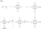
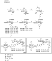
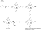
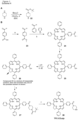
- the photosensitising agent is a conjugate of a photosensitiser and chitosan as defined in formula (I): wherein
- R 2 is selected from and
- R 3 is selected from and Preferably R 2 or R 3 is TPP a , TPC a1 or TPC c1 .
- Group A may provide 70 to 95% of the total Rn groups and group B may provide 5 to 30% of the total Rn groups.
- conjugate of a photosensitiser and chitosan is selected from (see numbering in Schemes 1-5B in Figure 4 ):
- the A/B % values provided refer to the proportion of Rn groups which are group A or B.
- the asterisks denote the remainder of the chitosan polymer.
- an "antigenic" molecule as referred to herein is a molecule which itself, or a part thereof, is capable of stimulating an immune response, when presented to the immune system or immune cells in an appropriate manner.
- the antigenic molecule will be a vaccine antigen or vaccine component, such as a polypeptide containing entity.
- antigens or antigenic vaccine components are known in the art and include all manner of bacterial or viral antigens or indeed antigens or antigenic components of any pathogenic species including protozoa or higher organisms. Whilst traditionally the antigenic components of vaccines have comprised whole organisms (whether live, dead or attenuated) i.e. whole cell vaccines, in addition sub-unit vaccines, i.e. vaccines based on particular antigenic components of organisms e.g. proteins or peptides, or even carbohydrates, have been widely investigated and reported in the literature. Any such "sub-unit"-based vaccine component may be used as the antigenic molecule of the present invention.
- a preferred antigenic molecule according to the invention is a peptide (which is defined herein to include peptides of both shorter and longer lengths i.e. peptides, oligopeptides or polypeptides, and also protein molecules or fragments thereof e.g. peptides of 5-500 e.g. 10 to 250 such as 15 to 75, or 8 to 25 amino acids).
- the invention has been illustrated using, for example, ovalbumin as the antigenic molecule which forms a preferred aspect of the invention, but antigenic molecules which are not ovalbumin are particularly preferred, such as those used in the Examples.
- peptides In addition to antigens derived from pathogenic organisms, peptides have also been proposed for use as vaccines against cancer or other diseases such as multiple sclerosis. For example, mutant oncogene peptides hold great promise as cancer vaccines acting as antigens in the stimulation of cytotoxic T-lymphocytes. ( Schirrmacher, Journal of Cancer Research and Clinical Oncology 1995, 121, 443-451 ; Curtis Cancer Chemotherapy and Biological Response Modifiers, 1997, 17, 316-327 ). Thus a melanoma antigen may be used as the antigenic molecule for use in the invention. In alternative embodiments, an antigenic molecule which is not a melanoma antigen may be used.
- a “melanoma antigen” as referred to herein is a molecule derived from a melanoma cell which itself, or a part thereof, is capable of stimulating an immune response, when presented to the immune system or immune cells in an appropriate manner.
- a molecule "derived" from a melanoma is a molecule which may appear in the melanoma cell or which is modified relative to the native molecule in the melanoma, e.g. by truncation, post-expression modification and/or sequence modification providing the modified molecule retains one or more epitopes from the native molecule which allows the modified molecule to generate an immune response which would recognise the native molecule.
- the melanoma antigen may be obtained by isolation from appropriate sources e.g. the subject's melanoma or may be synthesised e.g. by chemical synthesis or peptide/protein expression.
- a synthetic peptide vaccine has also been evaluated for the treatment of metastatic melanoma ( Rosenberg et al., Nat. Med. 1998, 4(3), 321-7 ).
- a T-cell receptor peptide vaccine for the treatment of multiple sclerosis is described in Wilson et al., J. Neuroimmunol. 1997, 76(1-2), 15-28 .
- Any such peptide vaccine component may be used as the antigenic molecule of the invention, as indeed may any of the peptides described or proposed as peptide vaccines in the literature.
- the peptide may thus be synthetic or isolated or otherwise derived from an organism.
- Preferred peptides include those used in the Examples, e.g. a HPV peptide such as the HPV long peptide having the sequence QAEPDRAHYNIVTFCCKCDSTLRLCVQSTHVDIR.
- an adjuvant is also used in the methods or uses of the invention.
- the adjuvant may be selected from a Toll-like receptor (TLR) ligand, such as a TLR 3 ligand, for example Poly(IC) (e.g. high (e.g. average size of 1.5-8kb) or low (e.g. average size of 0.2-1kb) MW Poly(IC)).
- TLR Toll-like receptor
- Poly(IC) e.g. high (e.g. average size of 1.5-8kb) or low (e.g. average size of 0.2-1kb) MW Poly(IC)).
- the dose of Poly(IC) may be between 5 ⁇ g and 200 ⁇ g, for example between 10 ⁇ g and 100 ⁇ g, preferably 10 ⁇ g or 50 ⁇ g for mice, which may be appropriately scaled where necessary for treatment of other animals.
- Products, methods or uses of the invention preferably contain or use Poly(IC).
- the antigenic molecule may be processed by the antigen-processing machinery of the cell.
- the antigenic molecule expressed or presented on the surface of the cell may be a part or fragment of the antigenic molecule which is internalised (endocytosed).
- a "part" of an antigenic molecule which is presented or expressed preferably comprises a part which is generated by antigen-processing machinery within the cell. Parts may, however, be generated by other means which may be achieved through appropriate antigen design (e.g. pH sensitive bonds) or through other cell processing means. Conveniently such parts are of sufficient size to generate an immune response, e.g. in the case of peptides greater than 5, e.g. greater than 10 or 20 amino acids in size.
- cytokine encompasses a large and diverse family of regulators produced throughout the body by cells of a variety of embryological origin.
- Cytokines are small cell signaling molecules which can be proteins, peptides, or glycoproteins. Cytokines include immunomodulating agents, such as interleukins (IL) and interferons (IFN) and also colony stimulating factors, tumour necrosis factors (TNF) and other regulatory molecules. Cytokines have been classed as lymphokines, interleukins, and chemokines, based on their function, cell of secretion, or target of action.
- IL interleukins
- IFN interferons
- TNF tumour necrosis factors
- cytokine has a matching cell-surface receptor, which initiates cascades of intracellular signalling which alter cell functions.
- Cytokines are well known in the art and all such cytokines are encompassed for use according to the invention. Preferred families are as described herein.
- Cytokines have been classified in various ways according to structure and/or function, and various families have been identified.
- the cytokines may be classified by virtue of the receptors to which they bind.
- the receptors (and hence their ligands) may be classified into Type 1 cytokine (hemopoietin) receptors, Type II cytokine receptors, TNF receptors, immunoglobulin superfamily receptors and seven transmembrane ⁇ -helical receptors.
- the invention is concerned with the cytokine GM-CSF.
- Granulocyte-macrophage colony-stimulating factor belongs to the family of cytokines which are ligands for hematopoietic cytokine receptors discussed above. Within this family of receptors is the GM-CSF receptor family, which have a common ⁇ chain. GM-CSF is secreted as a single chain glycoprotein containing 128 amino acids with a conserved disulphide bond by a variety of cell types.
- GM-CSF Functions of GM-CSF are mediated by the GM-CSF receptor, which comprises a GM-CSF-specific ⁇ chain and, in human cells, a ⁇ chain which is shared with the IL-3 and IL-5 receptors.
- the ⁇ chain is expressed as monomers on the cell surface, and binds to GM-CSF with high affinity. Following such binding, the ⁇ chain is recruited to the complex and activates signal transduction and functional responses.
- the ⁇ chain can exist as a soluble external molecule (generated by alternative splicing). This receptor competes with its membrane-bound counterpart for cytokine binding but does not participate in agonistic signalling.
- GM-CSF functions as a white blood cell growth factor. GM-CSF stimulates stem cells to produce granulocytes (neutrophils, eosinophils, and basophils) and monocytes. GM-CSF is therefore important in fighting infection.
- IL-2 receptor family Also within the type I cytokine receptor family is the IL-2 receptor family. Members of this family have a common ⁇ chain. Receptors in this family include IL-2, IL-4, IL-7, IL-9, IL-15 and IL-21 receptors. The majority of interleukins are synthesized by helper CD4 T lymphocytes, as well as through monocytes, macrophages, and endothelial cells. They promote the development and differentiation of T, B, and hematopoietic cells.
- IL-7 binds to the IL-7 receptor, which is a heterodimer consisting of Interleukin-7 receptor ⁇ and a common ⁇ chain receptor.
- IL-2, IL-15 and IL-21 are related to IL-7, in that they belong to the so-called gamma(c) family of cytokines, i.e. they bind to a receptor with a common ⁇ chain. These cytokines all employ the common cytokine ⁇ chain for signalling, and have potent effects on T-cells and NK cells.
- Interferons are examples of cytokines that bind to type II cytokine receptors. Interferons are proteins made and released by host cells in response to the presence of pathogens such as viruses, bacteria, parasites or tumor cells. They allow for communication between cells to trigger the protective defenses of the immune system that eradicate pathogens or tumours. Interferons can activate immune cells, such as natural killer cells and macrophages and they increase the ability of uninfected hosts to resist new infection by virus.
- pathogens such as viruses, bacteria, parasites or tumor cells. They allow for communication between cells to trigger the protective defenses of the immune system that eradicate pathogens or tumours. Interferons can activate immune cells, such as natural killer cells and macrophages and they increase the ability of uninfected hosts to resist new infection by virus.
- IFNs Approximately ten distinct IFNs have been identified in mammals (seven in humans). There are three IFN classes, delineated on the basis of the type of receptor through which they signal. All type I IFNs bind to a specific cell surface receptor complex known as the IFN- ⁇ receptor (IFNAR) that consists of IFNAR1 and IFNAR2 chains. The type I interferons present in humans are IFN- ⁇ , IFN- ⁇ and IFN- ⁇ . Type II IFNs bind to IFNGR, which consists of IFNGR1 and IFNGR2 chains. In humans this is IFN- ⁇ . Type III Interferons signal through a receptor complex consisting of IL10R2 and IFNLR1 .
- IFNAR IFN- ⁇ receptor
- IFNAR IFN- ⁇ receptor
- Type II IFNs bind to IFNGR, which consists of IFNGR1 and IFNGR2 chains. In humans this is IFN- ⁇ .
- Type III Interferons signal through a receptor complex consisting of
- the agent is GM-CSF.
- GM-CSF all known forms of GM-CSF can be used in the present invention, including also functionally equivalent variants, derivatives and fragments thereof.
- cytokine as used herein includes amino acid sequence variants of known cytokine polypeptides, and fragments of a cytokine polypeptide, or derivative thereof, as long as such fragments, variants or derivatives are active, or "functional", i.e. retain at least one function or activity (e.g. biological activity) of the relevant cytokine.
- the cytokine may be a recombinant polypeptide, a synthetic polypeptide or may be isolated from a natural source. Suitable cytokines are commercially available and would be known to the skilled man, for example human cytokines are available from GenScript (Piscataway, NJ, USA).
- the cytokine may be from any species (more particularly any vertebrate species), but preferably will be mammalian, and more preferably human.
- Variants of cytokines may include, for example, different allelic variants as they appear in nature e.g. in other species or due to geographical variation etc.
- Functionally equivalent variants may also include polypeptides which incorporate one or more amino acid substitutions, or intrasequence or terminal deletions or additions to known sequences.
- Functionally equivalent derivatives may include chemical modifications of the amino acid sequence, including for example the inclusion of chemically substituted or modified amino acid residues or PEGylated cytokines.
- a derivative may also be a molecule which is a peptidomimetic of a cytokine polypeptide.
- it may be a molecule which is functionally equivalent or similar to a polypeptide and which can adopt a 3-D structure which is similar to its polypeptide counterpart, but which is not composed solely of amino acids linked by peptide bonds.
- a peptidomimetic may be composed of sub-units which are not amino acids but which are structurally and functionally similar to an amino acid.
- the backbone moiety of the subunit may differ from a standard amino acid, e.g. it may comprise one or more nitrogen atoms instead of one or more carbon atoms.
- a preferred class of peptidomimetic is a peptoid, i.e. an N-substituted glycine.
- Peptoids are closely related to their peptide counterparts but differ chemically in that their side chains are appended to nitrogen atoms along the backbone of the molecule, rather than to the ⁇ -carbons as they are in amino acids.
- amino acid changes are of a minor nature, that is conservative amino acid substitutions that do not significantly affect the folding and/or activity of the protein; small deletions, typically of 1 to 30 amino acids; small amino- or carboxyl-terminal extensions; addition of a small linker peptide of up to about 20-25 residues; or a small extension that facilitates purification by changing net charge or another function, such as a poly-histidine tract, an antigenic epitope or a binding domain.
- N and/or C extensions to the protein or peptides are included in the definition.
- the lengths of each extended derivative may vary, for example, derivatives may be extended by up to 50, 30, 20, 10 or 5 amino acids.
- the cytokine preferably has at least 50%, 60%, 70%, 80%, 90%, 95%, 96%, 97%, 98%, 99% or 100% sequence identity or similarity to the known amino acid sequences of the cytokines described herein.
- Cytokine NCBI Reference Sequence UniProtKB/Swiss-Prot Reference granulocyte-macrophage colony-stimulating factor precursor Homo sapiens ) NP_000749.2 (144 amino acids)
- P04141 also known as CSF2
- interleukin-2 precursor Homo sapiens
- NP_000577.2 153 amino acids
- P60568 interleukin-7 isoform 1 precursor
- P13232 interleukin-15 isoform 1 preproprotein (Homo sapiens) NP_000576.1 (162 amino acids)
- P40933 interleukin-21 isoform 2 precursor Homo sapiens) NP_001193935.1 (153 amino acids)
- Q9HBE4 interferon alpha-1/13 precursor Homo sapiens
- NP_076918.1 189 amino acids
- P01562 also known as
- the degree of identity between two nucleic acid and two amino acid sequences may be determined by means of computer programs known in the art such as GAP provided in the GCG program package ( Needleman and Wunsch, 1970, Journal of Molecular Biology 48: 443-453 ).
- GAP can be used with the following settings: GAP creation penalty of 3.0 and GAP extension penalty of 0.1.
- the cytokine for use according to the invention is GM-CSF.
- the cytokine is from a human source.
- a "ligand” is a molecule which is able to bind to a receptor and initiate signalling through that receptor, or antagonise or agonise signalling through that receptor by the native ligand.
- expressing or “presenting” refers to the presence of the antigenic molecule or a part thereof on the surface of said cell such that at least a portion of that molecule is exposed and accessible to the environment surrounding that cell, preferably such that an immune response may be generated to the presented molecule or part thereof.
- Expression on the "surface” may be achieved in which the molecule to be expressed is in contact with the cell membrane and/or components which may be present or caused to be present in that membrane.
- cell is used herein to include all eukaryotic cells (including insect cells and fungal cells).
- Representative “cells” thus include all types of mammalian and non-mammalian animal cells (such as fish cells), plant cells, insect cells, fungal cells and protozoa.
- the cells are mammalian, for example cells from cats, dogs, horses, donkeys, sheep, pigs, goats, cows, mice, rats, rabbits, guinea pigs, but most preferably from humans.
- the cell which is subjected to the methods, uses etc. of the invention may be any cell which is capable of expressing, or presenting on its surface a molecule which is administered or transported into its cytosol.
- the cell is conveniently an immune cell i.e. a cell involved in the immune response.
- other cells may also present antigen to the immune system and these also fall within the scope of the invention.
- the cells are thus advantageously antigen-presenting cells as described hereinafter.
- the antigen-presenting cell may be involved in any aspect or "arm" of the immune response as defined herein.
- cytotoxic cells require antigens to be presented to the cell to be stimulated in a particular manner by the antigen-presenting cells, for example MHC Class I presentation (e.g. activation of CD8 + cytotoxic T-cells requires MHC-1 antigen presentation).
- Antibody-producing cells may also be stimulated by presentation of antigen by the antigen-presenting cells.
- Antigens may be taken up by antigen-presenting cells by endocytosis and degraded in the endocytic vesicles to peptides. These peptides may bind to MHC class II molecules in the endosomes and be transported to the cell surface where the peptide-MHC class II complex may be recognised by CD4+ T helper cells and induce an immune response.
- proteins in the cytosol may be degraded, e.g. by proteasomes and transported into endoplasmic reticulum by means of TAP (transporter associated with antigen presentation) where the peptides may bind to MHC class I molecules and be transported to the cell surface ( Yewdell and Bennink, 1992, Adv. Immunol.
- the peptide-MHC class I complex will be recognised by CD8+ cytotoxic T-cells (CTLs).
- CTLs cytotoxic T-cells
- the CTLs will bind to the peptide-MHC (HLA) class I complex and thereby be activated, start to proliferate and form a clone of CTLs.
- the target cell and other target cells with the same peptide-MHC class I complex on the cells surface may be killed by the CTL clone.
- Immunity against the foreign antigen may be established if a sufficient amount of the antigen can be introduced into the cytosol (Yewdell and Bennink, 1992, supra; Rock, 1996, Immunology Today 17: 131-137 ). This is the basis for development of inter alia cancer vaccines.
- One of the largest practical problems is to introduce sufficient amounts of antigens (or parts of the antigen) into the cytosol. This may be solved according to the present invention.
- the antigenic molecule may be processed by the antigen-processing machinery of the cell and presented on the cell surface in an appropriate manner e.g. by Class I MHC. This processing may involve degradation of the antigen, e.g. degradation of a protein or polypeptide antigen into peptides, which peptides are then complexed with molecules of the MHC for presentation.
- the antigenic molecule expressed or presented on the surface of the cell according to the present invention may be a part or fragment of the antigenic molecule which is internalised (endocytosed).
- lymphocytes both T and B cells
- dendritic cells macrophages etc.
- others include for example cancer cells e.g. melanoma cells. These cells are referred to herein as "antigen-presenting cells”.
- “Professional antigen-presenting cells” which are cells of the immune system principally involved in the presentation of antigen to effector cells of the immune system are known in the art and described in the literature and include B lymphocytes, dendritic cells and macrophages.
- the cell is a professional antigen-presenting cell.
- the antigenic molecule For antigen presentation by an antigen-presenting cell to a cytotoxic T-cell (CTL) the antigenic molecule needs to enter the cytosol of the antigen-presenting cell ( Germain, Cell, 1994, 76, 287-299 ).
- CTL cytotoxic T-cell
- the cell is a dendritic cell.
- Dendritic cells are immune cells forming part of the mammalian immune system. Their main function is to process antigenic material and present it on the surface to other cells of the immune system. Once activated, they migrate to the lymph nodes where they interact with T cells and B cells to initiate the adaptive immune response.
- Dendritic cells are derived from hematopoietic bone marrow progenitor cells. These progenitor cells initially transform into immature dendritic cells which are characterized by high endocytic activity and low T-cell activation potential. Once they have come into contact with a presentable antigen, they become activated into mature dendritic cells and begin to migrate to the lymph node. Immature dendritic cells phagocytose pathogens and degrade their proteins into small pieces and upon maturation present those fragments at their cell surface using MHC molecules.
- the dendritic cells may be derived from any appropriate source of dendritic cells, such as from the skin, inner lining of the nose, lungs, stomach and intestines or the blood.
- the dendritic cells are derived from bone marrow.
- Dendritic cells may be isolated from natural sources for use in the in vitro methods of the invention or may be generated in vitro. Dendritic cells arise from monocytes, i.e. white blood cells which circulate in the body and, depending on the right signal, can differentiate into either dendritic cells or macrophages. The monocytes in turn are formed from stem cells in the bone marrow. Monocyte-derived dendritic cells can be generated in vitro from peripheral blood mononuclear cells (PBMCs). Plating of PBMCs in a tissue culture flask permits adherence of monocytes.
- PBMCs peripheral blood mononuclear cells
- IL-4 interleukin 4
- GM-CSF granulocyte-macrophage colony stimulating factor
- TNF tumor necrosis factor
- contacting refers to bringing the cells and the photosensitising agent and/or the antigenic molecule and/or the cytokine as defined herein into physical contact with one another under conditions appropriate for internalization into the cells, e.g. preferably at 37°C in an appropriate nutritional medium, e.g. from 25-39°C or in vivo at body temperature, i.e. 36-38°C.
- the cell may be contacted with the photosensitising agent and antigenic molecule and the cytokine as defined herein sequentially or simultaneously. Preferably, and conveniently the components are contacted with the cell simultaneously.
- the photosensitising agent and antigenic molecule and optionally the cytokine may be taken up by the cell into the same or different intracellular compartments (e.g. they may be co-translocated).
- the cells are then exposed to light of suitable wavelengths to activate the photosensitising compound which in turn leads to the disruption of the intracellular compartment membranes.
- WO 02/44396 describes a method in which the order of the steps in the method may be arranged such that for example the photosensitising agent is contacted with the cells and activated by irradiation before the molecule to be internalised (in this case the antigenic molecule) is brought into contact with the cells.
- This method takes advantage of the fact that it is not necessary for the molecule to be internalised to be present in the same cellular subcompartment as the photosensitising agent at the time of irradiation.
- said photosensitising agent and/or said antigenic molecule and/or the cytokine as defined herein are applied to the cell together, or separately relative to one another. Irradiation is then performed at a time when at least the antigenic molecule and photosensitising agent appear in the same intracellular compartment. This is referred to as a "light after" method.
- said method can be performed by contacting said cell with the photosensitising agent first, followed by contact with the antigenic molecule and/or the cytokine as defined herein, and irradiation is performed after uptake of the photosensitising agent into an intracellular compartment, but prior to the cellular uptake of the antigenic molecule (and optionally the cytokine) into an intracellular compartment containing said photosensitising agent (e.g. it may be present in a different intracellular compartment at the time of light exposure), preferably prior to cellular uptake into any intracellular compartment, e.g. prior to any cellular uptake.
- the photosensitising agent may be administered followed by irradiation and then administration of the remaining agents. This is the so-called "light before" method.
- Internalisation refers to the intracellular, e.g. cytosolic, delivery of molecules.
- internalisation may include the step of release of molecules from intracellular/membrane bound compartments into the cytosol of the cells.
- cellular uptake or “translocation” refers to one of the steps of internalisation in which molecules external to the cell membrane are taken into the cell such that they are found interior to the outer lying cell membrane, e.g. by endocytosis or other appropriate uptake mechanisms, for example into or associated with intracellular membrane-restricted compartments, for example the endoplasmic reticulum, Golgi body, lysosomes, endosomes etc.
- the step of contacting the cells with the various agents may be carried out in any convenient or desired way.
- the cells may conveniently be maintained in an aqueous medium, such as for example appropriate cell culture medium, and at the appropriate time point the various agents can simply be added to the medium under appropriate conditions, for example at an appropriate concentration and for an appropriate length of time.
- the cells may be contacted with the agents in the presence of serum-free medium, or with serum-containing medium.
- the application of the various agents used in the methods or uses of the invention may be to cells in vitro or in vivo. In the latter case, the application may be via direct (i.e. localized) or indirect (i.e. systemic or non-localized) administration as described in more detail hereinbelow.
- the photosensitising agent is brought into contact with the cells at an appropriate concentration and for an appropriate length of time which can easily be determined by a skilled person using routine techniques, and will depend on such factors as the particular photosensitising agent used and the target cell type and location.
- concentration of the photosensitising agent is conveniently such that once taken up into the cell, e.g. into, or associated with, one or more of its intracellular compartments and activated by irradiation, one or more cell structures are disrupted e.g. one or more intracellular compartments are lysed or disrupted.
- photosensitising agents as described herein may be used at a concentration of for example 10 to 50 ⁇ g/ml.
- the range can be much broader, e.g. 0.0005-500 ⁇ g/ml.
- the photosensitising agent may be used in the range 0.05-20 mg/kg body weight when it is to be administered systemically. Alternatively, a range of 0.005-20mg/kg body weight may be used for systemic administration. If it is to be administered locally, for example by intradermal, subcutaneous or intratumoural administration, the dose may be in the region of 1-5000 ⁇ g, for example 10-2500, 25-1000, 50-500, 10-300 or 100-300 ⁇ g. Preferably the dose is selected from 100 ⁇ g, 150 ⁇ g, 200 ⁇ g and 250 ⁇ g.
- the dose is 75-125 ⁇ g, e.g. 100 ⁇ g.
- the doses provided are for a human of average weight (i.e. 70kg).
- the photosensitiser dose may be dissolved in 100 ⁇ l-1ml, i.e. the concentration may be in the range of 1-50000 ⁇ g/ml. In smaller animals the concentration range may be different and can be adjusted accordingly though when it is to be administered locally, little variation in dosing is necessary for different animals.
- the concentration of antigen to be used will depend on the antigen which is to be used. Conveniently a concentration of 0.001-500 ⁇ g/ml (e.g. 20-500, 20-300, 20-100 ⁇ g/ml, 20-50, 10-50, 5-50, 1-50, 0.01-50, or 0.001-50 ⁇ g/ml) antigen may be used in vitro.
- a concentration of 0.001-500 ⁇ g/ml e.g. 20-500, 20-300, 20-100 ⁇ g/ml, 20-50, 10-50, 5-50, 1-50, 0.01-50, or 0.001-50 ⁇ g/ml
- a concentration of 0.001-500 ⁇ g/ml e.g. 20-500, 20-300, 20-100 ⁇ g/ml, 20-50, 10-50, 5-50, 1-50, 0.01-50, or 0.001-50 ⁇ g/ml
- a concentration of 0.001-500 ⁇ g/ml e.g
- the protein antigen dose may be in the range 0.5-500 ⁇ g, for example 10-100 ⁇ g or 10-200 ⁇ g.
- an in vivo dose of 0.1-4000 ⁇ g e.g. 0.1-2000 ⁇ g, 0.1-1000 ⁇ g or 0.1-500 ⁇ g, for example 0.1-100 ⁇ g, may be employed.
- Such doses are appropriate for local administration.
- An appropriate concentration can be determined depending on the efficiency of uptake of the agent in question into the cells in question and the final concentration it is desired to achieve in the cells.
- the concentration of the cytokine as defined herein will also depend on the particular molecule which is to be used.
- the invention is concerned with the use of GM-CSF. Conveniently, in vitro, a concentration as shown in the table below may be used for GM-CSF. Doses of other cytokines are provided for comparison.
- An in vivo dose, e.g. for local administration, of 5-500 ⁇ g, e.g. 50-250 ⁇ g, may be used for GM-CSF.
- the photosensitising agent, the antigenic molecule and the cytokine as defined herein are administered together, but this may be varied. Thus different times or modes or sites of administration (or contact with the cell) are contemplated for each of the different components and such variations are encompassed within the scope of the invention.
- the cytokine as defined herein is administered separately from the antigen, for example in a separate formulation, or systemically, e.g. via oral administration.
- the cytokine may be administered prior to administration of the antigen and/or photosensitiser, for example 24 hours before.
- the cytokine may be administered separately relative to the other agents, e.g. approximately 2 hours prior to illumination.
- the agent may be administered with or at the same time, i.e. simultaneously, as the antigen.
- the contact between the cell and the photosensitising agent and/or antigenic molecule and/or the cytokine as defined herein is conveniently from 15 minutes to 24 hours, e.g. 30 minutes to four hours, preferably from 1.5 to 2.5 hours.
- the range of time may be from about 1 hour to about 48 hours, for example from about 2 hours to about 40 hours, or from about 6 hours to about 36 hours, e.g. from 12 hours to 30 hours, e.g. 16 hours to 20 hours, for example 18 hours or about 18 hours.
- the initial incubation of the cell is with the photosensitising agent.
- the time between the administration of the photosensitising agent and the antigenic molecule and/or cytokine is a matter of hours.
- the photosensitising agent may be applied 16 to 20 hours, e.g. 18 hours, before illumination
- the antigenic molecule and/or cytokine may be applied 1 to 3 hours, e.g. 2 hours before illumination.
- the time between the administration of the photosensitising agent and the antigenic molecule and/or the cytokine may be in the range of 15 to 23 hours.
- the cell is then incubated with the antigen and/or cytokine as defined herein after the incubation with the photosensitiser.
- the cells may be placed into photosensitiser/antigen-free medium after the contact with the photosensitiser/antigen and before irradiation, e.g. for 30 minutes to 4 hours, e.g. from 1.5 to 2.5 hours, depending on the timing of the incubation with the photosensitiser and antigenic molecule and cytokine.
- an appropriate use and time of incubation by which the various agents are brought into contact with the target cells will be dependent on factors such as the intended mode of administration and the type of agents which are used. For example, if the agents are to be injected into a tumour, tissue or organ which is to be treated/irradiated, the cells near the injection point will come into contact with and hence tend to take up the agents more rapidly than the cells located at a greater distance from the injection point, which are likely to come into contact with the agents at a later time point and lower concentration. Conveniently a time of 6-24 hours may be used.
- agents to be administered by intravenous injection or orally may take some time to arrive at the target cells and it may thus take longer post-administration e.g. several days, in order for a sufficient or optimal amount of the agents to accumulate in a target cell or tissue.
- the time of intended administration required for individual cells in vivo is thus likely to vary depending on these and other parameters.
- the time at which the molecules come into contact with the target cells must be such that before irradiation occurs an appropriate amount of the photosensitising agent has been taken up by the target cells and either: (i) before or during irradiation the antigenic molecule (and optionally the cytokine) has either been taken up, or will be taken up after sufficient contact with the target cells, into the cell, for example into the same or different intracellular compartments relative to the photosensitising agent or (ii) after irradiation the antigenic molecule (and optionally the cytokine) is in contact with the cells for a period of time sufficient to allow its uptake into the cells.
- any mode of administration common or standard in the art is suitable, e.g. injection, infusion, topical administration, transdermal administration, both to internal and external body surfaces etc.
- the invention can be used in relation to any tissue which contains cells to which the photosensitising agent containing compound or the molecule to be internalized is localized, including body fluid locations, as well as solid tissues. All tissues can be treated as long as the photosensitiser is taken up by the target cells, and the light can be properly delivered.
- Preferred modes of administration are intradermal, subcutaneous, topical or intratumoural administration or injection. Preferably administration is by intradermal injection.
- the uses or methods or parts thereof may be repeated e.g. "re-vaccination" may take place.
- the use or method in its entirety may be performed multiple times (e.g. 2, 3 or more times) after an appropriate interval or parts of the use or method may be repeated, e.g. further administration of the cytokine as defined herein or additional irradiation steps.
- the use or method or part of the use or method may be performed again a matter of days, e.g. between 5 and 60 days (for example 7, 14, 15, 21, 22, 42 or 51 days), e.g. 7 to 20 days, preferably 14 days, or weeks, e.g.
- All or part of the use or method may be repeated multiple times at appropriate intervals of time, e.g. every two weeks or 14 days. In a preferred embodiment the use or method is repeated at least once. In another embodiment the use or method is repeated twice.
- the antigenic molecule in the second or subsequent time the use or method is carried out is to be administered or administered in combination with the photosensitiser and illumination, i.e. the cytokine is not administered in the second or subsequent time the use or method is carried out.
- the antigenic molecule in the second or subsequent time the use or method is carried out may be for administration or administered in combination with the photosensitiser and illumination, i.e. the adjuvant is not for administration or administered in the second or subsequent time the use or method is carried out.
- the cytokine may be present or not in the second or subsequent time the use or method is carried out.
- parts of the use or method of the invention may be carried out prior to the use or method of the invention being carried out.
- the use or method may be carried out one or more times, for example twice, in the absence of cytokine before the use or method of the invention is carried out.
- the use or method may be carried out one or more times, for example twice, in the absence of photosensitiser and illumination before the use or method of the invention is carried out.
- Part of the use or method may be carried out a matter of days, e.g. 7 or 14 days, or weeks, e.g. 1, 3 or 4 weeks before the use or method of the invention is carried out.
- the antigenic molecule is to be administered or administered (e.g. to the subject) equal to or greater than 2 times (e.g. at the time intervals discussed above), wherein at least the administration of said antigenic molecule is to be performed or is performed in accordance with the use or method of the invention.
- “Irradiation" to activate the photosensitising agent refers to the administration of light directly or indirectly as described hereinafter.
- subjects or cells are to be illuminated or may be illuminated with a light source for example directly (e.g. on single cells in vitro ) or indirectly, e.g. in vivo when the cells are below the surface of the skin or are in the form of a layer of cells not all of which are directly illuminated, i.e. without the screen of other cells.
- Illumination of the cell or subject may occur approximately 18-24 hours after administration of the photosensitising agent, antigenic molecule and the cytokine as defined herein.
- the light irradiation step to activate the photosensitising agent may take place according to techniques and procedures well known in the art.
- the wavelength of light to be used is selected according to the photosensitising agent to be used. Suitable artificial light sources are well known in the art, e.g. using blue (400-475nm) or red (620-750nm) wavelength light.
- TPCS 2a for example a wavelength of between 400 and 500nm, more preferably between 400 and 450nm, e.g. from 430-440nm, and even more preferably approximately 435nm, or 435nm may be used.
- the photosensitiser e.g. a porphyrin or chlorin
- may be activated by green light for example the KillerRed (Evrogen, Moscow, Russia) photosensitiser may be activated by green light.
- Suitable light sources are well known in the art, for example the LumiSource ® lamp of PCI Biotech AS.
- an LED-based illumination device which has an adjustable output power of up to 60mW and an emission spectra of 430-435nm may be used.
- a suitable source of illumination is the PCI Biotech AS 652nm laser system SN576003 diode laser, although any suitable red light source may be used.
- the time for which the cells are exposed to light in the uses or methods of the present invention may vary.
- the efficiency of the internalisation of a molecule into the cytosol increases with increased exposure to light to a maximum beyond which cell damage and hence cell death increases.
- a preferred length of time for the irradiation step depends on factors such as the target, the photosensitiser, the amount of the photosensitiser accumulated in the target cells or tissue and the overlap between the absorption spectrum of the photosensitiser and the emission spectrum of the light source.
- the length of time for the irradiation step is in the order of seconds to minutes or up to several hours (even up to 12 hours), e.g. preferably up to 60 minutes e.g. from 0.25 or 1 to 30 minutes, e.g. from 0.5 to 3 minutes or from 1 to 5 minutes or from 1 to 10 minutes e.g. from 3 to 7 minutes, and preferably approximately 3 minutes, e.g. 2.5 to 3.5 minutes.
- Shorter irradiation times may also be used, for example 1 to 60 seconds, e.g. 10-50, 20-40 or 25-35 seconds.
- Appropriate light doses can be selected by a person skilled in the art and again will depend on the photosensitiser used and the amount of photosensitiser accumulated in the target cells or tissues.
- the light doses are usually lower when photosensitisers with higher extinction coefficients (e.g. in the red area, or blue area if blue light is used, depending on the photosensitiser used) of the visible spectrum are used.
- a light dose in the range of 0.24 - 7.2J/cm 2 at a fluence range of 0.05-20 mW/cm 2 e.g.
- 2.0 mW/cm 2 may be used when an LED-based illumination device which has an adjustable output power of up to 60mW and an emission spectra of 430-435nm is employed.
- the LumiSource ® lamp a light dose in the range of 0.1-6J/cm 2 at a fluence range of 0.1-20 (e.g. 13 as provided by Lumisource ® ) mW/cm 2 is appropriate.
- a light dose of 0.03-1J/cm 2 e.g. 0.3J/cm 2
- at a fluence range of 0.5-5 mW/cm 2 e.g. 0.81 mW/cm 2
- 0.03-1J/cm 2 e.g. 0.3J/cm 2
- a fluence range of 0.5-5 mW/cm 2 e.g. 0.81 mW/cm 2
- the uses and methods of the invention may inevitably give rise to some cell damage by virtue of the photochemical treatment i.e. by photodynamic therapy effects through the generation of toxic species on activation of the photosensitising agent. Depending on the proposed use, this cell death may not be of consequence and may indeed be advantageous for some applications (e.g. cancer treatment). In most embodiments, however, cell death is avoided to allow the generation of an immune response from the presenting cell.
- the uses or methods of the invention may be modified such that the fraction or proportion of the surviving cells is regulated by selecting the light dose in relation to the concentration of the photosensitising agent. Again, such techniques are known in the art.
- substantially all of the cells, or a significant majority are not killed.
- In vitro cell viability following PCI treatment can be measured by standard techniques known in the art such as the MTS test.
- In vivo cell death of one or more cell types may be assessed within a 1cm radius of the point of intended administration (or at a certain depth of tissue), e.g. by microscopy. As cell death may not occur instantly, the % cell death refers to the percent of cells which remain viable within a few hours of irradiation (e.g. up to 4 hours after irradiation) but preferably refers to the % viable cells 4 or more hours after irradiation.
- the method may be performed in vitro or ex vivo and the uses are performed in vivo.
- the method is used in vitro or ex vivo to generate cells for administration in vivo.
- compositions or agents described herein are used to generate an immune response in a subject.
- the present invention provides an antigenic molecule, a photosensitising agent, and the cytokine GM-CSF as defined hereinbefore, for use in stimulating an immune response in a subject to treat or prevent a disease, disorder or infection in the subject.
- an “immune response” which may be generated may be humoral and cell-mediated immunity, for example the stimulation of antibody production, or the stimulation of cytotoxic or killer cells, which may recognise and destroy (or otherwise eliminate) cells expressing "foreign" antigens on their surface.
- the term “stimulating an immune response” thus includes all types of immune responses and mechanisms for stimulating them and encompasses stimulating CTLs which forms a preferred aspect of the invention.
- the immune response which is stimulated is cytotoxic CD8 T cells.
- the extent of an immune response may be assessed by markers of an immune response, e.g. secreted molecules such as IL-2 or IFN ⁇ or the production of antigen specific T cells (e.g. assessed as described in the Examples).
- the stimulation of cytotoxic cells or antibody-producing cells requires antigens to be presented to the cell to be stimulated in a particular manner by the antigen-presenting cells, for example MHC Class I presentation (e.g. activation of CD8 + cytotoxic T-cells requires MHC-I antigen presentation).
- MHC Class I presentation e.g. activation of CD8 + cytotoxic T-cells requires MHC-I antigen presentation.
- the immune response is stimulated via MHC-I presentation.
- the immune response is used to treat or prevent a disease, disorder or infection, e.g. cancer.
- a disease, disorder or infection e.g. cancer.
- the cancer may be melanoma.
- the cancer may be a cancer which is not melanoma.
- the use is for vaccination.
- vaccination is the use of an antigen (or a molecule containing an antigen) to elicit an immune response which is prophylactic or therapeutic against the development (or further development) of a disease, disorder or infection, wherein that disease, disorder or infection is associated with abnormal expression or presence of that antigen.
- the disease is cancer (and the vaccination is therapeutic) or an immune response is to be generated to an infection (and the vaccination is prophylactic).
- the subject of the use is a non-mammalian animal (e.g. a fish) or a mammal, preferably a cat, dog, horse, donkey, sheep, pig, goat, cow, mouse, rat, rabbit or guinea pig, but most preferably the subject is a human.
- a non-mammalian animal e.g. a fish
- a mammal preferably a cat, dog, horse, donkey, sheep, pig, goat, cow, mouse, rat, rabbit or guinea pig, but most preferably the subject is a human.
- the methods and uses described herein achieve synergy, i.e. the extent of cell surface presentation or the immune response generated is enhanced more than the combined enhancement observed by (i) performing the method or use with the antigenic molecule in the absence of the cytokine and (ii) performing the method or use with the antigenic molecule in the absence of the photosensitising agent and the irradiation step, i.e. synergy between the methods or uses is observed.
- the level of cell surface presentation or immune response generation may be assessed by appropriate means, e.g. number of antigen-specific CD8+ cells or levels of markers of immune response activation, e.g. IFN ⁇ or IL-2.
- the various agents used in the uses of the invention may be administered to the subject separately, sequentially or simultaneously.
- the methods of the invention above may be used in vitro or in vivo, for example either for in situ treatment or for ex vivo treatment followed by the administration of the treated cells to the body.
- the present invention also provides a pharmaceutical composition
- a pharmaceutical composition comprising an antigenic molecule, a photosensitising agent, and the cytokine GM-CSF as defined herein and one or more pharmaceutically acceptable diluents, carriers or excipients.
- compositions (and products of the invention) may be formulated in any convenient manner according to techniques and procedures known in the pharmaceutical art, e.g. using one or more pharmaceutically acceptable diluents, carriers or excipients.
- “Pharmaceutically acceptable” as referred to herein refers to ingredients that are compatible with other ingredients of the compositions (or products) as well as physiologically acceptable to the recipient.
- the nature of the composition and carriers or excipient materials, dosages etc. may be selected in routine manner according to choice and the desired route of administration, purpose of treatment etc. Dosages may likewise be determined in routine manner and may depend upon the nature of the molecule (or components of the composition or product), purpose of treatment, age of patient, mode of administration etc. In connection with the photosensitising agent, the potency/ability to disrupt membranes on irradiation, should also be taken into account.
- Cells for example antigen presenting cells, may be prepared using methods of the invention in vitro. These cells may be administered to a body in vivo or a body tissue ex vivo such that those cells may stimulate an immune response, e.g. for prophylactic or therapeutic purposes.
- the invention further provides an antigenic molecule, a photosensitising agent, and the cytokine GM-CSF as defined herein, for use in prophylaxis or therapy or for use in stimulating an immune response, for example for vaccination purposes, e.g. for stimulating CTLs, in a subject, preferably for treating or preventing a disease, disorder or infection in said subject, particularly for treating or preventing cancer.
- the antigenic molecule, photosensitising agent and the cytokine may be combined and presented in a composition.
- the invention further provides a product comprising an antigenic molecule, photosensitising agent and the cytokine GM-CSF as defined herein as a combined preparation for simultaneous, separate or sequential use in stimulating an immune response in a subject to treat or prevent a disease, disorder or infection in a subject.
- the present invention also provides a kit for use in stimulating an immune response in a subject to treat or prevent a disease, disorder or infection in said subject, for example for use in vaccination or immunisation, said kit comprising
- the products and kits of the invention may be used to achieve therapeutic uses as defined herein.
- the antigenic presentation achieved by the claimed invention may advantageously result in the stimulation of an immune response when the treated cells are administered in vivo.
- an immune response which confers protection against subsequent challenge by an entity comprising or containing said antigenic molecule or part thereof is generated in uses of the invention, and consequently the invention finds particular utility for vaccination.
- the disease, disorder or infection is any disease, disorder or infection which may be treated or prevented by the generation of an immune response, e.g. by eliminating abnormal or foreign cells which may be identified on the basis of an antigen (or its level of expression) which allows discrimination (and elimination) relative to normal cells. Selection of the antigenic molecule to be used determines the disease, disorder or infection to be treated. Based on the antigenic molecules discussed above, the methods, uses, compositions, products, kits and so forth, described herein may be used to treat or prevent against, for example, infections (e.g. viral or bacterial as mentioned hereinbefore), cancers or multiple sclerosis. Prevention of such diseases, disorders or infection may constitute vaccination.
- infections e.g. viral or bacterial as mentioned hereinbefore
- cancers or multiple sclerosis e.g. HIV or bacterial as mentioned hereinbefore
- Prevention of such diseases, disorders or infection may constitute vaccination.
- treatment refers to reducing, alleviating or eliminating one or more symptoms of the disease, disorder or infection which is being treated, relative to the symptoms prior to treatment.
- prevention refers to delaying or preventing the onset of the symptoms of the disease, disorder or infection. Prevention may be absolute (such that no disease occurs) or may be effective only in some individuals or for a limited amount of time.
- mice C57BL/6 mice are purchased from Harlan (Horst, The Netherlands).
- OT-I mice transgenic for the T-cell receptor that recognises the MHC class-I restricted epitope OVA 257-264 from ovalbumin (OVA) are bred in facilities at the University of Zurich (originally purchased from Taconic Europe (Ry, Denmark)). All mice are kept under specified pathogen-free (SPF) conditions, and the procedures performed are approved by Swiss Veterinary authorities.
- the gene for the T-cell receptor has been engineered in such a way that nearly all of the CD8 + T-cells in these mice (called OT-1 cells) will specifically recognize the specific peptide epitope (SIINFEKL) from the ovalbumin (OVA) antigen.
- mice On day 0 female C57BL/6 mice are injected with 1.5 ⁇ 10 6 splenocytes from Rag2/OT-1 mice intravenously in the tail vein. In this way the mice that are vaccinated have a "background" of CD8 T-cells that can respond to the SIINFEKL-epitope from OVA if, and only if, this is properly presented on MHC class I on antigen presenting cells. Thus, the transfer of OT-1 cells "amplifies" the detection system in the vaccinated mice making it possible to easily assay for the effect of in vivo vaccination by measuring antigen specific CD8 + T-cells and IFN- ⁇ and IL-2 production.
- mice of groups 1-7 are anaesthetized and illuminated for 6 minutes with blue light using a LumiSource lamp (PCI Biotech AS). The animals are illuminated about 18 h after injection of the antigen solution, the fluence rate of the illumination is about 13 mW/cm 2 .
- the mice are bled from the tail vein and the blood cells are stained with SIINFEKL pentamer (ProImmune) , and CD8 and CD44 antibodies for flow cytometry analysis (see protocols below).
- the mice are euthanized and the spleens are collected.
- SIINFEKL peptide EMC microcollections, Tuebingen, Germany
- IFN- ⁇ expression IFN- ⁇ expression
- Another aliquot of the splenocytes is resuspended in cell culture medium, kept in this medium overnight (purely for practical reasons) without restimulation stained by SIINFEKL-pentamer as described above and analysed by flow cytometry (see protocol below).
- SIINFEKL-pentamer staining and flow cytometry on spleen cells is performed on cells that have been resuspended in cell medium and kept in this medium overnight (purely for practical reasons) without restimulation.
- a fluorescence-labeled CD8 or CD44 is added to a final concentration of 1:100, and incubated on ice for 25-45 min. Cells are washed in 100 ⁇ l FACS buffer and suspended in 100 ⁇ l FACS buffer. Cells are analysed with FACSCanto.
- Splenocytes are isolated and prepared for intracellular staining by crushing the spleen and separating cells in 2% FCS/PBS, by agitation in lysis buffer (Sigma) for 1-2 minutes and washing in 2% FCS/PBS. 1 ml of the cell suspension in complete medium is added per well of a 24-well plate (500,000 cells/ml) and 5 ⁇ g/ml SIINFEKL is added to each well and incubated overnight at 37°C. Brefeldin A (1-2 ⁇ g/ml) is added to each well and incubated for 4 hours at 37°C.
- Cells are transferred to U-formed 96 well plates, washed in 2% FCS/PBS and resuspended in 50 ⁇ l FACS buffer with FcR-blocking antibodies (1.0 ⁇ l anti-CD16/CD32 from Pharmingen), and incubated on ice for 10 minutes. Without washing, cells are incubated with surface antibodies CD8 or CD44 for 20-45 min on ice (dark), washed in FACS buffer and fixed by resuspending in 100 ul paraformaldehyde (PFA) (1% in PBS) for 10-20 minutes on ice. Cells are washed in FACS buffer, resuspended in 100 ⁇ l NP40 (0.1% in PBS) and incubated for 3 minutes on ice.
- FcR-blocking antibodies 1.0 ⁇ l anti-CD16/CD32 from Pharmingen
- the frequency of OVA-specific T-cells is determined by flow cytometry (FACSCanto from BD Biosciences, San Jose, USA). Before the flow cytometry run a compensation is performed using beads stained with each antibody separately. Before antibody staining, the red blood cells are lysed using Red Cell Lyse solution (Sigma). 10 000 CD8 + events are recorded for each sample, and the percentage of SlINFEKL-pentamer positive cells is calculated using FlowJo 8.5.2 software from Tree Star, Inc. (Ashland,OR) http://www.flowjo.com/ .
- ELISA is performed using the Ready-set Go! kit (eBioscience) for the relevant molecules according to the manufacturer's instructions.
- mice are vaccinated in vivo by the immunisation protocol described above. Blood is isolated after 7 days and spleen after 14 days. Blood is analysed for antigen-specific CD8 + T cells and spleen cells are either analysed directly for antigen-specific CD8 + T-cells or for IFN- ⁇ or IL-2 production after restimulation in vitro.
- the level of antigen-specific T-cells is measured by flow cytometry, using a fluorescently labelled antigen-specific "pentamer" that binds specifically to the antigen-specific T-cells.
- the number of antigen specific CD8 + T-cells in % of the total CD8 + T-cells in the animal is determined (see the staining and flow cytometry analysis described in the immunisation protocol and details of SIINFEKL staining).
- the endogenous T-cells serve as an internal control for the antigen-specificity of the effect, since a general stimulation effect on T-cells will affect also the endogenous T-cells not leading to an increase in the % of the antigen-specific cells.
- % of OT-1 cells is measured before vaccination and at time point(s) after vaccination.
- the effect of the antigen alone (“conventional vaccination") is compared to the effect of antigen+PCI.
- Spleens removed on day 14 of vaccination are subject to splenocyte isolation and restimulation with SIINFEKL antigen peptide and intracellular staining for IFN- ⁇ production for analysis of CD8 + T cells by flow cytometry as described in the protocols above.
- Spleens removed on day 14 of vaccination are subject to splenocyte isolation and restimulation with SIINFEKL antigen peptide and IFN- ⁇ and IL-2 production analysis by ELISA as described in the protocols above.
- C57BL/6 mice were purchased from Harlan (Horst, The Netherlands).
- CD8 T-cell receptor transgenic OT-I mice (B6.129S6-Rag2tm1Fwa Tg(TcraTcrb)1 100Mjb) were purchased from Taconic Europe (Ry, Denmark) or from Jackson Laboratories (Bar Harbor, Maine).
- the OT-I CD8 T cells recognise the H-2K b -restricted epitope SIINFEKL from ovalbumin (OVA, aa257-264). All mice were kept under SPF conditions, and the procedures performed were approved by the veterinary authorities in Switzerland and Norway.
- Chicken OVA was purchased from Sigma-Aldrich (Buchs, Switzerland), the SIINFEKL peptide from EMC microcollections (Tuebingen, Germany), and GM-CSF from Preprotech (Wien).
- the photosensitiser tetraphenyl chlorine disulfonate (TPCS 2a ) was from PCI Biotech (Lysaker, Norway).
- OVA, TPCS 2a and, when relevant, GM-CSF were mixed in PBS, kept light protected, and administered to mice within 60 minutes of preparation.
- TPCS 2a was activated by illumination with LumiSource TM (PCI Biotech).
- mice One day prior to the immunisation, spleens and lymph nodes were isolated from female OT-1 mice, and erythrocytes were removed by lysis (RBC Lysing Buffer Hybri-Max from Sigma-Aldrich) from the homogenised cell suspensions. The remaining cells were washed in PBS, filtered through 70 micron nylon strainers, and 2 ⁇ 10 6 OT-1 cells were administered by intravenous injection into recipient female C57BL/6 mice; the adoptive transfer of SIINFEKL-specific CD8 T cells facilitates monitoring of the immune response by flow cytometry. One day or 8 hours later, mice were bled by tail bleeding, and the blood was collected in heparin-containing tubes for analysis of the baseline frequency of OVA-specific CD8 T cells.
- mice were shaved on the abdominal area, and the vaccines, consisting of OVA or of different mixtures of OVA, TPCS 2a and GM-CSF (10 ⁇ g) were injected intradermally using syringes with 29G needles.
- the vaccines were kept light protected and used within 60 minutes of preparation.
- the vaccines were given in two injections of 50 ⁇ l each, on the left and right side of the abdominal mid line.
- OVA was used at a dose of 10 or 100 ⁇ g
- the TPCS 2a dose was 150 ⁇ g.
- mice 18 hours after the vaccine injection, the mice were anaesthetised by intraperitoneal injection of a mixture of ketamine (25 mg/kg body weight) and xylazin (4 mg/kg) and placed on the LumiSource light source (for illumination and activation of the photosensitiser TPCS 2a ).
- the illumination time was 6 minutes.
- mice were bled by tail bleeding and erythrocytes were removed by lysis, before analysis of antigen-specific CD8 T cells by flow cytometry. At the end of the experiment, the mice were euthanized.
- the frequency of OVA-specific CD8 T-cells in blood was monitored by staining the cells with anti-CD8 antibody and H-2K b /SIINFEKL Pro5 pentamer (Proimmune, Oxford, UK) for analysis by flow cytometry.
- the activation status of the cells was further analysed by testing the expression of CD44 by flow cytometry.
- the cells were analysed using FACSCanto (BD Biosciences, San Jose, USA) and analysed using FlowJo 8.5.2 software (Tree Star, Inc., Ashland, OR).
- mice received OT-1 cells as described.
- Fig.2A shows representative dot plots from the flow cytometry analysis.
- the population within the ellipses thus represents the CD8 + , pentamer + , CD44 + cells, representing the antigen-specific (pentamer binding), activated (CD44 expression) CD8 + cells. It can be seen that the number of cells in this population is increased (as compared to the OVA group) in the OVA+GM-CSF and the OVA PCI groups), and that the effect is further significantly increased in the OVA+GM-CSF PCI group.
- Fig.2B shows the average values (% antigen-specific, CD44 + cells of the total CD8 + cells) for the experimental groups, again showing a substantial increase in the OVA+GM-CSF PCI group over all the other groups.
- mice C57BL/6 mice were purchased from Harlan (Horst, The Netherlands). All mice were kept under SPF conditions, and the procedures performed were approved by the veterinary authorities in Norway.
- mice were shaved on the abdominal area, and the vaccines, consisting of 50 ⁇ g HPV long peptide antigen, 100 ⁇ g TPCS 2a and 10 ⁇ g GM-CSF and/or poly(IC) (as specified below) were injected intradermally using syringes with 29G needles.
- the vaccines were kept light protected and used within 60 minutes of preparation.
- the vaccines were given in two injections of 50 ⁇ l each, on the left and right side of the abdominal mid line. 18 hours after immunisation the mice were anaesthetised by intraperitoneal injection of a mixture of ketamine (25 mg/kg body weight) and xylazin (4 mg/kg) and illuminated as described below.
- TPCS 2a was activated by illumination with LumiSource TM (PCI Biotech). Illumination with LumiSource was performed for 6 min, 18 hours after immunisation.
- the frequency of antigen specific CD8 T-cells in blood was monitored by flow cytometry after staining the cells with anti-CD8 and anti-CD44 antibodies and a pentamer corresponding to the HPV antigen used.
- the activation status of the cells was analysed by testing the expression of CD44 by flow cytometry.
- the cells were analysed using FACSCanto (BD Biosciences, San Jose, USA) and analysed using FlowJo 8.5.2 software (Tree Star, Inc., Ashland, OR).
- the experiment was performed as described under Materials and Methods.
- the animals were immunised at day 0 and at day 14 with vaccine mixtures as specified below. Illumination for 6 min was performed with the LumiSource illumination device, 18 hours after immunisation. Blood samples from day 6 after each immunisation were stained with HPV pentamer, CD8 and CD44 antibodies, and analysed by flow cytometry as described.
- the following experimental groups were included:
- the first immunisation was performed with the HPV antigen alone, with antigen + poly(IC) or with combinations of the antigen + poly(IC) + PCI, or the antigen + poly(IC) + GM-CSF + PCI.
- the second immunisations were performed with the same combinations, but, for samples treated with PCI, without poly(IC). It can be seen from Fig. 4 that while the treatment regimen with PCI + poly(IC) alone had a positive effect on the immunological response, adding GM-CSF to the same treatment regimen substantially enhanced the response. Immunisation with antigen + GM-CSF + PCI improved the immunological CD8-cell response significantly over what was observed with antigen alone ( Fig.
Landscapes
- Health & Medical Sciences (AREA)
- Chemical & Material Sciences (AREA)
- Life Sciences & Earth Sciences (AREA)
- Medicinal Chemistry (AREA)
- Veterinary Medicine (AREA)
- Pharmacology & Pharmacy (AREA)
- Animal Behavior & Ethology (AREA)
- General Health & Medical Sciences (AREA)
- Public Health (AREA)
- Bioinformatics & Cheminformatics (AREA)
- Engineering & Computer Science (AREA)
- Epidemiology (AREA)
- Biochemistry (AREA)
- Molecular Biology (AREA)
- Nuclear Medicine, Radiotherapy & Molecular Imaging (AREA)
- General Chemical & Material Sciences (AREA)
- Chemical Kinetics & Catalysis (AREA)
- Organic Chemistry (AREA)
- Immunology (AREA)
- Oncology (AREA)
- Communicable Diseases (AREA)
- Medicines That Contain Protein Lipid Enzymes And Other Medicines (AREA)
- Medicines Containing Antibodies Or Antigens For Use As Internal Diagnostic Agents (AREA)
- Pharmaceuticals Containing Other Organic And Inorganic Compounds (AREA)
- Medicines Containing Material From Animals Or Micro-Organisms (AREA)
- Nitrogen Condensed Heterocyclic Rings (AREA)
- Polysaccharides And Polysaccharide Derivatives (AREA)
- Micro-Organisms Or Cultivation Processes Thereof (AREA)
- Medicinal Preparation (AREA)
- Peptides Or Proteins (AREA)
Claims (15)
- In vitro- oder Ex-vivo-Verfahren zur Expression eines antigenen Moleküls oder eines Teils davon auf der Oberfläche einer Zelle, umfassend das Inkontaktbringen der Zelle mit dem antigenen Molekül, einem Photosensibilisierungsmittel und dem Zytokin-GM-CSF und das Bestrahlen der Zelle mit Licht einer Wellenlänge, die wirksam ist, das Photosensibilisierungsmittel zu aktivieren, wobei das antigene Molekül in das Zytosol der Zelle freigesetzt wird und das antigene Molekül oder ein Teil davon anschließend auf der Oberfläche der Zelle präsentiert wird, um eine Zelle zu erzeugen, die das antigene Molekül oder einen Teil davon auf ihrer Oberfläche exprimiert.
- Verfahren nach Anspruch 1, wobei das antigene Molekül ein Molekül ist, das in der Lage ist, eine Immunantwort zu stimulieren, vorzugsweise ein Impfstoffantigen oder eine Impfstoffkomponente, wobei vorzugsweise die Antigenpräsentation in der Stimulation einer Immunantwort resultiert.
- Verfahren nach Anspruch 1 oder 2, wobei das Photosensibilisierungsmittel ausgewählt ist aus TPCS2a, AlPcS2a,TPPS4 und TPBS2a,, vorzugsweise TPCS2a oder einem Konjugat aus einem Photosensibilisator und Chitosan, wie in Formel (I) definiert:
 n eine ganze Zahl größer als oder gleich 3 ist;R in der Verbindung n Mal vorkommt, undin 0,1% bis 99,9% (vorzugsweise 0,5% bis 99,5%) der gesamten Rn-Gruppen jedes R eine Gruppe A ist, ausgewählt aus:
 wobei jedes R1, das gleich oder verschieden sein kann, ausgewählt ist aus H, CH3 und -(CH2)b-CH3; a 1, 2, 3, 4 oder 5 ist; und b 0, 1, 2, 3, 4 oder 5 ist (wobei das Gegenion zum Beispiel Cl- sein kann); vorzugsweise R1, CH3 ist und b 1 ist,
und wobei Y O, 5, SO2; -NCH3; oder -N(CH2)dCH3 ist; c = 1, 2, 3, 4 oder 5; und d = 1, 2, 3, 4 oder 5; vorzugsweise Y NCH3 ist und c 1 ist,wobei jede R-Gruppe gleich oder verschieden sein kann, undin 0,1% bis 99,9% (vorzugsweise 0,5% bis 99,5%) der gesamten Rn-Gruppen jedes R eine Gruppe B ist, ausgewählt aus:
 e 0, 1,2, 3, 4 oder 5 ist; und f 1, 2, 3, 4 oder 5 ist; vorzugsweise e und f=1,R2 eine Gruppe ist, ausgewählt aus:
 W eine Gruppe ist, ausgewählt aus O, S, NH oder N(CH3); vorzugsweise NH,R3 eine Gruppe ist, ausgewählt aus:
 V eine Gruppe ist, ausgewählt aus CO, SO2, PO, PO2H oder CH2; vorzugsweise CO, undR4 eine (an der o-, m- oder p-Position substituierte) Gruppe ist, die gleich oder verschieden sein kann, ausgewählt aus H, -OH, -OCH3, -CH3, -COCH3, C(CH3)4, -NH2, - NHCH3, -N(CH3)2 und -NCOCH3, vorzugsweise H,wobei jede R-Gruppe gleich oder verschieden sein kann.
 - Verfahren nach einem der Ansprüche 1 bis 3, wobei das antigene Molekül ein Peptid ist.
- Verfahren nach einem der Ansprüche 1 bis 4, wobei(i) die Zelle eine antigenpräsentierende Zelle ist, vorzugsweise eine dendritische Zelle; und/oder(ii) die Zelle mit dem antigenen Molekül, dem Photosensibilisierungsmittel und dem Zytokin gleichzeitig, getrennt oder nacheinander in Kontakt gebracht wird.
- Pharmazeutische Zusammensetzung, umfassend ein antigenes Molekül, ein Photosensibilisierungsmittel und den Zytokin-GM-CSF sowie ein oder mehrere pharmazeutisch annehmbare Verdünnungsmittel, Träger oder Hilfsstoffe.
- Zusammensetzung nach Anspruch 6, wobei das antigene Molekül wie in Anspruch 2 oder 4 definiert ist und/oder das Photosensibilisierungsmittel wie in Anspruch 3 definiert ist.
- Zusammensetzung nach Anspruch 6 oder 7 zur Verwendung bei der Prophylaxe oder Therapie.
- Zusammensetzung nach Anspruch 6 oder 7 zur Verwendung bei der Stimulierung einer Immunantwort in einem Subjekt zum Behandeln oder Vorbeugen einer Krankheit, Störung oder Infektion in dem Subjekt, vorzugsweise zur Verwendung bei der Impfung und/oder zur Verwendung bei der Behandlung oder Vorbeugung von Krebs.
- Antigenes Molekül, Photosensibilisierungsmittel und Zytokin-GM-CSF zur Verwendung bei der Prophylaxe oder Therapie.
- Antigenes Molekül, Photosensibilisierungsmittel und Zytokin-GM-CSF zur Verwendung nach Anspruch 10, wobei das antigene Molekül wie in Anspruch 2 oder 4 definiert ist und/oder das Photosensibilisierungsmittel wie in Anspruch 3 definiert ist.
- Antigenes Molekül, Photosensibilisierungsmittel und Zytokin-GM-CSF, wie in Anspruch 10 oder 11 definiert, zur Verwendung bei der Stimulierung einer Immunantwort in einem Subjekt zur Behandlung oder Vorbeugung einer Krankheit, Störung oder Infektion in dem Subjekt,wobei die Verwendung vorzugsweise ein Verfahren umfasst, bei dem eine Zelle in dem Subjekt mit dem antigenen Molekül, dem Photosensibilisierungsmittel und dem Zytokin-GM-CSF in Kontakt gebracht wird und die Zelle mit Licht einer Wellenlänge bestrahlt wird, die wirksam ist, um das Photosensibilisierungsmittel zu aktivieren, wobei das antigene Molekül in das Zytosol der Zelle freigesetzt werden soll und das antigene Molekül oder ein Teil davon anschließend auf der Oberfläche der Zelle präsentiert werden soll,wobei die Zelle vorzugsweise mit dem antigenen Molekül, dem Photosensibilisierungsmittel und dem Zytokin gleichzeitig, getrennt oder nacheinander in Kontakt gebracht werden soll, wobei das Subjekt vorzugsweise ein Nicht-Säugetier, vorzugsweise ein Fisch, oder ein Säugetier, vorzugsweise eine Katze, ein Hund, ein Pferd, ein Esel, ein Schaf, ein Schwein, eine Ziege, eine Kuh, eine Maus, eine Ratte, ein Kaninchen oder ein Meerschweinchen ist, wobei das Subjekt besonders bevorzugt ein Mensch ist.
- Produkt, umfassend ein antigenes Molekül, ein Photosensibilisierungsmittel und den Zytokin-GM-CSF als kombiniertes Präparat zur gleichzeitigen, getrennten oder aufeinanderfolgenden Verwendung bei der Stimulierung einer Immunantwort in einem Subjekt zur Behandlung oder Vorbeugung einer Krankheit, Störung oder Infektion in dem Subjekt,wobei das Subjekt vorzugsweise ein Nicht-Säugetier, vorzugsweise ein Fisch, oder ein Säugetier, vorzugsweise eine Katze, ein Hund, ein Pferd, ein Esel, ein Schaf, ein Schwein, eine Ziege, eine Kuh, eine Maus, eine Ratte, ein Kaninchen oder ein Meerschweinchen ist, wobei das Subjekt besonders bevorzugt ein Mensch ist,wobei das antigene Molekül vorzugsweise wie in Anspruch 2 oder 4 definiert ist und/oder das Photosensibilisierungsmittel wie in Anspruch 3 definiert ist.
- Kit zur Verwendung bei der Stimulierung einer Immunantwort in einem Subjekt zur Behandlung oder Vorbeugung einer Krankheit, Störung oder Infektion in dem Subjekt,wobei das Kiteinen ersten Behälter, enthaltend ein Photosensibilisierungsmittel;einen zweiten Behälter, enthaltend ein antigenes Molekül;
undeinen dritten Behälter, enthaltend den Zytokin-GM-CSF, umfasst;wobei das Subjekt vorzugsweise ein Nicht-Säugetier, vorzugsweise ein Fisch, oder ein Säugetier, vorzugsweise eine Katze, ein Hund, ein Pferd, ein Esel, ein Schaf, ein Schwein, eine Ziege, eine Kuh, eine Maus, eine Ratte, ein Kaninchen oder ein Meerschweinchen ist, wobei das Subjekt besonders bevorzugt ein Mensch ist;wobei das antigene Molekül vorzugsweise wie in Anspruch 2 oder 4 definiert ist und/oder das Photosensibilisierungsmittel wie in Anspruch 3 definiert ist. - Antigenes Molekül, Photosensibilisierungsmittel und Zytokin-GM-CSF, Produkt oder Kit zur Verwendung nach einem der Ansprüche 12 bis 14, wobei die Verwendung zur Impfung und/oder zur Behandlung oder Vorbeugung von Krebs dient.
Applications Claiming Priority (3)
| Application Number | Priority Date | Filing Date | Title |
|---|---|---|---|
| GB201415247A GB201415247D0 (en) | 2014-08-28 | 2014-08-28 | Compound and method |
| GB201420773A GB201420773D0 (en) | 2014-11-21 | 2014-11-21 | Compound and method |
| PCT/EP2015/069794 WO2016030529A1 (en) | 2014-08-28 | 2015-08-28 | Compound and method |
Publications (3)
| Publication Number | Publication Date |
|---|---|
| EP3185901A1 EP3185901A1 (de) | 2017-07-05 |
| EP3185901B1 true EP3185901B1 (de) | 2023-10-04 |
| EP3185901C0 EP3185901C0 (de) | 2023-10-04 |
Family
ID=54065872
Family Applications (1)
| Application Number | Title | Priority Date | Filing Date |
|---|---|---|---|
| EP15760426.5A Active EP3185901B1 (de) | 2014-08-28 | 2015-08-28 | Verbindung und verfahren |
Country Status (10)
| Country | Link |
|---|---|
| US (1) | US10537639B2 (de) |
| EP (1) | EP3185901B1 (de) |
| JP (2) | JP2017526371A (de) |
| KR (1) | KR20170047336A (de) |
| CN (1) | CN108136016A (de) |
| AU (1) | AU2015308345B9 (de) |
| BR (1) | BR112017003486A2 (de) |
| CA (1) | CA2959207A1 (de) |
| NZ (1) | NZ729408A (de) |
| WO (1) | WO2016030529A1 (de) |
Families Citing this family (2)
| Publication number | Priority date | Publication date | Assignee | Title |
|---|---|---|---|---|
| CN109912607B (zh) * | 2018-12-11 | 2021-01-22 | 南华大学 | 一类卟啉-白杨素复合物及其抗肿瘤活性 |
| JP7288951B2 (ja) | 2019-02-19 | 2023-06-08 | 大塚電子株式会社 | 光線力学的療法条件パラメータの決定方法および光線力学的療法装置 |
Family Cites Families (15)
| Publication number | Priority date | Publication date | Assignee | Title |
|---|---|---|---|---|
| NO180167C (no) | 1994-09-08 | 1997-02-26 | Photocure As | Fotokjemisk fremgangsmåte til å innföre molekyler i cellers cytosol |
| GB9905911D0 (en) | 1999-03-15 | 1999-05-05 | Photocure As | Method |
| CZ301427B6 (cs) | 2000-11-29 | 2010-02-24 | Pci Biotech As | Zpusob zavedení molekuly do cytosolu bunky |
| EP1351969A4 (de) | 2000-12-13 | 2006-04-05 | Immunex Corp | Verfahren zur erzeugung unsterblicher dendritischer zellinien |
| GB0121023D0 (en) | 2001-08-30 | 2001-10-24 | Norwegian Radium Hospital Res | Compound |
| EP1687322B1 (de) | 2003-11-12 | 2017-02-08 | The Government of the United States of America, represented by the Secretary, Department of Health and Human Services | Massgeschneiderte vektoren für die behandlung und prävention von bauchspeicheldrüsenkrebs |
| WO2005097976A1 (ja) | 2004-04-06 | 2005-10-20 | Ehime University | 抗原パルスして得られた樹状細胞 |
| WO2005100547A1 (ja) * | 2004-04-06 | 2005-10-27 | Techno Network Shikoku Co., Ltd. | 抗原パルスして得られた樹状細胞 |
| CA2728423A1 (en) * | 2008-06-06 | 2009-12-10 | Baylor Research Institute | Respiratory syncytial virus renders dendritic cells tolerogenic |
| US20120269765A1 (en) | 2009-07-24 | 2012-10-25 | Garcia K Christopher | Cytokine compositions and methods of use thereof |
| GB201208548D0 (en) * | 2012-05-15 | 2012-06-27 | Pci Biotech As | Compound and method |
| CA2906279A1 (en) | 2013-03-15 | 2014-09-18 | Pci Biotech As | A method of generating antigen presenting cells using photochemical internalisation |
| WO2015028575A1 (en) | 2013-08-28 | 2015-03-05 | Pci Biotech As | Immunisation method by photochemical internalisation |
| ES2959398T3 (es) | 2013-08-28 | 2024-02-26 | Pci Biotech As | Compuesto y método para vacunación e inmunización |
| EP3129045B1 (de) | 2014-04-11 | 2022-03-02 | PCI Biotech AS | Verfahren zur behandlung des melanoms |
-
2015
- 2015-08-28 WO PCT/EP2015/069794 patent/WO2016030529A1/en active Application Filing
- 2015-08-28 AU AU2015308345A patent/AU2015308345B9/en active Active
- 2015-08-28 US US15/506,453 patent/US10537639B2/en active Active
- 2015-08-28 EP EP15760426.5A patent/EP3185901B1/de active Active
- 2015-08-28 BR BR112017003486A patent/BR112017003486A2/pt active Search and Examination
- 2015-08-28 CA CA2959207A patent/CA2959207A1/en active Pending
- 2015-08-28 CN CN201580055883.9A patent/CN108136016A/zh active Pending
- 2015-08-28 NZ NZ729408A patent/NZ729408A/en unknown
- 2015-08-28 JP JP2017511702A patent/JP2017526371A/ja active Pending
- 2015-08-28 KR KR1020177008280A patent/KR20170047336A/ko active IP Right Grant
-
2020
- 2020-07-17 JP JP2020123157A patent/JP7079819B2/ja active Active
Also Published As
| Publication number | Publication date |
|---|---|
| EP3185901A1 (de) | 2017-07-05 |
| JP2020182486A (ja) | 2020-11-12 |
| US10537639B2 (en) | 2020-01-21 |
| CA2959207A1 (en) | 2016-03-03 |
| AU2015308345B9 (en) | 2020-12-24 |
| WO2016030529A1 (en) | 2016-03-03 |
| EP3185901C0 (de) | 2023-10-04 |
| AU2015308345B2 (en) | 2020-12-10 |
| JP2017526371A (ja) | 2017-09-14 |
| BR112017003486A2 (pt) | 2018-01-16 |
| JP7079819B2 (ja) | 2022-06-02 |
| US20170252441A1 (en) | 2017-09-07 |
| NZ729408A (en) | 2021-12-24 |
| KR20170047336A (ko) | 2017-05-04 |
| CN108136016A (zh) | 2018-06-08 |
| AU2015308345A1 (en) | 2017-03-16 |
Similar Documents
| Publication | Publication Date | Title |
|---|---|---|
| EP3038619B1 (de) | Verbindung und verfahren für impfung und immunisierung | |
| Håkerud et al. | Intradermal photosensitisation facilitates stimulation of MHC class-I restricted CD8 T-cell responses of co-administered antigen | |
| US8834899B2 (en) | Photodynamic therapy-generated mesothelioma vaccine | |
| JP2021130713A (ja) | 新規な免疫賦活法 | |
| EP3265120B1 (de) | Verfahren | |
| AU2014316791A1 (en) | Oncology vaccine | |
| AU2018229741A1 (en) | Peptides and methods for the treatment of diabetes | |
| US20220372108A1 (en) | Pdl1 peptides for use in cancer vaccines | |
| JP7079819B2 (ja) | 化合物および方法 | |
| JP6698541B2 (ja) | 細胞性の細胞傷害性免疫応答を誘導または延長する方法における使用のための医薬 | |
| WO2015028575A1 (en) | Immunisation method by photochemical internalisation | |
| US20160040128A1 (en) | Method | |
| EP3129045B1 (de) | Verfahren zur behandlung des melanoms | |
| WO2016030528A1 (en) | Compound and method | |
| Durantez et al. | Tumor therapy in mice by using a tumor antigen linked to modulin peptides from Staphylococcus epidermidis | |
| Qin et al. | Generation of an immune microenvironment as a novel mechanism for myotoxins to potentiate genetic vaccines | |
| WO2023073116A1 (en) | Method | |
| WO2022047248A1 (en) | Immune memory enhanced preparations and uses thereof | |
| Eggleton et al. | Calreticulin as cancer treatment adjuvant: combination with photodynamic therapy and photodynamic therapy-generated vaccines |
Legal Events
| Date | Code | Title | Description |
|---|---|---|---|
| STAA | Information on the status of an ep patent application or granted ep patent |
Free format text: STATUS: THE INTERNATIONAL PUBLICATION HAS BEEN MADE |
|
| PUAI | Public reference made under article 153(3) epc to a published international application that has entered the european phase |
Free format text: ORIGINAL CODE: 0009012 |
|
| STAA | Information on the status of an ep patent application or granted ep patent |
Free format text: STATUS: REQUEST FOR EXAMINATION WAS MADE |
|
| 17P | Request for examination filed |
Effective date: 20170322 |
|
| AK | Designated contracting states |
Kind code of ref document: A1 Designated state(s): AL AT BE BG CH CY CZ DE DK EE ES FI FR GB GR HR HU IE IS IT LI LT LU LV MC MK MT NL NO PL PT RO RS SE SI SK SM TR |
|
| AX | Request for extension of the european patent |
Extension state: BA ME |
|
| DAV | Request for validation of the european patent (deleted) | ||
| DAX | Request for extension of the european patent (deleted) | ||
| STAA | Information on the status of an ep patent application or granted ep patent |
Free format text: STATUS: EXAMINATION IS IN PROGRESS |
|
| 17Q | First examination report despatched |
Effective date: 20180307 |
|
| STAA | Information on the status of an ep patent application or granted ep patent |
Free format text: STATUS: EXAMINATION IS IN PROGRESS |
|
| STAA | Information on the status of an ep patent application or granted ep patent |
Free format text: STATUS: EXAMINATION IS IN PROGRESS |
|
| GRAP | Despatch of communication of intention to grant a patent |
Free format text: ORIGINAL CODE: EPIDOSNIGR1 |
|
| STAA | Information on the status of an ep patent application or granted ep patent |
Free format text: STATUS: GRANT OF PATENT IS INTENDED |
|
| INTG | Intention to grant announced |
Effective date: 20220927 |
|
| GRAJ | Information related to disapproval of communication of intention to grant by the applicant or resumption of examination proceedings by the epo deleted |
Free format text: ORIGINAL CODE: EPIDOSDIGR1 |
|
| STAA | Information on the status of an ep patent application or granted ep patent |
Free format text: STATUS: EXAMINATION IS IN PROGRESS |
|
| INTC | Intention to grant announced (deleted) | ||
| GRAP | Despatch of communication of intention to grant a patent |
Free format text: ORIGINAL CODE: EPIDOSNIGR1 |
|
| STAA | Information on the status of an ep patent application or granted ep patent |
Free format text: STATUS: GRANT OF PATENT IS INTENDED |
|
| INTG | Intention to grant announced |
Effective date: 20230109 |
|
| GRAJ | Information related to disapproval of communication of intention to grant by the applicant or resumption of examination proceedings by the epo deleted |
Free format text: ORIGINAL CODE: EPIDOSDIGR1 |
|
| STAA | Information on the status of an ep patent application or granted ep patent |
Free format text: STATUS: EXAMINATION IS IN PROGRESS |
|
| GRAP | Despatch of communication of intention to grant a patent |
Free format text: ORIGINAL CODE: EPIDOSNIGR1 |
|
| STAA | Information on the status of an ep patent application or granted ep patent |
Free format text: STATUS: GRANT OF PATENT IS INTENDED |
|
| INTC | Intention to grant announced (deleted) | ||
| INTG | Intention to grant announced |
Effective date: 20230616 |
|
| GRAS | Grant fee paid |
Free format text: ORIGINAL CODE: EPIDOSNIGR3 |
|
| GRAA | (expected) grant |
Free format text: ORIGINAL CODE: 0009210 |
|
| STAA | Information on the status of an ep patent application or granted ep patent |
Free format text: STATUS: THE PATENT HAS BEEN GRANTED |
|
| AK | Designated contracting states |
Kind code of ref document: B1 Designated state(s): AL AT BE BG CH CY CZ DE DK EE ES FI FR GB GR HR HU IE IS IT LI LT LU LV MC MK MT NL NO PL PT RO RS SE SI SK SM TR |
|
| REG | Reference to a national code |
Ref country code: GB Ref legal event code: FG4D |
|
| REG | Reference to a national code |
Ref country code: CH Ref legal event code: EP |
|
| REG | Reference to a national code |
Ref country code: IE Ref legal event code: FG4D |
|
| REG | Reference to a national code |
Ref country code: DE Ref legal event code: R096 Ref document number: 602015085948 Country of ref document: DE |
|
| U01 | Request for unitary effect filed |
Effective date: 20231023 |
|
| U07 | Unitary effect registered |
Designated state(s): AT BE BG DE DK EE FI FR IT LT LU LV MT NL PT SE SI Effective date: 20231027 |
|
| REG | Reference to a national code |
Ref country code: NO Ref legal event code: T2 Effective date: 20231004 |
|
| PG25 | Lapsed in a contracting state [announced via postgrant information from national office to epo] |
Ref country code: GR Free format text: LAPSE BECAUSE OF FAILURE TO SUBMIT A TRANSLATION OF THE DESCRIPTION OR TO PAY THE FEE WITHIN THE PRESCRIBED TIME-LIMIT Effective date: 20240105 |
|
| PG25 | Lapsed in a contracting state [announced via postgrant information from national office to epo] |
Ref country code: IS Free format text: LAPSE BECAUSE OF FAILURE TO SUBMIT A TRANSLATION OF THE DESCRIPTION OR TO PAY THE FEE WITHIN THE PRESCRIBED TIME-LIMIT Effective date: 20240204 |
|
| PG25 | Lapsed in a contracting state [announced via postgrant information from national office to epo] |
Ref country code: ES Free format text: LAPSE BECAUSE OF FAILURE TO SUBMIT A TRANSLATION OF THE DESCRIPTION OR TO PAY THE FEE WITHIN THE PRESCRIBED TIME-LIMIT Effective date: 20231004 |
|
| PG25 | Lapsed in a contracting state [announced via postgrant information from national office to epo] |
Ref country code: IS Free format text: LAPSE BECAUSE OF FAILURE TO SUBMIT A TRANSLATION OF THE DESCRIPTION OR TO PAY THE FEE WITHIN THE PRESCRIBED TIME-LIMIT Effective date: 20240204 Ref country code: GR Free format text: LAPSE BECAUSE OF FAILURE TO SUBMIT A TRANSLATION OF THE DESCRIPTION OR TO PAY THE FEE WITHIN THE PRESCRIBED TIME-LIMIT Effective date: 20240105 Ref country code: ES Free format text: LAPSE BECAUSE OF FAILURE TO SUBMIT A TRANSLATION OF THE DESCRIPTION OR TO PAY THE FEE WITHIN THE PRESCRIBED TIME-LIMIT Effective date: 20231004 |