EP3176253B1 - Method for evaluating quality of human mesenchymal stem cell, and monoclonal antibody for use in said method - Google Patents
Method for evaluating quality of human mesenchymal stem cell, and monoclonal antibody for use in said method Download PDFInfo
- Publication number
- EP3176253B1 EP3176253B1 EP15827606.3A EP15827606A EP3176253B1 EP 3176253 B1 EP3176253 B1 EP 3176253B1 EP 15827606 A EP15827606 A EP 15827606A EP 3176253 B1 EP3176253 B1 EP 3176253B1
- Authority
- EP
- European Patent Office
- Prior art keywords
- cells
- mesenchymal stem
- ror2
- human mesenchymal
- stem cells
- Prior art date
- Legal status (The legal status is an assumption and is not a legal conclusion. Google has not performed a legal analysis and makes no representation as to the accuracy of the status listed.)
- Active
Links
Images


Classifications
-
- C—CHEMISTRY; METALLURGY
- C12—BIOCHEMISTRY; BEER; SPIRITS; WINE; VINEGAR; MICROBIOLOGY; ENZYMOLOGY; MUTATION OR GENETIC ENGINEERING
- C12N—MICROORGANISMS OR ENZYMES; COMPOSITIONS THEREOF; PROPAGATING, PRESERVING, OR MAINTAINING MICROORGANISMS; MUTATION OR GENETIC ENGINEERING; CULTURE MEDIA
- C12N5/00—Undifferentiated human, animal or plant cells, e.g. cell lines; Tissues; Cultivation or maintenance thereof; Culture media therefor
- C12N5/06—Animal cells or tissues; Human cells or tissues
- C12N5/0602—Vertebrate cells
- C12N5/0652—Cells of skeletal and connective tissues; Mesenchyme
- C12N5/0662—Stem cells
- C12N5/0663—Bone marrow mesenchymal stem cells (BM-MSC)
-
- C—CHEMISTRY; METALLURGY
- C07—ORGANIC CHEMISTRY
- C07K—PEPTIDES
- C07K16/00—Immunoglobulins [IG], e.g. monoclonal or polyclonal antibodies
- C07K16/18—Immunoglobulins [IG], e.g. monoclonal or polyclonal antibodies against material from animals or humans
- C07K16/28—Immunoglobulins [IG], e.g. monoclonal or polyclonal antibodies against material from animals or humans against receptors, cell surface antigens or cell surface determinants
-
- C—CHEMISTRY; METALLURGY
- C12—BIOCHEMISTRY; BEER; SPIRITS; WINE; VINEGAR; MICROBIOLOGY; ENZYMOLOGY; MUTATION OR GENETIC ENGINEERING
- C12N—MICROORGANISMS OR ENZYMES; COMPOSITIONS THEREOF; PROPAGATING, PRESERVING, OR MAINTAINING MICROORGANISMS; MUTATION OR GENETIC ENGINEERING; CULTURE MEDIA
- C12N15/00—Mutation or genetic engineering; DNA or RNA concerning genetic engineering, vectors, e.g. plasmids, or their isolation, preparation or purification; Use of hosts therefor
- C12N15/02—Preparation of hybrid cells by fusion of two or more cells, e.g. protoplast fusion
-
- C—CHEMISTRY; METALLURGY
- C12—BIOCHEMISTRY; BEER; SPIRITS; WINE; VINEGAR; MICROBIOLOGY; ENZYMOLOGY; MUTATION OR GENETIC ENGINEERING
- C12P—FERMENTATION OR ENZYME-USING PROCESSES TO SYNTHESISE A DESIRED CHEMICAL COMPOUND OR COMPOSITION OR TO SEPARATE OPTICAL ISOMERS FROM A RACEMIC MIXTURE
- C12P1/00—Preparation of compounds or compositions, not provided for in groups C12P3/00 - C12P39/00, by using microorganisms or enzymes
-
- C—CHEMISTRY; METALLURGY
- C12—BIOCHEMISTRY; BEER; SPIRITS; WINE; VINEGAR; MICROBIOLOGY; ENZYMOLOGY; MUTATION OR GENETIC ENGINEERING
- C12Q—MEASURING OR TESTING PROCESSES INVOLVING ENZYMES, NUCLEIC ACIDS OR MICROORGANISMS; COMPOSITIONS OR TEST PAPERS THEREFOR; PROCESSES OF PREPARING SUCH COMPOSITIONS; CONDITION-RESPONSIVE CONTROL IN MICROBIOLOGICAL OR ENZYMOLOGICAL PROCESSES
- C12Q1/00—Measuring or testing processes involving enzymes, nucleic acids or microorganisms; Compositions therefor; Processes of preparing such compositions
- C12Q1/02—Measuring or testing processes involving enzymes, nucleic acids or microorganisms; Compositions therefor; Processes of preparing such compositions involving viable microorganisms
- C12Q1/04—Determining presence or kind of microorganism; Use of selective media for testing antibiotics or bacteriocides; Compositions containing a chemical indicator therefor
-
- C—CHEMISTRY; METALLURGY
- C12—BIOCHEMISTRY; BEER; SPIRITS; WINE; VINEGAR; MICROBIOLOGY; ENZYMOLOGY; MUTATION OR GENETIC ENGINEERING
- C12Q—MEASURING OR TESTING PROCESSES INVOLVING ENZYMES, NUCLEIC ACIDS OR MICROORGANISMS; COMPOSITIONS OR TEST PAPERS THEREFOR; PROCESSES OF PREPARING SUCH COMPOSITIONS; CONDITION-RESPONSIVE CONTROL IN MICROBIOLOGICAL OR ENZYMOLOGICAL PROCESSES
- C12Q1/00—Measuring or testing processes involving enzymes, nucleic acids or microorganisms; Compositions therefor; Processes of preparing such compositions
- C12Q1/68—Measuring or testing processes involving enzymes, nucleic acids or microorganisms; Compositions therefor; Processes of preparing such compositions involving nucleic acids
- C12Q1/6876—Nucleic acid products used in the analysis of nucleic acids, e.g. primers or probes
- C12Q1/6881—Nucleic acid products used in the analysis of nucleic acids, e.g. primers or probes for tissue or cell typing, e.g. human leukocyte antigen [HLA] probes
-
- C—CHEMISTRY; METALLURGY
- C12—BIOCHEMISTRY; BEER; SPIRITS; WINE; VINEGAR; MICROBIOLOGY; ENZYMOLOGY; MUTATION OR GENETIC ENGINEERING
- C12Y—ENZYMES
- C12Y207/00—Transferases transferring phosphorus-containing groups (2.7)
- C12Y207/10—Protein-tyrosine kinases (2.7.10)
- C12Y207/10001—Receptor protein-tyrosine kinase (2.7.10.1)
-
- G—PHYSICS
- G01—MEASURING; TESTING
- G01N—INVESTIGATING OR ANALYSING MATERIALS BY DETERMINING THEIR CHEMICAL OR PHYSICAL PROPERTIES
- G01N33/00—Investigating or analysing materials by specific methods not covered by groups G01N1/00 - G01N31/00
- G01N33/48—Biological material, e.g. blood, urine; Haemocytometers
- G01N33/50—Chemical analysis of biological material, e.g. blood, urine; Testing involving biospecific ligand binding methods; Immunological testing
- G01N33/53—Immunoassay; Biospecific binding assay; Materials therefor
- G01N33/569—Immunoassay; Biospecific binding assay; Materials therefor for microorganisms, e.g. protozoa, bacteria, viruses
- G01N33/56966—Animal cells
-
- G—PHYSICS
- G01—MEASURING; TESTING
- G01N—INVESTIGATING OR ANALYSING MATERIALS BY DETERMINING THEIR CHEMICAL OR PHYSICAL PROPERTIES
- G01N2333/00—Assays involving biological materials from specific organisms or of a specific nature
- G01N2333/435—Assays involving biological materials from specific organisms or of a specific nature from animals; from humans
- G01N2333/705—Assays involving receptors, cell surface antigens or cell surface determinants
-
- G—PHYSICS
- G01—MEASURING; TESTING
- G01N—INVESTIGATING OR ANALYSING MATERIALS BY DETERMINING THEIR CHEMICAL OR PHYSICAL PROPERTIES
- G01N2333/00—Assays involving biological materials from specific organisms or of a specific nature
- G01N2333/90—Enzymes; Proenzymes
- G01N2333/91—Transferases (2.)
- G01N2333/912—Transferases (2.) transferring phosphorus containing groups, e.g. kinases (2.7)
Definitions
- the present invention relates to a method for quality evaluation of human mesenchymal stem cells, and a method for isolation, selection and culture of human mesenchymal stem cells.
- Mesenchymal stem cells are a kind of somatic stem cells which are increasingly used for clinical applications, after hematopoietic stem cells, because they have fewer ethical problems associated with cell collection and have differentiation potency into various types of tissues such as bone, cartilage, fat, etc.
- Mesenchymal stem cells can be isolated through relatively simple manipulations as described later, and therefore are widely used as materials for biomaterials, for example, by being induced to differentiate into cartilage, bone and others mainly in test tubes and then used for local transplantation.
- Non-patent Document 1 As a method for isolation and culture of human mesenchymal stem cells, the culture method reported in Non-patent Document 1 is commonly used. However, a cell population obtained by such a conventional method contains many contaminant cells of less quality (which have lost their differentiation, proliferation and migration potency), and these contaminant cells serve as a factor causing further loss of quality because they affect the cells which should have inherently had potential.
- Non-patent Documents 2 and 3 and Patent Document 1 antibodies against CD271 (LNGFR) and CD90 (Thy1) are used to select LNGFR + Thy1 + cells from human bone marrow, placental chorion, fat tissue, peripheral blood, dental pulp and so on, whereby human mesenchymal stem cells can be enriched.
- LNGFR CD271
- Thy1 + cells from human bone marrow, placental chorion, fat tissue, peripheral blood, dental pulp and so on, whereby human mesenchymal stem cells can be enriched.
- the selected LNGFR + Thy1 + cells are subjected to single cell (clone) culture to select a rapidly expanding lot (REC: Rapidly Expanding Clone), whereby human mesenchymal stem cells excellent in proliferation potency, differentiation potency and migration potency can be obtained in high purity.
- REC Rapidly Expanding Clone
- the high purity human mesenchymal stem cells (RECs) thus obtained were found to have proliferation potency, differentiation potency and migration potency which were all 1000-fold or more higher than those of mesenchymal stem cells obtained by conventional methods.
- single cell culture allows the formation of conditions free from contaminant cells and thus enables expansion culture while maintaining cell quality.
- the resulting cells can be administered via the intravenous route, and therefore can be expected for use in serious systemic diseases such as bone and cartilage hypoplasia.
- Non-patent Document 4 describes how bone marrow mesenchymal stem cell derived Wnt5a inhibits leukemia cell progression in vitro via activation of the non-canonical Wnt signaling pathway.
- Xin et al. describes how the Wnt5a/Ror2 pathway is associated with determination of the differentiation fate of bone marrow mesenchymal stem cells in vascular calcification.
- Liu et al. Non-patent Document 6) describes how the orphan receptor tyrosine kinase Ror2 promotes osteoblast differentiation and enhances ex vivo bone formation.
- Non-patent Document 7 describes how the Wnt5a receptor, receptor tyrosine kinase-like orphan receptor 2, is a predictive cell surface marker of human mesenchymal stem cells with an enhanced capacity for chondrogenic differentiation.
- Miyamoto et al. (Non-patent Document 8) describes mechanisms for maintaining undifferentiated states by human mesenchymal stem cell-specific miRNA.
- JP2006524508A Patent Document 2) describes a method of modulating bone-related activity.
- JP2009513708A Patent Document 3 describes compositions and methods for diagnosing and treating cancer.
- JP2009527485A describes the modulation of bone formation.
- JP2015043768A Patent Document 5 describes a monoclonal antibody specifically recognizing human mesenchymal stem cells and methods for the separation and/or quality evaluation of human mesenchymal stem cells.
- RECs Unlike immortalized cell lines, RECs cannot avoid the loss of cell quality when repeatedly subcultured over a long period of time. However, at present, there is no accurate indicator for the loss of quality.
- the present invention aims to provide a method for quality evaluation of human mesenchymal stem cells, and a method for isolation, selection and culture of human mesenchymal stem cells. Also described is a cell population of rapidly proliferating human mesenchymal stem cells, as well as monoclonal antibodies that specifically recognize rapidly proliferating human mesenchymal stem cells.
- the present invention provides a method for quality evaluation of human mesenchymal stem cells, which comprises: a step where, from a cell population containing human mesenchymal stem cells, rapidly proliferating human mesenchymal stem cells are isolated, selected and cultured, wherein said step comprises:
- a method for isolation, selection and culture of human mesenchymal stem cells which comprises: a step where, from a cell population containing human mesenchymal stem cells, rapidly proliferating human mesenchymal stem cells are isolated, selected and cultured, wherein said step comprises:
- the method for quality evaluation of human mesenchymal stem cells is characterized in that rapidly proliferating human mesenchymal stem cells are isolated, selected and cultured from a cell population containing human mesenchymal stem cells, and cells expressing Ror2 (or the abundance ratio thereof) in the cell population thus isolated, selected and cultured are quantified to determine whether or not each cell population is acceptable.
- Ror2 allows determination of whether or not a cell population is composed of RECs, and the determination can be made in a simpler manner because cultured cells may also be used for this purpose.
- LNGFR is not expressed in cultured cells even when they are RECs, and Thy1 when expressed alone does not allow determination of whether or not a cell population is composed of RECs.
- Cells expressing Ror2 may be quantified by using anti-Ror2 monoclonal antibody. mRNA expression of Ror2 may be quantified by quantitative PCR, or alternatively, cells expressing Ror2 may be quantified by immunostaining. Ror2 is expressed extracellularly and hence analysis by flow cytometry (hereinafter referred to as "FCM") is easily applicable for the above purpose.
- FCM flow cytometry
- the method for isolation, selection and culture of human mesenchymal stem cells is characterized in that from a cell population containing human mesenchymal stem cells, rapidly proliferating human mesenchymal stem cells are isolated, selected and cultured, and cells expressing Ror2 (or the abundance ratio thereof) in the cell population thus isolated, selected and cultured are quantified to determine whether or not each cell population is acceptable, thus selecting only the cell population(s) determined to be acceptable.
- Cells expressing Ror2 may be quantified by using anti-Ror2 monoclonal antibody.
- Cells expressing mRNA of Ror2 may be quantified by quantitative PCR, or alternatively, cells expressing Ror2 by immunostaining.
- the above step where rapidly proliferating human mesenchymal stem cells are isolated, selected and cultured may comprise a step where the cell population containing human mesenchymal stem cells is analyzed by FCM for cells stained simultaneously with anti-LNGFR monoclonal antibody and anti-Thyl monoclonal antibody to thereby effect cell sorting of LNGFR + Thy1 + cells.
- the above methods may each comprise a step where the above cell population is directly prepared from cells derived from each tissue, including bone marrow.
- the method comprising the step of effecting cell sorting of Ror2 + cells may comprise a step where the above cell population is prepared by adherent culture of cells derived from each tissue, including bone marrow. This is because cultured cells of RECs are not positive for LNGFR but are positive for Ror2.
- the above cell sorting step comprises a step where the positive cells are seeded in wells of a culture plate and the cells in each well reaching confluence upon culture are then isolated and selected.
- the cell population of rapidly proliferating human mesenchymal stem cells is characterized in that from a cell population containing human mesenchymal stem cells, rapidly proliferating human mesenchymal stem cells are isolated, selected and cultured, and cells expressing Ror2 (or the abundance ratio thereof) in the cell population thus isolated, selected and cultured are quantified to determine whether or not each cell population is acceptable, thus selecting only the cell population(s) determined to be acceptable.
- the cell population containing human mesenchymal stem cells may be analyzed by FCM for cells stained simultaneously with anti-LNGFR monoclonal antibody and anti-Thyl monoclonal antibody to thereby effect cell sorting of LNGFR + Thy1 + cells, and these dual positive cells may be seeded in wells of a culture plate and the cells in each well reaching confluence upon culture may be isolated and selected, prior to the above quantification.
- the cell population containing human mesenchymal stem cells is analyzed by FCM for cells stained with anti-Ror2 monoclonal antibody to thereby effect cell sorting of Ror2 + cells, and these positive cells are seeded in wells of a culture plate, and the cells in each well reaching confluence upon culture may be isolated and selected, prior to the above quantification.
- a novel monoclonal antibody according to the disclosure is anti-Ror2 monoclonal antibody whose clone name is 7C9. Also disclosed is the anti-Fzd5 monoclonal antibody, clone name 6F5.
- Proteins encoded by the identified two genes are expressed specifically in RECs, and their expression is not observed in cell populations of less quality. Moreover, these genes are essential for maintaining the undifferentiated state of RECs, and they serve as effective indicators which are closely related to cell functions and ensure cell performance, but not serve as mere biomarkers, in light of the following results: 1) inhibition of their expression incudes loss of cell performance; and 2) forced expression of these genes allows prolonged undifferentiated state, etc.
- the present invention provides a method for quality evaluation of human mesenchymal stem cells, and a method for isolation, selection and culture of human mesenchymal stem cells. Also disclosed is a cell population of rapidly proliferating human mesenchymal stem cells, as well as monoclonal antibodies that specifically recognize rapidly proliferating human mesenchymal stem cells.
- Figure 1 illustrates the step of REC isolation by the single clone culture method.
- Figure 1 further shows the evaluation of cultured cells using REC markers (anti-Ror2 and anti-Fzd5).
- clone sorting of LT cells may be replaced with clone sorting of Ror2 + cells (P6 and P7 in Table 1).
- LT cells or Ror2 + cells may be selected and seeded in groups of two or more per well of a 96-well culture plate (expressed as “Multiple” in Table 1; P5, P9 and P14). In this case, however, the purity is lower than that in clone sorting.
- the term “confluence” or “confluent” refers to a state where 90% or more of the culture vessel surface is coated with cultured cells.
- the term “semi-confluence” or “semi-confluent” refers to a state where 70% to 80% of the culture vessel surface is coated with cultured cells.
- the size and type of culture devices to be used may be changed as appropriate depending on the growth rate of cells.
- the above cell population is directly prepared from cells derived from each tissue, including bone marrow.
- the above cell population may be prepared by adherent culture of cells derived from each tissue, including bone marrow (P10 to P15 in Table 1; expressed as "Adherent cultured cells").
- bone marrow mononuclear cells are seeded on a medium supplemented with 10% to 20% serum and bFGF (at 37°C under 1% to 5% CO 2 ) and cultured for about 2 weeks to collect fibroblast-like adherent cells (CFU-F) appearing after culture.
- the step of preparing a cell population may comprise treatment of bone marrow with collagenase. Alternatively, this step may be designed such that a cell population is prepared from peripheral blood after G-CSF administration.
- Step 2-2 the evaluation prior to shipment (Step 2-2) and Step 2-3)) is not always necessary, and adherent cultured cells may be used and subjected to sorting to isolate Ror2 + cells, optionally followed by expansion culture, and the thus obtained cells may be provided for treatment in Step 2-5) and the subsequent steps prior to shipment, as in Process P15 in Table 1.
- Figure 2 shows the results obtained when RECs, MECs and SECs were compared for their cell performance using various parameter data.
- Figure 2A shows the results obtained when human bone marrow mononuclear cells were stained with antibodies against LNGFR and Thy1, followed by FCM analysis.
- the area within the ellipse represents LT cells.
- Figure 2B is a schematic diagram for single cell isolation (clone sorting) of LT cells in a 96-well plate.
- Figure 2C is a graph showing the results measured for cell counts after single cell culture at fixed time intervals. RECs show a higher proliferation rate than MECs/SECs, and their count reaches 0.5 to 1 ⁇ 10 4 within about 2 weeks. A cell count of 0.5 to 1 ⁇ 10 4 is required to establish confluence in a well of a 96-well plate.
- Figure 2D shows the results obtained when RECs, MECs and SECs were induced to differentiate into bone and fat, followed by quantitative PCR to analyze gene expression specific to bone and fat cells. RECs were found to be particularly high in differentiation potency into fat cells when compared to MECs and SECs.
- Figure 2E is a graph showing the results obtained when RECs, MECs and SECs were seeded again in a 96-well plate by clone sorting and wells showing secondary colony formation were then counted and compared. Secondary colony formation serves as an indicator for self-replication potency which is indicative of the undifferentiated state. About 33% of RECs show secondary colony formation, whereas only a few colonies are formed from MECs/SECs.
- Figure 2F shows the results obtained when the following cell populations, each having been transformed with a Luc (luciferase) gene expression vector, i.e., WBMs (MSCs obtained in a standard manner; WBM is an abbreviation for Whole Bone Marrow), RECs and MECs/SECs, as well as non-luciferase-labeled WBMs prepared as a negative control group (Luc(-) Cultured MSCs) were each administered to immunodeficient mice via the intravenous route, followed by intraperitoneal administration of luciferin serving as a substrate for Luc to observe luminescence from luciferase by using an in vitro detection system (IVIS) at 24 hours after transplantation.
- Luc(-) Cultured MSCs Luc(-) Cultured MSCs
- the upper panel shows a graph obtained as follows: Luc luminescence intensity in each mouse was expressed numerically and, relative to the WBM MSC-transplanted group which was set to 100%, the luminescence ratio (%) was plotted for the mice transplanted with the other cells.
- the lower panel shows images of Luc luminescence in the recipient mice of the respective groups.
- the REC-transplanted mice show extremely low luminescence intensity in their lungs, thus indicating that RECs are rarely trapped into capillary vessels in the lungs, whereas MECs/SECs are trapped almost at the same level as WBMs (cultured MSCs obtained in a standard manner) and remain in the lungs.
- RECs are a cell population excellent in proliferation potency, differentiation potency and migration potency, and are particularly advantageous in that they have migration potency comparable to that of MSCs in fresh bone marrow, in terms of being able to be administered systemically against intractable diseases as described later.
- RECs are characterized by the following, when compared to normal MSCs:
- RECs are the most undifferentiated cell population among human MSCs, and have the most similar properties to MSCs in bone marrow. Moreover, when compared to MECs/SECs or MSCs obtained in a standard manner, RECs are a fresh and less mutated cell population ensuring cell performance due to their higher differentiation, proliferation and migration potency.
- Figure 3A shows a comparison of Ror2 mRNA expression in RECs, MECs and SECs, as measured by quantitative PCR.
- Figure 3B shows a comparison of Fzd5 mRNA expression in RECs, MECs and SECs, as measured by quantitative PCR.
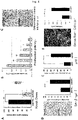
- Figure 3C shows a comparison of Fzd5 protein expression, as measured by Western blotting.
- Figure 3D shows photographs compared for intracellular localization of Fzd5 protein, as observed by immunofluorescent staining.
- the expression of Fzd5 and Ror2 is specific to RECs.
- the respective expression of Fzd5 and Ror2 when detected and quantified, would be effective as an indicator for cell quality evaluation of RECs.
- the newly prepared anti-Fzd5 monoclonal antibody and anti-Ror2 monoclonal antibody are able to detect and quantify their target protein antigens in any technique selected from flow cytometry, Western blotting and immunofluorescent staining.
- the RNA interference method is a technique to examine the function of a target gene by introduction of short RNA (shRNA) having a sequence complementary to target mRNA into cells to thereby disrupt the target mRNA.
- shRNA short RNA
- Figure 4 shows the results obtained from a series of experiments on cell properties in RECs when Fzd5 mRNA was disrupted by shRNA having a sequence complementary to Fzd5 (shFZD5), in comparison with the control group (shCTRL; shRNA having a random sequence not complementary to Fzd5).
- Figure 4A is a graph showing the mRNA level of Fzd5, as quantified by quantitative PCR, after introduction of shFZD5 or shCTRL into RECs.
- shCTRL short hairpin control
- Figure 4B is a graph whose vertical axis plots cell counts in the group forced to express shFZD5 after introduction of shFZD5 or shCTRL into RECs, relative to cell counts in the control group which are set to 1, and whose horizontal axis plots the number of days after shRNA introduction.
- RECs forced to express shFZD5 showed a sudden reduction in their cell count when compared to the control group, thus suggesting that Rzd5 inhibition would induce a reduction in proliferation potency.
- Figure 4C shows images of fat droplets stained with Oil-Red-O at 14 days of culture, after introduction of shFZD5 or shCTRL into RECs and the subsequent induction of their differentiation into fat cells. Fzd5 inhibition was found to induce a reduction in differentiation potency into fat cells, when compared to the control group.
- FIG. 4D shows images stained by x-gal staining after introduction of shFZD5 or shCTRL into RECs, along with a graph which plots the frequency of cells having SA- ⁇ -gal activity in each cell population.
- Figure 4E is a graph showing the mRNA level of p16(INK4a) serving as an indicator for cellular aging, as quantified by quantitative PCR.
- the control group was set to 100, the mRNA level of p16 in shFZD5-receiving RECs was about 300, thus indicating that inhibition of Fzd5 expression induced cellular aging.
- Figure 4F shows images observed for stress fiber formation by intracellular staining with anti-F-actin antibody in each cell population after introduction of shFZD5 or shCTRL into RECs, along with a graph which plots the averaged area (cell size) of individual cells in these respective cell populations.
- the full-length cDNA of Fzd5 was forced to be expressed in RECs to cause constitutive expression of Fzd5 mRNA, and the effect thereof on cell functions was confirmed.
- an expression vector is constructed to carry Fzd5 cDNA and a fluorescent protein GFP (Green Fluorescent Protein) in tandem to thereby allow co-expression of GFP in Fzd5 gene-receiving cells, so that the expression of the introduced gene can be confirmed under a fluorescence microscope.
- GFP Green Fluorescent Protein
- Figure 5A shows photographs observed for morphology of GFP-expressing cells under a fluorescence microscope. These images show cell morphology at 28 days after gene transfer in the cell population transformed with Fzd5 cDNA and GFP (Fzd5) as well as the control group transformed with the GFP gene alone (CTRL).
- Figure 5B is a graph whose vertical axis plots cell counts of Fzd5-expressing RECs, relative to cell counts in the control group which are set to 1, and whose horizontal axis plots the number of days after gene transfer. RECs forced to express Fzd5 were found to retain their proliferation potency for a long period of time, when compared to the control group.
- Fzd5-mediated stimulation of Wnt signaling would be expected to allow long-term culture amplification in a state maintaining the undifferentiated nature.
- the extracellular region of a human Fzd5 antigen was used as an immunogen to immunize host mice and hybridomas were then prepared in accordance with standard procedures, followed by screening with Ba/F3 cells engineered to express the Fzd5 gene, thereby obtaining a novel anti-Fzd5 monoclonal antibody (clone name: 6F5).
- This antibody was used to confirm whether or not the Fzd5 protein was able to be detected by various techniques.
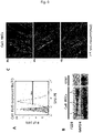
- FIG. 6A shows a histogram whose horizontal axis plots the fluorescence intensity of PE.
- the shaded histogram in the figure represents a negative control where an isotype control was added as a primary antibody, while the open histogram represents the PE fluorescence intensity in the sample stained with 6F5.
- the range indicated with the horizontal bar in the figure represents the range of Fzd5 + cells, while the numerical value represents the positive rate (%).
- Figure 6B shows the results of Western blotting obtained when intracellular proteins were prepared from three different clones of RECs and the Fzd5 protein was detected using 6F5 as a primary antibody.
- intracellular proteins were prepared from the monkey kidney-derived cell line COS7.
- Figure 6C shows images obtained when REC cells were stained with 6F5-biotin as a primary antibody and then fluorescently labeled with streptavidin (SAV)-Alexa 555, followed by observation under a fluorescence microscope.
- SAV streptavidin
- the anti-Fzd5 antibody (6F5) was found to be available for use in all of flow cytometry, Western blotting and immunofluorescent staining.
- a human Ror2 antigen was used as an immunogen to newly prepare an anti-Ror2 antibody (clone name: 7C9).
- Figure 7A shows the results obtained when RECs were stained with 7C9-biotin as a primary antibody and then labeled with SAV-PE, followed by flow cytometry to detect PE fluorescence.
- a sample prepared by adding an isotype control antibody as a primary antibody is used as a negative control.
- Figure 7A is a two-dimensional dot plot whose vertical axis plots FITC fluorescence (RECs are all negative because they are not stained) and whose horizontal axis plots PE fluorescence.
- Figure 7B is an image obtained when RECs were immunostained with 7C9-biotin as a primary antibody and fluorescently labeled with streptavidin-Alexa 488, and then observed for expression of the Ror2 protein under a fluorescence microscope. Most of these RECs were confirmed to express the Ror2 protein.
- Figure 7C shows the results obtained when fresh bone marrow cells were triple stained with LNGFR-APC, Thy1-FITC and Ror2-PE (monoclonal antibodies against their respective antigens), followed by flow cytometry analysis.
- the left panel shows a figure whose vertical axis plots LNGFR expression and whose horizontal axis plots Thy1 expression, and the boxed area represents a LNGFR + Thy1 + cell population containing human MSCs with high frequency.
- the right figure shows the LNGFR + Thy1 + cell population extracted alone.
- the boxed area represents the Ror2-positive area determined on the basis of the negative control, and the numerical value represents the positive rate (%).
- clone 7C9 92.3% of LNGFR + Thy1 + cells are Ror2-positive; and hence clone 7C9 can be used as a selection marker for MSCs to replace LNGFR Thy1.
- the Ror2 protein can be detected and quantified by flow cytometry and immunofluorescent staining ( Figures 7A and 7B ).
- the newly prepared anti-Ror2 can also be used as a marker for MSCs contained in bone marrow ( Figure 7C ).
- the frequency (% content) of positive cells is measured by flow cytometry.
- the frequency (% content) of positive cells may be measured under a fluorescence microscope, instead.
- the described method enables the efficient isolation and culture of human mesenchymal stem cells for use in the treatment of systemic diseases.
- the described method also enables quality evaluation to determine whether or not the resulting cell population is suitable for transplantation and/or exerts efficacy.
- candidates suitable for cell isolation can be provided as reagents for isolation of mesenchymal stem cells by being immobilized on magnetic nanoparticles.
- fluorescent substance-labeled antibodies or cell staining reagents can be provided for practical use.
- Mesenchymal stem cells are not only used as materials for biomaterials, as previously known, but also can be expected to have various applications by taking advantage of their pluripotency, as exemplified by administration to myasthenia gravis, chronic rheumatism and other diseases, as well as co-transplantation as supporting cells to provide a tissue scaffold (niche) for cell therapy required for treatment of severe diseases including spinal cord injury, heart and vascular failure, chronic liver failure and so on.
- tissue scaffold niche
- the use of RECs retaining their migration potency would be expected to provide a therapeutic effect never seen before when applied to metabolic diseases such as systemic bone and cartilage diseases including hypophosphatasia, for which no therapy has been found, as well as GVHD and all other diseases whose treatment requires administration via the intravenous route.
Landscapes
- Life Sciences & Earth Sciences (AREA)
- Health & Medical Sciences (AREA)
- Chemical & Material Sciences (AREA)
- Organic Chemistry (AREA)
- Engineering & Computer Science (AREA)
- Zoology (AREA)
- Wood Science & Technology (AREA)
- Genetics & Genomics (AREA)
- Bioinformatics & Cheminformatics (AREA)
- General Health & Medical Sciences (AREA)
- Biotechnology (AREA)
- Biochemistry (AREA)
- Immunology (AREA)
- General Engineering & Computer Science (AREA)
- Biomedical Technology (AREA)
- Microbiology (AREA)
- Molecular Biology (AREA)
- Proteomics, Peptides & Aminoacids (AREA)
- Cell Biology (AREA)
- Biophysics (AREA)
- Physics & Mathematics (AREA)
- Analytical Chemistry (AREA)
- Hematology (AREA)
- Developmental Biology & Embryology (AREA)
- Medicinal Chemistry (AREA)
- Urology & Nephrology (AREA)
- Rheumatology (AREA)
- Mycology (AREA)
- Chemical Kinetics & Catalysis (AREA)
- General Chemical & Material Sciences (AREA)
- Toxicology (AREA)
- Plant Pathology (AREA)
- Virology (AREA)
- Food Science & Technology (AREA)
- Tropical Medicine & Parasitology (AREA)
- Pathology (AREA)
- General Physics & Mathematics (AREA)
- Micro-Organisms Or Cultivation Processes Thereof (AREA)
- Measuring Or Testing Involving Enzymes Or Micro-Organisms (AREA)
- Peptides Or Proteins (AREA)
Applications Claiming Priority (2)
| Application Number | Priority Date | Filing Date | Title |
|---|---|---|---|
| JP2014157367 | 2014-08-01 | ||
| PCT/JP2015/071770 WO2016017795A1 (ja) | 2014-08-01 | 2015-07-31 | ヒト間葉系幹細胞の品質評価方法、及び、そのためのモノクローナル抗体 |
Publications (3)
| Publication Number | Publication Date |
|---|---|
| EP3176253A1 EP3176253A1 (en) | 2017-06-07 |
| EP3176253A4 EP3176253A4 (en) | 2018-03-21 |
| EP3176253B1 true EP3176253B1 (en) | 2022-08-24 |
Family
ID=55217690
Family Applications (1)
| Application Number | Title | Priority Date | Filing Date |
|---|---|---|---|
| EP15827606.3A Active EP3176253B1 (en) | 2014-08-01 | 2015-07-31 | Method for evaluating quality of human mesenchymal stem cell, and monoclonal antibody for use in said method |
Country Status (8)
| Country | Link |
|---|---|
| US (2) | US20170204374A1 (enExample) |
| EP (1) | EP3176253B1 (enExample) |
| JP (3) | JP6850944B2 (enExample) |
| AU (1) | AU2015297347B2 (enExample) |
| CA (1) | CA2954245C (enExample) |
| ES (1) | ES2927112T3 (enExample) |
| SG (2) | SG10202005439XA (enExample) |
| WO (1) | WO2016017795A1 (enExample) |
Families Citing this family (14)
| Publication number | Priority date | Publication date | Assignee | Title |
|---|---|---|---|---|
| US11293065B2 (en) | 2016-03-14 | 2022-04-05 | Aelan Cell Technologies, Inc. | Compositions and methods for the quality control of stem cell preparations |
| WO2020205732A1 (en) * | 2019-03-29 | 2020-10-08 | The Johns Hopkins University | Grid of responses indicating drug sensitivity |
| CN112094804B (zh) * | 2019-06-18 | 2024-05-14 | 中国医学科学院基础医学研究所 | 一种异质性干细胞群、其制备方法及用途 |
| US12460182B2 (en) | 2019-08-19 | 2025-11-04 | Cellaxia Inc. | Cell-containing pharmaceutical composition |
| WO2021144995A1 (ja) * | 2020-01-16 | 2021-07-22 | PuREC株式会社 | 高純度間葉系幹細胞 |
| US20240018588A1 (en) * | 2020-12-07 | 2024-01-18 | Samsung Life Public Welfare Foundation | Method for selecting mesenchymal stem cells having improved self-maintenance ability, and mesenchymal stem cells selected thereby |
| TW202246491A (zh) * | 2021-01-29 | 2022-12-01 | 國立大學法人北海道大學 | 椎間盤再生用組成物 |
| WO2022177032A1 (ja) | 2021-02-19 | 2022-08-25 | PuREC株式会社 | 骨疾患治療用医薬組成物 |
| JP7368679B2 (ja) * | 2021-09-01 | 2023-10-25 | イミュニティリサーチ株式会社 | 細胞集団同定システム、方法、およびプログラム |
| CN116486909A (zh) * | 2022-01-14 | 2023-07-25 | 天士力干细胞产业平台有限公司 | 一种干细胞质量评价系统 |
| WO2023189485A1 (ja) | 2022-03-31 | 2023-10-05 | 株式会社サイト-ファクト | 間葉系幹細胞の品質評価方法 |
| AU2024354962A1 (en) | 2023-10-02 | 2026-03-12 | National University Corporation Tokai National Higher Education And Research System | Method for managing quality of stem cells by image analysis using ai |
| CN120322547A (zh) * | 2023-10-26 | 2025-07-15 | 京东方科技集团股份有限公司 | 细胞培养方法、筛选方法以及间充质干细胞 |
| WO2025187668A1 (ja) * | 2024-03-06 | 2025-09-12 | 国立研究開発法人産業技術総合研究所 | 骨分化能判定方法 |
Family Cites Families (10)
| Publication number | Priority date | Publication date | Assignee | Title |
|---|---|---|---|---|
| WO2004094641A2 (en) | 2003-04-16 | 2004-11-04 | Wyeth | A novel method of modulating bone-realted activity |
| CA2628221A1 (en) * | 2005-10-31 | 2007-05-10 | Oncomed Pharmaceuticals, Inc. | Anti-frizzled receptor antibodies for treating cancer |
| PE20071309A1 (es) * | 2006-02-17 | 2008-02-13 | Wyeth Corp | Anticuerpos para la modulacion de formacion de huesos |
| WO2009011546A2 (en) | 2007-07-16 | 2009-01-22 | Catholic University Industry Academic Cooperation Foundation | Method for promoting the self-renewal of adult stem cells using mesenchymal stromal cells |
| JP2009060840A (ja) * | 2007-09-06 | 2009-03-26 | Keio Gijuku | ヒト間葉系幹細胞濃縮方法 |
| US20130209415A1 (en) | 2010-06-03 | 2013-08-15 | The Board Of Trustees Of The Leland Stanford Junior University | Purified compositions of cardiovascular progenitor cells |
| US20120087868A1 (en) | 2010-10-08 | 2012-04-12 | Gabriele Todd | Nanoparticle-loaded cells |
| JP2013066414A (ja) * | 2011-09-22 | 2013-04-18 | National Institute Of Advanced Industrial Science & Technology | 胃前駆細胞の表面マーカー |
| JP6463029B2 (ja) * | 2013-08-02 | 2019-01-30 | 有未 伊谷 | ヒト間葉系幹細胞を特異的に認識するモノクローナル抗体並びにこれを用いたヒト間葉系幹細胞の分離及び/または品質評価を行う方法 |
| JP5924750B2 (ja) | 2014-05-01 | 2016-05-25 | iHeart Japan株式会社 | Cd82陽性心筋前駆細胞 |
-
2015
- 2015-07-31 WO PCT/JP2015/071770 patent/WO2016017795A1/ja not_active Ceased
- 2015-07-31 JP JP2016538462A patent/JP6850944B2/ja active Active
- 2015-07-31 ES ES15827606T patent/ES2927112T3/es active Active
- 2015-07-31 SG SG10202005439XA patent/SG10202005439XA/en unknown
- 2015-07-31 EP EP15827606.3A patent/EP3176253B1/en active Active
- 2015-07-31 SG SG11201610795VA patent/SG11201610795VA/en unknown
- 2015-07-31 US US15/321,679 patent/US20170204374A1/en not_active Abandoned
- 2015-07-31 CA CA2954245A patent/CA2954245C/en active Active
- 2015-07-31 AU AU2015297347A patent/AU2015297347B2/en active Active
-
2019
- 2019-10-17 JP JP2019190347A patent/JP6932390B2/ja active Active
-
2021
- 2021-03-02 US US17/190,192 patent/US11441123B2/en active Active
- 2021-03-12 JP JP2021040712A patent/JP2021100414A/ja active Pending
Also Published As
| Publication number | Publication date |
|---|---|
| AU2015297347A1 (en) | 2017-01-19 |
| US20210230553A1 (en) | 2021-07-29 |
| CA2954245C (en) | 2023-07-25 |
| CA2954245A1 (en) | 2016-02-04 |
| JP6850944B2 (ja) | 2021-04-14 |
| JPWO2016017795A1 (ja) | 2017-06-22 |
| EP3176253A4 (en) | 2018-03-21 |
| SG10202005439XA (en) | 2020-07-29 |
| US11441123B2 (en) | 2022-09-13 |
| AU2015297347B2 (en) | 2021-05-27 |
| JP2020072662A (ja) | 2020-05-14 |
| WO2016017795A1 (ja) | 2016-02-04 |
| US20170204374A1 (en) | 2017-07-20 |
| JP6932390B2 (ja) | 2021-09-08 |
| ES2927112T3 (es) | 2022-11-02 |
| EP3176253A1 (en) | 2017-06-07 |
| JP2021100414A (ja) | 2021-07-08 |
| SG11201610795VA (en) | 2017-02-27 |
Similar Documents
| Publication | Publication Date | Title |
|---|---|---|
| EP3176253B1 (en) | Method for evaluating quality of human mesenchymal stem cell, and monoclonal antibody for use in said method | |
| JPWO2016017795A6 (ja) | ヒト間葉系幹細胞の品質評価方法、ヒト間葉系幹細胞の分離、選別及び培養方法並びに増殖の早いヒト間葉系幹細胞の細胞集団 | |
| JP6653689B2 (ja) | 癌幹細胞集団及びその作製方法 | |
| JP6463029B2 (ja) | ヒト間葉系幹細胞を特異的に認識するモノクローナル抗体並びにこれを用いたヒト間葉系幹細胞の分離及び/または品質評価を行う方法 | |
| Haasters et al. | Morphological and immunocytochemical characteristics indicate the yield of early progenitors and represent a quality control for human mesenchymal stem cell culturing | |
| Krohn et al. | CXCR4 receptor positive spheroid forming cells are responsible for tumor invasion in vitro | |
| Raynaud et al. | Comprehensive characterization of mesenchymal stem cells from human placenta and fetal membrane and their response to osteoactivin stimulation | |
| Prestegarden et al. | Glioma cell populations grouped by different cell type markers drive brain tumor growth | |
| Lepore et al. | Identification and enrichment of colony-forming cells from the adult murine pituitary | |
| Lee et al. | Clonal analysis and hierarchy of human bone marrow mesenchymal stem and progenitor cells | |
| CN105420192A (zh) | 外周血中分离富集造血干细胞的方法 | |
| Wang et al. | Enrichment of prostate cancer stem cells from primary prostate cancer cultures of biopsy samples | |
| Panwar et al. | Assessment of long-term in vitro Multiplied human Wharton’s jelly-derived mesenchymal stem cells prior to their Use in clinical administration | |
| Xu et al. | Neural ganglioside GD2+ cells define a subpopulation of mesenchymal stem cells in adult murine bone marrow | |
| JP2008182912A (ja) | 癌幹細胞の培養方法、および癌幹細胞 | |
| Liew et al. | Isolation and culture of adipose-derived stromal cells from subcutaneous fat | |
| Yamamoto et al. | Ex vivo culture of circulating tumor cells using magnetic force‐based coculture on a fibroblast feeder layer | |
| CN105062973B (zh) | 一株携带tp53突变的hpv阴性阴茎鳞癌细胞系及其用途 | |
| Hu et al. | Isolation of stem-like cells from 3-dimensional spheroid cultures | |
| WO2008093886A1 (ja) | Mcf7由来細胞 | |
| Potdar et al. | Establishment and molecular characterization of breast cancer mesenchymal stem cell line derived from human non-metastasis breast cancer tumor | |
| Singla et al. | Isolation and Characterization of Cervical Cancer-Associated Mesenchymal Stem Cells From Primary Tumors Using Explant Culture | |
| Heffernan et al. | PNJ scaffolds promote microenvironmental regulation of glioblastoma stem-like cell enrichment and radioresistance | |
| JP2012196226A (ja) | 癌幹細胞の培養方法、および癌幹細胞 | |
| Bigildeev et al. | 3056–NES-EGFP TRANSGENIC MICE AS A MODEL FOR IN VIVO AND IN VITRO STUDY OF BONE-MARROW-DERIVED MESENCHYMAL STEM CELLS (MSCS) |
Legal Events
| Date | Code | Title | Description |
|---|---|---|---|
| STAA | Information on the status of an ep patent application or granted ep patent |
Free format text: STATUS: THE INTERNATIONAL PUBLICATION HAS BEEN MADE |
|
| 17P | Request for examination filed |
Effective date: 20161220 |
|
| AK | Designated contracting states |
Kind code of ref document: A1 Designated state(s): AL AT BE BG CH CY CZ DE DK EE ES FI FR GB GR HR HU IE IS IT LI LT LU LV MC MK MT NL NO PL PT RO RS SE SI SK SM TR |
|
| AX | Request for extension of the european patent |
Extension state: BA ME |
|
| PUAI | Public reference made under article 153(3) epc to a published international application that has entered the european phase |
Free format text: ORIGINAL CODE: 0009012 |
|
| STAA | Information on the status of an ep patent application or granted ep patent |
Free format text: STATUS: REQUEST FOR EXAMINATION WAS MADE |
|
| DAV | Request for validation of the european patent (deleted) | ||
| DAX | Request for extension of the european patent (deleted) | ||
| A4 | Supplementary search report drawn up and despatched |
Effective date: 20180221 |
|
| RIC1 | Information provided on ipc code assigned before grant |
Ipc: C07K 16/28 20060101ALI20180215BHEP Ipc: C12P 21/08 20060101ALI20180215BHEP Ipc: C12P 1/00 20060101ALI20180215BHEP Ipc: C12N 5/0775 20100101AFI20180215BHEP Ipc: C12Q 1/04 20060101ALI20180215BHEP Ipc: C12N 15/02 20060101ALI20180215BHEP |
|
| STAA | Information on the status of an ep patent application or granted ep patent |
Free format text: STATUS: EXAMINATION IS IN PROGRESS |
|
| 17Q | First examination report despatched |
Effective date: 20191129 |
|
| GRAP | Despatch of communication of intention to grant a patent |
Free format text: ORIGINAL CODE: EPIDOSNIGR1 |
|
| STAA | Information on the status of an ep patent application or granted ep patent |
Free format text: STATUS: GRANT OF PATENT IS INTENDED |
|
| INTG | Intention to grant announced |
Effective date: 20220314 |
|
| GRAS | Grant fee paid |
Free format text: ORIGINAL CODE: EPIDOSNIGR3 |
|
| GRAA | (expected) grant |
Free format text: ORIGINAL CODE: 0009210 |
|
| STAA | Information on the status of an ep patent application or granted ep patent |
Free format text: STATUS: THE PATENT HAS BEEN GRANTED |
|
| RIN1 | Information on inventor provided before grant (corrected) |
Inventor name: IYOKU, YUMI Inventor name: MABUCHI, YO Inventor name: OKANO, HIDEYUKI |
|
| AK | Designated contracting states |
Kind code of ref document: B1 Designated state(s): AL AT BE BG CH CY CZ DE DK EE ES FI FR GB GR HR HU IE IS IT LI LT LU LV MC MK MT NL NO PL PT RO RS SE SI SK SM TR |
|
| REG | Reference to a national code |
Ref country code: CH Ref legal event code: EP |
|
| REG | Reference to a national code |
Ref country code: DE Ref legal event code: R096 Ref document number: 602015080499 Country of ref document: DE |
|
| REG | Reference to a national code |
Ref country code: IE Ref legal event code: FG4D |
|
| REG | Reference to a national code |
Ref country code: AT Ref legal event code: REF Ref document number: 1513672 Country of ref document: AT Kind code of ref document: T Effective date: 20220915 |
|
| REG | Reference to a national code |
Ref country code: ES Ref legal event code: FG2A Ref document number: 2927112 Country of ref document: ES Kind code of ref document: T3 Effective date: 20221102 |
|
| REG | Reference to a national code |
Ref country code: LT Ref legal event code: MG9D |
|
| REG | Reference to a national code |
Ref country code: NL Ref legal event code: MP Effective date: 20220824 |
|
| PG25 | Lapsed in a contracting state [announced via postgrant information from national office to epo] |
Ref country code: SE Free format text: LAPSE BECAUSE OF FAILURE TO SUBMIT A TRANSLATION OF THE DESCRIPTION OR TO PAY THE FEE WITHIN THE PRESCRIBED TIME-LIMIT Effective date: 20220824 Ref country code: RS Free format text: LAPSE BECAUSE OF FAILURE TO SUBMIT A TRANSLATION OF THE DESCRIPTION OR TO PAY THE FEE WITHIN THE PRESCRIBED TIME-LIMIT Effective date: 20220824 Ref country code: PT Free format text: LAPSE BECAUSE OF FAILURE TO SUBMIT A TRANSLATION OF THE DESCRIPTION OR TO PAY THE FEE WITHIN THE PRESCRIBED TIME-LIMIT Effective date: 20221226 Ref country code: NO Free format text: LAPSE BECAUSE OF FAILURE TO SUBMIT A TRANSLATION OF THE DESCRIPTION OR TO PAY THE FEE WITHIN THE PRESCRIBED TIME-LIMIT Effective date: 20221124 Ref country code: NL Free format text: LAPSE BECAUSE OF FAILURE TO SUBMIT A TRANSLATION OF THE DESCRIPTION OR TO PAY THE FEE WITHIN THE PRESCRIBED TIME-LIMIT Effective date: 20220824 Ref country code: LV Free format text: LAPSE BECAUSE OF FAILURE TO SUBMIT A TRANSLATION OF THE DESCRIPTION OR TO PAY THE FEE WITHIN THE PRESCRIBED TIME-LIMIT Effective date: 20220824 Ref country code: LT Free format text: LAPSE BECAUSE OF FAILURE TO SUBMIT A TRANSLATION OF THE DESCRIPTION OR TO PAY THE FEE WITHIN THE PRESCRIBED TIME-LIMIT Effective date: 20220824 Ref country code: FI Free format text: LAPSE BECAUSE OF FAILURE TO SUBMIT A TRANSLATION OF THE DESCRIPTION OR TO PAY THE FEE WITHIN THE PRESCRIBED TIME-LIMIT Effective date: 20220824 |
|
| REG | Reference to a national code |
Ref country code: AT Ref legal event code: MK05 Ref document number: 1513672 Country of ref document: AT Kind code of ref document: T Effective date: 20220824 |
|
| PG25 | Lapsed in a contracting state [announced via postgrant information from national office to epo] |
Ref country code: PL Free format text: LAPSE BECAUSE OF FAILURE TO SUBMIT A TRANSLATION OF THE DESCRIPTION OR TO PAY THE FEE WITHIN THE PRESCRIBED TIME-LIMIT Effective date: 20220824 Ref country code: IS Free format text: LAPSE BECAUSE OF FAILURE TO SUBMIT A TRANSLATION OF THE DESCRIPTION OR TO PAY THE FEE WITHIN THE PRESCRIBED TIME-LIMIT Effective date: 20221224 Ref country code: HR Free format text: LAPSE BECAUSE OF FAILURE TO SUBMIT A TRANSLATION OF THE DESCRIPTION OR TO PAY THE FEE WITHIN THE PRESCRIBED TIME-LIMIT Effective date: 20220824 Ref country code: GR Free format text: LAPSE BECAUSE OF FAILURE TO SUBMIT A TRANSLATION OF THE DESCRIPTION OR TO PAY THE FEE WITHIN THE PRESCRIBED TIME-LIMIT Effective date: 20221125 |
|
| PG25 | Lapsed in a contracting state [announced via postgrant information from national office to epo] |
Ref country code: SM Free format text: LAPSE BECAUSE OF FAILURE TO SUBMIT A TRANSLATION OF THE DESCRIPTION OR TO PAY THE FEE WITHIN THE PRESCRIBED TIME-LIMIT Effective date: 20220824 Ref country code: RO Free format text: LAPSE BECAUSE OF FAILURE TO SUBMIT A TRANSLATION OF THE DESCRIPTION OR TO PAY THE FEE WITHIN THE PRESCRIBED TIME-LIMIT Effective date: 20220824 Ref country code: DK Free format text: LAPSE BECAUSE OF FAILURE TO SUBMIT A TRANSLATION OF THE DESCRIPTION OR TO PAY THE FEE WITHIN THE PRESCRIBED TIME-LIMIT Effective date: 20220824 Ref country code: CZ Free format text: LAPSE BECAUSE OF FAILURE TO SUBMIT A TRANSLATION OF THE DESCRIPTION OR TO PAY THE FEE WITHIN THE PRESCRIBED TIME-LIMIT Effective date: 20220824 Ref country code: AT Free format text: LAPSE BECAUSE OF FAILURE TO SUBMIT A TRANSLATION OF THE DESCRIPTION OR TO PAY THE FEE WITHIN THE PRESCRIBED TIME-LIMIT Effective date: 20220824 |
|
| REG | Reference to a national code |
Ref country code: DE Ref legal event code: R097 Ref document number: 602015080499 Country of ref document: DE |
|
| PG25 | Lapsed in a contracting state [announced via postgrant information from national office to epo] |
Ref country code: SK Free format text: LAPSE BECAUSE OF FAILURE TO SUBMIT A TRANSLATION OF THE DESCRIPTION OR TO PAY THE FEE WITHIN THE PRESCRIBED TIME-LIMIT Effective date: 20220824 Ref country code: EE Free format text: LAPSE BECAUSE OF FAILURE TO SUBMIT A TRANSLATION OF THE DESCRIPTION OR TO PAY THE FEE WITHIN THE PRESCRIBED TIME-LIMIT Effective date: 20220824 |
|
| PG25 | Lapsed in a contracting state [announced via postgrant information from national office to epo] |
Ref country code: AL Free format text: LAPSE BECAUSE OF FAILURE TO SUBMIT A TRANSLATION OF THE DESCRIPTION OR TO PAY THE FEE WITHIN THE PRESCRIBED TIME-LIMIT Effective date: 20220824 |
|
| PLBE | No opposition filed within time limit |
Free format text: ORIGINAL CODE: 0009261 |
|
| STAA | Information on the status of an ep patent application or granted ep patent |
Free format text: STATUS: NO OPPOSITION FILED WITHIN TIME LIMIT |
|
| 26N | No opposition filed |
Effective date: 20230525 |
|
| PG25 | Lapsed in a contracting state [announced via postgrant information from national office to epo] |
Ref country code: SI Free format text: LAPSE BECAUSE OF FAILURE TO SUBMIT A TRANSLATION OF THE DESCRIPTION OR TO PAY THE FEE WITHIN THE PRESCRIBED TIME-LIMIT Effective date: 20220824 |
|
| PG25 | Lapsed in a contracting state [announced via postgrant information from national office to epo] |
Ref country code: MC Free format text: LAPSE BECAUSE OF FAILURE TO SUBMIT A TRANSLATION OF THE DESCRIPTION OR TO PAY THE FEE WITHIN THE PRESCRIBED TIME-LIMIT Effective date: 20220824 |
|
| PG25 | Lapsed in a contracting state [announced via postgrant information from national office to epo] |
Ref country code: MC Free format text: LAPSE BECAUSE OF FAILURE TO SUBMIT A TRANSLATION OF THE DESCRIPTION OR TO PAY THE FEE WITHIN THE PRESCRIBED TIME-LIMIT Effective date: 20220824 |
|
| REG | Reference to a national code |
Ref country code: BE Ref legal event code: MM Effective date: 20230731 |
|
| PG25 | Lapsed in a contracting state [announced via postgrant information from national office to epo] |
Ref country code: LU Free format text: LAPSE BECAUSE OF NON-PAYMENT OF DUE FEES Effective date: 20230731 |
|
| PG25 | Lapsed in a contracting state [announced via postgrant information from national office to epo] |
Ref country code: LU Free format text: LAPSE BECAUSE OF NON-PAYMENT OF DUE FEES Effective date: 20230731 |
|
| REG | Reference to a national code |
Ref country code: IE Ref legal event code: MM4A |
|
| PG25 | Lapsed in a contracting state [announced via postgrant information from national office to epo] |
Ref country code: BE Free format text: LAPSE BECAUSE OF NON-PAYMENT OF DUE FEES Effective date: 20230731 |
|
| PG25 | Lapsed in a contracting state [announced via postgrant information from national office to epo] |
Ref country code: IE Free format text: LAPSE BECAUSE OF NON-PAYMENT OF DUE FEES Effective date: 20230731 |
|
| PG25 | Lapsed in a contracting state [announced via postgrant information from national office to epo] |
Ref country code: IE Free format text: LAPSE BECAUSE OF NON-PAYMENT OF DUE FEES Effective date: 20230731 |
|
| PG25 | Lapsed in a contracting state [announced via postgrant information from national office to epo] |
Ref country code: BG Free format text: LAPSE BECAUSE OF FAILURE TO SUBMIT A TRANSLATION OF THE DESCRIPTION OR TO PAY THE FEE WITHIN THE PRESCRIBED TIME-LIMIT Effective date: 20220824 |
|
| PG25 | Lapsed in a contracting state [announced via postgrant information from national office to epo] |
Ref country code: BG Free format text: LAPSE BECAUSE OF FAILURE TO SUBMIT A TRANSLATION OF THE DESCRIPTION OR TO PAY THE FEE WITHIN THE PRESCRIBED TIME-LIMIT Effective date: 20220824 |
|
| PG25 | Lapsed in a contracting state [announced via postgrant information from national office to epo] |
Ref country code: CY Free format text: LAPSE BECAUSE OF FAILURE TO SUBMIT A TRANSLATION OF THE DESCRIPTION OR TO PAY THE FEE WITHIN THE PRESCRIBED TIME-LIMIT; INVALID AB INITIO Effective date: 20150731 |
|
| PG25 | Lapsed in a contracting state [announced via postgrant information from national office to epo] |
Ref country code: HU Free format text: LAPSE BECAUSE OF FAILURE TO SUBMIT A TRANSLATION OF THE DESCRIPTION OR TO PAY THE FEE WITHIN THE PRESCRIBED TIME-LIMIT; INVALID AB INITIO Effective date: 20150731 |
|
| PGFP | Annual fee paid to national office [announced via postgrant information from national office to epo] |
Ref country code: ES Payment date: 20250826 Year of fee payment: 11 |
|
| PGFP | Annual fee paid to national office [announced via postgrant information from national office to epo] |
Ref country code: DE Payment date: 20250722 Year of fee payment: 11 |
|
| PGFP | Annual fee paid to national office [announced via postgrant information from national office to epo] |
Ref country code: IT Payment date: 20250724 Year of fee payment: 11 |
|
| PGFP | Annual fee paid to national office [announced via postgrant information from national office to epo] |
Ref country code: GB Payment date: 20250722 Year of fee payment: 11 |
|
| PGFP | Annual fee paid to national office [announced via postgrant information from national office to epo] |
Ref country code: FR Payment date: 20250725 Year of fee payment: 11 |
|
| PGFP | Annual fee paid to national office [announced via postgrant information from national office to epo] |
Ref country code: CH Payment date: 20250801 Year of fee payment: 11 |
|
| PG25 | Lapsed in a contracting state [announced via postgrant information from national office to epo] |
Ref country code: TR Free format text: LAPSE BECAUSE OF FAILURE TO SUBMIT A TRANSLATION OF THE DESCRIPTION OR TO PAY THE FEE WITHIN THE PRESCRIBED TIME-LIMIT Effective date: 20220824 |