EP3150207B1 - Effet d'inhibition de composé à faible poids moléculaire sur un cancer et une fibrose - Google Patents
Effet d'inhibition de composé à faible poids moléculaire sur un cancer et une fibrose Download PDFInfo
- Publication number
- EP3150207B1 EP3150207B1 EP15769676.6A EP15769676A EP3150207B1 EP 3150207 B1 EP3150207 B1 EP 3150207B1 EP 15769676 A EP15769676 A EP 15769676A EP 3150207 B1 EP3150207 B1 EP 3150207B1
- Authority
- EP
- European Patent Office
- Prior art keywords
- cells
- alkyl
- alkoxy
- optionally substituted
- liver
- Prior art date
- Legal status (The legal status is an assumption and is not a legal conclusion. Google has not performed a legal analysis and makes no representation as to the accuracy of the status listed.)
- Active
Links
- 201000011510 cancer Diseases 0.000 title claims description 116
- 206010028980 Neoplasm Diseases 0.000 title description 68
- 230000002401 inhibitory effect Effects 0.000 title description 54
- 150000003384 small molecules Chemical class 0.000 title description 41
- 206010016654 Fibrosis Diseases 0.000 title description 35
- 230000004761 fibrosis Effects 0.000 title description 34
- 210000000130 stem cell Anatomy 0.000 claims description 70
- 238000011282 treatment Methods 0.000 claims description 63
- 150000001875 compounds Chemical class 0.000 claims description 54
- 210000004185 liver Anatomy 0.000 claims description 44
- 150000003839 salts Chemical class 0.000 claims description 42
- -1 4-methoxybenzyloxy Chemical group 0.000 claims description 38
- 125000004169 (C1-C6) alkyl group Chemical group 0.000 claims description 35
- 229940126585 therapeutic drug Drugs 0.000 claims description 35
- 201000007270 liver cancer Diseases 0.000 claims description 33
- 208000014018 liver neoplasm Diseases 0.000 claims description 33
- 125000004191 (C1-C6) alkoxy group Chemical group 0.000 claims description 25
- 238000000034 method Methods 0.000 claims description 24
- 125000000449 nitro group Chemical group [O-][N+](*)=O 0.000 claims description 23
- 239000012453 solvate Substances 0.000 claims description 22
- 229910052736 halogen Inorganic materials 0.000 claims description 21
- 150000002367 halogens Chemical class 0.000 claims description 21
- 208000019425 cirrhosis of liver Diseases 0.000 claims description 18
- 125000000882 C2-C6 alkenyl group Chemical group 0.000 claims description 15
- 125000004093 cyano group Chemical group *C#N 0.000 claims description 15
- 125000004890 (C1-C6) alkylamino group Chemical group 0.000 claims description 13
- 125000006577 C1-C6 hydroxyalkyl group Chemical group 0.000 claims description 13
- 125000003118 aryl group Chemical group 0.000 claims description 13
- 125000001188 haloalkyl group Chemical group 0.000 claims description 13
- 125000000033 alkoxyamino group Chemical group 0.000 claims description 12
- 125000004438 haloalkoxy group Chemical group 0.000 claims description 12
- 125000001072 heteroaryl group Chemical group 0.000 claims description 12
- 125000005113 hydroxyalkoxy group Chemical group 0.000 claims description 12
- 239000004305 biphenyl Substances 0.000 claims description 10
- 235000010290 biphenyl Nutrition 0.000 claims description 10
- ZUOUZKKEUPVFJK-UHFFFAOYSA-N phenylbenzene Natural products C1=CC=CC=C1C1=CC=CC=C1 ZUOUZKKEUPVFJK-UHFFFAOYSA-N 0.000 claims description 10
- 125000001424 substituent group Chemical group 0.000 claims description 10
- 125000005389 trialkylsiloxy group Chemical group 0.000 claims description 10
- 229910052801 chlorine Inorganic materials 0.000 claims description 9
- 229910052731 fluorine Inorganic materials 0.000 claims description 8
- CBOIHMRHGLHBPB-UHFFFAOYSA-N hydroxymethyl Chemical compound O[CH2] CBOIHMRHGLHBPB-UHFFFAOYSA-N 0.000 claims description 6
- 125000005913 (C3-C6) cycloalkyl group Chemical group 0.000 claims description 5
- 125000003601 C2-C6 alkynyl group Chemical group 0.000 claims description 5
- 125000006323 alkenyl amino group Chemical group 0.000 claims description 5
- 125000005036 alkoxyphenyl group Chemical group 0.000 claims description 5
- 125000006319 alkynyl amino group Chemical group 0.000 claims description 5
- 125000000392 cycloalkenyl group Chemical group 0.000 claims description 5
- 125000005020 hydroxyalkenyl group Chemical group 0.000 claims description 5
- 125000005016 hydroxyalkynyl group Chemical group 0.000 claims description 5
- 229910052739 hydrogen Inorganic materials 0.000 claims description 4
- 210000004027 cell Anatomy 0.000 description 177
- 239000000203 mixture Substances 0.000 description 75
- 229940079593 drug Drugs 0.000 description 49
- 239000003814 drug Substances 0.000 description 49
- 239000000243 solution Substances 0.000 description 40
- 108060000903 Beta-catenin Proteins 0.000 description 34
- 102000015735 Beta-catenin Human genes 0.000 description 34
- LFQSCWFLJHTTHZ-UHFFFAOYSA-N Ethanol Chemical compound CCO LFQSCWFLJHTTHZ-UHFFFAOYSA-N 0.000 description 33
- XEKOWRVHYACXOJ-UHFFFAOYSA-N Ethyl acetate Chemical compound CCOC(C)=O XEKOWRVHYACXOJ-UHFFFAOYSA-N 0.000 description 33
- 230000002829 reductive effect Effects 0.000 description 33
- YMWUJEATGCHHMB-UHFFFAOYSA-N Dichloromethane Chemical compound ClCCl YMWUJEATGCHHMB-UHFFFAOYSA-N 0.000 description 32
- 102000004887 Transforming Growth Factor beta Human genes 0.000 description 31
- 108090001012 Transforming Growth Factor beta Proteins 0.000 description 31
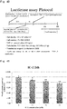
- 238000003670 luciferase enzyme activity assay Methods 0.000 description 27
- VLKZOEOYAKHREP-UHFFFAOYSA-N n-Hexane Chemical compound CCCCCC VLKZOEOYAKHREP-UHFFFAOYSA-N 0.000 description 27
- IAZDPXIOMUYVGZ-UHFFFAOYSA-N Dimethylsulphoxide Chemical compound CS(C)=O IAZDPXIOMUYVGZ-UHFFFAOYSA-N 0.000 description 25
- 210000004500 stellate cell Anatomy 0.000 description 24
- KCXVZYZYPLLWCC-UHFFFAOYSA-N EDTA Chemical compound OC(=O)CN(CC(O)=O)CCN(CC(O)=O)CC(O)=O KCXVZYZYPLLWCC-UHFFFAOYSA-N 0.000 description 22
- 238000006243 chemical reaction Methods 0.000 description 22
- 230000035755 proliferation Effects 0.000 description 21
- 108091003079 Bovine Serum Albumin Proteins 0.000 description 20
- 230000000694 effects Effects 0.000 description 20
- 239000012091 fetal bovine serum Substances 0.000 description 20
- 102100032912 CD44 antigen Human genes 0.000 description 19
- 101000868273 Homo sapiens CD44 antigen Proteins 0.000 description 19
- 206010009944 Colon cancer Diseases 0.000 description 18
- 208000029742 colonic neoplasm Diseases 0.000 description 18
- XLYOFNOQVPJJNP-UHFFFAOYSA-N water Substances O XLYOFNOQVPJJNP-UHFFFAOYSA-N 0.000 description 18
- 239000006144 Dulbecco’s modified Eagle's medium Substances 0.000 description 16
- FAPWRFPIFSIZLT-UHFFFAOYSA-M Sodium chloride Chemical compound [Na+].[Cl-] FAPWRFPIFSIZLT-UHFFFAOYSA-M 0.000 description 16
- 230000000259 anti-tumor effect Effects 0.000 description 16
- 238000005516 engineering process Methods 0.000 description 16
- 238000010898 silica gel chromatography Methods 0.000 description 16
- VZGDMQKNWNREIO-UHFFFAOYSA-N tetrachloromethane Chemical compound ClC(Cl)(Cl)Cl VZGDMQKNWNREIO-UHFFFAOYSA-N 0.000 description 16
- 210000001519 tissue Anatomy 0.000 description 16
- 239000000047 product Substances 0.000 description 13
- 239000006228 supernatant Substances 0.000 description 13
- 241000699670 Mus sp. Species 0.000 description 12
- 230000015572 biosynthetic process Effects 0.000 description 12
- 239000012044 organic layer Substances 0.000 description 12
- 239000000126 substance Substances 0.000 description 12
- 239000007832 Na2SO4 Substances 0.000 description 11
- PMZURENOXWZQFD-UHFFFAOYSA-L Sodium Sulfate Chemical compound [Na+].[Na+].[O-]S([O-])(=O)=O PMZURENOXWZQFD-UHFFFAOYSA-L 0.000 description 11
- 239000011541 reaction mixture Substances 0.000 description 11
- 229910052938 sodium sulfate Inorganic materials 0.000 description 11
- 238000002835 absorbance Methods 0.000 description 10
- 230000003247 decreasing effect Effects 0.000 description 10
- BDAGIHXWWSANSR-UHFFFAOYSA-N methanoic acid Natural products OC=O BDAGIHXWWSANSR-UHFFFAOYSA-N 0.000 description 10
- 239000007787 solid Substances 0.000 description 10
- 206010041823 squamous cell carcinoma Diseases 0.000 description 10
- HEMHJVSKTPXQMS-UHFFFAOYSA-M Sodium hydroxide Chemical compound [OH-].[Na+] HEMHJVSKTPXQMS-UHFFFAOYSA-M 0.000 description 9
- 230000005764 inhibitory process Effects 0.000 description 9
- 241000283707 Capra Species 0.000 description 8
- PMMYEEVYMWASQN-DMTCNVIQSA-N Hydroxyproline Chemical compound O[C@H]1CN[C@H](C(O)=O)C1 PMMYEEVYMWASQN-DMTCNVIQSA-N 0.000 description 8
- 239000005089 Luciferase Substances 0.000 description 8
- JGFZNNIVVJXRND-UHFFFAOYSA-N N,N-Diisopropylethylamine (DIPEA) Chemical compound CCN(C(C)C)C(C)C JGFZNNIVVJXRND-UHFFFAOYSA-N 0.000 description 8
- 238000010586 diagram Methods 0.000 description 8
- 201000010099 disease Diseases 0.000 description 8
- 208000037265 diseases, disorders, signs and symptoms Diseases 0.000 description 8
- PMMYEEVYMWASQN-UHFFFAOYSA-N dl-hydroxyproline Natural products OC1C[NH2+]C(C([O-])=O)C1 PMMYEEVYMWASQN-UHFFFAOYSA-N 0.000 description 8
- 229960002591 hydroxyproline Drugs 0.000 description 8
- 230000001965 increasing effect Effects 0.000 description 8
- 230000002700 inhibitory effect on cancer Effects 0.000 description 8
- 230000001225 therapeutic effect Effects 0.000 description 8
- FGMPLJWBKKVCDB-UHFFFAOYSA-N trans-L-hydroxy-proline Natural products ON1CCCC1C(O)=O FGMPLJWBKKVCDB-UHFFFAOYSA-N 0.000 description 8
- HQWTUOLCGKIECB-XZWHSSHBSA-N (6S,9aS)-6-[(4-hydroxyphenyl)methyl]-8-(1-naphthalenylmethyl)-4,7-dioxo-N-(phenylmethyl)-3,6,9,9a-tetrahydro-2H-pyrazino[1,2-a]pyrimidine-1-carboxamide Chemical compound C1=CC(O)=CC=C1C[C@H]1C(=O)N(CC=2C3=CC=CC=C3C=CC=2)C[C@H]2N1C(=O)CCN2C(=O)NCC1=CC=CC=C1 HQWTUOLCGKIECB-XZWHSSHBSA-N 0.000 description 7
- 108060001084 Luciferase Proteins 0.000 description 7
- 239000004480 active ingredient Substances 0.000 description 7
- 239000007864 aqueous solution Substances 0.000 description 7
- 238000004113 cell culture Methods 0.000 description 7
- 239000002771 cell marker Substances 0.000 description 7
- 239000003795 chemical substances by application Substances 0.000 description 7
- 239000005457 ice water Substances 0.000 description 7
- 239000003550 marker Substances 0.000 description 7
- 238000010172 mouse model Methods 0.000 description 7
- 108090000623 proteins and genes Proteins 0.000 description 7
- 238000003753 real-time PCR Methods 0.000 description 7
- 239000002904 solvent Substances 0.000 description 7
- QTBSBXVTEAMEQO-UHFFFAOYSA-N Acetic acid Chemical class CC(O)=O QTBSBXVTEAMEQO-UHFFFAOYSA-N 0.000 description 6
- QUMCIHKVKQYNPA-RUZDIDTESA-N C1(CCCCC1)CN1[C@@H](C=2N(C=3C=NC(=NC1=3)NC1=C(C=C(C(=O)NC3CCN(CC3)C)C=C1)OC)C(=NN=2)C)CC Chemical compound C1(CCCCC1)CN1[C@@H](C=2N(C=3C=NC(=NC1=3)NC1=C(C=C(C(=O)NC3CCN(CC3)C)C=C1)OC)C(=NN=2)C)CC QUMCIHKVKQYNPA-RUZDIDTESA-N 0.000 description 6
- 102000018651 Epithelial Cell Adhesion Molecule Human genes 0.000 description 6
- 108010066687 Epithelial Cell Adhesion Molecule Proteins 0.000 description 6
- 101000610551 Homo sapiens Prominin-1 Proteins 0.000 description 6
- 101000800116 Homo sapiens Thy-1 membrane glycoprotein Proteins 0.000 description 6
- VEXZGXHMUGYJMC-UHFFFAOYSA-N Hydrochloric acid Chemical class Cl VEXZGXHMUGYJMC-UHFFFAOYSA-N 0.000 description 6
- OKKJLVBELUTLKV-UHFFFAOYSA-N Methanol Chemical compound OC OKKJLVBELUTLKV-UHFFFAOYSA-N 0.000 description 6
- 102100040120 Prominin-1 Human genes 0.000 description 6
- QAOWNCQODCNURD-UHFFFAOYSA-N Sulfuric acid Chemical class OS(O)(=O)=O QAOWNCQODCNURD-UHFFFAOYSA-N 0.000 description 6
- 102100033523 Thy-1 membrane glycoprotein Human genes 0.000 description 6
- YXFVVABEGXRONW-UHFFFAOYSA-N Toluene Chemical compound CC1=CC=CC=C1 YXFVVABEGXRONW-UHFFFAOYSA-N 0.000 description 6
- ZMANZCXQSJIPKH-UHFFFAOYSA-N Triethylamine Chemical class CCN(CC)CC ZMANZCXQSJIPKH-UHFFFAOYSA-N 0.000 description 6
- 108090000631 Trypsin Proteins 0.000 description 6
- 102000004142 Trypsin Human genes 0.000 description 6
- 230000037396 body weight Effects 0.000 description 6
- 230000012010 growth Effects 0.000 description 6
- 239000007788 liquid Substances 0.000 description 6
- 238000005259 measurement Methods 0.000 description 6
- 238000003752 polymerase chain reaction Methods 0.000 description 6
- AYEKOFBPNLCAJY-UHFFFAOYSA-O thiamine pyrophosphate Chemical compound CC1=C(CCOP(O)(=O)OP(O)(O)=O)SC=[N+]1CC1=CN=C(C)N=C1N AYEKOFBPNLCAJY-UHFFFAOYSA-O 0.000 description 6
- 239000012588 trypsin Substances 0.000 description 6
- 210000004881 tumor cell Anatomy 0.000 description 6
- OSWFIVFLDKOXQC-UHFFFAOYSA-N 4-(3-methoxyphenyl)aniline Chemical class COC1=CC=CC(C=2C=CC(N)=CC=2)=C1 OSWFIVFLDKOXQC-UHFFFAOYSA-N 0.000 description 5
- 102100033601 Collagen alpha-1(I) chain Human genes 0.000 description 5
- 239000004378 Glycyrrhizin Substances 0.000 description 5
- UIIMBOGNXHQVGW-UHFFFAOYSA-M Sodium bicarbonate Chemical compound [Na+].OC([O-])=O UIIMBOGNXHQVGW-UHFFFAOYSA-M 0.000 description 5
- 125000000217 alkyl group Chemical group 0.000 description 5
- 108010029483 alpha 1 Chain Collagen Type I Proteins 0.000 description 5
- 239000000872 buffer Substances 0.000 description 5
- 230000003833 cell viability Effects 0.000 description 5
- 230000001419 dependent effect Effects 0.000 description 5
- HPNMFZURTQLUMO-UHFFFAOYSA-N diethylamine Chemical compound CCNCC HPNMFZURTQLUMO-UHFFFAOYSA-N 0.000 description 5
- 238000002474 experimental method Methods 0.000 description 5
- 235000019253 formic acid Nutrition 0.000 description 5
- LPLVUJXQOOQHMX-UHFFFAOYSA-N glycyrrhetinic acid glycoside Natural products C1CC(C2C(C3(CCC4(C)CCC(C)(CC4C3=CC2=O)C(O)=O)C)(C)CC2)(C)C2C(C)(C)C1OC1OC(C(O)=O)C(O)C(O)C1OC1OC(C(O)=O)C(O)C(O)C1O LPLVUJXQOOQHMX-UHFFFAOYSA-N 0.000 description 5
- 229960004949 glycyrrhizic acid Drugs 0.000 description 5
- UYRUBYNTXSDKQT-UHFFFAOYSA-N glycyrrhizic acid Natural products CC1(C)C(CCC2(C)C1CCC3(C)C2C(=O)C=C4C5CC(C)(CCC5(C)CCC34C)C(=O)O)OC6OC(C(O)C(O)C6OC7OC(O)C(O)C(O)C7C(=O)O)C(=O)O UYRUBYNTXSDKQT-UHFFFAOYSA-N 0.000 description 5
- 235000019410 glycyrrhizin Nutrition 0.000 description 5
- LPLVUJXQOOQHMX-QWBHMCJMSA-N glycyrrhizinic acid Chemical compound O([C@@H]1[C@@H](O)[C@H](O)[C@H](O[C@@H]1O[C@@H]1C([C@H]2[C@]([C@@H]3[C@@]([C@@]4(CC[C@@]5(C)CC[C@@](C)(C[C@H]5C4=CC3=O)C(O)=O)C)(C)CC2)(C)CC1)(C)C)C(O)=O)[C@@H]1O[C@H](C(O)=O)[C@@H](O)[C@H](O)[C@H]1O LPLVUJXQOOQHMX-QWBHMCJMSA-N 0.000 description 5
- 239000006166 lysate Substances 0.000 description 5
- 239000012139 lysis buffer Substances 0.000 description 5
- 239000002504 physiological saline solution Substances 0.000 description 5
- 235000018102 proteins Nutrition 0.000 description 5
- 102000004169 proteins and genes Human genes 0.000 description 5
- 238000011160 research Methods 0.000 description 5
- 208000024891 symptom Diseases 0.000 description 5
- 238000012360 testing method Methods 0.000 description 5
- 230000036962 time dependent Effects 0.000 description 5
- KAFZOLYKKCWUBI-HPMAGDRPSA-N (2s)-2-[[(2s)-2-[[(2s)-1-[(2s)-3-amino-2-[[(2s)-2-[[(2s)-2-(3-cyclohexylpropanoylamino)-4-methylpentanoyl]amino]-5-methylhexanoyl]amino]propanoyl]pyrrolidine-2-carbonyl]amino]-5-(diaminomethylideneamino)pentanoyl]amino]butanediamide Chemical compound N([C@@H](CC(C)C)C(=O)N[C@@H](CCC(C)C)C(=O)N[C@@H](CN)C(=O)N1[C@@H](CCC1)C(=O)N[C@@H](CCCN=C(N)N)C(=O)N[C@@H](CC(N)=O)C(N)=O)C(=O)CCC1CCCCC1 KAFZOLYKKCWUBI-HPMAGDRPSA-N 0.000 description 4
- IJGRMHOSHXDMSA-UHFFFAOYSA-N Atomic nitrogen Chemical compound N#N IJGRMHOSHXDMSA-UHFFFAOYSA-N 0.000 description 4
- 101100285903 Drosophila melanogaster Hsc70-2 gene Proteins 0.000 description 4
- 241000699666 Mus <mouse, genus> Species 0.000 description 4
- 230000002378 acidificating effect Effects 0.000 description 4
- 125000003545 alkoxy group Chemical group 0.000 description 4
- 238000004458 analytical method Methods 0.000 description 4
- 239000002246 antineoplastic agent Substances 0.000 description 4
- 229940041181 antineoplastic drug Drugs 0.000 description 4
- 238000003556 assay Methods 0.000 description 4
- 238000001574 biopsy Methods 0.000 description 4
- 230000008859 change Effects 0.000 description 4
- 238000001514 detection method Methods 0.000 description 4
- 235000019439 ethyl acetate Nutrition 0.000 description 4
- 102000006602 glyceraldehyde-3-phosphate dehydrogenase Human genes 0.000 description 4
- 108020004445 glyceraldehyde-3-phosphate dehydrogenase Proteins 0.000 description 4
- 230000007062 hydrolysis Effects 0.000 description 4
- 238000006460 hydrolysis reaction Methods 0.000 description 4
- 238000003384 imaging method Methods 0.000 description 4
- 230000008569 process Effects 0.000 description 4
- RWWYLEGWBNMMLJ-YSOARWBDSA-N remdesivir Chemical compound NC1=NC=NN2C1=CC=C2[C@]1([C@@H]([C@@H]([C@H](O1)CO[P@](=O)(OC1=CC=CC=C1)N[C@H](C(=O)OCC(CC)CC)C)O)O)C#N RWWYLEGWBNMMLJ-YSOARWBDSA-N 0.000 description 4
- 229910000030 sodium bicarbonate Inorganic materials 0.000 description 4
- FRJJJAKBRKABFA-TYFAACHXSA-N (4r,6s)-6-[(e)-2-[6-chloro-4-(4-fluorophenyl)-2-propan-2-ylquinolin-3-yl]ethenyl]-4-hydroxyoxan-2-one Chemical compound C(\[C@H]1OC(=O)C[C@H](O)C1)=C/C=1C(C(C)C)=NC2=CC=C(Cl)C=C2C=1C1=CC=C(F)C=C1 FRJJJAKBRKABFA-TYFAACHXSA-N 0.000 description 3
- OISVCGZHLKNMSJ-UHFFFAOYSA-N 2,6-dimethylpyridine Chemical class CC1=CC=CC(C)=N1 OISVCGZHLKNMSJ-UHFFFAOYSA-N 0.000 description 3
- YEMVHZBFYJVLJE-UHFFFAOYSA-N 2-[4-(methoxymethoxy)phenyl]acetic acid Chemical compound COCOC1=CC=C(CC(O)=O)C=C1 YEMVHZBFYJVLJE-UHFFFAOYSA-N 0.000 description 3
- CBWHOCVRDWMACN-UHFFFAOYSA-N 2-[4-[(4-methoxyphenyl)methoxy]phenyl]acetic acid Chemical compound C1=CC(OC)=CC=C1COC1=CC=C(CC(O)=O)C=C1 CBWHOCVRDWMACN-UHFFFAOYSA-N 0.000 description 3
- XQWYZOLEWPQRES-UHFFFAOYSA-N 2-[4-[[tert-butyl(dimethyl)silyl]oxymethyl]phenyl]acetic acid Chemical compound CC(C)(C)[Si](C)(C)OCC1=CC=C(CC(O)=O)C=C1 XQWYZOLEWPQRES-UHFFFAOYSA-N 0.000 description 3
- ZDXPYRJPNDTMRX-VKHMYHEASA-N L-glutamine Chemical compound OC(=O)[C@@H](N)CCC(N)=O ZDXPYRJPNDTMRX-VKHMYHEASA-N 0.000 description 3
- MUBZPKHOEPUJKR-UHFFFAOYSA-N Oxalic acid Chemical class OC(=O)C(O)=O MUBZPKHOEPUJKR-UHFFFAOYSA-N 0.000 description 3
- KDLHZDBZIXYQEI-UHFFFAOYSA-N Palladium on carbon Substances [Pd] KDLHZDBZIXYQEI-UHFFFAOYSA-N 0.000 description 3
- DTQVDTLACAAQTR-UHFFFAOYSA-N Trifluoroacetic acid Chemical class OC(=O)C(F)(F)F DTQVDTLACAAQTR-UHFFFAOYSA-N 0.000 description 3
- 231100000480 WST assay Toxicity 0.000 description 3
- SRVFFFJZQVENJC-IHRRRGAJSA-N aloxistatin Chemical compound CCOC(=O)[C@H]1O[C@@H]1C(=O)N[C@@H](CC(C)C)C(=O)NCCC(C)C SRVFFFJZQVENJC-IHRRRGAJSA-N 0.000 description 3
- 235000001014 amino acid Nutrition 0.000 description 3
- 150000001413 amino acids Chemical class 0.000 description 3
- AGEZXYOZHKGVCM-UHFFFAOYSA-N benzyl bromide Chemical compound BrCC1=CC=CC=C1 AGEZXYOZHKGVCM-UHFFFAOYSA-N 0.000 description 3
- OSVHLUXLWQLPIY-KBAYOESNSA-N butyl 2-[(6aR,9R,10aR)-1-hydroxy-9-(hydroxymethyl)-6,6-dimethyl-6a,7,8,9,10,10a-hexahydrobenzo[c]chromen-3-yl]-2-methylpropanoate Chemical compound C(CCC)OC(C(C)(C)C1=CC(=C2[C@H]3[C@H](C(OC2=C1)(C)C)CC[C@H](C3)CO)O)=O OSVHLUXLWQLPIY-KBAYOESNSA-N 0.000 description 3
- 239000006285 cell suspension Substances 0.000 description 3
- 230000005754 cellular signaling Effects 0.000 description 3
- 239000003153 chemical reaction reagent Substances 0.000 description 3
- KRKNYBCHXYNGOX-UHFFFAOYSA-N citric acid Chemical class OC(=O)CC(O)(C(O)=O)CC(O)=O KRKNYBCHXYNGOX-UHFFFAOYSA-N 0.000 description 3
- 229940125904 compound 1 Drugs 0.000 description 3
- 238000001816 cooling Methods 0.000 description 3
- 239000002552 dosage form Substances 0.000 description 3
- 238000011156 evaluation Methods 0.000 description 3
- RAXXELZNTBOGNW-UHFFFAOYSA-N imidazole Natural products C1=CNC=N1 RAXXELZNTBOGNW-UHFFFAOYSA-N 0.000 description 3
- 238000000338 in vitro Methods 0.000 description 3
- 208000019423 liver disease Diseases 0.000 description 3
- 210000005228 liver tissue Anatomy 0.000 description 3
- 238000004519 manufacturing process Methods 0.000 description 3
- 239000002609 medium Substances 0.000 description 3
- 125000000956 methoxy group Chemical group [H]C([H])([H])O* 0.000 description 3
- 229910052757 nitrogen Inorganic materials 0.000 description 3
- 229940002612 prodrug Drugs 0.000 description 3
- 239000000651 prodrug Substances 0.000 description 3
- XJMOSONTPMZWPB-UHFFFAOYSA-M propidium iodide Chemical compound [I-].[I-].C12=CC(N)=CC=C2C2=CC=C(N)C=C2[N+](CCC[N+](C)(CC)CC)=C1C1=CC=CC=C1 XJMOSONTPMZWPB-UHFFFAOYSA-M 0.000 description 3
- 230000000384 rearing effect Effects 0.000 description 3
- 229910021642 ultra pure water Inorganic materials 0.000 description 3
- 239000012498 ultrapure water Substances 0.000 description 3
- HZAXFHJVJLSVMW-UHFFFAOYSA-N 2-Aminoethan-1-ol Chemical class NCCO HZAXFHJVJLSVMW-UHFFFAOYSA-N 0.000 description 2
- XQXPVVBIMDBYFF-UHFFFAOYSA-N 4-hydroxyphenylacetic acid Chemical compound OC(=O)CC1=CC=C(O)C=C1 XQXPVVBIMDBYFF-UHFFFAOYSA-N 0.000 description 2
- 206010002091 Anaesthesia Diseases 0.000 description 2
- 201000009030 Carcinoma Diseases 0.000 description 2
- HEDRZPFGACZZDS-UHFFFAOYSA-N Chloroform Chemical compound ClC(Cl)Cl HEDRZPFGACZZDS-UHFFFAOYSA-N 0.000 description 2
- XJUZRXYOEPSWMB-UHFFFAOYSA-N Chloromethyl methyl ether Chemical compound COCCl XJUZRXYOEPSWMB-UHFFFAOYSA-N 0.000 description 2
- 102000008186 Collagen Human genes 0.000 description 2
- 108010035532 Collagen Proteins 0.000 description 2
- 108020004414 DNA Proteins 0.000 description 2
- 108010067770 Endopeptidase K Proteins 0.000 description 2
- 238000012413 Fluorescence activated cell sorting analysis Methods 0.000 description 2
- VZCYOOQTPOCHFL-OWOJBTEDSA-N Fumaric acid Chemical class OC(=O)\C=C\C(O)=O VZCYOOQTPOCHFL-OWOJBTEDSA-N 0.000 description 2
- 239000007821 HATU Substances 0.000 description 2
- 101500025419 Homo sapiens Epidermal growth factor Proteins 0.000 description 2
- 229930182816 L-glutamine Natural products 0.000 description 2
- 239000012097 Lipofectamine 2000 Substances 0.000 description 2
- TWRXJAOTZQYOKJ-UHFFFAOYSA-L Magnesium chloride Chemical compound [Mg+2].[Cl-].[Cl-] TWRXJAOTZQYOKJ-UHFFFAOYSA-L 0.000 description 2
- 206010027476 Metastases Diseases 0.000 description 2
- AFVFQIVMOAPDHO-UHFFFAOYSA-N Methanesulfonic acid Chemical compound CS(O)(=O)=O AFVFQIVMOAPDHO-UHFFFAOYSA-N 0.000 description 2
- 229930040373 Paraformaldehyde Natural products 0.000 description 2
- ISWSIDIOOBJBQZ-UHFFFAOYSA-N Phenol Chemical compound OC1=CC=CC=C1 ISWSIDIOOBJBQZ-UHFFFAOYSA-N 0.000 description 2
- NBIIXXVUZAFLBC-UHFFFAOYSA-N Phosphoric acid Chemical class OP(O)(O)=O NBIIXXVUZAFLBC-UHFFFAOYSA-N 0.000 description 2
- JUJWROOIHBZHMG-UHFFFAOYSA-N Pyridine Chemical class C1=CC=NC=C1 JUJWROOIHBZHMG-UHFFFAOYSA-N 0.000 description 2
- 208000000453 Skin Neoplasms Diseases 0.000 description 2
- DBMJMQXJHONAFJ-UHFFFAOYSA-M Sodium laurylsulphate Chemical compound [Na+].CCCCCCCCCCCCOS([O-])(=O)=O DBMJMQXJHONAFJ-UHFFFAOYSA-M 0.000 description 2
- 238000000692 Student's t-test Methods 0.000 description 2
- 208000023915 Ureteral Neoplasms Diseases 0.000 description 2
- 206010046392 Ureteric cancer Diseases 0.000 description 2
- 125000003342 alkenyl group Chemical group 0.000 description 2
- 125000003277 amino group Chemical group 0.000 description 2
- 230000037005 anaesthesia Effects 0.000 description 2
- 239000012298 atmosphere Substances 0.000 description 2
- IUGQFMIATSVYLK-UHFFFAOYSA-N benzyl 2-(4-hydroxyphenyl)acetate Chemical compound C1=CC(O)=CC=C1CC(=O)OCC1=CC=CC=C1 IUGQFMIATSVYLK-UHFFFAOYSA-N 0.000 description 2
- 239000008280 blood Substances 0.000 description 2
- 210000004369 blood Anatomy 0.000 description 2
- 125000004432 carbon atom Chemical group C* 0.000 description 2
- UHZZMRAGKVHANO-UHFFFAOYSA-M chlormequat chloride Chemical compound [Cl-].C[N+](C)(C)CCCl UHZZMRAGKVHANO-UHFFFAOYSA-M 0.000 description 2
- 229940061627 chloromethyl methyl ether Drugs 0.000 description 2
- 238000010367 cloning Methods 0.000 description 2
- 229920001436 collagen Polymers 0.000 description 2
- 210000002808 connective tissue Anatomy 0.000 description 2
- PAFZNILMFXTMIY-UHFFFAOYSA-N cyclohexylamine Chemical class NC1CCCCC1 PAFZNILMFXTMIY-UHFFFAOYSA-N 0.000 description 2
- 230000034994 death Effects 0.000 description 2
- 238000010908 decantation Methods 0.000 description 2
- 239000000835 fiber Substances 0.000 description 2
- 238000001914 filtration Methods 0.000 description 2
- 125000002541 furyl group Chemical group 0.000 description 2
- 210000002216 heart Anatomy 0.000 description 2
- 210000003494 hepatocyte Anatomy 0.000 description 2
- 239000012456 homogeneous solution Substances 0.000 description 2
- 229940116978 human epidermal growth factor Drugs 0.000 description 2
- 150000002430 hydrocarbons Chemical group 0.000 description 2
- 125000002887 hydroxy group Chemical group [H]O* 0.000 description 2
- 230000001939 inductive effect Effects 0.000 description 2
- 239000003112 inhibitor Substances 0.000 description 2
- 230000000160 inhibitory effect on fibrosis Effects 0.000 description 2
- 238000002347 injection Methods 0.000 description 2
- 239000007924 injection Substances 0.000 description 2
- YDNLNVZZTACNJX-UHFFFAOYSA-N isocyanatomethylbenzene Chemical compound O=C=NCC1=CC=CC=C1 YDNLNVZZTACNJX-UHFFFAOYSA-N 0.000 description 2
- 210000003734 kidney Anatomy 0.000 description 2
- 239000010410 layer Substances 0.000 description 2
- 210000004072 lung Anatomy 0.000 description 2
- 201000001441 melanoma Diseases 0.000 description 2
- 210000002901 mesenchymal stem cell Anatomy 0.000 description 2
- 229910052751 metal Inorganic materials 0.000 description 2
- 239000002184 metal Substances 0.000 description 2
- 230000009401 metastasis Effects 0.000 description 2
- XGDZEDRBLVIUMX-UHFFFAOYSA-N methyl 2-(4-hydroxyphenyl)acetate Chemical compound COC(=O)CC1=CC=C(O)C=C1 XGDZEDRBLVIUMX-UHFFFAOYSA-N 0.000 description 2
- 125000002496 methyl group Chemical group [H]C([H])([H])* 0.000 description 2
- 150000007522 mineralic acids Chemical class 0.000 description 2
- 125000001624 naphthyl group Chemical group 0.000 description 2
- GVUGOAYIVIDWIO-UFWWTJHBSA-N nepidermin Chemical compound C([C@@H](C(=O)N[C@@H]([C@@H](C)CC)C(=O)NCC(=O)N[C@@H](CCC(O)=O)C(=O)N[C@@H](CCCNC(N)=N)C(=O)N[C@@H](CS)C(=O)N[C@@H](CCC(N)=O)C(=O)N[C@@H](CC=1C=CC(O)=CC=1)C(=O)N[C@@H](CCCNC(N)=N)C(=O)N[C@@H](CC(O)=O)C(=O)N[C@@H](CC(C)C)C(=O)N[C@@H](CCCCN)C(=O)N[C@@H](CC=1C2=CC=CC=C2NC=1)C(=O)N[C@@H](CC=1C2=CC=CC=C2NC=1)C(=O)N[C@@H](CCC(O)=O)C(=O)N[C@@H](CC(C)C)C(=O)N[C@@H](CCCNC(N)=N)C(O)=O)NC(=O)CNC(=O)[C@@H](NC(=O)[C@@H](NC(=O)[C@H](CS)NC(=O)[C@H](CC(N)=O)NC(=O)[C@H](CS)NC(=O)[C@H](C)NC(=O)[C@H](CC=1C=CC(O)=CC=1)NC(=O)[C@H](CCCCN)NC(=O)[C@H](CC(O)=O)NC(=O)[C@H](CC(C)C)NC(=O)[C@H](C)NC(=O)[C@H](CCC(O)=O)NC(=O)[C@@H](NC(=O)[C@H](CC=1C=CC(O)=CC=1)NC(=O)[C@H](CCSC)NC(=O)[C@H](CS)NC(=O)[C@@H](NC(=O)CNC(=O)[C@H](CC(O)=O)NC(=O)[C@H](CC=1NC=NC=1)NC(=O)[C@H](CC(C)C)NC(=O)[C@H](CS)NC(=O)[C@H](CC=1C=CC(O)=CC=1)NC(=O)CNC(=O)[C@H](CC(O)=O)NC(=O)[C@H](CC=1NC=NC=1)NC(=O)[C@H](CO)NC(=O)[C@H](CC(C)C)NC(=O)[C@H]1N(CCC1)C(=O)[C@H](CS)NC(=O)[C@H](CCC(O)=O)NC(=O)[C@H](CO)NC(=O)[C@H](CC(O)=O)NC(=O)[C@H](CO)NC(=O)[C@@H](N)CC(N)=O)C(C)C)[C@@H](C)CC)C(C)C)C(C)C)C1=CC=C(O)C=C1 GVUGOAYIVIDWIO-UFWWTJHBSA-N 0.000 description 2
- 238000010899 nucleation Methods 0.000 description 2
- 210000000056 organ Anatomy 0.000 description 2
- 150000007524 organic acids Chemical class 0.000 description 2
- 150000007530 organic bases Chemical class 0.000 description 2
- 239000003960 organic solvent Substances 0.000 description 2
- 229920002866 paraformaldehyde Polymers 0.000 description 2
- 239000008188 pellet Substances 0.000 description 2
- 239000008194 pharmaceutical composition Substances 0.000 description 2
- 230000000144 pharmacologic effect Effects 0.000 description 2
- 125000001997 phenyl group Chemical group [H]C1=C([H])C([H])=C(*)C([H])=C1[H] 0.000 description 2
- WLJVXDMOQOGPHL-UHFFFAOYSA-N phenylacetic acid Chemical compound OC(=O)CC1=CC=CC=C1 WLJVXDMOQOGPHL-UHFFFAOYSA-N 0.000 description 2
- XNGIFLGASWRNHJ-UHFFFAOYSA-N phthalic acid Chemical class OC(=O)C1=CC=CC=C1C(O)=O XNGIFLGASWRNHJ-UHFFFAOYSA-N 0.000 description 2
- BWHMMNNQKKPAPP-UHFFFAOYSA-L potassium carbonate Chemical compound [K+].[K+].[O-]C([O-])=O BWHMMNNQKKPAPP-UHFFFAOYSA-L 0.000 description 2
- 230000000069 prophylactic effect Effects 0.000 description 2
- 239000012144 protein assay dye reagent concentrate Substances 0.000 description 2
- RXWNCPJZOCPEPQ-NVWDDTSBSA-N puromycin Chemical compound C1=CC(OC)=CC=C1C[C@H](N)C(=O)N[C@H]1[C@@H](O)[C@H](N2C3=NC=NC(=C3N=C2)N(C)C)O[C@@H]1CO RXWNCPJZOCPEPQ-NVWDDTSBSA-N 0.000 description 2
- 238000011002 quantification Methods 0.000 description 2
- 238000011084 recovery Methods 0.000 description 2
- 230000019491 signal transduction Effects 0.000 description 2
- 238000013424 sirius red staining Methods 0.000 description 2
- 210000003491 skin Anatomy 0.000 description 2
- 201000000849 skin cancer Diseases 0.000 description 2
- 239000011780 sodium chloride Substances 0.000 description 2
- 238000001228 spectrum Methods 0.000 description 2
- 239000000758 substrate Substances 0.000 description 2
- 239000013589 supplement Substances 0.000 description 2
- 238000003786 synthesis reaction Methods 0.000 description 2
- JOXIMZWYDAKGHI-UHFFFAOYSA-N toluene-4-sulfonic acid Chemical compound CC1=CC=C(S(O)(=O)=O)C=C1 JOXIMZWYDAKGHI-UHFFFAOYSA-N 0.000 description 2
- SLGRAIAQIAUZAQ-UHFFFAOYSA-N toxoflavin Chemical compound CN1N=CN=C2C1=NC(=O)N(C)C2=O SLGRAIAQIAUZAQ-UHFFFAOYSA-N 0.000 description 2
- VZCYOOQTPOCHFL-UHFFFAOYSA-N trans-butenedioic acid Chemical class OC(=O)C=CC(O)=O VZCYOOQTPOCHFL-UHFFFAOYSA-N 0.000 description 2
- 238000001890 transfection Methods 0.000 description 2
- 238000011269 treatment regimen Methods 0.000 description 2
- GETQZCLCWQTVFV-UHFFFAOYSA-N trimethylamine Chemical class CN(C)C GETQZCLCWQTVFV-UHFFFAOYSA-N 0.000 description 2
- 239000000439 tumor marker Substances 0.000 description 2
- 230000000007 visual effect Effects 0.000 description 2
- HDTRYLNUVZCQOY-UHFFFAOYSA-N α-D-glucopyranosyl-α-D-glucopyranoside Natural products OC1C(O)C(O)C(CO)OC1OC1C(O)C(O)C(O)C(CO)O1 HDTRYLNUVZCQOY-UHFFFAOYSA-N 0.000 description 1
- YBADLXQNJCMBKR-UHFFFAOYSA-N (4-nitrophenyl)acetic acid Chemical compound OC(=O)CC1=CC=C([N+]([O-])=O)C=C1 YBADLXQNJCMBKR-UHFFFAOYSA-N 0.000 description 1
- BJEPYKJPYRNKOW-REOHCLBHSA-N (S)-malic acid Chemical compound OC(=O)[C@@H](O)CC(O)=O BJEPYKJPYRNKOW-REOHCLBHSA-N 0.000 description 1
- MOHYOXXOKFQHDC-UHFFFAOYSA-N 1-(chloromethyl)-4-methoxybenzene Chemical compound COC1=CC=C(CCl)C=C1 MOHYOXXOKFQHDC-UHFFFAOYSA-N 0.000 description 1
- 125000004973 1-butenyl group Chemical group C(=CCC)* 0.000 description 1
- 125000006039 1-hexenyl group Chemical group 0.000 description 1
- 125000004066 1-hydroxyethyl group Chemical group [H]OC([H])([*])C([H])([H])[H] 0.000 description 1
- SQAINHDHICKHLX-UHFFFAOYSA-N 1-naphthaldehyde Chemical compound C1=CC=C2C(C=O)=CC=CC2=C1 SQAINHDHICKHLX-UHFFFAOYSA-N 0.000 description 1
- 125000001637 1-naphthyl group Chemical group [H]C1=C([H])C([H])=C2C(*)=C([H])C([H])=C([H])C2=C1[H] 0.000 description 1
- 125000006023 1-pentenyl group Chemical group 0.000 description 1
- 125000006017 1-propenyl group Chemical group 0.000 description 1
- YBYIRNPNPLQARY-UHFFFAOYSA-N 1H-indene Natural products C1=CC=C2CC=CC2=C1 YBYIRNPNPLQARY-UHFFFAOYSA-N 0.000 description 1
- FWZBPBKAANKOJQ-UHFFFAOYSA-N 2-[4-(hydroxymethyl)phenyl]acetic acid Chemical compound OCC1=CC=C(CC(O)=O)C=C1 FWZBPBKAANKOJQ-UHFFFAOYSA-N 0.000 description 1
- 125000004974 2-butenyl group Chemical group C(C=CC)* 0.000 description 1
- 125000000954 2-hydroxyethyl group Chemical group [H]C([*])([H])C([H])([H])O[H] 0.000 description 1
- 125000006020 2-methyl-1-propenyl group Chemical group 0.000 description 1
- XWKFPIODWVPXLX-UHFFFAOYSA-N 2-methyl-5-methylpyridine Chemical class CC1=CC=C(C)N=C1 XWKFPIODWVPXLX-UHFFFAOYSA-N 0.000 description 1
- BSKHPKMHTQYZBB-UHFFFAOYSA-N 2-methylpyridine Chemical class CC1=CC=CC=N1 BSKHPKMHTQYZBB-UHFFFAOYSA-N 0.000 description 1
- 125000001622 2-naphthyl group Chemical group [H]C1=C([H])C([H])=C2C([H])=C(*)C([H])=C([H])C2=C1[H] 0.000 description 1
- 125000006024 2-pentenyl group Chemical group 0.000 description 1
- 125000003903 2-propenyl group Chemical group [H]C([*])([H])C([H])=C([H])[H] 0.000 description 1
- LINBWYYLPWJQHE-UHFFFAOYSA-N 3-(9h-fluoren-9-ylmethoxycarbonylamino)propanoic acid Chemical compound C1=CC=C2C(COC(=O)NCCC(=O)O)C3=CC=CC=C3C2=C1 LINBWYYLPWJQHE-UHFFFAOYSA-N 0.000 description 1
- BMYNFMYTOJXKLE-UHFFFAOYSA-N 3-azaniumyl-2-hydroxypropanoate Chemical class NCC(O)C(O)=O BMYNFMYTOJXKLE-UHFFFAOYSA-N 0.000 description 1
- 125000004975 3-butenyl group Chemical group C(CC=C)* 0.000 description 1
- 125000006041 3-hexenyl group Chemical group 0.000 description 1
- 125000003119 4-methyl-3-pentenyl group Chemical group [H]\C(=C(/C([H])([H])[H])C([H])([H])[H])C([H])([H])C([H])([H])* 0.000 description 1
- 125000006043 5-hexenyl group Chemical group 0.000 description 1
- HBAQYPYDRFILMT-UHFFFAOYSA-N 8-[3-(1-cyclopropylpyrazol-4-yl)-1H-pyrazolo[4,3-d]pyrimidin-5-yl]-3-methyl-3,8-diazabicyclo[3.2.1]octan-2-one Chemical class C1(CC1)N1N=CC(=C1)C1=NNC2=C1N=C(N=C2)N1C2C(N(CC1CC2)C)=O HBAQYPYDRFILMT-UHFFFAOYSA-N 0.000 description 1
- NLXLAEXVIDQMFP-UHFFFAOYSA-N Ammonia chloride Chemical class [NH4+].[Cl-] NLXLAEXVIDQMFP-UHFFFAOYSA-N 0.000 description 1
- 239000004475 Arginine Substances 0.000 description 1
- 206010004146 Basal cell carcinoma Diseases 0.000 description 1
- 206010005003 Bladder cancer Diseases 0.000 description 1
- 241000283690 Bos taurus Species 0.000 description 1
- 238000009010 Bradford assay Methods 0.000 description 1
- 208000003174 Brain Neoplasms Diseases 0.000 description 1
- 206010006187 Breast cancer Diseases 0.000 description 1
- 208000026310 Breast neoplasm Diseases 0.000 description 1
- 238000011740 C57BL/6 mouse Methods 0.000 description 1
- 239000004215 Carbon black (E152) Substances 0.000 description 1
- 241000700199 Cavia porcellus Species 0.000 description 1
- 241000282693 Cercopithecidae Species 0.000 description 1
- 235000005956 Cosmos caudatus Nutrition 0.000 description 1
- 241000699800 Cricetinae Species 0.000 description 1
- FEWJPZIEWOKRBE-JCYAYHJZSA-N Dextrotartaric acid Chemical class OC(=O)[C@H](O)[C@@H](O)C(O)=O FEWJPZIEWOKRBE-JCYAYHJZSA-N 0.000 description 1
- XBPCUCUWBYBCDP-UHFFFAOYSA-N Dicyclohexylamine Chemical class C1CCCCC1NC1CCCCC1 XBPCUCUWBYBCDP-UHFFFAOYSA-N 0.000 description 1
- 102100035426 DnaJ homolog subfamily B member 7 Human genes 0.000 description 1
- 241000283073 Equus caballus Species 0.000 description 1
- 208000000461 Esophageal Neoplasms Diseases 0.000 description 1
- 101150095705 FBXW7 gene Proteins 0.000 description 1
- 241000282326 Felis catus Species 0.000 description 1
- 108090000379 Fibroblast growth factor 2 Proteins 0.000 description 1
- 102100024785 Fibroblast growth factor 2 Human genes 0.000 description 1
- 208000032612 Glial tumor Diseases 0.000 description 1
- 201000010915 Glioblastoma multiforme Diseases 0.000 description 1
- 206010018338 Glioma Diseases 0.000 description 1
- WQZGKKKJIJFFOK-GASJEMHNSA-N Glucose Natural products OC[C@H]1OC(O)[C@H](O)[C@@H](O)[C@@H]1O WQZGKKKJIJFFOK-GASJEMHNSA-N 0.000 description 1
- WHUUTDBJXJRKMK-UHFFFAOYSA-N Glutamic acid Chemical class OC(=O)C(N)CCC(O)=O WHUUTDBJXJRKMK-UHFFFAOYSA-N 0.000 description 1
- 208000002250 Hematologic Neoplasms Diseases 0.000 description 1
- 101000804114 Homo sapiens DnaJ homolog subfamily B member 7 Proteins 0.000 description 1
- 101001052035 Homo sapiens Fibroblast growth factor 2 Proteins 0.000 description 1
- 208000005016 Intestinal Neoplasms Diseases 0.000 description 1
- 239000007836 KH2PO4 Substances 0.000 description 1
- 208000008839 Kidney Neoplasms Diseases 0.000 description 1
- AHLPHDHHMVZTML-BYPYZUCNSA-N L-Ornithine Chemical class NCCC[C@H](N)C(O)=O AHLPHDHHMVZTML-BYPYZUCNSA-N 0.000 description 1
- ODKSFYDXXFIFQN-BYPYZUCNSA-P L-argininium(2+) Chemical class NC(=[NH2+])NCCC[C@H]([NH3+])C(O)=O ODKSFYDXXFIFQN-BYPYZUCNSA-P 0.000 description 1
- CKLJMWTZIZZHCS-REOHCLBHSA-N L-aspartic acid Chemical class OC(=O)[C@@H](N)CC(O)=O CKLJMWTZIZZHCS-REOHCLBHSA-N 0.000 description 1
- WHUUTDBJXJRKMK-VKHMYHEASA-N L-glutamic acid Chemical class OC(=O)[C@@H](N)CCC(O)=O WHUUTDBJXJRKMK-VKHMYHEASA-N 0.000 description 1
- KDXKERNSBIXSRK-YFKPBYRVSA-N L-lysine Chemical class NCCCC[C@H](N)C(O)=O KDXKERNSBIXSRK-YFKPBYRVSA-N 0.000 description 1
- 239000005517 L01XE01 - Imatinib Substances 0.000 description 1
- 206010058467 Lung neoplasm malignant Diseases 0.000 description 1
- 206010025323 Lymphomas Diseases 0.000 description 1
- KDXKERNSBIXSRK-UHFFFAOYSA-N Lysine Chemical class NCCCCC(N)C(O)=O KDXKERNSBIXSRK-UHFFFAOYSA-N 0.000 description 1
- 239000004472 Lysine Chemical class 0.000 description 1
- 208000032271 Malignant tumor of penis Diseases 0.000 description 1
- 241001465754 Metazoa Species 0.000 description 1
- 229910004013 NO 2 Inorganic materials 0.000 description 1
- GRYLNZFGIOXLOG-UHFFFAOYSA-N Nitric acid Chemical class O[N+]([O-])=O GRYLNZFGIOXLOG-UHFFFAOYSA-N 0.000 description 1
- AHLPHDHHMVZTML-UHFFFAOYSA-N Orn-delta-NH2 Chemical class NCCCC(N)C(O)=O AHLPHDHHMVZTML-UHFFFAOYSA-N 0.000 description 1
- UTJLXEIPEHZYQJ-UHFFFAOYSA-N Ornithine Chemical class OC(=O)C(C)CCCN UTJLXEIPEHZYQJ-UHFFFAOYSA-N 0.000 description 1
- 241000283973 Oryctolagus cuniculus Species 0.000 description 1
- 206010033128 Ovarian cancer Diseases 0.000 description 1
- 206010061535 Ovarian neoplasm Diseases 0.000 description 1
- 241000282577 Pan troglodytes Species 0.000 description 1
- 206010061902 Pancreatic neoplasm Diseases 0.000 description 1
- 241001494479 Pecora Species 0.000 description 1
- 208000002471 Penile Neoplasms Diseases 0.000 description 1
- 206010034299 Penile cancer Diseases 0.000 description 1
- QGMRQYFBGABWDR-UHFFFAOYSA-M Pentobarbital sodium Chemical compound [Na+].CCCC(C)C1(CC)C(=O)NC(=O)[N-]C1=O QGMRQYFBGABWDR-UHFFFAOYSA-M 0.000 description 1
- 241000009328 Perro Species 0.000 description 1
- OFOBLEOULBTSOW-UHFFFAOYSA-N Propanedioic acid Chemical class OC(=O)CC(O)=O OFOBLEOULBTSOW-UHFFFAOYSA-N 0.000 description 1
- 206010060862 Prostate cancer Diseases 0.000 description 1
- 208000000236 Prostatic Neoplasms Diseases 0.000 description 1
- 241000700159 Rattus Species 0.000 description 1
- 206010038389 Renal cancer Diseases 0.000 description 1
- CGNLCCVKSWNSDG-UHFFFAOYSA-N SYBR Green I Chemical compound CN(C)CCCN(CCC)C1=CC(C=C2N(C3=CC=CC=C3S2)C)=C2C=CC=CC2=[N+]1C1=CC=CC=C1 CGNLCCVKSWNSDG-UHFFFAOYSA-N 0.000 description 1
- 208000004337 Salivary Gland Neoplasms Diseases 0.000 description 1
- 206010061934 Salivary gland cancer Diseases 0.000 description 1
- 206010039491 Sarcoma Diseases 0.000 description 1
- 208000034189 Sclerosis Diseases 0.000 description 1
- 208000005718 Stomach Neoplasms Diseases 0.000 description 1
- KDYFGRWQOYBRFD-UHFFFAOYSA-N Succinic acid Chemical class OC(=O)CCC(O)=O KDYFGRWQOYBRFD-UHFFFAOYSA-N 0.000 description 1
- 241000282898 Sus scrofa Species 0.000 description 1
- FEWJPZIEWOKRBE-UHFFFAOYSA-N Tartaric acid Chemical class [H+].[H+].[O-]C(=O)C(O)C(O)C([O-])=O FEWJPZIEWOKRBE-UHFFFAOYSA-N 0.000 description 1
- 208000024313 Testicular Neoplasms Diseases 0.000 description 1
- 206010057644 Testis cancer Diseases 0.000 description 1
- 208000024770 Thyroid neoplasm Diseases 0.000 description 1
- HDTRYLNUVZCQOY-WSWWMNSNSA-N Trehalose Natural products O[C@@H]1[C@@H](O)[C@@H](O)[C@@H](CO)O[C@@H]1O[C@@H]1[C@H](O)[C@@H](O)[C@@H](O)[C@@H](CO)O1 HDTRYLNUVZCQOY-WSWWMNSNSA-N 0.000 description 1
- GSEJCLTVZPLZKY-UHFFFAOYSA-N Triethanolamine Chemical class OCCN(CCO)CCO GSEJCLTVZPLZKY-UHFFFAOYSA-N 0.000 description 1
- 208000007097 Urinary Bladder Neoplasms Diseases 0.000 description 1
- 208000002495 Uterine Neoplasms Diseases 0.000 description 1
- 201000005188 adrenal gland cancer Diseases 0.000 description 1
- 208000024447 adrenal gland neoplasm Diseases 0.000 description 1
- 230000002411 adverse Effects 0.000 description 1
- 229910052783 alkali metal Inorganic materials 0.000 description 1
- 229910052784 alkaline earth metal Inorganic materials 0.000 description 1
- HDTRYLNUVZCQOY-LIZSDCNHSA-N alpha,alpha-trehalose Chemical compound O[C@@H]1[C@@H](O)[C@H](O)[C@@H](CO)O[C@@H]1O[C@@H]1[C@H](O)[C@@H](O)[C@H](O)[C@@H](CO)O1 HDTRYLNUVZCQOY-LIZSDCNHSA-N 0.000 description 1
- BJEPYKJPYRNKOW-UHFFFAOYSA-N alpha-hydroxysuccinic acid Natural products OC(=O)C(O)CC(O)=O BJEPYKJPYRNKOW-UHFFFAOYSA-N 0.000 description 1
- AZDRQVAHHNSJOQ-UHFFFAOYSA-N alumane Chemical class [AlH3] AZDRQVAHHNSJOQ-UHFFFAOYSA-N 0.000 description 1
- 229910000147 aluminium phosphate Chemical class 0.000 description 1
- 230000001668 ameliorated effect Effects 0.000 description 1
- 150000003863 ammonium salts Chemical class 0.000 description 1
- 230000003444 anaesthetic effect Effects 0.000 description 1
- 238000010171 animal model Methods 0.000 description 1
- 125000000129 anionic group Chemical group 0.000 description 1
- 230000002300 anti-fibrosis Effects 0.000 description 1
- 230000003510 anti-fibrotic effect Effects 0.000 description 1
- 230000009876 antimalignant effect Effects 0.000 description 1
- ODKSFYDXXFIFQN-UHFFFAOYSA-N arginine Natural products OC(=O)C(N)CCCNC(N)=N ODKSFYDXXFIFQN-UHFFFAOYSA-N 0.000 description 1
- 125000002029 aromatic hydrocarbon group Chemical group 0.000 description 1
- 235000003704 aspartic acid Nutrition 0.000 description 1
- 159000000009 barium salts Chemical class 0.000 description 1
- 208000003373 basosquamous carcinoma Diseases 0.000 description 1
- JUHORIMYRDESRB-UHFFFAOYSA-N benzathine Chemical class C=1C=CC=CC=1CNCCNCC1=CC=CC=C1 JUHORIMYRDESRB-UHFFFAOYSA-N 0.000 description 1
- SRSXLGNVWSONIS-UHFFFAOYSA-N benzenesulfonic acid Chemical compound OS(=O)(=O)C1=CC=CC=C1 SRSXLGNVWSONIS-UHFFFAOYSA-N 0.000 description 1
- 229940092714 benzenesulfonic acid Drugs 0.000 description 1
- 125000004618 benzofuryl group Chemical group O1C(=CC2=C1C=CC=C2)* 0.000 description 1
- 125000004196 benzothienyl group Chemical group S1C(=CC2=C1C=CC=C2)* 0.000 description 1
- OQFSQFPPLPISGP-UHFFFAOYSA-N beta-carboxyaspartic acid Natural products OC(=O)C(N)C(C(O)=O)C(O)=O OQFSQFPPLPISGP-UHFFFAOYSA-N 0.000 description 1
- 125000002619 bicyclic group Chemical group 0.000 description 1
- 201000009036 biliary tract cancer Diseases 0.000 description 1
- 208000020790 biliary tract neoplasm Diseases 0.000 description 1
- 230000004071 biological effect Effects 0.000 description 1
- 229910052794 bromium Inorganic materials 0.000 description 1
- 125000005997 bromomethyl group Chemical group 0.000 description 1
- KDYFGRWQOYBRFD-NUQCWPJISA-N butanedioic acid Chemical class O[14C](=O)CC[14C](O)=O KDYFGRWQOYBRFD-NUQCWPJISA-N 0.000 description 1
- 159000000007 calcium salts Chemical class 0.000 description 1
- 239000002775 capsule Substances 0.000 description 1
- 125000003178 carboxy group Chemical group [H]OC(*)=O 0.000 description 1
- 125000002091 cationic group Chemical group 0.000 description 1
- 239000013592 cell lysate Substances 0.000 description 1
- 230000004663 cell proliferation Effects 0.000 description 1
- 239000002458 cell surface marker Substances 0.000 description 1
- 230000001413 cellular effect Effects 0.000 description 1
- 210000003169 central nervous system Anatomy 0.000 description 1
- 238000005119 centrifugation Methods 0.000 description 1
- 208000026106 cerebrovascular disease Diseases 0.000 description 1
- VDQQXEISLMTGAB-UHFFFAOYSA-N chloramine T Chemical compound [Na+].CC1=CC=C(S(=O)(=O)[N-]Cl)C=C1 VDQQXEISLMTGAB-UHFFFAOYSA-N 0.000 description 1
- 125000002603 chloroethyl group Chemical group [H]C([*])([H])C([H])([H])Cl 0.000 description 1
- 125000004218 chloromethyl group Chemical group [H]C([H])(Cl)* 0.000 description 1
- 208000006990 cholangiocarcinoma Diseases 0.000 description 1
- 230000007882 cirrhosis Effects 0.000 description 1
- 238000012258 culturing Methods 0.000 description 1
- 208000035250 cutaneous malignant susceptibility to 1 melanoma Diseases 0.000 description 1
- 150000001925 cycloalkenes Chemical class 0.000 description 1
- 230000002950 deficient Effects 0.000 description 1
- 238000011161 development Methods 0.000 description 1
- 238000003745 diagnosis Methods 0.000 description 1
- 125000004987 dibenzofuryl group Chemical group C1(=CC=CC=2OC3=C(C21)C=CC=C3)* 0.000 description 1
- 125000006003 dichloroethyl group Chemical group 0.000 description 1
- 125000004772 dichloromethyl group Chemical group [H]C(Cl)(Cl)* 0.000 description 1
- ZBCBWPMODOFKDW-UHFFFAOYSA-N diethanolamine Chemical class OCCNCCO ZBCBWPMODOFKDW-UHFFFAOYSA-N 0.000 description 1
- 230000004069 differentiation Effects 0.000 description 1
- 125000006001 difluoroethyl group Chemical group 0.000 description 1
- 125000001028 difluoromethyl group Chemical group [H]C(F)(F)* 0.000 description 1
- 238000007865 diluting Methods 0.000 description 1
- MKRTXPORKIRPDG-UHFFFAOYSA-N diphenylphosphoryl azide Chemical compound C=1C=CC=CC=1P(=O)(N=[N+]=[N-])C1=CC=CC=C1 MKRTXPORKIRPDG-UHFFFAOYSA-N 0.000 description 1
- BNIILDVGGAEEIG-UHFFFAOYSA-L disodium hydrogen phosphate Chemical compound [Na+].[Na+].OP([O-])([O-])=O BNIILDVGGAEEIG-UHFFFAOYSA-L 0.000 description 1
- 229910000397 disodium phosphate Inorganic materials 0.000 description 1
- 238000001647 drug administration Methods 0.000 description 1
- 239000013583 drug formulation Substances 0.000 description 1
- 201000004101 esophageal cancer Diseases 0.000 description 1
- WHQLQYRFIHPMNA-UHFFFAOYSA-N ethyl acetate;oxolane Chemical compound C1CCOC1.CCOC(C)=O WHQLQYRFIHPMNA-UHFFFAOYSA-N 0.000 description 1
- 125000001495 ethyl group Chemical group [H]C([H])([H])C([H])([H])* 0.000 description 1
- 125000000031 ethylamino group Chemical group [H]C([H])([H])C([H])([H])N([H])[*] 0.000 description 1
- 230000001747 exhibiting effect Effects 0.000 description 1
- 239000000706 filtrate Substances 0.000 description 1
- 125000003983 fluorenyl group Chemical group C1(=CC=CC=2C3=CC=CC=C3CC12)* 0.000 description 1
- 238000001943 fluorescence-activated cell sorting Methods 0.000 description 1
- 125000003784 fluoroethyl group Chemical group [H]C([H])(F)C([H])([H])* 0.000 description 1
- 125000004785 fluoromethoxy group Chemical group [H]C([H])(F)O* 0.000 description 1
- 125000004216 fluoromethyl group Chemical group [H]C([H])(F)* 0.000 description 1
- 235000013305 food Nutrition 0.000 description 1
- 239000001530 fumaric acid Chemical class 0.000 description 1
- 235000011087 fumaric acid Nutrition 0.000 description 1
- 206010017758 gastric cancer Diseases 0.000 description 1
- 208000005017 glioblastoma Diseases 0.000 description 1
- 239000008103 glucose Substances 0.000 description 1
- 235000013922 glutamic acid Nutrition 0.000 description 1
- 239000004220 glutamic acid Chemical class 0.000 description 1
- ZDXPYRJPNDTMRX-UHFFFAOYSA-N glutamine Natural products OC(=O)C(N)CCC(N)=O ZDXPYRJPNDTMRX-UHFFFAOYSA-N 0.000 description 1
- 239000008187 granular material Substances 0.000 description 1
- 238000000227 grinding Methods 0.000 description 1
- 230000009422 growth inhibiting effect Effects 0.000 description 1
- 201000003911 head and neck carcinoma Diseases 0.000 description 1
- 208000019622 heart disease Diseases 0.000 description 1
- 230000002440 hepatic effect Effects 0.000 description 1
- 125000005842 heteroatom Chemical group 0.000 description 1
- ACGUYXCXAPNIKK-UHFFFAOYSA-N hexachlorophene Chemical compound OC1=C(Cl)C=C(Cl)C(Cl)=C1CC1=C(O)C(Cl)=CC(Cl)=C1Cl ACGUYXCXAPNIKK-UHFFFAOYSA-N 0.000 description 1
- 229960004068 hexachlorophene Drugs 0.000 description 1
- 125000003707 hexyloxy group Chemical group [H]C([H])([H])C([H])([H])C([H])([H])C([H])([H])C([H])([H])C([H])([H])O* 0.000 description 1
- 229930195733 hydrocarbon Natural products 0.000 description 1
- 125000004029 hydroxymethyl group Chemical group [H]OC([H])([H])* 0.000 description 1
- KTUFNOKKBVMGRW-UHFFFAOYSA-N imatinib Chemical compound C1CN(C)CCN1CC1=CC=C(C(=O)NC=2C=C(NC=3N=C(C=CN=3)C=3C=NC=CC=3)C(C)=CC=2)C=C1 KTUFNOKKBVMGRW-UHFFFAOYSA-N 0.000 description 1
- 229960002411 imatinib Drugs 0.000 description 1
- 125000002883 imidazolyl group Chemical group 0.000 description 1
- 238000001727 in vivo Methods 0.000 description 1
- 125000003454 indenyl group Chemical group C1(C=CC2=CC=CC=C12)* 0.000 description 1
- 230000006698 induction Effects 0.000 description 1
- 230000000977 initiatory effect Effects 0.000 description 1
- 201000002313 intestinal cancer Diseases 0.000 description 1
- 238000007918 intramuscular administration Methods 0.000 description 1
- 238000007912 intraperitoneal administration Methods 0.000 description 1
- 238000001990 intravenous administration Methods 0.000 description 1
- 125000000959 isobutyl group Chemical group [H]C([H])([H])C([H])(C([H])([H])[H])C([H])([H])* 0.000 description 1
- 125000001449 isopropyl group Chemical group [H]C([H])([H])C([H])(*)C([H])([H])[H] 0.000 description 1
- 201000010982 kidney cancer Diseases 0.000 description 1
- 201000005202 lung cancer Diseases 0.000 description 1
- 208000020816 lung neoplasm Diseases 0.000 description 1
- 229910001629 magnesium chloride Inorganic materials 0.000 description 1
- 159000000003 magnesium salts Chemical class 0.000 description 1
- VZCYOOQTPOCHFL-UPHRSURJSA-N maleic acid Chemical class OC(=O)\C=C/C(O)=O VZCYOOQTPOCHFL-UPHRSURJSA-N 0.000 description 1
- 239000011976 maleic acid Chemical class 0.000 description 1
- 239000001630 malic acid Substances 0.000 description 1
- 235000011090 malic acid Nutrition 0.000 description 1
- 208000015486 malignant pancreatic neoplasm Diseases 0.000 description 1
- 208000020984 malignant renal pelvis neoplasm Diseases 0.000 description 1
- 241001515942 marmosets Species 0.000 description 1
- 239000000463 material Substances 0.000 description 1
- 230000007246 mechanism Effects 0.000 description 1
- 230000004060 metabolic process Effects 0.000 description 1
- 229940098779 methanesulfonic acid Drugs 0.000 description 1
- 229920000609 methyl cellulose Polymers 0.000 description 1
- 125000000250 methylamino group Chemical group [H]N(*)C([H])([H])[H] 0.000 description 1
- 239000001923 methylcellulose Substances 0.000 description 1
- 235000010981 methylcellulose Nutrition 0.000 description 1
- 238000002156 mixing Methods 0.000 description 1
- 125000002950 monocyclic group Chemical group 0.000 description 1
- 229910000402 monopotassium phosphate Inorganic materials 0.000 description 1
- 230000035772 mutation Effects 0.000 description 1
- KIWSYRHAAPLJFJ-DNZSEPECSA-N n-[(e,2z)-4-ethyl-2-hydroxyimino-5-nitrohex-3-enyl]pyridine-3-carboxamide Chemical compound [O-][N+](=O)C(C)C(/CC)=C/C(=N/O)/CNC(=O)C1=CC=CN=C1 KIWSYRHAAPLJFJ-DNZSEPECSA-N 0.000 description 1
- 125000004108 n-butyl group Chemical group [H]C([H])([H])C([H])([H])C([H])([H])C([H])([H])* 0.000 description 1
- 125000001280 n-hexyl group Chemical group C(CCCCC)* 0.000 description 1
- 125000000740 n-pentyl group Chemical group [H]C([H])([H])C([H])([H])C([H])([H])C([H])([H])C([H])([H])* 0.000 description 1
- 125000004123 n-propyl group Chemical group [H]C([H])([H])C([H])([H])C([H])([H])* 0.000 description 1
- 229910017604 nitric acid Inorganic materials 0.000 description 1
- 235000015097 nutrients Nutrition 0.000 description 1
- 229960003104 ornithine Drugs 0.000 description 1
- 235000006408 oxalic acid Nutrition 0.000 description 1
- 125000002971 oxazolyl group Chemical group 0.000 description 1
- 229910052760 oxygen Inorganic materials 0.000 description 1
- 201000002528 pancreatic cancer Diseases 0.000 description 1
- 208000008443 pancreatic carcinoma Diseases 0.000 description 1
- 239000012188 paraffin wax Substances 0.000 description 1
- 210000001428 peripheral nervous system Anatomy 0.000 description 1
- 229960003424 phenylacetic acid Drugs 0.000 description 1
- 239000003279 phenylacetic acid Substances 0.000 description 1
- 239000008363 phosphate buffer Substances 0.000 description 1
- ISWRGOKTTBVCFA-UHFFFAOYSA-N pirfenidone Chemical compound C1=C(C)C=CC(=O)N1C1=CC=CC=C1 ISWRGOKTTBVCFA-UHFFFAOYSA-N 0.000 description 1
- 229960003073 pirfenidone Drugs 0.000 description 1
- 239000013612 plasmid Substances 0.000 description 1
- 238000007747 plating Methods 0.000 description 1
- 239000013641 positive control Substances 0.000 description 1
- 229910000027 potassium carbonate Inorganic materials 0.000 description 1
- GNSKLFRGEWLPPA-UHFFFAOYSA-M potassium dihydrogen phosphate Chemical compound [K+].OP(O)([O-])=O GNSKLFRGEWLPPA-UHFFFAOYSA-M 0.000 description 1
- 159000000001 potassium salts Chemical class 0.000 description 1
- 239000000843 powder Substances 0.000 description 1
- 239000002243 precursor Substances 0.000 description 1
- 125000001844 prenyl group Chemical group [H]C([*])([H])C([H])=C(C([H])([H])[H])C([H])([H])[H] 0.000 description 1
- 238000002360 preparation method Methods 0.000 description 1
- 125000002924 primary amino group Chemical group [H]N([H])* 0.000 description 1
- 229950010131 puromycin Drugs 0.000 description 1
- 125000003373 pyrazinyl group Chemical group 0.000 description 1
- 125000003226 pyrazolyl group Chemical group 0.000 description 1
- 125000002098 pyridazinyl group Chemical group 0.000 description 1
- UMJSCPRVCHMLSP-UHFFFAOYSA-N pyridine Chemical class COC1=CC=CN=C1 UMJSCPRVCHMLSP-UHFFFAOYSA-N 0.000 description 1
- 125000004076 pyridyl group Chemical group 0.000 description 1
- 125000000714 pyrimidinyl group Chemical group 0.000 description 1
- 125000000168 pyrrolyl group Chemical group 0.000 description 1
- 108700005467 recombinant KCB-1 Proteins 0.000 description 1
- 239000001044 red dye Substances 0.000 description 1
- 201000007444 renal pelvis carcinoma Diseases 0.000 description 1
- 125000006413 ring segment Chemical group 0.000 description 1
- 125000002914 sec-butyl group Chemical group [H]C([H])([H])C([H])([H])C([H])(*)C([H])([H])[H] 0.000 description 1
- 239000012279 sodium borohydride Substances 0.000 description 1
- 229910000033 sodium borohydride Inorganic materials 0.000 description 1
- 159000000000 sodium salts Chemical class 0.000 description 1
- 238000000527 sonication Methods 0.000 description 1
- 230000003595 spectral effect Effects 0.000 description 1
- 239000003381 stabilizer Substances 0.000 description 1
- 239000011550 stock solution Substances 0.000 description 1
- 201000011549 stomach cancer Diseases 0.000 description 1
- 238000007920 subcutaneous administration Methods 0.000 description 1
- 238000006467 substitution reaction Methods 0.000 description 1
- 238000000967 suction filtration Methods 0.000 description 1
- 229910052717 sulfur Inorganic materials 0.000 description 1
- 230000009885 systemic effect Effects 0.000 description 1
- 239000011975 tartaric acid Chemical class 0.000 description 1
- 235000002906 tartaric acid Nutrition 0.000 description 1
- 125000000999 tert-butyl group Chemical group [H]C([H])([H])C(*)(C([H])([H])[H])C([H])([H])[H] 0.000 description 1
- BCNZYOJHNLTNEZ-UHFFFAOYSA-N tert-butyldimethylsilyl chloride Chemical compound CC(C)(C)[Si](C)(C)Cl BCNZYOJHNLTNEZ-UHFFFAOYSA-N 0.000 description 1
- 201000003120 testicular cancer Diseases 0.000 description 1
- FPGGTKZVZWFYPV-UHFFFAOYSA-M tetrabutylammonium fluoride Chemical compound [F-].CCCC[N+](CCCC)(CCCC)CCCC FPGGTKZVZWFYPV-UHFFFAOYSA-M 0.000 description 1
- 125000003831 tetrazolyl group Chemical group 0.000 description 1
- 125000000335 thiazolyl group Chemical group 0.000 description 1
- 125000001544 thienyl group Chemical group 0.000 description 1
- 201000002510 thyroid cancer Diseases 0.000 description 1
- 125000006000 trichloroethyl group Chemical group 0.000 description 1
- 125000003866 trichloromethyl group Chemical group ClC(Cl)(Cl)* 0.000 description 1
- 125000002023 trifluoromethyl group Chemical group FC(F)(F)* 0.000 description 1
- 201000005112 urinary bladder cancer Diseases 0.000 description 1
- 206010046766 uterine cancer Diseases 0.000 description 1
- 229940099259 vaseline Drugs 0.000 description 1
- 210000001631 vena cava inferior Anatomy 0.000 description 1
- 125000000391 vinyl group Chemical group [H]C([*])=C([H])[H] 0.000 description 1
Images


Classifications
-
- A—HUMAN NECESSITIES
- A61—MEDICAL OR VETERINARY SCIENCE; HYGIENE
- A61K—PREPARATIONS FOR MEDICAL, DENTAL OR TOILETRY PURPOSES
- A61K31/00—Medicinal preparations containing organic active ingredients
- A61K31/33—Heterocyclic compounds
- A61K31/395—Heterocyclic compounds having nitrogen as a ring hetero atom, e.g. guanethidine or rifamycins
- A61K31/495—Heterocyclic compounds having nitrogen as a ring hetero atom, e.g. guanethidine or rifamycins having six-membered rings with two or more nitrogen atoms as the only ring heteroatoms, e.g. piperazine or tetrazines
- A61K31/505—Pyrimidines; Hydrogenated pyrimidines, e.g. trimethoprim
- A61K31/519—Pyrimidines; Hydrogenated pyrimidines, e.g. trimethoprim ortho- or peri-condensed with heterocyclic rings
-
- A—HUMAN NECESSITIES
- A61—MEDICAL OR VETERINARY SCIENCE; HYGIENE
- A61K—PREPARATIONS FOR MEDICAL, DENTAL OR TOILETRY PURPOSES
- A61K31/00—Medicinal preparations containing organic active ingredients
- A61K31/075—Ethers or acetals
- A61K31/085—Ethers or acetals having an ether linkage to aromatic ring nuclear carbon
- A61K31/09—Ethers or acetals having an ether linkage to aromatic ring nuclear carbon having two or more such linkages
-
- A—HUMAN NECESSITIES
- A61—MEDICAL OR VETERINARY SCIENCE; HYGIENE
- A61K—PREPARATIONS FOR MEDICAL, DENTAL OR TOILETRY PURPOSES
- A61K31/00—Medicinal preparations containing organic active ingredients
- A61K31/16—Amides, e.g. hydroxamic acids
-
- A—HUMAN NECESSITIES
- A61—MEDICAL OR VETERINARY SCIENCE; HYGIENE
- A61K—PREPARATIONS FOR MEDICAL, DENTAL OR TOILETRY PURPOSES
- A61K31/00—Medicinal preparations containing organic active ingredients
- A61K31/16—Amides, e.g. hydroxamic acids
- A61K31/165—Amides, e.g. hydroxamic acids having aromatic rings, e.g. colchicine, atenolol, progabide
- A61K31/166—Amides, e.g. hydroxamic acids having aromatic rings, e.g. colchicine, atenolol, progabide having the carbon of a carboxamide group directly attached to the aromatic ring, e.g. procainamide, procarbazine, metoclopramide, labetalol
-
- A—HUMAN NECESSITIES
- A61—MEDICAL OR VETERINARY SCIENCE; HYGIENE
- A61P—SPECIFIC THERAPEUTIC ACTIVITY OF CHEMICAL COMPOUNDS OR MEDICINAL PREPARATIONS
- A61P1/00—Drugs for disorders of the alimentary tract or the digestive system
- A61P1/16—Drugs for disorders of the alimentary tract or the digestive system for liver or gallbladder disorders, e.g. hepatoprotective agents, cholagogues, litholytics
-
- A—HUMAN NECESSITIES
- A61—MEDICAL OR VETERINARY SCIENCE; HYGIENE
- A61P—SPECIFIC THERAPEUTIC ACTIVITY OF CHEMICAL COMPOUNDS OR MEDICINAL PREPARATIONS
- A61P19/00—Drugs for skeletal disorders
- A61P19/04—Drugs for skeletal disorders for non-specific disorders of the connective tissue
-
- A—HUMAN NECESSITIES
- A61—MEDICAL OR VETERINARY SCIENCE; HYGIENE
- A61P—SPECIFIC THERAPEUTIC ACTIVITY OF CHEMICAL COMPOUNDS OR MEDICINAL PREPARATIONS
- A61P35/00—Antineoplastic agents
-
- A—HUMAN NECESSITIES
- A61—MEDICAL OR VETERINARY SCIENCE; HYGIENE
- A61P—SPECIFIC THERAPEUTIC ACTIVITY OF CHEMICAL COMPOUNDS OR MEDICINAL PREPARATIONS
- A61P43/00—Drugs for specific purposes, not provided for in groups A61P1/00-A61P41/00
Definitions
- the present invention relates to therapeutic drugs for malignant tumors or fibrosis.
- Examples of the leading causes of human death include malignant tumors, heart disease, and cerebrovascular disease.
- malignant tumors include heart disease, and cerebrovascular disease.
- the mechanism of causing malignant tumors is complicated, so that the malignant tumors, in particular, can be said to be a hard-to-prevent and hard-to-treat disease.
- cancer stem cell has received attention in order to establish a new therapeutic strategy.
- the cancer stem cell is considered to differentiate into cancer cells.
- cancer may relapse after cancer cells have been removed and a certain period has then passed. This seems to be due to a very small number of surviving cancer stem cells.
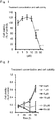
- Non-Patent Literature 2 describes the outcome of a clinical trial on pirfenidone involved with fibrosis treatment.
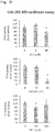
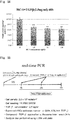
- Non-Patent Literatures 3 and 4 and Patent Literature 1 describe low-molecular-weight compounds that exert an inhibitory effect on proliferation of liver cancer cells and an inhibitory effect on a Wnt/ ⁇ -catenin signal.
- the low-molecular-weight compound disclosed in Non-Patent Literature 4 increased expression of CD44.
- neither Non-Patent Literature 3 nor 4 describes what kinds of the structure and function of a compound cause the compound to exert a growth inhibitory effect on liver cancer cells.
- WO2012/141038 A1 describes that PN-1-2, PN-3-4, PN-3-13, HC-1, and IC-2 inhibit a Wnt/ ⁇ -catenin signal in a mesenchymal stem cell, thereby inducing differentiation of the mesenchymal stem cell into hepatocytes.
- This literature discloses nothing about inhibition of proliferation of cancer cells or cancer stem cells.
- WO 2006/1 01 858 A1 discloses that the Wnt/ ⁇ -catenin signaling pathway is involved in fibrosis development. Inhibitors of the Wnt/ ⁇ -catenin signaling pathway such as ICG-001 have been shown to be efficient anti-fibrotic agents.
- the present invention has been made in view of the above situations.
- the purpose of the present invention is to provide a novel therapeutic drug for liver malignant tumors, liver cancer stem cells, or liver fibrosis.
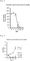
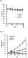
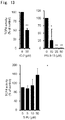
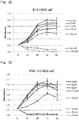
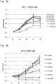
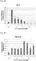
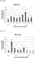
- the present inventors have conducted intensive research. As a result, the present inventors have successfully identified low-molecular-weight compounds that exert an anti-malignant tumor effect as described in Examples below. Further, these low-molecular-weight compounds have been found to exert an inhibitory effect on proliferation of not only tumor cells, but also cancer stem cells. Cancer stem cells are known to cause relapse and metastasis of malignant tumors. The above low-molecular-weight compounds that can inhibit proliferation of cancer stem cells can thus be said to be very promising compounds as an active ingredient in a therapeutic drug for malignant tumors. Besides, the above compounds have also been found to exert an inhibitory effect on fibrosis. Then, the present inventors have completed the present invention on the basis of these findings.
- an aspect of the present invention provides a therapeutic drug for a liver malignant tumor as defined in claim 1.
- the drug includes at least one compound selected from the group consisting of compounds represented by formula (1), a salt thereof, or a solvate thereof: wherein
- the drug includes at least one compound selected from the group consisting of compounds represented by the above formula (1), a salt thereof, or a solvate thereof.
- a therapeutic drug for liver fibrosis as defined in claim 7.
- the drug includes at least one compound selected from the group consisting of compounds represented by the above formula (1), a salt thereof, or a solvate thereof.
- a preferable embodiment of the present invention provides the above therapeutic drug for a liver malignant tumor, a liver cancer stem cell, or liver fibrosis, wherein the above R 1 , R 2 , and R 4 are the same or different and each represent H, halogen, nitro, cyano, OH, C 1-6 alkyl, C 1-6 halogenoalkyl, C 1-6 hydroxyalkyl, C 1-6 alkylamino, C 1-6 alkoxy, C 1-6 halogenoalkoxy, C 1-6 hydroxyalkoxy, C 1-6 alkoxyamino, C 1-6 alkoxy-substituted C 1-6 alkoxy, C 1-6 alkoxyphenyl-substituted C 1-6 alkoxy, (trialkylsiloxy) C 1-6 alkyl, or (alkyl diphenyl siloxy) C 1-6 alkyl; and the above R 3 is H.
- a novel therapeutic drug can be used to treat liver malignant tumors, liver cancer stem cells, or liver fibrosis.
- An embodiment of the present invention provides a therapeutic drug for a malignant tumor, which drug includes at least one compound selected from the group consisting of compounds represented by formula (1), a salt thereof, or a solvate thereof. This drug may be used to treat liver malignant tumors.
- An embodiment of the present invention provides a therapeutic drug for a liver cancer stem cell, which drug includes at least one compound selected from the group consisting of compounds represented by formula (1), a salt thereof, or a solvate thereof. This drug may be used to treat liver cancer stem cells.
- An embodiment of the present invention provides a growth-inhibiting drug for a liver malignant tumor cell or a liver cancer stem cell, which drug includes at least one compound selected from the group consisting of compounds represented by formula (1), a salt thereof, or a solvate thereof. This drug may be used to inhibit proliferation of liver malignant tumor cells or cancer stem cells.
- An embodiment of the present invention provides a drug for inhibiting the relapse of a liver malignant tumor, which drug includes at least one compound selected from the group consisting of compounds represented by formula (1), a salt thereof, or a solvate thereof.
- This inhibitory drug may be used to inhibit the relapse of a liver malignant tumor.
- An embodiment of the present invention provides a therapeutic drug for liver fibrosis, which drug includes at least one compound selected from the group consisting of compounds represented by formula (1), a salt thereof, or a solvate thereof. This drug may be used to treat liver fibrosis.
- An embodiment of the present invention provides a drug for treating a disease accompanied by liver fibrosis, which drug includes at least one compound selected from the group consisting of compounds represented by the above formula (1), a salt thereof, or a solvate thereof.
- This drug may be used to treat a disease accompanied by liver fibrosis.
- R 1 , R 2 , and R 4 are the same or different and each represent H, halogen, nitro, cyano, OH, optionally substituted C 1-6 alkyl, optionally substituted C 2-6 alkenyl, optionally substituted C 1-6 alkoxy, aryl, or heteroaryl.
- R 3 represents H, optionally substituted C 1-6 alkyl, or optionally substituted C 2-6 alkenyl.
- m represents an integer of any of 1 to 4.
- n is an integer of any of 1 to 3.
- p represents an integer of any of 1 to 5.
- R 1 , R 2 , and R 4 are the same or different and may each represent H, halogen, nitro, cyano, OH, C 1-6 alkyl, C 1-6 halogenoalkyl, C 1-6 hydroxyalkyl, C 1-6 alkylamino, C 1-6 alkoxy, C 1-6 halogenoalkoxy, C 1-6 hydroxyalkoxy, or C 1-6 alkoxyamino, or C 1-6 alkoxy-substituted C 1-6 alkoxy, C 1-6 alkoxyphenyl-substituted C 1-6 alkoxy, (trialkylsiloxy) C 1-6 alkyl, or (alkyl diphenyl siloxy) C 1-6 alkyl.
- the above R 3 is preferably H.
- halogen means F, Cl, Br, or I.
- alkyl and “alkenyl” mean a linear or branched hydrocarbon chain.
- C 1-6 refers to hydrocarbon containing 1, 2, 3, 4, 5, or 6 carbon atoms. That is, the term “C 1-6 alkyl” refers to alkyl containing 1, 2, 3, 4, 5, or 6 carbon atoms. Examples of C 1-6 alkyl include methyl, ethyl, n-propyl, isopropyl, n-butyl, isobutyl, sec-butyl, tert-butyl, n-pentyl, and n-hexyl.
- alkenyl examples include ethenyl, 1-propenyl, 2-propenyl, 2-methyl-1-propenyl, 1-butenyl, 2-butenyl, 3-butenyl, 3-methyl-2-butenyl, 1-pentenyl, 2-pentenyl, 3-pentenyl, 4-pentenyl, 4-methyl-3-pentenyl, 1-hexenyl, 3-hexenyl, and 5-hexenyl.
- alkoxy examples include methoxy, ethoxy, propoxy, isopropoxy, butoxy, isobutoxy, sec-butoxy, tert-butxy, pentoxy, isopentoxy, and hexoxy.
- the term "optionally substituted” means that a compound is unsubstituted or has 1, 2, 3, 4, or 5 substituents at substitutable positions. Note that when a plurality of substituents are included, these substituents may be the same or different. In addition, the position of each substitution may be position 1, 2, 3, 4, 5, 6, 7, 8, or 9.
- examples of the substituents include H, halogen, nitro, cyano, OH, C 1-6 alkyl, C 1-6 halogenoalkyl, C 1-6 hydroxyalkyl, C 1-6 alkylamino, C 3-6 cycloalkyl, C 2-6 alkenyl, C 2-6 halogenoalkenyl, C 2-6 hydroxyalkenyl, C 2-6 alkenylamino, C 3-6 cycloalkenyl, C 2-6 alkynyl, C 2-6 halogenoalkynyl, C 2-6 hydroxyalkynyl, C 2-6 alkynylamino, C 1-6 alkoxy, C 1-6 halogenoalkoxy, C 1-6 hydroxyalkoxy, C 1-6 alkoxyamino, C 1-6 alkoxyphenyl, trialkylsiloxy, alkyl diphenyl siloxy, aryl, and heteroaryl.
- C 1-6 halogenoalkyl refers to C 1-6 alkyl that is substituted by one or more halogens.
- the number of halogens may be, for example, 1, 2, 3, 4, 5, 6, or 13. Also, the number may be between any two of the numbers indicated above. In addition, when two or more halogens are included, the kind of each halogen may be the same or different.
- C 1-6 halogenoalkyl examples include, chloromethyl, dichloromethyl, trichloromethyl, fluoromethyl, difluoromethyl, trifluoromethyl, bromomethyl, dibromomethyl, tribromomethyl, chloroethyl, dichloroethyl, trichloroethyl, fluoroethyl, difluoroethyl, and trifluoroehtyl.
- C 1-6 hydroxyalkyl refers to C 1-6 alkyl that is substituted by one or more hydroxy groups.
- the number of the hydroxy groups may be, for example, 1, 2, 3, 4, 5, 6, or 13. Also, the number may be between any two of the numbers indicated above.
- Examples of C 1-6 hydroxyalkyl include hydroxymethyl, 1-hydroxyethyl, 2-hydroxyethyl, 2-hydroxy-n-propyl, and 2,3-dihydroxy-n-propyl.
- C 1-6 alkylamino refers to C 1-6 alkyl that is substituted by one or more amino groups.
- the number of the amino groups may be, for example, 1, 2, 3, 4, 5, 6, or 13. Also, the number may be between any two of the numbers indicated above.
- Examples of C 1-6 alkylamino include methylamino and ethylamino.
- C 1-6 halogenoalkoxy is equivalent to C 1-6 halogenoalkyl, the alkyl of which is replaced by alkoxy.
- Examples of C 1-6 halogenoalkoxy include fluoromethoxy, difluoromethoxy, trifluoromethoxy, 1-fluoroethoxy, 2-fluoroethoxy, 2-chloroethoxy, 2-bromoethoxy, (1,1-difluoro)ethoxy, (1,2-difluoro)ethoxy, (2,2,2-trifluoro)ethoxy, (1,1,2,2-tetrafluoro)ethoxy, (1,1,2,2,2-pentafluoro)ethoxy, 1-fluoron-n-propoxy, 1,1-difluoro-n-propoxy, 2,2-difluoro-n-propoxy, 3-fluoro-n-propoxy, (3,3,3-trifluoro)-n-propoxy, (2,2,3,3,3-pent
- C 1-6 hydroxyalkoxy is equivalent to C 1-6 hydroxyalkyl, the alkyl of which is replaced by alkoxy.
- Examples of C 1-6 hydroxyalkoxy include 2-hydroxyethoxy, 2-hydroxy-n-propoxy, 3-hydroxy-n-propoxy, 2,3-dihydroxy-n-propoxy, and 2-hydroxycyclopropyl.
- C 1-6 alkoxyamino is equivalent to C 1-6 alkylamino, the alkyl of which is replaced by alkoxy.
- Examples of C 1-6 alkoxyamino include methoxyamino and ethoxyamino.
- aryl refers to a C 6-14 monocyclic, dicyclic, or tricyclic aromatic hydrocarbon ring group.
- aryl include phenyl, naphthyl (e.g., 1-naphthyl, 2-naphthyl), tetrahydronaphthalenyl, indenyl, and fluorenyl. Particularly preferred are naphthyl or phenyl substituted with five halogens.
- the aryl includes a ring group that is condensed with C 5-8 cycloalkene at its double bond position.
- heteroaryl includes groups having 5 to 14 ring atoms within their rings, having a shared ⁇ electron system, and having 1 to 4 heteroatoms selected from the group consisting of N, S, and O.
- heteroaryl include thienyl, benzothienyl, furyl, benzofuryl, dibenzofuryl, pyrrolyl, imidazolyl, pyrazolyl, pyridyl, pyrazinyl, pyrimidinyl, pyridazinyl, tetrazolyl, oxazolyl, thiazolyl, and isooxazolyl.
- furyl that is substituted by one methyl group.
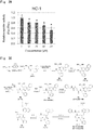
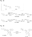
- the structure of a compound represented by the formula (1) preferably involves a configuration in which R 1 , R 2 , and R 3 are each H; and R 4 is at position 4 and represents H, F, Cl, nitro, OH, CH 2 OH, methoxy, methoxymethoxy, or tert-butyl dimethyl siloxymethyl. It is particularly preferable that the structure of a compound represented by the formula (1) is as close as the structure of a compound represented by the formula (3) in view of an effect of treating malignant tumors or fibrosis.
- the compounds represented by the formulas (3), (4), and (6) are sometimes referred to as IC-2, PN3-13, and HC-1, respectively. As used herein, the meaning of PN3-13 is same as of PN-3-13.
- examples of the "salt” include, but are not particularly limited to, anionic salts that are formed by using any acidic group (e.g., carboxyl) and cationic salts that are formed by using any basic group (e.g., amino).
- examples of the salts include inorganic salts, organic salts, and salts disclosed in the article ( Berge, Bighley, and Monkhouse, J. Pharm. Sci., 1977, 66, 1-19 ).
- the examples further include metal salts, ammonium salts, salts of an organic base, salts of an inorganic acid, salts of an organic acid, and salts of a basic or acidic amino acid.
- Examples of the metal salts include alkali metal salts (e.g., sodium salts, potassium salts), alkali earth metal salts (e.g., calcium salts, magnesium salts, barium salts), and aluminum salts.
- Examples of the salts of an organic base include salts of trimethylamine, triethylamine, pyridine, picoline, 2,6-lutidine, ethanolamine, diethanolamine, triethanolamine, cyclohexylamine, dicyclohexylamine, or N,N'-dibenzylethylenediamine.
- Examples of the salts of an inorganic acid include salts of hydrochloric acid, hydrobromic acid, nitric acid, sulfuric acid, or phosphoric acid.
- Examples of the salts of an organic acid include salts of formic acid, acetic acid, trifluoro acetic acid, phthalic acid, fumaric acid, oxalic acid, tartaric acid, maleic acid, citric acid, succinic acid, malic acid, methanesulfonic acid, benzenesulfonic acid, or p-toluenesulfonic acid.
- Examples of the salts of a basic amino acid include salts of arginine, lysine, or ornithine.
- Examples of the salts of an acidic amino acid include salts of aspartic acid or glutamic acid.
- solvate refers to a compound formed by using a solute and a solvent. J. Honig et al., The Van Nostrand Chemist's Dictionary P650 (1953) can be consulted regarding the solvate.
- the solvent is water, the solvate formed is a hydrate.
- the solvent does not interfere with the biological activity of the solute. Examples of such a preferable solvent include, but are not limited to, water, ethanol, and acetic acid. The most preferred solvent is water.
- a compound or a salt thereof according to an embodiment of the present invention absorbs moisture when contacting the air or recrystallized. They may have hygroscopic moisture or become a hydrate.
- the term “isomer” includes a molecule, the molecular formula of which is identical, but the structure of which is different. Examples of the isomer include enantiomers, geometric (cis/trans) isomers, and isomers (diastereomers) having one or more chiral centers that are not mirror images of one another.
- the term “prodrug” includes a precursor compound in which when the above compound is administered to a subject, a chemical change occurs due to metabolic processes or various chemical reactions to give rise to a compound, a salt thereof, or a solvate thereof according to the present invention. With regard to the prodrug, the article ( T. Higuchi and V. Stella, "Pro-Drugs as Novel Delivery Systems", A.C.S. Symposium Series, Volume 14 ) can be referred to.
- a malignant tumor examples include tumors caused by a mutation in a normal cell.
- the malignant tumors occur in all the organs and tissues in the body.
- the malignant tumor is, for example, at least one kind selected from the group consisting of lung cancer, esophagus cancer, gastric cancer, liver cancer, pancreatic cancer, renal cancer, adrenal cancer, biliary tract cancer, breast cancer, colon cancer, small intestinal cancer, ovarian cancer, uterine cancer, bladder cancer, prostate cancer, ureteral cancer, renal pelvis cancer, ureteral cancer, penile cancer, testicular cancer, brain tumor, cancer in central nervous system, cancer in peripheral nervous system, head and neck carcinoma, glioma, glioblastoma multiforme, skin cancer, melanoma, thyroid cancer, salivary gland cancer, malignant lymphoma, carcinoma, sarcoma, and hematological malignancies.
- the above liver cancer may be, for example, an epithelial tumor or nonepithelial tumor, and may be hepatocyte carcinoma or cholangiocellular carcinoma.
- Examples of the above skin cancer include basal cell carcinoma, squamous cell carcinoma, and malignant melanoma.
- the present invention refers to a liver malignant tumor.
- cancer stem cell includes a cell that generates cancer cells.
- This cancer stem cell includes a cell expressing a cancer stem cell marker.
- the cancer stem cell marker include CD44, CD90, CD133, and EpCAM.
- the present invention refers to a liver cancer stem cell.
- the "fibrosis” has been known as a symptom caused by loss of normal function due to tissue sclerosis in which the volume of a connective tissue mass including tissue components such as collagen is increased and a normal tissue is replaced by the connective tissue. Fibrosis occurs in, for example, respective tissues such as the liver, lung, kidney, heart, and skin. Also, occurrence of a large amount of fibrosis in a hepatic tissue, for example, may result in hepatic cirrhosis, leading to liver cancer. Each tissue, other than a liver tissue, may harbor a malignant tumor while fibrosis progresses.
- the term "fibrosis" include a disease accompanied by fibrosis. Examples of the disease accompanied by fibrosis include the above tissue fibrosis, cirrhosis, and malignant tumors accompanied by fibrosis.
- the present invention refers to a liver fibrosis.
- the term “treatment” includes exerting a prophylactic effect, an inhibitory effect, or a symptom-improving effect on a disease of a subject or on one or more symptoms involving the disease.
- the "therapeutic drug” may be a pharmaceutical composition containing an active ingredient and at least one pharmacologically acceptable carrier.
- the pharmaceutical composition can be produced by any process known in the art of drug formulation. Examples of the process include: mixing an active ingredient with the above carrier.
- the dosage form of the drug is not limited as long as the drug can be used for treatment.
- the drug may be an active ingredient alone or a mixture of an active ingredient and any component.
- examples of the dosage form of the above carrier include, but are not particularly limited to, a solid and liquid (e.g., a buffer).
- examples of a therapeutic drug for malignant tumors include: a drug (prophylactic) used for preventing a malignant tumor; a drug for inhibiting relapse of a malignant tumor; and a drug for inhibiting proliferation of a malignant tumor cell.
- examples of a therapeutic drug for cancer stem cells include: an agent for treating a cancer stem cell as a target; a therapeutic drug for malignant tumors derived from a cancer stem cell; and an inhibitor for cancer stem cells.
- a drug administration route effective in treatment is preferably used.
- the administration route include intravenous, subcutaneous, intramuscular, intraperitoneal, and oral administration.
- the dosage form may include an injection, a capsule, a tablet, and granules.
- an aqueous solution for an injection may be combined with, for example, a saline solution, sugar (e.g., trehalose), NaCl, or NaOH.
- the drug may be formulated with, for example, a buffer (e.g., a phosphate buffer) and/or a stabilizer.
- a dosage is not particularly limited, and may be, for example, 0.001, 1, 10, 100, or 1000 mg/kg body weight per administration.
- the dosage may be between any two of the above values.
- An administration interval is not particularly limited, and the drug may be dosed, for example, once or twice per 1, 7, 14, 21, or 28 days.
- the drug may be dosed once or twice per period between any two of the above values.
- the dosage, the administration interval, and the administration method can be appropriately selected depending on the age, body weight, symptom, affected organ, etc., of a patient.
- the drug preferably contains a therapeutically effective amount or a dose, which is effective in exerting a desired effect, of an active ingredient.
- the effect of treating malignant tumors may be evaluated by imaging, endoscopic examination, biopsy, or detection of a malignant tumor marker.
- the effect of treating cancer stem cells may be evaluated by imaging, endoscopic examination, biopsy, or detection of a cancer stem cell marker.
- the effect of treating fibrosis may be evaluated by imaging, endoscopic examination, biopsy, or detection of a fibrosis marker.
- the level of a marker after administration of a therapeutic drug may be 0.7, 0.5, 0.3, or 0.1 times the level before the administration (or of a control).
- the number of marker-positive cells after administration of the therapeutic drug may be 0.7, 0.5, 0.3, or 0.1 times the number before the administration (or of a control). Note that in Example 2 below, the therapeutic effect was evaluated by using mice in which CD44-positive HuH7 cells had been subcutaneously transplanted. The present inventors also replaced the above CD44-positive HuH7 cells by unsorted HuH7 cells. This experiment has demonstrated that IC-2 exhibits an effect of treating a malignant tumor.
- the growth rate of patient-derived test cells after administration of the therapeutic drug may be reduced to 0.7, 0.5, 0.3, or 0.1 times of the rate before the administration (or of a control).
- the term "significantly” may include a case of p ⁇ 0.05 or p ⁇ 0.01 when Student's t test (one-sided or two-sided), for example, is used to evaluate a statistically significant difference. Also, the term may include a state in which there is a substantial difference.
- examples of the "patient” include human and non-human mammals (e.g., at least one of a mouse, guinea pig, hamster, rat, mouse, rabbit, pig, sheep, goat, cow, horse, cat, dog, marmoset, monkey, and chimpanzee).
- the patient may be a patient who is determined or diagnosed as having the onset of a malignant tumor or fibrosis.
- the patient may be a patient who needs treatment of a malignant tumor or fibrosis.
- the patient may be a patient who is determined or diagnosed as having a significantly larger number of cancer stem cells in a tissue than healthy individuals. Note that the determination or diagnosis may be performed by imaging, endoscopic examination, biopsy, or detection of various markers.
- the wording "a state in which cell proliferation is inhibited” includes a state in which the growth rate of test cells is significantly less than that before drug treatment.
- the growth rate can be evaluated by measuring the level of proliferation of cells during a given period of time.
- the level of proliferation may be measured, for example, visually or by using absorbance as an index.
- the level of proliferation may be measured by using, as an index, the level of a malignant tumor marker in a patient or a patient-derived sample.
- the wording "inhibiting a cancer stem cell” includes, for example, inhibiting proliferation of a cancer stem cell and inhibiting the function of a cancer stem cell (e.g., inhibiting sphere formation, inhibiting marker expression).
- An embodiment of the present invention provides a drug for inhibiting expression of a marker for a malignant tumor cell, a cancer stem cell, or fibrosis, which drug includes at least one compound selected from the group consisting of compounds represented by formula (1), a salt thereof, or a solvate thereof.
- An embodiment of the present invention provides a drug for inhibiting formation of a sphere from a malignant tumor or of cancer stem cells, which drug includes at least one compound selected from the group consisting of compounds represented by formula (1), a salt thereof, or a solvate thereof. This drug for inhibiting formation of a sphere may be used for treatment of malignant tumors or cancer stem cells.
- An embodiment of the present invention provides a treatment method comprising the step of administering to a patient a low-molecular-weight compound contained in a therapeutic drug according to any of the above embodiments.
- An embodiment of the present invention provides use of a low-molecular-weight compound contained in the therapeutic drug according to any of the above embodiments in the manufacture of the therapeutic drug.
- An embodiment of the present invention provides a growth-inhibiting method comprising the step of administering to a patient a low-molecular-weight compound contained in the drug for inhibiting proliferation according to any of the above embodiments.
- An embodiment of the present invention provides use of a low-molecular-weight compound contained in the drug for inhibiting proliferation according to any of the above embodiments in the manufacture of the drug.
- An embodiment of the present invention provides a method for inhibiting relapse of a malignant tumor, the method comprising the step of administering to a patient a low-molecular-weight compound contained in the drug for inhibiting relapse according to any of the above embodiments.
- An embodiment of the present invention provides use of a low-molecular-weight compound contained in the drug for inhibiting relapse according to any of the above embodiments in the manufacture of the drug.
- An embodiment of the present invention provides at least one compound selected from the group consisting of compounds represented by the formula (1), a salt thereof, or a solvate thereof, wherein R 1 , R 2 , and R 3 are each H; and R 4 is at position 4 and represents F, Cl, nitro, OH, CH 2 OH, methoxy, methoxymethoxy, or tert-butyl dimethyl siloxymethyl.
- This compound, a salt thereof, or a solvate thereof may be used to treat liver malignant tumors, liver cancer stem cells, and/or liver fibrosis.
- a non-claimed aspect of the present disclosure provides a drug for inhibiting a Wnt/ ⁇ -catenin signal, which drug includes at least one compound selected from the group consisting of compounds represented by the formula (1), a salt thereof, or a solvate thereof, wherein R 1 , R 2 , and R 3 are each H; and R 4 is at position 4 and represents F, Cl, nitro, OH, CH 2 OH, methoxy, methoxymethoxy, or tert-butyl dimethyl siloxymethyl.
- This compound, a salt thereof, or a solvate thereof may be used to inhibit a Wnt/ ⁇ -catenin signal.
- the drug for inhibiting a Wnt/ ⁇ -catenin signal may be used for various applications in vitro or in vivo.
- the drug for inhibiting a Wnt/ ⁇ -catenin signal may be used for treatment of disease that can be ameliorated by, for example, an inhibitory effect on a Wnt/ ⁇ -catenin signal.
- HuH7 cells (a liver cancer cell line) were cultured in a DMEM on a 10-cm cell culture dish (TPP Techno Plastic Products AG, Trasadingen, Switzerland) at 5% CO 2 , 37°C, and a humidity of 100%. After the cells were grown to 70 to 90% confluency, 200 ⁇ L of a Trypsin/EDTA solution was added thereto to detach the cells. The cells were centrifuged at 1000 rpm for 5 min at room temperature for recovery. Then, the cells in one dish were divided into four dishes and subcultured.
- HuH7 cells at 70 to 90% confluency were collected, and 1 x 10 4 cells were seeded in each well of a 96-well plate (FALCON).
- FALCON 96-well plate
- 5 x 10 3 cells were seeded. Twenty-four hours after the seeding, the cells were treated with IC-2, PN3-13, or HC-1, and further incubated at 37°C. DMSO was used as a control.
- the concentration of each low-molecular-weight compound was provided below.
- IC-2 0,1, 5, 10, 25, 50 ⁇ M
- PN3-13 0, 1,5, 10, 25, 50 ⁇ M
- HC-1 0, 1, 5, 10, 25, 50 ⁇ M.
- 100 ⁇ L of 10% TetraColor ONE SEIKAGAKU CORPORATION, Tokyo, Japan
- 100 ⁇ L of 10% TetraColor ONE was added at 0, 1, 2, and 4 days after the chemical treatment. Then, the cells were incubated at 37°C for 45 min.
- IC 50 10 LOG A / B ⁇ 50 ⁇ C / D ⁇ C + LOG B , wherein A denotes a concentration higher than 50%; B denotes a concentration lower than 50%; C denotes the rate of inhibition at B; and D denotes the rate of inhibition at A.
- the PN3-13 treatment also significantly inhibited proliferation of HuH7 cells ( FIGS. 3 and 4 ).
- the HC-1 treatment also inhibited proliferation of HuH7 cells ( FIG. 5 ).
- CD44 a cancer stem cell marker
- HuH7 cells at 70 to 90% confluency were collected and 1.5 x 10 6 cells were seeded on a 10-cm dish (TPP Techno Plastic Products AG, Trasadingen, Switzerland). After 15 hours, the cells were treated with each of Hexachlorophene (15 ⁇ M), ICG-001 (15 ⁇ M), PKF118-310 (5 ⁇ M), IC-2 (50 ⁇ M), PN3-13 (10 ⁇ M), and 5-FU (0.5 ⁇ M) and were incubated at 37°C.
- DMSO which was a solvent for each of the low-molecular-weight compounds and anti-cancer drugs, was used as a control.
- Two days after the chemical treatment the cells on a dish were collected. Next, the cells were centrifuged at 1000 rpm for 5 min at 4°C to remove a supernatant. Then, the cells were washed twice with 1 mL of 0.5% FBS/2 mM EDTA/PBS. After that, the cells were suspended in 500 ⁇ L of 5% BSA/0.5% FBS/2 mM EDTA/PBS and blocked once for 15 min at 4°C.
- an anti-human CD44 monoclonal antibody (Cell Signaling Technology Inc., Danvers, MA, USA) was added. Then, the cells were suspended and a primary antibody reaction was performed in a dark room for 10 min at 4°C. After that, the cells were washed 3 times with 1 mL of PBS. After 1.0 ⁇ g of an Alexa 488-labeled goat anti-mouse IgG antibody (Life Technologies Corp., Carlsbad, CA, USA) was added, the mixture was resuspended and a secondary antibody reaction was performed in a dark room for 10 min at 4°C.
- an Alexa 488-labeled goat anti-mouse IgG antibody (Life Technologies Corp., Carlsbad, CA, USA) was added, the mixture was resuspended and a secondary antibody reaction was performed in a dark room for 10 min at 4°C.
- CD44-positive HuH7 cells were subcutaneously transplanted into mice.
- the mice were then divided into 3 groups (DMSO group: 5 mice; 5-FU group: 4 mice; and IC-2 group: 4 mice).
- DMSO group 5 mice; 5-FU group: 4 mice; and IC-2 group: 4 mice.
- 30 mg/kg of 5-FU, 50 mg/kg of IC-2, or DMSO (as a control), which was a solvent for each chemical was intraperitoneally administered every 3 days.
- the body weight and the tumor length and width of each mouse were measured every 3 days.
- the volume at Day 0 was used to normalize each tumor volume, and then a graph was created.
- the dosage that was usually 25 mg/kg in research articles was doubled to set a 5-FU dose.
- the IC-2 concentration corresponding to a concentration at which a Wnt/ ⁇ -catenin signal was inhibited in vitro was calculated. Then, the concentration was doubled to set an IC-2 dose.
- HuH7 cells were collected.
- the cells were stained with an anti-human CD44 monoclonal antibody (Cell Signaling Technology Inc., Danvers, MA, USA)), and then with an Alexa 488-labeled goat anti-mouse IgG antibody.
- a Beckman Coulter-MoFlo XDP Beckman Coulter Inc., Fullerton, CA, USA was used to sort the cells into a CD44-negative fraction and a CD44-positive fraction.
- the sorted cells were seeded at 5 x 10 4 cells/well on a 24-well ultra-low-attachment multiwell plate (Corning, NY, USA).
- the cells were cultured at 37°C in the presence of 5% CO 2 in a DMEM/Nutrient Mixture F-12 Ham (Sigma-Aldrich, St. Louis, MO, USA) containing 20 ng/mL of recombinant human epidermal growth factor, 20 ng/mL of human basic fibroblast growth factor, 1 x B27, and L-glutamine. Twenty-four hours after the plating, 1% DSMO, 50 ⁇ M IC-2, and 10 ⁇ M PN-3-13 were each added to the corresponding well and the cells were further cultured. One week after the chemical treatment, the number of spheres (with a size of 100 ⁇ m or larger) in 15 visual fields/well was counted.
- a liver cancer cell line HLF was seeded at 1.5 x 10 6 cells/dish on 10-cm dishes. Next, the cells were cultured at 37°C and 5% CO 2 . After 24 hours, 1% DSMO, 0.5 ⁇ M 5-FU, or 50 ⁇ M IC-2 was added thereto and the cells were further incubated. Two days after the chemical treatment, the cells were harvested. Then, the cells were blocked in 0.5% BSA/0.5% FBS/2 mM EDTA/PBS.
- an APC-labeled mouse anti-human CD90 antibody (BD Biosciences, San Jose, CA, USA) was added to 500 ⁇ L of a cell suspension and the resulting mixture was resuspended and incubated at 4°C for 10 min to carry out a primary antibody reaction.
- the cells were washed twice with 0.5% FBS/2 mM EDTA/PBS and once with 5% FBS/2 mM EDTA/DMEM.
- the cells were resuspended in 500 ⁇ L of 5% FBS/2 mM EDTA/PBS.
- 1 ⁇ g/mL of propidium iodide was added thereto.
- the concentration of 5-FU was determined on the basis of the IC 50 concentration at 24 hours in a WST assay.
- the concentration of IC-2 was determined on the basis of a concentration at which a Wnt/ ⁇ -catenin signal was inhibited.
- a liver cancer cell line HepG2 was seeded at 1.5 x 10 6 cells/dish on 10-cm dishes. Next, the cells were cultured at 37°C and 5% CO 2 . After 24 hours, 1% DSMO, 0.5 ⁇ M 5-FU, or 50 ⁇ M IC-2 was added thereto and the cells were further incubated. Then, the cells were harvested at two days after the chemical treatment. Meanwhile, the cells were blocked in 0.5% BSA/0.5% FBS/2 mM EDTA/PBS.
- an anti-human CD133 monoclonal antibody (Miltenyi Biotec) was added to 500 ⁇ L of a cell suspension and the resulting mixture was resuspended and incubated at 4°C for 10 min to carry out a primary antibody reaction. After the primary antibody reaction, the cells were washed 3 times with 1 mL of PBS. After 1.0 ⁇ g of an Alexa 488-labeled goat anti-mouse IgG antibody (Life Technologies Corp., Carlsbad, CA, USA) was added, the mixture was resuspended and a secondary antibody reaction was performed in a dark room for 10 min at 4°C.
- an Alexa 488-labeled goat anti-mouse IgG antibody (Life Technologies Corp., Carlsbad, CA, USA) was added, the mixture was resuspended and a secondary antibody reaction was performed in a dark room for 10 min at 4°C.
- the cells were washed twice with 5% FBS/2 mM EDTA/PBS and once with 5% FBS/2 mM EDTA/DEME.
- the cells were resuspended in 500 ⁇ L of 5% FBS/2 mM EDTA/PBS.
- 1 ⁇ g/mL of propidium iodide was added thereto.
- the mixture was made to pass through a tube with a 40- ⁇ m mesh filter.
- a Beckman Coulter-MoFlo XDP was used to analyze CD133-expressing cells.
- a liver cancer cell line HepG2 was seeded at 1.5 x 10 6 cells/dish on 10-cm dishes. Next, the cells were cultured at 37°C and 5% CO 2 . After 24 hours, 1% DSMO, 0.5 ⁇ M 5-FU, or 50 ⁇ M IC-2 was added thereto and the cells were further incubated. Then, the cells were harvested at two days after the chemical treatment. Meanwhile, the cells were blocked in 0.5% BSA/0.5% FBS/2 mM EDTA/PBS.
- an anti-human EpCAM monoclonal antibody (Cell Signaling Technology Inc., Danvers, MA, USA) was added to 500 ⁇ L of a cell suspension and the resulting mixture was resuspended and incubated at 4°C for 10 min to carry out a primary antibody reaction. After the primary antibody reaction, the cells were washed 3 times with 1 mL of PBS. After 1.0 ⁇ g of an Alexa 488-labeled goat anti-mouse IgG antibody (Life Technologies Corp., Carlsbad, CA, USA) was added, the mixture was resuspended and a secondary antibody reaction was performed in a dark room for 10 min at 4°C.
- an Alexa 488-labeled goat anti-mouse IgG antibody (Life Technologies Corp., Carlsbad, CA, USA) was added, the mixture was resuspended and a secondary antibody reaction was performed in a dark room for 10 min at 4°C.
- the cells were washed twice with 5% FBS/2 mM EDTA/PBS and once with 5% FBS/2 mM EDTA/DEME.
- the cells were resuspended in 500 ⁇ L of 5% FBS/2 mM EDTA/PBS.
- 1 ⁇ g/mL of propidium iodide was added thereto.
- the mixture was made to pass through a tube with a 40- ⁇ m mesh filter.
- a Beckman Coulter-MoFlo XDP was used to analyze EpCAM-expressing cells.
- a pTCF4-CMVpro-Luc was added to 5 x 10 6 HuH7 cells and the plasmid was introduced using a Cellject Pro (Thermo, Massachusetts, USA) (at 250 V, 1500 ⁇ F). Twenty-four hours after the gene introduction, 2 ⁇ g/mL of puromycin was added. Then, the cells were selected while a medium was changed every 3 to 5 days. It was observed that colonies of the pTCF4-CMVpro-Luc-containing HuH7 cells were formed at 20 days after initiation of the selection.
- a cloning ring which was manufactured such that an end portion of a blue tip was cut, (the bottom edge of the cloning ring was provided with vaseline), was then used to surround one of the colonies formed. Subsequently, 30 ⁇ L of a 0.25% Trypsin/1 mM EDTA solution, which was prepared by diluting a stock solution 5-fold with PBS, was added to detach and collect the cells. After that, 70 ⁇ L of DMEM was added and the mixture was plated on a 96-well plate.
- the HuH7 cells at 70 to 90% confluency were collected, and the 3.5 x 10 5 cells were seeded in each well of a 24-well plate (FALCON). After 24 hours, the cells were collected into a 15-mL tube, centrifuged at 1000 rpm and room temperature for 5 min, washed with 1 mL of 1 x PBS (-), and re-centrifuged at 1000 rpm and room temperature for 5 min. The cell mass was resuspended in 375 ⁇ L of a Proteinase K buffer. Then, 25 ⁇ L of sodium dodecyl sulfate (SDS) and 4 ⁇ L of Proteinase K (20 mg/mL) were added thereto.
- SDS sodium dodecyl sulfate
- the mixture was incubated overnight at 55°C to degrade cellular proteins.
- 400 mL of phenol/chloroform was added, and the mixture was vortexed and then centrifuged at 15000 rpm and 4°C for 5 min to recover a supernatant.
- a 2-fold volume of 100% ethanol was added to the supernatant and the mixture was incubated at -80°C for 30 min.
- the sample was centrifuged at 15000 rpm and 4°C for 20 min, a supernatant was discarded and 500 ⁇ L of 70% ethanol was added.
- the sample was further centrifuged at 15000 rpm and 4°C for 10 min.
- a supernatant was discarded and 20 ⁇ L of ultra-pure water was used to dissolve the resulting pellet.
- One ⁇ L of the dissolved pellet was measured by using a spectrophotometer (LMS, Tokyo, Japan) to adjust the DNA content of the sample to 1 ng/ ⁇ L.
- LMS spectrophotometer
- PCR polymerase chain reaction
- Primers for a glyceraldehyde-3-phosphate dehydrogenase (GAPDH) locus were used as an internal control. Also, primers for a luciferase region were used.
- a PCR was carried out by using rTaq (TOYOBO, Osaka, Japan) to amplify each gene at a 10- ⁇ L scale.
- the PCR consisted of: 95°C for 2 min; 25 cycles (95°C for 30 sec, 60°C for 30 sec, and 72°C for 30 sec); and 72°C for 5min.
- the PCR consisted of: 95°C for 2 min; 30 cycles (95°C for 30 sec, 55°C for 30 sec, and 72°C for 30 sec); and 72°C for 5min.
- a luciferase assay was conducted.
- the HuH7 cells at 70 to 90% confluency were collected, and the 5 x 10 4 cells were seeded in each well of a 24-well plate (FALCON).
- 100 ⁇ L of 20 % Passive Lysis Buffer (PLB) (PROMEGA, Wisconsin, USA) was added to each well of the 24-well plate at 17 hours after the cell seeding.
- the plate was shaken at room temperature for 30 min.
- 50 ⁇ L of Luciferase Assay Reagent (LAR) (PROMEGA) was added to each 3.5-ml Röhren tube (SARSTEDT, Numbrecht, German).
- the HuH7 cells that were at 70 to 90% confluency and stably expressed the pTCF4-CMVpro-Luc were collected, and the 1 x 10 4 cells were seeded in each well of a 96-well plate (FALCON). After 12 hours, the cells were treated with IC-2, PN3-13, or 5-FU at concentrations indicated in FIG. 13 , and further incubated at 37°C. DMSO was used as a control. Two days after the low-molecular-weight compound treatment, 100 ⁇ L of Steady-Glo (PROMEGA, Wisconsin, USA) was added to each well of a 96-well white plate (Corning Inc., New York, USA) and the mixture was incubated at room temperature for 5 min. Then, a fluorescence plate reader Infinite F500 (TECAN, Zurich, Switzerland) was used to measure luciferase activity.
- FALCON 96-well plate
- Colon cancer cells (DLD-1, HCT 116, colo 205) were cultured in a DMEM on a 10-cm cell culture dish (TPP Techno Plastic Products AG, Trasadingen, Switzerland) at 5% CO 2 , 37°C, and a humidity of 100%. The cells at 70-90% confluency were split and washed with PBS (-). Next, 300 ⁇ L of Trypsin/EDTA was added to 2 mL of PBS (-) and the cells were incubated with this solution at 37°C for 5 min to detach the cells. Then, 5 mL of DMEM was used to recover the cells. The recovered cells were centrifuged at 1000 rpm for 3 min to remove a supernatant. Then the cells were suspended in DMEM at 1:4and subcultured.
- a cell surface marker CD44 was used as an index to sort DLD-1 cells into the top 5% group and the bottom 5% group.
- An anti-human CD44 antibody (Abcam Ltd., Cambridge, UK) was used as a primary antibody and an Alexa 488-labeled goat anti-mouse IgG antibody (Life Technologies Corp., Carlsbad, CA, USA) was then used. After that, a MoFlo XDP (Beckman Coulter Inc., Fullerton, CA) was used to analyze and fractionate each group.
- the 5,000 collected cells were plated on each well of 24-well ultra-low-attachment multiwell plates (Corning Inc., Corning, NY) in 500 ⁇ l of a serum-free DMEM/F12 medium containing 20 ng/mL of recombinant human basic fibroblast growth factor (bFGF: Corefront Corporation, Tokyo, Japan), 20 ng/mL of recombinant human epidermal growth factor (Sigma Life Science, St. Louis, United States), B-27 (life technologies), 200 mmol/L of L-glutamine (Invitrogen, Life Technologies Corp., Carlsbad, CA), and 0.6% methyl cellulose (Sigma Life Science). Then, a difference in sphere formation capability was examined.
- bFGF basic fibroblast growth factor
- B-27 life technologies
- 200 mmol/L of L-glutamine Invitrogen, Life Technologies Corp., Carlsbad, CA
- 0.6% methyl cellulose Sigma Life Science
- the number of spheres formed per well in a cell population expressing CD44 more strongly is significantly higher than that in a cell population expressing CD44 weakly ( FIG. 17 ). This demonstrated that the group expressing CD44 strongly contained a larger number of cells exhibiting sphere formation capability and having cancer stem cell characteristics.
- DLD-1 cells were seeded on a 10-cm dish. After 24 hours, the cells were treated with 5-FU (0.5, 5 ⁇ M) or IC-2 (50 ⁇ M). After additional 48 hours, the cells were harvested. An anti-human CD44 antibody (Abcam Ltd., Cambridge, UK) was used as a primary antibody and an Alexa 488-labeled goat anti-mouse IgG antibody (Life Technologies Corp., Carlsbad, CA, USA) was then used. After that, analysis was performed by using a MoFlo XDP (Beckman Coulter Inc., Fullerton, CA).
- HSC2 cells (a squamous cell carcinoma cell line) were cultured in a DMEM on a 10-cm cell culture dish (TPP Techno Plastic Products AG, Trasadingen, Switzerland) at 5% CO 2 , 37°C, and a humidity of 100%. The cells at 70-90% confluency were to be split and washed with PBS (-). Next, 300 ⁇ L of Trypsin/EDTA was added to 2 mL of PBS (-) and the cells were incubated with this solution at 37°C for 5 min to detach the cells. Then, 5 mL of DMEM was used to recover the cells. The recovered cells were centrifuged at 1000 rpm for 3 min to remove a supernatant. The pelleted cells were suspended in DMEM and subcultured at 1:4.
- HSC2 cells 2.5 x 10 3 of HSC2 cells were seeded on each well of a 96-well plate (TPP). After 24 hours, the cells were treated with IC-2, PN3-13, or HC-1 at concentrations and durations indicated in FIGS. 22 to 24 . After 48 hours from the low-molecular-weight compound treatment, 100 ⁇ L of 10 % TetraColor ONE (SEIKAGAKU CORPORATION, Tokyo, Japan) was added and the mixture was then incubated at 37°C. After that, absorbance (at a measurement wavelength of 450 nm/a control wavelength of 600 nm) was measured by a 96-well microplate reader (TECAN, Zurich, Switzerland). The same procedure was used to investigate an anti-tumor effect of IC-2 on a squamous cell carcinoma cell line HSC3 after culturing.
- TPP 96-well plate
- HSC2 cells 5 x 10 5 of HSC2 cells were plated on a 10-cm dish. After 24 hours, the cells were treated with 5-FU (0.5 ⁇ M), IC-2 (25 ⁇ M), PN-3-13 (7.5 ⁇ M), or HC-1 (50 ⁇ M). Also, some of the cells were prepared without low-molecular-weight compound treatment (0 ⁇ M). After additional 48 hours, the cells were harvested. An anti-human CD44 antibody (Abcam Ltd., Cambridge, UK) was used as a primary antibody and an Alexa 488-labeled goat anti-mouse IgG antibody (Life Technologies Corp., Carlsbad, CA, USA) was then used.
- 5-FU 0.5 ⁇ M
- IC-2 25 ⁇ M
- PN-3-13 7.5 ⁇ M
- HC-1 50 ⁇ M
- some of the cells were prepared without low-molecular-weight compound treatment (0 ⁇ M). After additional 48 hours, the cells were harvested.
- An anti-human CD44 antibody Abcam Ltd., Cambridge, UK
- the plate was shaken at ordinary temperature for 15 min and was then allowed to stand overnight at -30°C.
- a Dual-Luciferase (a registered trademark) Reporter Assay System PROMEGA was used, and luciferase activity was then measured by using a MiniLumat LB 9506 (Berthold Technologies, Bad Wildbad, German).
- FIGS. 30 to 35 show the structures and spectral data of the IC-2 derivatives synthesized.
- HATU (0.76 g, 2.0 mmol) and diisopropylethylamine (DIEA) (0.26 g, 2.0 mmol) were added to a dry-DMF solution (7mL) containing Fmoc-L-Phe-OH (0.54 g, 2.0 mmol), and the mixture was stirred at room temperature for 30 min.
- Diphenylphosphoryl azide (0.83 g, 3 mmol) and Et3N (0.36 g, 3.6 mmol) were added to a toluene solution (10 mL) containing 4-methoxymethoxyphenylacetic acid (0.59 g, 3 mmol), and the mixture was stirred at 80°C for 2 h. After the mixture was allowed to cool, hexane (15 mL) was added and the mixture was stirred for a certain time. Next, a supernatant was collected by decantation. Hexane (7mL) was again added to the residue, and the mixture was stirred for a certain time. Then, a supernatant was collected by decantation, and this operation was repeated one more time.
- the HuH7 cells that were at 70 to 90% confluency and stably expressed the pTCF4-CMVpro-Luc were collected, and the 1 x 10 4 cells were seeded in each well of a 96-well plate (FALCON). After 24 hours, the cells were treated with IC-2 derivatives (IC-2-OMe, IC-2-F, IC-2-CI, or IC-2-NO2) at concentrations indicated in FIGS. 38 and 39 , and further incubated at 37°C. DMSO was used as a control.
- IC-2 derivatives IC-2-OMe, IC-2-F, IC-2-CI, or IC-2-NO2
- a human liver stellate cell line (LX-2 cells) was maintained and cultured in a DMEM containing 10% FBS.
- the cells used for experiments were suspended in a DMEM containing 1% FBS and plated.
- the cell density was at about 2.0 x 10 5 cell/cm 2 .
- IC-2 0, 15, 20, 25 ⁇ M
- PN-3-13 0, 5.5, 6.0, 6.5 ⁇ M
- HC-1 0, 12, 16, 20 ⁇ M.
- FIG. 40 illustrates a protocol when the cells were treated with a low-molecular-weight compound + TGF ⁇ . Substantially the same experiment was carried out when the cells were treated with a low-molecular-weight compound alone or a low-molecular-weight compound + Wnt3a. Note that TGF ⁇ or Wnt3a was added so as to increase the intensity of a Wnt/ ⁇ -catenin signal.
- 5 x Passive Lysis Buffer was diluted 5-fold with MilliQ water.
- the lysis buffer was added at 100 ⁇ l/well and the plate was shaken for 20 min.
- the lysate was frozen at -30°C overnight.
- the sample frozen a day before, LARII, a Stop&Glo buffer, and a Stop&Glo substrate (materials other than the sample were shaded) were incubated at room temperature for about 1 h.
- a Stop&Glo solution (49 ⁇ l of the Stop&Glo buffer and 1 ⁇ l of the Stop&Glo substrate) was prepared for the number of the samples + 1.
- 50 ⁇ l of the LARII was dispensed into a 3.5-ml tube.
- 10 ⁇ l of the sample was added and the tube was set in a measurement instrument.
- 50 ⁇ l of the Stop&Glo solution was added and the tube was again set in the measurement instrument for another assay.
- FIGS. 41 to 43 show the results of the luciferase assay at 24 hours after the IC-2 treatment.
- the treatment using any of IC-2 alone, IC-2 + TGF ⁇ , and IC-2 + Wnt3a resulted in inhibition of a Wnt/ ⁇ -catenin signal in the LX-2 cells.
- FIGS. 44 to 46 show the results of the luciferase assay at 48 hours after the IC-2 treatment.
- the treatment using IC-2 alone or IC-2 + TGF ⁇ resulted in inhibition of a Wnt/ ⁇ -catenin signal in the LX-2 cells.
- FIGS. 47 and 48 show the results of the luciferase assay at 24 or 48 hours after the PN3-13 treatment (not falling under the present invention).
- the treatment using PN3-13 + TGF ⁇ resulted in concentration-dependent inhibition of a Wnt/ ⁇ -catenin signal in the LX-2 cells.
- FIGS. 49 to 51 show the results of the luciferase assay at 24 hours after the HC-1 treatment (not falling under the present invention).
- the treatment using HC-1 alone or HC-1 + TGF ⁇ resulted in inhibition of a Wnt/ ⁇ -catenin signal in the LX-2 cells.
- FIGS. 52 to 54 show the results of the luciferase assay at 48 hours after the HC-1 treatment (not falling under the present invention).
- the treatment using any of HC-1 alone, HC-1 + TGF ⁇ , and HC-1 + Wnt3a resulted in inhibition of a Wnt/ ⁇ -catenin signal in the LX-2 cells.
- a real-time PCR was carried out to examine an inhibitory effect of each low-molecular-weight compound on fibrosis.
- the real-time PCR was conducted under conditions indicated in FIG. 55 and the following sections 9.4.1 to 9.4.2. Note that TGF ⁇ , ⁇ -SMA, and COL1A1 are fibrosis markers.
- a pre-Mix was prepared by using MilliQ water: 5.2 ⁇ L/sample, MgCl 2 : 0.8 ⁇ l/sample, 10 ⁇ M primer F: 0.5 ⁇ l, 10 ⁇ M primer R: 0.5 ⁇ l, and LightCycler FastStart DNA Master SYBER Green I (1a+1b): 1.0 ⁇ M/sample.
- 8 ⁇ L of the pre-Mix was pipetted into each well. Then, 2 ⁇ L of the sample was added thereto.
- FIGS. 56 to 58 show the results of the real-time PCR at 24 or 48 hours after the IC-2 + TGF ⁇ treatment. As the concentration of IC-2 increased, the level of expression of each of the fibrosis markers decreased. The level of expression of ⁇ -SMA, in particular, was remarkably reduced.
- FIGS. 59 to 61 show the results of the real-time PCR at 24 or 48 hours after the PN3-13 + TGF ⁇ treatment (not falling under the present invention).
- concentration of PN3-13 increased, the level of expression of each of the fibrosis markers decreased except the case of the level of TGF ⁇ at 24 hours after the treatment.
- the level of expression of ⁇ -SMA was remarkably reduced.
- FIGS. 62 to 64 show the results of the real-time PCR at 24 or 48 hours after the HC-1 + TGF ⁇ treatment (not falling under the present invention). As the concentration of HC-1 increased, the level of expression of each of the fibrosis markers decreased. The level of expression of ⁇ -SMA, in particular, was remarkably reduced.
- mice 7-week-old C57BL/6 male mice (Japan SLC, Shizuoka, Japan) were subject to 1-week preparatory rearing. Among them, healthy mice were used. The mice were housed in an animal cage at a room temperature of 22 ⁇ 1°C and a humidity of 50 ⁇ 5% throughout the preparatory rearing and experimental period and were given ad libitum access to food and water.
- a microsyringe (ITO CORPORATION, Shizuoka, Japan) was used to intraperitoneally administer carbon tetrachloride (CCl 4 : Wako Pure Chemical Industries, Ltd., Osaka, Japan) at 0.2 ml/kg, 3 times a week, for 4, 6, or 8 weeks.
- Carbon tetrachloride was dissolved into cone oil (Wako Pure Chemical Industries, Ltd.) at a concentration of 10%. The resulting solution was used. After this carbon tetrachloride solution was administered for 4 weeks, the mice were divided into a vehicle administration group, a glycyrrhizin administration group, an ICG-001 administration group, and an IC-2 administration group.
- a microsyringe was used to intraperitoneally administer, 3 times a week for 4 weeks, carbon tetrachloride and a drug solution prepared in accordance with the following process. Note that the carbon tetrachloride and the drug solution were alternately administered with one-day interval.
- Glycyrrhizin (TOKYO CHEMICAL INDUSTRY CO., LTD., Tokyo, Japan) was dissolved in physiological saline.
- the solution was prepared at a concentration of 30 mg/ml while the pH was adjusted to 7.0 with 4 M NaOH liquid.
- IC-2 and ICG-001 (AdooQ BioScience, California, USA) were each dissolved in a WellSolve (Celeste Corporation, Tokyo, Japan) at a concentration of 40 mg/ml and 10 mg/ml, respectively.
- the mixture was heated for 10 min in a hot water bath at 60°C and was then completely dissolved.
- a 9-fold volume of physiological saline was added to the WellSolve solution in which the above drugs had each been dissolved.
- a required amount of the drug solution was weighed so that glycyrrhizin, IC-2, and ICG-001 were administered at 150 mg/kg, 10.6 or 21.2 mg/kg, and 5 mg/kg, respectively.
- IC-2 and ICG-001 a WellSolve and physiological saline were mixed at 1:9 to prepare each solution.
- physiological saline was added. The volume of each solution was adjusted to 200 ⁇ l.
- a solution in which a WellSolve and physiological saline were mixed at 1:9 was prepared as a vehicle. Note that an IC-2 dosage was determined on the basis of an effective concentration in vitro.
- a 1-mL disposable syringe with a 27-G needle was used to intraperitoneally administer, to 5 mice, a systemic anesthetic Somnopentyl (Kyoritsuseiyaku Corporation, Tokyo, Japan) at 1 ⁇ l/g body weight. Then, anesthesia was induced. After the anesthesia induction, a 1-mL syringe with a 27-G needle was used to collect whole blood from inferior vena cava and the whole liver was removed. In addition, 8 weeks after the carbon tetrachloride administration, 8 mice of each group received the same operation as above.
- the frozen live tissue pieces as prepared by the above procedure were further dissected at a wet weight of 50 mg by using a surgical instrument.
- 500 ⁇ l of ultrapure water was added and the mixture was homogenized with a polytron homogenizer.
- the homogenized solution was once frozen in liquid nitrogen and thawed at room temperature.
- a sonicator BioRupture COSMO BIO co., ltd., Tokyo, Japan
- hydroxyproline quantification kit BioVision, California, USA
- a hydroxyproline quantification kit BioVision, California, USA
- 100 ⁇ l of a chloramine T solution was added to the hydrochloric acid-free hydrolysis lysate, and the mixture was well mixed using a vortex and was allowed to stand for 25 min.
- 100 ⁇ l of a DMAB solution was added and the mixture was reacted for 90 min in a hot water bath at 60°C. After cooling at room temperature, the reaction solution was transferred to a 96-well plate.
- absorbance at 560 nm was measured by using a microplate reader (TECAN, Zurich, Switzerland). The content of hydroxyproline was calculated from the obtained absorbance in accordance with a standard curve.
- the amount of protein in the lysate was determined by the Bradford method using a protein assay dye reagent concentrate (Bio-Rad, California, USA). The lysate was centrifuged at 15000 rpm and 4°C for 10 min, and 1 ⁇ l of the resulting supernatant was placed in a well of a 96-well plate. Next, 200 ⁇ l of the protein assay dye reagent concentrate, which was diluted 5-fold with ultrapure water, was added to the well and the mixture was stirred and then allowed to stand for 15 min. Subsequently, absorbance at 595 nm was measured by a microplate reader (TECAN). The amount of protein per ⁇ l of lysate was calculated from the obtained absorbance in accordance with a standard curve. Finally, the content of hydroxyproline per mass of protein was determined to evaluate the level of fibrosis.
- the liver tissue piece which was cut out by using the above procedure, was fixed in 4% paraformaldehyde at room temperature for 16 hours. After paraffin embedding, tissue sections were prepared by using a microtome. Next, a Picosirius Red Stain Kit (Polysciences, Pennsylvania, USA) was used to stain collagen fibers by using a Sirius red dye in accordance with an attached instruction. Then, an inverted fluorescence phase contrast microscope BZ-9000 (KEYENCE CORPORATION, Tokyo, Japan) was used to take 10 photos of a light field image (at 100x magnification) per tissue section. Finally, a fibrosis-positive area ratio, which was determined as the ratio of the area of fiber, which was stained red, to the tissue area in each captured image, was quantified.
- FIG. 65 shows the results of quantifying the content of hydroxyproline.
- the glycyrrhizin administration group positive control
- IC-2 administration group a large decrease was observed.
- FIG. 66 shows the results of quantifying the fibrosis area by using the Sirius red staining. In comparison with the vehicle group, a decrease in the fibrosis area was detected in the IC-2 administration group.
- IC-2, etc. inhibited proliferation of cancer cells and cancer stem cells.
- Cancer stem cells are known to cause relapse and metastasis of malignant tumors.
- IC-2, etc. which can inhibit proliferation of cancer stem cells, can thus be said to be a very promising compound as an active ingredient in a therapeutic drug for malignant tumors.
- IC-2, etc. also exerted an inhibitory effect on fibrosis, which is likely to cause the occurrence of cancer.
Landscapes
- Health & Medical Sciences (AREA)
- General Health & Medical Sciences (AREA)
- Chemical & Material Sciences (AREA)
- Veterinary Medicine (AREA)
- Medicinal Chemistry (AREA)
- Public Health (AREA)
- Pharmacology & Pharmacy (AREA)
- Life Sciences & Earth Sciences (AREA)
- Animal Behavior & Ethology (AREA)
- Epidemiology (AREA)
- Nuclear Medicine, Radiotherapy & Molecular Imaging (AREA)
- Organic Chemistry (AREA)
- General Chemical & Material Sciences (AREA)
- Chemical Kinetics & Catalysis (AREA)
- Engineering & Computer Science (AREA)
- Bioinformatics & Cheminformatics (AREA)
- Biomedical Technology (AREA)
- Physical Education & Sports Medicine (AREA)
- Gastroenterology & Hepatology (AREA)
- Pharmaceuticals Containing Other Organic And Inorganic Compounds (AREA)
- Acyclic And Carbocyclic Compounds In Medicinal Compositions (AREA)
- Medicines Containing Plant Substances (AREA)
Claims (9)
- Médicament thérapeutique pour une utilisation dans une méthode de traitement d'une tumeur maligne du foie, comprenant au moins un composé sélectionné dans le groupe consistant en des composés représentés par la formule (1), un sel de celui-ci ou un solvate de celui-ci :
 R1, R2 et R4 sont identiques ou différents et représentent chacun un H, halogène, nitro, cyano, OH, alkyle en C1-6 éventuellement substitué, alcényle en C2-6 éventuellement substitué, alkoxy en C1-6 éventuellement substitué, aryle ou hétéroaryle ;R3 représente un H, un alkyle en C1-6 éventuellement substitué ou un alcényle en C2-6 éventuellement substitué ;m représente un nombre entier qui peut prendre toute valeur de 1 à 4 ;n est un nombre entier qui peut prendre toute valeur de 1 à 3 ;p représente un nombre entier qui peut prendre toute valeur de 1 à 5 ; et oùle terme « éventuellement substitué » signifie que le groupement en question est non substitué ou porte 1, 2, 3, 4 ou 5 substituants sélectionnés parmi un halogène, nitro, cyano, OH, alkyle en C1-6, halogéno(alkyle en C1-6), hydroxy(alkyle en C1-6), (alkyle en C1-6)amino, cycloalkyle en C3-6, alcényle en C2-6, halogéno(alcényle en C2-6), hydroxy(alcényle en C2-6), (alcényle en C2-6)amino, cycloalcényle en C3-6, alcynyle en C2-6, halogéno(alcynyle en C2-6), hydroxy(alcynyle en C2-6), (alcynyle en C2-6)amino, alkoxy en C1-6, halogéno(alkoxy en C1-6), hydroxy(alkoxy en C1-6), (alkoxy en C1-6)amino, (alkoxy en C1-6)phényle, trialkylsiloxy, alkyldiphénylsiloxy, aryle et hétéroaryle.
 - Médicament thérapeutique pour une utilisation dans une méthode de traitement d'une tumeur maligne du foie selon la revendication 1, où R1, R2 et R4 sont identiques ou différents et représentent chacun un H, halogène, nitro, cyano, OH, alkyle en C1-6, halogéno(alkyle en C1-6), hydroxy(alkyle en C1-6), (alkyle en C1-6)amino, alkoxy en C1-6, halogéno(alkoxy en C1-6), hydroxy(alkoxy en C1-6) ou (alkoxy en C1-6)amino, ou un alkoxy en C1-6 substitué par un alkoxy en C1-6, alkoxy en C1-6 substitué par un (alkoxy en C1-6)phényle, (trialkylsiloxy)(alkyle en C1-6) ou (alkyldiphénylsiloxy)(alkyle en C1-6) ; et
R3 représente un H. - Médicament thérapeutique pour une utilisation dans une méthode de traitement d'une tumeur maligne du foie selon la revendication 1 ou 2 où, dans la formule (1),
R1, R2 et R3 représentent chacun un H et
R4 est en position 4 et représente un H, F, Cl, nitro, OH, CH2OH, méthoxy, méthoxyméthoxy, tert-butyl-diméthyl-siloxyméthyle ou 4-méthoxybenzyloxy. - Médicament thérapeutique pour une utilisation dans une méthode de traitement d'une cellule souche hépatique cancéreuse, comprenant au moins un composé sélectionné dans le groupe consistant en des composés représentés par la formule (1), un sel de celui-ci ou un solvate de celui-ci :
 R1, R2 et R4 sont identiques ou différents et représentent chacun un H, halogène, nitro, cyano, OH, alkyle en C1-6 éventuellement substitué, alcényle en C2-6 éventuellement substitué, alkoxy en C1-6 éventuellement substitué, aryle ou hétéroaryle ;R3 représente un H, un alkyle en C1-6 éventuellement substitué ou un alcényle en C2-6 éventuellement substitué ;m représente un nombre entier qui peut prendre toute valeur de 1 à 4 ;n est un nombre entier qui peut prendre toute valeur de 1 à 3 ;p représente un nombre entier qui peut prendre toute valeur de 1 à 5 ; et oùle terme « éventuellement substitué » signifie que le groupement en question est non substitué ou porte 1, 2, 3, 4 ou 5 substituants sélectionnés parmi un halogène, nitro, cyano, OH, alkyle en C1-6, halogéno(alkyle en C1-6), hydroxy(alkyle en C1-6), (alkyle en C1-6)amino, cycloalkyle en C3-6, alcényle en C2-6, halogéno(alcényle en C2-6), hydroxy(alcényle en C2-6), (alcényle en C2-6)amino, cycloalcényle en C3-6, alcynyle en C2-6, halogéno(alcynyle en C2-6), hydroxy(alcynyle en C2-6), (alcynyle en C2-6)amino, alkoxy en C1-6, halogéno(alkoxy en C1-6), hydroxy(alkoxy en C1-6), (alkoxy en C1-6)amino, (alkoxy en C1-6)phényle, trialkylsiloxy, alkyldiphénylsiloxy, aryle et hétéroaryle.
 - Médicament thérapeutique pour une utilisation dans une méthode de traitement d'une cellule souche hépatique cancéreuse selon la revendication 4, où R1, R2 et R4 sont identiques ou différents et représentent chacun un H, halogène, nitro, cyano, OH, alkyle en C1-6, halogéno(alkyle en C1-6), hydroxy(alkyle en C1-6), (alkyle en C1-6)amino, alkoxy en C1-6, halogéno(alkoxy en C1-6), hydroxy(alkoxy en C1-6) ou (alkoxy en C1-6)amino, ou un alkoxy en C1-6 substitué par un alkoxy en C1-6, alkoxy en C1-6 substitué par un (alkoxy en C1-6)phényle, (trialkylsiloxy)(alkyle en C1-6) ou (alkyldiphénylsiloxy)(alkyle en C1-6) ; et
R3 représente un H. - Médicament thérapeutique pour une utilisation dans une méthode de traitement d'une cellule souche hépatique cancéreuse selon la revendication 4 ou 5 où, dans la formule (1),
R1, R2 et R3 représentent chacun un H et
R4 est en position 4 et représente un H, F, Cl, nitro, OH, CH2OH, méthoxy, méthoxyméthoxy, tert-butyl-diméthyl-siloxyméthyle ou 4-méthoxybenzyloxy. - Médicament thérapeutique pour une utilisation dans une méthode de traitement d'une fibrose hépatique, comprenant au moins un composé sélectionné dans le groupe consistant en des composés représentés par la formule (1), un sel de celui-ci ou un solvate de celui-ci :
 R1, R2 et R4 sont identiques ou différents et représentent chacun un H, halogène, nitro, cyano, OH, alkyle en C1-6 éventuellement substitué, alcényle en C2-6 éventuellement substitué, alkoxy en C1-6 éventuellement substitué, aryle ou hétéroaryle ;R3 représente un H, un alkyle en C1-6 éventuellement substitué ou un alcényle en C2-6 éventuellement substitué ;m représente un nombre entier qui peut prendre toute valeur de 1 à 4 ;n est un nombre entier qui peut prendre toute valeur de 1 à 3 ;p représente un nombre entier qui peut prendre toute valeur de 1 à 5 ; et où
le terme « éventuellement substitué » signifie que le groupement en question est non substitué ou porte 1, 2, 3, 4 ou 5 substituants sélectionnés parmi un halogène, nitro, cyano, OH, alkyle en C1-6, halogéno(alkyle en C1-6), hydroxy(alkyle en C1-6), (alkyle en C1-6)amino, cycloalkyle en C3-6, alcényle en C2-6, halogéno(alcényle en C2-6), hydroxy(alcényle en C2-6), (alcényle en C2-6)amino, cycloalcényle en C3-6, alcynyle en C2-6, halogéno(alcynyle en C2-6), hydroxy(alcynyle en C2-6), (alcynyle en C2-6)amino, alkoxy en C1-6, halogéno(alkoxy en C1-6), hydroxy(alkoxy en C1-6), (alkoxy en C1-6)amino, (alkoxy en C1-6)phényle, trialkylsiloxy, alkyldiphénylsiloxy, aryle et hétéroaryle. - Médicament thérapeutique pour une utilisation dans une méthode de traitement d'une fibrose hépatique selon la revendication 7, où R1, R2 et R4 sont identiques ou différents et représentent chacun un H, halogène, nitro, cyano, OH, alkyle en C1-6, halogéno(alkyle en C1-6), hydroxy(alkyle en C1-6), (alkyle en C1-6)amino, alkoxy en C1-6, halogéno(alkoxy en C1-6), hydroxy(alkoxy en C1-6) ou (alkoxy en C1-6)amino, ou un alkoxy en C1-6 substitué par un alkoxy en C1-6, alkoxy en C1-6 substitué par un (alkoxy en C1-6)phényle, (trialkylsiloxy)(alkyle en C1-6) ou (alkyldiphénylsiloxy)(alkyle en C1-6) ; et
R3 représente un H. - Médicament thérapeutique pour une utilisation dans une méthode de traitement d'une fibrose hépatique selon la revendication 7 ou 8 où, dans la formule (1),
R1, R2 et R3 représentent chacun un H et
R4 est en position 4 et représente un H, F, Cl, nitro, OH, CH2OH, méthoxy, méthoxyméthoxy, tert-butyl-diméthyl-siloxyméthyle ou 4-méthoxybenzyloxy.
Applications Claiming Priority (2)
| Application Number | Priority Date | Filing Date | Title |
|---|---|---|---|
| JP2014070245 | 2014-03-28 | ||
| PCT/JP2015/059257 WO2015147107A1 (fr) | 2014-03-28 | 2015-03-25 | Effet d'inhibition de composé à faible poids moléculaire sur un cancer et une fibrose |
Publications (3)
| Publication Number | Publication Date |
|---|---|
| EP3150207A1 EP3150207A1 (fr) | 2017-04-05 |
| EP3150207A4 EP3150207A4 (fr) | 2018-05-23 |
| EP3150207B1 true EP3150207B1 (fr) | 2019-12-25 |
Family
ID=54195616
Family Applications (1)
| Application Number | Title | Priority Date | Filing Date |
|---|---|---|---|
| EP15769676.6A Active EP3150207B1 (fr) | 2014-03-28 | 2015-03-25 | Effet d'inhibition de composé à faible poids moléculaire sur un cancer et une fibrose |
Country Status (7)
| Country | Link |
|---|---|
| US (2) | US11213527B2 (fr) |
| EP (1) | EP3150207B1 (fr) |
| JP (1) | JP6552005B2 (fr) |
| KR (2) | KR20160130861A (fr) |
| CN (1) | CN106132417B (fr) |
| ES (1) | ES2774790T3 (fr) |
| WO (1) | WO2015147107A1 (fr) |
Families Citing this family (5)
| Publication number | Priority date | Publication date | Assignee | Title |
|---|---|---|---|---|
| CN108026104B (zh) * | 2015-09-18 | 2021-06-18 | 国立大学法人鸟取大学 | 利用低分子化合物的癌及纤维化的抑制和再生促进的效果 |
| TWI774829B (zh) * | 2017-09-01 | 2022-08-21 | 國立大學法人鳥取大學 | 具有纖維化抑制作用之細胞層片 |
| US11866483B2 (en) | 2018-06-08 | 2024-01-09 | Kanoncure, Inc. | Fibrosis-inhibiting composition, cells producing same, and cell sheet comprising said cells |
| CN116925081A (zh) * | 2022-04-11 | 2023-10-24 | 中国科学院上海药物研究所 | 一种环肽类化合物及其应用 |
| TW202408503A (zh) * | 2022-05-04 | 2024-03-01 | 美商雅斯娜製藥公司 | 治療纖維化之方法 |
Family Cites Families (9)
| Publication number | Priority date | Publication date | Assignee | Title |
|---|---|---|---|---|
| AU2002303694A1 (en) * | 2001-05-10 | 2002-11-18 | Sui Xiong Cai | Substituted n'-(arylcarbonyl)-benzhydrazides, n'-(arylcarbonyl)-benzylidene-hydrazides and analogs as activators of caspases and inducers of apoptosis and the use thereof |
| WO2003006447A2 (fr) | 2001-07-09 | 2003-01-23 | Pharmacia Italia Spa | Inhibiteurs d'interaction de tcf-4 avec de la beta-catenine |
| US7915251B2 (en) | 2005-03-18 | 2011-03-29 | Institute For Chemical Genomics | Alpha-helix mimetics and methods relating to the treatment of fibrosis |
| KR101486490B1 (ko) | 2005-11-08 | 2015-01-27 | 제이더블유중외제약 주식회사 | α-헬릭스 유사체 및 암 줄기세포의 치료에 관한 방법 |
| EP2029142A4 (fr) * | 2006-05-30 | 2010-06-16 | Choongwae Pharma Corp | Composition destinée à induire ou à inhiber la différenciation de cellules souches |
| JP5704554B2 (ja) * | 2010-04-14 | 2015-04-22 | 国立大学法人鳥取大学 | ヒト幹細胞を肝細胞へと分化誘導する化合物 |
| JP2012031076A (ja) * | 2010-07-29 | 2012-02-16 | Ltt Bio-Pharma Co Ltd | 癌幹細胞の分化誘導剤 |
| JP6230789B2 (ja) | 2010-10-06 | 2017-11-15 | 中外製薬株式会社 | 癌幹細胞集団及びその作製方法 |
| JP6008297B2 (ja) * | 2011-04-15 | 2016-10-19 | 国立大学法人鳥取大学 | ヒト間葉系幹細胞を肝細胞へ分化誘導する新規化合物の合成と解析 |
-
2015
- 2015-03-25 KR KR1020167030412A patent/KR20160130861A/ko not_active IP Right Cessation
- 2015-03-25 US US15/128,608 patent/US11213527B2/en active Active
- 2015-03-25 CN CN201580017338.0A patent/CN106132417B/zh active Active
- 2015-03-25 EP EP15769676.6A patent/EP3150207B1/fr active Active
- 2015-03-25 JP JP2016510450A patent/JP6552005B2/ja active Active
- 2015-03-25 ES ES15769676T patent/ES2774790T3/es active Active
- 2015-03-25 KR KR1020227032586A patent/KR20220132672A/ko not_active Application Discontinuation
- 2015-03-25 WO PCT/JP2015/059257 patent/WO2015147107A1/fr active Application Filing
-
2021
- 2021-08-27 US US17/459,117 patent/US20210386747A1/en active Pending
Non-Patent Citations (1)
| Title |
|---|
| None * |
Also Published As
| Publication number | Publication date |
|---|---|
| EP3150207A1 (fr) | 2017-04-05 |
| JP6552005B2 (ja) | 2019-07-31 |
| JPWO2015147107A1 (ja) | 2017-04-13 |
| KR20160130861A (ko) | 2016-11-14 |
| US20210386747A1 (en) | 2021-12-16 |
| US20180028536A1 (en) | 2018-02-01 |
| WO2015147107A1 (fr) | 2015-10-01 |
| ES2774790T3 (es) | 2020-07-22 |
| KR20220132672A (ko) | 2022-09-30 |
| CN106132417B (zh) | 2020-04-14 |
| EP3150207A4 (fr) | 2018-05-23 |
| US11213527B2 (en) | 2022-01-04 |
| CN106132417A (zh) | 2016-11-16 |
Similar Documents
| Publication | Publication Date | Title |
|---|---|---|
| US20210386747A1 (en) | Inhibitory effect of low molecular weight compound on cancer and fibrosis | |
| Kim et al. | Glycyrrhizic acid affords robust neuroprotection in the postischemic brain via anti-inflammatory effect by inhibiting HMGB1 phosphorylation and secretion | |
| Mosieniak et al. | Curcumin-treated cancer cells show mitotic disturbances leading to growth arrest and induction of senescence phenotype | |
| Singh et al. | Lung cancer stem cells: Molecular features and therapeutic targets | |
| EP3366687B1 (fr) | Effet favorisant la suppression et la régénération d'un composé de faible poids moléculaire sur le cancer et la fibrose | |
| Ferrari-Toninelli et al. | Targeting Notch pathway induces growth inhibition and differentiation of neuroblastoma cells | |
| WO2014023329A1 (fr) | Niclosamide et ses dérivés destinés à être utilisés dans le traitement de tumeurs solides | |
| JP7034072B2 (ja) | 薬物応答のバイオマーカーとしてのeb1の使用 | |
| Aguado et al. | The hallmarks of aging in Ataxia-Telangiectasia | |
| Pulito et al. | Dropwort-induced metabolic reprogramming restrains YAP/TAZ/TEAD oncogenic axis in mesothelioma | |
| Özgürbüz et al. | The effects of tramadol on cancer stem cells and metabolic changes in colon carcinoma cells lines | |
| Sun et al. | Berberine inhibits breast carcinoma proliferation and metastasis under hypoxic microenvironment involving gut microbiota and endogenous metabolites | |
| CN113164446A (zh) | 用于抑制和/或治疗代谢病和/或其临床病症的组合物和方法 | |
| JP2022501432A (ja) | ヒト疾患の治療のためのfl118プラットフォーム位置7および9由来の類似体の組成、合成、処方および適用の問題 | |
| Chen et al. | Quantitative chemical proteomics reveals resveratrol inhibition of A549 cell migration through binding multiple targets to regulate cytoskeletal remodeling and suppress EMT | |
| Meggers et al. | Inert ruthenium half-sandwich complexes with anticancer activity | |
| Cerezo et al. | Collateral sensitivity to cold stress and differential BCL-2 family expression in new daunomycin-resistant lymphoblastoid cell lines | |
| Uno et al. | Repeated-dose liver and gastrointestinal tract micronucleus assays for quinoline in rats | |
| Kamal et al. | Bleomycin-treated myoblasts undergo p21-associated cellular senescence and have severely impaired differentiation | |
| Cichorek et al. | Chloroacridine derivatives as potential anticancer agents which may act as tricarboxylic acid cycle enzyme inhibitors | |
| Yun et al. | Induction of apoptosis and suppression of angiogenesis of hepatocellular carcinoma by HS-159, a novel phosphatidylinositol 3-kinase inhibitor | |
| Chung et al. | Antitumor effects of a novel benzonaphthofurandione derivative (8e) on the human colon cancer cells in vitro and in vivo through cell cycle arrest accompanied with the modulation of EGFR and mTOR signaling | |
| Boddouhi et al. | Novel molecular insights and new therapeutic strategies in osteosarcoma | |
| Kahraman | Effects of PI3K/AKT/mTOR and VEGFR pathway inhibitors on liver cancer stem cells and bioactivities of novel pyrazolic chalcone derivatives on liver cancer | |
| Xu et al. | 5-Methoxyhydnocarpin shows selective anti-cancer effects and induces apoptosis in THP-1 human leukemia cancer cells via mitochondrial disruption, suppression of cell migration and invasion and cell cycle arrest |
Legal Events
| Date | Code | Title | Description |
|---|---|---|---|
| STAA | Information on the status of an ep patent application or granted ep patent |
Free format text: STATUS: THE INTERNATIONAL PUBLICATION HAS BEEN MADE |
|
| PUAI | Public reference made under article 153(3) epc to a published international application that has entered the european phase |
Free format text: ORIGINAL CODE: 0009012 |
|
| STAA | Information on the status of an ep patent application or granted ep patent |
Free format text: STATUS: REQUEST FOR EXAMINATION WAS MADE |
|
| 17P | Request for examination filed |
Effective date: 20170127 |
|
| AK | Designated contracting states |
Kind code of ref document: A1 Designated state(s): AL AT BE BG CH CY CZ DE DK EE ES FI FR GB GR HR HU IE IS IT LI LT LU LV MC MK MT NL NO PL PT RO RS SE SI SK SM TR |
|
| AX | Request for extension of the european patent |
Extension state: BA ME |
|
| DAV | Request for validation of the european patent (deleted) | ||
| DAX | Request for extension of the european patent (deleted) | ||
| RAP1 | Party data changed (applicant data changed or rights of an application transferred) |
Owner name: KANONCURE, INC. Owner name: NATIONAL UNIVERSITY CORPORATION TOTTORI UNIVERSITY |
|
| RIN1 | Information on inventor provided before grant (corrected) |
Inventor name: SAKABE, TOMOHIKO Inventor name: ADUMI, ZYUNYA Inventor name: ITABA, NORIKO Inventor name: KUNITA, SHINYA Inventor name: ABE, KENICHIRO Inventor name: KOUNO, YOHEI Inventor name: SETO, KENZO Inventor name: KANKI, KEITA Inventor name: SHIMIZU, HIROKI Inventor name: OKA, HIROYUKI Inventor name: SHIOTA, GOSHI Inventor name: MORIMOTO, MINORU |
|
| RIC1 | Information provided on ipc code assigned before grant |
Ipc: A61K 31/166 20060101ALI20180112BHEP Ipc: A61P 1/16 20060101ALI20180112BHEP Ipc: A61K 31/16 20060101ALI20180112BHEP Ipc: A61K 31/519 20060101AFI20180112BHEP Ipc: A61K 31/09 20060101ALI20180112BHEP Ipc: A61P 35/00 20060101ALI20180112BHEP |
|
| A4 | Supplementary search report drawn up and despatched |
Effective date: 20180420 |
|
| RIC1 | Information provided on ipc code assigned before grant |
Ipc: A61K 31/16 20060101ALI20180416BHEP Ipc: A61K 31/09 20060101ALI20180416BHEP Ipc: A61K 31/519 20060101AFI20180416BHEP Ipc: A61P 35/00 20060101ALI20180416BHEP Ipc: A61K 31/166 20060101ALI20180416BHEP Ipc: A61P 1/16 20060101ALI20180416BHEP |
|
| GRAP | Despatch of communication of intention to grant a patent |
Free format text: ORIGINAL CODE: EPIDOSNIGR1 |
|
| STAA | Information on the status of an ep patent application or granted ep patent |
Free format text: STATUS: GRANT OF PATENT IS INTENDED |
|
| INTG | Intention to grant announced |
Effective date: 20190723 |
|
| GRAS | Grant fee paid |
Free format text: ORIGINAL CODE: EPIDOSNIGR3 |
|
| GRAA | (expected) grant |
Free format text: ORIGINAL CODE: 0009210 |
|
| STAA | Information on the status of an ep patent application or granted ep patent |
Free format text: STATUS: THE PATENT HAS BEEN GRANTED |
|
| AK | Designated contracting states |
Kind code of ref document: B1 Designated state(s): AL AT BE BG CH CY CZ DE DK EE ES FI FR GB GR HR HU IE IS IT LI LT LU LV MC MK MT NL NO PL PT RO RS SE SI SK SM TR |
|
| REG | Reference to a national code |
Ref country code: GB Ref legal event code: FG4D |
|
| REG | Reference to a national code |
Ref country code: CH Ref legal event code: EP |
|
| REG | Reference to a national code |
Ref country code: AT Ref legal event code: REF Ref document number: 1216391 Country of ref document: AT Kind code of ref document: T Effective date: 20200115 |
|
| REG | Reference to a national code |
Ref country code: DE Ref legal event code: R096 Ref document number: 602015044319 Country of ref document: DE |
|
| REG | Reference to a national code |
Ref country code: IE Ref legal event code: FG4D |
|
| REG | Reference to a national code |
Ref country code: NL Ref legal event code: MP Effective date: 20191225 |
|
| PG25 | Lapsed in a contracting state [announced via postgrant information from national office to epo] |
Ref country code: LT Free format text: LAPSE BECAUSE OF FAILURE TO SUBMIT A TRANSLATION OF THE DESCRIPTION OR TO PAY THE FEE WITHIN THE PRESCRIBED TIME-LIMIT Effective date: 20191225 Ref country code: GR Free format text: LAPSE BECAUSE OF FAILURE TO SUBMIT A TRANSLATION OF THE DESCRIPTION OR TO PAY THE FEE WITHIN THE PRESCRIBED TIME-LIMIT Effective date: 20200326 Ref country code: NO Free format text: LAPSE BECAUSE OF FAILURE TO SUBMIT A TRANSLATION OF THE DESCRIPTION OR TO PAY THE FEE WITHIN THE PRESCRIBED TIME-LIMIT Effective date: 20200325 Ref country code: LV Free format text: LAPSE BECAUSE OF FAILURE TO SUBMIT A TRANSLATION OF THE DESCRIPTION OR TO PAY THE FEE WITHIN THE PRESCRIBED TIME-LIMIT Effective date: 20191225 Ref country code: SE Free format text: LAPSE BECAUSE OF FAILURE TO SUBMIT A TRANSLATION OF THE DESCRIPTION OR TO PAY THE FEE WITHIN THE PRESCRIBED TIME-LIMIT Effective date: 20191225 Ref country code: BG Free format text: LAPSE BECAUSE OF FAILURE TO SUBMIT A TRANSLATION OF THE DESCRIPTION OR TO PAY THE FEE WITHIN THE PRESCRIBED TIME-LIMIT Effective date: 20200325 Ref country code: FI Free format text: LAPSE BECAUSE OF FAILURE TO SUBMIT A TRANSLATION OF THE DESCRIPTION OR TO PAY THE FEE WITHIN THE PRESCRIBED TIME-LIMIT Effective date: 20191225 |
|
| REG | Reference to a national code |
Ref country code: LT Ref legal event code: MG4D |
|
| PG25 | Lapsed in a contracting state [announced via postgrant information from national office to epo] |
Ref country code: RS Free format text: LAPSE BECAUSE OF FAILURE TO SUBMIT A TRANSLATION OF THE DESCRIPTION OR TO PAY THE FEE WITHIN THE PRESCRIBED TIME-LIMIT Effective date: 20191225 Ref country code: HR Free format text: LAPSE BECAUSE OF FAILURE TO SUBMIT A TRANSLATION OF THE DESCRIPTION OR TO PAY THE FEE WITHIN THE PRESCRIBED TIME-LIMIT Effective date: 20191225 |
|
| PG25 | Lapsed in a contracting state [announced via postgrant information from national office to epo] |
Ref country code: AL Free format text: LAPSE BECAUSE OF FAILURE TO SUBMIT A TRANSLATION OF THE DESCRIPTION OR TO PAY THE FEE WITHIN THE PRESCRIBED TIME-LIMIT Effective date: 20191225 |
|
| REG | Reference to a national code |
Ref country code: ES Ref legal event code: FG2A Ref document number: 2774790 Country of ref document: ES Kind code of ref document: T3 Effective date: 20200722 |
|
| PG25 | Lapsed in a contracting state [announced via postgrant information from national office to epo] |
Ref country code: EE Free format text: LAPSE BECAUSE OF FAILURE TO SUBMIT A TRANSLATION OF THE DESCRIPTION OR TO PAY THE FEE WITHIN THE PRESCRIBED TIME-LIMIT Effective date: 20191225 Ref country code: PT Free format text: LAPSE BECAUSE OF FAILURE TO SUBMIT A TRANSLATION OF THE DESCRIPTION OR TO PAY THE FEE WITHIN THE PRESCRIBED TIME-LIMIT Effective date: 20200520 Ref country code: RO Free format text: LAPSE BECAUSE OF FAILURE TO SUBMIT A TRANSLATION OF THE DESCRIPTION OR TO PAY THE FEE WITHIN THE PRESCRIBED TIME-LIMIT Effective date: 20191225 Ref country code: CZ Free format text: LAPSE BECAUSE OF FAILURE TO SUBMIT A TRANSLATION OF THE DESCRIPTION OR TO PAY THE FEE WITHIN THE PRESCRIBED TIME-LIMIT Effective date: 20191225 Ref country code: NL Free format text: LAPSE BECAUSE OF FAILURE TO SUBMIT A TRANSLATION OF THE DESCRIPTION OR TO PAY THE FEE WITHIN THE PRESCRIBED TIME-LIMIT Effective date: 20191225 |
|
| PG25 | Lapsed in a contracting state [announced via postgrant information from national office to epo] |
Ref country code: SM Free format text: LAPSE BECAUSE OF FAILURE TO SUBMIT A TRANSLATION OF THE DESCRIPTION OR TO PAY THE FEE WITHIN THE PRESCRIBED TIME-LIMIT Effective date: 20191225 Ref country code: IS Free format text: LAPSE BECAUSE OF FAILURE TO SUBMIT A TRANSLATION OF THE DESCRIPTION OR TO PAY THE FEE WITHIN THE PRESCRIBED TIME-LIMIT Effective date: 20200425 Ref country code: SK Free format text: LAPSE BECAUSE OF FAILURE TO SUBMIT A TRANSLATION OF THE DESCRIPTION OR TO PAY THE FEE WITHIN THE PRESCRIBED TIME-LIMIT Effective date: 20191225 |
|
| REG | Reference to a national code |
Ref country code: DE Ref legal event code: R097 Ref document number: 602015044319 Country of ref document: DE |
|
| PG25 | Lapsed in a contracting state [announced via postgrant information from national office to epo] |
Ref country code: MC Free format text: LAPSE BECAUSE OF FAILURE TO SUBMIT A TRANSLATION OF THE DESCRIPTION OR TO PAY THE FEE WITHIN THE PRESCRIBED TIME-LIMIT Effective date: 20191225 Ref country code: DK Free format text: LAPSE BECAUSE OF FAILURE TO SUBMIT A TRANSLATION OF THE DESCRIPTION OR TO PAY THE FEE WITHIN THE PRESCRIBED TIME-LIMIT Effective date: 20191225 |
|
| PLBE | No opposition filed within time limit |
Free format text: ORIGINAL CODE: 0009261 |
|
| REG | Reference to a national code |
Ref country code: CH Ref legal event code: PL |
|
| STAA | Information on the status of an ep patent application or granted ep patent |
Free format text: STATUS: NO OPPOSITION FILED WITHIN TIME LIMIT |
|
| REG | Reference to a national code |
Ref country code: AT Ref legal event code: MK05 Ref document number: 1216391 Country of ref document: AT Kind code of ref document: T Effective date: 20191225 |
|
| PG25 | Lapsed in a contracting state [announced via postgrant information from national office to epo] |
Ref country code: SI Free format text: LAPSE BECAUSE OF FAILURE TO SUBMIT A TRANSLATION OF THE DESCRIPTION OR TO PAY THE FEE WITHIN THE PRESCRIBED TIME-LIMIT Effective date: 20191225 |
|
| 26N | No opposition filed |
Effective date: 20200928 |
|
| REG | Reference to a national code |
Ref country code: BE Ref legal event code: MM Effective date: 20200331 |
|
| PG25 | Lapsed in a contracting state [announced via postgrant information from national office to epo] |
Ref country code: LU Free format text: LAPSE BECAUSE OF NON-PAYMENT OF DUE FEES Effective date: 20200325 |
|
| PG25 | Lapsed in a contracting state [announced via postgrant information from national office to epo] |
Ref country code: IE Free format text: LAPSE BECAUSE OF NON-PAYMENT OF DUE FEES Effective date: 20200325 Ref country code: LI Free format text: LAPSE BECAUSE OF NON-PAYMENT OF DUE FEES Effective date: 20200331 Ref country code: CH Free format text: LAPSE BECAUSE OF NON-PAYMENT OF DUE FEES Effective date: 20200331 Ref country code: AT Free format text: LAPSE BECAUSE OF FAILURE TO SUBMIT A TRANSLATION OF THE DESCRIPTION OR TO PAY THE FEE WITHIN THE PRESCRIBED TIME-LIMIT Effective date: 20191225 |
|
| PG25 | Lapsed in a contracting state [announced via postgrant information from national office to epo] |
Ref country code: BE Free format text: LAPSE BECAUSE OF NON-PAYMENT OF DUE FEES Effective date: 20200331 Ref country code: PL Free format text: LAPSE BECAUSE OF FAILURE TO SUBMIT A TRANSLATION OF THE DESCRIPTION OR TO PAY THE FEE WITHIN THE PRESCRIBED TIME-LIMIT Effective date: 20191225 |
|
| PG25 | Lapsed in a contracting state [announced via postgrant information from national office to epo] |
Ref country code: TR Free format text: LAPSE BECAUSE OF FAILURE TO SUBMIT A TRANSLATION OF THE DESCRIPTION OR TO PAY THE FEE WITHIN THE PRESCRIBED TIME-LIMIT Effective date: 20191225 Ref country code: MT Free format text: LAPSE BECAUSE OF FAILURE TO SUBMIT A TRANSLATION OF THE DESCRIPTION OR TO PAY THE FEE WITHIN THE PRESCRIBED TIME-LIMIT Effective date: 20191225 Ref country code: CY Free format text: LAPSE BECAUSE OF FAILURE TO SUBMIT A TRANSLATION OF THE DESCRIPTION OR TO PAY THE FEE WITHIN THE PRESCRIBED TIME-LIMIT Effective date: 20191225 |
|
| PG25 | Lapsed in a contracting state [announced via postgrant information from national office to epo] |
Ref country code: MK Free format text: LAPSE BECAUSE OF FAILURE TO SUBMIT A TRANSLATION OF THE DESCRIPTION OR TO PAY THE FEE WITHIN THE PRESCRIBED TIME-LIMIT Effective date: 20191225 |
|
| PGFP | Annual fee paid to national office [announced via postgrant information from national office to epo] |
Ref country code: DE Payment date: 20240328 Year of fee payment: 10 Ref country code: GB Payment date: 20240318 Year of fee payment: 10 |
|
| PGFP | Annual fee paid to national office [announced via postgrant information from national office to epo] |
Ref country code: IT Payment date: 20240329 Year of fee payment: 10 Ref country code: FR Payment date: 20240320 Year of fee payment: 10 |
|
| PGFP | Annual fee paid to national office [announced via postgrant information from national office to epo] |
Ref country code: ES Payment date: 20240417 Year of fee payment: 10 |