EP3145517B1 - Functional oligodendrocytes derived from pluripotent stem cells and methods of making and using the same - Google Patents
Functional oligodendrocytes derived from pluripotent stem cells and methods of making and using the same Download PDFInfo
- Publication number
- EP3145517B1 EP3145517B1 EP15796034.5A EP15796034A EP3145517B1 EP 3145517 B1 EP3145517 B1 EP 3145517B1 EP 15796034 A EP15796034 A EP 15796034A EP 3145517 B1 EP3145517 B1 EP 3145517B1
- Authority
- EP
- European Patent Office
- Prior art keywords
- medium
- cells
- day
- hgf
- igf
- Prior art date
- Legal status (The legal status is an assumption and is not a legal conclusion. Google has not performed a legal analysis and makes no representation as to the accuracy of the status listed.)
- Active
Links
- 238000000034 method Methods 0.000 title claims description 90
- 210000004248 oligodendroglia Anatomy 0.000 title claims description 83
- 210000001778 pluripotent stem cell Anatomy 0.000 title claims description 29
- 210000004027 cell Anatomy 0.000 claims description 295
- CIWBSHSKHKDKBQ-JLAZNSOCSA-N Ascorbic acid Chemical compound OC[C@H](O)[C@H]1OC(=O)C(O)=C1O CIWBSHSKHKDKBQ-JLAZNSOCSA-N 0.000 claims description 68
- 108010038512 Platelet-Derived Growth Factor Proteins 0.000 claims description 63
- 102000010780 Platelet-Derived Growth Factor Human genes 0.000 claims description 63
- 102000003745 Hepatocyte Growth Factor Human genes 0.000 claims description 59
- 108090000100 Hepatocyte Growth Factor Proteins 0.000 claims description 59
- 101000599951 Homo sapiens Insulin-like growth factor I Proteins 0.000 claims description 59
- 102100037852 Insulin-like growth factor I Human genes 0.000 claims description 59
- 102000004230 Neurotrophin 3 Human genes 0.000 claims description 59
- 108090000742 Neurotrophin 3 Proteins 0.000 claims description 59
- 229940032018 neurotrophin 3 Drugs 0.000 claims description 59
- SHGAZHPCJJPHSC-YCNIQYBTSA-N all-trans-retinoic acid Chemical compound OC(=O)\C=C(/C)\C=C\C=C(/C)\C=C\C1=C(C)CCCC1(C)C SHGAZHPCJJPHSC-YCNIQYBTSA-N 0.000 claims description 47
- 229930002330 retinoic acid Natural products 0.000 claims description 47
- 229960001727 tretinoin Drugs 0.000 claims description 46
- 238000012258 culturing Methods 0.000 claims description 36
- 229960005070 ascorbic acid Drugs 0.000 claims description 34
- 235000010323 ascorbic acid Nutrition 0.000 claims description 34
- 239000011668 ascorbic acid Substances 0.000 claims description 34
- VFSUUTYAEQOIMW-YHBQERECSA-N 3-chloro-N-[trans-4-(methylamino)cyclohexyl]-N-[3-(pyridin-4-yl)benzyl]-1-benzothiophene-2-carboxamide Chemical compound C1C[C@@H](NC)CC[C@@H]1N(C(=O)C1=C(C2=CC=CC=C2S1)Cl)CC1=CC=CC(C=2C=CN=CC=2)=C1 VFSUUTYAEQOIMW-YHBQERECSA-N 0.000 claims description 22
- 210000004263 induced pluripotent stem cell Anatomy 0.000 claims description 20
- 230000011664 signaling Effects 0.000 claims description 20
- 210000000130 stem cell Anatomy 0.000 claims description 20
- 108010032788 PAX6 Transcription Factor Proteins 0.000 claims description 17
- 238000004115 adherent culture Methods 0.000 claims description 15
- 239000003112 inhibitor Substances 0.000 claims description 15
- 239000000725 suspension Substances 0.000 claims description 15
- CDOVNWNANFFLFJ-UHFFFAOYSA-N 4-[6-[4-(1-piperazinyl)phenyl]-3-pyrazolo[1,5-a]pyrimidinyl]quinoline Chemical compound C1CNCCN1C1=CC=C(C2=CN3N=CC(=C3N=C2)C=2C3=CC=CC=C3N=CC=2)C=C1 CDOVNWNANFFLFJ-UHFFFAOYSA-N 0.000 claims description 14
- NOESYZHRGYRDHS-UHFFFAOYSA-N insulin Chemical compound N1C(=O)C(NC(=O)C(CCC(N)=O)NC(=O)C(CCC(O)=O)NC(=O)C(C(C)C)NC(=O)C(NC(=O)CN)C(C)CC)CSSCC(C(NC(CO)C(=O)NC(CC(C)C)C(=O)NC(CC=2C=CC(O)=CC=2)C(=O)NC(CCC(N)=O)C(=O)NC(CC(C)C)C(=O)NC(CCC(O)=O)C(=O)NC(CC(N)=O)C(=O)NC(CC=2C=CC(O)=CC=2)C(=O)NC(CSSCC(NC(=O)C(C(C)C)NC(=O)C(CC(C)C)NC(=O)C(CC=2C=CC(O)=CC=2)NC(=O)C(CC(C)C)NC(=O)C(C)NC(=O)C(CCC(O)=O)NC(=O)C(C(C)C)NC(=O)C(CC(C)C)NC(=O)C(CC=2NC=NC=2)NC(=O)C(CO)NC(=O)CNC2=O)C(=O)NCC(=O)NC(CCC(O)=O)C(=O)NC(CCCNC(N)=N)C(=O)NCC(=O)NC(CC=3C=CC=CC=3)C(=O)NC(CC=3C=CC=CC=3)C(=O)NC(CC=3C=CC(O)=CC=3)C(=O)NC(C(C)O)C(=O)N3C(CCC3)C(=O)NC(CCCCN)C(=O)NC(C)C(O)=O)C(=O)NC(CC(N)=O)C(O)=O)=O)NC(=O)C(C(C)CC)NC(=O)C(CO)NC(=O)C(C(C)O)NC(=O)C1CSSCC2NC(=O)C(CC(C)C)NC(=O)C(NC(=O)C(CCC(N)=O)NC(=O)C(CC(N)=O)NC(=O)C(NC(=O)C(N)CC=1C=CC=CC=1)C(C)C)CC1=CN=CN1 NOESYZHRGYRDHS-UHFFFAOYSA-N 0.000 claims description 14
- 230000023105 myelination Effects 0.000 claims description 14
- YBJHBAHKTGYVGT-ZKWXMUAHSA-N (+)-Biotin Chemical compound N1C(=O)N[C@@H]2[C@H](CCCCC(=O)O)SC[C@@H]21 YBJHBAHKTGYVGT-ZKWXMUAHSA-N 0.000 claims description 12
- FHYUGAJXYORMHI-UHFFFAOYSA-N SB 431542 Chemical compound C1=CC(C(=O)N)=CC=C1C1=NC(C=2C=C3OCOC3=CC=2)=C(C=2N=CC=CC=2)N1 FHYUGAJXYORMHI-UHFFFAOYSA-N 0.000 claims description 12
- 150000001875 compounds Chemical class 0.000 claims description 12
- 210000002569 neuron Anatomy 0.000 claims description 12
- 201000006417 multiple sclerosis Diseases 0.000 claims description 11
- 102000004887 Transforming Growth Factor beta Human genes 0.000 claims description 10
- 108090001012 Transforming Growth Factor beta Proteins 0.000 claims description 10
- KISWVXRQTGLFGD-UHFFFAOYSA-N 2-[[2-[[6-amino-2-[[2-[[2-[[5-amino-2-[[2-[[1-[2-[[6-amino-2-[(2,5-diamino-5-oxopentanoyl)amino]hexanoyl]amino]-5-(diaminomethylideneamino)pentanoyl]pyrrolidine-2-carbonyl]amino]-3-hydroxypropanoyl]amino]-5-oxopentanoyl]amino]-5-(diaminomethylideneamino)p Chemical compound C1CCN(C(=O)C(CCCN=C(N)N)NC(=O)C(CCCCN)NC(=O)C(N)CCC(N)=O)C1C(=O)NC(CO)C(=O)NC(CCC(N)=O)C(=O)NC(CCCN=C(N)N)C(=O)NC(CO)C(=O)NC(CCCCN)C(=O)NC(C(=O)NC(CC(C)C)C(O)=O)CC1=CC=C(O)C=C1 KISWVXRQTGLFGD-UHFFFAOYSA-N 0.000 claims description 9
- 108010007726 Bone Morphogenetic Proteins Proteins 0.000 claims description 9
- 102000007350 Bone Morphogenetic Proteins Human genes 0.000 claims description 9
- 229940112869 bone morphogenetic protein Drugs 0.000 claims description 9
- 102000047918 Myelin Basic Human genes 0.000 claims description 8
- 101710107068 Myelin basic protein Proteins 0.000 claims description 8
- 230000015572 biosynthetic process Effects 0.000 claims description 8
- 102000004877 Insulin Human genes 0.000 claims description 7
- 108090001061 Insulin Proteins 0.000 claims description 7
- AUYYCJSJGJYCDS-LBPRGKRZSA-N Thyrolar Chemical compound IC1=CC(C[C@H](N)C(O)=O)=CC(I)=C1OC1=CC=C(O)C(I)=C1 AUYYCJSJGJYCDS-LBPRGKRZSA-N 0.000 claims description 7
- 229940125396 insulin Drugs 0.000 claims description 7
- 229960002685 biotin Drugs 0.000 claims description 6
- 235000020958 biotin Nutrition 0.000 claims description 6
- 239000011616 biotin Substances 0.000 claims description 6
- 108010055896 polyornithine Proteins 0.000 claims description 6
- 210000001671 embryonic stem cell Anatomy 0.000 claims description 5
- 210000005260 human cell Anatomy 0.000 claims description 5
- 238000011536 re-plating Methods 0.000 claims description 5
- 239000011159 matrix material Substances 0.000 claims description 4
- CJLMANFTWLNAKC-UHFFFAOYSA-N 3-[6-amino-5-(3,4,5-trimethoxyphenyl)pyridin-3-yl]phenol Chemical compound COC1=C(OC)C(OC)=CC(C=2C(=NC=C(C=2)C=2C=C(O)C=CC=2)N)=C1 CJLMANFTWLNAKC-UHFFFAOYSA-N 0.000 claims description 3
- VFSUUTYAEQOIMW-UHFFFAOYSA-N 3-chloro-n-[4-(methylamino)cyclohexyl]-n-[(3-pyridin-4-ylphenyl)methyl]-1-benzothiophene-2-carboxamide Chemical compound C1CC(NC)CCC1N(C(=O)C1=C(C2=CC=CC=C2S1)Cl)CC1=CC=CC(C=2C=CN=CC=2)=C1 VFSUUTYAEQOIMW-UHFFFAOYSA-N 0.000 claims description 3
- JMIFGARJSWXZSH-UHFFFAOYSA-N DMH1 Chemical compound C1=CC(OC(C)C)=CC=C1C1=CN2N=CC(C=3C4=CC=CC=C4N=CC=3)=C2N=C1 JMIFGARJSWXZSH-UHFFFAOYSA-N 0.000 claims description 3
- 108010037362 Extracellular Matrix Proteins Proteins 0.000 claims description 3
- 102000010834 Extracellular Matrix Proteins Human genes 0.000 claims description 3
- 150000001413 amino acids Chemical class 0.000 claims description 3
- XHBVYDAKJHETMP-UHFFFAOYSA-N dorsomorphin Chemical compound C=1C=C(C2=CN3N=CC(=C3N=C2)C=2C=CN=CC=2)C=CC=1OCCN1CCCCC1 XHBVYDAKJHETMP-UHFFFAOYSA-N 0.000 claims description 3
- 102000045246 noggin Human genes 0.000 claims description 3
- 108700007229 noggin Proteins 0.000 claims description 3
- VBEQCZHXXJYVRD-GACYYNSASA-N uroanthelone Chemical compound C([C@@H](C(=O)N[C@H](C(=O)N[C@@H](CS)C(=O)N[C@@H](CC(N)=O)C(=O)N[C@@H](CS)C(=O)N[C@H](C(=O)N[C@@H]([C@@H](C)CC)C(=O)NCC(=O)N[C@@H](CC=1C=CC(O)=CC=1)C(=O)N[C@@H](CO)C(=O)NCC(=O)N[C@@H](CC(O)=O)C(=O)N[C@@H](CCCNC(N)=N)C(=O)N[C@@H](CS)C(=O)N[C@@H](CCC(N)=O)C(=O)N[C@@H]([C@@H](C)O)C(=O)N[C@@H](CCCNC(N)=N)C(=O)N[C@@H](CC(O)=O)C(=O)N[C@@H](CC(C)C)C(=O)N[C@@H](CCCNC(N)=N)C(=O)N[C@@H](CC=1C2=CC=CC=C2NC=1)C(=O)N[C@@H](CC=1C2=CC=CC=C2NC=1)C(=O)N[C@@H](CCC(O)=O)C(=O)N[C@@H](CC(C)C)C(=O)N[C@@H](CCCNC(N)=N)C(O)=O)C(C)C)[C@@H](C)O)NC(=O)[C@H](CO)NC(=O)[C@H](CC(O)=O)NC(=O)[C@H](CC(C)C)NC(=O)[C@H](CO)NC(=O)[C@H](CCC(O)=O)NC(=O)[C@@H](NC(=O)[C@H](CC=1NC=NC=1)NC(=O)[C@H](CCSC)NC(=O)[C@H](CS)NC(=O)[C@@H](NC(=O)CNC(=O)CNC(=O)[C@H](CC(N)=O)NC(=O)[C@H](CC(C)C)NC(=O)[C@H](CS)NC(=O)[C@H](CC=1C=CC(O)=CC=1)NC(=O)CNC(=O)[C@H](CC(O)=O)NC(=O)[C@H](CC=1C=CC(O)=CC=1)NC(=O)[C@H](CO)NC(=O)[C@H](CO)NC(=O)[C@H]1N(CCC1)C(=O)[C@H](CS)NC(=O)CNC(=O)[C@H]1N(CCC1)C(=O)[C@H](CC=1C=CC(O)=CC=1)NC(=O)[C@H](CO)NC(=O)[C@@H](N)CC(N)=O)C(C)C)[C@@H](C)CC)C1=CC=C(O)C=C1 VBEQCZHXXJYVRD-GACYYNSASA-N 0.000 claims description 3
- 102000001253 Protein Kinase Human genes 0.000 claims description 2
- 210000002469 basement membrane Anatomy 0.000 claims description 2
- 238000010899 nucleation Methods 0.000 claims description 2
- 108060006633 protein kinase Proteins 0.000 claims description 2
- ZRKFYGHZFMAOKI-QMGMOQQFSA-N tgfbeta Chemical compound C([C@H](NC(=O)[C@H](C(C)C)NC(=O)CNC(=O)[C@H](CCC(O)=O)NC(=O)[C@H](CCCNC(N)=N)NC(=O)[C@H](CC(N)=O)NC(=O)[C@H](CC(C)C)NC(=O)[C@H]([C@@H](C)O)NC(=O)[C@H](CCC(O)=O)NC(=O)[C@H]([C@@H](C)O)NC(=O)[C@H](CC(C)C)NC(=O)CNC(=O)[C@H](C)NC(=O)[C@H](CO)NC(=O)[C@H](CCC(N)=O)NC(=O)[C@@H](NC(=O)[C@H](C)NC(=O)[C@H](C)NC(=O)[C@@H](NC(=O)[C@H](CC(C)C)NC(=O)[C@@H](N)CCSC)C(C)C)[C@@H](C)CC)C(=O)N[C@@H]([C@@H](C)O)C(=O)N[C@@H](C(C)C)C(=O)N[C@@H](CC=1C=CC=CC=1)C(=O)N[C@@H](C)C(=O)N1[C@@H](CCC1)C(=O)N[C@@H]([C@@H](C)O)C(=O)N[C@@H](CC(N)=O)C(=O)N[C@@H](CCC(O)=O)C(=O)N[C@@H](C)C(=O)N[C@@H](CC=1C=CC=CC=1)C(=O)N[C@@H](CCCNC(N)=N)C(=O)N[C@@H](C)C(=O)N[C@@H](CC(C)C)C(=O)N1[C@@H](CCC1)C(=O)N1[C@@H](CCC1)C(=O)N[C@@H](CCCNC(N)=N)C(=O)N[C@@H](CCC(O)=O)C(=O)N[C@@H](CCCNC(N)=N)C(=O)N[C@@H](CO)C(=O)N[C@@H](CCCNC(N)=N)C(=O)N[C@@H](CC(C)C)C(=O)N[C@@H](CC(C)C)C(O)=O)C1=CC=C(O)C=C1 ZRKFYGHZFMAOKI-QMGMOQQFSA-N 0.000 claims description 2
- 102000003974 Fibroblast growth factor 2 Human genes 0.000 claims 2
- 108090000379 Fibroblast growth factor 2 Proteins 0.000 claims 2
- 102000007354 PAX6 Transcription Factor Human genes 0.000 claims 1
- 239000002609 medium Substances 0.000 description 82
- 230000004069 differentiation Effects 0.000 description 58
- 208000037265 diseases, disorders, signs and symptoms Diseases 0.000 description 28
- 102000003693 Hedgehog Proteins Human genes 0.000 description 27
- 108090000031 Hedgehog Proteins Proteins 0.000 description 27
- 206010063401 primary progressive multiple sclerosis Diseases 0.000 description 27
- 241000699666 Mus <mouse, genus> Species 0.000 description 24
- 238000005516 engineering process Methods 0.000 description 24
- 208000012902 Nervous system disease Diseases 0.000 description 19
- 229920000314 poly p-methyl styrene Polymers 0.000 description 19
- 238000001727 in vivo Methods 0.000 description 17
- 101100518002 Danio rerio nkx2.2a gene Proteins 0.000 description 16
- 108700014808 Homeobox Protein Nkx-2.2 Proteins 0.000 description 16
- 102100027886 Homeobox protein Nkx-2.2 Human genes 0.000 description 16
- 101100460496 Homo sapiens NKX2-2 gene Proteins 0.000 description 16
- 102000006386 Myelin Proteins Human genes 0.000 description 16
- 108010083674 Myelin Proteins Proteins 0.000 description 16
- 102100037506 Paired box protein Pax-6 Human genes 0.000 description 16
- 210000005012 myelin Anatomy 0.000 description 16
- 201000010099 disease Diseases 0.000 description 14
- 208000035475 disorder Diseases 0.000 description 14
- 101000664703 Homo sapiens Transcription factor SOX-10 Proteins 0.000 description 13
- 102100038808 Transcription factor SOX-10 Human genes 0.000 description 13
- 238000000338 in vitro Methods 0.000 description 13
- 230000014509 gene expression Effects 0.000 description 12
- 230000005764 inhibitory process Effects 0.000 description 12
- 208000025966 Neurological disease Diseases 0.000 description 11
- 210000000877 corpus callosum Anatomy 0.000 description 11
- 108020004414 DNA Proteins 0.000 description 10
- 241001465754 Metazoa Species 0.000 description 10
- 230000003210 demyelinating effect Effects 0.000 description 10
- 210000002950 fibroblast Anatomy 0.000 description 10
- 108020004999 messenger RNA Proteins 0.000 description 10
- 238000007747 plating Methods 0.000 description 10
- 230000008672 reprogramming Effects 0.000 description 10
- 238000010186 staining Methods 0.000 description 10
- 238000002054 transplantation Methods 0.000 description 10
- 102100039289 Glial fibrillary acidic protein Human genes 0.000 description 9
- 101710193519 Glial fibrillary acidic protein Proteins 0.000 description 9
- 241000699670 Mus sp. Species 0.000 description 9
- 241000283973 Oryctolagus cuniculus Species 0.000 description 9
- 230000009977 dual effect Effects 0.000 description 9
- 210000005046 glial fibrillary acidic protein Anatomy 0.000 description 9
- 210000003050 axon Anatomy 0.000 description 8
- 238000001574 biopsy Methods 0.000 description 8
- 230000006698 induction Effects 0.000 description 8
- 238000011529 RT qPCR Methods 0.000 description 7
- 230000002518 glial effect Effects 0.000 description 7
- 108091070501 miRNA Proteins 0.000 description 7
- 239000002679 microRNA Substances 0.000 description 7
- FWBHETKCLVMNFS-UHFFFAOYSA-N 4',6-Diamino-2-phenylindol Chemical compound C1=CC(C(=N)N)=CC=C1C1=CC2=CC=C(C(N)=N)C=C2N1 FWBHETKCLVMNFS-UHFFFAOYSA-N 0.000 description 6
- 208000016192 Demyelinating disease Diseases 0.000 description 6
- 239000006144 Dulbecco’s modified Eagle's medium Substances 0.000 description 6
- 101000979001 Homo sapiens Methionine aminopeptidase 2 Proteins 0.000 description 6
- 101000969087 Homo sapiens Microtubule-associated protein 2 Proteins 0.000 description 6
- 108010085895 Laminin Proteins 0.000 description 6
- 102000007547 Laminin Human genes 0.000 description 6
- 102100023174 Methionine aminopeptidase 2 Human genes 0.000 description 6
- 102000001393 Platelet-Derived Growth Factor alpha Receptor Human genes 0.000 description 6
- 108010068588 Platelet-Derived Growth Factor alpha Receptor Proteins 0.000 description 6
- 206010043276 Teratoma Diseases 0.000 description 6
- 239000000556 agonist Substances 0.000 description 6
- 210000001130 astrocyte Anatomy 0.000 description 6
- 210000002242 embryoid body Anatomy 0.000 description 6
- 238000000684 flow cytometry Methods 0.000 description 6
- 239000000203 mixture Substances 0.000 description 6
- 230000008569 process Effects 0.000 description 6
- 239000002356 single layer Substances 0.000 description 6
- 210000001626 skin fibroblast Anatomy 0.000 description 6
- 108091032973 (ribonucleotides)n+m Proteins 0.000 description 5
- 102000018233 Fibroblast Growth Factor Human genes 0.000 description 5
- 108050007372 Fibroblast Growth Factor Proteins 0.000 description 5
- -1 GSK429286 Chemical compound 0.000 description 5
- 241000700159 Rattus Species 0.000 description 5
- 238000011161 development Methods 0.000 description 5
- 230000018109 developmental process Effects 0.000 description 5
- 235000021186 dishes Nutrition 0.000 description 5
- 229940126864 fibroblast growth factor Drugs 0.000 description 5
- 238000010166 immunofluorescence Methods 0.000 description 5
- 238000010859 live-cell imaging Methods 0.000 description 5
- 230000001537 neural effect Effects 0.000 description 5
- 210000001178 neural stem cell Anatomy 0.000 description 5
- DGVVWUTYPXICAM-UHFFFAOYSA-N β‐Mercaptoethanol Chemical compound OCCS DGVVWUTYPXICAM-UHFFFAOYSA-N 0.000 description 5
- 241000283707 Capra Species 0.000 description 4
- 206010012305 Demyelination Diseases 0.000 description 4
- 238000012413 Fluorescence activated cell sorting analysis Methods 0.000 description 4
- 241000287828 Gallus gallus Species 0.000 description 4
- 102000008763 Neurofilament Proteins Human genes 0.000 description 4
- 108010088373 Neurofilament Proteins Proteins 0.000 description 4
- 108010076089 accutase Proteins 0.000 description 4
- 238000004458 analytical method Methods 0.000 description 4
- 238000012512 characterization method Methods 0.000 description 4
- 238000002474 experimental method Methods 0.000 description 4
- 239000003102 growth factor Substances 0.000 description 4
- 239000001963 growth medium Substances 0.000 description 4
- 239000003550 marker Substances 0.000 description 4
- 108010082117 matrigel Proteins 0.000 description 4
- 239000003226 mitogen Substances 0.000 description 4
- 210000005044 neurofilament Anatomy 0.000 description 4
- 230000002265 prevention Effects 0.000 description 4
- 210000004129 prosencephalon Anatomy 0.000 description 4
- 238000007390 skin biopsy Methods 0.000 description 4
- 230000002269 spontaneous effect Effects 0.000 description 4
- 208000024891 symptom Diseases 0.000 description 4
- HJCMDXDYPOUFDY-WHFBIAKZSA-N Ala-Gln Chemical compound C[C@H](N)C(=O)N[C@H](C(O)=O)CCC(N)=O HJCMDXDYPOUFDY-WHFBIAKZSA-N 0.000 description 3
- 108091003079 Bovine Serum Albumin Proteins 0.000 description 3
- 108060005980 Collagenase Proteins 0.000 description 3
- 102000029816 Collagenase Human genes 0.000 description 3
- 108010010803 Gelatin Proteins 0.000 description 3
- 238000011495 NanoString analysis Methods 0.000 description 3
- 101150075516 RAG2 gene Proteins 0.000 description 3
- 239000000427 antigen Substances 0.000 description 3
- 108091007433 antigens Proteins 0.000 description 3
- 102000036639 antigens Human genes 0.000 description 3
- 230000009286 beneficial effect Effects 0.000 description 3
- 229960002424 collagenase Drugs 0.000 description 3
- 230000002354 daily effect Effects 0.000 description 3
- 239000003814 drug Substances 0.000 description 3
- 230000006862 enzymatic digestion Effects 0.000 description 3
- 239000012091 fetal bovine serum Substances 0.000 description 3
- 239000008273 gelatin Substances 0.000 description 3
- 229920000159 gelatin Polymers 0.000 description 3
- 235000019322 gelatine Nutrition 0.000 description 3
- 235000011852 gelatine desserts Nutrition 0.000 description 3
- 238000002347 injection Methods 0.000 description 3
- 239000007924 injection Substances 0.000 description 3
- 230000004770 neurodegeneration Effects 0.000 description 3
- 230000002062 proliferating effect Effects 0.000 description 3
- 108090000623 proteins and genes Proteins 0.000 description 3
- 238000011002 quantification Methods 0.000 description 3
- 210000002966 serum Anatomy 0.000 description 3
- 210000000278 spinal cord Anatomy 0.000 description 3
- 239000000126 substance Substances 0.000 description 3
- 230000002123 temporal effect Effects 0.000 description 3
- 229940124597 therapeutic agent Drugs 0.000 description 3
- 210000001519 tissue Anatomy 0.000 description 3
- 230000035899 viability Effects 0.000 description 3
- 108010059616 Activins Proteins 0.000 description 2
- 239000012099 Alexa Fluor family Substances 0.000 description 2
- 108010035532 Collagen Proteins 0.000 description 2
- 102000008186 Collagen Human genes 0.000 description 2
- 102000004190 Enzymes Human genes 0.000 description 2
- 108090000790 Enzymes Proteins 0.000 description 2
- 241000283074 Equus asinus Species 0.000 description 2
- 108010067306 Fibronectins Proteins 0.000 description 2
- 102000016359 Fibronectins Human genes 0.000 description 2
- WZUVPPKBWHMQCE-UHFFFAOYSA-N Haematoxylin Chemical compound C12=CC(O)=C(O)C=C2CC2(O)C1C1=CC=C(O)C(O)=C1OC2 WZUVPPKBWHMQCE-UHFFFAOYSA-N 0.000 description 2
- 241000282412 Homo Species 0.000 description 2
- 101000616465 Homo sapiens Sonic hedgehog protein Proteins 0.000 description 2
- 101000687905 Homo sapiens Transcription factor SOX-2 Proteins 0.000 description 2
- 102100026818 Inhibin beta E chain Human genes 0.000 description 2
- 201000011442 Metachromatic leukodystrophy Diseases 0.000 description 2
- 101710126211 POU domain, class 5, transcription factor 1 Proteins 0.000 description 2
- 108010065129 Patched-1 Receptor Proteins 0.000 description 2
- 102000012850 Patched-1 Receptor Human genes 0.000 description 2
- 108091005804 Peptidases Proteins 0.000 description 2
- 102000035195 Peptidases Human genes 0.000 description 2
- 108010039918 Polylysine Proteins 0.000 description 2
- 238000002123 RNA extraction Methods 0.000 description 2
- 102100024270 Transcription factor SOX-2 Human genes 0.000 description 2
- 239000000488 activin Substances 0.000 description 2
- 230000001464 adherent effect Effects 0.000 description 2
- 230000002776 aggregation Effects 0.000 description 2
- 238000004220 aggregation Methods 0.000 description 2
- 238000013459 approach Methods 0.000 description 2
- 238000003556 assay Methods 0.000 description 2
- 210000004556 brain Anatomy 0.000 description 2
- 230000030833 cell death Effects 0.000 description 2
- 239000006285 cell suspension Substances 0.000 description 2
- 210000003169 central nervous system Anatomy 0.000 description 2
- 210000003710 cerebral cortex Anatomy 0.000 description 2
- 238000006243 chemical reaction Methods 0.000 description 2
- 230000008045 co-localization Effects 0.000 description 2
- 229920001436 collagen Polymers 0.000 description 2
- 239000002299 complementary DNA Substances 0.000 description 2
- 239000000356 contaminant Substances 0.000 description 2
- 238000005138 cryopreservation Methods 0.000 description 2
- 230000002559 cytogenic effect Effects 0.000 description 2
- 238000009795 derivation Methods 0.000 description 2
- 238000010790 dilution Methods 0.000 description 2
- 239000012895 dilution Substances 0.000 description 2
- 239000006185 dispersion Substances 0.000 description 2
- 210000003981 ectoderm Anatomy 0.000 description 2
- 210000001900 endoderm Anatomy 0.000 description 2
- 229940088598 enzyme Drugs 0.000 description 2
- 230000003203 everyday effect Effects 0.000 description 2
- 210000001654 germ layer Anatomy 0.000 description 2
- 102000044728 human SHH Human genes 0.000 description 2
- 238000003384 imaging method Methods 0.000 description 2
- 238000003125 immunofluorescent labeling Methods 0.000 description 2
- KWGKDLIKAYFUFQ-UHFFFAOYSA-M lithium chloride Chemical compound [Li+].[Cl-] KWGKDLIKAYFUFQ-UHFFFAOYSA-M 0.000 description 2
- 210000003716 mesoderm Anatomy 0.000 description 2
- 230000003278 mimic effect Effects 0.000 description 2
- 210000000653 nervous system Anatomy 0.000 description 2
- 230000001575 pathological effect Effects 0.000 description 2
- 229920000656 polylysine Polymers 0.000 description 2
- 230000009467 reduction Effects 0.000 description 2
- 230000000717 retained effect Effects 0.000 description 2
- 210000001082 somatic cell Anatomy 0.000 description 2
- 229960005322 streptomycin Drugs 0.000 description 2
- 230000002195 synergetic effect Effects 0.000 description 2
- 230000001225 therapeutic effect Effects 0.000 description 2
- 238000001890 transfection Methods 0.000 description 2
- 230000007704 transition Effects 0.000 description 2
- 230000014616 translation Effects 0.000 description 2
- 238000004627 transmission electron microscopy Methods 0.000 description 2
- 230000003827 upregulation Effects 0.000 description 2
- 230000003612 virological effect Effects 0.000 description 2
- AWDORCFLUJZUQS-ZDUSSCGKSA-N (S)-2-methyl-1-(4-methylisoquinoline-5-sulfonyl)-1,4-diazepane Chemical compound C[C@H]1CNCCCN1S(=O)(=O)C1=CC=CC2=CN=CC(C)=C12 AWDORCFLUJZUQS-ZDUSSCGKSA-N 0.000 description 1
- 102000040650 (ribonucleotides)n+m Human genes 0.000 description 1
- GDVRVPIXWXOKQO-UHFFFAOYSA-N 1-[(3-hydroxyphenyl)methyl]-3-(4-pyridin-4-yl-1,3-thiazol-2-yl)urea Chemical compound OC1=CC=CC(CNC(=O)NC=2SC=C(N=2)C=2C=CN=CC=2)=C1 GDVRVPIXWXOKQO-UHFFFAOYSA-N 0.000 description 1
- JKMHFZQWWAIEOD-UHFFFAOYSA-N 2-[4-(2-hydroxyethyl)piperazin-1-yl]ethanesulfonic acid Chemical compound OCC[NH+]1CCN(CCS([O-])(=O)=O)CC1 JKMHFZQWWAIEOD-UHFFFAOYSA-N 0.000 description 1
- HVCOBJNICQPDBP-UHFFFAOYSA-N 3-[3-[3,5-dihydroxy-6-methyl-4-(3,4,5-trihydroxy-6-methyloxan-2-yl)oxyoxan-2-yl]oxydecanoyloxy]decanoic acid;hydrate Chemical class O.OC1C(OC(CC(=O)OC(CCCCCCC)CC(O)=O)CCCCCCC)OC(C)C(O)C1OC1C(O)C(O)C(O)C(C)O1 HVCOBJNICQPDBP-UHFFFAOYSA-N 0.000 description 1
- IYOZTVGMEWJPKR-VOMCLLRMSA-N 4-[(1R)-1-aminoethyl]-N-pyridin-4-yl-1-cyclohexanecarboxamide Chemical compound C1CC([C@H](N)C)CCC1C(=O)NC1=CC=NC=C1 IYOZTVGMEWJPKR-VOMCLLRMSA-N 0.000 description 1
- 102100030755 5-aminolevulinate synthase, nonspecific, mitochondrial Human genes 0.000 description 1
- 101150058497 ANPEP gene Proteins 0.000 description 1
- 239000012583 B-27 Supplement Substances 0.000 description 1
- 101150023320 B16R gene Proteins 0.000 description 1
- 101100454433 Biomphalaria glabrata BG01 gene Proteins 0.000 description 1
- 102100024505 Bone morphogenetic protein 4 Human genes 0.000 description 1
- 241000283690 Bos taurus Species 0.000 description 1
- 241000282472 Canis lupus familiaris Species 0.000 description 1
- 208000005623 Carcinogenesis Diseases 0.000 description 1
- 241000700198 Cavia Species 0.000 description 1
- 108050006400 Cyclin Proteins 0.000 description 1
- 102100021429 DNA-directed RNA polymerase II subunit RPB1 Human genes 0.000 description 1
- 101100239628 Danio rerio myca gene Proteins 0.000 description 1
- 101100193633 Danio rerio rag2 gene Proteins 0.000 description 1
- KCXVZYZYPLLWCC-UHFFFAOYSA-N EDTA Chemical compound OC(=O)CN(CC(O)=O)CCN(CC(O)=O)CC(O)=O KCXVZYZYPLLWCC-UHFFFAOYSA-N 0.000 description 1
- 102000009024 Epidermal Growth Factor Human genes 0.000 description 1
- 101800003838 Epidermal growth factor Proteins 0.000 description 1
- 241000283086 Equidae Species 0.000 description 1
- 108700039887 Essential Genes Proteins 0.000 description 1
- 241000282326 Felis catus Species 0.000 description 1
- 208000010055 Globoid Cell Leukodystrophy Diseases 0.000 description 1
- 239000007995 HEPES buffer Substances 0.000 description 1
- 101000843649 Homo sapiens 5-aminolevulinate synthase, nonspecific, mitochondrial Proteins 0.000 description 1
- 101000756632 Homo sapiens Actin, cytoplasmic 1 Proteins 0.000 description 1
- 101000762379 Homo sapiens Bone morphogenetic protein 4 Proteins 0.000 description 1
- 101001106401 Homo sapiens DNA-directed RNA polymerase II subunit RPB1 Proteins 0.000 description 1
- 101001139134 Homo sapiens Krueppel-like factor 4 Proteins 0.000 description 1
- 101001018318 Homo sapiens Myelin basic protein Proteins 0.000 description 1
- 101000984042 Homo sapiens Protein lin-28 homolog A Proteins 0.000 description 1
- 206010061598 Immunodeficiency Diseases 0.000 description 1
- 208000028226 Krabbe disease Diseases 0.000 description 1
- 102100020677 Krueppel-like factor 4 Human genes 0.000 description 1
- HXEACLLIILLPRG-YFKPBYRVSA-N L-pipecolic acid Chemical compound [O-]C(=O)[C@@H]1CCCC[NH2+]1 HXEACLLIILLPRG-YFKPBYRVSA-N 0.000 description 1
- 208000034800 Leukoencephalopathies Diseases 0.000 description 1
- 208000015439 Lysosomal storage disease Diseases 0.000 description 1
- 101100137555 Mus musculus Prg2 gene Proteins 0.000 description 1
- 101100193635 Mus musculus Rag2 gene Proteins 0.000 description 1
- 102100026784 Myelin proteolipid protein Human genes 0.000 description 1
- 239000012580 N-2 Supplement Substances 0.000 description 1
- 208000029067 Neuromyelitis optica spectrum disease Diseases 0.000 description 1
- 102000005803 Oligodendrocyte Transcription Factor 2 Human genes 0.000 description 1
- 108010019644 Oligodendrocyte Transcription Factor 2 Proteins 0.000 description 1
- 208000003435 Optic Neuritis Diseases 0.000 description 1
- 102100035423 POU domain, class 5, transcription factor 1 Human genes 0.000 description 1
- 229930040373 Paraformaldehyde Natural products 0.000 description 1
- 208000017493 Pelizaeus-Merzbacher disease Diseases 0.000 description 1
- 102000009339 Proliferating Cell Nuclear Antigen Human genes 0.000 description 1
- 102100025460 Protein lin-28 homolog A Human genes 0.000 description 1
- 108010010974 Proteolipids Proteins 0.000 description 1
- 102000016202 Proteolipids Human genes 0.000 description 1
- 208000007400 Relapsing-Remitting Multiple Sclerosis Diseases 0.000 description 1
- 102000006382 Ribonucleases Human genes 0.000 description 1
- 108010083644 Ribonucleases Proteins 0.000 description 1
- 241000283984 Rodentia Species 0.000 description 1
- 239000006146 Roswell Park Memorial Institute medium Substances 0.000 description 1
- 241001093501 Rutaceae Species 0.000 description 1
- 101000702917 Salmonella typhimurium (strain LT2 / SGSC1412 / ATCC 700720) CDP-abequose synthase Proteins 0.000 description 1
- 208000021811 Sandhoff disease Diseases 0.000 description 1
- 206010039491 Sarcoma Diseases 0.000 description 1
- 101001044869 Shewanella frigidimarina (strain NCIMB 400) Ice-binding protein 1 Proteins 0.000 description 1
- 108010007945 Smad Proteins Proteins 0.000 description 1
- 102000007374 Smad Proteins Human genes 0.000 description 1
- FAPWRFPIFSIZLT-UHFFFAOYSA-M Sodium chloride Chemical compound [Na+].[Cl-] FAPWRFPIFSIZLT-UHFFFAOYSA-M 0.000 description 1
- 241000282887 Suidae Species 0.000 description 1
- 208000022292 Tay-Sachs disease Diseases 0.000 description 1
- 102000040945 Transcription factor Human genes 0.000 description 1
- 108091023040 Transcription factor Proteins 0.000 description 1
- 229920004890 Triton X-100 Polymers 0.000 description 1
- 108090000631 Trypsin Proteins 0.000 description 1
- 102000004142 Trypsin Human genes 0.000 description 1
- XSQUKJJJFZCRTK-UHFFFAOYSA-N Urea Chemical compound NC(N)=O XSQUKJJJFZCRTK-UHFFFAOYSA-N 0.000 description 1
- 101100316831 Vaccinia virus (strain Copenhagen) B18R gene Proteins 0.000 description 1
- 101100004099 Vaccinia virus (strain Western Reserve) VACWR200 gene Proteins 0.000 description 1
- 241000251539 Vertebrata <Metazoa> Species 0.000 description 1
- 241000700605 Viruses Species 0.000 description 1
- 108010031318 Vitronectin Proteins 0.000 description 1
- 102100035140 Vitronectin Human genes 0.000 description 1
- IYOZTVGMEWJPKR-IJLUTSLNSA-N Y-27632 Chemical compound C1C[C@@H]([C@H](N)C)CC[C@@H]1C(=O)NC1=CC=NC=C1 IYOZTVGMEWJPKR-IJLUTSLNSA-N 0.000 description 1
- 230000036982 action potential Effects 0.000 description 1
- 208000002552 acute disseminated encephalomyelitis Diseases 0.000 description 1
- 208000018254 acute transverse myelitis Diseases 0.000 description 1
- 239000002543 antimycotic Substances 0.000 description 1
- 230000003140 astrocytic effect Effects 0.000 description 1
- 230000003376 axonal effect Effects 0.000 description 1
- 230000008901 benefit Effects 0.000 description 1
- 230000000903 blocking effect Effects 0.000 description 1
- 230000036952 cancer formation Effects 0.000 description 1
- 239000004202 carbamide Substances 0.000 description 1
- 231100000504 carcinogenesis Toxicity 0.000 description 1
- 210000000845 cartilage Anatomy 0.000 description 1
- 230000011712 cell development Effects 0.000 description 1
- 239000000919 ceramic Substances 0.000 description 1
- 239000002738 chelating agent Substances 0.000 description 1
- 239000003086 colorant Substances 0.000 description 1
- 230000003247 decreasing effect Effects 0.000 description 1
- 238000003745 diagnosis Methods 0.000 description 1
- 238000010586 diagram Methods 0.000 description 1
- 230000029087 digestion Effects 0.000 description 1
- 238000002224 dissection Methods 0.000 description 1
- 239000012154 double-distilled water Substances 0.000 description 1
- 210000001705 ectoderm cell Anatomy 0.000 description 1
- 238000010292 electrical insulation Methods 0.000 description 1
- 238000000635 electron micrograph Methods 0.000 description 1
- 230000008030 elimination Effects 0.000 description 1
- 238000003379 elimination reaction Methods 0.000 description 1
- 230000013020 embryo development Effects 0.000 description 1
- 210000004039 endoderm cell Anatomy 0.000 description 1
- 230000002255 enzymatic effect Effects 0.000 description 1
- YQGOJNYOYNNSMM-UHFFFAOYSA-N eosin Chemical compound [Na+].OC(=O)C1=CC=CC=C1C1=C2C=C(Br)C(=O)C(Br)=C2OC2=C(Br)C(O)=C(Br)C=C21 YQGOJNYOYNNSMM-UHFFFAOYSA-N 0.000 description 1
- 229940116977 epidermal growth factor Drugs 0.000 description 1
- 210000000981 epithelium Anatomy 0.000 description 1
- 239000003797 essential amino acid Substances 0.000 description 1
- 230000007717 exclusion Effects 0.000 description 1
- 230000001747 exhibiting effect Effects 0.000 description 1
- 238000010195 expression analysis Methods 0.000 description 1
- NGOGFTYYXHNFQH-UHFFFAOYSA-N fasudil Chemical compound C=1C=CC2=CN=CC=C2C=1S(=O)(=O)N1CCCNCC1 NGOGFTYYXHNFQH-UHFFFAOYSA-N 0.000 description 1
- 230000001605 fetal effect Effects 0.000 description 1
- 239000000835 fiber Substances 0.000 description 1
- 238000012632 fluorescent imaging Methods 0.000 description 1
- 229960003692 gamma aminobutyric acid Drugs 0.000 description 1
- BTCSSZJGUNDROE-UHFFFAOYSA-N gamma-aminobutyric acid Chemical compound NCCCC(O)=O BTCSSZJGUNDROE-UHFFFAOYSA-N 0.000 description 1
- 239000000499 gel Substances 0.000 description 1
- 230000000762 glandular Effects 0.000 description 1
- 239000011521 glass Substances 0.000 description 1
- 230000036541 health Effects 0.000 description 1
- 238000007490 hematoxylin and eosin (H&E) staining Methods 0.000 description 1
- 208000008675 hereditary spastic paraplegia Diseases 0.000 description 1
- 102000054064 human MBP Human genes 0.000 description 1
- 238000009396 hybridization Methods 0.000 description 1
- 230000002631 hypothermal effect Effects 0.000 description 1
- 238000012744 immunostaining Methods 0.000 description 1
- 230000001939 inductive effect Effects 0.000 description 1
- 230000002757 inflammatory effect Effects 0.000 description 1
- 230000001788 irregular Effects 0.000 description 1
- 238000002955 isolation Methods 0.000 description 1
- HXEACLLIILLPRG-RXMQYKEDSA-N l-pipecolic acid Natural products OC(=O)[C@H]1CCCCN1 HXEACLLIILLPRG-RXMQYKEDSA-N 0.000 description 1
- 210000003140 lateral ventricle Anatomy 0.000 description 1
- 239000010410 layer Substances 0.000 description 1
- 208000036546 leukodystrophy Diseases 0.000 description 1
- 230000000670 limiting effect Effects 0.000 description 1
- 150000002632 lipids Chemical class 0.000 description 1
- 239000012139 lysis buffer Substances 0.000 description 1
- 230000014759 maintenance of location Effects 0.000 description 1
- 230000035800 maturation Effects 0.000 description 1
- 238000005259 measurement Methods 0.000 description 1
- 230000007246 mechanism Effects 0.000 description 1
- 239000012092 media component Substances 0.000 description 1
- 210000001704 mesoblast Anatomy 0.000 description 1
- 208000030159 metabolic disease Diseases 0.000 description 1
- 230000002503 metabolic effect Effects 0.000 description 1
- 230000004060 metabolic process Effects 0.000 description 1
- 230000000394 mitotic effect Effects 0.000 description 1
- 238000010172 mouse model Methods 0.000 description 1
- 210000003007 myelin sheath Anatomy 0.000 description 1
- YOVNFNXUCOWYSG-UHFFFAOYSA-N n-[3-[2-(4-amino-1,2,5-oxadiazol-3-yl)-1-ethylimidazo[4,5-c]pyridin-6-yl]oxyphenyl]-4-(2-morpholin-4-ylethoxy)benzamide Chemical compound C1=C2N(CC)C(C=3C(=NON=3)N)=NC2=CN=C1OC(C=1)=CC=CC=1NC(=O)C(C=C1)=CC=C1OCCN1CCOCC1 YOVNFNXUCOWYSG-UHFFFAOYSA-N 0.000 description 1
- DOBKQCZBPPCLEG-UHFFFAOYSA-N n-benzyl-2-(pyrimidin-4-ylamino)-1,3-thiazole-4-carboxamide Chemical compound C=1SC(NC=2N=CN=CC=2)=NC=1C(=O)NCC1=CC=CC=C1 DOBKQCZBPPCLEG-UHFFFAOYSA-N 0.000 description 1
- 239000013642 negative control Substances 0.000 description 1
- 208000015122 neurodegenerative disease Diseases 0.000 description 1
- 208000008795 neuromyelitis optica Diseases 0.000 description 1
- 230000004031 neuronal differentiation Effects 0.000 description 1
- 239000002547 new drug Substances 0.000 description 1
- 239000002777 nucleoside Substances 0.000 description 1
- 125000003835 nucleoside group Chemical group 0.000 description 1
- 235000015097 nutrients Nutrition 0.000 description 1
- 239000012188 paraffin wax Substances 0.000 description 1
- 229920002866 paraformaldehyde Polymers 0.000 description 1
- 230000036961 partial effect Effects 0.000 description 1
- 230000008506 pathogenesis Effects 0.000 description 1
- 239000013610 patient sample Substances 0.000 description 1
- 210000001428 peripheral nervous system Anatomy 0.000 description 1
- 201000005936 periventricular leukomalacia Diseases 0.000 description 1
- 108010017843 platelet-derived growth factor A Proteins 0.000 description 1
- 108010011110 polyarginine Proteins 0.000 description 1
- 238000004393 prognosis Methods 0.000 description 1
- 230000035755 proliferation Effects 0.000 description 1
- 230000000069 prophylactic effect Effects 0.000 description 1
- 235000019833 protease Nutrition 0.000 description 1
- 102000004169 proteins and genes Human genes 0.000 description 1
- 238000007388 punch biopsy Methods 0.000 description 1
- FYBHCRQFSFYWPY-UHFFFAOYSA-N purmorphamine Chemical compound C1CCCCC1N1C2=NC(OC=3C4=CC=CC=C4C=CC=3)=NC(NC=3C=CC(=CC=3)N3CCOCC3)=C2N=C1 FYBHCRQFSFYWPY-UHFFFAOYSA-N 0.000 description 1
- 238000011084 recovery Methods 0.000 description 1
- 238000009256 replacement therapy Methods 0.000 description 1
- 238000010839 reverse transcription Methods 0.000 description 1
- 230000002441 reversible effect Effects 0.000 description 1
- 238000012552 review Methods 0.000 description 1
- 239000007320 rich medium Substances 0.000 description 1
- 239000000523 sample Substances 0.000 description 1
- 230000019491 signal transduction Effects 0.000 description 1
- 210000003625 skull Anatomy 0.000 description 1
- 150000003384 small molecules Chemical class 0.000 description 1
- 239000011780 sodium chloride Substances 0.000 description 1
- 238000001356 surgical procedure Methods 0.000 description 1
- 230000004083 survival effect Effects 0.000 description 1
- 238000004114 suspension culture Methods 0.000 description 1
- 238000010257 thawing Methods 0.000 description 1
- 238000002560 therapeutic procedure Methods 0.000 description 1
- 230000001052 transient effect Effects 0.000 description 1
- 238000013519 translation Methods 0.000 description 1
- 208000009174 transverse myelitis Diseases 0.000 description 1
- 239000012588 trypsin Substances 0.000 description 1
- 230000001810 trypsinlike Effects 0.000 description 1
- 231100000588 tumorigenic Toxicity 0.000 description 1
- 230000000381 tumorigenic effect Effects 0.000 description 1
- 210000004885 white matter Anatomy 0.000 description 1
Images


Classifications
-
- C—CHEMISTRY; METALLURGY
- C12—BIOCHEMISTRY; BEER; SPIRITS; WINE; VINEGAR; MICROBIOLOGY; ENZYMOLOGY; MUTATION OR GENETIC ENGINEERING
- C12N—MICROORGANISMS OR ENZYMES; COMPOSITIONS THEREOF; PROPAGATING, PRESERVING, OR MAINTAINING MICROORGANISMS; MUTATION OR GENETIC ENGINEERING; CULTURE MEDIA
- C12N5/00—Undifferentiated human, animal or plant cells, e.g. cell lines; Tissues; Cultivation or maintenance thereof; Culture media therefor
- C12N5/06—Animal cells or tissues; Human cells or tissues
- C12N5/0602—Vertebrate cells
- C12N5/0618—Cells of the nervous system
- C12N5/0622—Glial cells, e.g. astrocytes, oligodendrocytes; Schwann cells
-
- A—HUMAN NECESSITIES
- A61—MEDICAL OR VETERINARY SCIENCE; HYGIENE
- A61K—PREPARATIONS FOR MEDICAL, DENTAL OR TOILETRY PURPOSES
- A61K35/00—Medicinal preparations containing materials or reaction products thereof with undetermined constitution
- A61K35/12—Materials from mammals; Compositions comprising non-specified tissues or cells; Compositions comprising non-embryonic stem cells; Genetically modified cells
- A61K35/30—Nerves; Brain; Eyes; Corneal cells; Cerebrospinal fluid; Neuronal stem cells; Neuronal precursor cells; Glial cells; Oligodendrocytes; Schwann cells; Astroglia; Astrocytes; Choroid plexus; Spinal cord tissue
-
- A—HUMAN NECESSITIES
- A61—MEDICAL OR VETERINARY SCIENCE; HYGIENE
- A61P—SPECIFIC THERAPEUTIC ACTIVITY OF CHEMICAL COMPOUNDS OR MEDICINAL PREPARATIONS
- A61P25/00—Drugs for disorders of the nervous system
-
- A—HUMAN NECESSITIES
- A61—MEDICAL OR VETERINARY SCIENCE; HYGIENE
- A61P—SPECIFIC THERAPEUTIC ACTIVITY OF CHEMICAL COMPOUNDS OR MEDICINAL PREPARATIONS
- A61P25/00—Drugs for disorders of the nervous system
- A61P25/28—Drugs for disorders of the nervous system for treating neurodegenerative disorders of the central nervous system, e.g. nootropic agents, cognition enhancers, drugs for treating Alzheimer's disease or other forms of dementia
-
- C—CHEMISTRY; METALLURGY
- C12—BIOCHEMISTRY; BEER; SPIRITS; WINE; VINEGAR; MICROBIOLOGY; ENZYMOLOGY; MUTATION OR GENETIC ENGINEERING
- C12N—MICROORGANISMS OR ENZYMES; COMPOSITIONS THEREOF; PROPAGATING, PRESERVING, OR MAINTAINING MICROORGANISMS; MUTATION OR GENETIC ENGINEERING; CULTURE MEDIA
- C12N2501/00—Active agents used in cell culture processes, e.g. differentation
- C12N2501/10—Growth factors
- C12N2501/105—Insulin-like growth factors [IGF]
-
- C—CHEMISTRY; METALLURGY
- C12—BIOCHEMISTRY; BEER; SPIRITS; WINE; VINEGAR; MICROBIOLOGY; ENZYMOLOGY; MUTATION OR GENETIC ENGINEERING
- C12N—MICROORGANISMS OR ENZYMES; COMPOSITIONS THEREOF; PROPAGATING, PRESERVING, OR MAINTAINING MICROORGANISMS; MUTATION OR GENETIC ENGINEERING; CULTURE MEDIA
- C12N2501/00—Active agents used in cell culture processes, e.g. differentation
- C12N2501/10—Growth factors
- C12N2501/12—Hepatocyte growth factor [HGF]
-
- C—CHEMISTRY; METALLURGY
- C12—BIOCHEMISTRY; BEER; SPIRITS; WINE; VINEGAR; MICROBIOLOGY; ENZYMOLOGY; MUTATION OR GENETIC ENGINEERING
- C12N—MICROORGANISMS OR ENZYMES; COMPOSITIONS THEREOF; PROPAGATING, PRESERVING, OR MAINTAINING MICROORGANISMS; MUTATION OR GENETIC ENGINEERING; CULTURE MEDIA
- C12N2501/00—Active agents used in cell culture processes, e.g. differentation
- C12N2501/10—Growth factors
- C12N2501/135—Platelet-derived growth factor [PDGF]
-
- C—CHEMISTRY; METALLURGY
- C12—BIOCHEMISTRY; BEER; SPIRITS; WINE; VINEGAR; MICROBIOLOGY; ENZYMOLOGY; MUTATION OR GENETIC ENGINEERING
- C12N—MICROORGANISMS OR ENZYMES; COMPOSITIONS THEREOF; PROPAGATING, PRESERVING, OR MAINTAINING MICROORGANISMS; MUTATION OR GENETIC ENGINEERING; CULTURE MEDIA
- C12N2501/00—Active agents used in cell culture processes, e.g. differentation
- C12N2501/30—Hormones
- C12N2501/38—Hormones with nuclear receptors
- C12N2501/385—Hormones with nuclear receptors of the family of the retinoic acid recptor, e.g. RAR, RXR; Peroxisome proliferator-activated receptor [PPAR]
-
- C—CHEMISTRY; METALLURGY
- C12—BIOCHEMISTRY; BEER; SPIRITS; WINE; VINEGAR; MICROBIOLOGY; ENZYMOLOGY; MUTATION OR GENETIC ENGINEERING
- C12N—MICROORGANISMS OR ENZYMES; COMPOSITIONS THEREOF; PROPAGATING, PRESERVING, OR MAINTAINING MICROORGANISMS; MUTATION OR GENETIC ENGINEERING; CULTURE MEDIA
- C12N2501/00—Active agents used in cell culture processes, e.g. differentation
- C12N2501/40—Regulators of development
- C12N2501/41—Hedgehog proteins; Cyclopamine (inhibitor)
-
- C—CHEMISTRY; METALLURGY
- C12—BIOCHEMISTRY; BEER; SPIRITS; WINE; VINEGAR; MICROBIOLOGY; ENZYMOLOGY; MUTATION OR GENETIC ENGINEERING
- C12N—MICROORGANISMS OR ENZYMES; COMPOSITIONS THEREOF; PROPAGATING, PRESERVING, OR MAINTAINING MICROORGANISMS; MUTATION OR GENETIC ENGINEERING; CULTURE MEDIA
- C12N2501/00—Active agents used in cell culture processes, e.g. differentation
- C12N2501/70—Enzymes
- C12N2501/72—Transferases (EC 2.)
- C12N2501/727—Kinases (EC 2.7.)
-
- C—CHEMISTRY; METALLURGY
- C12—BIOCHEMISTRY; BEER; SPIRITS; WINE; VINEGAR; MICROBIOLOGY; ENZYMOLOGY; MUTATION OR GENETIC ENGINEERING
- C12N—MICROORGANISMS OR ENZYMES; COMPOSITIONS THEREOF; PROPAGATING, PRESERVING, OR MAINTAINING MICROORGANISMS; MUTATION OR GENETIC ENGINEERING; CULTURE MEDIA
- C12N2501/00—Active agents used in cell culture processes, e.g. differentation
- C12N2501/999—Small molecules not provided for elsewhere
-
- C—CHEMISTRY; METALLURGY
- C12—BIOCHEMISTRY; BEER; SPIRITS; WINE; VINEGAR; MICROBIOLOGY; ENZYMOLOGY; MUTATION OR GENETIC ENGINEERING
- C12N—MICROORGANISMS OR ENZYMES; COMPOSITIONS THEREOF; PROPAGATING, PRESERVING, OR MAINTAINING MICROORGANISMS; MUTATION OR GENETIC ENGINEERING; CULTURE MEDIA
- C12N2506/00—Differentiation of animal cells from one lineage to another; Differentiation of pluripotent cells
- C12N2506/02—Differentiation of animal cells from one lineage to another; Differentiation of pluripotent cells from embryonic cells
-
- C—CHEMISTRY; METALLURGY
- C12—BIOCHEMISTRY; BEER; SPIRITS; WINE; VINEGAR; MICROBIOLOGY; ENZYMOLOGY; MUTATION OR GENETIC ENGINEERING
- C12N—MICROORGANISMS OR ENZYMES; COMPOSITIONS THEREOF; PROPAGATING, PRESERVING, OR MAINTAINING MICROORGANISMS; MUTATION OR GENETIC ENGINEERING; CULTURE MEDIA
- C12N2506/00—Differentiation of animal cells from one lineage to another; Differentiation of pluripotent cells
- C12N2506/45—Differentiation of animal cells from one lineage to another; Differentiation of pluripotent cells from artificially induced pluripotent stem cells
-
- C—CHEMISTRY; METALLURGY
- C12—BIOCHEMISTRY; BEER; SPIRITS; WINE; VINEGAR; MICROBIOLOGY; ENZYMOLOGY; MUTATION OR GENETIC ENGINEERING
- C12N—MICROORGANISMS OR ENZYMES; COMPOSITIONS THEREOF; PROPAGATING, PRESERVING, OR MAINTAINING MICROORGANISMS; MUTATION OR GENETIC ENGINEERING; CULTURE MEDIA
- C12N2533/00—Supports or coatings for cell culture, characterised by material
- C12N2533/90—Substrates of biological origin, e.g. extracellular matrix, decellularised tissue
Definitions
- Oligodendrocytes are central nervous system cells present in vertebrates. They produce a laminated, lipid-rich myelin sheath that wraps the neuronal axons and creates defined segments of electrical insulation to maximize the speed of action potential conduction. Myelin is also important for axonal integrity and survival, and it has been shown that even small changes affecting oligodendrocyte metabolism can lead to neurodegeneration. Kassmann, C.M. et al., Nature Genet. 39:969-976 (2007 ). The myelination process is particularly important in humans, as human brains have a higher content of myelinated neurons (white matter) and myelination continues after birth and throughout life.
- MS Multiple sclerosis
- adrenoleukodystrophy adrenoleukodystrophy
- vanishing white matter disease Pelizaeus-Merzbacher disease
- leukodystrophies are examples of demyelinating or dysmyelinating disorders.
- oligodendrocytes are emerging in many other neurological disorders and neurodegenerative conditions, including amyotrophic lateral sclerosis, Huntington's disease, Alzheimer's disease, and schizophrenia. Bernstein, H.G. et al., Schizophr. Res. 161:4-18 (2015 ); Behrendt, G. et al., Glia 61:273-286 (2013 ); Kang, J. et al., Ann. Vasc. Surg.
- Human oligodendrocyte progenitor cells can be used to develop in vitro myelination assays, to screen for myelinating compounds, and ultimately could become a source for autologous cell replacement therapies.
- OPCs Human oligodendrocyte progenitor cells
- iPSCs induced pluripotent stem cells
- Pluripotent stem cells have been used within the last two decades as an alternative, useful source from which any desired cell type can be generated. This has been achieved by recapitulating in vitro the fundamental steps of embryonic development. Irion, S. et al., Cold Spring Harb. Sym. 73:101-110 (2008 ). Notably, most of the critical pathways of lineage commitment are highly conserved between mice and humans and therefore, insights gained from mouse developmental biology have been successfully applied to produce numerous cell types, including oligodendrocytes, from human PSCs (hPSCs). Murry, C.E. et al., Cell 132:661-680 (2008 ).
- oligodendrocyte precursors arise within the motor neuron progenitor (pMN) domain.
- pMN motor neuron progenitor
- RA retinoic acid
- SHH sonic hedgehog
- an improved oligodendrocyte differentiation protocol that generates large numbers of purified OPCs in a relatively short time is highly desirable. Moreover, this protocol should be reproducible among different PSC lines and should be highly efficient.
- the present invention provides a robust differentiation protocol that generates 40% to 70% O4 + OPCs within 75 days.
- the O4 + cells are able to engraft and, in short term (16 weeks) transplantation studies into the shiverer ( shi / shi ) mouse model, show comparable myelination potential to human fetal OPCs.
- OPCs are purified by fluorescent activated cell sorting (FACS) at the stage where they express O4 rather than PDGFR ⁇ , which eliminates contaminant cells and minimizes the tumorigenic potential.
- FACS fluorescent activated cell sorting
- the invention provides a method of generating OLIG2+ oligodendrocyte progenitor cells (OPCs), the method comprising: (a) preparing human pluripotent stem cell (hPSC) colonies by ( i ) seeding hPSCs at low density of 8,000 to 11,000 cells/cm 2 on a surface; and ( ii ) culturing the hPSCs in an adherent culture for 1-2 days, thereby preparing hPSC colonies; (b) culturing the hPSC colonies to confluence in a medium comprising a low concentration of retinoic acid (RA) of 10nM to 250nM, at least one inhibitor of transforming growth factor beta (TGF ⁇ ) signaling selected from SB431542, GW788388, LDN193189, LY2109761, LY2157299, and LY364947, and at least one inhibitor of bone morphogenetic protein (BMP) signaling selected from LDN193189, D
- the method of this aspect may further comprise: (d) culturing three-dimensional cell aggregates of OLIG2+ OPCs in suspension in a medium comprising SAG and a low concentration of RA of 10nM to 250nM for about 8 days; (e) culturing the cell aggregates in suspension in a medium comprising platelet-derived growth factor (PDGF), hepatocyte growth factor (HGF), insulin-like growth factor 1 (IGF-1), and neurotrophin 3 (NT3) for about 10 days; (f) re-plating the cell aggregates at a density of 2 spheres/cm 2 ; and (g) culturing the cell aggregates in an adherent culture in a medium comprising ( i ) ascorbic acid (AA) or ( ii ) PDGF, HGF, IGF-1, and NT3 until cells are O4+; thereby generating O4+ OPCs.
- PDGF platelet-derived growth factor
- HGF hepatocyte growth factor
- IGF-1
- Mature oligodendrocytes can be generated by culturing the O4+ OPCs in the absence of PDGF, HGF, IGF-1, and NT3 for about three weeks; thereby generating oligodendrocytes, wherein the oligodendrocytes express myelin basic protein (MBP+).
- MBP+ myelin basic protein
- the method of this aspect may further comprise: (d) lifting the OLIG2+ OPCs from the surface, thereby allowing formation of three-dimensional cell aggregates; (e) culturing the cell aggregates in suspension in a medium comprising SAG and a low concentration of RA of 10nM to 250nM for about 8 days; (f) culturing the cell aggregates in suspension in a medium comprising PDGF, HGF, IGF-1, and NT3 for about 10 days; (g) re-plating the cell aggregates at a density of 2 spheres/cm 2 ; (h) culturing the cell aggregates in an adherent culture in a medium comprising ( i ) AA or ( ii ) PDGF, HGF, IGF-1, and NT3 until cells are O4+, thereby generating O4+ OPCs; and (i) culturing the O4+ OPCs in a medium comprising AA and lacking PDGF, HGF, IGF-1
- Another instance of the disclosure is a method of generating oligodendrocytes, the method comprising: (a) culturing PSCs in an adherent culture as a monolayer in a medium comprising a low concentration of RA, ( i ) with dual SMAD inhibition from day 0 to day 8, and ( ii ) with SHH or an agonist of Smoothened from day 8 to day 12, to produce OLIG2+ cells; (b) enriching for OLIG2+ cells by culturing the cells of step (a) in suspension ( i ) from day 12 to day 20 in a medium comprising SHH or an agonist of Smoothened and a low concentration of RA, and ( ii ) from day 20 to day 30 in a medium comprising PDGF, HGF, IGF-1, and NT3; (c) culturing the cells of step (b) in an adherent culture in a medium comprising PDGF, HGF, IGF-1, and NT3 from day 30 to day 75
- An additional instance of the disclosure is a method of generating oligodendrocytes, the method comprising: (a) culturing PSCs in an adherent culture as a monolayer in a medium comprising a low concentration of RA, ( i ) with dual SMAD inhibition from day 0 to day 8, and ( ii ) with SHH or an agonist of Smoothened from day 8 to day 12, to produce OLIG2+ cells; (b) enriching for OLIG2+ cells by culturing the cells of step (a) in suspension ( i ) from day 12 to day 20 in a medium comprising SHH or an agonist of Smoothened and a low concentration of RA, and ( ii ) from day 20 to day 30 in a medium comprising PDGF, HGF, IGF-1, and NT3; and (c) culturing the cells of step (b) in an adherent culture in a medium comprising AA and lacking PDGF, HGF, IGF-1, and NT3
- Also provided is a method of identifying a compound that promotes myelination comprising: (a) generating an oligodendrocyte by a method of the invention; (b) contacting the oligodendrocyte with a candidate compound; and (c) determining whether the candidate compound promotes neuron myelination.
- Also described herein is a method of treating a neurological disease or disorder in a subject, the method comprising: (a) generating oligodendrocytes by a method of the invention; and (b) administering to the subject an effective amount of the oligodendrocytes, wherein the oligodendrocytes promote myelinogenesis in the nervous system of the subject; thereby treating the neurological disease in the subject.
- the disclosure also encompasses oligodendrocyte progenitor cells and oligodendrocytes produced by the methods of the invention, and non-human mammals comprising them.
- O4 + oligodendrocyte progenitor cells can be isolated by cell sorting for myelination studies, or can be terminally differentiated to mature MBP + oligodendrocytes.
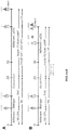
- the timeline of our oligodendrocyte differentiation protocol is shown in FIG. 1 .
- RA and SHH signaling mimic the pMN environment, inducing differentiation of the PSCs to OLIG2 progenitor cells.
- SHH signaling is activated through a Smoothened agonist instead of the human recombinant SHH protein.
- the present disclosure differs from recent work using iPSC-derived OPCs in that our iPSC lines are derived from PPMS patients. Further, the cells used for in vivo transplantation have been sorted using the late OPC marker O4, to maximally restrict the differentiation potential. Despite these differences, PPMS-derived O4 + -sorted OPCs exhibited similar engraftment efficiency, similar mitotic fraction and similar proportion of host ensheathed axons while generating fewer GFAP + astrocytes compared to unsorted iPSC-derived OPCs reported previously. Taken together, our data show that PPMS-derived OPCs performed in vivo at least as efficiently as healthy iPSC-derived cells (Wang et al. , 2013), and establish that our iPSC derivation and OPC induction protocols can generate myelinogenic oligodendrocytes from single-patient sample, which can be used in autologous cell-replacement therapies for MS.
- a particular instance of the disclosure is a method of generating OLIG2+ OPCs by first preparing PSC colonies.
- PSCs are seeded (plated) at low density and grown in an adherent culture for about 1-2 days.
- Low density means about 8,000 to about 11,000 cells/cm 2 .
- Cells are preferably seeded at about 9,500 to about 10,500 cells/cm 2 , more preferably at about 10,000 cells/cm 2 .
- the PSCs form colonies, which are preferably about 75 ⁇ m to about 300 ⁇ m in diameter, more preferably about 100 ⁇ m to about 250 ⁇ m in diameter.
- PSCs has its usual meaning in the art, i.e ., self-replicating cells that have the ability to develop into endoderm, ectoderm, and mesoderm cells.
- the PSCs described herein are hPSCs.
- hPSCs include ESCs and iPSCs, preferably hESCs and hiPSCs.
- PSCs can be seeded on a surface comprising a matrix, such as a gel or basement membrane matrix.
- a preferable matrix is the protein mixture secreted by Engelbreth-Holm-Swarm (EHS) mouse sarcoma cells, sold under trade names including MATRIGEL®, CULTREX®, and GELTREX®.
- Other suitable matrices include, without limitation, collagen, fibronectin, gelatin, laminin, poly-lysine, vitronectin, and combinations thereof.
- the medium in which PSCs are cultured preferably comprises an inhibitor of rho-associated protein kinase (ROCK), for example, GSK269962, GSK429286, H-1152, HA-1077, RKI-1447, thiazovivin, Y-27632, or derivatives thereof.
- ROCK rho-associated protein kinase
- the PSC colonies are then cultured in a monolayer to confluence in a medium comprising a low concentration of RA, at least one inhibitor of TGF ⁇ signaling, and at least one inhibitor of BMP signaling, wherein the first day of culturing in this medium is day 0.
- a "low concentration of RA" is about 10 nM to about 250 nM.
- the concentration of RA is preferably about 10 nM to about 100 nM, or about 25 nM to about 100 nM, or about 20 nM, 30 nM, 40 nM, 50 nM, 60 nM, 70 nM, 80 nM, 90 nM, and preferably, about 100 nM or less.
- Inhibitors of TGF ⁇ signaling described herein are GW788388, LDN193189, LY2109761, LY2157299, LY364947, and SB431542.
- Inhibitors of BMP signaling described herein are DMH1, dorsomorphin, K02288, Noggin, and LDN193189.
- SHH can be recombinant human SHH
- the medium lacks SHH
- the transition from PSCs to OLIG2 + progenitors is associated with massive proliferation causing the cultures to become overconfluent, resulting in the cells forming three-dimensional structures, ideally by about day 12.
- “Overconfluent” means that the cells begin piling up on one another, such that not all cells are in complete contact with the culture surface, and some cells are not in contact with the culture surface at all, but are only in contact with other cells. Preferably, at least about 50%, 60%, or 70% of the overconfluent cells are OLIG2+ by about day 12.
- OLIG2+ cells are to be further differentiated to O4+ cells, the overconfluent cells are lifted from the culture surface, which allows the formation of cell aggregates or spheres.
- OLIG2 - cells do not form aggregates, thus this process enriches for the OLIG2 + population, and OLIG2 - cells are eliminated gradually during subsequent media changes.
- aggregate and sphere are used interchangeably and refer to a multicellular three-dimensional structure, preferably, but not necessarily, of at least about 100 cells.
- Lifting can be performed mechanically, with a cell scraper or other suitable implement, or chemically.
- Chemical lifting can be achieved using a proteolytic enzyme, for example, collagenase, trypsin, trypsin-like proteinase, recombinant enzymes, such as that sold under the trade name TRYPLE®, naturally derived enzymes, such as that sold under the trade name ACCUTASE®, and combinations thereof.
- Chemical lifting can also be done using a chelator, such as EDTA, or a compound such as urea.
- Mechanical lifting or detachment offers the advantage of minimal cell death, however it produces aggregates of variable size, thus suitable spheres need to be selected through a manual picking process.
- Good spheres are defined as those having a round-shape, golden/brown color, with darker core and with a diameter between about 300 ⁇ m and about 800 ⁇ m.
- Detaching the cells using chemical methods, such as enzymatic digestion predominantly produces spheres that are appropriate for further culture. Therefore manual picking of spheres is not required, and the detachment steps can be adapted for automation and used in high throughput studies. However, enzymatic digestion increases cell death, resulting in a lower number of spheres.
- Three-dimensional aggregates of OLIG2+ OPCs are cultured in suspension in a medium comprising SAG and a low concentration of RA of 10nM to 250nM for about 8 days.
- the OLIG2+ OPCs can be generated a method of the invention, for example, as described above, or by other methods known in the art.
- the medium is changed to one comprising PDGF, HGF, IGF-1, and NT3, and optionally, insulin (preferably about 10 ⁇ g/ml to about 50 ⁇ g/ml, more preferably about 25 ⁇ g/ml), T3 (preferably about 20 ng/ml to about 100 ng/ml, more preferably about 60 ng/ml), biotin (preferably about 50 ng/ml to about 150 ng/ml, more preferably about 100 ng/ml), and/or cAMP (preferably about 100 nM to about 5 ⁇ M, more preferably about 1 ⁇ M).
- the medium preferably lacks bFGF and epidermal growth factor (EGF). If OLIG2+ cells are generated by the method of the invention, culture in suspension preferably begins on about day 12, and culture in the medium comprising PDGF, HGF, IGF-1, and NT3 preferably begins on about day 20.
- insulin preferably about 10 ⁇ g/ml to about 50 ⁇ g/ml, more
- the cell aggregates are plated in an adherent culture at a density of about 2 spheres/cm 2 .
- the surface on which the cell aggregates are plated and cultured can comprise an extracellular matrix protein (e.g., collagen, fibronectin, laminin) and/or a positively charged poly-amino acid (e.g ., poly-arginine, poly-lysine, poly-ornithine).
- the surface comprises laminin and/or poly-ornithine.
- the medium comprising PDGF, HGF, IGF-1, and NT3 can be continued (Option A), or a medium comprising AA and lacking growth factors (e.g ., PDGF, HGF, IGF-1, NT3, bFGF, and/or EGF) can be used (Option B).
- the medium comprising AA can optionally comprise insulin, T3, biotin, and/or cAMP.
- Cells cultured in the medium comprising PDGF, HGF, IGF-1, and NT3 are optimally O4+ by about 45 days after plating.
- At least about 35%, 40%, 45%, 50%, 55%, 60%, 65%, 70%, 75%, or 80% of these cells are O4+ by about 45 days after plating (day 75).
- Cells cultured in the medium comprising AA are optimally O4+ by about 25 days after plating.
- at least about 20%, 25%, 30%, 35%, or 40% of these cells are O4+ by about 25 days after plating (day 55).
- at least about 30%, 35%, 40%, 45%, 50%, 55%, or 60% of these cells are O4+ by about 33 days after plating (day 63).
- at least about 35%, 40%, 45%, 50%, 55%, 60%, 65%, 70%, or 75% of these cells are O4+ by about 45 days after plating (day 75).
- Mature oligodendrocytes expressing myelin basic protein can be generated by culturing the O4+ OPCs in the absence of PDGF, HGF, IGF-1, and NT3 for about three weeks, until cells are MBP+.
- at least about 20%, 25%, 30%, 35%, 40%, or 45% of the O4+ OPCs are MBP+ after about 20 days in culture in the medium lacking PDGF, HGF, IGF-1, and NT3. This occurs on about day 95 for "Option A” cells, and on about day 60 for "Option B” cells. Culturing "Option B" cells until at least about day 75 results in a higher efficiency of MBP+ expressing cells.
- the disclosure also encompasses OPCs, oligodendrocytes, and myelin-producing cells generated by the methods of the invention, and non-human mammals comprising them, preferably mice and/or rats.
- a myelin-producing cell is any cell that produces myelin, including without limitation, oligodendrocytes.
- myelin-producing cells are differentiated from PSCs, and in such instances, the PSCs can be iPSCs.
- the iPSCs can be derived from a somatic cell of a subject. In one instance, the subject has a demyelinating or dysmylelinating disease or disorder.
- the disclosure provides a method for generating viral and integration-free iPSCs from patients with MS, particularly PPMS.
- Our differentiation protocol can be used for the efficient differentiation of such iPSCs to OPCs and functional oligodendrocytes, as demonstrated by in vivo myelination in the shiverer mouse.
- the disclosure also provides a model system for a neurological disease, preferably a demyelinating or dysmyelinating disease or disorder.
- the model system comprises a myelin-producing cell differentiated from an iPSC derived from a subject having a demyelinating or dysmyelinating condition.
- the model system can further comprise a non-human mammal into which the myelin-producing cell has been transplanted.
- the non-human mammal is a mouse or a rat.
- Model systems provided by the disclosure can be used to study demyelinating or dysmyelinating diseases or disorders, including understanding underlying mechanisms and defining therapeutic targets.
- the disclosure also provides methods for treating and/or preventing a neurological disease or disorder in a subject by generating OPCs or oligodendrocytes according to a method of the invention; and administering an effective amount of the cells to the subject.
- the oligodendrocytes, or OPCs that have differentiated to oligodendrocytes in vivo promote myelinogenesis in the nervous system of the subject.
- the disclosure provides a use of the OPCs or oligodendrocytes of the disclosure in the treatment and/or prevention of a neurological disease or disorder in a subject.
- the neurological disease or disorder can be a demyelinating or dysmyelinating disease, or a neurodegenerative disease.
- the neurological disease or disorder can affect the central nervous system, the peripheral nervous system, or both.
- the demyelinating or dysmyleinating disease is an inflammatory demyelinating disease (such as multiple sclerosis, optic neuritis, Devic disease, acute-disseminated encephalomyelitis and transverse myelitis), viral demyelination, demyelination caused by acquired metabolic disorders, leukodystrophies (including hypomyelinating diseases, such as Pelizaeus-Merzbacher Disease and hereditary spastic paraplegia), X-linked disorders of proteo-lipid protein production, metabolic demyelinations and lysosomal storage disorders (such as metachromatic leukodystrophy-MLD, Tay-Sachs, Sandhoff's and Krabbe's diseases), vanishing white matter disease, and periventricular leukomalacia.
- MS and particularly PPMS are also conditions that can be treated or prevented by the methods of the disclosure.
- the OPCs or oligodendrocytes generated by a method of the disclosure are derived from iPSCs generated from a somatic cell of the subject.
- iPSC technology is emerging as a tool for developing new drugs and gaining insight into disease pathogenesis.
- the methods and cells of the disclosure will aid the development of high-throughput in vitro screens for compounds that promote myelination.
- the method comprising generating myelin-producing cell by a method of the invention; contacting the myelin-producing cell with a candidate compound; and determining whether the candidate compound promotes neuron myelination.
- the compound is a candidate therapeutic agent for treating a neurological disease or disorder, such as a demyelinating or dysmyelinating condition
- the method includes determining whether the candidate therapeutic agent has a beneficial effect on neuron myelination, wherein such beneficial effect is indicative of a candidate therapeutic agent for treating a demyelinating or dysmyelinating disease or disorder.
- the beneficial effect can be, for example, prevention of neuron demyelination, reduction of neuron demyelination, increased neuron conductance, and/or enhanced neuron myelination.
- the method is conducted in a high-throughput format.
- the cells, systems, and methods of the disclosure can also be useful for studying neurological diseases.
- the PPMS iPSC lines described here provide a new resource to investigate the process of neurodegeneration, particularly in MS ( FIG. 16 ).
- subject or “individual” or “patient” is meant any subject, particularly a mammalian subject, for whom diagnosis, prognosis, or therapy is desired.
- Mammalian subjects include humans, domestic animals, farm animals, sports animals, and zoo animals including, e.g., humans, non-human primates, dogs, cats, guinea pigs, rabbits, rats, mice, horses, cattle, pigs, and so on.
- Terms such as “treating” or “treatment” or “to treat” or “alleviating” or “to alleviate” refer to therapeutic measures that cure, slow down, lessen symptoms of, and/or halt progression of a diagnosed pathologic condition or disorder. Thus, those in need of treatment include those already with the disorder.
- a subject is successfully "treated” for a neurological disease or disorder, particularly a demyelinating or dysmyelinating disease or disorder, according to the methods provided herein if the patient shows, e.g., total, partial, or transient alleviation or elimination of symptoms associated with the disease or disorder.
- Prevent refers to prophylactic or preventative measures that prevent and/or slow the development of a targeted pathologic condition or disorder.
- those in need of prevention include those prone or susceptible to the disease or disorder.
- a neurological disease or disorder particularly a demyelinating or dysmyelinating disease or disorder, is successfully prevented according to the methods provided herein if the patient develops, transiently or permanently, e.g ., fewer or less severe symptoms associated with the disease or disorder, or a later onset of symptoms associated with the disease or disorder, than a patient who has not been subject to the methods of the disclosure.
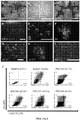
- FIG. 2B We then replaced the recombinant human SHH protein with SAG, which increased the yield further to 70.1% OLIG2 + progenitors ( FIG. 2B ).
- cells were detached and placed into low-attachment plates to promote their aggregation into spheres.
- the minimum number of cells required to form a sphere was at least 100 cells, and we noted that the majority of cells in the spheres were GFP + .
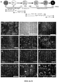
- PAX6 + cells arose at day 7, and by day 12 they arranged into multilayered structures ( FIG. 4B, 4C ). From day 12 to day 30, cells were grown as spheres and then plated onto poly-L-ornithine/laminin (pO/L)-coated dishes for the remainder of the protocol.
- pO/L poly-L-ornithine/laminin
- PDGF-AA, HGF, IGF1, and NT3 were added to the culture medium from day 20 onward.
- OLIG2 + progenitors upregulated NKX2.2, then SOX10, and finally matured to late OPCs identified by O4 live staining, and by their highly ramified processes ( FIG. 4D-4G ).
- O4 + OPCs expressing OLIG2, SOX10 and NG2 appeared as early as day 50 and their numbers increased dramatically around day 75.
- 40-50% of progenitor cells were proliferative, as indicated by Ki67 staining.
- the highly ramified O4 + cells did not divide in vitro ( FIG.
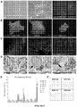
- O4 efficiencies ranged from 28% to 80% with nine different PSC lines, and the average was greater than 60% in four lines.
- Cells were stained with O4 antibody and analyzed by flow cytometry.
- One reference hESC line (RUES1) and eight hiPSC lines were tested. Technical replicates were performed using different batches of each line, at different passages. Results are also expressed as mean percentages ⁇ SEM. Table 1.
- O4 + OPCs can be purified through fluorescent activated cell sorting (FACS) and transplanted in vivo.
- FACS fluorescent activated cell sorting
- O4 + cells can also be cryopreserved immediately after sorting and thawed 24-48 hours prior transplantation.
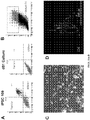
- OPCs are characterized by PDGFR ⁇ and NG2 expression, followed by expression of O4. Jakovcevski, I. et al., Front Neuroanat. 3:5 (2009 ). Under our culture conditions, by day 75, most O4 + cells have lost PDGFR ⁇ but have retained NG2 expression. At this stage we did not observe any residual pluripotent cells in culture.
- O4 + cells can either be isolated via FACS or further differentiated to MBP + oligodendrocytes ( FIG. 7G, 7H ).
- Other cell-types also exist in day 75 cultures, although at lower percentages.
- GFAP + cells in about 15% of the total cell population and about 20% ⁇ III-Tubulin + cells ( FIG. 7H ).
- GFAP + cells in about 15% of the total cell population and about 20% ⁇ III-Tubulin + cells ( FIG. 7H ).
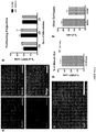
- FIG. 8C Expression profiling for seven pluripotency genes confirmed that all four iPSC lines exhibited a profile comparable to a reference hESC line and divergent from the parental fibroblasts ( FIG. 8C ). All iPSC lines displayed a normal karyotype ( FIG. 8D ) and were able to differentiate into cell types of the three germ layers, both in vitro, via spontaneous embryoid body differentiation ( FIG. 8E ), and in vivo via teratoma assay ( FIG. 8F ; FIG. 9 ).
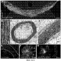
- hNA + cells were distributed throughout the corpus callosum and forebrain white matter.
- the density of hNA + cells in the corpus callosum at 12 weeks was 34,400 ⁇ 3,090 cells/mm 3 , and by 16 weeks, the number of human cells had approximately doubled since 12 weeks.
- FIG. 12E human MBP + oligodendrocytes were found diffusely throughout engrafted corpus callosum at 12 and 16 weeks ( FIG. 12A ). At 16 weeks, 31 ⁇ 3% of host mouse axons were ensheathed within the engrafted mouse corpus callosum ( FIG. 12B ).
- hNA + cells remained as NG2 + OPCs in the corpus callosum ( FIG. 12E ), and by 16 weeks they started to migrate to the overlying cerebral cortex ( FIG. 12F ).
- Very few O4-sorted cells underwent differentiation as hGFAP + astrocytes, and the majority of hGFAP + cells were localized to the SVZ and around the ventricles ( FIG. 12G ), suggesting that the local environment may induce astrocytic differentiation in these regions.
- hNESTIN-expressing cells were rarely found in corpus callosum and likewise concentrated in SVZ.
- ⁇ III-Tubulin + neurons were not detected in any of the engrafted animals.
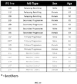
- Reference RUES1 and reference HUES 45 are both NIH-approved hESC lines; OLIG2-GFP reporter line is derived from reference BG01 hESC line (University of Texas Health Science Center at Houston).
- iPSC lines were derived in our laboratory from skin biopsies of PPMS patients through the mRNA/miRNA method (Stemgent).
- HUESM human Embryonic Stem Medium
- bFGF mouse embryonic fibroblast
- MEF mouse embryonic fibroblast
- HUESM is composed by Knockout-DMEM, 20% Knock-out serum, glutamax 2mM, NEAA 0.1mM, 1X P/S and ⁇ -mercaptoethanol 0.1mM, all purchased from Life Technologies (Grand Island, NY). At all stages of differentiation cells are cultured in 5% CO 2 incubators.
- PSCs were plated on MATRIGEL® (BD Biosciences; San Jose, CA) at a density of 10x10 3 cells/cm 2 in mTeSR1 medium (Stemcell Technologies; Vancouver, BC, Canada) containing 10 ⁇ M ROCK inhibitor, Y-27632 (Stemgent; Cambridge, MA) for 24 hours.
- This density of plated hPSCs was optimized to give a confluent well by day 8 and multilayered structures at day 12 of differentiation. This set up does not require significant PSC expansion, as only one well (80% confluent) of a 6-well plate contains enough cells to differentiate and isolate at least 2x10 6 oligodendrocytes. Cells were incubated for 1-2 days, until hPSC colonies reached a diameter of 100-250 ⁇ m.
- Neural Induction Medium which is mTeSR Custom Medium (Stemcell Technologies) containing the small molecules SB431542 10 ⁇ M (Stemgent) and LDN193189 250nM (Stemgent), as well as 100nM all-trans-RA (Sigma-Aldrich; St. Louis, MO).
- mTeSR Custom Medium has the same composition as the commercially available mTeSR-1 medium but without five factors that sustain pluripotency, namely lithium chloride, GABA, pipecolic acid, bFGF, and TGF ⁇ 1 (Stemcell Technologies).
- DMEM/F12 instead of DMEM/F12 with the addition of about 25 ⁇ g/ml insulin. Media changes were performed daily until day 8, with fresh RA, SB431542, and LDN193189 added to the medium every day.
- Aggregates were re-plated into Ultra-low attachment plates in N 2 B 27 Medium containing 1 ⁇ M SAG, changing it every other day.
- medium was switched to PDGF Medium, and 2/3 media changes were performed every other day.
- gentle pipetting was used to break apart any aggregates sticking to one another.
- spheres were plated onto plates coated with poly-L-ornithine hydrobromide (50 ⁇ g/ml; Sigma-Aldrich) and Laminin (20 ⁇ g/ml; Life Technologies) at a density of 2 spheres/cm 2 (about 20 spheres per well in a 6-well plate).
- plated spheres were cultured in a medium containing mitogens (Option A) or in a medium without any mitogens (Option B).
- Option A was optimized to obtain the highest yield of O4 + cells, while Option B was developed to provide a shorter and less costly version of the protocol.
- Spheres were plated on pO/L plates, as described above, in PDGF Medium at day 30, changing 2/3 of the PDGF Medium every other day until day 75 of differentiation.
- the appearance of O4 + cells was assessed by live O4 staining from day 55 onwards ( FIG. 6 ).
- O4+ OPCs could be isolated by FACS ( FIG. 7I ).
- FIG. 7G, 7H cells were cultured in Glial Medium from day 75, changing 2/3 of the medium every 3 days for two weeks.
- Spheres were plated on pO/L plates, as described above, in Glial Medium at day 30, changing 2/3 of the Glial Medium every other day until day 55 of differentiation.
- O4 + cells were visualized by live O4 staining ( FIG. 6A-F , FIG. 7F ) or isolated by FACS ( FIG. 6G ).
- O4+ OPCs could be isolated by FACS.
- cultures were kept in Glial Medium until day 75 to increase the efficiency of O4+ cells ( FIG. 6 ). We observed MBP+ cells beginning at about day 60.
- aggregates at day 30, and cells at the end of the differentiation could be cryopreserved with a viability >70%. Aggregates' viability is based on the number of thawed spheres that re-attach onto pO/L coated dishes after thawing.
- the sorted O4 + cells could be frozen immediately after sorting. The expected post-thaw viability of the sorted O4 + cells is 70 - 80%.
- Table 2 provides a list of media compositions used in the protocol. Table 2. Detailed Composition of Culture Media Media Components Provider Final Conc. N 2 Medium DMEM/F12 Life Technologies Glutamax (100X) Life Technologies 1X Non-Essential Amino Acids (100X) Life Technologies 1X ⁇ -Mercaptoethanol (1000X) Life Technologies 1X Penicillin-Streptomycin (100X) Life Technologies 1X N 2 Supplement (100X) Life Technologies 1X N2B27 N 2 Medium B 27 Supplement (50X) Life Technologies 1X PDGF Medium N 2 B 27 Medium PDGF R&D Systems 10ng/ml IGF-1 R&D Systems 10ng/ml HGF R&D Systems 5ng/ml NT3 EMD Millipore 10ng/ml Insulin Sigma-Aldrich 25 ⁇ g/ml Biotin Sigma-Aldrich 100ng/ml cAMP Sigma-Aldrich 1 ⁇ M T3 Sigma-Aldrich 60ng/ml Glial Medium N 2 B 27 Medium
- Skin biopsies were obtained from MS patients and healthy individuals ( FIG. 13 ).
- Four de-identified patients at the Tisch Multiple Sclerosis Research Center of New York were diagnosed with PPMS according to the standard diagnostic criteria. Their biopsies were obtained upon institutional review board approval (BRANY) and informed consent. All patients are Caucasian.
- Patients 102 and 107 are male, 56 and 61 years old respectively; patients 104 and 109 are female, 62 and 50 years old respectively.
- Biopsy Collection Medium consisting of RPMI 1460 (Life Technologies) and 1X Antibiotic-Antimycotic (Life Technologies). Biopsies were sliced into smaller pieces ( ⁇ 1mm) and plated onto a TC-treated 35mm dish for 5 minutes to dry and finally they were incubated in Biopsy Plating Medium, composed by Knockout DMEM, 2mM GLUTAMAXTM, 0.1mM NEAA, 0.1mM ⁇ -Mercaptoethanol, 10% Fetal Bovine Serum (FBS), 1X Penicillin-Streptomycin (P/S; all from Life Technologies) and 1% Nucleosides (EMD Millipore), for 5 days or until the first fibroblasts grew out of the biopsy.
- biopsies were digested with 1000U/ml Collagenase 1A (Sigma-Aldrich) for 1.5 hours at 37°C, washed, collected and plated onto 1% gelatin-coated 35mm dish in Biopsy Plating Medium for 5 days. Fibroblasts were then expanded in Culture Medium, consisting of DMEM (Life Technologies), 2mM GLUTAMAXTM, 0.1mM NEAA, 0.1mM ⁇ -Mercaptoethanol, 10% FBS and 1X P/S changing medium every other day.
- DMEM Life Technologies
- 2mM GLUTAMAXTM 2mM GLUTAMAXTM
- 0.1mM NEAA 0.1mM ⁇ -Mercaptoethanol
- 10% FBS and 1X P/S changing medium every other day.
- Skin fibroblasts at passage 3 to 5 were reprogrammed using the Stemgent mRNA/miRNA kit, which results in the generation of integration-free, virus free human iPSCs, through modified RNAs for OCT4 , SOX2 , KLF4 , cMYC and LIN28 ( FIG. 14 ).
- the addition of a specific cluster of miRNA has been found to increase the efficiency of reprogramming (Stemgent). Briefly, fibroblasts were plated onto Matrigel-coated 6-well or 12-well plates in a 5.5x10 3 cells/cm 2 density in culture medium. The following day, medium was replaced with NuFF-conditioned Pluriton reprogramming medium containing B18R.
- Cells were transfected for 11 consecutive days using STEMFECTTM as following: day 0 miRNA only, day1 to day3 mRNA cocktail only, d4 miRNA plus mRNA cocktail, day 5 to day 11 mRNA cocktail only. After day 11, visible colonies positively stained for live TRA-1-60 were picked and re-plated on MEFs with HLTESM medium.
- iPSC colonies were dissociated using Collagenase (Sigma-Aldrich) for 15 minutes at 37°C, washed, collected, and re-suspended in 200 ⁇ l HUESM. Cells were then mixed with 200 ⁇ l MatrigelTM (BD Biosciences) on ice, and were injected subcutaneously into immunodeficient mice (Jackson Laboratory; Bar Harbor, ME). Teratomas were allowed to grow for 9-12 weeks, isolated by dissection, and fixed in 4% PFA overnight at 4°C. Fixed tissues were embedded in paraffin, sectioned at 10 ⁇ m thickness, and stained with hematoxylin and eosin (H&E).
- Collagenase Sigma-Aldrich
- iPSCs were dissociated with ACCUTASE® (Life Technologies) for 5 minutes at 37°C and seeded into Ultra-Low attachment 6-well plates in HUESM without bFGF, changing media every other day. After 3 weeks of culture, embryoid bodies (EBs) were plated onto 1% gelatin-coated TC-treated dishes for another 2 weeks. EBs and their outgrowth were fixed in 4% PFA for 8 minutes at RT and immunostained for the appropriate markers.
- ACCUTASE® Life Technologies
- RNA isolation was performed using the RNeasy Plus Mini Kit with QIAshredder (Qiagen; Hilden, Germany). Briefly, cells were pelleted, washed with PBS, and re-suspended in lysis buffer. Samples were then stored at -80°C until processed further according to manufacturer's instructions. RNA was eluted in 30 ⁇ l RNase free ddH 2 O and quantified with a NanoDrop 8000 spectrophotometer (Thermo Scientific; Somerset, NJ).
- cDNA was synthesized using the GoScriptTM Reverse Transcription System (Promega; Madison, WI) with 0.5 ⁇ g of RNA and random primers. 20ng of cDNA were then loaded to a 96-well reaction plate together with 10 ⁇ l GoTaq® qPCR Master Mix and 1 ⁇ l of each primer (10nM) in a 20 ⁇ l reaction and the plate was ran in Stratagene Mx300P qPCR System (Agilent Technologies; Santa Clara, CA). Table 3 lists primer sequences. Table 3. Sequences of Primers Used for qRT-PCR SEQ ID. NO. Target gene Forward primer SEQ ID. NO.
- RNA was isolated from undifferentiated iPSCs and hESC HUES45 as previously described. RNA (100 ng/sample) was loaded for the hybridization with the specific Reporter Code Set and Capture Probe Set (NanoString Technologies; Seattle, WA) according to manufacturer's instructions. Data were normalized to the following housekeeping genes: ACTB , POLR2A , ALAS1. Data were expressed as fold changes to the expression of the hESC line (HUES45 1). See FIG. 15 .
- Images were acquired using an Olympus IX71 inverted microscope, equipped with Olympus DP30BW black and white digital camera for fluorescence and DP72 digital color camera for H&E staining. Fluorescent colors were digitally applied using the Olympus software DP Manager or with ImageJ. For counting, at least three non-overlapping fields were imported to ImageJ, thresholded and scored manually.
- Cells were enzymatically harvested by ACCUTASE® treatment for 25 min at 37°C to obtain a single cell suspension. Cells were then re-suspended in 100 ⁇ l of their respective medium containing the appropriate amount of either primary antibody or fluorescence-conjugated antibodies and were incubated on ice for 30 minutes shielded from light. When secondary antibodies were used, primary antibodies were washed with PBS and secondary antibodies were applied for 30 minutes on ice. Stained or GFP expressing cells were washed with PBS and sorted immediately on a 5 laser BD Biosciences ARIA-IIuTM Cell Sorter using the 100 ⁇ m ceramic nozzle, and 20 psi. DAPI was used for dead cell exclusion. Flow cytometry data were analyzed using BD FACSDivaTM software.
- Human cells were identified with mouse antihuman nuclei (hNA) and myelin basic protein-expressing oligodendrocytes were labeled with MBP.
- Human astrocytes and OPCs were stained with human-specific antibodies against hGFAP and hNG2 respectively.
- Mouse neurofilament (NF) was stained by 1:1 mixture of SMI311 and SMI312.
- InvitrogenTM Alexa Fluor secondary antibodies, goat anti-mouse 488, 594, and 647 were used at 1:500 dilution (Life Technologies).
- tissue was processed as described previously. Sim, F.J. et al., Molec. Cell. Neurosci. 20:669-682 (2002 ).
- Table 4 provides a list of primary antibodies used. Table 4.
Priority Applications (1)
| Application Number | Priority Date | Filing Date | Title |
|---|---|---|---|
| PL15796034T PL3145517T3 (pl) | 2014-05-22 | 2015-05-22 | Funkcjonalne oligodendrocyty pochodzące z pluripotencjalnych komórek macierzystych oraz sposoby ich otrzymywania i stosowania |
Applications Claiming Priority (2)
| Application Number | Priority Date | Filing Date | Title |
|---|---|---|---|
| US201462002048P | 2014-05-22 | 2014-05-22 | |
| PCT/US2015/032274 WO2015179822A1 (en) | 2014-05-22 | 2015-05-22 | Functional oligodendrocytes derived from pluripotent stem cells and methods of making and using the same |
Publications (3)
| Publication Number | Publication Date |
|---|---|
| EP3145517A1 EP3145517A1 (en) | 2017-03-29 |
| EP3145517A4 EP3145517A4 (en) | 2017-11-15 |
| EP3145517B1 true EP3145517B1 (en) | 2020-08-26 |
Family
ID=54554868
Family Applications (1)
| Application Number | Title | Priority Date | Filing Date |
|---|---|---|---|
| EP15796034.5A Active EP3145517B1 (en) | 2014-05-22 | 2015-05-22 | Functional oligodendrocytes derived from pluripotent stem cells and methods of making and using the same |
Country Status (10)
| Country | Link |
|---|---|
| US (3) | US10301592B2 (da) |
| EP (1) | EP3145517B1 (da) |
| JP (2) | JP6873705B2 (da) |
| KR (2) | KR102585909B1 (da) |
| CN (1) | CN106456672B (da) |
| AU (1) | AU2015263951B2 (da) |
| DK (1) | DK3145517T3 (da) |
| ES (1) | ES2846860T3 (da) |
| PL (1) | PL3145517T3 (da) |
| WO (1) | WO2015179822A1 (da) |
Families Citing this family (37)
| Publication number | Priority date | Publication date | Assignee | Title |
|---|---|---|---|---|
| CN106456672B (zh) * | 2014-05-22 | 2020-03-13 | 纽约干细胞基金会有限公司 | 源于多能干细胞的功能性少突胶质细胞及其制备和使用方法 |
| CN105567642B (zh) * | 2016-02-01 | 2019-07-12 | 中国科学院生物物理研究所 | 一种着色性干皮病人多能干细胞的制备方法 |
| US11920155B2 (en) | 2016-03-30 | 2024-03-05 | Asterias Biotherapeutics, Inc. | Oligodendrocyte progenitor cell compositions |
| SG11201909501TA (en) * | 2017-04-13 | 2019-11-28 | Univ Leland Stanford Junior | Personalized 3d neural culture system for generating human oligodendrocytes and studying myelination in vitro |
| JP2020526212A (ja) * | 2017-07-13 | 2020-08-31 | アリール バイオテクノロジー アンド ファーマシューティカルズ, インコーポレイテッド | ランドマーク転写因子を使用した幹細胞分化による神経前駆細胞、オリゴデンドロサイト前駆細胞、およびオリゴデンドロサイトの誘導 |
| CA3006897A1 (en) | 2017-08-04 | 2019-02-04 | University Health Network | Generation of oligodendrogenic neural progenitor cells |
| WO2019023793A1 (en) * | 2017-08-04 | 2019-02-07 | University Health Network | GENERATION OF OLIGODENDROGENIC NEURON PROGENITOR CELLS |
| CN107858331B (zh) * | 2017-11-02 | 2021-01-15 | 北京全式金生物技术有限公司 | 一种诱导人多能性干细胞分化为脊髓运动神经前体细胞的方法 |
| CN108384755A (zh) * | 2018-02-08 | 2018-08-10 | 北京呈诺医学科技有限公司 | 一种高效、快捷的诱导性多能干细胞向神经干细胞分化的方法 |
| US20210163888A1 (en) * | 2018-04-17 | 2021-06-03 | Case Western Reserve University | Induction of myelinating oligodendrocytes in human cortical spheroids |
| CN108624560B (zh) * | 2018-06-01 | 2022-04-08 | 南京艾尔普再生医学科技有限公司 | 一种分化培养基及少突胶质前体细胞的制备方法 |
| WO2019241620A1 (en) * | 2018-06-14 | 2019-12-19 | Academia Sinica | Method to generate induced oligodendrocyte-lineage cells and treatment using such cells |
| EP3660145A1 (en) * | 2018-11-30 | 2020-06-03 | Assistance Publique, Hopitaux De Paris | Use of oligodendrocytes from oral neuroectodermal stem cells in the repair of the nervous system |
| JP7469317B2 (ja) | 2019-01-23 | 2024-04-16 | アステリアス バイオセラピューティクス インコーポレイテッド | ヒト多能性幹細胞からの背側由来オリゴデンドロサイト前駆細胞 |
| US20220251504A1 (en) * | 2019-05-29 | 2022-08-11 | New York Stem Cell Foundation, Inc. | Functional astrocytes derived from pluripotent stem cells and methods of making and using the same |
| WO2020243392A1 (en) * | 2019-05-31 | 2020-12-03 | President And Fellows Of Harvard College | Sox9-induced oligodendrocyte progenitor cells |
| US20230025289A1 (en) | 2019-08-23 | 2023-01-26 | Sana Biotechnology, Inc. | Cd24 expressing cells and uses thereof |
| US20220323507A1 (en) * | 2019-09-06 | 2022-10-13 | Keio University | Method for Producing Cell Aggregate Including Glial Progenitor Cells |
| CN110716595A (zh) * | 2019-09-11 | 2020-01-21 | 合肥众建翔新能源有限公司 | 一种基于新能源的养殖系统 |
| AU2021241643A1 (en) | 2020-03-25 | 2022-11-24 | Sana Biotechnology, Inc. | Hypoimmunogenic neural cells for the treatment of neurological disorders and conditions |
| KR102657342B1 (ko) * | 2020-05-20 | 2024-04-15 | 고려대학교 산학협력단 | 희소돌기아교세포로 분화가 특화된 신경줄기세포로부터 희소돌기아교세포를 포함하는 오가노이드의 제조방법, 상기 방법으로 제조된 오가노이드 및 이의 용도 |
| KR20230074718A (ko) | 2020-08-13 | 2023-05-31 | 사나 바이오테크놀로지, 인크. | 저면역원성 세포로 감작된 환자를 치료하는 방법 및 관련 방법 및 조성물 |
| KR102568532B1 (ko) * | 2021-01-13 | 2023-08-18 | 건국대학교 산학협력단 | 3차원 배양을 통한 신경줄기세포 제조 방법 |
| US20220315891A1 (en) * | 2021-03-30 | 2022-10-06 | Trailhead Biosystems Inc. | Methods and compositions for generating oligodendrocyte progenitor cells |
| AU2022283291A1 (en) | 2021-05-27 | 2023-11-02 | Sana Biotechnology, Inc. | Hypoimmunogenic cells comprising engineered hla-e or hla-g |
| WO2023287827A2 (en) | 2021-07-14 | 2023-01-19 | Sana Biotechnology, Inc. | Altered expression of y chromosome-linked antigens in hypoimmunogenic cells |
| CN113564122B (zh) * | 2021-08-05 | 2022-04-08 | 呈诺再生医学科技(珠海横琴新区)有限公司 | 人诱导性多能干细胞向少突胶质细胞分化的方法,试剂盒以及应用 |
| WO2023019227A1 (en) | 2021-08-11 | 2023-02-16 | Sana Biotechnology, Inc. | Genetically modified cells for allogeneic cell therapy to reduce complement-mediated inflammatory reactions |
| AU2022325955A1 (en) | 2021-08-11 | 2024-02-08 | Sana Biotechnology, Inc. | Genetically modified cells for allogeneic cell therapy to reduce instant blood mediated inflammatory reactions |
| CA3227613A1 (en) | 2021-08-11 | 2023-02-16 | William Dowdle | Inducible systems for altering gene expression in hypoimmunogenic cells |
| WO2023019226A1 (en) | 2021-08-11 | 2023-02-16 | Sana Biotechnology, Inc. | Genetically modified cells for allogeneic cell therapy |
| CN113699117B (zh) * | 2021-09-03 | 2023-06-30 | 呈诺再生医学科技(北京)有限公司 | 经基因改造的少突胶质祖细胞在多发性硬化症中的应用 |
| CN114262686B (zh) * | 2022-03-02 | 2022-06-24 | 深圳市夏同生物医药科技有限公司 | 一种少突胶质细胞的制备方法及应用 |
| WO2023183313A1 (en) | 2022-03-22 | 2023-09-28 | Sana Biotechnology, Inc. | Engineering cells with a transgene in b2m or ciita locus and associated compositions and methods |
| KR20230171226A (ko) * | 2022-06-13 | 2023-12-20 | 고려대학교 산학협력단 | 키나아제 저해제를 유효성분으로 포함하는 탈수초성 질환 예방 또는 치료용 약학적 조성물 |
| US20240067917A1 (en) * | 2022-08-15 | 2024-02-29 | Cellino Biotech, Inc. | Systems and methods for cell manufacturing |
| CN115322965B (zh) * | 2022-08-19 | 2024-01-30 | 同济大学 | 一种体外获得后脑底板细胞的方法、成套培养基及应用 |
Family Cites Families (5)
| Publication number | Priority date | Publication date | Assignee | Title |
|---|---|---|---|---|
| WO2004052389A2 (en) * | 2002-12-06 | 2004-06-24 | Singapore General Hospital Pte Ltd. | Nogo, caspr, f3 nb-3 useful in the treatment of injury and disease to the central nervous system |
| EP2064319B1 (en) * | 2006-08-28 | 2017-02-22 | Yeda Research and Development Co. Ltd. | Methods of generating glial and neuronal cells and use of same for the treatment of medical conditions of the cns |
| US8227247B2 (en) * | 2007-12-20 | 2012-07-24 | Wisconsin Alumni Research Foundation | Method of generating myelinating oligodendrocytes |
| WO2015124725A1 (en) * | 2014-02-21 | 2015-08-27 | Johan Ericson | Late born central nervous system cell types |
| CN106456672B (zh) * | 2014-05-22 | 2020-03-13 | 纽约干细胞基金会有限公司 | 源于多能干细胞的功能性少突胶质细胞及其制备和使用方法 |
-
2015
- 2015-05-22 CN CN201580033959.8A patent/CN106456672B/zh active Active
- 2015-05-22 JP JP2016568940A patent/JP6873705B2/ja active Active
- 2015-05-22 EP EP15796034.5A patent/EP3145517B1/en active Active
- 2015-05-22 DK DK15796034.5T patent/DK3145517T3/da active
- 2015-05-22 KR KR1020217036151A patent/KR102585909B1/ko active IP Right Grant
- 2015-05-22 KR KR1020167034668A patent/KR102324714B1/ko active IP Right Grant
- 2015-05-22 ES ES15796034T patent/ES2846860T3/es active Active
- 2015-05-22 PL PL15796034T patent/PL3145517T3/pl unknown
- 2015-05-22 US US15/313,079 patent/US10301592B2/en active Active
- 2015-05-22 AU AU2015263951A patent/AU2015263951B2/en active Active
- 2015-05-22 WO PCT/US2015/032274 patent/WO2015179822A1/en active Application Filing
-
2019
- 2019-05-06 US US16/404,502 patent/US10676716B2/en active Active
-
2020
- 2020-06-03 US US16/892,181 patent/US11814648B2/en active Active
-
2021
- 2021-04-21 JP JP2021071974A patent/JP7410903B2/ja active Active
Non-Patent Citations (1)
| Title |
|---|
| None * |
Also Published As
| Publication number | Publication date |
|---|---|
| KR20210138783A (ko) | 2021-11-19 |
| KR102324714B1 (ko) | 2021-11-11 |
| AU2015263951A1 (en) | 2016-12-01 |
| US20200399595A1 (en) | 2020-12-24 |
| EP3145517A4 (en) | 2017-11-15 |
| EP3145517A1 (en) | 2017-03-29 |
| JP6873705B2 (ja) | 2021-05-19 |
| JP2017524340A (ja) | 2017-08-31 |
| US20190256820A1 (en) | 2019-08-22 |
| US10676716B2 (en) | 2020-06-09 |
| US10301592B2 (en) | 2019-05-28 |
| US20170183627A1 (en) | 2017-06-29 |
| JP2021118718A (ja) | 2021-08-12 |
| DK3145517T3 (da) | 2020-11-30 |
| JP7410903B2 (ja) | 2024-01-10 |
| ES2846860T3 (es) | 2021-07-29 |
| PL3145517T3 (pl) | 2021-04-06 |
| CN106456672B (zh) | 2020-03-13 |
| CN106456672A (zh) | 2017-02-22 |
| US11814648B2 (en) | 2023-11-14 |
| WO2015179822A1 (en) | 2015-11-26 |
| KR20170005844A (ko) | 2017-01-16 |
| KR102585909B1 (ko) | 2023-10-05 |
| AU2015263951B2 (en) | 2020-07-02 |
Similar Documents
| Publication | Publication Date | Title |
|---|---|---|
| US11814648B2 (en) | Functional oligodendrocytes derived from pluripotent stem cells and methods of making and using the same | |
| US20240067924A1 (en) | Specification of functional cranial placode derivatives from human pluripotent stem cells | |
| Curchoe et al. | Early acquisition of neural crest competence during hESCs neuralization | |
| Meneghini et al. | Generation of human induced pluripotent stem cell-derived bona fide neural stem cells for ex vivo gene therapy of metachromatic leukodystrophy | |
| KR101874463B1 (ko) | 세포의 재프로그램화 방법 및 이의 용도 | |
| EP3348631B1 (en) | Method for producing retinal tissue | |
| CN102597218A (zh) | 用于诱导多能干细胞分化成神经前体细胞的方法 | |
| KR20160068982A (ko) | 신규 연골 세포 유도 방법 | |
| Massumi et al. | Efficient programming of human eye conjunctiva-derived induced pluripotent stem (ECiPS) cells into definitive endoderm-like cells | |
| WO2013011093A1 (en) | Novel method for generation of neural progenitor cells | |
| US20220251504A1 (en) | Functional astrocytes derived from pluripotent stem cells and methods of making and using the same | |
| US20070020608A1 (en) | Method for the generation of neural progenitor cells | |
| EP3950933A1 (en) | Cell population including pluripotent stem cells and production method thereof | |
| JPWO2018199142A1 (ja) | 神経堤細胞および交感神経細胞の製造方法 | |
| Sim et al. | Panagiotis Douvaras, Jing Wang, 2 Matthew Zimmer, Stephanie Hanchuk, Melanie A. O’Bara, 2 Saud Sadiq, 3 | |
| WO2018185260A1 (en) | Novel human hepatocyte-derived matrix for stem-cell differentiation and tissue repair | |
| US20220135940A1 (en) | Method for producing kidney structure having dendritically branched collecting duct from pluripotent stem cells | |
| WO2023010209A1 (en) | Neural progenitor cells and therapeutic uses of same | |
| Kaur et al. | Neural stem cell assays | |
| Chaddah | Clonal derivation of neural stem cells from human embryonic stem cells | |
| Gagliardi | Neural differentiation and reprogramming of rhesus cells | |
| Gonzalez | Molecular mechanisms controlling human embryonic stem cell self-renewal and differentiation |
Legal Events
| Date | Code | Title | Description |
|---|---|---|---|
| STAA | Information on the status of an ep patent application or granted ep patent |
Free format text: STATUS: THE INTERNATIONAL PUBLICATION HAS BEEN MADE |
|
| PUAI | Public reference made under article 153(3) epc to a published international application that has entered the european phase |
Free format text: ORIGINAL CODE: 0009012 |
|
| STAA | Information on the status of an ep patent application or granted ep patent |
Free format text: STATUS: REQUEST FOR EXAMINATION WAS MADE |
|
| 17P | Request for examination filed |
Effective date: 20161215 |
|
| AK | Designated contracting states |
Kind code of ref document: A1 Designated state(s): AL AT BE BG CH CY CZ DE DK EE ES FI FR GB GR HR HU IE IS IT LI LT LU LV MC MK MT NL NO PL PT RO RS SE SI SK SM TR |
|
| AX | Request for extension of the european patent |
Extension state: BA ME |
|
| DAV | Request for validation of the european patent (deleted) | ||
| DAX | Request for extension of the european patent (deleted) | ||
| A4 | Supplementary search report drawn up and despatched |
Effective date: 20171017 |
|
| RIC1 | Information provided on ipc code assigned before grant |
Ipc: C12N 5/079 20100101ALI20171011BHEP Ipc: A61K 35/30 20150101AFI20171011BHEP |
|
| REG | Reference to a national code |
Ref country code: HK Ref legal event code: DE Ref document number: 1233498 Country of ref document: HK |
|
| STAA | Information on the status of an ep patent application or granted ep patent |
Free format text: STATUS: EXAMINATION IS IN PROGRESS |
|
| 17Q | First examination report despatched |
Effective date: 20180719 |
|
| GRAP | Despatch of communication of intention to grant a patent |
Free format text: ORIGINAL CODE: EPIDOSNIGR1 |
|
| STAA | Information on the status of an ep patent application or granted ep patent |
Free format text: STATUS: GRANT OF PATENT IS INTENDED |
|
| INTG | Intention to grant announced |
Effective date: 20200327 |
|
| GRAS | Grant fee paid |
Free format text: ORIGINAL CODE: EPIDOSNIGR3 |
|
| GRAA | (expected) grant |
Free format text: ORIGINAL CODE: 0009210 |
|
| STAA | Information on the status of an ep patent application or granted ep patent |
Free format text: STATUS: THE PATENT HAS BEEN GRANTED |
|
| AK | Designated contracting states |
Kind code of ref document: B1 Designated state(s): AL AT BE BG CH CY CZ DE DK EE ES FI FR GB GR HR HU IE IS IT LI LT LU LV MC MK MT NL NO PL PT RO RS SE SI SK SM TR |
|
| REG | Reference to a national code |
Ref country code: GB Ref legal event code: FG4D |
|
| REG | Reference to a national code |
Ref country code: CH Ref legal event code: EP |
|
| REG | Reference to a national code |
Ref country code: AT Ref legal event code: REF Ref document number: 1305699 Country of ref document: AT Kind code of ref document: T Effective date: 20200915 |
|
| REG | Reference to a national code |
Ref country code: IE Ref legal event code: FG4D |
|
| REG | Reference to a national code |
Ref country code: DE Ref legal event code: R096 Ref document number: 602015058072 Country of ref document: DE |
|
| REG | Reference to a national code |
Ref country code: CH Ref legal event code: NV Representative=s name: NOVAGRAAF INTERNATIONAL SA, CH Ref country code: DK Ref legal event code: T3 Effective date: 20201124 |
|
| REG | Reference to a national code |
Ref country code: SE Ref legal event code: TRGR |
|
| REG | Reference to a national code |
Ref country code: NL Ref legal event code: FP |
|
| REG | Reference to a national code |
Ref country code: LT Ref legal event code: MG4D |
|
| PG25 | Lapsed in a contracting state [announced via postgrant information from national office to epo] |
Ref country code: FI Free format text: LAPSE BECAUSE OF FAILURE TO SUBMIT A TRANSLATION OF THE DESCRIPTION OR TO PAY THE FEE WITHIN THE PRESCRIBED TIME-LIMIT Effective date: 20200826 Ref country code: LT Free format text: LAPSE BECAUSE OF FAILURE TO SUBMIT A TRANSLATION OF THE DESCRIPTION OR TO PAY THE FEE WITHIN THE PRESCRIBED TIME-LIMIT Effective date: 20200826 Ref country code: BG Free format text: LAPSE BECAUSE OF FAILURE TO SUBMIT A TRANSLATION OF THE DESCRIPTION OR TO PAY THE FEE WITHIN THE PRESCRIBED TIME-LIMIT Effective date: 20201126 Ref country code: PT Free format text: LAPSE BECAUSE OF FAILURE TO SUBMIT A TRANSLATION OF THE DESCRIPTION OR TO PAY THE FEE WITHIN THE PRESCRIBED TIME-LIMIT Effective date: 20201228 Ref country code: NO Free format text: LAPSE BECAUSE OF FAILURE TO SUBMIT A TRANSLATION OF THE DESCRIPTION OR TO PAY THE FEE WITHIN THE PRESCRIBED TIME-LIMIT Effective date: 20201126 Ref country code: GR Free format text: LAPSE BECAUSE OF FAILURE TO SUBMIT A TRANSLATION OF THE DESCRIPTION OR TO PAY THE FEE WITHIN THE PRESCRIBED TIME-LIMIT Effective date: 20201127 Ref country code: HR Free format text: LAPSE BECAUSE OF FAILURE TO SUBMIT A TRANSLATION OF THE DESCRIPTION OR TO PAY THE FEE WITHIN THE PRESCRIBED TIME-LIMIT Effective date: 20200826 |
|
| REG | Reference to a national code |
Ref country code: AT Ref legal event code: MK05 Ref document number: 1305699 Country of ref document: AT Kind code of ref document: T Effective date: 20200826 |
|
| PG25 | Lapsed in a contracting state [announced via postgrant information from national office to epo] |
Ref country code: LV Free format text: LAPSE BECAUSE OF FAILURE TO SUBMIT A TRANSLATION OF THE DESCRIPTION OR TO PAY THE FEE WITHIN THE PRESCRIBED TIME-LIMIT Effective date: 20200826 Ref country code: RS Free format text: LAPSE BECAUSE OF FAILURE TO SUBMIT A TRANSLATION OF THE DESCRIPTION OR TO PAY THE FEE WITHIN THE PRESCRIBED TIME-LIMIT Effective date: 20200826 Ref country code: IS Free format text: LAPSE BECAUSE OF FAILURE TO SUBMIT A TRANSLATION OF THE DESCRIPTION OR TO PAY THE FEE WITHIN THE PRESCRIBED TIME-LIMIT Effective date: 20201226 |
|
| PG25 | Lapsed in a contracting state [announced via postgrant information from national office to epo] |
Ref country code: EE Free format text: LAPSE BECAUSE OF FAILURE TO SUBMIT A TRANSLATION OF THE DESCRIPTION OR TO PAY THE FEE WITHIN THE PRESCRIBED TIME-LIMIT Effective date: 20200826 Ref country code: SM Free format text: LAPSE BECAUSE OF FAILURE TO SUBMIT A TRANSLATION OF THE DESCRIPTION OR TO PAY THE FEE WITHIN THE PRESCRIBED TIME-LIMIT Effective date: 20200826 Ref country code: RO Free format text: LAPSE BECAUSE OF FAILURE TO SUBMIT A TRANSLATION OF THE DESCRIPTION OR TO PAY THE FEE WITHIN THE PRESCRIBED TIME-LIMIT Effective date: 20200826 Ref country code: CZ Free format text: LAPSE BECAUSE OF FAILURE TO SUBMIT A TRANSLATION OF THE DESCRIPTION OR TO PAY THE FEE WITHIN THE PRESCRIBED TIME-LIMIT Effective date: 20200826 |
|
| REG | Reference to a national code |
Ref country code: DE Ref legal event code: R097 Ref document number: 602015058072 Country of ref document: DE |
|
| PG25 | Lapsed in a contracting state [announced via postgrant information from national office to epo] |
Ref country code: AL Free format text: LAPSE BECAUSE OF FAILURE TO SUBMIT A TRANSLATION OF THE DESCRIPTION OR TO PAY THE FEE WITHIN THE PRESCRIBED TIME-LIMIT Effective date: 20200826 Ref country code: AT Free format text: LAPSE BECAUSE OF FAILURE TO SUBMIT A TRANSLATION OF THE DESCRIPTION OR TO PAY THE FEE WITHIN THE PRESCRIBED TIME-LIMIT Effective date: 20200826 |
|
| PG25 | Lapsed in a contracting state [announced via postgrant information from national office to epo] |
Ref country code: SK Free format text: LAPSE BECAUSE OF FAILURE TO SUBMIT A TRANSLATION OF THE DESCRIPTION OR TO PAY THE FEE WITHIN THE PRESCRIBED TIME-LIMIT Effective date: 20200826 |
|
| PLBE | No opposition filed within time limit |
Free format text: ORIGINAL CODE: 0009261 |
|
| STAA | Information on the status of an ep patent application or granted ep patent |
Free format text: STATUS: NO OPPOSITION FILED WITHIN TIME LIMIT |
|
| REG | Reference to a national code |
Ref country code: ES Ref legal event code: FG2A Ref document number: 2846860 Country of ref document: ES Kind code of ref document: T3 Effective date: 20210729 |
|
| 26N | No opposition filed |
Effective date: 20210527 |
|
| PG25 | Lapsed in a contracting state [announced via postgrant information from national office to epo] |
Ref country code: SI Free format text: LAPSE BECAUSE OF FAILURE TO SUBMIT A TRANSLATION OF THE DESCRIPTION OR TO PAY THE FEE WITHIN THE PRESCRIBED TIME-LIMIT Effective date: 20200826 |
|
| PG25 | Lapsed in a contracting state [announced via postgrant information from national office to epo] |
Ref country code: LU Free format text: LAPSE BECAUSE OF NON-PAYMENT OF DUE FEES Effective date: 20210522 Ref country code: MC Free format text: LAPSE BECAUSE OF FAILURE TO SUBMIT A TRANSLATION OF THE DESCRIPTION OR TO PAY THE FEE WITHIN THE PRESCRIBED TIME-LIMIT Effective date: 20200826 |
|
| PG25 | Lapsed in a contracting state [announced via postgrant information from national office to epo] |
Ref country code: HU Free format text: LAPSE BECAUSE OF FAILURE TO SUBMIT A TRANSLATION OF THE DESCRIPTION OR TO PAY THE FEE WITHIN THE PRESCRIBED TIME-LIMIT; INVALID AB INITIO Effective date: 20150522 |
|
| P01 | Opt-out of the competence of the unified patent court (upc) registered |
Effective date: 20230507 |
|
| PG25 | Lapsed in a contracting state [announced via postgrant information from national office to epo] |
Ref country code: CY Free format text: LAPSE BECAUSE OF FAILURE TO SUBMIT A TRANSLATION OF THE DESCRIPTION OR TO PAY THE FEE WITHIN THE PRESCRIBED TIME-LIMIT Effective date: 20200826 |
|
| PGFP | Annual fee paid to national office [announced via postgrant information from national office to epo] |
Ref country code: NL Payment date: 20230526 Year of fee payment: 9 Ref country code: IT Payment date: 20230519 Year of fee payment: 9 Ref country code: IE Payment date: 20230529 Year of fee payment: 9 Ref country code: FR Payment date: 20230525 Year of fee payment: 9 Ref country code: ES Payment date: 20230601 Year of fee payment: 9 Ref country code: DK Payment date: 20230530 Year of fee payment: 9 Ref country code: DE Payment date: 20230530 Year of fee payment: 9 Ref country code: CH Payment date: 20230610 Year of fee payment: 9 |
|
| PGFP | Annual fee paid to national office [announced via postgrant information from national office to epo] |
Ref country code: SE Payment date: 20230527 Year of fee payment: 9 Ref country code: PL Payment date: 20230518 Year of fee payment: 9 |
|
| PGFP | Annual fee paid to national office [announced via postgrant information from national office to epo] |
Ref country code: BE Payment date: 20230529 Year of fee payment: 9 |
|
| PGFP | Annual fee paid to national office [announced via postgrant information from national office to epo] |
Ref country code: GB Payment date: 20230529 Year of fee payment: 9 |
|
| PG25 | Lapsed in a contracting state [announced via postgrant information from national office to epo] |
Ref country code: MK Free format text: LAPSE BECAUSE OF FAILURE TO SUBMIT A TRANSLATION OF THE DESCRIPTION OR TO PAY THE FEE WITHIN THE PRESCRIBED TIME-LIMIT Effective date: 20200826 |