EP3141904A1 - Procédés, dispositifs et kits de détection ou surveillance d'une lésion rénale aiguë - Google Patents
Procédés, dispositifs et kits de détection ou surveillance d'une lésion rénale aiguë Download PDFInfo
- Publication number
- EP3141904A1 EP3141904A1 EP16191697.8A EP16191697A EP3141904A1 EP 3141904 A1 EP3141904 A1 EP 3141904A1 EP 16191697 A EP16191697 A EP 16191697A EP 3141904 A1 EP3141904 A1 EP 3141904A1
- Authority
- EP
- European Patent Office
- Prior art keywords
- ngal
- antibody
- protein
- sample
- polyclonal
- Prior art date
- Legal status (The legal status is an assumption and is not a legal conclusion. Google has not performed a legal analysis and makes no representation as to the accuracy of the status listed.)
- Granted
Links
- 238000000034 method Methods 0.000 title claims abstract description 60
- 208000009304 Acute Kidney Injury Diseases 0.000 title claims abstract description 42
- 208000033626 Renal failure acute Diseases 0.000 title claims abstract description 42
- 201000011040 acute kidney failure Diseases 0.000 title claims abstract description 42
- 238000012544 monitoring process Methods 0.000 title description 6
- 102000013519 Lipocalin-2 Human genes 0.000 claims abstract description 324
- 108010051335 Lipocalin-2 Proteins 0.000 claims abstract description 324
- 210000001124 body fluid Anatomy 0.000 claims abstract description 20
- 239000010839 body fluid Substances 0.000 claims abstract description 20
- 238000011282 treatment Methods 0.000 claims abstract description 15
- 230000000536 complexating effect Effects 0.000 claims abstract description 6
- 210000002700 urine Anatomy 0.000 claims description 65
- 238000002965 ELISA Methods 0.000 claims description 51
- 210000000440 neutrophil Anatomy 0.000 claims description 25
- 238000007675 cardiac surgery Methods 0.000 claims description 19
- 210000003734 kidney Anatomy 0.000 claims description 17
- 230000002980 postoperative effect Effects 0.000 claims description 15
- 108090000623 proteins and genes Proteins 0.000 claims description 15
- 102000004169 proteins and genes Human genes 0.000 claims description 13
- 238000003556 assay Methods 0.000 abstract description 96
- 238000003745 diagnosis Methods 0.000 abstract description 3
- 239000000523 sample Substances 0.000 description 54
- DDRJAANPRJIHGJ-UHFFFAOYSA-N creatinine Chemical compound CN1CC(=O)NC1=N DDRJAANPRJIHGJ-UHFFFAOYSA-N 0.000 description 32
- 210000004027 cell Anatomy 0.000 description 28
- 238000003127 radioimmunoassay Methods 0.000 description 24
- 229940109239 creatinine Drugs 0.000 description 16
- 241000473945 Theria <moth genus> Species 0.000 description 15
- 238000001262 western blot Methods 0.000 description 15
- 238000005259 measurement Methods 0.000 description 12
- 235000018102 proteins Nutrition 0.000 description 12
- 239000001963 growth medium Substances 0.000 description 11
- 239000006228 supernatant Substances 0.000 description 11
- 210000002381 plasma Anatomy 0.000 description 9
- MZOFCQQQCNRIBI-VMXHOPILSA-N (3s)-4-[[(2s)-1-[[(2s)-1-[[(1s)-1-carboxy-2-hydroxyethyl]amino]-4-methyl-1-oxopentan-2-yl]amino]-5-(diaminomethylideneamino)-1-oxopentan-2-yl]amino]-3-[[2-[[(2s)-2,6-diaminohexanoyl]amino]acetyl]amino]-4-oxobutanoic acid Chemical compound OC[C@@H](C(O)=O)NC(=O)[C@H](CC(C)C)NC(=O)[C@H](CCCN=C(N)N)NC(=O)[C@H](CC(O)=O)NC(=O)CNC(=O)[C@@H](N)CCCCN MZOFCQQQCNRIBI-VMXHOPILSA-N 0.000 description 8
- 108060008682 Tumor Necrosis Factor Proteins 0.000 description 8
- 102000000852 Tumor Necrosis Factor-alpha Human genes 0.000 description 8
- 239000000758 substrate Substances 0.000 description 8
- 241000283973 Oryctolagus cuniculus Species 0.000 description 7
- 238000001514 detection method Methods 0.000 description 7
- 238000002523 gelfiltration Methods 0.000 description 7
- 230000003907 kidney function Effects 0.000 description 7
- 230000035945 sensitivity Effects 0.000 description 7
- 102000012192 Cystatin C Human genes 0.000 description 6
- 108010061642 Cystatin C Proteins 0.000 description 6
- 239000003550 marker Substances 0.000 description 6
- 238000011002 quantification Methods 0.000 description 6
- 239000000243 solution Substances 0.000 description 6
- 238000000540 analysis of variance Methods 0.000 description 5
- 239000000090 biomarker Substances 0.000 description 5
- 210000004369 blood Anatomy 0.000 description 5
- 239000008280 blood Substances 0.000 description 5
- 230000029142 excretion Effects 0.000 description 5
- 238000002474 experimental method Methods 0.000 description 5
- 230000014509 gene expression Effects 0.000 description 5
- 238000003752 polymerase chain reaction Methods 0.000 description 5
- 102000004127 Cytokines Human genes 0.000 description 4
- 108090000695 Cytokines Proteins 0.000 description 4
- 238000012286 ELISA Assay Methods 0.000 description 4
- 239000002033 PVDF binder Substances 0.000 description 4
- 238000004458 analytical method Methods 0.000 description 4
- 230000015572 biosynthetic process Effects 0.000 description 4
- 239000001913 cellulose Substances 0.000 description 4
- 229920002678 cellulose Polymers 0.000 description 4
- 239000003636 conditioned culture medium Substances 0.000 description 4
- 238000011534 incubation Methods 0.000 description 4
- 239000012528 membrane Substances 0.000 description 4
- 239000000178 monomer Substances 0.000 description 4
- 229940126619 mouse monoclonal antibody Drugs 0.000 description 4
- 238000002264 polyacrylamide gel electrophoresis Methods 0.000 description 4
- 229920002981 polyvinylidene fluoride Polymers 0.000 description 4
- 238000001356 surgical procedure Methods 0.000 description 4
- 238000003786 synthesis reaction Methods 0.000 description 4
- 108010085238 Actins Proteins 0.000 description 3
- 102000007469 Actins Human genes 0.000 description 3
- 101001023833 Homo sapiens Neutrophil gelatinase-associated lipocalin Proteins 0.000 description 3
- 108010015302 Matrix metalloproteinase-9 Proteins 0.000 description 3
- 241000699666 Mus <mouse, genus> Species 0.000 description 3
- 229920001213 Polysorbate 20 Polymers 0.000 description 3
- 238000000692 Student's t-test Methods 0.000 description 3
- 230000001154 acute effect Effects 0.000 description 3
- 238000005119 centrifugation Methods 0.000 description 3
- 239000012228 culture supernatant Substances 0.000 description 3
- 230000006378 damage Effects 0.000 description 3
- 238000001378 electrochemiluminescence detection Methods 0.000 description 3
- 210000000981 epithelium Anatomy 0.000 description 3
- 239000000284 extract Substances 0.000 description 3
- 239000008187 granular material Substances 0.000 description 3
- 102000047202 human LCN2 Human genes 0.000 description 3
- 238000001727 in vivo Methods 0.000 description 3
- 208000015181 infectious disease Diseases 0.000 description 3
- 210000002510 keratinocyte Anatomy 0.000 description 3
- 238000012417 linear regression Methods 0.000 description 3
- 239000002609 medium Substances 0.000 description 3
- 108020004999 messenger RNA Proteins 0.000 description 3
- 239000000203 mixture Substances 0.000 description 3
- 230000036470 plasma concentration Effects 0.000 description 3
- 239000000256 polyoxyethylene sorbitan monolaurate Substances 0.000 description 3
- 235000010486 polyoxyethylene sorbitan monolaurate Nutrition 0.000 description 3
- 239000000047 product Substances 0.000 description 3
- 210000002966 serum Anatomy 0.000 description 3
- 239000012679 serum free medium Substances 0.000 description 3
- 210000004926 tubular epithelial cell Anatomy 0.000 description 3
- VBEQCZHXXJYVRD-GACYYNSASA-N uroanthelone Chemical compound C([C@@H](C(=O)N[C@H](C(=O)N[C@@H](CS)C(=O)N[C@@H](CC(N)=O)C(=O)N[C@@H](CS)C(=O)N[C@H](C(=O)N[C@@H]([C@@H](C)CC)C(=O)NCC(=O)N[C@@H](CC=1C=CC(O)=CC=1)C(=O)N[C@@H](CO)C(=O)NCC(=O)N[C@@H](CC(O)=O)C(=O)N[C@@H](CCCNC(N)=N)C(=O)N[C@@H](CS)C(=O)N[C@@H](CCC(N)=O)C(=O)N[C@@H]([C@@H](C)O)C(=O)N[C@@H](CCCNC(N)=N)C(=O)N[C@@H](CC(O)=O)C(=O)N[C@@H](CC(C)C)C(=O)N[C@@H](CCCNC(N)=N)C(=O)N[C@@H](CC=1C2=CC=CC=C2NC=1)C(=O)N[C@@H](CC=1C2=CC=CC=C2NC=1)C(=O)N[C@@H](CCC(O)=O)C(=O)N[C@@H](CC(C)C)C(=O)N[C@@H](CCCNC(N)=N)C(O)=O)C(C)C)[C@@H](C)O)NC(=O)[C@H](CO)NC(=O)[C@H](CC(O)=O)NC(=O)[C@H](CC(C)C)NC(=O)[C@H](CO)NC(=O)[C@H](CCC(O)=O)NC(=O)[C@@H](NC(=O)[C@H](CC=1NC=NC=1)NC(=O)[C@H](CCSC)NC(=O)[C@H](CS)NC(=O)[C@@H](NC(=O)CNC(=O)CNC(=O)[C@H](CC(N)=O)NC(=O)[C@H](CC(C)C)NC(=O)[C@H](CS)NC(=O)[C@H](CC=1C=CC(O)=CC=1)NC(=O)CNC(=O)[C@H](CC(O)=O)NC(=O)[C@H](CC=1C=CC(O)=CC=1)NC(=O)[C@H](CO)NC(=O)[C@H](CO)NC(=O)[C@H]1N(CCC1)C(=O)[C@H](CS)NC(=O)CNC(=O)[C@H]1N(CCC1)C(=O)[C@H](CC=1C=CC(O)=CC=1)NC(=O)[C@H](CO)NC(=O)[C@@H](N)CC(N)=O)C(C)C)[C@@H](C)CC)C1=CC=C(O)C=C1 VBEQCZHXXJYVRD-GACYYNSASA-N 0.000 description 3
- 108091032973 (ribonucleotides)n+m Proteins 0.000 description 2
- UAIUNKRWKOVEES-UHFFFAOYSA-N 3,3',5,5'-tetramethylbenzidine Chemical compound CC1=C(N)C(C)=CC(C=2C=C(C)C(N)=C(C)C=2)=C1 UAIUNKRWKOVEES-UHFFFAOYSA-N 0.000 description 2
- 241000615866 Antho Species 0.000 description 2
- 241000894006 Bacteria Species 0.000 description 2
- 241000283690 Bos taurus Species 0.000 description 2
- 108091003079 Bovine Serum Albumin Proteins 0.000 description 2
- 102000057955 Eosinophil Cationic Human genes 0.000 description 2
- 101710191360 Eosinophil cationic protein Proteins 0.000 description 2
- 208000032456 Hemorrhagic Shock Diseases 0.000 description 2
- 102000019298 Lipocalin Human genes 0.000 description 2
- 108050006654 Lipocalin Proteins 0.000 description 2
- 102100030412 Matrix metalloproteinase-9 Human genes 0.000 description 2
- 241000699670 Mus sp. Species 0.000 description 2
- 206010061481 Renal injury Diseases 0.000 description 2
- 206010040047 Sepsis Diseases 0.000 description 2
- 206010049771 Shock haemorrhagic Diseases 0.000 description 2
- PXIPVTKHYLBLMZ-UHFFFAOYSA-N Sodium azide Chemical compound [Na+].[N-]=[N+]=[N-] PXIPVTKHYLBLMZ-UHFFFAOYSA-N 0.000 description 2
- QAOWNCQODCNURD-UHFFFAOYSA-N Sulfuric acid Chemical compound OS(O)(=O)=O QAOWNCQODCNURD-UHFFFAOYSA-N 0.000 description 2
- 239000012505 Superdex™ Substances 0.000 description 2
- 241000364021 Tulsa Species 0.000 description 2
- 241000700605 Viruses Species 0.000 description 2
- 208000027418 Wounds and injury Diseases 0.000 description 2
- 238000002835 absorbance Methods 0.000 description 2
- 230000008901 benefit Effects 0.000 description 2
- OWMVSZAMULFTJU-UHFFFAOYSA-N bis-tris Chemical compound OCCN(CCO)C(CO)(CO)CO OWMVSZAMULFTJU-UHFFFAOYSA-N 0.000 description 2
- 230000000903 blocking effect Effects 0.000 description 2
- 229940098773 bovine serum albumin Drugs 0.000 description 2
- 239000000872 buffer Substances 0.000 description 2
- 239000011545 carbonate/bicarbonate buffer Substances 0.000 description 2
- 238000006243 chemical reaction Methods 0.000 description 2
- 239000002872 contrast media Substances 0.000 description 2
- 238000010908 decantation Methods 0.000 description 2
- 230000003111 delayed effect Effects 0.000 description 2
- 238000004925 denaturation Methods 0.000 description 2
- 230000036425 denaturation Effects 0.000 description 2
- 206010012601 diabetes mellitus Diseases 0.000 description 2
- 239000000032 diagnostic agent Substances 0.000 description 2
- 229940039227 diagnostic agent Drugs 0.000 description 2
- 238000010790 dilution Methods 0.000 description 2
- 239000012895 dilution Substances 0.000 description 2
- 239000000539 dimer Substances 0.000 description 2
- 239000003814 drug Substances 0.000 description 2
- 230000000694 effects Effects 0.000 description 2
- 238000006911 enzymatic reaction Methods 0.000 description 2
- 239000000499 gel Substances 0.000 description 2
- 230000024924 glomerular filtration Effects 0.000 description 2
- 239000000710 homodimer Substances 0.000 description 2
- 230000001900 immune effect Effects 0.000 description 2
- 238000003119 immunoblot Methods 0.000 description 2
- 230000006872 improvement Effects 0.000 description 2
- 238000007901 in situ hybridization Methods 0.000 description 2
- 208000014674 injury Diseases 0.000 description 2
- 230000007246 mechanism Effects 0.000 description 2
- 238000012986 modification Methods 0.000 description 2
- 230000004048 modification Effects 0.000 description 2
- 230000003589 nefrotoxic effect Effects 0.000 description 2
- 231100000381 nephrotoxic Toxicity 0.000 description 2
- 238000001543 one-way ANOVA Methods 0.000 description 2
- 102000013415 peroxidase activity proteins Human genes 0.000 description 2
- 108040007629 peroxidase activity proteins Proteins 0.000 description 2
- YBYRMVIVWMBXKQ-UHFFFAOYSA-N phenylmethanesulfonyl fluoride Chemical compound FS(=O)(=O)CC1=CC=CC=C1 YBYRMVIVWMBXKQ-UHFFFAOYSA-N 0.000 description 2
- 230000001817 pituitary effect Effects 0.000 description 2
- 238000011084 recovery Methods 0.000 description 2
- 208000013223 septicemia Diseases 0.000 description 2
- 238000002415 sodium dodecyl sulfate polyacrylamide gel electrophoresis Methods 0.000 description 2
- 239000007790 solid phase Substances 0.000 description 2
- 238000007619 statistical method Methods 0.000 description 2
- 239000000725 suspension Substances 0.000 description 2
- 229940124597 therapeutic agent Drugs 0.000 description 2
- 238000012546 transfer Methods 0.000 description 2
- 239000011534 wash buffer Substances 0.000 description 2
- LKDMKWNDBAVNQZ-UHFFFAOYSA-N 4-[[1-[[1-[2-[[1-(4-nitroanilino)-1-oxo-3-phenylpropan-2-yl]carbamoyl]pyrrolidin-1-yl]-1-oxopropan-2-yl]amino]-1-oxopropan-2-yl]amino]-4-oxobutanoic acid Chemical compound OC(=O)CCC(=O)NC(C)C(=O)NC(C)C(=O)N1CCCC1C(=O)NC(C(=O)NC=1C=CC(=CC=1)[N+]([O-])=O)CC1=CC=CC=C1 LKDMKWNDBAVNQZ-UHFFFAOYSA-N 0.000 description 1
- 102000011767 Acute-Phase Proteins Human genes 0.000 description 1
- 108010062271 Acute-Phase Proteins Proteins 0.000 description 1
- 229920000936 Agarose Polymers 0.000 description 1
- 102000004173 Cathepsin G Human genes 0.000 description 1
- 108090000617 Cathepsin G Proteins 0.000 description 1
- LZZYPRNAOMGNLH-UHFFFAOYSA-M Cetrimonium bromide Chemical compound [Br-].CCCCCCCCCCCCCCCC[N+](C)(C)C LZZYPRNAOMGNLH-UHFFFAOYSA-M 0.000 description 1
- 239000003155 DNA primer Substances 0.000 description 1
- BWGNESOTFCXPMA-UHFFFAOYSA-N Dihydrogen disulfide Chemical compound SS BWGNESOTFCXPMA-UHFFFAOYSA-N 0.000 description 1
- 238000008157 ELISA kit Methods 0.000 description 1
- 102000013888 Eosinophil-Derived Neurotoxin Human genes 0.000 description 1
- 108010050456 Eosinophil-Derived Neurotoxin Proteins 0.000 description 1
- 102400001368 Epidermal growth factor Human genes 0.000 description 1
- 101800003838 Epidermal growth factor Proteins 0.000 description 1
- 102000013382 Gelatinases Human genes 0.000 description 1
- 108010026132 Gelatinases Proteins 0.000 description 1
- 102000003886 Glycoproteins Human genes 0.000 description 1
- 108090000288 Glycoproteins Proteins 0.000 description 1
- 102100034459 Hepatitis A virus cellular receptor 1 Human genes 0.000 description 1
- 101710185991 Hepatitis A virus cellular receptor 1 homolog Proteins 0.000 description 1
- 241000282412 Homo Species 0.000 description 1
- 108010001336 Horseradish Peroxidase Proteins 0.000 description 1
- 241000341655 Human papillomavirus type 16 Species 0.000 description 1
- 206010061218 Inflammation Diseases 0.000 description 1
- 102100034343 Integrase Human genes 0.000 description 1
- 102000003810 Interleukin-18 Human genes 0.000 description 1
- 108090000171 Interleukin-18 Proteins 0.000 description 1
- 201000008225 Klebsiella pneumonia Diseases 0.000 description 1
- 241000588747 Klebsiella pneumoniae Species 0.000 description 1
- 108010063045 Lactoferrin Proteins 0.000 description 1
- 102100032241 Lactotransferrin Human genes 0.000 description 1
- 102000001776 Matrix metalloproteinase-9 Human genes 0.000 description 1
- 102000016943 Muramidase Human genes 0.000 description 1
- 108010014251 Muramidase Proteins 0.000 description 1
- 102000003896 Myeloperoxidases Human genes 0.000 description 1
- 108090000235 Myeloperoxidases Proteins 0.000 description 1
- 108010062010 N-Acetylmuramoyl-L-alanine Amidase Proteins 0.000 description 1
- 238000012408 PCR amplification Methods 0.000 description 1
- 102000016387 Pancreatic elastase Human genes 0.000 description 1
- 108010067372 Pancreatic elastase Proteins 0.000 description 1
- 206010035717 Pneumonia klebsiella Diseases 0.000 description 1
- 208000035965 Postoperative Complications Diseases 0.000 description 1
- 229940124158 Protease/peptidase inhibitor Drugs 0.000 description 1
- 238000002123 RNA extraction Methods 0.000 description 1
- 108010092799 RNA-directed DNA polymerase Proteins 0.000 description 1
- 206010062237 Renal impairment Diseases 0.000 description 1
- 102000040739 Secretory proteins Human genes 0.000 description 1
- 108091058545 Secretory proteins Proteins 0.000 description 1
- 108010057517 Strep-avidin conjugated horseradish peroxidase Proteins 0.000 description 1
- 108010006785 Taq Polymerase Proteins 0.000 description 1
- 238000009825 accumulation Methods 0.000 description 1
- 208000012998 acute renal failure Diseases 0.000 description 1
- 239000002671 adjuvant Substances 0.000 description 1
- 238000000246 agarose gel electrophoresis Methods 0.000 description 1
- 230000004075 alteration Effects 0.000 description 1
- 230000003321 amplification Effects 0.000 description 1
- 238000000137 annealing Methods 0.000 description 1
- 210000001765 aortic valve Anatomy 0.000 description 1
- 239000012131 assay buffer Substances 0.000 description 1
- 239000012298 atmosphere Substances 0.000 description 1
- 238000004113 cell culture Methods 0.000 description 1
- 230000005779 cell damage Effects 0.000 description 1
- 208000037887 cell injury Diseases 0.000 description 1
- 208000020832 chronic kidney disease Diseases 0.000 description 1
- 239000002299 complementary DNA Substances 0.000 description 1
- 230000009918 complex formation Effects 0.000 description 1
- 230000001143 conditioned effect Effects 0.000 description 1
- 238000007796 conventional method Methods 0.000 description 1
- 210000004351 coronary vessel Anatomy 0.000 description 1
- 230000002596 correlated effect Effects 0.000 description 1
- 230000000875 corresponding effect Effects 0.000 description 1
- 238000011161 development Methods 0.000 description 1
- 238000000502 dialysis Methods 0.000 description 1
- 239000012470 diluted sample Substances 0.000 description 1
- 230000003292 diminished effect Effects 0.000 description 1
- 238000013399 early diagnosis Methods 0.000 description 1
- 238000010828 elution Methods 0.000 description 1
- 239000012149 elution buffer Substances 0.000 description 1
- 208000028208 end stage renal disease Diseases 0.000 description 1
- 201000000523 end stage renal failure Diseases 0.000 description 1
- 229940116977 epidermal growth factor Drugs 0.000 description 1
- 210000002919 epithelial cell Anatomy 0.000 description 1
- ZMMJGEGLRURXTF-UHFFFAOYSA-N ethidium bromide Chemical compound [Br-].C12=CC(N)=CC=C2C2=CC=C(N)C=C2[N+](CC)=C1C1=CC=CC=C1 ZMMJGEGLRURXTF-UHFFFAOYSA-N 0.000 description 1
- 229960005542 ethidium bromide Drugs 0.000 description 1
- 239000012737 fresh medium Substances 0.000 description 1
- 239000003102 growth factor Substances 0.000 description 1
- 239000000833 heterodimer Substances 0.000 description 1
- 210000002865 immune cell Anatomy 0.000 description 1
- 230000000951 immunodiffusion Effects 0.000 description 1
- 230000001771 impaired effect Effects 0.000 description 1
- 238000002513 implantation Methods 0.000 description 1
- 238000000338 in vitro Methods 0.000 description 1
- 230000002458 infectious effect Effects 0.000 description 1
- 230000004968 inflammatory condition Effects 0.000 description 1
- 230000004054 inflammatory process Effects 0.000 description 1
- 239000007924 injection Substances 0.000 description 1
- 238000002347 injection Methods 0.000 description 1
- 102000014909 interleukin-1 receptor activity proteins Human genes 0.000 description 1
- 108040006732 interleukin-1 receptor activity proteins Proteins 0.000 description 1
- 208000017169 kidney disease Diseases 0.000 description 1
- 230000005977 kidney dysfunction Effects 0.000 description 1
- 208000037806 kidney injury Diseases 0.000 description 1
- CSSYQJWUGATIHM-IKGCZBKSSA-N l-phenylalanyl-l-lysyl-l-cysteinyl-l-arginyl-l-arginyl-l-tryptophyl-l-glutaminyl-l-tryptophyl-l-arginyl-l-methionyl-l-lysyl-l-lysyl-l-leucylglycyl-l-alanyl-l-prolyl-l-seryl-l-isoleucyl-l-threonyl-l-cysteinyl-l-valyl-l-arginyl-l-arginyl-l-alanyl-l-phenylal Chemical compound C([C@H](N)C(=O)N[C@@H](CCCCN)C(=O)N[C@@H](CS)C(=O)N[C@@H](CCCNC(N)=N)C(=O)N[C@@H](CCCNC(N)=N)C(=O)N[C@@H](CC=1C2=CC=CC=C2NC=1)C(=O)N[C@@H](CCC(N)=O)C(=O)N[C@@H](CC=1C2=CC=CC=C2NC=1)C(=O)N[C@@H](CCCNC(N)=N)C(=O)N[C@@H](CCSC)C(=O)N[C@@H](CCCCN)C(=O)N[C@@H](CCCCN)C(=O)N[C@@H](CC(C)C)C(=O)NCC(=O)N[C@@H](C)C(=O)N1CCC[C@H]1C(=O)N[C@@H](CO)C(=O)N[C@@H]([C@@H](C)CC)C(=O)N[C@@H]([C@@H](C)O)C(=O)N[C@@H](CS)C(=O)N[C@@H](C(C)C)C(=O)N[C@@H](CCCNC(N)=N)C(=O)N[C@@H](CCCNC(N)=N)C(=O)N[C@@H](C)C(=O)N[C@@H](CC=1C=CC=CC=1)C(O)=O)C1=CC=CC=C1 CSSYQJWUGATIHM-IKGCZBKSSA-N 0.000 description 1
- 229940078795 lactoferrin Drugs 0.000 description 1
- 235000021242 lactoferrin Nutrition 0.000 description 1
- 210000005240 left ventricle Anatomy 0.000 description 1
- 210000005228 liver tissue Anatomy 0.000 description 1
- 210000004072 lung Anatomy 0.000 description 1
- 229960000274 lysozyme Drugs 0.000 description 1
- 235000010335 lysozyme Nutrition 0.000 description 1
- 239000004325 lysozyme Substances 0.000 description 1
- 238000004519 manufacturing process Methods 0.000 description 1
- 239000002105 nanoparticle Substances 0.000 description 1
- 239000000101 novel biomarker Substances 0.000 description 1
- 238000003199 nucleic acid amplification method Methods 0.000 description 1
- 108020004707 nucleic acids Proteins 0.000 description 1
- 102000039446 nucleic acids Human genes 0.000 description 1
- 150000007523 nucleic acids Chemical class 0.000 description 1
- 210000000056 organ Anatomy 0.000 description 1
- 239000000137 peptide hydrolase inhibitor Substances 0.000 description 1
- 239000012071 phase Substances 0.000 description 1
- 230000003389 potentiating effect Effects 0.000 description 1
- 230000009467 reduction Effects 0.000 description 1
- 238000000611 regression analysis Methods 0.000 description 1
- 210000005084 renal tissue Anatomy 0.000 description 1
- 230000008439 repair process Effects 0.000 description 1
- 238000011160 research Methods 0.000 description 1
- 238000003757 reverse transcription PCR Methods 0.000 description 1
- 238000000926 separation method Methods 0.000 description 1
- 239000002356 single layer Substances 0.000 description 1
- 238000010186 staining Methods 0.000 description 1
- 230000000638 stimulation Effects 0.000 description 1
- 238000012353 t test Methods 0.000 description 1
- 230000002123 temporal effect Effects 0.000 description 1
- 238000012360 testing method Methods 0.000 description 1
- 238000002560 therapeutic procedure Methods 0.000 description 1
- 230000004797 therapeutic response Effects 0.000 description 1
- 230000026683 transduction Effects 0.000 description 1
- 238000010361 transduction Methods 0.000 description 1
- 230000014616 translation Effects 0.000 description 1
- 238000011269 treatment regimen Methods 0.000 description 1
- WCTAGTRAWPDFQO-UHFFFAOYSA-K trisodium;hydrogen carbonate;carbonate Chemical compound [Na+].[Na+].[Na+].OC([O-])=O.[O-]C([O-])=O WCTAGTRAWPDFQO-UHFFFAOYSA-K 0.000 description 1
- 230000010245 tubular reabsorption Effects 0.000 description 1
- 238000007473 univariate analysis Methods 0.000 description 1
- 230000003827 upregulation Effects 0.000 description 1
- 230000002485 urinary effect Effects 0.000 description 1
Images


Classifications
-
- G—PHYSICS
- G01—MEASURING; TESTING
- G01N—INVESTIGATING OR ANALYSING MATERIALS BY DETERMINING THEIR CHEMICAL OR PHYSICAL PROPERTIES
- G01N33/00—Investigating or analysing materials by specific methods not covered by groups G01N1/00 - G01N31/00
- G01N33/48—Biological material, e.g. blood, urine; Haemocytometers
- G01N33/50—Chemical analysis of biological material, e.g. blood, urine; Testing involving biospecific ligand binding methods; Immunological testing
- G01N33/53—Immunoassay; Biospecific binding assay; Materials therefor
- G01N33/573—Immunoassay; Biospecific binding assay; Materials therefor for enzymes or isoenzymes
-
- G—PHYSICS
- G01—MEASURING; TESTING
- G01N—INVESTIGATING OR ANALYSING MATERIALS BY DETERMINING THEIR CHEMICAL OR PHYSICAL PROPERTIES
- G01N33/00—Investigating or analysing materials by specific methods not covered by groups G01N1/00 - G01N31/00
- G01N33/48—Biological material, e.g. blood, urine; Haemocytometers
- G01N33/50—Chemical analysis of biological material, e.g. blood, urine; Testing involving biospecific ligand binding methods; Immunological testing
- G01N33/53—Immunoassay; Biospecific binding assay; Materials therefor
-
- G—PHYSICS
- G01—MEASURING; TESTING
- G01N—INVESTIGATING OR ANALYSING MATERIALS BY DETERMINING THEIR CHEMICAL OR PHYSICAL PROPERTIES
- G01N33/00—Investigating or analysing materials by specific methods not covered by groups G01N1/00 - G01N31/00
- G01N33/48—Biological material, e.g. blood, urine; Haemocytometers
- G01N33/50—Chemical analysis of biological material, e.g. blood, urine; Testing involving biospecific ligand binding methods; Immunological testing
- G01N33/58—Chemical analysis of biological material, e.g. blood, urine; Testing involving biospecific ligand binding methods; Immunological testing involving labelled substances
-
- G—PHYSICS
- G01—MEASURING; TESTING
- G01N—INVESTIGATING OR ANALYSING MATERIALS BY DETERMINING THEIR CHEMICAL OR PHYSICAL PROPERTIES
- G01N33/00—Investigating or analysing materials by specific methods not covered by groups G01N1/00 - G01N31/00
- G01N33/48—Biological material, e.g. blood, urine; Haemocytometers
- G01N33/50—Chemical analysis of biological material, e.g. blood, urine; Testing involving biospecific ligand binding methods; Immunological testing
- G01N33/68—Chemical analysis of biological material, e.g. blood, urine; Testing involving biospecific ligand binding methods; Immunological testing involving proteins, peptides or amino acids
-
- G—PHYSICS
- G01—MEASURING; TESTING
- G01N—INVESTIGATING OR ANALYSING MATERIALS BY DETERMINING THEIR CHEMICAL OR PHYSICAL PROPERTIES
- G01N2800/00—Detection or diagnosis of diseases
- G01N2800/34—Genitourinary disorders
- G01N2800/347—Renal failures; Glomerular diseases; Tubulointerstitial diseases, e.g. nephritic syndrome, glomerulonephritis; Renovascular diseases, e.g. renal artery occlusion, nephropathy
Definitions
- the present invention is directed to methods, devices and kits for detecting or monitoring acute kidney injury, and particularly to such methods and kits which are based on measurement of neutrophil gelatinase-associated lipocalin (NGAL) protein, also known as human neutrophil lipocalin (HNL).
- NGAL neutrophil gelatinase-associated lipocalin
- HNL human neutrophil lipocalin
- Acute kidney injury is a serious condition which can occur post-operatively, for example as a complication of cardiac surgery or kidney transplant, as a side effect from the in vivo introduction of diagnostic agents, for example, X-ray contrast agents, or nephrotoxic therapeutic agents, and the like, and/or in connection with other medical conditions, for example, diabetes, septicemia, hemorrhagic shock, and the like.
- diagnostic agents for example, X-ray contrast agents, or nephrotoxic therapeutic agents, and the like
- other medical conditions for example, diabetes, septicemia, hemorrhagic shock, and the like.
- acute kidney injury may occur in up to 30% of all patients undergoing cardiac surgery and is associated with a high mortality, a more complicated hospital course, dialysis dependency, diminished quality of life, and a higher risk for infectious complications. While the introduction of therapy in early stages of acute kidney failure has been shown to reduce the mortality rate and/or shorten treatment regimens, it has often been difficult to detect acute kidney failure in early stages.
- NGAL protein is a glycoprotein and was originally identified as a neutrophil specific granule component and a member of the lipocalin family of proteins.
- the protein was shown to exist both as a 25-kDa monomer and a 45-kDa disulfide-linked homodimer, and it may also be covalently complexed with neutrophil gelatinase (also known as matrix metalloproteinase 9, MMP-9) via an intermolecular disulphide bridge as a 135-kDa heterodimeric form.
- neutrophil gelatinase also known as matrix metalloproteinase 9, MMP-9
- NGAL was first described as HNL as a specific marker of neutrophil activity in vivo and in vitro by Xu et al, Journal of Immunological Methods, 171:245-252 (1994 ), and for use as a diagnostic marker for inflammation by Venge, U.S. Patent No. 6,136,526 , which is incorporated herein by reference.
- the present invention provides methods, devices and kits for detecting acute kidney injury and for monitoring the effectiveness of treatments for acute kidney injury.
- the invention is directed to a method for detecting acute kidney injury in an individual, comprising (a) contacting a body fluid sample from the individual with an assay device including neutrophil gelatinase-associated lipocalin (NGAL) antibody and a detectable label, to allow complexing of NGAL protein in the sample with NGAL antibody, and (b) determining an amount of complex formed between NGAL protein from the sample and NGAL antibody in the assay device using the detectable label, wherein NGAL antibody in the device has binding capacity with more than two NGAL protein epitopes, and wherein the amount of the formed complex represents a level of acute kidney injury.
- an assay device including neutrophil gelatinase-associated lipocalin (NGAL) antibody and a detectable label
- the invention is directed to a method for detecting acute kidney injury in an individual, comprising (a) contacting a body fluid sample from the individual with a polyclonal neutrophil gelatinase-associated lipocalin (NGAL) antibody, and (b) determining an amount of complex formed between NGAL from the sample and the polyclonal NGAL antibody using a detectable label, wherein the amount of the complex represents a level of acute kidney injury.
- NGAL polyclonal neutrophil gelatinase-associated lipocalin
- the invention is directed to a method for monitoring the effectiveness of a treatment for acute kidney injury, comprising the steps of (a) contacting a first body fluid sample from the individual with a first assay device including neutrophil gelatinase-associated lipocalin (NGAL) antibody and a detectable label, to allow complexing of NGAL protein in the first sample with NGAL antibody, (b) determining an amount of complex formed between NGAL protein from the first sample and NGAL antibody in the first assay device using the detectable label, wherein NGAL antibody in the device has binding capacity with more than two NGAL protein epitopes, (c) contacting a second body fluid sample from the individual, obtained after commencing the treatment, with a second assay device including NGAL antibody and a detectable label, to allow complexing of NGAL protein in the second sample with NGAL antibody, (d) determining an amount of a second complex formed between NGAL from the second sample and the NGAL antibody in the second assay device using the detectable
- the invention is directed to a method for monitoring the effectiveness of a treatment for acute kidney injury, comprising the steps of (a) contacting a first body fluid sample from the individual, obtained prior to the treatment, with a polyclonal neutrophil gelatinase-associated lipocalin (NGAL) antibody, (b) determining an amount of a first complex formed between NGAL protein from the first sample and the polyclonal NGAL antibody using a detectable label, (c) contacting a second body fluid sample from the individual, obtained after commencing the treatment, with a polyclonal NGAL antibody, (d) determining an amount of a second complex formed between NGAL from the second sample and the polyclonal NGAL antibody using a detectable label, and (e) comparing the amount of the first complex formed between NGAL from the first sample and the polyclonal NGAL antibody with the amount of the second complex formed between NGAL from the second sample and the polyclonal NGAL antibody, wherein a decrease in the
- the invention is directed to a kit for detecting acute kidney injury in an individual.
- the kit comprises an assay device including neutrophil gelatinase-associated lipocalin (NGAL) antibody and a detectable label adapted for use in determining an amount of complex formed between NGAL in a body fluid sample and NGAL antibody, wherein NGAL antibody in the device has binding capacity with more than two NGAL protein epitopes.
- NGAL neutrophil gelatinase-associated lipocalin
- the kit comprises a first polyclonal neutrophil gelatinase-associated lipocalin (NGAL) antibody adapted for contact with a body fluid sample, a second NGAL antibody adapted for use in determining an amount of complex formed between NGAL protein in a body fluid sample and the first polyclonal NGAL antibody, and a detectable label adapted for use in determining an amount of complex formed between NGAL in a body fluid sample and the first polyclonal NGAL antibody.
- NGAL polyclonal neutrophil gelatinase-associated lipocalin
- the invention is directed to an assay device for detecting acute kidney injury in an individual comprising a polyclonal NGAL antibody immobilized on a substrate and adapted for contact with a body fluid sample, and a detectable label adapted to bind to a complex of NGAL protein and the immobilized polyclonal NGAL antibody.
- devices and kits of the invention employ NGAL antibody having binding capacity with more than two NGAL protein epitopes
- the methods and kits surprisingly exhibit improved sensitivity for NGAL as a biomarker and therefore exhibit improved sensitivity in detecting acute kidney injury. This improved sensitivity can provide earlier detection of such injury and consequently earlier therapeutic responses will be allowed.
- the invention is directed to methods for determining an origin of neutrophil gelatinase-associated lipocalin (NGAL) protein in a sample from an individual.
- the methods may be used to distinguish between NGAL originating in the kidney and NGAL originating in neutrophils.
- such methods comprise (a) determining relative amounts of monomeric, dimeric and heterodimeric forms of NGAL protein in the sample, and (b) comparing the determined amounts, wherein a predominate amount of monomeric and/or heterodimeric NGAL protein as compared with dimeric NGAL protein indicates NGAL protein originating from the individual's kidney, while an equal or predominate amount of dimeric NGAL protein as compared with monomeric or heterodimeric NGAL protein indicates NGAL protein originating in the individual's neutrophils. Determination or the origin of the NGAL protein will assist in condition diagnosis and will allow, inter alia, improved, targeted treatment.
- NGAL was originally isolated from human neutrophils and previous work indicated that the measurement ofNGAL in blood is a superior means to distinguish acute infections caused by bacteria and virus. More recently, the close relationships between excretion of NGAL, for example in body fluid samples such as urine, and acute kidney injury has been studied. Surprisingly, a comparison ofNGAL measurements with monoclonal-monoclonal assay and assays employing polyclonal antibody showed important differences in the clinical performance of these assays.
- NGAL antibody having the capability of reacting with more than two NGAL protein epitopes.
- NGAL antibody may comprise one or more polyclonal NGAL antibodies, and/or a combination of one or more polyclonal NGAL antibodies with one or more monoclonal NGAL antibodies, as described in further detail below.
- the NGAL antibody or antibodies may be employed for capture ofNGAL protein and/or with a detectable label.
- the methods for detecting acute kidney injury in an individual may be applied for any human individual, and particularly for an individual who may be at risk for acute kidney injury.
- Such individuals include, but are not limited to those having cardiac surgery or kidney transplant, in vivo introduction of diagnostic agents, for example, X-ray contrast agents, or nephrotoxic therapeutic agents, and the like, and/or having diabetes, septicemia, hemorrhagic shock, or the like.
- the individual is a cardiac surgery patient and the sample is obtained from the cardiac surgery patient within three hours of cardiac surgery.
- the detection method is repeated on respective samples obtained at successively subsequent time periods after cardiac surgery, for example at 2 hours and 5 hours after surgery, at 2 hours and 12 hours after surgery, at 2 hours, 12 hours and 24 hours after surgery, or the like.
- the methods employ a body fluid sample for detection.
- the sample comprises urine, blood, serum, or plasma, or a purified component thereof.
- the sample is urine.
- the method comprises (a) contacting a body fluid sample from the individual with an assay device including NGAL antibody and a detectable label, to allow complexing ofNGAL protein in the sample with NGAL antibody, and (b) determining an amount of complex formed between NGAL protein from the sample and NGAL antibody in the assay device using the detectable label, wherein NGAL antibody in the device has binding capacity with more than two NGAL protein epitopes, and wherein the amount of the formed complex represents a level of acute kidney injury.
- NGAL antibody having binding capacity with more than two NGAL protein epitopes may be provided by one or more different NGAL antibodies.
- Calibration with a particular device or technique may be conducted to determine a qualitative or quantitative amount of the formed complex which is representative of acute kidney injury.
- a qualitative or quantitative amount of the formed complex which is representative of acute kidney injury.
- measurement is made by radioimmunoassay (RIA)
- an amount which is representative of acute kidney injury is 60 ng/ml or more NGAL protein.
- measurement is made by enzyme-linked immunosorbent assay (ELISA)
- an amount which is representative of acute kidney injury is 100 ng/ml or more NGAL protein.
- the methods employ a polyclonal NGAL antibody, for example as disclosed by Xu et al, Journal of Immunological Methods, 171:245-252 (1994 ), incorporated herein by reference.
- polyclonal antibodies against NGAL HNL are raised in rabbits by multiple site intracutaneous injections into the rabbits of a total of 72 ⁇ g of purified protein homogenized in Freund's complete and incomplete adjuvant.
- the specificity of the antibodies can be evaluated by double immunodiffusion ( Devereux et al., Nucleic Acid Research, 12(1):387-394 (1984 )) in agarose and tested against extracts of neutrophil granules and the following purified proteins: cathepsin G, elastase, myeloperoxidase, lysozyme, lactoferrin, eosinophil cationic protein (ECP) and eosinophil protein X (EPX/EDN).
- ECP eosinophil cationic protein
- EPX/EDN eosinophil protein X
- Other NGAL antibodies may, of course, be employed.
- the methods according to the invention comprise (a) contacting a body fluid sample from the individual with a polyclonal NGAL antibody, and (b) determining an amount of complex formed between NGAL from the sample and the polyclonal NGAL antibody using a detectable label, wherein the amount of the complex represents a level of acute kidney injury.
- the NGAL antibody comprises a polyclonal antibody and the amount of complex formed between NGAL protein in the sample and NGAL antibody is determined by conventional radioimmunoassay techniques.
- Such techniques are well known in the art and include the use of double radioimmunoassay techniques as well, wherein two polyclonal antibodies may be employed or wherein one polyclonal antibody and one monoclonal antibody may be employed.
- the amount of complex formed between NGAL protein in the sample and NGAL antibody is determined by enzyme-linked immunosorbent assay (ELISA), wherein the ELISA employs at least one polyclonal NGAL antibody.
- ELISA techniques are also well known in the art.
- the assay device includes a polyclonal NGAL antibody and a monoclonal NGAL antibody, one of which NGAL antibodies is bound to a substrate and the other of which NGAL antibodies is bound to the detectable label.
- the polyclonal NGAL antibody is bound to, i.e., immobilized on, the substrate.
- the polyclonal NGAL antibody is bound to a substrate and a monoclonal NGAL antibody is bound to the detectable label.
- the ELISA may employ an assay device including two different polyclonal NGAL antibodies, one of which NGAL antibodies is bound to a substrate and the other of which NGAL antibodies is bound to the detectable label.
- an assay device including two different polyclonal NGAL antibodies, one of which NGAL antibodies is bound to a substrate and the other of which NGAL antibodies is bound to the detectable label.
- Other assay techniques known in the art may be employed wherein, for example, at least one polyclonal NGAL antibody is immobilized on a substrate and, in a more specific embodiment, the detectable label is bound to another NGAL antibody, which may be either a monoclonal NGAL antibody or a second polyclonal NGAL antibody.
- the assay device comprises a polyclonal NGAL antibody immobilized on a substrate and adapted for contact with a body fluid sample, and a detectable label adapted to bind to a complex of NGAL protein and the immobilized polyclonal NGAL antibody.
- the detectable label may, in specific embodiments, be complexed with an NGAL antibody, either monoclonal or polyclonal, for binding to NGAL protein which complexes with the immobilized polyclonal NGAL antibody.
- the assay device may be provided as a "point of care" device or kit, facilitating use by medical personnel.
- the invention may be further used to monitor treatment of acute kidney injury, by analysis of multiple samples from an individual before and after or during a treatment regime.
- Such methods generally comprise contacting a first body fluid sample from the individual with a first assay device as described and determining an amount of complex formed between NGAL protein from the first sample and NGAL antibody in the first assay device, contacting a second body fluid sample from the individual, obtained after commencing the treatment, with a second assay device as described and determining an amount of a second complex formed between NGAL from the second sample and the NGAL antibody in the second assay device, and comparing the amount of the first complex with the amount of the second complex.
- a decrease in the amount of the second complex as compared with the amount of the first complex indicates the treatment is effective.
- the invention is directed to methods for determining an origin of neutrophil gelatinase-associated lipocalin (NGAL) protein in a sample from an individual. Such methods are particularly effective for distinguishing between kidney NGAL protein and neutrophil NGAL protein.
- NGAL neutrophil gelatinase-associated lipocalin
- the methods comprise (a) determining relative amounts of monomeric, dimeric and heterodimeric forms of NGAL protein in the sample, and (b) comparing the determined amounts, wherein a predominate amount of monomeric and/or heterodimeric NGAL protein as compared with dimeric NGAL protein indicates NGAL protein originating from the individual's kidney, while an equal or predominate amount of dimeric NGAL protein as compared with monomeric or heterodimeric NGAL protein indicates NGAL protein originating in the individual's neutrophils. It has been discovered that kidney originating NGAL protein is substantially free of the dimeric form of NGAL protein, for example as shown by Western blotting.
- the sample comprises urine.
- the respective amounts of NGAL protein in the sample are determined by contacting the sample with an assay device including a monoclonal NGAL antibody. In another embodiment, the respective amounts of NGAL protein in the sample are determined by contacting the sample with an assay device including a polyclonal NGAL antibody. Any of the assay devices and techniques as described above, Western blotting, or other conventional techniques and/or devices may be employed.
- polyclonal and monoclonal antibodies identify (i.e., complex with) the monomeric, dimeric and heterodimeric NGAL protein forms.
- the results are substantially free of the dimeric form of NGAL protein while the dimeric form of NGAL is predominate in the results from NGAL protein from neutrophils.
- the different NGAL's expose different epitopes, which are then picked up differently by, for example, the monoclonal antibodies.
- the comparison as described can differentiate kidney origin NGAL protein from neutrophil origin NGAL protein.
- This Example describes a study of NGAL determinations using NGAL antibody having binding capacity with more than two NGAL protein epitopes and comparisons with NGAL determinations using only two monoclonal antibodies.
- Urine and blood samples were collected before operation and at various time points (2, 24, 48 and 72 hours) following cardiac surgery.
- the urine samples were immediately centrifuged at 3,000 rpm at 4°C for 15 min.
- EDTA-plasma was obtained by centrifugation of the blood at 3,000 rpm at 4°C for 15 min. All sample supernatants were immediately stored in aliquots at -20°C. Additionally, another 101 urine samples were collected from healthy employees and students and served as normal controls.
- the levels ofNGAL were measured by three different assays.
- a first assay technique employed a polyclonal-based RIA generally according to the teachings of Xu et al, supra.
- a second assay technique employed a polyclonal-monoclonal-based ELISA.
- the third assay technique employed a monoclonal-monoclonal-based assay.
- the first two techniques are according to the invention while the third technique was employed for comparison purposes.
- RIA polyclonal antibody based radioimmunoassay
- NGAL antibody complexes bound on anti-rabbit IgG antibody coated cellulose were separated and pelleted by means of centrifugation at 3400 rpm for 15 minutes. After decantation, the radioactivity was measured.
- the intra- and inter-assay coefficients of variation (CV) were less than 6% and 10%, respectively.
- the results of urine NGAL concentrations measured by the RIA assay device are designated NGAL RIA.
- the polyclonal and monoclonal antibody-based ELISA device was developed in this study. Briefly, microtitre plates (Nunc Maxsorp, Agogent, Denmark) were coated overnight with anti-NGAL monoclonal antibody (100 ⁇ L /well, 1 ⁇ g/mL) diluted in Carbonate-Bicarbonate Buffer (0.05M Na 2 CO 3 -NaHCO 3 , pH 9.6, Invitrogen Corporation, UK) at 4°C. Additional binding sites were blocked with Carbonate-Bicarbonate Buffer containing 2% bovine serum albumin (200 ⁇ L/well, Sigma-Aldrich, Steinhein, Germany) at 37°C for 1 hour.
- Carbonate-Bicarbonate Buffer 0.05M Na 2 CO 3 -NaHCO 3 , pH 9.6, Invitrogen Corporation, UK
- 100 ⁇ L standards (0.1 ng/ml to 6.4 ng/ml) or diluted samples diluted in assay solution (PBS containing 0.2% bovine serum albumin, 0.1% Tween-20, 0.05% CTAB and 0.02% NaN 3 ) were added in duplicates and incubated at room temperature for 2 hours. Subsequently, 100 ⁇ L diluted rabbit anti-NGAL polyclonal antibody was added per well and incubated at room temperature for 1 hour, followed by 100 ⁇ L diluted horseradish peroxidase-conjugated antibodies (GE Healthcare, UK) and incubated at room temperature for another hour.
- assay solution PBS containing 0.2% bovine serum albumin, 0.1% Tween-20, 0.05% CTAB and 0.02% NaN 3
- the plates enzyme reaction was visualized with 3,3',5,5'-tetramethylbenzidine solution (100 ⁇ L/well, Sigma-Aldrich, Steinhein, Germany) at room temperature for 20 min, and stopped by addition of 100 ⁇ L/well 1M H 2 SO 4 .
- the plates were washed four times in a wash buffer (PBS containing 0.05% Tween-20) between all steps using Microplate Washer (Anthos fluido, Salzburg, Austria). Absorbance was measured at 450 nm with reference reading at 540 nm in blank wells by a microplate reader (SPECTRAmax 250, GMI, Inc., USA).
- the average intra-assay CV was 2.8% (range 0.5% to 4.7%) and the inter-assay CV was 6.3 (range 2.1 to 10.4%). The average recovery was 99% (range 93 to 105%).
- the results of urine NGAL concentrations measured by ELISA are designated NGAL ELISA.
- the assay of NGAL on the double monoclonal assay was performed according to the instructions of the manufacturer.
- the intra- and inter-assay coefficients of variation (CV%) were less than 6%.
- the results of urine NGAL concentrations measured by this device are designated NGAL Mono-mono.
- Urine creatinine levels were measured on the Architect instrument at the routine Department of Clinical Chemistry at the Uppsala University Hospital and served to correct the urine levels of NGAL for variations in urine dilutions. Thus, the urine levels of NGAL are presented as NGAL in ⁇ g/mmol creatinine. All measurements were made in duplicates and the laboratory investigators were blinded to the sample sources and clinical outcomes until the end of the study.
- the blots were incubated with the mouse anti-NGAL monoclonal antibodies for 1 hour followed by 45 min incubation with peroxidase-conjugated secondary antibody (GE Healthcare, UK). Immunoblots were detected using enhanced chemiluminescence according to the instructions of the manufacturer (Amersham ECLTM Western Blotting System, GE Healthcare, UK).
- Plasma levels of creatinine and cystatin-C were measured using routine procedures at the Department of Clinical Chemistry, Uppsala University Hospital.
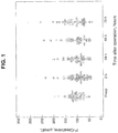
- Plasma creatinine levels before and up to 78 hours after operation are given in Fig. 1 and show no differences between the time periods.
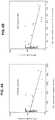
- the levels ofNGAL in urine obtained from healthy subjects and patients undergoing cardiac surgery were measured with using the three described techniques.
- the results determined using the RIA and Mono-mono techniques, respectively are shown in Figs. 2A and 2B .
- the pre-operative levels were similar to normal controls. Two hours after operation, the levels increased significantly (p ⁇ 0.0001) with about half the patients having levels above the upper limit of normals.
- the fold increases of medians were 18.7 when measured with the RIA and 15.6 and 11.4-fold increases when measured with the ELISA and the Mono-mono assays, respectively. After 24 hours, the levels were again reduced, but still higher than pre-operative levels. The levels stayed significantly higher during the whole postoperative period (p ⁇ 0.0001). At 72 hours, the fold increases were respectively 6.8, 8.5 and 5.9 for the RIA, ELISA and Mono-mono assays. Similar patterns over time were seen with all three assays.
- NGAL RIA
- NGAL Mono-mono
- the subjects were classified into two groups according to the percentage increase of plasma creatinine during the 72 hours postoperative period as compared with baseline ( ⁇ 120% or >119%).
- the correlations at different time points are given in Table 1 and show very good correlations with r 2 's ranging between 0.952-0.996, but with the exception of results obtained 2 hours postoperatively. At this time point the r 2 was 0.680 and significantly lower than the rest (p ⁇ 0.0001).
- NGAL neuropeptide derived neuropeptide derived neuropeptide derived neuropeptides
- results presented herein show that the choice of antibodies in the NGAL assay is critical in order to identify the many different variants of NGAL excreted in the urine under various conditions.
- assays employing NGAL antibody having the capacity to react with more than two NGAL protein epitopes provide improved sensitivity.
- the RIA is a polyclonal-based assay, which likely addresses all available epitopes in the molecule, whereas the monoclonal-monoclonal-based assay addresses only selected epitopes, some of which may or may not be disguised by complex formations and other molecular alterations.
- the polyclonal-monoclonal-based ELISA assay had characteristics somewhat in between these two extremes, which may be explained by the fact that this configuration allows the recognition of more epitopes than a double monoclonal assay but somewhat less than a purely polyclonal-based assay.
- NGAL is a useful early biomarker of postoperative kidney injury when measured in urine and newly demonstrate that the antibody configuration of the assay has an impact on the clinical performance of the assay, since some forms of NGAL may not be identified by assays with restricted antibody specificities.
- This Example describes a study of NGAL determinations with respect to monomeric, dimeric and heterodimeric forms in determining the NGAL protein origin.
- ELISAs based on different antibodies for NGAL quantification were employed, namely 1) Mab697-polyclonal (the monoclonal-polyclonal ELISA as described in Example 1), 2) Mab764-Mab765, 3) Mab764-polyclonal, 4) polyclonal-Mab765, 5) polyclonal-polyclonal, and Mab-697-Mab765.
- the basic protocols for these five ELISAs are the same as described in Example 1 except for the specific antibodies used in the assay.
- 96-well microtitre plates Nunc Maxsorp, Agogent, Danmark
- rabbit polyclonal or mouse monoclonal antibodies (Mab697 and Mab764) against human NGAL %. (Diagnostics Development, Uppsala, Sweden).
- Samples and standards (ranging from 0.039-5 ⁇ g/L) (100 ⁇ L/well) were incubated for 60 min (urine samples and gel filtration fractions) or 90 min (cell culture supernatants) at room temperature (RT).
- the enzyme reaction was visualized by 3,3',5,5'-tetramethylbenzidine solution (100 ⁇ L/well) (Sigma-Aldrich, Steinhein, Germany) as substrate at RT for 15 min and the reaction was stopped by adding 1 M H 2 SO 4 (100 ⁇ L/well). Absorbance was read at 450 nm with a spectrophotometer (SPECTRAmax 250, GMI, Inc., USA).
- the RIA was performed as described above. Briefly, a 50 ⁇ L of either sample or standards (2 ⁇ g/L - 128 ⁇ g/L) was mixed with 50 ⁇ L of I 125 -labelled NGAL and 50 ⁇ L of specific antibodies. The mixture was incubated for 3 h at room temperature. Thereafter, 500 ⁇ L of solid phase second antibody coated cellulose suspension (AA-SAC1, IDSLTD, United Kingdom) was added and incubated for 1 h at 4 °C. NGAL-antibody complexes bound on anti-rabbit IgG antibody coated cellulose were pelleted by centrifugation. After decantation, the radioactivity was measured.
- HK-2 human kidney 2, CRL-2190
- ATCC American Type Culture Collection
- BPE bovine pituitary extract
- EGF epidermal growth factor
- the cells were cultured with specific stimuli including cytokines (IL-1 ⁇ or TNF- ⁇ ) (Sigma-Aldrich, Steinhein, Germany) and LPS (Invitrogen-Giboco®, United Kingdom).
- cytokines IL-1 ⁇ or TNF- ⁇
- LPS Invitrogen-Giboco®, United Kingdom.
- 0.5 ⁇ 10 5 cells and 1 ml complete growth medium per well were seeded in 24-well plates (FALCON®, USA). Following 48 h subculture, the complete growth medium was removed and the monolayer (about 90% confluence) was washed twice with PBS (Invitrogen-Giboco®, United Kingdom).
- the cells were cultured with conditioned media for a 72 h period. The media supernatants were harvested for NGAL quantification at 2 h, 12 h, 24 h, 48 h and 72 h, respectively.
- sequences of the specific oligonucleotide primers for NGAL (5'-TCACCTCCGTCCTGTTTAGC-3' and 5'-CGAAGTCAGCTCCTTGGTTC-3') and ⁇ -actin (5'-TTCTACAATGAGCTGCGTGTGG-3' and 5'-GTGTTGAAGGTCTCAAACATGAT- 3') were selected according to the literature and synthesized by Thermo SCIENTIFIC (Germany).
- the initial denaturation condition is 94 °C for 2 min.
- PCR amplification was performed using a 30 sec denaturation step at 94 °C, followed by a 30 sec annealing step at 60 °C (for NGAL) or 59 °C (for ⁇ -actin), and a 30 sec extension at 72 °C. A total of 30 cycles were carried out for both genes followed by a final extension for 10 min at 72 °C. PCR products were separated by 2% agarose gel electrophoresis, and were detected by ethidium bromide staining.
- the neutrophil granule release products were obtained as described. HK-2 conditioned media supernatants at the 72-h time point were harvested, and supplemented with 0.1 mM PMSF (Sigma-Aldrich, Steinhein, Germany) and Complete TM protease inhibitor cocktail tablets (Roche, Mannheim, Germany). The supernatants were concentrated using Amicon® Ultra-4 centrifugal filter devices (10,000 MW) (Millipore, USA). SDS-PAGE and Western blotting were performed according to the manufacturer's instructions. Briefly, 25 ⁇ L of either urine or concentrated conditioned supernatants or neutrophil release products were applied to Nu-PAGE® 4-12% Bis-Tris Gel (Invitrogen, USA) under non-reducing conditions.
- the proteins were transferred onto a Hybone-P PVDF membrane (GE Healthcare, United Kingdom) by using Nu-PAGE® Transfer Buffer (Invitrogen, USA) at 25V for 1 hour. The additional binding sites of the PVDF membrane were blocked by a blocking solution (GE Healthcare, United Kingdom) for 1 h.
- the blots were incubated overnight at RT with either rabbit polyclonal antibodies or mouse monoclonal antibodies (Mab 697, Mab 699, Mab 763, Mab 764, or Mab 765) or a mixture of monoclonal antibodies against human NGAL followed by 1 h incubation with peroxidase-conjugated secondary antibodies (GE Healthcare, United Kingdom). Immunoblots were detected using enhanced chemiluminescence according to the instructions of the manufacturer (Amersham ECL TM Western Blotting System, GE Healthcare, United Kingdom).
- the polyclonal antibody seemed to have a stronger affinity to the dimer and a weaker affinity to the heterodimer than the two monoclonals.
- the Mabs764 and 765 had very similar performances on detecting all three molecular forms.
- the affinities of Mab764 and Mab765 to NGAL from high to low were monomeric, heterodimeric and dimeric forms, respectively. It is also shown that Mabs763, 699 and 697 have strong affinities to heterodimeric forms, whereas the affinities to the dimeric and monomeric forms were weak.
- the ability of the polyclonal antibodies and the Mabs765 and 697 in detecting monomeric and dimeric forms in supernatants of stimulated neutrophil granulocytes seemed very similar.
- Table 1 The performance characteristics of the RIA and the five ELISAs are shown in Table 1: Table 1. Measurements ofHNL/NGAL in urine samples collected from patients undergoing cardiac surgery by different assays Assay Pre-op ⁇ g/L 2 h post-op ⁇ g/L 24 h post-op Fold increase (Pre-op/ 2 h post-op) Student's t-test p-value ANOVA p-value RIA 7.19 (2.9-20.3) 248.20 (109-316.1) 26.96 (16.3-50.71) 34.5 0.000011 0.0000020 ELISA 1 (Mab697-Polyclonal) 0.94 (0.15-3.13) 28.82 (23.32-37.96) 4.75 (2.59-9.89) 30.7 0.00035 0.00027 ELISA 2 (Mab764-Mab765) 6.22 (1.16-12.8) 192.80 (78.2-287) 15.50 (9.55-40.7) 31.0 0.000055 0.00002 ELISA 3
- the levels of NGAL in urine obtained pre-operation and at 2 h and 24 h post operation were measured and the median levels of NGAL obtained by the assays are shown in Table 1.
- the median levels of NGAL pre-operation and 2 h post operation measured by RIA were the highest among the seven assays.
- the levels obtained by Mab697-based ELISA (ELISA 1 and ELISA 6) were significantly lower than the other assays.
- Table 1 shows the differences in levels pre- and 2 h post operation as well as overall differences during the 24 hour period. Highly significant differences pre- and postoperatively were seen with all assays.
- Fold increases of median levels preop vs 2 h postop were highest and >70 when measured by ELISA 3 (Mab764-Polyclonal), ELISA 5 (polyclonal-polyclonal) or ELISA 4 (polyclonal-Mab765), and 23-34-fold when measured by RIA, ELISA 2 (Mab764-Mab765), ELISA 1 (Mab 697-polyclonal) or ELISA 6 (Mab697-Mab765).
- NGAL is Up-Regulated in HK-2 Cells when Grown Under Stressful Conditions
- HK-2 cells were cultured at different lengths of time with Keratinocyte Serum Free Medium (K-SFM), K-SFM either supplemented with 0.05 mg/mL bovine pituitary extract (BPE) or 5 ng/mL human recombinant epidermal growth factor (EGF), or ATCC recommended complete growth medium (K-SFM supplemented with 0.05 mg/mL BPE and 5 ng/mL EGF) followed by culture for 48 h under standard conditions.
- K-SFM Keratinocyte Serum Free Medium
- K-SFM Keratinocyte Serum Free Medium
- K-SFM Keratinocyte Serum Free Medium
- NGAL is Up-Regulated in HK-2 Cells by IL- ⁇ , LPS AND TNF- ⁇
- HK-2 cells were grown with complete growth medium for 48 h after which the cells were further grown for various lengths of time in the presence of complete growth medium supplemented with either IL- ⁇ (1 ng/mL, human), LPS (125 ng/mL, Klebsiella pneumonia) or TNF- ⁇ (20 ng/mL, human).
- IL- ⁇ induced highly significant elevations of NGAL levels in the supernatant (8.9- to 41.9-fold increase).
- the incubation with TNF- ⁇ and LPS induced some elevations of NGAL in the supernatants (2.2 and 1.6-fold, respectively), but significantly less than IL- ⁇ (p ⁇ 0.001).
- HK-2 cells were also cultured with K-SFM supplemented with IL- ⁇ , TNF- ⁇ or LPS.
- Significant elevations of NGAL were seen with IL- ⁇ (1.3- to 12.8-fold increase), but not with TNF- ⁇ or LPS ( Fig. 9B ). Compared to cells grown in complete growth medium these elevation were, however, significantly less (p ⁇ 0.001).
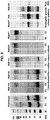
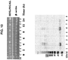
- NGAL secreted by HK-2 cells were determined by Western blotting using mixed monoclonal antibodies (Mab697, Mab764 and Mab765) as detecting antibodies.
- the results shown in Fig. 10 indicate that the major form of NGAL secreted by HK-2 cells grown either in complete culture media or under stressful conditions in K-SFM or in media supplemented with cytokines (IL- ⁇ or TNF- ⁇ ) or LPS is the monomeric form.
- IL- ⁇ or TNF- ⁇ cytokines
- LPS LPS
- the heterodimeric form of NGAL was also apparent after stimulation with IL- ⁇ whereas the dimeric form was absent in contrast to findings in supernatants of human neutrophils ( Fig. 6 ).
- the mRNA levels of NGAL are shown in HK-2 cells after incubation with IL- ⁇ . The results show increased expression indicating active synthesis ofNGAL by the HK-2 cells.
- NGAL was originally isolated from human neutrophils and it has been our previously shown that the measurement ofNGAL in blood is a superior means to distinguish acute infections caused by bacteria or virus. Subsequent studies have found that NGAL may also be expressed in other cells such as in kidney, liver and epithelial tissue under certain conditions and that NGAL measurement in urine and plasma might serve as a biomarker of acute kidney injury.
- Example 1 shows that antibody configuration of an NGAL assay has an impact on the clinical performance of the assay.
- Several forms ofNGAL were identified in urine of patients with AKI. This example further shows that the monomeric form and to some extent the heterodimeric forms are the predominant forms produced by tubular epithelial cells, whereas the dimeric form seems unique to the neutrophils (see Fig. 6 ).
- the monomeric form was also produced by neutrophils.
- One interesting finding in the present study is the differences in recognition of these different forms by the employed antibodies, since the monomeric and dimeric forms originating from neutrophils were identified by all monoclonal antibodies as well as by the polyclonal antibodies. This was contrasted by the almost complete inability of Mab697 to recognize these forms in urine. Also the Mab765 showed a strong reaction to these forms in neutrophil supernatants but only weak recognition of the dimeric form in urine. Without wishing to be bound by theory, it is believed that differences in epitope exposure of the different NGAL forms and thus differences in molecular structures are the cause.
- the invention is directed to an assay that preferentially identifies NGAL originating from the tubular epithelium, since the molecular structure ofNGAL seems slightly different from the structure ofNGAL from the neutrophils.
- assays are therefore more specific and sensitive in the detection of AKI and of major benefit to patients at risk of developing impaired kidney function.
Landscapes
- Health & Medical Sciences (AREA)
- Life Sciences & Earth Sciences (AREA)
- Engineering & Computer Science (AREA)
- Immunology (AREA)
- Molecular Biology (AREA)
- Chemical & Material Sciences (AREA)
- Biomedical Technology (AREA)
- Urology & Nephrology (AREA)
- Hematology (AREA)
- Cell Biology (AREA)
- Analytical Chemistry (AREA)
- Biotechnology (AREA)
- Pathology (AREA)
- Food Science & Technology (AREA)
- Medicinal Chemistry (AREA)
- Physics & Mathematics (AREA)
- Microbiology (AREA)
- Biochemistry (AREA)
- General Health & Medical Sciences (AREA)
- General Physics & Mathematics (AREA)
- Proteomics, Peptides & Aminoacids (AREA)
- Peptides Or Proteins (AREA)
- Investigating Or Analysing Biological Materials (AREA)
- Preparation Of Compounds By Using Micro-Organisms (AREA)
Applications Claiming Priority (3)
| Application Number | Priority Date | Filing Date | Title |
|---|---|---|---|
| US11671308P | 2008-11-21 | 2008-11-21 | |
| EP20090761023 EP2362943A1 (fr) | 2008-11-21 | 2009-11-23 | Procédés, dispositifs et kits pour détecter ou surveiller une insuffisance rénale sévère |
| PCT/IB2009/055299 WO2010058378A1 (fr) | 2008-11-21 | 2009-11-23 | Procédés, dispositifs et kits pour détecter ou surveiller une insuffisance rénale sévère |
Related Parent Applications (1)
| Application Number | Title | Priority Date | Filing Date |
|---|---|---|---|
| EP20090761023 Division EP2362943A1 (fr) | 2008-11-21 | 2009-11-23 | Procédés, dispositifs et kits pour détecter ou surveiller une insuffisance rénale sévère |
Publications (2)
| Publication Number | Publication Date |
|---|---|
| EP3141904A1 true EP3141904A1 (fr) | 2017-03-15 |
| EP3141904B1 EP3141904B1 (fr) | 2019-01-02 |
Family
ID=41611201
Family Applications (2)
| Application Number | Title | Priority Date | Filing Date |
|---|---|---|---|
| EP20090761023 Ceased EP2362943A1 (fr) | 2008-11-21 | 2009-11-23 | Procédés, dispositifs et kits pour détecter ou surveiller une insuffisance rénale sévère |
| EP16191697.8A Not-in-force EP3141904B1 (fr) | 2008-11-21 | 2009-11-23 | Procédés, dispositifs et kits de détection ou surveillance d'une lésion rénale aiguë |
Family Applications Before (1)
| Application Number | Title | Priority Date | Filing Date |
|---|---|---|---|
| EP20090761023 Ceased EP2362943A1 (fr) | 2008-11-21 | 2009-11-23 | Procédés, dispositifs et kits pour détecter ou surveiller une insuffisance rénale sévère |
Country Status (12)
| Country | Link |
|---|---|
| US (1) | US9476880B2 (fr) |
| EP (2) | EP2362943A1 (fr) |
| JP (1) | JP5438770B2 (fr) |
| KR (2) | KR20190006614A (fr) |
| CN (1) | CN102292637B (fr) |
| AU (1) | AU2009318813B2 (fr) |
| CA (1) | CA2744434C (fr) |
| ES (1) | ES2718731T3 (fr) |
| IL (1) | IL212988A (fr) |
| MY (1) | MY162697A (fr) |
| RU (1) | RU2519722C2 (fr) |
| WO (1) | WO2010058378A1 (fr) |
Families Citing this family (17)
| Publication number | Priority date | Publication date | Assignee | Title |
|---|---|---|---|---|
| PL2035835T3 (pl) | 2006-05-30 | 2012-05-31 | Antibodyshop As | Sposoby szybkiej oceny ciężkości urazu |
| WO2008113363A1 (fr) | 2007-03-21 | 2008-09-25 | Bioporto Diagnostics A/S | Essai diagnostique de lesion rénale |
| US8846036B2 (en) | 2007-10-19 | 2014-09-30 | Abbott Laboratories | Antibodies that bind to mammalian NGAL and uses thereof |
| JP2012508177A (ja) * | 2008-11-05 | 2012-04-05 | アボット・ラボラトリーズ | 尿および組換えチャイニーズハムスター卵巣(cho)細胞から濃縮された好中球ゼラチナーゼ関連リポカリン(ngal)タンパク質アイソフォーム、ならびに関連する組成物、抗体、ならびに濃縮、分析および使用の方法 |
| EP2568291A1 (fr) * | 2011-09-07 | 2013-03-13 | Roche Diagnostics GmbH | Diagnostic à base de L-FABP de lésion rénale après un événement aigu ou après une intervention chirurgicale |
| EP2758774A4 (fr) * | 2011-09-22 | 2015-04-29 | Univ Los Andes | Procédé de surveillance, de diagnostic et/ou de pronostic de la lésion rénale aiguë au stade précoce |
| WO2014053501A1 (fr) * | 2012-10-02 | 2014-04-10 | Sphingotec Gmbh | Méthode permettant de diagnostiquer ou de surveiller la fonction rénale ou de diagnostiquer une dysfonction rénale |
| CN102928606B (zh) * | 2012-11-16 | 2015-10-07 | 武汉明德生物科技股份有限公司 | 多抗体标记的降钙素原快速检测试剂盒 |
| EP2936160A1 (fr) * | 2012-12-20 | 2015-10-28 | Novartis AG | Insuffisance rénale aiguë |
| US9651547B2 (en) | 2013-03-14 | 2017-05-16 | Abbott Point Of Care Inc. | Electrochemical methods and devices for amending urine samples for immunosensor detection |
| US9488663B2 (en) | 2013-03-14 | 2016-11-08 | Abbott Point Of Care Inc. | Electrochemical methods and devices for amending urine samples for immunosensor detection |
| EP2972230B1 (fr) | 2013-03-15 | 2021-07-21 | Hycor Biomedical LLC | Dispositif et procédé associé de réalisation de mesures de luminescence et de fluorescence d'un échantillon |
| RU2566709C2 (ru) * | 2013-12-16 | 2015-10-27 | Владимир Борисович Бородулин | Способ ранней диагностики хронической болезни почек |
| CN107003320B (zh) * | 2014-11-25 | 2021-08-17 | 豪夫迈·罗氏有限公司 | 慢性肾脏疾病的快速进展的生物标志物 |
| CN104479015A (zh) * | 2014-12-04 | 2015-04-01 | 东南大学 | 针对ngal表位的纳米抗体及其应用 |
| CN105911274A (zh) * | 2016-06-12 | 2016-08-31 | 吉林大学 | 一种同步定量检测不同分子形式人中性粒细胞脂质运载蛋白免疫层析装置及其制备方法 |
| CN108802402A (zh) * | 2018-06-15 | 2018-11-13 | 重庆大学 | 一种快速鉴别呼吸道感染类型的试剂盒及其应用 |
Citations (5)
| Publication number | Priority date | Publication date | Assignee | Title |
|---|---|---|---|---|
| US6136526A (en) | 1994-04-21 | 2000-10-24 | Venge; Per | Use of human neutrophil lipocalin (HNL) as a diagnostic marker and anti-HNL-antibody preparation |
| US20040219603A1 (en) | 2003-03-27 | 2004-11-04 | Prasad Devarajan | Method and kit for detecting the early onset of renal tubular cell injury |
| US20050272101A1 (en) | 2004-06-07 | 2005-12-08 | Prasad Devarajan | Method for the early detection of renal injury |
| WO2009052390A1 (fr) * | 2007-10-19 | 2009-04-23 | Abbott Laboratories | Ngal mammalien glycosylé et son utilisation |
| WO2009062520A1 (fr) * | 2007-11-15 | 2009-05-22 | Bioporto Diagnostics A/S | Utilisation diagnostique de formes moléculaires particulières d'un biomarqueur |
Family Cites Families (10)
| Publication number | Priority date | Publication date | Assignee | Title |
|---|---|---|---|---|
| EP1334364A2 (fr) * | 2000-10-13 | 2003-08-13 | Children's Medical Center Corporation | Analyse enzymatique non invasive d'etats associes au remodelage d'un tissu |
| US7056702B2 (en) * | 2002-12-16 | 2006-06-06 | Kimberly Clark Co | Detecting lipocalin |
| CN1791797A (zh) * | 2003-03-27 | 2006-06-21 | 儿童医院医疗中心 | 用于检测肾小管细胞损伤的早发的方法和试剂盒 |
| CN101010001A (zh) * | 2004-05-06 | 2007-08-01 | 哥伦比亚大学受托人 | Ngal用于减少和改善局部缺血和肾中毒损伤 |
| JP2007536260A (ja) * | 2004-05-06 | 2007-12-13 | ザ・トラスティーズ・オブ・コロンビア・ユニバーシティ・イン・ザ・シティ・オブ・ニューヨーク | 虚血性および腎毒性障害の軽減および改善用ngal |
| ES2818028T3 (es) * | 2004-12-20 | 2021-04-09 | Antibodyshop As | Determinación de lipocalina asociada a gelatinasa de neutrófilos (NGAL) como marcador diagnóstico para trastornos renales |
| US20070037232A1 (en) | 2005-03-31 | 2007-02-15 | Barasch Jonathan M | Detection of NGAL in chronic renal disease |
| US20080090304A1 (en) | 2006-10-13 | 2008-04-17 | Barasch Jonathan Matthew | Diagnosis and monitoring of chronic renal disease using ngal |
| EP2375254A1 (fr) * | 2006-02-17 | 2011-10-12 | The Children's Medical Center Corporation | NGAL libre en tant que biomarqueur de cancer |
| US20090123970A1 (en) * | 2007-10-19 | 2009-05-14 | Abbott Laboratories | Glycosylated mammalian ngal and use thereof |
-
2009
- 2009-11-23 CN CN200980155194.XA patent/CN102292637B/zh not_active Expired - Fee Related
- 2009-11-23 MY MYPI2011002257A patent/MY162697A/en unknown
- 2009-11-23 KR KR1020197001145A patent/KR20190006614A/ko not_active Application Discontinuation
- 2009-11-23 US US13/130,456 patent/US9476880B2/en active Active
- 2009-11-23 JP JP2011537003A patent/JP5438770B2/ja not_active Expired - Fee Related
- 2009-11-23 EP EP20090761023 patent/EP2362943A1/fr not_active Ceased
- 2009-11-23 WO PCT/IB2009/055299 patent/WO2010058378A1/fr active Application Filing
- 2009-11-23 RU RU2011125320/15A patent/RU2519722C2/ru active
- 2009-11-23 EP EP16191697.8A patent/EP3141904B1/fr not_active Not-in-force
- 2009-11-23 KR KR1020117014191A patent/KR101939964B1/ko active IP Right Grant
- 2009-11-23 CA CA2744434A patent/CA2744434C/fr active Active
- 2009-11-23 AU AU2009318813A patent/AU2009318813B2/en not_active Ceased
- 2009-11-23 ES ES16191697T patent/ES2718731T3/es active Active
-
2011
- 2011-05-19 IL IL212988A patent/IL212988A/en active IP Right Grant
Patent Citations (5)
| Publication number | Priority date | Publication date | Assignee | Title |
|---|---|---|---|---|
| US6136526A (en) | 1994-04-21 | 2000-10-24 | Venge; Per | Use of human neutrophil lipocalin (HNL) as a diagnostic marker and anti-HNL-antibody preparation |
| US20040219603A1 (en) | 2003-03-27 | 2004-11-04 | Prasad Devarajan | Method and kit for detecting the early onset of renal tubular cell injury |
| US20050272101A1 (en) | 2004-06-07 | 2005-12-08 | Prasad Devarajan | Method for the early detection of renal injury |
| WO2009052390A1 (fr) * | 2007-10-19 | 2009-04-23 | Abbott Laboratories | Ngal mammalien glycosylé et son utilisation |
| WO2009062520A1 (fr) * | 2007-11-15 | 2009-05-22 | Bioporto Diagnostics A/S | Utilisation diagnostique de formes moléculaires particulières d'un biomarqueur |
Non-Patent Citations (5)
| Title |
|---|
| DENT ET AL., CRITICAL CARE, vol. 11, no. 6, 2007, pages R127 |
| DEVEREUX ET AL., NUCLEIC ACID RESEARCH, vol. 12, no. 1, 1984, pages 387 - 394 |
| GEBHARD WAGENER ET AL: "Increased Incidence of Acute Kidney Injury with Aprotinin Use during Cardiac Surgery Detected with Urinary NGAL", AMERICAN JOURNAL OF NEPHROLOGY, vol. 28, no. 4, 1 January 2008 (2008-01-01), CH, pages 576 - 582, XP055333375, ISSN: 0250-8095, DOI: 10.1159/000115973 * |
| TOWBIN ET AL., PROC. NATL. ACAD. SCI. USA, vol. 76, 1979, pages 4350 - 4 |
| XU ET AL., JOURNAL OF IMMUNOLOGICAL METHODS, vol. 171, 1994, pages 245 - 252 |
Also Published As
| Publication number | Publication date |
|---|---|
| AU2009318813A1 (en) | 2011-07-14 |
| CN102292637A (zh) | 2011-12-21 |
| CA2744434A1 (fr) | 2010-05-27 |
| EP2362943A1 (fr) | 2011-09-07 |
| MY162697A (en) | 2017-07-14 |
| IL212988A0 (en) | 2011-07-31 |
| WO2010058378A1 (fr) | 2010-05-27 |
| JP5438770B2 (ja) | 2014-03-12 |
| AU2009318813B2 (en) | 2016-11-24 |
| CN102292637B (zh) | 2015-08-05 |
| ES2718731T3 (es) | 2019-07-04 |
| RU2011125320A (ru) | 2012-12-27 |
| JP2012509477A (ja) | 2012-04-19 |
| KR20110103965A (ko) | 2011-09-21 |
| US9476880B2 (en) | 2016-10-25 |
| RU2519722C2 (ru) | 2014-06-20 |
| US20110287455A1 (en) | 2011-11-24 |
| KR20190006614A (ko) | 2019-01-18 |
| CA2744434C (fr) | 2017-10-03 |
| IL212988A (en) | 2014-08-31 |
| EP3141904B1 (fr) | 2019-01-02 |
| KR101939964B1 (ko) | 2019-01-18 |
Similar Documents
| Publication | Publication Date | Title |
|---|---|---|
| EP3141904B1 (fr) | Procédés, dispositifs et kits de détection ou surveillance d'une lésion rénale aiguë | |
| CN101960308B (zh) | 急性肾损伤及预后推测用生物标记物以及其用途 | |
| EP3540440B1 (fr) | Procédés et utilisations pour l'évaluation des lésions rénales et du statut rénal | |
| TR201807542T4 (tr) | Böbrek hasarı ve böbrek yetmezliği teşhisi ve prognozuna yönelik metotlar ve bileşimler. | |
| Cai et al. | Assays of urine levels of HNL/NGAL in patients undergoing cardiac surgery and the impact of antibody configuration on their clinical performances | |
| EP2834638B1 (fr) | Procédés de diagnostic et de pronostic d'un sepsis | |
| US11598781B2 (en) | Method for predicting the risk of incidence of chronic kidney disease | |
| EP3068893A1 (fr) | Methodes et compositions de diagnostic et de pronostic de la septicémie | |
| WO2011105474A1 (fr) | Procédé d'examen d'un trouble rénal aigu | |
| EP2875347A1 (fr) | Procédés et compositions pour le diagnostic et le pronostic de la sepsie | |
| KR102077987B1 (ko) | 신장 질환 진단용 바이오 마커 | |
| JP6062439B2 (ja) | 早期腎障害の評価マーカーとその測定方法 | |
| EP2882767B1 (fr) | Évaluation d'une lésion rénale à l'aide de l'acide hyaluronique | |
| Bhavsar | Subclinical albuminuria, beta trace protein, neutrophil gelatinase associated lipocalin, and kidney injury molecule-1 and risk of coronary heart disease, chronic kidney disease, and death |
Legal Events
| Date | Code | Title | Description |
|---|---|---|---|
| PUAI | Public reference made under article 153(3) epc to a published international application that has entered the european phase |
Free format text: ORIGINAL CODE: 0009012 |
|
| STAA | Information on the status of an ep patent application or granted ep patent |
Free format text: STATUS: THE APPLICATION HAS BEEN PUBLISHED |
|
| AC | Divisional application: reference to earlier application |
Ref document number: 2362943 Country of ref document: EP Kind code of ref document: P |
|
| AK | Designated contracting states |
Kind code of ref document: A1 Designated state(s): AT BE BG CH CY CZ DE DK EE ES FI FR GB GR HR HU IE IS IT LI LT LU LV MC MK MT NL NO PL PT RO SE SI SK SM TR |
|
| STAA | Information on the status of an ep patent application or granted ep patent |
Free format text: STATUS: REQUEST FOR EXAMINATION WAS MADE |
|
| 17P | Request for examination filed |
Effective date: 20170915 |
|
| RBV | Designated contracting states (corrected) |
Designated state(s): AT BE BG CH CY CZ DE DK EE ES FI FR GB GR HR HU IE IS IT LI LT LU LV MC MK MT NL NO PL PT RO SE SI SK SM TR |
|
| STAA | Information on the status of an ep patent application or granted ep patent |
Free format text: STATUS: EXAMINATION IS IN PROGRESS |
|
| 17Q | First examination report despatched |
Effective date: 20180214 |
|
| GRAP | Despatch of communication of intention to grant a patent |
Free format text: ORIGINAL CODE: EPIDOSNIGR1 |
|
| STAA | Information on the status of an ep patent application or granted ep patent |
Free format text: STATUS: GRANT OF PATENT IS INTENDED |
|
| INTG | Intention to grant announced |
Effective date: 20180620 |
|
| GRAS | Grant fee paid |
Free format text: ORIGINAL CODE: EPIDOSNIGR3 |
|
| GRAA | (expected) grant |
Free format text: ORIGINAL CODE: 0009210 |
|
| STAA | Information on the status of an ep patent application or granted ep patent |
Free format text: STATUS: THE PATENT HAS BEEN GRANTED |
|
| AC | Divisional application: reference to earlier application |
Ref document number: 2362943 Country of ref document: EP Kind code of ref document: P |
|
| AK | Designated contracting states |
Kind code of ref document: B1 Designated state(s): AT BE BG CH CY CZ DE DK EE ES FI FR GB GR HR HU IE IS IT LI LT LU LV MC MK MT NL NO PL PT RO SE SI SK SM TR |
|
| REG | Reference to a national code |
Ref country code: GB Ref legal event code: FG4D |
|
| REG | Reference to a national code |
Ref country code: CH Ref legal event code: EP Ref country code: AT Ref legal event code: REF Ref document number: 1085054 Country of ref document: AT Kind code of ref document: T Effective date: 20190115 |
|
| REG | Reference to a national code |
Ref country code: IE Ref legal event code: FG4D |
|
| REG | Reference to a national code |
Ref country code: DE Ref legal event code: R096 Ref document number: 602009056586 Country of ref document: DE |
|
| REG | Reference to a national code |
Ref country code: SE Ref legal event code: TRGR |
|
| REG | Reference to a national code |
Ref country code: CH Ref legal event code: NV Representative=s name: STOLMAR AND PARTNER INTELLECTUAL PROPERTY S.A., CH |
|
| REG | Reference to a national code |
Ref country code: NL Ref legal event code: FP |
|
| REG | Reference to a national code |
Ref country code: LT Ref legal event code: MG4D |
|
| REG | Reference to a national code |
Ref country code: AT Ref legal event code: MK05 Ref document number: 1085054 Country of ref document: AT Kind code of ref document: T Effective date: 20190102 |
|
| REG | Reference to a national code |
Ref country code: ES Ref legal event code: FG2A Ref document number: 2718731 Country of ref document: ES Kind code of ref document: T3 Effective date: 20190704 |
|
| PG25 | Lapsed in a contracting state [announced via postgrant information from national office to epo] |
Ref country code: FI Free format text: LAPSE BECAUSE OF FAILURE TO SUBMIT A TRANSLATION OF THE DESCRIPTION OR TO PAY THE FEE WITHIN THE PRESCRIBED TIME-LIMIT Effective date: 20190102 Ref country code: NO Free format text: LAPSE BECAUSE OF FAILURE TO SUBMIT A TRANSLATION OF THE DESCRIPTION OR TO PAY THE FEE WITHIN THE PRESCRIBED TIME-LIMIT Effective date: 20190402 Ref country code: PT Free format text: LAPSE BECAUSE OF FAILURE TO SUBMIT A TRANSLATION OF THE DESCRIPTION OR TO PAY THE FEE WITHIN THE PRESCRIBED TIME-LIMIT Effective date: 20190502 Ref country code: LT Free format text: LAPSE BECAUSE OF FAILURE TO SUBMIT A TRANSLATION OF THE DESCRIPTION OR TO PAY THE FEE WITHIN THE PRESCRIBED TIME-LIMIT Effective date: 20190102 Ref country code: PL Free format text: LAPSE BECAUSE OF FAILURE TO SUBMIT A TRANSLATION OF THE DESCRIPTION OR TO PAY THE FEE WITHIN THE PRESCRIBED TIME-LIMIT Effective date: 20190102 |
|
| PG25 | Lapsed in a contracting state [announced via postgrant information from national office to epo] |
Ref country code: GR Free format text: LAPSE BECAUSE OF FAILURE TO SUBMIT A TRANSLATION OF THE DESCRIPTION OR TO PAY THE FEE WITHIN THE PRESCRIBED TIME-LIMIT Effective date: 20190403 Ref country code: HR Free format text: LAPSE BECAUSE OF FAILURE TO SUBMIT A TRANSLATION OF THE DESCRIPTION OR TO PAY THE FEE WITHIN THE PRESCRIBED TIME-LIMIT Effective date: 20190102 Ref country code: BG Free format text: LAPSE BECAUSE OF FAILURE TO SUBMIT A TRANSLATION OF THE DESCRIPTION OR TO PAY THE FEE WITHIN THE PRESCRIBED TIME-LIMIT Effective date: 20190402 Ref country code: LV Free format text: LAPSE BECAUSE OF FAILURE TO SUBMIT A TRANSLATION OF THE DESCRIPTION OR TO PAY THE FEE WITHIN THE PRESCRIBED TIME-LIMIT Effective date: 20190102 Ref country code: IS Free format text: LAPSE BECAUSE OF FAILURE TO SUBMIT A TRANSLATION OF THE DESCRIPTION OR TO PAY THE FEE WITHIN THE PRESCRIBED TIME-LIMIT Effective date: 20190502 |
|
| REG | Reference to a national code |
Ref country code: DE Ref legal event code: R097 Ref document number: 602009056586 Country of ref document: DE |
|
| PG25 | Lapsed in a contracting state [announced via postgrant information from national office to epo] |
Ref country code: SK Free format text: LAPSE BECAUSE OF FAILURE TO SUBMIT A TRANSLATION OF THE DESCRIPTION OR TO PAY THE FEE WITHIN THE PRESCRIBED TIME-LIMIT Effective date: 20190102 Ref country code: RO Free format text: LAPSE BECAUSE OF FAILURE TO SUBMIT A TRANSLATION OF THE DESCRIPTION OR TO PAY THE FEE WITHIN THE PRESCRIBED TIME-LIMIT Effective date: 20190102 Ref country code: AT Free format text: LAPSE BECAUSE OF FAILURE TO SUBMIT A TRANSLATION OF THE DESCRIPTION OR TO PAY THE FEE WITHIN THE PRESCRIBED TIME-LIMIT Effective date: 20190102 Ref country code: DK Free format text: LAPSE BECAUSE OF FAILURE TO SUBMIT A TRANSLATION OF THE DESCRIPTION OR TO PAY THE FEE WITHIN THE PRESCRIBED TIME-LIMIT Effective date: 20190102 Ref country code: EE Free format text: LAPSE BECAUSE OF FAILURE TO SUBMIT A TRANSLATION OF THE DESCRIPTION OR TO PAY THE FEE WITHIN THE PRESCRIBED TIME-LIMIT Effective date: 20190102 Ref country code: CZ Free format text: LAPSE BECAUSE OF FAILURE TO SUBMIT A TRANSLATION OF THE DESCRIPTION OR TO PAY THE FEE WITHIN THE PRESCRIBED TIME-LIMIT Effective date: 20190102 |
|
| PLBE | No opposition filed within time limit |
Free format text: ORIGINAL CODE: 0009261 |
|
| STAA | Information on the status of an ep patent application or granted ep patent |
Free format text: STATUS: NO OPPOSITION FILED WITHIN TIME LIMIT |
|
| PG25 | Lapsed in a contracting state [announced via postgrant information from national office to epo] |
Ref country code: SM Free format text: LAPSE BECAUSE OF FAILURE TO SUBMIT A TRANSLATION OF THE DESCRIPTION OR TO PAY THE FEE WITHIN THE PRESCRIBED TIME-LIMIT Effective date: 20190102 |
|
| 26N | No opposition filed |
Effective date: 20191003 |
|
| PG25 | Lapsed in a contracting state [announced via postgrant information from national office to epo] |
Ref country code: SI Free format text: LAPSE BECAUSE OF FAILURE TO SUBMIT A TRANSLATION OF THE DESCRIPTION OR TO PAY THE FEE WITHIN THE PRESCRIBED TIME-LIMIT Effective date: 20190102 |
|
| PGFP | Annual fee paid to national office [announced via postgrant information from national office to epo] |
Ref country code: ES Payment date: 20191220 Year of fee payment: 11 |
|
| PG25 | Lapsed in a contracting state [announced via postgrant information from national office to epo] |
Ref country code: TR Free format text: LAPSE BECAUSE OF FAILURE TO SUBMIT A TRANSLATION OF THE DESCRIPTION OR TO PAY THE FEE WITHIN THE PRESCRIBED TIME-LIMIT Effective date: 20190102 |
|
| PG25 | Lapsed in a contracting state [announced via postgrant information from national office to epo] |
Ref country code: MC Free format text: LAPSE BECAUSE OF FAILURE TO SUBMIT A TRANSLATION OF THE DESCRIPTION OR TO PAY THE FEE WITHIN THE PRESCRIBED TIME-LIMIT Effective date: 20190102 Ref country code: LU Free format text: LAPSE BECAUSE OF NON-PAYMENT OF DUE FEES Effective date: 20191123 |
|
| REG | Reference to a national code |
Ref country code: BE Ref legal event code: MM Effective date: 20191130 |
|
| PG25 | Lapsed in a contracting state [announced via postgrant information from national office to epo] |
Ref country code: IE Free format text: LAPSE BECAUSE OF NON-PAYMENT OF DUE FEES Effective date: 20191123 |
|
| PG25 | Lapsed in a contracting state [announced via postgrant information from national office to epo] |
Ref country code: BE Free format text: LAPSE BECAUSE OF NON-PAYMENT OF DUE FEES Effective date: 20191130 |
|
| PG25 | Lapsed in a contracting state [announced via postgrant information from national office to epo] |
Ref country code: CY Free format text: LAPSE BECAUSE OF FAILURE TO SUBMIT A TRANSLATION OF THE DESCRIPTION OR TO PAY THE FEE WITHIN THE PRESCRIBED TIME-LIMIT Effective date: 20190102 |
|
| PG25 | Lapsed in a contracting state [announced via postgrant information from national office to epo] |
Ref country code: MT Free format text: LAPSE BECAUSE OF FAILURE TO SUBMIT A TRANSLATION OF THE DESCRIPTION OR TO PAY THE FEE WITHIN THE PRESCRIBED TIME-LIMIT Effective date: 20190102 Ref country code: HU Free format text: LAPSE BECAUSE OF FAILURE TO SUBMIT A TRANSLATION OF THE DESCRIPTION OR TO PAY THE FEE WITHIN THE PRESCRIBED TIME-LIMIT; INVALID AB INITIO Effective date: 20091123 |
|
| REG | Reference to a national code |
Ref country code: ES Ref legal event code: FD2A Effective date: 20220202 |
|
| PGFP | Annual fee paid to national office [announced via postgrant information from national office to epo] |
Ref country code: GB Payment date: 20220321 Year of fee payment: 13 Ref country code: DE Payment date: 20220322 Year of fee payment: 13 Ref country code: CH Payment date: 20220323 Year of fee payment: 13 |
|
| PG25 | Lapsed in a contracting state [announced via postgrant information from national office to epo] |
Ref country code: ES Free format text: LAPSE BECAUSE OF NON-PAYMENT OF DUE FEES Effective date: 20201124 |
|
| PGFP | Annual fee paid to national office [announced via postgrant information from national office to epo] |
Ref country code: SE Payment date: 20220318 Year of fee payment: 13 Ref country code: NL Payment date: 20220316 Year of fee payment: 13 Ref country code: IT Payment date: 20220322 Year of fee payment: 13 Ref country code: FR Payment date: 20220317 Year of fee payment: 13 |
|
| PG25 | Lapsed in a contracting state [announced via postgrant information from national office to epo] |
Ref country code: MK Free format text: LAPSE BECAUSE OF FAILURE TO SUBMIT A TRANSLATION OF THE DESCRIPTION OR TO PAY THE FEE WITHIN THE PRESCRIBED TIME-LIMIT Effective date: 20190102 |
|
| REG | Reference to a national code |
Ref country code: DE Ref legal event code: R119 Ref document number: 602009056586 Country of ref document: DE |
|
| REG | Reference to a national code |
Ref country code: CH Ref legal event code: PL |
|
| REG | Reference to a national code |
Ref country code: SE Ref legal event code: EUG |
|
| REG | Reference to a national code |
Ref country code: NL Ref legal event code: MM Effective date: 20221201 |
|
| GBPC | Gb: european patent ceased through non-payment of renewal fee |
Effective date: 20221123 |
|
| PG25 | Lapsed in a contracting state [announced via postgrant information from national office to epo] |
Ref country code: LI Free format text: LAPSE BECAUSE OF NON-PAYMENT OF DUE FEES Effective date: 20221130 Ref country code: CH Free format text: LAPSE BECAUSE OF NON-PAYMENT OF DUE FEES Effective date: 20221130 |
|
| PG25 | Lapsed in a contracting state [announced via postgrant information from national office to epo] |
Ref country code: SE Free format text: LAPSE BECAUSE OF NON-PAYMENT OF DUE FEES Effective date: 20221124 Ref country code: NL Free format text: LAPSE BECAUSE OF NON-PAYMENT OF DUE FEES Effective date: 20221201 |
|
| PG25 | Lapsed in a contracting state [announced via postgrant information from national office to epo] |
Ref country code: IT Free format text: LAPSE BECAUSE OF NON-PAYMENT OF DUE FEES Effective date: 20221123 Ref country code: GB Free format text: LAPSE BECAUSE OF NON-PAYMENT OF DUE FEES Effective date: 20221123 Ref country code: DE Free format text: LAPSE BECAUSE OF NON-PAYMENT OF DUE FEES Effective date: 20230601 |
|
| PG25 | Lapsed in a contracting state [announced via postgrant information from national office to epo] |
Ref country code: FR Free format text: LAPSE BECAUSE OF NON-PAYMENT OF DUE FEES Effective date: 20221130 |