EP3117214B1 - High-throughput and highly multiplexed imaging with programmable nucleic acid probes - Google Patents
High-throughput and highly multiplexed imaging with programmable nucleic acid probes Download PDFInfo
- Publication number
- EP3117214B1 EP3117214B1 EP15762370.3A EP15762370A EP3117214B1 EP 3117214 B1 EP3117214 B1 EP 3117214B1 EP 15762370 A EP15762370 A EP 15762370A EP 3117214 B1 EP3117214 B1 EP 3117214B1
- Authority
- EP
- European Patent Office
- Prior art keywords
- nucleic acids
- nucleic acid
- target
- docking
- imager
- Prior art date
- Legal status (The legal status is an assumption and is not a legal conclusion. Google has not performed a legal analysis and makes no representation as to the accuracy of the status listed.)
- Active
Links
Images


Classifications
-
- C—CHEMISTRY; METALLURGY
- C12—BIOCHEMISTRY; BEER; SPIRITS; WINE; VINEGAR; MICROBIOLOGY; ENZYMOLOGY; MUTATION OR GENETIC ENGINEERING
- C12Q—MEASURING OR TESTING PROCESSES INVOLVING ENZYMES, NUCLEIC ACIDS OR MICROORGANISMS; COMPOSITIONS OR TEST PAPERS THEREFOR; PROCESSES OF PREPARING SUCH COMPOSITIONS; CONDITION-RESPONSIVE CONTROL IN MICROBIOLOGICAL OR ENZYMOLOGICAL PROCESSES
- C12Q1/00—Measuring or testing processes involving enzymes, nucleic acids or microorganisms; Compositions therefor; Processes of preparing such compositions
- C12Q1/68—Measuring or testing processes involving enzymes, nucleic acids or microorganisms; Compositions therefor; Processes of preparing such compositions involving nucleic acids
- C12Q1/6804—Nucleic acid analysis using immunogens
-
- C—CHEMISTRY; METALLURGY
- C12—BIOCHEMISTRY; BEER; SPIRITS; WINE; VINEGAR; MICROBIOLOGY; ENZYMOLOGY; MUTATION OR GENETIC ENGINEERING
- C12Q—MEASURING OR TESTING PROCESSES INVOLVING ENZYMES, NUCLEIC ACIDS OR MICROORGANISMS; COMPOSITIONS OR TEST PAPERS THEREFOR; PROCESSES OF PREPARING SUCH COMPOSITIONS; CONDITION-RESPONSIVE CONTROL IN MICROBIOLOGICAL OR ENZYMOLOGICAL PROCESSES
- C12Q1/00—Measuring or testing processes involving enzymes, nucleic acids or microorganisms; Compositions therefor; Processes of preparing such compositions
- C12Q1/68—Measuring or testing processes involving enzymes, nucleic acids or microorganisms; Compositions therefor; Processes of preparing such compositions involving nucleic acids
- C12Q1/6813—Hybridisation assays
- C12Q1/6816—Hybridisation assays characterised by the detection means
- C12Q1/6818—Hybridisation assays characterised by the detection means involving interaction of two or more labels, e.g. resonant energy transfer
-
- C—CHEMISTRY; METALLURGY
- C12—BIOCHEMISTRY; BEER; SPIRITS; WINE; VINEGAR; MICROBIOLOGY; ENZYMOLOGY; MUTATION OR GENETIC ENGINEERING
- C12Q—MEASURING OR TESTING PROCESSES INVOLVING ENZYMES, NUCLEIC ACIDS OR MICROORGANISMS; COMPOSITIONS OR TEST PAPERS THEREFOR; PROCESSES OF PREPARING SUCH COMPOSITIONS; CONDITION-RESPONSIVE CONTROL IN MICROBIOLOGICAL OR ENZYMOLOGICAL PROCESSES
- C12Q2537/00—Reactions characterised by the reaction format or use of a specific feature
- C12Q2537/10—Reactions characterised by the reaction format or use of a specific feature the purpose or use of
- C12Q2537/143—Multiplexing, i.e. use of multiple primers or probes in a single reaction, usually for simultaneously analyse of multiple analysis
-
- C—CHEMISTRY; METALLURGY
- C12—BIOCHEMISTRY; BEER; SPIRITS; WINE; VINEGAR; MICROBIOLOGY; ENZYMOLOGY; MUTATION OR GENETIC ENGINEERING
- C12Q—MEASURING OR TESTING PROCESSES INVOLVING ENZYMES, NUCLEIC ACIDS OR MICROORGANISMS; COMPOSITIONS OR TEST PAPERS THEREFOR; PROCESSES OF PREPARING SUCH COMPOSITIONS; CONDITION-RESPONSIVE CONTROL IN MICROBIOLOGICAL OR ENZYMOLOGICAL PROCESSES
- C12Q2563/00—Nucleic acid detection characterized by the use of physical, structural and functional properties
- C12Q2563/179—Nucleic acid detection characterized by the use of physical, structural and functional properties the label being a nucleic acid
-
- C—CHEMISTRY; METALLURGY
- C12—BIOCHEMISTRY; BEER; SPIRITS; WINE; VINEGAR; MICROBIOLOGY; ENZYMOLOGY; MUTATION OR GENETIC ENGINEERING
- C12Q—MEASURING OR TESTING PROCESSES INVOLVING ENZYMES, NUCLEIC ACIDS OR MICROORGANISMS; COMPOSITIONS OR TEST PAPERS THEREFOR; PROCESSES OF PREPARING SUCH COMPOSITIONS; CONDITION-RESPONSIVE CONTROL IN MICROBIOLOGICAL OR ENZYMOLOGICAL PROCESSES
- C12Q2565/00—Nucleic acid analysis characterised by mode or means of detection
- C12Q2565/60—Detection means characterised by use of a special device
- C12Q2565/601—Detection means characterised by use of a special device being a microscope, e.g. atomic force microscopy [AFM]
Definitions
- the invention relates generally to the field of detection and quantification of analytes (e.g., targets).
- Fluorescence microscopy is a powerful tool for exploring molecules in, for example, a biological system.
- the number of distinct species that can be distinguishably and simultaneously visualized i.e. the multiplexing power
- the multiplexing power is limited by the spectral overlap between the fluorophores.
- the present disclosure provides, inter alia, methods and compositions for detecting, imaging and/or quantitating targets (e.g., biomolecules) of interest.
- Some of the methods provided herein involve (1) contacting a sample to be analyzed (e.g., a sample suspected of containing one or more targets of interest) with moieties that bind specifically to the targets (each moiety being a binding partner of a given target), wherein each moiety is conjugated to a nucleic acid (referred to herein as a docking strand) and wherein binding partners of different specificity are conjugated to different docking strands, (2) optionally removing unbound binding partners, (3) contacting the sample with labeled (e.g., fluorescently labeled) nucleic acids having a nucleotide sequence that is complementary to and thus specific for one docking strand (such labeled nucleic acids referred to herein as labeled imager strands), (4) optionally removing unbound imager
- labeled e.g.
- Imager strands may be identically labeled, including identically fluorescently labeled. In other embodiments, imager strands having an identical sequence may be identically labeled. The first approach may be more convenient as it requires a single excitation wavelength and detector.
- the distance between two or more targets may be below the resolution distance of the imaging system used to detect the targets, and still using the methods provided herein it would be possible to distinguish the two or more targets from each other, thereby facilitating a more accurate and robust detection and quantitation of such targets.
- the resolution distance may be about 50 nm, as an example.
- the "target content" of a sample may be known or suspected, or unknown and unsuspected, prior to performing the method.
- the binding partners contacting the sample may bind to the sample, or they may not, depending on whether the target is present or absent ( e.g., when the target is present, the binding partner may bind to the sample).
- the imager strands contacting the sample may bind to the sample, or they may not, depending on whether the target is present or absent ( e.g., when the target is present, the imager strand may bind a corresponding docking strand bound to the target). "Binding to the sample” means that the binding partner or the imager strand is bound to its respective target or docking strand.
- the binding partners may be protein in nature, such as antibodies or antibody fragments.
- the docking strands may be conjugated thereto at a constant region.
- the binding partner may be an antibody such as a monoclonal antibody, or it may be an antigen-binding antibody fragment such as an antigen-binding fragment from a monoclonal antibody.
- the binding partner is a receptor.
- the binding partner may be linked to the docking strand through an intermediate linker.
- an intermediate linker comprises biotin and/or streptavidin.
- the imager strands may be fluorescently labeled (i.e., they are conjugated to a fluorophore). Fluorophores conjugated to imager strands of different nucleotide sequence may be identical to each other, or they may have an emission profile that overlaps or that doesn't overlap with that of other fluorophores.
- the fluorescently labeled imager strand may comprise at least one fluorophore.
- fluorescently labeled imager nucleic acids such as imager strands may comprise 1, 2, 3, or more fluorophores.
- the sample may be a cell, a population of cells, or a cell lysate from a cell or a population of cells.
- the target may be a protein.
- the disclosure provides a method for detecting analytes by binding analytes to their respective binding partners and sequentially determining the presence of such binding partners, by repeatedly binding, detecting and extinguishing (e.g., bleaching, such as photobleaching) imager strands, that optionally are identically labeled (e.g., identically fluorescently labeled).
- detecting and extinguishing e.g., bleaching, such as photobleaching
- the disclosure provides a method comprising (1) contacting a sample being tested for the presence of one or more targets with one or more target-specific binding partners, wherein each target-specific binding partner is linked to a docking strand, and wherein target-specific binding partners of different specificity are linked to different docking strands, (2) optionally removing unbound target-specific binding partners, (3) contacting the sample with labeled imager strands having a nucleotide sequence that is complementary to a docking strand, (4) optionally removing unbound labeled imager strands, (5) imaging the sample to detect location and number of bound labeled imager strands, (6) extinguishing signal from the bound labeled imager strand, and (7) repeating steps (3)-(6), each time with a labeled imager strand having a unique nucleotide sequence relative to all other labeled imager strands.
- the sample is contacted with more than one target-specific binding partner in step (1).
- the target-specific binding partner is an antibody or an antibody fragment.
- the labeled imager strands are labeled identically. In some embodiments, the labeled imager strands each comprise a distinct label. In some embodiments, the labeled imager strands are fluorescently labeled imager strands.
- the one or more targets are proteins.
- the sample is a cell, a cell lysate or a tissue lysate.
- the sample is imaged in step (5) using confocal or epi-fluorescence microscopy.
- extinguishing signal in step (6) comprises photobleaching.
- the disclosure further provides a composition
- a composition comprising a sample bound to more than one target-recognition moieties such as target-specific binding partners, each target-recognition moiety bound to a docking nucleic acid such as a docking strand, and at least one docking nucleic acid stably bound to a labeled imager nucleic acid such as an imager strand.
- the disclosure further provides a composition comprising a sample bound to more than one target-specific binding partners, each binding partner bound to a docking strand, and at least one docking strand stably bound to a labeled imager strand.
- the disclosure further provides a method comprising (1) contacting a sample being tested for the presence of one or more targets with one or more target-recognition moieties such as target-specific binding partners, wherein each target-recognition moiety is linked to a docking nucleic acid such as a docking strand, and wherein target-recognition moieties of different specificity are linked to different docking nucleic acids, (2) optionally removing unbound target-recognition moieties, (3) contacting the sample with labeled imager nucleic acids such as imager strands having a nucleotide sequence that is complementary to a docking nucleic acid, (4) optionally removing unbound labeled imager nucleic acids, (5) imaging the sample to detect location and number of bound labeled imager nucleic acids, (6) removing the bound labeled imager nucleic acids from the docking nucleic acids by altering temperature and/or buffer condition, and (7) repeating steps (3)-(6), each time with a label
- the disclosure further provides a method comprising (1) contacting a sample being tested for the presence of one or more targets with one or more target-specific binding partners, wherein each target-specific binding partner is linked to a docking nucleic acid, and wherein target-specific binding partners of different specificity are linked to different docking nucleic acids, (2) optionally removing unbound target-specific binding partners, (3) contacting the sample with labeled imager nucleic acids having a nucleotide sequence that is complementary to a docking nucleic acid, (4) optionally removing unbound labeled imager nucleic acids, (5) imaging the sample to detect location and number of bound labeled imager nucleic acids, (6) removing the bound labeled imager nucleic acids from the docking nucleic acids by altering temperature and/or buffer condition, and (7) repeating steps (3)-(6), each time with a labeled imager nucleic acid having a unique nucleotide sequence relative to all other labeled imager nucleic acids.
- the labeled imager nucleic acids are removed from the docking nucleic acids by decreasing salt concentration, addition of a denaturant, or increasing temperature.
- the salt is Mg++.
- the denaturant is formamide, urea or DMSO.
- the disclosure further provides a method comprising (1) contacting a sample being tested for the presence of one or more targets with one or more target-recognition moieties such as target-specific binding partners, wherein each target-recognition moiety is linked to a docking nucleic acid such as a docking strand, and wherein target-recognition moieties of different specificity are linked to different docking nucleic acids, (2) optionally removing unbound target-recognition moieties, (3) contacting the sample with labeled imager nucleic acids such as imager strands having a nucleotide sequence that is complementary to a docking nucleic acid, (4) optionally removing unbound labeled imager nucleic acids, (5) imaging the sample to detect location and number of bound labeled imager nucleic acids, (6) removing the bound labeled imager nucleic acids from the docking nucleic acids, and (7) repeating steps (3)-(6), each time with a labeled imager nucleic acid having
- the disclosure further provides a method comprising (1) contacting a sample being tested for the presence of one or more targets with one or more target-specific binding partners, wherein each target-specific binding partner is linked to a docking nucleic acid, and wherein target-specific binding partners of different specificity are linked to different docking nucleic acids, (2) optionally removing unbound target-specific binding partners, (3) contacting the sample with labeled imager nucleic acids having a nucleotide sequence that is complementary to a docking nucleic acid, (4) optionally removing unbound labeled imager nucleic acids, (5) imaging the sample to detect location and number of bound labeled imager nucleic acids, (6) removing the bound labeled imager nucleic acids from the docking nucleic acids, and (7) repeating steps (3)-(6), each time with a labeled imager nucleic acid having a unique nucleotide sequence relative to all other labeled imager nucleic acids.
- step (6) the labeled imager nucleic acids are not removed from the docking nucleic acids by strand displacement in the presence of a competing nucleic acid.
- step (6) the labeled imager nucleic acids are removed from the docking nucleic acids by chemically, photochemically, or enzymatically cleaving, modifying or degrading the labeled imager nucleic acids.
- the labeled imager nucleic acid when the labeled imager nucleic acid is bound to its respective docking nucleic acid, there is no single-stranded region on the imager nucleic acid or the docking nucleic acid.
- the docking nucleic acid does not have a toehold sequence.
- the imager nucleic acid does not have a toehold sequence.
- the labeled imager nucleic acid is not self-quenching.
- the disclosure further provides a method comprising (1) contacting a sample being tested for the presence of one or more targets with one or more target-recognition moieties such as target-specific binding partners, wherein each target-recognition moiety is linked to a docking nucleic acid such as a docking strand, and wherein target-recognition moieties of different specificity are linked to different docking nucleic acids, (2) optionally removing unbound target-recognition moieties, (3) contacting the sample with labeled imager nucleic acids such as imager strands having a nucleotide sequence that is complementary to a docking nucleic acid, (4) optionally removing unbound labeled imager nucleic acids, (5) imaging the sample to detect location and number of bound labeled imager nucleic acids, (6) inactivating the bound labeled imager nucleic acids, by removing or modifying their signal-emitting moieties without removing the imager nucleic acid in its entirety, and (7)
- the disclosure further provides a method comprising (1) contacting a sample being tested for the presence of one or more targets with one or more target-specific binding partners, wherein each target-specific binding partner is linked to a docking nucleic acid, and wherein target-specific binding partners of different specificity are linked to different docking nucleic acids, (2) optionally removing unbound target-specific binding partners, (3) contacting the sample with labeled imager nucleic acids having a nucleotide sequence that is complementary to a docking nucleic acid, (4) optionally removing unbound labeled imager nucleic acids, (5) imaging the sample to detect location and number of bound labeled imager nucleic acids, (6) inactivating the bound labeled imager nucleic acids, by removing or modifying their signal-emitting moieties without removing the imager nucleic acid in its entirety, and (7) repeating steps (3)-(6), each time with a labeled imager nucleic acids having a unique nucleotide sequence relative to all other labele
- the sample is contacted with more than one target-specific binding partner in step (1).
- the target-specific binding partner is an antibody or an antibody fragment.
- the target-specific binding partner is a natural or engineered ligand, a small molecule, an aptamer, a peptide or an oligonucleotide.
- the labeled imager nucleic acids are labeled identically. In some embodiments, the labeled imager nucleic acids each comprise a distinct label. In some embodiments, the labeled imager nucleic acids are fluorescently labeled imager nucleic acids.
- the one or more targets are proteins.
- the sample is a cell, a cell lysate or a tissue lysate.
- the sample is imaged in step (5) using confocal or epi-fluorescence microscopy.
- the unbound docking nucleic acid is partially double-stranded. In some embodiments, the unbound imager nucleic acid is partially double-stranded.
- the imager nucleic acid is a molecular beacon or comprises a hairpin secondary structure. In some embodiments, the imager nucleic acid is a molecular beacon or comprises a hairpin secondary structure that is self-quenching. In some embodiments, the imager nucleic acid is a hemiduplex. In some embodiments, the hemiduplex is self-quenching. In some embodiments, the imager nucleic acid is bound to multiple signal-emitting moieties through a dendrimeric structure or a polymeric structure. The imager nucleic acid may be linear or branched.
- the docking nucleic acid comprises a hairpin secondary structure.
- compositions and methods for multiplexed fluorescence imaging for example, in a cellular environment using nucleic acid-based imaging probes (e.g., DNA-based imaging probes).
- Methods provided herein are based, in part, on the programmability of nucleic acid docking strands and imager strands. That is, for example, docking strands and imager strands can be designed such that they bind to each other under certain conditions for a certain period of time. This programmability permits stable binding of imager strands to docking strands, as provided herein.
- the methods provided herein are directed to identifying one or more target(s) (e.g., biomolecule(s) such as a protein or nucleic acid) in a particular sample (e.g., biological sample). In some instances, whether or not one or more target(s) is present in sample is unknown. Thus, methods of the present disclosure may be used to determine the presence or absence of one or more target(s) in a sample suspected of containing the target(s). In any one of the aspects and embodiments provided herein, a sample may contain or may be suspected of containing one or more target(s).
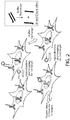
- the disclosure provides methods for performing high-throughput and highly multiplexed imaging and analyte/target detection based on programmable nucleic acid (e.g., DNA) probes. These methods rely on a sequential imaging approach employing orthogonal imager strands that can stably attach to a complementary docking strand immobilized on binding partners, such as antibodies ( FIG. 1 ). After hybridization and imaging with an imager strand, an extinguishing step (such as a photobleaching step) is performed to eliminate and/or reduce fluorescence from the hybridized (bound) imager strands.
- an extinguishing step such as a photobleaching step
- the methods utilize weaker binding between docking and imaging strands in order to remove signal.
- the hybridization conditions may be changed such that the melting point of the duplex that is formed between the docking or imager strands is slightly above room temperature (e.g., 25 °C) or the imaging temperature.
- the labeling step i.e., the step at which the imager strands are bound to their respective docking strands
- the imaging step are performed as described above.
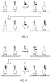
- the sample is subjected to a denaturing condition.
- the denaturing condition may be provided in a buffer exchange step using a solution with for example lower salt concentration, presence of or increase in the concentration of a denaturant such as formamide, or increased temperature ( FIG. 2 ).
- the sample may be alternatively or additionally exposed to an increased temperature.
- the aforementioned increases or decreases are relative to the conditions existing at the labeling step (i.e., when the imager strand is bound to the docking strand).
- the sample may be washed, the buffer exchange may be repeated, the sample may be washed again, and then the next imager strand may be added to the sample.
- orthogonal imager strands are sequentially applied after every step of, for example, photobleaching or other method for extinguishing signal or imager strand inactivation or removal to the same sample in order to potentially image an infinite number of targets.
- the methods provided herein are only limited by the number of possible orthogonal nucleotide sequences (of the docking strands or alternatively the imager strands).
- this approach has intrinsically scalable multiplexing capability just by using a single fluorophore.
- This method can be readily integrated with standard microscopy setups (e.g., confocal or epi-fluorescence microscopes), allowing high throughput analysis of the sample.
- the methods have applicability in, for example, high-throughput screening assays such as drug screening assays.
- This imaging approach allows analysis of large populations of cells ( ⁇ 1,000-10,000) or tissue samples in an ultra-multiplexed format while imaging using standard confocal or epi-fluorescence microscope. Screening large numbers of targets such as proteins from the same sample in a high-throughput manner will provide information about new drugs or modifiers while providing cellular heterogeneity information.
- the large scale screening of tissue samples with high-throughput and ultra-multiplexed imaging capabilities will be useful in pathology analysis, for example, in a hospital or other service provider setting.
- Methods provided herein can also be used to identify the absolute quantity of a single target (e.g., such as, for example, a particular protein), or the quantity of a single target relative to one or more other targets.
- a single target e.g., such as, for example, a particular protein
- methods provided herein may be used to identify the location of a target within a sample or relative to other targets in the sample.
- This disclosure therefore provides a method comprising (1) contacting a sample simultaneously with a plurality of sequence-labeled target-recognition moieties, (2) introducing imager nucleic acids such as imager strands recognizing, through sequence complementarity, a subset of docking nucleic acids such as docking strands in the sequence-labeled target-recognition moieties, (3) removing or inactivating the imager nucleic acids or extinguishing signal from the imager nucleic acids, and (4) repeating step (2) and optionally step (3) at least once in order to image and detect one or more additional docking nucleic acids.
- the method may optionally comprise labeling a plurality of target-recognition moieties with docking nucleic acids such as docking strands to form sequence-labeled target-recognition moieties.
- This disclosure further provides a method comprising (1) contacting a sample being tested for the presence of one or more targets with one or more target-specific binding partners, wherein each target-specific binding partner is linked to a docking strand, and wherein target-specific binding partners of different specificity are linked to different docking strands, (2) optionally removing unbound target-specific binding partners, (3) contacting the sample with labeled imager strands having a nucleotide sequence that is complementary to a docking strand, (4) optionally removing unbound labeled imager strands, (5) imaging the sample to detect location and number of bound labeled imager strands, (6) extinguishing signal from the bound labeled imager strand, and (7) repeating steps (3)-(6), each time with a labeled imager strand having a unique nucleotide sequence relative to all other labeled imager strands.
- Steps (3)-(6) may be repeated once or multiple times. For example, steps (3)-(6) may be repeated 1-10 times or more. In some embodiments, steps (3)-(6) are repeated 1, 2, 3, 4, 5, 6, 7, 8, 9 or 10 times.
- This disclosure further provides a method comprising (1) contacting a sample being tested for the presence of one or more targets with one or more target-recognition moieties such as target-specific binding partners, wherein each target-recognition moiety is linked to a docking nucleic acid, and wherein target-recognition moieties of different specificity are linked to different docking nucleic acids, (2) optionally removing unbound target-recognition moieties, (3) contacting the sample with labeled imager nucleic acids such as imager strands having a nucleotide sequence that is complementary to a docking nucleic acid, (4) optionally removing unbound labeled imager nucleic acids, (5) imaging the sample to detect location and number of bound labeled imager nucleic acids, (6) removing the bound labeled imager nucleic acids from the docking nucleic acids, and (7) repeating steps (3)-(6), each time with a labeled imager nucleic acid having a unique nucleotide
- Steps (3)-(6) may be repeated once or multiple times. For example, steps (3)-(6) may be repeated 1-10 times or more. In some embodiments, steps (3)-(6) are repeated 1, 2, 3, 4, 5, 6, 7, 8, 9 or 10 times.
- This disclosure further provides a method comprising (1) contacting a sample being tested for the presence of one or more targets with one or more target-recognition moieties such as target-specific binding partners, wherein each target-recognition moieties is linked to a docking nucleic acid such as a docking strand, and wherein target-recognition moieties of different specificity are linked to different docking nucleic acids, (2) optionally removing unbound target-recognition moieties, (3) contacting the sample with labeled imager nucleic acids such as imager strands having a nucleotide sequence that is complementary to a docking nucleic acid, (4) optionally removing unbound labeled imager nucleic acids, (5) imaging the sample to detect location and number of bound labeled imager nucleic acids, (6) inactivating the bound labeled imager nucleic acids, by removing or modifying their signal-emitting moieties without removing the imager nucleic acid in its entirety, and
- Steps (3)-(6) may be repeated once or multiple times. For example, steps (3)-(6) may be repeated 1-10 times or more. In some embodiments, steps (3)-(6) are repeated 1, 2, 3, 4, 5, 6, 7, 8, 9 or 10 times.
- the methods provided herein include a step of removing an imager nucleic acid such as an imager strand that is bound to a docking nucleic acids such as a docking strand, using a method other than strand displacement.
- the methods provided herein include a step of removing an imager nucleic acid such as an imager strand that is bound to a docking nucleic acid such as a docking strand, wherein the imager nucleic acid emits signal (i.e., such signal is not quenched) prior to binding to the docking nucleic acid.
- the methods provided herein include a step of removing an imager nucleic acid such as an imager strand that is bound to a docking nucleic acid such as a docking strand, wherein the imager nucleic acid is removed using a nucleic acid that does not comprise a quencher.
- the docking nucleic acid including the docking strand may be a single-stranded docking nucleic acid or docking strand, or it may be a double-stranded docking nucleic acid or docking strand, or it may be a partially double-stranded docking nucleic acid or docking strand ( e.g., containing a single-stranded and a double-stranded region).
- the plurality may be contacted with the sample, and thus with targets of interest, simultaneously.
- the target-recognition moieties such as the binding partners need not be contacted with the sample sequentially, although they can be.
- binding partners conjugated to nucleic acids e.g., docking nucleic acids such as docking strands
- binding partner-nucleic acid conjugates BP-NA conjugates
- binding partner-nucleic acid conjugates BP-NA conjugates
- sequence-labeled target-recognition moieties BP-NA conjugates
- binding partner-nucleic acid conjugate or “BP-NA conjugate” refers to a molecule linked ( e.g. , through an N-Hydroxysuccinimide (NHS) linker) to a single-stranded nucleic acid (e.g., DNA) docking strand.
- NHS N-Hydroxysuccinimide
- the binding partner of the conjugate may be any moiety (e.g. , antibody or aptamer) that has an affinity for ( e.g., binds to) a target, such as a biomolecule (e.g., protein or nucleic acid), of interest.
- a target such as a biomolecule (e.g., protein or nucleic acid), of interest.
- the binding partner is a protein.
- BP-NA-conjugates that comprise a protein (or peptide) linked to a docking strand may be referred to herein as "protein-nucleic acid conjugates," or “protein-NA conjugates.”
- proteins for use in the conjugates of the invention include, without limitation, antibodies (e.g., monoclonal antibodies), antigen-binding antibody fragments (e.g., Fab fragments), receptors, peptides and peptide aptamers.
- Other binding partners may be used in accordance with the invention. For example, binding partners that bind to targets through electrostatic (e.g., electrostatic particles), hydrophobic or magnetic ( e.g., magnetic particles) interactions are contemplated herein.
- antibody includes full-length antibodies and any antigen binding fragment (e.g., "antigen-binding portion") or single chain thereof.
- the term “antibody” includes, without limitation, a glycoprotein comprising at least two heavy (H) chains and two light (L) chains inter-connected by disulfide bonds, or an antigen binding portion thereof.
- Antibodies may be polyclonal or monoclonal; xenogeneic, allogeneic, or syngeneic; or modified forms thereof ( e.g., humanized, chimeric).
- antigen-binding portion of an antibody refers to one or more fragments of an antibody that retain the ability to specifically bind to an antigen.
- the antigen-binding function of an antibody can be performed by fragments of a full-length antibody.
- binding fragments encompassed within the term "antigen-binding portion" of an antibody include (i) a Fab fragment, a monovalent fragment consisting of the V H , V L , C L and C H1 domains; (ii) a F(ab')2 fragment, a bivalent fragment comprising two Fab fragments linked by a disulfide bridge at the hinge region; (iii) a Fd fragment consisting of the V H and C H1 domains; (iv) a Fv fragment consisting of the V H and V L domains of a single arm of an antibody, (v) a dAb fragment ( Ward et al., Nature 341:544 546, 1989 ), which consists of a V H domain; and (vi) an isolated complementarity determining region (CDR) or (vii) a combination of two or more isolated CDRs, which may optionally be joined by a synthetic linker.
- a Fab fragment a monovalent fragment consisting of the V H , V
- the two domains of the Fv fragment, V H and V L are coded for by separate genes, they can be joined, using recombinant methods, by a synthetic linker that enables them to be made as a single protein chain in which the V H and V L regions pair to form monovalent molecules (known as single chain Fv (scFv); see, e.g., Bird et al. Science 242:423 426, 1988 ; and Huston et al. Proc. Natl. Acad. Sci. USA 85:5879-5883, 1988 ).
- single chain Fv single chain Fv
- Such single chain antibodies are also encompassed within the term "antigen-binding portion" of an antibody.
- receptors refer to cellular-derived molecules (e.g., proteins) that bind to ligands such as, for example, peptides or small molecules (e.g., low molecular weight ( ⁇ 900 Daltons) organic or inorganic compounds).
- ligands such as, for example, peptides or small molecules (e.g., low molecular weight ( ⁇ 900 Daltons) organic or inorganic compounds).
- peptide aptamer refers to a molecule with a variable peptide sequence inserted into a constant scaffold protein (see, e.g., Baines IC, et al. Drug Discov. Today 11:334-341, 2006 ).
- the molecule of the BP-NA conjugate is a nucleic acid such as, for example, a nucleic acid aptamer.
- nucleic acid aptamer refers to a small RNA or DNA molecules that can form secondary and tertiary structures capable of specifically binding proteins or other cellular targets ( see, e.g., Ni X, et al. Curr Med Chem. 18(27): 4206-4214, 2011 ).
- the BP-NA conjugate may be an aptamer-nucleic acid conjugate.
- Target-recognition moieties are agents that specifically recognize targets of interest in the sample.
- target-recognition moieties include binding partners such as those recited herein.
- Target-recognition moieties include antibodies, antibody fragments and antibody derivatives such as single-chain antibodies, single-chain Fv domains, Fab domains, nanobodies, and the like, peptides, aptamers, and oligonucleotides (e.g., to detect nucleic acids of interest in procedures such as fluorescence in situ hybridization, or FISH).
- Docking nucleic acids include docking strands as described herein. Docking nucleic acids are linear nucleic acids capable of binding to a nucleic acid having a complementary sequence (such as an imager nucleic acid).
- a docking nucleic acid may be comprised of or may consist of DNA, RNA, or nucleic acid-like structures with other phosphate-sugar backbones (e.g. 2'-O-methyl RNA, 2'-fluoral RNA, LNA, XNA) or backbones comprising non-phosphate-sugar moieties (e.g., peptide nucleic acid and morpholino).
- the nucleobases may include naturally occurring nucleobases such as adenine, thymine, guanine, cytosine, inosine, and their derivatives, as well as non-naturally occurring nucleobases such as isoC, isoG, dP and dZ.
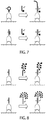
- a docking nucleic acid when not bound to its complementary imager nucleic acid, may be single-stranded without stable secondary structure. Alternatively, the docking nucleic acid may comprise secondary structure such as a hairpin loop ( FIG. 7 , top).
- a docking nucleic acid may be part of a multi-strand complex ( FIG. 7 , bottom).
- a "docking strand” refers to a single-stranded nucleic acid (e.g., DNA) capable of stably binding to its complementary imager strands. Stable binding may be a result of the length of the docking strand (and conversely the imager strand) or it may be the result of the particular conditions under which hybridization occurs ( e.g., salt concentration, temperature, etc.). In some embodiments, a docking strand is about 20 to about 60, or more, nucleotides in length. A docking strand may be capable of binding to one or more identical imager strands (of identical sequence and identically labeled).
- Imager nucleic acids such as imager strands
- Imager nucleic acids include imager strands as described herein.
- Imager nucleic acids are nucleic acids that can (1) interact with a docking nucleic acid via sequence-specific complementarity and (2) recruit a signal-emitting moiety or multiple copies of signal-emitting moieties by covalent or non-covalent interactions.
- the imager nucleic acids may be linear or branched as described herein.
- One imager nucleic acid may recruit multiple copies of the signal-emitting moiety via a polymeric ( FIG. 8 , top) or dendrimeric structure ( FIG. 8 , bottom).
- a polymeric or dendrimeric structure can be synthesized chemically using methods such as those discussed in Nazemi A. et al. Chemistry of Bioconjugates: Synthesis, Characterization, and Biomedical Applications, Published Online: 13 Feb. 2014 ) and references provided therein.
- the polymeric or dendrimeric structure can be formed by DNA hybridization as shown, for example, in Dirks R. et al. Proc. Nat. Acad. Sci. U.S.A., 2004;1010(43):15275-78 ; and in Um S.H. et al. Nat. Protocols 2006;1:995-1000 .
- An imager nucleic acid may be comprised of or may consist of DNA, RNA, or nucleic acid-like structures with other phosphate-sugar backbones (e.g. 2'-O-methyl RNA, 2'-fluoral RNA, LNA, XNA) or backbones comprising non-phosphate-sugar moieties (e.g. , peptide nucleic acid and morpholino).
- the nucleobases may include naturally occurring nucleobases such as adenine, thymine, guanine, cytosine, inosine, and their derivatives, as well as non-naturally occurring nucleobases such as isoC, isoG, dP and dZ.
- an imager nucleic acid is about 30 to about 60 nucleotides, or more, in length, including 30, 35, 40, 45, 50, 55 or 60 nucleotides in length. In some embodiments, an imager nucleic acid is 30 to 40, 30 to 50, 40 to 50, 40 to 60, or 50 to 60 nucleotides in length.
- An imager nucleic acid when not bound to its complementary docking nucleic acid, may be single-stranded without stable secondary structure.
- the imager nucleic acid may comprise secondary structure such as a hairpin loop ( FIG. 9 , top).
- An imager nucleic acid may be part of a multi-strand complex ( FIG. 9 , bottom).
- the imager strand can be self-quenching, intending that the unbound imager nucleic acid may carry a quencher moiety that is in close proximity with the signal-emitting moiety such as a fluorophore.
- the imager nucleic acid can be designed to adopt either a molecular beacon-like structure ( FIG. 5 ) or a hemiduplex structure ( FIG. 6 ).
- the binding and imaging buffer may contain additives routinely used in FISH, Northern Blotting and Southern Blotting (e.g., negatively charged polymers such as dextran sulfate and heparin) to reduce non-specific binding.
- a “signal-emitting moiety,” as used herein, is a moiety that, under certain conditions, emits detectable signal, such as photon, radiation, positron, electromagnetic wave, and magnetic-nuclear resonance.
- an "imager strand” is a single-stranded nucleic acid (e.g., DNA) that is about 30 to about 60 nucleotides, or more, in length.
- An imager strand of the invention is complementary to a docking strand and stably binds to the docking strand. Stable binding intends that the imager and docking strands remained bound to each other for the length of the assay, or for at least 30 minutes, or for at least for 60 minutes, or for at least for 2 hours, or more. Such binding may or may not be reversible or irreversible.
- a docking nucleic acid is considered stably bound to an imager nucleic acid such as an imager strand if the nucleic acids remain bound to each other for (or for at least) 30, 35, 40, 45, 50, 55 or 60 minutes (min). In some embodiments, a docking nucleic acid is considered stably bound to an imager nucleic acid if the nucleic acids remain bound to each other for (or for at least) 30 to 60 min, 30 to 120 min, 40 to 60 min, 40 to 120 min, or 60 to 120 min. Such binding may or may not be reversible, or may or may not be irreversible.
- binding refers to an association between at least two molecules due to, for example, electrostatic, hydrophobic, ionic and/or hydrogen-bond interactions, optionally under physiological conditions.
- nucleic acids or nucleic acid domains, are "complementary" to one another if they base-pair, or bind, with each other to form a double-stranded nucleic acid molecule via Watson-Crick interactions.
- nucleic acids of the invention such as the docking nucleic acids and the imager nucleic acids bind to each other with "perfect complementary,” which refers to 100% complementary (e.g., 5' - ATTCGC - 3' is perfectly complementary to 5' GCGAAT - 3').
- Imager strands of the invention may be labeled with a detectable label (e.g., a fluorescent label, and thus are considered “fluorescently labeled").
- a detectable label e.g., a fluorescent label, and thus are considered "fluorescently labeled"
- an imager strand may comprise at least one ( i.e., one or more) fluorophore.
- Imager nucleic acids including imager strands may be covalently labeled with a detectable label such as those recited herein or known in the art.
- imager nucleic acids including imager strands may comprise 2, 3, 4, or more detectable labels such as fluorophores.
- Orthogonal imager nucleic acids including imager strands may comprise a distinct label (e.g., a red fluorophore, a blue fluorophore, or a green fluorophore), or they may all comprise the same label (e.g. , red fluorophores) even if they differ in nucleotide sequence.
- a distinct label e.g., a red fluorophore, a blue fluorophore, or a green fluorophore
- they may all comprise the same label (e.g. , red fluorophores) even if they differ in nucleotide sequence.
- the BP-NA conjugates (e.g., protein-nucleic acid conjugates) of the invention may, in some embodiments, comprise an intermediate linker that links ( e.g., covalently or non-covalently) the binding partner to a docking strand.
- the intermediate linker may comprise biotin and/or streptavidin.
- an antibody and a docking strand may each be biotinylated ( i.e., linked to at least one biotin molecule) and linked to each other through biotin binding to an intermediate streptavidin molecule.
- Other intermediate linkers may be used in accordance with the invention.
- an intermediate linker may not be required.
- the docking strand of a BP-NA conjugate may be an extension (e.g., 5' or 3' extension) of a nucleic acid molecule such as, for example, a nucleic acid aptamer. Similar approaches may be used to generate sequence-labeled target recognition moieties as provided herein.
- Pluralities of BP-NA conjugates e.g., protein-nucleic acid conjugates
- imager strands are provided herein.
- a plurality may be a population of the same species or distinct species.
- a plurality of BP-NA conjugates of the same species may comprise conjugates that all bind to the same target (e.g., biomolecule) (e.g., the same epitope or region/domain).
- a plurality of BP-NA conjugates of distinct species may comprise conjugates, or subsets of conjugates, each conjugate or subset of conjugates binding to a distinct epitope on the same target or to a distinct target.
- a plurality of imager strands of the same species may comprise imager strands with the same nucleotide sequence and the same fluorescent label (e.g., Cy2, Cy3 or Cy4).
- a plurality of imager strands of distinct species may comprise imager strands with distinct nucleotide sequences (e.g., DNA sequences) and distinct fluorescent labels (e.g., Cy2, Cy3 or Cy4) or with distinct nucleotide sequences and the same fluorescent ( e.g., all Cy2).
- a plurality of BP-NA conjugates comprises at least 10, 50, 100, 500, 1000, 2000, 3000, 4000, 5000, 10 4 , 50000, 10 5 , 10 5 , 10 6 , 10 7 , 10 8 , 10 9 , 10 10 , 10 11 BP-NA conjugates.
- a plurality of fluorescently labeled imager strands comprises at least 10, 50, 100, 500, 1000, 2000, 3000, 4000, 5000, 10 4 50000, 10 5 , 10 5 , 10 6 , 10 7 , 10 8 , 10 9 , 10 10 , 10 11 fluorescently labeled imager strands.
- a plurality may contain 1 to about 200 or more distinct species of BP-NA conjugates and/or imager strands.
- a plurality may contain at least 1, 2, 3, 4, 5, 6, 7, 8, 9, 10, 15, 20, 25, 30, 35, 40, 45, 50, 55, 60, 65, 70, 75, 80, 85, 90, 95, 100, 125, 150, 175, 200 or more distinct species.
- a plurality may contain less than about 5 to about 200 distinct species of BP-NA conjugates and/or imager strands.
- a plurality may contain less than 5, 6, 7, 8, 9, 10, 15, 20, 25, 30, 35, 40, 45, 50, 55, 60, 65, 70, 75, 80, 85, 90, 95, 100, 125, 150, 175 or 200 distinct species.
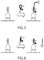
- the imager nucleic acids may be removed from the target-recognition moieties, including the binding partners, ( FIG. 3 ) by means such as but not limited to increasing temperature; decreasing the concentration of counter-ions (e.g., free Mg++); introducing or increasing the concentration of denaturants (e.g. formamide, urea, DMSO, and the like); and chemically, photochemically or enzymatically cleaving, modifying or degrading the imager strand, or any combination thereof.
- increasing temperature such as but not limited to increasing temperature; decreasing the concentration of counter-ions (e.g., free Mg++); introducing or increasing the concentration of denaturants (e.g. formamide, urea, DMSO, and the like); and chemically, photochemically or enzymatically cleaving, modifying or degrading the imager strand, or any combination thereof.
- denaturants e.g. formamide, urea, DMSO, and the like
- the imager nucleic acids may be inactivated by removing and/or modifying the signal-emitting moiety without removing the entirety of the nucleic acid portion of the imager nucleic acid from the docking strands. ( FIG. 4 .)
- the removal of the imager nucleic acid may be facilitated by cleaving the imager strand into multiple parts.
- the imager nucleic acid comprises a chemically cleavable moiety that can be cleaved by introduction of the chemical compound that acts upon such cleavable moiety.
- chemically cleavable moieties include but are not limited to allyl groups, which can be cleaved by certain Pd-based reagents ( Ju J. et al., Proc Natl Acad Sci USA.
- the imager nucleic acid comprises a photocleavable linker that can be cleaved photochemically (e.g. , by UV exposure).
- the imager nucleic acid contains a moiety that can be cleaved by an enzyme. Examples of such enzymatically cleavable moieties include but are not limited to ribonucleotides, which can be cleaved by a variety of RNases; deoxyuridines, which can be cleaved by enzyme combinations such as USER (New England Biolabs); and restriction sites, which can be cleaved by sequence-specific nicking enzymes or restriction enzymes.
- the restriction enzyme may cleave both the imager nucleic acid and the docking nucleic acid.
- the removal of the imager nucleic acid may be facilitated by modifying the imager nucleic acid into a form that binds the docking nucleic acid to form a duplex with decreased stability (or lower melting temperature).
- the imager nucleic acid comprises azobenzene, which can be photoisomerized, wherein different isomers affect the binding strength of the imager nucleic acid to the docking nucleic acid differently ( Asanuma H. et al. Angew Chem Int Ed Engl. 2001 Jul 16;40(14):2671-2673 ).
- the imager nucleic acid comprises a deoxyuridine, in which the uracil group may be cleaved by uracil-DNA glycosylase. After the uracil is removed the binding strength of the imager strand is weakened.
- the removal of the signal-emitting moiety can be achieved by cleaving a linker between the imager nucleic acid and the signal-emitting moiety, if such a linker exists. Chemistries described in herein can be used for this purpose as well.
- the inactivation of the signal-emitting moiety can be achieved by chemically or photochemically modifying the signal-emitting moiety.
- the signal-emitting moiety when it is a fluorophore, it can be bleached by chemical agents (such as for example hydrogen peroxide, Gerdes M. et al. Proc Natl Acad Sci USA. 2013 Jul 16;110(29):11982-87 ) or photobleached ( e.g., using soft multiwavelength excitation as described in Schubert W. et al. Nat. Biotech. 2006;24:1270-78 ).
- photobleaching refers to the photochemical alteration of a dye or a fluorophore molecule such that it is unable to fluoresce. This is caused by cleavage of covalent bonds or non-specific reactions between the fluorophore and surrounding molecules. Loss of activity caused by photobleaching can be controlled, in some embodiments, by reducing the intensity or time-span of light exposure, by increasing the concentration of fluorophores, by reducing the frequency and thus the photon energy of the input light, or by employing more robust fluorophores that are less prone to bleaching (e.g. Alexa Fluors or DyLight Fluors) See, e.g., Ghauharali R. et al. Journal of Microscopy 2001;198: 88-100 ; and Eggeling C. et al. Analytical Chemistry 1998;70:2651-59 .
- photobleaching may be used to remove, modify or in some instance extinguish signal from a signal-emitting moiety.
- Photobleaching may be performed by exposing fluorophores to a wavelength of light of suitable wavelength, energy and duration to permanently and irreversibly extinguish the ability of the fluorophore to emit further signal. Photobleaching techniques are known in the art.
- the predicted dissociation constant (using the parameter sets outlined in reference PMID 15139820) between ssDNA 5'-CATCTAAAGCC-3' and its reverse-complementary strand 5'-GGCTTTAGATG-3' is -90 pM at 23°C with 500 mM [Na+] and 10 mM [Mg++] concentration. In other words, in this condition the binding is very strong.
- the predicted dissociation constant of this pair of ssDNA is as high as ⁇ 500 nM at 37°C with 150 mM [Na+] and 0 mM [Mg++]. In other words, in this condition the binding is fairly weak.
- the imaging condition can be 23°C with 500 mM [Na+] and 10 mM [Mg++]
- the dye-inactivating condition can be 37°C with 150 mM [Na+] and 0 mM [Mg++].
- the sample being analyzed is cultured cells, tissue sections, or other samples from living organisms.
- the sample is dissociated cells that are immobilized to a solid surface (e.g. glass slide or cover slip), including individually immobilized.
- the sample may be cells in blood.
- the sample may contain cancer cells circulating in the blood (also known as circulating tumor cells, or CTCs).
- the sample may be cells grown in suspension.
- the sample may be cells disseminated from a solid tissue.
- a “sample” may comprise cells (or a cell), tissue, or bodily fluid such as blood (serum and/or plasma), urine, semen, lymphatic fluid, cerebrospinal fluid or amniotic fluid.
- a sample may be obtained from (or derived from) any source including, without limitation, humans, animals, bacteria, viruses, microbes and plants.
- a sample is a cell lysate or a tissue lysate.
- a sample may also contain mixtures of material from one source or different sources.
- a sample may be a spatial area or volume (e.g., a grid on an array, or a well in a plate or dish).
- a sample in some embodiments, includes target(s), BP-NA conjugate(s) and imager strand(s).
- the cells may be disseminated (or dissociated) cells.
- a “target” is any moiety that one wishes to observe or quantitate and for which a binding partner exists.
- a target in some embodiments, may be non-naturally occurring.
- the target in some embodiments, may be a biomolecule.
- a “biomolecule” is any molecule that is produced by a living organism, including large macromolecules such as proteins, polysaccharides, lipids and nucleic acids (e.g., DNA and RNA such as mRNA), as well as small molecules such as primary metabolites, secondary metabolites, and natural products.
- biomolecules include, without limitation, DNA, RNA, cDNA, or the DNA product of RNA subjected to reverse transcription, A23187 (Calcimycin, Calcium Ionophore), Abamectine, Abietic acid, Acetic acid, Acetylcholine, Actin, Actinomycin D, Adenosine, Adenosine diphosphate (ADP), Adenosine monophosphate (AMP), Adenosine triphosphate (ATP), Adenylate cyclase, Adonitol, Adrenaline, epinephrine, Adrenocorticotropic hormone (ACTH), Aequorin, Aflatoxin, Agar, Alamethicin, Alanine, Albumins, Aldosterone, Aleurone, Alpha-amanitin, Allantoin, Allethrin, ⁇ -Amanatin, Amino acid, Amylase, Anabolic steroid, Anethole, Angiotensinogen, Anisomycin,
- a target may be a protein target such as, for example, proteins of a cellular environment (e.g., intracellular or membrane proteins).
- proteins include, without limitation, fibrous proteins such as cytoskeletal proteins (e.g., actin, arp2/3, coronin, dystrophin, FtsZ, keratin, myosin, nebulin, spectrin, tau, titin, tropomyosin, tubulin and collagen) and extracellular matrix proteins (e.g., collagen, elastin, f-spondin, pikachurin, and fibronectin); globular proteins such as plasma proteins ( e.g., serum amyloid P component and serum albumin), coagulation factors (e.g., complement proteins, C1-inhibitor and C3-convertase, Factor VIII, Factor XIII, fibrin, Protein C, Protein S, Protein Z, Protein Z-related protease inhibitor, thrombin
- fibrous proteins such as
- ion channels e.g., ligand-gated ion channels such nicotinic acetylcholine receptors and GABAa receptors, and voltage-gated ion channels such as potassium, calcium and sodium channels
- synport/antiport proteins e.g.
- glucose transporter e.g., epidermal growth factor (EGF), fibroblast growth factor (FGF), vascular endothelial growth factor (VEGF), peptide hormones such as insulin, insulin-like growth factor and oxytocin, and steroid hormones such as androgens, estrogens and progesterones); receptors such as transmembrane receptors ( e.g., G-protein-coupled receptor, rhodopsin) and intracellular receptors ( e.g., estrogen receptor); DNA-binding proteins ( e.g., histones, protamines, CI protein); transcription regulators ( e.g., c-myc, FOXP2, FOXP3, MyoD and P53); immune system proteins ( e.g., immunoglobulins, major histocompatibility antigens and T cell receptors); nutrient storage/transport proteins (e.g., ferritin); chaperone proteins; and enzymes.
- EGF epidermal growth factor
- a target may be a nucleic acid target such as, for example, nucleic acids of a cellular environment.
- a nucleic acid refers to a polymeric form of nucleotides of any length, such as deoxyribonucleotides or ribonucleotides, or analogs thereof.
- a nucleic acid may be a DNA, RNA or the DNA product of RNA subjected to reverse transcription.
- Non-limiting examples of nucleic acids include coding or non-coding regions of a gene or gene fragment, loci (locus) defined from linkage analysis, exons, introns, messenger RNA (mRNA), transfer RNA, ribosomal RNA, ribozymes, cDNA, recombinant nucleic acids, branched nucleic acids, plasmids, vectors, isolated DNA of any sequence, isolated RNA of any sequence, nucleic acid probes, and primers.
- loci locus
- locus defined from linkage analysis, exons, introns, messenger RNA (mRNA), transfer RNA, ribosomal RNA, ribozymes, cDNA, recombinant nucleic acids, branched nucleic acids, plasmids, vectors, isolated DNA of any sequence, isolated RNA of any sequence, nucleic acid probes, and primers.
- nucleic acids include, without limitation, cDNA, aptamers, peptide nucleic acids ("PNA”), 2'-5' DNA (a synthetic material with a shortened backbone that has a base-spacing that matches the A conformation of DNA; 2'-5' DNA will not normally hybridize with DNA in the B form, but it will hybridize readily with RNA), locked nucleic acids (“LNA”), and nucleic acids with modified backbones (e.g., base- or sugar-modified forms of naturally-occurring nucleic acids).
- PNA peptide nucleic acids
- LNA locked nucleic acids
- nucleic acids with modified backbones e.g., base- or sugar-modified forms of naturally-occurring nucleic acids.
- a nucleic acid may comprise modified nucleotides, such as methylated nucleotides and nucleotide analogs ("analogous" forms of purines and pyrimidines are well known in the art). If present, modifications to the nucleotide structure may be imparted before or after assembly of the polymer.
- a nucleic acid may be a single-stranded, double-stranded, partially single-stranded, or partially double-stranded DNA or RNA.
- a nucleic acid (e.g., a nucleic acid target) is naturally-occurring.
- a "naturally occurring” refers to a nucleic acid that is present in organisms or viruses that exist in nature in the absence of human intervention.
- a nucleic acid naturally occurs in an organism or virus.
- a nucleic acid is genomic DNA, messenger RNA, ribosomal RNA, micro-RNA, pre-micro-RNA, pro-micro-RNA, viral DNA, viral RNA or piwi-RNA.
- a nucleic acid target is not a synthetic DNA nanostructure (e.g., two-dimensional (2-D) or three-dimensional (3-D) DNA nanostructure that comprises two or more nucleic acids hybridized to each other by Watson-Crick interactions to form the 2-D or 3-D nanostructure).
- a synthetic DNA nanostructure e.g., two-dimensional (2-D) or three-dimensional (3-D) DNA nanostructure that comprises two or more nucleic acids hybridized to each other by Watson-Crick interactions to form the 2-D or 3-D nanostructure.
- nucleic acid docking strands and imager strands described herein can be any one of the nucleic acids described above (e.g., DNA, RNA, modified nucleic acids, nucleic acid analogues, naturally-occurring nucleic acids, synthetic nucleic acids).
- compositions that comprise at least one or at least two (e.g., a plurality) BP-NA conjugate(s) (e.g., protein-nucleic acid conjugate(s)) of the invention.
- the BP-NA conjugates may be bound to a target of interest (e.g., biomolecule) and/or stable bound to a complementary fluorescently labeled imager strand.
- a composition may comprise a plurality of the same species or distinct species of BP-NA conjugates.
- a composition may comprise at least 10, 50, 100, 500, 1000, 2000, 3000, 4000, 5000, 10 4 , 50000, 10 5 , 10 5 , 10 6 , 10 7 , 10 8 , 10 9 , 10 10 , 10 11 BP-NA conjugates.
- a composition may comprise at least 10, 50, 100, 500, 1000, 2000, 3000, 4000, 5000, 10 4 , 50000, 10 5 , 10 5 , 10 6 , 10 7 , 10 8 , 10 9 , 10 10 , 10 11 complementary fluorescently labeled imager strands.
- a composition may contain 1 to about 200 or more distinct species of BP-NA conjugates and/or imager strands.
- a composition may contain at least 1, 2, 3, 4, 5, 6, 7, 8, 9, 10, 15, 20, 25, 30, 35, 40, 45, 50, 55, 60, 65, 70, 75, 80, 85, 90, 95, 100, 125, 150, 175, 200 or more distinct species.
- a composition may contain less than about 5 to about 200 distinct species of BP-NA conjugates and/or imager strands.
- a composition may contain less than 5, 6, 7, 8, 9, 10, 15, 20, 25, 30, 35, 40, 45, 50, 55, 60, 65, 70, 75, 80, 85, 90, 95, 100, 125, 150, 175 or 200 distinct species.
- the number of complementary fluorescently labeled imager strands imager stands in a composition may be less than, equal to or greater than the number of BP-NA conjugates in the composition.
- kits comprising one or more components of the invention.
- the kits may comprise, for example, a BP-NA conjugate and/or a fluorescently labeled imager strands.
- the kits may also comprise components for producing a BP-NA conjugate or for labeling an imager strand.
- the kits may comprise a binding partner (e.g., antibody), docking strands and intermediate linkers such as, for example, biotin and streptavidin molecules, and/or imager strands.
- the kits can be used for any purpose apparent to those of skill in the art, including, those described above.
- kits may include other reagents as well, for example, buffers for performing hybridization reactions.
- the kit may also include instructions for using the components of the kit, and/or for making and/or using the BP-NA conjugates and/or labeled imager strands.
- BP-NA conjugates e.g., protein-nucleic acid conjugates or antibody-nucleic acid conjugates
- the BP-NA conjugates of the invention can be used, inter alia, in any assay in which existing target detection technologies are used.
- assays include detection assays including diagnostic assays, prognostic assays, patient monitoring assays, screening assays, bio-warfare assays, forensic analysis assays, prenatal genomic diagnostic assays and the like.
- the assay may be an in vitro assay or an in vivo assay.
- the present invention provides the advantage that many different targets can be analyzed at one time from a single sample using the methods of the invention, even where such targets are spatially not resolvable (and thus spatially indistinct) using prior art imaging methods. This allows, for example, for several diagnostic tests to be performed on one sample.
- the BP-NA conjugates can also be used to simply observe an area or region.
- the methods of the invention may be applied to the analysis of samples obtained or derived from a patient so as to determine whether a diseased cell type is present in the sample and/or to stage the disease.
- a blood sample can be assayed according to any of the methods described herein to determine the presence and/or quantity of markers of a cancerous cell type in the sample, thereby diagnosing or staging the cancer.
- the methods described herein can be used to diagnose pathogen infections, for example infections by intracellular bacteria and viruses, by determining the presence and/or quantity of markers of bacterium or virus, respectively, in the sample.
- the targets detected using the compositions and methods of the disclosure may be either disclosure patient markers (such as a cancer marker) or markers of infection with a foreign agent, such as bacterial or viral markers.
- the quantitative imaging methods of the disclosure may be used, for example, to quantify targets (e.g., target biomolecules) whose abundance is indicative of a biological state or disease condition (e.g., blood markers that are upregulated or down-regulated as a result of a disease state).
- targets e.g., target biomolecules
- a biological state or disease condition e.g., blood markers that are upregulated or down-regulated as a result of a disease state.
- compositions and methods of the invention may be used to provide prognostic information that assists in determining a course of treatment for a patient.
- the amount of a particular marker for a tumor can be accurately quantified from even a small sample from a patient.
- overexpression of certain proteins, such as Her2-neu indicate a more aggressive course of treatment will be needed.
- the methods of the present invention may also be used for determining the effect of a perturbation, including chemical compounds, mutations, temperature changes, growth hormones, growth factors, disease, or a change in culture conditions, on various targets, thereby identifying targets whose presence, absence or levels are indicative of a particular biological states.
- the present invention is used to elucidate and discover components and pathways of disease states. For example, the comparison of quantities of targets present in a disease tissue with "normal" tissue allows the elucidation of important targets involved in the disease, thereby identifying targets for the discovery/screening of new drug candidates that can be used to treat disease.
- the sample being analyzed may be a biological sample, such as blood, sputum, lymph, mucous, stool, urine and the like.
- the sample may be an environmental sample such as a water sample, an air sample, a food sample and the like.
- the assay may be carried out with one or more components of the binding reaction immobilized.
- the targets or the BP-NA conjugates may be immobilized.
- the assay may be carried out with one or more components of the binding reaction non-immobilized.
- the assays may involve detection of a number of targets in a sample, essentially at the same time, in view of the multiplexing potential offered by the BP-NA conjugates and fluorescently labeled imager strands of the invention.
- an assay may be used to detect a particular cell type (e.g., based on a specific cell surface receptor) and a particular genetic mutation in that particular cell type. In this way, an end user may be able to determine how many cells of a particular type carry the mutation of interest, as an example.
- a reference to "A and/or B", when used in conjunction with open-ended language such as “comprising” can refer, in one embodiment, to A only (optionally including elements other than B); in another embodiment, to B only (optionally including elements other than A); in yet another embodiment, to both A and B (optionally including other elements); etc.
- the phrase "at least one,” in reference to a list of one or more elements, should be understood to mean at least one element selected from any one or more of the elements in the list of elements, but not necessarily including at least one of each and every element specifically listed within the list of elements and not excluding any combinations of elements in the list of elements.
- This definition also allows that elements may optionally be present other than the elements specifically identified within the list of elements to which the phrase "at least one" refers, whether related or unrelated to those elements specifically identified.
- At least one of A and B can refer, in one embodiment, to at least one, optionally including more than one, A, with no B present (and optionally including elements other than B); in another embodiment, to at least one, optionally including more than one, B, with no A present (and optionally including elements other than A); in yet another embodiment, to at least one, optionally including more than one, A, and at least one, optionally including more than one, B (and optionally including other elements); etc.
Landscapes
- Chemical & Material Sciences (AREA)
- Life Sciences & Earth Sciences (AREA)
- Organic Chemistry (AREA)
- Health & Medical Sciences (AREA)
- Zoology (AREA)
- Wood Science & Technology (AREA)
- Proteomics, Peptides & Aminoacids (AREA)
- Engineering & Computer Science (AREA)
- Analytical Chemistry (AREA)
- Immunology (AREA)
- Molecular Biology (AREA)
- Bioinformatics & Cheminformatics (AREA)
- Biotechnology (AREA)
- Biophysics (AREA)
- Physics & Mathematics (AREA)
- Genetics & Genomics (AREA)
- Biochemistry (AREA)
- Microbiology (AREA)
- General Engineering & Computer Science (AREA)
- General Health & Medical Sciences (AREA)
- Pathology (AREA)
- Measuring Or Testing Involving Enzymes Or Micro-Organisms (AREA)
- Investigating, Analyzing Materials By Fluorescence Or Luminescence (AREA)
- Investigating Or Analysing Biological Materials (AREA)
- Investigating Or Analysing Materials By The Use Of Chemical Reactions (AREA)
Priority Applications (3)
| Application Number | Priority Date | Filing Date | Title |
|---|---|---|---|
| EP20157624.6A EP3686599B1 (en) | 2014-03-11 | 2015-03-11 | High-throughput and highly multiplexed imaging with programmable nucleic acid probes |
| EP22151320.3A EP4030166B1 (en) | 2014-03-11 | 2015-03-11 | High-throughput and highly multiplexed imaging with programmable nucleic acid probes |
| EP23176015.8A EP4245859A3 (en) | 2014-03-11 | 2015-03-11 | High-throughput and highly multiplexed imaging with programmable nucleic acid probes |
Applications Claiming Priority (2)
| Application Number | Priority Date | Filing Date | Title |
|---|---|---|---|
| US201461951461P | 2014-03-11 | 2014-03-11 | |
| PCT/US2015/020034 WO2015138653A1 (en) | 2014-03-11 | 2015-03-11 | High-throughput and highly multiplexed imaging with programmable nucleic acid probes |
Related Child Applications (4)
| Application Number | Title | Priority Date | Filing Date |
|---|---|---|---|
| EP22151320.3A Division EP4030166B1 (en) | 2014-03-11 | 2015-03-11 | High-throughput and highly multiplexed imaging with programmable nucleic acid probes |
| EP23176015.8A Division EP4245859A3 (en) | 2014-03-11 | 2015-03-11 | High-throughput and highly multiplexed imaging with programmable nucleic acid probes |
| EP20157624.6A Division EP3686599B1 (en) | 2014-03-11 | 2015-03-11 | High-throughput and highly multiplexed imaging with programmable nucleic acid probes |
| EP20157624.6A Division-Into EP3686599B1 (en) | 2014-03-11 | 2015-03-11 | High-throughput and highly multiplexed imaging with programmable nucleic acid probes |
Publications (3)
| Publication Number | Publication Date |
|---|---|
| EP3117214A1 EP3117214A1 (en) | 2017-01-18 |
| EP3117214A4 EP3117214A4 (en) | 2017-12-20 |
| EP3117214B1 true EP3117214B1 (en) | 2020-05-06 |
Family
ID=54072381
Family Applications (4)
| Application Number | Title | Priority Date | Filing Date |
|---|---|---|---|
| EP15762370.3A Active EP3117214B1 (en) | 2014-03-11 | 2015-03-11 | High-throughput and highly multiplexed imaging with programmable nucleic acid probes |
| EP22151320.3A Active EP4030166B1 (en) | 2014-03-11 | 2015-03-11 | High-throughput and highly multiplexed imaging with programmable nucleic acid probes |
| EP23176015.8A Pending EP4245859A3 (en) | 2014-03-11 | 2015-03-11 | High-throughput and highly multiplexed imaging with programmable nucleic acid probes |
| EP20157624.6A Active EP3686599B1 (en) | 2014-03-11 | 2015-03-11 | High-throughput and highly multiplexed imaging with programmable nucleic acid probes |
Family Applications After (3)
| Application Number | Title | Priority Date | Filing Date |
|---|---|---|---|
| EP22151320.3A Active EP4030166B1 (en) | 2014-03-11 | 2015-03-11 | High-throughput and highly multiplexed imaging with programmable nucleic acid probes |
| EP23176015.8A Pending EP4245859A3 (en) | 2014-03-11 | 2015-03-11 | High-throughput and highly multiplexed imaging with programmable nucleic acid probes |
| EP20157624.6A Active EP3686599B1 (en) | 2014-03-11 | 2015-03-11 | High-throughput and highly multiplexed imaging with programmable nucleic acid probes |
Country Status (11)
Families Citing this family (41)
| Publication number | Priority date | Publication date | Assignee | Title |
|---|---|---|---|---|
| JP5978220B2 (ja) | 2010-10-29 | 2016-08-24 | プレジデント アンド フェローズ オブ ハーバード カレッジ | 核酸ナノ構造バーコードプローブ |
| US20160161472A1 (en) | 2013-07-30 | 2016-06-09 | President And Fellows Of Harvard College | Quantitative dna-based imaging and super-resolution imaging |
| JP6757255B2 (ja) | 2014-03-11 | 2020-09-16 | プレジデント アンド フェローズ オブ ハーバード カレッジ | プログラム可能な核酸プローブを用いた高スループット及び高度多重化イメージング |
| US9909167B2 (en) | 2014-06-23 | 2018-03-06 | The Board Of Trustees Of The Leland Stanford Junior University | On-slide staining by primer extension |
| CN119331954A (zh) | 2014-07-30 | 2025-01-21 | 哈佛学院院长及董事 | 探针文库构建 |
| WO2017027370A1 (en) | 2015-08-07 | 2017-02-16 | President And Fellows Of Harvard College | Super resolution imaging of protein-protein interactions |
| WO2017189498A1 (en) * | 2016-04-26 | 2017-11-02 | Ultivue, Inc. | Super-resolution immunofluorescence with diffraction-limited preview |
| WO2017197147A2 (en) * | 2016-05-11 | 2017-11-16 | Atkinson Robert G | Molecular beacon comprising prefabricated components and associated products, processes, and methods of use |
| CN109477834A (zh) * | 2016-05-19 | 2019-03-15 | 凸版印刷株式会社 | 目标分子的检测方法及目标分子检测试剂盒 |
| AU2017273720A1 (en) * | 2016-06-02 | 2018-12-20 | Ultivue, Inc. | Compositions and methods to expedite antibody-based exchange imaging |
| EP4180533B1 (en) * | 2016-07-27 | 2025-08-06 | The Board of Trustees of the Leland Stanford Junior University | Highly-multiplexed fluorescent imaging |
| TW201821609A (zh) * | 2016-08-22 | 2018-06-16 | Nissan Chemical Ind Ltd | 使用e-鈣黏蛋白結合型核酸適體之細胞貼附材料 |
| WO2018107054A1 (en) * | 2016-12-09 | 2018-06-14 | Ultivue, Inc. | Improved methods for multiplex imaging using labeled nucleic acid imaging agents |
| WO2018183876A1 (en) * | 2017-03-31 | 2018-10-04 | Ultivue, Inc. | Dna-antigen exchange and amplification |
| WO2018218150A1 (en) | 2017-05-26 | 2018-11-29 | President And Fellows Of Harvard College | Systems and methods for high-throughput image-based screening |
| US11935240B2 (en) | 2017-12-08 | 2024-03-19 | Ultivue, Inc. | Validation methods for multiplexed imaging method |
| US10871485B2 (en) | 2018-04-13 | 2020-12-22 | Rarecyte, Inc. | Kits for labeling of biomarkers and methods of using the same |
| AT521238B1 (de) | 2018-05-09 | 2020-02-15 | Lifetaq Analytics Gmbh | In-situ zellanalyse im zellkultursystem |
| WO2020014036A1 (en) * | 2018-07-09 | 2020-01-16 | Ultivue, Inc. | Methods for multicolor multiplex imaging |
| CN109628556A (zh) * | 2018-11-27 | 2019-04-16 | 山东师范大学 | 基于自催化复制介导的循环信号放大检测人8-羟基鸟嘌呤dna糖基化酶活性的方法 |
| AU2019397157A1 (en) * | 2018-12-14 | 2021-05-27 | Ultivue, Inc. | Methods and compositions for sequentially detecting targets |
| WO2020139757A1 (en) * | 2018-12-27 | 2020-07-02 | Bio-Rad Laboratories, Inc. | Sequential multiplex western blotting |
| CN114556074A (zh) * | 2019-06-14 | 2022-05-27 | 阿科亚生物科学股份有限公司 | 多重组织成像 |
| CN111004622B (zh) * | 2019-11-28 | 2023-08-15 | 郑州轻工业大学 | 一种检测多巴胺的高灵敏荧光探针的制备方法及其应用 |
| US20230221326A1 (en) * | 2019-12-30 | 2023-07-13 | Ultivue, Inc. | Methods for Reducing Nonspecific Interactions on Biological Samples |
| US20220064698A1 (en) * | 2020-08-26 | 2022-03-03 | Akoya Biosciences, Inc. | Multiplexed imaging reagent compositions and kits |
| CN112014563B (zh) * | 2020-08-27 | 2022-02-15 | 武汉大学 | 直接检测血液中循环肿瘤细胞的分子信标传递纳米探针、其制备方法及应用 |
| EP4214331B1 (en) * | 2020-09-16 | 2025-06-18 | Bruker Spatial Biology, Inc. | Chemical compositions and methods of using the same |
| GB2603935B (en) | 2021-02-19 | 2023-11-01 | Micrographia Bio Ltd | Method and kits for multiplex fluorescent microscopy |
| JP2024521682A (ja) * | 2021-05-19 | 2024-06-04 | ライカ マイクロシステムズ シーエムエス ゲゼルシャフト ミット ベシュレンクテル ハフツング | 生体試料または化合物または化学元素を分析するための方法 |
| CN113368899B (zh) * | 2021-07-03 | 2022-05-10 | 太原理工大学 | 一种高酸密度拟纤维素酶树脂固体酸催化剂的制备方法 |
| CN113584133B (zh) * | 2021-08-30 | 2024-03-26 | 中国药科大学 | 基于颜色编码与可编程性荧光探针的多重靶标原位检测方法 |
| US20230408382A1 (en) | 2022-05-31 | 2023-12-21 | Ultivue, Inc. | Methods and Systems for Removing a Histological Stain From a Sample |
| GB202212055D0 (en) | 2022-08-18 | 2022-10-05 | Micrographia Bio Ltd | Method and kits |
| WO2024168093A2 (en) * | 2023-02-08 | 2024-08-15 | Toreador Therapeutics, Inc. | Super-resolution microscopy (srm) multiplexing with cleavable moieties |
| EP4450971A1 (en) * | 2023-04-21 | 2024-10-23 | Leica Microsystems CMS GmbH | Marker for analysing a biological sample |
| CN116686824A (zh) * | 2023-06-07 | 2023-09-05 | 晶准生物医学(深圳)有限公司 | 血液样本保存液及其制备方法和应用 |
| CN116990271A (zh) * | 2023-06-29 | 2023-11-03 | 四川大学 | 基于比率荧光的四氢大麻酚及其代谢物的可视化检验方法 |
| CN116794290B (zh) * | 2023-07-03 | 2025-04-08 | 上海市第十人民医院 | 一种基于可逆dna自组装的组织切片靶标蛋白多重分析方法 |
| CN117347621B (zh) * | 2023-08-25 | 2024-03-12 | 广东省农业科学院农业生物基因研究中心 | 一种用蛋白模拟抗原-纳米抗体检测黄曲霉毒素b1的方法 |
| CN120334329B (zh) * | 2025-06-18 | 2025-08-22 | 徐州市中心医院 | 一种雌二醇生物传感器及其构建方法和应用 |
Family Cites Families (66)
| Publication number | Priority date | Publication date | Assignee | Title |
|---|---|---|---|---|
| DE4421891C2 (de) | 1994-06-23 | 1996-05-15 | Helmut Prof Dr Med Feucht | Verfahren zum Nachweis von komplementaktivierenden IgG-Antikörpern gegen HLA-DR-Antigene |
| US5491063A (en) * | 1994-09-01 | 1996-02-13 | Hoffmann-La Roche Inc. | Methods for in-solution quenching of fluorescently labeled oligonucleotide probes |
| DE19709348C2 (de) | 1996-05-29 | 1999-07-01 | Schubert Walter Dr Md | Automatisches Multi-Epitop-Ligand-Kartierungsverfahren |
| DE19717904A1 (de) | 1997-04-23 | 1998-10-29 | Diagnostikforschung Inst | Säurelabile und enzymatisch spaltbare Farbstoffkonstrukte zur Diagnostik mit Nahinfrarotlicht und zur Therapie |
| US20100081134A1 (en) | 1997-07-21 | 2010-04-01 | Mirkin Chad A | Bio-barcode based detection of target analytes |
| US6083486A (en) | 1998-05-14 | 2000-07-04 | The General Hospital Corporation | Intramolecularly-quenched near infrared fluorescent probes |
| US6635435B1 (en) | 1998-07-10 | 2003-10-21 | Chromagen, Inc. | Fluorogenic substrates and their use |
| US6573043B1 (en) | 1998-10-07 | 2003-06-03 | Genentech, Inc. | Tissue analysis and kits therefor |
| WO2000034527A2 (en) | 1998-12-11 | 2000-06-15 | The Regents Of The University Of California | Targeted molecular bar codes |
| AU3567900A (en) | 1999-03-30 | 2000-10-16 | Solexa Ltd. | Polynucleotide sequencing |
| CA2382436C (en) | 1999-08-30 | 2011-05-17 | Illumina, Inc. | Methods for improving signal detection from an array |
| WO2001023610A2 (en) | 1999-09-29 | 2001-04-05 | Solexa Ltd. | Polynucleotide sequencing |
| US7534568B2 (en) * | 1999-10-29 | 2009-05-19 | Hologic Inc. | Methods for detection of a target nucleic acid by forming a cleavage structure with a cleavage resistant probe |
| GB0002389D0 (en) | 2000-02-02 | 2000-03-22 | Solexa Ltd | Molecular arrays |
| DE10014685B4 (de) | 2000-03-24 | 2004-07-01 | Schubert, Walter, Dr. | Verfahren zur Identifizierung von zellspezifischen Zielstrukturen |
| US20020142310A1 (en) | 2000-04-07 | 2002-10-03 | Leif Andersson | Variants of the human AMP-activated protein kinase gamma 3 subunit |
| US20020106648A1 (en) | 2000-05-05 | 2002-08-08 | Lizardi Paul M. | Highly multiplexed reporter carrier systems |
| EP1313881A2 (en) | 2000-06-06 | 2003-05-28 | TM Bioscience Corporation | Capture moieties for nucleic acids and uses thereof |
| US20030044353A1 (en) | 2001-01-05 | 2003-03-06 | Ralph Weissleder | Activatable imaging probes |
| US20050230255A1 (en) | 2001-03-30 | 2005-10-20 | Sumner Lloyd W | Silver destaining method |
| US7219016B2 (en) | 2001-04-20 | 2007-05-15 | Yale University | Systems and methods for automated analysis of cells and tissues |
| US20020173053A1 (en) | 2001-04-27 | 2002-11-21 | Bassam Damaj | Multiple simultaneous antigen detection by immunohistochemistry |
| AU2002319613A1 (en) | 2001-07-19 | 2003-03-03 | Signet Laboratories, Inc. | Human tissue specific drug screening procedure |
| US8323903B2 (en) | 2001-10-12 | 2012-12-04 | Life Technologies Corporation | Antibody complexes and methods for immunolabeling |
| WO2003061711A2 (en) | 2002-01-16 | 2003-07-31 | Visen Medical, Inc. | Chromophore probes for optical imaging |
| JP4061401B2 (ja) | 2002-03-07 | 2008-03-19 | 国立大学法人九州大学 | Dnaの自己組織化によるdnaナノケージ及びその製造方法、並びにそれを用いたdnaナノチューブ、分子キャリアー |
| US7371520B2 (en) | 2002-05-28 | 2008-05-13 | U.S. Genomics, Inc. | Methods and apparati using single polymer analysis |
| WO2004061131A1 (en) | 2002-12-18 | 2004-07-22 | Aclara Biosciences, Inc. | Multiplexed immunohistochemical assays using molecular tags |
| WO2005017485A2 (en) | 2003-05-22 | 2005-02-24 | Agdia, Inc. | Multiplex enzyme-linked immunosorbent assay for detecting multiple analytes |
| US20050064435A1 (en) | 2003-09-24 | 2005-03-24 | Xing Su | Programmable molecular barcodes |
| US20050074781A1 (en) | 2003-10-02 | 2005-04-07 | Herbert von Schroeder | Nucleic acid braided J-probes |
| US20050287578A1 (en) | 2004-06-28 | 2005-12-29 | Exagen Diagnostics, Inc. | Methods for RNA fluorescence in situ hybridization |
| US20060024678A1 (en) | 2004-07-28 | 2006-02-02 | Helicos Biosciences Corporation | Use of single-stranded nucleic acid binding proteins in sequencing |
| JP2008541055A (ja) | 2005-05-03 | 2008-11-20 | アプレラ コーポレイション | 蛍光検出システムおよびそれとともに使用するための色素セット |
| US20070077570A1 (en) | 2005-05-31 | 2007-04-05 | Applera Corporation | Multiplexed amplification of short nucleic acids |
| US20070048759A1 (en) | 2005-06-10 | 2007-03-01 | Dan Luo | Detection of target molecules with labeled nucleic acid detection molecules |
| US7842793B2 (en) | 2005-06-14 | 2010-11-30 | The California Institute Of Technology | Methods of making nucleic acid nanostructures |
| US20060292616A1 (en) | 2005-06-23 | 2006-12-28 | U.S. Genomics, Inc. | Single molecule miRNA-based disease diagnostic methods |
| US7494776B2 (en) | 2005-07-07 | 2009-02-24 | Beckman Coulter, Inc. | Labeled complementary oligonucleotides to detect oligonucleotide-linked ligands |
| CA2640385C (en) | 2005-12-23 | 2014-07-15 | Nanostring Technologies, Inc. | Nanoreporters and methods of manufacturing and use thereof |
| WO2007092538A2 (en) | 2006-02-07 | 2007-08-16 | President And Fellows Of Harvard College | Methods for making nucleotide probes for sequencing and synthesis |
| US7838302B2 (en) | 2006-08-07 | 2010-11-23 | President And Fellows Of Harvard College | Sub-diffraction limit image resolution and other imaging techniques |
| US7741045B2 (en) * | 2006-11-16 | 2010-06-22 | General Electric Company | Sequential analysis of biological samples |
| US8305579B2 (en) | 2006-11-16 | 2012-11-06 | Thomas Pirrie Treynor | Sequential analysis of biological samples |
| US20100216978A1 (en) | 2007-04-17 | 2010-08-26 | Dsna-Farber Cancer Institute Inc. | Wireframe nanostructures |
| US20080287668A1 (en) | 2007-05-14 | 2008-11-20 | Tihamer Thomas Toth-Fejel | Nanostructures and methods of making |
| US9217151B2 (en) | 2007-05-16 | 2015-12-22 | California Institute Of Technology | Versatile nucleic acid hairpin motif for programming biomolecular self-assembly pathways |
| WO2009066447A1 (ja) | 2007-11-19 | 2009-05-28 | Japan Advanced Institute Of Science And Technology | 光クロスリンク能を有する光応答性人工ヌクレオチド |
| EP2232244A1 (en) | 2007-12-21 | 2010-09-29 | President and Fellows of Harvard College | Sub-diffraction limit image resolution in three dimensions |
| US9102520B2 (en) | 2008-08-13 | 2015-08-11 | California Institute Of Technology | Nanocomposite structures and related methods and systems |
| US9677125B2 (en) * | 2009-10-21 | 2017-06-13 | General Electric Company | Detection of plurality of targets in biological samples |
| WO2011050938A1 (de) * | 2009-10-26 | 2011-05-05 | Genovoxx Gmbh | Konjugate von nukleotiden und methoden zu deren anwendung |
| US20120004132A1 (en) | 2010-07-02 | 2012-01-05 | Affymetrix, Inc. | Detection of Nucleic Acids and Proteins |
| US8877438B2 (en) | 2010-07-20 | 2014-11-04 | California Institute Of Technology | Self-assembled polynucleotide structure |
| CA2814680C (en) | 2010-10-14 | 2021-11-16 | Meso Scale Technologies, Llc | Reagent storage in an assay device |
| JP5978220B2 (ja) | 2010-10-29 | 2016-08-24 | プレジデント アンド フェローズ オブ ハーバード カレッジ | 核酸ナノ構造バーコードプローブ |
| US20120258881A1 (en) | 2010-11-22 | 2012-10-11 | The University Of Chicago | Methods and/or Use of Oligonucleotide Conjugates for Assays and Microscopy/Imaging Detections |
| US20120178081A1 (en) | 2010-12-31 | 2012-07-12 | Affymetrix. Inc. | Methods of Labeling Cells, Labeled Cells, and uses Thereof |
| US8759036B2 (en) | 2011-03-21 | 2014-06-24 | Affymetrix, Inc. | Methods for synthesizing pools of probes |
| AU2013202354A1 (en) | 2012-06-18 | 2014-01-16 | Speedx Pty Ltd | Target detection and signal amplification |
| WO2014028538A2 (en) | 2012-08-13 | 2014-02-20 | William Marsh Rice University | Multiplexed in situ molecular analyses and programmable molecular probes for regulated signal amplification |
| AU2013337280B2 (en) | 2012-11-05 | 2018-11-08 | Takara Bio Usa, Inc. | Barcoding nucleic acids |
| WO2014130388A1 (en) | 2013-02-20 | 2014-08-28 | Emory University | Methods of sequencing nucleic acids in mixtures and compositions related thereto |
| SG11201507138RA (en) | 2013-03-08 | 2015-10-29 | Oxford Nanopore Tech Ltd | Enzyme stalling method |
| WO2015089506A2 (en) | 2013-12-15 | 2015-06-18 | President And Fellows Of Harvard College | Methods and compositions relating to optical super-resolution patterning |
| JP6757255B2 (ja) | 2014-03-11 | 2020-09-16 | プレジデント アンド フェローズ オブ ハーバード カレッジ | プログラム可能な核酸プローブを用いた高スループット及び高度多重化イメージング |
-
2015
- 2015-03-11 JP JP2016551812A patent/JP6757255B2/ja active Active
- 2015-03-11 AU AU2015229406A patent/AU2015229406C1/en active Active
- 2015-03-11 KR KR1020217032767A patent/KR102382892B1/ko active Active
- 2015-03-11 EP EP15762370.3A patent/EP3117214B1/en active Active
- 2015-03-11 CN CN201580006969.2A patent/CN105980855B/zh active Active
- 2015-03-11 EP EP22151320.3A patent/EP4030166B1/en active Active
- 2015-03-11 CN CN201910186444.1A patent/CN110004208A/zh active Pending
- 2015-03-11 RU RU2016139490A patent/RU2662969C2/ru active
- 2015-03-11 KR KR1020167021588A patent/KR102313982B1/ko active Active
- 2015-03-11 BR BR112016018314A patent/BR112016018314A2/pt not_active Application Discontinuation
- 2015-03-11 US US15/108,911 patent/US10294510B2/en active Active
- 2015-03-11 CA CA2932943A patent/CA2932943C/en active Active
- 2015-03-11 WO PCT/US2015/020034 patent/WO2015138653A1/en active Application Filing
- 2015-03-11 EP EP23176015.8A patent/EP4245859A3/en active Pending
- 2015-03-11 EP EP20157624.6A patent/EP3686599B1/en active Active
-
2016
- 2016-07-26 IL IL246962A patent/IL246962B/en active IP Right Grant
-
2017
- 2017-01-19 US US15/410,700 patent/US9944972B2/en active Active
-
2018
- 2018-03-23 US US15/934,031 patent/US10190151B2/en active Active
-
2019
- 2019-03-18 US US16/357,099 patent/US20190323061A1/en not_active Abandoned
-
2020
- 2020-08-28 JP JP2020144012A patent/JP7264856B2/ja active Active
- 2020-09-24 AU AU2020239730A patent/AU2020239730B2/en active Active
-
2022
- 2022-07-08 US US17/811,486 patent/US20230027974A1/en active Pending
-
2023
- 2023-02-24 JP JP2023027120A patent/JP7520172B2/ja active Active
Non-Patent Citations (1)
| Title |
|---|
| None * |
Also Published As
Similar Documents
| Publication | Publication Date | Title |
|---|---|---|
| US20230027974A1 (en) | High-throughput and highly multiplexed imaging with programmable nucleic acid probes | |
| US11536715B2 (en) | Quantitative DNA-based imaging and super-resolution imaging | |
| HK1227106A1 (en) | High-throughput and highly multiplexed imaging with programmable nucleic acid probes | |
| HK1227106B (zh) | 利用可编程核酸探针的高通量且高度多路复用的成像 | |
| HK40011429A (en) | High-throughput and highly multiplexed imaging with programmable nucleic acid probes |
Legal Events
| Date | Code | Title | Description |
|---|---|---|---|
| STAA | Information on the status of an ep patent application or granted ep patent |
Free format text: STATUS: THE INTERNATIONAL PUBLICATION HAS BEEN MADE |
|
| PUAI | Public reference made under article 153(3) epc to a published international application that has entered the european phase |
Free format text: ORIGINAL CODE: 0009012 |
|
| STAA | Information on the status of an ep patent application or granted ep patent |
Free format text: STATUS: REQUEST FOR EXAMINATION WAS MADE |
|
| 17P | Request for examination filed |
Effective date: 20160627 |
|
| AK | Designated contracting states |
Kind code of ref document: A1 Designated state(s): AL AT BE BG CH CY CZ DE DK EE ES FI FR GB GR HR HU IE IS IT LI LT LU LV MC MK MT NL NO PL PT RO RS SE SI SK SM TR |
|
| AX | Request for extension of the european patent |
Extension state: BA ME |
|
| DAV | Request for validation of the european patent (deleted) | ||
| DAX | Request for extension of the european patent (deleted) | ||
| RIC1 | Information provided on ipc code assigned before grant |
Ipc: G01N 33/58 20060101ALI20171107BHEP Ipc: C12Q 1/68 20060101ALI20171107BHEP Ipc: C12N 15/115 20100101ALI20171107BHEP Ipc: G01N 33/533 20060101AFI20171107BHEP |
|
| A4 | Supplementary search report drawn up and despatched |
Effective date: 20171117 |
|
| STAA | Information on the status of an ep patent application or granted ep patent |
Free format text: STATUS: EXAMINATION IS IN PROGRESS |
|
| 17Q | First examination report despatched |
Effective date: 20190114 |
|
| REG | Reference to a national code |
Ref country code: DE Ref legal event code: R079 Ref document number: 602015052261 Country of ref document: DE Free format text: PREVIOUS MAIN CLASS: G01N0033533000 Ipc: C12Q0001680400 |
|
| GRAP | Despatch of communication of intention to grant a patent |
Free format text: ORIGINAL CODE: EPIDOSNIGR1 |
|
| STAA | Information on the status of an ep patent application or granted ep patent |
Free format text: STATUS: GRANT OF PATENT IS INTENDED |
|
| RIC1 | Information provided on ipc code assigned before grant |
Ipc: C12Q 1/6804 20180101AFI20190429BHEP Ipc: C12Q 1/6818 20180101ALI20190429BHEP |
|
| INTG | Intention to grant announced |
Effective date: 20190531 |
|
| GRAJ | Information related to disapproval of communication of intention to grant by the applicant or resumption of examination proceedings by the epo deleted |
Free format text: ORIGINAL CODE: EPIDOSDIGR1 |
|
| STAA | Information on the status of an ep patent application or granted ep patent |
Free format text: STATUS: EXAMINATION IS IN PROGRESS |
|
| GRAP | Despatch of communication of intention to grant a patent |
Free format text: ORIGINAL CODE: EPIDOSNIGR1 |
|
| STAA | Information on the status of an ep patent application or granted ep patent |
Free format text: STATUS: GRANT OF PATENT IS INTENDED |
|
| INTC | Intention to grant announced (deleted) | ||
| INTG | Intention to grant announced |
Effective date: 20191017 |
|
| GRAS | Grant fee paid |
Free format text: ORIGINAL CODE: EPIDOSNIGR3 |
|
| GRAA | (expected) grant |
Free format text: ORIGINAL CODE: 0009210 |
|
| STAA | Information on the status of an ep patent application or granted ep patent |
Free format text: STATUS: THE PATENT HAS BEEN GRANTED |
|
| AK | Designated contracting states |
Kind code of ref document: B1 Designated state(s): AL AT BE BG CH CY CZ DE DK EE ES FI FR GB GR HR HU IE IS IT LI LT LU LV MC MK MT NL NO PL PT RO RS SE SI SK SM TR |
|
| REG | Reference to a national code |
Ref country code: GB Ref legal event code: FG4D |
|
| REG | Reference to a national code |
Ref country code: CH Ref legal event code: EP Ref country code: AT Ref legal event code: REF Ref document number: 1266789 Country of ref document: AT Kind code of ref document: T Effective date: 20200515 |
|
| REG | Reference to a national code |
Ref country code: IE Ref legal event code: FG4D |
|
| REG | Reference to a national code |
Ref country code: DE Ref legal event code: R096 Ref document number: 602015052261 Country of ref document: DE |
|
| REG | Reference to a national code |
Ref country code: CH Ref legal event code: NV Representative=s name: VALIPAT S.A. C/O BOVARD SA NEUCHATEL, CH |
|
| REG | Reference to a national code |
Ref country code: NL Ref legal event code: FP |
|
| REG | Reference to a national code |
Ref country code: LT Ref legal event code: MG4D |
|
| PG25 | Lapsed in a contracting state [announced via postgrant information from national office to epo] |
Ref country code: SE Free format text: LAPSE BECAUSE OF FAILURE TO SUBMIT A TRANSLATION OF THE DESCRIPTION OR TO PAY THE FEE WITHIN THE PRESCRIBED TIME-LIMIT Effective date: 20200506 Ref country code: LT Free format text: LAPSE BECAUSE OF FAILURE TO SUBMIT A TRANSLATION OF THE DESCRIPTION OR TO PAY THE FEE WITHIN THE PRESCRIBED TIME-LIMIT Effective date: 20200506 Ref country code: NO Free format text: LAPSE BECAUSE OF FAILURE TO SUBMIT A TRANSLATION OF THE DESCRIPTION OR TO PAY THE FEE WITHIN THE PRESCRIBED TIME-LIMIT Effective date: 20200806 Ref country code: GR Free format text: LAPSE BECAUSE OF FAILURE TO SUBMIT A TRANSLATION OF THE DESCRIPTION OR TO PAY THE FEE WITHIN THE PRESCRIBED TIME-LIMIT Effective date: 20200807 Ref country code: IS Free format text: LAPSE BECAUSE OF FAILURE TO SUBMIT A TRANSLATION OF THE DESCRIPTION OR TO PAY THE FEE WITHIN THE PRESCRIBED TIME-LIMIT Effective date: 20200906 Ref country code: PT Free format text: LAPSE BECAUSE OF FAILURE TO SUBMIT A TRANSLATION OF THE DESCRIPTION OR TO PAY THE FEE WITHIN THE PRESCRIBED TIME-LIMIT Effective date: 20200907 Ref country code: FI Free format text: LAPSE BECAUSE OF FAILURE TO SUBMIT A TRANSLATION OF THE DESCRIPTION OR TO PAY THE FEE WITHIN THE PRESCRIBED TIME-LIMIT Effective date: 20200506 |
|
| PG25 | Lapsed in a contracting state [announced via postgrant information from national office to epo] |
Ref country code: LV Free format text: LAPSE BECAUSE OF FAILURE TO SUBMIT A TRANSLATION OF THE DESCRIPTION OR TO PAY THE FEE WITHIN THE PRESCRIBED TIME-LIMIT Effective date: 20200506 Ref country code: RS Free format text: LAPSE BECAUSE OF FAILURE TO SUBMIT A TRANSLATION OF THE DESCRIPTION OR TO PAY THE FEE WITHIN THE PRESCRIBED TIME-LIMIT Effective date: 20200506 Ref country code: HR Free format text: LAPSE BECAUSE OF FAILURE TO SUBMIT A TRANSLATION OF THE DESCRIPTION OR TO PAY THE FEE WITHIN THE PRESCRIBED TIME-LIMIT Effective date: 20200506 Ref country code: BG Free format text: LAPSE BECAUSE OF FAILURE TO SUBMIT A TRANSLATION OF THE DESCRIPTION OR TO PAY THE FEE WITHIN THE PRESCRIBED TIME-LIMIT Effective date: 20200806 |
|
| REG | Reference to a national code |
Ref country code: AT Ref legal event code: MK05 Ref document number: 1266789 Country of ref document: AT Kind code of ref document: T Effective date: 20200506 |
|
| PG25 | Lapsed in a contracting state [announced via postgrant information from national office to epo] |
Ref country code: AL Free format text: LAPSE BECAUSE OF FAILURE TO SUBMIT A TRANSLATION OF THE DESCRIPTION OR TO PAY THE FEE WITHIN THE PRESCRIBED TIME-LIMIT Effective date: 20200506 |
|
| PG25 | Lapsed in a contracting state [announced via postgrant information from national office to epo] |
Ref country code: CZ Free format text: LAPSE BECAUSE OF FAILURE TO SUBMIT A TRANSLATION OF THE DESCRIPTION OR TO PAY THE FEE WITHIN THE PRESCRIBED TIME-LIMIT Effective date: 20200506 Ref country code: AT Free format text: LAPSE BECAUSE OF FAILURE TO SUBMIT A TRANSLATION OF THE DESCRIPTION OR TO PAY THE FEE WITHIN THE PRESCRIBED TIME-LIMIT Effective date: 20200506 Ref country code: EE Free format text: LAPSE BECAUSE OF FAILURE TO SUBMIT A TRANSLATION OF THE DESCRIPTION OR TO PAY THE FEE WITHIN THE PRESCRIBED TIME-LIMIT Effective date: 20200506 Ref country code: DK Free format text: LAPSE BECAUSE OF FAILURE TO SUBMIT A TRANSLATION OF THE DESCRIPTION OR TO PAY THE FEE WITHIN THE PRESCRIBED TIME-LIMIT Effective date: 20200506 Ref country code: ES Free format text: LAPSE BECAUSE OF FAILURE TO SUBMIT A TRANSLATION OF THE DESCRIPTION OR TO PAY THE FEE WITHIN THE PRESCRIBED TIME-LIMIT Effective date: 20200506 Ref country code: SM Free format text: LAPSE BECAUSE OF FAILURE TO SUBMIT A TRANSLATION OF THE DESCRIPTION OR TO PAY THE FEE WITHIN THE PRESCRIBED TIME-LIMIT Effective date: 20200506 Ref country code: RO Free format text: LAPSE BECAUSE OF FAILURE TO SUBMIT A TRANSLATION OF THE DESCRIPTION OR TO PAY THE FEE WITHIN THE PRESCRIBED TIME-LIMIT Effective date: 20200506 |
|
| REG | Reference to a national code |
Ref country code: DE Ref legal event code: R097 Ref document number: 602015052261 Country of ref document: DE |
|
| PG25 | Lapsed in a contracting state [announced via postgrant information from national office to epo] |
Ref country code: PL Free format text: LAPSE BECAUSE OF FAILURE TO SUBMIT A TRANSLATION OF THE DESCRIPTION OR TO PAY THE FEE WITHIN THE PRESCRIBED TIME-LIMIT Effective date: 20200506 Ref country code: SK Free format text: LAPSE BECAUSE OF FAILURE TO SUBMIT A TRANSLATION OF THE DESCRIPTION OR TO PAY THE FEE WITHIN THE PRESCRIBED TIME-LIMIT Effective date: 20200506 |
|
| PLBE | No opposition filed within time limit |
Free format text: ORIGINAL CODE: 0009261 |
|
| STAA | Information on the status of an ep patent application or granted ep patent |
Free format text: STATUS: NO OPPOSITION FILED WITHIN TIME LIMIT |
|
| 26N | No opposition filed |
Effective date: 20210209 |
|
| PG25 | Lapsed in a contracting state [announced via postgrant information from national office to epo] |
Ref country code: SI Free format text: LAPSE BECAUSE OF FAILURE TO SUBMIT A TRANSLATION OF THE DESCRIPTION OR TO PAY THE FEE WITHIN THE PRESCRIBED TIME-LIMIT Effective date: 20200506 |
|
| PG25 | Lapsed in a contracting state [announced via postgrant information from national office to epo] |
Ref country code: MC Free format text: LAPSE BECAUSE OF FAILURE TO SUBMIT A TRANSLATION OF THE DESCRIPTION OR TO PAY THE FEE WITHIN THE PRESCRIBED TIME-LIMIT Effective date: 20200506 |
|
| REG | Reference to a national code |
Ref country code: BE Ref legal event code: MM Effective date: 20210331 |
|
| PG25 | Lapsed in a contracting state [announced via postgrant information from national office to epo] |
Ref country code: LU Free format text: LAPSE BECAUSE OF NON-PAYMENT OF DUE FEES Effective date: 20210311 Ref country code: IE Free format text: LAPSE BECAUSE OF NON-PAYMENT OF DUE FEES Effective date: 20210311 |
|
| PG25 | Lapsed in a contracting state [announced via postgrant information from national office to epo] |
Ref country code: BE Free format text: LAPSE BECAUSE OF NON-PAYMENT OF DUE FEES Effective date: 20210331 |
|
| PG25 | Lapsed in a contracting state [announced via postgrant information from national office to epo] |
Ref country code: HU Free format text: LAPSE BECAUSE OF FAILURE TO SUBMIT A TRANSLATION OF THE DESCRIPTION OR TO PAY THE FEE WITHIN THE PRESCRIBED TIME-LIMIT; INVALID AB INITIO Effective date: 20150311 |
|
| P01 | Opt-out of the competence of the unified patent court (upc) registered |
Effective date: 20230505 |
|
| PG25 | Lapsed in a contracting state [announced via postgrant information from national office to epo] |
Ref country code: CY Free format text: LAPSE BECAUSE OF FAILURE TO SUBMIT A TRANSLATION OF THE DESCRIPTION OR TO PAY THE FEE WITHIN THE PRESCRIBED TIME-LIMIT Effective date: 20200506 |
|
| PG25 | Lapsed in a contracting state [announced via postgrant information from national office to epo] |
Ref country code: MK Free format text: LAPSE BECAUSE OF FAILURE TO SUBMIT A TRANSLATION OF THE DESCRIPTION OR TO PAY THE FEE WITHIN THE PRESCRIBED TIME-LIMIT Effective date: 20200506 |
|
| PG25 | Lapsed in a contracting state [announced via postgrant information from national office to epo] |
Ref country code: MT Free format text: LAPSE BECAUSE OF FAILURE TO SUBMIT A TRANSLATION OF THE DESCRIPTION OR TO PAY THE FEE WITHIN THE PRESCRIBED TIME-LIMIT Effective date: 20200506 |
|
| PGFP | Annual fee paid to national office [announced via postgrant information from national office to epo] |
Ref country code: DE Payment date: 20250327 Year of fee payment: 11 |
|
| PGFP | Annual fee paid to national office [announced via postgrant information from national office to epo] |
Ref country code: NL Payment date: 20250327 Year of fee payment: 11 |
|
| PGFP | Annual fee paid to national office [announced via postgrant information from national office to epo] |
Ref country code: FR Payment date: 20250325 Year of fee payment: 11 |
|
| PGFP | Annual fee paid to national office [announced via postgrant information from national office to epo] |
Ref country code: IT Payment date: 20250319 Year of fee payment: 11 Ref country code: GB Payment date: 20250327 Year of fee payment: 11 |
|
| PGFP | Annual fee paid to national office [announced via postgrant information from national office to epo] |
Ref country code: CH Payment date: 20250401 Year of fee payment: 11 |