EP2952894B1 - Aptamere enthaltende Kits - Google Patents
Aptamere enthaltende Kits Download PDFInfo
- Publication number
- EP2952894B1 EP2952894B1 EP15158380.4A EP15158380A EP2952894B1 EP 2952894 B1 EP2952894 B1 EP 2952894B1 EP 15158380 A EP15158380 A EP 15158380A EP 2952894 B1 EP2952894 B1 EP 2952894B1
- Authority
- EP
- European Patent Office
- Prior art keywords
- aptamer
- test sample
- target
- molecule
- tag
- Prior art date
- Legal status (The legal status is an assumption and is not a legal conclusion. Google has not performed a legal analysis and makes no representation as to the accuracy of the status listed.)
- Active
Links
- 108091023037 Aptamer Proteins 0.000 title claims description 601
- 239000000523 sample Substances 0.000 claims description 453
- 238000012360 testing method Methods 0.000 claims description 218
- 239000007787 solid Substances 0.000 claims description 130
- 238000002372 labelling Methods 0.000 claims description 91
- 239000003795 chemical substances by application Substances 0.000 claims description 83
- 108091033319 polynucleotide Proteins 0.000 claims description 56
- 102000040430 polynucleotide Human genes 0.000 claims description 56
- 239000002157 polynucleotide Substances 0.000 claims description 56
- 239000000243 solution Substances 0.000 claims description 53
- 239000003153 chemical reaction reagent Substances 0.000 claims description 50
- -1 promoter Substances 0.000 claims description 42
- 239000000758 substrate Substances 0.000 claims description 39
- 230000004048 modification Effects 0.000 claims description 32
- 238000012986 modification Methods 0.000 claims description 32
- 239000003446 ligand Substances 0.000 claims description 28
- 238000009739 binding Methods 0.000 claims description 25
- 230000027455 binding Effects 0.000 claims description 24
- 229920000642 polymer Polymers 0.000 claims description 22
- 102000004190 Enzymes Human genes 0.000 claims description 20
- 108090000790 Enzymes Proteins 0.000 claims description 20
- 239000000975 dye Substances 0.000 claims description 20
- 238000005406 washing Methods 0.000 claims description 10
- 229910052751 metal Inorganic materials 0.000 claims description 9
- 239000002184 metal Substances 0.000 claims description 9
- SOWBFZRMHSNYGE-UHFFFAOYSA-N Monoamide-Oxalic acid Natural products NC(=O)C(O)=O SOWBFZRMHSNYGE-UHFFFAOYSA-N 0.000 claims description 8
- 230000009870 specific binding Effects 0.000 claims description 8
- 208000035657 Abasia Diseases 0.000 claims description 6
- QAOWNCQODCNURD-UHFFFAOYSA-L Sulfate Chemical compound [O-]S([O-])(=O)=O QAOWNCQODCNURD-UHFFFAOYSA-L 0.000 claims description 6
- 239000002245 particle Substances 0.000 claims description 6
- 150000004713 phosphodiesters Chemical class 0.000 claims description 6
- VEXZGXHMUGYJMC-UHFFFAOYSA-M Chloride anion Chemical compound [Cl-] VEXZGXHMUGYJMC-UHFFFAOYSA-M 0.000 claims description 5
- 229920002307 Dextran Polymers 0.000 claims description 5
- HTTJABKRGRZYRN-UHFFFAOYSA-N Heparin Chemical compound OC1C(NC(=O)C)C(O)OC(COS(O)(=O)=O)C1OC1C(OS(O)(=O)=O)C(O)C(OC2C(C(OS(O)(=O)=O)C(OC3C(C(O)C(O)C(O3)C(O)=O)OS(O)(=O)=O)C(CO)O2)NS(O)(=O)=O)C(C(O)=O)O1 HTTJABKRGRZYRN-UHFFFAOYSA-N 0.000 claims description 5
- CZPWVGJYEJSRLH-UHFFFAOYSA-N Pyrimidine Chemical compound C1=CN=CN=C1 CZPWVGJYEJSRLH-UHFFFAOYSA-N 0.000 claims description 5
- 229910052799 carbon Inorganic materials 0.000 claims description 5
- 239000003054 catalyst Substances 0.000 claims description 5
- XPPKVPWEQAFLFU-UHFFFAOYSA-J diphosphate(4-) Chemical compound [O-]P([O-])(=O)OP([O-])([O-])=O XPPKVPWEQAFLFU-UHFFFAOYSA-J 0.000 claims description 5
- 235000011180 diphosphates Nutrition 0.000 claims description 5
- 239000007850 fluorescent dye Substances 0.000 claims description 5
- 229920000669 heparin Polymers 0.000 claims description 5
- 229960002897 heparin Drugs 0.000 claims description 5
- 239000012898 sample dilution Substances 0.000 claims description 5
- 229910021653 sulphate ion Inorganic materials 0.000 claims description 5
- OKTJSMMVPCPJKN-UHFFFAOYSA-N Carbon Chemical compound [C] OKTJSMMVPCPJKN-UHFFFAOYSA-N 0.000 claims description 4
- 125000001436 propyl group Chemical group [H]C([*])([H])C([H])([H])C([H])([H])[H] 0.000 claims description 4
- 230000002285 radioactive effect Effects 0.000 claims description 4
- 239000003155 DNA primer Substances 0.000 claims description 2
- 102000008394 Immunoglobulin Fragments Human genes 0.000 claims description 2
- 108010021625 Immunoglobulin Fragments Proteins 0.000 claims description 2
- 230000003592 biomimetic effect Effects 0.000 claims description 2
- 239000005515 coenzyme Substances 0.000 claims description 2
- 239000006181 electrochemical material Substances 0.000 claims description 2
- 239000004816 latex Substances 0.000 claims description 2
- 229920000126 latex Polymers 0.000 claims description 2
- 239000002502 liposome Substances 0.000 claims description 2
- 239000000843 powder Substances 0.000 claims description 2
- 239000002096 quantum dot Substances 0.000 claims description 2
- 238000000034 method Methods 0.000 description 230
- 108090000623 proteins and genes Proteins 0.000 description 101
- 102000004169 proteins and genes Human genes 0.000 description 101
- 238000003556 assay Methods 0.000 description 78
- 125000003729 nucleotide group Chemical group 0.000 description 75
- FAPWRFPIFSIZLT-UHFFFAOYSA-M Sodium chloride Chemical compound [Na+].[Cl-] FAPWRFPIFSIZLT-UHFFFAOYSA-M 0.000 description 74
- 210000002381 plasma Anatomy 0.000 description 72
- 239000002773 nucleotide Substances 0.000 description 71
- 239000000872 buffer Substances 0.000 description 59
- 238000001514 detection method Methods 0.000 description 56
- 150000007523 nucleic acids Chemical class 0.000 description 48
- 229920000136 polysorbate Polymers 0.000 description 48
- 235000018102 proteins Nutrition 0.000 description 47
- 239000011324 bead Substances 0.000 description 45
- 238000006243 chemical reaction Methods 0.000 description 45
- 238000009396 hybridization Methods 0.000 description 45
- 125000005647 linker group Chemical group 0.000 description 42
- 102000039446 nucleic acids Human genes 0.000 description 42
- 108020004707 nucleic acids Proteins 0.000 description 42
- 238000011002 quantification Methods 0.000 description 38
- 239000011780 sodium chloride Substances 0.000 description 37
- YBJHBAHKTGYVGT-ZKWXMUAHSA-N (+)-Biotin Chemical compound N1C(=O)N[C@@H]2[C@H](CCCCC(=O)O)SC[C@@H]21 YBJHBAHKTGYVGT-ZKWXMUAHSA-N 0.000 description 32
- 125000000524 functional group Chemical group 0.000 description 31
- 229920004890 Triton X-100 Polymers 0.000 description 30
- 239000013504 Triton X-100 Substances 0.000 description 28
- 238000003491 array Methods 0.000 description 28
- 150000001412 amines Chemical class 0.000 description 24
- 239000000203 mixture Substances 0.000 description 24
- 230000000295 complement effect Effects 0.000 description 22
- 239000000463 material Substances 0.000 description 22
- 210000002966 serum Anatomy 0.000 description 22
- 229940094991 herring sperm dna Drugs 0.000 description 21
- 102000005789 Vascular Endothelial Growth Factors Human genes 0.000 description 19
- 108010019530 Vascular Endothelial Growth Factors Proteins 0.000 description 19
- 238000000638 solvent extraction Methods 0.000 description 19
- 102100024785 Fibroblast growth factor 2 Human genes 0.000 description 18
- 108090000379 Fibroblast growth factor 2 Proteins 0.000 description 18
- 230000015572 biosynthetic process Effects 0.000 description 18
- 238000010790 dilution Methods 0.000 description 18
- 239000012895 dilution Substances 0.000 description 18
- 230000004044 response Effects 0.000 description 18
- 108091034117 Oligonucleotide Proteins 0.000 description 17
- 238000003752 polymerase chain reaction Methods 0.000 description 17
- DHMQDGOQFOQNFH-UHFFFAOYSA-N Glycine Chemical compound NCC(O)=O DHMQDGOQFOQNFH-UHFFFAOYSA-N 0.000 description 16
- 239000012472 biological sample Substances 0.000 description 16
- 239000011616 biotin Substances 0.000 description 16
- 229960002685 biotin Drugs 0.000 description 16
- 210000004027 cell Anatomy 0.000 description 16
- 150000004676 glycans Chemical class 0.000 description 16
- 238000004949 mass spectrometry Methods 0.000 description 16
- 229920001282 polysaccharide Polymers 0.000 description 16
- 239000005017 polysaccharide Substances 0.000 description 16
- 230000008569 process Effects 0.000 description 16
- 108090000765 processed proteins & peptides Proteins 0.000 description 16
- IAZDPXIOMUYVGZ-UHFFFAOYSA-N Dimethylsulphoxide Chemical compound CS(C)=O IAZDPXIOMUYVGZ-UHFFFAOYSA-N 0.000 description 15
- 238000005755 formation reaction Methods 0.000 description 15
- 108020004414 DNA Proteins 0.000 description 14
- 108010090804 Streptavidin Proteins 0.000 description 14
- 208000015778 Undifferentiated pleomorphic sarcoma Diseases 0.000 description 14
- 230000000694 effects Effects 0.000 description 14
- 229920001184 polypeptide Polymers 0.000 description 14
- 238000002360 preparation method Methods 0.000 description 14
- 102000004196 processed proteins & peptides Human genes 0.000 description 14
- 239000013074 reference sample Substances 0.000 description 14
- 239000000126 substance Substances 0.000 description 14
- 235000020958 biotin Nutrition 0.000 description 13
- 231100000673 dose–response relationship Toxicity 0.000 description 13
- 238000003753 real-time PCR Methods 0.000 description 13
- 108020003175 receptors Proteins 0.000 description 13
- 102000005962 receptors Human genes 0.000 description 13
- 238000006467 substitution reaction Methods 0.000 description 13
- 230000002441 reversible effect Effects 0.000 description 12
- 235000000346 sugar Nutrition 0.000 description 12
- 239000006228 supernatant Substances 0.000 description 12
- 210000001519 tissue Anatomy 0.000 description 12
- 239000003085 diluting agent Substances 0.000 description 11
- 150000002632 lipids Chemical class 0.000 description 11
- DTLVBHCSSNJCMJ-JXQFQVJHSA-N (2,5-dioxopyrrolidin-1-yl) 3-[2-[2-[2-[2-[5-[(3as,4s,6ar)-2-oxo-1,3,3a,4,6,6a-hexahydrothieno[3,4-d]imidazol-4-yl]pentanoylamino]ethoxy]ethoxy]ethoxy]ethoxy]propanoate Chemical compound C([C@H]1[C@H]2NC(=O)N[C@H]2CS1)CCCC(=O)NCCOCCOCCOCCOCCC(=O)ON1C(=O)CCC1=O DTLVBHCSSNJCMJ-JXQFQVJHSA-N 0.000 description 10
- JKMHFZQWWAIEOD-UHFFFAOYSA-N 2-[4-(2-hydroxyethyl)piperazin-1-yl]ethanesulfonic acid Chemical compound OCC[NH+]1CCN(CCS([O-])(=O)=O)CC1 JKMHFZQWWAIEOD-UHFFFAOYSA-N 0.000 description 10
- 239000007995 HEPES buffer Substances 0.000 description 10
- 108091093037 Peptide nucleic acid Proteins 0.000 description 10
- 238000007385 chemical modification Methods 0.000 description 10
- 230000003278 mimic effect Effects 0.000 description 10
- 229920001542 oligosaccharide Polymers 0.000 description 10
- 150000002482 oligosaccharides Chemical class 0.000 description 10
- 229920001213 Polysorbate 20 Polymers 0.000 description 9
- HEMHJVSKTPXQMS-UHFFFAOYSA-M Sodium hydroxide Chemical compound [OH-].[Na+] HEMHJVSKTPXQMS-UHFFFAOYSA-M 0.000 description 9
- 229920001577 copolymer Polymers 0.000 description 9
- 239000000017 hydrogel Substances 0.000 description 9
- 230000003993 interaction Effects 0.000 description 9
- 238000005259 measurement Methods 0.000 description 9
- 239000004005 microsphere Substances 0.000 description 9
- 235000013336 milk Nutrition 0.000 description 9
- 239000008267 milk Substances 0.000 description 9
- 210000004080 milk Anatomy 0.000 description 9
- 238000002156 mixing Methods 0.000 description 9
- 239000000256 polyoxyethylene sorbitan monolaurate Substances 0.000 description 9
- 235000010486 polyoxyethylene sorbitan monolaurate Nutrition 0.000 description 9
- 241000894007 species Species 0.000 description 9
- GPRLSGONYQIRFK-MNYXATJNSA-N triton Chemical compound [3H+] GPRLSGONYQIRFK-MNYXATJNSA-N 0.000 description 9
- 239000004471 Glycine Substances 0.000 description 8
- 239000002202 Polyethylene glycol Substances 0.000 description 8
- 150000001413 amino acids Chemical group 0.000 description 8
- 150000001615 biotins Chemical class 0.000 description 8
- 235000013861 fat-free Nutrition 0.000 description 8
- 230000005298 paramagnetic effect Effects 0.000 description 8
- 238000013207 serial dilution Methods 0.000 description 8
- 238000010186 staining Methods 0.000 description 8
- 150000003573 thiols Chemical class 0.000 description 8
- WOVKYSAHUYNSMH-RRKCRQDMSA-N 5-bromodeoxyuridine Chemical compound C1[C@H](O)[C@@H](CO)O[C@H]1N1C(=O)NC(=O)C(Br)=C1 WOVKYSAHUYNSMH-RRKCRQDMSA-N 0.000 description 7
- KCXVZYZYPLLWCC-UHFFFAOYSA-N EDTA Chemical compound OC(=O)CN(CC(O)=O)CCN(CC(O)=O)CC(O)=O KCXVZYZYPLLWCC-UHFFFAOYSA-N 0.000 description 7
- 230000004913 activation Effects 0.000 description 7
- 235000001014 amino acid Nutrition 0.000 description 7
- 125000003277 amino group Chemical group 0.000 description 7
- 238000010494 dissociation reaction Methods 0.000 description 7
- 230000005593 dissociations Effects 0.000 description 7
- 238000000816 matrix-assisted laser desorption--ionisation Methods 0.000 description 7
- 230000001376 precipitating effect Effects 0.000 description 7
- 239000013615 primer Substances 0.000 description 7
- 239000000047 product Substances 0.000 description 7
- VHYFNPMBLIVWCW-UHFFFAOYSA-N 4-Dimethylaminopyridine Chemical compound CN(C)C1=CC=NC=C1 VHYFNPMBLIVWCW-UHFFFAOYSA-N 0.000 description 6
- LQLQRFGHAALLLE-UHFFFAOYSA-N 5-bromouracil Chemical class BrC1=CNC(=O)NC1=O LQLQRFGHAALLLE-UHFFFAOYSA-N 0.000 description 6
- KSNXJLQDQOIRIP-UHFFFAOYSA-N 5-iodouracil Chemical class IC1=CNC(=O)NC1=O KSNXJLQDQOIRIP-UHFFFAOYSA-N 0.000 description 6
- KDCGOANMDULRCW-UHFFFAOYSA-N 7H-purine Chemical compound N1=CNC2=NC=NC2=C1 KDCGOANMDULRCW-UHFFFAOYSA-N 0.000 description 6
- TWRXJAOTZQYOKJ-UHFFFAOYSA-L Magnesium chloride Chemical compound [Mg+2].[Cl-].[Cl-] TWRXJAOTZQYOKJ-UHFFFAOYSA-L 0.000 description 6
- XUIMIQQOPSSXEZ-UHFFFAOYSA-N Silicon Chemical compound [Si] XUIMIQQOPSSXEZ-UHFFFAOYSA-N 0.000 description 6
- 230000000903 blocking effect Effects 0.000 description 6
- 150000001875 compounds Chemical class 0.000 description 6
- 230000005284 excitation Effects 0.000 description 6
- 239000012530 fluid Substances 0.000 description 6
- 239000012634 fragment Substances 0.000 description 6
- 239000011521 glass Substances 0.000 description 6
- PJJJBBJSCAKJQF-UHFFFAOYSA-N guanidinium chloride Chemical compound [Cl-].NC(N)=[NH2+] PJJJBBJSCAKJQF-UHFFFAOYSA-N 0.000 description 6
- 239000010410 layer Substances 0.000 description 6
- 239000008188 pellet Substances 0.000 description 6
- 229920001223 polyethylene glycol Polymers 0.000 description 6
- ZCCUUQDIBDJBTK-UHFFFAOYSA-N psoralen Chemical compound C1=C2OC(=O)C=CC2=CC2=C1OC=C2 ZCCUUQDIBDJBTK-UHFFFAOYSA-N 0.000 description 6
- 229910052710 silicon Inorganic materials 0.000 description 6
- 239000010703 silicon Substances 0.000 description 6
- 125000006850 spacer group Chemical group 0.000 description 6
- 238000000672 surface-enhanced laser desorption--ionisation Methods 0.000 description 6
- XLYOFNOQVPJJNP-UHFFFAOYSA-N water Substances O XLYOFNOQVPJJNP-UHFFFAOYSA-N 0.000 description 6
- 108091032973 (ribonucleotides)n+m Proteins 0.000 description 5
- 229920000089 Cyclic olefin copolymer Polymers 0.000 description 5
- VYPSYNLAJGMNEJ-UHFFFAOYSA-N Silicium dioxide Chemical compound O=[Si]=O VYPSYNLAJGMNEJ-UHFFFAOYSA-N 0.000 description 5
- JLCPHMBAVCMARE-UHFFFAOYSA-N [3-[[3-[[3-[[3-[[3-[[3-[[3-[[3-[[3-[[3-[[3-[[5-(2-amino-6-oxo-1H-purin-9-yl)-3-[[3-[[3-[[3-[[3-[[3-[[5-(2-amino-6-oxo-1H-purin-9-yl)-3-[[5-(2-amino-6-oxo-1H-purin-9-yl)-3-hydroxyoxolan-2-yl]methoxy-hydroxyphosphoryl]oxyoxolan-2-yl]methoxy-hydroxyphosphoryl]oxy-5-(5-methyl-2,4-dioxopyrimidin-1-yl)oxolan-2-yl]methoxy-hydroxyphosphoryl]oxy-5-(6-aminopurin-9-yl)oxolan-2-yl]methoxy-hydroxyphosphoryl]oxy-5-(6-aminopurin-9-yl)oxolan-2-yl]methoxy-hydroxyphosphoryl]oxy-5-(6-aminopurin-9-yl)oxolan-2-yl]methoxy-hydroxyphosphoryl]oxy-5-(6-aminopurin-9-yl)oxolan-2-yl]methoxy-hydroxyphosphoryl]oxyoxolan-2-yl]methoxy-hydroxyphosphoryl]oxy-5-(5-methyl-2,4-dioxopyrimidin-1-yl)oxolan-2-yl]methoxy-hydroxyphosphoryl]oxy-5-(4-amino-2-oxopyrimidin-1-yl)oxolan-2-yl]methoxy-hydroxyphosphoryl]oxy-5-(5-methyl-2,4-dioxopyrimidin-1-yl)oxolan-2-yl]methoxy-hydroxyphosphoryl]oxy-5-(5-methyl-2,4-dioxopyrimidin-1-yl)oxolan-2-yl]methoxy-hydroxyphosphoryl]oxy-5-(6-aminopurin-9-yl)oxolan-2-yl]methoxy-hydroxyphosphoryl]oxy-5-(6-aminopurin-9-yl)oxolan-2-yl]methoxy-hydroxyphosphoryl]oxy-5-(4-amino-2-oxopyrimidin-1-yl)oxolan-2-yl]methoxy-hydroxyphosphoryl]oxy-5-(4-amino-2-oxopyrimidin-1-yl)oxolan-2-yl]methoxy-hydroxyphosphoryl]oxy-5-(4-amino-2-oxopyrimidin-1-yl)oxolan-2-yl]methoxy-hydroxyphosphoryl]oxy-5-(6-aminopurin-9-yl)oxolan-2-yl]methoxy-hydroxyphosphoryl]oxy-5-(4-amino-2-oxopyrimidin-1-yl)oxolan-2-yl]methyl [5-(6-aminopurin-9-yl)-2-(hydroxymethyl)oxolan-3-yl] hydrogen phosphate Polymers Cc1cn(C2CC(OP(O)(=O)OCC3OC(CC3OP(O)(=O)OCC3OC(CC3O)n3cnc4c3nc(N)[nH]c4=O)n3cnc4c3nc(N)[nH]c4=O)C(COP(O)(=O)OC3CC(OC3COP(O)(=O)OC3CC(OC3COP(O)(=O)OC3CC(OC3COP(O)(=O)OC3CC(OC3COP(O)(=O)OC3CC(OC3COP(O)(=O)OC3CC(OC3COP(O)(=O)OC3CC(OC3COP(O)(=O)OC3CC(OC3COP(O)(=O)OC3CC(OC3COP(O)(=O)OC3CC(OC3COP(O)(=O)OC3CC(OC3COP(O)(=O)OC3CC(OC3COP(O)(=O)OC3CC(OC3COP(O)(=O)OC3CC(OC3COP(O)(=O)OC3CC(OC3COP(O)(=O)OC3CC(OC3COP(O)(=O)OC3CC(OC3CO)n3cnc4c(N)ncnc34)n3ccc(N)nc3=O)n3cnc4c(N)ncnc34)n3ccc(N)nc3=O)n3ccc(N)nc3=O)n3ccc(N)nc3=O)n3cnc4c(N)ncnc34)n3cnc4c(N)ncnc34)n3cc(C)c(=O)[nH]c3=O)n3cc(C)c(=O)[nH]c3=O)n3ccc(N)nc3=O)n3cc(C)c(=O)[nH]c3=O)n3cnc4c3nc(N)[nH]c4=O)n3cnc4c(N)ncnc34)n3cnc4c(N)ncnc34)n3cnc4c(N)ncnc34)n3cnc4c(N)ncnc34)O2)c(=O)[nH]c1=O JLCPHMBAVCMARE-UHFFFAOYSA-N 0.000 description 5
- 150000001299 aldehydes Chemical class 0.000 description 5
- 239000012131 assay buffer Substances 0.000 description 5
- 238000005119 centrifugation Methods 0.000 description 5
- 238000000132 electrospray ionisation Methods 0.000 description 5
- 229960000789 guanidine hydrochloride Drugs 0.000 description 5
- 238000011534 incubation Methods 0.000 description 5
- 238000002493 microarray Methods 0.000 description 5
- 150000003141 primary amines Chemical class 0.000 description 5
- 238000012545 processing Methods 0.000 description 5
- 230000002829 reductive effect Effects 0.000 description 5
- 230000003362 replicative effect Effects 0.000 description 5
- UFVWJVAMULFOMC-UHFFFAOYSA-N 6-amino-5-iodo-1h-pyrimidin-2-one Chemical compound NC=1NC(=O)N=CC=1I UFVWJVAMULFOMC-UHFFFAOYSA-N 0.000 description 4
- 108090001008 Avidin Proteins 0.000 description 4
- 238000000018 DNA microarray Methods 0.000 description 4
- 108090000288 Glycoproteins Proteins 0.000 description 4
- 102000003886 Glycoproteins Human genes 0.000 description 4
- NQTADLQHYWFPDB-UHFFFAOYSA-N N-Hydroxysuccinimide Chemical class ON1C(=O)CCC1=O NQTADLQHYWFPDB-UHFFFAOYSA-N 0.000 description 4
- 229910019142 PO4 Inorganic materials 0.000 description 4
- 108010022233 Plasminogen Activator Inhibitor 1 Proteins 0.000 description 4
- 102100039418 Plasminogen activator inhibitor 1 Human genes 0.000 description 4
- 239000004952 Polyamide Substances 0.000 description 4
- UIIMBOGNXHQVGW-UHFFFAOYSA-M Sodium bicarbonate Chemical compound [Na+].OC([O-])=O UIIMBOGNXHQVGW-UHFFFAOYSA-M 0.000 description 4
- UIIMBOGNXHQVGW-DEQYMQKBSA-M Sodium bicarbonate-14C Chemical compound [Na+].O[14C]([O-])=O UIIMBOGNXHQVGW-DEQYMQKBSA-M 0.000 description 4
- 102000013394 Troponin I Human genes 0.000 description 4
- 108010065729 Troponin I Proteins 0.000 description 4
- 150000001298 alcohols Chemical class 0.000 description 4
- 239000002585 base Substances 0.000 description 4
- 210000004369 blood Anatomy 0.000 description 4
- 239000008280 blood Substances 0.000 description 4
- 125000003636 chemical group Chemical group 0.000 description 4
- 238000010668 complexation reaction Methods 0.000 description 4
- 238000004590 computer program Methods 0.000 description 4
- 230000021615 conjugation Effects 0.000 description 4
- OPTASPLRGRRNAP-UHFFFAOYSA-N cytosine Chemical compound NC=1C=CNC(=O)N=1 OPTASPLRGRRNAP-UHFFFAOYSA-N 0.000 description 4
- 230000001419 dependent effect Effects 0.000 description 4
- 229960002086 dextran Drugs 0.000 description 4
- 230000006870 function Effects 0.000 description 4
- 238000009830 intercalation Methods 0.000 description 4
- 239000011159 matrix material Substances 0.000 description 4
- 150000002739 metals Chemical class 0.000 description 4
- 239000011325 microbead Substances 0.000 description 4
- 239000010452 phosphate Substances 0.000 description 4
- 229920002401 polyacrylamide Polymers 0.000 description 4
- 229920002647 polyamide Polymers 0.000 description 4
- 229920000447 polyanionic polymer Polymers 0.000 description 4
- 229920001343 polytetrafluoroethylene Polymers 0.000 description 4
- 239000004810 polytetrafluoroethylene Substances 0.000 description 4
- 230000035945 sensitivity Effects 0.000 description 4
- 230000003595 spectral effect Effects 0.000 description 4
- 230000007704 transition Effects 0.000 description 4
- VXGRJERITKFWPL-UHFFFAOYSA-N 4',5'-Dihydropsoralen Natural products C1=C2OC(=O)C=CC2=CC2=C1OCC2 VXGRJERITKFWPL-UHFFFAOYSA-N 0.000 description 3
- PWJBLRAJKVZZKN-UHFFFAOYSA-N 4-azido-2-nitroaniline Chemical compound NC1=CC=C(N=[N+]=[N-])C=C1[N+]([O-])=O PWJBLRAJKVZZKN-UHFFFAOYSA-N 0.000 description 3
- QFVKLKDEXOWFSL-UHFFFAOYSA-N 6-amino-5-bromo-1h-pyrimidin-2-one Chemical compound NC=1NC(=O)N=CC=1Br QFVKLKDEXOWFSL-UHFFFAOYSA-N 0.000 description 3
- CSCPPACGZOOCGX-UHFFFAOYSA-N Acetone Chemical compound CC(C)=O CSCPPACGZOOCGX-UHFFFAOYSA-N 0.000 description 3
- 102000002260 Alkaline Phosphatase Human genes 0.000 description 3
- 108020004774 Alkaline Phosphatase Proteins 0.000 description 3
- JBRZTFJDHDCESZ-UHFFFAOYSA-N AsGa Chemical compound [As]#[Ga] JBRZTFJDHDCESZ-UHFFFAOYSA-N 0.000 description 3
- 235000000638 D-biotin Nutrition 0.000 description 3
- 239000011665 D-biotin Substances 0.000 description 3
- 102000053602 DNA Human genes 0.000 description 3
- LFQSCWFLJHTTHZ-UHFFFAOYSA-N Ethanol Chemical compound CCO LFQSCWFLJHTTHZ-UHFFFAOYSA-N 0.000 description 3
- 229910001218 Gallium arsenide Inorganic materials 0.000 description 3
- 108010002352 Interleukin-1 Proteins 0.000 description 3
- 108090001005 Interleukin-6 Proteins 0.000 description 3
- OKKJLVBELUTLKV-UHFFFAOYSA-N Methanol Chemical compound OC OKKJLVBELUTLKV-UHFFFAOYSA-N 0.000 description 3
- 239000004677 Nylon Substances 0.000 description 3
- 229920002873 Polyethylenimine Polymers 0.000 description 3
- 239000004642 Polyimide Substances 0.000 description 3
- 239000004793 Polystyrene Substances 0.000 description 3
- 229920002396 Polyurea Polymers 0.000 description 3
- 206010036790 Productive cough Diseases 0.000 description 3
- PYMYPHUHKUWMLA-LMVFSUKVSA-N Ribose Natural products OC[C@@H](O)[C@@H](O)[C@@H](O)C=O PYMYPHUHKUWMLA-LMVFSUKVSA-N 0.000 description 3
- BQCADISMDOOEFD-UHFFFAOYSA-N Silver Chemical compound [Ag] BQCADISMDOOEFD-UHFFFAOYSA-N 0.000 description 3
- 241000700605 Viruses Species 0.000 description 3
- HMFHBZSHGGEWLO-UHFFFAOYSA-N alpha-D-Furanose-Ribose Natural products OCC1OC(O)C(O)C1O HMFHBZSHGGEWLO-UHFFFAOYSA-N 0.000 description 3
- 210000004381 amniotic fluid Anatomy 0.000 description 3
- 238000004458 analytical method Methods 0.000 description 3
- 239000000427 antigen Substances 0.000 description 3
- 108091007433 antigens Proteins 0.000 description 3
- 102000036639 antigens Human genes 0.000 description 3
- 238000001574 biopsy Methods 0.000 description 3
- 230000006287 biotinylation Effects 0.000 description 3
- 238000007413 biotinylation Methods 0.000 description 3
- 238000006664 bond formation reaction Methods 0.000 description 3
- 150000001720 carbohydrates Chemical class 0.000 description 3
- 235000014633 carbohydrates Nutrition 0.000 description 3
- 150000007942 carboxylates Chemical class 0.000 description 3
- 230000001413 cellular effect Effects 0.000 description 3
- 210000001175 cerebrospinal fluid Anatomy 0.000 description 3
- 238000004891 communication Methods 0.000 description 3
- 230000009918 complex formation Effects 0.000 description 3
- 239000000470 constituent Substances 0.000 description 3
- 150000001925 cycloalkenes Chemical class 0.000 description 3
- 235000014113 dietary fatty acids Nutrition 0.000 description 3
- 238000007865 diluting Methods 0.000 description 3
- 229940079593 drug Drugs 0.000 description 3
- 239000003814 drug Substances 0.000 description 3
- 230000007613 environmental effect Effects 0.000 description 3
- 229930195729 fatty acid Natural products 0.000 description 3
- 239000000194 fatty acid Substances 0.000 description 3
- 150000004665 fatty acids Chemical class 0.000 description 3
- 230000014509 gene expression Effects 0.000 description 3
- 229910052732 germanium Inorganic materials 0.000 description 3
- GNPVGFCGXDBREM-UHFFFAOYSA-N germanium atom Chemical compound [Ge] GNPVGFCGXDBREM-UHFFFAOYSA-N 0.000 description 3
- 230000000762 glandular Effects 0.000 description 3
- 230000013595 glycosylation Effects 0.000 description 3
- 238000006206 glycosylation reaction Methods 0.000 description 3
- PCHJSUWPFVWCPO-UHFFFAOYSA-N gold Chemical compound [Au] PCHJSUWPFVWCPO-UHFFFAOYSA-N 0.000 description 3
- 229910052737 gold Inorganic materials 0.000 description 3
- 239000010931 gold Substances 0.000 description 3
- 239000003102 growth factor Substances 0.000 description 3
- 239000005556 hormone Substances 0.000 description 3
- 229940088597 hormone Drugs 0.000 description 3
- 125000002887 hydroxy group Chemical group [H]O* 0.000 description 3
- 230000003100 immobilizing effect Effects 0.000 description 3
- 230000001965 increasing effect Effects 0.000 description 3
- 239000003112 inhibitor Substances 0.000 description 3
- 210000000265 leukocyte Anatomy 0.000 description 3
- 210000004880 lymph fluid Anatomy 0.000 description 3
- 229910001629 magnesium chloride Inorganic materials 0.000 description 3
- 230000005291 magnetic effect Effects 0.000 description 3
- 238000004519 manufacturing process Methods 0.000 description 3
- 239000012528 membrane Substances 0.000 description 3
- 239000002207 metabolite Substances 0.000 description 3
- 229910044991 metal oxide Inorganic materials 0.000 description 3
- 150000004706 metal oxides Chemical class 0.000 description 3
- 230000011987 methylation Effects 0.000 description 3
- 238000007069 methylation reaction Methods 0.000 description 3
- 210000002445 nipple Anatomy 0.000 description 3
- 235000015097 nutrients Nutrition 0.000 description 3
- 229920001778 nylon Polymers 0.000 description 3
- 238000002161 passivation Methods 0.000 description 3
- 210000003819 peripheral blood mononuclear cell Anatomy 0.000 description 3
- 125000002467 phosphate group Chemical group [H]OP(=O)(O[H])O[*] 0.000 description 3
- 150000003904 phospholipids Chemical class 0.000 description 3
- 230000002186 photoactivation Effects 0.000 description 3
- 229920003023 plastic Polymers 0.000 description 3
- 239000004033 plastic Substances 0.000 description 3
- 229920000412 polyarylene Polymers 0.000 description 3
- 239000004417 polycarbonate Substances 0.000 description 3
- 229920000515 polycarbonate Polymers 0.000 description 3
- 229920000728 polyester Polymers 0.000 description 3
- 229920001721 polyimide Polymers 0.000 description 3
- 229920001296 polysiloxane Polymers 0.000 description 3
- 229920002223 polystyrene Polymers 0.000 description 3
- 229920002635 polyurethane Polymers 0.000 description 3
- 239000004814 polyurethane Substances 0.000 description 3
- 239000002244 precipitate Substances 0.000 description 3
- 238000010791 quenching Methods 0.000 description 3
- 230000005855 radiation Effects 0.000 description 3
- 230000035484 reaction time Effects 0.000 description 3
- 210000003296 saliva Anatomy 0.000 description 3
- 150000003839 salts Chemical class 0.000 description 3
- 210000000582 semen Anatomy 0.000 description 3
- 229910052709 silver Inorganic materials 0.000 description 3
- 239000004332 silver Substances 0.000 description 3
- JJGWLCLUQNFDIS-GTSONSFRSA-M sodium;1-[6-[5-[(3as,4s,6ar)-2-oxo-1,3,3a,4,6,6a-hexahydrothieno[3,4-d]imidazol-4-yl]pentanoylamino]hexanoyloxy]-2,5-dioxopyrrolidine-3-sulfonate Chemical compound [Na+].O=C1C(S(=O)(=O)[O-])CC(=O)N1OC(=O)CCCCCNC(=O)CCCC[C@H]1[C@H]2NC(=O)N[C@H]2CS1 JJGWLCLUQNFDIS-GTSONSFRSA-M 0.000 description 3
- 210000003802 sputum Anatomy 0.000 description 3
- 208000024794 sputum Diseases 0.000 description 3
- 150000008163 sugars Chemical class 0.000 description 3
- 210000001179 synovial fluid Anatomy 0.000 description 3
- 238000003786 synthesis reaction Methods 0.000 description 3
- 230000009897 systematic effect Effects 0.000 description 3
- 210000002700 urine Anatomy 0.000 description 3
- YMXHPSHLTSZXKH-RVBZMBCESA-N (2,5-dioxopyrrolidin-1-yl) 5-[(3as,4s,6ar)-2-oxo-1,3,3a,4,6,6a-hexahydrothieno[3,4-d]imidazol-4-yl]pentanoate Chemical compound C([C@H]1[C@H]2NC(=O)N[C@H]2CS1)CCCC(=O)ON1C(=O)CCC1=O YMXHPSHLTSZXKH-RVBZMBCESA-N 0.000 description 2
- FVKRBXYHROENKF-UHFFFAOYSA-N 2,3,5,6-tetrafluoro-4-hydroxybenzenesulfonic acid Chemical compound OC1=C(F)C(F)=C(S(O)(=O)=O)C(F)=C1F FVKRBXYHROENKF-UHFFFAOYSA-N 0.000 description 2
- CRYCZDRIXVHNQB-UHFFFAOYSA-N 2-amino-8-bromo-3,7-dihydropurin-6-one Chemical compound N1C(N)=NC(=O)C2=C1N=C(Br)N2 CRYCZDRIXVHNQB-UHFFFAOYSA-N 0.000 description 2
- SXGFECRAKVVEJT-UHFFFAOYSA-N 2-amino-8-iodo-3,7-dihydropurin-6-one Chemical compound N1C(N)=NC(=O)C2=C1N=C(I)N2 SXGFECRAKVVEJT-UHFFFAOYSA-N 0.000 description 2
- ASJSAQIRZKANQN-CRCLSJGQSA-N 2-deoxy-D-ribose Chemical group OC[C@@H](O)[C@@H](O)CC=O ASJSAQIRZKANQN-CRCLSJGQSA-N 0.000 description 2
- FPQQSJJWHUJYPU-UHFFFAOYSA-N 3-(dimethylamino)propyliminomethylidene-ethylazanium;chloride Chemical compound Cl.CCN=C=NCCCN(C)C FPQQSJJWHUJYPU-UHFFFAOYSA-N 0.000 description 2
- KISUPFXQEHWGAR-RRKCRQDMSA-N 4-amino-5-bromo-1-[(2r,4s,5r)-4-hydroxy-5-(hydroxymethyl)oxolan-2-yl]pyrimidin-2-one Chemical class C1=C(Br)C(N)=NC(=O)N1[C@@H]1O[C@H](CO)[C@@H](O)C1 KISUPFXQEHWGAR-RRKCRQDMSA-N 0.000 description 2
- OVONXEQGWXGFJD-UHFFFAOYSA-N 4-sulfanylidene-1h-pyrimidin-2-one Chemical compound SC=1C=CNC(=O)N=1 OVONXEQGWXGFJD-UHFFFAOYSA-N 0.000 description 2
- KTZDXHJATZVCLY-UHFFFAOYSA-N 5-(2-iodoethenyl)-1h-pyrimidine-2,4-dione Chemical compound IC=CC1=CNC(=O)NC1=O KTZDXHJATZVCLY-UHFFFAOYSA-N 0.000 description 2
- BLXGZIDBSXVMLU-OWOJBTEDSA-N 5-[(e)-2-bromoethenyl]-1h-pyrimidine-2,4-dione Chemical compound Br\C=C\C1=CNC(=O)NC1=O BLXGZIDBSXVMLU-OWOJBTEDSA-N 0.000 description 2
- AIZSEFRCAOMKKE-UHFFFAOYSA-N 5-[2-(4-azidophenyl)-2-oxoethyl]sulfanyl-1h-pyrimidine-2,4-dione Chemical compound C1=CC(N=[N+]=[N-])=CC=C1C(=O)CSC1=CNC(=O)NC1=O AIZSEFRCAOMKKE-UHFFFAOYSA-N 0.000 description 2
- VOSQRPCUBLXVBR-UHFFFAOYSA-N 5-azido-1h-pyrimidine-2,4-dione Chemical compound [N-]=[N+]=NC1=CNC(=O)NC1=O VOSQRPCUBLXVBR-UHFFFAOYSA-N 0.000 description 2
- MIHCRNZMESVPJI-UHFFFAOYSA-N 5-sulfanyl-1h-pyrimidine-2,4-dione Chemical compound SC1=CNC(=O)NC1=O MIHCRNZMESVPJI-UHFFFAOYSA-N 0.000 description 2
- YBZRUQILMWTULU-UHFFFAOYSA-N 6-amino-5-(2-bromoethenyl)-1h-pyrimidin-2-one Chemical compound NC1=NC(=O)NC=C1C=CBr YBZRUQILMWTULU-UHFFFAOYSA-N 0.000 description 2
- JUVFQIJEDVXOGY-UHFFFAOYSA-N 6-amino-5-(2-iodoethenyl)-1h-pyrimidin-2-one Chemical compound NC1=NC(=O)NC=C1C=CI JUVFQIJEDVXOGY-UHFFFAOYSA-N 0.000 description 2
- SPDTXCFSGOHJQR-UHFFFAOYSA-N 6-amino-5-[2-(4-azidophenyl)-2-oxoethyl]sulfanyl-1h-pyrimidin-2-one Chemical compound NC1=NC(=O)NC=C1SCC(=O)C1=CC=C(N=[N+]=[N-])C=C1 SPDTXCFSGOHJQR-UHFFFAOYSA-N 0.000 description 2
- HOTOENNAJDHAFW-UHFFFAOYSA-N 6-amino-5-azido-1h-pyrimidin-2-one Chemical compound NC1=NC(=O)NC=C1N=[N+]=[N-] HOTOENNAJDHAFW-UHFFFAOYSA-N 0.000 description 2
- LGIBWIWTJYFVKU-UHFFFAOYSA-N 8-azido-3,7-dihydropurin-6-one Chemical compound N1=CNC(=O)C2=C1N=C(N=[N+]=[N-])N2 LGIBWIWTJYFVKU-UHFFFAOYSA-N 0.000 description 2
- FWUMTSDTTLOTDP-UHFFFAOYSA-N 8-azido-3,7-dihydropurine-2,6-dione Chemical compound N1C(=O)NC(=O)C2=C1N=C(N=[N+]=[N-])N2 FWUMTSDTTLOTDP-UHFFFAOYSA-N 0.000 description 2
- ZTWYAIASAJSBMA-UHFFFAOYSA-N 8-azido-7h-purin-6-amine Chemical compound NC1=NC=NC2=C1NC(N=[N+]=[N-])=N2 ZTWYAIASAJSBMA-UHFFFAOYSA-N 0.000 description 2
- IZBNLPSFAGAYBD-UHFFFAOYSA-N 8-bromo-3,7-dihydropurin-6-one Chemical compound N1C=NC(=O)C2=C1N=C(Br)N2 IZBNLPSFAGAYBD-UHFFFAOYSA-N 0.000 description 2
- ZFQWSCZYQLPFFZ-UHFFFAOYSA-N 8-bromo-3,7-dihydropurine-2,6-dione Chemical compound N1C(=O)NC(=O)C2=C1N=C(Br)N2 ZFQWSCZYQLPFFZ-UHFFFAOYSA-N 0.000 description 2
- FVXHPCVBOXMRJP-UHFFFAOYSA-N 8-bromo-7h-purin-6-amine Chemical compound NC1=NC=NC2=C1NC(Br)=N2 FVXHPCVBOXMRJP-UHFFFAOYSA-N 0.000 description 2
- GNQUJXHAPAXWBJ-UHFFFAOYSA-N 8-iodo-3,7-dihydropurin-6-one Chemical compound N1C=NC(=O)C2=C1N=C(I)N2 GNQUJXHAPAXWBJ-UHFFFAOYSA-N 0.000 description 2
- NUOWLRDOBJQZNY-UHFFFAOYSA-N 8-iodo-3,7-dihydropurine-2,6-dione Chemical compound N1C(=O)NC(=O)C2=C1N=C(I)N2 NUOWLRDOBJQZNY-UHFFFAOYSA-N 0.000 description 2
- XUMSFQKCBNKNCE-UHFFFAOYSA-N 8-iodo-7h-purin-6-amine Chemical compound NC1=NC=NC2=C1NC(I)=N2 XUMSFQKCBNKNCE-UHFFFAOYSA-N 0.000 description 2
- WOVKYSAHUYNSMH-UHFFFAOYSA-N BROMODEOXYURIDINE Natural products C1C(O)C(CO)OC1N1C(=O)NC(=O)C(Br)=C1 WOVKYSAHUYNSMH-UHFFFAOYSA-N 0.000 description 2
- MWNLTKCQHFZFHN-UHFFFAOYSA-N CBQCA reagent Chemical compound C1=CC(C(=O)O)=CC=C1C(=O)C1=CC2=CC=CC=C2N=C1C=O MWNLTKCQHFZFHN-UHFFFAOYSA-N 0.000 description 2
- LZZYPRNAOMGNLH-UHFFFAOYSA-M Cetrimonium bromide Chemical compound [Br-].CCCCCCCCCCCCCCCC[N+](C)(C)C LZZYPRNAOMGNLH-UHFFFAOYSA-M 0.000 description 2
- 239000004971 Cross linker Substances 0.000 description 2
- 239000004713 Cyclic olefin copolymer Substances 0.000 description 2
- 230000006820 DNA synthesis Effects 0.000 description 2
- RTZKZFJDLAIYFH-UHFFFAOYSA-N Diethyl ether Chemical compound CCOCC RTZKZFJDLAIYFH-UHFFFAOYSA-N 0.000 description 2
- BWGNESOTFCXPMA-UHFFFAOYSA-N Dihydrogen disulfide Chemical compound SS BWGNESOTFCXPMA-UHFFFAOYSA-N 0.000 description 2
- 239000004593 Epoxy Chemical group 0.000 description 2
- 108060002716 Exonuclease Proteins 0.000 description 2
- ZRALSGWEFCBTJO-UHFFFAOYSA-N Guanidine Chemical compound NC(N)=N ZRALSGWEFCBTJO-UHFFFAOYSA-N 0.000 description 2
- 108010001336 Horseradish Peroxidase Proteins 0.000 description 2
- 108091005804 Peptidases Proteins 0.000 description 2
- OAICVXFJPJFONN-UHFFFAOYSA-N Phosphorus Chemical compound [P] OAICVXFJPJFONN-UHFFFAOYSA-N 0.000 description 2
- 239000004698 Polyethylene Substances 0.000 description 2
- 102000009661 Repressor Proteins Human genes 0.000 description 2
- 108010034634 Repressor Proteins Proteins 0.000 description 2
- 102100037486 Reverse transcriptase/ribonuclease H Human genes 0.000 description 2
- 108091028664 Ribonucleotide Proteins 0.000 description 2
- UCKMPCXJQFINFW-UHFFFAOYSA-N Sulphide Chemical compound [S-2] UCKMPCXJQFINFW-UHFFFAOYSA-N 0.000 description 2
- IQFYYKKMVGJFEH-XLPZGREQSA-N Thymidine Chemical compound O=C1NC(=O)C(C)=CN1[C@@H]1O[C@H](CO)[C@@H](O)C1 IQFYYKKMVGJFEH-XLPZGREQSA-N 0.000 description 2
- ISAKRJDGNUQOIC-UHFFFAOYSA-N Uracil Chemical compound O=C1C=CNC(=O)N1 ISAKRJDGNUQOIC-UHFFFAOYSA-N 0.000 description 2
- 230000021736 acetylation Effects 0.000 description 2
- 238000006640 acetylation reaction Methods 0.000 description 2
- DZBUGLKDJFMEHC-UHFFFAOYSA-N acridine Chemical compound C1=CC=CC2=CC3=CC=CC=C3N=C21 DZBUGLKDJFMEHC-UHFFFAOYSA-N 0.000 description 2
- 125000001931 aliphatic group Chemical group 0.000 description 2
- 230000003321 amplification Effects 0.000 description 2
- PYKYMHQGRFAEBM-UHFFFAOYSA-N anthraquinone Natural products CCC(=O)c1c(O)c2C(=O)C3C(C=CC=C3O)C(=O)c2cc1CC(=O)OC PYKYMHQGRFAEBM-UHFFFAOYSA-N 0.000 description 2
- 150000004056 anthraquinones Chemical class 0.000 description 2
- 238000013459 approach Methods 0.000 description 2
- 125000003118 aryl group Chemical group 0.000 description 2
- 238000002820 assay format Methods 0.000 description 2
- 239000012911 assay medium Substances 0.000 description 2
- RWCCWEUUXYIKHB-UHFFFAOYSA-N benzophenone Chemical compound C=1C=CC=CC=1C(=O)C1=CC=CC=C1 RWCCWEUUXYIKHB-UHFFFAOYSA-N 0.000 description 2
- 239000012965 benzophenone Substances 0.000 description 2
- 229950004398 broxuridine Drugs 0.000 description 2
- 239000006227 byproduct Substances 0.000 description 2
- 125000003178 carboxy group Chemical group [H]OC(*)=O 0.000 description 2
- 150000001732 carboxylic acid derivatives Chemical class 0.000 description 2
- 238000010382 chemical cross-linking Methods 0.000 description 2
- 239000007795 chemical reaction product Substances 0.000 description 2
- 229940125368 controlled substance Drugs 0.000 description 2
- 239000000599 controlled substance Substances 0.000 description 2
- 238000004132 cross linking Methods 0.000 description 2
- 229940104302 cytosine Drugs 0.000 description 2
- 125000002637 deoxyribonucleotide group Chemical group 0.000 description 2
- 239000003599 detergent Substances 0.000 description 2
- 239000012954 diazonium Substances 0.000 description 2
- 150000001989 diazonium salts Chemical class 0.000 description 2
- 150000001993 dienes Chemical group 0.000 description 2
- XJWSAJYUBXQQDR-UHFFFAOYSA-M dodecyltrimethylammonium bromide Chemical compound [Br-].CCCCCCCCCCCC[N+](C)(C)C XJWSAJYUBXQQDR-UHFFFAOYSA-M 0.000 description 2
- 230000005670 electromagnetic radiation Effects 0.000 description 2
- 125000004185 ester group Chemical group 0.000 description 2
- 150000002148 esters Chemical class 0.000 description 2
- 238000012869 ethanol precipitation Methods 0.000 description 2
- 102000013165 exonuclease Human genes 0.000 description 2
- 238000010438 heat treatment Methods 0.000 description 2
- 239000003295 industrial effluent Substances 0.000 description 2
- 239000013067 intermediate product Substances 0.000 description 2
- 230000002427 irreversible effect Effects 0.000 description 2
- 230000029226 lipidation Effects 0.000 description 2
- 230000004807 localization Effects 0.000 description 2
- 235000018977 lysine Nutrition 0.000 description 2
- 125000003588 lysine group Chemical group [H]N([H])C([H])([H])C([H])([H])C([H])([H])C([H])([H])C([H])(N([H])[H])C(*)=O 0.000 description 2
- 230000001404 mediated effect Effects 0.000 description 2
- 239000002609 medium Substances 0.000 description 2
- QSHDDOUJBYECFT-UHFFFAOYSA-N mercury Chemical compound [Hg] QSHDDOUJBYECFT-UHFFFAOYSA-N 0.000 description 2
- 229910052753 mercury Inorganic materials 0.000 description 2
- 125000000956 methoxy group Chemical group [H]C([H])([H])O* 0.000 description 2
- 238000003199 nucleic acid amplification method Methods 0.000 description 2
- 239000002777 nucleoside Substances 0.000 description 2
- 150000003833 nucleoside derivatives Chemical class 0.000 description 2
- 230000003287 optical effect Effects 0.000 description 2
- NBIIXXVUZAFLBC-UHFFFAOYSA-K phosphate Chemical compound [O-]P([O-])([O-])=O NBIIXXVUZAFLBC-UHFFFAOYSA-K 0.000 description 2
- 230000026731 phosphorylation Effects 0.000 description 2
- 238000006366 phosphorylation reaction Methods 0.000 description 2
- 125000002924 primary amino group Chemical group [H]N([H])* 0.000 description 2
- 239000010453 quartz Substances 0.000 description 2
- 238000011160 research Methods 0.000 description 2
- 239000002336 ribonucleotide Substances 0.000 description 2
- 125000002652 ribonucleotide group Chemical group 0.000 description 2
- 125000000548 ribosyl group Chemical group C1([C@H](O)[C@H](O)[C@H](O1)CO)* 0.000 description 2
- 238000000926 separation method Methods 0.000 description 2
- 235000017557 sodium bicarbonate Nutrition 0.000 description 2
- 229910000030 sodium bicarbonate Inorganic materials 0.000 description 2
- 239000001488 sodium phosphate Substances 0.000 description 2
- 229910000162 sodium phosphate Inorganic materials 0.000 description 2
- 238000001179 sorption measurement Methods 0.000 description 2
- ATHGHQPFGPMSJY-UHFFFAOYSA-N spermidine Chemical compound NCCCCNCCCN ATHGHQPFGPMSJY-UHFFFAOYSA-N 0.000 description 2
- PFNFFQXMRSDOHW-UHFFFAOYSA-N spermine Chemical compound NCCCNCCCCNCCCN PFNFFQXMRSDOHW-UHFFFAOYSA-N 0.000 description 2
- 238000007447 staining method Methods 0.000 description 2
- 238000010561 standard procedure Methods 0.000 description 2
- 238000003860 storage Methods 0.000 description 2
- YBBRCQOCSYXUOC-UHFFFAOYSA-N sulfuryl dichloride Chemical class ClS(Cl)(=O)=O YBBRCQOCSYXUOC-UHFFFAOYSA-N 0.000 description 2
- 238000003239 susceptibility assay Methods 0.000 description 2
- 238000010189 synthetic method Methods 0.000 description 2
- 150000003568 thioethers Chemical class 0.000 description 2
- RWQNBRDOKXIBIV-UHFFFAOYSA-N thymine Chemical compound CC1=CNC(=O)NC1=O RWQNBRDOKXIBIV-UHFFFAOYSA-N 0.000 description 2
- 231100000041 toxicology testing Toxicity 0.000 description 2
- RYFMWSXOAZQYPI-UHFFFAOYSA-K trisodium phosphate Chemical compound [Na+].[Na+].[Na+].[O-]P([O-])([O-])=O RYFMWSXOAZQYPI-UHFFFAOYSA-K 0.000 description 2
- 239000011534 wash buffer Substances 0.000 description 2
- IGELFKKMDLGCJO-UHFFFAOYSA-N xenon difluoride Chemical compound F[Xe]F IGELFKKMDLGCJO-UHFFFAOYSA-N 0.000 description 2
- MTCFGRXMJLQNBG-REOHCLBHSA-N (2S)-2-Amino-3-hydroxypropansäure Chemical compound OC[C@H](N)C(O)=O MTCFGRXMJLQNBG-REOHCLBHSA-N 0.000 description 1
- VUFVGYBIFMCJPB-UHFFFAOYSA-N 1-iodopyrimidine-2,4-dione Chemical compound IN1C=CC(=O)NC1=O VUFVGYBIFMCJPB-UHFFFAOYSA-N 0.000 description 1
- 150000003923 2,5-pyrrolediones Chemical class 0.000 description 1
- RGNOTKMIMZMNRX-XVFCMESISA-N 2-amino-1-[(2r,3r,4s,5r)-3,4-dihydroxy-5-(hydroxymethyl)oxolan-2-yl]pyrimidin-4-one Chemical compound NC1=NC(=O)C=CN1[C@H]1[C@H](O)[C@H](O)[C@@H](CO)O1 RGNOTKMIMZMNRX-XVFCMESISA-N 0.000 description 1
- OZDAOHVKBFBBMZ-UHFFFAOYSA-N 2-aminopentanedioic acid;hydrate Chemical compound O.OC(=O)C(N)CCC(O)=O OZDAOHVKBFBBMZ-UHFFFAOYSA-N 0.000 description 1
- ASUDFOJKTJLAIK-UHFFFAOYSA-N 2-methoxyethanamine Chemical compound COCCN ASUDFOJKTJLAIK-UHFFFAOYSA-N 0.000 description 1
- IHDBZCJYSHDCKF-UHFFFAOYSA-N 4,6-dichlorotriazine Chemical class ClC1=CC(Cl)=NN=N1 IHDBZCJYSHDCKF-UHFFFAOYSA-N 0.000 description 1
- ZLOIGESWDJYCTF-UHFFFAOYSA-N 4-Thiouridine Natural products OC1C(O)C(CO)OC1N1C(=O)NC(=S)C=C1 ZLOIGESWDJYCTF-UHFFFAOYSA-N 0.000 description 1
- PGZIDERTDJHJFY-UHFFFAOYSA-N 4-fluoro-7-nitro-2,1,3-benzoxadiazole Chemical compound [O-][N+](=O)C1=CC=C(F)C2=NON=C12 PGZIDERTDJHJFY-UHFFFAOYSA-N 0.000 description 1
- ZLOIGESWDJYCTF-XVFCMESISA-N 4-thiouridine Chemical class O[C@@H]1[C@H](O)[C@@H](CO)O[C@H]1N1C(=O)NC(=S)C=C1 ZLOIGESWDJYCTF-XVFCMESISA-N 0.000 description 1
- LTUANIXWARRVBW-UHFFFAOYSA-N 6-(bromoamino)-1h-pyrimidin-2-one Chemical compound BrNC1=CC=NC(=O)N1 LTUANIXWARRVBW-UHFFFAOYSA-N 0.000 description 1
- BZTDTCNHAFUJOG-UHFFFAOYSA-N 6-carboxyfluorescein Chemical compound C12=CC=C(O)C=C2OC2=CC(O)=CC=C2C11OC(=O)C2=CC=C(C(=O)O)C=C21 BZTDTCNHAFUJOG-UHFFFAOYSA-N 0.000 description 1
- IGAZHQIYONOHQN-UHFFFAOYSA-N Alexa Fluor 555 Chemical compound C=12C=CC(=N)C(S(O)(=O)=O)=C2OC2=C(S(O)(=O)=O)C(N)=CC=C2C=1C1=CC=C(C(O)=O)C=C1C(O)=O IGAZHQIYONOHQN-UHFFFAOYSA-N 0.000 description 1
- 239000004475 Arginine Substances 0.000 description 1
- 241000972773 Aulopiformes Species 0.000 description 1
- DWRXFEITVBNRMK-UHFFFAOYSA-N Beta-D-1-Arabinofuranosylthymine Natural products O=C1NC(=O)C(C)=CN1C1C(O)C(O)C(CO)O1 DWRXFEITVBNRMK-UHFFFAOYSA-N 0.000 description 1
- ZOXJGFHDIHLPTG-UHFFFAOYSA-N Boron Chemical compound [B] ZOXJGFHDIHLPTG-UHFFFAOYSA-N 0.000 description 1
- 102100032366 C-C motif chemokine 7 Human genes 0.000 description 1
- 101710155834 C-C motif chemokine 7 Proteins 0.000 description 1
- QCMYYKRYFNMIEC-UHFFFAOYSA-N COP(O)=O Chemical class COP(O)=O QCMYYKRYFNMIEC-UHFFFAOYSA-N 0.000 description 1
- UXVMQQNJUSDDNG-UHFFFAOYSA-L Calcium chloride Chemical compound [Cl-].[Cl-].[Ca+2] UXVMQQNJUSDDNG-UHFFFAOYSA-L 0.000 description 1
- BVKZGUZCCUSVTD-UHFFFAOYSA-L Carbonate Chemical compound [O-]C([O-])=O BVKZGUZCCUSVTD-UHFFFAOYSA-L 0.000 description 1
- 108020004635 Complementary DNA Proteins 0.000 description 1
- 238000006969 Curtius rearrangement reaction Methods 0.000 description 1
- XFXPMWWXUTWYJX-UHFFFAOYSA-N Cyanide Chemical compound N#[C-] XFXPMWWXUTWYJX-UHFFFAOYSA-N 0.000 description 1
- HMFHBZSHGGEWLO-SOOFDHNKSA-N D-ribofuranose Chemical compound OC[C@H]1OC(O)[C@H](O)[C@@H]1O HMFHBZSHGGEWLO-SOOFDHNKSA-N 0.000 description 1
- 239000003298 DNA probe Substances 0.000 description 1
- 102000016928 DNA-directed DNA polymerase Human genes 0.000 description 1
- 108010014303 DNA-directed DNA polymerase Proteins 0.000 description 1
- 240000006497 Dianthus caryophyllus Species 0.000 description 1
- 235000009355 Dianthus caryophyllus Nutrition 0.000 description 1
- 238000005698 Diels-Alder reaction Methods 0.000 description 1
- 108010067770 Endopeptidase K Proteins 0.000 description 1
- 241000588724 Escherichia coli Species 0.000 description 1
- WHUUTDBJXJRKMK-UHFFFAOYSA-N Glutamic acid Natural products OC(=O)C(N)CCC(O)=O WHUUTDBJXJRKMK-UHFFFAOYSA-N 0.000 description 1
- VEXZGXHMUGYJMC-UHFFFAOYSA-N Hydrochloric acid Chemical compound Cl VEXZGXHMUGYJMC-UHFFFAOYSA-N 0.000 description 1
- CKLJMWTZIZZHCS-REOHCLBHSA-N L-aspartic acid Chemical compound OC(=O)[C@@H](N)CC(O)=O CKLJMWTZIZZHCS-REOHCLBHSA-N 0.000 description 1
- AYFVYJQAPQTCCC-GBXIJSLDSA-N L-threonine Chemical compound C[C@@H](O)[C@H](N)C(O)=O AYFVYJQAPQTCCC-GBXIJSLDSA-N 0.000 description 1
- QIVBCDIJIAJPQS-VIFPVBQESA-N L-tryptophane Chemical compound C1=CC=C2C(C[C@H](N)C(O)=O)=CNC2=C1 QIVBCDIJIAJPQS-VIFPVBQESA-N 0.000 description 1
- OUYCCCASQSFEME-QMMMGPOBSA-N L-tyrosine Chemical compound OC(=O)[C@@H](N)CC1=CC=C(O)C=C1 OUYCCCASQSFEME-QMMMGPOBSA-N 0.000 description 1
- KDXKERNSBIXSRK-UHFFFAOYSA-N Lysine Natural products NCCCCC(N)C(O)=O KDXKERNSBIXSRK-UHFFFAOYSA-N 0.000 description 1
- 239000004472 Lysine Substances 0.000 description 1
- PEEHTFAAVSWFBL-UHFFFAOYSA-N Maleimide Chemical compound O=C1NC(=O)C=C1 PEEHTFAAVSWFBL-UHFFFAOYSA-N 0.000 description 1
- 238000006683 Mannich reaction Methods 0.000 description 1
- 238000006845 Michael addition reaction Methods 0.000 description 1
- 102000007474 Multiprotein Complexes Human genes 0.000 description 1
- 108010085220 Multiprotein Complexes Proteins 0.000 description 1
- CHJJGSNFBQVOTG-UHFFFAOYSA-N N-methyl-guanidine Natural products CNC(N)=N CHJJGSNFBQVOTG-UHFFFAOYSA-N 0.000 description 1
- 229910052779 Neodymium Inorganic materials 0.000 description 1
- 108020004711 Nucleic Acid Probes Proteins 0.000 description 1
- 108091028043 Nucleic acid sequence Proteins 0.000 description 1
- 108020005187 Oligonucleotide Probes Proteins 0.000 description 1
- ABLZXFCXXLZCGV-UHFFFAOYSA-N Phosphorous acid Chemical group OP(O)=O ABLZXFCXXLZCGV-UHFFFAOYSA-N 0.000 description 1
- 108010039918 Polylysine Proteins 0.000 description 1
- 239000004743 Polypropylene Substances 0.000 description 1
- 239000004365 Protease Substances 0.000 description 1
- 102100029981 Receptor tyrosine-protein kinase erbB-4 Human genes 0.000 description 1
- 101710100963 Receptor tyrosine-protein kinase erbB-4 Proteins 0.000 description 1
- 108020004511 Recombinant DNA Proteins 0.000 description 1
- 239000002262 Schiff base Substances 0.000 description 1
- 150000004753 Schiff bases Chemical class 0.000 description 1
- MTCFGRXMJLQNBG-UHFFFAOYSA-N Serine Natural products OCC(N)C(O)=O MTCFGRXMJLQNBG-UHFFFAOYSA-N 0.000 description 1
- 108020004682 Single-Stranded DNA Proteins 0.000 description 1
- DBMJMQXJHONAFJ-UHFFFAOYSA-M Sodium laurylsulphate Chemical compound [Na+].CCCCCCCCCCCCOS([O-])(=O)=O DBMJMQXJHONAFJ-UHFFFAOYSA-M 0.000 description 1
- 229920006362 Teflon® Polymers 0.000 description 1
- RYYWUUFWQRZTIU-UHFFFAOYSA-N Thiophosphoric acid Chemical class OP(O)(S)=O RYYWUUFWQRZTIU-UHFFFAOYSA-N 0.000 description 1
- AYFVYJQAPQTCCC-UHFFFAOYSA-N Threonine Natural products CC(O)C(N)C(O)=O AYFVYJQAPQTCCC-UHFFFAOYSA-N 0.000 description 1
- 239000004473 Threonine Substances 0.000 description 1
- 102000005353 Tissue Inhibitor of Metalloproteinase-1 Human genes 0.000 description 1
- 108010031374 Tissue Inhibitor of Metalloproteinase-1 Proteins 0.000 description 1
- 108090000631 Trypsin Proteins 0.000 description 1
- 102000004142 Trypsin Human genes 0.000 description 1
- QIVBCDIJIAJPQS-UHFFFAOYSA-N Tryptophan Natural products C1=CC=C2C(CC(N)C(O)=O)=CNC2=C1 QIVBCDIJIAJPQS-UHFFFAOYSA-N 0.000 description 1
- 238000010521 absorption reaction Methods 0.000 description 1
- 239000002253 acid Substances 0.000 description 1
- 238000003916 acid precipitation Methods 0.000 description 1
- 150000007513 acids Chemical class 0.000 description 1
- 125000002015 acyclic group Chemical group 0.000 description 1
- 125000003172 aldehyde group Chemical group 0.000 description 1
- 238000005882 aldol condensation reaction Methods 0.000 description 1
- 125000003342 alkenyl group Chemical group 0.000 description 1
- 125000000217 alkyl group Chemical group 0.000 description 1
- 239000002168 alkylating agent Substances 0.000 description 1
- 229940059260 amidate Drugs 0.000 description 1
- BFNBIHQBYMNNAN-UHFFFAOYSA-N ammonium sulfate Chemical compound N.N.OS(O)(=O)=O BFNBIHQBYMNNAN-UHFFFAOYSA-N 0.000 description 1
- 229910052921 ammonium sulfate Inorganic materials 0.000 description 1
- 239000012736 aqueous medium Substances 0.000 description 1
- PYMYPHUHKUWMLA-WDCZJNDASA-N arabinose Chemical compound OC[C@@H](O)[C@@H](O)[C@H](O)C=O PYMYPHUHKUWMLA-WDCZJNDASA-N 0.000 description 1
- PYMYPHUHKUWMLA-UHFFFAOYSA-N arabinose Natural products OCC(O)C(O)C(O)C=O PYMYPHUHKUWMLA-UHFFFAOYSA-N 0.000 description 1
- ODKSFYDXXFIFQN-UHFFFAOYSA-N arginine Natural products OC(=O)C(N)CCCNC(N)=N ODKSFYDXXFIFQN-UHFFFAOYSA-N 0.000 description 1
- 235000003704 aspartic acid Nutrition 0.000 description 1
- 230000001580 bacterial effect Effects 0.000 description 1
- SRBFZHDQGSBBOR-UHFFFAOYSA-N beta-D-Pyranose-Lyxose Natural products OC1COC(O)C(O)C1O SRBFZHDQGSBBOR-UHFFFAOYSA-N 0.000 description 1
- IQFYYKKMVGJFEH-UHFFFAOYSA-N beta-L-thymidine Natural products O=C1NC(=O)C(C)=CN1C1OC(CO)C(O)C1 IQFYYKKMVGJFEH-UHFFFAOYSA-N 0.000 description 1
- OQFSQFPPLPISGP-UHFFFAOYSA-N beta-carboxyaspartic acid Natural products OC(=O)C(N)C(C(O)=O)C(O)=O OQFSQFPPLPISGP-UHFFFAOYSA-N 0.000 description 1
- 239000005312 bioglass Substances 0.000 description 1
- 230000005540 biological transmission Effects 0.000 description 1
- 229910021538 borax Inorganic materials 0.000 description 1
- 229910052796 boron Inorganic materials 0.000 description 1
- ZADPBFCGQRWHPN-UHFFFAOYSA-N boronic acid Chemical compound OBO ZADPBFCGQRWHPN-UHFFFAOYSA-N 0.000 description 1
- UIZLQMLDSWKZGC-UHFFFAOYSA-N cadmium helium Chemical compound [He].[Cd] UIZLQMLDSWKZGC-UHFFFAOYSA-N 0.000 description 1
- 239000001110 calcium chloride Substances 0.000 description 1
- 229910001628 calcium chloride Inorganic materials 0.000 description 1
- 150000004657 carbamic acid derivatives Chemical class 0.000 description 1
- 125000002837 carbocyclic group Chemical group 0.000 description 1
- 150000001718 carbodiimides Chemical class 0.000 description 1
- 125000000837 carbohydrate group Chemical group 0.000 description 1
- 125000004432 carbon atom Chemical group C* 0.000 description 1
- 150000004649 carbonic acid derivatives Chemical group 0.000 description 1
- 125000002915 carbonyl group Chemical group [*:2]C([*:1])=O 0.000 description 1
- PFKFTWBEEFSNDU-UHFFFAOYSA-N carbonyldiimidazole Chemical compound C1=CN=CN1C(=O)N1C=CN=C1 PFKFTWBEEFSNDU-UHFFFAOYSA-N 0.000 description 1
- 150000001735 carboxylic acids Chemical class 0.000 description 1
- 239000005018 casein Substances 0.000 description 1
- BECPQYXYKAMYBN-UHFFFAOYSA-N casein, tech. Chemical compound NCCCCC(C(O)=O)N=C(O)C(CC(O)=O)N=C(O)C(CCC(O)=N)N=C(O)C(CC(C)C)N=C(O)C(CCC(O)=O)N=C(O)C(CC(O)=O)N=C(O)C(CCC(O)=O)N=C(O)C(C(C)O)N=C(O)C(CCC(O)=N)N=C(O)C(CCC(O)=N)N=C(O)C(CCC(O)=N)N=C(O)C(CCC(O)=O)N=C(O)C(CCC(O)=O)N=C(O)C(COP(O)(O)=O)N=C(O)C(CCC(O)=N)N=C(O)C(N)CC1=CC=CC=C1 BECPQYXYKAMYBN-UHFFFAOYSA-N 0.000 description 1
- 235000021240 caseins Nutrition 0.000 description 1
- 238000004113 cell culture Methods 0.000 description 1
- 239000000919 ceramic Substances 0.000 description 1
- 230000008859 change Effects 0.000 description 1
- 239000002738 chelating agent Substances 0.000 description 1
- 150000001805 chlorine compounds Chemical class 0.000 description 1
- 238000010367 cloning Methods 0.000 description 1
- 238000000576 coating method Methods 0.000 description 1
- 239000012141 concentrate Substances 0.000 description 1
- 238000009833 condensation Methods 0.000 description 1
- 230000005494 condensation Effects 0.000 description 1
- 239000005289 controlled pore glass Substances 0.000 description 1
- 238000007796 conventional method Methods 0.000 description 1
- 238000001816 cooling Methods 0.000 description 1
- 230000009260 cross reactivity Effects 0.000 description 1
- ATDGTVJJHBUTRL-UHFFFAOYSA-N cyanogen bromide Chemical compound BrC#N ATDGTVJJHBUTRL-UHFFFAOYSA-N 0.000 description 1
- 125000000392 cycloalkenyl group Chemical group 0.000 description 1
- 235000018417 cysteine Nutrition 0.000 description 1
- XUJNEKJLAYXESH-UHFFFAOYSA-N cysteine Natural products SCC(N)C(O)=O XUJNEKJLAYXESH-UHFFFAOYSA-N 0.000 description 1
- UHDGCWIWMRVCDJ-ZAKLUEHWSA-N cytidine Chemical class O=C1N=C(N)C=CN1[C@H]1[C@H](O)[C@@H](O)[C@H](CO)O1 UHDGCWIWMRVCDJ-ZAKLUEHWSA-N 0.000 description 1
- SUYVUBYJARFZHO-RRKCRQDMSA-N dATP Chemical compound C1=NC=2C(N)=NC=NC=2N1[C@H]1C[C@H](O)[C@@H](COP(O)(=O)OP(O)(=O)OP(O)(O)=O)O1 SUYVUBYJARFZHO-RRKCRQDMSA-N 0.000 description 1
- SUYVUBYJARFZHO-UHFFFAOYSA-N dATP Natural products C1=NC=2C(N)=NC=NC=2N1C1CC(O)C(COP(O)(=O)OP(O)(=O)OP(O)(O)=O)O1 SUYVUBYJARFZHO-UHFFFAOYSA-N 0.000 description 1
- HAAZLUGHYHWQIW-KVQBGUIXSA-N dGTP Chemical compound C1=NC=2C(=O)NC(N)=NC=2N1[C@H]1C[C@H](O)[C@@H](COP(O)(=O)OP(O)(=O)OP(O)(O)=O)O1 HAAZLUGHYHWQIW-KVQBGUIXSA-N 0.000 description 1
- 239000003398 denaturant Substances 0.000 description 1
- 239000005547 deoxyribonucleotide Substances 0.000 description 1
- 230000008021 deposition Effects 0.000 description 1
- 238000001212 derivatisation Methods 0.000 description 1
- 229960000633 dextran sulfate Drugs 0.000 description 1
- IJGRMHOSHXDMSA-UHFFFAOYSA-O diazynium Chemical class [NH+]#N IJGRMHOSHXDMSA-UHFFFAOYSA-O 0.000 description 1
- 230000029087 digestion Effects 0.000 description 1
- 239000012470 diluted sample Substances 0.000 description 1
- SWSQBOPZIKWTGO-UHFFFAOYSA-N dimethylaminoamidine Natural products CN(C)C(N)=N SWSQBOPZIKWTGO-UHFFFAOYSA-N 0.000 description 1
- 238000004090 dissolution Methods 0.000 description 1
- 239000012153 distilled water Substances 0.000 description 1
- NAGJZTKCGNOGPW-UHFFFAOYSA-N dithiophosphoric acid Chemical class OP(O)(S)=S NAGJZTKCGNOGPW-UHFFFAOYSA-N 0.000 description 1
- AFOSIXZFDONLBT-UHFFFAOYSA-N divinyl sulfone Chemical compound C=CS(=O)(=O)C=C AFOSIXZFDONLBT-UHFFFAOYSA-N 0.000 description 1
- 238000010828 elution Methods 0.000 description 1
- 230000006862 enzymatic digestion Effects 0.000 description 1
- 230000002255 enzymatic effect Effects 0.000 description 1
- 238000011067 equilibration Methods 0.000 description 1
- CEIPQQODRKXDSB-UHFFFAOYSA-N ethyl 3-(6-hydroxynaphthalen-2-yl)-1H-indazole-5-carboximidate dihydrochloride Chemical compound Cl.Cl.C1=C(O)C=CC2=CC(C3=NNC4=CC=C(C=C43)C(=N)OCC)=CC=C21 CEIPQQODRKXDSB-UHFFFAOYSA-N 0.000 description 1
- NPUKDXXFDDZOKR-LLVKDONJSA-N etomidate Chemical compound CCOC(=O)C1=CN=CN1[C@H](C)C1=CC=CC=C1 NPUKDXXFDDZOKR-LLVKDONJSA-N 0.000 description 1
- 238000011156 evaluation Methods 0.000 description 1
- 239000012467 final product Substances 0.000 description 1
- 235000021550 forms of sugar Nutrition 0.000 description 1
- 235000013922 glutamic acid Nutrition 0.000 description 1
- 108091005608 glycosylated proteins Proteins 0.000 description 1
- 102000035122 glycosylated proteins Human genes 0.000 description 1
- 239000010439 graphite Substances 0.000 description 1
- 229910002804 graphite Inorganic materials 0.000 description 1
- 229960004198 guanidine Drugs 0.000 description 1
- 229910052736 halogen Inorganic materials 0.000 description 1
- 150000002367 halogens Chemical class 0.000 description 1
- 230000036541 health Effects 0.000 description 1
- 239000012145 high-salt buffer Substances 0.000 description 1
- 229920001519 homopolymer Polymers 0.000 description 1
- 125000004435 hydrogen atom Chemical group [H]* 0.000 description 1
- 230000002209 hydrophobic effect Effects 0.000 description 1
- 125000001165 hydrophobic group Chemical group 0.000 description 1
- 238000003384 imaging method Methods 0.000 description 1
- RAXXELZNTBOGNW-UHFFFAOYSA-N imidazole Substances C1=CNC=N1 RAXXELZNTBOGNW-UHFFFAOYSA-N 0.000 description 1
- 238000011065 in-situ storage Methods 0.000 description 1
- 230000001939 inductive effect Effects 0.000 description 1
- 230000002401 inhibitory effect Effects 0.000 description 1
- PGLTVOMIXTUURA-UHFFFAOYSA-N iodoacetamide Chemical compound NC(=O)CI PGLTVOMIXTUURA-UHFFFAOYSA-N 0.000 description 1
- 150000002500 ions Chemical class 0.000 description 1
- 239000012948 isocyanate Substances 0.000 description 1
- 150000002513 isocyanates Chemical class 0.000 description 1
- 150000002518 isoindoles Chemical class 0.000 description 1
- 150000002540 isothiocyanates Chemical class 0.000 description 1
- 150000002671 lyxoses Chemical class 0.000 description 1
- 230000014759 maintenance of location Effects 0.000 description 1
- 125000005439 maleimidyl group Chemical group C1(C=CC(N1*)=O)=O 0.000 description 1
- 238000001819 mass spectrum Methods 0.000 description 1
- 238000010369 molecular cloning Methods 0.000 description 1
- 239000003068 molecular probe Substances 0.000 description 1
- QEFYFXOXNSNQGX-UHFFFAOYSA-N neodymium atom Chemical compound [Nd] QEFYFXOXNSNQGX-UHFFFAOYSA-N 0.000 description 1
- 238000006386 neutralization reaction Methods 0.000 description 1
- 125000006501 nitrophenyl group Chemical group 0.000 description 1
- 230000009871 nonspecific binding Effects 0.000 description 1
- 238000007899 nucleic acid hybridization Methods 0.000 description 1
- 239000002853 nucleic acid probe Substances 0.000 description 1
- 239000012038 nucleophile Substances 0.000 description 1
- 239000002751 oligonucleotide probe Substances 0.000 description 1
- 238000002515 oligonucleotide synthesis Methods 0.000 description 1
- 238000005457 optimization Methods 0.000 description 1
- 239000012044 organic layer Substances 0.000 description 1
- 239000003960 organic solvent Substances 0.000 description 1
- 230000003647 oxidation Effects 0.000 description 1
- 238000007254 oxidation reaction Methods 0.000 description 1
- 230000001590 oxidative effect Effects 0.000 description 1
- 238000005192 partition Methods 0.000 description 1
- 244000052769 pathogen Species 0.000 description 1
- 239000000546 pharmaceutical excipient Substances 0.000 description 1
- 150000008300 phosphoramidites Chemical class 0.000 description 1
- 231100000614 poison Toxicity 0.000 description 1
- 229920000724 poly(L-arginine) polymer Polymers 0.000 description 1
- 229920000058 polyacrylate Polymers 0.000 description 1
- 108010011110 polyarginine Proteins 0.000 description 1
- 229920000573 polyethylene Polymers 0.000 description 1
- 229920000139 polyethylene terephthalate Polymers 0.000 description 1
- 239000005020 polyethylene terephthalate Substances 0.000 description 1
- 229920000656 polylysine Polymers 0.000 description 1
- 238000006116 polymerization reaction Methods 0.000 description 1
- 229920000193 polymethacrylate Polymers 0.000 description 1
- 229920001155 polypropylene Polymers 0.000 description 1
- 229920000915 polyvinyl chloride Polymers 0.000 description 1
- 239000004800 polyvinyl chloride Substances 0.000 description 1
- 239000011148 porous material Substances 0.000 description 1
- 238000001556 precipitation Methods 0.000 description 1
- 235000019419 proteases Nutrition 0.000 description 1
- 125000006239 protecting group Chemical group 0.000 description 1
- 230000006920 protein precipitation Effects 0.000 description 1
- 150000003212 purines Chemical class 0.000 description 1
- 150000003230 pyrimidines Chemical class 0.000 description 1
- 230000000171 quenching effect Effects 0.000 description 1
- 230000009257 reactivity Effects 0.000 description 1
- 230000009467 reduction Effects 0.000 description 1
- 238000006722 reduction reaction Methods 0.000 description 1
- 238000006268 reductive amination reaction Methods 0.000 description 1
- 230000010076 replication Effects 0.000 description 1
- 238000004007 reversed phase HPLC Methods 0.000 description 1
- 235000019515 salmon Nutrition 0.000 description 1
- 239000012723 sample buffer Substances 0.000 description 1
- 229940016590 sarkosyl Drugs 0.000 description 1
- 108700004121 sarkosyl Proteins 0.000 description 1
- 238000009738 saturating Methods 0.000 description 1
- 150000003341 sedoheptuloses Chemical class 0.000 description 1
- 229910000077 silane Inorganic materials 0.000 description 1
- 239000000377 silicon dioxide Substances 0.000 description 1
- BEOOHQFXGBMRKU-UHFFFAOYSA-N sodium cyanoborohydride Chemical compound [Na+].[B-]C#N BEOOHQFXGBMRKU-UHFFFAOYSA-N 0.000 description 1
- KSAVQLQVUXSOCR-UHFFFAOYSA-M sodium lauroyl sarcosinate Chemical compound [Na+].CCCCCCCCCCCC(=O)N(C)CC([O-])=O KSAVQLQVUXSOCR-UHFFFAOYSA-M 0.000 description 1
- 235000010339 sodium tetraborate Nutrition 0.000 description 1
- 239000008279 sol Substances 0.000 description 1
- 239000011343 solid material Substances 0.000 description 1
- 239000007790 solid phase Substances 0.000 description 1
- 238000001228 spectrum Methods 0.000 description 1
- 229940063673 spermidine Drugs 0.000 description 1
- 229940063675 spermine Drugs 0.000 description 1
- 230000002269 spontaneous effect Effects 0.000 description 1
- 239000007858 starting material Substances 0.000 description 1
- BDHFUVZGWQCTTF-UHFFFAOYSA-M sulfonate Chemical compound [O-]S(=O)=O BDHFUVZGWQCTTF-UHFFFAOYSA-M 0.000 description 1
- 239000000725 suspension Substances 0.000 description 1
- 229920001059 synthetic polymer Polymers 0.000 description 1
- YBRBMKDOPFTVDT-UHFFFAOYSA-N tert-butylamine Chemical compound CC(C)(C)N YBRBMKDOPFTVDT-UHFFFAOYSA-N 0.000 description 1
- 229940104230 thymidine Drugs 0.000 description 1
- 229940113082 thymine Drugs 0.000 description 1
- 239000003440 toxic substance Substances 0.000 description 1
- 238000013518 transcription Methods 0.000 description 1
- 230000035897 transcription Effects 0.000 description 1
- BSVBQGMMJUBVOD-UHFFFAOYSA-N trisodium borate Chemical compound [Na+].[Na+].[Na+].[O-]B([O-])[O-] BSVBQGMMJUBVOD-UHFFFAOYSA-N 0.000 description 1
- 239000012588 trypsin Substances 0.000 description 1
- 125000000430 tryptophan group Chemical group [H]N([H])C(C(=O)O*)C([H])([H])C1=C([H])N([H])C2=C([H])C([H])=C([H])C([H])=C12 0.000 description 1
- OUYCCCASQSFEME-UHFFFAOYSA-N tyrosine Natural products OC(=O)C(N)CC1=CC=C(O)C=C1 OUYCCCASQSFEME-UHFFFAOYSA-N 0.000 description 1
- 238000004402 ultra-violet photoelectron spectroscopy Methods 0.000 description 1
- 229940035893 uracil Drugs 0.000 description 1
- 238000003828 vacuum filtration Methods 0.000 description 1
- 230000003612 virological effect Effects 0.000 description 1
- 230000000007 visual effect Effects 0.000 description 1
- HGCGQDMQKGRJNO-UHFFFAOYSA-N xenon monochloride Chemical compound [Xe]Cl HGCGQDMQKGRJNO-UHFFFAOYSA-N 0.000 description 1
- 150000003742 xyloses Chemical class 0.000 description 1
Images


Classifications
-
- G—PHYSICS
- G01—MEASURING; TESTING
- G01N—INVESTIGATING OR ANALYSING MATERIALS BY DETERMINING THEIR CHEMICAL OR PHYSICAL PROPERTIES
- G01N33/00—Investigating or analysing materials by specific methods not covered by groups G01N1/00 - G01N31/00
- G01N33/48—Biological material, e.g. blood, urine; Haemocytometers
- G01N33/50—Chemical analysis of biological material, e.g. blood, urine; Testing involving biospecific ligand binding methods; Immunological testing
- G01N33/53—Immunoassay; Biospecific binding assay; Materials therefor
- G01N33/5308—Immunoassay; Biospecific binding assay; Materials therefor for analytes not provided for elsewhere, e.g. nucleic acids, uric acid, worms, mites
-
- C—CHEMISTRY; METALLURGY
- C12—BIOCHEMISTRY; BEER; SPIRITS; WINE; VINEGAR; MICROBIOLOGY; ENZYMOLOGY; MUTATION OR GENETIC ENGINEERING
- C12Q—MEASURING OR TESTING PROCESSES INVOLVING ENZYMES, NUCLEIC ACIDS OR MICROORGANISMS; COMPOSITIONS OR TEST PAPERS THEREFOR; PROCESSES OF PREPARING SUCH COMPOSITIONS; CONDITION-RESPONSIVE CONTROL IN MICROBIOLOGICAL OR ENZYMOLOGICAL PROCESSES
- C12Q1/00—Measuring or testing processes involving enzymes, nucleic acids or microorganisms; Compositions therefor; Processes of preparing such compositions
- C12Q1/68—Measuring or testing processes involving enzymes, nucleic acids or microorganisms; Compositions therefor; Processes of preparing such compositions involving nucleic acids
- C12Q1/6813—Hybridisation assays
- C12Q1/6816—Hybridisation assays characterised by the detection means
-
- C—CHEMISTRY; METALLURGY
- C12—BIOCHEMISTRY; BEER; SPIRITS; WINE; VINEGAR; MICROBIOLOGY; ENZYMOLOGY; MUTATION OR GENETIC ENGINEERING
- C12Q—MEASURING OR TESTING PROCESSES INVOLVING ENZYMES, NUCLEIC ACIDS OR MICROORGANISMS; COMPOSITIONS OR TEST PAPERS THEREFOR; PROCESSES OF PREPARING SUCH COMPOSITIONS; CONDITION-RESPONSIVE CONTROL IN MICROBIOLOGICAL OR ENZYMOLOGICAL PROCESSES
- C12Q1/00—Measuring or testing processes involving enzymes, nucleic acids or microorganisms; Compositions therefor; Processes of preparing such compositions
- C12Q1/68—Measuring or testing processes involving enzymes, nucleic acids or microorganisms; Compositions therefor; Processes of preparing such compositions involving nucleic acids
- C12Q1/6813—Hybridisation assays
- C12Q1/6832—Enhancement of hybridisation reaction
-
- C—CHEMISTRY; METALLURGY
- C12—BIOCHEMISTRY; BEER; SPIRITS; WINE; VINEGAR; MICROBIOLOGY; ENZYMOLOGY; MUTATION OR GENETIC ENGINEERING
- C12Q—MEASURING OR TESTING PROCESSES INVOLVING ENZYMES, NUCLEIC ACIDS OR MICROORGANISMS; COMPOSITIONS OR TEST PAPERS THEREFOR; PROCESSES OF PREPARING SUCH COMPOSITIONS; CONDITION-RESPONSIVE CONTROL IN MICROBIOLOGICAL OR ENZYMOLOGICAL PROCESSES
- C12Q1/00—Measuring or testing processes involving enzymes, nucleic acids or microorganisms; Compositions therefor; Processes of preparing such compositions
- C12Q1/68—Measuring or testing processes involving enzymes, nucleic acids or microorganisms; Compositions therefor; Processes of preparing such compositions involving nucleic acids
- C12Q1/6813—Hybridisation assays
- C12Q1/6834—Enzymatic or biochemical coupling of nucleic acids to a solid phase
-
- G—PHYSICS
- G01—MEASURING; TESTING
- G01N—INVESTIGATING OR ANALYSING MATERIALS BY DETERMINING THEIR CHEMICAL OR PHYSICAL PROPERTIES
- G01N33/00—Investigating or analysing materials by specific methods not covered by groups G01N1/00 - G01N31/00
- G01N33/48—Biological material, e.g. blood, urine; Haemocytometers
- G01N33/50—Chemical analysis of biological material, e.g. blood, urine; Testing involving biospecific ligand binding methods; Immunological testing
- G01N33/53—Immunoassay; Biospecific binding assay; Materials therefor
-
- G—PHYSICS
- G01—MEASURING; TESTING
- G01N—INVESTIGATING OR ANALYSING MATERIALS BY DETERMINING THEIR CHEMICAL OR PHYSICAL PROPERTIES
- G01N33/00—Investigating or analysing materials by specific methods not covered by groups G01N1/00 - G01N31/00
- G01N33/48—Biological material, e.g. blood, urine; Haemocytometers
- G01N33/50—Chemical analysis of biological material, e.g. blood, urine; Testing involving biospecific ligand binding methods; Immunological testing
- G01N33/58—Chemical analysis of biological material, e.g. blood, urine; Testing involving biospecific ligand binding methods; Immunological testing involving labelled substances
-
- C—CHEMISTRY; METALLURGY
- C12—BIOCHEMISTRY; BEER; SPIRITS; WINE; VINEGAR; MICROBIOLOGY; ENZYMOLOGY; MUTATION OR GENETIC ENGINEERING
- C12N—MICROORGANISMS OR ENZYMES; COMPOSITIONS THEREOF; PROPAGATING, PRESERVING, OR MAINTAINING MICROORGANISMS; MUTATION OR GENETIC ENGINEERING; CULTURE MEDIA
- C12N15/00—Mutation or genetic engineering; DNA or RNA concerning genetic engineering, vectors, e.g. plasmids, or their isolation, preparation or purification; Use of hosts therefor
- C12N15/09—Recombinant DNA-technology
- C12N15/11—DNA or RNA fragments; Modified forms thereof; Non-coding nucleic acids having a biological activity
- C12N15/115—Aptamers, i.e. nucleic acids binding a target molecule specifically and with high affinity without hybridising therewith ; Nucleic acids binding to non-nucleic acids, e.g. aptamers
-
- C—CHEMISTRY; METALLURGY
- C12—BIOCHEMISTRY; BEER; SPIRITS; WINE; VINEGAR; MICROBIOLOGY; ENZYMOLOGY; MUTATION OR GENETIC ENGINEERING
- C12N—MICROORGANISMS OR ENZYMES; COMPOSITIONS THEREOF; PROPAGATING, PRESERVING, OR MAINTAINING MICROORGANISMS; MUTATION OR GENETIC ENGINEERING; CULTURE MEDIA
- C12N2310/00—Structure or type of the nucleic acid
- C12N2310/10—Type of nucleic acid
- C12N2310/16—Aptamers
-
- C—CHEMISTRY; METALLURGY
- C12—BIOCHEMISTRY; BEER; SPIRITS; WINE; VINEGAR; MICROBIOLOGY; ENZYMOLOGY; MUTATION OR GENETIC ENGINEERING
- C12N—MICROORGANISMS OR ENZYMES; COMPOSITIONS THEREOF; PROPAGATING, PRESERVING, OR MAINTAINING MICROORGANISMS; MUTATION OR GENETIC ENGINEERING; CULTURE MEDIA
- C12N2310/00—Structure or type of the nucleic acid
- C12N2310/30—Chemical structure
- C12N2310/35—Nature of the modification
- C12N2310/351—Conjugate
- C12N2310/3519—Fusion with another nucleic acid
-
- C—CHEMISTRY; METALLURGY
- C12—BIOCHEMISTRY; BEER; SPIRITS; WINE; VINEGAR; MICROBIOLOGY; ENZYMOLOGY; MUTATION OR GENETIC ENGINEERING
- C12Q—MEASURING OR TESTING PROCESSES INVOLVING ENZYMES, NUCLEIC ACIDS OR MICROORGANISMS; COMPOSITIONS OR TEST PAPERS THEREFOR; PROCESSES OF PREPARING SUCH COMPOSITIONS; CONDITION-RESPONSIVE CONTROL IN MICROBIOLOGICAL OR ENZYMOLOGICAL PROCESSES
- C12Q2523/00—Reactions characterised by treatment of reaction samples
- C12Q2523/30—Characterised by physical treatment
- C12Q2523/313—Irradiation, e.g. UV irradiation
-
- C—CHEMISTRY; METALLURGY
- C12—BIOCHEMISTRY; BEER; SPIRITS; WINE; VINEGAR; MICROBIOLOGY; ENZYMOLOGY; MUTATION OR GENETIC ENGINEERING
- C12Q—MEASURING OR TESTING PROCESSES INVOLVING ENZYMES, NUCLEIC ACIDS OR MICROORGANISMS; COMPOSITIONS OR TEST PAPERS THEREFOR; PROCESSES OF PREPARING SUCH COMPOSITIONS; CONDITION-RESPONSIVE CONTROL IN MICROBIOLOGICAL OR ENZYMOLOGICAL PROCESSES
- C12Q2525/00—Reactions involving modified oligonucleotides, nucleic acids, or nucleotides
- C12Q2525/10—Modifications characterised by
- C12Q2525/161—Modifications characterised by incorporating target specific and non-target specific sites
-
- C—CHEMISTRY; METALLURGY
- C12—BIOCHEMISTRY; BEER; SPIRITS; WINE; VINEGAR; MICROBIOLOGY; ENZYMOLOGY; MUTATION OR GENETIC ENGINEERING
- C12Q—MEASURING OR TESTING PROCESSES INVOLVING ENZYMES, NUCLEIC ACIDS OR MICROORGANISMS; COMPOSITIONS OR TEST PAPERS THEREFOR; PROCESSES OF PREPARING SUCH COMPOSITIONS; CONDITION-RESPONSIVE CONTROL IN MICROBIOLOGICAL OR ENZYMOLOGICAL PROCESSES
- C12Q2525/00—Reactions involving modified oligonucleotides, nucleic acids, or nucleotides
- C12Q2525/10—Modifications characterised by
- C12Q2525/205—Aptamer
-
- C—CHEMISTRY; METALLURGY
- C12—BIOCHEMISTRY; BEER; SPIRITS; WINE; VINEGAR; MICROBIOLOGY; ENZYMOLOGY; MUTATION OR GENETIC ENGINEERING
- C12Q—MEASURING OR TESTING PROCESSES INVOLVING ENZYMES, NUCLEIC ACIDS OR MICROORGANISMS; COMPOSITIONS OR TEST PAPERS THEREFOR; PROCESSES OF PREPARING SUCH COMPOSITIONS; CONDITION-RESPONSIVE CONTROL IN MICROBIOLOGICAL OR ENZYMOLOGICAL PROCESSES
- C12Q2537/00—Reactions characterised by the reaction format or use of a specific feature
- C12Q2537/10—Reactions characterised by the reaction format or use of a specific feature the purpose or use of
- C12Q2537/101—Homogeneous assay format, e.g. one pot reaction
-
- C—CHEMISTRY; METALLURGY
- C12—BIOCHEMISTRY; BEER; SPIRITS; WINE; VINEGAR; MICROBIOLOGY; ENZYMOLOGY; MUTATION OR GENETIC ENGINEERING
- C12Q—MEASURING OR TESTING PROCESSES INVOLVING ENZYMES, NUCLEIC ACIDS OR MICROORGANISMS; COMPOSITIONS OR TEST PAPERS THEREFOR; PROCESSES OF PREPARING SUCH COMPOSITIONS; CONDITION-RESPONSIVE CONTROL IN MICROBIOLOGICAL OR ENZYMOLOGICAL PROCESSES
- C12Q2565/00—Nucleic acid analysis characterised by mode or means of detection
- C12Q2565/50—Detection characterised by immobilisation to a surface
- C12Q2565/514—Detection characterised by immobilisation to a surface characterised by the use of the arrayed oligonucleotides as identifier tags, e.g. universal addressable array, anti-tag or tag complement array
Definitions
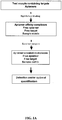
- the present invention relates generally to methods, devices, reagents, and kits for the detection of a target molecule in a sample and, more specifically, to the detection and/or quantification of one or more target molecules that may be contained in a test sample.
- Assays directed to the detection and quantification of physiologically significant molecules in biological samples and other samples are important tools in scientific research and in the health care field.
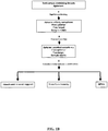
- One class of such assays involves the use of a microarray that includes one or more aptamers immobilized on a solid support.
- the aptamers are each capable of binding to a target molecule in a highly specific manner and with very high affinity. See, e.g., U.S. Patent No. 5,475,096 entitled “Nucleic Acid Ligands;” see also, e.g ., U.S. Patent No. 6,242,246 , U.S. Patent No. 6,458,543 , and U.S. Patent No.
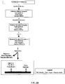
- a variation of this assay employs aptamers that include photoreactive functional groups that enable the aptamers to covalently bind, or "photocrosslink, " their target molecules. See, e.g., U.S. Patent No. 6,544,776 entitled “Nucleic Acid Ligand Diagnostic Biochip.” These photoreactive aptamers are also referred to as photoaptamers. See, e.g., U.S. Patent No. 5,763,177 , U.S. Patent No. 6,001,577 , and U.S. Patent No.
- Harsh wash conditions may be used, since target molecules that are bound to the photoaptamers are generally not removed, due to the covalent bonds effected by the photoactivated functional group(s) on the photoaptamers.
- the assay enables a determination of the absence, presence, amount, and/or concentration of the target molecules in the test sample.
- the aptamers are immobilized on the solid support prior to being contacted with the sample. Under certain circumstances, however, immobilization of the aptamers prior to contact with the sample may not provide an optimal assay. For example, pre-immobilization of the aptamers may result in inefficient mixing of the aptamers with the target molecules on the surface of the solid support, perhaps leading to lengthy reaction times and, therefore, extended incubation periods to permit efficient binding of the aptamers to their target molecules. Further, when photoaptamers are employed in the assay and depending upon the material utilized as a solid support, the solid support may tend to scatter or absorb the light used to effect the formation of covalent bonds between the photoaptamers and their target molecules.
- immobilization of the aptamers on the solid support generally involves an aptamer-preparation step (i.e. , the immobilization) prior to exposure of the aptamers to the sample, and this preparation step may affect the activity or functionality of the aptamers.
- Baldrich, E. et al., Analytical Chemistry (2004), 76(23):7053-7063 teaches an Enzyme Linked Aptamer Assay.
- the present disclosure includes methods, devices, reagents, and kits for the detection and/or quantification of one or more target molecules that may be present in a test sample.
- a test sample is contacted with an aptamer that includes a tag and has a specific affinity for a target molecule.
- An aptamer affinity complex that includes an aptamer bound to its target molecule is allowed to form. If the test sample contains the target molecule, an aptamer affinity complex will generally form in the test sample.
- the aptamer affinity complex is optionally converted to an aptamer covalent complex that includes an aptamer covalently bound to its target molecule.
- the aptamer affinity complex (or optional aptamer covalent complex) can then be detected and/or quantified using any of a variety of methods known to one skilled in the art, including but not limited to using a solid support, using mass spectrometry, and using quantitative polymerase chain reaction (Q-PCR).
- Q-PCR quantitative polymerase chain reaction
- the aptamer affinity complex (or optional aptamer covalent complex) is detected and/or quantified through the use of a solid support.
- the aptamer affinity complex (or optional aptamer covalent complex) is attached to a solid support. The attachment is accomplished by contacting the solid support with the aptamer affinity complex (or optional aptamer covalent complex) and allowing a tag included on the aptamer to associate, either directly or indirectly, with a probe that is attached to the solid support. The aptamer affinity complex (or optional aptamer covalent complex) that has associated with the probe on the solid support is then detected and optionally quantified.
- the complex is contacted with a labeling agent to permit detection of the bound target molecule.
- the aptamer affinity complex (or optional aptamer covalent complex) is detected and/or quantified using mass spectrometry.
- the aptamer affinity complex (or optional aptamer covalent complex) is attached to a solid support by contacting the solid support with the aptamer affinity complex (or optional aptamer covalent complex) and allowing a tag included on the aptamer to associate, either directly or indirectly, with a probe that is attached to the solid support.
- aptamer affinity complex or optional aptamer covalent complex
- the aptamer affinity complex (or optional aptamer covalent complex) that has associated with the probe on the solid support is then eluted and analyzed using mass spectrometry, which produces a spectrum of peaks that can be used to identify, and therefore detect, the target molecule.
- mass spectrometry which produces a spectrum of peaks that can be used to identify, and therefore detect, the target molecule.
- the aptamer affinity complex (or optional aptamer covalent complex) can be digested with protease enzymes, such as, for example, proteinase K or trypsin, to produce fragments of the bound target molecule that can then be used to identify the target molecule, and thereby enable detection and optional quantification of the target molecule.
- protease enzymes such as, for example, proteinase K or trypsin
- the aptamer affinity complex (or optional aptamer covalent complex) is detected and/or quantified using Q-PCR.
- free aptamer in the test sample is partitioned from the aptamer affinity complex (or optional aptamer covalent complex) prior to detection and/or quantification.
- the aptamer affinity complex (or optional aptamer covalent complex) is quantified by performing PCR and determining, either directly or indirectly, the amount or concentration of aptamer that had bound to its target molecule in the test sample.
- the amount or concentration of the target molecule in the test sample is generally directly proportional to the amount or concentration of the aptamer quantified by using Q-PCR.
- An exemplary method that may be employed to quantify an aptamer affinity complex (or optional aptamer covalent complex) in this manner is the TaqMan ® assay (PE Biosystems, Foster City, Calif.; see also U.S. Pat. No. 5,210,015 ).
- the terms “comprise,” “comprising,” “includes,” “including,” “contains,” “containing,” and any variations thereof, are intended to cover a non-exclusive inclusion, such that a process, method, product-by-process, or composition of matter that comprises, includes, or contains an element or list of elements does not include only those elements but may include other elements not expressly listed or inherent to such process, method, product-by-process, or composition of matter.
- the present disclosure includes methods, devices, reagents, and kits for the detection and/or quantification of one or more target molecules that may be present in a test sample.
- the disclosed methods, devices, reagents, and kits provide high sensitivity assays for the detection and/or quantification of target molecules in a test sample by optimizing conditions that affect (1) the activity of aptamers, (2) the efficiency of achieving binding equilibria for aptamer-target molecule complexes, (3) the formation of covalent bond(s) between an aptamer and its target molecule, and (4) the detection of aptamer-target molecule complexes.
- the presence of a target molecule in a test sample is detected and/or quantified by first contacting a test sample with an aptamer that has a specific affinity for a target molecule.
- An aptamer affinity complex that includes an aptamer bound to its target molecule is allowed to form. If the test sample contains the target molecule, an aptamer affinity complex will generally form in the test sample.
- the aptamer affinity complex is optionally converted, using a method appropriate to the aptamer being employed, to an aptamer covalent complex that includes an aptamer covalently bound to its target molecule.
- the aptamer affinity complex (or optional aptamer covalent complex) is then detected and/or quantified.
- the aptamer affinity complex (or optional aptamer covalent complex) is detected and/or quantified by attaching the aptamer affinity complex (or optional aptamer covalent complex) to a solid support.
- a test sample is contacted with an aptamer that includes a tag and has a specific affinity for a target molecule.
- An aptamer affinity complex that includes an aptamer bound to its target molecule is allowed to form. If the test sample contains the target molecule, an aptamer affinity complex will generally form in the test sample.
- the aptamer affinity complex is optionally converted, using a method appropriate to the aptamer being employed, to an aptamer covalent complex that includes an aptamer covalently bound to its target molecule.
- the aptamer affinity complex (or optional aptamer covalent complex) is attached to a solid support. The attachment is accomplished by contacting the solid support with the aptamer affinity complex (or optional aptamer covalent complex) and allowing the tag included on the aptamer to associate, either directly or indirectly, with a probe that is attached to the solid support.
- the aptamer affinity complex (or optional aptamer covalent complex) that has associated with the probe on the solid support is then detected and optionally quantified.
- the complex is contacted with a labeling agent to permit detection of the bound target molecule.
- nucleic acid As used herein, “nucleic acid,” “oligonucleotide,” and “polynucleotide” are used interchangeably to refer to a polymer of nucleotides of any length, and such nucleotides may include deoxyribonucleotides, ribonucleotides, and/or analogs or chemically modified deoxyribonucleotides or ribonucleotides.
- polynucleotide “oligonucleotide,” and “nucleic acid” include double- or single-stranded molecules as well as triple-helical molecules.
- chemical modifications of a nucleotide can include, singly or in any combination, 2'-position sugar modifications, 5-position pyrimidine modifications (e.g, 5-(N-benzylcarboxyamide)-2'-deoxyuridine, 5-(N-isobutylcarboxyamide)-2'-deoxyuridine, 5-(N-[2-(1H-indole-3yl)ethyl]carboxyamide)-2'-deoxyuridine, 5-(N-[1-(3-trimethylammonium) propyl]carboxyamide)-2'-deoxyuridine chloride, 5-(N-napthylcarboxyamide)-2'-deoxyuridine, and 5-(N-[1-(2,3-dihydroxypropyl)]carboxyamide)-2'-deoxyuridine), 8-position purine modifications, modifications at exocyclic amines, substitution of 4-thiouridine, substitution of 5-bromo- or 5-iodo-uracil, backbone modifications
- Modifications can also include 3' and 5' modifications, such as capping.
- Other modifications can include substitution of one or more of the naturally occurring nucleotides with an analog, internucleotide modifications such as, for example, those with uncharged linkages (e.g ., methyl phosphonates, phosphotriesters, phosphoamidates, carbamates, etc.) and those with charged linkages ( e.g ., phosphorothioates, phosphorodithioates, etc.), those with intercalators ( e.g., acridine, psoralen, etc.), those containing chelators ( e.g ., metals, radioactive metals, boron, oxidative metals, etc.), those containing alkylators, and those with modified linkages ( e.g ., alpha anomeric nucleic acids, etc.).
- internucleotide modifications such as, for example, those with uncharged linkages (e.g .,
- any of the hydroxyl groups ordinarily present in a sugar may be replaced by a phosphonate group or a phosphate group; protected by standard protecting groups; or activated to prepare additional linkages to additional nucleotides or to a solid support.
- the 5' and 3' terminal OH groups can be phosphorylated or substituted with amines, organic capping group moieties of from about 1 to about 20 carbon atoms, or organic capping group moieties of from about 1 to about 20 polyethylene glycol (PEG) polymers or other hydrophilic or hydrophobic biological or synthetic polymers.
- PEG polyethylene glycol
- a modification to the nucleotide structure may be imparted before or after assembly of a polymer.
- a sequence of nucleotides may be interrupted by non-nucleotide components.
- a polynucleotide may be further modified after polymerization, such as by conjugation with a labeling component.
- Polynucleotides can also contain analogous forms of ribose or deoxyribose sugars that are generally known in the art, including 2'-O-methyl-, 2'-O-allyl, 2'-fluoro- or 2'-azido-ribose, carbocyclic sugar analogs, ⁇ -anomeric sugars, epimeric sugars such as arabinose, xyloses or lyxoses, pyranose sugars, furanose sugars, sedoheptuloses, acyclic analogs and abasic nucleoside analogs such as methyl riboside.
- one or more phosphodiester linkages may be replaced by alternative linking groups.
- linking groups include embodiments wherein phosphate is replaced by P(O)S ("thioate”), P(S)S ("dithioate”), (O)NR 2 ("amidate"), P(O)R, P(O)OR', CO or CH 2 ("formacetal"), in which each R or R' is independently H or substituted or unsubstituted alkyl (1-20 C) optionally containing an ether (-O-) linkage, aryl, alkenyl, cycloalky, cycloalkenyl or araldyl. Not all linkages in a polynucleotide need be identical. Substitution of analogous forms of sugars, purines, and pyrimidines can be advantageous in designing a final product, as can alternative backbone structures like a polyamide backbone, for example.
- aptamer and “nucleic acid ligand” are used interchangeably to refer to a nucleic acid that has a specific binding affinity for a target molecule. It is recognized that affinity interactions are a matter of degree; however, in this context, the "specific binding affinity" of an aptamer for its target means that the aptamer binds to its target generally with a much higher degree of affinity than it binds to other components in a test sample.
- An “aptamer” is a set of copies of one type or species of nucleic acid molecule that has a particular nucleotide sequence. An aptamer can include any suitable number of nucleotides. "Aptamers" refers to more than one such set of molecules.
- Different aptamers may have either the same or different numbers of nucleotides. Any of the methods disclosed herein may include the use of one or more aptamers. Any of the methods disclosed herein may also include the use of two or more aptamers that specifically bind the same target molecule. As further described below, an aptamer may include a tag. If an aptamer includes a tag, all copies of the aptamer need not have the same tag. Moreover, if different aptamers each include a tag, these different aptamers may have either the same tag or a different tag.
- An aptamer can be identified using any known method, including the SELEX process. See, e.g. , U.S. Patent No. 5,475,096 entitled “Nucleic Acid Ligands.” Once identified, an aptamer can be prepared or synthesized in accordance with any known method, including chemical synthetic methods and enzymatic synthetic methods.
- SELEX and “SELEX process” are used interchangeably herein to refer generally to a combination of (1) the selection of nucleic acids that interact with a target molecule in a desirable manner, for example binding with high affinity to a protein, with (2) the amplification of those selected nucleic acids. See, e.g., U.S. Patent No. 5,475,096 entitled “Nucleic Acid Ligands.”
- the SELEX process may be used to generate an aptamer that covalently binds its target as well as an aptamer that non-covalently binds its target. See, e.g., U.S. Patent No. 5,705,337 entitled “Systematic Evolution of Nucleic Acid Ligands by Exponential Enrichment: Chemi-SELEX.”
- an aptamer can further comprise a "tag," which refers to a component that provides a means for attaching or immobilizing an aptamer (and any target molecule that is bound to it) to a solid support.
- a "tag” is a set of copies of one type or species of component that is capable of associating with a probe.
- Tags refers to more than one such set of components.
- the tag can be attached to or included in the aptamer by any method known in the art. Generally, the tag allows the aptamer to associate, either directly or indirectly, with a probe that is attached to the solid support.
- a tag can enable the localization of an aptamer covalent complex to a spatially defined address on a solid support.
- a tag can be a polynucleotide, a polypeptide, a peptide nucleic acid, a locked nucleic acid, an oligosaccharide, a polysaccharide, an antibody, an affybody, an antibody mimic, a cell receptor, a ligand, a lipid, any fragment or derivative of these structures, any combination of the foregoing, or any other structure with which a probe (or linker molecule, as described below) can be designed or configured to bind or otherwise associate with specificity.
- a tag is configured such that it does not interact intramolecularly with either itself or the aptamer to which it is attached or of which it is a part. If SELEX is used to identify an aptamer, the tag may be added to the aptamer either pre- or post-SELEX. In one embodiment, the tag is included on the 5'-end of the aptamer post-SELEX. In another embodiment, the tag is included on the 3'-end of the aptamer post-SELEX.
- the tag includes a polynucleotide that is designed to associate directly with a probe that includes a complementary polynucleotide sequence by hybridizing directly with the probe sequence.
- the tag is generally configured and the hybridization reaction is carried out under conditions such that the tag does not hybridize with a probe other than the probe for which the tag includes a perfect complement.
- the tag comprises nucleotides that are a part of the aptamer itself.
- the aptamer generally includes a 5'- fixed end separated from a 3'- fixed end by a nucleotide sequence that varies, depending upon the aptamer, that is, a variable region.
- the tag can comprise any suitable number of nucleotides included in a fixed end of the aptamer, such as, for example, an entire fixed end or any portion of a fixed end, including nucleotides that are internal to a fixed end.
- the tag can comprise any suitable number of nucleotides included within the variable region of the aptamer, such as, for example, the entire variable region or any portion of the variable region.
- the tag can comprise any suitable number of nucleotides that overlap both the variable region and one of the fixed ends, that is, the tag can comprise a nucleotide sequence that includes any portion (including all) of the variable region and any portion (including all) of a fixed end.
- a tag can associate directly with a probe and covalently bind to the probe, thereby covalently linking the aptamer to the surface of the solid support.
- the tag and the probe can include suitable reactive groups that, upon association of the tag with the probe, are sufficiently proximate to each other to undergo a chemical reaction that produces a covalent bond. The reaction may occur spontaneously or may require activation, such as, for example, photo-activation or chemical activation.
- the tag includes a diene moiety and the probe includes a dienophile, and covalent bond formation results from a spontaneous Diels-Alder conjugation reaction of the diene and dienophile. Any appropriate complementary chemistry can be used, such as, for example, N-Mannich reaction, disulfide formation, Curtius reaction, Aldol condensation, Schiff base formation, and Michael addition.
- the tag associates indirectly with a probe, such as, for example, through a linker molecule, as further described below.
- the tag can include a polynucleotide sequence that is complementary to a particular region or component of a linker molecule.
- the tag is generally configured and the hybridization reaction is carried out such that the tag does not hybridize with a polynucleotide sequence other than the polynucleotide sequence included in the linker molecule.
- the polynucleotide can include any suitable number of nucleotides. In one embodiment, a tag includes at least about 10 nucleotides. In another embodiment, the tag includes from about 10 to about 45 nucleotides. In yet another embodiment, the tag includes at least about 30 nucleotides. Different tags that include a polynucleotide can include either the same number of nucleotides or a different number of nucleotides.
- the term "about” represents an insignificant modification or variation of the numerical values such that the basic function of the item to which the numerical value relates is unchanged.
- association refers to an interaction or complexation between a tag and a probe resulting in a sufficiently stable complex so as to permit separation of "unassociated" or unbound materials, such as, for example, unbound components of a test sample, from the tag-probe complex under given complexation or reaction conditions.
- a tag and a probe can associate with each other directly by interacting and binding to each other with specificity.
- a tag and a probe can also associate with each other indirectly such as when their complexation is mediated by a linker molecule.
- probe refers to a molecule that is configured to associate, either directly or indirectly, with a tag.
- a “probe” is a set of copies of one type of molecule or one type of multi-molecular structure that is capable of immobilizing an aptamer to a solid support by associating, either directly or indirectly, with a tag.
- Probes refers to more than one such set of molecules.
- a probe can be a polynucleotide, a polypeptide, a peptide nucleic acid, a locked nucleic acid, an oligosaccharide, a polysaccharide, an antibody, an affybody, an antibody mimic, a cell receptor, a ligand, a lipid, any fragment or derivative of these structures, any combination of the foregoing, or any other structure with which a tag (or linker molecule) can be designed or configured to bind or otherwise associate with specificity.
- a probe can be attached to a solid support either covalently or non-covalently by any method known in the art.
- the probe includes a polynucleotide that has a sequence that is complementary to a polynucleotide tag sequence.
- the probe sequence is generally configured and the hybridization reaction is carried out under conditions such that the probe does not hybridize with a nucleotide sequence other than the tag for which the probe includes the complementary sequence ( i.e., the probe is generally configured and the hybridization reaction is carried out under conditions such that the probe does not hybridize with a different tag or an aptamer).
- the probe associates indirectly with a tag, for example, through a linker molecule.
- the probe can include a polynucleotide sequence that is complementary to a particular region or component of a linker molecule.
- the probe is generally configured and the hybridization reaction is carried out such that the probe does not hybridize with a polynucleotide sequence other than the polynucleotide sequence included in the linker molecule.
- a probe includes a polynucleotide
- the polynucleotide can include any suitable number of nucleotides. In one embodiment, a probe includes at least about 10 nucleotides. In another embodiment, a probe includes from about 10 to about 45 nucleotides. In yet another embodiment, a probe includes at least about 30 nucleotides. Different probes that include a polynucleotide can include either the same number of nucleotides or a different number of nucleotides.
- linker molecule refers to one or more molecules that are configured to mediate the association of a tag with a probe. Generally, the linker molecule is bi-functional in that it includes a functionality for linking to a tag and a functionality for linking to a probe.
- a "linker molecule” is a set of copies of one type or species of molecule(s) or multimolecular structure(s) that is capable of associating a tag with a probe.
- Linker molecules refers to more than one such set of molecules or multi-molecular structures.
- a linker molecule may have any suitable configuration and can include any suitable components, including a polynucleotide, a polypeptide, a peptide nucleic acid, a locked nucleic acid, an oligosaccharide, a polysaccharide, an antibody, an affybody, an antibody mimic, a polyethylene glycol (PEG) molecule, a cell receptor, a ligand, a lipid, any fragment or derivative of these structures, any combination of the foregoing, or any other structure or chemical component that can be designed or configured to mediate an association between a tag and a probe with specificity.
- a linker molecule may be aliphatic or aromatic.
- the composition of the linker molecule is not critical to any of the methods disclosed herein. It is often preferred that the linker molecule be hydrophilic. As a general rule, the length of a particular linker molecule can be selected to provide for convenience of synthesis and ease in mediating the association of a tag with a probe. The linker molecule should not contain functionalities, or be of a length, that will interfere with the reactions that are desired in accordance with the disclosed methods.
- the linker molecule when a linker molecule is employed in any of the methods disclosed herein, the linker molecule may be introduced at any suitable time during the performance of the assay and may first contact either a tag or a probe.
- a tag included on an aptamer may be contacted with the linker molecule any time before an aptamer covalent complex contacts the probe on a solid support.
- a probe attached to a solid support may be contacted with a linker molecule any time before the probe is exposed to the tag on an aptamer covalent complex.
- a probe may be contacted with both a linker molecule and a tag on an aptamer covalent complex simultaneously.
- a linker molecule generally comprises a tag association component and a probe association component.
- the tag association component and probe association component are independently selected based upon the particular tag and probe utilized in a particular assay.
- the tag association component is a polynucleotide that is complementary to a polynucleotide sequence included in a tag.
- the probe association component is a polynucleotide that is complementary to a polynucleotide sequence included in a probe.
- the tag association component is a polynucleotide and the probe association component is also a polynucleotide.
- the linker molecule includes a tag association component separated from a probe association component by a third component.
- the third component can include one or more molecules or sub-components, including a polynucleotide, a polypeptide, a peptide nucleic acid, a locked nucleic acid, an oligosaccharide, a polysaccharide, an antibody, an affybody, an antibody mimic, an aliphatic carbon molecule, a polyethylene glycol (PEG) molecule, a cell receptor, a ligand, a lipid, any fragment or derivative of these structures, any combination of the foregoing, or any other chemical structure or component that can aid in the association of the tag with the probe, such as, e.g., by increasing flexibility between the tag association component and the probe association component.
- PEG polyethylene glycol
- a polynucleotide component of a linker molecule can include any suitable number of nucleotides. In one embodiment, a polynucleotide component of a linker molecule includes at least about 10 nucleotides. In another embodiment, a polynucleotide component of a linker molecule includes from about 10 to about 45 nucleotides. In yet another embodiment, a polynucleotide component of a linker molecule includes at least about 30 nucleotides. Linker molecules used in any of the methods disclosed herein can include polynucleotide components having either the same number of nucleotides or a different number of nucleotides.
- photoaptamer As used herein, "photoaptamer,” “photoreactive nucleic acid ligand,” and “photoreactive aptamer” are used interchangeably to refer to an aptamer that contains one or more photoreactive functional groups that can covalently bind to or "crosslink" with a target molecule.
- a naturally occurring nucleic acid residue may be modified to include a chemical functional group that confers photoreactivity upon the nucleic acid residue upon exposure to a radiation source of an appropriate wavelength.
- a photoaptamer can be identified and/or prepared using any known method.
- a photoreactive aptamer is identified using the photoSELEX process. See, e.g., U.S. Patent No. 5,763,177 , U.S. Patent No.
- an aptamer is prepared and is subsequently modified to incorporate one or more photoreactive functional groups, thereby generating a photoaptamer.
- one or more photoreactive nucleic acid residues can be incorporated into an aptamer either by substituting a photoreactive nucleic acid residue in the place of one or more other nucleotides, such as one or more of the thymidine and/or cytidine nucleotides in the aptamer, for example, or by modifying one or more nucleic acid residues to include a photoreactive functional group.
- photoreactive functional groups that may be incorporated into a photoaptamer include 5-bromouracil, 5-iodouracil, 5-bromovinyluracil, 5-iodovinyluracil, 5-azidouracil, 4-thiouracil, 5-thiouracil, 4-thiocytosine, 5-bromocytosine, 5-iodocytosine, 5-bromovinylcytosine, 5-iodovinylcytosine, 5-azidocytosine, 8-azidoadenine, 8-bromoadenine, 8-iodoadenine, 8-aziodoguanine, 8-bromoguanine, 8-iodoguanine, 8-azidohypoxanthine, 8-bromohypoxanthine, 8-iodohypoxanthine, 8-azidoxanthine, 8-bromoxanthine, 8-iodoxanthine, 8-brom
- photoreactive functional groups that can be added to a terminal end of an aptamer using an appropriate linker molecule can also be used.
- photoreactive functional groups include benzophenone, anthraquinone, 4-azido-2-nitro-aniline, psoralen, derivatives of any of these, and the like.
- a photoreactive functional group incorporated into a photoaptamer may be activated by any suitable method.
- a photoaptamer containing a photoreactive functional group is crosslinked to its target by exposing the photoaptamer affinity complex to a source of electromagnetic radiation.
- electromagnetic radiation include ultraviolet light, visible light, X-rays, and gamma rays.
- Suitable radiation sources include sources that utilize either monochromatic light or filtered polychromatic light.
- a photoreactive nucleotide such as 4-azido-2-nitroaniline, for example, can be incorporated into a photoaptamer, and light having a wavelength ranging from about 325 nm to about 470 nm may be used to irradiate a photoaptamer affinity complex that includes this photoaptamer. Excitation at these wavelengths can be accomplished, for example, with inexpensive light emitting diodes (LEDs) using either a single LED or an array of LEDs, since the power requirements are modest. Nearly monochromatic light having a wavelength ranging from 465 to 475 nm, a 100 degree viewing angle and providing 38 lumens of light is supplied by one or more high-powered LEDs.
- LEDs inexpensive light emitting diodes
- a photoreactive nucleotide is incorporated into a photoaptamer, and light having a wavelength ranging from about 300 nm to about 350 nm may be used to irradiate a photoaptamer affinity complex that includes this photoaptamer to convert the photoaptamer affinity complex to a photoaptamer covalent complex.
- a photoreactive nucleotide such as a 5-iodouracil or a 5-iodocytosine, for example, can be incorporated into a photoaptamer, and light having a wavelength ranging from about 320 nm to about 325 nm may be used to irradiate a photoaptamer affinity complex that includes this photoaptamer. This combination facilitates selective photocrosslinking of the chromophore-containing photoaptamer to the target molecule without inducing other, non-specific photoreactions.
- any tryptophan residues that might be included in the target protein and any thymine and uracil bases that might be included in the photoaptamer may also be photoreactive. Since 5-iodouracil or 5-iodocytosine absorbs light having a wavelength of about 325 nm but tryptophan and naturally occurring nucleic acid bases do not, using light of this wavelength permits a selective photoreaction at the 5-iodouracil(s) or 5-iodocyctosine(s) within the photoaptamer affinity complex.
- Monochromatic light having a wavelength ranging from about 320nm to about 325 nm may be supplied, for example, by a frequency doubled tunable dye laser emitting light at a wavelength of about 320 nm or by a helium cadmium laser emitting light at a wavelength of about 325 nm.
- a photoaptamer affinity complex can be exposed to a xenon chloride (XeCl) excimer laser set to emit light at a wavelength of about 308 nm.
- the photoaptamer can include a photoreactive functional group (e.g., a 5-bromouracil or a 5-bromocytosine), and treating the photoaptamer affinity complex with the light source serves to photoactivate the photoreactive functional group such that the photoaptamer crosslinks with its target molecule and a photoaptamer covalent complex is formed.
- a photoaptamer in yet another embodiment, can be crosslinked to its target by exposing a photoaptamer affinity complex to a high-pressure mercury lamp set to emit light at a wavelength of about 313 nm.
- wavelength filters may be employed to restrict the emitted light to be greater than about 300 nm to minimize activation of chromophores other than those included in a photoaptamer affinity complex.
- a photoaptamer in a further embodiment, can be crosslinked to its target by exposing a photoaptamer affinity complex to a low pressure mercury lamp set to emit light at a wavelength of about 254 nm, which is then absorbed by a phosphor and re-emitted at a wavelength ranging from about 300 nm to about 325 nm.
- the re-emitted light is filtered to remove any light of about 254 nm that is not absorbed by the phosphor as well as any light of wavelengths ranging from about 290 nm to about 305 nm, which may be damaging to a target protein.
- a halogen photoreactive functional group such as an iodouracil or a bromocytosine for example, can be incorporated into a photoaptamer, and a photoaptamer affinity complex that includes this photoaptamer can be treated with light having a wavelength ranging from about 350nm to about 400 nm.
- a photoaptamer affinity complex that includes this photoaptamer can be treated with light having a wavelength ranging from about 350nm to about 400 nm.
- monochromatic light from the third harmonic of a Neodymium YAG laser set at about 355 nm or monochromatic light from the first harmonic of a xenon fluoride (XeF) excimer laser at about 351 nm may be used.
- target molecule and “target” are used interchangeably to refer to any molecule of interest to which an aptamer can bind with high affinity and specificity and that may be present in a test sample.
- a "molecule of interest” includes any minor variation of a particular molecule, such as, in the case of a protein, for example, minor variations in amino acid sequence, disulfide bond formation, glycosylation, lipidation, acetylation, phosphorylation, or any other manipulation or modification, such as conjugation with a labeling component that does not substantially alter the identity of the molecule.
- a “target molecule” or “target” is a set of copies of one type or species of molecule or multimolecular structure that is capable of binding to an aptamer.
- Target molecules refer to more than one such set of molecules.
- target molecules include proteins, polypeptides, nucleic acids, carbohydrates, lipids, polysaccharides, glycoproteins, hormones, receptors, antigens, antibodies, affybodies, antibody mimics, viruses, pathogens, toxic substances, substrates, metabolites, transition state analogs, cofactors, inhibitors, drugs, dyes, nutrients, growth factors, cells, tissues, and any fragment or portion of any of the foregoing.
- An aptamer may be identified for virtually any chemical or biological molecule of any size, and thus virtually any chemical or biological molecule of any size can be a suitable target.
- a target can also be modified to enhance the likelihood or strength of an interaction between the target and the aptamer.
- the target molecule is a protein. See U.S. Patent No. 6,376,190 entitled “Modified SELEX Processes Without Purified Protein” for methods in which the SELEX target is a peptide.
- Polypeptide “peptide,” and “protein” are used interchangeably herein to refer to polymers of amino acids of any length.
- the polymer may be linear or branched, it may comprise modified amino acids, and it may be interrupted by non-amino acids.
- the terms also encompass an amino acid polymer that has been modified naturally or by intervention; for example, disulfide bond formation, glycosylation, lipidation, acetylation, phosphorylation, or any other manipulation or modification, such as conjugation with a labeling component.
- polypeptides containing one or more analogs of an amino acid including, for example, unnatural amino acids, etc.
- Polypeptides can be single chains or associated chains.
- non-target molecule and “non-target” are used interchangeably to refer to a molecule contained in a test sample that can form a non-specific complex with an aptamer.
- a “non-target molecule” or “non-target” is a set of copies of one type or species of molecule or multi-molecular structure that is capable of binding to an aptamer.
- Non-target molecules or “non-targets” refers to more than one such set of molecules. It will be appreciated that a molecule that is a non-target for a first aptamer may be a target for a second aptamer. Likewise, a molecule that is a target for the first aptamer may be a non-target for the second aptamer.
- aptamer affinity complex refers to a non-covalent complex that is formed by the interaction of an aptamer with its target molecule.
- An "aptamer affinity complex” is a set of copies of one type or species of complex formed by an aptamer bound to its corresponding target molecule.
- Aptamer affinity complexes refers to more than one such set of complexes.
- An aptamer affinity complex can generally be reversed or dissociated by a change in an environmental condition, e.g ., an increase in temperature, an increase in salt concentration, or an addition of a denaturant.
- aptamer covalent complex refers to an aptamer affinity complex in which the aptamer has been induced to form or otherwise forms a covalent bond with its target molecule.
- An "aptamer covalent complex” is a set of copies of one type or species of complex formed by an aptamer covalently bound to its corresponding target molecule.
- Aptamer covalent complexes refers to more than one such set of complexes.
- a covalent bond or linkage between an aptamer and its target molecule can be induced by photoactivation of a chemical moiety on the aptamer, including those moieties described above with respect to photoaptamers.
- a covalent bond or linkage between an aptamer and its target molecule can also be induced chemically.
- Chemical groups that can be included in an aptamer and used to induce a covalent linkage with the target include but are not limited to aldehydes, maleimides, acrylyl derivatives, diazonium derivatives, thiols, etc.
- chemical crosslinking groups such as maleimide or diazonium salts, for example, can convert aptamer affinity complexes to aptamer covalent complexes simply by providing the proper environment and juxtaposition of reactive groups required for specific and sufficiently enhanced chemical reactivity to occur.
- chemical crosslinkers such as aldehyde groups
- no such chemical crosslinkers are included in an aptamer; rather, a third reagent is used to convert an aptamer affinity complex to an aptamer covalent complex by facilitating a covalent attachment between the aptamer and its target.
- a homo- or heterobifunctional reagent containing both an amine reactive moiety e.g., an N-hydroxy succinimidyl ester, an aldehyde, or an imidate
- a nucleoside-reactive group e.g ., an iodoacetamide or an activated aldehyde
- an aptamer affinity complex such as an affinity complex formed by an aptamer and a target protein.
- test sample refers herein to any material, solution, or mixture that contains a plurality of molecules and may include at least one target molecule.
- test sample includes biological samples, as defined below, and samples that may be used for environmental or toxicology testing, such as contaminated or potentially contaminated water and industrial effluents, for example.
- a test sample may also be an end product, intermediate product, or by-product of a preparatory process, for example a manufacturing process.
- a test sample may include any suitable assay medium, buffer, or diluent that has been added to a material, solution, or mixture obtained from an organism or from some other source ( e.g ., the environment or an industrial source).
- biological sample refers to any material, solution, or mixture obtained from an organism. This includes blood (including whole blood, leukocytes, peripheral blood mononuclear cells, plasma, and serum), sputum, breath, urine, semen, saliva, meningeal fluid, amniotic fluid, glandular fluid, lymph fluid, nipple aspirate, bronchial aspirate, synovial fluid, joint aspirate, cells, a cellular extract, and cerebrospinal fluid. This also includes experimentally separated fractions of all of the preceding.
- biological sample also includes materials, solutions, or mixtures containing homogenized solid material, such as from a stool sample, a tissue sample, or a tissue biopsy, for example.
- biological sample also includes materials, solutions, or mixtures derived from a tissue culture, cell culture, bacterial culture, or viral culture.
- Solid support refers to any substrate having a surface to which molecules may be attached, directly or indirectly, through either covalent or non-covalent bonds.
- the solid support may include any substrate material that is capable of providing physical support for the probes that are attached to the surface.
- the material is generally capable of enduring conditions related to the attachment of the probes to the surface and any subsequent treatment, handling, or processing encountered during the performance of an assay.
- the materials may be naturally occurring, synthetic, or a modification of a naturally occurring material.
- Suitable solid support materials may include silicon, graphite, mirrored surfaces, laminates, ceramics, plastics (including polymers such as, e.g ., poly(vinyl chloride), cyclo-olefin copolymers, polyacrylamide, polyacrylate, polyethylene, polypropylene, poly(4-methylbutene), polystyrene, polymethacrylate, poly(ethylene terephthalate), polytetrafluoroethylene (PTFE or Teflon®), nylon, poly(vinyl butyrate)), germanium, gallium arsenide, gold, silver, etc., either used by themselves or in conjunction with other materials.
- plastics including polymers such as, e.g ., poly(vinyl chloride), cyclo-olefin copolymers, polyacrylamide, polyacrylate, polyethylene, polypropylene, poly(4-methylbutene), polystyrene, polymethacrylate, poly(ethylene terephthalate), polytetra
- Additional rigid materials may be considered, such as glass, which includes silica and further includes, for example, glass that is available as Bioglass.
- Other materials that may be employed include porous materials, such as, for example, controlled pore glass beads. Any other materials known in the art that are capable of having one or more functional groups, such as any of an amino, carboxyl, thiol, or hydroxyl functional group, for example, incorporated on its surface, are also contemplated.
- the material used for a solid support may take any of a variety of configurations ranging from simple to complex.
- the solid support can have any one of a number of shapes, including a strip, plate, disk, rod, particle, including bead, tube, well, and the like.
- the material is relatively planar such as, for example, a slide, though it can be spherical, such as, for example, a bead, or cylindrical ( e.g ., a column).
- the material is shaped generally as a rectangular solid. Multiple predetermined arrangements such as, e.g ., arrays of probes, may be synthesized on a sheet, which is then diced, i.e.
- Exemplary solid supports that may be used include microtitre wells, microscope slides, membranes, paramagnetic beads, charged paper, Langmuir-Blodgett films, silicon wafer chips, flow through chips, and microbeads.
- the surface of the solid support is usually the outer portion of the substrate material that forms the solid support.
- the surface of the solid support onto which the probes are bound may be smooth or substantially planar, or have irregularities, such as depressions, grooves, elevations, or other textures.
- the surface may be modified with one or more different layers of compounds that serve to modify the properties of the surface in a desirable manner.
- such surface modification layers when present, can generally range in thickness from a monomolecular thickness to about 1 mm, or from a monomolecular thickness to about 0.1 mm, or from a monomolecular thickness to about 0.001 mm.
- Surface modification layers of interest include inorganic and organic layers, such as metals, metal oxides, polymers, small organic molecules, and the like.
- Polymeric layers of interest include methacrylate copolymers, polyacrylamides, polysaccharides, phospholipids, polyurethanes, polyesters, polycarbonates, polyureas, polyamides, polyethylene amines, polyarylene sulfides, polysiloxanes, polyimides, polyacetates, and the like, where the polymers may be hetero- or homo-polymeric, and may or may not have separate functional moieties attached thereto (for example, conjugated moieties).
- Other surface modifications of interest include three-dimensional networks, such as hydrogels, for example. Any suitable hydrogel known in the art may be used.
- the surface of a solid support includes a hydrogel.
- the hydrogel can comprise, for example, a polymer matrix.
- the hydrogel can be chemically attached to the surface of the solid support and can include a binding functionality that is capable of attaching, either directly or indirectly, a probe to the hydrogel.
- Exemplary binding functionalities include a hydrophobic group, a hydrophilic group, reactive groups such as aldehydes, epoxy, carbonates and the like, a carboxyl, a thiol, a sulfonate, a sulfate, an amino, a substituted amino, a phosphate, a metal chelating group, a thioether, a biotin, a boronate, etc.
- Any surface suitable for gene expression or SNP analysis may also be used, including substrates and surfaces offered, for example, by Affymetrix, General Electric (e.g ., CodeLink), Agilent, and Schott Nexterion, either as substrates and surfaces or as components of products that further comprise other components.
- a probe can be bound to the surface of the solid support in any suitable manner, so long as the probe will not become unbound during subsequent incubation and processing steps in accordance with the disclosed methods, such as washing the surface to remove non-specific complexes, for example.
- the probe may be bound by being non-covalently linked, e.g., adhered, absorbed, adsorbed, or by being covalently linked to the surface of the solid support.
- the surface of the solid support will contain a functional group that links to the probe. The nature of the functional group(s) used is dependent upon the nature of the probe. A variety of methods have been reported for the covalent attachment of molecules to a surface.
- these reactions are performed by the reaction of an active functional group on a molecule with an activated functional group on the surface.
- an amine-containing compound can be attached to a carboxylic acid containing surface by forming an activated ester of the carboxylic acid, such as an N-hydroxysuccinimide derivative. The amine readily reacts with this activated ester to form a stable amide bond. This reaction is useful under conditions whereby the reaction with the desired amine is significantly faster than with other nucleophiles in the system.
- Examples of methods that have been previously described in the art include the activation of surfaces with cyanogen bromide, N-hydroxysuccinimide esters, carbonyl diimidazole, carbodiimides, azlactones, cyanuric chlorides, organic sulfonyl chlorides, divinyl sulphone, nitrophenyl esters, iodoacetyl, maleimide, epoxy, hydrazide, reductive amination, diazonium salts, and Mannich condensations.
- Molecules that react with the activated surface include amines, alcohols, carboxylic acids, thiols, carbonyls, and compounds containing active hydrogens.
- the probes are bound to the surface of the solid support in a predetermined, spatial arrangement or pattern, which means any arrangement on the surface where the identity of a probe at a particular location is known.
- the predetermined arrangement is an array.
- An array generally includes any one-, two-, or three- dimensional arrangement of addressable regions bearing a particular probe associated with that region.
- An array is addressable in that it has multiple regions of different probes, such that a region or feature or spot of the array at a particular predetermined location or address on the array detects a particular aptamer covalent complex, and therefore a particular target molecule, by virtue of associating specifically with the tag on such aptamer covalent complex.
- An array assembly on the surface of a solid support refers to one or more arrays disposed along a surface of an individual solid support and separated by inter-array areas. Normally, the surface of the solid support opposite the surface with the arrays (opposing surface) does not carry any arrays.
- the arrays can be designed for testing against any type of test sample.
- the surface of the solid support may carry at least one, two, four, twenty, a hundred, or at least five hundred arrays. Depending upon intended use, any or all of the arrays may be the same or different from one another and each may contain multiple spots or features of probes disposed thereon.
- a typical array may contain more than ten, more than one hundred, more than one thousand, or ten thousand features, or even more than one hundred thousand features, in an area of less than about 20 cm 2 or even less than about 10 cm 2 .
- features may have widths (that is, diameter, for a round spot) in the range from about 10 ⁇ m to about 1.0 cm.
- each feature may have a width in the range of about 1.0 ⁇ m to about 1.0 mm, or from about 5.0 ⁇ m to about 500 ⁇ m, or from about 10 ⁇ m to about 200 ⁇ m.
- Non-round features may have area ranges equivalent to that of circular features with the foregoing width (diameter) ranges.
- an individual solid support may contain a single array or multiple arrays.
- Features of the array may be arranged in rectilinear rows and columns. This is particularly attractive for single arrays on a solid support.
- arrays can be arranged, for example, in a sequence of curvilinear rows across the surface of the solid support (for instance, a sequence of concentric circles or semi-circles of spots), and the like.
- the pattern of features may be varied from the rectilinear rows and columns of spots to include, for example, a sequence of curvilinear rows across the surface of the solid support (for example, a sequence of concentric circles or semi-circles of spots), and the like.
- the configuration of the arrays and their features may be selected according to manufacturing, handling, and use considerations.
- Each feature, or element, within the array is defined to be a small, regularly shaped region of the surface of the solid support.
- the features are arranged in a predetermined manner.
- Each feature of an array usually carries a predetermined probe or mixture of probes.
- Each feature within the molecular array may contain a different probe, and the probe within a given feature may differ from the probes within the remaining features of the array.
- Some or all of the features may be of different compositions.
- Each array may contain multiple spots or features, and each array may be separated from another array by spaces or areas. It will also be appreciated that there need not be any space separating arrays from one another. Interarray areas and interfeature areas are usually present but are not essential. As with any border areas, these interarray and interfeature areas do not carry any probes. It will be appreciated that the interarray areas and interfeature areas, when present, could be of various sizes and configurations.
- an array may be formed by attaching a probe to a first solid support, such as a bead, for example, and then arranging the first solid support in an array format on a second solid support, such as a microtiter plate, for example.
- a second solid support such as a microtiter plate
- an array may be formed by attaching probes to addressable beads. "Addressable beads" include dyes, barcodes, and transponders.
- the surface may be "blocked” or “passivated” in order to reduce or inhibit non-specific binding of molecules to the surface of the solid support.
- Blocking or passivation reagents include dry milk, casein, pooled serum, pooled plasma, BSA, PEG-PLL, PEG-silane, SuperBlock or StarterBlock (Pierce Biotechnology, Rockford, Ill.), and any combination of any of the foregoing.
- labeling agent refers to one or more reagents that can be used to detect a target molecule that is bound to an aptamer in an aptamer covalent complex.
- an aptamer covalent complex is contacted with a labeling agent that includes a binding partner that is specific for the target molecule bound to the aptamer.
- the specific binding partner may be any suitable moiety, including an antibody, an antibody fragment, a synthetic antibody mimetic, a biomimetic, an aptamer, a molecular imprinted ligand, and the like.
- the specific binding partner is conjugated or linked to another labeling agent component, usually, a detectable moiety or label.
- the linking of the specific binding partner to the label may be carried out by any of the aforementioned methods for linking a probe to the surface of the solid support.
- multiple aptamer covalent complexes can be contacted with a mixture of specific binding partners, each specific for a target molecule suspected of being present.
- the labels employed may be those that are known in the art for multiplexed detection of multiple target molecules.
- the detectable moiety or label is capable of being detected directly or indirectly.
- any reporter molecule that is detectable can be a label.
- Labels include, for example, (i) reporter molecules that can be detected directly by virtue of generating a signal, (ii) specific binding pair members that may be detected indirectly by subsequent binding to a cognate that contains a reporter molecule, (iii) mass tags detectable by mass spectrometry, (iv) oligonucleotide primers that can provide a template for amplification or ligation, and (v) a specific polynucleotide sequence or recognition sequence that can act as a ligand, such as, for example, a repressor protein, wherein in the latter two instances the oligonucleotide primer or repressor protein will have, or be capable of having, a reporter molecule, and so forth.
- the reporter molecule can be a catalyst, such as an enzyme, a polynucleotide coding for a catalyst, promoter, dye, fluorescent molecule, quantum dot, chemiluminescent molecule, coenzyme, enzyme substrate, radioactive group, a small organic molecule, amplifiable polynucleotide sequence, a particle such as latex or carbon particle, metal sol, crystallite, liposome, cell, etc., which may or may not be further labeled with a dye, catalyst or other detectable group, a mass tag that alters the weight of the molecule to which it is conjugated for mass spectrometry purposes, and the like.
- the label can be selected from electromagnetic or electrochemical materials.
- the detectable label is a fluorescent dye.
- Other labels and labeling schemes will be evident to one skilled in the art based on the disclosure herein.
- the target molecule is a protein
- the aptamer covalent complex is contacted with a labeling agent that comprises a universal protein stain.
- a labeling agent that comprises a universal protein stain.
- universal protein stain and “UPS” are used interchangeably to refer to any labeling agent that labels most, if not all, proteins present in a test sample with a detectable moiety but tends not to label, or labels only minimally, nucleic acids or other components of the assay, such as the solid support. Any reactive chemical group found on proteins, but not found on nucleic acids or the substrate surface, can serve as a site of covalent attachment.
- Exemplary reactive chemical groups include primary amines (e.g., on lysine residues), thiols (e.g., on cysteine, which may be produced by the reduction of disulfide linkages), alcohols (e.g ., on serine, threonine, tyrosine, and sugar moieties on glycoproteins (including the products of oxidation of cis-diols on such sugars)), and carboxylates (e.g ., on glutamic and aspartic acid).
- primary amines e.g., on lysine residues
- thiols e.g., on cysteine, which may be produced by the reduction of disulfide linkages
- alcohols e.g ., on serine, threonine, tyrosine, and sugar moieties on glycoproteins (including the products of oxidation of cis-diols on such sugars)
- carboxylates e.
- the detectable moiety can include any of the reporter molecules listed above and any other chemical or component that may be used in any manner to generate a detectable signal.
- the detectable moiety may be detected via a fluorescent signal, a chemiluminescent signal, or any other detectable signal that is dependent upon the identity of the moiety.
- the detectable moiety is an enzyme (for example, alkaline phosphatase)
- the signal may be generated in the presence of the enzyme substrate and any additional factors necessary for enzyme activity.
- the detectable moiety is an enzyme substrate, the signal may be generated in the presence of the enzyme and any additional factors necessary for enzyme activity.
- Suitable reagent configurations for attaching the detectable moiety to a target protein include covalent attachment of the detectable moiety to the target protein, non-covalent association of the detectable moiety with another labeling agent component that is covalently attached to the target protein, and covalent attachment of the detectable moiety to a labeling agent component that is non-covalently associated with the target protein.
- Universal protein stains are described in detail in U.S. Patent Publication 2006-0057573 filed August 12, 2004 , entitled "Methods and Reagents for Detecting Target Binding by Nucleic Acid Ligands.”
- the UPS is a single chemical reagent that comprises a detectable moiety and reacts covalently with a functional group that is characteristic of proteins, but not aptamers, and, in so reacting, covalently attaches the detectable moiety to a target protein.
- UPSs according to this embodiment include dyes with groups capable of reacting covalently with functional groups that are unique to proteins. Such groups may be added to the dyes by derivatization or may be present on the unmodified dye.
- the UPS comprises an N-hydroxysuccinimide-activated dye that reacts with amine groups, such as an N-hydroxysuccinimide-activated fluorophore, including NHS-Alexa fluorophores (such as, e.g ., NHS-Alexa 647).
- an N-hydroxysuccinimide-activated fluorophore including NHS-Alexa fluorophores (such as, e.g ., NHS-Alexa 647).
- Another UPS that may be used is CBQCA (3-(4-carboxybenzoyl)quinoline-2-carboxaldehyde), which also reacts with amines in the presence of cyanide or thiols to form highly fluorescent isoindoles.
- amine reactive groups suitable for use in UPS reagents include isocyanates, isothiocyanates, acyl azides, sulfonyl chlorides, aldehydes, 4-sulfo-2,3,5,6-tetrafluorophenol (STP) esters, TFP-Alexa 647, and arylating agents such as NBD (7-nitrobenz-2-oxa-1,3-diazole) chloride, NBD fluoride, and dichlorotriazines.
- the UPS comprises a plurality of reagents.
- the UPS can comprise a first reagent that reacts covalently with a target protein, and one or more further reagents that attach the detectable moiety, either directly or indirectly, and either covalently or non-covalently, to the target protein via a chemical group or other functionality introduced by the first reagent.
- the UPS comprises multiple reagents, it will be appreciated that in some cases the reagents are added sequentially, and in other cases they may be added simultaneously.
- a suitable UPS comprises (a) a biotin derivative that reacts with a target protein; and (b) a streptavidin-detectable moiety conjugate, such as, for example, a fluorescent streptavidin derivative or a streptavidin-enzyme conjugate.
- the biotin derivative reacts with amine groups, thereby covalently attaching biotin to the target protein; the streptavidin-detectable moiety conjugate binds to the immobilized biotin groups, thereby localizing the detectable moiety to site(s) on the solid support to which the target protein is bound.
- suitable reagents include PFP-biotin, NHS-PEO 4 -biotin (spacer arm 29 ⁇ ), Sulfo-NHS-LC-biotin (spacer arm 22.4 A), and TFP-PEO 3 -biotin (spacer arm 32.6 A).
- a suitable UPS comprises: (a) biotin, or a biotin derivative, conjugated to a reactive group that is capable of covalently attaching the biotin or biotin derivative to a bound target protein; (b) avidin and/or streptavidin; and (c) a biotin-detectable moiety conjugate, such as, for example, a fluorescent biotin derivative.
- the biotin derivative in (a) above may be an amine-reactive biotin derivative, such as, for example, NHS-Biotin, wherein the biotin is optionally separated from the NHS by spacer atoms (Calbiochem, Inc.).
- suitable reagents include PFP-biotin, NHS-PEO 4 -biotin (spacer arm 29 ⁇ ), Sulfo-NHS-LC-biotin (spacer arm 22.4 ⁇ ), and TFP-PEO 3 -biotin (spacer arm 32.6 ⁇ ).
- concentration of the labeling agent including the individual concentrations of the particular reagents that may be used.
- the final concentration of each of the reagents will normally be determined empirically to optimize the sensitivity of the method.
- the concentration of the labeling agent is usually sufficient to detect at least about 1% of the target molecules.
- the concentration of the labeling agent is usually sufficient to detect at least about 10% of the target molecules.
- the concentration of the labeling agent is usually sufficient to detect at least about 90% of the target molecules.
- Activation of the labeling agent depends upon the nature of the reagents used. For example, for those reagents that are activated with light, the reagent is irradiated with light of an appropriate wavelength. Other activation methods will be suggested to those skilled in the art in view of the disclosures herein. For some labeling agents, such as labeling agents that involve a radioactive label, an enzyme, and so forth, no activation agent is necessary. For enzyme systems, the addition of a substrate and/or a cofactor may be necessary.
- the examination of the solid support for the presence and/or amount of the signal generated by the labeling agent includes the detection of the signal, which is generally merely a step in which the signal is read.
- the signal is normally read using an instrument, the nature of which depends upon the nature of the signal.
- the instrument may be a spectrophotometer, fluorometer, an absorption spectrometer, luminometer, chemiluminometer, actinometer, photographic instrument, and the like.
- the presence and/or amount of signal detected is related to the presence and amount of any target molecule present in a test sample above a predetermined cut-off level. Temperatures during measurements generally can range from about 10° to about 70°C, or from about 20° to about 45°C, or from about 20° to about 25°C. In one approach, standard curves are formed using known concentrations of the target molecules being tested. Calibrators and other controls may also be used.
- a solid support including an array, for example, is moved to an examining device where the surface of the solid support is interrogated for the presence of any bound target molecules.
- the examining device may be a scanning device involving an optical system.
- the array can be examined or read, for example, by illuminating the array and reading the location and intensity of a resulting signal (e.g. , fluorescence) at each feature of the array.
- the scanner may be similar to, for example, the TECAN LS 300 scanner available from Tecan Systems, San Jose, California.
- arrays may be examined or read by using methods or an apparatus other than the foregoing, with other methods of examining an array including other optical techniques (for example, detecting chemiluminescent or electroluminescent labels) and electrical techniques.
- Results generated from an examination of the array may be raw results (such as fluorescence intensity readings for each feature in one or more color channels) or may be processed results, such as results obtained by rejecting a reading for a feature that is below a predetermined threshold and/or forming conclusions based upon the pattern read from the array (such as whether or not a particular target molecule may have been present in the test sample).
- the results of the examination may be forwarded (such as by communication) to a remote location, if desired, and received there for further use (such as further processing).
- the method is carried out under the control of a computer, that is, with the aid of a computer.
- a computer For example, an IBM ® compatible personal computer may be utilized.
- the computer is driven by software specific to the methods described herein.
- Computer hardware capable of assisting in the performance of the methods disclosed herein can involve a system having the following specifications: Pentium ® processor or better with a clock speed of at least 100 MHz, at least 32 megabytes of random access memory (RAM), and at least 80 megabytes of virtual memory, running under either the Microsoft Windows ® 95 or Microsoft Windows NT ® 4.0 operating system (or successor thereof), for example.
- Software that may be used to carry out the methods may be, for example, Microsoft Excel or Microsoft Access ® , suitably extended via user-written functions and templates, and linked when necessary to stand-alone programs that may be desired.
- Examples of software or computer programs used in assisting in conducting the present methods may be written in Visual BASIC ® , FORTRAN, C, C++, Java, Python, or any other suitable programming language available now or in the future. It should be understood that the above computer information and the software used herein are exemplary only and not intended to be restrictive. Any of the methods disclosed herein may be adapted to other computers, computer systems, and software. Other languages that may be used include, for example, PASCAL, PERL or assembly language.
- the aptamer affinity complex (or optional aptamer covalent complex) is detected and/or quantified using mass spectrometry.
- a test sample is contacted with an aptamer that includes a tag and has a specific affinity for a target molecule.
- An aptamer affinity complex that includes an aptamer bound to its target molecule is allowed to form. If the test sample contains the target molecule, an aptamer affinity complex will generally form in the test sample.
- the aptamer affinity complex is optionally converted, using a method appropriate to the aptamer being employed, to an aptamer covalent complex that includes an aptamer covalently bound to its target molecule.
- the aptamer affinity complex (or optional aptamer covalent complex) is attached to a solid support. The attachment is accomplished by contacting the solid support with the aptamer affinity complex (or optional aptamer covalent complex) and allowing the tag included on the aptamer to associate, either directly or indirectly, with a probe that is attached to the solid support.
- the aptamer affinity complex (or optional aptamer covalent complex) that has associated with the probe on the solid support is then prepared for detection (and optional quantification) using mass spectrometry.
- the aptamer affinity complex (or optional aptamer covalent complex) may be prepared for detection and optional quantification by mass spectrometry using any of a variety of methods.
- the aptamer affinity complex (or optional aptamer covalent complex) is prepared by protease digestion either before or after removing the complex from the solid support.
- the aptamer affinity complex (or optional aptamer covalent complex) is released from the solid support and then prepared for mass spectrometric analysis using any of a number of methods known in the art, including matrix-assisted laser desorption ionization (MALDI), surface enhanced laser desorption ionization (SELDI), electrospray ionization, or electron impact ionization.
- MALDI matrix-assisted laser desorption ionization
- SELDI surface enhanced laser desorption ionization
- electrospray ionization electrospray ionization
- electron impact ionization electron impact ionization.
- the aptamer affinity complex (or optional aptamer covalent complex) can be directly eluted into an electrospray ionization mass spectrometer.
- the eluted test sample may be subjected to further processing, such as, for example, enzymatic digestion or chemical modification, prior to mass spectrometric analysis.
- the mass spectra can
- quantification of a target molecule by mass spectrometry requires an internal standard, that is, a compound of known concentration that is introduced into the test sample that is to be analyzed. Ideal internal standards will have elution and ionization characteristics similar to those of the target molecule but will generate ions with different mass-to-charge ratios.
- a common internal standard is a stable isotope-labeled version of the target molecule.
- a stable isotope-labeled version of the target molecule is added to the test sample as an internal standard. Spectral peaks corresponding to the various components in the test sample are then compared with the internal standard's peak height or area and thereby enable quantification of the target molecule.
- quantification of the target molecule is accomplished by comparing the peak height or area of the spectral peaks corresponding to the target molecule with those spectral peaks generated from a set of samples with known concentrations of the target molecule.
- the peak heights or areas of the spectral peaks obtained from the samples having known concentrations of the target molecule constitute a standard curve from which the unknown concentration of the target molecule in the test sample can be computed.
- the aptamer affinity complex (or optional aptamer covalent complex) is detected and/or quantified using Q-PCR.
- Q-PCR refers to a PCR reaction performed in such a way and under such controlled conditions that the results of the assay are quantitative, that is, the assay is capable of quantifying the amount or concentration of aptamer present in the test sample.
- a test sample is contacted with an aptamer that may include a tag and has a specific affinity for a target molecule.
- An aptamer affinity complex that includes an aptamer bound to its target molecule is allowed to form. If the test sample contains the target molecule, an aptamer affinity complex will generally form in the test sample.
- the aptamer affinity complex is optionally converted, using a method appropriate to the aptamer being employed, to an aptamer covalent complex that includes an aptamer covalently bound to its target molecule.
- any free aptamer that may be present in the test sample is then partitioned from the aptamer affinity complex (or optional aptamer covalent complex).
- the aptamer affinity complex (or optional aptamer covalent complex) is then quantified using known techniques for the quantitative replication of polynucleotides.
- the amount or concentration of the aptamer affinity complex (or optional aptamer covalent complex) in the test sample is determined using TaqMan ® PCR.
- This technique generally relies on the 5'-3' exonuclease activity of the oligonucleotide replicating enzyme to generate a signal from a targeted sequence.
- a TaqMan probe is selected based upon the sequence of the aptamer to be quantified and generally includes a 5'-end fluor, such as 6-carboxyfluorescein, for example, and a 3'-end quencher, such as, for example, a 6-carboxytetramethylfluorescein, to generate signal as the aptamer sequence is amplified using polymerase chain reaction (PCR).
- PCR polymerase chain reaction
- the exonuclease activity frees the fluor from the probe, which is annealed downstream from the PCR primers, thereby generating signal.
- the signal increases as replicative product is produced.
- the amount of PCR product depends upon both the number of replicative cycles performed as well as the starting concentration of the aptamer.
- the amount or concentration of an aptamer affinity complex is determined using an intercalating fluorescent dye during the replicative process.
- the intercalating dye such as, for example, SYBR ® green, generates a large fluorescent signal in the presence of double-stranded DNA as compared to the fluorescent signal generated in the presence of single-stranded DNA.
- SYBR ® green As the double-stranded DNA product is formed during PCR, the signal produced by the dye increases. The magnitude of the signal produced is dependent upon both the number of PCR cycles and the starting concentration of the aptamer.
- the amount or concentration of the aptamer affinity complex is determined using a "molecular beacon" during the replicative process (see, e.g., Tyagi et al., Nat. Biotech. 16:49 53, 1998 ; U.S. Pat. No. 5,925,517 ).
- a molecular beacon is a specific nucleic acid probe that folds into a hairpin loop and contains a fluor on one end and a quencher on the other end of the hairpin structure such that little or no signal is generated by the fluor when the hairpin is formed.
- the loop sequence is specific for a target polynucleotide sequence and, upon hybridizing to the aptamer sequence the hairpin unfolds and thereby generates a fluorescent signal.
- a computer program may be utilized to carry out one or more steps of any of the methods disclosed herein.
- Another aspect of the present disclosure is a computer program product comprising a computer readable storage medium having a computer program stored thereon which, when loaded into a computer, performs or assists in the performance of any of the methods disclosed herein.
- One aspect of the disclosure is a product of any of the methods disclosed herein, namely, an assay result, which may be evaluated at the site of the testing or it may be shipped to another site for evaluation and communication to an interested party at a remote location, if desired.
- remote location refers to a location that is physically different than that at which the results are obtained. Accordingly, the results may be sent to a different room, a different building, a different part of city, a different city, and so forth.
- the data may be transmitted by standard means such as, e.g ., facsimile, mail, overnight delivery, e-mail, ftp, voice mail, and the like.
- Communication information refers to the transmission of the data representing that information as electrical signals over a suitable communication channel (for example, a private or public network).
- a suitable communication channel for example, a private or public network.
- Forceing an item refers to any means of getting that item from one location to the next, whether by physically transporting that item or otherwise (where that is possible) and includes, at least in the case of data, physically transporting a medium carrying the data or communicating the data.
- a test sample that may comprise a target molecule and at least one non-target molecule may be optionally contacted with a competitor molecule (indicated as Option A in FIG. 3 ).
- a competitor molecule indicated as Option A in FIG. 3 .
- One or more non-specific complexes may form.
- the test sample is then contacted with an aptamer that includes a tag and has specific affinity for a target molecule.
- An aptamer affinity complex that comprises an aptamer bound to its target molecule is allowed to form. If the test sample contains the target molecule, an aptamer affinity complex will generally form in the test sample.
- one or more non-specific complexes between the aptamer and one or more non-target molecules may also form. If the test sample was contacted with a competitor molecule, various non-specific complexes that comprise the competitor may also have formed and be present in the test sample.
- a kinetic challenge may comprise diluting the test sample, introducing a competitor molecule to the test sample, or the capture of aptamer affinity complexes on a solid support followed by washing, either with or without competitor molecules present in the wash solution. If a kinetic challenge is introduced, non-specific complexes between the aptamer and any non-target molecules are unlikely to re-form following dissociation. Since non-specific complexes generally dissociate more rapidly than an aptamer affinity complex, a kinetic challenge reduces the likelihood that an aptamer will be involved in a non-specific complex with a non-target. An effective kinetic challenge can provide the assay with additional specificity, beyond that of the initial aptamer binding event and the subsequent covalent interaction.
- the aptamer affinity complex that has formed is then converted, using a method appropriate to the aptamer being employed, to an aptamer covalent complex that comprises an aptamer covalently bound to its target molecule.
- any free or non-complexed aptamer that may be present in the test sample may be optionally partitioned from the test sample (indicated as Option C in FIG. 3 ).
- any free or non-complexed non-target and target molecules that may be present in the test sample may be partitioned from the test sample (indicated as Option D in FIG. 3 ).
- both free aptamer and free non-target and target molecules may be removed, in either order, after formation of the aptamer covalent complex.
- the subsequent test sample containing the aptamer covalent complex is preferably concentrated. If applicable, this concentration can be accomplished using methods described below with respect to the optional partitioning of any free aptamers from the test sample and/or the optional removal of other components of the test sample that can react with the labeling agent.
- the aptamer covalent complex in the test sample is then detected and/or quantified using any of the method described herein or any other suitable method known to one of ordinary skill in the art.
- the aptamer covalent complex can be attached to a surface of a solid support by contacting the solid support with the test sample and allowing a tag on the aptamer to associate, either directly or indirectly, with a probe that is immobilized on the surface of the solid support.
- the aptamer covalent complex that has associated with the probe on the solid support is then detected and optionally quantified.
- the aptamer covalent complex is contacted with a labeling agent to permit detection of the bound target molecule.
- the aptamer covalent complex can also be detected and/or quantified using mass spectrometry, Q-PCR, or any other suitable method known in the art.
- Competitor molecule and “competitor” are used interchangeably to refer to any molecule that can form a non-specific complex with a non-target molecule.
- a “competitor molecule” or “competitor” is a set of copies of one type or species of molecule.
- Competitor molecules or “competitors” refer to more than one such set of molecules. Competitor molecules include oligonucleotides, polyanions (e.g ., heparin, single-stranded salmon sperm DNA, and polydextrans (e.g., dextran sulphate)), abasic phosphodiester polymers, dNTPs, and pyrophosphate.
- the competitor can also be any molecule that can form a non-specific complex with an aptamer.
- Such competitor molecules include polycations (e.g ., spermine, spermidine, polylysine, and polyarginine) and amino acids (e.g ., arginine and lysine).
- non-specific complex refers to a non-covalent association between two or more molecules other than an aptamer and its target molecule. Because a non-specific complex is not selected on the basis of an affinity interaction between its constituent molecules, but represents an interaction between classes of molecules, molecules associated in a non-specific complex will exhibit, on average, much lower affinities for each other and will have a correspondingly higher dissociation rate than an aptamer and its target molecule.
- Non-specific complexes include complexes formed between an aptamer and a non-target molecule, a competitor and a non-target molecule, a competitor and a target molecule, and a target molecule and a non-target molecule.
- kinetically challenge and “kinetic challenge” refer to a process of enrichment for an aptamer affinity complex from a set of complexes that includes an aptamer affinity complex and non-specific complexes, by applying kinetic pressure and making use of the different affinity characteristics of the constituents of such classes of complexes, including dissociation rates.
- a kinetic challenge generally results in an increase in specificity, since aptamer-non-target complexes are typically reduced compared to aptamer-target complexes.
- kinetic pressure refers to a means for providing an opportunity for the natural dissociation of complexes and/or inhibiting the rebinding of molecules that dissociate from a complex naturally.
- Kinetic pressure can be applied by the addition of a competitor molecule, or by sample dilution, or by extensive washes when complexes are bound to a solid support, or by any other means known to one skilled in the art.
- a kinetic challenge generally depends upon the differing dissociation rates of aptamer affinity complexes and aptamer-non-target complexes
- the duration of the kinetic challenge is chosen so as to retain a high proportion of aptamer affinity complexes while substantially reducing the number of aptamer-non-target complexes.
- the dissociation rate for the aptamer affinity complex is preferably significantly lower than those for aptamer-non-target complexes. Since an aptamer can be selected to include particular properties, the constituents of an aptamer affinity complex can be designed to have a comparatively low dissociation rate.
- Partition refers to a separation or removal of one or more molecular species from the test sample. Partitioning can be used to increase sensitivity and/or reduce background. Partitioning is most effective following aptamer covalent complex formation, when the aptamer affinity complex becomes irreversible due to the covalent bonds.
- the target molecule is a protein and free aptamer is partitioned from the aptamer covalent complex (and the rest of the test sample) by using reagents that precipitate proteins, and complexes that include proteins, such as the aptamer covalent complex, and not free nucleic acids in the test sample.
- reagents can include K + /SDS, acetone, (NH 4 ) 2 SO 4 , ProCipitate, and other charged polymers that are known in the art.
- an aptamer such as, for example, a tagged photoaptamer with a specific affinity for a target molecule, in this case a target protein
- a target protein is introduced to the test sample.
- an aptamer affinity complex and the conversion to an aptamer covalent complex
- the aptamer-protein covalent complex and un-complexed protein are precipitated from the test sample using an appropriate reagent, such as any of the reagents listed above or any other suitable reagent.
- Precipitated components of the test sample are pelleted by centrifugation, and the supernatant containing free aptamer is discarded.
- the pellet, containing free protein and the aptamer-protein covalent complex is then suspended in an appropriate solution, such as for example the assay binding diluent, and the aptamer covalent complex can then be contacted with a labeling agent either before or after attachment of the aptamer covalent complex to the solid support.
- an appropriate solution such as for example the assay binding diluent
- the aptamer covalent complex can then be contacted with a labeling agent either before or after attachment of the aptamer covalent complex to the solid support.
- the target molecule if present in the test sample, is then detected and/or quantified by detecting the labeling agent on the aptamer covalent complex.
- molecules that are capable of reacting with the labeling agent and that are not covalently linked to the aptamer may be removed from the test sample. In one embodiment, this is accomplished by precipitating the aptamer, both free and complexed, from the test sample, leaving other molecules that can react with the labeling agent in the supernatant to be discarded. Such nucleic acid precipitation can be accomplished with reagents that include cetyltrimethylammonium bromide (CTAB), dodecyltrimethylammonium bromide (DTAB), and organic solvents such as ethanol, for example.
- CTAB cetyltrimethylammonium bromide
- DTAB dodecyltrimethylammonium bromide
- organic solvents such as ethanol
- the solid support in this case may include microbeads (for example, paramagnetic beads), any other suitable solid supports described herein, and the like.
- the solution containing molecules that can react with the labeling agent is easily removed, resulting in a concentration of the aptamer covalent complex.
- partitioning of the aptamer in this manner may use either a tag included on the aptamer or some other nucleic acid sequence on the aptamer to hybridize the aptamer to a suitably complementary nucleotide sequence attached to the solid support.
- a tagged aptamer such as a tagged photoaptamer with a specific affinity for a target protein
- a tagged aptamer is introduced to the test sample.
- the aptamer-protein covalent complex and free aptamer are precipitated from the test sample using an appropriate reagent, such as any of the reagents listed above or any other suitable reagent. Precipitated components of the test sample are pelleted by centrifugation, and the supernatant containing uncomplexed target and the remainder of the test sample is discarded.
- the pellet which contains the free aptamer and the aptamer covalent complex, is then suspended in an appropriate solution, such as for example the assay binding diluent, and the aptamer covalent complex and free aptamer can then be contacted with a labeling agent either before or after attachment of the aptamer covalent complex to the solid support.
- an appropriate solution such as for example the assay binding diluent
- the aptamer covalent complex and free aptamer can then be contacted with a labeling agent either before or after attachment of the aptamer covalent complex to the solid support.
- the target molecule if present in the test sample, is then detected and/or quantified by detecting the labeling agent on the aptamer covalent complex.
- a tagged aptamer such as a tagged photoaptamer with a specific affinity for a target protein
- a tagged aptamer is introduced to the test sample.
- the aptamer-protein covalent complex and free aptamer are captured on solid support using, for example, beads containing a probe that is complementary to the aptamer tag.
- the beads are pelleted either by magnetic force, in the case of paramagnetic beads, or centrifugation for non-paramagnetic beads, and the supernatant containing un-complexed target and test sample is discarded.
- the pellet is then suspended in an appropriate solution, such as for example the assay binding diluent, and the aptamer covalent complex and free aptamer are eluted using any suitable means for disrupting the hybridization interaction, including, for example, heat, high pH, distilled water, some combination of these, or any other known method.
- the beads are again pelleted, and the supernatant, containing the free aptamer and the aptamer covalent complex, can be contacted with a labeling agent either before or after attachment of the aptamer covalent complex to the solid support.
- the target molecule if present in the test sample, is then detected and/or quantified by detecting the labeling agent on the aptamer covalent complex.
- the assay is performed as outlined above, up to and including the step where the beads are suspended after discarding the supernatant containing un-complexed target and test sample. Then, prior to eluting the free aptamer and aptamer covalent complex from the beads, the aptamer covalent complex may be contacted with a labeling agent, followed by repeated pelleting and washing to remove unreactive labeling agent prior to contacting the solid support with the aptamer covalent complex for detection and/or quantification of the target molecule.
- the test sample may be prepared as two or more dilutions of the test sample, which may increase the dynamic range of concentrations at which a target molecule can be present in a test sample and be subject to detection by the methods disclosed herein.
- the individual dilution test samples are separately assayed up to and including aptamer covalent complex formation, after which the dilution test samples may be pooled for the remainder of the assay and detected simultaneously on a single solid support.
- each dilution test sample includes a unique aptamer, thereby enabling a single measurement of the corresponding target.
- an aptamer in another embodiment, can be added to two or more dilutions, each dilution contacting a distinctly tagged aptamer for a particular target, allowing for the detection of a specific aptamer signal for each of the different dilution samples on a single solid support. Chaining together diluted samples in this manner can extend a dynamic range for a single target molecule over many orders of magnitude and add accuracy when overlapping regions of quantification lead to multiple determinations of a single target's concentration.
- a set of test samples is prepared as serial dilutions to which a tagged aptamer, such as a tagged photoaptamer with a specific affinity for a target molecule, is introduced.
- a tagged aptamer such as a tagged photoaptamer with a specific affinity for a target molecule
- the same aptamer with a different tag can be added to each test sample dilution.
- the individual test samples can be pooled and contacted with a labeling agent either before or after attachment of the aptamer covalent complex to the solid support.
- the target molecule if present in the test sample, is then detected and/or quantified by detecting the labeling agent on the aptamer covalent complex.
- the resultant signals detected for each aptamer having a different tag can be combined to accurately quantify the amount or concentration of the target molecule in the original test sample.
- the first dilution may result in a maximal signal for the target, yielding only semi-quantitative information
- the second dilution may result in a signal that is less than saturating, allowing for an accurate quantification of the target in the original test sample.
- a set of test samples is prepared as serial dilutions to which a tagged aptamer, such as a tagged photoaptamer with a specific affinity for a target molecule, is introduced.
- a tagged aptamer such as a tagged photoaptamer with a specific affinity for a target molecule
- Different aptamers having unique tags may be added to each sample dilution.
- the individual test samples can be pooled and contacted with a labeling agent either before or after attachment of the aptamer covalent complexes to the solid support.
- Target molecules present in the test sample are then detected and/or quantified by detecting the labeling agent on the aptamer covalent complex.
- the resultant signals can be quantified for target ranges over many orders of magnitude depending on the different serial dilutions of the original sample.
- a test sample may be compared to a reference sample.
- a "reference sample” refers herein to any material, solution, or mixture that contains a plurality of molecules and is known to include at least one target molecule. The precise amount or concentration of any target molecules present in the reference sample may also be known.
- the term reference sample includes biological samples, as defined herein, and samples that may be used for environmental or toxicology testing, such as contaminated or potentially contaminated water and industrial effluents, for example.
- a reference sample may also be an end product, intermediate product, or by-product of a preparatory process, for example a manufacturing process.
- a reference sample may include any suitable assay medium, buffer, or diluent that has been added to a material, solution, or mixture obtained from an organism or from some other source (e.g., the environment or an industrial source).
- a reference sample is separately treated from a test sample in an identical manner up to, but not including, exposure to the labeling agent, which, in this embodiment only, must occur prior to attaching the aptamer covalent complex to the solid support.
- Two different labeling agents are used to differentiate target levels in the reference sample from the target levels in the test sample. After the samples are contacted with the labeling agent, they can be equally mixed and can be contacted with the solid support simultaneously. A direct comparison of any differential expression (i.e., differential amount or concentration of the target in the samples) between the reference sample and the test sample is then possible by measuring the signal from each labeling agent separately.
- the two labeling agents can include a Cy3 and Cy5 pair, and an Alexa555 and Alexa647 pair.
- the reference sample can be a pooled biological sample representing a control group.
- the reference sample can be a biological sample obtained from an individual, collected at a first time, and the test sample can be obtained from the same individual but collected at a second time, thereby facilitating a longitudinal study of an individual by measuring and evaluating any changes in the amount or concentration of one or more target molecules in multiple biological samples provided by the individual over time.
- a tagged aptamer such as a tagged photoaptamer with a specific affinity for a target molecule, is introduced to both a reference sample and a test sample.
- the two samples can be contacted with two distinct labeling agents, containing two different detection molecules.
- the two separately labeled samples containing the aptamer covalent complex can then be attached to the solid support.
- the target molecule if present in either or both of the samples, is then detected and/or quantified by detecting the two labeling agents on the aptamer covalent complex, typically by performing multiple scans at different excitation and emission wavelengths when the detection label is a fluorescent molecule, for example.
- Such quantification may lead to more accurate assessment of differential target amounts or concentrations in the reference sample and the test sample and can facilitate comparisons among different test samples when the reference sample employed is the same.
- multiple labeling agents may be used to analyze a single test sample.
- two or more labeling agents each with a distinct chemistry for labeling different target molecule moieties and an optionally different detection group, can be used with a single test sample.
- the different chemistries will label different functional groups on the target molecule from which additional information may be derived.
- one label may be attached to the target using chemistry that reacts with primary amines (e.g ., lysines), while a second label is attached to the target through chemistry that reacts with, for example, carbohydrate groups that are typically associated with glycosylated proteins.
- the relative quantification of these two moieties on the same target molecule, recognized by a common aptamer can provide useful information about the extent of glycosylation for that target within the test sample.
- a tagged aptamer such as a tagged photoaptamer with a specific affinity for a target molecule
- the test sample can be contacted with two distinct labeling agents, containing two different detection molecules, either before or after contacting the aptamer complex with the solid support.
- the detection labels and the chemistries are unique, allowing for sequential labeling of the same test sample containing an aptamer covalent complex.
- the chemistries may be carried out simultaneously in one reaction.
- the multiply-labeled aptamer covalent complex can then be detected and/or quantified by detecting the two labeling agents on the aptamer covalent complex, typically by performing multiple scans at different excitation and emission wavelengths when the detection labels are fluorescent molecules, for example.
- two or more distinct labeling agents employ the same detection label.
- the assay proceeds as described above, up to and including the conversion to an aptamer covalent complex, after which the test sample is split into as many aliquots as there are distinct labeling agents, and each aliquot of the test sample is contacted separately with a labeling agent either before or after attachment of the aptamer covalent complex to the solid support.
- the separately labeled aliquots of the test sample containing the aptamer covalent complex can be detected and/or quantified separately.
- Any of the methods described herein may be used to conduct a multiplexed analysis of a test sample.
- Any such multiplexed analysis can include the use of at least two, at least tens, at least hundreds, or at least thousands of aptamers to simultaneously assay an equal number of target molecules in a test sample, such as a biological sample, for example.
- a plurality of tagged aptamers, such as tagged photoaptamers, that each have a specific affinity for a target molecule is introduced to the test sample.
- the aptamer covalent complexes are attached to a solid support through a plurality of corresponding probes that are immobilized on the solid support.
- the aptamer covalent complexes can be contacted with a labeling agent either before or after attachment of the aptamer covalent complexes to the solid support.
- Target molecules present in the test sample are then detected and/or quantified by detecting the labeling agent on the aptamer covalent complex.
- kits useful for conveniently performing any of the methods disclosed herein to analyze test samples can be provided in packaged combination, in the same or separate containers, so that the ratio of the reagents provides for substantial optimization of the method and assay.
- the reagents may each be in separate containers or various reagents can be combined in one or more containers depending upon the cross-reactivity and stability of the reagents.
- a kit comprises, in packaged combination, at least one tagged aptamer, a solid support including at least one probe, and a suitable labeling agent.
- the kit may also include washing solutions such as buffered aqueous medium for sample dilution as well as array washing, sample preparation reagents, and so forth.
- washing solutions such as buffered aqueous medium for sample dilution as well as array washing, sample preparation reagents, and so forth.
- the relative amounts of the various reagents in the kits can be varied widely to provide for concentrations of the reagents that substantially optimize the reactions that need to occur during the assay and to further substantially optimize the sensitivity of the assay.
- one or more of the reagents in the kit can be provided as a dry powder, usually lyophilized, including excipients, which upon dissolution will provide a reagent solution having the appropriate concentrations for performing a method or assay in accordance with the present disclosure.
- the kit can further include a written description of a method as described herein.
- a kit for the detection and/or quantification of one or more target molecules that may be present in a test sample includes at least one aptamer having specific affinity for a target molecule and comprising a tag; a labeling agent; and a solid support, wherein the solid support includes at least one probe disposed thereon, and wherein the probe is capable of associating with the tag on the aptamer.
- Example 1 Photoaptamer Assay Measuring VEGF Serially Diluted into Buffer and Plasma Using Hybridization Capture by Probes Immobilized on a Surface for Detection and Quantification
- This example illustrates the steps of the assay as illustrated in FIGS. 2A , 2B , and 2C for a single photoaptamer and its target protein.
- the assay is performed using two different test samples, buffer and plasma.
- VEGF Aptamer 509-80 was incubated at 1 nM concentration with serially diluted target protein VEGF, from 5 nM to 1.6 pM, in 100 ⁇ L of assay diluent (1xSB17 (40 mM HEPES, 102 mM NaCl, 5 mM KCl, 5 mM MgCl 2 , 1 mM EDTA), 0.1% Tween ® 20, 0.05% BSA, 100 ⁇ g/mL herring sperm DNA) or in 10% plasma (10% plasma in 1xSB18 (40 mM HEPES, 102 mM NaCl, 5 mM KCl, 5 mM MgCl 2 ), 200 ⁇ g/mL herring sperm DNA, 0.1% Tween ® 20).
- assay diluent (1xSB17 (40 mM HEPES, 102 mM NaCl, 5 mM KCl, 5 mM Mg
- the samples were mixed gently and incubated at 37°C for 30 minutes.
- the samples were irradiated with 1 Joule of 308 nm light delivered by an excimer laser (Tui Laser ExciStar S200, 10 ns pulse length, 200 Hz, 0.8 mJ per pulse).
- Staining was initiated by addition to each sample of 1 ⁇ L of succinimidyl-Alexa647 at 10 mg/mL in DMSO.
- the solutions were mixed and reacted overnight at 4°C.
- the stain reaction was quenched by adding 5 ⁇ L of 10% BSA and incubating for 2 hours.
- the wells were sealed with Microseal 'F' Film and the slide was mixed at 600 rpm, 37°C on an Eppendorf Thermomixer R for 3 hours.
- the hybridization solutions were removed and the wells rinsed 3 times with a solution composed of SB17, 0.5% Triton, 100 ⁇ g/mL herring sperm DNA and 1 M NaCl.
- the gasket was then removed and the entire slide was placed in a pap jar in 30 mL of a solution composed of SB17, 0.5% Triton and 1 M NaCl for 30 minutes, followed by a 2 minute rinse in 1xSB17, 0.05% Tween ® 20, followed by a 30 second rinse in 0.5xSB17.
- the slide was dried under a stream of N 2 and scanned with a TECAN LS300 fluorescent scanner (excitation 633 nm/emission 670 nm).
- the resulting TIF image was analyzed with ArrayVision Software from Imaging Research, Inc. to isolate features and compute their average intensities using standard techniques for processing microarray images.
- FIG. 4 The results are set forth in FIG. 4 where the buffer ( FIG. 4A ) and plasma ( FIG. 4B ) results have been quantified as described above for the VEGF aptamer capture probe.
- the linear response for serial dilution in both media obtained with subtraction of the no protein buffer control from each response, encompasses the range from 2 pM to 5 nM in VEGF concentration.
- This example illustrates the steps of the assay as illustrated in FIGS. 2A , 2B , and 2C in a multiplexed format using 10 photoaptamers and their target proteins in buffer.
- the assay follows the scheme outlined in FIGS. 2A , 2B , and 2C .
- the basic protocol described in Example 1 was used. However, 41 photoaptamers were multiplexed and 10 target proteins to a subset of these aptamers were serially diluted in the samples.
- the protein:photoaptamer pairs quantified in this example include bFGF:6-7, ERBB4:1797-38, IL-1 R4:1472-3, MCP-3:851-90, PAI-1:1921-52, TIMP-1:90536, tPA:987-51, uPA:1162-70, VEGF:509-80, VEGF sR2:1546-23.
- the stain reaction was quenched by the addition of 25 ⁇ L of 5M NaCl, 5 ⁇ L of Triton X-100, 13.5 ⁇ L of 100 mM glycine and 1.5 ⁇ L of 10 mg/mL herring sperm DNA and incubated for 40 minutes at room temperature in the dark, followed by 2 minutes at 70°C.
- gasketed slides arrayed with hybridization probes were prepared as described in Example 1. The pre-hybridization solution was removed and the hybridization samples were added to the wells and incubated at 45°C for 2 hours in a humid chamber.
- This example demonstrates the utility of the assay as illustrated in FIGS. 2A , 2B , and 2C in a multiplexed format using 57 photoaptamer for measurements in serum samples.
- the 57 photoaptamers were split into two sets for multiplexing, one of 27 aptamers to low abundant targets and one of 30 aptamers to high abundant targets in human serum or plasma.
- 14 serum samples were prepared in two dilutions and mixed with the two sets of aptamers to give a final concentration of 1 nM for each aptamer and 10% serum, 0.9xSB1, 0.1% Tween ® 20, 50 mg/mL lghsDNA, 10 mg/mL (BrdU) 30 for the 27 aptamer set and 1% serum, 0.99xSB1, 0.1% Tween ® 20, 5 mg/mL lghsDNA, 1mg/mL (BrdU) 30 for the 30 aptamer set.
- SB1 is comprised of 40 mM HEPES, 102 mM NaCl, 5 mM KCl, 1 mM MgCl 2 , and 1 mM CaCl 2 .
- the samples were equilibrated, crosslinked and stained as described in Example 2.
- the stain reaction was quenched by the addition of 10 ⁇ L of 10% low fatty acid (FA) BSA, 25 ⁇ L of 5 nM NaCl, 5 ⁇ L of 10% Triton X100 and 7 ⁇ L 100 mM glycine and incubated for 40 minutes at room temperature. After quenching, 1.5 ⁇ L of 10% SDS was added and the samples were heated to 42°C for two minutes.
- FA low fatty acid
- Example 2 Gasketed slides were prepared as described in Example 1 and incubated with 1xSB1, 0.1% Tween ® 20, 0.66% low FA BSA, 830 mM NaCl, 0.33% Triton X100, 0.1% SDS, 50 mg/mL large herring sperm DNA and 10 mg/mL (BrdU) 30 for 15 minutes.
- the pre-hybridization buffer was removed, stained samples added and incubated in a humid chamber at 60°C for 2 hours. After hybridization, the slides were washed, dried, scanned and quantified as described in Example 2.
- FIG. 6 shows the signals quantified from the scanned images for replicate measurements of the 57 photoaptamers for serum samples from two individuals. The measurements are demonstrated to be highly reproducible, with Pearson correlations better than 0.99 for aptamer measurements between the replicate samples.
- This example illustrates the use of two optional steps in the assay depicted in FIG. 3 , a kinetic challenge followed by removal of free protein.
- the kinetic challenge accomplished by dilution, illustrates the loss of aptamer-protein non-specific complexes with retention of the aptamer-target affinity complexes in plasma.
- This example also illustrates the use of bead capture to concentrate the sample after dilution and to permit removal of free protein prior to staining.
- Photoaptamer 987-51 at 20 nM concentration was mixed with two no-protein controls and 10 serially diluted concentrations of target protein tPA in either assay buffer (SB 17, 0.1 % Tween ® 20) or plasma (10% plasma in SB18, 0.1 % Tween ® 20).
- assay buffer SB 17, 0.1 % Tween ® 20
- plasma 10% plasma in SB18, 0.1 % Tween ® 20
- the first 12 samples in both buffer and plasma were diluted 50-fold by addition of SB17, 0.1% Tween ® 20 and incubated for 5 minutes, followed by irradiation of all twenty-eight samples with 1 J UV light (OAI Hg lamp filtered light source). After irradiation, the remaining two samples each in buffer and plasma were diluted 50-fold as described above.
- FIG. 7 shows a graphical representation of the results in buffer ( ⁇ ) and plasma ( ⁇ ) after 50-fold dilution.
- the signal obtained from the high protein control samples with no dilution ( ⁇ buffer, ⁇ plasma) are in good agreement with the diluted values, indicating little loss of target signal during the kinetic challenge.
- the no-protein plasma RFU without and with dilution is 838 RFU ( ⁇ at 0.1 pM) and 131 RFU ( ⁇ ), resulting in an 84% drop in signal presumably due to removal of aptamer non-specific complexes during the kinetic challenge.
- This example illustrates the optional step of introducing a kinetic challenge in the assay depicted in FIG. 3 .
- the kinetic challenge accomplished in this example by the addition of a competitor molecule, illustrates the loss of aptamer-protein non-specific complexes while maintaining aptamer-target affinity complexes in plasma.
- Photoaptamer 987-51 at 20 nM concentration was mixed with a no-protein control and 6 serially diluted concentrations of target protein tPA in 10% plasma diluted into SB 18, 0.1% Tween ® 20.
- Two sets of samples were prepared to facilitate comparisons between samples with and without competitor addition. Each solution was incubated at 37 °C for 30 minutes. After the 30 minute equilibration, half of each sample from one set was added to the same volume of 550 ⁇ M dN 15 competitor in SB17, 0.1% Tween ® 20 at 37 °C and incubated for 9 minutes followed by irradiation as described in Example 4. After irradiation, SB17, 0.1% Tween-20 was added to the no competitor set of samples to equal the volume of the competitor addition.
- the samples were stained as described in Example 4 except the incubation time was 2 hours.
- the stain reaction was quenched with the addition of 45 ⁇ L 0.83 M NaCl, 0.33% Triton X-100, 0.1 mg/mL large herring sperm DNA and 9 mM glycine.
- the samples were mixed and incubated in the dark at room temperature for 40 minutes, followed by heating to 70°C for 2 minutes.
- FIG. 8 The results are shown in FIG. 8 , where the dose response curves for tPA in plasma with and without the addition of the competitor are presented.
- the plasma value with no added protein is reduced by 70% due to the addition of competitor, whereas the response to the highest target concentration is unchanged in the presence competitor.
- This example illustrates the optional step of introducing a kinetic challenge in the assay depicted in FIG. 3 .
- the kinetic challenge in this example is accomplished by capturing aptamer affinity complexes on beads and washing the immobilized complexes such that dissociated target proteins are removed prior to crosslinking.
- Photoaptamers 987-51, 1152-46, and 1920-1 at 4 nM concentration were mixed with biotinylated probes complementary to the unique tag sequence of each photoaptamer at 8 nM concentration and incubated for 15 seconds at 95°C, then slow-cooled at 0.1°C per second to 37C.
- the photoaptamer:biotinylated probe complexes at a concentration of 0.2 nM photoaptamer:0.4 nM probe were mixed with a no-protein control and 6 serially diluted concentrations of target proteins tPA, PAI-1, and IL-6 in either assay buffer (SB17, 0.1% Tween ® 20) or plasma (10% plasma in SB18, 0.1% Tween ® 20).
- the beads were washed once with 100 mM sodium bicarbonate, 1 mM EDTA, 0.02 % Tween ® -20, resuspended in 100 mM sodium bicarbonate, 1 mM EDTA, 0.02 % Tween ® -20, 0.2 mg/mL Alexa647-NHS dye, and shaken at room temperature for 1 hour.
- the gasketed slides were disassembled, placed in a pap jar containing 30 mL SB17, 0.1% Tween-20, 0.33% Triton X-100, 1 M guanidine hydrochloride, and mixed by rotation for 20 minutes at 45°C. Slides were then transferred to a pap jar containing 30 mL SB 17, 0.1% TWEEN-20 and mixed by rotation for 2 minutes at 20°C, transferred to a pap jar containing 30 mL 0.2X SB 17, 0.02% TWEEN-20 for 15 seconds without mixing, and dried, scanned, and quantified as described in Example 2.
- FIG. 9 shows a graphical representation of the results for tPA aptamer 987-51 ( FIG. 9A ), PAI-1 aptamer 1152-46 ( FIG. 9B ), and IL-6 aptamer 1920-1 ( FIG. 9C ) in buffer ( ⁇ ) and plasma ( ⁇ ).
- RFU values have been corrected by subtracting the no-protein buffer RFU value for each aptamer.
- the corrected no-protein plasma RFU values for these aptamers are 66, 26, and 49 RFU, respectively.
- This example illustrates the optional step of removing free aptamer prior to hybridization capture on the surface, as depicted in FIG. 3 .
- the free aptamer is removed by precipitation of protein and aptamer-protein covalent complexes by K + /SDS while free aptamer remains in solution.
- the supernatant containing free aptamer is discarded and the pellet is suspended to complete the step.
- Binding reactions were prepared in SB18 as described in Example 1 to contain a 2 nM final concentration of photoaptamer and serially diluted target proteins from 2 nM - 0.64 pM in addition to a no protein control. The reactions were incubated at 37°C for 30 minutes and irradiated as described in Example 1. The samples were transferred to 1.5-ml eppendorf tubes and 10 ⁇ L of pooled plasma was added. 300 ⁇ L of the following SDS solution was added to each sample: 1.33% SDS, 1.33 ⁇ g/ml tRNA and 10 mM HEPES (pH 7.5) (400 ⁇ L final volume). The samples were vigorously vortexed for 10 seconds, then incubated at 37°C for 10 minutes.
- Example 4 Each pellet was suspended in 200 ⁇ L of warm (> 37°C) 1 mM EDTA, 10 mM HEPES (pH 7.5). Paramagnetic beads, described in Example 4, were added to each sample and the bead suspension was vigorously mixed at 50°C for 30 minutes. Using a microwell plate magnet, the beads were washed 3 times with 100 ⁇ L SB 17, 0.1% Tween ® 20 buffer. The beads were suspended in carbonate buffer (pH 8.5) containing 0.2 mg/ml of the amine-reactive NHS-Alexa647 and incubated at 25°C for 60 minutes with constant mixing. The reaction was quenched, the beads were washed, aptamers eluted and hybridized as described in Example 4. The slides were scanned and quantified as described in Example 1.
- FIG. 10 shows the results for samples treated with and without free aptamer removal. The signals are higher when uncomplexed aptamers are removed from the sample prior to introducing the sample to the probes.
- This example demonstrates the effect of covalent attachment of a target molecule to its photoaptamer when detergent and a high salt concentration are used during hybridization of the photoaptamer complex to its probe on the solid support.
- the results of an assay performed both with and without covalent cross-linking, mediated here by photoactivation, are compared for a plasma sample and a sample containing bFGF in buffer.
- nM 6-7 was prepared in the buffer and plasma binding solutions as described in Example 1.
- 10 nM bFGF protein was added to the buffer sample only. The samples were mixed gently and incubated at 37°C for 30 minutes. One set of samples was irradiated as described in Example 1 and a duplicate set of samples was not irradiated. Samples were then stained as described in Example 1 except the reaction time was 3 hours at room temperature followed by the addition of 10 ⁇ L of BSA, 25 ⁇ L of 5 M NaCl, and 5 ⁇ L of 10% Triton X-100 and a 2 hour incubation.
- the slide was prepared as in Example 1 and 80 ⁇ L of a pre-hybridization solution (500 ⁇ L of 5 M NaCl and 100 ⁇ L of 10% Triton to 2 mL of sample buffer) was added to each well for 15 minutes at 42°C. The pre-hybridization solution was removed and replaced with 80 ⁇ L of sample.
- the wells were sealed with Microseal 'F' Film and the slides were then mixed at 600 rpm, 40°C on an Eppendorf Thermomixer R for 2 hours, followed by 20 minutes at 35°C.
- the wells were rinsed twice with 1xSB17, 0.5% Triton, 1 M NaCl and then incubated in the second wash for 15 minutes. After hybridization, the slide was treated as described in Example 1 except the 3 wash times were 15 minutes, 2 minutes, and 30 seconds.
- the slide was dried, scanned, and quantified as described in Example 1.
- the results for bFGF photoaptamer 6-7 are presented in FIG. 11 .
- the signals obtained for 10 nM bGFG in buffer and in 10% plasma are seen to be dependent on light.
- the signal on the no-light samples, where covalent attachment of target to photoaptamer cannot occur, are comparable to the signal observed outside a feature, termed "general background" here.
- the endogenous concentration of bFGF in 10% plasma is quite low as reflected in the small signal in plasma over the no-light and general background response.
- This example illustrates the activity of photoaptamers containing modified nucleotides in the assay format illustrated in FIGS. 2A , 2B , and 2C .
- the DNA photoaptamer (1987-74) to protein C4b is composed of 5-benzyl-dT and 5-bromo-dC nucleotides in place of standard dT and dC.
- a final concentration of 2 nM 1987-74 was incubated with a dilution series of C4b protein in SB 17, 0.1 % Tween ® 20 at the following concentrations: 25 nM, 5 nM, 1 nM, 0.2 nM, 0.04 nM and a no-protein control.
- the samples were equilibrated and irradiated as in Example 1, after which 2 ⁇ L of herring sperm DNA (10 mg/mL) was added.
- C4b protein was fluorescently labeled by addition of 7.5 ⁇ L NHS-Alexa-647 (1.33 mg/mL in DMSO) to each sample and incubated for 2 hours at room temperature. 25 ⁇ L 5 M NaCl, 5 ⁇ L 10% Triton-100 and 1 ⁇ L 100 mM glycine were added to quench unreacted label, disrupt noncovalent interactions and facilitate subsequent hybridization.
- FIG. 12 shows the resulting dose response curve of 1987-74 obtained as a function of target concentration, illustrating the activity of modified nucleotide aptmers in the assay.
- This example demonstrates the functionality of the assay on two surfaces of different composition, the methacrylate copolymer surface of the previous examples and a Schott Nexterion amine reactive surface on a glass substrate.
- the target protein staining reaction is carried out as in previous examples by direct attachment of Alexa-647 to the target protein or by first tagging the target protein with biotin followed by labeling with streptavidin-Alexa-647.
- the assay allows quantitative detection of VEGF protein in buffer using either surface or staining method.
- VEGF Aptamer 509-80 was incubated at 1 nM concentration in 100 ⁇ L volume with 6 serially diluted VEGF concentrations, from 50 nM to 16 pM, and a no-protein control in assay diluent (SB 17, 0.1% Tween ® 20, 0.05% BSA, 100 ⁇ g/mL herring sperm DNA). The samples were mixed gently and incubated at 37°C for 15 minutes followed by irradiation as described in Example 1. The samples were split and stained directly with NHS-Alexa-647 or by reaction with NHS-PEO 4 -biotin and subsequently stained with streptavidin Alexa- 647.
- Example 1 For staining with NHS-Alexa-647, the procedure of Example 1 was used, except the reaction was at room temperature for 4 hours.
- reaction with NHS-PEO 4 -biotin 1 ⁇ L of 20 mM NHS-PEO 4 -biotin dissolved in DMSO was added to 100 ⁇ L of sample. The solution was mixed and incubated at room temperature for 4 hours. Both stain reactions were quenched by the addition of 23 ⁇ L of 5 M NaCl, 11.4 ⁇ L of 10% BSA and 1.1 ⁇ L of 10% Triton X-100 and allowed to incubate for 2 hours.
- the two different slides were gasketted as described in Example 1 and pre-hybridized with 1xSB17, 0.1% Triton X-100, 0.5% BSA and 100 ⁇ g/mL hsDNA for 15 minutes at 42°C. The solution was then removed and replaced with 80 ⁇ L of sample. The wells were sealed with Microseal 'F' Film and the slides mixed at 600 rpm, 42°C on an Eppendorf Thermomixer R for 3 hours followed by 1 hour at 34°C. For the samples labeled directly with NHS-Alexa-647, the wells were rinsed 3 times with 1xSB17, 0.5% Triton and 1 M NaCl.
- the gasket was removed and the entire slide was placed in a pap jar in 30 mL of SB17, 0.5% Triton, 1 M NaCl for 30 minutes, followed by a 2 minute rinse in 1xSB17, 0.1% Tween ® 20, followed by a 20 second rinse in 0.25xSB17.
- the wells were rinsed 3 times with 1xSB17, 0.1% Tween ® 20, 0.5 M NaCl, 100 ⁇ g/mL herring sperm DNA, 0.5% BSA (streptavidin staining buffer).
- a solution of 4 ⁇ g/mL of streptavidin Alexa-647 was prepared in streptavidin staining buffer and 80 ⁇ L of this solution was added to each well for 15 minutes at 37°C. The wells were then rinsed 3 times with 1xSB17, 0.1% Tween ® 20. The gasket was removed and the entire slide was placed in a 30 mL pap jar in 1xSB17, 0.1% Tween ® 20 for 30 minutes, followed by a 20 second rinse in 0.25xSB17. The slides were dried, scanned and quantified as described in Example 1.
- FIG. 13 displays the results for the Schott Nexterion ( FIG. 13A ) and methacrylate copolymer surface ( FIG. 13B ) for the dose response as a function of protein concentration. Both surfaces give similar quantitative results, demonstrating the surface-independence of the assay. The two staining strategies are seen to be comparably effective as well.
- Example 11 Detection of Target Proteins VEGF and bFGF in Buffer and Serum by Hybridization Capture of the Photocrosslinked Proteins on an Affymetrix GeneChip ® Test3 Array on a Coated Quartz Surface
- This example demonstrates the utility of yet another surface, an Affymetrix GeneChip ® Test3 Array (quartz glass surface), for the hybridization capture step of the assay. Assays were run in both buffer and serum.
- VEGF aptamer 509-80 and bFGF aptamer 6-7 each at a concenration of 2 nM, was prepared with 20 nM VEGF and bFGF protein in assay diluent (1xSB17, 0.1% Tween ® 20, 0.02% BSA, 100 ⁇ g/mL herring sperm DNA).
- the samples were mixed gently and incubated at 37°C for 60 minutes, then irradiated and stained with NHS-PEO 4 -biotin as described in Example 9, except the reaction time was 1 hour.
- the reaction was quenched by the addition of 10 ⁇ L of 10% BSA, 1 ⁇ L of 10 mg/mL herring sperm DNA, 5 ⁇ L of 10% Triton X-100, and 25 ⁇ L of 5 M NaCl. 10 ⁇ L of serum was added to one sample to approximate a 10% serum sample.
- GeneChip ® Test3 Arrays from Affymetrix were incubated with 100 ⁇ L of a solution composed of 100 ⁇ L of assay diluent, 10 ⁇ L of 10% BSA, 1 ⁇ L of 10 mg/mL herring sperm DNA, 25 ⁇ L of 5 M NaCl, 5 ⁇ L of 10% Triton X-100 and 7 ⁇ L of DMSO for 1 hour at 45°C, after which 100 ⁇ L of sample was added to the Test3 Array chamber and incubated for 60 minutes at 45°C then rinsed with a buffer composed of 1xSB1, 0.5% Triton X-100 and 1 M NaCl.
- FIG. 14 The results are set forth in FIG. 14 .
- the VEGF aptamer hybridizes to probe 201 (denoted 1 in the figure) with an intensity of 3500 RFU and the bFGF aptamer hybridizes to probe 1121 (denoted 2 in the figure) with an intensity of 23000 RFU.
- the relative intensities are 5000 (1) and 18000 (2) for the VEGF and bFGF aptamers.
- Example 12 Surface Passivation of an Affymetrix GeneChip ® Test3 Array on a Coated Quartz Surface by Pre-Blocking with Nonfat Milk, Superblock or Unstained Plasma
- This example illustrates surface passivation to serum and plasma adsorption on an Affymetrix GeneChip ® Test3 Array by blocking with nonfat milk, Superblock, or unstained plasma.
- the control buffer is comprised of 0.75xSB 17, 0.1 % BSA, 100 ⁇ g/mL herring sperm DNA, 0.1% Tween ® 20 and 0.8 M NaCl.
- 10% biotinylated plasma was prepared with the control buffer and biotinylated plasma. The plasma was pretreated by adding 50 ⁇ L of 10% Triton X-100, 10 ⁇ L of 10% SDS and 10 ⁇ L of 10 mg/mL herring sperm DNA to 1 mL of 10% biotinylated plasma, heating to 95°C for 10 minutes and then adding 200 ⁇ L of 5 M NaCl. 1 ⁇ L of 1000x biotinylated probe mix was added to 1mL of either control buffer or 10% plasma.
- Arrays were incubated with 1xSB17, 1% BSA, 0.4% Triton X-100, 0.1% SDS, 1 M NaCl and 100 ⁇ g/mL herring sperm DNA.
- Array B was blocked with a solution of 2% non-fat Dry Milk (Nestle Carnation, INSTANT NONFAT DRY MILK) suspended in PBS, 0.1% SDS and 0.4%.
- Array C was blocked with a solution of StarterBlock (PIERCE), 0.1% SDS and 0.4%.
- Array D was blocked with a solution of 10% pooled plasma, 0.1% SDS and 0.4% Triton X-100.
- Each blocking solution was heated to 95°C for 10 minutes and allowed to cool to room temperature before addition to the array.
- Each Test3 Array was blocked with 100 ⁇ L of the respective blocking solution for 1.5 hours at 45°C. 100 ⁇ L of sample was then added to each array.
- Array A received the control buffer with probes and Arrays B, C, and D received the 10% plasma with probes. The arrays were incubated at 45°C for 45 minutes.
- the arrays were then rinsed with 1xSB17, 0.4% Triton X-100, 0.1% SDS, 1 M NaCl, 1% BSA by flowing 1 mL of wash through the chamber, allowing the array to incubate for 5 minutes, then flowing another 1 mL through the chamber and finally replacing the final 100 ⁇ L of wash with 100 ⁇ L of fresh wash. This procedure was performed 3 times.
- the arrays were incubated for approximately 1 hour before being placed on the Affymetrix GeneChip ® fluidics station and read on the Affymetrix scanner.
- FIGS. 15A-D The results are set forth in FIGS. 15A-D .
- the general background on the four arrays was 40, 300, 400 and 500 RFU, whereas the three biotinylated probes measure -17,000, -34000 and 18000 on the arrays.
- Example 13 Detection of Target Proteins by Hybridization Capture of Photocrosslinked Proteins on a GeneChip ® Test3 Array Pre-Blocked with Nonfat Milk
- This example illustrates the hybridization capture of target proteins IL-1 R4 and bFGF on an Affymetrix GeneChip ® Test3 Array on coated glass surfaces blocked with nonfat milk.
- each containing 2 nM concentration of each aptamer 1472-3 and 6-7 were prepared in 10% plasma to give the following final concentrations for the protein pairs (IL-1 R4, bFGF): (0,0), (1 nM, 30 pM), (100pM, 1 nM), and (10 pM,100 pM).
- the plasma diluent contained 0.9xSB18, 100 ⁇ g/mL herring sperm DNA, 5 ⁇ g/mL (BrdU) 30 , 0.1% Tween ® 20 and 10% plasma. The samples were incubated at 37°C for 30 minutes and irradiated as described in Example 1.
- Example 14 Detection of Target Proteins C5b,6 Complex, Neurotropin-3, and Troponin I by Hybridization Capture of the Photocrosslinked proteins on Luminex SeroMapTM Microspheres
- Luminex SeroMapTM microspheres as a solid support for detecting and optionally quantifying a target molecule that may be present in a test sample. These assays were performed with target proteins spiked into buffer.
- the detection instrument was a Luminex 100 IS instrument system.
- Amine-terminated probes assigned to the photoaptamers 2184-64 (C5b,6 complex aptamer), 2273-34 (neuroptropin-3 aptamer), and 2338-12 (Troponin I aptamer) were conjugated to -COOH functionalized SeroMapTM microspheres using EDC (1-ethyl-3-[3-dimethylaminopropyl]carbodiimide hydrochloride) chemistry.
- Dynal MyOne Streptavidin beads 400 ⁇ g were added to each assay sample and incubated for 10 minutes at 25°C with mixing to capture the photoaptamer:biotinylated oligo and protein-photoaptamer:biotinylated oligo hybrids.
- the beads were washed 2 times for 30 seconds each with 100 ⁇ L 100 mM sodium bicarbonate, 1 mM EDTA, 0.02% Tween ® 20, and 10 ⁇ M D-biotin (pH 8.5). The purpose of the D-biotin component of this buffer was to saturate free streptavidin binding sites.
- the washed beads were suspended in 100 ⁇ L 100 mM sodium bicarbonate (pH 8.5), 1 mM EDTA, 0.02% Tween ® 20, and 150 ⁇ M sulfo-NHS-LC-biotin (Pierce Biotechnology) for the purpose of labeling the photoaptamer-conjugated target protein with biotin.
- This biotinylation reaction was incubated at 25°C for 1 hour with constant mixing.
- the beads were then washed 3 times with 100 ⁇ L SB 17, 3.14 M guanidine hydrochloride, 0.05 % Tween ® 20 followed by washing 2 times with 100 ⁇ L SB17, 0.33% Triton X-100.
- the washed beads were suspended in 100 ⁇ L 10 ⁇ M D-biotin, 0.05% Tween ® 20, 10 mM HEPES, pH 7.5 then heated at 70°C for 5 min to release the photoaptamers from their bead-bound complementary biotinylated oligonucleotides.
- 75 ⁇ L of the bead eluate volume was combined with 25 ⁇ L of of the following high salt buffer: 4 M NaCl, 0.4% Tween ® 20, 160 mM Tris-Cl, pH 8.0.
- the assay samples were transferred to a 96-well microtiter vacuum filtration plate and the microspheres were washed 4 times with 200 mM NaCl, 0.1% Tween ® 20, 40 mM Tris-Cl (pH 8.0) at 65°C.
- the microspheres were then suspended in 80 ⁇ L of 200 mM NaCl, 0.1% Tween ® 20, 40 mM Tris-Cl (pH 8.0) and transferred to 96-well microtiter plate.
- 20 ⁇ L of 10 ⁇ g/ml streptavidin-R-phycoerythrin (Molecular Probes # S866) was added to permit detection of the photoaptamer-crosslinked biotinylated target proteins.
- the assay samples were subjected to a standard Luminex instrument signal (R-phycoerythrin) quantification protocol.
- FIG. 17 graphically represents the results for C5b,6 complex photoaptamer 2184-64 ( FIG. 17A ), neurotropin-3 photoaptamer 2273-34 ( FIG. 17B ), and troponin I photoaptamer 2338-12 ( FIG. 17C ).
- the MFI (median fluorescence intensity) values have been corrected by subtracting the no-protein control MFI value for each aptamer.
Claims (10)
- Set zum Detektieren zumindest eines gegebenenfalls in einer Testprobe vorhandenen Target-Moleküls, wobei das Set Folgendes umfasst:zumindest ein Aptamer, das eine Markierung umfasst und spezifische Affinität für ein Target-Molekül aufweist;ein Markierungsmittel;einen festen Träger, der zumindest eine Sonde umfasst; undein Konkurrenzmolekül, wobei das Konkurrenzmolekül aus einem Polydextran, dNTPs, abasischen Phosphodiesterpolymeren, Pyrophosphat, Dextransulfat und Heparin ausgewählt ist.
- Set nach Anspruch 1, wobei das Aptamer eine Pyrimidin-Modifikation an Position 5 umfasst.
- Set nach Anspruch 2, wobei die Pyrimidin-Modifikation an Position 5 aus der Gruppe bestehend aus 5-(N-Benzylcarboxyamid)-2'-desoxyuridin, 5-(N-Isobutyl-carboxyamid)-2'-desoxyuridin, 5-(N-[2-(1H-Indol-3-yl)ethyl]carboxyamid)-2'-desoxyuridin, 5-(N-[1-(3-Trimethylammonium)propyl]carboxyamid)-2'-desoxyuridinchlorid, Dihydroxypropyl)]carboxyamid)-2'-desoxyuridin ausgewählt ist.
- Set nach Anspruch 1, wobei die Sonde dazu in der Lage ist, mit der Markierung zu assoziieren.
- Set nach einem der vorangegangenen Ansprüche, das weiters eine Waschlösung zur Probenverdünnung umfasst.
- Set nach Anspruch 1, wobei das Markierungsmittel einen Bindungspartner, der für das Target-Molekül spezifisch ist, und eine Markierung umfasst.
- Set nach Anspruch 6, wobei der Bindungspartner ein Antikörper, ein Antikörperfragment, ein synthetisches Antikörpermimetikum, ein Biomimetikum, ein Aptamer oder ein molekular geprägter Ligand ist.
- Set nach Anspruch 6, wobei die Markierung aus Reportermolekülen, spezifischen Bindungspartner-Elementen, Massenmarkierungen, Oligonucleotidprimern und spezifischen Polynucleotidsequenzen ausgewählt ist.
- Set nach Anspruch 8, wobei die Markierung ein Reportermolekül ist, das ein Katalysator, ein Enzym, ein für den Katalysator kodierendes Polynucleotid, ein Promotor, ein Farbstoff, ein Quantenpunkt, ein chemolumineszierendes Molekül, ein Coenzym, ein Enzymsubstrat, eine radioaktive Gruppe, ein kleines organisches Molekül, eine amplifizierbare Polynucleotidsequenz, ein Teilchen, ein Latexteilchen, ein Kohlenstoffteilchen, ein Metallsol, ein Kristallit, ein Liposom, eine Zelle, eine Massenmarkierung, die das Gewicht des Molekül, an das sie konjugiert ist, verändert, ein elektromagnetisches oder elektrochemisches Material oder ein fluoreszierender Farbstoff ist.
- Set nach einem der vorangegangenen Ansprüche, wobei ein oder mehrere der Reagenzien als trockenes Pulver bereitgestellt ist/sind.
Applications Claiming Priority (2)
| Application Number | Priority Date | Filing Date | Title |
|---|---|---|---|
| US75967506P | 2006-01-17 | 2006-01-17 | |
| EP07718147.7A EP1994171B1 (de) | 2006-01-17 | 2007-01-16 | Multiplexanalysen von testproben |
Related Parent Applications (1)
| Application Number | Title | Priority Date | Filing Date |
|---|---|---|---|
| EP07718147.7A Division EP1994171B1 (de) | 2006-01-17 | 2007-01-16 | Multiplexanalysen von testproben |
Publications (2)
| Publication Number | Publication Date |
|---|---|
| EP2952894A1 EP2952894A1 (de) | 2015-12-09 |
| EP2952894B1 true EP2952894B1 (de) | 2017-08-30 |
Family
ID=38288365
Family Applications (3)
| Application Number | Title | Priority Date | Filing Date |
|---|---|---|---|
| EP15158380.4A Active EP2952894B1 (de) | 2006-01-17 | 2007-01-16 | Aptamere enthaltende Kits |
| EP13158346.0A Active EP2613147B1 (de) | 2006-01-17 | 2007-01-16 | Multiplex-Analysen von Testproben |
| EP07718147.7A Active EP1994171B1 (de) | 2006-01-17 | 2007-01-16 | Multiplexanalysen von testproben |
Family Applications After (2)
| Application Number | Title | Priority Date | Filing Date |
|---|---|---|---|
| EP13158346.0A Active EP2613147B1 (de) | 2006-01-17 | 2007-01-16 | Multiplex-Analysen von Testproben |
| EP07718147.7A Active EP1994171B1 (de) | 2006-01-17 | 2007-01-16 | Multiplexanalysen von testproben |
Country Status (16)
| Country | Link |
|---|---|
| US (1) | US20070166740A1 (de) |
| EP (3) | EP2952894B1 (de) |
| JP (3) | JP5680827B2 (de) |
| KR (1) | KR101149688B1 (de) |
| CN (1) | CN101374965B (de) |
| AU (1) | AU2007205974A1 (de) |
| CA (1) | CA2634987C (de) |
| DK (1) | DK1994171T3 (de) |
| ES (3) | ES2538121T3 (de) |
| HK (2) | HK1127093A1 (de) |
| HU (1) | HUE025489T2 (de) |
| PL (1) | PL1994171T3 (de) |
| PT (1) | PT1994171E (de) |
| SI (1) | SI1994171T1 (de) |
| TW (1) | TWI507528B (de) |
| WO (1) | WO2007084886A2 (de) |
Families Citing this family (45)
| Publication number | Priority date | Publication date | Assignee | Title |
|---|---|---|---|---|
| US20070166741A1 (en) | 1998-12-14 | 2007-07-19 | Somalogic, Incorporated | Multiplexed analyses of test samples |
| EP2083269B1 (de) * | 2006-09-27 | 2013-09-04 | National University Corporation Tokyo University of Agriculture and Technology | Verfahren zum testen einer zielsubstanz in einer probe, aptamermolekül und verfahren zur herstellung davon |
| US7964356B2 (en) * | 2007-01-16 | 2011-06-21 | Somalogic, Inc. | Method for generating aptamers with improved off-rates |
| US9068216B2 (en) * | 2007-03-22 | 2015-06-30 | Bret T. Barnhizer | Methods and devices for rapid detection and identification of live microorganisms by aptamers and/or antibodies immobilized on permeable membranes |
| EP2933340B1 (de) * | 2007-07-17 | 2017-09-06 | Somalogic, Inc. | Aptamere mit an der 5-Position mit einer Benzylgruppe substituiertem Uridin und/oder Thymidin |
| AU2013203374B2 (en) * | 2007-07-17 | 2015-04-09 | Somalogic Operating Co., Inc. | Method for generating aptamers with improved off-rates |
| AU2015261546B2 (en) * | 2007-07-17 | 2017-09-28 | Somalogic Operating Co., Inc. | Multiplexed analyses of test samples |
| GB0719367D0 (en) | 2007-10-03 | 2007-11-14 | Procarta Biosystems Ltd | Transcription factor decoys, compositions and methods |
| GB0906130D0 (en) * | 2008-10-03 | 2009-05-20 | Procrata Biosystems Ltd | Transcription factor decoys |
| US8841429B2 (en) | 2009-11-03 | 2014-09-23 | Vivonics, Inc. | Nucleic acid ligands against infectious prions |
| US8236570B2 (en) | 2009-11-03 | 2012-08-07 | Infoscitex | Methods for identifying nucleic acid ligands |
| GB201002413D0 (en) | 2010-02-12 | 2010-03-31 | Procarta Biosystems Ltd | Nucleic acid complexes |
| EP2542266A4 (de) | 2010-03-03 | 2013-10-23 | Somalogic Inc | Aptamere für 4-1bb und ihre verwendung bei der behandlung von krankheiten und leiden |
| SG2014006522A (en) | 2010-04-12 | 2014-03-28 | Somalogic Inc | 5-position modified pyrimidines and their use |
| CN102608091A (zh) * | 2012-03-22 | 2012-07-25 | 中国科学院广州生物医药与健康研究院 | 一种快速免标记检测硫离子的方法 |
| US9410156B2 (en) | 2012-03-28 | 2016-08-09 | Somalogic, Inc. | Aptamers to PDGF and VEGF and their use in treating PDGF and VEGF mediated conditions |
| IN2014KN02647A (de) * | 2012-06-07 | 2015-05-08 | Somalogic Inc | |
| JP6248633B2 (ja) * | 2012-08-31 | 2017-12-20 | 東レ株式会社 | 標的核酸の検出方法 |
| CN112961860A (zh) | 2013-03-14 | 2021-06-15 | 私募蛋白质体公司 | 结合il-6的适体及其在治疗或诊断il-6介导的疾患中的用途 |
| EP3693472A1 (de) | 2013-09-09 | 2020-08-12 | Somalogic, Inc. | Pdgf- und vegf-aptamere mit erhöhter stabilität und deren verwendung bei der behandlung pdgf- und vegf-vermittelter krankheiten und leiden |
| EP3071588A4 (de) | 2013-11-21 | 2017-08-02 | Somalogic, Inc. | Cytidin-5-carboxamid-modifizierte nukleotidzusammensetzungen und zugehörige verfahren |
| CN103616423A (zh) * | 2013-12-02 | 2014-03-05 | 济南大学 | 一种检测土霉素的竞争型适配体传感器的制备方法及应用 |
| KR101506147B1 (ko) * | 2013-12-13 | 2015-03-26 | 삼성전자 주식회사 | 시약 세트, 시료 검사방법, 미세유동장치 및 검사장치 |
| CA3152415A1 (en) * | 2014-02-18 | 2015-08-27 | Somalogic Operating Co., Inc. | Compositions and methods for detecting microorganisms |
| CA2949537C (en) * | 2014-05-23 | 2023-02-14 | Firefly Bioworks, Inc. | Substrate-mediated reactors for bioassays |
| NO2718257T3 (de) * | 2014-05-30 | 2018-04-14 | ||
| WO2016145362A2 (en) | 2015-03-12 | 2016-09-15 | The Regents Of The University Of Michigan | Inhibitors of dek protein and related methods |
| TWI770036B (zh) | 2016-07-01 | 2022-07-11 | 美商私募蛋白質體操作有限公司 | 包含經修飾核苷之寡核苷酸 |
| EP3548621B1 (de) * | 2016-12-01 | 2022-02-23 | Aptitude Medical Systems, Inc. | Verfahren und systeme zur identifizierung eines nukleinsäurewirkstoffkandidaten |
| JP7279633B2 (ja) * | 2017-03-29 | 2023-05-23 | 東洋紡株式会社 | 核酸の検出方法 |
| CN111164221B (zh) * | 2017-09-27 | 2024-03-29 | 生物辐射实验室股份有限公司 | 数字化亲和连接试验 |
| JP7217913B2 (ja) * | 2018-02-16 | 2023-02-06 | 株式会社日立ハイテクサイエンス | プラスチック標準物質の製造方法 |
| BR112020020108A2 (pt) * | 2018-04-03 | 2021-01-26 | The Royal Institution For The Advancement Of Learning / Mcgill University | ensaios sanduíche de colocalização por ligação |
| US11761955B2 (en) * | 2018-06-08 | 2023-09-19 | Ultivue, Inc. | Multiplexed catalyzed reporter deposition |
| WO2019246289A1 (en) * | 2018-06-22 | 2019-12-26 | Somalogic, Inc. | Improved proteomic multiplex assays |
| JP7153494B2 (ja) * | 2018-07-27 | 2022-10-14 | シスメックス株式会社 | 生体粒子の測定方法、非特異シグナルの検出方法、生体粒子測定方法及び生体粒子を検出するための試薬キット |
| KR102129937B1 (ko) * | 2018-07-31 | 2020-07-03 | 재단법인 아산사회복지재단 | 종이기반 핵산검출용 키트 및 pcr 증폭산물을 분석하기 위한 방법 |
| CN110824168B (zh) * | 2018-08-10 | 2023-06-09 | 天津医科大学 | 一种多肽探针及其在翻译后修饰的结合蛋白鉴定中的应用 |
| LU101073B1 (en) | 2018-12-21 | 2020-06-24 | Luxembourg Inst Science & Tech List | Dna aptamers specific of adenovirus types |
| CN113624724A (zh) * | 2020-05-07 | 2021-11-09 | 廖世奇 | 一种适配体分子信标对靶分子的多元检测分析方法 |
| CN111735869A (zh) * | 2020-05-29 | 2020-10-02 | 中山大学 | 一种蛋白质的检测试剂及检测方法 |
| CN112180102B (zh) * | 2020-09-29 | 2022-06-14 | 郑州安图生物工程股份有限公司 | 花生过敏原特异性IgE定量检测试剂、试剂盒及其应用 |
| CA3214113A1 (en) | 2021-04-13 | 2022-10-20 | Cate FOWLER | Modified nucleosides |
| CN113122544A (zh) * | 2021-04-25 | 2021-07-16 | 杭州广科安德生物科技有限公司 | 一种特异性结合timp1蛋白的核酸适配体及其应用 |
| CN113720894B (zh) * | 2021-09-02 | 2022-07-08 | 清华大学 | 一种直接采样电离分析系统及方法、应用 |
Family Cites Families (64)
| Publication number | Priority date | Publication date | Assignee | Title |
|---|---|---|---|---|
| US5599720A (en) * | 1982-08-27 | 1997-02-04 | Multilyte Limited | Measurement of analyte concentration |
| GB8314523D0 (en) * | 1983-05-25 | 1983-06-29 | Lowe C R | Diagnostic device |
| US4587044A (en) * | 1983-09-01 | 1986-05-06 | The Johns Hopkins University | Linkage of proteins to nucleic acids |
| US4737453A (en) * | 1984-12-12 | 1988-04-12 | Immunomedics, Inc. | Sandwich immunoassay utilizing a separation specific binding substance |
| US4752566A (en) * | 1985-12-17 | 1988-06-21 | Genetics Institute, Inc. | Displacement polynucleotide method and reagent complex employing labeled probe polynucleotide |
| GB8803000D0 (en) * | 1988-02-10 | 1988-03-09 | Ekins Roger Philip | Determination of ambient concentrations of several analytes |
| US4978608A (en) * | 1987-09-04 | 1990-12-18 | Molecular Devices Corporation | DNA detection system |
| US5744101A (en) * | 1989-06-07 | 1998-04-28 | Affymax Technologies N.V. | Photolabile nucleoside protecting groups |
| US5143854A (en) * | 1989-06-07 | 1992-09-01 | Affymax Technologies N.V. | Large scale photolithographic solid phase synthesis of polypeptides and receptor binding screening thereof |
| US5654151A (en) * | 1990-06-11 | 1997-08-05 | Nexstar Pharmaceuticals, Inc. | High affinity HIV Nucleocapsid nucleic acid ligands |
| US6344321B1 (en) * | 1990-06-11 | 2002-02-05 | Gilead Sciences, Inc. | Nucleic acid ligands which bind to hepatocyte growth factor/scatter factor (HGF/SF) or its receptor c-met |
| US6346611B1 (en) * | 1990-06-11 | 2002-02-12 | Gilead Sciences, Inc. | High affinity TGfβ nucleic acid ligands and inhibitors |
| US5688935A (en) * | 1990-06-11 | 1997-11-18 | Nexstar Pharmaceuticals, Inc. | Nucleic acid ligands of tissue target |
| US5270163A (en) * | 1990-06-11 | 1993-12-14 | University Research Corporation | Methods for identifying nucleic acid ligands |
| US5705337A (en) | 1990-06-11 | 1998-01-06 | Nexstar Pharmaceuticals, Inc. | Systematic evolution of ligands by exponential enrichment: chemi-SELEX |
| US6001577A (en) * | 1998-06-08 | 1999-12-14 | Nexstar Pharmaceuticals, Inc. | Systematic evolution of ligands by exponential enrichment: photoselection of nucleic acid ligands and solution selex |
| US5763177A (en) | 1990-06-11 | 1998-06-09 | Nexstar Pharmaceuticals, Inc. | Systematic evolution of ligands by exponential enrichment: photoselection of nucleic acid ligands and solution selex |
| US5660985A (en) * | 1990-06-11 | 1997-08-26 | Nexstar Pharmaceuticals, Inc. | High affinity nucleic acid ligands containing modified nucleotides |
| US5861254A (en) * | 1997-01-31 | 1999-01-19 | Nexstar Pharmaceuticals, Inc. | Flow cell SELEX |
| EP0533838B1 (de) | 1990-06-11 | 1997-12-03 | NeXstar Pharmaceuticals, Inc. | Nukleinsäureliganden |
| US5567588A (en) * | 1990-06-11 | 1996-10-22 | University Research Corporation | Systematic evolution of ligands by exponential enrichment: Solution SELEX |
| US5874218A (en) * | 1990-06-11 | 1999-02-23 | Nexstar Pharmaceuticals, Inc. | Method for detecting a target compound in a substance using a nucleic acid ligand |
| US5210015A (en) | 1990-08-06 | 1993-05-11 | Hoffman-La Roche Inc. | Homogeneous assay system using the nuclease activity of a nucleic acid polymerase |
| CA2104698A1 (en) * | 1991-02-21 | 1992-08-22 | John J. Toole | Aptamers specific for biomolecules and methods of making |
| US5582981A (en) * | 1991-08-14 | 1996-12-10 | Gilead Sciences, Inc. | Method for identifying an oligonucleotide aptamer specific for a target |
| US5605662A (en) * | 1993-11-01 | 1997-02-25 | Nanogen, Inc. | Active programmable electronic devices for molecular biological analysis and diagnostics |
| US5412087A (en) * | 1992-04-24 | 1995-05-02 | Affymax Technologies N.V. | Spatially-addressable immobilization of oligonucleotides and other biological polymers on surfaces |
| US6007987A (en) * | 1993-08-23 | 1999-12-28 | The Trustees Of Boston University | Positional sequencing by hybridization |
| US6458539B1 (en) * | 1993-09-17 | 2002-10-01 | Somalogic, Inc. | Photoselection of nucleic acid ligands |
| US5925517A (en) | 1993-11-12 | 1999-07-20 | The Public Health Research Institute Of The City Of New York, Inc. | Detectably labeled dual conformation oligonucleotide probes, assays and kits |
| US5658738A (en) * | 1994-05-31 | 1997-08-19 | Becton Dickinson And Company | Bi-directional oligonucleotides that bind thrombin |
| US5681702A (en) * | 1994-08-30 | 1997-10-28 | Chiron Corporation | Reduction of nonspecific hybridization by using novel base-pairing schemes |
| US5556752A (en) * | 1994-10-24 | 1996-09-17 | Affymetrix, Inc. | Surface-bound, unimolecular, double-stranded DNA |
| US5908745A (en) * | 1996-01-16 | 1999-06-01 | University Of Chicago | Use of continuous/contiguous stacking hybridization as a diagnostic tool |
| US6140098A (en) * | 1996-08-30 | 2000-10-31 | Schering Corporation | Nucleic acids encoding mammalian proteinases; related reagents |
| KR100566859B1 (ko) * | 1997-01-21 | 2006-04-03 | 제너럴 하스피톨 코포레이션 | Rna-단백질 융합물을 이용한 단백질의 선별 |
| US6037137A (en) * | 1997-02-20 | 2000-03-14 | Oncoimmunin, Inc. | Fluorogenic peptides for the detection of protease activity |
| EP0975748B1 (de) * | 1997-04-23 | 2006-03-29 | Universität Zürich | Verfahren zur erkennung von nucleinsäuremolekülen codierend für (poly)peptide, welche mit zielmolekülen interagieren |
| US20060057573A1 (en) * | 2002-02-15 | 2006-03-16 | Somalogic, Inc | Methods and reagents for detecting target binding by nucleic acid ligands |
| US6242246B1 (en) | 1997-12-15 | 2001-06-05 | Somalogic, Inc. | Nucleic acid ligand diagnostic Biochip |
| EP0995798A1 (de) * | 1998-10-24 | 2000-04-26 | Novimmune Sa | Transkriptionsfactor für MHC Klasse II gene sowie dessen Hemmer und deren Medizinische Anwendung |
| US7074586B1 (en) * | 1999-06-17 | 2006-07-11 | Source Precision Medicine, Inc. | Quantitative assay for low abundance molecules |
| JP3463098B2 (ja) * | 1999-10-08 | 2003-11-05 | 独立行政法人産業技術総合研究所 | モジュレートアプタマー及びこれを用いた標的タンパク質の検出方法 |
| SE516272C2 (sv) * | 2000-02-18 | 2001-12-10 | Ulf Landegren | Metoder och kit för analytdetektion mha proximitets-probning |
| US6897015B2 (en) * | 2000-03-07 | 2005-05-24 | Bioforce Nanosciences, Inc. | Device and method of use for detection and characterization of pathogens and biological materials |
| AU2001257091A1 (en) | 2000-04-18 | 2001-10-30 | Gilead Sciences, Inc. | Aptamer based two-site binding assay |
| US6376190B1 (en) | 2000-09-22 | 2002-04-23 | Somalogic, Inc. | Modified SELEX processes without purified protein |
| CA2443711A1 (en) * | 2001-04-11 | 2002-10-24 | Ptc Therapeutics, Inc. | Methods for identifying small molecules that bind specific rna structural motifs |
| US6942972B2 (en) * | 2001-10-24 | 2005-09-13 | Beckman Coulter, Inc. | Efficient synthesis of protein-oligonucleotide conjugates |
| WO2003050276A1 (en) | 2001-12-05 | 2003-06-19 | Dow Global Technologies Inc. | Method for immobilizing a biologic in a polyurethane-hydrogel composition, a composition prepared from the method, and biomedical applications |
| AU2003217580A1 (en) * | 2002-02-19 | 2003-09-09 | Syntherica Corporation | Surrogate antibodies and methods of preparation and use thereof |
| WO2003073106A2 (en) * | 2002-02-28 | 2003-09-04 | Microsens Biophage Limited | Binding of pathological forms of prion proteins |
| US20030219801A1 (en) * | 2002-03-06 | 2003-11-27 | Affymetrix, Inc. | Aptamer base technique for ligand identification |
| US20030228603A1 (en) * | 2002-04-05 | 2003-12-11 | Cload Sharon T. | Compositions selective for caffeine or aspartame and methods of using same |
| WO2003093785A2 (en) | 2002-05-02 | 2003-11-13 | Ciphergen Biosystems, Inc. | Biochips with surfaces coated with polysaccharide based hydrogels |
| US7045366B2 (en) | 2003-09-12 | 2006-05-16 | Ciphergen Biosystems, Inc. | Photocrosslinked hydrogel blend surface coatings |
| US20040216174A1 (en) * | 2003-03-14 | 2004-10-28 | Siegfried Hekimi | Screening assays for targets and drugs useful in treatment and prevention of lipid metabolism disorders |
| US7229763B2 (en) * | 2003-04-07 | 2007-06-12 | Beckman Coulter, Inc. | Assay system using labeled oligonucleotides |
| US7195875B2 (en) * | 2003-04-18 | 2007-03-27 | Beckman Coulter, Inc. | Oligonucleotide pairs for multiplexed binding assays |
| JP4357284B2 (ja) | 2003-05-15 | 2009-11-04 | トヨタ自動車株式会社 | 内燃機関の制御装置 |
| MXPA05013833A (es) * | 2003-06-18 | 2006-05-17 | Scripps Research Inst | Adiciones al codigo genetico de aminoacidos reactivos no naturales. |
| US7329742B2 (en) * | 2003-09-04 | 2008-02-12 | The Regents Of The University Of California | Aptamers and methods for their in vitro selection and uses thereof |
| US20050250147A1 (en) * | 2004-05-10 | 2005-11-10 | Macevicz Stephen C | Digital profiling of polynucleotide populations |
| JP2006258458A (ja) | 2005-03-15 | 2006-09-28 | Sumitomo Bakelite Co Ltd | 医療材料用高分子化合物及び該高分子化合物を用いたバイオチップ用基板 |
-
2007
- 2007-01-16 TW TW096101613A patent/TWI507528B/zh active
- 2007-01-16 ES ES13158346.0T patent/ES2538121T3/es active Active
- 2007-01-16 CN CN2007800032689A patent/CN101374965B/zh active Active
- 2007-01-16 PL PL07718147T patent/PL1994171T3/pl unknown
- 2007-01-16 KR KR1020087017428A patent/KR101149688B1/ko active IP Right Grant
- 2007-01-16 EP EP15158380.4A patent/EP2952894B1/de active Active
- 2007-01-16 DK DK07718147.7T patent/DK1994171T3/en active
- 2007-01-16 PT PT77181477T patent/PT1994171E/pt unknown
- 2007-01-16 US US11/623,535 patent/US20070166740A1/en not_active Abandoned
- 2007-01-16 HU HUE07718147A patent/HUE025489T2/en unknown
- 2007-01-16 EP EP13158346.0A patent/EP2613147B1/de active Active
- 2007-01-16 EP EP07718147.7A patent/EP1994171B1/de active Active
- 2007-01-16 ES ES15158380.4T patent/ES2644499T3/es active Active
- 2007-01-16 WO PCT/US2007/060557 patent/WO2007084886A2/en active Application Filing
- 2007-01-16 CA CA2634987A patent/CA2634987C/en active Active
- 2007-01-16 JP JP2008550555A patent/JP5680827B2/ja active Active
- 2007-01-16 ES ES07718147.7T patent/ES2538038T3/es active Active
- 2007-01-16 SI SI200731656T patent/SI1994171T1/sl unknown
- 2007-01-16 AU AU2007205974A patent/AU2007205974A1/en not_active Abandoned
-
2009
- 2009-07-17 HK HK09106550.0A patent/HK1127093A1/xx unknown
-
2014
- 2014-09-01 JP JP2014176974A patent/JP6144654B2/ja active Active
-
2016
- 2016-05-16 HK HK16105620.9A patent/HK1217540A1/zh unknown
-
2017
- 2017-02-09 JP JP2017021895A patent/JP6768547B2/ja active Active
Non-Patent Citations (1)
| Title |
|---|
| None * |
Also Published As
Similar Documents
| Publication | Publication Date | Title |
|---|---|---|
| US10648972B2 (en) | Multiplexed analyses of test samples | |
| EP2952894B1 (de) | Aptamere enthaltende Kits | |
| US9404919B2 (en) | Multiplexed analyses of test samples | |
| EP2076613B1 (de) | Analysen von testproben | |
| US7855054B2 (en) | Multiplexed analyses of test samples | |
| AU2013248169B2 (en) | Multiplexed analyses of test samples | |
| AU2017202493B2 (en) | Multiplexed analyses of test samples | |
| AU2013203360B2 (en) | Multiplexed analyses of test samples |
Legal Events
| Date | Code | Title | Description |
|---|---|---|---|
| PUAI | Public reference made under article 153(3) epc to a published international application that has entered the european phase |
Free format text: ORIGINAL CODE: 0009012 |
|
| AC | Divisional application: reference to earlier application |
Ref document number: 1994171 Country of ref document: EP Kind code of ref document: P |
|
| AK | Designated contracting states |
Kind code of ref document: A1 Designated state(s): AT BE BG CH CY CZ DE DK EE ES FI FR GB GR HU IE IS IT LI LT LU LV MC NL PL PT RO SE SI SK TR |
|
| 17P | Request for examination filed |
Effective date: 20160505 |
|
| RBV | Designated contracting states (corrected) |
Designated state(s): AT BE BG CH CY CZ DE DK EE ES FI FR GB GR HU IE IS IT LI LT LU LV MC NL PL PT RO SE SI SK TR |
|
| REG | Reference to a national code |
Ref country code: HK Ref legal event code: DE Ref document number: 1217540 Country of ref document: HK |
|
| GRAP | Despatch of communication of intention to grant a patent |
Free format text: ORIGINAL CODE: EPIDOSNIGR1 |
|
| INTG | Intention to grant announced |
Effective date: 20170331 |
|
| GRAS | Grant fee paid |
Free format text: ORIGINAL CODE: EPIDOSNIGR3 |
|
| GRAA | (expected) grant |
Free format text: ORIGINAL CODE: 0009210 |
|
| AC | Divisional application: reference to earlier application |
Ref document number: 1994171 Country of ref document: EP Kind code of ref document: P |
|
| AK | Designated contracting states |
Kind code of ref document: B1 Designated state(s): AT BE BG CH CY CZ DE DK EE ES FI FR GB GR HU IE IS IT LI LT LU LV MC NL PL PT RO SE SI SK TR |
|
| REG | Reference to a national code |
Ref country code: GB Ref legal event code: FG4D |
|
| REG | Reference to a national code |
Ref country code: CH Ref legal event code: EP |
|
| REG | Reference to a national code |
Ref country code: AT Ref legal event code: REF Ref document number: 924057 Country of ref document: AT Kind code of ref document: T Effective date: 20170915 |
|
| REG | Reference to a national code |
Ref country code: IE Ref legal event code: FG4D |
|
| REG | Reference to a national code |
Ref country code: DE Ref legal event code: R096 Ref document number: 602007052241 Country of ref document: DE |
|
| REG | Reference to a national code |
Ref country code: NL Ref legal event code: MP Effective date: 20170830 |
|
| REG | Reference to a national code |
Ref country code: LT Ref legal event code: MG4D |
|
| REG | Reference to a national code |
Ref country code: AT Ref legal event code: MK05 Ref document number: 924057 Country of ref document: AT Kind code of ref document: T Effective date: 20170830 |
|
| REG | Reference to a national code |
Ref country code: FR Ref legal event code: PLFP Year of fee payment: 12 |
|
| PG25 | Lapsed in a contracting state [announced via postgrant information from national office to epo] |
Ref country code: SE Free format text: LAPSE BECAUSE OF FAILURE TO SUBMIT A TRANSLATION OF THE DESCRIPTION OR TO PAY THE FEE WITHIN THE PRESCRIBED TIME-LIMIT Effective date: 20170830 Ref country code: AT Free format text: LAPSE BECAUSE OF FAILURE TO SUBMIT A TRANSLATION OF THE DESCRIPTION OR TO PAY THE FEE WITHIN THE PRESCRIBED TIME-LIMIT Effective date: 20170830 Ref country code: LT Free format text: LAPSE BECAUSE OF FAILURE TO SUBMIT A TRANSLATION OF THE DESCRIPTION OR TO PAY THE FEE WITHIN THE PRESCRIBED TIME-LIMIT Effective date: 20170830 Ref country code: FI Free format text: LAPSE BECAUSE OF FAILURE TO SUBMIT A TRANSLATION OF THE DESCRIPTION OR TO PAY THE FEE WITHIN THE PRESCRIBED TIME-LIMIT Effective date: 20170830 |
|
| PG25 | Lapsed in a contracting state [announced via postgrant information from national office to epo] |
Ref country code: LV Free format text: LAPSE BECAUSE OF FAILURE TO SUBMIT A TRANSLATION OF THE DESCRIPTION OR TO PAY THE FEE WITHIN THE PRESCRIBED TIME-LIMIT Effective date: 20170830 Ref country code: IS Free format text: LAPSE BECAUSE OF FAILURE TO SUBMIT A TRANSLATION OF THE DESCRIPTION OR TO PAY THE FEE WITHIN THE PRESCRIBED TIME-LIMIT Effective date: 20171230 Ref country code: BG Free format text: LAPSE BECAUSE OF FAILURE TO SUBMIT A TRANSLATION OF THE DESCRIPTION OR TO PAY THE FEE WITHIN THE PRESCRIBED TIME-LIMIT Effective date: 20171130 Ref country code: GR Free format text: LAPSE BECAUSE OF FAILURE TO SUBMIT A TRANSLATION OF THE DESCRIPTION OR TO PAY THE FEE WITHIN THE PRESCRIBED TIME-LIMIT Effective date: 20171201 |
|
| PG25 | Lapsed in a contracting state [announced via postgrant information from national office to epo] |
Ref country code: NL Free format text: LAPSE BECAUSE OF FAILURE TO SUBMIT A TRANSLATION OF THE DESCRIPTION OR TO PAY THE FEE WITHIN THE PRESCRIBED TIME-LIMIT Effective date: 20170830 |
|
| REG | Reference to a national code |
Ref country code: HK Ref legal event code: GR Ref document number: 1217540 Country of ref document: HK |
|
| PG25 | Lapsed in a contracting state [announced via postgrant information from national office to epo] |
Ref country code: CZ Free format text: LAPSE BECAUSE OF FAILURE TO SUBMIT A TRANSLATION OF THE DESCRIPTION OR TO PAY THE FEE WITHIN THE PRESCRIBED TIME-LIMIT Effective date: 20170830 Ref country code: PL Free format text: LAPSE BECAUSE OF FAILURE TO SUBMIT A TRANSLATION OF THE DESCRIPTION OR TO PAY THE FEE WITHIN THE PRESCRIBED TIME-LIMIT Effective date: 20170830 Ref country code: DK Free format text: LAPSE BECAUSE OF FAILURE TO SUBMIT A TRANSLATION OF THE DESCRIPTION OR TO PAY THE FEE WITHIN THE PRESCRIBED TIME-LIMIT Effective date: 20170830 Ref country code: RO Free format text: LAPSE BECAUSE OF FAILURE TO SUBMIT A TRANSLATION OF THE DESCRIPTION OR TO PAY THE FEE WITHIN THE PRESCRIBED TIME-LIMIT Effective date: 20170830 |
|
| PG25 | Lapsed in a contracting state [announced via postgrant information from national office to epo] |
Ref country code: SK Free format text: LAPSE BECAUSE OF FAILURE TO SUBMIT A TRANSLATION OF THE DESCRIPTION OR TO PAY THE FEE WITHIN THE PRESCRIBED TIME-LIMIT Effective date: 20170830 Ref country code: EE Free format text: LAPSE BECAUSE OF FAILURE TO SUBMIT A TRANSLATION OF THE DESCRIPTION OR TO PAY THE FEE WITHIN THE PRESCRIBED TIME-LIMIT Effective date: 20170830 |
|
| REG | Reference to a national code |
Ref country code: DE Ref legal event code: R097 Ref document number: 602007052241 Country of ref document: DE |
|
| PLBE | No opposition filed within time limit |
Free format text: ORIGINAL CODE: 0009261 |
|
| STAA | Information on the status of an ep patent application or granted ep patent |
Free format text: STATUS: NO OPPOSITION FILED WITHIN TIME LIMIT |
|
| 26N | No opposition filed |
Effective date: 20180531 |
|
| PG25 | Lapsed in a contracting state [announced via postgrant information from national office to epo] |
Ref country code: SI Free format text: LAPSE BECAUSE OF FAILURE TO SUBMIT A TRANSLATION OF THE DESCRIPTION OR TO PAY THE FEE WITHIN THE PRESCRIBED TIME-LIMIT Effective date: 20170830 |
|
| PG25 | Lapsed in a contracting state [announced via postgrant information from national office to epo] |
Ref country code: LU Free format text: LAPSE BECAUSE OF NON-PAYMENT OF DUE FEES Effective date: 20180116 |
|
| REG | Reference to a national code |
Ref country code: BE Ref legal event code: MM Effective date: 20180131 |
|
| PG25 | Lapsed in a contracting state [announced via postgrant information from national office to epo] |
Ref country code: BE Free format text: LAPSE BECAUSE OF NON-PAYMENT OF DUE FEES Effective date: 20180131 |
|
| PG25 | Lapsed in a contracting state [announced via postgrant information from national office to epo] |
Ref country code: MC Free format text: LAPSE BECAUSE OF FAILURE TO SUBMIT A TRANSLATION OF THE DESCRIPTION OR TO PAY THE FEE WITHIN THE PRESCRIBED TIME-LIMIT Effective date: 20170830 |
|
| PG25 | Lapsed in a contracting state [announced via postgrant information from national office to epo] |
Ref country code: TR Free format text: LAPSE BECAUSE OF FAILURE TO SUBMIT A TRANSLATION OF THE DESCRIPTION OR TO PAY THE FEE WITHIN THE PRESCRIBED TIME-LIMIT Effective date: 20170830 |
|
| PG25 | Lapsed in a contracting state [announced via postgrant information from national office to epo] |
Ref country code: PT Free format text: LAPSE BECAUSE OF FAILURE TO SUBMIT A TRANSLATION OF THE DESCRIPTION OR TO PAY THE FEE WITHIN THE PRESCRIBED TIME-LIMIT Effective date: 20170830 |
|
| PG25 | Lapsed in a contracting state [announced via postgrant information from national office to epo] |
Ref country code: HU Free format text: LAPSE BECAUSE OF FAILURE TO SUBMIT A TRANSLATION OF THE DESCRIPTION OR TO PAY THE FEE WITHIN THE PRESCRIBED TIME-LIMIT; INVALID AB INITIO Effective date: 20070116 Ref country code: CY Free format text: LAPSE BECAUSE OF FAILURE TO SUBMIT A TRANSLATION OF THE DESCRIPTION OR TO PAY THE FEE WITHIN THE PRESCRIBED TIME-LIMIT Effective date: 20170830 |
|
| REG | Reference to a national code |
Ref country code: DE Ref legal event code: R081 Ref document number: 602007052241 Country of ref document: DE Owner name: SOMALOGIC OPERATING CO., INC. (NACH DEN GESETZ, US Free format text: FORMER OWNER: SOMALOGIC, INC., BOULDER, COL., US Ref country code: DE Ref legal event code: R082 Ref document number: 602007052241 Country of ref document: DE Representative=s name: MEWBURN ELLIS LLP, DE |
|
| REG | Reference to a national code |
Ref country code: ES Ref legal event code: PC2A Owner name: SOMALOGIC OPERATING CO., INC. Effective date: 20220707 |
|
| PGFP | Annual fee paid to national office [announced via postgrant information from national office to epo] |
Ref country code: IE Payment date: 20230127 Year of fee payment: 17 Ref country code: FR Payment date: 20230125 Year of fee payment: 17 Ref country code: ES Payment date: 20230201 Year of fee payment: 17 Ref country code: CH Payment date: 20230130 Year of fee payment: 17 |
|
| PGFP | Annual fee paid to national office [announced via postgrant information from national office to epo] |
Ref country code: IT Payment date: 20230120 Year of fee payment: 17 Ref country code: GB Payment date: 20230127 Year of fee payment: 17 Ref country code: DE Payment date: 20230127 Year of fee payment: 17 |
|
| P01 | Opt-out of the competence of the unified patent court (upc) registered |
Effective date: 20230502 |
|
| PGFP | Annual fee paid to national office [announced via postgrant information from national office to epo] |
Ref country code: IE Payment date: 20240129 Year of fee payment: 18 Ref country code: ES Payment date: 20240201 Year of fee payment: 18 |