EP2931356B1 - Verfahren und vorrichtungen zur erkennung und erfassung von biomarkern - Google Patents
Verfahren und vorrichtungen zur erkennung und erfassung von biomarkern Download PDFInfo
- Publication number
- EP2931356B1 EP2931356B1 EP13862428.3A EP13862428A EP2931356B1 EP 2931356 B1 EP2931356 B1 EP 2931356B1 EP 13862428 A EP13862428 A EP 13862428A EP 2931356 B1 EP2931356 B1 EP 2931356B1
- Authority
- EP
- European Patent Office
- Prior art keywords
- probes
- microneedles
- different probes
- biomarkers
- microneedle
- Prior art date
- Legal status (The legal status is an assumption and is not a legal conclusion. Google has not performed a legal analysis and makes no representation as to the accuracy of the status listed.)
- Active
Links
- 239000000090 biomarker Substances 0.000 title claims description 343
- 238000000034 method Methods 0.000 title claims description 166
- 238000001514 detection method Methods 0.000 title description 41
- 239000000523 sample Substances 0.000 claims description 868
- 210000001519 tissue Anatomy 0.000 claims description 81
- 210000003491 skin Anatomy 0.000 claims description 61
- 102000040430 polynucleotide Human genes 0.000 claims description 55
- 108091033319 polynucleotide Proteins 0.000 claims description 55
- 239000002157 polynucleotide Substances 0.000 claims description 55
- 238000003752 polymerase chain reaction Methods 0.000 claims description 48
- 239000004417 polycarbonate Substances 0.000 claims description 30
- 229920000515 polycarbonate Polymers 0.000 claims description 30
- 206010028980 Neoplasm Diseases 0.000 claims description 28
- 108020004999 messenger RNA Proteins 0.000 claims description 27
- 210000004027 cell Anatomy 0.000 claims description 23
- 238000011065 in-situ storage Methods 0.000 claims description 22
- 239000003153 chemical reaction reagent Substances 0.000 claims description 19
- 229920000642 polymer Polymers 0.000 claims description 19
- 230000000295 complement effect Effects 0.000 claims description 15
- 238000003753 real-time PCR Methods 0.000 claims description 15
- 238000001574 biopsy Methods 0.000 claims description 13
- 229910052751 metal Inorganic materials 0.000 claims description 13
- 239000002184 metal Substances 0.000 claims description 13
- 201000011510 cancer Diseases 0.000 claims description 12
- 201000001441 melanoma Diseases 0.000 claims description 12
- 208000000453 Skin Neoplasms Diseases 0.000 claims description 10
- 201000000849 skin cancer Diseases 0.000 claims description 8
- 239000000919 ceramic Substances 0.000 claims description 7
- 230000001588 bifunctional effect Effects 0.000 claims description 4
- 125000002924 primary amino group Chemical group [H]N([H])* 0.000 claims description 4
- 125000003396 thiol group Chemical class [H]S* 0.000 claims description 4
- 208000023275 Autoimmune disease Diseases 0.000 claims description 2
- 208000035473 Communicable disease Diseases 0.000 claims description 2
- 230000036760 body temperature Effects 0.000 claims description 2
- 210000002752 melanocyte Anatomy 0.000 claims description 2
- 208000017520 skin disease Diseases 0.000 claims description 2
- 208000015181 infectious disease Diseases 0.000 claims 1
- 238000012207 quantitative assay Methods 0.000 claims 1
- 102000053602 DNA Human genes 0.000 description 89
- 108020004414 DNA Proteins 0.000 description 89
- 208000037265 diseases, disorders, signs and symptoms Diseases 0.000 description 61
- 108090000623 proteins and genes Proteins 0.000 description 46
- 108090000765 processed proteins & peptides Proteins 0.000 description 45
- 102000004169 proteins and genes Human genes 0.000 description 43
- -1 poly(ethylene glycol) Chemical group 0.000 description 41
- 201000010099 disease Diseases 0.000 description 38
- 239000012472 biological sample Substances 0.000 description 37
- 125000003729 nucleotide group Chemical group 0.000 description 28
- 102000004196 processed proteins & peptides Human genes 0.000 description 28
- 238000006243 chemical reaction Methods 0.000 description 26
- 238000003491 array Methods 0.000 description 25
- 239000002773 nucleotide Substances 0.000 description 25
- 208000035475 disorder Diseases 0.000 description 23
- 239000000243 solution Substances 0.000 description 23
- 239000003814 drug Substances 0.000 description 22
- 229920001184 polypeptide Polymers 0.000 description 22
- 239000010935 stainless steel Substances 0.000 description 22
- 229910001220 stainless steel Inorganic materials 0.000 description 22
- PCHJSUWPFVWCPO-UHFFFAOYSA-N gold Chemical compound [Au] PCHJSUWPFVWCPO-UHFFFAOYSA-N 0.000 description 21
- 239000010931 gold Substances 0.000 description 21
- 229910052737 gold Inorganic materials 0.000 description 21
- 239000000463 material Substances 0.000 description 21
- 239000003298 DNA probe Substances 0.000 description 20
- 229940079593 drug Drugs 0.000 description 20
- 102000039446 nucleic acids Human genes 0.000 description 20
- 108020004707 nucleic acids Proteins 0.000 description 20
- 150000007523 nucleic acids Chemical class 0.000 description 20
- 238000011282 treatment Methods 0.000 description 20
- 229920002477 rna polymer Polymers 0.000 description 19
- FAPWRFPIFSIZLT-UHFFFAOYSA-M Sodium chloride Chemical group [Na+].[Cl-] FAPWRFPIFSIZLT-UHFFFAOYSA-M 0.000 description 18
- 150000003573 thiols Chemical class 0.000 description 18
- 238000003556 assay Methods 0.000 description 17
- 108020003215 DNA Probes Proteins 0.000 description 16
- XLYOFNOQVPJJNP-UHFFFAOYSA-N water Substances O XLYOFNOQVPJJNP-UHFFFAOYSA-N 0.000 description 16
- 102000004190 Enzymes Human genes 0.000 description 15
- 108090000790 Enzymes Proteins 0.000 description 15
- 238000003745 diagnosis Methods 0.000 description 15
- 229940088598 enzyme Drugs 0.000 description 15
- 239000013615 primer Substances 0.000 description 15
- 102000010834 Extracellular Matrix Proteins Human genes 0.000 description 14
- 108010037362 Extracellular Matrix Proteins Proteins 0.000 description 14
- 210000002744 extracellular matrix Anatomy 0.000 description 14
- 229910052500 inorganic mineral Inorganic materials 0.000 description 14
- 239000011707 mineral Substances 0.000 description 14
- 150000003839 salts Chemical class 0.000 description 14
- 108091034117 Oligonucleotide Proteins 0.000 description 13
- 230000003287 optical effect Effects 0.000 description 13
- 210000004369 blood Anatomy 0.000 description 12
- 239000008280 blood Substances 0.000 description 12
- 238000001356 surgical procedure Methods 0.000 description 12
- 230000003321 amplification Effects 0.000 description 11
- 238000003199 nucleic acid amplification method Methods 0.000 description 11
- 239000000126 substance Substances 0.000 description 11
- 230000003993 interaction Effects 0.000 description 10
- 239000000758 substrate Substances 0.000 description 10
- 238000004458 analytical method Methods 0.000 description 9
- 238000010171 animal model Methods 0.000 description 9
- 239000000203 mixture Substances 0.000 description 9
- 238000012986 modification Methods 0.000 description 9
- 230000004048 modification Effects 0.000 description 9
- 210000000056 organ Anatomy 0.000 description 9
- 230000008569 process Effects 0.000 description 9
- 239000011780 sodium chloride Substances 0.000 description 9
- 206010038923 Retinopathy Diseases 0.000 description 8
- 239000003795 chemical substances by application Substances 0.000 description 8
- 230000014509 gene expression Effects 0.000 description 8
- 238000012545 processing Methods 0.000 description 8
- 230000004044 response Effects 0.000 description 8
- 239000000872 buffer Substances 0.000 description 7
- 230000021615 conjugation Effects 0.000 description 7
- 230000002500 effect on skin Effects 0.000 description 7
- 238000005516 engineering process Methods 0.000 description 7
- 230000002068 genetic effect Effects 0.000 description 7
- 238000011458 pharmacological treatment Methods 0.000 description 7
- 239000000047 product Substances 0.000 description 7
- 238000012360 testing method Methods 0.000 description 7
- 206010004146 Basal cell carcinoma Diseases 0.000 description 6
- 238000002965 ELISA Methods 0.000 description 6
- 108010003272 Hyaluronate lyase Proteins 0.000 description 6
- 102000001974 Hyaluronidases Human genes 0.000 description 6
- KFZMGEQAYNKOFK-UHFFFAOYSA-N Isopropanol Chemical compound CC(C)O KFZMGEQAYNKOFK-UHFFFAOYSA-N 0.000 description 6
- 108020004682 Single-Stranded DNA Proteins 0.000 description 6
- 150000001413 amino acids Chemical class 0.000 description 6
- 239000000427 antigen Substances 0.000 description 6
- 108091007433 antigens Proteins 0.000 description 6
- 102000036639 antigens Human genes 0.000 description 6
- 229960002773 hyaluronidase Drugs 0.000 description 6
- 238000003018 immunoassay Methods 0.000 description 6
- 210000004379 membrane Anatomy 0.000 description 6
- 239000012528 membrane Substances 0.000 description 6
- 238000012544 monitoring process Methods 0.000 description 6
- 239000013642 negative control Substances 0.000 description 6
- 210000002966 serum Anatomy 0.000 description 6
- 150000003384 small molecules Chemical class 0.000 description 6
- 102000007469 Actins Human genes 0.000 description 5
- 108010085238 Actins Proteins 0.000 description 5
- 108010014303 DNA-directed DNA polymerase Proteins 0.000 description 5
- 102000016928 DNA-directed DNA polymerase Human genes 0.000 description 5
- 208000010412 Glaucoma Diseases 0.000 description 5
- 238000011529 RT qPCR Methods 0.000 description 5
- JLCPHMBAVCMARE-UHFFFAOYSA-N [3-[[3-[[3-[[3-[[3-[[3-[[3-[[3-[[3-[[3-[[3-[[5-(2-amino-6-oxo-1H-purin-9-yl)-3-[[3-[[3-[[3-[[3-[[3-[[5-(2-amino-6-oxo-1H-purin-9-yl)-3-[[5-(2-amino-6-oxo-1H-purin-9-yl)-3-hydroxyoxolan-2-yl]methoxy-hydroxyphosphoryl]oxyoxolan-2-yl]methoxy-hydroxyphosphoryl]oxy-5-(5-methyl-2,4-dioxopyrimidin-1-yl)oxolan-2-yl]methoxy-hydroxyphosphoryl]oxy-5-(6-aminopurin-9-yl)oxolan-2-yl]methoxy-hydroxyphosphoryl]oxy-5-(6-aminopurin-9-yl)oxolan-2-yl]methoxy-hydroxyphosphoryl]oxy-5-(6-aminopurin-9-yl)oxolan-2-yl]methoxy-hydroxyphosphoryl]oxy-5-(6-aminopurin-9-yl)oxolan-2-yl]methoxy-hydroxyphosphoryl]oxyoxolan-2-yl]methoxy-hydroxyphosphoryl]oxy-5-(5-methyl-2,4-dioxopyrimidin-1-yl)oxolan-2-yl]methoxy-hydroxyphosphoryl]oxy-5-(4-amino-2-oxopyrimidin-1-yl)oxolan-2-yl]methoxy-hydroxyphosphoryl]oxy-5-(5-methyl-2,4-dioxopyrimidin-1-yl)oxolan-2-yl]methoxy-hydroxyphosphoryl]oxy-5-(5-methyl-2,4-dioxopyrimidin-1-yl)oxolan-2-yl]methoxy-hydroxyphosphoryl]oxy-5-(6-aminopurin-9-yl)oxolan-2-yl]methoxy-hydroxyphosphoryl]oxy-5-(6-aminopurin-9-yl)oxolan-2-yl]methoxy-hydroxyphosphoryl]oxy-5-(4-amino-2-oxopyrimidin-1-yl)oxolan-2-yl]methoxy-hydroxyphosphoryl]oxy-5-(4-amino-2-oxopyrimidin-1-yl)oxolan-2-yl]methoxy-hydroxyphosphoryl]oxy-5-(4-amino-2-oxopyrimidin-1-yl)oxolan-2-yl]methoxy-hydroxyphosphoryl]oxy-5-(6-aminopurin-9-yl)oxolan-2-yl]methoxy-hydroxyphosphoryl]oxy-5-(4-amino-2-oxopyrimidin-1-yl)oxolan-2-yl]methyl [5-(6-aminopurin-9-yl)-2-(hydroxymethyl)oxolan-3-yl] hydrogen phosphate Polymers Cc1cn(C2CC(OP(O)(=O)OCC3OC(CC3OP(O)(=O)OCC3OC(CC3O)n3cnc4c3nc(N)[nH]c4=O)n3cnc4c3nc(N)[nH]c4=O)C(COP(O)(=O)OC3CC(OC3COP(O)(=O)OC3CC(OC3COP(O)(=O)OC3CC(OC3COP(O)(=O)OC3CC(OC3COP(O)(=O)OC3CC(OC3COP(O)(=O)OC3CC(OC3COP(O)(=O)OC3CC(OC3COP(O)(=O)OC3CC(OC3COP(O)(=O)OC3CC(OC3COP(O)(=O)OC3CC(OC3COP(O)(=O)OC3CC(OC3COP(O)(=O)OC3CC(OC3COP(O)(=O)OC3CC(OC3COP(O)(=O)OC3CC(OC3COP(O)(=O)OC3CC(OC3COP(O)(=O)OC3CC(OC3COP(O)(=O)OC3CC(OC3CO)n3cnc4c(N)ncnc34)n3ccc(N)nc3=O)n3cnc4c(N)ncnc34)n3ccc(N)nc3=O)n3ccc(N)nc3=O)n3ccc(N)nc3=O)n3cnc4c(N)ncnc34)n3cnc4c(N)ncnc34)n3cc(C)c(=O)[nH]c3=O)n3cc(C)c(=O)[nH]c3=O)n3ccc(N)nc3=O)n3cc(C)c(=O)[nH]c3=O)n3cnc4c3nc(N)[nH]c4=O)n3cnc4c(N)ncnc34)n3cnc4c(N)ncnc34)n3cnc4c(N)ncnc34)n3cnc4c(N)ncnc34)O2)c(=O)[nH]c1=O JLCPHMBAVCMARE-UHFFFAOYSA-N 0.000 description 5
- 230000008878 coupling Effects 0.000 description 5
- 238000010168 coupling process Methods 0.000 description 5
- 238000005859 coupling reaction Methods 0.000 description 5
- 208000035250 cutaneous malignant susceptibility to 1 melanoma Diseases 0.000 description 5
- 230000007423 decrease Effects 0.000 description 5
- 230000000694 effects Effects 0.000 description 5
- 238000010828 elution Methods 0.000 description 5
- 239000000178 monomer Substances 0.000 description 5
- 210000002381 plasma Anatomy 0.000 description 5
- 239000013641 positive control Substances 0.000 description 5
- 238000004393 prognosis Methods 0.000 description 5
- 229920001169 thermoplastic Polymers 0.000 description 5
- 102000016911 Deoxyribonucleases Human genes 0.000 description 4
- 108010053770 Deoxyribonucleases Proteins 0.000 description 4
- 108010043121 Green Fluorescent Proteins Proteins 0.000 description 4
- 102000004144 Green Fluorescent Proteins Human genes 0.000 description 4
- 102100034343 Integrase Human genes 0.000 description 4
- 108010000684 Matrix Metalloproteinases Proteins 0.000 description 4
- 102000002274 Matrix Metalloproteinases Human genes 0.000 description 4
- 241000699666 Mus <mouse, genus> Species 0.000 description 4
- 241000243985 Onchocerca volvulus Species 0.000 description 4
- 229920000954 Polyglycolide Polymers 0.000 description 4
- 239000004365 Protease Substances 0.000 description 4
- 201000004681 Psoriasis Diseases 0.000 description 4
- 208000017442 Retinal disease Diseases 0.000 description 4
- 210000001124 body fluid Anatomy 0.000 description 4
- 210000004556 brain Anatomy 0.000 description 4
- 239000002299 complementary DNA Substances 0.000 description 4
- 150000001875 compounds Chemical class 0.000 description 4
- 230000001268 conjugating effect Effects 0.000 description 4
- 230000002255 enzymatic effect Effects 0.000 description 4
- 239000012530 fluid Substances 0.000 description 4
- 230000006870 function Effects 0.000 description 4
- 239000005090 green fluorescent protein Substances 0.000 description 4
- 210000003734 kidney Anatomy 0.000 description 4
- 230000003902 lesion Effects 0.000 description 4
- 210000004185 liver Anatomy 0.000 description 4
- 238000003754 machining Methods 0.000 description 4
- 208000002042 onchocerciasis Diseases 0.000 description 4
- 229920003229 poly(methyl methacrylate) Polymers 0.000 description 4
- 239000004926 polymethyl methacrylate Substances 0.000 description 4
- 239000011541 reaction mixture Substances 0.000 description 4
- 206010041823 squamous cell carcinoma Diseases 0.000 description 4
- 238000012800 visualization Methods 0.000 description 4
- 239000011534 wash buffer Substances 0.000 description 4
- DGVVWUTYPXICAM-UHFFFAOYSA-N β‐Mercaptoethanol Chemical compound OCCS DGVVWUTYPXICAM-UHFFFAOYSA-N 0.000 description 4
- 239000012099 Alexa Fluor family Substances 0.000 description 3
- 108700028369 Alleles Proteins 0.000 description 3
- 206010006187 Breast cancer Diseases 0.000 description 3
- 208000026310 Breast neoplasm Diseases 0.000 description 3
- 108090000317 Chymotrypsin Proteins 0.000 description 3
- 102000029816 Collagenase Human genes 0.000 description 3
- 108060005980 Collagenase Proteins 0.000 description 3
- 102000007260 Deoxyribonuclease I Human genes 0.000 description 3
- 108010008532 Deoxyribonuclease I Proteins 0.000 description 3
- 108700039887 Essential Genes Proteins 0.000 description 3
- 206010018338 Glioma Diseases 0.000 description 3
- AEMRFAOFKBGASW-UHFFFAOYSA-N Glycolic acid Polymers OCC(O)=O AEMRFAOFKBGASW-UHFFFAOYSA-N 0.000 description 3
- 206010020751 Hypersensitivity Diseases 0.000 description 3
- 241000699670 Mus sp. Species 0.000 description 3
- 206010030348 Open-Angle Glaucoma Diseases 0.000 description 3
- 238000012408 PCR amplification Methods 0.000 description 3
- 108090000526 Papain Proteins 0.000 description 3
- 108010092799 RNA-directed DNA polymerase Proteins 0.000 description 3
- 201000000582 Retinoblastoma Diseases 0.000 description 3
- 108010022999 Serine Proteases Proteins 0.000 description 3
- 102000012479 Serine Proteases Human genes 0.000 description 3
- 108010023197 Streptokinase Proteins 0.000 description 3
- 101710097834 Thiol protease Proteins 0.000 description 3
- 108090000631 Trypsin Proteins 0.000 description 3
- 102000004142 Trypsin Human genes 0.000 description 3
- 230000007815 allergy Effects 0.000 description 3
- 108010027597 alpha-chymotrypsin Proteins 0.000 description 3
- 125000000539 amino acid group Chemical group 0.000 description 3
- 206010002026 amyotrophic lateral sclerosis Diseases 0.000 description 3
- 230000015572 biosynthetic process Effects 0.000 description 3
- 239000010839 body fluid Substances 0.000 description 3
- 210000000481 breast Anatomy 0.000 description 3
- 210000000170 cell membrane Anatomy 0.000 description 3
- 230000001413 cellular effect Effects 0.000 description 3
- 210000003679 cervix uteri Anatomy 0.000 description 3
- 238000007385 chemical modification Methods 0.000 description 3
- 229960002376 chymotrypsin Drugs 0.000 description 3
- 229960002424 collagenase Drugs 0.000 description 3
- 210000001072 colon Anatomy 0.000 description 3
- 238000013461 design Methods 0.000 description 3
- 238000010586 diagram Methods 0.000 description 3
- 238000009826 distribution Methods 0.000 description 3
- 238000001962 electrophoresis Methods 0.000 description 3
- 238000011156 evaluation Methods 0.000 description 3
- 238000002474 experimental method Methods 0.000 description 3
- 239000000499 gel Substances 0.000 description 3
- 210000002216 heart Anatomy 0.000 description 3
- 238000009396 hybridization Methods 0.000 description 3
- 238000001727 in vivo Methods 0.000 description 3
- 230000001965 increasing effect Effects 0.000 description 3
- 208000014674 injury Diseases 0.000 description 3
- 239000010410 layer Substances 0.000 description 3
- 150000002632 lipids Chemical class 0.000 description 3
- 210000004072 lung Anatomy 0.000 description 3
- 208000020816 lung neoplasm Diseases 0.000 description 3
- 208000002780 macular degeneration Diseases 0.000 description 3
- 238000004949 mass spectrometry Methods 0.000 description 3
- 239000002207 metabolite Substances 0.000 description 3
- 210000000496 pancreas Anatomy 0.000 description 3
- 229940055729 papain Drugs 0.000 description 3
- 235000019834 papain Nutrition 0.000 description 3
- 230000000149 penetrating effect Effects 0.000 description 3
- 230000003285 pharmacodynamic effect Effects 0.000 description 3
- 230000000144 pharmacologic effect Effects 0.000 description 3
- 230000004962 physiological condition Effects 0.000 description 3
- 229920000747 poly(lactic acid) Polymers 0.000 description 3
- 229920000728 polyester Polymers 0.000 description 3
- 229920001223 polyethylene glycol Chemical group 0.000 description 3
- 102000054765 polymorphisms of proteins Human genes 0.000 description 3
- 210000002307 prostate Anatomy 0.000 description 3
- 238000011084 recovery Methods 0.000 description 3
- 230000010076 replication Effects 0.000 description 3
- 238000002271 resection Methods 0.000 description 3
- 229960004533 streptodornase Drugs 0.000 description 3
- 229960005202 streptokinase Drugs 0.000 description 3
- 238000003786 synthesis reaction Methods 0.000 description 3
- 230000009885 systemic effect Effects 0.000 description 3
- 238000005382 thermal cycling Methods 0.000 description 3
- 231100000331 toxic Toxicity 0.000 description 3
- 230000002588 toxic effect Effects 0.000 description 3
- 230000008733 trauma Effects 0.000 description 3
- 239000012588 trypsin Substances 0.000 description 3
- 229960001322 trypsin Drugs 0.000 description 3
- 210000004291 uterus Anatomy 0.000 description 3
- 230000000007 visual effect Effects 0.000 description 3
- 238000005406 washing Methods 0.000 description 3
- 238000001262 western blot Methods 0.000 description 3
- 108010022379 (N-acetylneuraminyl)-galactosylglucosylceramide N-acetylgalactosaminyltransferase Proteins 0.000 description 2
- REKYPYSUBKSCAT-UHFFFAOYSA-N 3-hydroxypentanoic acid Chemical compound CCC(O)CC(O)=O REKYPYSUBKSCAT-UHFFFAOYSA-N 0.000 description 2
- KDCGOANMDULRCW-UHFFFAOYSA-N 7H-purine Chemical compound N1=CNC2=NC=NC2=C1 KDCGOANMDULRCW-UHFFFAOYSA-N 0.000 description 2
- 208000036443 AIPL1-related retinopathy Diseases 0.000 description 2
- 208000031261 Acute myeloid leukaemia Diseases 0.000 description 2
- 206010003571 Astrocytoma Diseases 0.000 description 2
- 206010060971 Astrocytoma malignant Diseases 0.000 description 2
- 206010003594 Ataxia telangiectasia Diseases 0.000 description 2
- 208000000659 Autoimmune lymphoproliferative syndrome Diseases 0.000 description 2
- 241000894006 Bacteria Species 0.000 description 2
- 208000003174 Brain Neoplasms Diseases 0.000 description 2
- 206010007953 Central nervous system lymphoma Diseases 0.000 description 2
- 206010009944 Colon cancer Diseases 0.000 description 2
- 108010017826 DNA Polymerase I Proteins 0.000 description 2
- 102000004594 DNA Polymerase I Human genes 0.000 description 2
- 230000004544 DNA amplification Effects 0.000 description 2
- 239000003155 DNA primer Substances 0.000 description 2
- 230000006820 DNA synthesis Effects 0.000 description 2
- AHCYMLUZIRLXAA-SHYZEUOFSA-N Deoxyuridine 5'-triphosphate Chemical compound O1[C@H](COP(O)(=O)OP(O)(=O)OP(O)(O)=O)[C@@H](O)C[C@@H]1N1C(=O)NC(=O)C=C1 AHCYMLUZIRLXAA-SHYZEUOFSA-N 0.000 description 2
- 201000004624 Dermatitis Diseases 0.000 description 2
- 201000010374 Down Syndrome Diseases 0.000 description 2
- 208000003556 Dry Eye Syndromes Diseases 0.000 description 2
- 206010014967 Ependymoma Diseases 0.000 description 2
- 241000724791 Filamentous phage Species 0.000 description 2
- 206010017993 Gastrointestinal neoplasms Diseases 0.000 description 2
- 241000282412 Homo Species 0.000 description 2
- 101000623901 Homo sapiens Mucin-16 Proteins 0.000 description 2
- 206010020649 Hyperkeratosis Diseases 0.000 description 2
- 102000001284 I-kappa-B kinase Human genes 0.000 description 2
- 108060006678 I-kappa-B kinase Proteins 0.000 description 2
- 208000022559 Inflammatory bowel disease Diseases 0.000 description 2
- 208000007766 Kaposi sarcoma Diseases 0.000 description 2
- 201000001779 Leukocyte adhesion deficiency Diseases 0.000 description 2
- 206010058467 Lung neoplasm malignant Diseases 0.000 description 2
- TWRXJAOTZQYOKJ-UHFFFAOYSA-L Magnesium chloride Chemical compound [Mg+2].[Cl-].[Cl-] TWRXJAOTZQYOKJ-UHFFFAOYSA-L 0.000 description 2
- 206010025557 Malignant fibrous histiocytoma of bone Diseases 0.000 description 2
- 208000000172 Medulloblastoma Diseases 0.000 description 2
- 206010027476 Metastases Diseases 0.000 description 2
- 208000003445 Mouth Neoplasms Diseases 0.000 description 2
- 102100023123 Mucin-16 Human genes 0.000 description 2
- 208000033776 Myeloid Acute Leukemia Diseases 0.000 description 2
- 208000007256 Nevus Diseases 0.000 description 2
- PXHVJJICTQNCMI-UHFFFAOYSA-N Nickel Chemical compound [Ni] PXHVJJICTQNCMI-UHFFFAOYSA-N 0.000 description 2
- GRYLNZFGIOXLOG-UHFFFAOYSA-N Nitric acid Chemical compound O[N+]([O-])=O GRYLNZFGIOXLOG-UHFFFAOYSA-N 0.000 description 2
- 108091028043 Nucleic acid sequence Proteins 0.000 description 2
- 206010033128 Ovarian cancer Diseases 0.000 description 2
- 206010061535 Ovarian neoplasm Diseases 0.000 description 2
- 238000002944 PCR assay Methods 0.000 description 2
- 206010061902 Pancreatic neoplasm Diseases 0.000 description 2
- 208000018737 Parkinson disease Diseases 0.000 description 2
- 102000016249 Plakophilin-3 Human genes 0.000 description 2
- 108010057275 Plakophilins Proteins 0.000 description 2
- 239000004952 Polyamide Substances 0.000 description 2
- 239000004698 Polyethylene Substances 0.000 description 2
- 239000004743 Polypropylene Substances 0.000 description 2
- 208000006994 Precancerous Conditions Diseases 0.000 description 2
- 206010060862 Prostate cancer Diseases 0.000 description 2
- 208000000236 Prostatic Neoplasms Diseases 0.000 description 2
- 229920001218 Pullulan Polymers 0.000 description 2
- 239000004373 Pullulan Substances 0.000 description 2
- 208000006265 Renal cell carcinoma Diseases 0.000 description 2
- 208000007014 Retinitis pigmentosa Diseases 0.000 description 2
- FARPMBPKLYEDIL-UHFFFAOYSA-N S-3-Hydroxyundecanoic acid Natural products CCCCCCCCC(O)CC(O)=O FARPMBPKLYEDIL-UHFFFAOYSA-N 0.000 description 2
- 206010039491 Sarcoma Diseases 0.000 description 2
- VYPSYNLAJGMNEJ-UHFFFAOYSA-N Silicium dioxide Chemical compound O=[Si]=O VYPSYNLAJGMNEJ-UHFFFAOYSA-N 0.000 description 2
- XUIMIQQOPSSXEZ-UHFFFAOYSA-N Silicon Chemical compound [Si] XUIMIQQOPSSXEZ-UHFFFAOYSA-N 0.000 description 2
- 206010041067 Small cell lung cancer Diseases 0.000 description 2
- 208000005718 Stomach Neoplasms Diseases 0.000 description 2
- 108010017842 Telomerase Proteins 0.000 description 2
- 206010044688 Trisomy 21 Diseases 0.000 description 2
- 208000014769 Usher Syndromes Diseases 0.000 description 2
- 206010046851 Uveitis Diseases 0.000 description 2
- 241000700605 Viruses Species 0.000 description 2
- 208000033559 Waldenström macroglobulinemia Diseases 0.000 description 2
- 238000010521 absorption reaction Methods 0.000 description 2
- 239000002253 acid Substances 0.000 description 2
- 150000007513 acids Chemical class 0.000 description 2
- 208000009621 actinic keratosis Diseases 0.000 description 2
- 208000026935 allergic disease Diseases 0.000 description 2
- 150000001412 amines Chemical class 0.000 description 2
- 239000012298 atmosphere Substances 0.000 description 2
- 230000004888 barrier function Effects 0.000 description 2
- 230000008901 benefit Effects 0.000 description 2
- 229920002988 biodegradable polymer Polymers 0.000 description 2
- 239000004621 biodegradable polymer Substances 0.000 description 2
- 239000003124 biologic agent Substances 0.000 description 2
- 230000031018 biological processes and functions Effects 0.000 description 2
- 210000005013 brain tissue Anatomy 0.000 description 2
- DQXBYHZEEUGOBF-UHFFFAOYSA-N but-3-enoic acid;ethene Chemical compound C=C.OC(=O)CC=C DQXBYHZEEUGOBF-UHFFFAOYSA-N 0.000 description 2
- 159000000007 calcium salts Chemical class 0.000 description 2
- 238000004113 cell culture Methods 0.000 description 2
- 230000004640 cellular pathway Effects 0.000 description 2
- 201000007335 cerebellar astrocytoma Diseases 0.000 description 2
- 210000001175 cerebrospinal fluid Anatomy 0.000 description 2
- 230000008859 change Effects 0.000 description 2
- 238000012512 characterization method Methods 0.000 description 2
- 239000013043 chemical agent Substances 0.000 description 2
- HVYWMOMLDIMFJA-DPAQBDIFSA-N cholesterol Chemical compound C1C=C2C[C@@H](O)CC[C@]2(C)[C@@H]2[C@@H]1[C@@H]1CC[C@H]([C@H](C)CCCC(C)C)[C@@]1(C)CC2 HVYWMOMLDIMFJA-DPAQBDIFSA-N 0.000 description 2
- 238000004587 chromatography analysis Methods 0.000 description 2
- 201000006754 cone-rod dystrophy Diseases 0.000 description 2
- 229920001577 copolymer Polymers 0.000 description 2
- 239000005547 deoxyribonucleotide Substances 0.000 description 2
- 125000002637 deoxyribonucleotide group Chemical group 0.000 description 2
- 238000010511 deprotection reaction Methods 0.000 description 2
- 238000001212 derivatisation Methods 0.000 description 2
- 239000000104 diagnostic biomarker Substances 0.000 description 2
- 229940000406 drug candidate Drugs 0.000 description 2
- 238000009509 drug development Methods 0.000 description 2
- 238000001035 drying Methods 0.000 description 2
- 239000005038 ethylene vinyl acetate Substances 0.000 description 2
- 230000029142 excretion Effects 0.000 description 2
- 239000000284 extract Substances 0.000 description 2
- 238000000605 extraction Methods 0.000 description 2
- 108091006047 fluorescent proteins Proteins 0.000 description 2
- 102000034287 fluorescent proteins Human genes 0.000 description 2
- 239000012634 fragment Substances 0.000 description 2
- 238000007306 functionalization reaction Methods 0.000 description 2
- 206010017758 gastric cancer Diseases 0.000 description 2
- 239000011521 glass Substances 0.000 description 2
- 150000004676 glycans Chemical class 0.000 description 2
- 150000004820 halides Chemical class 0.000 description 2
- 239000005556 hormone Substances 0.000 description 2
- 229940088597 hormone Drugs 0.000 description 2
- 210000004408 hybridoma Anatomy 0.000 description 2
- 208000026278 immune system disease Diseases 0.000 description 2
- 230000001939 inductive effect Effects 0.000 description 2
- 208000027866 inflammatory disease Diseases 0.000 description 2
- 230000000977 initiatory effect Effects 0.000 description 2
- 238000003780 insertion Methods 0.000 description 2
- 230000037431 insertion Effects 0.000 description 2
- 210000000936 intestine Anatomy 0.000 description 2
- 238000011835 investigation Methods 0.000 description 2
- 150000002500 ions Chemical class 0.000 description 2
- 238000002955 isolation Methods 0.000 description 2
- 238000011005 laboratory method Methods 0.000 description 2
- 238000007834 ligase chain reaction Methods 0.000 description 2
- 208000012987 lip and oral cavity carcinoma Diseases 0.000 description 2
- 229910003002 lithium salt Inorganic materials 0.000 description 2
- 159000000002 lithium salts Chemical class 0.000 description 2
- 201000005202 lung cancer Diseases 0.000 description 2
- 206010025135 lupus erythematosus Diseases 0.000 description 2
- 159000000003 magnesium salts Chemical class 0.000 description 2
- 230000003211 malignant effect Effects 0.000 description 2
- 208000015486 malignant pancreatic neoplasm Diseases 0.000 description 2
- 238000002844 melting Methods 0.000 description 2
- 230000008018 melting Effects 0.000 description 2
- 230000004060 metabolic process Effects 0.000 description 2
- 230000009401 metastasis Effects 0.000 description 2
- 239000003607 modifier Substances 0.000 description 2
- 210000003205 muscle Anatomy 0.000 description 2
- 208000018795 nasal cavity and paranasal sinus carcinoma Diseases 0.000 description 2
- 239000000025 natural resin Substances 0.000 description 2
- 229910017604 nitric acid Inorganic materials 0.000 description 2
- 208000002154 non-small cell lung carcinoma Diseases 0.000 description 2
- 201000002528 pancreatic cancer Diseases 0.000 description 2
- 208000008443 pancreatic carcinoma Diseases 0.000 description 2
- 201000002530 pancreatic endocrine carcinoma Diseases 0.000 description 2
- 230000001717 pathogenic effect Effects 0.000 description 2
- 230000037368 penetrate the skin Effects 0.000 description 2
- 125000002467 phosphate group Chemical group [H]OP(=O)(O[H])O[*] 0.000 description 2
- 229920001200 poly(ethylene-vinyl acetate) Polymers 0.000 description 2
- 229920001606 poly(lactic acid-co-glycolic acid) Polymers 0.000 description 2
- 229920002492 poly(sulfone) Polymers 0.000 description 2
- 229920002647 polyamide Polymers 0.000 description 2
- 229920000573 polyethylene Polymers 0.000 description 2
- 239000004626 polylactic acid Substances 0.000 description 2
- 229920001155 polypropylene Polymers 0.000 description 2
- 229920001282 polysaccharide Polymers 0.000 description 2
- 239000005017 polysaccharide Substances 0.000 description 2
- 239000004814 polyurethane Substances 0.000 description 2
- 229920002635 polyurethane Polymers 0.000 description 2
- 159000000001 potassium salts Chemical class 0.000 description 2
- 208000016800 primary central nervous system lymphoma Diseases 0.000 description 2
- 235000019423 pullulan Nutrition 0.000 description 2
- 230000002285 radioactive effect Effects 0.000 description 2
- 238000003127 radioimmunoassay Methods 0.000 description 2
- 238000003757 reverse transcription PCR Methods 0.000 description 2
- 230000002441 reversible effect Effects 0.000 description 2
- 238000003118 sandwich ELISA Methods 0.000 description 2
- 238000000926 separation method Methods 0.000 description 2
- 239000010703 silicon Substances 0.000 description 2
- 229910052710 silicon Inorganic materials 0.000 description 2
- 239000002356 single layer Substances 0.000 description 2
- 208000000587 small cell lung carcinoma Diseases 0.000 description 2
- 159000000000 sodium salts Chemical class 0.000 description 2
- 239000007787 solid Substances 0.000 description 2
- 239000002904 solvent Substances 0.000 description 2
- 210000000952 spleen Anatomy 0.000 description 2
- 238000010561 standard procedure Methods 0.000 description 2
- 210000002784 stomach Anatomy 0.000 description 2
- 201000011549 stomach cancer Diseases 0.000 description 2
- 238000003860 storage Methods 0.000 description 2
- 238000007920 subcutaneous administration Methods 0.000 description 2
- 235000000346 sugar Nutrition 0.000 description 2
- 239000000057 synthetic resin Substances 0.000 description 2
- 230000001225 therapeutic effect Effects 0.000 description 2
- 238000004809 thin layer chromatography Methods 0.000 description 2
- 210000003813 thumb Anatomy 0.000 description 2
- 208000008732 thymoma Diseases 0.000 description 2
- 238000013518 transcription Methods 0.000 description 2
- 230000035897 transcription Effects 0.000 description 2
- 208000029729 tumor suppressor gene on chromosome 11 Diseases 0.000 description 2
- 208000018417 undifferentiated high grade pleomorphic sarcoma of bone Diseases 0.000 description 2
- 210000003932 urinary bladder Anatomy 0.000 description 2
- 239000011782 vitamin Substances 0.000 description 2
- 229940088594 vitamin Drugs 0.000 description 2
- 229930003231 vitamin Natural products 0.000 description 2
- 235000013343 vitamin Nutrition 0.000 description 2
- 108091005957 yellow fluorescent proteins Proteins 0.000 description 2
- XBUXARJOYUQNTC-UHFFFAOYSA-N ()-3-Hydroxynonanoic acid Chemical compound CCCCCCC(O)CC(O)=O XBUXARJOYUQNTC-UHFFFAOYSA-N 0.000 description 1
- DIGQNXIGRZPYDK-WKSCXVIASA-N (2R)-6-amino-2-[[2-[[(2S)-2-[[2-[[(2R)-2-[[(2S)-2-[[(2R,3S)-2-[[2-[[(2S)-2-[[2-[[(2S)-2-[[(2S)-2-[[(2R)-2-[[(2S,3S)-2-[[(2R)-2-[[(2S)-2-[[(2S)-2-[[(2S)-2-[[2-[[(2S)-2-[[(2R)-2-[[2-[[2-[[2-[(2-amino-1-hydroxyethylidene)amino]-3-carboxy-1-hydroxypropylidene]amino]-1-hydroxy-3-sulfanylpropylidene]amino]-1-hydroxyethylidene]amino]-1-hydroxy-3-sulfanylpropylidene]amino]-1,3-dihydroxypropylidene]amino]-1-hydroxyethylidene]amino]-1-hydroxypropylidene]amino]-1,3-dihydroxypropylidene]amino]-1,3-dihydroxypropylidene]amino]-1-hydroxy-3-sulfanylpropylidene]amino]-1,3-dihydroxybutylidene]amino]-1-hydroxy-3-sulfanylpropylidene]amino]-1-hydroxypropylidene]amino]-1,3-dihydroxypropylidene]amino]-1-hydroxyethylidene]amino]-1,5-dihydroxy-5-iminopentylidene]amino]-1-hydroxy-3-sulfanylpropylidene]amino]-1,3-dihydroxybutylidene]amino]-1-hydroxy-3-sulfanylpropylidene]amino]-1,3-dihydroxypropylidene]amino]-1-hydroxyethylidene]amino]-1-hydroxy-3-sulfanylpropylidene]amino]-1-hydroxyethylidene]amino]hexanoic acid Chemical compound C[C@@H]([C@@H](C(=N[C@@H](CS)C(=N[C@@H](C)C(=N[C@@H](CO)C(=NCC(=N[C@@H](CCC(=N)O)C(=NC(CS)C(=N[C@H]([C@H](C)O)C(=N[C@H](CS)C(=N[C@H](CO)C(=NCC(=N[C@H](CS)C(=NCC(=N[C@H](CCCCN)C(=O)O)O)O)O)O)O)O)O)O)O)O)O)O)O)N=C([C@H](CS)N=C([C@H](CO)N=C([C@H](CO)N=C([C@H](C)N=C(CN=C([C@H](CO)N=C([C@H](CS)N=C(CN=C(C(CS)N=C(C(CC(=O)O)N=C(CN)O)O)O)O)O)O)O)O)O)O)O)O DIGQNXIGRZPYDK-WKSCXVIASA-N 0.000 description 1
- KIUKXJAPPMFGSW-DNGZLQJQSA-N (2S,3S,4S,5R,6R)-6-[(2S,3R,4R,5S,6R)-3-Acetamido-2-[(2S,3S,4R,5R,6R)-6-[(2R,3R,4R,5S,6R)-3-acetamido-2,5-dihydroxy-6-(hydroxymethyl)oxan-4-yl]oxy-2-carboxy-4,5-dihydroxyoxan-3-yl]oxy-5-hydroxy-6-(hydroxymethyl)oxan-4-yl]oxy-3,4,5-trihydroxyoxane-2-carboxylic acid Chemical compound CC(=O)N[C@H]1[C@H](O)O[C@H](CO)[C@@H](O)[C@@H]1O[C@H]1[C@H](O)[C@@H](O)[C@H](O[C@H]2[C@@H]([C@@H](O[C@H]3[C@@H]([C@@H](O)[C@H](O)[C@H](O3)C(O)=O)O)[C@H](O)[C@@H](CO)O2)NC(C)=O)[C@@H](C(O)=O)O1 KIUKXJAPPMFGSW-DNGZLQJQSA-N 0.000 description 1
- NMWKYTGJWUAZPZ-WWHBDHEGSA-N (4S)-4-[[(4R,7S,10S,16S,19S,25S,28S,31R)-31-[[(2S)-2-[[(1R,6R,9S,12S,18S,21S,24S,27S,30S,33S,36S,39S,42R,47R,53S,56S,59S,62S,65S,68S,71S,76S,79S,85S)-47-[[(2S)-2-[[(2S)-4-amino-2-[[(2S)-2-[[(2S)-2-[[(2S)-2-[[(2S)-2-[[(2S)-2-amino-3-methylbutanoyl]amino]-3-methylbutanoyl]amino]-3-hydroxypropanoyl]amino]-3-(1H-imidazol-4-yl)propanoyl]amino]-3-phenylpropanoyl]amino]-4-oxobutanoyl]amino]-3-carboxypropanoyl]amino]-18-(4-aminobutyl)-27,68-bis(3-amino-3-oxopropyl)-36,71,76-tribenzyl-39-(3-carbamimidamidopropyl)-24-(2-carboxyethyl)-21,56-bis(carboxymethyl)-65,85-bis[(1R)-1-hydroxyethyl]-59-(hydroxymethyl)-62,79-bis(1H-imidazol-4-ylmethyl)-9-methyl-33-(2-methylpropyl)-8,11,17,20,23,26,29,32,35,38,41,48,54,57,60,63,66,69,72,74,77,80,83,86-tetracosaoxo-30-propan-2-yl-3,4,44,45-tetrathia-7,10,16,19,22,25,28,31,34,37,40,49,55,58,61,64,67,70,73,75,78,81,84,87-tetracosazatetracyclo[40.31.14.012,16.049,53]heptaoctacontane-6-carbonyl]amino]-3-methylbutanoyl]amino]-7-(3-carbamimidamidopropyl)-25-(hydroxymethyl)-19-[(4-hydroxyphenyl)methyl]-28-(1H-imidazol-4-ylmethyl)-10-methyl-6,9,12,15,18,21,24,27,30-nonaoxo-16-propan-2-yl-1,2-dithia-5,8,11,14,17,20,23,26,29-nonazacyclodotriacontane-4-carbonyl]amino]-5-[[(2S)-1-[[(2S)-1-[[(2S)-3-carboxy-1-[[(2S)-1-[[(2S)-1-[[(1S)-1-carboxyethyl]amino]-4-methyl-1-oxopentan-2-yl]amino]-4-methyl-1-oxopentan-2-yl]amino]-1-oxopropan-2-yl]amino]-1-oxopropan-2-yl]amino]-3-(1H-imidazol-4-yl)-1-oxopropan-2-yl]amino]-5-oxopentanoic acid Chemical compound CC(C)C[C@H](NC(=O)[C@H](CC(C)C)NC(=O)[C@H](CC(O)=O)NC(=O)[C@H](C)NC(=O)[C@H](Cc1c[nH]cn1)NC(=O)[C@H](CCC(O)=O)NC(=O)[C@@H]1CSSC[C@H](NC(=O)[C@@H](NC(=O)[C@@H]2CSSC[C@@H]3NC(=O)[C@H](Cc4ccccc4)NC(=O)[C@H](CCC(N)=O)NC(=O)[C@@H](NC(=O)[C@H](Cc4c[nH]cn4)NC(=O)[C@H](CO)NC(=O)[C@H](CC(O)=O)NC(=O)[C@@H]4CCCN4C(=O)[C@H](CSSC[C@H](NC(=O)[C@@H](NC(=O)CNC(=O)[C@H](Cc4c[nH]cn4)NC(=O)[C@H](Cc4ccccc4)NC3=O)[C@@H](C)O)C(=O)N[C@@H](CCCNC(N)=N)C(=O)N[C@@H](Cc3ccccc3)C(=O)N[C@@H](CC(C)C)C(=O)N[C@@H](C(C)C)C(=O)N[C@@H](CCC(N)=O)C(=O)N[C@@H](CCC(O)=O)C(=O)N[C@@H](CC(O)=O)C(=O)N[C@@H](CCCCN)C(=O)N3CCC[C@H]3C(=O)N[C@@H](C)C(=O)N2)NC(=O)[C@H](CC(O)=O)NC(=O)[C@H](CC(N)=O)NC(=O)[C@H](Cc2ccccc2)NC(=O)[C@H](Cc2c[nH]cn2)NC(=O)[C@H](CO)NC(=O)[C@@H](NC(=O)[C@@H](N)C(C)C)C(C)C)[C@@H](C)O)C(C)C)C(=O)N[C@@H](Cc2c[nH]cn2)C(=O)N[C@@H](CO)C(=O)NCC(=O)N[C@@H](Cc2ccc(O)cc2)C(=O)N[C@@H](C(C)C)C(=O)NCC(=O)N[C@@H](C)C(=O)N[C@@H](CCCNC(N)=N)C(=O)N1)C(=O)N[C@@H](C)C(O)=O NMWKYTGJWUAZPZ-WWHBDHEGSA-N 0.000 description 1
- FYSSBMZUBSBFJL-VIFPVBQESA-N (S)-3-hydroxydecanoic acid Chemical compound CCCCCCC[C@H](O)CC(O)=O FYSSBMZUBSBFJL-VIFPVBQESA-N 0.000 description 1
- MUCMKTPAZLSKTL-NSHDSACASA-N (S)-3-hydroxylauric acid Chemical compound CCCCCCCCC[C@H](O)CC(O)=O MUCMKTPAZLSKTL-NSHDSACASA-N 0.000 description 1
- SMZOUWXMTYCWNB-UHFFFAOYSA-N 2-(2-methoxy-5-methylphenyl)ethanamine Chemical compound COC1=CC=C(C)C=C1CCN SMZOUWXMTYCWNB-UHFFFAOYSA-N 0.000 description 1
- PENWAFASUFITRC-UHFFFAOYSA-N 2-(4-chlorophenyl)imidazo[2,1-a]isoquinoline Chemical compound C1=CC(Cl)=CC=C1C1=CN(C=CC=2C3=CC=CC=2)C3=N1 PENWAFASUFITRC-UHFFFAOYSA-N 0.000 description 1
- NIXOWILDQLNWCW-UHFFFAOYSA-N 2-Propenoic acid Natural products OC(=O)C=C NIXOWILDQLNWCW-UHFFFAOYSA-N 0.000 description 1
- 208000010543 22q11.2 deletion syndrome Diseases 0.000 description 1
- WHBMMWSBFZVSSR-UHFFFAOYSA-M 3-hydroxybutyrate Chemical compound CC(O)CC([O-])=O WHBMMWSBFZVSSR-UHFFFAOYSA-M 0.000 description 1
- OXSSIXNFGTZQMZ-UHFFFAOYSA-N 3-hydroxyheptanoic acid Chemical compound CCCCC(O)CC(O)=O OXSSIXNFGTZQMZ-UHFFFAOYSA-N 0.000 description 1
- HPMGFDVTYHWBAG-UHFFFAOYSA-N 3-hydroxyhexanoic acid Chemical compound CCCC(O)CC(O)=O HPMGFDVTYHWBAG-UHFFFAOYSA-N 0.000 description 1
- NDPLAKGOSZHTPH-UHFFFAOYSA-N 3-hydroxyoctanoic acid Chemical compound CCCCCC(O)CC(O)=O NDPLAKGOSZHTPH-UHFFFAOYSA-N 0.000 description 1
- ALRHLSYJTWAHJZ-UHFFFAOYSA-M 3-hydroxypropionate Chemical compound OCCC([O-])=O ALRHLSYJTWAHJZ-UHFFFAOYSA-M 0.000 description 1
- SJZRECIVHVDYJC-UHFFFAOYSA-M 4-hydroxybutyrate Chemical compound OCCCC([O-])=O SJZRECIVHVDYJC-UHFFFAOYSA-M 0.000 description 1
- SQDAZGGFXASXDW-UHFFFAOYSA-N 5-bromo-2-(trifluoromethoxy)pyridine Chemical compound FC(F)(F)OC1=CC=C(Br)C=N1 SQDAZGGFXASXDW-UHFFFAOYSA-N 0.000 description 1
- PHOJOSOUIAQEDH-UHFFFAOYSA-N 5-hydroxypentanoic acid Chemical compound OCCCCC(O)=O PHOJOSOUIAQEDH-UHFFFAOYSA-N 0.000 description 1
- 208000030507 AIDS Diseases 0.000 description 1
- 208000002008 AIDS-Related Lymphoma Diseases 0.000 description 1
- QTBSBXVTEAMEQO-UHFFFAOYSA-M Acetate Chemical group CC([O-])=O QTBSBXVTEAMEQO-UHFFFAOYSA-M 0.000 description 1
- 208000002874 Acne Vulgaris Diseases 0.000 description 1
- 201000010028 Acrocephalosyndactylia Diseases 0.000 description 1
- 201000004625 Acrodermatitis Diseases 0.000 description 1
- 241000251468 Actinopterygii Species 0.000 description 1
- 208000024893 Acute lymphoblastic leukemia Diseases 0.000 description 1
- 208000014697 Acute lymphocytic leukaemia Diseases 0.000 description 1
- 208000024827 Alzheimer disease Diseases 0.000 description 1
- 206010061424 Anal cancer Diseases 0.000 description 1
- 206010002198 Anaphylactic reaction Diseases 0.000 description 1
- 208000009575 Angelman syndrome Diseases 0.000 description 1
- 201000002862 Angle-Closure Glaucoma Diseases 0.000 description 1
- 208000007860 Anus Neoplasms Diseases 0.000 description 1
- 206010073360 Appendix cancer Diseases 0.000 description 1
- 108091023037 Aptamer Proteins 0.000 description 1
- 206010003591 Ataxia Diseases 0.000 description 1
- 206010003805 Autism Diseases 0.000 description 1
- 208000020706 Autistic disease Diseases 0.000 description 1
- 108090001008 Avidin Proteins 0.000 description 1
- 208000010839 B-cell chronic lymphocytic leukemia Diseases 0.000 description 1
- 208000032791 BCR-ABL1 positive chronic myelogenous leukemia Diseases 0.000 description 1
- 241000193738 Bacillus anthracis Species 0.000 description 1
- 201000001321 Bardet-Biedl syndrome Diseases 0.000 description 1
- 101710125298 Beta-defensin 2 Proteins 0.000 description 1
- 102100038326 Beta-defensin 4A Human genes 0.000 description 1
- 101710176951 Beta-defensin 4A Proteins 0.000 description 1
- 102100022548 Beta-hexosaminidase subunit alpha Human genes 0.000 description 1
- 206010004593 Bile duct cancer Diseases 0.000 description 1
- 206010005003 Bladder cancer Diseases 0.000 description 1
- 201000004569 Blindness Diseases 0.000 description 1
- 108010049955 Bone Morphogenetic Protein 4 Proteins 0.000 description 1
- 206010005949 Bone cancer Diseases 0.000 description 1
- 102100024505 Bone morphogenetic protein 4 Human genes 0.000 description 1
- 208000018084 Bone neoplasm Diseases 0.000 description 1
- 208000036938 Bonnevie-Ullrich syndrome Diseases 0.000 description 1
- 208000014644 Brain disease Diseases 0.000 description 1
- 102400000667 Brain natriuretic peptide 32 Human genes 0.000 description 1
- 101800000407 Brain natriuretic peptide 32 Proteins 0.000 description 1
- 101800002247 Brain natriuretic peptide 45 Proteins 0.000 description 1
- 201000010717 Bruton-type agammaglobulinemia Diseases 0.000 description 1
- 208000011691 Burkitt lymphomas Diseases 0.000 description 1
- 208000006448 Buruli Ulcer Diseases 0.000 description 1
- 208000023081 Buruli ulcer disease Diseases 0.000 description 1
- 108010074051 C-Reactive Protein Proteins 0.000 description 1
- 102100032752 C-reactive protein Human genes 0.000 description 1
- 208000003643 Callosities Diseases 0.000 description 1
- 208000022526 Canavan disease Diseases 0.000 description 1
- 241000282472 Canis lupus familiaris Species 0.000 description 1
- 241000283707 Capra Species 0.000 description 1
- BVKZGUZCCUSVTD-UHFFFAOYSA-L Carbonate Chemical compound [O-]C([O-])=O BVKZGUZCCUSVTD-UHFFFAOYSA-L 0.000 description 1
- 206010007247 Carbuncle Diseases 0.000 description 1
- 206010007279 Carcinoid tumour of the gastrointestinal tract Diseases 0.000 description 1
- 206010007559 Cardiac failure congestive Diseases 0.000 description 1
- 208000024172 Cardiovascular disease Diseases 0.000 description 1
- 108010059892 Cellulase Proteins 0.000 description 1
- 206010007882 Cellulitis Diseases 0.000 description 1
- 206010008342 Cervix carcinoma Diseases 0.000 description 1
- 208000010693 Charcot-Marie-Tooth Disease Diseases 0.000 description 1
- 229920002101 Chitin Polymers 0.000 description 1
- ZAMOUSCENKQFHK-UHFFFAOYSA-N Chlorine atom Chemical compound [Cl] ZAMOUSCENKQFHK-UHFFFAOYSA-N 0.000 description 1
- 229920001287 Chondroitin sulfate Polymers 0.000 description 1
- VYZAMTAEIAYCRO-UHFFFAOYSA-N Chromium Chemical compound [Cr] VYZAMTAEIAYCRO-UHFFFAOYSA-N 0.000 description 1
- 208000010833 Chronic myeloid leukaemia Diseases 0.000 description 1
- 206010053567 Coagulopathies Diseases 0.000 description 1
- 208000015943 Coeliac disease Diseases 0.000 description 1
- 208000006992 Color Vision Defects Diseases 0.000 description 1
- 206010010099 Combined immunodeficiency Diseases 0.000 description 1
- 201000003874 Common Variable Immunodeficiency Diseases 0.000 description 1
- 208000002330 Congenital Heart Defects Diseases 0.000 description 1
- 206010010356 Congenital anomaly Diseases 0.000 description 1
- 102000012437 Copper-Transporting ATPases Human genes 0.000 description 1
- 208000009283 Craniosynostoses Diseases 0.000 description 1
- 208000011231 Crohn disease Diseases 0.000 description 1
- 239000004971 Cross linker Substances 0.000 description 1
- XFXPMWWXUTWYJX-UHFFFAOYSA-N Cyanide Chemical compound N#[C-] XFXPMWWXUTWYJX-UHFFFAOYSA-N 0.000 description 1
- 201000003883 Cystic fibrosis Diseases 0.000 description 1
- 102100025621 Cytochrome b-245 heavy chain Human genes 0.000 description 1
- 108020001019 DNA Primers Proteins 0.000 description 1
- 206010011985 Decubitus ulcer Diseases 0.000 description 1
- 206010012289 Dementia Diseases 0.000 description 1
- 206010012426 Dermal cyst Diseases 0.000 description 1
- 206010012438 Dermatitis atopic Diseases 0.000 description 1
- 208000008743 Desmoplastic Small Round Cell Tumor Diseases 0.000 description 1
- 206010072449 Desmoplastic melanoma Diseases 0.000 description 1
- 206010064581 Desmoplastic small round cell tumour Diseases 0.000 description 1
- 229920002307 Dextran Polymers 0.000 description 1
- 239000004375 Dextrin Substances 0.000 description 1
- 229920001353 Dextrin Polymers 0.000 description 1
- 208000000398 DiGeorge Syndrome Diseases 0.000 description 1
- 206010012689 Diabetic retinopathy Diseases 0.000 description 1
- 206010013801 Duchenne Muscular Dystrophy Diseases 0.000 description 1
- 208000005373 Dyshidrotic Eczema Diseases 0.000 description 1
- KCXVZYZYPLLWCC-UHFFFAOYSA-N EDTA Chemical compound OC(=O)CN(CC(O)=O)CCN(CC(O)=O)CC(O)=O KCXVZYZYPLLWCC-UHFFFAOYSA-N 0.000 description 1
- 238000012286 ELISA Assay Methods 0.000 description 1
- 201000011001 Ebola Hemorrhagic Fever Diseases 0.000 description 1
- 206010014561 Emphysema Diseases 0.000 description 1
- 206010014733 Endometrial cancer Diseases 0.000 description 1
- 206010014759 Endometrial neoplasm Diseases 0.000 description 1
- 208000010305 Epidermal Cyst Diseases 0.000 description 1
- 241000283086 Equidae Species 0.000 description 1
- 201000000297 Erysipelas Diseases 0.000 description 1
- 208000000461 Esophageal Neoplasms Diseases 0.000 description 1
- 208000006168 Ewing Sarcoma Diseases 0.000 description 1
- 206010015943 Eye inflammation Diseases 0.000 description 1
- 208000024720 Fabry Disease Diseases 0.000 description 1
- 201000006107 Familial adenomatous polyposis Diseases 0.000 description 1
- 241000282326 Felis catus Species 0.000 description 1
- 208000004262 Food Hypersensitivity Diseases 0.000 description 1
- 206010016946 Food allergy Diseases 0.000 description 1
- 208000007212 Foot-and-Mouth Disease Diseases 0.000 description 1
- 241000710198 Foot-and-mouth disease virus Species 0.000 description 1
- 208000001914 Fragile X syndrome Diseases 0.000 description 1
- 208000027472 Galactosemias Diseases 0.000 description 1
- 208000022072 Gallbladder Neoplasms Diseases 0.000 description 1
- 206010051066 Gastrointestinal stromal tumour Diseases 0.000 description 1
- 208000015872 Gaucher disease Diseases 0.000 description 1
- 241000223783 Glaucoma Species 0.000 description 1
- 208000032612 Glial tumor Diseases 0.000 description 1
- 108010033128 Glucan Endo-1,3-beta-D-Glucosidase Proteins 0.000 description 1
- WQZGKKKJIJFFOK-GASJEMHNSA-N Glucose Natural products OC[C@H]1OC(O)[C@H](O)[C@@H](O)[C@@H]1O WQZGKKKJIJFFOK-GASJEMHNSA-N 0.000 description 1
- PEDCQBHIVMGVHV-UHFFFAOYSA-N Glycerine Chemical compound OCC(O)CO PEDCQBHIVMGVHV-UHFFFAOYSA-N 0.000 description 1
- 229920002527 Glycogen Polymers 0.000 description 1
- 201000005569 Gout Diseases 0.000 description 1
- 102000001398 Granzyme Human genes 0.000 description 1
- 108060005986 Granzyme Proteins 0.000 description 1
- 206010019280 Heart failures Diseases 0.000 description 1
- 208000018565 Hemochromatosis Diseases 0.000 description 1
- 208000002972 Hepatolenticular Degeneration Diseases 0.000 description 1
- 208000004898 Herpes Labialis Diseases 0.000 description 1
- 208000017604 Hodgkin disease Diseases 0.000 description 1
- 208000021519 Hodgkin lymphoma Diseases 0.000 description 1
- 208000010747 Hodgkins lymphoma Diseases 0.000 description 1
- 101000756632 Homo sapiens Actin, cytoplasmic 1 Proteins 0.000 description 1
- 208000023105 Huntington disease Diseases 0.000 description 1
- 208000025500 Hutchinson-Gilford progeria syndrome Diseases 0.000 description 1
- 208000003352 Hyper-IgM Immunodeficiency Syndrome Diseases 0.000 description 1
- 208000001021 Hyperlipoproteinemia Type I Diseases 0.000 description 1
- 208000008017 Hypohidrosis Diseases 0.000 description 1
- 206010021042 Hypopharyngeal cancer Diseases 0.000 description 1
- 206010056305 Hypopharyngeal neoplasm Diseases 0.000 description 1
- XQFRJNBWHJMXHO-RRKCRQDMSA-N IDUR Chemical compound C1[C@H](O)[C@@H](CO)O[C@H]1N1C(=O)NC(=O)C(I)=C1 XQFRJNBWHJMXHO-RRKCRQDMSA-N 0.000 description 1
- 102000008394 Immunoglobulin Fragments Human genes 0.000 description 1
- 108010021625 Immunoglobulin Fragments Proteins 0.000 description 1
- 206010021531 Impetigo Diseases 0.000 description 1
- 206010061218 Inflammation Diseases 0.000 description 1
- 208000002979 Influenza in Birds Diseases 0.000 description 1
- 102000048143 Insulin-Like Growth Factor II Human genes 0.000 description 1
- 108090001117 Insulin-Like Growth Factor II Proteins 0.000 description 1
- 101710203526 Integrase Proteins 0.000 description 1
- 206010061252 Intraocular melanoma Diseases 0.000 description 1
- 208000002260 Keloid Diseases 0.000 description 1
- 206010023330 Keloid scar Diseases 0.000 description 1
- 208000008839 Kidney Neoplasms Diseases 0.000 description 1
- 206010023439 Kidney transplant rejection Diseases 0.000 description 1
- 206010023825 Laryngeal cancer Diseases 0.000 description 1
- 206010056715 Laurence-Moon-Bardet-Biedl syndrome Diseases 0.000 description 1
- 241000270322 Lepidosauria Species 0.000 description 1
- 201000011062 Li-Fraumeni syndrome Diseases 0.000 description 1
- 206010061523 Lip and/or oral cavity cancer Diseases 0.000 description 1
- 102000004882 Lipase Human genes 0.000 description 1
- 108090001060 Lipase Proteins 0.000 description 1
- 239000004367 Lipase Substances 0.000 description 1
- 208000031422 Lymphocytic Chronic B-Cell Leukemia Diseases 0.000 description 1
- 206010025312 Lymphoma AIDS related Diseases 0.000 description 1
- 206010025323 Lymphomas Diseases 0.000 description 1
- 208000030289 Lymphoproliferative disease Diseases 0.000 description 1
- 239000005949 Malathion Substances 0.000 description 1
- 208000030070 Malignant epithelial tumor of ovary Diseases 0.000 description 1
- 206010073059 Malignant neoplasm of unknown primary site Diseases 0.000 description 1
- 206010064912 Malignant transformation Diseases 0.000 description 1
- 208000032271 Malignant tumor of penis Diseases 0.000 description 1
- 241000124008 Mammalia Species 0.000 description 1
- 208000000916 Mandibulofacial dysostosis Diseases 0.000 description 1
- 208000001826 Marfan syndrome Diseases 0.000 description 1
- 208000001940 Massive Hepatic Necrosis Diseases 0.000 description 1
- 201000005505 Measles Diseases 0.000 description 1
- 206010027145 Melanocytic naevus Diseases 0.000 description 1
- 206010027406 Mesothelioma Diseases 0.000 description 1
- 102000003792 Metallothionein Human genes 0.000 description 1
- 108090000157 Metallothionein Proteins 0.000 description 1
- 241001465754 Metazoa Species 0.000 description 1
- CERQOIWHTDAKMF-UHFFFAOYSA-N Methacrylic acid Chemical compound CC(=C)C(O)=O CERQOIWHTDAKMF-UHFFFAOYSA-N 0.000 description 1
- 108020005196 Mitochondrial DNA Proteins 0.000 description 1
- ZOKXTWBITQBERF-UHFFFAOYSA-N Molybdenum Chemical compound [Mo] ZOKXTWBITQBERF-UHFFFAOYSA-N 0.000 description 1
- 108010014251 Muramidase Proteins 0.000 description 1
- 102000016943 Muramidase Human genes 0.000 description 1
- 206010028289 Muscle atrophy Diseases 0.000 description 1
- 206010066289 Mycobacterium ulcerans infection Diseases 0.000 description 1
- 201000003793 Myelodysplastic syndrome Diseases 0.000 description 1
- 208000033761 Myelogenous Chronic BCR-ABL Positive Leukemia Diseases 0.000 description 1
- 201000007224 Myeloproliferative neoplasm Diseases 0.000 description 1
- 206010068871 Myotonic dystrophy Diseases 0.000 description 1
- 108010062010 N-Acetylmuramoyl-L-alanine Amidase Proteins 0.000 description 1
- 238000005481 NMR spectroscopy Methods 0.000 description 1
- 208000002454 Nasopharyngeal Carcinoma Diseases 0.000 description 1
- 206010061306 Nasopharyngeal cancer Diseases 0.000 description 1
- 206010028885 Necrotising fasciitis Diseases 0.000 description 1
- 206010029098 Neoplasm skin Diseases 0.000 description 1
- 208000034176 Neoplasms, Germ Cell and Embryonal Diseases 0.000 description 1
- 206010029260 Neuroblastoma Diseases 0.000 description 1
- 208000009905 Neurofibromatoses Diseases 0.000 description 1
- 208000015914 Non-Hodgkin lymphomas Diseases 0.000 description 1
- RMIXHJPMNBXMBU-QIIXEHPYSA-N Nonactin Chemical compound C[C@H]([C@H]1CC[C@H](O1)C[C@@H](OC(=O)[C@@H](C)[C@@H]1CC[C@@H](O1)C[C@@H](C)OC(=O)[C@H](C)[C@H]1CC[C@H](O1)C[C@H](C)OC(=O)[C@H]1C)C)C(=O)O[C@H](C)C[C@H]2CC[C@@H]1O2 RMIXHJPMNBXMBU-QIIXEHPYSA-N 0.000 description 1
- RMIXHJPMNBXMBU-UHFFFAOYSA-N Nonactin Natural products CC1C(=O)OC(C)CC(O2)CCC2C(C)C(=O)OC(C)CC(O2)CCC2C(C)C(=O)OC(C)CC(O2)CCC2C(C)C(=O)OC(C)CC2CCC1O2 RMIXHJPMNBXMBU-UHFFFAOYSA-N 0.000 description 1
- 206010029748 Noonan syndrome Diseases 0.000 description 1
- 108020004711 Nucleic Acid Probes Proteins 0.000 description 1
- 239000004677 Nylon Substances 0.000 description 1
- 206010030155 Oesophageal carcinoma Diseases 0.000 description 1
- 108020005187 Oligonucleotide Probes Proteins 0.000 description 1
- 206010031096 Oropharyngeal cancer Diseases 0.000 description 1
- 206010057444 Oropharyngeal neoplasm Diseases 0.000 description 1
- 206010031243 Osteogenesis imperfecta Diseases 0.000 description 1
- 208000007571 Ovarian Epithelial Carcinoma Diseases 0.000 description 1
- 206010061328 Ovarian epithelial cancer Diseases 0.000 description 1
- 101150031366 P3 gene Proteins 0.000 description 1
- 238000010222 PCR analysis Methods 0.000 description 1
- 239000012807 PCR reagent Substances 0.000 description 1
- 229930040373 Paraformaldehyde Natural products 0.000 description 1
- 208000000821 Parathyroid Neoplasms Diseases 0.000 description 1
- 206010034277 Pemphigoid Diseases 0.000 description 1
- 208000004843 Pendred Syndrome Diseases 0.000 description 1
- 208000002471 Penile Neoplasms Diseases 0.000 description 1
- 206010034299 Penile cancer Diseases 0.000 description 1
- 102000035195 Peptidases Human genes 0.000 description 1
- 108091005804 Peptidases Proteins 0.000 description 1
- 206010069493 Perennial allergy Diseases 0.000 description 1
- 208000031845 Pernicious anaemia Diseases 0.000 description 1
- 206010034764 Peutz-Jeghers syndrome Diseases 0.000 description 1
- 201000004014 Pfeiffer syndrome Diseases 0.000 description 1
- 208000009565 Pharyngeal Neoplasms Diseases 0.000 description 1
- 206010034811 Pharyngeal cancer Diseases 0.000 description 1
- 201000011252 Phenylketonuria Diseases 0.000 description 1
- YGYAWVDWMABLBF-UHFFFAOYSA-N Phosgene Chemical compound ClC(Cl)=O YGYAWVDWMABLBF-UHFFFAOYSA-N 0.000 description 1
- 206010035015 Pigmentary glaucoma Diseases 0.000 description 1
- 208000000528 Pilonidal Sinus Diseases 0.000 description 1
- 206010035043 Pilonidal cyst Diseases 0.000 description 1
- 206010035052 Pineal germinoma Diseases 0.000 description 1
- 208000007913 Pituitary Neoplasms Diseases 0.000 description 1
- 201000005746 Pituitary adenoma Diseases 0.000 description 1
- 206010061538 Pituitary tumour benign Diseases 0.000 description 1
- 206010035148 Plague Diseases 0.000 description 1
- 201000008199 Pleuropulmonary blastoma Diseases 0.000 description 1
- 229920003171 Poly (ethylene oxide) Polymers 0.000 description 1
- 229920001244 Poly(D,L-lactide) Polymers 0.000 description 1
- 229920002845 Poly(methacrylic acid) Polymers 0.000 description 1
- 229920002732 Polyanhydride Polymers 0.000 description 1
- 239000004793 Polystyrene Substances 0.000 description 1
- 241000097929 Porphyria Species 0.000 description 1
- 208000010642 Porphyrias Diseases 0.000 description 1
- 201000010769 Prader-Willi syndrome Diseases 0.000 description 1
- 208000006664 Precursor Cell Lymphoblastic Leukemia-Lymphoma Diseases 0.000 description 1
- 208000004210 Pressure Ulcer Diseases 0.000 description 1
- 241000288906 Primates Species 0.000 description 1
- 208000007932 Progeria Diseases 0.000 description 1
- CZPWVGJYEJSRLH-UHFFFAOYSA-N Pyrimidine Chemical compound C1=CN=CN=C1 CZPWVGJYEJSRLH-UHFFFAOYSA-N 0.000 description 1
- WHBMMWSBFZVSSR-UHFFFAOYSA-N R3HBA Natural products CC(O)CC(O)=O WHBMMWSBFZVSSR-UHFFFAOYSA-N 0.000 description 1
- 108091008103 RNA aptamers Proteins 0.000 description 1
- 238000010802 RNA extraction kit Methods 0.000 description 1
- 238000010240 RT-PCR analysis Methods 0.000 description 1
- 102000007056 Recombinant Fusion Proteins Human genes 0.000 description 1
- 108010008281 Recombinant Fusion Proteins Proteins 0.000 description 1
- 208000015634 Rectal Neoplasms Diseases 0.000 description 1
- 206010038389 Renal cancer Diseases 0.000 description 1
- 208000007660 Residual Neoplasm Diseases 0.000 description 1
- 201000007737 Retinal degeneration Diseases 0.000 description 1
- 206010038848 Retinal detachment Diseases 0.000 description 1
- 206010038933 Retinopathy of prematurity Diseases 0.000 description 1
- 208000006289 Rett Syndrome Diseases 0.000 description 1
- 206010039085 Rhinitis allergic Diseases 0.000 description 1
- 102000006382 Ribonucleases Human genes 0.000 description 1
- 108010083644 Ribonucleases Proteins 0.000 description 1
- 108091028664 Ribonucleotide Proteins 0.000 description 1
- 108010039491 Ricin Proteins 0.000 description 1
- 241001303601 Rosacea Species 0.000 description 1
- 208000004337 Salivary Gland Neoplasms Diseases 0.000 description 1
- 206010061934 Salivary gland cancer Diseases 0.000 description 1
- DYAHQFWOVKZOOW-UHFFFAOYSA-N Sarin Chemical compound CC(C)OP(C)(F)=O DYAHQFWOVKZOOW-UHFFFAOYSA-N 0.000 description 1
- 206010039710 Scleroderma Diseases 0.000 description 1
- 206010048908 Seasonal allergy Diseases 0.000 description 1
- 206010039793 Seborrhoeic dermatitis Diseases 0.000 description 1
- 206010039796 Seborrhoeic keratosis Diseases 0.000 description 1
- 108020004459 Small interfering RNA Proteins 0.000 description 1
- 208000021712 Soft tissue sarcoma Diseases 0.000 description 1
- GRXKLBBBQUKJJZ-UHFFFAOYSA-N Soman Chemical compound CC(C)(C)C(C)OP(C)(F)=O GRXKLBBBQUKJJZ-UHFFFAOYSA-N 0.000 description 1
- 244000300264 Spinacia oleracea Species 0.000 description 1
- 235000009337 Spinacia oleracea Nutrition 0.000 description 1
- 208000000102 Squamous Cell Carcinoma of Head and Neck Diseases 0.000 description 1
- 229920002472 Starch Polymers 0.000 description 1
- 206010041955 Stasis dermatitis Diseases 0.000 description 1
- 229910000831 Steel Inorganic materials 0.000 description 1
- 108010090804 Streptavidin Proteins 0.000 description 1
- 241000282887 Suidae Species 0.000 description 1
- 208000031673 T-Cell Cutaneous Lymphoma Diseases 0.000 description 1
- 206010042971 T-cell lymphoma Diseases 0.000 description 1
- 208000027585 T-cell non-Hodgkin lymphoma Diseases 0.000 description 1
- 108010006785 Taq Polymerase Proteins 0.000 description 1
- 208000022292 Tay-Sachs disease Diseases 0.000 description 1
- 239000004809 Teflon Substances 0.000 description 1
- 229920006362 Teflon® Polymers 0.000 description 1
- 102100032938 Telomerase reverse transcriptase Human genes 0.000 description 1
- 241000589500 Thermus aquaticus Species 0.000 description 1
- 206010043515 Throat cancer Diseases 0.000 description 1
- 201000009365 Thymic carcinoma Diseases 0.000 description 1
- 208000024770 Thyroid neoplasm Diseases 0.000 description 1
- 208000007712 Tinea Versicolor Diseases 0.000 description 1
- 206010056131 Tinea versicolour Diseases 0.000 description 1
- RTAQQCXQSZGOHL-UHFFFAOYSA-N Titanium Chemical compound [Ti] RTAQQCXQSZGOHL-UHFFFAOYSA-N 0.000 description 1
- 101710120037 Toxin CcdB Proteins 0.000 description 1
- 108020004566 Transfer RNA Proteins 0.000 description 1
- 102400001320 Transforming growth factor alpha Human genes 0.000 description 1
- 101800004564 Transforming growth factor alpha Proteins 0.000 description 1
- 201000003199 Treacher Collins syndrome Diseases 0.000 description 1
- 239000007984 Tris EDTA buffer Substances 0.000 description 1
- 239000007983 Tris buffer Substances 0.000 description 1
- 208000037280 Trisomy Diseases 0.000 description 1
- 208000026911 Tuberous sclerosis complex Diseases 0.000 description 1
- 208000034784 Tularaemia Diseases 0.000 description 1
- 206010064390 Tumour invasion Diseases 0.000 description 1
- 208000026928 Turner syndrome Diseases 0.000 description 1
- 208000025865 Ulcer Diseases 0.000 description 1
- 206010046431 Urethral cancer Diseases 0.000 description 1
- 206010046458 Urethral neoplasms Diseases 0.000 description 1
- 208000007097 Urinary Bladder Neoplasms Diseases 0.000 description 1
- 208000024780 Urticaria Diseases 0.000 description 1
- 208000006105 Uterine Cervical Neoplasms Diseases 0.000 description 1
- 201000005969 Uveal melanoma Diseases 0.000 description 1
- 238000005411 Van der Waals force Methods 0.000 description 1
- 241000700647 Variola virus Species 0.000 description 1
- 206010047115 Vasculitis Diseases 0.000 description 1
- 206010047642 Vitiligo Diseases 0.000 description 1
- 206010047741 Vulval cancer Diseases 0.000 description 1
- 208000004354 Vulvar Neoplasms Diseases 0.000 description 1
- 208000016025 Waldenstroem macroglobulinemia Diseases 0.000 description 1
- 208000008383 Wilms tumor Diseases 0.000 description 1
- 208000018839 Wilson disease Diseases 0.000 description 1
- 208000006110 Wiskott-Aldrich syndrome Diseases 0.000 description 1
- 208000016349 X-linked agammaglobulinemia Diseases 0.000 description 1
- 208000033779 X-linked lymphoproliferative disease Diseases 0.000 description 1
- 241000607479 Yersinia pestis Species 0.000 description 1
- 240000008042 Zea mays Species 0.000 description 1
- 235000005824 Zea mays ssp. parviglumis Nutrition 0.000 description 1
- 235000002017 Zea mays subsp mays Nutrition 0.000 description 1
- PJVJTCIRVMBVIA-JTQLQIEISA-N [dimethylamino(ethoxy)phosphoryl]formonitrile Chemical compound CCO[P@@](=O)(C#N)N(C)C PJVJTCIRVMBVIA-JTQLQIEISA-N 0.000 description 1
- 206010000496 acne Diseases 0.000 description 1
- 125000002252 acyl group Chemical group 0.000 description 1
- 208000020990 adrenal cortex carcinoma Diseases 0.000 description 1
- 208000025368 adrenal gland disease Diseases 0.000 description 1
- 208000007128 adrenocortical carcinoma Diseases 0.000 description 1
- 230000002411 adverse Effects 0.000 description 1
- 238000001042 affinity chromatography Methods 0.000 description 1
- 206010064930 age-related macular degeneration Diseases 0.000 description 1
- 238000007818 agglutination assay Methods 0.000 description 1
- 238000007605 air drying Methods 0.000 description 1
- 150000005215 alkyl ethers Chemical class 0.000 description 1
- 125000000217 alkyl group Chemical group 0.000 description 1
- 201000010105 allergic rhinitis Diseases 0.000 description 1
- 206010001902 amaurosis Diseases 0.000 description 1
- 125000003277 amino group Chemical group 0.000 description 1
- 239000012491 analyte Substances 0.000 description 1
- 230000036783 anaphylactic response Effects 0.000 description 1
- 208000003455 anaphylaxis Diseases 0.000 description 1
- 208000007502 anemia Diseases 0.000 description 1
- 230000003178 anti-diabetic effect Effects 0.000 description 1
- 239000003472 antidiabetic agent Substances 0.000 description 1
- 201000011165 anus cancer Diseases 0.000 description 1
- 208000021780 appendiceal neoplasm Diseases 0.000 description 1
- 125000003118 aryl group Chemical group 0.000 description 1
- 238000002820 assay format Methods 0.000 description 1
- 125000004429 atom Chemical group 0.000 description 1
- QVGXLLKOCUKJST-UHFFFAOYSA-N atomic oxygen Chemical compound [O] QVGXLLKOCUKJST-UHFFFAOYSA-N 0.000 description 1
- 201000008937 atopic dermatitis Diseases 0.000 description 1
- 208000010668 atopic eczema Diseases 0.000 description 1
- 208000010928 autoimmune thyroid disease Diseases 0.000 description 1
- 206010064097 avian influenza Diseases 0.000 description 1
- CJJOSEISRRTUQB-UHFFFAOYSA-N azinphos-methyl Chemical group C1=CC=C2C(=O)N(CSP(=S)(OC)OC)N=NC2=C1 CJJOSEISRRTUQB-UHFFFAOYSA-N 0.000 description 1
- 239000011324 bead Substances 0.000 description 1
- 208000026900 bile duct neoplasm Diseases 0.000 description 1
- 238000002306 biochemical method Methods 0.000 description 1
- 239000013060 biological fluid Substances 0.000 description 1
- 230000033228 biological regulation Effects 0.000 description 1
- QKSKPIVNLNLAAV-UHFFFAOYSA-N bis(2-chloroethyl) sulfide Chemical compound ClCCSCCCl QKSKPIVNLNLAAV-UHFFFAOYSA-N 0.000 description 1
- 230000036772 blood pressure Effects 0.000 description 1
- 210000004204 blood vessel Anatomy 0.000 description 1
- 210000000988 bone and bone Anatomy 0.000 description 1
- 208000015322 bone marrow disease Diseases 0.000 description 1
- 201000008873 bone osteosarcoma Diseases 0.000 description 1
- 201000002143 bronchus adenoma Diseases 0.000 description 1
- 238000010804 cDNA synthesis Methods 0.000 description 1
- 230000009400 cancer invasion Effects 0.000 description 1
- 208000020670 canker sore Diseases 0.000 description 1
- 238000005251 capillar electrophoresis Methods 0.000 description 1
- 150000001720 carbohydrates Chemical class 0.000 description 1
- 235000014633 carbohydrates Nutrition 0.000 description 1
- 150000001732 carboxylic acid derivatives Chemical class 0.000 description 1
- 230000000747 cardiac effect Effects 0.000 description 1
- 238000013130 cardiovascular surgery Methods 0.000 description 1
- 210000000845 cartilage Anatomy 0.000 description 1
- 238000000423 cell based assay Methods 0.000 description 1
- 230000006037 cell lysis Effects 0.000 description 1
- 229940106157 cellulase Drugs 0.000 description 1
- 229920002678 cellulose Polymers 0.000 description 1
- 239000001913 cellulose Substances 0.000 description 1
- 229920002301 cellulose acetate Polymers 0.000 description 1
- 208000030239 cerebral astrocytoma Diseases 0.000 description 1
- 201000010881 cervical cancer Diseases 0.000 description 1
- 150000005829 chemical entities Chemical class 0.000 description 1
- 208000011654 childhood malignant neoplasm Diseases 0.000 description 1
- 239000000460 chlorine Substances 0.000 description 1
- 229910052801 chlorine Inorganic materials 0.000 description 1
- 208000006990 cholangiocarcinoma Diseases 0.000 description 1
- 235000012000 cholesterol Nutrition 0.000 description 1
- 229940059329 chondroitin sulfate Drugs 0.000 description 1
- 229910052804 chromium Inorganic materials 0.000 description 1
- 239000011651 chromium Substances 0.000 description 1
- 208000016532 chronic granulomatous disease Diseases 0.000 description 1
- 208000032852 chronic lymphocytic leukemia Diseases 0.000 description 1
- 208000029664 classic familial adenomatous polyposis Diseases 0.000 description 1
- 230000035602 clotting Effects 0.000 description 1
- 239000010941 cobalt Substances 0.000 description 1
- 229910017052 cobalt Inorganic materials 0.000 description 1
- GUTLYIVDDKVIGB-UHFFFAOYSA-N cobalt atom Chemical compound [Co] GUTLYIVDDKVIGB-UHFFFAOYSA-N 0.000 description 1
- 208000029742 colonic neoplasm Diseases 0.000 description 1
- 230000001332 colony forming effect Effects 0.000 description 1
- 201000007254 color blindness Diseases 0.000 description 1
- 208000030251 communication disease Diseases 0.000 description 1
- 239000002131 composite material Substances 0.000 description 1
- 238000004624 confocal microscopy Methods 0.000 description 1
- 208000028831 congenital heart disease Diseases 0.000 description 1
- 208000006623 congenital stationary night blindness Diseases 0.000 description 1
- 210000002808 connective tissue Anatomy 0.000 description 1
- 239000013068 control sample Substances 0.000 description 1
- 238000001816 cooling Methods 0.000 description 1
- 235000005822 corn Nutrition 0.000 description 1
- 239000002537 cosmetic Substances 0.000 description 1
- 239000012228 culture supernatant Substances 0.000 description 1
- 201000007241 cutaneous T cell lymphoma Diseases 0.000 description 1
- 208000005035 cutaneous candidiasis Diseases 0.000 description 1
- 201000010251 cutis laxa Diseases 0.000 description 1
- 238000005520 cutting process Methods 0.000 description 1
- 230000001351 cycling effect Effects 0.000 description 1
- SUYVUBYJARFZHO-RRKCRQDMSA-N dATP Chemical compound C1=NC=2C(N)=NC=NC=2N1[C@H]1C[C@H](O)[C@@H](COP(O)(=O)OP(O)(=O)OP(O)(O)=O)O1 SUYVUBYJARFZHO-RRKCRQDMSA-N 0.000 description 1
- SUYVUBYJARFZHO-UHFFFAOYSA-N dATP Natural products C1=NC=2C(N)=NC=NC=2N1C1CC(O)C(COP(O)(=O)OP(O)(=O)OP(O)(O)=O)O1 SUYVUBYJARFZHO-UHFFFAOYSA-N 0.000 description 1
- RGWHQCVHVJXOKC-SHYZEUOFSA-J dCTP(4-) Chemical compound O=C1N=C(N)C=CN1[C@@H]1O[C@H](COP([O-])(=O)OP([O-])(=O)OP([O-])([O-])=O)[C@@H](O)C1 RGWHQCVHVJXOKC-SHYZEUOFSA-J 0.000 description 1
- HAAZLUGHYHWQIW-KVQBGUIXSA-N dGTP Chemical compound C1=NC=2C(=O)NC(N)=NC=2N1[C@H]1C[C@H](O)[C@@H](COP(O)(=O)OP(O)(=O)OP(O)(O)=O)O1 HAAZLUGHYHWQIW-KVQBGUIXSA-N 0.000 description 1
- 230000034994 death Effects 0.000 description 1
- 230000003247 decreasing effect Effects 0.000 description 1
- 229920006237 degradable polymer Polymers 0.000 description 1
- 238000004925 denaturation Methods 0.000 description 1
- 230000036425 denaturation Effects 0.000 description 1
- 238000000326 densiometry Methods 0.000 description 1
- 201000001981 dermatomyositis Diseases 0.000 description 1
- 239000003599 detergent Substances 0.000 description 1
- 238000011161 development Methods 0.000 description 1
- 235000019425 dextrin Nutrition 0.000 description 1
- 206010012601 diabetes mellitus Diseases 0.000 description 1
- 239000000032 diagnostic agent Substances 0.000 description 1
- 229940039227 diagnostic agent Drugs 0.000 description 1
- 238000002405 diagnostic procedure Methods 0.000 description 1
- JXSJBGJIGXNWCI-UHFFFAOYSA-N diethyl 2-[(dimethoxyphosphorothioyl)thio]succinate Chemical compound CCOC(=O)CC(SP(=S)(OC)OC)C(=O)OCC JXSJBGJIGXNWCI-UHFFFAOYSA-N 0.000 description 1
- 239000012153 distilled water Substances 0.000 description 1
- 238000001312 dry etching Methods 0.000 description 1
- 230000002526 effect on cardiovascular system Effects 0.000 description 1
- 238000004520 electroporation Methods 0.000 description 1
- 238000004049 embossing Methods 0.000 description 1
- 230000003511 endothelial effect Effects 0.000 description 1
- 239000002158 endotoxin Substances 0.000 description 1
- 238000003366 endpoint assay Methods 0.000 description 1
- 238000001952 enzyme assay Methods 0.000 description 1
- 210000000981 epithelium Anatomy 0.000 description 1
- 201000004101 esophageal cancer Diseases 0.000 description 1
- 210000001508 eye Anatomy 0.000 description 1
- 208000024519 eye neoplasm Diseases 0.000 description 1
- 230000001605 fetal effect Effects 0.000 description 1
- 238000000684 flow cytometry Methods 0.000 description 1
- GNBHRKFJIUUOQI-UHFFFAOYSA-N fluorescein Chemical compound O1C(=O)C2=CC=CC=C2C21C1=CC=C(O)C=C1OC1=CC(O)=CC=C21 GNBHRKFJIUUOQI-UHFFFAOYSA-N 0.000 description 1
- 238000000799 fluorescence microscopy Methods 0.000 description 1
- 235000020932 food allergy Nutrition 0.000 description 1
- 201000010175 gallbladder cancer Diseases 0.000 description 1
- 201000011243 gastrointestinal stromal tumor Diseases 0.000 description 1
- 239000008103 glucose Substances 0.000 description 1
- 108010026195 glycanase Proteins 0.000 description 1
- 229940096919 glycogen Drugs 0.000 description 1
- 230000005484 gravity Effects 0.000 description 1
- 238000000227 grinding Methods 0.000 description 1
- 201000009277 hairy cell leukemia Diseases 0.000 description 1
- 239000000380 hallucinogen Substances 0.000 description 1
- 210000003128 head Anatomy 0.000 description 1
- 201000010536 head and neck cancer Diseases 0.000 description 1
- 208000014829 head and neck neoplasm Diseases 0.000 description 1
- 230000036541 health Effects 0.000 description 1
- 201000010235 heart cancer Diseases 0.000 description 1
- 208000024348 heart neoplasm Diseases 0.000 description 1
- 238000010438 heat treatment Methods 0.000 description 1
- 229910001385 heavy metal Inorganic materials 0.000 description 1
- 201000011066 hemangioma Diseases 0.000 description 1
- 208000014951 hematologic disease Diseases 0.000 description 1
- 206010073071 hepatocellular carcinoma Diseases 0.000 description 1
- 231100000844 hepatocellular carcinoma Toxicity 0.000 description 1
- 208000029824 high grade glioma Diseases 0.000 description 1
- 238000004128 high performance liquid chromatography Methods 0.000 description 1
- HNDVDQJCIGZPNO-UHFFFAOYSA-N histidine Natural products OC(=O)C(N)CC1=CN=CN1 HNDVDQJCIGZPNO-UHFFFAOYSA-N 0.000 description 1
- 108010025934 hnRNP A2 Proteins 0.000 description 1
- 230000003054 hormonal effect Effects 0.000 description 1
- 229920002674 hyaluronan Polymers 0.000 description 1
- 229960003160 hyaluronic acid Drugs 0.000 description 1
- 239000000017 hydrogel Substances 0.000 description 1
- 230000002209 hydrophobic effect Effects 0.000 description 1
- 208000014796 hyper-IgE recurrent infection syndrome 1 Diseases 0.000 description 1
- 206010051040 hyper-IgE syndrome Diseases 0.000 description 1
- 206010066130 hyper-IgM syndrome Diseases 0.000 description 1
- 201000006866 hypopharynx cancer Diseases 0.000 description 1
- 230000002267 hypothalamic effect Effects 0.000 description 1
- 206010021198 ichthyosis Diseases 0.000 description 1
- 201000002597 ichthyosis vulgaris Diseases 0.000 description 1
- 238000003384 imaging method Methods 0.000 description 1
- 230000003100 immobilizing effect Effects 0.000 description 1
- 230000001900 immune effect Effects 0.000 description 1
- 210000000987 immune system Anatomy 0.000 description 1
- 230000000984 immunochemical effect Effects 0.000 description 1
- 230000000951 immunodiffusion Effects 0.000 description 1
- 238000000760 immunoelectrophoresis Methods 0.000 description 1
- 238000003364 immunohistochemistry Methods 0.000 description 1
- 238000000338 in vitro Methods 0.000 description 1
- 238000010249 in-situ analysis Methods 0.000 description 1
- 238000011534 incubation Methods 0.000 description 1
- 230000004054 inflammatory process Effects 0.000 description 1
- 238000001802 infusion Methods 0.000 description 1
- 238000001746 injection moulding Methods 0.000 description 1
- 230000000968 intestinal effect Effects 0.000 description 1
- 230000003834 intracellular effect Effects 0.000 description 1
- 230000001788 irregular Effects 0.000 description 1
- 210000004153 islets of langerhan Anatomy 0.000 description 1
- 208000024922 isolated oxycephaly Diseases 0.000 description 1
- 210000001117 keloid Anatomy 0.000 description 1
- 201000010982 kidney cancer Diseases 0.000 description 1
- 238000003367 kinetic assay Methods 0.000 description 1
- 206010023841 laryngeal neoplasm Diseases 0.000 description 1
- 201000003723 learning disability Diseases 0.000 description 1
- 208000032839 leukemia Diseases 0.000 description 1
- GIKLTQKNOXNBNY-OWOJBTEDSA-N lewisite Chemical compound Cl\C=C\[As](Cl)Cl GIKLTQKNOXNBNY-OWOJBTEDSA-N 0.000 description 1
- 201000011486 lichen planus Diseases 0.000 description 1
- 235000019421 lipase Nutrition 0.000 description 1
- 229920006008 lipopolysaccharide Polymers 0.000 description 1
- 206010024627 liposarcoma Diseases 0.000 description 1
- 201000007270 liver cancer Diseases 0.000 description 1
- 208000014018 liver neoplasm Diseases 0.000 description 1
- 230000033001 locomotion Effects 0.000 description 1
- WLHQHAUOOXYABV-UHFFFAOYSA-N lornoxicam Chemical compound OC=1C=2SC(Cl)=CC=2S(=O)(=O)N(C)C=1C(=O)NC1=CC=CC=N1 WLHQHAUOOXYABV-UHFFFAOYSA-N 0.000 description 1
- 210000004880 lymph fluid Anatomy 0.000 description 1
- 230000002934 lysing effect Effects 0.000 description 1
- 229960000274 lysozyme Drugs 0.000 description 1
- 239000004325 lysozyme Substances 0.000 description 1
- 235000010335 lysozyme Nutrition 0.000 description 1
- 102000033952 mRNA binding proteins Human genes 0.000 description 1
- 108091000373 mRNA binding proteins Proteins 0.000 description 1
- 201000000564 macroglobulinemia Diseases 0.000 description 1
- 229910001629 magnesium chloride Inorganic materials 0.000 description 1
- 229960000453 malathion Drugs 0.000 description 1
- 230000036212 malign transformation Effects 0.000 description 1
- 230000036210 malignancy Effects 0.000 description 1
- 208000030883 malignant astrocytoma Diseases 0.000 description 1
- 201000011614 malignant glioma Diseases 0.000 description 1
- 208000026045 malignant tumor of parathyroid gland Diseases 0.000 description 1
- 208000008585 mastocytosis Diseases 0.000 description 1
- 230000008774 maternal effect Effects 0.000 description 1
- 239000011159 matrix material Substances 0.000 description 1
- 238000005259 measurement Methods 0.000 description 1
- 210000000716 merkel cell Anatomy 0.000 description 1
- 208000030159 metabolic disease Diseases 0.000 description 1
- 230000002503 metabolic effect Effects 0.000 description 1
- 238000002705 metabolomic analysis Methods 0.000 description 1
- 230000001431 metabolomic effect Effects 0.000 description 1
- 229910001092 metal group alloy Inorganic materials 0.000 description 1
- 150000002739 metals Chemical class 0.000 description 1
- 208000037970 metastatic squamous neck cancer Diseases 0.000 description 1
- 108091070501 miRNA Proteins 0.000 description 1
- 239000002679 microRNA Substances 0.000 description 1
- 238000002493 microarray Methods 0.000 description 1
- 235000013336 milk Nutrition 0.000 description 1
- 210000004080 milk Anatomy 0.000 description 1
- 239000008267 milk Substances 0.000 description 1
- 230000003278 mimic effect Effects 0.000 description 1
- 108091064355 mitochondrial RNA Proteins 0.000 description 1
- 238000010369 molecular cloning Methods 0.000 description 1
- 239000003068 molecular probe Substances 0.000 description 1
- 208000008588 molluscum contagiosum Diseases 0.000 description 1
- 229910052750 molybdenum Inorganic materials 0.000 description 1
- 239000011733 molybdenum Substances 0.000 description 1
- 208000030454 monosomy Diseases 0.000 description 1
- 210000004400 mucous membrane Anatomy 0.000 description 1
- 206010051747 multiple endocrine neoplasia Diseases 0.000 description 1
- 201000006417 multiple sclerosis Diseases 0.000 description 1
- 230000020763 muscle atrophy Effects 0.000 description 1
- 201000000585 muscular atrophy Diseases 0.000 description 1
- 108010009719 mutanolysin Proteins 0.000 description 1
- 201000005962 mycosis fungoides Diseases 0.000 description 1
- 208000025113 myeloid leukemia Diseases 0.000 description 1
- 208000010125 myocardial infarction Diseases 0.000 description 1
- 201000011216 nasopharynx carcinoma Diseases 0.000 description 1
- 201000007970 necrotizing fasciitis Diseases 0.000 description 1
- 230000009826 neoplastic cell growth Effects 0.000 description 1
- 201000008026 nephroblastoma Diseases 0.000 description 1
- 210000005036 nerve Anatomy 0.000 description 1
- 210000000944 nerve tissue Anatomy 0.000 description 1
- HPNRHPKXQZSDFX-OAQDCNSJSA-N nesiritide Chemical compound C([C@H]1C(=O)NCC(=O)N[C@@H](CCCNC(N)=N)C(=O)N[C@@H](CCCCN)C(=O)N[C@@H](CCSC)C(=O)N[C@@H](CC(O)=O)C(=O)N[C@@H](CCCNC(N)=N)C(=O)N[C@H](C(N[C@@H](CO)C(=O)N[C@@H](CO)C(=O)N[C@@H](CO)C(=O)N[C@@H](CO)C(=O)NCC(=O)N[C@@H](CC(C)C)C(=O)NCC(=O)N[C@@H](CSSC[C@@H](C(=O)N1)NC(=O)CNC(=O)[C@H](CO)NC(=O)CNC(=O)[C@H](CCC(N)=O)NC(=O)[C@@H](NC(=O)[C@H](CCSC)NC(=O)[C@H](CCCCN)NC(=O)[C@H]1N(CCC1)C(=O)[C@@H](N)CO)C(C)C)C(=O)N[C@@H](CCCCN)C(=O)N[C@@H](C(C)C)C(=O)N[C@@H](CC(C)C)C(=O)N[C@@H](CCCNC(N)=N)C(=O)N[C@@H](CCCNC(N)=N)C(=O)N[C@@H](CC=1N=CNC=1)C(O)=O)=O)[C@@H](C)CC)C1=CC=CC=C1 HPNRHPKXQZSDFX-OAQDCNSJSA-N 0.000 description 1
- 201000004931 neurofibromatosis Diseases 0.000 description 1
- 201000001119 neuropathy Diseases 0.000 description 1
- 230000007823 neuropathy Effects 0.000 description 1
- 231100000189 neurotoxic Toxicity 0.000 description 1
- 230000002887 neurotoxic effect Effects 0.000 description 1
- 229910052759 nickel Inorganic materials 0.000 description 1
- 125000000449 nitro group Chemical group [O-][N+](*)=O 0.000 description 1
- 108091027963 non-coding RNA Proteins 0.000 description 1
- 102000042567 non-coding RNA Human genes 0.000 description 1
- 238000001613 nuclear run-on assay Methods 0.000 description 1
- 239000002853 nucleic acid probe Substances 0.000 description 1
- 229920001778 nylon Polymers 0.000 description 1
- 201000008106 ocular cancer Diseases 0.000 description 1
- 201000002575 ocular melanoma Diseases 0.000 description 1
- 239000003921 oil Substances 0.000 description 1
- 239000002751 oligonucleotide probe Substances 0.000 description 1
- 230000000771 oncological effect Effects 0.000 description 1
- 208000001749 optic atrophy Diseases 0.000 description 1
- 238000005457 optimization Methods 0.000 description 1
- 201000002740 oral squamous cell carcinoma Diseases 0.000 description 1
- 230000008520 organization Effects 0.000 description 1
- 201000006958 oropharynx cancer Diseases 0.000 description 1
- 201000008968 osteosarcoma Diseases 0.000 description 1
- 208000021284 ovarian germ cell tumor Diseases 0.000 description 1
- 229910052760 oxygen Inorganic materials 0.000 description 1
- 239000001301 oxygen Substances 0.000 description 1
- 239000005022 packaging material Substances 0.000 description 1
- 244000045947 parasite Species 0.000 description 1
- LCCNCVORNKJIRZ-UHFFFAOYSA-N parathion Chemical compound CCOP(=S)(OCC)OC1=CC=C([N+]([O-])=O)C=C1 LCCNCVORNKJIRZ-UHFFFAOYSA-N 0.000 description 1
- 244000052769 pathogen Species 0.000 description 1
- 230000001575 pathological effect Effects 0.000 description 1
- 239000000575 pesticide Substances 0.000 description 1
- 238000011170 pharmaceutical development Methods 0.000 description 1
- 208000028591 pheochromocytoma Diseases 0.000 description 1
- 210000004694 pigment cell Anatomy 0.000 description 1
- 201000007315 pineal gland astrocytoma Diseases 0.000 description 1
- 201000004838 pineal region germinoma Diseases 0.000 description 1
- 208000021310 pituitary gland adenoma Diseases 0.000 description 1
- 230000036470 plasma concentration Effects 0.000 description 1
- 210000004180 plasmocyte Anatomy 0.000 description 1
- 229920000118 poly(D-lactic acid) Polymers 0.000 description 1
- 229920002006 poly(N-vinylimidazole) polymer Polymers 0.000 description 1
- 229920002463 poly(p-dioxanone) polymer Polymers 0.000 description 1
- 229920002627 poly(phosphazenes) Polymers 0.000 description 1
- 229920002285 poly(styrene-co-acrylonitrile) Polymers 0.000 description 1
- 229920000058 polyacrylate Polymers 0.000 description 1
- 229920001610 polycaprolactone Polymers 0.000 description 1
- 208000030761 polycystic kidney disease Diseases 0.000 description 1
- 239000000622 polydioxanone Substances 0.000 description 1
- 229920000139 polyethylene terephthalate Polymers 0.000 description 1
- 239000005020 polyethylene terephthalate Substances 0.000 description 1
- 239000004633 polyglycolic acid Substances 0.000 description 1
- 229920002338 polyhydroxyethylmethacrylate Polymers 0.000 description 1
- 238000006116 polymerization reaction Methods 0.000 description 1
- 229920000098 polyolefin Polymers 0.000 description 1
- 229920006324 polyoxymethylene Polymers 0.000 description 1
- 229920001296 polysiloxane Polymers 0.000 description 1
- 229920002223 polystyrene Polymers 0.000 description 1
- 229920001343 polytetrafluoroethylene Polymers 0.000 description 1
- 239000004810 polytetrafluoroethylene Substances 0.000 description 1
- 239000011118 polyvinyl acetate Substances 0.000 description 1
- 229920002689 polyvinyl acetate Polymers 0.000 description 1
- 229920002451 polyvinyl alcohol Polymers 0.000 description 1
- 239000004800 polyvinyl chloride Substances 0.000 description 1
- 229920000915 polyvinyl chloride Polymers 0.000 description 1
- 229920002620 polyvinyl fluoride Polymers 0.000 description 1
- 238000012910 preclinical development Methods 0.000 description 1
- 239000002243 precursor Substances 0.000 description 1
- 230000035935 pregnancy Effects 0.000 description 1
- 208000025638 primary cutaneous T-cell non-Hodgkin lymphoma Diseases 0.000 description 1
- 201000006366 primary open angle glaucoma Diseases 0.000 description 1
- 239000002987 primer (paints) Substances 0.000 description 1
- 238000003672 processing method Methods 0.000 description 1
- 239000000092 prognostic biomarker Substances 0.000 description 1
- 230000002035 prolonged effect Effects 0.000 description 1
- 206010038038 rectal cancer Diseases 0.000 description 1
- 201000001275 rectum cancer Diseases 0.000 description 1
- 230000009467 reduction Effects 0.000 description 1
- 230000002829 reductive effect Effects 0.000 description 1
- 230000001105 regulatory effect Effects 0.000 description 1
- 208000030859 renal pelvis/ureter urothelial carcinoma Diseases 0.000 description 1
- 238000011160 research Methods 0.000 description 1
- 239000011347 resin Substances 0.000 description 1
- 229920005989 resin Polymers 0.000 description 1
- 230000000241 respiratory effect Effects 0.000 description 1
- 230000004258 retinal degeneration Effects 0.000 description 1
- 230000004264 retinal detachment Effects 0.000 description 1
- 238000012340 reverse transcriptase PCR Methods 0.000 description 1
- 238000010839 reverse transcription Methods 0.000 description 1
- 238000012552 review Methods 0.000 description 1
- 201000009410 rhabdomyosarcoma Diseases 0.000 description 1
- 206010039073 rheumatoid arthritis Diseases 0.000 description 1
- 239000002336 ribonucleotide Substances 0.000 description 1
- 125000002652 ribonucleotide group Chemical group 0.000 description 1
- 201000004700 rosacea Diseases 0.000 description 1
- 210000003296 saliva Anatomy 0.000 description 1
- 210000003079 salivary gland Anatomy 0.000 description 1
- 238000005070 sampling Methods 0.000 description 1
- 238000012216 screening Methods 0.000 description 1
- 238000007789 sealing Methods 0.000 description 1
- 208000008742 seborrheic dermatitis Diseases 0.000 description 1
- 201000003385 seborrheic keratosis Diseases 0.000 description 1
- 230000028327 secretion Effects 0.000 description 1
- 208000029138 selective IgA deficiency disease Diseases 0.000 description 1
- 210000000582 semen Anatomy 0.000 description 1
- 238000012206 semi-quantitative assay Methods 0.000 description 1
- 238000011896 sensitive detection Methods 0.000 description 1
- 238000012163 sequencing technique Methods 0.000 description 1
- 238000010008 shearing Methods 0.000 description 1
- 239000000377 silicon dioxide Substances 0.000 description 1
- 235000012239 silicon dioxide Nutrition 0.000 description 1
- 201000008261 skin carcinoma Diseases 0.000 description 1
- 201000002314 small intestine cancer Diseases 0.000 description 1
- 239000012279 sodium borohydride Substances 0.000 description 1
- 229910000033 sodium borohydride Inorganic materials 0.000 description 1
- 239000001509 sodium citrate Substances 0.000 description 1
- NLJMYIDDQXHKNR-UHFFFAOYSA-K sodium citrate Chemical compound O.O.[Na+].[Na+].[Na+].[O-]C(=O)CC(O)(CC([O-])=O)C([O-])=O NLJMYIDDQXHKNR-UHFFFAOYSA-K 0.000 description 1
- ULARYIUTHAWJMU-UHFFFAOYSA-M sodium;1-[4-(2,5-dioxopyrrol-1-yl)butanoyloxy]-2,5-dioxopyrrolidine-3-sulfonate Chemical compound [Na+].O=C1C(S(=O)(=O)[O-])CC(=O)N1OC(=O)CCCN1C(=O)C=CC1=O ULARYIUTHAWJMU-UHFFFAOYSA-M 0.000 description 1
- 238000000527 sonication Methods 0.000 description 1
- 125000006850 spacer group Chemical group 0.000 description 1
- 208000027765 speech disease Diseases 0.000 description 1
- 208000002320 spinal muscular atrophy Diseases 0.000 description 1
- 238000004544 sputter deposition Methods 0.000 description 1
- 235000019698 starch Nutrition 0.000 description 1
- 239000008107 starch Substances 0.000 description 1
- 239000010959 steel Substances 0.000 description 1
- 208000003265 stomatitis Diseases 0.000 description 1
- 238000013517 stratification Methods 0.000 description 1
- 210000000434 stratum corneum Anatomy 0.000 description 1
- 238000006467 substitution reaction Methods 0.000 description 1
- 150000008163 sugars Chemical class 0.000 description 1
- 229910052717 sulfur Inorganic materials 0.000 description 1
- 125000004434 sulfur atom Chemical group 0.000 description 1
- 201000008205 supratentorial primitive neuroectodermal tumor Diseases 0.000 description 1
- 239000000725 suspension Substances 0.000 description 1
- 210000001138 tear Anatomy 0.000 description 1
- 229940124597 therapeutic agent Drugs 0.000 description 1
- 201000002510 thyroid cancer Diseases 0.000 description 1
- 229910052719 titanium Inorganic materials 0.000 description 1
- 239000010936 titanium Substances 0.000 description 1
- 238000004448 titration Methods 0.000 description 1
- 210000002105 tongue Anatomy 0.000 description 1
- 231100000419 toxicity Toxicity 0.000 description 1
- 230000001988 toxicity Effects 0.000 description 1
- 231100000027 toxicology Toxicity 0.000 description 1
- 239000003053 toxin Substances 0.000 description 1
- 231100000765 toxin Toxicity 0.000 description 1
- 108700012359 toxins Proteins 0.000 description 1
- LENZDBCJOHFCAS-UHFFFAOYSA-N tris Chemical compound OCC(N)(CO)CO LENZDBCJOHFCAS-UHFFFAOYSA-N 0.000 description 1
- 208000029387 trophoblastic neoplasm Diseases 0.000 description 1
- 208000037972 tropical disease Diseases 0.000 description 1
- 208000009999 tuberous sclerosis Diseases 0.000 description 1
- 239000000107 tumor biomarker Substances 0.000 description 1
- 238000007039 two-step reaction Methods 0.000 description 1
- 231100000397 ulcer Toxicity 0.000 description 1
- 229920011532 unplasticized polyvinyl chloride Polymers 0.000 description 1
- 201000005112 urinary bladder cancer Diseases 0.000 description 1
- 210000002700 urine Anatomy 0.000 description 1
- 208000037965 uterine sarcoma Diseases 0.000 description 1
- 206010046885 vaginal cancer Diseases 0.000 description 1
- 208000013139 vaginal neoplasm Diseases 0.000 description 1
- 229920002554 vinyl polymer Polymers 0.000 description 1
- 210000001835 viscera Anatomy 0.000 description 1
- 238000001429 visible spectrum Methods 0.000 description 1
- 238000011179 visual inspection Methods 0.000 description 1
- 210000000239 visual pathway Anatomy 0.000 description 1
- 230000004400 visual pathway Effects 0.000 description 1
- 150000003722 vitamin derivatives Chemical class 0.000 description 1
- 208000006542 von Hippel-Lindau disease Diseases 0.000 description 1
- 201000005102 vulva cancer Diseases 0.000 description 1
- 238000001039 wet etching Methods 0.000 description 1
Images

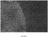


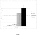
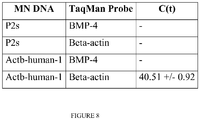
Classifications
-
- C—CHEMISTRY; METALLURGY
- C12—BIOCHEMISTRY; BEER; SPIRITS; WINE; VINEGAR; MICROBIOLOGY; ENZYMOLOGY; MUTATION OR GENETIC ENGINEERING
- C12Q—MEASURING OR TESTING PROCESSES INVOLVING ENZYMES, NUCLEIC ACIDS OR MICROORGANISMS; COMPOSITIONS OR TEST PAPERS THEREFOR; PROCESSES OF PREPARING SUCH COMPOSITIONS; CONDITION-RESPONSIVE CONTROL IN MICROBIOLOGICAL OR ENZYMOLOGICAL PROCESSES
- C12Q1/00—Measuring or testing processes involving enzymes, nucleic acids or microorganisms; Compositions therefor; Processes of preparing such compositions
- C12Q1/68—Measuring or testing processes involving enzymes, nucleic acids or microorganisms; Compositions therefor; Processes of preparing such compositions involving nucleic acids
- C12Q1/6813—Hybridisation assays
- C12Q1/6841—In situ hybridisation
-
- G—PHYSICS
- G01—MEASURING; TESTING
- G01N—INVESTIGATING OR ANALYSING MATERIALS BY DETERMINING THEIR CHEMICAL OR PHYSICAL PROPERTIES
- G01N33/00—Investigating or analysing materials by specific methods not covered by groups G01N1/00 - G01N31/00
- G01N33/48—Biological material, e.g. blood, urine; Haemocytometers
- G01N33/50—Chemical analysis of biological material, e.g. blood, urine; Testing involving biospecific ligand binding methods; Immunological testing
- G01N33/53—Immunoassay; Biospecific binding assay; Materials therefor
- G01N33/543—Immunoassay; Biospecific binding assay; Materials therefor with an insoluble carrier for immobilising immunochemicals
- G01N33/54366—Apparatus specially adapted for solid-phase testing
-
- A—HUMAN NECESSITIES
- A61—MEDICAL OR VETERINARY SCIENCE; HYGIENE
- A61B—DIAGNOSIS; SURGERY; IDENTIFICATION
- A61B10/00—Other methods or instruments for diagnosis, e.g. instruments for taking a cell sample, for biopsy, for vaccination diagnosis; Sex determination; Ovulation-period determination; Throat striking implements
- A61B10/0045—Devices for taking samples of body liquids
-
- A—HUMAN NECESSITIES
- A61—MEDICAL OR VETERINARY SCIENCE; HYGIENE
- A61B—DIAGNOSIS; SURGERY; IDENTIFICATION
- A61B10/00—Other methods or instruments for diagnosis, e.g. instruments for taking a cell sample, for biopsy, for vaccination diagnosis; Sex determination; Ovulation-period determination; Throat striking implements
- A61B10/02—Instruments for taking cell samples or for biopsy
- A61B10/0233—Pointed or sharp biopsy instruments
-
- A—HUMAN NECESSITIES
- A61—MEDICAL OR VETERINARY SCIENCE; HYGIENE
- A61B—DIAGNOSIS; SURGERY; IDENTIFICATION
- A61B5/00—Measuring for diagnostic purposes; Identification of persons
- A61B5/145—Measuring characteristics of blood in vivo, e.g. gas concentration, pH value; Measuring characteristics of body fluids or tissues, e.g. interstitial fluid, cerebral tissue
- A61B5/14503—Measuring characteristics of blood in vivo, e.g. gas concentration, pH value; Measuring characteristics of body fluids or tissues, e.g. interstitial fluid, cerebral tissue invasive, e.g. introduced into the body by a catheter or needle or using implanted sensors
-
- A—HUMAN NECESSITIES
- A61—MEDICAL OR VETERINARY SCIENCE; HYGIENE
- A61B—DIAGNOSIS; SURGERY; IDENTIFICATION
- A61B5/00—Measuring for diagnostic purposes; Identification of persons
- A61B5/145—Measuring characteristics of blood in vivo, e.g. gas concentration, pH value; Measuring characteristics of body fluids or tissues, e.g. interstitial fluid, cerebral tissue
- A61B5/14507—Measuring characteristics of blood in vivo, e.g. gas concentration, pH value; Measuring characteristics of body fluids or tissues, e.g. interstitial fluid, cerebral tissue specially adapted for measuring characteristics of body fluids other than blood
- A61B5/1451—Measuring characteristics of blood in vivo, e.g. gas concentration, pH value; Measuring characteristics of body fluids or tissues, e.g. interstitial fluid, cerebral tissue specially adapted for measuring characteristics of body fluids other than blood for interstitial fluid
- A61B5/14514—Measuring characteristics of blood in vivo, e.g. gas concentration, pH value; Measuring characteristics of body fluids or tissues, e.g. interstitial fluid, cerebral tissue specially adapted for measuring characteristics of body fluids other than blood for interstitial fluid using means for aiding extraction of interstitial fluid, e.g. microneedles or suction
-
- A—HUMAN NECESSITIES
- A61—MEDICAL OR VETERINARY SCIENCE; HYGIENE
- A61B—DIAGNOSIS; SURGERY; IDENTIFICATION
- A61B5/00—Measuring for diagnostic purposes; Identification of persons
- A61B5/145—Measuring characteristics of blood in vivo, e.g. gas concentration, pH value; Measuring characteristics of body fluids or tissues, e.g. interstitial fluid, cerebral tissue
- A61B5/1468—Measuring characteristics of blood in vivo, e.g. gas concentration, pH value; Measuring characteristics of body fluids or tissues, e.g. interstitial fluid, cerebral tissue using chemical or electrochemical methods, e.g. by polarographic means
-
- A—HUMAN NECESSITIES
- A61—MEDICAL OR VETERINARY SCIENCE; HYGIENE
- A61B—DIAGNOSIS; SURGERY; IDENTIFICATION
- A61B5/00—Measuring for diagnostic purposes; Identification of persons
- A61B5/15—Devices for taking samples of blood
- A61B5/150007—Details
- A61B5/150015—Source of blood
-
- A—HUMAN NECESSITIES
- A61—MEDICAL OR VETERINARY SCIENCE; HYGIENE
- A61B—DIAGNOSIS; SURGERY; IDENTIFICATION
- A61B5/00—Measuring for diagnostic purposes; Identification of persons
- A61B5/15—Devices for taking samples of blood
- A61B5/150007—Details
- A61B5/150015—Source of blood
- A61B5/150022—Source of blood for capillary blood or interstitial fluid
-
- A—HUMAN NECESSITIES
- A61—MEDICAL OR VETERINARY SCIENCE; HYGIENE
- A61B—DIAGNOSIS; SURGERY; IDENTIFICATION
- A61B5/00—Measuring for diagnostic purposes; Identification of persons
- A61B5/15—Devices for taking samples of blood
- A61B5/150977—Arrays of piercing elements for simultaneous piercing
- A61B5/150984—Microneedles or microblades
-
- A—HUMAN NECESSITIES
- A61—MEDICAL OR VETERINARY SCIENCE; HYGIENE
- A61K—PREPARATIONS FOR MEDICAL, DENTAL OR TOILETRY PURPOSES
- A61K9/00—Medicinal preparations characterised by special physical form
- A61K9/0012—Galenical forms characterised by the site of application
- A61K9/0019—Injectable compositions; Intramuscular, intravenous, arterial, subcutaneous administration; Compositions to be administered through the skin in an invasive manner
- A61K9/0021—Intradermal administration, e.g. through microneedle arrays, needleless injectors
-
- C—CHEMISTRY; METALLURGY
- C12—BIOCHEMISTRY; BEER; SPIRITS; WINE; VINEGAR; MICROBIOLOGY; ENZYMOLOGY; MUTATION OR GENETIC ENGINEERING
- C12Q—MEASURING OR TESTING PROCESSES INVOLVING ENZYMES, NUCLEIC ACIDS OR MICROORGANISMS; COMPOSITIONS OR TEST PAPERS THEREFOR; PROCESSES OF PREPARING SUCH COMPOSITIONS; CONDITION-RESPONSIVE CONTROL IN MICROBIOLOGICAL OR ENZYMOLOGICAL PROCESSES
- C12Q1/00—Measuring or testing processes involving enzymes, nucleic acids or microorganisms; Compositions therefor; Processes of preparing such compositions
- C12Q1/68—Measuring or testing processes involving enzymes, nucleic acids or microorganisms; Compositions therefor; Processes of preparing such compositions involving nucleic acids
- C12Q1/6806—Preparing nucleic acids for analysis, e.g. for polymerase chain reaction [PCR] assay
-
- C—CHEMISTRY; METALLURGY
- C12—BIOCHEMISTRY; BEER; SPIRITS; WINE; VINEGAR; MICROBIOLOGY; ENZYMOLOGY; MUTATION OR GENETIC ENGINEERING
- C12Q—MEASURING OR TESTING PROCESSES INVOLVING ENZYMES, NUCLEIC ACIDS OR MICROORGANISMS; COMPOSITIONS OR TEST PAPERS THEREFOR; PROCESSES OF PREPARING SUCH COMPOSITIONS; CONDITION-RESPONSIVE CONTROL IN MICROBIOLOGICAL OR ENZYMOLOGICAL PROCESSES
- C12Q1/00—Measuring or testing processes involving enzymes, nucleic acids or microorganisms; Compositions therefor; Processes of preparing such compositions
- C12Q1/68—Measuring or testing processes involving enzymes, nucleic acids or microorganisms; Compositions therefor; Processes of preparing such compositions involving nucleic acids
- C12Q1/6813—Hybridisation assays
- C12Q1/6834—Enzymatic or biochemical coupling of nucleic acids to a solid phase
- C12Q1/6837—Enzymatic or biochemical coupling of nucleic acids to a solid phase using probe arrays or probe chips
-
- C—CHEMISTRY; METALLURGY
- C12—BIOCHEMISTRY; BEER; SPIRITS; WINE; VINEGAR; MICROBIOLOGY; ENZYMOLOGY; MUTATION OR GENETIC ENGINEERING
- C12Q—MEASURING OR TESTING PROCESSES INVOLVING ENZYMES, NUCLEIC ACIDS OR MICROORGANISMS; COMPOSITIONS OR TEST PAPERS THEREFOR; PROCESSES OF PREPARING SUCH COMPOSITIONS; CONDITION-RESPONSIVE CONTROL IN MICROBIOLOGICAL OR ENZYMOLOGICAL PROCESSES
- C12Q1/00—Measuring or testing processes involving enzymes, nucleic acids or microorganisms; Compositions therefor; Processes of preparing such compositions
- C12Q1/68—Measuring or testing processes involving enzymes, nucleic acids or microorganisms; Compositions therefor; Processes of preparing such compositions involving nucleic acids
- C12Q1/6876—Nucleic acid products used in the analysis of nucleic acids, e.g. primers or probes
-
- G—PHYSICS
- G01—MEASURING; TESTING
- G01N—INVESTIGATING OR ANALYSING MATERIALS BY DETERMINING THEIR CHEMICAL OR PHYSICAL PROPERTIES
- G01N1/00—Sampling; Preparing specimens for investigation
- G01N1/02—Devices for withdrawing samples
- G01N1/04—Devices for withdrawing samples in the solid state, e.g. by cutting
- G01N1/08—Devices for withdrawing samples in the solid state, e.g. by cutting involving an extracting tool, e.g. core bit
-
- G—PHYSICS
- G01—MEASURING; TESTING
- G01N—INVESTIGATING OR ANALYSING MATERIALS BY DETERMINING THEIR CHEMICAL OR PHYSICAL PROPERTIES
- G01N33/00—Investigating or analysing materials by specific methods not covered by groups G01N1/00 - G01N31/00
- G01N33/48—Biological material, e.g. blood, urine; Haemocytometers
- G01N33/50—Chemical analysis of biological material, e.g. blood, urine; Testing involving biospecific ligand binding methods; Immunological testing
- G01N33/53—Immunoassay; Biospecific binding assay; Materials therefor
- G01N33/543—Immunoassay; Biospecific binding assay; Materials therefor with an insoluble carrier for immobilising immunochemicals
- G01N33/551—Immunoassay; Biospecific binding assay; Materials therefor with an insoluble carrier for immobilising immunochemicals the carrier being inorganic
- G01N33/553—Metal or metal coated
-
- A—HUMAN NECESSITIES
- A61—MEDICAL OR VETERINARY SCIENCE; HYGIENE
- A61B—DIAGNOSIS; SURGERY; IDENTIFICATION
- A61B10/00—Other methods or instruments for diagnosis, e.g. instruments for taking a cell sample, for biopsy, for vaccination diagnosis; Sex determination; Ovulation-period determination; Throat striking implements
- A61B10/0045—Devices for taking samples of body liquids
- A61B2010/008—Interstitial fluid
-
- A—HUMAN NECESSITIES
- A61—MEDICAL OR VETERINARY SCIENCE; HYGIENE
- A61B—DIAGNOSIS; SURGERY; IDENTIFICATION
- A61B2562/00—Details of sensors; Constructional details of sensor housings or probes; Accessories for sensors
- A61B2562/04—Arrangements of multiple sensors of the same type
- A61B2562/046—Arrangements of multiple sensors of the same type in a matrix array
-
- A—HUMAN NECESSITIES
- A61—MEDICAL OR VETERINARY SCIENCE; HYGIENE
- A61B—DIAGNOSIS; SURGERY; IDENTIFICATION
- A61B5/00—Measuring for diagnostic purposes; Identification of persons
- A61B5/68—Arrangements of detecting, measuring or recording means, e.g. sensors, in relation to patient
- A61B5/6846—Arrangements of detecting, measuring or recording means, e.g. sensors, in relation to patient specially adapted to be brought in contact with an internal body part, i.e. invasive
- A61B5/6847—Arrangements of detecting, measuring or recording means, e.g. sensors, in relation to patient specially adapted to be brought in contact with an internal body part, i.e. invasive mounted on an invasive device
- A61B5/685—Microneedles
-
- C—CHEMISTRY; METALLURGY
- C12—BIOCHEMISTRY; BEER; SPIRITS; WINE; VINEGAR; MICROBIOLOGY; ENZYMOLOGY; MUTATION OR GENETIC ENGINEERING
- C12Q—MEASURING OR TESTING PROCESSES INVOLVING ENZYMES, NUCLEIC ACIDS OR MICROORGANISMS; COMPOSITIONS OR TEST PAPERS THEREFOR; PROCESSES OF PREPARING SUCH COMPOSITIONS; CONDITION-RESPONSIVE CONTROL IN MICROBIOLOGICAL OR ENZYMOLOGICAL PROCESSES
- C12Q2543/00—Reactions characterised by the reaction site, e.g. cell or chromosome
- C12Q2543/10—Reactions characterised by the reaction site, e.g. cell or chromosome the purpose being "in situ" analysis
-
- C—CHEMISTRY; METALLURGY
- C12—BIOCHEMISTRY; BEER; SPIRITS; WINE; VINEGAR; MICROBIOLOGY; ENZYMOLOGY; MUTATION OR GENETIC ENGINEERING
- C12Q—MEASURING OR TESTING PROCESSES INVOLVING ENZYMES, NUCLEIC ACIDS OR MICROORGANISMS; COMPOSITIONS OR TEST PAPERS THEREFOR; PROCESSES OF PREPARING SUCH COMPOSITIONS; CONDITION-RESPONSIVE CONTROL IN MICROBIOLOGICAL OR ENZYMOLOGICAL PROCESSES
- C12Q2600/00—Oligonucleotides characterized by their use
- C12Q2600/16—Primer sets for multiplex assays
-
- G—PHYSICS
- G01—MEASURING; TESTING
- G01N—INVESTIGATING OR ANALYSING MATERIALS BY DETERMINING THEIR CHEMICAL OR PHYSICAL PROPERTIES
- G01N2800/00—Detection or diagnosis of diseases
- G01N2800/16—Ophthalmology
-
- G—PHYSICS
- G01—MEASURING; TESTING
- G01N—INVESTIGATING OR ANALYSING MATERIALS BY DETERMINING THEIR CHEMICAL OR PHYSICAL PROPERTIES
- G01N2800/00—Detection or diagnosis of diseases
- G01N2800/20—Dermatological disorders
Definitions
- the disclosure provides devices for detecting or extracting one or more biomarkers from a tissue or biological sample of a subject in situ.
- the devices can comprise an array of microneedles and a plurality of probes specific for the biomarkers, wherein the probes are covalently attached to the microneedles.
- the probes are specific for different biomarkers, and each different probe is attached to a different microneedle.
- the same probe for a specific biomarker is attached to two or more microneedles.
- the microneedles in the devices can be made of a polymer, a metal, a ceramic or any other suitable material.
- the disclosure provides a device for detecting or extracting one or more biomarkers from an in situ tissue or biological sample of a subject, comprising one or more microneedles, and one or more probes specific for the biomarkers, wherein the probes are attached to the microneedles, via covalent or non-covalent linkage.
- the device can comprise a first microneedle.
- the first microneedle can be covalently attached to a first probe, which is specific for a first biomarker.
- the first microneedle can be non-covalently attached to the first probe.
- the first probe can be attached to a plurality of microneedles.
- the invention provides a device comprising a first microneedle, wherein the first microneedle is covalently or non-covalently attached to a first probe specific for a first biomarker.
- the first biomarker can be a polynucleotide.
- the first probe can be a polynucleotide complementary to the first biomarker.
- the first biomarker can be a polypeptide, an antibody, a metabolite, or a small molecule.
- the first probe can be a polynucleotide, a polypeptide, a protein, an antibody, a small molecule, or a biological receptor.
- the first needle is formed on a substrate.
- the device can further comprise a second probe, which is specific for a second biomarker.
- the second probe can be different from the first probe.
- the second probe can be covalently or non-covalently attached to the first microneedle.
- the second probe can be covalently or non-covalently attached to a second microneedle.
- the second biomarker can be a polynucleotide.
- the second probe can be a polynucleotide complementary to the second biomarker.
- the second biomarker can also be a polypeptide, an antibody, a metabolite, or a small molecule.
- the second probe can be a polynucleotide, a polypeptide, a protein, an antibody, a small molecule, a biological receptor.
- the first probe specifically binds to a first nucleotide polymorphism and the second probe specifically binds to a second nucleotide polymorphism.
- the first and second probes are different antibodies each specific to a different epitope of the same biomarker.
- the device provides for a microneedle comprising a polymer, a metal or a ceramic.
- a first probe can be attached, covalently or non-covalently to a plurality of microneedles.
- a second probe and a plurality of other probes can be attached, covalently or non-covalently, to a plurality of microneedles.
- Some devices of the disclosure are directed to detecting nucleic acid biomarkers.
- the probes that can be conjugated to the microneedles are oligonucleotides or polynucleotides complementary to the biomarkers.
- Some of the devices employ microneedles that are fabricated with a thermoplastic polymer.
- the polynucleotide probes in the devices can be conjugated to the microneedles via many suitable linkages, including but not limited to a thiol/amino bifunctional linker or a poly(ethylene glycol) linker.
- a device of the invention further comprises a compartment for amplifying and identifying the first and second biomarkers.
- Some other devices of the disclosure are specifically designed for detection of peptide or protein biomarkers.
- the probes immobilized on the microneedles are antibodies specific for the biomarkers.
- a planar substrate is used to support the array of microneedles.
- the devices of the invention can additionally contain a means for amplifying the biomarkers detected by the probes.
- the device can comprise a plurality of microneedles.
- the plurality of microneedles can comprise at least one microneedle that is covalently or non-covalently attached to at least one probe specific for a biomarker.
- the biomarker can be indicative of a specific condition, including but not limited to a skin or eye condition.
- the biomarker can be indicative of a skin condition.
- the skin condition is a skin cancer.
- the biomarker can be indicative of an eye condition.
- the eye condition is an eye cancer or eye inflammation.
- the disclosure provides a device comprising a plurality of microneedles, wherein the plurality of microneedles comprises at least one microneedle that is covalently or non-covalently attached to at least one probe specific for a biomarker and the biomarker is indicative of a skin or eye condition.
- the biomarker is a polynucleotide
- the at least one probe is complementary to the biomarker.
- the at least one probe comprises different polynucleotide probes specific to different nucleotide polymorphisms of the same biomarker.
- the biomarker is a peptide or polypeptide, and the at least one probe is an antibody specific for the biomarkers.
- the at least one probe comprises different antibodies specific to different epitopes of the same biomarker.
- a device of the disclosure can comprise a plurality of probes specific for a plurality of different biomarkers, wherein the plurality of probes are attached to the same or to different microneedles. In some cases, at least two different probes are attached to the same microneedle. In some cases, the devices comprise at least two identical probes for a specific biomarker, wherein the at least two identical probes are attached to one or more microneedles.
- the biomarker can also be obtained during an intraoperative procedure.
- the device can further comprise a sensor that emits an optical signal when the probe detects the biomarker.
- the optical signal of the probe can change when the probe detects the biomarker.
- the disclosure provides methods for detecting or amplifying one or more biomarkers from an in situ tissue in subject (e.g., skin, blood stream, tissue) or ex vivo tissue sample.
- the methods entail (a) preparing a plurality of microneedles to which a plurality of probes specific for the biomarkers are covalently attached, (b) contacting the microneedles with a tissue or a biological sample of the subject, and (c) detecting biomarkers that are bound to the probes on the microneedles.
- the disclosure provides a method for detecting one or more biomarkers from an in situ tissue or biological sample in a subject, comprising (a) contacting a microneedle with a tissue or biological sample of the subject, wherein the microneedle is attached to a set of probes, and wherein he probes bind to one or more biomarkers in situ ; and (b) detecting the one or more biomarkers that are bound to the probes.
- the disclosure provides a device comprising a plurality of microneedles, wherein the plurality of microneedles comprise at least one microneedle that is covalently or non-covalently attached to a probe specific for a biomarker, wherein the probe comprises a sensor that emits an optical signal when the probe detects the biomarker.
- the optical signal of the probe increases when it detects the biomarker. In some cases, the optical signal of the probe decreases when it detects the biomarker.
- the disclosure provides a method for detecting one or more biomarkers from an in situ tissue or biological sample in a subject, comprising (a) preparing a device comprising one or more microneedles to which one or more probes specific for the biomarkers are covalently attached; (b) contacting the microneedle device with the tissue or biological sample of the subject; and (c) detecting biomarkers that are bound to the probes on the microneedle array.
- the disclosure provides a method for detecting one or more biomarkers from an in situ tissue or biological sample in a subject, comprising: (a) contacting a microneedle device with a tissue or biological sample of the subject, wherein the microneedle device comprises one or more probes specific for the one or more biomarkers, wherein at least one probe comprises a sensor that emits a visual signal when the probe detects the biomarkers; and (b) detecting biomarkers based on the visual signal.
- the method can comprise (a) contacting a microneedle device with a tissue or biological sample of the subject, and at least two probes specific for the one or more biomarker, wherein at least two probes specific for the biomarker are different, wherein the probes are attached to the microneedles, via covalent or non-covalent linkage, and (b) detecting biomarkers that are bound to the probes.
- the disclosure provides a method for detecting one or more biomarkers within a tissue of a subject, comprising (a) contacting a microneedle with the tissue of the subject in situ, wherein the tissue comprises an extracellular matrix, wherein the microneedle is covalently attached to a set of probes, and wherein the probes bind to a biomarker in situ ; and (b) disrupting the extracellular matrix.
- the extracellular matrix is disrupted by an enzymatic activity.
- the disrupting the extracellular matrix comprises applying ultrasonic energy to the extracellular matrix.
- the extracellular matrix is disrupted by an electric potential.
- a method can comprise (a) contacting a microneedle device with the sample comprising an extracellular matrix, wherein the microneedle is covalently attached to a set of probes, and wherein the probes bind to a biomarker in situ ; and (b) disrupting the extracellular matrix.
- the method can comprise disrupting the cellular membrane.
- the extracellular matrix or the cellular membrane can be disrupted by an enzymatic activity, an ultrasonic energy, or an electronic potential.
- a method can comprise: (a) contacting a microneedle device with a tissue or biological sample of the subject, wherein the microneedle device comprises one or more probes specific for the one or more biomarkers; and (b) amplifying the biomarkers.
- the biomarkers can be polynucleotides and the probes can be polynucleotides complementary to the biomarkers.
- the probes can be different polynucleotide probes specific to different nucleotide polymorphisms of the same biomarker
- the biomarkers can be peptides or polypeptides and the probes can be antibodies specific for the biomarkers. In some cases, the probes can be different antibodies specific to different epitopes of the same biomarker.
- the probes used in the methods can be made specific for the different biomarkers.
- the probes can be attached to a different microneedle, or the probes can be attached to the same microneedle.
- at least two microneedles can bear identical probes, or multiple probes can be used for detecting a specific biomarker.
- the disclosure also provides a method of preparing a microneedle device comprising: (a) obtaining a solution comprising a mineral salt; (b) adding a probe and a microneedle to the solution; and (c) conjugating the probe to the microneedle within the solution.
- the mineral salt is sodium chloride. In some cases the concentration of the mineral salt is less than 2.5 M.
- any suitable materials can be used in making the microneedle array used in these methods. Examples include but are not limited to a polymer, a metal or a ceramic. Some methods of the invention are intended for detection of nucleic acid biomarkers. In these methods, the employed probes are oligonucleotide or polynucleotide molecules with sequences that are complementary to that of the biomarkers. In some of the methods, the nucleic biomarkers captured by the microneedle device are detected and analyzed by PCR or quantitative real-time PCR (qRT-PCR). In some of the methods, the microneedles are made of a polymer, and the probes are attached to the microneedles via a thiol/amino bifunctional linker.
- qRT-PCR quantitative real-time PCR
- the microneedles are made of stainless steel and coated with gold, and the probes are attached to the microneedles via a thiol linker.
- the microneedles can be solid.
- Some other methods of the invention are intended for detection of peptide or polypeptide biomarkers.
- the employed probes are molecules that are capable of specifically binding to the biomarkers, e.g., monoclonal antibodies.
- the peptide or protein biomarkers bound to the probes on the microneedles can be detected by addition of an oligonucleotide-tagged secondary antibody and subsequent PCR amplification of the conjugated tag.
- the method for detecting one or more polynucleotide biomarkers from a skin or eye tissue in a subject can comprise contacting a microneedle device with the skin or eye tissue of the subject.
- the method can further comprise contacting the microneedle device with a dermal capillary of the subject.
- the method can comprise contacting a microneedle device with the tissue or biological sample of the subject during an intraoperative procedure.
- the tissue or biological sample can be from an organ selected from the group consisting of brain, heart, breast, liver, pancreas, spleen, bladder, stomach, lung, uterus, cervix, prostate, kidney, intestine, appendix, and colon.
- the method can further comprise contacting the microneedles to the margins of a tumor after the tumor is removed from the subject.
- a method of the disclosure detects a biomarker that is present in the blood of a subject.
- the disclosure also provides a method for conjugating a probe to a microneedle, wherein a mineral salt is added.
- mineral salts include but are not limited to lithium salts, potassium salts, sodium salts, magnesium salts, and calcium salts, often with a halide counter ion.
- the mineral salt is sodium chloride.
- the mineral salt can be added at a concentration between about 0.1 M to 2.0 M. Further, the mineral salt can be added at a concentration between about 0.5 M to 1.5 M.
- the various methods of the present invention can further comprise detecting one or more biomarkers from a reference tissue obtained from the subject.
- Some devices or methods of the invention are designed for detecting and acquiring biomarkers from the blood stream of a subject.
- the probe-conjugated microneedle array is contacted with the subject's blood stream by piercing the skin of the subject.
- kits comprising any of the devices for detecting or extracting biomarkers described in this application.
- the kits can further comprise a set of reagents for a polymerase chain reaction.
- the set of reagents can be for a reverse-transcriptase polymerase chain reaction.
- the kit can further comprise a holder, or written instructions for a use thereof.
- the disclosure provides a kit comprising: (a) a device, comprising a plurality of microneedles, wherein the plurality of microneedles comprise at least one microneedle that is covalently or non-covalently attached to a first specific for a biomarker; and (b) a set of reagents for a polymerase chain reaction.
- the kit further comprises a holder.
- the set of reagents comprises a polymerase enzyme, a buffer, and a control sample.
- the kit further comprises written instructions for a use thereof.
- the invention provides a composition comprising a plurality of microneedles coated with a substrate capable of disrupting an extracellular matrix.
- the substrate can be an enzyme.
- the enzyme can be selected from the group consisting of serine proteases, thiol proteases, and MMPs. Specific enzymes include papain, hyaluronidase, streptokinase, streptodornase, trypsin, chymotrypsin, alpha-chymotrypsin, alpha-amlyase, DNase, collagenase, and sutilain.
- the enzyme is hyaluronidase.
- microneedle array-based devices with a singular or plurality of microneedles are fabricated in accordance with the present invention.
- the length of individual microneedles on the array can vary, e.g., ranging from 50 ⁇ m to 5 mm.
- the devices can be fabricated by different materials, composites and material combinations, including, but not limited to, metals and metal alloys, inorganic ceramics and polymers.
- Microneedles are chemically modified to couple probes for biomarkers to the microneedle surface. The specific coupling chemistries depend on the material of the microneedles.
- biomarkers can be detected with cognate probes immobilized on the same array or the same microneedle.
- the devices can be applied to patient on anatomical locations such as skin, eye, tumor or other tissue for the purposes of allowing the desired biomarkers to bind to the probes presented.
- Application can be done by hand (e.g., thumb pressure), an applicator device, with or without a strap or band to hold it in place for the duration of sampling.
- the physical insertion of microneedle probes can disrupt cell membranes to release genetic material, including biomarkers, that are available for binding to probes on the inserted microneedles. Extracellular biomarkers are directly available for binding to probes, and do not need to be sampled from materials released from disrupted cells.
- the time needed for biomarkers to bind to the microneedles will depend on a number of parameters, including, but not limited to, the prevalent quantity, biodistribution and concentration of biomarkers, tissue organization, and the physical and chemical dimensions of microneedle probes (e.g., surface area, number of probes, number of binding sites). Application time can range, e.g., from less than 10 seconds to 60 minutes.
- biomarkers can be assayed using a number of different techniques, including, but not limited to, PCR, quantitative PCR, protein PCR, sandwich ELISA, elution mass spectroscopy, elution Western blot, and elution ELISA. Biomarkers can be assayed either after separation from microneedles, or directly on the microneedles.
- microneedles and the probes to be coupled various chemistries can be used in the conjugation of the probes to the microneedles.
- Chemical modifications of microneedles made of polymer for covalent attachment of biomolecules can be performed with numerous linkages that have been developed in the art.
- the carbonate monomer contains aromatic moieties that can be chemically derivatized post-polymerization.
- Treatment of needles with nitric acid, followed by reduction of the resulting nitro group gives an amine handle through which molecules can be coupled to the polymer backbone.
- this two-step reaction can be performed on manufactured microneedle arrays without compromising the integrity of the array or individual microneedles.
- standard amide bond coupling reagents can be used to link a carboxylic acid-containing probe to the microneedle surface.
- microneedles When the microneedles are fabricated with metal, application of the same principles as described for polymer surfaces to metal surfaces is attainable.
- stainless steel surfaces can be coated with gold via a sputter coating process, providing a chemical handle for attachment.
- thiols By utilizing the well-characterized affinity of thiols for gold surfaces, one can then attach a bivalent crosslinker with a thiol on one terminus and an amine on the other to the metal surface, thereby allowing for identical coupling chemistries of the probe molecule to the microneedle surface.
- Suitable methods for chemical modifications of other types of microneedle surfaces e.g., inorganic ceramic for covalent linkage of biological molecules are also known in the art.
- the invention described herein has broad applicability in many different aspects of diagnostics, including genetic (e.g., mRNA, DNA), protein, hormonal, small molecule and cellular biomarkers for diagnostics and disease prognosis.
- genetic e.g., mRNA, DNA
- protein e.g., IL-12
- hormonal e.g., IL-12
- small molecule e.g., IL-12
- cellular biomarkers for diagnostics and disease prognosis.
- the devices or methods described herein can be useful in detection of skin-based biomarkers, detection of systemically circulating biomarkers, pathogen detection (e.g., bacteria, viruses or parasites), or determination of tumor margins during surgical tumor resection.
- pathogen detection e.g., bacteria, viruses or parasites
- skin-based diagnostic applications of the invention include detection of biomarkers for cutaneous malignant melanoma, non-melanocyte skin cancers (e.g., basal cell carcinoma, squamous cell carcinoma), autoimmune disorders (e.g., psoriasis), infectious diseases, and tropical diseases (e.g., Buruli ulcer, onchocerciasis).
- non-skin based diagnostic applications of the invention include detection of markers for oncologic diseases, hematologic diseases, cardiovascular diseases, Down's syndrome, and real-time, rapid biomarker detection during clinical trials.
- Biomarkers broadly refer to any characteristics that are objectively measured and evaluated as indicators of normal biological processes, pathogenic processes, or pharmacologic responses to therapeutic intervention. Unless otherwise noted, the term biomarker as used herein specifically refers to biomarkers that have biophysical properties, which allow their measurements in biological samples (e.g., plasma, serum, cerebrospinal fluid, bronchoalveolar lavage, biopsy). Unless otherwise noted, the term biomarker is used interchangeably with "molecule biomarker” or "molecular markers.” Examples of biomarkers include nucleic acid biomarkers (e.g., oligonucleotides or polynucleotides), peptides or protein biomarkers, lipids, and lipopolysaccharide markers.
- nucleic acid biomarkers e.g., oligonucleotides or polynucleotides
- peptides or protein biomarkers e.g., lipids, and lipopolysaccharide markers.
- microneedle devices or microneedle arrays refer to a device comprising at least one small piercing element or microneedle onto which is immobilized a diagnostic agent or compound.
- the microneedle is capable of piercing the biological barriers in human or other mammalian subjects (e.g., the stratum corneum of the skin) upon contact.
- the device comprises a plurality of such microneedles, e.g., 2, 5, 10, 25, 50, 100, 150, 200, 250, 300, 350, 400, 450, 500, 550, 600, 650, 700, 750, 800, 850, 900, 950, 1000, 2000, 5000, 10000, 20000, or more.
- the microneedle devices of the invention provide a means for in situ detection of biomarkers in a tissue or biological sample of a subject (e.g., blood stream or skin).
- polynucleotide or “nucleic acid” as used herein refers to a polymeric form of nucleotides of any length, either ribonucleotides or deoxyribonucleotides, that comprise purine and pyrimidine bases, or other natural, chemically or biochemically modified, non-natural, or derivatized nucleotide bases.
- Polynucleotides of the embodiments of the invention include sequences of deoxyribonucleic acid (DNA), ribonucleic acid (RNA), or DNA copies of ribonucleic acid (cDNA), all of which may be isolated from natural sources, recombinantly produced, or artificially synthesized.
- a further example of a polynucleotide is polyamide polynucleotide (PNA).
- the polynucleotides and nucleic acids may exist as single-stranded or double-stranded.
- the backbone of the polynucleotide can comprise sugars and phosphate groups, as may typically be found in RNA or DNA, or modified or substituted sugar or phosphate groups.
- a polynucleotide may comprise modified nucleotides, such as methylated nucleotides and nucleotide analogs.
- the sequence of nucleotides may be interrupted by non-nucleotide components.
- the polymers made of nucleotides such as nucleic acids, polynucleotides and polynucleotides may also be referred to herein as nucleotide polymers.
- oligonucleotide is defined as a molecule comprised of two or more deoxyribonucleotides, preferably more than three. Its exact size will depend upon many factors which, in turn, depend upon the ultimate function and use of the oligonucleotide.
- primer refers to an oligonucleotide, whether occurring naturally as in a purified restriction digest or produced synthetically, which is capable of acting as a point of initiation of synthesis when placed under conditions in which synthesis of a primer extension product, which is complementary to a nucleic acid strand, is induced, i.e., in the presence of nucleotides and an inducing agent such as a DNA polymerase and at a suitable temperature and pH.
- the primer may be either single-stranded or double-stranded and must be sufficiently long to prime the synthesis of the desired extension product in the presence of the inducing agent. The exact length of the primer will depend upon many factors, including temperature, source of primer and the method used.
- the oligonucleotide primer typically contains 15-25 or more nucleotides, although it may contain fewer nucleotides.
- the factors involved in determining the appropriate length of primer are readily known to one of ordinary skill in the art.
- Polypeptides are polymer chains comprised of amino acid residue monomers which are joined together through amide bonds (peptide bonds).
- the amino acids may be the L-optical isomer or the D-optical isomer.
- polypeptides refer to long polymers of amino acid residues, e.g., those consisting of at least more than 10, 20, 50, 100, 200, 500, or more amino acid residue monomers.
- a polypeptide can be a chain of at least two amino acids, peptide-mimetics, a protein, a recombinant protein, an antibody (monoclonal or polyclonal), an antibody fragment, an antigen, an epitope, an enzyme, a receptor, a vitamin, or a structure analogue or combinations thereof.
- the term polypeptide as used herein also encompass short peptides which typically contain two or more amino acid monomers, but usually not more than 10, 15, or 20 amino acid monomers.
- Proteins are long polymers of amino acids linked via peptide bonds and which may be composed of two or more polypeptide chains. More specifically, the term “protein” refers to a molecule composed of one or more chains of amino acids in a specific order; for example, the order as determined by the base sequence of nucleotides in the gene coding for the protein. Proteins are essential for the structure, function, and regulation of the body's cells, tissues, and organs, and each protein has unique functions. Examples are hormones, enzymes, and antibodies. In some embodiments, the terms polypeptide and protein may be used interchangeably.
- Bio sample as used herein is a sample of biological tissue or chemical fluid that is suspected of containing a biomarker or an analyte of interest.
- the sample may be an ex vivo sample or in vivo sample.
- Samples include, for example, body fluids such as whole blood, serum, plasma, cerebrospinal fluid, urine, lymph fluids, and various external secretions of the respiratory, intestinal and genitourinary tracts such as tears, saliva, semen, milk, and the like; and other biological fluids such as cell culture suspensions, cell extracts, cell culture supernatants.
- Samples may also include tissue biopsies, e.g., from the lung, liver, brain, eye, tongue, colon, kidney, muscle, heart, breast, skin, pancreas, uterus, cervix, prostate, salivary gland, and the like. Samples may also be microbiopsies, small samples or even single cells extracted from patients and subsequently processed, for example, using laser capture microdisection. A sample may be suspended or dissolved in, e.g., buffers, extractants, solvents, and the like.
- tissue refers to a collection of similar cells and the intracellular substances surrounding them. There are four basic tissues in the body: 1) epithelium; 2) connective tissues, including blood, bone, and cartilage; 3) muscle tissue; and 4) nerve tissue.
- Ranges can be expressed herein as from “about” one particular value, and/or to "about” another particular value. When such a range is expressed, another embodiment includes from the one particular value and/or to the other particular value. Similarly, when values are expressed as approximations, by use of the antecedent "about,” it will be understood that the particular value forms another embodiment. It will be further understood that the endpoints of each of the ranges are significant both in relation to the other endpoint, and independently of the other endpoint. The term "about” as used herein refers to a range that is 15% plus or minus from a stated numerical value within the context of the particular usage. For example, about 10 would include a range from 8.5 to 11.5.
- a covalent bond is a chemical bond that involves the sharing of electron pairs between atoms.
- Covalent bonding includes many kinds of interactions, including ⁇ -bonding, ⁇ -bonding, metal-to-metal bonding, agostic interactions, and three-center two-electron bonds.
- a non-covalent interaction differs from a covalent bond in that it does not involve the sharing of electrons.
- a non-covalent bond can be generally classified into 4 categories: electrostatic, ⁇ -effects, van der Waals forces, and hydrophobic effects.
- Biomarkers can be used in clinical practice to identify risk for or diagnose a disease, stratify patients, assess disease severity or progression, predict prognosis, or guide treatment.
- biomarkers may be used to help determine how a drug works in the body, to determine a biologically effective dose of a drug, to help assess whether a drug is safe or effective, and to help identify patients most likely to respond to a treatment, or are least likely to suffer an adverse event when treated with a drug.
- Biomarkers can sometimes be used as part of the approval process for a drug or treatment, to inform regulatory decision-making.
- various biomarkers in the body of a subject can be detected with the methods and devices of the present invention.
- various biomarkers from the subject can be detected using the microneedle array device to capture a biomarker or set of biomarker and then using one or more additional tagged probes to detect the captured biomarkers.
- the biomarkers that can be detected with the present disclosure include nucleic acid-based biomarkers (DNA, RNA, mRNA transcripts, genomic DNA, tRNA, siRNA, miRNA, mitochondrial DNA, mitochondrial RNA, exosomal nucleic acids, cell-free DNA or RNA, polynucleotides carrying mutated genes and polymorphisms), peptides, proteins, lipids, lipids metabolites, and small molecules.
- the biomarkers include diagnostic biomarkers (e.g., cardiac troponin for the diagnosis of myocardial infarction), staging of disease biomarkers (e.g., brain natriuretic peptide for congestive heart failure), disease prognosis biomarkers (cancer biomarkers), and biomarkers for monitoring the clinical response to an intervention (HbAlc for anti-diabetic treatment). They also include biomarkers used in decision making in early drug development. For instance, pharmacodynamic (PD) biomarkers are markers of a certain pharmacological response, which are of special interest in dose optimization studies. Examples of biomarkers that have disease implications include serum LDL for high cholesterol and blood pressure, P53 gene and MMPs for cancer. Additional examples of specific nucleic acid biomarkers and protein biomarkers that are suitable for detection with methods and devices of the present invention are described below.
- diagnostic biomarkers e.g., cardiac troponin for the diagnosis of myocardial infarction
- staging of disease biomarkers e.
- nucleic acid biomarkers are known in the art. Examples include telomerase reverse transcriptase mRNA as a diagnostic biomarker for hepatocellular carcinoma ( Miura et al., Clin. Cancer Res. 11:3205-9, 2005 ), plasma hnRNP B1 mRNA as a biomarker of lung cancer ( Sato et al., J. Cancer Res. Clin. Oncol. 134:1191-7, 2008 ), GD2/GM2 synthase mRNA as a biomarker for small cell lung cancer ( Chen et al., Lung Cancer.
- probes specific for detecting these biomarkers can be easily designed and synthesized with routinely practiced methods of molecular biology. See, e.g., Sambrook et al., Molecular Cloning: A Laboratory Manual, Cold Spring Harbor Press, N.Y., (3rd ed., 2000 ); and Brent et al., Current Protocols in Molecular Biology, John Wiley & Sons, Inc. (ringbou ed., 2003 ).
- peptides or protein biomarkers are detected with the devices and methods of the invention. Methods and devices described herein are useful for detection of various peptide or protein biomarkers that have been characterized in the art.
- Specific examples of peptide or protein biomarkers suitable for the invention include, but are not limited to, PSA as a biomarker of prostate cancer ( Polascik et al., J. Urol. 162:293-306, 1999 ), cancer antigen 125 (CA125) as a biomarker for ovarian cancer ( Jacobs et al., Lancet 353:1207-1210, 1999 ), BC1, BC2 and BC3 as serum biomarkers for detection of breast cancer ( Mathelin et al., Breast Cancer Res.
- Treat. 96:83-90, 2006 ⁇ -Defensin-2 Protein as a serum biomarker for psoriasis ( Patrick et al., PLoS ONE 4: e4725, 2009 ), C-reactive protein as a biomarker of metastasis of renal cell carcinoma ( Johnson et al., Mol. Diagn. Ther. 14:191-3, 2010 ), high molecular weight-melanoma-associated antigen as a biomarker of desmoplastic melanoma ( Goto et al., Pigment Cell Melanoma Res.
- telomerase expression as a biomarker of malignant transformation in patients with inflammatory bowel disease ( Gonzalo et al., Gastroenterol Hepatol. 33:288-96, 2010 ), and insulin-like growth factor II mRNA-binding protein 3 (IMP3) as a prognostic biomarker for oral squamous cell carcinoma ( Li et al., Head Neck. Jul 22. 2010 ).
- Probes for detecting these biomarkers e.g., monoclonal antibodies
- can be readily generated with standard immunology techniques e.g., hybridoma technology
- commercial suppliers e.g., Abnova Corporation, Full Moon BioSystems and Spring Bioscience.
- the microneedle device described herein can be combined with various methods known in the art for amplifying and examining molecular entities.
- Many genomic and proteomics techniques are suitable for use in the device and methods of the invention for detecting and analyzing protein and nucleic acid markers.
- PCR can be used for amplifying and examining nucleic acid molecules bound to the probes on the microneedles.
- ELISA can be used for analyzing peptide or protein markers captured by the microneedle-based device of the invention.
- biomarker assay technique metabolomics, lipidomics, and glycomics techniques can also be used in the identification and detection of biomarkers of other chemical classes. For example, mass spectrometry, chromatography, and nuclear magnetic resonance are useful for detecting and analyzing various biological molecules bound to the microneedles.
- Microneedles for attaching diagnostic probes are Microneedles for attaching diagnostic probes.
- the invention provides microneedle devices with covalently attached molecular probes for detection and acquisition of biomarkers from a subject in situ.
- the microneedle based device contains one or more microneedles that can be pierced into mammalian biological barriers such as the skin or mucous membrane. Often, the microneedles are non-invasive, or minimally-invasive.
- the device can also have a planar substrate which supports the microneedles.
- the substrate can be made of the same material as that of the microneedle. It can also be made of a different material.
- FIGURE 4 is a diagram of a surface of a device of the invention comprising a plurality of microneedles. Each "square" in 401 illustrates an individual microneedle. 401 illustrates a plurality of microneedles on a surface of a device of the invention, wherein the height of the needles ranges from about 400 ⁇ m to about 1000 ⁇ m.
- the height of the needles is from about 20 ⁇ m to about 50 ⁇ m, from about 20 ⁇ m to about 100 ⁇ m, from about 20 ⁇ m to about 150 ⁇ m, from about 20 ⁇ m to about 200 ⁇ m, from about 20 ⁇ m to about 250 ⁇ m, from about 20 ⁇ m to about 300 ⁇ m, from about 20 ⁇ m to about 350 ⁇ m, from about 20 ⁇ m to about 400 ⁇ m, from about 20 ⁇ m to about 450 ⁇ m, from about 20 ⁇ m to about 500 ⁇ m, from about 20 ⁇ m to about 550 ⁇ m, from about 20 ⁇ m to about 600 ⁇ m, from about 20 ⁇ m to about 650 ⁇ m, from about 20 ⁇ m to about 700 ⁇ m, from about 20 ⁇ m to about 750 ⁇ m, from about 20 ⁇ m to about 800 ⁇ m, from about 20 ⁇ m to about 850 ⁇ m, from about 20 ⁇ m to about 900 ⁇ m, from about 20 ⁇ m to
- the height of the microneedles is less than 1 ⁇ m, less than 5 ⁇ m, less than 10 ⁇ m, less than 15 ⁇ m, less than 20 ⁇ m, less than 25 ⁇ m, less than 30 ⁇ m, less than 35 ⁇ m, less than 40 ⁇ m, less than 45 ⁇ m, less than 50 ⁇ m, less than 75 ⁇ m, less than 100 ⁇ m, less than 150 ⁇ m, less than 200 ⁇ m, less than 250 ⁇ m, less than 300 ⁇ m, less than 500 ⁇ m, less than 750 ⁇ m, less than 1000 ⁇ m, less than 2000 ⁇ m, less than 3000 ⁇ m, less than 4000 ⁇ m, less than 5000 ⁇ m, less than 7500 ⁇ m or less than 10000 ⁇ m.
- the height of the microneedles is greater than 1 ⁇ m, greater than 5 ⁇ m, greater than 10 ⁇ m, greater than 15 ⁇ m, greater than 20 ⁇ m, greater than 25 ⁇ m, greater than 30 ⁇ m, greater than 35 ⁇ m, greater than 40 ⁇ m, greater than 45 ⁇ m, greater than 50 ⁇ m, greater than 75 ⁇ m, greater than 100 ⁇ m, greater than 150 ⁇ m, greater than 200 ⁇ m, greater than 250 ⁇ m, greater than 300 ⁇ m, greater than 500 ⁇ m, greater than 750 ⁇ m, greater than 1000 ⁇ m, greater than 2000 ⁇ m, greater than 3000 ⁇ m, greater than 4000 ⁇ m, greater than 5000 ⁇ m, greater than 7500 ⁇ m or greater than 10000 ⁇ m.
- the needle-shaped microneedles can be blunt-pointed objects, they are preferably sharp-pointed objects.
- the microneedles have a circular cone structure with a diameter of the base generally in the range of 10 ⁇ m to 500 ⁇ m, preferably in the range of 20 ⁇ m to 200 ⁇ m.
- 401 illustrates a surface of a device of the invention with a plurality of microneedles wherein the diameter of the base is less than 10 mm in width.
- the microneedles can be present on the devices in rows.
- the rows can be spaced at virtually equal intervals to the space of the needles aligned in the row.
- the rows can be spaced at irregular intervals.
- a microneedle can have a plurality of shapes, for example, a microneedle can be round, conical, triangular, square, rectangular, pentagonal, hexagonal, heptagonal, octagonal, or any other suitable shape.
- a microneedle can be a sharp microneedle, a blunt microneedle, or any combination thereof.
- a device of the invention comprising a plurality of sharp microneedles can be used to penetrate the skin of a subject thereby contacting the probe(s) on the microneedle with, for example, an RNA biomarker.
- a sharp microneedle can be used to disrupt tissue of a biological sample, such as a layer of cells or the outer membrane of a cell.
- a blunt microneedle can be used to touch the surface of the skin of a subject thereby contacting the microneedle with, for example, a cell-surface biomarker on the skin.
- this disclosure provides microneedle devices that comprise microneedles with different shapes (e.g., sharp and blunt microneedles).
- a device of the invention comprises at least 1 microneedle, at least 100 microneedles, at least 200 microneedles, at least 300 microneedles, at least 400 microneedles, at least 500 microneedles, at least 600 microneedles, at least 700 microneedles, at least 800 microneedles, at least 900 microneedles, at least 1000 microneedles, at least 1100 microneedles, at least 1200 microneedles, at least 1300 microneedles, at least 1400 microneedles, at least 1500 microneedles, at least 1600 microneedles, at least 1700 microneedles, at least 1800 microneedles, at least 1900 microneedles, at least 2000 microneedles, at least 2100 microneedles, at least 2200 microneedles, at least 2300 microneedles, at least 2400 microneedles, at least 2500 microneedles, at least 2600 microneedles, at least 2700 microneedles, at least 2800 microneedles, at
- a device of the invention comprises at most 10000 microneedles, at most 5000 microneedles, at most 2500 microneedles, at most 2000 microneedles, at most 1000 microneedles, at most 500 microneedles, at most 400 microneedles, at most 300 microneedles, at most 100 microneedles, at most 90 microneedles, at most 50 microneedles, at most 40 microneedles, at most 30 microneedles, at most 20 microneedles, at most 15 microneedles, at most 10 microneedles, at most 9 microneedles, at most 8 microneedles, at most 7 microneedles, at most 6 microneedles, at most 5 microneedles, at most 4 microneedles, at most 3 microneedles, at most 2 microneedles, or 1 microneedle.
- a device of the invention comprises from about 1 microneedle to about 100 microneedles, from about 1 microneedle to about 200 microneedles, from about 1 microneedle to about 300 microneedles, from about 1 microneedle to about 400 microneedles, from about 1 microneedle to about 500 microneedles, from about 1 microneedle to about 600 microneedles, from about 1 microneedle to about 700 microneedles, from about 1 microneedle to about 800 microneedles, from about 1 microneedle to about 900 microneedles, from about 1 microneedle to about 1000 microneedles, from about 1 microneedle to about 1100 microneedles, from about 1 microneedle to about 1200 microneedles, from about 1 microneedle to about 1300 microneedles, from about 1 microneedle to about 1400 microneedles, from about 1 microneedle to about 1500 microneedles, from about 1 microneedle to about 1600 microneedles
- the substrate and the microneedles of the arrays can be made of various biodegradable or non-biodegradable materials.
- the material for the microneedles or substrate include poly(methyl methacrylate), silicon, silicon dioxide, ceramic, metal (such as stainless steel, titanium, nickel, molybdenum, chromium, and cobalt), and synthetic or natural resin material.
- Some embodiments use a biodegradable polymer such as polylactic acid, polyglycolide, polylactic acid-co-polyglycolide, pullulan, capronolactone, polyurethane or polyanhydride.
- a non-degradable material is employed to fabricate the microneedle array, e.g., a polymer polycarbonate, a synthetic or natural resin material such as polymethacrylic acid, ethylenevinylacetate, polytetrafluoroethylene, polysulfone, or polyoxymethylene.
- the employed material comprises or is coated with a polysaccharide such as hyaluronic acid, pullulan, dextran, dextrin or chondroitin sulfate.
- the microneedle is fabricated with a thermoplastic polymer.
- the substrate and the microneedles of the arrays can be made of various thermoplastic polymers.
- thermoplastic polymers include acrylic polymers, such as poly(methyl methacrylate) (PMMA), nylon, polyethylene, polypropylene, polystyrene, polyvinyl chloride, or Teflon.
- PMMA poly(methyl methacrylate)
- nylon polyethylene
- polypropylene polystyrene
- polyvinyl chloride polyvinyl chloride
- Teflon Teflon
- a device of the invention is fabricated with a thermoplastic polymer selected from the group consisting of polycarbonate, poly(methyl methacrylate), polyethylene and polypropylene.
- Non-limiting examples of non-degradable polymers include, for example, silicone, hydrogels such as crosslinked poly(vinyl alcohol) and poly(hydroxy ethylmethacrylate), ethylene-vivyl acetate, acyl substituted cellulose acetates and alkyl derivatives thereof, partially and completely hydrolyzed alkylene-vinyl acetate copolymers, unplasticized polyvinyl chloride, crosslinked homo- and copolymers of polyvinyl acetate, crosslinked polyesters of acrylic acid and/or methacrylic acid, polyvinyl alkyl ethers, polyvinyl fluoride, polycarbonate, polyurethane, polyamide, polysulphones, styrene acrylonitrile copolymers, crosslinked poly(ethylene oxide), poly(alkylenes), poly(vinyl imidazole), poly(esters), poly(ethylene terephthalate), polyphosphazenes, and chlorosulphonated polyolefines,
- biodegradable polymers include polyesters such as 3-hydroxypropionate, 3-hydroxybutyrate, 3-hydroxyvalerate, 3-hydroxycaproate, 3-hydroxyheptanoate, 3-hydroxyoctanoate, 3-hydroxynonanoate, 3-hydroxydecanoate, 3-hydroxyundecanoate, 3-hydroxydodecanoate, 4-hydroxybutyrate, 5-hydroxyvalerate, polylactide or polylactic acid including poly(d-lactic acid), poly(l-lactic acid), poly(d,l-lactic acid), polyglycolic acid and polyglycolide, poly(lactic-co-glycolic acid), poly(lactide-co-glycolide), poly( ⁇ -caprolactone) and polydioxanone.
- Polysaccharides including starch, glycogen, cellulose and chitin can also be used as biodegradable materials.
- 402 illustrates a surface of a device of the disclosure 401 comprising a plurality of microneedles that have been coupled with at least one type of probe.
- 402 can be coupled with, for example, a polynucleotide probe, a peptide probe, a protein probe, or any combination thereof.
- a probe attached to a microneedle on a device 402 can be covalently or non-covalently coupled to the microneedle. Examples 1 and 2 describe in further detail various methods for covalently coupling a probe to a surface.
- a distance between the center of two microneedles on a device of the disclosure can be calculated to determine a density of the microneedles in the device.
- the center-to-center distance between two microneedles can be less than 1000 ⁇ m, less than 900 ⁇ m, less than 800 ⁇ m, less than 700 ⁇ m, less than 600 ⁇ m, less than 500 ⁇ m, less than 400 ⁇ m, less than 300 ⁇ m, less than 200 ⁇ m, or less 100 ⁇ m.
- the center-to-center distance between two microneedles can be no greater than 100 ⁇ m, no greater than 200 ⁇ m, no greater than 300 ⁇ m, no greater than 400 ⁇ m, no greater than 500 ⁇ m, no greater than 600 ⁇ m, no greater than 700 ⁇ m, no greater than 800 ⁇ m, no greater than 900 ⁇ m, or no greater than 1000 ⁇ m.
- a microneedle of the disclosure can comprise a plurality of different diameters or base widths.
- the shape of the base of a microneedle can be, for example, round, rectangular, triangular, square, pentagonal, hexagonal, heptagonal, or other geometric shape.
- a microneedle of the disclosure can have a diameter or base width that is no greater than 500 ⁇ m , no greater than 400 ⁇ m, no greater than 300 ⁇ m, no greater than 200 ⁇ m, no greater than 100 ⁇ m, no greater than 50 ⁇ m, no greater than 40 ⁇ m, no greater than 30 ⁇ m, no greater than 20 ⁇ m, no greater than 10 ⁇ m, no greater than 1000 nm, no greater than 900 nm, no greater than 800 nm, no greater than 700 nm, no greater than 600 nm, no greater than 500 nm, no greater than 400 nm, no greater than 300 nm, no greater than 200 nm, or no greater than 100 nm.
- a probe can be covalently or non-covalently attached to a microneedle at a plurality of different depths within the microneedle.
- 403 illustrates a microneedle with a height of 600 ⁇ m and a width of 200 ⁇ m.
- 404 illustrates a microneedle with a height of 600 ⁇ m and a width of 200 ⁇ m and a covalently attached probe at a depth of 10 ⁇ m.
- 405 illustrates a microneedle with a height of 600 ⁇ m and a width of 200 ⁇ m and a covalently attached probe at a depth of 500 ⁇ m.
- the depth of a probe within a microneedle can be used to determine a depth in the tissue where a biomarker can be found.
- a device comprising a plurality of microneedles with a plurality of probes attached at different depths of different microneedles can be used to determine where in a tissue a biomarker can be found.
- the device can be used to determine the size or depth of a lesion, such as a cancerous lesion.
- a depth of a probe can be of about 10 ⁇ m, about 20 ⁇ m, about 30 ⁇ m, about 40 ⁇ m, about 50 ⁇ m, about 60 ⁇ m, about 70 ⁇ m, , about 80 ⁇ m, about 90 ⁇ m, about 100 ⁇ m, about 110 ⁇ m, about 120 ⁇ m, about 130 ⁇ m, about 140 ⁇ m, about 150 ⁇ m, about 160 ⁇ m, about 170 ⁇ m, about 180 ⁇ m, about 190 ⁇ m, about 200 ⁇ m, about 210 ⁇ m, about 220 ⁇ m, about 230 ⁇ m, about 240 ⁇ m, about 250 ⁇ m, about 260 ⁇ m, about 270 ⁇ m, about 280 ⁇ m, about 290 ⁇ m, about 300 ⁇ m, about 310 ⁇ m, about 320 ⁇ m, about 330 ⁇ m, about 340 ⁇ m, about 350 ⁇ m, about 360 ⁇ m, about 370 ⁇ m, about380 ⁇ m,
- the microneedle devices of the invention can additionally contain an applicator unit that can be used to apply the device to a subject.
- the applicator unit can control various application parameters, such as the speed with which the array is applied, the force with which the array is applied, and/or the angle with which the array impacts a tissue of the subject (e.g., the skin).
- the applicator may aid in handling or otherwise transferring the array from a storage unit to the subject.
- the applicator can be a single-use, disposable tool that serves as both a storage unit and an application tool. Examples of suitable applicators and methods of application of microneedle arrays are disclosed in U.S. Pat. Nos. 6,293,925 (Safabash et al.
- the applicator unit can have a plurality of different shapes.
- the applicator unit can be, for example, linear, triangular, rectangular, or disk-shaped.
- the applicator unit is a pen applicator.
- microneedle arrays for conjugating diagnostic probes can be readily produced using materials and methods well known in the art for fabricating arrays with microprotrusion structures. See, e.g., United States Patent Nos. 7416541 , 7332197 , 6663820 , 6503231 , United States Patent Application 20100106105 , and European Patent Application 2119469 A .
- the microneedle array can be fabricated by wet etching processing or dry etching processing using a silicon substrate, precision machining using metal or resin (such as discharge machining, laser machining, dicing processing, hot embossing, and injection molding), and mechanical cutting. With such processing method, the needle part and the support part are molded into one piece.
- Example of method of hollowing the needle part includes a method of performing secondary processing by using laser machining and the like after the needle part is being prepared.
- a microneedle of the disclosure can be a microneedle that is solid (not hollow).
- a microneedle of the disclosure can be etched to increase a surface area of the microneedle.
- a plurality of methods of cell disruption may be required to render the biomarker accessible to the probe.
- the cells can be disrupted by either disrupting the extracellular matrix, or disrupting the cell membrane.
- a device of the disclosure can cause cell disruption when, for example, at least one microneedle contacts and perforates a biological sample, such as a human skin.
- a plurality of cells in situ or ex vivo can be disrupted with a substance.
- the invention further provides a composition comprising a plurality of microneedles coated with a substance capable of disrupting an extracellular matrix.
- the substance is an enzyme.
- Non-limiting examples of enzymes that can be used for cell and tissue disruption include but are not limited to papain, hyaluronidase, streptokinase, streptodornase, trypsin, chymotrypsin, alpha-chymotrypsin, alpha-amlyase, DNase, collagenase, sutilain proteases, lysozyme, lipases, zymolase, cellulase, mutanolysin, or glycanases.
- the enzyme is hyalurodinase.
- Additional methods of cell and tissue disruption can include, for example, sonication, electroporation, cryopulverization, physical disruption with pressure, grinding, detergent-based cell lysis, or mechanical shearing of the tissue, with for example, a homogeneizer.
- the extracellular matrix or cell membrane may be disrupted by an ultrasonic energy or by an electrical potential.
- Application of a solvent to a biological sample may also be used to render a biomarker available for hybridization with a probe.
- a device of the disclosure may disrupt a biological tissue in situ or ex vivo in a minimally-invasive manner.
- a microneedle of the disclosure can be contacted with the skin in the eye of a subject.
- the microneedle can gently disrupt the membrane of a layer of cells in the skin of the eye thereby providing access of a biomarker in the eye to a probe(s) in the microneedle.
- the methods and devices of the disclosure can be applied to the identification and characterization of biomarkers from delicate tissues or inoperable tissues. For example biomarkers present in the eye or brain.
- the methods and devices of the disclosure are applied to the in situ characterization of biomarkers from a biological sample that may not be available for surgical removal in a biopsy, for example, certain types of brain tumors.
- Microneedles conjugated with probes are Microneedles conjugated with probes.
- a plurality of probes can be attached to a microneedle of the disclosure.
- the probes comprise polynucleotides (e.g., DNA, RNA, cDNA, cRNA, etc.).
- the polynucleotide probes are designed to bind or hybridize a specific polynucleotide biomarker.
- This disclosure also provides methods and devices for detecting peptide or protein biomarkers.
- the probes attached to the microneedles can specifically recognize and bind to target peptides or proteins of interest.
- the probes can be any substance capable of binding to a specific peptide or protein biomarker. They can be, e.g., a protein (e.g., an antibody, antigen, or fragment thereof), carbohydrate, or a polynucleotide.
- the polynucleotide may possess sequence specificity for the biomarker.
- the probes to be used depend on the biomarker or biomarkers to be detected. Thus, depending the nature and number of biomarkers to be detected, the number of probes immobilized to the microneedles can be 2, 3, 4, 5, 6, 7, 8, 9, 10, or more. In some cases, the total number of probes in a microneedle can be from about 1 probe to about 1,000 probes, from about 1 probe to about 10,000 probes, from about 1 probe to about 100,000 probes, from about 1 probe to about 1,000,000 probes, from about 1 probe to about 10,000,000, from about 1 probe to about 100,000,000 probes, from about 1,000 probes to about 10,000 probes, from about 1,000 probes to about 100,000 probes, from about 1,000 probes to about 1,000,000 probes, from about 1,000 probes to about 10,000,000, from about 1,000 probes to about 100,000,000 probes, from about 10,000 probes to about 100,000 probes, from about 10,000 probes to about 1,000,000 probes, from about 10,000 probes to about 10,000,000, from about 10,000 probes to about 100,000,000 probes, from about 100,000 probes to about 1,000,000 probe
- the total number of probes in a microneedle is at least about 1, about 2, about 3, about 4, about 5, about 6, about 7, about 8, about 9, about 10, about 12, about 14, about 16, about 18, about 20, about 25, about 30, about 35, about 40, about 45, about 50, about 60, about 70, about 80, about 90, about 100, about 150, about 200, about 250, about 300, about 350, about 400, about 450, about 500, about 600, about 700, about 800, about 900, about 1000, about 1100, about 1200, about 1300, about 1400, about 1500, about 2000, about 2500, about 3000, about 3500, about 4000, about 4500, about 5000, about 6000, about 7000, about 8000, about 9000, about 10000, about 12000, about 14000, about 16000, about 18000, about 20000, about 30000, about 40000, about 50000, about 60000, about 70000, about 80000, about 90000, about 100000, about 200000, about 300000, about 400000, about 500000
- the total number of probes in a microneedle is less than about 1, about 2, about 3, about 4, about 5, about 6, about 7, about 8, about 9, about 10, about 12, about 14, about 16, about 18, about 20, about 25, about 30, about 35, about 40, about 45, about 50, about 60, about 70, about 80, about 90, about 100, about 150, about 200, about 250, about 300, about 350, about 400, about 450, about 500, about 600, about 700, about 800, about 900, about 1000, about 1100, about 1200, about 1300, about 1400, about 1500, about 2000, about 2500, about 3000, about 3500, about 4000, about 4500, about 5000, about 6000, about 7000, about 8000, about 9000, about 10000, about 12000, about 14000, about 16000, about 18000, about 20000, about 30000, about 40000, about 50000, about 60000, about 70000, about 80000, about 90000, about 100000, about 200000, about 300000, about 400000, about 500000
- a probe for the biomarker can be immobilized to multiple microneedles in the device of the disclosure, particularly for detection of a biomarker of low concentration, as described further herein.
- a single microneedle comprises multiple different probes capable of binding or detecting the same biomarker.
- a single microneedle comprises at least 2 different probes for the same biomarker, at least 3 different probes, at least 4 different probes, at least 5 different probes, at least 6 different probes, at least 7 different probes, at least 8 different probes, at least 9 different probes, at least 10 different probes, at least 11 different probes, at least 12 different probes, at least 13 different probes, at least 14 different probes, at least 15 different probes, at least 16 different probes, at least 17 different probes, at least 18 different probes, at least 19 different probes, at least 20 different probes, at least 21 different probes, at least 22 different probes, at least 23 different probes, at least 24 different probes, at least 25 different probes, at least 26 different probes, at least 27 different probes, at least 28 different probes, at least 29 different probes, at least 30 different probes, at least 40 different probes, at least 41 different probes, at least 42 different probes, at least 43 different probes, at least 44 different probes, at least
- the same microneedle comprises a plurality of different probes.
- the different probes can be specific for the same biomarker or for a different biomarker.
- a microneedle can comprise at least 2 different probes, at least 10 different probes, at least 100 different probes, at least 200 different probes, at least 300 different probes, at least 400 different probes, at least 500 different probes, at least 600 different probes, at least 700 different probes, at least 800 different probes, at least 900 different probes, at least 1,000 different probes, at least 1,100 different probes, at least 1,200 different probes, at least 1,300 different probes, at least 1,400 different probes, at least 1,500 different probes, at least 1,600 different probes, at least 1,700 different probes, at least 1,800 different probes, at least 1,900 different probes, at least 2,000 different probes, at least 2,100 different probes, at least 2,200 different probes, at least 2,300 different probes, at least 2,400 different probes, at least 2,500 different probes,
- a microneedle can comprise less than 2 different probes, less than 10 different probes, less than 100 different probes, less than 200 different probes, less than 300 different probes, less than 400 different probes, less than 500 different probes, less than 600 different probes, less than 700 different probes, less than 800 different probes, less than 900 different probes, less than 1,000 different probes, less than 1,100 different probes, less than 1,200 different probes, less than 1,300 different probes, less than 1,400 different probes, less than 1,500 different probes, less than 1,600 different probes, less than 1,700 different probes, less than 1,800 different probes, less than 1,900 different probes, less than 2,000 different probes, less than 2,100 different probes, less than 2,200 different probes, less than 2,300 different probes, less than 2,400 different probes, less than 2,500 different probes, less than 2,600 different probes, less than 2,700 different probes, less than 2,800 different probes, less than 2,900 different probes, less than 3,000 different probes, less than
- the plurality of probes are identical (e.g., identical copies of the same polynucleotide or antibody).
- a microneedle can be associated with numerous copies of the same probe (e.g., greater than 2, 5, 10, 50, 100, 1000, 5000, 7500, 10000, or 50000 copies of the same probe).
- a microneedle can comprise a plurality of copies of a polynucleotide probe designed to hybridize the same polymorphism or biomarker.
- a microneedle may comprise a plurality of copies of an antibody probe designed to bind the same epitope.
- a microneedle comprises polynucleotide probes.
- the probes may be designed to detect different biomarkers associated with the same disease, disorder or condition.
- a first probe recognizes a polymorphism (e.g., DNA polymorphism, RNA polymorphism) associated with a disease and a second probe recognizes a different polymorphism associated with the same disease.
- a first DNA probe on a microneedle can be designed to detect a first polymorphism of an RNA biomarker associated with onchocerciasis, a skin condition.
- a second DNA probe on a microneedle can be designed to detect a second polymorphism of an RNA biomarker associated with onchocerciasis.
- a polymorphism can be, for example, a single nucleotide polymorphism (SNP). Genetic and genomic variations can comprise a single SNP or a plurality of SNPs. SNPs can occur at a single locus, or at many loci. Individuals who carry a particular SNP allele at one locus can predictably carry specific SNP alleles at other loci. A correlation of SNPs can provide an association between alleles predisposing an individual to disease or condition.
- the different polynucleotide probes are designed to detect different biomarkers associated with different conditions. For example, one probe may detect a biomarker of a disease, while the other probe may detect a housekeeping gene or gene product.
- the microneedles are attached to polynucleotides, polypeptides, or a mixture of polynucleotides and polypeptides.
- a microneedle can also be associated with a plurality of different protein or antibody probes.
- a first antibody probe on a microneedle can be designed to detect a first epitope of an antigen associated with, for example, a skin cancer.
- a second antibody probe can be designed to detect a second epitope associated with the antigen.
- the second antibody probe can detect an epitope associated with a different skin condition.
- this disclosure provides a microneedle device comprising a set of microneedles, wherein each microneedle in the set comprises identical probes or set of probes. In some embodiments, an identical probe is attached to a plurality of microneedles of a device.
- An identical probe can be attached to, for example, about 1% of the microneedles, about 5% of the microneedles, about 10% of the microneedles, about 15% of the microneedles, about 20% of the microneedles, about 25% of the microneedles, about 30% of the microneedles, about 35% of the microneedles, about 40% of the microneedles, about 45% of the microneedles, about 50% of the microneedles, about 55% of the microneedles, about 60% of the microneedles, about 65% of the microneedles, about 70% of the microneedles, about 75% of the microneedles, about 80% of the microneedles, about 85% of the microneedles, about 90% of the microneedles, about 95% of the microneedles, or about 100% of the microneedles.
- an identical probe is attached to no greater than 5% of the microneedles, no greater than 10% of the microneedles, no greater than 15% of the microneedles, no greater than 20% of the microneedles, no greater than 25% of the microneedles, no greater than 30% of the microneedles, no greater than 35% of the microneedles, no greater than 40% of the microneedles, no greater than 45% of the microneedles, no greater than 50% of the microneedles, no greater than 55% of the microneedles, no greater than 60% of the microneedles, no greater than 70% of the microneedles, no greater than 75% of the microneedles, no greater than 80% of the microneedles, no greater than 85% of the microneedles, no greater than 90% of the microneedles, no greater than 95% of the microneedles, or no greater than 99% of the microneedles.
- a set of microneedles may comprise at least one microneedle attached to a first probe and at least one microneedle attached to a second probe that is different from the first probe.
- the first probe may be a polynucleotide or polypeptide (e.g., antibody, protein) that specifically binds a biomarker of a disease or disorder and the second probe may be a polynucleotide or polypeptide that specifically binds a different biomarker associated with the same disease or disorder.
- the first probe may be a polynucleotide or polypeptide (e.g., antibody, protein) that specifically binds a biomarker of a disease or disorder and the second probe may be a polynucleotide or polypeptide that specifically binds a different biomarker associated with a different disease, disorder, or condition.
- the different disease, condition, or disorder is associated with the same organ.
- the first probe may be associated with a first disease, disorder, or condition associated with skin; and the second probe may be associated with a second disease, disorder, or condition associated with skin or eye.
- the device may comprise an array of microneedles, wherein each microneedle comprises a probe that detects a biomarker associated with a different disease, disorder, or condition associated with the same organ.
- the array of microneedles may comprise greater than 2, 3, 4, 5, 6, 7, 8, 9, 10, 20, 30, 40, 50, 75, 100, 150, 200, 50, or 1000 microneedles associated with different diseases, disorders, or conditions.
- the different diseases, disorders, or conditions are associated with different organs (e.g., greater than 1, 2, 3, 4, 5, 6, 7, 8, 9, 10, 15, or 20 organs).
- the microneedle device comprises a plurality of arrays of microneedles; often, the plurality of arrays of microneedles are suitable for multiplexed reactions. In some cases, the plurality of arrays comprises two or more arrays of microneedles, wherein the arrays are designed to detect a different biomarker. In some cases, a first array of microneedles may be designed to detect a biomarker associated with disease, disorder, or condition, and the second array of microneedles is designed to detect a different biomarker associated with the same disease, disorder, or condition.
- a first array of microneedles may be designed to detect a biomarker associated with disease, disorder, or condition
- a second array of microneedles may be designed to detect a different biomarker associated with a different disease, disorder, or condition.
- a first array of microneedles may be designed to detect a plurality of biomarkers associated with a disease, disorder, or condition
- a second array of microneedles may be designed to detect a plurality of biomarkers associated with a different disease, disorder, or condition.
- the second array of microneedles may be designed to detect control biomarkers (e.g., housekeeping genes), either positive controls or negative controls.
- each microneedle can be inserted into its own cavity (e.g., pinprick cavity), containing unique PCR reagents (e.g., unique probes or primers). PCR reactions can be conducted and the samples can be analyzed for various biomarkers.
- the multiplex reactions are performed with fluorescently-labeled probes, or probes that emit a different optical signal. In some cases, no fluorescence is used.
- a microneedle device described herein may comprise any number of probes; often, the probes are attached to multiple needles within the device.
- the probes may be identical or different.
- a probe for the biomarker can be immobilized to multiple microneedles in the device of the disclosure, particularly for detection of a biomarker of low concentration.
- the total number of probes in a microneedle device can be from about 1 probe to about 1,000 probes, from about 1 probe to about 10,000 probes, from about 1 probe to about 100,000 probes, from about 1 probe to about 1,000,000 probes, from about 1 probe to about 10,000,000, from about 1 probe to about 100,000,000 probes, from about 1,000 probes to about 10,000 probes, from about 1,000 probes to about 100,000 probes, from about 1,000 probes to about 1,000,000 probes, from about 1,000 probes to about 10,000,000, from about 1,000 probes to about 100,000,000 probes, from about 10,000 probes to about 100,000 probes, from about 10,000 probes to about 1,000,000 probes, from about 10,000 probes to about 10,000,000, from about 10,000 probes to about 100,000,000 probes, from about 100,000 probes to about 1,000,000 probes, from about 100,000 probes to about 10,000,000, from about 100,000 probes to about 100,000,000 probes, from about 100,000 probes to about 1,000,000 probes, from about 100,000 probes to about 10,000,000, from about 100,000 probes to about 100,000,000 probes, from about 1,000,000 probes to about
- the total number of probes in a microneedle device is at least about 1, about 2, about 3, about 4, about 5, about 6, about 7, about 8, about 9, about 10, about 12, about 14, about 16, about 18, about 20, about 25, about 30, about 35, about 40, about 45, about 50, about 60, about 70, about 80, about 90, about 100, about 150, about 200, about 250, about 300, about 350, about 400, about 450, about 500, about 600, about 700, about 800, about 900, about 1000, about 1100, about 1200, about 1300, about 1400, about 1500, about 2000, about 2500, about 3000, about 3500, about 4000, about 4500, about 5000, about 6000, about 7000, about 8000, about 9000, about 10000, about 12000, about 14000, about 16000, about 18000, about 20000, about 30000, about 40000, about 50000, about 60000, about 70000, about 80000, about 90000, about 100000,about 200000, about 300000, about 400000, about 500
- the total number of probes in a microneedle device is less than about 1, about 2, about 3, about 4, about 5, about 6, about 7, about 8, about 9, about 10, about 12, about 14, about 16, about 18, about 20, about 25, about 30, about 35, about 40, about 45, about 50, about 60, about 70, about 80, about 90, about 100, about 150, about 200, about 250, about 300, about 350, about 400, about 450, about 500, about 600, about 700, about 800, about 900, about 1000, about 1100, about 1200, about 1300, about 1400, about 1500, about 2000, about 2500, about 3000, about 3500, about 4000, about 4500, about 5000, about 6000, about 7000, about 8000, about 9000, about 10000, about 12000, about 14000, about 16000, about 18000, about 20000, about 30000, about 40000, about 50000, about 60000, about 70000, about 80000, about 90000, about 100000,about 200000, about 300000, about 400000, about 500
- a microneedle device comprises at least 2 different probes for the same biomarker, at least 3 different probes, at least 4 different probes, at least 5 different probes, at least 6 different probes, at least 7 different probes, at least 8 different probes, at least 9 different probes, at least 10 different probes, at least 11 different probes, at least 12 different probes, at least 13 different probes, at least 14 different probes, at least 15 different probes, at least 16 different probes, at least 17 different probes, at least 18 different probes, at least 19 different probes, at least 20 different probes, at least 21 different probes, at least 22 different probes, at least 23 different probes, at least 24 different probes, at least 25 different probes, at least 26 different probes, at least 27 different probes, at least 28 different probes, at least 29 different probes, at least 30 different probes, at least 40 different probes, at least 41 different probes, at least 42 different probes, at least 43 different probes, at least 44 different probes, at least
- a microneedle device comprises a plurality of different probes.
- the different probes can be specific for the same biomarker or for a different biomarker.
- a microneedle device can comprise at least 2 different probes, at least 10 different probes, at least 100 different probes, at least 200 different probes, at least 300 different probes, at least 400 different probes, at least 500 different probes, at least 600 different probes, at least 700 different probes, at least 800 different probes, at least 900 different probes, at least 1,000 different probes, at least 1,100 different probes, at least 1,200 different probes, at least 1,300 different probes, at least 1,400 different probes, at least 1,500 different probes, at least 1,600 different probes, at least 1,700 different probes, at least 1,800 different probes, at least 1,900 different probes, at least 2,000 different probes, at least 2,100 different probes, at least 2,200 different probes, at least 2,300 different probes, at least 2,400 different probes, at least 2,500 different probe
- a microneedle device can comprise less than 2 different probes, less than 10 different probes, less than 100 different probes, less than 200 different probes, less than 300 different probes, less than 400 different probes, less than 500 different probes, less than 600 different probes, less than 700 different probes, less than 800 different probes, less than 900 different probes, less than 1,000 different probes, less than 1,100 different probes, less than 1,200 different probes, less than 1,300 different probes, less than 1,400 different probes, less than 1,500 different probes, less than 1,600 different probes, less than 1,700 different probes, less than 1,800 different probes, less than 1,900 different probes, less than 2,000 different probes, less than 2,100 different probes, less than 2,200 different probes, less than 2,300 different probes, less than 2,400 different probes, less than 2,500 different probes, less than 2,600 different probes, less than 2,700 different probes, less than 2,800 different probes, less than 2,900 different probes, less than 3,000 different
- Probes for detecting various biological markers can be obtained commercially or synthesized in accordance with methods well known in the art.
- the probes can be designed according to any suitable methods.
- computerized search program can be used to design nucleic acid probes specific for target biomarker sequences (e.g., mRNAs) with minimal cross hybridization and similar hybridization efficiency.
- target biomarker sequences e.g., mRNAs
- Such exemplary programs include Oligo 5.0 (National Biosciences Inc.), Primer 3 (MIT), and Array Designer (Telechem International Inc.).
- the nucleotide probes used in the present methods can have any suitable length, e.g., from about 15 to about 100 nucleotides.
- specific probes (antibodies) for detecting the biomarkers can also be readily generated or commercially obtained.
- the nucleotide probes or polypeptide probes used in the present methods can comprise a detectable label.
- Any suitable label can be used.
- the detectable label can be detected by optical, magnetic, mechanic, spectroscopic, photochemical, biochemical, immunochemical, radioactive or enzymatic means.
- the detectable label is a fluorescent or chemiluminescent label, such as GFP; a magnetic moiety; a protein, such as avidin, streptavidin; or a peptide tag, such as a histidine tag or a FLAG tag.
- Immobilized probes are present on the surface of the microneedle devices described herein.
- the immobilized probes may bind covalently or noncovalently to the surface of the microneedles by methods known in the art or the specific linking methods described in the Examples herein.
- the probes can be conjugated to the microneedles via, e.g., a biotin-avidin or biotin-streptavidin interaction, a Protein A interaction, a Protein G interaction, a goat anti-mouse Fc interaction, an amide bond, or through any other covalent or noncovalent interaction.
- the probes can be covalently attached to the microneedles with or without a suitable spacer element between probe and microneedle surface such as poly(ethylene glycol) (PEG).
- PEG poly(ethylene glycol)
- the probes can also be modified with a reactive moiety and attached to the gold coated surface of one or more microneedles.
- the reactive moiety can be a thiol group.
- a mineral salt can be added. Examples of mineral salts include but are not limited to lithium salts, potassium salts, sodium salts, magnesium salts, and calcium salts, often with a halide counter ion.
- the mineral salt is sodium chloride.
- the mineral salt may be present at a concentration preferably between about 0.1 M to 2.0 M, about 0.2 M to 2.0 M, about 0.2 M to 1.5 M or about 0.5 M to 1.5M.
- the concentration of the mineral salt is less than about 0.1 M, less than about 0.2 M, less than about 0.2 M, less than about 0.3 M, less than about 0.4 M, less than about 0.5 M, less than about 1.0 M, less than about 1.1 M, less than about 1.2 M, less than about 1.3 M, less than about 1.4 M, less than about 1.5 M, less than about 1.6 M, less than about 1.7 M, less than about 1.8 M, less than about 1.9 M, less than about 2.0 M, less than about 2.5 M, less than about 3.0 M, less than about 3.5 M, less than about 4.0 M, or less than about 4.5 M.
- the concentration of the mineral salt is greater than about 0.1 M, greater than about 0.2 M, greater than about 0.2 M, greater than about 0.3 M, greater than about 0.4 M, greater than about 0.5 M, greater than about 1.0 M, greater than about 1.1 M, greater than about 1.2 M, greater than about 1.3 M, greater than about 1.4 M, greater than about 1.5 M, greater than about 1.6 M, greater than about 1.7 M, greater than about 1.8 M, greater than about 1.9 M, greater than about 2.0 M, greater than about 2.5 M, greater than about 3.0 M, greater than about 3.5 M, greater than about 4.0 M, or greater than about 4.5 M
- a microneedle that is covalently or non-covalently attached to a probe can be inserted into a biological sample in situ , such as human skin, eye, intraoperative tissue, dermal capillaries, etc.
- a microneedle covalently attached to a probe can also be inserted into a biological sample ex vivo , such as tissue extracted during a biopsy.
- the probe can be allowed to hybridize or bind to a biomarker under physiological conditions of the biological sample for a specified period of time.
- a temperature range of from about 20 to about 40 degrees Celsius, atmospheric pressure of 1, pH of 6-8, glucose concentration of 1-20 mM, atmospheric oxygen concentration, and earth gravity can be examples of physiological conditions for most subjects.
- the probe can be allowed to hybridize or bind to the biological sample for at least 1 minute, at least 2 minutes, at least 3 minutes, at least 5 minutes, at least 10 minutes, at least 15 minutes, at least 20 minutes, at least 25 minutes, at least 30 minutes, at least 45 minutes, at least 1 hour, at least 2 hours, at least 3 hours, at least 5 hours, at least 10 hours, or at least 24 hours.
- the probe can be allowed to hybridize to the biological sample for no more than 1 minute, no more than 2 minutes, no more than 3 minutes, no more than 5 minutes, no more than 10 minutes, no more than 15 minutes, no more than 20 minutes, no more than 25 minutes, no more than 30 minutes, no more than 1 hour, no more than 2 hours, no more than 3 hours, nor more than 5 hours, or no more than 10 hours.
- a microneedle with a covalently or non-covalently linked probe can be removed from the biological sample, for example, the human skin.
- a biomarker that is hybrized or bound to a probe can be isolated from a biological sample by removing the microneedle from the human skin.
- Some embodiments of the invention are directed to detecting polynucleotide biomarkers (e.g., mRNAs, DNA).
- the probes e.g., oligonucleotide probes, polynucleotide probes
- the probes can be readily synthesized based on the sequences of the target biomarkers.
- nucleic acid biomarkers Once nucleic acid biomarkers are bound to the probes on the microneedle device of the invention, they are typically subjected to an amplification reaction (e.g., PCR, reverse transcription PCR) or are detected by a labeled tag.
- the labeled tag may be directly linked to the probe that is attached to the microneedle or, in some cases, the labeled tag binds to the biomarker after it has already been captured by the probe attached to the microneedle.
- PCR polymerase chain reaction
- reverse transcription PCR reverse transcription PCR
- LCR ligase chain reaction
- Genomics 4,560 see Wu and Wallace, Genomics 4,560 (1989 ), Landegren et al., Science 241, 1077 (1988 ), transcription amplification ( Kwoh et al., Proc. Natl. Acad. Sci. USA 86, 1173 (1989 )), and self-sustained sequence replication ( Guatelli et al., Proc. Nat. Acad.
- RNA based sequence amplification NASBA
- ssRNA single stranded RNA
- dsDNA double stranded DNA
- identity of the captured biomarkers can be then readily confirmed by standard techniques, e.g., sequencing analysis, electrophoresis, etc.
- PCR can use thermal cycling, comprising cycles of repeated heating and cooling of the reaction for DNA melting and enzymatic replication of the DNA. These thermal cycling operations can physically separate the two strands in a DNA double helix at a high temperature in a process called DNA melting. At a lower temperature, each strand can then be used as the template in DNA synthesis by the DNA polymerase to selectively amplify the target DNA.
- the selectivity of PCR can result from the use of primers (short DNA fragments) that are complementary to the DNA region targeted for amplification under specific thermal cycling conditions.
- Primers containing sequences complementary to a biomarker of interest along with a DNA polymerase can be used to achieve selective and repeated amplification.
- the DNA generated can be used as a template for replication, setting in motion a chain reaction in which the DNA template is exponentially amplified.
- PCR applications can employ a heat-stable DNA polymerase, such as Taq polymerase, an enzyme originally isolated from the bacterium Thermus aquaticus.
- This DNA polymerase can enzymatically assemble a new DNA strand from the nucleotides, e.g., by using single-stranded DNA as a template and DNA oligonucleotides (also called DNA primers) for initiation of DNA synthesis.
- a PCR reaction can be performed directly on a microneedle that has been inserted into a biological sample.
- the microneedle may be placed in a tube or between two plates (e.g., glass slides) comprising the necessary reagents for a PCR reaction.
- a biomarker may be detached from the needle and released into the PCR tube by a plurality of different methods.
- a microneedle may be heated to release the biomarker from the microneedle, or a biomarker may spontaneously be released from the needle into the PCR solution.
- a PCR reaction can be performed as described above and the PCR product can be analyzed using standard procedures, for example, electrophoresis, real-time PCR, and other procedures, such as described in PCR Technology: Principles and Applications for DNA Amplification (ed. H.A. Erlich, Freeman Press, NY, NY, 1992 ); PCR Protocols: A Guide to Methods and Applications (eds. Innis, et al., Academic Press, San Diego, CA, 1990 ).
- Different PCR methods can be used to analyze the sample, such as standard PCR methods and Real-time PCR methods.
- Real-time PCR Real-time PCR (RT-PCR) is a laboratory technique based on PCR, which can be used to amplify and simultaneously quantify a targeted DNA molecule.
- Real-time PCR can be combined with reverse transcription to quantify messenger RNA and non-coding RNA in cells or tissues.
- a device of the disclosure can be further configured to comprise at least one compartment that can perform a PCR reaction.
- a device of the disclosure may be configured to comprise a plurality of microneedles or an array of microneedles and a compartment where a PCR reaction can be performed.
- a PCR reaction can selectively amplify a biomarker that has been hybridized to a particular microneedle or a PCR reaction can amplify a set of biomarkers that have been hybridized to a plurality of microneedles.
- 402 illustrates a surface of a device of the invention comprising a plurality of microneedles that have been contacted with a biological sample. Each "square" in 402 illustrates an individual microneedle, wherein each individual microneedle comprises at least one probe. Each probe in 402 can hybridize to a biomarker, or not.
- Each microneedle illustrated in 402 can be placed into a separate PCR tube and each PCR product can be analyzed individually. Alternatively, a plurality of microneedles illustrated in 402 can be placed into the same PCR tube for simultaneous analysis.
- a captured biomarker may be detected by using a labeled probe capable of binding to the captured biomarker.
- the labeled probe binds to the biomarker after the biomarker has already been captured by a probe attached to a microneedle described herein.
- the microneedle is directly attached to a probe that is tagged with a label designed to change its optical signal (either decreasing or increasing in intensity) when bound to a biomarker.
- the labeled probe may comprise a label (e.g., fluorophore, radioisotope, etc.) that can emit an optical signal.
- a fluorescent moiety can be a fluorescent protein, such as a green fluorescent protein (GFP), read fluorescent protein (RFP), yellow fluorescent protein (YFP) or variations thereof.
- the labeled probe comprises a label with an optical signal that increases or decreases when the probe is bound to its target.
- a fluorescent moiety can be an RNA aptamer that binds fluorophores.
- An RNA-fluorophore complex can emit an optical signal that spans the visible spectrum, see Paige et al, Science 333, 6042 (2011 ).
- the "Spinach aptamer sequence" is an RNA mimic of GFP that can be configured to emit a fluorescent optical signal when hybridized to a biomarker.
- a microneedle comprising a set of probes that are not-covalently or covalently bound therein can be used to detect a biomarker in a subject.
- a device of the disclosure can be contacted to the skin of a subject.
- the device can comprise a polynucleotide probe comprising an RNA-fluorophore moiety.
- the RNA-fluorophore moiety can be configured to emit an optical signal, such as a fluorescence signal, when hybridized to a biomarker of interest.
- Protein or peptide biomarkers can be detected and quantified by any of a number of methods well known to those of skill in the art for polypeptide detection. These include assay formats such as protein PCR and ELISAs. Both local and systemic protein and peptide biomarkers can be assayed using the microneedle array devices of the invention.
- analytic biochemical methods such as electrophoresis, capillary electrophoresis, high performance liquid chromatography (HPLC), thin layer chromatography (TLC), hyperdiffusion chromatography, mass spectroscopy and the like, or various other immunological methods such as fluid or gel precipitin reactions, immunodiffusion (single or double), immunohistochemistry, affinity chromatography, immunoelectrophoresis, radioimmunoassay (RIA), immunofluorescent assays, Western blotting, dipstick, and the like.
- analytic biochemical methods such as electrophoresis, capillary electrophoresis, high performance liquid chromatography (HPLC), thin layer chromatography (TLC), hyperdiffusion chromatography, mass spectroscopy and the like
- various other immunological methods such as fluid or gel precipitin reactions, immunodiffusion (single or double), immunohistochemistry, affinity chromatography, immunoelectrophoresis, radioimmunoassay (RIA), immunofluorescent assay
- Reagents for performing these assays with respect to any specific protein or peptide biomarker can be readily obtained from commercial sources or generated by standard and routinely practiced techniques (e.g., hybridoma technology for producing monoclonal antibodies).
- Binding interactions between a probe and a biomarker can be detected using a secondary detection reagent, such as a secondary antibody.
- a secondary detection reagent such as a secondary antibody.
- a "sandwich ELISA" can be used to detect binding of a biomarker to an antibody probe.
- the binding of a biomarker to a probe can be detected using a detection antibody that is specific to a different epitope of the biomarker.
- the antibody used in the detection can be of a different isotype than the antibody used as a probe, for example, an antibody probe can be an IgG antibody (including any of the subtypes, such as IgG1, IgG2, IgG3 and IgG4), and a secondary antibody can be of the IgA, IgM).
- the detection antibody can be, for example conjugated to a detectable label, such as a fluorescent moiety or a radioactive label.
- a detectable label such as a fluorescent moiety or a radioactive label.
- An antibody based method of detection such as an enzyme-linked immunosorbent assay (ELISA) method can be used to detect a binding of a probe to a biomarker.
- ELISA enzyme-linked immunosorbent assay
- Protein and peptide biomarkers can also be assayed using protein PCR techniques.
- one assay suitable for detecting the biomarkers in these applications is the PCR-ELISA protocol.
- the assay employs a standard immunoassay procedure.
- a capture antibody is attached to the surface of the microneedles of the devices.
- the secondary antibody is fused to a single stranded oligonucleotide that can then be amplified using PCR. After insertion and capture of the biomarkers, the presence of the biomarkers can be detected by addition of the oligonucleotide-labeled secondary antibody and subsequent PCR analysis of the conjugated tag.
- the devices and methods described herein are useful for detecting and capturing biomarkers in various diagnostic applications. These include, e.g., diagnosis of skin diseases, detection of circulating genetic markers, and detection of protein or peptide biomarkers.
- the devices can be readily employed in the diagnosis of cutaneous malignant melanoma (CMM) in subjects suspected to have or to be at risk of developing CMM.
- CMM cutaneous malignant melanoma
- probes for CMM-specific biomarkers can be coupled to microneedles.
- Devices containing one or more of the probe-conjugated microneedles can then be applied directly to all moles (nevi) on a subject's skin. The devices are removed from each nevus, and any biomarkers captured by the microneedles are then assayed either in situ on the device or after separation from the devices.
- a device of the disclosure can be used clinically for diagnostic and prognostic applications.
- the devices and methods described herein are employed in margin detection during skin cancer resection surgery.
- the devices contain probe-conjugated microneedles of different lengths. Detection and assaying of tumor specific biomarkers with these devices can inform the surgeon about the extent of tumor invasion in the cutaneous, subcutaneous and underlying space.
- These applications obviate the iterative histopathological examination that is currently required to determine margins during skin tumor resection.
- the method does not comprise biopsy of the skin.
- a method and a device of the invention can be used in the diagnosis and/or treatment of a skin conditions. These methods can comprise contacting the microneedle with the skin tissue of a subject.
- a skin condition can be a benign condition, a pre-malignant condition, or a malignant condition.
- a skin condition can be a healthy condition.
- Non-limiting examples of skin conditions include: skin cancer, such as melanoma and Mohs skin cancer; onchocerciasis; lupus; rubeola (measles); hemangioma; psoriasis; rosacea; seborrheic eczema; hives, vitiligo; warts; necrotizing fasciitis; cutaneous candidiasis; carbuncle; cellulitis; hypohidrosis; impetigo; cutis laxa; decubitus ulcer; erysipelas; dyshidrotic eczema; canker sore; mole(s); herpes stomatitis; ichthyosis vulgaris; acne; cold sore; dermatomyositis; molluscum contagiosum; acrodermatitis; sebaceous cyst; seborrheic keratosis; pilonidal sinus; keloid; lichen planus; act
- a method and a device of the invention can be used in the diagnosis and treatment of a plurality of ocular conditions such as uveitis, dry eye disease, retinal disease, glaucoma, inflammatory diseases.
- a device of the invention can be used to stage a cancer.
- a cancer can be staged as stage 0, stage I, stage II, stage III, or stage IV.
- a method and a device of the invention can also be used in the diagnosis and/or treatment of an eye conditions, such as, for example, corneal surface inflammation, uveitis, or dry eye disease.
- These methods can comprise contacting the microneedle with the eye tissue of a subject, for example, by contacting the sub-conjunctival space.
- An eye condition can be a benign condition, a pre-malignant condition, or a malignant condition.
- a eye condition can be a healthy condition.
- Non-limiting examples of eye conditions include: retinoblastoma; cutaneous or intraocular (eye) melanoma; retinitis pigmentosa (RP); diabetic retinopathy; glaucoma, (including open-angle glaucoma (e.
- primary open-angle glaucoma glaucoma
- angle-closure glaucoma glaucoma
- secondary glaucomas e. g., pigmentary glaucoma, pseudoexfoliative glaucoma, and glaucomas resulting from trauma and inflammatory diseases
- retinal detachment age-related or other maculopathies, age-related macular degeneration, photic retinopathies, surgery-induced retinopathies, toxic retinopathies, retinopathy of prematurity, retinopathies due to trauma or penetrating lesions of the eye, inherited retinal degenerations, surgery-induced retinopathies, toxic retinopathies, retinopathies due to trauma or penetrating lesions of the eye.
- Specific exemplary inherited conditions of interest include, but are not necessarily limited to, Bardet-Biedl syndrome; Congenital amaurosis ; Cone or cone-rod dystrophy; Congenital stationary night blindness; Macular degeneration; Optic atrophy; Syndromic or systemic retinopathy; and Usher syndrome .
- a method and a device of the invention may be used to monitor the expression of a biomarker in a surgical procedure or in a cosmetic procedure.
- the device and methods of the disclosure can be used to, for example perform a biopsy of delicate tissues, such as the eye tissue or brain tissue.
- the microneedle device can be contacted with the tissue or biological sample of the subject during an intraoperative procedure.
- the tissue or biological sample can be obtained from an organ selected from the group consisting of brain, heart, breast, liver, pancreas, spleen, bladder, stomach, lung, uterus, cervix, prostate, kidney, intestine, appendix, and colon.
- the microneedles can be contacted to the margins of a tumor before or after the tumor is removed from the subject.
- the methods and devices described herein can be used in the detection of systemic and circulating genetic biomarkers in a bodily fluid (e.g., the blood stream) of a subject.
- a bodily fluid e.g., the blood stream
- Many diseases e.g., Down's syndrome
- genetic e.g., mRNA
- probes for the biomarkers are capable of specifically binding to such biomarkers by penetrating into and accessing dermal or subcutaneous capillaries. Any other nucleic acid biomarkers that are known to be present in the blood can also be detected in a similar manner.
- the microneedles on a device of the disclosure can penetrate the skin of a subject in situ, and be in contact with the dermal capillaries of the subject.
- Dermal capillaries can be the smallest blood vessels in the body of a subject and the endothelial linings of dermal capillaries can be about one cell layer thick.
- a device of the disclosure can penetrate the membrane of one, or a plurality of dermal capillaries when the device is contacted to a skin of a subject.
- a device of the disclosure may be utilized to test biomarkers circulating in the bloodstream without the removal of a blood sample from a subject.
- a device of the disclosure can detect fetal or maternal biomarkers of a condition, including pregnancy associated biomarkers.
- a device of the invention can detect biomarkers circulating in the bloodstream such as proteins, hormones, vitamins, co-factors, or polynucleotides.
- Non-limiting examples of genetic conditions that can be diagnosed with a method and a device of the invention based on a polynucleotide biomarker include: cystic fibrosis; Duchenne muscular dystrophy; Haemochromatosis; Tay-Sachs disease; Prader-Willi syndrome; Angelman syndrome; neurofibromatosis; phenylketonuria; Canavan disease; Coeliac disease; Acid beta-glucosidase deficiency; Gaucher; Charcot-Marie-Tooth disease; color blindness; Cri du chat; polycystic kidney disease; acrocephaly; familial adenomatous polyposis; adrenal gland disorders; amyotrophic lateral sclerosis (ALS); Alzheimer's disease; Parkinson's disease; anemia; ataxia; ataxia telangiectasia; autism; bone marrow diseases; Bonnevie-Ullrich syndrome; brain diseases; von Hippel-Lindau disease; congenital heart disease; Crohn's disease
- the probes of the invention can be used to detect a condition of the immune system.
- a device of the invention can be used in allergy testing to confirm or to rule out allergies.
- a device of the invention can be used to analyze an allergy panel.
- immune disorders include: HIV, diabetes, Parkinson's disease, Alzheimer's, rheumatoid arthritis, lupus, cancer, multiple sclerosis, inflammatory bowel disease, psoriasis, scleroderma, autoimmune thyroid disease, vasculitis, pernicious anemia, severe combined immunodeficiency (SCID), DiGeorge syndrome, hyperimmunoglobulin E syndrome, common variable immunodeficiency, chronic granulomatous disease, Wiskott-Aldrich syndrome, autoimmune lymphoproliferative syndrome (ALPS), hyper IgM syndrome, leukocyte adhesion deficiency (LAD), NF- ⁇ B essential modifier (NEMO) disorders, selective immunoglobulin A deficiency, X-linked
- the devices and methods of the invention can be used to diagnose a variety of biomarkers associated with a plurality of cancers.
- cancers can include: acute lymphoblastic leukemia, acute myeloid leukemia, adrenocortical carcinoma, AIDS-related cancers, AIDS-related lymphoma, anal cancer, appendix cancer, astrocytomas, basal cell carcinoma, bile duct cancer, bladder cancer, bone cancers, brain tumors, such as cerebellar astrocytoma, cerebral astrocytoma/malignant glioma, ependymoma, medulloblastoma, supratentorial primitive neuroectodermal tumors, visual pathway and hypothalamic glioma, breast cancer, bronchial adenomas, Burkitt lymphoma, carcinoma of unknown primary origin, central nervous system lymphoma, cerebellar astrocytoma, cervical cancer, childhood cancers, chronic lymphocy
- a device of the disclosure can be used for an independent diagnostic method, or as a companion diagnostic to inform a therapeutic treatment.
- the microneedle and methods of the disclosure provide a method of diagnosis and a method of treatment that is minimally-invasive, fast, and accurate.
- the devices and methods disclosed herein can provide a portable, pain-free, and affordable diagnosis to a plurality of subjects.
- a subject of the invention can be of any age, including, for example, elderly adults, adults, adolescents, pre-adolescents, children, toddlers, and infants.
- a subject of the invention can be a mammal, a bird, a fish, a reptile, or an amphibian.
- Non-limiting examples of a subject include humans, primates, dogs, cats, horses, pigs, and mice.
- a subject can provide a plurality of biological samples for an analysis with a microneedle of the invention.
- An analysis of a biological sample of a subject can be performed in situ or ex vivo.
- an in situ analysis can comprise contacting a microneedle of the invention directly to the skin of a subject.
- An ex vivo analysis can comprise contacting a microneedle of the invention to a biopsy tissue.
- about 1 mg, about 5 mgs, about 10 mgs, about 15 mgs, about 20 mgs, about 25 mgs, about 30 mgs, about 35 mgs, about 40 mgs, about 45 mgs, about 50 mgs, about 55 mgs, about 60 mgs, about 65 mgs, about 7 mgs, about 75 mgs, about 80 mgs, about 85 mgs, about 90 mgs, about 95 mgs, or about 100 mgs of a biological sample are required for a biomarker analysis with a microneedle and method of the invention.
- additional assays are used to validate a diagnosis that is made based on the identification of biomarker identified with a microneedle and a method of the disclosure.
- assays that can validate a biomarker include: a) assays that evaluate the interactions of proteins with DNA, such as DNase footprinting assay and gel shift assays; b) assays that evaluate the integrity of an RNA molecule, such as nuclear run-on assays; c) end point assays, which can measure quantitatively or qualitatively the end result of an assay; d) kinetic assays, which can evaluate readings of data points at multiple time intervals and compare the kinetics of a biological process; e) semi-quantitative assays, which provide a read-out that can be quantitated within a context, such as western-blots, clotting and agglutination assays; f) immunoassays, which evaluate the response of an antigen antibody binding type of reaction; g
- the methods of the disclosure can further comprise detecting one or more biomarkers from a reference tissue obtained from the subject.
- the method may comprise detecting biomarkers from a sample tissue and a reference tissue.
- the reference tissue can be a benign tissue.
- the reference tissue is tissue from the same organ or region as the sample tissue.
- the sample tissue comprises tissue suspected to have a disease or disorder (e.g., malignancy) and the reference tissue comprises tissue of the same organ that is known to not have the disease or disorder.
- the detected levels of the biomarker in the sample tissue can be further compared to the detected levels of the biomarker in the reference tissue.
- a device of the disclosure can be used during surgery to remove tumors and/or to identify tumor margins.
- a device of the invention can be used to characterize tissues and tissue margins with distinct morphologies in situ or ex vivo. For example, in Mohs skin cancer surgery, where tissue is usually removed, sectioned and evaluated by histological methods a device of the invention could be used to analyze the margins of the tumor.
- a device of the invention can be used to identify the metastasis of a cancer by analyzing the margins of a tissue.
- a device and methods of the disclosure can be utilized in personalized medicine applications.
- a subject can provide a quantity of, for example, a skin sample to a clinician.
- the clinician may use a device of the disclosure to test for a plurality of biomarkers associated with the skin of the subject.
- the clinician may use the identified biomarkers to determine the expected efficacy of a treatment on a particular subject.
- a device and a method of the disclosure may also be used to monitor the response of a subject to a particular treatment, for example, by monitoring an increase or a decrease in the expression of a biomarker.
- a clinician may rely on an increase or a decrease of a biomarker expression level to determine an effectiveness of a treatment.
- a device of the invention can be used to quantitatively measure the expression of a biomarker.
- quantitative PCR can be used to amplify the number of copies of a biomarker that has hybridized to a polynucleotide probe on a microneedle.
- a microneedle that has not been contacted or hybridized with a biological sample can be used a negative control.
- a microneedle with a known amount of a standard control, such as a housekeeping gene, can be used as a positive control for the reaction.
- Quantitative PCR can be performed as described in The PCR Technique: Quantitative PCR, James W. Larrick, June 1997, Eaton Publishing .
- Biodefense can comprise detecting the release or dissemination of biological or chemical agents. These agents can be bacteria, viruses, or toxins, and may be in a naturally occurring or a human-modified form.
- a device of the disclosure can be used to detect a plurality of biomarkers associated with pathological agents that can be used in a biological war.
- An agent of biological and/or chemical warfare can include any biological and/or chemical entity that can be used as a weapon, e.g., to cause terror, chaos, disease, discomfort and/or death.
- chemical and biological agents include anthrax; small pox; tularemia; avian flu; plague; HIV; ebola; foot and mouth disease; ricin; neurotoxic organophosphates; cyanide; vesicants (or blister agents) such as mustard gas and Lewisite; choking agents, such as chlorine and phosgene; nerve agents, such as sarin, Tabun, Soman, and VX; hallucinogens, such as BZ; pesticides such as parathion, malathion, and azinphosmethyl; and derivatives or combinations thereof.
- kits for carrying out the diagnostic applications described herein typically contain a microneedle device comprising one or more microneedles.
- the microneedles are already conjugated with probes specific for detecting one or more biomarkers.
- probes for detecting one or more biomarkers and reagents for their conjugation to the microneedles are provided as separate components.
- Some kits of the invention are intended for detecting and capturing one specific biomarker for a known disease or disorder (e.g., Plakophilin-3 mRNA for gastrointestinal cancer).
- the microneedles of the device usually are conjugated or to be conjugated with the same probe molecule (e.g., an oligonucleotide complementary to the target biomarker).
- Some other kits of the invention are designed for detection of a plurality of biomarkers implicated in one or more diseases or disorders.
- different probes for the different biomarkers are conjugated or provided for conjugation to the microneedles of the device.
- the kits of the invention can also include other reagents for applying the device to a subject and for analyzing the captured biomarker (e.g., reagents for PCR amplification of biomarkers).
- the kits can further contain instructions (e.g., on an instruction sheet in the kits or packaging material of the kits) for using the kits to carry out the intended diagnostic applications.
- kits of the disclosure can be packaged as a kit.
- a kit includes written instructions on the use of the device for treatment of a condition, such as basal cell carcinoma.
- the written material can be, for example, a label.
- the written material can suggest conditions and methods of using the microneedle and additional reagents comprised in the kit.
- the instructions provide the subject and the supervising physician with the best guidance for achieving the optimal detection of a biomarker.
- a kit of the disclosure may comprise a device described herein and a set of reagents for a polymerase chain reaction.
- a kit of the disclosure may comprise a device described herein and a set of reagents for an ELISA assay.
- a kit may be designed for the identification of a specific condition, such as basal cell carcinoma or a kit may be designed for the simultaneous diagnosis of a plurality of conditions, such as squamous cell carcinoma, Kaposi's sarcoma, melanoma, basal cell carcinoma, and actinic keratosis (a precursor to squamous cell carcinoma).
- a kit of the disclosure may comprise a device of the invention and written material.
- a kit of the disclosure may comprise a device of the invention, a set of probes, and a set of reagents that can be used to link the probes to the microneedles.
- a kit of the disclosure can comprise positive and negative controls.
- a positive control can be, for example, a sample comprising a known biomarker.
- a positive control can be a polynucleotide of known sequence, such as a polynucleotide of a known SNP associated with basal cell carcinoma.
- a negative control can be, for example, a scrambled polynucleotide sequence.
- the kit may comprise reagents (e.g., polypeptide, polynucleotide) of known concentrations or quantities that can be used to prepare a standard curve in order to quantify the amount of biomarker present in a tissue or biological sample. Such reagents may also be used in conjunction with any method provided herein.
- An animal model can be a live animal used during the research and investigation of disease.
- the pharmaceutical development of a drug can include the investigation of toxic and adverse side effects that are not detected in cellular assays.
- An animal model can be used to appraise efficacy, absorption, metabolism, distribution, excretion, toxicity, pharmacology, and side effects of a pharmacological treatment.
- a device and the methods of the invention can be used to identify the response of a biomarker on an animal model to a pharmacological treatment.
- An animal model can provide guidance for the selection of pharmacological compounds for further evaluation.
- An animal model can, for example, provide guidance for pharmacokinetic/metabolic studies in humans.
- An animal model can be used, for example, for the evaluation of absorption, distribution, metabolism, excretion, and toxicology (ADMET) parameters supporting an Investigational New Drug (IND) application.
- ADMET absorption, distribution, metabolism, excretion, and toxicology
- IND Investigational New Drug
- a device of the disclosure can be used in a clinical trial.
- a device of the disclosure may be used during a surgical procedure to identify a biomarker in a tissue that is difficult to reach, or a tissue that cannot be excised and removed for a biopsy.
- a device of the disclosure can be applied to the detection of a biomarker in a cardiovascular tissue, a brain tissue, or an internal organ that is exposed to a clinician during a surgical procedure.
- a device of the invention can prevent medical complications that otherwise can arise when a clinician removes tissue from a subject for a biopsy.
- a device of the disclosure can be used, for example, in a routine medical consultation, during a surgical procedure, or by a subject at a subject's home.
- a device of the invention can be used, for example, in cardiovascular surgery or eye surgery.
- a device of the disclosure can be used in the design of a clinical trial protocol.
- a device of the disclosure can be used in conjunction with a clinical trial to detect the levels of a biomarker and to monitor the response of a subject to a pharmacological treatment.
- a device of the disclosure can provide guidelines for the design, conduct, and analyses of pre-clinical development and clinical trials.
- the disclosure provides a method of identifying and quantifying biomarkers that can be selected for the optimal administration of therapeutically effective compounds.
- a device of the disclosure can be used in predicting the subject's response to distinct drug dosages in a clinical trial. For example, by monitoring increases or decreases in the levels of a biomarker, such as a biomarker of retinal disease, in an ocular tissue the methods and devices of the invention can establish an efficacy of a pharmacological treatment for the retinal disease.
- a clinical trial for a therapeutic agent is conducted or altered based on the detection of a biomarker with a device of the disclosure.
- the methods and devices of the disclosure can be used to compare a treatment being evaluated in a clinical trial to a known standard-of-care treatment.
- Clinical trials typically proceed through several steps, including pre-clinical studies, pilot studies, safety screening studies, and efficacy evaluation studies. For a drug to be approved and marketed, all milestones specified in a clinical trial protocol must often be met, including, demonstration of efficacy within a proposed confidence interval, and inclusion of a significant number of individuals to demonstrate the statistical power of the invention.
- Non-limiting examples of applications of the invention include the monitoring of a plurality of biomarkers throughout the course of a clinical trial.
- a device and methods of the disclosure can also be used to monitor how distinct pharmacological treatments can affect the expression of biomarkers in the early stages of a clinical trial (pre-clinical and phase I).
- a device and a method of the disclosure can also be used to predict changes in underlying cellular pathways affected by a pharmacological treatment based on the expression levels of the identified biomarkers.
- EXAMPLE 1 Detecting ssDNA in solution with polycarbonate immobilized probes.
- This Example describes polycarbonate functionalized by a DNA probe and its utilizations for specific capture of ssDNA from solution.
- Polycarbonate derivatization polycarbonate was shaken in aqueous 30% HNO3 solution for 30 min at 65 °C and then washed extensively with water. Nitrated polycarbonate (step 1) was immersed into 10% solution of NaBH4 in water and shaken at room temperature overnight and finally washed extensively with water. Amino-modified polycarbonate (step 2) was immersed in 6.4 mM solution of Sulfo-GMBS (Pierce) in PBS pH 7.2 and shaken for 1.5 h at room temperature and finally washed extensively with water.
- Nitrated polycarbonate step 1 was immersed into 10% solution of NaBH4 in water and shaken at room temperature overnight and finally washed extensively with water.
- Amino-modified polycarbonate (step 2) was immersed in 6.4 mM solution of Sulfo-GMBS (Pierce) in PBS pH 7.2 and shaken for 1.5 h at room temperature and finally washed extensively with water.
- DNA thiol group deprotection DNA was purchased from IDT with Thiol Modifier C3 S-S at the 3' end. To deprotect the thiol modification, 5 ⁇ L of 100 ⁇ M DNA solution in water was treated with 25 ⁇ L of 100 mM 2-mercaptoethanol in PBS pH 8.1 for 30 min at room temperature. To purify the deprotected DNA, 600 ⁇ L of PN buffer from Qiagen nucleotide removal kit was added to the reaction mixture followed by addition of isopropyl alcohol (250 ⁇ L). The solution was applied onto Qiagen nucleotide removal kit silica-column and washed according to kit's instructions followed by elution with 35 ⁇ L of PBS pH 7.2.
- step 4 Solution of deprotected thiol modified DNA (step 4) was immediately applied onto polycarbonate modified with maleimide linker (step 3) and incubated at 37 °C in a humidified atmosphere for 45 min then washed extensively with water and dried in air.
- Cy5 modified DNA 25 mer DNA modified with one Cy5 at the 5' end and thiol modification at the 3' end was attached to the surface of the polycarbonate using chemistry described above. After extensive washing with water and drying, polycarbonate was mounted onto microscope slide and imaged with confocal microscope using Far Red filter. The edge of DNA modified area on the surface was localized and imaged, clearly showing effect of modification with Cy5 labeled DNA monolayer.
- DNA probe with thiol modification at 3' (5'-CAAGTTTGCCTTTAGCGTCAGACTGTATTTTTTTT/ThioMC3/-3') (SEQ ID NO:1) was attached to polycarbonate disks using procedure described above (steps 1-4).
- Disks for negative control experiments were modified with linker only (steps 1-3).
- Disks were immersed in solution of circular ssDNA isolated from filamentous phage (for DNA concentrations see TABLE 1 ) in SSC buffer 3x (150 mM NaCl, 15mM Sodium Citrate) and incubated for 10 min at 37 °C. Disks were washed subsequently with three washing buffers.
- washing buffer 1 1x SSC + 0.03% SDS
- washing buffer 2 0.2x SSC
- washing buffer 3 0.05x SSC.
- disk were immersed in minimal volume of sterile distilled water and heated at 90 °C for 2 minutes after which still warm water was removed from disks.
- ssDNA in aliquot of water solution was quantified using qPCR with primers specific for p3 gene of filamentous phage.
- EXAMPLE 2 Conjugation of DNA probes to stainless steel surfaces.
- This Example describes attachment of DNA oligomer probes containing a 3' thiol modification to stainless steel surfaces coated in gold.
- Attachment of DNA probe to gold-coated stainless steel Gold surfaces was readily modified by attaching thiol-derivatized single-stranded DNA. The sulfur atom of the thiolated DNA forms a covalent bond with gold.
- Visualization of DNA to gold-coated stainless steel To visualize the DNA on the gold-coated surface of the stainless steel sample, a fill-in PCR reaction was performed to synthesize the complementary strand of the single-stranded DNA using fluorescently labeled dUTP.
- the resulting double-stranded oligomers contained multiple copies of Chromatide® Alexa Fluor® 488-5-dUTP, which fluoresces in the green channel, similar to fluorescein. Fluorescence microscopy demonstrates successful functionalization of the gold-coated stainless steel surface ( FIGURE 3 ).
- DNA thiol group deprotection and conjugation to stainless steel DNA was purchased from IDT with Thiol Modifier C3 S-S at the 3' end. To deprotect the thiol modification, 5 pL of 100 pM DNA solution in water was treated with 25 pL of 100 mM 2-mercaptoethanol in PBS pH 8.1 for 30 min at room temperature. To purify the deprotected DNA, 600 pL of PN buffer from Qiagen nucleotide removal kit was added to the reaction mixture followed by addition of isopropyl alcohol (300 pL).
- the solution was applied onto Qiagen nucleotide removal kit silica-column and washed according to kit's instructions followed by elution with 24 pL of TE buffer (10 mM Tris; 1 mM EDTA), pH 7.2.
- a solution of deprotected thiol-modified DNA was immediately applied onto the gold surface of a piece of stainless steel (approximately 5 mm x 2.5 mm). The solution was incubated at 37 °C in a humidified atmosphere for 16-20 hours and then washed extensively with water followed by air drying.
- a 75 bp single-stranded DNA oligo with a thiol derivatization at the 3' end (5'-GCATGCATGCATGCATGCATGCATGCATGCATGCATGCATGCATGCATGCATGCATGCATGCATGCATGCATGCATGCATGCATGCATGCATGCGCCTGTGGGCGAC TAAATTCCGTTAAAGCCGGC/ThiolMC3/-3') (SEQ ID NO:2) was attached to the gold-coated surface of the sample of stainless steel. After washing and drying, the stainless steel sample was placed in a 0.5 mL tube. 48.5 pL of dH2O and 1.5 pL of a 10 pM primer complimentary to the 3' end of the thiol-derivatized DNA were then added. The mixture was incubated at 55 °C for 2 minutes to allow the primer to anneal to the single-stranded DNA. The mixture was then allowed to return to room temperature.
- the fill in reaction was then performed using a Chromatide® Alexa Fluor® 488- 5-dUTP purchased from Invitrogen.
- the following components were added to the mixture in step 3: 2 pL of 10 mM dNTP mix (dATP, dGTP, dCTP), 7.5 pL of 10X Klenow fragment buffer (New England Biolabs), 9.5 pL dH2O, 4 pL of 1 mM 488-5-dUTP and 2 pL of Klenow fragment (New England Biolabs).
- the reaction was incubated for 30 minutes at 37 °C.
- the stainless steel sample was removed from the tub, washed extensively with water and allowed to air dry.
- EXAMPLE 3 Enhanced binding of oligonucleotides to the surface of metal microneedle arrays To further facilitate the attachment of DNA oligomer probes containing a 3' thiol modification to stainless steel surfaces coated in gold, a NaCl titration was introduced. The protocol in EXAMPLE 2 described above was performed with the addition of increasing concentrations of NaCl. The amount of ssDNA coupled to the surface increased proportionally to the amount of NaCl added, and an approximately 10-fold increase was observed at the 1M NaCl concentration ( FIGURE 7 ). The quantitation of DNA bound to gold surface was determined using a Quanti-iT Green ssDNA Reagent Kit (Invitrogen).
- FIGURES 5 and 6 illustrate the non-invasive diagnosis of melanoma with the devices and methods of the disclosure.
- 501-505 describes the surface of 603 in greater detail.
- FIGURE 5 illustrates a process whereby a DNA probe designed to detect a biomarker of melanoma hybridizes to the desired biomarker.
- 501 illustrates a single DNA probe attached to the gold surface of a microneedle.
- the DNA probe in 501 comprises a single nucleotide polymorphism that selectively hybridizes to an RNA associated with melanoma.
- the DNA probe in 501 was covalently linked to the gold surface in the microneedle as described in Example 2.
- 505 illustrates a bright-field and a fluorescent field image of the surface of a device of the invention, comprising a plurality of microneedles with covalently linked DNA probes.
- the microneedles When the device touches the skin of the subject, the microneedles gently disrupt the membrane of the cells in the skin being contacted. This process exposes the probes on the microneedles to polynucleotide, peptide, and protein biomarkers within the cells.
- the probes on the microneedles can be allowed to hybridize 502 to the biomarkers for a specified period of time at physiological conditions in situ, e.x., the probes can be allowed to hybridize for about 30 minutes at physiological body temperature (about 37°C).
- a reverse-transcriptase-PCR (RT-PCR) assay 503 can be utilized to convert the hybridized RNA into DNA.
- a standard PCR protocol 504 can be utilized to amplify the product of 503.
- FIGURE 6 illustrates an overview of the method of providing a treatment with a device of the invention.
- 601 illustrates a clinician conducting a visual inspection of a subject's skin.
- a clinician 601 can determine that a skin 602 , or a portion of the skin, appears healthy or unhealthy.
- a clinician can touch the skin of the subject with a device of the invention, thereby contacting a probe for a biomarker to the skin of the subject in a non-invasive manner.
- 603 is a diagram of a surface of a device that was described in greater detail in FIGURE 5 .
- 604 illustrates a PCR assay that is performed to amplify the hybridized biomarker.
- 605 depicts a clinician returning the results of an analysis to a subject.
- This Example describes the in vivo detection of mouse actin using the modified polymer microneedle arrays described in this application.
- the polycarbonate microneedles were modified with a mouse actin ssDNA probe, and a sample of microneedles that had been coupled with a ssDNA probe was also coated with hyaluronidase.
- the microneedle arrays were applied to the shaved rear leg of mice using thumb pressure and held in place for approximately 10 seconds.
- mRNA that was bound to the arrays was then reverse transcribed into cDNA directly on the microneedle surface by inverting the array over a glass slide, sealing the space between the needle base and slide with oil, and placing the slide on a heat block for 30 minutes at 50 °C.
- the reaction mixture was then transferred from the slide into a PCR tube and amplified using traditional PCR techniques.
- the cycling program went as follows: 94 °C for 1 min; 40 cycles of: 94 °C for 15 s, 55 °C for 30 s, 68 °C for 60 s; 68 °C for 5 min.
- the samples were visualized by gel imaging and quantitated using densitometry.
- enzymes include but are not limited to papain, hyaluronidase, streptokinase, streptodornase, trypsin, chymotrypsin, alpha-chymotrypsin, alpha-amlyase, DNase, collagenase, and sutilain.
- EXAMPLE 6 Isolation of target mRNA from homogenized human skin.
- This Example describes the isolation of target mRNA from homogenized human skin.
- Excess human skin including both cancerous and benign tissue was acquired from Scripps Clinic, which was excised during Mohs micrographic surgery.
- the skin was homogenized and the total RNA extracted using the following procedure. Skin was homogenized in RNAlaterTM using a MP Biomedical FastPrep-24 and Lysing Matrix D beads ( ⁇ 30-40 mg tissue/tube) on a setting of 6.0 for 25 s. Total RNA was then isolated from this homogenate using a RNA extraction kit (QiagenTM) according to the manufacturer's instructions.
- QiagenTM RNA extraction kit
- DNA probes with sequences complimentary to human beta-actin were conjugated to gold coated stainless steel microneedles, placed into RNA solutions isolated from homogenized skin and incubated for periods of time at room temperature or at 37 °C. Stainless steel strips were isolated from the solutions and the bound mRNA was reverse transcribed to cDNA, which was then amplified and analyzed using qRT-PCR. As a result, only the microneedles that were coupled to the beta-actin probe and subsequently amplified using the appropriate beta-actin probe yielded measureable amounts of target ( FIGURE 8 ).
- non-specific DNA sequences were conjugated to the microneedle surface; a BMP-4: Taqman probe was used against a non-actin target; and a Actb-human-1: beta-actin sequence was conjugated to the surface of a microneedle.
- EXAMPLE 7 Detection of mRNA from a mouse skin mRNA library using microneedle arrays.
- target mRNA was isolated from a mouse skin mRNA library (ZyagenTM). The total concentration of mRNA in the pool was 250 ⁇ g/mL. Gold-coated stainless steel microneedle arrays onto which probe ssDNA had been coupled were then added to the mRNA library (5 ⁇ L in 20 ⁇ L water) and incubated at 37 °C for approximately 10 minutes. Arrays were then washed gently and briefly allowed to air dry.
- cDNA synthesis then was performed by adding reverse transcriptase reaction mix (3 ⁇ L 10x reverse transcriptase buffer, 6 ⁇ L 25 mM MgCl 2 , 3 ⁇ L 0.1 M DTT, 1.5 ⁇ L RNase OUTTM, 13 ⁇ L DEPC water, 2 ⁇ L 10 mM dNTP mix) to tubes containing microneedle arrays and incubating at 42 °C for 2 minutes, followed by the addition of 1.5 ⁇ L of SuperScriptIITM reverse transcriptase. The reactions were cycled through the following program: 42 °C for 50 min, 70 °C for 15 min, and then on ice for approximately 15 min.
- reverse transcriptase reaction mix 3 ⁇ L 10x reverse transcriptase buffer, 6 ⁇ L 25 mM MgCl 2 , 3 ⁇ L 0.1 M DTT, 1.5 ⁇ L RNase OUTTM, 13 ⁇ L DEPC water, 2 ⁇ L 10 mM dNTP mix
Claims (14)
- Verfahren zum Detektieren von einem oder mehreren Biomarkern eines Subjekts, wobei das Verfahren Folgendes umfasst:(a) Herstellen einer Mikronadelvorrichtung, umfassend eine oder mehrere Mikronadeln, an denen eine oder mehrere Sonden, die für den einen oder die mehreren Biomarker des Subjekts spezifisch sind, kovalent angelagert sind, wobei die eine oder die mehreren Sonden Polynukleotide sind, die zu dem einen oder den mehreren Biomarkern des Subjekts komplementär sind; (b) Inkontaktbringen der Mikronadelvorrichtung mit Hautgewebe des Subjekts in situ oder mit einer Hautgewebebiopsie von dem Subjekt, wodurch die eine oder die mehreren Sonden zu dem einen oder den mehreren Biomarker des Subjekts hybridisieren können; und (c) Detektieren der Biomarker, die zu der einen oder den mehreren Sonden hybridisiert sind, die kovalent an den einen oder die mehreren Mikronadeln angelagert sind, wobei mindestens ein Biomarker ein mRNA-Polynukleotid ist.
- Verfahren nach Anspruch 1, wobei das mRNA-Polynukleotid aus Zellen freigesetzt sind, die durch die eine oder mehrere Mikronadeln unterbrochen sind.
- Verfahren nach Anspruch 1, wobei das Inkontaktbringen bei Körpertemperatur erfolgt.
- Verfahren nach einem der Ansprüche 1-3, wobei die Vorrichtung eine Vielzahl von Sonden umfasst, die zu einer Vielzahl von verschiedenen Biomarkern komplementär sind, und wobei jede Sonde an einer anderen Mikronadel angelagert ist, und/oder
wobei die Vorrichtung Sonden umfasst, die zu einem einzelnen Biomarker komplementär sind, und wobei die Sonden an verschiedenen Mikronadeln angelagert sind. - Verfahren nach einem der Ansprüche 1-4, wobei mindestens eine der einen oder mehreren Sonden zu einem Polymorphismus in dem einem oder den mehreren Biomarkern hybridisiert.
- Verfahren nach einem der Ansprüche 1-5, wobei die Biomarker, die zu der einen oder den mehreren Sonden hybridisiert sind, die kovalent an die Mikronadeln angelagert sind, durch einen quantitativen Test detektiert werden.
- Verfahren nach einem der Ansprüche 1-6, wobei die Biomarker, die zu der einen oder den mehreren Sonden an den Mikronadeln hybridisiert sind, durch PCR detektiert werden.
- Verfahren nach einem der Ansprüche 1-7, wobei mindestens eine der einen oder mehreren Mikronadeln aus einem Polymer, einem Metall oder einer Keramik hergestellt ist, und/oder
wobei mindestens eine der einen oder mehreren Mikronadeln Polycarbonat ist und die eine oder die mehreren Sonden über einen bifunktionellen Thiol-Amino-Verbinder an der einen oder den mehreren Mikronadeln angelagert sind. - Verfahren nach einem der Ansprüche 1-8, wobei die eine oder die mehreren Sonden DNA-Polynukleotide sind.
- Verfahren nach einem der Ansprüche 1-9, wobei die Biomarker, die zu der einen oder den mehreren Sonden an den Mikronadeln hybridisiert sind, durch Echtzeit-PCR detektiert werden.
- Verfahren nach einem der Ansprüche 1-9, ferner umfassend ein Detektieren eines zweiten Biomarkers, wobei der zweite Biomarker DNA ist.
- Verfahren nach einem der Ansprüche 1-11, wobei mindestens einer von dem einen oder den mehreren Biomarkern mit einer Hauterkrankung, einer Autoimmunerkrankung oder einer Infektionserkrankung assoziiert ist.
- Verfahren nach einem der Ansprüche 1-12, wobei mindestens einer von dem einen oder den mehreren Biomarkern mit einem Krebs assoziiert ist, optional wobei der Krebs Melanom oder ein Nicht-Melanozyten-Hautkrebs ist.
- Verwendung eines Kits, der eine Vorrichtung nach Anspruch 1 umfasst, wobei das Kit Folgendes umfasst:(a) eine Vorrichtung, die eine Vielzahl von Mikronadeln umfasst, wobei die Vielzahl von Mikronadeln mindestens eine Mikronadel umfasst, die kovalent oder nicht-kovalent an einer ersten Sonde angelagert ist, die für einen Biomarker spezifisch ist; und(b) ein Set von Reagenzien für eine Polymerase-Kettenreaktion,in einem Verfahren zum Detektieren von einem oder mehreren Biomarkern eines Subjekts nach Anspruch 1.
Priority Applications (1)
| Application Number | Priority Date | Filing Date | Title |
|---|---|---|---|
| EP19155784.2A EP3542850A1 (de) | 2012-12-14 | 2013-12-13 | Verfahren und vorrichtungen zur erkennung und erfassung von biomarkern |
Applications Claiming Priority (2)
| Application Number | Priority Date | Filing Date | Title |
|---|---|---|---|
| US201261737237P | 2012-12-14 | 2012-12-14 | |
| PCT/US2013/075187 WO2014093934A1 (en) | 2012-12-14 | 2013-12-13 | Methods and devices for detection and acquisition of biomarkers |
Related Child Applications (1)
| Application Number | Title | Priority Date | Filing Date |
|---|---|---|---|
| EP19155784.2A Division EP3542850A1 (de) | 2012-12-14 | 2013-12-13 | Verfahren und vorrichtungen zur erkennung und erfassung von biomarkern |
Publications (3)
| Publication Number | Publication Date |
|---|---|
| EP2931356A1 EP2931356A1 (de) | 2015-10-21 |
| EP2931356A4 EP2931356A4 (de) | 2016-08-24 |
| EP2931356B1 true EP2931356B1 (de) | 2019-03-06 |
Family
ID=50935018
Family Applications (2)
| Application Number | Title | Priority Date | Filing Date |
|---|---|---|---|
| EP19155784.2A Pending EP3542850A1 (de) | 2012-12-14 | 2013-12-13 | Verfahren und vorrichtungen zur erkennung und erfassung von biomarkern |
| EP13862428.3A Active EP2931356B1 (de) | 2012-12-14 | 2013-12-13 | Verfahren und vorrichtungen zur erkennung und erfassung von biomarkern |
Family Applications Before (1)
| Application Number | Title | Priority Date | Filing Date |
|---|---|---|---|
| EP19155784.2A Pending EP3542850A1 (de) | 2012-12-14 | 2013-12-13 | Verfahren und vorrichtungen zur erkennung und erfassung von biomarkern |
Country Status (9)
| Country | Link |
|---|---|
| US (3) | US9540684B2 (de) |
| EP (2) | EP3542850A1 (de) |
| JP (3) | JP2016509471A (de) |
| KR (3) | KR20220038832A (de) |
| CN (2) | CN114176586A (de) |
| AU (4) | AU2013358890A1 (de) |
| CA (1) | CA2892587A1 (de) |
| HK (1) | HK1212932A1 (de) |
| WO (1) | WO2014093934A1 (de) |
Families Citing this family (27)
| Publication number | Priority date | Publication date | Assignee | Title |
|---|---|---|---|---|
| KR20220038832A (ko) | 2012-12-14 | 2022-03-29 | 민데라 코포레이션 | 바이오마커의 검출 및 취득을 위한 방법 및 디바이스 |
| WO2015195949A2 (en) | 2014-06-18 | 2015-12-23 | Clear Gene, Inc. | Methods, compositions, and devices for rapid analysis of biological markers |
| US10321858B2 (en) * | 2014-08-18 | 2019-06-18 | Proteadx, Inc. | Apparatus and methods for transdermal sensing of analytes in interstitial fluid and associated data transmission systems |
| EP3770274A1 (de) | 2014-11-05 | 2021-01-27 | Veracyte, Inc. | Systeme und verfahren zur diagnose von idiopathischer pulmonaler fibrose auf transbronchialen biopsien mit maschinellem lernen und hochdimensionalen transkriptionsdaten |
| WO2017007271A1 (ko) * | 2015-07-09 | 2017-01-12 | 주식회사 넥스모스 | 앱타머 코팅된 마이크로니들 기반 진단용 피부 패치 |
| GB201517373D0 (en) | 2015-10-01 | 2015-11-18 | Ge Healthcare Uk Ltd | Micro-Needle Sampling Device And Use Thereof |
| US11401558B2 (en) | 2015-12-18 | 2022-08-02 | Clear Gene, Inc. | Methods, compositions, kits and devices for rapid analysis of biological markers |
| WO2017147706A1 (en) * | 2016-03-02 | 2017-09-08 | Jp Scientific Limited | Method and instrument for extracting a component from a sample |
| KR101961076B1 (ko) | 2016-07-08 | 2019-03-21 | 카오카부시키가이샤 | 핵산 시료의 조제 방법 |
| CN113481294B (zh) * | 2016-07-08 | 2024-03-12 | 花王株式会社 | 核酸样品的制备方法 |
| US20190328938A1 (en) * | 2016-12-28 | 2019-10-31 | NEXMOS Co., Ltd | Method for fabricating microneedle-based diagnostic skin patch coated with aptamer and patch |
| CN110958853B (zh) * | 2017-06-02 | 2023-08-25 | 威拉赛特公司 | 用于鉴定或监测肺病的方法和系统 |
| WO2019089567A1 (en) | 2017-10-30 | 2019-05-09 | Massachusetts Institute Of Technology | Layer-by-layer nanoparticles for cytokine therapy in cancer treatment |
| DE102018103215B3 (de) | 2018-02-13 | 2019-08-14 | Gna Biosolutions Gmbh | Verfahren und Vorrichtung zur Extraktion einer Nukleinsäure aus einer Probenflüssigkeit |
| CN109459569A (zh) * | 2018-07-28 | 2019-03-12 | 南昌大学 | 一种免血样肿瘤早期监测的方法 |
| KR102239272B1 (ko) * | 2018-11-20 | 2021-04-12 | 부산대학교 산학협력단 | 마이크로니들, 이를 포함하는 패치 및 이의 제조 방법 |
| KR102187439B1 (ko) * | 2019-01-30 | 2020-12-07 | 주식회사 라파스 | 마이크로니들 패취를 이용한 최소 침습적 피부 생체 검사 방법 |
| WO2020190061A1 (ko) * | 2019-03-19 | 2020-09-24 | 한국생명공학연구원 | 전사 시스템 기반 고감도 센서 |
| EP3975841A1 (de) * | 2019-05-30 | 2022-04-06 | Massachusetts Institute of Technology | Peptid-nukleinsäurefunktionalisierte hydrogelmikronadeln zur probenahme und detektion von nukleinsäuren in interstitieller flüssigkeit |
| SG11202112898WA (en) * | 2019-06-14 | 2021-12-30 | Bio Rad Laboratories | System and method for automated single cell processing and analyses |
| NL2024277B1 (en) * | 2019-11-21 | 2021-08-11 | Imcomet B V | Interstitial fluid removal device |
| US20210380965A1 (en) * | 2020-06-08 | 2021-12-09 | North Carolina State University | Microneedle-based rapid analyte extraction from plant and animal tissues and related methods and systems |
| EP4171620A1 (de) * | 2020-06-26 | 2023-05-03 | Allymab, Inc. | Bindungsmodulator |
| JP2023551537A (ja) * | 2020-11-30 | 2023-12-08 | ミンデラ コーポレーション | マイクロニードルデバイスおよび方法、ならびに皮膚疾病アッセイ |
| WO2022115906A1 (en) * | 2020-12-04 | 2022-06-09 | Stefan Mazy | Minimally invasive skin sample collection apparatus |
| CN114113024B (zh) * | 2021-12-20 | 2023-07-07 | 深圳大学 | 一种检测乳腺癌组织间隙液中标志物的微针及其制备方法 |
| WO2023192004A2 (en) * | 2022-03-30 | 2023-10-05 | Inflammatix, Inc. | Methods for diagnosing myocardial infarction |
Citations (1)
| Publication number | Priority date | Publication date | Assignee | Title |
|---|---|---|---|---|
| US20030211528A1 (en) * | 2002-03-12 | 2003-11-13 | Norman Iscove | Exponential amplification of sub-picogram nucleic acid samples with retention of quantitative representation |
Family Cites Families (46)
| Publication number | Priority date | Publication date | Assignee | Title |
|---|---|---|---|---|
| US4683202A (en) | 1985-03-28 | 1987-07-28 | Cetus Corporation | Process for amplifying nucleic acid sequences |
| JP4647098B2 (ja) | 1997-12-31 | 2011-03-09 | メドトロニック ミニメド インコーポレイテッド | 挿入セットのための挿入装置 |
| US6503231B1 (en) | 1998-06-10 | 2003-01-07 | Georgia Tech Research Corporation | Microneedle device for transport of molecules across tissue |
| AU6123899A (en) | 1998-10-15 | 2000-05-01 | Toyo Kohan Co. Ltd. | Supports for immobilizing dna or the like |
| US6743211B1 (en) | 1999-11-23 | 2004-06-01 | Georgia Tech Research Corporation | Devices and methods for enhanced microneedle penetration of biological barriers |
| US6256533B1 (en) | 1999-06-09 | 2001-07-03 | The Procter & Gamble Company | Apparatus and method for using an intracutaneous microneedle array |
| US6511463B1 (en) | 1999-11-18 | 2003-01-28 | Jds Uniphase Corporation | Methods of fabricating microneedle arrays using sacrificial molds |
| US6692965B1 (en) * | 1999-11-23 | 2004-02-17 | Chromocell Corporation | Isolation of living cells and preparation of cell lines based on detection and quantification of preselected cellular ribonucleic acid sequences |
| JP4312407B2 (ja) | 1999-12-10 | 2009-08-12 | アルザ・コーポレーション | 持続した経皮による薬物送達のための皮膚処理装置 |
| DE60024312T2 (de) | 1999-12-10 | 2006-08-17 | Alza Corp., Mountain View | Transdermale Wirkstoffabgabe von makromolekularen Wirkstoffen und Vorrichtung hierfür |
| JPWO2001075447A1 (ja) | 2000-04-04 | 2004-01-08 | 東洋鋼鈑株式会社 | 表面処理層が形成されたスライドグラス |
| KR100544850B1 (ko) | 2000-08-08 | 2006-01-24 | 도요 고한 가부시키가이샤 | 기판의 활성화 키트 및 이를 사용한 dna 등의 검출 방법 |
| US6749575B2 (en) * | 2001-08-20 | 2004-06-15 | Alza Corporation | Method for transdermal nucleic acid sampling |
| US6533949B1 (en) | 2000-08-28 | 2003-03-18 | Nanopass Ltd. | Microneedle structure and production method therefor |
| NZ525295A (en) | 2000-10-13 | 2005-05-27 | Alza Corp | Microprotrusion member retainer for impact applicator |
| CA2425537C (en) | 2000-10-13 | 2009-09-08 | Alza Corporation | Microblade array impact applicator |
| JP3605039B2 (ja) * | 2001-01-09 | 2004-12-22 | 株式会社日立製作所 | 核酸マイクロアレイ、その製造方法及びそれを用いた核酸検出方法 |
| WO2002072889A2 (en) * | 2001-01-12 | 2002-09-19 | Applera Corporation | Methods and compositions for microarray control |
| US6663820B2 (en) | 2001-03-14 | 2003-12-16 | The Procter & Gamble Company | Method of manufacturing microneedle structures using soft lithography and photolithography |
| US7189509B2 (en) * | 2001-08-16 | 2007-03-13 | Zhifeng Shao | Analysis of gene expression profiles using sequential hybridization |
| US6881203B2 (en) | 2001-09-05 | 2005-04-19 | 3M Innovative Properties Company | Microneedle arrays and methods of manufacturing the same |
| CA2500452A1 (en) | 2001-09-28 | 2003-04-03 | Biovalve Technologies, Inc. | Switchable microneedle arrays and systems and methods relating to same |
| AU2012213965B2 (en) * | 2003-09-11 | 2015-10-22 | Labrador Diagnostics Llc | Medical device for analyte monitoring and drug delivery |
| WO2005082593A1 (en) * | 2004-02-17 | 2005-09-09 | Avery Dennison Corporation | Method of making microneedles |
| CN1934447B (zh) * | 2004-03-17 | 2010-11-10 | 松下电器产业株式会社 | 生物芯片的制造方法 |
| CN101060871A (zh) | 2004-09-10 | 2007-10-24 | 贝克顿·迪金森公司 | 重配输注装置 |
| US8007999B2 (en) | 2006-05-10 | 2011-08-30 | Theranos, Inc. | Real-time detection of influenza virus |
| JP2009540299A (ja) * | 2006-06-05 | 2009-11-19 | カリフォルニア インスティテュート オブ テクノロジー | リアルタイムマイクロアレイ |
| CA2657576C (en) * | 2006-07-14 | 2023-10-31 | The Regents Of The University Of California | Cancer biomarkers and methods of use thereof |
| EP2094870A4 (de) * | 2006-11-03 | 2010-05-05 | Univ Texas | Bifunktionelle prädiktoren der empfindlichkeit und resistenz gegen eine krebstherapie |
| WO2008096732A1 (ja) | 2007-02-06 | 2008-08-14 | Hisamitsu Pharmaceutical Co., Inc. | アレルギー診断用マイクロニードルデバイス |
| US20100105104A1 (en) * | 2007-03-05 | 2010-04-29 | On-Chip Cellomics Consortium | Chip for sampling cell component, system for analyzing cell component and method of analyzing cell component using the same |
| US20100286290A1 (en) * | 2007-06-04 | 2010-11-11 | Jakob Schwalbe Lohmann | Enzyme activity assay using rolling circle amplification |
| US8609041B2 (en) | 2007-08-06 | 2013-12-17 | Samir Mitragotri | Apparatus for solubilizing tissue |
| CN101861391A (zh) | 2007-08-10 | 2010-10-13 | 韦恩州立大学 | 通过翻译调控基因表达系统检测和调节细胞死亡的组合物和方法 |
| US8491534B2 (en) | 2007-11-21 | 2013-07-23 | Bioserentach Co., Ltd. | Preparation for body surface application and preparation for body surface application-holding sheet |
| CA2760680A1 (en) * | 2008-05-23 | 2009-11-26 | The University Of Queensland | Analyte detection by microneedle patch with analyte selective reagents |
| EP3881874A1 (de) | 2008-09-15 | 2021-09-22 | DEKA Products Limited Partnership | Systeme und verfahren zur flüssigkeitsabgabe |
| US8221480B2 (en) | 2008-10-31 | 2012-07-17 | The Invention Science Fund I, Llc | Compositions and methods for biological remodeling with frozen particle compositions |
| WO2010101625A2 (en) | 2009-03-02 | 2010-09-10 | Seventh Sense Biosystems, Inc. | Oxygen sensor |
| WO2010128158A1 (en) * | 2009-05-08 | 2010-11-11 | Novartis Ag | Diagnostic biomarkers for fibrotic disorders |
| EP2275164A1 (de) | 2009-07-15 | 2011-01-19 | Debiotech S.A. | Mikronadeln mit mehreren Kanälen |
| AU2011288209C1 (en) | 2010-04-28 | 2016-03-24 | Kimberly-Clark Worldwide, Inc. | Injection molded microneedle array and method for forming the microneedle array |
| US20110294996A1 (en) * | 2010-05-31 | 2011-12-01 | Scheiber Lane Bernard | Quantum unit of inheritance |
| KR101873079B1 (ko) * | 2010-12-03 | 2018-06-29 | 가부시키가이샤 야쿠르트 혼샤 | 3 제 병용 항암제의 감수성 판정 마커 |
| KR20220038832A (ko) | 2012-12-14 | 2022-03-29 | 민데라 코포레이션 | 바이오마커의 검출 및 취득을 위한 방법 및 디바이스 |
-
2013
- 2013-12-13 KR KR1020227009219A patent/KR20220038832A/ko not_active IP Right Cessation
- 2013-12-13 CN CN202111395029.0A patent/CN114176586A/zh active Pending
- 2013-12-13 KR KR1020217004156A patent/KR102378235B1/ko active IP Right Grant
- 2013-12-13 US US14/106,661 patent/US9540684B2/en active Active
- 2013-12-13 KR KR1020157018971A patent/KR102216949B1/ko active IP Right Grant
- 2013-12-13 CN CN201380065515.3A patent/CN104853802B/zh active Active
- 2013-12-13 WO PCT/US2013/075187 patent/WO2014093934A1/en active Application Filing
- 2013-12-13 EP EP19155784.2A patent/EP3542850A1/de active Pending
- 2013-12-13 EP EP13862428.3A patent/EP2931356B1/de active Active
- 2013-12-13 JP JP2015548022A patent/JP2016509471A/ja not_active Withdrawn
- 2013-12-13 CA CA2892587A patent/CA2892587A1/en active Pending
- 2013-12-13 AU AU2013358890A patent/AU2013358890A1/en not_active Abandoned
-
2016
- 2016-01-27 HK HK16100901.0A patent/HK1212932A1/zh unknown
- 2016-12-01 US US15/367,015 patent/US10995366B2/en active Active
-
2019
- 2019-02-19 AU AU2019201136A patent/AU2019201136B2/en active Active
- 2019-09-06 JP JP2019162715A patent/JP2019216746A/ja active Pending
-
2020
- 2020-09-24 AU AU2020239750A patent/AU2020239750B2/en active Active
-
2021
- 2021-02-12 US US17/175,606 patent/US20210317513A1/en active Pending
-
2022
- 2022-09-30 JP JP2022157858A patent/JP2022177287A/ja active Pending
-
2023
- 2023-07-10 AU AU2023204575A patent/AU2023204575A1/en active Pending
Patent Citations (1)
| Publication number | Priority date | Publication date | Assignee | Title |
|---|---|---|---|---|
| US20030211528A1 (en) * | 2002-03-12 | 2003-11-13 | Norman Iscove | Exponential amplification of sub-picogram nucleic acid samples with retention of quantitative representation |
Also Published As
| Publication number | Publication date |
|---|---|
| US20170145489A1 (en) | 2017-05-25 |
| CN104853802B (zh) | 2021-12-07 |
| EP3542850A1 (de) | 2019-09-25 |
| AU2019201136A1 (en) | 2019-03-07 |
| WO2014093934A1 (en) | 2014-06-19 |
| EP2931356A1 (de) | 2015-10-21 |
| JP2016509471A (ja) | 2016-03-31 |
| AU2020239750A1 (en) | 2020-10-22 |
| AU2013358890A1 (en) | 2015-06-04 |
| KR102378235B1 (ko) | 2022-03-25 |
| US20140287942A1 (en) | 2014-09-25 |
| AU2019201136B2 (en) | 2020-06-25 |
| AU2020239750B2 (en) | 2023-04-20 |
| US10995366B2 (en) | 2021-05-04 |
| KR20150123788A (ko) | 2015-11-04 |
| AU2023204575A1 (en) | 2023-08-03 |
| EP2931356A4 (de) | 2016-08-24 |
| HK1212932A1 (zh) | 2016-06-24 |
| US20210317513A1 (en) | 2021-10-14 |
| CN104853802A (zh) | 2015-08-19 |
| JP2019216746A (ja) | 2019-12-26 |
| CN114176586A (zh) | 2022-03-15 |
| US9540684B2 (en) | 2017-01-10 |
| KR102216949B1 (ko) | 2021-02-22 |
| KR20210020164A (ko) | 2021-02-23 |
| CA2892587A1 (en) | 2014-06-19 |
| KR20220038832A (ko) | 2022-03-29 |
| JP2022177287A (ja) | 2022-11-30 |
Similar Documents
| Publication | Publication Date | Title |
|---|---|---|
| AU2020239750B2 (en) | Methods and devices for detection and acquisition of biomarkers | |
| Díaz-Fernández et al. | Electrochemical aptasensors for cancer diagnosis in biological fluids–a review | |
| Li et al. | Ultrasensitive and reversible nanoplatform of urinary exosomes for prostate cancer diagnosis | |
| Zimmermann et al. | Salivary mRNA targets for cancer diagnostics | |
| Li et al. | In vitro selection of DNA aptamers for metastatic breast cancer cell recognition and tissue imaging | |
| RU2712245C2 (ru) | Лабильные линкеры для выявления биомаркера | |
| Dixon et al. | Microneedle-based devices for point-of-care infectious disease diagnostics | |
| JP2016509471A5 (de) | ||
| Mousavi et al. | Magnetic nanoparticle-enhanced SPR on gold nanoslits for ultra-sensitive, label-free detection of nucleic acid biomarkers | |
| WO2019024404A1 (zh) | 用于检测胰腺癌的基因标志物、试剂盒及胰腺癌检测方法 | |
| Chen et al. | Nanopore single-molecule analysis of biomarkers: Providing possible clues to disease diagnosis | |
| Li et al. | Tetrahedral framework nucleic acids linked CRISPR/Cas13a signal amplification system for rare tumor cell detection | |
| Wang et al. | Scanning calorimetric detections of multiple DNA biomarkers contained in complex fluids | |
| Casula et al. | An innovative PCR-free approach for DNA methylation measure: An application for early colorectal cancer detection by means of an organic biosensor | |
| CN107075438B (zh) | 涉及检测口腔癌生物标记的方法和装置 | |
| JP5211790B2 (ja) | Dnaメチル化測定方法 | |
| Liang et al. | CRISPR/dCas9-Mediated Specific Molecular Assembly Facilitates Genotyping of Mutant Circulating Tumor DNA | |
| JP6158030B2 (ja) | 分析装置 | |
| WO2012033382A3 (ko) | 신규한 rna 결합 단백질 및 이를 이용한 무표지 마이크로 rna 검출방법 | |
| WO2018039587A1 (en) | Phi29 nanochannel for early detection of breast cancer biomarkers | |
| GB2604048A (en) | Method and assay kit to detect an analyte | |
| GB2602597A (en) | Method and assay kit to detect an analyte |
Legal Events
| Date | Code | Title | Description |
|---|---|---|---|
| PUAI | Public reference made under article 153(3) epc to a published international application that has entered the european phase |
Free format text: ORIGINAL CODE: 0009012 |
|
| 17P | Request for examination filed |
Effective date: 20150602 |
|
| AK | Designated contracting states |
Kind code of ref document: A1 Designated state(s): AL AT BE BG CH CY CZ DE DK EE ES FI FR GB GR HR HU IE IS IT LI LT LU LV MC MK MT NL NO PL PT RO RS SE SI SK SM TR |
|
| AX | Request for extension of the european patent |
Extension state: BA ME |
|
| RIN1 | Information on inventor provided before grant (corrected) |
Inventor name: MAHMOOD, NADIR A. Inventor name: MAHMOOD, TAHIR A. Inventor name: CAPEK, PETR Inventor name: DICKERSON, TOBIN J. |
|
| DAX | Request for extension of the european patent (deleted) | ||
| RIN1 | Information on inventor provided before grant (corrected) |
Inventor name: MAHMOOD, NADIR A. Inventor name: CAPEK, PETR Inventor name: MAHMOOD, TAHIR A. Inventor name: DICKERSON, TOBIN J. |
|
| REG | Reference to a national code |
Ref country code: HK Ref legal event code: DE Ref document number: 1212932 Country of ref document: HK |
|
| A4 | Supplementary search report drawn up and despatched |
Effective date: 20160727 |
|
| RIC1 | Information provided on ipc code assigned before grant |
Ipc: A61M 37/00 20060101AFI20160721BHEP Ipc: C12Q 1/68 20060101ALI20160721BHEP |
|
| STAA | Information on the status of an ep patent application or granted ep patent |
Free format text: STATUS: EXAMINATION IS IN PROGRESS |
|
| 17Q | First examination report despatched |
Effective date: 20170920 |
|
| GRAP | Despatch of communication of intention to grant a patent |
Free format text: ORIGINAL CODE: EPIDOSNIGR1 |
|
| STAA | Information on the status of an ep patent application or granted ep patent |
Free format text: STATUS: GRANT OF PATENT IS INTENDED |
|
| INTG | Intention to grant announced |
Effective date: 20180912 |
|
| GRAS | Grant fee paid |
Free format text: ORIGINAL CODE: EPIDOSNIGR3 |
|
| GRAA | (expected) grant |
Free format text: ORIGINAL CODE: 0009210 |
|
| STAA | Information on the status of an ep patent application or granted ep patent |
Free format text: STATUS: THE PATENT HAS BEEN GRANTED |
|
| RIN1 | Information on inventor provided before grant (corrected) |
Inventor name: CAPEK, PETR Inventor name: DICKERSON, TOBIN J. Inventor name: MAHMOOD, NADIR A. Inventor name: MAHMOOD, TAHIR A. |
|
| RAP1 | Party data changed (applicant data changed or rights of an application transferred) |
Owner name: MINDERA CORPORATION Owner name: THE SCRIPPS RESEARCH INSTITUTE |
|
| AK | Designated contracting states |
Kind code of ref document: B1 Designated state(s): AL AT BE BG CH CY CZ DE DK EE ES FI FR GB GR HR HU IE IS IT LI LT LU LV MC MK MT NL NO PL PT RO RS SE SI SK SM TR |
|
| REG | Reference to a national code |
Ref country code: GB Ref legal event code: FG4D |
|
| REG | Reference to a national code |
Ref country code: CH Ref legal event code: EP Ref country code: AT Ref legal event code: REF Ref document number: 1103728 Country of ref document: AT Kind code of ref document: T Effective date: 20190315 |
|
| REG | Reference to a national code |
Ref country code: DE Ref legal event code: R096 Ref document number: 602013052039 Country of ref document: DE |
|
| REG | Reference to a national code |
Ref country code: IE Ref legal event code: FG4D |
|
| REG | Reference to a national code |
Ref country code: NL Ref legal event code: MP Effective date: 20190306 |
|
| REG | Reference to a national code |
Ref country code: LT Ref legal event code: MG4D |
|
| PG25 | Lapsed in a contracting state [announced via postgrant information from national office to epo] |
Ref country code: LT Free format text: LAPSE BECAUSE OF FAILURE TO SUBMIT A TRANSLATION OF THE DESCRIPTION OR TO PAY THE FEE WITHIN THE PRESCRIBED TIME-LIMIT Effective date: 20190306 Ref country code: FI Free format text: LAPSE BECAUSE OF FAILURE TO SUBMIT A TRANSLATION OF THE DESCRIPTION OR TO PAY THE FEE WITHIN THE PRESCRIBED TIME-LIMIT Effective date: 20190306 Ref country code: SE Free format text: LAPSE BECAUSE OF FAILURE TO SUBMIT A TRANSLATION OF THE DESCRIPTION OR TO PAY THE FEE WITHIN THE PRESCRIBED TIME-LIMIT Effective date: 20190306 Ref country code: NO Free format text: LAPSE BECAUSE OF FAILURE TO SUBMIT A TRANSLATION OF THE DESCRIPTION OR TO PAY THE FEE WITHIN THE PRESCRIBED TIME-LIMIT Effective date: 20190606 |
|
| PG25 | Lapsed in a contracting state [announced via postgrant information from national office to epo] |
Ref country code: NL Free format text: LAPSE BECAUSE OF FAILURE TO SUBMIT A TRANSLATION OF THE DESCRIPTION OR TO PAY THE FEE WITHIN THE PRESCRIBED TIME-LIMIT Effective date: 20190306 Ref country code: GR Free format text: LAPSE BECAUSE OF FAILURE TO SUBMIT A TRANSLATION OF THE DESCRIPTION OR TO PAY THE FEE WITHIN THE PRESCRIBED TIME-LIMIT Effective date: 20190607 Ref country code: BG Free format text: LAPSE BECAUSE OF FAILURE TO SUBMIT A TRANSLATION OF THE DESCRIPTION OR TO PAY THE FEE WITHIN THE PRESCRIBED TIME-LIMIT Effective date: 20190606 Ref country code: RS Free format text: LAPSE BECAUSE OF FAILURE TO SUBMIT A TRANSLATION OF THE DESCRIPTION OR TO PAY THE FEE WITHIN THE PRESCRIBED TIME-LIMIT Effective date: 20190306 Ref country code: HR Free format text: LAPSE BECAUSE OF FAILURE TO SUBMIT A TRANSLATION OF THE DESCRIPTION OR TO PAY THE FEE WITHIN THE PRESCRIBED TIME-LIMIT Effective date: 20190306 Ref country code: LV Free format text: LAPSE BECAUSE OF FAILURE TO SUBMIT A TRANSLATION OF THE DESCRIPTION OR TO PAY THE FEE WITHIN THE PRESCRIBED TIME-LIMIT Effective date: 20190306 |
|
| REG | Reference to a national code |
Ref country code: AT Ref legal event code: MK05 Ref document number: 1103728 Country of ref document: AT Kind code of ref document: T Effective date: 20190306 |
|
| PG25 | Lapsed in a contracting state [announced via postgrant information from national office to epo] |
Ref country code: PT Free format text: LAPSE BECAUSE OF FAILURE TO SUBMIT A TRANSLATION OF THE DESCRIPTION OR TO PAY THE FEE WITHIN THE PRESCRIBED TIME-LIMIT Effective date: 20190706 Ref country code: AL Free format text: LAPSE BECAUSE OF FAILURE TO SUBMIT A TRANSLATION OF THE DESCRIPTION OR TO PAY THE FEE WITHIN THE PRESCRIBED TIME-LIMIT Effective date: 20190306 Ref country code: EE Free format text: LAPSE BECAUSE OF FAILURE TO SUBMIT A TRANSLATION OF THE DESCRIPTION OR TO PAY THE FEE WITHIN THE PRESCRIBED TIME-LIMIT Effective date: 20190306 Ref country code: IT Free format text: LAPSE BECAUSE OF FAILURE TO SUBMIT A TRANSLATION OF THE DESCRIPTION OR TO PAY THE FEE WITHIN THE PRESCRIBED TIME-LIMIT Effective date: 20190306 Ref country code: RO Free format text: LAPSE BECAUSE OF FAILURE TO SUBMIT A TRANSLATION OF THE DESCRIPTION OR TO PAY THE FEE WITHIN THE PRESCRIBED TIME-LIMIT Effective date: 20190306 Ref country code: CZ Free format text: LAPSE BECAUSE OF FAILURE TO SUBMIT A TRANSLATION OF THE DESCRIPTION OR TO PAY THE FEE WITHIN THE PRESCRIBED TIME-LIMIT Effective date: 20190306 Ref country code: SK Free format text: LAPSE BECAUSE OF FAILURE TO SUBMIT A TRANSLATION OF THE DESCRIPTION OR TO PAY THE FEE WITHIN THE PRESCRIBED TIME-LIMIT Effective date: 20190306 Ref country code: ES Free format text: LAPSE BECAUSE OF FAILURE TO SUBMIT A TRANSLATION OF THE DESCRIPTION OR TO PAY THE FEE WITHIN THE PRESCRIBED TIME-LIMIT Effective date: 20190306 |
|
| PG25 | Lapsed in a contracting state [announced via postgrant information from national office to epo] |
Ref country code: SM Free format text: LAPSE BECAUSE OF FAILURE TO SUBMIT A TRANSLATION OF THE DESCRIPTION OR TO PAY THE FEE WITHIN THE PRESCRIBED TIME-LIMIT Effective date: 20190306 Ref country code: PL Free format text: LAPSE BECAUSE OF FAILURE TO SUBMIT A TRANSLATION OF THE DESCRIPTION OR TO PAY THE FEE WITHIN THE PRESCRIBED TIME-LIMIT Effective date: 20190306 |
|
| REG | Reference to a national code |
Ref country code: DE Ref legal event code: R097 Ref document number: 602013052039 Country of ref document: DE |
|
| PG25 | Lapsed in a contracting state [announced via postgrant information from national office to epo] |
Ref country code: IS Free format text: LAPSE BECAUSE OF FAILURE TO SUBMIT A TRANSLATION OF THE DESCRIPTION OR TO PAY THE FEE WITHIN THE PRESCRIBED TIME-LIMIT Effective date: 20190706 Ref country code: AT Free format text: LAPSE BECAUSE OF FAILURE TO SUBMIT A TRANSLATION OF THE DESCRIPTION OR TO PAY THE FEE WITHIN THE PRESCRIBED TIME-LIMIT Effective date: 20190306 |
|
| PLBE | No opposition filed within time limit |
Free format text: ORIGINAL CODE: 0009261 |
|
| STAA | Information on the status of an ep patent application or granted ep patent |
Free format text: STATUS: NO OPPOSITION FILED WITHIN TIME LIMIT |
|
| PG25 | Lapsed in a contracting state [announced via postgrant information from national office to epo] |
Ref country code: DK Free format text: LAPSE BECAUSE OF FAILURE TO SUBMIT A TRANSLATION OF THE DESCRIPTION OR TO PAY THE FEE WITHIN THE PRESCRIBED TIME-LIMIT Effective date: 20190306 |
|
| 26N | No opposition filed |
Effective date: 20191209 |
|
| PG25 | Lapsed in a contracting state [announced via postgrant information from national office to epo] |
Ref country code: SI Free format text: LAPSE BECAUSE OF FAILURE TO SUBMIT A TRANSLATION OF THE DESCRIPTION OR TO PAY THE FEE WITHIN THE PRESCRIBED TIME-LIMIT Effective date: 20190306 |
|
| PG25 | Lapsed in a contracting state [announced via postgrant information from national office to epo] |
Ref country code: TR Free format text: LAPSE BECAUSE OF FAILURE TO SUBMIT A TRANSLATION OF THE DESCRIPTION OR TO PAY THE FEE WITHIN THE PRESCRIBED TIME-LIMIT Effective date: 20190306 |
|
| REG | Reference to a national code |
Ref country code: CH Ref legal event code: PL |
|
| REG | Reference to a national code |
Ref country code: BE Ref legal event code: MM Effective date: 20191231 |
|
| PG25 | Lapsed in a contracting state [announced via postgrant information from national office to epo] |
Ref country code: MC Free format text: LAPSE BECAUSE OF FAILURE TO SUBMIT A TRANSLATION OF THE DESCRIPTION OR TO PAY THE FEE WITHIN THE PRESCRIBED TIME-LIMIT Effective date: 20190306 |
|
| PG25 | Lapsed in a contracting state [announced via postgrant information from national office to epo] |
Ref country code: IE Free format text: LAPSE BECAUSE OF NON-PAYMENT OF DUE FEES Effective date: 20191213 Ref country code: LU Free format text: LAPSE BECAUSE OF NON-PAYMENT OF DUE FEES Effective date: 20191213 |
|
| PG25 | Lapsed in a contracting state [announced via postgrant information from national office to epo] |
Ref country code: CH Free format text: LAPSE BECAUSE OF NON-PAYMENT OF DUE FEES Effective date: 20191231 Ref country code: LI Free format text: LAPSE BECAUSE OF NON-PAYMENT OF DUE FEES Effective date: 20191231 Ref country code: BE Free format text: LAPSE BECAUSE OF NON-PAYMENT OF DUE FEES Effective date: 20191231 |
|
| PG25 | Lapsed in a contracting state [announced via postgrant information from national office to epo] |
Ref country code: CY Free format text: LAPSE BECAUSE OF FAILURE TO SUBMIT A TRANSLATION OF THE DESCRIPTION OR TO PAY THE FEE WITHIN THE PRESCRIBED TIME-LIMIT Effective date: 20190306 |
|
| PG25 | Lapsed in a contracting state [announced via postgrant information from national office to epo] |
Ref country code: MT Free format text: LAPSE BECAUSE OF FAILURE TO SUBMIT A TRANSLATION OF THE DESCRIPTION OR TO PAY THE FEE WITHIN THE PRESCRIBED TIME-LIMIT Effective date: 20190306 Ref country code: HU Free format text: LAPSE BECAUSE OF FAILURE TO SUBMIT A TRANSLATION OF THE DESCRIPTION OR TO PAY THE FEE WITHIN THE PRESCRIBED TIME-LIMIT; INVALID AB INITIO Effective date: 20131213 |
|
| PG25 | Lapsed in a contracting state [announced via postgrant information from national office to epo] |
Ref country code: MK Free format text: LAPSE BECAUSE OF FAILURE TO SUBMIT A TRANSLATION OF THE DESCRIPTION OR TO PAY THE FEE WITHIN THE PRESCRIBED TIME-LIMIT Effective date: 20190306 |
|
| PGFP | Annual fee paid to national office [announced via postgrant information from national office to epo] |
Ref country code: DE Payment date: 20221228 Year of fee payment: 10 |
|
| P01 | Opt-out of the competence of the unified patent court (upc) registered |
Effective date: 20230518 |
|
| PGFP | Annual fee paid to national office [announced via postgrant information from national office to epo] |
Ref country code: GB Payment date: 20231227 Year of fee payment: 11 |
|
| PGFP | Annual fee paid to national office [announced via postgrant information from national office to epo] |
Ref country code: FR Payment date: 20231227 Year of fee payment: 11 |