EP2865686B1 - Egfr-bindendes peptid - Google Patents
Egfr-bindendes peptid Download PDFInfo
- Publication number
- EP2865686B1 EP2865686B1 EP13809117.8A EP13809117A EP2865686B1 EP 2865686 B1 EP2865686 B1 EP 2865686B1 EP 13809117 A EP13809117 A EP 13809117A EP 2865686 B1 EP2865686 B1 EP 2865686B1
- Authority
- EP
- European Patent Office
- Prior art keywords
- phage
- ala
- peptide
- egfr
- cancer
- Prior art date
- Legal status (The legal status is an assumption and is not a legal conclusion. Google has not performed a legal analysis and makes no representation as to the accuracy of the status listed.)
- Active
Links
Images









Classifications
-
- C—CHEMISTRY; METALLURGY
- C07—ORGANIC CHEMISTRY
- C07K—PEPTIDES
- C07K7/00—Peptides having 5 to 20 amino acids in a fully defined sequence; Derivatives thereof
- C07K7/04—Linear peptides containing only normal peptide links
- C07K7/08—Linear peptides containing only normal peptide links having 12 to 20 amino acids
-
- A—HUMAN NECESSITIES
- A61—MEDICAL OR VETERINARY SCIENCE; HYGIENE
- A61K—PREPARATIONS FOR MEDICAL, DENTAL OR TOILETRY PURPOSES
- A61K38/00—Medicinal preparations containing peptides
-
- A—HUMAN NECESSITIES
- A61—MEDICAL OR VETERINARY SCIENCE; HYGIENE
- A61P—SPECIFIC THERAPEUTIC ACTIVITY OF CHEMICAL COMPOUNDS OR MEDICINAL PREPARATIONS
- A61P35/00—Antineoplastic agents
-
- C—CHEMISTRY; METALLURGY
- C07—ORGANIC CHEMISTRY
- C07K—PEPTIDES
- C07K14/00—Peptides having more than 20 amino acids; Gastrins; Somatostatins; Melanotropins; Derivatives thereof
- C07K14/435—Peptides having more than 20 amino acids; Gastrins; Somatostatins; Melanotropins; Derivatives thereof from animals; from humans
- C07K14/46—Peptides having more than 20 amino acids; Gastrins; Somatostatins; Melanotropins; Derivatives thereof from animals; from humans from vertebrates
- C07K14/47—Peptides having more than 20 amino acids; Gastrins; Somatostatins; Melanotropins; Derivatives thereof from animals; from humans from vertebrates from mammals
- C07K14/4701—Peptides having more than 20 amino acids; Gastrins; Somatostatins; Melanotropins; Derivatives thereof from animals; from humans from vertebrates from mammals not used
- C07K14/4702—Regulators; Modulating activity
-
- G—PHYSICS
- G01—MEASURING; TESTING
- G01N—INVESTIGATING OR ANALYSING MATERIALS BY DETERMINING THEIR CHEMICAL OR PHYSICAL PROPERTIES
- G01N33/00—Investigating or analysing materials by specific methods not covered by groups G01N1/00 - G01N31/00
- G01N33/48—Biological material, e.g. blood, urine; Haemocytometers
- G01N33/50—Chemical analysis of biological material, e.g. blood, urine; Testing involving biospecific ligand binding methods; Immunological testing
- G01N33/53—Immunoassay; Biospecific binding assay; Materials therefor
- G01N33/575—Immunoassay; Biospecific binding assay; Materials therefor for cancer
-
- G—PHYSICS
- G01—MEASURING; TESTING
- G01N—INVESTIGATING OR ANALYSING MATERIALS BY DETERMINING THEIR CHEMICAL OR PHYSICAL PROPERTIES
- G01N33/00—Investigating or analysing materials by specific methods not covered by groups G01N1/00 - G01N31/00
- G01N33/48—Biological material, e.g. blood, urine; Haemocytometers
- G01N33/50—Chemical analysis of biological material, e.g. blood, urine; Testing involving biospecific ligand binding methods; Immunological testing
- G01N33/53—Immunoassay; Biospecific binding assay; Materials therefor
- G01N33/575—Immunoassay; Biospecific binding assay; Materials therefor for cancer
- G01N33/5758—Immunoassay; Biospecific binding assay; Materials therefor for cancer involving compounds serving as markers for tumours, cancers or neoplasias, e.g. cellular determinants, receptors, heat shock/stress proteins, A-protein, oligosaccharides or metabolites
- G01N33/5759—Immunoassay; Biospecific binding assay; Materials therefor for cancer involving compounds serving as markers for tumours, cancers or neoplasias, e.g. cellular determinants, receptors, heat shock/stress proteins, A-protein, oligosaccharides or metabolites involving compounds localised on the membrane of tumour or cancer cells
-
- G—PHYSICS
- G01—MEASURING; TESTING
- G01N—INVESTIGATING OR ANALYSING MATERIALS BY DETERMINING THEIR CHEMICAL OR PHYSICAL PROPERTIES
- G01N2333/00—Assays involving biological materials from specific organisms or of a specific nature
- G01N2333/435—Assays involving biological materials from specific organisms or of a specific nature from animals; from humans
- G01N2333/705—Assays involving receptors, cell surface antigens or cell surface determinants
- G01N2333/71—Assays involving receptors, cell surface antigens or cell surface determinants for growth factors; for growth regulators
Definitions
- the present invention relates to a peptide that specifically binds to an epidermal growth factor receptor (EGFR) and a pharmaceutical agent containing the same.
- EGFR epidermal growth factor receptor
- EGFR is a receptor recognizing an epidermal growth factor, that controls cell proliferation and growth and transmitting signals, and plays an important role in cellular functions such as cell proliferation, differentiation and migration. According to signals from the EGFR, cell proliferation is promoted, apoptosis is suppressed, and mobility of tumor cells and new blood vessel growth are increased. EGFR functions in tumor specific manner, and its high expression is observed within a wide range of tumors in human epithelial tissues including non-small-cell lung cancer (NSCLC), breast cancer, squamous cell carcinoma of the head and neck (SCCHN), stomach cancer, colon cancer, esophageal cancer, prostate cancer, urinary bladder cancer, kidney cancer, colon cancer, and ovarian cancer.
- NSCLC non-small-cell lung cancer
- SCCHN squamous cell carcinoma of the head and neck
- stomach cancer colon cancer
- esophageal cancer prostate cancer
- urinary bladder cancer urinary bladder cancer
- kidney cancer urinary bladder cancer
- colon cancer urinary bladder cancer
- EGFR is recognized as an important target for a delivery system mediating between a drug and a receptor of a gene in a cancer treatment.
- EGF that is a ligand of EGFR
- a peptide ligand which can be easily diffused and shows low immunogenicity, and also can easily take in a gene transportation vector has been studied as a target site for selectively delivering radionuclides, cytokines, chemical drugs, and therapeutic genes to a tumor.
- Non-Patent Document 1 a research result of obtaining a peptide ligand by use of phage library screening has been reported (Non-Patent Document 1).
- a receptor protein belonging to an EGFR family member is one of the best-characterized targets of cancer cells.
- the EGF family including EGFR known as HER1 or ErbB1, HER2 known as ErbB2 or neu, HER3 (ErbB3) and HER4 (ErbB4) is important in cell survival, differentiation and proliferation.
- EGFR signal is increased, it associates with carcinogenic transformation.
- HER3 ErbB3
- HER4 HER4
- EGFR signal when EGFR signal is increased, it associates with carcinogenic transformation.
- independent cell survival, tumor invasion, new blood vessel growth and metastasis are induced.
- Expression of EGFR and high expression thereof have been reported in various tumor types, a high expression level of EGFR in one third of epithelial cancers has been known, and high expression of EGFR is frequently associated with adverse prognosis.
- EGFR is known to be related to generation and growth of cancer, and drugs for chemotherapy targeting EGFR have been developed.
- Cetuximab (IMC-C225, Erbitax) which is one of inhibitors of EGFR is a monoclonal antibody approved as a therapeutic agent for metastatic colon cancer by FDA.
- Cetuximab is a human-mouse chimeric antibody and targets an extracellular domain of EGFR. Cetuximab binds to EGFR with higher affinity than EGF and a TGF- ⁇ that are original ligands and competitively inhibits binding to EGFR to suppress the activity (Non-Patent Document 2, Patent Document 1) .
- a human anti-EGFR monoclonal antibody includes panitumumab (Vectibix (registered trademark)) (Patent Document 2). Short oligopeptides have been identified which are capable of binding to EGFR on the surface of human tumor cells (Patent Document 3).
- an already reported peptide ligand of EGFR has insufficient binding properties to EGFR and an anti-EGFR antibody has a large molecular weight and thus causes problems such as limited administration routes and occurrence of side effects.
- an object of the present invention is to provide a substance capable of binding to EGFR specifically with a high affinity and a pharmaceutical agent containing the same.
- the present inventors searched for a phage that displays a peptide specifically binding to EGFR by use of a phage display method, and found that a peptide having a specific amino acid sequence specifically binds to a recombinant human EGFR protein, and cells and tissues, which express EGFR, can be specifically detected by use of this peptide or a labeled product thereof. Furthermore, the present inventors found that the peptide or a labeled product thereof is also useful as a cancer therapeutic agent on the grounds that the peptide or a labeled product thereof competitively inhibits binding between EGFR and EGF, and thus completed the invention.
- the peptide of the present invention shows specific and high binding properties to EGFR and this peptide or a labeled product thereof is thus useful as a detection agent for cells or tissues expressing EGFR, specifically various cancer cells and cancer tissues, that is, a diagnostic agent for cancer. Furthermore, the peptide of the present invention competitively inhibits binding between EGFR and EGF and is thus useful as therapeutic agent for a cancer expressing EGFR.
- the peptide of the present invention is a peptide which contains at least the amino acid sequence represented by the formula (1), (2) or (3) and is composed of 12 to 50 amino acids: X 1 -His-X 2 -X 3 -Asp-X 4 -X 5 -X 6 -X 7 -X 8 -Trp-His (1)
- X 1 represents Trp or Phe.
- X 2 represents Leu, Met, Ile or Val.
- X 3 represents Ser, Ala, Thr, Gly, Asn, Asp, Glu, Arg or Lys, preferably represents Ser, Ala, Thr, Gly or Asn, and more preferably represents Ser or Ala.
- X 4 represents Leu, Met, Ile, Val, Ala, Phe, Tyr, Trp, His or Cys, preferably represents Leu, Met, Ile, Val or Ala, and more preferably represents Leu or Ala.
- X 5 represents Trp, Phe or Tyr, preferably represents Trp or Phe, and more preferably represents Trp.
- X 6 represents Gln, Asn, Asp, Glu, Ala, Ser, Thr, Leu, Met, Lys or Arg, preferably represents Gln, Asn or Ala, and more preferably represents Gln or Ala.
- X 7 represents Asn, Gln, Asp, Glu, Ser, Thr, Ala, Gly, Lys or Arg, preferably represents Asn, GlnorAla, and more preferably represents Asn or Ala.
- X 8 represents Ala, Gly, Ser, Thr, Asn, Val or Cys, preferably represents Ala, Gly, Ser or Thr, and more preferably represents Ala.
- peptides of the formula (1) preferable is a peptide wherein X 1 represents Trp or Phe, X 2 represents Leu, Met, Ile or Val, X 3 represents Ser, Ala, Thr, Gly or Asn, X 4 represents Leu, Met, Ile, Val or Ala, X 5 represents Trp or Phe, X 6 represents Gln, Asn or Ala, X 7 represents Asn, Gln or Ala, and X 8 represents Ala, Gly, Ser or Thr, which is composed of 12 to 50 amino acids.
- peptides of the formula (1) more preferable is a peptide wherein X 1 represents Trp or Phe, X 2 represents Leu, Met, Ile or Val, X 3 represents Ser or Ala, X 4 represents Leu or Ala, X 5 represents Trp, X 6 represents Gln or Ala, X 7 represents Asn or Ala, and X 8 represents Ala, which is composed of 12 to 50 amino acids.
- the number of amino acids in the peptide of the present invention is from 12 to 50, the upper limit of the amino acid number is preferably 40, more preferably 30, further more preferably 20, and particularly preferably 18. Note that an optional amino acid may be added in either side of the N terminal or the C terminal in the formula (1).
- substitutions of X 1 to X 8 in the peptide of the present invention are based on showing the same activity when the substitutions are conservative substitution and semi-conservative substitution as a result of binding property tests described in examples below.
- typical conservative substitution and semi-conservative substitution are shown in table 1.
- the peptide of the present invention can be produced by a recombinant technique using DNA coding for the above described amino acid sequences, and also canbe produced by a peptide synthesis method in organic synthetic chemistry.
- the peptide synthesis method in organic synthetic chemistry is conducted by means of protection of general functional groups, activation of a carboxyl group, formulation of peptide binding, and deprotection of a protecting group. These reactions are preferably carried out by a solid phase method.
- peptides expressed by SEQ ID Nos. 2, 4 and 7 can be selected from a phage library that displays random peptide sequences by screening peptides having binding properties to EGFR in a phage display method.
- D12 is preferable as a phage used in the phage display method.
- a phage group is incubated with EGFR, a phase which did not bind to EGFR is washed away and then a phage which bound to the EGFR is collected.
- the corresponding part of the phage genome may be sequenced.
- a phage having weak binding force strings along as the background Accordingly, after a series of flow of binding ⁇ washing ⁇ recovery, phages are infected with Escherichia coli once again to prepare a secondary library, and a panning operation of repeating a series of the operations is carried out once again using this library.
- the number of phages having high binding ability to EGFR increases. In this way, a desired phage having a high binding property to EGFR can be selected.
- a labeled product of the peptide of the present invention may be a labeled product capable of detecting a peptide binding to EGFR and examples thereof include radioactive isotopes, affinity labeling (e.g., biotin and Avidin), enzyme labeling (e.g., horseradish peroxidase and alkaline phosphatase), fluorescence labeling (e.g., FITC and rhodamine), and paramagnetic atoms.
- fluorescence labeling and labeling with a positron nucleus are preferable for detecting cancer cells or cancer tissues which express EGFR, for example, colon cancer.
- a labeled product of the peptide of the present invention is useful for a diagnosis of cancer expressing EGFR, for example, an earlier cancer diagnosis.
- a site to be treated is brought into contact with, for example, the above described fluorescence labeled peptide by means of dusting, injection, or the like, and an excessive fluorescent component was then removed by a washing treatment, thereafter irradiating the site to be treated with an exciting light, and presence or absence of a fluorescence stained tissue can be thus confirmed with the naked eye or a microscope.
- the fluorescence labeled peptide of the present invention is used for a cancer diagnosis by an endoscope as a fluorescent contrast agent; for example, the fluorescence labeled peptide is endoscopically brought into contact with a tissue by means of dusting, or the like, thereafter conducting a washing treatment, the site to be treated is irradiated with an exciting light from the endoscopic light source, and presence or absence of a fluorescence stained tissue may be endoscopically confirmed.
- the fluorescence labeled peptide can be used not only for a diagnosis of discovering early stage cancer but also for staining a site which is generally suspected of a pathologic lesion endoscopically to judge a border between the pathologic lesion site and a non-pathologic lesion site from presence or absence of fluorescence by magnifying fluorescence observation.
- a kind of an endoscope is not particularly limited, and a fluorescence endoscope capable of irradiation of an exciting light for fluorescein as the endoscopic light source or a confocal endoscope additionally having magnifying power is preferable.
- a fluorescent dye with a different excitation wavelength such as an cyanine compound
- an excitation wavelength thereof further shifts to the long wavelength range as compared to fluorescein and, therefore, a cyanine compound is effective for confirmation of a pathologic lesion in a deeper part.
- a positron nucleus is modified to the peptide of the present invention, a pathologic lesion site can be detected by PET, SPECT, or the like.
- gadolinium is modified to the peptide of the present invention, a pathologic lesion site can be detected by MRI. Accordingly, the peptide of the present invention can be utilized not only in cancer in a site capable of being reached endoscopically such as mainly digestive organs but also in cancerous lesions in the entire body.
- the peptide of the present invention or a labeled product thereof competitively inhibits binding between EGFR and EGF and therefore can also be used not only in detection of cancer cells and cancer diagnoses as described above but also in cancer treatments.
- Cells or tissues capable of detection, diagnoses, treatments by the peptide of the present invention may be cells or tissues expressing EGFR, and examples thereof include cancer cells or cancer tissues of, specifically, non-small-cell lung cancer, breast cancer, squamous cell carcinoma of the head and neck (SCCHN), colon cancer, stomach cancer, esophageal cancer, ovarian cancer, prostate cancer, urinary bladder cancer, kidney cancer, and the like.
- SCCHN head and neck
- the peptide of the present invention or a labeled product thereof is used as a cancer detection agent, a cancer diagnostic agent or a cancer therapeutic agent, the peptide of the present invention or a labeled product thereof can be directly used and also formed into a composition suitable for various administration forms with pharmaceutically acceptable carriers.
- the composition include an injectable agent, an epipastic agent, an oral administration agent, and a transrectal agent.
- the pharmaceutically acceptable carriers include water, serine solution, various buffers, vehicles, disintegrating agents, binding agents and lubricants.
- An administration amount in the case of using the peptide of the present invention or a labeled product thereof as a cancer therapeutic agent differs depending on symptoms, body weight, and the like, is generally preferably from 0.1 mg to 1000 mg for an adult per day.
- Anon-solid phase protein in the wells was removed, thereto was added a blocking buffer (5 mg/mL BSA (bovine serum albumin)/TBS (50 mM Tris-HCl/150 mM NaCl)) in an amount of 300 ⁇ L/well, the mixture was stool still and incubated at 37°C for 1 hour and then washed three times with 200 ⁇ L/well of 0.1% TBST (0.1% Tween 20/TBS).
- BSA bovine serum albumin
- TSS Tris-HCl/150 mM NaCl
- peptide displaying phage libraries Two kinds of peptide displaying phage libraries (NEW ENGLAND BioLabs Inc.) were used, and the D12 library displays a linear random peptide with 12 residues and is a phage library having 2.7 ⁇ 10 9 kinds of different peptide sequences.
- the C7C library displays a cyclic random peptide with 7 residues and is a phage library having 1.2 ⁇ 10 9 kinds of different peptide sequences.
- Shaking incubation was carried out using a seesaw-shaker at room temperature for 1 hour, for the purpose of removing a peptide displayed phage (hereafter abbreviated as a phage) that is assumed to bind to a Fc site of a rhEGFR, the phage was allowed to bind to rhIgG 1 formed into a solid phase and the phage solution was then removed and washed 10 times with 200 ⁇ L/well of 0.1% TBST. 100 ⁇ L of 1 mg/mL-BSA/0.2 M glycine-HCl (pH 2.2) was added thereto and shaking incubation was carried out at room temperature for 10 minutes to elute the phage. The eluate was collected and 15 ⁇ L of 1 M Tris-HCl (pH 9.1) was added to the collected solution to neutralize the solution. The titer of the phage was measured using a part of the collected eluate.
- a phage peptide displayed phag
- the phage eluate obtained in the operation 1 was infected to ER2738 bacteria [F'lacl q ⁇ (lacZ) M15 proA + B + zzf : Tn10(Tet R )/fhuA2 supE thi ⁇ (lac-proAB) ⁇ (hs-dMS-mcrB)5(r k - m k - McrBC - )] in a logarithmic growth phase in 20 mL of LB and incubated for 4 hours and 30 minutes while intensively stirring at 37°C by use of a shaking incubator.
- a phage infected bacterial culture liquid 50 mL of a phage infected bacterial culture liquid was transferred into a centrifugal tube and the above described sample was centrifuged at 4°C by 8, 900 ⁇ g for 10 minutes using a high speed refrigerated centrifuge. After centrifugation, the supernatant was collected into a new tube for the purpose of removing the ER2738 bacteria.
- 3.6 mL (1/5 amount) of a PEG/NaCl (20% polyehtylene glycol 6,000, 2.5 M NaCl) solution was added to the collected phage solution and the mixture was well stirred and incubated at 4°C for 16 hours to precipitate a phage.
- the mixture was centrifuged at 4°C by 8,900 ⁇ g for 10 minutes by a high speed refrigerated centrifuge to remove the supernatant.
- the mixture was centrifuged once again to remove the supernatant.
- 1 mL of TBS cooled with ice was added to the precipitated phage, and the mixture was suspended and transferred into a microtube.
- the phage suspension was centrifuged at 4°C by 16,000 ⁇ g for 5 minutes by use of a high speed refrigerated centrifuge.
- the supernatant was collected to another tube and the residue that was not suspended was removed, and 200 ⁇ L of PEG/NaCl was added to the collected solution and stirred with a mixer.
- the above described solution was incubated on ice for 1 hour to precipitate a phage.
- the solution was centrifuged at 4°C by 16,000 ⁇ g for 10 minutes using a high speed refrigerated centrifuge to precipitate the phage and the supernatant was removed. This centrifugation step was carried out once again and the supernatant was completely removed.
- the second and third panning experiments were carried out using the concentrated phage solution.
- different points from the first operation lie in that an added phage amount was 2 ⁇ 10 11 PFU/well and the washing solution was 0.3% TBST (0.3% Tween20/TBS).
- different points from the first operation lie in that an added phage amount was 2 ⁇ 10 11 PFU/well and the washing solution was 0.5% TBST (0.5% Tween20/TBS).
- ER2738 bacteria were cultured in 3 mL of LB until a logarithmic growth phase (OD600; to 0.5) and 200 ⁇ L of the ER2738 culture solution was added with 10 ⁇ L of a phage solution diluted to have a required concentration. This mixed solution was well stirred with a mixer and incubated at room temperature for 5 minutes, and then mixed with 4 mL of a dissolved top ager solution to be inoculated on an LB/IPTG/Xgal plate.
- the phage infected Escherichia coli inoculated plate was incubated at 37°C for 16 hours and the number of the obtained blue plaques was counted.
- the phage number was calculated using a phage dilution ratio (count plaque number ⁇ dilution ratio/10 ⁇ L).
- a phage obtained in three rounds of the panning experiment using the D12 library was cloned according to a general method (Phage Display A Laboratory Manual, Cole Spring Harbor Laboratory Press, 2001) and the basic sequence of the phage displayed peptide part was determined.
- the determination of the basic sequence was made by a dideoxy termination method using a primer [-96gIII sequencing primer (5'- HO CCCTCATAGTTAGCGTAACG-3') (SEQ ID No. 1), S1259A, NEB] which corresponds to a complementary strand of a basic sequence located in the downstream of 96 residues from the displayed peptide region.
- a capillary sequencer was used for migration of a reaction product and data analysis.
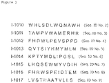
- the obtained I-1010 phage displayed peptide sequence (SEQ ID No. 2) had 6 clones having the same sequence in 15 clones that were examined in the three rounds (40%). Furthermore, two clones of each of I-1012 phage (SEQ ID No. 4) and 1-1015 phage (SEQ ID No. 7) were obtained (13.3%). Only one clone of each of 1-1011 phage (SEQ ID No. 3), I-1013 phage (SEQ ID No. 5), 1-1014 phage (SEQ ID No. 6), 1-1016 phage (SEQ ID No. 8), and I-1017 phage (SEQ ID No. 9) was obtained.
- Example 3 (Test of phage binding property (Condition of formulation of protein solid phase-1))
- a rhEGFR that is a target protein, rhIgG 1 that is a rhEGFR-labeled Fc moiety, and BSA as a standard protein were added to a 96-well microplate to be 1 ⁇ g/well, and stood still at 4°C overnight to be thus formed into a solid phase.
- a protein solution was removed, 300 ⁇ l of a blocking buffer was added and the mixture was incubated at 37°C for 1 hour to thus block the wells. After removal of the blocking buffer, the wells were washed with 200 ⁇ l of 0.5% TBST being the washing solution three times and thereto was finally added 100 ⁇ l of 0.5% TBST.
- An amplification phage solution (I-1010, I-1012 and 1-1015 phages and M13KE (peptide non-displayed phage)) was added to the above described wells to have 1 ⁇ 10 10 PFU and mixed by pipetting. In the reaction, the mixture was gently shaken at room temperature for 1 hour. The reaction solution was removed and the wells were washed with 200 ⁇ l of 0.5% TBST ten times, 100 ⁇ l of 0.2 M glycine-HCl (pH 2.2) was added to the wells and stirred by pipetting, thereafter gently shaking at room temperature for 10 minutes.
- the eluate was collected to a microtube from the wells and added with 15 ⁇ l of 1 M Tris-HCl (pH 9.1) to be neutralized and a target binding phage solution was thus obtained.
- the binding ability of the collected phage was examined by a measurement of a titer.
- Fig. 4 shows a ratio of an input titer/output titer of the binding property test by a solid phase formulation method using I-1010, I-1012 and I-1015 phages.
- a rhEGFR that is a target protein, a recombinant human ErbB2 (rhErbB2, PROSPEC) as a HER2 protein belonging to the ErbB family same as an EGFR, a recombinant human IGF-1R, CF (rhIGF-1R, R&D Systems) as an IGF-1R protein that is one of extracellular proteins were added to a 96-well microplate to be 1 ⁇ g/well, and stood still at 4°C overnight to be thus formed into a solid phase. A protein solution was removed, 300 ⁇ l of a blocking buffer was added and the mixture was incubated at 37°C for 1 hour to thus block the wells.
- the reaction solution was removed, the wells were washed with 200 ⁇ l of 0.5% TBST ten times, and 100 ⁇ l of 0.2 M glycine-HCl (pH 2.2) was then added to the wells and stirred by pipetting, thereafter gently shaking at room temperature for 10 minutes.
- the eluate was collected to a microtube from the wells and added with 15 ⁇ l of 1 M Tris-HCl (pH 9.1) to be neutralized and a target binding phage solution was thus obtained.
- the binding ability of the collected phage was examined by a measurement of a titer.
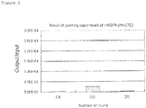
- Fig. 5 shows a ratio of an input titer/output titer of the binding property test by a solid phase formulation method using I-1010, 1-1012 and I-1015 phages. A result of the M13KE phage as comparison is also shown in Fig. 5 .
- a rhEGFR that is a target protein was added to a 96-well microplate to be 1 ⁇ g/well, and stood still at 4°C overnight to be thus formed into a solid phase.
- a protein solution was removed, 300 ⁇ l of a blocking buffer was added and the mixture was incubated at 37°C for 1 hour to thus block the wells. After removal of the blocking buffer, the wells were washed three times with 200 ⁇ l of 0.5% TBST washing solution, and finally 100 ⁇ l of 0.5% TBST was added thereto.
- An amplification I-1010 phage solution was added to the above described wells to have 1 ⁇ 10 10 PFU and, subsequently, a synthetic peptide Pep055 (purity 89%, HPLC grade, Hokkaido System Science Co., Ltd.) which was obtained by artificially synthesizing the I-1010 phage displayed peptide (SEQ ID No. 2) was added to have each concentration and a synthetic peptide Pep018 (GAASRTYLHELI: SEQ ID No. 10) (purity 90% ⁇ , HPLC grade, AnyGen, Korea) which is irrelevant to binding to a rhEGFR was added to have each concentration by pipetting. A reaction only of the I-1010 phage without adding both of the peptides was employed as a control.
- a synthetic peptide (SEQ ID No. 4) Pep056 (purity 86.7%, HPLC grade, Hokkaido System Science Co., Ltd.) of the I-1012 phage displayed peptide, a synthetic peptide (SEQ ID No. 7) Pep057 (purity 83.7% ⁇ , HPLC grade, Hokkaido System Science Co., Ltd.) of the 1-1015 phage displayed peptide were also used.
- the mixture was gently shaken at room temperature for 1 hour. The reaction solution was removed and the wells were washed with 200 ⁇ l of 0. 5% TBST ten times.
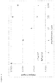
- Fig. 6 shows changes in values of ratios of input titers /output titers of an I-1010 phage and Pep055 in the competition test and a result of the I-1010 phage and Pep018.
- Pep018 is not involved with binding of the I-1010 phage to an EGFR and therefore decrease of a ratio of an input titer /output titer of the I-1010 phage was not observed even at a high concentration. While, it was revealed that a synthetic peptide Pep055 having a peptide sequence displayed by the I-1010 phage inhibited binding of the I-1010 phage to EGFR concentration-dependently. It canbe considered from this result that even though the I-1010 phage displayed peptide is single, the peptide binds to the same site where the I-1010 phage binds to a rhEGFR.
- a phage obtained by substituting an amino acid in a peptide displayed site of an M13KE phage with alanine was prepared using a KOD-Plus-Mutagenesis kit (SMK-101, TOYOBO) .
- An oligonucleotide primer having a nucleotide sequence of the displayed peptide sequence, which contains desired mutation, was entrusted with synthesis (NIHON GENE RESEARCH LABORATORIES Inc.).
- the synthesized primer was desalinated oligonucleotide that was purified by HPLC.
- the primer synthesized in this time had a length of 18 to 34 bp and was designed to have a GC content of 50 to 60%.
- phosphorylation can be conducted at the same time as self ligation of a PCR product, and thus, phosphorylation of the primer was not carried out.
- Template plasmid DNA was purified from the I-1010, I-1012, and I-1015 phage infected ER2738 bacteria using a QIAGEN Plasmid kit (QIAGEN).
- PCR was carried out in the condition of [94°C, 2 minutes] ⁇ ⁇ [98°C, 10 seconds] ⁇ [68°C, 7.5 minutes] ⁇ ⁇ 8 cycles ⁇ [4°C, Hold] using the template plasmid DNA, the primer, dNTPs, and KOD-plus-.
- a thermal cycler (PCR Thermal Cycler Dice, TAKARA) was employed in the PCR reaction.
- the PCR reaction conditions were designed based on an amplification size.
- Digestion of the template plasmid was conducted by a DpnI treatment, and self ligation of a PCR product was carried out by a T4 polynucleotide kinase treatment.
- XL-1 Blue competent cells were dissolved on ice. 15 ⁇ L of a solution treated by self-ligation was added to 60 ⁇ L of the XL-1 Blue competent cells and mixed. The mixture was incubated on ice for 30 minutes, then incubated on a heated block at 42°C for 90 seconds and incubated on ice for 2 minutes. 1 ⁇ L of the above described XL-Blue bacteria was added to 100 ⁇ L of an O/N culture of ER2738 bacteria in a 15 mL-conical tube and mixed. 74 ⁇ L of the XL-1 Blue bacteria were added in a 15 mL-conical tube. 4 mL of a top ager was added to each of the above described samples and mixed by a vortex mixer. The above described Escherichia coli sample was inoculated on an LB/IPTG/Xgal plate and inoculated at 37°C for 16 hours.
- alanine substituted phages having SEQ ID Nos. 11 to 21 shown in Fig. 9 were obtained.
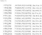
- alanine substituted phages having SEQ ID Nos. 22 to 33 shown in Fig. 10 were obtained, and for the 1-1015 phage, alanine substituted phages having SEQ ID Nos. 34 to 45 shown in Fig. 11 were obtained.
- a rhEGFR that is a target protein was added to a 96-well microplate to be 1 ⁇ g/well, and stood still at 4°C overnight to be thus formed into a solid phase.
- a protein solution was removed, 300 ⁇ l of a blocking buffer was added and the mixture was incubated at 37°C for 1 hour to thus block the wells. After removal of the blocking buffer, the wells were washed three times with 200 ⁇ l of 0.5% TBST washing solution, and finally 100 ⁇ l of 0.5% TBST was added thereto.
- An amplification phage solution (I-1010, I-1012 and I-1015 phages or the alanine substituted phage prepared in Example 6) was added to the above described wells to have 1 ⁇ 10 10 PFU and mixed by pipetting. In the reaction, the mixture was gently shaken at room temperature for 1 hour. The reaction solution was removed and the wells were washed with 200 ⁇ l of 0.5% TBST ten times, 100 ⁇ l of 0.2 M glycine-HCl (pH 2.2) was then added to the wells and stirred by pipetting, thereafter gently shaking at room temperature for 10 minutes.
- the eluate was collected to a microtube from the wells and added with 15 ⁇ l of 1 M Tris-HCl (pH 9.1) to be neutralized and a target binding phage solution was thus obtained.
- the binding ability of the collected phage was examined by a measurement of a titer.
- phages having decreased binding properties as compared to the I-1010 phage existed and were phages in which the 1st, 7th and 11th w (Trp), the 2th and 12th H (His), the 3rd L (Leu), and the 5th D (Asp) were substituted with alanine (Ala).
- binding properties of phages obtained by substituting the 1st and 7th W (Trp), the 2th H (His) and the 3rd L (Leu) in a peptide displayed by the I-1010 phage with alanine (Ala) were significantly reduced.
- amino acids which are assumed to contribute to binding of the I-1010 phage to EGFR were the 1st, 7nd and 11th W, the 2nd and 12nd H, the 3rd L and the 5th D.
- amino acids that are considered to particularly contribute to binding to a rhEGFR were the 1st and 7th W, the 2nd H and the 3rd L.
- amino acids that are considered to particularly contribute to binding to a rhEGFR were the 1st F, the 2nd H and the 4th W.
- phages having decreased binding properties as compared to the 1-1015 phage existed and were phages in which the 1st L (Leu), the 2nd H (His), the 4th S (Ser), the 6th W (Trp) and the 7 th M (Met) were substituted with alanine.
- binding properties of phages obtained by substituting the 4th S and the 6th W in a peptide displayed by the I-1015 phage with alanine were significantly reduced.
- amino acids which are assumed to contribute to binding of the I-1015 phage to a rhEGFR were the 1st L, the 2nd H, the 4th S, the 6th W and the 7th M.
- amino acids that are considered to particularly contribute to binding to a rhEGFR were the 4th S and the 6th W.
- a mutant phage obtained by substituting an amino acid in a peptide displayed moiety of an M13KE phage with another amino acid was prepared in the same procedure as in Example 6.
- a procedure of an operation of alanine scan is referred.
- a rhEGFR that is a target protein was added to a 96-well microplate to be 1 ⁇ g/well, and stood still at 4°C overnight to be thus formed into a solid phase.
- a protein solution was removed, 300 ⁇ l of a blocking buffer (5 mg/mL BSA/TBS) was added and the mixture was incubated at 37°C for 1 hour to thus block the wells.
- the wells were washed with 200 ⁇ l of 0.5% TBST (0.5% Tween20/TBS) being the washing solution three times and thereto was finally added 100 ⁇ l of 0.5% TBST.
- An amplification phage solution was added to the above described wells to have 1 ⁇ 10 10 PFU and mixed by pipetting.
- the mixture was gently shaken at room temperature for 1 hour.
- the reaction solution was removed and the wells were washed with 200 ⁇ l of 0.5% TBST ten times, 100 ⁇ l of 0.2 M glycine-HCl (pH 2.2) was then added to the wells and stirred by pipetting, thereafter gently shaking at room temperature for 10 minutes.
- the eluate was collected to a microtube from the wells and added with 15 ⁇ l of 1 M Tris-HCl (pH 9.1) to be neutralized and a target binding phage solution was thus obtained.
- the binding ability of the collected phage was examined by a measurement of a titer.
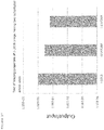
- Fig. 16 shows changes in values of ratios of input titers to output titers in a binding property test of a phage having one substituted amino acid.
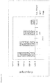
- Fig. 17 shows changes in values of ratios of input titers to output titers in the binding property test of a phage having two substituted amino acids.
- Phages obtained by substituting two amino acids were a phage in which the 1st W was substituted with F and the 8th Q was substituted with A (W1F/Q8A, SEQ ID No. 52), a phage in which 3rd L was substituted with M and the 8th Q was substituted with A (L3M/Q8A, SEQ ID No. 53), and both phages resulted in showing equivalent to about 1/10 binding properties as compared to the binding property of the I-1010 phage.
- EGFR expression amounts were evaluated by western blotting using human epidermal cancer derived A-431 cells (DS PHARMA BIOMEDICAL CO. , LTD.) and human breast cancer derived MCF7 cells (DS PHARMA BIOMEDICAL CO., LTD.).
- Example 10 An A-431 cell line in which expression of EGFR protein was observed in Example 10 was used as the target and a binding property test of the I-1010 phage was carried out.
- A-431 cells were inoculated in a 6-well culture plate at 40 ⁇ 10 4 cells/well and cultured at 37°C in the condition of 5%CO 2 overnight. The plate was incubated at 4°C for 30 minutes and a medium in each well was removed, and treated by washing with 1 mL of 1%BSA/PBS twice.
- the I-1010 phage and the M13KE phage were diluted with 1 mL of a 10% FBS medium (growth medium) to have a titer of 1 ⁇ 10 10 PFU and added to each well, and incubated at 4°C for 60 minutes. After the reaction, the reaction solution was removed and washed with 1 mL of 1% BSA/PBS ten times to remove non-binding phages.
- Fig. 19 shows a ratio of an input titer to an output titer of each phage considering the number of collected cells.
- the titer ratio of the I-1010 phage was 45 times as high as that of the M13KE phage.
- the I-1010 phage showed binding properties not only to a genetically modified EGFR protein but also to EGFR expressed by cells.
- A-431 cells were inoculated in a glass bottom dish to be 20 ⁇ 10 4 cells/dish and MCF7 cells were inoculated in a glass bottom dish to be 75 ⁇ 10 4 cells/dish, and cultured at 37°C in the condition of CO 2 for 24 hours. After culturing each kind of the cells for a specified time, EGF was added to have a concentration of 100 ng/mL and the mixture was cultured for 6 days. Thereafter, the culture solution in the dish was removed and washed with 1000 ⁇ L of a growth medium at 37°C ten times.
- An aminocaproic acid linker was connected to an N terminal of a peptide and the peptide in which FITC was labeled in the opposite side of the connected linker (Pep055-F, Pep032-F (YLPLWRLSESHM (peptide irrelevant to binding to EGFR) : SEQ ID No. 54)) (purity 80.6%, HPLC grade, AnyGen, Korea) was prepared to be 100 ⁇ M in a growth medium, and 1 mL of the medium was added and incubated at 37°C for 1 hour. A staining fluid was removed and washed with 1 mL of a growth medium at 37°C three times and thereto was finally added 1 mL of the medium. For observation, fluorescent observation was carried out by irradiating an exciting light laser with a wavelength of 488 nm using a confocal microscope (Leica SP2) .
- Fig. 20 shows stainability of A-431 cells of Pep055-F observed by a confocal microscope. Staining of the cytoplasm of the A-431 cells was observed. The nucleus was not stained.
- Fig. 21 shows stainability of A-431 cells of Pep032-F observed by a confocal microscope.
- the control peptide scarcely stained the A-431 cells.
- Fig. 22 shows stainability of MCF7 cells of Pep055-F observed by a confocal microscope
- Fig. 23 shows stainability of MCF7 cells of Pep032-F observed by a confocal microscope.
- Example 13 Competition test between recombinant human EGF and I-1010 phage
- a rhEGFR that was a target protein was added to a 96-well microplate to be 1 ⁇ g/well, and stood still at 4°C overnight to be thus formed into a solid phase.
- a protein solution was removed, 300 ⁇ l of a blocking buffer was added and the mixture was incubated at 37°C for 1 hour to thus block the wells. After removal of the blocking buffer, the wells were washed with 200 ⁇ l of 0.5% TBST being the washing solution three times.
- 100 ⁇ l of a recombinant human EGF (rhEGF, Invitrogen) which is a ligand protein of EGFR diluted with 0.5% TBST to have each concentration was added and the mixture was gently shaken at room temperature for 1 hour.
- I-1010 phage amplification phage solution was added to the above described wells to have 1 ⁇ 10 10 PFU and mixed by pipetting. In the reaction, the mixture was gently shaken at room temperature for 15 minutes . The reaction solution was removed and the wells were washed with 200 ⁇ L of 0.5% TBST ten times, and 100 ⁇ l of 0.2 M glycine-HCl (pH 2.2) was then added to the wells and stirred by pipetting, thereafter gently shaking at room temperature for 10 minutes. The eluate was collected to a microtube from the wells and added with 15 ⁇ l of 1 M Tris-HCl (pH 9.1) to be neutralized and a target binding phage solution was thus obtained. The binding ability of the collected phage was examined by a measurement of a titer.
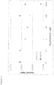
- Fig. 24 shows changes in values of ratios of input titers /output titers in a competition test of a rhEGF and an I-1010 phage.
- binding properties of an I-1010 phage to the rhEGFR decreased along with increasing the adding concentration.
- a rhEGF inhibits binding of an I-1010 phage to a rhEGFR from the fact that the binding properties of the I-1010 phage decrease concentration-depending to the adding concentration of the rhEGF. That is, an I-1010 phage is considered to be able to inhibit binding of EGF to EGFR and a peptide displayed by the I-1010 phage could be an inhibitor of EGF.
Landscapes
- Health & Medical Sciences (AREA)
- Life Sciences & Earth Sciences (AREA)
- Chemical & Material Sciences (AREA)
- Engineering & Computer Science (AREA)
- Immunology (AREA)
- General Health & Medical Sciences (AREA)
- Medicinal Chemistry (AREA)
- Molecular Biology (AREA)
- Organic Chemistry (AREA)
- Biochemistry (AREA)
- Proteomics, Peptides & Aminoacids (AREA)
- Urology & Nephrology (AREA)
- Hematology (AREA)
- Biomedical Technology (AREA)
- Biophysics (AREA)
- Genetics & Genomics (AREA)
- Cell Biology (AREA)
- Pharmacology & Pharmacy (AREA)
- Veterinary Medicine (AREA)
- Public Health (AREA)
- Animal Behavior & Ethology (AREA)
- Biotechnology (AREA)
- Microbiology (AREA)
- Pathology (AREA)
- General Physics & Mathematics (AREA)
- Analytical Chemistry (AREA)
- Physics & Mathematics (AREA)
- Food Science & Technology (AREA)
- Gastroenterology & Hepatology (AREA)
- Toxicology (AREA)
- Bioinformatics & Cheminformatics (AREA)
- Zoology (AREA)
- Epidemiology (AREA)
- Chemical Kinetics & Catalysis (AREA)
- Nuclear Medicine, Radiotherapy & Molecular Imaging (AREA)
- General Chemical & Material Sciences (AREA)
- Peptides Or Proteins (AREA)
- Medicines That Contain Protein Lipid Enzymes And Other Medicines (AREA)
- Oncology (AREA)
- Micro-Organisms Or Cultivation Processes Thereof (AREA)
Claims (10)
- Peptid, das eine Aminosäuresequenz enthält, die durch die nachstehend beschriebene Formel (1) dargestellt ist und aus 12 bis 50 Aminosäuren besteht:
X1-His-X2-X3-Asp-X4-X5-X6-X7-X8-Trp-His (1)
worin X1 Trp oder Phe darstellt;X2 Leu, Met, Ile oder Val darstellt;X3 Ser, Ala, Thr, Gly, Asn, Asp, Glu, Arg oder Lys darstellt;X4 Leu, Met, Ile, Val, Ala, Phe, Tyr, Trp, His oder Cys darstellt;X5 Trp, Phe oder Tyr darstellt;X6 Gln, Asn, Asp, Glu, Ala, Ser, Thr, Leu, Met, Lys oder Arg darstellt;X7 Asn, Gln, Asp, Glu, Ser, Thr, Ala, Gly, Lys oder Arg; undX8 Ala, Gly, Ser, Thr, Asn, Val oder Cys darstellt, wobei das Peptid spezifisch mit hoher Affinität an einen Epidermaiwachstumsfaktor-Rezeptor (EGFR) bindet. - Peptid nach Anspruch 1, wobei in der Formel (1) X1 Trp oder Phe darstellt, X2 Leu, Met, Ile oder Val darstellt, X3 Ser, Ala, Thr, Gly oder Asn darstellt, X4 Leu, Met, Ile, Val oder Ala darstellt, X5 Trp oder Phe darstellt, X6 Gln, Asn oder Ala darstellt, X7 Asn, Gln oder Ala darstellt, und X8 Ala, Gly, Ser oder Thr darstellt.
- Peptid nach Anspruch 1, wobei in der Formel (1) X1 Trp oder Phe darstellt, X2 Leu, Met, Ile oder Val darstellt, X3 Ser oder Ala darstellt, X4 Leu oder Ala darstellt, X5 Trp darstellt, X6 Gln oder Ala darstellt, X7 Asn oder Ala darstellt, und X8 Ala darstellt.
- Peptid nach einem der Ansprüche 1 bis 3, wobei die Zahl an Aminosäuren 30 oder kleiner ist.
- Pharmazeutisches Mittel, enthaltend das Peptid nach einem der Ansprüche 1 bis 4 oder ein ettiketiertes Produkt davon.
- Mittel zum Erfassen einer Krebszelle oder eines Krebsgewebes, enthaltend das Peptid nach einem der Ansprüche 1 bis 4 oder ein etikettiertes Produkt davon.
- Krebsdiagnosemittel, enthaltend das Peptid nach einem der Ansprüche 1 bis 4 oder ein etikettiertes Produkt davon.
- Krebs-Therapeutikum, enthaltend das Peptid nach einem der Ansprüche 1 bis 4 oder ein etikettiertes Produkt davon.
- Peptid nach einem der Ansprüche 1 bis 4 oder ein etikettiertes Produkt davon zur Verwendung in der Erfassung einer Krebszelle oder eines Krebsgewebes.
- Peptid nach einem der Ansprüche 1 bis 4 oder ein etikettiertes Produkt davon zur Verwendung in der Krebsdiagnose oder einer Krebsbehandlung.
Applications Claiming Priority (2)
| Application Number | Priority Date | Filing Date | Title |
|---|---|---|---|
| JP2012141700A JP6103832B2 (ja) | 2012-06-25 | 2012-06-25 | Egfr結合性ペプチド |
| PCT/JP2013/066762 WO2014002836A1 (ja) | 2012-06-25 | 2013-06-18 | Egfr結合性ペプチド |
Publications (3)
| Publication Number | Publication Date |
|---|---|
| EP2865686A1 EP2865686A1 (de) | 2015-04-29 |
| EP2865686A4 EP2865686A4 (de) | 2016-04-13 |
| EP2865686B1 true EP2865686B1 (de) | 2018-08-08 |
Family
ID=49782995
Family Applications (1)
| Application Number | Title | Priority Date | Filing Date |
|---|---|---|---|
| EP13809117.8A Active EP2865686B1 (de) | 2012-06-25 | 2013-06-18 | Egfr-bindendes peptid |
Country Status (5)
| Country | Link |
|---|---|
| US (1) | US9822148B2 (de) |
| EP (1) | EP2865686B1 (de) |
| JP (1) | JP6103832B2 (de) |
| CN (1) | CN104379595B (de) |
| WO (1) | WO2014002836A1 (de) |
Families Citing this family (5)
| Publication number | Priority date | Publication date | Assignee | Title |
|---|---|---|---|---|
| JP2909218B2 (ja) | 1995-04-13 | 1999-06-23 | 株式会社アドバンテスト | 半導体試験装置用周期発生器 |
| US11739163B2 (en) | 2016-09-29 | 2023-08-29 | Aebi Ltd. | Therapeutic multi-targeting constructs and uses thereof |
| CN106699890B (zh) * | 2016-10-31 | 2021-05-07 | 上海大学 | 基于金纳米粒子的靶向表皮生长因子受体egfr的人工抗体及其制备方法 |
| CN108101963A (zh) * | 2018-01-30 | 2018-06-01 | 广西天昌投资有限公司 | 一种寡糖肽及其制备方法 |
| CN109180787B (zh) * | 2018-08-14 | 2021-08-03 | 江苏大学 | 靶向egfr抑制egf促肿瘤细胞增殖的多肽 |
Family Cites Families (9)
| Publication number | Priority date | Publication date | Assignee | Title |
|---|---|---|---|---|
| JPH11507535A (ja) | 1995-06-07 | 1999-07-06 | イムクローン システムズ インコーポレイテッド | 腫瘍の成長を抑制する抗体および抗体フラグメント類 |
| CA2722378C (en) | 1996-12-03 | 2015-02-03 | Amgen Fremont Inc. | Human antibodies that bind tnf.alpha. |
| US7767792B2 (en) | 2004-02-20 | 2010-08-03 | Ludwig Institute For Cancer Research Ltd. | Antibodies to EGF receptor epitope peptides |
| CA2560305C (en) | 2004-03-19 | 2016-07-05 | Imclone Systems Incorporated | Human anti-epidermal growth factor receptor antibody |
| WO2008079973A2 (en) | 2006-12-21 | 2008-07-03 | Centocor, Inc. | Egfr binding peptides and uses thereof |
| CA2697896C (en) | 2007-09-18 | 2016-06-28 | Green Peptide Co., Ltd. | Ctl inducer composition |
| CN102083850B (zh) * | 2008-04-21 | 2015-08-12 | 加利福尼亚大学董事会 | 选择性高亲和力多齿配体及其制备方法 |
| JP2010154842A (ja) | 2008-12-03 | 2010-07-15 | Koji Kawakami | Egfrを標的にした新規抗がんキメラペプチド |
| US9006406B2 (en) | 2008-12-22 | 2015-04-14 | Alexander Levitzki | EGFR-homing double-stranded RNA vector for systemic cancer treatment |
-
2012
- 2012-06-25 JP JP2012141700A patent/JP6103832B2/ja active Active
-
2013
- 2013-06-18 EP EP13809117.8A patent/EP2865686B1/de active Active
- 2013-06-18 CN CN201380031904.4A patent/CN104379595B/zh active Active
- 2013-06-18 WO PCT/JP2013/066762 patent/WO2014002836A1/ja not_active Ceased
- 2013-06-18 US US14/410,329 patent/US9822148B2/en active Active
Non-Patent Citations (1)
| Title |
|---|
| None * |
Also Published As
| Publication number | Publication date |
|---|---|
| WO2014002836A1 (ja) | 2014-01-03 |
| CN104379595B (zh) | 2018-04-27 |
| EP2865686A4 (de) | 2016-04-13 |
| JP2014005235A (ja) | 2014-01-16 |
| US20150337012A1 (en) | 2015-11-26 |
| JP6103832B2 (ja) | 2017-03-29 |
| CN104379595A (zh) | 2015-02-25 |
| US9822148B2 (en) | 2017-11-21 |
| EP2865686A1 (de) | 2015-04-29 |
Similar Documents
| Publication | Publication Date | Title |
|---|---|---|
| Kam et al. | Detection of a long non-coding RNA (CCAT1) in living cells and human adenocarcinoma of colon tissues using FIT–PNA molecular beacons | |
| CN105039333B (zh) | 肝癌靶向肽及其应用 | |
| CN107001417B (zh) | 靶向肽及其使用方法 | |
| CN104262460B (zh) | 一种靶向人乳腺癌细胞的多肽及其应用 | |
| EP2865686B1 (de) | Egfr-bindendes peptid | |
| CN104130315B (zh) | 一种特异靶向her2蛋白的多肽 | |
| CN110511269B (zh) | 一种与muc4蛋白特异性结合的靶向多肽zp-16在制备药物中的用途 | |
| CN107073065B (zh) | 用于发育异常、早期癌症和癌症的检测和靶向的肽试剂和方法 | |
| CN112546244A (zh) | 成纤维细胞生长因子受体2特异性肽试剂和方法 | |
| CN110330551B (zh) | 一种胰腺癌特异结合肽及其制备方法与用途 | |
| CN115397513A (zh) | 改进的抗衰老化合物及其用途 | |
| Ma et al. | Screening of a specific peptide binding to esophageal squamous carcinoma cells from phage displayed peptide library | |
| CN108912212B (zh) | 一种与cd105特异性结合的多肽及其应用 | |
| Pallerla et al. | Design of cyclic and d‐amino acids containing peptidomimetics for inhibition of protein‐protein interactions of HER2‐HER3 | |
| CN112409447B (zh) | 一种靶向识别膜联蛋白a2的亲和短肽及其制备方法与用途 | |
| WO2009028838A1 (en) | Peptide aptamers against tenascin c | |
| CN111018951B (zh) | 一种靶向三阴性乳腺癌细胞的多肽及其应用 | |
| CN116194462B (zh) | 选择性结合癌细胞来源外泌体的肽及其用途 | |
| JP6155255B2 (ja) | Fzd10結合性ペプチド | |
| JP2014156401A (ja) | Igf−1r結合性ペプチド | |
| CA2367256A1 (en) | Surface localized colligin/hsp47 in carcinoma cells | |
| KR102659285B1 (ko) | 암세포 유래 엑소좀에 선택적으로 결합하는 펩타이드 및 이의 용도 | |
| CN110746489B (zh) | 一种特异性靶向三阴乳腺癌的多肽及其应用 | |
| US20240352068A1 (en) | Vimentin targeted peptoids for early diagnosis and treatment of cancer | |
| CN110317244B (zh) | 一种rage拮抗多肽及其应用 |
Legal Events
| Date | Code | Title | Description |
|---|---|---|---|
| PUAI | Public reference made under article 153(3) epc to a published international application that has entered the european phase |
Free format text: ORIGINAL CODE: 0009012 |
|
| 17P | Request for examination filed |
Effective date: 20150126 |
|
| AK | Designated contracting states |
Kind code of ref document: A1 Designated state(s): AL AT BE BG CH CY CZ DE DK EE ES FI FR GB GR HR HU IE IS IT LI LT LU LV MC MK MT NL NO PL PT RO RS SE SI SK SM TR |
|
| AX | Request for extension of the european patent |
Extension state: BA ME |
|
| DAX | Request for extension of the european patent (deleted) | ||
| RIC1 | Information provided on ipc code assigned before grant |
Ipc: C07K 14/00 20060101ALI20151117BHEP Ipc: A61P 35/00 20060101ALI20151117BHEP Ipc: G01N 33/574 20060101ALI20151117BHEP Ipc: C07K 7/08 20060101AFI20151117BHEP Ipc: A61K 38/00 20060101ALI20151117BHEP Ipc: C12N 15/09 20060101ALI20151117BHEP |
|
| RA4 | Supplementary search report drawn up and despatched (corrected) |
Effective date: 20160310 |
|
| RIC1 | Information provided on ipc code assigned before grant |
Ipc: C07K 14/00 20060101ALI20160304BHEP Ipc: G01N 33/574 20060101ALI20160304BHEP Ipc: C07K 7/08 20060101AFI20160304BHEP Ipc: C12N 15/09 20060101ALI20160304BHEP Ipc: A61K 38/00 20060101ALI20160304BHEP Ipc: A61P 35/00 20060101ALI20160304BHEP |
|
| RAP1 | Party data changed (applicant data changed or rights of an application transferred) |
Owner name: HOYA CORPORATION |
|
| GRAP | Despatch of communication of intention to grant a patent |
Free format text: ORIGINAL CODE: EPIDOSNIGR1 |
|
| STAA | Information on the status of an ep patent application or granted ep patent |
Free format text: STATUS: GRANT OF PATENT IS INTENDED |
|
| INTG | Intention to grant announced |
Effective date: 20180228 |
|
| RIN1 | Information on inventor provided before grant (corrected) |
Inventor name: YAMAGATA, MIZUE Inventor name: SHIONO, TOSHITAKA Inventor name: IIMORI, YUSUKE |
|
| GRAS | Grant fee paid |
Free format text: ORIGINAL CODE: EPIDOSNIGR3 |
|
| GRAA | (expected) grant |
Free format text: ORIGINAL CODE: 0009210 |
|
| STAA | Information on the status of an ep patent application or granted ep patent |
Free format text: STATUS: THE PATENT HAS BEEN GRANTED |
|
| AK | Designated contracting states |
Kind code of ref document: B1 Designated state(s): AL AT BE BG CH CY CZ DE DK EE ES FI FR GB GR HR HU IE IS IT LI LT LU LV MC MK MT NL NO PL PT RO RS SE SI SK SM TR |
|
| REG | Reference to a national code |
Ref country code: GB Ref legal event code: FG4D |
|
| REG | Reference to a national code |
Ref country code: CH Ref legal event code: EP Ref country code: AT Ref legal event code: REF Ref document number: 1026857 Country of ref document: AT Kind code of ref document: T Effective date: 20180815 |
|
| REG | Reference to a national code |
Ref country code: IE Ref legal event code: FG4D |
|
| REG | Reference to a national code |
Ref country code: DE Ref legal event code: R096 Ref document number: 602013041762 Country of ref document: DE |
|
| REG | Reference to a national code |
Ref country code: NL Ref legal event code: MP Effective date: 20180808 |
|
| REG | Reference to a national code |
Ref country code: LT Ref legal event code: MG4D |
|
| REG | Reference to a national code |
Ref country code: AT Ref legal event code: MK05 Ref document number: 1026857 Country of ref document: AT Kind code of ref document: T Effective date: 20180808 |
|
| PG25 | Lapsed in a contracting state [announced via postgrant information from national office to epo] |
Ref country code: GR Free format text: LAPSE BECAUSE OF FAILURE TO SUBMIT A TRANSLATION OF THE DESCRIPTION OR TO PAY THE FEE WITHIN THE PRESCRIBED TIME-LIMIT Effective date: 20181109 Ref country code: NO Free format text: LAPSE BECAUSE OF FAILURE TO SUBMIT A TRANSLATION OF THE DESCRIPTION OR TO PAY THE FEE WITHIN THE PRESCRIBED TIME-LIMIT Effective date: 20181108 Ref country code: AT Free format text: LAPSE BECAUSE OF FAILURE TO SUBMIT A TRANSLATION OF THE DESCRIPTION OR TO PAY THE FEE WITHIN THE PRESCRIBED TIME-LIMIT Effective date: 20180808 Ref country code: NL Free format text: LAPSE BECAUSE OF FAILURE TO SUBMIT A TRANSLATION OF THE DESCRIPTION OR TO PAY THE FEE WITHIN THE PRESCRIBED TIME-LIMIT Effective date: 20180808 Ref country code: SE Free format text: LAPSE BECAUSE OF FAILURE TO SUBMIT A TRANSLATION OF THE DESCRIPTION OR TO PAY THE FEE WITHIN THE PRESCRIBED TIME-LIMIT Effective date: 20180808 Ref country code: BG Free format text: LAPSE BECAUSE OF FAILURE TO SUBMIT A TRANSLATION OF THE DESCRIPTION OR TO PAY THE FEE WITHIN THE PRESCRIBED TIME-LIMIT Effective date: 20181108 Ref country code: IS Free format text: LAPSE BECAUSE OF FAILURE TO SUBMIT A TRANSLATION OF THE DESCRIPTION OR TO PAY THE FEE WITHIN THE PRESCRIBED TIME-LIMIT Effective date: 20181208 Ref country code: RS Free format text: LAPSE BECAUSE OF FAILURE TO SUBMIT A TRANSLATION OF THE DESCRIPTION OR TO PAY THE FEE WITHIN THE PRESCRIBED TIME-LIMIT Effective date: 20180808 Ref country code: FI Free format text: LAPSE BECAUSE OF FAILURE TO SUBMIT A TRANSLATION OF THE DESCRIPTION OR TO PAY THE FEE WITHIN THE PRESCRIBED TIME-LIMIT Effective date: 20180808 Ref country code: PL Free format text: LAPSE BECAUSE OF FAILURE TO SUBMIT A TRANSLATION OF THE DESCRIPTION OR TO PAY THE FEE WITHIN THE PRESCRIBED TIME-LIMIT Effective date: 20180808 Ref country code: LT Free format text: LAPSE BECAUSE OF FAILURE TO SUBMIT A TRANSLATION OF THE DESCRIPTION OR TO PAY THE FEE WITHIN THE PRESCRIBED TIME-LIMIT Effective date: 20180808 |
|
| PG25 | Lapsed in a contracting state [announced via postgrant information from national office to epo] |
Ref country code: HR Free format text: LAPSE BECAUSE OF FAILURE TO SUBMIT A TRANSLATION OF THE DESCRIPTION OR TO PAY THE FEE WITHIN THE PRESCRIBED TIME-LIMIT Effective date: 20180808 Ref country code: LV Free format text: LAPSE BECAUSE OF FAILURE TO SUBMIT A TRANSLATION OF THE DESCRIPTION OR TO PAY THE FEE WITHIN THE PRESCRIBED TIME-LIMIT Effective date: 20180808 Ref country code: AL Free format text: LAPSE BECAUSE OF FAILURE TO SUBMIT A TRANSLATION OF THE DESCRIPTION OR TO PAY THE FEE WITHIN THE PRESCRIBED TIME-LIMIT Effective date: 20180808 |
|
| PG25 | Lapsed in a contracting state [announced via postgrant information from national office to epo] |
Ref country code: RO Free format text: LAPSE BECAUSE OF FAILURE TO SUBMIT A TRANSLATION OF THE DESCRIPTION OR TO PAY THE FEE WITHIN THE PRESCRIBED TIME-LIMIT Effective date: 20180808 Ref country code: CZ Free format text: LAPSE BECAUSE OF FAILURE TO SUBMIT A TRANSLATION OF THE DESCRIPTION OR TO PAY THE FEE WITHIN THE PRESCRIBED TIME-LIMIT Effective date: 20180808 Ref country code: ES Free format text: LAPSE BECAUSE OF FAILURE TO SUBMIT A TRANSLATION OF THE DESCRIPTION OR TO PAY THE FEE WITHIN THE PRESCRIBED TIME-LIMIT Effective date: 20180808 Ref country code: IT Free format text: LAPSE BECAUSE OF FAILURE TO SUBMIT A TRANSLATION OF THE DESCRIPTION OR TO PAY THE FEE WITHIN THE PRESCRIBED TIME-LIMIT Effective date: 20180808 |
|
| REG | Reference to a national code |
Ref country code: DE Ref legal event code: R097 Ref document number: 602013041762 Country of ref document: DE |
|
| PG25 | Lapsed in a contracting state [announced via postgrant information from national office to epo] |
Ref country code: SK Free format text: LAPSE BECAUSE OF FAILURE TO SUBMIT A TRANSLATION OF THE DESCRIPTION OR TO PAY THE FEE WITHIN THE PRESCRIBED TIME-LIMIT Effective date: 20180808 Ref country code: SM Free format text: LAPSE BECAUSE OF FAILURE TO SUBMIT A TRANSLATION OF THE DESCRIPTION OR TO PAY THE FEE WITHIN THE PRESCRIBED TIME-LIMIT Effective date: 20180808 Ref country code: DK Free format text: LAPSE BECAUSE OF FAILURE TO SUBMIT A TRANSLATION OF THE DESCRIPTION OR TO PAY THE FEE WITHIN THE PRESCRIBED TIME-LIMIT Effective date: 20180808 |
|
| PLBE | No opposition filed within time limit |
Free format text: ORIGINAL CODE: 0009261 |
|
| STAA | Information on the status of an ep patent application or granted ep patent |
Free format text: STATUS: NO OPPOSITION FILED WITHIN TIME LIMIT |
|
| 26N | No opposition filed |
Effective date: 20190509 |
|
| PG25 | Lapsed in a contracting state [announced via postgrant information from national office to epo] |
Ref country code: SI Free format text: LAPSE BECAUSE OF FAILURE TO SUBMIT A TRANSLATION OF THE DESCRIPTION OR TO PAY THE FEE WITHIN THE PRESCRIBED TIME-LIMIT Effective date: 20180808 Ref country code: EE Free format text: LAPSE BECAUSE OF FAILURE TO SUBMIT A TRANSLATION OF THE DESCRIPTION OR TO PAY THE FEE WITHIN THE PRESCRIBED TIME-LIMIT Effective date: 20180808 |
|
| PG25 | Lapsed in a contracting state [announced via postgrant information from national office to epo] |
Ref country code: MC Free format text: LAPSE BECAUSE OF FAILURE TO SUBMIT A TRANSLATION OF THE DESCRIPTION OR TO PAY THE FEE WITHIN THE PRESCRIBED TIME-LIMIT Effective date: 20180808 |
|
| REG | Reference to a national code |
Ref country code: CH Ref legal event code: PL |
|
| GBPC | Gb: european patent ceased through non-payment of renewal fee |
Effective date: 20190618 |
|
| REG | Reference to a national code |
Ref country code: BE Ref legal event code: MM Effective date: 20190630 |
|
| PG25 | Lapsed in a contracting state [announced via postgrant information from national office to epo] |
Ref country code: TR Free format text: LAPSE BECAUSE OF FAILURE TO SUBMIT A TRANSLATION OF THE DESCRIPTION OR TO PAY THE FEE WITHIN THE PRESCRIBED TIME-LIMIT Effective date: 20180808 |
|
| PG25 | Lapsed in a contracting state [announced via postgrant information from national office to epo] |
Ref country code: IE Free format text: LAPSE BECAUSE OF NON-PAYMENT OF DUE FEES Effective date: 20190618 Ref country code: GB Free format text: LAPSE BECAUSE OF NON-PAYMENT OF DUE FEES Effective date: 20190618 |
|
| PG25 | Lapsed in a contracting state [announced via postgrant information from national office to epo] |
Ref country code: LU Free format text: LAPSE BECAUSE OF NON-PAYMENT OF DUE FEES Effective date: 20190618 Ref country code: LI Free format text: LAPSE BECAUSE OF NON-PAYMENT OF DUE FEES Effective date: 20190630 Ref country code: CH Free format text: LAPSE BECAUSE OF NON-PAYMENT OF DUE FEES Effective date: 20190630 Ref country code: BE Free format text: LAPSE BECAUSE OF NON-PAYMENT OF DUE FEES Effective date: 20190630 |
|
| PG25 | Lapsed in a contracting state [announced via postgrant information from national office to epo] |
Ref country code: PT Free format text: LAPSE BECAUSE OF FAILURE TO SUBMIT A TRANSLATION OF THE DESCRIPTION OR TO PAY THE FEE WITHIN THE PRESCRIBED TIME-LIMIT Effective date: 20181208 Ref country code: FR Free format text: LAPSE BECAUSE OF NON-PAYMENT OF DUE FEES Effective date: 20190630 |
|
| PG25 | Lapsed in a contracting state [announced via postgrant information from national office to epo] |
Ref country code: CY Free format text: LAPSE BECAUSE OF FAILURE TO SUBMIT A TRANSLATION OF THE DESCRIPTION OR TO PAY THE FEE WITHIN THE PRESCRIBED TIME-LIMIT Effective date: 20180808 |
|
| PG25 | Lapsed in a contracting state [announced via postgrant information from national office to epo] |
Ref country code: MT Free format text: LAPSE BECAUSE OF FAILURE TO SUBMIT A TRANSLATION OF THE DESCRIPTION OR TO PAY THE FEE WITHIN THE PRESCRIBED TIME-LIMIT Effective date: 20180808 Ref country code: HU Free format text: LAPSE BECAUSE OF FAILURE TO SUBMIT A TRANSLATION OF THE DESCRIPTION OR TO PAY THE FEE WITHIN THE PRESCRIBED TIME-LIMIT; INVALID AB INITIO Effective date: 20130618 |
|
| PG25 | Lapsed in a contracting state [announced via postgrant information from national office to epo] |
Ref country code: MK Free format text: LAPSE BECAUSE OF FAILURE TO SUBMIT A TRANSLATION OF THE DESCRIPTION OR TO PAY THE FEE WITHIN THE PRESCRIBED TIME-LIMIT Effective date: 20180808 |
|
| PGFP | Annual fee paid to national office [announced via postgrant information from national office to epo] |
Ref country code: DE Payment date: 20250429 Year of fee payment: 13 |