EP2850175B1 - Verbesserte verfahren für zellkulturen für adoptive zelltherapien - Google Patents
Verbesserte verfahren für zellkulturen für adoptive zelltherapien Download PDFInfo
- Publication number
- EP2850175B1 EP2850175B1 EP13791247.3A EP13791247A EP2850175B1 EP 2850175 B1 EP2850175 B1 EP 2850175B1 EP 13791247 A EP13791247 A EP 13791247A EP 2850175 B1 EP2850175 B1 EP 2850175B1
- Authority
- EP
- European Patent Office
- Prior art keywords
- cells
- cell
- culture
- medium
- growth surface
- Prior art date
- Legal status (The legal status is an assumption and is not a legal conclusion. Google has not performed a legal analysis and makes no representation as to the accuracy of the status listed.)
- Active
Links
Images


Classifications
-
- A—HUMAN NECESSITIES
- A61—MEDICAL OR VETERINARY SCIENCE; HYGIENE
- A61K—PREPARATIONS FOR MEDICAL, DENTAL OR TOILETRY PURPOSES
- A61K40/00—Cellular immunotherapy
- A61K40/10—Cellular immunotherapy characterised by the cell type used
- A61K40/11—T-cells, e.g. tumour infiltrating lymphocytes [TIL] or regulatory T [Treg] cells; Lymphokine-activated killer [LAK] cells
-
- A—HUMAN NECESSITIES
- A61—MEDICAL OR VETERINARY SCIENCE; HYGIENE
- A61K—PREPARATIONS FOR MEDICAL, DENTAL OR TOILETRY PURPOSES
- A61K40/00—Cellular immunotherapy
- A61K40/30—Cellular immunotherapy characterised by the recombinant expression of specific molecules in the cells of the immune system
- A61K40/31—Chimeric antigen receptors [CAR]
-
- A—HUMAN NECESSITIES
- A61—MEDICAL OR VETERINARY SCIENCE; HYGIENE
- A61K—PREPARATIONS FOR MEDICAL, DENTAL OR TOILETRY PURPOSES
- A61K40/00—Cellular immunotherapy
- A61K40/40—Cellular immunotherapy characterised by antigens that are targeted or presented by cells of the immune system
- A61K40/41—Vertebrate antigens
- A61K40/42—Cancer antigens
- A61K40/4256—Tumor associated carbohydrates
- A61K40/4257—Mucins, e.g. MUC-1
-
- A—HUMAN NECESSITIES
- A61—MEDICAL OR VETERINARY SCIENCE; HYGIENE
- A61K—PREPARATIONS FOR MEDICAL, DENTAL OR TOILETRY PURPOSES
- A61K40/00—Cellular immunotherapy
- A61K40/40—Cellular immunotherapy characterised by antigens that are targeted or presented by cells of the immune system
- A61K40/41—Vertebrate antigens
- A61K40/42—Cancer antigens
- A61K40/4274—Prostate associated antigens e.g. Prostate stem cell antigen [PSCA]; Prostate carcinoma tumor antigen [PCTA]; Prostatic acid phosphatase [PAP]; Prostate-specific G-protein-coupled receptor [PSGR]
-
- A—HUMAN NECESSITIES
- A61—MEDICAL OR VETERINARY SCIENCE; HYGIENE
- A61K—PREPARATIONS FOR MEDICAL, DENTAL OR TOILETRY PURPOSES
- A61K40/00—Cellular immunotherapy
- A61K40/40—Cellular immunotherapy characterised by antigens that are targeted or presented by cells of the immune system
- A61K40/46—Viral antigens
-
- C—CHEMISTRY; METALLURGY
- C12—BIOCHEMISTRY; BEER; SPIRITS; WINE; VINEGAR; MICROBIOLOGY; ENZYMOLOGY; MUTATION OR GENETIC ENGINEERING
- C12M—APPARATUS FOR ENZYMOLOGY OR MICROBIOLOGY; APPARATUS FOR CULTURING MICROORGANISMS FOR PRODUCING BIOMASS, FOR GROWING CELLS OR FOR OBTAINING FERMENTATION OR METABOLIC PRODUCTS, i.e. BIOREACTORS OR FERMENTERS
- C12M23/00—Constructional details, e.g. recesses, hinges
- C12M23/02—Form or structure of the vessel
- C12M23/08—Flask, bottle or test tube
-
- C—CHEMISTRY; METALLURGY
- C12—BIOCHEMISTRY; BEER; SPIRITS; WINE; VINEGAR; MICROBIOLOGY; ENZYMOLOGY; MUTATION OR GENETIC ENGINEERING
- C12M—APPARATUS FOR ENZYMOLOGY OR MICROBIOLOGY; APPARATUS FOR CULTURING MICROORGANISMS FOR PRODUCING BIOMASS, FOR GROWING CELLS OR FOR OBTAINING FERMENTATION OR METABOLIC PRODUCTS, i.e. BIOREACTORS OR FERMENTERS
- C12M25/00—Means for supporting, enclosing or fixing the microorganisms, e.g. immunocoatings
- C12M25/02—Membranes; Filters
-
- C—CHEMISTRY; METALLURGY
- C12—BIOCHEMISTRY; BEER; SPIRITS; WINE; VINEGAR; MICROBIOLOGY; ENZYMOLOGY; MUTATION OR GENETIC ENGINEERING
- C12M—APPARATUS FOR ENZYMOLOGY OR MICROBIOLOGY; APPARATUS FOR CULTURING MICROORGANISMS FOR PRODUCING BIOMASS, FOR GROWING CELLS OR FOR OBTAINING FERMENTATION OR METABOLIC PRODUCTS, i.e. BIOREACTORS OR FERMENTERS
- C12M29/00—Means for introduction, extraction or recirculation of materials, e.g. pumps
-
- C—CHEMISTRY; METALLURGY
- C07—ORGANIC CHEMISTRY
- C07K—PEPTIDES
- C07K14/00—Peptides having more than 20 amino acids; Gastrins; Somatostatins; Melanotropins; Derivatives thereof
- C07K14/435—Peptides having more than 20 amino acids; Gastrins; Somatostatins; Melanotropins; Derivatives thereof from animals; from humans
- C07K14/705—Receptors; Cell surface antigens; Cell surface determinants
- C07K14/70503—Immunoglobulin superfamily
- C07K14/7051—T-cell receptor (TcR)-CD3 complex
-
- C—CHEMISTRY; METALLURGY
- C07—ORGANIC CHEMISTRY
- C07K—PEPTIDES
- C07K2319/00—Fusion polypeptide
- C07K2319/01—Fusion polypeptide containing a localisation/targetting motif
- C07K2319/03—Fusion polypeptide containing a localisation/targetting motif containing a transmembrane segment
Definitions
- the present invention relates generally to methods of culturing cells, and more specifically to culturing cells for cell therapy.
- the desired cells are a relatively small population within a composition of cells that are placed into cell culture devices.
- the composition of cells typically includes the source of the desired cells (such as peripheral blood mononuclear cells), feeder cells that stimulate growth of the desired cells, and/or antigen presenting.
- Culture devices and methods that allow the medium that cells reside in to be in a generally undisturbed state are favored since the cells remain relatively undisturbed. Such devices include standard tissue culture plates, flasks, and bags.
- the culture progresses in stages generally consisting of allowing the cell composition to deplete the medium of growth substrates such as glucose, removing the spent medium, replacing the spent medium with fresh medium, and repeating the process until the desired quantity of desired cells is obtained.
- the cell composition is moved to other devices to initiate a new stage of production as the desired cell population increases and additional growth surface is needed.
- the rate of population growth of the desired cells slows as the population of cells upon the growth surface increases. The end result is that it is very time consuming and complicated to produce a sizable population of desired cells.
- EBV-CTLs Epstein Barr virus
- the conventional method for optimal expansion of EBV-CTLs uses standard 24-well tissue culture plates, each well having 2 cm 2 of surface area for cells to reside upon and the medium volume restricted to 1 ml/cm 2 due to gas transfer requirements.
- the culture process begins by placing a cell composition comprised of PBMC (peripheral blood mononuclear cells) in the presence of an irradiated antigen presenting cell line, which may be a lymphoblastoid cell line (LCL), at a surface density (i.e.
- EBV-CTLs are selectively expanded again in the presence of irradiated antigen presenting LCL at a new surface density ratio of 4:1, with a minimum surface density of about 2.5x10 5 EBV-CTL/cm 2 .
- Medium volume is limited to a maximum ratio of 1 ml/cm 2 of growth surface area to allow oxygen to reach the cells, which limits growth solutes such as glucose.
- the maximum surface density that can be achieved is about 2x10 6 EBV-CTL/cm 2 .
- the maximum weekly cell expansion is about 8-fold (i.e. 2x10 6 EBV-CTL/cm 2 divided by 2.5x10 5 EBV-CTL/cm 2 ) or less.
- Continued expansion of EBV-CTLs requires weekly transfer of the EBV-CTLs to additional 24-well plates with antigenic re-stimulation, and twice weekly exchanges of medium and growth factors within each well of the 24-well plate.
- EBV- CTLs The culture of EBV- CTLs is but one example of the complex cell production processes inherent to cell therapy. A more practical way of culturing cells for cell therapy that can reduce production time and simultaneously reduce production cost and complexity is needed.
- Primary non-adherent cells such as antigen specific T cells, natural killer cells (NK), regulatory T cells (Treg), tumor infiltrating lymphocytes (TIL), marrow infiltrating lymphocytes (TIL), and islets are often the focus of production.
- Many production processes aim to increase the population of desired cells, often referred to as effector cells, often in co-culture conditions that rely on other cell types to stimulate growth and/or antigen specificity of the desired cells.
- the cells used in co-culture are often referred to as feeder cells and/or antigen presenting cells. In some cases, co-cultures transition to expansion of the desired cell population in the absence of feeder and/or antigen presenting cells such as TIL production.
- Wilson '717 Alternative devices to the plate, flask, and bag have been introduced in co-pending U.S. Publication Nos. 2005/0106717 A1 to Wilson et al. (hereinafter referred to as Wilson '717) and 2008/0227176 A1 to Wilson (hereinafter referred to as Wilson '176), and alternative methods for culture have been introduced in the parent case which discloses a particularly powerful improvement of cell production process for the field of Adoptive Cell Therapy.
- Wilson '717 describes various innovative gas permeable devices that allow culture methods to be performed by scale up in the vertical direction, moving beyond the limited medium height and limited medium volume to growth surface area ratios of plates, flasks, and bags to allow more efficient use of physical space.
- Wilson '176 builds upon Wilson '717 by allowing even more growth area to reside in a given amount of physical space.
- the parent case discloses discoveries that allow more efficient co-culture of cells commonly used in the field of Adoptive Cell Therapy, including teaching away from state of the art limits relating to cell surface density in order to provide a wide range of unexpected benefits.
- the present invention builds upon the parent case with new discoveries that further improve the efficiency and practicality of cell production, particularly for the field of Adoptive Cell Therapy, and builds upon Wilson '717 and Wilson '176 to enable various methods disclosed herein.
- the unconventional conditions include reduced surface density (i.e. cells/cm 2 ) of desired cells, novel ratios of desired cells to antigen presenting and/or feeder cells, and/or use of growth surfaces comprised of gas permeable material with increased medium volume to surface area ratios.
- Embodiments of this invention relate to improved methods of culturing cells for cell therapy applications. They include methods that reduce the time, cost, and complexity needed to generate a desired number of desired cells by use of various novel methods that allow the desired cell population to maintain a higher growth rate throughout the production process relative to conventional methods.
- the methods described herein rely on conducting the culture process in stages and establishing conditions at the onset of one or more stages that allow the growth rate of the desired cell population to exceed what is currently possible. At least one stage of culture, and preferably nearly all, establish initial conditions that include the desired cells resting either on non-gas permeable or gas permeable growth surfaces at unconventionally low surface density and at an unconventional ratio of antigen presenting cells (and/or feeder cells) per desired cell. As a result, the desired cell population can experience more doublings in a shorter period of time than allowed by conventional methods, thereby reducing the duration of production.
- the methods described herein rely on conducting the culture process in stages and establishing conditions at the onset of one or more stages such that the growth rate of the desired cell population exceeds what is currently possible. At least one stage of culture, and preferably nearly all, establish conditions that include the desired cells resting on a growth surface comprised of gas permeable material at unconventionally high medium volume to growth surface area ratios. As a result, the desired cell population can experience more doublings in a shorter period of time than is allowed by conventional methods, thereby reducing the duration of production.
- the methods described herein rely on conducting the culture process in stages and establishing conditions of each stage such that the growth rate of the desired cell population exceeds what is currently possible. At least one stage of culture, and preferably nearly all, establish initial conditions that include the desired cells resting on growth surfaces comprised of gas permeable material at unconventionally low surface density (i.e. cells/cm 2 ) with an unconventional ratio of antigen presenting cells (and/or feeder cells) per desired cell and in the presence of unconventionally high medium volume to growth surface area ratios.
- the desired cell population can experience more doublings in a shorter period of time than conventional methods allow, thereby reducing the duration of production.
- cells are capable of initiating outgrowth when residing in a gas permeable device from a state wherein surface density (cells/cm 2 ) and cell density (cells/ml) are reduced below conventional methods.
- the need to count cells to determine how many cells are in culture at any given time can be replaced by taking a sample of solutes in the medium and using it to predict the population within the culture at any given time.
- medium volume to growth surface area is increased in order to reduce the frequency of feeding relative to state of the art methods or even eliminate the need to feed the culture altogether after culture onset.
- medium volume to growth surface area is further increased in order to allow a longer period of time at which a cell population can reside at high viability after reaching its maximum population.
- gas permeable cell culture and cell recovery devices that are capable of reducing the medium volume in a culture without cell loss, concentrating cells absent the need for centrifugation, and increasing cell density prior to removing cells from the devices.
- Methods of use for novel gas permeable cell culture and cell recovery devices are disclosed herein that are capable of reducing the medium volume in a culture without cell loss in order to minimize need to increase the number of devices in culture should an operator choose to feed the culture.
- gas permeable cell culture devices to culture cells, that can be used for rapidly producing CAR T cells and improving killing capacity by use of APCs in culture.
- gas permeable cell culture devices to culture cells, are linearly scalable in direct proportion to increase in the surface area of the growth surface.
- the present invention relates to a cell culture and cell recovery device for static cell culture comprising:
- said device is in the static cell culture position the distance between said growth surface and said medium removal opening is at least 0.2 cm. In certain embodiments, the distance between said growth surface and said medium opening is not beyond 2.0cm.
- the device including cells and media within the device, the device in a location with ambient gas suitable for cell culture and oriented in the static cell culture position.
- the cells include T-cells.
- the device is such that the permeable material is comprised of silicone.
- the device includes more than one medium removal opening.
- the device is such that the growth surface is non-porous.
- the device is clear to allow visual assessment.
- the device is rigid.
- EXAMPLE 1 Demonstration of limitations of conventional methods.
- the data of this example demonstrate the limits of conventional culture methods for the production of EBV-CTL in standard 24 well tissue culture plates (i.e. 2 cm 2 surface area per well) using a medium volume of 2 ml per well (i.e. medium height at 1.0 cm and a medium volume to surface area ratio of 1ml/cm 2 ).
- Stage 1 of culture, day 0 The expansion of an EBV-CTL population was initiated by culturing a cell composition of PBMCs from normal donors (about 1x10 6 cells/ml) with antigen presenting gamma-irradiated (40 Gy) autologous EBV-LCLs at a 40:1 ratio (PBMC:LCLs) and a medium volume to growth surface ratio of 1 ml/cm 2 thereby establishing a cell composition surface density of about 1x10 6 cells/cm 2 in RPMI 1640 supplemented with 45% Click medium (Irvine Scientific, Santa Ana, CA), with 2 mM GlutaMAX-I, and 10% FBS.
- PBMC:LCLs 40:1 ratio
- Click medium Irvine Scientific, Santa Ana, CA
- EBV-CTLs were harvested from the cell composition created in Stage 1, resuspended in fresh medium at a surface density of 0.5x10 6 EBV-CTL/cm 2 and re-stimulated with irradiated autologous EBV-LCLs at a ratio 4:1 CTL:LCL (surface density 0.5x10 6 CTL/cm 2 :1.25x10 5 LCL/cm 2 ).
- CTL:LCL surface density 0.5x10 6 CTL/cm 2 :1.25x10 5 LCL/cm 2
- 1 ml of the 2 ml medium volume in each well of the 24-well plates was removed and replaced with 1 ml of fresh medium containing recombinant human IL-2 (IL-2) (50 U/mL) (Proleukin; Chiron, Emeryville, CA)
- Stage 3 of culture, day 17-23 The conditions of Stage 2 were repeated with twice weekly addition of IL-2 and the culture was terminated on day 23. Although the culture was terminated, it could have been continued with additional culture stages that mimicked that of stages 2 and 3.
- BJAB a B cell lymphoma
- K562 a chronic erythroid leukemia
- ATCC American Type Culture Collection
- FCS heat-inactivated fetal calf serum
- FCS heat-inactivated fetal calf serum
- 2 mM L-glutamine 25 IU/mL penicillin
- 25 mg/mL streptomycin all from BioWhittaker, Walkersville, MD
- Cell surface Cells were stained with Phycoerythrin (PE), fluorescein isothiocyanate (FITC), periodin chlorophyll protein (PerCP) and allophycocyanin (APC)-conjugated monoclonal antibodies (MAbs) to CD3, CD4, CD8, CD56, CD16, CD62L, CD45RO, CD45RA, CD27, CD28, CD25, CD44 from Becton-Dickinson (Mountain View, CA, USA). PE-conjugated tetramers (Baylor College of Medicine) and APC-conjugated pentamers (Proimmune Ltd, Oxford, UK), were used to quantify EBV-CTL precursor frequencies. For cell surface and pentamer staining 10,000 and 100,000 live events, respectively, were acquired on a FACSCalibur flow cytometer and the data analyzed using Cell Quest software (Becton Dickinson).
- PE Phycoerythrin
- FITC fluorescein isothi
- CFSE labeling to measure cell division To assess the doubling rate of 2 ⁇ 10 7 PBMC or EBV-specific CTLs (EBV-CTLs) were washed twice and resuspended in 850 ⁇ l 1x phosphate-buffered saline (PBS) containing 0.1% Fetal Bovine Serum (FBS) (Sigma-Aldrich).
- PBS phosphate-buffered saline
- FBS Fetal Bovine Serum
- AnnexinV-7-AAD staining To determine the percentage of apoptotic and necrotic cells in our cultures we performed Annexin-7-AAD staining as per manufacturers' instructions (BD Pharmingen tm #559763, San Diego, CA). Briefly, EBV-CTL from the 24-well plates or the G-Rex were washed with cold PBS, resuspended in IX Binding Buffer at a concentration of 1x10 6 cells/ml, stained with Annexin V-PE and 7-AAD for 15 minutes at RT (25°C) in the dark. Following the incubation the cells were analyzed immediately by flow cytometry.
- Chromium release assay We evaluated the cytotoxic activity of EBV-CTLs in standard 4-hour 51 Cr release assay, as previously described. As desired cells we used autologous and HLA class I and II mismatched EBV-transformed lymphoblastoid cell line (EBV-LCL) to measure MHC restricted and unrestricted killing, as well as the K562 cell line to measure natural killer activity. Chromium-labeled desired cells incubated in medium alone or in 1% Triton X-100 were used to determine spontaneous and maximum 51 Cr release, respectively. The mean percentage of specific lysis of triplicate wells was calculated as follows: [(test counts -spontaneous counts)/(maximum counts - spontaneous counts)] ⁇ 100.
- Enzyme-Linked Immunospot (ELIspot) assay was used to quantify the frequency and function of T cells that secreted IFN ⁇ in response to antigen stimulation.
- CTLs were resuspended at 1x10 6 /ml in ELIspot medium [(RPMI 1640 (Hyclone, Logan, UT) supplemented with 5% Human Serum (Valley Biomedical, Inc., Winchester, Virginia) and 2-mM L-glutamine (GlutaMAX-I, Invitrogen, Carlsbad, CA)].
- ELIspot medium (RPMI 1640 (Hyclone, Logan, UT) supplemented with 5% Human Serum (Valley Biomedical, Inc., Winchester, Virginia) and 2-mM L-glutamine (GlutaMAX-I, Invitrogen, Carlsbad, CA)].
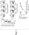
- the population of antigen-specific T-cells undergoes at least 7 cell doublings after the initial stimulation over the first 7 days, as shown in Figure 1A .
- a weekly T-cell expansion of 128-fold (as measured by the frequency of antigen-specific T-cells times the total number of cells in the cell composition).
- the frequency of tetramer positive cells after the first, second, and third stimulations is shown in Figure 1B .
- RAK and QAK was 0.02% and 0.01%, respectively.
- Example 1 demonstrates that the amount of time it takes to produce the desired cells is typically delayed after roughly the first week of production since the rate of population expansion of the desired cells decreases in subsequent stages of culture.
- EXAMPLE 2 Reducing the amount of time needed to increase the desired cell population can be achieved by reducing the cell surface density of the desired cell population as the onset of any given stage or stages of culture.
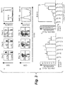
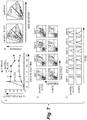
- the EBV antigen-specific T-cell component of PBMCs represents, at most, 2% of the population and so the antigen-specific responder T-cell seeding density is less than 2x10 4 per cm 2 , with the remaining PBMC acting as non-proliferating feeder cells (seen as the CFSE positive cells in Figure 3A ) that sustain optimal cell-to-cell contact allowing proliferation of the antigen-specific CTLs.
- the majority of T-cells are antigen-specific, and although the total cell density of the composition is about the same, the proliferating cell density is 50 to 100 fold higher. As a consequence, on re-stimulation the majority of cells proliferate and may therefore rapidly consume and exhaust their nutrients and O 2 supply.
- EXAMPLE 3 A minimum surface density of a cell population that includes the desired cells and/or antigen presenting cells can allow outgrowth of a desired cell population that is seeded at very low surface density.
- Figure 4 shows an example of results we obtained when continuing the work described in Figure 3 , which further demonstrated that when desired cells need the support of other cells, unconventionally low desired cell surface density can initiate population expansion so long as desired cells are in the presence of an adequate supply of feeder and/or antigen presenting cells.
- a total cell composition with a surface density and R:S ratio of between about 1.0x10 6 desired cells/cm 2 at an R:S ratio of 8 to 1 and merely about 3900 desired cells/cm 2 at an R:S ratio of 1 to 32 could allow desired cells to be greatly expanded to over 50 fold times the starting surface density, at which point we discontinued testing.
- EXAMPLE 4 The ability to allow a production process to repeat in stages by initiating a stage with an unconventionally low desired cell surface density, allowing population expansion, terminating the stage and repeating conditions was demonstrated to deliver repeatable outcomes.
- Example 3 We continued the assessments described in Example 3 at three of the desired cell surface densities (CTL/cm 2 ) as shown in Figure 5 . Each specific seeding density was able to consistently attain the same fold expansion. The implications will be described in more detail further on as they relate to the ability to dramatically reduce the production time for a desired cell population.
- EXAMPLE 5 Culturing desired cells on a growth surface that is comprised of gas permeable material while simultaneously increasing the medium volume to growth surface area ratio increases the number of times a desired cell population can double in a given stage of culture relative to conventional methods and increases the surface density that is attainable.
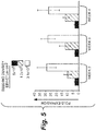
- G-Rex Test fixtures (hereinafter generically referred to as "G-Rex") were constructed as shown in Figure 6 .
- Bottom 20 of each G-Rex 10 was comprised of gas permeable silicone membrane, approximately 0.005 to 0.007 inches thick.
- Pending U.S. Publication No. 2005/0106717 A1 to Wilson is among many other sources of information relating to the use of alternative gas permeable materials and can be used to educate skilled artisans about gas permeable culture device shapes, features, and other useful characteristics that are beneficial to many of the methods or devices disclosed herein.
- G-Rex40 had a growth surface area of 10 cm 2 , upon which a cell composition (shown as item 30) rested, the characteristics of the cell composition varied throughout the experiment as described within.
- EBV-specific CTL and irradiated autologous EBV-LCLs at the conventional 4:1 ratio of CTL:LCL were cultured in G-Rex40 devices.
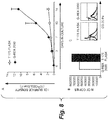
- EBV-CTLs were seeded at a surface density of 5x10 5 cells/cm 2 in the G-Rex40 and the rate of EBV-CTL population expansion was compared with EBV-CTL seeded at the same surface density in a standard 24-well plate with a medium volume to growth surface area of 1 ml/cm 2 .
- EBV-CTLs in the G-Rex40 had increased from 5x10 5 /cm 2 to a median of 7.9x10 6 /cm 2 (range 5.7 to 8.1x10 6 /cm 2 ) without any medium exchange.
- EBV-CTLs cultured for 3 days in conventional 24-well plates only increased from a surface density of 5x10 5 /cm 2 to a median of 1.8x10 6 /cm 2 (range 1.7 to 2.5x10 6 /cm 2 ) by day 3.
- surface density could be further increased by replenishing medium whereas cell surface density could not be increased by replenishing medium or IL2 in the 24-well plate.
- EBV-CTL surface density further increased in the G-Rex40 to 9.5x10 6 cells/cm 2 (range 8.5 x10 6 to 11.0 x10 6 /cm 2 ) after replenishing the medium and IL-2 on day 7 (data not shown).
- T-cells were labeled with CFSE on day 0 and divided between a G-Rex40 device with a 40 ml medium volume and a 24 well plate with each well at a 2 ml medium volume.
- Daily flow cytometric analysis demonstrated no differences in the number of cell divisions from day 1 to day 3. From day 3 onwards, however, the population of desired cells cultured in the G-Rex40 continued to increase at a rate that exceeded the diminishing rate of the 2 ml wells, indicating that the culture conditions had become limiting as shown in Figure 7D .
- the large population of desired cells in the G-Rex40 test fixtures resulted from a combination of decreased cell death and sustained proliferation relative to conventional methods.
- EXAMPLE 6 By use of unconventionally high ratios of medium volume to growth surface area and use of growth surfaces comprised of gas permeable material, the need to feed culture during production can be reduced while simultaneously obtaining unconventionally high desired cell surface density.
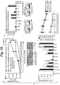
- G-Rex2000 refers to device as described in Figure 8 , the exception being the bottom is comprised of a 100 cm 2 growth surface area and a 2000 ml medium volume capacity is available.
- EBV-LCLs were cultured in and expand in the G-Rex2000 without changing the cell phenotype.
- EBV-LCL were plated into a G-Rex2000 at a surface density of 1x10 5 cells/cm 2 along with 1000 ml of complete RPMI medium to create a medium volume to surface area ratio of 10 ml/cm 2 .
- EBV-LCL were plated into a T175 flask at a surface density of 5x10 5 cells/cm 2 along with 30 ml of complete RPMI medium to create a medium volume to surface area ratio of about 0.18 ml/cm 2 .
- the EBV-LCL cultured in G-Rex2000 expanded more than those in the T175 flask without requiring any manipulation or media change. This culture condition did not modify the final cell product as evaluated by Q-PCR for EBER and B cell marker CD20 as presented in Figure 8B and Figure 8C .
- EXAMPLE 7 When sufficient feeder and/or antigen cells are not present at the onset of culture, desired cells may not expand. However, the cell composition can be altered to include an additional cell type acting as feeder cells and/or antigen presenting cell to allow expansion.
- Figure 9 shows an illustrative example in which we experimentally demonstrated that a very low cumulative surface density of desired cells and antigen presenting cells (in this case AL-CTLs and LCLs cells combining to create a cell composition with a surface density of 30,000 cells/cm 2 ) was unable to initiate outgrowth of the AL-CTL population.
- AL-CTLs and LCLs cells combining to create a cell composition with a surface density of 30,000 cells/cm 2
- this same cell composition could be made to grow by altering the composition to include another cell type acting as a feeder cell.
- the additive surface density of the antigen presenting cells and/or feeder cells and the desired cells should preferably be at least about 0.125x10 6 cells/cm 2 to create enough surface density in the cell composition to initiate the expansion of the desired cell population.
- the use of growth surfaces comprised of gas permeable material was used in this example along with a medium volume to surface area ratio of 4 ml/cm 2 .
- EXAMPLE 8 Reduced desired cell surface densities, altered responder cell to stimulatory cell ratios, increased medium to growth surface area ratios, and periodic distribution of cells at a low surface density culture onto growth surfaces comprised of gas permeable material allow more desired cells to be produced in a shorter period of time and simplifies the production process when compared to other methods.
- G-Rex500 refers to device as described in Figure 6 , the exception being the bottom is comprised of a 100 cm 2 growth surface area and a 500 ml medium volume capacity is available.
- a second stage was initiated on day 9, wherein 1x10 7 responder T-cells were transferred from the G-Rex40 to a G-Rex500 test fixture.
- stage two of culture 200 ml of CTL medium was placed in the G-Rex500, creating a medium volume to surface area ratio at the onset of stage two of 2 ml/cm 2 medium height at 2.0 cm above the growth surface area.
- the surface density of desired cells at the onset of stage two was 1x10 5 CTL/cm 2 with antigen presenting cells at a surface density of 5x10 5 LCL/cm 2 , thereby creating a non-conventional 1:5 ratio of desired cells to antigen presenting cells.
- This stage two cell surface density and R:S ratio produced consistent EBV-CTL expansion in all donors screened.
- IL-2 50U/ml - final concentration
- 200 ml of fresh medium bringing medium volume to surface area ratio to 4 ml/cm 2
- the cells were harvested and counted.
- the median surface density of CTLs obtained was 6.5x10 6 per cm 2 (range 2.4x10 6 to 3.5x10 7 ).
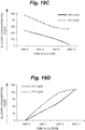
- FIG. 10A shows the comparison of this G-Rex approach of Example 8 to the use of conventional methods of Example 1 and the G-Rex approach described in Example 5. As shown, the conventional method needed 23 days to deliver as many desired cells as could be delivered in either G-Rex method in about 10 days.
- Example 8 After 23 days, the G-Rex approach of Example 8 was able to produce 23.7 more desired cells than the G-Rex method of Example 5 and 68.4 times more desired cells than the conventional method of Example 1. Furthermore, the desired cells continued to divide until day 27-30 without requiring additional antigen presenting cell stimulation provided the cultures were split when cell surface density was greater than 7x10 6 /cm 2 .
- FIG. 10D shows a representative culture in which T-cells stimulated with EBV peptide epitopes from LMP1, LMP2, BZLF1 and EBNA1 and stained with HLA-A2-LMP2 peptide pentamers staining showed similar frequencies of peptide-specific T-cells. Further, the expanded cells maintained their cytolytic activity and specificity and killed autologous EBV-LCL (62% ⁇ 12 vs.
- Examples 1-8 have been presented to demonstrate to skilled artisans how the use of various conditions including reduced surface density of the desired cell population at the onset of a production cycle, reduced surface density ratios between responder cells and stimulating cells, growth surfaces comprised of gas permeable materials, and/or increased medium volume to growth surface area ratios can be used to expedite and simplify the production of cells for research and clinical application of cell therapy.
- Examples 1-8 were related to the production of antigen specific T cells, these novel culture conditions can be applied to many important suspension cell types with clinical relevance (or required for pre-clinical proof of concept murine models) including regulatory T cells (Treg), natural killer cells (NK), tumor infiltrating lymphocytes (TIL), primary T lymphocytes, a wide variety of antigen specific cells, and many others (all of which can also be genetically modified to improve their function, in-vivo persistence or safety).
- Treg regulatory T cells
- NK natural killer cells
- TIL tumor infiltrating lymphocytes
- primary T lymphocytes a wide variety of antigen specific cells, and many others (all of which can also be genetically modified to improve their function, in-vivo persistence or safety).
- Cells can be expanded with feeder cells and/or antigen presenting cells that can include PBMC, PHA blast, OKT3 T, B blast, LCLs and K562, (natural or genetically modified to express and antigen and/or epitope as well as co-stimulatory molecules such as 41BBL, OX40L, CD80, CD86, HLA, and many others) which may or may not be pulsed with peptide and/or a relevant antigen.
- feeder cells and/or antigen presenting cells can include PBMC, PHA blast, OKT3 T, B blast, LCLs and K562, (natural or genetically modified to express and antigen and/or epitope as well as co-stimulatory molecules such as 41BBL, OX40L, CD80, CD86, HLA, and many others) which may or may not be pulsed with peptide and/or a relevant antigen.
- Unconventionally Low Initial Surface Density It was discovered that production time can be reduced relative to conventional methods by the use of lower desired cell surface density. In this manner, desired cells are able to have a greater numerical difference between their minimum and maximum cell surface densities than conventional methods allow. Preferably, when the rate of desired cell population growth has begun to diminish, but the quantity of desired cells is not yet sufficient to terminate production, the desired cells are re-distributed upon additional growth surfaces comprised of gas permeable material at low starting surface density once again.
- Figure 11 shows a graphical representation of expansion of a desired cell population on a growth surface under the conventional scenario as compared to population expansion of the desired cell type using a method disclosed herein.
- the surface density of desired cells at the onset of a production stage is less than conventional surface density.
- this explanation does not describe the process of initially obtaining the desired cell population.
- the 'Day" of culture starts at "0" to allow skilled artisans to more easily determine the relative time advantages of this novel method.
- each production cycle of the conventional method begins at a conventional surface density of 0.5x10 6 desired cells/cm 2 while each production cycle of this example begins at a much lower and unconventional surface density of 0.125x10 6 desired cells/cm 2 .
- 4 times more surface area i.e. 500,000/125,000
- the desired cells of the conventional method reaches a maximum surface density of 2x10 6 cells/cm 2 in 14 days.
- 1 cm 2 of growth area delivers 2x10 6 cells/cm 2 which are then re-distributed onto 4 cm 2 of growth area so that production can be continued using the conventional starting density of 0.5x10 6 cells/cm 2 (i.e.
- the novel method depicted in Figure 11 instead of using the conventional method of depositing 500,000 desired cells onto 1 cm 2 at the onset of production, distributes the 500,000 cells equally onto 4 cm 2 of growth area to create at unconventionally low starting surface density of 125,000 desired cells/cm 2 on Day 0.
- the novel method as with the conventional method, has its growth rate about to diminish on Day 7.
- Cells in the novel method are at a surface density of 1x10 6 cells/cm 2 .
- this stage of culture has produced 4x10 6 cells that are then re-distributed onto 32 cm 2 of growth area so that production in Stage 2 can be continued using the starting surface density of 0.125x10 6 cells/cm 2 (i.e.
- This example describes how, by lowering the desired cell surface density (in this case to 0.125x10 6 cells/cm 2 ) relative to conventional cell surface density, the same quantity of desired cells are delivered in just 33% of the time as the conventional method (14 days vs. 42 days).
- Desired cells should be deposited upon a growth surface at an unconventionally low cell surface density such that:
- growth surfaces comprised of gas permeable material and higher medium volume to growth surface area ratios can simplify and shorten production. It was discovered that the use of growth surfaces comprised of gas permeable material and medium volume to growth surface area ratios that exceed conventional ratios, and repeated cycles of production that increase the amount of growth surface area used over time will reduce production duration.
- Figure 12 augments the discussion to show an example of the advantages that can be obtained by utilizing a growth surface comprised of gas permeable material and an unconventionally high medium volume to growth surface area ratio beyond 1 or 2 ml/cm 2 .
- the discussion that follows is intended to demonstrate to skilled artisans how, by use of such a method, several options become available including reducing production time, reducing the amount of growth surface area used, and/or reducing labor and contamination risk. Skilled artisans will recognize that Figure 12 and associated discussion is merely an example, and does not limit the scope of this invention.
- the cell composition containing the desired cell population in this illustrative example is assumed to consume about 1 ml per "X" period of time.
- Figure 12 shows two production processes, labeled “conventional method” and "novel method.” At the onset of growth, each process begins with desired cells at a surface density of 0.5x10 6 /cm 2 .
- the growth surface of in the novel method is comprised of gas permeable material and medium volume to surface area ratio is 2 ml/cm 2 as opposed to the conventional method of 1 ml/cm 2 .
- time period "X” the desired cell population of the conventional method has a reached a surface density plateau of 2x10 6 /cm 2 and is depleted of nutrients while the additional medium volume of the novel method has allowed growth to continue and desired cell surface density is 3x10 6 /cm 2 . If the novel method continues, it reaches a surface density of 4x10 6 /cm 2 . Thus, many beneficial options accrue.
- the novel method can be terminated prior to time "X” with more cells produced than the conventional method, can be terminated at time "X” with about 1.5 times more cells produced than the conventional method, or can continue until the medium is depleted of nutrients with 2 times many desired cells produced as the conventional method in twice the time but without any need to handle the device for feeding.
- the conventional method In order for the conventional method to gather as many cells, the cells must be harvested and the process reinitiated, adding labor and possible contamination risk. Since cell therapy applications typically only are able to start with a fixed number of cells, the conventional method does not allow the option of simply increasing surface area at the onset of production.
- Figure 13 continues the example of Figure 12 to show how more than one production cycle can be of further benefit.
- Figure 13 shows a graphical representation of expansion of a desired cell population on a growth surface under the conventional method as compared to population expansion of the desired cell type under one method disclosed herein in which the surface density of the novel method exceeds surface density of the conventional method.
- the 'Day" of culture starts at "0" to allow skilled artisans to more easily determine the relative time advantages of the methods disclosed herein. In this example, both cultures are initiated using conventional desired cell surface density of 0.5x10 5 cells/cm 2 at "Day 0".
- the growth surface of the conventional method is also comprised of gas permeable material.
- the medium volume to growth surface ratio in the conventional method is 1 ml/cm 2 as opposed to 4 ml/cm 2 in the novel method.
- the desired cell population in the conventional method begins to diminish in growth rate when it is at a surface density of about 1.5x10 6 cells/cm 2 in about 4 days and reaches a maximum surface density of 2x10 6 cells/cm 2 in 14 days.
- the desired cell population is distributed to 4 cm 2 of growth area at a surface density of 0.5x10 6 /cm 2 in fresh medium at 1.0 ml/cm 2 and the production cycle begins again, reaching a surface density of 2x10 6 cells/cm 2 in another 14 days and delivering 8x10 6 desired cells in 28 days.
- the desired cell population in the novel method begins to diminish in growth rate when it is at a surface density of about 3x10 6 cells/cm 2 in roughly about 10 to 11 days and could reach a maximum surface density of 4x10 6 cells/cm 2 in 28 days.
- the cycle ends when the desired cell population is still in a high rate of growth.
- the 3x10 6 cells are re-distributed to 6 cm 2 of growth surface area at a surface density of 0.5x10 6 /cm 2 in fresh medium at 4.0 ml/cm 2 and the production cycle begins again, with the desired cell population reaching a surface density of 3x10 6 cells/cm 2 in roughly another 10 to 11 days and delivering 18x10 6 desired cells around 21 days.
- the novel method has produced over 2 times the number of desired cells as compared to the conventional method.
- Figure 14 shows another novel method in which still further advantages relative to conventional methods are obtained.
- skilled artisans will recognize that the description herein does not limit the scope of this invention, but instead acts to describe how to attain advantages of improved production efficiency.
- desired cells are doubling weekly in conventional conditions.
- the 'Day" of culture starts at "0" to allow skilled artisans to more easily determine the relative time advantages.
- issues previously described related to feeder and/or antigen presenting cell surface density ratios are not repeated to simplify this example.
- the conventional method begins with a surface density of 0.5x10 6 cells/cm 2 and a medium volume to surface area ratio of 1 ml/cm 2 .
- the novel method of this example begins with a surface density of 0.06x10 6 cells/cm 2 , a growth surface area comprised of gas permeable material, and a medium volume to surface area ratio of 6 ml/cm 2 .
- a surface density of 0.06x10 6 cells/cm 2 a growth surface area comprised of gas permeable material
- a medium volume to surface area ratio of 6 ml/cm 2 a medium volume to surface area ratio of 6 ml/cm 2 .
- the population is determined to be reaching plateau from noting that plateau is initiated in the conventional method when cell surface density approaches 1.5 times the medium volume to surface area ratio (i.e. about 1.5x10 6 cells/ml).
- a surface density of about 4.5x10 6 cells/cm 2 at about 9 days cells are distributed onto 36 cm 2 of growth surface area and the production cycle begins anew.
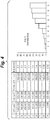
- Figure 15 tabulates a comparison of each production method depicted in Figure 14 , and extends to stages to demonstrate the power of the novel method, and why it is wise to adjust the production protocol at various stages to fully capture the efficiency.
- the novel method overpowers the conventional method after completing just the second stage of the production cycle, delivering nearly 1.37 times more cells in only about half the time with just 61% of the surface area requirement.
- the third stage of the production cycle creates a massive increase in cells and a corresponding increase in surface area.
- Figure 16 shows an example of how one could alter variables in the novel method to gain efficiency as production progresses.
- an increase in the starting surface density of cycle 3 from 0.06 to 0.70 cell/cm 2 and a change to the final surface density from 4.5 to 7.5 cells/cm 2 can be undertaken.
- Increasing the final surface density is a matter of increasing the medium volume to surface area ratio beyond the initial 6 ml/cm 2 to a greater number. The greater the medium volume to surface area, the longer the cycle remains in rapid growth phase (i.e. the population expansion prior to plateau). In this case we have allowed 5 extra days to complete the rapid growth phase and raised the medium volume to surface area ratio to about 8 ml/cm 2 .
- EXAMPLE 9 More efficient methods of producing cells within a static gas permeable culture device by establishing novel culture conditions at the start of the culture process.
- Static cell culture experiments were conducted in which K562 cells were cultured in test devices configured with a growth surface comprised of gas permeable silicone material and with wall height that allowed 10 cm of medium to reside above the growth surface.
- the growth surface was held in a substantially horizontal position with a growth surface support as described more thoroughly in Wilson '717.
- Medium was placed in the test devices at a medium height of 10 cm beyond the growth surface, establishing a medium volume to growth area ratio of 10 ml/cm 2 .
- K562 cells were also introduced into the test devices and the devices were placed into a cell culture incubator at 37C, 5% CO2, and 95% R.H, whereby cells were allowed to gravitate to the growth surface.
- the medium was not perfused or subjected to forced agitation and gas was not forced to flow past the growth surface, instead making contact with the growth surface by random motion of the ambient atmosphere.
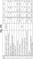
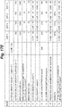
- FIG. 17A , FIG. 17B , FIG. 17C , FIG. 17D , and FIG. 17E show, for illustrative purposes, a representative spreadsheet of the experimental conditions and typical results.
- Initial static culture conditions established surface densities ranging between 1.0E+06 to 6.25E+04 cells/cm 2 , cell densities ranging from 1.0E+05 to 6.25E+03 cells/ml, with medium residing above the growth surface in all conditions at a constant height of 10 cm and all medium being the same formulation with glucose concentration at 240 mg/dl.
- the initial state of static culture was day 0 and cell counts and glucose concentration were assessed on day 4, day 8, day 11.
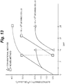
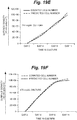
- FIG. 18 compares the fold expansion of the population increase relative to the surface density of each of the experimental conditions detailed in FIG. 17A through FIG. 17E .
- Fold expansion of each condition was determined by dividing the cell surface density on day 11 by the cell surface density on day 0.
- surface density is at a low limit of 5.0E+05 cells/cm 2 and medium height is at the upper limit of 2.0 cm
- dotted line 6 shows typical fold expansion in state of the art K562 production methods using gas permeable bags.
- Each surface density condition established in our experiments created a population expansion that exceeded state of the art population expansion.
- the ability to increase the fold expansion of the population of cells greatly increased as conditions of initial static culture surface density decreased to 0.125E+06, and then further reductions were less advantageous although still far superior to state of the art methods.
- EXAMPLE 10 Novel methods to determine the quantity cells in a population residing within a static gas permeable culture device without need of counting cells.
- glucose concentration of the culture as a surrogate indicator of the population of the culture.
- knowing the minimum total medium volume needed for the culture to reach maximum surface density and the total reduction in glucose concentration needed for the culture to reach maximum surface density sets the stage for a surrogate prediction of the number of cells in the population of the culture. Equipped with that knowledge, one initiating culture (or a stage of culture) would determine the baseline glucose concentration of medium, the baseline volume of medium, and would keep track of the volume of medium added to the culture prior to taking a measure of glucose concentration at the time of population estimation.
- the estimated number of cells in the population is a function of the prorated total reduction in glucose concentration needed to reach maximum cell density multiplied by the prorated minimum medium volume needed to reach maximum surface density and multiplied by the maximum surface density possible on the growth surface.
- the predictive formulas require knowledge of the cell culture applications maximum cell density (and/or maximum surface density) under conditions in which cells reside on a growth surface comprised of the particular gas permeable material the artisan has selected. Experiments can be undertaken to make that determination. For example, to determine the maximum cell surface density of K562 cells upon a growth surface comprised of the gas permeable material in our experimental fixtures (dimethyl silicone as described previously), we increased medium height until surface density could increase no more. The minimum volume of medium needed to support a maximum attainable surface density of K562 at about 12.0E+06 cells/cm 2 was determined to be 10 ml with a corresponding total reduction in glucose concentration of 250 mg/ml.
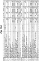
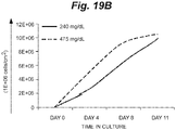
- FIG. 19A shows a representative spreadsheet of the experimental conditions and typical results for the culture of K562 cells under equivalent starting conditions except for the glucose concentration, which was 240 mg/dl vs. 475 mg/dl at the onset of culture. Results are graphically depicted in FIG. 19B , FIG. 19C , FIG. 19D , FIG. 19E, and FIG. 19F . Population growth by cell count and as predicted by glucose depletion was normalized for surface density.
- FIG. 19B shows the population expansion under each condition over a time period of 11 days. The population growth rate differed_slightly, but arrived at about the same number in 11 days.
- FIG. 19C shows the glucose depletion rate in each culture condition.
- FIG. 19D shows the glucose consumption rate in each culture condition.
- FIG. 19E shows an overlay of the predicted value, using the formulaic calculation of cell number, versus the cell number as determined by manual counts for the culture initiated at a glucose concentration of 240 mg/dl.
- FIG. 19F shows an overlay of the predicted value, using the formulaic calculation of cell number, versus the cell number as determined by manual counts for the culture initiated at a glucose concentration of 475 mg/dl. Note the predictive capacity of the formulaic approach relative to the method of manual cell counts. This further demonstrates that various aspects described herein can be utilized in conjunction with a method of reducing, or even eliminating, the frequency of cell counts in lieu of a surrogate measure of the concentration of solutes in the medium.
- manufacturers of gas permeable devices including those described in Wilson '717 or Wilson '176, could provide a simplified cell production process that can easily determine the number of live cells in culture within a gas permeable device, absent the need to count cells by providing a gas permeable cell culture device including a growth surface comprised of gas permeable material and providing instructions and/or disseminating information relating to the present disclosures.
- EXAMPLE 11 Less complicated methods of producing cells within a static gas permeable culture device by establishing novel conditions at the start of the culture process in order to limit feeding frequency in static cultures.
- Medium resided at a height of 2.5 cm, 5.0 cm, 10.0 cm, or 15.0 cm above the growth surface, which was comprised of silicone and had a surface area of 100 cm 2 .
- the growth surface was held in a substantially horizontal position with a growth surface support as described more thoroughly in Wilson '717.
- experimental conditions included ratios of medium volume to the surface area of growth surfaces at 2.5 ml/cm 2 , 5.0 ml/cm 2 , 10.0 ml/cm 2 , and 15.0 ml/cm 2 .
- initial cell density was 0.05E+06 cells/ml, 0.025E+06 cells/ml, 0.0125E+06 cells/ml, and 0.008E+06 cells/ml respectively.
- No further medium was added to the 10.0 ml/cm 2 or 15.0 ml/cm 2 conditions.
- the original medium volume of the 2.5 ml/cm 2 condition was doubled by adding 2.5 ml/cm 2 of fresh medium on day 11, tripled on day 14 by adding another 2.5 ml/cm 2 of fresh medium, and quadrupled on day 17 by adding another 2.5 ml/cm 2 of fresh medium.
- the original medium volume of the 5.0 ml/cm 2 condition was doubled by adding 5.0 ml/cm 2 of fresh medium on day 11. Eventually, the 2.5 ml/cm 2 and 5.0 ml/cm 2 conditions held 10.0 ml/cm 2 of medium.
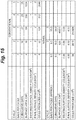
- FIG. 20 shows a graphical representation of population growth, normalized for growth surface area, under various medium feeding conditions.
- viability was relatively high in the 15.0 cm for a period of about 4 days after the maximum cell population was attained while it diminished rapidly after about 1 to 2 days in the 10.0 cm condition.
- the practical benefit created here is a production process that has a longer period of time in which to recover cells.
- Skilled artisans will recognize that all of the experimental culture conditions exhibited superior rates of cell population expansion compared to state of the art methods for Adoptive Cell Therapy, but should be aware that it is not only beneficial to reduce surface density relative to state of the art methods at the onset of culture, it is further possible to reduce the duration needed for production of desired number of cells by increasing medium height and/or medium to growth area ratios. Skilled artisans should recognize that improvements will be obtained in terms of the rate of population expansion as less surface density and more medium height and/or a further increase in medium volume to growth surface area ratio is undertaken, and are encouraged to balance the use of medium with the needs of the application. More medium at the onset of culture can be provided if a larger window of time to harvest cells while they reside at high viability is sought.
- a most preferred initial culture condition for production is a cell density of less than 0.5E+06 cells/cm 2 and most preferably about 0.125E+06 cells/cm 2 , and a medium height of about 5.0 cm or more and more preferably 10.0 cm to 15.0 cm, and/or a medium volume to growth surface area of about 5.0 ml/cm 2 or more and more preferably 10.0 ml/cm 2 to 15.0 ml/cm 2 , and/or an initial cell density about 0.025E+06 cells/ml or less and more preferably about 0.0125E+06 cells/ml to about 0.008E+06 cells/ml.
- EXAMPLE 12 Novel ways to limit feeding frequency of co-cultures residing within a static gas permeable culture device and determine the size of the cell population without need of counting cells, even though a portion of the cells are dying.
- Adoptive Cell Therapy often relies on co-culture with cells that are dying because they were irradiated (such as APC's) or cells that are dying as a result of being removed from the body (such as PBMC's).
- a good example of a co-culture application is in the culture of CMV-CTLs (cytomegalovirus specific cytotoxic T lymphocytes) out of a population of PBMCs. Initially, the CMV-CTL population is a very small percentage relative to the total population of PBMCs. As the culture progresses, the PBMC begin to die off and CMV-CTLs begin to grow. By the end of culture, the frequency of CMV-CTL in the cell composition has increased greatly.
- CMV-CTLs cytomegalovirus specific cytotoxic T lymphocytes
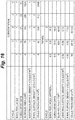
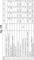
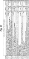
- FIG. 21 shows a spreadsheet that summarizes conditions on day 0, day 9, and day 16.
- PBMCs medium were introduced into the experimental devices and the devices were placed into a cell culture incubator at 37C, 5% CO2, and 95% R.H, whereby cells were allowed to gravitate to the growth surface at a surface density of 5.0E+05 cells/cm 2 .
- CMV-CTL antigen specific T cells
- Row 4 shows how the CMV-CTL fold expansion as a percentage of the total population diminished after day 9. This is because the PBMC are dying.
- Row 19 demonstrates the ability of the surrogate measures of solutes in the medium to predict the number of cells in culture. Note that the predicted value is nearly identical to the assessment of cell population by counting. Thus, the ability to use a surrogate measure to quantify cell population is useful even in cell compositions in which components of the cell composition are dying.
- EXAMPLE 13 Novel static gas permeable cell culture and cell recovery devices that enable simplified methods of medium exchange and novel methods for greatly diminishing the effort required to separate cells from medium after a cell production process is complete.
- FIG. 22A shows a cross-sectional view of one example of cell culture and cell recovery device 1000 configured to perform the disclosed novel cell culture and/or novel cell recovery methods.
- Cell removal opening 1002 of cell removal conduit 1004 resides in proximity of growth surface 1006.
- Medium removal opening 1008 of medium removal conduit 1010 resides near growth surface 1006.
- Growth surface 1006 is comprised of gas permeable material. There are many sources of information for skilled artisans to learn about appropriate gas permeable material including Wilson '717.
- growth surface 1006 is liquid impermeable and non-porous. The distance from growth surface 1006 to upper confine 1012 of internal volume 1014 defines the volume of space where medium can reside.
- medium can reside in medium removal conduit 1010 and cell removal conduit 1004, which can extend to a height beyond upper confine 1012, maximum medium height should be considered by skilled artisans to be the farthest distance from the bottom of internal volume 1014 to upper confine 1012 for purposes of describing this embodiment.
- the cell culture and cell recovery device does not require a stirring mechanism or any other mechanisms to mix the cells and/or medium.
- FIG. 22B shows cell culture and cell recovery device 1000 in an initial state of static culture at the onset of any given cell production stage of culture.
- Cell culture and cell recovery device 1000 resides in a position in which growth surface 1006 is in oriented in a horizontal position and cells 1016 have gravitated to growth surface 1006.
- growth surface support 1018 is used to hold growth surface 1006 in a horizontal position while allowing ambient gas to make contact with growth surface 1006 without need of pumps or other mechanisms to force gas past growth surface 1006.
- Skilled artisans can refer to Wilson '717 for information about how to configure growth surface support 1018.
- medium 1020 can reside at any level within the confines of internal volume 1014, preferably the entire uppermost medium location 1022 is parallel to growth surface 1006 as shown.
- Cell culture and cell recovery device 1000 resides in an atmosphere suitable for cell culture and at a temperature suitable for cell culture. Ambient gas makes contact with gas permeable material of growth surface 1006 by random motion and without need of pumps or other mechanisms to force gas to or from growth surface 1006.
- Medium height is determined by the distance from the lowermost medium location to the uppermost medium location, in this case the distance from growth surface 1006 to uppermost medium location 1022 at the onset of culture being the initial static culture medium height.
- the ratio of the number of cells 1016 having gravitated to growth surface 1006 to the volume of medium 1020 is an initial static culture cell density.
- the ratio of the number of cells 1016 upon growth surface 1006 to the surface area of growth surface 1006 is the initial static culture surface density.
- the ratio of medium 1020 volume to the surface area of growth surface 1006 is an initial static culture medium volume to growth surface area ratio.
- Cells reside in a state of static culture and the culture continues for a period of time. As described throughout this disclosure, the period of time may or may not include a medium replenishment step depending upon variables that include the initial static culture medium height, the initial static culture cell density, the initial static culture surface density, and/or the initial static culture medium volume to growth surface area ratio.
- FIG. 22C shows further steps to recover cells in a reduced volume of medium from cell culture and cell recovery device 1000.
- Medium is removed by way of medium removal opening 1008 in medium removal conduit 1010 while not withdrawing cells 1016.
- remaining medium is shown as cell recovery medium 1024.
- the location of the medium removal opening of the medium removal conduit is preferably located at a distance of 0.2 cm or more from the growth surface when the growth surface resides in a horizontal position. For example, between 0.2 cm and 2.0 cm from the growth surface when the growth surface resides in a horizontal position allows significant volume reduction for many of the cell culture methods disclosed herein.
- the upper limit of the distance between the medium removal opening and the growth surface when the growth surface resides in a horizontal position is preferably a distance that takes into account the typical height of medium at the point where medium is to be decreased for cell recovery.
- the medium removal opening of the medium removal conduit would be located at 50% of the medium height (assuming the device was designed such that the medium resided entirely over the growth surface). Since use of laboratory space is at a premium, device height should be about the height of medium expected to reside within it.
- a good rule of thumb is to design the device with a height that is at or just beyond typical medium height during use and locate medium removal opening of the medium removal conduit at any location from about 0.2 cm from the growth surface (when the growth surface resides in a horizontal position) to about the halfway point from the top of the device to the growth surface as measured from the inside of the device.
- the medium removal opening would preferably be located 0.2 cm or more above the growth surface when the growth surface resides in a horizontal position and 50% or less of the potential medium height. In the event it is uncertain where the medium height will reside, more than one medium removal conduit could be present in the device.
- the cell removal opening of the cell removal conduit is preferably located along the lower edge of the device and can collect cell recovery medium without reorienting the device. However, the device can be reoriented if desired.
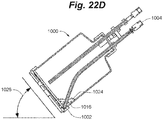
- FIG. 22D shows the process of reorienting cell culture and cell recovery device 1000 into a position at an angle 1026 that deviates from the original horizontal cell culture position in order to relocate cell recovery medium 1024, having cells 1016 distributed within it, relative to cell removal opening 1002 of cell removal conduit 1004, whereby cell recovery medium 1024 can subsequently be withdrawn.
- the cell removal opening of the cell removal conduit need not be located along the lower edge of the device.
- the cell recovery medium can be removed from any location in the device by simply rotating the device until the cell recovery medium is located at the cell removal opening, and then withdrawing the cell recovery medium by way of the cell removal conduit.
- the conduits need not be as shown, but can be any configuration.
- the key design feature is the ability to place the medium removal opening in the preferred locations relative the growth surface as previously described.
- the conduits can be as simple as locating a septum in the side of the device or as complex as telescoping tubes.
- the internal height of the cell culture and cell recovery device should preferably be at least more than 2.0 cm in any particular application.
- the cell culture and cell recovery device is preferably constructed with biocompatible materials, clear to allow visual assessment, and rigid to allow easy handling.
- a skilled artisan should seek to create a preferred embodiment of a static gas permeable cell culture and cell recovery device comprising:
- the device should preferably include relevant aspects of devices described in Wilson '717. Also, equipped with this knowledge, manufacturers of gas permeable devices, including those described in Wilson '717, could facilitate more efficient methods of cell culture by providing users with a cell culture and cell recovery device including a growth surface comprised of gas permeable material, a medium removal conduit, a medium removal opening, a cell removal conduit, a cell removal opening, and providing instructions and/or disseminating information for:
- the cell recovery medium volume to growth surface area ratio is at least 0.2 ml/cm 2 and the medium reduction percentage being at least 50%.
- this method is capable of utilizing any of: the desired initial static culture cell density, initial static culture surface density, initial static culture medium volume to growth surface area ratio, and/or initial static culture medium height that provide advantages described herein. Also, skilled artisans are encouraged to recognize that the method includes use for islets.
- EXAMPLE 14 Novel methods of using a static gas permeable culture device for superior production of CAR T cells.
- CAR T cells Three conditions for expansion of transduced antigen specific T cells (CAR T cells) were evaluated. Evaluation A included CAR T cells in the presence of K652 APC cells and included medium height at 10 cm. Evaluation B included CAR T cells without the presence of K652 APC cells and included medium height at 10 cm. Evaluation C cultured CAR T cells in accordance with state of the art methods.
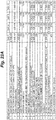
- FIG. 23A shows the conditions of Evaluation A at the onset of culture and as the culture progressed.
- the ratio of APC to CAR T cells at the onset of culture was 2:1 and medium resided at a height of 10 cm.
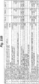
- FIG. 23B shows the conditions of Evaluation B at the onset of culture and as the culture progressed.
- APC were not present at the onset of culture and medium resided at a height of 10 cm.
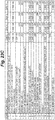
- FIG. 23C shows the conditions of Evaluation C at the onset of culture and as the culture progressed.
- the ratio of APC to CAR T cells at the onset of culture was 2:1 and medium resided at a height of 2 cm.
- cytokine stimulation is undertaken during medium exchange by adding cytokine (such as IL2) to the fresh medium.
- cytokine such as IL2
- feeding frequency is greatly reduced and even eliminated.
- Condition A and Condition B we also used Condition A and Condition B to evaluate the capacity to add cytokine in the absence of medium exchange.
- medium exchange we simply added a bolus of IL2 at the same frequency and at a quantity that brought the medium the same quantity per ml of state of the art methods and did not subject the medium to forced mixing of any sort to distribute the IL2 within the medium.
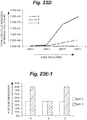
- FIG. 23D shows the total live cells in culture at various time points in the culture. As can be seen, the number of total live cells of Condition A were far superior to either of the alternative conditions.
- FIGS. 23E1 - E3 show the percentage of CAR T cell expression at the onset of culture and at the completion of culture. Histogram A represents Condition A, histogram B represents Condition B, and histogram C represents Conditions C. Condition B demonstrates the disadvantage of not providing APC in the culture at culture onset. When APC's were provided at the onset of culture, CAR expression improved from an initial state of about 40% to a state of about 80% by the end of culture.
- FIG. 23F shows the total fold expansion of CAR T cells during culture. It is clear that Condition A was able to generate a tremendously greater fold expansion than state of the art methods shown in Condition C.
- prediction of the live cell population in Evaluation A was representative of cell population as determined by manual counts. Furthermore, the devices used for Evaluation A and Evaluation B were able to have medium withdrawn at the end of culture, using the methods previously disclosed, from a state of 1000 ml to a state of 20 ml without cell loss.
- Condition A Another important finding was related to the presence of APC in culture.
- T cells recovered from Condition A have a greater capacity to kill tumor cells that express the relevant antigen (PSCA) due to a more enhanced T cell product, which at the end of the culture has a greater percentage of CAR expressing T cells (CAR-PSCA) in the population relative to its state at the onset of culture.
- Condition B due to its lack of APCs at the onset of culture, is unable to increase the percentage of CAR expressing T cells (CAR-PSCA) in the cell population at all over the culture period.
- FIG. 23H shows the capacity of cell obtained from Condition A and Condition B to kill tumor cells expressing PSCA, and to avoid killing cells that do not express the PSCA antigen.
- the ratio of effector cells to PSCA antigen expressing cells (Du145 and Capan1) or non-PSCA antigen expressing cells (293T) was 40:1. Effector cells (i.e. CAR T cells) were obtained from the cultures of Condition A and of Condition B at day 11 of culture.
- FIGS. 24A, 24B , 24C, and 24D summarize side by side comparisons of the population expansion of CAR T cells specific to PSCA and Mucl using the initial culture conditions described for Condition A (the exception being the antigen expression of the APC was PSCA and Mucl respectively). It can be seen that the novel initial conditions were able to produce a far greater number of CAR T cells in a shorter period of time than state of the art conditions in conventional culture ware. Skilled artisans will recognize the advantages are not limited to CAR T cells recognizing PSCA or Mucl antigens, but are applicable to CAR T cells recognizing any antigen.
- EXAMPLE 15 The methods disclosed herein are scalable in direct proportion to the surface area of the growth surface.
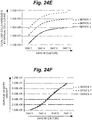
- FIG. 24E shows the population growth curves of three gas permeable culture devices with differing growth areas.
- Series 1 represents the live cell expansion of culture in the 640 cm 2 device, series 2 the 100 cm 2 device, and series 3 the 10 cm 2 device.
- FIG. 24F shows the population growth curves of FIG. 24E curves normalized to surface density and clearly demonstrates linear scalability.
- Wilson '176 Skilled artisans are encouraged to review Wilson '176 in the event that they seek to increase the growth surface area by scaling the culture in the vertical direction and will recognize that many aspects described herein can be undertaken using devices described in Wilson '176.
- growth surface is the area in a device upon which cells reside and is comprised of gas permeable material.
- Gas permeable material can be any materials know to skilled artisans in the field of cell culture and are preferably liquid impermeable and non-porous.
- the devices and culture methods disclosed herein can function in the absence of gas being forced past the growth surface that is comprised of gas permeable material. These methods pertain to static cell culture.
- the cells are preferably antigen presenting cells, and more preferably LCL or K562. If the culture comprises a co-culture, preferably it includes effector cells (i.e. desired or target cells) in combination with APC or feeder cells and may or may not include beads. Beads may also be a substitute for APC or feeder cells. If APC's are present, preferably they are professional antigen presenting cells, and more preferably of the type K562 or LCL, and even more preferably are irradiated. If present, unless they are islets, effector cells are preferably derived from peripheral blood or marrow, and more preferably are T cells, NK, Treg, TIL, or MIL. If effector cells are T cells, preferably they are naturally occurring antigen specific T cells or transduced antigen specific T cells.
- effector cells are T cells, preferably they are naturally occurring antigen specific T cells or transduced antigen specific T cells.
- Preferred surface density Cells reside upon a growth surface comprised of gas permeable material and at a preferred surface density less than 0.5E+06.
- Skilled artisans will recognize that the disclosure allows an analogue reduction in surface density from less than 0.5E+06 in order to increase rate population expansion relative to state of the art methods in the field of Adoptive Cell Therapy, with more and more reduction being preferred.
- rate of population expansion with surface density of 0.25E+06, 0.125E+06, and 0.0625E+06 exceeds that of state of the art methods.
- surface density need not be limited to just the stated values of our examples, the possibilities are not discrete values, but instead are analogue.
- Preferred cell density Cells reside upon a growth surface comprised of gas permeable material and at a preferred cell to medium density of less than 0.5E+06. Skilled artisans will recognize that the disclosure allows an analogue reduction in cell to medium density from less than 0.5E+06 in order to decrease the frequency of medium replenishment relative to state of the art methods in the field of Adoptive Cell Therapy, with more and more reduction being preferred. Thus, for example, we have demonstrated how to reduce the frequency of medium replenishment be decreasing the cell to medium density from the 0.5E+06 cell/ml lower limit of state of the art methods, while simultaneously being able to maintain a cell population that can expand at a rate that exceeds that of state of the art methods.
- Increased medium volume to growth surface area ratio Cells reside upon a growth surface comprised of gas permeable material and advantages accrue by increasing the ratio of medium volume to the surface area of the growth surface. Skilled artisans will recognize that the disclosure allows an analogue increase in the ratio of medium volume to the surface area of the growth surface order to provide numerous advantages when combined with other elements described herein such as reduced surface density. Therefore, although we describe these related advantages by use of examples that have discrete values here and in the parent case, the present invention is not limited to the discrete numbers presented herein, and those of ordinary skill in the art are encouraged to recognize the values, and combinations of values, presented are guiding them to obtain the described advantages by analogue interpretation of the values. Thus, the present invention is not limited to the discrete numbers presented herein.
- Increased medium height Cells reside upon a growth surface comprised of gas permeable material and advantages accrue by increasing height of medium relative to state of the art methods. Skilled artisans will recognize that the disclosure allows an analogue increase in the height of medium in order to provide numerous advantages when combined with other elements described herein such as reduced surface density. Therefore, although we describe these related advantages by use of examples that have discrete values here and in the parent case, the present invention is not limited to the discrete numbers presented herein, and those of ordinary skill in the art are encouraged to recognize the values, and combinations of values, presented are guiding them to obtain the described advantages by analogue interpretation of the values. Thus, the present invention is not limited to the discrete numbers presented herein.
- Cells reside upon a growth surface comprised of gas permeable material and advantages accrue by the ability to determine how many cells are in culture without having to count cells. Skilled artisans will recognize that the disclosure shows examples of how the decay in glucose concentration provides a measure of cell number in culture. Skilled artisans will also recognize that glucose is but one measurable substrate within medium that is utilized by cells, and that one of ordinary skill in the art, given the disclosure of this invention, could rely on the concentration depletion and/or increase of other substrates in the medium to indicate cell number, such as lactate.
- the present invention is not limited to a glucose substrate.
- the present invention is not limited to the discrete numbers presented herein.
- Cells reside upon a growth surface comprised of gas permeable material in various advantageous surface densities and can reside under an increased height of medium (and/or an increased medium volume to growth surface area ratio) relative to state of the art methods. Advantages accrue by subsequently decreasing medium height (and/or a decreased medium volume to growth surface area ratio) absent cell loss relative to state of the art methods. Skilled artisans will recognize that the disclosure allows an improved method of medium exchange (in which more devices need not be added to the process) and/or an improved method of cell recovery (in which a smaller volume of medium must be processed to recover cells).
- medium can reside at any height, preferably beyond 2.0 cm (such as 2.1 cm, 2.5 cm, 6.08 cm, 10.0 cm and on).
- the medium removal opening of the medium removal conduit preferably at 0.2 cm or more above the growth surface, can therefore preferably reside at any height of 0. 2 cm and beyond so long as it resides below the medium height at the time of medium removal without cell loss, thereby diminishing cell separation from medium and/or not forcing users to move cells to additional devices during medium exchange.
- the present invention is not limited to the discrete numbers presented herein.
- Gas permeable culture devices We describe discoveries including the capacity for cultures to be initiated at surface densities below the limits of conventional wisdom, minimal manipulation to provide cytokines, medium provision strategies that increase the growth rate of a population of cells relative to state of the art methods and reduce production duration novel cell culture devices, including improvement to methods of Wilson '717 and Wilson '176. Furthermore, device manufacturers and suppliers should recognize that the provision of gas permeable devices, including those described in Wilson '717 and Wilson '176, should include dissemination of the methods described herein to the users of such devices by way of instructions, protocols, and the like no matter the form (paper, electronic, website, etc.).
Landscapes
- Health & Medical Sciences (AREA)
- Life Sciences & Earth Sciences (AREA)
- General Health & Medical Sciences (AREA)
- Chemical & Material Sciences (AREA)
- Veterinary Medicine (AREA)
- Organic Chemistry (AREA)
- Public Health (AREA)
- Animal Behavior & Ethology (AREA)
- Epidemiology (AREA)
- Zoology (AREA)
- Engineering & Computer Science (AREA)
- Wood Science & Technology (AREA)
- Bioinformatics & Cheminformatics (AREA)
- Biochemistry (AREA)
- Genetics & Genomics (AREA)
- Sustainable Development (AREA)
- General Engineering & Computer Science (AREA)
- Microbiology (AREA)
- Biotechnology (AREA)
- Biomedical Technology (AREA)
- Immunology (AREA)
- Clinical Laboratory Science (AREA)
- Gastroenterology & Hepatology (AREA)
- Toxicology (AREA)
- Cell Biology (AREA)
- Biophysics (AREA)
- Medicinal Chemistry (AREA)
- Molecular Biology (AREA)
- Proteomics, Peptides & Aminoacids (AREA)
- Micro-Organisms Or Cultivation Processes Thereof (AREA)
- Apparatus Associated With Microorganisms And Enzymes (AREA)
- Medicines Containing Material From Animals Or Micro-Organisms (AREA)
- Absorbent Articles And Supports Therefor (AREA)
Claims (10)
- Zellkultur- und Zellrückgewinnungsvorrichtung für statische Zellkultur, umfassend:ein Zellkultur-Kompartiment, das durch eine Wachstumsoberfläche und eine gegenüberliegende obere Grenze begrenzt ist, wobei die Wachstumsoberfläche aus flüssigkeitsundurchlässigem, gasdurchlässigem Material besteht, wobei die Bodenfläche des gasdurchlässigen Materials in Kontakt mit einem Wachstumsoberflächenträger steht, der das gasdurchlässige Material während der Kultur in einer horizontalen Position hält und es Umgebungsgas ermöglicht, mit der Unterseite des gasdurchlässigen Materials in Kontakt zu kommen ohne zum Kontakt gezwungen zu werden,eine Medium-Entnahmeöffnung 1008 der Medium-Entnahmeleitung 1010, die sich in der Nähe der Wachstumsoberfläche 1006 befindet;eine Zell-Entnahmeleitung, die mit der Wachstumsoberfläche in Kontakt steht, einschließlich einer Zell-Entnahmeöffnung, die entlang des unteren Randes der Vorrichtung lokalisiert ist und die dazu dient, Medien und Zellen vom Tiefpunkt in der Vorrichtung abzuleiten, wenn die Vorrichtung in einer Zell-Rückgewinnungsposition ausgerichtet ist, in der die Wachstumsoberfläche nicht horizontal ist, undwenn die Vorrichtung in der statischen Kulturposition ausgerichtet ist, ist die Wachstumsoberfläche in einer horizontalen Position, wobei der Abstand von der Wachstumsoberfläche zu der Zell-Entnahmeöffnung weniger als die Hälfte des Abstandes von der Wachstumsoberfläche zu der oberen Grenze ist.
- Vorrichtung nach Anspruch 1, wobei, wenn die Vorrichtung in der statischen Zellkulturposition ist, der Abstand zwischen der Wachstumsoberfläche und der Medium-Entnahmeöffnung mindestens 0,2 cm ist.
- Vorrichtung nach Anspruch 1, wobei der Abstand zwischen der Wachstumsoberfläche und der Medium-Entnahmeöffnung nicht mehr als 2,0 cm ist.
- Vorrichtung nach Anspruch 1, einschließlich Zellen und Medien innerhalb der Vorrichtung, wobei sich die Vorrichtung an einer Stelle mit für Zellkultur geeignetem Umgebungsgas befindet und in der statischen Zellkulturposition ausgerichtet ist.
- Vorrichtung nach Anspruch 4, wobei die Zellen T-Zellen einschließen.
- Vorrichtung nach Anspruch 1, wobei das gasdurchlässige Material aus Silikon besteht.
- Vorrichtung nach Anspruch 1 einschließlich mehr als einer Medium-Entnahmeöffnung.
- Vorrichtung nach Anspruch 1, wobei die Wachstumsoberfläche nicht porös ist.
- Vorrichtung nach Anspruch 1, wobei die Vorrichtung klar ist, um eine visuelle Beurteilung zu ermöglichen.
- Vorrichtung nach Anspruch 1, wobei die Vorrichtung starr ist.
Priority Applications (1)
| Application Number | Priority Date | Filing Date | Title |
|---|---|---|---|
| EP20208422.4A EP3800242A1 (de) | 2012-05-18 | 2013-05-20 | Verbesserte verfahren zur zellkultivierung für adoptive zelltherapie |
Applications Claiming Priority (2)
| Application Number | Priority Date | Filing Date | Title |
|---|---|---|---|
| US13/475,700 US8956860B2 (en) | 2009-12-08 | 2012-05-18 | Methods of cell culture for adoptive cell therapy |
| PCT/US2013/041861 WO2013173835A1 (en) | 2012-05-18 | 2013-05-20 | Improved methods of cell culture for adoptive cell therapy |
Related Child Applications (1)
| Application Number | Title | Priority Date | Filing Date |
|---|---|---|---|
| EP20208422.4A Division EP3800242A1 (de) | 2012-05-18 | 2013-05-20 | Verbesserte verfahren zur zellkultivierung für adoptive zelltherapie |
Publications (3)
| Publication Number | Publication Date |
|---|---|
| EP2850175A1 EP2850175A1 (de) | 2015-03-25 |
| EP2850175A4 EP2850175A4 (de) | 2015-12-16 |
| EP2850175B1 true EP2850175B1 (de) | 2020-12-16 |
Family
ID=49584374
Family Applications (2)
| Application Number | Title | Priority Date | Filing Date |
|---|---|---|---|
| EP20208422.4A Pending EP3800242A1 (de) | 2012-05-18 | 2013-05-20 | Verbesserte verfahren zur zellkultivierung für adoptive zelltherapie |
| EP13791247.3A Active EP2850175B1 (de) | 2012-05-18 | 2013-05-20 | Verbesserte verfahren für zellkulturen für adoptive zelltherapien |
Family Applications Before (1)
| Application Number | Title | Priority Date | Filing Date |
|---|---|---|---|
| EP20208422.4A Pending EP3800242A1 (de) | 2012-05-18 | 2013-05-20 | Verbesserte verfahren zur zellkultivierung für adoptive zelltherapie |
Country Status (8)
| Country | Link |
|---|---|
| EP (2) | EP3800242A1 (de) |
| JP (6) | JP6386447B2 (de) |
| CN (2) | CN112574949A (de) |
| AU (5) | AU2013262485B2 (de) |
| CA (1) | CA2872504A1 (de) |
| IL (3) | IL307674A (de) |
| SG (3) | SG10201509930RA (de) |
| WO (1) | WO2013173835A1 (de) |
Families Citing this family (56)
| Publication number | Priority date | Publication date | Assignee | Title |
|---|---|---|---|---|
| US8956860B2 (en) | 2009-12-08 | 2015-02-17 | Juan F. Vera | Methods of cell culture for adoptive cell therapy |
| IL288241B2 (en) * | 2012-06-11 | 2023-10-01 | Wilson Wolf Mfg Corporation | Improved cell culture methods for stress cell therapy |
| ES2980788T3 (es) | 2014-04-10 | 2024-10-03 | H Lee Moffitt Cancer Ct & Res | Expansión mejorada de linfocitos infiltrantes de tumores para terapia celular adoptiva |
| CN105296341A (zh) * | 2015-11-30 | 2016-02-03 | 苏州钧隆塑胶有限公司 | 一种高效导管养菌瓶 |
| GB201522097D0 (en) | 2015-12-15 | 2016-01-27 | Cellular Therapeutics Ltd | Cells |
| JP7093771B2 (ja) | 2016-07-07 | 2022-06-30 | アイオバンス バイオセラピューティクス,インコーポレイテッド | プログラム死1リガンド1(pd-l1)結合タンパク質及びその使用方法 |
| CN110099998A (zh) | 2016-10-26 | 2019-08-06 | 艾欧凡斯生物治疗公司 | 冷冻保存的肿瘤浸润淋巴细胞的再刺激 |
| TWI788307B (zh) | 2016-10-31 | 2023-01-01 | 美商艾歐凡斯生物治療公司 | 用於擴增腫瘤浸潤性淋巴細胞之工程化人造抗原呈現細胞 |
| MX419719B (es) | 2016-11-17 | 2025-01-14 | Iovance Biotherapeutics Inc | Linfocitos infiltrantes de tumores remanentes y metodos de preparacion y uso de los mismos. |
| US20200121719A1 (en) | 2017-01-06 | 2020-04-23 | Iovance Biotherapeutics, Inc. | Expansion of tumor infiltrating lymphocytes (tils) with tumor necrosis factor receptor superfamily (tnfrsf) agonists and therapeutic combinations of tils and tnfrsf agonists |
| CA3049165A1 (en) | 2017-01-06 | 2018-07-12 | Iovance Biotherapeutics, Inc. | Expansion of tumor infiltrating lymphocytes with potassium channel agonists and therapeutic uses thereof |
| GB201700621D0 (en) | 2017-01-13 | 2017-03-01 | Guest Ryan Dominic | Method,device and kit for the aseptic isolation,enrichment and stabilsation of cells from mammalian solid tissue |
| JOP20190224A1 (ar) | 2017-03-29 | 2019-09-26 | Iovance Biotherapeutics Inc | عمليات من أجل إنتاج الخلايا اللمفاوية المرتشحة للأورام واستخداماتها في العلاج المناعي |
| US11254913B1 (en) | 2017-03-29 | 2022-02-22 | Iovance Biotherapeutics, Inc. | Processes for production of tumor infiltrating lymphocytes and uses of same in immunotherapy |
| WO2018209115A1 (en) | 2017-05-10 | 2018-11-15 | Iovance Biotherapeutics, Inc. | Expansion of tumor infiltrating lymphocytes from liquid tumors and therapeutic uses thereof |
| MY206158A (en) | 2017-05-24 | 2024-12-02 | Novartis Ag | Antibody-cytokine engrafted proteins and methods of use in the treatment of cancer |
| EP3635097A1 (de) | 2017-06-05 | 2020-04-15 | Iovance Biotherapeutics, Inc. | Verfahren zur verwendung von tumorinfiltrierenden lymphozyten in einem doppeltrefraktären melanom |
| EP3710576B1 (de) | 2017-11-17 | 2024-10-02 | Iovance Biotherapeutics, Inc. | Til-expansion von feinnadelaspiraten und kleinen biopsien |
| CA3083118A1 (en) | 2017-11-22 | 2019-05-31 | Iovance Biotherapeutics, Inc. | Expansion of peripheral blood lymphocytes (pbls) from peripheral blood |
| JP7565795B2 (ja) | 2017-12-15 | 2024-10-11 | アイオバンス バイオセラピューティクス,インコーポレイテッド | 腫瘍浸潤リンパ球の有益な投与を決定するシステム及び方法並びにその使用方法、並びに腫瘍浸潤リンパ球の有益な投与及びその使用方法 |
| CN117866899A (zh) | 2018-01-08 | 2024-04-12 | 艾欧凡斯生物治疗公司 | 产生富含肿瘤抗原特异性t细胞的til产品的方法 |
| US11713446B2 (en) | 2018-01-08 | 2023-08-01 | Iovance Biotherapeutics, Inc. | Processes for generating TIL products enriched for tumor antigen-specific T-cells |
| WO2019136459A1 (en) | 2018-01-08 | 2019-07-11 | Iovance Biotherapeutics, Inc. | Processes for generating til products enriched for tumor antigen-specific t-cells |
| EP4386080A3 (de) | 2018-03-29 | 2024-12-04 | Iovance Biotherapeutics, Inc. | Verfahren zur herstellung von tumorinfiltrierenden lymphozyten und verwendungen davon in der immuntherapie |
| CA3098303A1 (en) | 2018-04-27 | 2019-10-31 | Iovance Biotherapeutics, Inc. | Closed process for expansion and gene editing of tumor infiltrating lymphocytes and uses of same in immunotherapy |
| WO2019217753A1 (en) | 2018-05-10 | 2019-11-14 | Iovance Biotherapeutics, Inc. | Processes for production of tumor infiltrating lymphocytes and uses of same in immunotherapy |
| TW202031273A (zh) | 2018-08-31 | 2020-09-01 | 美商艾歐凡斯生物治療公司 | 抗pd-1抗體難治療性之非小細胞肺癌(nsclc)病患的治療 |
| EP3852524B1 (de) | 2018-09-20 | 2023-06-28 | Iovance Biotherapeutics, Inc. | Expansion von tils aus kryokonservierten tumorproben |
| CA3118634A1 (en) | 2018-11-05 | 2020-05-14 | Iovance Biotherapeutics, Inc. | Treatment of nsclc patients refractory for anti-pd-1 antibody |
| AU2019375416A1 (en) | 2018-11-05 | 2021-05-27 | Iovance Biotherapeutics, Inc. | Selection of improved tumor reactive T-cells |
| MX2021004775A (es) | 2018-11-05 | 2021-06-08 | Iovance Biotherapeutics Inc | Expansion de linfocitos infiltrantes de tumores (tils) usando inhibidores de la via de proteina cinasa b (akt). |
| US12453697B2 (en) | 2018-11-05 | 2025-10-28 | Iovance Biotherapeutics, Inc. | Processes for production of tumor infiltrating lymphocytes and uses of the same in immunotherapy |
| WO2020131547A1 (en) | 2018-12-19 | 2020-06-25 | Iovance Biotherapeutics, Inc. | Methods of expanding tumor infiltrating lymphocytes using engineered cytokine receptor pairs and uses thereof |
| US20220249559A1 (en) | 2019-05-13 | 2022-08-11 | Iovance Biotherapeutics, Inc. | Methods and compositions for selecting tumor infiltrating lymphocytes and uses of the same in immunotherapy |
| EP4048295A1 (de) | 2019-10-25 | 2022-08-31 | Iovance Biotherapeutics, Inc. | Genbearbeitung von tumorinfiltrierenden lymphozyten und deren verwendungen in der immuntherapie |
| JP7749561B2 (ja) | 2019-12-11 | 2025-10-06 | アイオバンス バイオセラピューティクス,インコーポレイテッド | 腫瘍浸潤リンパ球(til)の産生のためのプロセス及びそれを使用する方法 |
| AU2020408017A1 (en) | 2019-12-20 | 2022-07-14 | Instil Bio (Uk) Limited | Devices and methods for isolating tumor infiltrating lymphocytes and uses thereof |
| AU2021258257A1 (en) | 2020-04-22 | 2022-11-10 | Iovance Biotherapeutics, Inc. | Systems and methods for coordinating manufacturing of cells for patient-specific immunotherapy |
| TW202208616A (zh) | 2020-05-04 | 2022-03-01 | 美商艾歐凡斯生物治療公司 | 改良之腫瘤反應性t細胞的選擇 |
| EP4146794A1 (de) | 2020-05-04 | 2023-03-15 | Iovance Biotherapeutics, Inc. | Verfahren zur herstellung von tumorinfiltrierenden lymphozyten und verwendungen davon in der immuntherapie |
| WO2022076606A1 (en) | 2020-10-06 | 2022-04-14 | Iovance Biotherapeutics, Inc. | Treatment of nsclc patients with tumor infiltrating lymphocyte therapies |
| JP2023546359A (ja) | 2020-10-06 | 2023-11-02 | アイオバンス バイオセラピューティクス,インコーポレイテッド | 腫瘍浸潤リンパ球療法によるnsclc患者の治療 |
| JP7729031B2 (ja) * | 2020-11-13 | 2025-08-26 | 東洋製罐グループホールディングス株式会社 | 細胞培養システム、及び細胞の増殖性の検出方法 |
| US20240131064A1 (en) | 2020-12-11 | 2024-04-25 | Iovance Biotherapeutics, Inc. | Treatment of cancer patients with tumor infiltrating lymphocyte therapies in combination with braf inhibitors and/or mek inhibitors |
| CA3202473A1 (en) | 2020-12-17 | 2022-06-23 | Friedrich Graf Finckenstein | Treatment of cancers with tumor infiltrating lymphocytes |
| AU2021401302A1 (en) | 2020-12-17 | 2023-07-06 | Iovance Biotherapeutics, Inc. | Treatment with tumor infiltrating lymphocyte therapies in combination with ctla-4 and pd-1 inhibitors |
| TW202308669A (zh) | 2021-04-19 | 2023-03-01 | 美商艾歐凡斯生物治療公司 | 嵌合共刺激性受體、趨化激素受體及彼等於細胞免疫治療之用途 |
| EP4377446A1 (de) | 2021-07-28 | 2024-06-05 | Iovance Biotherapeutics, Inc. | Behandlung von krebspatienten mit tumorinfiltrierenden lymphozytentherapien in kombination mit kras-hemmern |
| JP2024541911A (ja) | 2021-10-27 | 2024-11-13 | アイオバンス バイオセラピューティクス,インコーポレイテッド | 患者特異的免疫療法のための細胞の製造を調整するためのシステム及び方法 |
| JP2024544867A (ja) | 2021-11-10 | 2024-12-05 | アイオバンス バイオセラピューティクス,インコーポレイテッド | Cd8腫瘍浸潤リンパ球を利用する拡張治療の方法 |
| EP4469066A1 (de) | 2022-01-28 | 2024-12-04 | Iovance Biotherapeutics, Inc. | Zur expression von nutzlasten manipulierte tumorinfiltrierende lymphozyten |
| CA3248034A1 (en) | 2022-04-15 | 2023-10-19 | Iovance Biotherapeutics, Inc. | METHODS FOR EXPANSION OF TIL CELLS BY MEANS OF SPECIFIC CYTOKINE COMBINATIONS AND/OR AKT INHIBITOR TREATMENT |
| WO2024030758A1 (en) | 2022-08-01 | 2024-02-08 | Iovance Biotherapeutics, Inc. | Chimeric costimulatory receptors, chemokine receptors, and the use of same in cellular immunotherapies |
| WO2024118836A1 (en) | 2022-11-30 | 2024-06-06 | Iovance Biotherapeutics, Inc. | Processes for production of tumor infiltrating lymphocytes with shortened rep step |
| WO2024151885A1 (en) | 2023-01-13 | 2024-07-18 | Iovance Biotherapeutics, Inc. | Use of til as maintenance therapy for nsclc patients who achieved pr/cr after prior therapy |
| WO2025101484A1 (en) | 2023-11-06 | 2025-05-15 | Iovance Biotherapeutics, Inc. | Treatment of endometrial cancers with tumor infiltrating lymphocyte therapies |
Citations (10)
| Publication number | Priority date | Publication date | Assignee | Title |
|---|---|---|---|---|
| US4789601A (en) | 1987-05-04 | 1988-12-06 | Banes Albert J | Biocompatible polyorganosiloxane composition for cell culture apparatus |
| EP0307048A2 (de) | 1987-09-11 | 1989-03-15 | Joseph Guy Cremonese | Zellkulturkolben mit einer Membranbarriere |
| WO1996000780A1 (en) | 1994-06-28 | 1996-01-11 | John Wilson | Compartmentalized tissue culture flask |
| US20020110915A1 (en) | 1996-09-26 | 2002-08-15 | Metabogal Ltd. | Cell/tissue culturing device and method |
| US20050186669A1 (en) | 2004-02-20 | 2005-08-25 | Cesco Bioengineering Co., Ltd. | Apparatus and method for preparing and culturing cells |
| JP2006006219A (ja) | 2004-06-25 | 2006-01-12 | Sanyo Electric Co Ltd | 培養容器、自動継代培養装置及び継代培養方法 |
| JP2006141328A (ja) | 2004-11-22 | 2006-06-08 | Hitachi Medical Corp | 培養器及び培養装置 |
| JP2007110996A (ja) | 2005-10-21 | 2007-05-10 | Hitachi Medical Corp | 細胞培養容器および細胞培養装置 |
| US20070231305A1 (en) | 2006-03-31 | 2007-10-04 | Aastrom Biosciences, Inc. | Ex vivo generated tissue system |
| EP2336293A2 (de) | 2003-10-08 | 2011-06-22 | Wilson Wolf Manufacturing Corporation | Zellkulturverfahren und Vorrichtungen mit Nutzung gasdurchlässiger Materialien |
Family Cites Families (18)
| Publication number | Priority date | Publication date | Assignee | Title |
|---|---|---|---|---|
| US4201845A (en) * | 1976-04-12 | 1980-05-06 | Monsanto Company | Cell culture reactor |
| US4649118A (en) * | 1984-04-05 | 1987-03-10 | The Virtis Company, Inc. | Cell culturing apparatus with improved stirring and filter means |
| US5248769A (en) * | 1985-06-26 | 1993-09-28 | Cetus Oncology Corporation | Process for recovering refractile bodies containing heterologous proteins from microbial hosts |
| US5693537A (en) * | 1994-06-28 | 1997-12-02 | Wilson; John R. | Compartmentalized tissue culture flask |
| US5707869A (en) * | 1994-06-28 | 1998-01-13 | Wolf; Martin L. | Compartmentalized multiple well tissue culture plate |
| US6228635B1 (en) * | 1995-06-07 | 2001-05-08 | Aastrom Bioscience, Inc. | Portable cell growth cassette for use in maintaining and growing biological cells |
| US5728581A (en) * | 1995-06-07 | 1998-03-17 | Systemix, Inc. | Method of expanding hematopoietic stem cells, reagents and bioreactors for use therein |
| US5585266A (en) * | 1995-10-05 | 1996-12-17 | Plitt; Cheryl A. | Immobilized cell bioreactor |
| JPH10174581A (ja) * | 1996-12-16 | 1998-06-30 | Kikkoman Corp | 培養容器 |
| DE19935643A1 (de) * | 1999-07-29 | 2001-02-01 | Augustinus Bader | Vorrichtung zum Züchten und/oder Behandeln von Zellen |
| CA2525842A1 (en) * | 2003-05-22 | 2004-12-09 | Pall Corporation | Cell culturing device and system |
| US8507266B2 (en) * | 2003-11-04 | 2013-08-13 | Case Western Reserve University | Apparatus and method for tissue engineering |
| US20070031963A1 (en) | 2005-06-01 | 2007-02-08 | Chang Jim Y | Cell culture flasks, systems, and methods for automated processing |
| CN101300340A (zh) * | 2005-11-01 | 2008-11-05 | 迈世耐特股份公司 | 细胞培养装置、细胞培养方法、细胞培养程序及细胞培养系统 |
| US20100317102A1 (en) * | 2006-01-17 | 2010-12-16 | Tsutomu Suzuki | Cell Culture Method and Automatic Culture System Using the Method |
| CA2671812C (en) | 2006-12-07 | 2015-06-16 | Wilson Wolf Manufacturing Corporation | Highly efficient gas permeable devices and methods for culturing cells |
| US9410113B2 (en) * | 2007-10-26 | 2016-08-09 | St3 Development Corporation | Bioreactor system for three-dimensional tissue stimulator |
| JP2013512694A (ja) * | 2009-12-08 | 2013-04-18 | ウィルソン ウォルフ マニュファクチャリング コーポレイション | 養子細胞療法のための細胞を培養する方法 |
-
2013
- 2013-05-20 SG SG10201509930RA patent/SG10201509930RA/en unknown
- 2013-05-20 CN CN202011431715.4A patent/CN112574949A/zh active Pending
- 2013-05-20 IL IL307674A patent/IL307674A/en unknown
- 2013-05-20 EP EP20208422.4A patent/EP3800242A1/de active Pending
- 2013-05-20 AU AU2013262485A patent/AU2013262485B2/en active Active
- 2013-05-20 CA CA2872504A patent/CA2872504A1/en active Pending
- 2013-05-20 EP EP13791247.3A patent/EP2850175B1/de active Active
- 2013-05-20 SG SG11201407226WA patent/SG11201407226WA/en unknown
- 2013-05-20 SG SG10202111564SA patent/SG10202111564SA/en unknown
- 2013-05-20 WO PCT/US2013/041861 patent/WO2013173835A1/en not_active Ceased
- 2013-05-20 JP JP2015512912A patent/JP6386447B2/ja active Active
- 2013-05-20 CN CN201380025756.5A patent/CN104379727A/zh active Pending
-
2014
- 2014-11-02 IL IL235443A patent/IL235443B/en active IP Right Grant
-
2018
- 2018-03-14 AU AU2018201826A patent/AU2018201826C1/en active Active
- 2018-08-09 JP JP2018150344A patent/JP6691584B2/ja active Active
-
2019
- 2019-11-28 IL IL271005A patent/IL271005A/en unknown
-
2020
- 2020-01-03 AU AU2020200037A patent/AU2020200037B2/en active Active
- 2020-04-09 JP JP2020070323A patent/JP6986591B2/ja active Active
-
2021
- 2021-11-25 JP JP2021190718A patent/JP7346526B2/ja active Active
-
2022
- 2022-07-06 AU AU2022204865A patent/AU2022204865A1/en not_active Abandoned
-
2023
- 2023-09-04 JP JP2023142845A patent/JP2023159460A/ja active Pending
-
2024
- 2024-09-05 AU AU2024219430A patent/AU2024219430A1/en active Pending
-
2025
- 2025-08-12 JP JP2025134207A patent/JP2025163260A/ja active Pending
Patent Citations (10)
| Publication number | Priority date | Publication date | Assignee | Title |
|---|---|---|---|---|
| US4789601A (en) | 1987-05-04 | 1988-12-06 | Banes Albert J | Biocompatible polyorganosiloxane composition for cell culture apparatus |
| EP0307048A2 (de) | 1987-09-11 | 1989-03-15 | Joseph Guy Cremonese | Zellkulturkolben mit einer Membranbarriere |
| WO1996000780A1 (en) | 1994-06-28 | 1996-01-11 | John Wilson | Compartmentalized tissue culture flask |
| US20020110915A1 (en) | 1996-09-26 | 2002-08-15 | Metabogal Ltd. | Cell/tissue culturing device and method |
| EP2336293A2 (de) | 2003-10-08 | 2011-06-22 | Wilson Wolf Manufacturing Corporation | Zellkulturverfahren und Vorrichtungen mit Nutzung gasdurchlässiger Materialien |
| US20050186669A1 (en) | 2004-02-20 | 2005-08-25 | Cesco Bioengineering Co., Ltd. | Apparatus and method for preparing and culturing cells |
| JP2006006219A (ja) | 2004-06-25 | 2006-01-12 | Sanyo Electric Co Ltd | 培養容器、自動継代培養装置及び継代培養方法 |
| JP2006141328A (ja) | 2004-11-22 | 2006-06-08 | Hitachi Medical Corp | 培養器及び培養装置 |
| JP2007110996A (ja) | 2005-10-21 | 2007-05-10 | Hitachi Medical Corp | 細胞培養容器および細胞培養装置 |
| US20070231305A1 (en) | 2006-03-31 | 2007-10-04 | Aastrom Biosciences, Inc. | Ex vivo generated tissue system |
Non-Patent Citations (1)
| Title |
|---|
| VERA JUAN F., LARA J. BRENNER, ULRIKE GERDEMANN, MINHTRAN C. NGO, ULUHAN SILI, HAO LIU, JOHN WILSON, GIANPIETRO DOTTI, HELEN E. HE: "Accelerated Production of Antigen-specific T Cells for Preclinical and Clinical Applications Using Gas-permeable Rapid Expansion Cultureware (G- Rex)", JOURNAL OF IMMUNOTHERAPY, vol. 33, no. 3, 1 April 2010 (2010-04-01), pages 305 - 315, XP055568018 |
Also Published As
Similar Documents
| Publication | Publication Date | Title |
|---|---|---|
| US12454667B2 (en) | Methods of cell culture for adoptive cell therapy | |
| AU2020200037B2 (en) | Improved methods of cell culture for adoptive cell therapy | |
| US8809050B2 (en) | Methods of cell culture for adoptive cell therapy | |
| JP2015516175A5 (de) |
Legal Events
| Date | Code | Title | Description |
|---|---|---|---|
| PUAI | Public reference made under article 153(3) epc to a published international application that has entered the european phase |
Free format text: ORIGINAL CODE: 0009012 |
|
| 17P | Request for examination filed |
Effective date: 20141124 |
|
| AK | Designated contracting states |
Kind code of ref document: A1 Designated state(s): AL AT BE BG CH CY CZ DE DK EE ES FI FR GB GR HR HU IE IS IT LI LT LU LV MC MK MT NL NO PL PT RO RS SE SI SK SM TR |
|
| AX | Request for extension of the european patent |
Extension state: BA ME |
|
| DAX | Request for extension of the european patent (deleted) | ||
| RA4 | Supplementary search report drawn up and despatched (corrected) |
Effective date: 20151116 |
|
| RIC1 | Information provided on ipc code assigned before grant |
Ipc: C12M 3/02 20060101AFI20151110BHEP Ipc: C12M 3/06 20060101ALI20151110BHEP |
|
| RIN1 | Information on inventor provided before grant (corrected) |
Inventor name: VERA, JUAN F. Inventor name: WILSON, JOHN R. Inventor name: WELCH, DANIEL P. Inventor name: LEEN, ANN M. Inventor name: ROONEY, CLIONA M. |
|
| STAA | Information on the status of an ep patent application or granted ep patent |
Free format text: STATUS: EXAMINATION IS IN PROGRESS |
|
| 17Q | First examination report despatched |
Effective date: 20161102 |
|
| GRAP | Despatch of communication of intention to grant a patent |
Free format text: ORIGINAL CODE: EPIDOSNIGR1 |
|
| STAA | Information on the status of an ep patent application or granted ep patent |
Free format text: STATUS: GRANT OF PATENT IS INTENDED |
|
| INTG | Intention to grant announced |
Effective date: 20200724 |
|
| GRAS | Grant fee paid |
Free format text: ORIGINAL CODE: EPIDOSNIGR3 |
|
| GRAA | (expected) grant |
Free format text: ORIGINAL CODE: 0009210 |
|
| STAA | Information on the status of an ep patent application or granted ep patent |
Free format text: STATUS: THE PATENT HAS BEEN GRANTED |
|
| AK | Designated contracting states |
Kind code of ref document: B1 Designated state(s): AL AT BE BG CH CY CZ DE DK EE ES FI FR GB GR HR HU IE IS IT LI LT LU LV MC MK MT NL NO PL PT RO RS SE SI SK SM TR |
|
| REG | Reference to a national code |
Ref country code: GB Ref legal event code: FG4D |
|
| REG | Reference to a national code |
Ref country code: IE Ref legal event code: FG4D |
|
| REG | Reference to a national code |
Ref country code: DE Ref legal event code: R096 Ref document number: 602013074764 Country of ref document: DE |
|
| REG | Reference to a national code |
Ref country code: AT Ref legal event code: REF Ref document number: 1345609 Country of ref document: AT Kind code of ref document: T Effective date: 20210115 |
|
| REG | Reference to a national code |
Ref country code: CH Ref legal event code: NV Representative=s name: VALIPAT S.A. C/O BOVARD SA NEUCHATEL, CH |
|
| REG | Reference to a national code |
Ref country code: SE Ref legal event code: TRGR |
|
| PG25 | Lapsed in a contracting state [announced via postgrant information from national office to epo] |
Ref country code: GR Free format text: LAPSE BECAUSE OF FAILURE TO SUBMIT A TRANSLATION OF THE DESCRIPTION OR TO PAY THE FEE WITHIN THE PRESCRIBED TIME-LIMIT Effective date: 20210317 Ref country code: NO Free format text: LAPSE BECAUSE OF FAILURE TO SUBMIT A TRANSLATION OF THE DESCRIPTION OR TO PAY THE FEE WITHIN THE PRESCRIBED TIME-LIMIT Effective date: 20210316 Ref country code: RS Free format text: LAPSE BECAUSE OF FAILURE TO SUBMIT A TRANSLATION OF THE DESCRIPTION OR TO PAY THE FEE WITHIN THE PRESCRIBED TIME-LIMIT Effective date: 20201216 Ref country code: FI Free format text: LAPSE BECAUSE OF FAILURE TO SUBMIT A TRANSLATION OF THE DESCRIPTION OR TO PAY THE FEE WITHIN THE PRESCRIBED TIME-LIMIT Effective date: 20201216 |
|
| REG | Reference to a national code |
Ref country code: AT Ref legal event code: MK05 Ref document number: 1345609 Country of ref document: AT Kind code of ref document: T Effective date: 20201216 |
|
| REG | Reference to a national code |
Ref country code: NL Ref legal event code: MP Effective date: 20201216 |
|
| PG25 | Lapsed in a contracting state [announced via postgrant information from national office to epo] |
Ref country code: LV Free format text: LAPSE BECAUSE OF FAILURE TO SUBMIT A TRANSLATION OF THE DESCRIPTION OR TO PAY THE FEE WITHIN THE PRESCRIBED TIME-LIMIT Effective date: 20201216 Ref country code: BG Free format text: LAPSE BECAUSE OF FAILURE TO SUBMIT A TRANSLATION OF THE DESCRIPTION OR TO PAY THE FEE WITHIN THE PRESCRIBED TIME-LIMIT Effective date: 20210316 |
|
| PG25 | Lapsed in a contracting state [announced via postgrant information from national office to epo] |
Ref country code: HR Free format text: LAPSE BECAUSE OF FAILURE TO SUBMIT A TRANSLATION OF THE DESCRIPTION OR TO PAY THE FEE WITHIN THE PRESCRIBED TIME-LIMIT Effective date: 20201216 Ref country code: NL Free format text: LAPSE BECAUSE OF FAILURE TO SUBMIT A TRANSLATION OF THE DESCRIPTION OR TO PAY THE FEE WITHIN THE PRESCRIBED TIME-LIMIT Effective date: 20201216 |
|
| REG | Reference to a national code |
Ref country code: LT Ref legal event code: MG9D |
|
| PG25 | Lapsed in a contracting state [announced via postgrant information from national office to epo] |
Ref country code: PT Free format text: LAPSE BECAUSE OF FAILURE TO SUBMIT A TRANSLATION OF THE DESCRIPTION OR TO PAY THE FEE WITHIN THE PRESCRIBED TIME-LIMIT Effective date: 20210416 Ref country code: RO Free format text: LAPSE BECAUSE OF FAILURE TO SUBMIT A TRANSLATION OF THE DESCRIPTION OR TO PAY THE FEE WITHIN THE PRESCRIBED TIME-LIMIT Effective date: 20201216 Ref country code: SK Free format text: LAPSE BECAUSE OF FAILURE TO SUBMIT A TRANSLATION OF THE DESCRIPTION OR TO PAY THE FEE WITHIN THE PRESCRIBED TIME-LIMIT Effective date: 20201216 Ref country code: LT Free format text: LAPSE BECAUSE OF FAILURE TO SUBMIT A TRANSLATION OF THE DESCRIPTION OR TO PAY THE FEE WITHIN THE PRESCRIBED TIME-LIMIT Effective date: 20201216 Ref country code: SM Free format text: LAPSE BECAUSE OF FAILURE TO SUBMIT A TRANSLATION OF THE DESCRIPTION OR TO PAY THE FEE WITHIN THE PRESCRIBED TIME-LIMIT Effective date: 20201216 Ref country code: CZ Free format text: LAPSE BECAUSE OF FAILURE TO SUBMIT A TRANSLATION OF THE DESCRIPTION OR TO PAY THE FEE WITHIN THE PRESCRIBED TIME-LIMIT Effective date: 20201216 Ref country code: EE Free format text: LAPSE BECAUSE OF FAILURE TO SUBMIT A TRANSLATION OF THE DESCRIPTION OR TO PAY THE FEE WITHIN THE PRESCRIBED TIME-LIMIT Effective date: 20201216 |
|
| PG25 | Lapsed in a contracting state [announced via postgrant information from national office to epo] |
Ref country code: AT Free format text: LAPSE BECAUSE OF FAILURE TO SUBMIT A TRANSLATION OF THE DESCRIPTION OR TO PAY THE FEE WITHIN THE PRESCRIBED TIME-LIMIT Effective date: 20201216 Ref country code: PL Free format text: LAPSE BECAUSE OF FAILURE TO SUBMIT A TRANSLATION OF THE DESCRIPTION OR TO PAY THE FEE WITHIN THE PRESCRIBED TIME-LIMIT Effective date: 20201216 |
|
| REG | Reference to a national code |
Ref country code: DE Ref legal event code: R026 Ref document number: 602013074764 Country of ref document: DE |
|
| PLBI | Opposition filed |
Free format text: ORIGINAL CODE: 0009260 |
|
| PLAX | Notice of opposition and request to file observation + time limit sent |
Free format text: ORIGINAL CODE: EPIDOSNOBS2 |
|
| PG25 | Lapsed in a contracting state [announced via postgrant information from national office to epo] |
Ref country code: IS Free format text: LAPSE BECAUSE OF FAILURE TO SUBMIT A TRANSLATION OF THE DESCRIPTION OR TO PAY THE FEE WITHIN THE PRESCRIBED TIME-LIMIT Effective date: 20210416 |
|
| 26 | Opposition filed |
Opponent name: GRUND INTELLECTUAL PROPERTY GROUP PATENTANWALT UND SOLICITOR PARTG MBB Effective date: 20210916 |
|
| PG25 | Lapsed in a contracting state [announced via postgrant information from national office to epo] |
Ref country code: AL Free format text: LAPSE BECAUSE OF FAILURE TO SUBMIT A TRANSLATION OF THE DESCRIPTION OR TO PAY THE FEE WITHIN THE PRESCRIBED TIME-LIMIT Effective date: 20201216 Ref country code: IT Free format text: LAPSE BECAUSE OF FAILURE TO SUBMIT A TRANSLATION OF THE DESCRIPTION OR TO PAY THE FEE WITHIN THE PRESCRIBED TIME-LIMIT Effective date: 20201216 |
|
| PG25 | Lapsed in a contracting state [announced via postgrant information from national office to epo] |
Ref country code: ES Free format text: LAPSE BECAUSE OF FAILURE TO SUBMIT A TRANSLATION OF THE DESCRIPTION OR TO PAY THE FEE WITHIN THE PRESCRIBED TIME-LIMIT Effective date: 20201216 Ref country code: DK Free format text: LAPSE BECAUSE OF FAILURE TO SUBMIT A TRANSLATION OF THE DESCRIPTION OR TO PAY THE FEE WITHIN THE PRESCRIBED TIME-LIMIT Effective date: 20201216 |
|
| PG25 | Lapsed in a contracting state [announced via postgrant information from national office to epo] |
Ref country code: LU Free format text: LAPSE BECAUSE OF NON-PAYMENT OF DUE FEES Effective date: 20210520 Ref country code: MC Free format text: LAPSE BECAUSE OF FAILURE TO SUBMIT A TRANSLATION OF THE DESCRIPTION OR TO PAY THE FEE WITHIN THE PRESCRIBED TIME-LIMIT Effective date: 20201216 |
|
| PLAS | Information related to reply of patent proprietor to notice(s) of opposition deleted |
Free format text: ORIGINAL CODE: EPIDOSDOBS3 |
|
| PLBB | Reply of patent proprietor to notice(s) of opposition received |
Free format text: ORIGINAL CODE: EPIDOSNOBS3 |
|
| PG25 | Lapsed in a contracting state [announced via postgrant information from national office to epo] |
Ref country code: SI Free format text: LAPSE BECAUSE OF FAILURE TO SUBMIT A TRANSLATION OF THE DESCRIPTION OR TO PAY THE FEE WITHIN THE PRESCRIBED TIME-LIMIT Effective date: 20201216 |
|
| PLAS | Information related to reply of patent proprietor to notice(s) of opposition deleted |
Free format text: ORIGINAL CODE: EPIDOSDOBS3 |
|
| PLBB | Reply of patent proprietor to notice(s) of opposition received |
Free format text: ORIGINAL CODE: EPIDOSNOBS3 |
|
| PLBB | Reply of patent proprietor to notice(s) of opposition received |
Free format text: ORIGINAL CODE: EPIDOSNOBS3 |
|
| PG25 | Lapsed in a contracting state [announced via postgrant information from national office to epo] |
Ref country code: IE Free format text: LAPSE BECAUSE OF NON-PAYMENT OF DUE FEES Effective date: 20210520 |
|
| PG25 | Lapsed in a contracting state [announced via postgrant information from national office to epo] |
Ref country code: IS Free format text: LAPSE BECAUSE OF FAILURE TO SUBMIT A TRANSLATION OF THE DESCRIPTION OR TO PAY THE FEE WITHIN THE PRESCRIBED TIME-LIMIT Effective date: 20210416 |
|
| PG25 | Lapsed in a contracting state [announced via postgrant information from national office to epo] |
Ref country code: HU Free format text: LAPSE BECAUSE OF FAILURE TO SUBMIT A TRANSLATION OF THE DESCRIPTION OR TO PAY THE FEE WITHIN THE PRESCRIBED TIME-LIMIT; INVALID AB INITIO Effective date: 20130520 |
|
| PG25 | Lapsed in a contracting state [announced via postgrant information from national office to epo] |
Ref country code: CY Free format text: LAPSE BECAUSE OF FAILURE TO SUBMIT A TRANSLATION OF THE DESCRIPTION OR TO PAY THE FEE WITHIN THE PRESCRIBED TIME-LIMIT Effective date: 20201216 |
|
| P01 | Opt-out of the competence of the unified patent court (upc) registered |
Effective date: 20231220 |
|
| APAH | Appeal reference modified |
Free format text: ORIGINAL CODE: EPIDOSCREFNO |
|
| APBP | Date of receipt of notice of appeal recorded |
Free format text: ORIGINAL CODE: EPIDOSNNOA2O |
|
| PG25 | Lapsed in a contracting state [announced via postgrant information from national office to epo] |
Ref country code: MK Free format text: LAPSE BECAUSE OF FAILURE TO SUBMIT A TRANSLATION OF THE DESCRIPTION OR TO PAY THE FEE WITHIN THE PRESCRIBED TIME-LIMIT Effective date: 20201216 |
|
| PLAB | Opposition data, opponent's data or that of the opponent's representative modified |
Free format text: ORIGINAL CODE: 0009299OPPO |
|
| R26 | Opposition filed (corrected) |
Opponent name: GRUND IPG PATENTANWAELTE UND SOLICITOR PARTG MBB Effective date: 20210916 |
|
| PG25 | Lapsed in a contracting state [announced via postgrant information from national office to epo] |
Ref country code: TR Free format text: LAPSE BECAUSE OF FAILURE TO SUBMIT A TRANSLATION OF THE DESCRIPTION OR TO PAY THE FEE WITHIN THE PRESCRIBED TIME-LIMIT Effective date: 20201216 |
|
| APBQ | Date of receipt of statement of grounds of appeal recorded |
Free format text: ORIGINAL CODE: EPIDOSNNOA3O |
|
| PG25 | Lapsed in a contracting state [announced via postgrant information from national office to epo] |
Ref country code: MT Free format text: LAPSE BECAUSE OF FAILURE TO SUBMIT A TRANSLATION OF THE DESCRIPTION OR TO PAY THE FEE WITHIN THE PRESCRIBED TIME-LIMIT Effective date: 20201216 |
|
| PGFP | Annual fee paid to national office [announced via postgrant information from national office to epo] |
Ref country code: DE Payment date: 20250416 Year of fee payment: 13 |
|
| PGFP | Annual fee paid to national office [announced via postgrant information from national office to epo] |
Ref country code: GB Payment date: 20250410 Year of fee payment: 13 |
|
| PGFP | Annual fee paid to national office [announced via postgrant information from national office to epo] |
Ref country code: BE Payment date: 20250422 Year of fee payment: 13 |
|
| PGFP | Annual fee paid to national office [announced via postgrant information from national office to epo] |
Ref country code: FR Payment date: 20250422 Year of fee payment: 13 |
|
| PGFP | Annual fee paid to national office [announced via postgrant information from national office to epo] |
Ref country code: CH Payment date: 20250601 Year of fee payment: 13 |
|
| PGFP | Annual fee paid to national office [announced via postgrant information from national office to epo] |
Ref country code: SE Payment date: 20250411 Year of fee payment: 13 |