EP2808398A1 - Methods for reverting methylation by targeting DNMT3A and DNMT3B - Google Patents
Methods for reverting methylation by targeting DNMT3A and DNMT3B Download PDFInfo
- Publication number
- EP2808398A1 EP2808398A1 EP20140170583 EP14170583A EP2808398A1 EP 2808398 A1 EP2808398 A1 EP 2808398A1 EP 20140170583 EP20140170583 EP 20140170583 EP 14170583 A EP14170583 A EP 14170583A EP 2808398 A1 EP2808398 A1 EP 2808398A1
- Authority
- EP
- European Patent Office
- Prior art keywords
- mir
- expression
- lung cancer
- marker
- cell
- Prior art date
- Legal status (The legal status is an assumption and is not a legal conclusion. Google has not performed a legal analysis and makes no representation as to the accuracy of the status listed.)
- Withdrawn
Links
Images


Classifications
-
- A—HUMAN NECESSITIES
- A61—MEDICAL OR VETERINARY SCIENCE; HYGIENE
- A61K—PREPARATIONS FOR MEDICAL, DENTAL OR TOILETRY PURPOSES
- A61K31/00—Medicinal preparations containing organic active ingredients
- A61K31/70—Carbohydrates; Sugars; Derivatives thereof
- A61K31/7088—Compounds having three or more nucleosides or nucleotides
-
- A—HUMAN NECESSITIES
- A61—MEDICAL OR VETERINARY SCIENCE; HYGIENE
- A61P—SPECIFIC THERAPEUTIC ACTIVITY OF CHEMICAL COMPOUNDS OR MEDICINAL PREPARATIONS
- A61P35/00—Antineoplastic agents
-
- C—CHEMISTRY; METALLURGY
- C12—BIOCHEMISTRY; BEER; SPIRITS; WINE; VINEGAR; MICROBIOLOGY; ENZYMOLOGY; MUTATION OR GENETIC ENGINEERING
- C12N—MICROORGANISMS OR ENZYMES; COMPOSITIONS THEREOF; PROPAGATING, PRESERVING, OR MAINTAINING MICROORGANISMS; MUTATION OR GENETIC ENGINEERING; CULTURE MEDIA
- C12N15/00—Mutation or genetic engineering; DNA or RNA concerning genetic engineering, vectors, e.g. plasmids, or their isolation, preparation or purification; Use of hosts therefor
- C12N15/09—Recombinant DNA-technology
- C12N15/11—DNA or RNA fragments; Modified forms thereof; Non-coding nucleic acids having a biological activity
- C12N15/111—General methods applicable to biologically active non-coding nucleic acids
-
- C—CHEMISTRY; METALLURGY
- C12—BIOCHEMISTRY; BEER; SPIRITS; WINE; VINEGAR; MICROBIOLOGY; ENZYMOLOGY; MUTATION OR GENETIC ENGINEERING
- C12N—MICROORGANISMS OR ENZYMES; COMPOSITIONS THEREOF; PROPAGATING, PRESERVING, OR MAINTAINING MICROORGANISMS; MUTATION OR GENETIC ENGINEERING; CULTURE MEDIA
- C12N15/00—Mutation or genetic engineering; DNA or RNA concerning genetic engineering, vectors, e.g. plasmids, or their isolation, preparation or purification; Use of hosts therefor
- C12N15/09—Recombinant DNA-technology
- C12N15/11—DNA or RNA fragments; Modified forms thereof; Non-coding nucleic acids having a biological activity
- C12N15/113—Non-coding nucleic acids modulating the expression of genes, e.g. antisense oligonucleotides; Antisense DNA or RNA; Triplex- forming oligonucleotides; Catalytic nucleic acids, e.g. ribozymes; Nucleic acids used in co-suppression or gene silencing
-
- C—CHEMISTRY; METALLURGY
- C12—BIOCHEMISTRY; BEER; SPIRITS; WINE; VINEGAR; MICROBIOLOGY; ENZYMOLOGY; MUTATION OR GENETIC ENGINEERING
- C12Q—MEASURING OR TESTING PROCESSES INVOLVING ENZYMES, NUCLEIC ACIDS OR MICROORGANISMS; COMPOSITIONS OR TEST PAPERS THEREFOR; PROCESSES OF PREPARING SUCH COMPOSITIONS; CONDITION-RESPONSIVE CONTROL IN MICROBIOLOGICAL OR ENZYMOLOGICAL PROCESSES
- C12Q1/00—Measuring or testing processes involving enzymes, nucleic acids or microorganisms; Compositions therefor; Processes of preparing such compositions
- C12Q1/68—Measuring or testing processes involving enzymes, nucleic acids or microorganisms; Compositions therefor; Processes of preparing such compositions involving nucleic acids
- C12Q1/6876—Nucleic acid products used in the analysis of nucleic acids, e.g. primers or probes
- C12Q1/6883—Nucleic acid products used in the analysis of nucleic acids, e.g. primers or probes for diseases caused by alterations of genetic material
- C12Q1/6886—Nucleic acid products used in the analysis of nucleic acids, e.g. primers or probes for diseases caused by alterations of genetic material for cancer
-
- C—CHEMISTRY; METALLURGY
- C12—BIOCHEMISTRY; BEER; SPIRITS; WINE; VINEGAR; MICROBIOLOGY; ENZYMOLOGY; MUTATION OR GENETIC ENGINEERING
- C12N—MICROORGANISMS OR ENZYMES; COMPOSITIONS THEREOF; PROPAGATING, PRESERVING, OR MAINTAINING MICROORGANISMS; MUTATION OR GENETIC ENGINEERING; CULTURE MEDIA
- C12N2310/00—Structure or type of the nucleic acid
- C12N2310/10—Type of nucleic acid
- C12N2310/14—Type of nucleic acid interfering N.A.
- C12N2310/141—MicroRNAs, miRNAs
-
- C—CHEMISTRY; METALLURGY
- C12—BIOCHEMISTRY; BEER; SPIRITS; WINE; VINEGAR; MICROBIOLOGY; ENZYMOLOGY; MUTATION OR GENETIC ENGINEERING
- C12N—MICROORGANISMS OR ENZYMES; COMPOSITIONS THEREOF; PROPAGATING, PRESERVING, OR MAINTAINING MICROORGANISMS; MUTATION OR GENETIC ENGINEERING; CULTURE MEDIA
- C12N2320/00—Applications; Uses
- C12N2320/30—Special therapeutic applications
-
- C—CHEMISTRY; METALLURGY
- C12—BIOCHEMISTRY; BEER; SPIRITS; WINE; VINEGAR; MICROBIOLOGY; ENZYMOLOGY; MUTATION OR GENETIC ENGINEERING
- C12Q—MEASURING OR TESTING PROCESSES INVOLVING ENZYMES, NUCLEIC ACIDS OR MICROORGANISMS; COMPOSITIONS OR TEST PAPERS THEREFOR; PROCESSES OF PREPARING SUCH COMPOSITIONS; CONDITION-RESPONSIVE CONTROL IN MICROBIOLOGICAL OR ENZYMOLOGICAL PROCESSES
- C12Q2600/00—Oligonucleotides characterized by their use
- C12Q2600/106—Pharmacogenomics, i.e. genetic variability in individual responses to drugs and drug metabolism
-
- C—CHEMISTRY; METALLURGY
- C12—BIOCHEMISTRY; BEER; SPIRITS; WINE; VINEGAR; MICROBIOLOGY; ENZYMOLOGY; MUTATION OR GENETIC ENGINEERING
- C12Q—MEASURING OR TESTING PROCESSES INVOLVING ENZYMES, NUCLEIC ACIDS OR MICROORGANISMS; COMPOSITIONS OR TEST PAPERS THEREFOR; PROCESSES OF PREPARING SUCH COMPOSITIONS; CONDITION-RESPONSIVE CONTROL IN MICROBIOLOGICAL OR ENZYMOLOGICAL PROCESSES
- C12Q2600/00—Oligonucleotides characterized by their use
- C12Q2600/118—Prognosis of disease development
-
- C—CHEMISTRY; METALLURGY
- C12—BIOCHEMISTRY; BEER; SPIRITS; WINE; VINEGAR; MICROBIOLOGY; ENZYMOLOGY; MUTATION OR GENETIC ENGINEERING
- C12Q—MEASURING OR TESTING PROCESSES INVOLVING ENZYMES, NUCLEIC ACIDS OR MICROORGANISMS; COMPOSITIONS OR TEST PAPERS THEREFOR; PROCESSES OF PREPARING SUCH COMPOSITIONS; CONDITION-RESPONSIVE CONTROL IN MICROBIOLOGICAL OR ENZYMOLOGICAL PROCESSES
- C12Q2600/00—Oligonucleotides characterized by their use
- C12Q2600/136—Screening for pharmacological compounds
-
- C—CHEMISTRY; METALLURGY
- C12—BIOCHEMISTRY; BEER; SPIRITS; WINE; VINEGAR; MICROBIOLOGY; ENZYMOLOGY; MUTATION OR GENETIC ENGINEERING
- C12Q—MEASURING OR TESTING PROCESSES INVOLVING ENZYMES, NUCLEIC ACIDS OR MICROORGANISMS; COMPOSITIONS OR TEST PAPERS THEREFOR; PROCESSES OF PREPARING SUCH COMPOSITIONS; CONDITION-RESPONSIVE CONTROL IN MICROBIOLOGICAL OR ENZYMOLOGICAL PROCESSES
- C12Q2600/00—Oligonucleotides characterized by their use
- C12Q2600/154—Methylation markers
-
- C—CHEMISTRY; METALLURGY
- C12—BIOCHEMISTRY; BEER; SPIRITS; WINE; VINEGAR; MICROBIOLOGY; ENZYMOLOGY; MUTATION OR GENETIC ENGINEERING
- C12Q—MEASURING OR TESTING PROCESSES INVOLVING ENZYMES, NUCLEIC ACIDS OR MICROORGANISMS; COMPOSITIONS OR TEST PAPERS THEREFOR; PROCESSES OF PREPARING SUCH COMPOSITIONS; CONDITION-RESPONSIVE CONTROL IN MICROBIOLOGICAL OR ENZYMOLOGICAL PROCESSES
- C12Q2600/00—Oligonucleotides characterized by their use
- C12Q2600/178—Oligonucleotides characterized by their use miRNA, siRNA or ncRNA
-
- Y—GENERAL TAGGING OF NEW TECHNOLOGICAL DEVELOPMENTS; GENERAL TAGGING OF CROSS-SECTIONAL TECHNOLOGIES SPANNING OVER SEVERAL SECTIONS OF THE IPC; TECHNICAL SUBJECTS COVERED BY FORMER USPC CROSS-REFERENCE ART COLLECTIONS [XRACs] AND DIGESTS
- Y10—TECHNICAL SUBJECTS COVERED BY FORMER USPC
- Y10T—TECHNICAL SUBJECTS COVERED BY FORMER US CLASSIFICATION
- Y10T436/00—Chemistry: analytical and immunological testing
- Y10T436/14—Heterocyclic carbon compound [i.e., O, S, N, Se, Te, as only ring hetero atom]
- Y10T436/142222—Hetero-O [e.g., ascorbic acid, etc.]
- Y10T436/143333—Saccharide [e.g., DNA, etc.]
Definitions
- Lung cancer is the leading cause of cancer mortality in the United States, with an incidence of approximately 213,000 new cases per year and a very high mortality 15 .
- the prognosis for lung cancer patients has not changed significantly in the last 20 years, emphasizing the need for novel treatment strategies.
- Targeting the epigenome represents a promising therapeutic strategy in cancer 16 .
- MicroRNAs non-coding RNAs of 19-25 nucleotides that regulate gene expression by inducing translational inhibition or cleavage of their target mRNAs through base pairing to partially or fully complementary sites, are involved in critical biological processes, including development, cell differentiation, apoptosis and proliferation 1,2 . Recently, specific miRNA expression profiles, with diagnostic and prognostic implications, have been identified for specific cancers (refs. 3-5 for review).
- members of the miR-29 family previously shown to be down-regulated in NSCLC 6,7 , have been predicted in silico to be complementary to sites in the 3' untranslated regions (3'UTRs) of DNMT3A and B genes, using different miRNA target gene prediction algorithms (PicTar 8 , TargetScan3.1 9 , MiRanda 10 , and miRGen 11 ) ( Fig. 1 ).
- the miR-29 family (29a, 29b, and 29c), has interesting complementarities to the 3' untranslated regions (UTRs) of DNMT3A and 3B (de novo methyltransferases) 8-11 , two key enzymes involved in DNA methylation, that are frequently up-regulated in lung cancer 12 and associated with poor prognosis 13 .
- miRNAs play a role in carcinogenesis
- miRNA expression is different in lung cancer versus its normal counterpart. Further, the significance of this aberrant expression is poorly understood.
- NSCLC non-small cell lung cancer
- a method for restoring a desired pattern of DNA methylation in a subject in need thereof comprising administering an effective amount of one or more miR-29s sufficient to target one or more of DNMT3A and DNMT3B.
- TSGs methylation-silenced tumor suppressor genes
- the TSG comprises one or more of FHIT and WWOX.
- a method for inhibiting tumorigenicity both in vitro and in vivo in a subject in need thereof comprising administering an effective amount of one or more miR-29s sufficient to target one or more of DNMT3A and DNMT3B.
- the methods described herein are useful in subjects suffering from malignancies such as lung cancer.
- NSCLC non-small cell lung cancer
- the endogenous miR-29b is useful as a primer to initiate the retrotranscription of DNMT3B mRNA.
- a method for reducing global DNA methylation comprising administering an effective amount of one or more miR-29s that target DNMT3A and DNMT3B, wherein expression of the miR-29s contribute to DNA epigenetic modifications in a cancer cell.
- nucleoside analog comprises decitabine.
- TSG tumor suppression gene
- the TSG comprises one or more of FHIT and WWOX proteins.
- a method for inhibiting in vitro cell growth and/or inducing apoptosis with respect to scrambled controls in the cells comprising transfecting one or more cells with one or more miR-29s.
- a method for down-modulating expression levels of FHIT and/or WWOX enzymes comprising regulating the DNMT3A and/or DNMT3B by transfecting the cell with one or more miR-29 family members.
- the cell is a lung cancer cell.
- the cancer cell is a lung cancer cell.
- a method of diagnosing whether a subject has, is at risk for developing, or has a decrease survival prognosis for, a lung cancer-related disease comprising measuring the level of at least one miR gene product in a test sample from the subject; wherein an alteration in the level of the miR gene product in the test sample, relative to the level of a corresponding miR gene product in a control sample, is indicative of the subject either having, or being at risk for developing, the lung cancer-related disease; and wherein the at least one miR gene product is selected from the group consisting of miR29a, miR-29b, miR-29c and combinations thereof.
- markers associated with a lung cancer-induced state of various cells there is described herein markers associated with a lung cancer-induced state of various cells. It has been discovered that the higher than normal level of expression of any of these markers or combination of these markers correlates with the presence of a lung cancer-related disease in a patient. Methods are provided for detecting the presence of a lung cancer-related disease in a sample; the absence of a in a sample; the stage of a lung cancer-related disease; and, other characteristics of a lung cancer-related disease that are relevant to the assessment, prevention, diagnosis, characterization and therapy of a lung cancer-related disease in a patient. Methods of treating a lung cancer-related disease are also provided.
- the method comprises providing to the patient an antisense oligonucleotide or polynucleotide complementary to a marker nucleic acid, or a segment thereof.
- an antisense polynucleotide may be provided to the patient through the delivery of a vector that expresses an anti-sense polynucleotide of a marker nucleic acid or a fragment thereof.
- the method comprises providing to the patient an antibody, an antibody derivative or antibody fragment, which binds specifically with a marker protein, or a fragment of the protein.
- the capital and bold letters identify perfect base matches, according to the TARGETSCAN 3.1 software.
- the PICTAR software identifies two additional match-regions between miR-29a and DNMT3B, indicated with an asterisk, *.
- Upper underlined black and blue nucleotides have no homology between target and experimental cDNAs.
- the lower underlined red nucleotides represent RNA sequence complementary to cDNAs that lack homology to miR-29b sequence. Nucleotides in bold represent the PICTAR predicted match site.
- a cell includes a plurality of cells, including mixtures thereof.
- a "miR gene product,” “microRNA,” “miR,” or “miRNA” refers to the unprocessed (e.g., precursor) or processed (e.g., mature) RNA transcript from a miR gene. As the miR gene products are not translated into protein, the term “miR gene products” does not include proteins.
- the unprocessed miR gene transcript is also called a “miR precursor” or “miR prec” and typically comprises an RNA transcript of about 70-100 nucleotides in length.
- the miR precursor can be processed by digestion with an RNAse (for example, Dicer, Argonaut, or RNAse III (e.g., E. coli RNAse III)) into an active 19-25 nucleotide RNA molecule. This active 19-25 nucleotide RNA molecule is also called the "processed" miR gene transcript or "mature” miRNA.
- a “marker” is a gene or protein whose altered level of expression in a tissue or cell from its expression level in normal or healthy tissue or cell is associated with a disease state.
- the "normal" level of expression of a marker is the level of expression of the marker in lung cells of a human subject or patient not afflicted with a lung cancer-related disease.
- an “over-expression” or “significantly higher level of expression” of a marker refers to an expression level in a test sample that is greater than the standard error of the assay employed to assess expression, and in certain embodiments, at least twice, and in other embodiments, three, four, five or ten times the expression level of the marker in a control sample (e.g., sample from a healthy subject not having the marker associated disease) and in certain embodiments, the average expression level of the marker in several control samples.
- a "significantly lower level of expression" of a marker refers to an expression level in a test sample that is at least twice, and in certain embodiments, three, four, five or ten times lower than the expression level of the marker in a control sample (e.g., sample from a healthy subject not having the marker associated disease) and in certain embodiments, the average expression level of the marker in several control samples.
- kits is any manufacture (e.g. a package or container) comprising at least one reagent, e.g., a probe, for specifically detecting the expression of a marker.
- the kit may be promoted, distributed or sold as a unit for performing the methods of the present invention.
- Proteins encompass marker proteins and their fragments; variant marker proteins and their fragments; peptides and polypeptides comprising an at least 15 amino acid segment of a marker or variant marker protein; and fusion proteins comprising a marker or variant marker protein, or an at least 15 amino acid segment of a marker or variant marker protein.
- the active 19-25 nucleotide RNA molecule can be obtained from the miR precursor through natural processing routes (e.g., using intact cells or cell lysates) or by synthetic processing routes (e.g., using isolated processing enzymes, such as isolated Dicer, Argonaut, or RNAse III). It is understood that the active 19-25 nucleotide RNA molecule can also be produced directly by biological or chemical synthesis, without having been processed from the miR precursor. When a microRNA is referred to herein by name, the name corresponds to both the precursor and mature forms, unless otherwise indicated.
- methods of diagnosing whether a subject has, or is at risk for developing, a lung cancer comprising measuring the level of at least one miR gene product in a test sample from the subject and comparing the level of the miR gene product in the test sample to the level of a corresponding miR gene product in a control sample.
- a "subject" can be any mammal that has, or is suspected of having, a lung cancer.
- the subject is a human who has, or is suspected of having, a lung cancer.
- the at least one miR gene product measured in the test sample is selected from the group consisting of miR29a, miR-29b, miR-29c, and combinations thereof.
- the miR gene product is miR-29b.
- the lung cancer-related disease can be any disorder or cancer that arises from the lung tissues.
- Such cancers are typically associated with the formation and/or presence of tumor masses and can be, for example, any form of lung cancer, for example, lung cancers of differing histology (e.g., adenocarcinoma, squamous cell carcinoma).
- the lung cancer may be associated with a particular prognosis (e.g., low survival rate, fast progression).
- the level of at least one miR gene product can be measured in a biological sample (e.g., cells, tissues) obtained from the subject.
- a biological sample e.g., cells, tissues
- a tissue sample e.g., from a tumor
- a blood sample can be removed from the subject, and blood cells (e.g., white blood cells) can be isolated for DNA extraction by standard techniques.
- the blood or tissue sample is preferably obtained from the subject prior to initiation of radiotherapy, chemotherapy or other therapeutic treatment.
- a corresponding control tissue or blood sample can be obtained from unaffected tissues of the subject, from a normal human individual or population of normal individuals, or from cultured cells corresponding to the majority of cells in the subject's sample.
- control tissue or blood sample is then processed along with the sample from the subject, so that the levels of miR gene product produced from a given miR gene in cells from the subject's sample can be compared to the corresponding miR gene product levels from cells of the control sample.
- a reference miR expression standard for the biological sample can also be used as a control.
- An alteration e.g., an increase or decrease
- an alteration in the level of a miR gene product in the sample obtained from the subject, relative to the level of a corresponding miR gene product in a control sample, is indicative of the presence of a lung cancer-related disease in the subject.
- the level of the at least one miR gene product in the test sample is greater than the level of the corresponding miR gene product in the control sample (i.e., expression of the miR gene product is "up-regulated”).
- expression of a miR gene product is "up-regulated” when the amount of miR gene product in a cell or tissue sample from a subject is greater than the amount of the same gene product in a control cell or tissue sample.
- the level of the at least one miR gene product in the test sample is less than the level of the corresponding miR gene product in the control sample (i.e., expression of the miR gene product is "down-regulated”).
- expression of a miR gene is “down-regulated” when the amount of miR gene product produced from that gene in a cell or tissue sample from a subject is less than the amount produced from the same gene in a control cell or tissue sample.
- the relative miR gene expression in the control and normal samples can be determined with respect to one or more RNA expression standards.
- the standards can comprise, for example, a zero miR gene expression level, the miR gene expression level in a standard cell line, the miR gene expression level in unaffected tissues of the subject, or the average level of miR gene expression previously obtained for a population of normal human controls.
- the level of a miR gene product in a sample can be measured using any technique that is suitable for detecting RNA expression levels in a biological sample. Suitable techniques (e.g., Northern blot analysis, RT-PCR, in situ hybridization) for determining RNA expression levels in a biological sample (e.g., cells, tissues) are well known to those of skill in the art.
- the level of at least one miR gene product is detected using Northern blot analysis. For example, total cellular RNA can be purified from cells by homogenization in the presence of nucleic acid extraction buffer, followed by centrifugation. Nucleic acids are precipitated, and DNA is removed by treatment with DNase and precipitation.
- RNA molecules are then separated by gel electrophoresis on agarose gels according to standard techniques, and transferred to nitrocellulose filters.
- the RNA is then immobilized on the filters by heating. Detection and quantification of specific RNA is accomplished using appropriately labeled DNA or RNA probes complementary to the RNA in question. See, for example, Molecular Cloning: A Laboratory Manual, J. Sambrook et al., eds., 2nd edition, Cold Spring Harbor Laboratory Press, 1989, Chapter 7 , the entire disclosure of which is incorporated by reference.
- Suitable probes for Northern blot hybridization of a given miR gene product can be produced from the nucleic acid sequences and include, but are not limited to, probes having at least about 70%, 75%, 80%, 85%, 90%, 95%, 98%, 99% or complete complementarity to a miR gene product of interest.
- Methods for preparation of labeled DNA and RNA probes, and the conditions for hybridization thereof to target nucleotide sequences, are described in Molecular Cloning: A Laboratory Manual, J. Sambrook et al., eds., 2nd edition, Cold Spring Harbor Laboratory Press, 1989, Chapters 10 and 11 , the disclosures of which are incorporated herein by reference.
- the nucleic acid probe can be labeled with, e.g., a radionuclide, such as 3 H, 32 P, 33 P, 14 C, or 35 S; a heavy metal; a ligand capable of functioning as a specific binding pair member for a labeled ligand (e.g., biotin, avidin or an antibody); a fluorescent molecule; a chemiluminescent molecule; an enzyme or the like.
- a radionuclide such as 3 H, 32 P, 33 P, 14 C, or 35 S
- a heavy metal such as 3 H, 32 P, 33 P, 14 C, or 35 S
- a heavy metal such as 3 H, 32 P, 33 P, 14 C, or 35 S
- a heavy metal such as 3 H, 32 P, 33 P, 14 C, or 35 S
- a heavy metal such as 3 H, 32 P, 33 P, 14 C, or 35 S
- a heavy metal such as 3 H, 32 P, 33 P, 14 C, or 35 S
- Probes can be labeled to high specific activity by either the nick translation method of Rigby et al. (1977), J. Mol. Biol. 113:237-251 or by the random priming method of Fienberg et al. (1983), Anal. Biochem. 132:6-13 , the entire disclosures of which are incorporated herein by reference.
- the latter is the method of choice for synthesizing 32 P-labeled probes of high specific activity from single-stranded DNA or from RNA templates. For example, by replacing preexisting nucleotides with highly radioactive nucleotides according to the nick translation method, it is possible to prepare 32 P-labeled nucleic acid probes with a specific activity well in excess of 10 8 cpm/microgram.
- Autoradiographic detection of hybridization can then be performed by exposing hybridized filters to photographic film. Densitometric scanning of the photographic films exposed by the hybridized filters provides an accurate measurement of miR gene transcript levels. Using another approach, miR gene transcript levels can be quantified by computerized imaging systems, such as the Molecular Dynamics 400-B 2D Phosphorimager available from Amersham Biosciences, Piscataway, NJ .
- the random-primer method can be used to incorporate an analogue, for example, the dTTP analogue 5-(N-(N-biotinyl-epsilon-aminocaproyl)-3-aminoallyl) deoxyuridine triphosphate, into the probe molecule.
- analogue for example, the dTTP analogue 5-(N-(N-biotinyl-epsilon-aminocaproyl)-3-aminoallyl) deoxyuridine triphosphate
- the biotinylated probe oligonucleotide can be detected by reaction with biotin-binding proteins, such as avidin, streptavidin, and antibodies (e.g., anti-biotin antibodies) coupled to fluorescent dyes or enzymes that produce color reactions.
- determining the levels of RNA transcripts can be accomplished using the technique of in situ hybridization.
- This technique requires fewer cells than the Northern blotting technique, and involves depositing whole cells onto a microscope cover slip and probing the nucleic acid content of the cell with a solution containing radioactive or otherwise labeled nucleic acid (e.g., cDNA or RNA) probes.
- This technique is particularly well-suited for analyzing tissue biopsy samples from subjects.
- the practice of the in situ hybridization technique is described in more detail in U.S. Pat. No. 5,427,916 , the entire disclosure of which is incorporated herein by reference.
- suitable probes for in situ hybridization of a given miR gene product can be produced from the nucleic acid sequences, and include, but are not limited to, probes having at least about 70%, 75%, 80%, 85%, 90%, 95%, 98%, 99% or complete complementarity to a miR gene product of interest, as described above.
- the relative number of miR gene transcripts in cells can also be determined by reverse transcription of miR gene transcripts, followed by amplification of the reverse-transcribed transcripts by polymerase chain reaction (RT-PCR).
- the levels of miR gene transcripts can be quantified in comparison with an internal standard, for example, the level of mRNA from a "housekeeping" gene present in the same sample.
- a suitable "housekeeping" gene for use as an internal standard includes, e.g., myosin or glyceraldehyde-3-phosphate dehydrogenase (G3PDH).
- RNA e.g., at least 20 ⁇ g for each Northern blot
- autoradiographic techniques that require radioactive isotopes.
- an oligolibrary in microchip format (i.e., a microarray), may be constructed containing a set of oligonucleotide (e.g., oligodeoxynucleotides) probes that are specific for a set of miR genes.
- oligonucleotide e.g., oligodeoxynucleotides
- the expression level of multiple microRNAs in a biological sample can be determined by reverse transcribing the RNAs to generate a set of target oligodeoxynucleotides, and hybridizing them to probe the oligonucleotides on the microarray to generate a hybridization, or expression, profile.
- the hybridization profile of the test sample can then be compared to that of a control sample to determine which microRNAs have an altered expression level in lung cancer cells.
- probe oligonucleotide or “probe oligodeoxynucleotide” refers to an oligonucleotide that is capable of hybridizing to a target oligonucleotide.
- Target oligonucleotide or “target oligodeoxynucleotide” refers to a molecule to be detected (e.g., via hybridization).
- miR-specific probe oligonucleotide or "probe oligonucleotide specific for a miR” is meant a probe oligonucleotide that has a sequence selected to hybridize to a specific miR gene product, or to a reverse transcript of the specific miR gene product.
- an "expression profile” or “hybridization profile” of a particular sample is essentially a fingerprint of the state of the sample; while two states may have any particular gene similarly expressed, the evaluation of a number of genes simultaneously allows the generation of a gene expression profile that is unique to the state of the cell. That is, normal tissue may be distinguished from cancerous (e.g., tumor) tissue, and within cancerous tissue, different prognosis states (for example, good or poor long term survival prospects) may be determined. By comparing expression profiles of the cancer tissue in different states, information regarding which genes are important (including both up- and down-regulation of genes) in each of these states is obtained. The identification of sequences that are differentially expressed in cancer tissue, as well as differential expression resulting in different prognostic outcomes, allows the use of this information in a number of ways.
- a particular treatment regime may be evaluated (e.g., to determine whether a chemotherapeutic drug acts to improve the long-term prognosis in a particular patient).
- diagnosis may be done or confirmed by comparing patient samples with known expression profiles.
- gene expression profiles or individual genes allow screening of drug candidates that suppress the lung cancer expression profile or convert a poor prognosis profile to a better prognosis profile.
- methods of diagnosing whether a subject has, or is at risk for developing, a lung cancer comprising reverse transcribing RNA from a test sample obtained from the subject to provide a set of target oligodeoxynucleotides, hybridizing the target oligodeoxynucleotides to a microarray comprising miRNA-specific probe oligonucleotides to provide a hybridization profile for the test sample, and comparing the test sample hybridization profile to a hybridization profile generated from a control sample or reference standard, wherein an alteration in the signal of at least one miRNA is indicative of the subject either having, or being at risk for developing, lung cancer.
- the microarray comprises miRNA-specific probe oligonucleotides for a substantial portion of all known human miRNAs.
- the microarray comprises miRNA-specific probe oligonucleotides for one or more miRNAs selected from the group consisting of miR29a, miR-29b, miR-29c and combinations thereof.
- the microarray can be prepared from gene-specific oligonucleotide probes generated from known miRNA sequences.
- the array may contain two different oligonucleotide probes for each miRNA, one containing the active, mature sequence and the other being specific for the precursor of the miRNA.
- the array may also contain controls, such as one or more mouse sequences differing from human orthologs by only a few bases, which can serve as controls for hybridization stringency conditions.
- tRNAs or other RNAs e.g., rRNAs, mRNAs
- sequences are selected based upon the absence of any homology with any known miRNAs.
- the microarray may be fabricated using techniques known in the art. For example, probe oligonucleotides of an appropriate length, e.g., 40 nucleotides, are 5'-amine modified at position C6 and printed using commercially available microarray systems, e.g., the GeneMachine OmniGridTM 100 Microarrayer and Amersham CodeLinkTM activated slides. Labeled cDNA oligomer corresponding to the target RNAs is prepared by reverse transcribing the target RNA with labeled primer. Following first strand synthesis, the RNA/DNA hybrids are denatured to degrade the RNA templates.
- probe oligonucleotides of an appropriate length, e.g., 40 nucleotides, are 5'-amine modified at position C6 and printed using commercially available microarray systems, e.g., the GeneMachine OmniGridTM 100 Microarrayer and Amersham CodeLinkTM activated slides.
- the labeled target cDNAs thus prepared are then hybridized to the microarray chip under hybridizing conditions, e.g., 6X SSPE/30% formamide at 25°C for 18 hours, followed by washing in 0.75X TNT (Tris HCl/NaCl/Tween 20) at 37° C for 40 minutes. At positions on the array where the immobilized probe DNA recognizes a complementary target cDNA in the sample, hybridization occurs.
- the labeled target cDNA marks the exact position on the array where binding occurs, allowing automatic detection and quantification.
- the output consists of a list of hybridization events, indicating the relative abundance of specific cDNA sequences, and therefore the relative abundance of the corresponding complementary miRs, in the patient sample.
- the labeled cDNA oligomer is a biotin-labeled cDNA, prepared from a biotin-labeled primer.
- the microarray is then processed by direct detection of the biotin-containing transcripts using, e.g., Streptavidin-Alexa647 conjugate, and scanned utilizing conventional scanning methods. Image intensities of each spot on the array are proportional to the abundance of the corresponding miR in the patient sample.
- the use of the array has several advantages for miRNA expression detection.
- the relatively limited number of miRNAs allows the construction of a common microarray for several species, with distinct oligonucleotide probes for each. Such a tool allows for analysis of trans-species expression for each known miR under various conditions.
- a microchip containing miRNA-specific probe oligonucleotides corresponding to a substantial portion of the miRNome, preferably the entire miRNome may be employed to carry out miR gene expression profiling, for analysis of miR expression patterns. Distinct miR signatures can be associated with established disease markers, or directly with a disease state.
- total RNA from a sample from a subject suspected of having a lung cancer-related disease quantitatively reverse transcribed to provide a set of labeled target oligodeoxynucleotides complementary to the RNA in the sample.
- the target oligodeoxynucleotides are then hybridized to a microarray comprising miRNA-specific probe oligonucleotides to provide a hybridization profile for the sample.
- the result is a hybridization profile for the sample representing the expression pattern of miRNA in the sample.
- the hybridization profile comprises the signal from the binding of the target oligodeoxynucleotides from the sample to the miRNA-specific probe oligonucleotides in the microarray.
- the profile may be recorded as the presence or absence of binding (signal vs. zero signal).
- the profile recorded includes the intensity of the signal from each hybridization.
- the profile is compared to the hybridization profile generated from a normal, i.e., noncancerous, control sample. An alteration in the signal is indicative of the presence of, or propensity to develop, cancer in the subject.
- methods of determining the prognosis of a subject with a lung cancer comprising measuring the level of at least one miR gene product, which is associated with a particular prognosis in a lung cancer-related disease (e.g., a good or positive prognosis, a poor or adverse prognosis), in a test sample from the subject.
- a lung cancer-related disease e.g., a good or positive prognosis, a poor or adverse prognosis
- an alteration in the level of a miR gene product that is associated with a particular prognosis in the test sample, as compared to the level of a corresponding miR gene product in a control sample, is indicative of the subject having a lung cancer with a particular prognosis.
- the miR gene product is associated with an adverse (i.e., poor) prognosis. Examples of an adverse prognosis include, but are not limited to, low survival rate and rapid disease progression.
- the level of the at least one miR gene product is measured by reverse transcribing RNA from a test sample obtained from the subject to provide a set of target oligodeoxynucleotides, hybridizing the target oligodeoxynucleotides to a microarray that comprises miRNA-specific probe oligonucleotides to provide a hybridization profile for the test sample, and comparing the test sample hybridization profile to a hybridization profile generated from a control sample.
- altering the level of the miR gene product e.g., by decreasing the level of a miR gene product that is up-regulated in lung cancer cells, by increasing the level of a miR gene product that is down-regulated in lung cancer cells
- the method comprises administering an effective amount of the at least one isolated miR gene product, or an isolated variant or biologically-active fragment thereof, such that proliferation of cancer cells in the subject is inhibited.
- a miR gene product when a miR gene product is down-regulated in a cancer cell in a subject, administering an effective amount of an isolated miR gene product to the subject can inhibit proliferation of the cancer cell.
- the isolated miR gene product that is administered to the subject can be identical to the endogenous wild-type miR gene product (e.g., a miR gene product) that is down-regulated in the cancer cell or it can be a variant or biologically-active fragment thereof.
- a "variant" of a miR gene product refers to a miRNA that has less than 100% identity to a corresponding wild-type miR gene product and possesses one or more biological activities of the corresponding wild-type miR gene product.
- biological activities include, but are not limited to, inhibition of expression of a target RNA molecule (e.g., inhibiting translation of a target RNA molecule, modulating the stability of a target RNA molecule, inhibiting processing of a target RNA molecule) and inhibition of a cellular process associated with lung cancer (e.g., cell differentiation, cell growth, cell death).
- variants include species variants and variants that are the consequence of one or more mutations (e.g., a substitution, a deletion, an insertion) in a miR gene.
- the variant is at least about 70%, 75%, 80%, 85%, 90%, 95%, 98%, or 99% identical to a corresponding wild-type miR gene product.
- a "biologically-active fragment" of a miR gene product refers to an RNA fragment of a miR gene product that possesses one or more biological activities of a corresponding wild-type miR gene product.
- biological activities include, but are not limited to, inhibition of expression of a target RNA molecule and inhibition of a cellular process associated with a lung cancer.
- the biologically-active fragment is at least about 5, 7, 10, 12, 15, or 17 nucleotides in length.
- an isolated miR gene product can be administered to a subject in combination with one or more additional anti-cancer treatments.
- Suitable anti-cancer treatments include, but are not limited to, chemotherapy, radiation therapy and combinations thereof (e.g., chemoradiation).
- the method comprises administering to the subject an effective amount of at least one compound for inhibiting expression of the at least one miR gene product, referred to herein as miR gene expression-inhibition compounds, such that proliferation of the cancer cells is inhibited.
- the at least one miR expression-inhibition compound is specific for a miR gene product selected from the group consisting miR29 family, including miR-29a, miR-29b, miR-29c, and combinations thereof.
- treat refers to ameliorating symptoms associated with a disease or condition, for example, a lung cancer, including preventing or delaying the onset of the disease symptoms, and/or lessening the severity or frequency of symptoms of the disease or condition.
- subject refers to include animals, such as mammals, including, but not limited to, primates, cows, sheep, goats, horses, dogs, cats, rabbits, guinea pigs, rats, mice or other bovine, ovine, equine, canine, feline, rodent, or murine species.
- the animal is a human.
- an "effective amount" of an isolated miR gene product is an amount sufficient to inhibit proliferation of a cancer cell in a subject suffering from a lung cancer.
- an effective amount of a miR gene product to be administered to a given subject by taking into account factors, such as the size and weight of the subject; the extent of disease penetration; the age, health and sex of the subject; the route of administration; and whether the administration is regional or systemic.
- an effective amount of an isolated miR gene product can be based on the approximate weight of a tumor mass to be treated.
- the approximate weight of a tumor mass can be determined by calculating the approximate volume of the mass, wherein one cubic centimeter of volume is roughly equivalent to one gram.
- An effective amount of the isolated miR gene product based on the weight of a tumor mass can be in the range of about 10-500 micrograms/gram of tumor mass.
- the tumor mass can be at least about 10 micrograms/gram of tumor mass, at least about 60 micrograms/gram of tumor mass or at least about 100 micrograms/gram of tumor mass.
- an effective amount of an isolated miR gene product can also be based on the approximate or estimated body weight of a subject to be treated. Preferably, such effective amounts are administered parenterally or enterally, as described herein.
- an effective amount of the isolated miR gene product is administered to a subject can range from about 5 to about 3000 micrograms/kg of body weight, from about 700 - 1000 micrograms/kg of body weight, or greater than about 1000 micrograms/kg of body weight.
- a miR gene product can be administered to the subject once (e.g., as a single injection or deposition).
- a miR gene product can be administered once or twice daily to a subject for a period of from about three to about twenty-eight days, more particularly from about seven to about ten days.
- a miR gene product is administered once a day for seven days.
- a dosage regimen comprises multiple administrations, it is understood that the effective amount of the miR gene product administered to the subject can comprise the total amount of gene product administered over the entire dosage regimen.
- an "isolated" miR gene product is one that is synthesized, or altered or removed from the natural state through human intervention.
- a synthetic miR gene product, or a miR gene product partially or completely separated from the coexisting materials of its natural state is considered to be “isolated.”
- An isolated miR gene product can exist in substantially-purified form, or can exist in a cell into which the miR gene product has been delivered.
- a miR gene product that is deliberately delivered to, or expressed in, a cell is considered an "isolated” miR gene product.
- a miR gene product produced inside a cell from a miR precursor molecule is also considered to be an "isolated” molecule.
- the isolated miR gene products described herein can be used for the manufacture of a medicament for treating a lung cancer in a subject (e.g., a human).
- Isolated miR gene products can be obtained using a number of standard techniques.
- the miR gene products can be chemically synthesized or recombinantly produced using methods known in the art.
- miR gene products are chemically synthesized using appropriately protected ribonucleoside phosphoramidites and a conventional DNA/RNA synthesizer.
- RNA molecules or synthesis reagents include, e.g., Proligo (Hamburg, Germany), Dharmacon Research (Lafayette, CO, U.S.A.), Pierce Chemical (part of Perbio Science, Rockford, IL, U.S.A.), Glen Research (Sterling, VA, U.S.A.), ChemGenes (Ashland, MA, U.S.A.) and Cruachem (Glasgow, UK).
- the miR gene products can be expressed from recombinant circular or linear DNA plasmids using any suitable promoter.
- suitable promoters for expressing RNA from a plasmid include, e.g., the U6 or H1 RNA pol III promoter sequences, or the cytomegalovirus promoters. Selection of other suitable promoters is within the skill in the art.
- the recombinant plasmids of the invention can also comprise inducible or regulatable promoters for expression of the miR gene products in cancer cells.
- the miR gene products that are expressed from recombinant plasmids can be isolated from cultured cell expression systems by standard techniques.
- the miR gene products that are expressed from recombinant plasmids can also be delivered to, and expressed directly in, the cancer cells.
- the use of recombinant plasmids to deliver the miR gene products to cancer cells is discussed in more detail below.
- the miR gene products can be expressed from a separate recombinant plasmid, or they can be expressed from the same recombinant plasmid.
- the miR gene products are expressed as RNA precursor molecules from a single plasmid, and the precursor molecules are processed into the functional miR gene product by a suitable processing system, including, but not limited to, processing systems extant within a cancer cell.
- suitable processing systems include, e.g., the in vitro Drosophila cell lysate system (e.g., as described in U.S. Published Patent Application No. 2002/0086356 to Tuschl et al. , the entire disclosure of which is incorporated herein by reference) and the E. coli RNAse III system (e.g., as described in U.S. Published Patent Application No. 2004/0014113 to Yang et al. , the entire disclosure of which is incorporated herein by reference).
- plasmids suitable for expressing the miR gene products are within the skill in the art. See, for example, Zeng et al. (2002), Molecular Cell 9:1327-1333 ; Tuschl (2002), Nat. Biotechnol, 20:446-448 ; Brummelkamp et al. (2002), Science 296:550-553 ; Miyagishi et al. (2002), Nat. Biotechnol. 20:497-500 ; Paddison et al. (2002), Genes Dev.
- a plasmid expressing the miR gene products comprises a sequence encoding a miR precursor RNA under the control of the CMV intermediate-early promoter.
- "under the control" of a promoter means that the nucleic acid sequences encoding the miR gene product are located 3' of the promoter, so that the promoter can initiate transcription of the miR gene product coding sequences.
- the miR gene products can also be expressed from recombinant viral vectors. It is contemplated that the miR gene products can be expressed from two separate recombinant viral vectors, or from the same viral vector.
- the RNA expressed from the recombinant viral vectors can either be isolated from cultured cell expression systems by standard techniques, or can be expressed directly in cancer cells. The use of recombinant viral vectors to deliver the miR gene products to cancer cells is discussed in more detail below.
- the recombinant viral vectors of the invention comprise sequences encoding the miR gene products and any suitable promoter for expressing the RNA sequences.
- suitable promoters include, but are not limited to, the U6 or H1 RNA pol III promoter sequences, or the cytomegalovirus promoters. Selection of other suitable promoters is within the skill in the art.
- the recombinant viral vectors of the invention can also comprise inducible or regulatable promoters for expression of the miR gene products in a cancer cell.
- Any viral vector capable of accepting the coding sequences for the miR gene products can be used; for example, vectors derived from adenovirus (AV); adeno-associated virus (AAV); retroviruses (e.g ., lentiviruses (LV), Rhabdoviruses, murine leukemia virus); herpes virus, and the like.
- AV adenovirus
- AAV adeno-associated virus
- retroviruses e.g ., lentiviruses (LV), Rhabdoviruses, murine leukemia virus
- herpes virus and the like.
- the tropism of the viral vectors can be modified by pseudotyping the vectors with envelope proteins or other surface antigens from other viruses, or by substituting different viral capsid proteins, as appropriate.
- lentiviral vectors of the invention can be pseudotyped with surface proteins from vesicular stomatitis virus (VSV), rabies, Ebola, Mokola, and the like.
- AAV vectors of the invention can be made to target different cells by engineering the vectors to express different capsid protein serotypes.
- an AAV vector expressing a serotype 2 capsid on a serotype 2 genome is called AAV 2/2.
- This serotype 2 capsid gene in the AAV 2/2 vector can be replaced by a serotype 5 capsid gene to produce an AAV 2/5 vector.
- AAV vectors that express different capsid protein serotypes are within the skill in the art; see, e.g., Rabinowitz, J.E., et al. (2002), J. Virol. 76:791-801 , the entire disclosure of which is incorporated herein by reference.
- recombinant viral vectors suitable for use in the invention methods for inserting nucleic acid sequences for expressing RNA into the vector, methods of delivering the viral vector to the cells of interest, and recovery of the expressed RNA products are within the skill in the art. See, for example, Dornburg (1995), Gene Therapy 2:301-310 ; Eglitis (1988), Biotechniques 6:608-614 ; Miller (1990), Hum. Gene Therapy 1:5-14 ; and Anderson (1998), Nature 392:25-30 , the entire disclosures of which are incorporated herein by reference.
- Particularly suitable viral vectors are those derived from AV and AAV.
- a suitable AV vector for expressing the miR gene products, a method for constructing the recombinant AV vector, and a method for delivering the vector into target cells are described in Xia et al. (2002), Nat. Biotech. 20:1006-1010 , the entire disclosure of which is incorporated herein by reference.
- Suitable AAV vectors for expressing the miR gene products, methods for constructing the recombinant AAV vector, and methods for delivering the vectors into target cells are described in Samulski et al. (1987), J. Virol. 61:3096-3101 ; Fisher et al. (1996), J.
- the miR gene products are expressed from a single recombinant AAV vector comprising the CMV intermediate early promoter.
- a recombinant AAV viral vector of the invention comprises a nucleic acid sequence encoding a miR precursor RNA in operable connection with a polyT termination sequence under the control of a human U6 RNA promoter.
- operable connection with a polyT termination sequence means that the nucleic acid sequences encoding the sense or antisense strands are immediately adjacent to the polyT termination signal in the 5' direction.
- the polyT termination signals act to terminate transcription.
- an effective amount of at least one compound that inhibits miR expression can be administered to the subject.
- inhibiting miR expression means that the production of the precursor and/or active, mature form of miR gene product after treatment is less than the amount produced prior to treatment.
- One skilled in the art can readily determine whether miR expression has been inhibited in a cancer cell, using, for example, the techniques for determining miR transcript level discussed above for the diagnostic method. Inhibition can occur at the level of gene expression (i.e., by inhibiting transcription of a miR gene encoding the miR gene product) or at the level of processing (e.g., by inhibiting processing of a miR precursor into a mature, active miR).
- an "effective amount" of a compound that inhibits miR expression is an amount sufficient to inhibit proliferation of a cancer cell in a subject suffering from a cancer (e.g., a lung cancer).
- a cancer e.g., a lung cancer.
- One skilled in the art can readily determine an effective amount of a miR expression-inhibition compound to be administered to a given subject, by taking into account factors, such as the size and weight of the subject; the extent of disease penetration; the age, health and sex of the subject; the route of administration; and whether the administration is regional or systemic.
- an effective amount of the expression-inhibition compound can be based on the approximate weight of a tumor mass to be treated, as described herein.
- An effective amount of a compound that inhibits miR expression can also be based on the approximate or estimated body weight of a subject to be treated, as described herein.
- One skilled in the art can also readily determine an appropriate dosage regimen for administering a compound that inhibits miR expression to a given subject.
- Suitable compounds for inhibiting miR gene expression include double-stranded RNA (such as short- or small-interfering RNA or "siRNA”), antisense nucleic acids, and enzymatic RNA molecules, such as ribozymes. Each of these compounds can be targeted to a given miR gene product and interfere with the expression of (e.g., inhibit translation of, induce cleavage or destruction of) the target miR gene product.
- siRNA short- or small-interfering RNA or "siRNA”
- antisense nucleic acids such as antisense nucleic acids
- enzymatic RNA molecules such as ribozymes.
- expression of a given miR gene can be inhibited by inducing RNA interference of the miR gene with an isolated double-stranded RNA (“dsRNA”) molecule which has at least 90%, for example at least 95%, at least 98%, at least 99%, or 100%, sequence homology with at least a portion of the miR gene product.
- dsRNA isolated double-stranded RNA
- the dsRNA molecule is a "short or small interfering RNA" or "siRNA.”
- siRNA useful in the present methods comprise short double-stranded RNA from about 17 nucleotides to about 29 nucleotides in length, preferably from about 19 to about 25 nucleotides in length.
- the siRNA comprise a sense RNA strand and a complementary antisense RNA strand annealed together by standard Watson-Crick base-pairing interactions (hereinafter "base-paired").
- the sense strand comprises a nucleic acid sequence that is substantially identical to a nucleic acid sequence contained within the target miR gene product.
- a nucleic acid sequence in an siRNA which is "substantially identical" to a target sequence contained within the target mRNA is a nucleic acid sequence that is identical to the target sequence, or that differs from the target sequence by one or two nucleotides.
- the sense and antisense strands of the siRNA can comprise two complementary, single-stranded RNA molecules, or can comprise a single molecule in which two complementary portions are base-paired and are covalently linked by a single-stranded "hairpin" area.
- the siRNA can also be altered RNA that differs from naturally-occurring RNA by the addition, deletion, substitution and/or alteration of one or more nucleotides.
- Such alterations can include addition of non-nucleotide material, such as to the end(s) of the siRNA or to one or more internal nucleotides of the siRNA, or modifications that make the siRNA resistant to nuclease digestion, or the substitution of one or more nucleotides in the siRNA with deoxyribonucleotides.
- the siRNA can also comprise a 3' overhang.
- a "3' overhang” refers to at least one unpaired nucleotide extending from the 3'-end of a duplexed RNA strand.
- the siRNA comprises at least one 3' overhang of from 1 to about 6 nucleotides (which includes ribonucleotides or deoxyribonucleotides) in length, from 1 to about 5 nucleotides in length, from 1 to about 4 nucleotides in length, or from about 2 to about 4 nucleotides in length.
- the 3' overhang is present on both strands of the siRNA, and is 2 nucleotides in length.
- each strand of the siRNA can comprise 3' overhangs of dithymidylic acid ("TT") or diuridylic acid (“uu").
- the siRNA can be produced chemically or biologically, or can be expressed from a recombinant plasmid or viral vector, as described above for the isolated miR gene products.
- Exemplary methods for producing and testing dsRNA or siRNA molecules are described in U.S. Published Patent Application No. 2002/0173478 to Gewirtz and in U.S. Published Patent Application No. 2004/0018176 to Reich et al. , the entire disclosures of both of which are incorporated herein by reference.
- an antisense nucleic acid refers to a nucleic acid molecule that binds to target RNA by means of RNA-RNA, RNA-DNA or RNA-peptide nucleic acid interactions, which alters the activity of the target RNA.
- Antisense nucleic acids suitable for use in the present methods are single-stranded nucleic acids (e.g., RNA, DNA, RNA-DNA chimeras, peptide nucleic acid (PNA)) that generally comprise a nucleic acid sequence complementary to a contiguous nucleic acid sequence in a miR gene product.
- the antisense nucleic acid can comprise a nucleic acid sequence that is 50-100% complementary, 75-100% complementary, or 95-100% complementary to a contiguous nucleic acid sequence in a miR gene product.
- the antisense nucleic acids activate RNase H or another cellular nuclease that digests the miR gene product/antisense nucleic acid duplex.
- Antisense nucleic acids can also contain modifications to the nucleic acid backbone or to the sugar and base moieties (or their equivalent) to enhance target specificity, nuclease resistance, delivery or other properties related to efficacy of the molecule.
- modifications include cholesterol moieties, duplex intercalators, such as acridine, or one or more nuclease-resistant groups.
- Antisense nucleic acids can be produced chemically or biologically, or can be expressed from a recombinant plasmid or viral vector, as described above for the isolated miR gene products. Exemplary methods for producing and testing are within the skill in the art; see, e.g., Stein and Cheng (1993), Science 261:1004 and U.S. Pat. No. 5,849,902 to Woolf et al. , the entire disclosures of which are incorporated herein by reference.
- an "enzymatic nucleic acid” refers to a nucleic acid comprising a substrate binding region that has complementarity to a contiguous nucleic acid sequence of a miR gene product, and which is able to specifically cleave the miR gene product.
- the enzymatic nucleic acid substrate binding region can be, for example, 50-100% complementary, 75-100% complementary, or 95-100% complementary to a contiguous nucleic acid sequence in a miR gene product.
- the enzymatic nucleic acids can also comprise modifications at the base, sugar, and/or phosphate groups.
- Exemplary enzymatic nucleic acids for use in the present methods include de novo methyltransferases, including DNMT3A and DNMT3B, as described in the Examples herein.
- the enzymatic nucleic acids can be produced chemically or biologically, or can be expressed from a recombinant plasmid or viral vector, as described above for the isolated miR gene products.
- exemplary methods for producing and testing dsRNA or siRNA molecules are described in Werner and Uhlenbeck (1995), Nucl. Acids Res. 23:2092-96 ; Hammann et al. (1999), Antisense and Nucleic Acid Drug Dev. 9:25-31 ; and U.S. Pat. No. 4,987,071 to Cech et al , the entire disclosures of which are incorporated herein by reference.
- Administration of at least one miR gene product, or at least one compound for inhibiting miR expression will inhibit the proliferation of cancer cells in a subject who has a lung cancer.
- to "inhibit the proliferation of a cancer cell” means to kill the cell, or permanently or temporarily arrest or slow the growth of the cell. Inhibition of cancer cell proliferation can be inferred if the number of such cells in the subject remains constant or decreases after administration of the miR gene products or miR gene expression-inhibition compounds. An inhibition of cancer cell proliferation can also be inferred if the absolute number of such cells increases, but the rate of tumor growth decreases.
- the number of cancer cells in the body of a subject can be determined by direct measurement, or by estimation from the size of primary or metastatic tumor masses.
- the number of cancer cells in a subject can be measured by immunohistological methods, flow cytometry, or other techniques designed to detect characteristic surface markers of cancer cells.
- the size of a tumor mass can be ascertained by direct visual observation, or by diagnostic imaging methods, such as X-ray, magnetic resonance imaging, ultrasound, and scintigraphy. Diagnostic imaging methods used to ascertain size of the tumor mass can be employed with or without contrast agents, as is known in the art.
- the size of a tumor mass can also be ascertained by physical means, such as palpation of the tissue mass or measurement of the tissue mass with a measuring instrument, such as a caliper.
- the miR gene products or miR gene expression-inhibition compounds can be administered to a subject by any means suitable for delivering these compounds to cancer cells of the subject.
- the miR gene products or miR expression-inhibition compounds can be administered by methods suitable to transfect cells of the subject with these compounds, or with nucleic acids comprising sequences encoding these compounds.
- the cells are transfected with a plasmid or viral vector comprising sequences encoding at least one miR gene product or miR gene expression-inhibition compound.
- Transfection methods for eukaryotic cells include, e.g., direct injection of the nucleic acid into the nucleus or pronucleus of a cell; electroporation; liposome transfer or transfer mediated by lipophilic materials; receptor-mediated nucleic acid delivery, bioballistic or particle acceleration; calcium phosphate precipitation, and transfection mediated by viral vectors.
- cells can be transfected with a liposomal transfer compound, e.g., DOTAP (N-[1-(2,3-dioleoyloxy)propyl]-N,N,N-trimethyl-ammonium methylsulfate, Boehringer-Mannheim) or an equivalent, such as LIPOFECTIN.
- DOTAP N-[1-(2,3-dioleoyloxy)propyl]-N,N,N-trimethyl-ammonium methylsulfate, Boehringer-Mannheim
- LIPOFECTIN LIPOFECTIN
- a miR gene product or miR gene expression-inhibition compound can also be administered to a subject by any suitable enteral or parenteral administration route.
- Suitable enteral administration routes for the present methods include, e.g., oral, rectal, or intranasal delivery.
- Suitable parenteral administration routes include, e.g., intravascular administration (e.g., intravenous bolus injection, intravenous infusion, intra-arterial bolus injection, intra-arterial infusion and catheter instillation into the vasculature); peri- and intra-tissue injection (e.g., peri-tumoral and intra-tumoral injection, intra-retinal injection, or subretinal injection); subcutaneous injection or deposition, including subcutaneous infusion (such as by osmotic pumps); direct application to the tissue of interest, for example by a catheter or other placement device (e.g., a retinal pellet or a suppository or an implant comprising a porous, non-porous, or gelatinous material); and inhalation.
- Particularly suitable administration routes are injection, infusion and direct injection into the tumor.
- a miR gene product or miR gene product expression-inhibition compound can be administered to the subject either as naked RNA, in combination with a delivery reagent, or as a nucleic acid (e.g., a recombinant plasmid or viral vector) comprising sequences that express the miR gene product or miR gene product expression-inhibition compound.
- Suitable delivery reagents include, e.g., the Mirus Transit TKO lipophilic reagent; lipofectin; lipofectamine; cellfectin; polycations (e.g., polylysine), and liposomes.
- Recombinant plasmids and viral vectors comprising sequences that express the miR gene products or miR gene expression-inhibition compounds, and techniques for delivering such plasmids and vectors to cancer cells, are discussed herein and/or are well known in the art.
- liposomes are used to deliver a miR gene product or miR gene expression-inhibition compound (or nucleic acids comprising sequences encoding them) to a subject.
- Liposomes can also increase the blood half-life of the gene products or nucleic acids.
- Suitable liposomes for use in the invention can be formed from standard vesicle-forming lipids, which generally include neutral or negatively charged phospholipids and a sterol, such as cholesterol. The selection of lipids is generally guided by consideration of factors, such as the desired liposome size and half-life of the liposomes in the blood stream. A variety of methods are known for preparing liposomes, for example, as described in Szoka et al. (1980), Ann.
- the liposomes for use in the present methods can comprise a ligand molecule that targets the liposome to cancer cells.
- Ligands that bind to receptors prevalent in cancer cells such as monoclonal antibodies that bind to tumor cell antigens, are preferred.
- the liposomes for use in the present methods can also be modified so as to avoid clearance by the mononuclear macrophage system ("MMS") and reticuloendothelial system ("RES").
- MMS mononuclear macrophage system
- RES reticuloendothelial system
- opsonization-inhibition moieties on the surface or incorporated into the liposome structure.
- a liposome of the invention can comprise both an opsonization-inhibition moiety and a ligand.
- Opsonization-inhibiting moieties for use in preparing the liposomes of the invention are typically large hydrophilic polymers that are bound to the liposome membrane.
- an opsonization-inhibiting moiety is "bound" to a liposome membrane when it is chemically or physically attached to the membrane, e.g., by the intercalation of a lipid-soluble anchor into the membrane itself, or by binding directly to active groups of membrane lipids.
- These opsonization-inhibiting hydrophilic polymers form a protective surface layer that significantly decreases the uptake of the liposomes by the MMS and RES; e.g., as described in U.S. Pat. No. 4,920,016 , the entire disclosure of which is incorporated herein by reference.
- Opsonization-inhibiting moieties suitable for modifying liposomes are preferably water-soluble polymers with a number-average molecular weight from about 500 to about 40,000 daltons, and more preferably from about 2,000 to about 20,000 daltons.
- Such polymers include polyethylene glycol (PEG) or polypropylene glycol (PPG) derivatives; e.g., methoxy PEG or PPG, and PEG or PPG stearate; synthetic polymers, such as polyacrylamide or poly N-vinyl pyrrolidone; linear, branched, or dendrimeric polyamidoamines; polyacrylic acids; polyalcohols, e.g., polyvinylalcohol and polyxylitol to which carboxylic or amino groups are chemically linked, as well as gangliosides, such as ganglioside GM1.
- PEG polyethylene glycol
- PPG polypropylene glycol
- synthetic polymers such as polyacrylamide or poly N
- Copolymers of PEG, methoxy PEG, or methoxy PPG, or derivatives thereof, are also suitable.
- the opsonization-inhibiting polymer can be a block copolymer of PEG and either a polyamino acid, polysaccharide, polyamidoamine, polyethyleneamine, or polynucleotide.
- the opsonization-inhibiting polymers can also be natural polysaccharides containing amino acids or carboxylic acids, e.g., galacturonic acid, glucuronic acid, mannuronic acid, hyaluronic acid, pectic acid, neuraminic acid, alginic acid, carrageenan; aminated polysaccharides or oligosaccharides (linear or branched); or carboxylated polysaccharides or oligosaccharides, e.g., reacted with derivatives of carbonic acids with resultant linking of carboxylic groups.
- the opsonization-inhibiting moiety is a PEG, PPG, or a derivative thereof. Liposomes modified with PEG or PEG-derivatives are sometimes called "PEGylated liposomes.”
- the opsonization-inhibiting moiety can be bound to the liposome membrane by any one of numerous well-known techniques.
- an N-hydroxysuccinimide ester of PEG can be bound to a phosphatidyl-ethanolamine lipid-soluble anchor, and then bound to a membrane.
- a dextran polymer can be derivatized with a stearylamine lipid-soluble anchor via reductive amination using Na(CN)BH 3 and a solvent mixture, such as tetrahydrofuran and water in a 30:12 ratio at 60 °C.
- Liposomes modified with opsonization-inhibition moieties remain in the circulation much longer than unmodified liposomes. For this reason, such liposomes are sometimes called "stealth” liposomes. Stealth liposomes are known to accumulate in tissues fed by porous or "leaky” microvasculature. Thus, tissue characterized by such microvasculature defects, for example, lung tumors, will efficiently accumulate these liposomes; see Gabizon, et al. (1988), Proc. Natl. Acad. Sci., U.S.A., 18:6949-53 .
- liposomes that are modified with opsonization-inhibition moieties are particularly suited to deliver the miR gene products or miR gene expression-inhibition compounds (or nucleic acids comprising sequences encoding them) to tumor cells.
- the miR gene products or miR gene expression-inhibition compounds can be formulated as pharmaceutical compositions, sometimes called “medicaments,” prior to administering them to a subject, according to techniques known in the art. Accordingly, the invention encompasses pharmaceutical compositions for treating a lung cancer.
- the pharmaceutical composition comprises at least one isolated miR gene product, or an isolated variant or biologically-active fragment thereof, and a pharmaceutically-acceptable carrier.
- the at least one miR gene product corresponds to a miR gene product that has a decreased level of expression in lung cancer cells relative to suitable control cells.
- the isolated miR gene product is selected from the group consisting of miR29a, miR-29b, miR-29c, and combinations thereof.
- the pharmaceutical compositions of the invention comprise at least one miR expression-inhibition compound.
- the at least one miR gene expression-inhibition compound is specific for a miR gene whose expression is greater in lung cancer cells than control cells.
- the miR gene expression-inhibition compound is specific for one or more miR gene products selected from the group consisting miR29a, miR-29b, miR-29c, and combinations thereof.
- compositions of the present invention are characterized as being at least sterile and pyrogen-free.
- pharmaceutical compositions include formulations for human and veterinary use. Methods for preparing pharmaceutical compositions of the invention are within the skill in the art, for example as described in Remington's Pharmaceutical Science, 17th ed., Mack Publishing Company, Easton, Pa. (1985 ), the entire disclosure of which is incorporated herein by reference.
- the present pharmaceutical compositions comprise at least one miR gene product or miR gene expression-inhibition compound (or at least one nucleic acid comprising sequences encoding them) (e.g., 0.1 to 90% by weight), or a physiologically-acceptable salt thereof, mixed with a pharmaceutically-acceptable carrier.
- the pharmaceutical compositions of the invention additionally comprise one or more anti-cancer agents (e.g., chemotherapeutic agents).
- the pharmaceutical formulations of the invention can also comprise at least one miR gene product or miR gene expression-inhibition compound (or at least one nucleic acid comprising sequences encoding them), which are encapsulated by liposomes and a pharmaceutically-acceptable carrier.
- the pharmaceutical composition comprises a miR gene or gene product that is one or more of miR29a, miR-29b and miR-29c.
- Especially suitable pharmaceutically-acceptable carriers are water, buffered water, normal saline, 0.4% saline, 0.3% glycine, hyaluronic acid and the like.
- compositions of the invention comprise at least one miR gene product or miR gene expression-inhibition compound (or at least one nucleic acid comprising sequences encoding them) that is resistant to degradation by nucleases.
- nucleic acids that are nuclease resistant, for example, by incorporating one or more ribonucleotides that is modified at the 2'-position into the miR gene product.
- Suitable 2'-modified ribonucleotides include those modified at the 2'-position with fluoro, amino, alkyl, alkoxy, and O-allyl.
- compositions of the invention can also comprise conventional pharmaceutical excipients and/or additives.
- Suitable pharmaceutical excipients include stabilizers, antioxidants, osmolality adjusting agents, buffers, and pH adjusting agents.
- Suitable additives include, e.g., physiologically biocompatible buffers (e.g., tromethamine hydrochloride), additions of chelants (such as, for example, DTPA or DTPA-bisamide) or calcium chelate complexes (such as, for example, calcium DTPA, CaNaDTPA-bisamide), or, optionally, additions of calcium or sodium salts (for example, calcium chloride, calcium ascorbate, calcium gluconate or calcium lactate).
- Pharmaceutical compositions of the invention can be packaged for use in liquid form, or can be lyophilized.
- conventional nontoxic solid pharmaceutically-acceptable carriers can be used; for example, pharmaceutical grades of mannitol, lactose, starch, magnesium stearate, sodium saccharin, talcum, cellulose, glucose, sucrose, magnesium carbonate, and the like.
- a solid pharmaceutical composition for oral administration can comprise any of the carriers and excipients listed above and 10-95%, preferably 25%-75%, of the at least one miR gene product or miR gene expression-inhibition compound (or at least one nucleic acid comprising sequences encoding them).
- a pharmaceutical composition for aerosol (inhalational) administration can comprise 0.01-20% by weight, preferably 1%-10% by weight, of the at least one miR gene product or miR gene expression-inhibition compound (or at least one nucleic acid comprising sequences encoding them) encapsulated in a liposome as described above, and a propellant.
- a carrier can also be included as desired; e.g., lecithin for intranasal delivery.
- compositions of the invention can further comprise one or more anti-cancer agents.
- the compositions comprise at least one miR gene product or miR gene expression-inhibition compound (or at least one nucleic acid comprising sequences encoding them) and at least one chemotherapeutic agent.
- Chemotherapeutic agents that are suitable for the methods of the invention include, but are not limited to, DNA-alkylating agents, anti-tumor antibiotic agents, anti-metabolic agents, tubulin stabilizing agents, tubulin destabilizing agents, hormone antagonist agents, topoisomerase inhibitors, protein kinase inhibitors, HMG-CoA inhibitors, CDK inhibitors, cyclin inhibitors, caspase inhibitors, metalloproteinase inhibitors, antisense nucleic acids, triple-helix DNAs, nucleic acids aptamers, and molecularly-modified viral, bacterial and exotoxic agents.
- Suitable agents for the compositions of the present invention include, but are not limited to, cytidine arabinoside, methotrexate, vincristine, etoposide (VP-16), doxorubicin (adriamycin), cisplatin (CDDP), dexamethasone, arglabin, cyclophosphamide, sarcolysin, methylnitrosourea, fluorouracil, 5-fluorouracil (5FU), vinblastine, camptothecin, actinomycin-D, mitomycin C, hydrogen peroxide, oxaliplatin, irinotecan, topotecan, leucovorin, carmustine, streptozocin, CPT-11, taxol, tamoxifen, dacarbazine, rituximab, daunorubicin, 1- ⁇ -D-arabinofuranosylcytosine, imatinib, fludarabine, docetaxel
- methods of identifying an inhibitor of tumorigenesis comprising providing a test agent to a cell and measuring the level of at least one miR gene product in the cell.
- the method comprises providing a test agent to a cell and measuring the level of at least one miR gene product associated with decreased expression levels in cancer cells.
- at least one miR gene product associated with decreased expression levels in cancer cells is selected from the group consisting of miR29a, miR-29b, miR-29c, and combinations thereof.
- the method comprises providing a test agent to a cell and measuring the level of at least one miR gene product associated with increased expression levels in cancer cells.
- at least one miR gene product associated with increased expression levels in cancer cells is selected from the group consisting of miR29a, miR-29b, miR-29c, and combinations thereof.
- Suitable agents include, but are not limited to drugs (e.g., small molecules, peptides), and biological macromolecules (e.g., proteins, nucleic acids).
- the agent can be produced recombinantly, synthetically, or it may be isolated (i.e., purified) from a natural source.
- Various methods for providing such agents to a cell e.g., transfection
- Methods for detecting the expression of at least one miR gene product e.g., Northern blotting, in situ hybridization, RT-PCR, expression profiling
- Several of these methods are also described hereinabove.
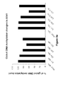
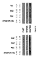
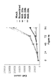
- MiR-29s expression is inversely correlated to DNMT3A and 3B in lung cancer patients.
- miR-29s directly target both DNMT3A and 3B.
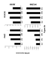
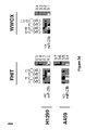
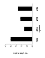
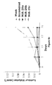
- the enforced expression of miR-29s in lung cancer cell lines restores normal patterns of DNA methylation, induces re-expression of methylation-silenced tumor suppressor genes (TSGs), such as FHIT, and WWOX 14 and inhibits tumorigenicity both in vitro and in vivo.
- TSGs methylation-silenced tumor suppressor genes
- TMAs tissue microarrays
- the DNMT3A and DNTM3B complementary sites were cloned into the 3'UTR of the firefly luciferase gene and co-transfected with miR-29a, mi-R29b or miR-29c in A459 (NSCLC) cells.
- miRNAs As shown in Fig. 2a , all three miRNAs (miR-29a, mi-R29b or miR-29c) significantly reduced the luciferase activity with respect to the scrambled oligonucleotide.
- qRT-PCR quantitative RT-PCR
- DNMT3B may be possibly due to an overall higher number of predicted matching "seeds" of miR-29s with 3B 3'-UTR (3 for 29a, 1 for 29b, 1 for 29c) than with 3A 3'-UTR (1 for each miR-29).
- the DNMT3B 3'UTR presents matching sites differing for no more than 1 nucleotide for miR-29a and for 29b/c matches.
- the transfection with any member of the miR-29 family may result in more robust silencing of DNMT3B than DNMT3A, according to the "coordinate principle" that miRNAs may act cooperatively through multiple target sites in one gene 10,23 .
- a recently described detection method was used to detect miRNA-mRNA complexes in eukaryotic cells by synthesizing cDNA on a mRNA template using miRNAs as the endogenous cytoplasmic primer 24 .
- the endogenous miR-29b at the Pictar-predicted site of interaction with 3' UTR 8 , is able to function as a "natural" primer to initiate the retrotranscription of DNMT3B mRNA ( Fig. 2d ).
- DNMT3A and DNMT3B mRNA expression is inversely correlated to the levels of miR-29s in primary NSCLC tissues.
- NSCLCs Fourteen (14) NSCLCs were analyzed for expression levels of DNMT3A and DNMT3B mRNAs and for miR-29a, 29b, and 29c expression by qRT-PCR 25 .
- miR-29s target DNMT3A and DNMT3B shows that expression of these miRNAs contributes to the DNA epigenetic modifications in cancer.
- A549 cells were transfected with miR-29a, miR-29b, miR-29c or scrambled oligonucleotides and analyzed global DNA methylation 48 and 72 h later, using an LC-MS/MS method. 26 .
- the effects of re-expression of miR-29s on tumorigenicity of A549 cells were also assessed.
- the ectopic expression of miR-29s in A549 inhibited in vitro cell growth ( Fig. 4a ), and induced apoptosis with respect to the scrambled control transfection ( Fig. 4b ).
- this example shows that expression of miR-29 family members is inversely correlated with DNMT3A and DNMT3B expression in lung cancers and these miRNAs down-modulate expression levels of both enzymes.
- NSCLCs non small cell lung carcinomas
- TMAs tissue microarrays
- RNAs were isolated by TRIzol (Invitrogen, Carlsbad, CA) extraction, according to the manufacturer's instructions.
- TMAs Tissue micro arrays
- each array contained 4 samples of each lung cancer along with multiple appropriate lung and other normal tissue spots.
- the TMAs usually two for each antiserum, were stained with antisera against DNMT1, DNTM3A and DNMT3B proteins, and expression of each of these enzymes in lung cancer was compared with clinical features to seek significant correlations.
- DNMT1 , DNMT3A and DNMT3B protein expression were assessed on the lung cancer TMAs , using DNMT1 antiserum from GeneTex (GTX13537, San Antonio, TX) at a dilution of 1:150; DNMT3A antiserum from Novus Biologicals (ab-4897, Littleton, CO) at a dilution of 1:25 and DNTM3B antiserum from Abgent (AP1035a, San Diego, CA) at a dilution of 1:32.
- GTX13537 San Antonio, TX
- DNMT3A antiserum from Novus Biologicals (ab-4897, Littleton, CO) at a dilution of 1:25
- DNTM3B antiserum from Abgent (AP1035a, San Diego, CA) at a dilution of 1:32.
- TMAs were read and scored by a pathologist who was blinded to clinical features; expression scores were determined by multiplying the percent of positive cells in an individual sample by the intensity of staining; the intensity of staining was assessed on a scale from 1 to 3, where 1 was the least intense staining and 3 was the most intense. For example, a sample with 10% positive cells with intensity 3 was assigned a score of 30, the same score as a sample with 30% positive cells with intensity 1.
- Quantitative RT-PCR Quantitative RT-PCR analysis for miRNAs was performed in triplicate with the TaqMan MicroRNA assays kit (Applied Biosystems, Foster City, CA), according to the manufacturer's instructions. 18S RNA was used for normalization; qRT-PCR analyses for other genes of interest were performed as previously described 1 . RNA was reverse transcribed to cDNA with gene-specific primers and IQ SYBR Green Supermix (Biorad, Hercules, CA). GAPDH served as normalization control.
- A549 and H1299 cells were transfected in 6-well plates by using Lipofectamine 2000 reagent (Invitrogen), according to the manufacturer's protocol, with 100 nM (final) of antisense miR-29a, 29b-1, 29c or scrambled antisense miR (Fidelity Systems, Gaithersburg, MD).
- A549 and H1299 lung cancer cells from the American Type Culture Collection were maintained in RPMI medium 1640 with 10% FBS and antibiotics (100 U/ml penicillin, and 100 ⁇ g/ml streptomycin).
- Luciferase reporter assay for targeting DNMT 3'UTRs.
- a DNMT3A 3'UTR segment of 979 bp and a DNMT3B 3'UTR segment of 978 bp were amplified by PCR from human genomic DNA and inserted into the pGL3-control vector with SV40 promoter (Promega), using the XbaI site immediately upstream from the stop codon of luciferase.
- the following sets of primers were used to generate specific fragments:
- DNMT3A-UTR Fw 5'- GCTCTAGAGC CGAAAAGGGTTGGACATCAT-3', [SEQ ID NO: 15]
- DNMT3A-UTR Rv 5'- GCTCTAGAGC GCCGAGGGAGTCTCCTTTTA-3'; [SEQ ID NO: 16]
- DNMT3B-UTR Fw 5'- GCTCTAGAGC TAGGTAGCAACGTGGCTTTT -3', [SEQ ID NO: 17]
- DNMT3B-UTR Rv 5'- GCTCTAGAGC GCCCCACAAAACTTGTCAAC -3'. [SEQ ID NO: 18]
- the predicted match seed of miR-29s is located in the DNMT3A 3'-UTR down fragment, which was used to perform the luciferase assay.
- A549 cells were co-transfected in 12-well plates by using Lipofectamine 2000 reagent (Invitrogen), according to the manufacturer's protocol, with 0.4 ⁇ g of the firefly luciferase report vector and 0.08 ⁇ g of the control vector containing Renilla luciferase pRL-TK vector (Promega). For each well, 100 nM (final) of precursor miR-29a, 29b-1, 29c or scrambled miR (Ambion) was used. Firefly and Renilla luciferase activities were measured consecutively by using dual-luciferase assays (Promega), 24 h after the transfection. The experiments were performed in triplicate.
- GFP-repression constructs to assess effect of DNMT 3'UTRs on protein expression.
- a DNMT3A 3'UTR segment of 1472 bp and a DNMT3B 3'UTR segment of 1566 bp were amplified by PCR from human genomic DNA and inserted into the QBI-GFP25 vector (Autofluorescent Proteins, Canada), using the BamHI-EcoRI cloning sites located 3' of the GFP encoding sequence of the vector (which has no stop codon at the end of the GFP coding sequence).
- the following primer sets were used to generate specific fragments:
- DNMT3A-GFP Fw 5'- CGGGATCC GCAGGATAGCCAAGTTCAGC -3', [SEQ ID NO: 19]
- DNMT3A-GFP Rv 5'- CCCAAGCTTAAGTGAGAAACTGGGCCTGA -3'; [SEQ ID NO: 20]
- DNMT3B-GFP Fw 5'- CGGGATCC CTCGATCAAACAGGGGAAAA -3', [SEQ ID NO: 21]
- DNMT3B-GFP Rv 5'- CCCAAGCTT GTTACGTCGTGGCTCCAGTT -3' [SEQ ID NO: 22].
- A549 cells were co-transfected in 12-well plates using Lipofectamine 2000 reagent (Invitrogen) according to the manufacturer's protocol with 2 ⁇ g of the GFP repression vector containing the 3'UTR of DNMT3A (QBI-GFP25-DNMT3A) or the 3'UTR of DNMT3B (QBI-GFP25-DNMT3B) and with 100 nM (final) of precursor miR 29a, 29b-1, 29c, or scrambled oligonucleotide (Ambion).
- a group of cells was also transfected with the GFP vector (no miR). Cells were harvested after 24 h. Protein extraction and immunoblot analysis were performed as previously described 2 . The following primary antisera were used: rabbit polyclonal anti-GFP, 1:1000 (Novus Biologicals, Littleton, CO).
- RNA complexes To detect miR-29b-DNMT3B RNA complexes, we used the method described by Vatolin S. et al. 3 to determine if endogenous miR-29b was able to serve as primer for retrotranscription of DNMT3B mRNA in A549 cells.
- the cDNAs were cloned in pCR2.1-TOPO Vector (Invitrogen). The following sets of primers and adapter sequence were used (GSP meaning gene specific primer):
- GSP-DNMT3B 5'-GAGATGACAGGGAAAACTGC-3'; [SEQ ID NO: 23]
- GSP-DNMT3B 5N 5'-ACAGGGAAAACTGCAAAGCT-3'; [SEQ ID NO: 24]
- Adapter 5N 5'- CTGAAGGAGTAGAAA -3' [SEQ ID NO: 26].
- Primers 5N represent nested primers from the adapter and GSP sequence used to sensitize the detection of the PCR bands.
- Quantitative DNA methylation Quantitative DNA methylation analysis of the regulatory regions of FHIT and WWOX was done using the EpiTYPER methylation analysis assay (Sequenom, San Diego, CA). Two bisulfite reactions (one for each gene CpG island) were designed, which covered 7 CpGs and 11 CpGs for FHIT and WWOX respectively.
- the DNA of scrambled-, or miR-29b-transfected A549/H1299 was extracted 48h after the transfection and 1 ⁇ g of DNA was bisulfite treated, in vitro transcribed, cleaved by Rnase A, and subjected to matrix-assisted laser desorption ionization-time of flight (MALDI-TOF) mass spectrometry analysis to determine methylation patterns, as described 5 .
- MALDI-TOF matrix-assisted laser desorption ionization-time of flight
- FHIT Fw 5'- GGGGAGGTAAGTTTAAGTGGAATATTGTT -3' [SEQ ID NO: 27]
- FHIT Rv 5'- CACCCCCAAAACCAAAAACTATAAC -3' [SEQ ID NO: 28]
- WWOX Fw 5'- TTGAAAGAAAGTTTTTTAAAATTAGGAAAT -3' [SEQ ID NO: 29]
- WWOX Rv 5'- TCAAAAAAACAAAACCTAAAAAAAA -3' [SEQ ID NO: 30].
- the heat map in Fig. 3d was created using Heatmap builder version 1.0 by Stanford University.
- A549 cells (5 x 10 4 ) were plated in 6x multi-well plates and transfected, after 24 hours, with scrambled oligonucleotides or miR-29s oligonucleotides from Ambion at a final concentration of 100nM, with Lipofectamine 2000 (Invitrogen), according to manufacturer's protocol. As a control also not transfected (mock) cells were included. Cells were harvested and counted at 24h intervals using a ViCell counter (Beckman Coulter, Fullerton, CA). Each sample was run in triplicate.
- A549 cells (2 x 10 5 ) were transfected with scrambled oligonucleotides or miR-29s oligonucleotides from Ambion at a final concentration of 100nM, with Lipofectamine 2000 (Invitrogen), according to manufacturer's protocol. After 24h cells were resuspended in binding buffer containing annexin V-fluorescein isothiocyanate (FITC) and propidium iodide according to the supplier's instructions (BD Biosciences, San Diego, CA), and assessed by flow cytometry using a Beckman-Coulter model EPICS XL cytometer (Beckman-Coulter). Each sample was run in triplicate.
- FITC annexin V-fluorescein isothiocyanate
- BD Biosciences San Diego, CA
- a diagnostic method of assessing whether a patient has a lung cancer-related disease or has higher than normal risk for developing a lung cancer-related disease comprising the steps of comparing the level of expression of a marker in a patient sample and the normal level of expression of the marker in a control, e.g., a sample from a patient without a lung cancer-related disease.
- a significantly higher level of expression of the marker in the patient sample as compared to the normal level is an indication that the patient is afflicted with a lung cancer-related disease or has higher than normal risk for developing a lung cancer-related disease.
- the markers are selected such that the positive predictive value of the methods is at least about 10%, and in certain non-limiting embodiments, about 25%, about 50% or about 90%. Also preferred for use in the methods are markers that are differentially expressed, as compared to normal cells, by at least two-fold in at least about 20%, and in certain non-limiting embodiments, about 50% or about 75%.
- the method comprises comparing: a) the level of expression of a marker in a patient sample, and b) the normal level of expression of the marker in a control non-lung cancer-related disease sample.
- a significantly higher level of expression of the marker in the patient sample as compared to the normal level is an indication that the patient is afflicted with a lung cancer-related disease.
- diagnostic methods for assessing the efficacy of a therapy for inhibiting a lung cancer-related disease in a patient comprise comparing: a) expression of a marker in a first sample obtained from the patient prior to providing at least a portion of the therapy to the patient, and b) expression of the marker in a second sample obtained from the patient following provision of the portion of the therapy.
- a significantly lower level of expression of the marker in the second sample relative to that in the first sample is an indication that the therapy is efficacious for inhibiting a lung cancer-related disease in the patient.
- the "therapy” may be any therapy for treating a lung cancer-related disease including, but not limited to, pharmaceutical compositions, gene therapy and biologic therapy such as the administering of antibodies and chemokines.
- the methods described herein may be used to evaluate a patient before, during and after therapy, for example, to evaluate the reduction in disease state.
- the diagnostic methods are directed to therapy using a chemical or biologic agent. These methods comprise comparing: a) expression of a marker in a first sample obtained from the patient and maintained in the presence of the chemical or biologic agent, and b) expression of the marker in a second sample obtained from the patient and maintained in the absence of the agent. A significantly lower level of expression of the marker in the second sample relative to that in the first sample is an indication that the agent is efficacious for inhibiting a lung cancer-related disease in the patient.
- the first and second samples can be portions of a single sample obtained from the patient or portions of pooled samples obtained from the patient.
- a monitoring method for assessing the progression of a lung cancer-related disease in a patient comprising: a) detecting in a patient sample at a first time point, the expression of a marker; b) repeating step a) at a subsequent time point in time; and c) comparing the level of expression detected in steps a) and b), and therefrom monitoring the progression of a lung cancer-related disease in the patient.
- a significantly higher level of expression of the marker in the sample at the subsequent time point from that of the sample at the first time point is an indication that the lung cancer-related disease has progressed, whereas a significantly lower level of expression is an indication that the lung cancer-related disease has regressed.
- a diagnostic method for determining whether a lung cancer-related disease has worsened or is likely to worsen in the future comprising comparing: a) the level of expression of a marker in a patient sample, and b) the normal level of expression of the marker in a control sample.
- a significantly higher level of expression in the patient sample as compared to the normal level is an indication that the lung cancer-related disease has worsened or is likely to worsen in the future.
- test method for selecting a composition for inhibiting a lung cancer-related disease in a patient.
- This method comprises the steps of: a) obtaining a sample comprising cells from the patient; b) separately maintaining aliquots of the sample in the presence of a plurality of test compositions; c) comparing expression of a marker in each of the aliquots; and d) selecting one of the test compositions which significantly reduces the level of expression of the marker in the aliquot containing that test composition, relative to the levels of expression of the marker in the presence of the other test compositions.
- test method of assessing the harmful potential of a compound in causing a lung cancer-related disease comprises the steps of: a) maintaining separate aliquots of cells in the presence and absence of the compound; and b) comparing expression of a marker in each of the aliquots. A significantly higher level of expression of the marker in the aliquot maintained in the presence of the compound, relative to that of the aliquot maintained in the absence of the compound, is an indication that the compound possesses such harmful potential.
- a method of inhibiting a lung cancer-related disease in a patient comprises the steps of: a) obtaining a sample comprising cells from the patient; b) separately maintaining aliquots of the sample in the presence of a plurality of compositions; c) comparing expression of a marker in each of the aliquots; and d) administering to the patient at least one of the compositions which significantly lowers the level of expression of the marker in the aliquot containing that composition, relative to the levels of expression of the marker in the presence of the other compositions.
- the level of expression of a marker in a sample can be assessed, for example, by detecting the presence in the sample of: the corresponding marker protein or a fragment of the protein (e.g. by using a reagent, such as an antibody, an antibody derivative, an antibody fragment or single-chain antibody, which binds specifically with the protein or protein fragment) the corresponding marker nucleic acid (e.g. a nucleotide transcript, or a complement thereof), or a fragment of the nucleic acid (e.g.
- a metabolite which is produced directly (i.e., catalyzed) or indirectly by the corresponding marker protein.
- any of the aforementioned methods may be performed using at least one or a plurality (e.g., 2, 3, 5, or 10 or more) of lung cancer-related disease markers.
- the level of expression in the sample of each of a plurality of markers, at least one of which is a marker is compared with the normal level of expression of each of the plurality of markers in samples of the same type obtained from control humans not afflicted with a lung cancer-related disease.
- a significantly altered (i.e., increased or decreased as specified in the above-described methods using a single marker) level of expression in the sample of one or more markers, or some combination thereof, relative to that marker's corresponding normal or control level, is an indication that the patient is afflicted with a lung cancer-related disease.
- the marker(s) are selected such that the positive predictive value of the method is at least about 10%.
- the candidate agents may be pharmacologic agents already known in the art or may be agents previously unknown to have any pharmacological activity.
- the agents may be naturally arising or designed in the laboratory. They may be isolated from microorganisms, animals or plants, or may be produced recombinantly, or synthesized by any suitable chemical method. They may be small molecules, nucleic acids, proteins, peptides or peptidomimetics.
- candidate agents are small organic compounds having a molecular weight of more than 50 and less than about 2,500 daltons.
- Candidate agents comprise functional groups necessary for structural interaction with proteins.
- Candidate agents are also found among biomolecules including, but not limited to: peptides, saccharides, fatty acids, steroids, purines, pyrimidines, derivatives, structural analogs or combinations thereof.
- Candidate agents are obtained from a wide variety of sources including libraries of synthetic or natural compounds. There are, for example, numerous means available for random and directed synthesis of a wide variety of organic compounds and biomolecules, including expression of randomized oligonucleotides and oligopeptides. Alternatively, libraries of natural compounds in the form of bacterial, fungal, plant and animal extracts are available or readily produced. Additionally, natural or synthetically produced libraries and compounds are readily modified through conventional chemical, physical and biochemical means, and may be used to produce combinatorial libraries.
- the candidate agents can be obtained using any of the numerous approaches in combinatorial library methods art, including, by non-limiting example: biological libraries; spatially addressable parallel solid phase or solution phase libraries; synthetic library methods requiring deconvolution; the "one-bead one-compound” library method; and synthetic library methods using affinity chromatography selection.
- certain pharmacological agents may be subjected to directed or random chemical modifications, such as acylation, alkylation, esterification, amidification, etc. to produce structural analogs.
- the same methods for identifying therapeutic agents for treating a lung cancer-related disease can also be used to validate lead compounds/agents generated from in vitro studies.
- the candidate agent may be an agent that up- or down-regulates one or more lung cancer-related disease response pathways.
- the candidate agent may be an antagonist that affects such pathway.
- an agent that interferes with a signaling cascade is administered to an individual in need thereof, such as, but not limited to, lung cancer-related disease patients in whom such complications are not yet evident and those who already have at least one lung cancer-related disease response.
- such treatment is useful to prevent the occurrence of such lung cancer-related disease response and/or reduce the extent to which they occur.
- such treatment is useful to reduce the extent to which such lung cancer-related disease response occurs, prevent their further development or reverse the lung cancer-related disease response.
- the agent that interferes with the lung cancer-related disease response cascade may be an antibody specific for such response.
- an antisense oligonucleotide can be provided to the lung cancer-related disease cells in order to inhibit transcription, translation, or both, of the marker(s).
- a polynucleotide encoding an antibody, an antibody derivative, or an antibody fragment which specifically binds a marker protein, and operably linked with an appropriate promoter/regulator region can be provided to the cell in order to generate intracellular antibodies which will inhibit the function or activity of the protein.
- the expression and/or function of a marker may also be inhibited by treating the lung cancer-related disease cell with an antibody, antibody derivative or antibody fragment that specifically binds a marker protein.
- a variety of molecules can be screened in order to identify molecules which inhibit expression of a marker or inhibit the function of a marker protein.
- the compound so identified can be provided to the patient in order to inhibit lung cancer-related disease cells of the patient.
- any marker or combination of markers, as well as any certain markers in combination with the markers, may be used in the compositions, kits and methods described herein.
- this difference can be as small as the limit of detection of the method for assessing expression of the marker, it is desirable that the difference be at least greater than the standard error of the assessment method, and, in certain embodiments, a difference of at least 2-, 3-, 4-, 5-, 6-, 7-, 8-, 9-, 10-, 15-, 20-, 100-, 500-, 1000-fold or greater than the level of expression of the same marker in normal tissue.
- marker proteins are secreted to the extracellular space surrounding the cells. These markers are used in certain embodiments of the compositions, kits and methods, owing to the fact that such marker proteins can be detected in a lung cancer-associated body fluid sample, which may be more easily collected from a human patient than a tissue biopsy sample.
- in vivo techniques for detection of a marker protein include introducing into a subject a labeled antibody directed against the protein.
- the antibody can be labeled with a radioactive marker whose presence and location in a subject can be detected by standard imaging techniques.
- the marker protein is expressed in, for example, a mammalian cell, such as a human lung line, extracellular fluid is collected, and the presence or absence of the protein in the extracellular fluid is assessed (e.g. using a labeled antibody which binds specifically with the protein).
- the level of expression of the marker can be assessed by assessing the amount (e.g., absolute amount or concentration) of the marker in a sample.
- the cell sample can, of course, be subjected to a variety of post-collection preparative and storage techniques (e.g., nucleic acid and/or protein extraction, fixation, storage, freezing, ultrafiltration, concentration, evaporation, centrifugation, etc.) prior to assessing the amount of the marker in the sample.
- the markers may be shed from the cells into the digestive system, the blood stream and/or interstitial spaces.
- the shed markers can be tested, for example, by examining the serum or plasma.
- compositions, kits and methods can be used to detect expression of marker proteins having at least one portion which is displayed on the surface of cells which express it.
- immunological methods may be used to detect such proteins on whole cells, or computer-based sequence analysis methods may be used to predict the presence of at least one extracellular domain (i.e., including both secreted proteins and proteins having at least one cell-surface domain).
- Expression of a marker protein having at least one portion which is displayed on the surface of a cell which expresses it may be detected without necessarily lysing the cell (e.g., using a labeled antibody which binds specifically with a cell-surface domain of the protein).
- Expression of a marker may be assessed by any of a wide variety of methods for detecting expression of a transcribed nucleic acid or protein.
- Non-limiting examples of such methods include immunological methods for detection of secreted, cell-surface, cytoplasmic or nuclear proteins, protein purification methods, protein function or activity assays, nucleic acid hybridization methods, nucleic acid reverse transcription methods and nucleic acid amplification methods.
- expression of a marker is assessed using an antibody (e.g., a radio-labeled, chromophore-labeled, fluorophore-labeled or enzyme-labeled antibody), an antibody derivative (e.g., an antibody conjugated with a substrate or with the protein or ligand of a protein-ligand pair), or an antibody fragment (e.g., a single-chain antibody, an isolated antibody hypervariable domain, etc.) which binds specifically with a marker protein or fragment thereof, including a marker protein which has undergone all or a portion of its normal post-translational modification.
- an antibody e.g., a radio-labeled, chromophore-labeled, fluorophore-labeled or enzyme-labeled antibody
- an antibody derivative e.g., an antibody conjugated with a substrate or with the protein or ligand of a protein-ligand pair
- an antibody fragment e.g., a single-chain antibody, an isolated antibody hyper
- expression of a marker is assessed by preparing mRNA/cDNA (i.e., a transcribed polynucleotide) from cells in a patient sample, and by hybridizing the mRNA/cDNA with a reference polynucleotide which is a complement of a marker nucleic acid, or a fragment thereof.
- cDNA can, optionally, be amplified using any of a variety of polymerase chain reaction methods prior to hybridization with the reference polynucleotide; preferably, it is not amplified.
- Expression of one or more markers can likewise be detected using quantitative PCR to assess the level of expression of the marker(s).
- any of the many methods of detecting mutations or variants e.g., single nucleotide polymorphisms, deletions, etc.
- a mixture of transcribed polynucleotides obtained from the sample is contacted with a substrate having fixed thereto a polynucleotide complementary to or homologous with at least a portion (e.g., at least 7, 10, 15, 20, 25, 30, 40, 50, 100, 500, or more nucleotide residues) of a marker nucleic acid.
- polynucleotides complementary to or homologous with are differentially detectable on the substrate (e.g., detectable using different chromophores or fluorophores, or fixed to different selected positions), then the levels of expression of a plurality of markers can be assessed simultaneously using a single substrate (e.g., a "gene chip" microarray of polynucleotides fixed at selected positions).
- a method of assessing marker expression which involves hybridization of one nucleic acid with another, it is desired that the hybridization be performed under stringent hybridization conditions.
- the biomarker assays can be performed using mass spectrometry or surface plasmon resonance.
- the method of identifying an agent active against a lung cancer-related disease can include a) providing a sample of cells containing one or more markers or derivative thereof; b) preparing an extract from said cells; c) mixing said extract with a labeled nucleic acid probe containing a marker binding site; and, d) determining the formation of a complex between the marker and the nucleic acid probe in the presence or absence of the test agent.
- the determining step can include subjecting said extract/nucleic acid probe mixture to an electrophoretic mobility shift assay.
- the determining step comprises an assay selected from an enzyme linked immunoabsorption assay (ELISA), fluorescence based assays and ultra high throughput assays, for example surface plasmon resonance (SPR) or fluorescence correlation spectroscopy (FCS) assays.
- ELISA enzyme linked immunoabsorption assay
- SPR fluorescence based assays
- FCS fluorescence correlation spectroscopy
- the SPR sensor is useful for direct real-time observation of biomolecular interactions since SPR is sensitive to minute refractive index changes at a metal-dielectric surface.
- SPR is a surface technique that is sensitive to changes of 10 5 to 10 -6 refractive index (RI) units within approximately 200 nm of the SPR sensor/sample interface.
- RI refractive index
- compositions, kits, and methods rely on detection of a difference in expression levels of one or more markers, it is desired that the level of expression of the marker is significantly greater than the minimum detection limit of the method used to assess expression in at least one of normal cells and lung cancer-affected cells.
- compositions, kits, and methods are thus useful for characterizing one or more of the stage, grade, histological type, and nature of a lung cancer-related disease in patients.
- the marker or panel of markers is selected such that a positive result is obtained in at least about 20%, and in certain embodiments, at least about 40%, 60%, or 80%, and in substantially all patients afflicted with a lung cancer-related disease of the corresponding stage, grade, histological type, or nature.
- the marker or panel of markers invention can be selected such that a positive predictive value of greater than about 10% is obtained for the general population (in a non-limiting example, coupled with an assay specificity greater than 80%).
- the level of expression of each marker in a patient sample can be compared with the normal level of expression of each of the plurality of markers in non- lung cancer samples of the same type, either in a single reaction mixture (i.e. using reagents, such as different fluorescent probes, for each marker) or in individual reaction mixtures corresponding to one or more of the markers.
- a significantly increased level of expression of more than one of the plurality of markers in the sample, relative to the corresponding normal levels is an indication that the patient is afflicted with a lung cancer-related disease.
- 2, 3, 4, 5, 8, 10, 12, 15, 20, 30, or 50 or more individual markers can be used; in certain embodiments, the use of fewer markers may be desired.
- the marker used therein be a marker which has a restricted tissue distribution, e.g., normally not expressed in a non-lung tissue.
- compositions, kits, and methods will be of particular utility to patients having an enhanced risk of developing a lung cancer-related disease and their medical advisors.
- Patients recognized as having an enhanced risk of developing a lung cancer-related disease include, for example, patients having a familial history of a lung cancer-related disease.
- the level of expression of a marker in normal human lung tissue can be assessed in a variety of ways.
- this normal level of expression is assessed by assessing the level of expression of the marker in a portion of lung cells which appear to be normal and by comparing this normal level of expression with the level of expression in a portion of the lung cells which is suspected of being abnormal.
- population-average values for normal expression of the markers may be used.
- the 'normal' level of expression of a marker may be determined by assessing expression of the marker in a patient sample obtained from a non-lung cancer-afflicted patient, from a patient sample obtained from a patient before the suspected onset of a lung cancer-related disease in the patient, from archived patient samples, and the like.
- compositions, kits, and methods for assessing the presence of lung cancer-related disease cells in a sample e.g. an archived tissue sample or a sample obtained from a patient.
- a sample e.g. an archived tissue sample or a sample obtained from a patient.
- these compositions, kits, and methods are substantially the same as those described above, except that, where necessary, the compositions, kits, and methods are adapted for use with samples other than patient samples.
- the sample to be used is a parafinized, archived human tissue sample, it can be necessary to adjust the ratio of compounds in the compositions, in the kits, or the methods used to assess levels of marker expression in the sample.
- a method of making an isolated hybridoma which produces an antibody useful for assessing whether a patient is afflicted with a lung cancer-related disease.
- a protein or peptide comprising the entirety or a segment of a marker protein is synthesized or isolated (e.g. by purification from a cell in which it is expressed or by transcription and translation of a nucleic acid encoding the protein or peptide in vivo or in vitro).
- a vertebrate for example, a mammal such as a mouse, rat, rabbit, or sheep, is immunized using the protein or peptide.
- the vertebrate may optionally (and preferably) be immunized at least one additional time with the protein or peptide, so that the vertebrate exhibits a robust immune response to the protein or peptide.
- Splenocytes are isolated from the immunized vertebrate and fused with an immortalized cell line to form hybridomas, using any of a variety of methods. Hybridomas formed in this manner are then screened using standard methods to identify one or more hybridomas which produce an antibody which specifically binds with the marker protein or a fragment thereof. There is also provided herein hybridomas made by this method and antibodies made using such hybridomas.
- a method of assessing the efficacy of a test compound for inhibiting lung cancer-related disease cells As described above, differences in the level of expression of the markers correlate with the abnormal state of lung cells. Although it is recognized that changes in the levels of expression of certain of the markers likely result from the abnormal state of lung cells, it is likewise recognized that changes in the levels of expression of other of the markers induce, maintain, and promote the abnormal state of those cells. Thus, compounds which inhibit a lung cancer-related disease in a patient will cause the level of expression of one or more of the markers to change to a level nearer the normal level of expression for that marker (i.e. the level of expression for the marker in normal lung cells).
- This method thus comprises comparing expression of a marker in a first lung cell sample and maintained in the presence of the test compound and expression of the marker in a second lung cell sample and maintained in the absence of the test compound.
- a significantly reduced expression of a marker in the presence of the test compound is an indication that the test compound inhibits a lung cancer-related disease.
- the lung cell samples may, for example, be aliquots of a single sample of normal lung cells obtained from a patient, pooled samples of normal lung cells obtained from a patient, cells of a normal lung cell line, aliquots of a single sample of lung cancer-related disease cells obtained from a patient, pooled samples of lung cancer-related disease cells obtained from a patient, cells of a lung cancer-related disease cell line, or the like.
- the samples are lung cancer-related disease cells obtained from a patient and a plurality of compounds believed to be effective for inhibiting various lung cancer-related diseases are tested in order to identify the compound which is likely to best inhibit the lung cancer-related disease in the patient.
- This method may likewise be used to assess the efficacy of a therapy for inhibiting a lung cancer-related disease in a patient.
- the level of expression of one or more markers in a pair of samples is assessed.
- the therapy induces a significantly lower level of expression of a marker then the therapy is efficacious for inhibiting a lung cancer-related disease.
- alternative therapies can be assessed in vitro in order to select a therapy most likely to be efficacious for inhibiting a lung cancer-related disease in the patient.
- the abnormal state of human lung cells is correlated with changes in the levels of expression of the markers.
- a method for assessing the harmful potential of a test compound comprises maintaining separate aliquots of human lung cells in the presence and absence of the test compound. Expression of a marker in each of the aliquots is compared. A significantly higher level of expression of a marker in the aliquot maintained in the presence of the test compound (relative to the aliquot maintained in the absence of the test compound) is an indication that the test compound possesses a harmful potential.
- the relative harmful potential of various test compounds can be assessed by comparing the degree of enhancement or inhibition of the level of expression of the relevant markers, by comparing the number of markers for which the level of expression is enhanced or inhibited, or by comparing both.
- One aspect pertains to isolated marker proteins and biologically active portions thereof, as well as polypeptide fragments suitable for use as immunogens to raise antibodies directed against a marker protein or a fragment thereof.
- the native marker protein can be isolated from cells or tissue sources by an appropriate purification scheme using standard protein purification techniques.
- a protein or peptide comprising the whole or a segment of the marker protein is produced by recombinant DNA techniques.
- Alternative to recombinant expression such protein or peptide can be synthesized chemically using standard peptide synthesis techniques.
- an “isolated” or “purified” protein or biologically active portion thereof is substantially free of cellular material or other contaminating proteins from the cell or tissue source from which the protein is derived, or substantially free of chemical precursors or other chemicals when chemically synthesized.
- the language “substantially free of cellular material” includes preparations of protein in which the protein is separated from cellular components of the cells from which it is isolated or recombinantly produced.
- protein that is substantially free of cellular material includes preparations of protein having less than about 30%, 20%, 10%, or 5% (by dry weight) of heterologous protein (also referred to herein as a "contaminating protein").
- the protein or biologically active portion thereof is recombinantly produced, it is also preferably substantially free of culture medium, i.e., culture medium represents less than about 20%, 10%, or 5% of the volume of the protein preparation.
- culture medium represents less than about 20%, 10%, or 5% of the volume of the protein preparation.
- the protein is produced by chemical synthesis, it is preferably substantially free of chemical precursors or other chemicals, i.e., it is separated from chemical precursors or other chemicals which are involved in the synthesis of the protein. Accordingly such preparations of the protein have less than about 30%, 20%, 10%, 5% (by dry weight) of chemical precursors or compounds other than the polypeptide of interest.
- Biologically active portions of a marker protein include polypeptides comprising amino acid sequences sufficiently identical to or derived from the amino acid sequence of the marker protein, which include fewer amino acids than the full length protein, and exhibit at least one activity of the corresponding full-length protein.
- biologically active portions comprise a domain or motif with at least one activity of the corresponding full-length protein.
- a biologically active portion of a marker protein can be a polypeptide which is, for example, 10, 25, 50, 100 or more amino acids in length.
- other biologically active portions, in which other regions of the marker protein are deleted can be prepared by recombinant techniques and evaluated for one or more of the functional activities of the native form of the marker protein.
- useful proteins are substantially identical (e.g., at least about 40%, and in certain embodiments, 50%, 60%, 70%, 80%, 90%, 95%, or 99%) to one of these sequences and retain the functional activity of the corresponding naturally-occurring marker protein yet differ in amino acid sequence due to natural allelic variation or mutagenesis.
- libraries of segments of a marker protein can be used to generate a variegated population of polypeptides for screening and subsequent selection of variant marker proteins or segments thereof.
- diagnostic assays for determining the level of expression of one or more marker proteins or nucleic acids, in order to determine whether an individual is at risk of developing a lung cancer-related disease.
- Such assays can be used for prognostic or predictive purposes to thereby prophylactically treat an individual prior to the onset of the lung cancer-related disease.
- the methods are useful for at least periodic screening of the same individual to see if that individual has been exposed to chemicals or toxins that change his/her expression patterns.
- Yet another aspect pertains to monitoring the influence of agents (e.g., drugs or other compounds administered either to inhibit a lung cancer-related disease or to treat or prevent any other disorder (e.g., in order to understand any system effects that such treatment may have) on the expression or activity of a marker in clinical trials.
- agents e.g., drugs or other compounds administered either to inhibit a lung cancer-related disease or to treat or prevent any other disorder (e.g., in order to understand any system effects that such treatment may have) on the expression or activity of a marker in clinical trials.
- a "pharmacogenomic marker” is an objective biochemical marker whose expression level correlates with a specific clinical drug response or susceptibility in a patient.
- the presence or quantity of the pharmacogenomic marker expression is related to the predicted response of the patient and more particularly the patient's tumor to therapy with a specific drug or class of drugs.
- Monitoring the influence of agents (e.g., drug compounds) on the level of expression of a marker can be applied not only in basic drug screening, but also in clinical trials.
- agents e.g., drug compounds
- the effectiveness of an agent to affect marker expression can be monitored in clinical trials of subjects receiving treatment for a lung cancer-related disease.
- the present invention provides a method for monitoring the effectiveness of treatment of a subject with an agent (e.g., an agonist, antagonist, peptidomimetic, protein, peptide, nucleic acid, small molecule, or other drug candidate) comprising the steps of (i) obtaining a pre-administration sample from a subject prior to administration of the agent; (ii) detecting the level of expression of one or more selected markers in the pre-administration sample; (iii) obtaining one or more post-administration samples from the subject; (iv) detecting the level of expression of the marker(s) in the post-administration samples; (v) comparing the level of expression of the marker(s) in the pre-administration sample with the level of expression of the marker(s) in the post-administration sample or samples; and (vi) altering the administration of the agent to the subject accordingly.
- an agent e.g., an agonist, antagonist, peptidomimetic, protein, peptide, nucleic acid, small molecule, or other drug candidate
- increased expression of the marker gene(s) during the course of treatment may indicate ineffective dosage and the desirability of increasing the dosage.
- decreased expression of the marker gene(s) may indicate efficacious treatment and no need to change dosage.
- electronic apparatus readable media refers to any suitable medium for storing, holding or containing data or information that can be read and accessed directly by an electronic apparatus.
- Such media can include, but are not limited to: magnetic storage media, such as floppy discs, hard disc storage medium, and magnetic tape; optical storage media such as compact disc; electronic storage media such as RAM, ROM, EPROM, EEPROM and the like; and general hard disks and hybrids of these categories such as magnetic/optical storage media.
- the medium is adapted or configured for having recorded thereon a marker as described herein.
- the term "electronic apparatus” is intended to include any suitable computing or processing apparatus or other device configured or adapted for storing data or information.
- Examples of electronic apparatus suitable for use with the present invention include stand-alone computing apparatus; networks, including a local area network (LAN), a wide area network (WAN) Internet, Intranet, and Extranet; electronic appliances such as personal digital assistants (PDAs), cellular phone, pager and the like; and local and distributed processing systems.
- recorded refers to a process for storing or encoding information on the electronic apparatus readable medium. Those skilled in the art can readily adopt any method for recording information on media to generate materials comprising the markers described herein.
- a variety of software programs and formats can be used to store the marker information of the present invention on the electronic apparatus readable medium. Any number of data processor structuring formats (e.g., text file or database) may be employed in order to obtain or create a medium having recorded thereon the markers.
- data processor structuring formats e.g., text file or database
- By providing the markers in readable form one can routinely access the marker sequence information for a variety of purposes. For example, one skilled in the art can use the nucleotide or amino acid sequences in readable form to compare a target sequence or target structural motif with the sequence information stored within the data storage means. Search means are used to identify fragments or regions of the sequences which match a particular target sequence or target motif.
- a medium for holding instructions for performing a method for determining whether a subject has a lung cancer-related disease or a pre-disposition to a lung cancer-related disease wherein the method comprises the steps of determining the presence or absence of a marker and based on the presence or absence of the marker, determining whether the subject has a lung cancer-related disease or a pre-disposition to a lung cancer-related disease and/or recommending a particular treatment for a lung cancer-related disease or pre-lung cancer-related disease condition.
- an electronic system and/or in a network a method for determining whether a subject has a lung cancer-related disease or a pre-disposition to a lung cancer-related disease associated with a marker
- the method comprises the steps of determining the presence or absence of the marker, and based on the presence or absence of the marker, determining whether the subject has a lung cancer-related disease or a pre-disposition to a lung cancer-related disease, and/or recommending a particular treatment for the lung cancer-related disease or pre- lung cancer-related disease condition.
- the method may further comprise the step of receiving phenotypic information associated with the subject and/or acquiring from a network phenotypic information associated with the subject.
- Also provided herein is a network, a method for determining whether a subject has a lung cancer-related disease or a pre-disposition to a lung cancer-related disease associated with a marker, the method comprising the steps of receiving information associated with the marker, receiving phenotypic information associated with the subject, acquiring information from the network corresponding to the marker and/or a lung cancer-related disease, and based on one or more of the phenotypic information, the marker, and the acquired information, determining whether the subject has a lung cancer-related disease or a pre-disposition to a lung cancer-related disease.
- the method may further comprise the step of recommending a particular treatment for the lung cancer-related disease or pre-lung cancer-related disease condition.
- a business method for determining whether a subject has a lung cancer-related disease or a pre-disposition to a lung cancer-related disease comprising the steps of receiving information associated with the marker, receiving phenotypic information associated with the subject, acquiring information from the network corresponding to the marker and/or a lung cancer-related disease, and based on one or more of the phenotypic information, the marker, and the acquired information, determining whether the subject has a lung cancer-related disease or a pre-disposition to a lung cancer-related disease.
- the method may further comprise the step of recommending a particular treatment for the lung cancer-related disease or pre-lung cancer-related disease condition.
- an array that can be used to assay expression of one or more genes in the array.
- the array can be used to assay gene expression in a tissue to ascertain tissue specificity of genes in the array. In this manner, up to about 7000 or more genes can be simultaneously assayed for expression. This allows a profile to be developed showing a battery of genes specifically expressed in one or more tissues.
- tissue specificity not only tissue specificity, but also the level of expression of a battery of genes in the tissue is ascertainable.
- genes can be grouped on the basis of their tissue expression per se and level of expression in that tissue. This is useful, for example, in ascertaining the relationship of gene expression between or among tissues.
- one tissue can be perturbed and the effect on gene expression in a second tissue can be determined.
- the effect of one cell type on another cell type in response to a biological stimulus can be determined.
- Such a determination is useful, for example, to know the effect of cell-cell interaction at the level of gene expression. If an agent is administered therapeutically to treat one cell type but has an undesirable effect on another cell type, the method provides an assay to determine the molecular basis of the undesirable effect and thus provides the opportunity to co-administer a counteracting agent or otherwise treat the undesired effect. Similarly, even within a single cell type, undesirable biological effects can be determined at the molecular level. Thus, the effects of an agent on expression of other than the target gene can be ascertained and counteracted.
- the array can be used to monitor the time course of expression of one or more genes in the array. This can occur in various biological contexts, as disclosed herein, for example development of a lung cancer-related disease, progression of a lung cancer-related disease, and processes, such as cellular transformation associated with a lung cancer-related disease.
- the array is also useful for ascertaining the effect of the expression of a gene or the expression of other genes in the same cell or in different cells. This provides, for example, for a selection of alternate molecular targets for therapeutic intervention if the ultimate or downstream target cannot be regulated.
- the array is also useful for ascertaining differential expression patterns of one or more genes in normal and abnormal cells. This provides a battery of genes that could serve as a molecular target for diagnosis or therapeutic intervention.
- the markers may serve as surrogate markers for one or more disorders or disease states or for conditions leading up to a lung cancer-related disease state.
- a "surrogate marker” is an objective biochemical marker which correlates with the absence or presence of a disease or disorder, or with the progression of a disease or disorder. The presence or quantity of such markers is independent of the disease. Therefore, these markers may serve to indicate whether a particular course of treatment is effective in lessening a disease state or disorder.
- Surrogate markers are of particular use when the presence or extent of a disease state or disorder is difficult to assess through standard methodologies, or when an assessment of disease progression is desired before a potentially dangerous clinical endpoint is reached.
- a "pharmacodynamic marker” is an objective biochemical marker which correlates specifically with drug effects.
- the presence or quantity of a pharmacodynamic marker is not related to the disease state or disorder for which the drug is being administered; therefore, the presence or quantity of the marker is indicative of the presence or activity of the drug in a subject.
- a pharmacodynamic marker may be indicative of the concentration of the drug in a biological tissue, in that the marker is either expressed or transcribed or not expressed or transcribed in that tissue in relationship to the level of the drug. In this fashion, the distribution or uptake of the drug may be monitored by the pharmacodynamic marker.
- the presence or quantity of the pharmacodynamic marker may be related to the presence or quantity of the metabolic product of a drug, such that the presence or quantity of the marker is indicative of the relative breakdown rate of the drug in vivo.
- Pharmacodynamic markers are of particular use in increasing the sensitivity of detection of drug effects, particularly when the drug is administered in low doses. Since even a small amount of a drug may be sufficient to activate multiple rounds of marker transcription or expression, the amplified marker may be in a quantity which is more readily detectable than the drug itself. Also, the marker may be more easily detected due to the nature of the marker itself; for example, using the methods described herein, antibodies may be employed in an immune-based detection system for a protein marker, or marker-specific radiolabeled probes may be used to detect a mRNA marker. Furthermore, the use of a pharmacodynamic marker may offer mechanism-based prediction of risk due to drug treatment beyond the range of possible direct observations.
- the method of testing for lung cancer-related diseases comprises, for example measuring the expression level of each marker gene in a biological sample from a subject over time and comparing the level with that of the marker gene in a control biological sample.
- the marker gene is one of the genes described herein and the expression level is differentially expressed (for examples, higher or lower than that in the control), the subject is judged to be affected with a lung cancer-related disease.
- the expression level of the marker gene falls within the permissible range, the subject is unlikely to be affected with a lung cancer-related disease.
- the standard value for the control may be pre-determined by measuring the expression level of the marker gene in the control, in order to compare the expression levels.
- the standard value can be determined based on the expression level of the above-mentioned marker gene in the control.
- the permissible range is taken as ⁇ 2S.D. based on the standard value.
- Expression levels of marker genes include transcription of the marker genes to mRNA, and translation into proteins. Therefore, one method of testing for a lung cancer-related disease is performed based on a comparison of the intensity of expression of mRNA corresponding to the marker genes, or the expression level of proteins encoded by the marker genes.
- the measurement of the expression levels of marker genes in the testing for a lung cancer-related disease can be carried out according to various gene analysis methods. Specifically, one can use, for example, a hybridization technique using nucleic acids that hybridize to these genes as probes, or a gene amplification technique using DNA that hybridize to the marker genes as primers.
- the probes or primers used for the testing can be designed based on the nucleotide sequences of the marker genes.
- the identification numbers for the nucleotide sequences of the respective marker genes are describer herein.
- genes of higher animals generally accompany polymorphism in a high frequency.
- genes of higher animals generally accompany polymorphism in a high frequency.
- marker genes can include homologs of other species in addition to humans.
- the expression "marker gene” refers to a homolog of the marker gene unique to the species or a foreign marker gene which has been introduced into an individual.
- a "homolog of a marker gene” refers to a gene derived from a species other than a human, which can hybridize to the human marker gene as a probe under stringent conditions. Such stringent conditions are known to one skilled in the art who can select an appropriate condition to produce an equal stringency experimentally or empirically.
- a polynucleotide comprising the nucleotide sequence of a marker gene or a nucleotide sequence that is complementary to the complementary strand of the nucleotide sequence of a marker gene and has at least 15 nucleotides, can be used as a primer or probe.
- a "complementary strand” means one strand of a double stranded DNA with respect to the other strand and which is composed of A:T (U for RNA) and G:C base pairs.
- complementary means not only those that are completely complementary to a region of at least 15 continuous nucleotides, but also those that have a nucleotide sequence homology of at least 40% in certain instances, 50% in certain instances, 60% in certain instances, 70% in certain instances, at least 80%, 90%, and 95% or higher.
- the degree of homology between nucleotide sequences can be determined by an algorithm, BLAST, etc.
- Such polynucleotides are useful as a probe to detect a marker gene, or as a primer to amplify a marker gene.
- the polynucleotide comprises usually 15 bp to 100 bp, and in certain embodiments 15 bp to 35 bp of nucleotides.
- a DNA comprises the whole nucleotide sequence of the marker gene (or the complementary strand thereof), or a partial sequence thereof that has at least 15 bp nucleotides.
- the 3' region must be complementary to the marker gene, while the 5' region can be linked to a restriction enzyme-recognition sequence or a tag.
- Polynucleotides may be either DNA or RNA. These polynucleotides may be either synthetic or naturally-occurring. Also, DNA used as a probe for hybridization is usually labeled. Those skilled in the art readily understand such labeling methods.
- oligonucleotide means a polynucleotide with a relatively low degree of polymerization. Oligonucleotides are included in polynucleotides.
- Tests for a lung cancer-related disease using hybridization techniques can be performed using, for example, Northern hybridization, dot blot hybridization, or the DNA microarray technique.
- gene amplification techniques such as the RT-PCR method may be used. By using the PCR amplification monitoring method during the gene amplification step in RT-PCR, one can achieve a more quantitative analysis of the expression of a marker gene.
- the detection target (DNA or reverse transcript of RNA) is hybridized to probes that are labeled with a fluorescent dye and a quencher which absorbs the fluorescence.
- the fluorescent dye and the quencher draw away from each other and the fluorescence is detected.
- the fluorescence is detected in real time.
- the method of testing for a lung cancer-related disease can be also carried out by detecting a protein encoded by a marker gene.
- a protein encoded by a marker gene is described as a "marker protein.”
- the Western blotting method, the immunoprecipitation method, and the ELISA method may be employed using an antibody that binds to each marker protein.
- Antibodies used in the detection that bind to the marker protein may be produced by any suitable technique. Also, in order to detect a marker protein, such an antibody may be appropriately labeled. Alternatively, instead of labeling the antibody, a substance that specifically binds to the antibody, for example, protein A or protein G, may be labeled to detect the marker protein indirectly. More specifically, such a detection method can include the ELISA method.
- a protein or a partial peptide thereof used as an antigen may be obtained, for example, by inserting a marker gene or a portion thereof into an expression vector, introducing the construct into an appropriate host cell to produce a transformant, culturing the transformant to express the recombinant protein, and purifying the expressed recombinant protein from the culture or the culture supernatant.
- the amino acid sequence encoded by a gene or an oligopeptide comprising a portion of the amino acid sequence encoded by a full-length cDNA are chemically synthesized to be used as an immunogen.
- a test for a lung cancer-related disease can be performed using as an index not only the expression level of a marker gene but also the activity of a marker protein in a biological sample.
- Activity of a marker protein means the biological activity intrinsic to the protein.
- Various methods can be used for measuring the activity of each protein.
- an increase or decrease in the expression level of the marker gene in a patient whose symptoms suggest at least a susceptibility to a lung cancer-related disease indicates that the symptoms are primarily caused by a lung cancer-related disease.
- the tests are useful to determine whether a lung cancer-related disease is improving in a patient.
- the methods described herein can be used to judge the therapeutic effect of a treatment for a lung cancer-related disease.
- the marker gene is one of the genes described herein, an increase or decrease in the expression level of the marker gene in a patient, who has been diagnosed as being affected by a lung cancer-related disease, implies that the disease has progressed more.
- the severity and/or susceptibility to a lung cancer-related disease may also be determined based on the difference in expression levels. For example, when the marker gene is one of the genes described herein, the degree of increase in the expression level of the marker gene is correlated with the presence and/or severity of a lung cancer-related disease.
- the expression itself of a marker gene can be controlled by introducing a mutation(s) into the transcriptional regulatory region of the gene.
- a mutation Those skilled in the art understand such amino acid substitutions.
- the number of amino acids that are mutated is not particularly restricted, as long as the activity is maintained. Normally, it is within 50 amino acids, in certain non-limiting embodiments, within 30 amino acids, within 10 amino acids, or within 3 amino acids.
- the site of mutation may be any site, as long as the activity is maintained.
- screening methods for candidate compounds for therapeutic agents to treat a lung cancer-related disease are provided herein.
- One or more marker genes are selected from the group of genes described herein.
- a therapeutic agent for a lung cancer-related disease can be obtained by selecting a compound capable of increasing or decreasing the expression level of the marker gene(s).
- a compound that increases the expression level of a gene refers to a compound that promotes any one of the steps of gene transcription, gene translation, or expression of a protein activity.
- the expression “a compound that decreases the expression level of a gene”, as used herein, refers to a compound that inhibits any one of these steps.
- the method of screening for a therapeutic agent for a lung cancer-related disease can be carried out either in vivo or in vitro.
- This screening method can be performed, for example, by (1) administering a candidate compound to an animal subject; (2) measuring the expression level of a marker gene(s) in a biological sample from the animal subject; or (3) selecting a compound that increases or decreases the expression level of a marker gene(s) as compared to that in a control with which the candidate compound has not been contacted.
- a method to assess the efficacy of a candidate compound for a pharmaceutical agent on the expression level of a marker gene(s) by contacting an animal subject with the candidate compound and monitoring the effect of the compound on the expression level of the marker gene(s) in a biological sample derived from the animal subject.
- the variation in the expression level of the marker gene(s) in a biological sample derived from the animal subject can be monitored using the same technique as used in the testing method described above.
- a candidate compound for a pharmaceutical agent can be selected by screening.
- kits useful for assessing whether a patient is afflicted with a lung cancer-related disease.
- the kit comprises a reagent for assessing expression of a marker.
- a kit is useful for assessing the suitability of a chemical or biologic agent for inhibiting a lung cancer-related disease in a patient.
- Such a kit comprises a reagent for assessing expression of a marker, and may also comprise one or more of such agents.
- kits are useful for assessing the presence of lung cancer-related disease cells or treating lung cancer-related diseases.
- kits comprise an antibody, an antibody derivative or an antibody fragment, which binds specifically with a marker protein or a fragment of the protein.
- kits may also comprise a plurality of antibodies, antibody derivatives or antibody fragments wherein the plurality of such antibody agents binds specifically with a marker protein or a fragment of the protein.
- kits are useful for assessing the presence of lung cancer-related disease cells, wherein the kit comprises a nucleic acid probe that binds specifically with a marker nucleic acid or a fragment of the nucleic acid.
- the kit may also comprise a plurality of probes, wherein each of the probes binds specifically with a marker nucleic acid, or a fragment of the nucleic acid.
- compositions, kits and methods described herein can have the following uses, among others: 1) assessing whether a patient is afflicted with a lung cancer-related disease; 2) assessing the stage of a lung cancer-related disease in a human patient; 3) assessing the grade of a lung cancer-related disease in a patient; 4) assessing the nature of a lung cancer-related disease in a patient; 5) assessing the potential to develop a lung cancer-related disease in a patient; 6) assessing the histological type of cells associated with a lung cancer-related disease in a patient; 7) making antibodies, antibody fragments or antibody derivatives that are useful for treating a lung cancer-related disease and/or assessing whether a patient is afflicted with a lung cancer-related disease; 8) assessing the presence of lung cancer-related disease cells; 9) assessing the efficacy of one or more test compounds for inhibiting a lung cancer-related disease in a patient; 10) assessing the efficacy of a therapy for inhibiting a lung cancer-related disease in
- kits are useful for assessing the presence of lung cancer-related disease cells (e.g. in a sample such as a patient sample).
- the kit comprises a plurality of reagents, each of which is capable of binding specifically with a marker nucleic acid or protein.
- Suitable reagents for binding with a marker protein include antibodies, antibody derivatives, antibody fragments, and the like.
- Suitable reagents for binding with a marker nucleic acid include complementary nucleic acids.
- the nucleic acid reagents may include oligonucleotides (labeled or non-labeled) fixed to a substrate, labeled oligonucleotides not bound with a substrate, pairs of PCR primers, molecular beacon probes, and the like.
- kits may optionally comprise additional components useful for performing the methods described herein.
- the kit may comprise fluids (e.g. SSC buffer) suitable for annealing complementary nucleic acids or for binding an antibody with a protein with which it specifically binds, one or more sample compartments, an instructional material which describes performance of the method, a sample of normal lung cells, a sample of lung cancer-related disease cells, and the like.
- a method for producing a non-human animal model for assessment of at least one lung cancer-related disease includes exposing the animal to repeated doses of at least one chemical believed to cause lung cancer.
- the method further includes collecting one or more selected samples from the animal; and comparing the collected sample to one or more indicia of potential lung cancer initiation or development.
- a method of producing the animal model that includes: maintaining the animal in a specific chemical-free environment and sensitizing the animal with at least one chemical believed to cause lung cancer. In certain embodiments, at least a part of the animal's lung is sensitized by multiple sequential exposures.
- a method of screening for an agent for effectiveness against at least one lung cancer-related disease The method generally includes: administering at least one agent to a test animal, determining whether the agent reduces or aggravates one or more symptoms of the lung cancer-related disease; correlating a reduction in one or more symptoms with effectiveness of the agent against the lung cancer-related disease; or correlating a lack of reduction in one or more symptoms with ineffectiveness of the agent.
- the animal model is useful for assessing one or more metabolic pathways that contribute to at least one of initiation, progression, severity, pathology, aggressiveness, grade, activity, disability, mortality, morbidity, disease sub-classification or other underlying pathogenic or pathological feature of at least one lung cancer-related disease.
- the analysis can be by one or more of: hierarchical clustering, signature network construction, mass spectroscopy proteomic analysis, surface plasmon resonance, linear statistical modeling, partial least squares discriminant analysis, and multiple linear regression analysis.
- the animal model is assessed for at least one lung cancer-related disease, by examining an expression level of one or more markers, or a functional equivalent thereto.
- the animal models created by the methods described herein will enable screening of therapeutic agents useful for treating or preventing a lung cancer-related disease. Accordingly, the methods are useful for identifying therapeutic agents for treating or preventing a lung cancer-related disease.
- the methods comprise administering a candidate agent to an animal model made by the methods described herein, assessing at least one lung cancer-related disease response in the animal model as compared to a control animal model to which the candidate agent has not been administered. If at least one lung cancer-related disease response is reduced in symptoms or delayed in onset, the candidate agent is an agent for treating or preventing the lung cancer-related disease.
- a “functionally equivalent gene” as used herein generally is a gene that encodes a protein having an activity similar to a known activity of a protein encoded by the marker gene.
- a representative example of a functionally equivalent gene includes a counterpart of a marker gene of a subject animal, which is intrinsic to the animal.
- the animal model for a lung cancer-related disease is useful for detecting physiological changes due to a lung cancer-related disease.
- the animal model is useful to reveal additional functions of marker genes and to evaluate drugs whose targets are the marker genes.
- an animal model for a lung cancer-related disease can be created by controlling the expression level of a counterpart gene or administering a counterpart gene.
- the method can include creating an animal model for a lung cancer-related disease by controlling the expression level of a gene selected from the group of genes described herein.
- the method can include creating an animal model for a lung cancer-related disease by administering the protein encoded by a gene described herein, or administering an antibody against the protein. It is to be also understood, that in certain other embodiments, the marker can be over-expressed such that the marker can then be measured using appropriate methods.
- an animal model for a lung cancer-related disease can be created by introducing a gene selected from such groups of genes, or by administering a protein encoded by such a gene.
- a lung cancer-related disease can be induced by suppressing the expression of a gene selected from such groups of genes or the activity of a protein encoded by such a gene.
- An antisense nucleic acid, a ribozyme, or an RNAi can be used to suppress the expression.
- the activity of a protein can be controlled effectively by administering a substance that inhibits the activity, such as an antibody.
- the animal model is useful to elucidate the mechanism underlying a lung cancer-related disease and also to test the safety of compounds obtained by screening. For example, when an animal model develops the symptoms of lung cancer-related disease, or when a measured value involved in a certain a lung cancer-related disease alters in the animal, a screening system can be constructed to explore compounds having activity to alleviate the disease.
- the expression "an increase in the expression level” refers to any one of the following: where a marker gene introduced as a foreign gene is expressed artificially; where the transcription of a marker gene intrinsic to the subject animal and the translation thereof into the protein are enhanced; or where the hydrolysis of the protein, which is the translation product, is suppressed.
- the expression “a decrease in the expression level” refers to either the state in which the transcription of a marker gene of the subject animal and the translation thereof into the protein are inhibited, or the state in which the hydrolysis of the protein, which is the translation product, is enhanced.
- the expression level of a gene can be determined, for example, by a difference in signal intensity on a DNA chip.
- the activity of the translation product--the protein-- can be determined by comparing with that in the normal state.
- the animal model can include transgenic animals, including, for example animals where a marker gene has been introduced and expressed artificially; marker gene knockout animals; and knock-in animals in which another gene has been substituted for a marker gene.
- transgenic animals including, for example animals where a marker gene has been introduced and expressed artificially; marker gene knockout animals; and knock-in animals in which another gene has been substituted for a marker gene.
- transgenic animals also include, for example, animals in which the activity of a marker protein has been enhanced or suppressed by introducing a mutation(s) into the coding region of the gene, or the amino acid sequence has been modified to become resistant or susceptible to hydrolysis. Mutations in an amino acid sequence include substitutions, deletions, insertions, and additions.
- a method for restoring a desired pattern of DNA methylation in a subject in need thereof comprising administering an effective amount of one or more miR-29s sufficient to target one or more of DNMT3A and DNMT3B.
- TSG comprises one or more of FHIT and WWOX.
- a method for inhibiting tumorigenicity both in vitro and in vivo in a subject in need thereof comprising administering an effective amount of one or more miR-29s sufficient to target one or more of DNMT3A and DNMT3B.
- the method according to embodiment 4, wherein the inhibiting method includes epigenetic regulation of non-small cell lung cancer (NSCLC).
- NSCLC non-small cell lung cancer
- a method for reducing global DNA methylation comprising administering an effective amount of one or more miR-29s that target DNMT3A and DNMT3B, wherein expression of the miR-29s contribute to DNA epigenetic modifications in a cancer cell.
- nucleoside analog comprises decitabine
- a method for increasing expression of a tumor suppression gene comprising transfecting a cell with one or more mi-R29s.
- TSG comprises one or more of FHIT and WWOX proteins.
- a method for inhibiting in vitro cell growth and/or inducing apoptosis with respect to scrambled controls in the cells comprising transfecting one or more cells with one or more miR-29s.
- a method for down-modulating expression levels of FHIT and/or WWOX enzymes comprising regulating the DNMT3A and/or DNMT3B by transfecting the cell with one or more miR-29 family members.
- a method for reducing global DNA methylation comprising inducing expression of mi-R29s in lung cancer cells.
- a method for restoring expression of TSGs comprising inducing expression of mi-R29s in lung cancer cells.
- a method for inhibiting tumorigenicity both in vitro and in vivo comprising inducing expression of mi-R29s in lung cancer cells.
- a method of diagnosing whether a subject has, is at risk for developing, or has a decrease survival prognosis for, a lung cancer-related disease comprising measuring the level of at least one miR gene product in a test sample from the subject, wherein an alteration in the level of the miR gene product in the test sample, relative to the level of a corresponding miR gene product in a control sample, is indicative of the subject either having, or being at risk for developing, the lung cancer-related disease, wherein the at least one miR gene product is selected from the group consisting of miR29a, miR-29b, miR-29c and combinations thereof.
- a method of testing for at least an initiation of, predisposition to, or decreased survival prognosis for, a lung cancer-related disease response which comprises:
- the sample comprises one or more of tissue, blood, plasma, serum, urine, and feces.
- a method of diagnosing whether a subject has, is at risk for developing, or has a decreased survival prognosis for, a lung cancer-related disease comprising:
- the at least one isolated miR gene product in step (1) and/or in step (2) is miR29a, miR-29b, miR-29c or isolated variants or biologically-active fragments or functional equivalents thereof, or an antibodies that bind thereto.
- a method of inhibiting tumorigenesis in a subject who has a lung cancer comprising:
- the method according to embodiment 28, wherein the at least one isolated miR gene product in step (i) is miR29a, miR-29b, miR-29c or isolated variants or biologically-active fragments thereof.
- step (ii) is selected from the group consisting of iR29a, miR-29b, miR-29c and combinations thereof, or isolated variants or biologically-active fragments thereof.
- a method of identifying an inhibitor of tumorigenesis comprising: providing a test agent to a cell, and measuring the level of at least one miR gene product associated with an altered expression level in a lung cancer-related disease, wherein an increase or decrease in the level of the miR gene product in the cell, relative to a suitable control cell, is indicative of the test agent being an inhibitor of tumorigenesis;, wherein the miR gene product is selected from the group consisting of miR29a, miR-29b, miR-29c and combinations thereof.
- a method of identifying an inhibitor of tumorigenesis comprising: providing a test agent to a cell, and measuring the level of at least one miR gene product associated with an altered expression level in a lung cancer-related disease, wherein a decrease in the level of the miR gene product in the cell, relative to a suitable control cell, is indicative of the test agent being an inhibitor of tumorigenesis, wherein the miR gene product is selected from the group consisting of miR29a, miR-29b, miR-29c and combinations thereof.
- a marker for assessing one or more metabolic pathways that contribute to at least one of initiation, progression, severity, pathology, aggressiveness, grade, activity, disability, mortality, morbidity, disease sub-classification or other underlying pathogenic or pathological feature of at least one lung cancer-related disease wherein the marker comprises one or more miR gene products selected from the group consisting of miR29a, miR-29b, miR-29c and combinations thereof.
- composition comprising one or more of the markers of the preceding embodiment.
- a method of identifying a potential for the initiation or development of at least one lung cancer-related disease in a subject the method providing measuring one or more of the markers of any of the preceding embodiments.
- a reagent for testing for a lung cancer-related disease comprising a polynucleotide comprising the nucleotide sequence of at least one marker of any of the preceding embodiments or a nucleotide sequence complementary to the nucleotide sequence of the marker.
- a reagent for testing for a lung cancer-related disease wherein the reagent comprises an antibody that recognizes a protein encoded by at least one marker of any of the preceding embodiments.
- a DNA chip for testing for a lung cancer-related disease on which a probe has been immobilized to assay at least one marker of any of the preceding embodiments.
- a method of assessing the effectiveness of a therapy to prevent, diagnose and/or treat at least one lung cancer-related disease comprising:
- the candidate therapeutic agent comprises one or more of: pharmaceutical compositions, nutraceutical compositions, and homeopathic compositions.
- a method of assessing the potential of at least one material for an ability to initiate a lung cancer-related disease response in an animal model the method providing:
- a pharmaceutical composition for treating a lung cancer-related disease comprising:
- composition according to embodiment 46 wherein the at least one miR gene product corresponds to a miR gene product that is up- or down-regulated in cancer cells relative to suitable control cells.
- composition according to embodiment 46 wherein the lung cancer-related disease is adenocarcinoma.
- a pharmaceutical composition for treating a lung cancer comprising at least one miR expression-inhibition compound, and a pharmaceutically-acceptable carrier, wherein the at least one miR expression-inhibition compound is specific for a miR gene product selected from the group consisting of miR29a, miR-29b, miR-29c and combinations thereof.
- composition according to embodiment 49 wherein the at least one miR expression-inhibition compound is specific for a miR gene product that is up- or down-regulated in cancer cells relative to suitable control cells.
- An article of manufacture comprising: at least one capture reagent that binds to a marker for a lung cancer-related disease selected from at least one of the markers of any of the preceding embodiments.
- kits for screening for a candidate compound for a therapeutic agent to treat a lung cancer-related disease comprising: one or more reagents of at least one marker of any of the preceding embodiments, and a cell expressing at least one marker.
- kit according to embodiment 52 wherein the presence of the marker is detected using a reagent comprising an antibody or an antibody fragment which specifically binds with at least one marker.
- kit according to embodiment 53 wherein the reagent is labeled, radio-labeled, or biotin-labeled, and/or wherein the antibody or antibody fragment is radio-labeled, chromophore-labeled, fluorophore-labeled, or enzyme-labeled.
- kit according to embodiment 54 further including a container comprising at least one of the markers.
- the reagent comprises one or more of: an antibody, a probe to which the reagent is attached or is attachable, and an immobilized metal chelate.
- a screening test for a lung cancer-related disease comprising: contacting one or more of the markers of any of the preceding embodiments with a substrate for such marker and with a test agent, and determining whether the test agent modulates the activity of the marker.
- a microarray for predicting the presence of a lung cancer-related disease in a subject comprising an antibody directed to at least one marker of any of the preceding embodiments.
- microarray according to embodiment 59, wherein a level of expression of the marker is assessed by detecting the presence of a transcribed polynucleotide or portion thereof, wherein the transcribed polynucleotide comprises a coding region of the marker.
- microarray according to embodiment 60, wherein the sample is a lung cancer-associated body fluid or tissue.
- microarray according to embodiment 61, wherein the sample comprises cells obtained from the patient.
- a method for treating, preventing, reversing or limiting the severity of a lung cancer-related disease complication in an individual in need thereof comprising:
- an agent that interferes with at least one lung cancer-related disease response signaling pathway for the manufacture of a medicament for treating, preventing, reversing or limiting the severity of a lung cancer-related disease complication in an individual, wherein the agent comprises at least one miR gene product selected from the group consisting of miR29a, miR-29b, miR-29c and combinations thereof.
- a method of treating, preventing, reversing or limiting the severity of a lung cancer-related disease complication in an individual in need thereof comprising administering to the individual an agent that interferes with at least one lung cancer-related disease response cascade, wherein the agent comprises at least one miR gene product selected from the group consisting of miR29a, miR-29b, miR-29c and combinations thereof.
- an agent that interferes with at least one lung cancer-related disease response cascade for the manufacture of a medicament for treating, preventing, reversing or limiting the severity of a lung cancer-related disease complication in an individual, wherein the agent comprises at least one miR gene product selected from the group consisting of miR29a, miR-29b, miR-29c and combinations thereof.
- a computer-readable medium comprising a database having a plurality of digitally-encoded reference profiles, wherein at least a first reference profile represents a level of at least a first marker in one or more samples from one or more subjects exhibiting an indicia of a lung cancer-related disease response, wherein the marker comprises one or more miR gene products selected from the group consisting of miR29a, miR-29b, miR-29c and combinations thereof.
- the computer readable medium according to embodiment 67 including at least a second reference profile that represents a level of at least a second marker in one or more samples from one or more subjects exhibiting indicia of a lung cancer-related disease response; or subjects having a lung cancer-related disease.
- a computer system for determining whether a subject has, is predisposed to having, or has a poor survival prognosis for, a lung cancer-related disease comprising the database of any of the preceding embodiments, and a server comprising a computer-executable code for causing the computer to receive a profile of a subject, identify from the database a matching reference profile that is diagnostically relevant to the subject profile, and generate an indication of whether the subject has, or is predisposed to having, a lung cancer-related disease.
- a computer-assisted method for evaluating the presence, absence, nature or extent of a lung cancer-related disease in a subject comprising:
- the at least one miR gene product and combinations thereof includes isolated variants or biologically-active fragments or functional equivalents thereof, or antibodies that bind thereto.
- An animal model for lung cancer wherein at least one of an altered expression of one or more miR gene products is selected from the group consisting of miR29a, miR-29b, miR-29c and combinations thereof, is present.
- the animal model according to embodiment 74 wherein the animal model is a mouse, rat, rabbit, or primate.
Applications Claiming Priority (2)
| Application Number | Priority Date | Filing Date | Title |
|---|---|---|---|
| US96279507P | 2007-07-31 | 2007-07-31 | |
| EP08796821.0A EP2173911B1 (en) | 2007-07-31 | 2008-07-30 | Methods for reverting methylation by targeting dnmt3a and dnmt3b |
Related Parent Applications (1)
| Application Number | Title | Priority Date | Filing Date |
|---|---|---|---|
| EP08796821.0A Division EP2173911B1 (en) | 2007-07-31 | 2008-07-30 | Methods for reverting methylation by targeting dnmt3a and dnmt3b |
Publications (1)
| Publication Number | Publication Date |
|---|---|
| EP2808398A1 true EP2808398A1 (en) | 2014-12-03 |
Family
ID=40305238
Family Applications (2)
| Application Number | Title | Priority Date | Filing Date |
|---|---|---|---|
| EP20140170583 Withdrawn EP2808398A1 (en) | 2007-07-31 | 2008-07-30 | Methods for reverting methylation by targeting DNMT3A and DNMT3B |
| EP08796821.0A Not-in-force EP2173911B1 (en) | 2007-07-31 | 2008-07-30 | Methods for reverting methylation by targeting dnmt3a and dnmt3b |
Family Applications After (1)
| Application Number | Title | Priority Date | Filing Date |
|---|---|---|---|
| EP08796821.0A Not-in-force EP2173911B1 (en) | 2007-07-31 | 2008-07-30 | Methods for reverting methylation by targeting dnmt3a and dnmt3b |
Country Status (8)
| Country | Link |
|---|---|
| US (2) | US8367632B2 (es) |
| EP (2) | EP2808398A1 (es) |
| JP (1) | JP2010535782A (es) |
| CN (1) | CN101809169B (es) |
| AU (1) | AU2008282318B2 (es) |
| CA (1) | CA2695184A1 (es) |
| ES (1) | ES2496172T3 (es) |
| WO (1) | WO2009018303A2 (es) |
Families Citing this family (32)
| Publication number | Priority date | Publication date | Assignee | Title |
|---|---|---|---|---|
| EP1937845B1 (en) | 2005-08-01 | 2015-06-10 | The Ohio State University Research Foundation | Micro-rna-based methods and compositions for the diagnosis, prognosis and treatment of breast cancer |
| EP2487253B1 (en) | 2006-01-05 | 2015-06-24 | The Ohio State University Research Foundation | MicroRNA-based methods and compositions for the diagnosis and treatment of solid cancers |
| EP2591794A1 (en) | 2006-01-05 | 2013-05-15 | The Ohio State University Research Foundation | MicroRNA expressions abnormalities in pancreatic endocrine and acinar tumors |
| US7943318B2 (en) | 2006-01-05 | 2011-05-17 | The Ohio State University Research Foundation | Microrna-based methods and compositions for the diagnosis, prognosis and treatment of lung cancer |
| EP1996731A2 (en) | 2006-03-20 | 2008-12-03 | The Ohio State University Research Foundation | Microrna fingerprints during human megakaryocytopoiesis |
| EP2455494B1 (en) | 2006-07-13 | 2013-12-18 | The Ohio State University Research Foundation | Micro-RNA-based methods and compositions for the diagnosis and treatment of colon related diseases |
| AU2007346101B2 (en) | 2006-09-19 | 2013-08-15 | The Ohio State University Research Foundation | TCL1 expression in chronic lymphocytic leukemia (CLL) regulated by miR-29 and miR-181 |
| EP2087135B8 (en) | 2006-11-01 | 2013-07-24 | The Ohio State University Research Foundation | Microrna expression signature for predicting survival and metastases in hepatocellular carcinoma |
| BRPI0720219A2 (pt) | 2006-12-08 | 2013-12-24 | Univ Iowa State Res Found Inc | Genes de planta envolvidos em absorção e metabolismo de nitrato |
| CN103555825B (zh) | 2007-01-31 | 2015-09-30 | 俄亥俄州立大学研究基金会 | 用于急性髓细胞白血病(aml)的诊断、预后和治疗的基于微rna的方法和组合物 |
| JP5690588B2 (ja) | 2007-06-08 | 2015-03-25 | アメリカ合衆国 | 肝細胞癌のサブタイプを決定し、肝癌幹細胞を検出するための方法 |
| CN101918424A (zh) | 2007-06-15 | 2010-12-15 | 俄亥俄州立大学研究基金会 | 用于靶向由Drosha介导的微小RNA加工的致癌ALL-1融合蛋白 |
| EP2808398A1 (en) | 2007-07-31 | 2014-12-03 | The Ohio State University Research Foundation | Methods for reverting methylation by targeting DNMT3A and DNMT3B |
| ES2627059T3 (es) | 2007-08-03 | 2017-07-26 | The Ohio State University Research Foundation | Regiones ultraconservadas que codifican ARNnc |
| CA2696887C (en) | 2007-08-22 | 2016-06-28 | The Ohio State University Research Foundation | Methods and compositions for inducing deregulation of epha7 and erk phosphorylation in human acute leukemias |
| JP2011504093A (ja) | 2007-10-26 | 2011-02-03 | ジ・オハイオ・ステイト・ユニバーシティ・リサーチ・ファウンデイション | 脆弱性ヒスチジン三連構造(fhit)相互作用を同定するための方法およびその使用 |
| US9125923B2 (en) | 2008-06-11 | 2015-09-08 | The Ohio State University | Use of MiR-26 family as a predictive marker for hepatocellular carcinoma and responsiveness to therapy |
| US8916533B2 (en) | 2009-11-23 | 2014-12-23 | The Ohio State University | Materials and methods useful for affecting tumor cell growth, migration and invasion |
| CN103648505B (zh) | 2010-11-12 | 2016-09-28 | 俄亥俄州立大学研究基金会 | 与微rna-21、错配修复和结肠直肠癌相关的材料和方法 |
| BR112013011942A2 (pt) | 2010-11-15 | 2016-11-01 | Univ Michigan | formulação, forma de dosagem de droga para administração transmucosa oral, sistema transmucoso de fornecimento de droga, método de tratamento e profilaxia de uma doença ou distúrbio, método de tratamento, formulação, método para tratamento ou prevenção de carcinoma de célula escamosa de cabeça e pescoço (hnscc), método para quimioprevenção de um câncer oral ou condição pré-cancerosa, método para aumentar a concentração de uma composição de retinida, método de tratamento e profilaxia de uma doença ou condição, método de ratamento de um sujeito apresentando uma condição médica sintomática, método de tratamento de um câncer oral ou condição pré-cancerosa num paciente, método para fazer um sistema de fornecimento de droga bucal, método para aumentar a liberação e permeação de uma composição de retinida. |
| AU2012225506B2 (en) | 2011-03-07 | 2016-11-17 | The Ohio State University | Mutator activity induced by microRNA-155 (miR-155) links inflammation and cancer |
| US20120251619A1 (en) * | 2011-03-31 | 2012-10-04 | Gunaratne Preethi H | MicroRNA-29a,b,c as a Tumor Suppressor and Sensitizing Agent for Chemotherapy |
| WO2012142480A1 (en) * | 2011-04-14 | 2012-10-18 | Beth Israel Deaconess Medical Center, Inc. | Chimeric rna oligonucleotides and uses thereof |
| WO2013032962A2 (en) * | 2011-08-28 | 2013-03-07 | The Board Of Trustees Of The Leland Stanford Junior University | Micro rnas to treat stroke, ischemic brain injury, traumatic brain injury, and neurodegenerative disease |
| CA2852066A1 (en) | 2011-10-14 | 2013-04-18 | The Ohio State University | Methods and materials related to ovarian cancer |
| CN102416185A (zh) * | 2011-12-12 | 2012-04-18 | 武汉大学 | miR29在制备抗流感病毒药物中的应用 |
| JP2015501843A (ja) | 2011-12-13 | 2015-01-19 | オハイオ・ステイト・イノベーション・ファウンデーション | miR−21およびmiR−29a、エキソソーム阻害、およびがん転移に関する方法および組成物 |
| CN104685065B (zh) | 2012-01-20 | 2017-02-22 | 俄亥俄州立大学 | 浸润性和预后的乳腺癌生物标志物标签 |
| CN102827938A (zh) * | 2012-09-07 | 2012-12-19 | 浙江省医学科学院 | 一种rt-pcr法诊断温石棉及人造矿物纤维致肺部病变的引物 |
| WO2016085944A1 (en) * | 2014-11-24 | 2016-06-02 | Case Western Reserve University | Diagnostic and therapeutic targeting of dnmt-1 associated rna in human cancer |
| CN108125976B (zh) * | 2017-12-29 | 2020-07-31 | 唐山市人民医院 | 预测肺癌脑转移分子标记物miR-4270及在药物和诊断试剂盒中的应用 |
| EP4045061A4 (en) | 2019-10-14 | 2024-04-17 | Aro Biotherapeutics Company | FIBRONECTIN TYPE III DOMAINS BINDING TO CD137 |
Citations (18)
| Publication number | Priority date | Publication date | Assignee | Title |
|---|---|---|---|---|
| US4235871A (en) | 1978-02-24 | 1980-11-25 | Papahadjopoulos Demetrios P | Method of encapsulating biologically active materials in lipid vesicles |
| US4501728A (en) | 1983-01-06 | 1985-02-26 | Technology Unlimited, Inc. | Masking of liposomes from RES recognition |
| US4683195A (en) | 1986-01-30 | 1987-07-28 | Cetus Corporation | Process for amplifying, detecting, and/or-cloning nucleic acid sequences |
| US4837028A (en) | 1986-12-24 | 1989-06-06 | Liposome Technology, Inc. | Liposomes with enhanced circulation time |
| US4920016A (en) | 1986-12-24 | 1990-04-24 | Linear Technology, Inc. | Liposomes with enhanced circulation time |
| US4987071A (en) | 1986-12-03 | 1991-01-22 | University Patents, Inc. | RNA ribozyme polymerases, dephosphorylases, restriction endoribonucleases and methods |
| US5019369A (en) | 1984-10-22 | 1991-05-28 | Vestar, Inc. | Method of targeting tumors in humans |
| US5139941A (en) | 1985-10-31 | 1992-08-18 | University Of Florida Research Foundation, Inc. | AAV transduction vectors |
| US5252479A (en) | 1991-11-08 | 1993-10-12 | Research Corporation Technologies, Inc. | Safe vector for gene therapy |
| WO1993024641A2 (en) | 1992-06-02 | 1993-12-09 | The United States Of America, As Represented By The Secretary, Department Of Health & Human Services | Adeno-associated virus with inverted terminal repeat sequences as promoter |
| WO1994013788A1 (en) | 1992-12-04 | 1994-06-23 | University Of Pittsburgh | Recombinant viral vector system |
| US5427916A (en) | 1988-05-09 | 1995-06-27 | Temple University Of The Commonwealth System Of Higher Education | Method for predicting the effectiveness of antineoplastic therapy in individual patients |
| US5849902A (en) | 1996-09-26 | 1998-12-15 | Oligos Etc. Inc. | Three component chimeric antisense oligonucleotides |
| US20020086356A1 (en) | 2000-03-30 | 2002-07-04 | Whitehead Institute For Biomedical Research | RNA sequence-specific mediators of RNA interference |
| US20020173478A1 (en) | 2000-11-14 | 2002-11-21 | The Trustees Of The University Of Pennsylvania | Post-transcriptional gene silencing by RNAi in mammalian cells |
| US20040014113A1 (en) | 2002-05-31 | 2004-01-22 | The Regents Of The University Of California | Method for efficient RNA interference in mammalian cells |
| US20040018176A1 (en) | 2002-07-24 | 2004-01-29 | The Trustees Of The University Of Pennsylvania | Compositions and methods for siRNA inhibition of angiogenesis |
| WO2007081720A2 (en) * | 2006-01-05 | 2007-07-19 | The Ohio State University Research Foundation | Microrna-based methods and compositions for the diagnosis, prognosis and treatment of lung cancer |
Family Cites Families (158)
| Publication number | Priority date | Publication date | Assignee | Title |
|---|---|---|---|---|
| US4196265A (en) * | 1977-06-15 | 1980-04-01 | The Wistar Institute | Method of producing antibodies |
| US4172124A (en) | 1978-04-28 | 1979-10-23 | The Wistar Institute | Method of producing tumor antibodies |
| US4608337A (en) * | 1980-11-07 | 1986-08-26 | The Wistar Institute | Human hybridomas and the production of human monoclonal antibodies by human hybridomas |
| US4701409A (en) | 1984-11-15 | 1987-10-20 | The Wistar Institute | Detection of B-cell neoplasms |
| US4693975A (en) | 1984-11-20 | 1987-09-15 | The Wistar Institute | Human hybridroma fusion partner for production of human monoclonal antibodies |
| US5015568A (en) * | 1986-07-09 | 1991-05-14 | The Wistar Institute | Diagnostic methods for detecting lymphomas in humans |
| US5202429A (en) * | 1986-07-09 | 1993-04-13 | The Wistar Institute | DNA molecules having human BCL-2 gene sequences |
| US5198338A (en) * | 1989-05-31 | 1993-03-30 | Temple University | Molecular probing for human t-cell leukemia and lymphoma |
| US5149628A (en) | 1989-11-15 | 1992-09-22 | Temple University | Methods for detecting bcl-3 gene in human leukemias |
| US5633135A (en) * | 1991-12-11 | 1997-05-27 | Thomas Jefferson University | Chimeric nucleic acids and proteins resulting from ALL-1 region chromosome abnormalities |
| US6040140A (en) * | 1991-12-11 | 2000-03-21 | Thomas Jefferson University | Methods for screening and treating leukemias resulting from all-1 region chromosome abnormalities |
| WO1993012136A1 (en) | 1991-12-11 | 1993-06-24 | Thomas Jefferson University | Detection and treatment of acute leukemias resulting from chromosome abnormalities in the all-1 region |
| JPH08502889A (ja) | 1992-10-29 | 1996-04-02 | トーマス・ジェファーソン・ユニバーシティ | 前立腺癌の微小転移を検出する方法 |
| US5674682A (en) * | 1992-10-29 | 1997-10-07 | Thomas Jefferson University | Nucleic acid primers for detecting micrometastasis of prostate cancer |
| US7175995B1 (en) * | 1994-10-27 | 2007-02-13 | Thomas Jefferson University | TCL-1 protein and related methods |
| US5985598A (en) * | 1994-10-27 | 1999-11-16 | Thomas Jefferson University | TCL-1 gene and protein and related methods and compositions |
| US5695944A (en) | 1995-05-05 | 1997-12-09 | Thomas Jefferson University | Modulation of bcl-2 phosphorylation |
| US5567586A (en) | 1995-05-18 | 1996-10-22 | Thomas Jefferson University | Methods of indentifying solid tumors with chromosome abnormalities in the ALL-1 region |
| US5928884A (en) * | 1996-02-09 | 1999-07-27 | Croce; Carlo M. | FHIT proteins and nucleic acids and methods based thereon |
| US6242212B1 (en) * | 1996-02-09 | 2001-06-05 | Thomas Jefferson University | Fragile histidine triad (FHIT) nucleic acids and methods of producing FHIT proteins |
| AU6659298A (en) * | 1997-02-18 | 1998-09-08 | Thomas Jefferson University | Compositions that bind to pancreatic cancer cells and methods of using the same |
| EP0972083A1 (en) * | 1997-04-04 | 2000-01-19 | THE TEXAS A&M UNIVERSITY SYSTEM | Noninvasive detection of colonic biomarkers using fecal messenger rna |
| EP1098968A4 (en) * | 1998-07-20 | 2002-01-02 | Univ Jefferson | NITRILASE APPROVALS |
| WO2000005419A1 (en) * | 1998-07-24 | 2000-02-03 | Yeda Research And Development Company Ltd. | Prevention of metastasis with 5-aza-2'-deoxycytidine |
| US7141417B1 (en) * | 1999-02-25 | 2006-11-28 | Thomas Jefferson University | Compositions, kits, and methods relating to the human FEZ1 gene, a novel tumor suppressor gene |
| EP1165586A4 (en) | 1999-03-15 | 2003-05-28 | Univ Jefferson | TCL-1B GENES AND PROTEINS AND RELATED METHODS AND METHODS |
| US7163801B2 (en) | 1999-09-01 | 2007-01-16 | The Burnham Institute | Methods for determining the prognosis for cancer patients using tucan |
| US6891031B2 (en) | 2000-02-18 | 2005-05-10 | The Regents Of The University Of California | Coordinate cytokine regulatory sequences |
| WO2001068666A1 (en) | 2000-03-14 | 2001-09-20 | Thomas Jefferson University | Tcl1 enhances akt kinase activity and mediates its nuclear translocation |
| CA2406366A1 (en) * | 2000-04-11 | 2001-10-18 | Thomas Jefferson University | Muir-torre-like syndrome infhit deficient mice |
| WO2001087958A2 (en) * | 2000-05-16 | 2001-11-22 | Thomas Jefferson University | CRYSTAL STRUCTURE OF WORM NitFhit REVEALS THAT A Nit TETRAMER BINDS TWO Fhit DIMERS |
| US7060811B2 (en) * | 2000-10-13 | 2006-06-13 | Board Of Regents, The University Of Texas System | WWOX: a tumor suppressor gene mutated in multiple cancers |
| US20040033502A1 (en) * | 2001-03-28 | 2004-02-19 | Amanda Williams | Gene expression profiles in esophageal tissue |
| US20050176025A1 (en) | 2001-05-18 | 2005-08-11 | Sirna Therapeutics, Inc. | RNA interference mediated inhibition of B-cell CLL/Lymphoma-2 (BCL-2) gene expression using short interfering nucleic acid (siNA) |
| IL161100A0 (en) * | 2001-09-28 | 2004-08-31 | Max Planck Gesellschaft | Identification of novel genes coding for small temporal rnas |
| US7371736B2 (en) * | 2001-11-07 | 2008-05-13 | The Board Of Trustees Of The University Of Arkansas | Gene expression profiling based identification of DKK1 as a potential therapeutic targets for controlling bone loss |
| GB0128898D0 (en) * | 2001-12-03 | 2002-01-23 | Biotech Res Ventures Pte Ltd | Materials and methods relating to the stabilization and activation of a tumour suppressor protein |
| MXPA04008722A (es) | 2002-03-08 | 2005-07-13 | Eisai Co Ltd | Compuestos macrociclicos utiles como farmaceuticos. |
| AU2003226279A1 (en) * | 2002-04-08 | 2003-10-27 | Ciphergen Biosystems, Inc. | Serum biomarkers in hepatocellular carcinoma |
| ATE446675T1 (de) * | 2002-04-29 | 2009-11-15 | Univ Jefferson | Humane chronische lymphozytische leukämie im mausmodell durch gezielte expression von tcl1 |
| US7217568B2 (en) * | 2002-05-31 | 2007-05-15 | The Board Of Trustees Of The Leland Stanford Junior University | Methods of identifying and isolating stem cells and cancer stem cells |
| US20050260639A1 (en) | 2002-09-30 | 2005-11-24 | Oncotherapy Science, Inc. | Method for diagnosing pancreatic cancer |
| US20050266443A1 (en) | 2002-10-11 | 2005-12-01 | Thomas Jefferson University | Novel tumor suppressor gene and compositions and methods for making and using the same |
| AU2003284083A1 (en) | 2002-10-11 | 2004-05-04 | Thomas Jefferson University | Novel tumor suppressor gene and compositions and methods for making and using the same |
| CN102304570B (zh) | 2002-11-13 | 2015-01-21 | 托马斯杰斐逊大学 | 用于癌症诊断和治疗的组合物和方法 |
| US7250496B2 (en) | 2002-11-14 | 2007-07-31 | Rosetta Genomics Ltd. | Bioinformatically detectable group of novel regulatory genes and uses thereof |
| WO2004071464A2 (en) * | 2003-02-12 | 2004-08-26 | Johns Hopkins University School Of Medicine | Diagnostic application of differentially-expressed genes in lympho-hematopoietic stem cells |
| WO2004079013A1 (en) | 2003-03-03 | 2004-09-16 | Arizona Board Of Regents On Behalf Of The University Of Arizona | Ecto-5’-nucleotidase (cd73) used in the diagnosis and the treatment of pancreatic cancer |
| US7183384B2 (en) | 2003-03-06 | 2007-02-27 | A & G Pharmaceutical, Inc. | Monoclonal antibody 7H11 reactive with human cancer |
| AU2003286741A1 (en) | 2003-05-02 | 2004-11-26 | Thomas Jefferson University | Methods and compositions for diagnosis and therapy of parkin-associated disorders |
| US20050069918A1 (en) * | 2003-05-29 | 2005-03-31 | Francois Claret | JAB1 as a prognostic marker and a therapeutic target for human cancer |
| BRPI0411526A (pt) | 2003-06-18 | 2006-08-01 | Genelux Corp | vìrus de vaccinia e outros microrganismos recombinates modificados e usos dos mesmos |
| US8145436B2 (en) * | 2003-07-15 | 2012-03-27 | The Trustees Of The University Of Pennsylvania | Method and systems for identifying micro-RNA targets and synthesizing novel micro-RNAs and uses of the same |
| US7683036B2 (en) | 2003-07-31 | 2010-03-23 | Regulus Therapeutics Inc. | Oligomeric compounds and compositions for use in modulation of small non-coding RNAs |
| US8106180B2 (en) * | 2003-08-07 | 2012-01-31 | Whitehead Institute For Biomedical Research | Methods and products for expression of micro RNAs |
| US20050037362A1 (en) * | 2003-08-11 | 2005-02-17 | Eppendorf Array Technologies, S.A. | Detection and quantification of siRNA on microarrays |
| US8412541B2 (en) | 2003-08-14 | 2013-04-02 | Edda Technology, Inc. | Method and system for intelligent qualitative and quantitative analysis for medical diagnosis |
| US20050084883A1 (en) | 2003-08-25 | 2005-04-21 | The Johns Hopkins University School Of Medicine | Method of diagnosis and treatment of pancreatic endocrine neoplasms based on differential gene expression analysis |
| JP2007505634A (ja) | 2003-09-22 | 2007-03-15 | ロゼッタ インファーマティクス エルエルシー | Rna干渉を用いる合成致死スクリーニング |
| WO2005028675A2 (en) * | 2003-09-24 | 2005-03-31 | Oncotherapy Science, Inc. | Methods for detecting, diagnosing and treating hepatocellular carcinomas (hcc) |
| US20050186589A1 (en) | 2003-11-07 | 2005-08-25 | University Of Massachusetts | Interspersed repetitive element RNAs as substrates, inhibitors and delivery vehicles for RNAi |
| US20050164252A1 (en) * | 2003-12-04 | 2005-07-28 | Yeung Wah Hin A. | Methods using non-genic sequences for the detection, modification and treatment of any disease or improvement of functions of a cell |
| US8034548B2 (en) | 2003-12-19 | 2011-10-11 | The Regents Of The University Of California | Methods and materials for assessing prostate cancer therapies |
| US20050256072A1 (en) | 2004-02-09 | 2005-11-17 | University Of Massachusetts | Dual functional oligonucleotides for use in repressing mutant gene expression |
| EP1713938A2 (en) * | 2004-02-09 | 2006-10-25 | Thomas Jefferson University | DIAGNOSIS AND TREATMENT OF CANCERS WITH MicroRNA LOCATED IN OR NEAR CANCER-ASSOCIATED CHROMOSOMAL FEATURES |
| EP1723162A4 (en) | 2004-02-13 | 2010-05-05 | Univ Rockefeller | ANTI-microRNA oligonucleotide molecules |
| CA2558366A1 (en) | 2004-02-23 | 2005-09-01 | Erasmus Universiteit Rotterdam | Classification, diagnosis and prognosis of acute myeloid leukemia by gene expression profiling |
| US7365058B2 (en) | 2004-04-13 | 2008-04-29 | The Rockefeller University | MicroRNA and methods for inhibiting same |
| CA2585525A1 (en) | 2004-04-20 | 2005-11-03 | Genaco Biomedical Products, Inc. | Method for detecting ncrna |
| IL179285A (en) * | 2004-05-14 | 2011-04-28 | Rosetta Genomics Ltd | Micrornas and uses thereof |
| EP2290071B1 (en) * | 2004-05-28 | 2014-12-31 | Asuragen, Inc. | Methods and compositions involving microRNA |
| US7635563B2 (en) * | 2004-06-30 | 2009-12-22 | Massachusetts Institute Of Technology | High throughput methods relating to microRNA expression analysis |
| US20060037088A1 (en) * | 2004-08-13 | 2006-02-16 | Shulin Li | Gene expression levels as predictors of chemoradiation response of cancer |
| EP1797183B1 (en) | 2004-09-02 | 2012-08-01 | Yale University | Regulation of oncogenes by micrornas |
| US7642348B2 (en) * | 2004-10-04 | 2010-01-05 | Rosetta Genomics Ltd | Prostate cancer-related nucleic acids |
| US7592441B2 (en) | 2004-10-04 | 2009-09-22 | Rosetta Genomics Ltd | Liver cancer-related nucleic acids |
| FR2877350B1 (fr) * | 2004-11-03 | 2010-08-27 | Centre Nat Rech Scient | IDENTIFICATION ET UTILISATION DE miRNAs IMPLIQUES DANS LA DIFFERENCIATION DE CELLULES ISSUES D'UNE LEUCEMIE MYELOIDE |
| CA2850323A1 (en) * | 2004-11-12 | 2006-12-28 | Asuragen, Inc. | Methods and compositions involving mirna and mirna inhibitor molecules |
| AU2005316384B2 (en) * | 2004-12-14 | 2012-02-09 | Alnylam Pharmaceuticals, Inc. | RNAi modulation of MLL-AF4 and uses thereof |
| US20060185027A1 (en) | 2004-12-23 | 2006-08-17 | David Bartel | Systems and methods for identifying miRNA targets and for altering miRNA and target expression |
| WO2006069584A2 (en) * | 2004-12-29 | 2006-07-06 | Exiqon A/S | NOVEL OLIGONUCLEOTIDE COMPOSITIONS AND PROBE SEQUENCES USEFUL FOR DETECTION AND ANALYSIS OF microRNAs AND THEIR TARGET mRNAs |
| US8071306B2 (en) | 2005-01-25 | 2011-12-06 | Merck Sharp & Dohme Corp. | Methods for quantitating small RNA molecules |
| DE602006016739D1 (de) * | 2005-01-25 | 2010-10-21 | Rosetta Inpharmatics Llc | Verfahren zur quantifizierung kleiner rna-moleküle |
| US20070065840A1 (en) * | 2005-03-23 | 2007-03-22 | Irena Naguibneva | Novel oligonucleotide compositions and probe sequences useful for detection and analysis of microRNAS and their target mRNAS |
| GB2425311A (en) | 2005-04-15 | 2006-10-25 | Ist Superiore Sanita | Micro RNA against kit protein |
| EP2631292A3 (en) | 2005-04-29 | 2013-11-20 | The Rockefeller University | Human microRNAs and methods for inhibiting same |
| US20090209621A1 (en) | 2005-06-03 | 2009-08-20 | The Johns Hopkins University | Compositions and methods for decreasing microrna expression for the treatment of neoplasia |
| US20070065844A1 (en) * | 2005-06-08 | 2007-03-22 | Massachusetts Institute Of Technology | Solution-based methods for RNA expression profiling |
| US20060292616A1 (en) | 2005-06-23 | 2006-12-28 | U.S. Genomics, Inc. | Single molecule miRNA-based disease diagnostic methods |
| EP1937845B1 (en) | 2005-08-01 | 2015-06-10 | The Ohio State University Research Foundation | Micro-rna-based methods and compositions for the diagnosis, prognosis and treatment of breast cancer |
| US20070123482A1 (en) * | 2005-08-10 | 2007-05-31 | Markus Stoffel | Chemically modified oligonucleotides for use in modulating micro RNA and uses thereof |
| US20070213292A1 (en) | 2005-08-10 | 2007-09-13 | The Rockefeller University | Chemically modified oligonucleotides for use in modulating micro RNA and uses thereof |
| EP1937280B1 (en) | 2005-09-12 | 2014-08-27 | The Ohio State University Research Foundation | Compositions for the therapy of bcl2-associated cancers |
| CN101312740A (zh) | 2005-10-05 | 2008-11-26 | 俄亥俄州立大学研究基金会 | Wwox基因、包含该基因的载体和在癌症治疗中的用途 |
| US20070092882A1 (en) * | 2005-10-21 | 2007-04-26 | Hui Wang | Analysis of microRNA |
| US7390792B2 (en) | 2005-12-15 | 2008-06-24 | Board Of Regents, The University Of Texas System | MicroRNA1 therapies |
| EP2487253B1 (en) | 2006-01-05 | 2015-06-24 | The Ohio State University Research Foundation | MicroRNA-based methods and compositions for the diagnosis and treatment of solid cancers |
| EP2591794A1 (en) * | 2006-01-05 | 2013-05-15 | The Ohio State University Research Foundation | MicroRNA expressions abnormalities in pancreatic endocrine and acinar tumors |
| US20090007281A1 (en) | 2006-01-13 | 2009-01-01 | Battelle Memorial Institute | Animal Model for Assessing Copd-Related Diseases |
| EP1996731A2 (en) | 2006-03-20 | 2008-12-03 | The Ohio State University Research Foundation | Microrna fingerprints during human megakaryocytopoiesis |
| WO2007112097A2 (en) | 2006-03-24 | 2007-10-04 | Children's Medical Center Corporation | Novel signature self renewal gene expression programs |
| KR101407707B1 (ko) | 2006-04-03 | 2014-06-19 | 산타리스 팔마 에이/에스 | Anti-mirna 안티센스 올리고뉴클레오타이드를 함유하는 약학적 조성물 |
| WO2007127190A2 (en) | 2006-04-24 | 2007-11-08 | The Ohio State University Research Foundation | Pre-b cell proliferation and lymphoblastic leukemia/high-grade lymphoma in mir155 transgenic mice |
| JP2010500867A (ja) | 2006-06-30 | 2010-01-14 | ロゼッタ ジノミクス リミテッド | 核酸の検出方法 |
| EP2455494B1 (en) | 2006-07-13 | 2013-12-18 | The Ohio State University Research Foundation | Micro-RNA-based methods and compositions for the diagnosis and treatment of colon related diseases |
| MX2009002306A (es) | 2006-08-30 | 2009-05-20 | Univ Michigan | Nuevos inhibidores de mdm2 de molecula pequeña y sus usos. |
| US20080193943A1 (en) | 2006-09-05 | 2008-08-14 | Abbott Laboratories | Companion diagnostic assays for cancer therapy |
| AU2007346101B2 (en) | 2006-09-19 | 2013-08-15 | The Ohio State University Research Foundation | TCL1 expression in chronic lymphocytic leukemia (CLL) regulated by miR-29 and miR-181 |
| EP2487240B1 (en) * | 2006-09-19 | 2016-11-16 | Interpace Diagnostics, LLC | Micrornas differentially expressed in pancreatic diseases and uses thereof |
| JP2010504350A (ja) * | 2006-09-19 | 2010-02-12 | アシュラジェン インコーポレイテッド | 治療的介入の標的としての、miR−200によって調節される遺伝子および経路 |
| WO2008036776A2 (en) | 2006-09-19 | 2008-03-27 | Asuragen, Inc. | Mir-15, mir-26, mir -31,mir -145, mir-147, mir-188, mir-215, mir-216 mir-331, mmu-mir-292-3p regulated genes and pathways as targets for therapeutic intervention |
| EP2087135B8 (en) | 2006-11-01 | 2013-07-24 | The Ohio State University Research Foundation | Microrna expression signature for predicting survival and metastases in hepatocellular carcinoma |
| US8293684B2 (en) * | 2006-11-29 | 2012-10-23 | Exiqon | Locked nucleic acid reagents for labelling nucleic acids |
| WO2008070082A2 (en) | 2006-12-04 | 2008-06-12 | The Johns Hopkins University | Stem-progenitor cell specific micro-ribonucleic acids and uses thereof |
| CA2671299A1 (en) * | 2006-12-08 | 2008-06-19 | Asuragen, Inc. | Functions and targets of let-7 micro rnas |
| WO2008073919A2 (en) * | 2006-12-08 | 2008-06-19 | Asuragen, Inc. | Mir-20 regulated genes and pathways as targets for therapeutic intervention |
| US20090092974A1 (en) * | 2006-12-08 | 2009-04-09 | Asuragen, Inc. | Micrornas differentially expressed in leukemia and uses thereof |
| AU2007333107A1 (en) * | 2006-12-08 | 2008-06-19 | Asuragen, Inc. | miR-21 regulated genes and pathways as targets for therapeutic intervention |
| US20090175827A1 (en) * | 2006-12-29 | 2009-07-09 | Byrom Mike W | miR-16 REGULATED GENES AND PATHWAYS AS TARGETS FOR THERAPEUTIC INTERVENTION |
| CN103555825B (zh) | 2007-01-31 | 2015-09-30 | 俄亥俄州立大学研究基金会 | 用于急性髓细胞白血病(aml)的诊断、预后和治疗的基于微rna的方法和组合物 |
| CA2699418A1 (en) * | 2007-02-27 | 2008-09-04 | Moshe Oren | Composition and methods for modulating cell proliferation and cell death |
| JP5331714B2 (ja) * | 2007-03-16 | 2013-10-30 | コヴァルクス・アーゲー | タンパク質複合体をターゲティングする薬物候補の直接質量分析 |
| TWI367949B (en) | 2007-04-10 | 2012-07-11 | Univ Nat Taiwan | Predicting post-treatment survival in cancer patients with microrn |
| AU2008248319B2 (en) | 2007-04-30 | 2013-09-05 | The Ohio State University Research Foundation | Methods for differentiating pancreatic cancer from normal pancreatic function and/or chronic pancreatitis |
| US20090005336A1 (en) * | 2007-05-08 | 2009-01-01 | Zhiguo Wang | Use of the microRNA miR-1 for the treatment, prevention, and diagnosis of cardiac conditions |
| US20090232893A1 (en) | 2007-05-22 | 2009-09-17 | Bader Andreas G | miR-143 REGULATED GENES AND PATHWAYS AS TARGETS FOR THERAPEUTIC INTERVENTION |
| US20090131354A1 (en) * | 2007-05-22 | 2009-05-21 | Bader Andreas G | miR-126 REGULATED GENES AND PATHWAYS AS TARGETS FOR THERAPEUTIC INTERVENTION |
| WO2008154098A2 (en) * | 2007-06-07 | 2008-12-18 | Wisconsin Alumni Research Foundation | Reagents and methods for mirna expression analysis and identification of cancer biomarkers |
| JP5690588B2 (ja) | 2007-06-08 | 2015-03-25 | アメリカ合衆国 | 肝細胞癌のサブタイプを決定し、肝癌幹細胞を検出するための方法 |
| AU2008261951A1 (en) | 2007-06-08 | 2008-12-18 | Asuragen, Inc. | miR-34 regulated genes and pathways as targets for therapeutic intervention |
| CN101918424A (zh) | 2007-06-15 | 2010-12-15 | 俄亥俄州立大学研究基金会 | 用于靶向由Drosha介导的微小RNA加工的致癌ALL-1融合蛋白 |
| EP2808398A1 (en) | 2007-07-31 | 2014-12-03 | The Ohio State University Research Foundation | Methods for reverting methylation by targeting DNMT3A and DNMT3B |
| ES2627059T3 (es) | 2007-08-03 | 2017-07-26 | The Ohio State University Research Foundation | Regiones ultraconservadas que codifican ARNnc |
| CA2696887C (en) | 2007-08-22 | 2016-06-28 | The Ohio State University Research Foundation | Methods and compositions for inducing deregulation of epha7 and erk phosphorylation in human acute leukemias |
| US20090061424A1 (en) * | 2007-08-30 | 2009-03-05 | Sigma-Aldrich Company | Universal ligation array for analyzing gene expression or genomic variations |
| AU2008296022B2 (en) | 2007-09-06 | 2014-01-23 | The Ohio State University Research Foundation | MicroRNA signatures in human ovarian cancer |
| JP5723156B2 (ja) | 2007-10-11 | 2015-05-27 | ジ・オハイオ・ステイト・ユニバーシティ・リサーチ・ファウンデイションThe Ohio State University Research Foundation | 食道腺癌の診断及び治療のための方法及び組成物 |
| JP2011504093A (ja) | 2007-10-26 | 2011-02-03 | ジ・オハイオ・ステイト・ユニバーシティ・リサーチ・ファウンデイション | 脆弱性ヒスチジン三連構造(fhit)相互作用を同定するための方法およびその使用 |
| US20090123933A1 (en) * | 2007-11-12 | 2009-05-14 | Wake Forest University Health Sciences | Microrna biomarkers in lupus |
| US8575121B2 (en) | 2007-11-12 | 2013-11-05 | The United States of America as represented by the Secetary of the Department of Health and Human Services | Therapeutic applications of p53 isoforms in regenerative medicine, aging and cancer |
| US20100323357A1 (en) | 2007-11-30 | 2010-12-23 | The Ohio State University Research Foundation | MicroRNA Expression Profiling and Targeting in Peripheral Blood in Lung Cancer |
| WO2009070805A2 (en) * | 2007-12-01 | 2009-06-04 | Asuragen, Inc. | Mir-124 regulated genes and pathways as targets for therapeutic intervention |
| WO2009086156A2 (en) * | 2007-12-21 | 2009-07-09 | Asuragen, Inc. | Mir-10 regulated genes and pathways as targets for therapeutic intervention |
| AU2009212543B2 (en) | 2008-02-01 | 2015-07-09 | The General Hospital Corporation | Use of microvesicles in diagnosis, prognosis and treatment of medical diseases and conditions |
| WO2009100430A2 (en) | 2008-02-08 | 2009-08-13 | Asuragen, Inc | miRNAs DIFFERENTIALLY EXPRESSED IN LYMPH NODES FROM CANCER PATIENTS |
| EP2260109A4 (en) | 2008-02-28 | 2011-06-08 | Univ Ohio State Res Found | METHODS AND COMPOSITIONS BASED ON MICRO-RNA FOR DIAGNOSING, PROGNOSING AND TREATING PROSTATE-RELATED DISORDERS |
| JP2011517932A (ja) | 2008-02-28 | 2011-06-23 | ジ・オハイオ・ステイト・ユニバーシティ・リサーチ・ファウンデイション | ヒト慢性リンパ球性白血病(ccl)と関連するマイクロrnaシグネチャーおよびその使用 |
| JP5662166B2 (ja) | 2008-02-28 | 2015-01-28 | ジ・オハイオ・ステイト・ユニバーシティ・リサーチ・ファウンデイションThe Ohio State University Research Foundation | 胃癌の診断、予後診断及び治療のためのマイクロrnaに基づいた方法及び組成物 |
| JP2011516033A (ja) | 2008-02-28 | 2011-05-26 | ジ・オハイオ・ステイト・ユニバーシティ・リサーチ・ファウンデイション | 急性骨髄性性白血病(aml)における細胞遺伝学および予後と関連するマイクロrnaシグネチャーおよびその使用 |
| EP2268832A2 (en) | 2008-03-06 | 2011-01-05 | Asuragen, INC. | Microrna markers for recurrence of colorectal cancer |
| WO2009154835A2 (en) | 2008-03-26 | 2009-12-23 | Asuragen, Inc. | Compositions and methods related to mir-16 and therapy of prostate cancer |
| EP2285960B1 (en) | 2008-05-08 | 2015-07-08 | Asuragen, INC. | Compositions and methods related to mir-184 modulation of neovascularization or angiogenesis |
| CN102112110A (zh) | 2008-06-06 | 2011-06-29 | 米尔纳医疗股份有限公司 | 用于RNAi试剂体内递送的新型组合物 |
| US9125923B2 (en) | 2008-06-11 | 2015-09-08 | The Ohio State University | Use of MiR-26 family as a predictive marker for hepatocellular carcinoma and responsiveness to therapy |
| EP2323677A4 (en) | 2008-08-12 | 2013-01-09 | Univ Ohio State Res Found | MICRO-RNA BASED COMPOSITIONS AND METHODS OF DIAGNOSIS, FORECAST AND TREATMENT OF MULTIPLE MYELOMA |
| US20110311555A1 (en) | 2008-11-21 | 2011-12-22 | The Ohio State University | Tc11 as a Transcriptional Regulator |
| US20110275534A1 (en) | 2008-12-05 | 2011-11-10 | The Ohio State University | MicroRNA-Based Methods and Compositions for the Diagnosis, Prognosis and Treatment of Ovarian Cancer Using a Real-Time PCR Platform |
| EP2401405A4 (en) | 2009-02-26 | 2013-10-16 | Univ Ohio State Res Found | MICROARN IN NON-SMOKERS AND METHODS AND RELATED MATERIALS |
-
2008
- 2008-07-30 EP EP20140170583 patent/EP2808398A1/en not_active Withdrawn
- 2008-07-30 JP JP2010520144A patent/JP2010535782A/ja active Pending
- 2008-07-30 CN CN2008801086252A patent/CN101809169B/zh not_active Expired - Fee Related
- 2008-07-30 WO PCT/US2008/071532 patent/WO2009018303A2/en active Application Filing
- 2008-07-30 EP EP08796821.0A patent/EP2173911B1/en not_active Not-in-force
- 2008-07-30 CA CA2695184A patent/CA2695184A1/en not_active Abandoned
- 2008-07-30 AU AU2008282318A patent/AU2008282318B2/en not_active Ceased
- 2008-07-30 US US12/671,580 patent/US8367632B2/en not_active Expired - Fee Related
- 2008-07-30 ES ES08796821.0T patent/ES2496172T3/es active Active
-
2013
- 2013-01-14 US US13/740,345 patent/US20130123343A1/en not_active Abandoned
Patent Citations (19)
| Publication number | Priority date | Publication date | Assignee | Title |
|---|---|---|---|---|
| US4235871A (en) | 1978-02-24 | 1980-11-25 | Papahadjopoulos Demetrios P | Method of encapsulating biologically active materials in lipid vesicles |
| US4501728A (en) | 1983-01-06 | 1985-02-26 | Technology Unlimited, Inc. | Masking of liposomes from RES recognition |
| US5019369A (en) | 1984-10-22 | 1991-05-28 | Vestar, Inc. | Method of targeting tumors in humans |
| US5139941A (en) | 1985-10-31 | 1992-08-18 | University Of Florida Research Foundation, Inc. | AAV transduction vectors |
| US4683195A (en) | 1986-01-30 | 1987-07-28 | Cetus Corporation | Process for amplifying, detecting, and/or-cloning nucleic acid sequences |
| US4683195B1 (es) | 1986-01-30 | 1990-11-27 | Cetus Corp | |
| US4987071A (en) | 1986-12-03 | 1991-01-22 | University Patents, Inc. | RNA ribozyme polymerases, dephosphorylases, restriction endoribonucleases and methods |
| US4920016A (en) | 1986-12-24 | 1990-04-24 | Linear Technology, Inc. | Liposomes with enhanced circulation time |
| US4837028A (en) | 1986-12-24 | 1989-06-06 | Liposome Technology, Inc. | Liposomes with enhanced circulation time |
| US5427916A (en) | 1988-05-09 | 1995-06-27 | Temple University Of The Commonwealth System Of Higher Education | Method for predicting the effectiveness of antineoplastic therapy in individual patients |
| US5252479A (en) | 1991-11-08 | 1993-10-12 | Research Corporation Technologies, Inc. | Safe vector for gene therapy |
| WO1993024641A2 (en) | 1992-06-02 | 1993-12-09 | The United States Of America, As Represented By The Secretary, Department Of Health & Human Services | Adeno-associated virus with inverted terminal repeat sequences as promoter |
| WO1994013788A1 (en) | 1992-12-04 | 1994-06-23 | University Of Pittsburgh | Recombinant viral vector system |
| US5849902A (en) | 1996-09-26 | 1998-12-15 | Oligos Etc. Inc. | Three component chimeric antisense oligonucleotides |
| US20020086356A1 (en) | 2000-03-30 | 2002-07-04 | Whitehead Institute For Biomedical Research | RNA sequence-specific mediators of RNA interference |
| US20020173478A1 (en) | 2000-11-14 | 2002-11-21 | The Trustees Of The University Of Pennsylvania | Post-transcriptional gene silencing by RNAi in mammalian cells |
| US20040014113A1 (en) | 2002-05-31 | 2004-01-22 | The Regents Of The University Of California | Method for efficient RNA interference in mammalian cells |
| US20040018176A1 (en) | 2002-07-24 | 2004-01-29 | The Trustees Of The University Of Pennsylvania | Compositions and methods for siRNA inhibition of angiogenesis |
| WO2007081720A2 (en) * | 2006-01-05 | 2007-07-19 | The Ohio State University Research Foundation | Microrna-based methods and compositions for the diagnosis, prognosis and treatment of lung cancer |
Non-Patent Citations (72)
| Title |
|---|
| "Immobilized Cells And Enzymes", 1986, IRL PRESS |
| "Methods In Enzymology", ACADEMIC PRESS, INC. |
| "Remington's Pharmaceutical Science, 17th ed.,", 1985, MACK PUBLISHING COMPANY |
| ANDERSON, NATURE, vol. 392, 1998, pages 25 - 30 |
| BARTEL, D.P.: "MicroRNAs: genomics, biogenesis, mechanism, and function", CELL, vol. 116, 2004, pages 281 - 297, XP002359089, DOI: doi:10.1016/S0092-8674(04)00045-5 |
| BELINSKY, S.A. ET AL.: "Inhibition of DNA methylation and histone deacetylation prevents murine lung cancer", CANCER RES., vol. 63, 2003, pages 7089 - 7093 |
| BRUMMELKAMP ET AL., SCIENCE, vol. 296, 2002, pages 550 - 553 |
| CALIN, G.A; CROCE, C.M.: "MicroRNA signatures in human cancers", NAT. REV. CANCER, vol. 6, 2006, pages 857 - 866, XP002473506, DOI: doi:10.1038/nrc1997 |
| CHEN, C. ET AL.: "Real-time quantification of microRNAs by stem-loop RT-PCR", NUCLEIC ACIDS RES., vol. 33, 2005, pages E179 |
| DORNBURG, GENE THERAPY, vol. 2, 1995, pages 301 - 310 |
| EGLITIS, BIOTECHNIQUES, vol. 6, 1988, pages 608 - 614 |
| EHRICH, M. ET AL.: "Quantitative high-throughput analysis of DNA methylation patterns by base-specific cleavage and mass spectrometry", PROC. NATL. ACAD. SCI. USA, vol. 102, 2005, pages 15785 - 15790, XP055083781, DOI: doi:10.1073/pnas.0507816102 |
| ESQUELA-KERSCHER, A.; SLACK, F.J.: "Oncomirs-microRNAs with a role in cancer", NAT. REV. CANCER, vol. 6, 2006, pages 259 - 269, XP002506706, DOI: doi:10.1038/NRC1840 |
| FABBRI MULLER ET AL: "MicroRNA-29 family reverts aberrant methylation in lung cancer by targeting DNA methyltransferases 3A and 3B", PROCEEDINGS OF THE NATIONAL ACADEMY OF SCIENCES, NATIONAL ACADEMY OF SCIENCES, US, vol. 104, no. 40, 21 September 2007 (2007-09-21), pages 15805 - 15810, XP002591425, ISSN: 0027-8424, DOI: 10.1073/PNAS.0707628104 * |
| FABBRI, M. ET AL.: "WWOX gene restoration prevents lung cancer growth in vitro and in vivo", PROC. NATL. ACAD. SCI. USA, vol. 102, 2005, pages 15611 - 15616, XP009118690, DOI: doi:10.1073/pnas.0505485102 |
| FIENBERG ET AL., ANAL. BIOCHEM., vol. 132, 1983, pages 6 - 13 |
| FISHER ET AL., J. VIROL., vol. 70, 1996, pages 520 - 532 |
| GABIZON ET AL., PROC. NATL. ACAD. SCI., U.S.A., vol. 18, 1988, pages 6949 - 53 |
| GAIT: "Oligonucleotide Synthesis", 1984 |
| GARZON, R.; FABBRI, M. ET AL.: "MicroRNA expression and function in cancer", TRENDS MOL. MED., vol. 12, 2006, pages 580 - 587, XP028058736, DOI: doi:10.1016/j.molmed.2006.10.006 |
| GLOVER: "DNA Cloning", vol. I, II, 1985 |
| HAMES & HIGGINS: "Nucleic Acid Hybridization", 1984 |
| HAMES & HIGGINS: "Transcription And Translation", 1984 |
| HAMMANN ET AL., ANTISENSE AND NUCLEIC ACID DRUG DEV., vol. 9, 1999, pages 25 - 31 |
| ILIOPOULOS DIMITRIOS ET AL: "Fragile genes as biomarkers: epigenetic control of WWOX and FHIT in lung, breast and bladder cancer", ONCOGENE, NATURE PUBLISHING GROUP, GB, vol. 24, no. 9, 17 January 2005 (2005-01-17), pages 1625 - 1633, XP002543042, ISSN: 0950-9232, [retrieved on 20050117], DOI: 10.1038/SJ.ONC.1208398 * |
| ILIOPOULOS, D. ET AL.: "Fragile genes as biomarkers: epigenetic control of WWOX and FHIT in lung, breast and bladder cancer", ONCOGENE, vol. 24, 2005, pages 1625 - 1633, XP002543042, DOI: doi:10.1038/sj.onc.1208398 |
| J. SAMBROOK ET AL.,: "Molecular Cloning: A Laboratory Manual, 2nd edition,", 1989, COLD SPRING HARBOR LABORATORY PRESS |
| JEMAL, A. ET AL.: "Cancer Statistics, 2007", CA CANCER J CLIN., vol. 57, 2007, pages 43 - 66 |
| JOHN, B. ET AL.: "Human microRNA targets", PLOS BIOL., vol. 2, 2004, pages E363 |
| KIM HOJOONG ET AL: "Elevated mRNA levels of DNA methyltransferase-1 as an independent prognostic factor in primary nonsmall cell lung cancer", CANCER, AMERICAN CANCER SOCIETY, PHILADELPHIA, PA, US, vol. 107, no. 5, 3 August 2006 (2006-08-03), pages 1042 - 1049, XP002591429, ISSN: 0008-543X, [retrieved on 20060803], DOI: 10.1002/CNCR.22087 * |
| KIM, H. ET AL.: "Elevated mRNA levels of DNA methyltransferase-1 as an independent prognostic factor in primary nonsmall cell lung cancer", CANCER, vol. 107, 2006, pages 1042 - 1049, XP002591429, DOI: doi:10.1002/CNCR.22087 |
| KREK, A. ET AL.: "Combinatorial microRNA target predictions", NAT. GENET., vol. 37, 2005, pages 495 - 500, XP002591843, DOI: doi:10.1038/ng1536 |
| LALL, S. ET AL.: "A Genome-wide map of conserved microRNA targets in C", ELEGANS. CURR. BIOL., vol. 16, 2006, pages 460 - 471, XP025108142, DOI: doi:10.1016/j.cub.2006.01.050 |
| LEE ET AL., NAT. BIOTECHNOL., vol. 20, 2002, pages 500 - 505 |
| LEWIS, B.P.; SHIH, LH.; JONES-RHOADES, M.W.; BARTEL, D.P.; BURGE, C.B.: "Prediction of mammalian microRNA targets", CELL, vol. 115, 2003, pages 787 - 798, XP002295226, DOI: doi:10.1016/S0092-8674(03)01018-3 |
| LIN RUO-KAI ET AL: "Alteration of DNA methyltransferases contributes to 5'CpG methylation and poor prognosis in lung cancer", LUNG CANCER, ELSEVIER, AMSTERDAM, NL, vol. 55, no. 2, February 2007 (2007-02-01), pages 205 - 213, XP002591431, ISSN: 0169-5002, [retrieved on 20061130], DOI: 10.1016/J.LUNGCAN.2006.10.022 * |
| LIN, R-K. ET AL.: "Alteration of DNA methyltransferases contributes to 5'CpG methylation and poor prognosis in lung cancer", LUNG CANCER, vol. 55, 2007, pages 205 - 213, XP002591431, DOI: doi:10.1016/J.LUNGCAN.2006.10.022 |
| LIU, Z. ET AL.: "Characterization of in vitro and in vivo hypomethylating effects of decitabine in acute myeloid leukemia by a rapid, specific and sensitive LC-MS/MS method", NUCLEIC ACIDS RES., 2007 |
| LIU, Z. ET AL.: "Characterization of in vitro and in vivo hypomethylating effects of decitabine in acute myeloid leukemia by a rapid, specific and sensitive LC-MS/MS method", NUCLEIC ACIDS RES., vol. 35, 2007, pages E31 |
| MAYER AND WALKER: "Immunochemical Methods In Cell And Molecular Biology", 1987, ACADEMIC PRESS |
| MEGRAW, M.; SETHUPATHY, P.; CORDA, B.; HATZIGEORGIOU, A.G.: "miRGen: A database for the study of animal microRNA genomic organization and function", NUCLEIC ACIDS RES., vol. 35, 2006, pages D149 - D155, XP002591427, DOI: doi:10.1093/NAR/GKL904 |
| MILLER AND CALOS: "Gene Transfer Vectors For Mammalian Cells", 1987, COLD SPRING HARBOR LABORATORY |
| MILLER, HUM. GENE THERAPY, vol. 1, 1990, pages 5 - 14 |
| MIYAGISHI ET AL., NAT. BIOTECHNOL., vol. 20, 2002, pages 497 - 500 |
| PADDISON ET AL., GENES DEV., vol. 16, 2002, pages 948 - 958 |
| PASQUINELLI, A.E.; HUNTER, S.; BRACHT, J.: "MicroRNAs: a developing story", CURR. OPIN. GENET. DEV., vol. 15, 2005, pages 200 - 205, XP004818921, DOI: doi:10.1016/j.gde.2005.01.002 |
| PAUL ET AL., NAT. BIOTECHNOL., vol. 20, 2002, pages 505 - 508 |
| PERBAL: "A Practical Guide To Molecular Cloning", 1984 |
| R. I. FRESHNEY,: "Culture Of Animal Cells", 1987, ALAN R. LISS, INC. |
| RABINOWITZ, J.E. ET AL., J. VIROL., vol. 76, 2002, pages 791 - 801 |
| RIGBY ET AL., J. MOL. BIOL., vol. 113, 1977, pages 237 - 251 |
| SAMBROOK, FRITSCH AND MANIATIS: "Molecular Cloning A Laboratory Manual, 2nd Ed.", 1989, COLD SPRING HARBOR LABORATORY PRESS |
| SAMULSKI ET AL., J. VIROL, vol. 61, 1987, pages 3096 - 3101 |
| SAMULSKI ET AL., J. VIROL., vol. 63, 1989, pages 3822 - 3826 |
| SCHRUMP, D.S.; NGUYEN, D.M.: "Targeting the epigenome for the treatment and prevention of lung cancer", SEMIN. ONCOL., vol. 32, 2005, pages 488 - 502 |
| SHARP; LAREGINA: "The Laboratory Rat", 1988, CRC PRESS |
| SHEN, H. ET AL.: "A novel polymorphism in human cytosine DNA-methyltransferase-3B promoter is associated with an increased risk of lung cancer", CANCER RES., vol. 62, 2002, pages 4992 - 4995, XP002591428 |
| STEIN; CHENG, SCIENCE, vol. 261, 1993, pages 1004 |
| SUZUKI, M. ET AL.: "RNA interference-mediated knockdown of DNA methyltransferase 1 leads to promoter demethylation and gene re-expression in human lung and breast cancer cells", CANCER RES., vol. 64, 2004, pages 3137 - 3143, XP002392685, DOI: doi:10.1158/0008-5472.CAN-03-3046 |
| SZOKA ET AL., ANN. REV. BIOPHYS. BIOENG., vol. 9, 1980, pages 467 |
| TUSCHL, NAT. BIOTECHNOL, vol. 20, 2002, pages 446 - 448 |
| ULIVI, P. ET AL.: "P16(INK4A) and CDH13 hypermethylation in tumor and serum of non-small cell lung cancer patients", J. CELL PHYSIOL., vol. 206, 2006, pages 611 - 615 |
| VATOLIN, S.; NAVARATNE, K.; WEIL, R.J.: "A novel method to detect functional microRNA targets", J. MOL. BIOL., vol. 358, 2006, pages 983 - 996, XP024950983, DOI: doi:10.1016/j.jmb.2006.02.063 |
| VOLINIA, S. ET AL.: "A micoRNA expression signature of human solid tumors defines cancer gene targets", PROC. NATL. ACAD. SCI. USA, vol. 103, 2006, pages 2257 - 2261 |
| WEIR AND BLACKWELL,: "Handbook Of Experimental Immunology", vol. I-IV, 1986 |
| WERNER; UHLENBECK, NUCL. ACIDS RES., vol. 23, 1995, pages 2092 - 96 |
| WU ET AL.: "Methods In Enzymology", vol. 154, 155 |
| XIA ET AL., NAT. BIOTECH., vol. 20, 2002, pages 1006 - 1010 |
| YANAIHARA NOZOMU ET AL: "Unique microRNA molecular profiles in lung cancer diagnosis and prognosis", CANCER CELL, CELL PRESS, US, vol. 9, no. 3, March 2006 (2006-03-01), pages 189 - 198, XP002510059, ISSN: 1535-6108, DOI: 10.1016/J.CCR2006.01.025 * |
| YANAIHARA, N. ET AL.: "Unique microRNA molecular profiles in lung cancer diagnosis and prognosis", CANCER CELL, vol. 9, 2006, pages 189 - 198 |
| YOO, C.B.; JONES, P.A.: "Epigenetic therapy of cancer: past, present and future", NAT. REV. DRUG DISCOV., vol. 5, 2006, pages 37 - 50, XP009142060, DOI: doi:10.1038/nrd1930 |
| ZENG ET AL., MOLECULAR CELL, vol. 9, 2002, pages 1327 - 1333 |
Also Published As
| Publication number | Publication date |
|---|---|
| JP2010535782A (ja) | 2010-11-25 |
| WO2009018303A2 (en) | 2009-02-05 |
| EP2173911B1 (en) | 2014-06-18 |
| EP2173911A4 (en) | 2010-09-01 |
| CA2695184A1 (en) | 2009-02-05 |
| ES2496172T3 (es) | 2014-09-18 |
| US20110179501A1 (en) | 2011-07-21 |
| CN101809169A (zh) | 2010-08-18 |
| WO2009018303A3 (en) | 2009-12-30 |
| US8367632B2 (en) | 2013-02-05 |
| CN101809169B (zh) | 2013-07-17 |
| AU2008282318B2 (en) | 2014-02-27 |
| EP2173911A2 (en) | 2010-04-14 |
| US20130123343A1 (en) | 2013-05-16 |
| AU2008282318A1 (en) | 2009-02-05 |
Similar Documents
| Publication | Publication Date | Title |
|---|---|---|
| EP2173911B1 (en) | Methods for reverting methylation by targeting dnmt3a and dnmt3b | |
| US9085804B2 (en) | Ultraconserved regions encoding ncRNAs | |
| JP5778708B2 (ja) | 結腸癌関連疾患の診断及び治療のためのマイクロrnaに基づいた方法及び組成物 | |
| EP2061907B1 (en) | Tcl1 expression in chronic lymphocytic leukemia (cll) regulated by mir-29 and mir-181 | |
| AU2013222027B2 (en) | Micro-RNA-based methods and compositions for the diagnosis and treatment of colon cancer-related diseases |
Legal Events
| Date | Code | Title | Description |
|---|---|---|---|
| PUAI | Public reference made under article 153(3) epc to a published international application that has entered the european phase |
Free format text: ORIGINAL CODE: 0009012 |
|
| 17P | Request for examination filed |
Effective date: 20140530 |
|
| AC | Divisional application: reference to earlier application |
Ref document number: 2173911 Country of ref document: EP Kind code of ref document: P |
|
| AK | Designated contracting states |
Kind code of ref document: A1 Designated state(s): AT BE BG CH CY CZ DE DK EE ES FI FR GB GR HR HU IE IS IT LI LT LU LV MC MT NL NO PL PT RO SE SI SK TR |
|
| R17P | Request for examination filed (corrected) |
Effective date: 20150404 |
|
| RBV | Designated contracting states (corrected) |
Designated state(s): AT BE BG CH CY CZ DE DK EE ES FI FR GB GR HR HU IE IS IT LI LT LU LV MC MT NL NO PL PT RO SE SI SK TR |
|
| 17Q | First examination report despatched |
Effective date: 20160606 |
|
| STAA | Information on the status of an ep patent application or granted ep patent |
Free format text: STATUS: EXAMINATION IS IN PROGRESS |
|
| STAA | Information on the status of an ep patent application or granted ep patent |
Free format text: STATUS: THE APPLICATION IS DEEMED TO BE WITHDRAWN |
|
| 18D | Application deemed to be withdrawn |
Effective date: 20170201 |