EP2801377B1 - Hydrogen contenant des cellules pour la libération locale des facteurs de croissance pour favoriser la récupération motrice après un accident vasculaire cérébral - Google Patents
Hydrogen contenant des cellules pour la libération locale des facteurs de croissance pour favoriser la récupération motrice après un accident vasculaire cérébral Download PDFInfo
- Publication number
- EP2801377B1 EP2801377B1 EP14171183.8A EP14171183A EP2801377B1 EP 2801377 B1 EP2801377 B1 EP 2801377B1 EP 14171183 A EP14171183 A EP 14171183A EP 2801377 B1 EP2801377 B1 EP 2801377B1
- Authority
- EP
- European Patent Office
- Prior art keywords
- stroke
- cells
- bdnf
- formulation
- recovery
- Prior art date
- Legal status (The legal status is an assumption and is not a legal conclusion. Google has not performed a legal analysis and makes no representation as to the accuracy of the status listed.)
- Active
Links
- 238000011084 recovery Methods 0.000 title claims description 151
- 239000000017 hydrogel Substances 0.000 title claims description 133
- 239000003102 growth factor Substances 0.000 title description 114
- 210000004027 cell Anatomy 0.000 claims description 216
- 108090000715 Brain-derived neurotrophic factor Proteins 0.000 claims description 164
- 102000004219 Brain-derived neurotrophic factor Human genes 0.000 claims description 164
- 229940077737 brain-derived neurotrophic factor Drugs 0.000 claims description 161
- 210000004556 brain Anatomy 0.000 claims description 81
- 238000000034 method Methods 0.000 claims description 68
- KIUKXJAPPMFGSW-MNSSHETKSA-N hyaluronan Chemical class CC(=O)N[C@H]1[C@H](O)O[C@H](CO)[C@@H](O)C1O[C@H]1[C@H](O)[C@@H](O)[C@H](O[C@H]2[C@@H](C(O[C@H]3[C@@H]([C@@H](O)[C@H](O)[C@H](O3)C(O)=O)O)[C@H](O)[C@@H](CO)O2)NC(C)=O)[C@@H](C(O)=O)O1 KIUKXJAPPMFGSW-MNSSHETKSA-N 0.000 claims description 66
- 239000000203 mixture Substances 0.000 claims description 64
- 108010010803 Gelatin Proteins 0.000 claims description 53
- 238000009472 formulation Methods 0.000 claims description 53
- 229920000159 gelatin Polymers 0.000 claims description 53
- 239000008273 gelatin Substances 0.000 claims description 53
- 235000019322 gelatine Nutrition 0.000 claims description 53
- 235000011852 gelatine desserts Nutrition 0.000 claims description 53
- 229920002674 hyaluronan Polymers 0.000 claims description 47
- 206010061216 Infarction Diseases 0.000 claims description 45
- 229920001223 polyethylene glycol Polymers 0.000 claims description 45
- 239000002202 Polyethylene glycol Substances 0.000 claims description 44
- 230000007574 infarction Effects 0.000 claims description 41
- 229940099552 hyaluronan Drugs 0.000 claims description 38
- 210000000130 stem cell Anatomy 0.000 claims description 36
- 230000000302 ischemic effect Effects 0.000 claims description 35
- HTTJABKRGRZYRN-UHFFFAOYSA-N Heparin Chemical compound OC1C(NC(=O)C)C(O)OC(COS(O)(=O)=O)C1OC1C(OS(O)(=O)=O)C(O)C(OC2C(C(OS(O)(=O)=O)C(OC3C(C(O)C(O)C(O3)C(O)=O)OS(O)(=O)=O)C(CO)O2)NS(O)(=O)=O)C(C(O)=O)O1 HTTJABKRGRZYRN-UHFFFAOYSA-N 0.000 claims description 34
- 241000124008 Mammalia Species 0.000 claims description 29
- -1 hyaluronan sodium salt Chemical class 0.000 claims description 25
- 238000013268 sustained release Methods 0.000 claims description 23
- 239000012730 sustained-release form Substances 0.000 claims description 23
- 239000007787 solid Substances 0.000 claims description 16
- 125000004386 diacrylate group Chemical group 0.000 claims description 14
- 229920000669 heparin Polymers 0.000 claims description 13
- 229960002897 heparin Drugs 0.000 claims description 13
- 239000002628 heparin derivative Substances 0.000 claims description 13
- 229920000671 polyethylene glycol diacrylate Polymers 0.000 claims description 13
- 201000006474 Brain Ischemia Diseases 0.000 claims description 10
- 206010008120 Cerebral ischaemia Diseases 0.000 claims description 10
- 206010008118 cerebral infarction Diseases 0.000 claims description 10
- KIUKXJAPPMFGSW-DNGZLQJQSA-N (2S,3S,4S,5R,6R)-6-[(2S,3R,4R,5S,6R)-3-Acetamido-2-[(2S,3S,4R,5R,6R)-6-[(2R,3R,4R,5S,6R)-3-acetamido-2,5-dihydroxy-6-(hydroxymethyl)oxan-4-yl]oxy-2-carboxy-4,5-dihydroxyoxan-3-yl]oxy-5-hydroxy-6-(hydroxymethyl)oxan-4-yl]oxy-3,4,5-trihydroxyoxane-2-carboxylic acid Chemical compound CC(=O)N[C@H]1[C@H](O)O[C@H](CO)[C@@H](O)[C@@H]1O[C@H]1[C@H](O)[C@@H](O)[C@H](O[C@H]2[C@@H]([C@@H](O[C@H]3[C@@H]([C@@H](O)[C@H](O)[C@H](O3)C(O)=O)O)[C@H](O)[C@@H](CO)O2)NC(C)=O)[C@@H](C(O)=O)O1 KIUKXJAPPMFGSW-DNGZLQJQSA-N 0.000 claims description 8
- 239000003153 chemical reaction reagent Substances 0.000 claims description 8
- 229960003160 hyaluronic acid Drugs 0.000 claims description 8
- 210000004263 induced pluripotent stem cell Anatomy 0.000 claims description 8
- 238000000151 deposition Methods 0.000 claims description 7
- 239000000463 material Substances 0.000 claims description 7
- XQUPVDVFXZDTLT-UHFFFAOYSA-N 1-[4-[[4-(2,5-dioxopyrrol-1-yl)phenyl]methyl]phenyl]pyrrole-2,5-dione Chemical compound O=C1C=CC(=O)N1C(C=C1)=CC=C1CC1=CC=C(N2C(C=CC2=O)=O)C=C1 XQUPVDVFXZDTLT-UHFFFAOYSA-N 0.000 claims description 6
- QTBSBXVTEAMEQO-UHFFFAOYSA-M Acetate Chemical compound CC([O-])=O QTBSBXVTEAMEQO-UHFFFAOYSA-M 0.000 claims description 6
- AFOSIXZFDONLBT-UHFFFAOYSA-N divinyl sulfone Chemical compound C=CS(=O)(=O)C=C AFOSIXZFDONLBT-UHFFFAOYSA-N 0.000 claims description 6
- 210000001671 embryonic stem cell Anatomy 0.000 claims description 6
- FQPSGWSUVKBHSU-UHFFFAOYSA-N methacrylamide Chemical compound CC(=C)C(N)=O FQPSGWSUVKBHSU-UHFFFAOYSA-N 0.000 claims description 6
- 229920003192 poly(bis maleimide) Polymers 0.000 claims description 6
- 210000004504 adult stem cell Anatomy 0.000 claims description 4
- 229940014259 gelatin Drugs 0.000 claims description 4
- SQDAZGGFXASXDW-UHFFFAOYSA-N 5-bromo-2-(trifluoromethoxy)pyridine Chemical class FC(F)(F)OC1=CC=C(Br)C=N1 SQDAZGGFXASXDW-UHFFFAOYSA-N 0.000 claims 1
- 208000006011 Stroke Diseases 0.000 description 248
- 238000012384 transportation and delivery Methods 0.000 description 62
- 238000011282 treatment Methods 0.000 description 53
- 230000011664 signaling Effects 0.000 description 47
- 230000003542 behavioural effect Effects 0.000 description 38
- 241001465754 Metazoa Species 0.000 description 37
- 230000000694 effects Effects 0.000 description 36
- 210000003194 forelimb Anatomy 0.000 description 32
- 238000002347 injection Methods 0.000 description 32
- 239000007924 injection Substances 0.000 description 32
- 108090000623 proteins and genes Proteins 0.000 description 29
- 235000018102 proteins Nutrition 0.000 description 28
- 102000004169 proteins and genes Human genes 0.000 description 28
- 239000003981 vehicle Substances 0.000 description 28
- 241000699670 Mus sp. Species 0.000 description 22
- 239000003795 chemical substances by application Substances 0.000 description 21
- 101800001649 Heparin-binding EGF-like growth factor Proteins 0.000 description 20
- 102000005789 Vascular Endothelial Growth Factors Human genes 0.000 description 20
- 108010019530 Vascular Endothelial Growth Factors Proteins 0.000 description 20
- 230000006870 function Effects 0.000 description 20
- 230000014509 gene expression Effects 0.000 description 19
- 239000008188 pellet Substances 0.000 description 19
- 210000001519 tissue Anatomy 0.000 description 19
- 108010037362 Extracellular Matrix Proteins Proteins 0.000 description 18
- 230000001404 mediated effect Effects 0.000 description 18
- 238000012360 testing method Methods 0.000 description 18
- 230000001537 neural effect Effects 0.000 description 17
- 102000010834 Extracellular Matrix Proteins Human genes 0.000 description 16
- 102100033762 Proheparin-binding EGF-like growth factor Human genes 0.000 description 16
- 108010057167 dimethylaniline monooxygenase (N-oxide forming) Proteins 0.000 description 16
- 238000001727 in vivo Methods 0.000 description 15
- 102000003678 AMPA Receptors Human genes 0.000 description 14
- 108090000078 AMPA Receptors Proteins 0.000 description 14
- 108090000723 Insulin-Like Growth Factor I Proteins 0.000 description 14
- 102000004218 Insulin-Like Growth Factor I Human genes 0.000 description 14
- 101100111461 Arabidopsis thaliana BHLH70 gene Proteins 0.000 description 13
- 101000599951 Homo sapiens Insulin-like growth factor I Proteins 0.000 description 13
- 101000652736 Homo sapiens Transgelin Proteins 0.000 description 13
- 102100037852 Insulin-like growth factor I Human genes 0.000 description 13
- 102100031013 Transgelin Human genes 0.000 description 13
- 230000008499 blood brain barrier function Effects 0.000 description 13
- 210000001218 blood-brain barrier Anatomy 0.000 description 13
- 239000013543 active substance Substances 0.000 description 12
- 230000001054 cortical effect Effects 0.000 description 12
- 229940079593 drug Drugs 0.000 description 12
- 239000003814 drug Substances 0.000 description 12
- 210000002744 extracellular matrix Anatomy 0.000 description 12
- 230000006698 induction Effects 0.000 description 12
- UUDAMDVQRQNNHZ-UHFFFAOYSA-N (S)-AMPA Chemical compound CC=1ONC(=O)C=1CC(N)C(O)=O UUDAMDVQRQNNHZ-UHFFFAOYSA-N 0.000 description 11
- 108090000379 Fibroblast growth factor 2 Proteins 0.000 description 11
- 102000003974 Fibroblast growth factor 2 Human genes 0.000 description 11
- 230000030833 cell death Effects 0.000 description 11
- 230000006872 improvement Effects 0.000 description 11
- 238000007912 intraperitoneal administration Methods 0.000 description 11
- 239000004971 Cross linker Substances 0.000 description 10
- 108090000386 Fibroblast Growth Factor 1 Proteins 0.000 description 10
- 102100040895 Growth/differentiation factor 10 Human genes 0.000 description 10
- 102100028761 Heat shock 70 kDa protein 6 Human genes 0.000 description 10
- 101000893563 Homo sapiens Growth/differentiation factor 10 Proteins 0.000 description 10
- 101001078680 Homo sapiens Heat shock 70 kDa protein 6 Proteins 0.000 description 10
- 101000735484 Homo sapiens Paired box protein Pax-9 Proteins 0.000 description 10
- 101000831616 Homo sapiens Protachykinin-1 Proteins 0.000 description 10
- 101000708790 Homo sapiens SPARC-related modular calcium-binding protein 2 Proteins 0.000 description 10
- 101000617130 Homo sapiens Stromal cell-derived factor 1 Proteins 0.000 description 10
- 101150056950 Ntrk2 gene Proteins 0.000 description 10
- 102100034901 Paired box protein Pax-9 Human genes 0.000 description 10
- 102100024304 Protachykinin-1 Human genes 0.000 description 10
- 102100032724 SPARC-related modular calcium-binding protein 2 Human genes 0.000 description 10
- 102100021669 Stromal cell-derived factor 1 Human genes 0.000 description 10
- 230000006378 damage Effects 0.000 description 10
- 230000008569 process Effects 0.000 description 10
- 230000001737 promoting effect Effects 0.000 description 10
- 102100031236 11-beta-hydroxysteroid dehydrogenase type 2 Human genes 0.000 description 9
- 102100036601 Aggrecan core protein Human genes 0.000 description 9
- 102100040069 Aldehyde dehydrogenase 1A1 Human genes 0.000 description 9
- 102100038778 Amphiregulin Human genes 0.000 description 9
- 102100024153 Cadherin-15 Human genes 0.000 description 9
- 102100030006 Calpain-5 Human genes 0.000 description 9
- 102100020856 Forkhead box protein F1 Human genes 0.000 description 9
- 101000845090 Homo sapiens 11-beta-hydroxysteroid dehydrogenase type 2 Proteins 0.000 description 9
- 101000890570 Homo sapiens Aldehyde dehydrogenase 1A1 Proteins 0.000 description 9
- 101000809450 Homo sapiens Amphiregulin Proteins 0.000 description 9
- 101000762242 Homo sapiens Cadherin-15 Proteins 0.000 description 9
- 101000714553 Homo sapiens Cadherin-3 Proteins 0.000 description 9
- 101000793666 Homo sapiens Calpain-5 Proteins 0.000 description 9
- 101000931494 Homo sapiens Forkhead box protein F1 Proteins 0.000 description 9
- 101001076312 Homo sapiens Insulin growth factor-like family member 3 Proteins 0.000 description 9
- 101000634538 Homo sapiens Neuronal PAS domain-containing protein 1 Proteins 0.000 description 9
- 101000610551 Homo sapiens Prominin-1 Proteins 0.000 description 9
- 101000928535 Homo sapiens Protein delta homolog 1 Proteins 0.000 description 9
- 101001106795 Homo sapiens Refilin-A Proteins 0.000 description 9
- 101000651309 Homo sapiens Retinoic acid receptor responder protein 1 Proteins 0.000 description 9
- 101000998897 Homo sapiens Serine protease HTRA3 Proteins 0.000 description 9
- 101000713585 Homo sapiens Tubulin beta-4A chain Proteins 0.000 description 9
- 101000976643 Homo sapiens Zinc finger protein ZIC 2 Proteins 0.000 description 9
- 102100025962 Insulin growth factor-like family member 3 Human genes 0.000 description 9
- 102100029052 Neuronal PAS domain-containing protein 1 Human genes 0.000 description 9
- 102100032251 Pro-thyrotropin-releasing hormone Human genes 0.000 description 9
- 102100040120 Prominin-1 Human genes 0.000 description 9
- 102100036467 Protein delta homolog 1 Human genes 0.000 description 9
- 241000700159 Rattus Species 0.000 description 9
- 102100021329 Refilin-A Human genes 0.000 description 9
- 102100027682 Retinoic acid receptor responder protein 1 Human genes 0.000 description 9
- 108091006419 SLC25A12 Proteins 0.000 description 9
- 101800004623 Thyrotropin-releasing hormone Proteins 0.000 description 9
- 102100036788 Tubulin beta-4A chain Human genes 0.000 description 9
- 102100023492 Zinc finger protein ZIC 2 Human genes 0.000 description 9
- 208000037265 diseases, disorders, signs and symptoms Diseases 0.000 description 9
- 238000000338 in vitro Methods 0.000 description 9
- 229920000642 polymer Polymers 0.000 description 9
- 238000002560 therapeutic procedure Methods 0.000 description 9
- 239000013598 vector Substances 0.000 description 9
- WBSMIPAMAXNXFS-UHFFFAOYSA-N 5-Nitro-2-(3-phenylpropylamino)benzoic acid Chemical compound OC(=O)C1=CC([N+]([O-])=O)=CC=C1NCCCC1=CC=CC=C1 WBSMIPAMAXNXFS-UHFFFAOYSA-N 0.000 description 8
- 102100028682 Claudin-11 Human genes 0.000 description 8
- 102100040499 Contactin-associated protein-like 2 Human genes 0.000 description 8
- 102100032788 Dimethylaniline monooxygenase [N-oxide-forming] 1 Human genes 0.000 description 8
- 102100035041 Dimethylaniline monooxygenase [N-oxide-forming] 3 Human genes 0.000 description 8
- 102100020848 Forkhead box protein F2 Human genes 0.000 description 8
- 101150017737 HSPB3 gene Proteins 0.000 description 8
- 102100039168 Heat shock protein beta-3 Human genes 0.000 description 8
- 102100025110 Homeobox protein Hox-A5 Human genes 0.000 description 8
- 102100037099 Homeobox protein MOX-1 Human genes 0.000 description 8
- 102100037102 Homeobox protein MOX-2 Human genes 0.000 description 8
- 102100038146 Homeobox protein goosecoid Human genes 0.000 description 8
- 101000766989 Homo sapiens Claudin-11 Proteins 0.000 description 8
- 101000749877 Homo sapiens Contactin-associated protein-like 2 Proteins 0.000 description 8
- 101000931482 Homo sapiens Forkhead box protein F2 Proteins 0.000 description 8
- 101001077568 Homo sapiens Homeobox protein Hox-A5 Proteins 0.000 description 8
- 101000955035 Homo sapiens Homeobox protein MOX-1 Proteins 0.000 description 8
- 101000955037 Homo sapiens Homeobox protein MOX-2 Proteins 0.000 description 8
- 101001032602 Homo sapiens Homeobox protein goosecoid Proteins 0.000 description 8
- 101001076292 Homo sapiens Insulin-like growth factor II Proteins 0.000 description 8
- 101000840275 Homo sapiens Interferon alpha-inducible protein 27, mitochondrial Proteins 0.000 description 8
- 101001007036 Homo sapiens Keratin, type I cuticular Ha4 Proteins 0.000 description 8
- 101000998027 Homo sapiens Keratin, type I cytoskeletal 17 Proteins 0.000 description 8
- 101000947695 Homo sapiens Microfibrillar-associated protein 5 Proteins 0.000 description 8
- 101000958751 Homo sapiens Myosin-3 Proteins 0.000 description 8
- 101000584362 Homo sapiens Myosin-binding protein H Proteins 0.000 description 8
- 101000928278 Homo sapiens Natriuretic peptides B Proteins 0.000 description 8
- 101001099423 Homo sapiens Proenkephalin-A Proteins 0.000 description 8
- 101000976653 Homo sapiens Zinc finger protein ZIC 1 Proteins 0.000 description 8
- 102100025947 Insulin-like growth factor II Human genes 0.000 description 8
- 102100029604 Interferon alpha-inducible protein 27, mitochondrial Human genes 0.000 description 8
- 102100028355 Keratin, type I cuticular Ha4 Human genes 0.000 description 8
- 102100033511 Keratin, type I cytoskeletal 17 Human genes 0.000 description 8
- 102100036203 Microfibrillar-associated protein 5 Human genes 0.000 description 8
- 102100038317 Myosin-3 Human genes 0.000 description 8
- 102100030626 Myosin-binding protein H Human genes 0.000 description 8
- 102100036836 Natriuretic peptides B Human genes 0.000 description 8
- 102100025386 Oxidized low-density lipoprotein receptor 1 Human genes 0.000 description 8
- 102100038931 Proenkephalin-A Human genes 0.000 description 8
- 102100021269 Regulator of G-protein signaling 1 Human genes 0.000 description 8
- 101710140408 Regulator of G-protein signaling 1 Proteins 0.000 description 8
- 102100023497 Zinc finger protein ZIC 1 Human genes 0.000 description 8
- 210000005056 cell body Anatomy 0.000 description 8
- 150000001875 compounds Chemical class 0.000 description 8
- 238000009826 distribution Methods 0.000 description 8
- 238000004519 manufacturing process Methods 0.000 description 8
- 239000011159 matrix material Substances 0.000 description 8
- 210000000337 motor cortex Anatomy 0.000 description 8
- 230000026731 phosphorylation Effects 0.000 description 8
- 238000006366 phosphorylation reaction Methods 0.000 description 8
- 108091005418 scavenger receptor class E Proteins 0.000 description 8
- 230000002459 sustained effect Effects 0.000 description 8
- 101000768857 Arabidopsis thaliana 3-phosphoshikimate 1-carboxyvinyltransferase, chloroplastic Proteins 0.000 description 7
- 108010049870 Bone Morphogenetic Protein 7 Proteins 0.000 description 7
- 102100022544 Bone morphogenetic protein 7 Human genes 0.000 description 7
- 102100029756 Cadherin-6 Human genes 0.000 description 7
- 102100031047 Coiled-coil domain-containing protein 3 Human genes 0.000 description 7
- 102100030091 Dickkopf-related protein 2 Human genes 0.000 description 7
- 102100032066 EMI domain-containing protein 1 Human genes 0.000 description 7
- 101000794604 Homo sapiens Cadherin-6 Proteins 0.000 description 7
- 101000777372 Homo sapiens Coiled-coil domain-containing protein 3 Proteins 0.000 description 7
- 101000864647 Homo sapiens Dickkopf-related protein 2 Proteins 0.000 description 7
- 101000921258 Homo sapiens EMI domain-containing protein 1 Proteins 0.000 description 7
- 101001013150 Homo sapiens Interstitial collagenase Proteins 0.000 description 7
- 101000614436 Homo sapiens Keratin, type I cytoskeletal 14 Proteins 0.000 description 7
- 101000605006 Homo sapiens Lysosome-associated membrane glycoprotein 5 Proteins 0.000 description 7
- 101001000104 Homo sapiens Myosin-11 Proteins 0.000 description 7
- 101000595669 Homo sapiens Pituitary homeobox 2 Proteins 0.000 description 7
- 101000684673 Homo sapiens Protein APCDD1 Proteins 0.000 description 7
- 101001123332 Homo sapiens Proteoglycan 4 Proteins 0.000 description 7
- 101000825960 Homo sapiens R-spondin-3 Proteins 0.000 description 7
- 101000617796 Homo sapiens SPARC-related modular calcium-binding protein 1 Proteins 0.000 description 7
- 101000697510 Homo sapiens Stathmin-2 Proteins 0.000 description 7
- 102100040445 Keratin, type I cytoskeletal 14 Human genes 0.000 description 7
- 102100038212 Lysosome-associated membrane glycoprotein 5 Human genes 0.000 description 7
- 102000000380 Matrix Metalloproteinase 1 Human genes 0.000 description 7
- 102100036639 Myosin-11 Human genes 0.000 description 7
- 102100036090 Pituitary homeobox 2 Human genes 0.000 description 7
- 102100023735 Protein APCDD1 Human genes 0.000 description 7
- 102100028965 Proteoglycan 4 Human genes 0.000 description 7
- 102100022766 R-spondin-3 Human genes 0.000 description 7
- 102000043322 Reelin Human genes 0.000 description 7
- 108700038365 Reelin Proteins 0.000 description 7
- 101150057388 Reln gene Proteins 0.000 description 7
- 102100021995 SPARC-related modular calcium-binding protein 1 Human genes 0.000 description 7
- 102100028051 Stathmin-2 Human genes 0.000 description 7
- 102100029819 UDP-glucuronosyltransferase 2B7 Human genes 0.000 description 7
- 101710200333 UDP-glucuronosyltransferase 2B7 Proteins 0.000 description 7
- 210000003050 axon Anatomy 0.000 description 7
- 210000000988 bone and bone Anatomy 0.000 description 7
- 231100000673 dose–response relationship Toxicity 0.000 description 7
- 230000001976 improved effect Effects 0.000 description 7
- 210000003141 lower extremity Anatomy 0.000 description 7
- 230000000921 morphogenic effect Effects 0.000 description 7
- 239000002105 nanoparticle Substances 0.000 description 7
- 102100022586 17-beta-hydroxysteroid dehydrogenase type 2 Human genes 0.000 description 6
- 101710137984 4-O-beta-D-mannosyl-D-glucose phosphorylase Proteins 0.000 description 6
- 102000004888 Aquaporin 1 Human genes 0.000 description 6
- 108090001004 Aquaporin 1 Proteins 0.000 description 6
- 102100025215 CCN family member 5 Human genes 0.000 description 6
- 108060001826 COP1 Proteins 0.000 description 6
- 102100027473 Cartilage oligomeric matrix protein Human genes 0.000 description 6
- 101710176668 Cartilage oligomeric matrix protein Proteins 0.000 description 6
- 102100025634 Caspase recruitment domain-containing protein 16 Human genes 0.000 description 6
- 102100038493 Cytokine receptor-like factor 1 Human genes 0.000 description 6
- 102100036411 Dermatopontin Human genes 0.000 description 6
- 102100033582 Dermokine Human genes 0.000 description 6
- 102100022874 Dexamethasone-induced Ras-related protein 1 Human genes 0.000 description 6
- 102100020921 Follistatin Human genes 0.000 description 6
- 102000017690 GABRB1 Human genes 0.000 description 6
- 102100039620 Granulocyte-macrophage colony-stimulating factor Human genes 0.000 description 6
- 101001045223 Homo sapiens 17-beta-hydroxysteroid dehydrogenase type 2 Proteins 0.000 description 6
- 101000934220 Homo sapiens CCN family member 5 Proteins 0.000 description 6
- 101000956431 Homo sapiens Cytokine receptor-like factor 1 Proteins 0.000 description 6
- 101000929047 Homo sapiens Dermatopontin Proteins 0.000 description 6
- 101000872044 Homo sapiens Dermokine Proteins 0.000 description 6
- 101000620808 Homo sapiens Dexamethasone-induced Ras-related protein 1 Proteins 0.000 description 6
- 101000931668 Homo sapiens Follistatin Proteins 0.000 description 6
- 101001001362 Homo sapiens Gamma-aminobutyric acid receptor subunit beta-1 Proteins 0.000 description 6
- 101001128393 Homo sapiens Interferon-induced GTP-binding protein Mx1 Proteins 0.000 description 6
- 101001076418 Homo sapiens Interleukin-1 receptor type 1 Proteins 0.000 description 6
- 101001121082 Homo sapiens Mimecan Proteins 0.000 description 6
- 101000635895 Homo sapiens Myosin light chain 4 Proteins 0.000 description 6
- 101000996111 Homo sapiens Neuroligin-4, X-linked Proteins 0.000 description 6
- 101000613577 Homo sapiens Paired box protein Pax-2 Proteins 0.000 description 6
- 101001095308 Homo sapiens Periostin Proteins 0.000 description 6
- 101000701518 Homo sapiens Probable phospholipid-transporting ATPase IM Proteins 0.000 description 6
- 101000591210 Homo sapiens Receptor-type tyrosine-protein phosphatase-like N Proteins 0.000 description 6
- 101000663381 Homo sapiens Scavenger receptor cysteine-rich domain-containing group B protein Proteins 0.000 description 6
- 101000864786 Homo sapiens Secreted frizzled-related protein 2 Proteins 0.000 description 6
- 101000884271 Homo sapiens Signal transducer CD24 Proteins 0.000 description 6
- 101001053754 Homo sapiens Type II iodothyronine deiodinase Proteins 0.000 description 6
- 102100031802 Interferon-induced GTP-binding protein Mx1 Human genes 0.000 description 6
- 102100026016 Interleukin-1 receptor type 1 Human genes 0.000 description 6
- 102100039809 Matrix Gla protein Human genes 0.000 description 6
- 101710147263 Matrix Gla protein Proteins 0.000 description 6
- 102100026632 Mimecan Human genes 0.000 description 6
- 102100030739 Myosin light chain 4 Human genes 0.000 description 6
- 102100034441 Neuroligin-4, X-linked Human genes 0.000 description 6
- 102100040852 Paired box protein Pax-2 Human genes 0.000 description 6
- 102100037765 Periostin Human genes 0.000 description 6
- 102100030468 Probable phospholipid-transporting ATPase IM Human genes 0.000 description 6
- 102100034091 Receptor-type tyrosine-protein phosphatase-like N Human genes 0.000 description 6
- 102100038959 Scavenger receptor cysteine-rich domain-containing group B protein Human genes 0.000 description 6
- 102100030054 Secreted frizzled-related protein 2 Human genes 0.000 description 6
- 102100038081 Signal transducer CD24 Human genes 0.000 description 6
- 102100024060 Type II iodothyronine deiodinase Human genes 0.000 description 6
- 230000004913 activation Effects 0.000 description 6
- VREFGVBLTWBCJP-UHFFFAOYSA-N alprazolam Chemical compound C12=CC(Cl)=CC=C2N2C(C)=NN=C2CN=C1C1=CC=CC=C1 VREFGVBLTWBCJP-UHFFFAOYSA-N 0.000 description 6
- 238000004458 analytical method Methods 0.000 description 6
- 238000013459 approach Methods 0.000 description 6
- 230000000903 blocking effect Effects 0.000 description 6
- 239000000872 buffer Substances 0.000 description 6
- 238000006243 chemical reaction Methods 0.000 description 6
- 230000006735 deficit Effects 0.000 description 6
- 230000003111 delayed effect Effects 0.000 description 6
- 210000003414 extremity Anatomy 0.000 description 6
- 239000000499 gel Substances 0.000 description 6
- 238000002513 implantation Methods 0.000 description 6
- 230000000670 limiting effect Effects 0.000 description 6
- 230000033001 locomotion Effects 0.000 description 6
- 229920002521 macromolecule Polymers 0.000 description 6
- 238000012986 modification Methods 0.000 description 6
- 230000000144 pharmacologic effect Effects 0.000 description 6
- YBYRMVIVWMBXKQ-UHFFFAOYSA-N phenylmethanesulfonyl fluoride Chemical compound FS(=O)(=O)CC1=CC=CC=C1 YBYRMVIVWMBXKQ-UHFFFAOYSA-N 0.000 description 6
- OXCMYAYHXIHQOA-UHFFFAOYSA-N potassium;[2-butyl-5-chloro-3-[[4-[2-(1,2,4-triaza-3-azanidacyclopenta-1,4-dien-5-yl)phenyl]phenyl]methyl]imidazol-4-yl]methanol Chemical compound [K+].CCCCC1=NC(Cl)=C(CO)N1CC1=CC=C(C=2C(=CC=CC=2)C2=N[N-]N=N2)C=C1 OXCMYAYHXIHQOA-UHFFFAOYSA-N 0.000 description 6
- 238000002360 preparation method Methods 0.000 description 6
- 230000008439 repair process Effects 0.000 description 6
- 102100039819 Actin, alpha cardiac muscle 1 Human genes 0.000 description 5
- 206010001497 Agitation Diseases 0.000 description 5
- 102100040743 Alpha-crystallin B chain Human genes 0.000 description 5
- 102100024075 Alpha-internexin Human genes 0.000 description 5
- 102100034594 Angiopoietin-1 Human genes 0.000 description 5
- 102100036824 Annexin A8 Human genes 0.000 description 5
- 101150061927 BMP2 gene Proteins 0.000 description 5
- 101150072730 Bmp6 gene Proteins 0.000 description 5
- 102100024505 Bone morphogenetic protein 4 Human genes 0.000 description 5
- 102100025221 CD70 antigen Human genes 0.000 description 5
- 102100034622 Complement factor B Human genes 0.000 description 5
- 238000002965 ELISA Methods 0.000 description 5
- 102100031939 Erythropoietin Human genes 0.000 description 5
- 102100027186 Extracellular superoxide dismutase [Cu-Zn] Human genes 0.000 description 5
- 102000003971 Fibroblast Growth Factor 1 Human genes 0.000 description 5
- 102100031706 Fibroblast growth factor 1 Human genes 0.000 description 5
- 102000034615 Glial cell line-derived neurotrophic factor Human genes 0.000 description 5
- 108091010837 Glial cell line-derived neurotrophic factor Proteins 0.000 description 5
- 102000004269 Granulocyte Colony-Stimulating Factor Human genes 0.000 description 5
- 108010017080 Granulocyte Colony-Stimulating Factor Proteins 0.000 description 5
- 108010017213 Granulocyte-Macrophage Colony-Stimulating Factor Proteins 0.000 description 5
- 102100040896 Growth/differentiation factor 15 Human genes 0.000 description 5
- 102100028707 Homeobox protein MSX-1 Human genes 0.000 description 5
- 101000959247 Homo sapiens Actin, alpha cardiac muscle 1 Proteins 0.000 description 5
- 101000891982 Homo sapiens Alpha-crystallin B chain Proteins 0.000 description 5
- 101000833549 Homo sapiens Alpha-internexin Proteins 0.000 description 5
- 101000924552 Homo sapiens Angiopoietin-1 Proteins 0.000 description 5
- 101000928300 Homo sapiens Annexin A8 Proteins 0.000 description 5
- 101000762379 Homo sapiens Bone morphogenetic protein 4 Proteins 0.000 description 5
- 101000934356 Homo sapiens CD70 antigen Proteins 0.000 description 5
- 101000710032 Homo sapiens Complement factor B Proteins 0.000 description 5
- 101000836222 Homo sapiens Extracellular superoxide dismutase [Cu-Zn] Proteins 0.000 description 5
- 101000893549 Homo sapiens Growth/differentiation factor 15 Proteins 0.000 description 5
- 101000985653 Homo sapiens Homeobox protein MSX-1 Proteins 0.000 description 5
- 101000998139 Homo sapiens Interleukin-32 Proteins 0.000 description 5
- 101000998011 Homo sapiens Keratin, type I cytoskeletal 19 Proteins 0.000 description 5
- 101001055956 Homo sapiens Mannan-binding lectin serine protease 1 Proteins 0.000 description 5
- 101001121506 Homo sapiens Protein odd-skipped-related 2 Proteins 0.000 description 5
- 101000800116 Homo sapiens Thy-1 membrane glycoprotein Proteins 0.000 description 5
- 101000845170 Homo sapiens Thymic stromal lymphopoietin Proteins 0.000 description 5
- 101000652720 Homo sapiens Transgelin-3 Proteins 0.000 description 5
- 101000764260 Homo sapiens Troponin T, cardiac muscle Proteins 0.000 description 5
- 102100033501 Interleukin-32 Human genes 0.000 description 5
- 102100033420 Keratin, type I cytoskeletal 19 Human genes 0.000 description 5
- 102100020880 Kit ligand Human genes 0.000 description 5
- 108090000581 Leukemia inhibitory factor Proteins 0.000 description 5
- 102100032352 Leukemia inhibitory factor Human genes 0.000 description 5
- 102100026061 Mannan-binding lectin serine protease 1 Human genes 0.000 description 5
- 101100165554 Mus musculus Bmp5 gene Proteins 0.000 description 5
- 101100165560 Mus musculus Bmp7 gene Proteins 0.000 description 5
- 108090000742 Neurotrophin 3 Proteins 0.000 description 5
- 102000004264 Osteopontin Human genes 0.000 description 5
- 108010081689 Osteopontin Proteins 0.000 description 5
- 102100035194 Placenta growth factor Human genes 0.000 description 5
- 102100033237 Pro-epidermal growth factor Human genes 0.000 description 5
- 102100025660 Protein odd-skipped-related 2 Human genes 0.000 description 5
- 108010057722 Synaptosomal-Associated Protein 25 Proteins 0.000 description 5
- 102000004183 Synaptosomal-Associated Protein 25 Human genes 0.000 description 5
- 102100033523 Thy-1 membrane glycoprotein Human genes 0.000 description 5
- 102100031294 Thymic stromal lymphopoietin Human genes 0.000 description 5
- 102000004887 Transforming Growth Factor beta Human genes 0.000 description 5
- 108090001012 Transforming Growth Factor beta Proteins 0.000 description 5
- 102100030986 Transgelin-3 Human genes 0.000 description 5
- 102100026893 Troponin T, cardiac muscle Human genes 0.000 description 5
- 230000004071 biological effect Effects 0.000 description 5
- 101150067309 bmp4 gene Proteins 0.000 description 5
- 239000004202 carbamide Substances 0.000 description 5
- 238000004132 cross linking Methods 0.000 description 5
- 230000004069 differentiation Effects 0.000 description 5
- 208000035475 disorder Diseases 0.000 description 5
- 239000012039 electrophile Substances 0.000 description 5
- 210000004602 germ cell Anatomy 0.000 description 5
- 230000001939 inductive effect Effects 0.000 description 5
- 239000007788 liquid Substances 0.000 description 5
- 239000002609 medium Substances 0.000 description 5
- 239000011859 microparticle Substances 0.000 description 5
- BASFCYQUMIYNBI-UHFFFAOYSA-N platinum Chemical compound [Pt] BASFCYQUMIYNBI-UHFFFAOYSA-N 0.000 description 5
- 210000001778 pluripotent stem cell Anatomy 0.000 description 5
- 238000000164 protein isolation Methods 0.000 description 5
- 150000003839 salts Chemical class 0.000 description 5
- 239000012679 serum free medium Substances 0.000 description 5
- 239000000243 solution Substances 0.000 description 5
- 230000000638 stimulation Effects 0.000 description 5
- 230000009885 systemic effect Effects 0.000 description 5
- 238000012546 transfer Methods 0.000 description 5
- XLYOFNOQVPJJNP-UHFFFAOYSA-N water Substances O XLYOFNOQVPJJNP-UHFFFAOYSA-N 0.000 description 5
- QUTYKIXIUDQOLK-PRJMDXOYSA-N 5-O-(1-carboxyvinyl)-3-phosphoshikimic acid Chemical compound O[C@H]1[C@H](OC(=C)C(O)=O)CC(C(O)=O)=C[C@H]1OP(O)(O)=O QUTYKIXIUDQOLK-PRJMDXOYSA-N 0.000 description 4
- FHVDTGUDJYJELY-UHFFFAOYSA-N 6-{[2-carboxy-4,5-dihydroxy-6-(phosphanyloxy)oxan-3-yl]oxy}-4,5-dihydroxy-3-phosphanyloxane-2-carboxylic acid Chemical compound O1C(C(O)=O)C(P)C(O)C(O)C1OC1C(C(O)=O)OC(OP)C(O)C1O FHVDTGUDJYJELY-UHFFFAOYSA-N 0.000 description 4
- 102100026446 Aldo-keto reductase family 1 member C1 Human genes 0.000 description 4
- 208000024827 Alzheimer disease Diseases 0.000 description 4
- 102100024316 Calcium/calmodulin-dependent 3',5'-cyclic nucleotide phosphodiesterase 1A Human genes 0.000 description 4
- 102100032756 Cysteine-rich protein 1 Human genes 0.000 description 4
- 108020004414 DNA Proteins 0.000 description 4
- 102100039439 DNA-binding protein inhibitor ID-4 Human genes 0.000 description 4
- 102100027842 Fibroblast growth factor receptor 3 Human genes 0.000 description 4
- 101710182396 Fibroblast growth factor receptor 3 Proteins 0.000 description 4
- 102100037156 Gap junction beta-2 protein Human genes 0.000 description 4
- 102100035379 Growth/differentiation factor 5 Human genes 0.000 description 4
- 102400001369 Heparin-binding EGF-like growth factor Human genes 0.000 description 4
- 101000718028 Homo sapiens Aldo-keto reductase family 1 member C1 Proteins 0.000 description 4
- 101000945426 Homo sapiens CB1 cannabinoid receptor-interacting protein 1 Proteins 0.000 description 4
- 101001117044 Homo sapiens Calcium/calmodulin-dependent 3',5'-cyclic nucleotide phosphodiesterase 1A Proteins 0.000 description 4
- 101000942084 Homo sapiens Cysteine-rich protein 1 Proteins 0.000 description 4
- 101001036276 Homo sapiens DNA-binding protein inhibitor ID-4 Proteins 0.000 description 4
- 101000954092 Homo sapiens Gap junction beta-2 protein Proteins 0.000 description 4
- 101001023988 Homo sapiens Growth/differentiation factor 5 Proteins 0.000 description 4
- 101001023271 Homo sapiens Laminin subunit gamma-2 Proteins 0.000 description 4
- 101000979249 Homo sapiens Neuromodulin Proteins 0.000 description 4
- 101000756808 Homo sapiens Repulsive guidance molecule A Proteins 0.000 description 4
- 101000835984 Homo sapiens SLIT and NTRK-like protein 6 Proteins 0.000 description 4
- 101000655125 Homo sapiens Transmembrane protein 100 Proteins 0.000 description 4
- 101000808011 Homo sapiens Vascular endothelial growth factor A Proteins 0.000 description 4
- 101710177504 Kit ligand Proteins 0.000 description 4
- 102100035159 Laminin subunit gamma-2 Human genes 0.000 description 4
- 108010025020 Nerve Growth Factor Proteins 0.000 description 4
- 102000015336 Nerve Growth Factor Human genes 0.000 description 4
- 102100023206 Neuromodulin Human genes 0.000 description 4
- 102100029268 Neurotrophin-3 Human genes 0.000 description 4
- 108010082093 Placenta Growth Factor Proteins 0.000 description 4
- 102100022813 Repulsive guidance molecule A Human genes 0.000 description 4
- 102000007460 S100 Calcium-Binding Protein A4 Human genes 0.000 description 4
- 108010085149 S100 Calcium-Binding Protein A4 Proteins 0.000 description 4
- 102100025504 SLIT and NTRK-like protein 6 Human genes 0.000 description 4
- 102100033028 Transmembrane protein 100 Human genes 0.000 description 4
- XSQUKJJJFZCRTK-UHFFFAOYSA-N Urea Chemical compound NC(N)=O XSQUKJJJFZCRTK-UHFFFAOYSA-N 0.000 description 4
- 102100039037 Vascular endothelial growth factor A Human genes 0.000 description 4
- 230000009471 action Effects 0.000 description 4
- 229940072056 alginate Drugs 0.000 description 4
- 235000010443 alginic acid Nutrition 0.000 description 4
- 229920000615 alginic acid Polymers 0.000 description 4
- ROOXNKNUYICQNP-UHFFFAOYSA-N ammonium persulfate Chemical compound [NH4+].[NH4+].[O-]S(=O)(=O)OOS([O-])(=O)=O ROOXNKNUYICQNP-UHFFFAOYSA-N 0.000 description 4
- 230000003376 axonal effect Effects 0.000 description 4
- 230000010442 axonal sprouting Effects 0.000 description 4
- 229920001222 biopolymer Polymers 0.000 description 4
- 210000001109 blastomere Anatomy 0.000 description 4
- 150000001718 carbodiimides Chemical class 0.000 description 4
- 230000001413 cellular effect Effects 0.000 description 4
- 239000003636 conditioned culture medium Substances 0.000 description 4
- 230000001419 dependent effect Effects 0.000 description 4
- 201000010099 disease Diseases 0.000 description 4
- 230000002964 excitative effect Effects 0.000 description 4
- 229960003692 gamma aminobutyric acid Drugs 0.000 description 4
- BTCSSZJGUNDROE-UHFFFAOYSA-N gamma-aminobutyric acid Chemical compound NCCCC(O)=O BTCSSZJGUNDROE-UHFFFAOYSA-N 0.000 description 4
- 230000001771 impaired effect Effects 0.000 description 4
- 210000001161 mammalian embryo Anatomy 0.000 description 4
- 230000007246 mechanism Effects 0.000 description 4
- 230000007659 motor function Effects 0.000 description 4
- WEXRUCMBJFQVBZ-UHFFFAOYSA-N pentobarbital Chemical compound CCCC(C)C1(CC)C(=O)NC(=O)NC1=O WEXRUCMBJFQVBZ-UHFFFAOYSA-N 0.000 description 4
- 108090000765 processed proteins & peptides Proteins 0.000 description 4
- 239000000047 product Substances 0.000 description 4
- 230000003238 somatosensory effect Effects 0.000 description 4
- LJUNPHMOGNFFOS-UHFFFAOYSA-N 2,3-dihydro-1,4-benzodioxin-6-yl(piperidin-1-yl)methanone Chemical compound C=1C=C2OCCOC2=CC=1C(=O)N1CCCCC1 LJUNPHMOGNFFOS-UHFFFAOYSA-N 0.000 description 3
- YMZPQKXPKZZSFV-CPWYAANMSA-N 2-[3-[(1r)-1-[(2s)-1-[(2s)-2-[(1r)-cyclohex-2-en-1-yl]-2-(3,4,5-trimethoxyphenyl)acetyl]piperidine-2-carbonyl]oxy-3-(3,4-dimethoxyphenyl)propyl]phenoxy]acetic acid Chemical compound C1=C(OC)C(OC)=CC=C1CC[C@H](C=1C=C(OCC(O)=O)C=CC=1)OC(=O)[C@H]1N(C(=O)[C@@H]([C@H]2C=CCCC2)C=2C=C(OC)C(OC)=C(OC)C=2)CCCC1 YMZPQKXPKZZSFV-CPWYAANMSA-N 0.000 description 3
- FPQQSJJWHUJYPU-UHFFFAOYSA-N 3-(dimethylamino)propyliminomethylidene-ethylazanium;chloride Chemical compound Cl.CCN=C=NCCCN(C)C FPQQSJJWHUJYPU-UHFFFAOYSA-N 0.000 description 3
- 102100022524 Alpha-1-antichymotrypsin Human genes 0.000 description 3
- CURLTUGMZLYLDI-UHFFFAOYSA-N Carbon dioxide Chemical compound O=C=O CURLTUGMZLYLDI-UHFFFAOYSA-N 0.000 description 3
- 108010077544 Chromatin Proteins 0.000 description 3
- 102100022640 Collagen alpha-1(XV) chain Human genes 0.000 description 3
- 102100025278 Coxsackievirus and adenovirus receptor Human genes 0.000 description 3
- 108090000368 Fibroblast growth factor 8 Proteins 0.000 description 3
- 101000678026 Homo sapiens Alpha-1-antichymotrypsin Proteins 0.000 description 3
- 101000899935 Homo sapiens Collagen alpha-1(XV) chain Proteins 0.000 description 3
- 101000858031 Homo sapiens Coxsackievirus and adenovirus receptor Proteins 0.000 description 3
- 101000610630 Homo sapiens Inactive serine protease 35 Proteins 0.000 description 3
- 101000840566 Homo sapiens Insulin-like growth factor-binding protein 5 Proteins 0.000 description 3
- 101000960337 Homo sapiens Intercellular adhesion molecule 5 Proteins 0.000 description 3
- 101001082060 Homo sapiens Interferon-induced protein with tetratricopeptide repeats 3 Proteins 0.000 description 3
- 101001069727 Homo sapiens Paired mesoderm homeobox protein 1 Proteins 0.000 description 3
- 101001069723 Homo sapiens Paired mesoderm homeobox protein 2 Proteins 0.000 description 3
- 101000595182 Homo sapiens Podocan Proteins 0.000 description 3
- 101000611663 Homo sapiens Prolargin Proteins 0.000 description 3
- 101000903887 Homo sapiens Protein BEX1 Proteins 0.000 description 3
- 101001019136 Homo sapiens Putative methyltransferase-like protein 7A Proteins 0.000 description 3
- 101000835083 Homo sapiens Tissue factor pathway inhibitor 2 Proteins 0.000 description 3
- 101000598051 Homo sapiens Transmembrane protein 119 Proteins 0.000 description 3
- 102100040339 Inactive serine protease 35 Human genes 0.000 description 3
- 206010061218 Inflammation Diseases 0.000 description 3
- 102100029225 Insulin-like growth factor-binding protein 5 Human genes 0.000 description 3
- 102100039919 Intercellular adhesion molecule 5 Human genes 0.000 description 3
- 102100027302 Interferon-induced protein with tetratricopeptide repeats 3 Human genes 0.000 description 3
- WHUUTDBJXJRKMK-VKHMYHEASA-N L-glutamic acid Chemical compound OC(=O)[C@@H](N)CCC(O)=O WHUUTDBJXJRKMK-VKHMYHEASA-N 0.000 description 3
- 241000699666 Mus <mouse, genus> Species 0.000 description 3
- 102100035423 POU domain, class 5, transcription factor 1 Human genes 0.000 description 3
- 102100033786 Paired mesoderm homeobox protein 1 Human genes 0.000 description 3
- 102100033829 Paired mesoderm homeobox protein 2 Human genes 0.000 description 3
- 108010038512 Platelet-Derived Growth Factor Proteins 0.000 description 3
- 102000010780 Platelet-Derived Growth Factor Human genes 0.000 description 3
- 102100039277 Pleiotrophin Human genes 0.000 description 3
- 102100036036 Podocan Human genes 0.000 description 3
- 229920003171 Poly (ethylene oxide) Polymers 0.000 description 3
- 102100040659 Prolargin Human genes 0.000 description 3
- 102100024042 Protein BEX1 Human genes 0.000 description 3
- 102100034758 Putative methyltransferase-like protein 7A Human genes 0.000 description 3
- 102100026134 Tissue factor pathway inhibitor 2 Human genes 0.000 description 3
- 102100037029 Transmembrane protein 119 Human genes 0.000 description 3
- 230000004075 alteration Effects 0.000 description 3
- 239000005557 antagonist Substances 0.000 description 3
- 230000006399 behavior Effects 0.000 description 3
- 230000008901 benefit Effects 0.000 description 3
- 210000002459 blastocyst Anatomy 0.000 description 3
- 208000029028 brain injury Diseases 0.000 description 3
- 125000003178 carboxy group Chemical group [H]OC(*)=O 0.000 description 3
- 210000003483 chromatin Anatomy 0.000 description 3
- 238000012258 culturing Methods 0.000 description 3
- 210000003520 dendritic spine Anatomy 0.000 description 3
- 239000002552 dosage form Substances 0.000 description 3
- 238000012377 drug delivery Methods 0.000 description 3
- 238000002001 electrophysiology Methods 0.000 description 3
- 230000007831 electrophysiology Effects 0.000 description 3
- 238000005516 engineering process Methods 0.000 description 3
- 210000002683 foot Anatomy 0.000 description 3
- 229930195712 glutamate Natural products 0.000 description 3
- 239000001963 growth medium Substances 0.000 description 3
- 238000003119 immunoblot Methods 0.000 description 3
- 239000007943 implant Substances 0.000 description 3
- 239000010410 layer Substances 0.000 description 3
- 230000003902 lesion Effects 0.000 description 3
- 239000012528 membrane Substances 0.000 description 3
- 210000002901 mesenchymal stem cell Anatomy 0.000 description 3
- 239000002207 metabolite Substances 0.000 description 3
- 239000004005 microsphere Substances 0.000 description 3
- 210000002569 neuron Anatomy 0.000 description 3
- 230000008587 neuronal excitability Effects 0.000 description 3
- 239000008194 pharmaceutical composition Substances 0.000 description 3
- 238000011422 pharmacological therapy Methods 0.000 description 3
- 238000000746 purification Methods 0.000 description 3
- 238000011002 quantification Methods 0.000 description 3
- 102000005962 receptors Human genes 0.000 description 3
- 108020003175 receptors Proteins 0.000 description 3
- 230000004044 response Effects 0.000 description 3
- 210000004092 somatosensory cortex Anatomy 0.000 description 3
- 238000007619 statistical method Methods 0.000 description 3
- 230000004936 stimulating effect Effects 0.000 description 3
- 239000000126 substance Substances 0.000 description 3
- 208000024891 symptom Diseases 0.000 description 3
- 239000003826 tablet Substances 0.000 description 3
- 230000001225 therapeutic effect Effects 0.000 description 3
- 150000003573 thiols Chemical class 0.000 description 3
- 230000001256 tonic effect Effects 0.000 description 3
- 238000012549 training Methods 0.000 description 3
- 102000015534 trkB Receptor Human genes 0.000 description 3
- 108010064880 trkB Receptor Proteins 0.000 description 3
- 229920001285 xanthan gum Polymers 0.000 description 3
- SMZOUWXMTYCWNB-UHFFFAOYSA-N 2-(2-methoxy-5-methylphenyl)ethanamine Chemical compound COC1=CC=C(C)C=C1CCN SMZOUWXMTYCWNB-UHFFFAOYSA-N 0.000 description 2
- NIXOWILDQLNWCW-UHFFFAOYSA-N 2-Propenoic acid Natural products OC(=O)C=C NIXOWILDQLNWCW-UHFFFAOYSA-N 0.000 description 2
- 206010002091 Anaesthesia Diseases 0.000 description 2
- 208000006096 Attention Deficit Disorder with Hyperactivity Diseases 0.000 description 2
- 102000004506 Blood Proteins Human genes 0.000 description 2
- 108010017384 Blood Proteins Proteins 0.000 description 2
- 102100023074 Calcium-activated potassium channel subunit beta-1 Human genes 0.000 description 2
- 241000282465 Canis Species 0.000 description 2
- 241000282693 Cercopithecidae Species 0.000 description 2
- 108010035532 Collagen Proteins 0.000 description 2
- 102000008186 Collagen Human genes 0.000 description 2
- 102000000634 Cytochrome c oxidase subunit IV Human genes 0.000 description 2
- 108050008072 Cytochrome c oxidase subunit IV Proteins 0.000 description 2
- 102000004127 Cytokines Human genes 0.000 description 2
- 108090000695 Cytokines Proteins 0.000 description 2
- 102100023227 E3 SUMO-protein ligase EGR2 Human genes 0.000 description 2
- KCXVZYZYPLLWCC-UHFFFAOYSA-N EDTA Chemical compound OC(=O)CN(CC(O)=O)CCN(CC(O)=O)CC(O)=O KCXVZYZYPLLWCC-UHFFFAOYSA-N 0.000 description 2
- LFQSCWFLJHTTHZ-UHFFFAOYSA-N Ethanol Chemical compound CCO LFQSCWFLJHTTHZ-UHFFFAOYSA-N 0.000 description 2
- 102100040615 Homeobox protein MSX-2 Human genes 0.000 description 2
- 241000282412 Homo Species 0.000 description 2
- 101001049849 Homo sapiens Calcium-activated potassium channel subunit beta-1 Proteins 0.000 description 2
- 101001049692 Homo sapiens E3 SUMO-protein ligase EGR2 Proteins 0.000 description 2
- 101000967222 Homo sapiens Homeobox protein MSX-2 Proteins 0.000 description 2
- 101000626386 Homo sapiens Synaptotagmin-12 Proteins 0.000 description 2
- 101000825086 Homo sapiens Transcription factor SOX-11 Proteins 0.000 description 2
- 101000687905 Homo sapiens Transcription factor SOX-2 Proteins 0.000 description 2
- 208000036626 Mental retardation Diseases 0.000 description 2
- NWIBSHFKIJFRCO-WUDYKRTCSA-N Mytomycin Chemical compound C1N2C(C(C(C)=C(N)C3=O)=O)=C3[C@@H](COC(N)=O)[C@@]2(OC)[C@@H]2[C@H]1N2 NWIBSHFKIJFRCO-WUDYKRTCSA-N 0.000 description 2
- 108090000099 Neurotrophin-4 Proteins 0.000 description 2
- 101710126211 POU domain, class 5, transcription factor 1 Proteins 0.000 description 2
- 208000018737 Parkinson disease Diseases 0.000 description 2
- NQRYJNQNLNOLGT-UHFFFAOYSA-N Piperidine Chemical compound C1CCNCC1 NQRYJNQNLNOLGT-UHFFFAOYSA-N 0.000 description 2
- 108010067787 Proteoglycans Proteins 0.000 description 2
- 102000016611 Proteoglycans Human genes 0.000 description 2
- 208000004756 Respiratory Insufficiency Diseases 0.000 description 2
- 206010038678 Respiratory depression Diseases 0.000 description 2
- VYPSYNLAJGMNEJ-UHFFFAOYSA-N Silicium dioxide Chemical compound O=[Si]=O VYPSYNLAJGMNEJ-UHFFFAOYSA-N 0.000 description 2
- UIIMBOGNXHQVGW-UHFFFAOYSA-M Sodium bicarbonate Chemical compound [Na+].OC([O-])=O UIIMBOGNXHQVGW-UHFFFAOYSA-M 0.000 description 2
- FAPWRFPIFSIZLT-UHFFFAOYSA-M Sodium chloride Chemical compound [Na+].[Cl-] FAPWRFPIFSIZLT-UHFFFAOYSA-M 0.000 description 2
- 102100024619 Synaptotagmin-12 Human genes 0.000 description 2
- 241000906446 Theraps Species 0.000 description 2
- 102000003978 Tissue Plasminogen Activator Human genes 0.000 description 2
- 108090000373 Tissue Plasminogen Activator Proteins 0.000 description 2
- RTAQQCXQSZGOHL-UHFFFAOYSA-N Titanium Chemical compound [Ti] RTAQQCXQSZGOHL-UHFFFAOYSA-N 0.000 description 2
- 102100022415 Transcription factor SOX-11 Human genes 0.000 description 2
- 102100024270 Transcription factor SOX-2 Human genes 0.000 description 2
- 208000030886 Traumatic Brain injury Diseases 0.000 description 2
- 108090000631 Trypsin Proteins 0.000 description 2
- 102000004142 Trypsin Human genes 0.000 description 2
- 108700005077 Viral Genes Proteins 0.000 description 2
- 208000027418 Wounds and injury Diseases 0.000 description 2
- 230000001154 acute effect Effects 0.000 description 2
- 239000000556 agonist Substances 0.000 description 2
- 230000003281 allosteric effect Effects 0.000 description 2
- 150000001408 amides Chemical class 0.000 description 2
- 229910001870 ammonium persulfate Inorganic materials 0.000 description 2
- 230000037005 anaesthesia Effects 0.000 description 2
- 210000004102 animal cell Anatomy 0.000 description 2
- 238000010171 animal model Methods 0.000 description 2
- 239000002585 base Substances 0.000 description 2
- 230000006736 behavioral deficit Effects 0.000 description 2
- 239000012620 biological material Substances 0.000 description 2
- 230000037396 body weight Effects 0.000 description 2
- 210000001185 bone marrow Anatomy 0.000 description 2
- 239000002775 capsule Substances 0.000 description 2
- 230000022159 cartilage development Effects 0.000 description 2
- 210000003169 central nervous system Anatomy 0.000 description 2
- 230000008859 change Effects 0.000 description 2
- 238000010367 cloning Methods 0.000 description 2
- 229920001436 collagen Polymers 0.000 description 2
- 239000000470 constituent Substances 0.000 description 2
- 230000008094 contradictory effect Effects 0.000 description 2
- 238000013270 controlled release Methods 0.000 description 2
- 238000009295 crossflow filtration Methods 0.000 description 2
- 239000002577 cryoprotective agent Substances 0.000 description 2
- BOCUKUHCLICSIY-QJWLJZLASA-N cyclothiazide Chemical compound C1=C(Cl)C(S(=O)(=O)N)=CC(S(N2)(=O)=O)=C1NC2C1[C@H](C=C2)C[C@H]2C1 BOCUKUHCLICSIY-QJWLJZLASA-N 0.000 description 2
- 229960003176 cyclothiazide Drugs 0.000 description 2
- 229940009976 deoxycholate Drugs 0.000 description 2
- KXGVEGMKQFWNSR-LLQZFEROSA-N deoxycholic acid Chemical compound C([C@H]1CC2)[C@H](O)CC[C@]1(C)[C@@H]1[C@@H]2[C@@H]2CC[C@H]([C@@H](CCC(O)=O)C)[C@@]2(C)[C@@H](O)C1 KXGVEGMKQFWNSR-LLQZFEROSA-N 0.000 description 2
- VHJLVAABSRFDPM-QWWZWVQMSA-N dithiothreitol Chemical compound SC[C@@H](O)[C@H](O)CS VHJLVAABSRFDPM-QWWZWVQMSA-N 0.000 description 2
- 239000003596 drug target Substances 0.000 description 2
- 150000002148 esters Chemical class 0.000 description 2
- 230000000763 evoking effect Effects 0.000 description 2
- 230000036749 excitatory postsynaptic potential Effects 0.000 description 2
- 238000002474 experimental method Methods 0.000 description 2
- 230000004720 fertilization Effects 0.000 description 2
- 230000001605 fetal effect Effects 0.000 description 2
- 102000034287 fluorescent proteins Human genes 0.000 description 2
- 108091006047 fluorescent proteins Proteins 0.000 description 2
- 235000013305 food Nutrition 0.000 description 2
- 210000004744 fore-foot Anatomy 0.000 description 2
- 238000001879 gelation Methods 0.000 description 2
- 210000001654 germ layer Anatomy 0.000 description 2
- 239000011521 glass Substances 0.000 description 2
- 239000011539 homogenization buffer Substances 0.000 description 2
- 230000007062 hydrolysis Effects 0.000 description 2
- 238000006460 hydrolysis reaction Methods 0.000 description 2
- 238000011065 in-situ storage Methods 0.000 description 2
- 230000006749 inflammatory damage Effects 0.000 description 2
- 238000001802 infusion Methods 0.000 description 2
- 230000002401 inhibitory effect Effects 0.000 description 2
- 208000014674 injury Diseases 0.000 description 2
- 238000001990 intravenous administration Methods 0.000 description 2
- 208000028867 ischemia Diseases 0.000 description 2
- 238000002955 isolation Methods 0.000 description 2
- 238000002372 labelling Methods 0.000 description 2
- 238000002156 mixing Methods 0.000 description 2
- 230000004048 modification Effects 0.000 description 2
- 210000002198 morula cell Anatomy 0.000 description 2
- 238000010172 mouse model Methods 0.000 description 2
- ZIUHHBKFKCYYJD-UHFFFAOYSA-N n,n'-methylenebisacrylamide Chemical compound C=CC(=O)NCNC(=O)C=C ZIUHHBKFKCYYJD-UHFFFAOYSA-N 0.000 description 2
- 239000002682 neuronal tract tracer Substances 0.000 description 2
- 210000000287 oocyte Anatomy 0.000 description 2
- 229940127240 opiate Drugs 0.000 description 2
- 230000008186 parthenogenesis Effects 0.000 description 2
- 239000002245 particle Substances 0.000 description 2
- 230000007310 pathophysiology Effects 0.000 description 2
- 229960001412 pentobarbital Drugs 0.000 description 2
- 230000035699 permeability Effects 0.000 description 2
- 239000000546 pharmaceutical excipient Substances 0.000 description 2
- 230000004983 pleiotropic effect Effects 0.000 description 2
- 229920001451 polypropylene glycol Polymers 0.000 description 2
- 230000002265 prevention Effects 0.000 description 2
- 102000004196 processed proteins & peptides Human genes 0.000 description 2
- 239000000651 prodrug Substances 0.000 description 2
- 229940002612 prodrug Drugs 0.000 description 2
- 230000002250 progressing effect Effects 0.000 description 2
- 230000035755 proliferation Effects 0.000 description 2
- 238000012216 screening Methods 0.000 description 2
- 230000001953 sensory effect Effects 0.000 description 2
- 208000019116 sleep disease Diseases 0.000 description 2
- 208000020685 sleep-wake disease Diseases 0.000 description 2
- 210000001082 somatic cell Anatomy 0.000 description 2
- 241000894007 species Species 0.000 description 2
- 238000001228 spectrum Methods 0.000 description 2
- 239000006228 supernatant Substances 0.000 description 2
- 230000000946 synaptic effect Effects 0.000 description 2
- 229920001059 synthetic polymer Polymers 0.000 description 2
- 238000007910 systemic administration Methods 0.000 description 2
- 125000003396 thiol group Chemical group [H]S* 0.000 description 2
- 230000000451 tissue damage Effects 0.000 description 2
- 231100000827 tissue damage Toxicity 0.000 description 2
- 229960000187 tissue plasminogen activator Drugs 0.000 description 2
- 239000010936 titanium Substances 0.000 description 2
- 229910052719 titanium Inorganic materials 0.000 description 2
- 231100000419 toxicity Toxicity 0.000 description 2
- 230000001988 toxicity Effects 0.000 description 2
- 238000002054 transplantation Methods 0.000 description 2
- 239000012588 trypsin Substances 0.000 description 2
- 238000000108 ultra-filtration Methods 0.000 description 2
- WCDDVEOXEIYWFB-VXORFPGASA-N (2s,3s,4r,5r,6r)-3-[(2s,3r,5s,6r)-3-acetamido-5-hydroxy-6-(hydroxymethyl)oxan-2-yl]oxy-4,5,6-trihydroxyoxane-2-carboxylic acid Chemical class CC(=O)N[C@@H]1C[C@H](O)[C@@H](CO)O[C@H]1O[C@@H]1[C@@H](C(O)=O)O[C@@H](O)[C@H](O)[C@H]1O WCDDVEOXEIYWFB-VXORFPGASA-N 0.000 description 1
- YXTROGRGRSPWKL-UHFFFAOYSA-N 1-benzoylpiperidine Chemical class C=1C=CC=CC=1C(=O)N1CCCCC1 YXTROGRGRSPWKL-UHFFFAOYSA-N 0.000 description 1
- IXPNQXFRVYWDDI-UHFFFAOYSA-N 1-methyl-2,4-dioxo-1,3-diazinane-5-carboximidamide Chemical compound CN1CC(C(N)=N)C(=O)NC1=O IXPNQXFRVYWDDI-UHFFFAOYSA-N 0.000 description 1
- FMOKHNPUXVSZTG-UHFFFAOYSA-N 2,1,3-benzoxadiazole-4-carboxamide Chemical compound NC(=O)C1=CC=CC2=NON=C12 FMOKHNPUXVSZTG-UHFFFAOYSA-N 0.000 description 1
- QKNYBSVHEMOAJP-UHFFFAOYSA-N 2-amino-2-(hydroxymethyl)propane-1,3-diol;hydron;chloride Chemical compound Cl.OCC(N)(CO)CO QKNYBSVHEMOAJP-UHFFFAOYSA-N 0.000 description 1
- CMLFRMDBDNHMRA-UHFFFAOYSA-N 2h-1,2-benzoxazine Chemical compound C1=CC=C2C=CNOC2=C1 CMLFRMDBDNHMRA-UHFFFAOYSA-N 0.000 description 1
- 102100028552 40S ribosomal protein S4, Y isoform 2 Human genes 0.000 description 1
- 102100022900 Actin, cytoplasmic 1 Human genes 0.000 description 1
- 108010085238 Actins Proteins 0.000 description 1
- 102100034608 Angiopoietin-2 Human genes 0.000 description 1
- 108010048036 Angiopoietin-2 Proteins 0.000 description 1
- 108020000948 Antisense Oligonucleotides Proteins 0.000 description 1
- CIWBSHSKHKDKBQ-JLAZNSOCSA-N Ascorbic acid Chemical compound OC[C@H](O)[C@H]1OC(=O)C(O)=C1O CIWBSHSKHKDKBQ-JLAZNSOCSA-N 0.000 description 1
- 238000012935 Averaging Methods 0.000 description 1
- 108010081589 Becaplermin Proteins 0.000 description 1
- 108010049931 Bone Morphogenetic Protein 2 Proteins 0.000 description 1
- 102100024506 Bone morphogenetic protein 2 Human genes 0.000 description 1
- 238000011740 C57BL/6 mouse Methods 0.000 description 1
- RZZPDXZPRHQOCG-OJAKKHQRSA-O CDP-choline(1+) Chemical compound O[C@@H]1[C@H](O)[C@@H](COP(O)(=O)OP(O)(=O)OCC[N+](C)(C)C)O[C@H]1N1C(=O)N=C(N)C=C1 RZZPDXZPRHQOCG-OJAKKHQRSA-O 0.000 description 1
- OYPRJOBELJOOCE-UHFFFAOYSA-N Calcium Chemical compound [Ca] OYPRJOBELJOOCE-UHFFFAOYSA-N 0.000 description 1
- 206010008089 Cerebral artery occlusion Diseases 0.000 description 1
- 229920001661 Chitosan Polymers 0.000 description 1
- 108010023736 Chondroitinases and Chondroitin Lyases Proteins 0.000 description 1
- 208000000532 Chronic Brain Injury Diseases 0.000 description 1
- 108010005939 Ciliary Neurotrophic Factor Proteins 0.000 description 1
- 102100031614 Ciliary neurotrophic factor Human genes 0.000 description 1
- 102000029816 Collagenase Human genes 0.000 description 1
- 108060005980 Collagenase Proteins 0.000 description 1
- 108010071942 Colony-Stimulating Factors Proteins 0.000 description 1
- 229920002307 Dextran Polymers 0.000 description 1
- 239000006144 Dulbecco’s modified Eagle's medium Substances 0.000 description 1
- 238000008157 ELISA kit Methods 0.000 description 1
- 102000003951 Erythropoietin Human genes 0.000 description 1
- 108090000394 Erythropoietin Proteins 0.000 description 1
- 241000282324 Felis Species 0.000 description 1
- IAJILQKETJEXLJ-UHFFFAOYSA-N Galacturonsaeure Natural products O=CC(O)C(O)C(O)C(O)C(O)=O IAJILQKETJEXLJ-UHFFFAOYSA-N 0.000 description 1
- 229920002683 Glycosaminoglycan Polymers 0.000 description 1
- 108010051696 Growth Hormone Proteins 0.000 description 1
- 206010019196 Head injury Diseases 0.000 description 1
- 229920002971 Heparan sulfate Polymers 0.000 description 1
- 101000696127 Homo sapiens 40S ribosomal protein S4, Y isoform 2 Proteins 0.000 description 1
- 101001139134 Homo sapiens Krueppel-like factor 4 Proteins 0.000 description 1
- 101001030211 Homo sapiens Myc proto-oncogene protein Proteins 0.000 description 1
- 101000595923 Homo sapiens Placenta growth factor Proteins 0.000 description 1
- 101000871708 Homo sapiens Proheparin-binding EGF-like growth factor Proteins 0.000 description 1
- 101000984042 Homo sapiens Protein lin-28 homolog A Proteins 0.000 description 1
- 101000766253 Homo sapiens TLR4 interactor with leucine rich repeats Proteins 0.000 description 1
- 208000023105 Huntington disease Diseases 0.000 description 1
- 108010003272 Hyaluronate lyase Proteins 0.000 description 1
- WOBHKFSMXKNTIM-UHFFFAOYSA-N Hydroxyethyl methacrylate Chemical compound CC(=C)C(=O)OCCO WOBHKFSMXKNTIM-UHFFFAOYSA-N 0.000 description 1
- 206010061598 Immunodeficiency Diseases 0.000 description 1
- 102100020677 Krueppel-like factor 4 Human genes 0.000 description 1
- 241000283953 Lagomorpha Species 0.000 description 1
- 102100038884 Major vault protein Human genes 0.000 description 1
- 101710094960 Major vault protein Proteins 0.000 description 1
- OFOBLEOULBTSOW-UHFFFAOYSA-N Malonic acid Chemical group OC(=O)CC(O)=O OFOBLEOULBTSOW-UHFFFAOYSA-N 0.000 description 1
- 102100038895 Myc proto-oncogene protein Human genes 0.000 description 1
- KWYHDKDOAIKMQN-UHFFFAOYSA-N N,N,N',N'-tetramethylethylenediamine Chemical compound CN(C)CCN(C)C KWYHDKDOAIKMQN-UHFFFAOYSA-N 0.000 description 1
- OVRNDRQMDRJTHS-FMDGEEDCSA-N N-acetyl-beta-D-glucosamine Chemical group CC(=O)N[C@H]1[C@H](O)O[C@H](CO)[C@@H](O)[C@@H]1O OVRNDRQMDRJTHS-FMDGEEDCSA-N 0.000 description 1
- 102100031688 N-acetylgalactosamine-6-sulfatase Human genes 0.000 description 1
- 102000003683 Neurotrophin-4 Human genes 0.000 description 1
- 102100033857 Neurotrophin-4 Human genes 0.000 description 1
- 239000000020 Nitrocellulose Substances 0.000 description 1
- 101710193592 ORF3a protein Proteins 0.000 description 1
- 241001306288 Ophrys fuciflora Species 0.000 description 1
- 241000282577 Pan troglodytes Species 0.000 description 1
- 229930040373 Paraformaldehyde Natural products 0.000 description 1
- 102000003992 Peroxidases Human genes 0.000 description 1
- 229920005372 Plexiglas® Polymers 0.000 description 1
- 239000004695 Polyether sulfone Substances 0.000 description 1
- 108010020346 Polyglutamic Acid Proteins 0.000 description 1
- 239000004793 Polystyrene Substances 0.000 description 1
- 229940124158 Protease/peptidase inhibitor Drugs 0.000 description 1
- 102100025460 Protein lin-28 homolog A Human genes 0.000 description 1
- 102000004278 Receptor Protein-Tyrosine Kinases Human genes 0.000 description 1
- 108090000873 Receptor Protein-Tyrosine Kinases Proteins 0.000 description 1
- 238000012952 Resampling Methods 0.000 description 1
- XUIMIQQOPSSXEZ-UHFFFAOYSA-N Silicon Chemical compound [Si] XUIMIQQOPSSXEZ-UHFFFAOYSA-N 0.000 description 1
- 102100038803 Somatotropin Human genes 0.000 description 1
- 238000000692 Student's t-test Methods 0.000 description 1
- 238000003639 Student–Newman–Keuls (SNK) method Methods 0.000 description 1
- 102100026308 TLR4 interactor with leucine rich repeats Human genes 0.000 description 1
- 108091023040 Transcription factor Proteins 0.000 description 1
- 102000040945 Transcription factor Human genes 0.000 description 1
- 229920004890 Triton X-100 Polymers 0.000 description 1
- 239000013504 Triton X-100 Substances 0.000 description 1
- 108010073929 Vascular Endothelial Growth Factor A Proteins 0.000 description 1
- ZHAFUINZIZIXFC-UHFFFAOYSA-N [9-(dimethylamino)-10-methylbenzo[a]phenoxazin-5-ylidene]azanium;chloride Chemical compound [Cl-].O1C2=CC(=[NH2+])C3=CC=CC=C3C2=NC2=C1C=C(N(C)C)C(C)=C2 ZHAFUINZIZIXFC-UHFFFAOYSA-N 0.000 description 1
- 230000002159 abnormal effect Effects 0.000 description 1
- 238000010521 absorption reaction Methods 0.000 description 1
- 239000002253 acid Substances 0.000 description 1
- NIXOWILDQLNWCW-UHFFFAOYSA-M acrylate group Chemical group C(C=C)(=O)[O-] NIXOWILDQLNWCW-UHFFFAOYSA-M 0.000 description 1
- 239000004480 active ingredient Substances 0.000 description 1
- 230000002411 adverse Effects 0.000 description 1
- 238000001042 affinity chromatography Methods 0.000 description 1
- 150000001298 alcohols Chemical class 0.000 description 1
- 230000019552 anatomical structure morphogenesis Effects 0.000 description 1
- 210000003484 anatomy Anatomy 0.000 description 1
- ZXNRTKGTQJPIJK-UHFFFAOYSA-N aniracetam Chemical compound C1=CC(OC)=CC=C1C(=O)N1C(=O)CCC1 ZXNRTKGTQJPIJK-UHFFFAOYSA-N 0.000 description 1
- 229960000793 aniracetam Drugs 0.000 description 1
- 230000003042 antagnostic effect Effects 0.000 description 1
- 230000008485 antagonism Effects 0.000 description 1
- 239000000427 antigen Substances 0.000 description 1
- 102000036639 antigens Human genes 0.000 description 1
- 108091007433 antigens Proteins 0.000 description 1
- 239000000074 antisense oligonucleotide Substances 0.000 description 1
- 238000012230 antisense oligonucleotides Methods 0.000 description 1
- 230000001174 ascending effect Effects 0.000 description 1
- 238000003556 assay Methods 0.000 description 1
- 239000012298 atmosphere Substances 0.000 description 1
- QVGXLLKOCUKJST-UHFFFAOYSA-N atomic oxygen Chemical compound [O] QVGXLLKOCUKJST-UHFFFAOYSA-N 0.000 description 1
- 230000002238 attenuated effect Effects 0.000 description 1
- 230000004888 barrier function Effects 0.000 description 1
- 239000007640 basal medium Substances 0.000 description 1
- 239000003637 basic solution Substances 0.000 description 1
- 230000009286 beneficial effect Effects 0.000 description 1
- SVPXDRXYRYOSEX-UHFFFAOYSA-N bentoquatam Chemical compound O.O=[Si]=O.O=[Al]O[Al]=O SVPXDRXYRYOSEX-UHFFFAOYSA-N 0.000 description 1
- 125000005605 benzo group Chemical group 0.000 description 1
- 125000003236 benzoyl group Chemical group [H]C1=C([H])C([H])=C(C([H])=C1[H])C(*)=O 0.000 description 1
- 230000033228 biological regulation Effects 0.000 description 1
- 230000005540 biological transmission Effects 0.000 description 1
- 230000015572 biosynthetic process Effects 0.000 description 1
- 210000004703 blastocyst inner cell mass Anatomy 0.000 description 1
- 230000000740 bleeding effect Effects 0.000 description 1
- 210000004369 blood Anatomy 0.000 description 1
- 239000008280 blood Substances 0.000 description 1
- 210000004204 blood vessel Anatomy 0.000 description 1
- 230000006931 brain damage Effects 0.000 description 1
- 231100000874 brain damage Toxicity 0.000 description 1
- 210000005013 brain tissue Anatomy 0.000 description 1
- 108010046910 brain-derived growth factor Proteins 0.000 description 1
- 239000008366 buffered solution Substances 0.000 description 1
- 239000006227 byproduct Substances 0.000 description 1
- 229910052791 calcium Inorganic materials 0.000 description 1
- 239000011575 calcium Substances 0.000 description 1
- 238000011088 calibration curve Methods 0.000 description 1
- 235000011089 carbon dioxide Nutrition 0.000 description 1
- 125000002915 carbonyl group Chemical group [*:2]C([*:1])=O 0.000 description 1
- 125000002057 carboxymethyl group Chemical group [H]OC(=O)C([H])([H])[*] 0.000 description 1
- 230000015556 catabolic process Effects 0.000 description 1
- 150000001768 cations Chemical class 0.000 description 1
- 238000004113 cell culture Methods 0.000 description 1
- 239000013553 cell monolayer Substances 0.000 description 1
- 239000006285 cell suspension Substances 0.000 description 1
- 230000003833 cell viability Effects 0.000 description 1
- 230000007248 cellular mechanism Effects 0.000 description 1
- 238000005119 centrifugation Methods 0.000 description 1
- 239000000919 ceramic Substances 0.000 description 1
- 230000002490 cerebral effect Effects 0.000 description 1
- 238000012512 characterization method Methods 0.000 description 1
- 238000010382 chemical cross-linking Methods 0.000 description 1
- 238000007385 chemical modification Methods 0.000 description 1
- 229940045110 chitosan Drugs 0.000 description 1
- FOCAUTSVDIKZOP-UHFFFAOYSA-N chloroacetic acid Chemical compound OC(=O)CCl FOCAUTSVDIKZOP-UHFFFAOYSA-N 0.000 description 1
- 229940106681 chloroacetic acid Drugs 0.000 description 1
- 238000011281 clinical therapy Methods 0.000 description 1
- GVPFVAHMJGGAJG-UHFFFAOYSA-L cobalt dichloride Chemical compound [Cl-].[Cl-].[Co+2] GVPFVAHMJGGAJG-UHFFFAOYSA-L 0.000 description 1
- 208000010877 cognitive disease Diseases 0.000 description 1
- 230000001149 cognitive effect Effects 0.000 description 1
- 229960002424 collagenase Drugs 0.000 description 1
- 238000004891 communication Methods 0.000 description 1
- 239000002131 composite material Substances 0.000 description 1
- 239000012141 concentrate Substances 0.000 description 1
- 238000010924 continuous production Methods 0.000 description 1
- 230000001276 controlling effect Effects 0.000 description 1
- 238000007334 copolymerization reaction Methods 0.000 description 1
- 230000002596 correlated effect Effects 0.000 description 1
- 210000005257 cortical tissue Anatomy 0.000 description 1
- 239000003431 cross linking reagent Substances 0.000 description 1
- 230000003247 decreasing effect Effects 0.000 description 1
- 238000006731 degradation reaction Methods 0.000 description 1
- 230000002939 deleterious effect Effects 0.000 description 1
- 238000002716 delivery method Methods 0.000 description 1
- 210000001947 dentate gyrus Anatomy 0.000 description 1
- 229960003964 deoxycholic acid Drugs 0.000 description 1
- 230000008021 deposition Effects 0.000 description 1
- 238000011161 development Methods 0.000 description 1
- 230000018109 developmental process Effects 0.000 description 1
- 238000003745 diagnosis Methods 0.000 description 1
- 238000009792 diffusion process Methods 0.000 description 1
- 238000004090 dissolution Methods 0.000 description 1
- 239000003937 drug carrier Substances 0.000 description 1
- 210000003981 ectoderm Anatomy 0.000 description 1
- 229920001971 elastomer Polymers 0.000 description 1
- 238000001378 electrochemiluminescence detection Methods 0.000 description 1
- 238000001962 electrophoresis Methods 0.000 description 1
- 230000008030 elimination Effects 0.000 description 1
- 238000003379 elimination reaction Methods 0.000 description 1
- 210000001900 endoderm Anatomy 0.000 description 1
- 229940105423 erythropoietin Drugs 0.000 description 1
- DEFVIWRASFVYLL-UHFFFAOYSA-N ethylene glycol bis(2-aminoethyl)tetraacetic acid Chemical compound OC(=O)CN(CC(O)=O)CCOCCOCCN(CC(O)=O)CC(O)=O DEFVIWRASFVYLL-UHFFFAOYSA-N 0.000 description 1
- 230000003492 excitotoxic effect Effects 0.000 description 1
- 231100000063 excitotoxicity Toxicity 0.000 description 1
- 230000007717 exclusion Effects 0.000 description 1
- 230000001747 exhibiting effect Effects 0.000 description 1
- 238000000605 extraction Methods 0.000 description 1
- 230000001815 facial effect Effects 0.000 description 1
- XFVRBYKKGGDPAJ-UHFFFAOYSA-N farampator Chemical compound C1=CC2=NON=C2C=C1C(=O)N1CCCCC1 XFVRBYKKGGDPAJ-UHFFFAOYSA-N 0.000 description 1
- 235000013861 fat-free Nutrition 0.000 description 1
- 210000003754 fetus Anatomy 0.000 description 1
- 239000006260 foam Substances 0.000 description 1
- 239000004088 foaming agent Substances 0.000 description 1
- 239000012537 formulation buffer Substances 0.000 description 1
- 238000005194 fractionation Methods 0.000 description 1
- 239000012737 fresh medium Substances 0.000 description 1
- 125000000524 functional group Chemical group 0.000 description 1
- 229920000370 gamma-poly(glutamate) polymer Polymers 0.000 description 1
- 230000002068 genetic effect Effects 0.000 description 1
- MSWZFWKMSRAUBD-IVMDWMLBSA-N glucosamine group Chemical group OC1[C@H](N)[C@@H](O)[C@H](O)[C@H](O1)CO MSWZFWKMSRAUBD-IVMDWMLBSA-N 0.000 description 1
- 229940097043 glucuronic acid Drugs 0.000 description 1
- 150000004676 glycans Chemical class 0.000 description 1
- 210000003714 granulocyte Anatomy 0.000 description 1
- 238000000227 grinding Methods 0.000 description 1
- 230000003370 grooming effect Effects 0.000 description 1
- 244000144993 groups of animals Species 0.000 description 1
- 239000000122 growth hormone Substances 0.000 description 1
- 108010038082 heparin proteoglycan Proteins 0.000 description 1
- 125000005842 heteroatom Chemical group 0.000 description 1
- 210000001320 hippocampus Anatomy 0.000 description 1
- 239000012510 hollow fiber Substances 0.000 description 1
- 230000002209 hydrophobic effect Effects 0.000 description 1
- 125000002887 hydroxy group Chemical group [H]O* 0.000 description 1
- 230000028993 immune response Effects 0.000 description 1
- 238000003018 immunoassay Methods 0.000 description 1
- 229940127121 immunoconjugate Drugs 0.000 description 1
- 238000000099 in vitro assay Methods 0.000 description 1
- 238000005462 in vivo assay Methods 0.000 description 1
- 239000005414 inactive ingredient Substances 0.000 description 1
- 238000011534 incubation Methods 0.000 description 1
- 230000004054 inflammatory process Effects 0.000 description 1
- 230000004941 influx Effects 0.000 description 1
- 230000005764 inhibitory process Effects 0.000 description 1
- 230000003993 interaction Effects 0.000 description 1
- 238000001361 intraarterial administration Methods 0.000 description 1
- 238000007914 intraventricular administration Methods 0.000 description 1
- 238000004255 ion exchange chromatography Methods 0.000 description 1
- 230000001057 ionotropic effect Effects 0.000 description 1
- 150000002500 ions Chemical class 0.000 description 1
- 230000003447 ipsilateral effect Effects 0.000 description 1
- 229910052741 iridium Inorganic materials 0.000 description 1
- GKOZUEZYRPOHIO-UHFFFAOYSA-N iridium atom Chemical compound [Ir] GKOZUEZYRPOHIO-UHFFFAOYSA-N 0.000 description 1
- 238000001155 isoelectric focusing Methods 0.000 description 1
- 239000003446 ligand Substances 0.000 description 1
- 239000012160 loading buffer Substances 0.000 description 1
- 238000011068 loading method Methods 0.000 description 1
- 230000007774 longterm Effects 0.000 description 1
- 238000012423 maintenance Methods 0.000 description 1
- 238000013289 male long evans rat Methods 0.000 description 1
- 210000005171 mammalian brain Anatomy 0.000 description 1
- 238000013507 mapping Methods 0.000 description 1
- 230000006386 memory function Effects 0.000 description 1
- 210000003716 mesoderm Anatomy 0.000 description 1
- 229910052751 metal Inorganic materials 0.000 description 1
- 239000002184 metal Substances 0.000 description 1
- 239000003094 microcapsule Substances 0.000 description 1
- 201000007309 middle cerebral artery infarction Diseases 0.000 description 1
- 235000013336 milk Nutrition 0.000 description 1
- 239000008267 milk Substances 0.000 description 1
- 210000004080 milk Anatomy 0.000 description 1
- 230000003278 mimic effect Effects 0.000 description 1
- 229960004857 mitomycin Drugs 0.000 description 1
- 239000002062 molecular scaffold Substances 0.000 description 1
- 238000009126 molecular therapy Methods 0.000 description 1
- 238000004264 monolayer culture Methods 0.000 description 1
- 230000001459 mortal effect Effects 0.000 description 1
- 210000000472 morula Anatomy 0.000 description 1
- 230000037023 motor activity Effects 0.000 description 1
- 230000004973 motor coordination Effects 0.000 description 1
- 229920005615 natural polymer Polymers 0.000 description 1
- 210000000933 neural crest Anatomy 0.000 description 1
- 230000004770 neurodegeneration Effects 0.000 description 1
- 208000015122 neurodegenerative disease Diseases 0.000 description 1
- 230000008284 neuronal mechanism Effects 0.000 description 1
- 230000007996 neuronal plasticity Effects 0.000 description 1
- 229940097998 neurotrophin 4 Drugs 0.000 description 1
- 229920001220 nitrocellulos Polymers 0.000 description 1
- 231100000252 nontoxic Toxicity 0.000 description 1
- 230000003000 nontoxic effect Effects 0.000 description 1
- 235000015097 nutrients Nutrition 0.000 description 1
- 238000001543 one-way ANOVA Methods 0.000 description 1
- 230000003287 optical effect Effects 0.000 description 1
- 210000000056 organ Anatomy 0.000 description 1
- 210000004789 organ system Anatomy 0.000 description 1
- 229910052760 oxygen Inorganic materials 0.000 description 1
- 239000001301 oxygen Substances 0.000 description 1
- 229920002866 paraformaldehyde Polymers 0.000 description 1
- 239000000137 peptide hydrolase inhibitor Substances 0.000 description 1
- 210000001871 perforant pathway Anatomy 0.000 description 1
- KHIWWQKSHDUIBK-UHFFFAOYSA-N periodic acid Chemical compound OI(=O)(=O)=O KHIWWQKSHDUIBK-UHFFFAOYSA-N 0.000 description 1
- 230000008823 permeabilization Effects 0.000 description 1
- 108040007629 peroxidase activity proteins Proteins 0.000 description 1
- 238000011458 pharmacological treatment Methods 0.000 description 1
- 230000004962 physiological condition Effects 0.000 description 1
- 230000035790 physiological processes and functions Effects 0.000 description 1
- 229920003023 plastic Polymers 0.000 description 1
- 239000004033 plastic Substances 0.000 description 1
- 108010017843 platelet-derived growth factor A Proteins 0.000 description 1
- 108010000685 platelet-derived growth factor AB Proteins 0.000 description 1
- 229910052697 platinum Inorganic materials 0.000 description 1
- 229920001606 poly(lactic acid-co-glycolic acid) Polymers 0.000 description 1
- 229920002401 polyacrylamide Polymers 0.000 description 1
- 108010064470 polyaspartate Proteins 0.000 description 1
- 229920006393 polyether sulfone Polymers 0.000 description 1
- 239000004926 polymethyl methacrylate Substances 0.000 description 1
- 229920001282 polysaccharide Polymers 0.000 description 1
- 239000005017 polysaccharide Substances 0.000 description 1
- 229920002223 polystyrene Polymers 0.000 description 1
- 230000010644 positive regulation of cell differentiation Effects 0.000 description 1
- 230000034918 positive regulation of cell growth Effects 0.000 description 1
- 239000000843 powder Substances 0.000 description 1
- 230000002062 proliferating effect Effects 0.000 description 1
- 230000000644 propagated effect Effects 0.000 description 1
- 230000000069 prophylactic effect Effects 0.000 description 1
- 238000002731 protein assay Methods 0.000 description 1
- 235000004252 protein component Nutrition 0.000 description 1
- 238000000751 protein extraction Methods 0.000 description 1
- 230000017854 proteolysis Effects 0.000 description 1
- 238000000425 proton nuclear magnetic resonance spectrum Methods 0.000 description 1
- 150000003235 pyrrolidines Chemical class 0.000 description 1
- 125000002112 pyrrolidino group Chemical group [*]N1C([H])([H])C([H])([H])C([H])([H])C1([H])[H] 0.000 description 1
- 239000000700 radioactive tracer Substances 0.000 description 1
- 229920013730 reactive polymer Polymers 0.000 description 1
- 239000012966 redox initiator Substances 0.000 description 1
- 230000009467 reduction Effects 0.000 description 1
- 230000002829 reductive effect Effects 0.000 description 1
- 238000006894 reductive elimination reaction Methods 0.000 description 1
- 238000005067 remediation Methods 0.000 description 1
- 238000007634 remodeling Methods 0.000 description 1
- 230000003252 repetitive effect Effects 0.000 description 1
- 230000008672 reprogramming Effects 0.000 description 1
- 230000000717 retained effect Effects 0.000 description 1
- 230000001177 retroviral effect Effects 0.000 description 1
- 238000004366 reverse phase liquid chromatography Methods 0.000 description 1
- 239000012266 salt solution Substances 0.000 description 1
- 230000003248 secreting effect Effects 0.000 description 1
- 238000004062 sedimentation Methods 0.000 description 1
- 238000001338 self-assembly Methods 0.000 description 1
- 238000000926 separation method Methods 0.000 description 1
- 210000002966 serum Anatomy 0.000 description 1
- 238000010008 shearing Methods 0.000 description 1
- 229910052710 silicon Inorganic materials 0.000 description 1
- 239000010703 silicon Substances 0.000 description 1
- 239000000377 silicon dioxide Substances 0.000 description 1
- 238000001542 size-exclusion chromatography Methods 0.000 description 1
- 210000003625 skull Anatomy 0.000 description 1
- 150000003384 small molecules Chemical class 0.000 description 1
- 235000010413 sodium alginate Nutrition 0.000 description 1
- 239000000661 sodium alginate Substances 0.000 description 1
- 229940005550 sodium alginate Drugs 0.000 description 1
- 235000017557 sodium bicarbonate Nutrition 0.000 description 1
- 229910000030 sodium bicarbonate Inorganic materials 0.000 description 1
- 238000000935 solvent evaporation Methods 0.000 description 1
- 238000000638 solvent extraction Methods 0.000 description 1
- 208000020431 spinal cord injury Diseases 0.000 description 1
- 230000002269 spontaneous effect Effects 0.000 description 1
- 238000001694 spray drying Methods 0.000 description 1
- 239000003381 stabilizer Substances 0.000 description 1
- 238000010186 staining Methods 0.000 description 1
- 229910001220 stainless steel Inorganic materials 0.000 description 1
- 239000010935 stainless steel Substances 0.000 description 1
- 238000010561 standard procedure Methods 0.000 description 1
- 230000002739 subcortical effect Effects 0.000 description 1
- 239000000758 substrate Substances 0.000 description 1
- 238000001356 surgical procedure Methods 0.000 description 1
- 239000000725 suspension Substances 0.000 description 1
- 230000001839 systemic circulation Effects 0.000 description 1
- 238000012385 systemic delivery Methods 0.000 description 1
- 238000012353 t test Methods 0.000 description 1
- 230000008467 tissue growth Effects 0.000 description 1
- 230000017423 tissue regeneration Effects 0.000 description 1
- 230000009478 tonic inhibition Effects 0.000 description 1
- 210000003014 totipotent stem cell Anatomy 0.000 description 1
- 230000002110 toxicologic effect Effects 0.000 description 1
- 231100000027 toxicology Toxicity 0.000 description 1
- 230000032258 transport Effects 0.000 description 1
- 230000009529 traumatic brain injury Effects 0.000 description 1
- 238000011269 treatment regimen Methods 0.000 description 1
- 229920000428 triblock copolymer Polymers 0.000 description 1
- 230000001228 trophic effect Effects 0.000 description 1
- 230000005740 tumor formation Effects 0.000 description 1
- 230000007306 turnover Effects 0.000 description 1
- 238000007492 two-way ANOVA Methods 0.000 description 1
- 238000005303 weighing Methods 0.000 description 1
- 230000004580 weight loss Effects 0.000 description 1
- 239000008096 xylene Substances 0.000 description 1
- 150000003738 xylenes Chemical class 0.000 description 1
Images

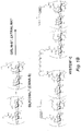


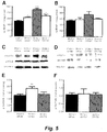
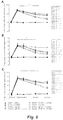
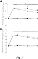

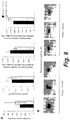


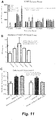
Classifications
-
- A—HUMAN NECESSITIES
- A61—MEDICAL OR VETERINARY SCIENCE; HYGIENE
- A61K—PREPARATIONS FOR MEDICAL, DENTAL OR TOILETRY PURPOSES
- A61K38/00—Medicinal preparations containing peptides
- A61K38/16—Peptides having more than 20 amino acids; Gastrins; Somatostatins; Melanotropins; Derivatives thereof
- A61K38/17—Peptides having more than 20 amino acids; Gastrins; Somatostatins; Melanotropins; Derivatives thereof from animals; from humans
- A61K38/18—Growth factors; Growth regulators
- A61K38/185—Nerve growth factor [NGF]; Brain derived neurotrophic factor [BDNF]; Ciliary neurotrophic factor [CNTF]; Glial derived neurotrophic factor [GDNF]; Neurotrophins, e.g. NT-3
-
- A—HUMAN NECESSITIES
- A61—MEDICAL OR VETERINARY SCIENCE; HYGIENE
- A61K—PREPARATIONS FOR MEDICAL, DENTAL OR TOILETRY PURPOSES
- A61K31/00—Medicinal preparations containing organic active ingredients
- A61K31/33—Heterocyclic compounds
- A61K31/395—Heterocyclic compounds having nitrogen as a ring hetero atom, e.g. guanethidine or rifamycins
- A61K31/40—Heterocyclic compounds having nitrogen as a ring hetero atom, e.g. guanethidine or rifamycins having five-membered rings with one nitrogen as the only ring hetero atom, e.g. sulpiride, succinimide, tolmetin, buflomedil
-
- A—HUMAN NECESSITIES
- A61—MEDICAL OR VETERINARY SCIENCE; HYGIENE
- A61K—PREPARATIONS FOR MEDICAL, DENTAL OR TOILETRY PURPOSES
- A61K31/00—Medicinal preparations containing organic active ingredients
- A61K31/33—Heterocyclic compounds
- A61K31/395—Heterocyclic compounds having nitrogen as a ring hetero atom, e.g. guanethidine or rifamycins
- A61K31/435—Heterocyclic compounds having nitrogen as a ring hetero atom, e.g. guanethidine or rifamycins having six-membered rings with one nitrogen as the only ring hetero atom
- A61K31/44—Non condensed pyridines; Hydrogenated derivatives thereof
- A61K31/445—Non condensed piperidines, e.g. piperocaine
-
- A—HUMAN NECESSITIES
- A61—MEDICAL OR VETERINARY SCIENCE; HYGIENE
- A61K—PREPARATIONS FOR MEDICAL, DENTAL OR TOILETRY PURPOSES
- A61K31/00—Medicinal preparations containing organic active ingredients
- A61K31/70—Carbohydrates; Sugars; Derivatives thereof
- A61K31/715—Polysaccharides, i.e. having more than five saccharide radicals attached to each other by glycosidic linkages; Derivatives thereof, e.g. ethers, esters
- A61K31/726—Glycosaminoglycans, i.e. mucopolysaccharides
- A61K31/727—Heparin; Heparan
-
- A—HUMAN NECESSITIES
- A61—MEDICAL OR VETERINARY SCIENCE; HYGIENE
- A61K—PREPARATIONS FOR MEDICAL, DENTAL OR TOILETRY PURPOSES
- A61K31/00—Medicinal preparations containing organic active ingredients
- A61K31/70—Carbohydrates; Sugars; Derivatives thereof
- A61K31/715—Polysaccharides, i.e. having more than five saccharide radicals attached to each other by glycosidic linkages; Derivatives thereof, e.g. ethers, esters
- A61K31/726—Glycosaminoglycans, i.e. mucopolysaccharides
- A61K31/728—Hyaluronic acid
-
- A—HUMAN NECESSITIES
- A61—MEDICAL OR VETERINARY SCIENCE; HYGIENE
- A61K—PREPARATIONS FOR MEDICAL, DENTAL OR TOILETRY PURPOSES
- A61K31/00—Medicinal preparations containing organic active ingredients
- A61K31/70—Carbohydrates; Sugars; Derivatives thereof
- A61K31/715—Polysaccharides, i.e. having more than five saccharide radicals attached to each other by glycosidic linkages; Derivatives thereof, e.g. ethers, esters
- A61K31/737—Sulfated polysaccharides, e.g. chondroitin sulfate, dermatan sulfate
-
- A—HUMAN NECESSITIES
- A61—MEDICAL OR VETERINARY SCIENCE; HYGIENE
- A61K—PREPARATIONS FOR MEDICAL, DENTAL OR TOILETRY PURPOSES
- A61K38/00—Medicinal preparations containing peptides
- A61K38/16—Peptides having more than 20 amino acids; Gastrins; Somatostatins; Melanotropins; Derivatives thereof
- A61K38/17—Peptides having more than 20 amino acids; Gastrins; Somatostatins; Melanotropins; Derivatives thereof from animals; from humans
- A61K38/18—Growth factors; Growth regulators
-
- A—HUMAN NECESSITIES
- A61—MEDICAL OR VETERINARY SCIENCE; HYGIENE
- A61K—PREPARATIONS FOR MEDICAL, DENTAL OR TOILETRY PURPOSES
- A61K47/00—Medicinal preparations characterised by the non-active ingredients used, e.g. carriers or inert additives; Targeting or modifying agents chemically bound to the active ingredient
- A61K47/30—Macromolecular organic or inorganic compounds, e.g. inorganic polyphosphates
- A61K47/36—Polysaccharides; Derivatives thereof, e.g. gums, starch, alginate, dextrin, hyaluronic acid, chitosan, inulin, agar or pectin
-
- A—HUMAN NECESSITIES
- A61—MEDICAL OR VETERINARY SCIENCE; HYGIENE
- A61K—PREPARATIONS FOR MEDICAL, DENTAL OR TOILETRY PURPOSES
- A61K47/00—Medicinal preparations characterised by the non-active ingredients used, e.g. carriers or inert additives; Targeting or modifying agents chemically bound to the active ingredient
- A61K47/30—Macromolecular organic or inorganic compounds, e.g. inorganic polyphosphates
- A61K47/42—Proteins; Polypeptides; Degradation products thereof; Derivatives thereof, e.g. albumin, gelatin or zein
-
- A—HUMAN NECESSITIES
- A61—MEDICAL OR VETERINARY SCIENCE; HYGIENE
- A61K—PREPARATIONS FOR MEDICAL, DENTAL OR TOILETRY PURPOSES
- A61K9/00—Medicinal preparations characterised by special physical form
- A61K9/0012—Galenical forms characterised by the site of application
- A61K9/0085—Brain, e.g. brain implants; Spinal cord
-
- A—HUMAN NECESSITIES
- A61—MEDICAL OR VETERINARY SCIENCE; HYGIENE
- A61K—PREPARATIONS FOR MEDICAL, DENTAL OR TOILETRY PURPOSES
- A61K9/00—Medicinal preparations characterised by special physical form
- A61K9/06—Ointments; Bases therefor; Other semi-solid forms, e.g. creams, sticks, gels
-
- A—HUMAN NECESSITIES
- A61—MEDICAL OR VETERINARY SCIENCE; HYGIENE
- A61L—METHODS OR APPARATUS FOR STERILISING MATERIALS OR OBJECTS IN GENERAL; DISINFECTION, STERILISATION OR DEODORISATION OF AIR; CHEMICAL ASPECTS OF BANDAGES, DRESSINGS, ABSORBENT PADS OR SURGICAL ARTICLES; MATERIALS FOR BANDAGES, DRESSINGS, ABSORBENT PADS OR SURGICAL ARTICLES
- A61L27/00—Materials for grafts or prostheses or for coating grafts or prostheses
- A61L27/36—Materials for grafts or prostheses or for coating grafts or prostheses containing ingredients of undetermined constitution or reaction products thereof, e.g. transplant tissue, natural bone, extracellular matrix
- A61L27/38—Materials for grafts or prostheses or for coating grafts or prostheses containing ingredients of undetermined constitution or reaction products thereof, e.g. transplant tissue, natural bone, extracellular matrix containing added animal cells
- A61L27/3804—Materials for grafts or prostheses or for coating grafts or prostheses containing ingredients of undetermined constitution or reaction products thereof, e.g. transplant tissue, natural bone, extracellular matrix containing added animal cells characterised by specific cells or progenitors thereof, e.g. fibroblasts, connective tissue cells, kidney cells
- A61L27/3834—Cells able to produce different cell types, e.g. hematopoietic stem cells, mesenchymal stem cells, marrow stromal cells, embryonic stem cells
-
- A—HUMAN NECESSITIES
- A61—MEDICAL OR VETERINARY SCIENCE; HYGIENE
- A61L—METHODS OR APPARATUS FOR STERILISING MATERIALS OR OBJECTS IN GENERAL; DISINFECTION, STERILISATION OR DEODORISATION OF AIR; CHEMICAL ASPECTS OF BANDAGES, DRESSINGS, ABSORBENT PADS OR SURGICAL ARTICLES; MATERIALS FOR BANDAGES, DRESSINGS, ABSORBENT PADS OR SURGICAL ARTICLES
- A61L27/00—Materials for grafts or prostheses or for coating grafts or prostheses
- A61L27/50—Materials characterised by their function or physical properties, e.g. injectable or lubricating compositions, shape-memory materials, surface modified materials
- A61L27/52—Hydrogels or hydrocolloids
-
- A—HUMAN NECESSITIES
- A61—MEDICAL OR VETERINARY SCIENCE; HYGIENE
- A61L—METHODS OR APPARATUS FOR STERILISING MATERIALS OR OBJECTS IN GENERAL; DISINFECTION, STERILISATION OR DEODORISATION OF AIR; CHEMICAL ASPECTS OF BANDAGES, DRESSINGS, ABSORBENT PADS OR SURGICAL ARTICLES; MATERIALS FOR BANDAGES, DRESSINGS, ABSORBENT PADS OR SURGICAL ARTICLES
- A61L27/00—Materials for grafts or prostheses or for coating grafts or prostheses
- A61L27/50—Materials characterised by their function or physical properties, e.g. injectable or lubricating compositions, shape-memory materials, surface modified materials
- A61L27/54—Biologically active materials, e.g. therapeutic substances
-
- A—HUMAN NECESSITIES
- A61—MEDICAL OR VETERINARY SCIENCE; HYGIENE
- A61P—SPECIFIC THERAPEUTIC ACTIVITY OF CHEMICAL COMPOUNDS OR MEDICINAL PREPARATIONS
- A61P25/00—Drugs for disorders of the nervous system
-
- A—HUMAN NECESSITIES
- A61—MEDICAL OR VETERINARY SCIENCE; HYGIENE
- A61P—SPECIFIC THERAPEUTIC ACTIVITY OF CHEMICAL COMPOUNDS OR MEDICINAL PREPARATIONS
- A61P25/00—Drugs for disorders of the nervous system
- A61P25/02—Drugs for disorders of the nervous system for peripheral neuropathies
-
- A—HUMAN NECESSITIES
- A61—MEDICAL OR VETERINARY SCIENCE; HYGIENE
- A61P—SPECIFIC THERAPEUTIC ACTIVITY OF CHEMICAL COMPOUNDS OR MEDICINAL PREPARATIONS
- A61P7/00—Drugs for disorders of the blood or the extracellular fluid
- A61P7/06—Antianaemics
-
- A—HUMAN NECESSITIES
- A61—MEDICAL OR VETERINARY SCIENCE; HYGIENE
- A61P—SPECIFIC THERAPEUTIC ACTIVITY OF CHEMICAL COMPOUNDS OR MEDICINAL PREPARATIONS
- A61P9/00—Drugs for disorders of the cardiovascular system
-
- A—HUMAN NECESSITIES
- A61—MEDICAL OR VETERINARY SCIENCE; HYGIENE
- A61L—METHODS OR APPARATUS FOR STERILISING MATERIALS OR OBJECTS IN GENERAL; DISINFECTION, STERILISATION OR DEODORISATION OF AIR; CHEMICAL ASPECTS OF BANDAGES, DRESSINGS, ABSORBENT PADS OR SURGICAL ARTICLES; MATERIALS FOR BANDAGES, DRESSINGS, ABSORBENT PADS OR SURGICAL ARTICLES
- A61L2300/00—Biologically active materials used in bandages, wound dressings, absorbent pads or medical devices
- A61L2300/40—Biologically active materials used in bandages, wound dressings, absorbent pads or medical devices characterised by a specific therapeutic activity or mode of action
- A61L2300/412—Tissue-regenerating or healing or proliferative agents
- A61L2300/414—Growth factors
-
- A—HUMAN NECESSITIES
- A61—MEDICAL OR VETERINARY SCIENCE; HYGIENE
- A61L—METHODS OR APPARATUS FOR STERILISING MATERIALS OR OBJECTS IN GENERAL; DISINFECTION, STERILISATION OR DEODORISATION OF AIR; CHEMICAL ASPECTS OF BANDAGES, DRESSINGS, ABSORBENT PADS OR SURGICAL ARTICLES; MATERIALS FOR BANDAGES, DRESSINGS, ABSORBENT PADS OR SURGICAL ARTICLES
- A61L2300/00—Biologically active materials used in bandages, wound dressings, absorbent pads or medical devices
- A61L2300/60—Biologically active materials used in bandages, wound dressings, absorbent pads or medical devices characterised by a special physical form
- A61L2300/602—Type of release, e.g. controlled, sustained, slow
-
- A—HUMAN NECESSITIES
- A61—MEDICAL OR VETERINARY SCIENCE; HYGIENE
- A61L—METHODS OR APPARATUS FOR STERILISING MATERIALS OR OBJECTS IN GENERAL; DISINFECTION, STERILISATION OR DEODORISATION OF AIR; CHEMICAL ASPECTS OF BANDAGES, DRESSINGS, ABSORBENT PADS OR SURGICAL ARTICLES; MATERIALS FOR BANDAGES, DRESSINGS, ABSORBENT PADS OR SURGICAL ARTICLES
- A61L2300/00—Biologically active materials used in bandages, wound dressings, absorbent pads or medical devices
- A61L2300/60—Biologically active materials used in bandages, wound dressings, absorbent pads or medical devices characterised by a special physical form
- A61L2300/64—Animal cells
-
- A—HUMAN NECESSITIES
- A61—MEDICAL OR VETERINARY SCIENCE; HYGIENE
- A61L—METHODS OR APPARATUS FOR STERILISING MATERIALS OR OBJECTS IN GENERAL; DISINFECTION, STERILISATION OR DEODORISATION OF AIR; CHEMICAL ASPECTS OF BANDAGES, DRESSINGS, ABSORBENT PADS OR SURGICAL ARTICLES; MATERIALS FOR BANDAGES, DRESSINGS, ABSORBENT PADS OR SURGICAL ARTICLES
- A61L2430/00—Materials or treatment for tissue regeneration
- A61L2430/20—Materials or treatment for tissue regeneration for reconstruction of the heart, e.g. heart valves
Definitions
- Stroke is the leading cause of adult disability in developed countries. There is no therapy that targets the promotion of stroke recovery. As present the only stroke therapy is the administration of tissue plasminogen activator (tPA). TPA is a "clot busting" drug that does not target stroke recovery, but targets the blood vessel that is obstructed in stroke. TPA must be given within 4.5 hours after stroke because if given later it will cause bleeding into the brain. There is no stroke therapy that can be given at a time point later than 4.5 hours. There is no stroke therapy that targets the processes of tissue repair and not the vessel that is obstructed.
- tissue plasminogen activator tPA
- Kurozumi et al., (2004) Molecular Therapy, 9(2): 189-197 discloses that BDNF gene-modified mesenchymal stem cells promote functional recovery and reduce infarct size in a rat middle cerebral artery occlusion model.
- the present invention provides a formulation for use in a method of improving recovery of a mammal after cerebral ischemia, wherein said method comprises administering a therapeutically effective amount of brain derived neurotrophic factor (BDNF) to the infarct cavity in the brain of said mammal, wherein said administration comprises depositing said formulation into the infarct cavity, and wherein said formulation comprises a hydrogel comprising cells that provide a sustained release of BDNF, Further embodiments of the invention are specified in the dependent claims.
- BDNF brain derived neurotrophic factor
- BDNF growth factor(s)
- the growth factor(s) (e.g., BDNF) delivery is locally done within a specific region of the brain in a site directly adjacent to its intended brain target.
- the biocompatible device delivers growth factor(s) (e.g., BDNF) in a way that bypasses the blood brain barrier and provides a sustained, timed release.
- the release of growth factor(s) (e.g., BDNF) from the device occurs for one month after stroke, after just one injection of the device.
- the device delivers the growth factor(s)(s) in a way that does not promote inflammation or tissue damage.
- the device can be injected using very small delivery needles that minimize brain damage.
- the methods typically involve administering a therapeutically effective amount of a brain growth factor to the infarct cavity in the brain of the mammal.
- the improvement in motor recovery comprises an improvement in motor coordination, and/or balance, and/or speech.
- the improvement is an improvement in the degree of recovered function and/or the rate of recovery of function.
- the administering comprises depositing a depot delivery system into the infarct cavity where the depot delivery system comprises the growth factor and provides sustained release of the growth factor, and/or the depot delivery system comprises cells that provide a sustained release of the brain growth factor.
- the depot delivery system comprises a delivery system selected from the group consisting of a nanoparticle formulation, a hydrogel formulation, and an implantable mechanical delivery system.
- the depot delivery system comprises a hydrogel comprising the growth factor and/or the cells (e.g., a hydrogel depot formulation).
- the hydrogel comprises a biopolymer.
- the hydrogel comprises one or more materials selected from the group consisting of thiol-modified hyaluronan, thiol-modified heparin, a hyaluronan sodium salt, an acrylated hyaluronic acid, and cross-linker peptides.
- the hydrogel comprises hyaluronan and gelatin or a hyaluronan derivative and/or a gelatin derivative (e.g., a thiolated hyaluronan and/or a thiolated gelatin).
- the hyaluronan and the gelatin have each been thiol-modified using carbodiimide mediated hydrazide chemistry.
- the hyaluronan and gelatin or hyaluronan derivative and/or gelatin derivative are crosslinked. In certain aspects they are crosslinked using a divalent or polyvalent (e.g., trivalent, tetravalent, pentavalent, etc .) electrophile.
- the derivatives of hyaluronan and gelatin are crosslinked with a reagent selected from the group consisting of a polyethylene glycol diacrylate (PEGDA), a polyethylene glycol divinyl sulfone, a polyethylene glycol bismaleimide, a polyethylene glycol dimethyacrylate, a polyethylene glycol (methyl)acrylamide, and a polyethylene glycol di(halo)acetate.
- PEGDA polyethylene glycol diacrylate
- PEGDA polyethylene glycol diacrylate
- PEGDA polyethylene glycol diacrylate
- the hydrogel further comprises a heparin or heparin derivative (e.g., a thiol-modified heparin).
- a heparin or heparin derivative is linked to the hyaluronan and/or to the gelatin (or to a hyaluronan derivative and/or a gelatin derivative).
- the heparin derivative is linked to the hyaluronan derivative and/or to the gelatin derivative with a divalent or polyvalent (e.g., trivalent, tetravalent, pentavalent, etc .) electrophile.
- the heparin derivative is linked to the hyaluronan derivative and/or to the gelatin derivative with a reagent selected from the group consisting of a polyethylene glycol diacrylate (PEGDA), a polyethylene glycol divinyl sulfone, a polyethylene glycol bismaleimide, a polyethylene glycol dimethyacrylate, a polyethylene glycol (methyl)acrylamide, and a polyethylene glycol di(halo)acetate.
- PEGDA polyethylene glycol diacrylate
- PEGDA polyethylene glycol diacrylate
- the hydrogel comprises at most about 10% solids w/v, or at most about 5% solids w/v, or at most about 4% solids w/v, or at most about 2% solids w/v, or at most about 1% solids w/v.
- the depot delivery system comprises a hydrogel containing the growth factor(s) where the hydrogel and provides sustained release of the growth factor (e.g., over at least one day, or over at least 2 days, or over at least about 3 days, or over at least about 5 days, or over at least about 1 week, or over at least about 2 weeks, or over at least about 3 weeks, or over at least about 4 weeks, or over at least about 5 weeks, or over at least about 6 weeks, or over at least about 2 months).
- sustained release of the growth factor e.g., over at least one day, or over at least 2 days, or over at least about 3 days, or over at least about 5 days, or over at least about 1 week, or over at least about 2 weeks, or over at least about 3 weeks, or over at least about 4 weeks, or over at least about 5 weeks, or over at least about 6 weeks, or over at least about 2 months).
- the brain growth factor comprises one or more factors selected from the group consisting of BDNF, VEGF, IGF1, bFGF/FGF2, Ang1, Ang 2, BMP 2, BMP 3a, BMP 3b, BMP 4, BMP 5, BMP 6, BMP 7 (OP-1), CTNF, EGF, EPO, aFGF/FGF1, bFGF/FGF2, G-CSF, GDF10, GDF15, GDNF, GH, GM-CSF, HB-EGF, LIF, NGF, NT-3, NT 4/5, Osteopontin, PDGFaa, PDGFbb, PDGFab, PlGF, SCF, SDF1/CXCL12, and TGF ⁇ .
- factors selected from the group consisting of BDNF, VEGF, IGF1, bFGF/FGF2, Ang1, Ang 2, BMP 2, BMP 3a, BMP 3b, BMP 4, BMP 5, BMP 6, BMP 7 (OP-1), CTNF, EGF,
- the brain growth factor comprises one or more factors selected from the group consisting of BDNF, VEGF, IGF-1, and bFGF.
- the brain growth factor comprises BDNF.
- the depot delivery system comprises a hydrogel comprising cells that provide a sustained release of the brain growth factor.
- the cells comprise stem cells (e.g., adult stem cells, embryonic stem cells, cord stem cells, and induced pluripotent stem cells (IPSCs), and the like).
- the cells are cells that have differentiated from stem cells in culture or after implantation into the mammal.
- the cells are derived from cells/cell lines selected from the group consisting of SM22, CM02, E33, 7PEND24, B16, C4ELS5.1, T36, E68, T43, W10, and EN13. In certain aspects the cells are derived from cells/cell lines selected from the group consisting of SM22, CM02, E33, and EN13.
- the cells express and secrete one or more factors selected from the group consisting of BDNF, VEGF, IGF1, bFGF/FGF2, Ang1, Ang 2, BMP 2, BMP 3a, BMP 3b, BMP 4, BMP 5, BMP 6, BMP 7 (OP-1), CTNF, EGF, EPO, aFGF/FGF1, bFGF/FGF2, G-CSF, GDF10, GDF15, GDNF, GH, GM-CSF, HB-EGF, LIF, NGF, NT-3, NT 4/5, Osteopontin, PDGFaa, PDGFbb, PDGFab, PlGF, SCF, SDF1/CXCL12, and TGF ⁇ .
- factors selected from the group consisting of BDNF, VEGF, IGF1, bFGF/FGF2, Ang1, Ang 2, BMP 2, BMP 3a, BMP 3b, BMP 4, BMP 5, BMP 6, BMP 7 (OP-1), CTNF, EGF
- the cells secrete one or more factors selected from the group consisting of BDNF, HBEGF, VEGF, IGF-1, and bFGF. In certain aspects the cells secrete BDNF. In certain aspects BDNF expression by the cells is enhanced by culturing the cells in the presence of neuronal media.
- the administering comprises injecting the depot formulation into the ischemic cavity.
- the depot delivery system is not administered until AMPAR signaling switches from promoting cell death to promoting behavioral recovery.
- the depot delivery system is not administered within the first hour, or not administered within the first three hours, or not administered within the first 6 hours, or not administered within the first 12 hours, or not administered within the first 24 hours after the onset of the ischemic event.
- the depot delivery system is administered after at least 24 hours after the onset of the ischemic event, or after at 2 days after the onset of the ischemic event, or after at least 3 days after the onset of the ischemic event, or after at least 4 or 5 days after the onset of the ischemic event, or after at least 7 days after the onset of the ischemic event.
- the depot delivery system is administered within one week of the onset of the ischemic event.
- the depot delivery system is administered after the subject is clinically stable from the ischemia and damage has substantially stopped progressing.
- the depot delivery system is initially administered after at least 3 days, or at least five or more days, after the initial ischemic event.
- the depot delivery system is administered up to about 1 year, or up to about 1.5 years, or up to about 2 years after the initial ischemic event.
- mammal is a human.
- mammal is a non-human mammal.
- the formulations typically comprise a hydrogel comprising one or more brain growth factors that, where when the formulation is placed in an infarct cavity in the brain of a mammal, provides sustained release of the growth factor over at least about one week; and/or a hydrogel comprising cells that provide a sustained release of a brain growth factor over a period of at least about one week, or at least about two weeks, or at least about three weeks.
- the hydrogel comprises hyaluronan and gelatin and/or a hyaluronan derivative and/or a gelatin derivative.
- the hyaluronan and the gelatin are each thiolated.
- the hyaluronan and the gelatin have each been thiol-modified using carbodiimide mediated hydrazide chemistry.
- the hyaluronan derivative and gelatin derivative are crosslinked.
- the derivatives of hyaluronan and gelatin are crosslinked with a divalent or polyvalent electrophile.
- the derivatives of hyaluronan and gelatin are crosslinked with a reagent selected from the group consisting of a polyethylene glycol diacrylate (PEGDA), a polyethylene glycol divinyl sulfone, a polyethylene glycol bismaleimide, a polyethylene glycol dimethyacrylate, a polyethylene glycol (methyl)acrylamide, and a polyethylene glycol di(halo)acetate.
- PEGDA polyethylene glycol diacrylate
- a polyethylene glycol divinyl sulfone a polyethylene glycol bismaleimide
- a polyethylene glycol dimethyacrylate a polyethylene glycol (methyl)acrylamide
- a polyethylene glycol di(halo)acetate a polyethylene glycol di(halo)acetate.
- the hyaluronan derivative h is linked to the gelatin derivative with a divalent or polyvalent electrophile.
- the hyaluronan derivative is linked to the gelatin derivative with a reagent selected from the group consisting of a polyethylene glycol diacrylate (PEGDA), a polyethylene glycol divinyl sulfone, a polyethylene glycol bismaleimide, a polyethylene glycol dimethyacrylate, a polyethylene glycol (methyl)acrylamide, and a polyethylene glycol di(halo)acetate.
- PEGDA polyethylene glycol diacrylate
- the hyaluronan derivative and gelatin derivative are crosslinked with a polyethylene glycol diacrylate (PEGDA) linker.
- the hydrogel further comprises a heparin derivative.
- the heparin derivative is a thiol-modified heparin. In certain embodiments the heparin derivative is linked to the hyaluronan derivative and/or to the gelatin derivative. In certain embodiments the heparin derivative is linked to the hyaluronan derivative and/or to the gelatin derivative with a divalent or polyvalent electrophile.
- the heparin derivative is linked to the hyaluronan derivative and/or to the gelatin derivative with a reagent selected from the group consisting of a polyethylene glycol diacrylate (PEGDA), a polyethylene glycol divinyl sulfone, a polyethylene glycol bismaleimide, a polyethylene glycol dimethyacrylate, a polyethylene glycol (methyl)acrylamide, and a polyethylene glycol di(halo)acetate.
- PEGDA polyethylene glycol diacrylate
- PEGDA polyethylene glycol divinyl sulfone
- a polyethylene glycol bismaleimide a polyethylene glycol dimethyacrylate
- a polyethylene glycol (methyl)acrylamide a polyethylene glycol di(halo)acetate
- the heparin derivative is linked to the hyaluronan and/or to the gelatin with a polyethylene glycol diacrylate (PEGDA) linker.
- the hydrogel comprises at most about 10% solids w/v, or at most about 5% solids w/v, or at most about 3% solids w/v, or at most about 2% solids w/v.
- the formulation comprises a hydrogel comprising one or more brain growth factors that, when placed in an infarct cavity in the brain of a mammal, provides sustained release of the growth factor over at least about one week.
- the brain growth factor comprises one or more factors selected from the group consisting of BDNF, VEGF, IGF1, bFGF/FGF2, Ang1, Ang 2, BMP 2, BMP 3a, BMP 3b, BMP 4, BMP 5, BMP 6, BMP 7 (OP-1), CTNF, EGF, EPO, aFGF/FGF1, bFGF/FGF2, G-CSF, GDF10, GDF15, GDNF, GH, GM-CSF, HB-EGF, LIF, NGF, NT-3, NT 4/5, Osteopontin, PDGFaa, PDGFbb, PDGFab, PlGF, SCF, SDF1/CXCL12, and TGF ⁇ .
- factors selected from the group consisting of BDNF, VEGF, IGF1, bFGF/FGF2, Ang1, Ang 2, BMP 2, BMP 3a, BMP 3b, BMP 4, BMP 5, BMP 6, BMP 7 (OP-1), CTNF, EGF,
- the brain growth factor comprises one or more factors selected from the group consisting of BDNF, VEGF, IGF-1, and bFGF.
- the brain growth factor comprises BDNF.
- the formulation comprises a hydrogel comprising cells that provide a sustained release of a brain growth factor over a period of at least about one week.
- the cells comprise stem cells (e.g., adult stem cells, embryonic stem cells, cord stem cells, and induced pluripotent stem cells (IPSCs), and the like).
- the cells are cells that have differentiated from stem cells in culture or after implantation into the mammal.
- the cells are derived from cells/cell lines selected from the group consisting of SM22, CM02, E33, 7PEND24, B16, C4ELS5.1, T36, E68, T43, W10, and EN13. In certain aspects the cells are derived from cells/cell lines selected from the group consisting of SM22, CM02, E33, and EN13.
- the cells secrete one or more factors selected from the group consisting of BDNF, VEGF, IGF1, bFGF/FGF2, Ang1, Ang 2, BMP 2, BMP 3a, BMP 3b, BMP 4, BMP 5, BMP 6, BMP 7 (OP-1), CTNF, EGF, EPO, aFGF/FGF1, bFGF/FGF2, G-CSF, GDF10, GDF15, GDNF, GH, GM-CSF, HB-EGF, LIF, NGF, NT-3, NT 4/5, Osteopontin, PDGF-AA, PDGF-BB, PDGF-AB, PlGF, SCF, SDF1/CXCL12, and TGF ⁇ .
- factors selected from the group consisting of BDNF, VEGF, IGF1, bFGF/FGF2, Ang1, Ang 2, BMP 2, BMP 3a, BMP 3b, BMP 4, BMP 5, BMP 6, BMP 7 (OP-1), CTNF, EGF
- the cells secrete one or more factors selected from the group consisting of BDNF, HBEGF, VEGF, IGF-1, and bFGF.
- the cells secrete BDNF.
- BDNF expression by the cells is enhanced by culturing the cells in the presence of neuronal media.
- the formulation is disposed in vivo in an infarct cavity in a mammalian brain. Also provided is any formulation described herein for use in facilitating recovery from an ischemic event ( e.g., a stroke, an acute brain trauma, etc. ).
- Described herein are methods for improving recovery of a mammal after cerebral ischemia comprising administering an agent to the mammal that enhances AMPA signaling, where the agent is administered after a delay period from the onset of the ischemic event; and where the agent induces an increase in BDNF expression and/or activity.
- the agent is not administered until AMPAR signaling switches from promoting cell death to promoting behavioral recovery.
- the agent is not administered within the first hour after the onset of the ischemic event.
- agent is not administered within the first three hours after the onset of the ischemic event.
- the agent is not administered within the first six hours after the onset of the ischemic event, or within the first 12 hours after the onset of the ischemic event, or within the first 24 hours after the onset of the ischemic event. In certain aspects the agent is administered after at least 24 hours after the onset of the ischemic event, or after at 2 days after the onset of the ischemic event, or after at least 3 days after the onset of the ischemic event, or after at least 4 or 5 days after the onset of the ischemic event, or after at least 7 days after the onset of the ischemic event.
- the cerebral ischemia is due to a stroke or a head injury. In certain aspects the mammal is a human.
- the mammal is a non-human mammal.
- the agent comprises an ampakine.
- the ampakine comprises a high impact (type II) ampakine.
- the ampakine is CX1837.
- the agent is an agent that crosses the blood/brain barrier.
- the agent is delivered directly to the stroke cavity.
- the mammal is not diagnosed as having and/or is not under treatment for one or more conditions selected from the group consisting of Alzheimer's disease (AD), attention deficit hyperactivity disorder (ADHD), Parkinson's disease, a sleep disorder, depression, fragile X disorder, mental retardation, and an opiate-induced or other respiratory depression.
- the mammal is not diagnosed as having and/or is not under treatment for a neuropsychiatric disorder.
- the methods described herein are not used in the treatment of spinal cord injury.
- Methods are described herein that produce improved recovery in stroke or other cerebral ischemic conditions.
- the methods involve slowly releasing a brain growth factor from a medical device (or implantable depot pharmaceutical formulation) in or into the ischemic (e.g., stroke) cavity. It was a surprising discovery that slow (controlled/sustained) release of a brain growth factor, such as BNDF, into the ischemic cavity can substantially and dramatically improve the rate and/or degree of recovery after the ischemic event.
- a brain growth factor such as BNDF
- the methods involve depositing slow/sustained release delivery vehicle comprising one or more brain growth factors and/or cells that express/secrete one or more brain growth factors into the ischemic (e.g., stroke) cavity (the area of cell death and absorption of dead tissue) in the brain subject to the ischemic event.
- ischemic e.g., stroke
- the tissue adjacent to the stroke cavity undergoes the most substantial recovery of function after ischemia. This recovering brain tissue is termed the peri-infarct tissue.
- AMPAR function plays a critical but functionally contradictory role in the spectrum of stroke pathophysiology.
- AMPAR signaling in the tissue adjacent to the infarct mediates behavioral recovery of limb control over weeks after the stroke. This process occurs through BDNF induction in periinfarct cortex.
- Positive AMPAR modulation in a way that also induces BDNF promotes improved recovery of motor function during this recovery phase after stroke and blockade of AMPA signaling retards motor recovery.
- AMPARs are involved in cell death and infarct evolution. Positive AMPAR modulation increases infarct size immediately after stroke.
- BDNF BDNF signaling in behavioral recovery after stroke.
- Systemic administration of CX1837 induces BDNF levels and TrkB phosphorylation in periinfarct cortex.
- Local blockade of BDNF induction in periinfarct cortex not only prevents the ampakine-mediated behavioral recovery but also generally blocks motor recovery after stroke.
- the present data indicate that BDNF normally mediates motor recovery after stroke, localizes this effect to the periinfarct cortex adjacent to the stroke site, and identifies a systemic pharmacological therapy that will modulate BDNF in this critical periinfarct region for motor recovery. It is believed these data are the first to specifically localize motor recovery to one brain region after stroke, the periinfarct cortex.
- BDNF and other neural (brain) growth factors do not easily pass the blood brain barrier and are likely to have significant toxicity if given systemically
- local administration into the infarct cavity is contemplated.
- certain depot delivery systems in particular hydrogel depot formulations show surprising efficacy along with an effective sustained release profile. Described herein are methods of improving recovery of a mammal after cerebral ischemia where the method comprises administering a therapeutically effective amount of a brain growth factor to the infarct cavity in the brain of mammal.
- the administering comprises depositing a depot delivery system into the infarct cavity where the depot delivery system comprises/contains the growth factor(s) and provides sustained release of the growth factor(s) over time, and/or the depot delivery system comprises cells that provide a sustained release of the brain growth factor.
- the depot delivery system comprises a hydrogel.
- the methods described herein contemplate the introduction of one or more depot delivery systems into the infarct cavity where they provide a sustained release of neural (brain) growth factors (e.g., BDNF, HBEGF, VEGF, IGF-1, bFGF, and the like) as described herein.
- the depot delivery systems comprise one or more growth factors and provide a sustained release of those growth factors and/or they comprise cells that provide a sustained release of one or more growth factors.
- Implantable sustained delivery systems are known to those of skill in the art. Such systems include, but are not limited to, mechanical and/or electronic devices such as implantable drug pumps or microchip systems as well as implantable controlled delivery polymeric matrices.
- Implantable microchip systems include systems such as the MICROCHIPS® device (MicroChips, Inc. Bedford MA).
- the MicroCHIPS implantable drug delivery system is based on a microfabricated silicon chip that contains multiple drug-filled reservoirs. the chip is attached to a titanium case containing a battery, control circuitry, and telemetry .
- the drug chip and titanium case are hermetically sealed and electrically linked by a ceramic substrate with metal interconnects.
- the IDDS communicates with an external handheld controller through wireless transmission.
- a drug regimen can be transmitted to the implanted device through this link, allowing reservoirs to be opened at prescribed times without any need for further communication. Alternatively, reservoirs can be opened as desired on command from the controller.
- Controlled release polymeric devices can be made for long term release following implantation.
- Illustrative controlled polymeric release devices comprise an implantable rod, cylinder, film, disk, and the like, or an injectable polymeric formulation (e.g. a microparticle formulation).
- the implantable matrix can be in the form of microparticles such as microspheres, where the brain growth factor(s) (e.g., BNDF) are dispersed within a solid polymeric matrix or microcapsules.
- the core is of a different material than the polymeric shell, and the active agent (e.g., BNDF) will be dispersed or suspended in the core, which may be liquid or solid in nature.
- the polymer may be cast as a thin slab or film, ranging from, e.g., nanometers to four centimeters, a powder produced by grinding or other standard techniques, or may be suspended in the soluble components of a hydrogel prior to crosslinking.
- non-biodegradable or biodegradable matrices can be used for delivery of brain growth factors and/or cells as described herein, however, biodegradable matrices are typically preferred. These can include natural or synthetic polymers. Often synthetic polymers provide better characterization of degradation and release profiles. The polymer is typically selected based on the period over which release is desired. In some cases linear release may be most useful, although in others a pulse release or "bulk release" may provide more effective results. As discussed below, in certain embodiments, the polymer is in the form of a hydrogel, and can optionally be crosslinked with multivalent ions or polymers.
- the matrices can be formed by solvent evaporation, spray drying, solvent extraction and other methods known to those skilled in the art.
- Bioerodible microspheres can be prepared using any of the methods developed for making microspheres for drug delivery, for example, as described by Mathiowitz and Langer (1987) J. Controlled Release 5:13-22 ; Mathiowitz, et al. (1987) Reactive Polymers 6: 275-283 , Mathiowitz, et al. (1988) J. Appl. Polymer Sci. 35:755-774 , and the like.
- the devices can be formulated for local release to treat the area of implantation, e.g., the infarct cavity. In various aspects these can be implanted or injected into the desired region.
- the implantable the depot delivery systems comprise a microparticles patterned within a hydrogel.
- the growth factors are provided within microparticles (e.g., PLGA microparticles) entrapped within a hydrogel (e.g., PEG hydrogel) base.
- a hydrogel e.g., PEG hydrogel
- the methods described herein utilize a hydrogel, preferably a biopolymer hydrogel to contain and provide sustained release of the growth factor(s) (e .g., BDNF, and/or other neural tissue growth factors) into the infarct cavity and/or to contain cells ( e.g., stem cells) that afford sustained release of one or more brain growth factors).
- the hydrogel can be made of naturally occurring brain proteins, and does not provoke a brain reaction.
- the hydrogel can be a HYSTEM ® hydrogel (e.g., a HYSTEM ®-C hydrogel, a HYSTEM ®-HP hydrogel, and the like).
- the growth factor(s) are released slowly over at least one day, or over at least 3 days or over at least 5 days, or over at least one week, or over at least two weeks, or over at least 3 weeks, or over at least one month, or over at least two months, or over at least three months, or over at least six months.
- the growth factor(s) are released directly to the target tissue in the body (e.g., the peri-infarct issue) after the ischemic event (e.g., stroke) and produce an improvement in recovery from the ischemic event.
- hydrogel to contain one or more growth factors and/or cells that secrete one or more brain growth factors and to deliver these growth factor(s) in a sustained manner directly to the area of repair and recovery in the brain (e.g., the infarct cavity) is believed to offer a number of advantages.
- the delivery is locally done within the brain.
- Systemic delivery of a growth factor after stroke such as intravenous, intra-arterial or oral delivery, exposes the whole body to the growth factor.
- BDNF and other growth factors are "pleiotropic". They have many actions on other organ systems in the body besides the brain. This pleiotropic action means that giving a growth factor systemically will cause untoward effects outside of the brain.
- Hydrogel delivery of BDNF (or other growth factors) and/or hydrogel containment of cells that secrete growth factors from the stroke cavity bypasses the blood brain barrier.
- the blood brain barrier blocks delivery of most growth factors from the systemic circulation to the brain ( Wu (2005) NeuroRx 2: 120-128 ).
- the hydrogel systems provide sustained release of the growth factor(s).
- the release of the growth factors (e.g., BDNF, HBEGF, VEGF, IGF-1, bFGF, and the like) from the hydrogel occurs for one month or longer after just one delivery.
- the growth factor containing and/or cell containing hydrogel is delivered into the infarct cavity and slowly releases the growth factor (e.g., BDNF).
- Other delivery methods that also bypass the blood brain barrier and produce direct growth factor delivery to the brain use viral gene transfer ( Wang et al. (2006) Front Biosci. 11: 1101-1107 ). With current technology, viral gene transfer cannot be turned off, meaning that the growth factor will be continuously produced for the life of the patient.
- Hydrogel delivery of growth factors as described herein can utilize a hydrogel that is made of naturally occurring brain components, including proteins and glycosaminoglycans such as hyaluronic acid or its derivatives, and does not promote inflammation or tissue damage ( Zhong et al. (2010) Neurorehabil. Neural. Repair, 24:636 ).
- the hydrogels are designed to crosslink into the hydrogel form starting from a liquid form after it is injected into the brain.
- hydrogels or tissue engineering approaches use semi-rigid substances that must be implanted.
- the liquid and delayed self-assembly of these hydrogels and the surprising discovery that they permit injection without shearing forces that would destroy cells allows a very small needle delivery into the brain.
- hyaluronan based hydrogels are contemplated and preferred.
- a family of well suited hydrogels is produced by BioTime and are marketed under the name HYSTEM ® .
- the hydrogels mimic the natural extracellular matrix environment (ECM) and are designed to recapitulate the minimal composition necessary to obtain a functional ECM.
- the individual components of the hydrogels are crosslinkable in situ, and can be seeded with the brain growth factors (e.g., BDNF, HBEGF, VEGF, IGF-1, bFGF, and the like) or cells prior to injection in vivo, without compromising either the cells or the recipient tissues.
- brain growth factors e.g., BDNF, HBEGF, VEGF, IGF-1, bFGF, and the like
- the hydrogel comprises a hyaluronan (or a functionalized/derivatized hyaluronan) and a gelatin (or a functionalized/derivatized gelatin).
- the hyaluronan and/or the gelatin are each thiol-modified, e.g., by using carbodiimide mediated hydrazide chemistry.
- the gel-forming material is based on chemically-modified hyaluronic acid.
- the gel-forming hyaluronic acid matrix is HYSTEM ® , HYSTEM® -HP, or HYSTEM ® -C.
- the HYSTEM ® hydrogels are formed by crosslinking mixtures of these thiolated macromolecules using polyethylene glycol diacrylate (PEGDA) or other suitable crosslinkers.
- PEGDA polyethylene glycol diacrylate
- the rate of gelation and hydrogel stiffness can be controlled by varying the amount of cross-linker.
- an important attribute of the hydrogels used herein is their large water content, typically greater than about 95% resulting in high permeability for oxygen, nutrients, and other water-soluble metabolites.
- co-crosslinked hyaluronan/gelatin hydrogels contemplated for use in certain embodiments described herein have been shown to support attachment and proliferation of a wide variety of cell types in both 2-D and 3-D cultures and exhibit a high degree of biocompatibility when implanted in vivo.
- These hydrogels are readily resorbed in vitro and in vivo through hydrolysis by collagenase, chondroitinase and hyaluronidase enzymes.
- HYSTEM ® hydrogels When implanted in HYSTEM ® hydrogels, cells remain attached and localized within the hydrogel and slowly degrade the implanted matrix replacing it with their natural ECMs.
- the hydrogels are initially provided as a three component system comprising 1) a hyaluronan (e.g., a thiol-modified hyaluronan), 2) a gelatin (e.g., a thiol-modified gelatin; and 3) a linker (e.g., a polyethyleneglycoldiacrylate linker).
- a hyaluronan e.g., a thiol-modified hyaluronan
- a gelatin e.g., a thiol-modified gelatin
- a linker e.g., a polyethyleneglycoldiacrylate linker
- the individual components are supplied in vials as pre-measured, sterile, lyophilized solids that, when dissolved in physiologic buffer (i.e. normal saline, Lactated Ringers, etc.) and mixed together, form a clear, transparent viscoelastic hydrogel in approximately 20 minutes at room temperature.
- physiologic buffer i.e. normal saline, Lactated Ringers, etc.
- the compliance (stiffness) of the hydrogel is ⁇ 70 + 20 Pa which is similar to adipose and neural tissue.
- the lyophilized hydrogel components contain no extra salts so that upon dissolution in physiologic buffer the resultant gel is isotonic with a pH ⁇ 7.4.
- the typical percent solids of a suitable hydrogel e.g., HYSTEM ®-C hydrogel is less than 2.0% (w/v) and the EXTRALINK ® cross-linker concentration utilizes less than 30% of the available thiol groups on the other components so that any unreacted acrylate groups of the cross-linker are negligible.
- one ml of HYSTEM ®-C hydrogel formulated in physiologic buffer contains 4 mg of GLYCOSIL ® and 4 mg of GELIN ® cross-linked with 4 mg of EXTRALINK ® at the ionic strength and pH of the formulation buffer.
- typical hydrogel volumes may range between 0.1, or 0.5, or 1.0 ml/implant site up to 5.0 ml, 4.0 ml, or 3.0 ml, or 2.0 ml per implant site with a total volume of up to 20 mls, 15mls, or 10 mls of hydrogel in situations with multiple implant sites per tissue or organ.
- DTP dithiobispropanoic dihydrazide
- EDC N-(3-dimethylaminopropyl)-N'-ethylcarbodiimide hydrochloride
- DTT dithiothreitol
- the thiol-modified products are purified by exhaustive tangential flow filtration (TFF) using a polyethersulfone membrane with a 10 kDa lower exclusion limit.
- Figure 1A illustrates the preparation of thiol-modified hyaluronan from hyaluronan (HA).
- HA hyaluronan
- CMHA carboxymethyl HA
- the second step is the conversion of carboxyl groups to thiol functionalities.
- thiol-modified hyaluronan this occurs preferentially at the newly added carboxymethyl carboxylic acid groups, and next at a fraction of the glucuronic acid carboxylic acid moieties of the HA backbone.
- the thiol-modified hyaluronan e.g., GLYCOSIL ®
- GLYCOSIL ® has most of its thiol groups for cross-linking on the newly introduced carboxymethyl groups, which preserves the structure of the hyaluronan backbone and allows better recognition of HA by cellular proteins.
- the preparation of thiol-modified gelatin and thiol-modified hyaluronan is described in U.S. Patents 7,928,069 and 7,981,871 .
- HYSTEM ®-C GELIN ®-S (thiol modified gelatin) is mixed with GLYCOSIL ® (CMHA-S) prior to cross-linking with EXTRALINK ® .
- Thiol-modified gelatin can be prepared from porcine gelatin by the same thiol-modification chemistry as used for hyaluronan.
- the reaction sequence for producing a HYSTEM ®-C hydrogel is illustrated in Figure 1B .
- the hydrogel used to contain the growth factor(s) and/or the cells that release growth factors comprises a heparin.
- the hydrogel comprises chemical derivatives of hyaluronan, gelatin and heparin.
- One illustrative commercially available formulation of such a hydrogel is HYSTEM ®-HP .
- HYSTEM ®-HP contains small amounts of thiolated heparin that can ionically bind a wide variety of growth factors and slowly releases them over time.
- HYSTEM ®-HP hydrogels contains thiol-modified heparin which allows the slow release of growth factors (GFs) within an easily customizable, environment.
- the hydrogel is just formulated with a thiolated hyaluronan and a thiolated heparin.
- This hydrogel is a synthetic extracellular matrix (ECM) that can be injected and crosslinked in situ.
- ECM extracellular matrix
- HYSTEM ®-HP is chemically defined and is typically nonimmunogenic.
- the hydrogel is formulated with a thiolated hyaluronan, a thiolated heparin, and a thiolated gelatin.
- the immobilized heparin in the hydrogel mimics the heparin sulfate proteoglycans normally present in the extracellular matrix (ECM). It also helps protect growth factors from proteolysis and slows their release to attached cells. This reduces the amount of growth factor required to achieve stimulation of cell growth or differentiation when compared to the use of free growth factor in media.
- ECM extracellular matrix
- thiol-modified hyaluronate hydrogels such as the HYSTEM® products described herein, have demonstrated broad biocompatibility among a variety of cell types. Moreover, in vivo studies of cells encapsulated such hydrogels show excellent compatibility in the host animal without evidence of inflammation or immune response arising from the implanted hydrogel.
- a crosslinked hydrogel comprises thiol-modified hyaluronan and thiol-modified heparin reacted with a thiol-reactive crosslinker such as polyethylene glycol diacrylate.
- a thiol-reactive crosslinker such as polyethylene glycol diacrylate.
- examples also include hyaluronan sodium salt solution mixed with ADH and carbodiimide reagent (EDC); or acrylated hyaluronic acid dissolved in a TEA-buffered solution and mixed cross-linker peptides such as GCRDGPQGIWGQDRCG (SEQ ID NO:1), or GCRDGDQGIAGFDRCG (SEQ ID NO:2), and the like.
- the growth factor(s) e.g., BDNF
- the growth factor(s) are added to the hydrogel solution, for example at 100 ⁇ g/mL of solution.
- the gel/growth factor (e.g., BDNF) mixture is injected into the ischemic (stroke) cavity based on stereotaxic coordinates identified for the stroke.
- hydrogels include, for example, calcium crosslinked alginate, photocrosslinked alginate, and collagen hydrogels (see, e.g., Krebs et al. (2009) J. Am. Chem. Soc., 131(26): 9204-9206 ).
- Gils, et al. 2010 Am. J. Biomed. Sci. 2(4), 373-383 describe modification of xanthum gum (XG) polysaccharide to produce drug delivery hydrogels.
- XG xanthum gum
- Oxidized alginate and gelatin (e.g., periodate oxidized sodium alginate) hydrogels are described by Balakrishnan and Jayakrishnan (2005) Biomaterials 26(18): 3941-3951 .
- a number of injectable hydrogel systems (alginate, chitosan, hyaluronan, polyethylene oxide/polypropylene oxide) are described by Gutowska et al. (2001) Anat Rec. 263(4): 342-349 .
- a dextran-aldehyde conjugate hydrogel can be formulated by mixing carboxymethylcellulose-hydrazide with dextran-aldehyde (see, e.g., Hudson et al. (2010) Biomaterials 31(6): 1444-1452 ).
- CM-Tec Newark, DE offers commercial hydrogels made from gelatin and polyaspartate (or polyglutamate).
- the methods described herein contemplate deliver of brain growth factors (e.g., BDNF, HBEGF, VEGF, IGF-1, bFGF, and the like) to the infarct cavity to improve recovery from an ischemic event.
- the growth factors are contained in a hydrogel (or other sustained delivery system) delivered to the infarct cavity.
- growth factors are secreted by cells contained within a hydrogel or other delivery system delivered to the infarct cavity.
- the growth factors provided in the delivery system and/or the growth factors secreted by the cells comprise one or more of the brain growth factors listed in Table 1. Table 1. Illustrative, but non-limiting list of growth factors for use in the methods described herein.
- Ang1 angiopoietin 1 Ang 2 angiopoietin 2
- BMP 2 bone morphogenic protein 2 BMP 3a bone morphogenic protein 3a BMP 3b bone morphogenic protein 3b
- BMP 4 bone morphogenic protein 4 BMP 5 bone morphogenic protein 5
- BMP 6 bone morphogenic protein 6 BMP 7 (OP-1) bone morphogenic protein 7 (osteogenic protein 1)
- CTNF ciliary neurotrophic factor EGF epidermal growth factor EPO erythropoetin aFGF/FGF1 acidic fibroblast growth factor bFGF/FGF2 basic fibroblast growth factor
- G-CSF granulocyte colony stimulating factor GDF10 growth differentiation factor 10
- GDF15 growth differentiation factor 15 GDNF glial derived neurotrophic factor GH growth hormone GM-CSF granulocyte moncoyte colony stimulating factor HB-EGF heparin binding epidermal growth factor IGF1 insulin like growth factor 1 LIF le
- a single growth factor (e.g., BDNF, or HBEGF, or VEGF, or IGF-1, or bFGF, and the like) is provided in the hydrogel and/or secreted by the cell(s).
- two growth factors or three growth factors, or 4 growth factors, or 5 growth factors, or 6 growth factors, or 7 growth factors, or 8 growth factors, or 9 growth factors, independently selected from Table 1 are contemplated.
- the method described herein contemplate the use of cells (e.g., stem cells or other progenitor cells) as a source of BDNF and/or the other factors for use in the methods described herein.
- the cells are provided in the delivery system (e.g., a hydrogel) that is introduced into the infarct cavity where they provided a source for sustained release of the desired growth factors.
- the cells include stem cells (e.g., adult stem cells, embryonic stem cells, cord stem cells, induced pluripotent stem cells (IPSCs), and the like).
- the cells are cells that have differentiated from stem cells in culture or after implantation into said mammal.
- the cells comprise clonal human embryonic progenitor cell lines and can act as a source of BDNF and other growth factors useful in improving recovery from stroke.
- human ES-derived clonal, oligoclonal, or pooled clonal or pooled oligoclonal embryonic progenitor cell lines that express BDNF, HBEGF, VEGF, IGF-1, or bFGF, or combinations of these factors, may be introduced into the stroke cavity or peri-infarct region of the stroke as a means of continuous delivery of these factors to facilitate recovery.
- These cells may be in a normal state previously propagated in vitro, or alternatively, they may be mitotically inactivated to limit the risk of undesired proliferation as is well-known in the art such as by treatment with mitomycin-C.
- transplantation of these cells and slow release of the factors may be facilitated by transplantation of the cells into the stroke cavity or peri-infarct region with hydrogel matrices including, but not limited to HYSTEM ® , and/or HYSTEM ®-C , and / or HYSTEM ®-HP-C as described herein.
- hydrogel matrices including, but not limited to HYSTEM ® , and/or HYSTEM ®-C , and / or HYSTEM ®-HP-C as described herein.
- human ES-derived Clonal, oligoclonal, or pooled clonal or pooled oligoclonal embryonic progenitor cell lines that express growth factors such as BDNF, HBEGF, VEGF (e.g., VEGFA), IGF-1, bFGF, and the like, or combinations of these factors, may be cultured in vitro to obtain secreted proteins that are in turn formulated with slow release hydrogels such as HYSTEM ® -C as described herein.
- Such preparations of secreted proteins from the embryonic progenitor cell lines may be accomplished by many means known in the art including the collection of conditioned medium and the fractionation or concentration of the protein components of said media. Additional examples that may be used for the preparation of various secreted proteins are as follows:
- Cells are grown in either their normal propagation medium ( West et al. (2008) Regen Med., 3(3): 287-308 ) or the differentiation conditions described herein. To obtain conditioned medium on a smaller scale (typically 1-2 L or less), the cells are grown in monolayer cultures in T150, T175 or T225 flasks (Corning or BD Falcon) in a 37°C incubator with 10% CO 2 atmosphere. For larger volume medium collections, the cells are typically grown either in 2 L roller bottles, on microcarrier suspensions (porous such as Cytodex varieties from Sigma-Aldrich, St.
- non-porous such as from SoloHill Engineering, Ann Arbor, MI) in spinner flasks or other bioreactors, or in hollow fiber cartridge bioreactors (GE Healthcare, Piscataway, NJ).
- the cultures Prior to conditioned medium collection, the cultures are rinsed twice with PBS and then incubated for 2 hours at 37°C in the presence of serum-free medium wherein the medium is the same basal medium as described herein for the propagation or differentiation of the cells, in order to remove fetal serum proteins.
- the serum-free medium is then removed and replaced with fresh medium, followed by continued as described herein at 37°C for 24-48 hours.
- the culture-conditioned medium is then collected by separation from the cell-bound vessel surface or matrix (e.g., by pouring off directly or after sedimentation) and processed further for secreted protein concentration, enrichment or purification. As deemed appropriate for the collection volume, the culture medium is first centrifuged at 500 to 10,000 xg to remove residual cells and cellular debris in 15 or 50 ml centrifuge tubes or 250 ml bottles.
- collection fractions are tested for the presence and quantity of the specific secreted protein by ELISA (e.g., using BMP-2 or BMP-7 ELISA kits from R&D Systems, Minneapolis, MN).
- ELISA e.g., using BMP-2 or BMP-7 ELISA kits from R&D Systems, Minneapolis, MN.
- the purified proteins are then kept in solution or lyophilized and then stored at 4 °C or minus 20-80°C.
- the rinse with CMF-PBS and the incubation in serum-free medium together aid in the removal of fetal serum proteins from the surface of the cells.
- the serum-free medium is then removed and 10 ml /T150 of freshly made 200 mM urea in CMF-PBS was added.
- the flasks are then placed on a rocker at 37°C. for 6.0 hours.
- the urea solution was then removed and immediately frozen at - 70°C.
- Extracellular matrix proteins can be extracted using the method of Hedman et al. (1979) J. Cell Biol. 81: 83-91 .
- Cell layers are rinsed three times with CMF-PBS buffer at ambient temperature and then washed with 30 mL of 0.5% sodium deoxycholate (DOC), 1 mM phenylmethylsulfonylfluride (PMSF, from 0.4M solution in EtOH), CMF-PBS buffer 3 X 10 min. on ice while on a rocking platform.
- the flasks are then washed in the same manner with 2mM Tris-HCI, pH 8.0 and 1 mM PMSF 3 X 5 min.
- the protein remaining attached to the flask is then removed in 2 mL of gel loading buffer with a rubber policeman.
- the cell lines described herein are also useful as a means of screening diverse embryonic secretomes for varied biological activities.
- the cell lines of the present disclosure cultured at 18-21 doublings of clonal expansion express a wide array of secreted soluble and extracellular matrix genes (see US Patent Application Publication 2010/0184033 ).
- the cells of the present disclosure differentially express secreted soluble and extracellular matrix genes.
- These proteins, proteoglycans, cytokines, and growth factors may be harvested from the cell lines of the present disclosure by various techniques known in the art including but not limited to Secreted Protein Isolation Protocol 1 or 2.
- These pools of secreted and extracellular matrix proteins may be further purified or used as mixtures of factors and used in varied in vitro or in vivo assays of biological activity as is known in the art.
- Clonal embryonic progenitor cell lines useful in the production of BDNF include, but are not limited to, SM22, CM02, E33, 7PEND24, B16, C4ELS5.1, T36, E68, T43, W10, and EN13.
- BDNF expression was enhanced by culturing the cells in the presence of neuronal media such as neurobasal medium (Cat. No. 12348-017) with N2 purchased from Invitrogen. DMEM (Cat. No. 11960-069) or B27, and the like.
- Clonal embryonic progenitor cell lines useful in the production of HBEGF include, but are not limited to SM22, CM02, E33, and EN13, and the like.
- Clonal embryonic progenitor cell lines useful in the production of VEGFA include, but are not limited to SM22, CM02, E33, and EN13, and the like.
- Clonal embryonic progenitor cell lines useful in the production of IGF1 include, but are not limited to include: SM22, CM02, and EN13, and the like.
- Clonal embryonic progenitor cell lines useful in the production of bFGF include, but are not limited to SM22, CM02, E33, and EN13, and the like.
- the molecular markers of the cell lines SM22, CM02, E33, 7PEND24, B16, C4ELS5.1, T36, E68, T43, W10, and EN13 from 18-21 doublings from the initial clonal expansion from hES cells can be found in the following: U.S. Patent Publication 2008/0070303 ; 2010/0184033 A1 ; U.S. provisional application Ser. No.
- the gene expression markers for the cell line 7PEND24 are: AQP1, BEX1, CDH3, DIO2, DLK1, FOXF1, FST, GABRB1, IGF2, IGFBP5, IL1R1, KIAA0644, MSX1, PODN, PRRX2, SERPINA3, SOX11, SRCRB4D and TFPI2 and are negative for the markers: ACTC, AGC1, AKR1C1, ALDH1A1, ANXA8, APCDD1, AREG, CFB, C3, C6, C7, PRSS35, CCDC3, CD24, CLDN11, COMP, COP1, CXADR, DKK2, EMID1, FGFR3, FMO1, FMO3, GAP43, GDF10, GSC, HOXA5, HSD11B2, HSPA6, HTRA3, ICAM5, ID4, IFI27, IFIT3, INA, KCNMB1, KRT14, KRT17, KRT34, IGFL3, LOC92196, MFAP5, MASP1, MEOX
- Described herein are methods of improving recovery of a mammal after cerebral ischemia where the methods involve administering a therapeutically effective amount of a brain growth factor to the infarct cavity in the brain of mammal.
- the administering comprises depositing a depot delivery system into the infarct cavity where the depot delivery system comprises/contains the growth factor(s) and provides sustained release of the growth factor(s), and/or the depot delivery system comprises cells that provide a sustained release of the brain growth factor.
- the depot delivery system comprises a hydrogel.
- a depot delivery system e.g., depot formulation or device
- a region of the brain e.g., an infarct cavity
- the depot formulation or device are surgically implanted into the desired site.
- the formulation can be delivered into the desired cite by direct injection or through an implanted cannula.
- hydrogel formulations comprising cells as described herein
- certain hydrogel formulations described herein e.g. thiolated hyaluronan/thiolated gelatin hydrogels optionally containing heparin
- the methods described herein would be practiced as stroke recovery therapy in humans or non-human mammals.
- the hydrogel/growth factor (e.g., BDNF) formulation would be given to stroke patients once they are clinically stable from their stroke, and the stroke damage has stopped progressing. This period begins at about day 5 based on clinical studies and animal models of stroke.
- the stroke cavity would be identified by brain MRI, and used for stereotaxic neurosurgical delivery.
- the hydrogel/growth factor and/or hydrogel/cell formulation would be directly injected into the stroke cavity in the operating room.
- the window for hydrogel/BDNF therapy after stroke is from 5 days after stroke to one year after stroke. This window is established by the normal recovery period for human stroke. However, in certain circumstances earlier administration/delivery may be merited.
- the depot devices and/or depot formulations described herein are designed to provide a sustained delivery of a therapeutically effective dose of one or more the growth factors (e.g., BDNF, HBEGF, VEGF, IGF-1, bFGF, and the like).
- the growth factors e.g., BDNF, HBEGF, VEGF, IGF-1, bFGF, and the like.
- the concentration of growth factor (s) can vary widely, and will typically be selected primarily based on activity of the active ingredient(s), body weight and the like in accordance with the particular mode of administration and/or formulation selected and the subject's needs (see, e.g., Remington's Pharmaceutical Science, 15th ed., Mack Publishing Company, Easton, Pennsylvania (1980 ), Remington: The Science and Practice of Pharmacy, 21st Ed. 2005, Lippincott Williams & Wilkins , and the like). In certain aspects amounts, however, will typically be selected to provide dosages ranging from about 0.001, 0.01, 0.1 1, or 10 mg/kg/day to about 50 mg/kg/day and sometimes higher.
- typical dosages range from about 1 mg/kg/day to about 3 mg/kg/day, preferably from about 3 mg/kg/day to about 10 mg/kg/day, more preferably from about 10 mg/kg/day to about 20.0 mg/kg/day, and most preferably from about 20 mg/kg/day to about 50 mg/kg/day. In certain preferred aspects, dosages range from about 10 mg/kg/day to about 50 mg/kg/day.
- the dosage of cells useful in the delivery of the secreted factors will depend upon the size and location that would be determined at the time of treatment by a person skilled in the art. From large clinical studies the mean stroke size in humans is 42 mL with a standard deviation of +/-48 mL and median 21 mL at acute time points (24 hours or less). These volumes are mean 83 mL with a standard deviation of +/-75 mL and median 60 mL at 3 months ( MR Stroke Collaborative Group (2006) Stroke, 37: 2521-2525 ).
- the secreted factors e.g., BDNF, IGF1, VEGF, HBEGF, bFGF, and the like
- a typical formulation may provide a HYSTEM ® - C volume of one-half the lesion cavity, this means that typical clinically relevant volumes would typically range from about 2.5 mL to about 40 mL. Mixed with the hydrogel prior to cross linking would be 10 million to 1.0 billion cells per mL. While not wishing to provide a limitation, a typical dosage is about 100 million cells/mL with an injected volume determined by the size of the injury.”
- AMPAR signaling Described herein are also methods based on the discovery of the role of AMPAR signaling in stroke recovery.
- the role of AMPAR signaling in stroke recovery was tested using pharmacological gain- and loss-of-function studies. The results indicate that a delayed enhancement in AMPA signaling promotes behavioral recovery after stroke, whereas blocking AMPAR signaling during the same period retards recovery. This recovery effect is mediated via induced BDNF activity within the periinfarct cortex. In contrast to this delayed recovery effect, early enhancement of AMPA signaling increases infarct size.
- methods of improving recovery of a mammal after an ischemic event involve administering an agent to the mammal that enhances AMPA signaling, wherein said agent is administered after a delay period from the onset of the ischemic event; and wherein the agent directly or indirectly induces BDNF expression and/or activity.
- the agent is not administered until AMPAR signaling switches from promoting cell death to promoting behavioral recovery.
- AMPA receptor potentiators are useful in methods described herein, including ampakines (see, e.g., PCT Publication No: WO 94/02475 ( PCT/US93/06916 ), WO98/12185 , US Patent Nos: 5,773,434 , 6,030,968 , 6,274,600 , 6,166,008 , and U.S. Patent Pub.
- Illustrative ampakines include, but are not limited to CX546 (1-(1,4-benzodioxan-6-yl carbonyl) piperidine), CX516 (1-quinoxalan-6yl-carbonyl)piperidine), CX614 (2H, 3H, 6aH pyrrolidino[2",1"-3',2']1,3-oxazino[6',5'-5,4]benzo[e]1,4-dioxan-10-one), and CX929.
- particular compounds of interest include, but are not limited to: aniracetam, 7-chloro-3-methyl-3-4-dihydro-2H-1,2,4 benzothiadiazine S,S, dioxide, (see, e.g., Zivkovic et al. (1995) J. Pharmacol. Exp. Therap., 272: 300-309 ; Thompson et al. (1995) Proc. Natl. Acad. Sci., USA, 92:7667-7671 ).
- ampakine(s) include one or more high-impact ampakines.
- ampakines as described, for example, in U.S. Patent 6,166,008 .
- Such ampakines include, compounds according to formula I of US Patent 6,166,008 :
- the methods utilizing ampakines expressly exclude subjects diagnosed as having and/or under treatment for one or more conditions selected from the group consisting of Alzheimer's disease (AD), attention deficit hyperactivity disorder (ADHD), Parkinson's disease, a sleep disorder, depression, fragile X disorder, mental retardation, and an opiate-induced or other respiratory depression.
- AD Alzheimer's disease
- ADHD attention deficit hyperactivity disorder
- Parkinson's disease a sleep disorder
- depression depression
- fragile X disorder fragile X disorder
- mental retardation and an opiate-induced or other respiratory depression.
- AMPAR AMPA receptors
- AMPARs brain-derived neurotrophic factor
- BDNF brain-derived neurotrophic factor
- periinfarct cortex blocking local BDNF function in periinfarct cortex blocks AMPAR-mediated recovery and prevents the normal pattern of motor recovery.
- early administration of AMPAR agonists after stroke increases stroke damage.
- Focal stroke was induced by photothrombosis in adult male C57BL/6 mice weighing 20-25 g as previously described ( Clarkson et al. (2010) Nature 468: 305-309 ).
- CX1837 (0.33 and 1 mg/kg) and CX1739 (3 and 30 mg/kg) were dissolved in 30% hydroxypropyl_ ⁇ -cyclodextran (HPCD) (made 1:1 in 0.9% saline and distilled H 2 O) and administered intraperitoneally twice daily starting 5 d after stroke for a period of 6 weeks.
- HPCD 30% hydroxypropyl_ ⁇ -cyclodextran
- TheAMPARantagonist CFM2 50 ⁇ M/kg
- De Sarro et al. (1999) Eur. J. Pharmacol., 368: 149-159 was administered intraperitoneally twice daily for 6 weeks.
- a hyaluronan/heparin sulfate proteoglycan biopolymer hydrogel (Extracel-HP, a.k.a., HYSTEM ® - HP; BioTime,) was used to locally deliver TrkB-Fc (5 ⁇ g/ml) and human IgG-Fc (antibody and vehicle control) to the periinfarct cortex ( Li et al. (2010) Nat. Neurosci. 13: 1496-1504 ).
- This hydrogel was chosen because it is composed of naturally occurring brain extracellular matrix constituents; remains liquid for a period after mixing so that it can be injected into the brain through a small, minimally invasive needle; and will gel within the stroke cavity, conforming to the boundaries of this cavity.
- Extracel-HP impregnated with antibody was injected immediately after preparation into the stroke cavity at stereotaxic coordinates 0 mm anteroposterior (AP), 1.5 mm mediolateral (ML), and 0.75 mm dorsoventral (DV).
- AP anteroposterior
- ML 1.5 mm mediolateral
- DV dorsoventral
- TrkB-Fc-impregnated biopolymer hydrogel was administered alone and in concert with twice daily intraperitoneal administration of CX1837 (1 mg/kg) starting from 5 d after stroke for 6 weeks.
- mice were tested once on both the grid-walking and cylinder tasks, 1 week before surgery to establish baseline performance levels. For the reaching task, mice were trained for a period of 14 d and subsequently tested on day 15 to establish a baseline reading. For all of the studies, animals were tested on weeks 1, 2, 4, and 6 after stroke at approximately the same time each day at the end of their dark cycle. Behaviors were scored by observers who were blind to the treatment group of the animals in the study as previously described ( Clarkson et al. (2010) Nature 468: 305-309 ).
- mice were fasted to 90% of their body weight and maintained at this level for the full 3 week training period. Animals were habituated during the first week by placing them into the lanes two times for 7.5 min each time with a 5 min recovery period in their home cage. Sugar pellets were freely available on the lane floor within tongue reach as well as just outside the slot opening. Pellets were gradually removed from the floor until only the pellets just outside of slot remained and the mice were forced to retrieve the pellets. The second 2 weeks consisted of training the mice one time for 15 min to retrieve 15 pellets through the slot. Pellets were gradually moved further away from the slot ( ⁇ 1 cmmaximal distance) to force the mice to use their paw and not their tongue.
- mice All mice were fasted the night before testing. Pellets were presented one at a time and reaches were recorded with a Canon VIXIA HV30 video recorder. Each animal was presented with a total of 15 pellets during each 15 min test period. If an animal reached through the slot and obtained/grasped a food pellet and brought the pellet back through the slot, the reach was scored as a success. If an animal knocked the pellet away or dropped the pellet after grasping it, the reach was scored as a miss. The performance of each mouse was scored as follows: percentage success - (number of successful retrievals/15) ⁇ 100.
- the cortex was removed from the subcortical tissue and flattened precisely between two glass slides separated by 2mmsteel washers to ensure equivalent cortical thickness across subjects.
- Tangential cortical sections (40 ⁇ m) were generated using a sliding microtome and stored in cryoprotectant at -20°C. Tangential cortical sections were removed from cryoprotectant and rinsed in 0.1 M KPBS. Sections were processed for cytochrome oxidase histochemistry to visualize the somatosensory body map. BDA was visualized in the same sections using the Standard Vectastain Elite kit (Vector Laboratories) and the chromagen DAB, enhanced with cobalt chloride ( Carmichael et al. (2001) Neurobiol.
- the BDA injection volume was measured by calculating the average injection core volume for each treatment group.
- the average BDA injection area in each section determined by outlining the limit of extracellular tracer deposition, was multiplied by the sum of the thickness of the section and then summed for all sections in the series.
- Anterior/posterior and medial/lateral BDA injection location was analyzed by measuring the distance from the center of the injection site to the rostral edge of the tissue and the midline of the cortex, respectively ( Li et al. (2010) Nat. Neurosci. 13: 1496-1504 ).
- the size and location of each BDA injection did not vary significantly across animals or by treatment condition (see Fig. 9 ).
- Sprouting was quantified by digitally marking each BDA-positive cell in the superficial layers of the cortex (layers 2/3) from each group with a digitizing microscope/computer-controlled motorized stage system (Leica Microsystems; Ludl Electronic Products) and interfaced camera (MicroFire) with a neuroanatomical analysis program (MicroBright- Field).
- BDA-positive cells were marked in x / y coordinates relative to the center of the injection site by an observer blind to the treatment conditions.
- the hardware provides a labeling precision of ⁇ 5 ⁇ m in mapping the location of all BDA cells within the tangential cortical sections. This process generates an xly plot of the location of all labeled cells in each brain section.
- the x / y plots of each brain from each experimental group were registered with respect to the injection site and coregistered with functionally relevant anatomical regions, produced by the staining of the mouse somatosensory body map in cytochrome oxidase, to generate a composite axonal map for each treatment condition ( Carmichael et al. (2001) Neurobiol. Dis., 8: 910-922 ; Li et al. (2010) Nat. Neurosci. 13: 1496-1504 ).
- Custom software was developed to produce quantitative connectional maps that consist of pixels, with the number of axons in each pixel mapped in register with anatomical brain structures. Polar plots representing these circular data illustrate both location and direction of sprouting.
- Polygons represent the 70th percentile of the distances of BDA-labeled axons from the injection site in each segment of the graph.
- Weighted polar vectors represent the median vector multiplied by the median of the normal distribution of the number of points in a given segment of the graph. The normal distribution is the axonal projection pattern that would occur if neurons projected equally and radially from the injection site.
- Values of p represent the ridge estimate of (log) Hotelling's T 2 for the comparison between two groups.
- a mask with a radius of 500_m was applied around the injection site to account for the uniformity of the injection site itself and immediately adjacent BDA labeling across groups, regardless of sprouting pattern.
- the location of each cell body was transferred to common polar space and a mean projection vector was computed for each treatment group.
- the projection vector was defined by the angle of projection from the injection site ( ⁇ ) and distance (length of vector, r) from the center of the injection site (forelimb motor cortex).
- Tissue was collected from around the stroke site from stroke plus vehicle, stroke plus CX1837 (1 mg/kg), and stroke plus CX1739 (3 mg/kg), and control groups 7 d after stroke.
- Cortical tissue was dissected in a 1 mm radius around the stroke infarct core, including the core itself, and flash frozen on dry ice.
- Equal volumes of tissue were homogenized in 100 ml of homogenization buffer [Complete Protease Inhibitor Tablet (Invitrogen), 1mM phenylmethylsulfonylfluoride, 50mM Tris-HCl, 5mM EDTA, 10mM EGTA, 1% Triton X-100] for ⁇ 1 min.
- Tissue and homogenization buffer were incubated on ice for 30 min, followed by a 5 min spin at 14,000 rpm. The supernatant was collected and total protein concentrations were determined using the DC Protein Assay (Bio-Rad).
- BDNF was measured using the BDNF ELISA Emax Immunoassay System (Promega) as per the manufacturer's instructions.
- BDNF levels were determined relative to a standard curve constructed from measures of kit-supplied BDNF protein standards (0-500 pg of BDNF protein) that were assayed simultaneously with the experimental samples. BDNF levels are expressed as picograms of BDNF per 100 ⁇ g of sample protein.
- protein (10 ⁇ g) was loaded onto a 12% SDS-polyacrylamide gel, subjected to electrophoresis, and transferred to a pure nitrocellulose membrane (GE Healthcare).
- the membrane was blocked in 10% nonfat milk and probed with polyclonal antibodies specific for anti-TrkB (1:1000; Santa Cruz Biotechnology) and anti-p-Trk (1:5000; Santa Cruz Biotechnology).
- the blots were incubated with peroxidase-labeled anti-rabbit IgG (1:2000; Vector Laboratories) and immunoreactive proteins were visualized using enhanced chemiluminescence (GE Healthcare).
- ⁇ -Actin was used as a loading control (1:5000; Abeam).
- Optical density (OD) was determined using the NIH ImageJ software. Pixel intensities were converted to OD using the calibration curve of the software, and background-subtracted values were expressed as OD/100 g total protein.
- mice Male Long-Evans rats (250-350 g) were anesthetized by pentobarbital (60 mg/kg, i.p.) and maintained under anesthesia by pentobarbital infusion (2-4 mg • kg -1 • h -1 ). Under anesthesia, animals were placed in a stereotaxic frame and small holes were drilled into the skull of the left hemisphere to allow the positioning of a stimulating electrode (-7.8 to -8.1 AP; 4.2 to 4.4 ML) and a recording electrode (-3.0 to -3.3 AP; 1.6 to 2.2 ML).
- a monopolar stainless-steel stimulating electrode (175 ⁇ m, insulated with Formvar) was lowered into the perforant path together with a platinum/iridium recording electrode (75 ⁇ m) into the hilus of the dentate gyrus of the hippocampus.
- the current used to elicit an evoked potential was adjusted to produce a response size 50-60% of the maximal spike-free amplitude.
- Evoked hilar EPSPs were recorded in response to single-pulse stimulation delivered at a frequency of one pulse per 20 s. After 20-30 min of stable baseline recordings, CX1837 or CX1739 in 33% HPCD were injected intraperitoneally and field potentials recorded continuously every 20 s for an additional 80-100 min (see Fig. 4 ).
- mice were given a stroke in forelimb motor cortex and received behavioral testing of forelimb and hindlimb motor function for 6 weeks after stroke. Stroke causes mice to exhibit limb use deficits for at least 6 weeks after the infarct, with mice still exhibiting 55% impairment in forelimb function on the grid walk and 65% on the cylinder task. Mice have an increase in the number of footfaults (both right-forelimb and hindlimb) on a grid walk task, and an increased use of the ipsilateral forelimb to the stroke in spontaneous use on the cylinder task ( Fig. 2 ).
- CX1837 (0.33 or 1 mg/kg, i.p., bid) promotes a dose dependent gain of function in the impaired forelimb ( Fig. 2 , panels A, C), from week 4 after stroke onward, with only a mild impairment still evident by week 6 after stroke ( Fig. 2 , panels A, C). Animals treated with CX1837 also showed a mild gain of function with the right hindlimb ( Fig. 2 , panel B).
- AMPA gain of function is attenuated in the presence of BDNF blockade
- a BDNF-inducing ampakine such as CX1837 may promote motor recovery after stroke because of potentiation of inward cation influx and excitatory signaling after stroke, or via the enhancement of BDNF expression. Furthermore, as CX1837 is administered systemically and freely penetrates the BBB ( Fig. 4 ), the locus within the brain responsible for the regulation of motor recovery after stroke remains unknown. To help delineate a BNDF effect versus an activity-dependent AMPAR-mediated effect, we first measured the induction of BDNF and activation of its tyrosine kinase receptor, TrkB, in stroke alone and after CX1837 treatment. To further isolate the locus of BDNF signaling, local periinfarct BDNF blockade was performed in stroke and in the CX1837 treatment groups.
- BDNF signals via activation and phosphorylation of its TrkB receptor.
- Studies have previously reported a positive correlation between ampakine-mediated BDNF expression and phosphorylation of TrkB ( Jourdi et al. (2009) J. Neurosci., 29: 8688-8697 ; Lauterborn et al. (2009) Neuroscience 159: 283-295 ).
- Stroke and CX1837 significantly induce BDNF and BDNF signaling.
- Stroke induced BDNF in periinfarct cortex compared with control cortex at 7 d after stroke ( p ⁇ 0.05) ( Fig. 5 , panel A).
- TrkBFc BDNF receptor decoy
- Boosting AMPAR-mediated BDNF signaling in periinfarct cortex promotes motor recovery after stroke in this mouse model. If AMPAR signaling is indeed necessary for motor recovery after stroke, then blocking AMPAR signaling starting 5 d after stroke should impair motor recovery.
- CFM2 a blood-brain barrier permeable AMPAR antagonist ( De Sarro et al. (1999) Eur. J. Pharmacol., 368: 149-159 ), was administered (50 ⁇ mol/kg, i.p., bid) for 6 weeks starting 5 d after stroke. Treatment of CFM2 did not produce general behavioral side effects, such as reduced motor activity, impaired grooming, or weight loss (De Sarro et al., 1999).
- CFM2 administration results in a significant impairment in the normal gain of motor function after stroke as assessed by normal forelimb movement ( Fig. 7 , panels A, C ).
- AMPAR blockade On further test the behavioral effects of AMPAR signaling, we tested AMPAR blockade on a task that normally recovers after stroke, a skilled-reaching behavior. The ability to retrieve food pellets successfully through a small opening using the impaired right forelimb was significantly decreased only at the 1 week time point in normal stroke ( Fig. 7 , panel D ). Treatment with CFM2 impaired this early recovery, as shown by a significant impairment in the ability to retrieve pellets successfully out to 2 weeks after stroke ( Fig. 7 , panel D ).
- BDNF has its locus of action in periinfarct cortex ( Figs. 5 , 6 ) and has been shown to have significant effects on neuronal sprouting ( Batchelor et al. (2008) Brain Res., 1209: 49-56 ).
- AMPAR function plays a critical but functionally contradictory role in the spectrum of stroke pathophysiology.
- AMPAR signaling in the tissue adjacent to the infarct mediates behavioral recovery of limb control over weeks after the stroke. This process occurs through BDNF induction in periinfarct cortex.
- Positive AMPAR modulation in a way that also induces BDNF promotes improved recovery of motor function during this recovery phase after stroke and blockade of AMPA signaling retards motor recovery.
- AMPARs are involved in cell death and infarct evolution.
- Positive AMPAR modulation increases infarct size immediately after stroke.
- Stroke increases the level of LTP-like cortical excitability Di Lazzaro et al. (2010) Cereb. Cortex 20: 1523-1528 ) and alters dendritic spine structure Brown et al. (2007) J. Neurosci., 27: 4101-4109 ; Sigler et al. (2009) Proc. Natl. Acad. Sci. U.S.A., 106: 11759-11764 ).
- AMPAR signaling is one leading candidate for a common memory and stroke recovery system.
- AMPAR trafficking is important in the induction and maintenance of LTP ( Derkach et al. (2007) Nat. Rev. Neurosci. 8: 101-113 ).
- Increased AMPAR signaling promotes neuronal remodeling and dendritic sprouting that underlies many aspects of learning and memory Lynch et al. (2008) Eur. J. Pharmacol. 585: 2-13 ).
- AMPAR signaling after stroke controls major aspects of motor recovery via an increase in local BDNF levels.
- BDNF is an activity-dependent trophic factor that mediates many aspects of neuronal plasticity. BDNF mediates neuronal spine plasticity in a process that is thought to underlie LTP ( Bramham (2008) Curr. Opin. Neurobiol., 18: 524-531 ; Ji et al. (2010) Nat. Neurosci., 13: 302-309 ). Additionally, BDNF directly modifies cortical map plasticity ( Prakash et al. (1996) Nature 381: 702-706 . Behavioral recovery in stroke is closely correlated with changes in cognitive, motor, and sensory maps. In human stroke patients, an expansion in motor representation maps is seen in tissue adjacent or connected to stroke ( Carmichael (2006) Ann. Neurol. 59:7 35-742 ).
- the present data show a clear role for BDNF signaling in behavioral recovery after stroke.
- Systemic administration of CX1837 induces BDNF levels and TrkB phosphorylation in periinfarct cortex.
- Local blockade of BDNF induction in periinfarct cortex not only prevents the ampakine-mediated behavioral recovery but also generally blocks motor recovery after stroke.
- Previous studies have shown that intravenous administration of BDNF Schabitz et al. (2004) Stroke 35: 992-997 ) improves behavioral outcome after stroke, and intraventricular infusion of BDNF antisense oligonucleotides ( Ploughman et al. (2009) Stroke 40: 1490-1495 ) blocks aspects of recovery after stroke.
- BDNF was delivered in this stroke model discussed above ( Clarkson et al. (2010) Nature 468: 305-309 ) through HYSTEM®- - C delivery, started 7 days after the stroke. BDNF delivered in this way produces significant improvements in functional recovery weeks after the stroke ( Fig. 11C ).
- the present data indicate that BDNF normally mediates motor recovery after stroke, localizes this effect to the periinfarct cortex adjacent to the stroke site, and identifies a systemic pharmacological therapy that will modulate BDNF in this critical periinfarct region for motor recovery. BDNF does not appear to induce an improvement in functional recovery through axonal sprouting.
- Ampakines have been successfully shown to boost learning and memory function in normal animals, and in genetic models of cognitive diseases, such as Huntington's disease Simmons et al. (2009) Proc. Natl. Acad. Sci. U.S.A., 106: 4906-4911 ).
- Initial cell death and delayed neuronal recovery both occur through overlapping excitatory mechanisms.
- An important point from the present studies is that treatments that focus on manipulating molecular memory systems to alter excitatory signaling and recovery in the brain must be accomplished at specific delay points after the onset of stroke.
- panels A-C show the slow release properties of BDNF from HYSTEM ® - C and HYSTEM ® - HP® , IGF1 release from HYSTEM ®-HP and the improvement in functional recovery with this slow release of BDFN from HYSTEM ® - C after stroke.
- BDNF was suspended into a second hyaluronan hydrogel, HYSTEM®- HP.
- BDNF was also directly injected into the brain, without either hydrogel.
- BDNF is slowly released by both HYSTEM ® - C and HYSTEM ®-HP at significant levels up to 3 weeks after stroke.
- Panel B IGF1 was suspended into HYSTEM ®-HP and injected into the stroke cavity.
- Panel C Recovery of forelimb movement control was measured in mice subjected to stroke and compared to unoperated (naive) mice.
- HYSTEM ®-C+BDNF was injected just after the first testing period, 7 days after stroke. 4 weeks after injection, and 5 weeks after stroke, HYSTEM ® - C+BDNF improves functional recovery, such that the performance of mice with treatment is not different from naive, non-stroke mice.
- Human ES-derived clonal embryonic progenitor cell lines described herein designated SM22, CM02, and EN13 that are each capable of expressing BDNF, HBEGF, VEGFA, IGF1, and FGF2 (bFGF) and E33 that is capable of expressing BDNF, HBEGF, VEGFA, and FGF2 (bFGF) but not IGF1 were cultured in their respective optimized growth medium on 0.1% gelatin coated Corning tissue culture treated polystyrene flasks in a humidified incubator at 37°C with 5% O 2 and 10% CO 2 . On the day of injection the confluent cells were detached with Trypsin/EDTA.
- growth media containing serum
- cell concentration was determined using a hemocytometer.
- Approximately 7.5 million cells were aliquoted and prepared for injections into the chemically injured brain regions of rats. The cells were pelleted by centrifugation, the supernatant was aspirated, and the pelleted cells were resuspended in H YSTEM ®-C (hyaluronan and collagen-based hydrogel Cat. # GS311(BioTime, Alameda, CA) at 1x10 5 cells/ ⁇ l. Fifteen microliters of the cell suspension were aliquoted into separate sterile microfuge tubes for a 10ul injection containing 1x10 6 cells per rat.
- a total of 20 rats were utilized as follows: 1) 4 rats injected with 10 ⁇ L of vehicle control ( HYSTEM ®-C); 2) 4 rats each injected with 1 x 10 6 EN13 cells in 10 ⁇ L of HYSTEM ®-C ; 3) 4 rats each injected with 1 x 10 6 CM02 cells in 10 ⁇ L of HYSTEM ®-C ; 4) 4 rats each injected with 1 x 10 6 E33 cells in 10 ⁇ L of HYSTEM ® - C; 5) 4 rats each injected with 1 x 10 6 SM22 cells in 10 ⁇ L of HYSTEM ®-C . Cells were injected immediately following the induction of injury.
Claims (12)
- Formulation destinée à une utilisation dans une méthode d'amélioration de la récupération d'un mammifère après une ischémie cérébrale, dans laquelle ladite méthode comprend l'administration d'une quantité thérapeutiquement efficace d'un facteur neurotrophique dérivé du cerveau (BDNF) à la cavité infarcie dans le cerveau dudit mammifère, ladite administration comprenant le dépôt de ladite formulation dans la cavité infarcie, et ladite formulation comprenant un hydrogel comprenant des cellules qui permettent une libération prolongée de BDNF.
- Formulation destinée à une utilisation selon la revendication 1, dans laquelle ledit hydrogel comprend au moins une matière choisie dans le groupe constitué par hyaluronane modifié par thiol, gélatine modifiée par thiol, hyaluronane, gélatine, héparine, héparine modifiée par thiol, sulfate de chondroïtine modifié par thiol, un sel de sodium d'hyaluronane et un acide hyaluronique acrylé.
- Formulation destinée à une utilisation selon la revendication 2, dans laquelle(a) ledit hydrogel comprend un hyaluronane thiolé et une gélatine thiolée ; et/ou(b) ledit hydrogel comprend l'héparine.
- Formulation destinée à une utilisation selon la revendication 3, dans laquelle ledit dérivé hyaluronane et ledit dérivé de gélatine sont réticulés.
- Formulation destinée à une utilisation selon la revendication 4, dans laquelle(a) ledit dérivé d'hyaluronane et ledit dérivé de gélatine sont réticulés avec un réactif choisi dans le groupe constitué par un diacrylate de polyéthylène-glycol (PEGDA), une divinylsulfone de polyéthylène-glycol, un bismaléimide de polyéthylène-glycol, un diméthyacrylate de polyéthylène-glycol, un (méthyl)acrylamide de polyéthylène-glycol et un di(halo)acétate de polyéthylène-glycol ; ou(b) lesdits dérivé hyaluronane et dérivé de gélatine sont réticulés avec un liant de diacrylate de polyéthylène-glycol (PEGDA).
- Formulation destinée à une utilisation selon l'une quelconque des revendications précédentes, dans laquelle ledit hydrogel comprend une héparine ou un dérivé d'héparine.
- Formulation destinée à une utilisation selon la revendication 6, dans laquelle ladite héparine ou ledit dérivé d'héparine est une héparine modifiée par thiol.
- Formulation destinée à une utilisation selon l'une quelconque des revendications précédentes, dans laquelle ledit hydrogel comprend au plus 5 % de solides p/v, ou au plus 3 % de solides p/v ou au plus 2 % de solides p/v.
- Formulation destinée à une utilisation selon l'une quelconque des revendications précédentes, dans laquelle lesdites cellules comprennent des cellules progénitrices.
- Formulation destinée à une utilisation selon l'une quelconque des revendications précédentes, dans laquelle lesdites cellules sont des cellules qui se sont différenciées de cellules souches en culture.
- Formulation destinée à une utilisation selon l'une quelconque des revendications 1 à 9, dans laquelle lesdites cellules comprennent des cellules souches, et/ou des cellules choisies dans le groupe constitué par des cellules souches adultes, des cellules souches embryonnaires, des cellules souches de cordon et des cellules souches pluripotentes induites (IPSC).
- Formulation destinée à une utilisation selon l'une quelconque des revendications précédentes, destinée à une utilisation dans la facilitation de la récupération suite à un événement ischémique.
Applications Claiming Priority (3)
| Application Number | Priority Date | Filing Date | Title |
|---|---|---|---|
| US201161449435P | 2011-03-04 | 2011-03-04 | |
| EP12755443.4A EP2701727B1 (fr) | 2011-03-04 | 2012-03-01 | Facteurs de croissance libérés localement pour favoriser la récupération motrice après un accident vasculaire cérébral |
| PCT/US2012/027278 WO2012121971A2 (fr) | 2011-03-04 | 2012-03-01 | Facteurs de croissance libérés localement pour favoriser la récupération motrice après un accident vasculaire cérébral |
Related Parent Applications (2)
| Application Number | Title | Priority Date | Filing Date |
|---|---|---|---|
| EP12755443.4A Division EP2701727B1 (fr) | 2011-03-04 | 2012-03-01 | Facteurs de croissance libérés localement pour favoriser la récupération motrice après un accident vasculaire cérébral |
| EP12755443.4A Division-Into EP2701727B1 (fr) | 2011-03-04 | 2012-03-01 | Facteurs de croissance libérés localement pour favoriser la récupération motrice après un accident vasculaire cérébral |
Publications (2)
| Publication Number | Publication Date |
|---|---|
| EP2801377A1 EP2801377A1 (fr) | 2014-11-12 |
| EP2801377B1 true EP2801377B1 (fr) | 2019-08-07 |
Family
ID=46798705
Family Applications (2)
| Application Number | Title | Priority Date | Filing Date |
|---|---|---|---|
| EP12755443.4A Active EP2701727B1 (fr) | 2011-03-04 | 2012-03-01 | Facteurs de croissance libérés localement pour favoriser la récupération motrice après un accident vasculaire cérébral |
| EP14171183.8A Active EP2801377B1 (fr) | 2011-03-04 | 2012-03-01 | Hydrogen contenant des cellules pour la libération locale des facteurs de croissance pour favoriser la récupération motrice après un accident vasculaire cérébral |
Family Applications Before (1)
| Application Number | Title | Priority Date | Filing Date |
|---|---|---|---|
| EP12755443.4A Active EP2701727B1 (fr) | 2011-03-04 | 2012-03-01 | Facteurs de croissance libérés localement pour favoriser la récupération motrice après un accident vasculaire cérébral |
Country Status (5)
| Country | Link |
|---|---|
| US (2) | US9700596B2 (fr) |
| EP (2) | EP2701727B1 (fr) |
| AU (3) | AU2012225784B2 (fr) |
| CA (1) | CA2829006A1 (fr) |
| WO (1) | WO2012121971A2 (fr) |
Families Citing this family (19)
| Publication number | Priority date | Publication date | Assignee | Title |
|---|---|---|---|---|
| US8983570B2 (en) | 2007-03-27 | 2015-03-17 | Cardiovascular Biotherapeutics, Inc. | Therapeutic angiogenesis for treatment of the spine |
| US11224635B2 (en) | 2007-03-27 | 2022-01-18 | Venturis Thereuptics, Inc. | Therapeutic angiogenesis for treatment of the spine and other tissues |
| AU2014228170A1 (en) * | 2013-03-15 | 2015-11-05 | Cardiovascular Biotherapeutics, Inc. | Angiogenic compositions for wounds, articular joints and graft materials |
| KR101623521B1 (ko) * | 2013-08-05 | 2016-05-24 | 한국과학기술연구원 | siRNA의 종양 표적화된 전달을 위한 젤라틴 기반 나노입자 복합체 및 그 제조방법 |
| CN103405751B (zh) * | 2013-08-22 | 2015-05-20 | 赵轩 | 一种具有细胞修复功能的组合物及其制备方法和应用 |
| CN108135839A (zh) * | 2015-08-15 | 2018-06-08 | 阿斯特利亚斯生物治疗股份公司 | 用于治疗白质中风的干细胞衍生的少突胶质细胞祖细胞 |
| WO2017136427A1 (fr) * | 2016-02-02 | 2017-08-10 | The Regents Of The University Of California | Hydrogel permettant la sélection de cellule progénitrice neurologique endogène |
| US11931480B2 (en) | 2016-02-16 | 2024-03-19 | The Regents Of The University Of California | Microporous annealed particle gels and methods of use |
| US20190112300A1 (en) * | 2016-03-29 | 2019-04-18 | Arnold Stan Lippa | Compositions And Methods For Treating Attention Deficit Disorders |
| EP3439670A4 (fr) | 2016-04-08 | 2019-11-27 | The Regents of the University of California | Hydrogels et protéines d'acide hyaluronique modifié pour la libération contrôlée dans le temps d'agents biologiques |
| WO2018187184A1 (fr) * | 2017-04-04 | 2018-10-11 | The Regents Of The University Of California | Substance angiogénique injectable à visée thérapeutique pour la réparation du cerveau |
| CN107287160A (zh) * | 2017-08-23 | 2017-10-24 | 安徽惠恩生物科技股份有限公司 | 一种人工诱导多能干细胞分化为神经细胞的方法 |
| WO2019050893A1 (fr) * | 2017-09-05 | 2019-03-14 | Excel Med, Llc | Composition d'héparine pour le traitement de l'ischémie |
| US20210113611A1 (en) * | 2018-01-31 | 2021-04-22 | The Regents Of The University Of Michigan | Compositions and methods for treating neurological disorders |
| WO2019200152A1 (fr) * | 2018-04-13 | 2019-10-17 | Thomas Zarembinski | Compositions et procédés pour le traitement de lésions du cerveau |
| WO2019241577A1 (fr) * | 2018-06-13 | 2019-12-19 | University Of Pittsburgh - Of The Commonwealth System Of Higher Education | Régénération osseuse dans des plaies compromises |
| US20200009226A1 (en) * | 2018-07-03 | 2020-01-09 | Cardiovascular Biotherapeutics, Inc. | Compositions and Methods for Treating Stroke |
| KR20220100850A (ko) * | 2019-07-26 | 2022-07-18 | 판도럼 테크놀로지스 프라이뱃 리미티드 | 생체공학적 제형, 이의 제조 및 구현 공정 |
| CN112626031B (zh) * | 2020-12-31 | 2021-08-03 | 中国人民解放军军事科学院军事医学研究院 | 一种基因修饰的牙髓干细胞及其制备方法和应用 |
Family Cites Families (40)
| Publication number | Priority date | Publication date | Assignee | Title |
|---|---|---|---|---|
| US1582404A (en) | 1924-09-27 | 1926-04-27 | Edwin F Hurst | Dehydrator for petroleum emulsions |
| US3219101A (en) | 1962-05-10 | 1965-11-23 | Martin Fleit | Awning |
| ATE253909T1 (de) | 1992-07-24 | 2003-11-15 | Univ California | Arzneimittel die den durch ampa rezeptoren vermittelten synaptischen response erhöhen |
| US6274600B1 (en) | 1995-06-05 | 2001-08-14 | The Regents Of The University Of California | Heteroatom substituted benzoyl derivatives that enhance synaptic responses mediated by AMPA receptors |
| US5843780A (en) | 1995-01-20 | 1998-12-01 | Wisconsin Alumni Research Foundation | Primate embryonic stem cells |
| US5852008A (en) | 1995-01-24 | 1998-12-22 | The Regents Of The University Of California | Heteroatom substituted benzoyl derivatives that enhance synaptic response mediated by receptors |
| US5650409A (en) | 1995-06-02 | 1997-07-22 | Cortex Pharmaceuticals, Inc. | Benzoyl piperidines/pyrrolidines for enhancing synaptic response |
| US5773434A (en) | 1995-08-30 | 1998-06-30 | Gary A. Rogers | Facilitation of AMPA receptor-mediated synaptic transmission in brain as a treatment for schizophrenia |
| JP2000507939A (ja) | 1996-03-22 | 2000-06-27 | ザ ジェネラル ホスピタル コーポレーション | 中枢神経系の虚血または外傷後のポリペプチド成長因子の投与 |
| US5736543A (en) | 1996-04-03 | 1998-04-07 | The Regents Of The University Of California | Benzoxazines for enhancing synaptic response |
| AU4416197A (en) | 1996-09-17 | 1998-04-14 | Regents Of The University Of California, The | Positive ampa receptor modulation to enhance brain neurotrophic factor expression |
| AU4345297A (en) | 1996-09-17 | 1998-04-14 | Regents Of The University Of California, The | Benzothiadiazide derivatives and their use as allosteric up-modulators of the ampa receptor |
| CA2213906A1 (fr) | 1996-09-23 | 1998-03-23 | Dusica Maysinger | Composition pharmaceutique et methode pour sauvegarder les neurones au cours d'un accident cerebro-vasculaire ischemique |
| US6110935A (en) | 1997-02-13 | 2000-08-29 | The Regents Of The University Of California | Benzofurazan compounds for enhancing glutamatergic synaptic responses |
| CN1281335A (zh) | 1997-10-27 | 2001-01-24 | 科泰克斯药物股份有限公司 | 用安哌克和精神安定剂治疗精神分裂症 |
| US5985871A (en) | 1997-12-24 | 1999-11-16 | Cortex Pharmaceuticals, Inc. | Benzoxazine compounds for enhancing synaptic response |
| WO2001012236A2 (fr) * | 1999-08-18 | 2001-02-22 | The General Hospital Corporation | Procedes, compositions et trousses pour favoriser le retablissement suite a une lesion du systeme nerveux central |
| US6280718B1 (en) | 1999-11-08 | 2001-08-28 | Wisconsin Alumni Reasearch Foundation | Hematopoietic differentiation of human pluripotent embryonic stem cells |
| US6602711B1 (en) | 2000-02-21 | 2003-08-05 | Wisconsin Alumni Research Foundation | Method of making embryoid bodies from primate embryonic stem cells |
| US7005252B1 (en) | 2000-03-09 | 2006-02-28 | Wisconsin Alumni Research Foundation | Serum free cultivation of primate embryonic stem cells |
| KR100832943B1 (ko) | 2000-12-22 | 2008-05-27 | 기린 파마 가부시끼가이샤 | 재구성된 공여체 염색질 또는 공여체 세포를 이용한 포유류의 클로닝방법 |
| US20020142397A1 (en) | 2000-12-22 | 2002-10-03 | Philippe Collas | Methods for altering cell fate |
| US20030103945A1 (en) | 2001-02-09 | 2003-06-05 | Chen Dong Feng | Methods and compositions for stimulating axon regeneration and preventing neuronal cell degeneration |
| AU2002330131A1 (en) | 2001-09-28 | 2003-04-07 | Goldberg, Eugene, P. | Biopolymer and biopolymer-cell compositions for nerve tissue repair |
| US6887706B2 (en) | 2001-10-03 | 2005-05-03 | Wisconsin Alumni Research Foundation | Method of in vitro differentiation of transplantable neural precursor cells from primate embryonic stem cells |
| GB2389791B (en) * | 2002-04-30 | 2006-12-13 | Steven Gill | Implantable drug delivery pump |
| AU2003299509A1 (en) | 2002-06-21 | 2004-05-13 | University Of Utah Research Foundation | Crosslinked compounds and methods of making and using thereof |
| US7875272B2 (en) | 2003-06-27 | 2011-01-25 | Ethicon, Incorporated | Treatment of stroke and other acute neuraldegenerative disorders using postpartum derived cells |
| US7981871B2 (en) | 2003-12-04 | 2011-07-19 | University Of Utah Research Foundation | Modified macromolescules and associated methods of synthesis and use |
| EP1715863B1 (fr) | 2004-01-26 | 2012-05-09 | Cortex Pharmaceuticals, Inc. | Renforcement de la facilitation induite par ampakine des reponses synaptiques par les inhibiteurs de la cholinesterase |
| JP2005220070A (ja) * | 2004-02-05 | 2005-08-18 | Medgel Corp | 神経再生および保護のための生体吸収性高分子ハイドロゲル製剤 |
| JPWO2006011600A1 (ja) | 2004-07-29 | 2008-05-01 | アンジェスMg株式会社 | 脳機能改善のための医薬および方法 |
| US20070207209A1 (en) * | 2004-08-27 | 2007-09-06 | Murphy Christopher J | Trophic factor combinations for nervous system treatment |
| US20060069009A1 (en) | 2004-09-28 | 2006-03-30 | Messina Darin J | Treatment of neurological deficits in the striatum or substanta nigra pars compacta |
| US9205046B2 (en) * | 2005-04-25 | 2015-12-08 | The Governing Council Of The University Of Toronto | Enhanced stability of inverse thermal gelling composite hydrogels |
| US20080070303A1 (en) | 2005-11-21 | 2008-03-20 | West Michael D | Methods to accelerate the isolation of novel cell strains from pluripotent stem cells and cells obtained thereby |
| US7618938B2 (en) | 2007-02-07 | 2009-11-17 | Academia Sinica | Treating cerebrovascular diseases with erythropoietin and granulocyte-colony stimulating factor jointly |
| US20100184033A1 (en) | 2008-07-16 | 2010-07-22 | West Michael D | Methods to accelerate the isolation of novel cell strains from pluripotent stem cells and cells obtained thereby |
| EP2512516B1 (fr) * | 2009-12-18 | 2016-02-17 | The Governing Council Of The University Of Toronto | Composition polymère injectable utilisée comme véhicule pour l'administration de cellules |
| US9306916B2 (en) | 2013-12-25 | 2016-04-05 | Cavium, Inc. | System and a method for a remote direct memory access over converged ethernet |
-
2012
- 2012-03-01 US US14/003,060 patent/US9700596B2/en active Active
- 2012-03-01 EP EP12755443.4A patent/EP2701727B1/fr active Active
- 2012-03-01 EP EP14171183.8A patent/EP2801377B1/fr active Active
- 2012-03-01 CA CA2829006A patent/CA2829006A1/fr active Pending
- 2012-03-01 AU AU2012225784A patent/AU2012225784B2/en not_active Ceased
- 2012-03-01 WO PCT/US2012/027278 patent/WO2012121971A2/fr active Application Filing
-
2016
- 2016-04-20 AU AU2016202522A patent/AU2016202522A1/en not_active Abandoned
-
2017
- 2017-06-07 US US15/616,862 patent/US20170340706A1/en not_active Abandoned
-
2018
- 2018-02-20 AU AU2018201206A patent/AU2018201206A1/en not_active Abandoned
Non-Patent Citations (1)
| Title |
|---|
| KUROZUMI KAZUHIKO ET AL: "BDNF gene-modified mesenchymal stem cells promote functional recovery and reduce infarct size in the rat middle cerebral artery occlusion model", MOLECULAR THERAPY, NATURE PUBLISHING GROUP, GB, vol. 9, no. 2, 1 February 2004 (2004-02-01), pages 189 - 197, XP009117446, ISSN: 1525-0016, DOI: 10.1016/J.YMTHE.2003.10.012 * |
Also Published As
| Publication number | Publication date |
|---|---|
| AU2012225784B2 (en) | 2016-03-17 |
| EP2701727A2 (fr) | 2014-03-05 |
| CA2829006A1 (fr) | 2012-09-13 |
| AU2016202522A1 (en) | 2016-06-02 |
| EP2701727A4 (fr) | 2014-09-03 |
| AU2018201206A1 (en) | 2018-03-08 |
| EP2801377A1 (fr) | 2014-11-12 |
| EP2701727B1 (fr) | 2019-05-08 |
| WO2012121971A2 (fr) | 2012-09-13 |
| US20170340706A1 (en) | 2017-11-30 |
| WO2012121971A3 (fr) | 2012-11-08 |
| US9700596B2 (en) | 2017-07-11 |
| AU2012225784A1 (en) | 2013-05-02 |
| US20140315805A1 (en) | 2014-10-23 |
Similar Documents
| Publication | Publication Date | Title |
|---|---|---|
| EP2801377B1 (fr) | Hydrogen contenant des cellules pour la libération locale des facteurs de croissance pour favoriser la récupération motrice après un accident vasculaire cérébral | |
| US10286015B2 (en) | Methods for treating traumatic brain injury with amnion-derived cellular cytokine solution (ACCS) or amnion-derived multipotent progenitor (AMP) cells | |
| Thompson et al. | Effect of hyaluronic acid hydrogels containing astrocyte-derived extracellular matrix and/or V2a interneurons on histologic outcomes following spinal cord injury | |
| Yao et al. | A dual-enzymatically cross-linked injectable gelatin hydrogel loaded with BMSC improves neurological function recovery of traumatic brain injury in rats | |
| EP3067069B1 (fr) | Hydrogels à base de polymères de dextrane-tyramine et conjugués tyramine de polymères naturels | |
| Gondaliya et al. | Mesenchymal stem cell-derived exosomes loaded with miR-155 inhibitor ameliorate diabetic wound healing | |
| CA2743871C (fr) | Compositions de matrice extracellulaire pour le traitement du cancer | |
| JP6821908B2 (ja) | 哺乳動物種における誘導組織再生のための組成物および方法 | |
| AU2006311482B2 (en) | Acellularized biomaterial from embryonic stem cells | |
| Sun et al. | Neurotrophin-3-loaded multichannel nanofibrous scaffolds promoted anti-inflammation, neuronal differentiation, and functional recovery after spinal cord injury | |
| US20160250385A1 (en) | Neuronal replacement and reestablishment of axonal connections | |
| Woods et al. | Biomimetic scaffolds for spinal cord applications exhibit stiffness‐dependent immunomodulatory and neurotrophic characteristics | |
| US20200397947A1 (en) | Scaffold-Free Cell Sheet for Nerve Repair | |
| CN114984053B (zh) | 一种用于治疗脊髓损伤的组合物及其制备方法和用途 | |
| Ramos Ferrer et al. | Sustained neurotrophin‐3 delivery from hyaluronic acid hydrogels for neural tissue regeneration | |
| Manoukian | Biopolymer-nanotube Nerve Guidance Conduit Drug Delivery for Peripheral Nerve Regeneration | |
| Ferrer | Sustained Release of Growth Factors From Hyaluronic Acid Hydrogels and Polyethylene-Glycol Microparticles for Axonal Growth | |
| Ashang Luwang et al. | Dual delivery gene-activated scaffold directs fibroblast activity and keratinocyte epithelization | |
| US20220031760A1 (en) | Compositions and methods for reducing inflammation | |
| Brice | Wheat protein-based bio-scaffold for neural regeneration | |
| JP2017520517A (ja) | 持続放出性血管新生調節組成物ならびに血管新生を誘導および調節する方法 | |
| Hortensius | Bioinspired alterations of collagen-glycosaminoglycan scaffolds for tissue regeneration applications |
Legal Events
| Date | Code | Title | Description |
|---|---|---|---|
| PUAI | Public reference made under article 153(3) epc to a published international application that has entered the european phase |
Free format text: ORIGINAL CODE: 0009012 |
|
| 17P | Request for examination filed |
Effective date: 20140604 |
|
| AC | Divisional application: reference to earlier application |
Ref document number: 2701727 Country of ref document: EP Kind code of ref document: P |
|
| AK | Designated contracting states |
Kind code of ref document: A1 Designated state(s): AL AT BE BG CH CY CZ DE DK EE ES FI FR GB GR HR HU IE IS IT LI LT LU LV MC MK MT NL NO PL PT RO RS SE SI SK SM TR |
|
| AX | Request for extension of the european patent |
Extension state: BA ME |
|
| R17P | Request for examination filed (corrected) |
Effective date: 20150512 |
|
| RBV | Designated contracting states (corrected) |
Designated state(s): AL AT BE BG CH CY CZ DE DK EE ES FI FR GB GR HR HU IE IS IT LI LT LU LV MC MK MT NL NO PL PT RO RS SE SI SK SM TR |
|
| STAA | Information on the status of an ep patent application or granted ep patent |
Free format text: STATUS: EXAMINATION IS IN PROGRESS |
|
| 17Q | First examination report despatched |
Effective date: 20161118 |
|
| REG | Reference to a national code |
Ref country code: DE Ref legal event code: R079 Ref document number: 602012062850 Country of ref document: DE Free format text: PREVIOUS MAIN CLASS: A61L0027380000 Ipc: A61K0038180000 |
|
| RIC1 | Information provided on ipc code assigned before grant |
Ipc: A61K 38/18 20060101AFI20181015BHEP Ipc: A61P 25/00 20060101ALI20181015BHEP |
|
| GRAP | Despatch of communication of intention to grant a patent |
Free format text: ORIGINAL CODE: EPIDOSNIGR1 |
|
| STAA | Information on the status of an ep patent application or granted ep patent |
Free format text: STATUS: GRANT OF PATENT IS INTENDED |
|
| INTG | Intention to grant announced |
Effective date: 20181210 |
|
| GRAJ | Information related to disapproval of communication of intention to grant by the applicant or resumption of examination proceedings by the epo deleted |
Free format text: ORIGINAL CODE: EPIDOSDIGR1 |
|
| GRAP | Despatch of communication of intention to grant a patent |
Free format text: ORIGINAL CODE: EPIDOSNIGR1 |
|
| GRAJ | Information related to disapproval of communication of intention to grant by the applicant or resumption of examination proceedings by the epo deleted |
Free format text: ORIGINAL CODE: EPIDOSDIGR1 |
|
| INTG | Intention to grant announced |
Effective date: 20181210 |
|
| STAA | Information on the status of an ep patent application or granted ep patent |
Free format text: STATUS: EXAMINATION IS IN PROGRESS |
|
| GRAJ | Information related to disapproval of communication of intention to grant by the applicant or resumption of examination proceedings by the epo deleted |
Free format text: ORIGINAL CODE: EPIDOSDIGR1 |
|
| GRAP | Despatch of communication of intention to grant a patent |
Free format text: ORIGINAL CODE: EPIDOSNIGR1 |
|
| GRAC | Information related to communication of intention to grant a patent modified |
Free format text: ORIGINAL CODE: EPIDOSCIGR1 |
|
| STAA | Information on the status of an ep patent application or granted ep patent |
Free format text: STATUS: GRANT OF PATENT IS INTENDED |
|
| INTC | Intention to grant announced (deleted) | ||
| INTG | Intention to grant announced |
Effective date: 20190315 |
|
| GRAS | Grant fee paid |
Free format text: ORIGINAL CODE: EPIDOSNIGR3 |
|
| GRAA | (expected) grant |
Free format text: ORIGINAL CODE: 0009210 |
|
| STAA | Information on the status of an ep patent application or granted ep patent |
Free format text: STATUS: THE PATENT HAS BEEN GRANTED |
|
| AC | Divisional application: reference to earlier application |
Ref document number: 2701727 Country of ref document: EP Kind code of ref document: P |
|
| AK | Designated contracting states |
Kind code of ref document: B1 Designated state(s): AL AT BE BG CH CY CZ DE DK EE ES FI FR GB GR HR HU IE IS IT LI LT LU LV MC MK MT NL NO PL PT RO RS SE SI SK SM TR |
|
| REG | Reference to a national code |
Ref country code: GB Ref legal event code: FG4D |
|
| REG | Reference to a national code |
Ref country code: AT Ref legal event code: REF Ref document number: 1162784 Country of ref document: AT Kind code of ref document: T Effective date: 20190815 Ref country code: CH Ref legal event code: EP |
|
| REG | Reference to a national code |
Ref country code: DE Ref legal event code: R096 Ref document number: 602012062850 Country of ref document: DE |
|
| REG | Reference to a national code |
Ref country code: IE Ref legal event code: FG4D |
|
| REG | Reference to a national code |
Ref country code: NL Ref legal event code: MP Effective date: 20190807 |
|
| REG | Reference to a national code |
Ref country code: LT Ref legal event code: MG4D |
|
| PG25 | Lapsed in a contracting state [announced via postgrant information from national office to epo] |
Ref country code: SE Free format text: LAPSE BECAUSE OF FAILURE TO SUBMIT A TRANSLATION OF THE DESCRIPTION OR TO PAY THE FEE WITHIN THE PRESCRIBED TIME-LIMIT Effective date: 20190807 Ref country code: FI Free format text: LAPSE BECAUSE OF FAILURE TO SUBMIT A TRANSLATION OF THE DESCRIPTION OR TO PAY THE FEE WITHIN THE PRESCRIBED TIME-LIMIT Effective date: 20190807 Ref country code: PT Free format text: LAPSE BECAUSE OF FAILURE TO SUBMIT A TRANSLATION OF THE DESCRIPTION OR TO PAY THE FEE WITHIN THE PRESCRIBED TIME-LIMIT Effective date: 20191209 Ref country code: BG Free format text: LAPSE BECAUSE OF FAILURE TO SUBMIT A TRANSLATION OF THE DESCRIPTION OR TO PAY THE FEE WITHIN THE PRESCRIBED TIME-LIMIT Effective date: 20191107 Ref country code: NO Free format text: LAPSE BECAUSE OF FAILURE TO SUBMIT A TRANSLATION OF THE DESCRIPTION OR TO PAY THE FEE WITHIN THE PRESCRIBED TIME-LIMIT Effective date: 20191107 Ref country code: HR Free format text: LAPSE BECAUSE OF FAILURE TO SUBMIT A TRANSLATION OF THE DESCRIPTION OR TO PAY THE FEE WITHIN THE PRESCRIBED TIME-LIMIT Effective date: 20190807 Ref country code: NL Free format text: LAPSE BECAUSE OF FAILURE TO SUBMIT A TRANSLATION OF THE DESCRIPTION OR TO PAY THE FEE WITHIN THE PRESCRIBED TIME-LIMIT Effective date: 20190807 Ref country code: LT Free format text: LAPSE BECAUSE OF FAILURE TO SUBMIT A TRANSLATION OF THE DESCRIPTION OR TO PAY THE FEE WITHIN THE PRESCRIBED TIME-LIMIT Effective date: 20190807 |
|
| REG | Reference to a national code |
Ref country code: AT Ref legal event code: MK05 Ref document number: 1162784 Country of ref document: AT Kind code of ref document: T Effective date: 20190807 |
|
| PG25 | Lapsed in a contracting state [announced via postgrant information from national office to epo] |
Ref country code: ES Free format text: LAPSE BECAUSE OF FAILURE TO SUBMIT A TRANSLATION OF THE DESCRIPTION OR TO PAY THE FEE WITHIN THE PRESCRIBED TIME-LIMIT Effective date: 20190807 Ref country code: AL Free format text: LAPSE BECAUSE OF FAILURE TO SUBMIT A TRANSLATION OF THE DESCRIPTION OR TO PAY THE FEE WITHIN THE PRESCRIBED TIME-LIMIT Effective date: 20190807 Ref country code: IS Free format text: LAPSE BECAUSE OF FAILURE TO SUBMIT A TRANSLATION OF THE DESCRIPTION OR TO PAY THE FEE WITHIN THE PRESCRIBED TIME-LIMIT Effective date: 20191207 Ref country code: GR Free format text: LAPSE BECAUSE OF FAILURE TO SUBMIT A TRANSLATION OF THE DESCRIPTION OR TO PAY THE FEE WITHIN THE PRESCRIBED TIME-LIMIT Effective date: 20191108 Ref country code: LV Free format text: LAPSE BECAUSE OF FAILURE TO SUBMIT A TRANSLATION OF THE DESCRIPTION OR TO PAY THE FEE WITHIN THE PRESCRIBED TIME-LIMIT Effective date: 20190807 Ref country code: RS Free format text: LAPSE BECAUSE OF FAILURE TO SUBMIT A TRANSLATION OF THE DESCRIPTION OR TO PAY THE FEE WITHIN THE PRESCRIBED TIME-LIMIT Effective date: 20190807 |
|
| PG25 | Lapsed in a contracting state [announced via postgrant information from national office to epo] |
Ref country code: TR Free format text: LAPSE BECAUSE OF FAILURE TO SUBMIT A TRANSLATION OF THE DESCRIPTION OR TO PAY THE FEE WITHIN THE PRESCRIBED TIME-LIMIT Effective date: 20190807 |
|
| PG25 | Lapsed in a contracting state [announced via postgrant information from national office to epo] |
Ref country code: PL Free format text: LAPSE BECAUSE OF FAILURE TO SUBMIT A TRANSLATION OF THE DESCRIPTION OR TO PAY THE FEE WITHIN THE PRESCRIBED TIME-LIMIT Effective date: 20190807 Ref country code: IT Free format text: LAPSE BECAUSE OF FAILURE TO SUBMIT A TRANSLATION OF THE DESCRIPTION OR TO PAY THE FEE WITHIN THE PRESCRIBED TIME-LIMIT Effective date: 20190807 Ref country code: RO Free format text: LAPSE BECAUSE OF FAILURE TO SUBMIT A TRANSLATION OF THE DESCRIPTION OR TO PAY THE FEE WITHIN THE PRESCRIBED TIME-LIMIT Effective date: 20190807 Ref country code: DK Free format text: LAPSE BECAUSE OF FAILURE TO SUBMIT A TRANSLATION OF THE DESCRIPTION OR TO PAY THE FEE WITHIN THE PRESCRIBED TIME-LIMIT Effective date: 20190807 Ref country code: EE Free format text: LAPSE BECAUSE OF FAILURE TO SUBMIT A TRANSLATION OF THE DESCRIPTION OR TO PAY THE FEE WITHIN THE PRESCRIBED TIME-LIMIT Effective date: 20190807 Ref country code: AT Free format text: LAPSE BECAUSE OF FAILURE TO SUBMIT A TRANSLATION OF THE DESCRIPTION OR TO PAY THE FEE WITHIN THE PRESCRIBED TIME-LIMIT Effective date: 20190807 |
|
| PG25 | Lapsed in a contracting state [announced via postgrant information from national office to epo] |
Ref country code: SM Free format text: LAPSE BECAUSE OF FAILURE TO SUBMIT A TRANSLATION OF THE DESCRIPTION OR TO PAY THE FEE WITHIN THE PRESCRIBED TIME-LIMIT Effective date: 20190807 Ref country code: IS Free format text: LAPSE BECAUSE OF FAILURE TO SUBMIT A TRANSLATION OF THE DESCRIPTION OR TO PAY THE FEE WITHIN THE PRESCRIBED TIME-LIMIT Effective date: 20200224 Ref country code: CZ Free format text: LAPSE BECAUSE OF FAILURE TO SUBMIT A TRANSLATION OF THE DESCRIPTION OR TO PAY THE FEE WITHIN THE PRESCRIBED TIME-LIMIT Effective date: 20190807 Ref country code: SK Free format text: LAPSE BECAUSE OF FAILURE TO SUBMIT A TRANSLATION OF THE DESCRIPTION OR TO PAY THE FEE WITHIN THE PRESCRIBED TIME-LIMIT Effective date: 20190807 |
|
| REG | Reference to a national code |
Ref country code: DE Ref legal event code: R097 Ref document number: 602012062850 Country of ref document: DE |
|
| PLBE | No opposition filed within time limit |
Free format text: ORIGINAL CODE: 0009261 |
|
| STAA | Information on the status of an ep patent application or granted ep patent |
Free format text: STATUS: NO OPPOSITION FILED WITHIN TIME LIMIT |
|
| PG2D | Information on lapse in contracting state deleted |
Ref country code: IS |
|
| 26N | No opposition filed |
Effective date: 20200603 |
|
| PG25 | Lapsed in a contracting state [announced via postgrant information from national office to epo] |
Ref country code: SI Free format text: LAPSE BECAUSE OF FAILURE TO SUBMIT A TRANSLATION OF THE DESCRIPTION OR TO PAY THE FEE WITHIN THE PRESCRIBED TIME-LIMIT Effective date: 20190807 |
|
| PG25 | Lapsed in a contracting state [announced via postgrant information from national office to epo] |
Ref country code: MC Free format text: LAPSE BECAUSE OF FAILURE TO SUBMIT A TRANSLATION OF THE DESCRIPTION OR TO PAY THE FEE WITHIN THE PRESCRIBED TIME-LIMIT Effective date: 20190807 |
|
| REG | Reference to a national code |
Ref country code: CH Ref legal event code: PL |
|
| REG | Reference to a national code |
Ref country code: BE Ref legal event code: MM Effective date: 20200331 |
|
| PG25 | Lapsed in a contracting state [announced via postgrant information from national office to epo] |
Ref country code: LU Free format text: LAPSE BECAUSE OF NON-PAYMENT OF DUE FEES Effective date: 20200301 |
|
| PG25 | Lapsed in a contracting state [announced via postgrant information from national office to epo] |
Ref country code: LI Free format text: LAPSE BECAUSE OF NON-PAYMENT OF DUE FEES Effective date: 20200331 Ref country code: CH Free format text: LAPSE BECAUSE OF NON-PAYMENT OF DUE FEES Effective date: 20200331 Ref country code: IE Free format text: LAPSE BECAUSE OF NON-PAYMENT OF DUE FEES Effective date: 20200301 |
|
| PG25 | Lapsed in a contracting state [announced via postgrant information from national office to epo] |
Ref country code: BE Free format text: LAPSE BECAUSE OF NON-PAYMENT OF DUE FEES Effective date: 20200331 |
|
| PGFP | Annual fee paid to national office [announced via postgrant information from national office to epo] |
Ref country code: GB Payment date: 20220328 Year of fee payment: 11 Ref country code: DE Payment date: 20220329 Year of fee payment: 11 |
|
| PG25 | Lapsed in a contracting state [announced via postgrant information from national office to epo] |
Ref country code: MT Free format text: LAPSE BECAUSE OF FAILURE TO SUBMIT A TRANSLATION OF THE DESCRIPTION OR TO PAY THE FEE WITHIN THE PRESCRIBED TIME-LIMIT Effective date: 20190807 Ref country code: CY Free format text: LAPSE BECAUSE OF FAILURE TO SUBMIT A TRANSLATION OF THE DESCRIPTION OR TO PAY THE FEE WITHIN THE PRESCRIBED TIME-LIMIT Effective date: 20190807 |
|
| PGFP | Annual fee paid to national office [announced via postgrant information from national office to epo] |
Ref country code: FR Payment date: 20220325 Year of fee payment: 11 |
|
| PG25 | Lapsed in a contracting state [announced via postgrant information from national office to epo] |
Ref country code: MK Free format text: LAPSE BECAUSE OF FAILURE TO SUBMIT A TRANSLATION OF THE DESCRIPTION OR TO PAY THE FEE WITHIN THE PRESCRIBED TIME-LIMIT Effective date: 20190807 |
|
| REG | Reference to a national code |
Ref country code: DE Ref legal event code: R119 Ref document number: 602012062850 Country of ref document: DE |
|
| GBPC | Gb: european patent ceased through non-payment of renewal fee |
Effective date: 20230301 |
|
| PG25 | Lapsed in a contracting state [announced via postgrant information from national office to epo] |
Ref country code: GB Free format text: LAPSE BECAUSE OF NON-PAYMENT OF DUE FEES Effective date: 20230301 |
|
| PG25 | Lapsed in a contracting state [announced via postgrant information from national office to epo] |
Ref country code: GB Free format text: LAPSE BECAUSE OF NON-PAYMENT OF DUE FEES Effective date: 20230301 Ref country code: FR Free format text: LAPSE BECAUSE OF NON-PAYMENT OF DUE FEES Effective date: 20230331 Ref country code: DE Free format text: LAPSE BECAUSE OF NON-PAYMENT OF DUE FEES Effective date: 20231003 |