EP2785269B1 - Guidage robotique de sonde à ultrasons en chirurgie endoscopique - Google Patents
Guidage robotique de sonde à ultrasons en chirurgie endoscopique Download PDFInfo
- Publication number
- EP2785269B1 EP2785269B1 EP12810427.0A EP12810427A EP2785269B1 EP 2785269 B1 EP2785269 B1 EP 2785269B1 EP 12810427 A EP12810427 A EP 12810427A EP 2785269 B1 EP2785269 B1 EP 2785269B1
- Authority
- EP
- European Patent Office
- Prior art keywords
- robot
- location
- endoscope
- probe
- images
- Prior art date
- Legal status (The legal status is an assumption and is not a legal conclusion. Google has not performed a legal analysis and makes no representation as to the accuracy of the status listed.)
- Active
Links
- 239000000523 sample Substances 0.000 title claims description 77
- 238000002604 ultrasonography Methods 0.000 title description 68
- 238000002674 endoscopic surgery Methods 0.000 title description 2
- 230000000007 visual effect Effects 0.000 claims description 29
- 210000004204 blood vessel Anatomy 0.000 claims description 7
- 238000013507 mapping Methods 0.000 claims description 3
- 238000000034 method Methods 0.000 description 29
- 238000003384 imaging method Methods 0.000 description 17
- 238000001356 surgical procedure Methods 0.000 description 12
- 210000001367 artery Anatomy 0.000 description 11
- 238000010586 diagram Methods 0.000 description 9
- 230000006870 function Effects 0.000 description 7
- 210000001519 tissue Anatomy 0.000 description 7
- 210000004351 coronary vessel Anatomy 0.000 description 4
- 239000012636 effector Substances 0.000 description 4
- 210000002216 heart Anatomy 0.000 description 4
- 230000008901 benefit Effects 0.000 description 3
- 230000008878 coupling Effects 0.000 description 3
- 238000010168 coupling process Methods 0.000 description 3
- 238000005859 coupling reaction Methods 0.000 description 3
- 238000002324 minimally invasive surgery Methods 0.000 description 3
- 230000003287 optical effect Effects 0.000 description 3
- 230000000740 bleeding effect Effects 0.000 description 2
- 230000000747 cardiac effect Effects 0.000 description 2
- 238000002591 computed tomography Methods 0.000 description 2
- 230000037361 pathway Effects 0.000 description 2
- 239000004065 semiconductor Substances 0.000 description 2
- 210000000577 adipose tissue Anatomy 0.000 description 1
- 210000003484 anatomy Anatomy 0.000 description 1
- 210000000709 aorta Anatomy 0.000 description 1
- 230000017531 blood circulation Effects 0.000 description 1
- 210000004556 brain Anatomy 0.000 description 1
- 210000000038 chest Anatomy 0.000 description 1
- 230000000295 complement effect Effects 0.000 description 1
- 238000004590 computer program Methods 0.000 description 1
- 230000000694 effects Effects 0.000 description 1
- 238000005516 engineering process Methods 0.000 description 1
- 238000011156 evaluation Methods 0.000 description 1
- 239000000835 fiber Substances 0.000 description 1
- 239000012530 fluid Substances 0.000 description 1
- 238000002594 fluoroscopy Methods 0.000 description 1
- 210000001035 gastrointestinal tract Anatomy 0.000 description 1
- 238000001802 infusion Methods 0.000 description 1
- 230000003993 interaction Effects 0.000 description 1
- 230000002452 interceptive effect Effects 0.000 description 1
- 238000013152 interventional procedure Methods 0.000 description 1
- 210000004072 lung Anatomy 0.000 description 1
- 238000002595 magnetic resonance imaging Methods 0.000 description 1
- 239000003550 marker Substances 0.000 description 1
- 230000007246 mechanism Effects 0.000 description 1
- 238000012986 modification Methods 0.000 description 1
- 230000004048 modification Effects 0.000 description 1
- 210000000056 organ Anatomy 0.000 description 1
- 230000002093 peripheral effect Effects 0.000 description 1
- 230000008569 process Effects 0.000 description 1
- 238000002271 resection Methods 0.000 description 1
- 230000000250 revascularization Effects 0.000 description 1
- 239000007787 solid Substances 0.000 description 1
- 210000000115 thoracic cavity Anatomy 0.000 description 1
- 238000007631 vascular surgery Methods 0.000 description 1
- 210000003462 vein Anatomy 0.000 description 1
Images


Classifications
-
- A—HUMAN NECESSITIES
- A61—MEDICAL OR VETERINARY SCIENCE; HYGIENE
- A61B—DIAGNOSIS; SURGERY; IDENTIFICATION
- A61B34/00—Computer-aided surgery; Manipulators or robots specially adapted for use in surgery
- A61B34/30—Surgical robots
-
- C—CHEMISTRY; METALLURGY
- C07—ORGANIC CHEMISTRY
- C07J—STEROIDS
- C07J43/00—Normal steroids having a nitrogen-containing hetero ring spiro-condensed or not condensed with the cyclopenta(a)hydrophenanthrene skeleton
- C07J43/003—Normal steroids having a nitrogen-containing hetero ring spiro-condensed or not condensed with the cyclopenta(a)hydrophenanthrene skeleton not condensed
-
- A—HUMAN NECESSITIES
- A61—MEDICAL OR VETERINARY SCIENCE; HYGIENE
- A61B—DIAGNOSIS; SURGERY; IDENTIFICATION
- A61B1/00—Instruments for performing medical examinations of the interior of cavities or tubes of the body by visual or photographical inspection, e.g. endoscopes; Illuminating arrangements therefor
- A61B1/00002—Operational features of endoscopes
- A61B1/00043—Operational features of endoscopes provided with output arrangements
- A61B1/00045—Display arrangement
- A61B1/0005—Display arrangement combining images e.g. side-by-side, superimposed or tiled
-
- A—HUMAN NECESSITIES
- A61—MEDICAL OR VETERINARY SCIENCE; HYGIENE
- A61B—DIAGNOSIS; SURGERY; IDENTIFICATION
- A61B8/00—Diagnosis using ultrasonic, sonic or infrasonic waves
- A61B8/12—Diagnosis using ultrasonic, sonic or infrasonic waves in body cavities or body tracts, e.g. by using catheters
-
- A—HUMAN NECESSITIES
- A61—MEDICAL OR VETERINARY SCIENCE; HYGIENE
- A61B—DIAGNOSIS; SURGERY; IDENTIFICATION
- A61B8/00—Diagnosis using ultrasonic, sonic or infrasonic waves
- A61B8/42—Details of probe positioning or probe attachment to the patient
- A61B8/4209—Details of probe positioning or probe attachment to the patient by using holders, e.g. positioning frames
- A61B8/4218—Details of probe positioning or probe attachment to the patient by using holders, e.g. positioning frames characterised by articulated arms
-
- A—HUMAN NECESSITIES
- A61—MEDICAL OR VETERINARY SCIENCE; HYGIENE
- A61B—DIAGNOSIS; SURGERY; IDENTIFICATION
- A61B90/00—Instruments, implements or accessories specially adapted for surgery or diagnosis and not covered by any of the groups A61B1/00 - A61B50/00, e.g. for luxation treatment or for protecting wound edges
- A61B90/36—Image-producing devices or illumination devices not otherwise provided for
- A61B90/361—Image-producing devices, e.g. surgical cameras
-
- A—HUMAN NECESSITIES
- A61—MEDICAL OR VETERINARY SCIENCE; HYGIENE
- A61K—PREPARATIONS FOR MEDICAL, DENTAL OR TOILETRY PURPOSES
- A61K47/00—Medicinal preparations characterised by the non-active ingredients used, e.g. carriers or inert additives; Targeting or modifying agents chemically bound to the active ingredient
- A61K47/06—Organic compounds, e.g. natural or synthetic hydrocarbons, polyolefins, mineral oil, petrolatum or ozokerite
- A61K47/26—Carbohydrates, e.g. sugar alcohols, amino sugars, nucleic acids, mono-, di- or oligo-saccharides; Derivatives thereof, e.g. polysorbates, sorbitan fatty acid esters or glycyrrhizin
-
- A—HUMAN NECESSITIES
- A61—MEDICAL OR VETERINARY SCIENCE; HYGIENE
- A61K—PREPARATIONS FOR MEDICAL, DENTAL OR TOILETRY PURPOSES
- A61K47/00—Medicinal preparations characterised by the non-active ingredients used, e.g. carriers or inert additives; Targeting or modifying agents chemically bound to the active ingredient
- A61K47/06—Organic compounds, e.g. natural or synthetic hydrocarbons, polyolefins, mineral oil, petrolatum or ozokerite
- A61K47/28—Steroids, e.g. cholesterol, bile acids or glycyrrhetinic acid
-
- C—CHEMISTRY; METALLURGY
- C07—ORGANIC CHEMISTRY
- C07J—STEROIDS
- C07J31/00—Normal steroids containing one or more sulfur atoms not belonging to a hetero ring
- C07J31/006—Normal steroids containing one or more sulfur atoms not belonging to a hetero ring not covered by C07J31/003
-
- C—CHEMISTRY; METALLURGY
- C07—ORGANIC CHEMISTRY
- C07J—STEROIDS
- C07J41/00—Normal steroids containing one or more nitrogen atoms not belonging to a hetero ring
- C07J41/0033—Normal steroids containing one or more nitrogen atoms not belonging to a hetero ring not covered by C07J41/0005
- C07J41/0055—Normal steroids containing one or more nitrogen atoms not belonging to a hetero ring not covered by C07J41/0005 the 17-beta position being substituted by an uninterrupted chain of at least three carbon atoms which may or may not be branched, e.g. cholane or cholestane derivatives, optionally cyclised, e.g. 17-beta-phenyl or 17-beta-furyl derivatives
- C07J41/0061—Normal steroids containing one or more nitrogen atoms not belonging to a hetero ring not covered by C07J41/0005 the 17-beta position being substituted by an uninterrupted chain of at least three carbon atoms which may or may not be branched, e.g. cholane or cholestane derivatives, optionally cyclised, e.g. 17-beta-phenyl or 17-beta-furyl derivatives one of the carbon atoms being part of an amide group
-
- C—CHEMISTRY; METALLURGY
- C07—ORGANIC CHEMISTRY
- C07J—STEROIDS
- C07J51/00—Normal steroids with unmodified cyclopenta(a)hydrophenanthrene skeleton not provided for in groups C07J1/00 - C07J43/00
-
- C—CHEMISTRY; METALLURGY
- C07—ORGANIC CHEMISTRY
- C07J—STEROIDS
- C07J9/00—Normal steroids containing carbon, hydrogen, halogen or oxygen substituted in position 17 beta by a chain of more than two carbon atoms, e.g. cholane, cholestane, coprostane
-
- C—CHEMISTRY; METALLURGY
- C07—ORGANIC CHEMISTRY
- C07K—PEPTIDES
- C07K1/00—General methods for the preparation of peptides, i.e. processes for the organic chemical preparation of peptides or proteins of any length
- C07K1/14—Extraction; Separation; Purification
- C07K1/145—Extraction; Separation; Purification by extraction or solubilisation
-
- A—HUMAN NECESSITIES
- A61—MEDICAL OR VETERINARY SCIENCE; HYGIENE
- A61B—DIAGNOSIS; SURGERY; IDENTIFICATION
- A61B34/00—Computer-aided surgery; Manipulators or robots specially adapted for use in surgery
- A61B34/30—Surgical robots
- A61B2034/301—Surgical robots for introducing or steering flexible instruments inserted into the body, e.g. catheters or endoscopes
-
- A—HUMAN NECESSITIES
- A61—MEDICAL OR VETERINARY SCIENCE; HYGIENE
- A61B—DIAGNOSIS; SURGERY; IDENTIFICATION
- A61B90/00—Instruments, implements or accessories specially adapted for surgery or diagnosis and not covered by any of the groups A61B1/00 - A61B50/00, e.g. for luxation treatment or for protecting wound edges
- A61B90/36—Image-producing devices or illumination devices not otherwise provided for
- A61B90/37—Surgical systems with images on a monitor during operation
- A61B2090/378—Surgical systems with images on a monitor during operation using ultrasound
- A61B2090/3782—Surgical systems with images on a monitor during operation using ultrasound transmitter or receiver in catheter or minimal invasive instrument
- A61B2090/3784—Surgical systems with images on a monitor during operation using ultrasound transmitter or receiver in catheter or minimal invasive instrument both receiver and transmitter being in the instrument or receiver being also transmitter
-
- A—HUMAN NECESSITIES
- A61—MEDICAL OR VETERINARY SCIENCE; HYGIENE
- A61K—PREPARATIONS FOR MEDICAL, DENTAL OR TOILETRY PURPOSES
- A61K9/00—Medicinal preparations characterised by special physical form
- A61K9/10—Dispersions; Emulsions
- A61K9/107—Emulsions ; Emulsion preconcentrates; Micelles
Definitions
- This disclosure relates to robotic guidance and more particularly to robotically guided ultrasonic probes for surgical imaging.
- Coronary bypass surgery is a procedure in which a diseased coronary artery is abridged using an artery grafted elsewhere in the body.
- access to arteries is limited by the following factors: 1) Arteries are covered with fatty tissue or may run intramyocardially which makes them invisible in endoscope images; and 2) Due to limited motion permitted through ports, it is difficult to manually reach specific areas of the heart. These factors pose limitations for use of intraoperative US in minimally invasive bypass surgery.
- U.S. Patent Application No. 2009/0036902 entitled “Interactive User Interfaces for Robotic Minimally Invasive Surgical Systems,” to DiMaio et al. , relates to a minimally invasive surgical system.
- U.S. Patent Application No. 2009/0192519 entitled “Surgical System,” to Omori , relates to a surgical system to perform a surgical procedure on a patient with manipulators and an endoscope.
- U.S. Patent No. 2011/0130659 entitled “Imaging System for Following a Surgical Tool in an Operation Field,” to Cinquin et al. , relates to an imaging system to monitor at least one surgical instrument in an operative site inside a volume of the body of an animal.
- Document US6032673A entitled “Methods for tissue removal” relates to a tissue resection device comprising a handle housing having a fluid infusion lumen.
- Document US2009088897A1 entitled “Methods and systems for robotic instrument tool tracking” relates to a method for a robotic system to track one or more robotic instruments. The method includes generating kinematics information for the robotic instrument within a field of view of a camera; capturing image information in the field of view of the camera; and adaptively fusing the kinematics information and the image information together to determine pose information of the robotic instrument.
- a system for surgical robotic guidance includes a robotic system having a robot configured to pass to a target through a port.
- the robotic system includes a detachable endoscope employed in guiding the robot along a path to a location.
- the location is defined in accordance with a position and orientation of the robot.
- the system is configured to store feedback from the robot when the location is reached, the stored feedback enabling to re-obtain the location using the robot.
- An ultrasonic probe can replace the endoscope.
- the ultrasonic probe Upon having employed the endoscope in guiding the robot to the location and upon a subsequent replacement of the endoscope with the ultrasonic probe, the ultrasonic probe is guided by the robot to the location based on the stored feedback of the robot to permit engagement of the ultrasonic probe to collect ultrasonic images at the location, such that visual images and ultrasonic images are taken at different times but at the same location.
- a system, apparatus and method that enable ultrasound (US) scanning of endoluminal vessels and in particular coronary arteries in minimally invasive procedures and in particular, bypass surgery, using a robotically steered ultrasound probe.
- US ultrasound
- immediate evaluation of success of revascularization in coronary artery bypass surgery is performed with the ultrasound probe.
- this operation is very complex given difficult handling of instruments through ports.
- arteries may not be visible if they are intramyocardial or covered with fibrous-fatty tissue.
- Approximately 500,000 coronary bypass procedures are performed per year in the United States alone. Most of those procedures are done on more than one vessel.
- the present principles provide for ultrasound scanning, which is complementary to endoscopically assisted robot technology.
- the robot provides a repeatable instrument mounting position which is immune from port limitations and other conditions.
- a robotic endoscope assistant is configured to permit replacement of an endoscope with a US probe.
- the robot with an US probe uses cameras in one or more of ports, instruments or the actual US probe to provide visual feedback.
- the endoscope and the US probe are placed in one mechanical fixture that permits actuation of the US probe along the fixture.
- endoscope mode the US is retracted and inactive. Once a user selects an area or an artery to investigate, the US probe slides out of a fixture to achieve acoustic coupling and collect image data.
- the ultrasound is introduced through an instrument port with the endoscope intact, and the robot is decoupled from the endoscope and attached to the US probe.
- the user can either select a manual path for the US probe or can select a path (e.g., an artery) from an overlay in endoscope images.
- the present invention will be described in terms of medical instruments; however, the teachings of the present invention are much broader and are applicable to any instruments employed in tracking or analyzing complex biological or mechanical systems.
- the present principles are applicable to internal tracking procedures of biological systems, procedures in all areas of the body such as the lungs, heart, gastro-intestinal tract, excretory organs, brain, blood vessels, etc.
- the elements depicted in the FIGS. may be implemented in various combinations of hardware and software and provide functions which may be combined in a single element or multiple elements.
- processor or “controller” should not be construed to refer exclusively to hardware capable of executing software, and can implicitly include, without limitation, digital signal processor ("DSP") hardware, read-only memory (“ROM”) for storing software, random access memory (“RAM”), non-volatile storage, etc.
- DSP digital signal processor
- ROM read-only memory
- RAM random access memory
- non-volatile storage etc.
- embodiments of the present invention can take the form of a computer program product accessible from a computer-usable or computer-readable storage medium providing program code for use by or in connection with a computer or any instruction execution system.
- a computer-usable or computer readable storage medium can be any apparatus that may include, store, communicate, propagate, or transport the program for use by or in connection with the instruction execution system, apparatus, or device.
- the medium can be an electronic, magnetic, optical, electromagnetic, infrared, or semiconductor system (or apparatus or device) or a propagation medium.
- Examples of a computer-readable medium include a semiconductor or solid state memory, magnetic tape, a removable computer diskette, a random access memory (RAM), a read-only memory (ROM), a rigid magnetic disk and an optical disk.
- Current examples of optical disks include compact disk - read only memory (CD-ROM), compact disk - read/write (CD-R/W), DVD and Blu-ray TM .
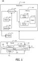
- System 100 may include a workstation or console 112 from which a procedure is supervised and managed.
- Workstation 112 preferably includes one or more processors 114 and memory 116 for storing programs and applications.
- Memory 116 may store a robot control module 115 configured to control servos, actuators or the like for a repeatably controlling positions or orientations of a mechanism or robot system 122.
- the robot system 122 may include a sensing/tracking device or devices 126 to provide feedback signals to ensure positional accuracy, a robot or linkage 124 and other devices or sensors for proper operation of the robot 124.
- sensing/tracking module 128 may be provided to interpret feedback from the device or devices 126.
- sensing/tracking module 128 is configured to use signal feedback from sensing devices 126 or the robot system 122 to reconstruct deformations, deflections and other changes associated with the robot 124.
- Workstation 112 may include a display 118 for viewing internal images of a subject if an imaging system 110 is employed.
- the imaging system 110 may include, e.g., a magnetic resonance imaging (MRI) system, a fluoroscopy system, a computed tomography (CT) system, ultrasound (US), etc.
- Display 118 may also permit a user to interact with the workstation 112 and its components and functions. This is further facilitated by an interface 120 which may include a keyboard, mouse, a joystick or any other peripheral or control to permit user interaction with the workstation 112.
- Imaging system 110 may be provided for collecting pre-operative imaging data or real-time intra-operative imaging data.
- the pre-operative imaging may be performed at another facility, location, etc. in advance of any procedure.
- Images 111 may be stored in memory 116, and may include pre-operative 3D image volumes of a patient or pathway system.
- a medical device 102 is preferably elongated and may include, e.g., a catheter, a guide wire, an endoscope, a probe, a robot, an electrode, a filter device, a balloon device, or other medical component, etc.
- an endoscope is employed for the medical device 102 and is guided to a position within a patient using the robot 124. When a desired position is reached, feedback from the robot 124 may be stored to ensure that the position can be re-obtained in subsequent activities.
- an endoscope (102) is employed to find a location; then the endoscope (102) is removed and an ultrasonic (US) probe 125 is positioned by the robot 124 at the same location.
- US ultrasonic
- the US probe 125 can now be positioned in contact with tissue to image a region corresponding to the endoscopically determined location. Since the robot 124 is employed, the location is repeatably obtained by the US probe 125.
- Methods for overlaying an arterial tree on endoscope images may be employed to provide visibility of arteries in images. These overlays may be employed for robotic navigation to ensure reproducible point location.
- the robot 124 may follow the arterial path in endoscope images. Other robotic navigation techniques may also be employed.
- the device 102 and probe 125 are introduced in a patient 130 through ports 134, e.g., ports to a thoracic cavity, etc.
- Intraoperative ultrasound may be used in coronary bypass surgery to assess the function of blood vessels. US may be used before bypass is completed to detect positions of arterial plaque or after the bypass is placed to assess blood flow through the bypass, aorta, or other coronary arteries.
- the ultrasound probe 125 is placed on or in a sterile sheath and moved on a heart surface to collect US images.
- Minimally invasive bypass surgery is performed through small ports 134 (e.g., between 5 mm for totally endoscopic procedures and 50-60 mm for minimally invasive direct bypass surgery).
- the present principles include robotic steering of a US probe 125 to systematically scan arteries and other areas of interest.
- the robot 124 holds the imaging probe 125 for deployment in accordance with the endoscopic images or planned pathway.
- the robot 124 can be any kind of actuated device capable of moving around a fulcrum point at a chest surface (e.g., through port(s) 134) or other entry point on the patient 130.
- the robot 124 conveys one or more of the US probe 125 and a visual component (e.g., camera, endoscope, etc.) to a target 132. In this way, the target can be analyzed visually and ultrasonically from a same repeatably obtained location.
- the robot 124 provides highly accurate coordinates from which US imaging and visual imaging may be obtained from a same otherwise obscured location.
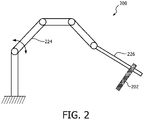
- an illustrative robotic system 200 with an imaging device 202 is shown in accordance with one embodiment.
- a robot 224 steers an endoscope (202). Additional functionality is provided through replacement of the endoscope with an ultrasound probe (202).
- the imaging device 202 is shown generically and is detachable from the robot 224 (which maybe the same as robot 124 in FIG. 1 ).
- the device 202 may be a detachable endoscope or a detachable US transducer. With the endoscope placed through a port, a surgeon or user chooses a path on an endoscope image as shown, e.g., in FIG. 3 .
- a path 300 is identified in an endoscopic image 302 and an overlay 304 is positioned over the path.
- a user interface 120 may be employed for the selection of the path. The surgeon chooses the path which can be a manually selected curve, an artery/vein, etc.
- the path 300 may include a blood vessel selected in the pre-operative images, etc.
- the endoscope is replaced with the US probe and the path is followed with US probe collecting a series of images. Every image is associated with a point on the path. After the operation is completed, the user can slide a virtual point 306 on the path to select different US images.
- An inset 310 shows an US image taken at virtual point 306.
- the path 300 can be defined by manual input, for example, with a computer mouse, or can be selected as an artery from the pre-operative arterial overlay. Additionally, the path 300 can be selected on the pre-operative images (111, FIG. 1 ) (for example selecting an artery or a series of landmarks), and transferred to the current endoscope image and used to robotically control the endoscope. The path 300 could be selected on pre-operative images (111, FIG. 1 ) and then mapped to the endoscope image 302.
- the endoscope is replaced with the ultrasound probe.
- the robot 224 moves the probe 202 towards a target to secure acoustic coupling. This can be done by the surgeon using some input device (such as a mouse, joystick or controls on a user interface 120, FIG. 1 ) or automatically using force control or other methods related to the US image.
- One example employs a method of knowing depth which has the US probe located in a pre-defined specific manner with respect to an end effector 226 of the robot 224, thus defining the relationship between the end effector 226 and US probe images, allowing depth of structures from the robot 224 to be obtained from the US image.
- the robot 224 moves the probe along the predefined path.
- a position of the probe (202) relative to the path is known.
- the user can replay the path (300) by using a sliding function on the path or selecting a specific point (e.g., using the workstation 112 with display 118 and interface 120).
- the robot 224 can move the probe (202) in different directions to obtain images in different planes relative to the anatomy. This will permit imaging of both cross sections, coronal and sagittal images of the targeted structure, etc.
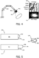
- a US probe 402 is connected to an end effector 426 of a robot 424.
- a camera or cameras 410 are added. Visual feedback of the US probe 402 and a surgical region may be provided while the robot 402 is moving.
- the cameras 410 can be added by, for example, attaching a small camera 410 to the US probe 402, which would provide visual feedback coupled with the US images of the targeted structure.
- the robot 424 holds the US probe 402 and moves along a predefined path providing visual feedback of the probe 402 using camera 410, which may be attached to the probe 402. Both visual 412 and ultrasonic 414 images can be combined or individually shown to the surgeon or user.
- a first view 520 shows an endoscopic mode where an endoscope 504 is employed.
- a second view 522 shows an ultrasonic mode where an ultrasonic probe 508 is extended to gather US images.
- a third view 524 shows a front view of the housing 502.
- the endoscope 504 and the ultrasound probe 508 are packaged in a single fixture 502. Probe 508 is permitted to slide along a longitudinal axis of the fixture 502.
- the US probe 508 and endoscope 504 are placed in the fixture or housing 502 with US probe 508 able to move relative to the housing 502 to be able to make contact with tissue to acquire images.
- the US probe 508 In an endoscope mode (view 520), the US probe 508 is retracted and inactive. As the path is selected, the US probe 508 is moved outward (view 522) until tissue coupling is achieved. The US probe 508 is activated, and the collection of images is performed, as described above.
- One advantage is that visual feedback is provided by the endoscope 504 during US image collection. This configuration may need a larger port to accommodate the larger housing or fixture 502. It maybe possible to introduce very small US probes together with an endoscope without using larger sized ports.
- one configuration employs two small instrument ports 612 and 614.
- an endoscope 604 is fixed using a passive holding arm 616 or is held by a human through instrument port 612.
- the passive holding arm 616 may include any number of devices, such as a catheter, push rod, wire, etc.
- a robot 624 is equipped with an ultrasound probe 602 and introduced through the instrument port 614.
- the endoscope 604 and probe 602 are independently provided.
- the endoscope 604 is employed to visually locate the robot 624 and probe 602. This permits the use of small ports and visual feedback.
- a position of the robot 624 is known and can be employed to pinpoint a point of view for the US images.
- While the present principles are applicable to any interventional procedure, a particularly useful application includes coronary bypass surgery.
- the present embodiments can be employed in other endoscopic vascular surgeries or where US imaging of structures during minimally invasive surgery is convenient and useful.
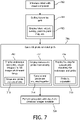
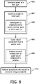
- a robot is introduced having a visual guidance component to pass to a target preferably through surgical ports.
- a path or target marker is defined at a location on the target for the robot.
- the robot may be guided along the path by using previously obtained images or by using guideposts collected during the deployment of the robot.
- known tracking techniques may be employed to locate or guide the robot to the target.
- the path may be determined based on one or more of the following techniques. A blood vessel may be followed or traced to provide the path, a point to point mapping in an image may be employed and then provided to the robot system for control commands, and an overlay may be placed on an image and followed, etc.
- an ultrasonic probe is guided along the path to permit engagement of the probe to collect ultrasonic images at positions along the path.
- the ultrasonic probe may be deployed contemporaneously with the visual component or according to the invention is deployed at a different time.
- the visual component may include an endoscope, and the endoscope is deployed first and then the ultrasonic probe is deployed such that visual images and ultrasonic images are taken at different times.
- the endoscope and the probe are detachable from the robot and are employed separately in a same procedure.
- the visual component includes a camera, and the camera and the ultrasonic probe are deployed concurrently.
- the camera may be mounted on the ultrasonic probe, and visual images and ultrasonic image are acquired concurrently from a same vantage point.
- a housing or fixture is coupled to an end effector of the robot and configured to deploy an endoscope - as the visual component - and the ultrasonic probe, concurrently, wherein the ultrasonic probe may be actuated or deployed from within the housing to engage tissue for acquiring images.
- the ultrasonic probe moves along a longitudinal axis of the housing. Other movements or positioning systems may also be employed.
- a procedure is carried out where the location is accurately known due to the robot, and US images may be collected at the accurately known location.
- This may include providing US images along a defined path.
- visual and US imaging are provided together. In a cardiac bypass surgery example, this enables viewing of a bypass for bleeding (visual) while checking flow characteristics (US) through the bypass.
- Other advantages and applications are also contemplated.
- a robot is introduced, which is configured to pass to a target location through a first port.
- an ultrasonic probe is guided by the robot to the location to permit engagement of the probe to collect ultrasonic images at the location.
- the path may be determined based on one or more of the following techniques. A blood vessel may be followed or traced to provide the path, a point to point mapping in an image may be employed and then provided to the robot system for control commands, and an overlay may be placed on an image and followed, etc.
- a visual component is separately guided to the location through a second port.
- the location is defined in accordance with a position and orientation of the robot. Data from the robot may be employed to provide guidance to the location for the visual component. The guidance would be visual as the robot can be visually followed to the location.
- the visual component may include, e.g., an endoscope or a camera.
- separate guidance of the visual components may be employed with a passive holding arm to guide the visual component to the location.
- a procedure is carried out where the location is accurately known due to the robot, and US images may be collected at the accurately known location. This may include providing US images along a defined path. In a cardiac bypass surgery example, this enables viewing of a bypass for bleeding (visual) while checking flow characteristics (US) through the bypass. Other advantages and applications are also contemplated.
Landscapes
- Health & Medical Sciences (AREA)
- Life Sciences & Earth Sciences (AREA)
- Chemical & Material Sciences (AREA)
- General Health & Medical Sciences (AREA)
- Engineering & Computer Science (AREA)
- Surgery (AREA)
- Organic Chemistry (AREA)
- Animal Behavior & Ethology (AREA)
- Molecular Biology (AREA)
- Public Health (AREA)
- Veterinary Medicine (AREA)
- Medical Informatics (AREA)
- Heart & Thoracic Surgery (AREA)
- Biomedical Technology (AREA)
- Nuclear Medicine, Radiotherapy & Molecular Imaging (AREA)
- Biophysics (AREA)
- Pathology (AREA)
- Medicinal Chemistry (AREA)
- Physics & Mathematics (AREA)
- Radiology & Medical Imaging (AREA)
- Pharmacology & Pharmacy (AREA)
- Biochemistry (AREA)
- Oil, Petroleum & Natural Gas (AREA)
- Chemical Kinetics & Catalysis (AREA)
- General Chemical & Material Sciences (AREA)
- Epidemiology (AREA)
- Analytical Chemistry (AREA)
- Optics & Photonics (AREA)
- Genetics & Genomics (AREA)
- Proteomics, Peptides & Aminoacids (AREA)
- Robotics (AREA)
- Oral & Maxillofacial Surgery (AREA)
- Ultra Sonic Daignosis Equipment (AREA)
- Endoscopes (AREA)
Claims (2)
- Système de guidage robotique chirurgical, comprenant:un système robotique (122) ayant un robot (124) configuré pour passer à une cible à travers un orifice (134), le système robotique (122) comprenant un endoscope amovible (102) utilisé pour guider le robot (124) le long d'un chemin (300) vers un emplacement, l'emplacement étant défini en fonction d'une position et d'une orientation du robot (124), où le système est configuré pour stocker la rétroaction du robot (124) lorsque l'emplacement est atteint, la rétroaction stockée permettant de réobtenir l'emplacement à l'aide du robot (124); et caractérisé en ce que le système comprend en outreune sonde à ultrasons (125) qui peut remplacer l'endoscope (102),où, après avoir utilisé l'endoscope (102) pour guider le robot (124) vers l'emplacement et lors d'un remplacement ultérieur du endoscope (102) avec la sonde à ultrasons (125), la sonde à ultrasons (125) est guidée automatiquement par le robot (124) le long du chemin (300) vers l'emplacement sur la base de la rétroaction stockée du robot (124) pour permettre l'engagement de la sonde à ultrasons (125) pour collecter des images ultrasonores au niveau de l'emplacement, de telle sorte que des images visuelles et des images ultrasonores soient prises à des moments différents mais au même emplacement.
- Système selon la revendication 1, dans lequel le chemin (300) est déterminé sur la base d'une ou plusieurs parmi: une image préopératoire, une image de vaisseau sanguin, une cartographie point à point dans une image et une superposition sur une image.
Applications Claiming Priority (2)
| Application Number | Priority Date | Filing Date | Title |
|---|---|---|---|
| US201161566625P | 2011-12-03 | 2011-12-03 | |
| PCT/IB2012/056758 WO2013080124A1 (fr) | 2011-12-03 | 2012-11-27 | Guidage robotique de sonde à ultrasons en chirurgie endoscopique |
Publications (2)
| Publication Number | Publication Date |
|---|---|
| EP2785269A1 EP2785269A1 (fr) | 2014-10-08 |
| EP2785269B1 true EP2785269B1 (fr) | 2022-05-11 |
Family
ID=48539462
Family Applications (1)
| Application Number | Title | Priority Date | Filing Date |
|---|---|---|---|
| EP12810427.0A Active EP2785269B1 (fr) | 2011-12-03 | 2012-11-27 | Guidage robotique de sonde à ultrasons en chirurgie endoscopique |
Country Status (8)
| Country | Link |
|---|---|
| US (1) | US10414792B2 (fr) |
| EP (1) | EP2785269B1 (fr) |
| JP (1) | JP6559956B2 (fr) |
| CN (1) | CN104105455B (fr) |
| BR (1) | BR112014013073A2 (fr) |
| IN (1) | IN2014CN04516A (fr) |
| RU (1) | RU2014127126A (fr) |
| WO (1) | WO2013080124A1 (fr) |
Families Citing this family (25)
| Publication number | Priority date | Publication date | Assignee | Title |
|---|---|---|---|---|
| WO2012159123A2 (fr) | 2011-05-19 | 2012-11-22 | Alec Rivers | Outils à guidage automatique |
| US9498231B2 (en) | 2011-06-27 | 2016-11-22 | Board Of Regents Of The University Of Nebraska | On-board tool tracking system and methods of computer assisted surgery |
| US11911117B2 (en) | 2011-06-27 | 2024-02-27 | Board Of Regents Of The University Of Nebraska | On-board tool tracking system and methods of computer assisted surgery |
| CN103764061B (zh) | 2011-06-27 | 2017-03-08 | 内布拉斯加大学评议会 | 工具承载的追踪系统和计算机辅助外科方法 |
| WO2013163588A1 (fr) | 2012-04-26 | 2013-10-31 | Alec Rothmyer Rivers | Systèmes et procédés permettant de réaliser une tâche sur un matériau, ou permettant de localiser la position d'un dispositif par rapport à la surface du matériau |
| US10105149B2 (en) | 2013-03-15 | 2018-10-23 | Board Of Regents Of The University Of Nebraska | On-board tool tracking system and methods of computer assisted surgery |
| CN105934216B (zh) | 2014-01-24 | 2019-09-17 | 皇家飞利浦有限公司 | 机器人引导系统、控制单元和装置 |
| EP3169264A1 (fr) | 2014-07-15 | 2017-05-24 | Koninklijke Philips N.V. | Intégration d'images et commande d'endoscope robotique dans une installation radiographique |
| WO2016169787A1 (fr) | 2015-04-21 | 2016-10-27 | Koninklijke Philips N.V. | Bras réglable pour dispositif de surveillance de patient |
| EP3657279B1 (fr) | 2015-05-13 | 2023-03-29 | Shaper Tools, Inc. | Systèmes, procédés et appareil pour outils guidés |
| JP2019514476A (ja) * | 2016-04-19 | 2019-06-06 | コーニンクレッカ フィリップス エヌ ヴェKoninklijke Philips N.V. | 超音波イメージングプローブの位置決め |
| US20190290247A1 (en) * | 2016-05-31 | 2019-09-26 | Koninklijke Philips N.V. | Image-based fusion of endoscopic image and ultrasound images |
| CN206063225U (zh) * | 2016-06-04 | 2017-04-05 | 深圳市前海康启源科技有限公司 | 用于辅助手术的医疗机器人 |
| KR102424132B1 (ko) | 2016-08-19 | 2022-07-22 | 샤퍼 툴스 인코포레이티드 | 공구 제작 및 디자인 데이터 공유를 위한 시스템, 방법 및 장치 |
| CN106419957B (zh) * | 2016-10-09 | 2019-12-13 | 深圳华大智造科技有限公司 | 一种超声扫描装置辅助系统 |
| CN106562804A (zh) * | 2016-11-01 | 2017-04-19 | 河池学院 | 一种b超检测机器人 |
| EP3554370B1 (fr) | 2016-12-19 | 2022-09-28 | Koninklijke Philips N.V. | Commande d'acquisition d'image anatomique à l'aide d'informations physiologiques |
| US11583349B2 (en) | 2017-06-28 | 2023-02-21 | Intuitive Surgical Operations, Inc. | Systems and methods for projecting an endoscopic image to a three-dimensional volume |
| EP3648674A1 (fr) * | 2017-07-07 | 2020-05-13 | Koninklijke Philips N.V. | Intégration de guide d'instrument robotique avec une sonde acoustique |
| CN108403146B (zh) * | 2018-03-20 | 2020-06-30 | 余夏夏 | 基于多传感器信息融合的三维超声成像方法及装置 |
| EP3982816A2 (fr) * | 2019-10-07 | 2022-04-20 | Boston Scientific Scimed Inc. | Dispositifs, systèmes et procédés d'imagerie à l'intérieur d'une lumière corporelle |
| USD1022197S1 (en) | 2020-11-19 | 2024-04-09 | Auris Health, Inc. | Endoscope |
| CN112472137B (zh) * | 2020-12-15 | 2022-09-16 | 深圳市德力凯医疗设备股份有限公司 | 一种基于触摸屏滑动操作轨迹的超声扫描方法及系统 |
| CN114694442A (zh) * | 2020-12-31 | 2022-07-01 | 无锡触典科技有限公司 | 基于虚拟现实的超声培训方法、装置、存储介质及超声设备 |
| CN112914601B (zh) * | 2021-01-19 | 2024-04-02 | 深圳市德力凯医疗设备股份有限公司 | 一种机械臂的避障方法、装置、存储介质及超声设备 |
Citations (1)
| Publication number | Priority date | Publication date | Assignee | Title |
|---|---|---|---|---|
| US20090088897A1 (en) * | 2007-09-30 | 2009-04-02 | Intuitive Surgical, Inc. | Methods and systems for robotic instrument tool tracking |
Family Cites Families (14)
| Publication number | Priority date | Publication date | Assignee | Title |
|---|---|---|---|---|
| US6032673A (en) | 1994-10-13 | 2000-03-07 | Femrx, Inc. | Methods and devices for tissue removal |
| WO2001062173A2 (fr) * | 2000-02-25 | 2001-08-30 | The Board Of Trustees Of The Leland Stanford Junior University | Procedes et appareils de maintien d'une trajectoire en stereotaxie destines a la recherche d'une cible a l'interieur d'un corps |
| JP2001275931A (ja) * | 2000-04-03 | 2001-10-09 | Olympus Optical Co Ltd | 医療システム |
| US20040034297A1 (en) * | 2002-08-13 | 2004-02-19 | General Electric Company | Medical device positioning system and method |
| KR101258912B1 (ko) | 2005-06-06 | 2013-04-30 | 인튜어티브 서지컬 인코포레이티드 | 복강경의 초음파 로보트 수술 시스템 |
| US8398541B2 (en) * | 2006-06-06 | 2013-03-19 | Intuitive Surgical Operations, Inc. | Interactive user interfaces for robotic minimally invasive surgical systems |
| JP4695479B2 (ja) * | 2005-09-30 | 2011-06-08 | 富士フイルム株式会社 | 超音波発生装置 |
| US20070238981A1 (en) * | 2006-03-13 | 2007-10-11 | Bracco Imaging Spa | Methods and apparatuses for recording and reviewing surgical navigation processes |
| AU2007254159B2 (en) | 2006-05-19 | 2013-07-04 | Mako Surgical Corp. | System and method for verifying calibration of a surgical device |
| JP5073415B2 (ja) | 2006-08-28 | 2012-11-14 | オリンパスメディカルシステムズ株式会社 | 超音波内視鏡 |
| FR2920084B1 (fr) | 2007-08-24 | 2010-08-20 | Endocontrol | Systeme d'imagerie pour le suivi d'un outil chirurgical dans un champ operatoire |
| DE102007055204B4 (de) | 2007-11-19 | 2010-04-08 | Deutsches Zentrum für Luft- und Raumfahrt e.V. | Roboter, medizinischer Arbeitsplatz und Verfahren zum Projizieren eines Bildes auf die Oberfläche eines Objekts |
| JP5154961B2 (ja) | 2008-01-29 | 2013-02-27 | テルモ株式会社 | 手術システム |
| US8815263B2 (en) | 2011-11-07 | 2014-08-26 | Wisconsin Alumni Research Foundation | Tandem facial amphiphiles |
-
2012
- 2012-11-27 BR BR112014013073A patent/BR112014013073A2/pt not_active Application Discontinuation
- 2012-11-27 RU RU2014127126A patent/RU2014127126A/ru not_active Application Discontinuation
- 2012-11-27 EP EP12810427.0A patent/EP2785269B1/fr active Active
- 2012-11-27 WO PCT/IB2012/056758 patent/WO2013080124A1/fr active Application Filing
- 2012-11-27 JP JP2014544012A patent/JP6559956B2/ja active Active
- 2012-11-27 US US14/361,943 patent/US10414792B2/en active Active
- 2012-11-27 IN IN4516CHN2014 patent/IN2014CN04516A/en unknown
- 2012-11-27 CN CN201280068844.9A patent/CN104105455B/zh active Active
Patent Citations (1)
| Publication number | Priority date | Publication date | Assignee | Title |
|---|---|---|---|---|
| US20090088897A1 (en) * | 2007-09-30 | 2009-04-02 | Intuitive Surgical, Inc. | Methods and systems for robotic instrument tool tracking |
Also Published As
| Publication number | Publication date |
|---|---|
| BR112014013073A8 (pt) | 2017-06-13 |
| EP2785269A1 (fr) | 2014-10-08 |
| US20140343571A1 (en) | 2014-11-20 |
| JP6559956B2 (ja) | 2019-08-14 |
| CN104105455A (zh) | 2014-10-15 |
| WO2013080124A1 (fr) | 2013-06-06 |
| JP2015505686A (ja) | 2015-02-26 |
| IN2014CN04516A (fr) | 2015-09-11 |
| US10414792B2 (en) | 2019-09-17 |
| BR112014013073A2 (pt) | 2017-06-13 |
| RU2014127126A (ru) | 2016-01-27 |
| CN104105455B (zh) | 2017-04-19 |
Similar Documents
| Publication | Publication Date | Title |
|---|---|---|
| EP2785269B1 (fr) | Guidage robotique de sonde à ultrasons en chirurgie endoscopique | |
| JP7314136B2 (ja) | 医療器具のナビゲーションおよびターゲット用のシステムおよび方法 | |
| JP7098723B2 (ja) | ナビゲーション経路追跡用に構成されたロボットシステム | |
| EP2866638B1 (fr) | Visualisation améliorée de vaisseaux sanguins à l'aide d'un endoscope dirigé par robotique | |
| EP3104801B1 (fr) | Système de visualisation spatiale de l'artère mammaire interne pendant une chirurgie de dérivation très peu invasive | |
| JP2022502179A (ja) | 内視鏡支援経皮的医療処置のためのシステム及び方法 | |
| WO2019169178A1 (fr) | Procédés et systèmes de cartographie et de navigation | |
| JP2021525123A (ja) | 画像ベースの気道分析及びマッピング | |
| JP2020536654A (ja) | 画像ベースの分岐検出およびナビゲーション用マッピング | |
| JP2019512354A (ja) | カテーテル配置のための画像誘導ロボット | |
| CN105828721B (zh) | 用于微创介入的形状感测的机器人超声 | |
| KR20190015582A (ko) | 조향 가능한 신장된 장치의 시스템 및 방법 | |
| JP2014525765A (ja) | 内視鏡手術におけるガイド下注入のためのシステム及び方法 | |
| JP2017500935A5 (fr) | ||
| Elek et al. | Robotic platforms for ultrasound diagnostics and treatment | |
| US20220096183A1 (en) | Haptic feedback for aligning robotic arms | |
| WO2022064369A1 (fr) | Rétroaction haptique pour aligner des bras robotiques | |
| KR20230058119A (ko) | 로봇으로 제어가능한 필드 발생기 |
Legal Events
| Date | Code | Title | Description |
|---|---|---|---|
| PUAI | Public reference made under article 153(3) epc to a published international application that has entered the european phase |
Free format text: ORIGINAL CODE: 0009012 |
|
| 17P | Request for examination filed |
Effective date: 20140703 |
|
| AK | Designated contracting states |
Kind code of ref document: A1 Designated state(s): AL AT BE BG CH CY CZ DE DK EE ES FI FR GB GR HR HU IE IS IT LI LT LU LV MC MK MT NL NO PL PT RO RS SE SI SK SM TR |
|
| DAX | Request for extension of the european patent (deleted) | ||
| 17Q | First examination report despatched |
Effective date: 20160316 |
|
| STAA | Information on the status of an ep patent application or granted ep patent |
Free format text: STATUS: EXAMINATION IS IN PROGRESS |
|
| STAA | Information on the status of an ep patent application or granted ep patent |
Free format text: STATUS: EXAMINATION IS IN PROGRESS |
|
| RAP1 | Party data changed (applicant data changed or rights of an application transferred) |
Owner name: KONINKLIJKE PHILIPS N.V. |
|
| REG | Reference to a national code |
Ref country code: DE Ref legal event code: R079 Ref document number: 602012078212 Country of ref document: DE Free format text: PREVIOUS MAIN CLASS: A61B0019000000 Ipc: A61B0034300000 |
|
| GRAP | Despatch of communication of intention to grant a patent |
Free format text: ORIGINAL CODE: EPIDOSNIGR1 |
|
| STAA | Information on the status of an ep patent application or granted ep patent |
Free format text: STATUS: GRANT OF PATENT IS INTENDED |
|
| RIC1 | Information provided on ipc code assigned before grant |
Ipc: A61B 34/30 20160101AFI20211122BHEP |
|
| INTG | Intention to grant announced |
Effective date: 20211215 |
|
| GRAS | Grant fee paid |
Free format text: ORIGINAL CODE: EPIDOSNIGR3 |
|
| GRAA | (expected) grant |
Free format text: ORIGINAL CODE: 0009210 |
|
| STAA | Information on the status of an ep patent application or granted ep patent |
Free format text: STATUS: THE PATENT HAS BEEN GRANTED |
|
| AK | Designated contracting states |
Kind code of ref document: B1 Designated state(s): AL AT BE BG CH CY CZ DE DK EE ES FI FR GB GR HR HU IE IS IT LI LT LU LV MC MK MT NL NO PL PT RO RS SE SI SK SM TR |
|
| REG | Reference to a national code |
Ref country code: GB Ref legal event code: FG4D |
|
| REG | Reference to a national code |
Ref country code: CH Ref legal event code: EP |
|
| REG | Reference to a national code |
Ref country code: AT Ref legal event code: REF Ref document number: 1490753 Country of ref document: AT Kind code of ref document: T Effective date: 20220515 |
|
| REG | Reference to a national code |
Ref country code: DE Ref legal event code: R096 Ref document number: 602012078212 Country of ref document: DE |
|
| REG | Reference to a national code |
Ref country code: IE Ref legal event code: FG4D |
|
| REG | Reference to a national code |
Ref country code: LT Ref legal event code: MG9D |
|
| REG | Reference to a national code |
Ref country code: NL Ref legal event code: MP Effective date: 20220511 |
|
| REG | Reference to a national code |
Ref country code: AT Ref legal event code: MK05 Ref document number: 1490753 Country of ref document: AT Kind code of ref document: T Effective date: 20220511 |
|
| PG25 | Lapsed in a contracting state [announced via postgrant information from national office to epo] |
Ref country code: SE Free format text: LAPSE BECAUSE OF FAILURE TO SUBMIT A TRANSLATION OF THE DESCRIPTION OR TO PAY THE FEE WITHIN THE PRESCRIBED TIME-LIMIT Effective date: 20220511 Ref country code: PT Free format text: LAPSE BECAUSE OF FAILURE TO SUBMIT A TRANSLATION OF THE DESCRIPTION OR TO PAY THE FEE WITHIN THE PRESCRIBED TIME-LIMIT Effective date: 20220912 Ref country code: NO Free format text: LAPSE BECAUSE OF FAILURE TO SUBMIT A TRANSLATION OF THE DESCRIPTION OR TO PAY THE FEE WITHIN THE PRESCRIBED TIME-LIMIT Effective date: 20220811 Ref country code: NL Free format text: LAPSE BECAUSE OF FAILURE TO SUBMIT A TRANSLATION OF THE DESCRIPTION OR TO PAY THE FEE WITHIN THE PRESCRIBED TIME-LIMIT Effective date: 20220511 Ref country code: LT Free format text: LAPSE BECAUSE OF FAILURE TO SUBMIT A TRANSLATION OF THE DESCRIPTION OR TO PAY THE FEE WITHIN THE PRESCRIBED TIME-LIMIT Effective date: 20220511 Ref country code: HR Free format text: LAPSE BECAUSE OF FAILURE TO SUBMIT A TRANSLATION OF THE DESCRIPTION OR TO PAY THE FEE WITHIN THE PRESCRIBED TIME-LIMIT Effective date: 20220511 Ref country code: GR Free format text: LAPSE BECAUSE OF FAILURE TO SUBMIT A TRANSLATION OF THE DESCRIPTION OR TO PAY THE FEE WITHIN THE PRESCRIBED TIME-LIMIT Effective date: 20220812 Ref country code: FI Free format text: LAPSE BECAUSE OF FAILURE TO SUBMIT A TRANSLATION OF THE DESCRIPTION OR TO PAY THE FEE WITHIN THE PRESCRIBED TIME-LIMIT Effective date: 20220511 Ref country code: ES Free format text: LAPSE BECAUSE OF FAILURE TO SUBMIT A TRANSLATION OF THE DESCRIPTION OR TO PAY THE FEE WITHIN THE PRESCRIBED TIME-LIMIT Effective date: 20220511 Ref country code: BG Free format text: LAPSE BECAUSE OF FAILURE TO SUBMIT A TRANSLATION OF THE DESCRIPTION OR TO PAY THE FEE WITHIN THE PRESCRIBED TIME-LIMIT Effective date: 20220811 Ref country code: AT Free format text: LAPSE BECAUSE OF FAILURE TO SUBMIT A TRANSLATION OF THE DESCRIPTION OR TO PAY THE FEE WITHIN THE PRESCRIBED TIME-LIMIT Effective date: 20220511 |
|
| PG25 | Lapsed in a contracting state [announced via postgrant information from national office to epo] |
Ref country code: RS Free format text: LAPSE BECAUSE OF FAILURE TO SUBMIT A TRANSLATION OF THE DESCRIPTION OR TO PAY THE FEE WITHIN THE PRESCRIBED TIME-LIMIT Effective date: 20220511 Ref country code: PL Free format text: LAPSE BECAUSE OF FAILURE TO SUBMIT A TRANSLATION OF THE DESCRIPTION OR TO PAY THE FEE WITHIN THE PRESCRIBED TIME-LIMIT Effective date: 20220511 Ref country code: LV Free format text: LAPSE BECAUSE OF FAILURE TO SUBMIT A TRANSLATION OF THE DESCRIPTION OR TO PAY THE FEE WITHIN THE PRESCRIBED TIME-LIMIT Effective date: 20220511 Ref country code: IS Free format text: LAPSE BECAUSE OF FAILURE TO SUBMIT A TRANSLATION OF THE DESCRIPTION OR TO PAY THE FEE WITHIN THE PRESCRIBED TIME-LIMIT Effective date: 20220911 |
|
| PG25 | Lapsed in a contracting state [announced via postgrant information from national office to epo] |
Ref country code: SM Free format text: LAPSE BECAUSE OF FAILURE TO SUBMIT A TRANSLATION OF THE DESCRIPTION OR TO PAY THE FEE WITHIN THE PRESCRIBED TIME-LIMIT Effective date: 20220511 Ref country code: SK Free format text: LAPSE BECAUSE OF FAILURE TO SUBMIT A TRANSLATION OF THE DESCRIPTION OR TO PAY THE FEE WITHIN THE PRESCRIBED TIME-LIMIT Effective date: 20220511 Ref country code: RO Free format text: LAPSE BECAUSE OF FAILURE TO SUBMIT A TRANSLATION OF THE DESCRIPTION OR TO PAY THE FEE WITHIN THE PRESCRIBED TIME-LIMIT Effective date: 20220511 Ref country code: EE Free format text: LAPSE BECAUSE OF FAILURE TO SUBMIT A TRANSLATION OF THE DESCRIPTION OR TO PAY THE FEE WITHIN THE PRESCRIBED TIME-LIMIT Effective date: 20220511 Ref country code: DK Free format text: LAPSE BECAUSE OF FAILURE TO SUBMIT A TRANSLATION OF THE DESCRIPTION OR TO PAY THE FEE WITHIN THE PRESCRIBED TIME-LIMIT Effective date: 20220511 Ref country code: CZ Free format text: LAPSE BECAUSE OF FAILURE TO SUBMIT A TRANSLATION OF THE DESCRIPTION OR TO PAY THE FEE WITHIN THE PRESCRIBED TIME-LIMIT Effective date: 20220511 |
|
| REG | Reference to a national code |
Ref country code: DE Ref legal event code: R097 Ref document number: 602012078212 Country of ref document: DE |
|
| PLBE | No opposition filed within time limit |
Free format text: ORIGINAL CODE: 0009261 |
|
| STAA | Information on the status of an ep patent application or granted ep patent |
Free format text: STATUS: NO OPPOSITION FILED WITHIN TIME LIMIT |
|
| PG25 | Lapsed in a contracting state [announced via postgrant information from national office to epo] |
Ref country code: AL Free format text: LAPSE BECAUSE OF FAILURE TO SUBMIT A TRANSLATION OF THE DESCRIPTION OR TO PAY THE FEE WITHIN THE PRESCRIBED TIME-LIMIT Effective date: 20220511 |
|
| 26N | No opposition filed |
Effective date: 20230214 |
|
| PG25 | Lapsed in a contracting state [announced via postgrant information from national office to epo] |
Ref country code: SI Free format text: LAPSE BECAUSE OF FAILURE TO SUBMIT A TRANSLATION OF THE DESCRIPTION OR TO PAY THE FEE WITHIN THE PRESCRIBED TIME-LIMIT Effective date: 20220511 |
|
| PG25 | Lapsed in a contracting state [announced via postgrant information from national office to epo] |
Ref country code: MC Free format text: LAPSE BECAUSE OF FAILURE TO SUBMIT A TRANSLATION OF THE DESCRIPTION OR TO PAY THE FEE WITHIN THE PRESCRIBED TIME-LIMIT Effective date: 20220511 |
|
| REG | Reference to a national code |
Ref country code: CH Ref legal event code: PL |
|
| REG | Reference to a national code |
Ref country code: BE Ref legal event code: MM Effective date: 20221130 |
|
| PG25 | Lapsed in a contracting state [announced via postgrant information from national office to epo] |
Ref country code: LI Free format text: LAPSE BECAUSE OF NON-PAYMENT OF DUE FEES Effective date: 20221130 Ref country code: CH Free format text: LAPSE BECAUSE OF NON-PAYMENT OF DUE FEES Effective date: 20221130 |
|
| PG25 | Lapsed in a contracting state [announced via postgrant information from national office to epo] |
Ref country code: LU Free format text: LAPSE BECAUSE OF NON-PAYMENT OF DUE FEES Effective date: 20221127 |
|
| PG25 | Lapsed in a contracting state [announced via postgrant information from national office to epo] |
Ref country code: IE Free format text: LAPSE BECAUSE OF NON-PAYMENT OF DUE FEES Effective date: 20221127 |
|
| PG25 | Lapsed in a contracting state [announced via postgrant information from national office to epo] |
Ref country code: FR Free format text: LAPSE BECAUSE OF NON-PAYMENT OF DUE FEES Effective date: 20221130 Ref country code: BE Free format text: LAPSE BECAUSE OF NON-PAYMENT OF DUE FEES Effective date: 20221130 |
|
| PGFP | Annual fee paid to national office [announced via postgrant information from national office to epo] |
Ref country code: GB Payment date: 20231121 Year of fee payment: 12 |
|
| PG25 | Lapsed in a contracting state [announced via postgrant information from national office to epo] |
Ref country code: IT Free format text: LAPSE BECAUSE OF FAILURE TO SUBMIT A TRANSLATION OF THE DESCRIPTION OR TO PAY THE FEE WITHIN THE PRESCRIBED TIME-LIMIT Effective date: 20220511 |
|
| PGFP | Annual fee paid to national office [announced via postgrant information from national office to epo] |
Ref country code: DE Payment date: 20231127 Year of fee payment: 12 |
|
| PG25 | Lapsed in a contracting state [announced via postgrant information from national office to epo] |
Ref country code: HU Free format text: LAPSE BECAUSE OF FAILURE TO SUBMIT A TRANSLATION OF THE DESCRIPTION OR TO PAY THE FEE WITHIN THE PRESCRIBED TIME-LIMIT; INVALID AB INITIO Effective date: 20121127 |
|
| PG25 | Lapsed in a contracting state [announced via postgrant information from national office to epo] |
Ref country code: CY Free format text: LAPSE BECAUSE OF FAILURE TO SUBMIT A TRANSLATION OF THE DESCRIPTION OR TO PAY THE FEE WITHIN THE PRESCRIBED TIME-LIMIT Effective date: 20220511 |