EP2628802A1 - Dna-impfstoff auf basis des exotoxin-fusionsgens b7-1-pe40kdel und verwendung davon - Google Patents
Dna-impfstoff auf basis des exotoxin-fusionsgens b7-1-pe40kdel und verwendung davon Download PDFInfo
- Publication number
- EP2628802A1 EP2628802A1 EP11855878.2A EP11855878A EP2628802A1 EP 2628802 A1 EP2628802 A1 EP 2628802A1 EP 11855878 A EP11855878 A EP 11855878A EP 2628802 A1 EP2628802 A1 EP 2628802A1
- Authority
- EP
- European Patent Office
- Prior art keywords
- pe40kdel
- dna vaccine
- seq
- group
- cells
- Prior art date
- Legal status (The legal status is an assumption and is not a legal conclusion. Google has not performed a legal analysis and makes no representation as to the accuracy of the status listed.)
- Granted
Links
- 108090000623 proteins and genes Proteins 0.000 title claims abstract description 47
- OTLLEIBWKHEHGU-UHFFFAOYSA-N 2-[5-[[5-(6-aminopurin-9-yl)-3,4-dihydroxyoxolan-2-yl]methoxy]-3,4-dihydroxy-6-(hydroxymethyl)oxan-2-yl]oxy-3,5-dihydroxy-4-phosphonooxyhexanedioic acid Chemical compound C1=NC=2C(N)=NC=NC=2N1C(C(C1O)O)OC1COC1C(CO)OC(OC(C(O)C(OP(O)(O)=O)C(O)C(O)=O)C(O)=O)C(O)C1O OTLLEIBWKHEHGU-UHFFFAOYSA-N 0.000 title claims abstract description 22
- 239000002095 exotoxin Substances 0.000 title claims abstract description 22
- 231100000776 exotoxin Toxicity 0.000 title claims abstract description 22
- 229960005486 vaccine Drugs 0.000 title claims description 7
- 230000004927 fusion Effects 0.000 title abstract description 18
- 108010041986 DNA Vaccines Proteins 0.000 claims abstract description 133
- 229940021995 DNA vaccine Drugs 0.000 claims abstract description 133
- 239000013598 vector Substances 0.000 claims abstract description 41
- 239000013604 expression vector Substances 0.000 claims abstract description 37
- 239000000203 mixture Substances 0.000 claims abstract description 19
- 238000003259 recombinant expression Methods 0.000 claims abstract description 16
- 108020001507 fusion proteins Proteins 0.000 claims abstract description 13
- 102000037865 fusion proteins Human genes 0.000 claims abstract description 13
- 210000000056 organ Anatomy 0.000 claims abstract description 12
- 230000000735 allogeneic effect Effects 0.000 claims abstract description 8
- 206010052779 Transplant rejections Diseases 0.000 claims abstract description 4
- 241000701161 unidentified adenovirus Species 0.000 claims abstract description 3
- 238000000034 method Methods 0.000 claims description 25
- 238000002347 injection Methods 0.000 claims description 22
- 239000007924 injection Substances 0.000 claims description 22
- 102000004169 proteins and genes Human genes 0.000 claims description 16
- 238000010255 intramuscular injection Methods 0.000 claims description 12
- 239000007927 intramuscular injection Substances 0.000 claims description 12
- 210000001519 tissue Anatomy 0.000 claims description 9
- 238000002360 preparation method Methods 0.000 claims description 8
- 239000003814 drug Substances 0.000 claims description 6
- 239000007928 intraperitoneal injection Substances 0.000 claims description 6
- 230000009465 prokaryotic expression Effects 0.000 claims description 5
- FWMNVWWHGCHHJJ-SKKKGAJSSA-N 4-amino-1-[(2r)-6-amino-2-[[(2r)-2-[[(2r)-2-[[(2r)-2-amino-3-phenylpropanoyl]amino]-3-phenylpropanoyl]amino]-4-methylpentanoyl]amino]hexanoyl]piperidine-4-carboxylic acid Chemical compound C([C@H](C(=O)N[C@H](CC(C)C)C(=O)N[C@H](CCCCN)C(=O)N1CCC(N)(CC1)C(O)=O)NC(=O)[C@H](N)CC=1C=CC=CC=1)C1=CC=CC=C1 FWMNVWWHGCHHJJ-SKKKGAJSSA-N 0.000 claims description 4
- 239000002671 adjuvant Substances 0.000 claims description 4
- 239000002773 nucleotide Substances 0.000 claims description 4
- 125000003729 nucleotide group Chemical group 0.000 claims description 4
- 108020004705 Codon Proteins 0.000 claims description 2
- 239000007864 aqueous solution Substances 0.000 claims description 2
- 238000009472 formulation Methods 0.000 claims description 2
- 238000001361 intraarterial administration Methods 0.000 claims description 2
- 238000010253 intravenous injection Methods 0.000 claims description 2
- 239000000843 powder Substances 0.000 claims description 2
- 238000010254 subcutaneous injection Methods 0.000 claims description 2
- 239000007929 subcutaneous injection Substances 0.000 claims description 2
- 108091028043 Nucleic acid sequence Proteins 0.000 claims 1
- 210000004877 mucosa Anatomy 0.000 claims 1
- 230000000694 effects Effects 0.000 abstract description 26
- 208000024908 graft versus host disease Diseases 0.000 abstract description 25
- 238000011282 treatment Methods 0.000 abstract description 23
- 230000002265 prevention Effects 0.000 abstract description 9
- 238000011134 hematopoietic stem cell transplantation Methods 0.000 abstract description 4
- 241000699670 Mus sp. Species 0.000 description 93
- 210000004027 cell Anatomy 0.000 description 75
- 238000002054 transplantation Methods 0.000 description 34
- FBOZXECLQNJBKD-ZDUSSCGKSA-N L-methotrexate Chemical compound C=1N=C2N=C(N)N=C(N)C2=NC=1CN(C)C1=CC=C(C(=O)N[C@@H](CCC(O)=O)C(O)=O)C=C1 FBOZXECLQNJBKD-ZDUSSCGKSA-N 0.000 description 31
- 229960000485 methotrexate Drugs 0.000 description 31
- 239000000047 product Substances 0.000 description 31
- 238000003752 polymerase chain reaction Methods 0.000 description 25
- 230000014509 gene expression Effects 0.000 description 24
- 210000001744 T-lymphocyte Anatomy 0.000 description 21
- 239000013612 plasmid Substances 0.000 description 20
- 238000006243 chemical reaction Methods 0.000 description 18
- 210000005259 peripheral blood Anatomy 0.000 description 18
- 239000011886 peripheral blood Substances 0.000 description 18
- 210000002966 serum Anatomy 0.000 description 17
- 230000004083 survival effect Effects 0.000 description 17
- 101150059736 SRY gene Proteins 0.000 description 16
- 239000000243 solution Substances 0.000 description 15
- 210000004379 membrane Anatomy 0.000 description 14
- 239000012528 membrane Substances 0.000 description 14
- 108700010045 sry Genes Proteins 0.000 description 14
- 102000004127 Cytokines Human genes 0.000 description 13
- 108090000695 Cytokines Proteins 0.000 description 13
- 238000004458 analytical method Methods 0.000 description 13
- LFQSCWFLJHTTHZ-UHFFFAOYSA-N Ethanol Chemical compound CCO LFQSCWFLJHTTHZ-UHFFFAOYSA-N 0.000 description 12
- 101001057504 Homo sapiens Interferon-stimulated gene 20 kDa protein Proteins 0.000 description 12
- 101001055144 Homo sapiens Interleukin-2 receptor subunit alpha Proteins 0.000 description 12
- 102100026878 Interleukin-2 receptor subunit alpha Human genes 0.000 description 12
- 210000004369 blood Anatomy 0.000 description 12
- 239000008280 blood Substances 0.000 description 12
- 210000004989 spleen cell Anatomy 0.000 description 12
- 108020004414 DNA Proteins 0.000 description 11
- 108700033844 Pseudomonas aeruginosa toxA Proteins 0.000 description 11
- 238000001514 detection method Methods 0.000 description 11
- 239000000499 gel Substances 0.000 description 11
- 230000004544 DNA amplification Effects 0.000 description 10
- 102000004887 Transforming Growth Factor beta Human genes 0.000 description 10
- 108090001012 Transforming Growth Factor beta Proteins 0.000 description 10
- 210000000265 leukocyte Anatomy 0.000 description 10
- 239000006228 supernatant Substances 0.000 description 10
- 108010076504 Protein Sorting Signals Proteins 0.000 description 9
- 239000003153 chemical reaction reagent Substances 0.000 description 9
- 238000001962 electrophoresis Methods 0.000 description 9
- 238000011534 incubation Methods 0.000 description 9
- XLYOFNOQVPJJNP-UHFFFAOYSA-N water Substances O XLYOFNOQVPJJNP-UHFFFAOYSA-N 0.000 description 9
- 108091032973 (ribonucleotides)n+m Proteins 0.000 description 8
- 210000002798 bone marrow cell Anatomy 0.000 description 8
- 238000000684 flow cytometry Methods 0.000 description 8
- 239000007788 liquid Substances 0.000 description 8
- 230000005855 radiation Effects 0.000 description 8
- 210000000952 spleen Anatomy 0.000 description 8
- 238000012546 transfer Methods 0.000 description 8
- 238000002965 ELISA Methods 0.000 description 7
- 108010002350 Interleukin-2 Proteins 0.000 description 7
- 102000000588 Interleukin-2 Human genes 0.000 description 7
- 241000699666 Mus <mouse, genus> Species 0.000 description 7
- 238000000246 agarose gel electrophoresis Methods 0.000 description 7
- 239000000427 antigen Substances 0.000 description 7
- 108091007433 antigens Proteins 0.000 description 7
- 102000036639 antigens Human genes 0.000 description 7
- 230000000903 blocking effect Effects 0.000 description 7
- 229940079593 drug Drugs 0.000 description 7
- 235000018102 proteins Nutrition 0.000 description 7
- 238000012340 reverse transcriptase PCR Methods 0.000 description 7
- 102000007469 Actins Human genes 0.000 description 6
- 108010085238 Actins Proteins 0.000 description 6
- TWRXJAOTZQYOKJ-UHFFFAOYSA-L Magnesium chloride Chemical compound [Mg+2].[Cl-].[Cl-] TWRXJAOTZQYOKJ-UHFFFAOYSA-L 0.000 description 6
- 239000012980 RPMI-1640 medium Substances 0.000 description 6
- 108060008682 Tumor Necrosis Factor Proteins 0.000 description 6
- 210000002593 Y chromosome Anatomy 0.000 description 6
- 230000003321 amplification Effects 0.000 description 6
- 238000010790 dilution Methods 0.000 description 6
- 239000012895 dilution Substances 0.000 description 6
- 210000003743 erythrocyte Anatomy 0.000 description 6
- 238000002474 experimental method Methods 0.000 description 6
- 239000001963 growth medium Substances 0.000 description 6
- 210000004185 liver Anatomy 0.000 description 6
- 210000004698 lymphocyte Anatomy 0.000 description 6
- 108020004999 messenger RNA Proteins 0.000 description 6
- 238000010172 mouse model Methods 0.000 description 6
- 238000003199 nucleic acid amplification method Methods 0.000 description 6
- 239000011534 wash buffer Substances 0.000 description 6
- NLXLAEXVIDQMFP-UHFFFAOYSA-N Ammonia chloride Chemical compound [NH4+].[Cl-] NLXLAEXVIDQMFP-UHFFFAOYSA-N 0.000 description 5
- 206010068051 Chimerism Diseases 0.000 description 5
- 238000008157 ELISA kit Methods 0.000 description 5
- 108090000174 Interleukin-10 Proteins 0.000 description 5
- 108090000978 Interleukin-4 Proteins 0.000 description 5
- 241000283973 Oryctolagus cuniculus Species 0.000 description 5
- 238000011529 RT qPCR Methods 0.000 description 5
- 230000001580 bacterial effect Effects 0.000 description 5
- 230000037396 body weight Effects 0.000 description 5
- 239000012141 concentrate Substances 0.000 description 5
- UQLDLKMNUJERMK-UHFFFAOYSA-L di(octadecanoyloxy)lead Chemical compound [Pb+2].CCCCCCCCCCCCCCCCCC([O-])=O.CCCCCCCCCCCCCCCCCC([O-])=O UQLDLKMNUJERMK-UHFFFAOYSA-L 0.000 description 5
- 210000003527 eukaryotic cell Anatomy 0.000 description 5
- 230000003394 haemopoietic effect Effects 0.000 description 5
- 230000006058 immune tolerance Effects 0.000 description 5
- 210000005229 liver cell Anatomy 0.000 description 5
- 239000003550 marker Substances 0.000 description 5
- 239000002609 medium Substances 0.000 description 5
- 239000004005 microsphere Substances 0.000 description 5
- 239000013642 negative control Substances 0.000 description 5
- 238000010827 pathological analysis Methods 0.000 description 5
- 229920002981 polyvinylidene fluoride Polymers 0.000 description 5
- 210000000813 small intestine Anatomy 0.000 description 5
- ZRKFYGHZFMAOKI-QMGMOQQFSA-N tgfbeta Chemical compound C([C@H](NC(=O)[C@H](C(C)C)NC(=O)CNC(=O)[C@H](CCC(O)=O)NC(=O)[C@H](CCCNC(N)=N)NC(=O)[C@H](CC(N)=O)NC(=O)[C@H](CC(C)C)NC(=O)[C@H]([C@@H](C)O)NC(=O)[C@H](CCC(O)=O)NC(=O)[C@H]([C@@H](C)O)NC(=O)[C@H](CC(C)C)NC(=O)CNC(=O)[C@H](C)NC(=O)[C@H](CO)NC(=O)[C@H](CCC(N)=O)NC(=O)[C@@H](NC(=O)[C@H](C)NC(=O)[C@H](C)NC(=O)[C@@H](NC(=O)[C@H](CC(C)C)NC(=O)[C@@H](N)CCSC)C(C)C)[C@@H](C)CC)C(=O)N[C@@H]([C@@H](C)O)C(=O)N[C@@H](C(C)C)C(=O)N[C@@H](CC=1C=CC=CC=1)C(=O)N[C@@H](C)C(=O)N1[C@@H](CCC1)C(=O)N[C@@H]([C@@H](C)O)C(=O)N[C@@H](CC(N)=O)C(=O)N[C@@H](CCC(O)=O)C(=O)N[C@@H](C)C(=O)N[C@@H](CC=1C=CC=CC=1)C(=O)N[C@@H](CCCNC(N)=N)C(=O)N[C@@H](C)C(=O)N[C@@H](CC(C)C)C(=O)N1[C@@H](CCC1)C(=O)N1[C@@H](CCC1)C(=O)N[C@@H](CCCNC(N)=N)C(=O)N[C@@H](CCC(O)=O)C(=O)N[C@@H](CCCNC(N)=N)C(=O)N[C@@H](CO)C(=O)N[C@@H](CCCNC(N)=N)C(=O)N[C@@H](CC(C)C)C(=O)N[C@@H](CC(C)C)C(O)=O)C1=CC=C(O)C=C1 ZRKFYGHZFMAOKI-QMGMOQQFSA-N 0.000 description 5
- 238000001890 transfection Methods 0.000 description 5
- 238000011144 upstream manufacturing Methods 0.000 description 5
- 238000011740 C57BL/6 mouse Methods 0.000 description 4
- ULGZDMOVFRHVEP-RWJQBGPGSA-N Erythromycin Chemical compound O([C@@H]1[C@@H](C)C(=O)O[C@@H]([C@@]([C@H](O)[C@@H](C)C(=O)[C@H](C)C[C@@](C)(O)[C@H](O[C@H]2[C@@H]([C@H](C[C@@H](C)O2)N(C)C)O)[C@H]1C)(C)O)CC)[C@H]1C[C@@](C)(OC)[C@@H](O)[C@H](C)O1 ULGZDMOVFRHVEP-RWJQBGPGSA-N 0.000 description 4
- KFZMGEQAYNKOFK-UHFFFAOYSA-N Isopropanol Chemical compound CC(C)O KFZMGEQAYNKOFK-UHFFFAOYSA-N 0.000 description 4
- 206010028851 Necrosis Diseases 0.000 description 4
- 230000006044 T cell activation Effects 0.000 description 4
- 102000000852 Tumor Necrosis Factor-alpha Human genes 0.000 description 4
- 238000002835 absorbance Methods 0.000 description 4
- 210000001185 bone marrow Anatomy 0.000 description 4
- 238000010322 bone marrow transplantation Methods 0.000 description 4
- 239000000872 buffer Substances 0.000 description 4
- 238000004113 cell culture Methods 0.000 description 4
- 239000006285 cell suspension Substances 0.000 description 4
- 230000008859 change Effects 0.000 description 4
- 238000010276 construction Methods 0.000 description 4
- 230000034994 death Effects 0.000 description 4
- 230000007850 degeneration Effects 0.000 description 4
- 239000012154 double-distilled water Substances 0.000 description 4
- 210000001703 glandular epithelial cell Anatomy 0.000 description 4
- 238000007490 hematoxylin and eosin (H&E) staining Methods 0.000 description 4
- 238000001727 in vivo Methods 0.000 description 4
- 230000008595 infiltration Effects 0.000 description 4
- 238000001764 infiltration Methods 0.000 description 4
- 238000001802 infusion Methods 0.000 description 4
- 230000002401 inhibitory effect Effects 0.000 description 4
- 238000005259 measurement Methods 0.000 description 4
- 230000001404 mediated effect Effects 0.000 description 4
- 230000017074 necrotic cell death Effects 0.000 description 4
- 239000013641 positive control Substances 0.000 description 4
- 108091008146 restriction endonucleases Proteins 0.000 description 4
- 210000003491 skin Anatomy 0.000 description 4
- 238000007619 statistical method Methods 0.000 description 4
- 208000024891 symptom Diseases 0.000 description 4
- 229940126580 vector vaccine Drugs 0.000 description 4
- 238000001262 western blot Methods 0.000 description 4
- 241000894006 Bacteria Species 0.000 description 3
- 229930105110 Cyclosporin A Natural products 0.000 description 3
- PMATZTZNYRCHOR-CGLBZJNRSA-N Cyclosporin A Chemical compound CC[C@@H]1NC(=O)[C@H]([C@H](O)[C@H](C)C\C=C\C)N(C)C(=O)[C@H](C(C)C)N(C)C(=O)[C@H](CC(C)C)N(C)C(=O)[C@H](CC(C)C)N(C)C(=O)[C@@H](C)NC(=O)[C@H](C)NC(=O)[C@H](CC(C)C)N(C)C(=O)[C@H](C(C)C)NC(=O)[C@H](CC(C)C)N(C)C(=O)CN(C)C1=O PMATZTZNYRCHOR-CGLBZJNRSA-N 0.000 description 3
- 108010036949 Cyclosporine Proteins 0.000 description 3
- 239000006144 Dulbecco’s modified Eagle's medium Substances 0.000 description 3
- 101000914484 Homo sapiens T-lymphocyte activation antigen CD80 Proteins 0.000 description 3
- OKKJLVBELUTLKV-UHFFFAOYSA-N Methanol Chemical compound OC OKKJLVBELUTLKV-UHFFFAOYSA-N 0.000 description 3
- 241001529936 Murinae Species 0.000 description 3
- FAPWRFPIFSIZLT-UHFFFAOYSA-M Sodium chloride Chemical compound [Na+].[Cl-] FAPWRFPIFSIZLT-UHFFFAOYSA-M 0.000 description 3
- 102100027222 T-lymphocyte activation antigen CD80 Human genes 0.000 description 3
- 230000001154 acute effect Effects 0.000 description 3
- XAGFODPZIPBFFR-UHFFFAOYSA-N aluminium Chemical compound [Al] XAGFODPZIPBFFR-UHFFFAOYSA-N 0.000 description 3
- 229910052782 aluminium Inorganic materials 0.000 description 3
- 238000010171 animal model Methods 0.000 description 3
- 238000004820 blood count Methods 0.000 description 3
- 210000005252 bulbus oculi Anatomy 0.000 description 3
- 238000005119 centrifugation Methods 0.000 description 3
- 239000003795 chemical substances by application Substances 0.000 description 3
- 229960001265 ciclosporin Drugs 0.000 description 3
- 239000002299 complementary DNA Substances 0.000 description 3
- 239000012228 culture supernatant Substances 0.000 description 3
- 239000003085 diluting agent Substances 0.000 description 3
- 239000011888 foil Substances 0.000 description 3
- 210000004907 gland Anatomy 0.000 description 3
- 229920000669 heparin Polymers 0.000 description 3
- 238000000338 in vitro Methods 0.000 description 3
- 208000015181 infectious disease Diseases 0.000 description 3
- 229910001629 magnesium chloride Inorganic materials 0.000 description 3
- 239000000463 material Substances 0.000 description 3
- 230000001575 pathological effect Effects 0.000 description 3
- 210000003289 regulatory T cell Anatomy 0.000 description 3
- 238000010839 reverse transcription Methods 0.000 description 3
- 238000012163 sequencing technique Methods 0.000 description 3
- 230000000638 stimulation Effects 0.000 description 3
- 230000008961 swelling Effects 0.000 description 3
- 238000012360 testing method Methods 0.000 description 3
- 230000001988 toxicity Effects 0.000 description 3
- 231100000419 toxicity Toxicity 0.000 description 3
- 210000003462 vein Anatomy 0.000 description 3
- 239000002699 waste material Substances 0.000 description 3
- 230000004580 weight loss Effects 0.000 description 3
- QKNYBSVHEMOAJP-UHFFFAOYSA-N 2-amino-2-(hydroxymethyl)propane-1,3-diol;hydron;chloride Chemical compound Cl.OCC(N)(CO)CO QKNYBSVHEMOAJP-UHFFFAOYSA-N 0.000 description 2
- 208000011691 Burkitt lymphomas Diseases 0.000 description 2
- HEDRZPFGACZZDS-UHFFFAOYSA-N Chloroform Chemical compound ClC(Cl)Cl HEDRZPFGACZZDS-UHFFFAOYSA-N 0.000 description 2
- 206010012735 Diarrhoea Diseases 0.000 description 2
- 241000588724 Escherichia coli Species 0.000 description 2
- 101710082714 Exotoxin A Proteins 0.000 description 2
- WSFSSNUMVMOOMR-UHFFFAOYSA-N Formaldehyde Chemical compound O=C WSFSSNUMVMOOMR-UHFFFAOYSA-N 0.000 description 2
- CEAZRRDELHUEMR-URQXQFDESA-N Gentamicin Chemical compound O1[C@H](C(C)NC)CC[C@@H](N)[C@H]1O[C@H]1[C@H](O)[C@@H](O[C@@H]2[C@@H]([C@@H](NC)[C@@](C)(O)CO2)O)[C@H](N)C[C@@H]1N CEAZRRDELHUEMR-URQXQFDESA-N 0.000 description 2
- 229930182566 Gentamicin Natural products 0.000 description 2
- 208000009329 Graft vs Host Disease Diseases 0.000 description 2
- WZUVPPKBWHMQCE-UHFFFAOYSA-N Haematoxylin Chemical compound C12=CC(O)=C(O)C=C2CC2(O)C1C1=CC=C(O)C(O)=C1OC2 WZUVPPKBWHMQCE-UHFFFAOYSA-N 0.000 description 2
- HTTJABKRGRZYRN-UHFFFAOYSA-N Heparin Chemical compound OC1C(NC(=O)C)C(O)OC(COS(O)(=O)=O)C1OC1C(OS(O)(=O)=O)C(O)C(OC2C(C(OS(O)(=O)=O)C(OC3C(C(O)C(O)C(O3)C(O)=O)OS(O)(=O)=O)C(CO)O2)NS(O)(=O)=O)C(C(O)=O)O1 HTTJABKRGRZYRN-UHFFFAOYSA-N 0.000 description 2
- 102000014150 Interferons Human genes 0.000 description 2
- 108010050904 Interferons Proteins 0.000 description 2
- 108010065805 Interleukin-12 Proteins 0.000 description 2
- 102000015696 Interleukins Human genes 0.000 description 2
- 108010063738 Interleukins Proteins 0.000 description 2
- 102000003960 Ligases Human genes 0.000 description 2
- 108090000364 Ligases Proteins 0.000 description 2
- 206010025323 Lymphomas Diseases 0.000 description 2
- 101000854961 Mus musculus WD repeat and HMG-box DNA-binding protein 1 Proteins 0.000 description 2
- 206010028980 Neoplasm Diseases 0.000 description 2
- 108700026244 Open Reading Frames Proteins 0.000 description 2
- 238000012408 PCR amplification Methods 0.000 description 2
- 108010002747 Pfu DNA polymerase Proteins 0.000 description 2
- 108091081024 Start codon Proteins 0.000 description 2
- QAOWNCQODCNURD-UHFFFAOYSA-N Sulfuric acid Chemical compound OS(O)(=O)=O QAOWNCQODCNURD-UHFFFAOYSA-N 0.000 description 2
- 108091008874 T cell receptors Proteins 0.000 description 2
- 102000016266 T-Cell Antigen Receptors Human genes 0.000 description 2
- 239000006180 TBST buffer Substances 0.000 description 2
- 108010009583 Transforming Growth Factors Proteins 0.000 description 2
- 102000009618 Transforming Growth Factors Human genes 0.000 description 2
- 230000004913 activation Effects 0.000 description 2
- 150000001413 amino acids Chemical group 0.000 description 2
- ROOXNKNUYICQNP-UHFFFAOYSA-N ammonium persulfate Chemical compound [NH4+].[NH4+].[O-]S(=O)(=O)OOS([O-])(=O)=O ROOXNKNUYICQNP-UHFFFAOYSA-N 0.000 description 2
- 229960000723 ampicillin Drugs 0.000 description 2
- AVKUERGKIZMTKX-NJBDSQKTSA-N ampicillin Chemical compound C1([C@@H](N)C(=O)N[C@H]2[C@H]3SC([C@@H](N3C2=O)C(O)=O)(C)C)=CC=CC=C1 AVKUERGKIZMTKX-NJBDSQKTSA-N 0.000 description 2
- 238000000137 annealing Methods 0.000 description 2
- 239000003146 anticoagulant agent Substances 0.000 description 2
- 229940127219 anticoagulant drug Drugs 0.000 description 2
- 230000006399 behavior Effects 0.000 description 2
- 230000000740 bleeding effect Effects 0.000 description 2
- 238000009395 breeding Methods 0.000 description 2
- 230000001488 breeding effect Effects 0.000 description 2
- 230000030833 cell death Effects 0.000 description 2
- 238000011109 contamination Methods 0.000 description 2
- 230000001472 cytotoxic effect Effects 0.000 description 2
- 230000003013 cytotoxicity Effects 0.000 description 2
- 231100000135 cytotoxicity Toxicity 0.000 description 2
- 230000002354 daily effect Effects 0.000 description 2
- 230000007423 decrease Effects 0.000 description 2
- 230000003247 decreasing effect Effects 0.000 description 2
- 230000008021 deposition Effects 0.000 description 2
- 238000011161 development Methods 0.000 description 2
- 230000018109 developmental process Effects 0.000 description 2
- 230000029087 digestion Effects 0.000 description 2
- 201000010099 disease Diseases 0.000 description 2
- 208000037265 diseases, disorders, signs and symptoms Diseases 0.000 description 2
- 239000003651 drinking water Substances 0.000 description 2
- 235000020188 drinking water Nutrition 0.000 description 2
- 238000001378 electrochemiluminescence detection Methods 0.000 description 2
- 239000002158 endotoxin Substances 0.000 description 2
- YQGOJNYOYNNSMM-UHFFFAOYSA-N eosin Chemical compound [Na+].OC(=O)C1=CC=CC=C1C1=C2C=C(Br)C(=O)C(Br)=C2OC2=C(Br)C(O)=C(Br)C=C21 YQGOJNYOYNNSMM-UHFFFAOYSA-N 0.000 description 2
- 210000003979 eosinophil Anatomy 0.000 description 2
- 210000000981 epithelium Anatomy 0.000 description 2
- 229960003276 erythromycin Drugs 0.000 description 2
- 238000010195 expression analysis Methods 0.000 description 2
- 210000001723 extracellular space Anatomy 0.000 description 2
- 229960002518 gentamicin Drugs 0.000 description 2
- 210000002216 heart Anatomy 0.000 description 2
- 229960002897 heparin Drugs 0.000 description 2
- 206010020718 hyperplasia Diseases 0.000 description 2
- 230000003053 immunization Effects 0.000 description 2
- 238000002649 immunization Methods 0.000 description 2
- 229940079322 interferon Drugs 0.000 description 2
- 210000003734 kidney Anatomy 0.000 description 2
- 238000001638 lipofection Methods 0.000 description 2
- 210000004072 lung Anatomy 0.000 description 2
- 239000012139 lysis buffer Substances 0.000 description 2
- 238000009629 microbiological culture Methods 0.000 description 2
- 230000001338 necrotic effect Effects 0.000 description 2
- 231100000915 pathological change Toxicity 0.000 description 2
- 230000036285 pathological change Effects 0.000 description 2
- 230000002093 peripheral effect Effects 0.000 description 2
- 239000013600 plasmid vector Substances 0.000 description 2
- 238000011084 recovery Methods 0.000 description 2
- 230000002829 reductive effect Effects 0.000 description 2
- 230000000284 resting effect Effects 0.000 description 2
- 239000003161 ribonuclease inhibitor Substances 0.000 description 2
- 238000012216 screening Methods 0.000 description 2
- 230000028327 secretion Effects 0.000 description 2
- 238000002415 sodium dodecyl sulfate polyacrylamide gel electrophoresis Methods 0.000 description 2
- 239000007787 solid Substances 0.000 description 2
- 238000012353 t test Methods 0.000 description 2
- 102000003390 tumor necrosis factor Human genes 0.000 description 2
- 210000000689 upper leg Anatomy 0.000 description 2
- 239000012224 working solution Substances 0.000 description 2
- MZOFCQQQCNRIBI-VMXHOPILSA-N (3s)-4-[[(2s)-1-[[(2s)-1-[[(1s)-1-carboxy-2-hydroxyethyl]amino]-4-methyl-1-oxopentan-2-yl]amino]-5-(diaminomethylideneamino)-1-oxopentan-2-yl]amino]-3-[[2-[[(2s)-2,6-diaminohexanoyl]amino]acetyl]amino]-4-oxobutanoic acid Chemical compound OC[C@@H](C(O)=O)NC(=O)[C@H](CC(C)C)NC(=O)[C@H](CCCN=C(N)N)NC(=O)[C@H](CC(O)=O)NC(=O)CNC(=O)[C@@H](N)CCCCN MZOFCQQQCNRIBI-VMXHOPILSA-N 0.000 description 1
- UAIUNKRWKOVEES-UHFFFAOYSA-N 3,3',5,5'-tetramethylbenzidine Chemical compound CC1=C(N)C(C)=CC(C=2C=C(C)C(N)=C(C)C=2)=C1 UAIUNKRWKOVEES-UHFFFAOYSA-N 0.000 description 1
- HRPVXLWXLXDGHG-UHFFFAOYSA-N Acrylamide Chemical compound NC(=O)C=C HRPVXLWXLXDGHG-UHFFFAOYSA-N 0.000 description 1
- 101000942941 Arabidopsis thaliana DNA ligase 6 Proteins 0.000 description 1
- 239000004475 Arginine Substances 0.000 description 1
- 108091026890 Coding region Proteins 0.000 description 1
- 206010050685 Cytokine storm Diseases 0.000 description 1
- 238000011238 DNA vaccination Methods 0.000 description 1
- 206010011968 Decreased immune responsiveness Diseases 0.000 description 1
- 101710093617 Dihydroxyacetone synthase Proteins 0.000 description 1
- 206010016100 Faeces discoloured Diseases 0.000 description 1
- 208000002250 Hematologic Neoplasms Diseases 0.000 description 1
- 208000026350 Inborn Genetic disease Diseases 0.000 description 1
- 206010061218 Inflammation Diseases 0.000 description 1
- 108010038453 Interleukin-2 Receptors Proteins 0.000 description 1
- 102000010789 Interleukin-2 Receptors Human genes 0.000 description 1
- AGPKZVBTJJNPAG-WHFBIAKZSA-N L-isoleucine Chemical compound CC[C@H](C)[C@H](N)C(O)=O AGPKZVBTJJNPAG-WHFBIAKZSA-N 0.000 description 1
- ROHFNLRQFUQHCH-YFKPBYRVSA-N L-leucine Chemical compound CC(C)C[C@H](N)C(O)=O ROHFNLRQFUQHCH-YFKPBYRVSA-N 0.000 description 1
- COLNVLDHVKWLRT-QMMMGPOBSA-N L-phenylalanine Chemical compound OC(=O)[C@@H](N)CC1=CC=CC=C1 COLNVLDHVKWLRT-QMMMGPOBSA-N 0.000 description 1
- KZSNJWFQEVHDMF-BYPYZUCNSA-N L-valine Chemical compound CC(C)[C@H](N)C(O)=O KZSNJWFQEVHDMF-BYPYZUCNSA-N 0.000 description 1
- ROHFNLRQFUQHCH-UHFFFAOYSA-N Leucine Natural products CC(C)CC(N)C(O)=O ROHFNLRQFUQHCH-UHFFFAOYSA-N 0.000 description 1
- 206010024642 Listless Diseases 0.000 description 1
- 241001465754 Metazoa Species 0.000 description 1
- 101000756628 Mus musculus Actin, cytoplasmic 1 Proteins 0.000 description 1
- KWYHDKDOAIKMQN-UHFFFAOYSA-N N,N,N',N'-tetramethylethylenediamine Chemical compound CN(C)CCN(C)C KWYHDKDOAIKMQN-UHFFFAOYSA-N 0.000 description 1
- 239000012570 Opti-MEM I medium Substances 0.000 description 1
- 206010053159 Organ failure Diseases 0.000 description 1
- 239000002033 PVDF binder Substances 0.000 description 1
- 229930040373 Paraformaldehyde Natural products 0.000 description 1
- 108010084695 Pea Proteins Proteins 0.000 description 1
- ONIBWKKTOPOVIA-UHFFFAOYSA-N Proline Natural products OC(=O)C1CCCN1 ONIBWKKTOPOVIA-UHFFFAOYSA-N 0.000 description 1
- 238000012181 QIAquick gel extraction kit Methods 0.000 description 1
- 238000002123 RNA extraction Methods 0.000 description 1
- 238000010240 RT-PCR analysis Methods 0.000 description 1
- MTCFGRXMJLQNBG-UHFFFAOYSA-N Serine Natural products OCC(N)C(O)=O MTCFGRXMJLQNBG-UHFFFAOYSA-N 0.000 description 1
- 102000057181 Sex-Determining Region Y Human genes 0.000 description 1
- 108700032475 Sex-Determining Region Y Proteins 0.000 description 1
- 206010072207 Splenic fibrosis Diseases 0.000 description 1
- 238000000692 Student's t-test Methods 0.000 description 1
- 206010060872 Transplant failure Diseases 0.000 description 1
- 206010066901 Treatment failure Diseases 0.000 description 1
- 208000025865 Ulcer Diseases 0.000 description 1
- KZSNJWFQEVHDMF-UHFFFAOYSA-N Valine Natural products CC(C)C(N)C(O)=O KZSNJWFQEVHDMF-UHFFFAOYSA-N 0.000 description 1
- 210000001015 abdomen Anatomy 0.000 description 1
- 230000005856 abnormality Effects 0.000 description 1
- 239000011543 agarose gel Substances 0.000 description 1
- 229940024606 amino acid Drugs 0.000 description 1
- 235000019270 ammonium chloride Nutrition 0.000 description 1
- 229910001870 ammonium persulfate Inorganic materials 0.000 description 1
- 230000003698 anagen phase Effects 0.000 description 1
- 238000000540 analysis of variance Methods 0.000 description 1
- 230000003110 anti-inflammatory effect Effects 0.000 description 1
- 230000000719 anti-leukaemic effect Effects 0.000 description 1
- 230000000259 anti-tumor effect Effects 0.000 description 1
- 230000004596 appetite loss Effects 0.000 description 1
- 239000008346 aqueous phase Substances 0.000 description 1
- ODKSFYDXXFIFQN-UHFFFAOYSA-N arginine Natural products OC(=O)C(N)CCCNC(N)=N ODKSFYDXXFIFQN-UHFFFAOYSA-N 0.000 description 1
- 230000033228 biological regulation Effects 0.000 description 1
- 239000012888 bovine serum Substances 0.000 description 1
- 230000022534 cell killing Effects 0.000 description 1
- 238000001516 cell proliferation assay Methods 0.000 description 1
- 239000003593 chromogenic compound Substances 0.000 description 1
- 230000001684 chronic effect Effects 0.000 description 1
- 239000011248 coating agent Substances 0.000 description 1
- 238000000576 coating method Methods 0.000 description 1
- 238000011284 combination treatment Methods 0.000 description 1
- 230000000139 costimulatory effect Effects 0.000 description 1
- 238000012258 culturing Methods 0.000 description 1
- 206010052015 cytokine release syndrome Diseases 0.000 description 1
- 210000001151 cytotoxic T lymphocyte Anatomy 0.000 description 1
- 230000006378 damage Effects 0.000 description 1
- 210000004207 dermis Anatomy 0.000 description 1
- 230000004069 differentiation Effects 0.000 description 1
- 238000010494 dissociation reaction Methods 0.000 description 1
- 230000005593 dissociations Effects 0.000 description 1
- 230000009977 dual effect Effects 0.000 description 1
- 230000004064 dysfunction Effects 0.000 description 1
- 230000008030 elimination Effects 0.000 description 1
- 238000003379 elimination reaction Methods 0.000 description 1
- 239000003480 eluent Substances 0.000 description 1
- 210000002919 epithelial cell Anatomy 0.000 description 1
- 238000011067 equilibration Methods 0.000 description 1
- 239000003797 essential amino acid Substances 0.000 description 1
- 235000020776 essential amino acid Nutrition 0.000 description 1
- 238000011156 evaluation Methods 0.000 description 1
- 238000001704 evaporation Methods 0.000 description 1
- 230000008020 evaporation Effects 0.000 description 1
- 230000003203 everyday effect Effects 0.000 description 1
- 239000013613 expression plasmid Substances 0.000 description 1
- 238000000605 extraction Methods 0.000 description 1
- 239000012634 fragment Substances 0.000 description 1
- 231100000221 frame shift mutation induction Toxicity 0.000 description 1
- 230000037433 frameshift Effects 0.000 description 1
- 230000006870 function Effects 0.000 description 1
- 208000016361 genetic disease Diseases 0.000 description 1
- 230000000762 glandular Effects 0.000 description 1
- 230000037308 hair color Effects 0.000 description 1
- 210000003128 head Anatomy 0.000 description 1
- 210000003958 hematopoietic stem cell Anatomy 0.000 description 1
- ZFGMDIBRIDKWMY-PASTXAENSA-N heparin Chemical compound CC(O)=N[C@@H]1[C@@H](O)[C@H](O)[C@@H](COS(O)(=O)=O)O[C@@H]1O[C@@H]1[C@@H](C(O)=O)O[C@@H](O[C@H]2[C@@H]([C@@H](OS(O)(=O)=O)[C@@H](O[C@@H]3[C@@H](OC(O)[C@H](OS(O)(=O)=O)[C@H]3O)C(O)=O)O[C@@H]2O)CS(O)(=O)=O)[C@H](O)[C@H]1O ZFGMDIBRIDKWMY-PASTXAENSA-N 0.000 description 1
- 229960001008 heparin sodium Drugs 0.000 description 1
- HNDVDQJCIGZPNO-UHFFFAOYSA-N histidine Natural products OC(=O)C(N)CC1=CN=CN1 HNDVDQJCIGZPNO-UHFFFAOYSA-N 0.000 description 1
- 239000005457 ice water Substances 0.000 description 1
- 238000003384 imaging method Methods 0.000 description 1
- 230000036737 immune function Effects 0.000 description 1
- 230000003832 immune regulation Effects 0.000 description 1
- 230000028993 immune response Effects 0.000 description 1
- 230000005847 immunogenicity Effects 0.000 description 1
- 229940088592 immunologic factor Drugs 0.000 description 1
- 239000000367 immunologic factor Substances 0.000 description 1
- 230000001506 immunosuppresive effect Effects 0.000 description 1
- 229960003444 immunosuppressant agent Drugs 0.000 description 1
- 239000003018 immunosuppressive agent Substances 0.000 description 1
- 230000006698 induction Effects 0.000 description 1
- 230000004054 inflammatory process Effects 0.000 description 1
- 230000005764 inhibitory process Effects 0.000 description 1
- 210000004347 intestinal mucosa Anatomy 0.000 description 1
- AGPKZVBTJJNPAG-UHFFFAOYSA-N isoleucine Natural products CCC(C)C(N)C(O)=O AGPKZVBTJJNPAG-UHFFFAOYSA-N 0.000 description 1
- 229960000310 isoleucine Drugs 0.000 description 1
- 230000003902 lesion Effects 0.000 description 1
- 208000032839 leukemia Diseases 0.000 description 1
- 239000003446 ligand Substances 0.000 description 1
- 208000017971 listlessness Diseases 0.000 description 1
- 210000005228 liver tissue Anatomy 0.000 description 1
- 239000012160 loading buffer Substances 0.000 description 1
- 230000007774 longterm Effects 0.000 description 1
- 235000021266 loss of appetite Nutrition 0.000 description 1
- 208000019017 loss of appetite Diseases 0.000 description 1
- 206010025482 malaise Diseases 0.000 description 1
- 230000036210 malignancy Effects 0.000 description 1
- 238000010369 molecular cloning Methods 0.000 description 1
- 230000035772 mutation Effects 0.000 description 1
- 239000012188 paraffin wax Substances 0.000 description 1
- 229920002866 paraformaldehyde Polymers 0.000 description 1
- 230000036961 partial effect Effects 0.000 description 1
- 230000007170 pathology Effects 0.000 description 1
- 230000037361 pathway Effects 0.000 description 1
- 235000019702 pea protein Nutrition 0.000 description 1
- 239000008188 pellet Substances 0.000 description 1
- COLNVLDHVKWLRT-UHFFFAOYSA-N phenylalanine Natural products OC(=O)C(N)CC1=CC=CC=C1 COLNVLDHVKWLRT-UHFFFAOYSA-N 0.000 description 1
- 230000003389 potentiating effect Effects 0.000 description 1
- 230000001681 protective effect Effects 0.000 description 1
- 230000036647 reaction Effects 0.000 description 1
- 238000003753 real-time PCR Methods 0.000 description 1
- 108020003175 receptors Proteins 0.000 description 1
- 102000005962 receptors Human genes 0.000 description 1
- 230000009467 reduction Effects 0.000 description 1
- 239000013643 reference control Substances 0.000 description 1
- 230000001105 regulatory effect Effects 0.000 description 1
- 230000002441 reversible effect Effects 0.000 description 1
- 238000012764 semi-quantitative analysis Methods 0.000 description 1
- 230000035945 sensitivity Effects 0.000 description 1
- 238000000926 separation method Methods 0.000 description 1
- 230000019491 signal transduction Effects 0.000 description 1
- 210000002027 skeletal muscle Anatomy 0.000 description 1
- PVFDPMYXCZLHKY-MLLWLMKGSA-M sodium [(1R,2R,4aR,8aS)-2-hydroxy-5-[(2E)-2-[(4S)-4-hydroxy-2-oxooxolan-3-ylidene]ethyl]-1,4a,6-trimethyl-2,3,4,7,8,8a-hexahydronaphthalen-1-yl]methyl sulfate Chemical compound [Na+].C([C@@H]1[C@](C)(COS([O-])(=O)=O)[C@H](O)CC[C@]11C)CC(C)=C1C\C=C1/[C@H](O)COC1=O PVFDPMYXCZLHKY-MLLWLMKGSA-M 0.000 description 1
- 239000011780 sodium chloride Substances 0.000 description 1
- 238000003153 stable transfection Methods 0.000 description 1
- 238000010186 staining Methods 0.000 description 1
- 239000008223 sterile water Substances 0.000 description 1
- 239000012089 stop solution Substances 0.000 description 1
- 238000013517 stratification Methods 0.000 description 1
- 239000000126 substance Substances 0.000 description 1
- 230000001629 suppression Effects 0.000 description 1
- 230000008685 targeting Effects 0.000 description 1
- 230000001225 therapeutic effect Effects 0.000 description 1
- 230000010474 transient expression Effects 0.000 description 1
- 231100000397 ulcer Toxicity 0.000 description 1
- 239000004474 valine Substances 0.000 description 1
- 108700026220 vif Genes Proteins 0.000 description 1
- 238000005406 washing Methods 0.000 description 1
Images


Classifications
-
- C—CHEMISTRY; METALLURGY
- C07—ORGANIC CHEMISTRY
- C07K—PEPTIDES
- C07K14/00—Peptides having more than 20 amino acids; Gastrins; Somatostatins; Melanotropins; Derivatives thereof
- C07K14/001—Peptides having more than 20 amino acids; Gastrins; Somatostatins; Melanotropins; Derivatives thereof by chemical synthesis
-
- A—HUMAN NECESSITIES
- A61—MEDICAL OR VETERINARY SCIENCE; HYGIENE
- A61K—PREPARATIONS FOR MEDICAL, DENTAL OR TOILETRY PURPOSES
- A61K39/00—Medicinal preparations containing antigens or antibodies
- A61K39/0005—Vertebrate antigens
- A61K39/001—Preparations to induce tolerance to non-self, e.g. prior to transplantation
-
- A—HUMAN NECESSITIES
- A61—MEDICAL OR VETERINARY SCIENCE; HYGIENE
- A61P—SPECIFIC THERAPEUTIC ACTIVITY OF CHEMICAL COMPOUNDS OR MEDICINAL PREPARATIONS
- A61P37/00—Drugs for immunological or allergic disorders
- A61P37/02—Immunomodulators
- A61P37/06—Immunosuppressants, e.g. drugs for graft rejection
-
- C—CHEMISTRY; METALLURGY
- C07—ORGANIC CHEMISTRY
- C07K—PEPTIDES
- C07K14/00—Peptides having more than 20 amino acids; Gastrins; Somatostatins; Melanotropins; Derivatives thereof
- C07K14/195—Peptides having more than 20 amino acids; Gastrins; Somatostatins; Melanotropins; Derivatives thereof from bacteria
- C07K14/21—Peptides having more than 20 amino acids; Gastrins; Somatostatins; Melanotropins; Derivatives thereof from bacteria from Pseudomonadaceae (F)
-
- C—CHEMISTRY; METALLURGY
- C07—ORGANIC CHEMISTRY
- C07K—PEPTIDES
- C07K14/00—Peptides having more than 20 amino acids; Gastrins; Somatostatins; Melanotropins; Derivatives thereof
- C07K14/435—Peptides having more than 20 amino acids; Gastrins; Somatostatins; Melanotropins; Derivatives thereof from animals; from humans
- C07K14/705—Receptors; Cell surface antigens; Cell surface determinants
- C07K14/70503—Immunoglobulin superfamily
- C07K14/70532—B7 molecules, e.g. CD80, CD86
-
- A—HUMAN NECESSITIES
- A61—MEDICAL OR VETERINARY SCIENCE; HYGIENE
- A61K—PREPARATIONS FOR MEDICAL, DENTAL OR TOILETRY PURPOSES
- A61K39/00—Medicinal preparations containing antigens or antibodies
- A61K2039/51—Medicinal preparations containing antigens or antibodies comprising whole cells, viruses or DNA/RNA
- A61K2039/53—DNA (RNA) vaccination
-
- C—CHEMISTRY; METALLURGY
- C07—ORGANIC CHEMISTRY
- C07K—PEPTIDES
- C07K2319/00—Fusion polypeptide
- C07K2319/01—Fusion polypeptide containing a localisation/targetting motif
- C07K2319/04—Fusion polypeptide containing a localisation/targetting motif containing an ER retention signal such as a C-terminal HDEL motif
-
- C—CHEMISTRY; METALLURGY
- C07—ORGANIC CHEMISTRY
- C07K—PEPTIDES
- C07K2319/00—Fusion polypeptide
- C07K2319/55—Fusion polypeptide containing a fusion with a toxin, e.g. diphteria toxin
Definitions
- the present invention relates to the fields of immunology and molecular biology and is related to a B7-1-PE40KDEL exotoxin fusion gene-based DNA vaccine and the use thereof.
- the invention also relates to a B7-1-PE40KDEL exotoxin fusion gene, an exotoxin fusion protein encoded by the fusion gene, a recombinant expression vector containing the fusion gene, and a composition that contain the recombinant expression vector.
- Allogeneic hematopoietic stem cell transplantation (allo-HSCT) and solid organ transplantation have been widely used in the treatment of hematologic malignancies, certain genetic diseases, acute severe radiation sickness, and a variety of solid organ failures and malignancies. Due to the differences in the major histocompatibility antigen (MHC) between donors and recipients, along with many other known and unknown factors such as the minor histocompatibility antigen (mHA), tissue-specific antigens, and non-immunological factors, host-versus-graft diseases (HVGDs) and graft-versus-host diseases (GVHDs or GVHRs) are inevitable and are one of the most important and fundamental causes of allogeneic tissue graft failure and graft chronic dysfunction (GCD).
- MHC major histocompatibility antigen
- mHA minor histocompatibility antigen
- GVHDs or GVHRs graft-versus-host diseases
- an ideal and efficient allo-HSCT and organ transplantation should amend or modify the initial reaction of T cells to allogeneic antigens rather than removing all T cells from the body of the transplant recipient or the donor graft. That is, the induction of recipient or donor-specific immune tolerance should target the elimination or inhibition of the recipient or donor T-cell reaction to allogeneic antigens but still retain the normal reaction of T cells to other antigens.
- T cell activation requires two signals: T cell receptor signal and co-stimulation signal of cluster of differentiation 28 (CD28). The first signal is regulated by the second signal, resulting in T cell activation, partial activation or anergy.
- aGVHD acute GVHD
- CTLA4-Ig cytotoxic T-lymphocyte antigen 4-Ig
- B7 antibody-induced specific immune tolerance is primarily used to achieve the goal of preventing HVGD and aGVHD and has been proven effective in a large number of in vivo and in vitro animal experiments.
- CTLA4-Ig cytotoxic T-lymphocyte antigen 4-Ig
- B7 antibody-induced specific immune tolerance is primarily used to achieve the goal of preventing HVGD and aGVHD and has been proven effective in a large number of in vivo and in vitro animal experiments.
- the shortcomings of this method are that the duration of the induced immune tolerance is short and that the induced incompetent T cells can be reactivated by other signaling pathways, leading to treatment failure.
- the monoclonal antibodies used for blocking are mostly murine, and their immunogenicity will affect the treatment's efficacy.
- the inventors have discovered a B7-1-PE40KDEL exotoxin fusion gene and, surprisingly, found that the recombinant expression vector pcDNA3.1/Zeo(+) -B7-1-PE40KDEL that is transfected into eukaryotic cells is transcribed, translated, post-translationally modified, and then secreted into the extracellular space.
- the vector pcDNA3.1/Zeo(+)-B7-1-PE40KDEL can be efficiently expressed in eukaryotic cells, and the expression product has good targeting immunosuppressive activity.
- the inventors have also found that the pcDNA3.1/Zeo(+)-B7-1-PE40KDEL exotoxin fusion gene-based DNA vaccine can be effective in the prevention and treatment in murine aGVHD model. Therefore, the following invention is provided.
- One aspect of the invention relates to a B7-1-PE40KDEL exotoxin fusion gene, and its nucleotide sequence is shown as SEQ ID NO: I or SEQ ID NO: 2.
- the underlined parts are Kpn I restriction sites, and the framed parts are the start codon and stop codon.
- the open reading frame of the above sequence SEQ ID NO: 1 is SEQ ID NO: 2 (1827 bp).
- Another aspect of the invention relates to the exotoxin fusion protein (SEQ ID NO: 3) encoded by the exotoxin fusion gene (SEQ ID NO: 1 or SEQ ID NO: 2).
- amino acid sequence of SEQ ID NO: 3 is shown as follows: (608 amino acids, the underlined part is the signal peptide sequence).
- Another aspect of the invention relates to a recombinant expression vector containing the B7-1-PE40KDEL exotoxin fusion gene that can be effectively fused with selected eukaryotic expression vectors, such as pcDNA3.1/Zeo(+), pWLNEO, pSV2CAT, pOG44, pXT1, pSG, pSVK3, pBPV, pMSG, pSVL, and adenoviruses.
- selected eukaryotic expression vectors such as pcDNA3.1/Zeo(+), pWLNEO, pSV2CAT, pOG44, pXT1, pSG, pSVK3, pBPV, pMSG, pSVL, and adenoviruses.
- the recombinant expression vector consists of the B7-1-PE40KDEL gene and pcDNA3.1/Zeo(+) vector.
- Another aspect of the invention relates to a composition containing any of the recombinant expression vectors in the invention.
- the invention also relates to a DNA vaccine for the treatment or prevention of GVHD, which includes any of the recombinant expression vectors in the invention.
- DNA vaccine described herein also includes pharmaceutically acceptable immune adjuvants.
- DNA vaccine described herein is used for immunization via injection or mucosal or gene gun transfer.
- DNA vaccine is used for immunization via at least one of the following methods: intravenous injection, intraarterial injection, intramuscular injection, subcutaneous injection, organ injection, pleural injection, and intraperitoneal injection.
- the DNA vaccine described herein can be in the form of either an aqueous solution or a reconstituted freeze-dried powder used for injection or administration via the mucosae.
- Another aspect of the invention relates to a method of DNA vaccine preparation, including the procedures to effectively fuse the B7-1-PE40 exotoxin fusion gene containing the SEQ ID NO: 1 or 2 nucleotide sequence and the pcDNA3.1/Zeo(+) vector.
- DNA vaccine preparation described herein according to the invention include the following steps:
- Another aspect of the invention relates to the exotoxin fusion gene or the application of the recombinant expression vector in the preparation of a DNA vaccine for the treatment or prevention of GVHD.
- An additional aspect of the invention relates to the methods for the treatment or prevention of GVHD, including the steps of administering the effective treatment or prevention doses of a DNA vaccine to the subject.
- the daily dose can be administered to the subject all at once or can also be administered in two, three, four, or more smaller doses at appropriate intervals over the course of a day. Smaller doses described herein may be prepared as unit doses, for example, each unit dose contains the dose that corresponds to the total daily dose divided by the number of appropriate doses. Additionally, DNA vaccine can also be administered at a certain interval: once a day, once every two days, once a week, once a month, once every two months, once every three months, once every six months, once a year, or once every two years.
- Example 1 Construction of Eukaryotic Expression Vector pcDNA3.1/B7-1-PE40KDEL
- the prokaryotic expression vector pGEMT-B7-1-PE40KDEL (2003.3.3) was constructed by our group, the strain contains said vector named as Escherichia coli DHAS ⁇ -pGEMT-B7-PE40KDEL, was deposited at June 27, 2011 in the deposition number CGMCC NO.4987 at China General Microbiological Culture Collection Center, No 1 Building, No.3, Beichen west Road, Chaoyang district Beijing, China).
- the expression vector pcDNA3.1/Zeo(+) was stored in our department. ZeocinTM, TRIzol, and LipofectamineTM2000 were purchased from Invitrogen Corp. DMEM/F12 medium was purchased from Gibco Inc. The Jurkat and Raji cell lines were stored in our department.
- the CHO-K1-RPE.40 cell line was established by JM Moehring and TJ Moehring and was kindly provided by Dr. Sucic Joseph.
- the rabbit polyclonal anti-Pseudomonas exotoxin A antibody was purchased from Sigma Inc.
- CD80 monoclonal antibody was purchased from R&D Inc.
- PVDF membrane(polyvinylidene fluoride) and Amicon Ultra-4 were purchased from Millipore Inc.
- Super enhanced chemiluminescence detection reagents was purchased from Pierce Inc.
- MTS CellTiter 96 AQueous One Solution Cell Proliferation Assay
- the PureYieldTM plasmid midiprep system were purchased from Promega Co. Ltd.
- a reverse transcription kit SYBR ® Premix Ex TaqTM, Kpn I, Xba I, and T4 ligase were purchased from TaKaRa Bio. Inc.
- the QIAquick gel extraction kit was purchased from QIAGEN Inc.
- the upstream primer (P1) containing the signal peptide sequence and Kpn I restriction sites and the downstream primer (P2) containing the Xba I restriction sites were designed.
- the signal peptide sequence was based on the sequence of the pSecTag2-B vector of Invitrogen Inc., Murine Ig k-chain V-J2-C signal peptide, which can be secreted in a large amount into the extracellular space.
- the sequences of the primers were as follows:
- the spliced overlap extension (SOE)-PCR method was used to split P1 into two primers for overlapping amplification.
- the reaction system was as follows: 2 ⁇ Pfu PCR Master Mix 12.5 ⁇ l P1 (20 ⁇ M) 0.5 ⁇ l P2 (20 ⁇ M) 0.5 ⁇ l pGEMT-B7-1-PE40KDEL(1:50) 3 ⁇ l ddH 2 O 8.5 ⁇ l Total Volume 25 ⁇ l
- the PCR reaction conditions were as follows: pre-denaturing at 96°C for 5 min, denaturing at 96°C for I min, annealing at 63°C for 1 min, and extension at 72°C for 2 min for 30 cycles, followed by extension at 72°C for 10 min.
- the products were resolved using 1% agarose gel electrophoresis.
- the PCR products were recovered.
- the recovered PCR products and pcDNA3.1/Zeo(+) vector were double-digested with Kpn I+Xba I, and the digested products were recovered after electrophoresis.
- the digestion system was as follows: 10 ⁇ Buffer M 2 ⁇ l Xba I 1 ⁇ l Kpn I 1 ⁇ l 0.1% BSA 2 ⁇ l Recovered PCR Products and pcDNA3.1/Zeo(+) Vector 14 ⁇ l Total Volume 20 ⁇ l
- the mixture was placed in a water bath at 37°C for 3 h.
- the digested products were recovered according to the following steps: 1) The target band from the agarose gel was cut off with a clean blade and placed in a 1.5 ml EP tube. 2) The gel was weighed, and a corresponding volume of Buffer QG was added at the ratio of 100 mg gel: 300 ⁇ l Buffer QG. 3) The gel was incubated in a water bath at 50°C for 10 min until it was completely dissolved, and the tube was then inverted upside down every 2-3 minutes to mix thoroughly. 4) One volume of isopropanol was added and mixed thoroughly. 5) The sample was loaded into a QIAquick column and centrifuged for 1 min.
- the ligation reaction was as assembled follows: 2 ⁇ T4 Ligase Buffer 5 ⁇ l T4 Ligase 1 ⁇ l Double-digested PCR Products 3 ⁇ l Double-digested pcDNA3.1/Zeo(+) Vector 1 ⁇ l Total Volume 10 ⁇ l
- the tube filled with ligation reaction was placed in 4°C ice water to overnight.
- Ampicillin-resistant bacterial clones were selected to extract plasmids, which were then screened by double-digestion with restriction enzymes (the same conditions as described above). The positive clones were sent to TaKaRa Bio., Inc. for sequencing.
- the bacterial clones with the correct sequence were cryopreserved, cultured and deposited the strain deposited in the name of Escherichia coli DHA5 ⁇ -pcDNA3.1/B7-1-PE40KDEL at June 27, 2011 in China General Microbiological Culture Collection Center (No 1 Building, No.3, Beichen west Road, Chaoyang district Beijing, China), the deposition number is CGMCC NO.4986).
- the PureYieldTM plasmid extraction kit was used to extract and purify large amounts of plasmids. The purified plasmids were dissolved in saline with an OD 260/280 ratio of 1.8-2.0 and a concentration of >0.5 ⁇ g/ ⁇ l.
- CHO-K1-RPE.40 cells were seeded into 6-well plates (2 ml of medium: DMEM/F12, 7.5% FBS, 1 ⁇ non-essential amino acids), the Liposome transfection method was adopted: 4 ⁇ l of plasmid and 10 ⁇ l of LipofectamineTM2000 were each diluted with 250 ⁇ l of OPTI-MEM I medium and were then mixed and incubated for 20 min. The mixture was slowly added to the 6-well plates, which were then incubated at 37°C, 5% CO 2 for 6 h. Then, the medium was replaced with complete DMEM medium. After 48 h, the cells and supernatant were collected and screened using RT-PCR and Western blot analysis.
- CHO-K1-RPE.40 cells in growth phase were rinsed twice with PBS.
- Total RNA was extracted in accordance with the instructions of the TRIzol kit and was then reverse-transcribed into cDNA for PCR amplification using the aforementioned P1 and P2 primers.
- ⁇ -actin was used as the reference control.
- the reverse transcription reaction was assembled as follows: MgCl 2 2 ⁇ l 10 ⁇ RNA PCR Buffer 1 ⁇ l dNTP Mixture 1 ⁇ l RNase Inhibitor 0.25 ⁇ l AMV 0.5 ⁇ l Oligo dT 0.5 ⁇ l RNA 4.75 ⁇ l Total Volume 10 ⁇ l
- the reaction conditions were 42°C for 30 min, 99°C for 5 min, then 5°C for 5 min.
- the PCR reaction system was as follows: MgCl 2 3 ⁇ l 10 ⁇ LA Buffer 4 ⁇ l ddH 2 O 31.75 ⁇ l LA Taq 0.25 ⁇ l P1 (20 ⁇ m) 0.5 ⁇ l P2 (20 ⁇ m) 0.5 ⁇ l First-Strand cDNA 10 ⁇ l Total Volume 50 ⁇ l
- the reaction conditions were as follows: pre-denaturing at 96°C for 5 min, denaturing at 96°C for 1 min, annealing at 55°C for 1 min, and extension at 72°C for 3 min for 30 cycles, followed by extension at 72°C for 10 min.
- the PCR products were identified with 1% agarose gel electrophoresis.
- Protein electrophoresis Forty-eight hours after transfection, the supernatant of the CHO-K1-RPE.40 cell culture was collected and concentrated. Subsequently, 15 ⁇ l of sample and 15 ⁇ l of 2 ⁇ SDS loading buffer were combined and boiled for 5 min for sodium dodecyl sulfate-polyacrylamide gel electrophoresis (SDS-PAGE). Protein electrophoresis was conducted at 80 V until protein bands had migrated out of the stacking gel, followed by electrophoresis at 150 V until the protein bands had reached the bottom of the separating gel, at which point the power was disconnected.
- SDS-PAGE sodium dodecyl sulfate-polyacrylamide gel electrophoresis
- the formulation of the protein electrophoresis was as follows: 10% Separating gel (5 ml) 5% Stacking gel (2 ml) ddH 2 O 1.90 ml 1.40 ml 30% Acrylamide 1.70 ml 0.33 ml 1.5 M Tris-HCl (pH 8.8) 1.30 ml - 1.0 M Tris-HCl (pH 6.8) - 0.25 ml 10% SDS 0.05 ml 0.02 ml 10% Ammonium persulfate 0.05 ml 0.02 ml TEMED 0.002 ml 0.002 ml 2) Membrane transfer: proteins were electro-transferred to a polyvinylidene difluoride (PVDF) membrane.
- PVDF polyvinylidene difluoride
- Immobilon-P was selected as the PVDF membrane, which was soaked in methanol for 15 s, then in water for 2 min, and then in electro-transfer buffer for 20 min. Meanwhile, the filter paper and gel were soaked in electro-transfer buffer for 15 min.
- the transfer system was set up as + (white)/three layers of filter paper/membrane/gel/three layers of filter paper/black. The transfer was performed at 60 mA for 40 min. 3) The membrane was blocked at room temperature for 2 h. 4) Primary antibody, rabbit anti-PEA antiserum, or CD80 monoclonal antibody that was appropriately diluted with the blocking solution was added, and the membrane was incubated at 4°C overnight.
- Liposome transfection was performed with the same procedures outlined in section 1.4. Twenty-four hours after transfection, the transfected cells were diluted at a 1:20 ratio, and ZeocinTM was added to a concentration of 800 ⁇ g/ml. The medium was replaced every 4 days, and cells were seeded into 24-well plates as the Zeo (+) clones gradually increased in size. Once the cells reached confluence, they were seeded into 6-well plates for continual culturing in multiple plates. Cells were identified by the real-time quantitative RT-PCR method, and positive cell clones were cryopreserved and stored for later use.
- Human lymphoma cell line Jurkat cells with a high expression level of CD28 and positive CHO-K1-RPE.40 cells stably transfected with pcDNA3.1/B7-1-PE40KDEL were co-cultured in 96-well plates at a concentration of 1 ⁇ 10 5 /ml at 37°C and 5% CO 2 for 48 h. Then, 20 ⁇ l of MTS was added; 1 h later, the absorbance values at 490 nm were measured, and cytotoxic activity was calculated.
- the human Burkitt's lymphoma cell line Raji with a lower expression level of CD28 was used as a negative control.
- B7-1-PE40KDEL fusion gene in eukaryotic cells, a eukaryotic expression vector pcDNA3.1/B7-1-PE40KDEL which contains B7-1-PE40KDEL and the Zeo (+) gene, was constructed as shown in Figure 1 .
- the designed PCR primers were used to amplify human B7-1-PE40KDEL, which was analyzed by agarose gel electrophoresis. The expected specific band of 1,850 bp was observed ( Figure 2 ).
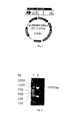
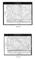
- the recovered double-digested products were cloned into the Kpn I and Xba I restriction sites of the eukaryotic expression vector pcDNA3.1/Zeo(+) to construct the recombinant plasmid pcDNA3.1/B7-1-PE40KDEL, which was then extracted, electrophoresed ( Figure 3 ), and screened by sequencing ( Figure 4 .
- the sequence is shown in Appendix 1) to obtain positive clones.
- the sequencing results revealed no point mutations or frameshift mutations in the sequence following the signal peptide sequence and were consistent with the sequence of the prokaryotic expression plasmid pRSETA-B7-1-PE40KDEL.
- B7-1-PE40KDEL The comparison results of the primary and secondary protein structures between B7-1-PE40KDEL, human B7-1, and PE40 are shown in Table 1.
- Table 1 Comparison of the primary and secondary protein structures between B7-1-PE40KDEL fusion protein, B7-1, and PE40.
- B7-1-PE40 KDEL Structure Primary structure Secondary structure 25aa Leucine ⁇ Proline B7-1-208 (1-208aa) No change 156aa Histidine ⁇ Arginine Link-19(209-227aa) PE40-360 (228-587aa) 247aa Phenylalanine ⁇ Serine No change 397aa Valine ⁇ Isoleucine No change
- the eukaryotic expression vector pcDNA3.1/B7-1-PE40KDEL with the correct sequence was transiently transfected into CHO-K1-RP E.40 cells, and the following methods were used to detect the expression of B7-1-PE40KDEL fusion protein: first, RT-PCR was used to detect the expression of B7-1-PE40KDEL mRNA in the transfected cells. A specific band of 1,850 bp was present in CHO-K1-RPE.40 cells transfected with pcDNA3.1/B7-1-PE40KDEL, whereas the specific band was not observed in CHO-K1-RPE.40 cells transfected with an empty vector.
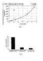
- OD 450 was used as the ordinate and PEA concentrations as the abscissa to graph the standard curve using Curve Expert 1.3 software.
- the standard curve of the polynomial fitting equation ( Figure 7 ) was used to calculate the PEA concentrations from the culture supernatant of stably transfected cell strains.
- Table 3 The expression levels of B7-1-PE40KDEL fusion proteins in different positive clones Clone No. OD450 Value PEA Concentrations (pg/ml) Clone 1* 0.352 268.26 Clone 2* 0.317 230.3 Clone 4 0.385 303.98 Clone 32 0.425 347.28
- the human lymphoma cell line Jurkat with a high expression level of CD28 was used to detect the selective cytotoxic activity of the eukaryotic expressed B7-1-PE40KDEL.
- the human Burkitt's lymphoma cell line Raji with a low expression level of CD28 was used as a negative control.
- the MTT results indicated that after 48 h of co-incubation of stably transfected cells and Jurkat cells, the OD value was 0.782, whereas the OD values were 1.466 for untransfected cells, 1.29 for Jurkat cells alone, 1.296 for the co-incubation of stably transfected cells and Raji cells, and 1.564 for untransfected cells alone.
- Example 2 The protection against aGVHD by B7-1-PE40KDEL DNA vaccine (1)
- the eukaryotic expression vector pcDNA3.1/B7-1-PE40KDEL was constructed by our group. This eukaryotic expression vector was previously constructed and stored by Dr. Hong Xue.
- the rabbit polyclonal anti-Pseudomonas exotoxin A antibody was purchased from Sigma Inc.
- FITC-anti-mouse CD3, PE-anti-mouse CD28, and PharmlyseTM were purchased from BD Inc.
- SYBR ® Premix Ex TaqTM (perfect real-time) was purchased from TaKaRa Bio. Inc.
- MTX for injection was purchased from Jiangsu Hengrui Medicine Co., Ltd., and cyclosporin A was purchased from Novartis Pharma Buch AG.
- Mouse lymphocyte separation solution was purchased from Tianjin Hao Yang Biological Products Co. Ltd.
- All experimental animals were provided by the Animal Center of the Academy of Military Medical Sciences, and the breeding conditions were at the specific pathogen-free (SPF) level.
- the Department of Pathology at the affiliated Hospital of the Academy of Military Medical Sciences assisted in the preparation and observation of pathological samples.
- Flow cytometric analysis was conducted by the Institute of Radiation Medicine of the Academy of Military Medical Sciences.
- the real-time quantitative PCR analysis was conducted by the blood center of the affiliated Hospital of the Academy of Military Medical Sciences.
- mice were provided with drinking water that contained gentamicin (32 ⁇ 10 4 U/L) and erythromycin (250 mg/L) one week prior to the transplantation to prevent infection and were fed in a sterile laminar airflow cabinet. 60 Co total body irradiation (TBI) (8.0 Gy, dose rate is 1.8 Gy/min) was administered 4 h prior to transplantation.
- TBI total body irradiation
- the preparation of donor bone marrow cells and spleen cells was as follows: C57BL/6 donor mice were sacrificed by cervical dislocation and then soaked in 75% ethanol for a few minutes. The femur was sterilely obtained with surgical scissors, and RPMI-1640 culture medium was used to flush out the bone marrow from the bone marrow cavity, which was then sifted through a 200-mesh cell sieve. Next, 0.83% ammonium chloride solution was used to lyse the erythrocytes, followed by two washes with RPMI-1640 culture medium (1000 rpm, 10 min). This produced a single-cell suspension of bone marrow cells. The cell concentration was then adjusted to I ⁇ 10 8 /ml.
- the spleens were removed aseptically and then ground on a 200-mesh cell sieve. After the cells were collected by centrifugation, 0.83% NH 4 Cl solution was used to lyse the erythrocytes, followed by two washes with RPMI-1640 culture medium (1000 rpm, 10 min). The resulting single-cell suspension of spleen cells was then adjusted to a cell concentration of 1 ⁇ 10 8 /ml.
- the prepared spleen cells and bone marrow cells were infused via tail vein injections into the recipient mice of all groups.
- the numbers of infused cells were 2 ⁇ 10 7 spleen cells/mouse and 1 ⁇ 10 7 bone marrow cells/mouse.
- the establishment of a stable aGVHD mouse model was confirmed with physical signs, the assessment of hematopoietic reconstruction, pathological analysis, and chimerism analysis after transplantation.
- mice Sixty aGVHD mice were randomly divided into six groups with 10 mice in each group: (1) B7-1-PE40KDEL DNA vaccine group; (2) B7-2-PE40KDEL DNA vaccine group; (3) B7-1-PE40KDEL+B7-2-PE40KDEL DNA vaccine group; (4) empty vector group; (5) CsA+MTX postive control group; and (6) untreated aGVHD group. Both the empty vector and DNA vaccine vector were dissolved in normal saline. There was no RNA contamination of the plasmid vector. Endotoxin was removed. Supercoiled DNA accounted for 70%-80% of the DNA. The OD 260 /OD 280 ratios ranged from 1.8-2.0. The concentration was not less than 0.5 ⁇ g/ ⁇ l.
- the pulse parameters were as follows: voltage, 200 V/cm; pulse width, 10 ms; the number of pulses, 6; frequency, 1 Hz ( Mir LM, Bureau MF, Gehl J, et al. High-efficiency gene transfer into skeletal muscle mediated by electric pulses. Proc. Natl. Acad. Sci. USA, 1999, 96: 4262-4267 ).
- the "gold standard" regimen CsA+MTX that has been commonly used in clinical practice, was adopted as the control drugs.
- CsA was administered via intraperitoneal injection once per day at a dose of 1.5 mg/kg.d.
- MTX was administered via intraperitoneal injection on days 1, 3, 6, and 11 at a dose of 0.4 mg/kg.d ( Xu K, Li C, Pan X, et al. Study of Relieving Graft-versus-Host Disease by Blocking CD137-CD137 Ligand Costimulatory Pathway in Vitro. Int J Hematol, 2007, 86:84-90 .).
- RNA extraction was performed as follows: mouse lymphocyte-separating solution was used to isolate white blood cells from the peripheral blood of mice (approximately 1 ml), which were washed twice with PBS before 1 ml of TRIzol was added. The isolated cells rested at room temperature for 5 min, and 200 ⁇ l of chloroform was then added and mixed well by shaking. After stratification, the mixture was centrifuged at 4°C and 12,000 g for 15 min. The upper aqueous phase was transferred to another tube, and 500 ⁇ l of isopropanol was added and mixed well. After resting at room temperature for 10 min, the mixture was centrifuged at 4°C and 12,000 g for 10 min.
- RNA was at the bottom of the tube, which was washed with 75% ethanol and then centrifuged at 4°C and 8000 g for 5 min. The supernatant was then discarded. After the complete evaporation of ethanol, RNA was dissolved with an appropriate amount of DEPC-treated water and quantitatively measured with a UV spectrophotometer.
- reaction conditions of reverse transcription were as follows: MgCl 2 2 ⁇ l 10 ⁇ RNA PCR Buffer 1 ⁇ l dNTP 1 ⁇ l RNase Inhibitor 0.25 ⁇ l AMV 0.5 ⁇ l Oligo dT 0.5 ⁇ l RNA 0.5 ⁇ g Total volume 10 ⁇ l
- the reaction conditions were 42°C for 30 min, 99°C for 5 min, and 5°C for 5 min.
- mouse ⁇ -actin primers were:
- the PCR reaction was assembled as follows: SYBR ® Premix Ex TaqTM (2 ⁇ ) 12.5 ⁇ l ddH 2 O 10.5 ⁇ l P1 (20 ⁇ M) 0.5 ⁇ l P2 (20 ⁇ M) 0.5 ⁇ l cDNA 1 ⁇ l Total Volume 25 ⁇ l
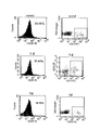
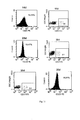
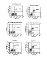
- mice eyeballs were removed, and blood was collected and treated with heparin.
- One microliter of both FITC-anti-mouse CD3 and PE-anti-mouse CD28 was added to 100 ⁇ l of anticoagulant blood, followed by incubation at room temperature in the dark for 30 min.
- 2 ml of PharmlyseTM erythrocyte lysis buffer was added, followed by incubation at room temperature in the dark for 15 min. The mixture was then centrifuged at 1500 rpm for 5 min.
- the pellet was re-suspended with PBS, followed by centrifugation at 1500 rpm for 5 min. After the supernatant was discarded, 0.5 ml of 2% paraformaldehyde-PBS was added for detection.
- mice eyeballs were removed, the blood was collected and centrifuged, and the serum was stored at -20°C for later use.
- ELISA was performed as follows: PEA (1 mg/mL) was dissolved in coating buffer, and each well was coated with 200 ng/100 ⁇ l, followed by incubation at 4°C overnight. The next day, the wells were washed 2-3 times with PBST and then blocked in bovine serum blocking solution at room temperature for 2 h. The serum sample and standard samples (100 ⁇ l/well) were incubated at 37°C for 1 h and then washed 3 times with PBST.
- mice in all groups were weighed each day, and physical signs such as hair, spirit, and diarrhea were observed.
- mice Different genders of mice were used as the donor and recipient in bone marrow transplantation. Therefore, the genomic DNA could be extracted from the peripheral blood of female recipient mice and the Y chromosome-specific gene sry primers could be designed to PCR-amplify the Y chromosome of male donor mice to detect transplant engraftment.
- livers, spleens, small intestines, and skin of mice in all groups were removed and fixed in 10% formalin solution, followed by paraffin sectioning and HE staining to observe the pathological changes.
- SPSS 13.0 software was used for statistical analysis. The t tests were used to test the statistical significance of the differences in the mean measurement data between the two groups. A non-parametric test (the Mann-Whitney test) was used to compare ranked data between the two groups. P ⁇ 0.05 was considered statistically significant. SPSS 13.0 software was also used to graph the Kaplan-Meier survival curve.
- the anti-PEA antibody level was detected using ELISA.
- a commercial anti-PEA antibody was used as the standard to graph the standard curve.
- the OD 450 was used for the abscissa, and anti-PEA antibody concentration was used for the ordinate to graph the standard curve using CurveExpert 1.3 software ( Figure 13 ).
- a quadratic fitting equation was used to calculate concentrations of serum anti-PEA antibodies (Table 4).
- the anti-PEA antibody level of B7-1-PE40KDEL DNA vaccine intramuscular-injection group was significantly lower than that of the positive control group (p ⁇ 0.05), and there was no significant difference when compared to that of the negative control group (i.e., the empty-vector injection group).
- Table 4 Serum anti-PEA antibody levels in mice on day 21 after the intramuscular injection of B7-1-PE40KDEL DNA vaccine Groups Absorbance Value (450 nm) Concentration (pg/ml) pcDNA3.1/B7-1-PE40KDEL(75 ⁇ g) 0.283 ⁇ 0.029* 63.824 ⁇ 6.402* pcDNA3.1 group (negative control) 0.139 ⁇ 0.022* 49.814 ⁇ 1.669* Positive control 0.564 ⁇ 0.084 312.458 ⁇ 3.842 * indicates the comparison with the positive control, P ⁇ 0.05
- mice After the intramuscular injection of 75 ⁇ g of B7-1-PE40KDEL DNA vaccine, the mice were observed every day, and no abnormalities in physical signs or behavior were observed. Histopathological examination of hearts, livers, spleens, lungs, and kidneys removed on days 7, 14, and 21 after the injection did not reveal any pathological changes.
- the white blood cell count was 1.54 ⁇ 0.14 ⁇ 10 9 /L at death.
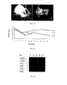
- the specific Y-chromosome sry gene sequence of male donor mice could be detected in the peripheral blood of all recipient mice, as shown in Figure 16 as the 371-bp fragment, suggesting that the transplantation was successful.
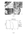
- Pathological examination revealed typical GVHD pathological findings as follows: 1 Necrosis of the glandular epithelial cells in the intestinal mucosa was observed in small intestine tissue, and necrotic cell debris was present in the epithelial cells and glandular cavity; glandular epithelial cells were flat, glands were cystic, the number of glands was reduced, glands disappeared, and the mucosal epithelium fell off ( Figure 17-A ); 2 In liver tissue, focal liver cell degeneration and necrosis occurred, and lymphocyte and eosinophil infiltration was observed ( Figure 17-B ).
- B7-1-PE40KDEL DNA vaccine called as B7-1
- B7-2-PE40KDEL DNA vaccine called as B7-2
- B7-1-PE40KDEL + B7-2-PE40KDEL DNA vaccine called as B7-1+ B7-2
- mice The weight loss of the mice was the smallest in B7-1-PE40KDEL DNA vaccine group, followed by CsA+MTX group, B7-1-PE40KDEL + B7-2-PE40KDEL DNA vaccine group, and B7-2-PE40KDEL DNA vaccine group.
- the changes in body weight of GVHD mice in all groups after treatment are shown in Figure 19 .
- the WBC counts in the peripheral blood of the aGVHD mice in all groups began to drop on day 1 to a low point of approximately 0.6 ⁇ 10 9 /L on day 3 ⁇ day 4.
- the WBC count began to recover on day 6, and the level of recovery was higher in B7-1-PE40KDEL DNA vaccine, B7-2-PE40KDEL DNA vaccine, B7-1-PE40KDEL + B7-2-PE40KDEL DNA vaccine, and CsA+MTX groups than in the empty vector group and the untreated group.
- the level of WBC recovery was the highest in B7-1-PE40KDEL DNA vaccine group. Subsequently, WBC counts continued to drop again to >10 ⁇ 10 9 /L at death in all groups ( Figure 20 ).
- group A B7-1-PE40KDEL DNA vaccine
- group B B7-2-PE40KDEL DNA vaccine
- group C B7-1-PE40KDEL + B7-2-PE40KDEL DNA vaccine
- group E CsA+MTX
- the lesions were less severe, and the degree of lymphocyte infiltration was also lower than in groups D and F.
- Pathological analysis of the liver revealed that in group A only, the degree of liver cells' swelling and hydropic degeneration was relatively low. In other groups, there was focal liver cell degeneration and necrosis, infiltration of lymphocytes and eosinophils, and more serious liver cell swelling and degeneration.
- the survival time of the mice in B7-1-PE40KDEL DNA vaccine group was the longest, with a median survival time of 51 days.
- the median survival times of B7-2-PE40KDEL DNA vaccine and B7-1-PE40KDEL + B7-2-PE40KDEL DNA vaccine groups, empty vector group, CsA+MTX group, and the untreated group were 41, 47, 22.5, 39.7, and 21.5 days, respectively.
- the survival rates of the mice in all groups are depicted in a Kaplan-Meier survival curve ( Figure 24 ).
- SPF pathogen-free
- the eukaryotic expression vector pcDNA3.1/Zeo(+)-B7-2-PE40KDEL was constructed and stored in our department.
- the eukaryotic expression vector was constructed and stored as described in section 1.
- MTX for injection was purchased from Jiangsu Hengrui Medicine Co., Ltd.
- Cyclosporin A was purchased from Novartis Pharma Sau AG.
- FITC-anti-mouse CD3, PE-anti-mouse CD28, PE-Cy5-anti-mouse CD8, PE-Cy5-anti-mouse CD4, PE-anti-mouse CD4, and PE-anti-mouse CD25 were all purchased from BD Inc.
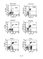
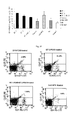
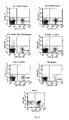
- Fluorokine MAP mouse interferon (IFN) ⁇ , interleukin (IL) -2, IL-4, IL-10, IL-12, tumor necrosis factor (TNF) ⁇ kits, and Luminex liquid chips for cytokine detection were provided by R&D Inc.
- Tumor growth factor (TGF) ⁇ and IL-2 ELISA kits were provided by R&D Inc.
- mice were provided drinking water that contained gentamicin (32 ⁇ 10 4 U/L) and erythromycin (250 mg/L) one week prior to transplantation to prevent infection and were fed in a sterile laminar airflow cabinet. 60 Co total body irradiation (TBI) (8.0 Gy, dose rate is 1.8 Gy/min) was administered 4 h prior to transplantation.
- TBI total body irradiation
- the preparation of donor bone marrow cells and spleen cells was as follows: C57BL/6 donor mice were sacrificed by cervical dislocation and then soaked in 75% ethanol for a few minutes. The femur was sterilely obtained with surgical scissors, and RPMI-1640 culture medium was used to flush out the bone marrow from the bone marrow cavity, which was then sifted through a 200-mesh cell sieve. Next, 0.83% NH 4 Cl solution was used to lyse the erythrocytes, followed by two washes with RPMI-1640 culture medium (1000 rpm, 10 min). As a result, a single-cell suspension of bone marrow cells was obtained, and the cell concentration was then adjusted to 1 ⁇ 10 8 / ml.
- the spleens were removed aseptically and ground on a 200-mesh cell sieve. After the cells were collected by centrifugation, 0.83% NH 4 Cl solution was used to lyse the erythrocytes, followed by two washes with RPMI-1640 culture medium (1000 rpm, 10 min). As a result, a single-cell suspension of spleen cells was obtained, and then the cell concentration was adjusted to 1 ⁇ 10 8 /ml.
- the prepared spleen cells and bone marrow cells were infused via tail vein injection into the recipient mice of all groups.
- the numbers of infused cells were 2 ⁇ 10 7 spleen cells/mouse and 1 ⁇ 10 7 bone marrow cells/mouse.
- the establishment of a stable aGVHD mouse model was confirmed with the detection of physical signs, hematopoietic reconstruction, transplantation, and pathological analysis.
- B7-PE40KDEL DNA vaccine and other treatment were as follows: drugs were administered to the mice of all groups at day 1 after transplantation. There were six groups with five mice in each group, as follows: (1) B7-1-PE40KDEL DNA vaccine group; (2) B7-2-PE40KDEL DNA vaccine group; (3) B7-1-PE40KDEL+B7-2-PE40KDEL DNA vaccine group; (4) empty vector group; (5) CsA+MTX control group; (6) the untreated aGVHD group; and (7) the normal control group. Both the empty vector and DNA vaccine vector were dissolved in normal saline. There was no RNA contamination of the plasmid vector. Endotoxin was removed.
- the human TGF- ⁇ ELISA kit As a substitute reagent in view of the fact that there is high homology between human TGF- ⁇ and mouse TGF- ⁇ . It is difficult to determine the serum IL-2 concentration. But when a T cell is activated, IL-2 binds to its receptors (with ⁇ , ⁇ , and ⁇ subunits), and the ⁇ subunit falls off of the cell surface and can be detected in the serum, called soluble IL-2 receptor (sIL-2R). Thus, an ELISA kit can be used to re-detect sIL-2R in mice.
- mice and human TGF ⁇ were as follows:
- the concentrations of the standard samples were used as the abscissa, and OD values were used as the ordinates to graph the standard curve.
- the concentrations of the samples can be determined on the standard curve by using their OD values. If the OD value of a sample was higher than the upper limit of the standard curve, the sample was appropriately diluted for re-measurement. The dilution fold was then multiplied while calculating the concentration of the sample.
- the data were expressed as x +s, and SPSS 13.0 software was used for statistical analysis.
- ANOVA and Dunnett's t-test were used for comparison among groups when there was homogeneity of variance. P ⁇ 0.05 is considered statistically significant.
- Non-parametric analysis was used when there was heterogeneity of variance.
- Luminex liquid chip and the ELISA method were used to detect the changes of serum cytokines in the mice of all experimental groups.
- the results are presented in Table 1.
- the IFN ⁇ level gradually declined, and by day 21, IFN ⁇ had returned to a near-normal level.
- the levels of IL-2 and TNF ⁇ , two of the Th1 cytokines were maintained at levels far higher than normal.
- IL-2 level was effectively reduced in both B7-1-PE40KDEL DNA vaccine group and CsA+MTX group, and the difference was statistically significant when compared with that in the aGVHD control group (P ⁇ 0.05).
- the effect on the Th1 cytokine TNF- ⁇ was not significant in either B7-1-PE40KDEL DNA vaccine group or CsA+MTX group.
- Th2 cytokinines such as IL-4 and IL-10 and Th3 cytokine TGF- ⁇ , which are involved in the suppression of inflammation
- Th2 cytokine IL-4 there was no statistical difference in the effect on Th2 cytokine IL-4 between B7-1-PE40KDEL DNA vaccine group and CsA+MTX group.
- the levels of Th2 cytokine IL-10 were effectively increased in both B7-1-PE40KDEL DNA vaccine group and CsA+MTX group, and the difference was statistically significant when compared with the level in the aGVHD control group.
- Th3 cytokine TGF- ⁇ only B7-1-PE40KDEL DNA vaccine effectively increased its level, and B7-2-PE40KDEL DNA vaccine group had no significant effect.
- CsA+MTX group slightly decreased the expression level of Th3 cytokine TGF- ⁇ .
- Table 5 The effect of B7-1-PE40KDEL DNA vaccine on Th1, Th2, and Th3 cvtokines in the peripheral blood of aGVHD mice Group Th1, Th2, and Th3 cytokines (pg/ml) INF- ⁇ TNF- ⁇ IL-2 IL-4 IL-10 TGF- ⁇ Normal group + day 7 0 0.52 ⁇ 0.27 11.17 ⁇ 3.47 284.50 ⁇ 22.18 4.70 ⁇ 0.78 1072.55 ⁇ 147.85 + day 14 0 0.52 ⁇ 0.27 11.17 ⁇ 3.47 284.50 ⁇ 22.18 4.70 ⁇ 0.78 1072.55 ⁇ 147.85 + day 21 0 0.52 ⁇ 0.27 11.17 ⁇ 3.47 284.50+22.18 4.70 ⁇ 0.78 1072.55 ⁇ 147.85 NS group + day 7 1465.03 ⁇ 316.77 19.97 ⁇ 3.69 20.54 ⁇ 1.58 277.94 ⁇ 22.14 8.76 ⁇ 0.53 1239
- CD4/CD8 ratio was approximately 3.2, which was seriously inverted in aGVHD mice after radiation and transplantation.
- CD3 + CD8 + T-cell proportions were significantly increased, whereas CD3 + CD4 + proportions were significantly decreased.
- B7-1-PE40KDEL DNA vaccine and B7-2-PE40KDEL DNA vaccines, B7-1-PE40KDEL+B7-2-PE40KDEL DNA vaccine, and CsA+MTX treatment did not significantly reverse CD4/CD8 ratio ( Figures 26-27 and Table 6).
- CD4 + CD25 + Treg proportions were also higher in B7-2-PE40KDEL DNA vaccine group than in the empty vector group and the NS control group (P ⁇ 0.05), but the effect of B7-2-PE40KDEL DNA vaccine was weaker than that of B7-1-PE40KDEL DNA vaccine. In contrast, there was no significant difference in CD4 + CD25 + Treg proportions in CsA+MTX group.
- Table 6 The effect of B7-1-PE40KDEL DNA vaccine on CD4 + CD25 + and CD8 + CD28 - T sub-groups and CD4/CD8 ratios in the peripheral blood of aGVHD mice
Landscapes
- Health & Medical Sciences (AREA)
- Chemical & Material Sciences (AREA)
- Life Sciences & Earth Sciences (AREA)
- Organic Chemistry (AREA)
- Immunology (AREA)
- General Health & Medical Sciences (AREA)
- Medicinal Chemistry (AREA)
- Biophysics (AREA)
- Molecular Biology (AREA)
- Proteomics, Peptides & Aminoacids (AREA)
- Genetics & Genomics (AREA)
- Gastroenterology & Hepatology (AREA)
- Biochemistry (AREA)
- Pharmacology & Pharmacy (AREA)
- Animal Behavior & Ethology (AREA)
- Public Health (AREA)
- Veterinary Medicine (AREA)
- Transplantation (AREA)
- Chemical Kinetics & Catalysis (AREA)
- General Chemical & Material Sciences (AREA)
- Toxicology (AREA)
- Microbiology (AREA)
- Zoology (AREA)
- Cell Biology (AREA)
- Epidemiology (AREA)
- Mycology (AREA)
- Engineering & Computer Science (AREA)
- Bioinformatics & Cheminformatics (AREA)
- Nuclear Medicine, Radiotherapy & Molecular Imaging (AREA)
- Peptides Or Proteins (AREA)
- Medicines Containing Antibodies Or Antigens For Use As Internal Diagnostic Agents (AREA)
- Medicines That Contain Protein Lipid Enzymes And Other Medicines (AREA)
- Medicines Containing Material From Animals Or Micro-Organisms (AREA)
- Pharmaceuticals Containing Other Organic And Inorganic Compounds (AREA)
- Micro-Organisms Or Cultivation Processes Thereof (AREA)
- Medicinal Preparation (AREA)
Applications Claiming Priority (2)
| Application Number | Priority Date | Filing Date | Title |
|---|---|---|---|
| CN2011100239856A CN102161998B (zh) | 2011-01-14 | 2011-01-14 | 基于b7-1-pe40kdel外毒素融合基因的dna疫苗及其用途 |
| PCT/CN2011/079862 WO2012094905A1 (zh) | 2011-01-14 | 2011-09-20 | 基于b7-1-pe40kdel外毒素融合基因的dna疫苗及其用途 |
Publications (3)
| Publication Number | Publication Date |
|---|---|
| EP2628802A1 true EP2628802A1 (de) | 2013-08-21 |
| EP2628802A4 EP2628802A4 (de) | 2014-05-07 |
| EP2628802B1 EP2628802B1 (de) | 2016-10-26 |
Family
ID=44463451
Family Applications (1)
| Application Number | Title | Priority Date | Filing Date |
|---|---|---|---|
| EP11855878.2A Not-in-force EP2628802B1 (de) | 2011-01-14 | 2011-09-20 | Dna-impfstoff auf basis des exotoxin-fusionsgens b7-1-pe40kdel und verwendung davon |
Country Status (5)
| Country | Link |
|---|---|
| US (1) | US9029520B2 (de) |
| EP (1) | EP2628802B1 (de) |
| JP (2) | JP2014505473A (de) |
| CN (1) | CN102161998B (de) |
| WO (1) | WO2012094905A1 (de) |
Cited By (1)
| Publication number | Priority date | Publication date | Assignee | Title |
|---|---|---|---|---|
| US12589141B2 (en) | 2019-10-10 | 2026-03-31 | The Fifth Medical Center Of Chinese Pla General Hospital | DNA vaccine capable of effectively treating and/or preventing type 1 diabetes and use thereof |
Families Citing this family (2)
| Publication number | Priority date | Publication date | Assignee | Title |
|---|---|---|---|---|
| CN102161998B (zh) | 2011-01-14 | 2013-01-09 | 中国人民解放军军事医学科学院附属医院 | 基于b7-1-pe40kdel外毒素融合基因的dna疫苗及其用途 |
| AU2014215458A1 (en) * | 2013-02-05 | 2015-08-13 | Anthrogenesis Corporation | Natural killer cells from placenta |
Family Cites Families (18)
| Publication number | Priority date | Publication date | Assignee | Title |
|---|---|---|---|---|
| JPH11513669A (ja) * | 1995-10-13 | 1999-11-24 | アメリカ合衆国 | ジスルフィド安定化抗体フラグメントを含む免疫毒素 |
| US20030235557A1 (en) * | 1998-09-30 | 2003-12-25 | Corixa Corporation | Compositions and methods for WT1 specific immunotherapy |
| US7829064B2 (en) * | 1999-05-10 | 2010-11-09 | Immunomedics, Inc. | Anti-CD74 immunoconjugates and methods |
| US6492498B1 (en) | 1999-11-15 | 2002-12-10 | Regents Of The University Of Minnesota | Multimeric immunotoxins |
| BRPI0214168B8 (pt) * | 2001-11-14 | 2021-05-25 | Centocor Inc | anticorpos anti-il-6, moléculas de ácido nucleico codificando os mesmos, vetores compreendendo as referidas moléculas, composições e formulações compreendendo os referidos anticorpos, bem como métodos de produção dos mesmos |
| AU2002361390A1 (en) * | 2001-12-14 | 2003-06-30 | Friedrich-Alexander-Universitaet Erlangen-Nuernberg | Anti-cd7 immunotoxin as fusion protein |
| CN1274717C (zh) * | 2002-11-06 | 2006-09-13 | 中国人民解放军军事医学科学院附属医院 | 一种新型免疫抑制融合蛋白、其编码核酸及其用途 |
| JP2008504219A (ja) * | 2003-12-11 | 2008-02-14 | シドニー キンメル キャンサー センター | 抗原に対する免疫を生じさせる方法 |
| GB0411411D0 (en) * | 2004-05-21 | 2004-06-23 | Glaxosmithkline Biolog Sa | Vaccines |
| JP4940136B2 (ja) * | 2004-06-28 | 2012-05-30 | イエダ リサーチ アンド ディベロップメント カンパニー リミテッド | キメラタンパク質およびその使用 |
| AU2005291039A1 (en) * | 2004-10-01 | 2006-04-13 | Avidex Ltd. | T-cell receptors containing a non-native disulfide interchain bond linked to therapeutic agents |
| WO2006056464A2 (en) * | 2004-11-26 | 2006-06-01 | Pieris Ag | Compound with affinity for the cytotoxic t lymphocyte-associated antigen (ctla-4) |
| CA2589698A1 (en) * | 2004-12-14 | 2006-06-22 | Therapeutic Human Polyclonals, Inc. | Improved dna immunization with recombinase/transposase |
| WO2007100357A2 (en) * | 2005-10-07 | 2007-09-07 | Nuvelo, Inc. | Stem cell factor-like protein scfa1 and uses thereof |
| CN1313484C (zh) * | 2005-11-17 | 2007-05-02 | 中国人民解放军第四军医大学 | 人b细胞特异性膜分子cd20的12氨基酸模拟表位及其用该模拟表位构建的多肽表位疫苗 |
| CN101343328B (zh) * | 2008-08-25 | 2011-06-01 | 北京博翱泰生物技术有限公司 | 靶特异性双突变体融合蛋白质 |
| CN101824424B (zh) * | 2010-04-07 | 2013-01-09 | 中国人民解放军军事医学科学院附属医院 | 基于b7-2-pe40外毒素融合基因的dna疫苗及其用途 |
| CN102161998B (zh) * | 2011-01-14 | 2013-01-09 | 中国人民解放军军事医学科学院附属医院 | 基于b7-1-pe40kdel外毒素融合基因的dna疫苗及其用途 |
-
2011
- 2011-01-14 CN CN2011100239856A patent/CN102161998B/zh not_active Expired - Fee Related
- 2011-09-20 JP JP2013548720A patent/JP2014505473A/ja active Pending
- 2011-09-20 US US13/993,685 patent/US9029520B2/en not_active Expired - Fee Related
- 2011-09-20 EP EP11855878.2A patent/EP2628802B1/de not_active Not-in-force
- 2011-09-20 WO PCT/CN2011/079862 patent/WO2012094905A1/zh not_active Ceased
-
2016
- 2016-08-18 JP JP2016160350A patent/JP6517175B2/ja not_active Expired - Fee Related
Cited By (1)
| Publication number | Priority date | Publication date | Assignee | Title |
|---|---|---|---|---|
| US12589141B2 (en) | 2019-10-10 | 2026-03-31 | The Fifth Medical Center Of Chinese Pla General Hospital | DNA vaccine capable of effectively treating and/or preventing type 1 diabetes and use thereof |
Also Published As
| Publication number | Publication date |
|---|---|
| CN102161998B (zh) | 2013-01-09 |
| JP2014505473A (ja) | 2014-03-06 |
| CN102161998A (zh) | 2011-08-24 |
| JP2017018122A (ja) | 2017-01-26 |
| JP6517175B2 (ja) | 2019-05-22 |
| EP2628802A4 (de) | 2014-05-07 |
| WO2012094905A1 (zh) | 2012-07-19 |
| EP2628802B1 (de) | 2016-10-26 |
| US9029520B2 (en) | 2015-05-12 |
| US20130344101A1 (en) | 2013-12-26 |
Similar Documents
| Publication | Publication Date | Title |
|---|---|---|
| Offner et al. | Recombinant human β-galactoside binding lectin suppresses clinical and histological signs of experimental autoimmune encephalomyelitis | |
| CN102770455B (zh) | 用于体内扩增调节性t细胞的方法 | |
| AU2010282280B2 (en) | Use of IL-15 to increase thymic output and to treat lymphopenia | |
| CA2807552A1 (en) | Engineered nucleic acids and methods of use thereof | |
| KR101521197B1 (ko) | 병용 요법에 있어서 cd83의 용도 | |
| EP2288387B1 (de) | Neue lösliche cd83-polypeptide, formulierungen und anwendungsverfahren | |
| KR20180030879A (ko) | 복막암을 치료하기 위한 조성물 및 방법 | |
| JP2022513372A (ja) | シアリルルイスaを標的とするキメラ抗原受容体およびその使用 | |
| EP2628802B1 (de) | Dna-impfstoff auf basis des exotoxin-fusionsgens b7-1-pe40kdel und verwendung davon | |
| KR101310511B1 (ko) | 흉선-특이성 단백질 | |
| US10821134B2 (en) | BK virus specific T cells | |
| WO2012082771A1 (en) | Cardiac glycosides for treating autoimmune disease | |
| EP3447138B1 (de) | Aktivator für regulatorische t-zellen und verwendung davon | |
| CN102898508B (zh) | 封闭TGF-β受体或IL-10受体的多肽、其药物组合物及其用途 | |
| WO2020097236A1 (en) | Fgl2 neutraling cell therapy and methods of use thereof | |
| CN121362259A (zh) | 新型PDL1-CAR-γδT细胞的构建及其抗肿瘤应用 | |
| WO2023235422A2 (en) | Engineered chimeric fusion protein compositions and methods of use thereof | |
| CN120590545A (zh) | 包括cd40l和il7的融合蛋白及其在制备治疗肿瘤药物中的应用 | |
| CN118974244A (zh) | 用于细胞免疫疗法的膜结合il-12 | |
| WO2005027958A1 (en) | Thymosin-beta-sulphoxide as an immunosuppressive active agent | |
| HK1155066B (en) | Novel soluble cd83 polypeptides, formulations and methods of use |
Legal Events
| Date | Code | Title | Description |
|---|---|---|---|
| PUAI | Public reference made under article 153(3) epc to a published international application that has entered the european phase |
Free format text: ORIGINAL CODE: 0009012 |
|
| 17P | Request for examination filed |
Effective date: 20130516 |
|
| AK | Designated contracting states |
Kind code of ref document: A1 Designated state(s): AL AT BE BG CH CY CZ DE DK EE ES FI FR GB GR HR HU IE IS IT LI LT LU LV MC MK MT NL NO PL PT RO RS SE SI SK SM TR |
|
| REG | Reference to a national code |
Ref country code: DE Ref legal event code: R079 Ref document number: 602011031768 Country of ref document: DE Free format text: PREVIOUS MAIN CLASS: C12N0015620000 Ipc: C07K0019000000 |
|
| DAX | Request for extension of the european patent (deleted) | ||
| A4 | Supplementary search report drawn up and despatched |
Effective date: 20140408 |
|
| RIC1 | Information provided on ipc code assigned before grant |
Ipc: C12N 15/79 20060101ALI20140402BHEP Ipc: A61P 37/06 20060101ALI20140402BHEP Ipc: C12N 15/62 20060101ALI20140402BHEP Ipc: A61K 48/00 20060101ALI20140402BHEP Ipc: C07K 19/00 20060101AFI20140402BHEP |
|
| GRAP | Despatch of communication of intention to grant a patent |
Free format text: ORIGINAL CODE: EPIDOSNIGR1 |
|
| INTG | Intention to grant announced |
Effective date: 20160610 |
|
| GRAS | Grant fee paid |
Free format text: ORIGINAL CODE: EPIDOSNIGR3 |
|
| GRAA | (expected) grant |
Free format text: ORIGINAL CODE: 0009210 |
|
| AK | Designated contracting states |
Kind code of ref document: B1 Designated state(s): AL AT BE BG CH CY CZ DE DK EE ES FI FR GB GR HR HU IE IS IT LI LT LU LV MC MK MT NL NO PL PT RO RS SE SI SK SM TR |
|
| REG | Reference to a national code |
Ref country code: GB Ref legal event code: FG4D |
|
| REG | Reference to a national code |
Ref country code: CH Ref legal event code: EP |
|
| REG | Reference to a national code |
Ref country code: AT Ref legal event code: REF Ref document number: 839970 Country of ref document: AT Kind code of ref document: T Effective date: 20161115 |
|
| REG | Reference to a national code |
Ref country code: IE Ref legal event code: FG4D |
|
| REG | Reference to a national code |
Ref country code: DE Ref legal event code: R096 Ref document number: 602011031768 Country of ref document: DE |
|
| REG | Reference to a national code |
Ref country code: LT Ref legal event code: MG4D |
|
| PG25 | Lapsed in a contracting state [announced via postgrant information from national office to epo] |
Ref country code: LV Free format text: LAPSE BECAUSE OF FAILURE TO SUBMIT A TRANSLATION OF THE DESCRIPTION OR TO PAY THE FEE WITHIN THE PRESCRIBED TIME-LIMIT Effective date: 20161026 |
|
| REG | Reference to a national code |
Ref country code: NL Ref legal event code: MP Effective date: 20161026 |
|
| REG | Reference to a national code |
Ref country code: AT Ref legal event code: MK05 Ref document number: 839970 Country of ref document: AT Kind code of ref document: T Effective date: 20161026 |
|
| PG25 | Lapsed in a contracting state [announced via postgrant information from national office to epo] |
Ref country code: NO Free format text: LAPSE BECAUSE OF FAILURE TO SUBMIT A TRANSLATION OF THE DESCRIPTION OR TO PAY THE FEE WITHIN THE PRESCRIBED TIME-LIMIT Effective date: 20170126 Ref country code: GR Free format text: LAPSE BECAUSE OF FAILURE TO SUBMIT A TRANSLATION OF THE DESCRIPTION OR TO PAY THE FEE WITHIN THE PRESCRIBED TIME-LIMIT Effective date: 20170127 Ref country code: SE Free format text: LAPSE BECAUSE OF FAILURE TO SUBMIT A TRANSLATION OF THE DESCRIPTION OR TO PAY THE FEE WITHIN THE PRESCRIBED TIME-LIMIT Effective date: 20161026 Ref country code: LT Free format text: LAPSE BECAUSE OF FAILURE TO SUBMIT A TRANSLATION OF THE DESCRIPTION OR TO PAY THE FEE WITHIN THE PRESCRIBED TIME-LIMIT Effective date: 20161026 |
|
| PG25 | Lapsed in a contracting state [announced via postgrant information from national office to epo] |
Ref country code: PL Free format text: LAPSE BECAUSE OF FAILURE TO SUBMIT A TRANSLATION OF THE DESCRIPTION OR TO PAY THE FEE WITHIN THE PRESCRIBED TIME-LIMIT Effective date: 20161026 Ref country code: RS Free format text: LAPSE BECAUSE OF FAILURE TO SUBMIT A TRANSLATION OF THE DESCRIPTION OR TO PAY THE FEE WITHIN THE PRESCRIBED TIME-LIMIT Effective date: 20161026 Ref country code: PT Free format text: LAPSE BECAUSE OF FAILURE TO SUBMIT A TRANSLATION OF THE DESCRIPTION OR TO PAY THE FEE WITHIN THE PRESCRIBED TIME-LIMIT Effective date: 20170227 Ref country code: AT Free format text: LAPSE BECAUSE OF FAILURE TO SUBMIT A TRANSLATION OF THE DESCRIPTION OR TO PAY THE FEE WITHIN THE PRESCRIBED TIME-LIMIT Effective date: 20161026 Ref country code: NL Free format text: LAPSE BECAUSE OF FAILURE TO SUBMIT A TRANSLATION OF THE DESCRIPTION OR TO PAY THE FEE WITHIN THE PRESCRIBED TIME-LIMIT Effective date: 20161026 Ref country code: BE Free format text: LAPSE BECAUSE OF FAILURE TO SUBMIT A TRANSLATION OF THE DESCRIPTION OR TO PAY THE FEE WITHIN THE PRESCRIBED TIME-LIMIT Effective date: 20161026 Ref country code: IS Free format text: LAPSE BECAUSE OF FAILURE TO SUBMIT A TRANSLATION OF THE DESCRIPTION OR TO PAY THE FEE WITHIN THE PRESCRIBED TIME-LIMIT Effective date: 20170226 Ref country code: ES Free format text: LAPSE BECAUSE OF FAILURE TO SUBMIT A TRANSLATION OF THE DESCRIPTION OR TO PAY THE FEE WITHIN THE PRESCRIBED TIME-LIMIT Effective date: 20161026 Ref country code: HR Free format text: LAPSE BECAUSE OF FAILURE TO SUBMIT A TRANSLATION OF THE DESCRIPTION OR TO PAY THE FEE WITHIN THE PRESCRIBED TIME-LIMIT Effective date: 20161026 Ref country code: FI Free format text: LAPSE BECAUSE OF FAILURE TO SUBMIT A TRANSLATION OF THE DESCRIPTION OR TO PAY THE FEE WITHIN THE PRESCRIBED TIME-LIMIT Effective date: 20161026 |
|
| REG | Reference to a national code |
Ref country code: DE Ref legal event code: R097 Ref document number: 602011031768 Country of ref document: DE |
|
| PG25 | Lapsed in a contracting state [announced via postgrant information from national office to epo] |
Ref country code: RO Free format text: LAPSE BECAUSE OF FAILURE TO SUBMIT A TRANSLATION OF THE DESCRIPTION OR TO PAY THE FEE WITHIN THE PRESCRIBED TIME-LIMIT Effective date: 20161026 Ref country code: SK Free format text: LAPSE BECAUSE OF FAILURE TO SUBMIT A TRANSLATION OF THE DESCRIPTION OR TO PAY THE FEE WITHIN THE PRESCRIBED TIME-LIMIT Effective date: 20161026 Ref country code: DK Free format text: LAPSE BECAUSE OF FAILURE TO SUBMIT A TRANSLATION OF THE DESCRIPTION OR TO PAY THE FEE WITHIN THE PRESCRIBED TIME-LIMIT Effective date: 20161026 Ref country code: EE Free format text: LAPSE BECAUSE OF FAILURE TO SUBMIT A TRANSLATION OF THE DESCRIPTION OR TO PAY THE FEE WITHIN THE PRESCRIBED TIME-LIMIT Effective date: 20161026 Ref country code: CZ Free format text: LAPSE BECAUSE OF FAILURE TO SUBMIT A TRANSLATION OF THE DESCRIPTION OR TO PAY THE FEE WITHIN THE PRESCRIBED TIME-LIMIT Effective date: 20161026 |
|
| REG | Reference to a national code |
Ref country code: FR Ref legal event code: PLFP Year of fee payment: 7 |
|
| PG25 | Lapsed in a contracting state [announced via postgrant information from national office to epo] |
Ref country code: IT Free format text: LAPSE BECAUSE OF FAILURE TO SUBMIT A TRANSLATION OF THE DESCRIPTION OR TO PAY THE FEE WITHIN THE PRESCRIBED TIME-LIMIT Effective date: 20161026 Ref country code: BG Free format text: LAPSE BECAUSE OF FAILURE TO SUBMIT A TRANSLATION OF THE DESCRIPTION OR TO PAY THE FEE WITHIN THE PRESCRIBED TIME-LIMIT Effective date: 20170126 Ref country code: SM Free format text: LAPSE BECAUSE OF FAILURE TO SUBMIT A TRANSLATION OF THE DESCRIPTION OR TO PAY THE FEE WITHIN THE PRESCRIBED TIME-LIMIT Effective date: 20161026 |
|
| PLBE | No opposition filed within time limit |
Free format text: ORIGINAL CODE: 0009261 |
|
| STAA | Information on the status of an ep patent application or granted ep patent |
Free format text: STATUS: NO OPPOSITION FILED WITHIN TIME LIMIT |
|
| 26N | No opposition filed |
Effective date: 20170727 |
|
| PG25 | Lapsed in a contracting state [announced via postgrant information from national office to epo] |
Ref country code: SI Free format text: LAPSE BECAUSE OF FAILURE TO SUBMIT A TRANSLATION OF THE DESCRIPTION OR TO PAY THE FEE WITHIN THE PRESCRIBED TIME-LIMIT Effective date: 20161026 |
|
| REG | Reference to a national code |
Ref country code: CH Ref legal event code: PL |
|
| PG25 | Lapsed in a contracting state [announced via postgrant information from national office to epo] |
Ref country code: MC Free format text: LAPSE BECAUSE OF FAILURE TO SUBMIT A TRANSLATION OF THE DESCRIPTION OR TO PAY THE FEE WITHIN THE PRESCRIBED TIME-LIMIT Effective date: 20161026 |
|
| REG | Reference to a national code |
Ref country code: IE Ref legal event code: MM4A |
|
| PG25 | Lapsed in a contracting state [announced via postgrant information from national office to epo] |
Ref country code: LU Free format text: LAPSE BECAUSE OF NON-PAYMENT OF DUE FEES Effective date: 20170920 |
|
| PG25 | Lapsed in a contracting state [announced via postgrant information from national office to epo] |
Ref country code: CH Free format text: LAPSE BECAUSE OF NON-PAYMENT OF DUE FEES Effective date: 20170930 Ref country code: IE Free format text: LAPSE BECAUSE OF NON-PAYMENT OF DUE FEES Effective date: 20170920 Ref country code: LI Free format text: LAPSE BECAUSE OF NON-PAYMENT OF DUE FEES Effective date: 20170930 |
|
| REG | Reference to a national code |
Ref country code: FR Ref legal event code: PLFP Year of fee payment: 8 |
|
| PG25 | Lapsed in a contracting state [announced via postgrant information from national office to epo] |
Ref country code: MT Free format text: LAPSE BECAUSE OF NON-PAYMENT OF DUE FEES Effective date: 20170920 |
|
| PG25 | Lapsed in a contracting state [announced via postgrant information from national office to epo] |
Ref country code: HU Free format text: LAPSE BECAUSE OF FAILURE TO SUBMIT A TRANSLATION OF THE DESCRIPTION OR TO PAY THE FEE WITHIN THE PRESCRIBED TIME-LIMIT; INVALID AB INITIO Effective date: 20110920 |
|
| PG25 | Lapsed in a contracting state [announced via postgrant information from national office to epo] |
Ref country code: CY Free format text: LAPSE BECAUSE OF NON-PAYMENT OF DUE FEES Effective date: 20161026 |
|
| PG25 | Lapsed in a contracting state [announced via postgrant information from national office to epo] |
Ref country code: MK Free format text: LAPSE BECAUSE OF FAILURE TO SUBMIT A TRANSLATION OF THE DESCRIPTION OR TO PAY THE FEE WITHIN THE PRESCRIBED TIME-LIMIT Effective date: 20161026 |
|
| PG25 | Lapsed in a contracting state [announced via postgrant information from national office to epo] |
Ref country code: TR Free format text: LAPSE BECAUSE OF FAILURE TO SUBMIT A TRANSLATION OF THE DESCRIPTION OR TO PAY THE FEE WITHIN THE PRESCRIBED TIME-LIMIT Effective date: 20161026 |
|
| PG25 | Lapsed in a contracting state [announced via postgrant information from national office to epo] |
Ref country code: AL Free format text: LAPSE BECAUSE OF FAILURE TO SUBMIT A TRANSLATION OF THE DESCRIPTION OR TO PAY THE FEE WITHIN THE PRESCRIBED TIME-LIMIT Effective date: 20161026 |
|
| PGFP | Annual fee paid to national office [announced via postgrant information from national office to epo] |
Ref country code: FR Payment date: 20210927 Year of fee payment: 11 |
|
| PGFP | Annual fee paid to national office [announced via postgrant information from national office to epo] |
Ref country code: DE Payment date: 20210818 Year of fee payment: 11 Ref country code: GB Payment date: 20210818 Year of fee payment: 11 |
|
| REG | Reference to a national code |
Ref country code: DE Ref legal event code: R119 Ref document number: 602011031768 Country of ref document: DE |
|
| GBPC | Gb: european patent ceased through non-payment of renewal fee |
Effective date: 20220920 |
|
| PG25 | Lapsed in a contracting state [announced via postgrant information from national office to epo] |
Ref country code: FR Free format text: LAPSE BECAUSE OF NON-PAYMENT OF DUE FEES Effective date: 20220930 Ref country code: DE Free format text: LAPSE BECAUSE OF NON-PAYMENT OF DUE FEES Effective date: 20230401 |
|
| PG25 | Lapsed in a contracting state [announced via postgrant information from national office to epo] |
Ref country code: GB Free format text: LAPSE BECAUSE OF NON-PAYMENT OF DUE FEES Effective date: 20220920 |