EP2488876B1 - Protein detection via nanoreporters - Google Patents
Protein detection via nanoreporters Download PDFInfo
- Publication number
- EP2488876B1 EP2488876B1 EP10824048.2A EP10824048A EP2488876B1 EP 2488876 B1 EP2488876 B1 EP 2488876B1 EP 10824048 A EP10824048 A EP 10824048A EP 2488876 B1 EP2488876 B1 EP 2488876B1
- Authority
- EP
- European Patent Office
- Prior art keywords
- protein
- probe
- oligo
- nanoreporter
- region
- Prior art date
- Legal status (The legal status is an assumption and is not a legal conclusion. Google has not performed a legal analysis and makes no representation as to the accuracy of the status listed.)
- Active
Links
Images


Classifications
-
- G—PHYSICS
- G01—MEASURING; TESTING
- G01N—INVESTIGATING OR ANALYSING MATERIALS BY DETERMINING THEIR CHEMICAL OR PHYSICAL PROPERTIES
- G01N33/00—Investigating or analysing materials by specific methods not covered by groups G01N1/00 - G01N31/00
- G01N33/48—Biological material, e.g. blood, urine; Haemocytometers
- G01N33/50—Chemical analysis of biological material, e.g. blood, urine; Testing involving biospecific ligand binding methods; Immunological testing
- G01N33/53—Immunoassay; Biospecific binding assay; Materials therefor
- G01N33/5306—Improving reaction conditions, e.g. reduction of non-specific binding, promotion of specific binding
-
- G—PHYSICS
- G01—MEASURING; TESTING
- G01N—INVESTIGATING OR ANALYSING MATERIALS BY DETERMINING THEIR CHEMICAL OR PHYSICAL PROPERTIES
- G01N33/00—Investigating or analysing materials by specific methods not covered by groups G01N1/00 - G01N31/00
- G01N33/48—Biological material, e.g. blood, urine; Haemocytometers
- G01N33/50—Chemical analysis of biological material, e.g. blood, urine; Testing involving biospecific ligand binding methods; Immunological testing
- G01N33/68—Chemical analysis of biological material, e.g. blood, urine; Testing involving biospecific ligand binding methods; Immunological testing involving proteins, peptides or amino acids
- G01N33/6803—General methods of protein analysis not limited to specific proteins or families of proteins
-
- C—CHEMISTRY; METALLURGY
- C12—BIOCHEMISTRY; BEER; SPIRITS; WINE; VINEGAR; MICROBIOLOGY; ENZYMOLOGY; MUTATION OR GENETIC ENGINEERING
- C12Q—MEASURING OR TESTING PROCESSES INVOLVING ENZYMES, NUCLEIC ACIDS OR MICROORGANISMS; COMPOSITIONS OR TEST PAPERS THEREFOR; PROCESSES OF PREPARING SUCH COMPOSITIONS; CONDITION-RESPONSIVE CONTROL IN MICROBIOLOGICAL OR ENZYMOLOGICAL PROCESSES
- C12Q2537/00—Reactions characterised by the reaction format or use of a specific feature
- C12Q2537/10—Reactions characterised by the reaction format or use of a specific feature the purpose or use of
- C12Q2537/125—Sandwich assay format
-
- G—PHYSICS
- G01—MEASURING; TESTING
- G01N—INVESTIGATING OR ANALYSING MATERIALS BY DETERMINING THEIR CHEMICAL OR PHYSICAL PROPERTIES
- G01N2333/00—Assays involving biological materials from specific organisms or of a specific nature
- G01N2333/435—Assays involving biological materials from specific organisms or of a specific nature from animals; from humans
- G01N2333/52—Assays involving cytokines
- G01N2333/54—Interleukins [IL]
- G01N2333/55—IL-2
-
- G—PHYSICS
- G01—MEASURING; TESTING
- G01N—INVESTIGATING OR ANALYSING MATERIALS BY DETERMINING THEIR CHEMICAL OR PHYSICAL PROPERTIES
- G01N2458/00—Labels used in chemical analysis of biological material
- G01N2458/10—Oligonucleotides as tagging agents for labelling antibodies
Definitions
- the present invention relates generally to field of protein detection, quantification, identification, and multiplex analysis using the tools of molecular biology to generate unique nanoreporter constructs and the methods for using them.
- WO 2007/076128 describes compositions and methods for detection and quantification of individual target molecules in biomolecular samples.
- US 6 268 147 relates to a method for analyzing genomic DNA and expressed sequences using auxiliary oligonucleotides.
- WO 03/003810 relates to a method for detecting a nucleic acid analyte using labeled probes; and WO 2005/071401 describes a method for the detection of multiple nucleic acid target sequences.
- WO 2008/124847 relates to compositions and methods for detection and quantification of individual target molecules in biomolecular samples Kang, Y. et al.: "Electrochemical detection of thrombin by sandwich approach using antibody and aptamer", Bioelectrochemistry., vol. 73, no. IS.1., pp. 76-81, June 2008 ; Sasajima, Y. et al.: "Detection of protein tyrosine phosphorylation by open sandwich fluoroimmunoassay", Biotechnology Progress., vol. 22, no.4, pp. 968-973, July 2006 ; and Han, X.X.
- the disclosure provides methods and compositions for analysis of proteins, including methods and compositions for the detection and/or quantification of proteins in a sample.
- the invention provides a method for determining the concentration of at least one protein in a sample comprising the steps of: (a) providing: (i) at least one protein, (ii) a first protein probe specific for a first region of the at least one protein, wherein the first protein probe is attached to a first capture region or a first matrix, (iii) a second protein probe specific for a second region of the at least one protein, wherein the second protein probe comprises a signal oligo, and (iv) when the first probe is attached to a first capture region: a second matrix having attached thereto a moiety which is capable of binding to the capture region in the first protein probe; (b) forming at least a first complex comprising the at least one protein, the first protein probe, and the second protein probe, wherein the at least one protein is bound to the first and second protein probes, and wherein when the first probe is attached to a first capture region the capture probe is bound to the moiety in the second matrix; (c) releasing the signal oligo from the first complex
- a moiety refers to and is also known as an entity.
- a moiety of the invention is operably linked to a matrix and binds with a capture region of a first protein probe.
- the moiety is operably linked to the matrix by a physical or chemical bond, including, but not limited to, a covalent bond, a non-covalent bond, an electron bond, a bent bond, an aromatic bond, a metallic bond, a hydrogen bond, an ionic bond, or van der Waals forces.
- the moiety binds with a capture region of a first protein probe through any of the physical or chemical bonds described herein, receptor-ligand interactions, hybridization events between two oligonucleotides, or interactions between an oligonucleotide and a polypeptide.
- a capture region that contains biotin binds to a moiety containing streptavidin, forming a strong non-covalent bond, wherein a matrix having attached to the streptavidin, permits the matrix to bind to the capture region of the first protein probe (see, Figure 1 ).
- Kd dissociation constant
- Hybridization events occur between oligonucleotides having complementary sequences, however, perfect or complete complementarity is not required.
- the invention encompasses those hybridization events between oligonucleotides having 50%, 60%, 70%, 80%, 90%, 95%, 100%, and any percentage complementarity in between.
- the association of an aptamer with a first protein probe provides a non-limiting example of a preferred interaction between an oligonucleotide and a polypeptide.
- the invention provides a method for determining the concentration of at least one protein in a sample comprising the steps: (a) providing: (i) at least one protein (ii) a first protein probe specific for a first region of the at least one protein, wherein the first protein probe is attached to a first oligo; (iii) a second protein probe specific for a second region of the at least one protein, wherein the second protein probe is attached to a second oligo; (b) forming a first complex comprising at least one protein, the first protein probe and the second protein probe, wherein the at least one protein is bound to the first and second protein probes, (c) ligating the first and the second oligo to form a signal oligo; (d) forming a second complex comprising (1) at least the signal oligo and (2) at least one oligo probe comprising a signal oligo-specific region and a region comprising a nanoreporter wherein the nanoreporter comprises a plurality of different detectable labels;
- the concentration of two or more target proteins is determined. In some embodiments, the concentration of 3, 4, 5, 10, 20, 30, 50, 100, 200, 300, 500, 600, 700, 800, 900, 1000 or more than 1000 different target proteins is determined. In some embodiments, the concentration of at least 972 different target proteins is determined.
- the matrix is selected from the group consisting of a bead and an array. In some embodiments, the matrix is a bead. In some embodiments where a plurality of target proteins is analyzed, the matrix is a bead and each moiety in each complex of the plurality of complex is attached to a different bead. In some embodiments, the matrix is a surface. In some embodiments where a plurality of target proteins is analyzed, the matrix is a surface and each moiety in each complex of the plurality of complex is attached to a different location of the surface.
- the first protein probe and the second protein probe are independently selected from the group consisting of antibody, peptide, aptamer and peptoid.
- the nanoreporter comprises a single-stranded nucleic acid backbone, the backbone comprising a plurality of label attachment regions covalently attached together in a linear combination, where each label attachment region is hybridized to a complementary polynucleotide sequence having attached thereto the detectable label.
- the nanoreporter is attached to the second probe through hybridization to a linker oligo.
- the nanoreporter is hybridized to the linker oligo at a temperature of about 32 degrees Celsius (°C) to about 40°C.
- the nanoreporter is hybridized to the linker oligo at a temperature of about 37°C.
- the nanoreporter comprises a portion that is complementary to the linker oligo.
- the complementary region is about 15 to about 20 bases.
- the disclosure provides compositions and methods for detection and quantification of individual target molecules in biomolecular samples.
- the disclosure provides protein probes that are capable of binding individual target molecules.
- the disclosure also provides the use of nanoreporters. Through nanoreporters' label codes, the binding of the protein probes to target molecules results in the identification of the target molecules. Methods of making and using such protein probes and/or nanoreporters are also provided.
- the methods and compositions described herein can be used in a wide variety of applications such as diagnostic, prognostic, quality control and screening applications.
- Certain aspects of the disclosure relate to the detection of multiple target molecules. Multiplexing is the measurement of more than one target molecule within a sample without having to split the sample.
- the methods described herein provide potential benefits in the areas of multiplexing, quantification, and sensitivity.
- the target molecule is a protein. Measurement of protein concentrations is challenging. Proteins are sticky and tend to aggregate. In addition proteins are unstable, and tend to unfold easier than RNA or DNA. Extremes in pH, temperatures, solute concentration, and the presence of denaturants are conditions that can interrupt protein stability and complicate measurement.
- the disclosure provides methods and compositions for multiplexed protein measurements that are sensitive and reliable.
- Multiplexing within a fluid sample is a key advantage of this approach. Multiplexing within one sample saves significant labor, reduces sample quantity requirements proportional to the number of measurements, and improves accuracy by elimination of errors compounded by separate sample handling and measurement steps.
- the methods described herein allow for the pooling of different samples together during processing to be analyzed at once. This offers throughput advantages and can accelerate the analysis of different samples, e.g., up to eight times.
- the disclosure provides protein probes for the analysis of target molecules.
- the disclosure provides a protein probe population for use in a multiplexed assay. Each protein probe in the population is specific for a target molecule. The binding of the target molecules to the proteins probes is then detected using nanoreporters. Each nanoreporter comprises a unique label code that can be associated to a specific target molecule.
- the nanoreporters are attached, directly or indirectly, to the protein probes.
- a unique nanoreporter's label code is then assigned to a specific protein probe such that each nanoreporter's label code can be associated to the target molecule bound to the protein probe.
- the protein probes are attached, directly or indirectly, to a signal oligo.
- Each protein probe is attached to a unique signal oligo.
- the nanoreporters used for the analysis of the signal oligo comprise a portion that is complementary to the signal oligo.
- a unique nanoreporter's label code is assigned to a specific signal oligo sequence such that each nanoreporter's label code can be associated to the target molecule via the signal oligo sequence.
- the target molecules may be detected by measuring signals digitally.
- Current technologies use analogue fluorescent signals to quantify the presence of target molecules. Quantification using fluorescence can be error prone for a variety of reasons. For example, fluorophores can photobleach. There can be changes in the spectra in the presence of proteins or due to local environment, e.g., pH, salt.
- the light sources can vary in intensity over time. For example, arc lamps, a commonly used light source, demonstrate a phenomenon called arc wander that can cause significantly different illumination levels over time.
- the target molecules are detected digitally. While fluorescence might be used to read the nanoreporter's label code, the signals are high and the spot is either present or not, thus the digital detection. The digital detection of target molecules leads to more accurate quantification.
- Protein probes are molecules or assemblies that are designed to bind with at least one target protein, at least one target protein surrogate, or both; and can, under appropriate conditions, form a molecular complex comprising the protein probe and the target protein.
- the terms "protein”, “polypeptide”, “peptide”, and “amino acid sequence” are used interchangeably herein to refer to polymers of amino acids of any length.
- the polymer may be linear or branched, it may comprise modified amino acids, and it may be interrupted by non amino acids or synthetic amino acids.
- the terms also encompass an amino acid polymer that has been modified, for example, by disulfide bond formation, glycosylation, lipidation, acetylation, phosphorylation, or any other manipulation, such as conjugation with a labeling component.
- amino acid refers to either natural and/or unnatural or synthetic amino acids, including but not limited to glycine and both the D or L optical isomers, and amino acid analogs and peptidomimetics.
- the methods herein also encompass protein probes designed to bind targets other than proteins.
- target other than proteins include, but are not limited to, nucleic acids, lipids, carbohydrates, ions, small molecules, organic monomers, and drugs.
- nucleic acids include, but are not limited to, nucleic acids, lipids, carbohydrates, ions, small molecules, organic monomers, and drugs.
- Protein probes typically are part of at least one probe set, comprising at least one first probe and at least one second probe. In certain embodiments, however, at least one probe set can comprise only first probes or second probes, but not both first probes and second probes. Probes comprise at least one reaction portion that allow them to bind to or interact with at least one target protein, at least one part of at least one target protein, at least one target protein surrogate, at least part of a target protein surrogate, or combinations thereof; typically in a sequence-specific, a confirmation-specific manner, or both; for example but not limited to antigen-antibody binding, aptamer-target binding, and the like.
- the protein probes comprise an identity portion or at least part of an identity portion, for example, a signal oligo, a nanoreporter and/or linker oligo.
- the protein probes comprise a capture region.
- the capture region is used for the isolation of the protein probe and/or immobilization of the protein probe into a surface.
- the capture region can be an affinity tag as described below, a bead, a slide or an array.
- the protein probe is an antibody.
- antibody and antibodies are used in a broad sense, to include not only intact antibody molecules, for example but not limited to immunoglobulin A, immunoglobulin G and immunoglobulin M, but also any immunoreactive component(s) of an antibody molecule that immunospecifically bind to at least one epitope.
- immunoreactive components include but are not limited to, Fab fragments, Fab' fragments, F(ab') 2 fragments, single chain antibody fragments (scFv), miniantibodies, diabodies, crosslinked antibody fragments, AffibodyTM, cyclotides, molecules, and the like.
- Immunoreactive products derived using antibody engineering or protein engineering techniques are also expressly within the meaning of the term antibodies.
- Detailed descriptions of antibody and/or protein engineering, including relevant protocols, can be found in, among other places, J. Maynard and G. Georgiou, Ann. Rev. Biomed. Eng. 2:339 76 (2000 ); Antibody Engineering, R. Kontermann and S. Dubel, eds., Springer Lab Manual, Springer Verlag (2001 ); U.S. Pat. No. 5,831,012 ; and S. Paul, Antibody Engineering Protocols, Humana Press (1995 ).
- antibody can be obtained from a variety of sources, including but not limited to polyclonal antibody, monoclonal antibody, monospecific antibody, recombinantly expressed antibody, humanized antibody, plantibodies, and the like; and can be obtained from a variety of animal species, including rabbit, mouse, goat, rat, human, horse, bovine, guinea pig, chicken, sheep, donkey, human, and the like.
- a wide variety of antibody is commercially available and custom-made antibody can be obtained from a number of contract labs.
- the antibodies described herein are attached to a nucleic acid, e.g., signal oligo, linker oligo and/or nanoreporter.
- a nucleic acid e.g., signal oligo, linker oligo and/or nanoreporter.
- Methods to attach nucleic acids to antibodies are known in the art. Any suitable method to attach nucleic acids to antibodies may be used.
- the antibodies described herein can be attached to a nucleic acid by the methods described in Gullberg et al., PNAS 101 (22): pages 228420-8424 (2004 ); and Boozer et al, Analytical Chemistry, 76(23): pages 6967-6972 (2004 ).
- the antibodies described herein can be attached to a nucleic acid by random amine attachment.
- the antibodies described herein can be attached to a nucleic acid by random amine attachment using a 10 to 1 ratio of nucleic acid to antibody.
- the antibodies described herein can be attached to a nucleic acid by the methods described in Kozlov et al., Biopolymers 5: 73 (5): pages 621-630 (2004 ).
- the antibodies described herein can be attached to a nucleic acid by hydrazine chemistry.
- the antibodies described herein can be attached to a nucleic acid using tadpoles as described in Nolan, Nature Methods 2, 11 - 12 (2005 ).
- the antibodies described herein can be attached to a nucleic acid by any suitable methods known in the art to generate engineered antibodies including the ones described herein.
- the protein probe is an aptamer.
- Aptamers include nucleic acid aptamers (i.e., single-stranded DNA molecules or single-stranded RNA molecules) and peptide aptamers. Aptamers bind target molecules in a highly specific, conformation-dependent manner, typically with very high affinity, although aptamers with lower binding affinity can be selected if desired. Aptamers have been shown to distinguish between targets based on very small structural differences such as the presence or absence of a methyl or hydroxyl group and certain aptamers can distinguish between D- and L-enantiomers.
- Aptamers have been obtained that bind small molecular targets, including drugs, metal ions, and organic dyes, peptides, biotin, and proteins, including but not limited to streptavidin, VEGF, and viral proteins. Aptamers have been shown to retain functional activity after biotinylation, fluorescein labeling, and when attached to glass surfaces and microspheres.
- Nucleic acid aptamers are identified by an in vitro selection process known as systematic evolution of ligands by exponential amplification (SELEX).
- SELEX systematic evolution of ligands by exponential amplification
- oligonucleotides for example 10 14 to 10 15 individual sequences, often as large as 60 -100 nucleotides long, are routinely screened by an iterative process of in vitro selection and amplification.
- Most targets are affinity enriched within 8 - 15 cycles and the process has been automated allowing for faster aptamer isolation.
- Peptide aptamers are typically identified by several different protein engineering techniques known in the art, including but not limited to, phage display, ribosome display, mRNA display, selectively infected phage technology (SIP), and the like.
- SIP selectively infected phage technology
- nucleic acid aptamers and peptide aptamers can be obtained following conventional procedures and without undue experimentation.
- Detailed descriptions of aptamers, including relevant protocols, can be found in, among other places, L. Gold, J. Biol. Chem., 270(23):13581 84 (1995 ); S. Jayasena, Clin. Chem., 45:1628-50 (1999 ); V. Sieber et al., Nat Biotechnol.
- the aptamer will be ligated or hybridized to a signal oligo, a linker oligo and/or a nanoreporter.
- the ligation of the aptamer to a nanoreporter is done before annealing segments with labels to the nanoreporters.
- the hybridization or ligation of aptamers can be done by any suitable method known in art.
- ligation can be performed enzymatically by at least one DNA ligase or at least one RNA ligase, for example but not limited to, T4 DNA ligase, T4 RNA ligase, Thermus thermophilus (Tth) ligase, Thermus aquaticus (Taq) DNA ligase, or Pyrococcus furiosus (Pfu) ligase.
- T4 DNA ligase T4 RNA ligase
- T4 RNA ligase Thermus thermophilus (Tth) ligase, Thermus aquaticus (Taq) DNA ligase, or Pyrococcus furiosus (Pfu) ligase.
- Tth Thermus thermophilus
- Taq Thermus aquaticus
- Pfu Pyrococcus furiosus
- Ligation can also be performed by chemical ligation can, using activating and reducing agents such as carbodiimide, cyanogen bromide (BrCN), imidazole, 1-methylimidazole/carbodiimide/cystamine, N-cyanoimidazole, dithiothreitol (DTT) and ultraviolet light.
- activating and reducing agents such as carbodiimide, cyanogen bromide (BrCN), imidazole, 1-methylimidazole/carbodiimide/cystamine, N-cyanoimidazole, dithiothreitol (DTT) and ultraviolet light.
- the protein probe is a peptoid.
- Peptoids are short sequences of N-substituted glycines synthetic peptides that bind proteins.
- small size peptoids improve diffusion and kinetics of the methods described herein. Any suitable method known in the art to generate peptoids is encompassed in the methods described herein. See Simon et al., PNAS 15; 89(20): 9367-9371 (1992 ).
- Target proteins are the protein detected or measured by binding of a protein probe whose target-specific region(s) recognize thereto.
- the disclosure includes detection of other targets beyond proteins such as nucleic acid, a lipid, a carbohydrate, a small molecule, an organic monomer, or a drug.
- Nucleic acids that can be analyzed by the methods herein include: doublestranded DNA, single-stranded DNA, single-stranded DNA hairpins, DNA/RNA hybrids, RNA (e.g. mRNA or miRNA) and RNA hairpins.
- RNA e.g. mRNA or miRNA
- the methods described herein are explained mostly in the context of analyzing proteins. However, the embodiments described herein also can be used to detect non-protein targets.
- a target protein can be part of a biomolecular sample that contains other components or can be the sole or major component of the sample.
- a target protein can be a component of a whole cell or tissue, a cell or tissue extract, a fractionated lysate thereof or a substantially purified molecule.
- the target protein can be attached in solution or solid-phase, including, for example, to a solid surface such as a chip, microarray or bead. Also the target molecule can have either a known or unknown structure or sequence.
- compositions, methods, and kits disclosed herein can also be used in a wide variety of applications to determine the presence of target proteins in a sample.
- the compositions, methods, and kits are useful for, pharmacokinetic studies, including but not limited to, drug metabolism, ADME profiling, and toxicity studies; target validation for drug discovery; protein expression profiling; proteome analyses; metabolomic studies; post-translation modification studies, including but not limited to glycosylation, phosphorylation, acetylation, and amino acid modification, such as modification of glutamate to form gamma-carboxy glutamate and hydroxylation of proline to form hydroxylation; analyses of specific serum or mucosal antibody levels; evaluation of non-nucleic acid diagnostic indicators; foreign antigen detection; and the like.
- At least one first protein probe, at least one second protein probe, or both the first protein probe and the second protein probe of at least one probe set comprise at least one antibody, aptamer or peptoid that reacts specifically with at least one target protein or at least one target protein surrogate.
- at least one first protein probe, at least one second protein probe, or both the first protein probe and the second protein probe of at least one probe set comprise binding proteins that specifically interact with at least one target protein or at least one target protein surrogate.
- the reactive portion typically comprises the antigen binding site and related residues of the antibody molecule; and the target sequences comprise that portion of the analyte that includes the epitope, whether such sequences are linear, conformational, or combinations thereof.
- the molecular complexes and the at least part of the molecular complexes described herein can be individually detected while tethered or attached to a substrate or while in solution, depending on, among other things, the nature of the specific molecular complex or cleavable component and the SMD technique and detection apparatus employed.
- Protein isolation techniques are also well known in the art and kits employing at least some of these techniques are commercially available. Protein isolation techniques typically employ one or more of the following: maceration and cell lysis, including physical, chemical and enzymatic methods; centrifugation; separations by molecular weight, such as size exclusion chromatography and preparative electrophoresis; selective precipitation, for example, salting-in and salting-out procedures; various chromatographic methods; and the like. Detailed descriptions of and relevant protocols for protein purification techniques can be found in, among other places, Marchak et al., Strategies for Protein Purification and Characterization: A Laboratory Course Manual, Cold Spring Harbor Press (1996 ); Essentials from Cells: A Laboratory Manual, D. Spector and R.
- kits can also be used, for example but not limited to, ProteoExtract.TM. Partial Proteome Extraction Kits (P-PEK) and ProteoExtract.TM. Complete Proteome Extraction Kits (C-PEK), available from CALBIOCHEM.RTM., La Jolla, Calif.
- P-PEK Partial Proteome Extraction Kits
- C-PEK Complete Proteome Extraction Kits
- the present invention provides methods for detection and quantification of individual target proteins in biomolecular samples.
- the methods use protein probes that are capable of binding individual target proteins.
- the methods also use nanoreporters. Through nanoreporters' label codes, the binding of the protein probes to target proteins results in the identification of the target proteins. Methods of making and using such protein probes and/or nanoreporters are also provided.
- a protein probe comprises at least one reaction portion that allow the probe to bind to or interact with the target protein or a target protein surrogate or combinations thereof; typically in a sequence-specific, a confirmation-specific manner, or both; for example but not limited to antigen-antibody binding, aptamer-target binding, and the like.
- Protein probes typically are part of at least one probe set, comprising at least one first probe and at least one second probe.
- methods for detection and/or quantification of a target protein by binding a protein probe set to a target protein, where the protein probe set comprises a first protein probe and a second protein probe.
- the first protein probe and the second protein probe comprise at least one reaction portion that allow the probes to bind to or interact with different regions of the target protein or a target protein surrogate or combinations thereof, e.g., in a sequence-specific manner, a confirmation-specific manner, or both.
- the methods described herein further comprise protein probes containing an identity portion or at least part of an identity portion, for example, a signal oligo, a nanoreporter and/or linker oligo.
- the identity portion allows for the identification of the presence or absence of the protein probe or probes bound to the target protein in the detection step of the methods described herein.
- methods for detection and/or quantification of a target protein by binding the protein probe or protein probe set to a target protein, wherein the protein probe or at least one of the protein probes in the probe set contains an identity portion (e.g., a signal oligo, a nanoreporter and/or linker oligo).
- the identity portion is a signal oligo.
- a signal oligo comprises a polynucleotide sequence. Each protein probe or protein probe set will have a specific and/or unique signal oligo in an assay, such that the signal oligo can be associated with the target protein.
- the signal oligo comprises about 4, 5, 6, 7, 8, 9, 10, 11, 12, 13, 14, 15, 16, 17, 18, 19, 20, 21, 22, 23, 24, 25, 26, 27, 28, 29, 30, 40, 50, 60, 70 or more nucleotide bases
- the signal oligo comprises between 40 to 120 bases, or between 80 and 100 bases.
- the signal oligo is biotinylated and used with a capture probe and a nanoreporter as described below.
- the signal oligo can be attached directly or indirectly to the protein probe. Methods for attaching nucleic acid to proteins probes are known in art including those described herein. Signal oligos can be a designed synthetic nucleic acid sequences or a natural sequence derived from a natural source such as sequence from viral genome, bacteriophages, or animal genomes.
- the signal oligo is attached indirectly to a protein probe through hybridization with a linker oligo attached to the protein probe.
- a linker oligo comprises a polynucleotide sequence.
- each linker oligos will be specific and/or unique for a protein probe or protein probe set in an assay such that the complementary signal oligo can be associated to the target protein.
- the signal oligo comprises a portion that is complementary to the linker oligo attached to the protein probe.
- the complementary portion of the signal oligo is 5, 6, 7, 8, 9, 10, 11, 12, 13, 14, 15, 16, 17, 18, 19, 20, 21, 22, 23, 24, 25, 26, 27, 28, 29, 30, 40, 50, 60, 70 or more nucleotide bases. In some embodiments, the complementary portion of the signal oligo is 10-25 bases. In some embodiments, the complementary portion of the signal oligo is in the range of 15-20 bases. In some embodiments, the complementary portion of the signal oligo is 40 bases. In some embodiments, the complementary portion of the signal oligo is 30 bases. In some embodiments, the complementary portion of the signal oligo is 20 bases.
- the linker oligo can be a designed synthetic nucleic acid sequences or a natural sequence derived from a natural source such as a sequence from viral genome, bacteriophages, or animal genomes.
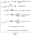
- Figure 1 shows a schematic representation of an embodiment in which a signal oligo is used for the detection of the target protein.
- the embodiment depicted in Figure 1 is set up to separate the binding of the target protein from the hybridization of the nanoreporters.
- Figure 1 in step 1) shows a first protein probe comprising a signal oligo attached to the probe via hybridization with a linker oligo; and a second protein attached to an affinity tag.
- the protein probes are antibodies and the affinity tag is biotin.
- the embodiment depicted in this figure can utilize any of the protein probes and affinity tags described herein.
- Both the first and second protein probes comprise a target specific region capable of binding one or more portions of a target.
- step 2) and 3 the target protein is mixed with the first and second protein probes.
- step 4 the complex of target protein and protein probes is purified.
- the complex of target protein and protein probes is purified using streptavidin-coupled magnetic beads, such as Dynabeads® (Invitrogen).
- the complex of target protein and protein probe (s) can be purified by any suitable method known in the art such as chromatography, including but not limited to HPLC, FPLC, size exclusion (gel filtration) chromatography, affinity chromatography, ion exchange chromatography, hydrophobic interaction chromatography, immunoaffinity chromatography, and reverse phase chromatography; ligand-receptor binding, such as biotin-avidin, maltose-maltose binding protein (MBP), calcium-calcium binding peptide; aptamer-target binding; zip code hybridization; and the like.
- chromatography including but not limited to HPLC, FPLC, size exclusion (gel filtration) chromatography, affinity chromatography, ion exchange chromatography, hydrophobic interaction chromatography, immunoaffinity chromatography, and reverse phase chromatography
- ligand-receptor binding such as biotin-avidin, maltose-maltose binding protein (MBP), calcium-calcium binding peptide;
- the signal oligo is eluted from the complex of target protein and protein probes and analyzed using nanoreporters as described below.
- Methods for eluting the signal oligos are known in the art including the ones depicted in Figure 1 and described herein.
- the methods depicted in Figure 1 are used to detect and/or quantify a plurality of target proteins. Each target protein will be detected by a probe set comprising a first probe and a second probe as described in Figure 1 . Each probe set will have a specific and/or unique signal oligo that can then be associated to the target protein of each probe set.
- the protein probes comprise a capture region.
- the capture region is used for the isolation of the protein probe and/or immobilization of the protein probe into a surface.
- the capture region can be an affinity tag as described below or a solid surface such as bead, a slide or an array.
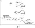
- Figure 6 shows a schematic representation of one of the embodiments of the invention.
- a protein probe is attached to a capture region, e.g. a magnetic bead.
- Figure 6 depicts the use of an antibody.
- the protein probes e.g., antibodies
- the target protein is mixed with the protein probe containing the capture region.
- the complex of target protein and protein probe is then contacted with a second protein probe attached to a signal oligo via a linker oligo.
- the complex of target protein and protein probes are purified.
- the complex of target protein and antibody is purified using the magnetic bead in the capture antibody.
- the complex of target protein and protein probes can be purified by any suitable method known in art such as the methods described above. If the capture region is a slide or an array, the complex of target protein and protein probes can be purified by washing off the excess of unbound sample and protein probes. The isolated target protein/protein probes complex is then washed and the signal oligo is eluted. The signal oligo is analyzed using nanoreporters as described below. Methods for eluting the signal oligos are known in the art including the methods described herein.
- the proteins and nanoreporters are largely separate, which eliminates concerns about protein stickiness.
- the methods depicted in Figure 6 are used to detect and/or quantify a plurality of target proteins. Each target protein will be detected by a probe set comprising a first probe and a second probe as described in Figure 6 . Each probe set will have a specific and/or unique signal oligo that can then be associated to the target protein of each probe set.
- the signal oligo is attached to an affinity tag.
- the affinity tag in the signal oligo can be used to isolate and/or immobilized the signal oligo.
- the signal oligo can be attached to an affinity tag.
- Figure 7 shows a schematic representation of one of the embodiments of the disclosure. This embodiment can be used with any of the methods described herein.
- the diagram is Figure 7 shows antibodies as protein probes, however, this example can be used with any of the protein probes described herein.
- Figure 7 shows an antibody attached directly or indirectly (e.g. via hybridization through an oligo) to a capture region (e.g. a magnetic bead) and a second antibody attached to a biotinylated signal oligo.
- a capture region e.g. a magnetic bead
- the target protein is mixed with the protein probes.
- the complex of target protein and antibodies is purified using the magnetic bead in the capture antibody.
- the complex of target protein and protein probes can be purified by any suitable method known in art such as the methods described above. If the capture region is a slide or an array, the complex of target protein and protein probes can be purified by washing off the excess of unbound sample and protein probes. The isolated target protein/antibody complex is then washed and the signal oligo is eluted by any suitable method known in the art including those described herein. In the embodiment of Figure 7 , the signal oligo is purified using oligonucleotide-coupled beads such as Dynabeads®. However, the signal oligo can be purified by any suitable method according to the affinity tag attached to it.
- the signal oligo is analyzed using nanoreporters as described below.
- the methods depicted in Figure 7 are used to detect and/or quantify a plurality of target proteins.
- Each target protein will be detected by a probe set comprising a first probe and a second probe as described in Figure 7 .
- Each probe set will have a specific and/or unique signal oligo that can then be associated to the target protein of each probe set.
- the embodiments described in Figure 7 provide the advantage that it requires only two bead purifications.
- proteins and nanoreporters are largely separate, which eliminates concerns about protein stickiness.
- the signal oligo is generated by ligating two oligos that are in close proximity, e.g., proximity ligation.
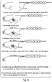
- a diagram of proximity ligation is depicted in Figure 8 .
- probes containing the oligos are designed to bind pairwise to a target protein and to form a signal oligo by ligation when the probes are brought in proximity.
- Figure 8 shows an embodiment using antibodies as protein probes. However, the method described in Figure 8 can be used with any of the protein probes described herein.
- the probes containing the oligos can be prepared and purified by any methods known in the art, for example the methods described in Gullberg et al, PNAS 101(22), p 8420-24 (2004 ).
- the target protein is then mixed with the probes containing the oligos and the bridging oligos.
- a bridging oligo comprises a polynucleotide sequence.
- the oligos attached to protein probes comprise a portion that is complementary to the bridging oligo.
- the complementary portions of the oligos are 5, 6, 7, 8, 9, 10, 11, 12, 13, 14, 15, 16, 17, 18, 19, 20, 21, 22, 23, 24, 25, 26, 27, 28, 29, 30, 40, 50, 60, 70 or more nucleotide bases.
- the complementary portions of the bridging oligo with each of the oligos attached to the protein probe are 6 to 15 bases, with a total length of bridging oligo is 12-30 bases.
- the complementary portions of the oligos are 40 bases.
- the complementary portions of the oligos are 30 bases.
- the complementary portions of the oligos are 20 bases.
- step 4) of Figure 8 the components required for probe ligation are added.
- the oligos in the protein probes can be ligated by any suitable method known in art. Ligation comprises any enzymatic or chemical process wherein an inter-nucleotide linkage is formed between the opposing ends of nucleic acid sequences that are adjacently hybridized to the bridging oligo.
- Example of enzymes that can be used for ligation include but are not limited to DNA ligase, and RNA ligase such as T4 DNA ligase, T4 RNA ligase, Thermus thermophilus (Tth) ligase, Thermus aquaticus (Taq) DNA ligase, or Pyrococcus furiosus (Pfu) ligase.
- Chemical ligation can be performed using activating and reducing agents such as carbodiimide, cyanogen bromide (BrCN), imidazole, 1-methylimidazole/carbodiimide/cystamine, N-cyanoimidazole, dithiothreitol (DTT) and ultraviolet light.
- activating and reducing agents such as carbodiimide, cyanogen bromide (BrCN), imidazole, 1-methylimidazole/carbodiimide/cystamine, N-cyanoimidazole, dithiothreitol (DTT
- Ligation techniques such as gap-filling ligation, including, without limitation, gap-filling OLA and LCR, bridging oligonucleotide ligation, and correction ligation, may be used. Descriptions of these techniques can be found, among other places, in U.S. Pat. No. 5,185,243 , published European Patent Applications EP 320308 and EP 439182 , and PCT Publication Nos. WO 90/01069 and WO 01/57268 .
- the signal oligo is then released via disulfide reduction, uracil excision, restriction digest, proteinase K, or any other suitable method know in the art. Additionally, the signal oligo can be released by the methods depicted in Figure 8B-8D .
- Figure 8B shows an embodiment in which the signal oligo has an affinity tag such as biotin or a sequence. The affinity tag can be used to isolate and/or immobilized the signal oligo as described herein.
- Figure 8C shows an embodiment in which the bridging oligo has an affinity tag such as biotin or a sequence. The affinity tag can be used to isolate and/or immobilized the signal oligo as described herein.
- Figure 8D shows an embodiment in which the embodiments of Figure 8B and 8C are combined.
- the signal oligo is analyzed using nanoreporters as described below.
- the methods depicted in Figure 8 are used to detect and/or quantify a plurality of target proteins.
- Each target protein will be detected by a probe set comprising a first probe and a second probe as described in Figure 8 .
- Each probe set will have a specific and/or unique signal oligo that can then be associated to the target protein of each probe set.
- the embodiments described in Figure 8 have several benefits around sensitivity, minimization of cross-reactivity, and multiplexing. Proximity ligations have shown high sensitivity and have the effect of lowering the apparent Kd by essentially decreasing the off-rate.
- one of the oligos is attached to a nanoreporter.
- Figure 9 shows a diagram of one of such embodiments.
- step 1) of Figure 9 probes containing the oligos are designed to bind pairwise to target proteins.
- One of the oligos in one of the protein probes is attached to a nanoreporter.
- Figure 9 shows an embodiment using antibodies as protein probes. However, the method described in Figure 9 can be used with any of the protein probes described herein.
- the probes containing the oligos can be prepared and purified as described above.
- step 2) and 3) of Figure 9 the target protein is then mixed with the probes containing the oligos and the bridging oligos.
- the bridging oligo binds to the oligo in a first protein probe and a portion of the nanoreporter attached to the second protein probe.
- the oligo attached to the first protein probe and the nanoreporter comprise a portion that is complementary to the bridging oligo.
- the complementary portion is 5, 6, 7, 8, 9, 10, 11, 12, 13, 14, 15, 16, 17, 18, 19, 20, 21, 22, 23, 24, 25, 26, 27, 28, 29, 30, 40, 50, 60, 70 or more nucleotide bases.
- the complementary portion is 40 bases.
- the complementary portion is 30 bases.
- the complementary portion is 20 bases.
- the complementary portions of the bridging oligo with each the oligos attached to the protein probe and the nanoreporter are 6 to 15 bases, with a total length of bridging oligo is 12-30 bases.
- step 4) of Figure 9 the components required for probe ligation are then added.
- the oligo in the first protein probe and the nanoreporter can be ligated by any suitable method known in art as described above.
- step 5) of Figure 9 after ligation, the signal oligo can be optionally released via disulfide reduction, uracil excision, restriction digest, proteinase K, or any other suitable method know in the art.
- the signal oligo can be released by the methods depicted in Figure 8B-8D .
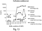
- a purification step is performed to separate ligated oligos from non-ligated oligos after release of the signal oligo from, for instance, an antibody.
- This purification step can be performed using magnetic beads or any other method known in the art for the physical separation of proteins.
- the purification step further includes a rinsing step with a buffer solution.
- Figure 13 demonstrates how various components of an antibody reporter complex are purified and rinsed in a variety of buffer conditions.
- a preferred rinsing buffer is SSPE; however, other buffers and all concentrations having similar capacities for retaining counts of a reporter complex or a component thereof are encompassed by these methods.
- the signal oligo is analyzed using nanoreporters as described below.
- the methods depicted in Figure 9 are used to detect and/or quantify a plurality of target proteins.
- Each target protein will be detected by a probe set comprising a first probe and a second probe as described in Figure 9 .
- Each probe set will have a specific and/or unique signal oligo that can then be associated to the target protein of each probe set.
- the embodiments described in Figure 9 take advantage of the decrease in the Koff via proximity ligation.
- a lower Koff means a lower Kd and the ability to work with lower concentrations of protein probe. This decrease in Kd makes it easier to work in concentrations required for reporters, and thus to contemplate direct detection approaches for multiplex analysis and lower reagent costs.
- These embodiments do not need a step for hybridization to reporters within the assay. Thus, these assays will be faster and have a shorter time to answer.
- the signal oligo is analyzed/detected using nanoreporter(s) as described in sections below.
- the nanoreporter(s) comprise a portion that is complementary to the signal oligo.
- the complementary portion is 5, 6, 7, 8, 9, 10, 11, 12, 13, 14, 15, 16, 17, 18, 19, 20, 21, 22, 23, 24, 25, 26, 27, 28, 29, 30, 40, 50, 60, 70 or more nucleotide bases.
- the complementary portion is 40 bases.
- the complementary portion is 30 bases.
- the complementary portion is 20 bases. In some embodiments, the complementary portion 15-20 bases.
- the methods described herein further comprise protein probes containing a nanoreporter.
- the methods described herein further comprise protein probes containing a nanoreporter.
- Figure 4 shows a schematic diagram of one of the embodiments of the disclosure.
- a nanoreporter is attached to one of the antibodies.
- the methods described in Figure 4 can be utilized using any of the protein probes described herein.
- the nanoreporter can be directly attached to the protein probe.
- the nanoreporter can be attached to a protein probe via hybridization through a linker oligo.
- the nanoreporter comprises a portion that is complementary to the linker oligo in the protein probe.
- the complementary portion is 5, 6, 7, 8, 9, 10, 11, 12, 13, 14, 15, 16, 17, 18, 19, 20, 21, 22, 23, 24, 25, 26, 27, 28, 29, 30, 40, 50, 60, 70 or more nucleotide bases.
- the linker oligo is 15-20 bases.
- the complementary portion is 40 bases.
- the complementary portion is 30 bases.
- the complementary portion is 20 bases.
- the complementary portion is 15 bases.
- the hybridization of the nanoreporter to the linker oligo can occur at different temperatures depending of the length of the complementary portion.
- the nanoreporter can be hybridized to a linker oligo attached to a protein probe at a temperature in the range of 32°C to 40°C.
- the nanoreporter can be hybridized to a linker oligo attached to a protein probe at a temperature of 35°C.
- the nanoreporter can be hybridized to a linker oligo attached to a protein probe at a temperature of 37°C.
- the nanoreporter can be hybridized to a linker oligo attached to a protein probe at a temperature of 45°C.
- the nanoreporter can be hybridized to a linker oligo attached to a protein probe at a temperature of 52-57°C. In some embodiments, the nanoreporter can be hybridized to a linker oligo attached to a protein probe at a temperature of 15-20°C below the melting temperature (Tm) of the complementary portions of the nanoreporter with the linker oligo.
- Tm melting temperature
- the protein probe is an antibody and the length of the complementary portions of the nanoreporter with the linker oligo is 15-20 bases, which gives a Tm of about 57°C or 15-20 °C above the ideal antibody temperature of 37°C.
- the protein probe is an antibody, the length of the complementary portions of the nanoreporter with the linker oligo is 15-20 bases and the hybridizing temperature is 37°C.
- Figure 4 shows that a complex of target protein and antibodies is formed in which one of the antibodies is bound to biotin and the other antibody has a nanoreporter attached.
- the methods described in Figure 4 can use any affinity tag described herein besides biotin.
- Purification of the target protein-antibodies complex can be performed by any suitable method known in the art including those described herein.
- Elution of the nanoreporter can be accomplished by melting off G and F beads, via digestion or any other suitable method known in the art.
- the complex can be bound to a coverslip, e.g., coated with streptavidin (Optichem®, Accelr8 Technology Corporation). The nanoreporter is analyzed as described below.
- the methods depicted in Figure 4 are used to detect and/or quantify a plurality of target proteins.
- Each target protein will be detected by a probe set comprising a first probe and a second probe as described in Figure 4 .
- Each probe set will have a specific and/or unique nanoreporter that can then be associated to the target protein of each probe set.
- the embodiments of the inventions that utilize a signal oligo present several advantages: (1) these embodiments separate the target proteins and the protein probes from the nanoreporters. Separation of the proteins from the reporters eliminates the potential problems of solubility and stickiness associated with using nanoreporters to measure proteins.
- the indirect signal oligo approach can be run as a process upstream of the nanoreporter assay described below, thereby taking advantage of an optimized nanoreporter assay;
- protein probe sets e.g, antibody pairs
- capture antibody on surface on a magnetic bead for example
- Some antibodies work best in this configuration; (4) with these embodiments problems associated with the protein probes coming off the target (Koff rate) are minimized, e.g., antibodies only have to stay bound to the target during binding and purification on the beads. This allows for use of a large range of antibodies including antibodies with lower binding affinity; and (5) proteins can be read in the same lane as nucleic acids, e.g., RNA or DNA.
- the sample is first split: part is run through the protein detection embodiments described herein (lyse cells with detergent then bind and purify as described herein), and part is split off and processed as nucleic acid samples (cells are lysed with GITC).
- RNA samples are then recombined and analyzed using nanoreporters as described below, potentially in the same lane.
- Measurement of both nucleic acids (e.g., RNA) and proteins in the same lane will minimize measurement differences, make protein and nucleic acid expression data more comparable, and eliminate the need for multiple measurement methods to get the required data.
- the methods described herein provide for the measurement of nucleic acids, e.g., RNA or DNA, in combination with the measurement of proteins.
- any of the embodiments described herein can be used in the detection of multiple target proteins.
- methods comprising protein probes for the analysis of target proteins.
- a protein probe population for use in a multiplexed assay. Each protein probe in the population is specific for a target molecule. The binding of the target proteins to the protein probes is then detected using nanoreporters. Each nanoreporter comprises a unique label code that can be associated to a specific target molecule as described below.
- the detection of the nanoreporters as described below is digital in nature in that one molecule at a time is counted. While fluorescence is used to read the code, the signals are high and the spot is either present of not, thus the digital detection.
- digital detection rather than an analogue fluorescent signal used to quantify signal leads to more accurate quantification.
- the methods described herein allows for multiplexing to levels beyond currently possible, for more accurate quantification, and possibly higher sensitivity.
- a nanoreporter which provides a code of signals (the nanoreporter label code) associated with a specific target.
- the nanoreporter code upon binding of the nanoreporter to a signal oligo or a linker oligo associated with a protein probe, the nanoreporter code identifies the signal oligo or the protein probe to which the nanoreporter is bound.
- the nanoreporters comprise two main portions: (i) a sequence specific for a signal oligo-specific or a linker oligo associated with a protein probe; and (ii) a labeled nanoreporter.
- the nanoreporters are directly attached to a protein probe.
- Nanoreporters are modular structures.
- the nanoreporter comprises a plurality of different detectable molecules.
- a labeled nanoreporter is a molecular moiety containing certain basic elements: (i) a plurality of label attachment regions attached in linear combination, and (ii) complementary polynucleotide sequences attached to the label attachment regions of the backbone.
- the labeled nanoreporter comprises 2, 3, 4, 5, 6, 7, 8, 9, 10 or more unique label attachment regions attached in a linear combination, and complementary polynucleotide sequences attached to the label attachment regions of the backbone.
- the labeled nanoreporter comprises 3 or more label attachment regions attached in linear combination, and complementary polynucleotide sequences attached to the label attachment regions of the backbone.
- label attachment region includes a region of defined polynucleotide sequence within a given backbone that may serve as an individual attachment point for a detectable molecule.
- the plurality of label attachment regions attached in linear combination can comprise uniquely designed sequences.
- the plurality of label attachment regions attached in linear combination in the nanoreporters can comprise at least one template, for example but not limited to, at least one nucleic acid sequence, such as at least part of a linear or linearizable viral genome, such as the genomes of adenovirus, hepatitis virus, herpes virus, rotavirus, and the like, or bacteriophages such as lambda, M13, ⁇ X-174, T-series bacteriophages, and the like, including derivatives thereof comprising cloning cassettes, polylinkers, and the like; plasmids, such as pBR322 and pUC series plasmids, etc., including derivatives thereof comprising cloning cassettes, polylinkers, and the like; synthetic templates; templates comprising artificial sequences; and the like.
- nucleic acid can serve as a template for fabricating a nanoreporter provided that it is large enough to include at least two label attachment regions, or it can be combined with at least one other nucleic acid sequence so that the combined sequence is large enough to include at least two label attachment regions.
- the labeled nanoreporter also comprises a backbone containing a constant region.
- the constant region can be directly or indirectly attached to the nanoreporter.
- the constant region can covalently attached to a nanoreporter or the constant region can be bound to the nanoreporter later in the assay.
- the term constant region includes tandemly-repeated sequences of about 10 to about 25 nucleotides.
- the constant region can be attached at either the 5' region or the 3' region of a nanoreporter, and may be utilized for capture and immobilization of a nanoreporter for imaging or detection, such as by attaching to a solid substrate a sequence that is complementary to the constant region.
- the elements of a nanoreporter can be found in a single molecular moiety (a singular nanoreporter), or two distinct molecular moieties (a dual nanoreporter). Each molecular moiety may be composed of one molecule or more than one molecule attached to one another by covalent or non-covalent means. In some embodiments, each component of a dual nanoreporter has a signal oligo-specific sequence that binds to a different site on the same signal oligo molecule. When using a dual nanoreporter system one of the nanoreporter probes may be unlabeled. In some embodiments, the unlabeled nanoreporter probe may comprise a capture region.
- the unlabeled nanoreporter probe may comprise a signal oligo-specific region and a backbone that may be single stranded. In some embodiments, the unlabeled nanoreporter probe may comprise a signal oligo-specific region and a backbone that may be double stranded.
- the complementary polynucleotide sequences attached to a nanoreporter backbone serve to attach detectable molecules, or label monomers, to the nanoreporter backbone.
- the complementary polynucleotide sequences may be directly labeled, for example, by covalent incorporation of one or more detectable molecules into the complementary polynucleotide sequence.
- the complementary polynucleotide sequences may be indirectly labeled, such as by incorporation of biotin or other molecule capable of a specific ligand interaction into the complementary polynucleotide sequence.
- the ligand e.g., streptavidin in the case of biotin incorporation into the complementary polynucleotide sequence
- the detectable molecules attached to a label attachment region are not directly incorporated into the complementary polynucleotide sequence, this sequence serves as a bridge between the detectable molecule and the label attachment region, and may be referred to as a bridging molecule, e.g., a bridging nucleic acid.
- Nanoreporters are described in US patent 7,473,767 ; US applications No. 10/542,458 ; 12/324,357 ; 11/645,270 and 12/541,131 .
- the nanoreporters comprise at least one constant region, which may serve as an affinity tag for purification and/or for immobilization (for example to a solid surface).
- the constant region typically comprises two or more tandemly-repeated regions of repeat nucleotides, such as a series of 15-base repeats.
- the nanoreporter whether complexed to a signal oligo, a target molecule or otherwise, can be purified or immobilized by an affinity reagent coated with a 15-base oligonucleotide which is the reverse complement of the repeat unit.
- Nanoreporters, nanoreporter-signal oligo complexes, or nanoreporter-protein probe complexes can be purified in two or more affinity selection steps.
- the nanoreporter in the embodiments in which the nanoreporter is attached to a protein probe, the nanoreporter can comprise an affinity tag.
- one nanoreporter probe can comprise a first affinity tag and the other nanoreporter probe can comprise a second (different) affinity tag.
- the nanoreporter probes are mixed with the signal oligos, and complexes comprising the two probes of the dual nanoreporters are separated from unbound materials (e.g., the signal oligo or the individual probes of the nanoreporter) by affinity purification against one or both individual affinity tags.
- the mixture can be bound to an affinity reagent for the first affinity tag, so that only probes comprising the first affinity tag and the desired complexes are purified.
- the bound materials are released from the first affinity reagent and optionally bound to an affinity reagent for the second affinity tag, allowing the separation of complexes from nanoreporter probes comprising the first affinity tag. At this point only full complexes would be bound.
- the complexes are finally released from the affinity reagent for the second affinity tag and then preferably stretched and imaged.
- the affinity reagent can be any solid surface coated with a binding partner for the affinity tag, such as a column, bead (e.g., latex or magnetic bead) or slide coated with the binding partner. Immobilizing and stretching nanoreporters using affinity reagents is fully described in U.S. Provisional Application no. 60/753,816 by Sean M. Ferree and Dwayne L. Dunaway, entitled “Compositions Comprising Oriented, Immobilized Macromolecules and Methods for Their Preparation,” filed on December 23, 2005 , and US patent 7,473,767 ; US applications No. 10/542,458 ; 12/324,357 ; 11/645,270 and 12/541,131 .
- the sequence of signals provided by the label monomers associated with the various label attachment regions of the backbone of a given nanoreporter allows for the unique identification of the nanoreporter.
- a nanoreporter having a unique identity or unique spectral signature is associated with a signal oligo-specific sequence or a protein probe that recognizes a specific target molecule or a portion thereof.
- Detection of the nanoreporter signal such as the spectral code of a fluorescently labeled nanoreporter, associated with the nanoreporter allows detection of the presence of the target molecule in the mixture (qualitative analysis).
- Counting all the label monomers associated with a given spectral code or signature allows the counting of all the molecules in the mixture associated with the signal oligo -specific sequence or the protein probe coupled to the nanoreporter (quantitative analysis).
- the signal oligos then can be correlated to the target molecule via the binding of target molecule to the protein probe associated with the signal oligo.
- Nanoreporters are thus useful for the diagnosis or prognosis of different biological states (e.g., disease vs. healthy) by quantitative analysis of known biological markers.
- the tiny sensitivity of single molecule detection and quantification allows for the identification of new diagnostic and prognostic markers, including those whose fluctuations among the different biological states is too slight detect a correlation with a particular biological state using traditional molecular methods.
- the sensitivity of nanoreporter-based molecular detection permits detailed pharmacokinetic analysis of therapeutic and diagnostic agents in small biological samples.
- Nanoreporters' syntheses can be performed by any suitable methods known in the art. Examples of nanoreporters' syntheses are described in US patent 7,473,767 ; US applications No. 10/542,458 ; 12/324,357 ; 11/645,270 and 12/541,131 .
- a nanoreporter further comprising an affinity tag attached to the nanoreporter backbone, such that attachment of the affinity tag to a support allows backbone stretching and resolution of signals provided by label monomers corresponding to different label attachment regions on the backbone.
- Nanoreporter stretching may involve any stretching means known in the art including but not limited to, means involving physical, hydrodynamic or electrical means.
- the affinity tag may comprise a constant region.
- each nanoreporter probe in a population of probe allows for the multiplexed analysis of a plurality of target molecules.
- each nanoreporter probe can contain contains six label attachment regions, where each label attachment region of each backbone is different from the other label attachment regions in that same backbone. If the label attachment regions are going to be labeled with one of four colors and there are 24 possible unique sequences for the label attachment regions and each label attachment region is assigned a specific color, each label attachment region in each backbone will consist of one of four sequences. There will be 4096 possible nanoreporters in this example.
- the number of possible nanoreporters can be increased, for example, by increasing the number of colors, increasing the number of unique sequences for the label attachment regions and/or increasing the number of label attachment regions per backbone. Likewise the number of possible nanoreporters can be decreased by decreasing the number of colors, decreasing the number of unique sequences for the label attachment regions and/or decreasing the number of label attachment regions per backbone.
- the methods of detection are performed in multiplex assays, whereby a plurality of target molecules is detected in the same assay (a single reaction mixture).
- the assay is a hybridization assay in which the plurality of target molecules is detected simultaneously.
- the plurality of target molecules detected in the same assay is, at least 2, at least 5 different target molecules, at least 10 different target molecules, at least 20 different target molecules, at least 50 different target molecules, at least 75 different target molecules, at least 100 different target molecules, at least 200 different target molecules, at least 500 different target molecules, or at least 750 different target molecules, or at least 1000 different target molecules.
- the plurality of target molecules detected in the same assay is up to 50 different target molecules, up to 100 different target molecules, up to 150 different target molecules, up to 200 different target molecules, up to 300 different target molecules, up to 500 different target molecules, up to 750 different target molecules, up to 1000 different target molecules, up to 2000 target molecules, or up to 5000 target molecules.
- the plurality of target molecules detected is any range in between the foregoing numbers of different target molecules, such as, but not limited to, from 20 to 50 different target molecules, from 50 to 200 different target molecules, from 100 to 1000 different target molecules, from 500 to 5000 different target molecules, and so on and so forth.
- the nanoreporters are uniquely suitable for conducting quantitative analyses.
- the nanoreporters By providing a one to one binding between the nanoreporters (whether singular or dual nanoreporters) and their target molecules in a biomolecular sample, all or a representative portion of the target molecules present in the sample can be identified and counted.
- This individual counting of the various molecular species provides an accurate and direct method for determining the absolute or relative concentration of the target molecule in the biomolecular sample.
- the ability to address each molecule in a mixture individually leverages benefits of miniaturization including high sensitivity, minimal sample quantity requirements, high reaction rates which are afforded by solution phase kinetics in a small volume, and ultimately very low reagent costs.
- the nanoreporters can be labeled with any of a variety of label monomers, such as a radioisotope, fluorochrome, dye, enzyme, nanoparticle, chemiluminescent marker, biotin, or other monomer known in the art that can be detected directly (e.g., by light emission) or indirectly (e.g., by binding of a fluorescently-labeled antibody).
- label monomers such as a radioisotope, fluorochrome, dye, enzyme, nanoparticle, chemiluminescent marker, biotin, or other monomer known in the art that can be detected directly (e.g., by light emission) or indirectly (e.g., by binding of a fluorescently-labeled antibody).
- one or more of the label attachment regions in the nanoreporter is labeled with one or more label monomers, and the signals provided by the label monomers attached to the label attachment regions of a nanoreporter constitute a detectable code that identifies the target to which the target-specific region of
- affinity tags known in the art may be used, e.g., to purify and/or immobilize nanoreporters.
- a biotin anchor is attached to the nanoreporter, allowing immobilization of the nanoreporter on a streptavidin coated slide.
- a labeled nanoreporter will contain an affinity tag at each end, A1 and A2.
- the labeled nanoreporter can be immobilized to a surface through the binding of A1 to an immobilized affinity partner. In the absence of an affinity binding partner for A2, the A2 end of the nanoreporter remains in solution, but in the presence of an affinity binding partner (A2'), the A2 end of the nanoreporter is also immobilized.
- a labeled nanoreporter will contain a single affinity tag, A1.
- affinity tag can be attached to the nanoreporter by direct binding of the nanoreporter to a molecule containing A2 (e.g., if the nanoreporter is or comprises a nucleic acid, it can hybridize directly with another nucleic acid to which A2 is attached).
- affinity tag can be attached to the labeled nanoreporter via a bridging molecule, such as the bridging nucleic acid.
- the nanoreporter upon immobilization of A1, the nanoreporter can be stretched, or "elongated", for example by electrostretching, for separation of the label attachment regions in a manner that permits detection of the nanoreporter code.
- A2 while the nanoreporter is in an elongated state, A2 is introduced and binds the end of the nanoreporter that is complementary to A2 down to the surface.
- an affinity tag is attached to a protein probe, e.g., to purify and/or immobilize the protein probe.
- An affinity tag can be used for attachment to beads or other matrixes for a variety of useful applications including but not limited to purification.
- the sample may comprise any number of things, including, but not limited to: biological samples, such as cells (including both primary cells and cultured cell lines), cell lysates, or extracts, tissues and tissue extracts; bodily fluids (including, but not limited to, blood, urine, serum, lymph, bile, cerebrospinal fluid, interstitial fluid, aqueous or vitreous humor, colostrum, sputum, amniotic fluid, saliva, anal and vaginal secretions, perspiration and semen, a transudate, an exudate (e.g., fluid obtained from an abscess or any other site of infection or inflammation) or fluid obtained from a joint (e.g., a normal joint or a joint affected by disease such as rheumatoid arthritis, osteoarthritis, gout or septic arthritis) of virtually any organism, with mamma
- biological samples such as cells (including both primary cells and cultured cell lines), cell lysates, or extracts, tissues and tissue extracts
- the biomolecular samples can be indirectly derived from biological specimens.
- the target protein of interest is a kinase
- the biomolecular sample can be a sample containing isolated proteins from a cell lysate.
- the biomolecular sample is generated by subjecting a biological specimen to fractionation, e.g., size fractionation or membrane fractionation.
- the biomolecular samples may be either native, e.g., not subject to manipulation or treatment, or treated, which can include any number of treatments, including exposure to candidate agents including drugs, genetic engineering (e.g., the addition or deletion of a gene), etc.
- Biomolecular samples may also include environmental samples, such as those containing bacteria or other organisms, such as diatoms, dinoflagellates, algae, among others, such as in certain marine or earth-based samples.
- environmental samples such as those containing bacteria or other organisms, such as diatoms, dinoflagellates, algae, among others, such as in certain marine or earth-based samples.
- Nanoreporters are detected by any means available in the art that is capable of detecting the specific signals on a given nanoreporter. Where the nanoreporter is fluorescently labeled, suitable consideration of appropriate excitation sources may be investigated. Possible sources may include but are not limited to arc lamp, xenon lamp, lasers, light emitting diodes or some combination thereof.
- the appropriate excitation source is used in conjunction with an appropriate optical detection system, for example an inverted fluorescent microscope, an epifluorescent microscope or a confocal microscope.
- a microscope is used that can allow for detection with enough spatial resolution to determine the sequence of the spots on the nanoreporter. For example in one embodiment an image of a dual nanoreporter hybridized to a target molecule can be obtained.
- the nanoreporters are labeled with three different colors, Alexa 488, Cy3 and Alexa 647 (labeled 1, 2 and 3, respectively). Colors 1, 2 and 3 are each acquired in different channels and the first and second registers, which can be seen as rows of spots, are shifted up by several pixels to be able to show each register individually.
- compositions and methods described herein can be used for diagnostic, prognostic, therapeutic, patient stratification, drug development, treatment selection and screening purposes.
- target proteins can be analyzed at one time from a single biomolecular sample using the methods of the invention. This allows, for example, for several diagnostic tests to be performed on one sample.
- composition and methods can be used in proteomics.
- the methods described herein will typically provide an answer rapidly which is very desirable for this application.
- the methods and composition described herein can be used in the process of finding biomarkers that may be used for diagnostics or prognostics and as indicators of health and disease.
- the methods and composition described herein can be used to screen for drugs, e.g., drug development, selection of treatment, determination of treatment efficacy and/or identify targets for pharmaceutical development.
- the ability to test protein expression on screening assays involving drugs is very important because proteins are the final gene product in the body.
- the methods and compositions described herein will measure both protein and gene expression simultaneously which will provide the most information regarding the particular screening being performed.
- the present methods can be applied to the analysis of biomolecular samples obtained or derived from a patient so as to determine whether a diseased cell type is present in the sample, the stage of the disease, the prognosis for the patient, the ability to the patient to respond to a particular treatment, or the best treatment for the patient.
- the present methods can also be applied to identified biomarkers for a particular disease
- the methods described herein are used in the diagnosis of a condition.
- diagnosis or “diagnosis” of a condition includes predicting or diagnosing the condition, determining predisposition to the condition, monitoring treatment of the condition, diagnosing a therapeutic response of the disease, and prognosis of the condition, condition progression, and response to particular treatment of the condition.
- a blood sample can be assayed according to any of the methods described herein to determine the presence and/or quantity of markers of a disease or malignant cell type in the sample, thereby diagnosing or staging the a disease or a cancer.
- the methods and composition described herein are used for the diagnosis and prognosis of a condition.
- Immunologic diseases and disorders include allergic diseases and disorders, disorders of immune function, and autoimmune diseases and conditions.
- Allergic diseases and disorders include but are not limited to allergic rhinitis, allergic conjunctivitis, allergic asthma, atopic eczema, atopic dermatitis, and food allergy.
- Immunodeficiencies include but are not limited to severe combined immunodeficiency (SCID), hypereosinophilic syndrome, chronic granulomatous disease, leukocyte adhesion deficiency I and II, hyper IgE syndrome, Chediak Higashi, neutrophilias, neutropenias, aplasias, Agammaglobulinemia, hyper-IgM syndromes, DiGeorge/Velocardial-facial syndromes and Interferon gamma-TH1 pathway defects.
- SCID severe combined immunodeficiency
- Autoimmune and immune dysregulation disorders include but are not limited to rheumatoid arthritis, diabetes, systemic lupus erythematosus, Graves' disease, Graves ophthalmopathy, Crohn's disease, multiple sclerosis, psoriasis, systemic sclerosis, goiter and struma lymphomatosa (Hashimoto's thyroiditis, lymphadenoid goiter), alopecia aerata, autoimmune myocarditis, lichen sclerosis, autoimmune uveitis, Addison's disease, atrophic gastritis, myasthenia gravis, idiopathic thrombocytopenic purpura, hemolytic anemia, primary biliary cirrhosis, Wegener's granulomatosis, polyarteritis nodosa, and inflammatory bowel disease, allograft rejection and tissue destructive from allergic reactions to infectious microorganisms or to environmental antigens.
- Proliferative diseases and disorders that may be evaluated by the methods described herein include, but are not limited to, hemangiomatosis in newborns; secondary progressive multiple sclerosis; chronic progressive myelodegenerative disease; neurofibromatosis; ganglioneuromatosis; keloid formation; Paget's Disease of the bone; fibrocystic disease (e.g., of the breast or uterus); sarcoidosis; Peronies and Duputren's fibrosis, cirrhosis, atherosclerosis and vascular restenosis.
- Malignant diseases and disorders that may be evaluated by the methods described herein include both hematologic malignancies and solid tumors.
- Hematologic malignancies are especially amenable to the methods described herein when the sample is a blood sample, because such malignancies involve changes in blood-borne cells.
- Such malignancies include non-Hodgkin's lymphoma, Hodgkin's lymphoma, non-B cell lymphomas, and other lymphomas, acute or chronic leukemias, polycythemias, thrombocythemias, multiple myeloma, myelodysplastic disorders, myeloproliferative disorders, myelofibroses, atypical immune lymphoproliferations and plasma cell disorders.
- Plasma cell disorders that may be evaluated by the methods described herein include multiple myeloma, amyloidosis and Waldenstrom's macroglobulinemia.
- Example of solid tumors include, but are not limited to, colon cancer, breast cancer, lung cancer, prostate cancer, brain tumors, central nervous system tumors, bladder tumors, melanomas, liver cancer, osteosarcoma and other bone cancers, testicular and ovarian carcinomas, head and neck tumors, and cervical neoplasms.
- the methods described herein can be used to diagnose pathogen infections, for example infections by intracellular bacteria and viruses, by determining the presence and/or quantity of markers of bacterium or virus, respectively, in the sample.
- infectious diseases can be detected by the process described herein. Typically, these are caused by bacterial, viral, parasite, and fungal infectious agents. The resistance of various infectious agents to drugs can also be determined.
- Bacterial infectious agents which can be detected include Escherichia coli, Salmonella, Shigella, Klebsiella, Pseudomonas, Listeria monocytogenes, Mycobacterium tuberculosis, Mycobacterium aviumintracellulare, Yersinia, Francisella, Pasteurella, Brucella, Clostridia, Bordetella pertussis, Bacteroides, Staphylococcus aureus, Streptococcus pneumonia, B-Hemolytic strep., Corynebacteria, Legionella, Mycoplasma, Ureaplasma, Chlamydia, Neisseria gonorrhea, Neisseria meningitides, Hemophilus influenza, Enterococcus faecalis, Proteus vulgaris, Proteus mirabilis, Helicobacter pylori, Treponema palladium, Borrelia burgdorferi, Borrelia recurrent
- Fungal infectious agents which can be detected include Cryptococcus neoformans, Blastomyces dermatitidis, Histoplasma capsulatum, Coccidioides immitis, Paracoccidioides brasiliensis, Candida albicans, Aspergillus fumigautus, Phycomycetes (Rhizopus), Sporothrix schenckii, Chromomycosis, and Maduromycosis.
- Viral infectious agents which can be detected include human immunodeficiency virus, human T-cell lymphocytotrophic virus, hepatitis viruses (e.g., Hepatitis B Virus and Hepatitis C Virus), Epstein-Barr Virus, cytomegalovirus, human papillomaviruses, orthomyxo viruses, paramyxo viruses, adenoviruses, corona viruses, rhabdo viruses, polio viruses, toga viruses, bunya viruses, arena viruses, rubella viruses, and reo viruses.
- human immunodeficiency virus e.g., human T-cell lymphocytotrophic virus
- hepatitis viruses e.g., Hepatitis B Virus and Hepatitis C Virus
- Epstein-Barr Virus Epstein-Barr Virus
- cytomegalovirus human papillomaviruses
- orthomyxo viruses paramyxo viruses
- corona viruses corona viruses
- Parasitic agents which can be detected include Plasmodium falciparum, Plasmodium malaria, Plasmodium vivax, Plasmodium ovale, Onchoverva volvulus, Leishmania, Trypanosoma spp., Schistosoma spp., Entamoeba histolytica, Cryptosporidum, Giardia spp., Trichimonas spp., Balatidium coli, Wuchereria bancrofti, Toxoplasma spp., Enterobius vermicularis, Ascaris lumbricoides, Trichuris trichiura, Dracunculus medinesis, trematodes, Diphyllobothrium latum, Taenia spp., Pneumocystis carinii, and Necator americanis.
- the methods described herein are also useful for detection of drug resistance by infectious agents.
- vancomycin-resistant Enterococcus faecium methicillin-resistant Staphylococcus aureus, penicillin-resistant Streptococcus pneumoniae, multi-drug resistant Mycobacterium tuberculosis, and AZT-resistant human immunodeficiency virus can all be identified.
- the target molecules detected using the compositions and methods described herein can be either patient markers (such as a cancer marker) or markers of infection with a foreign agent, such as bacterial or viral markers.
- compositions and methods described herein can be used to quantitate target protein whose abundance is indicative of a biological state or disease condition, for example, blood markers that are upregulated or downregulated as a result of a disease state.
- the methods and compositions described herein can be used for cytokine detection.
- the low sensitivity of the methods described herein would be helpful for early detection of cytokines, e.g., as biomarkers of a condition, diagnosis or prognosis of a disease such as cancer, and the identification of subclinical conditions.
- kits comprising one or more components.
- the kits can comprise, for example, one or more protein probe sets and/or one or more nanoreporters.
- the kits can be used for any purpose apparent to those of skill in the art, including those described above.
- kits useful for the extension and selective immobilization of nanoreporters can comprise a substrate for immobilization and one or more binding partners to facilitate extension or immobilization of a nanoreporter.
- the binding partners could, in certain embodiments, comprise a moiety useful for extension of the nanoreporter in an appropriate force.
- the binding partners could facilitate immobilization or selective immobilization of the nanoreporter to the surface.
- the kits could comprise a nanoreporter for extension and immobilization.
- the kits could comprise a device capable of extending the nanoreporter.
- kits can contain a population of protein probes and/or nanoreporters as described herein.
- kits can contain pre-labeled nanoreporters, or unlabeled nanoreporters with one or more components for labeling the nanoreporters.
- the nanoreporters provided in a kit may or may not have target-specific sequences pre-attached.
- the target sequences are provided in the kit unattached to the nanoreporter backbone.
- kits can comprise other reagents such as signal oligos, linker oligos and bridging oligos. In some embodiments, the kits can separate the protein probe pairs into different premixes.
- kits can include other reagents as well, for example, buffers for performing hybridization reactions, linkers, restriction endonucleases, and DNA I ligases.
- kits also will include instructions for using the components of the kit, and/or for making and/or using the labeled nanoreporters.
- FIG. 1 A diagram of the protocol for this Example is depicted in Figure 1 .
- the assay is set up to separate the binding of the protein target from the hybridization of the reporters to eliminate issues with the mismatch between low binding affinity of protein probes and the working concentration of reporters.
- the linker oligos were attached to the antibody by random amine attachment using a 10 to 1 ratio of linker oligos to antibodies.
- bifunctional crosslinker Sulfosuccinimidyl 4-[p-maleimidophenyl]butyrate (SMPB) (ThermoFisher, Inc., Waltham, MA) was coupled to anti-IL2 antibody A then reacted to thiolated oligo to crosslinker at an antibody:oligo ratio of 1:3 at room temperature.
- the antibody A linked to SMPB was purified by running the mixture thru Zeba column 2X, 1000G, (ThermoFisher, Inc., Waltham, MA) and the yield is determined.
- the oligo linked to IL-2 antibody A the antibody-SMPB-oligo and PBE were added to Pall Nanosep® Centrifugal Device with Omega Membrane, (MWCO 100 kDa, Sigma-Aldrich, Inc., St. Louis, MO) was washed and centrifuged.
- Pall Nanosep® Centrifugal Device with Omega Membrane (MWCO 100 kDa, Sigma-Aldrich, Inc., St. Louis, MO) was washed and centrifuged.
- the signal oligo was preannealed to the oligo linked to IL-2 antibody A by adding oligo linked Il2 antibody A and signal oligo in a ratio of 3:2, signal oligo:Antibody ratio Other ratios are contemplated.
- IL-2 antibody A annealed to signal oligo was mixed with biotinylated antibody B (BAF202,R&D systems, Inc., Minneapolis, MN) at approximately 1x10 -15 to 1x10 -8 M, and blockers (salmon sperm), with room to add target solution.
- the target protein IL-2 was added to the desired dilution ( ⁇ 1x10 -8 M).
- Antibodies were at 10X concentration over an estimated Kd of 10 0-15 to 10 0-8 M. The mixture was incubated.
- the complex of target protein and antibodies was purified using Streptavidin-coupled Dynabeads® (Invitrogen) according to protocol.
- the isolated target protein/antibody complex was washed and the signal oligo was eluted at greater than 45 °C using 0.1X SSPE for 10 to 15 minutes. Shorter and longer periods are contemplated Detection of Signal Oligo
- Detection of the signal oligo in each sample was carried out using a dual nanoreporter system having both labeled nanoreporter probes and unlabeled nanoreporter probes.
- the signal oligos from each sample were hybridized with final concentrations of the hybridization reagents as follows: unlabeled, biotinylated probe labeled reporter probe, 5X SSPE (pH 7.5), 5X Denhardt's reagent (Sigma), sheared salmon sperm DNA (Sigma), and detergent. Reagents were mixed and incubated in a thermocycler block with a heated lid for 16 hours.
- reaction was purified over magnetic beads (InvitrogenTM) coupled to oligonucleotides complementary to the 3'-repeat sequence contained on every biotinylated probe. Reactions were first diluted SSPE in 0.1% detergent mixture/TE and allowed to bind to beads at greater than 20°C with continuous rotation. The beads were washed three times in SSPE and detergent and the hybridized complexes eluted in of 0.1X SSPE/0.1% /detergent mixture for 15 minutes at 45°C.
- samples were purified a second time to remove excess biotinylated probes by binding to magnetic beads coupled to oligonucleotides complementary to the 5'-repeat sequence contained on every reporter probe.
- the elutions from the anti-3'-repeat beads were brought to a final concentration of 1X SSPE and bound for 15 minutes at 22.5°C with rotation. Beads were washed as above and eluted in of 0.1X SSPE/0.1% /detergent mixture at greater than 40°C.
- the doubly-purified samples were then prepared for capture as described below.
- FIG. 4 A diagram of the protocol for this Example is depicted in Figure 4 .
- the nanoreporter is attached to one of the antibodies. Preparation of the antibodies and binding to the sample happens similar to the protocol described in Example 1.
- a complex of target protein and antibodies is formed in which one of the antibodies is bound to biotin and the other antibody has a nanoreporter attached.
- One approach is to attach the antibody on the reporter before the binding of the antibody to the target protein. This approach requires very strong binding affinity (Kd) antibodies to allow for concentrations of multiplexed reporters significantly below the reporter entanglement threshold of 1x10 -7 M.
- a linker oligo is added to the antibody as described in Example 1.
- the nanoreporter is the attached to the antibody by hybridization to the linker oligo at temperatures between 37 - 45°C.
- Antibody: reporter ratio was 1:1.
- the labeled antibody and the reporter were hybridized at 0.05 nM, 37°C, 1X SSPE, overnight.
- a second direct approach is to first bind the antibodies to the target in solution and purify the complex as described in Example 1. After the purification the nanoreporter is hybridized to the antibody (after target binding) using the protocol described above. In this approach, problems of mismatch between hybridization and protein binding are avoided.
- Koff rate antibody dissociation from the target protein
- Purification of the target protein-antibodies complex can be performed as described in Example 1. Elution can be accomplished by melting off G and F beads or via digestion. However, one skilled in the art will understand that the melting off the complex might require optimization of conditions to allow for antibodies to remain bound but affinity tags to release.
- the protein-antibodies complex can be bound to a coverslip coated with streptavidin (Optichem®, Accelr8 Technology Corporation), stretched and image as described in Example 1. Stretching of a reporter hybridized to an antibody was tested to be sure that the presence of the antibody does not cause non-specific binding or stickiness to the surface that inhibited the normal binding, stretching, and imaging process (data not shown).
- FIG. 6 A diagram of the protocol for this Example is depicted in Figure 6 .
- the capture antibody is attached to a surface, e.g. a magnetic bead, and the second antibody is attached to a signal oligo.
- the antibodies can be prepared by any methods knows in the art including the methods described in Example 1.
- proteins and nanoreporters are largely separate, which eliminates concerns about protein stickiness.
- the local antibody concentrations on the surface can be high.
- the target proteins are mixed with the capture antibody on the magnetic beads (2 hours to overnight, 1X PBS, and room temperature). The unbound protein sample is washed away. The labeled antibody signal oligo complex is added to the beads with blockers (1X PBS and room temperature). After a period of binding the excess labeled antibody signal oligo complex is washed away. The isolated target protein/antibody complex is then washed and the signal oligo is eluted and analyzed as described in Example 1.
- FIG. 7 A diagram of the protocol for this Example is depicted in Figure 7 .
- the capture antibody is attached to a surface, e.g. a magnetic bead, and the second antibody is in solution or attached to a biotinylated signal oligo.
- This assay provides the advantages that it requires only two bead purifications.
- proteins and nanoreporters are largely separate, which eliminates concerns about protein stickiness.
- the target proteins are mixed with the capture antibody, labeled antibody signal oligo complex, and blockers as described in Example 3.
- the complex of target protein and antibodies are purified using the magnetic bead in the capture antibody as described in Example 3.
- the isolated target protein/antibody complex is then washed and the signal oligo is eluted as described in Example 1.
- the signal oligo can then be purified using Streptavidin-coupled Dynabeads® (Invitrogen) according to manufacturer's protocol.
- the signal oligo is then analyzed as described in Example 1.
- FIG. 8 A diagram of the protocol for this Example is depicted in Figure 8 .
- this assay two physically close oligos are ligated.
- Probes containing the oligos are designed to bind pairwise to target proteins and to form a signal oligo by ligation when the probes are brought in proximity.
- the probes containing the oligos are prepared and purified as described in Gullberg et al, PNAS 101(22), p 8420-24 (2004 ).
- the target proteins are then mixed with the probes containing the oligos and the bridging oligos by incubating samples for one hour.
- the components required for probe ligation are then added as described in Gullberg et al.
- the signal oligo is then released as via disulfide reduction, uracil excision, restriction digest, proteinase K, or any other suitable method know in the art. Additionally, the signal oligo can be released by the methods depicted in Figure 8B-8D
- the signal oligo is the analyzed as described in Example 1.
- the assay can be performed as depicted in Figure 9 .
- one of the oligos is attached to a nanoreporter.
- This approach takes advantage of the decrease in the Koff via proximity ligation.
- a lower Koff means a lower Kd and the ability to work with lower concentrations of protein probe.
- This decrease in Kd makes it easier to work in concentrations required for reporters, and thus to contemplate direct detection approaches for multiplex analysis and lower reagent costs.
- This approach does not need a step for hybridization to reporters within the assay as some of the other methods proposed herein. Thus, it will be faster and have a shorter time to answer.
- purification conditions are optimized to eliminate ligated oligos from non-ligated oligos after release from the antibodies.
- this purification step can be performed using magnetic beads.
- the amount of antibody used is higher than the amount of reporters used, then the resultant excess of unligated oligos may block the hybridization of the reporter to the oligo.
- Antibody pairs were labeled with oligos using the methods described in Example 8. These oligos were designed to include a bridging oligo having an overlap of 9 bases and melting temperatures of 37° C in 1X PBS.
- the bridging oligo had 18 bases and a biotin tag for purification purposes.
- the ligated oligos have 18 bases of overlap with the biotinylated bridge and, thus, the ligated oligos are more stable than the unligated oligos that are bound to the antibodies.
- the biotinlyated oligos are isolated from the solution on magnetic beads coated with streptavidin. It was determined that only ligated oligos have sufficiently high melting temperatures to remain attached to the biotinylated oligos following a rinsing step using stringent buffer conditions.
- Figure 13 demonstrates how various components are purified in a variety of buffer conditions. Components were present in solution at the concentrations used in the assay. The solutions were digested by protease prior to purification to release the oligos from the antibodies, as performed in the assay. As expected by melting temperature estimates, 0.03X SSPE provided the most efficient buffer. PROX05 represents a ligated product that is retained following this rinsing step.
- Example 2 This example is similar to Example 1, however, the detection is multiplexed and utilizes a different coupling chemistry between the antibodies and oligos.
- Signal oligos were designed with minimal cross-reactivity at a range of temperatures, typically from about 4 to about 37°C,and 1X PBS. Unique overlaps between signal oligos and labeling oligos had melting temperatures of 51 to 56 in 1X PBS to allow for hybridizing the signal oligo to the labeling oligo at 37C. These overlaps of 15 to 17 bases had melting temperatures of 41 to 45°C in 0.1X SSPE to allow for elution after a magnetic bead purification.
- Oligos were coupled to antibodies using aldehyde-hydrazine chemistry. All antibodies and target were purchased from R&D systems, Inc. (Minneapolis, MN). Each antibody A (see, Table 1) was desalted using a size exclusion spin column (0.5 ml Zeba Spin Column, Fisher Scientific, Pittsburgh, PA). Succinimidyl 6-hydrazinonicotinate acetone hydrazone (Solulink, San Diego, CA) was reacted to each antibody A. Each antibody was again purified using a size exclusion spin column. Table 1.
- Antibodies Part numbers Protein Target Antibody A Antibody B TNF alpha 210-TA-010 MAB610 BAF210 IL1 alpha 200-LA-002 MAB200 BAF200 IL6 alpha 206-TA-010 MAB206 BAF206 VEGF 293-VE-010 MAB293 BAF293
- amine oligos were desalted using a membrane spin column (5K MWCO VivaSpin, Fisher Scientific, Pittsburgh, PA). Twenty molar equivalents succinimidyl-4-formyl benzoate (Solulink, San Diego, CA) were reacted with each oligo. The oligos were again purified using membrane spin columns.
- Each unique signal oligo was preannealed separately to the unique oligo linked to each antibody A by adding oligo linked antibody A and corresponding signal oligo in a ratio of 4:1, signal oligo:Antibody ratio. Other ratios are contemplated.
- a 2X master mix was created containing: each antibody A annealed to signal oligo and biotinylated antibody B (4 pairs) in a single tube at approximately 1x10 -15 to 1x10 -8 M, with blockers (salmon sperm).
- the target proteins were added to the desired dilution ( ⁇ 1x10 -8 M) to aliquots of this master mix.
- Antibodies were at 10X concentration over an estimated Kd of 10 -15 to 10 -8 M. The mixture was incubated.
- the complex of target protein and antibodies was purified using Streptavidin-coupled Dynabeads® (Invitrogen) according to protocol.
- the isolated target protein/antibody complex was washed and the signal oligo was eluted at greater than 45°C using 0.1X SSPE for 10 to 15 minutes. Shorter and longer periods are contemplated
- Detection of the signal oligo in each sample was carried out using a dual nanoreporter system having both labeled nanoreporter probes and unlabeled nanoreporter probes.
- the signal oligos from each sample were hybridized with final concentrations of the hybridization reagents as follows: unlabeled, biotinylated probe labeled reporter probe, 5X SSPE (pH 7.5), 5X Denhardt's reagent (Sigma), sheared salmon sperm DNA (Sigma), and detergent. Reagents were mixed and incubated in a thermocycler block with a heated lid for 16 hours.
- reaction was purified over magnetic beads (InvitrogenTM) coupled to oligonucleotides complementary to the 3'-repeat sequence contained on every biotinylated probe. Reactions were first diluted SSPE in 0.1% detergent mixture/TE and allowed to bind to beads at greater than 20C with continuous rotation. The beads were washed three times in SSPE and detergent and the hybridized complexes eluted in of 0.1X SSPE/0.1% /detergent mixture for 15 minutes at 45°C.
- samples were purified a second time to remove excess biotinylated probes by binding to magnetic beads coupled to oligonucleotides complementary to the 5'-repeat sequence contained on every reporter probe.
- the elutions from the anti-3'-repeat beads were brought to a final concentration of 1X SSPE and bound for 15 minutes at 22.5°C with rotation. Beads were washed as above and eluted in of 0.1X SSPE/0.1% /detergent mixture at greater than 40°C.
- the doubly-purified samples were then prepared for capture as described below.
- Results are shown in Figure 10 .
- the results of this experiment showed that 4 proteins were simultaneously detected by the assay described herein ( Figure 10 ).
- the proteins detected were TNF ⁇ , IL1 ⁇ , IL6, and VEGF.
- This experiment showed sensitivities of about 1x10 -12 M.
- Figure 11 shows the same data plotted versus fluid sample.
- a subsequent experiment ( Figure 12 ) showed the limit of detection of two of these proteins was 26 and 38 pg/ml (1.4x10 -12 and 1.9x10 -12 M for IL1 ⁇ and IL6, respectively).
- Improvements in background i.e. reduction of background detection or the improvement of background to target detection ratios, allow for increases in sensitivities of approximately 2 orders of magnitude, thus sensitivity would reach 1x10 -14 M or significantly ⁇ 1 pg/ml.
- An anti-streptavidin antibody (Affinity Bioreagents, Rockford, IL) was labeled with an oligonucleotide (oligo), as described in Example 1.
- an antibody-labeled oligo was hybridized to a reporter at concentration of 0.05 nM, at a temperature of 45°C, in 1X SSPE buffer, overnight. There was a 25-base overlap between the reporter and oligo that was bound to the antibody. This overlap may be shortened, if desired. In certain embodiments, the overlap is optionally 1, 5, 10, 15, 20, 25 bases or any length in between. A shorter overlap of bases between the reporter and oligo on the antibody allows for increased efficiency of hybridization of the antibody-oligo to the reporter at temperatures that produce antibody stability.
- a reporter with an anti-streptavidin antibody probe was introduced into the flow chamber of the cartridge (at a concentration of 0.025 nM in 0.25X SSPE buffer) and a first end of the reporter was allowed to bind to the streptavidin surface for 10 minutes.
- the chamber was then washed with TAE buffer.
- the reporters were stretched and subsequently immobilized on the surface by first using an electric field of 200 volts (V)/centimeter (cm) and, second, introducing biotinlyated oligos to attach a second end of the reporter to the surface.
- the sample was washed again with TAE, and SlowFadeTM was introduced to stabilize the dyes. This sample was then imaged.
- FIG 14 shows that only the reporter with the anti-streptavidin probe (S16) was detected.
Landscapes
- Life Sciences & Earth Sciences (AREA)
- Health & Medical Sciences (AREA)
- Engineering & Computer Science (AREA)
- Molecular Biology (AREA)
- Chemical & Material Sciences (AREA)
- Immunology (AREA)
- Hematology (AREA)
- Urology & Nephrology (AREA)
- Biomedical Technology (AREA)
- Physics & Mathematics (AREA)
- Microbiology (AREA)
- Pathology (AREA)
- Biotechnology (AREA)
- Cell Biology (AREA)
- General Physics & Mathematics (AREA)
- General Health & Medical Sciences (AREA)
- Biochemistry (AREA)
- Food Science & Technology (AREA)
- Medicinal Chemistry (AREA)
- Analytical Chemistry (AREA)
- Bioinformatics & Cheminformatics (AREA)
- Bioinformatics & Computational Biology (AREA)
- Biophysics (AREA)
- Proteomics, Peptides & Aminoacids (AREA)
- Chemical Kinetics & Catalysis (AREA)
- Measuring Or Testing Involving Enzymes Or Micro-Organisms (AREA)
- Investigating Or Analysing Biological Materials (AREA)
- Peptides Or Proteins (AREA)
Priority Applications (1)
| Application Number | Priority Date | Filing Date | Title |
|---|---|---|---|
| EP16201369.2A EP3236264A3 (en) | 2009-10-13 | 2010-10-13 | Protein detection via nanoreporters |
Applications Claiming Priority (4)
| Application Number | Priority Date | Filing Date | Title |
|---|---|---|---|
| US25119209P | 2009-10-13 | 2009-10-13 | |
| US32522410P | 2010-04-16 | 2010-04-16 | |
| US32678710P | 2010-04-22 | 2010-04-22 | |
| PCT/US2010/052556 WO2011047087A2 (en) | 2009-10-13 | 2010-10-13 | Protein detection via nanoreporters |
Related Child Applications (2)
| Application Number | Title | Priority Date | Filing Date |
|---|---|---|---|
| EP16201369.2A Division EP3236264A3 (en) | 2009-10-13 | 2010-10-13 | Protein detection via nanoreporters |
| EP16201369.2A Division-Into EP3236264A3 (en) | 2009-10-13 | 2010-10-13 | Protein detection via nanoreporters |
Publications (3)
| Publication Number | Publication Date |
|---|---|
| EP2488876A2 EP2488876A2 (en) | 2012-08-22 |
| EP2488876A4 EP2488876A4 (en) | 2013-03-20 |
| EP2488876B1 true EP2488876B1 (en) | 2017-03-01 |
Family
ID=43855316
Family Applications (2)
| Application Number | Title | Priority Date | Filing Date |
|---|---|---|---|
| EP10824048.2A Active EP2488876B1 (en) | 2009-10-13 | 2010-10-13 | Protein detection via nanoreporters |
| EP16201369.2A Withdrawn EP3236264A3 (en) | 2009-10-13 | 2010-10-13 | Protein detection via nanoreporters |
Family Applications After (1)
| Application Number | Title | Priority Date | Filing Date |
|---|---|---|---|
| EP16201369.2A Withdrawn EP3236264A3 (en) | 2009-10-13 | 2010-10-13 | Protein detection via nanoreporters |
Country Status (8)
| Country | Link |
|---|---|
| US (2) | US9714937B2 (enExample) |
| EP (2) | EP2488876B1 (enExample) |
| JP (3) | JP5954876B2 (enExample) |
| AU (1) | AU2010306862B2 (enExample) |
| CA (1) | CA2775613C (enExample) |
| ES (1) | ES2627063T3 (enExample) |
| HK (1) | HK1249580A1 (enExample) |
| WO (1) | WO2011047087A2 (enExample) |
Families Citing this family (140)
| Publication number | Priority date | Publication date | Assignee | Title |
|---|---|---|---|---|
| JP5954876B2 (ja) | 2009-10-13 | 2016-07-20 | ナノストリング テクノロジーズ, インコーポレイテッド | ナノレポーターによるタンパク質の検出 |
| EP2534174A4 (en) * | 2010-02-12 | 2015-02-25 | Solulink Inc | Preparation and / or purification of oligonucleotide conjugates |
| US10787701B2 (en) | 2010-04-05 | 2020-09-29 | Prognosys Biosciences, Inc. | Spatially encoded biological assays |
| US20190300945A1 (en) | 2010-04-05 | 2019-10-03 | Prognosys Biosciences, Inc. | Spatially Encoded Biological Assays |
| PT2556171E (pt) | 2010-04-05 | 2015-12-21 | Prognosys Biosciences Inc | Ensaios biológicos codificados espacialmente |
| US9523680B2 (en) * | 2010-06-30 | 2016-12-20 | Ambergen, Inc. | Global Proteomic screening of random bead arrays using mass spectrometry imaging |
| EP2622103B2 (en) | 2010-09-30 | 2022-11-16 | Bio-Rad Laboratories, Inc. | Sandwich assays in droplets |
| US9150852B2 (en) | 2011-02-18 | 2015-10-06 | Raindance Technologies, Inc. | Compositions and methods for molecular labeling |
| GB201106254D0 (en) | 2011-04-13 | 2011-05-25 | Frisen Jonas | Method and product |
| JP2014531908A (ja) | 2011-10-14 | 2014-12-04 | プレジデント アンド フェローズ オブ ハーバード カレッジ | 構造アッセンブリによる配列決定 |
| US9410934B2 (en) | 2011-10-20 | 2016-08-09 | Exxonmobil Upstream Research Company | Nanoparticle probes, methods, and systems for use thereof |
| WO2013067489A1 (en) * | 2011-11-05 | 2013-05-10 | President And Fellows Of Harvard College | Nucleic acid-based linkers for detecting and measuring interactions |
| ES2700792T3 (es) | 2011-11-11 | 2019-02-19 | Eli N Glezer | Procedimientos de ensayo asistidos por coagente de unión |
| ES2991004T3 (es) | 2011-12-22 | 2024-12-02 | Harvard College | Métodos para la detección de analitos |
| ES2952728T3 (es) | 2011-12-22 | 2023-11-03 | Harvard College | Métodos para la detección de analitos |
| EP4361607A3 (en) | 2012-02-03 | 2024-07-17 | California Institute of Technology | Signal encoding and decoding in multiplexed biochemical assays |
| WO2013184754A2 (en) | 2012-06-05 | 2013-12-12 | President And Fellows Of Harvard College | Spatial sequencing of nucleic acids using dna origami probes |
| EP2880184B1 (en) | 2012-08-03 | 2021-03-31 | California Institute of Technology | Multiplexing and quantification in pcr with reduced hardware and requirements |
| DK3511423T4 (da) | 2012-10-17 | 2024-07-29 | Spatial Transcriptomics Ab | Fremgangsmåder og produkt til optimering af lokaliseret eller rumlig detektion af genekspression i en vævsprøve |
| US10829816B2 (en) | 2012-11-19 | 2020-11-10 | Apton Biosystems, Inc. | Methods of analyte detection |
| CA2891939C (en) | 2012-11-19 | 2020-10-27 | Apton Biosystems, Inc. | Digital analysis of molecular analytes using single molecule detection |
| US10138509B2 (en) | 2013-03-12 | 2018-11-27 | President And Fellows Of Harvard College | Method for generating a three-dimensional nucleic acid containing matrix |
| AU2014248759B2 (en) | 2013-03-13 | 2020-02-27 | Meso Scale Technologies, Llc. | Improved assay methods |
| US10114015B2 (en) | 2013-03-13 | 2018-10-30 | Meso Scale Technologies, Llc. | Assay methods |
| RU2690935C2 (ru) | 2013-06-04 | 2019-06-06 | Президент Энд Фэллоуз Оф Харвард Коллидж | Направляемая рнк регуляция транскрипции |
| EP3587585B1 (en) | 2013-06-12 | 2021-03-24 | The General Hospital Corporation | Methods, kits, and systems for multiplexed detection of target molecules and uses thereof |
| EP4234716A3 (en) | 2013-06-25 | 2023-12-06 | Prognosys Biosciences, Inc. | Methods for determining spatial patterns of biological targets in a sample |
| EP3143401A4 (en) | 2014-05-15 | 2017-10-11 | Meso Scale Technologies, LLC | Improved assay methods |
| EP3143165A4 (en) * | 2014-05-15 | 2018-05-16 | Two Pore Guys, Inc. | Scaffold data storage and target detection in a sample using a nanopore |
| WO2016007839A1 (en) | 2014-07-11 | 2016-01-14 | President And Fellows Of Harvard College | Methods for high-throughput labelling and detection of biological features in situ using microscopy |
| ES3014093T3 (en) | 2014-07-30 | 2025-04-16 | Harvard College | Method for determining nucleic acids |
| CN113403373B (zh) | 2014-11-21 | 2025-04-11 | 布鲁克空间生物学公司 | 无酶且无扩增的测序 |
| SG11201704177SA (en) | 2014-11-24 | 2017-06-29 | Nanostring Technologies Inc | Methods and apparatuses for gene purification and imaging |
| WO2016089588A1 (en) | 2014-12-06 | 2016-06-09 | Children's Medical Center Corporation | Nucleic acid-based linkers for detecting and measuring interactions |
| EP4119677B1 (en) | 2015-04-10 | 2023-06-28 | Spatial Transcriptomics AB | Spatially distinguished, multiplex nucleic acid analysis of biological specimens |
| WO2016196824A1 (en) | 2015-06-02 | 2016-12-08 | Children's Medical Center Corporation | Nucleic acid complexes for screening barcoded compounds |
| US12077807B2 (en) | 2015-06-27 | 2024-09-03 | The Research Foundation For The State University Of New York | Compositions and methods for analyte detection using nanoswitches |
| US20170002405A1 (en) * | 2015-06-30 | 2017-01-05 | Nanostring Technologies, Inc. | Methods and kits for simultaneously detecting gene or protein expression in a plurality of sample types using self-assembling fluorescent barcode nanoreporters |
| SG10202107055SA (en) | 2015-07-17 | 2021-08-30 | Nanostring Technologies Inc | Simultaneous quantification of a plurality of proteins in a user-defined region of a cross-sectioned tissue |
| US10640816B2 (en) | 2015-07-17 | 2020-05-05 | Nanostring Technologies, Inc. | Simultaneous quantification of gene expression in a user-defined region of a cross-sectioned tissue |
| CN105132533B (zh) * | 2015-07-24 | 2018-11-23 | 清华大学深圳研究生院 | 一种靶分子浓度的检测方法 |
| US20170081713A1 (en) * | 2015-09-03 | 2017-03-23 | Nanostring Technologies, Inc. | Multivalent probes having single nucleotide resolution |
| JP6882282B2 (ja) | 2015-11-03 | 2021-06-02 | プレジデント アンド フェローズ オブ ハーバード カレッジ | 三次元核酸含有マトリックスの立体撮像のための方法と装置 |
| CN115369161A (zh) | 2015-12-04 | 2022-11-22 | 10X 基因组学有限公司 | 用于核酸分析的方法和组合物 |
| US11965891B2 (en) | 2015-12-30 | 2024-04-23 | Bio-Rad Laboratories, Inc. | Digital protein quantification |
| WO2017139409A1 (en) | 2016-02-09 | 2017-08-17 | Children's Medical Center Corporation | Method for detection of analytes via polymer complexes |
| US10919037B2 (en) | 2016-03-23 | 2021-02-16 | Children's Medical Center Corporation | Systems and apparatus for detecting compounds in human biological samples |
| WO2017165647A1 (en) | 2016-03-23 | 2017-09-28 | Children's Medical Center Corporation | Rapid and sensitive detection and quantification of analytes in complex samples using polymer-based methods |
| EP4613756A3 (en) | 2016-04-25 | 2025-11-12 | President And Fellows Of Harvard College | Hybridization chain reaction methods for in situ molecular detection |
| CN109715825B (zh) | 2016-05-16 | 2023-01-06 | 纳米线科技公司 | 用于检测样品中目标核酸的方法 |
| WO2017218777A1 (en) | 2016-06-17 | 2017-12-21 | California Institute Of Technology | Nucleic acid reactions and related methods and compositions |
| WO2017222453A1 (en) | 2016-06-21 | 2017-12-28 | Hauling Thomas | Nucleic acid sequencing |
| EP3264087B1 (en) * | 2016-06-27 | 2020-04-22 | Chimera Biotec GmbH | Method and device for quantification of target molecules |
| US9927441B1 (en) | 2016-07-29 | 2018-03-27 | X Development Llc | Combinatorial methods for aptamer based proteomics |
| EP3507364A4 (en) | 2016-08-31 | 2020-05-20 | President and Fellows of Harvard College | METHODS OF GENERATING NUCLEIC ACID SEQUENCE LIBRARIES FOR IN SITU FLUORESCENT SEQUENCING DETECTION |
| CN109923216B (zh) | 2016-08-31 | 2024-08-02 | 哈佛学院董事及会员团体 | 将生物分子的检测组合到使用荧光原位测序的单个试验的方法 |
| JP6730525B2 (ja) | 2016-11-21 | 2020-07-29 | ナノストリング テクノロジーズ,インコーポレイティド | 化学組成物とそれを利用する方法 |
| KR101968565B1 (ko) | 2017-03-16 | 2019-08-13 | 장준영 | 재킷 임시 걸이부를 구비한 옷걸이 |
| CN114921537A (zh) | 2017-03-17 | 2022-08-19 | 雅普顿生物系统公司 | 测序和高分辨率成像 |
| US11185568B2 (en) | 2017-04-14 | 2021-11-30 | President And Fellows Of Harvard College | Methods for generation of cell-derived microfilament network |
| US11788123B2 (en) | 2017-05-26 | 2023-10-17 | President And Fellows Of Harvard College | Systems and methods for high-throughput image-based screening |
| WO2019018394A1 (en) | 2017-07-17 | 2019-01-24 | The Brigham And Women's Hospital, Inc. | TREATMENT OF RHEUMATOID ARTHRITIS |
| CN120210336A (zh) | 2017-10-06 | 2025-06-27 | 10X基因组学有限公司 | Rna模板化连接 |
| US11591636B2 (en) | 2017-11-20 | 2023-02-28 | Children's Medical Center Corporation | Force-controlled nanoswitch assays for single-molecule detection in complex biological fluids |
| WO2019148001A1 (en) * | 2018-01-25 | 2019-08-01 | Apton Biosystems, Inc. | Methods and composition for high throughput single molecule protein detection systems |
| KR20250161650A (ko) | 2018-02-12 | 2025-11-17 | 브루커 스페이셜 바이올로지, 인크. | 유전자 및 단백질 발현을 검출하는 생체 분자 프로브 및 방법 |
| WO2019200326A1 (en) | 2018-04-13 | 2019-10-17 | Rarecyte, Inc. | Kits for labeling of biomarkers and methods of using the same |
| EP3781710A4 (en) | 2018-04-17 | 2022-01-05 | Chromacode, Inc. | METHODS AND SYSTEMS FOR MULTIPLEX ANALYSIS |
| WO2019213478A1 (en) | 2018-05-04 | 2019-11-07 | Nanostring Technologies, Inc. | Gene expression assay for measurement of dna mismatch repair deficiency |
| EP3794146B1 (en) | 2018-05-14 | 2025-12-10 | Bruker Spatial Biology, Inc. | Method for identifying a predetermined nucleotide sequence |
| CN112771177A (zh) | 2018-05-21 | 2021-05-07 | 纳米线科技公司 | 分子基因标签及其使用方法 |
| US12203129B2 (en) | 2018-07-03 | 2025-01-21 | ChromaCode, Inc. | Formulations and signal encoding and decoding methods for massively multiplexed biochemical assays |
| CN112770776B (zh) | 2018-07-30 | 2025-08-19 | 瑞德库尔有限责任公司 | 用于样品处理或分析的方法和系统 |
| US11519033B2 (en) | 2018-08-28 | 2022-12-06 | 10X Genomics, Inc. | Method for transposase-mediated spatial tagging and analyzing genomic DNA in a biological sample |
| WO2020061237A1 (en) | 2018-09-19 | 2020-03-26 | Apton Biosystems, Inc. | Densely-packed analyte layers and detection methods |
| WO2020076976A1 (en) | 2018-10-10 | 2020-04-16 | Readcoor, Inc. | Three-dimensional spatial molecular indexing |
| CN109337909B (zh) * | 2018-11-12 | 2020-10-13 | 湖南大学 | 一种检测肝癌耐药细胞株的核酸适体及其应用 |
| US12385083B2 (en) | 2018-12-10 | 2025-08-12 | 10X Genomics, Inc. | Methods of using master / copy arrays for spatial detection |
| CN113366116A (zh) | 2018-12-13 | 2021-09-07 | 哈佛学院院长及董事 | 用于merfish和其他应用的扩增方法和系统 |
| US11649485B2 (en) | 2019-01-06 | 2023-05-16 | 10X Genomics, Inc. | Generating capture probes for spatial analysis |
| US11926867B2 (en) | 2019-01-06 | 2024-03-12 | 10X Genomics, Inc. | Generating capture probes for spatial analysis |
| EP3976820A1 (en) | 2019-05-30 | 2022-04-06 | 10X Genomics, Inc. | Methods of detecting spatial heterogeneity of a biological sample |
| CN113906147B (zh) | 2019-05-31 | 2024-09-13 | 10X基因组学有限公司 | 检测目标核酸分子的方法 |
| EP4025711A2 (en) | 2019-11-08 | 2022-07-13 | 10X Genomics, Inc. | Enhancing specificity of analyte binding |
| IT201900024159A1 (it) | 2019-12-16 | 2021-06-16 | Menarini Silicon Biosystems Spa | Metodo e kit per amplificazione totale del genoma e analisi di molecole bersaglio in un campione biologico |
| GB201919032D0 (en) | 2019-12-20 | 2020-02-05 | Cartana Ab | Method of detecting an analyte |
| WO2021133842A1 (en) | 2019-12-23 | 2021-07-01 | 10X Genomics, Inc. | Compositions and methods for using fixed biological samples in partition-based assays |
| WO2021133849A1 (en) | 2019-12-23 | 2021-07-01 | 10X Genomics, Inc. | Methods for spatial analysis using rna-templated ligation |
| US12365942B2 (en) | 2020-01-13 | 2025-07-22 | 10X Genomics, Inc. | Methods of decreasing background on a spatial array |
| US12405264B2 (en) | 2020-01-17 | 2025-09-02 | 10X Genomics, Inc. | Electrophoretic system and method for analyte capture |
| US11732299B2 (en) | 2020-01-21 | 2023-08-22 | 10X Genomics, Inc. | Spatial assays with perturbed cells |
| US11702693B2 (en) | 2020-01-21 | 2023-07-18 | 10X Genomics, Inc. | Methods for printing cells and generating arrays of barcoded cells |
| US20210230681A1 (en) | 2020-01-24 | 2021-07-29 | 10X Genomics, Inc. | Methods for spatial analysis using proximity ligation |
| US12076701B2 (en) | 2020-01-31 | 2024-09-03 | 10X Genomics, Inc. | Capturing oligonucleotides in spatial transcriptomics |
| US12110548B2 (en) | 2020-02-03 | 2024-10-08 | 10X Genomics, Inc. | Bi-directional in situ analysis |
| US11898205B2 (en) | 2020-02-03 | 2024-02-13 | 10X Genomics, Inc. | Increasing capture efficiency of spatial assays |
| US12110541B2 (en) | 2020-02-03 | 2024-10-08 | 10X Genomics, Inc. | Methods for preparing high-resolution spatial arrays |
| US11732300B2 (en) | 2020-02-05 | 2023-08-22 | 10X Genomics, Inc. | Increasing efficiency of spatial analysis in a biological sample |
| US12129516B2 (en) | 2020-02-07 | 2024-10-29 | 10X Genomics, Inc. | Quantitative and automated permeabilization performance evaluation for spatial transcriptomics |
| US12281357B1 (en) | 2020-02-14 | 2025-04-22 | 10X Genomics, Inc. | In situ spatial barcoding |
| US12215379B2 (en) | 2020-02-17 | 2025-02-04 | 10X Genomics, Inc. | In situ analysis of chromatin interaction |
| JP2023514749A (ja) | 2020-02-21 | 2023-04-07 | 10エックス ジェノミクス インコーポレイテッド | 統合型インサイチュ空間的アッセイのための方法および組成物 |
| US11891654B2 (en) | 2020-02-24 | 2024-02-06 | 10X Genomics, Inc. | Methods of making gene expression libraries |
| US12188085B2 (en) | 2020-03-05 | 2025-01-07 | 10X Genomics, Inc. | Three-dimensional spatial transcriptomics with sequencing readout |
| EP4549582A3 (en) | 2020-04-22 | 2025-06-18 | 10x Genomics, Inc. | Methods for spatial analysis using targeted rna depletion |
| EP4553168A3 (en) | 2020-05-22 | 2025-08-27 | 10x Genomics, Inc. | Spatial analysis to detect sequence variants |
| WO2021236929A1 (en) | 2020-05-22 | 2021-11-25 | 10X Genomics, Inc. | Simultaneous spatio-temporal measurement of gene expression and cellular activity |
| US12031177B1 (en) | 2020-06-04 | 2024-07-09 | 10X Genomics, Inc. | Methods of enhancing spatial resolution of transcripts |
| ES2981265T3 (es) | 2020-06-08 | 2024-10-08 | 10X Genomics Inc | Métodos para determinar un margen quirúrgico y métodos de uso del mismo |
| US12209273B2 (en) | 2020-06-12 | 2025-01-28 | 10X Genomics, Inc. | Nucleic acid assays using click chemistry bioconjugation |
| EP4450639B1 (en) | 2020-06-25 | 2025-10-15 | 10X Genomics, Inc. | Spatial analysis of dna methylation |
| US11761038B1 (en) | 2020-07-06 | 2023-09-19 | 10X Genomics, Inc. | Methods for identifying a location of an RNA in a biological sample |
| US12209280B1 (en) | 2020-07-06 | 2025-01-28 | 10X Genomics, Inc. | Methods of identifying abundance and location of an analyte in a biological sample using second strand synthesis |
| US11981960B1 (en) | 2020-07-06 | 2024-05-14 | 10X Genomics, Inc. | Spatial analysis utilizing degradable hydrogels |
| US20220049303A1 (en) | 2020-08-17 | 2022-02-17 | Readcoor, Llc | Methods and systems for spatial mapping of genetic variants |
| US12297499B2 (en) | 2020-08-17 | 2025-05-13 | 10X Genomics, Inc. | Multicomponent nucleic acid probes for sample analysis |
| US11981958B1 (en) | 2020-08-20 | 2024-05-14 | 10X Genomics, Inc. | Methods for spatial analysis using DNA capture |
| US11926822B1 (en) | 2020-09-23 | 2024-03-12 | 10X Genomics, Inc. | Three-dimensional spatial analysis |
| US12071667B2 (en) | 2020-11-04 | 2024-08-27 | 10X Genomics, Inc. | Sequence analysis using meta-stable nucleic acid molecules |
| US11827935B1 (en) | 2020-11-19 | 2023-11-28 | 10X Genomics, Inc. | Methods for spatial analysis using rolling circle amplification and detection probes |
| EP4121555A1 (en) | 2020-12-21 | 2023-01-25 | 10X Genomics, Inc. | Methods, compositions, and systems for capturing probes and/or barcodes |
| US12060603B2 (en) | 2021-01-19 | 2024-08-13 | 10X Genomics, Inc. | Methods for internally controlled in situ assays using padlock probes |
| US12275984B2 (en) | 2021-03-02 | 2025-04-15 | 10X Genomics, Inc. | Sequential hybridization and quenching |
| WO2022221425A1 (en) | 2021-04-14 | 2022-10-20 | 10X Genomics, Inc. | Methods of measuring mislocalization of an analyte |
| WO2022236054A1 (en) | 2021-05-06 | 2022-11-10 | 10X Genomics, Inc. | Methods for increasing resolution of spatial analysis |
| EP4582555A3 (en) | 2021-06-03 | 2025-10-22 | 10X Genomics, Inc. | Methods, compositions, kits, and systems for enhancing analyte capture for spatial analysis |
| WO2023009842A1 (en) | 2021-07-30 | 2023-02-02 | 10X Genomics, Inc. | Methods and compositions for synchronizing reactions in situ |
| US12139751B2 (en) | 2021-07-30 | 2024-11-12 | 10X Genomics, Inc. | Circularizable probes for in situ analysis |
| US12391984B2 (en) | 2021-08-03 | 2025-08-19 | 10X Genomics, Inc. | Compositions and methods for rolling circle amplification |
| US12460251B2 (en) | 2021-08-03 | 2025-11-04 | 10X Genomics, Inc. | Stabilization and/or compaction of nucleic acid molecules |
| CN117858958A (zh) | 2021-08-16 | 2024-04-09 | 10X基因组学有限公司 | 包含分割条形码区的探针和使用方法 |
| EP4196605B1 (en) | 2021-09-01 | 2024-12-04 | 10X Genomics, Inc. | Methods for blocking a capture probe on a spatial array |
| EP4419707A1 (en) | 2021-11-10 | 2024-08-28 | 10X Genomics, Inc. | Methods, compositions, and kits for determining the location of an analyte in a biological sample |
| WO2023102118A2 (en) | 2021-12-01 | 2023-06-08 | 10X Genomics, Inc. | Methods, compositions, and systems for improved in situ detection of analytes and spatial analysis |
| CN118974836A (zh) | 2022-03-08 | 2024-11-15 | 10X基因组学有限公司 | 将光学拥挤最小化的原位代码设计方法 |
| EP4505177A1 (en) | 2022-04-01 | 2025-02-12 | 10x Genomics, Inc. | Compositions and methods for targeted masking of autofluorescence |
| CN119213143A (zh) | 2022-05-11 | 2024-12-27 | 10X基因组学有限公司 | 用于原位测序的组合物和方法 |
| WO2023245190A1 (en) | 2022-06-17 | 2023-12-21 | 10X Genomics, Inc. | Catalytic de-crosslinking of samples for in situ analysis |
| WO2024040060A1 (en) | 2022-08-16 | 2024-02-22 | 10X Genomics, Inc. | Ap50 polymerases and uses thereof |
| US12319956B2 (en) | 2023-07-31 | 2025-06-03 | 10X Genomics, Inc. | Methods and systems for targeted RNA cleavage and target RNA-primed rolling circle amplification |
Family Cites Families (77)
| Publication number | Priority date | Publication date | Assignee | Title |
|---|---|---|---|---|
| CA1323293C (en) | 1987-12-11 | 1993-10-19 | Keith C. Backman | Assay using template-dependent nucleic acid probe reorganization |
| JP2955759B2 (ja) | 1988-07-20 | 1999-10-04 | セゲブ・ダイアグノスティックス・インコーポレイテッド | 核酸配列を増幅及び検出する方法 |
| US5185243A (en) | 1988-08-25 | 1993-02-09 | Syntex (U.S.A.) Inc. | Method for detection of specific nucleic acid sequences |
| ES2089038T3 (es) | 1990-01-26 | 1996-10-01 | Abbott Lab | Procedimiento mejorado para amplificar acidos nucleicos blanco aplicable para la reaccion en cadena de polimerasa y ligasa. |
| US5320814A (en) | 1991-01-25 | 1994-06-14 | Trustees Of Tufts College | Fiber optic array sensors, apparatus, and methods for concurrently visualizing and chemically detecting multiple analytes of interest in a fluid sample |
| US5543838A (en) | 1993-08-31 | 1996-08-06 | Xerox Corporation | Signal multiplexing system for an image sensor array |
| SE9400088D0 (sv) | 1994-01-14 | 1994-01-14 | Kabi Pharmacia Ab | Bacterial receptor structures |
| AU6898296A (en) | 1995-08-14 | 1997-03-12 | Ely Michael Rabani | Methods and devices for parallel multiplex polynucleotide sequencing |
| EP0852004B1 (en) | 1995-10-11 | 2011-01-19 | Luminex Corporation | Multiplexed analysis of clinical specimens |
| US6387707B1 (en) | 1996-04-25 | 2002-05-14 | Bioarray Solutions | Array Cytometry |
| US7144119B2 (en) | 1996-04-25 | 2006-12-05 | Bioarray Solutions Ltd. | System and method for programmable illumination pattern generation |
| US6416960B1 (en) | 1996-08-08 | 2002-07-09 | Prolume, Ltd. | Detection and visualization of neoplastic tissues and other tissues |
| US6635452B1 (en) | 1996-12-10 | 2003-10-21 | Sequenom Inc. | Releasable nonvolatile mass label molecules |
| US20020034737A1 (en) | 1997-03-04 | 2002-03-21 | Hyseq, Inc. | Methods and compositions for detection or quantification of nucleic acid species |
| US6180415B1 (en) | 1997-02-20 | 2001-01-30 | The Regents Of The University Of California | Plasmon resonant particles, methods and apparatus |
| US20050037397A1 (en) * | 2001-03-28 | 2005-02-17 | Nanosphere, Inc. | Bio-barcode based detection of target analytes |
| US7348181B2 (en) | 1997-10-06 | 2008-03-25 | Trustees Of Tufts College | Self-encoding sensor with microspheres |
| US7115884B1 (en) | 1997-10-06 | 2006-10-03 | Trustees Of Tufts College | Self-encoding fiber optic sensor |
| IL135593A (en) | 1997-10-14 | 2004-06-01 | Luminex Corp | Precisely fluorescently painted polymeric microspheres and methods for making and using them |
| US6207392B1 (en) | 1997-11-25 | 2001-03-27 | The Regents Of The University Of California | Semiconductor nanocrystal probes for biological applications and process for making and using such probes |
| WO1999037663A1 (en) | 1998-01-27 | 1999-07-29 | California Institute Of Technology | Method of detecting a nucleic acid |
| WO1999067641A2 (en) | 1998-06-24 | 1999-12-29 | Illumina, Inc. | Decoding of array sensors with microspheres |
| EP0990903B1 (en) | 1998-09-18 | 2003-03-12 | Massachusetts Institute Of Technology | Biological applications of semiconductor nanocrystals |
| US7079241B2 (en) | 2000-04-06 | 2006-07-18 | Invitrogen Corp. | Spatial positioning of spectrally labeled beads |
| US6268147B1 (en) | 1998-11-02 | 2001-07-31 | Kenneth Loren Beattie | Nucleic acid analysis using sequence-targeted tandem hybridization |
| US6465193B2 (en) | 1998-12-11 | 2002-10-15 | The Regents Of The University Of California | Targeted molecular bar codes and methods for using the same |
| US6429027B1 (en) | 1998-12-28 | 2002-08-06 | Illumina, Inc. | Composite arrays utilizing microspheres |
| US6908737B2 (en) | 1999-04-15 | 2005-06-21 | Vitra Bioscience, Inc. | Systems and methods of conducting multiplexed experiments |
| US6355431B1 (en) | 1999-04-20 | 2002-03-12 | Illumina, Inc. | Detection of nucleic acid amplification reactions using bead arrays |
| AU4701200A (en) | 1999-05-07 | 2000-11-21 | Quantum Dot Corporation | A method of detecting an analyte using semiconductor nanocrystals |
| US20020051971A1 (en) | 1999-05-21 | 2002-05-02 | John R. Stuelpnagel | Use of microfluidic systems in the detection of target analytes using microsphere arrays |
| AU5764500A (en) | 1999-06-25 | 2001-01-31 | Motorola, Inc. | Novel methods and products for arrayed microsphere analysis |
| US7214477B1 (en) | 1999-07-26 | 2007-05-08 | The United States Of America As Represented By The Secretary Of The Department Of Health And Human Services | Layered device with capture regions for cellular analysis |
| EP1218743B1 (en) | 1999-07-26 | 2006-05-31 | THE GOVERNMENT OF THE UNITED STATES OF AMERICA, as represented by THE SECRETARY, DEPARTMENT OF HEALTH AND HUMAN SERVICES | Layered device with capture regions for cellular analysis |
| AU2246601A (en) | 1999-08-30 | 2001-04-10 | Illumina, Inc. | Methods for improving signal detection from an array |
| AU2922701A (en) | 1999-11-04 | 2001-05-14 | Arcturus Engineering, Inc. | Automated laser capture microdissection |
| AU3806701A (en) | 2000-02-07 | 2001-08-14 | Illumina Inc | Nucleic acid detection methods using universal priming |
| CA2399908A1 (en) | 2000-02-10 | 2001-08-16 | Todd Dickinson | Array of individual arrays as substrate for bead-based simultaneous processing of samples and manufacturing method therefor |
| US20020028457A1 (en) | 2000-02-16 | 2002-03-07 | Quantum Dot Corporation | Single target counting assays using semiconductor nanocrystals |
| CN1416365A (zh) | 2000-02-22 | 2003-05-07 | 基因谱公司 | 微阵列制造技术及设备 |
| US20030186426A1 (en) | 2000-03-15 | 2003-10-02 | The Regents Of The University Of California | Multichannel flow cell for interacting single optically trapped, DNA molecules with different chemical species |
| WO2002009836A2 (en) | 2000-08-01 | 2002-02-07 | Surromed, Inc. | Methods for solid phase nanoextraction and desorption |
| US6777244B2 (en) | 2000-12-06 | 2004-08-17 | Hrl Laboratories, Llc | Compact sensor using microcavity structures |
| US7473767B2 (en) | 2001-07-03 | 2009-01-06 | The Institute For Systems Biology | Methods for detection and quantification of analytes in complex mixtures |
| US7553619B2 (en) | 2002-02-08 | 2009-06-30 | Qiagen Gmbh | Detection method using dissociated rolling circle amplification |
| US20040229294A1 (en) | 2002-05-21 | 2004-11-18 | Po-Ying Chan-Hui | ErbB surface receptor complexes as biomarkers |
| CN106337046B (zh) | 2002-10-30 | 2021-03-23 | 纽韦卢森公司 | 合成双功能复合物的方法 |
| AU2004254367B2 (en) | 2003-06-27 | 2008-06-19 | Nanosphere, Inc. | Bio-barcode based detection of target analytes |
| US7402398B2 (en) | 2003-07-17 | 2008-07-22 | Monogram Biosciences, Inc. | Measuring receptor homodimerization |
| US20050048498A1 (en) | 2003-08-29 | 2005-03-03 | Applera Corporation | Compositions, methods, and kits for assembling probes |
| EP1681983A4 (en) | 2003-10-14 | 2008-12-10 | Monogram Biosciences Inc | RECEPTOR TYROSINE KINASE SIGNAL PATH ANALYSIS FOR DIAGNOSIS AND THERAPY |
| US8637650B2 (en) | 2003-11-05 | 2014-01-28 | Genovoxx Gmbh | Macromolecular nucleotide compounds and methods for using the same |
| TW200526778A (en) | 2003-11-14 | 2005-08-16 | Sembiosys Genetics Inc | Methods for the production of apolipoproteins in transgenic plants |
| US7569392B2 (en) | 2004-01-08 | 2009-08-04 | Vanderbilt University | Multiplex spatial profiling of gene expression |
| WO2005071401A2 (en) * | 2004-01-15 | 2005-08-04 | Chiron Corporation | Homogeneous multiplex assay for nucleic acid targets |
| US7588906B2 (en) | 2004-02-24 | 2009-09-15 | Wisconsin Alumni Research Foundation | Hydrogels for biomolecule analysis and corresponding method to analyze biomolecules |
| JP2005257641A (ja) * | 2004-03-15 | 2005-09-22 | Geneticlab Co Ltd | Rnaポリメラーゼを用いて複数の蛋白質を同時に検出する方法 |
| EP2322920B1 (en) | 2005-06-07 | 2014-12-10 | Centre National de la Recherche Scientifique (CNRS) | MALDI tissue imaging using conjugates cleavable by fast fragmentation |
| EP3211086B1 (en) * | 2005-09-20 | 2021-06-09 | Menarini Silicon Biosystems S.p.A. | Methods and composition to generate unique sequence dna probes, labeling of dna probes and the use of these probes |
| EP1963531B1 (en) * | 2005-12-23 | 2011-09-21 | Nanostring Technologies, Inc. | Nanoreporters and methods of manufacturing and use thereof |
| JP5700911B2 (ja) | 2005-12-23 | 2015-04-15 | ナノストリング テクノロジーズ,インコーポレーテッド | 配向され、固定化された巨大分子を含む組成物とその製造法 |
| US7855057B2 (en) | 2006-03-23 | 2010-12-21 | Millipore Corporation | Protein splice variant/isoform discrimination and quantitative measurements thereof |
| US8003312B2 (en) | 2007-02-16 | 2011-08-23 | The Board Of Trustees Of The Leland Stanford Junior University | Multiplex cellular assays using detectable cell barcodes |
| US7728287B2 (en) | 2007-03-01 | 2010-06-01 | Lawrence Livermore National Security, Llc | Imaging mass spectrometer with mass tags |
| WO2008122310A1 (en) * | 2007-04-04 | 2008-10-16 | Chimera Biotec Gmbh | Method for the detection of an analyte in biological matrix |
| CA2687292C (en) * | 2007-04-10 | 2017-07-04 | Nanostring Technologies, Inc. | Methods and computer systems for identifying target-specific sequences for use in nanoreporters |
| US8906700B2 (en) | 2007-11-06 | 2014-12-09 | Ambergen, Inc. | Methods and compositions for phototransfer |
| ES2614810T3 (es) | 2008-08-14 | 2017-06-02 | Nanostring Technologies, Inc | Nanoindicadores estables |
| EP2163900A1 (en) | 2008-09-04 | 2010-03-17 | Commissariat A L'energie Atomique | New method of imaging by mass spectrometry and new mass tag associated trityl derivatives |
| US8309306B2 (en) | 2008-11-12 | 2012-11-13 | Nodality, Inc. | Detection composition |
| DE102008062372B3 (de) | 2008-12-17 | 2010-06-17 | Medizinische Hochschule Hannover | Nachweiskonjugat und Verfahren zur Analyse |
| SG172983A1 (en) | 2009-01-15 | 2011-08-29 | Lab Corp America Holdings | Methods of determining patient response by measurement of her-3 |
| JP5457222B2 (ja) | 2009-02-25 | 2014-04-02 | エフ.ホフマン−ラ ロシュ アーゲー | 小型化ハイスループット核酸分析 |
| WO2010115089A2 (en) | 2009-04-03 | 2010-10-07 | University Of Virginia Patent Foundation | Serial multiple antigen colocalization in paraffin-embedded tissue |
| DE102009002653B3 (de) | 2009-04-27 | 2010-05-27 | Federal-Mogul Nürnberg GmbH | Verfahren und Vorrichtung zur Herstellung eines Kolbens für einen Verbrennungsmotor sowie Kolben für einen Verbrennungsmotor |
| JP5954876B2 (ja) | 2009-10-13 | 2016-07-20 | ナノストリング テクノロジーズ, インコーポレイテッド | ナノレポーターによるタンパク質の検出 |
| EP2542577A1 (en) | 2010-03-01 | 2013-01-09 | Lostam Biopharmaceuticals Ltd | Improved therapeutic antibodies against flagellated pseudomonas aeruginosa |
-
2010
- 2010-10-13 JP JP2012534336A patent/JP5954876B2/ja active Active
- 2010-10-13 US US12/904,078 patent/US9714937B2/en active Active
- 2010-10-13 EP EP10824048.2A patent/EP2488876B1/en active Active
- 2010-10-13 EP EP16201369.2A patent/EP3236264A3/en not_active Withdrawn
- 2010-10-13 AU AU2010306862A patent/AU2010306862B2/en active Active
- 2010-10-13 CA CA2775613A patent/CA2775613C/en active Active
- 2010-10-13 ES ES10824048.2T patent/ES2627063T3/es active Active
- 2010-10-13 WO PCT/US2010/052556 patent/WO2011047087A2/en not_active Ceased
-
2015
- 2015-07-30 US US14/814,216 patent/US9995739B2/en active Active
-
2016
- 2016-04-15 JP JP2016081762A patent/JP2016136161A/ja active Pending
- 2016-04-15 JP JP2016081763A patent/JP6225212B2/ja active Active
-
2018
- 2018-04-24 HK HK18105349.7A patent/HK1249580A1/en unknown
Non-Patent Citations (1)
| Title |
|---|
| None * |
Also Published As
| Publication number | Publication date |
|---|---|
| US20110086774A1 (en) | 2011-04-14 |
| JP2013507642A (ja) | 2013-03-04 |
| AU2010306862B2 (en) | 2015-08-27 |
| AU2010306862A1 (en) | 2012-04-12 |
| CA2775613A1 (en) | 2011-04-21 |
| JP6225212B2 (ja) | 2017-11-01 |
| JP2016136161A (ja) | 2016-07-28 |
| WO2011047087A3 (en) | 2011-08-18 |
| ES2627063T3 (es) | 2017-07-26 |
| US9995739B2 (en) | 2018-06-12 |
| US20160003809A1 (en) | 2016-01-07 |
| EP3236264A3 (en) | 2017-11-08 |
| US9714937B2 (en) | 2017-07-25 |
| EP2488876A2 (en) | 2012-08-22 |
| EP3236264A2 (en) | 2017-10-25 |
| EP2488876A4 (en) | 2013-03-20 |
| JP2016136162A (ja) | 2016-07-28 |
| CA2775613C (en) | 2018-06-12 |
| JP5954876B2 (ja) | 2016-07-20 |
| WO2011047087A2 (en) | 2011-04-21 |
| HK1249580A1 (en) | 2018-11-02 |
Similar Documents
| Publication | Publication Date | Title |
|---|---|---|
| EP2488876B1 (en) | Protein detection via nanoreporters | |
| US11932903B2 (en) | Kit for split-pool barcoding target molecules that are in or on cells or cell organelles | |
| US20230081326A1 (en) | Increasing dynamic range for identifying multiple epitopes in cells | |
| JP2025020158A (ja) | 改善されたアッセイ方法 | |
| US20220056515A1 (en) | Methods of identifying multiple epitopes in cells | |
| US11560585B2 (en) | Methods of identifying multiple epitopes in cells | |
| AU2015261558B2 (en) | Protein Detection Via Nanoreporters | |
| Leuchowius et al. | Protein diagnostics by proximity ligation: Combining multiple recognition and DNA amplification for improved protein analyses |
Legal Events
| Date | Code | Title | Description |
|---|---|---|---|
| PUAI | Public reference made under article 153(3) epc to a published international application that has entered the european phase |
Free format text: ORIGINAL CODE: 0009012 |
|
| 17P | Request for examination filed |
Effective date: 20120509 |
|
| AK | Designated contracting states |
Kind code of ref document: A2 Designated state(s): AL AT BE BG CH CY CZ DE DK EE ES FI FR GB GR HR HU IE IS IT LI LT LU LV MC MK MT NL NO PL PT RO RS SE SI SK SM TR |
|
| DAX | Request for extension of the european patent (deleted) | ||
| A4 | Supplementary search report drawn up and despatched |
Effective date: 20130220 |
|
| RIC1 | Information provided on ipc code assigned before grant |
Ipc: C12Q 1/68 20060101ALI20130214BHEP Ipc: G01N 33/68 20060101AFI20130214BHEP Ipc: G01N 33/58 20060101ALI20130214BHEP Ipc: G01N 33/53 20060101ALI20130214BHEP |
|
| 17Q | First examination report despatched |
Effective date: 20140613 |
|
| GRAP | Despatch of communication of intention to grant a patent |
Free format text: ORIGINAL CODE: EPIDOSNIGR1 |
|
| INTG | Intention to grant announced |
Effective date: 20160729 |
|
| GRAJ | Information related to disapproval of communication of intention to grant by the applicant or resumption of examination proceedings by the epo deleted |
Free format text: ORIGINAL CODE: EPIDOSDIGR1 |
|
| STAA | Information on the status of an ep patent application or granted ep patent |
Free format text: STATUS: EXAMINATION IS IN PROGRESS |
|
| INTC | Intention to grant announced (deleted) | ||
| GRAR | Information related to intention to grant a patent recorded |
Free format text: ORIGINAL CODE: EPIDOSNIGR71 |
|
| GRAS | Grant fee paid |
Free format text: ORIGINAL CODE: EPIDOSNIGR3 |
|
| STAA | Information on the status of an ep patent application or granted ep patent |
Free format text: STATUS: GRANT OF PATENT IS INTENDED |
|
| GRAA | (expected) grant |
Free format text: ORIGINAL CODE: 0009210 |
|
| STAA | Information on the status of an ep patent application or granted ep patent |
Free format text: STATUS: THE PATENT HAS BEEN GRANTED |
|
| INTG | Intention to grant announced |
Effective date: 20170119 |
|
| AK | Designated contracting states |
Kind code of ref document: B1 Designated state(s): AL AT BE BG CH CY CZ DE DK EE ES FI FR GB GR HR HU IE IS IT LI LT LU LV MC MK MT NL NO PL PT RO RS SE SI SK SM TR |
|
| REG | Reference to a national code |
Ref country code: GB Ref legal event code: FG4D |
|
| REG | Reference to a national code |
Ref country code: CH Ref legal event code: EP Ref country code: AT Ref legal event code: REF Ref document number: 871973 Country of ref document: AT Kind code of ref document: T Effective date: 20170315 |
|
| REG | Reference to a national code |
Ref country code: IE Ref legal event code: FG4D |
|
| REG | Reference to a national code |
Ref country code: DE Ref legal event code: R096 Ref document number: 602010040467 Country of ref document: DE |
|
| REG | Reference to a national code |
Ref country code: CH Ref legal event code: NV Representative=s name: MICHELI AND CIE SA, CH |
|
| REG | Reference to a national code |
Ref country code: NL Ref legal event code: MP Effective date: 20170301 |
|
| REG | Reference to a national code |
Ref country code: LT Ref legal event code: MG4D |
|
| REG | Reference to a national code |
Ref country code: AT Ref legal event code: MK05 Ref document number: 871973 Country of ref document: AT Kind code of ref document: T Effective date: 20170301 |
|
| REG | Reference to a national code |
Ref country code: ES Ref legal event code: FG2A Ref document number: 2627063 Country of ref document: ES Kind code of ref document: T3 Effective date: 20170726 |
|
| PG25 | Lapsed in a contracting state [announced via postgrant information from national office to epo] |
Ref country code: HR Free format text: LAPSE BECAUSE OF FAILURE TO SUBMIT A TRANSLATION OF THE DESCRIPTION OR TO PAY THE FEE WITHIN THE PRESCRIBED TIME-LIMIT Effective date: 20170301 Ref country code: GR Free format text: LAPSE BECAUSE OF FAILURE TO SUBMIT A TRANSLATION OF THE DESCRIPTION OR TO PAY THE FEE WITHIN THE PRESCRIBED TIME-LIMIT Effective date: 20170602 Ref country code: NO Free format text: LAPSE BECAUSE OF FAILURE TO SUBMIT A TRANSLATION OF THE DESCRIPTION OR TO PAY THE FEE WITHIN THE PRESCRIBED TIME-LIMIT Effective date: 20170601 Ref country code: FI Free format text: LAPSE BECAUSE OF FAILURE TO SUBMIT A TRANSLATION OF THE DESCRIPTION OR TO PAY THE FEE WITHIN THE PRESCRIBED TIME-LIMIT Effective date: 20170301 Ref country code: LT Free format text: LAPSE BECAUSE OF FAILURE TO SUBMIT A TRANSLATION OF THE DESCRIPTION OR TO PAY THE FEE WITHIN THE PRESCRIBED TIME-LIMIT Effective date: 20170301 |
|
| PG25 | Lapsed in a contracting state [announced via postgrant information from national office to epo] |
Ref country code: SE Free format text: LAPSE BECAUSE OF FAILURE TO SUBMIT A TRANSLATION OF THE DESCRIPTION OR TO PAY THE FEE WITHIN THE PRESCRIBED TIME-LIMIT Effective date: 20170301 Ref country code: LV Free format text: LAPSE BECAUSE OF FAILURE TO SUBMIT A TRANSLATION OF THE DESCRIPTION OR TO PAY THE FEE WITHIN THE PRESCRIBED TIME-LIMIT Effective date: 20170301 Ref country code: AT Free format text: LAPSE BECAUSE OF FAILURE TO SUBMIT A TRANSLATION OF THE DESCRIPTION OR TO PAY THE FEE WITHIN THE PRESCRIBED TIME-LIMIT Effective date: 20170301 Ref country code: RS Free format text: LAPSE BECAUSE OF FAILURE TO SUBMIT A TRANSLATION OF THE DESCRIPTION OR TO PAY THE FEE WITHIN THE PRESCRIBED TIME-LIMIT Effective date: 20170301 Ref country code: BG Free format text: LAPSE BECAUSE OF FAILURE TO SUBMIT A TRANSLATION OF THE DESCRIPTION OR TO PAY THE FEE WITHIN THE PRESCRIBED TIME-LIMIT Effective date: 20170601 |
|
| PG25 | Lapsed in a contracting state [announced via postgrant information from national office to epo] |
Ref country code: NL Free format text: LAPSE BECAUSE OF FAILURE TO SUBMIT A TRANSLATION OF THE DESCRIPTION OR TO PAY THE FEE WITHIN THE PRESCRIBED TIME-LIMIT Effective date: 20170301 |
|
| REG | Reference to a national code |
Ref country code: FR Ref legal event code: PLFP Year of fee payment: 8 |
|
| PG25 | Lapsed in a contracting state [announced via postgrant information from national office to epo] |
Ref country code: EE Free format text: LAPSE BECAUSE OF FAILURE TO SUBMIT A TRANSLATION OF THE DESCRIPTION OR TO PAY THE FEE WITHIN THE PRESCRIBED TIME-LIMIT Effective date: 20170301 Ref country code: CZ Free format text: LAPSE BECAUSE OF FAILURE TO SUBMIT A TRANSLATION OF THE DESCRIPTION OR TO PAY THE FEE WITHIN THE PRESCRIBED TIME-LIMIT Effective date: 20170301 Ref country code: SK Free format text: LAPSE BECAUSE OF FAILURE TO SUBMIT A TRANSLATION OF THE DESCRIPTION OR TO PAY THE FEE WITHIN THE PRESCRIBED TIME-LIMIT Effective date: 20170301 Ref country code: RO Free format text: LAPSE BECAUSE OF FAILURE TO SUBMIT A TRANSLATION OF THE DESCRIPTION OR TO PAY THE FEE WITHIN THE PRESCRIBED TIME-LIMIT Effective date: 20170301 |
|
| PG25 | Lapsed in a contracting state [announced via postgrant information from national office to epo] |
Ref country code: PL Free format text: LAPSE BECAUSE OF FAILURE TO SUBMIT A TRANSLATION OF THE DESCRIPTION OR TO PAY THE FEE WITHIN THE PRESCRIBED TIME-LIMIT Effective date: 20170301 Ref country code: IS Free format text: LAPSE BECAUSE OF FAILURE TO SUBMIT A TRANSLATION OF THE DESCRIPTION OR TO PAY THE FEE WITHIN THE PRESCRIBED TIME-LIMIT Effective date: 20170701 Ref country code: PT Free format text: LAPSE BECAUSE OF FAILURE TO SUBMIT A TRANSLATION OF THE DESCRIPTION OR TO PAY THE FEE WITHIN THE PRESCRIBED TIME-LIMIT Effective date: 20170703 Ref country code: SM Free format text: LAPSE BECAUSE OF FAILURE TO SUBMIT A TRANSLATION OF THE DESCRIPTION OR TO PAY THE FEE WITHIN THE PRESCRIBED TIME-LIMIT Effective date: 20170301 |
|
| REG | Reference to a national code |
Ref country code: DE Ref legal event code: R097 Ref document number: 602010040467 Country of ref document: DE |
|
| PLBE | No opposition filed within time limit |
Free format text: ORIGINAL CODE: 0009261 |
|
| STAA | Information on the status of an ep patent application or granted ep patent |
Free format text: STATUS: NO OPPOSITION FILED WITHIN TIME LIMIT |
|
| PG25 | Lapsed in a contracting state [announced via postgrant information from national office to epo] |
Ref country code: DK Free format text: LAPSE BECAUSE OF FAILURE TO SUBMIT A TRANSLATION OF THE DESCRIPTION OR TO PAY THE FEE WITHIN THE PRESCRIBED TIME-LIMIT Effective date: 20170301 |
|
| 26N | No opposition filed |
Effective date: 20171204 |
|
| PG25 | Lapsed in a contracting state [announced via postgrant information from national office to epo] |
Ref country code: SI Free format text: LAPSE BECAUSE OF FAILURE TO SUBMIT A TRANSLATION OF THE DESCRIPTION OR TO PAY THE FEE WITHIN THE PRESCRIBED TIME-LIMIT Effective date: 20170301 |
|
| PG25 | Lapsed in a contracting state [announced via postgrant information from national office to epo] |
Ref country code: MC Free format text: LAPSE BECAUSE OF FAILURE TO SUBMIT A TRANSLATION OF THE DESCRIPTION OR TO PAY THE FEE WITHIN THE PRESCRIBED TIME-LIMIT Effective date: 20170301 |
|
| PG25 | Lapsed in a contracting state [announced via postgrant information from national office to epo] |
Ref country code: LU Free format text: LAPSE BECAUSE OF NON-PAYMENT OF DUE FEES Effective date: 20171013 |
|
| REG | Reference to a national code |
Ref country code: BE Ref legal event code: MM Effective date: 20171031 |
|
| PG25 | Lapsed in a contracting state [announced via postgrant information from national office to epo] |
Ref country code: BE Free format text: LAPSE BECAUSE OF NON-PAYMENT OF DUE FEES Effective date: 20171031 |
|
| PG25 | Lapsed in a contracting state [announced via postgrant information from national office to epo] |
Ref country code: MT Free format text: LAPSE BECAUSE OF NON-PAYMENT OF DUE FEES Effective date: 20171013 |
|
| REG | Reference to a national code |
Ref country code: FR Ref legal event code: PLFP Year of fee payment: 9 |
|
| PG25 | Lapsed in a contracting state [announced via postgrant information from national office to epo] |
Ref country code: HU Free format text: LAPSE BECAUSE OF FAILURE TO SUBMIT A TRANSLATION OF THE DESCRIPTION OR TO PAY THE FEE WITHIN THE PRESCRIBED TIME-LIMIT; INVALID AB INITIO Effective date: 20101013 |
|
| PG25 | Lapsed in a contracting state [announced via postgrant information from national office to epo] |
Ref country code: CY Free format text: LAPSE BECAUSE OF NON-PAYMENT OF DUE FEES Effective date: 20170301 |
|
| PG25 | Lapsed in a contracting state [announced via postgrant information from national office to epo] |
Ref country code: MK Free format text: LAPSE BECAUSE OF FAILURE TO SUBMIT A TRANSLATION OF THE DESCRIPTION OR TO PAY THE FEE WITHIN THE PRESCRIBED TIME-LIMIT Effective date: 20170301 |
|
| PG25 | Lapsed in a contracting state [announced via postgrant information from national office to epo] |
Ref country code: TR Free format text: LAPSE BECAUSE OF FAILURE TO SUBMIT A TRANSLATION OF THE DESCRIPTION OR TO PAY THE FEE WITHIN THE PRESCRIBED TIME-LIMIT Effective date: 20170301 |
|
| PG25 | Lapsed in a contracting state [announced via postgrant information from national office to epo] |
Ref country code: AL Free format text: LAPSE BECAUSE OF FAILURE TO SUBMIT A TRANSLATION OF THE DESCRIPTION OR TO PAY THE FEE WITHIN THE PRESCRIBED TIME-LIMIT Effective date: 20170301 |
|
| P01 | Opt-out of the competence of the unified patent court (upc) registered |
Effective date: 20230511 |
|
| REG | Reference to a national code |
Ref country code: GB Ref legal event code: 732E Free format text: REGISTERED BETWEEN 20240711 AND 20240717 |
|
| PGFP | Annual fee paid to national office [announced via postgrant information from national office to epo] |
Ref country code: DE Payment date: 20241029 Year of fee payment: 15 |
|
| PGFP | Annual fee paid to national office [announced via postgrant information from national office to epo] |
Ref country code: GB Payment date: 20241028 Year of fee payment: 15 |
|
| PGFP | Annual fee paid to national office [announced via postgrant information from national office to epo] |
Ref country code: FR Payment date: 20241025 Year of fee payment: 15 |
|
| PGFP | Annual fee paid to national office [announced via postgrant information from national office to epo] |
Ref country code: IE Payment date: 20241028 Year of fee payment: 15 |
|
| PGFP | Annual fee paid to national office [announced via postgrant information from national office to epo] |
Ref country code: IT Payment date: 20241021 Year of fee payment: 15 Ref country code: ES Payment date: 20241104 Year of fee payment: 15 |
|
| PGFP | Annual fee paid to national office [announced via postgrant information from national office to epo] |
Ref country code: CH Payment date: 20241101 Year of fee payment: 15 |
|
| REG | Reference to a national code |
Ref country code: CH Ref legal event code: U11 Free format text: ST27 STATUS EVENT CODE: U-0-0-U10-U11 (AS PROVIDED BY THE NATIONAL OFFICE) Effective date: 20251101 |