EP2477648B1 - Synergistic anti-cd47 therapy for hematologic cancers - Google Patents
Synergistic anti-cd47 therapy for hematologic cancers Download PDFInfo
- Publication number
- EP2477648B1 EP2477648B1 EP10817793.2A EP10817793A EP2477648B1 EP 2477648 B1 EP2477648 B1 EP 2477648B1 EP 10817793 A EP10817793 A EP 10817793A EP 2477648 B1 EP2477648 B1 EP 2477648B1
- Authority
- EP
- European Patent Office
- Prior art keywords
- cells
- antibody
- human
- expression
- mice
- Prior art date
- Legal status (The legal status is an assumption and is not a legal conclusion. Google has not performed a legal analysis and makes no representation as to the accuracy of the status listed.)
- Active
Links
- 201000005787 hematologic cancer Diseases 0.000 title claims description 6
- 238000002560 therapeutic procedure Methods 0.000 title description 18
- 230000002195 synergetic effect Effects 0.000 title description 16
- 101000868279 Homo sapiens Leukocyte surface antigen CD47 Proteins 0.000 claims description 292
- 102100032913 Leukocyte surface antigen CD47 Human genes 0.000 claims description 283
- 208000015914 Non-Hodgkin lymphomas Diseases 0.000 claims description 108
- 238000011282 treatment Methods 0.000 claims description 81
- 206010012818 diffuse large B-cell lymphoma Diseases 0.000 claims description 31
- 208000031671 Large B-Cell Diffuse Lymphoma Diseases 0.000 claims description 28
- 239000011885 synergistic combination Substances 0.000 claims description 6
- 102100022005 B-lymphocyte antigen CD20 Human genes 0.000 claims 2
- 101000897405 Homo sapiens B-lymphocyte antigen CD20 Proteins 0.000 claims 2
- 210000004027 cell Anatomy 0.000 description 366
- 230000014509 gene expression Effects 0.000 description 159
- 241000699670 Mus sp. Species 0.000 description 142
- 208000031261 Acute myeloid leukaemia Diseases 0.000 description 139
- 208000033776 Myeloid Acute Leukemia Diseases 0.000 description 130
- 206010057249 Phagocytosis Diseases 0.000 description 116
- 230000008782 phagocytosis Effects 0.000 description 116
- 210000002540 macrophage Anatomy 0.000 description 114
- 229960004641 rituximab Drugs 0.000 description 100
- 210000001185 bone marrow Anatomy 0.000 description 89
- 206010028980 Neoplasm Diseases 0.000 description 81
- 206010000830 Acute leukaemia Diseases 0.000 description 71
- 208000036676 acute undifferentiated leukemia Diseases 0.000 description 67
- 241000699666 Mus <mouse, genus> Species 0.000 description 63
- 230000001965 increasing effect Effects 0.000 description 54
- 239000003795 chemical substances by application Substances 0.000 description 47
- 230000004083 survival effect Effects 0.000 description 43
- 206010025323 Lymphomas Diseases 0.000 description 39
- 230000001225 therapeutic effect Effects 0.000 description 39
- 239000011886 peripheral blood Substances 0.000 description 35
- 108090000765 processed proteins & peptides Proteins 0.000 description 35
- 210000005259 peripheral blood Anatomy 0.000 description 34
- 238000000684 flow cytometry Methods 0.000 description 33
- 210000003958 hematopoietic stem cell Anatomy 0.000 description 33
- 238000000034 method Methods 0.000 description 33
- 201000011510 cancer Diseases 0.000 description 32
- 238000001727 in vivo Methods 0.000 description 32
- 238000004458 analytical method Methods 0.000 description 30
- 208000032839 leukemia Diseases 0.000 description 30
- 102000004196 processed proteins & peptides Human genes 0.000 description 28
- 238000003556 assay Methods 0.000 description 27
- 102100031573 Hematopoietic progenitor cell antigen CD34 Human genes 0.000 description 26
- 101000777663 Homo sapiens Hematopoietic progenitor cell antigen CD34 Proteins 0.000 description 26
- 238000000338 in vitro Methods 0.000 description 26
- 210000000822 natural killer cell Anatomy 0.000 description 26
- 229920001184 polypeptide Polymers 0.000 description 26
- 208000010839 B-cell chronic lymphocytic leukemia Diseases 0.000 description 24
- 208000010833 Chronic myeloid leukaemia Diseases 0.000 description 24
- 208000031422 Lymphocytic Chronic B-Cell Leukemia Diseases 0.000 description 23
- 210000003719 b-lymphocyte Anatomy 0.000 description 23
- 208000032852 chronic lymphocytic leukemia Diseases 0.000 description 23
- 201000010099 disease Diseases 0.000 description 23
- 208000037265 diseases, disorders, signs and symptoms Diseases 0.000 description 23
- 208000032791 BCR-ABL1 positive chronic myelogenous leukemia Diseases 0.000 description 22
- 230000000903 blocking effect Effects 0.000 description 22
- 239000005090 green fluorescent protein Substances 0.000 description 21
- 230000000242 pagocytic effect Effects 0.000 description 21
- 241001465754 Metazoa Species 0.000 description 19
- 239000003814 drug Substances 0.000 description 19
- 230000000694 effects Effects 0.000 description 19
- 108060003951 Immunoglobulin Proteins 0.000 description 18
- 102000018358 immunoglobulin Human genes 0.000 description 18
- 230000001404 mediated effect Effects 0.000 description 18
- 239000005089 Luciferase Substances 0.000 description 17
- 208000033761 Myelogenous Chronic BCR-ABL Positive Leukemia Diseases 0.000 description 17
- 230000000295 complement effect Effects 0.000 description 17
- 125000005647 linker group Chemical group 0.000 description 17
- 230000007246 mechanism Effects 0.000 description 17
- 239000000203 mixture Substances 0.000 description 17
- 238000010186 staining Methods 0.000 description 17
- 238000013456 study Methods 0.000 description 17
- 108010043121 Green Fluorescent Proteins Proteins 0.000 description 16
- 102000004144 Green Fluorescent Proteins Human genes 0.000 description 16
- 108060001084 Luciferase Proteins 0.000 description 16
- 230000010056 antibody-dependent cellular cytotoxicity Effects 0.000 description 16
- 230000027455 binding Effects 0.000 description 16
- 210000004185 liver Anatomy 0.000 description 16
- 230000008685 targeting Effects 0.000 description 16
- 210000001519 tissue Anatomy 0.000 description 16
- 102100031585 ADP-ribosyl cyclase/cyclic ADP-ribose hydrolase 1 Human genes 0.000 description 15
- 101000777636 Homo sapiens ADP-ribosyl cyclase/cyclic ADP-ribose hydrolase 1 Proteins 0.000 description 15
- 101000863873 Homo sapiens Tyrosine-protein phosphatase non-receptor type substrate 1 Proteins 0.000 description 15
- 102100029948 Tyrosine-protein phosphatase non-receptor type substrate 1 Human genes 0.000 description 15
- 238000003384 imaging method Methods 0.000 description 15
- 230000003993 interaction Effects 0.000 description 15
- 210000000952 spleen Anatomy 0.000 description 15
- 208000025205 Mantle-Cell Lymphoma Diseases 0.000 description 14
- 230000006907 apoptotic process Effects 0.000 description 14
- 210000004369 blood Anatomy 0.000 description 14
- 239000008280 blood Substances 0.000 description 14
- 230000004540 complement-dependent cytotoxicity Effects 0.000 description 14
- 231100000433 cytotoxic Toxicity 0.000 description 14
- 230000001472 cytotoxic effect Effects 0.000 description 14
- 238000001943 fluorescence-activated cell sorting Methods 0.000 description 14
- 239000012634 fragment Substances 0.000 description 14
- 238000002493 microarray Methods 0.000 description 14
- 230000035772 mutation Effects 0.000 description 14
- 108090000623 proteins and genes Proteins 0.000 description 14
- 150000001413 amino acids Chemical class 0.000 description 13
- 239000011248 coating agent Substances 0.000 description 13
- 238000000576 coating method Methods 0.000 description 13
- 230000000875 corresponding effect Effects 0.000 description 13
- 229940079593 drug Drugs 0.000 description 13
- 210000000130 stem cell Anatomy 0.000 description 13
- 238000012360 testing method Methods 0.000 description 13
- 101000738771 Homo sapiens Receptor-type tyrosine-protein phosphatase C Proteins 0.000 description 12
- 102100037422 Receptor-type tyrosine-protein phosphatase C Human genes 0.000 description 12
- 239000000427 antigen Substances 0.000 description 12
- 108091007433 antigens Proteins 0.000 description 12
- 102000036639 antigens Human genes 0.000 description 12
- 210000002360 granulocyte-macrophage progenitor cell Anatomy 0.000 description 12
- 239000000523 sample Substances 0.000 description 12
- 208000024891 symptom Diseases 0.000 description 12
- 241001529936 Murinae Species 0.000 description 11
- 208000014767 Myeloproliferative disease Diseases 0.000 description 11
- 108010004729 Phycoerythrin Proteins 0.000 description 11
- 208000017733 acquired polycythemia vera Diseases 0.000 description 11
- 230000004071 biological effect Effects 0.000 description 11
- 230000008030 elimination Effects 0.000 description 11
- 238000003379 elimination reaction Methods 0.000 description 11
- 201000003444 follicular lymphoma Diseases 0.000 description 11
- 102000044459 human CD47 Human genes 0.000 description 11
- 229940072221 immunoglobulins Drugs 0.000 description 11
- 238000011534 incubation Methods 0.000 description 11
- 239000007924 injection Substances 0.000 description 11
- 238000002347 injection Methods 0.000 description 11
- 208000037244 polycythemia vera Diseases 0.000 description 11
- 230000009467 reduction Effects 0.000 description 11
- 241000282412 Homo Species 0.000 description 10
- 102000016971 Proto-Oncogene Proteins c-kit Human genes 0.000 description 10
- 108010014608 Proto-Oncogene Proteins c-kit Proteins 0.000 description 10
- 239000002299 complementary DNA Substances 0.000 description 10
- 238000003745 diagnosis Methods 0.000 description 10
- 230000002401 inhibitory effect Effects 0.000 description 10
- 208000025113 myeloid leukemia Diseases 0.000 description 10
- 239000000126 substance Substances 0.000 description 10
- 238000002054 transplantation Methods 0.000 description 10
- 210000004881 tumor cell Anatomy 0.000 description 10
- 108091003079 Bovine Serum Albumin Proteins 0.000 description 9
- 101000934338 Homo sapiens Myeloid cell surface antigen CD33 Proteins 0.000 description 9
- 102100025243 Myeloid cell surface antigen CD33 Human genes 0.000 description 9
- 210000001744 T-lymphocyte Anatomy 0.000 description 9
- 230000002411 adverse Effects 0.000 description 9
- 201000010902 chronic myelomonocytic leukemia Diseases 0.000 description 9
- 230000003247 decreasing effect Effects 0.000 description 9
- 239000012636 effector Substances 0.000 description 9
- 239000003550 marker Substances 0.000 description 9
- 102000004169 proteins and genes Human genes 0.000 description 9
- 238000013517 stratification Methods 0.000 description 9
- YBJHBAHKTGYVGT-ZKWXMUAHSA-N (+)-Biotin Chemical compound N1C(=O)N[C@@H]2[C@H](CCCCC(=O)O)SC[C@@H]21 YBJHBAHKTGYVGT-ZKWXMUAHSA-N 0.000 description 8
- 102100024222 B-lymphocyte antigen CD19 Human genes 0.000 description 8
- 101000980825 Homo sapiens B-lymphocyte antigen CD19 Proteins 0.000 description 8
- 201000003793 Myelodysplastic syndrome Diseases 0.000 description 8
- 108010029485 Protein Isoforms Proteins 0.000 description 8
- 102000001708 Protein Isoforms Human genes 0.000 description 8
- 210000004979 bone marrow derived macrophage Anatomy 0.000 description 8
- 238000002512 chemotherapy Methods 0.000 description 8
- 230000002950 deficient Effects 0.000 description 8
- 230000001419 dependent effect Effects 0.000 description 8
- 238000002474 experimental method Methods 0.000 description 8
- 230000012010 growth Effects 0.000 description 8
- 210000001165 lymph node Anatomy 0.000 description 8
- 210000004698 lymphocyte Anatomy 0.000 description 8
- 108020004999 messenger RNA Proteins 0.000 description 8
- 230000035755 proliferation Effects 0.000 description 8
- 230000004044 response Effects 0.000 description 8
- 230000009870 specific binding Effects 0.000 description 8
- 238000012413 Fluorescence activated cell sorting analysis Methods 0.000 description 7
- 101000946889 Homo sapiens Monocyte differentiation antigen CD14 Proteins 0.000 description 7
- 102100035877 Monocyte differentiation antigen CD14 Human genes 0.000 description 7
- 208000033833 Myelomonocytic Chronic Leukemia Diseases 0.000 description 7
- 206010035226 Plasma cell myeloma Diseases 0.000 description 7
- 238000011579 SCID mouse model Methods 0.000 description 7
- -1 aminodextran (e.g. Chemical class 0.000 description 7
- 238000002648 combination therapy Methods 0.000 description 7
- 238000010168 coupling process Methods 0.000 description 7
- 230000002559 cytogenic effect Effects 0.000 description 7
- MHMNJMPURVTYEJ-UHFFFAOYSA-N fluorescein-5-isothiocyanate Chemical compound O1C(=O)C2=CC(N=C=S)=CC=C2C21C1=CC=C(O)C=C1OC1=CC(O)=CC=C21 MHMNJMPURVTYEJ-UHFFFAOYSA-N 0.000 description 7
- 230000028993 immune response Effects 0.000 description 7
- 210000003643 myeloid progenitor cell Anatomy 0.000 description 7
- 210000000056 organ Anatomy 0.000 description 7
- 210000001539 phagocyte Anatomy 0.000 description 7
- 108010054624 red fluorescent protein Proteins 0.000 description 7
- 108700012359 toxins Proteins 0.000 description 7
- 238000010200 validation analysis Methods 0.000 description 7
- 208000024893 Acute lymphoblastic leukemia Diseases 0.000 description 6
- 108020004414 DNA Proteins 0.000 description 6
- IAZDPXIOMUYVGZ-UHFFFAOYSA-N Dimethylsulphoxide Chemical compound CS(C)=O IAZDPXIOMUYVGZ-UHFFFAOYSA-N 0.000 description 6
- 102000003729 Neprilysin Human genes 0.000 description 6
- 108090000028 Neprilysin Proteins 0.000 description 6
- 208000006664 Precursor Cell Lymphoblastic Leukemia-Lymphoma Diseases 0.000 description 6
- 108010090804 Streptavidin Proteins 0.000 description 6
- 208000007502 anemia Diseases 0.000 description 6
- 238000009175 antibody therapy Methods 0.000 description 6
- 201000004892 atypical chronic myeloid leukemia Diseases 0.000 description 6
- 231100000196 chemotoxic Toxicity 0.000 description 6
- 230000002604 chemotoxic effect Effects 0.000 description 6
- 230000008878 coupling Effects 0.000 description 6
- 238000005859 coupling reaction Methods 0.000 description 6
- 230000034994 death Effects 0.000 description 6
- 231100000517 death Toxicity 0.000 description 6
- 210000003743 erythrocyte Anatomy 0.000 description 6
- 230000006870 function Effects 0.000 description 6
- 230000011132 hemopoiesis Effects 0.000 description 6
- 210000004408 hybridoma Anatomy 0.000 description 6
- 239000007928 intraperitoneal injection Substances 0.000 description 6
- 238000004519 manufacturing process Methods 0.000 description 6
- 230000002018 overexpression Effects 0.000 description 6
- 230000026731 phosphorylation Effects 0.000 description 6
- 238000006366 phosphorylation reaction Methods 0.000 description 6
- 208000014697 Acute lymphocytic leukaemia Diseases 0.000 description 5
- 208000037398 BCR-ABL1 negative atypical chronic myeloid leukemia Diseases 0.000 description 5
- 208000032800 BCR-ABL1 positive blast phase chronic myelogenous leukemia Diseases 0.000 description 5
- 208000004860 Blast Crisis Diseases 0.000 description 5
- 102000017420 CD3 protein, epsilon/gamma/delta subunit Human genes 0.000 description 5
- 108050005493 CD3 protein, epsilon/gamma/delta subunit Proteins 0.000 description 5
- 206010068051 Chimerism Diseases 0.000 description 5
- 101000998120 Homo sapiens Interleukin-3 receptor subunit alpha Proteins 0.000 description 5
- 101000932478 Homo sapiens Receptor-type tyrosine-protein kinase FLT3 Proteins 0.000 description 5
- 102100033493 Interleukin-3 receptor subunit alpha Human genes 0.000 description 5
- 201000007224 Myeloproliferative neoplasm Diseases 0.000 description 5
- 239000004365 Protease Substances 0.000 description 5
- 102100020718 Receptor-type tyrosine-protein kinase FLT3 Human genes 0.000 description 5
- 229960000548 alemtuzumab Drugs 0.000 description 5
- 238000001574 biopsy Methods 0.000 description 5
- 230000010261 cell growth Effects 0.000 description 5
- 230000001413 cellular effect Effects 0.000 description 5
- 230000007423 decrease Effects 0.000 description 5
- LOKCTEFSRHRXRJ-UHFFFAOYSA-I dipotassium trisodium dihydrogen phosphate hydrogen phosphate dichloride Chemical compound P(=O)(O)(O)[O-].[K+].P(=O)(O)([O-])[O-].[Na+].[Na+].[Cl-].[K+].[Cl-].[Na+] LOKCTEFSRHRXRJ-UHFFFAOYSA-I 0.000 description 5
- 229960003722 doxycycline Drugs 0.000 description 5
- XQTWDDCIUJNLTR-CVHRZJFOSA-N doxycycline monohydrate Chemical compound O.O=C1C2=C(O)C=CC=C2[C@H](C)[C@@H]2C1=C(O)[C@]1(O)C(=O)C(C(N)=O)=C(O)[C@@H](N(C)C)[C@@H]1[C@H]2O XQTWDDCIUJNLTR-CVHRZJFOSA-N 0.000 description 5
- 239000012091 fetal bovine serum Substances 0.000 description 5
- 230000003394 haemopoietic effect Effects 0.000 description 5
- 238000010820 immunofluorescence microscopy Methods 0.000 description 5
- 239000003112 inhibitor Substances 0.000 description 5
- 210000000265 leukocyte Anatomy 0.000 description 5
- 230000007774 longterm Effects 0.000 description 5
- 230000003211 malignant effect Effects 0.000 description 5
- 210000000135 megakaryocyte-erythroid progenitor cell Anatomy 0.000 description 5
- 229920000609 methyl cellulose Polymers 0.000 description 5
- 239000001923 methylcellulose Substances 0.000 description 5
- 210000001616 monocyte Anatomy 0.000 description 5
- 210000005087 mononuclear cell Anatomy 0.000 description 5
- 230000000877 morphologic effect Effects 0.000 description 5
- 238000010172 mouse model Methods 0.000 description 5
- 201000000050 myeloid neoplasm Diseases 0.000 description 5
- 230000008506 pathogenesis Effects 0.000 description 5
- 239000002953 phosphate buffered saline Substances 0.000 description 5
- 239000000047 product Substances 0.000 description 5
- 230000002829 reductive effect Effects 0.000 description 5
- 210000002966 serum Anatomy 0.000 description 5
- 238000009097 single-agent therapy Methods 0.000 description 5
- 239000000243 solution Substances 0.000 description 5
- 238000007619 statistical method Methods 0.000 description 5
- XKRFYHLGVUSROY-UHFFFAOYSA-N Argon Chemical compound [Ar] XKRFYHLGVUSROY-UHFFFAOYSA-N 0.000 description 4
- IJGRMHOSHXDMSA-UHFFFAOYSA-N Atomic nitrogen Chemical compound N#N IJGRMHOSHXDMSA-UHFFFAOYSA-N 0.000 description 4
- 208000011691 Burkitt lymphomas Diseases 0.000 description 4
- UHDGCWIWMRVCDJ-CCXZUQQUSA-N Cytarabine Chemical compound O=C1N=C(N)C=CN1[C@H]1[C@@H](O)[C@H](O)[C@@H](CO)O1 UHDGCWIWMRVCDJ-CCXZUQQUSA-N 0.000 description 4
- 206010061818 Disease progression Diseases 0.000 description 4
- KCXVZYZYPLLWCC-UHFFFAOYSA-N EDTA Chemical compound OC(=O)CN(CC(O)=O)CCN(CC(O)=O)CC(O)=O KCXVZYZYPLLWCC-UHFFFAOYSA-N 0.000 description 4
- 101001046686 Homo sapiens Integrin alpha-M Proteins 0.000 description 4
- 108010021625 Immunoglobulin Fragments Proteins 0.000 description 4
- 102000008394 Immunoglobulin Fragments Human genes 0.000 description 4
- 102100022338 Integrin alpha-M Human genes 0.000 description 4
- 208000008771 Lymphadenopathy Diseases 0.000 description 4
- 101150036449 SIRPA gene Proteins 0.000 description 4
- 206010041660 Splenomegaly Diseases 0.000 description 4
- 102100027208 T-cell antigen CD7 Human genes 0.000 description 4
- 102000004142 Trypsin Human genes 0.000 description 4
- 108090000631 Trypsin Proteins 0.000 description 4
- 230000005856 abnormality Effects 0.000 description 4
- 238000013459 approach Methods 0.000 description 4
- 239000011324 bead Substances 0.000 description 4
- 230000029918 bioluminescence Effects 0.000 description 4
- 238000005415 bioluminescence Methods 0.000 description 4
- 229960002685 biotin Drugs 0.000 description 4
- 239000011616 biotin Substances 0.000 description 4
- 235000020958 biotin Nutrition 0.000 description 4
- 210000002798 bone marrow cell Anatomy 0.000 description 4
- 238000004113 cell culture Methods 0.000 description 4
- 238000006243 chemical reaction Methods 0.000 description 4
- 239000003153 chemical reaction reagent Substances 0.000 description 4
- 230000001684 chronic effect Effects 0.000 description 4
- 230000002596 correlated effect Effects 0.000 description 4
- 229960000684 cytarabine Drugs 0.000 description 4
- 238000001514 detection method Methods 0.000 description 4
- 238000011161 development Methods 0.000 description 4
- 230000018109 developmental process Effects 0.000 description 4
- 230000004069 differentiation Effects 0.000 description 4
- 230000005750 disease progression Effects 0.000 description 4
- 238000009093 first-line therapy Methods 0.000 description 4
- 230000004927 fusion Effects 0.000 description 4
- 210000001280 germinal center Anatomy 0.000 description 4
- 210000001102 germinal center b cell Anatomy 0.000 description 4
- 229940127121 immunoconjugate Drugs 0.000 description 4
- 230000006698 induction Effects 0.000 description 4
- 238000001990 intravenous administration Methods 0.000 description 4
- 230000036210 malignancy Effects 0.000 description 4
- 238000005259 measurement Methods 0.000 description 4
- 210000003593 megakaryocyte Anatomy 0.000 description 4
- 238000011275 oncology therapy Methods 0.000 description 4
- 210000004180 plasmocyte Anatomy 0.000 description 4
- XOFYZVNMUHMLCC-ZPOLXVRWSA-N prednisone Chemical compound O=C1C=C[C@]2(C)[C@H]3C(=O)C[C@](C)([C@@](CC4)(O)C(=O)CO)[C@@H]4[C@@H]3CCC2=C1 XOFYZVNMUHMLCC-ZPOLXVRWSA-N 0.000 description 4
- 229960004618 prednisone Drugs 0.000 description 4
- 238000004393 prognosis Methods 0.000 description 4
- 102000005962 receptors Human genes 0.000 description 4
- 108020003175 receptors Proteins 0.000 description 4
- 125000003396 thiol group Chemical group [H]S* 0.000 description 4
- 230000001988 toxicity Effects 0.000 description 4
- 231100000419 toxicity Toxicity 0.000 description 4
- 239000003053 toxin Substances 0.000 description 4
- 231100000765 toxin Toxicity 0.000 description 4
- 238000012549 training Methods 0.000 description 4
- 230000009261 transgenic effect Effects 0.000 description 4
- 238000011269 treatment regimen Methods 0.000 description 4
- 239000012588 trypsin Substances 0.000 description 4
- 230000004614 tumor growth Effects 0.000 description 4
- 229960004528 vincristine Drugs 0.000 description 4
- OGWKCGZFUXNPDA-XQKSVPLYSA-N vincristine Chemical compound C([N@]1C[C@@H](C[C@]2(C(=O)OC)C=3C(=CC4=C([C@]56[C@H]([C@@]([C@H](OC(C)=O)[C@]7(CC)C=CCN([C@H]67)CC5)(O)C(=O)OC)N4C=O)C=3)OC)C[C@@](C1)(O)CC)CC1=C2NC2=CC=CC=C12 OGWKCGZFUXNPDA-XQKSVPLYSA-N 0.000 description 4
- OGWKCGZFUXNPDA-UHFFFAOYSA-N vincristine Natural products C1C(CC)(O)CC(CC2(C(=O)OC)C=3C(=CC4=C(C56C(C(C(OC(C)=O)C7(CC)C=CCN(C67)CC5)(O)C(=O)OC)N4C=O)C=3)OC)CN1CCC1=C2NC2=CC=CC=C12 OGWKCGZFUXNPDA-UHFFFAOYSA-N 0.000 description 4
- OOIBFPKQHULHSQ-UHFFFAOYSA-N (3-hydroxy-1-adamantyl) 2-methylprop-2-enoate Chemical compound C1C(C2)CC3CC2(O)CC1(OC(=O)C(=C)C)C3 OOIBFPKQHULHSQ-UHFFFAOYSA-N 0.000 description 3
- 108020004463 18S ribosomal RNA Proteins 0.000 description 3
- 102000007469 Actins Human genes 0.000 description 3
- 108010085238 Actins Proteins 0.000 description 3
- 208000025324 B-cell acute lymphoblastic leukemia Diseases 0.000 description 3
- 208000003950 B-cell lymphoma Diseases 0.000 description 3
- 102100038080 B-cell receptor CD22 Human genes 0.000 description 3
- 102100032912 CD44 antigen Human genes 0.000 description 3
- CMSMOCZEIVJLDB-UHFFFAOYSA-N Cyclophosphamide Chemical compound ClCCN(CCCl)P1(=O)NCCCO1 CMSMOCZEIVJLDB-UHFFFAOYSA-N 0.000 description 3
- AOJJSUZBOXZQNB-TZSSRYMLSA-N Doxorubicin Chemical compound O([C@H]1C[C@@](O)(CC=2C(O)=C3C(=O)C=4C=CC=C(C=4C(=O)C3=C(O)C=21)OC)C(=O)CO)[C@H]1C[C@H](N)[C@H](O)[C@H](C)O1 AOJJSUZBOXZQNB-TZSSRYMLSA-N 0.000 description 3
- 208000032027 Essential Thrombocythemia Diseases 0.000 description 3
- 206010019842 Hepatomegaly Diseases 0.000 description 3
- 101000868273 Homo sapiens CD44 antigen Proteins 0.000 description 3
- 101000596234 Homo sapiens T-cell surface protein tactile Proteins 0.000 description 3
- 238000010824 Kaplan-Meier survival analysis Methods 0.000 description 3
- OUYCCCASQSFEME-QMMMGPOBSA-N L-tyrosine Chemical compound OC(=O)[C@@H](N)CC1=CC=C(O)C=C1 OUYCCCASQSFEME-QMMMGPOBSA-N 0.000 description 3
- 102000007651 Macrophage Colony-Stimulating Factor Human genes 0.000 description 3
- 108010046938 Macrophage Colony-Stimulating Factor Proteins 0.000 description 3
- 238000000692 Student's t-test Methods 0.000 description 3
- 102100035268 T-cell surface protein tactile Human genes 0.000 description 3
- 108091023040 Transcription factor Proteins 0.000 description 3
- 102000040945 Transcription factor Human genes 0.000 description 3
- 230000002159 abnormal effect Effects 0.000 description 3
- 239000000654 additive Substances 0.000 description 3
- 230000008901 benefit Effects 0.000 description 3
- 210000000988 bone and bone Anatomy 0.000 description 3
- 229940098773 bovine serum albumin Drugs 0.000 description 3
- 239000000969 carrier Substances 0.000 description 3
- 230000008859 change Effects 0.000 description 3
- 208000021668 chronic eosinophilic leukemia Diseases 0.000 description 3
- 238000003776 cleavage reaction Methods 0.000 description 3
- 150000001875 compounds Chemical class 0.000 description 3
- 238000002790 cross-validation Methods 0.000 description 3
- 229960004397 cyclophosphamide Drugs 0.000 description 3
- 239000000975 dye Substances 0.000 description 3
- 238000005538 encapsulation Methods 0.000 description 3
- 229960005420 etoposide Drugs 0.000 description 3
- VJJPUSNTGOMMGY-MRVIYFEKSA-N etoposide Chemical compound COC1=C(O)C(OC)=CC([C@@H]2C3=CC=4OCOC=4C=C3[C@@H](O[C@H]3[C@@H]([C@@H](O)[C@@H]4O[C@H](C)OC[C@H]4O3)O)[C@@H]3[C@@H]2C(OC3)=O)=C1 VJJPUSNTGOMMGY-MRVIYFEKSA-N 0.000 description 3
- 239000007850 fluorescent dye Substances 0.000 description 3
- 210000000987 immune system Anatomy 0.000 description 3
- 230000008595 infiltration Effects 0.000 description 3
- 238000001764 infiltration Methods 0.000 description 3
- 108010045069 keyhole-limpet hemocyanin Proteins 0.000 description 3
- 239000007788 liquid Substances 0.000 description 3
- 201000007924 marginal zone B-cell lymphoma Diseases 0.000 description 3
- 208000021937 marginal zone lymphoma Diseases 0.000 description 3
- 239000000463 material Substances 0.000 description 3
- 206010028537 myelofibrosis Diseases 0.000 description 3
- 230000015286 negative regulation of phagocytosis Effects 0.000 description 3
- 230000036961 partial effect Effects 0.000 description 3
- 239000013610 patient sample Substances 0.000 description 3
- 239000008194 pharmaceutical composition Substances 0.000 description 3
- 238000002360 preparation method Methods 0.000 description 3
- XJMOSONTPMZWPB-UHFFFAOYSA-M propidium iodide Chemical compound [I-].[I-].C12=CC(N)=CC=C2C2=CC=C(N)C=C2[N+](CCC[N+](C)(CC)CC)=C1C1=CC=CC=C1 XJMOSONTPMZWPB-UHFFFAOYSA-M 0.000 description 3
- 238000003757 reverse transcription PCR Methods 0.000 description 3
- 230000002441 reversible effect Effects 0.000 description 3
- 230000007017 scission Effects 0.000 description 3
- 238000007790 scraping Methods 0.000 description 3
- 230000035945 sensitivity Effects 0.000 description 3
- 238000000926 separation method Methods 0.000 description 3
- 238000010561 standard procedure Methods 0.000 description 3
- 230000004936 stimulating effect Effects 0.000 description 3
- 239000006228 supernatant Substances 0.000 description 3
- 239000000725 suspension Substances 0.000 description 3
- MPLHNVLQVRSVEE-UHFFFAOYSA-N texas red Chemical compound [O-]S(=O)(=O)C1=CC(S(Cl)(=O)=O)=CC=C1C(C1=CC=2CCCN3CCCC(C=23)=C1O1)=C2C1=C(CCC1)C3=[N+]1CCCC3=C2 MPLHNVLQVRSVEE-UHFFFAOYSA-N 0.000 description 3
- 206010043554 thrombocytopenia Diseases 0.000 description 3
- 230000000381 tumorigenic effect Effects 0.000 description 3
- 238000007492 two-way ANOVA Methods 0.000 description 3
- OUYCCCASQSFEME-UHFFFAOYSA-N tyrosine Natural products OC(=O)C(N)CC1=CC=C(O)C=C1 OUYCCCASQSFEME-UHFFFAOYSA-N 0.000 description 3
- 230000003827 upregulation Effects 0.000 description 3
- 239000013598 vector Substances 0.000 description 3
- 206010073478 Anaplastic large-cell lymphoma Diseases 0.000 description 2
- 108090000672 Annexin A5 Proteins 0.000 description 2
- 102000004121 Annexin A5 Human genes 0.000 description 2
- 101100485276 Arabidopsis thaliana XPO1 gene Proteins 0.000 description 2
- 108090001008 Avidin Proteins 0.000 description 2
- FERIUCNNQQJTOY-UHFFFAOYSA-N Butyric acid Chemical compound CCCC(O)=O FERIUCNNQQJTOY-UHFFFAOYSA-N 0.000 description 2
- 102100034808 CCAAT/enhancer-binding protein alpha Human genes 0.000 description 2
- 101100289995 Caenorhabditis elegans mac-1 gene Proteins 0.000 description 2
- 241000219122 Cucurbita Species 0.000 description 2
- 235000009804 Cucurbita pepo subsp pepo Nutrition 0.000 description 2
- 102000004190 Enzymes Human genes 0.000 description 2
- 108090000790 Enzymes Proteins 0.000 description 2
- 229920001917 Ficoll Polymers 0.000 description 2
- 208000005176 Hepatitis C Diseases 0.000 description 2
- 208000017604 Hodgkin disease Diseases 0.000 description 2
- 208000021519 Hodgkin lymphoma Diseases 0.000 description 2
- 208000010747 Hodgkins lymphoma Diseases 0.000 description 2
- 101000884305 Homo sapiens B-cell receptor CD22 Proteins 0.000 description 2
- 101000945515 Homo sapiens CCAAT/enhancer-binding protein alpha Proteins 0.000 description 2
- 101000581981 Homo sapiens Neural cell adhesion molecule 1 Proteins 0.000 description 2
- 101000800116 Homo sapiens Thy-1 membrane glycoprotein Proteins 0.000 description 2
- 108091006905 Human Serum Albumin Proteins 0.000 description 2
- 102000008100 Human Serum Albumin Human genes 0.000 description 2
- XDXDZDZNSLXDNA-TZNDIEGXSA-N Idarubicin Chemical compound C1[C@H](N)[C@H](O)[C@H](C)O[C@H]1O[C@@H]1C2=C(O)C(C(=O)C3=CC=CC=C3C3=O)=C3C(O)=C2C[C@@](O)(C(C)=O)C1 XDXDZDZNSLXDNA-TZNDIEGXSA-N 0.000 description 2
- XDXDZDZNSLXDNA-UHFFFAOYSA-N Idarubicin Natural products C1C(N)C(O)C(C)OC1OC1C2=C(O)C(C(=O)C3=CC=CC=C3C3=O)=C3C(O)=C2CC(O)(C(C)=O)C1 XDXDZDZNSLXDNA-UHFFFAOYSA-N 0.000 description 2
- 102000006496 Immunoglobulin Heavy Chains Human genes 0.000 description 2
- 108010019476 Immunoglobulin Heavy Chains Proteins 0.000 description 2
- 102000018682 Interleukin Receptor Common gamma Subunit Human genes 0.000 description 2
- 108010066719 Interleukin Receptor Common gamma Subunit Proteins 0.000 description 2
- XEEYBQQBJWHFJM-UHFFFAOYSA-N Iron Chemical compound [Fe] XEEYBQQBJWHFJM-UHFFFAOYSA-N 0.000 description 2
- 208000032004 Large-Cell Anaplastic Lymphoma Diseases 0.000 description 2
- 241000713666 Lentivirus Species 0.000 description 2
- 101710098610 Leukocyte surface antigen CD47 Proteins 0.000 description 2
- TWRXJAOTZQYOKJ-UHFFFAOYSA-L Magnesium chloride Chemical compound [Mg+2].[Cl-].[Cl-] TWRXJAOTZQYOKJ-UHFFFAOYSA-L 0.000 description 2
- 206010064912 Malignant transformation Diseases 0.000 description 2
- 101710085938 Matrix protein Proteins 0.000 description 2
- 101710127721 Membrane protein Proteins 0.000 description 2
- 208000034578 Multiple myelomas Diseases 0.000 description 2
- 206010028561 Myeloid metaplasia Diseases 0.000 description 2
- 102100032965 Myomesin-2 Human genes 0.000 description 2
- 102100027347 Neural cell adhesion molecule 1 Human genes 0.000 description 2
- 206010033661 Pancytopenia Diseases 0.000 description 2
- 108090000526 Papain Proteins 0.000 description 2
- 108090000284 Pepsin A Proteins 0.000 description 2
- 102000057297 Pepsin A Human genes 0.000 description 2
- 102000035195 Peptidases Human genes 0.000 description 2
- 108091005804 Peptidases Proteins 0.000 description 2
- 239000004698 Polyethylene Substances 0.000 description 2
- 208000008691 Precursor B-Cell Lymphoblastic Leukemia-Lymphoma Diseases 0.000 description 2
- 208000007660 Residual Neoplasm Diseases 0.000 description 2
- 108010039491 Ricin Proteins 0.000 description 2
- 101100407739 Saccharomyces cerevisiae (strain ATCC 204508 / S288c) PET18 gene Proteins 0.000 description 2
- 206010042971 T-cell lymphoma Diseases 0.000 description 2
- 208000027585 T-cell non-Hodgkin lymphoma Diseases 0.000 description 2
- 102100033523 Thy-1 membrane glycoprotein Human genes 0.000 description 2
- 102000006601 Thymidine Kinase Human genes 0.000 description 2
- 108020004440 Thymidine kinase Proteins 0.000 description 2
- 230000001594 aberrant effect Effects 0.000 description 2
- 230000001464 adherent effect Effects 0.000 description 2
- 125000000539 amino acid group Chemical group 0.000 description 2
- 210000000628 antibody-producing cell Anatomy 0.000 description 2
- 229910052786 argon Inorganic materials 0.000 description 2
- 238000003491 array Methods 0.000 description 2
- 230000033228 biological regulation Effects 0.000 description 2
- 210000003969 blast cell Anatomy 0.000 description 2
- 239000002981 blocking agent Substances 0.000 description 2
- 210000004556 brain Anatomy 0.000 description 2
- 108010049223 bryodin Proteins 0.000 description 2
- 239000000872 buffer Substances 0.000 description 2
- 150000001720 carbohydrates Chemical group 0.000 description 2
- 230000030833 cell death Effects 0.000 description 2
- 230000011712 cell development Effects 0.000 description 2
- 239000013592 cell lysate Substances 0.000 description 2
- 239000002771 cell marker Substances 0.000 description 2
- 230000004663 cell proliferation Effects 0.000 description 2
- 239000006285 cell suspension Substances 0.000 description 2
- 210000003169 central nervous system Anatomy 0.000 description 2
- 238000005119 centrifugation Methods 0.000 description 2
- 238000011281 clinical therapy Methods 0.000 description 2
- 230000005757 colony formation Effects 0.000 description 2
- 238000011284 combination treatment Methods 0.000 description 2
- 230000000052 comparative effect Effects 0.000 description 2
- 230000002860 competitive effect Effects 0.000 description 2
- 230000021615 conjugation Effects 0.000 description 2
- 238000007596 consolidation process Methods 0.000 description 2
- 208000024389 cytopenia Diseases 0.000 description 2
- 230000006378 damage Effects 0.000 description 2
- 230000003828 downregulation Effects 0.000 description 2
- 229960004679 doxorubicin Drugs 0.000 description 2
- 239000003651 drinking water Substances 0.000 description 2
- 235000020188 drinking water Nutrition 0.000 description 2
- 108010048367 enhanced green fluorescent protein Proteins 0.000 description 2
- 229940088598 enzyme Drugs 0.000 description 2
- 229950009760 epratuzumab Drugs 0.000 description 2
- 230000000925 erythroid effect Effects 0.000 description 2
- 238000011156 evaluation Methods 0.000 description 2
- 230000017188 evasion or tolerance of host immune response Effects 0.000 description 2
- 206010016256 fatigue Diseases 0.000 description 2
- 230000002349 favourable effect Effects 0.000 description 2
- 229960000390 fludarabine Drugs 0.000 description 2
- GIUYCYHIANZCFB-FJFJXFQQSA-N fludarabine phosphate Chemical compound C1=NC=2C(N)=NC(F)=NC=2N1[C@@H]1O[C@H](COP(O)(O)=O)[C@@H](O)[C@@H]1O GIUYCYHIANZCFB-FJFJXFQQSA-N 0.000 description 2
- 239000012530 fluid Substances 0.000 description 2
- 125000000524 functional group Chemical group 0.000 description 2
- 229950001109 galiximab Drugs 0.000 description 2
- 210000001035 gastrointestinal tract Anatomy 0.000 description 2
- 229960003297 gemtuzumab ozogamicin Drugs 0.000 description 2
- 238000003197 gene knockdown Methods 0.000 description 2
- 210000003714 granulocyte Anatomy 0.000 description 2
- 201000009277 hairy cell leukemia Diseases 0.000 description 2
- 150000004820 halides Chemical class 0.000 description 2
- 208000005252 hepatitis A Diseases 0.000 description 2
- 208000002672 hepatitis B Diseases 0.000 description 2
- 230000007062 hydrolysis Effects 0.000 description 2
- 238000006460 hydrolysis reaction Methods 0.000 description 2
- FDGQSTZJBFJUBT-UHFFFAOYSA-N hypoxanthine Chemical compound O=C1NC=NC2=C1NC=N2 FDGQSTZJBFJUBT-UHFFFAOYSA-N 0.000 description 2
- 229960000908 idarubicin Drugs 0.000 description 2
- 210000002865 immune cell Anatomy 0.000 description 2
- 238000003364 immunohistochemistry Methods 0.000 description 2
- 230000001976 improved effect Effects 0.000 description 2
- 238000000099 in vitro assay Methods 0.000 description 2
- 239000000411 inducer Substances 0.000 description 2
- 230000001939 inductive effect Effects 0.000 description 2
- 238000001361 intraarterial administration Methods 0.000 description 2
- 238000007912 intraperitoneal administration Methods 0.000 description 2
- 238000011835 investigation Methods 0.000 description 2
- 238000002955 isolation Methods 0.000 description 2
- 230000002147 killing effect Effects 0.000 description 2
- 239000003446 ligand Substances 0.000 description 2
- 208000018555 lymphatic system disease Diseases 0.000 description 2
- 238000009115 maintenance therapy Methods 0.000 description 2
- 206010025482 malaise Diseases 0.000 description 2
- 230000036212 malign transformation Effects 0.000 description 2
- 238000013507 mapping Methods 0.000 description 2
- 230000001394 metastastic effect Effects 0.000 description 2
- 206010061289 metastatic neoplasm Diseases 0.000 description 2
- 230000004048 modification Effects 0.000 description 2
- 238000012986 modification Methods 0.000 description 2
- 238000002625 monoclonal antibody therapy Methods 0.000 description 2
- 201000005987 myeloid sarcoma Diseases 0.000 description 2
- 210000000440 neutrophil Anatomy 0.000 description 2
- 229910052757 nitrogen Inorganic materials 0.000 description 2
- 210000004287 null lymphocyte Anatomy 0.000 description 2
- 229940055729 papain Drugs 0.000 description 2
- 235000019834 papain Nutrition 0.000 description 2
- 229940111202 pepsin Drugs 0.000 description 2
- 210000004976 peripheral blood cell Anatomy 0.000 description 2
- 230000002093 peripheral effect Effects 0.000 description 2
- 230000000144 pharmacologic effect Effects 0.000 description 2
- 239000013612 plasmid Substances 0.000 description 2
- 108010094020 polyglycine Proteins 0.000 description 2
- 239000002243 precursor Substances 0.000 description 2
- 150000003141 primary amines Chemical class 0.000 description 2
- 230000002250 progressing effect Effects 0.000 description 2
- 230000000750 progressive effect Effects 0.000 description 2
- 235000019419 proteases Nutrition 0.000 description 2
- 230000002488 pyknotic effect Effects 0.000 description 2
- 230000005855 radiation Effects 0.000 description 2
- 238000001959 radiotherapy Methods 0.000 description 2
- 230000009257 reactivity Effects 0.000 description 2
- 238000003753 real-time PCR Methods 0.000 description 2
- 230000003362 replicative effect Effects 0.000 description 2
- 239000007787 solid Substances 0.000 description 2
- 238000011476 stem cell transplantation Methods 0.000 description 2
- 238000012353 t test Methods 0.000 description 2
- 210000001550 testis Anatomy 0.000 description 2
- 238000010257 thawing Methods 0.000 description 2
- 229940126622 therapeutic monoclonal antibody Drugs 0.000 description 2
- WYWHKKSPHMUBEB-UHFFFAOYSA-N tioguanine Chemical compound N1C(N)=NC(=S)C2=C1N=CN2 WYWHKKSPHMUBEB-UHFFFAOYSA-N 0.000 description 2
- 231100000588 tumorigenic Toxicity 0.000 description 2
- 210000000689 upper leg Anatomy 0.000 description 2
- 210000002700 urine Anatomy 0.000 description 2
- 239000003981 vehicle Substances 0.000 description 2
- 238000005406 washing Methods 0.000 description 2
- 108091032973 (ribonucleotides)n+m Proteins 0.000 description 1
- VSNHCAURESNICA-NJFSPNSNSA-N 1-oxidanylurea Chemical compound N[14C](=O)NO VSNHCAURESNICA-NJFSPNSNSA-N 0.000 description 1
- UEJJHQNACJXSKW-UHFFFAOYSA-N 2-(2,6-dioxopiperidin-3-yl)-1H-isoindole-1,3(2H)-dione Chemical compound O=C1C2=CC=CC=C2C(=O)N1C1CCC(=O)NC1=O UEJJHQNACJXSKW-UHFFFAOYSA-N 0.000 description 1
- RTQWWZBSTRGEAV-PKHIMPSTSA-N 2-[[(2s)-2-[bis(carboxymethyl)amino]-3-[4-(methylcarbamoylamino)phenyl]propyl]-[2-[bis(carboxymethyl)amino]propyl]amino]acetic acid Chemical compound CNC(=O)NC1=CC=C(C[C@@H](CN(CC(C)N(CC(O)=O)CC(O)=O)CC(O)=O)N(CC(O)=O)CC(O)=O)C=C1 RTQWWZBSTRGEAV-PKHIMPSTSA-N 0.000 description 1
- BFFPVEVGHKMWLT-UHFFFAOYSA-N 2-amino-3,7-dihydropurin-6-one;3,7-dihydropurin-6-one Chemical compound O=C1NC=NC2=C1NC=N2.O=C1NC(N)=NC2=C1NC=N2 BFFPVEVGHKMWLT-UHFFFAOYSA-N 0.000 description 1
- PTNZGHXUZDHMIQ-UHFFFAOYSA-N 4-(dimethylamino)-1,5,10,11,12a-pentahydroxy-6-methyl-3,12-dioxo-4a,5,5a,6-tetrahydro-4h-tetracene-2-carboxamide;hydrochloride Chemical compound Cl.C1=CC=C2C(C)C(C(O)C3C(C(O)=C(C(N)=O)C(=O)C3N(C)C)(O)C3=O)C3=C(O)C2=C1O PTNZGHXUZDHMIQ-UHFFFAOYSA-N 0.000 description 1
- STQGQHZAVUOBTE-UHFFFAOYSA-N 7-Cyan-hept-2t-en-4,6-diinsaeure Natural products C1=2C(O)=C3C(=O)C=4C(OC)=CC=CC=4C(=O)C3=C(O)C=2CC(O)(C(C)=O)CC1OC1CC(N)C(O)C(C)O1 STQGQHZAVUOBTE-UHFFFAOYSA-N 0.000 description 1
- YXHLJMWYDTXDHS-IRFLANFNSA-N 7-aminoactinomycin D Chemical compound C[C@H]1OC(=O)[C@H](C(C)C)N(C)C(=O)CN(C)C(=O)[C@@H]2CCCN2C(=O)[C@@H](C(C)C)NC(=O)[C@H]1NC(=O)C1=C(N)C(=O)C(C)=C2OC(C(C)=C(N)C=C3C(=O)N[C@@H]4C(=O)N[C@@H](C(N5CCC[C@H]5C(=O)N(C)CC(=O)N(C)[C@@H](C(C)C)C(=O)O[C@@H]4C)=O)C(C)C)=C3N=C21 YXHLJMWYDTXDHS-IRFLANFNSA-N 0.000 description 1
- 108700012813 7-aminoactinomycin D Proteins 0.000 description 1
- ZCYVEMRRCGMTRW-UHFFFAOYSA-N 7553-56-2 Chemical compound [I] ZCYVEMRRCGMTRW-UHFFFAOYSA-N 0.000 description 1
- 108010066676 Abrin Proteins 0.000 description 1
- 102100026423 Adhesion G protein-coupled receptor E5 Human genes 0.000 description 1
- 206010001413 Adult T-cell lymphoma/leukaemia Diseases 0.000 description 1
- 208000008190 Agammaglobulinemia Diseases 0.000 description 1
- 108010088751 Albumins Proteins 0.000 description 1
- 102000009027 Albumins Human genes 0.000 description 1
- 206010002199 Anaphylactic shock Diseases 0.000 description 1
- 102100021569 Apoptosis regulator Bcl-2 Human genes 0.000 description 1
- 102000015790 Asparaginase Human genes 0.000 description 1
- 108010024976 Asparaginase Proteins 0.000 description 1
- 102100039341 Atrial natriuretic peptide receptor 2 Human genes 0.000 description 1
- 101710102159 Atrial natriuretic peptide receptor 2 Proteins 0.000 description 1
- 208000023275 Autoimmune disease Diseases 0.000 description 1
- 208000028564 B-cell non-Hodgkin lymphoma Diseases 0.000 description 1
- 208000032568 B-cell prolymphocytic leukaemia Diseases 0.000 description 1
- 206010003908 B-cell small lymphocytic lymphoma Diseases 0.000 description 1
- 208000025321 B-lymphoblastic leukemia/lymphoma Diseases 0.000 description 1
- 238000011725 BALB/c mouse Methods 0.000 description 1
- 108700004676 Bence Jones Proteins 0.000 description 1
- 206010004231 Bence Jones proteinuria Diseases 0.000 description 1
- 206010061728 Bone lesion Diseases 0.000 description 1
- 206010006002 Bone pain Diseases 0.000 description 1
- 102100026094 C-type lectin domain family 12 member A Human genes 0.000 description 1
- 101710188619 C-type lectin domain family 12 member A Proteins 0.000 description 1
- 102000000618 CD47 immunoglobulin-like Human genes 0.000 description 1
- 108050008055 CD47 immunoglobulin-like Proteins 0.000 description 1
- 108010065524 CD52 Antigen Proteins 0.000 description 1
- 102000013135 CD52 Antigen Human genes 0.000 description 1
- 108060001253 CD99 Proteins 0.000 description 1
- 102000024905 CD99 Human genes 0.000 description 1
- 108010051152 Carboxylesterase Proteins 0.000 description 1
- 102000013392 Carboxylesterase Human genes 0.000 description 1
- 201000009030 Carcinoma Diseases 0.000 description 1
- 102000014914 Carrier Proteins Human genes 0.000 description 1
- 108010078791 Carrier Proteins Proteins 0.000 description 1
- 102000009016 Cholera Toxin Human genes 0.000 description 1
- 108010049048 Cholera Toxin Proteins 0.000 description 1
- VYZAMTAEIAYCRO-UHFFFAOYSA-N Chromium Chemical compound [Cr] VYZAMTAEIAYCRO-UHFFFAOYSA-N 0.000 description 1
- 208000005443 Circulating Neoplastic Cells Diseases 0.000 description 1
- 101710172562 Cobra venom factor Proteins 0.000 description 1
- 208000001333 Colorectal Neoplasms Diseases 0.000 description 1
- 108010047041 Complementarity Determining Regions Proteins 0.000 description 1
- 108010008286 DNA nucleotidylexotransferase Proteins 0.000 description 1
- 102100029764 DNA-directed DNA/RNA polymerase mu Human genes 0.000 description 1
- 102000016607 Diphtheria Toxin Human genes 0.000 description 1
- 108010053187 Diphtheria Toxin Proteins 0.000 description 1
- 208000002460 Enteropathy-Associated T-Cell Lymphoma Diseases 0.000 description 1
- 241000283074 Equus asinus Species 0.000 description 1
- 108700024394 Exon Proteins 0.000 description 1
- 208000033371 Extranodal NK/T-cell lymphoma, nasal type Diseases 0.000 description 1
- 108010087819 Fc receptors Proteins 0.000 description 1
- 102000009109 Fc receptors Human genes 0.000 description 1
- 108090000270 Ficain Proteins 0.000 description 1
- 238000000729 Fisher's exact test Methods 0.000 description 1
- 102100030708 GTPase KRas Human genes 0.000 description 1
- 102100039788 GTPase NRas Human genes 0.000 description 1
- 108010010803 Gelatin Proteins 0.000 description 1
- 108700004714 Gelonium multiflorum GEL Proteins 0.000 description 1
- 102000009465 Growth Factor Receptors Human genes 0.000 description 1
- 108010009202 Growth Factor Receptors Proteins 0.000 description 1
- 208000017891 HER2 positive breast carcinoma Diseases 0.000 description 1
- 108010007707 Hepatitis A Virus Cellular Receptor 2 Proteins 0.000 description 1
- 102100034458 Hepatitis A virus cellular receptor 2 Human genes 0.000 description 1
- 102100039121 Histone-lysine N-methyltransferase MECOM Human genes 0.000 description 1
- 241001272567 Hominoidea Species 0.000 description 1
- 101000718243 Homo sapiens Adhesion G protein-coupled receptor E5 Proteins 0.000 description 1
- 101000971171 Homo sapiens Apoptosis regulator Bcl-2 Proteins 0.000 description 1
- 101000980898 Homo sapiens Cell division cycle-associated protein 4 Proteins 0.000 description 1
- 101100335080 Homo sapiens FLT3 gene Proteins 0.000 description 1
- 101000584612 Homo sapiens GTPase KRas Proteins 0.000 description 1
- 101000744505 Homo sapiens GTPase NRas Proteins 0.000 description 1
- 101001033728 Homo sapiens Histone-lysine N-methyltransferase MECOM Proteins 0.000 description 1
- 101100341822 Homo sapiens KBTBD11 gene Proteins 0.000 description 1
- 101000917858 Homo sapiens Low affinity immunoglobulin gamma Fc region receptor III-A Proteins 0.000 description 1
- 101000917839 Homo sapiens Low affinity immunoglobulin gamma Fc region receptor III-B Proteins 0.000 description 1
- 101000934372 Homo sapiens Macrosialin Proteins 0.000 description 1
- 101001109719 Homo sapiens Nucleophosmin Proteins 0.000 description 1
- 101001084266 Homo sapiens Parathyroid hormone 2 receptor Proteins 0.000 description 1
- 101000797623 Homo sapiens Protein AMBP Proteins 0.000 description 1
- 101000914496 Homo sapiens T-cell antigen CD7 Proteins 0.000 description 1
- 101000934341 Homo sapiens T-cell surface glycoprotein CD5 Proteins 0.000 description 1
- 101000914484 Homo sapiens T-lymphocyte activation antigen CD80 Proteins 0.000 description 1
- 241000598436 Human T-cell lymphotropic virus Species 0.000 description 1
- 208000037147 Hypercalcaemia Diseases 0.000 description 1
- 206010020983 Hypogammaglobulinaemia Diseases 0.000 description 1
- UGQMRVRMYYASKQ-UHFFFAOYSA-N Hypoxanthine nucleoside Natural products OC1C(O)C(CO)OC1N1C(NC=NC2=O)=C2N=C1 UGQMRVRMYYASKQ-UHFFFAOYSA-N 0.000 description 1
- 108010073807 IgG Receptors Proteins 0.000 description 1
- 102000009490 IgG Receptors Human genes 0.000 description 1
- 108010067060 Immunoglobulin Variable Region Proteins 0.000 description 1
- 102000017727 Immunoglobulin Variable Region Human genes 0.000 description 1
- 102100022827 Kelch repeat and BTB domain-containing protein 11 Human genes 0.000 description 1
- FBOZXECLQNJBKD-ZDUSSCGKSA-N L-methotrexate Chemical compound C=1N=C2N=C(N)N=C(N)C2=NC=1CN(C)C1=CC=C(C(=O)N[C@@H](CCC(O)=O)C(O)=O)C=C1 FBOZXECLQNJBKD-ZDUSSCGKSA-N 0.000 description 1
- 208000006404 Large Granular Lymphocytic Leukemia Diseases 0.000 description 1
- 206010069360 Leukaemic infiltration Diseases 0.000 description 1
- 102100029185 Low affinity immunoglobulin gamma Fc region receptor III-B Human genes 0.000 description 1
- 208000028018 Lymphocytic leukaemia Diseases 0.000 description 1
- 206010025280 Lymphocytosis Diseases 0.000 description 1
- 201000003791 MALT lymphoma Diseases 0.000 description 1
- 102100028123 Macrophage colony-stimulating factor 1 Human genes 0.000 description 1
- 101710127797 Macrophage colony-stimulating factor 1 Proteins 0.000 description 1
- 102100025136 Macrosialin Human genes 0.000 description 1
- 241000124008 Mammalia Species 0.000 description 1
- 102000004318 Matrilysin Human genes 0.000 description 1
- 108090000855 Matrilysin Proteins 0.000 description 1
- 206010027476 Metastases Diseases 0.000 description 1
- 208000035489 Monocytic Acute Leukemia Diseases 0.000 description 1
- 241000699660 Mus musculus Species 0.000 description 1
- 101100335081 Mus musculus Flt3 gene Proteins 0.000 description 1
- 101100477532 Mus musculus Sirpa gene Proteins 0.000 description 1
- 102100038610 Myeloperoxidase Human genes 0.000 description 1
- 108090000235 Myeloperoxidases Proteins 0.000 description 1
- GXCLVBGFBYZDAG-UHFFFAOYSA-N N-[2-(1H-indol-3-yl)ethyl]-N-methylprop-2-en-1-amine Chemical compound CN(CCC1=CNC2=C1C=CC=C2)CC=C GXCLVBGFBYZDAG-UHFFFAOYSA-N 0.000 description 1
- 206010061309 Neoplasm progression Diseases 0.000 description 1
- 239000000020 Nitrocellulose Substances 0.000 description 1
- 206010029461 Nodal marginal zone B-cell lymphomas Diseases 0.000 description 1
- 102100022678 Nucleophosmin Human genes 0.000 description 1
- 239000004677 Nylon Substances 0.000 description 1
- 241000283973 Oryctolagus cuniculus Species 0.000 description 1
- 108010058846 Ovalbumin Proteins 0.000 description 1
- 206010061535 Ovarian neoplasm Diseases 0.000 description 1
- 229930012538 Paclitaxel Natural products 0.000 description 1
- 102100030869 Parathyroid hormone 2 receptor Human genes 0.000 description 1
- 208000027190 Peripheral T-cell lymphomas Diseases 0.000 description 1
- 241000577979 Peromyscus spicilegus Species 0.000 description 1
- 208000007452 Plasmacytoma Diseases 0.000 description 1
- 208000008601 Polycythemia Diseases 0.000 description 1
- 239000002202 Polyethylene glycol Substances 0.000 description 1
- 208000032758 Precursor T-lymphoblastic lymphoma/leukaemia Diseases 0.000 description 1
- 208000007541 Preleukemia Diseases 0.000 description 1
- 241000288906 Primates Species 0.000 description 1
- 208000035416 Prolymphocytic B-Cell Leukemia Diseases 0.000 description 1
- 208000033759 Prolymphocytic T-Cell Leukemia Diseases 0.000 description 1
- 102100032859 Protein AMBP Human genes 0.000 description 1
- 108010076504 Protein Sorting Signals Proteins 0.000 description 1
- 102000004022 Protein-Tyrosine Kinases Human genes 0.000 description 1
- 108090000412 Protein-Tyrosine Kinases Proteins 0.000 description 1
- 101000762949 Pseudomonas aeruginosa (strain ATCC 15692 / DSM 22644 / CIP 104116 / JCM 14847 / LMG 12228 / 1C / PRS 101 / PAO1) Exotoxin A Proteins 0.000 description 1
- 206010037660 Pyrexia Diseases 0.000 description 1
- CZPWVGJYEJSRLH-UHFFFAOYSA-N Pyrimidine Chemical compound C1=CN=CN=C1 CZPWVGJYEJSRLH-UHFFFAOYSA-N 0.000 description 1
- 238000010240 RT-PCR analysis Methods 0.000 description 1
- 241000700159 Rattus Species 0.000 description 1
- 208000009527 Refractory anemia Diseases 0.000 description 1
- 208000035415 Reinfection Diseases 0.000 description 1
- 208000001647 Renal Insufficiency Diseases 0.000 description 1
- 208000009359 Sezary Syndrome Diseases 0.000 description 1
- 108010079723 Shiga Toxin Proteins 0.000 description 1
- 108010029157 Sialic Acid Binding Ig-like Lectin 2 Proteins 0.000 description 1
- 108091027967 Small hairpin RNA Proteins 0.000 description 1
- FAPWRFPIFSIZLT-UHFFFAOYSA-M Sodium chloride Chemical compound [Na+].[Cl-] FAPWRFPIFSIZLT-UHFFFAOYSA-M 0.000 description 1
- 208000000102 Squamous Cell Carcinoma of Head and Neck Diseases 0.000 description 1
- 208000010502 Subcutaneous panniculitis-like T-cell lymphoma Diseases 0.000 description 1
- 241000282898 Sus scrofa Species 0.000 description 1
- 208000031672 T-Cell Peripheral Lymphoma Diseases 0.000 description 1
- 208000029052 T-cell acute lymphoblastic leukemia Diseases 0.000 description 1
- 208000026651 T-cell prolymphocytic leukemia Diseases 0.000 description 1
- 102100025244 T-cell surface glycoprotein CD5 Human genes 0.000 description 1
- 102100027222 T-lymphocyte activation antigen CD80 Human genes 0.000 description 1
- 108010006785 Taq Polymerase Proteins 0.000 description 1
- 229940123237 Taxane Drugs 0.000 description 1
- IQFYYKKMVGJFEH-XLPZGREQSA-N Thymidine Chemical compound O=C1NC(=O)C(C)=CN1[C@@H]1O[C@H](CO)[C@@H](O)C1 IQFYYKKMVGJFEH-XLPZGREQSA-N 0.000 description 1
- 101710120037 Toxin CcdB Proteins 0.000 description 1
- 102000004357 Transferases Human genes 0.000 description 1
- 108090000992 Transferases Proteins 0.000 description 1
- 108700019146 Transgenes Proteins 0.000 description 1
- 239000007983 Tris buffer Substances 0.000 description 1
- 108010032099 V(D)J recombination activating protein 2 Proteins 0.000 description 1
- 102100029591 V(D)J recombination-activating protein 2 Human genes 0.000 description 1
- 238000001772 Wald test Methods 0.000 description 1
- 238000010817 Wright-Giemsa staining Methods 0.000 description 1
- 102000007624 ZAP-70 Protein-Tyrosine Kinase Human genes 0.000 description 1
- 108010046882 ZAP-70 Protein-Tyrosine Kinase Proteins 0.000 description 1
- 239000002253 acid Substances 0.000 description 1
- 238000005903 acid hydrolysis reaction Methods 0.000 description 1
- 230000001154 acute effect Effects 0.000 description 1
- 230000003044 adaptive effect Effects 0.000 description 1
- 230000033289 adaptive immune response Effects 0.000 description 1
- 230000000996 additive effect Effects 0.000 description 1
- 208000013228 adenopathy Diseases 0.000 description 1
- 239000002671 adjuvant Substances 0.000 description 1
- 238000001042 affinity chromatography Methods 0.000 description 1
- 230000002776 aggregation Effects 0.000 description 1
- 238000004220 aggregation Methods 0.000 description 1
- 208000015230 aggressive NK-cell leukemia Diseases 0.000 description 1
- 125000000217 alkyl group Chemical group 0.000 description 1
- 230000000735 allogeneic effect Effects 0.000 description 1
- 238000011316 allogeneic transplantation Methods 0.000 description 1
- 125000003277 amino group Chemical group 0.000 description 1
- 230000003321 amplification Effects 0.000 description 1
- 238000000540 analysis of variance Methods 0.000 description 1
- 208000003455 anaphylaxis Diseases 0.000 description 1
- 206010002449 angioimmunoblastic T-cell lymphoma Diseases 0.000 description 1
- 150000008064 anhydrides Chemical class 0.000 description 1
- 238000000137 annealing Methods 0.000 description 1
- 230000008485 antagonism Effects 0.000 description 1
- 229940045799 anthracyclines and related substance Drugs 0.000 description 1
- 230000003302 anti-idiotype Effects 0.000 description 1
- 230000000259 anti-tumor effect Effects 0.000 description 1
- 230000005875 antibody response Effects 0.000 description 1
- 230000009227 antibody-mediated cytotoxicity Effects 0.000 description 1
- 230000000890 antigenic effect Effects 0.000 description 1
- 239000002246 antineoplastic agent Substances 0.000 description 1
- 230000001640 apoptogenic effect Effects 0.000 description 1
- 238000003782 apoptosis assay Methods 0.000 description 1
- 239000007864 aqueous solution Substances 0.000 description 1
- 229960003272 asparaginase Drugs 0.000 description 1
- DCXYFEDJOCDNAF-UHFFFAOYSA-M asparaginate Chemical compound [O-]C(=O)C(N)CC(N)=O DCXYFEDJOCDNAF-UHFFFAOYSA-M 0.000 description 1
- 229910052789 astatine Inorganic materials 0.000 description 1
- RYXHOMYVWAEKHL-UHFFFAOYSA-N astatine atom Chemical compound [At] RYXHOMYVWAEKHL-UHFFFAOYSA-N 0.000 description 1
- 238000011888 autopsy Methods 0.000 description 1
- VSRXQHXAPYXROS-UHFFFAOYSA-N azanide;cyclobutane-1,1-dicarboxylic acid;platinum(2+) Chemical compound [NH2-].[NH2-].[Pt+2].OC(=O)C1(C(O)=O)CCC1 VSRXQHXAPYXROS-UHFFFAOYSA-N 0.000 description 1
- 230000009286 beneficial effect Effects 0.000 description 1
- 210000002960 bfu-e Anatomy 0.000 description 1
- 230000001588 bifunctional effect Effects 0.000 description 1
- 229940125385 biologic drug Drugs 0.000 description 1
- 239000013060 biological fluid Substances 0.000 description 1
- 239000012472 biological sample Substances 0.000 description 1
- 230000015572 biosynthetic process Effects 0.000 description 1
- 210000003995 blood forming stem cell Anatomy 0.000 description 1
- 210000001772 blood platelet Anatomy 0.000 description 1
- 230000037396 body weight Effects 0.000 description 1
- GXJABQQUPOEUTA-RDJZCZTQSA-N bortezomib Chemical compound C([C@@H](C(=O)N[C@@H](CC(C)C)B(O)O)NC(=O)C=1N=CC=NC=1)C1=CC=CC=C1 GXJABQQUPOEUTA-RDJZCZTQSA-N 0.000 description 1
- 229960001467 bortezomib Drugs 0.000 description 1
- 229910052792 caesium Inorganic materials 0.000 description 1
- TVFDJXOCXUVLDH-UHFFFAOYSA-N caesium atom Chemical compound [Cs] TVFDJXOCXUVLDH-UHFFFAOYSA-N 0.000 description 1
- 229930195731 calicheamicin Natural products 0.000 description 1
- HXCHCVDVKSCDHU-LULTVBGHSA-N calicheamicin Chemical compound C1[C@H](OC)[C@@H](NCC)CO[C@H]1O[C@H]1[C@H](O[C@@H]2C\3=C(NC(=O)OC)C(=O)C[C@](C/3=C/CSSSC)(O)C#C\C=C/C#C2)O[C@H](C)[C@@H](NO[C@@H]2O[C@H](C)[C@@H](SC(=O)C=3C(=C(OC)C(O[C@H]4[C@@H]([C@H](OC)[C@@H](O)[C@H](C)O4)O)=C(I)C=3C)OC)[C@@H](O)C2)[C@@H]1O HXCHCVDVKSCDHU-LULTVBGHSA-N 0.000 description 1
- 229940112129 campath Drugs 0.000 description 1
- 230000005907 cancer growth Effects 0.000 description 1
- 208000035269 cancer or benign tumor Diseases 0.000 description 1
- 125000000837 carbohydrate group Chemical group 0.000 description 1
- 235000014633 carbohydrates Nutrition 0.000 description 1
- 125000002915 carbonyl group Chemical group [*:2]C([*:1])=O 0.000 description 1
- 229960004562 carboplatin Drugs 0.000 description 1
- 125000003178 carboxy group Chemical group [H]OC(*)=O 0.000 description 1
- 231100000357 carcinogen Toxicity 0.000 description 1
- 239000003183 carcinogenic agent Substances 0.000 description 1
- 230000022131 cell cycle Effects 0.000 description 1
- 238000002659 cell therapy Methods 0.000 description 1
- 230000007541 cellular toxicity Effects 0.000 description 1
- 229960005395 cetuximab Drugs 0.000 description 1
- 230000000973 chemotherapeutic effect Effects 0.000 description 1
- 229940044683 chemotherapy drug Drugs 0.000 description 1
- JCKYGMPEJWAADB-UHFFFAOYSA-N chlorambucil Chemical compound OC(=O)CCCC1=CC=C(N(CCCl)CCCl)C=C1 JCKYGMPEJWAADB-UHFFFAOYSA-N 0.000 description 1
- 229960004630 chlorambucil Drugs 0.000 description 1
- 229910052804 chromium Inorganic materials 0.000 description 1
- 239000011651 chromium Substances 0.000 description 1
- 208000024207 chronic leukemia Diseases 0.000 description 1
- DQLATGHUWYMOKM-UHFFFAOYSA-L cisplatin Chemical compound N[Pt](N)(Cl)Cl DQLATGHUWYMOKM-UHFFFAOYSA-L 0.000 description 1
- 229960004316 cisplatin Drugs 0.000 description 1
- ACSIXWWBWUQEHA-UHFFFAOYSA-N clodronic acid Chemical compound OP(O)(=O)C(Cl)(Cl)P(O)(O)=O ACSIXWWBWUQEHA-UHFFFAOYSA-N 0.000 description 1
- 229960002286 clodronic acid Drugs 0.000 description 1
- 238000009096 combination chemotherapy Methods 0.000 description 1
- 238000002591 computed tomography Methods 0.000 description 1
- 238000011441 consolidation chemotherapy Methods 0.000 description 1
- 238000011254 conventional chemotherapy Methods 0.000 description 1
- 238000011443 conventional therapy Methods 0.000 description 1
- 239000013256 coordination polymer Substances 0.000 description 1
- 239000003246 corticosteroid Substances 0.000 description 1
- 229960001334 corticosteroids Drugs 0.000 description 1
- 230000000139 costimulatory effect Effects 0.000 description 1
- 238000004132 cross linking Methods 0.000 description 1
- 238000012258 culturing Methods 0.000 description 1
- 230000009089 cytolysis Effects 0.000 description 1
- 230000007402 cytotoxic response Effects 0.000 description 1
- 230000003013 cytotoxicity Effects 0.000 description 1
- 231100000135 cytotoxicity Toxicity 0.000 description 1
- 238000007405 data analysis Methods 0.000 description 1
- 229960000975 daunorubicin Drugs 0.000 description 1
- STQGQHZAVUOBTE-VGBVRHCVSA-N daunorubicin Chemical compound O([C@H]1C[C@@](O)(CC=2C(O)=C3C(=O)C=4C=CC=C(C=4C(=O)C3=C(O)C=21)OC)C(C)=O)[C@H]1C[C@H](N)[C@H](O)[C@H](C)O1 STQGQHZAVUOBTE-VGBVRHCVSA-N 0.000 description 1
- 230000007812 deficiency Effects 0.000 description 1
- 230000003111 delayed effect Effects 0.000 description 1
- 238000004925 denaturation Methods 0.000 description 1
- 230000036425 denaturation Effects 0.000 description 1
- 239000000412 dendrimer Substances 0.000 description 1
- 210000004443 dendritic cell Anatomy 0.000 description 1
- 229920000736 dendritic polymer Polymers 0.000 description 1
- 238000009795 derivation Methods 0.000 description 1
- 239000003599 detergent Substances 0.000 description 1
- 230000001627 detrimental effect Effects 0.000 description 1
- 238000009826 distribution Methods 0.000 description 1
- 231100000673 dose–response relationship Toxicity 0.000 description 1
- 229960004082 doxycycline hydrochloride Drugs 0.000 description 1
- 239000003937 drug carrier Substances 0.000 description 1
- 239000000890 drug combination Substances 0.000 description 1
- 230000005014 ectopic expression Effects 0.000 description 1
- 239000000839 emulsion Substances 0.000 description 1
- 238000005516 engineering process Methods 0.000 description 1
- HKSZLNNOFSGOKW-UHFFFAOYSA-N ent-staurosporine Natural products C12=C3N4C5=CC=CC=C5C3=C3CNC(=O)C3=C2C2=CC=CC=C2N1C1CC(NC)C(OC)C4(C)O1 HKSZLNNOFSGOKW-UHFFFAOYSA-N 0.000 description 1
- 229940082789 erbitux Drugs 0.000 description 1
- 210000003527 eukaryotic cell Anatomy 0.000 description 1
- 230000001747 exhibiting effect Effects 0.000 description 1
- 238000010195 expression analysis Methods 0.000 description 1
- 230000012953 feeding on blood of other organism Effects 0.000 description 1
- 210000004700 fetal blood Anatomy 0.000 description 1
- 239000012894 fetal calf serum Substances 0.000 description 1
- 235000019836 ficin Nutrition 0.000 description 1
- POTUGHMKJGOKRI-UHFFFAOYSA-N ficin Chemical compound FI=CI=N POTUGHMKJGOKRI-UHFFFAOYSA-N 0.000 description 1
- 238000000799 fluorescence microscopy Methods 0.000 description 1
- 239000005338 frosted glass Substances 0.000 description 1
- 210000004475 gamma-delta t lymphocyte Anatomy 0.000 description 1
- 239000007789 gas Substances 0.000 description 1
- 229920000159 gelatin Polymers 0.000 description 1
- 239000008273 gelatin Substances 0.000 description 1
- 235000019322 gelatine Nutrition 0.000 description 1
- 235000011852 gelatine desserts Nutrition 0.000 description 1
- SDUQYLNIPVEERB-QPPQHZFASA-N gemcitabine Chemical compound O=C1N=C(N)C=CN1[C@H]1C(F)(F)[C@H](O)[C@@H](CO)O1 SDUQYLNIPVEERB-QPPQHZFASA-N 0.000 description 1
- 229960005277 gemcitabine Drugs 0.000 description 1
- 238000010353 genetic engineering Methods 0.000 description 1
- 150000004676 glycans Chemical class 0.000 description 1
- 230000013595 glycosylation Effects 0.000 description 1
- 238000006206 glycosylation reaction Methods 0.000 description 1
- 210000000224 granular leucocyte Anatomy 0.000 description 1
- 208000035474 group of disease Diseases 0.000 description 1
- 230000036541 health Effects 0.000 description 1
- 230000002489 hematologic effect Effects 0.000 description 1
- 230000002440 hepatic effect Effects 0.000 description 1
- 229940022353 herceptin Drugs 0.000 description 1
- 210000003701 histiocyte Anatomy 0.000 description 1
- 230000001744 histochemical effect Effects 0.000 description 1
- 238000010231 histologic analysis Methods 0.000 description 1
- 230000007236 host immunity Effects 0.000 description 1
- 102000044493 human CDCA4 Human genes 0.000 description 1
- 210000000688 human artificial chromosome Anatomy 0.000 description 1
- 210000005260 human cell Anatomy 0.000 description 1
- 230000000148 hypercalcaemia Effects 0.000 description 1
- 208000030915 hypercalcemia disease Diseases 0.000 description 1
- 206010020718 hyperplasia Diseases 0.000 description 1
- 229960001001 ibritumomab tiuxetan Drugs 0.000 description 1
- 210000003405 ileum Anatomy 0.000 description 1
- 239000012216 imaging agent Substances 0.000 description 1
- 239000012642 immune effector Substances 0.000 description 1
- 230000036737 immune function Effects 0.000 description 1
- 230000003053 immunization Effects 0.000 description 1
- 230000002163 immunogen Effects 0.000 description 1
- 230000016784 immunoglobulin production Effects 0.000 description 1
- 229940121354 immunomodulator Drugs 0.000 description 1
- 238000013394 immunophenotyping Methods 0.000 description 1
- 239000012133 immunoprecipitate Substances 0.000 description 1
- 230000003308 immunostimulating effect Effects 0.000 description 1
- 239000002596 immunotoxin Substances 0.000 description 1
- 230000001771 impaired effect Effects 0.000 description 1
- 230000006872 improvement Effects 0.000 description 1
- 238000005462 in vivo assay Methods 0.000 description 1
- 238000010348 incorporation Methods 0.000 description 1
- 230000006882 induction of apoptosis Effects 0.000 description 1
- 208000015181 infectious disease Diseases 0.000 description 1
- 230000005764 inhibitory process Effects 0.000 description 1
- 108091008042 inhibitory receptors Proteins 0.000 description 1
- 210000005007 innate immune system Anatomy 0.000 description 1
- 102000006495 integrins Human genes 0.000 description 1
- 108010044426 integrins Proteins 0.000 description 1
- 230000008611 intercellular interaction Effects 0.000 description 1
- 230000003834 intracellular effect Effects 0.000 description 1
- 229910052740 iodine Inorganic materials 0.000 description 1
- 239000011630 iodine Substances 0.000 description 1
- 229940036646 iodine-131-tositumomab Drugs 0.000 description 1
- UWKQSNNFCGGAFS-XIFFEERXSA-N irinotecan Chemical compound C1=C2C(CC)=C3CN(C(C4=C([C@@](C(=O)OC4)(O)CC)C=4)=O)C=4C3=NC2=CC=C1OC(=O)N(CC1)CCC1N1CCCCC1 UWKQSNNFCGGAFS-XIFFEERXSA-N 0.000 description 1
- 229960004768 irinotecan Drugs 0.000 description 1
- 229910052742 iron Inorganic materials 0.000 description 1
- 201000006370 kidney failure Diseases 0.000 description 1
- 210000001865 kupffer cell Anatomy 0.000 description 1
- 210000002414 leg Anatomy 0.000 description 1
- 229960004942 lenalidomide Drugs 0.000 description 1
- GOTYRUGSSMKFNF-UHFFFAOYSA-N lenalidomide Chemical compound C1C=2C(N)=CC=CC=2C(=O)N1C1CCC(=O)NC1=O GOTYRUGSSMKFNF-UHFFFAOYSA-N 0.000 description 1
- 230000002122 leukaemogenic effect Effects 0.000 description 1
- 206010024378 leukocytosis Diseases 0.000 description 1
- 230000000670 limiting effect Effects 0.000 description 1
- 239000002502 liposome Substances 0.000 description 1
- 238000001325 log-rank test Methods 0.000 description 1
- 210000004072 lung Anatomy 0.000 description 1
- 201000011649 lymphoblastic lymphoma Diseases 0.000 description 1
- 208000003747 lymphoid leukemia Diseases 0.000 description 1
- 210000003563 lymphoid tissue Anatomy 0.000 description 1
- 201000007919 lymphoplasmacytic lymphoma Diseases 0.000 description 1
- 239000012139 lysis buffer Substances 0.000 description 1
- 230000002101 lytic effect Effects 0.000 description 1
- 229910001629 magnesium chloride Inorganic materials 0.000 description 1
- 238000007885 magnetic separation Methods 0.000 description 1
- 238000012423 maintenance Methods 0.000 description 1
- 238000007726 management method Methods 0.000 description 1
- 239000011159 matrix material Substances 0.000 description 1
- 208000020968 mature T-cell and NK-cell non-Hodgkin lymphoma Diseases 0.000 description 1
- SGDBTWWWUNNDEQ-LBPRGKRZSA-N melphalan Chemical compound OC(=O)[C@@H](N)CC1=CC=C(N(CCCl)CCCl)C=C1 SGDBTWWWUNNDEQ-LBPRGKRZSA-N 0.000 description 1
- 229960001924 melphalan Drugs 0.000 description 1
- 239000012528 membrane Substances 0.000 description 1
- 210000001806 memory b lymphocyte Anatomy 0.000 description 1
- QSHDDOUJBYECFT-UHFFFAOYSA-N mercury Chemical compound [Hg] QSHDDOUJBYECFT-UHFFFAOYSA-N 0.000 description 1
- 229910052753 mercury Inorganic materials 0.000 description 1
- 230000009401 metastasis Effects 0.000 description 1
- 229960000485 methotrexate Drugs 0.000 description 1
- 238000010208 microarray analysis Methods 0.000 description 1
- 239000011325 microbead Substances 0.000 description 1
- 244000005700 microbiome Species 0.000 description 1
- 231100000324 minimal toxicity Toxicity 0.000 description 1
- 229960001156 mitoxantrone Drugs 0.000 description 1
- KKZJGLLVHKMTCM-UHFFFAOYSA-N mitoxantrone Chemical compound O=C1C2=C(O)C=CC(O)=C2C(=O)C2=C1C(NCCNCCO)=CC=C2NCCNCCO KKZJGLLVHKMTCM-UHFFFAOYSA-N 0.000 description 1
- 230000037230 mobility Effects 0.000 description 1
- 239000009562 momordin Substances 0.000 description 1
- 238000012544 monitoring process Methods 0.000 description 1
- 238000010202 multivariate logistic regression analysis Methods 0.000 description 1
- 201000005962 mycosis fungoides Diseases 0.000 description 1
- 210000000066 myeloid cell Anatomy 0.000 description 1
- 230000002071 myeloproliferative effect Effects 0.000 description 1
- 239000013642 negative control Substances 0.000 description 1
- 230000006654 negative regulation of apoptotic process Effects 0.000 description 1
- 230000001613 neoplastic effect Effects 0.000 description 1
- 208000004235 neutropenia Diseases 0.000 description 1
- 229920001220 nitrocellulos Polymers 0.000 description 1
- 230000000683 nonmetastatic effect Effects 0.000 description 1
- 231100000252 nontoxic Toxicity 0.000 description 1
- 230000003000 nontoxic effect Effects 0.000 description 1
- 238000010606 normalization Methods 0.000 description 1
- 238000003199 nucleic acid amplification method Methods 0.000 description 1
- 230000000269 nucleophilic effect Effects 0.000 description 1
- 235000015097 nutrients Nutrition 0.000 description 1
- 229920001778 nylon Polymers 0.000 description 1
- 229940092253 ovalbumin Drugs 0.000 description 1
- 229960001592 paclitaxel Drugs 0.000 description 1
- 210000002741 palatine tonsil Anatomy 0.000 description 1
- 238000011375 palliative radiation therapy Methods 0.000 description 1
- 238000004091 panning Methods 0.000 description 1
- 230000006320 pegylation Effects 0.000 description 1
- 239000008188 pellet Substances 0.000 description 1
- 230000000737 periodic effect Effects 0.000 description 1
- 210000003819 peripheral blood mononuclear cell Anatomy 0.000 description 1
- 230000002085 persistent effect Effects 0.000 description 1
- 150000004633 phorbol derivatives Chemical class 0.000 description 1
- 239000002644 phorbol ester Substances 0.000 description 1
- DCWXELXMIBXGTH-UHFFFAOYSA-N phosphotyrosine Chemical compound OC(=O)C(N)CC1=CC=C(OP(O)(O)=O)C=C1 DCWXELXMIBXGTH-UHFFFAOYSA-N 0.000 description 1
- 238000007747 plating Methods 0.000 description 1
- 108700028325 pokeweed antiviral Proteins 0.000 description 1
- 229920001223 polyethylene glycol Polymers 0.000 description 1
- 238000006116 polymerization reaction Methods 0.000 description 1
- 108091033319 polynucleotide Proteins 0.000 description 1
- 102000040430 polynucleotide Human genes 0.000 description 1
- 239000002157 polynucleotide Substances 0.000 description 1
- 229920001282 polysaccharide Polymers 0.000 description 1
- 239000005017 polysaccharide Substances 0.000 description 1
- 229950008882 polysorbate Drugs 0.000 description 1
- 229920000136 polysorbate Polymers 0.000 description 1
- 229920000036 polyvinylpyrrolidone Polymers 0.000 description 1
- 239000001267 polyvinylpyrrolidone Substances 0.000 description 1
- 235000013855 polyvinylpyrrolidone Nutrition 0.000 description 1
- 230000015323 positive regulation of phagocytosis Effects 0.000 description 1
- 230000036515 potency Effects 0.000 description 1
- 230000003389 potentiating effect Effects 0.000 description 1
- 239000003755 preservative agent Substances 0.000 description 1
- 125000002924 primary amino group Chemical group [H]N([H])* 0.000 description 1
- 208000037920 primary disease Diseases 0.000 description 1
- 208000003476 primary myelofibrosis Diseases 0.000 description 1
- 230000037452 priming Effects 0.000 description 1
- 230000000861 pro-apoptotic effect Effects 0.000 description 1
- 230000008569 process Effects 0.000 description 1
- 230000005522 programmed cell death Effects 0.000 description 1
- 230000002062 proliferating effect Effects 0.000 description 1
- 230000035752 proliferative phase Effects 0.000 description 1
- 230000002035 prolonged effect Effects 0.000 description 1
- 230000001737 promoting effect Effects 0.000 description 1
- 230000000644 propagated effect Effects 0.000 description 1
- 230000000069 prophylactic effect Effects 0.000 description 1
- 238000011321 prophylaxis Methods 0.000 description 1
- 201000001474 proteinuria Diseases 0.000 description 1
- 150000003212 purines Chemical class 0.000 description 1
- 239000002096 quantum dot Substances 0.000 description 1
- 230000002285 radioactive effect Effects 0.000 description 1
- 229910052705 radium Inorganic materials 0.000 description 1
- HCWPIIXVSYCSAN-UHFFFAOYSA-N radium atom Chemical compound [Ra] HCWPIIXVSYCSAN-UHFFFAOYSA-N 0.000 description 1
- 229940051173 recombinant immunotoxin Drugs 0.000 description 1
- 230000001105 regulatory effect Effects 0.000 description 1
- 238000011160 research Methods 0.000 description 1
- 238000010839 reverse transcription Methods 0.000 description 1
- 150000003839 salts Chemical class 0.000 description 1
- 238000009118 salvage therapy Methods 0.000 description 1
- 238000009738 saturating Methods 0.000 description 1
- 238000012216 screening Methods 0.000 description 1
- 208000011571 secondary malignant neoplasm Diseases 0.000 description 1
- 239000004055 small Interfering RNA Substances 0.000 description 1
- 229940126586 small molecule drug Drugs 0.000 description 1
- 238000002415 sodium dodecyl sulfate polyacrylamide gel electrophoresis Methods 0.000 description 1
- 210000004872 soft tissue Anatomy 0.000 description 1
- 238000005063 solubilization Methods 0.000 description 1
- 230000007928 solubilization Effects 0.000 description 1
- 125000006850 spacer group Chemical group 0.000 description 1
- 206010062113 splenic marginal zone lymphoma Diseases 0.000 description 1
- 230000007480 spreading Effects 0.000 description 1
- 238000003892 spreading Methods 0.000 description 1
- 230000006641 stabilisation Effects 0.000 description 1
- 238000011105 stabilization Methods 0.000 description 1
- 239000003381 stabilizer Substances 0.000 description 1
- 230000000087 stabilizing effect Effects 0.000 description 1
- 238000011255 standard chemotherapy Methods 0.000 description 1
- HKSZLNNOFSGOKW-FYTWVXJKSA-N staurosporine Chemical compound C12=C3N4C5=CC=CC=C5C3=C3CNC(=O)C3=C2C2=CC=CC=C2N1[C@H]1C[C@@H](NC)[C@@H](OC)[C@]4(C)O1 HKSZLNNOFSGOKW-FYTWVXJKSA-N 0.000 description 1
- CGPUWJWCVCFERF-UHFFFAOYSA-N staurosporine Natural products C12=C3N4C5=CC=CC=C5C3=C3CNC(=O)C3=C2C2=CC=CC=C2N1C1CC(NC)C(OC)C4(OC)O1 CGPUWJWCVCFERF-UHFFFAOYSA-N 0.000 description 1
- 238000003860 storage Methods 0.000 description 1
- 125000001424 substituent group Chemical group 0.000 description 1
- 239000000758 substrate Substances 0.000 description 1
- YCUVUDODLRLVIC-VPHDGDOJSA-N sudan black b Chemical compound C1=CC(=C23)NC(C)(C)NC2=CC=CC3=C1\N=N\C(C1=CC=CC=C11)=CC=C1\N=N\C1=CC=CC=C1 YCUVUDODLRLVIC-VPHDGDOJSA-N 0.000 description 1
- 230000000153 supplemental effect Effects 0.000 description 1
- 230000008093 supporting effect Effects 0.000 description 1
- 230000002459 sustained effect Effects 0.000 description 1
- 208000011580 syndromic disease Diseases 0.000 description 1
- 230000009885 systemic effect Effects 0.000 description 1
- RCINICONZNJXQF-MZXODVADSA-N taxol Chemical compound O([C@@H]1[C@@]2(C[C@@H](C(C)=C(C2(C)C)[C@H](C([C@]2(C)[C@@H](O)C[C@H]3OC[C@]3([C@H]21)OC(C)=O)=O)OC(=O)C)OC(=O)[C@H](O)[C@@H](NC(=O)C=1C=CC=CC=1)C=1C=CC=CC=1)O)C(=O)C1=CC=CC=C1 RCINICONZNJXQF-MZXODVADSA-N 0.000 description 1
- 229960003433 thalidomide Drugs 0.000 description 1
- 229940124597 therapeutic agent Drugs 0.000 description 1
- 238000011285 therapeutic regimen Methods 0.000 description 1
- 230000004797 therapeutic response Effects 0.000 description 1
- 208000016595 therapy related acute myeloid leukemia and myelodysplastic syndrome Diseases 0.000 description 1
- 210000002303 tibia Anatomy 0.000 description 1
- 229960003087 tioguanine Drugs 0.000 description 1
- ORYDPOVDJJZGHQ-UHFFFAOYSA-N tirapazamine Chemical compound C1=CC=CC2=[N+]([O-])C(N)=N[N+]([O-])=C21 ORYDPOVDJJZGHQ-UHFFFAOYSA-N 0.000 description 1
- 229950002376 tirapazamine Drugs 0.000 description 1
- 238000007483 tonsillectomy Methods 0.000 description 1
- 238000010361 transduction Methods 0.000 description 1
- 230000026683 transduction Effects 0.000 description 1
- 238000012546 transfer Methods 0.000 description 1
- 230000001131 transforming effect Effects 0.000 description 1
- 238000011830 transgenic mouse model Methods 0.000 description 1
- 230000001052 transient effect Effects 0.000 description 1
- 229960000575 trastuzumab Drugs 0.000 description 1
- 238000011277 treatment modality Methods 0.000 description 1
- LENZDBCJOHFCAS-UHFFFAOYSA-N tris Chemical compound OCC(N)(CO)CO LENZDBCJOHFCAS-UHFFFAOYSA-N 0.000 description 1
- 230000005748 tumor development Effects 0.000 description 1
- 239000000439 tumor marker Substances 0.000 description 1
- 230000005751 tumor progression Effects 0.000 description 1
- 230000007306 turnover Effects 0.000 description 1
- 230000004222 uncontrolled growth Effects 0.000 description 1
- 210000003462 vein Anatomy 0.000 description 1
- GBABOYUKABKIAF-GHYRFKGUSA-N vinorelbine Chemical compound C1N(CC=2C3=CC=CC=C3NC=22)CC(CC)=C[C@H]1C[C@]2(C(=O)OC)C1=CC([C@]23[C@H]([C@]([C@H](OC(C)=O)[C@]4(CC)C=CCN([C@H]34)CC2)(O)C(=O)OC)N2C)=C2C=C1OC GBABOYUKABKIAF-GHYRFKGUSA-N 0.000 description 1
- 229960002066 vinorelbine Drugs 0.000 description 1
- 238000007794 visualization technique Methods 0.000 description 1
- XLYOFNOQVPJJNP-UHFFFAOYSA-N water Substances O XLYOFNOQVPJJNP-UHFFFAOYSA-N 0.000 description 1
- 230000004580 weight loss Effects 0.000 description 1
- 208000016261 weight loss Diseases 0.000 description 1
- 238000012447 xenograft mouse model Methods 0.000 description 1
- 238000002689 xenotransplantation Methods 0.000 description 1
Images
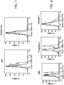
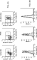

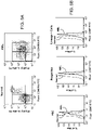


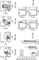


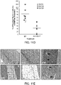
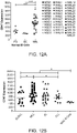
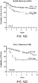
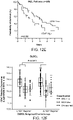


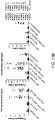

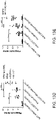
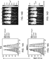


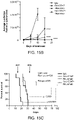


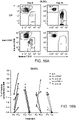

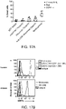
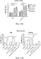


Classifications
-
- C—CHEMISTRY; METALLURGY
- C07—ORGANIC CHEMISTRY
- C07K—PEPTIDES
- C07K16/00—Immunoglobulins [IGs], e.g. monoclonal or polyclonal antibodies
- C07K16/18—Immunoglobulins [IGs], e.g. monoclonal or polyclonal antibodies against material from animals or humans
- C07K16/28—Immunoglobulins [IGs], e.g. monoclonal or polyclonal antibodies against material from animals or humans against receptors, cell surface antigens or cell surface determinants
- C07K16/2803—Immunoglobulins [IGs], e.g. monoclonal or polyclonal antibodies against material from animals or humans against receptors, cell surface antigens or cell surface determinants against the immunoglobulin superfamily
-
- A—HUMAN NECESSITIES
- A61—MEDICAL OR VETERINARY SCIENCE; HYGIENE
- A61K—PREPARATIONS FOR MEDICAL, DENTAL OR TOILETRY PURPOSES
- A61K39/00—Medicinal preparations containing antigens or antibodies
- A61K39/395—Antibodies; Immunoglobulins; Immune serum, e.g. antilymphocytic serum
- A61K39/39533—Antibodies; Immunoglobulins; Immune serum, e.g. antilymphocytic serum against materials from animals
- A61K39/39558—Antibodies; Immunoglobulins; Immune serum, e.g. antilymphocytic serum against materials from animals against tumor tissues, cells, antigens
-
- A—HUMAN NECESSITIES
- A61—MEDICAL OR VETERINARY SCIENCE; HYGIENE
- A61K—PREPARATIONS FOR MEDICAL, DENTAL OR TOILETRY PURPOSES
- A61K51/00—Preparations containing radioactive substances for use in therapy or testing in vivo
- A61K51/02—Preparations containing radioactive substances for use in therapy or testing in vivo characterised by the carrier, i.e. characterised by the agent or material covalently linked or complexing the radioactive nucleus
- A61K51/04—Organic compounds
- A61K51/08—Peptides, e.g. proteins, carriers being peptides, polyamino acids, proteins
- A61K51/10—Antibodies or immunoglobulins; Fragments thereof, the carrier being an antibody, an immunoglobulin or a fragment thereof, e.g. a camelised human single domain antibody or the Fc fragment of an antibody
- A61K51/1093—Antibodies or immunoglobulins; Fragments thereof, the carrier being an antibody, an immunoglobulin or a fragment thereof, e.g. a camelised human single domain antibody or the Fc fragment of an antibody conjugates with carriers being antibodies
-
- A—HUMAN NECESSITIES
- A61—MEDICAL OR VETERINARY SCIENCE; HYGIENE
- A61P—SPECIFIC THERAPEUTIC ACTIVITY OF CHEMICAL COMPOUNDS OR MEDICINAL PREPARATIONS
- A61P35/00—Antineoplastic agents
-
- A—HUMAN NECESSITIES
- A61—MEDICAL OR VETERINARY SCIENCE; HYGIENE
- A61P—SPECIFIC THERAPEUTIC ACTIVITY OF CHEMICAL COMPOUNDS OR MEDICINAL PREPARATIONS
- A61P35/00—Antineoplastic agents
- A61P35/02—Antineoplastic agents specific for leukemia
-
- C—CHEMISTRY; METALLURGY
- C07—ORGANIC CHEMISTRY
- C07K—PEPTIDES
- C07K16/00—Immunoglobulins [IGs], e.g. monoclonal or polyclonal antibodies
- C07K16/18—Immunoglobulins [IGs], e.g. monoclonal or polyclonal antibodies against material from animals or humans
- C07K16/28—Immunoglobulins [IGs], e.g. monoclonal or polyclonal antibodies against material from animals or humans against receptors, cell surface antigens or cell surface determinants
- C07K16/2887—Immunoglobulins [IGs], e.g. monoclonal or polyclonal antibodies against material from animals or humans against receptors, cell surface antigens or cell surface determinants against CD20
-
- A—HUMAN NECESSITIES
- A61—MEDICAL OR VETERINARY SCIENCE; HYGIENE
- A61K—PREPARATIONS FOR MEDICAL, DENTAL OR TOILETRY PURPOSES
- A61K39/00—Medicinal preparations containing antigens or antibodies
- A61K2039/505—Medicinal preparations containing antigens or antibodies comprising antibodies
- A61K2039/507—Comprising a combination of two or more separate antibodies
-
- C—CHEMISTRY; METALLURGY
- C07—ORGANIC CHEMISTRY
- C07K—PEPTIDES
- C07K2317/00—Immunoglobulins specific features
- C07K2317/70—Immunoglobulins specific features characterized by effect upon binding to a cell or to an antigen
- C07K2317/73—Inducing cell death, e.g. apoptosis, necrosis or inhibition of cell proliferation
- C07K2317/732—Antibody-dependent cellular cytotoxicity [ADCC]
-
- C—CHEMISTRY; METALLURGY
- C07—ORGANIC CHEMISTRY
- C07K—PEPTIDES
- C07K2317/00—Immunoglobulins specific features
- C07K2317/70—Immunoglobulins specific features characterized by effect upon binding to a cell or to an antigen
- C07K2317/73—Inducing cell death, e.g. apoptosis, necrosis or inhibition of cell proliferation
- C07K2317/734—Complement-dependent cytotoxicity [CDC]
-
- C—CHEMISTRY; METALLURGY
- C07—ORGANIC CHEMISTRY
- C07K—PEPTIDES
- C07K2317/00—Immunoglobulins specific features
- C07K2317/70—Immunoglobulins specific features characterized by effect upon binding to a cell or to an antigen
- C07K2317/77—Internalization into the cell
Definitions
- Therapeutic antibodies have proven effective at fighting cancer, especially in cases where conventional therapy fails: Out of the 21 marketed therapeutic antibodies, 9 are for the treatment of cancer. Even more encouraging is that antibodies for cancer generally operate in a distinct mechanism from traditional chemotherapy or radiotherapy, so they can often be combined with traditional therapies to generate a synergistic effect. However, there is still a lot of room for improvement, in that even the most effective cancer antibodies rarely cure the disease, especially in its more advanced stages.
- the first patient treated in the United States with monoclonal antibody therapy was a patient with non-Hodgkin's lymphoma, using a murine monoclonal antibody designated AB 89.
- treatment was not successful in inducing a significant clinical response, it did represent the first proof of principle in humans that a monoclonal antibody could induce transient decreases in the number of circulating tumor cells, induce circulating dead cells, and form complexes with circulating antigen, all with minimal toxicity to the patient.
- a monoclonal antibody could induce transient decreases in the number of circulating tumor cells, induce circulating dead cells, and form complexes with circulating antigen, all with minimal toxicity to the patient.
- there was much excitement about this new treatment modality there were also problems and limitations, including the modulation of antigens off the cell surface and into circulation or internalization.
- Monoclonal antibodies achieve their therapeutic effect through various mechanisms. They can have direct effects in producing apoptosis or programmed cell death. They can block growth factor receptors, effectively arresting proliferation of tumor cells. In cells that express monoclonal antibodies, they can bring about anti-idiotype antibody formation. Indirect effects include recruiting cells that have cytotoxicity, such as monocytes and macrophages. This type of antibody-mediated cell kill is called antibody-dependent cell mediated cytotoxicity (ADCC). Monoclonal antibodies also bind complement, leading to direct cell toxicity, known as complement dependent cytotoxicity (CDC).
- ADCC antibody-dependent cell mediated cytotoxicity
- Rituximab became the first monoclonal antibody approved specifically for cancer therapy, and a number of other naked or immunoconjugated antibodies have followed, including a humanized monoclonal antibody to CD33 conjugated to calicheamicin, and radiolabeled antibodies specific for CD20 (ytrrium-90 ibritumomab tiuxetan and iodine-131 tositumomab).
- Rituximab has become the largest-selling biologic drug in clinical oncology, and is active in a variety of human lymphomas and chronic lymphocytic leukemia. This is a chimeric monoclonal antibody targeting the CD20 antigen found on both normal B cells and on most low-grade and some higher grade B-cell lymphomas. It can be effective as a single agent in induction and maintenance therapy. It is primarily used, however, in combination with standard chemotherapies in the treatment of patients with non-Hodgkin's B-cell lymphomas and chronic lymphocytic leukemia.
- Alemtuzumab is a humanized monoclonal antibody targeting the CD52 antigen found on B lymphocytes and is used primarily for chronic lymphocytic leukemia, and is effective as induction and maintenance therapy. Alemtuzumab is also reactive with T lymphocytes, and is typically not combined with chemotherapy because of the increased risk of infection.
- the CD22 antigen is also targeted by a number of antibodies, and has recently demonstrated efficacy combined with toxin in chemotherapy-resistant hairy cell leukemia.
- WO 2009/091547 describes markers of acute myeloid leukemia stem cells.
- WO 2009/091601 describes methods for manipulating phagocytosis mediated by CD47.
- Chao et al. (2009) Blood 114(22):1063-1064 reports that therapeutic antibody targeting of CD47 synergizes with rituximab to completely eradicate human B-cell lymphoma xenografts.
- US 2009/191202 describes methods for manipulating phagocytosis mediated by CD47.
- the invention provides a synergistic combination of agents for use in the treatment of a hematologic cancer in a patient, wherein the hematologic cancer is non-Hodgkin's lymphoma (NHL) and wherein the NHL is a diffuse large B cell lymphoma, wherein the combination of agents comprises a first agent that selectively blocks CD47 and a second agent that binds to CD20, wherein the first and second agents are comprised in a bispecific FcR-engaging antibody selective for CD47 and for CD20.
- NDL non-Hodgkin's lymphoma
- NHL a diffuse large B cell lymphoma
- the combination of agents comprises a first agent that selectively blocks CD47 and a second agent that binds to CD20, wherein the first and second agents are comprised in a bispecific FcR-engaging antibody selective for CD47 and for CD20.
- One of the agents targets the tumor cells for phagocytosis by blocking CD47 on the cell surface. It is shown that leukemia or lymphoma cells evade macrophage surveillance by upregulation of CD47 expression. Administration of agents that mask the CD47 protein to a patient increases the clearance of cancer cells via phagocytosis.
- Compositions of such bispecific antibodies are also provided, where the bispecific antibody is desirably human or humanized; and may be modified to extend the blood half-life, e.g. by pegylation, etc.
- Synergistic combination may provide for a therapeutic effect that is comparable to the effectiveness of a monotherapy, while reducing adverse side effects, e.g. damage to non-targeted tissues, immune status, and other clinical indicia.
- synergistic combinations may provide for an improved effectiveness, which effect may be measured by total tumor cell number; length of time to relapse; and other indicia of patient health.
- CD47 polypeptides The three transcript variants of human CD 47 (variant 1, NM 001777; variant 2, NM 198793; and variant 3, NM 001025079) encode three isoforms of CD47 polypeptide.
- CD47 isoform 1 (NP 001768), the longest of the three isoforms, is 323 amino acids long.
- CD47 isoform 2 (NP 942088) is 305 amino acid long.
- CD47 isoform 3 is 312 amino acids long.
- the three isoforms are identical in sequence in the first 303 amino acids.
- Amino acids 1-8 comprise the signal sequence
- amino acids 9-142 comprise the CD47 immunoglobulin like domain, which is the soluble fragment
- amino acids 143-300 is the transmembrane domain.
- a “fusion” polypeptide is a polypeptide comprising a polypeptide or portion (e.g., one or more domains) thereof fused or bonded to heterologous polypeptide.
- a fusion soluble CD47 protein for example, will share at least one biological property in common with a native sequence soluble CD47 polypeptide.
- Examples of fusion polypeptides include immunoadhesins, as described above, which combine a portion of the CD47 polypeptide with an immunoglobulin sequence, and epitope tagged polypeptides, which comprise a soluble CD47 polypeptide or portion thereof fused to a "tag polypeptide".
- the tag polypeptide has enough residues to provide an epitope against which an antibody can be made, yet is short enough such that it does not interfere with biological activity of the CD47 polypeptide.
- Suitable tag polypeptides generally have at least six amino acid residues and usually between about 6-60 amino acid residues.
- a “functional derivative” of a native sequence polypeptide is a compound having a qualitative biological property in common with a native sequence polypeptide.
- “Functional derivatives” include, but are not limited to, fragments of a native sequence and derivatives of a native sequence polypeptide and its fragments, provided that they have a biological activity in common with a corresponding native sequence polypeptide.
- the term “derivative” encompasses both amino acid sequence variants of polypeptide and covalent modifications thereof. Derivatives and fusion of soluble CD47 find use as CD47 mimetic molecules.
- the first 142 amino acids of CD47 polypeptide comprise the extracellular region of CD47 (SEQ ID NO: 1).
- the three isoforms have identical amino acid sequence in the extracellular region, and thus any of the isoforms are can be used to generate soluble CD47.
- “Soluble CD47” is a CD47 protein that lacks the transmembrane domain. Soluble CD47 is secreted out of the cell expressing it instead of being localized at the cell surface.
- In vitro assays for CD47 biological activity include, e.g. inhibition of phagocytosis of porcine cells by human macrophages, binding to SIRP ⁇ receptor, SIRP ⁇ tyrosine phosphorylation, etc.
- An exemplary assay for CD47 biological activity contacts a human macrophage composition in the presence of a candidate agent. The cells are incubated with the candidate agent for about 30 minutes and lysed. The cell lysate is mixed with anti-human SIRP ⁇ antibodies to immunoprecipitate SIRP ⁇ . Precipitated proteins are resolved by SDS PAGE, then transferred to nitrocellulose and probed with antibodies specific for phosphotyrosine.
- a candidate agent useful as a CD47 mimetic increases SIRP a tyrosine phosphorylation by at least 10%, or up to 20%, or 50%, or 70% or 80% or up to about 90% compared to the level of phosphorylation observed in the absence of candidate agent.
- Another exemplary assay for CD47 biological activity measures phagocytosis of hematopoietic cells by human macrophages.
- a candidate agent useful as a CD47 mimetic results in the down regulation of phagocytosis by at least about 10%, at least about 20%, at least about 50%, at least about 70%, at least about 80%, or up to about 90% compared to level of phagocytosis observed in absence of candidate agent.
- manipulating phagocytosis is meant an up-regulation or a down-regulation in phagocytosis by at least about 10%, or up to 20%, or 50%, or 70% or 80% or up to about 90% compared to level of phagocytosis observed in absence of intervention.
- manipulating phagocytosis means a down-regulation in phagocytosis by at least about 10%, or up to 20%, or 50%, or 70% or 80% or up to about 90% compared to level of phagocytosis observed in absence of intervention.
- CD47 inhibitors Agents of interest as CD47 inhibitors include specific binding members that prevent the binding of CD47 with SIRP ⁇ receptor.
- the term "specific binding member” or “binding member” as used herein refers to a member of a specific binding pair, i.e. two molecules, usually two different molecules, where one of the molecules (i.e., first specific binding member) through chemical or physical means specifically binds to the other molecule (i.e., second specific binding member).
- CD47 inhibitors include analogs, derivatives and fragments of the original specific binding member.
- a specific binding member may be an antibody.
- antibody or “antibody moiety” is intended to include any polypeptide chain-containing molecular structure with a specific shape that fits to and recognizes an epitope, where one or more non-covalent binding interactions stabilize the complex between the molecular structure and the epitope.
- Antibodies utilized in the present invention may be polyclonal antibodies, although monoclonal antibodies are preferred because they may be reproduced by cell culture or recombinantly, and can be modified to reduce their antigenicity.
- Polyclonal antibodies can be raised by a standard protocol by injecting a production animal with an antigenic composition. See, e.g., Harlow and Lane, Antibodies: A Laboratory Manual, Cold Spring Harbor Laboratory, 1988 .
- antibodies may be raised by immunizing the production animal with the protein and a suitable adjuvant (e.g., Freund's, Freund's complete, oil-in-water emulsions, etc.)
- a suitable adjuvant e.g., Freund's, Freund's complete, oil-in-water emulsions, etc.
- conjugate proteins that are commercially available for such use include bovine serum albumin (BSA) and keyhole limpet hemocyanin (KLH).
- BSA bovine serum albumin
- KLH keyhole limpet hemocyanin
- peptides derived from the full sequence may be utilized.
- a superior immune response may be elicited if the polypeptide is joined to a carrier protein, such as ovalbumin, BSA or KLH.
- hybridomas may be formed by isolating the stimulated immune cells, such as those from the spleen of the inoculated animal.
- immortalized cells such as myeloma cells or transformed cells, which are capable of replicating indefinitely in cell culture, thereby producing an immortal, immunoglobulin-secreting cell line.
- immortalized cells such as myeloma cells or transformed cells, which are capable of replicating indefinitely in cell culture, thereby producing an immortal, immunoglobulin-secreting cell line.
- the antibodies or antigen binding fragments may be produced by genetic engineering. Humanized, chimeric, or xenogeneic human antibodies, which produce less of an immune response when administered to humans, are preferred for use in the present invention.
- immunoglobulin fragments comprising the epitope binding site (e.g., Fab', F(ab') 2 , or other fragments) are useful as antibody moieties in the present invention.
- Such antibody fragments may be generated from whole immunoglobulins by ricin, pepsin, papain, or other protease cleavage.
- "Fragment,” or minimal immunoglobulins may be designed utilizing recombinant immunoglobulin techniques.
- Fv immunoglobulins for use in the present invention may be produced by linking a variable light chain region to a variable heavy chain region via a peptide linker (e.g., poly-glycine or another sequence which does not form an alpha helix or beta sheet motif).
- a peptide linker e.g., poly-glycine or another sequence which does not form an alpha helix or beta sheet motif.
- Antibodies include free antibodies and antigen binding fragments derived therefrom, and conjugates, e.g. pegylated antibodies, drug, radioisotope, or toxin conjugates, and the like. Monoclonal antibodies directed against a specific epitope, or combination of epitopes, will allow for the targeting and/or depletion of cellular populations expressing the marker.
- Various techniques can be utilized using monoclonal antibodies to screen for cellular populations expressing the marker(s), and include magnetic separation using antibody-coated magnetic beads, "panning" with antibody attached to a solid matrix (i.e., plate), and flow cytometry (See, e.g., U.S. Pat. No. 5,985,660 ; and Morrison et al. Cell, 96:737-49 (1999 )). These techniques allow for the screening of particular populations of cells; in immunohistochemistry of biopsy samples; in detecting the presence of markers shed by cancer cells into the blood and other biologic fluids, and the like.
- Humanized versions of such antibodies are also within the scope of this invention. Humanized antibodies are especially useful for in vivo applications in humans due to their low antigenicity.
- multispecific or bispecific antibody refers to a synthetic or recombinant antibody that recognizes more than one protein.
- Examples include bispecific antibodies 2B1, 520C9xH22, mDX-H210, and MDX447.
- Bispecific antibodies directed against a combination of epitopes will allow for the targeting and/or depletion of cellular populations expressing the combination of epitopes.
- Exemplary bi-specific antibodies include those targeting a combination of CD47 and a cancer cell marker, such as, CD20, CD22, CD96, CD97, CD99, PTHR2, HAVCR2 etc.
- Generation of bi-specific antibodies are described in the literature, for example, in USPN 5989830 , USPN 5798229 . Higher order specificities, e.g. trispecific antibodies, are described by Holliger and Hudson (2005) Nature Biotechnology 23:1126-1136 .
- the efficacy of a CD47 inhibitor is assessed by assaying CD47 activity.
- the above-mentioned assays or modified versions thereof are used.
- AML SCs are incubated with bone marrow derived macrophages, in the presence or absence of the candidate agent.
- An inhibitor of the cell surface CD47 will up-regulate phagocytosis by at least about 10%, or up to 20%, or 50%, or 70% or 80% or up to about 90% compared to the phagocytosis in absence of the candidate agent.
- an in vitro assay for levels of tyrosine phosphorylation of SIRP ⁇ will show a decrease in phosphorylation by at least about 10%, or up to 20%, or 50%, or 70% or 80% or up to about 90% compared to phosphorylation observed in absence of the candidate agent.
- the agent, or a pharmaceutical composition comprising the agent may be provided in an amount effective to detectably inhibit the binding of CD47 to SIRP ⁇ receptor present on the surface of phagocytic cells.
- the effective amount is determined via empirical testing routine in the art. The effective amount may vary depending on the number of cells being transplanted, site of transplantation and factors specific to the transplant recipient.
- phagocytic cells and “phagocytes” are used interchangeably herein to refer to a cell that is capable of phagocytosis.
- phagocytes There are four main categories of phagocytes: macrophages, mononuclear cells (histiocytes and monocytes); polymorphonuclear leukocytes (neutrophils) and dendritic cells.
- biological sample encompasses a variety of sample types obtained from an organism and can be used in a diagnostic or monitoring assay.
- the term encompasses blood and other liquid samples of biological origin, solid tissue samples, such as a biopsy specimen or tissue cultures or cells derived therefrom and the progeny thereof.
- the term encompasses samples that have been manipulated in any way after their procurement, such as by treatment with reagents, solubilization, or enrichment for certain components.
- the term encompasses a clinical sample, and also includes cells in cell culture, cell supernatants, cell lysates, serum, plasma, biological fluids, and tissue samples.
- treatment means treating, “treating”, “treat” and the like to generally refer to obtaining a desired pharmacologic and/or physiologic effect.
- the effect may be prophylactic in terms of completely or partially preventing a disease or symptom thereof and/or may be therapeutic in terms of a partial or complete stabilization or cure for a disease and/or adverse effect attributable to the disease.
- Treatment covers any treatment of a disease in a mammal, e.g.
- mice, rat, rabbit, pig, primate including humans and other apes, particularly a human, and includes: (a) preventing the disease or symptom from occurring in a subject which may be predisposed to the disease or symptom but has not yet been diagnosed as having it; (b) inhibiting the disease symptom, i.e., arresting its development; or (c) relieving the disease symptom, i.e., causing regression of the disease or symptom.
- a "host cell”, as used herein, refers to a microorganism or a eukaryotic cell or cell line cultured as a unicellular entity which can be, or has been, used as a recipient for a recombinant vector or other transfer polynucleotides, and include the progeny of the original cell which has been transfected. It is understood that the progeny of a single cell may not necessarily be completely identical in morphology or in genomic or total DNA complement as the original parent, due to natural, accidental, or deliberate mutation.
- cancer neoplasm
- tumor tumor
- tumor tumor
- tumor tumor
- tumor tumor
- tumor tumor
- cells of interest for detection or treatment in the present application include precancerous (e.g., benign), malignant, pre-metastatic, metastatic, and non-metastatic cells. Detection of cancerous cells is of particular interest.
- normal as used in the context of "normal cell,” is meant to refer to a cell of an untransformed phenotype or exhibiting a morphology of a non-transformed cell of the tissue type being examined.
- Cancerous phenotype generally refers to any of a variety of biological phenomena that are characteristic of a cancerous cell, which phenomena can vary with the type of cancer.
- the cancerous phenotype is generally identified by abnormalities in, for example, cell growth or proliferation (e.g., uncontrolled growth or proliferation), regulation of the cell cycle, cell mobility, cell-cell interaction, or metastasis, etc.
- “Therapeutic target” refers to a gene or gene product that, upon modulation of its activity (e.g., by modulation of expression, biological activity, and the like), can provide for modulation of the cancerous phenotype.
- modulation is meant to refer to an increase or a decrease in the indicated phenomenon (e.g., modulation of a biological activity refers to an increase in a biological activity or a decrease in a biological activity).
- Acute leukemias are rapidly progressing leukemia characterized by replacement of normal bone marrow by blast cells of a clone arising from malignant transformation of a hematopoietic cell.
- the acute leukemias include acute lymphoblastic leukemia (ALL) and acute myelogenous leukemia (AML). ALL often involves the CNS, whereas acute monoblastic leukemia involves the gums, and AML involves localized collections in any site (granulocytic sarcomas or chloromas).
- the presenting symptoms are usually nonspecific (e.g., fatigue, fever, malaise, weight loss) and reflect the failure of normal hematopoiesis.
- Anemia and thrombocytopenia are very common (75 to 90%).
- the WBC count may be decreased, normal, or increased.
- Blast cells are usually found in the blood smear unless the WBC count is markedly decreased.
- the blasts of ALL can be distinguished from those of AML by histochemical studies, cytogenetics, immunophenotyping, and molecular biology studies. In addition to smears with the usual stains, terminal transferase, myeloperoxidase, Sudan black B, and specific and nonspecific esterase.
- ALL is the most common malignancy in children, with a peak incidence from ages 3 to 5 yr. It also occurs in adolescents and has a second, lower peak in adults.
- Typical treatment emphasizes early introduction of an intensive multidrug regimen, which may include prednisone, vincristine, anthracycline or asparaginase.
- Other drugs and combinations are cytarabine and etoposide, and cyclophosphamide.
- Relapse usually occurs in the bone marrow but may also occur in the CNS or testes, alone or concurrent with bone marrow. Although second remissions can be induced in many children, subsequent remissions tend to be brief.
- AML AML may be associated with chemotherapy or irradiation (secondary AML).
- Remission induction rates are lower than with ALL, and long-term disease-free survival reportedly occurs in only 20 to 40% of patients.
- Treatment differs most from ALL in that AML responds to fewer drugs.
- the basic induction regimen includes cytarabine; along with daunorubicin or idarubicin. Some regimens include 6-thioguanine, etoposide, vincristine, and prednisone.
- PV Polycythemia vera
- Hb concentration and RBC mass erythrocytosis
- PV occurs in about 2.3/100,000 people per year; more often in males (about 1.4:1).
- the mean age at diagnosis is 60 yr (range, 15 to 90 yr; rarely in childhood); 5% of patients are ⁇ 40 yr at onset.
- the bone marrow sometimes appears normal but usually is hypercellular; hyperplasia involves all marrow elements and replaces marrow fat.
- RBCs, neutrophils, and platelets Increased megakaryocytes may be present in clumps.
- Marrow iron is absent in > 90% of patients, even when phlebotomy has not been performed.
- Myelodysplastic syndrome is a group of syndromes (preleukemia, refractory anemias, Ph-negative chronic myelocytic leukemia, chronic myelomonocytic leukemia, myeloid metaplasia) commonly seen in older patients. Exposure to carcinogens may by be implicated. MDS is characterized by clonal proliferation of hematopoietic cells, including erythroid, myeloid, and megakaryocytic forms. The bone marrow is normal or hypercellular, and ineffective hematopoiesis causes variable cytopenias, the most frequent being anemia. The disordered cell production is also associated with morphologic cellular abnormalities in marrow and blood.
- Extramedullary hematopoiesis may occur, leading to hepatomegaly and splenomegaly.
- Myelofibrosis is occasionally present at diagnosis or may develop during the course of MDS.
- the MDS clone is unstable and tends to progress to AML.
- Non-Hodgkin lymphomas are a heterogeneous group of disorders involving malignant monoclonal proliferation of lymphoid cells in lymphoreticular sites, including lymph nodes, bone marrow, the spleen, the liver, and the GI tract. It is the 6th most common cancer in the US; about 65,000 new cases are diagnosed annually in all age groups. Presenting symptoms usually include peripheral lymphadenopathy. However, some patients present without adenopathy but with abnormal lymphocytes in circulation. Compared with Hodgkin lymphoma, there is a greater likelihood of disseminated disease at the time of diagnosis. Diagnosis is usually based on lymph node or bone marrow biopsy or both. Treatment involves radiation therapy, chemotherapy, or both. Stem cell transplantation is usually reserved for salvage therapy after incomplete remission or relapse.
- NHLs arise from B cells; the remainder arise from T cells or natural killer cells. Either precursor or mature cells may be involved. Overlap exists between leukemia and NHL because both involve proliferation of lymphocytes or their precursors. A leukemia-like picture with peripheral lymphocytosis and bone marrow involvement may be present in up to 50% of children and in about 20% of adults with some types of NHL.
- lymphomas within this group are: precursor B-lymphoblastic leukemia/lymphoma; B-cell chronic lymphocytic leukemia/small lymphocytic lymphoma; B-cell prolymphocytic leukemia; Lymphoplasmacytic lymphoma; Splenic marginal zone B-cell lymphoma ( ⁇ villous lymphocytes); Hairy cell leukemia; Plasma cell myeloma/plasmacytoma; Extranodal marginal zone B-cell lymphoma of the MALT type; Nodal marginal zone B-cell lymphoma ( ⁇ monocytoid B cells); Follicular lymphoma; Mantle cell lymphoma; Diffuse large B-cell lymphomas; Burkitt's lymphoma; Precursor T-lymphoblastic lymphoma/leukemia; T-cell prolymphocytic leukemia; T-cell granular lymphocytic leukemia; Aggressive NK cell leukemia;
- B cell hematologic cancers within the NHL group include, without limitation, diffuse large B cell lymphoma, chronic lymphocytic leukemia, mantle cell lymphoma, B lymphoblastic leukemia/lymphoma, and Burkitt's lymphoma. Diffuse large B cell lymphomas are of particular interest in this group.
- Lymphomas are commonly also categorized as indolent or aggressive. Indolent lymphomas are slowly progressive and responsive to therapy but are often not curable with conventional approaches. Aggressive lymphomas are rapidly progressive but responsive to therapy and often curable.
- NHL lymphoblastic lymphoma
- GI tract involvement particularly in the terminal ileum
- meningeal spread requiring CSF prophylaxis or treatment
- other sanctuary sites of involvement such as testis or brain.
- treatment adverse effects as well as outcome must be considered, including late risks of secondary cancer.
- Rituximab is included in the standard chemotherapeutic regimen. Patients in the highest risk groups (patients with 4 or 5 risk factors) now have a 50% 5-yr survival. In patients with the aggressive B-cell lymphomas (eg, diffuse large B cell), the standard drug combination is rituximab plus cyclophosphamide, hydroxydaunorubicin, vincristine, prednisone(R-CHOP). Complete disease regression is expected in ⁇ 70% of patients, depending on the IPI category.
- Chronic leukemia usually manifests as abnormal leukocytosis with or without cytopenia in an otherwise asymptomatic person. Findings and management differ significantly between chronic lymphocytic leukemia (CLL) and chronic myelocytic leukemia (CML).
- CLL chronic lymphocytic leukemia
- CML chronic myelocytic leukemia
- CLL The most common type of leukemia in the Western world, CLL involves mature-appearing defective neoplastic lymphocytes (almost always B cells) with an abnormally long life span.
- the peripheral blood, bone marrow, spleen, and lymph nodes undergo leukemic infiltration. Symptoms may be absent or may include lymphadenopathy, splenomegaly, hepatomegaly, and nonspecific symptoms attributable to anemia (fatigue, malaise). Diagnosis is by examination of peripheral smear and bone marrow aspirate.
- Treatment delayed until symptoms develop, is aimed at lengthening life and decreasing symptoms and may involve chlorambucil or fludarabine, prednisone, and cyclophosphamide or doxorubicin or both.
- Monoclonal antibodies such as alemtuzumab and rituximab, are increasingly being used.
- Palliative radiation therapy is reserved for patients whose lymphadenopathy or splenomegaly interferes with other organs.
- CD5+ B cells undergo malignant transformation, with lymphocytes initially accumulating in the bone marrow and then spreading to lymph nodes and other lymphoid tissues, eventually inducing splenomegaly and hepatomegaly.
- abnormal hematopoiesis results in anemia, neutropenia, thrombocytopenia, and decreased immunoglobulin production.
- Many patients develop hypogammaglobulinemia and impaired antibody response, perhaps related to increased T-suppressor cell activity.
- Patients have increased susceptibility to autoimmune disease characterized by immunohemolytic anemias (usually Coombs' test-positive) or thrombocytopenia and a modest increase in risk of developing other cancers.
- Rituximab is the first monoclonal antibody used in the successful treatment of lymphoid cancers.
- the partial response rate with conventional doses in CLL is 10 to 15%.
- the response rate is 75%, with 20% of patients achieving complete remission.
- Alemtuzumab has a 33% response rate in previously treated patients refractory to fludarabine and a 75 to 80% response rate in previously untreated patients.
- Multiple myeloma is a cancer of plasma cells that produce monoclonal immunoglobulin and invade and destroy adjacent bone tissue. Common manifestations include bone pain, renal insufficiency, hypercalcemia, anemia, and recurrent infections. Diagnosis requires demonstration of M-protein (sometimes present in urine and not serum) and either lytic bone lesions, light-chain proteinuria, or excessive marrow plasma cells. A bone marrow biopsy is usually needed. Specific treatment includes conventional chemotherapy with the addition of bortezomib, lenalidomide, thalidomide, corticosteroids, and high-dose melphalan followed by autologous peripheral blood stem cell transplantation.
- the M-protein produced by the malignant plasma cells is IgG in about 55% of myeloma patients and IgA in about 20%; of patients producing either IgG or IgA, 40% also have Bence Jones proteinuria, which is free monoclonal ⁇ or ⁇ light chains in the urine. In 15 to 20% of patients, plasma cells secrete only Bence Jones protein. IgD myeloma accounts for about 1% of cases.
- the invention provides a synergistic combination of agents for use in methods for reducing growth of diffuse large B cell lymphoma cells through the introduction of a CD47 blocking agent, in combination with a second agent that binds to CD20, wherein the CD47 blocking agent and the second agent are comprised in a bispecific FcR-engaging antibody selective for CD47 and for CD20.
- a CD47 blocking agent in combination with a second agent that binds to CD20
- the CD47 blocking agent and the second agent are comprised in a bispecific FcR-engaging antibody selective for CD47 and for CD20.
- Reducing growth of cancer cells includes, but is not limited to, reducing proliferation of cancer cells, and reducing the incidence of a non-cancerous cell becoming a cancerous cell. Whether a reduction in cancer cell growth has been achieved can be readily determined using any known assay, including, but not limited to, [ 3 H]-thymidine incorporation; counting cell number over a period of time; detecting and/or measuring a marker associated with AML, etc.
- Whether a substance, or a specific amount of the substance, is effective in treating cancer can be assessed using any of a variety of known diagnostic assays for cancer, including, but not limited to biopsy, contrast radiographic studies, CAT scan, and detection of a tumor marker associated with cancer in the blood of the individual.
- the substance can be administered systemically or locally, usually systemically.
- An agent e.g. a chemotherapeutic drug that reduces cancer cell growth
- a method of delivering a drug to a cancer cell may comprise administering a drug-antibody complex to a subject, wherein the antibody is specific for a cancer-associated polypeptide, and the drug is one that reduces cancer cell growth, a variety of which are known in the art.
- Targeting can be accomplished by coupling (e.g., linking, directly or via a linker molecule, either covalently or non-covalently, so as to form a drug-antibody complex) a drug to an antibody specific for a cancer-associated polypeptide.
- Methods of coupling a drug to an antibody are well known in the art and need not be elaborated upon herein.
- antibody or “antibody moiety” is intended to include any polypeptide chain-containing molecular structure that has a specific shape which fits to and recognizes an epitope, where one or more non-covalent binding interactions stabilize the complex between the molecular structure and the epitope.
- hybridomas may be formed by isolating the stimulated immune cells, such as those from the spleen of the inoculated animal. These cells are then fused to immortalized cells, such as myeloma cells or transformed cells, which are capable of replicating indefinitely in cell culture, thereby producing an immortal, immunoglobulin-secreting cell line.
- the immortal cell line utilized is preferably selected to be deficient in enzymes necessary for the utilization of certain nutrients.
- Many such cell lines (such as myelomas) are known to those skilled in the art, and include, for example: thymidine kinase (TK) or hypoxanthine-guanine phosphoriboxyl transferase (HGPRT). These deficiencies allow selection for fused cells according to their ability to grow on, for example, hypoxanthine aminopterinthymidine medium (HAT).
- TK thymidine kinase
- HGPRT hypoxanthine-guanine phosphoriboxyl transferase
- Antibodies which have a reduced propensity to induce a violent or detrimental immune response in humans (such as anaphylactic shock), and which also exhibit a reduced propensity for priming an immune response which would prevent repeated dosage with the antibody therapeutic or imaging agent (e.g., the human-anti-murine-antibody "HAMA" response), are preferred for use in the invention. These antibodies are preferred for all administrative routes. Thus, humanized, chimeric, or xenogenic human antibodies, which produce less of an immune response when administered to humans, are preferred for use in the present invention.
- Chimeric antibodies may be made by recombinant means by combining the murine variable light and heavy chain regions (VK and VH), obtained from a murine (or other animal-derived) hybridoma clone, with the human constant light and heavy chain regions, in order to produce an antibody with predominantly human domains.
- VK and VH murine variable light and heavy chain regions
- the production of such chimeric antibodies is well known in the art, and may be achieved by standard means (as described, e.g., in U.S. Pat. No. 5,624,659 ).
- Humanized antibodies are engineered to contain even more human-like immunoglobulin domains, and incorporate only the complementarity-determining regions of the animal-derived antibody.
- polyclonal or monoclonal antibodies may be produced from animals which have been genetically altered to produce human immunoglobulins.
- the transgenic animal may be produced by initially producing a "knock-out" animal which does not produce the animal's natural antibodies, and stably transforming the animal with a human antibody locus (e.g., by the use of a human artificial chromosome). Only human antibodies are then made by the animal. Techniques for generating such animals, and deriving antibodies therefrom, are described in U.S. Pat. Nos. 6,162,963 and 6,150,584 . Such fully human xenogenic antibodies are a preferred antibody for use in the methods and compositions of the present invention.
- single chain antibodies can be produced from phage libraries containing human variable regions. See U.S. Pat. No. 6,174,708 .
- immunoglobulin fragments comprising the epitope binding site (e.g., Fab', F(ab') 2 , or other fragments) are useful as antibody moieties in the present invention.
- Such antibody fragments may be generated from whole immunoglobulins by ficin, pepsin, papain, or other protease cleavage. "Fragment,” or minimal immunoglobulins may be designed utilizing recombinant immunoglobulin techniques.
- Fv immunoglobulins for use in the present invention may be produced by linking a variable light chain region to a variable heavy chain region via a peptide linker (e.g., poly-glycine or another sequence which does not form an alpha helix or beta sheet motif).
- a peptide linker e.g., poly-glycine or another sequence which does not form an alpha helix or beta sheet motif.
- Fv fragments are heterodimers of the variable heavy chain domain (V H ) and the variable light chain domain (V L ).
- the heterodimers of heavy and light chain domains that occur in whole IgG, for example, are connected by a disulfide bond.
- Recombinant Fvs in which V H and V L are connected by a peptide linker are typically stable, see, for example, Huston et al., Proc. Natl. Acad, Sci. USA 85:5879 5883 (1988 ) and Bird et al., Science 242:423 426 (1988 ).
- These are single chain Fvs which have been found to retain specificity and affinity and have been shown to be useful for imaging tumors and to make recombinant immunotoxins for tumor therapy.
- derivatized immunoglobulins with added chemical linkers detectable moieties such as fluorescent dyes, enzymes, substrates, chemiluminescent moieties, specific binding moieties such as streptavidin, avidin, or biotin, or drug conjugates may be utilized in the methods and compositions of the present invention.
- the antibodies are coupled or conjugated to one or more therapeutic cytotoxic or imaging moieties.
- cytotoxic moiety simply means a moiety which inhibits cell growth or promotes cell death when proximate to or absorbed by the cell.
- Suitable cytotoxic moieties include radioactive isotopes (radionuclides), chemotoxic agents such as differentiation inducers and small chemotoxic drugs, toxin proteins, and derivatives thereof.
- Agents may be conjugated to the antibody by any suitable technique, with appropriate consideration of the need for pharmokinetic stability and reduced overall toxicity to the patient.
- a therapeutic agent may be coupled to a suitable antibody moiety either directly or indirectly (e.g. via a linker group).
- a direct reaction between an agent and an antibody is possible when each possesses a functional group capable of reacting with the other.
- a nucleophilic group such as an amino or sulfhydryl group
- a carbonyl-containing group such as an anhydride or an acid halide, or with an alkyl group containing a good leaving group (e.g., a halide).
- a suitable chemical linker group may be used.
- a linker group can function as a spacer to distance an antibody from an agent in order to avoid interference with binding capabilities.
- a linker group can also serve to increase the chemical reactivity of a substituent on a moiety or an antibody, and thus increase the coupling efficiency. An increase in chemical reactivity may also facilitate the use of moieties, or functional groups on moieties, which otherwise would not be possible.
- Suitable linkage chemistries include maleimidyl linkers and alkyl halide linkers (which react with a sulfhydryl on the antibody moiety) and succinimidyl linkers (which react with a primary amine on the antibody moiety).
- Several primary amine and sulfhydryl groups are present on immunoglobulins, and additional groups may be designed into recombinant immunoglobulin molecules. It will be evident to those skilled in the art that a variety of bifunctional or polyfunctional reagents, both homo- and hetero-functional (such as those described in the catalog of the Pierce Chemical Co., Rockford, III.), may be employed as a linker group.
- Coupling may be effected, for example, through amino groups, carboxyl groups, sulfhydryl groups or oxidized carbohydrate residues.
- cytotoxic moieties may be coupled to the antibody through a an oxidized carbohydrate group at a glycosylation site, as described in U.S. Pat. Nos. 5,057,313 and 5,156,840 .
- Yet another alternative method of coupling the antibody moiety to the cytotoxic moiety is by the use of a non-covalent binding pair, such as streptavidin/biotin, or avidin/biotin. In these embodiments, one member of the pair is covalently coupled to the antibody moiety and the other member of the binding pair is covalently coupled to the cytotoxic or imaging moiety.
- a cytotoxic moiety is more potent when free from the antibody portion of the immunoconjugates of the present invention
- a number of different cleavable linker groups have been described.
- the mechanisms for the intracellular release of a cytotoxic moiety agent from these linker groups include cleavage by reduction of a disulfide bond (e.g., U.S. Pat. No. 4,489,710 ), by irradiation of a photolabile bond (e.g., U.S. Pat. No.
- immunoconjugates with more than one moiety may be prepared in a variety of ways. For example, more than one moiety may be coupled directly to an antibody molecule, or linkers which provide multiple sites for attachment (e.g., dendrimers) can be used. Alternatively, a carrier with the capacity to hold more than one cytotoxic or imaging moiety can be used.
- a carrier may bear the agents in a variety of ways, including covalent bonding either directly or via a linker group, and non-covalent associations.
- Suitable covalent-bond carriers include proteins such as albumins (e.g., U.S. Pat. No. 4,507,234 ), peptides, and polysaccharides such as aminodextran (e.g., U.S. Pat. No. 4,699,784 ), each of which have multiple sites for the attachment of moieties.
- a carrier may also bear an agent by non-covalent associations, such as non-covalent bonding or by encapsulation, such as within a liposome vesicle (e.g., U.S. Pat. Nos.
- Encapsulation carriers are especially useful for imaging moiety conjugation to antibody moieties for use in the invention, as a sufficient amount of the imaging moiety (dye, magnetic resonance contrast reagent, etc.) for detection may be more easily associated with the antibody moiety.
- encapsulation carriers are also useful in chemotoxic therapeutic embodiments, as they can allow the therapeutic compositions to gradually release a chemotoxic moiety over time while concentrating it in the vicinity of the tumor cells.
- radionuclides for use as cytotoxic moieties are radionuclides which are suitable for pharmacological administration.
- radionuclides include 123 I, 125 I, 131 I, 90 Y, 211 At, 67 Cu, 186 Re, 188 Re, 212 Pb, and 212 Bi.
- Iodine and astatine isotopes are more preferred radionuclides for use in the therapeutic compositions of the present invention, as a large body of literature has been accumulated regarding their use.
- Preferred chemotoxic agents include small-molecule drugs such as carboplatin, cisplatin, vincristine, taxanes such as paclitaxel and doceltaxel, hydroxyurea, gemcitabine, vinorelbine, irinotecan, tirapazamine, matrilysin, methotrexate, pyrimidine and purine analogs, and other suitable small toxins known in the art.
- Preferred chemotoxin differentiation inducers include phorbol esters and butyric acid. Chemotoxic moieties may be directly conjugated to the antibody moiety via a chemical linker, or may be encapsulated in a carrier, which is in turn coupled to the antibody.
- Preferred toxin proteins for use as cytotoxic moieties include ricins A and B, abrin, diphtheria toxin, bryodin 1 and 2, momordin, trichokirin, cholera toxin, gelonin, Pseudomonas exotoxin, Shigella toxin, pokeweed antiviral protein, and other toxin proteins known in the medicinal biochemistry arts.
- ricins A and B abrin, diphtheria toxin, bryodin 1 and 2, momordin, trichokirin, cholera toxin, gelonin, Pseudomonas exotoxin, Shigella toxin, pokeweed antiviral protein, and other toxin proteins known in the medicinal biochemistry arts.
- these toxin agents may elicit undesirable immune responses in the patient, especially if injected intravascularly, it is preferred that they be encapsulated in a carrier for coupling to the antibody.
- the agent or agents are mixed, prior to administration, with a non-toxic, pharmaceutically acceptable carrier substance.
- a non-toxic, pharmaceutically acceptable carrier substance usually, this will be an aqueous solution, such as normal saline or phosphate-buffered saline (PBS), Ringer's solution, lactate-Ringer's solution, or any isotonic physiologically acceptable solution for administration by the chosen means.
- PBS normal saline or phosphate-buffered saline
- Ringer's solution such as normal saline or phosphate-buffered saline (PBS), Ringer's solution, lactate-Ringer's solution, or any isotonic physiologically acceptable solution for administration by the chosen means.
- PBS normal saline or phosphate-buffered saline
- Ringer's solution such as normal saline or phosphate-buffered saline (PBS), Ringer's solution, lactate-Ringer's solution, or
- the agents may be stabilized against aggregation and polymerization with amino acids and non-ionic detergents, polysorbate, and polyethylene glycol.
- additional stabilizers may include various physiologically-acceptable carbohydrates and salts.
- polyvinylpyrrolidone may be added in addition to the amino acid.
- Suitable therapeutic immunoglobulin solutions which are stabilized for storage and administration to humans are described in U.S. Pat. No. 5,945,098 .
- Other agents such as human serum albumin (HSA), may be added to the therapeutic or imaging composition to stabilize the antibody conjugates.
- HSA human serum albumin
- compositions of the invention may be administered using any medically appropriate procedure, e.g., intravascular (intravenous, intraarterial, intracapillary) administration, injection into the lymph nodes, etc.
- Intravascular injection may be by intravenous or intraarterial injection.
- the effective amount of the therapeutic compositions to be given to a particular patient will depend on a variety of factors, several of which will be different from patient to patient.
- a competent clinician will be able to determine an effective amount of a therapeutic composition to administer to a patient to retard the growth and promote the death of tumor cells. Dosage of the agents will depend on the treatment of the tumor, route of administration, the nature of the therapeutics, sensitivity of the tumor to the therapeutics, etc.
- a clinician can determine the maximum safe dose for an individual, depending on the route of administration. For instance, an intravenously administered dose may be more than an locally administered dose, given the greater body of fluid into which the therapeutic composition is being administered. Similarly, compositions which are rapidly cleared from the body may be administered at higher doses, or in repeated doses, in order to maintain a therapeutic concentration. Utilizing ordinary skill, the competent clinician will be able to optimize the dosage of a particular therapeutic or imaging composition in the course of routine clinical trials.
- the dosage will be 0.001 to 100 milligrams of antibody per kilogram subject body weight.
- the ratio of anti-CD47 to the second agent may range from 1:100; 1:50; 1:10; 1:5; 1:2; 1:1; 2:1; 5:1; 10:1; 50:1; 100:1.
- the agents can be administered to the subject in a series of more than one administration.
- regular periodic administration e.g., every 2-3 days
- antibody moieties which do not provoke HAMA or other immune responses are preferred.
- depletion of cancer stem cells is useful in the treatment of cancer.
- Depletion can be achieved by several methods. Depletion is defined as a reduction in the target population by up to about 30%, or up to about 40%, or up to about 50%, or up to about 75% or more.
- An effective depletion is usually determined by the sensitivity of the particular disease condition to the levels of the target population. Thus in the treatment of certain conditions a depletion of even about 20% could be beneficial.
- a CD47 specific agent that specifically depletes the targeted cancer stem cells is used to contact the patient blood in vitro or in vivo, wherein after the contacting step, there is a reduction in the number of viable cancer stem cells in the targeted population.
- An effective dose of antibodies for such a purpose is sufficient to decrease the targeted population to the desired level, for example as described above.
- Antibodies for such purposes may have low antigenicity in humans or may be humanized antibodies.
- CD47 IS A MARKER OF MYELOID LEUKEMIAS
- Cytospins of double sorted myeloid progenitor populations (CMP, GMP), IL-3R ⁇ high CD45 RA+ cells and CD14+c-kit+lin- cells were performed using a Shandon cytospin apparatus. Cytospins were stained with Giemsa diluted 1/5 with H20 for 10 min followed by staining with May-Grunwald for 20 minutes. Cytospins were analyzed with the aid of a Zeiss microscope.
- CMML bone marrow samples were obtained with informed consent, from previously untreated patients, at Stanford University Medical Center.
- myeloid progenitors were stained with lineage marker specific phycoerythrin (PE)-Cy5-conjugated antibodies including CD2 RPA-2.10; CD11b, ICRF44; CD20, 2H7; CD56, B159; GPA, GA-R2 (Becton Dickinson - PharMingen, San Diego), CD3,S4.1;CD4, S3.5; CD7, CD7-6B7; CD8, 3B5; CD10, 5-1B4, CD14, TUK4; CD19, SJ25-C1 (Caltag, South San Francisco, CA) and APC-conjugated anti-CD34, HPCA-2 (Becton Dickinson-PharMingen), biotinylated anti-CD38, HIT2 (Caltag) in addition to PE-conjugated anti-IL-3R ⁇ , 9F5 (Becton Dickinson- ParMingen) and FITC-conjugated anti-CD45RA,
- PE lineage marker specific
- HSC Double sorted progenitor cells
- CMP Common myeloid progenitors
- GMP granulocyte/macrophage progenitors
- MEP megakaryocyte/erythrocyte progenitors
- Peripheral blood and bone marrow samples were obtained with informed consent from patients with myeloproliferative disorders and acute myelogenous leukemia at Stanford University Medical Center according to Stanford IRB and HIPAA regulations.
- Peripheral blood or bone marrow mononuclear cells (1 - 5x10 6 cells) were stained with lineage cocktail as above but excluding CD7, CD11b and CD14.
- samples were stained with CD14 PE (1/25), CD47 FITC (1/25), CD38 Bio (Bio) and c-kit APC (1/25) or CD34 APC or FITC (1/50) for 45 min followed by washing and staining with Streptavidin Texas Red (1/25) for 45 min and finally resuspension in propidium iodide.
- CD47 overexpression is characteristic of progression of human myeloproliferative disorders to AML (see Figures 1 - 5B ).
- CD47 controls integrin function but also the ability of macrophages to phagocytose cells, depending on the level of CD47 expression.
- aberrant CD47 expression may allow LSC to evade both innate and adaptive host immunity.
- PV polycythemia vera
- PPMM/MF post-polycythemic myeloid metaplasia/myelofibrosis
- ET essential thrombocythemia
- CML chronic myelogenous leukemia
- CML chronic myeosinophilic leukemia
- CMML chronic myelomonocytic
- CD47 Facilitates Engraftment, Inhibits Phagocytosis, and is More Highly Expressed on AML LSC.
- HSC Long-CD34+CD38-CD90+
- AML LSC Long-CD34+CD38-CD90-
- AML LSC In order to test the model that CD47 overexpression on AML LSC prevents phagocytosis of these cells through its interaction with SIRP ⁇ , on effector cells, we have utilized a monoclonal antibody directed against CD47 known to disrupt the CD47-SIRPa interaction.
- B6H12 mouse-anti-human CD47 monoclonal antibody
- CD47 a ligand for the macrophage inhibitory signal regulatory protein alpha (SIRP ⁇ ) receptor
- SIRP ⁇ macrophage inhibitory signal regulatory protein alpha
- CD47 also known as integrin associated protein (IAP)
- IAP integrin associated protein
- CD47 functions in promoting leukemogenesis by inhibiting phagocytosis of the leukemic cells by macrophages.
- CD47 is significantly upregulated in leukemic Fas Ipr/Ipr ⁇ hMRP8bcl2 transgenic bone marrow, and in leukemic hMRP8bcr/abl ⁇ hMRP8bcl2 mice.
- Transcripts for CD47 are increased in leukemic hMRP8bcr/abl ⁇ hMRP8bcl2 bone marrow 3-4 fold by quantitative RT-PCR and 6-7 fold in c-Kit enriched leukemic marrow relative to healthy hMRP8bcl2+ bone marrow ( Figure 8e ).
- Leukemic spleen had an expansion of the granulocyte macrophage progenitor (GMP) population as well as c-Kit+ Sca-1+ Lin-stem and progenitor subsets relative to control mice, which were of the same genotype as leukemic mice but failed to develop disease ( Figure 8a-d ).
- GMP granulocyte macrophage progenitor
- Expression levels for CD47 protein were found to begin increasing in leukemic mice relative to control mice at the stage of the Flk2- CD34- c-Kit+ Sca-1+ Lin- long-term hematopoietic stem cell (LT-HSC) ( Figure 8f ).
- PV polycythemia vera
- MF myelofibrosis
- E essential thrombocythemia
- CMML chronic myelomonocytic leukaemia
- GMP granulocyte-macrophage progenitors
- AML HSC and progenitors uniformly exhibited higher levels of CD47 expression compared with normal controls ( Figure 9b ); every sample from BC-CML and AML had elevated levels of CD47.
- progression from chronic phase CML to blast crisis was associated with a significant increase in CD47 expression ( Figure 9c ).
- MOLM-13 cells which are derived from a patient with AML 5a, were transduced with Tet-MCS-IRES-GFP (Tet) or Tet-CD47-MCS-IRES-GFP (Tet-CD47), and stable integrants were propagated on the basis of GFP expression.
- Tet Tet-MCS-IRES-GFP
- Tet-CD47-MCS-IRES-GFP Tet-CD47
- Tet-CD47 MOLM-13 cells In addition to developing tumor nodules, the Tet-CD47 MOLM-13 cells exhibited patterns of hepatic involvement typically seen with human AML, with leukemic cells infiltrating the liver with a sinusoidal and perivenous pattern. Overall, Tet-CD47 MOLM-13 transplanted mice died more quickly than Tet MOLM-13 transplanted mice, which had virtually no engraftment of leukemic cells in hematopoietic tissues. Tet-MOLM-13 mice still had significant mortality, most likely due to localized growth at the site of injection (retro-orbital sinus) with extension into the brain.
- CD47 dramatically lowered macrophage clearance of these cells at all time points tested, whereas Tet-MOLM-13 were quickly phagocytosed in a manner that increased over time.
- MOLM-13 hematopoietic organs 2 hours later for evidence of macrophage phagocytosis. Macrophages in bone marrow, spleen, and liver all had higher GFP+ fraction when injected with Tet MOLM-13 cells as compared to CD47 expressing cells. This indicates that CD47 over-expression can compensate for pro-phagocytic signals already present on leukemic cells, allowing them to survive when they would otherwise be cleared by macrophages.
- the masses were invariably 1-2 large nodes that were well-encapsulated and physically segregated from the liver parenchyma, in marked contrast to tumor masses from CD47hi MOLM-13 cells which consisted of hundreds of small masses scattered throughout the parenchyma.
- these large tumor masses consist of cells which have found macrophage free-niches to grow in separate from the main organ body.
- the infiltration of MOLM-13 cells in bone marrow and spleen of recipient mice was much higher for mice transplanted with CD47 hi MOLM-13 cells as well.
- CD47 expression level would provide added protection against macrophage phagocytosis.
- RFP red fluorescent protein
- CD47 is expressed in MOLM-13 cells using a Tet-OFF system
- Tet-inducible promoter element to control expression of CD47 in MOLM-13 cells.
- a cohort of mice was given doxycycline and followed for up to 75 days post-transplant.
- none of the mice given doxycycline succumbed to disease or had large infiltration of MOLM-13 cells in hematopoietic organs.
- muCD47 expression in MOLM-13 cells was reduced to levels below that of normal mouse bone marrow, but notably not completely absent.
- a sustained high level of CD47 expression is required for robust MOLM-13 survival in hematopoietic organs.
- CD47 was first cloned as an ovarian tumor cell marker, indicating that it may play a role in preventing phagocytosis of other tissue cancers as well. Furthermore, solid tumors often metastasize to macrophage rich tissues such as liver, lung, bone marrow, and lymph nodes, indicating that they must be able to escape macrophage-mediated killing in those tissues. Finding methods to disrupt CD47-SIRP ⁇ interaction may thus prove broadly useful in developing novel therapies for cancer. Preventing CD47-SIRP ⁇ interaction is doubly effective since antigens from phagocytosed tumor cells may be presented by macrophages to activate an adaptive immune response, leading to further tumor destruction.
- mice hMRP8bcrabl, hMRP8bcl2, and Fas Ipr/Ipr transgenic mice were created as previously described and crossed to obtain double transgenics.
- hMRP8bcl2 homozygotes were obtained by crossing heterozygote mice to each other.
- C57BI/6 Ka mice from our colony were used as a source of wild-type cells.
- cells were transplanted into C57BI/6 RAG2 -/- common gamma chain (Gc) -/- mice given a radiation dose of 4 Gy using gamma rays from a cesium irradiator (Phillips).
- Primary mouse leukemias were transplanted into CD45.2 C57Bl6/Ka mice given a radiation dose of 9.5 Gy. Mice were euthanized when moribund.
- Bone marrow was obtained from leukemic hMRP8bcr/abl ⁇ hMRP8bcl2 mice or hMRP8bcl2 control mice.
- Cells were c-Kit enriched using c-Kit microbeads and an autoMACS column (Miltenyi).
- RNA was extracted using Trizol reagent (Invitrogen) and reverse transcription performed using SuperScriptll reverse polymerase (Invitrogen). cDNA corresponding to approximately 1000 cells was used per PCR reaction.
- Quantitative PCR was performed with a SYBR green kit on an ABI Prism 7000 PCR (Applied Biosystems) machine at 50°C for 2 minutes, followed by 95°C for 10 minutes and then 40 cycles of 95°C for 15 minutes followed by 60°C for 1 minute.
- Beta-actin and 18S RNA were used as controls for cDNA quantity and results of CD47 expression were normalized. Sequences for 18S RNA forward and reverse primers were TTGACGGAAGGGCACCACCAG and GCACCACCACCCACGGAATCG, respectively, for beta-actin were TTCCTTCTTGGGTATGGAAT and GAGCAATGATCTTGATCCTC, and for CD47 were AGGCCAAGTCCAGAAGCATTC and AATCATTCTGCTGCTCGTTGC.
- CMML chronic myelomonocytic leukemia
- CML chronic myeloid leukemia
- AML acute myelogenous leukemia
- MOLM-13 cells were obtained from DSMZ. HL-60 and Jurkat cells were obtained from ATCC. Cells were maintained in Iscove's modified Dulbecco's media (IMDM) plus 10% fetal bovine serum (FBS) (Hyclone). To fractionate MOLM-13 cells into those with high and low CD47 expression, Tet-CD47 MOLM-13 cells were stained with anti-mouse CD47 Alexa-680 antibody (mIAP301). The highest and lowest 5% of mouse CD47 expressing cells was sorted on a BD FACSAria and re-grown in IMDM+10%FCS for 2 weeks. The cells were sorted for three more rounds of selection following the same protocol to obtain the high and low expressing cells used in this study. To obtain red fluorescent protein (RFP) constructs, the mCherry RFP DNA was cloned into Lentilox 3.7 (pLL3.7) empty vector. Lentivirus obtained from this construct was then used to infect cell lines.
- IMDM Iscove's
- Mouse CD47 antibody (clone mIAP301) was assessed using biotinylated antibody produced in our lab. Cells were then stained with streptavidin conjugated Quantum Dot 605 (Chemicon). Samples were analyzed using a FACSAria (Beckton Dickinson).
- CD34+ cells were enriched from mononuclear fractions with the aid of immunomagnetic beads (CD34+ Progenitor Isolation Kit, Miltenyi Biotec, Bergisch-Gladbach, Germany).
- myeloid progenitors were stained with lineage marker specific phycoerythrin (PE)-Cy5-conjugated antibodies including CD2 RPA-2.10; CD11b, ICRF44; CD20, 2H7; CD56, B159; GPA, GA-R2 (Becton Dickinson - PharMingen, San Diego), CD3,S4.1;CD4, S3.5; CD7, CD7-6B7; CD8, 3B5; CD10, 5-1B4, CD14, TUK4; CD19, SJ25-C1 (Caltag, South San Francisco, CA) and APC-conjugated anti-CD34, HPCA-2 (Becton Dickinson-PharMingen), biotinylated anti-CD38, HIT2 (Caltag) in addition to PE-conjugated anti-IL-3R ⁇ , 9F5 (Becton Dickinson- ParMingen) and FITC-conjugated anti-CD45RA,
- PE lineage marker specific
- HSC Hematopoietic stem cells
- Anti-human CD47 FITC (clone B6H12, Pharmingen) was used to assess CD47 expression in all human samples. Fold change for CD47 expression was determined by dividing the average mean fluorescence intensity of CD47 for all the samples of CML-BC, CML-CP, or AML by the average mean fluorescence intensity of normal cells for a given cell population.
- CMP Common myeloid progenitors
- GMP granulocyte/macrophage progenitors
- MEP megakaryocyte/erythrocyte progenitors
- Engraftment of MOLM-13 cells was assessed by using anti-human CD45 PE-Cy7 (Pharmingen), anti-mouse CD45.2 APC (clone AL1-4A2), and anti-mouse CD47 Alexa-680 (mIAP301). All samples were resuspended in propidium iodide containing buffer before analysis to exclude dead cells. FACS data was analyzed using FloJo software (Treestar).
- pRRL.sin-18.PPT.Tet07.IRES.GFP.pre, CMV, VSV, and tet trans-activator (tTA) plasmids were obtained from Luigi Naldini.
- the full length murine cDNA for CD47 form 2 was provided by Eric Brown (UCSF).
- the CD47 cDNA construct was ligated into the BamHI/Nhel site of Tet-MCS-IRES-GFP. Plasmid DNA was transfected into 293T cells using standard protocols. The supernatant was harvested and concentrated using a Beckman LM-8 centrifuge (Beckman). Cells were transduced with Tet or Tet-CD47-MCS-IRES-GFP and tTA lentivirus for 48 hours. GFP+ cells were sorted to purity and grown for several generations to ensure stability of the transgenes.
- MOLM-13 cell engraftment Animals were euthanized when moribund and bone marrow, spleen, and liver harvested. Peripheral blood was obtained by tail bleed of the animals 1 hour prior to euthanization. Engraftment of MOLM-13 cells in marrow, spleen, and peripheral blood was determined as described above. Tumor burden in the liver was determined by calculating the area of each visible tumor nodule using the formula ((length in mm + width in mm)/2) * ⁇ . Area of each nodule was then added together per liver.
- Doxycycline administration Doxycycline hydrochloride (Sigma) was added to drinking water at a final concentration of 1 mg/mL. Drinking water was replaced every 4 days and protected from light. In addition, mice received a 10 ⁇ g bolus of doxycycline by i.p. injection once a week.
- Bone marrow derived macrophages Femurs and tibias were harvested from C57BI/6 Ka mice and the marrow was flushed and placed into a sterile suspension of PBS. The bone marrow suspension was grown in IMDM plus 10% FBS with 10 ng/mL of recombinant murine macrophage colony stimulating factor (MCSF, Peprotech) for 7-10 days.
- MCSF murine macrophage colony stimulating factor
- BMDM In vitro phagocytosis assays.
- BMDM were harvested by incubation in trypsin/EDTA (Gibco) for 5 minutes and gentle scraping. Macrophages were plated at 5 ⁇ 10 4 cells per well in a 24-well tissue culture plate (Falcon). After 24 hours, media was replaced with serum-free IMDM. After an additional 2 hours, 2.5 ⁇ 10 5 Tet or Tet-CD47 MOLM-13 cells were added to the macrophage containing wells and incubated at 37 C° for the indicated times. After co-incubation, wells were washed thoroughly with IMDM 3 times and examined under an Eclipse T5100 (Nikon) using an enhanced green fluorescent protein (GFP) or Texas Red filter set (Nikon).
- GFP enhanced green fluorescent protein
- Nikon Texas Red filter set
- phagocytic index number of ingested cells/(number of macrophages/100). At least 200 macrophages were counted per well.
- phagocytic index number of ingested cells/(number of macrophages/100). At least 200 macrophages were counted per well.
- For flow cytometry analysis of phagocytosis macrophages were harvested after incubation with MOLM-13 cells using trypsin/EDTA and gentle scraping. Cells were stained with anti-Mac-1 PE antibody and analyzed on a BD FACSAria.
- Fluorescent and brightfield images were taken separately using an Eclipse T5100 (Nikon), a super high pressure mercury lamp (Nikon), an endow green fluorescent protein (eGFP) bandpass filter (Nikon) a Texas Red bandpass filter (Nikon), and a RT Slider (Spot Diagnostics) camera. Images were merged with Photoshop software (Adobe).
- marrow from leg long bones, spleen, and liver were harvested 2 hours after injecting target cells into RAG2 -/- ,Gc -/- mice. They were prepared into single cell suspensions in PBS plus 2% FCS. Cells were labeled with anti-human CD45 Cy7-PE and anti-mouse F4/80 biotin (eBiosciences). Secondary stain was performed with Streptavidin-APC (eBiosciences). Cells that were human CD45-, F4/80+ were considered to be macrophages, and GFP+ cells in this fraction was assessed.
- CD47 is an Independent Prognostic Factor and Therapeutic Antibody Target on Human Acute Myeloid Leukemia Cells
- AML Acute myelogenous leukemia
- LSC self-renewing leukemia stem cells
- CD47 expression is an independent poor prognostic factor that can be targeted on human AML stem cells with monoclonal antibodies capable of stimulating phagocytosis of LSC.
- CD47 is More Highly Expressed on AML LSC than Their Normal Counterparts and is Associated with the FLT3-ITD Mutation.
- flow cytometry CD47 was more highly expressed on multiple specimens of AML LSC than normal bone marrow HSC and MPP ( Figure 5 ). This increased expression extended to the bulk leukemia cells, which expressed CD47 similarly to the LSC-enriched fraction.
- CD47 lo cells engrafted with normal myelo-lymphoid hematopoiesis in vivo and formed numerous morphologically normal myeloid colonies in vitro ( Figure 10B,C ).
- Monoclonal Antibodies Directed Against Human CD47 Preferentially Enable Phagocytosis of AML LSC by Human Macrophages.
- CD47 expression on human AML contributes to pathogenesis by inhibiting phagocytosis of leukemia cells, leading us to predict that disruption of the CD47-SIRP ⁇ interaction with a monoclonal antibody directed against CD47 will preferentially enable the phagocytosis of AML LSC.
- Several anti-human CD47 monoclonal antibodies have been generated including some capable of blocking the CD47-SIRP ⁇ interaction (B6H12.2 and BRIC126) and others unable to do so (2D3) ( Subramanian et al., 2006 Blood 107, 2548-2556 ).
- a Monoclonal Antibody Directed Against Human CD47 Inhibits AML LSC Engraftment and Eliminates AML in Vivo.
- the ability of the blocking anti-CD47 antibody B6H12.2 to target AML LSC in vivo was tested.
- AML LSC were purified by FACS and incubated with IgG1 isotype control, anti-human CD45, or anti-human CD47 antibody.
- An aliquot of the cells was analyzed for coating by staining with a secondary antibody demonstrating that both anti-CD45 and anti-CD47 antibody bound the cells.
- the remaining cells were transplanted into newborn NOG mice that were analyzed for leukemic engraftment 13 weeks later.
- the isotype control and anti-CD45 antibody coated cells exhibited long-term leukemic engraftment.
- most mice transplanted with cells coated with anti-CD47 antibody had no detectable leukemia engraftment.
- mice were first engrafted with human AML LSC and then administered daily intraperitoneal injections of 100 micrograms of either mouse IgG or anti-CD47 antibody for 14 days, with leukemic engraftment determined pre- and post-treatment.
- Analysis of the peripheral blood showed near complete elimination of circulating leukemia in mice treated with anti-CD47 antibody, often after a single dose, with no response in control mice ( Figure 11A,B ).
- there was a significant reduction in leukemic engraftment in the bone marrow of mice treated with anti-CD47 antibody, while leukemic involvement increased in control IgG-treated mice ( Figure 11C , D ).
- CD47 contributes to pathogenesis via a distinct mechanism, conferring a survival advantage to LSC and progeny blasts through evasion of phagocytosis by the innate immune system.
- increased CD47 expression represents the first described immune evasion mechanism with prognostic and therapeutic implications for human AML.
- AML LSC are enriched in the Lin-CD34+CD38- fraction, which in normal bone marrow contains HSC and MPP.
- MPP minimal residual disease
- Increased CD47 expression is independently predictive of a worse clinical outcome in AML patients with a normal karyotype, including the subset without the FLT3-ITD mutation, which is the largest subgroup of AML patients.
- a quantitative PCR assay for AML prognosis may be based on the level of CD47 expression. Such an assay could be utilized in risk adapted therapeutic decision making, particularly in the large subgroup of AML patients with normal karyotypes who lack the FLT3-ITD mutation.
- CD47 Cell surface molecules preferentially expressed on LSC compared to their normal counterparts are suitable for targeting with therapeutic monoclonal antibodies. Thus far, several molecules have been targeted on AML including CD33, CD44, CD123, and now CD47.
- CD33 is the target of the monoclonal antibody conjugate gemtuzumab ozogamicin (Mylotarg), which is approved for the treatment of relapsed AML in older patients.
- Mylotarg gemtuzumab ozogamicin
- Targeting of CD44 with a monoclonal antibody was shown to markedly reduce AML engraftment in mice, with evidence that it acts specifically on LSC to induce differentiation.
- a monoclonal antibody directed against CD123 was recently reported to have efficacy in reducing AML LSC function in vivo.
- a monoclonal antibody directed against CD47 is able to stimulate phagocytosis of AML LSC in vitro and inhibit engraftment in vivo.
- a combination of CD47 with CD33; with CD44 or with CD123 may provide for synergistic results.
- the experimental evidence presented here provides for anti-CD47 monoclonal antibodies as monotherapy or a combination strategy for treatment of leukemia.
- the combination of an anti-CD47 antibody, able to block a strong inhibitory signal for phagocytosis, with a second antibody able to bind a LSC-specific molecule (for example CD96, CD33, CD123, etc .) and engage Fc receptors on phagocytes, thereby delivering a strong positive signal for phagocytosis, may result in a synergistic stimulus for phagocytosis and specific elimination of LSC.
- combinations of monoclonal antibodies to LSC that include blocking anti-CD47 and human IgG1 antibodies directed against two other cell surface antigens will be more likely to eliminate leukemia cells with pre-existing epitope variants or antigen loss that are likely to recur in patients treated with a single antibody.
- Human Samples Normal human bone marrow mononuclear cells were purchased from AllCells Inc. (Emeryville, CA). Human acute myeloid leukemia samples (Figure 1A) were obtained from patients at the Stanford University Medical Center with informed consent, according to an IRB-approved protocol (Stanford IRB# 76935 and 6453). Human CD34- positive cells were enriched with magnetic beads (Miltenyi Biotech).
- a panel of antibodies was used for analysis and sorting of AML LSC (Lin-CD34+CD38-CD90-, where lineage included CD3, CD19, and CD20), HSC (Lin-CD34+CD38-CD90+), and MPP (Lin-CD34+CD38-CD90-CD45RA-) as previously described (Majeti et al., 2007).
- Analysis of CD47 expression was performed with an anti-human CD47 PE antibody (clone B6H12, BD Biosciences, San Jose CA).
- Genomic DNA Preparation and Analysis of FLT3-ITD by PCR Genomic DNA was isolated from cell pellets using the Gentra Puregene Kit according to the manufacturer's protocol (Gentra Systems, Minneapolis, MN). FLT3-ITD status was screened by PCR using primers that generated a wild-type product of 329 bp and ITD products of variable larger sizes.
- Anti-Human CD47 Antibodies Monoclonal mouse anti-human CD47 antibodies included: BRIC126, IgG2b (Abcam, Cambridge, MA), 2D3, IgG1 (Ebiosciences. San Diego, CA), and B6H12.2, IgG1.
- B6H12.2 hybridoma was obtained from the American Type Culture Collection (Rockville, MD).
- Antibody was either purified from hybridoma supernatant using protein G affinity chromatography according to standard procedures or obtained from BioXCell (Lebanon, NH).
- Methylcellulose Colony Assay Methylcellulose colony formation was assayed by plating sorted cells into a 6-well plate, each well containing 1 ml of complete methylcellulose (Methocult GF+ H4435, Stem Cell Technologies). Plates were incubated for 14 days at 37°C, then scored based on morphology.
- mice were sacrificed 13 weeks post-transplantation and bone marrow was analyzed for human leukemia engraftment (hCD45+hCD33+) by flow cytometry ( Majeti et al., 2007 Cell Stem Cell 1, 635-645 ). The presence of human leukemia was confirmed by Wright-Giemsa staining of hCD45+ cells and FLT3-ITD PCR. Statistical analysis using Student's t-test was performed with GraphPad Prism (San Diego, CA).
- AML Patients, Microarray Gene Expression Data, and Statistical Analysis Gene expression and clinical data were analyzed for three previously described cohorts of adult AML patients: (1) a training dataset of 285 patients with diverse cytogenetic and molecular abnormalities described by Valk et al., (2) a test dataset of 242 patients with normal karyotypes described by Metzeler et al., and (3) a validation dataset of 137 patients with normal karyotypes described by Bullinger et al.
- the clinical end points analyzed included overall and event-free survival, with events defined as the interval between study enrollment and removal from the study owing to a lack of complete remission, relapse, or death from any cause, with data censored for patients who did not have an event at the last follow-up visit.
- FLT3-ITD PCR All reactions were performed in a volume of 50 ⁇ l containing 5 ⁇ l of 10x PCR buffer (50mM KCL/10nM Tris/2mM MgCl2/0.01% gelatin), 1 ⁇ l of 10mM dNTPs, 2 units of Taq polymerase (Invitrogen), 1 ul of 10 ⁇ M forward primer 11F (5'-GCAATTTAGGTATGAAAGCCAGC-3') and reverse primer 12R (5'-CTTTCAGCATTTTGACGGCAACC-3'), and 10-50 ng of genomic DNA.
- PCR conditions for amplification of the FLT3 gene were 40 cycles of denaturation (30 sec at 95°C) annealing (30 sec at 62°C), and extension (30 sec at 72°C).
- BALB/c mouse bone marrow mononuclear cells were harvested and grown in IMDM containing 10% FBS supplemented with 10 ng/mL recombinant murine macrophage colony stimulating factor (M-CSF, Peprotech, Rocky Hill, NJ) for 7-10 days to allow terminal differentiation of monocytes to macrophages.
- M-CSF murine macrophage colony stimulating factor
- Human peripheral blood mononuclear cells were prepared from discarded normal blood from the Stanford University Medical Center. Monocytes were isolated by adhering mononuclear cells to culture plates for one hour at 37°C, after which non-adherent cells were removed by washing. The remaining cells were >95% CD14 and CD11b positive. Adherent cells were then incubated in IMDM plus 10% human serum (Valley Biomedical, Winchester, VA) for 7-10 days to allow terminal differentiation of monocytes to macrophages.
- BMDM or peripheral blood macrophages were harvested by incubation in trypsin/EDTA (Gibco/Invitrogen) for 5 minutes followed by gentle scraping. 5 ⁇ 10 4 macrophages were plated in each well of a 24-well tissue culture plate in 10% IMDM containing 10% FBS. After 24 hours, media was replaced with serum-free IMDM and cells were cultured an additional 2 hours. LSC were fluorescently labeled with CFSE according to the manufacturer's protocol (Invitrogen).
- CFSE-labeled LSC 2 ⁇ 10 4 CFSE-labeled LSC were added to the macrophage-containing wells along with 7 ⁇ g/mL of IgG1 isotype (Ebiosciences), anti-CD45 (clone HI30, Ebiosciences), or anti-CD47 antibody, and incubated for 2 hours. Wells were then washed 3 times with IMDM and examined under an Eclipse T5100 immunofluorescent microscope (Nikon) using an enhanced green fluorescent protein filter able to detected CFSE fluorescence. The number of CFSE positive cells within macrophages was counted and the phagocytic index was determined as the number of ingested cells per 100 macrophages. At least 200 macrophages were counted per well.
- Microarray Gene Expression Data Training Set: Gene expression data, cytogenetics data, and molecular data for the 285 and 465 patients with AML profiled with Affymetrix HG-U133A and HG-U133 Plus 2.0 microarrays by Valk et al. and Jongen-Lavrencic et al. respectively, were obtained from the Gene Expression Omnibus using the corresponding accession numbers (GSE1159 and GSE6891). Outcome data were only available for the former dataset, and the corresponding clinical information were kindly provided by the authors. This cohort is presented as the "training" dataset. The latter dataset was used to confirm univariate associations with karyotype and molecular mutations described in the former.
- probe set 213857_s_at from the Affymetrix HG-U133A and HG-U133 Plus 2.0 microarrays, given its similar expression distribution (Supplemental Figure 3B ), and considering its position within the mRNA transcript as compared with cDNA clones on the Stanford cDNA microarrays as annotated within the NetAffx resource.
- Test Set Gene expression and clinical data for the 242 adult patients with NKAML profiled with Affymetrix HG-U133A and HG-U133 Plus 2.0 microarrays by Metzeler et al. were obtained from the Gene Expression Omnibus using the corresponding accession numbers (GSE12417). Since raw data were not available for this dataset, for purposes of assessing the prognostic value of CD47, we employed the normalized datasets provided by the authors (base 2 logarithms) and assessed expression of CD47 using the probe set 213857_s_at on the corresponding microarrays.
- Validation Set Gene expression data for the 137 patients with normal karyotype AML profiled with cDNA microarrays by Bullinger et al. were obtained from the Stanford Microarray Database10. The corresponding clinical information including outcome data and FLT3 mutation status were kindly provided by the authors. Using the original annotations of microarray features as well as SOURCE11, RefSeq5, and the UCSC Genome Browser6, we identified IMAGE:811819 as a sequence verified cDNA clone mapping to the constitutively transcribed 3' terminal exon of CD47 on the corresponding cDNA microarrays.
- AML patients described by Valk et al. (training set), were treated according to several protocols of the Dutch-Belgian Hematology-Oncology Cooperative group. The majority (90%) of the NK-AML patients described by Metzeler et al. (test set) were treated per protocol AMLCG-1999 of the German AML Cooperative Group, with all patients receiving intensive double-induction and consolidation chemotherapy. All 137 NK-AML patients described by Bullinger et al.
- validation set received standard-of-care intensified treatment regimens (protocol AML HD98A), which included 2 courses of induction therapy with idarubicin, cytarabine, and etoposide, one consolidation cycle of high-dose cytarabine and mitoxantrone (HAM), followed by random assignment to a late consolidation cycle of HAM versus autologous hematopoietic cell transplantation in case no HLA identical family donor was available for allogeneic hematopoietic cell transplantation.
- protocol AML HD98A standard-of-care intensified treatment regimens
- HAM high-dose cytarabine and mitoxantrone
- the prognostic value of CD47 expression was measured through comparison of the event-free and overall survival of patients with estimation of survival curves by the Kaplan-Meier product limit method and the log-rank test.
- This stratification segregates the 261 AML patients with available outcome data into two unequally sized groups, with 72% of patients with lowest expression considered CD47 low, and 28% with highest expression considered CD47 high. These two groups have different overall survival with a hazard ratio of 1.42 for the CD47 high group, and a corresponding uncorrected p-value of 0.033, which requires cross-validation to avoid the risk of overfitting.
- DLBCL diffuse large B cell lymphoma
- B-CLL B cell chronic lymphocytic leukemia
- MCL mantle cell lymphoma
- FL follicular lymphoma
- MZL marginal zone lymphoma
- pre-B ALL pre-B acute lymphoblastic leukemia
- CD47 expression was associated with known adverse molecular features in NHL.
- DLBCL normal germinal center B cells
- ABC activated blood memory B cells
- CD47 expression was significantly higher in ABC-like DLBCL ( Figure 12F ).
- CD47 expression was not found to have independent prognostic value within GCB and ABC subtypes, suggesting a strong association with the cell-of-origin classification of DLBCL.
- CD47 expression was also associated with unmutated immunoglobulin heavy chain variable regions (IgVH) in CLL ( Figure 12G ) and significantly correlated with the proliferative index in MCL ( Figure 12H ), both known adverse prognostic factors.
- IgVH immunoglobulin heavy chain variable regions
- MCL proliferative index
- CD47 expression remained prognostic of disease progression independent from the international prognostic index in DLBCL and two major prognostic factors in CLL: IgVH mutation status and ZAP-70 status.
- a multivariate model did not find independent prognostic value for CD47 when considering the proliferation index.
- Incubation of NHL cells in the presence of IgG1 isotype control or anti-CD45 IgG1 antibody did not result in significant phagocytosis; however, two different blocking anti-CD47 antibodies (B6H12.2 and BRIC126) enabled phagocytosis of NHL cells but not NPB cells ( Figures 13A and 13B ).
- CD47 expression levels correlated with the degree of anti-CD47 antibody-mediated phagocytosis.
- lentiviral shRNA vectors were used to knock down expression of CD47 in Raji cells.
- Several clones were generated with a range of CD47 knockdown. Those clones with a greater than 50% reduction in CD47 expression (shCD47-1 and shCD47-2) demonstrated a significant reduction in anti-CD47 antibody-mediated phagocytosis.
- shCD47-1 and shCD47-2 demonstrated a significant reduction in anti-CD47 antibody-mediated phagocytosis.
- a statistical analysis demonstrated a positive correlation between CD47 expression and degree of anti-CD47 antibody-mediated phagocytosis with both mouse and human macrophage effector cells.
- anti-CD47 antibodies might be predicted to directly induce apoptosis of NHL cells that are then recognized by macrophages and phagocytosed. Contrary to this prediction, when NHL cells were incubated with anti-CD47 antibody in the absence of macrophages, no induction of apoptosis was observed when cells were incubated in suspension for either 2 hr ( Figure 13B , right) or 8 hr. Incubation of NHL cells with immobilized anti-CD47 antibody resulted in increased apoptosis compared to controls, consistent with prior reports. Given that phagocytosis of NHL cells occurs in the presence of soluble anti-CD47 mAbs, it is unlikely that these mAbs induce apoptosis of NHL cells that are then secondarily phagocytosed.
- lymphoma Using Raji, SUDHL4, and NHL17 cells, which express varying levels of both CD47 and CD20, anti-CD47 antibody synergized with rituximab or anti-human CD20 (mouse IgG2a) antibody, as indicated by combination indices less than 1 ( Figure 13C ).
- in vitro phagocytosis assays were conducted with primary NHL cells incubated with either anti-CD47 antibody or rituximab alone, or both in combination at half of the single agent dose. NHL cells exhibited a significant increase in phagocytosis when incubated with the combination compared to either antibody alone when using mouse ( Figure 13D ) or human ( Figure 13E ) macrophage effectors. No phagocytosis of NPB cells was observed with either antibody alone or in combination with human macrophages ( Figure 13E ).
- Combination Therapy with Anti-CD47 Antibody and Rituximab Eliminates Lymphoma in Both Disseminated and Localized Human NHL Xenotransplant Models.
- mice were first engrafted with NHL and then administered single or combination antibody therapy.
- To best model NHL we established disseminated and localized xenotransplantation models in NSG mice that are deficient in T, B, and NK cells but retain phagocyte effector cells.
- luciferase expressing Raji cells were transplanted intravenously into adult NSG mice.
- mice Two weeks later, these mice were administered daily injections of either control mouse IgG, anti-CD47 antibody, rituximab, or anti-CD47 antibody and rituximab.
- Anti-CD47 antibody treatment decreased the lymphoma burden in these mice ( Figures 15A and 15B ) and significantly prolonged survival compared to control IgG, although all mice eventually died ( Figure 15C ). Similar results were seen with rituximab and were not statistically different compared to anti-CD47 antibody ( Figures 15A-15C ).
- combination therapy with anti-CD47 antibody and rituximab eliminated lymphoma in 60% of mice as indicated by long-term survival ( Figure 15C ) and the absence of luciferase-positive lymphoma more than 4 months after the end of treatment.
- rituximab efficacy is thought to be primarily mediated by both macrophages and NK cells.
- NSG mice lack NK cells, we conducted a similar experiment in NK cell-containing SCID mice and observed similar therapeutic responses as in NSG mice.
- luciferase-expressing Raji cells were transplanted subcutaneously in the right flank of NSG mice. Once tumors were palpable (approximately 2 weeks), mice were treated with antibody therapy. Mice treated with anti-CD47 antibody or rituximab demonstrated a decreased rate of lymphoma growth compared to control IgG-treated mice as measured by both luciferase signal and tumor volume ( Figures 15D-15F ) but, like controls, eventually had to be sacrificed due to enlarged tumor growth.
- mice treated with the combination of anti-CD47 antibody and rituximab demonstrated complete elimination of lymphoma, with 86% of treated mice having no measurable mass or luciferase-positive lymphoma 4 weeks after the end of therapy ( Figures 15D-15F ). Moreover, all showed no evidence of tumor growth, remained relapse free, and were alive at over 197 days after tumor engraftment. Out of six mice achieving a complete remission, five remained relapse free whereas one mouse died of nontumor- related causes ( Figure 15E ). For both disseminated and localized xenograft models, expression of CD47 and CD20 in transplanted Raji cells did not differ from Raji cells in culture.
- NHL cell lines have been valuable for the evaluation of therapeutics, but they may not accurately recapitulate the heterogeneity of the primary disease.
- NHL cell lines have been valuable for the evaluation of therapeutics, but they may not accurately recapitulate the heterogeneity of the primary disease.
- two new mouse xenograft models for NHL in which intravenous transplantation of cells from a DLBCL patient (NHL7/SUNHL7) and a FL patient (NHL31/SUNHL31) give rise to robust lymphoma engraftment in the bone marrow and/or peripheral blood.
- Primary DLBCL cells were transplanted into mice, which 2 weeks later were treated with daily injections of antibodies for 14 days.
- mice 8 out of 9 mice (89%) administered combination antibody treatment were cured of lymphoma, as indicated by long-term disease-free survival more than 4 months after the end of treatment ( Figure 16C ) with no detectable lymphoma in the bone marrow.
- FL cells were transplanted intravenously in a similar manner.
- mice were treated with a single intraperitoneal injection of either control IgG, anti-CD47 antibody, rituximab, or the combination and then followed for disease progression.
- a single dose of anti-CD47 antibody alone or in combination with rituximab eliminated lymphoma both in the peripheral blood ( Figure 16D ) and in bone marrow ( Figure 16E ).
- a single dose of rituximab enabled a partial reduction in tumor burden that rebounded back to baseline levels in the peripheral blood with no tumor reduction observed in the bone marrow.
- the difference in anti-CD47 antibody clearance of lymphoma as a single dose in FL-engrafted mice may be due to cell-intrinsic differences in antibody sensitivity between different NHL subtypes or due to different anti-CD47 antibody potencies in distinct tissue compartments (peripheral blood versus bone marrow (BM) versus soft tissue compartments).
- BM bone marrow
- NHL cells were incubated with either anti-CD47 antibody/rituximab alone or in combination without macrophages, and cell death was measured.
- No synergistic apoptosis was observed when NHL cells were incubated with soluble ( Figure 17A ) or immobilized antibodies.
- crosslinking of soluble anti-CD47 antibody alone or in combination with rituximab by macrophages did not induce increased apoptosis of nonphagocytosed NHL cells compared to IgG1 isotype control, whereas rituximab induced a slight increase in apoptosis.
- No synergistic apoptosis was observed in this context. The small increase in apoptosis upon antibody treatment was not FcR dependent given that results were similar with macrophages lacking the Fcg subunit.
- NK cells could mediate tumor elimination by anti-CD47 antibody alone or in synergy with rituximab.
- the ability of anti-CD47 antibody to induce NK cell-mediated ADCC through FcRs or by CD47-SIRP ⁇ blockade was investigated. Utilizing human NK cells as effectors, anti-CD47 antibody did not induce increased ADCC of Raji or SUDHL4 cells compared to IgG1 isotype control ( Figure 17C ).
- the F(ab') 2 fragment did not enable ADCC, indicating that the increased ADCC was likely mediated through FcRs ( Figure 17D ).
- the combination of anti-CD47 antibody or F(ab') 2 fragment with rituximab did not induce a statistically significant increase in ADCC compared to single agent therapy, indicating no synergistic effect.
- NK cell and complement depletion Compared to vehicle control, NK cell and complement depletion had no effect on tumor elimination by combination antibody therapy. In contrast, macrophage depletion significantly abrogated the therapeutic effect, indicating that macrophages, and not NK cells or complement, are required for combination anti-CD47 antibody and rituximab-mediated elimination of NHL in vivo.
- Anti-CD47 Antibody Synergizes with Rituximab through FcR-Independent and FcR-Dependent Mechanisms.
- FcR-Independent and FcR-Dependent Mechanisms we hypothesize that the observed synergy between an anti-CD47 antibody and rituximab occurs through the combination of two separate mechanisms for stimulating phagocytosis: (1) FcR-independent through blockade of an inhibitory phagocytic signal by anti-CD47 antibody, and (2) FcR-dependent through delivery of a positive phagocytic signal by rituximab.
- FcR-independent through blockade of an inhibitory phagocytic signal by anti-CD47 antibody
- FcR-dependent FcR-dependent through delivery of a positive phagocytic signal by rituximab.
- F(ab') 2 fragments of both anti-CD47 antibody and rituximab were generated and utilized in phagocytosis assays with NHL cells and wild-type macrophages.
- anti-CD47 F(ab') 2 , but not rituximab F(ab') 2 enabled phagocytosis of NHL cells ( Figure 18E ).
- a bispecific FcR-engaging antibody with one arm binding and blocking CD47 and the other binding to a validated cancer antibody target (CD20) could reduce potential antibody toxicity, while retaining the synergy effect, especially as CD47 is expressed in multiple normal tissue types.
- a Burkitt's lymphoma cell line (Raji) and a DLBCL cell line (SUDHL4) were obtained from the American Type Culture Collection or generated in the lab.
- the NHL17* cell line was generated from a patient with DLBCL by culturing bulk cells in vitro with IMDM supplemented with 10% human AB serum for 1.5 months.
- Normal human peripheral blood and human NHL samples were obtained from the Stanford University Medical Center (Stanford, CA, USA) with informed consent, according to an IRB-approved protocol or with informed consent from the Norwegian Radium Hospital (Oslo, Norway) according to a Regional Ethic Committee (REK)-approved protocol.
- Normal tonsils for germinal center B cell analysis were obtained from discarded tonsillectomy specimens from consented pediatric patients at Stanford University Medical Center according to an IRB-approved protocol.
- CD19, CD20, CD3, CD10, CD45, CD5, CD38 (Invitrogen, Carlsbad, CA, USA and BD Biosciences, San Jose, CA, USA). Analysis of CD47 expression was performed with an antihuman CD47 FITC antibody (clone B6H12.2, BD Biosciences). Cell staining and flow cytometry analysis was performed as previously described.
- Rituximab anti-CD20, human IgG1 was obtained from the Stanford University Medical Center, mouse anti-human CD20, IgG2a from Beckman Coulter (Miami, FL, USA), and anti-CD47 antibody BRIC126, IgG2b from AbD Serotec (Raleigh, NC, USA). Other anti-CD47 antibodies were used as in Majeti et al. (2009). All in vivo antibody experiments were performed using the anti-CD47 B6H12.2 antibody.
- a CI of less than, equal to, and greater than 1 indicates synergy, additivity, and antagonism, respectively.
Landscapes
- Health & Medical Sciences (AREA)
- Chemical & Material Sciences (AREA)
- Immunology (AREA)
- Life Sciences & Earth Sciences (AREA)
- Organic Chemistry (AREA)
- Medicinal Chemistry (AREA)
- General Health & Medical Sciences (AREA)
- Proteomics, Peptides & Aminoacids (AREA)
- Pharmacology & Pharmacy (AREA)
- Animal Behavior & Ethology (AREA)
- Public Health (AREA)
- Veterinary Medicine (AREA)
- Biochemistry (AREA)
- Molecular Biology (AREA)
- Genetics & Genomics (AREA)
- Biophysics (AREA)
- Engineering & Computer Science (AREA)
- Epidemiology (AREA)
- Oncology (AREA)
- Chemical Kinetics & Catalysis (AREA)
- General Chemical & Material Sciences (AREA)
- Nuclear Medicine, Radiotherapy & Molecular Imaging (AREA)
- Optics & Photonics (AREA)
- Microbiology (AREA)
- Bioinformatics & Cheminformatics (AREA)
- Mycology (AREA)
- Biomedical Technology (AREA)
- Physics & Mathematics (AREA)
- Hematology (AREA)
- Medicines Containing Antibodies Or Antigens For Use As Internal Diagnostic Agents (AREA)
- Medicines That Contain Protein Lipid Enzymes And Other Medicines (AREA)
- Peptides Or Proteins (AREA)
- Pharmaceuticals Containing Other Organic And Inorganic Compounds (AREA)
Priority Applications (2)
| Application Number | Priority Date | Filing Date | Title |
|---|---|---|---|
| EP22185805.3A EP4134095A1 (en) | 2009-09-15 | 2010-09-15 | Synergistic anti-cd47 therapy for hematologic cancers |
| SI201032114T SI2477648T1 (sl) | 2009-09-15 | 2010-09-15 | Sinergijska terapija proti CD47 za krvne rake |
Applications Claiming Priority (2)
| Application Number | Priority Date | Filing Date | Title |
|---|---|---|---|
| US27672609P | 2009-09-15 | 2009-09-15 | |
| PCT/US2010/048992 WO2011034969A1 (en) | 2009-09-15 | 2010-09-15 | Synergistic anti-cd47 therapy for hematologic cancers |
Related Child Applications (1)
| Application Number | Title | Priority Date | Filing Date |
|---|---|---|---|
| EP22185805.3A Division EP4134095A1 (en) | 2009-09-15 | 2010-09-15 | Synergistic anti-cd47 therapy for hematologic cancers |
Publications (3)
| Publication Number | Publication Date |
|---|---|
| EP2477648A1 EP2477648A1 (en) | 2012-07-25 |
| EP2477648A4 EP2477648A4 (en) | 2013-05-08 |
| EP2477648B1 true EP2477648B1 (en) | 2022-07-20 |
Family
ID=43758999
Family Applications (2)
| Application Number | Title | Priority Date | Filing Date |
|---|---|---|---|
| EP22185805.3A Pending EP4134095A1 (en) | 2009-09-15 | 2010-09-15 | Synergistic anti-cd47 therapy for hematologic cancers |
| EP10817793.2A Active EP2477648B1 (en) | 2009-09-15 | 2010-09-15 | Synergistic anti-cd47 therapy for hematologic cancers |
Family Applications Before (1)
| Application Number | Title | Priority Date | Filing Date |
|---|---|---|---|
| EP22185805.3A Pending EP4134095A1 (en) | 2009-09-15 | 2010-09-15 | Synergistic anti-cd47 therapy for hematologic cancers |
Country Status (9)
| Country | Link |
|---|---|
| US (4) | US8758750B2 (pt) |
| EP (2) | EP4134095A1 (pt) |
| AU (1) | AU2010295661B2 (pt) |
| CA (2) | CA2771336C (pt) |
| ES (1) | ES2927646T3 (pt) |
| PL (1) | PL2477648T3 (pt) |
| PT (1) | PT2477648T (pt) |
| SI (1) | SI2477648T1 (pt) |
| WO (1) | WO2011034969A1 (pt) |
Families Citing this family (62)
| Publication number | Priority date | Publication date | Assignee | Title |
|---|---|---|---|---|
| US11072655B2 (en) | 2008-01-15 | 2021-07-27 | The Board Of Trustees Of The Leland Stanford Junior University | Markers of acute myeloid leukemia stem cells |
| CA2711370C (en) | 2008-01-15 | 2017-06-13 | The Board Of Trustees Of The Leland Stanford Junior University | Markers of acute myeloid leukemia stem cells |
| JP5547656B2 (ja) | 2008-01-15 | 2014-07-16 | ザ ボード オブ トラスティーズ オブ ザ レランド スタンフォード ジュニア ユニバーシティー | Cd47によって媒介される食作用を操作するための方法 |
| EP2111869A1 (en) * | 2008-04-23 | 2009-10-28 | Stichting Sanquin Bloedvoorziening | Compositions and methods to enhance the immune system |
| WO2013023015A2 (en) | 2011-08-11 | 2013-02-14 | Albert Einstein College Of Medicine Of Yeshiva University | Targets for diagnosis, prognosis and therapy of acute myeloid leukemia and myelodysplastic syndromes |
| MX360772B (es) | 2012-02-06 | 2018-11-15 | Inhibrx Inc | Anticuerpos cd47 y metodos de uso de los mismos. |
| EP2850106B1 (en) * | 2012-05-18 | 2022-03-23 | Aptevo Research and Development LLC | Bispecific scfv immunofusion (bif) binding to cd123 and cd3 |
| AU2013353763B2 (en) * | 2012-12-03 | 2018-09-13 | Novimmune S.A. | Anti-CD47 antibodies and methods of use thereof |
| US9221908B2 (en) | 2012-12-12 | 2015-12-29 | Vasculox, Inc. | Therapeutic CD47 antibodies |
| SG11201504469XA (en) | 2012-12-12 | 2015-07-30 | Vasculox Inc | Therapeutic cd47 antibodies |
| WO2014121093A1 (en) * | 2013-01-31 | 2014-08-07 | Thomas Jefferson University | Fusion proteins that facilitate cancer cell destruction |
| CA2905418C (en) | 2013-03-13 | 2023-03-28 | The United States Of America, As Represented By The Secretary, Department Of Health And Human Services | Methods for modulating chemotherapeutic cytotoxicity |
| CN114917336A (zh) * | 2013-03-15 | 2022-08-19 | 小利兰·斯坦福大学托管委员会 | 用于实现治疗有效剂量的抗cd47剂的方法 |
| US9945870B2 (en) | 2013-05-17 | 2018-04-17 | The Board Of Trustees Of The Leland Stanford Junior University | Methods for determining responsiveness to an anti-CD47 agent |
| NL2011904C2 (en) * | 2013-12-06 | 2015-06-09 | Stichting Vu Vumc | Leukemic stem cell markers. |
| AU2015204766B2 (en) * | 2014-01-08 | 2020-08-13 | The Board Of Trustees Of The Leland Stanford Junior University | Targeted therapy for small cell lung cancer |
| AU2015229448B2 (en) | 2014-03-11 | 2020-09-03 | The Board Of Trustees Of The Leland Stanford Junior University | Anti SIRP-alpha antibodies and Bi-specific Macrophage Enhancing antibodies |
| EP3188758B1 (en) * | 2014-08-08 | 2023-10-04 | The Board of Trustees of the Leland Stanford Junior University | Sirp alpha-antibody fusion proteins |
| ES2751915T5 (es) | 2014-08-15 | 2022-12-23 | Merck Patent Gmbh | Proteínas de fusión de inmunoglobulina con SIRP alfa |
| ES2845898T3 (es) * | 2014-08-26 | 2021-07-28 | Univ Leland Stanford Junior | Injerto de células madre con una combinación de un agente dirigido a las células madre y modulación de señalización inmunoreguladora |
| EP3204420B1 (en) | 2014-10-10 | 2020-09-02 | The United States of America, as represented by The Secretary, Department of Health and Human Services | Methods to eliminate cancer stem cells by targeting cd47 |
| FI3850946T3 (fi) * | 2014-12-05 | 2023-12-28 | Regeneron Pharma | Ei-ihmiseläimiä, joilla on humanisoitu erilaistumisklusterin 47 geeni |
| MX2017008819A (es) | 2014-12-30 | 2018-03-14 | Celgene Corp | Anticuerpos anti-cd47 y usos de los mismos. |
| MX2017011882A (es) * | 2015-03-18 | 2018-04-20 | Memorial Sloan Kettering Cancer Center | Composiciones y metodos para focalizar cd99 en malignidades hematopoyeticas y linfoides. |
| WO2017035480A1 (en) * | 2015-08-26 | 2017-03-02 | The Board Of Trustees Of The Leland Stanford Junior University | Enhanced depletion of targeted cells with cd47 blockade and an immune costimulatory agonist |
| BR112018005322A2 (pt) | 2015-09-18 | 2018-12-11 | Arch Oncology, Inc. | anticorpo monoclonal ou seu fragmento de ligação a antígenos, composição farmacêutica, anticorpo monoclonal ou seu fragmento de ligação a antígenos para uso, método de tratamento de lesão de isquemia-reperfusão, método de tratamento de câncer em um paciente humano, método de avaliação da expressão de cd47 em células tumorais e/ou imunes usando um anticorpo monoclonal ou seu fragmento de ligação a antígenos |
| AU2016326423A1 (en) | 2015-09-21 | 2018-04-26 | Erasmus University Medical Center | Anti-CD47 antibodies and methods of use |
| US20170189548A1 (en) * | 2015-11-25 | 2017-07-06 | Immunogen, Inc. | Pharmaceutical formulations and methods of use thereof |
| CN109152824B (zh) | 2015-11-27 | 2022-12-06 | 卡瑟里克斯私人有限公司 | 经遗传修饰的细胞及其用途 |
| US10946042B2 (en) * | 2015-12-01 | 2021-03-16 | The Trustees Of The University Of Pennsylvania | Compositions and methods for selective phagocytosis of human cancer cells |
| CA3177526A1 (en) | 2016-01-11 | 2017-07-20 | Forty Seven, Inc. | Humanized, mouse or chimeric anti-cd47 monoclonal antibodies |
| CN109071676A (zh) * | 2016-01-21 | 2018-12-21 | 小利兰·斯坦福大学托管委员会 | 使用免疫调节剂的组合的癌症治疗 |
| JP2019511552A (ja) * | 2016-04-15 | 2019-04-25 | トリリウム セラピューティクス インコーポレイテッド | Cd47遮断療法におけるマクロファージの刺激 |
| EP3448421B1 (en) | 2016-04-29 | 2021-11-03 | The Board of Trustees of the Leland Stanford Junior University | Methods and compositions for the prevention and treatment of surgical adhesions |
| NZ749355A (en) | 2016-05-27 | 2023-04-28 | Agenus Inc | Anti-tim-3 antibodies and methods of use thereof |
| CN110087673A (zh) | 2016-07-19 | 2019-08-02 | 梯瓦制药澳大利亚股份有限公司 | 抗cd47联合治疗 |
| WO2018024609A2 (en) * | 2016-08-03 | 2018-02-08 | Cbmed Gmbh Center For Biomarker Research In Medicine | Method for prognosing and diagnosing tumors |
| KR20230142658A (ko) | 2016-08-03 | 2023-10-11 | 더 보드 어브 트러스티스 어브 더 리랜드 스탠포드 주니어 유니버시티 | 항-SIRPalpha 항체 요법의 대식세포 증진 효능에서 Fc 수용체 참여의 붕괴 |
| EP3515491A4 (en) | 2016-09-21 | 2020-09-16 | Aptevo Research and Development LLC | CD123 BINDING PROTEINS AND RELATED COMPOSITIONS AND PROCEDURES |
| EP3529276A4 (en) | 2016-10-21 | 2020-06-17 | Arch Oncology, Inc. | CD47 THERAPEUTIC ANTIBODIES |
| WO2018081448A1 (en) | 2016-10-26 | 2018-05-03 | The Board Of Trustees Of The Leland Stanford Junior University | Modified immunoglobulin hinge regions to reduce hemagglutination |
| EP3345924A1 (en) | 2017-01-10 | 2018-07-11 | Universität Duisburg-Essen | Use of cd47 antibodies |
| CA3055957A1 (en) | 2017-03-09 | 2018-09-13 | The Board Of Trustees Of The Leland Stanford Junior University | Treatment of pediatric brain tumors with targeting of cd47 pathway |
| CN110831974B (zh) * | 2017-06-21 | 2024-05-14 | 小利兰·斯坦福大学托管委员会 | 针对血液恶性肿瘤的cd47靶向疗法的给药参数 |
| CN111183155B (zh) | 2017-08-02 | 2024-02-20 | 凡恩世制药(北京)有限公司 | 抗cd47抗体及其用途 |
| CN109422811A (zh) | 2017-08-29 | 2019-03-05 | 信达生物制药(苏州)有限公司 | 抗cd47抗体及其用途 |
| KR20200068730A (ko) | 2017-10-18 | 2020-06-15 | 포티 세븐, 인코포레이티드 | 항-cd47 작용제-기초된 난소암 요법 |
| WO2019086574A1 (en) * | 2017-11-01 | 2019-05-09 | Hummingbird Bioscience Holdings Pte. Ltd. | Cd47 and cd33 antigen-binding molecules |
| JP2021500926A (ja) | 2017-11-01 | 2021-01-14 | ハミングバード・バイオサイエンス・ホールディングス・プライベート・リミテッド | Cd47抗原結合分子 |
| SI3752190T1 (sl) | 2018-02-12 | 2022-11-30 | Forty Seven, Inc. | Shema zdravljenja proti raku z uporabo protiteles proti CD47 in proti CD20 |
| CN110305212A (zh) | 2018-03-27 | 2019-10-08 | 信达生物制药(苏州)有限公司 | 抗cd47抗体及其用途 |
| BR112021000637A2 (pt) * | 2018-07-17 | 2021-04-13 | The Regents Of The University Of California | Célula cardíaca hipoimune isolada, método para tratar um paciente que sofre de uma afecção ou doença cardíaca, método para produzir uma população de células cardíacas hipoimunes, célula endotelial hipoimune isolada, método para tratar um paciente que sofre de uma afecção ou doença vascular, método para produzir uma população de células endoteliais hipoimunes, neurônio dopaminérgico hipoimune isolado, método para tratar um paciente que sofre de uma doença ou afecção neurodegenerativa, método para produzir uma população de neurônios dopaminérgicos hipoimunes, célula de ilhota pancreática hipoimune isolada, método para tratar um paciente que sofre de diabetes, método para produzir uma população de células de ilhota pancreática hipoimunes, célula de epitélio pigmentado da retina (rpe) hipoimune isolada, método para tratar um paciente que sofre de uma afecção ocular, e método para produzir uma população de células de epitélio pigmentado da retina (rpe) hipoimunes |
| US11591390B2 (en) | 2018-09-27 | 2023-02-28 | Celgene Corporation | SIRP-α binding proteins and methods of use thereof |
| CA3117816A1 (en) | 2018-11-26 | 2020-06-04 | Forty Seven, Inc. | Humanized antibodies against c-kit |
| AR117327A1 (es) | 2018-12-20 | 2021-07-28 | 23Andme Inc | Anticuerpos anti-cd96 y métodos de uso de estos |
| US11162079B2 (en) | 2019-05-10 | 2021-11-02 | The Regents Of The University Of California | Blood type O Rh-hypo-immunogenic pluripotent cells |
| CN114599392A (zh) * | 2019-10-31 | 2022-06-07 | 四十七公司 | 基于抗cd47和抗cd20的血癌治疗 |
| CN110755635B (zh) * | 2019-11-14 | 2022-10-11 | 天津大学 | Flu@α-cd47@RGD制剂的合成方法 |
| EP4069286A2 (en) * | 2019-12-05 | 2022-10-12 | Sorrento Therapeutics, Inc. | Compositions and methods comprising an anti-cd47 antibody in combination with a tumor targeting antibody |
| WO2022130016A1 (en) | 2020-12-18 | 2022-06-23 | Instil Bio (Uk) Limited | Tumor infiltrating lymphocytes and anti-cd47 therapeutics |
| EP4370542A2 (en) * | 2021-07-15 | 2024-05-22 | Vcreate, Inc. | Compositions and methods for making novel t-cell receptors |
| WO2023165618A1 (en) * | 2022-03-04 | 2023-09-07 | Nanjing University | Methods for treating cancer |
Citations (3)
| Publication number | Priority date | Publication date | Assignee | Title |
|---|---|---|---|---|
| WO1999058572A1 (en) | 1998-05-08 | 1999-11-18 | Cambridge University Technical Services Limited | Binding molecules derived from immunoglobulins which do not trigger complement mediated lysis |
| WO2009091601A1 (en) | 2008-01-15 | 2009-07-23 | The Board Of Trustees Of The Leland Stanford Junior University | Methods for manipulating phagocytosis mediated by cd47 |
| US20090191202A1 (en) | 2005-09-29 | 2009-07-30 | Jamieson Catriona Helen M | Methods for manipulating phagocytosis mediated by CD47 |
Family Cites Families (29)
| Publication number | Priority date | Publication date | Assignee | Title |
|---|---|---|---|---|
| US4489710A (en) | 1981-06-23 | 1984-12-25 | Xoma Corporation | Composition and method for transplantation therapy |
| US4429008B1 (en) | 1981-12-10 | 1995-05-16 | Univ California | Thiol reactive liposomes |
| US4671958A (en) | 1982-03-09 | 1987-06-09 | Cytogen Corporation | Antibody conjugates for the delivery of compounds to target sites |
| US5156840A (en) | 1982-03-09 | 1992-10-20 | Cytogen Corporation | Amine-containing porphyrin derivatives |
| JPS59116229A (ja) | 1982-12-24 | 1984-07-05 | Teijin Ltd | 細胞毒性複合体を活性成分とする癌治療用剤およびその製造法 |
| US4873088A (en) | 1983-09-06 | 1989-10-10 | Liposome Technology, Inc. | Liposome drug delivery method and composition |
| US4625014A (en) | 1984-07-10 | 1986-11-25 | Dana-Farber Cancer Institute, Inc. | Cell-delivery agent |
| US4542225A (en) | 1984-08-29 | 1985-09-17 | Dana-Farber Cancer Institute, Inc. | Acid-cleavable compound |
| US4638045A (en) | 1985-02-19 | 1987-01-20 | Massachusetts Institute Of Technology | Non-peptide polyamino acid bioerodible polymers |
| US4699784A (en) | 1986-02-25 | 1987-10-13 | Center For Molecular Medicine & Immunology | Tumoricidal methotrexate-antibody conjugate |
| US5057313A (en) | 1986-02-25 | 1991-10-15 | The Center For Molecular Medicine And Immunology | Diagnostic and therapeutic antibody conjugates |
| US6150584A (en) | 1990-01-12 | 2000-11-21 | Abgenix, Inc. | Human antibodies derived from immunized xenomice |
| US6673986B1 (en) | 1990-01-12 | 2004-01-06 | Abgenix, Inc. | Generation of xenogeneic antibodies |
| US5945098A (en) | 1990-02-01 | 1999-08-31 | Baxter International Inc. | Stable intravenously-administrable immune globulin preparation |
| WO1994021293A1 (en) | 1993-03-19 | 1994-09-29 | Duke University | Method of treatment of tumors with an antibody binding to tenascin |
| US5747654A (en) | 1993-06-14 | 1998-05-05 | The United States Of America As Represented By The Department Of Health And Human Services | Recombinant disulfide-stabilized polypeptide fragments having binding specificity |
| UA40577C2 (uk) | 1993-08-02 | 2001-08-15 | Мерк Патент Гмбх | Біспецифічна молекула, що використовується для лізису пухлинних клітин, спосіб її одержання, моноклональне антитіло (варіанти), фармацевтичний препарат, фармацевтичний набір (варіанти), спосіб видалення пухлинних клітин |
| US5985660A (en) | 1994-06-15 | 1999-11-16 | Systemix, Inc. | Method of identifying biological response modifiers involved in dendritic and/or lymphoid progenitor cell proliferation and/or differentiation |
| WO1996004925A1 (en) | 1994-08-12 | 1996-02-22 | Immunomedics, Inc. | Immunoconjugates and humanized antibodies specific for b-cell lymphoma and leukemia cells |
| US5989830A (en) | 1995-10-16 | 1999-11-23 | Unilever Patent Holdings Bv | Bifunctional or bivalent antibody fragment analogue |
| FR2741892B1 (fr) | 1995-12-04 | 1998-02-13 | Pasteur Merieux Serums Vacc | Procede de preparation d'une banque multicombinatoire de vecteurs d'expression de genes d'anticorps, banque et systemes d'expression d'anticorps "coliclonaux" obtenus |
| CA2226962A1 (en) * | 1998-02-16 | 1999-08-16 | Marie Sarfati | Use of binding agents to cd47 and its ligands in the treatment or the prophylaxis of various inflammatory, autoimmune and allergic diseases and in the treatment of graft rejection |
| WO2003072036A2 (en) * | 2002-02-21 | 2003-09-04 | Duke University | Treatment methods using anti-cd22 antibodies |
| AU2003294210A1 (en) * | 2002-07-31 | 2004-05-04 | Seattle Genetics, Inc | Anti-cd20 antibody-drug conjugates for the treatment of cancer and immune disorders |
| CA2545166A1 (en) | 2003-11-11 | 2005-05-19 | Chugai Seiyaku Kabushiki Kaisha | Humanized anti-cd47 antibody |
| WO2007053189A2 (en) | 2005-06-01 | 2007-05-10 | Northwestern University | Compositions and methods for altering immune function |
| CA2682266A1 (en) | 2007-03-28 | 2008-10-09 | Biogen Idec Inc. | Treatment of hodgkins lymphoma |
| CA2711370C (en) | 2008-01-15 | 2017-06-13 | The Board Of Trustees Of The Leland Stanford Junior University | Markers of acute myeloid leukemia stem cells |
| EP2111869A1 (en) * | 2008-04-23 | 2009-10-28 | Stichting Sanquin Bloedvoorziening | Compositions and methods to enhance the immune system |
-
2010
- 2010-09-15 US US13/394,060 patent/US8758750B2/en active Active
- 2010-09-15 AU AU2010295661A patent/AU2010295661B2/en active Active
- 2010-09-15 SI SI201032114T patent/SI2477648T1/sl unknown
- 2010-09-15 EP EP22185805.3A patent/EP4134095A1/en active Pending
- 2010-09-15 PL PL10817793.2T patent/PL2477648T3/pl unknown
- 2010-09-15 CA CA2771336A patent/CA2771336C/en active Active
- 2010-09-15 PT PT108177932T patent/PT2477648T/pt unknown
- 2010-09-15 EP EP10817793.2A patent/EP2477648B1/en active Active
- 2010-09-15 CA CA 3057650 patent/CA3057650A1/en not_active Abandoned
- 2010-09-15 WO PCT/US2010/048992 patent/WO2011034969A1/en active Application Filing
- 2010-09-15 ES ES10817793T patent/ES2927646T3/es active Active
-
2014
- 2014-05-12 US US14/275,628 patent/US20140369924A1/en not_active Abandoned
-
2018
- 2018-04-03 US US15/944,669 patent/US20190048075A1/en not_active Abandoned
-
2021
- 2021-01-15 US US17/150,881 patent/US20210214437A1/en not_active Abandoned
Patent Citations (3)
| Publication number | Priority date | Publication date | Assignee | Title |
|---|---|---|---|---|
| WO1999058572A1 (en) | 1998-05-08 | 1999-11-18 | Cambridge University Technical Services Limited | Binding molecules derived from immunoglobulins which do not trigger complement mediated lysis |
| US20090191202A1 (en) | 2005-09-29 | 2009-07-30 | Jamieson Catriona Helen M | Methods for manipulating phagocytosis mediated by CD47 |
| WO2009091601A1 (en) | 2008-01-15 | 2009-07-23 | The Board Of Trustees Of The Leland Stanford Junior University | Methods for manipulating phagocytosis mediated by cd47 |
Non-Patent Citations (20)
| Title |
|---|
| ABèS RIAD, DUTERTRE CHARLES-ANTOINE, AGNELLI LAUREN, TEILLAUD JEAN-LUC: "Activating and inhibitory Fcγ receptors in immunotherapy: being the actor or being the target", EXPERT REVIEW OF CLINICAL IMMUNOLOGY, FUTURE DRUGS LTD., LONDON, GB, vol. 5, no. 6, 10 November 2009 (2009-11-10), GB , pages 735 - 747, XP055798960, ISSN: 1744-666X, DOI: 10.1586/eci.09.57 |
| ASANO ET AL.: "Highly Effective Recombinant Format of a Humanized IgG-like Bispecific Antibody for Cancer Immunotherapy with Retargeting of Lymphocytes to Tumor Cells", THE JOURNAL OF BIOLOGICAL CHEMISTRY, vol. 282, 2007, pages 27659 - 27665, XP055043823, DOI: 10.1074/jbc.M704719200 |
| BAEUERLE PATRICK A ET AL: "Bispecific T-cell engaging antibodies for cancer therapy", CANCER RESEARCH, AACR, US PHILADELPHIA, PA, vol. 69, no. 12, 15 June 2009 (2009-06-15), pages 4941 - 4944, XP002665118, ISSN: 1538-7445, [retrieved on 20090609], DOI: 10.1158/0008-5472.CAN-09-0547 * |
| CHAMES PATRICK ET AL: "Bispecific antibodies for cancer therapy. The light at the end of the tunnel?", MABS, LANDES BIOSCIENCE, US, vol. 1, no. 6, 1 November 2009 (2009-11-01), pages 539 - 547, XP002688758, ISSN: 1942-0862, DOI: 10.4161/MABS.1.6.10015 * |
| CHAO ET AL.: "Anti- CD 47 Antibody Synergizes with Rituximab to Promote Phagocytosis and Eradicate Non-Hodgkin Lymphoma", CELL, vol. 142, 3 September 2010 (2010-09-03), pages 699 - 713, XP002769488, DOI: 10.1016/j.cell.2010.07.044 |
| CHAO ET AL.: "CD 47 is an adverse prognostic factor in non-Hodgkin lymphoma and a therapeutic antibody target that synergizes with rituximab", EXPERIMENTAL HEMATOLOGY, vol. 37, no. 1, pages S8 - S9, XP009168476 |
| CHAO ET AL.: "Therapeutic Antibody Targeting of CD 47 Synergizes with Rituximab to Completely Eradicate Human B- Cell Lymphoma Xenografts", BLOOD, vol. 114, no. 22, pages 2716, XP086690066, DOI: 10.1182/blood.V114.22.2716.2716 |
| DAFNE MÜLLER AND ROLAND KONTERMANN ED - STEFAN DÜBEL: "Handbook of Therapeutic antibodies; Part III, Chapter 2 :Bispecific Antibodies", 1 February 2008, HANDBOOK OF THERAPEUTIC ANTIBODIES, WILEY-VCH, WEINHEIM, PAGE(S) 352 - 378, ISBN: 978-3-527-31453-9, XP002541368 * |
| ELIE DHEILLY ET AL: "Selective Blockade of the Ubiquitous Checkpoint Receptor CD47 Is Enabled by Dual-Targeting Bispecific Antibodies", MOLECULAR THERAPY, vol. 25, no. 2, 1 February 2017 (2017-02-01), US, pages 523 - 533, XP055407335, ISSN: 1525-0016, DOI: 10.1016/j.ymthe.2016.11.006 * |
| EMILY C PICCIONE ET AL: "A bispecific antibody targeting CD47 and CD20 selectively binds and eliminates dual antigen expressing lymphoma cells", MABS, vol. 7, no. 5, 17 June 2015 (2015-06-17), US, pages 946 - 956, XP055314609, ISSN: 1942-0862, DOI: 10.1080/19420862.2015.1062192 * |
| FISCHER ET AL.: "Bispecific Antibodies: Molecules That Enable Novel Therapeutic Strategies", PATHOBIOLOGY, vol. 74, 2007, pages 3 - 14, XP008102855, DOI: 10.1159/000101046 |
| JONATHAN S MARVIN, ZHENPING ZHU: "Recombinant approaches to IgG-like bispecific antibodies", ACTA PHARMACOLOGICA SINICA, NATURE PUBLISHING GROUP, GB, vol. 26, no. 6, 1 June 2005 (2005-06-01), GB , pages 649 - 658, XP055686455, ISSN: 1671-4083, DOI: 10.1111/j.1745-7254.2005.00119.x |
| MICHAELSON ET AL.: "Anti-tumor activity of stability-engineered IgG-like bispecific antibodies targeting TRAIL-R2 and LT betaR", MABS, vol. 1, no. 2, March 2009 (2009-03-01), pages 128 - 141, XP002622926, DOI: 10.4161/mabs.1.2.7631 |
| PLOSKER ET AL.: "Rituximab A Review of its Use in Non-Hodgkin's Lymphoma and Chronic Lymphocytic Leukaemia", DRUGS, vol. 63, no. 8, 2003, pages 803 - 843, XP009095724, DOI: 10.2165/00003495-200363080-00005 |
| ROBERT MARCUS; ANTON HAGENBEEK: "The therapeutic use of rituximab in non‐Hodgkin's lymphoma", EUROPEAN JOURNAL OF HAEMATOLOGY, MUNSKGAARD, COPENHAGEN., DK, vol. 78, 7 December 2006 (2006-12-07), DK , pages 5 - 14, XP071759131, ISSN: 0902-4441, DOI: 10.1111/j.1600-0579.2004.00340.x-i1 |
| ROBINSON ET AL.: "Targeting ErbB2 and ErbB3 with a bispecific single-chain Fv enhances targeting selectivity and induces a therapeutic effect in vitro", BRITISH JOURNAL OF CANCER, vol. 99, 2008, pages 1415 - 1425, XP009115294, DOI: 10.1038/sj.bjc.6604700 |
| RUDNICK ET AL.: "Affinity and avidity in antibody-based tumor targeting", CANCER BIOTHER RADIOPHARM, vol. 24, 2009, pages 155 - 161, XP055069540, DOI: 10.1089/cbr.2009.0627 |
| SIBÉRIL ET AL.: "FcgammaR: The key to optimize therapeutic antibodies?", CRITICAL REVIEWS IN ONCOLOGY/HEMATOLOGY, vol. 62, 2007, pages 26 - 33, XP005912443, DOI: 10.1016/j.critrevonc.2006.12.003 |
| SMITH: "Rituximab (monoclonal anti- CD 20 antibody): mechanisms of action and resistance", ONCOGENE, vol. 22, 2003, pages 7359 - 7368, XP037738073, DOI: 10.1038/sj.onc.1206939 |
| VAN MEERTEN ET AL.: "Complement-Induced Cell Death by Rituximab Depends on CD 20 Expression Level and Acts Complementary to Antibody-Dependent Cellular Cytotoxicity", CLIN CANCER RES, vol. 12, no. 13, 2006, pages 4027 - 4035, XP055518725, DOI: 10.1158/1078-0432.CCR-06-0066 |
Also Published As
| Publication number | Publication date |
|---|---|
| PL2477648T3 (pl) | 2022-11-07 |
| WO2011034969A1 (en) | 2011-03-24 |
| CA3057650A1 (en) | 2011-03-24 |
| US20140369924A1 (en) | 2014-12-18 |
| EP4134095A1 (en) | 2023-02-15 |
| AU2010295661B2 (en) | 2015-04-30 |
| ES2927646T3 (es) | 2022-11-08 |
| CA2771336C (en) | 2019-11-26 |
| EP2477648A1 (en) | 2012-07-25 |
| PT2477648T (pt) | 2022-10-27 |
| SI2477648T1 (sl) | 2022-09-30 |
| EP2477648A4 (en) | 2013-05-08 |
| US20190048075A1 (en) | 2019-02-14 |
| US20210214437A1 (en) | 2021-07-15 |
| US8758750B2 (en) | 2014-06-24 |
| AU2010295661A1 (en) | 2012-03-22 |
| US20120282174A1 (en) | 2012-11-08 |
| CA2771336A1 (en) | 2011-03-24 |
Similar Documents
| Publication | Publication Date | Title |
|---|---|---|
| US20210214437A1 (en) | Synergistic anti-cd47 therapy for hematologic cancers | |
| US20220267439A1 (en) | Methods for Manipulating Phagocytosis Mediated by CD47 | |
| EP3539617A1 (en) | Methods for manipulating phagocytosis mediated by cd47 | |
| US20140161805A1 (en) | Methods for Manipulating Phagocytosis Mediated by CD47 | |
| AU2023266367A1 (en) | Methods for manipulating phagocytosis mediated by CD74 | |
| AU2021240137B2 (en) | Methods for manipulating phagocytosis mediated by CD74 |
Legal Events
| Date | Code | Title | Description |
|---|---|---|---|
| PUAI | Public reference made under article 153(3) epc to a published international application that has entered the european phase |
Free format text: ORIGINAL CODE: 0009012 |
|
| 17P | Request for examination filed |
Effective date: 20120329 |
|
| AK | Designated contracting states |
Kind code of ref document: A1 Designated state(s): AL AT BE BG CH CY CZ DE DK EE ES FI FR GB GR HR HU IE IS IT LI LT LU LV MC MK MT NL NO PL PT RO SE SI SK SM TR |
|
| DAX | Request for extension of the european patent (deleted) | ||
| A4 | Supplementary search report drawn up and despatched |
Effective date: 20130410 |
|
| RIC1 | Information provided on ipc code assigned before grant |
Ipc: C07K 16/28 20060101ALI20130404BHEP Ipc: A61K 39/00 20060101AFI20130404BHEP Ipc: C12P 21/08 20060101ALI20130404BHEP |
|
| RAP1 | Party data changed (applicant data changed or rights of an application transferred) |
Owner name: THE BOARD OF TRUSTEES OF THE LELAND STANFORD JUNIO |
|
| RAP1 | Party data changed (applicant data changed or rights of an application transferred) |
Owner name: THE BOARD OF TRUSTEES OF THE LELAND STANFORD JUNIO |
|
| 17Q | First examination report despatched |
Effective date: 20150703 |
|
| STAA | Information on the status of an ep patent application or granted ep patent |
Free format text: STATUS: EXAMINATION IS IN PROGRESS |
|
| APBK | Appeal reference recorded |
Free format text: ORIGINAL CODE: EPIDOSNREFNE |
|
| APBN | Date of receipt of notice of appeal recorded |
Free format text: ORIGINAL CODE: EPIDOSNNOA2E |
|
| APBR | Date of receipt of statement of grounds of appeal recorded |
Free format text: ORIGINAL CODE: EPIDOSNNOA3E |
|
| APAF | Appeal reference modified |
Free format text: ORIGINAL CODE: EPIDOSCREFNE |
|
| RAP1 | Party data changed (applicant data changed or rights of an application transferred) |
Owner name: THE BOARD OF TRUSTEES OF THE LELAND STANFORD JUNIO |
|
| APBT | Appeal procedure closed |
Free format text: ORIGINAL CODE: EPIDOSNNOA9E |
|
| STAA | Information on the status of an ep patent application or granted ep patent |
Free format text: STATUS: EXAMINATION IS IN PROGRESS |
|
| STAA | Information on the status of an ep patent application or granted ep patent |
Free format text: STATUS: EXAMINATION IS IN PROGRESS |
|
| GRAP | Despatch of communication of intention to grant a patent |
Free format text: ORIGINAL CODE: EPIDOSNIGR1 |
|
| STAA | Information on the status of an ep patent application or granted ep patent |
Free format text: STATUS: GRANT OF PATENT IS INTENDED |
|
| RIC1 | Information provided on ipc code assigned before grant |
Ipc: C07K 16/28 20060101ALI20220209BHEP Ipc: C12P 21/08 20060101ALI20220209BHEP Ipc: A61K 39/00 20060101AFI20220209BHEP |
|
| INTG | Intention to grant announced |
Effective date: 20220316 |
|
| GRAS | Grant fee paid |
Free format text: ORIGINAL CODE: EPIDOSNIGR3 |
|
| GRAA | (expected) grant |
Free format text: ORIGINAL CODE: 0009210 |
|
| STAA | Information on the status of an ep patent application or granted ep patent |
Free format text: STATUS: THE PATENT HAS BEEN GRANTED |
|
| AK | Designated contracting states |
Kind code of ref document: B1 Designated state(s): AL AT BE BG CH CY CZ DE DK EE ES FI FR GB GR HR HU IE IS IT LI LT LU LV MC MK MT NL NO PL PT RO SE SI SK SM TR |
|
| REG | Reference to a national code |
Ref country code: GB Ref legal event code: FG4D |
|
| REG | Reference to a national code |
Ref country code: CH Ref legal event code: EP |
|
| REG | Reference to a national code |
Ref country code: DE Ref legal event code: R096 Ref document number: 602010068359 Country of ref document: DE |
|
| REG | Reference to a national code |
Ref country code: AT Ref legal event code: REF Ref document number: 1505114 Country of ref document: AT Kind code of ref document: T Effective date: 20220815 |
|
| REG | Reference to a national code |
Ref country code: IE Ref legal event code: FG4D |
|
| REG | Reference to a national code |
Ref country code: SE Ref legal event code: TRGR |
|
| REG | Reference to a national code |
Ref country code: NL Ref legal event code: FP |
|
| REG | Reference to a national code |
Ref country code: PT Ref legal event code: SC4A Ref document number: 2477648 Country of ref document: PT Date of ref document: 20221027 Kind code of ref document: T Free format text: AVAILABILITY OF NATIONAL TRANSLATION Effective date: 20221018 |
|
| REG | Reference to a national code |
Ref country code: NO Ref legal event code: T2 Effective date: 20220720 |
|
| REG | Reference to a national code |
Ref country code: ES Ref legal event code: FG2A Ref document number: 2927646 Country of ref document: ES Kind code of ref document: T3 Effective date: 20221108 |
|
| REG | Reference to a national code |
Ref country code: SK Ref legal event code: T3 Ref document number: E 40369 Country of ref document: SK Ref country code: LT Ref legal event code: MG9D |
|
| REG | Reference to a national code |
Ref country code: GR Ref legal event code: EP Ref document number: 20220401962 Country of ref document: GR Effective date: 20221109 |
|
| PG25 | Lapsed in a contracting state [announced via postgrant information from national office to epo] |
Ref country code: LV Free format text: LAPSE BECAUSE OF FAILURE TO SUBMIT A TRANSLATION OF THE DESCRIPTION OR TO PAY THE FEE WITHIN THE PRESCRIBED TIME-LIMIT Effective date: 20220720 Ref country code: LT Free format text: LAPSE BECAUSE OF FAILURE TO SUBMIT A TRANSLATION OF THE DESCRIPTION OR TO PAY THE FEE WITHIN THE PRESCRIBED TIME-LIMIT Effective date: 20220720 Ref country code: FI Free format text: LAPSE BECAUSE OF FAILURE TO SUBMIT A TRANSLATION OF THE DESCRIPTION OR TO PAY THE FEE WITHIN THE PRESCRIBED TIME-LIMIT Effective date: 20220720 |
|
| PG25 | Lapsed in a contracting state [announced via postgrant information from national office to epo] |
Ref country code: IS Free format text: LAPSE BECAUSE OF FAILURE TO SUBMIT A TRANSLATION OF THE DESCRIPTION OR TO PAY THE FEE WITHIN THE PRESCRIBED TIME-LIMIT Effective date: 20221120 Ref country code: HR Free format text: LAPSE BECAUSE OF FAILURE TO SUBMIT A TRANSLATION OF THE DESCRIPTION OR TO PAY THE FEE WITHIN THE PRESCRIBED TIME-LIMIT Effective date: 20220720 |
|
| REG | Reference to a national code |
Ref country code: DE Ref legal event code: R026 Ref document number: 602010068359 Country of ref document: DE |
|
| PLBI | Opposition filed |
Free format text: ORIGINAL CODE: 0009260 |
|
| PG25 | Lapsed in a contracting state [announced via postgrant information from national office to epo] |
Ref country code: SM Free format text: LAPSE BECAUSE OF FAILURE TO SUBMIT A TRANSLATION OF THE DESCRIPTION OR TO PAY THE FEE WITHIN THE PRESCRIBED TIME-LIMIT Effective date: 20220720 Ref country code: RO Free format text: LAPSE BECAUSE OF FAILURE TO SUBMIT A TRANSLATION OF THE DESCRIPTION OR TO PAY THE FEE WITHIN THE PRESCRIBED TIME-LIMIT Effective date: 20220720 Ref country code: MC Free format text: LAPSE BECAUSE OF FAILURE TO SUBMIT A TRANSLATION OF THE DESCRIPTION OR TO PAY THE FEE WITHIN THE PRESCRIBED TIME-LIMIT Effective date: 20220720 Ref country code: DK Free format text: LAPSE BECAUSE OF FAILURE TO SUBMIT A TRANSLATION OF THE DESCRIPTION OR TO PAY THE FEE WITHIN THE PRESCRIBED TIME-LIMIT Effective date: 20220720 |
|
| PLAX | Notice of opposition and request to file observation + time limit sent |
Free format text: ORIGINAL CODE: EPIDOSNOBS2 |
|
| 26 | Opposition filed |
Opponent name: JG OPPOSITIONS LIMITED Effective date: 20230419 |
|
| PG25 | Lapsed in a contracting state [announced via postgrant information from national office to epo] |
Ref country code: EE Free format text: LAPSE BECAUSE OF FAILURE TO SUBMIT A TRANSLATION OF THE DESCRIPTION OR TO PAY THE FEE WITHIN THE PRESCRIBED TIME-LIMIT Effective date: 20220720 |
|
| PG25 | Lapsed in a contracting state [announced via postgrant information from national office to epo] |
Ref country code: LU Free format text: LAPSE BECAUSE OF NON-PAYMENT OF DUE FEES Effective date: 20220915 Ref country code: AL Free format text: LAPSE BECAUSE OF FAILURE TO SUBMIT A TRANSLATION OF THE DESCRIPTION OR TO PAY THE FEE WITHIN THE PRESCRIBED TIME-LIMIT Effective date: 20220720 |
|
| PLBB | Reply of patent proprietor to notice(s) of opposition received |
Free format text: ORIGINAL CODE: EPIDOSNOBS3 |
|
| REG | Reference to a national code |
Ref country code: AT Ref legal event code: UEP Ref document number: 1505114 Country of ref document: AT Kind code of ref document: T Effective date: 20220720 |
|
| PGFP | Annual fee paid to national office [announced via postgrant information from national office to epo] |
Ref country code: NL Payment date: 20230825 Year of fee payment: 14 |
|
| PGFP | Annual fee paid to national office [announced via postgrant information from national office to epo] |
Ref country code: TR Payment date: 20230914 Year of fee payment: 14 Ref country code: NO Payment date: 20230911 Year of fee payment: 14 Ref country code: IT Payment date: 20230829 Year of fee payment: 14 Ref country code: IE Payment date: 20230823 Year of fee payment: 14 Ref country code: GB Payment date: 20230824 Year of fee payment: 14 Ref country code: CZ Payment date: 20230831 Year of fee payment: 14 Ref country code: AT Payment date: 20230825 Year of fee payment: 14 |
|
| PGFP | Annual fee paid to national office [announced via postgrant information from national office to epo] |
Ref country code: SK Payment date: 20230824 Year of fee payment: 14 Ref country code: SI Payment date: 20230825 Year of fee payment: 14 Ref country code: SE Payment date: 20230830 Year of fee payment: 14 Ref country code: PT Payment date: 20230913 Year of fee payment: 14 Ref country code: PL Payment date: 20230901 Year of fee payment: 14 Ref country code: GR Payment date: 20230829 Year of fee payment: 14 Ref country code: FR Payment date: 20230821 Year of fee payment: 14 Ref country code: DE Payment date: 20230822 Year of fee payment: 14 Ref country code: BE Payment date: 20230825 Year of fee payment: 14 |
|
| PGFP | Annual fee paid to national office [announced via postgrant information from national office to epo] |
Ref country code: ES Payment date: 20231004 Year of fee payment: 14 |
|
| PGFP | Annual fee paid to national office [announced via postgrant information from national office to epo] |
Ref country code: CH Payment date: 20231001 Year of fee payment: 14 |
|
| PG25 | Lapsed in a contracting state [announced via postgrant information from national office to epo] |
Ref country code: HU Free format text: LAPSE BECAUSE OF FAILURE TO SUBMIT A TRANSLATION OF THE DESCRIPTION OR TO PAY THE FEE WITHIN THE PRESCRIBED TIME-LIMIT; INVALID AB INITIO Effective date: 20100915 |
|
| PG25 | Lapsed in a contracting state [announced via postgrant information from national office to epo] |
Ref country code: CY Free format text: LAPSE BECAUSE OF FAILURE TO SUBMIT A TRANSLATION OF THE DESCRIPTION OR TO PAY THE FEE WITHIN THE PRESCRIBED TIME-LIMIT Effective date: 20220720 |