EP3448421B1 - Methods and compositions for the prevention and treatment of surgical adhesions - Google Patents
Methods and compositions for the prevention and treatment of surgical adhesions Download PDFInfo
- Publication number
- EP3448421B1 EP3448421B1 EP17790649.2A EP17790649A EP3448421B1 EP 3448421 B1 EP3448421 B1 EP 3448421B1 EP 17790649 A EP17790649 A EP 17790649A EP 3448421 B1 EP3448421 B1 EP 3448421B1
- Authority
- EP
- European Patent Office
- Prior art keywords
- adhesion
- agent
- adhesions
- msln
- antibody
- Prior art date
- Legal status (The legal status is an assumption and is not a legal conclusion. Google has not performed a legal analysis and makes no representation as to the accuracy of the status listed.)
- Active
Links
- 238000000034 method Methods 0.000 title claims description 48
- 230000002265 prevention Effects 0.000 title claims description 11
- 208000031737 Tissue Adhesions Diseases 0.000 title claims description 7
- 238000011282 treatment Methods 0.000 title description 52
- 239000000203 mixture Substances 0.000 title description 11
- 239000003795 chemical substances by application Substances 0.000 claims description 100
- 230000015572 biosynthetic process Effects 0.000 claims description 84
- 210000005033 mesothelial cell Anatomy 0.000 claims description 66
- 108090000623 proteins and genes Proteins 0.000 claims description 66
- 102000003735 Mesothelin Human genes 0.000 claims description 60
- 108090000015 Mesothelin Proteins 0.000 claims description 60
- 230000027455 binding Effects 0.000 claims description 42
- 230000014509 gene expression Effects 0.000 claims description 42
- 108050009527 Hypoxia-inducible factor-1 alpha Proteins 0.000 claims description 38
- 108090000765 processed proteins & peptides Proteins 0.000 claims description 34
- 238000001356 surgical procedure Methods 0.000 claims description 31
- 239000012634 fragment Substances 0.000 claims description 29
- 229920001184 polypeptide Polymers 0.000 claims description 29
- 102000004196 processed proteins & peptides Human genes 0.000 claims description 29
- 102100038853 Uroplakin-1b Human genes 0.000 claims description 28
- 230000002757 inflammatory effect Effects 0.000 claims description 24
- 230000000694 effects Effects 0.000 claims description 22
- 210000000440 neutrophil Anatomy 0.000 claims description 22
- 210000003714 granulocyte Anatomy 0.000 claims description 21
- AUJXLBOHYWTPFV-BLWRDSOESA-N CS[C@H]1SC[C@H]2N(C)C(=O)[C@@H](C)NC(=O)[C@H](COC(=O)[C@@H](C(C)C)N(C)C(=O)[C@@H]1N(C)C(=O)[C@@H](C)NC(=O)[C@H](COC(=O)[C@@H](C(C)C)N(C)C2=O)NC(=O)c1cnc2ccccc2n1)NC(=O)c1cnc2ccccc2n1 Chemical compound CS[C@H]1SC[C@H]2N(C)C(=O)[C@@H](C)NC(=O)[C@H](COC(=O)[C@@H](C(C)C)N(C)C(=O)[C@@H]1N(C)C(=O)[C@@H](C)NC(=O)[C@H](COC(=O)[C@@H](C(C)C)N(C)C2=O)NC(=O)c1cnc2ccccc2n1)NC(=O)c1cnc2ccccc2n1 AUJXLBOHYWTPFV-BLWRDSOESA-N 0.000 claims description 20
- 108010009858 Echinomycin Proteins 0.000 claims description 20
- 101000868279 Homo sapiens Leukocyte surface antigen CD47 Proteins 0.000 claims description 20
- 102100032913 Leukocyte surface antigen CD47 Human genes 0.000 claims description 20
- AUJXLBOHYWTPFV-UHFFFAOYSA-N quinomycin A Natural products CN1C(=O)C(C)NC(=O)C(NC(=O)C=2N=C3C=CC=CC3=NC=2)COC(=O)C(C(C)C)N(C)C(=O)C2N(C)C(=O)C(C)NC(=O)C(NC(=O)C=3N=C4C=CC=CC4=NC=3)COC(=O)C(C(C)C)N(C)C(=O)C1CSC2SC AUJXLBOHYWTPFV-UHFFFAOYSA-N 0.000 claims description 20
- 230000007115 recruitment Effects 0.000 claims description 15
- 108091032973 (ribonucleotides)n+m Proteins 0.000 claims description 14
- 101000863873 Homo sapiens Tyrosine-protein phosphatase non-receptor type substrate 1 Proteins 0.000 claims description 13
- 102100029948 Tyrosine-protein phosphatase non-receptor type substrate 1 Human genes 0.000 claims description 13
- BPBPYQWMFCTCNG-UHFFFAOYSA-N 2-(butan-2-yldisulfanyl)-1H-imidazole Chemical compound CCC(C)SSC1=NC=CN1 BPBPYQWMFCTCNG-UHFFFAOYSA-N 0.000 claims description 12
- -1 Bortezumib Chemical compound 0.000 claims description 11
- 206010000050 Abdominal adhesions Diseases 0.000 claims description 10
- WQZGKKKJIJFFOK-QTVWNMPRSA-N D-mannopyranose Chemical compound OC[C@H]1OC(O)[C@@H](O)[C@@H](O)[C@@H]1O WQZGKKKJIJFFOK-QTVWNMPRSA-N 0.000 claims description 10
- 239000003550 marker Substances 0.000 claims description 10
- 230000002401 inhibitory effect Effects 0.000 claims description 9
- XVUOIWIIQVGWAJ-UHFFFAOYSA-N 3-[[(2,4-dinitrophenyl)-oxomethyl]amino]benzoic acid [2-(4-methylphenyl)-2-oxoethyl] ester Chemical compound C1=CC(C)=CC=C1C(=O)COC(=O)C1=CC=CC(NC(=O)C=2C(=CC(=CC=2)[N+]([O-])=O)[N+]([O-])=O)=C1 XVUOIWIIQVGWAJ-UHFFFAOYSA-N 0.000 claims description 7
- 102100022875 Hypoxia-inducible factor 1-alpha Human genes 0.000 claims description 7
- 108050006947 CXC Chemokine Proteins 0.000 claims description 6
- 102000019388 CXC chemokine Human genes 0.000 claims description 6
- GVKKJJOMQCNPGB-JTQLQIEISA-N Cryptotanshinone Chemical compound O=C1C(=O)C2=C3CCCC(C)(C)C3=CC=C2C2=C1[C@@H](C)CO2 GVKKJJOMQCNPGB-JTQLQIEISA-N 0.000 claims description 6
- GVKKJJOMQCNPGB-UHFFFAOYSA-N Cryptotanshinone Natural products O=C1C(=O)C2=C3CCCC(C)(C)C3=CC=C2C2=C1C(C)CO2 GVKKJJOMQCNPGB-UHFFFAOYSA-N 0.000 claims description 6
- 108020004414 DNA Proteins 0.000 claims description 6
- 101001046870 Homo sapiens Hypoxia-inducible factor 1-alpha Proteins 0.000 claims description 6
- 102100034221 Growth-regulated alpha protein Human genes 0.000 claims description 4
- 101000889128 Homo sapiens C-X-C motif chemokine 2 Proteins 0.000 claims description 4
- 101001069921 Homo sapiens Growth-regulated alpha protein Proteins 0.000 claims description 4
- RTIXKCRFFJGDFG-UHFFFAOYSA-N chrysin Chemical compound C=1C(O)=CC(O)=C(C(C=2)=O)C=1OC=2C1=CC=CC=C1 RTIXKCRFFJGDFG-UHFFFAOYSA-N 0.000 claims description 4
- 230000000779 depleting effect Effects 0.000 claims description 4
- 101001007348 Arachis hypogaea Galactose-binding lectin Proteins 0.000 claims description 3
- 102100036166 C-X-C chemokine receptor type 1 Human genes 0.000 claims description 3
- 102100028989 C-X-C chemokine receptor type 2 Human genes 0.000 claims description 3
- 102100039398 C-X-C motif chemokine 2 Human genes 0.000 claims description 3
- 102100024209 CD177 antigen Human genes 0.000 claims description 3
- 102100025470 Carcinoembryonic antigen-related cell adhesion molecule 8 Human genes 0.000 claims description 3
- 101000947174 Homo sapiens C-X-C chemokine receptor type 1 Proteins 0.000 claims description 3
- 101000980845 Homo sapiens CD177 antigen Proteins 0.000 claims description 3
- 101000914320 Homo sapiens Carcinoembryonic antigen-related cell adhesion molecule 8 Proteins 0.000 claims description 3
- 101000694615 Homo sapiens Membrane primary amine oxidase Proteins 0.000 claims description 3
- 108010018951 Interleukin-8B Receptors Proteins 0.000 claims description 3
- 102100027159 Membrane primary amine oxidase Human genes 0.000 claims description 3
- 102000003729 Neprilysin Human genes 0.000 claims description 3
- 108090000028 Neprilysin Proteins 0.000 claims description 3
- 108010043958 Peptoids Proteins 0.000 claims description 3
- CSCOJWVRGOZGPK-UHFFFAOYSA-N 3-[[2-[4-(2-adamantyl)phenoxy]-1-oxoethyl]amino]-4-hydroxybenzoic acid methyl ester Chemical compound COC(=O)C1=CC=C(O)C(NC(=O)COC=2C=CC(=CC=2)C2C3CC4CC(C3)CC2C4)=C1 CSCOJWVRGOZGPK-UHFFFAOYSA-N 0.000 claims description 2
- RPJFWRZEEKJTGN-UHFFFAOYSA-N 4-[2-(4-hydroxyphenyl)propan-2-yl]-2,6-dimethylphenol Chemical compound CC1=C(O)C(C)=CC(C(C)(C)C=2C=CC(O)=CC=2)=C1 RPJFWRZEEKJTGN-UHFFFAOYSA-N 0.000 claims description 2
- NYCXYKOXLNBYID-UHFFFAOYSA-N 5,7-Dihydroxychromone Natural products O1C=CC(=O)C=2C1=CC(O)=CC=2O NYCXYKOXLNBYID-UHFFFAOYSA-N 0.000 claims description 2
- OQQVFCKUDYMWGV-UHFFFAOYSA-N [5-[1-(phenylmethyl)-3-indazolyl]-2-furanyl]methanol Chemical compound O1C(CO)=CC=C1C(C1=CC=CC=C11)=NN1CC1=CC=CC=C1 OQQVFCKUDYMWGV-UHFFFAOYSA-N 0.000 claims description 2
- PEJLNXHANOHNSU-UHFFFAOYSA-N acridine-3,6-diamine;10-methylacridin-10-ium-3,6-diamine;chloride Chemical compound [Cl-].C1=CC(N)=CC2=NC3=CC(N)=CC=C3C=C21.C1=C(N)C=C2[N+](C)=C(C=C(N)C=C3)C3=CC2=C1 PEJLNXHANOHNSU-UHFFFAOYSA-N 0.000 claims description 2
- ZRZWBWPDBOVIGQ-OKMJTBRXSA-N chaetomin Chemical compound C1=C(C[C@]23C(N(C)[C@@](CO)(SS2)C(=O)N3C)=O)C2=CC=CC=C2N1[C@@]12C[C@]3(SS4)C(=O)N(C)[C@]4(CO)C(=O)N3[C@H]2NC2=CC=CC=C12 ZRZWBWPDBOVIGQ-OKMJTBRXSA-N 0.000 claims description 2
- DZRJLJPPUJADOO-UHFFFAOYSA-N chaetomin Natural products CN1C(=O)C2(Cc3cn(C)c4ccccc34)SSC1(CO)C(=O)N2C56CC78SSC(CO)(N(C)C7=O)C(=O)N8C5Nc9ccccc69 DZRJLJPPUJADOO-UHFFFAOYSA-N 0.000 claims description 2
- 229940043370 chrysin Drugs 0.000 claims description 2
- 235000015838 chrysin Nutrition 0.000 claims description 2
- BNJOZDZCRHCODO-UHFFFAOYSA-N dimethyloxalylglycine Chemical compound COC(=O)CNC(=O)C(=O)OC BNJOZDZCRHCODO-UHFFFAOYSA-N 0.000 claims description 2
- 238000005755 formation reaction Methods 0.000 description 90
- 230000006698 induction Effects 0.000 description 55
- 241000699670 Mus sp. Species 0.000 description 53
- 210000004027 cell Anatomy 0.000 description 53
- 102000002177 Hypoxia-inducible factor-1 alpha Human genes 0.000 description 37
- 210000002540 macrophage Anatomy 0.000 description 33
- 230000006378 damage Effects 0.000 description 32
- 208000014674 injury Diseases 0.000 description 29
- 210000001519 tissue Anatomy 0.000 description 29
- 230000001225 therapeutic effect Effects 0.000 description 28
- 208000027418 Wounds and injury Diseases 0.000 description 26
- 210000000056 organ Anatomy 0.000 description 26
- 102100037265 Podoplanin Human genes 0.000 description 23
- 101710118150 Podoplanin Proteins 0.000 description 23
- 210000004303 peritoneum Anatomy 0.000 description 22
- 150000001413 amino acids Chemical class 0.000 description 18
- 230000000302 ischemic effect Effects 0.000 description 17
- 102100037362 Fibronectin Human genes 0.000 description 15
- 238000003559 RNA-seq method Methods 0.000 description 15
- 108010067306 Fibronectins Proteins 0.000 description 14
- 241000699666 Mus <mouse, genus> Species 0.000 description 14
- 229920001436 collagen Polymers 0.000 description 14
- 239000008194 pharmaceutical composition Substances 0.000 description 14
- 102000008186 Collagen Human genes 0.000 description 13
- 108010035532 Collagen Proteins 0.000 description 13
- 230000001413 cellular effect Effects 0.000 description 13
- 201000010099 disease Diseases 0.000 description 13
- 208000037265 diseases, disorders, signs and symptoms Diseases 0.000 description 13
- 239000003814 drug Substances 0.000 description 13
- CWERGRDVMFNCDR-UHFFFAOYSA-M thioglycolate(1-) Chemical compound [O-]C(=O)CS CWERGRDVMFNCDR-UHFFFAOYSA-M 0.000 description 13
- 102100021943 C-C motif chemokine 2 Human genes 0.000 description 12
- LOKCTEFSRHRXRJ-UHFFFAOYSA-I dipotassium trisodium dihydrogen phosphate hydrogen phosphate dichloride Chemical compound P(=O)(O)(O)[O-].[K+].P(=O)(O)([O-])[O-].[Na+].[Na+].[Cl-].[K+].[Cl-].[Na+] LOKCTEFSRHRXRJ-UHFFFAOYSA-I 0.000 description 12
- 210000002950 fibroblast Anatomy 0.000 description 12
- 230000037361 pathway Effects 0.000 description 12
- 239000002953 phosphate buffered saline Substances 0.000 description 12
- 206010021143 Hypoxia Diseases 0.000 description 11
- 206010028980 Neoplasm Diseases 0.000 description 11
- 229920001606 poly(lactic acid-co-glycolic acid) Polymers 0.000 description 11
- CDEURGJCGCHYFH-DJLDLDEBSA-N 5-ethynyl-2'-deoxyuridine Chemical compound C1[C@H](O)[C@@H](CO)O[C@H]1N1C(=O)NC(=O)C(C#C)=C1 CDEURGJCGCHYFH-DJLDLDEBSA-N 0.000 description 10
- 101710155857 C-C motif chemokine 2 Proteins 0.000 description 10
- 206010060932 Postoperative adhesion Diseases 0.000 description 10
- 101710127857 Wilms tumor protein Proteins 0.000 description 10
- 238000005299 abrasion Methods 0.000 description 10
- 238000004458 analytical method Methods 0.000 description 10
- 238000001727 in vivo Methods 0.000 description 10
- 230000001965 increasing effect Effects 0.000 description 10
- 230000005764 inhibitory process Effects 0.000 description 10
- 230000008506 pathogenesis Effects 0.000 description 10
- 230000009467 reduction Effects 0.000 description 10
- 208000024891 symptom Diseases 0.000 description 10
- LFQSCWFLJHTTHZ-UHFFFAOYSA-N Ethanol Chemical compound CCO LFQSCWFLJHTTHZ-UHFFFAOYSA-N 0.000 description 9
- 208000008383 Wilms tumor Diseases 0.000 description 9
- 102100022748 Wilms tumor protein Human genes 0.000 description 9
- 230000003187 abdominal effect Effects 0.000 description 9
- 238000013459 approach Methods 0.000 description 9
- 238000010166 immunofluorescence Methods 0.000 description 9
- 239000000463 material Substances 0.000 description 9
- 102000004169 proteins and genes Human genes 0.000 description 9
- 238000011746 C57BL/6J (JAX™ mouse strain) Methods 0.000 description 8
- 241000283973 Oryctolagus cuniculus Species 0.000 description 8
- 102100024616 Platelet endothelial cell adhesion molecule Human genes 0.000 description 8
- 208000026448 Wilms tumor 1 Diseases 0.000 description 8
- 238000005516 engineering process Methods 0.000 description 8
- 238000000338 in vitro Methods 0.000 description 8
- 230000000977 initiatory effect Effects 0.000 description 8
- 239000000126 substance Substances 0.000 description 8
- 102000004127 Cytokines Human genes 0.000 description 7
- 108090000695 Cytokines Proteins 0.000 description 7
- 241001465754 Metazoa Species 0.000 description 7
- 108010085149 S100 Calcium-Binding Protein A4 Proteins 0.000 description 7
- 150000001875 compounds Chemical class 0.000 description 7
- 230000007423 decrease Effects 0.000 description 7
- 230000018109 developmental process Effects 0.000 description 7
- 229940079593 drug Drugs 0.000 description 7
- 239000003112 inhibitor Substances 0.000 description 7
- 210000004185 liver Anatomy 0.000 description 7
- 210000001616 monocyte Anatomy 0.000 description 7
- 230000008569 process Effects 0.000 description 7
- 102000005962 receptors Human genes 0.000 description 7
- 108020003175 receptors Proteins 0.000 description 7
- 108010054624 red fluorescent protein Proteins 0.000 description 7
- 230000002103 transcriptional effect Effects 0.000 description 7
- 241000124008 Mammalia Species 0.000 description 6
- 102100023087 Protein S100-A4 Human genes 0.000 description 6
- 230000003247 decreasing effect Effects 0.000 description 6
- 238000011161 development Methods 0.000 description 6
- 238000002474 experimental method Methods 0.000 description 6
- 230000007954 hypoxia Effects 0.000 description 6
- 150000003384 small molecules Chemical class 0.000 description 6
- 230000008685 targeting Effects 0.000 description 6
- 229940124597 therapeutic agent Drugs 0.000 description 6
- 108091003079 Bovine Serum Albumin Proteins 0.000 description 5
- 102000009123 Fibrin Human genes 0.000 description 5
- 108010073385 Fibrin Proteins 0.000 description 5
- BWGVNKXGVNDBDI-UHFFFAOYSA-N Fibrin monomer Chemical compound CNC(=O)CNC(=O)CN BWGVNKXGVNDBDI-UHFFFAOYSA-N 0.000 description 5
- 108060003951 Immunoglobulin Proteins 0.000 description 5
- 206010034650 Peritoneal adhesions Diseases 0.000 description 5
- 206010057249 Phagocytosis Diseases 0.000 description 5
- QVGXLLKOCUKJST-UHFFFAOYSA-N atomic oxygen Chemical compound [O] QVGXLLKOCUKJST-UHFFFAOYSA-N 0.000 description 5
- 230000037396 body weight Effects 0.000 description 5
- 201000011510 cancer Diseases 0.000 description 5
- 239000002299 complementary DNA Substances 0.000 description 5
- 230000008030 elimination Effects 0.000 description 5
- 238000003379 elimination reaction Methods 0.000 description 5
- 239000012091 fetal bovine serum Substances 0.000 description 5
- 229950003499 fibrin Drugs 0.000 description 5
- 238000009472 formulation Methods 0.000 description 5
- 102000018358 immunoglobulin Human genes 0.000 description 5
- 239000007924 injection Substances 0.000 description 5
- 238000002347 injection Methods 0.000 description 5
- 238000007912 intraperitoneal administration Methods 0.000 description 5
- 230000005012 migration Effects 0.000 description 5
- 238000013508 migration Methods 0.000 description 5
- 229910052760 oxygen Inorganic materials 0.000 description 5
- 239000001301 oxygen Substances 0.000 description 5
- 230000008782 phagocytosis Effects 0.000 description 5
- 239000000546 pharmaceutical excipient Substances 0.000 description 5
- 229920000642 polymer Polymers 0.000 description 5
- 238000002360 preparation method Methods 0.000 description 5
- 238000011084 recovery Methods 0.000 description 5
- 230000004044 response Effects 0.000 description 5
- 102000019034 Chemokines Human genes 0.000 description 4
- 108010012236 Chemokines Proteins 0.000 description 4
- WZUVPPKBWHMQCE-UHFFFAOYSA-N Haematoxylin Natural products C12=CC(O)=C(O)C=C2CC2(O)C1C1=CC=C(O)C(O)=C1OC2 WZUVPPKBWHMQCE-UHFFFAOYSA-N 0.000 description 4
- 206010061218 Inflammation Diseases 0.000 description 4
- 238000010171 animal model Methods 0.000 description 4
- 210000004369 blood Anatomy 0.000 description 4
- 239000008280 blood Substances 0.000 description 4
- 210000001185 bone marrow Anatomy 0.000 description 4
- 238000004113 cell culture Methods 0.000 description 4
- 230000004663 cell proliferation Effects 0.000 description 4
- 108010082025 cyan fluorescent protein Proteins 0.000 description 4
- 230000008021 deposition Effects 0.000 description 4
- 239000003085 diluting agent Substances 0.000 description 4
- 210000003958 hematopoietic stem cell Anatomy 0.000 description 4
- 230000001146 hypoxic effect Effects 0.000 description 4
- 238000003125 immunofluorescent labeling Methods 0.000 description 4
- 229940072221 immunoglobulins Drugs 0.000 description 4
- 230000008595 infiltration Effects 0.000 description 4
- 238000001764 infiltration Methods 0.000 description 4
- 230000004054 inflammatory process Effects 0.000 description 4
- 230000003993 interaction Effects 0.000 description 4
- 210000000936 intestine Anatomy 0.000 description 4
- 239000010410 layer Substances 0.000 description 4
- 239000003446 ligand Substances 0.000 description 4
- 238000012986 modification Methods 0.000 description 4
- 230000004048 modification Effects 0.000 description 4
- 230000035755 proliferation Effects 0.000 description 4
- 238000010186 staining Methods 0.000 description 4
- UCSJYZPVAKXKNQ-HZYVHMACSA-N streptomycin Chemical compound CN[C@H]1[C@H](O)[C@@H](O)[C@H](CO)O[C@H]1O[C@@H]1[C@](C=O)(O)[C@H](C)O[C@H]1O[C@@H]1[C@@H](NC(N)=N)[C@H](O)[C@@H](NC(N)=N)[C@H](O)[C@H]1O UCSJYZPVAKXKNQ-HZYVHMACSA-N 0.000 description 4
- 229940071127 thioglycolate Drugs 0.000 description 4
- 239000003981 vehicle Substances 0.000 description 4
- 102000010834 Extracellular Matrix Proteins Human genes 0.000 description 3
- 108010037362 Extracellular Matrix Proteins Proteins 0.000 description 3
- 108091034117 Oligonucleotide Proteins 0.000 description 3
- 229930040373 Paraformaldehyde Natural products 0.000 description 3
- 238000012228 RNA interference-mediated gene silencing Methods 0.000 description 3
- 241000700159 Rattus Species 0.000 description 3
- 108091023040 Transcription factor Proteins 0.000 description 3
- 102000040945 Transcription factor Human genes 0.000 description 3
- 239000002671 adjuvant Substances 0.000 description 3
- 238000003556 assay Methods 0.000 description 3
- 210000004979 bone marrow derived macrophage Anatomy 0.000 description 3
- 239000002775 capsule Substances 0.000 description 3
- 239000000969 carrier Substances 0.000 description 3
- 210000004534 cecum Anatomy 0.000 description 3
- 239000003153 chemical reaction reagent Substances 0.000 description 3
- 238000003501 co-culture Methods 0.000 description 3
- 229920001577 copolymer Polymers 0.000 description 3
- 230000001419 dependent effect Effects 0.000 description 3
- 230000003467 diminishing effect Effects 0.000 description 3
- 239000003937 drug carrier Substances 0.000 description 3
- YQGOJNYOYNNSMM-UHFFFAOYSA-N eosin Chemical compound [Na+].OC(=O)C1=CC=CC=C1C1=C2C=C(Br)C(=O)C(Br)=C2OC2=C(Br)C(O)=C(Br)C=C21 YQGOJNYOYNNSMM-UHFFFAOYSA-N 0.000 description 3
- 238000000684 flow cytometry Methods 0.000 description 3
- 230000009368 gene silencing by RNA Effects 0.000 description 3
- 230000002068 genetic effect Effects 0.000 description 3
- 210000000224 granular leucocyte Anatomy 0.000 description 3
- 238000007901 in situ hybridization Methods 0.000 description 3
- 238000002955 isolation Methods 0.000 description 3
- 238000004519 manufacturing process Methods 0.000 description 3
- 239000002609 medium Substances 0.000 description 3
- 210000002901 mesenchymal stem cell Anatomy 0.000 description 3
- 231100000252 nontoxic Toxicity 0.000 description 3
- 230000003000 nontoxic effect Effects 0.000 description 3
- 102000039446 nucleic acids Human genes 0.000 description 3
- 108020004707 nucleic acids Proteins 0.000 description 3
- 150000007523 nucleic acids Chemical class 0.000 description 3
- 229920002866 paraformaldehyde Polymers 0.000 description 3
- 108091033319 polynucleotide Proteins 0.000 description 3
- 102000040430 polynucleotide Human genes 0.000 description 3
- 239000002157 polynucleotide Substances 0.000 description 3
- 230000002980 postoperative effect Effects 0.000 description 3
- 238000011160 research Methods 0.000 description 3
- 239000007787 solid Substances 0.000 description 3
- 239000000243 solution Substances 0.000 description 3
- 230000009870 specific binding Effects 0.000 description 3
- 239000000725 suspension Substances 0.000 description 3
- 238000012360 testing method Methods 0.000 description 3
- 230000008719 thickening Effects 0.000 description 3
- 230000008733 trauma Effects 0.000 description 3
- 210000005166 vasculature Anatomy 0.000 description 3
- 239000013598 vector Substances 0.000 description 3
- 210000001835 viscera Anatomy 0.000 description 3
- WZUVPPKBWHMQCE-XJKSGUPXSA-N (+)-haematoxylin Chemical compound C12=CC(O)=C(O)C=C2C[C@]2(O)[C@H]1C1=CC=C(O)C(O)=C1OC2 WZUVPPKBWHMQCE-XJKSGUPXSA-N 0.000 description 2
- CPKVUHPKYQGHMW-UHFFFAOYSA-N 1-ethenylpyrrolidin-2-one;molecular iodine Chemical compound II.C=CN1CCCC1=O CPKVUHPKYQGHMW-UHFFFAOYSA-N 0.000 description 2
- 102100036732 Actin, aortic smooth muscle Human genes 0.000 description 2
- 102100035882 Catalase Human genes 0.000 description 2
- 108010053835 Catalase Proteins 0.000 description 2
- 206010068051 Chimerism Diseases 0.000 description 2
- 229910021580 Cobalt(II) chloride Inorganic materials 0.000 description 2
- 230000004568 DNA-binding Effects 0.000 description 2
- 102000016911 Deoxyribonucleases Human genes 0.000 description 2
- 108010053770 Deoxyribonucleases Proteins 0.000 description 2
- 208000032843 Hemorrhage Diseases 0.000 description 2
- 238000010867 Hoechst staining Methods 0.000 description 2
- 241000282412 Homo Species 0.000 description 2
- 101000929319 Homo sapiens Actin, aortic smooth muscle Proteins 0.000 description 2
- 101001027128 Homo sapiens Fibronectin Proteins 0.000 description 2
- 108010028501 Hypoxia-Inducible Factor 1 Proteins 0.000 description 2
- 102000016878 Hypoxia-Inducible Factor 1 Human genes 0.000 description 2
- 102000008394 Immunoglobulin Fragments Human genes 0.000 description 2
- 108010021625 Immunoglobulin Fragments Proteins 0.000 description 2
- PIWKPBJCKXDKJR-UHFFFAOYSA-N Isoflurane Chemical compound FC(F)OC(Cl)C(F)(F)F PIWKPBJCKXDKJR-UHFFFAOYSA-N 0.000 description 2
- LRQKBLKVPFOOQJ-YFKPBYRVSA-N L-norleucine Chemical group CCCC[C@H]([NH3+])C([O-])=O LRQKBLKVPFOOQJ-YFKPBYRVSA-N 0.000 description 2
- NWIBSHFKIJFRCO-WUDYKRTCSA-N Mytomycin Chemical compound C1N2C(C(C(C)=C(N)C3=O)=O)=C3[C@@H](COC(N)=O)[C@@]2(OC)[C@@H]2[C@H]1N2 NWIBSHFKIJFRCO-WUDYKRTCSA-N 0.000 description 2
- 229930182555 Penicillin Natural products 0.000 description 2
- JGSARLDLIJGVTE-MBNYWOFBSA-N Penicillin G Chemical compound N([C@H]1[C@H]2SC([C@@H](N2C1=O)C(O)=O)(C)C)C(=O)CC1=CC=CC=C1 JGSARLDLIJGVTE-MBNYWOFBSA-N 0.000 description 2
- 229920000954 Polyglycolide Polymers 0.000 description 2
- 239000004365 Protease Substances 0.000 description 2
- 239000006146 Roswell Park Memorial Institute medium Substances 0.000 description 2
- 108091027967 Small hairpin RNA Proteins 0.000 description 2
- 210000001744 T-lymphocyte Anatomy 0.000 description 2
- 102000004338 Transferrin Human genes 0.000 description 2
- 108090000901 Transferrin Proteins 0.000 description 2
- 102000009618 Transforming Growth Factors Human genes 0.000 description 2
- 108010009583 Transforming Growth Factors Proteins 0.000 description 2
- 208000003443 Unconsciousness Diseases 0.000 description 2
- JLCPHMBAVCMARE-UHFFFAOYSA-N [3-[[3-[[3-[[3-[[3-[[3-[[3-[[3-[[3-[[3-[[3-[[5-(2-amino-6-oxo-1H-purin-9-yl)-3-[[3-[[3-[[3-[[3-[[3-[[5-(2-amino-6-oxo-1H-purin-9-yl)-3-[[5-(2-amino-6-oxo-1H-purin-9-yl)-3-hydroxyoxolan-2-yl]methoxy-hydroxyphosphoryl]oxyoxolan-2-yl]methoxy-hydroxyphosphoryl]oxy-5-(5-methyl-2,4-dioxopyrimidin-1-yl)oxolan-2-yl]methoxy-hydroxyphosphoryl]oxy-5-(6-aminopurin-9-yl)oxolan-2-yl]methoxy-hydroxyphosphoryl]oxy-5-(6-aminopurin-9-yl)oxolan-2-yl]methoxy-hydroxyphosphoryl]oxy-5-(6-aminopurin-9-yl)oxolan-2-yl]methoxy-hydroxyphosphoryl]oxy-5-(6-aminopurin-9-yl)oxolan-2-yl]methoxy-hydroxyphosphoryl]oxyoxolan-2-yl]methoxy-hydroxyphosphoryl]oxy-5-(5-methyl-2,4-dioxopyrimidin-1-yl)oxolan-2-yl]methoxy-hydroxyphosphoryl]oxy-5-(4-amino-2-oxopyrimidin-1-yl)oxolan-2-yl]methoxy-hydroxyphosphoryl]oxy-5-(5-methyl-2,4-dioxopyrimidin-1-yl)oxolan-2-yl]methoxy-hydroxyphosphoryl]oxy-5-(5-methyl-2,4-dioxopyrimidin-1-yl)oxolan-2-yl]methoxy-hydroxyphosphoryl]oxy-5-(6-aminopurin-9-yl)oxolan-2-yl]methoxy-hydroxyphosphoryl]oxy-5-(6-aminopurin-9-yl)oxolan-2-yl]methoxy-hydroxyphosphoryl]oxy-5-(4-amino-2-oxopyrimidin-1-yl)oxolan-2-yl]methoxy-hydroxyphosphoryl]oxy-5-(4-amino-2-oxopyrimidin-1-yl)oxolan-2-yl]methoxy-hydroxyphosphoryl]oxy-5-(4-amino-2-oxopyrimidin-1-yl)oxolan-2-yl]methoxy-hydroxyphosphoryl]oxy-5-(6-aminopurin-9-yl)oxolan-2-yl]methoxy-hydroxyphosphoryl]oxy-5-(4-amino-2-oxopyrimidin-1-yl)oxolan-2-yl]methyl [5-(6-aminopurin-9-yl)-2-(hydroxymethyl)oxolan-3-yl] hydrogen phosphate Polymers Cc1cn(C2CC(OP(O)(=O)OCC3OC(CC3OP(O)(=O)OCC3OC(CC3O)n3cnc4c3nc(N)[nH]c4=O)n3cnc4c3nc(N)[nH]c4=O)C(COP(O)(=O)OC3CC(OC3COP(O)(=O)OC3CC(OC3COP(O)(=O)OC3CC(OC3COP(O)(=O)OC3CC(OC3COP(O)(=O)OC3CC(OC3COP(O)(=O)OC3CC(OC3COP(O)(=O)OC3CC(OC3COP(O)(=O)OC3CC(OC3COP(O)(=O)OC3CC(OC3COP(O)(=O)OC3CC(OC3COP(O)(=O)OC3CC(OC3COP(O)(=O)OC3CC(OC3COP(O)(=O)OC3CC(OC3COP(O)(=O)OC3CC(OC3COP(O)(=O)OC3CC(OC3COP(O)(=O)OC3CC(OC3COP(O)(=O)OC3CC(OC3CO)n3cnc4c(N)ncnc34)n3ccc(N)nc3=O)n3cnc4c(N)ncnc34)n3ccc(N)nc3=O)n3ccc(N)nc3=O)n3ccc(N)nc3=O)n3cnc4c(N)ncnc34)n3cnc4c(N)ncnc34)n3cc(C)c(=O)[nH]c3=O)n3cc(C)c(=O)[nH]c3=O)n3ccc(N)nc3=O)n3cc(C)c(=O)[nH]c3=O)n3cnc4c3nc(N)[nH]c4=O)n3cnc4c(N)ncnc34)n3cnc4c(N)ncnc34)n3cnc4c(N)ncnc34)n3cnc4c(N)ncnc34)O2)c(=O)[nH]c1=O JLCPHMBAVCMARE-UHFFFAOYSA-N 0.000 description 2
- 210000001015 abdomen Anatomy 0.000 description 2
- 238000012084 abdominal surgery Methods 0.000 description 2
- 230000004913 activation Effects 0.000 description 2
- 125000000539 amino acid group Chemical group 0.000 description 2
- 210000003484 anatomy Anatomy 0.000 description 2
- 230000033115 angiogenesis Effects 0.000 description 2
- 210000003719 b-lymphocyte Anatomy 0.000 description 2
- 229940064804 betadine Drugs 0.000 description 2
- 239000000090 biomarker Substances 0.000 description 2
- 210000000988 bone and bone Anatomy 0.000 description 2
- RMRJXGBAOAMLHD-IHFGGWKQSA-N buprenorphine Chemical compound C([C@]12[C@H]3OC=4C(O)=CC=C(C2=4)C[C@@H]2[C@]11CC[C@]3([C@H](C1)[C@](C)(O)C(C)(C)C)OC)CN2CC1CC1 RMRJXGBAOAMLHD-IHFGGWKQSA-N 0.000 description 2
- 229960001736 buprenorphine Drugs 0.000 description 2
- 230000005779 cell damage Effects 0.000 description 2
- 208000037887 cell injury Diseases 0.000 description 2
- 230000008859 change Effects 0.000 description 2
- 210000003040 circulating cell Anatomy 0.000 description 2
- 230000015271 coagulation Effects 0.000 description 2
- 238000005345 coagulation Methods 0.000 description 2
- 239000000562 conjugate Substances 0.000 description 2
- 238000005520 cutting process Methods 0.000 description 2
- 230000007850 degeneration Effects 0.000 description 2
- 238000007598 dipping method Methods 0.000 description 2
- 238000010494 dissociation reaction Methods 0.000 description 2
- 230000005593 dissociations Effects 0.000 description 2
- 210000000981 epithelium Anatomy 0.000 description 2
- 238000000799 fluorescence microscopy Methods 0.000 description 2
- 230000006870 function Effects 0.000 description 2
- 230000008303 genetic mechanism Effects 0.000 description 2
- 238000011194 good manufacturing practice Methods 0.000 description 2
- 238000010438 heat treatment Methods 0.000 description 2
- 238000003384 imaging method Methods 0.000 description 2
- 230000002519 immonomodulatory effect Effects 0.000 description 2
- 238000009169 immunotherapy Methods 0.000 description 2
- 208000015181 infectious disease Diseases 0.000 description 2
- 239000007928 intraperitoneal injection Substances 0.000 description 2
- 238000001990 intravenous administration Methods 0.000 description 2
- 229960002725 isoflurane Drugs 0.000 description 2
- 231100000518 lethal Toxicity 0.000 description 2
- 230000001665 lethal effect Effects 0.000 description 2
- 230000000670 limiting effect Effects 0.000 description 2
- 239000002502 liposome Substances 0.000 description 2
- 239000007788 liquid Substances 0.000 description 2
- 239000012139 lysis buffer Substances 0.000 description 2
- 230000001404 mediated effect Effects 0.000 description 2
- 230000009456 molecular mechanism Effects 0.000 description 2
- 230000000877 morphologic effect Effects 0.000 description 2
- 231100000957 no side effect Toxicity 0.000 description 2
- 238000004806 packaging method and process Methods 0.000 description 2
- 239000012188 paraffin wax Substances 0.000 description 2
- 230000001575 pathological effect Effects 0.000 description 2
- 230000007170 pathology Effects 0.000 description 2
- 239000008188 pellet Substances 0.000 description 2
- 229940049954 penicillin Drugs 0.000 description 2
- 229920000747 poly(lactic acid) Polymers 0.000 description 2
- 230000001737 promoting effect Effects 0.000 description 2
- 230000000069 prophylactic effect Effects 0.000 description 2
- 230000002829 reductive effect Effects 0.000 description 2
- 230000008929 regeneration Effects 0.000 description 2
- 238000011069 regeneration method Methods 0.000 description 2
- 238000011808 rodent model Methods 0.000 description 2
- 238000012163 sequencing technique Methods 0.000 description 2
- 210000002966 serum Anatomy 0.000 description 2
- 230000019491 signal transduction Effects 0.000 description 2
- 230000011664 signaling Effects 0.000 description 2
- 210000003491 skin Anatomy 0.000 description 2
- 239000004055 small Interfering RNA Substances 0.000 description 2
- 210000000329 smooth muscle myocyte Anatomy 0.000 description 2
- ATHGHQPFGPMSJY-UHFFFAOYSA-N spermidine Chemical compound NCCCCNCCCN ATHGHQPFGPMSJY-UHFFFAOYSA-N 0.000 description 2
- PFNFFQXMRSDOHW-UHFFFAOYSA-N spermine Chemical compound NCCCNCCCCNCCCN PFNFFQXMRSDOHW-UHFFFAOYSA-N 0.000 description 2
- 238000003860 storage Methods 0.000 description 2
- 229960005322 streptomycin Drugs 0.000 description 2
- 239000000758 substrate Substances 0.000 description 2
- 230000004083 survival effect Effects 0.000 description 2
- 238000002560 therapeutic procedure Methods 0.000 description 2
- 231100000331 toxic Toxicity 0.000 description 2
- 230000002588 toxic effect Effects 0.000 description 2
- 239000012581 transferrin Substances 0.000 description 2
- 230000003442 weekly effect Effects 0.000 description 2
- UKAUYVFTDYCKQA-UHFFFAOYSA-N -2-Amino-4-hydroxybutanoic acid Natural products OC(=O)C(N)CCO UKAUYVFTDYCKQA-UHFFFAOYSA-N 0.000 description 1
- PRDFBSVERLRRMY-UHFFFAOYSA-N 2'-(4-ethoxyphenyl)-5-(4-methylpiperazin-1-yl)-2,5'-bibenzimidazole Chemical compound C1=CC(OCC)=CC=C1C1=NC2=CC=C(C=3NC4=CC(=CC=C4N=3)N3CCN(C)CC3)C=C2N1 PRDFBSVERLRRMY-UHFFFAOYSA-N 0.000 description 1
- CDEURGJCGCHYFH-UHFFFAOYSA-N 5-ethynyl-1-[4-hydroxy-5-(hydroxymethyl)oxolan-2-yl]pyrimidine-2,4-dione Chemical compound C1C(O)C(CO)OC1N1C(=O)NC(=O)C(C#C)=C1 CDEURGJCGCHYFH-UHFFFAOYSA-N 0.000 description 1
- 108010085238 Actins Proteins 0.000 description 1
- 102000007469 Actins Human genes 0.000 description 1
- 229920000936 Agarose Polymers 0.000 description 1
- 108020000948 Antisense Oligonucleotides Proteins 0.000 description 1
- 108020005544 Antisense RNA Proteins 0.000 description 1
- 238000011725 BALB/c mouse Methods 0.000 description 1
- 108010017009 CD11b Antigen Proteins 0.000 description 1
- 210000004366 CD4-positive T-lymphocyte Anatomy 0.000 description 1
- 102100032912 CD44 antigen Human genes 0.000 description 1
- UXVMQQNJUSDDNG-UHFFFAOYSA-L Calcium chloride Chemical compound [Cl-].[Cl-].[Ca+2] UXVMQQNJUSDDNG-UHFFFAOYSA-L 0.000 description 1
- 241000282472 Canis lupus familiaris Species 0.000 description 1
- 241000283707 Capra Species 0.000 description 1
- OKTJSMMVPCPJKN-UHFFFAOYSA-N Carbon Chemical compound [C] OKTJSMMVPCPJKN-UHFFFAOYSA-N 0.000 description 1
- 108090000994 Catalytic RNA Proteins 0.000 description 1
- 102000053642 Catalytic RNA Human genes 0.000 description 1
- 102000000844 Cell Surface Receptors Human genes 0.000 description 1
- 108010001857 Cell Surface Receptors Proteins 0.000 description 1
- 229920001661 Chitosan Polymers 0.000 description 1
- 208000000094 Chronic Pain Diseases 0.000 description 1
- 108020004705 Codon Proteins 0.000 description 1
- 102100033601 Collagen alpha-1(I) chain Human genes 0.000 description 1
- 102100031611 Collagen alpha-1(III) chain Human genes 0.000 description 1
- 102100036213 Collagen alpha-2(I) chain Human genes 0.000 description 1
- 102000029816 Collagenase Human genes 0.000 description 1
- 108060005980 Collagenase Proteins 0.000 description 1
- 108010037462 Cyclooxygenase 2 Proteins 0.000 description 1
- 230000004543 DNA replication Effects 0.000 description 1
- 239000006144 Dulbecco’s modified Eagle's medium Substances 0.000 description 1
- 241000283086 Equidae Species 0.000 description 1
- 206010015548 Euthanasia Diseases 0.000 description 1
- 241000282326 Felis catus Species 0.000 description 1
- 208000007984 Female Infertility Diseases 0.000 description 1
- 108010049003 Fibrinogen Proteins 0.000 description 1
- 102000008946 Fibrinogen Human genes 0.000 description 1
- 102000003972 Fibroblast growth factor 7 Human genes 0.000 description 1
- 108090000385 Fibroblast growth factor 7 Proteins 0.000 description 1
- 108010010803 Gelatin Proteins 0.000 description 1
- WQZGKKKJIJFFOK-GASJEMHNSA-N Glucose Chemical compound OC[C@H]1OC(O)[C@H](O)[C@@H](O)[C@@H]1O WQZGKKKJIJFFOK-GASJEMHNSA-N 0.000 description 1
- AEMRFAOFKBGASW-UHFFFAOYSA-N Glycolic acid Polymers OCC(O)=O AEMRFAOFKBGASW-UHFFFAOYSA-N 0.000 description 1
- 244000060234 Gmelina philippensis Species 0.000 description 1
- 239000012981 Hank's balanced salt solution Substances 0.000 description 1
- 238000012752 Hepatectomy Methods 0.000 description 1
- 101000897480 Homo sapiens C-C motif chemokine 2 Proteins 0.000 description 1
- 101000868273 Homo sapiens CD44 antigen Proteins 0.000 description 1
- 101000993285 Homo sapiens Collagen alpha-1(III) chain Proteins 0.000 description 1
- 101000875067 Homo sapiens Collagen alpha-2(I) chain Proteins 0.000 description 1
- 101000935043 Homo sapiens Integrin beta-1 Proteins 0.000 description 1
- 101000935040 Homo sapiens Integrin beta-2 Proteins 0.000 description 1
- 101000588302 Homo sapiens Nuclear factor erythroid 2-related factor 2 Proteins 0.000 description 1
- 101000808114 Homo sapiens Uroplakin-1b Proteins 0.000 description 1
- PMMYEEVYMWASQN-DMTCNVIQSA-N Hydroxyproline Chemical compound O[C@H]1CN[C@H](C(O)=O)C1 PMMYEEVYMWASQN-DMTCNVIQSA-N 0.000 description 1
- 206010021928 Infertility female Diseases 0.000 description 1
- 102100022338 Integrin alpha-M Human genes 0.000 description 1
- 102100025304 Integrin beta-1 Human genes 0.000 description 1
- 102100025390 Integrin beta-2 Human genes 0.000 description 1
- 102000008070 Interferon-gamma Human genes 0.000 description 1
- 108010074328 Interferon-gamma Proteins 0.000 description 1
- UKAUYVFTDYCKQA-VKHMYHEASA-N L-homoserine Chemical group OC(=O)[C@@H](N)CCO UKAUYVFTDYCKQA-VKHMYHEASA-N 0.000 description 1
- FFEARJCKVFRZRR-BYPYZUCNSA-N L-methionine Chemical group CSCC[C@H](N)C(O)=O FFEARJCKVFRZRR-BYPYZUCNSA-N 0.000 description 1
- QEFRNWWLZKMPFJ-ZXPFJRLXSA-N L-methionine (R)-S-oxide Chemical group C[S@@](=O)CC[C@H]([NH3+])C([O-])=O QEFRNWWLZKMPFJ-ZXPFJRLXSA-N 0.000 description 1
- QEFRNWWLZKMPFJ-UHFFFAOYSA-N L-methionine sulphoxide Chemical group CS(=O)CCC(N)C(O)=O QEFRNWWLZKMPFJ-UHFFFAOYSA-N 0.000 description 1
- 102100028123 Macrophage colony-stimulating factor 1 Human genes 0.000 description 1
- 101710127797 Macrophage colony-stimulating factor 1 Proteins 0.000 description 1
- 101710091439 Major capsid protein 1 Proteins 0.000 description 1
- 206010054949 Metaplasia Diseases 0.000 description 1
- 102000014962 Monocyte Chemoattractant Proteins Human genes 0.000 description 1
- 108010064136 Monocyte Chemoattractant Proteins Proteins 0.000 description 1
- 229940123821 Neurokinin 1 receptor antagonist Drugs 0.000 description 1
- 208000015914 Non-Hodgkin lymphomas Diseases 0.000 description 1
- 102100031701 Nuclear factor erythroid 2-related factor 2 Human genes 0.000 description 1
- 108091028043 Nucleic acid sequence Proteins 0.000 description 1
- 208000002193 Pain Diseases 0.000 description 1
- 108090000526 Papain Proteins 0.000 description 1
- 102000057297 Pepsin A Human genes 0.000 description 1
- 108090000284 Pepsin A Proteins 0.000 description 1
- 108091005804 Peptidases Proteins 0.000 description 1
- 241000577979 Peromyscus spicilegus Species 0.000 description 1
- 102100035922 Polyamine-modulated factor 1 Human genes 0.000 description 1
- 101710192873 Polyamine-modulated factor 1 Proteins 0.000 description 1
- 102100038280 Prostaglandin G/H synthase 2 Human genes 0.000 description 1
- 101800004937 Protein C Proteins 0.000 description 1
- 102100037486 Reverse transcriptase/ribonuclease H Human genes 0.000 description 1
- 108091028664 Ribonucleotide Proteins 0.000 description 1
- 108010039491 Ricin Proteins 0.000 description 1
- 102100036546 Salivary acidic proline-rich phosphoprotein 1/2 Human genes 0.000 description 1
- 241000304195 Salvia miltiorrhiza Species 0.000 description 1
- 235000011135 Salvia miltiorrhiza Nutrition 0.000 description 1
- 101800001700 Saposin-D Proteins 0.000 description 1
- 206010050207 Skin fibrosis Diseases 0.000 description 1
- 108020004459 Small interfering RNA Proteins 0.000 description 1
- 230000005867 T cell response Effects 0.000 description 1
- HYXITZLLTYIPOF-UHFFFAOYSA-N Tanshinone II Natural products O=C1C(=O)C2=C3CCCC(C)(C)C3=CC=C2C2=C1C(C)=CO2 HYXITZLLTYIPOF-UHFFFAOYSA-N 0.000 description 1
- 108090000190 Thrombin Proteins 0.000 description 1
- 101150091393 Vegfb gene Proteins 0.000 description 1
- 241000700605 Viruses Species 0.000 description 1
- 210000000683 abdominal cavity Anatomy 0.000 description 1
- 229930000074 abietane Natural products 0.000 description 1
- 230000035508 accumulation Effects 0.000 description 1
- 238000009825 accumulation Methods 0.000 description 1
- 108020002494 acetyltransferase Proteins 0.000 description 1
- 102000005421 acetyltransferase Human genes 0.000 description 1
- 239000004480 active ingredient Substances 0.000 description 1
- 230000000996 additive effect Effects 0.000 description 1
- 230000000240 adjuvant effect Effects 0.000 description 1
- 238000012382 advanced drug delivery Methods 0.000 description 1
- 239000000443 aerosol Substances 0.000 description 1
- 108010029483 alpha 1 Chain Collagen Type I Proteins 0.000 description 1
- 125000003277 amino group Chemical group 0.000 description 1
- 230000003321 amplification Effects 0.000 description 1
- 239000003242 anti bacterial agent Substances 0.000 description 1
- 239000002260 anti-inflammatory agent Substances 0.000 description 1
- 229940121363 anti-inflammatory agent Drugs 0.000 description 1
- 230000000692 anti-sense effect Effects 0.000 description 1
- 230000000259 anti-tumor effect Effects 0.000 description 1
- 229940088710 antibiotic agent Drugs 0.000 description 1
- 238000009175 antibody therapy Methods 0.000 description 1
- 239000000427 antigen Substances 0.000 description 1
- 102000036639 antigens Human genes 0.000 description 1
- 108091007433 antigens Proteins 0.000 description 1
- 239000002246 antineoplastic agent Substances 0.000 description 1
- 239000000074 antisense oligonucleotide Substances 0.000 description 1
- 238000012230 antisense oligonucleotides Methods 0.000 description 1
- 230000006907 apoptotic process Effects 0.000 description 1
- 230000004888 barrier function Effects 0.000 description 1
- 210000002469 basement membrane Anatomy 0.000 description 1
- 239000011324 bead Substances 0.000 description 1
- 230000009286 beneficial effect Effects 0.000 description 1
- 230000008901 benefit Effects 0.000 description 1
- 230000004071 biological effect Effects 0.000 description 1
- 230000031018 biological processes and functions Effects 0.000 description 1
- 230000033228 biological regulation Effects 0.000 description 1
- 238000001574 biopsy Methods 0.000 description 1
- 230000000740 bleeding effect Effects 0.000 description 1
- 230000000903 blocking effect Effects 0.000 description 1
- 210000000601 blood cell Anatomy 0.000 description 1
- 230000036765 blood level Effects 0.000 description 1
- 230000001680 brushing effect Effects 0.000 description 1
- 239000001110 calcium chloride Substances 0.000 description 1
- 235000011148 calcium chloride Nutrition 0.000 description 1
- 229910001628 calcium chloride Inorganic materials 0.000 description 1
- 229910052799 carbon Inorganic materials 0.000 description 1
- 125000003178 carboxy group Chemical group [H]OC(*)=O 0.000 description 1
- UHBYWPGGCSDKFX-UHFFFAOYSA-N carboxyglutamic acid Chemical compound OC(=O)C(N)CC(C(O)=O)C(O)=O UHBYWPGGCSDKFX-UHFFFAOYSA-N 0.000 description 1
- 238000013132 cardiothoracic surgery Methods 0.000 description 1
- 230000020411 cell activation Effects 0.000 description 1
- 230000024245 cell differentiation Effects 0.000 description 1
- 230000010261 cell growth Effects 0.000 description 1
- 239000002771 cell marker Substances 0.000 description 1
- 238000001516 cell proliferation assay Methods 0.000 description 1
- 239000006285 cell suspension Substances 0.000 description 1
- 230000004640 cellular pathway Effects 0.000 description 1
- 229920002678 cellulose Polymers 0.000 description 1
- 239000001913 cellulose Substances 0.000 description 1
- 238000006243 chemical reaction Methods 0.000 description 1
- 230000035605 chemotaxis Effects 0.000 description 1
- 238000002512 chemotherapy Methods 0.000 description 1
- 238000003776 cleavage reaction Methods 0.000 description 1
- 238000011509 clonal analysis Methods 0.000 description 1
- 229960002424 collagenase Drugs 0.000 description 1
- 238000002648 combination therapy Methods 0.000 description 1
- 238000005056 compaction Methods 0.000 description 1
- 239000003184 complementary RNA Substances 0.000 description 1
- 238000013329 compounding Methods 0.000 description 1
- 210000002808 connective tissue Anatomy 0.000 description 1
- 238000007821 culture assay Methods 0.000 description 1
- 238000012258 culturing Methods 0.000 description 1
- 201000010251 cutis laxa Diseases 0.000 description 1
- 230000002380 cytological effect Effects 0.000 description 1
- 230000009089 cytolysis Effects 0.000 description 1
- 230000002950 deficient Effects 0.000 description 1
- 239000000412 dendrimer Substances 0.000 description 1
- 229920000736 dendritic polymer Polymers 0.000 description 1
- 239000005547 deoxyribonucleotide Substances 0.000 description 1
- 125000002637 deoxyribonucleotide group Chemical group 0.000 description 1
- UQLDLKMNUJERMK-UHFFFAOYSA-L di(octadecanoyloxy)lead Chemical compound [Pb+2].CCCCCCCCCCCCCCCCCC([O-])=O.CCCCCCCCCCCCCCCCCC([O-])=O UQLDLKMNUJERMK-UHFFFAOYSA-L 0.000 description 1
- 239000000539 dimer Substances 0.000 description 1
- 230000003292 diminished effect Effects 0.000 description 1
- 239000012153 distilled water Substances 0.000 description 1
- 238000009826 distribution Methods 0.000 description 1
- 229930004069 diterpene Natural products 0.000 description 1
- PMMYEEVYMWASQN-UHFFFAOYSA-N dl-hydroxyproline Natural products OC1C[NH2+]C(C([O-])=O)C1 PMMYEEVYMWASQN-UHFFFAOYSA-N 0.000 description 1
- 239000002552 dosage form Substances 0.000 description 1
- 230000002222 downregulating effect Effects 0.000 description 1
- 230000003828 downregulation Effects 0.000 description 1
- 238000009510 drug design Methods 0.000 description 1
- 238000009509 drug development Methods 0.000 description 1
- 238000007876 drug discovery Methods 0.000 description 1
- 238000002651 drug therapy Methods 0.000 description 1
- 230000002500 effect on skin Effects 0.000 description 1
- 210000002310 elbow joint Anatomy 0.000 description 1
- 230000013020 embryo development Effects 0.000 description 1
- 238000005538 encapsulation Methods 0.000 description 1
- 239000003797 essential amino acid Substances 0.000 description 1
- 235000020776 essential amino acid Nutrition 0.000 description 1
- 150000002148 esters Chemical class 0.000 description 1
- 230000017188 evasion or tolerance of host immune response Effects 0.000 description 1
- 230000007717 exclusion Effects 0.000 description 1
- 230000001747 exhibiting effect Effects 0.000 description 1
- 210000002744 extracellular matrix Anatomy 0.000 description 1
- 230000035558 fertility Effects 0.000 description 1
- 239000000835 fiber Substances 0.000 description 1
- 229940012952 fibrinogen Drugs 0.000 description 1
- 230000020764 fibrinolysis Effects 0.000 description 1
- 230000003352 fibrogenic effect Effects 0.000 description 1
- 230000003176 fibrotic effect Effects 0.000 description 1
- 239000012530 fluid Substances 0.000 description 1
- 230000009454 functional inhibition Effects 0.000 description 1
- 230000006650 fundamental cellular process Effects 0.000 description 1
- 230000005021 gait Effects 0.000 description 1
- 229920000159 gelatin Polymers 0.000 description 1
- 239000008273 gelatin Substances 0.000 description 1
- 235000019322 gelatine Nutrition 0.000 description 1
- 235000011852 gelatine desserts Nutrition 0.000 description 1
- ZTQSADJAYQOCDD-UHFFFAOYSA-N ginsenoside-Rd2 Natural products C1CC(C2(CCC3C(C)(C)C(OC4C(C(O)C(O)C(CO)O4)O)CCC3(C)C2CC2O)C)(C)C2C1C(C)(CCC=C(C)C)OC(C(C(O)C1O)O)OC1COC1OCC(O)C(O)C1O ZTQSADJAYQOCDD-UHFFFAOYSA-N 0.000 description 1
- 150000004676 glycans Chemical class 0.000 description 1
- 230000012010 growth Effects 0.000 description 1
- 230000035876 healing Effects 0.000 description 1
- 230000036541 health Effects 0.000 description 1
- 201000005787 hematologic cancer Diseases 0.000 description 1
- 239000002874 hemostatic agent Substances 0.000 description 1
- 210000003494 hepatocyte Anatomy 0.000 description 1
- 230000017945 hippo signaling cascade Effects 0.000 description 1
- 238000010562 histological examination Methods 0.000 description 1
- 229940121372 histone deacetylase inhibitor Drugs 0.000 description 1
- 239000003276 histone deacetylase inhibitor Substances 0.000 description 1
- 229940099552 hyaluronan Drugs 0.000 description 1
- 229920002674 hyaluronan Polymers 0.000 description 1
- KIUKXJAPPMFGSW-MNSSHETKSA-N hyaluronan Chemical compound CC(=O)N[C@H]1[C@H](O)O[C@H](CO)[C@@H](O)C1O[C@H]1[C@H](O)[C@@H](O)[C@H](O[C@H]2[C@@H](C(O[C@H]3[C@@H]([C@@H](O)[C@H](O)[C@H](O3)C(O)=O)O)[C@H](O)[C@@H](CO)O2)NC(C)=O)[C@@H](C(O)=O)O1 KIUKXJAPPMFGSW-MNSSHETKSA-N 0.000 description 1
- 239000000017 hydrogel Substances 0.000 description 1
- 229910052739 hydrogen Inorganic materials 0.000 description 1
- 239000001257 hydrogen Substances 0.000 description 1
- 125000004435 hydrogen atom Chemical group [H]* 0.000 description 1
- 229960002591 hydroxyproline Drugs 0.000 description 1
- 210000002865 immune cell Anatomy 0.000 description 1
- 238000003364 immunohistochemistry Methods 0.000 description 1
- 239000002596 immunotoxin Substances 0.000 description 1
- 239000007943 implant Substances 0.000 description 1
- 230000002458 infectious effect Effects 0.000 description 1
- 208000000509 infertility Diseases 0.000 description 1
- 230000036512 infertility Effects 0.000 description 1
- 208000021267 infertility disease Diseases 0.000 description 1
- 210000004969 inflammatory cell Anatomy 0.000 description 1
- 230000028709 inflammatory response Effects 0.000 description 1
- 229960003130 interferon gamma Drugs 0.000 description 1
- 208000003243 intestinal obstruction Diseases 0.000 description 1
- 238000001361 intraarterial administration Methods 0.000 description 1
- 238000007917 intracranial administration Methods 0.000 description 1
- 238000007918 intramuscular administration Methods 0.000 description 1
- 230000002601 intratumoral effect Effects 0.000 description 1
- 230000009545 invasion Effects 0.000 description 1
- 238000011835 investigation Methods 0.000 description 1
- 230000001788 irregular Effects 0.000 description 1
- 208000028867 ischemia Diseases 0.000 description 1
- 210000003127 knee Anatomy 0.000 description 1
- 210000000629 knee joint Anatomy 0.000 description 1
- 238000002372 labelling Methods 0.000 description 1
- 238000002357 laparoscopic surgery Methods 0.000 description 1
- 210000002429 large intestine Anatomy 0.000 description 1
- 239000004816 latex Substances 0.000 description 1
- 229920000126 latex Polymers 0.000 description 1
- 210000002414 leg Anatomy 0.000 description 1
- 230000003902 lesion Effects 0.000 description 1
- 208000032839 leukemia Diseases 0.000 description 1
- 150000002632 lipids Chemical class 0.000 description 1
- 239000006193 liquid solution Substances 0.000 description 1
- 239000006194 liquid suspension Substances 0.000 description 1
- 229920002521 macromolecule Polymers 0.000 description 1
- 238000007885 magnetic separation Methods 0.000 description 1
- 230000036210 malignancy Effects 0.000 description 1
- FGJIDQWRRLDGDB-GMKZXUHWSA-N manoalide Natural products CC(=CCCC1=CC[C@@H](O[C@H]1O)C2=CC(=O)O[C@H]2O)CCC3=C(C)CCCC3(C)C FGJIDQWRRLDGDB-GMKZXUHWSA-N 0.000 description 1
- FGJIDQWRRLDGDB-CPIXEKRISA-N manoalide Chemical compound C=1([C@@H](O[C@H](CC=1)C=1[C@@H](OC(=O)C=1)O)O)CC\C=C(/C)CCC1=C(C)CCCC1(C)C FGJIDQWRRLDGDB-CPIXEKRISA-N 0.000 description 1
- 239000011159 matrix material Substances 0.000 description 1
- 238000002483 medication Methods 0.000 description 1
- 210000004379 membrane Anatomy 0.000 description 1
- 239000012528 membrane Substances 0.000 description 1
- 108020004999 messenger RNA Proteins 0.000 description 1
- 230000015689 metaplastic ossification Effects 0.000 description 1
- 229930182817 methionine Chemical group 0.000 description 1
- LSDPWZHWYPCBBB-UHFFFAOYSA-O methylsulfide anion Chemical compound [SH2+]C LSDPWZHWYPCBBB-UHFFFAOYSA-O 0.000 description 1
- 239000011859 microparticle Substances 0.000 description 1
- 229960004857 mitomycin Drugs 0.000 description 1
- 108091005601 modified peptides Proteins 0.000 description 1
- 230000004784 molecular pathogenesis Effects 0.000 description 1
- 239000000178 monomer Substances 0.000 description 1
- 150000002772 monosaccharides Chemical class 0.000 description 1
- 230000002969 morbid Effects 0.000 description 1
- 238000010172 mouse model Methods 0.000 description 1
- 210000003205 muscle Anatomy 0.000 description 1
- 210000000651 myofibroblast Anatomy 0.000 description 1
- PFPSZGPAQFBVHZ-UHFFFAOYSA-N n-(3-chlorophenyl)-2-[(4-phenyl-5-pyridin-4-yl-1,2,4-triazol-3-yl)sulfanyl]acetamide Chemical compound ClC1=CC=CC(NC(=O)CSC=2N(C(C=3C=CN=CC=3)=NN=2)C=2C=CC=CC=2)=C1 PFPSZGPAQFBVHZ-UHFFFAOYSA-N 0.000 description 1
- 229930014626 natural product Natural products 0.000 description 1
- 230000017074 necrotic cell death Effects 0.000 description 1
- 239000002742 neurokinin 1 receptor antagonist Substances 0.000 description 1
- 231100000956 nontoxicity Toxicity 0.000 description 1
- 238000012758 nuclear staining Methods 0.000 description 1
- 238000003199 nucleic acid amplification method Methods 0.000 description 1
- 239000002773 nucleotide Substances 0.000 description 1
- 125000003729 nucleotide group Chemical group 0.000 description 1
- 238000002515 oligonucleotide synthesis Methods 0.000 description 1
- 210000004789 organ system Anatomy 0.000 description 1
- 230000008520 organization Effects 0.000 description 1
- 238000004091 panning Methods 0.000 description 1
- 235000019834 papain Nutrition 0.000 description 1
- 229940055729 papain Drugs 0.000 description 1
- 230000001936 parietal effect Effects 0.000 description 1
- 210000000505 parietal peritoneum Anatomy 0.000 description 1
- 230000036961 partial effect Effects 0.000 description 1
- 230000007310 pathophysiology Effects 0.000 description 1
- 229940111202 pepsin Drugs 0.000 description 1
- 210000003516 pericardium Anatomy 0.000 description 1
- 210000003200 peritoneal cavity Anatomy 0.000 description 1
- 210000003245 peritoneal mesothelial cell Anatomy 0.000 description 1
- 206010034674 peritonitis Diseases 0.000 description 1
- 230000002085 persistent effect Effects 0.000 description 1
- BZQFBWGGLXLEPQ-REOHCLBHSA-N phosphoserine Chemical compound OC(=O)[C@@H](N)COP(O)(O)=O BZQFBWGGLXLEPQ-REOHCLBHSA-N 0.000 description 1
- 230000001766 physiological effect Effects 0.000 description 1
- 230000035790 physiological processes and functions Effects 0.000 description 1
- 229920003023 plastic Polymers 0.000 description 1
- 239000004033 plastic Substances 0.000 description 1
- 210000004224 pleura Anatomy 0.000 description 1
- 229920002401 polyacrylamide Polymers 0.000 description 1
- 108010094020 polyglycine Proteins 0.000 description 1
- 239000003910 polypeptide antibiotic agent Substances 0.000 description 1
- 229920001282 polysaccharide Polymers 0.000 description 1
- 239000005017 polysaccharide Substances 0.000 description 1
- 230000003334 potential effect Effects 0.000 description 1
- 238000001556 precipitation Methods 0.000 description 1
- 239000002243 precursor Substances 0.000 description 1
- 230000000770 proinflammatory effect Effects 0.000 description 1
- 230000002062 proliferating effect Effects 0.000 description 1
- 230000002035 prolonged effect Effects 0.000 description 1
- 238000011321 prophylaxis Methods 0.000 description 1
- 235000019419 proteases Nutrition 0.000 description 1
- 230000001681 protective effect Effects 0.000 description 1
- 229960000856 protein c Drugs 0.000 description 1
- 230000000541 pulsatile effect Effects 0.000 description 1
- 238000011552 rat model Methods 0.000 description 1
- 230000008707 rearrangement Effects 0.000 description 1
- 229940044551 receptor antagonist Drugs 0.000 description 1
- 239000002464 receptor antagonist Substances 0.000 description 1
- 238000009877 rendering Methods 0.000 description 1
- 230000008439 repair process Effects 0.000 description 1
- 238000012552 review Methods 0.000 description 1
- 239000002336 ribonucleotide Substances 0.000 description 1
- 125000002652 ribonucleotide group Chemical group 0.000 description 1
- 108091092562 ribozyme Proteins 0.000 description 1
- 229960004641 rituximab Drugs 0.000 description 1
- 150000003839 salts Chemical class 0.000 description 1
- 230000007017 scission Effects 0.000 description 1
- 238000012216 screening Methods 0.000 description 1
- 230000003248 secreting effect Effects 0.000 description 1
- 230000028327 secretion Effects 0.000 description 1
- 239000002356 single layer Substances 0.000 description 1
- 210000002460 smooth muscle Anatomy 0.000 description 1
- AZEZEAABTDXEHR-UHFFFAOYSA-M sodium;1,6,6-trimethyl-10,11-dioxo-8,9-dihydro-7h-naphtho[1,2-g][1]benzofuran-2-sulfonate Chemical compound [Na+].C12=CC=C(C(CCC3)(C)C)C3=C2C(=O)C(=O)C2=C1OC(S([O-])(=O)=O)=C2C AZEZEAABTDXEHR-UHFFFAOYSA-M 0.000 description 1
- 239000012453 solvate Substances 0.000 description 1
- 238000001228 spectrum Methods 0.000 description 1
- 229940063673 spermidine Drugs 0.000 description 1
- 229940063675 spermine Drugs 0.000 description 1
- 239000003381 stabilizer Substances 0.000 description 1
- 238000010561 standard procedure Methods 0.000 description 1
- 210000000130 stem cell Anatomy 0.000 description 1
- 238000007920 subcutaneous administration Methods 0.000 description 1
- 230000002459 sustained effect Effects 0.000 description 1
- 238000013268 sustained release Methods 0.000 description 1
- 239000012730 sustained-release form Substances 0.000 description 1
- 230000002195 synergetic effect Effects 0.000 description 1
- 230000009885 systemic effect Effects 0.000 description 1
- AIGAZQPHXLWMOJ-UHFFFAOYSA-N tanshinone IIA Natural products C1=CC2=C(C)C=CC=C2C(C(=O)C2=O)=C1C1=C2C(C)=CO1 AIGAZQPHXLWMOJ-UHFFFAOYSA-N 0.000 description 1
- 231100001274 therapeutic index Toxicity 0.000 description 1
- 229960004072 thrombin Drugs 0.000 description 1
- 210000002303 tibia Anatomy 0.000 description 1
- 230000000699 topical effect Effects 0.000 description 1
- 231100000419 toxicity Toxicity 0.000 description 1
- 230000001988 toxicity Effects 0.000 description 1
- FGMPLJWBKKVCDB-UHFFFAOYSA-N trans-L-hydroxy-proline Natural products ON1CCCC1C(O)=O FGMPLJWBKKVCDB-UHFFFAOYSA-N 0.000 description 1
- 108091006107 transcriptional repressors Proteins 0.000 description 1
- 230000009466 transformation Effects 0.000 description 1
- 230000001131 transforming effect Effects 0.000 description 1
- 238000011820 transgenic animal model Methods 0.000 description 1
- 230000007704 transition Effects 0.000 description 1
- 239000013638 trimer Substances 0.000 description 1
- 241000701161 unidentified adenovirus Species 0.000 description 1
- 210000000689 upper leg Anatomy 0.000 description 1
- 230000003827 upregulation Effects 0.000 description 1
- 210000004291 uterus Anatomy 0.000 description 1
- 238000010200 validation analysis Methods 0.000 description 1
- 230000003612 virological effect Effects 0.000 description 1
- 210000000504 visceral peritoneum Anatomy 0.000 description 1
- XLYOFNOQVPJJNP-UHFFFAOYSA-N water Chemical compound O XLYOFNOQVPJJNP-UHFFFAOYSA-N 0.000 description 1
- 230000029663 wound healing Effects 0.000 description 1
- 210000001325 yolk sac Anatomy 0.000 description 1
Images


Classifications
-
- C—CHEMISTRY; METALLURGY
- C07—ORGANIC CHEMISTRY
- C07K—PEPTIDES
- C07K16/00—Immunoglobulins [IGs], e.g. monoclonal or polyclonal antibodies
- C07K16/18—Immunoglobulins [IGs], e.g. monoclonal or polyclonal antibodies against material from animals or humans
- C07K16/28—Immunoglobulins [IGs], e.g. monoclonal or polyclonal antibodies against material from animals or humans against receptors, cell surface antigens or cell surface determinants
- C07K16/2803—Immunoglobulins [IGs], e.g. monoclonal or polyclonal antibodies against material from animals or humans against receptors, cell surface antigens or cell surface determinants against the immunoglobulin superfamily
-
- A—HUMAN NECESSITIES
- A61—MEDICAL OR VETERINARY SCIENCE; HYGIENE
- A61K—PREPARATIONS FOR MEDICAL, DENTAL OR TOILETRY PURPOSES
- A61K31/00—Medicinal preparations containing organic active ingredients
- A61K31/33—Heterocyclic compounds
- A61K31/335—Heterocyclic compounds having oxygen as the only ring hetero atom, e.g. fungichromin
- A61K31/34—Heterocyclic compounds having oxygen as the only ring hetero atom, e.g. fungichromin having five-membered rings with one oxygen as the only ring hetero atom, e.g. isosorbide
- A61K31/343—Heterocyclic compounds having oxygen as the only ring hetero atom, e.g. fungichromin having five-membered rings with one oxygen as the only ring hetero atom, e.g. isosorbide condensed with a carbocyclic ring, e.g. coumaran, bufuralol, befunolol, clobenfurol, amiodarone
-
- A—HUMAN NECESSITIES
- A61—MEDICAL OR VETERINARY SCIENCE; HYGIENE
- A61K—PREPARATIONS FOR MEDICAL, DENTAL OR TOILETRY PURPOSES
- A61K31/00—Medicinal preparations containing organic active ingredients
- A61K31/33—Heterocyclic compounds
- A61K31/395—Heterocyclic compounds having nitrogen as a ring hetero atom, e.g. guanethidine or rifamycins
- A61K31/435—Heterocyclic compounds having nitrogen as a ring hetero atom, e.g. guanethidine or rifamycins having six-membered rings with one nitrogen as the only ring hetero atom
- A61K31/47—Quinolines; Isoquinolines
- A61K31/473—Quinolines; Isoquinolines ortho- or peri-condensed with carbocyclic ring systems, e.g. acridines, phenanthridines
-
- A—HUMAN NECESSITIES
- A61—MEDICAL OR VETERINARY SCIENCE; HYGIENE
- A61K—PREPARATIONS FOR MEDICAL, DENTAL OR TOILETRY PURPOSES
- A61K31/00—Medicinal preparations containing organic active ingredients
- A61K31/70—Carbohydrates; Sugars; Derivatives thereof
- A61K31/7004—Monosaccharides having only carbon, hydrogen and oxygen atoms
-
- A—HUMAN NECESSITIES
- A61—MEDICAL OR VETERINARY SCIENCE; HYGIENE
- A61K—PREPARATIONS FOR MEDICAL, DENTAL OR TOILETRY PURPOSES
- A61K38/00—Medicinal preparations containing peptides
-
- A—HUMAN NECESSITIES
- A61—MEDICAL OR VETERINARY SCIENCE; HYGIENE
- A61K—PREPARATIONS FOR MEDICAL, DENTAL OR TOILETRY PURPOSES
- A61K38/00—Medicinal preparations containing peptides
- A61K38/04—Peptides having up to 20 amino acids in a fully defined sequence; Derivatives thereof
- A61K38/05—Dipeptides
-
- A—HUMAN NECESSITIES
- A61—MEDICAL OR VETERINARY SCIENCE; HYGIENE
- A61K—PREPARATIONS FOR MEDICAL, DENTAL OR TOILETRY PURPOSES
- A61K38/00—Medicinal preparations containing peptides
- A61K38/16—Peptides having more than 20 amino acids; Gastrins; Somatostatins; Melanotropins; Derivatives thereof
- A61K38/164—Peptides having more than 20 amino acids; Gastrins; Somatostatins; Melanotropins; Derivatives thereof from bacteria
-
- A—HUMAN NECESSITIES
- A61—MEDICAL OR VETERINARY SCIENCE; HYGIENE
- A61K—PREPARATIONS FOR MEDICAL, DENTAL OR TOILETRY PURPOSES
- A61K38/00—Medicinal preparations containing peptides
- A61K38/16—Peptides having more than 20 amino acids; Gastrins; Somatostatins; Melanotropins; Derivatives thereof
- A61K38/17—Peptides having more than 20 amino acids; Gastrins; Somatostatins; Melanotropins; Derivatives thereof from animals; from humans
- A61K38/177—Receptors; Cell surface antigens; Cell surface determinants
-
- A—HUMAN NECESSITIES
- A61—MEDICAL OR VETERINARY SCIENCE; HYGIENE
- A61K—PREPARATIONS FOR MEDICAL, DENTAL OR TOILETRY PURPOSES
- A61K39/00—Medicinal preparations containing antigens or antibodies
- A61K39/0005—Vertebrate antigens
- A61K39/0011—Cancer antigens
- A61K39/001166—Adhesion molecules, e.g. NRCAM, EpCAM or cadherins
- A61K39/001168—Mesothelin [MSLN]
-
- A—HUMAN NECESSITIES
- A61—MEDICAL OR VETERINARY SCIENCE; HYGIENE
- A61K—PREPARATIONS FOR MEDICAL, DENTAL OR TOILETRY PURPOSES
- A61K45/00—Medicinal preparations containing active ingredients not provided for in groups A61K31/00 - A61K41/00
- A61K45/06—Mixtures of active ingredients without chemical characterisation, e.g. antiphlogistics and cardiaca
-
- A—HUMAN NECESSITIES
- A61—MEDICAL OR VETERINARY SCIENCE; HYGIENE
- A61K—PREPARATIONS FOR MEDICAL, DENTAL OR TOILETRY PURPOSES
- A61K49/00—Preparations for testing in vivo
- A61K49/0002—General or multifunctional contrast agents, e.g. chelated agents
-
- A—HUMAN NECESSITIES
- A61—MEDICAL OR VETERINARY SCIENCE; HYGIENE
- A61P—SPECIFIC THERAPEUTIC ACTIVITY OF CHEMICAL COMPOUNDS OR MEDICINAL PREPARATIONS
- A61P41/00—Drugs used in surgical methods, e.g. surgery adjuvants for preventing adhesion or for vitreum substitution
-
- C—CHEMISTRY; METALLURGY
- C07—ORGANIC CHEMISTRY
- C07K—PEPTIDES
- C07K16/00—Immunoglobulins [IGs], e.g. monoclonal or polyclonal antibodies
- C07K16/18—Immunoglobulins [IGs], e.g. monoclonal or polyclonal antibodies against material from animals or humans
- C07K16/28—Immunoglobulins [IGs], e.g. monoclonal or polyclonal antibodies against material from animals or humans against receptors, cell surface antigens or cell surface determinants
- C07K16/30—Immunoglobulins [IGs], e.g. monoclonal or polyclonal antibodies against material from animals or humans against receptors, cell surface antigens or cell surface determinants from tumour cells
-
- A—HUMAN NECESSITIES
- A61—MEDICAL OR VETERINARY SCIENCE; HYGIENE
- A61K—PREPARATIONS FOR MEDICAL, DENTAL OR TOILETRY PURPOSES
- A61K39/00—Medicinal preparations containing antigens or antibodies
- A61K2039/505—Medicinal preparations containing antigens or antibodies comprising antibodies
-
- A—HUMAN NECESSITIES
- A61—MEDICAL OR VETERINARY SCIENCE; HYGIENE
- A61K—PREPARATIONS FOR MEDICAL, DENTAL OR TOILETRY PURPOSES
- A61K39/00—Medicinal preparations containing antigens or antibodies
- A61K2039/505—Medicinal preparations containing antigens or antibodies comprising antibodies
- A61K2039/507—Comprising a combination of two or more separate antibodies
-
- A—HUMAN NECESSITIES
- A61—MEDICAL OR VETERINARY SCIENCE; HYGIENE
- A61K—PREPARATIONS FOR MEDICAL, DENTAL OR TOILETRY PURPOSES
- A61K31/00—Medicinal preparations containing organic active ingredients
- A61K31/185—Acids; Anhydrides, halides or salts thereof, e.g. sulfur acids, imidic, hydrazonic or hydroximic acids
- A61K31/19—Carboxylic acids, e.g. valproic acid
- A61K31/195—Carboxylic acids, e.g. valproic acid having an amino group
- A61K31/197—Carboxylic acids, e.g. valproic acid having an amino group the amino and the carboxyl groups being attached to the same acyclic carbon chain, e.g. gamma-aminobutyric acid [GABA], beta-alanine, epsilon-aminocaproic acid, pantothenic acid
- A61K31/198—Alpha-aminoacids, e.g. alanine, edetic acids [EDTA]
-
- A—HUMAN NECESSITIES
- A61—MEDICAL OR VETERINARY SCIENCE; HYGIENE
- A61K—PREPARATIONS FOR MEDICAL, DENTAL OR TOILETRY PURPOSES
- A61K45/00—Medicinal preparations containing active ingredients not provided for in groups A61K31/00 - A61K41/00
-
- C—CHEMISTRY; METALLURGY
- C07—ORGANIC CHEMISTRY
- C07K—PEPTIDES
- C07K2317/00—Immunoglobulins specific features
- C07K2317/70—Immunoglobulins specific features characterized by effect upon binding to a cell or to an antigen
- C07K2317/76—Antagonist effect on antigen, e.g. neutralization or inhibition of binding
Definitions
- Adhesions are fibrous tissues that develop as the result of trauma to the peritoneum, either due to physical trauma (often due to surgery), or major inflammatory insults (peritonitis, infection, etc). Although many adhesions are asymptomatic, their primary sequelae include bowel obstruction, female infertility (when occurring on or near the uterus), chronic pain, and poor quality of life. The National Institutes of Health estimate that over 93% of patients having undergone abdominal surgery progress to adhesion formation. Current estimates suggest that up to 20% of patients following abdominal surgery are readmitted to hospitals due to post-operative adhesion formation and project the healthcare cost to be well over $1 billion in the United States alone, rivaling that of many cancers. Still, no definitive strategies exist to prevent adhesion formation, underscoring the notion that adhesions present a significant healthcare burden and treatment and prevention should be an ongoing medical priority.
- the coagulation cascade is activated concurrently with immune cell infiltration.
- Thrombin converts fibrinogen to fibrin, which form deposits on the surface of organs shortly after mesothelial injury and organize into fibrin lattices for wound healing or adhesion development.
- MSCs mesenchymal stem cells
- the mesothelium is the outer epithelial monolayer that lines all serous cavities (the pleural, pericardial and peritoneal cavities) of the mammalian body and the outer surfaces of various internal organs.
- the primary architecture of the mesothelium is that of a squamous epithelium, the tissue also expresses cytological and biochemical features that are characteristic of a fibroblast cell type (Michailova & Usunoff, 2006) rendering the mesothelium with remarkable cellular plasticity (Rinkevich et al., 2012).
- mesothelial cells respond to cytokines and other signals and can take on a myofibroblastic phenotype, secreting cytokines and extracellular matrix proteins, which exacerbates the adhesion process.
- cytokines and other signals respond to cytokines and other signals and can take on a myofibroblastic phenotype, secreting cytokines and extracellular matrix proteins, which exacerbates the adhesion process.
- Recent lineage tracing studies further demonstrate the plasticity of surface mesothelium to generate fibroblast and smooth muscle cell types for the internal organs and their vasculature.
- US 5,639,468 describes a method for reducing or preventing post-surgical adhesion formation using manoalide and analogs thereof.
- WO 99/20297 describes prevention and treatment of adhesion formation.
- Lope et al. (2009) European Journal of Cardio-Thoracic Surgery 35(2):313-318 describes keratinocyte growth factor.
- Nair et al. (2013) Gynecologic and Obstetric Investigation 76(2):119-124 reports that fiber and transcriptional modifications enhance adenovirus targeting towards human adhesion cells.
- the invention provides an agent that disrupts adhesion-formation by: (a) depleting granulocyte or inflammatory granulocytes or (b) inhibiting granulocyte or inflammatory granulocyte recruitment for use in a method of treating a subject to reduce adhesion formation or to reduce already-formed adhesions, wherein the agent inhibits the expression or activity of a gene product whose expression is induced in the injured mesothelial cells, or selectively binds to a gene product whose expression is induced in the injured mesothelial cells.
- the invention further provides an agent that disrupts adhesion-formation by: (a) depleting granulocyte or inflammatory granulocytes or (b) inhibiting granulocyte or inflammatory granulocyte recruitment for use in a method of treating a subject to reduce adhesion formation or to reduce already-formed adhesions, wherein the agent results in one or both of neutrophil depletion and prevention of neutrophil recruitment, optionally wherein the agent is a polypeptide that binds a granulocyte marker selected from Gr-1, CD66b, CD177, CXCR1, VAP1, CXCR2, and CD10, optionally further wherein the agent is an antibody or binding fragment thereof.
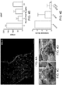
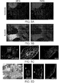
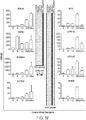
- the present disclosure demonstrates that the surface mesothelium represents the tissue-of-origin for adhesion formation, identifying adhesions as a gross pathologic manifestation of mesothelium cells themselves. It also provides the molecular chain of events leading normal mesothelium towards an adhesion program and demonstrates that functional disruption of these pathways curtail adhesion formation in vivo. These results provide a new pathomechanistic understanding of adhesion formation and establish novel therapeutic tactics/avenues that treat and/or prevent adhesion pathogenesis.
- references in the description to methods of treatment refer to the agents of the present invention for use in a method of treatment of the human or animal body by therapy.
- the present disclosure provides methods of treating a subject to reduce adhesion formation, the method comprising administering to a subject in need of thereof an agent that that targets adhesion-formation by injured mesothelial cells.
- the agent targets the injured mesothelial cells for destruction.
- the agent recruits inflammatory macrophages to the site of adhesion.
- the agent prevents neutrophil recruitment to the site of adhesion.
- the agent inhibits the expression or activity of a gene product whose expression is induced in the injured mesothelial cells.
- Non limiting examples of agents include: an anti-mesothelin antibody or mesothelin binding fragment thereof; an anti-CD47 antibody, a signal regulatory protein alpha (SIRP ⁇ ) polypeptide, or a CD47 binding fragment of either; monocyte chemoattractant protein-1 (MCP-1) or an inflammatory macrophage recruiting portion thereof; an anti-Gr-1 antibody or Gr-1 binding fragment thereof or other marker involved in depletion of granulocytes; thioglycolate; PLGA; mannose or mannose conjugated to a binding moiety specific for injured mesothelium including UPKB1-binding moiety or MSLN binding moiety; and any combination thereof. Included are peptide, peptoid, RNA, DNA, PNA, or other engineered molecule selected for binding for injured mesothelium, e.g. for UPKB1, MSLN, etc..
- Applicants have identified biological markers that are expressed selectively on injured mesothelium at the time of adhesion formation, which marker include without limitation MSLN, UPK1B, etc.
- the markers provide targets for removal or destruction of injured mesothelial cells, for example by apoptosis, phagocytosis, necrosis, etc.
- a specific binding moiety e.g. an antibody or fragment thereof, is administered to an individual to remove injured mesothelial cells, e.g. to prevent adhesion formation, to reduce ongoing adhesion formation, or to reduce existing adhesions.
- the markers are useful biomarkers for the presence of injured mesothelial cells, e.g. by PET, MRI, etc.
- an antibody or other specific binding moiety that binds to an injured mesothelial cell marker is labeled with a detectable marker.
- the marker may include, for example, moieties useful in PET or MRI imaging.
- the lebeled antibody or other specific binding moiety is contacted with the patient, for example by iv or other delivery system, and the presence of the labeled moiety used to image the cells involved in adhesion formation.
- therapeutic agent includes one or more "therapeutic agents” or “drugs.”
- therapeutic agents or “drugs” can be used interchangeably herein and include pharmaceutically active compounds. Examples include, without limitation: antibiotics, small molecules, proteins or polypeptides, polynucleotides, nucleic acids, oligonucleotides, ribozymes, anti-sense oligonucleotides, gene/vector systems, antisense nucleic acid (RNA or DNA), virus vectors or vectors derived from viral sources, antibodies, receptor antagonists, transcriptional repressors, etc. Polynucleotides can code for therapeutic proteins or polypeptides.
- Polynucleotide or “oligonucleotide” is used interchangeably and each means a linear polymer of nucleotide monomers, e.g., deoxyribonucleotides and/or ribonucleotides.
- polypeptide peptide
- protein protein
- amino acid polymers in which one or more amino acid residue is an artificial chemical mimetic of a corresponding naturally occurring amino acid, as well as to naturally occurring amino acid polymers and non-naturally occurring amino acid polymer.
- amino acid refers to naturally occurring and synthetic amino acids, as well as amino acid analogs and amino acid mimetics that function in a manner similar to the naturally occurring amino acids.
- Naturally occurring amino acids are those encoded by the genetic code, as well as those amino acids that are later modified, e.g., hydroxyproline, gamma-carboxyglutamate, and O-phosphoserine.
- amino acid analog refers to a compound that has the same basic chemical structure as a naturally occurring amino acid, i.e., an alpha carbon that is bound to a hydrogen, a carboxyl group, an amino group, and an R group, e.g., homoserine, norleucine, methionine sulfoxide, methionine methyl sulfonium.
- R groups e.g., norleucine
- Amino acid mimetics refers to chemical compounds that have a structure that is different from the general chemical structure of an amino acid, but that functions in a manner similar to a naturally occurring amino acid.
- antibody or “antibody moiety” is intended to include any polypeptide chain-containing molecular structure with a specific shape that fits to and recognizes an epitope, where one or more non-covalent binding interactions stabilize the complex between the molecular structure and the epitope.
- Antibodies utilized in the present invention may be polyclonal antibodies or monoclonal antibodies. In whil aspects, monoclonal antibodies may be preferable because they may be reproduced by cell culture or recombinantly, and can be modified to reduce their antigenicity. The production of polyclonal and monoclonal antibodies is well known in the art.
- immunoglobulin fragments comprising the epitope binding site (e.g., Fab', F(ab')2, or other fragments) are useful as antibody moieties in the present invention and are sometimes referred to as "binding fragments" or "epitope binding fragmnets" of an antibody.
- binding fragments e.g., Fab', F(ab')2, or other fragments
- Such antibody fragments may be generated from whole immunoglobulins by ricin, pepsin, papain, or other protease cleavage.
- “Fragment,” or minimal immunoglobulins may be designed utilizing recombinant immunoglobulin techniques.
- Fv immunoglobulins for use in the present invention may be produced by linking a variable light chain region to a variable heavy chain region via a peptide linker (e.g., poly-glycine or another sequence which does not form an alpha helix or beta sheet motif).
- a peptide linker e.g., poly-glycine or another sequence which does not form an alpha helix or beta sheet motif.
- Antibodies include free antibodies and antigen binding fragments derived therefrom, and conjugates, e.g. pegylated antibodies, drug, radioisotope, or toxin conjugates, and the like. Monoclonal antibodies directed against a specific epitope, or combination of epitopes, will allow for the targeting and/or depletion of cellular populations expressing the marker.
- Various techniques can be utilized using monoclonal antibodies to screen for cellular populations expressing the marker(s), and include magnetic separation using antibody- coated magnetic beads, "panning" with antibody attached to a solid matrix (i.e., plate), and flow cytometry (See, e.g., U.S. Pat. No. 5,985,660 ; and Morrison et al. Cell, 96:737-49 (1999 )). These techniques allow for the screening of particular populations of cells; in immunohistochemistry of biopsy samples; in detecting the presence of markers shed by cancer cells into the blood and other biologic fluids, and the like.
- Humanized versions of such antibodies are also within the scope of this invention. Humanized antibodies are especially useful for in vivo applications in humans due to their low antigenicity.
- subject means a mammal being assessed for treatment and/or being treated.
- the mammal is a human.
- the terms "subject,” “individual,” and “patient” encompass, without limitation, individuals having or at risk of having an adhesion.
- Subjects may be human, but also include other mammals, particularly those mammals useful as laboratory models for human disease, e.g. mouse, rat, etc.
- treatment refers to administering an agent, or carrying out a procedure, for the purposes of obtaining an effect in a subject (e.g., a patient).
- the effect may be prophylactic in terms of completely or partially preventing an undesirable condition, disease or symptom thereof and/or may be therapeutic in terms of effecting a partial or complete remedy, remission, or cure for an undesirable condition, disease and/or symptoms of the disease.
- Treating thus encompasses the dinistration of an agent before an undesirable condition, disease or symptom thereof occurs, during the development of an undesirable condition, disease or symptom thereof, and/or after an undesirable condition, disease or symptom thereof has occurred.
- Treating may refer to any indicia of success in the treatment or amelioration or prevention of an undesirable condition or disease, including any objective or subjective parameter such as abatement; remission; diminishing of symptoms or making the undesirable condition more tolerable to the patient; slowing in the rate of degeneration or decline; or making the final point of degeneration less debilitating.
- the treatment or amelioration of symptoms can be based on objective or subjective parameters; including the results of an examination by a physician.
- treating includes the administration of the compounds or agents of the present disclosure to prevent or delay, to alleviate, or to arrest or inhibit development of the symptoms or undesirable condition.
- therapeutic effect refers to the reduction, elimination, or prevention of the undesirable condition, disease, or symptoms or side effects of thereof.
- each component can be administered at the same time or sequentially in any order at different points in time. Thus, each component can be administered separately but sufficiently closely in time so as to provide the desired therapeutic effect.
- correlates refers to a statistical association between instances of two events, where events include numbers, data sets, and the like.
- a positive correlation also referred to herein as a "direct correlation” means that as one increases, the other increases as well.
- a negative correlation also referred to herein as an "inverse correlation” means that as one increases, the other decreases.
- Dosage unit refers to physically discrete units suited as unitary dosages for the particular individual to be treated. Each unit can contain a predetermined quantity of active compound(s) calculated to produce the desired therapeutic effect(s) in association with the required pharmaceutical carrier.
- the specification for the dosage unit forms can be dictated by (a) the unique characteristics of the active compound(s) and the particular therapeutic effect(s) to be achieved, and (b) the limitations inherent in the art of compounding such active compound(s).
- “Pharmaceutically acceptable excipient” means an excipient that is useful in preparing a pharmaceutical composition that is generally safe, non-toxic, and desirable, and includes excipients that are acceptable for veterinary use as well as for human pharmaceutical use. Such excipients can be solid, liquid, semisolid, or, in the case of an aerosol composition, gaseous.
- compositions, carriers, diluents and reagents are used interchangeably and represent that the materials are capable of administration to or upon a human without the production of undesirable physiological effects to a degree that would prohibit administration of the composition.
- a “therapeutically effective amount” means the amount that, when administered to a subject, is sufficient to effect treatment of an undesirable condition, symptom, or disease of the subject.
- the effective dose is sufficient to reduce adhesions by at least about 10%, at least about 20%, at least about 30%, at leastr about 40%, at least about 50%, at least about 60%, at least about 70%, at least about 80%, at least about 90%, or more.
- the dose of a therapeutic antibody may be from about 1 mg/kg body weight to about 100 mg/kg body weight, and may be administered immediately following surgery or other event expected to result in adhesion formation; or for reducing existing adhesions. Dosing may be repeated daily, every 2 days, every 3 days, semi-weekly, weekly, etc.
- the present disclosure provides methods for treating surgical adhesions in a subject, which includes, e.g., preventing adhesion formation, halting or reducing the formation of adhesions, and/or reversing or eliminating established adhesions in a subject.
- Composton and kits are also provided for performing such methods.
- the present disclosure relates to methods for treating a subject to reduce adhesion formation, e.g., surgical adhesions, and includes administering to a subject an agent that that targets adhesion-formation by injured mesothelial cells. Compositons and kits configured to perform such treatment methods are also provided.
- the disclosure is based, at least in part, on our identification of the surface mesothelium as a primary cell type responsible for initiating and contributing towards adhesions (see Examples below). We demonstate that adhesion formation can be treated in subjects by targeting one or more of multiple different checkpoints in the adhesion formation process by injured mesothelial cells.
- the agent targets the injured mesothelial cells for destruction.
- the agent may bind to a gene product whose expression is induced in the injured mesothelial cells, thereby differentiating it from un-injured mesothelial cells and allowing it to be eliminated.
- the agent is specific for (e.g., specifically binds to) a target protein that is expressed in injured mesothelial cells but not in un-injured mesothelial cells.
- the gene product is mesothelin (MSLN) or uroplakin 1B (UPK1B), both of which are discussed in further detail below.
- MSLN is a desirable target as it is an embryonic mesothelial gene and is not widely expressed in adult stages, with the notable exclusion of adhesions (as described herein) and tumors.
- the agent is a polypeptide that specifically binds to MSLN.
- the polypeptide can be an anti-MSLN antibody or MSLN binding fragment thereof.
- the agent can be administered alone or in combination with other agents, e.g., immunomodulatory agents.
- the mehods further include administering anti-MSLN antibodies alone or in conjunction with other immune modulators, e.g., agents that induce cell removal.
- the second agent inhibits the activity of CD47 in the injured mesothelial cells, e.g., a polypeptide that binds to CD47 and disrupts the binding of CD47 with SIRP ⁇ , at a dose that achieves a depletion of injured mesothelial cells.
- the polypeptide is selected from the group consisting of: an anti-CD47 antibody, a signal regulatory protein alpha (SIRP ⁇ ) polypeptide, a CD47 binding fragment of either, and any combination thereof.
- SIRP ⁇ signal regulatory protein alpha
- polypeptide e.g., antibody
- polypeptide e.g., antibody
- an anti-MSLN antibody shows substantial reduction in adhesion grades in animal models after induction and have demonstrated feasibility due to animals manifesting little or no side effects.
- immune adjuvants including without limitation anti-CD47.
- the agent recruits inflammatory macrophages (through recruitment of circulating monocytes) to the site of adhesion. This recruitment results in the reduction of adhesion formation in a clinical setting.
- the use of and inflammatory macrophage/monocyte recruiting agents are effective in treating post-operative adhesions in a subject (e.g., preventing or removing).
- a subject e.g., preventing or removing.
- administration of MCP-1 e.g., intraperitoneally
- Such administration was demonstrated to be feasible due to animals manifesting little or no side effects.
- agents that induce a sterile inflammation in a subject find use in treating adhesion formation.
- agents that induce a sterile inflammation in a subject find use in treating adhesion formation.
- agents that induce a sterile inflammation in a subject find use in treating adhesion formation.
- agents that induce a sterile inflammation in a subject find use in treating adhesion formation.
- agents that induce a sterile inflammation in a subject find use in treating adhesion formation.
- agents that induce a sterile inflammation in a subject find use in treating adhesion formation.
- agents include thioglycollate and poly(lactic-co-glycolic acid) PLGA.
- the recruitment of inflammatory macrophages can be achieved with any convenient agent or agents, including but not limited to: monocyte chemoattractant protein-1 (MCP-1) or an inflammatory macrophage recruiting portion thereof, thioglycolate, PLGA, or any combination thereof.
- MCP-1 monocyte chemoattractant protein-1
- thioglycolate thioglycolate
- PLGA thioglycolate
- the agent prevents neutrophil recruitment.
- elimination/reduction of circulating neutrophils or prevention of their migration to potential adhesion sites results in the reduction of adhesion formation in a clinical setting.
- any agent that eliminates/reduces circulating neutrophils and/or prevents their migration to potential adhesion sites can be administered to a subject to treat adhesions.
- the agent can be a polypeptide that binds to a granulocyte marker selected from Gr-1, CD66b, CD177, CXCR1, VAP1, CXCR2, and CD10, which are expressed on neutrophils and other inflammatory granulocytes, and leads to their elimination/reduction or inhibits their migration to potential adhesion sites.
- the agent is a polypeptide, e.g an antibody or binding fragment thereof.
- one or more agents that promote inflammatory macrophage recruitment and one or more agents that eliminate/reduce or inhibit neutrophil migration can be combined.
- anti-MSLN antibody and anti-granulocyte antibody can be administered to a subject to treat adhesions.
- the agent inhibits the expression or activity of a gene product whose expression is induced in the injured mesothelial cells, e.g. MSLN, UPK1B, etc.
- the expression of a number of genes is induced in injured mesothelial cells that can be exploited to treat adhesions in a subject.
- a number of different agents can be used, each of which may target different induced genes and/or the cellular pathways or cellular activities they regulate.
- the agent can inhibit gene expression, e.g., an inhibitory RNA agent, e.g., RNAi, shRNA, anti-sense RNA, and the like.
- the agent can inhibit the activity of the gene product, e.g., preventing its interaction with a receptor/ligand or a signaling molecule in a signal transduction pathway in which the gene product plays a role. No limitation in this regard is intended.
- the gene for hypoxia-inducible factor 1-alpha is induced upon mesothelial cell injury.
- Administering an agent that inhibits the expression and/or the activity of HIF-1 ⁇ to a subject can treat (e.g., prevent, eliminate or reduce) adhesions in the subject.
- Any agent that inhibits HIF-1 ⁇ expression or activity can be used, including but not limited to: echinomycin, PX12, FM19G11, cryptotanshinone, chetomin, Bortezumib, acriflavin, methyl 3-[[2-[4-(2-adamantyl) phenoxy] acetyl] amino]-4-hydroxybenzoate, dimethyloxaloylglycine (DMOG), chemotin, YC-1, chrysin, dimethyl-bisphenol A, CL67, and combinations thereof.
- DMOG dimethyloxaloylglycine
- chemotin chemotin
- YC-1 chemotin
- chrysin dimethyl-bisphenol A, CL67, and combinations thereof.
- echinomycin significantly reduces the formation of adhesions in a mouse model by inhibiting mesothelial cell proliferation, activation, and cell-cell contact.
- Additional agents that interfere with HIF-1 ⁇ activity also reduce adhesion formation in these models, including PX12, FM19G11, and cryptotanshinone.
- the gene for uroplakin 1B is induced upon mesothelial cell injury.
- Administering an agent that inhibits the expression and/or the activity of UPK1B to a subject can treat (e.g., prevent, eliminate or reduce) adhesions in the subject. Any agent that inhibits UPK1B expression or activity can be used.
- agents that target UPK1B expressing cells for destruction or removal can be used.
- the agent is a polypeptide that specifically binds to UPK1B.
- the polypeptide can be an anti-UPK1B antibody or UPK1B binding fragment thereof.
- the agent can be administered alone or in combination with other agents, e.g., immunomodulatory agents.
- the mehods further include administering anti-UPK1B antibodies alone or in conjunction with other immune modulators, e.g., agents that induce cell removal.
- the second agent inhibits the activity of CD47 in the injured mesothelial cells, e.g., a polypeptide that binds to CD47 (as described above).
- UPK1B is a desirable target as it is an embryonic mesothelial gene and is not widely expressed in the adult except in adhesions (injured mesothelium) and the bladder epithelium.
- Mannose is a simple monosaccharide that competitively binds UPK1B and prevents its interaction with cognate receptor/ligand(s). As shown in the Examples herein, mannose competitively binds to UKP1B and leads to substantial reduction in adhesion scores in animal models after induction. Thus, in certain embodiments, the agent adminsitered to the subject to treat adhesions competitively binds to UPKB1.
- agents include, e.g., mannose, a UPKB1-binding derivative thereof (e.g., protein conjugates, dimers, trimers, dendrimers, etc), and/or analogs as well as other agents that competitively bind UPKB1, e.g., soluble polypeptides that bind to UPKB1, e.g., derivded from natural receptor/ligand domains to which UPKB1 binds.
- UPKB1-binding derivative thereof e.g., protein conjugates, dimers, trimers, dendrimers, etc
- analogs as well as other agents that competitively bind UPKB1, e.g., soluble polypeptides that bind to UPKB1, e.g., derivded from natural receptor/ligand domains to which UPKB1 binds.
- the expression of a number of neutrophil recruiting gene products are induced in injured mesothelial cells, including CXC chemokines.
- CXC chemokines include CXC chemokines.
- the CXCL1 and CXCL2 genes are induced in mesothelial cells upon injury.
- an agent that inhibits the expresson and/or activity of a neutrophil rectuiting gene product can be administered to a subject to treat adhesions.
- the gene product is a CXC chemokine, e.g., CXCL1, CXCL2, or combinations thereof.
- the agent can be an antibody or binding fragment thereof that binds to and inactivates the CXC chemokine or an agent that block expression of the CXC chemokine (e.g., and inhibitory RNA moiety, e.g., siRNA, shRNA, etc.).
- adhesion treatment methods disclosed herein can be used to treat a subject for any of a variety of conditions that can promote adhesion formation, e.g., post-operative/surgical lesions (e.g., open or laparoscopic surgeries) and/or for treatment after inflammatory insults.
- the agents can be administered for adhesion prophylaxis, e.g., prior to the formation of an adhesion, or after adhesions have been formed or initiated.
- the adhesion is an abdominal adhesion, e.g., an abdominal surgical adhesion.
- the agent can be administered prior to, during, or after the surgical procedure.
- adhesion treatments agents as described herein can be provided in pharmaceutical compositions suitable for therapeutic use, e.g. for human treatment.
- pharmaceutical compositions of the present disclosure include one or more therapeutic entities of the present disclosure or pharmaceutically acceptable salts, esters or solvates thereof.
- pharmaceutical compositions of the present disclosure include one or more therapeutic entities of the present disclosure in combination with another therapeutic agent, e.g., an anti-inflammatory agent or additional agent for treating an adhesion.
- compositions comprising an active therapeutic agent and other pharmaceutically acceptable excipient.
- the preferred form depends on the intended mode of administration and therapeutic application.
- the compositions can also include, depending on the formulation desired, pharmaceutically-acceptable, non-toxic carriers or diluents, which are defined as vehicles commonly used to formulate pharmaceutical compositions for animal or human administration.
- the diluent is selected so as not to affect the biological activity of the combination. Examples of such diluents are distilled water, physiological phosphate-buffered saline, Ringer's solutions, dextrose solution, and Hank's solution.
- the pharmaceutical composition or formulation may also include other carriers, adjuvants, or nontoxic, nontherapeutic, nonimmunogenic stabilizers and the like.
- compositions of the present disclosure can also include large, slowly metabolized macromolecules such as proteins, polysaccharides such as chitosan, polylactic acids, polyglycolic acids and copolymers (such as latex functionalized SepharoseTM, agarose, cellulose, and the like), polymeric amino acids, amino acid copolymers, and lipid aggregates (such as oil droplets or liposomes).
- macromolecules such as proteins, polysaccharides such as chitosan, polylactic acids, polyglycolic acids and copolymers (such as latex functionalized SepharoseTM, agarose, cellulose, and the like), polymeric amino acids, amino acid copolymers, and lipid aggregates (such as oil droplets or liposomes).
- methods for treating adhesions in a subject by administering an agent that that targets adhesion-formation by injured mesothelial cells.
- Such methods include administering to a subject in need of treatment a therapeutically effective amount or an effective dose of one or more agents as detailed herein.
- Effective doses of the therapeutic entity of the present disclosure vary depending upon many different factors, including means of administration, target site, physiological state of the patient, whether the patient is human or an animal, other medications administered, and whether treatment is prophylactic or therapeutic.
- the patient is a human, but nonhuman mammals may also be treated, e.g. companion animals such as dogs, cats, horses, etc., laboratory mammals such as rabbits, mice, rats, etc., and the like. Treatment dosages can be titrated to optimize safety and efficacy.
- the therapeutic dosage may range from about 0.0001 to 100 mg/kg, and more usually 0.01 to 5 mg/kg, of the host body weight.
- dosages can be 1 mg/kg body weight or 10 mg/kg body weight or within the range of 1-10 mg/kg.
- An exemplary treatment regime entails administration once every two weeks or once a month or once every 3 to 6 months.
- Therapeutic entities of the present disclosure can be administered on multiple occasions. Intervals between single dosages can be weekly, monthly or yearly. Intervals can also be irregular as indicated by measuring blood levels of the therapeutic entity in the patient.
- therapeutic entities of the present disclosure can be administered as a sustained release formulation, in which case less frequent administration is required. Dosage and frequency vary depending on the half-life of the polypeptide in the patient.
- compositions for the treatment of adhesions can be administered by parenteral, topical, intravenous, intratumoral, oral, subcutaneous, intraarterial, intracranial, intraperitoneal, intranasal or intramuscular means.
- a typical route of administration is intravenous or intraperitoneal, although other routes can be equally effective.
- compositions are prepared as injectables, either as liquid solutions or suspensions; solid forms suitable for solution in, or suspension in, liquid vehicles prior to injection can also be prepared.
- the preparation also can be emulsified or encapsulated in liposomes or micro particles such as polylactide, polyglycolide, or copolymer for enhanced adjuvant effect, as discussed above. Langer, Science 249: 1527, 1990 and Hanes, Advanced Drug Delivery Reviews 28: 97-119, 1997 .
- the agents of this disclosure can be administered in the form of a depot injection or implant preparation which can be formulated in such a manner as to permit a sustained or pulsatile release of the active ingredient.
- the pharmaceutical compositions are generally formulated as sterile, substantially isotonic and in full compliance with all Good Manufacturing Practice (GMP) regulations of the U.S. Food and Drug Administration.
- GMP Good Manufacturing Practice
- Toxicity of adhesion treatment agents can be determined by standard pharmaceutical procedures in cell cultures or experimental animals, e.g., by determining the LD 50 (the dose lethal to 50% of the population) or the LD 100 (the dose lethal to 100% of the population). The dose ratio between toxic and therapeutic effect is the therapeutic index.
- the data obtained from these cell culture assays and animal studies can be used in formulating a dosage range that is not toxic for use in human.
- the dosage of the proteins described herein lies preferably within a range of circulating concentrations that include the effective dose with little or no toxicity. The dosage can vary within this range depending upon the dosage form employed and the route of administration utilized. The exact formulation, route of administration and dosage can be chosen by the individual physician in view of the patient's condition. It is noted that a number of the adhesion treatment agents discosed herein are approved for use in humans.
- kits that contain an adhesion treatment agent or formulations thereof and instructions for use.
- the kit can further contain a least one additional reagent.
- Kits typically include a label indicating the intended use of the contents of the kit.
- the term label includes any writing, or recorded material supplied on or with the kit, or which otherwise accompanies the kit.
- kits that contain: (a) a pharmaceutical composition having (i) an agent that that targets adhesion-formation by injured mesothelial cells in an amount effective to reduce adhesions (as described herein), and (ii) a pharmaceutically acceptable carrier, and (b) instructions for administering the pharmaceutical composition to a subject who has had or will have surgery.
- the agent is selected from the group consisting of: an anti-MSLN antibody or MSLN binding fragment thereof; an anti- UPKB1 antibody or UPKB1 binding fragment thereof; an anti-CD47 antibody, a SIRP ⁇ polypeptide, or a CD47 binding fragment of either; MCP-1 or an inflammatory macrophage recruiting portion thereof; an anti-Gr-1 antibody or Gr-1 binding fragment thereof; thioglycolate; PLGA; mannose or a UPKB1-binding derivative thereof; and any combination thereof.
- kits for making articles of manufacture (e.g., systems or kits) as described above, e.g., a kit, and that includes combining (a) a pharmaceutical composition having (i) an agent that that targets adhesion-formation by injured mesothelial cells in an amount effective to reduce adhesions (as described herein), and (ii) a pharmaceutically acceptable carrier, and (b) instructions for administering the pharmaceutical composition to a subject who has had or will have surgery.
- a pharmaceutical composition having (i) an agent that that targets adhesion-formation by injured mesothelial cells in an amount effective to reduce adhesions (as described herein), and (ii) a pharmaceutically acceptable carrier, and (b) instructions for administering the pharmaceutical composition to a subject who has had or will have surgery.
- the agent is selected from the group consisting of: an anti-MSLN antibody or MSLN binding fragment thereof; an anti-CD47 antibody, a SIRP ⁇ polypeptide, or a CD47 binding fragment of either; MCP-1 or an inflammatory macrophage recruiting portion thereof; an anti-Gr-1 antibody or Gr-1 binding fragment thereof; thioglycolate; PLGA; mannose or a UPKB1-binding derivative thereof; and any combination thereof.
- the instructions for practicing the subject methods are generally recorded on a suitable recording medium.
- the instructions may be printed on a substrate, such as paper or plastic, etc.
- the instructions may be present in the kits as a package insert, in the labeling of the container of the kit or components thereof (i.e., associated with the packaging or sub-packaging) etc.
- the instructions are present as an electronic storage data file present on a suitable computer readable storage medium, e.g. CD-ROM, diskette, etc.
- the actual instructions are not present in the kit, but means for obtaining the instructions from a remote source, e.g. via the internet, are provided.
- An example of this instance is a kit that includes a web address where the instructions can be viewed and/or from which the instructions can be downloaded. As with the instructions, this means for obtaining the instructions is recorded on a suitable substrate.
- Peritoneal adhesions are fibrous tissues that tether organs to one another or to the peritoneal wall and are a significant cause of post-surgical and infectious morbidity. Extensive studies have been done and suggest that hematopoietic cells, cytokines, and fibrin deposition play a major role in promoting adhesion formation. However, the molecular pathogenesis initially promoting adhesion formation has not been well characterized.
- mesothelium As a primary cell type responsible for initiating and contributing towards adhesions.
- Time courses of mesothelial specific stains and proliferation markers demonstrate that adhesions are formed from mesothelial cell expansion.
- Isolation and RNA sequencing of activated mesothelial cells in a brief time course immediately following adhesion induction suggest a specific surface marker phenotype of an adhesion, and candidate regulators of adhesion formation.
- Mesothelin (MSLN) and uroplakin1B (UPK1B) were identified as unique surface receptors on injured mesothelium.
- hypoxia inducible factor 1 ⁇ (HIF1 ⁇ ) was identified as an early regulator, and functional inhibition showed significant diminishing of adhesion formation, suggesting new therapeutic agents to prevent post-operative adhesions.
- RNA sequencing analysis of HIF1 ⁇ deficient mesothelial cells following adhesion induction demonstrated that targets, such as Wilms Tumor 1 (WT1) and UPK1 B are downstream to and controlled by HIF1a and play a role in the adhesion process.
- WT1 Wilms Tumor 1
- UPK1 B are downstream to and controlled by HIF1a and play a role in the adhesion process.
- Analyses of human adhesion tissue confirm that similar genetic signatures are present in human disease. Our results highlight the deeply critical role that injured mesothelium plays in adhesion formation and investigates its pathophysiology cellularly and molecularly.
- Adhesion induction surgeries were done on wild type B6 (C57BL/6J (The Jackson Laboratory) mice aged 6-10 weeks. Mice were anesthetized by inhaled isoflurane until determined unconscious confirmed by toe-pinch test. The abdomen was disinfected with betadine and phosphate buffered saline (PBS). A left mid-clavicular incision was made in the skin running down the length of the mouse. A similar left-mid clavicular incision was made in the peritoneum running down the length of the peritoneum. The peritoneum was gently folded to the right and held down by a hemostat.
- PBS betadine and phosphate buffered saline
- a single, ischemic button was placed on the right half of the peritoneal wall by clamping a small ( ⁇ 5mm diameter) piece of peritoneum and ligating the base with a 4-0 silk suture (Ethicon, 683G). Light abrasion on the button (24 times) and on the adjacent liver, cecum, and small and large bowels (7 times) was optionally performed (depending on the desired adhesion severity) with a surgical brush. Light brushing with fewer repetitions was performed to avoid pinpoint bleeding. The peritoneum was closed using 4-0 silk sutures and the skin was stapled closed (EZ Clips, 9mm, Braintree Scientific Inc). Mice were allowed to recover on a heating pad and injected with 0.05-0.1 mg/kg of buprenorphine. Mice were followed closely and monitored daily for signs of morbidity for 7 days until euthanasia. Adhesed tissues were dissected, scored, and fixed in 2% paraformaldehyde overnight at 4 degrees.
- Tissues were fixed in 2% paraformaldehyde overnight at four degree and were embedded and frozen in optimal cutting temperature compound O.C.T (Sakura) or embedded in paraffin. Frozen sections were cut at 10-12um throughout the adhesed organs and saved for immunofluorescence. Paraffin sections were cut at 5um and hematoxylin/eosin and Masson's trichome stains were performed via standard protocols.
- mice were immediately injected subcutaneously with 0.025mg of 5-ethynyl-2'-deoxyuridine (Life Technologies) in 90% PBS and 10% ethanol. Mice were traced for 7 days and euthanized. Adhesed tissues were dissected and fixed with 2% paraformaldehyde overnight, frozen in O.C.T (Sakura), and sectioned at 12um. EdU positive cells were visualized with Click-iT EdU Imaging Kit (Life Technologies).
- buttons were placed on each side of the peritoneum and no abrasion of the button or abdominal organs was done. Mice were allowed to recover for 6, 12, or 24 hours and euthanized. Ischemic buttons were dissected by cutting the base and placed in dissociation media (DMEM (Life Technologies, 10565-042), 50mg/ml collagenase IV (Worthington Biochemical), 20uM CaCl2). Buttons were homogenized using a single edge blade (Razor Blade Company), and incubated in dissociation media for 30 minutes at 37C.
- DMEM dissociation media
- Buttons were homogenized using a single edge blade (Razor Blade Company), and incubated in dissociation media for 30 minutes at 37C.
- the subsequent cell suspension was filtered through a 100um filter and spun and washed with 2% fetal bovine serum (FBS) in PBS.
- FBS fetal bovine serum
- Cells were treated with 1ml of ACK lysis buffer (Life Technologies) for 5 minutes at 4C and spun and washed.
- Cells were blocked with 1% goat serum (Life Technologies) for 10 minutes and stained with anti-PDPN (BioLegend, 8.1.1, 1:100), anti-LYVE-1 (eBioscience, ALY-7, 1:100), anti-CD31 (eBioscience, 390, 1:100), and anti-CD45 (BioLegend, 30-F11, 1:100) for 30 minutes at 4C.
- Cells were spun down, filtered, and resuspended in 200ul of 2% FBS in PBS.
- Cells were run through a FACSAria (BD Bioscences) and PDPN+LYVE1-CD31-CD45- cells were sorted directly into 750ul of Trizol LS
- RNA from sorted mesothelial population was isolated using Trizol (ThermoFisher) as per manufacturer's recommendation and was facilitated by addition of linear polyacrylamide (Sigma) as a carrier during RNA precipitation.
- Purified total RNA was treated with 4 units of RQ1 RNase-free DNase (Promega) at 37 °C for 1 hour to remove trace amounts of genomic DNA.
- the DNase-treated total RNA was cleaned-up using RNeasy micro kit (QIAGEN). 10-50 ng of total RNA was used as input for cDNA preparation and amplification using Ovation RNA-Seq System V2 (NuGEN).
- Amplified cDNA was sheared using Covaris S2 (Covaris) using the following settings: total volume 120 ml, duty cycle 10%, intensity 5, cycle/burst 100, total time 2 min.
- 500 ng of sheared and size-selected cDNA were used as input for library preparation using NEBNext Ultra DNA Library Prep Kit for Illumina (New England BioLabs) as per manufacturer's recommendations.
- Resulting libraries (fragment distribution: 300-700 bp; peak 500-550 bp) were sequenced using HiSeq 4000 (Illumina) to obtain 2x150 base pair paired-end reads. The reads obtained were trimmed for base call quality and the presence of adapter sequences using Skewer (ref). High quality reads thus obtained were aligned to mouse genome using OLego (ref) and the levels of expressed mRNAs were estimated using cuffdiff2 (ref) and represented as fragments per kilo-base per million mapped reads (FPKM).
- Adhesions were induced in wild type B6 (C57BL/6J (The Jackson Laboratory) and allowed to recover for 7 days.
- 200ug of monoclonal anti-MSLN (B35) antibody were administered via intraperitoneal injections at 7, 10, and 13 days post injection.
- 200ug of monoclonal anti-CD47 (MIAP301) (BioXCell) were co-administered via intraperitoneal injections at the same frequency. Mice were euthanized 17 days after initial surgery and scored for adhesion severity.
- B35 anti-MSLN antibody was a gift from A. Miyajima.
- Wild type B6 (C57BL/6J (The Jackson Laboratory) were euthanized and the mesothelium was excised from the renal capsule and intestine. Excised mesothelium was cut into smaller fractions and placed into culture dishes pretreated with EmbryoMax 0.1% Gelatin Solution (EmdMillipore) for 30 minutes and cultured in Delbucco's Modified Eagle Medium (Life Technologies) with 10% fetal bovine serum, 1% penicillin/streptomycin and 1% non-essential amino acids at 37 degrees for 7 days. Mesothelial cells were co-cultured with macrophages (added to confluency) in normal conditions or in hypoxic conditions (1% O2 incubator or 100uM CoCl2).
- BMDMs Primary Bone-marrow Derived Macrophages
- mice were humanely euthanized, and disinfected with 70% ethanol. An incision was made along the legs, and the muscle removed from the bones. The femur and tibia were removed from the body, and rinsed in PBS. The bones were flushed with a 6 mL syringe and 23 gauge needle, and the marrow resuspended in 10 mL RPMI. The suspension was centrifuged 5 minutes, 1200 rpm, and the pellet resuspended in 5 mL ACK lysis buffer (Invitrogen) for 5 minutes to remove blood cells. The suspension was filtered over a 70 ⁇ m Falcon cell strainer, and centrifuged again.
- ACK lysis buffer Invitrogen
- the pellet was resuspended in 40 mL of macrophage media (RPMI+10% FBS+10% penicillin/streptomycin+10 ng/mL MCSF), and plated on four 10 cm petri dishes. On day 4, the macrophage media was replaced. On day 7, the macrophages were lifted from the dish and used.
- macrophage media RPMI+10% FBS+10% penicillin/streptomycin+10 ng/mL MCSF
- Adhesion induction surgeries were performed on 4-8 week old wild type B6 (C57BL/6J (The Jackson Laboratory) as previously described. 200mg/kg of cryptotanshinone (Sigma Aldrich), 2mg/kg of FM19G11 (Sigma Aldrich), 10ug/kg or 20ug/kg of echinomycin (Sigma Aldrich), or 25mg/kg of PX12 (Sigma Aldrich) immediately after injury, 4 hours after injury, and every 24 hours subsequently for 7 days.
- mice Parabiosis surgeries were done on age matched (4-6 week old) female wild type B6 (C57BL/6J (The Jackson Laboratory) and C57BL/Ka Rosa26 mRFP1 mice. Mice to undergo parabiosis were housed together in a single cage for 10 days prior to surgery. Mice were anesthetized by inhaled isoflurane until determined unconscious confirmed by toe-pinch test. The sides of the mice were shaved and cleaned with 70% ethanol and betadine. Mice were laid next to each other and incisions from elbow to knee were made on adjacent sides. The elbow and knee joints were ligated using 4-0 sutures (Ethicon) and the loose skin from adjacent mice was stapled together.
- 4-0 sutures Ethicon
- mice were allowed to recover on a heating pad and injected with 0.05-0.1 mg/kg of buprenorphine. Mice were followed closely and monitored daily for signs of morbidity for 14 days. Staples were removed following 14 days and mice were bled retro-orbitally to assay for chimerism.
- mice were allowed to recover and euthanized 30 minutes, 1h, 2h, 4h, 24h, 72h, and 7 days post induction.
- All ischemic buttons with ectopic adhesions that developed were dissected, fixed, and stained with haematoxylin and eosin (H&E) or with Podoplanin (PDPN) and Mesothelin (MSLN), mesothelium surface receptor markers(Rinkevich et al., 2012).
- H&E haematoxylin and eosin
- PDPN Podoplanin
- MSLN Mesothelin
- PDPN + cells were observed to be visible throughout all adhesion sites and were found to be non-colored, indicating that mesothelial cells contributing to the adhesion are derived from a local source instead of circulating cells ( Fig 1D ).
- RFP + cells were visible and scattered throughout the adhesion site and many RFP + cells also stained positive for F4/80, indicating that macrophages derived from circulating monocytes, infiltrate the adhesion site.
- EdU 5-ethynyl-2'-deoxyuridine
- mesothelial cells from the induction site at 6, 12, and 24 hours following adhesion induction by employing fluorescent activated cell sorting (FACS) with the following mesothelium cell surface phenotype: PDPN + LYVE1 - CD31 - CD45 - ( Fig 2A ).
- FACS fluorescent activated cell sorting
- Fig 2D Analysis of genesets enrichment using GO term analysis shows within the first six hours of induction early genes associated with angiogenesis and hypoxia response are upregulated in mesothelial cells. Hallmark mediators of the inflammatory response were significantly upregulated over the entire 24 hour time course, including chemokine and chemotaxis activity, cytokine secretion, and the NF kappa B pathway ( Fig 2D ). Fundamental cellular processes such as proliferation are upregulated over a course of 24 hours, corroborating our pulse-chase experiments.
- genesets relating to the extracellular region and collagen organization were downregulated across the 24 hour time course following adhesion induction.
- These genesets include the specific genes fibronectin 1 (FN1), and various collagens including COL1A1, COL1A2, COL3A1, ( Fig 2D ) indicating major transcriptional rearrangements take place during the early stages of mesothelial injury.
- FSP1 Fibroblast specific protein 1
- ACTA2 smooth muscle actin
- hypoxia inducible factor 1 alpha matched this trend ( Fig 2B , 2E ).
- RNA sequencing screen we induced adhesions in wild type C57BL/6J mice as described previously, and immunostained the subsequent adhesions 7 days after surgery for our gene targets ( Fig 2F ).
- Some targets, such as PDPN, MSLN, and HIF1A are expressed throughout the adhesion tissue, whereas WT1 activated within a subpopulation of the adhesion foci indicating heterogeneity within the mesothelium and the adhesion.
- an adhesion with a score of 0 has no adhesion between two areas, with limited mesothelial thickening on the button ( Fig 3A ).
- the area stains positive for MSLN and fibronectin ( Fig 3A ).
- An adhesion score of 1 indicated a "string" adhesion, connecting the two adhesed areas with a light fibrous bridge ( Fig 3B ). The string and surrounding areas were immunopositive for MSLN and fibronectin ( Fig 3B ).
- HIF1A transcription factor a transcription factor that exerts on surface mesothelium in vitro.
- Primary mesothelial cells were removed from the renal capsule and intestine and transplanted on cultured wells as previously described (Rinkevich et al., 2012). After 7 days mesothelial cells emerged from their tissue explant and expanded on the culture dish. Cultured mesothelial cells were incubated in low oxygen conditions (5% O 2 or in the presence of 100uM CoCl 2 ) for three days.
- mice that had undergone adhesion induction with small molecular inhibitors against HIF1A or its immediate downstream cascade.
- cryptotanshinone Sigma Aldrich
- FM19G11 FM19G11
- echinomycin Sigma Aldrich
- PX-12 PX-12
- HIF1A targets VEGFb and transferrin (TRF) were found to be downregulated after echinomycin treatment ( Fig 8 ).
- TRF transferrin
- MSLN is an embryonic mesothelial surface molecule that has very limited expression in the adult, but is re-expressed upon mesothelial injury and adhesion induction, making it an ideal candidate for targeting.
- Mouse monoclonal anti-MSLN antibodies were found to bind to injured sites immediately after adhesion induction and antibody injection ( Fig 4A ).
- CD47 is a surface receptor that is highly upregulated on a spectrum of tumors and acts as a "don't eat me signal,” inhibiting phagocytosis by binding to SIRP1 ⁇ , its ligand on macrophages. Inhibition of this interaction by blocking either CD47 or SIRP1 ⁇ , in addition to administration of tumor specific monoclonal antibodies, has been shown to greatly decrease tumor burden and increase survival(Chao et al., 2010; Chao, Weissman, & Majeti, 2012; Jaiswal et al., 2009; Tseng et al., 2013).
- Fig 4B we observed high CD47 expression in the mesothelium prior to and 24 hours following adhesion induction
- RNA sequencing data from purified mesothelium demonstrate a diverse signaling cascade that occurs following induction and begins to outline pathways within the mesothelium that contribute towards adhesion induction.
- MSLN embryonic mesothelial markers
- UPK1B uroplakin 1B
- WT1 Wilms Tumor 1
- Injured mesothelium also upregulates expression of fibrocytic genes S100A4 and ACTA2, which, together with our in vivo staining and in vitro results, suggest that it is the mesothelium that becomes a fibroblast like cell.
- the expression of HIF1A reflects the hypoxic environment initiated by the adhesion induction protocol, which in turn mimics procedures done during surgical operations. What is striking however, is the significant effect that inhibition of HIF1A demonstrates on adhesion formation.
- HIF1A pathway Disruption of the HIF1A pathway by small molecule inhibitors echinomycin and or PX12 is sufficient to lead to a dramatic decrease in adhesion formation and frequency, suggesting that HIF1A plays an important role in the pathogenesis of adhesion formation.
- RNA interference RNA interference
- Our study is the first to demonstrate the use of HIF1A inhibition using small molecule inhibitors, a treatment option much more suitable for clinical therapeutic use.
- Our data proposes the use of echinomycin, a peptide antibiotic, and or PX12, both of which are easily administrable in a clinical setting.
- HIF1A is a general transcription factor
- the in vivo model can provide a functional platform interrogate the molecular biology underlying the mesothelial contribution to adhesion pathogenesis.
- adhesion specific molecules Many of these are embryonic mesothelial genes that are re-expressed during injury and in few other tissues, making them prime targets for post formation treatments. With the identification of adhesion specific targets, we have demonstrated that the use of an antibody-based immunotherapy provides a therapeutic approach for the treatment of already formed adhesions.
- RNA sequencing results from injured mesothelial cells over a 24 hour time course were taken from previous experiments (see Example 1) and showed differential expression of multiple genesets and processes, including inflammatory chemokines and cytokines ( Fig 9A ).
- Specific neutrophil and macrophage recruitment signals, CXCL1, CCL2, and CXCL2 are immediately upregulated after 6 hours and stay increased over the first 24 hours.
- Peritoneal washes from similar time points confirmed that increased numbers of neutrophils and macrophages enter the peritoneum.
- Adhesion induction as previously described in Example 1, in parabiosis experiments between a mouse constitutively expressing the red fluorescent protein mCherry ("TM7") and a wild type (non-colored mouse) showed red F4/80+ cells infiltrating the adhesion sites in a wild type mouse, suggesting that the macrophages that enter the adhesions are circulating monocytes, or bone marrow derived ( Fig 9B ).
- Adhesion inductions in constitutive cyan fluorescent protein (CFP) mice (“Host") receiving bone marrow transplants from constitutive mCherry mice (TM7 "Donor”) also show red F4/80+ cells in adhesion sites, again suggesting that macrophages entering the adhesions are circulating bone marrow derived monocytes ( Fig 9C ).
- Mice treated with thioglycollate 24 hours prior to adhesion induction show diminished adhesions, indicating that sterile inflammation may have beneficial effects towards adhesion formation.
- Catalase treatment which provides neutrophils a selective advantage over macrophages, 24 hours prior to adhesion induction, worsens adhesion formation in comparison to the thioglycollate treatment.
- MCP-1 a known monocyte chemoattractant
- anti-GR-1 antibodies resulting in depletion of circulating neutrophils, for three days following adhesion induction surgery both diminish adhesion formation.
- MCP1 in conjunction with anti-GR-1 has an additive effect in diminishing adhesion formation ( Fig 9D ).
- mice were pre-treated with either PLGA, thioglycollate, or PBS control 3 days prior to adhesion induction surgery as described in Example 1. Mice were evaluated 7 days post injury and evaluated on an adhesion severity scale from 0-5 for adhesions forming on the suture site (S) or the button (B). Fig 10 shows that mice pretreated with PLGA or thioglycollate have significantly reduced severity of adhesions at both the suture site and button.
- pretreatment with PLGA alone, thioglycollate alone, or a combination of PLGA and thioglycollate prior to surgery will reduce the formation and/or severity of adhesions in a subject.
Description
- Adhesions are fibrous tissues that develop as the result of trauma to the peritoneum, either due to physical trauma (often due to surgery), or major inflammatory insults (peritonitis, infection, etc). Although many adhesions are asymptomatic, their primary sequelae include bowel obstruction, female infertility (when occurring on or near the uterus), chronic pain, and poor quality of life. The National Institutes of Health estimate that over 93% of patients having undergone abdominal surgery progress to adhesion formation. Current estimates suggest that up to 20% of patients following abdominal surgery are readmitted to hospitals due to post-operative adhesion formation and project the healthcare cost to be well over $1 billion in the United States alone, rivaling that of many cancers. Still, no definitive strategies exist to prevent adhesion formation, underscoring the notion that adhesions present a significant healthcare burden and treatment and prevention should be an ongoing medical priority.
- Extensive studies on the pathogenesis of adhesion formation focused on inflammatory infiltration and a balance between fibrin deposition and fibrinolysis. Currently it is believed that damage to the peritoneal surface initiates a complex cascade that recruits inflammatory cells through a panel of cytokine signals. Meticulous studies have been done to map the kinetics of the cellular infiltrate: polymorphonuclear leukocytes (PMNs) are present early on (within 6 hours) of injury, followed by macrophage entry (persisting over 72 hours) and later stage recruitment of T and B-cells. The roles of each of these cell types in adhesion formation have been explored, and most studies agree that the pro-inflammatory profiles of PMNs, macrophages, and CD4+ T-cells serve to exacerbate the severity of the adhesions.
- More work has been done on the contribution of the coagulation cascade as a key driver of the adhesion process. The coagulation cascade is activated concurrently with immune cell infiltration. Thrombin converts fibrinogen to fibrin, which form deposits on the surface of organs shortly after mesothelial injury and organize into fibrin lattices for wound healing or adhesion development.
- The current state of knowledge stipulates that peritoneal or organ injury sufficient to produce adhesions requires the removal of the surface mesothelium (diZerega & Campeau; Richardson, 1911; Schade & Williamson, 1968; A. Suzuki, 2015) exposing their underlying basement membranes which leads to fibrin attachments between two denuded surfaces and is followed by accumulations of fibroblasts, from an unknown origin. Since these observations have been largely dependent on classic snap-shot histological examinations, and because of the difficulties in identifying cell type, the source of new invading fibroblast cells, has been a matter of debate. Among the possible sources are metaplasia of myofibroblasts within the connective tissue beneath the surfaces, the expansions of mesenchymal stem cells (MSCs), the invasion of undifferentiated primitive MSCs, or to the deposition of extracellular matrix by subperitoneal fibroblasts leading to formation of adhesions (Cheong et al.; Ellis, Harrison, & Hugh, 1965; Hellebrekers & Kooistra, 2011; Lucas, Warejcka, Young, & Lee, 1996; Raftery, 1973).
- Following the above paradigms, investigators have transplanted the mentioned fibroblast cell types into the abdominal cavities of rodent models at different times after surgery and consequently showing increased formation of postoperative adhesions, and claiming to support the notion that adhesions arise from these cell types (Lucas et al., 1996). Others have indicated that the epithelial characteristics of the surface mesothelium, including its frictionless interface, point to a protective role against adhesion formation, and have even proposed the use of mesothelium cell sheets as a clinical approach for preventing postoperative adhesions (Inagaki, Inagaki, Kokudo, & Miyajima, 2015).
- In the absence of a cell-of-origin that can be used to study fibrotic disease initiation and progression (Rinkevich et al., 2012, 2015), not much is known about the early molecular and cellular events leading up to abdominal adhesions, and therefore no robust preventative or postsurgical treatments have been developed. Hence, current clinical treatments use physical barriers to prevent contact between adjacent organs as a preventative treatment ((Liu, Li, Shu, Gray, & Prestwich, 2005), or adhesion lysis that requires a physician to surgically separate the adhered organs, a tactic hampered by a high rate of adhesion reformation (Ray, 1998).
- The mesothelium is the outer epithelial monolayer that lines all serous cavities (the pleural, pericardial and peritoneal cavities) of the mammalian body and the outer surfaces of various internal organs. Although the primary architecture of the mesothelium is that of a squamous epithelium, the tissue also expresses cytological and biochemical features that are characteristic of a fibroblast cell type (Michailova & Usunoff, 2006) rendering the mesothelium with remarkable cellular plasticity (Rinkevich et al., 2012). For example, it has been suggested that the mesothelial cells respond to cytokines and other signals and can take on a myofibroblastic phenotype, secreting cytokines and extracellular matrix proteins, which exacerbates the adhesion process. Recent lineage tracing studies further demonstrate the plasticity of surface mesothelium to generate fibroblast and smooth muscle cell types for the internal organs and their vasculature.
-
US 5,639,468 describes a method for reducing or preventing post-surgical adhesion formation using manoalide and analogs thereof.WO 99/20297 - While a number of different reports have suggested that ischemia, physical trauma, hemorrhage, temperature, chemical damage, and tissue desiccation are initiating factors in adhesion formation, the underlying molecular and genetic mechanisms that regulate adhesion pathogenesis have not been defined. The present disclosure provides insights into the molecular and genetic mechanisms of adhesion formation and thereby provides, inter alia, therapeutic treatments based on these findings.
- The invention provides an agent that disrupts adhesion-formation by: (a) depleting granulocyte or inflammatory granulocytes or (b) inhibiting granulocyte or inflammatory granulocyte recruitment for use in a method of treating a subject to reduce adhesion formation or to reduce already-formed adhesions, wherein the agent inhibits the expression or activity of a gene product whose expression is induced in the injured mesothelial cells, or selectively binds to a gene product whose expression is induced in the injured mesothelial cells.
- The invention further provides an agent that disrupts adhesion-formation by: (a) depleting granulocyte or inflammatory granulocytes or (b) inhibiting granulocyte or inflammatory granulocyte recruitment for use in a method of treating a subject to reduce adhesion formation or to reduce already-formed adhesions, wherein the agent results in one or both of neutrophil depletion and prevention of neutrophil recruitment, optionally wherein the agent is a polypeptide that binds a granulocyte marker selected from Gr-1, CD66b, CD177, CXCR1, VAP1, CXCR2, and CD10, optionally further wherein the agent is an antibody or binding fragment thereof.
- The present disclosure demonstrates that the surface mesothelium represents the tissue-of-origin for adhesion formation, identifying adhesions as a gross pathologic manifestation of mesothelium cells themselves. It also provides the molecular chain of events leading normal mesothelium towards an adhesion program and demonstrates that functional disruption of these pathways curtail adhesion formation in vivo. These results provide a new pathomechanistic understanding of adhesion formation and establish novel therapeutic tactics/avenues that treat and/or prevent adhesion pathogenesis.
- Any references in the description to methods of treatment refer to the agents of the present invention for use in a method of treatment of the human or animal body by therapy.
- In certain instances, the present disclosure provides methods of treating a subject to reduce adhesion formation, the method comprising administering to a subject in need of thereof an agent that that targets adhesion-formation by injured mesothelial cells. In certain instances, the agent targets the injured mesothelial cells for destruction. In certain instances, the agent recruits inflammatory macrophages to the site of adhesion. In certain embodiments, the agent prevents neutrophil recruitment to the site of adhesion. In certain embodiments, the agent inhibits the expression or activity of a gene product whose expression is induced in the injured mesothelial cells. Non limiting examples of agents include: an anti-mesothelin antibody or mesothelin binding fragment thereof; an anti-CD47 antibody, a signal regulatory protein alpha (SIRPα) polypeptide, or a CD47 binding fragment of either; monocyte chemoattractant protein-1 (MCP-1) or an inflammatory macrophage recruiting portion thereof; an anti-Gr-1 antibody or Gr-1 binding fragment thereof or other marker involved in depletion of granulocytes; thioglycolate; PLGA; mannose or mannose conjugated to a binding moiety specific for injured mesothelium including UPKB1-binding moiety or MSLN binding moiety; and any combination thereof. Included are peptide, peptoid, RNA, DNA, PNA, or other engineered molecule selected for binding for injured mesothelium, e.g. for UPKB1, MSLN, etc..
- Applicants have identified biological markers that are expressed selectively on injured mesothelium at the time of adhesion formation, which marker include without limitation MSLN, UPK1B, etc. The markers provide targets for removal or destruction of injured mesothelial cells, for example by apoptosis, phagocytosis, necrosis, etc. In some embodiments a specific binding moiety, e.g. an antibody or fragment thereof, is administered to an individual to remove injured mesothelial cells, e.g. to prevent adhesion formation, to reduce ongoing adhesion formation, or to reduce existing adhesions. The markers are useful biomarkers for the presence of injured mesothelial cells, e.g. by PET, MRI, etc. In some embodiments an antibody or other specific binding moiety that binds to an injured mesothelial cell marker, including without limitation MSLN or UPK1B, is labeled with a detectable marker. The marker may include, for example, moieties useful in PET or MRI imaging. The lebeled antibody or other specific binding moiety is contacted with the patient, for example by iv or other delivery system, and the presence of the labeled moiety used to image the cells involved in adhesion formation.
-
-
Figure 1 : The surface mesothelium proliferates in response to adhesion induction. Panel A: Haematoxylin and eosin (H&E) stains of ischemic buttons (B) after 30 minutes, 1 hour, 2 hours and 4 hours following placement and abrasion show the continued immediate presence of the peritoneal mesothelium (arrow). Panel B: Immunofluorescence staining for mesothelin (MSLN) ofischemic buttons 1 hour, 24 hours, 72 hours, and immediately after placement and abrasion confirm and show prolonged presence of the mesothelium, and potential thickening from a single cell layer after 24 and 72 hours. Panel C: Immunofluorescence staining for MSLN and podoplanin (PDPN), mesothelial specific markers, 7 days after adhesion induction show a fully formed string adhesion between the liver (L) and peritoneum (P). Panel D: Immunofluorescence staining for PDPN and F4/80 of adhesions from wild type and RFP+ parabionts. Panel E: Numbers of mesothelial cells (MSLN+) (top) and double positive EdU+MSLN+ mesothelial cells (bottom) were counted per high powered field (0.75mm x 1mm) (n=20) in normal peritoneal tissue versus adhesed peritoneal tissue. Significantly more mesothelial and double positive cells were observed in the adhesion sites versus normal peritoneal tissue. Panel F: Immunofluorescence staining for MSLN and EdU of normal peritoneum (P) (top) and adhesions (ADH) between the peritoneum and large intestine (I). -
Figure 2 : Panel A: Surface mesothelium was isolated from ischemic buttons and purified by a PDPN+LYVE1-CD31-CD45- surface phenotype. Panel B: Heatmap of RNA sequencing of purified surface mesothelium immediately after and 6 hours, 12 hours, and 24 hours after button placement clustered by gene expression. Representative genes are shown above clusters. Panel C: Log fold changes in transcript levels were calculated for each 6, 12, and 24 hours post adhesion induction surgery in comparison to the control and plotted against all genes found in the genome. Panel D: Heatmap of genesets upregulated or downregulated in activatedsurface mesothelium adhesed tissue 7 days post induction. All scale bars are 100µm. -
Figure 3 : Panel A: An adhesion score of 0 shows no visible adhesion to the ischemic button (B). Little proliferation is visible on H&E and hoechst stain and the button stains positive for MSLN and fibronectin. Panel B: An adhesion score of 1 is a slight string adhesion to the ischemic button with fibrous, nucleated tissue on H&E and hoechst stain. The string adhesion stains positive for MSLN and fibronectin. Panel C: Adhesions with scores of 2 show direct attachment of one area of an organ to the ischemic button with identifiable delineation of the ischemic button on H&E and hoescht staining and stain positive for MSLN, fibronectin, and F4/80. Panel D: Adhesion scores of 3 show direct attachment of two non-continuous areas of one organ or two areas of two organs to the ischemic button with two identifiable areas on H&E and hoescht stain and stain positive for MSLN, fibronectin, F4/80, pan collagen, and CD31. Panel E: Adhesion scores of 4 show direct attachment of three or more non-continuous areas to the ischemic button. Organ specific mesothelium is often hard to identify on H&E and hoescht stains, and adhesion areas stain for MSLN, fibronectin, F4/80, pan collagen, CD31. Panel F: Adhesion scores of 5 show complete compacted abdomens with attachments between the peritoneum and organs as well as systemic attachments between organs on H&E and hoescht stains. All scale bars are 100um. -
Figure 4 : Anti-mesothelin antibodies resolve adhesions. Panel A: Immunofluorescence stains show anti-MSLN antibodies injected intraperiteonally bind to the injured mesothelial surface immediately after adhesion induction in vivo. Panel B: CD47 is expressed 6, 12, and 24 hours following adhesion induction, as well as in normal states. Panel C: Adhesions are present two weeks after induction and treatment using vehicle controls. Panel D: Treatment with anti-MSLN antibodies alone show significant decreases in adhesions. Panel E: Anti-CD47 treatment with anti-MSLN antibodies improves clearance of adhesions. Panel F: Immunofluorescent stains of collagen, fibronectin, CD31, F4/80, and MSLN reveal removal of MSLN+ cells. -
Figure 5 : Knockdown of HIF1α is sufficient for adhesion prevention. Panel A: In vitro mesothelial macrophage co-cultures in normal oxygen conditions stained for PDPN or HIF1A. Panel B: In vitro mesothelial macrophage co-cultures in hypoxia (5% O2 incubator) stained for PDPN or HIF1A. Scale bars are 250um. Panel C: Immunofluorescence stains show adhesions areHIF1A+ 7 days after induction. Panel D: Immunofluorescence stains show adhesions are HIF1A- 7 days after induction and treatment with small molecular inhibitors. All scale bars are 100um. Panel E: Treatment with Hif1α small molecule inhibitors echinomycin and PX12 show significant decreases in adhesion formation by downregulating Hif1α. Panel F: Expression levels from RNA sequencing of selected target genes during a 24 hour time course following adhesion induction and after echinomycin treatment. Heatmap of RNA sequencing of purified surface mesothelium immediately after and 6 hours, 12 hours, 24 hours and with echinomycin treatment after button placement clustered by gene expression. -
Figure 6 : Human peritoneal adhesions share similar expression of target genes. Panel A: H&E stains of human adhesion tissue. Panel B: Trichrome stains of human adhesion tissue. Panel C: Immunofluorescence and in situ hybridization stains of human adhesion tissue. Scale bars are all 100um unless otherwise noted. -
Figure 7 : Mesothelial in vitro culture assay. Mesothelial explanted cultures with and without macrophages in different oxygen conditions. Co-cultures were placed in normal oxygen conditions, 5% oxygen incubator, and in 100uM CoCI2. -
Figure 8 : HIF1A and mesothelial gene targets after HIF1A knockdown. Expression values from a RNA sequencing screen of HIF1A and mesothelial targets genes were taken over a 24 hour time course and compared to samples treated with 20ug/kg echinomycin. -
Figure 9 : Panel A: RNA sequencing results from injured mesothelial cells over a 24 hour time course shows differential expression of multiple genesets and processes, including inflammatory chemokines and cytokines. Panel B: Fluorescence microscopy of an adhesion induced in parabiosis experiments between a mouse constitutively expressing the red fluorescent protein mCherry ("TM7") and a wild type (non-colored mouse). F4/80 is shown in green. Panel C: Fluorescence microscopy of an adhesion induced in constitutive cyan fluorescent protein (CFP) mice ("Host") receiving bone marrow transplants from constitutive mCherry mice (TM7 "Donor"). F4/80 is shown in green. Panel D: Adhesion score of mice pre-treated with thioglycollate or catalase (24 hours prior to adhesion induction) as well as treatment with MCP-1, anti-GR-1 antibody, or both, for three days following adhesion induction surgery. -
Figure 10 : Adhesion scores of mice pre-treated with either PLGA, thioglycollate, orPBS control 3 days prior to adhesion induction surgery. Mice were evaluated 7 days post injury and evaluated on an adhesion severity scale from 0-5 for adhesions forming on the suture site (S) or the button (B). - In the description that follows, a number of terms conventionally used in the field of cell culture are utilized. In order to provide a clear and consistent understanding of the specification and claims, and the scope to be given to such terms, the following definitions are provided.
- Terms and symbols of nucleic acid chemistry, biochemistry, genetics, and molecular biology used herein follow those of standard treatises and texts in the field, e.g. Kornberg and Baker, DNA Replication, Second Edition (W.H. Freeman, New York, 1992); Lehninger, Biochemistry, Second Edition (Worth Publishers, New York, 1975); Strachan and Read, Human Molecular Genetics, Second Edition (Wiley-Liss, New York, 1999); Eckstein, editor, Oligonucleotides and Analogs: A Practical Approach (Oxford University Press, New York, 1991); Gait, editor, Oligonucleotide Synthesis: A Practical Approach (IRL Press, Oxford, 1984); and the like.
- The term "therapeutic agent" as used herein includes one or more "therapeutic agents" or "drugs." The terms "therapeutic agents" or "drugs" can be used interchangeably herein and include pharmaceutically active compounds. Examples include, without limitation: antibiotics, small molecules, proteins or polypeptides, polynucleotides, nucleic acids, oligonucleotides, ribozymes, anti-sense oligonucleotides, gene/vector systems, antisense nucleic acid (RNA or DNA), virus vectors or vectors derived from viral sources, antibodies, receptor antagonists, transcriptional repressors, etc. Polynucleotides can code for therapeutic proteins or polypeptides.
- "Polynucleotide" or "oligonucleotide" is used interchangeably and each means a linear polymer of nucleotide monomers, e.g., deoxyribonucleotides and/or ribonucleotides.
- The terms "polypeptide," "peptide" and "protein" are used interchangeably herein to refer to a polymer of amino acid residues. The terms also apply to amino acid polymers in which one or more amino acid residue is an artificial chemical mimetic of a corresponding naturally occurring amino acid, as well as to naturally occurring amino acid polymers and non-naturally occurring amino acid polymer.
- The term "amino acid" refers to naturally occurring and synthetic amino acids, as well as amino acid analogs and amino acid mimetics that function in a manner similar to the naturally occurring amino acids. Naturally occurring amino acids are those encoded by the genetic code, as well as those amino acids that are later modified, e.g., hydroxyproline, gamma-carboxyglutamate, and O-phosphoserine. An amino acid analog refers to a compound that has the same basic chemical structure as a naturally occurring amino acid, i.e., an alpha carbon that is bound to a hydrogen, a carboxyl group, an amino group, and an R group, e.g., homoserine, norleucine, methionine sulfoxide, methionine methyl sulfonium. Such analogs have modified R groups (e.g., norleucine) or modified peptide backbones, but retain the same basic chemical structure as a naturally occurring amino acid. Amino acid mimetics refers to chemical compounds that have a structure that is different from the general chemical structure of an amino acid, but that functions in a manner similar to a naturally occurring amino acid.
- The term "antibody" or "antibody moiety" is intended to include any polypeptide chain-containing molecular structure with a specific shape that fits to and recognizes an epitope, where one or more non-covalent binding interactions stabilize the complex between the molecular structure and the epitope. Antibodies utilized in the present invention may be polyclonal antibodies or monoclonal antibodies. In certaine aspects, monoclonal antibodies may be preferable because they may be reproduced by cell culture or recombinantly, and can be modified to reduce their antigenicity. The production of polyclonal and monoclonal antibodies is well known in the art.
- In addition to entire immunoglobulins (or their recombinant counterparts), immunoglobulin fragments comprising the epitope binding site (e.g., Fab', F(ab')2, or other fragments) are useful as antibody moieties in the present invention and are sometimes referred to as "binding fragments" or "epitope binding fragmnets" of an antibody. Such antibody fragments may be generated from whole immunoglobulins by ricin, pepsin, papain, or other protease cleavage. "Fragment," or minimal immunoglobulins may be designed utilizing recombinant immunoglobulin techniques. For instance "Fv" immunoglobulins for use in the present invention may be produced by linking a variable light chain region to a variable heavy chain region via a peptide linker (e.g., poly-glycine or another sequence which does not form an alpha helix or beta sheet motif).
- Antibodies include free antibodies and antigen binding fragments derived therefrom, and conjugates, e.g. pegylated antibodies, drug, radioisotope, or toxin conjugates, and the like. Monoclonal antibodies directed against a specific epitope, or combination of epitopes, will allow for the targeting and/or depletion of cellular populations expressing the marker. Various techniques can be utilized using monoclonal antibodies to screen for cellular populations expressing the marker(s), and include magnetic separation using antibody- coated magnetic beads, "panning" with antibody attached to a solid matrix (i.e., plate), and flow cytometry (See, e.g.,
U.S. Pat. No. 5,985,660 ; and Morrison et al. Cell, 96:737-49 (1999)). These techniques allow for the screening of particular populations of cells; in immunohistochemistry of biopsy samples; in detecting the presence of markers shed by cancer cells into the blood and other biologic fluids, and the like. - Humanized versions of such antibodies are also within the scope of this invention. Humanized antibodies are especially useful for in vivo applications in humans due to their low antigenicity.
- The terms "subject," "individual," and "patient" are used interchangeably herein to refer to a mammal being assessed for treatment and/or being treated. In an embodiment, the mammal is a human. The terms "subject," "individual," and "patient" encompass, without limitation, individuals having or at risk of having an adhesion. Subjects may be human, but also include other mammals, particularly those mammals useful as laboratory models for human disease, e.g. mouse, rat, etc.
- As used herein, the terms "treatment," "treating," and the like, refer to administering an agent, or carrying out a procedure, for the purposes of obtaining an effect in a subject (e.g., a patient). The effect may be prophylactic in terms of completely or partially preventing an undesirable condition, disease or symptom thereof and/or may be therapeutic in terms of effecting a partial or complete remedy, remission, or cure for an undesirable condition, disease and/or symptoms of the disease. Treating thus encompasses the dinistration of an agent before an undesirable condition, disease or symptom thereof occurs, during the development of an undesirable condition, disease or symptom thereof, and/or after an undesirable condition, disease or symptom thereof has occurred. Treating may refer to any indicia of success in the treatment or amelioration or prevention of an undesirable condition or disease, including any objective or subjective parameter such as abatement; remission; diminishing of symptoms or making the undesirable condition more tolerable to the patient; slowing in the rate of degeneration or decline; or making the final point of degeneration less debilitating. The treatment or amelioration of symptoms can be based on objective or subjective parameters; including the results of an examination by a physician. Accordingly, the term "treating" includes the administration of the compounds or agents of the present disclosure to prevent or delay, to alleviate, or to arrest or inhibit development of the symptoms or undesirable condition. The term "therapeutic effect" refers to the reduction, elimination, or prevention of the undesirable condition, disease, or symptoms or side effects of thereof.
- "In combination with", "combination therapy" and "combination products" refer, in certain embodiments, to the concurrent administration to a patient of a first therapeutic and a second therapeutic. When administered in combination, each component can be administered at the same time or sequentially in any order at different points in time. Thus, each component can be administered separately but sufficiently closely in time so as to provide the desired therapeutic effect.
- As used herein, the term "correlates," or "correlates with," and like terms, refers to a statistical association between instances of two events, where events include numbers, data sets, and the like. For example, when the events involve numbers, a positive correlation (also referred to herein as a "direct correlation") means that as one increases, the other increases as well. A negative correlation (also referred to herein as an "inverse correlation") means that as one increases, the other decreases.
- "Dosage unit" refers to physically discrete units suited as unitary dosages for the particular individual to be treated. Each unit can contain a predetermined quantity of active compound(s) calculated to produce the desired therapeutic effect(s) in association with the required pharmaceutical carrier. The specification for the dosage unit forms can be dictated by (a) the unique characteristics of the active compound(s) and the particular therapeutic effect(s) to be achieved, and (b) the limitations inherent in the art of compounding such active compound(s).
- "Pharmaceutically acceptable excipient" means an excipient that is useful in preparing a pharmaceutical composition that is generally safe, non-toxic, and desirable, and includes excipients that are acceptable for veterinary use as well as for human pharmaceutical use. Such excipients can be solid, liquid, semisolid, or, in the case of an aerosol composition, gaseous.
- The terms "pharmaceutically acceptable", "physiologically tolerable" and grammatical variations thereof, as they refer to compositions, carriers, diluents and reagents, are used interchangeably and represent that the materials are capable of administration to or upon a human without the production of undesirable physiological effects to a degree that would prohibit administration of the composition.
- A "therapeutically effective amount" means the amount that, when administered to a subject, is sufficient to effect treatment of an undesirable condition, symptom, or disease of the subject. The effective dose is sufficient to reduce adhesions by at least about 10%, at least about 20%, at least about 30%, at leastr about 40%, at least about 50%, at least about 60%, at least about 70%, at least about 80%, at least about 90%, or more. The dose of a therapeutic antibody, for example, may be from about 1 mg/kg body weight to about 100 mg/kg body weight, and may be administered immediately following surgery or other event expected to result in adhesion formation; or for reducing existing adhesions. Dosing may be repeated daily, every 2 days, every 3 days, semi-weekly, weekly, etc.
- As summarized above, the present disclosure provides methods for treating surgical adhesions in a subject, which includes, e.g., preventing adhesion formation, halting or reducing the formation of adhesions, and/or reversing or eliminating established adhesions in a subject. Composton and kits are also provided for performing such methods.
- Before aspects of the present invention are described in greater detail, it is to be understood that this invention is not limited to particular embodiments described, as such may, of course, vary. It is also to be understood that the terminology used herein is for the purpose of describing particular embodiments only, and is not intended to be limiting, since the scope of the present invention will be limited only by the appended claims.
- Where a range of values is provided, it is understood that each intervening value, to the tenth of the unit of the lower limit unless the context clearly dictates otherwise, between the upper and lower limit of that range and any other stated or intervening value in that stated range, is encompassed within the invention. The upper and lower limits of these smaller ranges may independently be included in the smaller ranges and are also encompassed within the invention, subject to any specifically excluded limit in the stated range. Where the stated range includes one or both of the limits, ranges excluding either or both of those included limits are also included in the invention.
- Certain ranges are presented herein with numerical values being preceded by the term "about." The term "about" is used herein to provide literal support for the exact number that it precedes, as well as a number that is near to or approximately the number that the term precedes. In determining whether a number is near to or approximately a specifically recited number, the near or approximating unrecited number may be a number which, in the context in which it is presented, provides the substantial equivalent of the specifically recited number.
- Unless defined otherwise, all technical and scientific terms used herein have the same meaning as commonly understood by one of ordinary skill in the art to which this invention belongs. Although any methods and materials similar or equivalent to those described herein can also be used in the practice or testing of the present invention, representative illustrative methods and materials are now described.
- The citation of any publication is for its disclosure prior to the filing date and should not be construed as an admission that the present invention is not entitled to antedate such publication by virtue of prior invention. Further, the dates of publication provided may be different from the actual publication dates which may need to be independently confirmed.
- It is noted that, as used herein and in the appended claims, the singular forms "a", "an", and "the" include plural referents unless the context clearly dictates otherwise. It is further noted that the claims may be drafted to exclude any optional element. As such, this statement is intended to serve as antecedent basis for use of such exclusive terminology as "solely," "only" and the like in connection with the recitation of claim elements, or use of a "negative" limitation.
- . Any recited method can be carried out in the order of events recited or in any other order which is logically possible.
- The present disclosure relates to methods for treating a subject to reduce adhesion formation, e.g., surgical adhesions, and includes administering to a subject an agent that that targets adhesion-formation by injured mesothelial cells. Compositons and kits configured to perform such treatment methods are also provided. The disclosure is based, at least in part, on our identification of the surface mesothelium as a primary cell type responsible for initiating and contributing towards adhesions (see Examples below). We demonstate that adhesion formation can be treated in subjects by targeting one or more of multiple different checkpoints in the adhesion formation process by injured mesothelial cells.
- In certain embodimens, the agent targets the injured mesothelial cells for destruction. For example, the agent may bind to a gene product whose expression is induced in the injured mesothelial cells, thereby differentiating it from un-injured mesothelial cells and allowing it to be eliminated. In some of these embodiments, the agent is specific for (e.g., specifically binds to) a target protein that is expressed in injured mesothelial cells but not in un-injured mesothelial cells. In certain embodiments, the gene product is mesothelin (MSLN) or uroplakin 1B (UPK1B), both of which are discussed in further detail below.
- As described in the Examples section, the use of agents that target MSLN can increase clearance of post-operative adhesions. In addition, MSLN is a desirable target as it is an embryonic mesothelial gene and is not widely expressed in adult stages, with the notable exclusion of adhesions (as described herein) and tumors.
- In certain embodiments, the agent is a polypeptide that specifically binds to MSLN. For example, the polypeptide can be an anti-MSLN antibody or MSLN binding fragment thereof.
- The agent can be administered alone or in combination with other agents, e.g., immunomodulatory agents. As such, in certain embodiments, the mehods further include administering anti-MSLN antibodies alone or in conjunction with other immune modulators, e.g., agents that induce cell removal. In certain of these embodiments, the second agent inhibits the activity of CD47 in the injured mesothelial cells, e.g., a polypeptide that binds to CD47 and disrupts the binding of CD47 with SIRPα, at a dose that achieves a depletion of injured mesothelial cells. In certain embodiments, the polypeptide is selected from the group consisting of: an anti-CD47 antibody, a signal regulatory protein alpha (SIRPα) polypeptide, a CD47 binding fragment of either, and any combination thereof. See, e.g.,
U.S. Patent no. US8,758,750 entitled "Synergistic anti-CD47 therapy for hematologic cancers";US Patent no. 8,562,997 ;US Patent no. 9,623,079 US 2015/0071905 entitled "High Affinity Sirp-Alpha Reagents". - The Examples provided herein demonstrate that the use of polypeptide (e.g., antibody) agents specific for induced gene products in injured mesothelial cells results in the reduction of adhesions in a clinical setting through the elimination of the injured mesothelial cells. Specifically, targeting the cell-surface receptor MSLN with an anti-MSLN antibody shows substantial reduction in adhesion grades in animal models after induction and have demonstrated feasibility due to animals manifesting little or no side effects. The efficacy of treatment is further increased by the use of immune adjuvants, including without limitation anti-CD47.
- In certain instances, the agent recruits inflammatory macrophages (through recruitment of circulating monocytes) to the site of adhesion. This recruitment results in the reduction of adhesion formation in a clinical setting.
- As described in the Examles section, the use of and inflammatory macrophage/monocyte recruiting agents are effective in treating post-operative adhesions in a subject (e.g., preventing or removing). For example, administration of MCP-1 (e.g., intraperitoneally) showed substantial reduction in adhesion grades in animal models after induction. Such administration was demonstrated to be feasible due to animals manifesting little or no side effects.
- In addition to MCP-1, agents that induce a sterile inflammation in a subject find use in treating adhesion formation. Examples of such agents include thioglycollate and poly(lactic-co-glycolic acid) PLGA.
- Therefore, the recruitment of inflammatory macrophages can be achieved with any convenient agent or agents, including but not limited to: monocyte chemoattractant protein-1 (MCP-1) or an inflammatory macrophage recruiting portion thereof, thioglycolate, PLGA, or any combination thereof.
- In certain embodiments, the agent prevents neutrophil recruitment. As described in the Examples section, elimination/reduction of circulating neutrophils or prevention of their migration to potential adhesion sites results in the reduction of adhesion formation in a clinical setting. Thus, any agent that eliminates/reduces circulating neutrophils and/or prevents their migration to potential adhesion sites can be administered to a subject to treat adhesions.
- For example, the agent can be a polypeptide that binds to a granulocyte marker selected from Gr-1, CD66b, CD177, CXCR1, VAP1, CXCR2, and CD10, which are expressed on neutrophils and other inflammatory granulocytes, and leads to their elimination/reduction or inhibits their migration to potential adhesion sites. In certain embodiments, the agent is a polypeptide, e.g an antibody or binding fragment thereof.
- It is noted that one or more agents that promote inflammatory macrophage recruitment and one or more agents that eliminate/reduce or inhibit neutrophil migration can be combined. For example, anti-MSLN antibody and anti-granulocyte antibody can be administered to a subject to treat adhesions.
- In certain embodiments, the agent inhibits the expression or activity of a gene product whose expression is induced in the injured mesothelial cells, e.g. MSLN, UPK1B, etc.
- As descrbed in the Examples section, the expression of a number of genes is induced in injured mesothelial cells that can be exploited to treat adhesions in a subject. A number of different agents can be used, each of which may target different induced genes and/or the cellular pathways or cellular activities they regulate. The agent can inhibit gene expression, e.g., an inhibitory RNA agent, e.g., RNAi, shRNA, anti-sense RNA, and the like. The agent can inhibit the activity of the gene product, e.g., preventing its interaction with a receptor/ligand or a signaling molecule in a signal transduction pathway in which the gene product plays a role. No limitation in this regard is intended.
- For example, the gene for hypoxia-inducible factor 1-alpha (HIF-1α) is induced upon mesothelial cell injury. Administering an agent that inhibits the expression and/or the activity of HIF-1α to a subject can treat (e.g., prevent, eliminate or reduce) adhesions in the subject. Any agent that inhibits HIF-1α expression or activity can be used, including but not limited to: echinomycin, PX12, FM19G11, cryptotanshinone, chetomin, Bortezumib, acriflavin, methyl 3-[[2-[4-(2-adamantyl) phenoxy] acetyl] amino]-4-hydroxybenzoate, dimethyloxaloylglycine (DMOG), chemotin, YC-1, chrysin, dimethyl-bisphenol A, CL67, and combinations thereof.
- For example, as shown herein, echinomycin significantly reduces the formation of adhesions in a mouse model by inhibiting mesothelial cell proliferation, activation, and cell-cell contact. Additional agents that interfere with HIF-1α activity also reduce adhesion formation in these models, including PX12, FM19G11, and cryptotanshinone.
- As another example, the gene for uroplakin 1B (UPK1B) is induced upon mesothelial cell injury. Administering an agent that inhibits the expression and/or the activity of UPK1B to a subject can treat (e.g., prevent, eliminate or reduce) adhesions in the subject. Any agent that inhibits UPK1B expression or activity can be used. In addition, agents that target UPK1B expressing cells for destruction or removal. In certain embodiments, the agent is a polypeptide that specifically binds to UPK1B. For example, the polypeptide can be an anti-UPK1B antibody or UPK1B binding fragment thereof. The agent can be administered alone or in combination with other agents, e.g., immunomodulatory agents. As such, in certain embodiments, the mehods further include administering anti-UPK1B antibodies alone or in conjunction with other immune modulators, e.g., agents that induce cell removal. In certain of these embodiments, the second agent inhibits the activity of CD47 in the injured mesothelial cells, e.g., a polypeptide that binds to CD47 (as described above). UPK1B is a desirable target as it is an embryonic mesothelial gene and is not widely expressed in the adult except in adhesions (injured mesothelium) and the bladder epithelium.
- Mannose is a simple monosaccharide that competitively binds UPK1B and prevents its interaction with cognate receptor/ligand(s). As shown in the Examples herein, mannose competitively binds to UKP1B and leads to substantial reduction in adhesion scores in animal models after induction. Thus, in certain embodiments, the agent adminsitered to the subject to treat adhesions competitively binds to UPKB1. These agents include, e.g., mannose, a UPKB1-binding derivative thereof (e.g., protein conjugates, dimers, trimers, dendrimers, etc), and/or analogs as well as other agents that competitively bind UPKB1, e.g., soluble polypeptides that bind to UPKB1, e.g., derivded from natural receptor/ligand domains to which UPKB1 binds.
- As another example, the expression of a number of neutrophil recruiting gene products are induced in injured mesothelial cells, including CXC chemokines. For example, the CXCL1 and CXCL2 genes are induced in mesothelial cells upon injury. As noted above, the recruitment of neutrophils leads to enhanced adhesion formation, and as such preventing the expression and/or activity of neutrophil recruiting gene products will reduce adhesion formation in a subject. Therefore, in certain embodiments, an agent that inhibits the expresson and/or activity of a neutrophil rectuiting gene product can be administered to a subject to treat adhesions. In certina embodiments, the gene product is a CXC chemokine, e.g., CXCL1, CXCL2, or combinations thereof. The agent can be an antibody or binding fragment thereof that binds to and inactivates the CXC chemokine or an agent that block expression of the CXC chemokine (e.g., and inhibitory RNA moiety, e.g., siRNA, shRNA, etc.).
- The adhesion treatment methods disclosed herein can be used to treat a subject for any of a variety of conditions that can promote adhesion formation, e.g., post-operative/surgical lesions (e.g., open or laparoscopic surgeries) and/or for treatment after inflammatory insults. The agents can be administered for adhesion prophylaxis, e.g., prior to the formation of an adhesion, or after adhesions have been formed or initiated. Thus, in certain embodiments, the adhesion is an abdominal adhesion, e.g., an abdominal surgical adhesion. The agent can be administered prior to, during, or after the surgical procedure.
- According to the present disclosure, adhesion treatments agents as described herein can be provided in pharmaceutical compositions suitable for therapeutic use, e.g. for human treatment. In some embodiments, pharmaceutical compositions of the present disclosure include one or more therapeutic entities of the present disclosure or pharmaceutically acceptable salts, esters or solvates thereof. In some other embodiments, pharmaceutical compositions of the present disclosure include one or more therapeutic entities of the present disclosure in combination with another therapeutic agent, e.g., an anti-inflammatory agent or additional agent for treating an adhesion.
- Therapeutic entities of the present disclosure are often administered as pharmaceutical compositions comprising an active therapeutic agent and other pharmaceutically acceptable excipient. The preferred form depends on the intended mode of administration and therapeutic application. The compositions can also include, depending on the formulation desired, pharmaceutically-acceptable, non-toxic carriers or diluents, which are defined as vehicles commonly used to formulate pharmaceutical compositions for animal or human administration. The diluent is selected so as not to affect the biological activity of the combination. Examples of such diluents are distilled water, physiological phosphate-buffered saline, Ringer's solutions, dextrose solution, and Hank's solution. In addition, the pharmaceutical composition or formulation may also include other carriers, adjuvants, or nontoxic, nontherapeutic, nonimmunogenic stabilizers and the like.
- In still some other embodiments, pharmaceutical compositions of the present disclosure can also include large, slowly metabolized macromolecules such as proteins, polysaccharides such as chitosan, polylactic acids, polyglycolic acids and copolymers (such as latex functionalized Sepharose™, agarose, cellulose, and the like), polymeric amino acids, amino acid copolymers, and lipid aggregates (such as oil droplets or liposomes).
- As noted above, herein disclosed are methods for treating adhesions in a subject by administering an agent that that targets adhesion-formation by injured mesothelial cells. Such methods include administering to a subject in need of treatment a therapeutically effective amount or an effective dose of one or more agents as detailed herein.
- Effective doses of the therapeutic entity of the present disclosure, e.g. for the treatment of adhesions, vary depending upon many different factors, including means of administration, target site, physiological state of the patient, whether the patient is human or an animal, other medications administered, and whether treatment is prophylactic or therapeutic. Usually, the patient is a human, but nonhuman mammals may also be treated, e.g. companion animals such as dogs, cats, horses, etc., laboratory mammals such as rabbits, mice, rats, etc., and the like. Treatment dosages can be titrated to optimize safety and efficacy.
- In some embodiments, the therapeutic dosage may range from about 0.0001 to 100 mg/kg, and more usually 0.01 to 5 mg/kg, of the host body weight. For example dosages can be 1 mg/kg body weight or 10 mg/kg body weight or within the range of 1-10 mg/kg. An exemplary treatment regime entails administration once every two weeks or once a month or once every 3 to 6 months. Therapeutic entities of the present disclosure can be administered on multiple occasions. Intervals between single dosages can be weekly, monthly or yearly. Intervals can also be irregular as indicated by measuring blood levels of the therapeutic entity in the patient. Alternatively, therapeutic entities of the present disclosure can be administered as a sustained release formulation, in which case less frequent administration is required. Dosage and frequency vary depending on the half-life of the polypeptide in the patient.
- Compositions for the treatment of adhesions can be administered by parenteral, topical, intravenous, intratumoral, oral, subcutaneous, intraarterial, intracranial, intraperitoneal, intranasal or intramuscular means. A typical route of administration is intravenous or intraperitoneal, although other routes can be equally effective.
- Typically, compositions are prepared as injectables, either as liquid solutions or suspensions; solid forms suitable for solution in, or suspension in, liquid vehicles prior to injection can also be prepared. The preparation also can be emulsified or encapsulated in liposomes or micro particles such as polylactide, polyglycolide, or copolymer for enhanced adjuvant effect, as discussed above. Langer, Science 249: 1527, 1990 and Hanes, Advanced Drug Delivery Reviews 28: 97-119, 1997. The agents of this disclosure can be administered in the form of a depot injection or implant preparation which can be formulated in such a manner as to permit a sustained or pulsatile release of the active ingredient. The pharmaceutical compositions are generally formulated as sterile, substantially isotonic and in full compliance with all Good Manufacturing Practice (GMP) regulations of the U.S. Food and Drug Administration.
- Toxicity of adhesion treatment agents can be determined by standard pharmaceutical procedures in cell cultures or experimental animals, e.g., by determining the LD50 (the dose lethal to 50% of the population) or the LD100 (the dose lethal to 100% of the population). The dose ratio between toxic and therapeutic effect is the therapeutic index. The data obtained from these cell culture assays and animal studies can be used in formulating a dosage range that is not toxic for use in human. The dosage of the proteins described herein lies preferably within a range of circulating concentrations that include the effective dose with little or no toxicity. The dosage can vary within this range depending upon the dosage form employed and the route of administration utilized. The exact formulation, route of administration and dosage can be chosen by the individual physician in view of the patient's condition. It is noted that a number of the adhesion treatment agents discosed herein are approved for use in humans.
- Also within the scope of the disclosure are articles of manufcacture (e.g., systems or kits) that contain an adhesion treatment agent or formulations thereof and instructions for use. The kit can further contain a least one additional reagent. Kits typically include a label indicating the intended use of the contents of the kit. The term label includes any writing, or recorded material supplied on or with the kit, or which otherwise accompanies the kit.
- As such, aspects of the disclosure include a kit that contain: (a) a pharmaceutical composition having (i) an agent that that targets adhesion-formation by injured mesothelial cells in an amount effective to reduce adhesions (as described herein), and (ii) a pharmaceutically acceptable carrier, and (b) instructions for administering the pharmaceutical composition to a subject who has had or will have surgery. In some instances, the the agent is selected from the group consisting of: an anti-MSLN antibody or MSLN binding fragment thereof; an anti- UPKB1 antibody or UPKB1 binding fragment thereof; an anti-CD47 antibody, a SIRPα polypeptide, or a CD47 binding fragment of either; MCP-1 or an inflammatory macrophage recruiting portion thereof; an anti-Gr-1 antibody or Gr-1 binding fragment thereof; thioglycolate; PLGA; mannose or a UPKB1-binding derivative thereof; and any combination thereof.
- Also disclosed are methods of making articles of manufacture (e.g., systems or kits) as described above, e.g., a kit, and that includes combining (a) a pharmaceutical composition having (i) an agent that that targets adhesion-formation by injured mesothelial cells in an amount effective to reduce adhesions (as described herein), and (ii) a pharmaceutically acceptable carrier, and (b) instructions for administering the pharmaceutical composition to a subject who has had or will have surgery. In some instances, the the agent is selected from the group consisting of: an anti-MSLN antibody or MSLN binding fragment thereof; an anti-CD47 antibody, a SIRPα polypeptide, or a CD47 binding fragment of either; MCP-1 or an inflammatory macrophage recruiting portion thereof; an anti-Gr-1 antibody or Gr-1 binding fragment thereof; thioglycolate; PLGA; mannose or a UPKB1-binding derivative thereof; and any combination thereof.
- The instructions for practicing the subject methods are generally recorded on a suitable recording medium. For example, the instructions may be printed on a substrate, such as paper or plastic, etc. As such, the instructions may be present in the kits as a package insert, in the labeling of the container of the kit or components thereof (i.e., associated with the packaging or sub-packaging) etc. In other instances, the instructions are present as an electronic storage data file present on a suitable computer readable storage medium, e.g. CD-ROM, diskette, etc. In yet other instances, the actual instructions are not present in the kit, but means for obtaining the instructions from a remote source, e.g. via the internet, are provided. An example of this instance is a kit that includes a web address where the instructions can be viewed and/or from which the instructions can be downloaded. As with the instructions, this means for obtaining the instructions is recorded on a suitable substrate.
- The following examples are put forth so as to provide those of ordinary skill in the art with a complete disclosure and description of how to make and use the present invention, and are not intended to limit the scope of what the inventors regard as their invention nor are they intended to represent that the experiments below are all or the only experiments performed. Efforts have been made to ensure accuracy with respect to numbers used (e.g. amounts, temperature, etc.) but some experimental errors and deviations should be accounted for. Unless indicated otherwise, parts are parts by weight, molecular weight is weight average molecular weight, temperature is in degrees Centigrade, and pressure is at or near atmospheric.
- The present invention has been described in terms of particular embodiments found or proposed by the present inventor to comprise preferred modes for the practice of the invention. It will be appreciated by those of skill in the art that, in light of the present disclosure, numerous modifications and changes can be made in the particular embodiments exemplified. For example, due to codon redundancy, changes can be made in the underlying DNA sequence without affecting the protein sequence. Moreover, due to biological functional equivalency considerations, changes can be made in protein structure without affecting the biological action in kind or amount. All such modifications are intended to be included within the scope of the appended claims.
- Peritoneal adhesions are fibrous tissues that tether organs to one another or to the peritoneal wall and are a significant cause of post-surgical and infectious morbidity. Extensive studies have been done and suggest that hematopoietic cells, cytokines, and fibrin deposition play a major role in promoting adhesion formation. However, the molecular pathogenesis initially promoting adhesion formation has not been well characterized.
- Here we identify the surface mesothelium as a primary cell type responsible for initiating and contributing towards adhesions. Time courses of mesothelial specific stains and proliferation markers demonstrate that adhesions are formed from mesothelial cell expansion. Isolation and RNA sequencing of activated mesothelial cells in a brief time course immediately following adhesion induction suggest a specific surface marker phenotype of an adhesion, and candidate regulators of adhesion formation. Mesothelin (MSLN) and uroplakin1B (UPK1B) were identified as unique surface receptors on injured mesothelium. Hypoxia inducible factor 1α (HIF1α) was identified as an early regulator, and functional inhibition showed significant diminishing of adhesion formation, suggesting new therapeutic agents to prevent post-operative adhesions. Further RNA sequencing analysis of HIF1α deficient mesothelial cells following adhesion induction demonstrated that targets, such as Wilms Tumor 1 (WT1) and UPK1 B are downstream to and controlled by HIF1a and play a role in the adhesion process. Analyses of human adhesion tissue confirm that similar genetic signatures are present in human disease. Our results highlight the deeply critical role that injured mesothelium plays in adhesion formation and investigates its pathophysiology cellularly and molecularly.
- Adhesion induction surgeries were done on wild type B6 (C57BL/6J (The Jackson Laboratory) mice aged 6-10 weeks. Mice were anesthetized by inhaled isoflurane until determined unconscious confirmed by toe-pinch test. The abdomen was disinfected with betadine and phosphate buffered saline (PBS). A left mid-clavicular incision was made in the skin running down the length of the mouse. A similar left-mid clavicular incision was made in the peritoneum running down the length of the peritoneum. The peritoneum was gently folded to the right and held down by a hemostat. A single, ischemic button was placed on the right half of the peritoneal wall by clamping a small (∼5mm diameter) piece of peritoneum and ligating the base with a 4-0 silk suture (Ethicon, 683G). Light abrasion on the button (24 times) and on the adjacent liver, cecum, and small and large bowels (7 times) was optionally performed (depending on the desired adhesion severity) with a surgical brush. Light brushing with fewer repetitions was performed to avoid pinpoint bleeding. The peritoneum was closed using 4-0 silk sutures and the skin was stapled closed (EZ Clips, 9mm, Braintree Scientific Inc). Mice were allowed to recover on a heating pad and injected with 0.05-0.1 mg/kg of buprenorphine. Mice were followed closely and monitored daily for signs of morbidity for 7 days until euthanasia. Adhesed tissues were dissected, scored, and fixed in 2% paraformaldehyde overnight at 4 degrees.
- In functional studies we assign a single score for an adhesion, based on the criteria detailed in the text above, taking into consideration both surface area contact and molecular makeup. However it should be noted that the amount of surface area contact does not always dictate molecular phenotype and vice versa, as the severity of the adhesion likely exists on a continuum represented by our discrete criteria described above. We have observed adhesion areas with high surface area contact (a score of 3 or 4) with little or no collagen or macrophage involvement (F4/80-). Conversely, we have also observed adhesion areas with low surface area contact (a score of 2) with high collagen and macrophage infiltration. In these cases we will score adhesions based on surface area contact or number of organs involved, as we predict this is a more significant indicator of clinical outcome.
- Tissues were fixed in 2% paraformaldehyde overnight at four degree and were embedded and frozen in optimal cutting temperature compound O.C.T (Sakura) or embedded in paraffin. Frozen sections were cut at 10-12um throughout the adhesed organs and saved for immunofluorescence. Paraffin sections were cut at 5um and hematoxylin/eosin and Masson's trichome stains were performed via standard protocols.
- Immunofluorescence studies were performed on frozen sections. Frozen sections were thawed at room temperature for ten minutes and washed in PBS twice. Slides were blocked in 5% serum for 30 minutes at room temperature. Sections were subsequently stained with primary antibodies against PDPN (1:100, mouse monoclonal, Abcam), MSLN (1:200, rabbit monoclonal, ABBIOTEC), fibronectin (1:100, rabbit monoclonal, Abcam), F4/80 (1:100, rat monoclonal, Abcam), CD31 (1:100, rabbit monoclonal, Abcam), pan-collagen (1:100, rabbit polyclonal, Abcam), WT1 (1:100, rabbit polyclonal, Abcam), CD44 (1:100, rabbit polyclonal, Abcam), HIF1A (1:100, mouse monoclonal), and S100A4 (1:100, rabbit polyclonal, Abcam), overnight at 4 degrees, and washed three times in PBS. Slides were stained were incubated with secondary antibodies conjugated to Alex Fluor 488, 594, or 647 for one to two hours at room temperature. Stains were washed once with PBST and three times with PBS before nuclear staining with Hoechst 33342 (Life Technologies), for two minutes and mounted with Fluoromount G (Southern Biotech).
- Following adhesion induction, mice were immediately injected subcutaneously with 0.025mg of 5-ethynyl-2'-deoxyuridine (Life Technologies) in 90% PBS and 10% ethanol. Mice were traced for 7 days and euthanized. Adhesed tissues were dissected and fixed with 2% paraformaldehyde overnight, frozen in O.C.T (Sakura), and sectioned at 12um. EdU positive cells were visualized with Click-iT EdU Imaging Kit (Life Technologies).
- A modified adhesion induction surgery was done according to previously described procedures on wild type C57BL/6J (The Jackson Laboratory). Two ischemic buttons were placed on each side of the peritoneum and no abrasion of the button or abdominal organs was done. Mice were allowed to recover for 6, 12, or 24 hours and euthanized. Ischemic buttons were dissected by cutting the base and placed in dissociation media (DMEM (Life Technologies, 10565-042), 50mg/ml collagenase IV (Worthington Biochemical), 20uM CaCl2). Buttons were homogenized using a single edge blade (Razor Blade Company), and incubated in dissociation media for 30 minutes at 37C. The subsequent cell suspension was filtered through a 100um filter and spun and washed with 2% fetal bovine serum (FBS) in PBS. Cells were treated with 1ml of ACK lysis buffer (Life Technologies) for 5 minutes at 4C and spun and washed. Cells were blocked with 1% goat serum (Life Technologies) for 10 minutes and stained with anti-PDPN (BioLegend, 8.1.1, 1:100), anti-LYVE-1 (eBioscience, ALY-7, 1:100), anti-CD31 (eBioscience, 390, 1:100), and anti-CD45 (BioLegend, 30-F11, 1:100) for 30 minutes at 4C. Cells were spun down, filtered, and resuspended in 200ul of 2% FBS in PBS. Cells were run through a FACSAria (BD Bioscences) and PDPN+LYVE1-CD31-CD45- cells were sorted directly into 750ul of Trizol LS (Life Technologies).
- Total RNA from sorted mesothelial population was isolated using Trizol (ThermoFisher) as per manufacturer's recommendation and was facilitated by addition of linear polyacrylamide (Sigma) as a carrier during RNA precipitation. Purified total RNA was treated with 4 units of RQ1 RNase-free DNase (Promega) at 37 °C for 1 hour to remove trace amounts of genomic DNA. The DNase-treated total RNA was cleaned-up using RNeasy micro kit (QIAGEN). 10-50 ng of total RNA was used as input for cDNA preparation and amplification using Ovation RNA-Seq System V2 (NuGEN). Amplified cDNA was sheared using Covaris S2 (Covaris) using the following settings: total volume 120 ml,
duty cycle 10%,intensity 5, cycle/burst 100,total time 2 min. The sheared cDNA was cleaned up using Agencourt Ampure XP (Beckman Coulter) to obtain cDNA fragments >= 400 base pairs (bp). 500 ng of sheared and size-selected cDNA were used as input for library preparation using NEBNext Ultra DNA Library Prep Kit for Illumina (New England BioLabs) as per manufacturer's recommendations. Resulting libraries (fragment distribution: 300-700 bp; peak 500-550 bp) were sequenced using HiSeq 4000 (Illumina) to obtain 2x150 base pair paired-end reads. The reads obtained were trimmed for base call quality and the presence of adapter sequences using Skewer (ref). High quality reads thus obtained were aligned to mouse genome using OLego (ref) and the levels of expressed mRNAs were estimated using cuffdiff2 (ref) and represented as fragments per kilo-base per million mapped reads (FPKM). - Adhesions were induced in wild type B6 (C57BL/6J (The Jackson Laboratory) and allowed to recover for 7 days. 200ug of monoclonal anti-MSLN (B35) antibody were administered via intraperitoneal injections at 7, 10, and 13 days post injection. 200ug of monoclonal anti-CD47 (MIAP301) (BioXCell) were co-administered via intraperitoneal injections at the same frequency. Mice were euthanized 17 days after initial surgery and scored for adhesion severity. B35 anti-MSLN antibody was a gift from A. Miyajima.
- Wild type B6 (C57BL/6J (The Jackson Laboratory) were euthanized and the mesothelium was excised from the renal capsule and intestine. Excised mesothelium was cut into smaller fractions and placed into culture dishes pretreated with EmbryoMax 0.1% Gelatin Solution (EmdMillipore) for 30 minutes and cultured in Delbucco's Modified Eagle Medium (Life Technologies) with 10% fetal bovine serum, 1% penicillin/streptomycin and 1% non-essential amino acids at 37 degrees for 7 days. Mesothelial cells were co-cultured with macrophages (added to confluency) in normal conditions or in hypoxic conditions (1% O2 incubator or 100uM CoCl2).
- To prepare primary BMDMs, BALB/c mice were humanely euthanized, and disinfected with 70% ethanol. An incision was made along the legs, and the muscle removed from the bones. The femur and tibia were removed from the body, and rinsed in PBS. The bones were flushed with a 6 mL syringe and 23 gauge needle, and the marrow resuspended in 10 mL RPMI. The suspension was centrifuged 5 minutes, 1200 rpm, and the pellet resuspended in 5 mL ACK lysis buffer (Invitrogen) for 5 minutes to remove blood cells. The suspension was filtered over a 70 µm Falcon cell strainer, and centrifuged again. The pellet was resuspended in 40 mL of macrophage media (RPMI+10% FBS+10% penicillin/streptomycin+10 ng/mL MCSF), and plated on four 10 cm petri dishes. On
day 4, the macrophage media was replaced. Onday 7, the macrophages were lifted from the dish and used. - Adhesion induction surgeries were performed on 4-8 week old wild type B6 (C57BL/6J (The Jackson Laboratory) as previously described. 200mg/kg of cryptotanshinone (Sigma Aldrich), 2mg/kg of FM19G11 (Sigma Aldrich), 10ug/kg or 20ug/kg of echinomycin (Sigma Aldrich), or 25mg/kg of PX12 (Sigma Aldrich) immediately after injury, 4 hours after injury, and every 24 hours subsequently for 7 days.
- Parabiosis surgeries were done on age matched (4-6 week old) female wild type B6 (C57BL/6J (The Jackson Laboratory) and C57BL/Ka Rosa26 mRFP1 mice. Mice to undergo parabiosis were housed together in a single cage for 10 days prior to surgery. Mice were anesthetized by inhaled isoflurane until determined unconscious confirmed by toe-pinch test. The sides of the mice were shaved and cleaned with 70% ethanol and betadine. Mice were laid next to each other and incisions from elbow to knee were made on adjacent sides. The elbow and knee joints were ligated using 4-0 sutures (Ethicon) and the loose skin from adjacent mice was stapled together. Mice were allowed to recover on a heating pad and injected with 0.05-0.1 mg/kg of buprenorphine. Mice were followed closely and monitored daily for signs of morbidity for 14 days. Staples were removed following 14 days and mice were bled retro-orbitally to assay for chimerism.
- To identify an adhesion model with consistent adhesion severity and location we analyzed current published rodent models that involved intraperitoneal (I.P.) injections of inflammatory chemicals, rough abrasion of the cecum and peritoneum using a surgical brush, sandpaper, or a similar instrument (Chung, 2002; Wei et al., 2015), cauterization of the peritoneum and abdominal organs(Kosaka, Yoshimoto, Yoshimoto, Fujimoto, & Nakanishi, 2008; T. Suzuki et al., 2015), and or placement of ischemic buttons in the peritoneal wall(Cassidy, Sherburne, Heydrick, & Stucchi, 2015). Our final model must be able to robustly predict the locations where adhesions will form so the cells of origins that initiate and contribute to the adhesion can be readily identified and isolated during adhesion development. We found that the use of chemical methods or general abrasion made it difficult to control the locations and severities of abdominal adhesions, even with a localized abrasion to organ surfaces (on the peritoneum, cecum, liver). The locations of adhesion formations were often unpredictable, likely due to peritoneal inflammation. Cauterization led to high morbidity and inconsistent adhesion formations as well.
- Out of the established models, we found that placing a single ischemic button on the right half of the peritoneal wall (see 'Materials and Methods') resulted in adhesions between the button and liver or intestine, with little or no morbidities. Optionally, we found that by applying a gentle abrasion of the button and of the adjacent abdominal organs followed, we would able to control the severity of the adhesion. Using this adhesion protocol we were able to consistently control the site of adhesions and therefore predict the location of the adhesion forming cells, and their severity to explore in more detail the transition of surface mesothelium during the progression of the disease.
- Following surgery, mice were allowed to recover and euthanized 30 minutes, 1h, 2h, 4h, 24h, 72h, and 7 days post induction. All ischemic buttons with ectopic adhesions that developed were dissected, fixed, and stained with haematoxylin and eosin (H&E) or with Podoplanin (PDPN) and Mesothelin (MSLN), mesothelium surface receptor markers(Rinkevich et al., 2012). An intact cellular layer was clearly visible on the ischemic button with and without abrasion at all time points post surgery (
Fig 1A, 1B ) indicating that the mesothelium is not denudated, nor does it retract following mechanical stress or injury. Four hours post surgery, the mesothelium underwent cellular proliferation, and showed thickening 24h hours after induction (Fig 1B ), becoming a multicellular layer. Cell proliferation was localized to the site of injury, as undamaged mesothelium both adjacent to the button exhibited normal single cell layer morphology. Significant adhesion formation developed locally at 7 days post induction, with the adhesion tissue staining strongly for both PDPN and MSLN (Fig 1C ), suggesting that adhesion foci are derived from the underlying mesothelium and comprised of mesothelial cells. - To confirm that adhesion foci are derived from surface mesothelium and not circulating cells, we performed parabiosis studies on C57BL/6J mice and C57BL/Ka Rosa26 mRFP1 mice, constitutively expressing red fluorescent protein (RFP)(Ueno & Weissman, 2006). The vasculature of aged matched (4-6 week old) female mice was connected (see "Materials and Methods") and after 14 days, parabiotic mice were confirmed to have achieved blood chimerism. Adhesions were then induced in non-colored wild type C57BL/6J mice of the parabionts and after 7 days were assayed for adhesion formation. Parabiotic adhesions were sectioned and stained with PDPN to confirm mesothelial cell contribution. PDPN+ cells were observed to be visible throughout all adhesion sites and were found to be non-colored, indicating that mesothelial cells contributing to the adhesion are derived from a local source instead of circulating cells (
Fig 1D ). RFP+ cells were visible and scattered throughout the adhesion site and many RFP+ cells also stained positive for F4/80, indicating that macrophages derived from circulating monocytes, infiltrate the adhesion site. - We also performed 5-ethynyl-2'-deoxyuridine (EdU) pulse chase studies following adhesion induction to assay for cell proliferation. Mice were injected with 0.025mg of EdU intraperitoneally after adhesion induction and allowed to recover for 7 days until they were euthanized and their adhesions dissected, sectioned, and stained for EdU, PDPN, and MSLN. Mesothelial cells (defined as PDPN+ or MSLN+) cells were counted in multiple (n=20) high power (0.75mm x 1mm fields) in control mice (undergoing no adhesion induction), and mice (n=16) following adhesion induction. There were significantly greater numbers of mesothelial cells in areas of adhesion when compared to control mice, indicating increased number of mesothelial cells in adhesions (
Fig 1E, 1F ). To assess for mesothelial proliferation, the number of double positive EdU+MSLN+ or EdU+PDPN+ cells were counted in a 0.75mm x 1mm field (n=20). We found a large increase of double positive cells in adhesion areas over control areas, indicating that the adhesion is largely due to the expansions of preexisting mesothelial cells at sites of injury (Fig 1E, 1F ). - To better understand the changes in the transcriptional landscape in mesothelial cells that lead to adhesion formation, we isolated mesothelial cells from the induction site at 6, 12, and 24 hours following adhesion induction by employing fluorescent activated cell sorting (FACS) with the following mesothelium cell surface phenotype: PDPN+LYVE1-CD31-CD45- (
Fig 2A ). In order to obtain adequate amounts of mesothelial cells, four buttons were placed onto single C57BL/6J mice (two on each side of the peritoneal wall). Five mice (20 buttons) were taken for a single triplicate, and three cohorts of mice were sacrificed 6, 12, or 24 hours post injury. Peritoneal mesothelial cells were isolated by FACS from mice that did not undergo surgery and were used as a control (t=0) reference point for transcriptional changes associated with the induction of an adhesion program. RNA was isolated from pooled cells and sequenced according to standard procedures (see "Materials and Methods"). Differentially expressed genes were clustered based on expression patterns across the 24 hour time course and run through gene set enrichment analyses (see "Materials and Methods") to elucidate the global transcriptomic landscape that occurs in the first 24 hours of adhesion progression (Fig 2B ,C,D). - We found that differentially expressed genes broadly clustered into six main expression patterns, peaking or dipping at 6, 12, and 24 hours (
Fig 2B ), corresponding to what we believe are two main events in adhesion pathogenesis: initiation and progression. Early genes, which show maximal or minimal expression after 6 hours, are likely regulators of the adhesion process and initiation formation. Intermediate genes, peaking or dipping at 12 hours, are likely responding to early genes can potentially lead to adhesion progression. Late genes, increasing or decreasing at 24 hours, are likely the genes that control adhesion progression. - Fold changes of transcripts after 6, 12, and 24 hours were also calculated and plotted against the entirety of the genome (
Fig 2C ). Significantly upregulated or downregulated genes were calculated based on a q <0.05 threshold, plotted against gene identity and ordered by significance in fold change. Strikingly, we found that within 24 hours, approximately 8000 genes were significantly differentially expressed in the mesothelium after injury. The amount of genes that are significantly upregulated and downregulated during our time course rival changes found between terminally differentiated macrophages and B-cells, approximately XXXX, (unpublished data). This indicates that injured mesothelial cells undergo a complete transformation in the first 24 hours following injury, and hints at the unexplored diversity that exists in the mesothelium. - Analysis of genesets enrichment using GO term analysis (
Fig 2D ) shows within the first six hours of induction early genes associated with angiogenesis and hypoxia response are upregulated in mesothelial cells. Hallmark mediators of the inflammatory response were significantly upregulated over the entire 24 hour time course, including chemokine and chemotaxis activity, cytokine secretion, and the NF kappa B pathway (Fig 2D ). Fundamental cellular processes such as proliferation are upregulated over a course of 24 hours, corroborating our pulse-chase experiments. - Interestingly, genesets relating to the extracellular region and collagen organization were downregulated across the 24 hour time course following adhesion induction. These genesets include the specific genes fibronectin 1 (FN1), and various collagens including COL1A1, COL1A2, COL3A1, (
Fig 2D ) indicating major transcriptional rearrangements take place during the early stages of mesothelial injury. - Classical developmental pathways were also decreased over this time window; Wnt, Hedgehog, BMP, and Hippo signaling pathways were all found to be downregulated in the first day after adhesion induction, though the significance of this has yet to be explored. Finally, the TGF family, a pathway thought to contribute significantly to adhesion formation and inflammation in general, was found to be downregulated in the mesothelium over 24 hours (
Fig 2D ), indicating that an upregulation in TGF signaling is not occurring in mesothelial cells in vivo. - Finally, we performed direct analysis of the behavior of specific genes. Interestingly, many specific markers expressed by mesothelium during embryonic development were upregulated after injury, specifically mesothelin (MSLN), uroplakin1B (UPK1B), and Wilm's Tumor 1 (WT1), all of which peak at 24 hours (
Fig 2E ), suggesting they may regulate the later stages of adhesion formation. Re-expression of embryonic genes after injury is not uncommon but hints at a proliferative phenotype. We also found that hallmark fibroblast genes, specifically S100A4, also known as Fibroblast specific protein 1 (FSP1), and smooth muscle actin (ACTA2) are also upregulated after injury, suggesting that the mesothelium differentiates into a more fibroblast like phenotype after injury. Finally, we noticed that adhesion molecules are upregulated, indicating that adhesion and migration may play an important role in adhesion formation. - To identify initiating regulators of the adhesion program we looked for transcription factors that peaked within the first 6 hours of adhesion induction and that remained constant or decreased shortly after. The gene expression profile for hypoxia
inducible factor 1 alpha (HIF1A) matched this trend (Fig 2B ,2E ). - To validate our RNA sequencing screen, we induced adhesions in wild type C57BL/6J mice as described previously, and immunostained the
subsequent adhesions 7 days after surgery for our gene targets (Fig 2F ). We observed strong expression of many of our targets and immunofluorescence and in situ hybridization analysis demonstrate the specificity of our approach to the surface mesothelium and its derived adhesion tissue. Some targets, such as PDPN, MSLN, and HIF1A, are expressed throughout the adhesion tissue, whereas WT1 activated within a subpopulation of the adhesion foci indicating heterogeneity within the mesothelium and the adhesion. - In order to functionally interrogate these targets, we first required a method to evaluate the severity of an adhesion. Previous studies have characterized adhesions by focusing on the strength (mechanical difficulty in separating adhesed organs) and or the number of organs adhesed in order to score adhesion severity(Dinarvand, Hassanian, Weiler, & Rezaie, 2015; Kosaka et al., 2008; Wang et al., 2014).
- Our standard scheme for scoring adhesions took into consideration both the extent of the adhesed surface area, as well as the molecular makeup of the adhesion, providing a detailed cellular and molecular guide to score adhesion severity, with severity of the adhesion exhibiting a continuum of our discrete criteria described bellow. Adhesions were induced in wild type C57BL/6J mice using the method described above, with different intensity and scope of abrasions to induce a range of adhesion severity. These mice were allowed to recover for seven days before their adhesions were scored.
- In our scheme, an adhesion with a score of 0 has no adhesion between two areas, with limited mesothelial thickening on the button (
Fig 3A ). The area stains positive for MSLN and fibronectin (Fig 3A ). Mice with an adhesion score of 0 had no signs of morbidity and survived the 7 day recovery period (n=25). An adhesion score of 1 indicated a "string" adhesion, connecting the two adhesed areas with a light fibrous bridge (Fig 3B ). The string and surrounding areas were immunopositive for MSLN and fibronectin (Fig 3B ). Most mice with an adhesion score of 1 had no signs of morbidity and survived the 7 day recovery period (n=11). - All adhesions with scores of 2 or above involve a single direct contact between two tissues. The adhesion contact itself was light, and usually involved contact between the peritoneum and an abdominal organ (
Fig 3C ). The adhesion in between the two adhesed areas stained positive for MSLN, fibronectin, and F4/80, suggesting some macrophage infiltration has occurred (Fig 3C ). Most mice with an adhesion score of 2 had no signs of morbidity and survived the 7 day recovery period (n=12). An adhesion score of 3 was characterized by direct, continuous contact of three areas, usually between the peritoneum and two abdominal organs or two distinct, non-continuous areas of a single organ (Fig 3D ). The adhesed area stained positive for MSLN, fibronectin, F4/80, pan collagen, and CD31 (Fig 3D ). Most mice with an adhesion score of 3 had little signs of morbidity and survived the 7 day recovery period (n=9). An adhesion score of 4 included direct and continuous contact of four or more areas, usually between the peritoneum and three abdominal organs, or multiple separate areas of one or two organs (Fig 3E ). The adhesed area stained positive for MSLN, fibronectin, F4/80, pan collagen, and CD31 (Fig 3E ). Most mice with an adhesion score of 4 showed little signs of morbidity and survived the 7 day recovery period (n=14). An adhesion score of 5 was characterized as full compaction/encapsulation of the abdominal organs. Most organs were adhesed to the peritoneum as well as to each other as a single, rigid mass (Fig 3F ). The adhesed areas stained positive for MSLN, fibronectin, F4/80, collagen, and CD31 (Fig 3F ). Most mice with an adhesion score of 5 were visibly morbid with low survival rates at 7 day post injury (n=5). - To explore preventative routes we focused our initial attention on the HIF1A transcription factor, as it is significantly increased in transcript abundance at 6 hours post injury. First, we analyzed the potential effects HIF1A exerts on surface mesothelium in vitro. Primary mesothelial cells were removed from the renal capsule and intestine and transplanted on cultured wells as previously described (Rinkevich et al., 2012). After 7 days mesothelial cells emerged from their tissue explant and expanded on the culture dish. Cultured mesothelial cells were incubated in low oxygen conditions (5% O2 or in the presence of 100uM CoCl2) for three days. Surprisingly, little morphological difference was seen between the hypoxic and normal mesothelium (
Fig 7 ). In contrast, 7-day-old mesothelial explants that were co-cultured with macrophages derived from the same mouse showed a striking morphological response (Fig 5A , B) becoming highly dense and fibrocytic, and stained positive for PDPN and HIF1A. Though the fibrocytic phenotype was concentrated near the explant, mesothelial cells derived from adjacent explants would often grow towards each other. - To explore whether inhibiting the HIF1A pathway in vivo can prevent adhesion formation, we treated mice that had undergone adhesion induction with small molecular inhibitors against HIF1A or its immediate downstream cascade. We chose cryptotanshinone (Sigma Aldrich) which inhibits HIF1A activation(Dat et al., 2007), FM19G11 (Sigma Aldrich) which represses the targets of the alpha subunits(Moreno-Manzano et al., 2010), echinomycin (Sigma Aldrich) which blocks HIF1A DNA binding(Kong et al., 2005), and PX-12 (Sigma Aldrich) which blocks HIF1A transcriptional activity(Kim, Coon, Baker, & Powis, 2011). Wild type C57BL/6J mice underwent adhesion induction and were injected with (200mg/kg) of cryptotanshinone or (2mg/kg) of FM19G11 or (10ug/kg or 20ug/kg) of echinomycin or (25mg/kg) of PX12 immediately after injury, 4 hours after injury, and every 24 hours subsequently for 7 days. Mice were euthanized after 7 days and their adhesions were imaged and scored. Control adhesions were positive for PDPN, MSLN and HIF1A (
Fig 5C ) whereas mesothelium surrounding the buttons in mice treated with echinomycin or PX12 stained with only MSLN and PDPN (Fig 5D ). More importantly, adhesions in these areas were significantly reduced (Fig 5E ), with a majority of the mice treated with PX12 (n = 5) or with echinomycin (n=15) showing no adhesions (score 0). - To broadly determine the causative transcriptional changes in injured mesothelial cells downstream of HIF1A inhibition, we treated 10 wild type C57BL/6J mice with (20ug/kg) of echinomycin prior to and immediately following adhesion induction via placement of four ischemic buttons. Using a similar method as outlined above, we isolated the damaged mesothelium from buttons, sorted and isolated their RNA for sequencing.
- We first confirmed that known HIF1A targets VEGFb and transferrin (TRF) were found to be downregulated after echinomycin treatment (
Fig 8 ). After applying similar analyses, we found that over 200 genes that were normally upregulated along our initial 24 hour time course were decreased with HIF1A inhibition. Conversely, over 600 genes showed little or decreased expression during the 24 hour time course were upregulated by HIF1A inhibition (Fig 5F ). - Many of our initial target genes (
Fig 2E ) were unaffected by echinomycin treatment. Specifically ITGAM, ITGB1, and ITGB2 showed little difference in gene expression after HIF1A inhibition (Fig 8 ). Interestingly, PDPN, MSLN, and S100A4, all increased in expression after echinomycin treatment (Fig 5F ). - However, from our initial target screen, UPK1B and WT1 were found to be HIF1A dependent, as their expression levels dropped significantly following echinomycin treatment (
Fig 5F ). These results therefore suggest that the embryonic mesothelial genes WT1 and UPK1B are targets of HIF1A during adhesion initiation and begin to draw a hypoxia dependent pathway to initiate adhesion formation. - Many studies have been done to prevent adhesion formation or to diminish adhesions once they have been established. Our RNA sequencing screen provided us with potential targets for both preventative and post-formation treatments. We previously observed that adhesions are highly cellular, and therefore hypothesized that clearance of adhesion tissue may be induced via elimination of these cells using an immunotherapy approach involving specific targeted antibodies. MSLN is an embryonic mesothelial surface molecule that has very limited expression in the adult, but is re-expressed upon mesothelial injury and adhesion induction, making it an ideal candidate for targeting. Mouse monoclonal anti-MSLN antibodies were found to bind to injured sites immediately after adhesion induction and antibody injection (
Fig 4A ). - We and others have recently shown that targeting immune adjuvants in addition to primary targets greatly increases the efficacy of tumor clearance. Specifically, CD47 is a surface receptor that is highly upregulated on a spectrum of tumors and acts as a "don't eat me signal," inhibiting phagocytosis by binding to SIRP1α, its ligand on macrophages. Inhibition of this interaction by blocking either CD47 or SIRP1α, in addition to administration of tumor specific monoclonal antibodies, has been shown to greatly decrease tumor burden and increase survival(Chao et al., 2010; Chao, Weissman, & Majeti, 2012; Jaiswal et al., 2009; Tseng et al., 2013). We observed high CD47 expression in the mesothelium prior to and 24 hours following adhesion induction (
Fig 4B ), indicating that a similar regimen may help to induce adhesion clearance. - To explore whether MSLN antibody binding could initiate adhesion clearance, we induced adhesions in wild type C57BL/6J mice and allowed mice to recover for 7 days. We then administered 200ug of anti-MSLN antibody with and without administration of 200µg of
anti-CD47 antibody Fig 4D ) in comparison to vehicle controls (Fig 4C ), and even greater reduction using anti-MSLN in association with anti-CD47 (Fig 4E ). Immunohistological analysis on buttons dissected from antibody treated mice showed collagen, fibronectin, and CD31 staining, but a lack of MSLN+ cells (Fig 4F ), suggesting removal of these cells and with them the adhesion tissue. These results indicate that targeted antibody therapy is a potential avenue for treatment post-adhesion formation. - Our data illustrates a potential, hypoxia induced pathway in the mesothelial cell that is necessary for adhesion formation to occur. However, as many systems are governed differently in mouse and in human, we obtained human adhesion samples to determine whether our target genes were similarly expressed in human diseased tissue. We fixed, sectioned, and stained human adhesions with H&E and trichrome stains and observed a similar morphology; the adhesion was highly cellular and stained strongly for collagen deposition (
Fig 6A ). Subsequent stains with PDPN, MSLN, HIF1A, S100A4, and WT1 (Fig 6B ) and in situ hybridization for UPK1B RNA demonstrate that many of our target genes are indeed expressed on human mesothelial tissue, suggesting that they may play a similar role in human adhesion formation. - Through our work we believe we have achieved unprecedented cellular resolution of adhesion formation. Our current knowledge remains ambiguous as to the cellular origins for abdominal adhesions. Using specific stains and proliferation assays, we have discovered that surface mesothelium is the tissue-of-origin for abdominal adhesions and that the mechanistic basis of adhesion formation are rooted in programs of mesothelium cells, and potentially even further in mesothelium cell subsets.
- Our sequencing studies have also offered unprecedented molecular resolution of the adhesion program. By focusing on the adhesion's tissue of origin, we have employed a panel of surface receptors combined with flow cytometry methods to isolate mesothelial cells in vivo. We have preserved its transcriptional programs and used RNA sequencing approaches to determine which programs in the surface mesothelium contribute to adhesion formation, including the time course of molecular events that lead naive surface mesothelium to form adhesions. Chief among these programs is the hypoxia-induced pathway whose inhibition is sufficient enough to prevent adhesion formation. However, we should not ignore other unexplored programs, such as the early upregrulation of angiogenesis, the steadily increased expression of various chemokines, and the early downregulation of extracellular matrix proteins and collagens. These data hint at a global phenomena underlying adhesion formation that involves other organ systems and tissues besides the mesothelium whose cooperation results in adhesion pathology.
- The RNA sequencing data from purified mesothelium demonstrate a diverse signaling cascade that occurs following induction and begins to outline pathways within the mesothelium that contribute towards adhesion induction. We noticed that the mesothelial response is characterized by the re-expression of the embryonic mesothelial markers MSLN, uroplakin 1B (UPK1B), and Wilms Tumor 1 (WT1) This is not entirely surprising as embryonic phenotypes are often re-expressed during injury or malignancy. Injured mesothelium also upregulates expression of fibrocytic genes S100A4 and ACTA2, which, together with our in vivo staining and in vitro results, suggest that it is the mesothelium that becomes a fibroblast like cell. Finally, the expression of HIF1A reflects the hypoxic environment initiated by the adhesion induction protocol, which in turn mimics procedures done during surgical operations. What is striking however, is the significant effect that inhibition of HIF1A demonstrates on adhesion formation. Disruption of the HIF1A pathway by small molecule inhibitors echinomycin and or PX12 is sufficient to lead to a dramatic decrease in adhesion formation and frequency, suggesting that HIF1A plays an important role in the pathogenesis of adhesion formation.
- Limited research has been done establishing the role of HIF1A in adhesion formation and even less in implicating the mesothelium as a key driver in this cascade. A few groups have shown the use of HIF1A inhibition in addressing adhesions, but these studies make use of RNA interference (RNAi) and or transgenic animal models. To our knowledge our study is the first to demonstrate the use of HIF1A inhibition using small molecule inhibitors, a treatment option much more suitable for clinical therapeutic use. Our data proposes the use of echinomycin, a peptide antibiotic, and or PX12, both of which are easily administrable in a clinical setting. However, as HIF1A is a general transcription factor, more work needs to be done to elucidate this pathway to devise a more targeted therapeutic approach. The in vivo model can provide a functional platform interrogate the molecular biology underlying the mesothelial contribution to adhesion pathogenesis.
- We have also demonstrated an in vitro system as a substitute for our in vivo adhesion induction surgery. By co-culturing mesothelial explants from the renal capsule and intestines with macrophages in hypoxic conditions, we were able to create an adhesion-like phenotype, characterized by increased cellular density and fibrocytic change. Other hematopoietic cell types such as neutrophils and monocytes may be co-cultured with mesothelial cells. Stressors other that hypoxia such as mechanical perturbations or chemical or microorganismal challenges may induce similar responses. An in vitro system is useful to screen and identify molecular targets, and can provide a platform for drug discovery in regards to adhesion pathogenesis.
- We have identified multiple adhesion specific molecules. Many of these are embryonic mesothelial genes that are re-expressed during injury and in few other tissues, making them prime targets for post formation treatments. With the identification of adhesion specific targets, we have demonstrated that the use of an antibody-based immunotherapy provides a therapeutic approach for the treatment of already formed adhesions.
- RNA sequencing results from injured mesothelial cells over a 24 hour time course were taken from previous experiments (see Example 1) and showed differential expression of multiple genesets and processes, including inflammatory chemokines and cytokines (
Fig 9A ). Specific neutrophil and macrophage recruitment signals, CXCL1, CCL2, and CXCL2, are immediately upregulated after 6 hours and stay increased over the first 24 hours. Peritoneal washes from similar time points confirmed that increased numbers of neutrophils and macrophages enter the peritoneum. Although previous studies have demonstrated that neutrophils and macrophages respond to and are involved in adhesion pathogenesis, little work has been done demonstrating which subsets of these hematopoietic cells play key roles. Adhesion induction, as previously described in Example 1, in parabiosis experiments between a mouse constitutively expressing the red fluorescent protein mCherry ("TM7") and a wild type (non-colored mouse) showed red F4/80+ cells infiltrating the adhesion sites in a wild type mouse, suggesting that the macrophages that enter the adhesions are circulating monocytes, or bone marrow derived (Fig 9B ). Adhesion inductions in constitutive cyan fluorescent protein (CFP) mice ("Host") receiving bone marrow transplants from constitutive mCherry mice (TM7 "Donor") also show red F4/80+ cells in adhesion sites, again suggesting that macrophages entering the adhesions are circulating bone marrow derived monocytes (Fig 9C ). Mice treated withthioglycollate 24 hours prior to adhesion induction show diminished adhesions, indicating that sterile inflammation may have beneficial effects towards adhesion formation. Catalase treatment, which provides neutrophils a selective advantage over macrophages, 24 hours prior to adhesion induction, worsens adhesion formation in comparison to the thioglycollate treatment. Treatment with MCP-1, a known monocyte chemoattractant, and anti-GR-1 antibodies, resulting in depletion of circulating neutrophils, for three days following adhesion induction surgery both diminish adhesion formation. MCP1 in conjunction with anti-GR-1 has an additive effect in diminishing adhesion formation (Fig 9D ). - Mice were pre-treated with either PLGA, thioglycollate, or
PBS control 3 days prior to adhesion induction surgery as described in Example 1. Mice were evaluated 7 days post injury and evaluated on an adhesion severity scale from 0-5 for adhesions forming on the suture site (S) or the button (B).Fig 10 shows that mice pretreated with PLGA or thioglycollate have significantly reduced severity of adhesions at both the suture site and button. We conclude that pretreatment with PLGA alone, thioglycollate alone, or a combination of PLGA and thioglycollate prior to surgery will reduce the formation and/or severity of adhesions in a subject. -
- 1. Ward, B. C. & Panitch, A. Abdominal adhesions: current and novel therapies. J. Surg. Res. 165, 91-111 (2011).
- 2. Ghellai, A. Role of transforming growth factor beta-1 in peritonitis-induced adhesions,. J. Gastrointest. Surg. 4, 316-323 (2000).
- 3. Parker, M. C. et al. Colorectal surgery: the risk and burden of adhesion-related complications. Colorectal Dis. 6, 506-11 (2004).
- 4. Hershlag, A., Diamond, M. P. & DeCherney, A. H. Adhesiolysis. Clin. Obstet. Gynecol. 34, 395-402 (1991).
- 5. Ellis, H. et al. Adhesion-related hospital readmissions after abdominal and pelvic surgery: a retrospective cohort study. Lancet (London, England) 353, 1476-80 (1999).
- 6. Liakakos, T., Thomakos, N., Fine, P. M., Dervenis, C. & Young, R. L. Peritoneal adhesions: etiology, pathophysiology, and clinical significance. Recent advances in prevention and management. Dig. Surg. 18, 260-73 (2001).
- 7. Schnüriger, B. et al. Prevention of postoperative peritoneal adhesions: a review of the literature. Am. J. Surg. 201, 111-121 (2011).
- 8. Parker, M. C. et al. The SCAR-3 study: 5-year adhesion-related readmission risk following lower abdominal surgical procedures. Colorectal Dis. 7, 551-8 (2005).
- 9. Parker, M. C. et al. Postoperative adhesions: ten-year follow-up of 12,584 patients undergoing lower abdominal surgery. Dis. Colon Rectum 44, 822-29; discussion 829-30 (2001).
- 10. Ray, N. F., Denton, W. G., Thamer, M., Henderson, S. C. & Perry, S. Abdominal adhesiolysis: inpatient care and expenditures in the United States in 1994. J. Am. Coll. Surg. 186, 1-9 (1998).
- 11. Chung, D. R. CD4+ T Cells Regulate Surgical and Postinfectious Adhesion Formation. J. Exp. Med. 195, 1471-1478 (2002).
- 12. Rinkevich, Y. et al. Identification and prospective isolation of a mesothelial precursor lineage giving rise to smooth muscle cells and fibroblasts for mammalian internal organs, and their vasculature. Nat. Cell Biol. 14, 1251-60 (2012).
- 13. Trochsler, M. & Maddern, G. J. Adhesion barriers for abdominal surgery: a sticky problem. Lancet (London, England) 383, 8-10 (2014).
- 14. Suzuki, T. et al. An injured tissue affects the opposite intact peritoneum during postoperative adhesion formation. Sci. Rep. 5, 7668 (2015).
- Cassidy, M. R., Sherburne, A. C., Heydrick, S. J., & Stucchi, A. F. (2015). Combined intraoperative administration of a histone deacetylase inhibitor and a neurokinin-1 receptor antagonist synergistically reduces intra-abdominal adhesion formation in a rat model. Surgery, 157(3), 581-589. doi:10.1016/j.surg.2014.09.031
- Chao, M. P., Alizadeh, A. A., Tang, C., Myklebust, J. H., Varghese, B., Gill, S., ... Majeti, R. (2010). Anti-CD47 antibody synergizes with rituximab to promote phagocytosis and eradicate non-Hodgkin lymphoma. Cell, 142(5), 699-713. doi:10.1016/j.cell.2010.07.044
- Chao, M. P., Weissman, I. L., & Majeti, R. (2012). The CD47-SIRPα pathway in cancer immune evasion and potential therapeutic implications. Current Opinion in Immunology, 24(2), 225-32. doi:10.1016/j.coi.2012.01.010
- Cheong, Y. C., Laird, S. M., Li, T. C., Shelton, J. B., Ledger, W. L., & Cooke, I. D. Peritoneal healing and adhesion formation/reformation. Human Reproduction Update, 7(6), 556-66. Retrieved from http://www.ncbi.nlm.nih.gov/pubmed/11727864
- Chung, D. R. (2002). CD4+ T Cells Regulate Surgical and Postinfectious Adhesion Formation. Journal of Experimental Medicine, 195(11), 1471-1478. doi:10.1084/jem.20020028
- Dat, N. T., Jin, X., Lee, J.-H., Lee, D., Hong, Y.-S., Lee, K., ... Lee, J. J. (2007). Abietane diterpenes from Salvia miltiorrhiza inhibit the activation of hypoxia-inducible factor-1. Journal of Natural Products, 70(7), 1093-7. doi:10.1021/np060482d
- Dinarvand, P., Hassanian, S. M., Weiler, H., & Rezaie, A. R. (2015). Intraperitoneal administration of activated protein C prevents postsurgical adhesion band formation. Blood, 125(8), 1339-48. doi:10.1182/blood-2014-10-609339
- diZerega, G. S., & Campeau, J. D. Peritoneal repair and post-surgical adhesion formation. Human Reproduction Update, 7(6), 547-55. Retrieved from http://www.ncbi.nlm.nih.gov/pubmed/11727863
- Ellis, H., Harrison, W., & Hugh, T. B. (1965). The healing of peritoneum under normal and pathological conditions. British Journal of Surgery, 52(6), 471-476. doi:10.1002/bjs.1800520616
- Hellebrekers, B. W. J., & Kooistra, T. (2011). Pathogenesis of postoperative adhesion formation. The British Journal of Surgery, 98(11), 1503-16. doi:10.1002/bjs.7657
- Inagaki, N. F., Inagaki, F. F., Kokudo, N., & Miyajima, A. (2015). Use of mouse liver mesothelial cells to prevent postoperative adhesion and promote liver regeneration after hepatectomy. Journal of Hepatology, 62(5), 1141-7. doi:10.1016/j.jhep.2014.12.010
- Jaiswal, S., Jamieson, C. H. M., Pang, W. W., Park, C. Y., Chao, M. P., Majeti, R., ... Weissman, I. L. (2009). CD47 is upregulated on circulating hematopoietic stem cells and leukemia cells to avoid phagocytosis. Cell, 138(2), 271-85. doi:10.1016/j.cell.2009.05.046
- Kim, Y. H., Coon, A., Baker, A. F., & Powis, G. (2011). Antitumor agent PX-12 inhibits HIF-1α protein levels through an Nrf2/PMF-1-mediated increase in spermidine/spermine acetyl transferase. Cancer Chemotherapy and Pharmacology, 68(2), 405-13. doi:10.1007/s00280-010-1500-0
- Kong, D., Park, E. J., Stephen, A. G., Calvani, M., Cardellina, J. H., Monks, A., ... Melillo, G. (2005). Echinomycin, a small-molecule inhibitor of hypoxia-inducible factor-1 DNA-binding activity. Cancer Research, 65(19), 9047-55. doi:10.1158/0008-5472.CAN-05-1235
- Kosaka, H., Yoshimoto, T., Yoshimoto, T., Fujimoto, J., & Nakanishi, K. (2008). Interferon-gamma is a therapeutic target molecule for prevention of postoperative adhesion formation. Nature Medicine, 14(4), 437-41. doi:10.1038/nm1733
- Liu, Y., Li, H., Shu, X. Z., Gray, S. D., & Prestwich, G. D. (2005). Crosslinked hyaluronan hydrogels containing mitomycin C reduce postoperative abdominal adhesions. Fertility and Sterility, 83 Suppl 1(4), 1275-83. doi:10.1016/j.fertnstert.2004.09.038
- Lucas, P. A., Warejcka, D. J., Young, H. E., & Lee, B. Y. (1996). Formation of abdominal adhesions is inhibited by antibodies to transforming growth factor-betal. The Journal of Surgical Research, 65(2), 135-8. doi:10.1006/jsre.1996.0355
- Michailova, K. N., & Usunoff, K. G. (2006). Serosal membranes (pleura, pericardium, peritoneum). Normal structure, development and experimental pathology. Advances in Anatomy, Embryology, and Cell Biology, 183, i-vii, 1-144, back cover. Retrieved from http://www.ncbi.nlm.nih.gov/pubmed/16570866
- Moreno-Manzano, V., Rodriguez-Jimenez, F. J., Aceña-Bonilla, J. L., Fustero-Lardíes, S., Erceg, S., Dopazo, J., ... Sanchez-Puelles, J. M. (2010). FM19G11, a new hypoxia-inducible factor (HIF) modulator, affects stem cell differentiation status. The Journal of Biological Chemistry, 285(2), 1333-42. doi:10.1074/jbc.M109.008326
- Raftery, A. T. (1973). Regeneration of parietal and visceral peritoneum: an electron microscopical study. Journal of Anatomy, 115(Pt 3), 375-92. Retrieved from http://www.pubmedcentral.nih.gov/articlerender.fcgi?artid=1271491&tool=pmcentrez&rendertyp e=abstract
- Ray, N. (1998). Abdominal Adhesiolysis: Inpatient Care and Expenditures in the United States in 1994. Journal of the American College of Surgeons, 186(1), 1-9. doi:10.1016/S1072-7515(97)00127-0
- Richardson, E. H. (1911). IV. Studies on Peritoneal Adhesions: With a Contribution to the Treatment of Denuded Bowel Surfaces. Annals of Surgery, 54(6), 758-97. Retrieved from http://www.pubmedcentral.nih.gov/articlerender.fcgi?artid=1406358&tool=pmcentrez&rendertyp e=abstract
- Rinkevich, Y., Mori, T., Sahoo, D., Xu, P.-X., Bermingham, J. R., & Weissman, I. L. (2012). Identification and prospective isolation of a mesothelial precursor lineage giving rise to smooth muscle cells and fibroblasts for mammalian internal organs, and their vasculature. Nature Cell Biology, 14(12), 1251-60. doi:10.1038/ncb2610
- Rinkevich, Y., Walmsley, G. G., Hu, M. S., Maan, Z. N., Newman, A. M., Drukker, M., ... Longaker, M. T. (2015). Skin fibrosis. Identification and isolation of a dermal lineage with intrinsic fibrogenic potential. Science (New York, N.Y.), 348(6232), aaa2151. doi:10.1126/science.aaa2151
- Schade, D. S., & Williamson, J. R. (1968). The pathogenesis of peritoneal adhesions: an ultrastructural study. Annals of Surgery, 167(4), 500-10. Retrieved from http://www.pubmedcentral.nih.gov/articlerender.fcgi?artid=1387240&tool=pmcentrez&rendertyp e=abstract
- Suzuki, A. (2015). Evidence of cell-fate conversion from hepatocytes to cholangiocytes in the injured liver: in-vivo genetic lineage-tracing approaches. Current Opinion in Gastroenterology. doi:10.1097/MOG.0000000000000172
- Suzuki, T., Kono, T., Bochimoto, H., Hira, Y., Watanabe, T., & Furukawa, H. (2015). An injured tissue affects the opposite intact peritoneum during postoperative adhesion formation. Scientific Reports, 5, 7668. doi:10.1038/srep07668
- Tseng, D., Volkmer, J.-P., Willingham, S. B., Contreras-Trujillo, H., Fathman, J. W., Fernhoff, N. B., ... Weissman, I. L. (2013). Anti-CD47 antibody-mediated phagocytosis of cancer by macrophages primes an effective antitumor T-cell response. Proceedings of the National Academy of Sciences of the United States of America, 110(27), 11103-8. doi:10.1073/pnas.1305569110
- Ueno, H., & Weissman, I. L. (2006). Clonal analysis of mouse development reveals a polyclonal origin for yolk sac blood islands. Developmental Cell, 11(4), 519-33. doi:10.1016/j.devcel.2006.08.001
- Wang, C., Li, X., Meng, X., Zhou, J., Qin, F., & Hou, L. (2014). Prevention of experimental postoperative peritoneal adhesions through the intraperitoneal administration of tanshinone IIA. Planta Medica, 80(12), 969-73. doi:10.1055/s-0034-1382877
- Wei, G., Chen, X., Wang, G., Jia, P., Xu, Q., Ping, G., ... Li, X. (2015). Inhibition of cyclooxygenase-2 prevents intra-abdominal adhesions by decreasing activity of peritoneal fibroblasts. Drug Design, Development and Therapy, 9, 3083-98. doi:10.2147/DDDT.S80221
- Although the foregoing invention has been described in some detail by way of illustration and example for purposes of clarity of understanding, it is readily apparent to those of ordinary skill in the art in light of the teachings of this invention that certain changes and modifications may be made thereto without departing from the scope of the appended claims.
Claims (8)
- An agent that disrupts adhesion-formation by: (a) depleting granulocyte or inflammatory granulocytes or (b) inhibiting granulocyte or inflammatory granulocyte recruitment for use in a method of treating a subject to reduce adhesion formation or to reduce already-formed adhesions, wherein the agent inhibits the expression or activity of a gene product whose expression is induced in the injured mesothelial cells, or selectively binds to a gene product whose expression is induced in the injured mesothelial cells.
- The agent for use of claim 1, wherein the gene product is uroplakin 1B (UPK1B) or mesothelin (MSLN).
- The agent for use of claim 1 or claim 2 wherein:(a) the agent competitively binds to or targets UPK1B, optionally wherein the agent is selected from an anti-UP1KB antibody or UPK1B binding fragment thereof, mannose, mannose conjugated to a UPK1B-binding moiety, or a peptide, peptoid, RNA, DNA, PNA, or other engineered molecule selected for binding for UPK1B, or(b) the agent is a polypeptide that specifically binds to MSLN, optionally wherein the agent is selected from an anti-MSLN antibody or MSLN binding fragment thereof, mannose conjugated to a MSLN-binding moiety, or a peptide, peptoid, RNA, DNA, PNA, or other engineered molecule selected for binding for MSLN.
- The agent for use of any of claims 1-3, wherein the method further comprises administering a second agent that disrupts the binding of CD47 with SIRPα, at a dose that achieves a depletion of injured mesothelial cells,
optionally wherein the second agent is a polypeptide that binds to CD47, optionally further wherein the polypeptide is selected from the group consisting of: an anti-CD47 antibody, a signal regulatory protein alpha (SIRPα) polypeptide, a CD47 binding fragment of either, and any combination thereof. - The agent for use of claim 1, wherein:(a) the gene product recruits or depletes neutrophils, and/or(b) the gene product is a CXC chemokine, optionally wherein: (i) the CXC chemokine is selected from the group consisting of: CXCL1 and CXCL2, or (ii) the agent is selected from the group consisting of: echinomycin, PX12, FM19G11, cryptotanshinone, chetomin, Bortezumib, acriflavin, methyl 3-[[2-[4-(2-adamantyl) phenoxy] acetyl] amino]-4-hydroxybenzoate, dimethyloxaloylglycine (DMOG), chemotin, YC-1, chrysin, dimethyl-bisphenol A, CL67, and combinations thereof, or(c) the gene product is hypoxia-inducible factor 1-alpha (HIF-1α).
- An agent that disrupts adhesion-formation by: (a) depleting granulocyte or inflammatory granulocytes or (b) inhibiting granulocyte or inflammatory granulocyte recruitment for use in a method of treating a subject to reduce adhesion formation or to reduce already-formed adhesions, wherein the agent results in one or both of neutrophil depletion and prevention of neutrophil recruitment, optionally wherein the agent is a polypeptide that binds a granulocyte marker selected from Gr-1, CD66b, CD177, CXCR1, VAP1, CXCR2, and CD10, optionally further wherein the agent is an antibody or binding fragment thereof.
- The agent for use of claim 5 or claim 6, wherein the method further comprises administering a second agent that disrupts the binding of CD47 with SIRPα, at a dose that achieves a depletion of injured mesothelial cells,
optionally wherein the second agent is a polypeptide that binds to CD47, optionally further wherein the polypeptide is selected from the group consisting of: an anti-CD47 antibody, a signal regulatory protein alpha (SIRPα) polypeptide, a CD47 binding fragment of either, and any combination thereof. - The agent for use of any of claims 1-7, wherein:(a) the adhesion is an abdominal adhesion,(b) the adhesion is a surgical adhesion, optionally wherein:(i) the agent is administered prior to a surgical procedure performed on the subject, or(ii) the agent is administered after a surgical procedure performed on the subject.
Priority Applications (2)
| Application Number | Priority Date | Filing Date | Title |
|---|---|---|---|
| PL17790649T PL3448421T3 (en) | 2016-04-29 | 2017-05-01 | Methods and compositions for the prevention and treatment of surgical adhesions |
| SI201731041T SI3448421T1 (en) | 2016-04-29 | 2017-05-01 | Methods and compositions for the prevention and treatment of surgical adhesions |
Applications Claiming Priority (2)
| Application Number | Priority Date | Filing Date | Title |
|---|---|---|---|
| US201662329666P | 2016-04-29 | 2016-04-29 | |
| PCT/US2017/030454 WO2017190148A1 (en) | 2016-04-29 | 2017-05-01 | Methods and compositions for the prevention and treatment of surgical adhesions |
Publications (3)
| Publication Number | Publication Date |
|---|---|
| EP3448421A1 EP3448421A1 (en) | 2019-03-06 |
| EP3448421A4 EP3448421A4 (en) | 2019-12-25 |
| EP3448421B1 true EP3448421B1 (en) | 2021-11-03 |
Family
ID=60161169
Family Applications (1)
| Application Number | Title | Priority Date | Filing Date |
|---|---|---|---|
| EP17790649.2A Active EP3448421B1 (en) | 2016-04-29 | 2017-05-01 | Methods and compositions for the prevention and treatment of surgical adhesions |
Country Status (9)
| Country | Link |
|---|---|
| US (1) | US10934355B2 (en) |
| EP (1) | EP3448421B1 (en) |
| JP (1) | JP7160683B2 (en) |
| CA (1) | CA3019841A1 (en) |
| ES (1) | ES2903210T3 (en) |
| PL (1) | PL3448421T3 (en) |
| PT (1) | PT3448421T (en) |
| SI (1) | SI3448421T1 (en) |
| WO (1) | WO2017190148A1 (en) |
Families Citing this family (8)
| Publication number | Priority date | Publication date | Assignee | Title |
|---|---|---|---|---|
| CN108795867A (en) * | 2018-06-05 | 2018-11-13 | 华东理工大学 | The method for shifting external threedimensional model for building colon cancer cell peritonaeum |
| US11591390B2 (en) | 2018-09-27 | 2023-02-28 | Celgene Corporation | SIRP-α binding proteins and methods of use thereof |
| EP3917515A1 (en) | 2019-01-29 | 2021-12-08 | Helmholtz Zentrum München - Deutsches Forschungszentrum für Gesundheit und Umwelt (GmbH) | Treating the causative agent in adhesiogenesis |
| US20200386772A1 (en) * | 2019-06-05 | 2020-12-10 | Mark D. Noar | Method For Detecting Bodily Adhesions By Identifying Markers In Circulating Cells |
| CN115252610B (en) * | 2020-02-26 | 2024-03-26 | 上海科技大学 | Use of proton pump inhibitors against coronaviruses |
| US20220233135A1 (en) * | 2021-01-22 | 2022-07-28 | Ethicon Llc | Prediction of adhesions based on biomarker monitoring |
| CN113244400B (en) * | 2021-05-21 | 2022-12-16 | 华北理工大学 | Application of C5aRA in preparation of product for treating and/or preventing abdominal cavity adhesion |
| CA3233397A1 (en) * | 2021-11-05 | 2023-05-11 | Adrian Fischer | Modulators of mesothelial ecm movement |
Family Cites Families (21)
| Publication number | Priority date | Publication date | Assignee | Title |
|---|---|---|---|---|
| US5985660A (en) | 1994-06-15 | 1999-11-16 | Systemix, Inc. | Method of identifying biological response modifiers involved in dendritic and/or lymphoid progenitor cell proliferation and/or differentiation |
| US5639468A (en) * | 1995-06-07 | 1997-06-17 | University Of Southern California | Method for reducing or preventing post-surgical adhesion formation using manoalide and analogs thereof |
| WO1999020297A1 (en) | 1997-10-16 | 1999-04-29 | Nederlandse Organisatie Voor Toegepast-Natuurwetenschappelijk Onderzoek Tno | Prevention and treatment of adhesion formation |
| US6812220B2 (en) | 2001-08-29 | 2004-11-02 | University Of British Columbia | Pharmaceutical compositions and methods relating to fucans |
| US20050163781A1 (en) | 2002-01-29 | 2005-07-28 | Philippe Koninckx | Tissue adhesion formation control |
| US20040131600A1 (en) | 2002-03-11 | 2004-07-08 | Diamond Michael P. | Method of preventing adhesions by apoptosis of adhesion peritoneal cells |
| JP4224586B2 (en) | 2004-04-28 | 2009-02-18 | 国立大学法人群馬大学 | Macrophage activator, production method thereof and screening method |
| EP1814570A2 (en) | 2004-10-25 | 2007-08-08 | Henry M. Jackson Foundation | Methods for lowering hif-1 medicated gene expression |
| ES2429340T3 (en) * | 2005-03-10 | 2013-11-14 | Morphotek, Inc. | Anti-mesothelin antibodies |
| US9505867B2 (en) | 2005-05-31 | 2016-11-29 | Ecole Polytechmique Fédérale De Lausanne | Triblock copolymers for cytoplasmic delivery of gene-based drugs |
| DK1940439T3 (en) | 2005-08-11 | 2011-02-14 | Univ Saskatchewan | Reduction of postoperative adhesion formation with intraperitoneal glutamine |
| WO2008011726A1 (en) * | 2006-07-27 | 2008-01-31 | Ottawa Health Research Institute | Staged immune-response modulation in oncolytic therapy |
| PL2195017T3 (en) * | 2007-10-01 | 2015-03-31 | Bristol Myers Squibb Co | Human antibodies that bind mesothelin, and uses thereof |
| CA3054220C (en) | 2008-01-15 | 2022-10-11 | The Board Of Trustees Of The Leland Stanford Junior University | Methods for manilpulating phagocytosis mediated by cd47 |
| ES2325724B1 (en) | 2008-03-11 | 2010-05-31 | FUNDACION DE LA COMUNIDAD VALENCIANA "CENTRO DE INVESTIGACIONES PRINCIPE FELIPE" | PHARMACEUTICAL COMPOSITION TO INHIBIT THE INDUCABLE TRANSCRIPTION FACTOR BY HYPOXIA. MODULATORS OF PATHOLOGICAL PROCESSES OF ANGIOGENESIS, ONCOGENESIS, INFLAMMATION, APOPTOSIS AND CELL THERAPY. |
| CA2756393C (en) * | 2009-03-24 | 2017-06-20 | The Government Of The United States Of America, As Represented By The Secretary Of The Department Of Health And Human Services | Anti-mesothelin antibodies |
| WO2011034969A1 (en) | 2009-09-15 | 2011-03-24 | The Board Of Trustees Of The Leland Stanford Junior University | Synergistic anti-cd47 therapy for hematologic cancers |
| JP2011195574A (en) | 2010-02-25 | 2011-10-06 | Kwansei Gakuin | Hif inhibitor containing bisphenol a derivative |
| CN104136037B (en) * | 2012-01-17 | 2018-02-23 | 小利兰·斯坦福大学托管委员会 | High-affinity SIRP α reagents |
| EP2825206A1 (en) | 2012-03-16 | 2015-01-21 | The Johns Hopkins University | Controlled release formulations for the delivery of hif-1 inhibitors |
| ES2728066T3 (en) | 2013-03-15 | 2019-10-22 | Univ Leland Stanford Junior | Methods for obtaining therapeutically effective doses of anti-CD47 agents |
-
2017
- 2017-05-01 SI SI201731041T patent/SI3448421T1/en unknown
- 2017-05-01 ES ES17790649T patent/ES2903210T3/en active Active
- 2017-05-01 JP JP2018556332A patent/JP7160683B2/en active Active
- 2017-05-01 PT PT177906492T patent/PT3448421T/en unknown
- 2017-05-01 PL PL17790649T patent/PL3448421T3/en unknown
- 2017-05-01 US US16/079,927 patent/US10934355B2/en active Active
- 2017-05-01 EP EP17790649.2A patent/EP3448421B1/en active Active
- 2017-05-01 WO PCT/US2017/030454 patent/WO2017190148A1/en unknown
- 2017-05-01 CA CA3019841A patent/CA3019841A1/en active Pending
Also Published As
| Publication number | Publication date |
|---|---|
| ES2903210T3 (en) | 2022-03-31 |
| SI3448421T1 (en) | 2022-02-28 |
| JP2019514913A (en) | 2019-06-06 |
| US20190048077A1 (en) | 2019-02-14 |
| EP3448421A4 (en) | 2019-12-25 |
| PT3448421T (en) | 2022-01-14 |
| JP7160683B2 (en) | 2022-10-25 |
| EP3448421A1 (en) | 2019-03-06 |
| WO2017190148A1 (en) | 2017-11-02 |
| US10934355B2 (en) | 2021-03-02 |
| PL3448421T3 (en) | 2022-03-28 |
| CA3019841A1 (en) | 2017-11-02 |
Similar Documents
| Publication | Publication Date | Title |
|---|---|---|
| EP3448421B1 (en) | Methods and compositions for the prevention and treatment of surgical adhesions | |
| Singla et al. | BMP-7 treatment increases M2 macrophage differentiation and reduces inflammation and plaque formation in Apo E-/-mice | |
| Cox et al. | Activated innate immunity and the involvement of CX3CR1–fractalkine in promoting hematuria in patients with IgA nephropathy | |
| Li et al. | Endometriotic mesenchymal stem cells significantly promote fibrogenesis in ovarian endometrioma through the Wnt/β-catenin pathway by paracrine production of TGF-β1 and Wnt1 | |
| US10041044B2 (en) | Age-associated clonal hematopoiesis accelerates cardio-metabolic disease development | |
| US20230218713A1 (en) | Debaryomyces species as an indicator of non-healing ulcers in crohn's disease | |
| EP2368564A1 (en) | Pharmaceutical composition for acute phase protein targeted treatment of acute and chronic dysregulated inflammatory diseases or conditions | |
| Sharma et al. | Fibrous stroma: Driver and passenger in cancer development | |
| Yoshimura et al. | The chemokine monocyte chemoattractant protein-1/CCL2 is a promoter of breast cancer metastasis | |
| Zheng et al. | The presence of Prevotella melaninogenica within tissue and preliminary study on its role in the pathogenesis of oral lichen planus | |
| US20220411500A1 (en) | Treatment of fibrosis with combined blockade of il-6 and immune checkpoint | |
| He et al. | Administration of exogenous fractalkine, a CX3C chemokine, is capable of modulating inflammatory response in cecal ligation and puncture-induced sepsis | |
| Tang et al. | Surgical Treatment of Osteosarcoma Induced Distant Pre‐Metastatic Niche in Lung to Facilitate the Colonization of Circulating Tumor Cells | |
| EP3405214B1 (en) | Anti-neutrophil activity on innate immune response | |
| US20200132691A1 (en) | Detection and targeting of tumor-promoting neutrophils | |
| WO2017159835A1 (en) | Therapeutic agent for plasma cell neoplasm | |
| Khan | Developing physiological 3D in vitro tissue models for rheumatology research | |
| US20230241035A1 (en) | Use of inhibitors of the activator protein 1 (ap-1) for preventing adhesions | |
| WO2017195042A1 (en) | Methods of treating diseases associated with ilc3 cells | |
| Orsi | CHARACTERIZATION OF A NEW INTERMEDIATE MACROPHAGE SUBPOPULATION: SDC-1-POSITIVE SPM-LIKE MACROPHAGES POSSESS IMMUNOSUPPRESSIVE FUNCTIONS IN EARLY MESOTHELIOMAGENIC RESPONSES TO CARBON NANOTUBES | |
| Murgo | Characterization and evaluation of clinically relevant readouts in a pre-clinical model of Idiopathic Pulmonary Fibrosis (IPF) | |
| Baer | Tissue Resident Macrophages Drive Fibrosis During Pancreas Inflammatory Injury and Pancreatic Ductal Adenocarcinoma | |
| WO2023224980A1 (en) | Methods and compositions for treating or preventing inflammatory skin disorders | |
| Watt | Dissecting the role of TGFβ in pancreatic ductal adenocarcinoma | |
| Ji et al. | Mammary Epithelial Cell-Derived Exosomal miR-221-3p Regulates Macrophage Polarization by Targeting Igf 2 bp 2 during Mastitis |
Legal Events
| Date | Code | Title | Description |
|---|---|---|---|
| STAA | Information on the status of an ep patent application or granted ep patent |
Free format text: STATUS: THE INTERNATIONAL PUBLICATION HAS BEEN MADE |
|
| PUAI | Public reference made under article 153(3) epc to a published international application that has entered the european phase |
Free format text: ORIGINAL CODE: 0009012 |
|
| STAA | Information on the status of an ep patent application or granted ep patent |
Free format text: STATUS: REQUEST FOR EXAMINATION WAS MADE |
|
| 17P | Request for examination filed |
Effective date: 20181129 |
|
| AK | Designated contracting states |
Kind code of ref document: A1 Designated state(s): AL AT BE BG CH CY CZ DE DK EE ES FI FR GB GR HR HU IE IS IT LI LT LU LV MC MK MT NL NO PL PT RO RS SE SI SK SM TR |
|
| AX | Request for extension of the european patent |
Extension state: BA ME |
|
| RIN1 | Information on inventor provided before grant (corrected) |
Inventor name: WEISSMAN, IRVING, L. Inventor name: RINKEVICH, YUVAL Inventor name: TSAI, JONATHAN Inventor name: SINHA, RAHUL Inventor name: FERNHOFF, NATHANIEL |
|
| STAA | Information on the status of an ep patent application or granted ep patent |
Free format text: STATUS: REQUEST FOR EXAMINATION WAS MADE |
|
| RIN1 | Information on inventor provided before grant (corrected) |
Inventor name: SINHA, RAHUL Inventor name: RINKEVICH, YUVAL Inventor name: TSAI, JONATHAN Inventor name: FERNHOFF, NATHANIEL Inventor name: WEISSMAN, IRVING, L. |
|
| DAV | Request for validation of the european patent (deleted) | ||
| DAX | Request for extension of the european patent (deleted) | ||
| RAP1 | Party data changed (applicant data changed or rights of an application transferred) |
Owner name: THE BOARD OF TRUSTEES OF THE LELAND STANFORD JUNIO |
|
| A4 | Supplementary search report drawn up and despatched |
Effective date: 20191126 |
|
| RIC1 | Information provided on ipc code assigned before grant |
Ipc: A61K 39/00 20060101AFI20191120BHEP Ipc: A61K 45/00 20060101ALI20191120BHEP |
|
| GRAP | Despatch of communication of intention to grant a patent |
Free format text: ORIGINAL CODE: EPIDOSNIGR1 |
|
| STAA | Information on the status of an ep patent application or granted ep patent |
Free format text: STATUS: GRANT OF PATENT IS INTENDED |
|
| INTG | Intention to grant announced |
Effective date: 20210518 |
|
| RIN1 | Information on inventor provided before grant (corrected) |
Inventor name: TSAI, JONATHAN Inventor name: FERNHOFF, NATHANIEL Inventor name: SINHA, RAHUL Inventor name: RINKEVICH, YUVAL Inventor name: WEISSMAN, IRVING, L. |
|
| GRAS | Grant fee paid |
Free format text: ORIGINAL CODE: EPIDOSNIGR3 |
|
| GRAA | (expected) grant |
Free format text: ORIGINAL CODE: 0009210 |
|
| STAA | Information on the status of an ep patent application or granted ep patent |
Free format text: STATUS: THE PATENT HAS BEEN GRANTED |
|
| AK | Designated contracting states |
Kind code of ref document: B1 Designated state(s): AL AT BE BG CH CY CZ DE DK EE ES FI FR GB GR HR HU IE IS IT LI LT LU LV MC MK MT NL NO PL PT RO RS SE SI SK SM TR |
|
| REG | Reference to a national code |
Ref country code: GB Ref legal event code: FG4D |
|
| REG | Reference to a national code |
Ref country code: AT Ref legal event code: REF Ref document number: 1443383 Country of ref document: AT Kind code of ref document: T Effective date: 20211115 Ref country code: CH Ref legal event code: EP |
|
| REG | Reference to a national code |
Ref country code: IE Ref legal event code: FG4D |
|
| REG | Reference to a national code |
Ref country code: DE Ref legal event code: R096 Ref document number: 602017048785 Country of ref document: DE |
|
| REG | Reference to a national code |
Ref country code: SE Ref legal event code: TRGR |
|
| REG | Reference to a national code |
Ref country code: NL Ref legal event code: FP |
|
| REG | Reference to a national code |
Ref country code: PT Ref legal event code: SC4A Ref document number: 3448421 Country of ref document: PT Date of ref document: 20220114 Kind code of ref document: T Free format text: AVAILABILITY OF NATIONAL TRANSLATION Effective date: 20220105 |
|
| REG | Reference to a national code |
Ref country code: NO Ref legal event code: T2 Effective date: 20211103 |
|
| REG | Reference to a national code |
Ref country code: LT Ref legal event code: MG9D |
|
| REG | Reference to a national code |
Ref country code: GR Ref legal event code: EP Ref document number: 20220400093 Country of ref document: GR Effective date: 20220211 |
|
| REG | Reference to a national code |
Ref country code: ES Ref legal event code: FG2A Ref document number: 2903210 Country of ref document: ES Kind code of ref document: T3 Effective date: 20220331 |
|
| REG | Reference to a national code |
Ref country code: SK Ref legal event code: T3 Ref document number: E 39104 Country of ref document: SK |
|
| PG25 | Lapsed in a contracting state [announced via postgrant information from national office to epo] |
Ref country code: RS Free format text: LAPSE BECAUSE OF FAILURE TO SUBMIT A TRANSLATION OF THE DESCRIPTION OR TO PAY THE FEE WITHIN THE PRESCRIBED TIME-LIMIT Effective date: 20211103 Ref country code: LT Free format text: LAPSE BECAUSE OF FAILURE TO SUBMIT A TRANSLATION OF THE DESCRIPTION OR TO PAY THE FEE WITHIN THE PRESCRIBED TIME-LIMIT Effective date: 20211103 Ref country code: FI Free format text: LAPSE BECAUSE OF FAILURE TO SUBMIT A TRANSLATION OF THE DESCRIPTION OR TO PAY THE FEE WITHIN THE PRESCRIBED TIME-LIMIT Effective date: 20211103 Ref country code: BG Free format text: LAPSE BECAUSE OF FAILURE TO SUBMIT A TRANSLATION OF THE DESCRIPTION OR TO PAY THE FEE WITHIN THE PRESCRIBED TIME-LIMIT Effective date: 20220203 |
|
| PG25 | Lapsed in a contracting state [announced via postgrant information from national office to epo] |
Ref country code: IS Free format text: LAPSE BECAUSE OF FAILURE TO SUBMIT A TRANSLATION OF THE DESCRIPTION OR TO PAY THE FEE WITHIN THE PRESCRIBED TIME-LIMIT Effective date: 20220303 Ref country code: LV Free format text: LAPSE BECAUSE OF FAILURE TO SUBMIT A TRANSLATION OF THE DESCRIPTION OR TO PAY THE FEE WITHIN THE PRESCRIBED TIME-LIMIT Effective date: 20211103 Ref country code: HR Free format text: LAPSE BECAUSE OF FAILURE TO SUBMIT A TRANSLATION OF THE DESCRIPTION OR TO PAY THE FEE WITHIN THE PRESCRIBED TIME-LIMIT Effective date: 20211103 |
|
| PG25 | Lapsed in a contracting state [announced via postgrant information from national office to epo] |
Ref country code: SM Free format text: LAPSE BECAUSE OF FAILURE TO SUBMIT A TRANSLATION OF THE DESCRIPTION OR TO PAY THE FEE WITHIN THE PRESCRIBED TIME-LIMIT Effective date: 20211103 Ref country code: RO Free format text: LAPSE BECAUSE OF FAILURE TO SUBMIT A TRANSLATION OF THE DESCRIPTION OR TO PAY THE FEE WITHIN THE PRESCRIBED TIME-LIMIT Effective date: 20211103 Ref country code: EE Free format text: LAPSE BECAUSE OF FAILURE TO SUBMIT A TRANSLATION OF THE DESCRIPTION OR TO PAY THE FEE WITHIN THE PRESCRIBED TIME-LIMIT Effective date: 20211103 Ref country code: DK Free format text: LAPSE BECAUSE OF FAILURE TO SUBMIT A TRANSLATION OF THE DESCRIPTION OR TO PAY THE FEE WITHIN THE PRESCRIBED TIME-LIMIT Effective date: 20211103 |
|
| REG | Reference to a national code |
Ref country code: DE Ref legal event code: R097 Ref document number: 602017048785 Country of ref document: DE |
|
| PLBE | No opposition filed within time limit |
Free format text: ORIGINAL CODE: 0009261 |
|
| STAA | Information on the status of an ep patent application or granted ep patent |
Free format text: STATUS: NO OPPOSITION FILED WITHIN TIME LIMIT |
|
| 26N | No opposition filed |
Effective date: 20220804 |
|
| PG25 | Lapsed in a contracting state [announced via postgrant information from national office to epo] |
Ref country code: AL Free format text: LAPSE BECAUSE OF FAILURE TO SUBMIT A TRANSLATION OF THE DESCRIPTION OR TO PAY THE FEE WITHIN THE PRESCRIBED TIME-LIMIT Effective date: 20211103 |
|
| PG25 | Lapsed in a contracting state [announced via postgrant information from national office to epo] |
Ref country code: MC Free format text: LAPSE BECAUSE OF FAILURE TO SUBMIT A TRANSLATION OF THE DESCRIPTION OR TO PAY THE FEE WITHIN THE PRESCRIBED TIME-LIMIT Effective date: 20211103 Ref country code: LU Free format text: LAPSE BECAUSE OF NON-PAYMENT OF DUE FEES Effective date: 20220501 |
|
| REG | Reference to a national code |
Ref country code: AT Ref legal event code: UEP Ref document number: 1443383 Country of ref document: AT Kind code of ref document: T Effective date: 20211103 |
|
| PGFP | Annual fee paid to national office [announced via postgrant information from national office to epo] |
Ref country code: NL Payment date: 20230417 Year of fee payment: 7 |
|
| PGFP | Annual fee paid to national office [announced via postgrant information from national office to epo] |
Ref country code: PT Payment date: 20230502 Year of fee payment: 7 Ref country code: NO Payment date: 20230510 Year of fee payment: 7 Ref country code: IT Payment date: 20230412 Year of fee payment: 7 Ref country code: IE Payment date: 20230412 Year of fee payment: 7 Ref country code: FR Payment date: 20230411 Year of fee payment: 7 Ref country code: ES Payment date: 20230605 Year of fee payment: 7 Ref country code: CZ Payment date: 20230419 Year of fee payment: 7 Ref country code: CH Payment date: 20230602 Year of fee payment: 7 Ref country code: DE Payment date: 20230412 Year of fee payment: 7 |
|
| PGFP | Annual fee paid to national office [announced via postgrant information from national office to epo] |
Ref country code: TR Payment date: 20230428 Year of fee payment: 7 Ref country code: SK Payment date: 20230412 Year of fee payment: 7 Ref country code: SI Payment date: 20230413 Year of fee payment: 7 Ref country code: SE Payment date: 20230412 Year of fee payment: 7 Ref country code: PL Payment date: 20230418 Year of fee payment: 7 Ref country code: GR Payment date: 20230419 Year of fee payment: 7 Ref country code: AT Payment date: 20230425 Year of fee payment: 7 |
|
| PGFP | Annual fee paid to national office [announced via postgrant information from national office to epo] |
Ref country code: BE Payment date: 20230418 Year of fee payment: 7 |
|
| PGFP | Annual fee paid to national office [announced via postgrant information from national office to epo] |
Ref country code: GB Payment date: 20230406 Year of fee payment: 7 |
|
| PG25 | Lapsed in a contracting state [announced via postgrant information from national office to epo] |
Ref country code: HU Free format text: LAPSE BECAUSE OF FAILURE TO SUBMIT A TRANSLATION OF THE DESCRIPTION OR TO PAY THE FEE WITHIN THE PRESCRIBED TIME-LIMIT; INVALID AB INITIO Effective date: 20170501 |