EP2465923A2 - Verfahren zur Verwendung von Zellen aus Fettgewebe zur Behandlung kardiovaskulärer Erkrankungen - Google Patents
Verfahren zur Verwendung von Zellen aus Fettgewebe zur Behandlung kardiovaskulärer Erkrankungen Download PDFInfo
- Publication number
- EP2465923A2 EP2465923A2 EP11152868A EP11152868A EP2465923A2 EP 2465923 A2 EP2465923 A2 EP 2465923A2 EP 11152868 A EP11152868 A EP 11152868A EP 11152868 A EP11152868 A EP 11152868A EP 2465923 A2 EP2465923 A2 EP 2465923A2
- Authority
- EP
- European Patent Office
- Prior art keywords
- cells
- tissue
- adipose
- regenerative
- cell
- Prior art date
- Legal status (The legal status is an assumption and is not a legal conclusion. Google has not performed a legal analysis and makes no representation as to the accuracy of the status listed.)
- Granted
Links
Images


Classifications
-
- C—CHEMISTRY; METALLURGY
- C12—BIOCHEMISTRY; BEER; SPIRITS; WINE; VINEGAR; MICROBIOLOGY; ENZYMOLOGY; MUTATION OR GENETIC ENGINEERING
- C12N—MICROORGANISMS OR ENZYMES; COMPOSITIONS THEREOF; PROPAGATING, PRESERVING, OR MAINTAINING MICROORGANISMS; MUTATION OR GENETIC ENGINEERING; CULTURE MEDIA
- C12N5/00—Undifferentiated human, animal or plant cells, e.g. cell lines; Tissues; Cultivation or maintenance thereof; Culture media therefor
- C12N5/06—Animal cells or tissues; Human cells or tissues
- C12N5/0602—Vertebrate cells
- C12N5/0652—Cells of skeletal and connective tissues; Mesenchyme
- C12N5/0662—Stem cells
- C12N5/0667—Adipose-derived stem cells [ADSC]; Adipose stromal stem cells
-
- A—HUMAN NECESSITIES
- A61—MEDICAL OR VETERINARY SCIENCE; HYGIENE
- A61K—PREPARATIONS FOR MEDICAL, DENTAL OR TOILETRY PURPOSES
- A61K35/00—Medicinal preparations containing materials or reaction products thereof with undetermined constitution
- A61K35/12—Materials from mammals; Compositions comprising non-specified tissues or cells; Compositions comprising non-embryonic stem cells; Genetically modified cells
- A61K35/28—Bone marrow; Haematopoietic stem cells; Mesenchymal stem cells of any origin, e.g. adipose-derived stem cells
-
- A—HUMAN NECESSITIES
- A61—MEDICAL OR VETERINARY SCIENCE; HYGIENE
- A61K—PREPARATIONS FOR MEDICAL, DENTAL OR TOILETRY PURPOSES
- A61K35/00—Medicinal preparations containing materials or reaction products thereof with undetermined constitution
- A61K35/12—Materials from mammals; Compositions comprising non-specified tissues or cells; Compositions comprising non-embryonic stem cells; Genetically modified cells
- A61K35/35—Fat tissue; Adipocytes; Stromal cells; Connective tissues
-
- A—HUMAN NECESSITIES
- A61—MEDICAL OR VETERINARY SCIENCE; HYGIENE
- A61K—PREPARATIONS FOR MEDICAL, DENTAL OR TOILETRY PURPOSES
- A61K38/00—Medicinal preparations containing peptides
- A61K38/16—Peptides having more than 20 amino acids; Gastrins; Somatostatins; Melanotropins; Derivatives thereof
- A61K38/17—Peptides having more than 20 amino acids; Gastrins; Somatostatins; Melanotropins; Derivatives thereof from animals; from humans
- A61K38/18—Growth factors; Growth regulators
- A61K38/1891—Angiogenesic factors; Angiogenin
-
- A—HUMAN NECESSITIES
- A61—MEDICAL OR VETERINARY SCIENCE; HYGIENE
- A61K—PREPARATIONS FOR MEDICAL, DENTAL OR TOILETRY PURPOSES
- A61K9/00—Medicinal preparations characterised by special physical form
- A61K9/0012—Galenical forms characterised by the site of application
- A61K9/0019—Injectable compositions; Intramuscular, intravenous, arterial, subcutaneous administration; Compositions to be administered through the skin in an invasive manner
-
- A—HUMAN NECESSITIES
- A61—MEDICAL OR VETERINARY SCIENCE; HYGIENE
- A61K—PREPARATIONS FOR MEDICAL, DENTAL OR TOILETRY PURPOSES
- A61K9/00—Medicinal preparations characterised by special physical form
- A61K9/10—Dispersions; Emulsions
- A61K9/127—Liposomes
-
- A—HUMAN NECESSITIES
- A61—MEDICAL OR VETERINARY SCIENCE; HYGIENE
- A61P—SPECIFIC THERAPEUTIC ACTIVITY OF CHEMICAL COMPOUNDS OR MEDICINAL PREPARATIONS
- A61P9/00—Drugs for disorders of the cardiovascular system
-
- A—HUMAN NECESSITIES
- A61—MEDICAL OR VETERINARY SCIENCE; HYGIENE
- A61P—SPECIFIC THERAPEUTIC ACTIVITY OF CHEMICAL COMPOUNDS OR MEDICINAL PREPARATIONS
- A61P9/00—Drugs for disorders of the cardiovascular system
- A61P9/02—Non-specific cardiovascular stimulants, e.g. drugs for syncope, antihypotensives
-
- A—HUMAN NECESSITIES
- A61—MEDICAL OR VETERINARY SCIENCE; HYGIENE
- A61P—SPECIFIC THERAPEUTIC ACTIVITY OF CHEMICAL COMPOUNDS OR MEDICINAL PREPARATIONS
- A61P9/00—Drugs for disorders of the cardiovascular system
- A61P9/04—Inotropic agents, i.e. stimulants of cardiac contraction; Drugs for heart failure
-
- A—HUMAN NECESSITIES
- A61—MEDICAL OR VETERINARY SCIENCE; HYGIENE
- A61P—SPECIFIC THERAPEUTIC ACTIVITY OF CHEMICAL COMPOUNDS OR MEDICINAL PREPARATIONS
- A61P9/00—Drugs for disorders of the cardiovascular system
- A61P9/10—Drugs for disorders of the cardiovascular system for treating ischaemic or atherosclerotic diseases, e.g. antianginal drugs, coronary vasodilators, drugs for myocardial infarction, retinopathy, cerebrovascula insufficiency, renal arteriosclerosis
-
- C—CHEMISTRY; METALLURGY
- C12—BIOCHEMISTRY; BEER; SPIRITS; WINE; VINEGAR; MICROBIOLOGY; ENZYMOLOGY; MUTATION OR GENETIC ENGINEERING
- C12N—MICROORGANISMS OR ENZYMES; COMPOSITIONS THEREOF; PROPAGATING, PRESERVING, OR MAINTAINING MICROORGANISMS; MUTATION OR GENETIC ENGINEERING; CULTURE MEDIA
- C12N5/00—Undifferentiated human, animal or plant cells, e.g. cell lines; Tissues; Cultivation or maintenance thereof; Culture media therefor
- C12N5/06—Animal cells or tissues; Human cells or tissues
- C12N5/0602—Vertebrate cells
- C12N5/0652—Cells of skeletal and connective tissues; Mesenchyme
- C12N5/0653—Adipocytes; Adipose tissue
-
- A—HUMAN NECESSITIES
- A61—MEDICAL OR VETERINARY SCIENCE; HYGIENE
- A61K—PREPARATIONS FOR MEDICAL, DENTAL OR TOILETRY PURPOSES
- A61K35/00—Medicinal preparations containing materials or reaction products thereof with undetermined constitution
- A61K35/12—Materials from mammals; Compositions comprising non-specified tissues or cells; Compositions comprising non-embryonic stem cells; Genetically modified cells
Definitions
- This invention generally relates to regenerative cells derived from adipose tissue, and more particularly, to adipose-derived regenerative cells (e.g., adipose-derived stem and progenitor cells), methods of using adipose-derived regenerative cells, compositions containing adipose-derived regenerative cells, and systems for preparing and using adipose-derived regenerative cells, which are used to treat cardiovascular diseases and disorders.
- adipose-derived regenerative cells e.g., adipose-derived stem and progenitor cells
- Cardiovascular diseases and disorders are the leading cause of death and disability in all industrialized countries. In the United States alone, cardiovascular disease accounts for about 40 percent of the mortality rate and affects 58 million Americans (American-Heart-Association, 2002).
- One of the primary factors that renders cardiovascular disease particularly devastating is the heart's inability to repair itself following damage. Since cardiac muscle cells are unable to divide and repopulate areas of damage, cardiac cell loss as a result of injury or disease is largely irreversible (Abbate et al., 2002; Remme, 2000).
- Embryonic stem cells (hereinafter referred to as "ESCs") are known to become all of the cell and tissue types of the body. ESCs not only contain all the genetic information of the individual but also contain the nascent capacity to become any of the 200+ cells and tissues of the body.
- ESCs can be grown into specific tissues such as heart, lung or kidney which could then be used to repair damaged and diseased organs (Assady et al., 2001; Jacobson et al., 2001; Odorico et al., 2001).
- ESC derived tissues have clinical limitations. Since ESCs are necessarily derived from another individual, i.e., an embryo, there is a risk that the recipient's immune system will reject the new biological material. Although immunosuppressive drugs to prevent such rejection are available, such drugs are also known to block desirable immune responses such as those against bacterial infections and viruses.
- the ethical debate over the source of ESCs, i.e., embryos is well-chronicled and presents an additional and, perhaps, insurmountable obstacle for the foreseeable future.
- ASC populations have been shown to be present in one or more of bone marrow, skin, muscle, liver and brain (Jiang et al., 2002b; Alison, 1998; Crosby and Strain, 2001). However, the frequency of ASCs in these tissues is low. For example, mesenchymal stem cell frequency in bone marrow is estimated at between 1 in 100,000 and 1 in 1,000,000 nucleated cells (D'Ippolito et al., 1999; Banfi et al., 2001; Falla et al., 1993).
- cell culture steps may provide increased cell number, purity, and maturity, they do so at a cost.
- This cost can include one or more of the following technical difficulties: loss of cell function due to cell aging, loss of potentially useful non-stem cell populations, delays in potential application of cells to patients, increased monetary cost, and increased risk of contamination of cells with environmental microorganisms during culture.
- Recent studies examining the therapeutic effects of bone-marrow derived ASCs have used essentially whole marrow to circumvent the problems associated with cell culturing (Horwitz et al., 2001; Orlic et al., 2001; Stamm et al., 2003; Strauer et al., 2002).
- the clinical benefits however, have been suboptimal, an outcome almost certainly related to the limited ASC dose and purity inherently available in bone marrow.
- adipose tissue has been shown to be a source of ASCs (Zuk et al., 2001; Zuk et al., 2002). Unlike marrow, skin, muscle, liver and brain, adipose tissue is comparably easy to harvest in relatively large amounts (Commons et al., 2001; Katz et al., 2001b). Furthermore, adipose derived ASCs have been shown to possess the ability to generate multiple tissues in vitro, including bone, fat, cartilage, and muscle (Ashjian et al., 2003; Mizuno et al., 2002; Zuk et al., 2001; Zuk et al., 2002).
- adipose tissue presents an optimal source for ASCs for use in regenerative medicine.
- Suitable methods for harvesting adipose derived ASCs are lacking in the art.
- the existing methods suffer from a number of shortcomings.
- the existing methods lack the ability to optimally accommodate an aspiration device for removal of adipose tissue.
- the existing methods also lack partial or full automation from the harvesting of adipose tissue phase through the processing of tissue phases (Katz et al., 2001a).
- the existing methods further lack volume capacity greater than 100ml of adipose tissue.
- the existing methods yet further lack a partially or completely closed system from the harvesting of adipose tissue phase through the processing of tissue phases.
- the existing methods lack disposability of components to attenuate concomitant risks of cross-contamination of material from one sample to another.
- the prior art methods for harvesting ASCs from adipose tissue do not overcome the technical difficulties associated with harvesting ASCs from skin, muscle, liver and brain described above.
- the present invention relates to regenerative cells, e.g., adult stem and progenitor cells, that can be used for the treatment of cardiovascular conditions, diseases and disorders.
- the present invention also relates to systems and methods for separating and concentrating regenerative cells from tissue, e.g., adipose tissue.
- the present invention further relates to compositions of regenerative cells for cardiovascular related therapeutic applications. Accordingly, in a general embodiment, the present invention is directed to compositions, methods, and systems for using regenerative cells derived from tissue that are placed directly into a recipient along with such additives necessary to promote, engender, or support a therapeutic cardiovascular related benefit.
- the regenerative cells of the present invention may be used to treat cardiovascular conditions, diseases and disorders based on, for example, their ability to synthesize and secrete growth factors stimulating new blood vessel formation, their ability to synthesize and secrete growth factors stimulating cell survival, proliferation and/or alteration of the injury response of other cells, their ability to proliferate and/or differentiate into cells directly participating in new blood vessel formation, their ability to engraft damaged myocardium and alter scar formation (collagen deposition, collagen degradation and cross-linking), their ability to proliferate and differentiate into cardiomyocytes or cardiomyocyte-like muscle cells capable of contributing to myocardial contractility, their ability to proliferate and differentiate into myocardial cells, their ability to improve perfusion and regenerate damaged myocardium, and their ability to prevent progression of hypertrophy (remodeling) post myocardial infarct.
- their ability to synthesize and secrete growth factors stimulating new blood vessel formation their ability to synthesize and secrete growth factors stimulating cell survival, proliferation and/or alteration of the injury response of
- the regenerative cells administered to the cardiac patient may be comprised of, e.g., stem cells, progenitor cells or combination thereof. In certain embodiments, administration of multiple doses and/or types of regenerative cells may be needed to derive a therapeutic benefit.
- additives such as one or more growth factors may be administered with the regenerative cells.
- the regenerative cells are administered with angiogenic, arteriogenic and/or cardiac specific growth factors alone or in combination with other additives.
- the regenerative cells may also be administered with one or more immunosuppressive drugs.
- the regenerative cells Prior to administration to a patient, the regenerative cells may be grown in cell culture to, for example, promote differentiation towards a cardiac phenotype. Prior to administration to a patient, the cells could also be modified by gene transfer such that expression of one or more genes, e.g., a cardiac gene, in the modified regenerative cells is altered.
- a method of treating cardiovascular related disorder in a patient includes steps of: a) providing a tissue removal system; b) removing adipose tissue from a patient using the tissue removal system, the adipose tissue having a concentration of regenerative cells; c) processing at least a part of the adipose tissue to obtain a concentration of regenerative cells other than the concentration of regenerative cells of the adipose tissue before processing; and d) administering the regenerative cells to a patient without removing the regenerative cells from the tissue removal system before being administered to the patient.
- regenerative cells include: adult stem cells, endothelial cells, endothelial precursor cells, endothelial progenitor cells, macrophages, fibroblasts, pericytes, smooth muscle cells, preadipocytes, differentiated or de-differentiated adipocytes, keratinocytes, unipotent and multipotent progenitor and precursor cells (and their progeny), lymphocytes , neutrophils, histiocytes (tissue macrophages), lymphatic system related cells, neurons, neuronal precursor cells and Schwann cells.
- the regenerative cells may provide a therapeutic, structural or cosmetic benefit
- ASCs and/or their progeny may incorporate into newly generated bone, muscle, or other structural or functional tissue and thereby cause or contribute to a therapeutic, structural or cosmetic improvement.
- endothelial cells or endothelial precursor or progenitor cells and their progeny may incorporate into existing, newly generated, repaired, or expanded blood vessels to thereby cause or contribute to a therapeutic, structural or cosmetic benefit.
- regenerative cells may provide a therapeutic, structural or cosmetic benefit
- expressing and/or secreting molecules e.g., growth factors, that promote creation, retention, restoration, and/or regeneration of structure or function of a given tissue or tissue component.
- regenerative cells may express and/or secrete molecules which result in enhanced growth of tissues or cells that then participate directly or indirectly in improved structure or function.
- Regenerative cells may express and/or secrete growth factors, including, for example, Vascular Endothelial Growth Factor (VEGF), Placental Growth factor (P1GF), bFGF, IGF-II, Eotaxin, G-CSF, GM-CSF, IL-12 p40/p70, IL-12 p70, IL-13, IL-6, IL-9, Leptin, MCP-1, M-CSF, MIG, PF-4, TIP-1, TIMP-2, TNF- ⁇ , Thrombopoetin, and their isoforms, which may perform one or more of the following functions: stimulate development of new blood vessels, i.e., promote angiogenesis; improve oxygen supply of pre-existent small blood vessels (collaterals) by expanding their blood carrying capacity; induce mobilization of regenerative cells from sites distant from the site of injury to thereby enhance the homing and migration of such cells to the site of injury; stimulate the growth and/or promote the survival of cells within a site of injury thereby
- angiogenesis refers to the process by which new blood vessels are generated from existing vasculature and tissue (Folkman, 1995).
- the term “repair” refers to the reformation of damaged vasculature and tissue.
- the alleviation of tissue ischemia is critically dependent upon angiogenesis.
- the spontaneous growth of new blood vessels provides collateral circulation in and around an ischemic area, improves blood flow, and alleviates the symptoms caused by the ischemia.
- angiogenic factor or "angiogenic protein” refers to any known protein, peptide or other agent capable of promoting growth of new blood vessels from existing vasculature (“angiogenesis").
- progenitor cell refers to a multipotent regenerative cell with the potential to differentiate into more than one cell type and has limited or no ability to self-renew.
- Progenitor cell also refers to a unipotent cell with the potential to differentiate into only a single cell type, which performs one or more specific functions and has limited or no ability to self-renew.
- endothelial progenitor cell refers to a multipotent or unipotent cell with the potential to differentiate into vascular endothelial cells.
- precursor cell refers to a unipotent regenerative cell with the potential to differentiate into one cell type. Precursor cells and their progeny may retain extensive proliferative capacity, e.g., lymphocytes and endothelial cells, which can proliferate under appropriate conditions.
- unit of adipose tissue refers to a discrete or measurable amount of adipose tissue.
- a unit of adipose tissue may be measured by determining the weight and/or volume of the unit. Based on the data identified above, a unit of processed lipoaspirate, as removed from a patient, has a cellular component in which at least 0.1% of the cellular component is stem cells; that is, it has a stem cell frequency, determined as described above, of at least 0.1%.
- portion refers to an amount of a material that is less than a whole.
- a minor portion refers to an amount that is less than 50%, and a major portion refers to an amount greater than 50%.
- a unit of adipose tissue that is less than the entire amount of adipose tissue removed from a patient is a portion of the removed adipose tissue.
- processed lipoaspirate refers to adipose tissue that has been processed to separate the active cellular component (e.g., the component containing regenerative) from the mature adipocytes and connective tissue. This fraction is referred to herein as "adipose-derived cells” or "ADC.”
- ADC refers to the pellet of regenerative cells obtained by washing and separating and concentrating the cells from the adipose tissue. The pellet is typically obtained by centrifuging a suspension of cells so that the cells aggregate at the bottom of a centrifuge chamber or cell concentrator.
- treating includes reducing or alleviating at least one adverse effect or symptom of a disease or disorder.
- terapéuticaally effective dose of regenerative cells refers to an amount of regenerative cells that are sufficient to bring about a beneficial or desired clinical effect. Said dose could be administered in one or more administrations. However, the precise determination of what would be considered an effective dose may be based on factors individual to each patient, including, but not limited to, the patient's age, size, type or extent of disease, stage of the disease, route of administration of the regenerative cells, the type or extent of supplemental therapy used, ongoing disease process and type of treatment desired (e.g., aggressive vs. conventional treatment).
- the term "subject" includes warm-blooded animals, preferably mammals, including humans.
- the subject is a primate.
- the subject is a human.
- regenerative cells e.g., stem and progenitor cells
- the system of the present invention may be used for all such tissues.
- Adipose tissue is an especially rich source of regenerative cells. Accordingly, the system of the present invention is illustrated herein using adipose tissue as a source of regenerative cells by way of example only and not limitation.
- Adipose tissue can be obtained by any method known to a person of ordinary skill in the art.
- adipose tissue may be removed from a patient by liposuction (syringe or power assisted) or by lipectomy, e.g., suction-assisted lipoplasty, ultrasound-assisted lipoplasty, and excisional lipectomy or combinations thereof.
- the adipose tissue is removed and collected and may be processed in accordance with any of the embodiments of a system of the invention described herein.
- the amount of tissue collected depends on numerous factors, including the body mass index and age of the donor, the time available for collection, the availability of accessible adipose tissue harvest sites, concomitant and pre-existing medications and conditions (such as anticoagulant therapy), and the clinical purpose for which the tissue is being collected.
- the regenerative cell percentage of 100 ml of adipose tissue extracted from a lean individual is greater than that extracted from an obese donor (Table 1). This likely reflects a dilutive effect of the increased fat content in the obese individual. Therefore, it may be desirable, in accordance with one aspect of the invention, to obtain larger amounts of tissue from overweight donors compared to the amounts that would be withdrawn from leaner patients.
- Table 1 Effect of Body Mass Index on Tissue and Cell Yield Body Mass Index Status Amount of Tissue Obtained (g) Total Regenerative Cell Yield (x10 7 ) Normal 641 ⁇ 142 2.1 ⁇ 0.4 Obese 1,225 ⁇ 173 2.4 ⁇ 0.5 p value 0.03 0.6
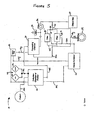
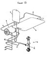
- a system 10 of the present invention is generally comprised of one or more of a tissue collection chamber 20, a processing chamber 30, a waste chamber 40, an output chamber 50 and a sample chamber 60.
- the various chambers are coupled together via one or more conduits 12 such that fluids containing biological material may pass from one chamber to another while maintaining a closed, sterile fluid/tissue pathway.
- the conduits may comprise rigid or flexible bodies referred to interchangeably herein as lumens and tubing, respectively.
- the conduits are in the form of flexible tubing, such as polyethylene tubing conventionally used in clinical settings, silicone or any other material known in the art.
- the conduits 12 can vary in size depending on whether passage of fluid or tissue is desired.
- the conduits 12 may also vary in size depending on the amount of tissue or fluid that is cycled through the system.
- the conduits may have a diameter ranging from about 0.060 to about 0.750 inches and for the passage of tissue, the conduits may have a diameter ranging from 0.312 to 0.750 inches.
- the size of the conduits is selected to balance the volume the conduits can accommodate and the time required to transport the tissue or fluids through said conduits.
- the foregoing parameters i.e., volume and time for transport, must be identified such that the appropriate signals can be transmitted to the processing device of the system. This allows the device to move accurate volumes of liquid and tissue from one chamber to another.
- the flexile tubing used should be capable of withstanding negative pressure to reduce the likelihood of collapse.
- the flexible tubing used should also be capable of withstanding positive pressure which is generated by, for example, a positive displacement pump, which may be used in the system.
- the conduits and ports may be coupled to, for example, a suction device (not shown) which may be manually or automatically operated.
- the suction device may be, e.g., a syringe or an electric pump.
- the suction device should be capable of providing sufficient negative pressure to aspirate tissue from a patient.
- any suitable suction device known to one of ordinary skill in the art, e.g., a surgeon, may be used.
- the conduits 12 may further comprise one or more clamps (not shown) to control the flow of material among various components of the system.
- the clamps are useful for maintaining the sterility of the system by effectively sealing different regions of the system.
- the conduits 12 may comprise one or more valves 14 that control the flow of material through the system.
- the valves 14 are identified as open circles in the Figures.
- the valves may be electromechanical pinch valves.
- the valves may be pneumatic valves.
- the valves may be hydraulic valves or mechanical valves.
- Such valves are preferably activated by a control system which may be coupled to levers. The levers may be manually manipulated such that the levers are activated.
- control system may be coupled to the levers as well as to a processing device which may activate the valves at pre-determined activation conditions.
- activation of the valves may be partially automated and partially subject to the user's preference such that the process may be optimized.
- certain valves may be activated manually and others automatically through the processing device.
- the valves 14 may also be used in conjunction with one or more pumps, e.g., peristaltic pumps 34 or positive displacement pumps (not shown).
- the conduits 12 and/or the valves 14 may also be comprised of sensors 29, e.g., optical sensors, ultrasonic sensors, pressure sensors or other forms of monitors known in the art that are capable of distinguishing among the various fluid components and fluid levels that flow through the system.
- the sensors 29 may be optical sensors.
- the system may also include a plurality of filters 36.
- the filters may be within a chamber of the system 28. Different chambers within the system may be comprised of different filters.
- the filters are effective to separate the regenerative cells, e.g., stem cells and/or progenitor cells, from undesirable cells and disaggregation agents that may be used in accordance with the system.
- a filter assembly 36 includes a hollow fiber filtration device.
- a filter assembly 36 includes a percolative filtration device, which may or may not be used with a sedimentation process.
- the filter assembly 36 comprises a centrifugation device, which may or may not be used with an elutriation device and process.
- the system comprises a combination of these filtering devices.
- the filtration functions of the present invention can be two-fold, with some filters removing things from the final concentration such as collagen, free lipid, free adipocytes and residual collagenase, and with other filters being used to concentrate the final product.
- the filters of the system may be comprised of a plurality of pores ranging in diameters and/or length from 20 to 800 ⁇ m.
- the collection chamber 20 has a prefixed filter 28 with a plurality of pores ranging from 80 to 400 ⁇ m.
- the collection chamber 20 has a prefixed filter 28 with a plurality of 265 ⁇ m pores.
- the filters may be detachable and/or disposable.
- the system may also be comprised of one or more temperature control devices (not shown) that are positioned to adjust the temperature of the material contained within one or more chambers of the system.
- the temperature control device may be a heater, a cooler or both, i.e., it may be able to switch between a heater and a cooler.
- the temperature device may adjust the temperature of any of the material passing through the system, including the tissue, the disaggregation agents, the resuspension agents, the rinsing agents, the washing agents or the additives. For example, heating of adipose tissue facilitates disaggregation whereas the cooling of the regenerative cell output is desirable to maintain viability. Also, if pre-warmed reagents are needed for optimal tissue processing, the role of the temperature device would be to maintain the pre-determined temperature rather than to increase or decrease the temperature.
- the collection chamber 20 may be comprised of a plurality of flexible or rigid canisters or cylinders or combinations thereof.
- the collection chamber 20 may be comprised of one or more rigid canisters of varying sizes.
- the collection chamber 20 may also be comprised of one or more flexible bags.
- the bag is preferably provided with a support, such as in internal or external frame, that helps reduce the likelihood that the bag will collapse upon the application of suction to the bag.
- the collection chamber 20 is sized to hold the requisite amount of saline to appropriately wash and disaggregate the tissue prior to the wash and concentrate stage of the process performed in the processing chamber 30.
- the volume of tissue or fluid present in the collection chamber 20 is easily ascertainable to the naked eye.
- a suitable collection chamber has the capacity to hold 800 ml of lipoaspirate and 1200 ml of saline. Accordingly, in one embodiment, the collection chamber 20 has a capacity of at least 2 liters. In another embodiment, to separate and concentrate red blood cells from blood, the collection chamber 20 has a capacity of at least 1.5 liters.
- the size of the collection chamber 20 will vary depending on the type and amount of tissue collected from the patient. The collection chamber 20 may be sized to hold as little as about 5 ml to up to about 2 liters of tissue. For smaller tissue volumes, e.g., 5 mls to 100 mls, the tissue may be gathered in a syringe prior to transfer to the collection chamber 20.
- the collection chamber 20 may be constructed using any suitable biocompatible material that can be sterilized.
- the collection chamber 20 is constructed of disposable material that meets biocompatibility requirements for intravascular contact as described in the ISO 10993 standard.
- polycarbonate acrylic or ABS may be used.
- the fluid path of the collection chamber 20 is preferably pyrogen free, i.e., suitable for blood use without danger of disease transmittal.
- the collection chamber 20 is constructed of a material that allows the user to visually determine the approximate volume of tissue present in the chamber. In other embodiments, the volume of tissue and/or fluid in the collection chamber 20 is determined by automated sensors 29.
- the collection chamber 20 is preferably designed such that in an automated embodiment, the system can determine the volume of tissue and/or fluid within the chamber with a reasonable degree of accuracy. In a preferred embodiment, the system senses the volume within the collection chamber with an accuracy of plus or minus fifteen percent.
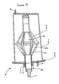
- the collection chamber 20 is in the form of a rigid chamber, for example, a chamber constructed of a medical grade polycarbonate containing a roughly conical prefixed filter 28 of medical grade polyester with a mesh size of 265 ⁇ m (see Figure 5 ).
- the rigid tissue collection container may have a size of approximately eight inches high and approximately five inches in diameter; the wall thickness may be about 0.125 inches.
- the interior of the cylinder may be accessed through, for example, one or more ports for suction tubing, one or more ports with tubing for connection through sterile docking technology, and/or one or more ports for needle puncture access through a rubber septum.
- the prefixed filter 28 in the interior of the collection chamber 20 is preferably structured to retain adipose tissue and to pass non-adipose tissue as, for example, the tissues are removed from the patient. More specifically, the filter 28 may allow passage of free lipid, blood, and saline, while retaining fragments of adipose tissue during, or in another embodiment after, the initial harvesting of the adipose tissue.
- the filter 28 includes a plurality of pores, of either the same or different sizes, but ranging in size from about 20 ⁇ m to 5 mm. In a preferred embodiment, the filter 28 includes a plurality of 400 ⁇ m pores.
- the filter 28 is a medical grade polyester mesh of around 200 ⁇ m thickness with a pore size of around 265 ⁇ m and around 47% open area. This material holds the tissue during rinsing but allows cells to pass out through the mesh following tissue disaggregation. Thus, when the tissues are aspirated from the patient, non-adipose tissue may be separated from adipose tissue.
- the same functionality could be achieved with different materials, mesh size, and the number and type of ports. For example, mesh pore sizes smaller than 100 ⁇ m or as large as several thousand microns would achieve the same purpose of allowing passage of saline and blood cells while retaining adipose tissue aggregates and fragments. Similarly, the same purpose could be achieved by use of an alternative rigid plastic material, or by many other modifications that would be known to those skilled in the art
- the containers for the washing solution 23 and the disaggregation agents 24 may be any suitable container that can hold their contents in a sterile manner, e.g., a collapsible bag, such as an IV bag used in clinical settings. These containers may have conduits 12, such as conduit 12e, coupled to the collection chamber 20 so that the washing solution and the disaggregation agent may be delivered to the interior of the collection chamber 20.
- the washing solution and the disaggregation agent may be delivered to the interior of the collection chamber 20 through any art-recognized manner, including simple gravity pressure applied to the outside of the containers for the saline 23 and/or the disaggregation agents 24 or by placement of a positive displacement pump on the conduits, e.g., conduit 12d in Figure 4 .
- the tissue and/or fluid within the collection chamber should be maintained at a temperature ranging from 30 degrees Celsius to 40 degrees Celsius. In a preferred embodiment, the temperature of the suspension inside the collection chamber is maintained at 37 degrees Celsius. In certain embodiments, if the surgical procedure or therapeutic application needs to be delayed, the selected tissue may be stored in the collection chamber for later use. The tissue may be stored at or about room temperature or at about 4 degrees Celsius for up to 96 hours.
- the washing solution may be any solution known to one of skill in the art, including saline or any other buffered or unbuffered electrolyte solution.
- the types of tissue being processed will dictate the types or combinations of washing solutions used.
- the washing solution such as saline
- the washing solution may be delivered to the collection chamber 20 before the adipose tissue is extracted, or may be delivered to the collection chamber 20 concurrently with the adipose tissue.
- the washing solution and the extracted adipose tissue may be mixed by any means including the methods described below.
- the tissue may be washed by agitation (which maximizes cell viability and minimizes the amount of free lipid released).
- the tissue is agitated by rotating the entire collection chamber 20 through an arc of varying degrees (e.g., through an arc of about 45 degrees to about 90 degrees) at varying speeds, e.g., about 30 revolutions per minute.
- the tissue is agitated by rotating the entire collection chamber 20, wherein the collection chamber 20 is comprised of one or more paddles or protrusions rigidly attached to an inside surface of the collection chamber, through an arc of varying degrees (e.g., through an arc of about 45 degrees to about 90 degrees) at varying speeds, e.g., about 30 revolutions per minute.
- the rotation of the collection chamber 20 described above may be accomplished by a drive mechanism attached to or in proximity with the collection chamber 20.
- the drive mechanism may be a simple belt or gear or other drive mechanism known in the art.
- the speed of the rotation may be, for example, 30 revolutions per minute. Generally, higher speeds have been found to generate larger volumes of free lipids and may not be optimal.
- the tissue is agitated by placing a rotatable shaft 25 inside the collection chamber 20, wherein the rotatable shaft is comprised of one or more paddles 25a or protrusions rigidly attached to the rotatable shaft 25 which pass through the mixture as the shaft is being rotated.
- the rotatable shaft 25 with rigidly attached 25a paddles may be rested on the bottom of the collection chamber 20. This may be accomplished, for example, by placing the paddle-like device into a spinning magnetic field (e.g., magnetic stirrer).
- agitating of the tissue may be accomplished using a simple agitator known in the art, i.e. a device implementing shaking up and down without rotation.
- the tissue may also be washed using any other art-recognized means including rocking, stirring, inversion, etc.
- a tissue disaggregation agent may be delivered to the collection chamber 20 to separate the regenerative cells from the remaining adipose tissue components.
- the disaggregation agent may be any disaggregation agent known to one of skill in the art. Disaggregation agents that may be used include neutral proteases, collagenase, trypsin, lipase, hyaluronidase, deoxyribonuclease, members of the Blendzyme enzyme mixture family, e.g., Liberase H1, pepsin, ultrasonic or other physical energy, lasers, microwaves, other mechanical devices and/or combinations thereof.
- a preferred disaggregation agent of the invention is collagenase.
- the disaggregation agents may be added with other solutions.
- saline such as saline delivered from a saline source 23 as described above, may be added to the adipose tissue along with or immediately followed by addition of collagenase.
- the washed adipose tissue is mixed with a collagenase-containing enzyme solution at or around 37° C for about 20-60 minutes.
- a higher concentration of collagenase or similar agent may be added to decrease the digestion time.
- the washed adipose tissue and the tissue disaggregation agent may then be agitated in manners similar to the agitation methods described above, until the washed adipose tissue is disaggregated.
- the adipose tissue may either be partially disaggregated, or completely disaggregated.
- a portion of washed adipose tissue may be removed and set aside in a sample container prior to any digestion.
- harvested adipose tissue is partially disaggregated to concentrate cells before being reintroduced back into the patient.
- the adipose tissue is mixed with a tissue disaggregation agent for a period of time generally less than about 20 minutes. A portion of the partially disaggregated tissue may then be removed from the collection chamber, and the remaining partially disaggregated tissue may be further disaggregated by mixing the adipose tissue with a tissue disaggregation agent for another 40 minutes.
- the adipose derived cells are to be used as an essentially pure population of regenerative cells, the adipose tissue may be fully disaggregated.
- the collection chamber 20 is preferably comprised of an outlet port 22 at the lowest point of the chamber such that blood and other non-buoyant components of the tissue may be drained to one or more waste containers 40 via one or more conduits 12.
- the collection chamber 20 is generally in (or may be placed in) an upright position such that the outlet ports 22 are located at the bottom of the collection chamber.
- the draining may be passive or active.
- the non-buoyant components described above could be drained using gravity, by applying positive or negative pressure, by use of pumps 34 or by use of vents 32.
- the processing device can signal certain valves and/or pumps to drain the non-buoyant layer from the collection chamber 20.
- the automated embodiments may also be comprised of sensors 29 which can detect when the interface between the buoyant and non-buoyant liquids has been reached.
- the automated embodiments may also be comprised of a sensor 29, e.g., an optical sensor, which may be capable of detecting a change in the light refraction of the effluent which is flowing in the conduit leading out of the collection chamber. The appropriate change in the light refraction may signal the presence of the buoyant layer in the outgoing conduits which indicates that the non-buoyant layer has been drained.
- the sensor 29 can then signal the processing device to proceed with the next step.
- the collection chamber 20 typically includes one or more ports 21 for permitting the washing solution to be delivered to the interior of the chamber, and one or more ports 22 for permitting waste and other materials to be directed out from the collection chamber 20.
- the collection chamber may include one or more sealed entry ports as described herein.
- the collection chamber 20 may also include one or more caps (not shown), such as a top cap and a bottom cap to further ensure that the system remains sterile while washing solution is delivered into the collection chamber and/or waste is transported out.
- the ports 21 may be provided on the caps of the collection chamber or on a sidewall of the collection chamber.
- the process of washing with fresh wash solution may be repeated until the residual content of non-buoyant contaminants in the solution reaches a pre-determined level.
- the remaining material in the collection chamber 20, which comprises the buoyant material of the mixture described above, including adipose tissue fragments may be washed one or more additional times until the amount of undesired material is reduced to a desired pre-determined level.
- One method of determining the end point of the washing is to measure the amount of red blood cells in the tissue solution. This can be accomplished by measuring the light absorbed on the 540 nm wavelength. In a preferred embodiment, a range between about 0.546 and about 0.842 is deemed acceptable.
- additives may be added to the various containers as needed to enhance the results.
- additives include agents that optimize washing and disaggregation, additives that enhance the viability of the active cell population during processing, anti-microbial agents (e.g., antibiotics), additives that lyse adipocytes and/or red blood cells, or additives that enrich for cell populations of interest (by differential adherence to solid phase moieties or to otherwise promote the substantial reduction or enrichment of cell populations).
- anti-microbial agents e.g., antibiotics
- additives that lyse adipocytes and/or red blood cells or additives that enrich for cell populations of interest (by differential adherence to solid phase moieties or to otherwise promote the substantial reduction or enrichment of cell populations).
- Other possible additives include those that promote recovery and viability of regenerative cells (for example, caspase inhibitors) or which reduce the likelihood of adverse reaction on infusion or emplacement (for example, inhibitors of re-aggregation of cells or connective tissue).
- This non-buoyant fraction is referred to herein as the regenerative cell composition and comprises multiple different types of cells, including stem cells, progenitor cells, endothelial precursor cells, adipocytes and other regenerative cells described herein.
- the regenerative cell composition may also contain one or more contaminants, such as collagen and other connective tissue proteins and fragments thereof, which were present in the adipose tissue fragments, or residual collagenase from the tissue disaggregation process.
- the processing chamber 30 of the invention is preferably positioned within the system such that the regenerative cell composition moves from the collection chamber 20 to the processing chamber 30 by way of tubing 12, valves 14 and pump 34 in a sterile manner.
- the processing chamber is sized to accommodate tissue/fluid mixtures ranging from 10mL to 1.2L. In a preferred embodiment, the processing chamber is sized to accommodate 800 mLs. In certain embodiments, the entire regenerative cell composition from the collection chamber 20 is directed to the processing chamber 30.
- the processing chamber 30 may be constructed in any manner suitable for separating and concentrating cells, including filtration and centrifugation and/or combinations thereof.
- the regenerative cell composition from the collection chamber 20 is introduced into the processing chamber 30 where the composition can be filtered to separate and/or concentrate a particular regenerative cell population.
- Cell filtration is a method of separating particular components and cells from other different components or types of cells.
- the regenerative cell composition of the invention comprises multiple different types of cells, including stem cells, progenitor cells and adipocytes, as well as one or more contaminants, such as collagen, which was present in the adipose tissue fragments, or residual collagenase from the tissue disaggregation process.
- the filters 36 present in the processing chamber 30 may allow for separation and concentration of a particular subpopulation of regenerative cells, e.g., stem cells or endothelial progenitors cells etc.
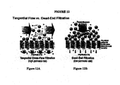
- Some variables which are associated with filtration of cells from a liquid include, but are not limited to, pore size of the filter media, geometry (shape) of the pore, surface area of the filter, flow direction of the solution being filtered, trans-membrane pressure, dilution of the particular cell population, particulate size and shape as well as cell size and cell viability.
- the particular cells that are desired to be separated or filtered are typically adipose derived stem cells.
- the particular cells may include adipose derived progenitor cells, such as endothelial precursor cells, alone or in combination with the stem cells.
- the regenerative cell composition containing the washed cells and residual collagen, adipocytes, and/or undigested tissue disaggregation agent, may be directed through the first filter to remove at least a portion of and preferably substantially all of the collagen particles from the composition so that fewer, and preferably no, collagen particles are present in the filtered solution.
- the filtered regenerative cell composition containing the adipocytes and/or undigested tissue disaggregation agent may then be directed through the second filter to remove at least a portion of and preferably substantially all of the free adipocytes from the filtered regenerative cell composition.
- the twice filtered regenerative cell composition containing the undigested tissue disaggregation agent, may be directed through the third filter, such as a hollow fiber filtration device, as discussed herein, to remove or reduce the undigested tissue disaggregation agent from the regenerative cell composition.
- the third filter such as a hollow fiber filtration device, as discussed herein, to remove or reduce the undigested tissue disaggregation agent from the regenerative cell composition.
- the efficiency of filtration of the stem cells may be enhanced relative to other filtration techniques.
- the pores of the filter media are placed in such a manner that the filter is orientated perpendicular to the flow of the fluid so that the Filter media blocks the path of the fluid being filtered, as illustrated in Figure 12B .
- the particles which are being filtered out of the regenerative cell composition e.g., the stem cells, tend to build up on one side of the filter and block the flow of the fluid through the pores. This blockage can reduce the efficiency of the filter.
- filtration is enhanced by generating a vacuum on the outside of the hollow tube filter media. Due to the size of the regenerative cells, e.g., stem cells or progenitor cells, these cells typically cannot pass through the pores of the body and therefore remain on the inside of the hollow tube filter (e.g., in the lumens of the tubes) and are directed back to the processing chamber 30 via a conduit between the filter and the processing chamber, or to the output chamber 50.
- the regenerative cells e.g., stem cells or progenitor cells
- the hollow fiber filter has about a 0.05 micron pore size, and contains approximately 550 cm 2 surface area of filter media.
- An individual media tube typically has a diameter of about 0.5 mm.
- approximately 120 ml of additional saline may be added to the composition.
- the processing or filter time may be approximately 8 minutes.
- the differential of the pressures on either side of the body of the hollow fiber tube e.g., the pressure inside the lumen of the body, and outside the body
- the trans-membrane pressure can range from about 1 mmHg to about 500 mmHg with a preferred pressure being about 200 mmHg.
- the average nucleated cell recovery and viability using hollow fiber filtration can be approximately 80% of viable cells.
- liquid may flow through the pump 34 before it is passed to the filter assembly 36.
- liquid may pass through the pressure sensor 39 before passing through the filter assembly to obtain a pre-filter liquid pressure in the system.
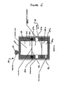
- one or more of these components may also be provided as an element of the processing chamber 30, such as the vent 32 as illustrated in Figure. 6 .
- the pressure sensor 39 is in line to determine the pressure of the regenerative cell composition which is generated by the pump 34 as it enters the filtering chamber of the filter assembly 36. This construction can facilitate monitoring of the trans-membrane pressure across the filter membrane.
- Additional saline or other buffer and washing solution can be added to the regenerative cell composition to assist in the removal of unwanted proteins as the composition is being filtered through the filter assembly 36. This repeated washing can be performed multiple times to enhance the purity of the regenerative cells.
- the saline can be added at any step as deemed necessary to enhance filtration.
- an output chamber 50 such as an output bag, may be connected to an outlet port of the processing chamber 30 and/or the filter assembly 36, depending on the specific embodiment.
- a vent such as the vent 32, may then be opened to facilitate the output of the concentrated regenerative cells.
- this determination of when a minimum concentration has been reached is made empirically after experiments have been run and programmed into the electronic controls of the device.
- Port 31a is illustrated as a sample inlet port, which is constructed to be coupled to a conduit so that a composition containing regenerative cells can be passed into the interior of the processing chamber 30.
- Port 31b is illustrated as an outlet port constructed to be coupled to a conduit so that the separated and concentrated cells may be removed from the interior of the processing chamber 30.
- Port 31c is illustrated as an inlet port constructed to be coupled to a conduit for delivery of a fresh washing solution, such as saline into the interior of the processing chamber 30.
- the composition containing the separated cells may be concentrated, as discussed herein.
- the composition may be further concentrated after it has been removed from chamber 37a through outlet port 31b, or while it is in the chamber 37a.
- the concentration of cells in the composition is increased in the following manner.
- the filters such as filters 36a and 36b, may be moved towards each other. This movement has the effect of reducing the volume between the two filters (e.g., the volume of chamber 37a).
- a vibrating member may also be provided in connection with the processing chamber 30 to facilitate concentrating of the cells in the composition.
- the vibrating member may be coupled to the filter 36b (e.g., the small pore filter). Vibrating can reduce an incidence of cells becoming trapped in the filters. The reduction in volume of the composition allows the excess saline to be removed as waste and the cells to be concentrated in a smaller volume.
- the filter 36b e.g., the small pore filter
- the concentration of the regenerative cells is accomplished in the following manner.
- the regenerative cell composition can be transferred to another chamber (not shown) which uses gravity to filter out the excess saline.
- the sedimentation can occur at the same time as the percolation. This sedimentation may be accomplished by introducing the composition on top of a filter which has a pore size ranging from about 10 kD to about 2 microns. In one embodiment, a suitable filter has a pore size of about 1 micron. The force of gravity will allow the saline and smaller particles to be passed through the filter while preventing the cells in the composition to flow through the filter.
- the regenerative cell composition may be agitated to remove the cells from the filter and, subsequently, the concentrated regenerative cells may be transferred to the output bag.
- the smaller particles can be drawn off as waste through an outlet.
- the regenerative cell composition from the collection chamber 20 is transported to the processing chamber 30 wherein the composition can be centrifuged to separate and concentrate regenerative cells.
- Centrifugation principles are well know in the art and will be not be repeated herein in the interest of brevity. Standard, art-recognized centrifugation devices, components and parameters are utilized herein.
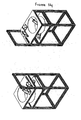
- An exemplary processing chamber for use as part of a centrifuge device is shown in Figures 7 and 8 .
- a centrifuge device causes a centrifuge chamber (such as the one shown in Figure 7 ) to spin around an axis to thereby increasing the force on the cells in the solution to be greater than gravity.
- the processing chamber may be comprised of a cell concentrator in the form of a spinning membrane filter.
- centrifugal elutriation may also be applied.
- the cells may be separated based on the individual cell sedimentation rate such that the directional (e.g., outward) force applied by centrifugation causes cells and solutes to sediment at different rates.
- the sedimentation rate of the target cell population is opposed by an opposite (e.g., inward) flow rate applied by pumping solution in the opposite direction to the centrifugal force. The counterflow is adjusted so that the cells and particles within the solution are separated.
- Figure 9 illustrates principles associated with an elutriation implementation in accordance with the present invention.
- the elutriation embodiment can be similar to a centrifugation implementation to the extent that a force is applied to the solution using a spinning rotor.
- Some of the variables which are associated with the presently embodied elutriation separation include, but are not limited to, the size and shape of the spinning chamber, the diameter of the rotor, the speed of the rotor, the diameter of the counter flow tubing, the flow rate of the counter flow, as well as the size and density of the particles and cells which are to be removed from solution.
- the regenerative cells can be separated based on individual cell densities.
- the processing chamber 30 or the output chamber 50 may include one or more ports, e.g., ports 51 or 52.
- ports 51 or 52 may be designed to transport the regenerative cells obtained using any combination of methods described above, or a portion thereof, via conduits to other surgical devices, cell culturing devices, cell marinading devices, gene therapy devices or purification devices.
- These ports may also be designed to transport the regenerative cells via conduits to additional chambers or containers within the system or as part of another system for the same purposes described above.
- the ports and conduits may be also be used to add one or more additives, e.g., growth factors, re-suspension fluids, cell culture reagents, cell expansion reagents, cell preservation reagents or cell modification reagents including agents that transfer genes to the cells.
- the ports and conduits may also be used to transport the regenerative cells to other targets such as implant materials (e.g., scaffolds or bone fragments) as well as other surgical implants and devices.
- the system can be reconfigured by any of the means described above such that the regenerative cells obtained using the system may be subject to one or more of the following: cell expansion (of one or more regenerative cell types)and cell maintenance (including cell sheet rinsing and media changing); sub-culturing; cell seeding; transient transfection (including seeding of transfected cells from bulk supply); harvesting (including enzymatic, non-enzymatic harvesting and harvesting by mechanical scraping); measuring cell viability; cell plating (e.g., on microtiter plates, including picking cells from individual wells for expansion, expansion of cells into fresh wells); high throughput screening; cell therapy applications; gene therapy applications; tissue engineering applications; therapeutic protein applications; viral vaccine applications; harvest of regenerative cells or supernatant for banking or screening, measurement of cell growth, lysis, inoculation, infection or induction; generation of cells lines (including hybridoma cells); culture of cells for permeability studies; cells for RNAi and viral resistance studies; cells for knock-out and transgenic
- additives include agents that optimize washing and disaggregation, additives that enhance the viability of the active cell population during processing, anti-microbial agents (e.g., antibiotics), additives that lyse adipocytes and/or red blood cells, or additives that enrich for cell populations of interest (by differential adherence to solid phase moieties or to otherwise promote the substantial reduction or enrichment of cell populations) as described herein.
- anti-microbial agents e.g., antibiotics
- additives that lyse adipocytes and/or red blood cells e.g., lyse adipocytes and/or red blood cells
- additives that enrich for cell populations of interest by differential adherence to solid phase moieties or to otherwise promote the substantial reduction or enrichment of cell populations
- the system is automated.
- the system has both automated and manual components.
- the system may be comprised of one or more disposable components connected to or mounted on a re-usable hardware component or module.
- the automated systems of the invention provide screen displays (see Figure 16 ) that prompt proper operation of the system.
- the automated systems may also provide a screen that provides status of the procedure and/or the step by step instructions as to the proper setup of the disposable components of the system.
- the screen may also indicate problems or failures in the system if they occur and provide "troubleshooting" guidance if appropriate.
- the screen is a user interface screen that allows the user to input parameters into the system through, e.g., a touch screen.
- the processing device may also have pre-programmed software which provides the user with appropriate parameters to optimize the process based on the user's input of relevant information such as the amount of regenerative cells required, the type of tissue being processed, the type of post-processing manipulation required, the type of therapeutic application, etc.
- a sensor 29 can signal the processing device present in the re-usable component to activate the steps needed to wash and disaggregate the tissue.
- the processing device may introduce a pre-set volume of washing agent based on the volume of tissue collected using automated valves and pumps. This cycle may be repeated in the collection chamber until the optical sensor determines that the effluent liquid is sufficiently clear and devoid of unwanted material.
- an optical sensor 29 along the conduit leading out of the collection chamber 12b or 12d can detect that the unwanted materials have been removed and can signal the processing device to close the required valves and initiate the next step.
- the processing chamber 30 shown in Figure 4 is in the form of a centrifuge chamber.
- a detailed illustration of the processing chamber of Figure 4 is shown in Figures 7 and 8 .
- Such a processing chamber 30 is generally comprised of a rotating seal network30.1 comprising an outer housing 30.2, one or more seals 30.3, one or more bearings 30.4 and an attachment point 30.6 for connecting the processing chamber to the centrifuge device present in the re-usable component of the system; one or more fluid paths 30.5 in the form of conduits extending out from the rotating seal and ending in a centrifuge chamber on each end which is in the form of an output chamber 50 housed in a frame 53 wherein the frame is comprised of one or more ports 52 and one or more handles to manually re-position the output chamber 50.
- the three lip seals are comprised of a circular "U" shaped channel (not shown) as well as a circular spring (not shown).
- the circular "U” shaped channel is preferably fabricated using flexible material such that a leakage proof junction with the rotating shaft of the rotating seal network 30.1 is formed.
- the lip seals are preferably oriented in a manner such that pressure from the regenerative cell composition flowing through the processing chamber causes the seal assembly to tighten its junction with the rotating shaft by way of increased tension.
- the seals may be secured in position by way of one or more circular clips (not shown) which are capable of expanding and/or collapsing as needed in order to engage a groove in the outer housing 30.2 of the rotating seal network 30.1.
- the rotating seal network 30.1 is comprised of a single rubber seal 30.3 and an air gasket (not shown). This seal and gasket provide a tortuous path for any biologic matter which could compromise the sterility of the system.
- the rotating seal network 30.1 is comprised of multiple spring loaded seals 30.3 which isolate the individual fluid paths. The seals 30.3 are fabricated of a material which can be sterilized as well as seal the rotating shaft without lubricant.
- the rotating seal network 30.1 is compromised of a pair of ceramic disks (not shown) which create the different fluid paths and can withstand the rotation of the system and not cause cell lysis.
- the fluid pathway is flexible and is allowed to wind and unwind with respect to the processing chamber. This is accomplished by having the flexible fluid pathway rotate one revolution for every two revolutions of the processing chamber 30. This eliminates the need for a rotating seal altogether.
- the cell pellet that is obtained using the system shown in Figure 4 comprises the concentrated regenerative cells of the invention.
- a fluid path 30.5 may be used to re-suspend the cell pellet that is formed after centrifugation with additional solutions and/or other additives. Re-suspension of the cell pellet in this manner allows for further washing of the regenerative cells to remove unwanted proteins and chemical compounds as well as increasing the flow of oxygen to the cells.
- the resulting suspension may be subjected to another load of approximately 400 times the force of gravity for another period of approximately 5 minutes.
- PIGF and VEGF expression by adipose derived regenerative cells was examined using an ELISA assay (R&D Systems, Minneapolis, MN) using cells from three donors.
- One donor had a history of hyperglycemia and Type 2 diabetes (a condition highly associated with microvascular and macrovascular disease, including patients with coronary artery disease).
- ADC cells from each donor were plated at 1,000 cells/cm 2 in DMEM/F-12 medium supplemented with 10% FCS and 5% HS and grown until confluent. Supernatant samples were taken and assayed for expression of PIGF and VEGF protein.
- Figures 16A and 16B the results demonstrate robust expression of both VEGF ( Figure 16A ) and PIGF ( Figure 16B ) by the adipose derived regenerative cells of the invention.
- VEGF Vascular Endothelial Growth Factor
- bFGF Vascular Endothelial Growth Factor
- IGF-II Vascular Endothelial Growth Factor
- Eotaxin G-CSF
- GM-CSF IL-12 p40/p70
- IL-12 p70 IL-13
- IL-6 IL-9
- Leptin MCP-1, M-CSF, MIG, PF-4, TIMP-1, TIMP-2, TNF- ⁇ , and Thrombopoetin.
- the following angiogenic related growth factors or cytokines were elevated at least twice compare to blank control medium with 10% FBS: Vascular Endothelial Growth Factor (VEGF), Eotaxin, G-CSF, IL-6, MCP-1 and PF-4.
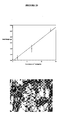
- ADCs were isolated by enzymatic digestion of human subcutaneous adipose tissue. ADCs (100 ⁇ l) were incubated in phosphate saline buffer (PBS) containing 0.2% fetal bovine serum (FBS), and incubated for 20 to 30 minutes at 4°C with fluorescently labeled antibodies directed towards the human endothelial markers CD-31 (differentiated endothelial cell marker) and CD-34 (EPC marker), as well as human ABCG2 (ATP binding cassette transporter), which is selectively expressed on multipotent cells. After washing, cells were analyzed on a FACSAria Sorter (Beckton Dickenson - Immunocytometry).
- PBS phosphate saline buffer
- FBS fetal bovine serum
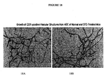
- EPCs within the adipose tissue derived regenerative cells indicates that these cells can participate directly in development of new blood vessels and enhance angiogenesis and restore perfusion.
- Transplant of autologous adipose tissue is a relatively common procedure in plastic and reconstructive surgery ⁇ Fulton, 1998; Shiffman, 2001 ⁇ .
- this procedure is limited by the fact that the adipose tissue fragments are transferred without a vascular supply and, as a result, graft survival is dependent upon neovascularization (Coleman, 1995; Eppley et al., 1990).
- the transplanted tissue represents an ischemic tissue.
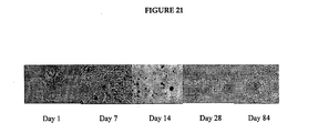
- mice hearts were harvested and processed at the following 5 timepoints after injection: day 1, day 7, day 14, day 28, day 84.
- the results demonstrate engraftment of donor derived adipose derived regenerative cells in the area of infarcted myocardium at all timepoints referenced above.
- Figure 21 demonstrates a histological timeline of engraftment.
- Improvements in the patient are noted within approximately six hours after the cell administration procedure. Several days after the cell administration procedure further improvement of the patient is noted evidenced by increased cardiac ejection fraction, decreased rate of heart failure, decreased infarct size, improved exercise tolerance and other quality of life measures.
Priority Applications (1)
| Application Number | Priority Date | Filing Date | Title |
|---|---|---|---|
| EP11152868.3A EP2465923B1 (de) | 2005-05-25 | 2005-05-25 | Verfahren zur verwendung von zellen aus fettgewebe zur behandlung kardiovaskulärer erkrankungen |
Applications Claiming Priority (3)
| Application Number | Priority Date | Filing Date | Title |
|---|---|---|---|
| EP05754073A EP1885382B1 (de) | 2005-05-25 | 2005-05-25 | Verfahren zur verwendung von aus fettgewebe stammenden zellen bei der behandlung von herz-kreislauf-leiden |
| EP11152868.3A EP2465923B1 (de) | 2005-05-25 | 2005-05-25 | Verfahren zur verwendung von zellen aus fettgewebe zur behandlung kardiovaskulärer erkrankungen |
| PCT/US2005/018605 WO2006127007A2 (en) | 2005-05-25 | 2005-05-25 | Methods of using adipose tissue-derived cells in the treatment of cardiovascular conditions |
Related Parent Applications (2)
| Application Number | Title | Priority Date | Filing Date |
|---|---|---|---|
| EP05754073.4 Division | 2005-05-25 | ||
| EP05754073A Division EP1885382B1 (de) | 2005-05-25 | 2005-05-25 | Verfahren zur verwendung von aus fettgewebe stammenden zellen bei der behandlung von herz-kreislauf-leiden |
Publications (3)
| Publication Number | Publication Date |
|---|---|
| EP2465923A2 true EP2465923A2 (de) | 2012-06-20 |
| EP2465923A3 EP2465923A3 (de) | 2012-06-27 |
| EP2465923B1 EP2465923B1 (de) | 2018-04-11 |
Family
ID=37452485
Family Applications (2)
| Application Number | Title | Priority Date | Filing Date |
|---|---|---|---|
| EP05754073A Not-in-force EP1885382B1 (de) | 2005-05-25 | 2005-05-25 | Verfahren zur verwendung von aus fettgewebe stammenden zellen bei der behandlung von herz-kreislauf-leiden |
| EP11152868.3A Not-in-force EP2465923B1 (de) | 2005-05-25 | 2005-05-25 | Verfahren zur verwendung von zellen aus fettgewebe zur behandlung kardiovaskulärer erkrankungen |
Family Applications Before (1)
| Application Number | Title | Priority Date | Filing Date |
|---|---|---|---|
| EP05754073A Not-in-force EP1885382B1 (de) | 2005-05-25 | 2005-05-25 | Verfahren zur verwendung von aus fettgewebe stammenden zellen bei der behandlung von herz-kreislauf-leiden |
Country Status (12)
| Country | Link |
|---|---|
| EP (2) | EP1885382B1 (de) |
| JP (1) | JP5210155B2 (de) |
| KR (2) | KR101400544B1 (de) |
| CN (1) | CN101443023A (de) |
| AT (1) | ATE500319T1 (de) |
| AU (1) | AU2005332046B2 (de) |
| CA (1) | CA2609361C (de) |
| DE (1) | DE602005026724D1 (de) |
| DK (1) | DK1885382T3 (de) |
| ES (1) | ES2364689T3 (de) |
| PL (1) | PL1885382T3 (de) |
| WO (1) | WO2006127007A2 (de) |
Cited By (1)
| Publication number | Priority date | Publication date | Assignee | Title |
|---|---|---|---|---|
| EP3406255A4 (de) * | 2016-01-19 | 2019-08-14 | Osaka University | Transplantatmaterial für therapie für herzkrankheiten |
Families Citing this family (30)
| Publication number | Priority date | Publication date | Assignee | Title |
|---|---|---|---|---|
| US9597395B2 (en) | 2001-12-07 | 2017-03-21 | Cytori Therapeutics, Inc. | Methods of using adipose tissue-derived cells in the treatment of cardiovascular conditions |
| WO2003053346A2 (en) | 2001-12-07 | 2003-07-03 | Macropore Biosurgery, Inc. | Systems and methods for treating patients with processed lipoaspirate cells |
| US20050048035A1 (en) | 2001-12-07 | 2005-03-03 | Fraser John K. | Methods of using regenerative cells in the treatment of stroke and related diseases and disorders |
| US20050095228A1 (en) | 2001-12-07 | 2005-05-05 | Fraser John K. | Methods of using regenerative cells in the treatment of peripheral vascular disease and related disorders |
| US7771716B2 (en) | 2001-12-07 | 2010-08-10 | Cytori Therapeutics, Inc. | Methods of using regenerative cells in the treatment of musculoskeletal disorders |
| US8622997B2 (en) | 2005-03-23 | 2014-01-07 | Ronald D. Shippert | Tissue transfer method and apparatus |
| US9581942B1 (en) | 2005-03-23 | 2017-02-28 | Shippert Enterprises, Llc | Tissue transfer method and apparatus |
| US7789872B2 (en) | 2005-03-23 | 2010-09-07 | Shippert Ronald D | Tissue transplantation method and apparatus |
| US8062286B2 (en) | 2005-03-23 | 2011-11-22 | Shippert Ronald D | Tissue transplantation method and apparatus |
| US7780649B2 (en) | 2005-03-23 | 2010-08-24 | Shippert Ronald D | Tissue transplantation method and apparatus |
| US7794449B2 (en) | 2005-03-23 | 2010-09-14 | Shippert Ronald D | Tissue transplantation method and apparatus |
| ES2325715B1 (es) | 2007-08-03 | 2010-06-17 | Genetrix, S.L. | Poblacion de celulas madre adultas derivadas de tejido adiposo cardiaco y su uso en regeneracion cardiaca. |
| WO2010021993A1 (en) | 2008-08-19 | 2010-02-25 | Cytori Therapeutics, Inc. | Methods of using adipose tissue-derived cells in the treatment of the lymphatic system and malignant disease |
| KR101679671B1 (ko) * | 2009-10-27 | 2016-11-28 | 도병록 | 재생성 세포 추출 시스템 |
| US8887770B1 (en) | 2011-03-17 | 2014-11-18 | Ronald D. Shippert | Vessel fill control method and apparatus |
| CN102151153A (zh) * | 2011-03-25 | 2011-08-17 | 吴成翰 | 外科手术过程中组织碎片的回收装置 |
| JP5628131B2 (ja) * | 2011-10-19 | 2014-11-19 | サイトリ セラピューティクス インコーポレイテッド | 心血管状態の治療において脂肪組織由来細胞を使用する方法 |
| DE102013209718B4 (de) * | 2012-06-22 | 2015-09-10 | Human Med Ag | Vorrichtung zum Separieren von adulten Stammzellen |
| US9259435B2 (en) | 2012-08-31 | 2016-02-16 | W. L. Gore & Associates, Inc. | Reactive oxidative species generating materials and methods of use |
| US9468709B2 (en) | 2012-11-12 | 2016-10-18 | Shippert Enterprises, Llc | Syringe fill method and apparatus |
| US9580678B2 (en) | 2013-06-21 | 2017-02-28 | The Regents Of The University Of California | Microfluidic tumor tissue dissociation device |
| WO2015131087A1 (en) * | 2014-02-28 | 2015-09-03 | Cytovera Inc. | A system for tissu manipulation |
| US10772997B2 (en) | 2014-05-15 | 2020-09-15 | Ronald D. Shippert | Tissue parcelization method and apparatus |
| DE102015109148B3 (de) * | 2015-06-10 | 2016-05-04 | Human Med Ag | Vorrichtung zum Separieren von regenerativen und adulten Stammzellen |
| CN106318905A (zh) * | 2015-07-10 | 2017-01-11 | 中国科学院上海生命科学研究院 | 血栓烷素a2受体作为脂肪干细胞治疗缺血性疾病的靶标 |
| US10722540B1 (en) | 2016-02-01 | 2020-07-28 | The Regents Of The University Of California | Microfluidic device and method for shear stress-induced transformation of cells |
| IL263422B (en) | 2016-06-08 | 2022-09-01 | Univ California | Method and device for processing tissues and cells |
| JP6256786B1 (ja) * | 2017-04-12 | 2018-01-10 | 学校法人福岡大学 | 不妊症の処置に用いられる医薬組成物およびその製造方法 |
| CN107988151A (zh) * | 2017-12-06 | 2018-05-04 | 北京大学第三医院 | 一种体外诱导带血管蒂的脂肪组织成肌方法 |
| US20230063104A1 (en) * | 2020-02-04 | 2023-03-02 | Hierabio Inc | Composition for intrapericardial injection comprising stem cells and use thereof |
Citations (12)
| Publication number | Priority date | Publication date | Assignee | Title |
|---|---|---|---|---|
| US482820A (en) | 1892-09-20 | Ernest p | ||
| WO1997023510A1 (fr) | 1995-12-21 | 1997-07-03 | Centre National De La Recherche Scientifique | Anticorps anti-idiotypiques du facteur de croissance endotheliale vasculaire et leur utilisation comme medicaments |
| US5919234A (en) | 1996-08-19 | 1999-07-06 | Macropore, Inc. | Resorbable, macro-porous, non-collapsing and flexible membrane barrier for skeletal repair and regeneration |
| US6001642A (en) | 1998-06-29 | 1999-12-14 | Wyle Laboratories, Inc. Life Sciences | Bioreactor and cell culturing processes using the bioreactor |
| US6238908B1 (en) | 1995-06-07 | 2001-05-29 | Aastrom Biosciences, Inc. | Apparatus and method for maintaining and growth biological cells |
| US6269716B1 (en) | 1998-11-18 | 2001-08-07 | Macropore, Inc. | High-torque resorbable screws |
| US6280473B1 (en) | 1996-08-19 | 2001-08-28 | Macropore, Inc. | Resorbable, macro-porous, non-collapsing and flexible membrane barrier for skeletal repair and regeneration |
| US6391059B1 (en) | 1998-04-07 | 2002-05-21 | Macropore, Inc. | Membrane with tissue-guiding surface corrugations |
| US20020182211A1 (en) | 2000-05-26 | 2002-12-05 | Peach Robert J. | Soluble CTLA4 mutant molecules and uses thereof |
| US6635064B2 (en) | 2000-10-04 | 2003-10-21 | Macropore Biosurgery, Inc. | Non-scatterable, radio-opaque material for medical imaging applications |
| US6653146B1 (en) | 1999-11-15 | 2003-11-25 | Chemclean Corporation | Bio-burden visualization system |
| US6673362B2 (en) | 2000-03-10 | 2004-01-06 | Macropore Biosurgery, Inc. | Resorbable barrier micro-membranes for attenuation of scar tissue during healing |
Family Cites Families (10)
| Publication number | Priority date | Publication date | Assignee | Title |
|---|---|---|---|---|
| DK1007631T4 (da) * | 1997-07-14 | 2009-04-27 | Osiris Therapeutics Inc | Hjertemuskelregeneration ved anvendelse af mesenkymale stamceller |
| FR2819265B1 (fr) * | 2001-01-10 | 2004-01-02 | Centre Nat Rech Scient | Cellules du tissu adipeux extramedullaire et leurs applications dans la reconstitution des lignees hematopoietiques |
| JP2004532202A (ja) * | 2001-03-15 | 2004-10-21 | シアオ,ヨング−フー | 生存哺乳動物における臨床的に認められた形態の心臓の病状を治療的に処置する方法 |
| US20050008626A1 (en) * | 2001-12-07 | 2005-01-13 | Fraser John K. | Methods of using adipose tissue-derived cells in the treatment of cardiovascular conditions |
| WO2003053346A2 (en) * | 2001-12-07 | 2003-07-03 | Macropore Biosurgery, Inc. | Systems and methods for treating patients with processed lipoaspirate cells |
| JP2004099471A (ja) * | 2002-09-05 | 2004-04-02 | Cardio Corp | 心筋梗塞および心不全の治療薬 |
| RU2372924C2 (ru) * | 2003-02-20 | 2009-11-20 | Ситори Терапьютикс,Инк | Применение композиции, состоящей из концентрированной клеточной популяции, представляющей собой стволовые клетки и недифференцированные клетки-предшественники |
| CA2530630A1 (en) * | 2003-06-25 | 2005-02-10 | Macropore Biosurgery Inc. | Systems and methods for separating and concentrating regenerative cells from tissue |
| ES2633604T3 (es) * | 2003-09-17 | 2017-09-22 | Cytori Therapeutics, Inc. | Métodos para usar células regenerativas derivadas de adiposo en el tratamiento de una enfermedad vascular periférica |
| DE602004024125D1 (de) * | 2003-12-25 | 2009-12-24 | Kanazawa University Technology | Induktion einer myokardzelle unter verwendung einer säuger-knochenmarkzelle oder aus nabelschnurblut stammenden zelle und fettgewebe |
-
2005
- 2005-05-25 CN CNA2005800502651A patent/CN101443023A/zh active Pending
- 2005-05-25 EP EP05754073A patent/EP1885382B1/de not_active Not-in-force
- 2005-05-25 KR KR1020137005581A patent/KR101400544B1/ko active IP Right Grant
- 2005-05-25 DK DK05754073.4T patent/DK1885382T3/da active
- 2005-05-25 DE DE602005026724T patent/DE602005026724D1/de active Active
- 2005-05-25 KR KR1020077030170A patent/KR101278437B1/ko active IP Right Grant
- 2005-05-25 JP JP2008513441A patent/JP5210155B2/ja active Active
- 2005-05-25 AU AU2005332046A patent/AU2005332046B2/en not_active Ceased
- 2005-05-25 CA CA2609361A patent/CA2609361C/en not_active Expired - Fee Related
- 2005-05-25 ES ES05754073T patent/ES2364689T3/es active Active
- 2005-05-25 PL PL05754073T patent/PL1885382T3/pl unknown
- 2005-05-25 EP EP11152868.3A patent/EP2465923B1/de not_active Not-in-force
- 2005-05-25 AT AT05754073T patent/ATE500319T1/de not_active IP Right Cessation
- 2005-05-25 WO PCT/US2005/018605 patent/WO2006127007A2/en active Application Filing
Patent Citations (13)
| Publication number | Priority date | Publication date | Assignee | Title |
|---|---|---|---|---|
| US482820A (en) | 1892-09-20 | Ernest p | ||
| US6238908B1 (en) | 1995-06-07 | 2001-05-29 | Aastrom Biosciences, Inc. | Apparatus and method for maintaining and growth biological cells |
| WO1997023510A1 (fr) | 1995-12-21 | 1997-07-03 | Centre National De La Recherche Scientifique | Anticorps anti-idiotypiques du facteur de croissance endotheliale vasculaire et leur utilisation comme medicaments |
| US5919234A (en) | 1996-08-19 | 1999-07-06 | Macropore, Inc. | Resorbable, macro-porous, non-collapsing and flexible membrane barrier for skeletal repair and regeneration |
| US6280473B1 (en) | 1996-08-19 | 2001-08-28 | Macropore, Inc. | Resorbable, macro-porous, non-collapsing and flexible membrane barrier for skeletal repair and regeneration |
| US6343531B2 (en) | 1997-11-20 | 2002-02-05 | Macropore, Inc. | High-torque resorbable screws |
| US6391059B1 (en) | 1998-04-07 | 2002-05-21 | Macropore, Inc. | Membrane with tissue-guiding surface corrugations |
| US6001642A (en) | 1998-06-29 | 1999-12-14 | Wyle Laboratories, Inc. Life Sciences | Bioreactor and cell culturing processes using the bioreactor |
| US6269716B1 (en) | 1998-11-18 | 2001-08-07 | Macropore, Inc. | High-torque resorbable screws |
| US6653146B1 (en) | 1999-11-15 | 2003-11-25 | Chemclean Corporation | Bio-burden visualization system |
| US6673362B2 (en) | 2000-03-10 | 2004-01-06 | Macropore Biosurgery, Inc. | Resorbable barrier micro-membranes for attenuation of scar tissue during healing |
| US20020182211A1 (en) | 2000-05-26 | 2002-12-05 | Peach Robert J. | Soluble CTLA4 mutant molecules and uses thereof |
| US6635064B2 (en) | 2000-10-04 | 2003-10-21 | Macropore Biosurgery, Inc. | Non-scatterable, radio-opaque material for medical imaging applications |
Cited By (1)
| Publication number | Priority date | Publication date | Assignee | Title |
|---|---|---|---|---|
| EP3406255A4 (de) * | 2016-01-19 | 2019-08-14 | Osaka University | Transplantatmaterial für therapie für herzkrankheiten |
Also Published As
| Publication number | Publication date |
|---|---|
| PL1885382T3 (pl) | 2011-09-30 |
| WO2006127007A3 (en) | 2009-04-16 |
| ES2364689T3 (es) | 2011-09-12 |
| KR101400544B1 (ko) | 2014-05-27 |
| CN101443023A (zh) | 2009-05-27 |
| KR20130038412A (ko) | 2013-04-17 |
| EP1885382A4 (de) | 2009-09-09 |
| CA2609361A1 (en) | 2006-11-30 |
| CA2609361C (en) | 2017-11-14 |
| KR101278437B1 (ko) | 2013-06-25 |
| EP2465923B1 (de) | 2018-04-11 |
| ATE500319T1 (de) | 2011-03-15 |
| KR20080017389A (ko) | 2008-02-26 |
| AU2005332046B2 (en) | 2013-05-16 |
| WO2006127007A2 (en) | 2006-11-30 |
| EP2465923A3 (de) | 2012-06-27 |
| DE602005026724D1 (de) | 2011-04-14 |
| AU2005332046A1 (en) | 2006-11-30 |
| EP1885382A2 (de) | 2008-02-13 |
| JP2008545687A (ja) | 2008-12-18 |
| EP1885382B1 (de) | 2011-03-02 |
| DK1885382T3 (da) | 2011-06-20 |
| JP5210155B2 (ja) | 2013-06-12 |
Similar Documents
| Publication | Publication Date | Title |
|---|---|---|
| EP1885382B1 (de) | Verfahren zur verwendung von aus fettgewebe stammenden zellen bei der behandlung von herz-kreislauf-leiden | |
| US20170281771A1 (en) | Methods of using adipose tissue-derived cells in the treatment of cardiovascular conditions | |
| US9849149B2 (en) | Methods of using regenerative cells in the treatment of erectile dysfunction | |
| EP2348103B1 (de) | Verfahren zur Verwendung von regenerativen Zellen bei der Behandlung der peripheren Gefäßkrankheit und zugehörigen Erkrankungen | |
| US9504718B2 (en) | Methods of using adipose derived regenerative cells in the treatment of renal diseases and disorders | |
| US8105580B2 (en) | Methods of using adipose derived stem cells to promote wound healing | |
| EP2332555A2 (de) | Verfahren zur Verwendung regenerativer Zellen zur Förderung der Wundheilung | |
| AU2013216683B2 (en) | Methods of using adipose tissue-derived cells in the treatment of cardiovascular conditions | |
| JP5628131B2 (ja) | 心血管状態の治療において脂肪組織由来細胞を使用する方法 | |
| EP1778834B1 (de) | Verfahren zur verwendung regenerativer zellen bei der behandlung von nierenkrankheiten und störungen |
Legal Events
| Date | Code | Title | Description |
|---|---|---|---|
| PUAL | Search report despatched |
Free format text: ORIGINAL CODE: 0009013 |
|
| PUAI | Public reference made under article 153(3) epc to a published international application that has entered the european phase |
Free format text: ORIGINAL CODE: 0009012 |
|
| AC | Divisional application: reference to earlier application |
Ref document number: 1885382 Country of ref document: EP Kind code of ref document: P |
|
| AK | Designated contracting states |
Kind code of ref document: A2 Designated state(s): AT BE BG CH CY CZ DE DK EE ES FI FR GB GR HU IE IS IT LI LT LU MC NL PL PT RO SE SI SK TR |
|
| AK | Designated contracting states |
Kind code of ref document: A3 Designated state(s): AT BE BG CH CY CZ DE DK EE ES FI FR GB GR HU IE IS IT LI LT LU MC NL PL PT RO SE SI SK TR |
|
| RIC1 | Information provided on ipc code assigned before grant |
Ipc: C12N 5/0775 20100101AFI20120524BHEP |
|
| 17P | Request for examination filed |
Effective date: 20121220 |
|
| 17Q | First examination report despatched |
Effective date: 20130614 |
|
| GRAP | Despatch of communication of intention to grant a patent |
Free format text: ORIGINAL CODE: EPIDOSNIGR1 |
|
| INTG | Intention to grant announced |
Effective date: 20171031 |
|
| GRAS | Grant fee paid |
Free format text: ORIGINAL CODE: EPIDOSNIGR3 |
|
| GRAA | (expected) grant |
Free format text: ORIGINAL CODE: 0009210 |
|
| AC | Divisional application: reference to earlier application |
Ref document number: 1885382 Country of ref document: EP Kind code of ref document: P |
|
| AK | Designated contracting states |
Kind code of ref document: B1 Designated state(s): AT BE BG CH CY CZ DE DK EE ES FI FR GB GR HU IE IS IT LI LT LU MC NL PL PT RO SE SI SK TR |
|
| REG | Reference to a national code |
Ref country code: GB Ref legal event code: FG4D |
|
| REG | Reference to a national code |
Ref country code: CH Ref legal event code: EP |
|
| REG | Reference to a national code |
Ref country code: AT Ref legal event code: REF Ref document number: 988041 Country of ref document: AT Kind code of ref document: T Effective date: 20180415 |
|
| REG | Reference to a national code |
Ref country code: IE Ref legal event code: FG4D |
|
| REG | Reference to a national code |
Ref country code: DE Ref legal event code: R096 Ref document number: 602005053823 Country of ref document: DE |
|
| REG | Reference to a national code |
Ref country code: NL Ref legal event code: MP Effective date: 20180411 |
|
| REG | Reference to a national code |
Ref country code: LT Ref legal event code: MG4D |
|
| PG25 | Lapsed in a contracting state [announced via postgrant information from national office to epo] |
Ref country code: NL Free format text: LAPSE BECAUSE OF FAILURE TO SUBMIT A TRANSLATION OF THE DESCRIPTION OR TO PAY THE FEE WITHIN THE PRESCRIBED TIME-LIMIT Effective date: 20180411 |
|
| PG25 | Lapsed in a contracting state [announced via postgrant information from national office to epo] |
Ref country code: LT Free format text: LAPSE BECAUSE OF FAILURE TO SUBMIT A TRANSLATION OF THE DESCRIPTION OR TO PAY THE FEE WITHIN THE PRESCRIBED TIME-LIMIT Effective date: 20180411 Ref country code: PL Free format text: LAPSE BECAUSE OF FAILURE TO SUBMIT A TRANSLATION OF THE DESCRIPTION OR TO PAY THE FEE WITHIN THE PRESCRIBED TIME-LIMIT Effective date: 20180411 Ref country code: FI Free format text: LAPSE BECAUSE OF FAILURE TO SUBMIT A TRANSLATION OF THE DESCRIPTION OR TO PAY THE FEE WITHIN THE PRESCRIBED TIME-LIMIT Effective date: 20180411 Ref country code: SE Free format text: LAPSE BECAUSE OF FAILURE TO SUBMIT A TRANSLATION OF THE DESCRIPTION OR TO PAY THE FEE WITHIN THE PRESCRIBED TIME-LIMIT Effective date: 20180411 Ref country code: BG Free format text: LAPSE BECAUSE OF FAILURE TO SUBMIT A TRANSLATION OF THE DESCRIPTION OR TO PAY THE FEE WITHIN THE PRESCRIBED TIME-LIMIT Effective date: 20180711 Ref country code: ES Free format text: LAPSE BECAUSE OF FAILURE TO SUBMIT A TRANSLATION OF THE DESCRIPTION OR TO PAY THE FEE WITHIN THE PRESCRIBED TIME-LIMIT Effective date: 20180411 |
|
| PG25 | Lapsed in a contracting state [announced via postgrant information from national office to epo] |
Ref country code: GR Free format text: LAPSE BECAUSE OF FAILURE TO SUBMIT A TRANSLATION OF THE DESCRIPTION OR TO PAY THE FEE WITHIN THE PRESCRIBED TIME-LIMIT Effective date: 20180712 |
|
| PGFP | Annual fee paid to national office [announced via postgrant information from national office to epo] |
Ref country code: GB Payment date: 20180825 Year of fee payment: 14 |
|
| REG | Reference to a national code |
Ref country code: DE Ref legal event code: R119 Ref document number: 602005053823 Country of ref document: DE |
|
| REG | Reference to a national code |
Ref country code: CH Ref legal event code: PL |
|
| REG | Reference to a national code |
Ref country code: AT Ref legal event code: MK05 Ref document number: 988041 Country of ref document: AT Kind code of ref document: T Effective date: 20180411 |
|
| PG25 | Lapsed in a contracting state [announced via postgrant information from national office to epo] |
Ref country code: PT Free format text: LAPSE BECAUSE OF FAILURE TO SUBMIT A TRANSLATION OF THE DESCRIPTION OR TO PAY THE FEE WITHIN THE PRESCRIBED TIME-LIMIT Effective date: 20180813 |
|
| REG | Reference to a national code |
Ref country code: BE Ref legal event code: MM Effective date: 20180531 |
|
| PG25 | Lapsed in a contracting state [announced via postgrant information from national office to epo] |
Ref country code: MC Free format text: LAPSE BECAUSE OF FAILURE TO SUBMIT A TRANSLATION OF THE DESCRIPTION OR TO PAY THE FEE WITHIN THE PRESCRIBED TIME-LIMIT Effective date: 20180411 Ref country code: DK Free format text: LAPSE BECAUSE OF FAILURE TO SUBMIT A TRANSLATION OF THE DESCRIPTION OR TO PAY THE FEE WITHIN THE PRESCRIBED TIME-LIMIT Effective date: 20180411 Ref country code: AT Free format text: LAPSE BECAUSE OF FAILURE TO SUBMIT A TRANSLATION OF THE DESCRIPTION OR TO PAY THE FEE WITHIN THE PRESCRIBED TIME-LIMIT Effective date: 20180411 Ref country code: EE Free format text: LAPSE BECAUSE OF FAILURE TO SUBMIT A TRANSLATION OF THE DESCRIPTION OR TO PAY THE FEE WITHIN THE PRESCRIBED TIME-LIMIT Effective date: 20180411 Ref country code: SK Free format text: LAPSE BECAUSE OF FAILURE TO SUBMIT A TRANSLATION OF THE DESCRIPTION OR TO PAY THE FEE WITHIN THE PRESCRIBED TIME-LIMIT Effective date: 20180411 Ref country code: RO Free format text: LAPSE BECAUSE OF FAILURE TO SUBMIT A TRANSLATION OF THE DESCRIPTION OR TO PAY THE FEE WITHIN THE PRESCRIBED TIME-LIMIT Effective date: 20180411 Ref country code: CZ Free format text: LAPSE BECAUSE OF FAILURE TO SUBMIT A TRANSLATION OF THE DESCRIPTION OR TO PAY THE FEE WITHIN THE PRESCRIBED TIME-LIMIT Effective date: 20180411 |
|
| PLBE | No opposition filed within time limit |
Free format text: ORIGINAL CODE: 0009261 |
|
| STAA | Information on the status of an ep patent application or granted ep patent |
Free format text: STATUS: NO OPPOSITION FILED WITHIN TIME LIMIT |
|
| PG25 | Lapsed in a contracting state [announced via postgrant information from national office to epo] |
Ref country code: IT Free format text: LAPSE BECAUSE OF FAILURE TO SUBMIT A TRANSLATION OF THE DESCRIPTION OR TO PAY THE FEE WITHIN THE PRESCRIBED TIME-LIMIT Effective date: 20180411 Ref country code: LI Free format text: LAPSE BECAUSE OF NON-PAYMENT OF DUE FEES Effective date: 20180531 Ref country code: CH Free format text: LAPSE BECAUSE OF NON-PAYMENT OF DUE FEES Effective date: 20180531 |
|
| 26N | No opposition filed |
Effective date: 20190114 |
|
| PG25 | Lapsed in a contracting state [announced via postgrant information from national office to epo] |
Ref country code: LU Free format text: LAPSE BECAUSE OF NON-PAYMENT OF DUE FEES Effective date: 20180525 |
|
| PG25 | Lapsed in a contracting state [announced via postgrant information from national office to epo] |
Ref country code: DE Free format text: LAPSE BECAUSE OF NON-PAYMENT OF DUE FEES Effective date: 20181201 Ref country code: FR Free format text: LAPSE BECAUSE OF NON-PAYMENT OF DUE FEES Effective date: 20180531 |
|
| PG25 | Lapsed in a contracting state [announced via postgrant information from national office to epo] |
Ref country code: SI Free format text: LAPSE BECAUSE OF FAILURE TO SUBMIT A TRANSLATION OF THE DESCRIPTION OR TO PAY THE FEE WITHIN THE PRESCRIBED TIME-LIMIT Effective date: 20180411 Ref country code: BE Free format text: LAPSE BECAUSE OF NON-PAYMENT OF DUE FEES Effective date: 20180531 |
|
| PGFP | Annual fee paid to national office [announced via postgrant information from national office to epo] |
Ref country code: IE Payment date: 20190405 Year of fee payment: 15 |
|
| GBPC | Gb: european patent ceased through non-payment of renewal fee |
Effective date: 20190525 |
|
| PG25 | Lapsed in a contracting state [announced via postgrant information from national office to epo] |
Ref country code: TR Free format text: LAPSE BECAUSE OF FAILURE TO SUBMIT A TRANSLATION OF THE DESCRIPTION OR TO PAY THE FEE WITHIN THE PRESCRIBED TIME-LIMIT Effective date: 20180411 |
|
| PG25 | Lapsed in a contracting state [announced via postgrant information from national office to epo] |
Ref country code: GB Free format text: LAPSE BECAUSE OF NON-PAYMENT OF DUE FEES Effective date: 20190525 |
|
| PG25 | Lapsed in a contracting state [announced via postgrant information from national office to epo] |
Ref country code: HU Free format text: LAPSE BECAUSE OF FAILURE TO SUBMIT A TRANSLATION OF THE DESCRIPTION OR TO PAY THE FEE WITHIN THE PRESCRIBED TIME-LIMIT; INVALID AB INITIO Effective date: 20050525 |
|
| PG25 | Lapsed in a contracting state [announced via postgrant information from national office to epo] |
Ref country code: CY Free format text: LAPSE BECAUSE OF FAILURE TO SUBMIT A TRANSLATION OF THE DESCRIPTION OR TO PAY THE FEE WITHIN THE PRESCRIBED TIME-LIMIT Effective date: 20180411 |
|
| PG25 | Lapsed in a contracting state [announced via postgrant information from national office to epo] |
Ref country code: IS Free format text: LAPSE BECAUSE OF FAILURE TO SUBMIT A TRANSLATION OF THE DESCRIPTION OR TO PAY THE FEE WITHIN THE PRESCRIBED TIME-LIMIT Effective date: 20180811 |
|
| PG25 | Lapsed in a contracting state [announced via postgrant information from national office to epo] |
Ref country code: IE Free format text: LAPSE BECAUSE OF NON-PAYMENT OF DUE FEES Effective date: 20200525 |