EP2465923A2 - Methods of using adipose tissue-derived cells in the treatment of cardiovascular conditions - Google Patents
Methods of using adipose tissue-derived cells in the treatment of cardiovascular conditions Download PDFInfo
- Publication number
- EP2465923A2 EP2465923A2 EP11152868A EP11152868A EP2465923A2 EP 2465923 A2 EP2465923 A2 EP 2465923A2 EP 11152868 A EP11152868 A EP 11152868A EP 11152868 A EP11152868 A EP 11152868A EP 2465923 A2 EP2465923 A2 EP 2465923A2
- Authority
- EP
- European Patent Office
- Prior art keywords
- cells
- tissue
- adipose
- regenerative
- cell
- Prior art date
- Legal status (The legal status is an assumption and is not a legal conclusion. Google has not performed a legal analysis and makes no representation as to the accuracy of the status listed.)
- Granted
Links
Images


Classifications
-
- C—CHEMISTRY; METALLURGY
- C12—BIOCHEMISTRY; BEER; SPIRITS; WINE; VINEGAR; MICROBIOLOGY; ENZYMOLOGY; MUTATION OR GENETIC ENGINEERING
- C12N—MICROORGANISMS OR ENZYMES; COMPOSITIONS THEREOF; PROPAGATING, PRESERVING, OR MAINTAINING MICROORGANISMS; MUTATION OR GENETIC ENGINEERING; CULTURE MEDIA
- C12N5/00—Undifferentiated human, animal or plant cells, e.g. cell lines; Tissues; Cultivation or maintenance thereof; Culture media therefor
- C12N5/06—Animal cells or tissues; Human cells or tissues
- C12N5/0602—Vertebrate cells
- C12N5/0652—Cells of skeletal and connective tissues; Mesenchyme
- C12N5/0662—Stem cells
- C12N5/0667—Adipose-derived stem cells [ADSC]; Adipose stromal stem cells
-
- A—HUMAN NECESSITIES
- A61—MEDICAL OR VETERINARY SCIENCE; HYGIENE
- A61K—PREPARATIONS FOR MEDICAL, DENTAL OR TOILETRY PURPOSES
- A61K35/00—Medicinal preparations containing materials or reaction products thereof with undetermined constitution
- A61K35/12—Materials from mammals; Compositions comprising non-specified tissues or cells; Compositions comprising non-embryonic stem cells; Genetically modified cells
- A61K35/28—Bone marrow; Haematopoietic stem cells; Mesenchymal stem cells of any origin, e.g. adipose-derived stem cells
-
- A—HUMAN NECESSITIES
- A61—MEDICAL OR VETERINARY SCIENCE; HYGIENE
- A61K—PREPARATIONS FOR MEDICAL, DENTAL OR TOILETRY PURPOSES
- A61K35/00—Medicinal preparations containing materials or reaction products thereof with undetermined constitution
- A61K35/12—Materials from mammals; Compositions comprising non-specified tissues or cells; Compositions comprising non-embryonic stem cells; Genetically modified cells
- A61K35/35—Fat tissue; Adipocytes; Stromal cells; Connective tissues
-
- A—HUMAN NECESSITIES
- A61—MEDICAL OR VETERINARY SCIENCE; HYGIENE
- A61K—PREPARATIONS FOR MEDICAL, DENTAL OR TOILETRY PURPOSES
- A61K38/00—Medicinal preparations containing peptides
- A61K38/16—Peptides having more than 20 amino acids; Gastrins; Somatostatins; Melanotropins; Derivatives thereof
- A61K38/17—Peptides having more than 20 amino acids; Gastrins; Somatostatins; Melanotropins; Derivatives thereof from animals; from humans
- A61K38/18—Growth factors; Growth regulators
- A61K38/1891—Angiogenesic factors; Angiogenin
-
- A—HUMAN NECESSITIES
- A61—MEDICAL OR VETERINARY SCIENCE; HYGIENE
- A61K—PREPARATIONS FOR MEDICAL, DENTAL OR TOILETRY PURPOSES
- A61K9/00—Medicinal preparations characterised by special physical form
- A61K9/0012—Galenical forms characterised by the site of application
- A61K9/0019—Injectable compositions; Intramuscular, intravenous, arterial, subcutaneous administration; Compositions to be administered through the skin in an invasive manner
-
- A—HUMAN NECESSITIES
- A61—MEDICAL OR VETERINARY SCIENCE; HYGIENE
- A61K—PREPARATIONS FOR MEDICAL, DENTAL OR TOILETRY PURPOSES
- A61K9/00—Medicinal preparations characterised by special physical form
- A61K9/10—Dispersions; Emulsions
- A61K9/127—Liposomes
-
- A—HUMAN NECESSITIES
- A61—MEDICAL OR VETERINARY SCIENCE; HYGIENE
- A61P—SPECIFIC THERAPEUTIC ACTIVITY OF CHEMICAL COMPOUNDS OR MEDICINAL PREPARATIONS
- A61P9/00—Drugs for disorders of the cardiovascular system
-
- A—HUMAN NECESSITIES
- A61—MEDICAL OR VETERINARY SCIENCE; HYGIENE
- A61P—SPECIFIC THERAPEUTIC ACTIVITY OF CHEMICAL COMPOUNDS OR MEDICINAL PREPARATIONS
- A61P9/00—Drugs for disorders of the cardiovascular system
- A61P9/02—Non-specific cardiovascular stimulants, e.g. drugs for syncope, antihypotensives
-
- A—HUMAN NECESSITIES
- A61—MEDICAL OR VETERINARY SCIENCE; HYGIENE
- A61P—SPECIFIC THERAPEUTIC ACTIVITY OF CHEMICAL COMPOUNDS OR MEDICINAL PREPARATIONS
- A61P9/00—Drugs for disorders of the cardiovascular system
- A61P9/04—Inotropic agents, i.e. stimulants of cardiac contraction; Drugs for heart failure
-
- A—HUMAN NECESSITIES
- A61—MEDICAL OR VETERINARY SCIENCE; HYGIENE
- A61P—SPECIFIC THERAPEUTIC ACTIVITY OF CHEMICAL COMPOUNDS OR MEDICINAL PREPARATIONS
- A61P9/00—Drugs for disorders of the cardiovascular system
- A61P9/10—Drugs for disorders of the cardiovascular system for treating ischaemic or atherosclerotic diseases, e.g. antianginal drugs, coronary vasodilators, drugs for myocardial infarction, retinopathy, cerebrovascula insufficiency, renal arteriosclerosis
-
- C—CHEMISTRY; METALLURGY
- C12—BIOCHEMISTRY; BEER; SPIRITS; WINE; VINEGAR; MICROBIOLOGY; ENZYMOLOGY; MUTATION OR GENETIC ENGINEERING
- C12N—MICROORGANISMS OR ENZYMES; COMPOSITIONS THEREOF; PROPAGATING, PRESERVING, OR MAINTAINING MICROORGANISMS; MUTATION OR GENETIC ENGINEERING; CULTURE MEDIA
- C12N5/00—Undifferentiated human, animal or plant cells, e.g. cell lines; Tissues; Cultivation or maintenance thereof; Culture media therefor
- C12N5/06—Animal cells or tissues; Human cells or tissues
- C12N5/0602—Vertebrate cells
- C12N5/0652—Cells of skeletal and connective tissues; Mesenchyme
- C12N5/0653—Adipocytes; Adipose tissue
-
- A—HUMAN NECESSITIES
- A61—MEDICAL OR VETERINARY SCIENCE; HYGIENE
- A61K—PREPARATIONS FOR MEDICAL, DENTAL OR TOILETRY PURPOSES
- A61K35/00—Medicinal preparations containing materials or reaction products thereof with undetermined constitution
- A61K35/12—Materials from mammals; Compositions comprising non-specified tissues or cells; Compositions comprising non-embryonic stem cells; Genetically modified cells
Definitions
- This invention generally relates to regenerative cells derived from adipose tissue, and more particularly, to adipose-derived regenerative cells (e.g., adipose-derived stem and progenitor cells), methods of using adipose-derived regenerative cells, compositions containing adipose-derived regenerative cells, and systems for preparing and using adipose-derived regenerative cells, which are used to treat cardiovascular diseases and disorders.
- adipose-derived regenerative cells e.g., adipose-derived stem and progenitor cells
- Cardiovascular diseases and disorders are the leading cause of death and disability in all industrialized countries. In the United States alone, cardiovascular disease accounts for about 40 percent of the mortality rate and affects 58 million Americans (American-Heart-Association, 2002).
- One of the primary factors that renders cardiovascular disease particularly devastating is the heart's inability to repair itself following damage. Since cardiac muscle cells are unable to divide and repopulate areas of damage, cardiac cell loss as a result of injury or disease is largely irreversible (Abbate et al., 2002; Remme, 2000).
- Embryonic stem cells (hereinafter referred to as "ESCs") are known to become all of the cell and tissue types of the body. ESCs not only contain all the genetic information of the individual but also contain the nascent capacity to become any of the 200+ cells and tissues of the body.
- ESCs can be grown into specific tissues such as heart, lung or kidney which could then be used to repair damaged and diseased organs (Assady et al., 2001; Jacobson et al., 2001; Odorico et al., 2001).
- ESC derived tissues have clinical limitations. Since ESCs are necessarily derived from another individual, i.e., an embryo, there is a risk that the recipient's immune system will reject the new biological material. Although immunosuppressive drugs to prevent such rejection are available, such drugs are also known to block desirable immune responses such as those against bacterial infections and viruses.
- the ethical debate over the source of ESCs, i.e., embryos is well-chronicled and presents an additional and, perhaps, insurmountable obstacle for the foreseeable future.
- ASC populations have been shown to be present in one or more of bone marrow, skin, muscle, liver and brain (Jiang et al., 2002b; Alison, 1998; Crosby and Strain, 2001). However, the frequency of ASCs in these tissues is low. For example, mesenchymal stem cell frequency in bone marrow is estimated at between 1 in 100,000 and 1 in 1,000,000 nucleated cells (D'Ippolito et al., 1999; Banfi et al., 2001; Falla et al., 1993).
- cell culture steps may provide increased cell number, purity, and maturity, they do so at a cost.
- This cost can include one or more of the following technical difficulties: loss of cell function due to cell aging, loss of potentially useful non-stem cell populations, delays in potential application of cells to patients, increased monetary cost, and increased risk of contamination of cells with environmental microorganisms during culture.
- Recent studies examining the therapeutic effects of bone-marrow derived ASCs have used essentially whole marrow to circumvent the problems associated with cell culturing (Horwitz et al., 2001; Orlic et al., 2001; Stamm et al., 2003; Strauer et al., 2002).
- the clinical benefits however, have been suboptimal, an outcome almost certainly related to the limited ASC dose and purity inherently available in bone marrow.
- adipose tissue has been shown to be a source of ASCs (Zuk et al., 2001; Zuk et al., 2002). Unlike marrow, skin, muscle, liver and brain, adipose tissue is comparably easy to harvest in relatively large amounts (Commons et al., 2001; Katz et al., 2001b). Furthermore, adipose derived ASCs have been shown to possess the ability to generate multiple tissues in vitro, including bone, fat, cartilage, and muscle (Ashjian et al., 2003; Mizuno et al., 2002; Zuk et al., 2001; Zuk et al., 2002).
- adipose tissue presents an optimal source for ASCs for use in regenerative medicine.
- Suitable methods for harvesting adipose derived ASCs are lacking in the art.
- the existing methods suffer from a number of shortcomings.
- the existing methods lack the ability to optimally accommodate an aspiration device for removal of adipose tissue.
- the existing methods also lack partial or full automation from the harvesting of adipose tissue phase through the processing of tissue phases (Katz et al., 2001a).
- the existing methods further lack volume capacity greater than 100ml of adipose tissue.
- the existing methods yet further lack a partially or completely closed system from the harvesting of adipose tissue phase through the processing of tissue phases.
- the existing methods lack disposability of components to attenuate concomitant risks of cross-contamination of material from one sample to another.
- the prior art methods for harvesting ASCs from adipose tissue do not overcome the technical difficulties associated with harvesting ASCs from skin, muscle, liver and brain described above.
- the present invention relates to regenerative cells, e.g., adult stem and progenitor cells, that can be used for the treatment of cardiovascular conditions, diseases and disorders.
- the present invention also relates to systems and methods for separating and concentrating regenerative cells from tissue, e.g., adipose tissue.
- the present invention further relates to compositions of regenerative cells for cardiovascular related therapeutic applications. Accordingly, in a general embodiment, the present invention is directed to compositions, methods, and systems for using regenerative cells derived from tissue that are placed directly into a recipient along with such additives necessary to promote, engender, or support a therapeutic cardiovascular related benefit.
- the regenerative cells of the present invention may be used to treat cardiovascular conditions, diseases and disorders based on, for example, their ability to synthesize and secrete growth factors stimulating new blood vessel formation, their ability to synthesize and secrete growth factors stimulating cell survival, proliferation and/or alteration of the injury response of other cells, their ability to proliferate and/or differentiate into cells directly participating in new blood vessel formation, their ability to engraft damaged myocardium and alter scar formation (collagen deposition, collagen degradation and cross-linking), their ability to proliferate and differentiate into cardiomyocytes or cardiomyocyte-like muscle cells capable of contributing to myocardial contractility, their ability to proliferate and differentiate into myocardial cells, their ability to improve perfusion and regenerate damaged myocardium, and their ability to prevent progression of hypertrophy (remodeling) post myocardial infarct.
- their ability to synthesize and secrete growth factors stimulating new blood vessel formation their ability to synthesize and secrete growth factors stimulating cell survival, proliferation and/or alteration of the injury response of
- the regenerative cells administered to the cardiac patient may be comprised of, e.g., stem cells, progenitor cells or combination thereof. In certain embodiments, administration of multiple doses and/or types of regenerative cells may be needed to derive a therapeutic benefit.
- additives such as one or more growth factors may be administered with the regenerative cells.
- the regenerative cells are administered with angiogenic, arteriogenic and/or cardiac specific growth factors alone or in combination with other additives.
- the regenerative cells may also be administered with one or more immunosuppressive drugs.
- the regenerative cells Prior to administration to a patient, the regenerative cells may be grown in cell culture to, for example, promote differentiation towards a cardiac phenotype. Prior to administration to a patient, the cells could also be modified by gene transfer such that expression of one or more genes, e.g., a cardiac gene, in the modified regenerative cells is altered.
- a method of treating cardiovascular related disorder in a patient includes steps of: a) providing a tissue removal system; b) removing adipose tissue from a patient using the tissue removal system, the adipose tissue having a concentration of regenerative cells; c) processing at least a part of the adipose tissue to obtain a concentration of regenerative cells other than the concentration of regenerative cells of the adipose tissue before processing; and d) administering the regenerative cells to a patient without removing the regenerative cells from the tissue removal system before being administered to the patient.
- regenerative cells include: adult stem cells, endothelial cells, endothelial precursor cells, endothelial progenitor cells, macrophages, fibroblasts, pericytes, smooth muscle cells, preadipocytes, differentiated or de-differentiated adipocytes, keratinocytes, unipotent and multipotent progenitor and precursor cells (and their progeny), lymphocytes , neutrophils, histiocytes (tissue macrophages), lymphatic system related cells, neurons, neuronal precursor cells and Schwann cells.
- the regenerative cells may provide a therapeutic, structural or cosmetic benefit
- ASCs and/or their progeny may incorporate into newly generated bone, muscle, or other structural or functional tissue and thereby cause or contribute to a therapeutic, structural or cosmetic improvement.
- endothelial cells or endothelial precursor or progenitor cells and their progeny may incorporate into existing, newly generated, repaired, or expanded blood vessels to thereby cause or contribute to a therapeutic, structural or cosmetic benefit.
- regenerative cells may provide a therapeutic, structural or cosmetic benefit
- expressing and/or secreting molecules e.g., growth factors, that promote creation, retention, restoration, and/or regeneration of structure or function of a given tissue or tissue component.
- regenerative cells may express and/or secrete molecules which result in enhanced growth of tissues or cells that then participate directly or indirectly in improved structure or function.
- Regenerative cells may express and/or secrete growth factors, including, for example, Vascular Endothelial Growth Factor (VEGF), Placental Growth factor (P1GF), bFGF, IGF-II, Eotaxin, G-CSF, GM-CSF, IL-12 p40/p70, IL-12 p70, IL-13, IL-6, IL-9, Leptin, MCP-1, M-CSF, MIG, PF-4, TIP-1, TIMP-2, TNF- ⁇ , Thrombopoetin, and their isoforms, which may perform one or more of the following functions: stimulate development of new blood vessels, i.e., promote angiogenesis; improve oxygen supply of pre-existent small blood vessels (collaterals) by expanding their blood carrying capacity; induce mobilization of regenerative cells from sites distant from the site of injury to thereby enhance the homing and migration of such cells to the site of injury; stimulate the growth and/or promote the survival of cells within a site of injury thereby
- angiogenesis refers to the process by which new blood vessels are generated from existing vasculature and tissue (Folkman, 1995).
- the term “repair” refers to the reformation of damaged vasculature and tissue.
- the alleviation of tissue ischemia is critically dependent upon angiogenesis.
- the spontaneous growth of new blood vessels provides collateral circulation in and around an ischemic area, improves blood flow, and alleviates the symptoms caused by the ischemia.
- angiogenic factor or "angiogenic protein” refers to any known protein, peptide or other agent capable of promoting growth of new blood vessels from existing vasculature (“angiogenesis").
- progenitor cell refers to a multipotent regenerative cell with the potential to differentiate into more than one cell type and has limited or no ability to self-renew.
- Progenitor cell also refers to a unipotent cell with the potential to differentiate into only a single cell type, which performs one or more specific functions and has limited or no ability to self-renew.
- endothelial progenitor cell refers to a multipotent or unipotent cell with the potential to differentiate into vascular endothelial cells.
- precursor cell refers to a unipotent regenerative cell with the potential to differentiate into one cell type. Precursor cells and their progeny may retain extensive proliferative capacity, e.g., lymphocytes and endothelial cells, which can proliferate under appropriate conditions.
- unit of adipose tissue refers to a discrete or measurable amount of adipose tissue.
- a unit of adipose tissue may be measured by determining the weight and/or volume of the unit. Based on the data identified above, a unit of processed lipoaspirate, as removed from a patient, has a cellular component in which at least 0.1% of the cellular component is stem cells; that is, it has a stem cell frequency, determined as described above, of at least 0.1%.
- portion refers to an amount of a material that is less than a whole.
- a minor portion refers to an amount that is less than 50%, and a major portion refers to an amount greater than 50%.
- a unit of adipose tissue that is less than the entire amount of adipose tissue removed from a patient is a portion of the removed adipose tissue.
- processed lipoaspirate refers to adipose tissue that has been processed to separate the active cellular component (e.g., the component containing regenerative) from the mature adipocytes and connective tissue. This fraction is referred to herein as "adipose-derived cells” or "ADC.”
- ADC refers to the pellet of regenerative cells obtained by washing and separating and concentrating the cells from the adipose tissue. The pellet is typically obtained by centrifuging a suspension of cells so that the cells aggregate at the bottom of a centrifuge chamber or cell concentrator.
- treating includes reducing or alleviating at least one adverse effect or symptom of a disease or disorder.
- terapéuticaally effective dose of regenerative cells refers to an amount of regenerative cells that are sufficient to bring about a beneficial or desired clinical effect. Said dose could be administered in one or more administrations. However, the precise determination of what would be considered an effective dose may be based on factors individual to each patient, including, but not limited to, the patient's age, size, type or extent of disease, stage of the disease, route of administration of the regenerative cells, the type or extent of supplemental therapy used, ongoing disease process and type of treatment desired (e.g., aggressive vs. conventional treatment).
- the term "subject" includes warm-blooded animals, preferably mammals, including humans.
- the subject is a primate.
- the subject is a human.
- regenerative cells e.g., stem and progenitor cells
- the system of the present invention may be used for all such tissues.
- Adipose tissue is an especially rich source of regenerative cells. Accordingly, the system of the present invention is illustrated herein using adipose tissue as a source of regenerative cells by way of example only and not limitation.
- Adipose tissue can be obtained by any method known to a person of ordinary skill in the art.
- adipose tissue may be removed from a patient by liposuction (syringe or power assisted) or by lipectomy, e.g., suction-assisted lipoplasty, ultrasound-assisted lipoplasty, and excisional lipectomy or combinations thereof.
- the adipose tissue is removed and collected and may be processed in accordance with any of the embodiments of a system of the invention described herein.
- the amount of tissue collected depends on numerous factors, including the body mass index and age of the donor, the time available for collection, the availability of accessible adipose tissue harvest sites, concomitant and pre-existing medications and conditions (such as anticoagulant therapy), and the clinical purpose for which the tissue is being collected.
- the regenerative cell percentage of 100 ml of adipose tissue extracted from a lean individual is greater than that extracted from an obese donor (Table 1). This likely reflects a dilutive effect of the increased fat content in the obese individual. Therefore, it may be desirable, in accordance with one aspect of the invention, to obtain larger amounts of tissue from overweight donors compared to the amounts that would be withdrawn from leaner patients.
- Table 1 Effect of Body Mass Index on Tissue and Cell Yield Body Mass Index Status Amount of Tissue Obtained (g) Total Regenerative Cell Yield (x10 7 ) Normal 641 ⁇ 142 2.1 ⁇ 0.4 Obese 1,225 ⁇ 173 2.4 ⁇ 0.5 p value 0.03 0.6
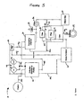
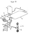
- a system 10 of the present invention is generally comprised of one or more of a tissue collection chamber 20, a processing chamber 30, a waste chamber 40, an output chamber 50 and a sample chamber 60.
- the various chambers are coupled together via one or more conduits 12 such that fluids containing biological material may pass from one chamber to another while maintaining a closed, sterile fluid/tissue pathway.
- the conduits may comprise rigid or flexible bodies referred to interchangeably herein as lumens and tubing, respectively.
- the conduits are in the form of flexible tubing, such as polyethylene tubing conventionally used in clinical settings, silicone or any other material known in the art.
- the conduits 12 can vary in size depending on whether passage of fluid or tissue is desired.
- the conduits 12 may also vary in size depending on the amount of tissue or fluid that is cycled through the system.
- the conduits may have a diameter ranging from about 0.060 to about 0.750 inches and for the passage of tissue, the conduits may have a diameter ranging from 0.312 to 0.750 inches.
- the size of the conduits is selected to balance the volume the conduits can accommodate and the time required to transport the tissue or fluids through said conduits.
- the foregoing parameters i.e., volume and time for transport, must be identified such that the appropriate signals can be transmitted to the processing device of the system. This allows the device to move accurate volumes of liquid and tissue from one chamber to another.
- the flexile tubing used should be capable of withstanding negative pressure to reduce the likelihood of collapse.
- the flexible tubing used should also be capable of withstanding positive pressure which is generated by, for example, a positive displacement pump, which may be used in the system.
- the conduits and ports may be coupled to, for example, a suction device (not shown) which may be manually or automatically operated.
- the suction device may be, e.g., a syringe or an electric pump.
- the suction device should be capable of providing sufficient negative pressure to aspirate tissue from a patient.
- any suitable suction device known to one of ordinary skill in the art, e.g., a surgeon, may be used.
- the conduits 12 may further comprise one or more clamps (not shown) to control the flow of material among various components of the system.
- the clamps are useful for maintaining the sterility of the system by effectively sealing different regions of the system.
- the conduits 12 may comprise one or more valves 14 that control the flow of material through the system.
- the valves 14 are identified as open circles in the Figures.
- the valves may be electromechanical pinch valves.
- the valves may be pneumatic valves.
- the valves may be hydraulic valves or mechanical valves.
- Such valves are preferably activated by a control system which may be coupled to levers. The levers may be manually manipulated such that the levers are activated.
- control system may be coupled to the levers as well as to a processing device which may activate the valves at pre-determined activation conditions.
- activation of the valves may be partially automated and partially subject to the user's preference such that the process may be optimized.
- certain valves may be activated manually and others automatically through the processing device.
- the valves 14 may also be used in conjunction with one or more pumps, e.g., peristaltic pumps 34 or positive displacement pumps (not shown).
- the conduits 12 and/or the valves 14 may also be comprised of sensors 29, e.g., optical sensors, ultrasonic sensors, pressure sensors or other forms of monitors known in the art that are capable of distinguishing among the various fluid components and fluid levels that flow through the system.
- the sensors 29 may be optical sensors.
- the system may also include a plurality of filters 36.
- the filters may be within a chamber of the system 28. Different chambers within the system may be comprised of different filters.
- the filters are effective to separate the regenerative cells, e.g., stem cells and/or progenitor cells, from undesirable cells and disaggregation agents that may be used in accordance with the system.
- a filter assembly 36 includes a hollow fiber filtration device.
- a filter assembly 36 includes a percolative filtration device, which may or may not be used with a sedimentation process.
- the filter assembly 36 comprises a centrifugation device, which may or may not be used with an elutriation device and process.
- the system comprises a combination of these filtering devices.
- the filtration functions of the present invention can be two-fold, with some filters removing things from the final concentration such as collagen, free lipid, free adipocytes and residual collagenase, and with other filters being used to concentrate the final product.
- the filters of the system may be comprised of a plurality of pores ranging in diameters and/or length from 20 to 800 ⁇ m.
- the collection chamber 20 has a prefixed filter 28 with a plurality of pores ranging from 80 to 400 ⁇ m.
- the collection chamber 20 has a prefixed filter 28 with a plurality of 265 ⁇ m pores.
- the filters may be detachable and/or disposable.
- the system may also be comprised of one or more temperature control devices (not shown) that are positioned to adjust the temperature of the material contained within one or more chambers of the system.
- the temperature control device may be a heater, a cooler or both, i.e., it may be able to switch between a heater and a cooler.
- the temperature device may adjust the temperature of any of the material passing through the system, including the tissue, the disaggregation agents, the resuspension agents, the rinsing agents, the washing agents or the additives. For example, heating of adipose tissue facilitates disaggregation whereas the cooling of the regenerative cell output is desirable to maintain viability. Also, if pre-warmed reagents are needed for optimal tissue processing, the role of the temperature device would be to maintain the pre-determined temperature rather than to increase or decrease the temperature.
- the collection chamber 20 may be comprised of a plurality of flexible or rigid canisters or cylinders or combinations thereof.
- the collection chamber 20 may be comprised of one or more rigid canisters of varying sizes.
- the collection chamber 20 may also be comprised of one or more flexible bags.
- the bag is preferably provided with a support, such as in internal or external frame, that helps reduce the likelihood that the bag will collapse upon the application of suction to the bag.
- the collection chamber 20 is sized to hold the requisite amount of saline to appropriately wash and disaggregate the tissue prior to the wash and concentrate stage of the process performed in the processing chamber 30.
- the volume of tissue or fluid present in the collection chamber 20 is easily ascertainable to the naked eye.
- a suitable collection chamber has the capacity to hold 800 ml of lipoaspirate and 1200 ml of saline. Accordingly, in one embodiment, the collection chamber 20 has a capacity of at least 2 liters. In another embodiment, to separate and concentrate red blood cells from blood, the collection chamber 20 has a capacity of at least 1.5 liters.
- the size of the collection chamber 20 will vary depending on the type and amount of tissue collected from the patient. The collection chamber 20 may be sized to hold as little as about 5 ml to up to about 2 liters of tissue. For smaller tissue volumes, e.g., 5 mls to 100 mls, the tissue may be gathered in a syringe prior to transfer to the collection chamber 20.
- the collection chamber 20 may be constructed using any suitable biocompatible material that can be sterilized.
- the collection chamber 20 is constructed of disposable material that meets biocompatibility requirements for intravascular contact as described in the ISO 10993 standard.
- polycarbonate acrylic or ABS may be used.
- the fluid path of the collection chamber 20 is preferably pyrogen free, i.e., suitable for blood use without danger of disease transmittal.
- the collection chamber 20 is constructed of a material that allows the user to visually determine the approximate volume of tissue present in the chamber. In other embodiments, the volume of tissue and/or fluid in the collection chamber 20 is determined by automated sensors 29.
- the collection chamber 20 is preferably designed such that in an automated embodiment, the system can determine the volume of tissue and/or fluid within the chamber with a reasonable degree of accuracy. In a preferred embodiment, the system senses the volume within the collection chamber with an accuracy of plus or minus fifteen percent.
- the collection chamber 20 is in the form of a rigid chamber, for example, a chamber constructed of a medical grade polycarbonate containing a roughly conical prefixed filter 28 of medical grade polyester with a mesh size of 265 ⁇ m (see Figure 5 ).
- the rigid tissue collection container may have a size of approximately eight inches high and approximately five inches in diameter; the wall thickness may be about 0.125 inches.
- the interior of the cylinder may be accessed through, for example, one or more ports for suction tubing, one or more ports with tubing for connection through sterile docking technology, and/or one or more ports for needle puncture access through a rubber septum.
- the prefixed filter 28 in the interior of the collection chamber 20 is preferably structured to retain adipose tissue and to pass non-adipose tissue as, for example, the tissues are removed from the patient. More specifically, the filter 28 may allow passage of free lipid, blood, and saline, while retaining fragments of adipose tissue during, or in another embodiment after, the initial harvesting of the adipose tissue.
- the filter 28 includes a plurality of pores, of either the same or different sizes, but ranging in size from about 20 ⁇ m to 5 mm. In a preferred embodiment, the filter 28 includes a plurality of 400 ⁇ m pores.
- the filter 28 is a medical grade polyester mesh of around 200 ⁇ m thickness with a pore size of around 265 ⁇ m and around 47% open area. This material holds the tissue during rinsing but allows cells to pass out through the mesh following tissue disaggregation. Thus, when the tissues are aspirated from the patient, non-adipose tissue may be separated from adipose tissue.
- the same functionality could be achieved with different materials, mesh size, and the number and type of ports. For example, mesh pore sizes smaller than 100 ⁇ m or as large as several thousand microns would achieve the same purpose of allowing passage of saline and blood cells while retaining adipose tissue aggregates and fragments. Similarly, the same purpose could be achieved by use of an alternative rigid plastic material, or by many other modifications that would be known to those skilled in the art
- the containers for the washing solution 23 and the disaggregation agents 24 may be any suitable container that can hold their contents in a sterile manner, e.g., a collapsible bag, such as an IV bag used in clinical settings. These containers may have conduits 12, such as conduit 12e, coupled to the collection chamber 20 so that the washing solution and the disaggregation agent may be delivered to the interior of the collection chamber 20.
- the washing solution and the disaggregation agent may be delivered to the interior of the collection chamber 20 through any art-recognized manner, including simple gravity pressure applied to the outside of the containers for the saline 23 and/or the disaggregation agents 24 or by placement of a positive displacement pump on the conduits, e.g., conduit 12d in Figure 4 .
- the tissue and/or fluid within the collection chamber should be maintained at a temperature ranging from 30 degrees Celsius to 40 degrees Celsius. In a preferred embodiment, the temperature of the suspension inside the collection chamber is maintained at 37 degrees Celsius. In certain embodiments, if the surgical procedure or therapeutic application needs to be delayed, the selected tissue may be stored in the collection chamber for later use. The tissue may be stored at or about room temperature or at about 4 degrees Celsius for up to 96 hours.
- the washing solution may be any solution known to one of skill in the art, including saline or any other buffered or unbuffered electrolyte solution.
- the types of tissue being processed will dictate the types or combinations of washing solutions used.
- the washing solution such as saline
- the washing solution may be delivered to the collection chamber 20 before the adipose tissue is extracted, or may be delivered to the collection chamber 20 concurrently with the adipose tissue.
- the washing solution and the extracted adipose tissue may be mixed by any means including the methods described below.
- the tissue may be washed by agitation (which maximizes cell viability and minimizes the amount of free lipid released).
- the tissue is agitated by rotating the entire collection chamber 20 through an arc of varying degrees (e.g., through an arc of about 45 degrees to about 90 degrees) at varying speeds, e.g., about 30 revolutions per minute.
- the tissue is agitated by rotating the entire collection chamber 20, wherein the collection chamber 20 is comprised of one or more paddles or protrusions rigidly attached to an inside surface of the collection chamber, through an arc of varying degrees (e.g., through an arc of about 45 degrees to about 90 degrees) at varying speeds, e.g., about 30 revolutions per minute.
- the rotation of the collection chamber 20 described above may be accomplished by a drive mechanism attached to or in proximity with the collection chamber 20.
- the drive mechanism may be a simple belt or gear or other drive mechanism known in the art.
- the speed of the rotation may be, for example, 30 revolutions per minute. Generally, higher speeds have been found to generate larger volumes of free lipids and may not be optimal.
- the tissue is agitated by placing a rotatable shaft 25 inside the collection chamber 20, wherein the rotatable shaft is comprised of one or more paddles 25a or protrusions rigidly attached to the rotatable shaft 25 which pass through the mixture as the shaft is being rotated.
- the rotatable shaft 25 with rigidly attached 25a paddles may be rested on the bottom of the collection chamber 20. This may be accomplished, for example, by placing the paddle-like device into a spinning magnetic field (e.g., magnetic stirrer).
- agitating of the tissue may be accomplished using a simple agitator known in the art, i.e. a device implementing shaking up and down without rotation.
- the tissue may also be washed using any other art-recognized means including rocking, stirring, inversion, etc.
- a tissue disaggregation agent may be delivered to the collection chamber 20 to separate the regenerative cells from the remaining adipose tissue components.
- the disaggregation agent may be any disaggregation agent known to one of skill in the art. Disaggregation agents that may be used include neutral proteases, collagenase, trypsin, lipase, hyaluronidase, deoxyribonuclease, members of the Blendzyme enzyme mixture family, e.g., Liberase H1, pepsin, ultrasonic or other physical energy, lasers, microwaves, other mechanical devices and/or combinations thereof.
- a preferred disaggregation agent of the invention is collagenase.
- the disaggregation agents may be added with other solutions.
- saline such as saline delivered from a saline source 23 as described above, may be added to the adipose tissue along with or immediately followed by addition of collagenase.
- the washed adipose tissue is mixed with a collagenase-containing enzyme solution at or around 37° C for about 20-60 minutes.
- a higher concentration of collagenase or similar agent may be added to decrease the digestion time.
- the washed adipose tissue and the tissue disaggregation agent may then be agitated in manners similar to the agitation methods described above, until the washed adipose tissue is disaggregated.
- the adipose tissue may either be partially disaggregated, or completely disaggregated.
- a portion of washed adipose tissue may be removed and set aside in a sample container prior to any digestion.
- harvested adipose tissue is partially disaggregated to concentrate cells before being reintroduced back into the patient.
- the adipose tissue is mixed with a tissue disaggregation agent for a period of time generally less than about 20 minutes. A portion of the partially disaggregated tissue may then be removed from the collection chamber, and the remaining partially disaggregated tissue may be further disaggregated by mixing the adipose tissue with a tissue disaggregation agent for another 40 minutes.
- the adipose derived cells are to be used as an essentially pure population of regenerative cells, the adipose tissue may be fully disaggregated.
- the collection chamber 20 is preferably comprised of an outlet port 22 at the lowest point of the chamber such that blood and other non-buoyant components of the tissue may be drained to one or more waste containers 40 via one or more conduits 12.
- the collection chamber 20 is generally in (or may be placed in) an upright position such that the outlet ports 22 are located at the bottom of the collection chamber.
- the draining may be passive or active.
- the non-buoyant components described above could be drained using gravity, by applying positive or negative pressure, by use of pumps 34 or by use of vents 32.
- the processing device can signal certain valves and/or pumps to drain the non-buoyant layer from the collection chamber 20.
- the automated embodiments may also be comprised of sensors 29 which can detect when the interface between the buoyant and non-buoyant liquids has been reached.
- the automated embodiments may also be comprised of a sensor 29, e.g., an optical sensor, which may be capable of detecting a change in the light refraction of the effluent which is flowing in the conduit leading out of the collection chamber. The appropriate change in the light refraction may signal the presence of the buoyant layer in the outgoing conduits which indicates that the non-buoyant layer has been drained.
- the sensor 29 can then signal the processing device to proceed with the next step.
- the collection chamber 20 typically includes one or more ports 21 for permitting the washing solution to be delivered to the interior of the chamber, and one or more ports 22 for permitting waste and other materials to be directed out from the collection chamber 20.
- the collection chamber may include one or more sealed entry ports as described herein.
- the collection chamber 20 may also include one or more caps (not shown), such as a top cap and a bottom cap to further ensure that the system remains sterile while washing solution is delivered into the collection chamber and/or waste is transported out.
- the ports 21 may be provided on the caps of the collection chamber or on a sidewall of the collection chamber.
- the process of washing with fresh wash solution may be repeated until the residual content of non-buoyant contaminants in the solution reaches a pre-determined level.
- the remaining material in the collection chamber 20, which comprises the buoyant material of the mixture described above, including adipose tissue fragments may be washed one or more additional times until the amount of undesired material is reduced to a desired pre-determined level.
- One method of determining the end point of the washing is to measure the amount of red blood cells in the tissue solution. This can be accomplished by measuring the light absorbed on the 540 nm wavelength. In a preferred embodiment, a range between about 0.546 and about 0.842 is deemed acceptable.
- additives may be added to the various containers as needed to enhance the results.
- additives include agents that optimize washing and disaggregation, additives that enhance the viability of the active cell population during processing, anti-microbial agents (e.g., antibiotics), additives that lyse adipocytes and/or red blood cells, or additives that enrich for cell populations of interest (by differential adherence to solid phase moieties or to otherwise promote the substantial reduction or enrichment of cell populations).
- anti-microbial agents e.g., antibiotics
- additives that lyse adipocytes and/or red blood cells or additives that enrich for cell populations of interest (by differential adherence to solid phase moieties or to otherwise promote the substantial reduction or enrichment of cell populations).
- Other possible additives include those that promote recovery and viability of regenerative cells (for example, caspase inhibitors) or which reduce the likelihood of adverse reaction on infusion or emplacement (for example, inhibitors of re-aggregation of cells or connective tissue).
- This non-buoyant fraction is referred to herein as the regenerative cell composition and comprises multiple different types of cells, including stem cells, progenitor cells, endothelial precursor cells, adipocytes and other regenerative cells described herein.
- the regenerative cell composition may also contain one or more contaminants, such as collagen and other connective tissue proteins and fragments thereof, which were present in the adipose tissue fragments, or residual collagenase from the tissue disaggregation process.
- the processing chamber 30 of the invention is preferably positioned within the system such that the regenerative cell composition moves from the collection chamber 20 to the processing chamber 30 by way of tubing 12, valves 14 and pump 34 in a sterile manner.
- the processing chamber is sized to accommodate tissue/fluid mixtures ranging from 10mL to 1.2L. In a preferred embodiment, the processing chamber is sized to accommodate 800 mLs. In certain embodiments, the entire regenerative cell composition from the collection chamber 20 is directed to the processing chamber 30.
- the processing chamber 30 may be constructed in any manner suitable for separating and concentrating cells, including filtration and centrifugation and/or combinations thereof.
- the regenerative cell composition from the collection chamber 20 is introduced into the processing chamber 30 where the composition can be filtered to separate and/or concentrate a particular regenerative cell population.
- Cell filtration is a method of separating particular components and cells from other different components or types of cells.
- the regenerative cell composition of the invention comprises multiple different types of cells, including stem cells, progenitor cells and adipocytes, as well as one or more contaminants, such as collagen, which was present in the adipose tissue fragments, or residual collagenase from the tissue disaggregation process.
- the filters 36 present in the processing chamber 30 may allow for separation and concentration of a particular subpopulation of regenerative cells, e.g., stem cells or endothelial progenitors cells etc.
- Some variables which are associated with filtration of cells from a liquid include, but are not limited to, pore size of the filter media, geometry (shape) of the pore, surface area of the filter, flow direction of the solution being filtered, trans-membrane pressure, dilution of the particular cell population, particulate size and shape as well as cell size and cell viability.
- the particular cells that are desired to be separated or filtered are typically adipose derived stem cells.
- the particular cells may include adipose derived progenitor cells, such as endothelial precursor cells, alone or in combination with the stem cells.
- the regenerative cell composition containing the washed cells and residual collagen, adipocytes, and/or undigested tissue disaggregation agent, may be directed through the first filter to remove at least a portion of and preferably substantially all of the collagen particles from the composition so that fewer, and preferably no, collagen particles are present in the filtered solution.
- the filtered regenerative cell composition containing the adipocytes and/or undigested tissue disaggregation agent may then be directed through the second filter to remove at least a portion of and preferably substantially all of the free adipocytes from the filtered regenerative cell composition.
- the twice filtered regenerative cell composition containing the undigested tissue disaggregation agent, may be directed through the third filter, such as a hollow fiber filtration device, as discussed herein, to remove or reduce the undigested tissue disaggregation agent from the regenerative cell composition.
- the third filter such as a hollow fiber filtration device, as discussed herein, to remove or reduce the undigested tissue disaggregation agent from the regenerative cell composition.
- the efficiency of filtration of the stem cells may be enhanced relative to other filtration techniques.
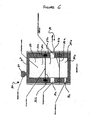
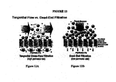
- the pores of the filter media are placed in such a manner that the filter is orientated perpendicular to the flow of the fluid so that the Filter media blocks the path of the fluid being filtered, as illustrated in Figure 12B .
- the particles which are being filtered out of the regenerative cell composition e.g., the stem cells, tend to build up on one side of the filter and block the flow of the fluid through the pores. This blockage can reduce the efficiency of the filter.
- filtration is enhanced by generating a vacuum on the outside of the hollow tube filter media. Due to the size of the regenerative cells, e.g., stem cells or progenitor cells, these cells typically cannot pass through the pores of the body and therefore remain on the inside of the hollow tube filter (e.g., in the lumens of the tubes) and are directed back to the processing chamber 30 via a conduit between the filter and the processing chamber, or to the output chamber 50.
- the regenerative cells e.g., stem cells or progenitor cells
- the hollow fiber filter has about a 0.05 micron pore size, and contains approximately 550 cm 2 surface area of filter media.
- An individual media tube typically has a diameter of about 0.5 mm.
- approximately 120 ml of additional saline may be added to the composition.
- the processing or filter time may be approximately 8 minutes.
- the differential of the pressures on either side of the body of the hollow fiber tube e.g., the pressure inside the lumen of the body, and outside the body
- the trans-membrane pressure can range from about 1 mmHg to about 500 mmHg with a preferred pressure being about 200 mmHg.
- the average nucleated cell recovery and viability using hollow fiber filtration can be approximately 80% of viable cells.
- liquid may flow through the pump 34 before it is passed to the filter assembly 36.
- liquid may pass through the pressure sensor 39 before passing through the filter assembly to obtain a pre-filter liquid pressure in the system.
- one or more of these components may also be provided as an element of the processing chamber 30, such as the vent 32 as illustrated in Figure. 6 .
- the pressure sensor 39 is in line to determine the pressure of the regenerative cell composition which is generated by the pump 34 as it enters the filtering chamber of the filter assembly 36. This construction can facilitate monitoring of the trans-membrane pressure across the filter membrane.
- Additional saline or other buffer and washing solution can be added to the regenerative cell composition to assist in the removal of unwanted proteins as the composition is being filtered through the filter assembly 36. This repeated washing can be performed multiple times to enhance the purity of the regenerative cells.
- the saline can be added at any step as deemed necessary to enhance filtration.
- an output chamber 50 such as an output bag, may be connected to an outlet port of the processing chamber 30 and/or the filter assembly 36, depending on the specific embodiment.
- a vent such as the vent 32, may then be opened to facilitate the output of the concentrated regenerative cells.
- this determination of when a minimum concentration has been reached is made empirically after experiments have been run and programmed into the electronic controls of the device.
- Port 31a is illustrated as a sample inlet port, which is constructed to be coupled to a conduit so that a composition containing regenerative cells can be passed into the interior of the processing chamber 30.
- Port 31b is illustrated as an outlet port constructed to be coupled to a conduit so that the separated and concentrated cells may be removed from the interior of the processing chamber 30.
- Port 31c is illustrated as an inlet port constructed to be coupled to a conduit for delivery of a fresh washing solution, such as saline into the interior of the processing chamber 30.
- the composition containing the separated cells may be concentrated, as discussed herein.
- the composition may be further concentrated after it has been removed from chamber 37a through outlet port 31b, or while it is in the chamber 37a.
- the concentration of cells in the composition is increased in the following manner.
- the filters such as filters 36a and 36b, may be moved towards each other. This movement has the effect of reducing the volume between the two filters (e.g., the volume of chamber 37a).
- a vibrating member may also be provided in connection with the processing chamber 30 to facilitate concentrating of the cells in the composition.
- the vibrating member may be coupled to the filter 36b (e.g., the small pore filter). Vibrating can reduce an incidence of cells becoming trapped in the filters. The reduction in volume of the composition allows the excess saline to be removed as waste and the cells to be concentrated in a smaller volume.
- the filter 36b e.g., the small pore filter
- the concentration of the regenerative cells is accomplished in the following manner.
- the regenerative cell composition can be transferred to another chamber (not shown) which uses gravity to filter out the excess saline.
- the sedimentation can occur at the same time as the percolation. This sedimentation may be accomplished by introducing the composition on top of a filter which has a pore size ranging from about 10 kD to about 2 microns. In one embodiment, a suitable filter has a pore size of about 1 micron. The force of gravity will allow the saline and smaller particles to be passed through the filter while preventing the cells in the composition to flow through the filter.
- the regenerative cell composition may be agitated to remove the cells from the filter and, subsequently, the concentrated regenerative cells may be transferred to the output bag.
- the smaller particles can be drawn off as waste through an outlet.
- the regenerative cell composition from the collection chamber 20 is transported to the processing chamber 30 wherein the composition can be centrifuged to separate and concentrate regenerative cells.
- Centrifugation principles are well know in the art and will be not be repeated herein in the interest of brevity. Standard, art-recognized centrifugation devices, components and parameters are utilized herein.
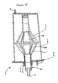
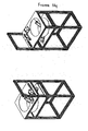
- An exemplary processing chamber for use as part of a centrifuge device is shown in Figures 7 and 8 .
- a centrifuge device causes a centrifuge chamber (such as the one shown in Figure 7 ) to spin around an axis to thereby increasing the force on the cells in the solution to be greater than gravity.
- the processing chamber may be comprised of a cell concentrator in the form of a spinning membrane filter.
- centrifugal elutriation may also be applied.
- the cells may be separated based on the individual cell sedimentation rate such that the directional (e.g., outward) force applied by centrifugation causes cells and solutes to sediment at different rates.
- the sedimentation rate of the target cell population is opposed by an opposite (e.g., inward) flow rate applied by pumping solution in the opposite direction to the centrifugal force. The counterflow is adjusted so that the cells and particles within the solution are separated.
- Figure 9 illustrates principles associated with an elutriation implementation in accordance with the present invention.
- the elutriation embodiment can be similar to a centrifugation implementation to the extent that a force is applied to the solution using a spinning rotor.
- Some of the variables which are associated with the presently embodied elutriation separation include, but are not limited to, the size and shape of the spinning chamber, the diameter of the rotor, the speed of the rotor, the diameter of the counter flow tubing, the flow rate of the counter flow, as well as the size and density of the particles and cells which are to be removed from solution.
- the regenerative cells can be separated based on individual cell densities.
- the processing chamber 30 or the output chamber 50 may include one or more ports, e.g., ports 51 or 52.
- ports 51 or 52 may be designed to transport the regenerative cells obtained using any combination of methods described above, or a portion thereof, via conduits to other surgical devices, cell culturing devices, cell marinading devices, gene therapy devices or purification devices.
- These ports may also be designed to transport the regenerative cells via conduits to additional chambers or containers within the system or as part of another system for the same purposes described above.
- the ports and conduits may be also be used to add one or more additives, e.g., growth factors, re-suspension fluids, cell culture reagents, cell expansion reagents, cell preservation reagents or cell modification reagents including agents that transfer genes to the cells.
- the ports and conduits may also be used to transport the regenerative cells to other targets such as implant materials (e.g., scaffolds or bone fragments) as well as other surgical implants and devices.
- the system can be reconfigured by any of the means described above such that the regenerative cells obtained using the system may be subject to one or more of the following: cell expansion (of one or more regenerative cell types)and cell maintenance (including cell sheet rinsing and media changing); sub-culturing; cell seeding; transient transfection (including seeding of transfected cells from bulk supply); harvesting (including enzymatic, non-enzymatic harvesting and harvesting by mechanical scraping); measuring cell viability; cell plating (e.g., on microtiter plates, including picking cells from individual wells for expansion, expansion of cells into fresh wells); high throughput screening; cell therapy applications; gene therapy applications; tissue engineering applications; therapeutic protein applications; viral vaccine applications; harvest of regenerative cells or supernatant for banking or screening, measurement of cell growth, lysis, inoculation, infection or induction; generation of cells lines (including hybridoma cells); culture of cells for permeability studies; cells for RNAi and viral resistance studies; cells for knock-out and transgenic
- additives include agents that optimize washing and disaggregation, additives that enhance the viability of the active cell population during processing, anti-microbial agents (e.g., antibiotics), additives that lyse adipocytes and/or red blood cells, or additives that enrich for cell populations of interest (by differential adherence to solid phase moieties or to otherwise promote the substantial reduction or enrichment of cell populations) as described herein.
- anti-microbial agents e.g., antibiotics
- additives that lyse adipocytes and/or red blood cells e.g., lyse adipocytes and/or red blood cells
- additives that enrich for cell populations of interest by differential adherence to solid phase moieties or to otherwise promote the substantial reduction or enrichment of cell populations
- the system is automated.
- the system has both automated and manual components.
- the system may be comprised of one or more disposable components connected to or mounted on a re-usable hardware component or module.
- the automated systems of the invention provide screen displays (see Figure 16 ) that prompt proper operation of the system.
- the automated systems may also provide a screen that provides status of the procedure and/or the step by step instructions as to the proper setup of the disposable components of the system.
- the screen may also indicate problems or failures in the system if they occur and provide "troubleshooting" guidance if appropriate.
- the screen is a user interface screen that allows the user to input parameters into the system through, e.g., a touch screen.
- the processing device may also have pre-programmed software which provides the user with appropriate parameters to optimize the process based on the user's input of relevant information such as the amount of regenerative cells required, the type of tissue being processed, the type of post-processing manipulation required, the type of therapeutic application, etc.
- a sensor 29 can signal the processing device present in the re-usable component to activate the steps needed to wash and disaggregate the tissue.
- the processing device may introduce a pre-set volume of washing agent based on the volume of tissue collected using automated valves and pumps. This cycle may be repeated in the collection chamber until the optical sensor determines that the effluent liquid is sufficiently clear and devoid of unwanted material.
- an optical sensor 29 along the conduit leading out of the collection chamber 12b or 12d can detect that the unwanted materials have been removed and can signal the processing device to close the required valves and initiate the next step.
- the processing chamber 30 shown in Figure 4 is in the form of a centrifuge chamber.
- a detailed illustration of the processing chamber of Figure 4 is shown in Figures 7 and 8 .
- Such a processing chamber 30 is generally comprised of a rotating seal network30.1 comprising an outer housing 30.2, one or more seals 30.3, one or more bearings 30.4 and an attachment point 30.6 for connecting the processing chamber to the centrifuge device present in the re-usable component of the system; one or more fluid paths 30.5 in the form of conduits extending out from the rotating seal and ending in a centrifuge chamber on each end which is in the form of an output chamber 50 housed in a frame 53 wherein the frame is comprised of one or more ports 52 and one or more handles to manually re-position the output chamber 50.
- the three lip seals are comprised of a circular "U" shaped channel (not shown) as well as a circular spring (not shown).
- the circular "U” shaped channel is preferably fabricated using flexible material such that a leakage proof junction with the rotating shaft of the rotating seal network 30.1 is formed.
- the lip seals are preferably oriented in a manner such that pressure from the regenerative cell composition flowing through the processing chamber causes the seal assembly to tighten its junction with the rotating shaft by way of increased tension.
- the seals may be secured in position by way of one or more circular clips (not shown) which are capable of expanding and/or collapsing as needed in order to engage a groove in the outer housing 30.2 of the rotating seal network 30.1.
- the rotating seal network 30.1 is comprised of a single rubber seal 30.3 and an air gasket (not shown). This seal and gasket provide a tortuous path for any biologic matter which could compromise the sterility of the system.
- the rotating seal network 30.1 is comprised of multiple spring loaded seals 30.3 which isolate the individual fluid paths. The seals 30.3 are fabricated of a material which can be sterilized as well as seal the rotating shaft without lubricant.
- the rotating seal network 30.1 is compromised of a pair of ceramic disks (not shown) which create the different fluid paths and can withstand the rotation of the system and not cause cell lysis.
- the fluid pathway is flexible and is allowed to wind and unwind with respect to the processing chamber. This is accomplished by having the flexible fluid pathway rotate one revolution for every two revolutions of the processing chamber 30. This eliminates the need for a rotating seal altogether.
- the cell pellet that is obtained using the system shown in Figure 4 comprises the concentrated regenerative cells of the invention.
- a fluid path 30.5 may be used to re-suspend the cell pellet that is formed after centrifugation with additional solutions and/or other additives. Re-suspension of the cell pellet in this manner allows for further washing of the regenerative cells to remove unwanted proteins and chemical compounds as well as increasing the flow of oxygen to the cells.
- the resulting suspension may be subjected to another load of approximately 400 times the force of gravity for another period of approximately 5 minutes.
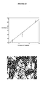
- PIGF and VEGF expression by adipose derived regenerative cells was examined using an ELISA assay (R&D Systems, Minneapolis, MN) using cells from three donors.
- One donor had a history of hyperglycemia and Type 2 diabetes (a condition highly associated with microvascular and macrovascular disease, including patients with coronary artery disease).
- ADC cells from each donor were plated at 1,000 cells/cm 2 in DMEM/F-12 medium supplemented with 10% FCS and 5% HS and grown until confluent. Supernatant samples were taken and assayed for expression of PIGF and VEGF protein.
- Figures 16A and 16B the results demonstrate robust expression of both VEGF ( Figure 16A ) and PIGF ( Figure 16B ) by the adipose derived regenerative cells of the invention.
- VEGF Vascular Endothelial Growth Factor
- bFGF Vascular Endothelial Growth Factor
- IGF-II Vascular Endothelial Growth Factor
- Eotaxin G-CSF
- GM-CSF IL-12 p40/p70
- IL-12 p70 IL-13
- IL-6 IL-9
- Leptin MCP-1, M-CSF, MIG, PF-4, TIMP-1, TIMP-2, TNF- ⁇ , and Thrombopoetin.
- the following angiogenic related growth factors or cytokines were elevated at least twice compare to blank control medium with 10% FBS: Vascular Endothelial Growth Factor (VEGF), Eotaxin, G-CSF, IL-6, MCP-1 and PF-4.
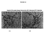
- ADCs were isolated by enzymatic digestion of human subcutaneous adipose tissue. ADCs (100 ⁇ l) were incubated in phosphate saline buffer (PBS) containing 0.2% fetal bovine serum (FBS), and incubated for 20 to 30 minutes at 4°C with fluorescently labeled antibodies directed towards the human endothelial markers CD-31 (differentiated endothelial cell marker) and CD-34 (EPC marker), as well as human ABCG2 (ATP binding cassette transporter), which is selectively expressed on multipotent cells. After washing, cells were analyzed on a FACSAria Sorter (Beckton Dickenson - Immunocytometry).
- PBS phosphate saline buffer
- FBS fetal bovine serum
- EPCs within the adipose tissue derived regenerative cells indicates that these cells can participate directly in development of new blood vessels and enhance angiogenesis and restore perfusion.
- Transplant of autologous adipose tissue is a relatively common procedure in plastic and reconstructive surgery ⁇ Fulton, 1998; Shiffman, 2001 ⁇ .
- this procedure is limited by the fact that the adipose tissue fragments are transferred without a vascular supply and, as a result, graft survival is dependent upon neovascularization (Coleman, 1995; Eppley et al., 1990).
- the transplanted tissue represents an ischemic tissue.
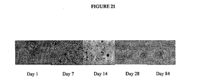
- mice hearts were harvested and processed at the following 5 timepoints after injection: day 1, day 7, day 14, day 28, day 84.
- the results demonstrate engraftment of donor derived adipose derived regenerative cells in the area of infarcted myocardium at all timepoints referenced above.
- Figure 21 demonstrates a histological timeline of engraftment.
- Improvements in the patient are noted within approximately six hours after the cell administration procedure. Several days after the cell administration procedure further improvement of the patient is noted evidenced by increased cardiac ejection fraction, decreased rate of heart failure, decreased infarct size, improved exercise tolerance and other quality of life measures.
Landscapes
- Health & Medical Sciences (AREA)
- Life Sciences & Earth Sciences (AREA)
- Engineering & Computer Science (AREA)
- Chemical & Material Sciences (AREA)
- Bioinformatics & Cheminformatics (AREA)
- General Health & Medical Sciences (AREA)
- Biomedical Technology (AREA)
- Public Health (AREA)
- Medicinal Chemistry (AREA)
- Veterinary Medicine (AREA)
- Pharmacology & Pharmacy (AREA)
- Animal Behavior & Ethology (AREA)
- Organic Chemistry (AREA)
- Zoology (AREA)
- Cell Biology (AREA)
- Biotechnology (AREA)
- Cardiology (AREA)
- Heart & Thoracic Surgery (AREA)
- Developmental Biology & Embryology (AREA)
- Epidemiology (AREA)
- General Chemical & Material Sciences (AREA)
- Chemical Kinetics & Catalysis (AREA)
- Nuclear Medicine, Radiotherapy & Molecular Imaging (AREA)
- Wood Science & Technology (AREA)
- Genetics & Genomics (AREA)
- Immunology (AREA)
- General Engineering & Computer Science (AREA)
- Vascular Medicine (AREA)
- Biochemistry (AREA)
- Virology (AREA)
- Rheumatology (AREA)
- Microbiology (AREA)
- Hospice & Palliative Care (AREA)
- Proteomics, Peptides & Aminoacids (AREA)
- Gastroenterology & Hepatology (AREA)
- Urology & Nephrology (AREA)
- Dispersion Chemistry (AREA)
- Hematology (AREA)
- Dermatology (AREA)
- Medicines Containing Material From Animals Or Micro-Organisms (AREA)
Abstract
Description
- This application is a continuation-in-part application of
U.S. Application No. 10/783,957 U.S. Provisional Application No. 60/449,279 U.S. Provisional Application No. 60/462,911 U.S. Application No. 10/877,822 U.S. Provisional Application No. 60/496,467 U.S. Provisional Application No. 60/482,820 U.S. Application No. 10/316,127, filed on December 9, 2002 U.S. Provisional Application No. 60/338,856, filed December 7, 2001 - This invention generally relates to regenerative cells derived from adipose tissue, and more particularly, to adipose-derived regenerative cells (e.g., adipose-derived stem and progenitor cells), methods of using adipose-derived regenerative cells, compositions containing adipose-derived regenerative cells, and systems for preparing and using adipose-derived regenerative cells, which are used to treat cardiovascular diseases and disorders.
- Cardiovascular diseases and disorders are the leading cause of death and disability in all industrialized nations. In the United States alone, cardiovascular disease accounts for about 40 percent of the mortality rate and affects 58 million Americans (American-Heart-Association, 2002). One of the primary factors that renders cardiovascular disease particularly devastating is the heart's inability to repair itself following damage. Since cardiac muscle cells are unable to divide and repopulate areas of damage, cardiac cell loss as a result of injury or disease is largely irreversible (Abbate et al., 2002; Remme, 2000).
- Of the available forms of therapy, human to human heart transplants have been the most effective in treating severe cardiovascular diseases and disorders. In fact, the one-year and five-year survival rate of the average cardiac transplant recipient is currently over 70 percent. Unfortunately, however, transplantation is a severely limited form of therapy for a number of reasons, namely, the scarcity of suitable donors, the expense of the procedure and the high likelihood of graft rejection and associated problems such as infections, renal dysfunction and immunosuppressant related cancers (American-Heart-Association, 2002).
- An alternative to transplant therapy is the use of regenerative medicine to repair and regenerate damaged cardiac muscle cells. Regenerative medicine harnesses, in a clinically targeted manner, the ability of stem cells (i.e., the unspecialized master cells of the body) to renew themselves indefinitely and develop into mature specialized cells. Stem cells are found in embryos during early stages of development, in fetal tissue and in some adult organs and tissue (Pera et al., 2000). Embryonic stem cells (hereinafter referred to as "ESCs") are known to become all of the cell and tissue types of the body. ESCs not only contain all the genetic information of the individual but also contain the nascent capacity to become any of the 200+ cells and tissues of the body. Thus, these cells have tremendous potential for regenerative medicine. For example, ESCs can be grown into specific tissues such as heart, lung or kidney which could then be used to repair damaged and diseased organs (Assady et al., 2001; Jacobson et al., 2001; Odorico et al., 2001). However, ESC derived tissues have clinical limitations. Since ESCs are necessarily derived from another individual, i.e., an embryo, there is a risk that the recipient's immune system will reject the new biological material. Although immunosuppressive drugs to prevent such rejection are available, such drugs are also known to block desirable immune responses such as those against bacterial infections and viruses. Moreover, the ethical debate over the source of ESCs, i.e., embryos, is well-chronicled and presents an additional and, perhaps, insurmountable obstacle for the foreseeable future.
- Adult stem cells (hereinafter interchangeably referred to as "ASCs") represent an alternative to the use of ESCs. ASCs reside quietly in many non-embryonic tissues, presumably waiting to respond to trauma or other destructive disease processes so that they can heal the injured tissue (Arvidsson et al., 2002; Bonner-Weir and Sharma, 2002; Clarke and Frisen, 2001; Crosby and Strain, 2001; Jiang et al., 2002a). Notably, emerging scientific evidence indicates that each individual carries a pool of ASCs that may share with ESCs the ability to become many if not all types of cells and tissues (Young et al., 2001; Jiang et al., 2002a; Jiang et al., 2002b; Schwartz et al., 2002). Thus, ASCs, like ESCs, have tremendous potential for clinical applications of regenerative medicine.
- ASC populations have been shown to be present in one or more of bone marrow, skin, muscle, liver and brain (Jiang et al., 2002b; Alison, 1998; Crosby and Strain, 2001). However, the frequency of ASCs in these tissues is low. For example, mesenchymal stem cell frequency in bone marrow is estimated at between 1 in 100,000 and 1 in 1,000,000 nucleated cells (D'Ippolito et al., 1999; Banfi et al., 2001; Falla et al., 1993). Similarly, extraction of ASCs from skin involves a complicated series of cell culture steps over several weeks (Toma et al., 2001) and clinical application of skeletal muscle-derived ASCs requires a two to three week culture phase (Hagege et al., 2003). Thus, any proposed clinical application of ASCs from such tissues requires increasing cell number, purity, and maturity by processes of cell purification and cell culture.
- Although cell culture steps may provide increased cell number, purity, and maturity, they do so at a cost. This cost can include one or more of the following technical difficulties: loss of cell function due to cell aging, loss of potentially useful non-stem cell populations, delays in potential application of cells to patients, increased monetary cost, and increased risk of contamination of cells with environmental microorganisms during culture. Recent studies examining the therapeutic effects of bone-marrow derived ASCs have used essentially whole marrow to circumvent the problems associated with cell culturing (Horwitz et al., 2001; Orlic et al., 2001; Stamm et al., 2003; Strauer et al., 2002). The clinical benefits, however, have been suboptimal, an outcome almost certainly related to the limited ASC dose and purity inherently available in bone marrow.
- Recently, adipose tissue has been shown to be a source of ASCs (Zuk et al., 2001; Zuk et al., 2002). Unlike marrow, skin, muscle, liver and brain, adipose tissue is comparably easy to harvest in relatively large amounts (Commons et al., 2001; Katz et al., 2001b). Furthermore, adipose derived ASCs have been shown to possess the ability to generate multiple tissues in vitro, including bone, fat, cartilage, and muscle (Ashjian et al., 2003; Mizuno et al., 2002; Zuk et al., 2001; Zuk et al., 2002). Thus, adipose tissue presents an optimal source for ASCs for use in regenerative medicine. Suitable methods for harvesting adipose derived ASCs, however, are lacking in the art. The existing methods suffer from a number of shortcomings. For example, the existing methods lack the ability to optimally accommodate an aspiration device for removal of adipose tissue. The existing methods also lack partial or full automation from the harvesting of adipose tissue phase through the processing of tissue phases (Katz et al., 2001a). The existing methods further lack volume capacity greater than 100ml of adipose tissue. The existing methods yet further lack a partially or completely closed system from the harvesting of adipose tissue phase through the processing of tissue phases. Finally, the existing methods lack disposability of components to attenuate concomitant risks of cross-contamination of material from one sample to another. In summary, the prior art methods for harvesting ASCs from adipose tissue do not overcome the technical difficulties associated with harvesting ASCs from skin, muscle, liver and brain described above.
- Accordingly, given the tremendous therapeutic potential of ASCs, there exists an urgent need in the art for a device, system or method for harvesting ASCs from adipose tissue that produces a population of ASCs with increased yield, consistency and/or purity and does so rapidly and reliably with a diminished or non-existent need for post-extraction manipulation. Ideally, such a device, system or method would yield ASCs in a manner suitable for direct placement into a recipient. Access to such a device, system or method in combination with methods and compositions using adipose derived ASCs for the treatment of cardiovascular diseases and disorders would revolutionize the treatment of such disorders. Given the prevalence of cardiovascular disease and the scarcity of current treatment options, such a treatment is urgently needed.
- The present invention relates to regenerative cells, e.g., adult stem and progenitor cells, that can be used for the treatment of cardiovascular conditions, diseases and disorders. The present invention also relates to systems and methods for separating and concentrating regenerative cells from tissue, e.g., adipose tissue. The present invention further relates to compositions of regenerative cells for cardiovascular related therapeutic applications. Accordingly, in a general embodiment, the present invention is directed to compositions, methods, and systems for using regenerative cells derived from tissue that are placed directly into a recipient along with such additives necessary to promote, engender, or support a therapeutic cardiovascular related benefit.
- In specific embodiments, the regenerative cells of the present invention may be used to treat cardiovascular conditions, diseases and disorders based on, for example, their ability to synthesize and secrete growth factors stimulating new blood vessel formation, their ability to synthesize and secrete growth factors stimulating cell survival, proliferation and/or alteration of the injury response of other cells, their ability to proliferate and/or differentiate into cells directly participating in new blood vessel formation, their ability to engraft damaged myocardium and alter scar formation (collagen deposition, collagen degradation and cross-linking), their ability to proliferate and differentiate into cardiomyocytes or cardiomyocyte-like muscle cells capable of contributing to myocardial contractility, their ability to proliferate and differentiate into myocardial cells, their ability to improve perfusion and regenerate damaged myocardium, and their ability to prevent progression of hypertrophy (remodeling) post myocardial infarct.
- The regenerative cells administered to the cardiac patient may be comprised of, e.g., stem cells, progenitor cells or combination thereof. In certain embodiments, administration of multiple doses and/or types of regenerative cells may be needed to derive a therapeutic benefit. In addition, additives such as one or more growth factors may be administered with the regenerative cells. In a preferred embodiment, the regenerative cells are administered with angiogenic, arteriogenic and/or cardiac specific growth factors alone or in combination with other additives. The regenerative cells may also be administered with one or more immunosuppressive drugs.
- The routes of administration for the regenerative cells are known in the art and include direct administration of cells to the site of intended benefit. This may be achieved by direct injection into the myocardium through the external surface of the heart (epicardial), direct injection into the myocardium through the internal surface (endocardial) through insertion of a suitable cannula, by arterial or venous infusion (including retrograde flow mechanisms) or by other means disclosed herein or known in the art such as pericardial injection. Routes of administration known to one of ordinary skill in the art, include but are not limited to, intravenous, intracoronary, endomyocardial, epimyocardial, intraventicular, retrograde coronary sinus or intravenous.
- Prior to administration to a patient, the regenerative cells may be grown in cell culture to, for example, promote differentiation towards a cardiac phenotype. Prior to administration to a patient, the cells could also be modified by gene transfer such that expression of one or more genes, e.g., a cardiac gene, in the modified regenerative cells is altered.
- The present invention also relates to highly versatile systems and methods capable of separating and concentrating regenerative cells, e.g., stem and progenitor cells, from a given tissue that are suitable for re-infusion into a subject. In a preferred embodiment, the system is automated. The system of the present invention generally includes one or more of a collection chamber, a processing chamber, a waste chamber, an output chamber and a sample chamber. The various chambers are coupled together via one or more conduits such that fluids containing biological material may pass from one chamber to another in a closed, sterile fluid/tissue pathway. In certain embodiments, the waste chamber, the output chamber and the sample chamber are optional. In one embodiment, the entire procedure from tissue extraction through processing and placement of the device into-the recipient would all be performed in the same facility, indeed, even within the same room of the patient undergoing the procedure.
- Accordingly, in one embodiment, a method of treating cardiovascular related disorder in a patient includes steps of: a) providing a tissue removal system; b) removing adipose tissue from a patient using the tissue removal system, the adipose tissue having a concentration of regenerative cells; c) processing at least a part of the adipose tissue to obtain a concentration of regenerative cells other than the concentration of regenerative cells of the adipose tissue before processing; and d) administering the regenerative cells to a patient without removing the regenerative cells from the tissue removal system before being administered to the patient.
- Any feature or combination of features described herein are included within the scope of the present invention provided that the features included in any such combination are not mutually inconsistent as will be apparent from the context, this specification, and the knowledge of one of ordinary skill in the art. Additional advantages and aspects of the present invention are apparent in the following detailed description.
-
-
Figure 1 . is an illustration of a system for separating and concentrating regenerative cells from tissue which includes one filter assembly. -
Figure 2 is an illustration of a system similar toFigure 1 having a plurality of filter assemblies in a serial configuration. -
Figure 3 is an illustration of a system similar toFigure 1 having a plurality of filter assemblies in a parallel configuration. -
Figure 4 is an illustration of a system for separating and concentrating regenerative cells from tissue which includes a centrifuge chamber. -
Figure 5 is a sectional view of a collection chamber including a prefixed filter utilized in a system for separating and concentrating regenerative cells from tissue. -
Figure 6 is a sectional view of a processing chamber of a system for separating and concentrating regenerative cells from tissue utilizing a percolative filtration system. -
Figure 7 is a sectional view of a processing chamber of a system for separating and concentrating regenerative cells utilizing a centrifuge device for concentrating the regenerative cells. -
Figure 8 is another sectional view of the processing chamber ofFigure 7 . -
Figures. 9.1, 9.2 and 9.3 illustrate an elutriation component in use with the system of the invention. -
Figure 10 is an illustration of a system for separating and concentrating regenerative cells from tissue utilizing vacuum pressure to move fluids through the system. A vacuum system can be constructed by applying a vacuum pump or vacuum source to the outlet of the system, controlled at a predetermined rate to pull tissue and fluid through, using a system of stopcocks, vents, and clamps to control the direction and timing of the flow. -
Figure 11 is an illustration of a system for separating and concentrating regenerative cells from tissue utilizing positive pressure to move fluids through the system. A positive pressure system uses a mechanical means such as a peristaltic pump to push or propel the fluid and tissue through the system at a determined rate, using valves, stopcocks, vents, and clamps to control the direction and timing of the flow. -
Figure 12A illustrates a filtration process in which the feed stream of fluid flows tangentially to the pores of the filter.Figure 12B illustrates a filtration process in which the feed stream of fluid flows perpendicular to the pores of the filter. -
Figure 13 is an illustration of an exemplary disposable set for a system of the invention. -
Figure 14 is an illustration of an exemplary re-usable component for a system of the invention. -
Figure 15A is an illustration of an exemplary device of the invention assembled using the disposable set ofFigure 13 and a re-usable component ofFigure 14 . -
Figure 15B is a flowchart depicting exemplary pre-programmed steps, implemented through a software program, that control automated embodiments of a system of the present invention. Two alternative processing parameters are shown indicating the versatility of the system. -
Figures 16A and 16B depict the expression of VEGF (5A) and PIGF (5B) protein by cultured adipose derived regenerative cells. -
Figure 17 depicts detection of endothelial progenitor cells within adipose derived regenerative cell populations. -
Figures 18A and 18B depict the in vitro development of vascular structures in both normal (7A) and streptozotocin-treated (7B) mice. -
Figure 19 depicts the increased average restoration of blood flow in hindlimb ischemia mice treated with adipose derived regenerative cells compared to a negative control. -
Figures 20A and 20B shows that increasing adipose derived regenerative cell dose improves graft survival and angiogenesis (20A) and depicts the retention of adipose tissue architecture in histologic specimen (20B). -
Figure 21 depicts the histological timeline of engraftment of donor derived adipose derived regenerative cells in the area of infarcted myocardium. -
Figure 22 depicts dual positive staining for both beta-galactosidase and myosin heavy chain. Highlighted cells exhibit both blue betagalactosidase staining, demonstrating their origin from donor adipose tissue cells, and brown staining indicating expression of the cardiac muscle protein myosin heavy chain. Cells exhibiting both brown and blue staining (as indicated by arrows) are adipose tissue-derived cells that have taken on the phenotype of cardiac muscle cells. -
Figure 23 depicts clusters of donor derived adipose derived regenerative cells in a region of infarcted myocardium following occlusion/reperfusion injury in the rat. - Figure 24 depicts significantly improved ejection fraction at twelve weeks post myocardial infarction of rats treated with adipose derived regenerative cells compared with saline based on echo and contractility analysis.
- Figure 25 depicts significantly improved baseline contractility at twelve weeks post myocardial infarction of rats treated with adipose derived regenerative cells compared with saline based on echo and contractility analysis.
- Figure 26 depicts significantly improved baseline relaxation at twelve weeks post myocardial infarction of rats treated with adipose derived regenerative cells compared with saline based on echo and contractility analysis.
- Figure 27 depicts changes in remodeling parameters, i.e., reduced ventricular septal thickness, at twelve weeks post myocardial infarction of rats treated with adipose derived regenerative cells compared with saline.
- Figure 28 depicts prevention of the progression of ventricular dilation as evident in reduced ventricular septal thickness (systole) at twelve weeks post myocardial infarction of rats treated with ADCs compared with saline.
- Figures 29 and 30 depict prevention of the progression of ventricular dilation as evident in reduced posterior wall thickness in both diastole (Figure 29) and systole (Figure 30) at twelve weeks post myocardial infarction of rats treated with adipose derived regenerative cells compared with saline.
- Figure 31 depicts significant increase of left ventricle ejection fraction (LVEF) in pigs in the cell infusion group (3.0% ± 6.0%, mean ± SD) versus the control group (-9.0% ± 5.0%, p=0.01). LVEF by cineangiography (n=12) showed a similar trend with values of 3.7% ± 5.0% vs. -2.0% ± 7.5%, respectively (p=0.16).
- The present invention provides systems and methods for treating cardiovascular conditions, diseases and disorders using adipose derived regenerative cells, e.g., stem and progenitor cells. Specifically, the present invention demonstrates that the adipose derived regenerative cells of the invention (1) express angiogenic and arteriogenic growth factors, including PIGF, VEGF, bFGF, IGF-II, Eotaxin, G-CSF, GM-CSF, IL-12 p40/p70, IL-12 p70, IL-13, IL-6, IL-9, Leptin, MCP-1, M-CSF, MIG, PF-4, TIMP-1, TIMP-2, TNF- α, and Thrombopoetin, (2) comprise endothelial progenitor cells (EPC) which have a well-established function in blood vessel formation, (3) develop into blood vessels in vitro, and (4) support ischemic tissue survival in vivo. (2) contain endothelial progenitor cells (EPC) which have a well-established function in blood vessel formation, (3) develop into blood vessels in vitro, (4) support ischemic tissue survival in vivo, (5) restore perfusion following occlusion/reperfusion injury of the hind limb, (6) when injected into animals after heart injury home to the heart, (7) when injected into an animals after heart injury differentiate into cells expressing markers consistent with their differentiation into cardiac myocytes or cardiac myocyte like cells, (8) improve perfusion post myocardial infarct in a surgical models of hind limb ischemia in mice, (9) prevent progression of remodeling and (10) improve function in a small and large animal models of myocardial infarction. Accordingly, the instant disclosure demonstrates that the regenerative cells of the present invention are useful for the treatment of cardiovascular diseases and disorders.
- In order that the present invention may be more readily understood, certain terms are first defined. Additional definitions are set forth throughout the detailed description.
- As used herein, "regenerative cells" refers to any heterogeneous or homologous cells obtained using the systems and methods of the present invention which cause or contribute to complete or partial regeneration, restoration, or substitution of structure or function of an organ, tissue, or physiologic unit or system to thereby provide a therapeutic, structural or cosmetic benefit. Examples of regenerative cells include: adult stem cells, endothelial cells, endothelial precursor cells, endothelial progenitor cells, macrophages, fibroblasts, pericytes, smooth muscle cells, preadipocytes, differentiated or de-differentiated adipocytes, keratinocytes, unipotent and multipotent progenitor and precursor cells (and their progeny), lymphocytes , neutrophils, histiocytes (tissue macrophages), lymphatic system related cells, neurons, neuronal precursor cells and Schwann cells.
- One mechanism by which the regenerative cells may provide a therapeutic, structural or cosmetic benefit is by incorporating themselves or their progeny into newly generated, existing or repaired tissues or tissue components. For example, ASCs and/or their progeny may incorporate into newly generated bone, muscle, or other structural or functional tissue and thereby cause or contribute to a therapeutic, structural or cosmetic improvement. Similarly, endothelial cells or endothelial precursor or progenitor cells and their progeny may incorporate into existing, newly generated, repaired, or expanded blood vessels to thereby cause or contribute to a therapeutic, structural or cosmetic benefit.
- Another mechanism by which the regenerative cells may provide a therapeutic, structural or cosmetic benefit is by expressing and/or secreting molecules, e.g., growth factors, that promote creation, retention, restoration, and/or regeneration of structure or function of a given tissue or tissue component. For example, regenerative cells may express and/or secrete molecules which result in enhanced growth of tissues or cells that then participate directly or indirectly in improved structure or function. Regenerative cells may express and/or secrete growth factors, including, for example, Vascular Endothelial Growth Factor (VEGF), Placental Growth factor (P1GF), bFGF, IGF-II, Eotaxin, G-CSF, GM-CSF, IL-12 p40/p70, IL-12 p70, IL-13, IL-6, IL-9, Leptin, MCP-1, M-CSF, MIG, PF-4, TIP-1, TIMP-2, TNF- α, Thrombopoetin, and their isoforms, which may perform one or more of the following functions: stimulate development of new blood vessels, i.e., promote angiogenesis; improve oxygen supply of pre-existent small blood vessels (collaterals) by expanding their blood carrying capacity; induce mobilization of regenerative cells from sites distant from the site of injury to thereby enhance the homing and migration of such cells to the site of injury; stimulate the growth and/or promote the survival of cells within a site of injury thereby promoting retention of function or structure; deliver molecules with anti-apoptotic properties thereby reducing the rate or likelihood of cell death and permanent loss of function; and interact with endogenous regenerative cells and/or other physiological mechanisms.
- The regenerative cells may be used in their 'native' form as present in or separated and concentrated from the tissue using the systems and methods of the present invention or they may be modified by stimulation or priming with growth factors or other biologic response modifiers, by gene transfer (transient or stable transfer), by further sub-fractionation of the resultant population on the basis or physical properties (for example size or density), differential adherence to a solid phase material, expression of cell surface or intracellular molecules, cell culture or other ex vivo or in vivo manipulation, modification, or fractionation as further described herein. The regenerative cells may also be used in combination with other cells or devices such as synthetic or biologic scaffolds, materials or devices that deliver factors, drugs, chemicals or other agents that modify or enhance the relevant characteristics of the cells as further described herein.
- As used herein, "regenerative cell composition" refers to the composition of cells typically present in a volume of liquid after a tissue, e.g., adipose tissue, is washed and at least partially disaggregated. For example, a regenerative cell composition of the invention comprises multiple different types of regenerative cells, including ASCs, endothelial cells, endothelial precursor cells, endothelial progenitor cells, macrophages, fibroblasts, pericytes, smooth muscle cells, preadipocytes, differentiated or de-differentiated adipocytes, keratinocytes, unipotent and multipotent progenitor and precursor cells (and their progeny), and lymphocytes. The regenerative cell composition may also contain one or more contaminants, such as collagen, which may be present in the tissue fragments, or residual collagenase or other enzyme or agent employed in or resulting from the tissue disaggregation process described herein.
- As used herein, "regenerative medicine" refers to any therapeutic, structural or cosmetic benefit that is derived from the placement, either directly or indirectly, of regenerative cells into a subject. As used herein, the phrase "cardiovascular condition, disease or disorder" is intended to include all disorders characterized by insufficient, undesired or abnormal cardiac function, e.g., ischemic heart disease, hypertensive heart disease and pulmonary hypertensive heart disease, valvular disease, congenital heart disease, toxic or infectious cardiomyopathies and any condition which leads to heart failure, e.g., congestive heart failure, in a subject, particularly a human subject. Insufficient or abnormal cardiac function can be the result of disease, injury and/or aging. By way of background, a response to myocardial injury follows a well-defined path in which some cells die while others enter a state of hibernation where they are not yet dead but are dysfunctional. This is followed by infiltration of inflammatory cells and a deposition of collagen as part of scarring, all of which happen in parallel with in-growth of new blood vessels and a degree of continued cell death. As used herein, the term "ischemia" refers to any tissue ischemia (e.g., localized tissue ischemia or generalized tissue ischemia such as in cases of shock) caused by the reduction of the inflow of blood and/or by the reduction in the supply of oxygen and/or other nutrients,. The term "myocardial ischemia" refers to circulatory disturbances caused by coronary atherosclerosis and/or inadequate blood, oxygen and/or nutrient supply to the myocardium. The term "myocardial infarction" refers to an ischemic insult to myocardial tissue resulting from a lack of blood flow. Specifically, this insult results from an occlusive (e.g., thrombotic or embolic) event in the coronary circulation and produces an environment in which the myocardial metabolic demands exceed the supply of oxygen and or nutrients to the myocardial tissue.
- As used herein, the term "angiogenesis" refers to the process by which new blood vessels are generated from existing vasculature and tissue (Folkman, 1995). The term "repair" refers to the reformation of damaged vasculature and tissue. The alleviation of tissue ischemia is critically dependent upon angiogenesis. The spontaneous growth of new blood vessels provides collateral circulation in and around an ischemic area, improves blood flow, and alleviates the symptoms caused by the ischemia. As used herein, the term "angiogenic factor" or "angiogenic protein" refers to any known protein, peptide or other agent capable of promoting growth of new blood vessels from existing vasculature ("angiogenesis"). Suitable angiogenic factors for use in the invention include, but are not limited to, Placenta Growth Factor (Luttun et al., 2002), Macrophage Colony Stimulating Factor (Aharinejad et al., 1995), Granulocyte Macrophage Colony Stimulating Factor (Buschmann et al., 2003), Vascular Endothelial Growth Factor (VEGF)-A, VEGF-A, VEGF-B, VEGF-C, VEGF-D, VEGF-E (Mints et al., 2002), neuropilin (Wang et al., 2003), fibroblast growth factor (FGF)-1, FGF-2(bFGF), FGF-3, FGF-4, FGF-5, FGF-6 (Botta et al., 2000),
Angiopoietin 1, Angiopoietin 2 (Sundberg et al., 2002), erythropoietin (Ribatti et al., 2003), BMP-2, BMP-4, BMP-7 (Carano and Filvaroff, 2003), TGF-beta (Xiong et al., 2002), IGF-1 (Shigematsu et al., 1999), Osteopontin (Asou et al., 2001), Pleiotropin (Beecken et al., 2000), Activin (Lamouille et al., 2002), Endothelin-1 (Bagnato and Spinella, 2003)and combinations thereof. Angiogenic factors can act independently, or in combination with one another. When in combination, angiogenic factors can also act synergistically, whereby the combined effect of the factors is greater than the sum of the effects of the individual factors taken separately. The term "angiogenic factor" or "angiogenic protein" also encompasses functional analogues of such factors. Functional analogues include, for example, functional portions of the factors. Functional analogues also include anti-idiotypic antibodies which bind to the receptors of the factors and, thus, mimic the activity of the factors in promoting angiogenesis and/or tissue repair. Methods for generating such anti-idiotypic antibodies are well known in the art and are described, for example, inWO 97/23510 - Angiogenic factors used in the present invention can be produced or obtained from any suitable source. For example, the factors can be purified from their native sources, or produced synthetically or by recombinant expression. The factors can be administered to patients as a protein composition. Alternatively, the factors can be administered in the form of an expression plasmid encoding the factors. The construction of suitable expression plasmids is well known in the art. Suitable vectors for constructing expression plasmids include, for example, adenoviral vectors, retroviral vectors, adeno-associated viral vectors, RNA vectors, liposomes, cationic lipids, lentiviral vectors and transposons.
- As used herein, the term "arteriogenesis" refers to the process of enhancing growth of collateral arteries and/or other arteries from pre-existing arteriolar connections (Carmeliet, 2000; Scholz et al., 2001; Scholz et al., 2002). More particularly, arteriogenesis is the in situ recruitment and expansion of arteries by proliferation of endothelial and smooth muscle cells from pre-existing arteriolar connections supplying blood to ischemic tissue, tumor or site of inflammation. These vessels largely grow outside the affected tissue and are important for the delivery of nutrients to the ischemic territory, the tumor or the site of inflammation. Arteriogenesis is part of the normal response to myocardial ischemia (Mills et al., 2000; Monteiro et al., 2003). In addition, the common surgical technique of a coronary artery bypass graft (CABG) is, in effect, no more than creation of an artificial collateral vessel (Sergeant et al., 1997). Thus, processes which enhance arteriogenesis following an infarct will improve blood flow to ischemic tissue resulting in decreased cell death and decreased infarct size. These improvements will result in improved cardiac function and therapeutic benefit.
- As used herein, "stem cell" refers to a multipotent regenerative cell with the potential to differentiate into a variety of other cell types, which perform one or more specific functions and have the ability to self-renew. Some of the stem cells disclosed herein may be multipotent.
- As used herein, "progenitor cell" refers to a multipotent regenerative cell with the potential to differentiate into more than one cell type and has limited or no ability to self-renew. "Progenitor cell", as used herein, also refers to a unipotent cell with the potential to differentiate into only a single cell type, which performs one or more specific functions and has limited or no ability to self-renew. In particular, as used herein, "endothelial progenitor cell" refers to a multipotent or unipotent cell with the potential to differentiate into vascular endothelial cells.
- As used herein, "precursor cell" refers to a unipotent regenerative cell with the potential to differentiate into one cell type. Precursor cells and their progeny may retain extensive proliferative capacity, e.g., lymphocytes and endothelial cells, which can proliferate under appropriate conditions.
- As used herein "stem cell number" or "stem cell frequency" refers to the number of colonies observed in a clonogenic assay in which adipose derived cells (ADC) are plated at low cell density (<10,000 cells/well) and grown in growth medium supporting MSC growth (for example, DMEM/F12 medium supplemented with 10% fetal calf serum, 5% horse serum, and antibiotic/antimycotic agents). Cells are grown for two weeks after which cultures are stained with hematoxylin and colonies of more than 50 cells are counted as CFU-F. Stem cell frequency is calculated as the number of CFU-F observed per 100 nucleated cells plated (for example; 15 colonies counted in a plate initiated with 1,000 nucleated regenerative cells gives a stem cell frequency of 1.5%). Stem cell number is calculated as stem cell frequency multiplied by the total number of nucleated ADC cells obtained. A high percentage (∼100%) of CFU-F grown from regenerative cells express the cell surface molecule CD105 which is also expressed by marrow-derived stem cells (Barry et al., 1999). CD105 is also expressed by adipose tissue-derived stem cells (Zuk et al., 2002).
- As used herein, the term "adipose tissue" refers to fat including the connective tissue that stores fat. Adipose tissue contains multiple regenerative cell types, including ASCs and endothelial progenitor and precursor cells.
- As used herein, the term "unit of adipose tissue" refers to a discrete or measurable amount of adipose tissue. A unit of adipose tissue may be measured by determining the weight and/or volume of the unit. Based on the data identified above, a unit of processed lipoaspirate, as removed from a patient, has a cellular component in which at least 0.1% of the cellular component is stem cells; that is, it has a stem cell frequency, determined as described above, of at least 0.1%. In reference to the disclosure herein, a unit of adipose tissue may refer to the entire amount of adipose tissue removed from a patient, or an amount that is less than the entire amount of adipose tissue removed from a patient. Thus, a unit of adipose tissue may be combined with another unit of adipose tissue to form a unit of adipose tissue that has a weight or volume that is the sum of the individual units.
- As used herein, the term "portion" refers to an amount of a material that is less than a whole. A minor portion refers to an amount that is less than 50%, and a major portion refers to an amount greater than 50%. Thus, a unit of adipose tissue that is less than the entire amount of adipose tissue removed from a patient is a portion of the removed adipose tissue.
- As used herein, the term "processed lipoaspirate" refers to adipose tissue that has been processed to separate the active cellular component (e.g., the component containing regenerative) from the mature adipocytes and connective tissue. This fraction is referred to herein as "adipose-derived cells" or "ADC." Typically, ADC refers to the pellet of regenerative cells obtained by washing and separating and concentrating the cells from the adipose tissue. The pellet is typically obtained by centrifuging a suspension of cells so that the cells aggregate at the bottom of a centrifuge chamber or cell concentrator.
- As used herein, the terms "administering," "introducing," "delivering," "placement" and "transplanting" are used interchangeably herein and refer to the placement of the regenerative cells of the invention into a subject by a method or route which results in at least partial localization of the regenerative cells at a desired site. The regenerative cells can be administered by any appropriate route which results in delivery to a desired location in the subject where at least a portion of the cells or components of the cells remain viable. The period of viability of the cells after administration to a subject can be as short as a few hours, e.g., twenty-four hours, to a few days, to as long as several years.
- As used herein, the term "treating" includes reducing or alleviating at least one adverse effect or symptom of a disease or disorder.
- As used herein, "therapeutically effective dose of regenerative cells" refers to an amount of regenerative cells that are sufficient to bring about a beneficial or desired clinical effect. Said dose could be administered in one or more administrations. However, the precise determination of what would be considered an effective dose may be based on factors individual to each patient, including, but not limited to, the patient's age, size, type or extent of disease, stage of the disease, route of administration of the regenerative cells, the type or extent of supplemental therapy used, ongoing disease process and type of treatment desired (e.g., aggressive vs. conventional treatment).
- As used herein, the term "subject" includes warm-blooded animals, preferably mammals, including humans. In a preferred embodiment, the subject is a primate. In an even more preferred embodiment, the subject is a human.
- As previously set forth herein, regenerative cells, e.g., stem and progenitor cells, can be harvested from a wide variety of tissues. The system of the present invention may be used for all such tissues. Adipose tissue, however, is an especially rich source of regenerative cells. Accordingly, the system of the present invention is illustrated herein using adipose tissue as a source of regenerative cells by way of example only and not limitation.
- Adipose tissue can be obtained by any method known to a person of ordinary skill in the art. For example, adipose tissue may be removed from a patient by liposuction (syringe or power assisted) or by lipectomy, e.g., suction-assisted lipoplasty, ultrasound-assisted lipoplasty, and excisional lipectomy or combinations thereof. The adipose tissue is removed and collected and may be processed in accordance with any of the embodiments of a system of the invention described herein. The amount of tissue collected depends on numerous factors, including the body mass index and age of the donor, the time available for collection, the availability of accessible adipose tissue harvest sites, concomitant and pre-existing medications and conditions (such as anticoagulant therapy), and the clinical purpose for which the tissue is being collected. For example, the regenerative cell percentage of 100 ml of adipose tissue extracted from a lean individual is greater than that extracted from an obese donor (Table 1). This likely reflects a dilutive effect of the increased fat content in the obese individual. Therefore, it may be desirable, in accordance with one aspect of the invention, to obtain larger amounts of tissue from overweight donors compared to the amounts that would be withdrawn from leaner patients. This observation also indicates that the utility of this invention is not limited to individuals with large amounts of adipose tissue.
Table 1: Effect of Body Mass Index on Tissue and Cell Yield Body Mass Index Status Amount of Tissue
Obtained (g)Total Regenerative
Cell Yield (x107)Normal 641±142 2.1±0.4 Obese 1,225±173 2.4±0.5 p value 0.03 0.6 - After the adipose tissue is processed, the resulting regenerative cells are substantially free from mature adipocytes and connective tissue. For example, the resulting regenerative cells may be substantially free of collagen and extracellular matrix components but may be comprised of fibroblasts which are present in connective tissue. Accordingly, the system of the present invention generates a heterogeneous plurality of adipose derived regenerative cells which may be used for research and/or therapeutic purposes. In a preferred embodiment, the cells are suitable for placement or re-infusion within the body of a recipient. In other embodiments, the cells may be used for research, e.g., the cells can be used to establish stem or progenitor cell lines which can survive for extended periods of time and be used for further study.
- Reference will now be made in detail to the presently preferred embodiments of the invention, examples of which are illustrated in the accompanying drawings. Wherever possible, the same or similar reference numbers are used in the drawings and the description to refer to the same or like parts. It should be noted that the drawings are in simplified form and are not to precise scale. In reference to the disclosure herein, for purposes of convenience and clarity only, directional terms, such as, top, bottom, left, right, up, down, over, above, below, beneath, rear, front, distal, and proximal are used with respect to the accompanying drawings. Such directional terms should not be construed to limit the scope of the invention in any manner.
- Although the disclosure herein refers to certain illustrated embodiments, it is to be understood that these embodiments are presented by way of example and not by way of limitation. The intent of the following detailed description, although discussing exemplary embodiments, is to be construed to cover all modifications, alternatives, and equivalents of the embodiments as may fall within the spirit and scope of the invention as defined by the appended claims. The present invention may be utilized in conjunction with various medical procedures that are conventionally used in the art.
- Referring now to the Figures, a
system 10 of the present invention is generally comprised of one or more of atissue collection chamber 20, aprocessing chamber 30, awaste chamber 40, anoutput chamber 50 and a sample chamber 60. The various chambers are coupled together via one ormore conduits 12 such that fluids containing biological material may pass from one chamber to another while maintaining a closed, sterile fluid/tissue pathway. The conduits may comprise rigid or flexible bodies referred to interchangeably herein as lumens and tubing, respectively. In certain embodiments, the conduits are in the form of flexible tubing, such as polyethylene tubing conventionally used in clinical settings, silicone or any other material known in the art. Theconduits 12 can vary in size depending on whether passage of fluid or tissue is desired. Theconduits 12 may also vary in size depending on the amount of tissue or fluid that is cycled through the system. For example, for the passage of fluid, the conduits may have a diameter ranging from about 0.060 to about 0.750 inches and for the passage of tissue, the conduits may have a diameter ranging from 0.312 to 0.750 inches. Generally, the size of the conduits is selected to balance the volume the conduits can accommodate and the time required to transport the tissue or fluids through said conduits. In automated embodiments of the system, the foregoing parameters, i.e., volume and time for transport, must be identified such that the appropriate signals can be transmitted to the processing device of the system. This allows the device to move accurate volumes of liquid and tissue from one chamber to another. The flexile tubing used should be capable of withstanding negative pressure to reduce the likelihood of collapse. The flexible tubing used should also be capable of withstanding positive pressure which is generated by, for example, a positive displacement pump, which may be used in the system. - All the chambers of the system may be comprised of one or more ports, e.g.,
outlet 22 orinlet 21 ports, which accept standard IV, syringe and suction tubing connections. The ports may be a sealed port such as a rubber septum closed syringe needle access port 51. The inlet ports may be coupled to one or more cannulas (not shown) by way of conduits. For example, atissue inlet port 21 may be coupled to an integrated single use liposuction cannula and the conduit may be a flexible tubing. The conduits are generally positioned to provide fluid passageways from one chamber of the system to another. Towards this end, the conduits and ports may be coupled to, for example, a suction device (not shown) which may be manually or automatically operated. The suction device may be, e.g., a syringe or an electric pump. The suction device should be capable of providing sufficient negative pressure to aspirate tissue from a patient. Generally, any suitable suction device known to one of ordinary skill in the art, e.g., a surgeon, may be used. - The
conduits 12 may further comprise one or more clamps (not shown) to control the flow of material among various components of the system. The clamps are useful for maintaining the sterility of the system by effectively sealing different regions of the system. Alternatively, theconduits 12 may comprise one ormore valves 14 that control the flow of material through the system. Thevalves 14 are identified as open circles in the Figures. In preferred embodiments, the valves may be electromechanical pinch valves. In another embodiment, the valves may be pneumatic valves. In yet other embodiments, the valves may be hydraulic valves or mechanical valves. Such valves are preferably activated by a control system which may be coupled to levers. The levers may be manually manipulated such that the levers are activated. In automated embodiments, the control system may be coupled to the levers as well as to a processing device which may activate the valves at pre-determined activation conditions. In certain automated embodiments, activation of the valves may be partially automated and partially subject to the user's preference such that the process may be optimized. In yet other embodiments, certain valves may be activated manually and others automatically through the processing device. Thevalves 14 may also be used in conjunction with one or more pumps, e.g.,peristaltic pumps 34 or positive displacement pumps (not shown). Theconduits 12 and/or thevalves 14 may also be comprised ofsensors 29, e.g., optical sensors, ultrasonic sensors, pressure sensors or other forms of monitors known in the art that are capable of distinguishing among the various fluid components and fluid levels that flow through the system. In a preferred embodiment, thesensors 29 may be optical sensors. - The system may also include a plurality of
filters 36. In certain embodiments, the filters may be within a chamber of thesystem 28. Different chambers within the system may be comprised of different filters. The filters are effective to separate the regenerative cells, e.g., stem cells and/or progenitor cells, from undesirable cells and disaggregation agents that may be used in accordance with the system. In one embodiment, afilter assembly 36 includes a hollow fiber filtration device. In another embodiment, afilter assembly 36 includes a percolative filtration device, which may or may not be used with a sedimentation process. In a further embodiment, thefilter assembly 36 comprises a centrifugation device, which may or may not be used with an elutriation device and process. In yet another embodiment, the system comprises a combination of these filtering devices. The filtration functions of the present invention can be two-fold, with some filters removing things from the final concentration such as collagen, free lipid, free adipocytes and residual collagenase, and with other filters being used to concentrate the final product. The filters of the system may be comprised of a plurality of pores ranging in diameters and/or length from 20 to 800µm. In a preferred embodiment, thecollection chamber 20 has a prefixedfilter 28 with a plurality of pores ranging from 80 to 400 µm. In another preferred embodiment, thecollection chamber 20 has a prefixedfilter 28 with a plurality of 265 µm pores. In other embodiments, the filters may be detachable and/or disposable. - The system may also be comprised of one or more temperature control devices (not shown) that are positioned to adjust the temperature of the material contained within one or more chambers of the system. The temperature control device may be a heater, a cooler or both, i.e., it may be able to switch between a heater and a cooler. The temperature device may adjust the temperature of any of the material passing through the system, including the tissue, the disaggregation agents, the resuspension agents, the rinsing agents, the washing agents or the additives. For example, heating of adipose tissue facilitates disaggregation whereas the cooling of the regenerative cell output is desirable to maintain viability. Also, if pre-warmed reagents are needed for optimal tissue processing, the role of the temperature device would be to maintain the pre-determined temperature rather than to increase or decrease the temperature.
- To maintain a closed, sterile fluid/tissue pathway, all ports and valves may comprise a closure that maintains the sealed configuration of the system. The closure may be a membrane that is impermeable to fluid, air and other contaminants or it may be any other suitable closure known in the art. Furthermore, all ports of the system may be designed such that they can accommodate syringes, needles or other devices for withdrawing the materials in the chambers without compromising the sterility of the system.
- As set forth herein, tissue may be extracted from a patient via any art recognized method. The aspirated tissue may be extracted prior to being placed in the system for processing. The aspirated tissue is typically transferred to the
collection chamber 20 throughconduits 12 via a sealed entry port, such as a rubber septum closed syringe needle access port (not shown on collection chamber). Alternatively, the tissue extraction step may be part of the system. For example, thecollection chamber 20 may be comprised of a vacuum line 11 which facilitates tissue removal using a standard cannula inserted into the patient. Thus, in this embodiment, the entire system is attached to the patient. The tissue may be introduced into thecollection chamber 20 through aninlet port 21 via a conduit such as 12a which are part of a closed sterile pathway. Thecollection chamber 20 may be comprised of a plurality of flexible or rigid canisters or cylinders or combinations thereof. For example, thecollection chamber 20 may be comprised of one or more rigid canisters of varying sizes. Thecollection chamber 20 may also be comprised of one or more flexible bags. In such systems, the bag is preferably provided with a support, such as in internal or external frame, that helps reduce the likelihood that the bag will collapse upon the application of suction to the bag. Thecollection chamber 20 is sized to hold the requisite amount of saline to appropriately wash and disaggregate the tissue prior to the wash and concentrate stage of the process performed in theprocessing chamber 30. Preferably, the volume of tissue or fluid present in thecollection chamber 20 is easily ascertainable to the naked eye. For example, to obtain regenerative cells from adipose tissue, a suitable collection chamber has the capacity to hold 800 ml of lipoaspirate and 1200 ml of saline. Accordingly, in one embodiment, thecollection chamber 20 has a capacity of at least 2 liters. In another embodiment, to separate and concentrate red blood cells from blood, thecollection chamber 20 has a capacity of at least 1.5 liters. Generally, the size of thecollection chamber 20 will vary depending on the type and amount of tissue collected from the patient. Thecollection chamber 20 may be sized to hold as little as about 5 ml to up to about 2 liters of tissue. For smaller tissue volumes, e.g., 5 mls to 100 mls, the tissue may be gathered in a syringe prior to transfer to thecollection chamber 20. - The
collection chamber 20 may be constructed using any suitable biocompatible material that can be sterilized. In a preferred embodiment, thecollection chamber 20 is constructed of disposable material that meets biocompatibility requirements for intravascular contact as described in the ISO 10993 standard. For example, polycarbonate acrylic or ABS may be used. The fluid path of thecollection chamber 20 is preferably pyrogen free, i.e., suitable for blood use without danger of disease transmittal. In one embodiment, thecollection chamber 20 is constructed of a material that allows the user to visually determine the approximate volume of tissue present in the chamber. In other embodiments, the volume of tissue and/or fluid in thecollection chamber 20 is determined byautomated sensors 29. Thecollection chamber 20 is preferably designed such that in an automated embodiment, the system can determine the volume of tissue and/or fluid within the chamber with a reasonable degree of accuracy. In a preferred embodiment, the system senses the volume within the collection chamber with an accuracy of plus or minus fifteen percent. - In a particular embodiment provided by way of example only, the
collection chamber 20 is in the form of a rigid chamber, for example, a chamber constructed of a medical grade polycarbonate containing a roughly conical prefixedfilter 28 of medical grade polyester with a mesh size of 265 µm (seeFigure 5 ). The rigid tissue collection container may have a size of approximately eight inches high and approximately five inches in diameter; the wall thickness may be about 0.125 inches. The interior of the cylinder may be accessed through, for example, one or more ports for suction tubing, one or more ports with tubing for connection through sterile docking technology, and/or one or more ports for needle puncture access through a rubber septum. The prefixedfilter 28 in the interior of thecollection chamber 20 is preferably structured to retain adipose tissue and to pass non-adipose tissue as, for example, the tissues are removed from the patient. More specifically, thefilter 28 may allow passage of free lipid, blood, and saline, while retaining fragments of adipose tissue during, or in another embodiment after, the initial harvesting of the adipose tissue. In that regard, thefilter 28 includes a plurality of pores, of either the same or different sizes, but ranging in size from about 20 µm to 5 mm. In a preferred embodiment, thefilter 28 includes a plurality of 400µm pores. In a preferred embodiment, thefilter 28 is a medical grade polyester mesh of around 200 µm thickness with a pore size of around 265 µm and around 47% open area. This material holds the tissue during rinsing but allows cells to pass out through the mesh following tissue disaggregation. Thus, when the tissues are aspirated from the patient, non-adipose tissue may be separated from adipose tissue. The same functionality could be achieved with different materials, mesh size, and the number and type of ports. For example, mesh pore sizes smaller than 100 µm or as large as several thousand microns would achieve the same purpose of allowing passage of saline and blood cells while retaining adipose tissue aggregates and fragments. Similarly, the same purpose could be achieved by use of an alternative rigid plastic material, or by many other modifications that would be known to those skilled in the art - The
system 10 may also be comprised of one or more solution sources 22. The solution source may comprise awashing solution source 23, and a tissuedisaggregation agent source 24, such as collagenase. Thecollection chamber 20 is comprised of closed fluid pathways that allows for the washing and disaggregating solutions or agents to be added to the tissue in an aseptic manner. - The containers for the
washing solution 23 and thedisaggregation agents 24 may be any suitable container that can hold their contents in a sterile manner, e.g., a collapsible bag, such as an IV bag used in clinical settings. These containers may haveconduits 12, such asconduit 12e, coupled to thecollection chamber 20 so that the washing solution and the disaggregation agent may be delivered to the interior of thecollection chamber 20. The washing solution and the disaggregation agent may be delivered to the interior of thecollection chamber 20 through any art-recognized manner, including simple gravity pressure applied to the outside of the containers for thesaline 23 and/or thedisaggregation agents 24 or by placement of a positive displacement pump on the conduits, e.g.,conduit 12d inFigure 4 . In automated embodiments, the processing device of the system calculates various parameters, e.g., the volume of saline and time or number of cycles required for washing as well as the concentration or amount of disaggregation agent and the time required for disaggregation based on information initially entered by the user (e.g., volume of tissue being processed). Alternatively, the amounts, times etc. can be manually manipulated by the user. - The tissue and/or fluid within the collection chamber should be maintained at a temperature ranging from 30 degrees Celsius to 40 degrees Celsius. In a preferred embodiment, the temperature of the suspension inside the collection chamber is maintained at 37 degrees Celsius. In certain embodiments, if the surgical procedure or therapeutic application needs to be delayed, the selected tissue may be stored in the collection chamber for later use. The tissue may be stored at or about room temperature or at about 4 degrees Celsius for up to 96 hours.
- The washing solution may be any solution known to one of skill in the art, including saline or any other buffered or unbuffered electrolyte solution. The types of tissue being processed will dictate the types or combinations of washing solutions used. Typically, the washing solution, such as saline, enters the
collection chamber 20 after the adipose tissue has been removed from the patient and placed in the collection chamber. However, the washing solution may be delivered to thecollection chamber 20 before the adipose tissue is extracted, or may be delivered to thecollection chamber 20 concurrently with the adipose tissue. In thecollection chamber 20, the washing solution and the extracted adipose tissue may be mixed by any means including the methods described below. - For example, the tissue may be washed by agitation (which maximizes cell viability and minimizes the amount of free lipid released). In one embodiment, the tissue is agitated by rotating the
entire collection chamber 20 through an arc of varying degrees (e.g., through an arc of about 45 degrees to about 90 degrees) at varying speeds, e.g., about 30 revolutions per minute. In other embodiments, the tissue is agitated by rotating theentire collection chamber 20, wherein thecollection chamber 20 is comprised of one or more paddles or protrusions rigidly attached to an inside surface of the collection chamber, through an arc of varying degrees (e.g., through an arc of about 45 degrees to about 90 degrees) at varying speeds, e.g., about 30 revolutions per minute. The rotation of thecollection chamber 20 described above may be accomplished by a drive mechanism attached to or in proximity with thecollection chamber 20. The drive mechanism may be a simple belt or gear or other drive mechanism known in the art. The speed of the rotation may be, for example, 30 revolutions per minute. Generally, higher speeds have been found to generate larger volumes of free lipids and may not be optimal. - In other embodiments, the tissue is agitated by placing a
rotatable shaft 25 inside thecollection chamber 20, wherein the rotatable shaft is comprised of one ormore paddles 25a or protrusions rigidly attached to therotatable shaft 25 which pass through the mixture as the shaft is being rotated. In certain embodiments, therotatable shaft 25 with rigidly attached 25a paddles may be rested on the bottom of thecollection chamber 20. This may be accomplished, for example, by placing the paddle-like device into a spinning magnetic field (e.g., magnetic stirrer). Alternatively, agitating of the tissue may be accomplished using a simple agitator known in the art, i.e. a device implementing shaking up and down without rotation. The tissue may also be washed using any other art-recognized means including rocking, stirring, inversion, etc. - After a desired amount of wash cycles, a tissue disaggregation agent may be delivered to the
collection chamber 20 to separate the regenerative cells from the remaining adipose tissue components. The disaggregation agent may be any disaggregation agent known to one of skill in the art. Disaggregation agents that may be used include neutral proteases, collagenase, trypsin, lipase, hyaluronidase, deoxyribonuclease, members of the Blendzyme enzyme mixture family, e.g., Liberase H1, pepsin, ultrasonic or other physical energy, lasers, microwaves, other mechanical devices and/or combinations thereof. A preferred disaggregation agent of the invention is collagenase. The disaggregation agents may be added with other solutions. For example, saline, such as saline delivered from asaline source 23 as described above, may be added to the adipose tissue along with or immediately followed by addition of collagenase. In one embodiment, the washed adipose tissue is mixed with a collagenase-containing enzyme solution at or around 37° C for about 20-60 minutes. In other embodiments, a higher concentration of collagenase or similar agent may be added to decrease the digestion time. The washed adipose tissue and the tissue disaggregation agent may then be agitated in manners similar to the agitation methods described above, until the washed adipose tissue is disaggregated. For example, the washed adipose tissue and the tissue disaggregation agent may be agitated by rotating the entire collection chamber through an arc of approximately 90 degrees, by having a shaft which contains one or more paddles which pass through the solution as the shaft is being rotated, and/or by rotating the entire collection chamber which contains paddles or protrusions on the inside surface of the collection chamber. - Depending on the purpose for which the adipose derived cells will be used, the adipose tissue may either be partially disaggregated, or completely disaggregated. For example, in embodiments in which the adipose derived cells are to be combined with a unit of adipose tissue, it may be desirable to partially disaggregate the harvested adipose tissue, to remove a portion of the partially disaggregated adipose tissue, and then continue disaggregating the remaining portion of adipose tissue remaining in the collection chamber. Alternatively, a portion of washed adipose tissue may be removed and set aside in a sample container prior to any digestion. In another embodiment, harvested adipose tissue is partially disaggregated to concentrate cells before being reintroduced back into the patient. In one embodiment, the adipose tissue is mixed with a tissue disaggregation agent for a period of time generally less than about 20 minutes. A portion of the partially disaggregated tissue may then be removed from the collection chamber, and the remaining partially disaggregated tissue may be further disaggregated by mixing the adipose tissue with a tissue disaggregation agent for another 40 minutes. When the adipose derived cells are to be used as an essentially pure population of regenerative cells, the adipose tissue may be fully disaggregated.
- After digestion, the tissue and disaggregation agent solution is allowed to settle for a period of time sufficient to allow the buoyant and non-buoyant components of the solution to differentiate within the collection chamber. Typically, the time ranges from about 15 seconds to several minutes but other times may be implemented in modified embodiments. The buoyant layer is comprised of the regenerative cells that require further washing and concentrating. The non-buoyant layer comprises blood, collagen, lipids and other non-regenerative cell components of the tissue. The non-buoyant layer must be removed to the waste chamber.
- Accordingly, the
collection chamber 20 is preferably comprised of anoutlet port 22 at the lowest point of the chamber such that blood and other non-buoyant components of the tissue may be drained to one ormore waste containers 40 via one ormore conduits 12. Thecollection chamber 20 is generally in (or may be placed in) an upright position such that theoutlet ports 22 are located at the bottom of the collection chamber. The draining may be passive or active. For example, the non-buoyant components described above could be drained using gravity, by applying positive or negative pressure, by use ofpumps 34 or by use ofvents 32. In automated embodiments, the processing device can signal certain valves and/or pumps to drain the non-buoyant layer from thecollection chamber 20. The automated embodiments may also be comprised ofsensors 29 which can detect when the interface between the buoyant and non-buoyant liquids has been reached. The automated embodiments may also be comprised of asensor 29, e.g., an optical sensor, which may be capable of detecting a change in the light refraction of the effluent which is flowing in the conduit leading out of the collection chamber. The appropriate change in the light refraction may signal the presence of the buoyant layer in the outgoing conduits which indicates that the non-buoyant layer has been drained. Thesensor 29 can then signal the processing device to proceed with the next step. - In certain embodiments however, the tissue may be processed to retrieve the non-regenerative cell component of the tissue. For example, in certain therapeutic or research applications, collagen, proteins, matrix or stromal components, lipids, adipocytes or other components of the tissue may be desired. In such embodiments, it is the buoyant layer comprising the regenerative cells that must be removed as described above to the waste chamber. The non-buoyant layer is then retained in the system for further processing as needed.
- Once the non-buoyant layer is removed, the buoyant layer comprising the regenerative cells may be washed one or more times to remove residual contaminants. Accordingly, the
collection chamber 20 typically includes one ormore ports 21 for permitting the washing solution to be delivered to the interior of the chamber, and one ormore ports 22 for permitting waste and other materials to be directed out from thecollection chamber 20. For example, the collection chamber may include one or more sealed entry ports as described herein. Thecollection chamber 20 may also include one or more caps (not shown), such as a top cap and a bottom cap to further ensure that the system remains sterile while washing solution is delivered into the collection chamber and/or waste is transported out. Theports 21 may be provided on the caps of the collection chamber or on a sidewall of the collection chamber. - The process of washing with fresh wash solution may be repeated until the residual content of non-buoyant contaminants in the solution reaches a pre-determined level. In other words, the remaining material in the
collection chamber 20, which comprises the buoyant material of the mixture described above, including adipose tissue fragments, may be washed one or more additional times until the amount of undesired material is reduced to a desired pre-determined level. One method of determining the end point of the washing is to measure the amount of red blood cells in the tissue solution. This can be accomplished by measuring the light absorbed on the 540 nm wavelength. In a preferred embodiment, a range between about 0.546 and about 0.842 is deemed acceptable. - During the washing and/or disaggregation, one or more additives may be added to the various containers as needed to enhance the results. Some examples of additives include agents that optimize washing and disaggregation, additives that enhance the viability of the active cell population during processing, anti-microbial agents (e.g., antibiotics), additives that lyse adipocytes and/or red blood cells, or additives that enrich for cell populations of interest (by differential adherence to solid phase moieties or to otherwise promote the substantial reduction or enrichment of cell populations). Other possible additives include those that promote recovery and viability of regenerative cells (for example, caspase inhibitors) or which reduce the likelihood of adverse reaction on infusion or emplacement (for example, inhibitors of re-aggregation of cells or connective tissue).
- After a sufficient settling time has elapsed, the non-buoyant fraction of the resulting mixture of washed adipose tissue fragments and tissue disaggregation agents will contain regenerative cells, e.g., stem cells and other adipose derived progenitor cells. As discussed herein, the non-buoyant fraction containing the regenerative cells will be transferred to the
processing chamber 30 wherein the regenerative cells of interest, such as the adipose derived stem cells, will be separated from other cells and materials present in the non-buoyant fraction of the mixture. This non-buoyant fraction is referred to herein as the regenerative cell composition and comprises multiple different types of cells, including stem cells, progenitor cells, endothelial precursor cells, adipocytes and other regenerative cells described herein. The regenerative cell composition may also contain one or more contaminants, such as collagen and other connective tissue proteins and fragments thereof, which were present in the adipose tissue fragments, or residual collagenase from the tissue disaggregation process. - The
processing chamber 30 of the invention is preferably positioned within the system such that the regenerative cell composition moves from thecollection chamber 20 to theprocessing chamber 30 by way oftubing 12,valves 14 and pump 34 in a sterile manner. The processing chamber is sized to accommodate tissue/fluid mixtures ranging from 10mL to 1.2L. In a preferred embodiment, the processing chamber is sized to accommodate 800 mLs. In certain embodiments, the entire regenerative cell composition from thecollection chamber 20 is directed to theprocessing chamber 30. However, in other embodiments, a portion of the regenerative cell composition is directed to theprocessing chamber 30, and another portion is directed to a different region of the system, e.g., the sample chamber 60; to be recombined with cells processed in theprocessing chamber 30 at a later time. - The
processing chamber 30 may be constructed using any suitable biocompatible material that can be sterilized. In a preferred embodiment, theprocessing chamber 30 is constructed of disposable material that meets biocompatibility requirements for intravascular contact, as described in the ISO 10993 standard. For example, polycarbonate, acrylic, ABS, ethylene vinyl acetate or styrene-butadiene copolymers (SBC) may be used. In another embodiment, the fluid path of the disposable processing chamber is pyrogen free. The processing chamber may be in the form of a plastic bag, such as those conventionally used in processing blood in blood banks; or in other embodiments, it may be structurally rigid (Figure 6 ). In one embodiment, theprocessing chamber 30 may be similar to the processing chamber disclosed in commonly ownedU.S. Application No. 10/316,127, filed December 7, 2001 U.S. Application No. 10/325,728, filed December 20, 2002 - The
processing chamber 30 may be constructed in any manner suitable for separating and concentrating cells, including filtration and centrifugation and/or combinations thereof. In certain embodiments, the regenerative cell composition from thecollection chamber 20 is introduced into theprocessing chamber 30 where the composition can be filtered to separate and/or concentrate a particular regenerative cell population. Cell filtration is a method of separating particular components and cells from other different components or types of cells. For example, the regenerative cell composition of the invention comprises multiple different types of cells, including stem cells, progenitor cells and adipocytes, as well as one or more contaminants, such as collagen, which was present in the adipose tissue fragments, or residual collagenase from the tissue disaggregation process. Thefilters 36 present in theprocessing chamber 30 may allow for separation and concentration of a particular subpopulation of regenerative cells, e.g., stem cells or endothelial progenitors cells etc. - Some variables which are associated with filtration of cells from a liquid include, but are not limited to, pore size of the filter media, geometry (shape) of the pore, surface area of the filter, flow direction of the solution being filtered, trans-membrane pressure, dilution of the particular cell population, particulate size and shape as well as cell size and cell viability. In accordance with the disclosure herein, the particular cells that are desired to be separated or filtered are typically adipose derived stem cells. However, in certain embodiments, the particular cells may include adipose derived progenitor cells, such as endothelial precursor cells, alone or in combination with the stem cells.
- The regenerative cell composition may be directed through a filter assembly, such as
filter assembly 36. In certain embodiments, thefilter assembly 36 comprises a plurality of filters which are structured to perform different functions and separate the regenerative cell composition into distinct parts or components. For example, one of the filters may be configured to separate collagen from the regenerative cell composition, one of the filters may be configured to separate adipocytes and/or lipid components from the regenerative cell composition, and one of the filters may be configured to separate residual enzymes, such as the tissue disaggregation agent, from the regenerative cell composition. In certain embodiments, one of the filters is capable of performing two functions, such as separating collagen and the tissue disaggregation agent from the composition. The plurality of filters are typically serially arranged; however, at least a portion of the filters may be arranged in parallel, as well. A serial arrangement of the filters of thefilter assembly 36 is shown inFigure 2 . A parallel arrangement of the filters of thefilter assembly 36 is shown inFigure 3 . - In one embodiment, the
filter assembly 36 comprises a first filter, a second filter, and a third filter. The first filter is configured to remove collagen particles present in the regenerative cell composition. These collagen particles are typically approximately 0.1 microns in diameter and can be up to 20 microns long. The collagen particles may be of varying sizes depending on the digestion. They also may be fibrils, meaning they have twists and turns. Any of the filters described herein may be made from polyethersulfone, polyester, PTFE, polypropylene, PVDF, or possibly cellulose. There are two possibilities for filtering the collagen. One is to try to remove the larger particles first, letting the cells go through, which would require for example a filter probably in the 10 micron range. The second method is to use a smaller size filter, such as 4.5 micron, with the intent that the collagen would be well digested, so as to trap the cells, and let the collagen pass through. This would require a means to float the cells back off the filter. There may also be a possibility of implementing a filter which would attract and hold the collagen fibers. - The second filter is configured to remove free immature adipocytes which are not buoyant in the regenerative cell composition. In one embodiment the second filter can be constructed of polyester and have a pore size between about 30 and about 50 microns with a preferred pore size being about 40 microns. Although referred to as a second filter, placement of such a device may be in a first, rather than second, position to facilitate an initial removal of larger cells and particles. The third filter is configured to remove the unused, or residual collagenase or other tissue disaggregation agent present in the composition. In a preferred implementation, the collagenase may degenerate over time. In one embodiment, the third filter comprises a plurality of pores having a diameter, or length less than 1 µm. In certain embodiments, the pores may have diameters that are smaller than 1 µm. In other embodiments, the pores have diameters between 10 kD and 5 microns. In certain embodiments, the third filter may be configured to concentrate the regenerative cell population into a small volume of saline or other washing solution, as discussed herein. As presently preferred, only the final filter is the hollow fiber unit. It is not necessary for any of the filters to be of the hollow fiber type. The hollow fiber unit is used for the final filter in a preferred implementation because it is the most efficient in removing the collagenase with the smallest detrimental effect to the regenerative cells. In an embodiment wherein the device is a collection of off the shelf items, the three filters are in separate housings. It is feasible to have the first and second filters combined into one housing if a hollow fiber unit is used for the third filter. If the final filter is not a hollow fiber set-up then all three filters can be contained in one housing.
- The filters of the
filter assembly 36 may be located in theprocessing chamber 30 or may be provided as components separate from theprocessing chamber 30. In addition, the filters of thefilter assembly 36 may be provided in multiple processing chambers or in an inline fashion. In certain embodiments, the conduits or tubing may act as a processing chamber or chambers. The processing chamber can be reduced in size such that it becomes the inside volume of the conduits which connect the filters. This type of system will function correctly if the volume of tissue solution is sized appropriately. Thus, the conduits may act as the processing chamber by containing the fluid with cells as it is being run through the filters. Care may be taken to minimize the volume of the conduits so that cells/tissue are not unnecessarily lost in the process of priming and running the system. - Referring to the embodiment described above, the regenerative cell composition, containing the washed cells and residual collagen, adipocytes, and/or undigested tissue disaggregation agent, may be directed through the first filter to remove at least a portion of and preferably substantially all of the collagen particles from the composition so that fewer, and preferably no, collagen particles are present in the filtered solution. The filtered regenerative cell composition containing the adipocytes and/or undigested tissue disaggregation agent, may then be directed through the second filter to remove at least a portion of and preferably substantially all of the free adipocytes from the filtered regenerative cell composition. Subsequently, the twice filtered regenerative cell composition, containing the undigested tissue disaggregation agent, may be directed through the third filter, such as a hollow fiber filtration device, as discussed herein, to remove or reduce the undigested tissue disaggregation agent from the regenerative cell composition.
- The thrice-filtered regenerative cell composition (i.e., the composition remaining after being passed through the first, second, and third filters) may then be directed to multiple outlets, which may include a portion of the
processing chamber 30 comprising multiple outlets. These outlets can serve to maintain the necessary pressure, as well as to provide connections via conduits to other containers which may include thecollection chamber 20, theoutput chamber 50, and/or thewaste container 40. - In one embodiment, a filter of the
filter assembly 36 comprises a hollow-fiber filtration member. Or, in other words, the filter comprises a collection of hollow tubes formed with the filter media. Examples of filter media which can be used with the disclosedsystem 10 include polysulfone, polyethersulfone or a mixed ester material, and the like. These hollow fibers or hollow tubes of filter media may be contained in a cylindrical cartridge of thefilter assembly 36. The individual tubes or fibers of filter media typically have an inside diameter which ranges from about 0.1 mm to about 1 mm with a preferred value being about 0.5 mm. The diameter and length of a suitable cylindrical cartridge will determine the number of individual tubes of filter media which can be placed inside the cartridge. One example of a suitable hollow fiber filter cartridge is the FiberFlo® Tangential Flow Filter, catalog #M-C-050-K(Minntech, Minneapolis, Minnesota). Pore sizes of the filter media can range between about 10 kiloDaltons and about 5 microns with a preferred pore size being about 0.5 microns. - In the hollow-fiber filter, each hollow tube has a body with a first end, a second end, and a lumen located in the body and extending between the first end and second end. The body of each hollow tube includes a plurality of pores. The pores are generally oriented in the body so that a regenerative cell composition is filtered by flowing through the lumen of the body, and the products to be filtered tangentially pass through the pores, as shown in
Figure 12A . In other words, the smaller particles in the liquid pass tangentially through the pores relative the flow of fluid through the lumen of the body. The composition with the regenerative cells passes through the lumen of each hollow tube when the composition is being filtered. Preferably, the flow of the composition is tangential to the pores of the body of each hollow tube. - By using a tangential flow of fluid, the efficiency of filtration of the stem cells may be enhanced relative to other filtration techniques. For example, in accordance with some filtration techniques, the pores of the filter media are placed in such a manner that the filter is orientated perpendicular to the flow of the fluid so that the Filter media blocks the path of the fluid being filtered, as illustrated in
Figure 12B . In this type of filtration, the particles which are being filtered out of the regenerative cell composition, e.g., the stem cells, tend to build up on one side of the filter and block the flow of the fluid through the pores. This blockage can reduce the efficiency of the filter. In addition, the cells are constantly compressed by the pressure of the fluid flow as well as the weight of the cells accumulating on the upstream side of the filter. This can lead to increased lysis of stem cells. Thus, in such filtration techniques wherein the flow of fluid is parallel to the orientation of the pores in the filter, both large cells and small particles can be undesirably directed against the filter media as the fluid is passed through the pores. Consequently, larger products in the liquid such as cells may block the pores, thereby decreasing the filtering effect and increasing an occurrence of cell rupture or injury. - In contrast, in the hollow fiber configuration of the
present system 10, the fluid which is being filtered flows inside the lumen of the hollow tube. The portion of the fluid which has the ability to pass through the pores of the body of the filter does so with the aid of the positive pressure of the fluid on the inside of the body as well as a negative pressure which is applied on the outside of the body. In this embodiment, the cells typically are not subjected to the pressure of the fluid flow or the weight of other cells, and therefore, the shear forces on the stem cells are reduced Thus, the efficiency and effectiveness of the filtration can be enhanced by the reduction in clogging rates and the reduction in regenerative cell lysis. Due to the size of the saline and unwanted protein molecules, during filtration, these molecules and other small components pass through the pores of the bodies of the hollow tubes to the outside of the hollow tubes and are directed to thewaste container 40. In one embodiment, filtration is enhanced by generating a vacuum on the outside of the hollow tube filter media. Due to the size of the regenerative cells, e.g., stem cells or progenitor cells, these cells typically cannot pass through the pores of the body and therefore remain on the inside of the hollow tube filter (e.g., in the lumens of the tubes) and are directed back to theprocessing chamber 30 via a conduit between the filter and the processing chamber, or to theoutput chamber 50. - In one specific embodiment, the hollow fiber filter has about a 0.05 micron pore size, and contains approximately 550 cm2 surface area of filter media. An individual media tube typically has a diameter of about 0.5 mm. In processing 130 ml of the regenerative cell composition, approximately 120 ml of additional saline may be added to the composition. The processing or filter time may be approximately 8 minutes. The differential of the pressures on either side of the body of the hollow fiber tube (e.g., the pressure inside the lumen of the body, and outside the body) is considered the trans-membrane pressure. The trans-membrane pressure can range from about 1 mmHg to about 500 mmHg with a preferred pressure being about 200 mmHg. The average nucleated cell recovery and viability using hollow fiber filtration can be approximately 80% of viable cells.
- The amount of collagenase which is typically removed in such a system equates to a three log reduction. For example if the initial concentration of collagenase in the regenerative cell composition which is transferred from the collection chamber to the processing chamber is 0.078 U/ml the collagenase concentration of the final regenerative cell composition would be 0.00078 U/ml. The collagenase is removed in the hollow fiber filter, and the hollow fiber filter corresponds to the third filter discussed above.
- Processing chambers illustrating one or more cell filtration methods described above are shown in the Figures, particularly
Figures 1-3 . With reference toFigures 1-3 , between theprocessing chamber 30 and the filtering chamber of thefilter assembly 36, a pump may be provided, such aspump 34. In addition, vent and pressure sensors, such asvent 32, andpressure sensor 39, may be provided in line with theprocessing chamber 30 and thefilter assembly 36. Fittings for theoutput chamber 50 may also be provided. These optional components (e.g., thepump 34, thevent 32, thepressure sensor 39, and the fittings for the output chamber 50) may be provided between theprocessing chamber 30 and thefilter assembly 36 so that liquid contained in theprocessing chamber 30 may flow to one or more of these optional components before flowing through thefilter assembly 36. For example, liquid may flow through thepump 34 before it is passed to thefilter assembly 36. Or, liquid may pass through thepressure sensor 39 before passing through the filter assembly to obtain a pre-filter liquid pressure in the system. In certain situations, one or more of these components may also be provided as an element of theprocessing chamber 30, such as thevent 32 as illustrated inFigure. 6 . In the illustrated embodiment, thepressure sensor 39 is in line to determine the pressure of the regenerative cell composition which is generated by thepump 34 as it enters the filtering chamber of thefilter assembly 36. This construction can facilitate monitoring of the trans-membrane pressure across the filter membrane. Additional saline or other buffer and washing solution can be added to the regenerative cell composition to assist in the removal of unwanted proteins as the composition is being filtered through thefilter assembly 36. This repeated washing can be performed multiple times to enhance the purity of the regenerative cells. In certain embodiments, the saline can be added at any step as deemed necessary to enhance filtration. - In one specific embodiment, which is provided by way of example and not limitation, the unwanted proteins and saline or other washing solution is removed in the following manner. The composition with the regenerative cells, as well as collagen and connective tissue particles or fragments, adipocytes, and collagenase, is cycled through a series of filters until a minimum volume is reached. The minimum volume is a function of the total hold up volume of the system and some predetermined constant. The hold up volume is the volume of liquid which is contained in the tubing and conduits if all of the processing chambers are empty. In one embodiment, the minimum volume is 15 ml. When the minimum volume is reached, a predetermined volume of washing solution is introduced into the system to be mixed with the regenerative cell composition. This mixture of washing solution and the regenerative cell composition is then cycled through the filters until the minimum volume is reached again. This cycle can be repeated multiple times to enhance the purity of the regenerative cells, or in other words, to increase the ratio of regenerative cells in the composition to the other materials in the composition. See
Figures 10 and11 . - After it has been determined that the regenerative cell composition has been cleansed of unwanted proteins and concentrated sufficiently (in exemplary embodiments, minimum concentrations within a range of about 1 x 10⁵ to about 1 x 10⁷ cells/ml can be used and, in a preferred embodiment the minimum concentration can be about 1 x 10⁷ cells/ml), an
output chamber 50, such as an output bag, may be connected to an outlet port of theprocessing chamber 30 and/or thefilter assembly 36, depending on the specific embodiment. A vent, such as thevent 32, may then be opened to facilitate the output of the concentrated regenerative cells. In one implementation, this determination of when a minimum concentration has been reached is made empirically after experiments have been run and programmed into the electronic controls of the device. The determination can be an input into the process of what is desired to yield, i.e., how many stem/progenitor cells are desired, or range of cell concentration. Based on scientific data, a predefined amount of adipose tissue needs to be obtained and placed into the system to achieve the desired output. With thevent 32 open, a pump, such as thepump 34, can function to transfer the concentrated regenerative cells into the output bag. In one embodiment, theoutput bag 50 is similar to an empty blood bag which has a tube with a fitting on one end. In a sterile fashion, the fitting on the output bag may be attached to the outlet port, and the concentrated regenerative cells may be transferred to the output bag. - As illustrated in
Figures 1-3 , a vacuum pump 26 may be provided in thesystem 10 to change the pressure in the system, among other things. For example, the vacuum pump 26 may be coupled to thecollection chamber 20 via a conduit, such as conduit 12b, to cause a decrease in pressure within thecollection chamber 20. Vacuum pump 26 may also be coupled to theprocessing chamber 30 by way of a conduit, such as conduit 12g. Regarding the operation of vacuum pump 26 in connection withpump 34, two separate vacuum pumps or sources may be implemented, or a single one may be implemented by using valves which, direct the vacuum pull to the different conduits that need it at specific points in the process. In addition, vacuum pump 26 may be coupled to thewaste container 40 via a conduit, such as conduit 12f. - With reference to
Figures 10 and11 , the pressure generated by the vacuum pump 26 can be used to direct the flow of fluids, including the regenerative cells, through theconduits 12. This pressure can be supplied in multiple directions, for example, by automatically or manually controlling the position of one ormore valves 14 in thesystem 10. Thesystem 10 can be made to function properly with the use of positive pressure or through the use of negative pressure, or combinations thereof. For instance, the regenerative cells can be pulled through the first and second filters described above into a soft sided container which is connected to the third filter. The soft-sided container can be in line (serial) connected ahead of the third filter. The final output chamber may be a soft sided container which is on the other side (e.g., the downstream side) of the third filter. In this embodiment, pressure is used to move the regenerative cells from one soft sided container to a second soft sided container through the filter. - In another embodiment of the
system 10, the filtration of the stem cells and/or adipose derived progenitor cells may be accomplished using a combination of percolative filtration and sedimentation. For example, such a system uses saline that is passed through a tissue regenerative cell composition (e.g., the composition containing the stem cells and/or adipose derived progenitor cells) and then through a filter. Some of the variables which are associated with percolative filtration of cells from a regenerative cell composition include, but are not limited to, pore size of the filter media, pore geometry or shape, surface area of the filter, flow direction of the regenerative cell composition being filtered, flow rate of the infused saline, trans-membrane pressure, dilution of the cell population, cell size and viability. - In one embodiment of the
system 10, theprocessing chamber 30 uses afilter assembly 36 which implements percolative filtration and sedimentation to separate and concentrate the regenerative cells. By way of example, and not by way of limitation, theprocessing chamber 30 is defined as a generally cylindrical body having asidewall 30a, a top surface 30b, and a bottom surface 30c, as shown inFigure 6 . Asterile vent 32 is provided in the top surface 30b. - In the embodiment of
Figure 6 , theprocessing chamber 30 is illustrated as including afilter assembly 36, which includes two filters, such as large pore filter 36a, andsmall pore filter 36b. The pore sizes of thefilters 36a and 36b typically are in a range between about 0.05 microns and about 10 microns. The large pore filter 36a may comprise pores with a diameter of about 5 µm, and thesmall pore filter 36b may comprise pores with a diameter of about 1-3 µm. In one embodiment, the filters have a surface area of about 785 mm2.Filters 36a and 36b divide an interior of theprocessing chamber 30 to include afirst chamber 37a, a second chamber 37b, and a third chamber 37c. As shown inFigure 6 ,first chamber 37a is located between second chamber 37b and third chamber 37c. In addition,first chamber 37a is shown as being the region of theprocessing chamber 30 having an inlet port 31a and an outlet port 31b. The illustratedprocessing chamber 30 includes a plurality of ports providing communication paths from an exterior of theprocessing chamber 30 to the interior of theprocessing chamber 30, such asports 31a, 31b, and 31c. Theports 31a, 31b, and 31c, are illustrated as being disposed in thesidewall 30a of a body of theprocessing chamber 30. However, theports 31a, 31b, and 31c could be positioned in other regions, as well. Port 31a is illustrated as a sample inlet port, which is constructed to be coupled to a conduit so that a composition containing regenerative cells can be passed into the interior of theprocessing chamber 30. Port 31b is illustrated as an outlet port constructed to be coupled to a conduit so that the separated and concentrated cells may be removed from the interior of theprocessing chamber 30.Port 31c is illustrated as an inlet port constructed to be coupled to a conduit for delivery of a fresh washing solution, such as saline into the interior of theprocessing chamber 30. - In use, the regenerative cells may be introduced into the
central chamber 37a via inlet port 31a. Saline or other buffer is introduced into the bottom chamber 37b throughinlet port 31c. The saline may be directed through the regenerative cell composition inchamber 37a at a rate of about 10 ml/min. The flow rate of the saline is such that it counteracts the force of gravity. The flow of saline gives the cells in the chamber the ability to separate based on the density of the cells. Typically, as the saline is forced up through the composition the larger cells in the composition will settle to the bottom of thecentral chamber 37a, and the smaller cells and proteins will be carried away through thesecond filter 36b into the top chamber 37c. This filtering is accomplished by adjusting the flow rate of the saline such that the larger cells are rolled in place which allows the smaller particles to be liberated and carried off with the saline. Thesterile vent 32 is included in thechamber 30 to ensure that the correct pressure gradient is maintained in the three chambers within the processing unit. The upper chamber 37c can comprise anabsorbent media 33. The purpose of the absorbent media is to trap the unwanted proteins in the solution to ensure that they do not cross the filter media back into the processing solution, if, for example, the saline flow rate decreases. An absorbent media can be a type of filter material that is absorbent, or attracts materials or components to be filtered out. An outflow port can be added above the top filter to help draw off the waste. Another embodiment of this may be to apply a gentle vacuum from the top to help pull off waste. Absorbent media can be implemented when, as in the illustrated embodiment, the flow rates are relatively small. Excess saline and proteins are then carried away to a waste container. - When the larger cells, (e.g., the adipose derived stem cells and/or progenitor cells) have been sufficiently separated from smaller cells and proteins, the composition containing the separated cells may be concentrated, as discussed herein. The composition may be further concentrated after it has been removed from
chamber 37a through outlet port 31b, or while it is in thechamber 37a. In one embodiment, the concentration of cells in the composition is increased in the following manner. After the cells have been sufficiently separated the filters, such asfilters 36a and 36b, may be moved towards each other. This movement has the effect of reducing the volume between the two filters (e.g., the volume ofchamber 37a). A vibrating member may also be provided in connection with theprocessing chamber 30 to facilitate concentrating of the cells in the composition. In one embodiment, the vibrating member may be coupled to thefilter 36b (e.g., the small pore filter). Vibrating can reduce an incidence of cells becoming trapped in the filters. The reduction in volume of the composition allows the excess saline to be removed as waste and the cells to be concentrated in a smaller volume. - In another embodiment, the concentration of the regenerative cells is accomplished in the following manner. After the cells have been sufficiently separated, the regenerative cell composition can be transferred to another chamber (not shown) which uses gravity to filter out the excess saline. In a preferred embodiment, the sedimentation can occur at the same time as the percolation. This sedimentation may be accomplished by introducing the composition on top of a filter which has a pore size ranging from about 10 kD to about 2 microns. In one embodiment, a suitable filter has a pore size of about 1 micron. The force of gravity will allow the saline and smaller particles to be passed through the filter while preventing the cells in the composition to flow through the filter. After the desired concentration of cells has been obtained, and after the filtered smaller particles have been removed from below the filter, the regenerative cell composition may be agitated to remove the cells from the filter and, subsequently, the concentrated regenerative cells may be transferred to the output bag. The smaller particles can be drawn off as waste through an outlet.
- In a particular embodiment, the regenerative cell composition from the
collection chamber 20 is transported to theprocessing chamber 30 wherein the composition can be centrifuged to separate and concentrate regenerative cells. Centrifugation principles are well know in the art and will be not be repeated herein in the interest of brevity. Standard, art-recognized centrifugation devices, components and parameters are utilized herein. An exemplary processing chamber for use as part of a centrifuge device is shown inFigures 7 and8 . Typically, a centrifuge device causes a centrifuge chamber (such as the one shown inFigure 7 ) to spin around an axis to thereby increasing the force on the cells in the solution to be greater than gravity. The denser or heavier materials in the solution typically settle to one end of the centrifuge chamber, i.e., anoutput chamber 50 ofFigure 7 , to form a regenerative cell pellet. The pellet may then be re-suspended to obtain a solution with a desired concentration of cells and/or a desired volume of cells and medium. The processing chamber shown inFigure 7 is constructed to separate and concentrate cells using both centrifugal and gravitational forces. Specifically, during centrifugation, centrifugal force directs the denser components of the regenerative cell composition, e.g., the regenerative cells, towards the outermost ends of the centrifuge chamber. As the centrifuge chamber slows down and eventually stops, gravitational force helps the regenerative cells to remain in the outermost ends of the centrifuge chamber and form a cell pellet. Accordingly, the unwanted components of the regenerative cell composition, i.e., the waste, can be removed without disturbing the cell pellet. - In yet another embodiment of the invention, the processing chamber may be comprised of a cell concentrator in the form of a spinning membrane filter. In a further embodiment of the centrifugation process, centrifugal elutriation may also be applied. In this embodiment, the cells may be separated based on the individual cell sedimentation rate such that the directional (e.g., outward) force applied by centrifugation causes cells and solutes to sediment at different rates. In elutriation, the sedimentation rate of the target cell population is opposed by an opposite (e.g., inward) flow rate applied by pumping solution in the opposite direction to the centrifugal force. The counterflow is adjusted so that the cells and particles within the solution are separated. Elutriation has been applied in many instances of cell separation (Inoue, Carsten et al. 1981; Hayner, Braun et al. 1984; Noga 1999) and the principles and practices used to optimize flow and centrifugal parameters can be applied herein in light of the present disclosure by one skilled in the art.
-
Figure 9 illustrates principles associated with an elutriation implementation in accordance with the present invention. The elutriation embodiment can be similar to a centrifugation implementation to the extent that a force is applied to the solution using a spinning rotor. Some of the variables which are associated with the presently embodied elutriation separation include, but are not limited to, the size and shape of the spinning chamber, the diameter of the rotor, the speed of the rotor, the diameter of the counter flow tubing, the flow rate of the counter flow, as well as the size and density of the particles and cells which are to be removed from solution. As in centrifugation, the regenerative cells can be separated based on individual cell densities. - In one embodiment the regenerative cell composition, e.g., the solution containing the regenerative cells and the collagenase, is introduced into a chamber of a spinning rotor, as shown in
Figure 9.1 . After the solution is added to the chamber additional saline is added to the chamber at a predetermined flow rate. The flow rate of the saline can be predetermined as a function of the speed of the rotor, the cell diameter, and the chamber constant which has been established empirically. The flow rate will be controlled for example with a device similar to an IV pump. A purpose of the additional saline is to provide a condition inside the rotor chamber where the larger particles will move to one side of the chamber and the smaller particles will move to the other, as illustrated inFigure 9.2. The flow is adjusted so that, in this application, the smaller particles will exit the chamber and move to a waste container, as shown inFigure 9.3 . This movement results in the solution in the rotor chamber having a substantially homogenous population of cells, such as stem cells. After it has been determined that the stem cells have been separated from the rest of the items in the solution (with unwanted proteins and free lipids having been removed from the chamber), the counter flow is stopped. The cells inside the chamber will then form a concentrated pellet on the outside wall of the chamber. The counter flow is reversed and the cell pellet is transferred to the output bag. - As previously set forth herein, the
processing chamber 30 or theoutput chamber 50 may include one or more ports, e.g.,ports 51 or 52. One or more of these ports may be designed to transport the regenerative cells obtained using any combination of methods described above, or a portion thereof, via conduits to other surgical devices, cell culturing devices, cell marinading devices, gene therapy devices or purification devices. These ports may also be designed to transport the regenerative cells via conduits to additional chambers or containers within the system or as part of another system for the same purposes described above. The ports and conduits may be also be used to add one or more additives, e.g., growth factors, re-suspension fluids, cell culture reagents, cell expansion reagents, cell preservation reagents or cell modification reagents including agents that transfer genes to the cells. The ports and conduits may also be used to transport the regenerative cells to other targets such as implant materials (e.g., scaffolds or bone fragments) as well as other surgical implants and devices. - Further processing of the cells may also be initiated by reconfiguring the interconnections of the disposable sets of the existing system, re-programming the processing device of the existing system, by providing different or additional containers and/ or chambers for the existing system, by transporting the cells to a one or more additional systems or devices and/or any combinations thereof. For example, the system can be reconfigured by any of the means described above such that the regenerative cells obtained using the system may be subject to one or more of the following: cell expansion (of one or more regenerative cell types)and cell maintenance (including cell sheet rinsing and media changing); sub-culturing; cell seeding; transient transfection (including seeding of transfected cells from bulk supply); harvesting (including enzymatic, non-enzymatic harvesting and harvesting by mechanical scraping); measuring cell viability; cell plating (e.g., on microtiter plates, including picking cells from individual wells for expansion, expansion of cells into fresh wells); high throughput screening; cell therapy applications; gene therapy applications; tissue engineering applications; therapeutic protein applications; viral vaccine applications; harvest of regenerative cells or supernatant for banking or screening, measurement of cell growth, lysis, inoculation, infection or induction; generation of cells lines (including hybridoma cells); culture of cells for permeability studies; cells for RNAi and viral resistance studies; cells for knock-out and transgenic animal studies; affinity purification studies; structural biology applications; assay development and protein engineering applications.
- For example, if expansion of a regenerative cell population is required for a particular application, an approach using culture conditions to preferentially expand the population while other populations are either maintained (and thereby reduced by dilution with the growing selected cells) or lost due to absence of required growth conditions could be used. Sekiya et al have described conditions which might be employed in this regard for bone marrow-derived stem cells (Sekiya et al., 2002). This approach (with or without differential adherence to the tissue culture plastic) could be applied to a further embodiment of this invention. In this embodiment the final regenerative cell pellet is removed from the output chamber and placed into a second system providing the cell culture component. This could be in the form of a conventional laboratory tissue culture incubator or a Bioreactor-style device such as that described by
Tsao et al., US Patent No. 6,001,642 , or byArmstrong et al., US Patent No. 6,238,908 . In an alternative embodiment, the cell expansion or cell culture component could be added to the existing system, e.g., into the output chamber, allowing for short-term adherence and/or cell culture of the adipose derived cell populations. This alternate embodiment would permit integration of the cell culture and/or cell expansion component to the system and remove the need for removing the cells from this system and placement within another. - During the processing, one or more additives may be added to or provided with the various chambers or containers as needed to enhance the results. These additives may also be provided as part of another system associated with the existing system or separate from the existing system. For example, in certain embodiments, the additives are added or provided without the need for removing the regenerative cells from the system. In other embodiments, the additives are added or provided by connecting a new container or chamber comprising the additives into an unused port of the system in a sterile manner. In yet other embodiments, the additives are added or provided in a second system or device that is not connected to the system of the present invention. Some examples of additives include agents that optimize washing and disaggregation, additives that enhance the viability of the active cell population during processing, anti-microbial agents (e.g., antibiotics), additives that lyse adipocytes and/or red blood cells, or additives that enrich for cell populations of interest (by differential adherence to solid phase moieties or to otherwise promote the substantial reduction or enrichment of cell populations) as described herein.
- For example, to obtain a homogenous regenerative cell population, any suitable method for separating and concentrating the particular regenerative cell type may be employed, such as the use of cell-specific antibodies that recognize and bind antigens present on, for example, stem cells or progenitor cells, e.g., endothelial precursor cells. These include both positive selection (selecting the target cells), negative selection (selective removal of unwanted cells), or combinations thereof. Intracellular markers such as enzymes may also be used in selection using molecules which fluoresce when acted upon by specific enzymes. In addition, a solid phase material with adhesive properties selected to allow for differential adherence and/or elution of a particular population of regenerative cells within the final cell pellet could be inserted into the output chamber of the system.
- An alternate embodiment of this differential adherence approach would include use of antibodies and/or combinations of antibodies recognizing surface molecules differentially expressed on target regenerative cells and unwanted cells. Selection on the basis of expression of specific cell surface markers (or combinations thereof) is another commonly applied technique in which antibodies are attached (directly or indirectly) to a solid phase support structure (Geiselhart et al., 1996; Formanek et al., 1998; Graepler et al., 1998; Kobari et al., 2001; Mohr et al., 2001).
- In another embodiment the cell pellet could be re-suspended, layered over (or under) a fluid material formed into a continuous or discontinuous density gradient and placed in a centrifuge for separation of cell populations on the basis of cell density. In a similar embodiment continuous flow approaches such as apheresis (Smith, 1997), and elutriation (with or without counter-current) (Lasch et al., 2000) (Ito and Shinomiya, 2001) may also be employed.
- Other examples of additives may include additional biological or structural components, such as cell differentiation factors, growth promoters, immunosuppressive agents, medical devices, or any combinations thereof, as discussed herein. For example, other cells (e.g. cardiospheres), tissue (e.g., heart tissue), tissue fragments, growth factors such as VEGF and other known angiogenic or arteriogenic growth factors, biologically active or inert compounds (e.g., cardiogenol C), resorbable scaffolds, or other additives intended to enhance the delivery, efficacy, tolerability, or function of the population of regenerative cells may be added.
- The regenerative cell population may also be modified by insertion of DNA (e.g., encoding the Id proteins, the WNT family of proteins, signaling proteins e.g., the RXR family of proteins and gp130 and/or growth factors, e.g., IGF-1) or by placement in a cell culture system (as described herein or known in the art) in such a way as to change, enhance, or supplement the function of the regenerative cells for derivation of a structural or therapeutic purpose. For example, genetic engineering could be used to customize the pacing rate of the regenerative cells that differentiate into cardiac myocytes or cardiac myocyte like cells prior to reinfusion into the patient to ensure optimal beating rate.
- Gene transfer techniques for stem cells are known by persons of ordinary skill in the art, as disclosed in (Morizono et al., 2003; Mosca et al., 2000), and may include viral transfection techniques, and more specifically, adeno-associated virus gene transfer techniques, as disclosed in (Walther and Stein, 2000) and (Athanasopoulos et al., 2000). Non-viral based techniques may also be performed as disclosed in (Muramatsu et al., 1998). A gene encoding one or more cellular differentiating factors, e.g., a growth factor(s) or a cytokine(s), could also be added. Examples of various cell differentiation agents are disclosed in (Gimble et al., 1995; Lennon et al., 1995; Majumdar et al., 1998; Caplan and Goldberg, 1999; Ohgushi and Caplan, 1999; Pittenger et al., 1999; Caplan and Bruder, 2001; Fukuda, 2001; Worster et al., 2001; Zuk et al., 2001). Genes encoding anti-apoptotic or anti-necrotic factors or agents could also be added. Addition of the gene (or combination of genes) could be by any technology known in the art including but not limited to adenoviral transduction, "gene guns," liposome-mediated transduction, and retrovirus or lentivirus-mediated transduction, plasmid, adeno-associated virus. These regenerative cells could then be implanted along with a carrier material bearing gene delivery vehicle capable of releasing and/or presenting genes to the cells over time such that transduction can continue or be initiated in situ.
- When the cells and/or tissue containing the cells are administered to a patient other than the patient from whom the cells and/or tissue were obtained, one or more immunosuppressive agents may be administered to the patient receiving the cells and/or tissue to reduce, and preferably prevent, rejection of the transplant. As used herein, the term "immunosuppressive drug or agent" is intended to include pharmaceutical agents which inhibit or interfere with normal immune function. Examples of immunosuppressive agents suitable with the methods disclosed herein include agents that inhibit T-cell/B-cell costimulation pathways, such as agents that interfere with the coupling of T-cells and B-cells via the CTLA4 and B7 pathways, as disclosed in
U.S. Patent Pub. No. 20020182211 . A preferred immunosuppressive agent is cyclosporine A. Other examples include myophenylate mofetil, rapamicin, and anti-thymocyte globulin. In one embodiment, the immunosuppressive drug is administered with at least one other therapeutic agent. The immunosuppressive drug is administered in a formulation which is compatible with the route of administration and is administered to a subject at a dosage sufficient to achieve the desired therapeutic effect. In another embodiment, the immunosuppressive drug is administered transiently for a sufficient time to induce tolerance to the regenerative cells of the invention. - In these embodiments, the regenerative cells may be contacted, combined, mixed or added to the additives through any art recognized manner, including devices such as the agitation devices and associated methods described herein. For example, rocking, inversion, compression pulsed or moving rollers may be used.
- In another aspect, the cell population could be placed into the recipient and surrounded by a resorbable plastic sheath or other materials and related components such as those manufactured by MacroPore Biosurgery, Inc. (see e.g.,
U.S. Patent Nos. 6,269,716 ;5,919,234 ;6,673,362 ;6,635,064 ;6,653,146 ;6,391,059 ;6,343,531 ;6,280,473 ). - In all of the foregoing embodiments, at least a portion of the separated and concentrated regenerative cells may be cryopreserved, as described in
U.S. Patent Application No. 10/242,094 U.S. Provisional Patent Application 60/322,070 filed September 14, 2001 - At the end of processing, the regenerative cells may be manually retrieved from the output chamber. The cells may be loaded into a delivery device, such as a syringe, for placement into the recipient by either, subcutaneous, intramuscular, or other technique allowing delivery of the cells to the target site within the patient. In other words, cells may be placed into the patient by any means known to persons of ordinary skill in the art. Preferred embodiments include placement by needle or catheter, or by direct surgical implantation. In other embodiments, the cells may be automatically transported to an output chamber which may be in the form of a container, syringe or catheter etc., which may be used to place the cells in the patient. The container may also be used to store the cells for later use or for cryopreservation. All retrieval methods are performed in a sterile manner. In the embodiment of surgical implantation, the cells could be applied in association with additives such as a preformed matrix or scaffold as described herein.
- In preferred embodiments of the invention (e.g., the embodiment shown in
Figure 4 ), the system is automated. In another embodiment, the system has both automated and manual components. The system may be comprised of one or more disposable components connected to or mounted on a re-usable hardware component or module. The automated systems of the invention provide screen displays (seeFigure 16 ) that prompt proper operation of the system. The automated systems may also provide a screen that provides status of the procedure and/or the step by step instructions as to the proper setup of the disposable components of the system. The screen may also indicate problems or failures in the system if they occur and provide "troubleshooting" guidance if appropriate. In one embodiment, the screen is a user interface screen that allows the user to input parameters into the system through, e.g., a touch screen. - The partial and fully automated systems may include a processing device (e.g., microprocessor or personal computer) and associated software programs that provide the control logic for the system to operate and to automate one or more steps of the process based on user input. In certain embodiments, one or more aspects of the system may be user-programmable via software residing in the processing device. The processing device may have one or more pre-programmed software programs in Read Only Memory (ROM). For example, the processing device may have pre-programmed software tailored for processing blood, another program for processing adipose tissue to obtain small volumes of regenerative cells and another program for processing adipose tissue to obtain larger volumes of regenerative cells. The processing device may also have pre-programmed software which provides the user with appropriate parameters to optimize the process based on the user's input of relevant information such as the amount of regenerative cells required, the type of tissue being processed, the type of post-processing manipulation required, the type of therapeutic application, etc.
- The software may also allow automation of steps such as controlling the ingress and egress of fluids and tissues along particular tubing paths by controlling pumps and valves of the system; controlling the proper sequence and/or direction of activation; detecting blockages with pressure sensors; mixing mechanisms, measuring the amount of tissue and/or fluid to be moved along a particular pathway using volumetric mechanisms; maintaining temperatures of the various components using heat control devices; and integrating the separation and concentration process with timing and software mechanisms. The processing device can also control centrifuge speeds based on the tissue type being processed and/or the cell population or subpopulation being harvested, and the types of procedures to be performed (e.g., tissue enhancement using adipose tissue augmented with regenerative cells, or processing of cells for bone repair applications using regenerative cell coated bone grafts). The processing device may also include standard parallel or serial ports or other means of communicating with other computers or networks. Accordingly, the processing device can be a stand alone unit or be associated one or more additional devices for the further processing methods described herein.
- The software may allow for automated collection of "run data" including, for example, the lot numbers of disposable components, temperature and volume measurements, tissue volume and cell number parameters, dose of enzyme applied, incubation time, operator identity, date and time, patient identity, etc. In a preferred embodiment of the device a character recognition system, such as a bar code reading system would be integrated to permit data entry of these variables (for example disposable set lot number and expiration date, lot number and expiration date of the Collagenase, patient/sample identifiers, etc.) into the processing device as part of documentation of processing. This would reduce the opportunity for data entry errors. Such a bar code reading system could be easily incorporated into the processing device using a USB or other interface port and system known to the art. In this way the device would provide integrated control of the data entry and documentation of the process. A print-out report of these parameters would be part of the user-defined parameters of a programmed operation of the system. Naturally this would require integration of a printer component (hardware and driver) or printer driver in software plus an interface output connector for a printer (e.g., a USB port) in the hardware of the device.
- In certain embodiments, the system is a fully automated system. For example, the user may initially select the amount of tissue to be processed, attach the system to the patient and the system may automatically aspirate the required tissue and separate and concentrate regenerative cells in an uninterrupted sequence without further user input. The user may also input the amount of regenerative cells required and allow the system to aspirate the requisite amount of tissue and process the tissue. A fully automated system also includes a system which is capable of being reconfigured based on a number of (e.g., two or more) user input parameters, e.g., number of wash cycles, speed of centrifugation etc. The system can also be run in semi-automatic mode during which the system goes through certain steps without user intervention but requires user intervention before certain processes can occur. In other embodiments, the system is a single integrated system that displays instructions to guide the user to perform predetermined operations at predetermined times. For example, the processing device may prompt users through the steps necessary for proper insertion of tubing, chambers and other components of the system. Accordingly, the user can ensure that the proper sequence of operations is being performed. Such a system can additionally require confirmation of each operational step by the user to prevent inadvertent activation or termination of steps in the process. In a further embodiment, the system may initiate automated testing to confirm correct insertion of tubing, chambers, absence of blockages etc. In yet another embodiment, the system of the present invention is capable of being programmed to perform multiple separation and concentration processes through automated control of tissue flow through the system. This feature may be important, for example, during surgery on a patient where tissue that would otherwise be lost is collected into the system, and regenerative cells from the tissue are separated and concentrated and returned to the patient.
- As set forth above, components of the system may be disposable (referred to herein as "disposable set(s)"), such that portions of the system can be disposed of after a single use. This implementation can help ensure that any surface which comes in contact with the patient's tissue will be disposed of properly after being used. An exemplary disposable set is illustrated in
Figure 13 . In a preferred embodiment, the disposable components of the system are pre-sterilized and packaged so as to be usable "off the shelf" that are easy to use and easy to load and that eliminate the need for many tubing connections and complex routing of tubing connections. Such disposable components are relatively inexpensive to manufacture, and therefore, do not create a substantial expense due to their disposal. In one embodiment, the disposable system (referred to interchangeably herein as "disposable set(s)") comprises, consists essentially of, or consists of, thecollection chamber 20, theprocessing chamber 30, thewaste chamber 40, theoutput chamber 50, thefilter assemblies 36, the sample bag 60 and the associatedconduits 12 or tubing. In preferred embodiments of the disposable sets of the system, thecollection chamber 20 and theprocessing chamber 30 are connected by way ofconduits 12 that are housed in a rigid frame. The rotating seal network (Figures 7 &8 ) of aprocessing chamber 30 may also be housed in the same rigid frame. In another preferred embodiment, the various chambers and containers of the disposable set are comprised of the necessary interfaces that are capable of communicating with the processing device of the system such that the pumps, valves, sensors and other devices that automate the system are appropriately activated or de-activated as needed without user intervention. The interfaces also reduce the time and expertise required to set up the system and also reduce errors by indicating how to properly set up the system and alerting the user in the event of an erroneous setup. - Most of the disposable sets of the invention will have many common elements. However, the ordinarily skilled artisan will recognize that different applications of the system may require additional components which may be part of the disposable sets. Accordingly, the disposable sets may further comprise one or more needles or syringes suitable for obtaining adipose or other tissue from the patient and returning regenerative cells to the patient. The type number and variety of the needles and syringes included will depend on the type and amount of tissue being processed. The disposable sets may further comprise one or more rigid or flexible containers to hold washing fluids and other processing reagents used in the system. For example, the disposable sets may comprise containers to hold saline, enzymes and any other treatment or replacement fluids required for the procedure. In addition, suitable washing solutions, re-suspension fluids, additives, agents or transplant materials may be provided with the disposable sets for use in conjunction with the systems and methods of the invention.
- Any combination of system components, equipment or supplies described herein or otherwise required to practice the invention may be provided in the form of a kit. For example, a kit of the invention may include, e.g., the optimal length and gage needle for the syringe based liposuction and sterile syringes which contain the preferred filter media which allows for the processing of small volumes of tissue. Other exemplary equipment and supplies which may be used with the invention and may also be included with the kits of the,invention are listed in Tables II and III.
- Table II below identifies examples of supplies that can be used in to obtain adipose derived regenerative cell in accordance with the systems and methods of the present invention:
Table II Description Vendor Quantity Note 10 ml syringe Becton-Dickinson as req'd Optional, used for liposuction 14GA blunt tip needle as req'd Optional, used for liposuction Single Blood Pack (600ml) Baxter Fenwal 1 Main cell processing bag; bag has spike adaptor on line and two free spike ports Transfer pack with coupler (150ml) Baxter Fenwal 1 Quad bag set Transfer pack with coupler (1L) Baxter Fenwal 1 Waste bag Sample Site Coupler Baxter Fenwal 2 0.9% saline (for injection) Baxter Fenwal 1 14GA sharp needle Monoject as req'd For adding liposuction tissue to bag 20GA sharp needle Monoject 3 For adding collagenase and removing PLA cells 0.2µm Sterflip filter Millipore 1 For filtering collagenase Teruflex Aluminium sealing clips Terumo 4 ME*ACS121 for temporary tube sealing Povidone Iodine prep Triadine as req'd 10-3201 pad Liberase H1 Roche See Procedure Note 1Collagenase TSCD wafers Terumo 2 1SC*W017 for use with TSCD Sterile Tubing Welder Table III, below, identifies equipment that may be used with the systems and methods disclosed herein. Table III Description Vendor Quantity Note Sorvall Legend T Easy Set Fisher Scientific 1 75-004-367 Centrifuge Rotor Kendro/ Sorvall 1 TTH-750 rotor Rotor buckets Kenro/Sorvall 4 75006441 round buckets Adaptor for 150ml bags Kendro/Sorvall 4 00511 Plasma Expressor Baxter Fenwal 1 4R4414 Tube Sealer Sebra 1 Model 1060 TSCD Sterile Tubing Welder Terumo 1 3ME*SC201AD LabLine Thermal Rocker LabLine 1 4637 'Disposable' plastic hemostat- Davron 3 style clamp Balance Bags Sets 2 Water-filled bags used to balance centrifuge Biohazard Sharps Chamber 1 Biohazard Waste Chamber 1 - The re-usable component of the system comprises, consists essentially of, or consists of the agitation mechanism for the collection chamber, the pump, and assorted sensors which activate valves and pump controls, the centrifuge motor, the rotating frame of the centrifuge motor, the user interface screen and USB ports, an interlocking or docking device or configuration to connect the disposable set such that the disposable set is securely attached to and interface with the re-usable hardware component and other associated devices. An exemplary re-usable component is illustrated in
Figure 14 . In preferred embodiments, the re-usable component includes a means for separating and concentrating the regenerative cells from the regenerative cell composition, e.g., a rotating centrifuge. In this embodiment, the re-usable component is designed connect to and interface with a portion of the processing chamber (comprising a centrifuge chamber) of the disposable set as shown inFigure 15A . It is understood that the means for separating and concentrating regenerative cells in the re-usable component is not limited to a rotating centrifuge but may also include any other configuration described herein, including a spinning membrane filter. The re-usable component may also house the processing device described herein which contains pre-programmed software for carrying out several different tissue processing procedures and selectively activating the various pumps and valves of the system accordingly. The processor may also include data storage capability for storing donor/patient information, processing or collection information and other data for later downloading or compilation. The re-usable component may be used with a variety of disposable sets. The disposable set is connected to the re-usable component through, e.g., an interlocking device or configuration to connect the disposable set such that the disposable set is securely attached to and interfaces with the re-usable hardware component in a manner that the processing device present on the re-usable component can control, i.e., send and receive signals to and from the various components of the disposable set as well as various components of the re-usable component and other associated devices and systems. - In one embodiment, a disposable set for use in the system is comprised of a
collection chamber 20 which can accommodate about 800 mL of tissue; aprocessing chamber 30 which can process the regenerative cell composition generated by about 800 mL of tissue washed and digested in thecollection chamber 20; anoutput chamber 50 which can accommodate at least 0.5 mL of regenerative cells; and awaster container 40 which can accommodate about 10 L of waste. In this embodiment, the hardware device is no larger than 24"L X 18"W X 36"H. Alternative dimensions of the various components of the disposable sets as well as the hardware device may be constructed as needed and are intended to be encompassed by the present invention without limitation. - The disposable components of the system are easy to place on the device. An illustration of a disposable set utilized assembled together with a corresponding re-usable component is illustrated in
Figure 15A . The system is preferably designed such that it can detect an improperly loaded disposable component. For example, the components of each disposable set may have color-guided marks to properly align and insert the tubing, chambers etc. into appropriate places in the system. In additional embodiments, the system disclosed herein is a portable unit. For example, the portable unit may be able to be moved from one location where adipose tissue harvesting has occurred, to another location for adipose tissue harvesting. In certain implementations, the portable unit is suitable for harvesting and processing of adipose tissue by a patient's bedside. Thus, a portable unit may be part of a system which can be moved from patient to patient. Accordingly, the portable unit may be on wheels which lock in place and, thus, can be easily placed and used in a convenient location in a stable and secure position throughout the procedure. In other embodiments, the portable unit is designed for set-up and operation on a flat surface such as a table top. The portable unit may also be enclosed in a housing unit. The portable unit may further be comprised of hangers, hooks, labels, scales and other devices to assist in the procedure. All of the herein described re-usable components of the system such as the centrifuge, processing device, display screen may be mounted on the portable unit of the system. - Alternate manual embodiments for obtaining regenerative cells are also within the scope of this invention. For example, in one embodiment, adipose tissue may be processed using any combination of the components of the system, equipment and/or supplies described herein.
- A manual embodiment of the system of the invention may be practiced in accordance with the following steps and information, which are provided by way of example and not by way of limitation. First, adipose tissue is collected from a patient. A tissue retrieval line, or sampling site coupler, is opened and a spike is inserted into a side port of the 600ml blood bag. Approximately 10ml of adipose tissue is collected in a 10ml syringe through the blunt cannula. The blunt cannula is replaced with a relatively sharp needle (14G). The sampling site is wiped with an iodine wipe. The adipose tissue is injected into the 600ml bag through the sampling site. The syringe and needle are then discarded in a sharps chamber. These steps are repeated to place sufficient tissue into the bag. Sufficient tissue is determined on a case-by case basis based on the clinical specifics of the patient and application.
- Second, the aspirated adipose tissue is washed. A pre-warmed (37°C) saline bag is hooked above the work surface. A blue hempostat clamp is placed on the tubing between the 600ml bag and the spike. The clamp is closed to seal the tubing. The spike on the 600ml bag is used to enter the saline bag (in this setting use the needle on the 600ml bag to enter the saline bag through the rubber septum, wipe the septum with iodine prior to insertion of needle). The blue clamp is released and approximately 150ml of saline is allowed to enter the 600ml bag. The blue clamp is closed when the desired volume of saline has entered the 600ml bag. The 600ml bag is inverted 10-15 times over approximately 15 seconds. A second blue clamp is applied to the tubing leading from the 3L waste bag to the spike. The spike on the 3L bag is used to enter the 600ml bag. The 600ml bag is hung inverted over the work surface, and is allowed to sit for approximately 1 minute. The blue clamp leading to the 3L bag is released. Waste fluid is allowed to flow into the 3L bag. The blue clamp is applied to stop the flow before tissue enters the tubing. The 600ml bag is lowered to the work surface. These steps are repeated two more times. If the saline waste still appears noticeably red, a third additional cycle is indicated. A heat sealer is used to seal the tubing between the 3L waste bag and the 600ml bag. The seal is made at approximately the half way point on the tubing. The 3L waste bag is removed and discarded. The 600 ml bag is returned to the work surface.
- Third, the washed adipose tissue is digested. The blue clamp on the tubing between the saline and the 600ml bag is released to allow approximately 150ml of saline to enter the 600ml bag. The sampling site on the 600ml bag is wiped with an iodine wipe. Collagenase is injected through the sampling site to the 600ml bag. The collagenase is prepared by thawing one collagenase vial in a 37°C water bath or equivalent, other than microwaving. A 1ml syringe with a 22G needle is inserted into the vial. The collagenase is withdrawn into the needle. The needle is removed and replaced with a 0.2µm filter and second 22G needle. The collagenase is then expelled from the syringe through the 0.2 µm filter and needle. Digestion of the adipose tissue is performed at a final collagenase concentration of 0.1-0.2 Wünsch units/ml. The heating pad is placed on the rocker. During this time, the saline bag, while still attached, is set to the side of the rocker. Care is taken to ensure that the tubing leading to the saline bag is positioned in such a way that it does not get caught on the rocker when in motion. The heating pad controller is set to 37°C. The 600ml bag is placed on the rocker. The rocker is set to maximum. The bag is observed to ensure that it is stable, and is allowed to rock for approximately 1 hour (55±10 mins).
- Fourth, the regenerative cell composition is retrieved. The bag is removed from the rocker. A blue clamp is applied to the closed tubing formerly leading to the waste bag. The sterile connecting device is used to attach the quad bag set (pre-prepared according to the following instructions) to the tubing that was formerly attached to the waste bag. The quad pack can be seen as two linked quad packs. Identify the tubing splitting it into two packs, fold the tubing back on itself, and slip a metal loop over the folded tubing (over both pieces of tubing). Slide the loop down approx 0.5 inch. The crimp formed at the bend acts to seal the tubing. Use a hemostat to partially crimp the loop closed. The loop is not crimped too tightly because the loop will need to be removed during processing. The 600ml bag is hung inverted over the work surface and is allowed to sit for approximately 3 minutes. The blue clamp on tubing leading to the quad set is released to drain the cell fraction (the layer under the yellow/orange fat layer) into the quad set. Care is taken to prevent the fat layer to enter the tubing. During this process, the tubing can be crimped manually to slow the flow as the fat layer gets close to the tubing. The tubing leading to the quad bag set is then closed with a blue clamp, the 600 ml bag is returned to the work surface, and the saline bag is hung. The blue clamp on the tubing between the saline and the 600ml bag is released to allow approximately 150ml of saline to enter the 600ml bag. The 600ml bag is inverted approximately 10-15 times over approximately 15 seconds. The 600ml bag is then hung inverted over the work surface and is allowed to sit for about 3-5 minutes. The blue clamp on tubing leading to the quad set is released, and the cell fraction (the layer under the yellow/orange fat layer) is drained into the quad set. Care is taken to prevent the fat layer from entering the tubing. For example, the flow can be slowed as the fat layer gets close to the tubing by crimping the tubing manually. The tubing leading to the quad bag set is closed with a blue clamp. The tubing leading from the quad set to the 600ml bag is then heat sealed. The 600ml bag is then removed and discarded.
- Fifth, the regenerative cell composition is washed. A metal clip is placed on the tubing between the two full bags to seal the tubing. The quad set is placed on a balance. Water is added to a second "dummy" quad set to balance the quad set. The quad set and balanced set are placed on opposite buckets of the centrifuge. For the hollow filter, the cells are washed and placed in the bag, and tubing is sealed between the bag and the hollow fiber filter assembly described above. Using a peristaltic pump, the fluid is run through the filter assembly and the cell concentrate is collected in a bag on the downstream end. Care is taken to make sure the quad set bags are not compressed and are upright. The centrifuge is operated at 400xg for 10 minutes. The quad set is removed from the centrifuge and placed in the plasma expressor. Care is taken to place the bags in the expressor in such a way that the hard tubing at the top of the bag is just at the top of the backplate. If the bag is too high, too much saline will be retained, if it is too low the tubing will interfere with the front plate's ability to close and again too much saline will be retained. A blue clamp is applied to each of the lines leading from the full quad set to the empty one. The metal loops and blue clamps are removed to allow supernatant to flow into the empty quad set. As much saline as possible is expressed off, but care is taken not to dislodge the cell pellet. The tubing running into each of the bags containing supernatant is heat sealed. The waste bags containing the supernatant are removed. Blue clamps are applied to the tubing leading to each of the quad set bags containing cells. The bags are taken out of the plasma expressor. A sterile connecting device is used to connect the tubing leading to the quad pack to the saline bag. The blue clamp leading to one of the quad set bags is removed to allow approximately 150ml saline to flow into the bag, and then the clamp is reapplied to stop the flow of saline. The full quad set bag is then inverted approximately 10-15 times for approximately 15 seconds. The blue clamp leading to the empty quad set bag is then removed and all of the contents of full bag are drained into the empty bag. The metal loop clamp is reapplied to seal the tubing between two quad set bags. The tubing is then heat sealed and the saline bag is removed. The full quad set bag is then inverted approximately 10-15 times over approximately 15 seconds. Another dummy quad set is placed on a balance and is re-balanced to the cell quad set. The quad set bags (one full, one empty) are then placed into the centrifuge so that the quad set bags are not compressed and are upright.
- The centrifuge is operated at about 400xg for 10 minutes. The quad set is then removed from the centrifuge and is placed carefully in the plasma expressor in such a way that the hard tubing at the top of the bag is just at the top of the backplate. If the bag is too high too much saline will be retained, if it is too low the tubing will interfere with the front plate's ability to close and again too much saline will be retained. The metal loop is removed to express all the supernatant from the full bag into the empty bag taking care not to dislodge the regenerative cell pellet. The tubing between the bags is sealed, and the full (waste) bag is removed and discarded. A new sampling site coupler is then inserted into the remaining bag. The cells of the cell pellet are then resuspended in the residual saline (if any) to obtain a concentration of regenerative cells. The resuspension can be performed by gentle manipulation of the bag (e.g., squeezing and rubbing).
- A particular example of the system embodying the present invention is shown in
Figure 4. Figure 4 illustrates an automated system and method for separating and concentrating regenerative cells from tissue, e.g., adipose tissue, suitable for re-infusion within a patient. In certain embodiments of the system shown inFigure 4 , the system further includes an automated step for aspirating a given amount of tissue from the patient. The system shown inFigure 4 is comprised of the disposable set shown inFigure 13 which is connected to the re-usable component of the system shown inFigure 14 to arrive at an automated embodiment of the system shown inFigure 15A . The disposable set is connected to the re-usable component through, e.g., an interlocking or docking device or configuration, which connects the disposable set to the re-usable component such that the disposable set is securely attached to and associated with the re-usable hardware component in a manner that the processing device present on the re-usable component can control and interface with, i.e., send and receive signals to and from the various components of the disposable set as well as various components of the re-usable component and other associated devices and systems. - The user may connect the disposable set to the re-usable component, input certain parameters using the user interface, e.g., the volume of tissue being collected, attach the system to the patient, and the system automatically performs all of the steps shown in
Figure 4 in an uninterrupted sequence using pre-programmed and/or user input parameters. One such sequence is illustrated inFigure 15B . Alternatively, the tissue may be manually aspirated from the patient by the user and transported to system for processing, i.e., separation and concentration of regenerative cells. - Specifically, as shown in
Figure 4 , tissue, e.g., adipose tissue, may be withdrawn from thepatient using conduit 12 and introduced intocollection chamber 20. A detailed illustration of the collection chamber ofFigure 4 is shown inFigure 5 . As illustrated inFigure 5 , thecollection chamber 20 may be comprised of a vacuum line 11 which facilitates tissue removal using a standard cannula. The user may enter the estimated volume of tissue directed to thecollection chamber 20 at this point. The tissue is introduced into thecollection chamber 20 through aninlet port 21 which is part of a closed fluid pathway that allows the tissue, saline and other agents to be added to the tissue in an aseptic manner. An optical sensor of the system, e.g.,sensor 29, can detect when the user input volume of tissue is present in thecollection chamber 20. In certain embodiments, if less tissue is present in the collection chamber than the user input, the user will have the option to begin processing the volume of tissue which is present in thecollection chamber 20. In certain embodiments, a portion of the tissue removed from the patient may be directed to the sample chamber 60 through the use of a pump, e.g., a peristaltic pump, via a conduit, which may be activated via user input utilizing the user interface. - A
sensor 29 can signal the processing device present in the re-usable component to activate the steps needed to wash and disaggregate the tissue. For example, the processing device may introduce a pre-set volume of washing agent based on the volume of tissue collected using automated valves and pumps. This cycle may be repeated in the collection chamber until the optical sensor determines that the effluent liquid is sufficiently clear and devoid of unwanted material. For example, anoptical sensor 29 along the conduit leading out of thecollection chamber 12b or 12d can detect that the unwanted materials have been removed and can signal the processing device to close the required valves and initiate the next step.. - Next, the processing device may introduce a pre-programmed amount of disaggregation agent based on the volume of tissue collected. The processing device may also activate agitation of the tissue in the collection chamber for a preset period of time based on the initial volume of tissue collected or based on user input. In the embodiment shown in
Figure 4 , once the disaggregation agent, e.g., collagenase, is added to thecollection chamber 20 through thecollagenase source 24, the motor in thecollection chamber 20 is activated via the processing device. The motor activates therotatable shaft 25 which is comprised of a magnetic stirrer and a paddle-like device wherein one ormore paddles 25a are rigidly attached to thefilter cage 27 of a filter prefixed to thecollection chamber 28. The paddles agitate the in the presence of the disaggregation agent such that the regenerative cells separate from the tissue. - The solution in the
collection chamber 20 is allowed to settle for a preset period of time. The buoyant portion of the solution is allowed to rise to the top of the solution. Once the preset period of time elapses, the necessary valves and pumps are activated by the processing device to remove the non-buoyant portion to thewaste chamber 40. The transfer into thewaste chamber 40 continues until asensor 29 along the conduit leading out of thecollection chamber 12b or 12d can detect that the buoyant fraction of the solution is about to be transferred to thewaste chamber 30. For example, asensor 29 along the conduit leading out of thecollection chamber 12b or 12d can detect that the unwanted materials have been removed and can signal the processing device to close the required valves. - At this time the non-buoyant fraction of the solution, i.e., the regenerative cell composition, is moved to the
processing chamber 30. This is accomplished through the use of the necessary valves and peristaltic pumps. In certain embodiments, before transfer of the regenerative cell composition to theprocessing chamber 30, an additional volume of saline may be added to the buoyant fraction of solution remaining in thecollection chamber 20. Another wash cycle may be repeated. After this cycle, the solution is allowed to settle and the non-buoyant fraction (which contains the regenerative cells) is transported to theprocessing chamber 30 and the buoyant fraction is drained to thewaste chamber 40. The additional wash cycle is used to optimize transfer of all the separated regenerative cells to theprocessing chamber 30. - Once the regenerative cell composition is transported to the
processing chamber 30 by way ofconduits 12, the composition may be subject to one or more additional washing steps prior to the start of the concentration phase. This ensures removal of waste and residual contaminants from thecollection chamber 20. Similarly, subsequent to the concentration step, the regenerative cell composition may be subjected to one or more additional washing steps to remove residual contaminants. The unwanted materials may be removed from theprocessing chamber 30 to thewaste chamber 40 in the same manner, i.e., control of valves and pumps via signals from the processing device, as described above. - The various embodiments of the
processing chamber 30 shown inFigure 4 are described in detail below. Theprocessing chamber 30 shown inFigure 4 is in the form of a centrifuge chamber. A detailed illustration of the processing chamber ofFigure 4 is shown inFigures 7 and8 . Such aprocessing chamber 30 is generally comprised of a rotating seal network30.1 comprising an outer housing 30.2, one or more seals 30.3, one or more bearings 30.4 and an attachment point 30.6 for connecting the processing chamber to the centrifuge device present in the re-usable component of the system; one or more fluid paths 30.5 in the form of conduits extending out from the rotating seal and ending in a centrifuge chamber on each end which is in the form of anoutput chamber 50 housed in a frame 53 wherein the frame is comprised of one ormore ports 52 and one or more handles to manually re-position theoutput chamber 50. - The rotating seal network 30.1 is included to ensure that the fluid pathways of the processing chamber can be maintained in a sterile condition. In addition, the fluid pathways of the processing chamber can be accessed in a sterile manner (e.g., to add agents or washing solution) at any time, even while the centrifuge chamber of the processing chamber is spinning.
- The rotating seal network 30.1 shown in
Figures 7 and8 includes a rotating shaft comprised of two or more bearings 30.4, three or more lip seals 30.3, and an outer housing 30.2. In this embodiment, the bearings 30.4 further comprise an outer and inner shaft (not shown) referred to herein as races. These races may be separated by precision ground spheres. The races and spheres comprising the bearings are preferably fabricated with material suitable for contact with bodily fluid, or are coated with material suitable for contact with bodily fluid. In a preferred embodiment, the races and spheres are fabricated using, for example, silicone nitride or zirconia. Furthermore, in this embodiment, the three lip seals are comprised of a circular "U" shaped channel (not shown) as well as a circular spring (not shown). The circular "U" shaped channel is preferably fabricated using flexible material such that a leakage proof junction with the rotating shaft of the rotating seal network 30.1 is formed. Additionally, the lip seals are preferably oriented in a manner such that pressure from the regenerative cell composition flowing through the processing chamber causes the seal assembly to tighten its junction with the rotating shaft by way of increased tension. The seals may be secured in position by way of one or more circular clips (not shown) which are capable of expanding and/or collapsing as needed in order to engage a groove in the outer housing 30.2 of the rotating seal network 30.1. The heat generated by or near the rotating seal network 30.1 must be controlled to prevent lysis of the cells in the solution which is being moved through the passage. This may be accomplished by, for example, selecting a hard material for constructing the rotating shaft, polishing the area of the rotating shaft which comes in contact with the seals and minimizing contact between the rotating shaft and the seal. - In another embodiment the rotating seal network 30.1 is comprised of a single rubber seal 30.3 and an air gasket (not shown). This seal and gasket provide a tortuous path for any biologic matter which could compromise the sterility of the system. In another embodiment the rotating seal network 30.1 is comprised of multiple spring loaded seals 30.3 which isolate the individual fluid paths. The seals 30.3 are fabricated of a material which can be sterilized as well as seal the rotating shaft without lubricant. In another embodiment the rotating seal network 30.1 is compromised of a pair of ceramic disks (not shown) which create the different fluid paths and can withstand the rotation of the system and not cause cell lysis. In another embodiment the fluid pathway is flexible and is allowed to wind and unwind with respect to the processing chamber. This is accomplished by having the flexible fluid pathway rotate one revolution for every two revolutions of the
processing chamber 30. This eliminates the need for a rotating seal altogether. - The regenerative cell composition is pumped from the
collection chamber 20 along a fluid path through the axis of rotation of the rotating seal network 30.1 and then divides into a minimum of two fluid pathways 30.5 each of which radiate outward from the central axis of theprocessing chamber 30 and terminate near the outer ends of theprocessing chamber 30, i.e., within the centrifuge chambers which house the output chambers 50 (Figure 7 and8 ). Accordingly, in a preferred embodiment, theprocessing chamber 30 is comprised of two ormore output chambers 50 as shown inFigures 7 and8 . Theseoutput chambers 50 are positioned such that they are in one orientation during processing 30.7 and another orientation for retrieval of concentrated regenerative cells 30.8. For example, the output changes are tilted in one angle during processing and another angle for cell retrieval. The cell retrieval angle is more vertical than the processing angle. The two positions of theoutput chamber 50 may be manually manipulated through a handle 53 which protrudes out of theprocessing chamber 30. The regenerative cells can be manually retrieved from theoutput chambers 50 when they are in the retrieval orientation 30.8 using a syringe. In another embodiment, fluid path 30.5 is constructed such that it splits outside the processing chamber and then connects to the outer ends of theprocessing chamber 30, i.e., within the centrifuge chambers which house the output chambers 50 (not shown). In this embodiment, large volumes of regenerative cell composition and/or additives, solutions etc. may be transported to the centrifuge chamber and/or the output chambers directly. - With reference to
Figures 4 and7-9 , between thecollection chamber 20 and theprocessing chamber 30, apump 34 and one ormore valves 14 may be provided. In a preferred embodiment, thevalves 14 are electromechanical valves. In addition, sensors, such aspressure sensor 29, may be provided in line with theprocessing chamber 30 and thecollection chamber 20. The valves, pumps and sensors act in concert with the processing device present on the re-usable component (Figure 14 ) to automate the concentration steps of the system. - The sensors detect the presence of the regenerative cell composition in the centrifuge chambers and activate the centrifuge device through communication with the processing device of the system. The regenerative cell composition is then subjected to a pre-programmed load for a pre-programmed time based on the amount of tissue originally collected and/or user input. In certain embodiments, this step may be repeated either automatically or through user input. For example, the composition is subjected to a load of approximately 400 times the force of gravity for a period of approximately 5 minutes. The
output chamber 50 is constructed such that the outer extremes of the chamber form a small reservoir for the dense particles and cells. Theoutput chamber 50 retains the dense particles in what is termed a 'cell pellet', while allowing the lighter supernatant to be removed through a fluid path, e.g., a fluid path which is along the axis of rotation of the rotating seal network 30.1 and travels from the low point in the center of theprocessing chamber 30 through the rotating seal network 30.1 to thewaste container 40. Thevalves 14 and pumps 34 signal the processing device to activate steps to remove the supernatant to thewaste container 40 without disturbing the cell pellet present in theoutput chamber 50. - The cell pellet that is obtained using the system shown in
Figure 4 comprises the concentrated regenerative cells of the invention. In some embodiments, after the supernatant is removed and directed to thewaste chamber 40, a fluid path 30.5 may be used to re-suspend the cell pellet that is formed after centrifugation with additional solutions and/or other additives. Re-suspension of the cell pellet in this manner allows for further washing of the regenerative cells to remove unwanted proteins and chemical compounds as well as increasing the flow of oxygen to the cells. The resulting suspension may be subjected to another load of approximately 400 times the force of gravity for another period of approximately 5 minutes. After a second cell pellet is formed, and the resulting supernatant is removed to thewaste chamber 40, a final wash in the manner described above may be performed with saline or some other appropriate buffer solution. This repeated washing can be performed multiple times to enhance the purity of the regenerative cell solution. In certain embodiments, the saline can be added at any step as deemed necessary to enhance processing. The concentrations of regenerative cells obtained using the system shown inFigure 4 may vary depending on amount of tissue collected, patient age, patient profile etc. Exemplary yields are provided in Table 1. - The final pellet present in the
output chamber 50 may then be retrieved in an aseptic manner using an appropriate syringe after theoutput chamber 50 is positioned in the orientation appropriate for cell removal. In other embodiments, the final pellet may be automatically moved to a container in the in theoutput chamber 50 which may be removed and stored or used as needed. This container may be in any appropriate form or size. For example, the container may be a syringe. In certain embodiments, theoutput container 50 itself may be heat sealed (either automatically or manually) and isolated from the other components of the processing chamber for subsequent retrieval and use of the regenerative cells in therapeutic applications as described herein including re-infusion into the patient. The cells may also be subject to further processing as described herein either prior to retrieval from the output chamber or after transfer to a second system or device. The re-usable component shown inFigure 14 is constructed such that it can be connected to one or more additional systems or devices for further processing as needed. - As described herein, the adipose derived regenerative cells obtained using the systems and methods of the present invention can be used for the treatment of cardiovascular diseases and disorders. based on their properties as described in the Examples. Accordingly, in one aspect of the present invention, adipose tissue-derived cells are extracted from a donor's adipose tissue and are used to elicit a therapeutic benefit to damaged or degenerated myocardium or other cardiovascular tissue through one or more of the mechanisms demonstrated herein. In a preferred embodiment the cells are extracted from the adipose tissue of the person into whom they are to be implanted, thereby reducing potential complications associated with antigenic and/or immunogenic responses to the transplant. Patients are typically evaluated to assess myocardial damage or disease by one or more of the following procedures performed by a physician or other clinical provider: patient's health history, physical examination, and objective data including but not limited to EKG, serum cardiac enzyme profile, and echocardiography.
- In one embodiment, the harvesting procedure is performed prior to the patient receiving any products designed to reduce blood clotting in connection with treatment of the myocardial infarction. However, in certain embodiments, the patient may have received aspirin and/or other antiplatelet substances (i.e. Clopidogrel) prior to the harvesting procedure. In addition, one preferred method includes collection of adipose tissue prior to any attempted revascularization procedure. However, as understood by persons skilled in the art, the timing of collection is expected to vary and will depend on several factors including, among other things, patient stability, patient coagulation profile, provider availability, and quality care standards. Ultimately, the timing of collection will be determined by the practitioner responsible for administering care to the affected patient.
- The volume of adipose tissue collected from the patient can vary from about 0 cc to about 2000 cc and in some embodiments up to about 3000 cc. The volume of fat removed will vary from patient to patient and will depend on a number of factors including but not limited to: age, body habitus, coagulation profile, hemodynamic stability, severity of infarct, co-morbidities, and physician preference.
- Cells may be administered to a patient in any setting in which myocardial function is compromised. Examples of such settings include, but are not limited to, acute myocardial infarction (heart attack), congestive heart failure (either as therapy or as a bridge to transplant), non-ischemic cardiomyopathies (i.e. toxic or infectious) and supplementation of coronary artery bypass graft surgery, among other things. The cells may be extracted in advance and stored in a cryopreserved fashion or they may be extracted at or around the time of defined need. As disclosed herein, the cells may be administered to the patient, or applied directly to the damaged tissue, or in proximity of the damaged tissue, without further processing or following additional procedures to further purify, modify, stimulate, or otherwise change the cells. For example, the cells obtained from a patient may be administered to a patient in need thereof without culturing the cells before administering them to the patient. In one embodiment, the collection of adipose tissue will be performed at a patient's bedside. Hemodynamic monitoring may be used to monitor the patient's clinical status.
- In accordance with the invention herein disclosed, the adipose derived cells can be delivered to the patient soon after harvesting the adipose tissue from the patient. For example, the cells may be administered immediately after the processing of the adipose tissue to obtain a composition of adipose derived regenerative cells. In one embodiment, the preferred timing of delivery should take place on the order of hours to days after the infarction to take advantage of the neurohormonal and inflammatory environment which exists after cardiac injury. Ultimately, the timing of delivery will depend upon patient availability and the time required to process the adipose tissue. In another embodiment, the timing for delivery may be relatively longer if the cells to be re-infused to the patient are subject to additional modification, purification, stimulation, or other manipulation, as discussed herein. Furthermore, adipose derived cells may be administered multiple times after the infarction. For example, the cells may be administered continuously over an extended period of time (e.g., hours), or may be administered in multiple bolus injections extended over a period of time. In certain embodiments, an initial administration of cells will be administered within about 12 hours after an infarction, such as at 6 hours, and one or more doses of cells will be administered at 12 hour intervals.
- The number of cells administered to a patient may be related to, for example, the cell yield after adipose tissue processing or the size or type of the injury. A portion of the total number of cells may be retained for later use or cryopreserved. In addition, the dose delivered will depend on the route of delivery of the cells to the patient. Fewer cells may be needed when epicardial or endocardial delivery systems are employed, as these systems and methods can provide the most direct pathway for treating cardiovascular conditions. In one embodiment of the invention, a number of cells, e.g., unpurified cells, to be delivered to the patient is expected to be about 5.5 x 10⁴ cells. However, this number can be adjusted by orders of magnitude to achieve the desired therapeutic effect.
- The cells may also be applied with additives to enhance, control, or otherwise direct the intended therapeutic effect. For example, in one embodiment, and as described herein, the cells may be further purified by use of antibody-mediated positive and/or negative cell selection to enrich the cell population to increase efficacy, reduce morbidity, or to facilitate ease of the procedure. Similarly, cells may be applied with a biocompatible matrix which facilitates in vivo tissue engineering by supporting and/or directing the fate of the implanted cells. In the same way, cells may be administered following genetic manipulation such that they express gene products that are believed to or are intended to promote the therapeutic response(s) provided by the cells. Examples of manipulations include manipulations to control (increase or decrease) expression of factors promoting angiogenesis or vasculogenesis (for example VEGF), expression of developmental genes promoting differentiation into specific cell lineages (for example MyoD) or that stimulate cell growth and proliferation (for example bFGF-1).
- The cells may also be subjected to cell culture on a scaffold material prior to being implanted. Thus, tissue engineered valves, ventricular patches, pericardium, blood vessels, and other structures could be synthesized on natural or synthetic matrices or scaffolds using ADC prior to insertion or implantation into the recipient (Eschenhagen et al., 2002; Zimmermann et al., 2004; Zimmermann et al., 2002; Nerem and Ensley, 2004).
- In one embodiment, direct administration of cells to the site of intended benefit is preferred. This may be achieved by direct injection into myocardium through the external surface of the heart (epicardial), direct injection into the myocardium through the internal surface (endocardial) through insertion of a suitable cannula, by arterial or venous infusion (including retrograde flow mechanisms) or by other means disclosed herein or known in the art such as pericardial injection. Routes of administration known to one of ordinary skill in the art, include but are not limited to, intravenous, intracoronary, endomyocardial, epimyocardial, intraventicular, retrograde coronary sinus or intravenous.
- As mentioned above, cells may be applied by several routes including systemic administration by venous or arterial infusion (including retrograde flow infusion) or by direct injection into the heart muscle or chambers. Systemic administration, particularly by peripheral venous access, has the advantage of being minimally invasive relying on the natural perfusion of the heart and the ability of adipose tissue-derived cells to home to the site of damage. Cells may be injected in a single bolus, through a slow infusion, or through a staggered series of applications separated by several hours or, provided cells are appropriately stored, several days or weeks. Cells may also be applied by use of catheterization such that the first pass of cells through the heart is enhanced; the application may be improved further by using balloons to manage myocardial blood flow. As with peripheral venous access, cells may be injected through the catheters in a single bolus or in multiple smaller aliquots. Cells may also be applied directly to the myocardium by epicardial injection. This could be employed under direct visualization in the context of an open heart procedure (such as a Coronary Artery Bypass Graft Surgery) or placement of a ventricular assist device. Catheters equipped with needles may be employed to deliver cells directly into the myocardium in an endocardial fashion which would allow a less invasive means of direct application.
- In one embodiment, the route of delivery will include intravenous delivery through a standard peripheral intravenous catheter, a central venous catheter, or a pulmonary artery catheter. In other embodiments, the cells may be delivered through an intracoronary route to be accessed via currently accepted methods. The flow of cells may be controlled by serial inflation/deflation of distal and proximal balloons located within the patient's vasculature, thereby creating temporary no-flow zones which promote cellular engraftment or cellular therapeutic action. In another embodiment, cells may be delivered through an endocardial (inner surface of heart chamber) method which may require the use of a compatible catheter as well as the ability to image or detect the intended target tissue. Alternatively, cells may be delivered through an epicardial (outer surface of the heart) method. This delivery may be achieved through direct visualization at the time of an open heart procedure or through a thoracoscopic approach requiring specialized cell delivery instruments. Furthermore, cells could be delivered through the following routes, alone, or in combination with one or more of the approaches identified above: subcutaneous, intramuscular, sublingual, retrograde coronary perfusion, coronary bypass machinery, extracorporeal membrane oxygenation (ECMO) equipment and via a pericardial window.
- In one embodiment, cells are administered to the patient as an intra-vessel bolus or timed infusion. In another embodiment, cells may be resuspended in an artificial or natural medium or tissue scaffold prior to be administered to the patient.
- The cell dose administered to the patient will be dependent on the amount of adipose tissue harvested and the body mass index of the donor (as a measure of the amount of available adipose tissue). The amount of tissue harvested will also be determined by the extent of the myocardial injury or degeneration. Multiple treatments using multiple tissue harvests or using a single harvest with appropriate storage of cells between applications are within the scope of this invention.
- Portions of the processed lipoaspirate may be stored before being administered to a patient. For short term storage (less than 6 hours) cells may be stored at or below room temperature in a sealed container with or without supplementation with a nutrient solution. Medium term storage (less than 48 hours) is preferably performed at 2-8°C in an isosmotic, buffered solution (for example Plasmalyte®) in a container composed of or coated with a material that prevents cell adhesion. Longer term storage is preferably performed by appropriate cryopreservation and storage of cells under conditions that promote retention of cellular function, such as disclosed in commonly owned and assigned PCT application number
PCT/US02/29207, filed September 13, 2002 andU.S. Provisional application number 60/322,070, filed September 14, 2001 - In accordance with one aspect of the invention, the adipose-tissue derived cells that are administered to a patient can act as growth factor delivery vehicles. For example, by engineering the cells to express one or more growth factors suitable for alleviating symptoms associated with a cardiovascular disorder or disease, the cells can be administered to a patient, and engineered to release one or more of the growth factors. The release can be provided in a controlled fashion for extended periods of time. For example, the release can be controlled so that the growth factor(s) are released in a pulsed or periodic manner such that there are local elevations in growth factor, and/or local recessions in the amount of growth factor in proximity to an injured area of tissue.
- The cells that are administered to the patient not only help restore function to damaged or otherwise unhealthy tissues, but also facilitate repair of the damaged tissues.
- Cell delivery may take place but is not limited to the following locations: clinic, clinical office, emergency department, hospital ward, intensive care unit, operating room, catheterization suites, and radiologic suites.
- In one embodiment, the effects of cell delivery therapy would be demonstrated by, but not limited to, one of the following clinical measures: increased heart ejection fraction, decreased rate of heart failure, decreased infarct size, increased contractility (dP/dT), reduced ventricular stiffness (increase in -dP/dT), decreased associated morbidity (pulmonary edema, renal failure, arrhythmias, target vessel revascularization) improved exercise tolerance or other quality of life measures, and decreased mortality. The effects of cellular therapy can be evident over the course of days to weeks after the procedure. However, beneficial effects may be observed as early as several hours after the procedure, and may persist for several years.
- Patients are typically monitored prior to and during the delivery of the cells. Monitoring procedures include, and are not limited to: coagulation studies, oxygen saturation, hemodynamic monitoring, and cardiac rhythm monitoring. After delivery of cells, patients may require an approximate 24 hour period of monitoring for adverse events. Follow-up studies to assess functional improvements from the procedures may include and are not limited to: patient functional capacity (e.g., dyspnea on exertion, paroxysmal nocturnal dysnpea, angina), echocardiography, nuclear perfusion studies, magnetic resonance imaging, postiron emission topography, and coronary angiography.
- As previously set forth above, in a preferred embodiment, the ADC, i.e., the active adipose derived regenerative cell population, is administered directly into the patient. In other words, the active cell population (e.g., the stem cells and/or endothelial precursor cells) are administered to the patient without being removed from the system or exposed to the external environment of the system before being administered to the patient. Providing a closed system reduces the possibility of contamination of the material being administered to the patient. Thus, processing the adipose tissue in a closed system provides advantages over existing methods because the active cell population is more likely to be sterile. In such an embodiment, the only time the stem cells and/or endothelial precursor cells are exposed to the external environment, or removed from the system, is when the cells are being withdrawn into an application device and being administered to the patient. In one embodiment, the application device can also be part of the closed system. Thus, the cells used in these embodiments are not processed for culturing or cryopreservation and may be administered to a patient without further processing, or may be administered to a patient after being mixed with other tissues or cells.
- In other embodiments, at least a portion of the active cell population is stored for later implantation/infusion. The population may be divided into more than one aliquot or unit such that part of the population of stem cells and/or endothelial precursor cells is retained for later application while part is applied immediately to the patient. Moderate to long-term storage of all or part of the cells in a cell bank is also within the scope of this invention, as disclosed in
U.S. Patent Application No. 10/242,094 U.S. Provisional Patent Application 60/322,070 filed September 14, 2001 - The active cell population may be applied alone or in combination with other cells, tissue, tissue fragments, growth factors such as VEGF and other known angiogenic or arteriogenic growth factors, biologically active or inert compounds, resorbable plastic scaffolds, or other additive intended to enhance the delivery, efficacy, tolerability, or function of the population. The cell population may also be modified by insertion of DNA or by placement in cell culture in such a way as to change, enhance, or supplement the function of the cells for derivation of a structural or therapeutic purpose. For example, gene transfer techniques for stem cells are known by persons of ordinary skill in the art, as disclosed in (Morizono et al., 2003; Mosca et al., 2000), and may include viral transfection techniques, and more specifically, adeno-associated virus gene transfer techniques, as disclosed in (Walther and Stein, 2000) and (Athanasopoulos et al., 2000). Non-viral based techniques may also be performed as disclosed in (Muramatsu et al., 1998).
- In another aspect, the cells could be combined with a gene encoding pro-angiogenic and/or cardiomyogenic growth factor(s) which would allow cells to act as their own source of growth factor during cardiac repair or regeneration. Genes encoding anti-apoptotic factors or agents could also be applied.
- In certain embodiments of the invention, the cells are administered to a patient with one or more cellular differentiation agents, such as cytokines and growth factors. Examples of various cell differentiation agents are disclosed in (Gimble et al., 1995; Lennon et al., 1995; Majumdar et al., 1998; Caplan and Goldberg, 1999; Ohgushi and Caplan, 1999; Pittenger et al., 1999; Caplan and Bruder, 2001; Fukuda, 2001; Worster et al., 2001; Zuk et al., 2001).
- The present invention is further illustrated by the following examples which in no way should be construed as being further limiting, The contents of all cited references, including literature references, issued patents, published patent applications, and co-pending patent applications, cited throughout this application are hereby expressly incorporated by reference.
- The adipose derived regenerative cells used throughout the examples set forth below were obtained by the method(s) described in the instant disclosure and/or incorporated herein by reference.
- Vascular Endothelial Growth Factor (VEGF) is one of the key regulators of angiogenesis (Nagy et al., 2003; Folkman, 1995). Placenta Growth Factor, another member of the VEGF family, plays a similar role in both angiogenesis as well as in arteriogenesis, the process by which collateral vessels are recruited and expanded in response to increased perfusion and shear force (Nagy et al., 2003; Pipp et al., 2003; Scholz et al., 2003). Specifically, transplant of wild-type (PIGF +/+) cells into a PIGF knockout mouse restores ability to induce rapid recovery from hind limb ischemia (Scholz et al., 2003).
- Given the importance of both angiogenesis and arteriogenesis to the revascularization process, PIGF and VEGF expression by adipose derived regenerative cells was examined using an ELISA assay (R&D Systems, Minneapolis, MN) using cells from three donors. One donor had a history of hyperglycemia and
Type 2 diabetes (a condition highly associated with microvascular and macrovascular disease, including patients with coronary artery disease). ADC cells from each donor were plated at 1,000 cells/cm2 in DMEM/F-12 medium supplemented with 10% FCS and 5% HS and grown until confluent. Supernatant samples were taken and assayed for expression of PIGF and VEGF protein. As shown inFigures 16A and 16B , the results demonstrate robust expression of both VEGF (Figure 16A ) and PIGF (Figure 16B ) by the adipose derived regenerative cells of the invention. - In a separate study, the relative quantity of angiogenic related cytokines secreted by cultured regenerative cells from normal adult mice was measured. The regenerative cells were cultured in alpha-MEM with 10% FBS to five days beyond cell confluence, at which time the cell culture medium was harvested and evaluated by antibody array analysis (RayBio® Mouse Cytokine Antibody Array II (RayBiotech, Inc.). The following angiogenic related growth factors were detected: Vascular Endothelial Growth Factor (VEGF), bFGF, IGF-II, Eotaxin, G-CSF, GM-CSF, IL-12 p40/p70, IL-12 p70, IL-13, IL-6, IL-9, Leptin, MCP-1, M-CSF, MIG, PF-4, TIMP-1, TIMP-2, TNF- α, and Thrombopoetin. The following angiogenic related growth factors or cytokines were elevated at least twice compare to blank control medium with 10% FBS: Vascular Endothelial Growth Factor (VEGF), Eotaxin, G-CSF, IL-6, MCP-1 and PF-4.
- These data demonstrate that the regenerative cells of the present invention express a wide array of angiogenic and arteriogenic growth factors. These data also demonstrate that adipose tissue derived stem and progenitor cells from both normal and diabetic patients express angiogenic and arteriogenic growth factors. This is important as patients with diabetes are at increased risk of cardiovascular disease and these data indicate that ADC cells retain their angiogenic ability in the diabetic setting. The finding that a diabetic patient expressed VEGF and PIGF at equivalent levels to those of normal patients suggest that diabetic patients may be candidates for angiogenic therapy by autologous adipose derived regenerative cells.
- Endothelial cells and their precursors, endothelial progenitor cells (EPCs), are known to participate in angiogenesis. To determine whether EPCs are present in adipose derived regenerative cells, human adipose derived regenerative cells were evaluated for EPC cell surface markers, e.g., CD-34.
- ADCs were isolated by enzymatic digestion of human subcutaneous adipose tissue. ADCs (100µl) were incubated in phosphate saline buffer (PBS) containing 0.2% fetal bovine serum (FBS), and incubated for 20 to 30 minutes at 4°C with fluorescently labeled antibodies directed towards the human endothelial markers CD-31 (differentiated endothelial cell marker) and CD-34 (EPC marker), as well as human ABCG2 (ATP binding cassette transporter), which is selectively expressed on multipotent cells. After washing, cells were analyzed on a FACSAria Sorter (Beckton Dickenson - Immunocytometry). Data acquisition and analyses were then performed by FACSDiva software (BD-Immunocytometry, CA). The results (not shown) showed that the adipose derived regenerative cells from a healthy adult expressed the EPC marker CD-34 and ABCG2, but not the endothelial cell marker CD-31. Cells expressing the EPC marker CD-34 and ABCG2 were detected at similar frequency in regenerative cells derived from a donor with a history of diabetes.
- To determine the frequency of EPCs in human adipose derived regenerative cells after their culture in endothelial cell differentiation medium, regenerative cells were plated onto fibronectin-coated plates and cultured in endothelial cell medium for three days to remove mature endothelial cells. Nonadherent cells were removed and re-plated. After 14 days, colonies were identified by staining with FITC-conjugated Ulex europaeus Agglutinin-1 (Vector Labs, Burlingame, CA) and DiI-labeled acetylated LDL (Molecular Probes, Eugene, OR). As shown in
Figure 17 , the results indicate an EPC frequency of approximately 500 EPC/106 ADC cells. - The presence of EPCs within the adipose tissue derived regenerative cells indicates that these cells can participate directly in development of new blood vessels and enhance angiogenesis and restore perfusion.
- An art-recognized assay for angiogenesis is one in which endothelial cells grown on a feeder layer of fibroblasts develop a complex network of CD31-positive tubes reminiscent of a nascent capillary network (Donovan et al., 2001). Since adipose derived regenerative cells contain endothelial cells, EPCs and other stromal cell precursors, we tested the ability of these regenerative cells to form capillary-like structures in the absence of a feeder layer. Regenerative cells obtained from inguinal fat pads of normal mice developed capillary networks two weeks after culture (
Figure 18A ). Notably, regenerative cells from hyperglycemic mice with streptozotocin (STZ)-inducedType 1 diabetes eight weeks following administration of STZ formed equivalent capillary networks as those formed by cells from normal mice (Figure 18B ). - In a subsequent study, adipose derived regenerative cells were cultured in complete culture medium (α-MEM supplemented with 10% FCS) and no additional growth factors. These regenerative cells also formed capillary networks. Furthermore, the vascular structures formed stained positive for the endothelial cell markers CD31, CD34, VE-cadherin and von Willebrand factor/Factor VIII, but not the pan-lymphocyte marker, CD45.
- To compare the ability of regenerative cells from young vs. elderly mice to form capillary networks, regenerative cells from normal young and elderly mice (aged 1, 12, or 18 months) were cultured for 2 weeks in complete culture medium (α-MEM supplemented with 10% FCS) and no additional growth factors. Equivalent capillary-like networks were observed in cultures of regenerative cells from all donors (not shown).
- The foregoing data demonstrates that adipose derived regenerative cells from normal and diabetic, as well as young and elderly patients can form vascular structures consistent with the formation of nascent capillary networks. Accordingly, the regenerative cells of the invention may be used to treat angiogenic insufficiencies.
- In vitro angiogenic potential, while promising, is of little value if the cells do not exert in vivo angiogenic activity. Surgical induction of critical limb ischemia in rodents is a well-recognized model in which concurrent processes of arteriogenesis (recruitment and expansion of collateral vessels largely in response to increased shear force) and angiogenesis (development of new vessels in response to ischemia) can be observed {Schatteman, 2000; Scholz, 2002; Takahashi, 1999). This model was developed in immunodeficient (NOD-SCID) mice in which the ability of human cells to restore perfusion could be observed. Specifically, animals were anesthetized with ketamine and xylazine (80mg.kg; 7.5mg/kg) and placed on the operating surface in the supine position. Pre-operative blood flow values were determined for both hind limbs as described below. Animals were prepped with Betadine and draped in the usual sterile fashion and placed on a circulating waterbath. A unilateral 1.5cm incision was made extending from the origin of the hind-limb to just proximal of the knee to expose the iliac artery, proximal to its bifurcation into the deep and superficial femoral arteries. The vasculature was tied off with a 3-0 silk ligature at the following sites: 1) iliac artery proximal to its bifurcation, 2) just distal to the origin of deep femoral artery, 3) just proximal to branching of the superficial femoral artery. After ligation, the vasculature was removed en bloc. An effort was then made to identify any obvious collateral circulation which was ligated and subsequently removed. The wound and the muscle layer were closed with 4-0 vicryl and the skin closed with 5-0 vicryl suture. Animals were treated post-operatively with buprenorphine (0.5mg/kg) and recovered on the circulating water bath until spontaneously recumbent. Twenty four hours after surgery animals were injected with 5x10⁶ ADC cells through the tail vein. NOD-SCID mice were injected with human donor cells, including in one study, cells from a patient with diabetes. Flow was imaged 14 days following treatment.
- In these studies, ADC-treated animals showed statistically significant improvement in retention of limb structures (limb salvage; 2/3 untreated mice lost all lower hind limb structures compared with 0/5 ADC-treated animals) and restoration of flow (
Figure 19 ). Most notably, in NOD SCID mice receiving diabetic human donor cells,day 14 flow was restored to 50±11% in treated animals compared to 10±10% in untreated animals (p<0.05). By day 19 rebound had occurred such that perfusion in the experimental limb was greater than that of the control (136±37%). This response is within the range observed with cells obtained from two normal (non-diabetic) donors (50-90%). - In a similar experiment in immunocompetent mice (129S mice) in which the effects of autologous cell transfer could be determined ADC cell treated mice exhibited 80±12% restoration of flow at
day 14 compared to 56±4% in untreated mice. - In this model restoration of blood flow comes from the recruitment and expansion of collateral vessels and by angiogenesis in the lower limb. These processes also are key to restoration of flow in the heart following infarct. Thus, the ability of ADC to stimulate these processes in vivo strongly supports application of ADC cells in the setting of a myocardial infarction. It is also important to note that ADC cells obtained from a diabetic donor (a member of a patient population at higher risk of cardiovascular disease) also demonstrated this activity.
- Transplant of autologous adipose tissue is a relatively common procedure in plastic and reconstructive surgery {Fulton, 1998; Shiffman, 2001}. However, this procedure is limited by the fact that the adipose tissue fragments are transferred without a vascular supply and, as a result, graft survival is dependent upon neovascularization (Coleman, 1995; Eppley et al., 1990). Thus, in a limited way, the transplanted tissue represents an ischemic tissue.
- A study in Fisher rats was performed in which adipose tissue fragments were transplanted into the subcutaneous space over the muscles of the outer thigh. The right leg was transplanted with 0.2g of adipose tissue fragments alone, the left leg with 0.2g of adipose tissue fragments supplemented by addition of adipose derived regenerative cells at three different doses (1.7x10⁵-1.3x10⁶ cells/graft; three animals per dose); in this way the contralateral leg acted as a control. Animals were then maintained for one month after which the animals were euthanized and the grafts recovered, weighed, fixed in formalin and embedded in paraffin for histologic analysis.
- As shown in
Figure 20A , the results show minimal retention of grafted tissue in the control leg and a dose-dependent increase in retention of graft weight in the treated leg. Further, immunohistochemical analysis of the grafts showed considerable neoangiogenesis and perfusion in the adipose derived regenerative cell treated grafts (Figure 20B , arrows). This was also associated with retention of adipose tissue morphology (Figure 20B ). - As above, demonstration that ADC cells promote survival of inadequately perfused, ischemic tissue is an important indicator of clinical potential in cardiovascular disease.
- Cryoinjury to the myocardium is a well established surgical model to investigate the role of cellular therapy in myocardial regeneration (Marchlinski et al., 1987). To demonstrate the ability of ADC cells to engraft damaged myocardium and thereby inhibit scar formation (collagen deposition and cross-linking), myocardial cryoinjury in B6129SF1/J mice was performed. Immediately after injury, 1 million (1.0 x 10⁶) ADC cells harvested from ROSA26 mice which are transgenic for the lacZ gene were injected via an intra-ventricular route. Recipient heart tissue stained with B-galactosidase will detect the presence of donor adipose derived regenerative cells by staining blue. Mice hearts were harvested and processed at the following 5 timepoints after injection:
day 1,day 7,day 14,day 28,day 84. As shown inFigure 10 , the results demonstrate engraftment of donor derived adipose derived regenerative cells in the area of infarcted myocardium at all timepoints referenced above.Figure 21 demonstrates a histological timeline of engraftment. - Importantly, immunohistochemical analysis of donor-derived (beta galactosidase-positive) cells at
day 14 indicated that many donor-derived cells expressed the cardiac myocyte markers sarcomeric myosin heavy chain (Figure 22 ), troponin I and nkx2.5. This indicates that ADC cells are capable of homing to the site of injury in a damaged heart and of differentiating into cardiac myocytes or cardiac myocyte like cells. Thus, ADC cells may be capable of replenishing cardiac myocytes or cardiac myocyte like cells that are lost following a heart attack (myocardial infarction). - To extend these findings across species, engraftment of donor derived processed lipoaspirate in a rat occlusion/reperfusion model of the heart was studied. In this experimental set-up, the left anterior descending coronary artery of an immunocompetent Wistar rat was temporarily occluded using a 7-0 prolene suture and a small piece of silastic tubing acting as a snare over the artery. After one hour, the occlusion was released and blood was allowed to reperfuse the ischemic myocardium. This model more closely represents the mechanisms of injury and repair present in the human clinical paradigm. Immediately after reperfusion, approximately 1 million (1 x 10⁶) ADC cells obtained from Rosa 26 mice were injected into the left ventricular chamber of the heart. Hearts were harvested one week following injection. As shown in
Figure 23 , the results demonstrate engraftment of donor derived ADC cells. - Thus, the regenerative cells of the present invention are clinically relevant and have the potential to improve perfusion and regenerate damaged myocardium.
- Acute Myocardial Infarction ("AMI") results in ischemic myocardium that initiates a negative milieu of events eventually leading to congestive heart failure. Cellular cardiomyoplasty using ADCs has the potential to alter this progression by providing regenerative cells to replace/repair those host cells damaged by the ischemia. This example describes a small animal model of myocardial infarction and demonstrates functional improvement in those animals receiving a bolus of ADCs.
- Myocardial infarction was induced by a 60 minute ligation of the left anterior descending coronary artery (LAD) in female Lewis rats (250-300 grams). Eighteen rats were randomly divided into two groups: (1) ADC treated (n = 10) or (2) saline treated (n = 8). For ADC treated animals, five million ADCs were introduced into the left ventricular cavity, an approximation of intracoronary delivery, 15 minutes following reperfusion. Echocardiographic analysis was taken prior to infarction, and 4, 8 and 12 weeks post AMI. Upon completion of the study, invasive contractility measurements were taken to analyze the contractility/relaxation of the left ventricle. Hearts were then arrested in diastole and prepared for histologic analysis.
- Echo and contractility analysis at 12 weeks post MI revealed that rats treated with ADCs compared with saline had a significantly improved Ejection Fraction (76.0 ± 0.9% versus 68.3 ± 1.9%, p<0.01, Figure 24); baseline contractility (+dP/dT: 3494.46 ± 550.76 mmHg versus 2837.61 ± 301.19 mmHg, p<0.05, Figure 25); baseline relaxation (-dP/dT: -6326.28 ± 544.61 versus -2716.49 ± 331.83 mmHg, p<0.05, Figure 26); and remodeling parameters, including ventricular septal thickness (diastole: 1.23 ± 0.03mm versus 1.50 ± 0.11mm, p<0.05, Figure 27). ADC treatment also prevented the progression of ventricular dilatation, evident in ventricular septal thickness (systole, Figure 28) and posterior wall thickness (in both diastole (Figure 29) and systole (Figure 30)).
- As demonstrated above, ADCs can improve cardiac function following AMI in small animals. This study demonstrates that some of these functional benefits can also be observed in large animals. This study also demonstrates that the intravascular delivery of ADCs into the left anterior descending coronary artery ("LAD") is safe and feasible.
- An antero-apical myocardial infarction was induced in 13 juvenile pigs by balloon occlusion of the mid LAD. Forty-eight hours after the infarction, adipose tissue was harvested through a right groin lipectomy, autologous ADC's were isolated, and the pigs were randomized to either an intracoronary infusion of saline (control) or 40-140 million (mean 52x106) ADC's infused distal to the site of occlusion in the LAD. Coronary angiography, left ventricular (LV) cineangiography, and 2D echocardiography were performed at baseline, prior to infarction, immediately post-infarction, and at six months.
- All 13 pigs (7 ADC treated, 6 control) survived to the six month follow-up period with TIMI-3 coronary flow in the LAD and without major adverse cardiac events. As Figure 31 demonstrates, left-ventricular ejection fraction ("LVEF") by echocardiography, measurable in 10 pigs, increased significantly in the cell infusion group (3.0% ± 6.0%, mean ± SD) versus the control group (-9.0% ± 5.0%, p=0.01). LVEF by cineangiography (n=12) showed a similar trend with values of 3.7% ± 5.0% vs. -2.0% ± 7.5%, respectively (p=0.16). The results are summarized in Table IV below. In summary, these results show that ADCs cause significant improvement in heart function in large animals and that delivery of ADCs is safe and effective in preserving ventricular systolic function.
Table IV Diagnostic Method LVEF at Baseline LVEF at 6-Months Change in LVEF Standard Deviation P-value 2D Echocardiography - Treated 46% 49% +3% ± 6% 0.01 Control 47% 38% -9% ± 5% Cineangiography Treated 51% 55% +4% ± 5% 0.16 Control 49% 47% -2% ± 7% - Acute myocardial infarct (heart attack) results in ischemic injury to the myocardium. Tissue damage can be minimized by early reperfusion of the damaged tissue and by regeneration of myocardial tissue (Murry et al., 1996; Orlic et al., 2001; Orlic et al., 2003; Rajnoch et al., 2001; Strauer et al., 2002; Assmus et al., 2002). Adipose tissue derived cellular therapy, as disclosed herein, seeks to provide a superior source of regenerative cells relative to non-adipose tissue derived cellular therapies, due to for example at least one of the use of a greater number of non-cultured cells and more pure cells with attenuated morbidity associated with non-adipose tissue derived therapies, such as bone marrow harvesting.
- A patient is suspected of having suffered from a myocardial infarction. The patient is admitted within an hour of experiencing the infarction. The patient is prescribed an adipose tissue derived cellular therapy. The patient's habitus is examined for a site suitable for adipose tissue collection. Harvest sites are characterized by at least one of the following: potential space(s) limited by normal anatomical structures, no major vascular or visceral structures at risk for damage, and ease of access. Virgin harvest sites are preferred, but a previous harvest site does not preclude additional adipose tissue harvest. Potential harvest sites include, but are not limited to, the following: lateral and medial thigh regions of bilateral lower extremities, anterior abdominal wall pannus, and bilateral flank regions.
- The patient receives a subcutaneous injection of a tumescent fluid solution containing a combination of lidocaine, saline, and epinephrine in for example different standardized dosing regimens. Using a scalpel (e.g., an 11-blade scalpel), a small puncture wound is made in the patient's medial thigh region of his right and/or left legs in order to transverse the dermis. The blade is turned 360 degrees to complete the wound. A blunt tip cannula (e.g., 14-guage cannula) is inserted into the subcutaneous adipose tissue plane below the incision. The cannula is connected to a power assisted suction device. The cannula is moved throughout the adipose tissue plane to disrupt the connective tissue architecture. Approximately 500 cc of aspirate is obtained. After removal of the adipose tissue, hemostasis is achieved with standard surgical techniques and the wound is closed.
- The lipoaspirate is processed in accordance with the methods disclosed hereinabove to obtain a unit of concentrated adipose tissue derived stem cells.
- Approximately six hours after the infarction, the patient is administered the stem cells. Based on the processing of the lipoaspirate, it is estimated that the patient receives an initial dose of stem cells in a range of between approximately 5.5 x 10⁴ stem cells and 5.5 x 10⁵ stem cells. The patient receives two supplemental dosages at 12 hour intervals after the initial administration. The stem cells can be administered to the patient through a central venous catheter. In other embodiments, to promote cellular engraftment in the target region; the flow of stem cells is controlled by a balloon located downstream of the target site and by a balloon upstream of the target site to create regions of low or minimal blood flow.
- Improvements in the patient are noted within approximately six hours after the cell administration procedure. Several days after the cell administration procedure further improvement of the patient is noted evidenced by increased cardiac ejection fraction, decreased rate of heart failure, decreased infarct size, improved exercise tolerance and other quality of life measures.
- Any feature or combination of features described herein are included within the scope of the present invention provided that the features included in any such combination are not mutually inconsistent as will be apparent from the context, this specification, and the knowledge of one of ordinary skill in the art. For purposes of summarizing the present invention, certain aspects, advantages and novel features of the present invention have been described herein. Of course, it is to be understood that not necessarily all such aspects, advantages or features will be embodied in any particular embodiment of the present invention. Additional advantages and aspects of the present invention are apparent in the following detailed description and claims.
- The above-described embodiments have been provided by way of example, and the present invention is not limited to these examples. Multiple variations and modification to the disclosed embodiments will occur, to the extent not mutually exclusive, to those skilled in the art upon consideration of the foregoing description. Additionally, other combinations, omissions, substitutions and modifications will be apparent to the skilled artisan in view of the disclosure herein. Accordingly, the present invention is not intended to be limited by the disclosed embodiments, but is to be defined by reference to the appended claims.
- A number of publications and patents have been cited hereinabove. Each of the cited publications and patents are hereby incorporated by reference in their entireties.
- Those skilled in the art will recognize, or be able to ascertain using no more than routine experimentation, many equivalents to the specific embodiments of the invention described herein. Such equivalents are intended to be encompassed by the following claims.
- The invention will now be described in further details in the following non-limiting items.
-
Item 1. A method for treating a disorder characterized by insufficient cardiac function in a subject, comprising administering to the subject a composition comprising adipose derived cells, such that the disorder is treated. -
Item 2. The method ofitem 1, wherein the subject is human. -
Item 3. The method ofitem 1, wherein the adipose derived cells are comprised of stem cells. - Item 4. The method of
item 1, wherein the adipose derived cells are comprised of progenitor cells. -
Item 5. The method ofitem 1, wherein the adipose derived cells are comprised of a combination of stem cells and progenitor cells. - Item 6. The method of
item 1 , wherein the disorder is heart failure. -
Item 7. The method ofitem 1, wherein the disorder is myocardial infarction. - Item 8. The method of
item 1, wherein the method comprises administering a bolus of the adipose derived cells. - Item 9. The method of
item 1 , wherein the method comprises administering multiple doses of the adipose derived cells. -
Item 10. The method ofitem 1 , wherein the composition further comprises one or more angiogenic factors. - Item 11. The method of
item 1 , wherein the composition further comprises one or more arteriogenic factors. -
Item 12. The method ofitem 1 , wherein the composition further comprises one or more immunosuppressive drugs. - Item 13. The method of
item 1, wherein the composition is administered via an endomyocardial, epimyocardial, intraventricular, intracoronary, retrograde coronary sinus, intra-arterial, intra-pericardial, or intravenous administration route. -
Item 14. The method ofitem 1, wherein the adipose derived cells are modified by gene transfer such that expression of one or more genes in the modified adipose derived cells is altered. -
Item 15. The method ofitem 14, wherein the modification results in alteration of the level of angiogenesis in the subject. - Item 16. The method of
item 14, wherein the modification results in alteration of the level of arteriogenesis in the subject. - Item 17. The method of
item 14, wherein the modification results in alteration of the level of apoptosis or necrosis in the subject. - Item 18. The method of item 17, wherein apoptosis or necrosis of cardiac myocytes is altered.
- Item 19. The method of
item 14, wherein the modification results in alteration of the homing properties of the adipose derived cells. -
Item 20. A method of treating a cardiovascular disorder characterized by insufficient perfusion, comprising administering a composition comprising adipose derived cells, such that perfusion is improved. -
Item 21. The method ofitem 20, wherein the subject is human. -
Item 22. The method ofitem 20, wherein the adipose derived cells are comprised of stem cells. -
Item 23. The method ofitem 20, wherein the adipose derived cells are comprised of progenitor cells. -
Item 24. The method ofitem 20, wherein the adipose derived cells are comprised of a combination of stem cells and progenitor cells. -
Item 25. The method ofitem 20, wherein the disorder is heart failure. - Item 26. The method of
item 20, wherein the disorder is myocardial infarction. -
Item 27. The method ofitem 20, wherein the method comprises administering a bolus of the adipose derived cells. -
Item 28. The method ofitem 20, wherein the method comprises administering multiple doses of the adipose derived cells. -
Item 29. The method ofitem 20, wherein the composition further comprises one or more angiogenic factors. -
Item 30. The method ofitem 20, wherein the composition further comprises one or more arteriogenic factors. -
Item 31. The method ofitem 20, wherein the composition further comprises one or more immunosuppressive drugs. -
Item 32. The method ofitem 20, wherein the composition is administered via an endomyocardial, epimyocardial, intraventricular, intracoronary, retrograde coronary sinus, intra-arterial, intra-pericardial, or intravenous administration route. -
Item 33. The method ofitem 20, wherein the adipose derived cells are modified by gene transfer such that expression of one or more genes in the modified adipose derived cells is altered. -
Item 34. The method ofitem 33, wherein the modification results in alteration of the level of angiogenesis in the subject. - Item 35. The method of
item 33, wherein the modification results in alteration of the level of arteriogenesis in the subject. -
Item 36. The method ofitem 33, wherein the modification results in alteration of the level of apoptosis or necrosis in the subject. - Item 37. The method of
item 36, wherein apoptosis or necrosis of cardiac myocytes is altered. - Item 38. The method of
item 33, wherein the modification results in alteration of the homing properties of the adipose derived cells. - Item 39 A method of treating a cardiovascular disorder characterized by damaged myocardium, comprising administering a composition comprising adipose derived cells, such that the myocardium is regenerated.
-
Item 40. The method ofitem 39, wherein the subject is human. - Item 41. The method of
item 39, wherein the adipose derived cells are comprised of stem cells. - Item 42. The method of
item 39, wherein the adipose derived cells are comprised of progenitor cells. - Item 43. The method of
item 39, wherein the adipose derived cells are comprised of a combination of stem cells and progenitor cells. - Item 44. The method of
item 39, wherein the disorder is heart failure. - Item 45. The method of
item 39, wherein the disorder is myocardial infarction. - Item 46. The method of
item 39, wherein the method comprises administering a bolus of the adipose derived cells. - Item 47. The method of
item 39, wherein the method comprises administering multiple doses of the adipose derived cells. - Item 48. The method of
item 39, wherein the composition further comprises one or more angiogenic factors. - Item 49. The method of
item 39, wherein the composition further comprises one or more arteriogenic factors. -
Item 50. The method ofitem 39, wherein the composition further comprises one or more immunosuppressive drugs. - Item 51. The method of
item 39, wherein the composition is administered via an endomyocardial, epimyocardial, intraventricular, intracoronary, retrograde coronary sinus, intra-arterial, intra-pericardial, or intravenous administration route. -
Item 52. The method ofitem 39, wherein the adipose derived cells are modified by gene transfer such that expression of one or more genes in the modified adipose derived cells is altered. - Item 53. The method of
item 52, wherein the modification results in alteration of the level of angiogenesis in the subject. - Item 54. The method of
item 52, wherein the modification results in alteration of the level of arteriogenesis in the subject. - Item 55. The method of
item 52, wherein the modification results in alteration of the level of apoptosis or necrosis in the subject. - Item 56. The method of item 55, wherein apoptosis or necrosis of cardiac myocytes is altered.
- Item 57. The method of
item 52, wherein the modification results in alteration of the homing properties of the adipose derived cells. - Item 58. A method of treating a cardiovascular disorder characterized by cardiac tissue remodeling, comprising administering a composition comprising adipose derived cells, such that progression of cardiac tissue remodeling is prevented.
- Item 59. The method of item 58, wherein the subject is human.
- Item 60. The method of item 58, wherein the adipose derived cells are comprised of stem cells.
- Item 61. The method of item 58, wherein the adipose derived cells are comprised of progenitor cells.
- Item 62. The method of item 58, wherein the adipose derived cells are comprised of a combination of stem cells and progenitor cells.
- Item 63. The method of item 58, wherein the disorder is heart failure.
- Item 64. The method of item 58, wherein the disorder is myocardial infarction.
- Item 65. The method of item 58, wherein the method comprises administering a bolus of the adipose derived cells.
- Item 66. The method of item 58, wherein the method comprises administering multiple doses of the adipose derived cells.
- Item 67. The method of item 58, wherein the composition further comprises one or more angiogenic factors.
- Item 68. The method of item 58, wherein the composition further comprises one or more arteriogenic factors.
- Item 69. The method of item 58, wherein the composition further comprises one or more immunosuppressive drugs.
- Item 70. The method of item 58, wherein the composition is administered via an endomyocardial, epimyocardial, intraventricular, intracoronary, retrograde coronary sinus, intra-arterial, intra-pericardial, or intravenous administration route.
- Item 71. The method of item 58, wherein the adipose derived cells are modified by gene transfer such that expression of one or more genes in the modified adipose derived cells is altered.
- Item 72. The method of item 71, wherein the modification results in alteration of the level of angiogenesis in the subject.
- Item 73. The method of item 71, wherein the modification results in alteration of the level of artenogenesis in the subject.
- Item 74. The method of item 71, wherein the modification results in alteration of the level of apoptosis or necrosis in the subject.
- Item 75. The method of item 74, wherein apoptosis or necrosis of cardiac myocytes is altered.
- Item 76. The method of item 71, wherein the modification results in alteration of the homing properties of the adipose derived cells.
- Item 77. A method of treating tissue ischemia in a subject comprising delivering a composition comprising adipose derived cells to an area of tissue in an amount effective to treat the ischemia by forming blood vessels.
- Item 78. The method of item 77, wherein the tissue ischemia is myocardial ischemia.
- Item 79. The method of item 77, wherein the subject is human.
- Item 80. The method of item 77, wherein the adipose derived cells are comprised of stem cells.
- Item 81. The method of item 77, wherein the adipose derived cells are comprised of progenitor cells.
- Item 82. The method of item 77, wherein the adipose derived cells are comprised of a combination of stem cells and progenitor cells.
- Item 83. The method of item 77, wherein the composition further comprises one or more angiogenic factors.
-
Item 84. The method of item 77, wherein the composition further comprises one or more arteriogenic factors. - Item 85. The method of item 77, wherein the composition further comprises one or more immunosuppressive drugs.
Claims (18)
- Use of a concentrated population of cells that comprises adipose-derived regenerative cells for the manufacture of a medicament for the treatment of myocardial infraction.
- The use of claim 1, wherein said concentrated population of cells that comprises adipose-derived regenerative cells comprises adipose-derived progenitor cells.
- The use of claim 1, wherein said concentrated population of cells that comprises adipose-derived regenerative cells comprises adipose-derived stem cells.
- The use of any one of claims 1-3, wherein said medicament is formulated for administration in a bolus.
- The use of any one of claims 1-3, wherein said medicament is formulated for administration in multiple doses.
- The use of any one of claims 1-5, wherein said medicament further comprises an angiogenic factor.
- The use of any one of claims 1-5, wherein said medicament further comprises an arteriogenic factor.
- The use of any one of claims 1-7, wherein said medicament further comprises one or more immunosuppressive drugs.
- The use of any one of claims 1-8, wherein said medicament is formulated for administration via an endomyocardial, epimyocaridal, intraventricular, intracoronary, retrograde coronary sinus, intra-arterial, intra-pericardial, or intravenous route.
- The use of any one of claims 1-9, wherein said concentrated population of cells that comprises adipose-derived stem cells is modified by gene transfer.
- The use according to any of the preceding claims, wherein said concentrated population of cells that comprises adipose-derived stem cells further comprises an additive.
- The use of claim 11, wherein the additive is selected from the group consisting of a growth factor, an immunosuppressive agent, a cell-reaggregation inhibitor.
- The use according to any of the preceding claims, wherein the concentrated population of cells that comprises adipose-derived stem cells has not been cultured.
- The use according to any of the preceding claims, wherein the concentrated population of cells that comprises adipose-derived stem cells is isolated in a system which is configured to maintain a closed sterile, fluid/tissue pathway.
- A method of making the concentrated population of cells comprising adipose-derived regenerative cells of claim 1, comprising:providing removed adipose tissue to an automated adipose tissue derived cell processing system;processing the adipose tissue to separate and concentrate adipose tissue derived regenerative cells from adipose tissue to obtain a concentrated population of cells comprising adipose-derived regenerative cells.
- The method of claim 15, wherein said processing step comprises:disaggregating said adipose tissue to produce an adipose derived cell suspension comprising adipose-derived regenerative cells; andconcentrating said adipose tissue derived cell suspension to produce the concentrated population of cells comprising adipose derived regenerative cells.
- The method of claim 16, wherein said disaggregating comprises treating the adipose tissue with a disaggregation agent selected from the group consisting of neutral proteases, collagenase, trypsin, lipase, hyaluronidase, deoxyribonucleoase, Liberase H1, pepsin, ultrasonic energy, lasers, microwaves, and a mechanical device, or any combination thereof.
- The method according to any of the preceding claims, wherein said concentrating comprises centrifugation or filtration.
Priority Applications (1)
| Application Number | Priority Date | Filing Date | Title |
|---|---|---|---|
| EP11152868.3A EP2465923B1 (en) | 2005-05-25 | 2005-05-25 | Methods of using adipose tissue-derived cells in the treatment of cardiovascular conditions |
Applications Claiming Priority (3)
| Application Number | Priority Date | Filing Date | Title |
|---|---|---|---|
| PCT/US2005/018605 WO2006127007A2 (en) | 2005-05-25 | 2005-05-25 | Methods of using adipose tissue-derived cells in the treatment of cardiovascular conditions |
| EP11152868.3A EP2465923B1 (en) | 2005-05-25 | 2005-05-25 | Methods of using adipose tissue-derived cells in the treatment of cardiovascular conditions |
| EP05754073A EP1885382B1 (en) | 2005-05-25 | 2005-05-25 | Methods of using adipose tissue-derived cells in the treatment of cardiovascular conditions |
Related Parent Applications (2)
| Application Number | Title | Priority Date | Filing Date |
|---|---|---|---|
| EP05754073.4 Division | 2005-05-25 | ||
| EP05754073A Division EP1885382B1 (en) | 2005-05-25 | 2005-05-25 | Methods of using adipose tissue-derived cells in the treatment of cardiovascular conditions |
Publications (3)
| Publication Number | Publication Date |
|---|---|
| EP2465923A2 true EP2465923A2 (en) | 2012-06-20 |
| EP2465923A3 EP2465923A3 (en) | 2012-06-27 |
| EP2465923B1 EP2465923B1 (en) | 2018-04-11 |
Family
ID=37452485
Family Applications (2)
| Application Number | Title | Priority Date | Filing Date |
|---|---|---|---|
| EP05754073A Not-in-force EP1885382B1 (en) | 2005-05-25 | 2005-05-25 | Methods of using adipose tissue-derived cells in the treatment of cardiovascular conditions |
| EP11152868.3A Not-in-force EP2465923B1 (en) | 2005-05-25 | 2005-05-25 | Methods of using adipose tissue-derived cells in the treatment of cardiovascular conditions |
Family Applications Before (1)
| Application Number | Title | Priority Date | Filing Date |
|---|---|---|---|
| EP05754073A Not-in-force EP1885382B1 (en) | 2005-05-25 | 2005-05-25 | Methods of using adipose tissue-derived cells in the treatment of cardiovascular conditions |
Country Status (12)
| Country | Link |
|---|---|
| EP (2) | EP1885382B1 (en) |
| JP (1) | JP5210155B2 (en) |
| KR (2) | KR101400544B1 (en) |
| CN (1) | CN101443023A (en) |
| AT (1) | ATE500319T1 (en) |
| AU (1) | AU2005332046B2 (en) |
| CA (1) | CA2609361C (en) |
| DE (1) | DE602005026724D1 (en) |
| DK (1) | DK1885382T3 (en) |
| ES (1) | ES2364689T3 (en) |
| PL (1) | PL1885382T3 (en) |
| WO (1) | WO2006127007A2 (en) |
Cited By (1)
| Publication number | Priority date | Publication date | Assignee | Title |
|---|---|---|---|---|
| EP3406255A4 (en) * | 2016-01-19 | 2019-08-14 | Osaka University | Transplant material for therapy for heart disease |
Families Citing this family (30)
| Publication number | Priority date | Publication date | Assignee | Title |
|---|---|---|---|---|
| CA2469370C (en) | 2001-12-07 | 2014-07-08 | Macropore Biosurgery, Inc. | Adipose-derived cell processing unit |
| US8105580B2 (en) | 2001-12-07 | 2012-01-31 | Cytori Therapeutics, Inc. | Methods of using adipose derived stem cells to promote wound healing |
| US9597395B2 (en) | 2001-12-07 | 2017-03-21 | Cytori Therapeutics, Inc. | Methods of using adipose tissue-derived cells in the treatment of cardiovascular conditions |
| US20050095228A1 (en) | 2001-12-07 | 2005-05-05 | Fraser John K. | Methods of using regenerative cells in the treatment of peripheral vascular disease and related disorders |
| US7771716B2 (en) | 2001-12-07 | 2010-08-10 | Cytori Therapeutics, Inc. | Methods of using regenerative cells in the treatment of musculoskeletal disorders |
| US9581942B1 (en) | 2005-03-23 | 2017-02-28 | Shippert Enterprises, Llc | Tissue transfer method and apparatus |
| US7780649B2 (en) | 2005-03-23 | 2010-08-24 | Shippert Ronald D | Tissue transplantation method and apparatus |
| US7794449B2 (en) | 2005-03-23 | 2010-09-14 | Shippert Ronald D | Tissue transplantation method and apparatus |
| US8062286B2 (en) | 2005-03-23 | 2011-11-22 | Shippert Ronald D | Tissue transplantation method and apparatus |
| US8622997B2 (en) | 2005-03-23 | 2014-01-07 | Ronald D. Shippert | Tissue transfer method and apparatus |
| US7789872B2 (en) | 2005-03-23 | 2010-09-07 | Shippert Ronald D | Tissue transplantation method and apparatus |
| ES2325715B1 (en) | 2007-08-03 | 2010-06-17 | Genetrix, S.L. | POPULATION OF ADULT MOTHER CELLS DERIVED FROM CARDIAC ADIPOSE TISSUE AND ITS USE IN CARDIAC REGENERATION. |
| WO2010021993A1 (en) | 2008-08-19 | 2010-02-25 | Cytori Therapeutics, Inc. | Methods of using adipose tissue-derived cells in the treatment of the lymphatic system and malignant disease |
| KR101679671B1 (en) * | 2009-10-27 | 2016-11-28 | 도병록 | System for extracting regenerative cells |
| US8887770B1 (en) | 2011-03-17 | 2014-11-18 | Ronald D. Shippert | Vessel fill control method and apparatus |
| CN102151153A (en) * | 2011-03-25 | 2011-08-17 | 吴成翰 | Recycling device of tissue fragments in surgical operation process |
| JP5628131B2 (en) * | 2011-10-19 | 2014-11-19 | サイトリ セラピューティクス インコーポレイテッド | Methods of using adipose tissue-derived cells in the treatment of cardiovascular conditions |
| DE102013209718B4 (en) | 2012-06-22 | 2015-09-10 | Human Med Ag | Device for separating adult stem cells |
| US9259435B2 (en) * | 2012-08-31 | 2016-02-16 | W. L. Gore & Associates, Inc. | Reactive oxidative species generating materials and methods of use |
| US9468709B2 (en) | 2012-11-12 | 2016-10-18 | Shippert Enterprises, Llc | Syringe fill method and apparatus |
| US9580678B2 (en) | 2013-06-21 | 2017-02-28 | The Regents Of The University Of California | Microfluidic tumor tissue dissociation device |
| WO2015131087A1 (en) * | 2014-02-28 | 2015-09-03 | Cytovera Inc. | A system for tissu manipulation |
| US10772997B2 (en) | 2014-05-15 | 2020-09-15 | Ronald D. Shippert | Tissue parcelization method and apparatus |
| DE102015109148B3 (en) * | 2015-06-10 | 2016-05-04 | Human Med Ag | Device for separating regenerative and adult stem cells |
| CN106318905A (en) * | 2015-07-10 | 2017-01-11 | 中国科学院上海生命科学研究院 | Target for treating ischemic disease by thromboxane A2 receptor serving as adipose-derived stem cells |
| US10722540B1 (en) | 2016-02-01 | 2020-07-28 | The Regents Of The University Of California | Microfluidic device and method for shear stress-induced transformation of cells |
| WO2017214323A1 (en) | 2016-06-08 | 2017-12-14 | The Regents Of The University Of California | Method and device for processing tissues and cells |
| JP6256786B1 (en) * | 2017-04-12 | 2018-01-10 | 学校法人福岡大学 | Pharmaceutical composition used for treatment of infertility and method for producing the same |
| CN107988151A (en) * | 2017-12-06 | 2018-05-04 | 北京大学第三医院 | A kind of adipose tissue of external evoked vascular pedicle is into flesh method |
| EP4101440A4 (en) * | 2020-02-04 | 2024-02-21 | Hierabio Inc. | Composition for intrapericardial injection comprising stem cells and use thereof |
Citations (12)
| Publication number | Priority date | Publication date | Assignee | Title |
|---|---|---|---|---|
| US482820A (en) | 1892-09-20 | Ernest p | ||
| WO1997023510A1 (en) | 1995-12-21 | 1997-07-03 | Centre National De La Recherche Scientifique | Anti-idiotypic antibodies of vascular endothelial growth factor and use thereof as drugs |
| US5919234A (en) | 1996-08-19 | 1999-07-06 | Macropore, Inc. | Resorbable, macro-porous, non-collapsing and flexible membrane barrier for skeletal repair and regeneration |
| US6001642A (en) | 1998-06-29 | 1999-12-14 | Wyle Laboratories, Inc. Life Sciences | Bioreactor and cell culturing processes using the bioreactor |
| US6238908B1 (en) | 1995-06-07 | 2001-05-29 | Aastrom Biosciences, Inc. | Apparatus and method for maintaining and growth biological cells |
| US6269716B1 (en) | 1998-11-18 | 2001-08-07 | Macropore, Inc. | High-torque resorbable screws |
| US6280473B1 (en) | 1996-08-19 | 2001-08-28 | Macropore, Inc. | Resorbable, macro-porous, non-collapsing and flexible membrane barrier for skeletal repair and regeneration |
| US6391059B1 (en) | 1998-04-07 | 2002-05-21 | Macropore, Inc. | Membrane with tissue-guiding surface corrugations |
| US20020182211A1 (en) | 2000-05-26 | 2002-12-05 | Peach Robert J. | Soluble CTLA4 mutant molecules and uses thereof |
| US6635064B2 (en) | 2000-10-04 | 2003-10-21 | Macropore Biosurgery, Inc. | Non-scatterable, radio-opaque material for medical imaging applications |
| US6653146B1 (en) | 1999-11-15 | 2003-11-25 | Chemclean Corporation | Bio-burden visualization system |
| US6673362B2 (en) | 2000-03-10 | 2004-01-06 | Macropore Biosurgery, Inc. | Resorbable barrier micro-membranes for attenuation of scar tissue during healing |
Family Cites Families (10)
| Publication number | Priority date | Publication date | Assignee | Title |
|---|---|---|---|---|
| EP1007631B2 (en) * | 1997-07-14 | 2009-02-18 | Osiris Therapeutics, Inc. | Cardiac muscle regeneration using mesenchymal stem cells |
| WO2002075302A1 (en) * | 2001-03-15 | 2002-09-26 | Xiao Yong Fu | Method for therapeutically treating a clinically recognized form of cardiopathology in a living mammal |
| FR2819265B1 (en) * | 2001-01-10 | 2004-01-02 | Centre Nat Rech Scient | EXTRAMEDULAR ADIPOSE TISSUE CELLS AND THEIR APPLICATIONS IN THE RECONSTRUCTION OF HEMATOPOIETIC LINES |
| CA2469370C (en) * | 2001-12-07 | 2014-07-08 | Macropore Biosurgery, Inc. | Adipose-derived cell processing unit |
| US20050008626A1 (en) * | 2001-12-07 | 2005-01-13 | Fraser John K. | Methods of using adipose tissue-derived cells in the treatment of cardiovascular conditions |
| JP2004099471A (en) * | 2002-09-05 | 2004-04-02 | Cardio Corp | Medicine for treating cardiac infarction and cardiac failure |
| MXPA05009044A (en) * | 2003-02-20 | 2006-03-17 | Macropore Biosurgery Inc | Methods of using adipose tissue-derived cells in the treatment of cardiovascular conditions. |
| BRPI0411798A (en) * | 2003-06-25 | 2006-08-08 | Macropore Biosurgery Inc | systems and methods for separating and concentrating regenerative cells from tissue |
| CN1878470A (en) * | 2003-09-17 | 2006-12-13 | 马克罗珀尔生物外科公司 | Methods of using regenerative cells in the treatment of peripheral vascular disease and related disorders |
| EP1712616B1 (en) * | 2003-12-25 | 2009-11-11 | Kanazawa University Technology Licensing Organization Ltd. | Induction of myocardial cell with the use of mammalian bone marrow cell or cord blood-origin cell and fat tissue |
-
2005
- 2005-05-25 KR KR1020137005581A patent/KR101400544B1/en active IP Right Grant
- 2005-05-25 PL PL05754073T patent/PL1885382T3/en unknown
- 2005-05-25 WO PCT/US2005/018605 patent/WO2006127007A2/en active Application Filing
- 2005-05-25 AT AT05754073T patent/ATE500319T1/en not_active IP Right Cessation
- 2005-05-25 CA CA2609361A patent/CA2609361C/en not_active Expired - Fee Related
- 2005-05-25 KR KR1020077030170A patent/KR101278437B1/en active IP Right Grant
- 2005-05-25 JP JP2008513441A patent/JP5210155B2/en active Active
- 2005-05-25 AU AU2005332046A patent/AU2005332046B2/en not_active Ceased
- 2005-05-25 EP EP05754073A patent/EP1885382B1/en not_active Not-in-force
- 2005-05-25 DK DK05754073.4T patent/DK1885382T3/en active
- 2005-05-25 EP EP11152868.3A patent/EP2465923B1/en not_active Not-in-force
- 2005-05-25 DE DE602005026724T patent/DE602005026724D1/en active Active
- 2005-05-25 CN CNA2005800502651A patent/CN101443023A/en active Pending
- 2005-05-25 ES ES05754073T patent/ES2364689T3/en active Active
Patent Citations (13)
| Publication number | Priority date | Publication date | Assignee | Title |
|---|---|---|---|---|
| US482820A (en) | 1892-09-20 | Ernest p | ||
| US6238908B1 (en) | 1995-06-07 | 2001-05-29 | Aastrom Biosciences, Inc. | Apparatus and method for maintaining and growth biological cells |
| WO1997023510A1 (en) | 1995-12-21 | 1997-07-03 | Centre National De La Recherche Scientifique | Anti-idiotypic antibodies of vascular endothelial growth factor and use thereof as drugs |
| US5919234A (en) | 1996-08-19 | 1999-07-06 | Macropore, Inc. | Resorbable, macro-porous, non-collapsing and flexible membrane barrier for skeletal repair and regeneration |
| US6280473B1 (en) | 1996-08-19 | 2001-08-28 | Macropore, Inc. | Resorbable, macro-porous, non-collapsing and flexible membrane barrier for skeletal repair and regeneration |
| US6343531B2 (en) | 1997-11-20 | 2002-02-05 | Macropore, Inc. | High-torque resorbable screws |
| US6391059B1 (en) | 1998-04-07 | 2002-05-21 | Macropore, Inc. | Membrane with tissue-guiding surface corrugations |
| US6001642A (en) | 1998-06-29 | 1999-12-14 | Wyle Laboratories, Inc. Life Sciences | Bioreactor and cell culturing processes using the bioreactor |
| US6269716B1 (en) | 1998-11-18 | 2001-08-07 | Macropore, Inc. | High-torque resorbable screws |
| US6653146B1 (en) | 1999-11-15 | 2003-11-25 | Chemclean Corporation | Bio-burden visualization system |
| US6673362B2 (en) | 2000-03-10 | 2004-01-06 | Macropore Biosurgery, Inc. | Resorbable barrier micro-membranes for attenuation of scar tissue during healing |
| US20020182211A1 (en) | 2000-05-26 | 2002-12-05 | Peach Robert J. | Soluble CTLA4 mutant molecules and uses thereof |
| US6635064B2 (en) | 2000-10-04 | 2003-10-21 | Macropore Biosurgery, Inc. | Non-scatterable, radio-opaque material for medical imaging applications |
Cited By (1)
| Publication number | Priority date | Publication date | Assignee | Title |
|---|---|---|---|---|
| EP3406255A4 (en) * | 2016-01-19 | 2019-08-14 | Osaka University | Transplant material for therapy for heart disease |
Also Published As
| Publication number | Publication date |
|---|---|
| EP1885382A2 (en) | 2008-02-13 |
| PL1885382T3 (en) | 2011-09-30 |
| KR101400544B1 (en) | 2014-05-27 |
| AU2005332046B2 (en) | 2013-05-16 |
| DK1885382T3 (en) | 2011-06-20 |
| EP1885382B1 (en) | 2011-03-02 |
| CN101443023A (en) | 2009-05-27 |
| EP2465923A3 (en) | 2012-06-27 |
| CA2609361A1 (en) | 2006-11-30 |
| DE602005026724D1 (en) | 2011-04-14 |
| ATE500319T1 (en) | 2011-03-15 |
| ES2364689T3 (en) | 2011-09-12 |
| AU2005332046A1 (en) | 2006-11-30 |
| WO2006127007A3 (en) | 2009-04-16 |
| JP2008545687A (en) | 2008-12-18 |
| EP1885382A4 (en) | 2009-09-09 |
| KR101278437B1 (en) | 2013-06-25 |
| WO2006127007A2 (en) | 2006-11-30 |
| CA2609361C (en) | 2017-11-14 |
| JP5210155B2 (en) | 2013-06-12 |
| EP2465923B1 (en) | 2018-04-11 |
| KR20080017389A (en) | 2008-02-26 |
| KR20130038412A (en) | 2013-04-17 |
Similar Documents
| Publication | Publication Date | Title |
|---|---|---|
| EP1885382B1 (en) | Methods of using adipose tissue-derived cells in the treatment of cardiovascular conditions | |
| US20170281771A1 (en) | Methods of using adipose tissue-derived cells in the treatment of cardiovascular conditions | |
| US9849149B2 (en) | Methods of using regenerative cells in the treatment of erectile dysfunction | |
| EP2348103B1 (en) | Methods of using regenerative cells in the treatment of peripheral vascular disease and related disorders | |
| US9504718B2 (en) | Methods of using adipose derived regenerative cells in the treatment of renal diseases and disorders | |
| US8105580B2 (en) | Methods of using adipose derived stem cells to promote wound healing | |
| EP2332555A2 (en) | Methods of using regenerative cells to promote wound healing | |
| AU2013216683B2 (en) | Methods of using adipose tissue-derived cells in the treatment of cardiovascular conditions | |
| JP5628131B2 (en) | Methods of using adipose tissue-derived cells in the treatment of cardiovascular conditions | |
| EP1778834B1 (en) | Methods of using regenerative cells in the treatment of renal diseases and disorders |
Legal Events
| Date | Code | Title | Description |
|---|---|---|---|
| PUAL | Search report despatched |
Free format text: ORIGINAL CODE: 0009013 |
|
| PUAI | Public reference made under article 153(3) epc to a published international application that has entered the european phase |
Free format text: ORIGINAL CODE: 0009012 |
|
| AC | Divisional application: reference to earlier application |
Ref document number: 1885382 Country of ref document: EP Kind code of ref document: P |
|
| AK | Designated contracting states |
Kind code of ref document: A2 Designated state(s): AT BE BG CH CY CZ DE DK EE ES FI FR GB GR HU IE IS IT LI LT LU MC NL PL PT RO SE SI SK TR |
|
| AK | Designated contracting states |
Kind code of ref document: A3 Designated state(s): AT BE BG CH CY CZ DE DK EE ES FI FR GB GR HU IE IS IT LI LT LU MC NL PL PT RO SE SI SK TR |
|
| RIC1 | Information provided on ipc code assigned before grant |
Ipc: C12N 5/0775 20100101AFI20120524BHEP |
|
| 17P | Request for examination filed |
Effective date: 20121220 |
|
| 17Q | First examination report despatched |
Effective date: 20130614 |
|
| GRAP | Despatch of communication of intention to grant a patent |
Free format text: ORIGINAL CODE: EPIDOSNIGR1 |
|
| INTG | Intention to grant announced |
Effective date: 20171031 |
|
| GRAS | Grant fee paid |
Free format text: ORIGINAL CODE: EPIDOSNIGR3 |
|
| GRAA | (expected) grant |
Free format text: ORIGINAL CODE: 0009210 |
|
| AC | Divisional application: reference to earlier application |
Ref document number: 1885382 Country of ref document: EP Kind code of ref document: P |
|
| AK | Designated contracting states |
Kind code of ref document: B1 Designated state(s): AT BE BG CH CY CZ DE DK EE ES FI FR GB GR HU IE IS IT LI LT LU MC NL PL PT RO SE SI SK TR |
|
| REG | Reference to a national code |
Ref country code: GB Ref legal event code: FG4D |
|
| REG | Reference to a national code |
Ref country code: CH Ref legal event code: EP |
|
| REG | Reference to a national code |
Ref country code: AT Ref legal event code: REF Ref document number: 988041 Country of ref document: AT Kind code of ref document: T Effective date: 20180415 |
|
| REG | Reference to a national code |
Ref country code: IE Ref legal event code: FG4D |
|
| REG | Reference to a national code |
Ref country code: DE Ref legal event code: R096 Ref document number: 602005053823 Country of ref document: DE |
|
| REG | Reference to a national code |
Ref country code: NL Ref legal event code: MP Effective date: 20180411 |
|
| REG | Reference to a national code |
Ref country code: LT Ref legal event code: MG4D |
|
| PG25 | Lapsed in a contracting state [announced via postgrant information from national office to epo] |
Ref country code: NL Free format text: LAPSE BECAUSE OF FAILURE TO SUBMIT A TRANSLATION OF THE DESCRIPTION OR TO PAY THE FEE WITHIN THE PRESCRIBED TIME-LIMIT Effective date: 20180411 |
|
| PG25 | Lapsed in a contracting state [announced via postgrant information from national office to epo] |
Ref country code: LT Free format text: LAPSE BECAUSE OF FAILURE TO SUBMIT A TRANSLATION OF THE DESCRIPTION OR TO PAY THE FEE WITHIN THE PRESCRIBED TIME-LIMIT Effective date: 20180411 Ref country code: PL Free format text: LAPSE BECAUSE OF FAILURE TO SUBMIT A TRANSLATION OF THE DESCRIPTION OR TO PAY THE FEE WITHIN THE PRESCRIBED TIME-LIMIT Effective date: 20180411 Ref country code: FI Free format text: LAPSE BECAUSE OF FAILURE TO SUBMIT A TRANSLATION OF THE DESCRIPTION OR TO PAY THE FEE WITHIN THE PRESCRIBED TIME-LIMIT Effective date: 20180411 Ref country code: SE Free format text: LAPSE BECAUSE OF FAILURE TO SUBMIT A TRANSLATION OF THE DESCRIPTION OR TO PAY THE FEE WITHIN THE PRESCRIBED TIME-LIMIT Effective date: 20180411 Ref country code: BG Free format text: LAPSE BECAUSE OF FAILURE TO SUBMIT A TRANSLATION OF THE DESCRIPTION OR TO PAY THE FEE WITHIN THE PRESCRIBED TIME-LIMIT Effective date: 20180711 Ref country code: ES Free format text: LAPSE BECAUSE OF FAILURE TO SUBMIT A TRANSLATION OF THE DESCRIPTION OR TO PAY THE FEE WITHIN THE PRESCRIBED TIME-LIMIT Effective date: 20180411 |
|
| PG25 | Lapsed in a contracting state [announced via postgrant information from national office to epo] |
Ref country code: GR Free format text: LAPSE BECAUSE OF FAILURE TO SUBMIT A TRANSLATION OF THE DESCRIPTION OR TO PAY THE FEE WITHIN THE PRESCRIBED TIME-LIMIT Effective date: 20180712 |
|
| PGFP | Annual fee paid to national office [announced via postgrant information from national office to epo] |
Ref country code: GB Payment date: 20180825 Year of fee payment: 14 |
|
| REG | Reference to a national code |
Ref country code: DE Ref legal event code: R119 Ref document number: 602005053823 Country of ref document: DE |
|
| REG | Reference to a national code |
Ref country code: CH Ref legal event code: PL |
|
| REG | Reference to a national code |
Ref country code: AT Ref legal event code: MK05 Ref document number: 988041 Country of ref document: AT Kind code of ref document: T Effective date: 20180411 |
|
| PG25 | Lapsed in a contracting state [announced via postgrant information from national office to epo] |
Ref country code: PT Free format text: LAPSE BECAUSE OF FAILURE TO SUBMIT A TRANSLATION OF THE DESCRIPTION OR TO PAY THE FEE WITHIN THE PRESCRIBED TIME-LIMIT Effective date: 20180813 |
|
| REG | Reference to a national code |
Ref country code: BE Ref legal event code: MM Effective date: 20180531 |
|
| PG25 | Lapsed in a contracting state [announced via postgrant information from national office to epo] |
Ref country code: MC Free format text: LAPSE BECAUSE OF FAILURE TO SUBMIT A TRANSLATION OF THE DESCRIPTION OR TO PAY THE FEE WITHIN THE PRESCRIBED TIME-LIMIT Effective date: 20180411 Ref country code: DK Free format text: LAPSE BECAUSE OF FAILURE TO SUBMIT A TRANSLATION OF THE DESCRIPTION OR TO PAY THE FEE WITHIN THE PRESCRIBED TIME-LIMIT Effective date: 20180411 Ref country code: AT Free format text: LAPSE BECAUSE OF FAILURE TO SUBMIT A TRANSLATION OF THE DESCRIPTION OR TO PAY THE FEE WITHIN THE PRESCRIBED TIME-LIMIT Effective date: 20180411 Ref country code: EE Free format text: LAPSE BECAUSE OF FAILURE TO SUBMIT A TRANSLATION OF THE DESCRIPTION OR TO PAY THE FEE WITHIN THE PRESCRIBED TIME-LIMIT Effective date: 20180411 Ref country code: SK Free format text: LAPSE BECAUSE OF FAILURE TO SUBMIT A TRANSLATION OF THE DESCRIPTION OR TO PAY THE FEE WITHIN THE PRESCRIBED TIME-LIMIT Effective date: 20180411 Ref country code: RO Free format text: LAPSE BECAUSE OF FAILURE TO SUBMIT A TRANSLATION OF THE DESCRIPTION OR TO PAY THE FEE WITHIN THE PRESCRIBED TIME-LIMIT Effective date: 20180411 Ref country code: CZ Free format text: LAPSE BECAUSE OF FAILURE TO SUBMIT A TRANSLATION OF THE DESCRIPTION OR TO PAY THE FEE WITHIN THE PRESCRIBED TIME-LIMIT Effective date: 20180411 |
|
| PLBE | No opposition filed within time limit |
Free format text: ORIGINAL CODE: 0009261 |
|
| STAA | Information on the status of an ep patent application or granted ep patent |
Free format text: STATUS: NO OPPOSITION FILED WITHIN TIME LIMIT |
|
| PG25 | Lapsed in a contracting state [announced via postgrant information from national office to epo] |
Ref country code: IT Free format text: LAPSE BECAUSE OF FAILURE TO SUBMIT A TRANSLATION OF THE DESCRIPTION OR TO PAY THE FEE WITHIN THE PRESCRIBED TIME-LIMIT Effective date: 20180411 Ref country code: LI Free format text: LAPSE BECAUSE OF NON-PAYMENT OF DUE FEES Effective date: 20180531 Ref country code: CH Free format text: LAPSE BECAUSE OF NON-PAYMENT OF DUE FEES Effective date: 20180531 |
|
| 26N | No opposition filed |
Effective date: 20190114 |
|
| PG25 | Lapsed in a contracting state [announced via postgrant information from national office to epo] |
Ref country code: LU Free format text: LAPSE BECAUSE OF NON-PAYMENT OF DUE FEES Effective date: 20180525 |
|
| PG25 | Lapsed in a contracting state [announced via postgrant information from national office to epo] |
Ref country code: DE Free format text: LAPSE BECAUSE OF NON-PAYMENT OF DUE FEES Effective date: 20181201 Ref country code: FR Free format text: LAPSE BECAUSE OF NON-PAYMENT OF DUE FEES Effective date: 20180531 |
|
| PG25 | Lapsed in a contracting state [announced via postgrant information from national office to epo] |
Ref country code: SI Free format text: LAPSE BECAUSE OF FAILURE TO SUBMIT A TRANSLATION OF THE DESCRIPTION OR TO PAY THE FEE WITHIN THE PRESCRIBED TIME-LIMIT Effective date: 20180411 Ref country code: BE Free format text: LAPSE BECAUSE OF NON-PAYMENT OF DUE FEES Effective date: 20180531 |
|
| PGFP | Annual fee paid to national office [announced via postgrant information from national office to epo] |
Ref country code: IE Payment date: 20190405 Year of fee payment: 15 |
|
| GBPC | Gb: european patent ceased through non-payment of renewal fee |
Effective date: 20190525 |
|
| PG25 | Lapsed in a contracting state [announced via postgrant information from national office to epo] |
Ref country code: TR Free format text: LAPSE BECAUSE OF FAILURE TO SUBMIT A TRANSLATION OF THE DESCRIPTION OR TO PAY THE FEE WITHIN THE PRESCRIBED TIME-LIMIT Effective date: 20180411 |
|
| PG25 | Lapsed in a contracting state [announced via postgrant information from national office to epo] |
Ref country code: GB Free format text: LAPSE BECAUSE OF NON-PAYMENT OF DUE FEES Effective date: 20190525 |
|
| PG25 | Lapsed in a contracting state [announced via postgrant information from national office to epo] |
Ref country code: HU Free format text: LAPSE BECAUSE OF FAILURE TO SUBMIT A TRANSLATION OF THE DESCRIPTION OR TO PAY THE FEE WITHIN THE PRESCRIBED TIME-LIMIT; INVALID AB INITIO Effective date: 20050525 |
|
| PG25 | Lapsed in a contracting state [announced via postgrant information from national office to epo] |
Ref country code: CY Free format text: LAPSE BECAUSE OF FAILURE TO SUBMIT A TRANSLATION OF THE DESCRIPTION OR TO PAY THE FEE WITHIN THE PRESCRIBED TIME-LIMIT Effective date: 20180411 |
|
| PG25 | Lapsed in a contracting state [announced via postgrant information from national office to epo] |
Ref country code: IS Free format text: LAPSE BECAUSE OF FAILURE TO SUBMIT A TRANSLATION OF THE DESCRIPTION OR TO PAY THE FEE WITHIN THE PRESCRIBED TIME-LIMIT Effective date: 20180811 |
|
| PG25 | Lapsed in a contracting state [announced via postgrant information from national office to epo] |
Ref country code: IE Free format text: LAPSE BECAUSE OF NON-PAYMENT OF DUE FEES Effective date: 20200525 |