EP2445932B1 - Soricidin derived peptides and methods for the detection of trpv-6 cancers and drug delivery - Google Patents
Soricidin derived peptides and methods for the detection of trpv-6 cancers and drug delivery Download PDFInfo
- Publication number
- EP2445932B1 EP2445932B1 EP10791109.1A EP10791109A EP2445932B1 EP 2445932 B1 EP2445932 B1 EP 2445932B1 EP 10791109 A EP10791109 A EP 10791109A EP 2445932 B1 EP2445932 B1 EP 2445932B1
- Authority
- EP
- European Patent Office
- Prior art keywords
- trpv6
- cancer
- compound
- sample
- binding peptide
- Prior art date
- Legal status (The legal status is an assumption and is not a legal conclusion. Google has not performed a legal analysis and makes no representation as to the accuracy of the status listed.)
- Active
Links
Images


Classifications
-
- A—HUMAN NECESSITIES
- A61—MEDICAL OR VETERINARY SCIENCE; HYGIENE
- A61K—PREPARATIONS FOR MEDICAL, DENTAL OR TOILETRY PURPOSES
- A61K31/00—Medicinal preparations containing organic active ingredients
- A61K31/33—Heterocyclic compounds
- A61K31/335—Heterocyclic compounds having oxygen as the only ring hetero atom, e.g. fungichromin
- A61K31/337—Heterocyclic compounds having oxygen as the only ring hetero atom, e.g. fungichromin having four-membered rings, e.g. taxol
-
- A—HUMAN NECESSITIES
- A61—MEDICAL OR VETERINARY SCIENCE; HYGIENE
- A61K—PREPARATIONS FOR MEDICAL, DENTAL OR TOILETRY PURPOSES
- A61K33/00—Medicinal preparations containing inorganic active ingredients
- A61K33/24—Heavy metals; Compounds thereof
-
- A—HUMAN NECESSITIES
- A61—MEDICAL OR VETERINARY SCIENCE; HYGIENE
- A61K—PREPARATIONS FOR MEDICAL, DENTAL OR TOILETRY PURPOSES
- A61K39/00—Medicinal preparations containing antigens or antibodies
- A61K39/0005—Vertebrate antigens
-
- A—HUMAN NECESSITIES
- A61—MEDICAL OR VETERINARY SCIENCE; HYGIENE
- A61K—PREPARATIONS FOR MEDICAL, DENTAL OR TOILETRY PURPOSES
- A61K41/00—Medicinal preparations obtained by treating materials with wave energy or particle radiation ; Therapies using these preparations
- A61K41/0052—Thermotherapy; Hyperthermia; Magnetic induction; Induction heating therapy
-
- A—HUMAN NECESSITIES
- A61—MEDICAL OR VETERINARY SCIENCE; HYGIENE
- A61K—PREPARATIONS FOR MEDICAL, DENTAL OR TOILETRY PURPOSES
- A61K41/00—Medicinal preparations obtained by treating materials with wave energy or particle radiation ; Therapies using these preparations
- A61K41/009—Neutron capture therapy, e.g. using uranium or non-boron material
- A61K41/0095—Boron neutron capture therapy, i.e. BNCT, e.g. using boronated porphyrins
-
- A—HUMAN NECESSITIES
- A61—MEDICAL OR VETERINARY SCIENCE; HYGIENE
- A61K—PREPARATIONS FOR MEDICAL, DENTAL OR TOILETRY PURPOSES
- A61K45/00—Medicinal preparations containing active ingredients not provided for in groups A61K31/00 - A61K41/00
- A61K45/06—Mixtures of active ingredients without chemical characterisation, e.g. antiphlogistics and cardiaca
-
- A—HUMAN NECESSITIES
- A61—MEDICAL OR VETERINARY SCIENCE; HYGIENE
- A61K—PREPARATIONS FOR MEDICAL, DENTAL OR TOILETRY PURPOSES
- A61K47/00—Medicinal preparations characterised by the non-active ingredients used, e.g. carriers or inert additives; Targeting or modifying agents chemically bound to the active ingredient
- A61K47/50—Medicinal preparations characterised by the non-active ingredients used, e.g. carriers or inert additives; Targeting or modifying agents chemically bound to the active ingredient the non-active ingredient being chemically bound to the active ingredient, e.g. polymer-drug conjugates
- A61K47/51—Medicinal preparations characterised by the non-active ingredients used, e.g. carriers or inert additives; Targeting or modifying agents chemically bound to the active ingredient the non-active ingredient being chemically bound to the active ingredient, e.g. polymer-drug conjugates the non-active ingredient being a modifying agent
- A61K47/62—Medicinal preparations characterised by the non-active ingredients used, e.g. carriers or inert additives; Targeting or modifying agents chemically bound to the active ingredient the non-active ingredient being chemically bound to the active ingredient, e.g. polymer-drug conjugates the non-active ingredient being a modifying agent the modifying agent being a protein, peptide or polyamino acid
- A61K47/64—Drug-peptide, drug-protein or drug-polyamino acid conjugates, i.e. the modifying agent being a peptide, protein or polyamino acid which is covalently bonded or complexed to a therapeutically active agent
-
- A—HUMAN NECESSITIES
- A61—MEDICAL OR VETERINARY SCIENCE; HYGIENE
- A61K—PREPARATIONS FOR MEDICAL, DENTAL OR TOILETRY PURPOSES
- A61K49/00—Preparations for testing in vivo
- A61K49/001—Preparation for luminescence or biological staining
- A61K49/0013—Luminescence
- A61K49/0017—Fluorescence in vivo
- A61K49/0019—Fluorescence in vivo characterised by the fluorescent group, e.g. oligomeric, polymeric or dendritic molecules
- A61K49/0021—Fluorescence in vivo characterised by the fluorescent group, e.g. oligomeric, polymeric or dendritic molecules the fluorescent group being a small organic molecule
- A61K49/0032—Methine dyes, e.g. cyanine dyes
-
- A—HUMAN NECESSITIES
- A61—MEDICAL OR VETERINARY SCIENCE; HYGIENE
- A61K—PREPARATIONS FOR MEDICAL, DENTAL OR TOILETRY PURPOSES
- A61K49/00—Preparations for testing in vivo
- A61K49/001—Preparation for luminescence or biological staining
- A61K49/0013—Luminescence
- A61K49/0017—Fluorescence in vivo
- A61K49/005—Fluorescence in vivo characterised by the carrier molecule carrying the fluorescent agent
- A61K49/0056—Peptides, proteins, polyamino acids
-
- A—HUMAN NECESSITIES
- A61—MEDICAL OR VETERINARY SCIENCE; HYGIENE
- A61K—PREPARATIONS FOR MEDICAL, DENTAL OR TOILETRY PURPOSES
- A61K49/00—Preparations for testing in vivo
- A61K49/001—Preparation for luminescence or biological staining
- A61K49/0013—Luminescence
- A61K49/0017—Fluorescence in vivo
- A61K49/005—Fluorescence in vivo characterised by the carrier molecule carrying the fluorescent agent
- A61K49/0058—Antibodies
-
- A—HUMAN NECESSITIES
- A61—MEDICAL OR VETERINARY SCIENCE; HYGIENE
- A61K—PREPARATIONS FOR MEDICAL, DENTAL OR TOILETRY PURPOSES
- A61K49/00—Preparations for testing in vivo
- A61K49/06—Nuclear magnetic resonance [NMR] contrast preparations; Magnetic resonance imaging [MRI] contrast preparations
- A61K49/08—Nuclear magnetic resonance [NMR] contrast preparations; Magnetic resonance imaging [MRI] contrast preparations characterised by the carrier
- A61K49/10—Organic compounds
- A61K49/14—Peptides, e.g. proteins
- A61K49/16—Antibodies; Immunoglobulins; Fragments thereof
-
- A—HUMAN NECESSITIES
- A61—MEDICAL OR VETERINARY SCIENCE; HYGIENE
- A61K—PREPARATIONS FOR MEDICAL, DENTAL OR TOILETRY PURPOSES
- A61K49/00—Preparations for testing in vivo
- A61K49/06—Nuclear magnetic resonance [NMR] contrast preparations; Magnetic resonance imaging [MRI] contrast preparations
- A61K49/18—Nuclear magnetic resonance [NMR] contrast preparations; Magnetic resonance imaging [MRI] contrast preparations characterised by a special physical form, e.g. emulsions, microcapsules, liposomes
- A61K49/1818—Nuclear magnetic resonance [NMR] contrast preparations; Magnetic resonance imaging [MRI] contrast preparations characterised by a special physical form, e.g. emulsions, microcapsules, liposomes particles, e.g. uncoated or non-functionalised microparticles or nanoparticles
- A61K49/1821—Nuclear magnetic resonance [NMR] contrast preparations; Magnetic resonance imaging [MRI] contrast preparations characterised by a special physical form, e.g. emulsions, microcapsules, liposomes particles, e.g. uncoated or non-functionalised microparticles or nanoparticles coated or functionalised microparticles or nanoparticles
- A61K49/1824—Nuclear magnetic resonance [NMR] contrast preparations; Magnetic resonance imaging [MRI] contrast preparations characterised by a special physical form, e.g. emulsions, microcapsules, liposomes particles, e.g. uncoated or non-functionalised microparticles or nanoparticles coated or functionalised microparticles or nanoparticles coated or functionalised nanoparticles
- A61K49/1827—Nuclear magnetic resonance [NMR] contrast preparations; Magnetic resonance imaging [MRI] contrast preparations characterised by a special physical form, e.g. emulsions, microcapsules, liposomes particles, e.g. uncoated or non-functionalised microparticles or nanoparticles coated or functionalised microparticles or nanoparticles coated or functionalised nanoparticles having a (super)(para)magnetic core, being a solid MRI-active material, e.g. magnetite, or composed of a plurality of MRI-active, organic agents, e.g. Gd-chelates, or nuclei, e.g. Eu3+, encapsulated or entrapped in the core of the coated or functionalised nanoparticle
- A61K49/1866—Nuclear magnetic resonance [NMR] contrast preparations; Magnetic resonance imaging [MRI] contrast preparations characterised by a special physical form, e.g. emulsions, microcapsules, liposomes particles, e.g. uncoated or non-functionalised microparticles or nanoparticles coated or functionalised microparticles or nanoparticles coated or functionalised nanoparticles having a (super)(para)magnetic core, being a solid MRI-active material, e.g. magnetite, or composed of a plurality of MRI-active, organic agents, e.g. Gd-chelates, or nuclei, e.g. Eu3+, encapsulated or entrapped in the core of the coated or functionalised nanoparticle the nanoparticle having a (super)(para)magnetic core coated or functionalised with a peptide, e.g. protein, polyamino acid
-
- A—HUMAN NECESSITIES
- A61—MEDICAL OR VETERINARY SCIENCE; HYGIENE
- A61K—PREPARATIONS FOR MEDICAL, DENTAL OR TOILETRY PURPOSES
- A61K51/00—Preparations containing radioactive substances for use in therapy or testing in vivo
- A61K51/02—Preparations containing radioactive substances for use in therapy or testing in vivo characterised by the carrier, i.e. characterised by the agent or material covalently linked or complexing the radioactive nucleus
- A61K51/04—Organic compounds
- A61K51/08—Peptides, e.g. proteins, carriers being peptides, polyamino acids, proteins
- A61K51/10—Antibodies or immunoglobulins; Fragments thereof, the carrier being an antibody, an immunoglobulin or a fragment thereof, e.g. a camelised human single domain antibody or the Fc fragment of an antibody
- A61K51/1093—Antibodies or immunoglobulins; Fragments thereof, the carrier being an antibody, an immunoglobulin or a fragment thereof, e.g. a camelised human single domain antibody or the Fc fragment of an antibody conjugates with carriers being antibodies
-
- A—HUMAN NECESSITIES
- A61—MEDICAL OR VETERINARY SCIENCE; HYGIENE
- A61P—SPECIFIC THERAPEUTIC ACTIVITY OF CHEMICAL COMPOUNDS OR MEDICINAL PREPARATIONS
- A61P35/00—Antineoplastic agents
-
- A—HUMAN NECESSITIES
- A61—MEDICAL OR VETERINARY SCIENCE; HYGIENE
- A61P—SPECIFIC THERAPEUTIC ACTIVITY OF CHEMICAL COMPOUNDS OR MEDICINAL PREPARATIONS
- A61P43/00—Drugs for specific purposes, not provided for in groups A61P1/00-A61P41/00
-
- B—PERFORMING OPERATIONS; TRANSPORTING
- B82—NANOTECHNOLOGY
- B82Y—SPECIFIC USES OR APPLICATIONS OF NANOSTRUCTURES; MEASUREMENT OR ANALYSIS OF NANOSTRUCTURES; MANUFACTURE OR TREATMENT OF NANOSTRUCTURES
- B82Y5/00—Nanobiotechnology or nanomedicine, e.g. protein engineering or drug delivery
-
- C—CHEMISTRY; METALLURGY
- C07—ORGANIC CHEMISTRY
- C07K—PEPTIDES
- C07K16/00—Immunoglobulins [IG], e.g. monoclonal or polyclonal antibodies
- C07K16/18—Immunoglobulins [IG], e.g. monoclonal or polyclonal antibodies against material from animals or humans
- C07K16/28—Immunoglobulins [IG], e.g. monoclonal or polyclonal antibodies against material from animals or humans against receptors, cell surface antigens or cell surface determinants
-
- G—PHYSICS
- G01—MEASURING; TESTING
- G01N—INVESTIGATING OR ANALYSING MATERIALS BY DETERMINING THEIR CHEMICAL OR PHYSICAL PROPERTIES
- G01N33/00—Investigating or analysing materials by specific methods not covered by groups G01N1/00 - G01N31/00
- G01N33/48—Biological material, e.g. blood, urine; Haemocytometers
- G01N33/50—Chemical analysis of biological material, e.g. blood, urine; Testing involving biospecific ligand binding methods; Immunological testing
- G01N33/53—Immunoassay; Biospecific binding assay; Materials therefor
- G01N33/575—Immunoassay; Biospecific binding assay; Materials therefor for cancer
- G01N33/57515—Immunoassay; Biospecific binding assay; Materials therefor for cancer of the breast
-
- G—PHYSICS
- G01—MEASURING; TESTING
- G01N—INVESTIGATING OR ANALYSING MATERIALS BY DETERMINING THEIR CHEMICAL OR PHYSICAL PROPERTIES
- G01N33/00—Investigating or analysing materials by specific methods not covered by groups G01N1/00 - G01N31/00
- G01N33/48—Biological material, e.g. blood, urine; Haemocytometers
- G01N33/50—Chemical analysis of biological material, e.g. blood, urine; Testing involving biospecific ligand binding methods; Immunological testing
- G01N33/53—Immunoassay; Biospecific binding assay; Materials therefor
- G01N33/575—Immunoassay; Biospecific binding assay; Materials therefor for cancer
- G01N33/57545—Immunoassay; Biospecific binding assay; Materials therefor for cancer of the ovaries
-
- G—PHYSICS
- G01—MEASURING; TESTING
- G01N—INVESTIGATING OR ANALYSING MATERIALS BY DETERMINING THEIR CHEMICAL OR PHYSICAL PROPERTIES
- G01N33/00—Investigating or analysing materials by specific methods not covered by groups G01N1/00 - G01N31/00
- G01N33/48—Biological material, e.g. blood, urine; Haemocytometers
- G01N33/50—Chemical analysis of biological material, e.g. blood, urine; Testing involving biospecific ligand binding methods; Immunological testing
- G01N33/53—Immunoassay; Biospecific binding assay; Materials therefor
- G01N33/575—Immunoassay; Biospecific binding assay; Materials therefor for cancer
- G01N33/57555—Immunoassay; Biospecific binding assay; Materials therefor for cancer of the prostate
-
- G—PHYSICS
- G01—MEASURING; TESTING
- G01N—INVESTIGATING OR ANALYSING MATERIALS BY DETERMINING THEIR CHEMICAL OR PHYSICAL PROPERTIES
- G01N33/00—Investigating or analysing materials by specific methods not covered by groups G01N1/00 - G01N31/00
- G01N33/48—Biological material, e.g. blood, urine; Haemocytometers
- G01N33/50—Chemical analysis of biological material, e.g. blood, urine; Testing involving biospecific ligand binding methods; Immunological testing
- G01N33/53—Immunoassay; Biospecific binding assay; Materials therefor
- G01N33/575—Immunoassay; Biospecific binding assay; Materials therefor for cancer
- G01N33/5758—Immunoassay; Biospecific binding assay; Materials therefor for cancer involving compounds serving as markers for tumours, cancers or neoplasias, e.g. cellular determinants, receptors, heat shock/stress proteins, A-protein, oligosaccharides or metabolites
- G01N33/5759—Immunoassay; Biospecific binding assay; Materials therefor for cancer involving compounds serving as markers for tumours, cancers or neoplasias, e.g. cellular determinants, receptors, heat shock/stress proteins, A-protein, oligosaccharides or metabolites involving compounds localised on the membrane of tumour or cancer cells
-
- G—PHYSICS
- G01—MEASURING; TESTING
- G01N—INVESTIGATING OR ANALYSING MATERIALS BY DETERMINING THEIR CHEMICAL OR PHYSICAL PROPERTIES
- G01N33/00—Investigating or analysing materials by specific methods not covered by groups G01N1/00 - G01N31/00
- G01N33/48—Biological material, e.g. blood, urine; Haemocytometers
- G01N33/50—Chemical analysis of biological material, e.g. blood, urine; Testing involving biospecific ligand binding methods; Immunological testing
- G01N33/68—Chemical analysis of biological material, e.g. blood, urine; Testing involving biospecific ligand binding methods; Immunological testing involving proteins, peptides or amino acids
- G01N33/6872—Intracellular protein regulatory factors and their receptors, e.g. including ion channels
-
- A—HUMAN NECESSITIES
- A61—MEDICAL OR VETERINARY SCIENCE; HYGIENE
- A61K—PREPARATIONS FOR MEDICAL, DENTAL OR TOILETRY PURPOSES
- A61K33/00—Medicinal preparations containing inorganic active ingredients
- A61K33/24—Heavy metals; Compounds thereof
- A61K33/242—Gold; Compounds thereof
-
- G—PHYSICS
- G01—MEASURING; TESTING
- G01N—INVESTIGATING OR ANALYSING MATERIALS BY DETERMINING THEIR CHEMICAL OR PHYSICAL PROPERTIES
- G01N2333/00—Assays involving biological materials from specific organisms or of a specific nature
- G01N2333/435—Assays involving biological materials from specific organisms or of a specific nature from animals; from humans
- G01N2333/705—Assays involving receptors, cell surface antigens or cell surface determinants
Definitions
- the present invention relates to detection and diagnosis of Transient Receptor Potential Vanilloid 6 (TRPV6)-expressing cancers. Certain embodiments of the invention also relate to TRPV-6 binding compounds that target cells that express TRPV6 for use in diagnostics or drug delivery.
- TRPV6 Transient Receptor Potential Vanilloid 6
- Soricidin (NCBI accession no. P0C2C6) is a fifty-four amino acid paralytic peptide isolated from the submaxilary saliva gland of the Northern Short-tailed Shrew ( Blarina brevicauda ) .
- US Patent No. 7,119,168 describes soricidin, its paralytic activity and usefulness of the peptide for conditions, such as treating pain and neuromuscular disease.
- US Patent No. 7,273,850 describes that soricidin has paralytic activity, and among other things, provides data that it inhibits calcium uptake in two ovarian cancer cell lines.
- TRP Transient Receptor Potential
- TRPV Transient Receptor Potential Vanilloid
- the Transient Receptor Potential Vanilloid (TRPV) members of the TRP super-family were named after it was discovered that they activate in the presence of vanilloids (capsaicin from hot peppers for example). The first four of these receptors tested (TRPV1, TRPV2, TRPV3 and TRPV4) all responded to capsaicin and were also responsible for detecting changes in temperature and other environmental signals. The remaining two of the TRPV sub-family, TRPV5 and TRPV6, were found predominantly in epithelial type or derived tissues and were responsible for influx of calcium ion into the cell.
- US Patent No. 7,7205,108 describes genes encoding TRPV 8, 9 and 10 and their use as biomarkers for cancer and in associated diagnostic and therapeutic methods.
- TRPV6 was identified as responsible for import of calcium into epithelial tissues of the intestine and hence uptake of calcium from the diet. These channels were also shown to be present in a number of other tissues in varying amounts, but most notably intestinal epithelial cell, kidney, placenta and pancreas. The expression of TRPV6 was measured as highly elevated in some cancer tissues and in some known ovarian, breast, prostate, and leukemia cancer cell-lines. (Peng et al. 2000; Zhuang et al. 2002). TRPV6 was shown to have a role in breast cancer carcinogenesis ( Bolanz et al. 2008. Molecular Cancer Therapeutics 7, 271-279 ).
- TRPV6 has also been shown to be regulated by calciotropic hormones and Ca 2+ ( den Dekker et al. 2003. Cell Calcium 33, 497-507 ).
- WO 2004/046178 discloses a TRPV6-binding peptide and its use in therapy of neuromuscular disease, as well as its cosmetic use.
- US 7273850 discloses inhibition of calcium uptake by a cancer cell using a TRPV6-binding peptide can reduce cell proliferation.
- the peptide component represents the entirety of the compound, while in other cases the peptide is a component of the compound, for example, if the compound comprises peptide conjugated to a drug or a detectable label.
- TRPV6 Fluorescent conjugates of the compounds described herein bind to cells expressing TRPV6 protein in co-localization experiments with TRPV6 antibodies. TRPV6 has been shown to be overexpressed in a number of cancer-tissue samples and cell lines. TRPV6-binding compounds are useful for the identification of cancer as well as for targeting anti-cancer drug activity to cells that express TRPV. Also disclosed is the use of TRPV6 antibodies in the identification and diagnosis of cancer as well as for targeting anti-cancer drug activity to cells that express TRPV.
- TRPV6-binding compounds have been shown to be useful for imaging and identifying tumors in vivo.
- the TRPV6-binding peptides described herein have also been shown to be useful for targeting biomolecules to cells expressing TRPV6, such as tumor cells.
- some embodiments include a compound comprising a Transient Receptor Potential Vanilloid 6 (TRPV6)-binding peptide conjugated to a biomolecule, wherein the TRPV6-binding peptide does not exhibit paralytic activity, and comprises from 9 to 27 contiguous amino acids of the C-terminal sequence of SEQ ID NO:1, or at least 70%, at least 80%, or at least 90% identity to one of HPSKVDLPR, KEFLHPSKVDLPR or EGKLSSNDTEGGLCKEFLHPSKVDLPR (SEQ ID NO:1).
- TRPV6-binding peptide does not exhibit paralytic activity, and comprises from 9 to 27 contiguous amino acids of the C-terminal sequence of SEQ ID NO:1, or at least 70%, at least 80%, or at least 90% identity to one of HPSKVDLPR, KEFLHPSKVDLPR or EGKLSSNDTEGGLCKEFLHPSKVDLPR (SEQ ID NO:1).
- the compound comprises at least 9 contiguous amino acids of SEQ ID NO:1, at least 10 contiguous amino acids of SEQ ID NO:1 or greater than 10 contiguous amino acids of SEQ ID NO:1.
- the TRPV6-binding peptide comprises the amino acid sequence HPSKVDLPR or KEFLHPSKVDLPR.
- Also disclosed herein is a compound comprising an antibody to TRPV6 conjugated to a biomolecule.
- the compound comprises a biomolecule with a detectable label.
- the biomolecule is fluorescently, radioactively or immunologically labeled.
- the detectable label comprises a magnetic resonance imaging (MRI) contrast agent.
- the detectable label is super-paramagnetic iron oxide (SPIO).
- the biomolecule is a therapeutic agent.
- the therapeutic agent is an anti-cancer agent for example a taxane-based drug, anthracycline-type drug or a platin-based drug.
- the biomolecule is a small drug molecule, oligosaccharide, antibody, antibody epitope, nanometallic cluster, radioactively-labeled molecule, taxane-based drug, anthracycline-type drug, platin-based drug, antibiotic, anti-cancer drug, anti-fungal, anti-viral or anti-retroviral, or boron complex, epitope for an endogenous or therapeutically administered antibody, signaling peptide or oligosaccharide that recruits immune cells e.g. killer-T cells.
- the TRPV6-binding peptide and the biomolecule are attached through a spacer. In one embodiment, the TRPV6-binding peptide is conjugated to more than one biomolecule or to more than one type of biomolecule.
- compositions comprising a compound of the invention and a pharmaceutically acceptable carrier.
- Described herein is a method for detecting TRPV6 protein in a sample comprising contacting the sample with a TRPV6-binding peptide comprising all or part of a peptide comprising EGKLSSNDTEGGLCKEFLHPSKVDLPR (SEQ ID NO:1) and detecting the TRPV6-binding peptide.
- the TRPV6-binding peptide is detected using an antibody that selectively binds the TRPV6-binding peptide.
- Another embodiment includes a method for detecting TRPV6 protein in a sample comprising contacting the sample with a compound of the invention and then detecting the biomolecule.
- the biomolecule is a fluorophore and the compound comprising the TRPV6-binding peptide is detected by detecting the fluorophore.
- the sample is a bodily fluid such as blood, saliva or urine.
- Some embodiments relate to methods for identifying cancer in a sample from a subject comprising detecting TRPV6 protein in the sample, wherein detecting TRPV6 protein in the sample comprises contacting the sample with a compound of the invention, and comparing the amount of TRPV6 protein in the sample with an amount of TRPV6 protein in a control sample, wherein an increased amount of TRPV6 protein in the sample compared to the control is indicative of cancer.
- the sample is a bodily fluid.
- the TRPV6 protein is detected in vivo, ex vivo or in vitro.
- said cancer is an early stage cancer such as stage I or stage II cancer.
- the subject can be a mammal, such as a human.
- the sample may comprise a bodily fluid, excreta, tissue sample, tumor sample or microvesicles.
- the bodily fluid is blood, urine, saliva, plasma, cerebrospinal fluid, mucus, vaginal secretions, lymph or pleural fluid.
- the methods described herein are used to identify breast cancer, ovarian cancer, blood cancer, brain cancer, retinal cancer, liver cancer, thyroid cancer, colon cancer, prostate cancer, pancreatic cancer, glial cancer, leukemia or endometrial cancer.
- the cancer is metastatic cancer or lymph node metastatic cancer.
- the methods described herein may be used to stage cancer, for instance by comparing the amount of TRPV6 protein in a sample with a control sample or samples that have stage I, II, III or IV cancer.
- the methods described herein may be used to grade cancer cells or tumors, e.g. ovarian cancer, for instance to identify subjects with grade I ovarian cancer.
- TRPV6-binding peptide may be covalently conjugated to a biomolecule.
- the TRPV6-binding peptide may comprise all or part of SEQ ID NO:1 and the biomolecule may be attached through the cystein thiol corresponding to position 14 in SEQ ID NO:1.
- the biomolecule may be conjugated to the peptide through an activated maleimide.
- Yet another embodiment includes a compound of the invention for use in treating or detecting cancer in a patient, wherein the biomolecule is delivered to a cell expressing TRPV6
- the biomolecule comprises a detectable label or a therapeutic agent.
- the step of contacting the cell with the compound can occur in vivo, in vitro or ex vivo.
- the cell expressing TRPV6 comprises a tissue, tumor or microvesicle.
- kits for detecting TRPV6 in a sample comprising reagents for conducting the methods described herein and instructions for use.
- Other kits may be for diagnosing cancer, reagents for conducting the methods described herein and instructions for use.
- a compound of the invention for use in method of identifying a cancer tumor in a subject, said method comprising administering to the subject a compound of the invention, detecting the TRPV6-binding peptide in the subject, thereby detecting TRPV6; and identifying regions of the subject with increased levels of TRPV6 relative to a control level, wherein increased levels of TRPV6 are indicative of a tumor.
- the control level may be as those observed in a non-cancerous tissue, a pre-determined control level, or an average level taken throughout the subject.
- TRPV6 is to be detected for example by detecting a fluorescent label conjugated to the TRPV6-binding peptide or antibody to TRPV6.
- TRPV6 is to be detected using magnetic resonance imaging (MRI) and an MRI contrast agent conjugated to a TRPV6-binding peptide.
- MRI magnetic resonance imaging
- the cancer tumor is a prostate tumor, a breast tumor or an ovarian tumor.
- the inventor has determined that TRPV6 calcium channel expression is upregulated in certain cells, such as ovarian cancer cells, and that this overexpression is indicative of cancer.
- the present description provides new methods for detecting TRPV6 overexpression for identifying and/or diagnosing cancer.
- the application also provides new compounds and methods that target TRPV6 protein or cells that express TRPV6 such as cancer cells.
- the inventor synthesized compounds that bind to calcium channels and in particular to TRPV6 calcium channels.
- the TRPV6-binding compounds of the invention sequence identity to part of soricidin but do not exhibit paralytic activity. It is surprising that the compounds retain TRPV-6 binding activity in the absence of paralytic activity. It was not previously known that soricidin has two functional domains in its structure, one portion that binds to calcium channels and the other portion which binds to sodium channels. It was also unknown that peptides could be prepared that separated the calcium channel detection/binding activity from the sodium-channel binding paralytic activity.
- the inventor has determined that it is an N-terminal domain of soricidin that has the paralytic function and a C-terminal domain that has the calcium channel inhibitor function, and more specifically TRPV6-binding activity. Truncating soricidin at the N-terminal successfully produced peptides that retain calcium channel binding/detection activity without exhibiting paralytic activity. The compounds therefore are more likely to bind to TRPV6 in vivo because sodium channels cannot bind the compounds and remove them from circulation. As well, the calcium channel-binding activity is now obtained without the unwanted side effect of sodium channel-binding, which avoids paralysis and other potential side effects.
- the compounds described herein typically have a peptide component that is half the length of soricidin, or shorter.
- the peptide retains TRPV6 binding activity.
- the peptide component is a component of the compound, i.e. the compound comprises peptide conjugated to a biomolecule.
- the TRPV6-binding peptide comprises all or part of a peptide comprising EGKLSSNDTEGGLCKEFLHPSKVDLPR (called "SorC27"; SEQ ID NO:1).
- the TRPV6-binding peptide optionally comprises a contiguous part of SEQ ID NO:1.
- the TRPV6-binding peptide comprises a contiguous part of the C-terminal sequence of SEQ ID NO:1.
- the TRPV6-binding peptide comprises at least: 9, 10, 11, 12, 13, 14, 15, 16, 17, 18, 19 or 20 amino acids of SEQ ID NO:1.
- the compound comprises a TRPV6-binding peptide that has at least 50% identity over its full length to a part of SEQ ID NO:1.
- the compound comprises a TRPV6-binding peptide that has at least 70%, 80% or 90% identity over its full length to a part of SEQ ID NO:1.
- the TRPV6-binding peptide optionally comprises, consists essentially of or consists of the amino acid sequence: HPSKVDLPR (called “SorC9”; amino acid nos. 19-27 of SEQ ID NO:1), KEFLHPSKVDLPR (called “SorC13”; amino acid nos. 15-27 of SEQ ID NO:1) or EGKLSSNDTEGGLCKEFLHPSKVDLPR ("SorC27"; SEQ ID NO:1).
- the SorC9 9 amino acid sequence HPSKVDLPR contains three positive charges at physiological pH that are expected to interact with the four negatively charged aspartic acids that are present at the entrance to the calcium channel of the TRPV6 tetramer (den Dekker et al. 2003).
- the TRPV6-binding peptides are typically 35 or 30 amino acids or less, optionally less than: 27, 25, 20, 15 or 13 amino acids long, while optionally at least 9 amino acids long.
- the TRPV6-binding peptide optionally comprises at least 9-13, 10-15, 15-20, 20-25 or 20-27 amino acids.
- amino acids may be added to the TRPV6-binding peptides described herein. One can readily make longer peptides by adding a variety of additional amino acids to the SorC27 sequence to make a TRPV6-binding peptide that could be up to, for example, 30, 35, 40 or 45 amino acids long (e.g.
- soricidin amino acid sequence such as one or more of the amino acids that are immediately towards the N-terminal segment of SorC27 in soricidin (SILARPAELNTETCILEC SEQ ID NO:2), a targeting sequence, or other amino acids) or longer.
- the compounds comprise TRPV6-binding peptide conjugated to a biomolecule, which is optionally a second protein or peptide, either directly or through a spacer.
- the spacer molecule may be a carbon containing substance.
- the spacer may be a short peptide, such as 3 amino acids (Gly-Gly-Gly) or longer, or a carbon chain, for example, (CH 2 ) n wherein n optionally equals 1 to 5, 1 to 10, or greater than 10.
- the spacer is optionally a polymer, for example Polyethylene Glycol (PEG), a sugar, a lipid or the like.
- a TRPV6-binding peptide of the invention will have calcium channel binding activity.
- the peptides having such activity are readily identified with any known assays suitable for measuring binding of the peptides with TRPV6.
- a peptide having calcium channel inhibition activity is optionally identified by determining that the peptide reduces calcium channel activity by reducing (i.e., partially or fully inhibiting) the flow of calcium through calcium channels.
- TRPV6-binding activity can also be measured using methods such as: internalized calcifluors (such as FURA-2am); whole cell patch clamping to measure inhibition calcium currents electrophysiologically; equilibrium binding assays using peptides labeled with detectable tags such as radioactive or fluorescent tags; and/or NMR determination of 'bound' and 'unbound' peptides.
- the TRPV6-binding peptides also have i) calcium channel blocking and ii) lack of paralytic activity.
- Calcium channel binding activity is optionally identified by using a readily available cell line (e.g. human embryonic kidney cell lines-HEK293) transfected with an expression vector for TRPV6. Such transfected cells are readily aliquoted and stored (typically -80°C) until required. This provides a standard transfected cell preparation to test for detection of calcium ion channels in the cells and for testing for the inhibition of these ion channel activities.
- the peptides have an equilibrium inhibition constant of less than: 1000 nM, 150 nM or 100 nM in LNCaP, HEK293 or SKOV-3 cell models.
- the equilibrium dissociation constant (Kd) for SorC27 is 140 nM and for SorC13 is 100 nM in an LNCaP model. Based on a linear relationship with the number of amino acids, the Kd of SorC9 is expected to be approximately 90 nM.
- the TRPV6-binding peptides described herein optionally include analogs of the aforementioned peptides.
- Analogs of the protein of the invention optionally include, but are not limited to an amino acid sequence containing one or more amino acid substitutions, insertions, deletions and/or mutations.
- Amino acid substitutions may be of a conserved or non-conserved nature. conserveed amino acid substitutions involve replacing one or more amino acids of the peptides of the invention with amino acids of similar charge, size, and/or hydrophobicity characteristics. When only conserved substitutions are made, the resulting analog should be functionally equivalent.
- Non-conserved substitutions involve replacing one or more amino acids of the amino acid sequence with one or more amino acids that possess dissimilar charge, size, and/or hydrophobicity characteristics.
- the analog is optionally a peptoid, which is an N-substituted polyglycine with amino acid R groups attached at the N atom.
- Another analog is optionally a peptide synthesized from D-amino acids rather than the natural L-amino acids.
- amino acid insertions are optionally introduced into the TRPV6-binding peptide sequences of the invention.
- Amino acid insertions consist of single amino acid residues or sequential amino acids ranging for example from 2 to 15 amino acids in length.
- Deletions consist of the removal of one or more amino acids, or discrete portions from the amino acid sequence of the peptide.
- the deleted amino acids may or may not be contiguous.
- Analogs of a TRPV6-binding peptide of the invention are optionally prepared by introducing mutations in a nucleotide sequence encoding the peptide. Mutations in nucleotide sequences constructed for expression of analogs of a protein of the invention preserve the reading frame of the coding sequences. Furthermore, the mutations will preferably not create complementary regions that could hybridize to produce secondary mRNA structures such as loops or hairpins, which could adversely affect translation of the mRNA.
- Mutations are optionally introduced at particular loci by synthesizing oligonucleotides containing a mutant sequence, flanked by restriction sites enabling ligation to fragments of the native sequence. Following ligation, the resulting reconstructed sequence encodes an analog having the desired amino acid insertion, substitution, or deletion.
- oligonucleotide-directed site-specific mutagenesis procedures are employed to provide an altered gene having particular codons altered according to the substitution, deletion, or insertion required.
- Deletion or truncation of a peptide of the invention is also readily achieved by utilizing convenient restriction endonuclease sites adjacent to the desired deletion. Subsequent to restriction, overhangs may be filled in, and the DNA re-ligated. Exemplary methods of making the alterations set forth above are disclosed by Sambrook et al. ( Sambrook J et al. 2000. Molecular Cloning: A Laboratory Manual (Third Edition), Cold Spring Harbor Laboratory Press ).
- TRPV6-binding peptide of the invention are readily prepared by chemical synthesis using techniques well known in the chemistry of proteins such as solid phase synthesis ( Merrifield, 1964, J. Am. Chem. Assoc. 85:2149-2154 ) or synthesis in homogenous solution ( Houbenweyl, 1987, Methods of Organic Chemistry, ed. E. Wansch, Vol. 15 I and II, Thieme, Stuttgart ).
- the TRPV6-binding peptides of the invention also include peptides having sequence identity to a peptide of the invention, mutated peptides and/or truncations thereof as described herein.
- Such peptides have amino acid sequences that correspond to nucleic acid sequences that hybridize under stringent hybridization conditions (see discussion of stringent hybridization conditions herein) with a probe used to obtain a peptide of the invention.
- Peptides having sequence identity will often have the regions that are characteristic of the protein.
- peptides of the invention optionally comprise, consist essentially of or consist of an amino acid sequence with at least: 30%, 40%, 50%, 60%, 70%, 80%, 90% or 95% sequence identity to all or part of SEQ ID NO:1 described herein, wherein the peptide has TRPV6-binding activity.
- Sequence identity is typically assessed by the BLAST version 2.1 program-advanced search (parameters as above; Altschul, S.F., Gish, W., Miller, W., Myers, E.W. & Lipman, D.J. (1990) "Basic local alignment search tool.” J. Mol. Biol. 215:403-410 ).
- BLAST is a series of programs that are available online through the U.S.
- Antibodies to TRPV6 are described herein. Antibodies to the TRPV6-binding peptides, or antibodies to the compounds comprising the TRPV6-binding peptides, are useful to identify the presence of the peptide in a test sample. Any method of labeling the antibody that would report on peptide density/location would be useful (e.g. radioactively labeled peptide or fluorescently tagged peptide).
- the antibody is typically a monoclonal antibody or a polyclonal antibody. The antibodies are also valuable for immuno-purification of peptides.
- compositions including the antibody, a medium suitable for the formation of an immunological complex between the antibody and a peptide recognized by the antibody and a reagent capable of detecting the immunological complex to ascertain the presence of TRPV6, the TRPV6-binding peptides of the invention or similar peptides, are disclosed herein.
- TRPV6-binding peptides of the invention one may generate antibodies against a range of unique epitopes throughout the peptides.
- the compounds comprising the TRPV6-binding peptides one may generate antibodies against a range of unique epitopes throughout the compound.
- Monoclonal and polyclonal antibodies are prepared according to the description in this application and techniques known in the art.
- methods of the preparation and uses of monoclonal antibodies see U.S. Pat. Nos. 5,688,681 , 5,688,657 , 5,683,693 , 5,667,781 , 5,665,356 , 5,591,628 , 5,510,241 , 5,503,987 , 5,501,988 , 5,500,345 and 5,496 ,.
- Examples of the preparation and uses of polyclonal antibodies are disclosed in U.S. Pat. Nos. 5,512,282 , 4,828,985 , 5,225,331 and 5,124,147 .
- antibody as used herein includes fragments thereof which also specifically react with TRPV6 or a TRPV6-binding peptide of the invention.
- Antibodies can be fragmented using conventional techniques and the fragments screened for utility in the same manner as described above. For example, F(ab')2 fragments can be generated by treating antibody with pepsin. The resulting F(ab')2 fragment can be treated to reduce disulfide bridges to produce Fab' fragments.
- a TRPV6 protein may be detected in a sample by contacting the sample with a TRPV6-binding peptides and then detecting the TRPV6-binding peptide with an antibody that selectively binds the TRPV6-binding peptide.
- the antibody may selectively bind a TRPV6-binding peptide or compound of the invention but not soricidin.
- a biomolecule includes any atom or molecule that is detectable through chemical, biological or physical means or exhibits chemical or biological activity.
- the "biomolecule” comprises an inorganic molecule such as boron clusters and iron, gold, silver and nickel nano-structures.
- the biomolecule comprises a detectable label or a therapeutic agent.
- the biomolecule is a moiety of the compound that is distinguishable from the TRPV6-binding component of the compound.
- conjugated to a biomolecule refers to linking the TRPV6-binding peptide with a biomolecule.
- the linking is the result of a chemical bond between a TRPV6-binding peptide and a biomolecule.
- the linking is a covalent bond.
- the TRPV6-binding peptide may also be conjugated to a biomolecule through the use of recombinant genetic technologies wherein a nucleic acid sequence encodes both the TRPV6-binding peptide and a protein biomolecule.
- the TRPV6-binding molecule is directly linked to a biomolecule.
- a spacer is used to link the TRPV6-binding peptide with the biomolecule.
- a TRPV6-binding molecule may also be chemically modified such that it comprises a biomolecule, such as by radioactively labeling the TRPV6-binding peptide.
- the biomolecule is conjugated to the TRPV6-binding peptide through a bond that can be hydrolyzed by general hydrolytic enzymes.
- the compounds and compositions described herein may be used to detect or bind TRPV6 protein in vivo, in vitro or ex vivo.
- the compounds include a biomolecule that comprises a detectable label. Any suitable biomolecular labeling system known in the art may be used to detectably label the TRPV6-binding peptides described herein.
- the label is selected from the group consisting of a radioisotope, a bioluminescent compound, a chemiluminescent compound, a fluorescent compound, a metal chelate, and an enzyme.
- the biomolecule may comprise a fluorescent, radioactive or immunological labeled.
- fluorescent label refers to a molecule or moiety that can absorb energy of a specific wavelength and re-emit energy at a different specific wavelength.
- fluorescent labels include, but are not limited to, Cy2, FluorX, Cy3, Cy3.5, Cy5, Cy5.5, Cy7, fluorescein isothiocyanate (FITC), Texas Red, or Rhodamine.
- the compound includes more than one biomolecule conjugated to TRPV6-binding peptides.
- the compounds described herein can be used to deliver diagnostically or therapeutically useful biomolecules to tumors, tissues or cells that produce TRPV6.
- the compound includes a biomolecule that is detectable by Positron Emission Tomography (PET), radiometric detection, or by Magnetic Resonance Imaging (MRI).
- PET Positron Emission Tomography
- MRI Magnetic Resonance Imaging
- the biomolecules conjugated to the TRPV6-binding peptides include metallic nano-clusters.
- metallic clusters such as SPIO (super paramagnetic iron oxide) provide for very sensitive detection using MRI (Magnetic Resonance Imaging) that could be used for primary detection of TRPV6-rich tumors, tissues and cells.
- MRI Magnetic Resonance Imaging
- the TRPV6-binding peptide SorC27 conjugated to SPIO targets tumors and can be used to identify cancer tumors in vivo.
- the compounds and methods described herein allow for the estimation of tumor volume or for following the change in tumor volume during the course of treatment of the cancer.
- Microvesicles sloughed of from TRPV6-rich tumors and present in a bodily fluid are also readily detected by metallo-TRPV6-binding peptide conjugates.
- the TRPV6-binding peptides are conjugated with biomolecules that are radio-molecules such as 18 F-containing biomolecules.
- biomolecules that are radio-molecules such as 18 F-containing biomolecules.
- These compounds target TRPV6-expressing tumors, cells or tissues, and allow for the detection in vitro, ex vivo or in vivo using PET scanning (Positron Emission Tomography) of tumors, cells or tissues that express TRPV6 ( Cheng et al. J. Nucl. Med. 48:987-994, 2007 ).
- PET scanning PET scanning
- microvesicles sloughed of from TRPV6-rich tumors and present in the blood, other bodily fluids and excreta can be detected by 18 F -derivatives of the TRPV6-binding peptides.
- TRPV6-binding peptides conjugated with 125 I-containing biomolecules target such compounds to TRPV6-expressing tumors, cells or tissues in vitro, ex vivo or in vivo to allow for detection using radiometric methods of these tumors, cells or tissues ( Bolton et al. Biochem. J., 133, 529-539, 1973 ).
- microvesicles sloughed off from TRPV6-rich tumors and present in the blood, other body fluids or excreta can be detected by using the compounds described herein.
- the compounds disclosed herein include a biomolecule that is a therapeutic agent.
- a therapeutic agent is a substance used in the treatment, cure, prevention or suppression of a disease, disorder or medical condition.
- a therapeutic agent may also be used in the treatment, cure prevention or suppression of any symptoms associated with a disease, disorder or medical condition.
- the therapeutic agent is an anti-cancer agent.
- anti-cancer agents include, but are not limited to, taxane-based drugs, platin-based drugs, anthracyclines (e.g. doxorubicin, cyclophosphamide), topoisomerase II inhibitors (e.g. etoposide), alkylating agents (e.g. ifosfamide) plant alkaloids (e.g. vinorelbine), and antimetabolites (e.g. fluorouracil).
- taxane-based drugs e.g. doxorubicin, cyclophosphamide
- topoisomerase II inhibitors e.g. etoposide
- alkylating agents e.g. ifosfamide
- plant alkaloids e.g. vinorelbine
- antimetabolites e.g. fluorouracil
- the biomolecule is a small drug molecule, oligosaccharide, antibody, antibody epitope, nanometallic cluster, radioactively-labeled molecule, taxane-based drug, anthracycline-type drug, platin-based drug, antibiotic, anti-cancer drug, anti-fungal, anti-viral or anti-retroviral, or boron complex.
- the compound comprises a TRPV6-binding peptide attached to a biomolecule through a spacer.
- the biomolecule is a protein or peptide and the compound is a fusion protein of the TRPV6-binding peptide and the biomolecule.
- the fusion protein includes a peptide spacer between and the TRPV6-binding peptide and the biomolecule.
- An isolated nucleic acid encoding a TRPV6-binding peptide of the invention such as a nucleic acid encoding SEQ ID NO:1 or one of its fragments described herein, is disclosed. Also disclosed are isolated antibodies against a peptide of the invention, including an antibody which selectively binds a peptide of the invention, but does not bind to soricidin.
- TRPV6 antibody conjugated to a biomolecule is disclosed. These compounds may be generated and used as described herein for compounds comprising TRPV6-binding peptides conjugated to a biomolecule.
- TRPV6 antibodies may be conjugated to a biomolecule, such as a detectable label or anti-cancer agent, either directly or through a spacer.
- antibodies to TRPV6 and the fluorescently labeled TRPV6-binding peptide SorC27-cy5.5 co-localize in HEK293 cells expressing recombinant TRPV6 as well as in samples of Grade II serous papillary adenocarcinoma.
- the compounds of the invention bind to TRPV6 calcium channels and in some embodiments are useful to identify calcium channels in cells, tissues, tumors or microvesicles.
- the peptides may be useful to identify cells, tumors, tissues or microvesicles that do not express TRPV6.
- the peptides are useful to identify or label cells, tissues tumors or microvesicles that express large quantities of calcium channels.
- the of the invention are useful for quantifying the amount of TRPV6 in a sample.
- some embodiments described herein include a method for detecting TRPV6 protein in a sample comprising contacting the sample with a TRPV6-binding compound of the invention and detecting the TRPV6-binding compound, thereby detecting the TRPV6 protein.
- the TRPV6-binding compound can be detected using an antibody that selectively binds to the TRPV6-binding peptide.
- inventions described herein that include a detectable label are also useful to detect TRPV6 protein or cells, tissues, tumors or microvesicles that express TRPV6 protein. Accordingly, embodiments described herein include methods for detecting TRPV6 protein in a sample comprising contacting the sample with any one of the compounds comprising a detectable biomolecule described herein and detecting the biomolecule conjugated to the TRPV6-binding peptide.
- sample refers to biological sample representative of an organism or part of an organism.
- samples include, but are not limited to, biological fluids, blood, tissue samples, tissue biopsies, samples taken from tissue culture, biological fluids, tissue extracts, freshly harvested cells, microvesicles and lysates of cells which have been incubated in cell cultures.
- sample may also refer to a defined area or volume of an organism, in vivo or ex vivo, such as an sample volume or area for magnetic resonance imaging.
- the sample is an in vitro sample from a subject, such as a blood sample taken from the subject.
- contacting the sample typically includes, but is not limited to, mixing or incubating a compound as described herein with the sample, and the sample may include additional components such as a buffer, solution or test reagent. "Contacting the sample” may also include injecting or administering a compound described herein to an organism.
- TRPV6 has been shown to be overexpressed in a number of cancer cell lines.
- the TRPV6-binding compounds disclosed herein are therefore useful for detecting cells that have over-expressed TRPV6 and accordingly the identification or diagnosis of tumors or cancer in vivo, ex vivo or in vitro.
- TRPV6-binding compounds described herein have been shown to bind to and co-localize with TRPV6 in vitro.
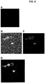
- a compound comprising SorC27 conjugated to an infrared fluorescent tag binds to TRPV6 expressed in HEK293 cells by transfection of the cells with an expression vector, but not to HEK293 cells without a transfected vector expressing TRPV6.
- Figure 6 also shows that the TRPV6-binding compound co-localizes with a fluorescently labeled antibody to TRPV6.
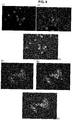
- Figures 7-9 further show that SorC27 co-localizes with TRPV6 in prostate cancer cell line PC-3, breast cancer cell line T 47D and ovarian cancer cell line SKOV-3.
- TRPV6 is expressed in samples taken from human ovarian tumor biopsies. As shown in Figure 10 and Example 4, TRPV6 transcripts were identified in cDNA libraries from human ovarian tissue tumor biopsies. The amount of TRPV6 in each sample was estimated and also quantitatively measured by densitometry with respect to the levels for the housekeeping gene ⁇ -actin. Table 1 provides the tumor type, ratio of TRPV6/ ⁇ -actin and qualitative TRPV6 level for 18 patients with ovarian tumors.
- Table 1 The relative amounts of TRPV6 transcripts in 18 human ovarian tumor biopsies detected by PCR of cDNA libraries produced from the tumors, and a ratio of the integrated band density of the TRPV6 amplicon to that of the house keeping gene ⁇ -actin.
- TRPV6 cDNA was also identified in a number of cDNA libraries prepared from total RNA extracts of human ovarian cancer cell lines including OVCAR3, SKOV3, OVC13, HEYC2, and OV2008 as shown in Figure 11 .
- TRPV6 expression was also determined in relation to the expression of the housekeeping gene ⁇ -actin in a series of breast cancer lines including T47D, HCC1954 and MB468 as shown in Figure 12A and 12B .
- the TRPV6 mRNA status results with respect to a number of breast cancer cell lines and ovarian cancer cell lines is shown in Table 2: Table 2: TRPV6 mRNA status of human breast and ovarian cancer cell lines.
- Breast Cancer Cell Lines TRPV6 mRNA status MB 231 Positive MB468 Positive T 47D Positive HCC1954 Positive MCF 7 Positive Ovarian Cancer Cell Lines TRPV6 mRNA status OVCar-3 Positive SKOV-3 Positive OV 90 Positive HeyC2 Positive OV 2008 Positive OV C13 Positive
- TRPV6 cDNA was also detected in a cDNA library made from a human prostate cancer cell line as shown in Figure 13 .
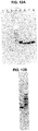
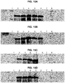
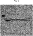
- TRPV6 protein levels were shown to be over-expressed in a number of different cancer cell lines and ovarian tumor samples as shown in Figures 14-16 and Example 5.
- embodiments disclosed herein include a method for identifying or diagnosing cancer in a test sample from a subject comprising detecting TRPV6 protein in the test sample and comparing the amount of TRPV6 protein in the test sample with a control sample.
- an increased amount of TRPV6 mRNA or protein in the test sample compared to the control sample is indicative of cancer.
- a minimal increase of at least 10 % is indicative of cancer.
- an increased amount of at least: 20%, 50%, or 100% of mRNA or protein in the test sample compared to the control sample is indicative of cancer.
- an increase of 3-times the amount of TRPV6 mRNA or protein in the test sample compared to the control sample is indicative of cancer.
- tissue where TRPV6 would be present in the control are colon, kidney, prostate and breast. In other tissues, such as any endothelial-derived tissues, TRPV6 is not expressed at all, so detection of the presence of TRPV6 mRNA or protein in the test sample is indicative of the presence of cancer. Methods may therefore be performed on such tissues by detecting TRPV6 in the test sample, without also using a control sample comparison.
- TRPV6 rich tumours are nearly exclusively of epithelial origin although some prostate cancer cell lines such as DU145 do not express TRPV6.
- Types of cancer that are TRPV6-rich include, but are not limited to, ovarian, breast, prostate, liver, endometrial, glial, colon, pancreatic, and leukemia cancers.
- the subject is a mammal, such as a human.
- the methods described herein are used in vivo, ex vivo or in vitro.
- control sample includes any sample that can be used to establish a base or normal level, and may include samples taken from healthy persons or tissues.
- the "control sample” is a predetermined standardized control.
- the "control sample” is a pre-determined value, threshold or range.
- identifying cancer includes the detection of cells in a sample from a subject that have lost normal control mechanisms and have unregulated proliferative growth.
- identifying cancer in a sample from a subject can refer to diagnosing cancer in the subject.
- cancer stage typically includes the determination of information regarding the stage of cancer (e.g. tumor stage) or type of cancer.
- Cancer stage may be determined as known in the art using the Tumor, Node, Metastasis (TNM) system.
- T for Tumor
- N for node
- M for Metastasis
- T reflects on whether the cancer has metastasized (See for example AJCC Cancer Staging Manual, Seventh Edition (2010) published by Springer-Verlag New York ).
- the cancer is ovarian cancer and the following staging guidelines may be used:
- the methods described herein are useful to further characterize a tumor by providing an estimation of tumor volume, tumor location, or tumor type.
- the diagnosis of cancer includes obtaining therapy response information such as to determine the result of a course of anti-cancer therapy on tumor size typically measured as tumor volume.
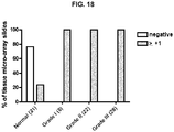
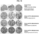
- TRPV6 levels of TRPV6 are higher in tissue samples from subjects with cancer compared to samples of normal tissue. More specifically, Figure 18 and Table 3 show that tissue micro-array samples of Grade I, II and III serous papillary adenocarcinoma exhibited more expression of TRPV6 compared to samples of normal ovarian tissue. TRPV6 levels are useful to distinguish between early stage Grade I cancers compared to normal samples. Accordingly, detection of TRPV6 using antibodies or using the TRPV6-binding peptides described herein is useful to identify or diagnose subjects with cancer or with a greater likelihood of developing cancer. Optionally, levels of TRPV6 expression are useful to grade cancers or identify more aggressive forms of cancer.
- Methods described herein may be used to identify or diagnose breast cancer, ovarian cancer, blood cancer, brain cancer, retinal cancer, liver cancer, thyroid cancer, colon cancer, prostate cancer, pancreatic cancer, glial cancer or endometrial cancer.
- Measuring the expression of the trpv6 gene through measurement of the amount of TRPV6 mRNA or corresponding cDNA transcripts produced in a sample or cell line cell provides a diagnostic tool with which to identify cancer in a sample.
- the presence or amount of TRPV6 mRNA or transcripts in a sample from a subject may be used to diagnose or indicate the stage of cancer in the subject.
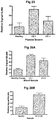
- Figure 24 shows that the relative levels of TRPV6 mRNA in blood is significantly higher in subjects with cancer compared to levels in healthy controls. Accordingly, the presence or amount of TRPV6 mRNA or transcripts in a sample from a subject may be useful to identify cancer in a subject.
- the presence or amount of TRPV6 mRNA or transcripts in a sample may be useful to identify ovarian, breast or prostate cancer in a subject.
- the relative amount of TRPV6 protein is also higher in samples of plasma from subjects with stage I or II ovarian cancer compared to healthy controls. Accordingly, the detection of the TRPV6 protein may be useful to identify cancer in a subject. For example, levels of TRPV6 protein in a test sample from a subject may be compared to levels of TRPV6 protein in a corresponding sample from a healthy control, wherein higher levels of TRPV6 in the test sample are indicative of cancer.
- the test sample may be a blood or plasma sample and a TRPV6 level that is twice as high as the level in a corresponding sample from a healthy control may be indicative of cancer.
- the TRPV6-binding peptides localize predominantly in lymph nodes, but also in lung, liver and kidney, which are common sites where metastatic cancer is located. Accordingly, the TRPV6-binding peptides described herein are useful for detecting and binding to metastatic cancer, including glioblastomas and cancers that have spread to the lung, liver, kidney, spleen, pancreas and bone marrow.
- Lymph node metastases optionally include lung cancer (Mujoomdar et al, 2007 ), gastric cancer, cervical carcinoma (Lyshchik et al., 2007 ), vulvar carcinoma (Vernooij et al, 2007 ), endometrial cancer (Aalders et al, 2007 ), head and neck squamous cell carcinoma (Veness et al., 2007 ), esophagus and throat cancer, nasopharyngeal carcinoma (Ma et al., 2007 ), gastrointestinal cancer (Wind et al., 2006 ), Gall bladder cancer, brain cancer (Mujoomdar et al., 2007 ), thyroid cancer, breast cancer, ovarian cancer, prostate cancer, glial cell cancer and colorectal cancer.
- the peptides of the invention are therefore useful in detecting cancer in a mammal at any of cancer
- TRPV6-binding peptides and compositions described herein are also useful for detecting intact TRPV6 channels present in microvesicles sloughed off from tumors, circulating in the blood or cancer cells circulating in the blood.
- Microvesicles sloughed off from TRPV6-expressing tumors, or cancer cells circulating in bodily fluids or excreta may be detected by PCR-based (e.g. RT-PCR or Q-RT-PCR) or antibody-based methods (e.g. Western blotting, immunofluorescent detection in biopsies or bodies such as cells or microvesicles in bodily fluids or excreta).
- FIG. 24 to 26 show that blood and plasma samples taken from subjects with cancer, including stage I cancer, have significantly higher levels of TRPV6 mRNA or protein compared to healthy controls. Testing bodily fluids such as blood or plasma for TRPV6 mRNA or protein therefore provides a relatively simple test for the early stage detection of cancer, compared to, for example, detecting tumors and testing tumor biopsies for the presence of cancer cells.
- Antibodies developed to the TRPV6-binding peptides are useful to detect the TRPV6-binding peptide/TRPV6 complex in tumors, tissues or cells in vitro or ex vivo by tagging the TRPV6-binding peptide antibodies with a detectable entity (fluorescent tag, radioactive tag, etc.). Similarly, microvesicles sloughed of from TRPV6-rich tumors and present in bodily fluids or excreta are readily detected by such antibodies or conjugates of the TRPV6-binding peptides.
- the compounds of the invention are useful to deliver biomolecules to tumors, cells or tissues that express TRPV6. As shown in Examples 26 and 28, TRPV6-binding peptides are able to target TRPV6 expressing cells and deliver compounds to tumor sites in vivo.
- Biomolecules are readily conjugated to the TRPV6-binding peptides or antibodies to TRPV6-binding peptides, by methods known in the art, such as those set out in Examples 1,11-13, 16, 19-23, 26 and 28.
- Biomolecules can be conjugated to the TRPV6-binding peptide by linking either directly or indirectly the TRPV6-binding peptide to the biomolecule.
- the TRPV6-binding peptide may be conjugated to Cy5.5 at the single cysteine thiol through a maleamide-activated reaction.
- the biomolecule may be linked to either the C-terminus, or N-terminus of the peptide.
- the biomolecule may alternatively be linked through any another suitable molecular site such as a functional group side chain of the peptide.
- the biomolecule may also be conjugated to a TRPV6-binding peptide via chemical modification such as by an ester linkage or an amide linkage.
- chemical modification such as by an ester linkage or an amide linkage.
- Various methods of conjugating peptides to a biomolecule are disclosed for example in Peng Li et al., Biopolymers 87: 225-230, 2007 ; U.S. Pat. No. 6,348,317 ; and U.S. Application Nos. 20070218502 and 20070020264 .
- Also disclosed herein is a method of delivering a pharmaceutical composition to a cell expressing TRPV6 comprising contacting the cell with a compound comprising i) TRPV6-binding peptide conjugated to a biomolecule and ii) a carrier.
- the methods of delivering a pharmaceutical composition include methods for delivering the compositions comprising the TRPV6-binding peptides described herein.
- the compounds may optionally comprise a TRPV6-binding peptide chemically altered to deliver nano-metallic clusters to tumors, tissues or cells (for example, gold nano-particles, nano-spheres, nano-tubes or other nano-constructs) that, when irradiated with electromagnetic radiation, heat and kill cells in the vicinity of the metallic cluster.
- a TRPV6-binding peptide chemically altered to deliver nano-metallic clusters to tumors, tissues or cells (for example, gold nano-particles, nano-spheres, nano-tubes or other nano-constructs) that, when irradiated with electromagnetic radiation, heat and kill cells in the vicinity of the metallic cluster.
- the compound may comprise TRPV6-binding peptides that are chemically altered to deliver boron clusters (e.g. closo-boron, a cluster containing 12 boron atoms) which, when irradiated with slow thermal neutrons, produce energetic alpha particles that kill nearby cells.
- boron clusters e.g. closo-boron, a cluster containing 12 boron atoms
- TRPV6-binding peptides of the invention chemically altered to deliver to TRPV6 producing tumors, cells or tissues, antigens that serve to recruit pre-existing antibodies to the TRPV6-rich tumors, tissues or cells. This, in turn, would mark cancers for destruction by the immune cell system.
- the compounds described herein are also useful to deliver to TRPV6 producing tumors, cells or tissues, novel antigens toward which monoclonal antibodies are specifically developed and administered with the result being antibody tagging of TRPV6-rich cancers. This would mark cancers for destruction by the immune cell system.
- the compounds described herein are also useful to deliver covalently attached radioactively labeled molecules to that deliver a therapeutic radiation dose to tumors, tissues or cells rich in TRPV6 channels.
- the compounds described optionally deliver to TRPV6-producing tumors, cells or tissues, covalently attached therapeutics such as the taxane-based drugs, anthracyline-type drugs, platin based drugs or any other therapeutic molecule.
- the methods and compounds described herein are also useful to deliver anti-biotics, anti-fungals, anti-virals and anti-retrovirals or any other therapeutic drug to cells that express TRPV6 or to lymph nodes, lung, liver and/or kidney.
- detection of TRPV may be used to identify a cancer tumor in a subject in vivo.
- identifying a cancer tumor refers to localizing or detecting a region in a sample or subject that has a cancer tumor.
- cancer tumor refers to a neoplasm or a solid lesion formed by the abnormal growth of cells that have lost normal control mechanism and have unregulated proliferative growth. A number of cancers have been shown to overexpress TRPV6 and therefore generate TRPV6-rich tumors (see Examples 4 and 5).
- compounds of the invention may be for use in a method for detecting a cancer tumor in a subject, said method comprising administering to a subject a compound of the invention, and detecting the TRPV6-binding peptide in the subject, thereby detecting TRVP6.
- the compounds also include a detectable label that facilitates the detection of TRPV6 in the subject.
- the TRPV6-binding peptide is SorC27, and the detectable label is Cy5.5.
- the TRPV6-binding peptide is conjugated to a magnetic resonance imaging (MRI) contrast agent such as super-paramagnetic iron oxide and MRI is used to detect regions in a sample or subject with increased levels of TRPV6.
- MRI magnetic resonance imaging
- Regions of the subject that exhibit increased levels of TRPV6 relative to a control level are indicative of a TRPV6-rich tumor in that region.
- Mathematical models that compare the distribution of TRPV6 across the subject are readily applied to identify specific regions of the subject that have increased levels of TRPV6 that are indicative of a TRPV6-rich tumor.
- an average level of TRPV6 observed throughout a subject is used to normalize the TRPV6 levels and identify specific regions with increased expression of TRPV6.
- levels of TRPV6 are compared to a pre-standardized control level or to levels observed in corresponding regions in subjects known not to contain TRPV6-rich cancer tumors.
- the TRPV6-binding peptides are optionally radioactively labeled and detected using a scintillation counter.
- the TRPV6-binding peptides are fluorescently labeled and detected using an optical detection system.
- the TRPV6-binding peptides are conjugated with a contrast agent.
- a contrast agent is a substance used to enhance the contrast of structures or cells within a sample of subject in medical imaging.
- the contrast agent is a MRI contrast agent, such as an agent that alters the T1 or T2 relaxation time of protons located nearby.
- MRI contrast agents include paramagnetic gadolinium, paramagnetic manganese, or super-paramagnetic iron oxide (SPIO).
- TRPV6-binding peptides of the compounds described herein, such as SorC13 and SorC27, are typically stable in aqueous solution at 4°C for at least 3 weeks with no change in purity as measured by HPLC. As dry solids, the peptides are typically stable at -80°C for at least 1.5 years.
- the TRPV6-binding peptides also avoid a major adverse effects of pharmaceuticals related to the ability of a substance to cross the central nervous system protective barrier, the blood-brain barrier.
- the inability of the peptides of the invention to cross this protective barrier obviates the potential toxicity to the central nervous system.
- the peptides of the invention are typically less antigenic.
- Peptides having a number of amino acids equal to or less than the empirical cutoff for antigenicity possess no antigenicity.
- kits that comprise some or all of the reagents necessary to perform any of the methods described herein are provided.
- the kits may include one or more control samples.
- the control sample may be known to express or contain TRPV6 (a positive control), or it may be a negative control that is known not to express or contain TRPV6.
- the control sample may be known to express or contain a certain level of TRPV6 or correspond to specific type or stage of cancer.
- the kits may include at least one compound comprising a TRPV6-binding peptide as described herein, and a buffer solution.
- the kits may include nucleic acid primers for amplifying or detecting TRPV6 mRNA in a polymerase chain reaction.
- the kits can also include nucleotides, enzymes and buffers useful in the method of the invention as well as electrophoretic markers such as a bp ladder.
- the kits may include detailed instructions for carrying out the methods described herein.
- SorC13 and SorC27 were labeled with the near-infrared probe, Cy5.5.
- SorC13 was labeled at lysine-1 and lysine-8 with the infrared fluorescent probe cy5.5 through reaction with Cy5.5 NHS ester-activated process.
- SorC27 was labeled at the single cysteine thiol with Cy5.5 maleimide-activated reaction.
- the labeled peptides were purified with a combination of size exclusion chromatography and HPLC.
- the label, Cy5.5 fluoresces in the infra-red region after excitation with a scanning laser. The low energy laser is able to penetrate the animal to about 1 cm and, thus, by scanning prone and supine positions, the presence of the tagged peptides can be quantified in three dimensions.
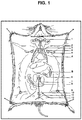
- Cy5.5-labeled peptides were intravenously injected into CD1 mice (4 for each compound) at 100 ug per animal in 100 uL, and animals were imaged live using an optical imaging system, Optix eXplorer (GE Healthcare Systems) at different time points (30 min, 90 min, 4 h). Some animals were observed at 24 hours after perfusion to remove blood (and lymph). The bio-distribution of the labeled peptides in different organs and tissues were visualized and relatively quantified by optical imaging analysis. This protocol allows for visualization of the location of the labeled peptides and how the location changes over time. Figure 1 shows the location of lymph nodes in the mouse.
- TRPV6 expressed in a cancer cell line (SKOV-3) or in ex vivo samples is optionally detected using a primary antibody to the TRPV6 protein, followed by a secondary antibody that is, itself, detectable.
- cancers are readily detected by using the TRPV6-binding peptides conjugated or tagged with a detectable molecule.
- an antibody developed to the TRPV6-binding peptides is readily tagged with a detectable entity such as a fluorescent tag or radioactive tag.
- an antibody to the TRPV6-binding peptides is readily developed and used in traditional immunochemical fashion for tissues ( in vitro ) , tumors, cells or microvesicles.
- HEK-293 cells were transfected with a TRPV6 expression vector and incubated with fluorescently labeled antibodies to the N-terminal peptides of TRPV6 as well as fluorescently labeled SorC27 (SorC27-cy5.5).
- HEK-293 cells that were not transfected with a TRPV6 expression vector were also incubated with fluorescently labeled anti-TRPV6 and SorC27-cy5.5 as a negative control ( Figure 6A ).
- SorC27-cy5.5 bound to TRPV6 as indicated by the co-localization of SorC27-cy5.5 and anti-TRPV6.
- This experiment shows that the compounds described herein target and bind to TRPV6 and further that compounds comprising a TRPV6-binding peptide conjugated to a biomolecule, are effectively localized to cells that express TRPV6.
- EXAMPLE 4 Expression of TRPV6 mRNA in Cancer Cell Lines and Tumor Biopsies as a Diagnostic for Cancer and for Staging Cancers
- polymerase chain reaction with primers directed towards TRPV6 transcripts detects up-regulation of TRPV6 mRNA in cDNA libraries produced from extracts of total RNA of cancer cells or biopsies of human cancerous tumors.
- transcripts were also amplified and detected for the housekeeping gene ⁇ -actin and the observed ratio of TRPV6 ⁇ -actin was recorded.
- Figures 10-13 show a significant increase in the expression of TRPV6 in samples of cancer lines such as ovarian cancer.
- the methods described herein that detect either TRPV6 mRNA or protein levels are therefore useful for the staging of cancers such as ovarian cancer.
- TRPV6 protein was over-expressed in extracts from ovarian (SKOV-3), breast (T47D) and prostate cancer (PC-3) cell lines compared to extracts from a hepatoblastoma HEP G2 control cell line known to express TRPV6.
- Figures 15A through 15D show that extracts from human ovarian tumors also overexpress TRPV6 protein and that TRPV6 is upregulated in ovarian tumors.
- FIG 16 shows that TRPV6 is over-expressed in extracts from human glioblastoma (U87MG), human colon (CaCo-2) and pancreatic carcinoma cells (Panc1). Further, the degree of de-glycosylation in Panc1 increases with increase numbers of culture passaging.
- Antibodies to TRPV6 are used to determine the stage of a tumor (e.g. ovarian tumors) using immunolocalization methods. Staged tissue microarrays of ovarian tumors are probed with fluorescently labeled-TRPV6 antibodies.
- EXAMPLE 7 Use of Fluorescently Tagged TRPV6-binding peptides to Determine Cancer Stages
- Fluorescently tagged TRPV6-binding peptides are useful to determine the stage of a tumor by direct incubation of Tissue Micro-Arrays (TMA) with the peptide reagent. SorC27-cy5.5 compound is incubated with cells from a subject and fluorescence levels are compared to levels from control staged tumor samples.
- TMA Tissue Micro-Arrays
- EXAMPLE 8 Use of Fluorescently Tagged Antibodies to Compounds Comprising TRPV6-Binding Peptides to Determine Cancer Stages
- Fluorescently tagged antibodies to TRPV6-binding peptides are used to detect TRPV6-binding peptides bound to TRPV6 channels of a validated ovarian cancer tissue microarray.
- mice xenografted with an ovarian TRPV6-rich tumor are injected with the fluorescently-labeled TRPV6-binding peptide SorC27-cy5.5.
- the ovarian tumors over-expressing the TRPV6 channel are detected in vivo by SorC27-cy5.5 compound after administration to mice in which TRPV6 tumors are xenografted (e.g. ovarian tumors).
- TRPV6-rich xenografted tumor mass derived from SKOV-3 cells is clearly distinguished from the background tissue as strongly fluorescing in the far infrared.
- a compound comprising TRPV6-binding peptide conjugated with Super Paramagnetic Iron Oxide such as SPIO-SorC27 is incubated with SKOV-3 cells (TRPV6 positive) and HEK293 cells (TRPV6 negative). The cells are then imaged using Magnetic Resonance Imaging.
- SKOV-3 cells over-expressing the TRPV6 channel are detected in vitro by MRI enhancement agents such as SPIO-SorC27.
- the TRPV6-rich SKOV-3 cells are clearly distinguishable from the TRPV6-negative HEK293 cells by strongly enhanced magnetic resonance signals and imaging.
- mice xenografted with a SKOV-3 TRPV6-rich ovarian tumor are administered SPIO-SorC27 and imaged using Magnetic Resonance Imaging.
- TRPV6-binding peptides bind to TRPV6-rich tumors from human ovarian cancer cell line SKOV-3, and can imaged using Magnetic Resonance Imaging.
- ovarian tumors over-expressing the TRPV6 channel are detected in vivo by MRI enhancement agents such as SPIO-SorC27 after administration to the mouse.
- the TRPV6-rich xenografted tumor mass is clearly distinguished from the background tissue by strongly enhanced magnetic resonance signals and imaging.
- Compounds comprising TRPV6-binding peptides covalently labeled with 18 F-containing radio-molecules target such molecules to TRPV6-expressing tumors, cells or tissues and allow for detection using PET scanning (Positron Emission Tomography) and detection of these tumors, cells or tissues ( Cheng et al. J. Nucl. Med. 48:987-994, 2007 ).
- SorC27 conjugated with an 18 F-containing radio-molecule is incubated with SKOV-3 cells and HEK293 cells. The cells are then imaged using PET scanning.
- the 18 F-SorC27 clearly shows the identification of the TRPV6-rich SKOV-3 cells as compared to TRPV6-negative cells (HEK293).
- Compounds comprising TRPV6-binding peptides covalently labeled with 125 I-containing radio-molecules target such molecules to TRPV6-expressing tumors, cells or tissues and allow for detection using radiometric detection of these tumors, cells or tissues ( Bolton et al. Biochem. J., 133, 529-539, 1973 ). SorC27 conjugated with a 125 I-containing radio-molecule is incubated with SKOV-3 cells and HEK293 cells. The cells are then imaged using PET scanning.
- the 125 I-C-peptide clearly shows the identification of the TRPV6-rich SKOV-3 cells as compared to TRPV6-negative cells (HEK293).
- EXAMPLE 14 Detection Of TRPV6 mRNA In Microvesicles Isolated From Samples of Blood, Other Body Fluids or Excreta
- TRPV6 mRNA is detected in microvesicles isolated from samples of blood, other body fluids or excreta of patients with TRPV6-rich cancers (e.g. ovarian tumors).
- the fraction of blood containing microvesicles sloughed off of tumors through an endocytotic process is isolated from a sample obtained from a subject. RNA is then extracted from these microvesicles, with subsequent analysis by PCR-based techniques.
- Q-RT-PCR shows the presence of TRPV6 mRNA at excess in cancer patient blood samples in comparison to people without a TRPV6-rich cancer. Detection of relative amounts of TRPV6 expression is therefore useful to diagnose TRPV6-rich cancers such as ovarian cancer.
- TRPV6 protein is detected in microvesicles isolated from bodily fluids such as blood, lymph or excreta samples of patients with TRPV6-rich cancers (e.g. ovarian tumors). The fraction of the sample containing microvesicles sloughed off of tumors through an endocytotic process is isolated. Protein is then extracted from these microvesicles, with subsequent analysis by Western blotting and antibody-based techniques to detect TRPV6 protein. Alternatively, isolated microvesicles can be treated with either anti-TRPV6 antibodies or TRPV6-binding peptides and detected.
- Western blotting shows the presence of TRPV6 protein at excess in cancer patient blood samples in comparison to people without a TRPV6-rich cancer such as ovarian cancer.
- Whole microvesicles show TRPV6 by immunofluorescence using an antibody to TRPV6 and by using appropriately labeled TRPV6-binding peptides.
- TRPV6-binding peptides are useful to deliver the attached fluorophore of the cyanidine class (cy5.5) to tissues, and to cells expressing TRPV6 (See Figures 6 to 9 ).
- Compounds comprising the TRPV6-binding peptides described herein may therefore be used to deliver other molecules to cells or tissues expressing TRPV6.
- Compounds are prepared by covalent attachment of oncology drugs such as a taxanes to the TRPV6-binding peptides in order to use the TRPV6-targetting function of the peptides to deliver the drug directly to a TRPV6-rich tumor or cancer cell.
- oncology drugs such as a taxanes
- the chemistry for attaching such drugs to compounds is known in the art (see for example, Peng Li et al., Biopolymers 87: 225-230, 2007 ).
- Paclitaxel is attached through a four carbon spacer molecule to the SorC27 peptide at a primary amine or free thiol group.
- the SorC27-conjugated taxane is then administered to mice xenografted with a TRPV6-rich tumor. Control mice are administered saline solution.
- mice In vivo experiments in mice show that a compound comprising a TRPV6-binding peptide conjugated to paclitaxel results in the adequate delivery of paclitaxel to the tumor site, regression of the tumor and the death of cancer cells when compared to control mice administered saline solution.
- Anti-viral or anti-retroviral drugs are readily covalently attached to TRPV6-binding peptides for the treatment of reservoirs of HIV.
- a primary reservoir of HIV in humans is the mesenteric lymph nodes ( Cumont et al. Cell Death and Differentiation, 14, 1747-1758, 2007 ; Estaquier and Hurtrel. Medical Science (Paris) 24(12): 1055-1060, 2008 ).
- Compounds comprising the TRPV6-binding peptides carrying a fluorescent tag accumulate in the mesenteric lymph nodes as shown by the presence of fluorescence in these nodes after i.v. injection of the tagged peptide (see Figure 1 ).
- Anti-viral or anti-retrovirals are covalently attached to the TRPV6-binding peptides described herein.
- a compound comprising a SorC27-anti-viral conjugate is then administered to mice following the protocol set out in Example 1.
- the results show the anti-viral conjugate is detected in lymph node tissue. Anti-viral activity is obtained, blocking HIV reproduction and thereby treating HIV.
- anti-microbial drugs to the TRPV6-binding peptides is useful for the treatment of reservoirs of bacterial infection.
- Compounds comprising the TRPV6-binding peptide conjugated to a fluorescent tag accumulate in the lymph nodes as indicated by the presence of fluorescence in these nodes after iv injection of the tagged peptide (see Figure 1 ).
- anti-microbials are optionally covalently attached to the TRPV6-binding peptides by methods known in the art, and used to target lymph nodes.
- TRPV6-binding peptides Target and contact TRPV6-rich cells and tumors causing concentration of the boron complexes therein (each with multiple boron atoms, for example the 12 boron closo-boron complex). Irradiation with thermal neutrons results in neutron capture by boron-10 atoms and transfer of high energy alpha-particles to the tissue killing tumor cells.
- This aspect of the invention provides for large amounts of boron clusters being targeted to tumors.
- the threshold has been stated to be about 20 ug B/g tumor ( Barth et al.
- the concentration of boron is greatly increased in TRPV6-rich tumors such as ovarian tumors. Irradiation of the tumor with thermal neutrons kills the tumor cells.
- the boron-peptide compounds are useful for killing tumor cells.
- TRPV6-binding peptides The covalent attachment of known and/or recognized epitopes of global antibodies to the TRPV6-binding peptides recruit these pre-existing antibodies to TRPV6-expressing tumors and cancer cells.
- This complex results in detection and destruction of TRPV6-expressing cells by immune cells as shown in xenograft experiments where tumors (e.g. ovarian) had lower growth rates.
- TRPV6-binding peptides covalent attachment of a novel antigen or epitope to the TRPV6-binding peptides and administration of the compound to mice directs the epitope/TRPV6-binding peptide complex to the TRPV6-bearing tumors.
- a monoclonal or polyclonal antibody directed against the specific epitope results in either type of antibody attaching to the TPRV6-rich cell, tissue or tumor.
- immune cells e.g. killer T-cells
- TRPV6-binding peptides and a molecule that is recognized and bound by receptors on cells of the immune system, particularly killer T-cells, directs such cells to TRPV6-rich tumors or cancer cells.
- the recruitment of such cells as killer T-cells destroy the cancer cell or tumor.
- the experiments indicate that the TRPV6-binding peptide targets the TRPV6 channel while the 'bate molecule' attached to the other end of the TRPV6-binding peptides recruits the immune cell; administration of this product causes shrinkage of tumors (e.g. xenografted ovarian tumors).
- the covalent attachment of metallic nano-structures (e.g nano-gold particles) to the TRPV6-binding peptides targets the metal nano-particles or other constructs (spheres, rods etc.) through the TRPV6 binding function of the peptides, to cancer cells and tumors. Because of the unique properties of, for example, nano-gold particles, irradiation with radio frequencies or infrared radiation cause the particles to heat up. Subsequent heating of the cancer cell or tumor causes death of the cells or tumor. The chemistry to attach metallic nano-particles to molecules such as peptides, is well established. Experiments indicate that targeting of the nano-gold conjugate, to TRPV6-rich xenografted tumors (e.g. ovarian tumors), with subsequent irradiation causes shrinkage of the tumor mass.
- TRPV6-rich xenografted tumors e.g. ovarian tumors
- Samples comprising tissue sections of 146 independent biopsies over 4 tissue micro-arrays (TMA) (OV483, OV802, T112, BCN721 obtained from US Biomax, Inc. Rockville, MD 20850, USA) were tested for immunohistochemical staining of TRPV6 using a TRPV6 antibody, secondary antibody and colorimetric detection using horseradish peroxidase (HRP).
- TAA tissue micro-arrays
- HRP horseradish peroxidase
- the samples included eighteen different ovarian cancer types representing all the major types of ovarian cancers as well as 21 samples of normal ovarian tissues. Many of the samples represented cancers that were previously graded.
- Figure 17 shows the calibration and representative grading of TMA samples on a six-point scale from (-) to (++++) used to assess intensity of TRPV6 staining. As shown in Table 3, each of the 146 samples was ranked according to this six-point scale.
- Figure 18 shows that a much smaller percentage of normal ovarian tissue samples had a TRPV6 stain intensity score ⁇ 1+ compared to serous papillary adenocarcinoma tissue samples with grade I, grade II, or grade III cancer. For example, 100% of serous papillary adenocarcinoma tissue samples had a stain intensity score of ⁇ 1, compared with only about 24% of normal ovarian tissues. Accordingly, detection of TRPV6 such as by antibody staining is useful in order to predict or diagnose the likelihood of cancer in a sample. Furthermore, detection of TRPV6 is useful to identify samples with Grade I (early stage) cancer compared to normal ovarian tissue samples.
- Figure 19 shows examples of the immunohistochemical detection of TRPV6 using TRPV6 antibodies in micro-array samples of normal ovarian tissues, as well as samples of Grades I, II and III serous papillary carcinoma.
- Figure 19 shows an observed trend of increased staining of TRPV6 in samples with higher Grades of cancer.
- Cancer staging data was also available for a number of tissue array samples. Cancers were staged according to the Tumor, Node, Metastasis (TNM) system as known in the art. TRPV6 immunohistochemical staining intensity for each sample assessed on a five-point scale from (-) to (++++) using a TRPV6 antibody, secondary antibody and colorimetric detection using horseradish peroxidase (HRP) is provided in Table 4.
- TPM Tumor, Node, Metastasis
- FIG. 20 shows TRPV6 antibody detection and fluorescent detection a tissue micro-array sample of grade II serous papillary adenocarcinoma stained with antibodies to TRPV6 as well as SorC27 fluorescently labeled with cy5.5 (SorC27-cy5.5).
- the use of fluorescently-labeled TRPV6-binding peptides therefore corresponds to the standard immunohistochemical detection of TRPV6 ion channels and TRPV6-binding peptides are useful to reliably detect TRPV6 or cells or tissues that express TRPV6.
- EXAMPLE 26 Localization of SorC27 to Xenografted Cancer Tumors In Vivo
- mice xenografted with a TRPV6-rich ovarian (SKOV-3 cells) and prostate (DU145 cells) tumors were injected (intraperitoneally) with the fluorescently-labeled TRPV6-binding peptide SorC27-cy5.5.
- SorC27 was labeled with the near-infrared probe Cy5.5 at the single cysteine thiol using a Cy5.5 maleimide-activated reaction.
- the labeled peptides were purified with a combination of size exclusion chromatography and HPLC. Cy5.5, fluoresces in the infra-red region after excitation with a scanning laser.
- the low energy laser is able to penetrate the animal to about 1 cm and, thus, by scanning, the presence of the tagged peptides can be quantified in three dimensions.
- Cy5.5-labeled peptides were intraperitoneally injected into nude CD1 mice at 100 ug per animal, and animals were imaged live using an optical imaging system, Optix eXplorer (GE Healthcare Systems) at different time points (30 min, 90 min, 4 h).
- FIG. 21A shows the time dependent localization of SorC27-cy5.5 in ovarian tumor
- Figure 21B shows the time dependent localization of SorC27-cy5.5 in prostate tumor.
- the kidneys are also highlighted in these two images.
- the 3-D nature of the scanning device allows clear discrimination between the tumors and the visible kidney tissues by isolating a 2 mm slice of the animal.
- a 'slice' through the tumor allows clear observation of the fluorescing central region of the tumors.
- EXAMPLE 27 Quantitative RT-PCR of TRPV6 mRNA in Cancer Tissues
- RT-PCR for TRPV6 mRNA was performed on samples of ovarian, prostate and breast cancer biopsies as well as corresponding pooled control samples from 15 healthy individuals. 18 ovarian tumor biopsies, 4 prostate tumor biopsies, and 3 breast tumor biopsies were tested. 3 different RT-PCR primer sets (A, B and C) were used for testing of the prostate samples. Results were standardized against the expression levels of the housekeeping gene hypoxanthine phosphoribosyl transferase (HPRT) and expressed as a ratio of the standardized signal from the tumor samples to the standardized signals from a pooled sample of 15 healthy tissues.
- HPRT housekeeping gene hypoxanthine phosphoribosyl transferase
- Figure 22 shows the results of Q-RT-PCR quantification of TRPV6 mRNA extracted from human ovarian (A), prostate (B) and breast cancer biopsies (C), compared to healthy tissues.
- Tables 5 to 7 also provide the quantitative Q-RT-PCR results for each of the sample biopsies relative to normal controls.
- Tumor biopsies showed a significant increase in expression levels of TRPV6 mRNA.
- Increases in TRPV6 mRNA expression relative to control tissues were seen in each cancer sample tested except one ovarian cancer sample (LTL290).
- Ovarian cancer samples showed an average 39 times increase in the expression of TRPV6 mRNA compared to healthy controls, while prostate cancer samples and breast cancer samples exhibited 8.7 times and 13 times increases respectively.
- EXAMPLE 28 In Vivo Injection and MRI Imaging of TRPV6-Binding Peptide Conjugates (SPIO-SorC27)
- SPIO Super Paramagnetic Iron Oxide
- the MRI images were acquired on a 3T Varian Direct Drive Console using a 305/210 mm OD/ID Magnex gradient coil and a 25 mm diameter quadrature mouse RF coil from Doty Inc.
- the images were acquired using a pulse sequence specifically selected to optimize contrast sensitivity to iron-oxide nanoparticles.
- the iron-oxide appears dark (negative contrast) for these types of acquisitions.
- the acquisitions used a 3D balanced steady state free precession (b-SSFP) pulse sequence with a TR/TE of 8/4 ms and an image resolution of 150 micron (150 micron pixel dimension in all 3 directions).
- b-SSFP 3D balanced steady state free precession
- Figure 23 shows MRI images and the localization of the MRI enhancement agent (SPIO-SorC27) to SKOV-3 derived ovarian tumors xenografted into CD-1 nude mice.
- the upper control panels (A) show the administration of the SPIO control beads, without conjugated SorC27. The SPIO control beads were cleared from the tumor by 24 hours post-injection.
- the lower level panel (B) shows that the SPIO-SorC27 compound labels the cortex of the tumor 24 hours post-injection.
- the solid white arrow shown in the left-hand images indicates the position of the tumor in the xenograft.
- the dashed arrow in the bottom right panel indicates the darkened enhanced MRI signal of the SPIO-SorC27 construct bound to the cortex of the tumor.
- EXAMPLE 29 RT-PCR of Blood From Staged Cancer Subjects
- Samples of blood taken from subjects with prostate, breast or ovarian cancer were tested for TRPV6 mRNA expression using RT-PCR.
- Control samples of blood taken from a healthy male (prostate) or healthy female (breast and ovarian) were also tested.
- RT-PCR products were loaded onto agarose gels and separated using electrophoresis. Integrated band density was then measured on the agarose gels for the amplicons of the TRPV6 mRNA (-320 bp) for each sample and control.
- Figure 24 shows that the expression of TRPV6 mRNA in blood samples taken from patients with cancer was significantly higher compared to samples taken from normal healthy controls.
- Figure 24A shows that subjects with stage I, II, III or IV prostate cancer had up to 6 times more expression of TRPV6 in blood. A significant increase in TRPV6 expression is also seen in samples representing different cancer stages compared to normal samples.
- Figure 24B shows that subjects with breast cancer exhibit a significant increase in blood TRPV6 expression.
- Figure 24C shows that subjects with ovarian cancer also exhibit an increase in TRPV6 expression compared to healthy female control samples.
- the integrated signals represented in Figure 24 were divided by a factor of either 100,000 (24A & 24B) or 10,000 (24C). Analysis of expression levels of TRPV6 mRNA in blood is therefore useful to identify subjects with cancer, including the early detection of stage I prostate, breast or ovarian cancer.
- EXAMPLE 30 TRPV6 mRNA in Plasma Samples from Subjects with Stage I or Stage II Ovarian Cancer
- TRPV6 mRNA (cDNA) amplicons were imaged and quantified with an Alpha Innotech FluorChem® FC2 imager, using filter position#1 (green filter) and UV light (302nm) using Auto Expose. All samples were analyzed in triplicate and data were compared using the Student's t-test and the 95% confidence limit.
- Plasma samples were obtained from healthy women and women diagnosed with stage I or II ovarian cancer. Protein was isolated from the plasma samples following the TRI Reagent® LS method (Sigma Aldrich). Lysates were prepared from the plasma protein pellets from the TRI Reagent® LS procedure by heating in a solution of 1% SDS in PBS and 15 mM dithiothrietol (DTT) in a boiling water bath. Protein extracts were quantified by measuring the absorbance at 280 nm of each lysate on a Varian Cary 50 UV spectrophotometer. The amount of protein in ug/uL was extrapolated from a bovine serum albumin protein standard curve.
- Protein extracts were electrophoresed on NuPage® Novex 4-12% Bis-Tris Gel 1.5 mm wells (Invitrogen) at 145V for 55 mins. The gels were transferred for 10 mins onto a iBlot® Transfer Stack, PVDF regular (Invitrogen) using the Invitrogen iBlot transfer system. PVDF blocking, antibody incubation and washing were performed using a SNAP i.d. protein detection system (Millipore). The PVDF membranes were then blocked for 30 seconds with 0.5% ECL advanced blocking buffer (Fisher).
- the PVDF's were incubated in a 1/30 dilution of TRPV6 (H-90) primary antibody (Santa Cruz) for 10 min and washed 3 times with 30 mL of TBS-T.
- the PVDFs were then incubated in a 1/1500 dilution of goat anti-rabbit IgG HRP secondary antibody (Santa Cruz) for 10 min and washed 3 times with 30 mL of TBS-T.
- the TRPV6 bands were detected with 15 ml of Luminol for 3 mins. Gels were imaged and the band density was quantified with an Alpha Innotech FluroChem imager for 10 minutes. All samples were analyzed in triplicate and data were compared using the Student's t-test and the 95% confidence limit.
Landscapes
- Health & Medical Sciences (AREA)
- Life Sciences & Earth Sciences (AREA)
- Chemical & Material Sciences (AREA)
- Engineering & Computer Science (AREA)
- General Health & Medical Sciences (AREA)
- Immunology (AREA)
- Medicinal Chemistry (AREA)
- Public Health (AREA)
- Animal Behavior & Ethology (AREA)
- Veterinary Medicine (AREA)
- Molecular Biology (AREA)
- Epidemiology (AREA)
- Biomedical Technology (AREA)
- Urology & Nephrology (AREA)
- Hematology (AREA)
- Pharmacology & Pharmacy (AREA)
- Proteomics, Peptides & Aminoacids (AREA)
- Nanotechnology (AREA)
- Cell Biology (AREA)
- Biochemistry (AREA)
- Biotechnology (AREA)
- Microbiology (AREA)
- Physics & Mathematics (AREA)
- Nuclear Medicine, Radiotherapy & Molecular Imaging (AREA)
- Food Science & Technology (AREA)
- Pathology (AREA)
- General Physics & Mathematics (AREA)
- Analytical Chemistry (AREA)
- Bioinformatics & Cheminformatics (AREA)
- Organic Chemistry (AREA)
- Radiology & Medical Imaging (AREA)
- Biophysics (AREA)
- Crystallography & Structural Chemistry (AREA)
- Medical Informatics (AREA)
- Genetics & Genomics (AREA)
- General Engineering & Computer Science (AREA)
- Chemical Kinetics & Catalysis (AREA)
- General Chemical & Material Sciences (AREA)
- Mycology (AREA)
- Optics & Photonics (AREA)
Applications Claiming Priority (3)
| Application Number | Priority Date | Filing Date | Title |
|---|---|---|---|
| US22083309P | 2009-06-26 | 2009-06-26 | |
| US24463409P | 2009-09-22 | 2009-09-22 | |
| PCT/CA2010/000971 WO2010148501A1 (en) | 2009-06-26 | 2010-06-28 | Soricidin derived peptides and methods for the detection of trpv-6 cancers and drug delivery |
Publications (3)
| Publication Number | Publication Date |
|---|---|
| EP2445932A1 EP2445932A1 (en) | 2012-05-02 |
| EP2445932A4 EP2445932A4 (en) | 2015-10-14 |
| EP2445932B1 true EP2445932B1 (en) | 2018-02-28 |
Family
ID=43381003
Family Applications (1)
| Application Number | Title | Priority Date | Filing Date |
|---|---|---|---|
| EP10791109.1A Active EP2445932B1 (en) | 2009-06-26 | 2010-06-28 | Soricidin derived peptides and methods for the detection of trpv-6 cancers and drug delivery |
Country Status (8)
| Country | Link |
|---|---|
| US (4) | US20100329983A1 (enExample) |
| EP (1) | EP2445932B1 (enExample) |
| JP (1) | JP5836940B2 (enExample) |
| BR (1) | BRPI1012675A2 (enExample) |
| CA (1) | CA2766272C (enExample) |
| ES (1) | ES2670588T3 (enExample) |
| MX (1) | MX2012000086A (enExample) |
| WO (1) | WO2010148501A1 (enExample) |
Families Citing this family (9)
| Publication number | Priority date | Publication date | Assignee | Title |
|---|---|---|---|---|
| EP2445932B1 (en) | 2009-06-26 | 2018-02-28 | Soricimed Biopharma Inc. | Soricidin derived peptides and methods for the detection of trpv-6 cancers and drug delivery |
| US20130116404A1 (en) * | 2011-11-08 | 2013-05-09 | Case Western Reserve University | Targeted non-invasive imaging probes of egfr expressing cells |
| EP2895504B1 (en) * | 2012-09-14 | 2019-05-08 | John M. Stewart | Trpv3 agonists for the treatment of skin conditions |
| WO2019183633A1 (en) | 2018-03-23 | 2019-09-26 | Case Western Reserve Univeristy | Psma targeted conjugate compounds and uses thereof |
| CN111484982A (zh) * | 2020-05-14 | 2020-08-04 | 西南民族大学 | 一种人结直肠腺癌细胞trpv1基因敲除细胞株及应用 |
| US20260116965A1 (en) * | 2021-01-15 | 2026-04-30 | Coherent Biopharma (Suzhou) , Limited | Anti-trpv6 monoclonal antibody and application thereof |
| AU2022365387A1 (en) * | 2021-10-14 | 2024-05-02 | INSERM (Institut National de la Santé et de la Recherche Médicale) | Antibodies against extracellular epitopes of human trpv6 channel and their diagnostic and therapeutic uses |
| US20250332265A1 (en) | 2021-11-09 | 2025-10-30 | Case Western Reserve University | Psma targeted conjugate compounds and uses thereof |
| CN118846104A (zh) * | 2024-06-27 | 2024-10-29 | 河北工业大学 | 一种近红外响应纳米复合物及其制备方法和应用 |
Family Cites Families (48)
| Publication number | Priority date | Publication date | Assignee | Title |
|---|---|---|---|---|
| US7002A (en) * | 1850-01-08 | Obe-washek | ||
| GB2161165A (en) | 1984-06-12 | 1986-01-08 | Cambridge Patent Dev | Secondary antibodies |
| DK255887D0 (da) | 1987-05-20 | 1987-05-20 | Claus Koch | Immunoassay |
| GB8722018D0 (en) | 1987-09-18 | 1987-10-28 | Central Blood Lab Authority | Human anti-rh(d)monoclonal antibodies |
| US5496548A (en) | 1987-09-18 | 1996-03-05 | National Blood Authority | Human anti-RH(D) monoclonal antibodies, cell lines and methods of use of antibodies in immunoassays |
| US5688657A (en) | 1988-03-31 | 1997-11-18 | International Bio-Immune Systems, Inc. | Monoclonal antibodies against human colon carcinoma-associated antigens and uses therefor |
| ATE147634T1 (de) | 1988-12-19 | 1997-02-15 | American Cyanamid Co | Produkte zur behandlung des endotoxin -schocks bei einem säugetier |
| AU648022B2 (en) | 1989-03-14 | 1994-04-14 | Bionebraska, Inc. | Monoclonal antibodies for metallic cations on small molecules |
| US5500345A (en) | 1989-04-25 | 1996-03-19 | Iatron Laboratories, Inc. | Hybridomas producing monoclonal antibodies specific for C-reactive protein and methods for detection of C-reactive protein |
| CA2025266A1 (en) | 1989-10-04 | 1991-04-05 | Kiyoshi Kobayashi | Anti-hcg-.beta. core monoclonal antibody, its production and use |
| WO1991014452A1 (en) | 1990-03-27 | 1991-10-03 | The Salk Institute For Biological Studies | Method for inhibiting cell growth and compositions useful therefor |
| US5510241A (en) | 1990-10-01 | 1996-04-23 | The Minister Of Agriculture, Fisheries And Food In Her Britannic Majesty's Government Of The United Kingdom Of Great Britian And Northern Ireland | Method of testing for the presence of Salmonella serotypes expressing Salmonella enteritidis fimbrial antigen (SEFA) and reagents therefore |
| US5225331A (en) | 1991-04-25 | 1993-07-06 | National Research Council Of Canada | Immunoassay for detecting group b streptococcus |
| AU667533B2 (en) | 1992-03-26 | 1996-03-28 | Microcarb, Inc. | Monospecific polyclonal antibodies to shiga-like toxins |
| AU4231393A (en) | 1992-05-08 | 1993-12-13 | Genentech Inc. | Antibodies to leukemia inhibitory factor |
| US5424286A (en) * | 1993-05-24 | 1995-06-13 | Eng; John | Exendin-3 and exendin-4 polypeptides, and pharmaceutical compositions comprising same |
| US5683693A (en) | 1994-04-25 | 1997-11-04 | Trustees Of Dartmouth College | Method for inducing T cell unresponsiveness to a tissue or organ graft with anti-CD40 ligand antibody or soluble CD40 |
| JPH10236963A (ja) | 1997-02-26 | 1998-09-08 | Sagami Chem Res Center | カルシウムチャンネル拮抗剤 |
| AU7389098A (en) * | 1997-05-16 | 1998-12-08 | Brigham And Women's Hospital | Local anesthetic formulations |
| US6946475B1 (en) * | 1999-04-07 | 2005-09-20 | University Of Virginia Patent Foundation | Anticancer calcium channel blockers |
| EP1165508B1 (en) | 1999-04-07 | 2004-06-23 | The University of Virginia Patent Foundation | Anticancer calcium channel blockers |
| US6534642B1 (en) * | 1999-07-09 | 2003-03-18 | Matthias A. Hediger | Compositions corresponding to a calcium transporter and methods of making and using same |
| US6348317B1 (en) | 1999-11-18 | 2002-02-19 | The Arizona Board Of Regents | Fluorescent and DNA cleavage properties of peptide/dye conjugates |
| US20040001827A1 (en) | 2002-06-28 | 2004-01-01 | Dennis Mark S. | Serum albumin binding peptides for tumor targeting |
| US7176037B2 (en) | 2000-07-13 | 2007-02-13 | The Scripps Research Institute | Labeled peptides, proteins and antibodies and processes and intermediates useful for their preparation |
| DE60134178D1 (de) | 2000-07-28 | 2008-07-03 | Ulrich Wissenbach | Trp8 krebsmarker |
| AU2003281200A1 (en) | 2002-07-03 | 2004-01-23 | Tasuku Honjo | Immunopotentiating compositions |
| US7119168B2 (en) * | 2002-11-18 | 2006-10-10 | Bioprospecting Nb Inc. | Paralytic peptide for use in neuromuscular therapy |
| CA2544467C (en) * | 2002-11-18 | 2014-02-18 | John M. Stewart | Paralytic peptide for use in neuromuscular therapy |
| DK2439273T3 (da) | 2005-05-09 | 2019-06-03 | Ono Pharmaceutical Co | Humane monoklonale antistoffer til programmeret død-1(pd-1) og fremgangsmåder til behandling af cancer ved anvendelse af anti-pd-1- antistoffer alene eller i kombination med andre immunterapeutika |
| PT1907424E (pt) | 2005-07-01 | 2015-10-09 | Squibb & Sons Llc | Anticorpos monoclonais humanos para o ligando 1 de morte programada (pd-l1) |
| JP5749154B2 (ja) * | 2008-03-19 | 2015-07-15 | ソリシメド・バイオファーマ・インコーポレーテッド | Trpv6カルシウムチャネル活性阻害による癌治療のためのペプチド組成物 |
| US9181342B2 (en) | 2008-09-12 | 2015-11-10 | Isis Innovation Limited | PD-1 specific antibodies and uses thereof |
| US8552154B2 (en) | 2008-09-26 | 2013-10-08 | Emory University | Anti-PD-L1 antibodies and uses therefor |
| JP5245853B2 (ja) | 2009-01-19 | 2013-07-24 | セイコーエプソン株式会社 | 画像形成装置 |
| JP5844159B2 (ja) | 2009-02-09 | 2016-01-13 | ユニヴェルシテ デクス−マルセイユUniversite D’Aix−Marseille | Pd−1抗体およびpd−l1抗体ならびにその使用 |
| EP2445932B1 (en) | 2009-06-26 | 2018-02-28 | Soricimed Biopharma Inc. | Soricidin derived peptides and methods for the detection of trpv-6 cancers and drug delivery |
| EP2545078A1 (en) | 2010-03-11 | 2013-01-16 | UCB Pharma, S.A. | Pd-1 antibody |
| TW201134488A (en) | 2010-03-11 | 2011-10-16 | Ucb Pharma Sa | PD-1 antibodies |
| EP2895504B1 (en) | 2012-09-14 | 2019-05-08 | John M. Stewart | Trpv3 agonists for the treatment of skin conditions |
| CN112552401B (zh) | 2013-09-13 | 2023-08-25 | 广州百济神州生物制药有限公司 | 抗pd1抗体及其作为治疗剂与诊断剂的用途 |
| TWI681969B (zh) | 2014-01-23 | 2020-01-11 | 美商再生元醫藥公司 | 針對pd-1的人類抗體 |
| PE20170255A1 (es) | 2014-01-24 | 2017-03-22 | Dana Farber Cancer Inst Inc | Moleculas de anticuerpo que se unen a pd-1 y usos de las mismas |
| AU2015243537B2 (en) | 2014-04-08 | 2020-10-22 | The Methodist Hospital | INOS-inhibitory compositions and their use as breast cancer therapeutics |
| JP6772199B2 (ja) | 2015-08-11 | 2020-10-21 | コヒレント バイオファーマ | 多リガンド−薬物複合体及びその使用 |
| EP3439658B1 (en) | 2016-04-04 | 2021-11-24 | ChemoCentryx, Inc. | Soluble c5ar antagonists |
| KR20180133442A (ko) | 2016-04-13 | 2018-12-14 | 비비아 바이오테크 에스.엘. | 생체외 bite 활성화된 t 세포 |
| CA3083950A1 (en) | 2017-12-01 | 2019-06-06 | Soricimed Biopharma Inc. | Trpv6 inhibitors and combination therapies for treating cancers |
-
2010
- 2010-06-28 EP EP10791109.1A patent/EP2445932B1/en active Active
- 2010-06-28 ES ES10791109.1T patent/ES2670588T3/es active Active
- 2010-06-28 JP JP2012516450A patent/JP5836940B2/ja active Active
- 2010-06-28 WO PCT/CA2010/000971 patent/WO2010148501A1/en not_active Ceased
- 2010-06-28 US US12/824,935 patent/US20100329983A1/en not_active Abandoned
- 2010-06-28 CA CA2766272A patent/CA2766272C/en active Active
- 2010-06-28 BR BRPI1012675A patent/BRPI1012675A2/pt not_active IP Right Cessation
- 2010-06-28 MX MX2012000086A patent/MX2012000086A/es not_active Application Discontinuation
-
2014
- 2014-12-22 US US14/579,157 patent/US10064964B2/en active Active
-
2018
- 2018-07-26 US US16/046,786 patent/US11090396B2/en active Active
-
2021
- 2021-07-15 US US17/376,670 patent/US12458618B2/en active Active
Also Published As
| Publication number | Publication date |
|---|---|
| EP2445932A1 (en) | 2012-05-02 |
| US20100329983A1 (en) | 2010-12-30 |
| CA2766272C (en) | 2021-02-09 |
| US20190209717A1 (en) | 2019-07-11 |
| US10064964B2 (en) | 2018-09-04 |
| US20150182644A1 (en) | 2015-07-02 |
| ES2670588T3 (es) | 2018-05-31 |
| BRPI1012675A2 (pt) | 2016-04-05 |
| JP5836940B2 (ja) | 2015-12-24 |
| JP2012530725A (ja) | 2012-12-06 |
| US20220175977A1 (en) | 2022-06-09 |
| EP2445932A4 (en) | 2015-10-14 |
| MX2012000086A (es) | 2012-06-12 |
| WO2010148501A1 (en) | 2010-12-29 |
| CA2766272A1 (en) | 2010-12-29 |
| US12458618B2 (en) | 2025-11-04 |
| US11090396B2 (en) | 2021-08-17 |
Similar Documents
| Publication | Publication Date | Title |
|---|---|---|
| US12458618B2 (en) | Compounds and methods for the detection of TRPV-6 cancers and drug delivery | |
| JP6046070B2 (ja) | 膵管腺癌の検出および治療のためのプレクチン−1標的化剤 | |
| Bowen et al. | In vivo detection of human TRPV6-rich tumors with anti-cancer peptides derived from soricidin | |
| Kelly et al. | Targeted nanoparticles for imaging incipient pancreatic ductal adenocarcinoma | |
| Kim et al. | PET imaging of HER2 expression with an 18F-fluoride labeled aptamer | |
| Pung et al. | Generation of peptides using phage display technology for cancer diagnosis and molecular imaging | |
| US9486545B2 (en) | Method of screening for colon cancer using biomarkers | |
| JP2012530725A5 (enExample) | ||
| Ma et al. | Screening of an annexin‐A2‐targeted heptapeptide for pancreatic adenocarcinoma localization | |
| Heo et al. | Clinical big-data-based design of GLUT2-targeted carbon nanodots for accurate diagnosis of hepatocellular carcinoma | |
| HK1169661B (en) | Soricidin derived peptides and methods for the detection of trpv-6 cancers and drug delivery | |
| HK1169661A (en) | Soricidin derived peptides and methods for the detection of trpv-6 cancers and drug delivery | |
| Daquinag et al. | Fibulin-4 is highly expressed in metastatic breast cancer and can serve as a target of peptide-based imaging probes and experimental therapeutics | |
| Zhang et al. | EDB-FN-Targeted Positron Emission Tomography Imaging of Breast Cancer in Mice | |
| Bowen et al. | In Vivo Detection of Human TRPV6-Rich Tumors with Anti-Cancer Peptides Derived |
Legal Events
| Date | Code | Title | Description |
|---|---|---|---|
| PUAI | Public reference made under article 153(3) epc to a published international application that has entered the european phase |
Free format text: ORIGINAL CODE: 0009012 |
|
| 17P | Request for examination filed |
Effective date: 20120124 |
|
| AK | Designated contracting states |
Kind code of ref document: A1 Designated state(s): AL AT BE BG CH CY CZ DE DK EE ES FI FR GB GR HR HU IE IS IT LI LT LU LV MC MK MT NL NO PL PT RO SE SI SK SM TR |
|
| DAX | Request for extension of the european patent (deleted) | ||
| REG | Reference to a national code |
Ref country code: HK Ref legal event code: DE Ref document number: 1169661 Country of ref document: HK |
|
| RIC1 | Information provided on ipc code assigned before grant |
Ipc: C07K 7/08 20060101ALI20150422BHEP Ipc: C07K 14/47 20060101AFI20150422BHEP Ipc: G01N 33/566 20060101ALI20150422BHEP Ipc: C07K 14/705 20060101ALI20150422BHEP Ipc: C07K 7/06 20060101ALI20150422BHEP Ipc: A61K 47/48 20060101ALI20150422BHEP Ipc: A61K 49/00 20060101ALI20150422BHEP Ipc: A61P 35/00 20060101ALI20150422BHEP Ipc: G01N 33/48 20060101ALI20150422BHEP |
|
| RIC1 | Information provided on ipc code assigned before grant |
Ipc: A61P 35/00 20060101ALI20150901BHEP Ipc: C07K 14/705 20060101ALI20150901BHEP Ipc: C07K 7/08 20060101ALI20150901BHEP Ipc: G01N 33/566 20060101ALI20150901BHEP Ipc: C07K 7/06 20060101ALI20150901BHEP Ipc: A61K 47/48 20060101ALI20150901BHEP Ipc: G01N 33/48 20060101ALI20150901BHEP Ipc: A61K 49/00 20060101ALI20150901BHEP Ipc: C07K 14/47 20060101AFI20150901BHEP |
|
| RA4 | Supplementary search report drawn up and despatched (corrected) |
Effective date: 20150914 |
|
| RIC1 | Information provided on ipc code assigned before grant |
Ipc: A61K 47/48 20060101ALI20150908BHEP Ipc: G01N 33/566 20060101ALI20150908BHEP Ipc: A61K 49/00 20060101ALI20150908BHEP Ipc: G01N 33/48 20060101ALI20150908BHEP Ipc: C07K 14/47 20060101AFI20150908BHEP Ipc: A61P 35/00 20060101ALI20150908BHEP Ipc: C07K 7/06 20060101ALI20150908BHEP Ipc: C07K 14/705 20060101ALI20150908BHEP Ipc: C07K 7/08 20060101ALI20150908BHEP |
|
| STAA | Information on the status of an ep patent application or granted ep patent |
Free format text: STATUS: EXAMINATION IS IN PROGRESS |
|
| 17Q | First examination report despatched |
Effective date: 20170406 |
|
| RIC1 | Information provided on ipc code assigned before grant |
Ipc: G01N 33/566 20060101ALI20170706BHEP Ipc: C07K 14/705 20060101ALI20170706BHEP Ipc: C07K 7/06 20060101ALI20170706BHEP Ipc: C07K 7/08 20060101ALI20170706BHEP Ipc: A61K 49/00 20060101ALI20170706BHEP Ipc: C07K 14/47 20060101AFI20170706BHEP Ipc: G01N 33/48 20060101ALI20170706BHEP Ipc: A61P 35/00 20060101ALI20170706BHEP |
|
| GRAP | Despatch of communication of intention to grant a patent |
Free format text: ORIGINAL CODE: EPIDOSNIGR1 |
|
| STAA | Information on the status of an ep patent application or granted ep patent |
Free format text: STATUS: GRANT OF PATENT IS INTENDED |
|
| INTG | Intention to grant announced |
Effective date: 20170825 |
|
| RIN1 | Information on inventor provided before grant (corrected) |
Inventor name: STEWART, JOHN M. |
|
| GRAS | Grant fee paid |
Free format text: ORIGINAL CODE: EPIDOSNIGR3 |
|
| GRAA | (expected) grant |
Free format text: ORIGINAL CODE: 0009210 |
|
| STAA | Information on the status of an ep patent application or granted ep patent |
Free format text: STATUS: THE PATENT HAS BEEN GRANTED |
|
| AK | Designated contracting states |
Kind code of ref document: B1 Designated state(s): AL AT BE BG CH CY CZ DE DK EE ES FI FR GB GR HR HU IE IS IT LI LT LU LV MC MK MT NL NO PL PT RO SE SI SK SM TR |
|
| REG | Reference to a national code |
Ref country code: GB Ref legal event code: FG4D Ref country code: CH Ref legal event code: EP |
|
| REG | Reference to a national code |
Ref country code: AT Ref legal event code: REF Ref document number: 974019 Country of ref document: AT Kind code of ref document: T Effective date: 20180315 |
|
| REG | Reference to a national code |
Ref country code: IE Ref legal event code: FG4D |
|
| REG | Reference to a national code |
Ref country code: DE Ref legal event code: R096 Ref document number: 602010048860 Country of ref document: DE |
|
| REG | Reference to a national code |
Ref country code: ES Ref legal event code: FG2A Ref document number: 2670588 Country of ref document: ES Kind code of ref document: T3 Effective date: 20180531 |
|
| REG | Reference to a national code |
Ref country code: SE Ref legal event code: TRGR |
|
| REG | Reference to a national code |
Ref country code: NL Ref legal event code: FP |
|
| REG | Reference to a national code |
Ref country code: FR Ref legal event code: PLFP Year of fee payment: 9 |
|
| REG | Reference to a national code |
Ref country code: LT Ref legal event code: MG4D |
|
| REG | Reference to a national code |
Ref country code: AT Ref legal event code: MK05 Ref document number: 974019 Country of ref document: AT Kind code of ref document: T Effective date: 20180228 |
|
| PG25 | Lapsed in a contracting state [announced via postgrant information from national office to epo] |
Ref country code: NO Free format text: LAPSE BECAUSE OF FAILURE TO SUBMIT A TRANSLATION OF THE DESCRIPTION OR TO PAY THE FEE WITHIN THE PRESCRIBED TIME-LIMIT Effective date: 20180528 Ref country code: HR Free format text: LAPSE BECAUSE OF FAILURE TO SUBMIT A TRANSLATION OF THE DESCRIPTION OR TO PAY THE FEE WITHIN THE PRESCRIBED TIME-LIMIT Effective date: 20180228 Ref country code: CY Free format text: LAPSE BECAUSE OF FAILURE TO SUBMIT A TRANSLATION OF THE DESCRIPTION OR TO PAY THE FEE WITHIN THE PRESCRIBED TIME-LIMIT Effective date: 20180228 Ref country code: FI Free format text: LAPSE BECAUSE OF FAILURE TO SUBMIT A TRANSLATION OF THE DESCRIPTION OR TO PAY THE FEE WITHIN THE PRESCRIBED TIME-LIMIT Effective date: 20180228 Ref country code: LT Free format text: LAPSE BECAUSE OF FAILURE TO SUBMIT A TRANSLATION OF THE DESCRIPTION OR TO PAY THE FEE WITHIN THE PRESCRIBED TIME-LIMIT Effective date: 20180228 |
|
| PG25 | Lapsed in a contracting state [announced via postgrant information from national office to epo] |
Ref country code: AT Free format text: LAPSE BECAUSE OF FAILURE TO SUBMIT A TRANSLATION OF THE DESCRIPTION OR TO PAY THE FEE WITHIN THE PRESCRIBED TIME-LIMIT Effective date: 20180228 Ref country code: GR Free format text: LAPSE BECAUSE OF FAILURE TO SUBMIT A TRANSLATION OF THE DESCRIPTION OR TO PAY THE FEE WITHIN THE PRESCRIBED TIME-LIMIT Effective date: 20180529 Ref country code: BG Free format text: LAPSE BECAUSE OF FAILURE TO SUBMIT A TRANSLATION OF THE DESCRIPTION OR TO PAY THE FEE WITHIN THE PRESCRIBED TIME-LIMIT Effective date: 20180528 Ref country code: LV Free format text: LAPSE BECAUSE OF FAILURE TO SUBMIT A TRANSLATION OF THE DESCRIPTION OR TO PAY THE FEE WITHIN THE PRESCRIBED TIME-LIMIT Effective date: 20180228 |
|
| PG25 | Lapsed in a contracting state [announced via postgrant information from national office to epo] |
Ref country code: AL Free format text: LAPSE BECAUSE OF FAILURE TO SUBMIT A TRANSLATION OF THE DESCRIPTION OR TO PAY THE FEE WITHIN THE PRESCRIBED TIME-LIMIT Effective date: 20180228 Ref country code: EE Free format text: LAPSE BECAUSE OF FAILURE TO SUBMIT A TRANSLATION OF THE DESCRIPTION OR TO PAY THE FEE WITHIN THE PRESCRIBED TIME-LIMIT Effective date: 20180228 Ref country code: RO Free format text: LAPSE BECAUSE OF FAILURE TO SUBMIT A TRANSLATION OF THE DESCRIPTION OR TO PAY THE FEE WITHIN THE PRESCRIBED TIME-LIMIT Effective date: 20180228 Ref country code: PL Free format text: LAPSE BECAUSE OF FAILURE TO SUBMIT A TRANSLATION OF THE DESCRIPTION OR TO PAY THE FEE WITHIN THE PRESCRIBED TIME-LIMIT Effective date: 20180228 |
|
| REG | Reference to a national code |
Ref country code: DE Ref legal event code: R097 Ref document number: 602010048860 Country of ref document: DE |
|
| PG25 | Lapsed in a contracting state [announced via postgrant information from national office to epo] |
Ref country code: CZ Free format text: LAPSE BECAUSE OF FAILURE TO SUBMIT A TRANSLATION OF THE DESCRIPTION OR TO PAY THE FEE WITHIN THE PRESCRIBED TIME-LIMIT Effective date: 20180228 Ref country code: SK Free format text: LAPSE BECAUSE OF FAILURE TO SUBMIT A TRANSLATION OF THE DESCRIPTION OR TO PAY THE FEE WITHIN THE PRESCRIBED TIME-LIMIT Effective date: 20180228 Ref country code: DK Free format text: LAPSE BECAUSE OF FAILURE TO SUBMIT A TRANSLATION OF THE DESCRIPTION OR TO PAY THE FEE WITHIN THE PRESCRIBED TIME-LIMIT Effective date: 20180228 Ref country code: SM Free format text: LAPSE BECAUSE OF FAILURE TO SUBMIT A TRANSLATION OF THE DESCRIPTION OR TO PAY THE FEE WITHIN THE PRESCRIBED TIME-LIMIT Effective date: 20180228 |
|
| PLBE | No opposition filed within time limit |
Free format text: ORIGINAL CODE: 0009261 |
|
| STAA | Information on the status of an ep patent application or granted ep patent |
Free format text: STATUS: NO OPPOSITION FILED WITHIN TIME LIMIT |
|
| 26N | No opposition filed |
Effective date: 20181129 |
|
| PG25 | Lapsed in a contracting state [announced via postgrant information from national office to epo] |
Ref country code: SI Free format text: LAPSE BECAUSE OF FAILURE TO SUBMIT A TRANSLATION OF THE DESCRIPTION OR TO PAY THE FEE WITHIN THE PRESCRIBED TIME-LIMIT Effective date: 20180228 |
|
| REG | Reference to a national code |
Ref country code: BE Ref legal event code: MM Effective date: 20180630 |
|
| PG25 | Lapsed in a contracting state [announced via postgrant information from national office to epo] |
Ref country code: MC Free format text: LAPSE BECAUSE OF FAILURE TO SUBMIT A TRANSLATION OF THE DESCRIPTION OR TO PAY THE FEE WITHIN THE PRESCRIBED TIME-LIMIT Effective date: 20180228 Ref country code: LU Free format text: LAPSE BECAUSE OF NON-PAYMENT OF DUE FEES Effective date: 20180628 |
|
| REG | Reference to a national code |
Ref country code: IE Ref legal event code: MM4A |
|
| PG25 | Lapsed in a contracting state [announced via postgrant information from national office to epo] |
Ref country code: IE Free format text: LAPSE BECAUSE OF NON-PAYMENT OF DUE FEES Effective date: 20180628 |
|
| PG25 | Lapsed in a contracting state [announced via postgrant information from national office to epo] |
Ref country code: BE Free format text: LAPSE BECAUSE OF NON-PAYMENT OF DUE FEES Effective date: 20180630 |
|
| PG25 | Lapsed in a contracting state [announced via postgrant information from national office to epo] |
Ref country code: MT Free format text: LAPSE BECAUSE OF NON-PAYMENT OF DUE FEES Effective date: 20180628 |
|
| PG25 | Lapsed in a contracting state [announced via postgrant information from national office to epo] |
Ref country code: TR Free format text: LAPSE BECAUSE OF FAILURE TO SUBMIT A TRANSLATION OF THE DESCRIPTION OR TO PAY THE FEE WITHIN THE PRESCRIBED TIME-LIMIT Effective date: 20180228 |
|
| PG25 | Lapsed in a contracting state [announced via postgrant information from national office to epo] |
Ref country code: PT Free format text: LAPSE BECAUSE OF FAILURE TO SUBMIT A TRANSLATION OF THE DESCRIPTION OR TO PAY THE FEE WITHIN THE PRESCRIBED TIME-LIMIT Effective date: 20180228 Ref country code: HU Free format text: LAPSE BECAUSE OF FAILURE TO SUBMIT A TRANSLATION OF THE DESCRIPTION OR TO PAY THE FEE WITHIN THE PRESCRIBED TIME-LIMIT; INVALID AB INITIO Effective date: 20100628 |
|
| PG25 | Lapsed in a contracting state [announced via postgrant information from national office to epo] |
Ref country code: MK Free format text: LAPSE BECAUSE OF NON-PAYMENT OF DUE FEES Effective date: 20180228 |
|
| PG25 | Lapsed in a contracting state [announced via postgrant information from national office to epo] |
Ref country code: IS Free format text: LAPSE BECAUSE OF FAILURE TO SUBMIT A TRANSLATION OF THE DESCRIPTION OR TO PAY THE FEE WITHIN THE PRESCRIBED TIME-LIMIT Effective date: 20180628 |
|
| REG | Reference to a national code |
Ref country code: CH Ref legal event code: U11 Free format text: ST27 STATUS EVENT CODE: U-0-0-U10-U11 (AS PROVIDED BY THE NATIONAL OFFICE) Effective date: 20251222 |
|
| PGFP | Annual fee paid to national office [announced via postgrant information from national office to epo] |
Ref country code: GB Payment date: 20251223 Year of fee payment: 16 |
|
| PGFP | Annual fee paid to national office [announced via postgrant information from national office to epo] |
Ref country code: IT Payment date: 20251223 Year of fee payment: 16 |
|
| PGFP | Annual fee paid to national office [announced via postgrant information from national office to epo] |
Ref country code: NL Payment date: 20251222 Year of fee payment: 16 Ref country code: FR Payment date: 20251222 Year of fee payment: 16 |
|
| PGFP | Annual fee paid to national office [announced via postgrant information from national office to epo] |
Ref country code: CH Payment date: 20251222 Year of fee payment: 16 Ref country code: SE Payment date: 20251222 Year of fee payment: 16 |
|
| PGFP | Annual fee paid to national office [announced via postgrant information from national office to epo] |
Ref country code: ES Payment date: 20260327 Year of fee payment: 16 |
|
| PGFP | Annual fee paid to national office [announced via postgrant information from national office to epo] |
Ref country code: DE Payment date: 20251229 Year of fee payment: 16 |