EP2436690B1 - Umgekehrt fotoschaltbare Polypeptide - Google Patents
Umgekehrt fotoschaltbare Polypeptide Download PDFInfo
- Publication number
- EP2436690B1 EP2436690B1 EP10186413.0A EP10186413A EP2436690B1 EP 2436690 B1 EP2436690 B1 EP 2436690B1 EP 10186413 A EP10186413 A EP 10186413A EP 2436690 B1 EP2436690 B1 EP 2436690B1
- Authority
- EP
- European Patent Office
- Prior art keywords
- polypeptide
- prt
- artificial
- fluorescent
- nucleic acid
- Prior art date
- Legal status (The legal status is an assumption and is not a legal conclusion. Google has not performed a legal analysis and makes no representation as to the accuracy of the status listed.)
- Not-in-force
Links
- 108090000765 processed proteins & peptides Proteins 0.000 title claims description 376
- 102000004196 processed proteins & peptides Human genes 0.000 title claims description 367
- 229920001184 polypeptide Polymers 0.000 title claims description 363
- 108090000623 proteins and genes Proteins 0.000 claims description 80
- 150000007523 nucleic acids Chemical class 0.000 claims description 67
- 108020004707 nucleic acids Proteins 0.000 claims description 60
- 102000039446 nucleic acids Human genes 0.000 claims description 60
- 102000004169 proteins and genes Human genes 0.000 claims description 58
- 108020001507 fusion proteins Proteins 0.000 claims description 53
- 102000037865 fusion proteins Human genes 0.000 claims description 53
- 235000018102 proteins Nutrition 0.000 claims description 50
- 238000000034 method Methods 0.000 claims description 35
- 150000001413 amino acids Chemical group 0.000 claims description 31
- 230000005284 excitation Effects 0.000 claims description 29
- 239000013598 vector Substances 0.000 claims description 18
- 230000014509 gene expression Effects 0.000 claims description 16
- 230000004807 localization Effects 0.000 claims description 16
- 230000000694 effects Effects 0.000 claims description 15
- XLYOFNOQVPJJNP-UHFFFAOYSA-N water Substances O XLYOFNOQVPJJNP-UHFFFAOYSA-N 0.000 claims description 14
- 239000000203 mixture Substances 0.000 claims description 12
- 238000001514 detection method Methods 0.000 claims description 9
- CAAMSDWKXXPUJR-UHFFFAOYSA-N 3,5-dihydro-4H-imidazol-4-one Chemical group O=C1CNC=N1 CAAMSDWKXXPUJR-UHFFFAOYSA-N 0.000 claims description 6
- 108091028043 Nucleic acid sequence Proteins 0.000 claims description 6
- 230000002068 genetic effect Effects 0.000 claims description 5
- DHMQDGOQFOQNFH-UHFFFAOYSA-N Glycine Chemical compound NCC(O)=O DHMQDGOQFOQNFH-UHFFFAOYSA-N 0.000 claims description 4
- 230000000295 complement effect Effects 0.000 claims description 3
- 238000012258 culturing Methods 0.000 claims description 3
- 239000004471 Glycine Substances 0.000 claims description 2
- OUYCCCASQSFEME-QMMMGPOBSA-N L-tyrosine Chemical compound OC(=O)[C@@H](N)CC1=CC=C(O)C=C1 OUYCCCASQSFEME-QMMMGPOBSA-N 0.000 claims description 2
- OUYCCCASQSFEME-UHFFFAOYSA-N tyrosine Natural products OC(=O)C(N)CC1=CC=C(O)C=C1 OUYCCCASQSFEME-UHFFFAOYSA-N 0.000 claims description 2
- QNAYBMKLOCPYGJ-REOHCLBHSA-N L-alanine Chemical compound C[C@H](N)C(O)=O QNAYBMKLOCPYGJ-REOHCLBHSA-N 0.000 claims 1
- 235000004279 alanine Nutrition 0.000 claims 1
- 210000004027 cell Anatomy 0.000 description 51
- 235000001014 amino acid Nutrition 0.000 description 23
- 238000009396 hybridization Methods 0.000 description 18
- 239000000523 sample Substances 0.000 description 12
- 108091006047 fluorescent proteins Proteins 0.000 description 11
- 102000034287 fluorescent proteins Human genes 0.000 description 11
- 239000000816 peptidomimetic Substances 0.000 description 11
- 108700008625 Reporter Genes Proteins 0.000 description 10
- 150000001875 compounds Chemical class 0.000 description 10
- 108020004414 DNA Proteins 0.000 description 9
- 239000013078 crystal Substances 0.000 description 9
- 238000005259 measurement Methods 0.000 description 9
- 239000000243 solution Substances 0.000 description 9
- FAPWRFPIFSIZLT-UHFFFAOYSA-M Sodium chloride Chemical compound [Na+].[Cl-] FAPWRFPIFSIZLT-UHFFFAOYSA-M 0.000 description 8
- 230000004048 modification Effects 0.000 description 8
- 238000012986 modification Methods 0.000 description 8
- 125000000539 amino acid group Chemical group 0.000 description 7
- 238000003556 assay Methods 0.000 description 7
- 230000004927 fusion Effects 0.000 description 7
- 229910052739 hydrogen Inorganic materials 0.000 description 7
- 108010043121 Green Fluorescent Proteins Proteins 0.000 description 6
- 102000004144 Green Fluorescent Proteins Human genes 0.000 description 6
- 239000005090 green fluorescent protein Substances 0.000 description 6
- 238000003384 imaging method Methods 0.000 description 6
- 229920002401 polyacrylamide Polymers 0.000 description 6
- 238000000746 purification Methods 0.000 description 6
- 230000002441 reversible effect Effects 0.000 description 6
- 238000006297 dehydration reaction Methods 0.000 description 5
- 238000002474 experimental method Methods 0.000 description 5
- 239000012634 fragment Substances 0.000 description 5
- 239000013612 plasmid Substances 0.000 description 5
- 210000001519 tissue Anatomy 0.000 description 5
- IJGRMHOSHXDMSA-UHFFFAOYSA-N Atomic nitrogen Chemical compound N#N IJGRMHOSHXDMSA-UHFFFAOYSA-N 0.000 description 4
- 108091026890 Coding region Proteins 0.000 description 4
- 108020004705 Codon Proteins 0.000 description 4
- 241000588724 Escherichia coli Species 0.000 description 4
- 239000007983 Tris buffer Substances 0.000 description 4
- 238000000862 absorption spectrum Methods 0.000 description 4
- 230000004913 activation Effects 0.000 description 4
- 239000003153 chemical reaction reagent Substances 0.000 description 4
- 238000002708 random mutagenesis Methods 0.000 description 4
- 239000011780 sodium chloride Substances 0.000 description 4
- 239000000126 substance Substances 0.000 description 4
- 230000009261 transgenic effect Effects 0.000 description 4
- LENZDBCJOHFCAS-UHFFFAOYSA-N tris Chemical compound OCC(N)(CO)CO LENZDBCJOHFCAS-UHFFFAOYSA-N 0.000 description 4
- WEVYAHXRMPXWCK-UHFFFAOYSA-N Acetonitrile Chemical compound CC#N WEVYAHXRMPXWCK-UHFFFAOYSA-N 0.000 description 3
- 241001465754 Metazoa Species 0.000 description 3
- 108010076504 Protein Sorting Signals Proteins 0.000 description 3
- 102000004243 Tubulin Human genes 0.000 description 3
- 108090000704 Tubulin Proteins 0.000 description 3
- 238000010521 absorption reaction Methods 0.000 description 3
- 238000004458 analytical method Methods 0.000 description 3
- 239000000427 antigen Substances 0.000 description 3
- 108091007433 antigens Proteins 0.000 description 3
- 102000036639 antigens Human genes 0.000 description 3
- 230000000903 blocking effect Effects 0.000 description 3
- 239000000872 buffer Substances 0.000 description 3
- 229910052799 carbon Inorganic materials 0.000 description 3
- 238000004113 cell culture Methods 0.000 description 3
- 230000001276 controlling effect Effects 0.000 description 3
- 239000011549 crystallization solution Substances 0.000 description 3
- 230000018044 dehydration Effects 0.000 description 3
- 238000000295 emission spectrum Methods 0.000 description 3
- 108010048367 enhanced green fluorescent protein Proteins 0.000 description 3
- 238000000799 fluorescence microscopy Methods 0.000 description 3
- 229910052731 fluorine Inorganic materials 0.000 description 3
- 238000006703 hydration reaction Methods 0.000 description 3
- 239000001257 hydrogen Substances 0.000 description 3
- 230000028993 immune response Effects 0.000 description 3
- 239000007788 liquid Substances 0.000 description 3
- 108020004999 messenger RNA Proteins 0.000 description 3
- 238000002703 mutagenesis Methods 0.000 description 3
- 231100000350 mutagenesis Toxicity 0.000 description 3
- 229910052757 nitrogen Inorganic materials 0.000 description 3
- 238000011084 recovery Methods 0.000 description 3
- 238000005406 washing Methods 0.000 description 3
- OYIFNHCXNCRBQI-UHFFFAOYSA-N 2-aminoadipic acid Chemical compound OC(=O)C(N)CCCC(O)=O OYIFNHCXNCRBQI-UHFFFAOYSA-N 0.000 description 2
- OQEBBZSWEGYTPG-UHFFFAOYSA-N 3-aminobutanoic acid Chemical compound CC(N)CC(O)=O OQEBBZSWEGYTPG-UHFFFAOYSA-N 0.000 description 2
- QCHPKSFMDHPSNR-UHFFFAOYSA-N 3-aminoisobutyric acid Chemical compound NCC(C)C(O)=O QCHPKSFMDHPSNR-UHFFFAOYSA-N 0.000 description 2
- USFZMSVCRYTOJT-UHFFFAOYSA-N Ammonium acetate Chemical compound N.CC(O)=O USFZMSVCRYTOJT-UHFFFAOYSA-N 0.000 description 2
- 239000005695 Ammonium acetate Substances 0.000 description 2
- 241000972773 Aulopiformes Species 0.000 description 2
- 241000894006 Bacteria Species 0.000 description 2
- 108091005960 Citrine Proteins 0.000 description 2
- 108010016626 Dipeptides Proteins 0.000 description 2
- ZHNUHDYFZUAESO-UHFFFAOYSA-N Formamide Chemical compound NC=O ZHNUHDYFZUAESO-UHFFFAOYSA-N 0.000 description 2
- NTYJJOPFIAHURM-UHFFFAOYSA-N Histamine Chemical compound NCCC1=CN=CN1 NTYJJOPFIAHURM-UHFFFAOYSA-N 0.000 description 2
- PMMYEEVYMWASQN-DMTCNVIQSA-N Hydroxyproline Chemical compound O[C@H]1CN[C@H](C(O)=O)C1 PMMYEEVYMWASQN-DMTCNVIQSA-N 0.000 description 2
- 101150009249 MAP2 gene Proteins 0.000 description 2
- 241000699670 Mus sp. Species 0.000 description 2
- 108091034117 Oligonucleotide Proteins 0.000 description 2
- 108091005804 Peptidases Proteins 0.000 description 2
- 239000004365 Protease Substances 0.000 description 2
- 102100037486 Reverse transcriptase/ribonuclease H Human genes 0.000 description 2
- 240000004808 Saccharomyces cerevisiae Species 0.000 description 2
- 235000014680 Saccharomyces cerevisiae Nutrition 0.000 description 2
- DBMJMQXJHONAFJ-UHFFFAOYSA-M Sodium laurylsulphate Chemical compound [Na+].CCCCCCCCCCCCOS([O-])(=O)=O DBMJMQXJHONAFJ-UHFFFAOYSA-M 0.000 description 2
- 238000002835 absorbance Methods 0.000 description 2
- 235000019257 ammonium acetate Nutrition 0.000 description 2
- 229940043376 ammonium acetate Drugs 0.000 description 2
- 230000001580 bacterial effect Effects 0.000 description 2
- 230000008901 benefit Effects 0.000 description 2
- UCMIRNVEIXFBKS-UHFFFAOYSA-N beta-alanine Chemical compound NCCC(O)=O UCMIRNVEIXFBKS-UHFFFAOYSA-N 0.000 description 2
- 210000004899 c-terminal region Anatomy 0.000 description 2
- 239000011035 citrine Substances 0.000 description 2
- 238000002447 crystallographic data Methods 0.000 description 2
- XVOYSCVBGLVSOL-UHFFFAOYSA-N cysteic acid Chemical compound OC(=O)C(N)CS(O)(=O)=O XVOYSCVBGLVSOL-UHFFFAOYSA-N 0.000 description 2
- 230000001419 dependent effect Effects 0.000 description 2
- PMMYEEVYMWASQN-UHFFFAOYSA-N dl-hydroxyproline Natural products OC1C[NH2+]C(C([O-])=O)C1 PMMYEEVYMWASQN-UHFFFAOYSA-N 0.000 description 2
- 210000002472 endoplasmic reticulum Anatomy 0.000 description 2
- 238000005516 engineering process Methods 0.000 description 2
- 238000001917 fluorescence detection Methods 0.000 description 2
- 238000001943 fluorescence-activated cell sorting Methods 0.000 description 2
- BTCSSZJGUNDROE-UHFFFAOYSA-N gamma-aminobutyric acid Chemical compound NCCCC(O)=O BTCSSZJGUNDROE-UHFFFAOYSA-N 0.000 description 2
- 239000000499 gel Substances 0.000 description 2
- 229960002591 hydroxyproline Drugs 0.000 description 2
- 238000005286 illumination Methods 0.000 description 2
- 238000007654 immersion Methods 0.000 description 2
- 238000000338 in vitro Methods 0.000 description 2
- 230000002779 inactivation Effects 0.000 description 2
- 238000011534 incubation Methods 0.000 description 2
- 230000003993 interaction Effects 0.000 description 2
- 238000002955 isolation Methods 0.000 description 2
- 238000004949 mass spectrometry Methods 0.000 description 2
- 230000004060 metabolic process Effects 0.000 description 2
- 238000010369 molecular cloning Methods 0.000 description 2
- 230000009456 molecular mechanism Effects 0.000 description 2
- 238000012544 monitoring process Methods 0.000 description 2
- 210000000056 organ Anatomy 0.000 description 2
- 229910052698 phosphorus Inorganic materials 0.000 description 2
- 238000012545 processing Methods 0.000 description 2
- 239000000047 product Substances 0.000 description 2
- 230000000644 propagated effect Effects 0.000 description 2
- YPFDHNVEDLHUCE-UHFFFAOYSA-N propane-1,3-diol Chemical compound OCCCO YPFDHNVEDLHUCE-UHFFFAOYSA-N 0.000 description 2
- 235000019515 salmon Nutrition 0.000 description 2
- FSYKKLYZXJSNPZ-UHFFFAOYSA-N sarcosine Chemical compound C[NH2+]CC([O-])=O FSYKKLYZXJSNPZ-UHFFFAOYSA-N 0.000 description 2
- 229940055619 selenocysteine Drugs 0.000 description 2
- 239000001509 sodium citrate Substances 0.000 description 2
- NLJMYIDDQXHKNR-UHFFFAOYSA-K sodium citrate Chemical compound O.O.[Na+].[Na+].[Na+].[O-]C(=O)CC(O)(CC([O-])=O)C([O-])=O NLJMYIDDQXHKNR-UHFFFAOYSA-K 0.000 description 2
- 229940083575 sodium dodecyl sulfate Drugs 0.000 description 2
- 239000004289 sodium hydrogen sulphite Substances 0.000 description 2
- 235000019333 sodium laurylsulphate Nutrition 0.000 description 2
- DAEPDZWVDSPTHF-UHFFFAOYSA-M sodium pyruvate Chemical compound [Na+].CC(=O)C([O-])=O DAEPDZWVDSPTHF-UHFFFAOYSA-M 0.000 description 2
- UCSJYZPVAKXKNQ-HZYVHMACSA-N streptomycin Chemical compound CN[C@H]1[C@H](O)[C@@H](O)[C@H](CO)O[C@H]1O[C@@H]1[C@](C=O)(O)[C@H](C)O[C@H]1O[C@@H]1[C@@H](NC(N)=N)[C@H](O)[C@@H](NC(N)=N)[C@H](O)[C@H]1O UCSJYZPVAKXKNQ-HZYVHMACSA-N 0.000 description 2
- 229910052717 sulfur Inorganic materials 0.000 description 2
- 230000002194 synthesizing effect Effects 0.000 description 2
- 238000010361 transduction Methods 0.000 description 2
- 230000026683 transduction Effects 0.000 description 2
- 210000003501 vero cell Anatomy 0.000 description 2
- OGNSCSPNOLGXSM-UHFFFAOYSA-N (+/-)-DABA Natural products NCCC(N)C(O)=O OGNSCSPNOLGXSM-UHFFFAOYSA-N 0.000 description 1
- JJVXHDTWVIPRBZ-VKHMYHEASA-N (2r)-2-[carboxy(methyl)amino]-3-sulfanylpropanoic acid Chemical compound OC(=O)N(C)[C@@H](CS)C(O)=O JJVXHDTWVIPRBZ-VKHMYHEASA-N 0.000 description 1
- FDKWRPBBCBCIGA-REOHCLBHSA-N (2r)-2-azaniumyl-3-$l^{1}-selanylpropanoate Chemical compound [Se]C[C@H](N)C(O)=O FDKWRPBBCBCIGA-REOHCLBHSA-N 0.000 description 1
- 108091032973 (ribonucleotides)n+m Proteins 0.000 description 1
- UKAUYVFTDYCKQA-UHFFFAOYSA-N -2-Amino-4-hydroxybutanoic acid Natural products OC(=O)C(N)CCO UKAUYVFTDYCKQA-UHFFFAOYSA-N 0.000 description 1
- KZNQNBZMBZJQJO-UHFFFAOYSA-N 1-(2-azaniumylacetyl)pyrrolidine-2-carboxylate Chemical compound NCC(=O)N1CCCC1C(O)=O KZNQNBZMBZJQJO-UHFFFAOYSA-N 0.000 description 1
- BLCJBICVQSYOIF-UHFFFAOYSA-N 2,2-diaminobutanoic acid Chemical compound CCC(N)(N)C(O)=O BLCJBICVQSYOIF-UHFFFAOYSA-N 0.000 description 1
- DPVHGFAJLZWDOC-UHFFFAOYSA-N 2-(hydroxymethyl)-6-[3,4,5-trihydroxy-6-(hydroxymethyl)oxan-2-yl]oxyoxane-3,4,5-triol dihydrate Chemical compound O.O.OC1C(O)C(O)C(CO)OC1OC1C(O)C(O)C(O)C(CO)O1 DPVHGFAJLZWDOC-UHFFFAOYSA-N 0.000 description 1
- QWCKQJZIFLGMSD-UHFFFAOYSA-N 2-Aminobutanoic acid Natural products CCC(N)C(O)=O QWCKQJZIFLGMSD-UHFFFAOYSA-N 0.000 description 1
- QKNYBSVHEMOAJP-UHFFFAOYSA-N 2-amino-2-(hydroxymethyl)propane-1,3-diol;hydron;chloride Chemical compound Cl.OCC(N)(CO)CO QKNYBSVHEMOAJP-UHFFFAOYSA-N 0.000 description 1
- ALYNCZNDIQEVRV-PZFLKRBQSA-N 4-amino-3,5-ditritiobenzoic acid Chemical compound [3H]c1cc(cc([3H])c1N)C(O)=O ALYNCZNDIQEVRV-PZFLKRBQSA-N 0.000 description 1
- ODHCTXKNWHHXJC-VKHMYHEASA-N 5-oxo-L-proline Chemical compound OC(=O)[C@@H]1CCC(=O)N1 ODHCTXKNWHHXJC-VKHMYHEASA-N 0.000 description 1
- HBZVNWNSRNTWPS-UHFFFAOYSA-N 6-amino-4-hydroxynaphthalene-2-sulfonic acid Chemical compound C1=C(S(O)(=O)=O)C=C(O)C2=CC(N)=CC=C21 HBZVNWNSRNTWPS-UHFFFAOYSA-N 0.000 description 1
- HJCMDXDYPOUFDY-WHFBIAKZSA-N Ala-Gln Chemical compound C[C@H](N)C(=O)N[C@H](C(O)=O)CCC(N)=O HJCMDXDYPOUFDY-WHFBIAKZSA-N 0.000 description 1
- QNAYBMKLOCPYGJ-UHFFFAOYSA-N Alanine Chemical compound CC([NH3+])C([O-])=O QNAYBMKLOCPYGJ-UHFFFAOYSA-N 0.000 description 1
- 108700028369 Alleles Proteins 0.000 description 1
- 108010031426 Anemonia sulcata FP595 protein Proteins 0.000 description 1
- 244000105975 Antidesma platyphyllum Species 0.000 description 1
- 241000193830 Bacillus <bacterium> Species 0.000 description 1
- 241000283690 Bos taurus Species 0.000 description 1
- 101000583086 Bunodosoma granuliferum Delta-actitoxin-Bgr2b Proteins 0.000 description 1
- 241000282693 Cercopithecidae Species 0.000 description 1
- 241000282552 Chlorocebus aethiops Species 0.000 description 1
- 241000699800 Cricetinae Species 0.000 description 1
- YPWSLBHSMIKTPR-UHFFFAOYSA-N Cystathionine Natural products OC(=O)C(N)CCSSCC(N)C(O)=O YPWSLBHSMIKTPR-UHFFFAOYSA-N 0.000 description 1
- FDKWRPBBCBCIGA-UWTATZPHSA-N D-Selenocysteine Natural products [Se]C[C@@H](N)C(O)=O FDKWRPBBCBCIGA-UWTATZPHSA-N 0.000 description 1
- QWCKQJZIFLGMSD-GSVOUGTGSA-N D-alpha-aminobutyric acid Chemical compound CC[C@@H](N)C(O)=O QWCKQJZIFLGMSD-GSVOUGTGSA-N 0.000 description 1
- ILRYLPWNYFXEMH-UHFFFAOYSA-N D-cystathionine Natural products OC(=O)C(N)CCSCC(N)C(O)=O ILRYLPWNYFXEMH-UHFFFAOYSA-N 0.000 description 1
- 238000000018 DNA microarray Methods 0.000 description 1
- 241000252212 Danio rerio Species 0.000 description 1
- UQBOJOOOTLPNST-UHFFFAOYSA-N Dehydroalanine Chemical compound NC(=C)C(O)=O UQBOJOOOTLPNST-UHFFFAOYSA-N 0.000 description 1
- 229920002307 Dextran Polymers 0.000 description 1
- 239000006144 Dulbecco’s modified Eagle's medium Substances 0.000 description 1
- 241000283073 Equus caballus Species 0.000 description 1
- 241000588722 Escherichia Species 0.000 description 1
- 241000282326 Felis catus Species 0.000 description 1
- WQZGKKKJIJFFOK-GASJEMHNSA-N Glucose Natural products OC[C@H]1OC(O)[C@H](O)[C@@H](O)[C@@H]1O WQZGKKKJIJFFOK-GASJEMHNSA-N 0.000 description 1
- HTTJABKRGRZYRN-UHFFFAOYSA-N Heparin Chemical compound OC1C(NC(=O)C)C(O)OC(COS(O)(=O)=O)C1OC1C(OS(O)(=O)=O)C(O)C(OC2C(C(OS(O)(=O)=O)C(OC3C(C(O)C(O)C(O3)C(O)=O)OS(O)(=O)=O)C(CO)O2)NS(O)(=O)=O)C(C(O)=O)O1 HTTJABKRGRZYRN-UHFFFAOYSA-N 0.000 description 1
- 108091027305 Heteroduplex Proteins 0.000 description 1
- 241000238631 Hexapoda Species 0.000 description 1
- LCWXJXMHJVIJFK-UHFFFAOYSA-N Hydroxylysine Natural products NCC(O)CC(N)CC(O)=O LCWXJXMHJVIJFK-UHFFFAOYSA-N 0.000 description 1
- 239000007836 KH2PO4 Substances 0.000 description 1
- 241000235058 Komagataella pastoris Species 0.000 description 1
- SNDPXSYFESPGGJ-BYPYZUCNSA-N L-2-aminopentanoic acid Chemical compound CCC[C@H](N)C(O)=O SNDPXSYFESPGGJ-BYPYZUCNSA-N 0.000 description 1
- AHLPHDHHMVZTML-BYPYZUCNSA-N L-Ornithine Chemical compound NCCC[C@H](N)C(O)=O AHLPHDHHMVZTML-BYPYZUCNSA-N 0.000 description 1
- GFXYTQPNNXGICT-YFKPBYRVSA-N L-allysine Chemical compound OC(=O)[C@@H](N)CCCC=O GFXYTQPNNXGICT-YFKPBYRVSA-N 0.000 description 1
- ILRYLPWNYFXEMH-WHFBIAKZSA-N L-cystathionine Chemical compound [O-]C(=O)[C@@H]([NH3+])CCSC[C@H]([NH3+])C([O-])=O ILRYLPWNYFXEMH-WHFBIAKZSA-N 0.000 description 1
- GGLZPLKKBSSKCX-YFKPBYRVSA-N L-ethionine Chemical compound CCSCC[C@H](N)C(O)=O GGLZPLKKBSSKCX-YFKPBYRVSA-N 0.000 description 1
- FFFHZYDWPBMWHY-VKHMYHEASA-N L-homocysteine Chemical compound OC(=O)[C@@H](N)CCS FFFHZYDWPBMWHY-VKHMYHEASA-N 0.000 description 1
- UKAUYVFTDYCKQA-VKHMYHEASA-N L-homoserine Chemical compound OC(=O)[C@@H](N)CCO UKAUYVFTDYCKQA-VKHMYHEASA-N 0.000 description 1
- DWPCPZJAHOETAG-IMJSIDKUSA-N L-lanthionine Chemical compound OC(=O)[C@@H](N)CSC[C@H](N)C(O)=O DWPCPZJAHOETAG-IMJSIDKUSA-N 0.000 description 1
- SNDPXSYFESPGGJ-UHFFFAOYSA-N L-norVal-OH Natural products CCCC(N)C(O)=O SNDPXSYFESPGGJ-UHFFFAOYSA-N 0.000 description 1
- LRQKBLKVPFOOQJ-YFKPBYRVSA-N L-norleucine Chemical compound CCCC[C@H]([NH3+])C([O-])=O LRQKBLKVPFOOQJ-YFKPBYRVSA-N 0.000 description 1
- HXEACLLIILLPRG-YFKPBYRVSA-N L-pipecolic acid Chemical compound [O-]C(=O)[C@@H]1CCCC[NH2+]1 HXEACLLIILLPRG-YFKPBYRVSA-N 0.000 description 1
- ZFOMKMMPBOQKMC-KXUCPTDWSA-N L-pyrrolysine Chemical compound C[C@@H]1CC=N[C@H]1C(=O)NCCCC[C@H]([NH3+])C([O-])=O ZFOMKMMPBOQKMC-KXUCPTDWSA-N 0.000 description 1
- GHSJKUNUIHUPDF-BYPYZUCNSA-N L-thialysine Chemical compound NCCSC[C@H](N)C(O)=O GHSJKUNUIHUPDF-BYPYZUCNSA-N 0.000 description 1
- DZLNHFMRPBPULJ-VKHMYHEASA-N L-thioproline Chemical compound OC(=O)[C@@H]1CSCN1 DZLNHFMRPBPULJ-VKHMYHEASA-N 0.000 description 1
- 239000006142 Luria-Bertani Agar Substances 0.000 description 1
- 239000004472 Lysine Substances 0.000 description 1
- IMSOBGJSYSFTKG-PKPIPKONSA-N Lysinoalanine Chemical compound OC(=O)[C@@H](N)CCCCNCC(N)C(O)=O IMSOBGJSYSFTKG-PKPIPKONSA-N 0.000 description 1
- 241000124008 Mammalia Species 0.000 description 1
- 102000009664 Microtubule-Associated Proteins Human genes 0.000 description 1
- 108010020004 Microtubule-Associated Proteins Proteins 0.000 description 1
- 241000699666 Mus <mouse, genus> Species 0.000 description 1
- 238000000636 Northern blotting Methods 0.000 description 1
- AHLPHDHHMVZTML-UHFFFAOYSA-N Orn-delta-NH2 Natural products NCCCC(N)C(O)=O AHLPHDHHMVZTML-UHFFFAOYSA-N 0.000 description 1
- UTJLXEIPEHZYQJ-UHFFFAOYSA-N Ornithine Natural products OC(=O)C(C)CCCN UTJLXEIPEHZYQJ-UHFFFAOYSA-N 0.000 description 1
- 241000283973 Oryctolagus cuniculus Species 0.000 description 1
- 229930182555 Penicillin Natural products 0.000 description 1
- JGSARLDLIJGVTE-MBNYWOFBSA-N Penicillin G Chemical compound N([C@H]1[C@H]2SC([C@@H](N2C1=O)C(O)=O)(C)C)C(=O)CC1=CC=CC=C1 JGSARLDLIJGVTE-MBNYWOFBSA-N 0.000 description 1
- 241000009328 Perro Species 0.000 description 1
- 229920002562 Polyethylene Glycol 3350 Polymers 0.000 description 1
- ONPXCLZMBSJLSP-CSMHCCOUSA-N Pro-Hyp Chemical compound C1[C@H](O)C[C@@H](C(O)=O)N1C(=O)[C@H]1NCCC1 ONPXCLZMBSJLSP-CSMHCCOUSA-N 0.000 description 1
- ODHCTXKNWHHXJC-GSVOUGTGSA-N Pyroglutamic acid Natural products OC(=O)[C@H]1CCC(=O)N1 ODHCTXKNWHHXJC-GSVOUGTGSA-N 0.000 description 1
- 241000700159 Rattus Species 0.000 description 1
- 241000607142 Salmonella Species 0.000 description 1
- 108010077895 Sarcosine Proteins 0.000 description 1
- 238000002105 Southern blotting Methods 0.000 description 1
- 241000187747 Streptomyces Species 0.000 description 1
- 101710172711 Structural protein Proteins 0.000 description 1
- 229930006000 Sucrose Natural products 0.000 description 1
- CZMRCDWAGMRECN-UGDNZRGBSA-N Sucrose Chemical compound O[C@H]1[C@H](O)[C@@H](CO)O[C@@]1(CO)O[C@@H]1[C@H](O)[C@@H](O)[C@H](O)[C@@H](CO)O1 CZMRCDWAGMRECN-UGDNZRGBSA-N 0.000 description 1
- QAOWNCQODCNURD-UHFFFAOYSA-L Sulfate Chemical compound [O-]S([O-])(=O)=O QAOWNCQODCNURD-UHFFFAOYSA-L 0.000 description 1
- 239000012505 Superdex™ Substances 0.000 description 1
- 241000282898 Sus scrofa Species 0.000 description 1
- 241000700605 Viruses Species 0.000 description 1
- 239000002253 acid Substances 0.000 description 1
- ODHCTXKNWHHXJC-UHFFFAOYSA-N acide pyroglutamique Natural products OC(=O)C1CCC(=O)N1 ODHCTXKNWHHXJC-UHFFFAOYSA-N 0.000 description 1
- 150000007513 acids Chemical class 0.000 description 1
- 230000009471 action Effects 0.000 description 1
- 239000004480 active ingredient Substances 0.000 description 1
- 238000001042 affinity chromatography Methods 0.000 description 1
- 230000004075 alteration Effects 0.000 description 1
- 150000003862 amino acid derivatives Chemical class 0.000 description 1
- 238000013459 approach Methods 0.000 description 1
- 125000004429 atom Chemical group 0.000 description 1
- 238000005452 bending Methods 0.000 description 1
- 229940000635 beta-alanine Drugs 0.000 description 1
- 230000004071 biological effect Effects 0.000 description 1
- 239000000090 biomarker Substances 0.000 description 1
- 238000004061 bleaching Methods 0.000 description 1
- 238000005251 capillar electrophoresis Methods 0.000 description 1
- 125000004432 carbon atom Chemical group C* 0.000 description 1
- UHBYWPGGCSDKFX-UHFFFAOYSA-N carboxyglutamic acid Chemical compound OC(=O)C(N)CC(C(O)=O)C(O)=O UHBYWPGGCSDKFX-UHFFFAOYSA-N 0.000 description 1
- 230000001413 cellular effect Effects 0.000 description 1
- 230000008859 change Effects 0.000 description 1
- 238000012512 characterization method Methods 0.000 description 1
- 239000003795 chemical substances by application Substances 0.000 description 1
- 210000004978 chinese hamster ovary cell Anatomy 0.000 description 1
- 238000004587 chromatography analysis Methods 0.000 description 1
- -1 citroline Chemical compound 0.000 description 1
- 238000003776 cleavage reaction Methods 0.000 description 1
- 238000010367 cloning Methods 0.000 description 1
- 239000000470 constituent Substances 0.000 description 1
- 238000012926 crystallographic analysis Methods 0.000 description 1
- 238000013480 data collection Methods 0.000 description 1
- YSMODUONRAFBET-UHFFFAOYSA-N delta-DL-hydroxylysine Natural products NCC(O)CCC(N)C(O)=O YSMODUONRAFBET-UHFFFAOYSA-N 0.000 description 1
- 238000009792 diffusion process Methods 0.000 description 1
- 238000009826 distribution Methods 0.000 description 1
- 229940079593 drug Drugs 0.000 description 1
- 239000003814 drug Substances 0.000 description 1
- 238000001962 electrophoresis Methods 0.000 description 1
- 230000008030 elimination Effects 0.000 description 1
- 238000003379 elimination reaction Methods 0.000 description 1
- RDYMFSUJUZBWLH-UHFFFAOYSA-N endosulfan Chemical compound C12COS(=O)OCC2C2(Cl)C(Cl)=C(Cl)C1(Cl)C2(Cl)Cl RDYMFSUJUZBWLH-UHFFFAOYSA-N 0.000 description 1
- 230000002255 enzymatic effect Effects 0.000 description 1
- YSMODUONRAFBET-UHNVWZDZSA-N erythro-5-hydroxy-L-lysine Chemical compound NC[C@H](O)CC[C@H](N)C(O)=O YSMODUONRAFBET-UHNVWZDZSA-N 0.000 description 1
- 230000001747 exhibiting effect Effects 0.000 description 1
- 239000000284 extract Substances 0.000 description 1
- 238000000684 flow cytometry Methods 0.000 description 1
- 238000002073 fluorescence micrograph Methods 0.000 description 1
- 239000007850 fluorescent dye Substances 0.000 description 1
- 108010021843 fluorescent protein 583 Proteins 0.000 description 1
- 238000009472 formulation Methods 0.000 description 1
- 229960003692 gamma aminobutyric acid Drugs 0.000 description 1
- UHBYWPGGCSDKFX-VKHMYHEASA-N gamma-carboxy-L-glutamic acid Chemical compound OC(=O)[C@@H](N)CC(C(O)=O)C(O)=O UHBYWPGGCSDKFX-VKHMYHEASA-N 0.000 description 1
- 238000002523 gelfiltration Methods 0.000 description 1
- 238000010353 genetic engineering Methods 0.000 description 1
- 239000011521 glass Substances 0.000 description 1
- 239000008103 glucose Substances 0.000 description 1
- 238000009499 grossing Methods 0.000 description 1
- 239000001963 growth medium Substances 0.000 description 1
- 235000009424 haa Nutrition 0.000 description 1
- 229960002897 heparin Drugs 0.000 description 1
- 229920000669 heparin Polymers 0.000 description 1
- 229960001340 histamine Drugs 0.000 description 1
- 210000005260 human cell Anatomy 0.000 description 1
- 230000036571 hydration Effects 0.000 description 1
- 230000002209 hydrophobic effect Effects 0.000 description 1
- QJHBJHUKURJDLG-UHFFFAOYSA-N hydroxy-L-lysine Natural products NCCCCC(NO)C(O)=O QJHBJHUKURJDLG-UHFFFAOYSA-N 0.000 description 1
- 238000001114 immunoprecipitation Methods 0.000 description 1
- 230000006872 improvement Effects 0.000 description 1
- 238000001727 in vivo Methods 0.000 description 1
- 230000002427 irreversible effect Effects 0.000 description 1
- 238000006317 isomerization reaction Methods 0.000 description 1
- GCHPUFAZSONQIV-UHFFFAOYSA-N isovaline Chemical compound CCC(C)(N)C(O)=O GCHPUFAZSONQIV-UHFFFAOYSA-N 0.000 description 1
- 210000003734 kidney Anatomy 0.000 description 1
- 230000001535 kindling effect Effects 0.000 description 1
- HXEACLLIILLPRG-RXMQYKEDSA-N l-pipecolic acid Natural products OC(=O)[C@H]1CCCCN1 HXEACLLIILLPRG-RXMQYKEDSA-N 0.000 description 1
- 238000011068 loading method Methods 0.000 description 1
- 210000004962 mammalian cell Anatomy 0.000 description 1
- 238000004519 manufacturing process Methods 0.000 description 1
- 239000003550 marker Substances 0.000 description 1
- 238000001819 mass spectrum Methods 0.000 description 1
- 239000011159 matrix material Substances 0.000 description 1
- 230000007246 mechanism Effects 0.000 description 1
- 239000002609 medium Substances 0.000 description 1
- 239000012528 membrane Substances 0.000 description 1
- DWPCPZJAHOETAG-UHFFFAOYSA-N meso-lanthionine Natural products OC(=O)C(N)CSCC(N)C(O)=O DWPCPZJAHOETAG-UHFFFAOYSA-N 0.000 description 1
- 238000002493 microarray Methods 0.000 description 1
- 238000000386 microscopy Methods 0.000 description 1
- 230000002438 mitochondrial effect Effects 0.000 description 1
- 229910000402 monopotassium phosphate Inorganic materials 0.000 description 1
- 230000035772 mutation Effects 0.000 description 1
- 238000001426 native polyacrylamide gel electrophoresis Methods 0.000 description 1
- 230000007935 neutral effect Effects 0.000 description 1
- 238000010606 normalization Methods 0.000 description 1
- 238000007899 nucleic acid hybridization Methods 0.000 description 1
- 238000005935 nucleophilic addition reaction Methods 0.000 description 1
- 229960003104 ornithine Drugs 0.000 description 1
- 101150008465 pdb1 gene Proteins 0.000 description 1
- 229940049954 penicillin Drugs 0.000 description 1
- GNSKLFRGEWLPPA-UHFFFAOYSA-M potassium dihydrogen phosphate Chemical compound [K+].OP(O)([O-])=O GNSKLFRGEWLPPA-UHFFFAOYSA-M 0.000 description 1
- 239000003755 preservative agent Substances 0.000 description 1
- 229940002612 prodrug Drugs 0.000 description 1
- 239000000651 prodrug Substances 0.000 description 1
- 238000012514 protein characterization Methods 0.000 description 1
- 239000012460 protein solution Substances 0.000 description 1
- 230000005588 protonation Effects 0.000 description 1
- 230000008707 rearrangement Effects 0.000 description 1
- 108010054624 red fluorescent protein Proteins 0.000 description 1
- 230000009467 reduction Effects 0.000 description 1
- 230000022532 regulation of transcription, DNA-dependent Effects 0.000 description 1
- 230000001105 regulatory effect Effects 0.000 description 1
- 238000012552 review Methods 0.000 description 1
- 102220005198 rs33990253 Human genes 0.000 description 1
- 229940043230 sarcosine Drugs 0.000 description 1
- 230000007017 scission Effects 0.000 description 1
- 238000012216 screening Methods 0.000 description 1
- ZKZBPNGNEQAJSX-UHFFFAOYSA-N selenocysteine Natural products [SeH]CC(N)C(O)=O ZKZBPNGNEQAJSX-UHFFFAOYSA-N 0.000 description 1
- 235000016491 selenocysteine Nutrition 0.000 description 1
- 230000019491 signal transduction Effects 0.000 description 1
- 238000002741 site-directed mutagenesis Methods 0.000 description 1
- 239000001488 sodium phosphate Substances 0.000 description 1
- 229910000162 sodium phosphate Inorganic materials 0.000 description 1
- 229940054269 sodium pyruvate Drugs 0.000 description 1
- 239000007787 solid Substances 0.000 description 1
- 125000006850 spacer group Chemical group 0.000 description 1
- 238000001228 spectrum Methods 0.000 description 1
- 238000010561 standard procedure Methods 0.000 description 1
- 238000003860 storage Methods 0.000 description 1
- 229960005322 streptomycin Drugs 0.000 description 1
- 238000006467 substitution reaction Methods 0.000 description 1
- 239000000758 substrate Substances 0.000 description 1
- 239000005720 sucrose Substances 0.000 description 1
- 229910021653 sulphate ion Inorganic materials 0.000 description 1
- 239000006228 supernatant Substances 0.000 description 1
- 230000005469 synchrotron radiation Effects 0.000 description 1
- 230000008685 targeting Effects 0.000 description 1
- 231100000419 toxicity Toxicity 0.000 description 1
- 230000001988 toxicity Effects 0.000 description 1
- FGMPLJWBKKVCDB-UHFFFAOYSA-N trans-L-hydroxy-proline Natural products ON1CCCC1C(O)=O FGMPLJWBKKVCDB-UHFFFAOYSA-N 0.000 description 1
- 238000001890 transfection Methods 0.000 description 1
- 230000001131 transforming effect Effects 0.000 description 1
- 238000009966 trimming Methods 0.000 description 1
- RYFMWSXOAZQYPI-UHFFFAOYSA-K trisodium phosphate Chemical compound [Na+].[Na+].[Na+].[O-]P([O-])([O-])=O RYFMWSXOAZQYPI-UHFFFAOYSA-K 0.000 description 1
- 238000000108 ultra-filtration Methods 0.000 description 1
- 241001515965 unidentified phage Species 0.000 description 1
- 239000003643 water by type Substances 0.000 description 1
Images


Classifications
-
- G—PHYSICS
- G01—MEASURING; TESTING
- G01N—INVESTIGATING OR ANALYSING MATERIALS BY DETERMINING THEIR CHEMICAL OR PHYSICAL PROPERTIES
- G01N33/00—Investigating or analysing materials by specific methods not covered by groups G01N1/00 - G01N31/00
- G01N33/48—Biological material, e.g. blood, urine; Haemocytometers
- G01N33/50—Chemical analysis of biological material, e.g. blood, urine; Testing involving biospecific ligand binding methods; Immunological testing
- G01N33/53—Immunoassay; Biospecific binding assay; Materials therefor
- G01N33/531—Production of immunochemical test materials
- G01N33/532—Production of labelled immunochemicals
- G01N33/533—Production of labelled immunochemicals with fluorescent label
-
- C—CHEMISTRY; METALLURGY
- C07—ORGANIC CHEMISTRY
- C07K—PEPTIDES
- C07K14/00—Peptides having more than 20 amino acids; Gastrins; Somatostatins; Melanotropins; Derivatives thereof
- C07K14/435—Peptides having more than 20 amino acids; Gastrins; Somatostatins; Melanotropins; Derivatives thereof from animals; from humans
- C07K14/43504—Peptides having more than 20 amino acids; Gastrins; Somatostatins; Melanotropins; Derivatives thereof from animals; from humans from invertebrates
- C07K14/43595—Peptides having more than 20 amino acids; Gastrins; Somatostatins; Melanotropins; Derivatives thereof from animals; from humans from invertebrates from coelenteratae, e.g. medusae
-
- G—PHYSICS
- G01—MEASURING; TESTING
- G01N—INVESTIGATING OR ANALYSING MATERIALS BY DETERMINING THEIR CHEMICAL OR PHYSICAL PROPERTIES
- G01N33/00—Investigating or analysing materials by specific methods not covered by groups G01N1/00 - G01N31/00
- G01N33/48—Biological material, e.g. blood, urine; Haemocytometers
- G01N33/50—Chemical analysis of biological material, e.g. blood, urine; Testing involving biospecific ligand binding methods; Immunological testing
- G01N33/58—Chemical analysis of biological material, e.g. blood, urine; Testing involving biospecific ligand binding methods; Immunological testing involving labelled substances
- G01N33/582—Chemical analysis of biological material, e.g. blood, urine; Testing involving biospecific ligand binding methods; Immunological testing involving labelled substances with fluorescent label
Definitions
- the present invention concerns new fluorescent polypeptides able to switch reversibly between a fluorescent state and a non-fluorescent state.
- the fluorescent polypeptide according to the present invention is able to switch reversibly between a fluorescent state and a non-fluorescent state when irradiated with the light of a first wavelength to switch a polypeptide from the non-fluorescent state to fluorescent state and irradiated with the light of a second wavelength different to the first wavelength to switch a polypeptide from the fluorescent state to the non-fluorescent state, whereby said first and second wavelength for switching the polypeptide are different to a third wavelength for excitation of the fluorescent emission of said polypeptide.
- the present invention relates to fusion proteins comprising the fluorescent polypeptide according to the present invention as well as nucleic acids encoding the same.
- diagnostic compositions, kits and systems comprising said fluorescent polypeptides or nucleic acids encoding the same are provided.
- the present invention relates to methods for detecting the presence and/or the localisation of a protein or nucleic acid molecule of interest.
- Photoswitchable fluorescent proteins FP
- FP whose fluorescence can be reversibly or irreversibly modulated by irradiation with light, started a new momentum following a revolution originating in the discovery of the green fluorescent protein (GFP)
- GFP green fluorescent protein
- Photoswitchable fluorescent proteins facilitated a new powerful in vitro protein tracking schemes ( Lippincott-Schwartz, J., et al., 2003. Nature Cell Biology:S7-S14 .), single molecule applications ( Dickson, R.M., et al., 1997. Nature.
- Photochromic or reversibly switchable fluorescent proteins can be repeatedly photoswitched between a fluorescent and a non-fluorescent state by irradiation with light of two different wavelengths.
- the wavelength of light used for exciting fluorescence in RSFPs disclosed in the art is inevitably identical to one of the switching wavelengths, and tailing a complex interlocking of switching and fluorescent read-out, e.g. Ando, R., H. et al., 2004. Science. 306:1370-1373 or Andresen, et al., 2008. Nature Biotechnology. 26:1035-40 . This prevents or complicates many potential applications of RSFPs in cell and molecular biology.
- WO 02/096924 kindling fluorescent protein compositions and nucleic acids encoding the same are provided.
- Said fluorescent proteins are characterised in that they become brightly fluorescent proteins from an initial non-fluorescent or low fluorescent state upon exposure to a kindling stimulus, which may be reversible or irreversible.
- the protein described therein suffers from the fact that the excitation of the fluorescence results in switching the protein from the non-fluorescent state to the fluorescent state.
- US 2009/0155888 provides a non-fluorescent protein in which on- and off-fluorescence thereof can be controlled by irradiation with lights of two different wavelengths.
- the wavelength of light used for exciting fluorescence is inevitably identical to one of the switching wavelengths.
- compounds, which can be transferred from an inactivated state to an activated state using a first activation wavelength and which can be transferred from an activated state to an inactivated state using a second activation wavelength Furthermore, it has been speculated that these compounds may be excited with a third wavelength different to the first and the second wavelength. However, no compounds having these these properties have been described so far.
- the wavelengths of the excitation light is close to or identical to the wavelength of the inactivation or of the activation light, thus, resulting in inactivation or activation of the compound when analysing the readout of the fluorescence. That is, the prior art discloses either compounds as described above or compounds which may be activated by a first wavelength to allow further fluorescence excitation that cannot be reversibly deactivated but may be bleached only, thus, destroying the activity irrevocably.
- the identification of a reversibly switchable fluorescent protein where the switching is independent from the fluorescence excitation fundamentally eliminating the current limitations of switchable FPs, is desired. In the following, these switchable FPs will be named Triad-RSFP.
- the present inventors aim in providing new fluorescent polypeptides able to switch reversibly between a fluorescent state and a non-fluorescent state overcoming the above limitations, i.e. aimed in providing Triad-RSFPs.
- the present invention provides a fluorescent polypeptide according to claim 1 able to switch reversibly between a fluorescent state and a non-fluorescent state when irradiated with the light of a first wavelength to switch the polypeptide from the non-fluorescent state to the fluorescent state and irradiated with the light of a second wavelength different to the first wavelength to switch the polypeptide from the fluorescent state to the non-fluorescent state, whereby said first and second wavelength for switching the polypeptide are different to a third wavelength for excitation of the fluorescent emission of said polypeptide.
- the switching is separated from the fluorescence readout.
- the fluorescence excitation does not switch the protein or only to a negligible extent.
- the polypeptide according to the present invention is characterised in that the chromophore is formed by the amino acid sequence X 1 YG, wherein X 1 is G or A.
- the polypeptide according to the present invention further has I at position 64, and/or H at position 145, and/or D at position 146, and/or N at position 205, and/or M at position 220 of SEQ ID No. 1 or SEQ ID No. 2.
- the present invention provides fusion proteins comprising the reversibly switchable fluorescent polypeptides according to the present invention.
- nucleic acid molecules encoding the same are provided as well as nucleic acid molecules hybridising under stringent conditions to said nucleic acid molecules.
- vectors as well as non-human hosts including cell-lines are disclosed containing the nucleic acid sequences according to the present invention.
- the present invention provides methods for producing the fluorescent polypeptides according to the present invention.
- the present invention relates to diagnostic compositions as well as kits comprising at least the polypeptides, fusion proteins, nucleic acid molecules and/or vectors according to the present invention. Moreover, a system is provided allowing detection of the presence, in particular, the localisation of a fusion protein, a polypeptide or nucleic acid molecules as described herein.
- the present invention relates to a method of detecting the presence and/or the localisation of a protein of interest, a method of detecting the presence and/or localisation of a polypeptide as well as to methods of detecting the expression of a gene of interest or for detection of the activity of a promoter of interest.
- the present invention relates to reversibly switchable fluorescent polypeptides according to claim 1 able to switch reversibly between a fluorescent state and a non-fluorescent state when irradiated with a light of a first wavelengths to switch a polypeptide from the non-fluorescent state to the fluorescent state and irradiated with a light of a second wavelength different to the first wavelength to switch said polypeptide from the fluorescent state to the non-fluorescent state, whereby said first and second wavelengths for switching the polypeptide are different to a third wavelength for excitation of the fluorescence emission of said polypeptide.
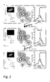
- An example of the different wavelengths and the outcome of excitation therewith is shown in Figure 1a ).
- the polypeptide exemplified in Fig. 1a corresponding to Seq. ID. No. 4 according to the present invention may be switched from the on- to the off-state by near UV-light of e.g. 405 nm, from the off- to the on-state by UV-light, e.g. 365 nm and the fluorescence is probed by excitation with green light of e.g. 515 nm while the maximum fluorescence is present at a wavelength of about 529 nm.
- near UV-light e.g. 405 nm
- UV-light e.g. 365 nm
- the fluorescence is probed by excitation with green light of e.g. 515 nm while the maximum fluorescence is present at a wavelength of about 529 nm.
- the polypeptides according to the present invention are characterised in being polypeptides where the switching wavelengths are independent from the fluorescence excitation and, thus, excitation of the fluorescence does not influence the switching properties of the polypeptides.
- the switching wavelengths are independent from the fluorescence excitation and, thus, excitation of the fluorescence does not influence the switching properties of the polypeptides.
- the fluorescent polypeptide according to the present invention is particularly a reversibly switchable fluorescent polypeptide (RSFP) wherein the first wavelength and the second wavelength which are different from each other are at least 50 nm below the third wavelength for excitation of the fluorescence emission of said polypeptide.
- RSFP reversibly switchable fluorescent polypeptide
- the first wavelength (365 nm) and the second wavelength (405 nm) are different from each other and both wavelengths are more than 50 nm below the third wavelength, 515 nm for excitation of the fluorescence emission of the polypeptide according to the present invention.
- the Triad-RSFPs according to the present invention have a fluorescence emission with a maximum emission between 450 nm and 1000 nm, in particular, between 450 nm and 700 nm.
- the maximum emission is in the range of the visible light, thus, allowing the use of the Triad-RFSPs according to the present invention in fluorescence microscopy.
- the Triad-RSFPs according to the present invention are characterised in that the mechanistic basis of the switching is a light-induced reversible water addition/elimination to/from the chromophore, as shown in scheme 1 above.
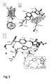
- the molecular basis for the reversible light induced switching of the polypeptide according to the present invention was solved by analysing the structures of the light induced off-state and on-state consecutively using the same polypeptide crystal. As demonstrated in scheme 1 and Fig. 2 , the structure of the polypeptide according to the present invention resembles that of GFP related proteins.
- the chromophore autocatalytically formed from amino acid residues 65, 66 and 67 according to the polypeptide of SEQ ID No. 1 or 2, resides in an alpha-helical-segment, enclosed by an 11-stranded-beta-barrel.
- the on-state chromophore consists of an imidazolinone-ring, connected by a methine bridge to a p-hydroxyphenyl-ring.
- the two rings of the chromophore system are largely co-planar, facilitating a conjugated pi-electron system and hence supporting fluorescence.
- the planarity of the five ring is markedly distorted with the chromophoric C65 -carbon exhibiting a tetrahedral geometry (Scheme 1).
- the fluorescent polypeptide is characterised in that the chromorphore comprises an imidazolinone-ring, connected by a methine bridge to a p-hydroxyphenyl-ring formed the by amino acid sequence X 1 YG wherein X 1 is G or A.
- amino acid residues G65, Y203 and E222 of SEQ ID No. 1 or 2 are present. It is assumed that the side chains of G65 and Y203 facilitate the positioning of the side chain of E222 close to the imidazolinone ring as demonstrated in Figure 2 .
- Irradiation into the about 510 nm bands induces fluorescence whereas irradiation into the about 410 nm band induces a covalent modification of the five ring resulting in a non-fluorescent chromophore absorbing at about 340 nm. Subsequent irradiation to this band results in a dehydration of an off-state chromophore converting it back into the on-state chromophore.
- the Triad-RSFPs according to the present invention are polypeptides comprising the amino acid sequence of SEQ ID No. 1 or 2.
- Particular preferred embodiments of the Triad-RSFPs are any one of SEQ ID Nos. 3 to 242, in particular, Seq. ID Nos. 3 to 10.
- the Triad-RSFPs according to the present invention are characterised in that said polypeptide is stable both in the fluorescent state and the non-fluorescent state.
- said polypeptide is stable in the fluorescent state and the non-fluorescent state over a time of at least 100 ms, preferably of at least 1s.
- Triad-RSFPs comprises polypeptides of natural occurring amino acids.
- unusual amino acids may be present.
- conservative amino acid exchanges distinguish the natural sequence form a non-natural sequence, e.g. chemical and/or physical similar amino acid residues are exchanged for each other.
- Such chemical or physical similar amino acid residues for example can be grouped according to their charge status (positive: R, H, K; negative: D, E; uncharged: A, N, C, Q, G, I, L, M, F, P, S, T, W, Y, V), or according to their volume and polarity (special: C; neutral and small: A, G, P, S, T; polar and relatively small: N, D, Q, E; polar and relatively large: R, H, K; nonpolar and relatively small: I, L, M, V; and nonpolar and relatively large: F, W, Y). Amino acid exchanges within a group are regarded as conservative exchanges ( Mol Biol Evolution, 2002, 19:1022-1025 ).
- Amino acids mean for the purposes of the invention besides the 20 amino acids determined by the genetic code also the amino acids which can be encoded by stop codons, such as, for example, seleno-cysteine or pyro-lysine. Additionally included are all amino acid and peptide derivatives such as, for example, glycosylated, phosphorylated, sulfated amino acids/peptides, and L-isomeric and D-isomeric amino acids. Modifications of amino acids may occur in the peptide backbone, in the amino acid side chains, at N-terminal ends of the peptide or at C-terminal ends of the peptide.
- the modifications may be present in single amino acids, in a plurality of amino acids or in all amino acids, and it is possible for no, one or a plurality of types of modifications to be present in any combinations in a peptide.
- unusual amino acids which may be mentioned by way of example are, inter alia: alpha-aminobutyric acid, beta-aminobutyric acid, beta-aminoisobutyric acid, beta-alanine, gamma-aminobutyric acid, alpha-aminoadipic acid, 4-aminobenzoic acid, aminoethylcysteine, alpha-aminopenicillanic acid, allysine, 4-carboxyglutamic acid, cystathionine, carboxyglutamic acid, carboxyamidomethylcysteine, carboxymethylcysteine, cysteic acid, citroline, dehydroalanine, diaminobutyric acid, dehydroamino-2-butyric acid, ethion
- amino acids can be present in the form of their L isomers or in the form of their D isomers as long as this is permitted by their structure.
- amino acids and amino acid derivatives which occur naturally or are formed or can be prepared enzymatic or chemically or in another wayare included in the term "amino acid".
- peptidomimetic is used in the present application in the form of the widest possible definition.
- a peptidomimetic is a substance which comprises non-peptide structural elements and is able to imitate or to antagonize the biological effect of the natural parent molecule. Numerous studies dealing in detail with possibilities for using peptidomimetics as replacement for conventional peptide structures are known in the art.
- one or more elements of the Triad-RSFP may be composed entirely or partly of peptidomimetics ( Curr Opin Chem Biol, 1997, 1:114-9 , J Recept Signal Transduct Res, 2001, 21:489-506 , J Med Chem, 2001, 44:1938-50 ).
- Transduction elements may possibly penetrate more efficiently into cells, antigen elements may lead to an enhanced or reduced immune response relative to the immune response to the conventional antigen, or tag elements may have better physicochemical properties, improving their suitability for the isolation and/or detection of the Triad-RSFP, etc.
- peptidomimetics it is additionally possible through the use of peptidomimetics in some circumstances to reduce or increase the in vivo half-life of the Triad-RSFP molecules, to reduce its toxicity, to improve its solubility in hydrophilic or hydrophobic media, and to prolong its in vitro stability and possibly to reduce the costs for synthesizing the peptidomimetic relative to the cost for synthesizing the corresponding conventional Triad-RSFP without peptidomimetic structures incorporated within it.
- peptidomimetics are Spiegelmers ® supplied by NOXXON Pharma AG, Berlin, Germany.
- the present invention relates to a polypeptide comprising the amino acid sequence of SEQ ID No. 1 or 2, like any one of SEQ ID Nos. 3 to 242, preferably to polypeptides of Seq. ID Nos. 3 to 10.
- the present invention also relates to a fusion protein comprising the Triad-RSFP according to the present invention.
- a fusion protein according to the present invention contains at least one additional, heterologous sequence. Often, but not necessarily, these additional sequences will be located at the N-, or C-terminal end of the polypeptide. It may e.g. be convenient to initially express the polypeptide as a fusion protein from which the additional amino acid residues can be removed, e.g. by a protease capable of specifically trimming the polypeptide of the present invention.
- the additional heterologous sequence may help in the expression or purification or attachment to a carrier of the polypeptides referred to in the present invention.
- the Triad-RSFP of the present invention could also be fused to a protease cleavage sequence and a quencher that when cleaved allows identification by increase of the fluorescence.
- the polypeptide of the invention is fused to one or more tags or signal sequences. Said tags or sequences allow to purify the protein or facilitate expression thereof.
- the signal sequence may allow intra- or extracellular targeting thereof.
- proteins to be combined with the polypeptide of the present invention and optionally the tags and signal sequences as described above are in general proteins the presence or localisation of which in cells and/or tissues is to be investigated. That is, the fusion protein according to the present invention may be useful as a biomarker and in diagnostics.
- the present invention relates to nucleic acid molecules encoding the Triad-RSFP according to the present invention.
- the present invention relates to a nucleic acid molecule encoding a fluorescent polypeptide wherein said nucleic acid molecule is selected from the group consisting of
- Stringent conditions refers to hybridisation conditions under which the polynulceotides that are capable of hybridizing to the nulciec acids of the invention or parts thereof hybridizes to these target sequences to a detectably greater degree than to other sequences (e.g., at least 2-fold over background). Stringent conditions are sequence-dependent and will be different in different circumstances. By controlling the stringency of the hybridisation and/or washing conditions, target sequences that have at least 90% sequence identity, more preferably 95%, such as 98% and more preferred 100% sequence identity to the probe can be identified (highly stringent hybridisation conditions). Alternatively, stringency conditions can be adjusted to allow a higher degree of mismatching in sequences (low stringency conditions of hybridisation). Such highly stringent and low stringent conditions for hybridisation are well known to the person skilled in the art.
- stringent conditions refers to hybridization conditions which comprise, e.g. an overnight incubation at 65°C in 4x SSC (600 mM NaCI, 60 mM sodium citrate) followed by washing at 65°C in 0.1x SSC for one hour.
- hybridization conditions can comprise: an overnight incubation at 42°C in a solution comprising 50% formamide, 5x SSC (750 mM NaCl, 75 mM sodium citrate), 50 mM sodium phosphate (pH 7.6), 5x Denhardt's solution, 10% dextran sulphate, and 20 microg/ml denatured, sheared salmon sperm DNA, followed by washing in e.g.
- 0.1-0.5x SSC at about 55-65°C for about 5 to 20 min.
- Said conditions for hybridization are also known by a person skilled in the art as "highly stringent conditions for hybridization". It is of note that variations in the above conditions may be accomplished through the inclusion and/or substitution of alternate blocking reagents.
- Typical blocking reagents include Denhardt's reagent, BLOTTO, heparin, denatured salmon sperm DNA, and commercially available proprietary formulations.
- the inclusion of specific blocking reagents may require modification of the hybridization conditions described above, due to problems with compatibility. Such modifications can generally be effected by the skilled person without further ado.
- a hybridization complex may be formed in solution (e.g., Cot or Rot analysis) or between one nucleic acid sequence present in solution and another nucleic acid sequence immobilized on a solid support (e.g., membranes, filters, chips, pins or glass slides to which, e.g., cells have been fixed).
- a solid support e.g., membranes, filters, chips, pins or glass slides to which, e.g., cells have been fixed.
- hybridization assays comprise without limitation Northern and Southern blot assays, heteroduplex analysis, detection of mutations by sequence specific oligonucleotide hybridization, allele-specific oligonucleotide hybridization on DNA chips, assays based on the Illumina's® technology, assays based on the BeadArray® technology, see, for example, Barnes et al., Nucleic Acids Res. 33 (2005) 5914-5923 .
- degenerate in accordance with the present invention refers to the degeneracy of the genetic code. Degeneracy results because a triplet code designates 20 amino acids and a stop codon. Because four bases exist which are utilized to encode genetic information, triplet codons are required to produce at least 21 different codes. The possible 4 3 possibilities for bases in triplets gives 64 possible codons, meaning that some degeneracy must exist. As a result, some amino acids are encoded by more than one triplet, i.e. by up to six. The degeneracy mostly arises from alterations in the third position in a triplet. This means that nucleic acid molecules having a different sequence than that specified above, but still encoding the same polypeptide lie within the scope of the present invention.
- the nucleic acid is a nucleic acid molecule encoding a polypeptide according to the present invention.
- the nucleic acid molecule may be in form of RNA or DNA.
- the present invention also relates to a vector comprising the nucleic acid molecule of the present invention.
- the vector is a plasmid, cosmid, virus, bacteriophage or another vector used conventionally e.g. in genetic engineering.
- the skilled person is well aware of suitable vectors designed for the specific application, e.g. expression of the polypeptide, transfection of cells etc.
- the present invention relates to a non-human host, in particular a cell line, transformed with a vector or a recombinant non-human host, in particular, a cell line, containing the nucleic acid sequence as a foreign nucleic acid sequence.
- the term “foreign” refers to a nucleic acid sequence not occurring naturally in said host.
- Non-human host can be single cells or multicellular organisms.
- Suitable prokaryotic host comprises e.g. bacteria of the species Escherichia, Streptomyces, Salmonella or Bacillus.
- Suitable eukaryotic host cells e.g. yeast, such as Saccharomyces cerevisiae or Pichia pastoris, insect cells, etc.
- Mammalian host cells that could be used include human cell lines, mice cell lines etc. Suitable cell lines include Hela, COS cells, CHO cells etc. In another aspect the cell line is a primary cell, e.g. obtained from mice etc. Appropriate methods, culture media and conditions for the above described host cells are known in the art. Further, the host may be transgenic non-human animal transfected with and/or expressing the nucleic acid molecule of the present invention. In a preferred embodiment, the transgenic animal is a mammal, e.g. a hamster, mouse, rat, cow, cat, pig, dog, horse, rabbit or monkey or other suitable animals, like zebra fish or C. elegans.
- a mammal e.g. a hamster, mouse, rat, cow, cat, pig, dog, horse, rabbit or monkey or other suitable animals, like zebra fish or C. elegans.
- Transgenic plants as hosts transfected with and/or expressing the nucleic acid molecule of the present invention also lay within the scope of the present invention.
- transgenic non-human animals Suitable methods for the production of said transgenic non-human animals, plants etc. are known in the art.
- the present invention relates to a method of producing a polypeptide or fusion protein according to the present invention.
- Said method comprises culturing the host of the invention under suitable conditions and isolating the polypeptide of the fusion protein used. Suitable conditions for culturing the host are well known to the person skilled in the art.
- purification methods and purification systems are well known.
- the polypeptide or fusion protein contains a tag allowing purification. Methods of isolation of the polypeptide are fusion proteins produced are well known in the art including chromatography, immunoprecipitation etc.
- the present invention provides diagnostic compositions comprising the Triad-RSFPs according to the present invention, the fusion protein according to the present invention, nucleic acid molecules according to the present invention or a vector according to the present invention.
- the present invention provides a kit comprising at least one of the Triad-RSFPs according to the present invention, a fusion protein, a nucleic acid molecule, a vector or a host according to the present invention.
- the various components of the diagnostic composition or of the kit according to the present invention may be packaged in one or more containers such as one or more vials.
- the vials may, envision to the components, comprise preservatives or buffers for storage.
- the kit or the diagnostic compositions are useful for various purposes.
- the diagnostic composition or the kit may be part of a system for detecting the presence, in particular, the localisation of a fusion protein according to the present invention, or of a polypeptide according to the present invention.
- a system for detecting the presence and/or localisation of a protein of interest in a cell tissue, said system comprises a kit or diagnostic composition according to the present invention, a source emitting a first and a second wavelength allowing reversibly switching the fusion protein or Triad-RSFP according to the present invention as well as a source for exciting said fusion protein or Triad-RSFP, and means allowing the detection of the presence and/or the localisation of the fluorescence emitted by the excited fusion protein or polypeptide.
- the system comprises a fluorescence microscope, means allowing the detection of fluorescence and the diagnostic composition or kit according to the present invention.
- the present invention furthermore relates to a method of detecting the presence and/or the localisation of a protein of interest, comprising
- the present invention relates to a method of detecting the presence and/or absence and/or the localisation of a Triad-RSFP according to the present invention or a fusion protein according to the present invention, comprising
- the polypeptides according to the present invention are useful for FRAS (fluorescence recovery after switching).
- This method includes the presence of the Triad-RSFP in an area of interest. Switching the Triad-RSFP from the off- to the on-stage or vice versa and determining the fluorescence in the area of interest over time.
- FRAS fluorescence recovery after switching
- the Triad-RSFP allows one to monitor the metabolism and possible effects of foreign and natural molecules in the area of interest in a time-resolved fashion.
- the term area of interest may be a whole organism, or tissues, or organs, or a cell,or sub-compartments of a cell.
- Still another embodiment of the present invention relates to a method of detecting the expression of a gene of interest comprising and operably linking the nucleic acid molecule according to the present invention with a promoter controlling said gene of interest or fusing the nucleic acid molecule according to the present invention to said gene of interest, and detecting the fluorescence of the protein encoded by said nucleic acid molecules.
- the present invention relates to a method of detecting the activity of a promoter of interest, comprising and operably linking the nucleic acid molecule according to the present invention with said promoter of interest and detecting the fluorescence of the protein encoded by said nucleic acid molecule.
- Fluorescence instruments are primarily of four types, each providing distinctly different information: Spectrofluorometers and microplate readers measure the average properties of bulk ( ⁇ L to mL) samples. Fluorescence microscopes resolve fluorescence as a function of spatial coordinates in two or three dimensions for microscopic objects (less than ⁇ 0.1 mm diameter).
- Fluorescence scanners including microarray readers, resolve fluorescence as a function of spatial coordinates in two dimensions for macroscopic objects such as electrophoresis gels, blots and chromatograms.
- Flow cytometers measure fluorescence per cell in a flowing stream, allowing subpopulations within a large sample to be identified and quantitated.
- the overall fluorescence can be detected in a sample as well as the location of the fluorescence within a sample.
- a reporter gene is a gene that can be operably linked to a gene of interest or simply to a promoter controlling the gene of interest in cell culture, animals or plants. Reporter genes are e.g. used to determine whether the gene of interest has been taken up by or expressed in the cell or organism population or whether the gene of interest naturally present in the cell or organism is expressed. It is important to use a reporter gene that is not natively expressed in the cell or organism under study, since the expression of the reporter is being used as a marker.
- the nucleic acid of the present invention encodes a GFP variant suitable as reporter gene with advantageous properties as described above.
- reporter gene and the gene of interest are placed in the same DNA construct to be inserted into the cell or organism, most often as a plasmid.
- Reporter genes can also be used to assay for the expression of the gene of interest, which may produce a protein that has little obvious or immediate effect on the cell culture or organism.
- the reporter is directly attached to the gene of interest to create a gene fusion.
- the two genes are under the control of the same promoter and are transcribed into a single messenger RNA molecule (mRNA). The mRNA is then translated into protein. In these cases it is important that both proteins are able to properly fold into their active conformations and interact with their substrates despite being fused.
- a segment of DNA coding for a flexible polypeptide linker region is usually included so that the reporter and the gene product of will only minimally interfere with one another.
- the present invention relates to a method of detecting the activity of a promoter of interest, comprising operably linking the nucleic acid molecule of the invention with said promoter of interest and detecting the fluorescence of the protein encoded by said nucleic acid molecule.
- Reporter genes can also be used to assay for the activity of a particular promoter in a cell or organism.
- the reporter gene is simply placed under the control of the target promoter and the reporter gene product's activity is quantitatively measured. The results are normally reported relative to the activity under a "consensus" promoter known to induce strong gene expression.
- the present invention relates to a method of detecting the presence of a protein of interest, comprising contacting a sample with the fusion protein of the invention, wherein said fusion protein comprises a polypeptide specifically binding to the protein of interest.
- the method of the invention can be carried out using FACS scans and preparative FACS sorts. Alternatively fluorescence microscope techniques may be applied.
- a sample can be any matter potentially containing the protein of interest.
- Preferred samples are liquids, e.g. cellular extracts or supernatants of cell cultures, or samples comprising living or dead cells in single form or present as a tissue or organ.
- Contacting a sample in accordance with the present invention means bringing the sample into contact with the fusion protein of the invention.
- the term comprises bringing both components into close vicinity as well as e.g. transforming or transfecting cells with a nucleic acid molecule encoding the fusion protein of the invention in order to express the fusion protein in the cells.
- Specifically binding in accordance with the present invention refers to the property of certain proteins to have an affinity for specific proteins, i.e. target proteins, which is greater by the factor of at least 10, preferably at least 100, more preferably at least 1,000 and most preferably at least 10,000 as compared to the affinity to proteins unrelated to the target protein.
- target proteins can e.g. be natural interaction partners of the target protein including proteins interacting in the course of signal transduction pathways or in context with structural proteins or antibodies.
- the present invention furthermore relates to a method of detecting the localization of a fusion protein of the present invention or a fusion protein comprising the polypeptide of the present invention in a cell or tissue, comprising exciting said fusion protein or polypeptide and detecting the location of the fluorescence emitted by the excited fusion protein or polypeptide.
- the present method can be used to localize the fusion protein of the invention as well as interaction partners of the fusions proteins of the invention.
- the Triad-RSFPs may be used in combination with known conventional fluorescent molecules to provide suitable FRET-pairs.
- the Triad-RSFPs may represent the donor or the acceptor, respectively.
- the FRET-pair may be composed of two Triad-RSFPs. These FRET-pairs may be used to determine the spatial distribution, in particular, the spatial proximity of fusion proteins.
- the Triad-RSFPs may be used to determine repeatedly movements of fusion proteins in a cell in three room dimensions. In particular, it is possible to repeatedly determine the position of the molecule in the same cell allowing improvement of the accuracy of measurement. The repeatability of the measurements may be used for screening the effects of different molecules on the mobility or processability of the fusion protein in the cell.
- the methods of the present invention are also suitable in the identification of compounds influencing e.g. the expression of target genes, the location of target proteins or the activity of target proteins.
- Said compounds may then be contacted, at the same time or separately, with a cell comprising the nucleic acid of the invention, e.g. fused to a target gene or a regulatory sequence.
- expressed (fusion) protein is excited in a cell contacted with the compound(s) and in a cell which has not been contacted.
- the expression or location of the (fusion) protein in both cells indicates that the compound has an influence on the expression of target genes and/or the location or activity of target proteins.
- RSFPs Switching of RSFPs were recorded either on E. coli colonies grown on LB-agar plates or in buffered protein solutions (100 mM Tris, 150 mM NaCl, pH 7.5; Figs. S1 or Citrat buffer pH 4,6).
- a modified computer-controlled fluorescence microscope (Leica Microsystems) equipped with a 50x NA 0.5 or a 20x NA 0.4 air objective lens and three 100 W Hg lamps was utilized for data acquisition. Fluorescence was recorded using a photomultiplier tube (HR9306-0, Hamamatsu, Hamamatsu City, Japan) behind a 525 nm longpass detection filter (HQ 525 LP, AHF Analysentechnik, Tübingen, Germany).
- a Leica DM6000 epifluorescence microscope (Leica, Wetzlar, Germany) equipped with a 100x NA 1.40 oil immersion objective lens and a DFC350 FX camera (Leica) was used.
- Cells were irradiated for 300 ms with near-UV light (BP420nm/30nm, 11 W/cm 2 ) to switch the molecules to the non-fluorescent state and 300 ms with UV light (BP360nm/40nm, 2 W/cm 2 ) to switch the proteins back to the fluorescent state.
- the fluorescence images were recorded with green light (BP495nm/15nm, 8 W/cm 2 ).
- a 510 nm dichroic mirror and a BP 530nm/30nm detection filter were used for fluorescence detection.
- the length of the image acquisition time was adjusted to the brightness of every sample and was generally between 50 and 150 ms.
- Imaging was performed with a beam scanning confocal microscope (SP5, Leica Microsystems) equipped with a UV-corrected 1.4 NA oil immersion lens (63x HCX PL APO). All imaging was performed at ambient temperatures. The following microscope settings were used: pinhole: 1 airy unit (AU); pixel size: 160.8 nm x 160.8 nm; scan speed: 1400 Hz. Fluorescence was detected between 519 and 590 nm.
- SP5 beam scanning confocal microscope
- UV-corrected 1.4 NA oil immersion lens 63x HCX PL APO
- the molecules in a predefined region of interest (ROI) (1,3 ⁇ m x 10 ⁇ m) were switched to the non-fluorescent state using 405 nm laser light.
- the fluorescence was recorded by excitation with 515 nm laser light.
- the molecules in the whole sample were switched to the fluorescent state with 360 nm widefield illumination.
- This imaging sequence was repeated several times on the same cell. In every sequence a second ROI on the other side of the same cell and a third ROI in a neighboring cell were analyzed. No further data processing was perfomed, except background subtraction and normalization to 1.
- switched-off and switched-on state 160 ⁇ g of the polypeptides in 10mM ammonium acetate were diluted with or without prior photoswitching to a final concentration of 18% acetonitrile in 10 mM ammonium acetate.
- the photoswitching was performed essential as described above.
- RSFP polypeptide solution was concentrated to -35 ⁇ g/ ⁇ l by ultrafiltration and taken up in 20 mM Tris/HCl, 120 mM NaCl, pH 7.5.
- the protein was crystallized by sitting drop vapor diffusion at 20°C with a reservoir solution containing 15% (w/v) PEG 3350, 0.2 M KH 2 PO 4 and 3% (w/v) D-(+)-trehalose-dihydrate.
- the crystallization solution was cryoprotected by adding propane-1,3-diol to a final concentration of 25% (w/v).
- RSFP RSFP protein crystal
- a single RSFP protein crystal was manually transferred into a vessel with cryoprotected crystallization solution and then switched to the non-fluorescent state by irradiation with blue light (405 ⁇ 10 nm, 110 W/cm 2 ) until the fluorescence signal reached a minimum. After switching the crystal, it was transferred within 10 seconds into liquid nitrogen, flash frozen, and diffraction data were collected at 100 K.
- the same crystal was unmounted, transferred back into cryoprotected crystallization solution kept at room temperature and immediately switched to the fluorescent-state by irradiation with UV light (365 ⁇ 25 nm, 1.4 W/cm 2 ) until the fluorescence signal reached a maximum. Subsequently the crystal was again flash frozen in liquid nitrogen and another diffraction dataset was collected.
- the equilibrium structure of RSFP was determined on crystals that were not previously irradiated.
- the omitted chromophore was placed manually into vacant patches of the 2 F o- F c and Fo-Fc electron densities.
- the chromophore restraints for the on-state Fluorophore were generated in PRODRG ⁇ Schuttelkopf, 2004 #1844 ⁇ , and the restraints of the off-state chromophore were generated with eLBOW ⁇ Moriarty, 2009 #1845 ⁇ .
- Example 1 Generation of reversibly switchable fluorescent proteins
- the QuikChange Site Directed Mutagenesis Kit (Stratagene, La Jolla, CA) was used according to the manufacturer's instructions. Error prone random mutagenesis as well as directed simultaneous mutagenesis of multiple sites were performed according to standard protocols ⁇ Leung, 1989 #1547;Sawano, 2000 #1544 ⁇ .
- Mutated sequences present in suitable plasmids were transformed into E. coli for expression of the mutated polypeptides. After growing of the transformed bacteria, the colonies were screened using computer assisted fluorescence microscopy. The microscope was running with three different sources of light which were controlled independently. Fluorescence of the polypeptides were measured and computed
- the computed data were evaluated based on the fluorescence for each single colony. Selected colonies were transferred and propagated by known methods, sequenced and analysed for their fluorescence and switching properties.
- the most promising mutants were selected, e.g. expressing polypeptides of Seq. ID Nos. 3 to 242, and analysed further.
- the coding sequence of the RSFP was amplified by PCR.
- the PCR fragments were digested with Sal I and Not I and inserted into the vector pEF/myc/ER (Invitrogen, Carlsbad, CA).
- the coding sequence of RSFP was amplified by PCR.
- the PCR fragments were digested with Age I and Not I and inserted into the vector pDsRed1-Mito (Clontech, Mountain View, CA), replacing the DsRed1 sequence.
- the RSFP coding sequence was amplified by PCR.
- the PCR fragments were digested with Nhe I and Bg III and inserted into the vector pEGFP-Tub (Clontech), replacing the EGFP sequence.
- Map2 coding sequence obtained from pDONR223-MAP2, ⁇ Lamesch, 2007 #1549 ⁇
- the PCR fragments were digested with Xho I and Bam HI and inserted into the vector RSFP- ⁇ -tubulin, replacing the ⁇ -tubulin sequence.
- the Histon-2B coding sequence (obtained from pDONR223-HIST1H2BN, ⁇ Lamesch, 2007 #1549 ⁇ ) was amplified by PCR.
- the PCR fragments were digested with Xho I and Bam HI and inserted into the vector RSFP- ⁇ -tubulin, replacing the ⁇ -tubulin sequence.
- Proteins were expressed in the Escherichia coli strain BL21-CodonPlus (Stratagene) and purified by Ni-NTA affinity chromatography, followed by a gel filtration step on a Superdex 200 column (Amersham Biosciences, Piscataway, NJ, USA) using a Tris-buffer (100 mM Tris-HCl, 150 mM NaCl, pH 7.5).
- the respective plasmids were transfected into Vero (kidney epithel of Cercopithecus aethiops) cells using the Nanofectin Kit (PAA, Pasching, Austria) according to the manufacturer's instructions.
- the cells were propagated in DMEM medium with GlutaMAX and 4.5 g/l glucose, 10% (v/v) FCS, 1mM sodium pyruvate, 50 mg/ml penicillin and 50 mg/ml streptomycin. Cells were grown on coverslips in petri dishes at 37°C under 90% humidity and 5% CO 2 .
- a solution of RSFP molecules was embedded in PAA (final concentration: 15 % PAA in 1.5 M Tris/HCl buffer, pH 8.8) and squeezed under a coverslip to ensure a layer thickness of ⁇ 1 ⁇ m.
- PAA final concentration: 15 % PAA in 1.5 M Tris/HCl buffer, pH 8.8
- a coverslip For reading and writing a Leica SP5 beam scanning confocal microscope equipped with a 40x NA 1.25 oil imersion objective was utilized. The following settings were used: pinhole: 1.48 AU; pixel size: 378.8 nm x 378.8 nm; scan speed: 400 Hz. Fluorescence was detected between 517 and 575 nm. Except smoothing and contrast stretching, no further imaging processing was applied.
- each individual image was written utilizing a group of ROIs, defining the positions where RSFP was transferred from the on- to the off-state by irradiation with 405 nm laser-light. Subsequently the written image was recorded with 515 nm laser-irradiation only. In the next step all proteins were switched back to the fluorescent state with 365 nm widefield illumination and the next image was recorded.
Landscapes
- Health & Medical Sciences (AREA)
- Life Sciences & Earth Sciences (AREA)
- Immunology (AREA)
- Chemical & Material Sciences (AREA)
- Engineering & Computer Science (AREA)
- Molecular Biology (AREA)
- Biomedical Technology (AREA)
- Hematology (AREA)
- Urology & Nephrology (AREA)
- Medicinal Chemistry (AREA)
- General Health & Medical Sciences (AREA)
- Biochemistry (AREA)
- Physics & Mathematics (AREA)
- Food Science & Technology (AREA)
- Microbiology (AREA)
- Analytical Chemistry (AREA)
- Cell Biology (AREA)
- Biotechnology (AREA)
- General Physics & Mathematics (AREA)
- Pathology (AREA)
- Organic Chemistry (AREA)
- Toxicology (AREA)
- Zoology (AREA)
- Gastroenterology & Hepatology (AREA)
- Biophysics (AREA)
- Genetics & Genomics (AREA)
- Proteomics, Peptides & Aminoacids (AREA)
- Tropical Medicine & Parasitology (AREA)
- Peptides Or Proteins (AREA)
- Measuring Or Testing Involving Enzymes Or Micro-Organisms (AREA)
Claims (18)
- Ein reversibel schaltbares Fluoreszenzpolypeptid, das reversible zwischen einem fluoreszierenden Zustand und einem nicht-fluoreszierenden Zustand umschalten kann, wenn es mit einem Licht einer ersten Wellenlänge bestrahlt wird und von dem fluoreszierenden Zustand zu dem nicht-fluoreszierenden Zustand umschalten kann, wenn es mit einem Licht einer zweiten Wellenlänge, die sich von dem Licht mit einer ersten Wellenlänge unterscheidet, bestrahlt wird, dadurch gekennzeichnet, dass die Anregung der Fluoreszenzemission des Polypeptids durch Licht einer dritten Wellenlänge erfolgt, die sich von dem Licht der ersten und zweiten Wellenlänge unterscheidet, wobei ein Chromophor umfassend einen Imidazolinonring verbunden mittels einer Methinbrücke zu einem p-Hydroxyphenylring ausgebildet wird durch die Aminosäuresequenz X1YG wobei X1 G oder A ist und wobei ein Wassermolekül an die Imidazolinongruppe im nicht-fluoreszierenden Zustand gebunden ist, während dieses Wassermolekül im fluoreszierenden Zustand frei ist, wie in Schema 1 dargestellt.

- Das reversibel schaltbare Fluoreszenzpolypeptid nach Anspruch 1, wobei die erste und zweite Wellenlänge mindestens 50nm unterhalb der dritten Wellenlänge für die Anregung der Fluoreszenzemission des Polypeptids ist.
- Reversibel schaltbares Fluroeszenzpolypeptid nach Anspruch 1 oder 2, wobei das Polypeptid eine Fluoreszenzemission mit einer maximalen Emission zwischen 450 nm und 1000 nm aufzeigt.
- Das reversibel schaltbare Fluoreszenzpolypeptid nach einem der vorherigen Ansprüche, wobei dieses Polypeptid stabil für einen Zeitraum von mindestens 100 ms ist sowohl im Fluoreszenzzustand als auch im Nicht-Fluoreszenzzustand.
- Reversibel schaltbares Fluoreszenzpolypeptid nach einem der vorherigen Ansprüche umfassend die Aminosäuresequenz von Seq. ID Nr. 1 oder 2.
- Reversibel schaltbares Fluoreszenzpolypeptid nach Anspruch 5 umfassend die Aminosäuresequenz einer der Seq. ID Nr. 3 bis 242, insbesondere Seq. ID Nr. 3 bis 10.
- Fusionsprotein umfassend das Polypeptid nach einem der vorherigen Ansprüche.
- Nukleinsäuremolekül kodierend ein reversibel schaltbares Fluoreszenzpolypeptid, wobei dieses Nukleinsäuremolekül ausgebildet ist aus der Gruppe bestehend aus:a) Nukleinsäuremolekül kodierend ein Polypeptid nach einem der Ansprüche 1 bis 7;b) Nukleinsäuremolekül das unter stringenten Bedingungen zu dem komplementären Strang der Nukleinsäure von a) hybridisiert, wobei das Nukleinsäuremolekül gemäß b) ein Polypeptid kodiert, das an der Position entsprechend Position 203 der Seq. ID Nr. 1 oder 2 ein Tyrosin und an der Position entsprechend der Position 65 der Seq. ID Nr. 1 oder 2 ein Glycin oder ein Alanin aufweist; wobei das durch das Nukleinsäuremolekül kodierte Polypeptid eines ist, das reversibel zwischen einem Fluoreszenzzustand umschalten kann und die Wellenlängen notwendig für das reversible Umschalten dieses Polypeptids unterschiedlich ist zu der Wellenlänge für das Anregen der Fluoreszenzemission;c) eine Nukleinsäure degeneriert in Bezug auf den genetischen Code des Nukleinsäuremoleküls von a) oder b).
- Vektor umfassend das Nukleinsäuremolekül nach Anspruch 8.
- Ein nicht-humaner Wirt, insbesondere eine Zelllinie, transformiert mit dem Vektor gemäß Anspruch 9 oder enthaltend eine Nukleinsäuresequenz nach Anspruch 8.
- Verfahren zur Herstellung eines reversibel schaltbaren Fluoreszenzpolypeptids umfassend Kultivieren des Wirts nach Anspruch 10 unter geeigneten Bedingungen und Isolieren des hergestellten Polypeptids.
- Eine diagnostische Zusammensetzung umfassend mindestens eines ausa) dem reversibel schaltbaren Fluoreszenzpolypeptid nach Anspruch 1 bis 6;b) dem Fusionsprotein nach Anspruch 7;c) dem Nukleinsäuremolekül nach Anspruch 8; oderd) dem Vektor nach Anspruch 9.
- Kit umfassend mindestens eines ausa) dem reversibel schaltbaren Fluoreszenzpolypeptid nach Anspruch 6;b) dem Fusionsprotein nach Anspruch 7;c) dem Nukleinsäuremolekül nach Anspruch 8;d) dem Vektor nach Anspruch 9; odere) dem Wirt nach Anspruch 10.
- System zum Nachweis des Vorhandenseins, insbesondere der Lokalisation eines Fusionsproteins nach Anspruch 7 oder eines Polypeptids umfassend das Polypeptid nach einem der Ansprüche 1 bis 6, insbesondere zum Nachweis des Vorhandenseins und/oder Lokalisation eines interessierenden Proteins in einer Zelle oder Gewebe, umfassend die diagnostische Zusammensetzung nach Anspruch 12 oder das Kit nach Anspruch 13, eine Quelle zum Emitieren einer ersten und zweiten Wellenlänge, die das reversible Schalten des Fusionsproteins oder Polypeptids erlaubt, sowie eine Quelle zum Anregen des Fusionsproteins oder Polypeptids und Mittel zum Nachweis des Vorhandenseins und/oder der Lokalisation der emittierten Fluoreszenz durch das angeregte Fusionsprotein oder Polypeptid.
- Verfahren zum Nachweis des Vorhandenseins und/oder der Lokalisation eines interessierenden Proteins, umfassendi.) Exprimieren des Fusionsproteins nach Anspruch 7 umfassend das interessierende Protein in einer Probe oderii.) in Kontakt bringen einer Probe mit dem Fusionsprotein nach Anspruch 7, wobei dieses Fusionsprotein umfasst ein Polypeptid, das spezifisch an das interessierende Protein bindet.
- Verfahren zur Bestimmung des Vorhandenseins und/oder der Lokalisation eines Polypeptids umfassend ein reversibel schaltbares Fluoreszenzpolypeptid nach einem der Ansprüche 1 bis 6 oder ein Fusionsprotein nach Anspruch 7 in einer Zelle oder Gewebe umfassenda) Anregen dieses Fusionsproteins oder Polypeptid mit einer ersten oder zweiten Wellenlänge, die das Umschalten des Polypeptids von einem Fluoreszenzzustand zu einem Nicht-Fluoreszenzzustand oder umgekehrt ermöglicht;b) Anregen dieses Fusionsproteins oder Polypeptids mit einer Wellenlänge, die sich von der ersten oder zweiten Wellenlänge unterscheidet zum Anregen der Fluoreszenzemission undc) Bestimmen der Lokalisation der emittierten Fluoreszenz durch Anregen des Fusionsproteins oder Polypeptids.
- Verfahren zum Bestimmen der Expression eines interessierenden Gens umfassenda) operatives Verknüpfen des Nukleinsäuremoleküls nach Anspruch 8 mit einem Promotor, der das zu interessierende Gen kontrolliert oderb) Fusionieren des Nukleinsäuremoleküls nach Anspruch 8 mit dem interessierenden Gen undc) Bestimmen der Fluoreszenz des durch das Nukleinsäuremolekül von a) oder b) kodierte Protein.
- Verfahren zum Bestimmen der Aktivität eines interessierenden Promotors umfassend ein operatives Verknüpfen des Nukleinsäuremoleküls nach Anspruch 8 mit diesem zu interessierenden Promotor und Bestimmen der Fluoreszenz des durch dieses Nukleinsäuremolekül kodierten Proteins.
Priority Applications (2)
| Application Number | Priority Date | Filing Date | Title |
|---|---|---|---|
| EP10186413.0A EP2436690B1 (de) | 2010-10-04 | 2010-10-04 | Umgekehrt fotoschaltbare Polypeptide |
| PCT/EP2011/067298 WO2012045733A1 (en) | 2010-10-04 | 2011-10-04 | Reversibly photoswitchable polypeptides |
Applications Claiming Priority (1)
| Application Number | Priority Date | Filing Date | Title |
|---|---|---|---|
| EP10186413.0A EP2436690B1 (de) | 2010-10-04 | 2010-10-04 | Umgekehrt fotoschaltbare Polypeptide |
Publications (2)
| Publication Number | Publication Date |
|---|---|
| EP2436690A1 EP2436690A1 (de) | 2012-04-04 |
| EP2436690B1 true EP2436690B1 (de) | 2015-12-23 |
Family
ID=43217035
Family Applications (1)
| Application Number | Title | Priority Date | Filing Date |
|---|---|---|---|
| EP10186413.0A Not-in-force EP2436690B1 (de) | 2010-10-04 | 2010-10-04 | Umgekehrt fotoschaltbare Polypeptide |
Country Status (2)
| Country | Link |
|---|---|
| EP (1) | EP2436690B1 (de) |
| WO (1) | WO2012045733A1 (de) |
Families Citing this family (3)
| Publication number | Priority date | Publication date | Assignee | Title |
|---|---|---|---|---|
| US8735096B2 (en) | 2012-03-22 | 2014-05-27 | The Board Of Trustees Of The Leland Stanford Junior University | Optical control of protein activity and localization by fusion to photochromic protein domains |
| US20150135343A1 (en) * | 2012-05-18 | 2015-05-14 | The Board Of Regents Of The University Of Texas Sy | GTP Sensors |
| WO2016182019A1 (ja) * | 2015-05-12 | 2016-11-17 | 国立大学法人大阪大学 | 蛍光蛋白質 |
Family Cites Families (3)
| Publication number | Priority date | Publication date | Assignee | Title |
|---|---|---|---|---|
| US6699687B1 (en) * | 1999-05-21 | 2004-03-02 | The Regents Of The University Of California | Circularly permuted fluorescent protein indicators |
| WO2002096924A1 (en) | 2001-05-25 | 2002-12-05 | Clontech Laboratories, Inc. | Kindling fluorescent proteins and methods for their use |
| EP1767635B1 (de) | 2004-05-20 | 2013-07-31 | Riken | Fluoreszenzprotein |
-
2010
- 2010-10-04 EP EP10186413.0A patent/EP2436690B1/de not_active Not-in-force
-
2011
- 2011-10-04 WO PCT/EP2011/067298 patent/WO2012045733A1/en active Application Filing
Also Published As
| Publication number | Publication date |
|---|---|
| EP2436690A1 (de) | 2012-04-04 |
| WO2012045733A1 (en) | 2012-04-12 |
Similar Documents
| Publication | Publication Date | Title |
|---|---|---|
| US9664697B2 (en) | Green-to-red photo-convertible fluorescent calcium indicator | |
| Miyawaki | Innovations in the imaging of brain functions using fluorescent proteins | |
| Klima et al. | Incorporation of sensing modalities into de novo designed fluorescence-activating proteins | |
| Saito et al. | Auto-luminescent genetically-encoded ratiometric indicator for real-time Ca2+ imaging at the single cell level | |
| WO2013052946A2 (en) | Genetically encoded biosensors | |
| JP6707472B2 (ja) | 蛍光団を活性化し、シフトするタグ(fast) | |
| JP5570803B2 (ja) | 蛍光タンパク質およびpH測定方法 | |
| CA2766856A1 (en) | Detection and visualization of the cell cycle in living cells | |
| EP2695893A1 (de) | Neuartiges tiefrot-fluoreszierendes Protein | |
| EP2436690B1 (de) | Umgekehrt fotoschaltbare Polypeptide | |
| ES2254044T3 (es) | Proteinas fluorescentes procedentes de especies de copepodos y procedimientos para usarlas. | |
| US20090035788A1 (en) | Genetically encoded bioindicators of calcium-ions | |
| ES2874138T3 (es) | Indicadores de iones potasio genéticamente codificados | |
| EP3312279A1 (de) | Ph-reaktive proteolysesonde | |
| WO2009124135A2 (en) | Annexin-based apoptosis markers | |
| US9383366B2 (en) | Blue fluorescent protein and methods of use thereof | |
| JP6132350B2 (ja) | 人工生物発光酵素 | |
| WO2013163681A1 (en) | Fluorescent proteins and uses thereof | |
| WO2005019447A1 (ja) | 単色蛍光プローブ | |
| CN110305199A (zh) | 结合藻红胆素的荧光生物标记物及标记方法 | |
| JPWO2018235787A1 (ja) | 蛍光タンパク質を用いたタンパク質間相互作用の判定方法 | |
| JP5875773B2 (ja) | 紫外光測定方法 | |
| US20230203110A1 (en) | Photostable fluorescent proteins | |
| WO2010060618A1 (en) | Monomeric variants of he tetrameric eqfp611 | |
| RU2338785C2 (ru) | Флуоресцирующие белки и хромопротеины из видов hydrozoa, не относящихся к aequorea, и способы их получения |
Legal Events
| Date | Code | Title | Description |
|---|---|---|---|
| PUAI | Public reference made under article 153(3) epc to a published international application that has entered the european phase |
Free format text: ORIGINAL CODE: 0009012 |
|
| AK | Designated contracting states |
Kind code of ref document: A1 Designated state(s): AL AT BE BG CH CY CZ DE DK EE ES FI FR GB GR HR HU IE IS IT LI LT LU LV MC MK MT NL NO PL PT RO RS SE SI SK SM TR |
|
| AX | Request for extension of the european patent |
Extension state: BA ME |
|
| 17P | Request for examination filed |
Effective date: 20121002 |
|
| 17Q | First examination report despatched |
Effective date: 20121217 |
|
| GRAP | Despatch of communication of intention to grant a patent |
Free format text: ORIGINAL CODE: EPIDOSNIGR1 |
|
| INTG | Intention to grant announced |
Effective date: 20150619 |
|
| GRAS | Grant fee paid |
Free format text: ORIGINAL CODE: EPIDOSNIGR3 |
|
| GRAA | (expected) grant |
Free format text: ORIGINAL CODE: 0009210 |
|
| AK | Designated contracting states |
Kind code of ref document: B1 Designated state(s): AL AT BE BG CH CY CZ DE DK EE ES FI FR GB GR HR HU IE IS IT LI LT LU LV MC MK MT NL NO PL PT RO RS SE SI SK SM TR |
|
| REG | Reference to a national code |
Ref country code: GB Ref legal event code: FG4D |
|
| REG | Reference to a national code |
Ref country code: CH Ref legal event code: EP |
|
| REG | Reference to a national code |
Ref country code: IE Ref legal event code: FG4D |
|
| REG | Reference to a national code |
Ref country code: AT Ref legal event code: REF Ref document number: 766527 Country of ref document: AT Kind code of ref document: T Effective date: 20160115 |
|
| REG | Reference to a national code |
Ref country code: DE Ref legal event code: R096 Ref document number: 602010029654 Country of ref document: DE |
|
| REG | Reference to a national code |
Ref country code: LT Ref legal event code: MG4D |
|
| REG | Reference to a national code |
Ref country code: NL Ref legal event code: MP Effective date: 20151223 |
|
| PG25 | Lapsed in a contracting state [announced via postgrant information from national office to epo] |
Ref country code: HR Free format text: LAPSE BECAUSE OF FAILURE TO SUBMIT A TRANSLATION OF THE DESCRIPTION OR TO PAY THE FEE WITHIN THE PRESCRIBED TIME-LIMIT Effective date: 20151223 Ref country code: NO Free format text: LAPSE BECAUSE OF FAILURE TO SUBMIT A TRANSLATION OF THE DESCRIPTION OR TO PAY THE FEE WITHIN THE PRESCRIBED TIME-LIMIT Effective date: 20160323 Ref country code: LT Free format text: LAPSE BECAUSE OF FAILURE TO SUBMIT A TRANSLATION OF THE DESCRIPTION OR TO PAY THE FEE WITHIN THE PRESCRIBED TIME-LIMIT Effective date: 20151223 |
|
| REG | Reference to a national code |
Ref country code: AT Ref legal event code: MK05 Ref document number: 766527 Country of ref document: AT Kind code of ref document: T Effective date: 20151223 |
|
| PG25 | Lapsed in a contracting state [announced via postgrant information from national office to epo] |
Ref country code: GR Free format text: LAPSE BECAUSE OF FAILURE TO SUBMIT A TRANSLATION OF THE DESCRIPTION OR TO PAY THE FEE WITHIN THE PRESCRIBED TIME-LIMIT Effective date: 20160324 Ref country code: NL Free format text: LAPSE BECAUSE OF FAILURE TO SUBMIT A TRANSLATION OF THE DESCRIPTION OR TO PAY THE FEE WITHIN THE PRESCRIBED TIME-LIMIT Effective date: 20151223 Ref country code: LV Free format text: LAPSE BECAUSE OF FAILURE TO SUBMIT A TRANSLATION OF THE DESCRIPTION OR TO PAY THE FEE WITHIN THE PRESCRIBED TIME-LIMIT Effective date: 20151223 Ref country code: FI Free format text: LAPSE BECAUSE OF FAILURE TO SUBMIT A TRANSLATION OF THE DESCRIPTION OR TO PAY THE FEE WITHIN THE PRESCRIBED TIME-LIMIT Effective date: 20151223 Ref country code: RS Free format text: LAPSE BECAUSE OF FAILURE TO SUBMIT A TRANSLATION OF THE DESCRIPTION OR TO PAY THE FEE WITHIN THE PRESCRIBED TIME-LIMIT Effective date: 20151223 Ref country code: SE Free format text: LAPSE BECAUSE OF FAILURE TO SUBMIT A TRANSLATION OF THE DESCRIPTION OR TO PAY THE FEE WITHIN THE PRESCRIBED TIME-LIMIT Effective date: 20151223 |
|
| PG25 | Lapsed in a contracting state [announced via postgrant information from national office to epo] |
Ref country code: CZ Free format text: LAPSE BECAUSE OF FAILURE TO SUBMIT A TRANSLATION OF THE DESCRIPTION OR TO PAY THE FEE WITHIN THE PRESCRIBED TIME-LIMIT Effective date: 20151223 Ref country code: ES Free format text: LAPSE BECAUSE OF FAILURE TO SUBMIT A TRANSLATION OF THE DESCRIPTION OR TO PAY THE FEE WITHIN THE PRESCRIBED TIME-LIMIT Effective date: 20151223 Ref country code: IT Free format text: LAPSE BECAUSE OF FAILURE TO SUBMIT A TRANSLATION OF THE DESCRIPTION OR TO PAY THE FEE WITHIN THE PRESCRIBED TIME-LIMIT Effective date: 20151223 |
|
| PG25 | Lapsed in a contracting state [announced via postgrant information from national office to epo] |
Ref country code: PL Free format text: LAPSE BECAUSE OF FAILURE TO SUBMIT A TRANSLATION OF THE DESCRIPTION OR TO PAY THE FEE WITHIN THE PRESCRIBED TIME-LIMIT Effective date: 20151223 Ref country code: EE Free format text: LAPSE BECAUSE OF FAILURE TO SUBMIT A TRANSLATION OF THE DESCRIPTION OR TO PAY THE FEE WITHIN THE PRESCRIBED TIME-LIMIT Effective date: 20151223 Ref country code: SK Free format text: LAPSE BECAUSE OF FAILURE TO SUBMIT A TRANSLATION OF THE DESCRIPTION OR TO PAY THE FEE WITHIN THE PRESCRIBED TIME-LIMIT Effective date: 20151223 Ref country code: SM Free format text: LAPSE BECAUSE OF FAILURE TO SUBMIT A TRANSLATION OF THE DESCRIPTION OR TO PAY THE FEE WITHIN THE PRESCRIBED TIME-LIMIT Effective date: 20151223 Ref country code: PT Free format text: LAPSE BECAUSE OF FAILURE TO SUBMIT A TRANSLATION OF THE DESCRIPTION OR TO PAY THE FEE WITHIN THE PRESCRIBED TIME-LIMIT Effective date: 20160426 Ref country code: IS Free format text: LAPSE BECAUSE OF FAILURE TO SUBMIT A TRANSLATION OF THE DESCRIPTION OR TO PAY THE FEE WITHIN THE PRESCRIBED TIME-LIMIT Effective date: 20160423 Ref country code: RO Free format text: LAPSE BECAUSE OF FAILURE TO SUBMIT A TRANSLATION OF THE DESCRIPTION OR TO PAY THE FEE WITHIN THE PRESCRIBED TIME-LIMIT Effective date: 20151223 Ref country code: AT Free format text: LAPSE BECAUSE OF FAILURE TO SUBMIT A TRANSLATION OF THE DESCRIPTION OR TO PAY THE FEE WITHIN THE PRESCRIBED TIME-LIMIT Effective date: 20151223 |
|
| REG | Reference to a national code |
Ref country code: DE Ref legal event code: R097 Ref document number: 602010029654 Country of ref document: DE |
|
| REG | Reference to a national code |
Ref country code: FR Ref legal event code: PLFP Year of fee payment: 7 |
|
| PLBE | No opposition filed within time limit |
Free format text: ORIGINAL CODE: 0009261 |
|
| STAA | Information on the status of an ep patent application or granted ep patent |
Free format text: STATUS: NO OPPOSITION FILED WITHIN TIME LIMIT |
|
| PG25 | Lapsed in a contracting state [announced via postgrant information from national office to epo] |
Ref country code: DK Free format text: LAPSE BECAUSE OF FAILURE TO SUBMIT A TRANSLATION OF THE DESCRIPTION OR TO PAY THE FEE WITHIN THE PRESCRIBED TIME-LIMIT Effective date: 20151223 |
|
| 26N | No opposition filed |
Effective date: 20160926 |
|
| PG25 | Lapsed in a contracting state [announced via postgrant information from national office to epo] |
Ref country code: BE Free format text: LAPSE BECAUSE OF FAILURE TO SUBMIT A TRANSLATION OF THE DESCRIPTION OR TO PAY THE FEE WITHIN THE PRESCRIBED TIME-LIMIT Effective date: 20151223 |
|
| PG25 | Lapsed in a contracting state [announced via postgrant information from national office to epo] |
Ref country code: SI Free format text: LAPSE BECAUSE OF FAILURE TO SUBMIT A TRANSLATION OF THE DESCRIPTION OR TO PAY THE FEE WITHIN THE PRESCRIBED TIME-LIMIT Effective date: 20151223 |
|
| REG | Reference to a national code |
Ref country code: IE Ref legal event code: MM4A |
|
| PG25 | Lapsed in a contracting state [announced via postgrant information from national office to epo] |
Ref country code: LU Free format text: LAPSE BECAUSE OF NON-PAYMENT OF DUE FEES Effective date: 20161004 |
|
| REG | Reference to a national code |
Ref country code: FR Ref legal event code: PLFP Year of fee payment: 8 |
|
| PG25 | Lapsed in a contracting state [announced via postgrant information from national office to epo] |
Ref country code: IE Free format text: LAPSE BECAUSE OF NON-PAYMENT OF DUE FEES Effective date: 20161004 |
|
| PG25 | Lapsed in a contracting state [announced via postgrant information from national office to epo] |
Ref country code: HU Free format text: LAPSE BECAUSE OF FAILURE TO SUBMIT A TRANSLATION OF THE DESCRIPTION OR TO PAY THE FEE WITHIN THE PRESCRIBED TIME-LIMIT; INVALID AB INITIO Effective date: 20101004 Ref country code: CY Free format text: LAPSE BECAUSE OF FAILURE TO SUBMIT A TRANSLATION OF THE DESCRIPTION OR TO PAY THE FEE WITHIN THE PRESCRIBED TIME-LIMIT Effective date: 20151223 |
|
| PG25 | Lapsed in a contracting state [announced via postgrant information from national office to epo] |
Ref country code: TR Free format text: LAPSE BECAUSE OF FAILURE TO SUBMIT A TRANSLATION OF THE DESCRIPTION OR TO PAY THE FEE WITHIN THE PRESCRIBED TIME-LIMIT Effective date: 20151223 Ref country code: MC Free format text: LAPSE BECAUSE OF FAILURE TO SUBMIT A TRANSLATION OF THE DESCRIPTION OR TO PAY THE FEE WITHIN THE PRESCRIBED TIME-LIMIT Effective date: 20151223 Ref country code: MK Free format text: LAPSE BECAUSE OF FAILURE TO SUBMIT A TRANSLATION OF THE DESCRIPTION OR TO PAY THE FEE WITHIN THE PRESCRIBED TIME-LIMIT Effective date: 20151223 Ref country code: MT Free format text: LAPSE BECAUSE OF NON-PAYMENT OF DUE FEES Effective date: 20161031 |
|
| PG25 | Lapsed in a contracting state [announced via postgrant information from national office to epo] |
Ref country code: BG Free format text: LAPSE BECAUSE OF FAILURE TO SUBMIT A TRANSLATION OF THE DESCRIPTION OR TO PAY THE FEE WITHIN THE PRESCRIBED TIME-LIMIT Effective date: 20151223 |
|
| REG | Reference to a national code |
Ref country code: FR Ref legal event code: PLFP Year of fee payment: 9 |
|
| PG25 | Lapsed in a contracting state [announced via postgrant information from national office to epo] |
Ref country code: AL Free format text: LAPSE BECAUSE OF FAILURE TO SUBMIT A TRANSLATION OF THE DESCRIPTION OR TO PAY THE FEE WITHIN THE PRESCRIBED TIME-LIMIT Effective date: 20151223 |
|
| PGFP | Annual fee paid to national office [announced via postgrant information from national office to epo] |
Ref country code: GB Payment date: 20211022 Year of fee payment: 12 |
|
| PGFP | Annual fee paid to national office [announced via postgrant information from national office to epo] |
Ref country code: FR Payment date: 20211022 Year of fee payment: 12 Ref country code: CH Payment date: 20211022 Year of fee payment: 12 |
|
| PGFP | Annual fee paid to national office [announced via postgrant information from national office to epo] |
Ref country code: DE Payment date: 20221026 Year of fee payment: 13 |
|
| REG | Reference to a national code |
Ref country code: CH Ref legal event code: PL |
|
| GBPC | Gb: european patent ceased through non-payment of renewal fee |
Effective date: 20221004 |
|
| PG25 | Lapsed in a contracting state [announced via postgrant information from national office to epo] |
Ref country code: LI Free format text: LAPSE BECAUSE OF NON-PAYMENT OF DUE FEES Effective date: 20221031 Ref country code: FR Free format text: LAPSE BECAUSE OF NON-PAYMENT OF DUE FEES Effective date: 20221031 Ref country code: CH Free format text: LAPSE BECAUSE OF NON-PAYMENT OF DUE FEES Effective date: 20221031 |
|
| PG25 | Lapsed in a contracting state [announced via postgrant information from national office to epo] |
Ref country code: GB Free format text: LAPSE BECAUSE OF NON-PAYMENT OF DUE FEES Effective date: 20221004 |
|
| REG | Reference to a national code |
Ref country code: DE Ref legal event code: R119 Ref document number: 602010029654 Country of ref document: DE |
|
| PG25 | Lapsed in a contracting state [announced via postgrant information from national office to epo] |
Ref country code: DE Free format text: LAPSE BECAUSE OF NON-PAYMENT OF DUE FEES Effective date: 20240501 |