EP2436690B1 - Reversibly photoswitchable polypeptides - Google Patents
Reversibly photoswitchable polypeptides Download PDFInfo
- Publication number
- EP2436690B1 EP2436690B1 EP10186413.0A EP10186413A EP2436690B1 EP 2436690 B1 EP2436690 B1 EP 2436690B1 EP 10186413 A EP10186413 A EP 10186413A EP 2436690 B1 EP2436690 B1 EP 2436690B1
- Authority
- EP
- European Patent Office
- Prior art keywords
- polypeptide
- prt
- artificial
- fluorescent
- nucleic acid
- Prior art date
- Legal status (The legal status is an assumption and is not a legal conclusion. Google has not performed a legal analysis and makes no representation as to the accuracy of the status listed.)
- Active
Links
Images


Classifications
-
- G—PHYSICS
- G01—MEASURING; TESTING
- G01N—INVESTIGATING OR ANALYSING MATERIALS BY DETERMINING THEIR CHEMICAL OR PHYSICAL PROPERTIES
- G01N33/00—Investigating or analysing materials by specific methods not covered by groups G01N1/00 - G01N31/00
- G01N33/48—Biological material, e.g. blood, urine; Haemocytometers
- G01N33/50—Chemical analysis of biological material, e.g. blood, urine; Testing involving biospecific ligand binding methods; Immunological testing
- G01N33/53—Immunoassay; Biospecific binding assay; Materials therefor
- G01N33/531—Production of immunochemical test materials
- G01N33/532—Production of labelled immunochemicals
- G01N33/533—Production of labelled immunochemicals with fluorescent label
-
- C—CHEMISTRY; METALLURGY
- C07—ORGANIC CHEMISTRY
- C07K—PEPTIDES
- C07K14/00—Peptides having more than 20 amino acids; Gastrins; Somatostatins; Melanotropins; Derivatives thereof
- C07K14/435—Peptides having more than 20 amino acids; Gastrins; Somatostatins; Melanotropins; Derivatives thereof from animals; from humans
- C07K14/43504—Peptides having more than 20 amino acids; Gastrins; Somatostatins; Melanotropins; Derivatives thereof from animals; from humans from invertebrates
- C07K14/43595—Peptides having more than 20 amino acids; Gastrins; Somatostatins; Melanotropins; Derivatives thereof from animals; from humans from invertebrates from coelenteratae, e.g. medusae
-
- G—PHYSICS
- G01—MEASURING; TESTING
- G01N—INVESTIGATING OR ANALYSING MATERIALS BY DETERMINING THEIR CHEMICAL OR PHYSICAL PROPERTIES
- G01N33/00—Investigating or analysing materials by specific methods not covered by groups G01N1/00 - G01N31/00
- G01N33/48—Biological material, e.g. blood, urine; Haemocytometers
- G01N33/50—Chemical analysis of biological material, e.g. blood, urine; Testing involving biospecific ligand binding methods; Immunological testing
- G01N33/58—Chemical analysis of biological material, e.g. blood, urine; Testing involving biospecific ligand binding methods; Immunological testing involving labelled substances
- G01N33/582—Chemical analysis of biological material, e.g. blood, urine; Testing involving biospecific ligand binding methods; Immunological testing involving labelled substances with fluorescent label
Definitions
- the present invention concerns new fluorescent polypeptides able to switch reversibly between a fluorescent state and a non-fluorescent state.
- the fluorescent polypeptide according to the present invention is able to switch reversibly between a fluorescent state and a non-fluorescent state when irradiated with the light of a first wavelength to switch a polypeptide from the non-fluorescent state to fluorescent state and irradiated with the light of a second wavelength different to the first wavelength to switch a polypeptide from the fluorescent state to the non-fluorescent state, whereby said first and second wavelength for switching the polypeptide are different to a third wavelength for excitation of the fluorescent emission of said polypeptide.
- the present invention relates to fusion proteins comprising the fluorescent polypeptide according to the present invention as well as nucleic acids encoding the same.
- diagnostic compositions, kits and systems comprising said fluorescent polypeptides or nucleic acids encoding the same are provided.
- the present invention relates to methods for detecting the presence and/or the localisation of a protein or nucleic acid molecule of interest.
- Photoswitchable fluorescent proteins FP
- FP whose fluorescence can be reversibly or irreversibly modulated by irradiation with light, started a new momentum following a revolution originating in the discovery of the green fluorescent protein (GFP)
- GFP green fluorescent protein
- Photoswitchable fluorescent proteins facilitated a new powerful in vitro protein tracking schemes ( Lippincott-Schwartz, J., et al., 2003. Nature Cell Biology:S7-S14 .), single molecule applications ( Dickson, R.M., et al., 1997. Nature.
- Photochromic or reversibly switchable fluorescent proteins can be repeatedly photoswitched between a fluorescent and a non-fluorescent state by irradiation with light of two different wavelengths.
- the wavelength of light used for exciting fluorescence in RSFPs disclosed in the art is inevitably identical to one of the switching wavelengths, and tailing a complex interlocking of switching and fluorescent read-out, e.g. Ando, R., H. et al., 2004. Science. 306:1370-1373 or Andresen, et al., 2008. Nature Biotechnology. 26:1035-40 . This prevents or complicates many potential applications of RSFPs in cell and molecular biology.
- WO 02/096924 kindling fluorescent protein compositions and nucleic acids encoding the same are provided.
- Said fluorescent proteins are characterised in that they become brightly fluorescent proteins from an initial non-fluorescent or low fluorescent state upon exposure to a kindling stimulus, which may be reversible or irreversible.
- the protein described therein suffers from the fact that the excitation of the fluorescence results in switching the protein from the non-fluorescent state to the fluorescent state.
- US 2009/0155888 provides a non-fluorescent protein in which on- and off-fluorescence thereof can be controlled by irradiation with lights of two different wavelengths.
- the wavelength of light used for exciting fluorescence is inevitably identical to one of the switching wavelengths.
- compounds, which can be transferred from an inactivated state to an activated state using a first activation wavelength and which can be transferred from an activated state to an inactivated state using a second activation wavelength Furthermore, it has been speculated that these compounds may be excited with a third wavelength different to the first and the second wavelength. However, no compounds having these these properties have been described so far.
- the wavelengths of the excitation light is close to or identical to the wavelength of the inactivation or of the activation light, thus, resulting in inactivation or activation of the compound when analysing the readout of the fluorescence. That is, the prior art discloses either compounds as described above or compounds which may be activated by a first wavelength to allow further fluorescence excitation that cannot be reversibly deactivated but may be bleached only, thus, destroying the activity irrevocably.
- the identification of a reversibly switchable fluorescent protein where the switching is independent from the fluorescence excitation fundamentally eliminating the current limitations of switchable FPs, is desired. In the following, these switchable FPs will be named Triad-RSFP.
- the present inventors aim in providing new fluorescent polypeptides able to switch reversibly between a fluorescent state and a non-fluorescent state overcoming the above limitations, i.e. aimed in providing Triad-RSFPs.
- the present invention provides a fluorescent polypeptide according to claim 1 able to switch reversibly between a fluorescent state and a non-fluorescent state when irradiated with the light of a first wavelength to switch the polypeptide from the non-fluorescent state to the fluorescent state and irradiated with the light of a second wavelength different to the first wavelength to switch the polypeptide from the fluorescent state to the non-fluorescent state, whereby said first and second wavelength for switching the polypeptide are different to a third wavelength for excitation of the fluorescent emission of said polypeptide.
- the switching is separated from the fluorescence readout.
- the fluorescence excitation does not switch the protein or only to a negligible extent.
- the polypeptide according to the present invention is characterised in that the chromophore is formed by the amino acid sequence X 1 YG, wherein X 1 is G or A.
- the polypeptide according to the present invention further has I at position 64, and/or H at position 145, and/or D at position 146, and/or N at position 205, and/or M at position 220 of SEQ ID No. 1 or SEQ ID No. 2.
- the present invention provides fusion proteins comprising the reversibly switchable fluorescent polypeptides according to the present invention.
- nucleic acid molecules encoding the same are provided as well as nucleic acid molecules hybridising under stringent conditions to said nucleic acid molecules.
- vectors as well as non-human hosts including cell-lines are disclosed containing the nucleic acid sequences according to the present invention.
- the present invention provides methods for producing the fluorescent polypeptides according to the present invention.
- the present invention relates to diagnostic compositions as well as kits comprising at least the polypeptides, fusion proteins, nucleic acid molecules and/or vectors according to the present invention. Moreover, a system is provided allowing detection of the presence, in particular, the localisation of a fusion protein, a polypeptide or nucleic acid molecules as described herein.
- the present invention relates to a method of detecting the presence and/or the localisation of a protein of interest, a method of detecting the presence and/or localisation of a polypeptide as well as to methods of detecting the expression of a gene of interest or for detection of the activity of a promoter of interest.
- the present invention relates to reversibly switchable fluorescent polypeptides according to claim 1 able to switch reversibly between a fluorescent state and a non-fluorescent state when irradiated with a light of a first wavelengths to switch a polypeptide from the non-fluorescent state to the fluorescent state and irradiated with a light of a second wavelength different to the first wavelength to switch said polypeptide from the fluorescent state to the non-fluorescent state, whereby said first and second wavelengths for switching the polypeptide are different to a third wavelength for excitation of the fluorescence emission of said polypeptide.
- An example of the different wavelengths and the outcome of excitation therewith is shown in Figure 1a ).
- the polypeptide exemplified in Fig. 1a corresponding to Seq. ID. No. 4 according to the present invention may be switched from the on- to the off-state by near UV-light of e.g. 405 nm, from the off- to the on-state by UV-light, e.g. 365 nm and the fluorescence is probed by excitation with green light of e.g. 515 nm while the maximum fluorescence is present at a wavelength of about 529 nm.
- near UV-light e.g. 405 nm
- UV-light e.g. 365 nm
- the fluorescence is probed by excitation with green light of e.g. 515 nm while the maximum fluorescence is present at a wavelength of about 529 nm.
- the polypeptides according to the present invention are characterised in being polypeptides where the switching wavelengths are independent from the fluorescence excitation and, thus, excitation of the fluorescence does not influence the switching properties of the polypeptides.
- the switching wavelengths are independent from the fluorescence excitation and, thus, excitation of the fluorescence does not influence the switching properties of the polypeptides.
- the fluorescent polypeptide according to the present invention is particularly a reversibly switchable fluorescent polypeptide (RSFP) wherein the first wavelength and the second wavelength which are different from each other are at least 50 nm below the third wavelength for excitation of the fluorescence emission of said polypeptide.
- RSFP reversibly switchable fluorescent polypeptide
- the first wavelength (365 nm) and the second wavelength (405 nm) are different from each other and both wavelengths are more than 50 nm below the third wavelength, 515 nm for excitation of the fluorescence emission of the polypeptide according to the present invention.
- the Triad-RSFPs according to the present invention have a fluorescence emission with a maximum emission between 450 nm and 1000 nm, in particular, between 450 nm and 700 nm.
- the maximum emission is in the range of the visible light, thus, allowing the use of the Triad-RFSPs according to the present invention in fluorescence microscopy.
- the Triad-RSFPs according to the present invention are characterised in that the mechanistic basis of the switching is a light-induced reversible water addition/elimination to/from the chromophore, as shown in scheme 1 above.
- the molecular basis for the reversible light induced switching of the polypeptide according to the present invention was solved by analysing the structures of the light induced off-state and on-state consecutively using the same polypeptide crystal. As demonstrated in scheme 1 and Fig. 2 , the structure of the polypeptide according to the present invention resembles that of GFP related proteins.
- the chromophore autocatalytically formed from amino acid residues 65, 66 and 67 according to the polypeptide of SEQ ID No. 1 or 2, resides in an alpha-helical-segment, enclosed by an 11-stranded-beta-barrel.
- the on-state chromophore consists of an imidazolinone-ring, connected by a methine bridge to a p-hydroxyphenyl-ring.
- the two rings of the chromophore system are largely co-planar, facilitating a conjugated pi-electron system and hence supporting fluorescence.
- the planarity of the five ring is markedly distorted with the chromophoric C65 -carbon exhibiting a tetrahedral geometry (Scheme 1).
- the fluorescent polypeptide is characterised in that the chromorphore comprises an imidazolinone-ring, connected by a methine bridge to a p-hydroxyphenyl-ring formed the by amino acid sequence X 1 YG wherein X 1 is G or A.
- amino acid residues G65, Y203 and E222 of SEQ ID No. 1 or 2 are present. It is assumed that the side chains of G65 and Y203 facilitate the positioning of the side chain of E222 close to the imidazolinone ring as demonstrated in Figure 2 .
- Irradiation into the about 510 nm bands induces fluorescence whereas irradiation into the about 410 nm band induces a covalent modification of the five ring resulting in a non-fluorescent chromophore absorbing at about 340 nm. Subsequent irradiation to this band results in a dehydration of an off-state chromophore converting it back into the on-state chromophore.
- the Triad-RSFPs according to the present invention are polypeptides comprising the amino acid sequence of SEQ ID No. 1 or 2.
- Particular preferred embodiments of the Triad-RSFPs are any one of SEQ ID Nos. 3 to 242, in particular, Seq. ID Nos. 3 to 10.
- the Triad-RSFPs according to the present invention are characterised in that said polypeptide is stable both in the fluorescent state and the non-fluorescent state.
- said polypeptide is stable in the fluorescent state and the non-fluorescent state over a time of at least 100 ms, preferably of at least 1s.
- Triad-RSFPs comprises polypeptides of natural occurring amino acids.
- unusual amino acids may be present.
- conservative amino acid exchanges distinguish the natural sequence form a non-natural sequence, e.g. chemical and/or physical similar amino acid residues are exchanged for each other.
- Such chemical or physical similar amino acid residues for example can be grouped according to their charge status (positive: R, H, K; negative: D, E; uncharged: A, N, C, Q, G, I, L, M, F, P, S, T, W, Y, V), or according to their volume and polarity (special: C; neutral and small: A, G, P, S, T; polar and relatively small: N, D, Q, E; polar and relatively large: R, H, K; nonpolar and relatively small: I, L, M, V; and nonpolar and relatively large: F, W, Y). Amino acid exchanges within a group are regarded as conservative exchanges ( Mol Biol Evolution, 2002, 19:1022-1025 ).
- Amino acids mean for the purposes of the invention besides the 20 amino acids determined by the genetic code also the amino acids which can be encoded by stop codons, such as, for example, seleno-cysteine or pyro-lysine. Additionally included are all amino acid and peptide derivatives such as, for example, glycosylated, phosphorylated, sulfated amino acids/peptides, and L-isomeric and D-isomeric amino acids. Modifications of amino acids may occur in the peptide backbone, in the amino acid side chains, at N-terminal ends of the peptide or at C-terminal ends of the peptide.
- the modifications may be present in single amino acids, in a plurality of amino acids or in all amino acids, and it is possible for no, one or a plurality of types of modifications to be present in any combinations in a peptide.
- unusual amino acids which may be mentioned by way of example are, inter alia: alpha-aminobutyric acid, beta-aminobutyric acid, beta-aminoisobutyric acid, beta-alanine, gamma-aminobutyric acid, alpha-aminoadipic acid, 4-aminobenzoic acid, aminoethylcysteine, alpha-aminopenicillanic acid, allysine, 4-carboxyglutamic acid, cystathionine, carboxyglutamic acid, carboxyamidomethylcysteine, carboxymethylcysteine, cysteic acid, citroline, dehydroalanine, diaminobutyric acid, dehydroamino-2-butyric acid, ethion
- amino acids can be present in the form of their L isomers or in the form of their D isomers as long as this is permitted by their structure.
- amino acids and amino acid derivatives which occur naturally or are formed or can be prepared enzymatic or chemically or in another wayare included in the term "amino acid".
- peptidomimetic is used in the present application in the form of the widest possible definition.
- a peptidomimetic is a substance which comprises non-peptide structural elements and is able to imitate or to antagonize the biological effect of the natural parent molecule. Numerous studies dealing in detail with possibilities for using peptidomimetics as replacement for conventional peptide structures are known in the art.
- one or more elements of the Triad-RSFP may be composed entirely or partly of peptidomimetics ( Curr Opin Chem Biol, 1997, 1:114-9 , J Recept Signal Transduct Res, 2001, 21:489-506 , J Med Chem, 2001, 44:1938-50 ).
- Transduction elements may possibly penetrate more efficiently into cells, antigen elements may lead to an enhanced or reduced immune response relative to the immune response to the conventional antigen, or tag elements may have better physicochemical properties, improving their suitability for the isolation and/or detection of the Triad-RSFP, etc.
- peptidomimetics it is additionally possible through the use of peptidomimetics in some circumstances to reduce or increase the in vivo half-life of the Triad-RSFP molecules, to reduce its toxicity, to improve its solubility in hydrophilic or hydrophobic media, and to prolong its in vitro stability and possibly to reduce the costs for synthesizing the peptidomimetic relative to the cost for synthesizing the corresponding conventional Triad-RSFP without peptidomimetic structures incorporated within it.
- peptidomimetics are Spiegelmers ® supplied by NOXXON Pharma AG, Berlin, Germany.
- the present invention relates to a polypeptide comprising the amino acid sequence of SEQ ID No. 1 or 2, like any one of SEQ ID Nos. 3 to 242, preferably to polypeptides of Seq. ID Nos. 3 to 10.
- the present invention also relates to a fusion protein comprising the Triad-RSFP according to the present invention.
- a fusion protein according to the present invention contains at least one additional, heterologous sequence. Often, but not necessarily, these additional sequences will be located at the N-, or C-terminal end of the polypeptide. It may e.g. be convenient to initially express the polypeptide as a fusion protein from which the additional amino acid residues can be removed, e.g. by a protease capable of specifically trimming the polypeptide of the present invention.
- the additional heterologous sequence may help in the expression or purification or attachment to a carrier of the polypeptides referred to in the present invention.
- the Triad-RSFP of the present invention could also be fused to a protease cleavage sequence and a quencher that when cleaved allows identification by increase of the fluorescence.
- the polypeptide of the invention is fused to one or more tags or signal sequences. Said tags or sequences allow to purify the protein or facilitate expression thereof.
- the signal sequence may allow intra- or extracellular targeting thereof.
- proteins to be combined with the polypeptide of the present invention and optionally the tags and signal sequences as described above are in general proteins the presence or localisation of which in cells and/or tissues is to be investigated. That is, the fusion protein according to the present invention may be useful as a biomarker and in diagnostics.
- the present invention relates to nucleic acid molecules encoding the Triad-RSFP according to the present invention.
- the present invention relates to a nucleic acid molecule encoding a fluorescent polypeptide wherein said nucleic acid molecule is selected from the group consisting of
- Stringent conditions refers to hybridisation conditions under which the polynulceotides that are capable of hybridizing to the nulciec acids of the invention or parts thereof hybridizes to these target sequences to a detectably greater degree than to other sequences (e.g., at least 2-fold over background). Stringent conditions are sequence-dependent and will be different in different circumstances. By controlling the stringency of the hybridisation and/or washing conditions, target sequences that have at least 90% sequence identity, more preferably 95%, such as 98% and more preferred 100% sequence identity to the probe can be identified (highly stringent hybridisation conditions). Alternatively, stringency conditions can be adjusted to allow a higher degree of mismatching in sequences (low stringency conditions of hybridisation). Such highly stringent and low stringent conditions for hybridisation are well known to the person skilled in the art.
- stringent conditions refers to hybridization conditions which comprise, e.g. an overnight incubation at 65°C in 4x SSC (600 mM NaCI, 60 mM sodium citrate) followed by washing at 65°C in 0.1x SSC for one hour.
- hybridization conditions can comprise: an overnight incubation at 42°C in a solution comprising 50% formamide, 5x SSC (750 mM NaCl, 75 mM sodium citrate), 50 mM sodium phosphate (pH 7.6), 5x Denhardt's solution, 10% dextran sulphate, and 20 microg/ml denatured, sheared salmon sperm DNA, followed by washing in e.g.
- 0.1-0.5x SSC at about 55-65°C for about 5 to 20 min.
- Said conditions for hybridization are also known by a person skilled in the art as "highly stringent conditions for hybridization". It is of note that variations in the above conditions may be accomplished through the inclusion and/or substitution of alternate blocking reagents.
- Typical blocking reagents include Denhardt's reagent, BLOTTO, heparin, denatured salmon sperm DNA, and commercially available proprietary formulations.
- the inclusion of specific blocking reagents may require modification of the hybridization conditions described above, due to problems with compatibility. Such modifications can generally be effected by the skilled person without further ado.
- a hybridization complex may be formed in solution (e.g., Cot or Rot analysis) or between one nucleic acid sequence present in solution and another nucleic acid sequence immobilized on a solid support (e.g., membranes, filters, chips, pins or glass slides to which, e.g., cells have been fixed).
- a solid support e.g., membranes, filters, chips, pins or glass slides to which, e.g., cells have been fixed.
- hybridization assays comprise without limitation Northern and Southern blot assays, heteroduplex analysis, detection of mutations by sequence specific oligonucleotide hybridization, allele-specific oligonucleotide hybridization on DNA chips, assays based on the Illumina's® technology, assays based on the BeadArray® technology, see, for example, Barnes et al., Nucleic Acids Res. 33 (2005) 5914-5923 .
- degenerate in accordance with the present invention refers to the degeneracy of the genetic code. Degeneracy results because a triplet code designates 20 amino acids and a stop codon. Because four bases exist which are utilized to encode genetic information, triplet codons are required to produce at least 21 different codes. The possible 4 3 possibilities for bases in triplets gives 64 possible codons, meaning that some degeneracy must exist. As a result, some amino acids are encoded by more than one triplet, i.e. by up to six. The degeneracy mostly arises from alterations in the third position in a triplet. This means that nucleic acid molecules having a different sequence than that specified above, but still encoding the same polypeptide lie within the scope of the present invention.
- the nucleic acid is a nucleic acid molecule encoding a polypeptide according to the present invention.
- the nucleic acid molecule may be in form of RNA or DNA.
- the present invention also relates to a vector comprising the nucleic acid molecule of the present invention.
- the vector is a plasmid, cosmid, virus, bacteriophage or another vector used conventionally e.g. in genetic engineering.
- the skilled person is well aware of suitable vectors designed for the specific application, e.g. expression of the polypeptide, transfection of cells etc.
- the present invention relates to a non-human host, in particular a cell line, transformed with a vector or a recombinant non-human host, in particular, a cell line, containing the nucleic acid sequence as a foreign nucleic acid sequence.
- the term “foreign” refers to a nucleic acid sequence not occurring naturally in said host.
- Non-human host can be single cells or multicellular organisms.
- Suitable prokaryotic host comprises e.g. bacteria of the species Escherichia, Streptomyces, Salmonella or Bacillus.
- Suitable eukaryotic host cells e.g. yeast, such as Saccharomyces cerevisiae or Pichia pastoris, insect cells, etc.
- Mammalian host cells that could be used include human cell lines, mice cell lines etc. Suitable cell lines include Hela, COS cells, CHO cells etc. In another aspect the cell line is a primary cell, e.g. obtained from mice etc. Appropriate methods, culture media and conditions for the above described host cells are known in the art. Further, the host may be transgenic non-human animal transfected with and/or expressing the nucleic acid molecule of the present invention. In a preferred embodiment, the transgenic animal is a mammal, e.g. a hamster, mouse, rat, cow, cat, pig, dog, horse, rabbit or monkey or other suitable animals, like zebra fish or C. elegans.
- a mammal e.g. a hamster, mouse, rat, cow, cat, pig, dog, horse, rabbit or monkey or other suitable animals, like zebra fish or C. elegans.
- Transgenic plants as hosts transfected with and/or expressing the nucleic acid molecule of the present invention also lay within the scope of the present invention.
- transgenic non-human animals Suitable methods for the production of said transgenic non-human animals, plants etc. are known in the art.
- the present invention relates to a method of producing a polypeptide or fusion protein according to the present invention.
- Said method comprises culturing the host of the invention under suitable conditions and isolating the polypeptide of the fusion protein used. Suitable conditions for culturing the host are well known to the person skilled in the art.
- purification methods and purification systems are well known.
- the polypeptide or fusion protein contains a tag allowing purification. Methods of isolation of the polypeptide are fusion proteins produced are well known in the art including chromatography, immunoprecipitation etc.
- the present invention provides diagnostic compositions comprising the Triad-RSFPs according to the present invention, the fusion protein according to the present invention, nucleic acid molecules according to the present invention or a vector according to the present invention.
- the present invention provides a kit comprising at least one of the Triad-RSFPs according to the present invention, a fusion protein, a nucleic acid molecule, a vector or a host according to the present invention.
- the various components of the diagnostic composition or of the kit according to the present invention may be packaged in one or more containers such as one or more vials.
- the vials may, envision to the components, comprise preservatives or buffers for storage.
- the kit or the diagnostic compositions are useful for various purposes.
- the diagnostic composition or the kit may be part of a system for detecting the presence, in particular, the localisation of a fusion protein according to the present invention, or of a polypeptide according to the present invention.
- a system for detecting the presence and/or localisation of a protein of interest in a cell tissue, said system comprises a kit or diagnostic composition according to the present invention, a source emitting a first and a second wavelength allowing reversibly switching the fusion protein or Triad-RSFP according to the present invention as well as a source for exciting said fusion protein or Triad-RSFP, and means allowing the detection of the presence and/or the localisation of the fluorescence emitted by the excited fusion protein or polypeptide.
- the system comprises a fluorescence microscope, means allowing the detection of fluorescence and the diagnostic composition or kit according to the present invention.
- the present invention furthermore relates to a method of detecting the presence and/or the localisation of a protein of interest, comprising
- the present invention relates to a method of detecting the presence and/or absence and/or the localisation of a Triad-RSFP according to the present invention or a fusion protein according to the present invention, comprising
- the polypeptides according to the present invention are useful for FRAS (fluorescence recovery after switching).
- This method includes the presence of the Triad-RSFP in an area of interest. Switching the Triad-RSFP from the off- to the on-stage or vice versa and determining the fluorescence in the area of interest over time.
- FRAS fluorescence recovery after switching
- the Triad-RSFP allows one to monitor the metabolism and possible effects of foreign and natural molecules in the area of interest in a time-resolved fashion.
- the term area of interest may be a whole organism, or tissues, or organs, or a cell,or sub-compartments of a cell.
- Still another embodiment of the present invention relates to a method of detecting the expression of a gene of interest comprising and operably linking the nucleic acid molecule according to the present invention with a promoter controlling said gene of interest or fusing the nucleic acid molecule according to the present invention to said gene of interest, and detecting the fluorescence of the protein encoded by said nucleic acid molecules.
- the present invention relates to a method of detecting the activity of a promoter of interest, comprising and operably linking the nucleic acid molecule according to the present invention with said promoter of interest and detecting the fluorescence of the protein encoded by said nucleic acid molecule.
- Fluorescence instruments are primarily of four types, each providing distinctly different information: Spectrofluorometers and microplate readers measure the average properties of bulk ( ⁇ L to mL) samples. Fluorescence microscopes resolve fluorescence as a function of spatial coordinates in two or three dimensions for microscopic objects (less than ⁇ 0.1 mm diameter).
- Fluorescence scanners including microarray readers, resolve fluorescence as a function of spatial coordinates in two dimensions for macroscopic objects such as electrophoresis gels, blots and chromatograms.
- Flow cytometers measure fluorescence per cell in a flowing stream, allowing subpopulations within a large sample to be identified and quantitated.
- the overall fluorescence can be detected in a sample as well as the location of the fluorescence within a sample.
- a reporter gene is a gene that can be operably linked to a gene of interest or simply to a promoter controlling the gene of interest in cell culture, animals or plants. Reporter genes are e.g. used to determine whether the gene of interest has been taken up by or expressed in the cell or organism population or whether the gene of interest naturally present in the cell or organism is expressed. It is important to use a reporter gene that is not natively expressed in the cell or organism under study, since the expression of the reporter is being used as a marker.
- the nucleic acid of the present invention encodes a GFP variant suitable as reporter gene with advantageous properties as described above.
- reporter gene and the gene of interest are placed in the same DNA construct to be inserted into the cell or organism, most often as a plasmid.
- Reporter genes can also be used to assay for the expression of the gene of interest, which may produce a protein that has little obvious or immediate effect on the cell culture or organism.
- the reporter is directly attached to the gene of interest to create a gene fusion.
- the two genes are under the control of the same promoter and are transcribed into a single messenger RNA molecule (mRNA). The mRNA is then translated into protein. In these cases it is important that both proteins are able to properly fold into their active conformations and interact with their substrates despite being fused.
- a segment of DNA coding for a flexible polypeptide linker region is usually included so that the reporter and the gene product of will only minimally interfere with one another.
- the present invention relates to a method of detecting the activity of a promoter of interest, comprising operably linking the nucleic acid molecule of the invention with said promoter of interest and detecting the fluorescence of the protein encoded by said nucleic acid molecule.
- Reporter genes can also be used to assay for the activity of a particular promoter in a cell or organism.
- the reporter gene is simply placed under the control of the target promoter and the reporter gene product's activity is quantitatively measured. The results are normally reported relative to the activity under a "consensus" promoter known to induce strong gene expression.
- the present invention relates to a method of detecting the presence of a protein of interest, comprising contacting a sample with the fusion protein of the invention, wherein said fusion protein comprises a polypeptide specifically binding to the protein of interest.
- the method of the invention can be carried out using FACS scans and preparative FACS sorts. Alternatively fluorescence microscope techniques may be applied.
- a sample can be any matter potentially containing the protein of interest.
- Preferred samples are liquids, e.g. cellular extracts or supernatants of cell cultures, or samples comprising living or dead cells in single form or present as a tissue or organ.
- Contacting a sample in accordance with the present invention means bringing the sample into contact with the fusion protein of the invention.
- the term comprises bringing both components into close vicinity as well as e.g. transforming or transfecting cells with a nucleic acid molecule encoding the fusion protein of the invention in order to express the fusion protein in the cells.
- Specifically binding in accordance with the present invention refers to the property of certain proteins to have an affinity for specific proteins, i.e. target proteins, which is greater by the factor of at least 10, preferably at least 100, more preferably at least 1,000 and most preferably at least 10,000 as compared to the affinity to proteins unrelated to the target protein.
- target proteins can e.g. be natural interaction partners of the target protein including proteins interacting in the course of signal transduction pathways or in context with structural proteins or antibodies.
- the present invention furthermore relates to a method of detecting the localization of a fusion protein of the present invention or a fusion protein comprising the polypeptide of the present invention in a cell or tissue, comprising exciting said fusion protein or polypeptide and detecting the location of the fluorescence emitted by the excited fusion protein or polypeptide.
- the present method can be used to localize the fusion protein of the invention as well as interaction partners of the fusions proteins of the invention.
- the Triad-RSFPs may be used in combination with known conventional fluorescent molecules to provide suitable FRET-pairs.
- the Triad-RSFPs may represent the donor or the acceptor, respectively.
- the FRET-pair may be composed of two Triad-RSFPs. These FRET-pairs may be used to determine the spatial distribution, in particular, the spatial proximity of fusion proteins.
- the Triad-RSFPs may be used to determine repeatedly movements of fusion proteins in a cell in three room dimensions. In particular, it is possible to repeatedly determine the position of the molecule in the same cell allowing improvement of the accuracy of measurement. The repeatability of the measurements may be used for screening the effects of different molecules on the mobility or processability of the fusion protein in the cell.
- the methods of the present invention are also suitable in the identification of compounds influencing e.g. the expression of target genes, the location of target proteins or the activity of target proteins.
- Said compounds may then be contacted, at the same time or separately, with a cell comprising the nucleic acid of the invention, e.g. fused to a target gene or a regulatory sequence.
- expressed (fusion) protein is excited in a cell contacted with the compound(s) and in a cell which has not been contacted.
- the expression or location of the (fusion) protein in both cells indicates that the compound has an influence on the expression of target genes and/or the location or activity of target proteins.
- RSFPs Switching of RSFPs were recorded either on E. coli colonies grown on LB-agar plates or in buffered protein solutions (100 mM Tris, 150 mM NaCl, pH 7.5; Figs. S1 or Citrat buffer pH 4,6).
- a modified computer-controlled fluorescence microscope (Leica Microsystems) equipped with a 50x NA 0.5 or a 20x NA 0.4 air objective lens and three 100 W Hg lamps was utilized for data acquisition. Fluorescence was recorded using a photomultiplier tube (HR9306-0, Hamamatsu, Hamamatsu City, Japan) behind a 525 nm longpass detection filter (HQ 525 LP, AHF Analysentechnik, Tübingen, Germany).
- a Leica DM6000 epifluorescence microscope (Leica, Wetzlar, Germany) equipped with a 100x NA 1.40 oil immersion objective lens and a DFC350 FX camera (Leica) was used.
- Cells were irradiated for 300 ms with near-UV light (BP420nm/30nm, 11 W/cm 2 ) to switch the molecules to the non-fluorescent state and 300 ms with UV light (BP360nm/40nm, 2 W/cm 2 ) to switch the proteins back to the fluorescent state.
- the fluorescence images were recorded with green light (BP495nm/15nm, 8 W/cm 2 ).
- a 510 nm dichroic mirror and a BP 530nm/30nm detection filter were used for fluorescence detection.
- the length of the image acquisition time was adjusted to the brightness of every sample and was generally between 50 and 150 ms.
- Imaging was performed with a beam scanning confocal microscope (SP5, Leica Microsystems) equipped with a UV-corrected 1.4 NA oil immersion lens (63x HCX PL APO). All imaging was performed at ambient temperatures. The following microscope settings were used: pinhole: 1 airy unit (AU); pixel size: 160.8 nm x 160.8 nm; scan speed: 1400 Hz. Fluorescence was detected between 519 and 590 nm.
- SP5 beam scanning confocal microscope
- UV-corrected 1.4 NA oil immersion lens 63x HCX PL APO
- the molecules in a predefined region of interest (ROI) (1,3 ⁇ m x 10 ⁇ m) were switched to the non-fluorescent state using 405 nm laser light.
- the fluorescence was recorded by excitation with 515 nm laser light.
- the molecules in the whole sample were switched to the fluorescent state with 360 nm widefield illumination.
- This imaging sequence was repeated several times on the same cell. In every sequence a second ROI on the other side of the same cell and a third ROI in a neighboring cell were analyzed. No further data processing was perfomed, except background subtraction and normalization to 1.
- switched-off and switched-on state 160 ⁇ g of the polypeptides in 10mM ammonium acetate were diluted with or without prior photoswitching to a final concentration of 18% acetonitrile in 10 mM ammonium acetate.
- the photoswitching was performed essential as described above.
- RSFP polypeptide solution was concentrated to -35 ⁇ g/ ⁇ l by ultrafiltration and taken up in 20 mM Tris/HCl, 120 mM NaCl, pH 7.5.
- the protein was crystallized by sitting drop vapor diffusion at 20°C with a reservoir solution containing 15% (w/v) PEG 3350, 0.2 M KH 2 PO 4 and 3% (w/v) D-(+)-trehalose-dihydrate.
- the crystallization solution was cryoprotected by adding propane-1,3-diol to a final concentration of 25% (w/v).
- RSFP RSFP protein crystal
- a single RSFP protein crystal was manually transferred into a vessel with cryoprotected crystallization solution and then switched to the non-fluorescent state by irradiation with blue light (405 ⁇ 10 nm, 110 W/cm 2 ) until the fluorescence signal reached a minimum. After switching the crystal, it was transferred within 10 seconds into liquid nitrogen, flash frozen, and diffraction data were collected at 100 K.
- the same crystal was unmounted, transferred back into cryoprotected crystallization solution kept at room temperature and immediately switched to the fluorescent-state by irradiation with UV light (365 ⁇ 25 nm, 1.4 W/cm 2 ) until the fluorescence signal reached a maximum. Subsequently the crystal was again flash frozen in liquid nitrogen and another diffraction dataset was collected.
- the equilibrium structure of RSFP was determined on crystals that were not previously irradiated.
- the omitted chromophore was placed manually into vacant patches of the 2 F o- F c and Fo-Fc electron densities.
- the chromophore restraints for the on-state Fluorophore were generated in PRODRG ⁇ Schuttelkopf, 2004 #1844 ⁇ , and the restraints of the off-state chromophore were generated with eLBOW ⁇ Moriarty, 2009 #1845 ⁇ .
- Example 1 Generation of reversibly switchable fluorescent proteins
- the QuikChange Site Directed Mutagenesis Kit (Stratagene, La Jolla, CA) was used according to the manufacturer's instructions. Error prone random mutagenesis as well as directed simultaneous mutagenesis of multiple sites were performed according to standard protocols ⁇ Leung, 1989 #1547;Sawano, 2000 #1544 ⁇ .
- Mutated sequences present in suitable plasmids were transformed into E. coli for expression of the mutated polypeptides. After growing of the transformed bacteria, the colonies were screened using computer assisted fluorescence microscopy. The microscope was running with three different sources of light which were controlled independently. Fluorescence of the polypeptides were measured and computed
- the computed data were evaluated based on the fluorescence for each single colony. Selected colonies were transferred and propagated by known methods, sequenced and analysed for their fluorescence and switching properties.
- the most promising mutants were selected, e.g. expressing polypeptides of Seq. ID Nos. 3 to 242, and analysed further.
- the coding sequence of the RSFP was amplified by PCR.
- the PCR fragments were digested with Sal I and Not I and inserted into the vector pEF/myc/ER (Invitrogen, Carlsbad, CA).
- the coding sequence of RSFP was amplified by PCR.
- the PCR fragments were digested with Age I and Not I and inserted into the vector pDsRed1-Mito (Clontech, Mountain View, CA), replacing the DsRed1 sequence.
- the RSFP coding sequence was amplified by PCR.
- the PCR fragments were digested with Nhe I and Bg III and inserted into the vector pEGFP-Tub (Clontech), replacing the EGFP sequence.
- Map2 coding sequence obtained from pDONR223-MAP2, ⁇ Lamesch, 2007 #1549 ⁇
- the PCR fragments were digested with Xho I and Bam HI and inserted into the vector RSFP- ⁇ -tubulin, replacing the ⁇ -tubulin sequence.
- the Histon-2B coding sequence (obtained from pDONR223-HIST1H2BN, ⁇ Lamesch, 2007 #1549 ⁇ ) was amplified by PCR.
- the PCR fragments were digested with Xho I and Bam HI and inserted into the vector RSFP- ⁇ -tubulin, replacing the ⁇ -tubulin sequence.
- Proteins were expressed in the Escherichia coli strain BL21-CodonPlus (Stratagene) and purified by Ni-NTA affinity chromatography, followed by a gel filtration step on a Superdex 200 column (Amersham Biosciences, Piscataway, NJ, USA) using a Tris-buffer (100 mM Tris-HCl, 150 mM NaCl, pH 7.5).
- the respective plasmids were transfected into Vero (kidney epithel of Cercopithecus aethiops) cells using the Nanofectin Kit (PAA, Pasching, Austria) according to the manufacturer's instructions.
- the cells were propagated in DMEM medium with GlutaMAX and 4.5 g/l glucose, 10% (v/v) FCS, 1mM sodium pyruvate, 50 mg/ml penicillin and 50 mg/ml streptomycin. Cells were grown on coverslips in petri dishes at 37°C under 90% humidity and 5% CO 2 .
- a solution of RSFP molecules was embedded in PAA (final concentration: 15 % PAA in 1.5 M Tris/HCl buffer, pH 8.8) and squeezed under a coverslip to ensure a layer thickness of ⁇ 1 ⁇ m.
- PAA final concentration: 15 % PAA in 1.5 M Tris/HCl buffer, pH 8.8
- a coverslip For reading and writing a Leica SP5 beam scanning confocal microscope equipped with a 40x NA 1.25 oil imersion objective was utilized. The following settings were used: pinhole: 1.48 AU; pixel size: 378.8 nm x 378.8 nm; scan speed: 400 Hz. Fluorescence was detected between 517 and 575 nm. Except smoothing and contrast stretching, no further imaging processing was applied.
- each individual image was written utilizing a group of ROIs, defining the positions where RSFP was transferred from the on- to the off-state by irradiation with 405 nm laser-light. Subsequently the written image was recorded with 515 nm laser-irradiation only. In the next step all proteins were switched back to the fluorescent state with 365 nm widefield illumination and the next image was recorded.
Description
- The present invention concerns new fluorescent polypeptides able to switch reversibly between a fluorescent state and a non-fluorescent state. In particular, the fluorescent polypeptide according to the present invention is able to switch reversibly between a fluorescent state and a non-fluorescent state when irradiated with the light of a first wavelength to switch a polypeptide from the non-fluorescent state to fluorescent state and irradiated with the light of a second wavelength different to the first wavelength to switch a polypeptide from the fluorescent state to the non-fluorescent state, whereby said first and second wavelength for switching the polypeptide are different to a third wavelength for excitation of the fluorescent emission of said polypeptide. In addition, the present invention relates to fusion proteins comprising the fluorescent polypeptide according to the present invention as well as nucleic acids encoding the same. In addition, diagnostic compositions, kits and systems comprising said fluorescent polypeptides or nucleic acids encoding the same are provided. Finally, the present invention relates to methods for detecting the presence and/or the localisation of a protein or nucleic acid molecule of interest.
- Photoswitchable fluorescent proteins (FP), whose fluorescence can be reversibly or irreversibly modulated by irradiation with light, started a new momentum following a revolution originating in the discovery of the green fluorescent protein (GFP) (Tsien, R.Y. 1998. Annual Review of Biochemistry. 67:509-544.). Photoswitchable fluorescent proteins facilitated a new powerful in vitro protein tracking schemes (Lippincott-Schwartz, J., et al., 2003. Nature Cell Biology:S7-S14.), single molecule applications (Dickson, R.M., et al., 1997. Nature. 388:355-358.) as well as sub-diffraction resolution microscopy (Hofmann, M., et al., 2005. Proc Natl Acad Sci U S A. 102:17565-9). However, photoswitchable proteins could not display their full potential because photoactivateable proteins can only be switched once, preventing repeated measurements with the same molecule, e.g. Patterson, G.H., and J. Lippincott-Schwartz. 2002. Science. 297:1873-1877 or Chudakov, et al., 2004. Nature Biotechnology. 22:1435-1439. Photochromic or reversibly switchable fluorescent proteins (RSFP) can be repeatedly photoswitched between a fluorescent and a non-fluorescent state by irradiation with light of two different wavelengths. However, the wavelength of light used for exciting fluorescence in RSFPs disclosed in the art is inevitably identical to one of the switching wavelengths, and tailing a complex interlocking of switching and fluorescent read-out, e.g. Ando, R., H. et al., 2004. Science. 306:1370-1373 or Andresen, et al., 2008. Nature Biotechnology. 26:1035-40. This prevents or complicates many potential applications of RSFPs in cell and molecular biology.
- In
WO 02/096924 -
US 2009/0155888 provides a non-fluorescent protein in which on- and off-fluorescence thereof can be controlled by irradiation with lights of two different wavelengths. However, the wavelength of light used for exciting fluorescence is inevitably identical to one of the switching wavelengths. Further, it has been speculated in the literature to provide compounds, which can be transferred from an inactivated state to an activated state using a first activation wavelength and which can be transferred from an activated state to an inactivated state using a second activation wavelength. Furthermore, it has been speculated that these compounds may be excited with a third wavelength different to the first and the second wavelength. However, no compounds having these these properties have been described so far. In contrast, for all RSFP disclosed in the art, the wavelengths of the excitation light is close to or identical to the wavelength of the inactivation or of the activation light, thus, resulting in inactivation or activation of the compound when analysing the readout of the fluorescence. That is, the prior art discloses either compounds as described above or compounds which may be activated by a first wavelength to allow further fluorescence excitation that cannot be reversibly deactivated but may be bleached only, thus, destroying the activity irrevocably. Hence, the identification of a reversibly switchable fluorescent protein where the switching is independent from the fluorescence excitation, fundamentally eliminating the current limitations of switchable FPs, is desired. In the following, these switchable FPs will be named Triad-RSFP. - The present inventors aim in providing new fluorescent polypeptides able to switch reversibly between a fluorescent state and a non-fluorescent state overcoming the above limitations, i.e. aimed in providing Triad-RSFPs.
- The present invention provides a fluorescent polypeptide according to
claim 1 able to switch reversibly between a fluorescent state and a non-fluorescent state when irradiated with the light of a first wavelength to switch the polypeptide from the non-fluorescent state to the fluorescent state and irradiated with the light of a second wavelength different to the first wavelength to switch the polypeptide from the fluorescent state to the non-fluorescent state, whereby said first and second wavelength for switching the polypeptide are different to a third wavelength for excitation of the fluorescent emission of said polypeptide. According to the present invention the switching is separated from the fluorescence readout. Thus, the fluorescence excitation does not switch the protein or only to a negligible extent. - The present inventors recognised that the fluorescent polypeptide according to the present invention seems to be based on the mechanism that in the non-fluorescent state a H2O molecule is added to the imidazolinone moiety present in the fluorescent state, as shown in
scheme 1 below, changing the spectroscopic properties of the fluorophore.
- The polypeptide according to the present invention is characterised in that the chromophore is formed by the amino acid sequence X1YG, wherein X1 is G or A. Preferably, the polypeptide according to the present invention further has I at position 64, and/or H at position 145, and/or D at position 146, and/or N at
position 205, and/or M at position 220 of SEQ ID No. 1 or SEQ ID No. 2. - In addition, the present invention provides fusion proteins comprising the reversibly switchable fluorescent polypeptides according to the present invention.
- Furthermore, nucleic acid molecules encoding the same are provided as well as nucleic acid molecules hybridising under stringent conditions to said nucleic acid molecules. Moreover, vectors as well as non-human hosts including cell-lines are disclosed containing the nucleic acid sequences according to the present invention. In addition, the present invention provides methods for producing the fluorescent polypeptides according to the present invention.
- In another aspect, the present invention relates to diagnostic compositions as well as kits comprising at least the polypeptides, fusion proteins, nucleic acid molecules and/or vectors according to the present invention. Moreover, a system is provided allowing detection of the presence, in particular, the localisation of a fusion protein, a polypeptide or nucleic acid molecules as described herein.
- Finally, the present invention relates to a method of detecting the presence and/or the localisation of a protein of interest, a method of detecting the presence and/or localisation of a polypeptide as well as to methods of detecting the expression of a gene of interest or for detection of the activity of a promoter of interest.
-
-
Fig. 1 . Properties of the reversibly switchable fluorescent polypeptide of Seq. ID. No. 4 according to the present invention (in the following referred to as Triad-RSFP1). (A) Scheme depicting Triad-RSFP1 switching modality. (B) Normalized absorbance (black), fluorescence emission (dashed) and fluorescence excitation (grey) spectra of the (fluorescent) equilibrium-state Triad-RSFP1 at pH 7.5. (C and D) Switching curves of Triad-RSFP1's fluorescence recorded on colonies of living E. coli. Switching was performed with green light (515 nm), near-UV light (405 nm) and UV light (365 nm). The respective irradiation scheme is indicated on top of the graphs. (C) One switching cycle. Fluorescence was continuously recorded. (D) 20 consecutive switching cycles. Fluorescence was recorded when the cells were irradiated with green light only. (E) Irradiation-dependent changes in Triad-RSFP1 absorbance. Absorbance spectra obtained at the indicated time-points during switching of equilibrium-state Triad-RSFP1 (pH 7.5) into the off-state by irradiation with 405 nm. (F) Irradiation-independent changes in Triad-RSFP1 fluorescence due to the thermal relaxation from the off-state into the fluorescent equilibrium state. After switching Triad-RSFP1 in the off-state, fluorescence was recorded under constant irradiation with 515 nm (0.82 W/cm2) (line) or by consecutive 0.02 s pulses of 515 nm light with 120 s intervals (dots). The similar curves demonstrate that light of 515 nm does not photoswitch Triad-RSFP1. -
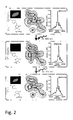
Fig. 2 . Molecular basis of photoswitching in the Triad-RSFPs. Triad-RSFP1 in the fluorescent equilibrium-state (top), the light-driven non-fluorescent off-state (middle) and the light-driven fluorescent on-state (bottom). Each left, top: representative Triad-RSFP1 protein crystal in the respective state. Each left, bottom: proposed chemical structure of the chromophore in the respective state. Each middle: Details of the X-ray structures of the respective states. Shown is the chromophore and the final 2Fo-Fc electron densities around the chromophore contoured at the 1σ level. The off-state and the on-state structure have been successively recorded on the same protein crystal. Each right: representative mass spectrum of Triad-RSFP1 photoswitched in solution. -
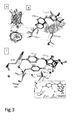
Fig. 3 . (A) Overall Triad-RSFP1 structure displayed in two orthogonal views. (B) Chromophore and immediate surrounding of on-state Triad-RSFP1 (dark grey) and EGFP (PDB1 EMA) (light grey). The Van-der-Waals radii of important atoms are indicated by spheres to highlight structural restraints. The chromophores are depicted as ball and stick while the surrounding amino acid residues are shown in the stick representation. (C) Comparison of the RFSP1 hydrogen bond network in the equilibrium- (dark grey) and the off-state (light grey). Important water molecules are shown as spheres Inset: hydrogen bond network in EGFP. -
Fig. 4 . Applications of Triad-RSFP1. Top: 33 individual freeze images referring to a computer game were written successively on the same field of Triad-RSFP1 embedded in PVA. Before writing each new frame, all molecules were photoswitched to the on-state. Shown are the first and the 33rd frame. Scale bar: 100 µm. Bottom: Switching of various Triad-RSFP1 fusion proteins in living Vero cells. From top to bottom: Triad-RSFP1-MAP2, Triad-RSFP1-α-tubulin, mito-Triad-RSFP1, Triad-RSFP1-Histon2B. Fluorescence was excited with green light (495 nm), switch-off: near-UV (420 nm), switch-on: UV-light (360 nm). -
Fig. 5 . FRAS (Fluorescence Recovery After Switching) with Triad-RSFP1. Triad-RSFP1 was targeted to the endoplasmic reticulum in living Vero cells. For a single FRAS measurement the Triad-RSFP1 molecules were switched off in Region-of-Interest-1 (ROI-1), and subsequently the change of the fluorescence signal was recorded in this ROI, as well as in ROI-2 (in the same cell) and in ROI-3 (in a neighboring cell). The reversibility of Triad-RSFP switching allowed repeating the experiment many times in the same cell. Images from top to bottom: Overview, before switching, immediately after switching Triad-RSFP off in ROI-1, and at the end of the experiment (65 s after switching). Graphs on the right: The plotted fluorescence signals were collected within the indicated ROIs during 20 repetitions of the same FRAS experiment on a single cell. The dark dots mark the mean values at each time point, demonstrating the reduction of statistical noise. Scale bars: 10 µm. - In a first aspect, the present invention relates to reversibly switchable fluorescent polypeptides according to
claim 1 able to switch reversibly between a fluorescent state and a non-fluorescent state when irradiated with a light of a first wavelengths to switch a polypeptide from the non-fluorescent state to the fluorescent state and irradiated with a light of a second wavelength different to the first wavelength to switch said polypeptide from the fluorescent state to the non-fluorescent state, whereby said first and second wavelengths for switching the polypeptide are different to a third wavelength for excitation of the fluorescence emission of said polypeptide. An example of the different wavelengths and the outcome of excitation therewith is shown inFigure 1a ). For example, the polypeptide exemplified inFig. 1a corresponding to Seq. ID. No. 4 according to the present invention may be switched from the on- to the off-state by near UV-light of e.g. 405 nm, from the off- to the on-state by UV-light, e.g. 365 nm and the fluorescence is probed by excitation with green light of e.g. 515 nm while the maximum fluorescence is present at a wavelength of about 529 nm. - Hence, the polypeptides according to the present invention are characterised in being polypeptides where the switching wavelengths are independent from the fluorescence excitation and, thus, excitation of the fluorescence does not influence the switching properties of the polypeptides. Hence, it is possible to eliminate the current limitations of switchable fluorescent proteins, namely, interference of the fluorescence excitation with the switching of said polypeptide.
- The fluorescent polypeptide according to the present invention is particularly a reversibly switchable fluorescent polypeptide (RSFP) wherein the first wavelength and the second wavelength which are different from each other are at least 50 nm below the third wavelength for excitation of the fluorescence emission of said polypeptide. As described above, for the Triad-RSFP1 having Seq. ID. No. 4, the first wavelength (365 nm) and the second wavelength (405 nm) are different from each other and both wavelengths are more than 50 nm below the third wavelength, 515 nm for excitation of the fluorescence emission of the polypeptide according to the present invention.
- Particularly, the Triad-RSFPs according to the present invention have a fluorescence emission with a maximum emission between 450 nm and 1000 nm, in particular, between 450 nm and 700 nm. Particularly preferred, the maximum emission is in the range of the visible light, thus, allowing the use of the Triad-RFSPs according to the present invention in fluorescence microscopy.
- The Triad-RSFPs according to the present invention are characterised in that the mechanistic basis of the switching is a light-induced reversible water addition/elimination to/from the chromophore, as shown in
scheme 1 above. Not to be bound by theory, the molecular basis for the reversible light induced switching of the polypeptide according to the present invention was solved by analysing the structures of the light induced off-state and on-state consecutively using the same polypeptide crystal. As demonstrated inscheme 1 andFig. 2 , the structure of the polypeptide according to the present invention resembles that of GFP related proteins. The chromophore, autocatalytically formed from amino acid residues 65, 66 and 67
according to the polypeptide of SEQ ID No. 1 or 2, resides in an alpha-helical-segment, enclosed by an 11-stranded-beta-barrel. The on-state chromophore consists of an imidazolinone-ring, connected by a methine bridge to a p-hydroxyphenyl-ring. In the on- and the equilibrium-state structures, the two rings of the chromophore system are largely co-planar, facilitating a conjugated pi-electron system and hence supporting fluorescence. In the off-state structure, the planarity of the five ring is markedly distorted with the chromophoric C65-carbon exhibiting a tetrahedral geometry (Scheme 1). - Based on electrospay mass spectrometry of light switched polypeptides, it was demonstrated that a mass difference of 18 +/- 1 units between the non-fluorescent state and the light induced on-state or the equilibrium state, respectively, can be observed. Hence, it is assumed that a water molecule is bound to the imidazolinone group in the off-state while water is free in the on-state as depicted in
scheme 1. The light induced addition of a mass of 18 +/- 1 mass units, indicative of the addition of water, was reversible and occurred parallel to changes in the fluorescence of the protein. Thus, a dehydration/hydration reaction of the chromophoric five membered ring seems to represent the underlying molecular mechanism of switching of the polypeptides according to the present invention. - That is, the fluorescent polypeptide is characterised in that the chromorphore comprises an imidazolinone-ring, connected by a methine bridge to a p-hydroxyphenyl-ring formed the by amino acid sequence X1YG wherein X1 is G or A.
- Moreover, it is preferred that the amino acid residues G65, Y203 and E222 of SEQ ID No. 1 or 2 are present. It is assumed that the side chains of G65 and Y203 facilitate the positioning of the side chain of E222 close to the imidazolinone ring as demonstrated in
Figure 2 . - As shown in
Figure 2 , when amino acid 203 is Y and amino acid 222 is E hydrogen bonds may be formed and thereby positioning a water molecule in close vicinity to the carbon atom of G65 of the chromophore. It appears that this situation is unique for the polypeptides according to the present invention while absent in non-switchable GFP variants. It is further proposed that the water molecule is in a suitable position for a nucleophilic addition to the C=N bond of the imidazolinone ring upon excitation of the on-state chromophore. The off-state structure reveals a different water molecule. It is assumed that said water molecule is taken up from the environment after the first water molecule is utilised for the light induced water addition to the imidazolinone ring. The dehydration reaction is induced by excitation of the shortened chromophoric system, in the present case, the 340 nm absorption band of the off-state chromophore. - Other amino acid residue changes, e.g. V61L, F64I, Y145H, and/or N146D, as e.g. in SEQ ID No. 1 or 2 have no immediate influence on the molecular mechanism outlined above, but affect the protonation state of an on-state chromophore causing the absorption band at about 410 nm allowing effective switching. Thus, it is assumed that a reversible light induced dehydration/hydration reaction of the five membered ring occurs. In the on-state, the chromophore exists in the protonated and the deprotonated form, resulting in absorption bands at about 410 nm and about 510 nm, respectively. Irradiation into the about 510 nm bands induces fluorescence whereas irradiation into the about 410 nm band induces a covalent modification of the five ring resulting in a non-fluorescent chromophore absorbing at about 340 nm. Subsequent irradiation to this band results in a dehydration of an off-state chromophore converting it back into the on-state chromophore. Although the reversible hydration/dehydration reaction appears to be a factor in the usual switching behaviour of the polypeptides according to the present invention, it is possible that additional short-lived intramolecular rearrangements possibly including Cis-trans-isomerisation, structure flexibility or a strong bending of the off-state chromophore may occur.
- In preferred embodiments, the Triad-RSFPs according to the present invention are polypeptides comprising the amino acid sequence of SEQ ID No. 1 or 2. Particular preferred embodiments of the Triad-RSFPs are any one of SEQ ID Nos. 3 to 242, in particular, Seq. ID Nos. 3 to 10.
- The Triad-RSFPs according to the present invention are characterised in that said polypeptide is stable both in the fluorescent state and the non-fluorescent state. In particular, it is preferred that said polypeptide is stable in the fluorescent state and the non-fluorescent state over a time of at least 100 ms, preferably of at least 1s.
- The term "Triad-RSFPs" comprises polypeptides of natural occurring amino acids. In addition, unusual amino acids may be present. Regarding peptide or protein sequences, if conservative amino acid exchanges distinguish the natural sequence form a non-natural sequence, e.g. chemical and/or physical similar amino acid residues are exchanged for each other. Such chemical or physical similar amino acid residues for example can be grouped according to their charge status (positive: R, H, K; negative: D, E; uncharged: A, N, C, Q, G, I, L, M, F, P, S, T, W, Y, V), or according to their volume and polarity (special: C; neutral and small: A, G, P, S, T; polar and relatively small: N, D, Q, E; polar and relatively large: R, H, K; nonpolar and relatively small: I, L, M, V; and nonpolar and relatively large: F, W, Y). Amino acid exchanges within a group are regarded as conservative exchanges (Mol Biol Evolution, 2002, 19:1022-1025).
- Amino acids mean for the purposes of the invention besides the 20 amino acids determined by the genetic code also the amino acids which can be encoded by stop codons, such as, for example, seleno-cysteine or pyro-lysine. Additionally included are all amino acid and peptide derivatives such as, for example, glycosylated, phosphorylated, sulfated amino acids/peptides, and L-isomeric and D-isomeric amino acids. Modifications of amino acids may occur in the peptide backbone, in the amino acid side chains, at N-terminal ends of the peptide or at C-terminal ends of the peptide. The modifications may be present in single amino acids, in a plurality of amino acids or in all amino acids, and it is possible for no, one or a plurality of types of modifications to be present in any combinations in a peptide. Examples of unusual amino acids which may be mentioned by way of example are, inter alia: alpha-aminobutyric acid, beta-aminobutyric acid, beta-aminoisobutyric acid, beta-alanine, gamma-aminobutyric acid, alpha-aminoadipic acid, 4-aminobenzoic acid, aminoethylcysteine, alpha-aminopenicillanic acid, allysine, 4-carboxyglutamic acid, cystathionine, carboxyglutamic acid, carboxyamidomethylcysteine, carboxymethylcysteine, cysteic acid, citroline, dehydroalanine, diaminobutyric acid, dehydroamino-2-butyric acid, ethionine, glycine-proline dipeptide, 4-hydroxyproline, hydroxylysine, hydroxyproline, homoserine, homocysteine, histamine, isovaline, lysinoalanine, lanthionine, norvaline, norleucine, ornithine,
2-piperidinecarboxylic acid, pyroglutamic acid, pyrrolysine, proline-hydroxyproline dipeptide, sarcosine, 4-selenocysteine, syndesines, thioproline, etc. All said amino acids can be present in the form of their L isomers or in the form of their D isomers as long as this is permitted by their structure. In general, all amino acids and amino acid derivatives which occur naturally or are formed or can be prepared enzymatic or chemically or in another wayare included in the term "amino acid". - In general, all modifications of peptides which occur naturally or are formed or can be prepared enzymatically or chemically or in another way, and modifications of peptides which will be known in future, are included in the term "peptide" and may be constituent of Triad-RSFP for the purposes of the invention.
- It is additionally possible for the purposes of the invention for one or more amino acids of elements or the complete element or all elements of the Triad-RSFP to be replaced by structures consisting of peptidomimetics. The term peptidomimetic is used in the present application in the form of the widest possible definition. A peptidomimetic is a substance which comprises non-peptide structural elements and is able to imitate or to antagonize the biological effect of the natural parent molecule. Numerous studies dealing in detail with possibilities for using peptidomimetics as replacement for conventional peptide structures are known in the art. It is generally possible for one or more elements of the Triad-RSFP to be composed entirely or partly of peptidomimetics (Curr Opin Chem Biol, 1997, 1:114-9, J Recept Signal Transduct Res, 2001, 21:489-506, J Med Chem, 2001, 44:1938-50). This may have various advantages. Transduction elements may possibly penetrate more efficiently into cells, antigen elements may lead to an enhanced or reduced immune response relative to the immune response to the conventional antigen, or tag elements may have better physicochemical properties, improving their suitability for the isolation and/or detection of the Triad-RSFP, etc. It is additionally possible through the use of peptidomimetics in some circumstances to reduce or increase the in vivo half-life of the Triad-RSFP molecules, to reduce its toxicity, to improve its solubility in hydrophilic or hydrophobic media, and to prolong its in vitro stability and possibly to reduce the costs for synthesizing the peptidomimetic relative to the cost for synthesizing the corresponding conventional Triad-RSFP without peptidomimetic structures incorporated within it. One example of peptidomimetics are Spiegelmers® supplied by NOXXON Pharma AG, Berlin, Germany. This type of peptidomimetics has the advantage for example that they do not elicit an immune response and therefore could be employed in a worthwhile manner for example in transduction elements, tag elements, spacer elements, etc. of Triad-RSFP. Spiegelmers® would, however, not be suitable as antigen element.
- In another aspect, the present invention relates to a polypeptide comprising the amino acid sequence of SEQ ID No. 1 or 2, like any one of SEQ ID Nos. 3 to 242, preferably to polypeptides of Seq. ID Nos. 3 to 10.
- The present invention also relates to a fusion protein comprising the Triad-RSFP according to the present invention. In addition to the polypeptide of the present invention, a fusion protein according to the present invention contains at least one additional, heterologous sequence. Often, but not necessarily, these additional sequences will be located at the N-, or C-terminal end of the polypeptide. It may e.g. be convenient to initially express the polypeptide as a fusion protein from which the additional amino acid residues can be removed, e.g. by a protease capable of specifically trimming the polypeptide of the present invention. The additional heterologous sequence may help in the expression or purification or attachment to a carrier of the polypeptides referred to in the present invention. The Triad-RSFP of the present invention could also be fused to a protease cleavage sequence and a quencher that when cleaved allows identification by increase of the fluorescence. In another embodiment, the polypeptide of the invention is fused to one or more tags or signal sequences. Said tags or sequences allow to purify the protein or facilitate expression thereof. In addition, the signal sequence may allow intra- or extracellular targeting thereof.
- Further preferred proteins to be combined with the polypeptide of the present invention and optionally the tags and signal sequences as described above are in general proteins the presence or localisation of which in cells and/or tissues is to be investigated. That is, the fusion protein according to the present invention may be useful as a biomarker and in diagnostics.
- In another aspect, the present invention relates to nucleic acid molecules encoding the Triad-RSFP according to the present invention.
- In particular, the present invention relates to a nucleic acid molecule encoding a fluorescent polypeptide wherein said nucleic acid molecule is selected from the group consisting of
- a) a nucleic acid molecule encoding a polypeptide according to the present invention;
- b) a nucleic acid molecule hybridising under stringent conditions to the complementary strand of a nucleic acid molecule of a) wherein said nucleic acid molecule of b) encodes a polypeptide having at the position corresponding to position 203 of SEQ ID No. 1 or 2 a tyrosine and at position corresponding to position 65 of SEQ ID No. 1 or 2 a glycine or an alanin; wherein the polypeptide encoded by the nucleic acid molecule is able to switch reversibly between a fluorescent state and a non-fluorescent state and the wavelengths required for reversibly switching said polypeptide is different to the wavelength for excitation of the fluorescence emission; or c) a nucleic acid molecule degenerated with respect to the genetic code of the nucleic acid molecule of a) or b).
- It is well known in the art how to perform hybridization experiments with nucleic acid molecules. Correspondingly, the person skilled in the art knows what hybridization conditions she/he has to use to allow for a successful hybridization. The establishment of suitable hybridization conditions is referred to in standard text books such as Sambrook, Russell "Molecular Cloning, A Laboratory Manual", Cold Spring Harbor Laboratory, N.Y. (2001); Ausubel, "Current Protocols in Molecular Biology", Green Publishing Associates and Wiley Interscience, N.Y. (1989), or Higgins and Hames (Eds.) "Nucleic acid hybridization, a practical approach" IRL Press Oxford, Washington DC, (1985).
- "Stringent conditions" refers to hybridisation conditions under which the polynulceotides that are capable of hybridizing to the nulciec acids of the invention or parts thereof hybridizes to these target sequences to a detectably greater degree than to other sequences (e.g., at least 2-fold over background). Stringent conditions are sequence-dependent and will be different in different circumstances. By controlling the stringency of the hybridisation and/or washing conditions, target sequences that have at least 90% sequence identity, more preferably 95%, such as 98% and more preferred 100% sequence identity to the probe can be identified (highly stringent hybridisation conditions). Alternatively, stringency conditions can be adjusted to allow a higher degree of mismatching in sequences (low stringency conditions of hybridisation). Such highly stringent and low stringent conditions for hybridisation are well known to the person skilled in the art.
- For example, "stringent conditions" refers to hybridization conditions which comprise, e.g. an overnight incubation at 65°C in 4x SSC (600 mM NaCI, 60 mM sodium citrate) followed by washing at 65°C in 0.1x SSC for one hour. Alternatively, hybridization conditions can comprise: an overnight incubation at 42°C in a solution comprising 50% formamide, 5x SSC (750 mM NaCl, 75 mM sodium citrate), 50 mM sodium phosphate (pH 7.6), 5x Denhardt's solution, 10% dextran sulphate, and 20 microg/ml denatured, sheared salmon sperm DNA, followed by washing in e.g. 0.1-0.5x SSC at about 55-65°C for about 5 to 20 min. Said conditions for hybridization are also known by a person skilled in the art as "highly stringent conditions for hybridization". It is of note that variations in the above conditions may be accomplished through the inclusion and/or substitution of alternate blocking reagents. Typical blocking reagents include Denhardt's reagent, BLOTTO, heparin, denatured salmon sperm DNA, and commercially available proprietary formulations. The inclusion of specific blocking reagents may require modification of the hybridization conditions described above, due to problems with compatibility. Such modifications can generally be effected by the skilled person without further ado.
- A hybridization complex may be formed in solution (e.g., Cot or Rot analysis) or between one nucleic acid sequence present in solution and another nucleic acid sequence immobilized on a solid support (e.g., membranes, filters, chips, pins or glass slides to which, e.g., cells have been fixed).
- Examples for hybridization assays comprise without limitation Northern and Southern blot assays, heteroduplex analysis, detection of mutations by sequence specific oligonucleotide hybridization, allele-specific oligonucleotide hybridization on DNA chips, assays based on the Illumina's® technology, assays based on the BeadArray® technology, see, for example, Barnes et al., Nucleic Acids Res. 33 (2005) 5914-5923.
- The term "degenerate" in accordance with the present invention refers to the degeneracy of the genetic code. Degeneracy results because a triplet code designates 20 amino acids and a stop codon. Because four bases exist which are utilized to encode genetic information, triplet codons are required to produce at least 21 different codes. The possible 43 possibilities for bases in triplets gives 64 possible codons, meaning that some degeneracy must exist. As a result, some amino acids are encoded by more than one triplet, i.e. by up to six. The degeneracy mostly arises from alterations in the third position in a triplet. This means that nucleic acid molecules having a different sequence than that specified above, but still encoding the same polypeptide lie within the scope of the present invention.
- Preferably, the nucleic acid is a nucleic acid molecule encoding a polypeptide according to the present invention.
- The nucleic acid molecule may be in form of RNA or DNA.
- The present invention also relates to a vector comprising the nucleic acid molecule of the present invention. Preferably, the vector is a plasmid, cosmid, virus, bacteriophage or another vector used conventionally e.g. in genetic engineering. The skilled person is well aware of suitable vectors designed for the specific application, e.g. expression of the polypeptide, transfection of cells etc. In another aspect, the present invention relates to a non-human host, in particular a cell line, transformed with a vector or a recombinant non-human host, in particular, a cell line, containing the nucleic acid sequence as a foreign nucleic acid sequence. In this connection, the term "foreign" refers to a nucleic acid sequence not occurring naturally in said host.
- Non-human host according to the present invention can be single cells or multicellular organisms. Suitable prokaryotic host comprises e.g. bacteria of the species Escherichia, Streptomyces, Salmonella or Bacillus. Suitable eukaryotic host cells e.g. yeast, such as Saccharomyces cerevisiae or Pichia pastoris, insect cells, etc.
- Mammalian host cells that could be used include human cell lines, mice cell lines etc. Suitable cell lines include Hela, COS cells, CHO cells etc. In another aspect the cell line is a primary cell, e.g. obtained from mice etc. Appropriate methods, culture media and conditions for the above described host cells are known in the art. Further, the host may be transgenic non-human animal transfected with and/or expressing the nucleic acid molecule of the present invention. In a preferred embodiment, the transgenic animal is a mammal, e.g. a hamster, mouse, rat, cow, cat, pig, dog, horse, rabbit or monkey or other suitable animals, like zebra fish or C. elegans.
- Transgenic plants as hosts transfected with and/or expressing the nucleic acid molecule of the present invention also lay within the scope of the present invention.
- Suitable methods for the production of said transgenic non-human animals, plants etc. are known in the art.
- Moreover, the present invention relates to a method of producing a polypeptide or fusion protein according to the present invention. Said method comprises culturing the host of the invention under suitable conditions and isolating the polypeptide of the fusion protein used. Suitable conditions for culturing the host are well known to the person skilled in the art. In addition, purification methods and purification systems are well known. For example, the polypeptide or fusion protein contains a tag allowing purification. Methods of isolation of the polypeptide are fusion proteins produced are well known in the art including chromatography, immunoprecipitation etc.
- Moreover, the present invention provides diagnostic compositions comprising the Triad-RSFPs according to the present invention, the fusion protein according to the present invention, nucleic acid molecules according to the present invention or a vector according to the present invention. Moreover, the present invention provides a kit comprising at least one of the Triad-RSFPs according to the present invention, a fusion protein, a nucleic acid molecule, a vector or a host according to the present invention. The various components of the diagnostic composition or of the kit according to the present invention may be packaged in one or more containers such as one or more vials. The vials may, envision to the components, comprise preservatives or buffers for storage.
- The kit or the diagnostic compositions are useful for various purposes. For example, the diagnostic composition or the kit may be part of a system for detecting the presence, in particular, the localisation of a fusion protein according to the present invention, or of a polypeptide according to the present invention. Further, a system is provided for detecting the presence and/or localisation of a protein of interest in a cell tissue, said system comprises a kit or diagnostic composition according to the present invention, a source emitting a first and a second wavelength allowing reversibly switching the fusion protein or Triad-RSFP according to the present invention as well as a source for exciting said fusion protein or Triad-RSFP, and means allowing the detection of the presence and/or the localisation of the fluorescence emitted by the excited fusion protein or polypeptide.
- In a preferred embodiment, the system comprises a fluorescence microscope, means allowing the detection of fluorescence and the diagnostic composition or kit according to the present invention.
- The present invention furthermore relates to a method of detecting the presence and/or the localisation of a protein of interest, comprising
- i) expressing a fusion protein according to the present invention comprising the protein of interest in a sample or
- ii) contacting a sample with said fusion protein, wherein said fusion protein comprises a polypeptide specifically binding to the protein of interest.
- In another aspect, the present invention relates to a method of detecting the presence and/or absence and/or the localisation of a Triad-RSFP according to the present invention or a fusion protein according to the present invention, comprising
- a) exciting said fusion protein or polypeptide with a first or second wavelength allowing switching of the polypeptide from a fluorescent state to a non-fluorescent state or vice versa;
- b) exciting said fusion protein or polypeptide with a wavelength different to the first or second wavelength for excitation of the fluorescence emission and
- c) detecting the location of the fluorescence emitted by the excited fusion protein or polypeptide.
- That is, the polypeptides according to the present invention, in particular, the fusion proteins, as well as the nucleic acids encoding the same, are useful for FRAS (fluorescence recovery after switching). This method includes the presence of the Triad-RSFP in an area of interest. Switching the Triad-RSFP from the off- to the on-stage or vice versa and determining the fluorescence in the area of interest over time. Using the polypeptides and fusion proteins according to the present invention in the methods described herein allow to monitor the effect and/or metabolism of active ingredients including prodrugs and drugs in the area of interest, the use as a local biosensor, etc. In particular, it is possible to obtain a time-resolved monitoring of action since the Triad-RSFPs are switchable and, thus, the measurement can be repeated many times. The Triad-RSFP according to the present invention allows one to monitor the metabolism and possible effects of foreign and natural molecules in the area of interest in a time-resolved fashion. In this connection, the term area of interest may be a whole organism, or tissues, or organs, or a cell,or sub-compartments of a cell.
- Still another embodiment of the present invention relates to a method of detecting the expression of a gene of interest comprising and operably linking the nucleic acid molecule according to the present invention with a promoter controlling said gene of interest or fusing the nucleic acid molecule according to the present invention to said gene of interest, and detecting the fluorescence of the protein encoded by said nucleic acid molecules.
- Finally, the present invention relates to a method of detecting the activity of a promoter of interest, comprising and operably linking the nucleic acid molecule according to the present invention with said promoter of interest and detecting the fluorescence of the protein encoded by said nucleic acid molecule.
- As essential elements of fluorescent detecting systems in the following can be identified:
- a) an excitation source,
- b) two additional light sources for switching the Triad-RSFPs, or the appropriate filters and dichroic mirrors to obtain the required wavelength for switching from a single excitation source
- c) a fluorophore,
- d) wavelength filters to isolate emission photons from excitation photons and
- e) a detector that registers emitted photons and produces a recordable output, e.g. as an electronic signal or a photographic image.
- Fluorescence instruments are primarily of four types, each providing distinctly different information: Spectrofluorometers and microplate readers measure the average properties of bulk (µL to mL) samples. Fluorescence microscopes resolve fluorescence as a function of spatial coordinates in two or three dimensions for microscopic objects (less than ∼0.1 mm diameter).
- Fluorescence scanners, including microarray readers, resolve fluorescence as a function of spatial coordinates in two dimensions for macroscopic objects such as electrophoresis gels, blots and chromatograms.
- Flow cytometers measure fluorescence per cell in a flowing stream, allowing subpopulations within a large sample to be identified and quantitated.
- Other types of instrumentation that use fluorescence detection include capillary electrophoresis apparatus, DNA sequencers and microfluidic devices. Each type of instrument produces different measurement artefacts and makes different demands on the fluorescent probe. For example, although photo-bleaching is often a significant problem in fluorescence microscopy, it is not a major impediment in flow cytometry or DNA sequencers because the dwell time of individual cells or DNA molecules in the excitation beam is short.
- In accordance with the present invention and depending on the demands and possibilities of the methods described herein, the overall fluorescence can be detected in a sample as well as the location of the fluorescence within a sample.
- A reporter gene is a gene that can be operably linked to a gene of interest or simply to a promoter controlling the gene of interest in cell culture, animals or plants. Reporter genes are e.g. used to determine whether the gene of interest has been taken up by or expressed in the cell or organism population or whether the gene of interest naturally present in the cell or organism is expressed. It is important to use a reporter gene that is not natively expressed in the cell or organism under study, since the expression of the reporter is being used as a marker. The nucleic acid of the present invention encodes a GFP variant suitable as reporter gene with advantageous properties as described above.
- To introduce a reporter gene into an organism, the reporter gene and the gene of interest are placed in the same DNA construct to be inserted into the cell or organism, most often as a plasmid. Reporter genes can also be used to assay for the expression of the gene of interest, which may produce a protein that has little obvious or immediate effect on the cell culture or organism. In these cases the reporter is directly attached to the gene of interest to create a gene fusion. The two genes are under the control of the same promoter and are transcribed into a single messenger RNA molecule (mRNA). The mRNA is then translated into protein. In these cases it is important that both proteins are able to properly fold into their active conformations and interact with their substrates despite being fused. In building the DNA construct, a segment of DNA coding for a flexible polypeptide linker region is usually included so that the reporter and the gene product of will only minimally interfere with one another.
- In addition, the present invention relates to a method of detecting the activity of a promoter of interest, comprising operably linking the nucleic acid molecule of the invention with said promoter of interest and detecting the fluorescence of the protein encoded by said nucleic acid molecule.
- The analysis of the activity of promoters, which has been proven to be a highly sensitive and time-saving technique, is widely used e.g. in the study of transcriptional regulation.
- Reporter genes can also be used to assay for the activity of a particular promoter in a cell or organism. In this case the reporter gene is simply placed under the control of the target promoter and the reporter gene product's activity is quantitatively measured. The results are normally reported relative to the activity under a "consensus" promoter known to induce strong gene expression.
- The skilled person is well aware of promoter and reporter assays the principles of which are commonly known in the art (see e.g. Lewin, Genes IX or Sambrook et al., Molecular Cloning).
- Furthermore, the present invention relates to a method of detecting the presence of a protein of interest, comprising contacting a sample with the fusion protein of the invention, wherein said fusion protein comprises a polypeptide specifically binding to the protein of interest.
- The method of the invention can be carried out using FACS scans and preparative FACS sorts. Alternatively fluorescence microscope techniques may be applied.
- A sample can be any matter potentially containing the protein of interest. Preferred samples are liquids, e.g. cellular extracts or supernatants of cell cultures, or samples comprising living or dead cells in single form or present as a tissue or organ.
- Contacting a sample in accordance with the present invention means bringing the sample into contact with the fusion protein of the invention. The term comprises bringing both components into close vicinity as well as e.g. transforming or transfecting cells with a nucleic acid molecule encoding the fusion protein of the invention in order to express the fusion protein in the cells.
- Specifically binding in accordance with the present invention refers to the property of certain proteins to have an affinity for specific proteins, i.e. target proteins, which is greater by the factor of at least 10, preferably at least 100, more preferably at least 1,000 and most preferably at least 10,000 as compared to the affinity to proteins unrelated to the target protein. These proteins can e.g. be natural interaction partners of the target protein including proteins interacting in the course of signal transduction pathways or in context with structural proteins or antibodies.
- The present invention furthermore relates to a method of detecting the localization of a fusion protein of the present invention or a fusion protein comprising the polypeptide of the present invention in a cell or tissue, comprising exciting said fusion protein or polypeptide and detecting the location of the fluorescence emitted by the excited fusion protein or polypeptide.
- The present method can be used to localize the fusion protein of the invention as well as interaction partners of the fusions proteins of the invention.
- Further, the Triad-RSFPs may be used in combination with known conventional fluorescent molecules to provide suitable FRET-pairs. In the FRET-pairs, the Triad-RSFPs may represent the donor or the acceptor, respectively. In addition, the FRET-pair may be composed of two Triad-RSFPs. These FRET-pairs may be used to determine the spatial distribution, in particular, the spatial proximity of fusion proteins.
- In addition, the Triad-RSFPs may be used to determine repeatedly movements of fusion proteins in a cell in three room dimensions. In particular, it is possible to repeatedly determine the position of the molecule in the same cell allowing improvement of the accuracy of measurement. The repeatability of the measurements may be used for screening the effects of different molecules on the mobility or processability of the fusion protein in the cell.
- The methods of the present invention are also suitable in the identification of compounds influencing e.g. the expression of target genes, the location of target proteins or the activity of target proteins. Said compounds may then be contacted, at the same time or separately, with a cell comprising the nucleic acid of the invention, e.g. fused to a target gene or a regulatory sequence. Afterwards, expressed (fusion) protein is excited in a cell contacted with the compound(s) and in a cell which has not been contacted. The expression or location of the (fusion) protein in both cells indicates that the compound has an influence on the expression of target genes and/or the location or activity of target proteins.
- The invention is further illustrated by the examples below without limiting the invention.
- Absorption and emission spectra were recorded on a Varian Cary 4000 UV/VIS spectrophotometer and a Varian Cary Eclipse fluorescence spectrometer (Varian, Palo Alto, CA, USA), respectively.
- Switching of RSFPs were recorded either on E. coli colonies grown on LB-agar plates or in buffered protein solutions (100 mM Tris, 150 mM NaCl, pH 7.5; Figs. S1 or
Citrat buffer pH 4,6). A modified computer-controlled fluorescence microscope (Leica Microsystems) equipped with a 50x NA 0.5 or a 20x NA 0.4 air objective lens and three 100 W Hg lamps was utilized for data acquisition. Fluorescence was recorded using a photomultiplier tube (HR9306-0, Hamamatsu, Hamamatsu City, Japan) behind a 525 nm longpass detection filter (HQ 525 LP, AHF Analysentechnik, Tübingen, Germany). - For the determination of the relaxation half-time from the non-fluorescent off-state into the fluorescent on-state (thermal equilibrium), colonies were switched completely into the off-state and the relaxation to the equilibrium state was followed at room temperature by consecutive short measurements with light of 515 nm (515/10 nm, 0,82 W/cm2) every 120 s.
- For widefield images a Leica DM6000 epifluorescence microscope (Leica, Wetzlar, Germany) equipped with a 100x NA 1.40 oil immersion objective lens and a DFC350 FX camera (Leica) was used. Cells were irradiated for 300 ms with near-UV light (BP420nm/30nm, 11 W/cm2) to switch the molecules to the non-fluorescent state and 300 ms with UV light (BP360nm/40nm, 2 W/cm2) to switch the proteins back to the fluorescent state. The fluorescence images were recorded with green light (BP495nm/15nm, 8 W/cm2). For fluorescence detection a 510 nm dichroic mirror and a BP 530nm/30nm detection filter were used. The length of the image acquisition time was adjusted to the brightness of every sample and was generally between 50 and 150 ms.
- Imaging was performed with a beam scanning confocal microscope (SP5, Leica Microsystems) equipped with a UV-corrected 1.4 NA oil immersion lens (63x HCX PL APO). All imaging was performed at ambient temperatures. The following microscope settings were used: pinhole: 1 airy unit (AU); pixel size: 160.8 nm x 160.8 nm; scan speed: 1400 Hz. Fluorescence was detected between 519 and 590 nm.
- For each measurement, the molecules in a predefined region of interest (ROI) (1,3 µm x 10 µm) were switched to the non-fluorescent state using 405 nm laser light. The fluorescence was recorded by excitation with 515 nm laser light. Finally, the molecules in the whole sample were switched to the fluorescent state with 360 nm widefield illumination. This imaging sequence was repeated several times on the same cell. In every sequence a second ROI on the other side of the same cell and a third ROI in a neighboring cell were analyzed. No further data processing was perfomed, except background subtraction and normalization to 1.
- For the determination of the molecular mass of the RSFP in its equilibrium, switched-off and switched-on state 160µg of the polypeptides in 10mM ammonium acetate were diluted with or without prior photoswitching to a final concentration of 18% acetonitrile in 10 mM ammonium acetate. The photoswitching was performed essential as described above.
- After purification, a RSFP polypeptide solution was concentrated to -35 µg/µl by ultrafiltration and taken up in 20 mM Tris/HCl, 120 mM NaCl, pH 7.5. The protein was crystallized by sitting drop vapor diffusion at 20°C with a reservoir solution containing 15% (w/v) PEG 3350, 0.2 M KH2PO4 and 3% (w/v) D-(+)-trehalose-dihydrate. The crystallization solution was cryoprotected by adding propane-1,3-diol to a final concentration of 25% (w/v).
- For the structure determination of RSFP in the on- and the off-state, a single RSFP protein crystal was manually transferred into a vessel with cryoprotected crystallization solution and then switched to the non-fluorescent state by irradiation with blue light (405 ± 10 nm, 110 W/cm2) until the fluorescence signal reached a minimum. After switching the crystal, it was transferred within 10 seconds into liquid nitrogen, flash frozen, and diffraction data were collected at 100 K. Following the data collection of the off-state structure, the same crystal was unmounted, transferred back into cryoprotected crystallization solution kept at room temperature and immediately switched to the fluorescent-state by irradiation with UV light (365 ± 25 nm, 1.4 W/cm2) until the fluorescence signal reached a maximum. Subsequently the crystal was again flash frozen in liquid nitrogen and another diffraction dataset was collected.
- The equilibrium structure of RSFP was determined on crystals that were not previously irradiated.
- All diffraction data sets were collected on beamline 14-2 at BESSY (Berliner Elektronenspeicherring-Gesellschaft für Synchrotronstrahlung, Berlin, Germany) using a MAR 225 mm CCD detector. The data were processed using the XDS program package {Kabsch, 1993 #1842}. The crystal structures were solved by molecular replacement with PHASER {McCoy, 2007 #1708} using the structure coordinates of Citrine (PDB ID 1HUY; {Griesbeck, 2001 #834}) as a model omitting the chromophore and the water molecules. The sequence was manually adjusted and fitted to the 2F o-F c and F o-F c electron densities with COOT {Emsley, 2004 #1348}. Atomic B-factors and coordinates were refined automatically with PHENIX.REFINE {Adams, 2010 #1843}. TLS (translation-libration-screw) refinement cycles were performed. Refinement was complemented by manual model building with COOT. Waters were built automatically with PHENIX.REFINE and completed manually. During the entire refinement procedure, 5% of randomly selected reflections were set aside for monitoring of the R free factor.
- Finally, the omitted chromophore was placed manually into vacant patches of the 2Fo-Fc and Fo-Fc electron densities. The chromophore restraints for the on-state Fluorophore were generated in PRODRG {Schuttelkopf, 2004 #1844}, and the restraints of the off-state chromophore were generated with eLBOW {Moriarty, 2009 #1845}.
- Starting from the known sequences of citrine and GFP, respectively, site-directed random mutagenesis experiments were performed using standard methods.
- For protein expression and mutagenesis the respective coding sequences were PCR amplified, digested and inserted into the BamHI/HindIII restriction sites of the plasmid pQE31 (Quiagen, Hilden, Germany).
- For site-directed random mutagenesis, the QuikChange Site Directed Mutagenesis Kit (Stratagene, La Jolla, CA) was used according to the manufacturer's instructions. Error prone random mutagenesis as well as directed simultaneous mutagenesis of multiple sites were performed according to standard protocols {Leung, 1989 #1547;Sawano, 2000 #1544}.
- Mutated sequences present in suitable plasmids were transformed into E. coli for expression of the mutated polypeptides. After growing of the transformed bacteria, the colonies were screened using computer assisted fluorescence microscopy. The microscope was running with three different sources of light which were controlled independently. Fluorescence of the polypeptides were measured and computed
- For selecting appropriate colonies, the computed data were evaluated based on the fluorescence for each single colony. Selected colonies were transferred and propagated by known methods, sequenced and analysed for their fluorescence and switching properties.
- Further rounds of site-directed random mutagenesis were conducted using the bacterial clones selected in the previous round following the procedure described above.
- The most promising mutants were selected, e.g. expressing polypeptides of Seq. ID Nos. 3 to 242, and analysed further.
- To target the RSFP to the lumen of the endoplasmic reticulum (ER), the coding sequence of the RSFP was amplified by PCR. The PCR fragments were digested with SalI and NotI and inserted into the vector pEF/myc/ER (Invitrogen, Carlsbad, CA).
- To target the RSFP to the mitochondrial matrix, the coding sequence of RSFP was amplified by PCR. The PCR fragments were digested with AgeI and NotI and inserted into the vector pDsRed1-Mito (Clontech, Mountain View, CA), replacing the DsRed1 sequence.
- For the generation of the α-tubulin fusion construct, the RSFP coding sequence was amplified by PCR. The PCR fragments were digested with NheI and BgIII and inserted into the vector pEGFP-Tub (Clontech), replacing the EGFP sequence.
- For the generation of the microtubule-associated protein 2 (Map2) fusion construct, the Map2 coding sequence (obtained from pDONR223-MAP2, {Lamesch, 2007 #1549}) was amplified by PCR. The PCR fragments were digested with XhoI and BamHI and inserted into the vector RSFP-α-tubulin, replacing the α-tubulin sequence.
- For the generation of the Histon-2B fusion construct, the Histon-2B coding sequence (obtained from pDONR223-HIST1H2BN, {Lamesch, 2007 #1549}) was amplified by PCR. The PCR fragments were digested with XhoI and BamHI and inserted into the vector RSFP-α-tubulin, replacing the α-tubulin sequence.
- Proteins were expressed in the Escherichia coli strain BL21-CodonPlus (Stratagene) and purified by Ni-NTA affinity chromatography, followed by a gel filtration step on a Superdex 200 column (Amersham Biosciences, Piscataway, NJ, USA) using a Tris-buffer (100 mM Tris-HCl, 150 mM NaCl, pH 7.5).
- Semi-native PAGE. Proteins (20 µg each) were loaded onto 15% polyacrylamid gels containing 0.1 % sodiumdodecyl sulfate (SDS) {Laemmli, 1970 #262}. For loading, proteins were taken up in 60% (w/v) sucrose solution. As a standard, purified tetrameric DsRed and dimeric tdTomato were used.
- For imaging of mammalian cells, the respective plasmids were transfected into Vero (kidney epithel of Cercopithecus aethiops) cells using the Nanofectin Kit (PAA, Pasching, Austria) according to the manufacturer's instructions. The cells were propagated in DMEM medium with GlutaMAX and 4.5 g/l glucose, 10% (v/v) FCS, 1mM sodium pyruvate, 50 mg/ml penicillin and 50 mg/ml streptomycin. Cells were grown on coverslips in petri dishes at 37°C under 90% humidity and 5% CO2.
- A solution of RSFP molecules was embedded in PAA (final concentration: 15 % PAA in 1.5 M Tris/HCl buffer, pH 8.8) and squeezed under a coverslip to ensure a layer thickness of ∼1 µm. For reading and writing a Leica SP5 beam scanning confocal microscope equipped with a 40x NA 1.25 oil imersion objective was utilized. The following settings were used: pinhole: 1.48 AU; pixel size: 378.8 nm x 378.8 nm; scan speed: 400 Hz. Fluorescence was detected between 517 and 575 nm. Except smoothing and contrast stretching, no further imaging processing was applied.
- To generate the PacMan-movie (
Fig. 4A ), each individual image was written utilizing a group of ROIs, defining the positions where RSFP was transferred from the on- to the off-state by irradiation with 405 nm laser-light. Subsequently the written image was recorded with 515 nm laser-irradiation only. In the next step all proteins were switched back to the fluorescent state with 365 nm widefield illumination and the next image was recorded. -
- <110> Max-Planck-Gesellschaft zur Förderung der Wissenschaften e.v.
- <120> REVERSIBLY PHOTOSWITCHABLE POLYPEPTIDES
- <130> 4910-002-EP-1
- <160> 242
- <170> PatentIn version 3.3
- <210> 1
<211> 238
<212> PRT
<213> Artificial - <220>
<223> polypeptide - <220>
<221> MISC_FEATURE
<222> (61)..(61)
<223> X is I or V - <220>
<221> MISC_FEATURE
<222> (64)..(64)
<223> X is I, or F - <220>
<221> MISC_FEATURE
<222> (65)..(65)
<223> X is G or A - <220>
<221> MISC_FEATURE
<222> (145)..(145)
<223> X is H, or N - <220>
<221> MISC_FEATURE
<222> (146)..(146)
<223> X is D or E - <220>
<221> MISC_FEATURE
<222> (206)..(206)
<223> X is M, K, L, R, or A - <400> 1


- <210> 2
<211> 238
<212> PRT
<213> Artificial - <220>
<223> polypeptide - <220>
<221> MISC_FEATURE
<222> (65)..(65)
<223> X is G, or A - <220>
<221> MISC_FEATURE
<222> (69)..(69)
<223> X is M, or Q - <220>
<221> MISC_FEATURE
<222> (145)..(145)
<223> X is H, or Y - <220>
<221> MISC_FEATURE
<222> (205)..(205)
<223> X is N, Q, or S - <220>
<221> MISC_FEATURE
<222> (206)..(206)
<223> X is L, M, R, A, or K - <400> 2


- <210> 3
<211> 238
<212> PRT
<213> Artificial - <220>
<223> polypeptide - <400> 3


- <210> 4
<211> 238
<212> PRT
<213> Artificial - <220>
<223> polypeptide - <400> 4


- <210> 5
<211> 238
<212> PRT
<213> Artificial - <220>
<223> polypeptide - <400> 5


- <210> 6
<211> 238
<212> PRT
<213> Artificial - <220>
<223> polypeptide - <400> 6


- <210> 7
<211> 238
<212> PRT
<213> Artificial - <220>
<223> polypeptide - <400> 7


- <210> 8
<211> 238
<212> PRT
<213> Artificial - <220>
<223> polypeptide - <400> 8


- <210> 9
<211> 238
<212> PRT
<213> Artificial - <220>
<223> polypeptide - <400> 9


- <210> 10
<211> 238
<212> PRT
<213> Artificial - <220>
<223> polypeptide - <400> 10


- <210> 11
<211> 238
<212> PRT
<213> Artificial - <220>
<223> polypeptide - <400> 11


- <210> 12
<211> 238
<212> PRT
<213> Artificial - <220>
<223> polypeptide - <400> 12


- <210> 13
<211> 238
<212> PRT
<213> Artificial - <220>
<223> polypeptide - <400> 13


- <210> 14
<211> 238
<212> PRT
<213> Artificial - <220>
<223> polypeptide

- <210> 15
<211> 238
<212> PRT
<213> Artificial - <220>
<223> polypeptide - <400> 15


- <210> 16
<211> 238
<212> PRT
<213> Artificial - <220>
<223> polypeptide - <400> 16


- <210> 17
<211> 238
<212> PRT
<213> Artificial - <220>
<223> polypeptide - <400> 17


- <210> 18
<211> 163
<212> PRT
<213> Artificial - <220>
<223> polypeptide - <400> 18


- <210> 19
<211> 238
<212> PRT
<213> Artificial - <220>
<223> polypeptide - <400> 19


- <210> 20
<211> 238
<212> PRT
<213> Artificial - <220>
<223> polypeptide - <400> 20


- <210> 21
<211> 238
<212> PRT
<213> Artificial - <220>
<223> polypeptide - <400> 21


- <210> 22
<211> 238
<212> PRT
<213> Artificial - <220>
<223> polypeptide - <400> 22


- <210> 23
<211> 238
<212> PRT
<213> Artificial - <220>
<223> polypeptide - <400> 23


- <210> 24
<211> 238
<212> PRT
<213> Artificial - <220>
<223> polypeptide - <400> 24


- <210> 25
<211> 238
<212> PRT
<213> Artificial - <220>
<223> polypeptide - <400> 25


- <210> 26
<211> 238
<212> PRT
<213> Artificial - <220>
<223> polypeptide - <400> 26


- <210> 27
<211> 238
<212> PRT
<213> Artificial - <220>
<223> polypeptide - <400> 27


- <210> 28
<211> 238
<212> PRT
<213> Artificial - <220>
<223> polypeptide - <400> 28


- <210> 29
<211> 238
<212> PRT
<213> Artificial - <220>
<223> polypeptide - <400> 29


- <210> 30
<211> 238
<212> PRT
<213> Artificial - <220>
<223> polypeptide - <400> 30


- <210> 31
<211> 238
<212> PRT
<213> Artificial - <220>
<223> polypeptide - <400> 31


- <210> 32
<211> 238
<212> PRT
<213> Artificial - <220>
<223> polypeptide - <400> 32


- <210> 33
<211> 238
<212> PRT
<213> Artificial - <220>
<223> polypeptide - <400> 33


- <210> 34
<211> 238
<212> PRT
<213> Artificial - <220>
<223> polypeptide - <400> 34


- <210> 35
<211> 238
<212> PRT
<213> Artificial - <220>
<223> polypeptide - <400> 35


- <210> 36
<211> 238
<212> PRT
<213> Artificial - <220>
<223> polypeptide - <400> 36


- <210> 37
<211> 238
<212> PRT
<213> Artificial - <220>
<223> polypeptide - <400> 37

- <210> 38
<211> 238
<212> PRT
<213> Artificial - <220>
<223> polypeptide - <400> 38

- <210> 39
<211> 238
<212> PRT
<213> Artificial - <220>
<223> polypeptide - <400> 39

- <210> 40
<211> 238
<212> PRT
<213> Artificial - <220>
<223> polypeptide - <400> 40

- <210> 41
<211> 238
<212> PRT
<213> Artificial - <220>
<223> polypeptide - <400> 41

- <210> 42
<211> 238
<212> PRT
<213> Artificial - <220>
<223> polypeptide - <400> 42

- <210> 43
<211> 238
<212> PRT
<213> Artificial - <220>
<223> polypeptide - <400> 43


- <210> 44
<211> 238
<212> PRT
<213> Artificial - <220>
<223> polypeptide - <400> 44


- <210> 45
<211> 238
<212> PRT
<213> Artificial - <220>
<223> polypeptide - <400> 45


- <210> 46
<211> 238
<212> PRT
<213> Artificial - <220>
<223> polypeptide - <400> 46


- <210> 47
<211> 238
<212> PRT
<213> Artificial - <220>
<223> polypeptide - <400> 47


- <210> 48
<211> 238
<212> PRT
<213> Artificial - <220>
<223> polypeptide - <400> 48


- <210> 49
<211> 238
<212> PRT
<213> Artificial - <220>
<223> polypeptide - <400> 49


- <210> 50
<211> 238
<212> PRT
<213> Artificial - <220>
<223> polypeptide - <400> 50


- <210> 51
<211> 238
<212> PRT
<213> Artificial - <220>
<223> polypeptide - <400> 51


- <210> 52
<211> 238
<212> PRT
<213> Artificial - <220>
<223> polypeptide - <400> 52


- <210> 53
<211> 238
<212> PRT
<213> Artificial - <220>
<223> polypeptide - <400> 53


- <210> 54
<211> 238
<212> PRT
<213> Artificial - <220>
<223> polypeptide - <400> 54


- <210> 55
<211> 238
<212> PRT
<213> Artificial - <220>
<223> polypeptide - <400> 55


- <210> 56
<211> 238
<212> PRT
<213> Artificial - <220>
<223> polypeptide - <400> 56


- <210> 57
<211> 238
<212> PRT
<213> Artificial - <220>
<223> polypeptide - <400> 57


- <210> 58
<211> 238
<212> PRT
<213> Artificial - <220>
<223> polypeptide - <400> 58


- <210> 59
<211> 238
<212> PRT
<213> Artificial - <220>
<223> polypeptide - <400> 59


- <210> 60
<211> 238
<212> PRT
<213> Artificial - <220>
<223> polypeptide - <400> 60


- <210> 61
<211> 238
<212> PRT
<213> Artificial - <220>
<223> polypeptide - <400> 61


- <210> 62
<211> 238
<212> PRT
<213> Artificial - <220>
<223> polypeptide - <400> 62


- <210> 63
<211> 238
<212> PRT
<213> Artificial - <220>
<223> polypeptide - <400> 63


- <210> 64
<211> 238
<212> PRT
<213> Artificial - <220>
<223> polypeptide - <400> 64


- <210> 65
<211> 238
<212> PRT
<213> Artificial - <220>
<223> polypeptide - <400> 65


- <210> 66
<211> 238
<212> PRT
<213> Artificial - <220>
<223> polypeptide - <400> 66


- <210> 67
<211> 238
<212> PRT
<213> Artificial - <220>
<223> polypeptide - <400> 67


- <210> 68
<211> 238
<212> PRT
<213> Artificial - <220>
<223> polypeptide - <400> 68


- <210> 69
<211> 238
<212> PRT
<213> Artificial - <220>
<223> polypeptide - <400> 69


- <210> 70
<211> 238
<212> PRT
<213> Artificial - <220>
<223> polypeptide - <400> 70


- <210> 71
<211> 238
<212> PRT
<213> Artificial - <220>
<223> polypeptide - <400> 71

- <210> 72
<211> 238
<212> PRT
<213> Artificial - <220>
<223> polypeptide - <400> 72

- <210> 73
<211> 238
<212> PRT
<213> Artificial - <220>
<223> polypeptide - <400> 73

- <210> 74
<211> 238
<212> PRT
<213> Artificial - <220>
<223> polypeptide - <400> 74

- <210> 75
<211> 238
<212> PRT
<213> Artificial - <220>
<223> polypeptide - <400> 75

- <210> 76
<211> 238
<212> PRT
<213> Artificial - <220>
<223> polypeptide - <400> 76

- <210> 77
<211> 238
<212> PRT
<213> Artificial - <220>
<223> polypeptide - <400> 77


- <210> 78
<211> 238
<212> PRT
<213> Artificial - <220>
<223> polypeptide - <400> 78


- <210> 79
<211> 238
<212> PRT
<213> Artificial - <220>
<223> polypeptide - <400> 79


- <210> 80
<211> 238
<212> PRT
<213> Artificial - <220>
<223> polypeptide - <400> 80


- <210> 81
<211> 238
<212> PRT
<213> Artificial - <220>
<223> polypeptide - <400> 81


- <210> 82
<211> 238
<212> PRT
<213> Artificial - <220>
<223> polypeptide - <400> 82


- <210> 83
<211> 238
<212> PRT
<213> Artificial - <220>
<223> polypeptide - <400> 83


- <210> 84
<211> 238
<212> PRT
<213> Artificial - <220>
<223> polypeptide - <400> 84


- <210> 85
<211> 238
<212> PRT
<213> Artificial - <220>
<223> polypeptide - <400> 85


- <210> 86
<211> 238
<212> PRT
<213> Artificial - <220>
<223> polypeptide - <400> 86


- <210> 87
<211> 238
<212> PRT
<213> Artificial - <220>
<223> polypeptide - <400> 87


- <210> 88
<211> 238
<212> PRT
<213> Artificial - <220>
<223> polypeptide - <400> 88


- <210> 89
<211> 238
<212> PRT
<213> Artificial - <220>
<223> polypeptide - <400> 89


- <210> 90
<211> 238
<212> PRT
<213> Artificial - <220>
<223> polypeptide - <400> 90


- <210> 91
<211> 238
<212> PRT
<213> Artificial - <220>
<223> polypeptide - <400> 91


- <210> 92
<211> 238
<212> PRT
<213> Artificial - <220>
<223> polypeptide - <400> 92


- <210> 93
<211> 238
<212> PRT
<213> Artificial - <220>
<223> polypeptide - <400> 93


- <210> 94
<211> 238
<212> PRT
<213> Artificial - <220>
<223> polypeptide - <400> 94


- <210> 95
<211> 238
<212> PRT
<213> Artificial - <220>
<223> polypeptide - <400> 95


- <210> 96
<211> 238
<212> PRT
<213> Artificial - <220>
<223> polypeptide - <400> 96


- <210> 97
<211> 238
<212> PRT
<213> Artificial - <220>
<223> polypeptide - <400> 97


- <210> 98
<211> 238
<212> PRT
<213> Artificial - <220>
<223> polypeptide - <400> 98


- <210> 99
<211> 238
<212> PRT
<213> Artificial - <220>
<223> polypeptide - <400> 99


- <210> 100
<211> 238
<212> PRT
<213> Artificial - <220>
<223> polypeptide - <400> 100


- <210> 101
<211> 238
<212> PRT
<213> Artificial - <220>
<223> polypeptide - <400> 101


- <210> 102
<211> 238
<212> PRT
<213> Artificial - <220>
<223> polypeptide - <400> 102


- <210> 103
<211> 238
<212> PRT
<213> Artificial - <220>
<223> polypeptide - <400> 103


- <210> 104
<211> 238
<212> PRT
<213> Artificial - <220>
<223> polypeptide - <400> 104


- <210> 105
<211> 238
<212> PRT
<213> Artificial - <220>
<223> polypeptide - <400> 105

- <210> 106
<211> 238
<212> PRT
<213> Artificial - <220>
<223> polypeptide - <400> 106

- <210> 107
<211> 238
<212> PRT
<213> Artificial - <220>
<223> polypeptide - <400> 107

- <210> 108
<211> 238
<212> PRT
<213> Artificial - <220>
<223> polypeptide - <400> 108

- <210> 109
<211> 238
<212> PRT
<213> Artificial - <220>
<223> polypeptide - <400> 109

- <210> 110
<211> 238
<212> PRT
<213> Artificial - <220>
<223> polypeptide - <400> 110

- <210> 111
<211> 238
<212> PRT
<213> Artificial - <220>
<223> polypeptide - <400> 111


- <210> 112
<211> 238
<212> PRT
<213> Artificial - <220>
<223> Polypeptide - <400> 112


- <210> 113
<211> 238
<212> PRT
<213> Artificial - <220>
<223> Polypeptide - <400> 113


- <210> 114
<211> 238
<212> PRT
<213> Artificial - <220>
<223> Polypeptide - <400> 114


- <210> 115
<211> 238
<212> PRT
<213> Artificial - <220>
<223> Polypeptide - <400> 115


- <210> 116
<211> 238
<212> PRT
<213> Artificial - <220>
<223> Polypeptide - <400> 116


- <210> 117
<211> 238
<212> PRT
<213> Artificial - <220>
<223> Polypeptide - <400> 117


- <210> 118
<211> 238
<212> PRT
<213> Artificial - <220>
<223> Polypeptide - <400> 118


- <210> 119
<211> 238
<212> PRT
<213> Artificial - <220>
<223> Polypeptide - <400> 119


- <210> 120
<211> 238
<212> PRT
<213> Artificial - <220>
<223> Polypeptide - <400> 120


- <210> 121
<211> 238
<212> PRT
<213> Artificial - <220>
<223> Polypeptide - <400> 121


- <210> 122
<211> 238
<212> PRT
<213> Artificial - <220>
<223> Polypeptide - <400> 122


- <210> 123
<211> 238
<212> PRT
<213> Artificial - <220>
<223> Polypeptide - <400> 123


- <210> 124
<211> 238
<212> PRT
<213> Artificial - <220>
<223> Polypeptide - <400> 124


- <210> 125
<211> 238
<212> PRT
<213> Artificial - <220>
<223> Polypeptide - <400> 125


- <210> 126
<211> 238
<212> PRT
<213> Artificial - <220>
<223> Polypeptide - <400> 126


- <210> 127
<211> 238
<212> PRT
<213> Artificial - <220>
<223> Polypeptide - <400> 127


- <210> 128
<211> 238
<212> PRT
<213> Artificial - <220>
<223> Polypeptide - <400> 128


- <210> 129
<211> 238
<212> PRT
<213> Artificial - <220>
<223> Polypeptide - <400> 129


- <210> 130
<211> 238
<212> PRT
<213> Artificial - <220>
<223> Polypeptide - <400> 130


- <210> 131
<211> 238
<212> PRT
<213> Artificial - <220>
<223> Polypeptide - <400> 131


- <210> 132
<211> 238
<212> PRT
<213> Artificial - <220>
<223> Polypeptide - <400> 132


- <210> 133
<211> 238
<212> PRT
<213> Artificial - <220>
<223> Polypeptide - <400> 133


- <210> 134
<211> 238
<212> PRT
<213> Artificial - <220>
<223> Polypeptide - <400> 134


- <210> 135
<211> 238
<212> PRT
<213> Artificial - <220>
<223> Polypeptide - <400> 135


- <210> 136
<211> 238
<212> PRT
<213> Artificial - <220>
<223> Polypeptide - <400> 136


- <210> 137
<211> 238
<212> PRT
<213> Artificial - <220>
<223> Polypeptide - <400> 137


- <210> 138
<211> 238
<212> PRT
<213> Artificial - <220>
<223> Polypeptide - <400> 138


- <210> 139
<211> 238
<212> PRT
<213> Artificial - <220>
<223> Polypeptide - <400> 139

- <210> 140
<211> 238
<212> PRT
<213> Artificial - <220>
<223> Polypeptide - <400> 140

- <210> 141
<211> 238
<212> PRT
<213> Artificial - <220>
<223> Polypeptide - <400> 141

- <210> 142
<211> 238
<212> PRT
<213> Artificial - <220>
<223> Polypeptide - <400> 142

- <210> 143
<211> 238
<212> PRT
<213> Artificial - <220>
<223> Polypeptide - <400> 143

- <210> 144
<211> 238
<212> PRT
<213> Artificial - <220>
<223> Polypeptide - <400> 144

- <210> 145
<211> 238
<212> PRT
<213> Artificial - <220>
<223> Polypeptide - <400> 145


- <210> 146
<211> 238
<212> PRT
<213> Artificial - <220>
<223> Polypeptide - <400> 146


- <210> 147
<211> 238
<212> PRT
<213> Artificial - <220>
<223> Polypeptide - <400> 147


- <210> 148
<211> 238
<212> PRT
<213> Artificial - <220>
<223> Polypeptide - <400> 148


- <210> 149
<211> 238
<212> PRT
<213> Artificial - <220>
<223> Polypeptide - <400> 149


- <210> 150
<211> 238
<212> PRT
<213> Artificial - <220>
<223> Polypeptide - <400> 150


- <210> 151
<211> 238
<212> PRT
<213> Artificial - <220>
<223> Polypeptide - <400> 151


- <210> 152
<211> 238
<212> PRT
<213> Artificial - <220>
<223> Polypeptide - <400> 152


- <210> 153
<211> 238
<212> PRT
<213> Artificial - <220>
<223> Polypeptide - <400> 153


- <210> 154
<211> 238
<212> PRT
<213> Artificial - <220>
<223> Polypeptide - <400> 154


- <210> 155
<211> 238
<212> PRT
<213> Artificial - <220>
<223> Polypeptide - <400> 155


- <210> 156
<211> 238
<212> PRT
<213> Artificial - <220>
<223> Polypeptide - <400> 156


- <210> 157
<211> 238
<212> PRT
<213> Artificial - <220>
<223> Polypeptide - <400> 157


- <210> 158
<211> 238
<212> PRT
<213> Artificial - <220>
<223> Polypeptide - <400> 158


- <210> 159
<211> 238
<212> PRT
<213> Artificial - <220>
<223> Polypeptide - <400> 159


- <210> 160
<211> 238
<212> PRT
<213> Artificial - <220>
<223> Polypeptide - <400> 160


- <210> 161
<211> 238
<212> PRT
<213> Artificial - <220>
<223> Polypeptide - <400> 161


- <210> 162
<211> 238
<212> PRT
<213> Artificial - <220>
<223> Polypeptide - <400> 162


- <210> 163
<211> 238
<212> PRT
<213> Artificial - <220>
<223> Polypeptide - <400> 163


- <210> 164
<211> 238
<212> PRT
<213> Artificial - <220>
<223> Polypeptide - <400> 164


- <210> 165
<211> 238
<212> PRT
<213> Artificial - <220>
<223> Polypeptide - <400> 165


- <210> 166
<211> 238
<212> PRT
<213> Artificial - <220>
<223> Polypeptide - <400> 166


- <210> 167
<211> 238
<212> PRT
<213> Artificial - <220>
<223> Polypeptide - <400> 167


- <210> 168
<211> 238
<212> PRT
<213> Artificial - <220>
<223> Polypeptide - <400> 168


- <210> 169
<211> 238
<212> PRT
<213> Artificial - <220>
<223> Polypeptide - <400> 169


- <210> 170
<211> 238
<212> PRT
<213> Artificial - <220>
<223> Polypeptide - <400> 170


- <210> 171
<211> 238
<212> PRT
<213> Artificial - <220>
<223> Polypeptide - <400> 171


- <210> 172
<211> 238
<212> PRT
<213> Artificial - <220>
<223> Polypeptide - <400> 172


- <210> 173
<211> 238
<212> PRT
<213> Artificial - <220>
<223> polypeptide - <400> 173

- <210> 174
<211> 238
<212> PRT
<213> Artificial - <220>
<223> Polypeptide - <400> 174

- <210> 175
<211> 238
<212> PRT
<213> Artificial - <220>
<223> Polypeptide - <400> 175

- <210> 176
<211> 238
<212> PRT
<213> Artificial - <220>
<223> Polypeptide - <400> 176

- <210> 177
<211> 238
<212> PRT
<213> Artificial - <220>
<223> Polypeptide - <400> 177

- <210> 178
<211> 238
<212> PRT
<213> Artificial - <220>
<223> Polypeptide - <400> 178

- <210> 179
<211> 238
<212> PRT
<213> Artificial - <220>
<223> Polypeptide - <400> 179


- <210> 180
<211> 238
<212> PRT
<213> Artificial - <220>
<223> Polypeptide - <400> 180


- <210> 181
<211> 238
<212> PRT
<213> Artificial - <220>
<223> Polypeptide - <400> 181


- <210> 182
<211> 238
<212> PRT
<213> Artificial - <220>
<223> Polypeptide - <400> 182


- <210> 183
<211> 238
<212> PRT
<213> Artificial - <220>
<223> Polypeptide - <400> 183


- <210> 184
<211> 238
<212> PRT
<213> Artificial - <220>
<223> Polypeptide - <400> 184


- <210> 185
<211> 238
<212> PRT
<213> Artificial - <220>
<223> Polypeptide - <400> 185


- <210> 186
<211> 238
<212> PRT
<213> Artificial - <220>
<223> Polypeptide - <400> 186


- <210> 187
<211> 238
<212> PRT
<213> Artificial - <220>
<223> Polypeptide - <400> 187


- <210> 188
<211> 238
<212> PRT
<213> Artificial - <220>
<223> Polypeptide - <400> 188


- <210> 189
<211> 238
<212> PRT
<213> Artificial - <220>
<223> Polypeptide - <400> 189


- <210> 190
<211> 238
<212> PRT
<213> Artificial - <220>
<223> Polypeptide - <400> 190


- <210> 191
<211> 238
<212> PRT
<213> Artificial - <220>
<223> Polypeptide - <400> 191


- <210> 192
<211> 238
<212> PRT
<213> Artificial - <220>
<223> Polypeptide - <400> 192


- <210> 193
<211> 238
<212> PRT
<213> Artificial - <220>
<223> Polypeptide - <400> 193


- <210> 194
<211> 238
<212> PRT
<213> Artificial - <220>
<223> Polypeptide - <400> 194


- <210> 195
<211> 238
<212> PRT
<213> Artificial - <220>
<223> Polypeptide - <400> 195


- <210> 196
<211> 238
<212> PRT
<213> Artificial - <220>
<223> Polypeptide - <400> 196


- <210> 197
<211> 238
<212> PRT
<213> Artificial - <220>
<223> Polypeptide - <400> 197


- <210> 198
<211> 238
<212> PRT
<213> Artificial - <220>
<223> Polypeptide - <400> 198


- <210> 199
<211> 238
<212> PRT
<213> Artificial - <220>
<223> Polypeptide - <400> 199


- <210> 200
<211> 238
<212> PRT
<213> Artificial - <220>
<223> Polypeptide - <400> 200


- <210> 201
<211> 238
<212> PRT
<213> Artificial - <220>
<223> Polypeptide - <400> 201


- <210> 202
<211> 238
<212> PRT
<213> Artificial - <220>
<223> Polypeptide - <400> 202


- <210> 203
<211> 238
<212> PRT
<213> Artificial - <220>
<223> Polypeptide - <400> 203


- <210> 204
<211> 238
<212> PRT
<213> Artificial - <220>
<223> Polypeptide - <400> 204


- <210> 205
<211> 238
<212> PRT
<213> Artificial - <220>
<223> Polypeptide - <400> 205


- <210> 206
<211> 238
<212> PRT
<213> Artificial - <220>
<223> Polypeptide - <400> 206


- <210> 207
<211> 238
<212> PRT
<213> Artificial - <220>
<223> Polypeptide - <400> 207

- <210> 208
<211> 238
<212> PRT
<213> Artificial - <220>
<223> Polypeptide - <400> 208

- <210> 209
<211> 238
<212> PRT
<213> Artificial - <220>
<223> Polypeptide - <400> 209

- <210> 210
<211> 238
<212> PRT
<213> Artificial - <220>
<223> Polypeptide - <400> 210

- <210> 211
<211> 238
<212> PRT
<213> Artificial - <220>
<223> Polypeptide - <400> 211

- <210> 212
<211> 238
<212> PRT
<213> Artificial - <220>
<223> Polypeptide - <400> 212

- <210> 213
<211> 238
<212> PRT
<213> Artificial - <220>
<223> Polypeptide - <400> 213


- <210> 214
<211> 238
<212> PRT
<213> Artificial - <220>
<223> Polypeptide - <400> 214


- <210> 215
<211> 238
<212> PRT
<213> Artificial - <220>
<223> Polypeptide - <400> 215


- <210> 216
<211> 238
<212> PRT
<213> Artificial - <220>
<223> Polypeptide - <400> 216


- <210> 217
<211> 238
<212> PRT
<213> Artificial - <220>
<223> Polypeptide - <400> 217


- <210> 218
<211> 238
<212> PRT
<213> Artificial - <220>
<223> Polypeptide - <400> 218


- <210> 219
<211> 238
<212> PRT
<213> Artificial - <220>
<223> Polypeptide - <400> 219


- <210> 220
<211> 238
<212> PRT
<213> Artificial - <220>
<223> Polypeptide - <400> 220


- <210> 221
<211> 238
<212> PRT
<213> Artificial - <220>
<223> Polypeptide - <400> 221


- <210> 222
<211> 238
<212> PRT
<213> Artificial - <220>
<223> Polypeptide - <400> 222


- <210> 223
<211> 238
<212> PRT
<213> Artificial - <220>
<223> Polypeptide - <400> 223


- <210> 224
<211> 238
<212> PRT
<213> Artificial - <220>
<223> Polypeptide - <400> 224


- <210> 225
<211> 238
<212> PRT
<213> Artificial - <220>
<223> Polypeptide - <400> 225


- <210> 226
<211> 238
<212> PRT
<213> Artificial - <220>
<223> Polypeptide - <400> 226


- <210> 227
<211> 238
<212> PRT
<213> Artificial - <220>
<223> Polypeptide - <400> 227


- <210> 228
<211> 238
<212> PRT
<213> Artificial - <220>
<223> Polypeptide - <400> 228


- <210> 229
<211> 238
<212> PRT
<213> Artificial - <220>
<223> Polypeptide - <400> 229


- <210> 230
<211> 238
<212> PRT
<213> Artificial - <220>
<223> Polypeptide - <400> 230


- <210> 231
<211> 238
<212> PRT
<213> Artificial - <220>
<223> Polypeptide - <400> 231


- <210> 232
<211> 238
<212> PRT
<213> Artificial - <220>
<223> Polypeptide - <400> 232


- <210> 233
<211> 238
<212> PRT
<213> Artificial - <220>
<223> Polypeptide - <400> 233


- <210> 234
<211> 238
<212> PRT
<213> Artificial - <220>
<223> Polypeptide - <400> 234


- <210> 235
<211> 238
<212> PRT
<213> Artificial - <220>
<223> Polypeptide - <400> 235


- <210> 236
<211> 238
<212> PRT
<213> Artificial - <220>
<223> Polypeptide - <400> 236


- <210> 237
<211> 238
<212> PRT
<213> Artificial - <220>
<223> Polypeptide - <400> 237


- <210> 238
<211> 238
<212> PRT
<213> Artificial - <220>
<223> Polypeptide - <400> 238


- <210> 239
<211> 238
<212> PRT
<213> Artificial - <220>
<223> Polypeptide - <400> 239


- <210> 240
<211> 238
<212> PRT
<213> Artificial - <220>
<223> Polypeptide - <400> 240


- <210> 241
<211> 238
<212> PRT
<213> Artificial - <220>
<223> Polypeptide - <400> 241

- <210> 242
<211> 238
<212> PRT
<213> Artificial - <220>
<223> Polypeptide - <400> 242

Claims (18)
- A reversibly switchable fluorescent polypeptide able to switch reversibly between a fluorescent state and a non-fluorescent state, said polypeptide switching from the non-fluorescent state to the fluorescent state when irradiated with light of a first wavelength and switching from the fluorescent state to the non-fluorescent state when irradiated with light of a second wavelength different to light of said first wavelength, characterized in that the excitation of the fluorescence emission of said polypeptide is effected by light of a third wavelength which is different to light of said first and second wavelength, wherein a chromophore comprising an imidazolinone-ring, connected by a methine bridge to a phydroxyphenyl ring is formed by the amino acid sequence X1YG wherein X1 is G or A and wherein a water molecule is bound to the imidazolinone group in the non-fluorescent state while said water molecule is free in the fluorescent state, as depicted in scheme 1.

- The reversibly switchable fluorescent polypeptide according to claim 1, wherein the first and second wavelengths are at least 50 nm below the third wavelength for excitation of the fluorescence emission of said polypeptide.
- The reversibly switchable fluorescent polypeptide according to claim 1 or 2 said polypeptide having a fluorescence emission with a maximum emission between 450 nm and 1000 nm.
- The reversibly switchable fluorescent polypeptide according to any one of the preceding claims said polypeptide ibeing stable both in the fluorescent state and in the non-fluorescent state over a time of at least 100ms.
- The reversibly switchable fluorescent polypeptide according to any one of the preceding claims comprising the amino acid sequence of Seq. ID. No. 1 or 2.
- The reversibly switchable fluorescent polypeptide according to claim 5 comprising the amino acid sequence of any one of Seq. ID. Nos. 3 to 242, in particular, Seq. ID NOs. 3 to 10.
- A fusion protein comprising the polypeptide of any one of the preceding claims.
- A nucleic acid molecule encoding a reversibly switchable fluorescent poly-peptide wherein said nucleic acid molecule is selected from the group consisting ofa) a nucleic acid molecule encoding a polypeptide according to any one of claims 1 to 7;b) a nucleic acid molecule hybridising under stringent conditions to the complementary strand of a nucleic acid molecule of a) wherein said nucleic acid molecule of b) encodes a polypeptide having at the position corresponding to position 203 of SEQ ID Nos. 1 or 2 a tyrosine and at position corresponding to position 65 of SEQ ID Nos. 1 or 2 a glycine or an alanine;
wherein the polypeptide encoded by the nucleic acid molecule is able to switch reversibly between a fluorescent state and a non-fluorescent state and the wavelengths required for reversibly switching said polypeptide is different to the wave length for excitation of the fluorescence emission;c) a nucleic acid molecule degenerate with respect to the genetic code of the nucleic acid molecule of a) or b). - A vector comprising the nucleic acid molecule of claim 8.
- A non-human host, in particular, a cell line, transformed with the vector of claim 9 or containing a nucleic acid sequence according to claim 8.
- A method of producing a reversibly switchable fluorescent polypeptide comprising culturing the host of claim 10 under suitable conditions and isolating the polypeptide produced.
- A diagnostic composition comprising at least one ofa) the reversibly switchable fluorescent polypeptide of claims 1 to 6;b) the fusion protein of claim 7;c) the nucleic acid molecule of claim 8; ord) the vector of claim 9.
- A kit comprising at least one ofa) the reversibly switchable fluorescent polypeptide of claim 1 to 6;b) the fusion protein of claim 7;c) the nucleic acid molecule of claim 8;d) the vector of claim 9; ore) the host of claim 10.
- A system for detecting the presence, preferably, the localisation of a fusion protein according to claim 7, or a polypeptide comprising the polypeptide of any one of claims 1 to 6, in particular, for detecting the presence and/or localisation of a protein of interest, in a cell or tissue, comprising the diagnostic composition according to claim 12 or the kit according to claim 13, a source for emitting a first and a second wavelength allowing reversibly switching of the fusion protein or polypeptide as well as a source for exciting said fusion protein or polypeptide, and means allowing the detection of the presence and/or the localisation of the fluorescence emitted by the excited fusion protein or polypeptide.
- A method of detecting the presence and/or the localisation of a protein of interest, comprisingi.) expressing the fusion protein according to claim 7 comprising the protein of interest in a sample orii.) contacting a sample with the fusion protein of claim 7, wherein said fusion protein comprises a polypeptide specifically binding to the protein of interest.
- A method of detecting the presence and/or the localisation of a polypeptide comprising a reversibly switchable fluorescent polypeptide of anyone of claims 1 to 6 or a fusion protein according to 7 in a cell or tissue, comprisinga) exciting said fusion protein or polypeptide with a first or second wavelength allowing switching of the polypeptide from a fluorescent state to a non-fluorescent state or vice versa;b) exciting said fusion protein or polypeptide with a wavelength different to the first or second wavelength for excitation of the fluorescence emission andc) detecting the location of the fluorescence emitted by the excited fusion protein or polypeptide.
- A method of detecting the expression of a gene of interest, comprisinga) operably linking the nucleic acid molecule of claim 8 with a promoter controlling said gene of interest orb) fusing the nucleic acid molecule of claim 8 to said gene of interest, andc) detecting the fluorescence of the protein encoded by said nucleic acid molecule of a) or b).
- A method of detecting the activity of a promoter of interest, comprising operably linking the nucleic acid molecule of claim 8 with said promoter of interest and detecting the fluorescence of the protein encoded by said nucleic acid molecule.
Priority Applications (2)
| Application Number | Priority Date | Filing Date | Title |
|---|---|---|---|
| EP10186413.0A EP2436690B1 (en) | 2010-10-04 | 2010-10-04 | Reversibly photoswitchable polypeptides |
| PCT/EP2011/067298 WO2012045733A1 (en) | 2010-10-04 | 2011-10-04 | Reversibly photoswitchable polypeptides |
Applications Claiming Priority (1)
| Application Number | Priority Date | Filing Date | Title |
|---|---|---|---|
| EP10186413.0A EP2436690B1 (en) | 2010-10-04 | 2010-10-04 | Reversibly photoswitchable polypeptides |
Publications (2)
| Publication Number | Publication Date |
|---|---|
| EP2436690A1 EP2436690A1 (en) | 2012-04-04 |
| EP2436690B1 true EP2436690B1 (en) | 2015-12-23 |
Family
ID=43217035
Family Applications (1)
| Application Number | Title | Priority Date | Filing Date |
|---|---|---|---|
| EP10186413.0A Active EP2436690B1 (en) | 2010-10-04 | 2010-10-04 | Reversibly photoswitchable polypeptides |
Country Status (2)
| Country | Link |
|---|---|
| EP (1) | EP2436690B1 (en) |
| WO (1) | WO2012045733A1 (en) |
Families Citing this family (3)
| Publication number | Priority date | Publication date | Assignee | Title |
|---|---|---|---|---|
| US8735096B2 (en) | 2012-03-22 | 2014-05-27 | The Board Of Trustees Of The Leland Stanford Junior University | Optical control of protein activity and localization by fusion to photochromic protein domains |
| WO2013173821A2 (en) * | 2012-05-18 | 2013-11-21 | The Board Of Regents Of The University Of Texas System | Gtp sensors |
| US20180194816A1 (en) * | 2015-05-12 | 2018-07-12 | Osaka University | Fluorescent protein |
Family Cites Families (3)
| Publication number | Priority date | Publication date | Assignee | Title |
|---|---|---|---|---|
| US6699687B1 (en) * | 1999-05-21 | 2004-03-02 | The Regents Of The University Of California | Circularly permuted fluorescent protein indicators |
| CA2447690A1 (en) | 2001-05-25 | 2002-12-05 | Clontech Laboratories, Inc. | Kindling fluorescent proteins and methods for their use |
| EP1767635B1 (en) | 2004-05-20 | 2013-07-31 | Riken | Fluorescent protein |
-
2010
- 2010-10-04 EP EP10186413.0A patent/EP2436690B1/en active Active
-
2011
- 2011-10-04 WO PCT/EP2011/067298 patent/WO2012045733A1/en active Application Filing
Also Published As
| Publication number | Publication date |
|---|---|
| WO2012045733A1 (en) | 2012-04-12 |
| EP2436690A1 (en) | 2012-04-04 |
Similar Documents
| Publication | Publication Date | Title |
|---|---|---|
| US9664697B2 (en) | Green-to-red photo-convertible fluorescent calcium indicator | |
| Saito et al. | Auto-luminescent genetically-encoded ratiometric indicator for real-time Ca2+ imaging at the single cell level | |
| Klima et al. | Incorporation of sensing modalities into de novo designed fluorescence-activating proteins | |
| WO2013052946A9 (en) | Genetically encoded biosensors | |
| JP6707472B2 (en) | Tag that activates and shifts fluorophores (FAST) | |
| JP5570803B2 (en) | Fluorescent protein and pH measurement method | |
| EP2695893A1 (en) | Novel far red fluorescent protein | |
| EP2436690B1 (en) | Reversibly photoswitchable polypeptides | |
| ES2254044T3 (en) | FLUORESCENT PROTEINS FROM COPEPODE SPECIES AND PROCEDURES FOR USE. | |
| US20090035788A1 (en) | Genetically encoded bioindicators of calcium-ions | |
| ES2874138T3 (en) | Genetically encoded potassium ion indicators | |
| WO2009124135A2 (en) | Annexin-based apoptosis markers | |
| US9383366B2 (en) | Blue fluorescent protein and methods of use thereof | |
| EP3312279A1 (en) | pH-RESPONSIVE PROTEOLYSIS PROBE | |
| JP6132350B2 (en) | Artificial bioluminescent enzyme | |
| WO2013163681A1 (en) | Fluorescent proteins and uses thereof | |
| WO2005019447A1 (en) | Monochromatic fluorescent probe | |
| JPWO2018235787A1 (en) | Method for determining protein-protein interaction using fluorescent protein | |
| JP5875773B2 (en) | Ultraviolet light measurement method | |
| US20230203110A1 (en) | Photostable fluorescent proteins | |
| EP2365981A1 (en) | Monomeric variants of he tetrameric eqfp611 | |
| RU2338785C2 (en) | Fluorescing proteins and chromoproteins from kinds hydrozoa which are not concerning to aequorea, and methods of their obtaining | |
| JPWO2016182019A1 (en) | Fluorescent protein | |
| ES2445018B1 (en) | VARIANTS OF THE CALCIUM SENSITIVE FUSION PROTEIN tdTOMATO-AEQUORINA | |
| WO2014009590A1 (en) | Apoaequorin mutants and methods for the use thereof |
Legal Events
| Date | Code | Title | Description |
|---|---|---|---|
| PUAI | Public reference made under article 153(3) epc to a published international application that has entered the european phase |
Free format text: ORIGINAL CODE: 0009012 |
|
| AK | Designated contracting states |
Kind code of ref document: A1 Designated state(s): AL AT BE BG CH CY CZ DE DK EE ES FI FR GB GR HR HU IE IS IT LI LT LU LV MC MK MT NL NO PL PT RO RS SE SI SK SM TR |
|
| AX | Request for extension of the european patent |
Extension state: BA ME |
|
| 17P | Request for examination filed |
Effective date: 20121002 |
|
| 17Q | First examination report despatched |
Effective date: 20121217 |
|
| GRAP | Despatch of communication of intention to grant a patent |
Free format text: ORIGINAL CODE: EPIDOSNIGR1 |
|
| INTG | Intention to grant announced |
Effective date: 20150619 |
|
| GRAS | Grant fee paid |
Free format text: ORIGINAL CODE: EPIDOSNIGR3 |
|
| GRAA | (expected) grant |
Free format text: ORIGINAL CODE: 0009210 |
|
| AK | Designated contracting states |
Kind code of ref document: B1 Designated state(s): AL AT BE BG CH CY CZ DE DK EE ES FI FR GB GR HR HU IE IS IT LI LT LU LV MC MK MT NL NO PL PT RO RS SE SI SK SM TR |
|
| REG | Reference to a national code |
Ref country code: GB Ref legal event code: FG4D |
|
| REG | Reference to a national code |
Ref country code: CH Ref legal event code: EP |
|
| REG | Reference to a national code |
Ref country code: IE Ref legal event code: FG4D |
|
| REG | Reference to a national code |
Ref country code: AT Ref legal event code: REF Ref document number: 766527 Country of ref document: AT Kind code of ref document: T Effective date: 20160115 |
|
| REG | Reference to a national code |
Ref country code: DE Ref legal event code: R096 Ref document number: 602010029654 Country of ref document: DE |
|
| REG | Reference to a national code |
Ref country code: LT Ref legal event code: MG4D |
|
| REG | Reference to a national code |
Ref country code: NL Ref legal event code: MP Effective date: 20151223 |
|
| PG25 | Lapsed in a contracting state [announced via postgrant information from national office to epo] |
Ref country code: HR Free format text: LAPSE BECAUSE OF FAILURE TO SUBMIT A TRANSLATION OF THE DESCRIPTION OR TO PAY THE FEE WITHIN THE PRESCRIBED TIME-LIMIT Effective date: 20151223 Ref country code: NO Free format text: LAPSE BECAUSE OF FAILURE TO SUBMIT A TRANSLATION OF THE DESCRIPTION OR TO PAY THE FEE WITHIN THE PRESCRIBED TIME-LIMIT Effective date: 20160323 Ref country code: LT Free format text: LAPSE BECAUSE OF FAILURE TO SUBMIT A TRANSLATION OF THE DESCRIPTION OR TO PAY THE FEE WITHIN THE PRESCRIBED TIME-LIMIT Effective date: 20151223 |
|
| REG | Reference to a national code |
Ref country code: AT Ref legal event code: MK05 Ref document number: 766527 Country of ref document: AT Kind code of ref document: T Effective date: 20151223 |
|
| PG25 | Lapsed in a contracting state [announced via postgrant information from national office to epo] |
Ref country code: GR Free format text: LAPSE BECAUSE OF FAILURE TO SUBMIT A TRANSLATION OF THE DESCRIPTION OR TO PAY THE FEE WITHIN THE PRESCRIBED TIME-LIMIT Effective date: 20160324 Ref country code: NL Free format text: LAPSE BECAUSE OF FAILURE TO SUBMIT A TRANSLATION OF THE DESCRIPTION OR TO PAY THE FEE WITHIN THE PRESCRIBED TIME-LIMIT Effective date: 20151223 Ref country code: LV Free format text: LAPSE BECAUSE OF FAILURE TO SUBMIT A TRANSLATION OF THE DESCRIPTION OR TO PAY THE FEE WITHIN THE PRESCRIBED TIME-LIMIT Effective date: 20151223 Ref country code: FI Free format text: LAPSE BECAUSE OF FAILURE TO SUBMIT A TRANSLATION OF THE DESCRIPTION OR TO PAY THE FEE WITHIN THE PRESCRIBED TIME-LIMIT Effective date: 20151223 Ref country code: RS Free format text: LAPSE BECAUSE OF FAILURE TO SUBMIT A TRANSLATION OF THE DESCRIPTION OR TO PAY THE FEE WITHIN THE PRESCRIBED TIME-LIMIT Effective date: 20151223 Ref country code: SE Free format text: LAPSE BECAUSE OF FAILURE TO SUBMIT A TRANSLATION OF THE DESCRIPTION OR TO PAY THE FEE WITHIN THE PRESCRIBED TIME-LIMIT Effective date: 20151223 |
|
| PG25 | Lapsed in a contracting state [announced via postgrant information from national office to epo] |
Ref country code: CZ Free format text: LAPSE BECAUSE OF FAILURE TO SUBMIT A TRANSLATION OF THE DESCRIPTION OR TO PAY THE FEE WITHIN THE PRESCRIBED TIME-LIMIT Effective date: 20151223 Ref country code: ES Free format text: LAPSE BECAUSE OF FAILURE TO SUBMIT A TRANSLATION OF THE DESCRIPTION OR TO PAY THE FEE WITHIN THE PRESCRIBED TIME-LIMIT Effective date: 20151223 Ref country code: IT Free format text: LAPSE BECAUSE OF FAILURE TO SUBMIT A TRANSLATION OF THE DESCRIPTION OR TO PAY THE FEE WITHIN THE PRESCRIBED TIME-LIMIT Effective date: 20151223 |
|
| PG25 | Lapsed in a contracting state [announced via postgrant information from national office to epo] |
Ref country code: PL Free format text: LAPSE BECAUSE OF FAILURE TO SUBMIT A TRANSLATION OF THE DESCRIPTION OR TO PAY THE FEE WITHIN THE PRESCRIBED TIME-LIMIT Effective date: 20151223 Ref country code: EE Free format text: LAPSE BECAUSE OF FAILURE TO SUBMIT A TRANSLATION OF THE DESCRIPTION OR TO PAY THE FEE WITHIN THE PRESCRIBED TIME-LIMIT Effective date: 20151223 Ref country code: SK Free format text: LAPSE BECAUSE OF FAILURE TO SUBMIT A TRANSLATION OF THE DESCRIPTION OR TO PAY THE FEE WITHIN THE PRESCRIBED TIME-LIMIT Effective date: 20151223 Ref country code: SM Free format text: LAPSE BECAUSE OF FAILURE TO SUBMIT A TRANSLATION OF THE DESCRIPTION OR TO PAY THE FEE WITHIN THE PRESCRIBED TIME-LIMIT Effective date: 20151223 Ref country code: PT Free format text: LAPSE BECAUSE OF FAILURE TO SUBMIT A TRANSLATION OF THE DESCRIPTION OR TO PAY THE FEE WITHIN THE PRESCRIBED TIME-LIMIT Effective date: 20160426 Ref country code: IS Free format text: LAPSE BECAUSE OF FAILURE TO SUBMIT A TRANSLATION OF THE DESCRIPTION OR TO PAY THE FEE WITHIN THE PRESCRIBED TIME-LIMIT Effective date: 20160423 Ref country code: RO Free format text: LAPSE BECAUSE OF FAILURE TO SUBMIT A TRANSLATION OF THE DESCRIPTION OR TO PAY THE FEE WITHIN THE PRESCRIBED TIME-LIMIT Effective date: 20151223 Ref country code: AT Free format text: LAPSE BECAUSE OF FAILURE TO SUBMIT A TRANSLATION OF THE DESCRIPTION OR TO PAY THE FEE WITHIN THE PRESCRIBED TIME-LIMIT Effective date: 20151223 |
|
| REG | Reference to a national code |
Ref country code: DE Ref legal event code: R097 Ref document number: 602010029654 Country of ref document: DE |
|
| REG | Reference to a national code |
Ref country code: FR Ref legal event code: PLFP Year of fee payment: 7 |
|
| PLBE | No opposition filed within time limit |
Free format text: ORIGINAL CODE: 0009261 |
|
| STAA | Information on the status of an ep patent application or granted ep patent |
Free format text: STATUS: NO OPPOSITION FILED WITHIN TIME LIMIT |
|
| PG25 | Lapsed in a contracting state [announced via postgrant information from national office to epo] |
Ref country code: DK Free format text: LAPSE BECAUSE OF FAILURE TO SUBMIT A TRANSLATION OF THE DESCRIPTION OR TO PAY THE FEE WITHIN THE PRESCRIBED TIME-LIMIT Effective date: 20151223 |
|
| 26N | No opposition filed |
Effective date: 20160926 |
|
| PG25 | Lapsed in a contracting state [announced via postgrant information from national office to epo] |
Ref country code: BE Free format text: LAPSE BECAUSE OF FAILURE TO SUBMIT A TRANSLATION OF THE DESCRIPTION OR TO PAY THE FEE WITHIN THE PRESCRIBED TIME-LIMIT Effective date: 20151223 |
|
| PG25 | Lapsed in a contracting state [announced via postgrant information from national office to epo] |
Ref country code: SI Free format text: LAPSE BECAUSE OF FAILURE TO SUBMIT A TRANSLATION OF THE DESCRIPTION OR TO PAY THE FEE WITHIN THE PRESCRIBED TIME-LIMIT Effective date: 20151223 |
|
| REG | Reference to a national code |
Ref country code: IE Ref legal event code: MM4A |
|
| PG25 | Lapsed in a contracting state [announced via postgrant information from national office to epo] |
Ref country code: LU Free format text: LAPSE BECAUSE OF NON-PAYMENT OF DUE FEES Effective date: 20161004 |
|
| REG | Reference to a national code |
Ref country code: FR Ref legal event code: PLFP Year of fee payment: 8 |
|
| PG25 | Lapsed in a contracting state [announced via postgrant information from national office to epo] |
Ref country code: IE Free format text: LAPSE BECAUSE OF NON-PAYMENT OF DUE FEES Effective date: 20161004 |
|
| PG25 | Lapsed in a contracting state [announced via postgrant information from national office to epo] |
Ref country code: HU Free format text: LAPSE BECAUSE OF FAILURE TO SUBMIT A TRANSLATION OF THE DESCRIPTION OR TO PAY THE FEE WITHIN THE PRESCRIBED TIME-LIMIT; INVALID AB INITIO Effective date: 20101004 Ref country code: CY Free format text: LAPSE BECAUSE OF FAILURE TO SUBMIT A TRANSLATION OF THE DESCRIPTION OR TO PAY THE FEE WITHIN THE PRESCRIBED TIME-LIMIT Effective date: 20151223 |
|
| PG25 | Lapsed in a contracting state [announced via postgrant information from national office to epo] |
Ref country code: TR Free format text: LAPSE BECAUSE OF FAILURE TO SUBMIT A TRANSLATION OF THE DESCRIPTION OR TO PAY THE FEE WITHIN THE PRESCRIBED TIME-LIMIT Effective date: 20151223 Ref country code: MC Free format text: LAPSE BECAUSE OF FAILURE TO SUBMIT A TRANSLATION OF THE DESCRIPTION OR TO PAY THE FEE WITHIN THE PRESCRIBED TIME-LIMIT Effective date: 20151223 Ref country code: MK Free format text: LAPSE BECAUSE OF FAILURE TO SUBMIT A TRANSLATION OF THE DESCRIPTION OR TO PAY THE FEE WITHIN THE PRESCRIBED TIME-LIMIT Effective date: 20151223 Ref country code: MT Free format text: LAPSE BECAUSE OF NON-PAYMENT OF DUE FEES Effective date: 20161031 |
|
| PG25 | Lapsed in a contracting state [announced via postgrant information from national office to epo] |
Ref country code: BG Free format text: LAPSE BECAUSE OF FAILURE TO SUBMIT A TRANSLATION OF THE DESCRIPTION OR TO PAY THE FEE WITHIN THE PRESCRIBED TIME-LIMIT Effective date: 20151223 |
|
| REG | Reference to a national code |
Ref country code: FR Ref legal event code: PLFP Year of fee payment: 9 |
|
| PG25 | Lapsed in a contracting state [announced via postgrant information from national office to epo] |
Ref country code: AL Free format text: LAPSE BECAUSE OF FAILURE TO SUBMIT A TRANSLATION OF THE DESCRIPTION OR TO PAY THE FEE WITHIN THE PRESCRIBED TIME-LIMIT Effective date: 20151223 |
|
| PGFP | Annual fee paid to national office [announced via postgrant information from national office to epo] |
Ref country code: GB Payment date: 20211022 Year of fee payment: 12 |
|
| PGFP | Annual fee paid to national office [announced via postgrant information from national office to epo] |
Ref country code: FR Payment date: 20211022 Year of fee payment: 12 Ref country code: CH Payment date: 20211022 Year of fee payment: 12 |
|
| PGFP | Annual fee paid to national office [announced via postgrant information from national office to epo] |
Ref country code: DE Payment date: 20221026 Year of fee payment: 13 |
|
| REG | Reference to a national code |
Ref country code: CH Ref legal event code: PL |
|
| GBPC | Gb: european patent ceased through non-payment of renewal fee |
Effective date: 20221004 |
|
| PG25 | Lapsed in a contracting state [announced via postgrant information from national office to epo] |
Ref country code: LI Free format text: LAPSE BECAUSE OF NON-PAYMENT OF DUE FEES Effective date: 20221031 Ref country code: FR Free format text: LAPSE BECAUSE OF NON-PAYMENT OF DUE FEES Effective date: 20221031 Ref country code: CH Free format text: LAPSE BECAUSE OF NON-PAYMENT OF DUE FEES Effective date: 20221031 |
|
| PG25 | Lapsed in a contracting state [announced via postgrant information from national office to epo] |
Ref country code: GB Free format text: LAPSE BECAUSE OF NON-PAYMENT OF DUE FEES Effective date: 20221004 |