EP2394659A1 - Compositions et procédés pour le diagnostic, le traitement et la prévention des cancers répondant à l'hormone de croissance - Google Patents
Compositions et procédés pour le diagnostic, le traitement et la prévention des cancers répondant à l'hormone de croissance Download PDFInfo
- Publication number
- EP2394659A1 EP2394659A1 EP10184646A EP10184646A EP2394659A1 EP 2394659 A1 EP2394659 A1 EP 2394659A1 EP 10184646 A EP10184646 A EP 10184646A EP 10184646 A EP10184646 A EP 10184646A EP 2394659 A1 EP2394659 A1 EP 2394659A1
- Authority
- EP
- European Patent Office
- Prior art keywords
- serum
- steroid hormone
- cells
- cell
- growth
- Prior art date
- Legal status (The legal status is an assumption and is not a legal conclusion. Google has not performed a legal analysis and makes no representation as to the accuracy of the status listed.)
- Granted
Links
Images


Classifications
-
- C—CHEMISTRY; METALLURGY
- C12—BIOCHEMISTRY; BEER; SPIRITS; WINE; VINEGAR; MICROBIOLOGY; ENZYMOLOGY; MUTATION OR GENETIC ENGINEERING
- C12N—MICROORGANISMS OR ENZYMES; COMPOSITIONS THEREOF; PROPAGATING, PRESERVING, OR MAINTAINING MICROORGANISMS; MUTATION OR GENETIC ENGINEERING; CULTURE MEDIA
- C12N5/00—Undifferentiated human, animal or plant cells, e.g. cell lines; Tissues; Cultivation or maintenance thereof; Culture media therefor
- C12N5/06—Animal cells or tissues; Human cells or tissues
- C12N5/0602—Vertebrate cells
- C12N5/0625—Epidermal cells, skin cells; Cells of the oral mucosa
- C12N5/0629—Keratinocytes; Whole skin
-
- A—HUMAN NECESSITIES
- A61—MEDICAL OR VETERINARY SCIENCE; HYGIENE
- A61K—PREPARATIONS FOR MEDICAL, DENTAL OR TOILETRY PURPOSES
- A61K31/00—Medicinal preparations containing organic active ingredients
- A61K31/13—Amines
- A61K31/135—Amines having aromatic rings, e.g. ketamine, nortriptyline
- A61K31/138—Aryloxyalkylamines, e.g. propranolol, tamoxifen, phenoxybenzamine
-
- A—HUMAN NECESSITIES
- A61—MEDICAL OR VETERINARY SCIENCE; HYGIENE
- A61K—PREPARATIONS FOR MEDICAL, DENTAL OR TOILETRY PURPOSES
- A61K35/00—Medicinal preparations containing materials or reaction products thereof with undetermined constitution
- A61K35/12—Materials from mammals; Compositions comprising non-specified tissues or cells; Compositions comprising non-embryonic stem cells; Genetically modified cells
- A61K35/14—Blood; Artificial blood
- A61K35/16—Blood plasma; Blood serum
-
- A—HUMAN NECESSITIES
- A61—MEDICAL OR VETERINARY SCIENCE; HYGIENE
- A61K—PREPARATIONS FOR MEDICAL, DENTAL OR TOILETRY PURPOSES
- A61K39/00—Medicinal preparations containing antigens or antibodies
- A61K39/0005—Vertebrate antigens
- A61K39/0008—Antigens related to auto-immune diseases; Preparations to induce self-tolerance
-
- A—HUMAN NECESSITIES
- A61—MEDICAL OR VETERINARY SCIENCE; HYGIENE
- A61P—SPECIFIC THERAPEUTIC ACTIVITY OF CHEMICAL COMPOUNDS OR MEDICINAL PREPARATIONS
- A61P35/00—Antineoplastic agents
-
- C—CHEMISTRY; METALLURGY
- C07—ORGANIC CHEMISTRY
- C07K—PEPTIDES
- C07K14/00—Peptides having more than 20 amino acids; Gastrins; Somatostatins; Melanotropins; Derivatives thereof
- C07K14/435—Peptides having more than 20 amino acids; Gastrins; Somatostatins; Melanotropins; Derivatives thereof from animals; from humans
- C07K14/705—Receptors; Cell surface antigens; Cell surface determinants
- C07K14/70567—Nuclear receptors, e.g. retinoic acid receptor [RAR], RXR, nuclear orphan receptors
-
- C—CHEMISTRY; METALLURGY
- C07—ORGANIC CHEMISTRY
- C07K—PEPTIDES
- C07K16/00—Immunoglobulins [IGs], e.g. monoclonal or polyclonal antibodies
-
- C—CHEMISTRY; METALLURGY
- C07—ORGANIC CHEMISTRY
- C07K—PEPTIDES
- C07K16/00—Immunoglobulins [IGs], e.g. monoclonal or polyclonal antibodies
- C07K16/06—Immunoglobulins [IGs], e.g. monoclonal or polyclonal antibodies from serum
- C07K16/065—Purification, fragmentation
-
- C—CHEMISTRY; METALLURGY
- C07—ORGANIC CHEMISTRY
- C07K—PEPTIDES
- C07K16/00—Immunoglobulins [IGs], e.g. monoclonal or polyclonal antibodies
- C07K16/12—Immunoglobulins [IGs], e.g. monoclonal or polyclonal antibodies against material from bacteria
-
- C—CHEMISTRY; METALLURGY
- C07—ORGANIC CHEMISTRY
- C07K—PEPTIDES
- C07K16/00—Immunoglobulins [IGs], e.g. monoclonal or polyclonal antibodies
- C07K16/18—Immunoglobulins [IGs], e.g. monoclonal or polyclonal antibodies against material from animals or humans
- C07K16/26—Immunoglobulins [IGs], e.g. monoclonal or polyclonal antibodies against material from animals or humans against hormones ; against hormone releasing or inhibiting factors
-
- C—CHEMISTRY; METALLURGY
- C12—BIOCHEMISTRY; BEER; SPIRITS; WINE; VINEGAR; MICROBIOLOGY; ENZYMOLOGY; MUTATION OR GENETIC ENGINEERING
- C12N—MICROORGANISMS OR ENZYMES; COMPOSITIONS THEREOF; PROPAGATING, PRESERVING, OR MAINTAINING MICROORGANISMS; MUTATION OR GENETIC ENGINEERING; CULTURE MEDIA
- C12N5/00—Undifferentiated human, animal or plant cells, e.g. cell lines; Tissues; Cultivation or maintenance thereof; Culture media therefor
- C12N5/06—Animal cells or tissues; Human cells or tissues
- C12N5/0602—Vertebrate cells
- C12N5/0625—Epidermal cells, skin cells; Cells of the oral mucosa
- C12N5/0631—Mammary cells
-
- C—CHEMISTRY; METALLURGY
- C12—BIOCHEMISTRY; BEER; SPIRITS; WINE; VINEGAR; MICROBIOLOGY; ENZYMOLOGY; MUTATION OR GENETIC ENGINEERING
- C12N—MICROORGANISMS OR ENZYMES; COMPOSITIONS THEREOF; PROPAGATING, PRESERVING, OR MAINTAINING MICROORGANISMS; MUTATION OR GENETIC ENGINEERING; CULTURE MEDIA
- C12N5/00—Undifferentiated human, animal or plant cells, e.g. cell lines; Tissues; Cultivation or maintenance thereof; Culture media therefor
- C12N5/06—Animal cells or tissues; Human cells or tissues
- C12N5/0602—Vertebrate cells
- C12N5/0693—Tumour cells; Cancer cells
-
- G—PHYSICS
- G01—MEASURING; TESTING
- G01N—INVESTIGATING OR ANALYSING MATERIALS BY DETERMINING THEIR CHEMICAL OR PHYSICAL PROPERTIES
- G01N33/00—Investigating or analysing materials by specific methods not covered by groups G01N1/00 - G01N31/00
- G01N33/48—Biological material, e.g. blood, urine; Haemocytometers
- G01N33/50—Chemical analysis of biological material, e.g. blood, urine; Testing involving biospecific ligand binding methods; Immunological testing
- G01N33/5005—Chemical analysis of biological material, e.g. blood, urine; Testing involving biospecific ligand binding methods; Immunological testing involving human or animal cells
- G01N33/5008—Chemical analysis of biological material, e.g. blood, urine; Testing involving biospecific ligand binding methods; Immunological testing involving human or animal cells for testing or evaluating the effect of chemical or biological compounds, e.g. drugs, cosmetics
- G01N33/5011—Chemical analysis of biological material, e.g. blood, urine; Testing involving biospecific ligand binding methods; Immunological testing involving human or animal cells for testing or evaluating the effect of chemical or biological compounds, e.g. drugs, cosmetics for testing antineoplastic activity
-
- G—PHYSICS
- G01—MEASURING; TESTING
- G01N—INVESTIGATING OR ANALYSING MATERIALS BY DETERMINING THEIR CHEMICAL OR PHYSICAL PROPERTIES
- G01N33/00—Investigating or analysing materials by specific methods not covered by groups G01N1/00 - G01N31/00
- G01N33/48—Biological material, e.g. blood, urine; Haemocytometers
- G01N33/50—Chemical analysis of biological material, e.g. blood, urine; Testing involving biospecific ligand binding methods; Immunological testing
- G01N33/53—Immunoassay; Biospecific binding assay; Materials therefor
- G01N33/574—Immunoassay; Biospecific binding assay; Materials therefor for cancer
-
- G—PHYSICS
- G01—MEASURING; TESTING
- G01N—INVESTIGATING OR ANALYSING MATERIALS BY DETERMINING THEIR CHEMICAL OR PHYSICAL PROPERTIES
- G01N33/00—Investigating or analysing materials by specific methods not covered by groups G01N1/00 - G01N31/00
- G01N33/48—Biological material, e.g. blood, urine; Haemocytometers
- G01N33/50—Chemical analysis of biological material, e.g. blood, urine; Testing involving biospecific ligand binding methods; Immunological testing
- G01N33/68—Chemical analysis of biological material, e.g. blood, urine; Testing involving biospecific ligand binding methods; Immunological testing involving proteins, peptides or amino acids
- G01N33/6854—Immunoglobulins
-
- G—PHYSICS
- G01—MEASURING; TESTING
- G01N—INVESTIGATING OR ANALYSING MATERIALS BY DETERMINING THEIR CHEMICAL OR PHYSICAL PROPERTIES
- G01N33/00—Investigating or analysing materials by specific methods not covered by groups G01N1/00 - G01N31/00
- G01N33/48—Biological material, e.g. blood, urine; Haemocytometers
- G01N33/50—Chemical analysis of biological material, e.g. blood, urine; Testing involving biospecific ligand binding methods; Immunological testing
- G01N33/74—Chemical analysis of biological material, e.g. blood, urine; Testing involving biospecific ligand binding methods; Immunological testing involving hormones or other non-cytokine intercellular protein regulatory factors such as growth factors, including receptors to hormones and growth factors
- G01N33/743—Steroid hormones
-
- G—PHYSICS
- G01—MEASURING; TESTING
- G01N—INVESTIGATING OR ANALYSING MATERIALS BY DETERMINING THEIR CHEMICAL OR PHYSICAL PROPERTIES
- G01N33/00—Investigating or analysing materials by specific methods not covered by groups G01N1/00 - G01N31/00
- G01N33/48—Biological material, e.g. blood, urine; Haemocytometers
- G01N33/50—Chemical analysis of biological material, e.g. blood, urine; Testing involving biospecific ligand binding methods; Immunological testing
- G01N33/96—Chemical analysis of biological material, e.g. blood, urine; Testing involving biospecific ligand binding methods; Immunological testing involving blood or serum control standard
-
- A—HUMAN NECESSITIES
- A61—MEDICAL OR VETERINARY SCIENCE; HYGIENE
- A61K—PREPARATIONS FOR MEDICAL, DENTAL OR TOILETRY PURPOSES
- A61K39/00—Medicinal preparations containing antigens or antibodies
- A61K2039/505—Medicinal preparations containing antigens or antibodies comprising antibodies
-
- A—HUMAN NECESSITIES
- A61—MEDICAL OR VETERINARY SCIENCE; HYGIENE
- A61K—PREPARATIONS FOR MEDICAL, DENTAL OR TOILETRY PURPOSES
- A61K39/00—Medicinal preparations containing antigens or antibodies
- A61K2039/54—Medicinal preparations containing antigens or antibodies characterised by the route of administration
- A61K2039/541—Mucosal route
- A61K2039/542—Mucosal route oral/gastrointestinal
-
- A—HUMAN NECESSITIES
- A61—MEDICAL OR VETERINARY SCIENCE; HYGIENE
- A61K—PREPARATIONS FOR MEDICAL, DENTAL OR TOILETRY PURPOSES
- A61K39/00—Medicinal preparations containing antigens or antibodies
- A61K2039/545—Medicinal preparations containing antigens or antibodies characterised by the dose, timing or administration schedule
-
- C—CHEMISTRY; METALLURGY
- C07—ORGANIC CHEMISTRY
- C07K—PEPTIDES
- C07K2317/00—Immunoglobulins specific features
- C07K2317/50—Immunoglobulins specific features characterized by immunoglobulin fragments
- C07K2317/52—Constant or Fc region; Isotype
-
- C—CHEMISTRY; METALLURGY
- C07—ORGANIC CHEMISTRY
- C07K—PEPTIDES
- C07K2317/00—Immunoglobulins specific features
- C07K2317/70—Immunoglobulins specific features characterized by effect upon binding to a cell or to an antigen
- C07K2317/73—Inducing cell death, e.g. apoptosis, necrosis or inhibition of cell proliferation
-
- C—CHEMISTRY; METALLURGY
- C12—BIOCHEMISTRY; BEER; SPIRITS; WINE; VINEGAR; MICROBIOLOGY; ENZYMOLOGY; MUTATION OR GENETIC ENGINEERING
- C12N—MICROORGANISMS OR ENZYMES; COMPOSITIONS THEREOF; PROPAGATING, PRESERVING, OR MAINTAINING MICROORGANISMS; MUTATION OR GENETIC ENGINEERING; CULTURE MEDIA
- C12N2500/00—Specific components of cell culture medium
- C12N2500/05—Inorganic components
- C12N2500/10—Metals; Metal chelators
- C12N2500/12—Light metals, i.e. alkali, alkaline earth, Be, Al, Mg
- C12N2500/14—Calcium; Ca chelators; Calcitonin
-
- C—CHEMISTRY; METALLURGY
- C12—BIOCHEMISTRY; BEER; SPIRITS; WINE; VINEGAR; MICROBIOLOGY; ENZYMOLOGY; MUTATION OR GENETIC ENGINEERING
- C12N—MICROORGANISMS OR ENZYMES; COMPOSITIONS THEREOF; PROPAGATING, PRESERVING, OR MAINTAINING MICROORGANISMS; MUTATION OR GENETIC ENGINEERING; CULTURE MEDIA
- C12N2500/00—Specific components of cell culture medium
- C12N2500/05—Inorganic components
- C12N2500/10—Metals; Metal chelators
- C12N2500/20—Transition metals
- C12N2500/24—Iron; Fe chelators; Transferrin
- C12N2500/25—Insulin-transferrin; Insulin-transferrin-selenium
-
- C—CHEMISTRY; METALLURGY
- C12—BIOCHEMISTRY; BEER; SPIRITS; WINE; VINEGAR; MICROBIOLOGY; ENZYMOLOGY; MUTATION OR GENETIC ENGINEERING
- C12N—MICROORGANISMS OR ENZYMES; COMPOSITIONS THEREOF; PROPAGATING, PRESERVING, OR MAINTAINING MICROORGANISMS; MUTATION OR GENETIC ENGINEERING; CULTURE MEDIA
- C12N2500/00—Specific components of cell culture medium
- C12N2500/90—Serum-free medium, which may still contain naturally-sourced components
-
- C—CHEMISTRY; METALLURGY
- C12—BIOCHEMISTRY; BEER; SPIRITS; WINE; VINEGAR; MICROBIOLOGY; ENZYMOLOGY; MUTATION OR GENETIC ENGINEERING
- C12N—MICROORGANISMS OR ENZYMES; COMPOSITIONS THEREOF; PROPAGATING, PRESERVING, OR MAINTAINING MICROORGANISMS; MUTATION OR GENETIC ENGINEERING; CULTURE MEDIA
- C12N2501/00—Active agents used in cell culture processes, e.g. differentation
- C12N2501/30—Hormones
- C12N2501/38—Hormones with nuclear receptors
- C12N2501/39—Steroid hormones
- C12N2501/392—Sexual steroids
-
- C—CHEMISTRY; METALLURGY
- C12—BIOCHEMISTRY; BEER; SPIRITS; WINE; VINEGAR; MICROBIOLOGY; ENZYMOLOGY; MUTATION OR GENETIC ENGINEERING
- C12N—MICROORGANISMS OR ENZYMES; COMPOSITIONS THEREOF; PROPAGATING, PRESERVING, OR MAINTAINING MICROORGANISMS; MUTATION OR GENETIC ENGINEERING; CULTURE MEDIA
- C12N2501/00—Active agents used in cell culture processes, e.g. differentation
- C12N2501/30—Hormones
- C12N2501/38—Hormones with nuclear receptors
- C12N2501/395—Thyroid hormones
-
- C—CHEMISTRY; METALLURGY
- C12—BIOCHEMISTRY; BEER; SPIRITS; WINE; VINEGAR; MICROBIOLOGY; ENZYMOLOGY; MUTATION OR GENETIC ENGINEERING
- C12N—MICROORGANISMS OR ENZYMES; COMPOSITIONS THEREOF; PROPAGATING, PRESERVING, OR MAINTAINING MICROORGANISMS; MUTATION OR GENETIC ENGINEERING; CULTURE MEDIA
- C12N2503/00—Use of cells in diagnostics
- C12N2503/02—Drug screening
-
- C—CHEMISTRY; METALLURGY
- C12—BIOCHEMISTRY; BEER; SPIRITS; WINE; VINEGAR; MICROBIOLOGY; ENZYMOLOGY; MUTATION OR GENETIC ENGINEERING
- C12N—MICROORGANISMS OR ENZYMES; COMPOSITIONS THEREOF; PROPAGATING, PRESERVING, OR MAINTAINING MICROORGANISMS; MUTATION OR GENETIC ENGINEERING; CULTURE MEDIA
- C12N2510/00—Genetically modified cells
- C12N2510/02—Cells for production
-
- C—CHEMISTRY; METALLURGY
- C12—BIOCHEMISTRY; BEER; SPIRITS; WINE; VINEGAR; MICROBIOLOGY; ENZYMOLOGY; MUTATION OR GENETIC ENGINEERING
- C12N—MICROORGANISMS OR ENZYMES; COMPOSITIONS THEREOF; PROPAGATING, PRESERVING, OR MAINTAINING MICROORGANISMS; MUTATION OR GENETIC ENGINEERING; CULTURE MEDIA
- C12N2510/00—Genetically modified cells
- C12N2510/04—Immortalised cells
-
- G—PHYSICS
- G01—MEASURING; TESTING
- G01N—INVESTIGATING OR ANALYSING MATERIALS BY DETERMINING THEIR CHEMICAL OR PHYSICAL PROPERTIES
- G01N2333/00—Assays involving biological materials from specific organisms or of a specific nature
- G01N2333/435—Assays involving biological materials from specific organisms or of a specific nature from animals; from humans
- G01N2333/705—Assays involving receptors, cell surface antigens or cell surface determinants
- G01N2333/71—Assays involving receptors, cell surface antigens or cell surface determinants for growth factors; for growth regulators
Definitions
- the present invention generally relates to the regulation of steroid hormone responsive cancer cell growth, and more particularly to compositions and in vitro methods and models for demonstrating secretory immune system immunoglobulin regulation of mucosal epithelial cancer cell growth.
- Estrocolyones appeared to act as estrogen reversible inhibitors of steroid hormone target tissue cell growth. Subsequently, the inhibitor has been variously identified as an unstable M r 70,000 to 80,000 protein ( Soto AM et al. (1992) J Steroid Biochem Mol Biol 43, 703-712 ), the intact serum albumin molecule ( Laursen I et al. (1990) Anticancer Res 10, 343-352 ; Sonnenschein C et al.
- SHBG-like fraction contained little or no serum albumin as judged by immunological methods and by standard polyacylamide gel electrophoresis in the presence of reducing agents and sodium dodecyl sulfate (SDS-PAGE) ( Laemmli UK (1976) Nature (Lond) 227, 680-685 ).
- SDS-PAGE sodium dodecyl sulfate
- Carcinogen-induced rat mammary tumors have been studied extensively as models for the in vivo role of hormones in the induction and growth of breast cancer ( Welsch CW (1985) Cancer Res 45, 3415-3443 ). Despite ample evidence of hormone dependence in vivo, the carcinogen-induced tumors have not yet yielded permanent tissue culture cell lines that show the same responsiveness to steroid hormones in in vitro culture. Typically, cultures initiated from primary tumors very quickly lose hormone responsiveness.
- estrogen target tissues include breast, uterus, cervix, vagina, ovary, pituitary, liver, leukocytes and kidney.
- a partial list of androgen target tissues includes the male reproductive tract (e. g. prostate, epididymus, and testis), kidney, bladder, liver and muscle. Whatever mechanism is proposed to explain sex steroid dependent growth, one would expect it to be applicable to cells from several of the major target tissues.
- estrocolyone 1 i.e. the serum-borne estrogen reversible inhibitor
- estrocolyone 1 was human serum albumin or a combination of two domains of albumin
- They have also sought the inhibitor as an estrogen-binding glycoprotein different than SHBG using Concanavalin-A chromatography ( Reny J-L and Soto AM (1989) J Clin Endocrinol Metab 68, 938-945 ).
- the outcome of this effort did not identify the inhibitor.
- the exact chemical nature of the inhibitor was even further complicated by U.S. Patent No.
- Patents do not address the issues of (i) whether there are one or more inhibitors, (ii) what is/are the exact chemical composition of the inhibitor(s), and (iii) what conditions were required to yield the long term stable product(s) necessary for the commercial application of the testing methodology described.
- Estrogens, androgens, progestins, corticosteroids, mineral steroids, vitamin D, retinoic acid and thyroid hormone receptors all belong to a family of DNA binding intracellular receptors that are activated by binding of the appropriate hormone/ligand ( Evans RM (1988) Science (Wash DC) 240, 889-895 ; Giguere V (1990) Genetic Eng (NY) 12, 183-200 ; Williams GR and Franklyn JA (1994) Baillieres Clin Endocrinol Metab 8, 241-266 ; Kumar R and Thompson EB (1999) Steroids 64, 310-319 ; Pemrick SM et al.
- estrogen resistance in man is caused by a mutation in the ER ⁇ ( Smith EP et al. N Eng J Med 331, 1056-1061 ).
- This point mutation i.e. cytosine ⁇ thymidine
- cytosine ⁇ thymidine generated a premature stop codon, but was not lethal.
- development into adulthood was observed without expression of a functional ER ⁇ .
- This fact is further strengthened by the experiments with ER ⁇ gene knockout mice ( Couse JF and Korach KS (1999) Endocr Rev 20, 358-417 ).
- ER ⁇ is not simply an alternate splicing product of the ER ⁇ gene. Furthermore, ER ⁇ is distinguishable from ER ⁇ based on critical differences in the amino acid sequences of functional domains ( Kuiper GG et al. (1996) Proc Natl Acad Sci USA 93, 5925-5930 ; Enmark E et al. (1997) 82 , 4258-4265; Dickson RB and Stancel GM (2000) J Natl Cancer Inst Monogr No. 27, 135-145 ). For example, the sequence homology between the two receptors is 97% in the DNA binding domain, but 59% in the C-terminal ligand-binding (i.e.
- ER ⁇ N-terminal domain is much abbreviated compared to the ER ⁇ (Enmark E et al. (1997) 82 , 4258-4265).
- Rat ER ⁇ contains an 18 amino acid insert in the domain binding the ligand.
- ER ⁇ and ER ⁇ bind E 2 with the same affinity ( Kuiper GG et al. (1996) Proc Natl Acad Sci USA 93, 5925-5930 ; Dickson RB and Stancel GM (2000) J Natl Cancer Inst Monogr No. 27, 135-145 ). In fact, others ( Tremblay GB et al.
- ER ⁇ binds androgens whereas ER ⁇ does not. This fact, plus the location of ER ⁇ in prostate indicates a new function that may be androgen related.
- estrogen related receptors (ERR 1 and 2) or "orphan” receptors identified that share properties with ER ⁇ but do not have a known function and do not have a known ligand ( Giguere V et al. (1988) Nature (Lond) 331, 91-94 ; Gustafsson J-A (1999) J Endocrinol 163, 379-383 ).
- Estrogen related receptors (ERR 1 and 2) or "orphan” receptors identified that share properties with ER ⁇ but do not have a known function and do not have a known ligand.
- Giguere V et al. (1988) Nature (Lond) 331, 91-94 ; Gustafsson J-A (1999) J Endocrinol 163, 379-383 In fact, today, there are more than 70 "orphan” receptors seeking ligands and functions ( Gustafsson J-A (1999) Science (Wash DC) 284, 1285-1286 ).
- the major immunoglobulins secreted as mucosal immune protectors include IgA, IgM and IgG.
- IgG In human serum, the percent content of IgG, IgA and IgM are 80, 6 and 13%, respectively. In humans, the major subclasses of IgG are IgG1, IgG2, IgG3 and IgG4. These are 66, 23, 7 and 4 % of the total IgG, respectively.
- the relative content of human immunoglobulin classes/subclasses in adult serum follow the order IgG1 > IgG2 > IgA1 > IgM > IgG3 > IgA2 > IgD > IgE ( Spiegelberg HL (1974) Adv Immunol 19, 259-294 ).
- IgA and IgM are known to bind to bacterial, parasite and viral surface antigens. These complexes bind to receptors on inflammatory cells leading to destruction of the pathogen by antibody-dependent cell-mediated cytotoxicity ( Hamilton RG (1997) "Human Immunoglobulins” In: Handbook of Human Immunology, Leffell MS et al., eds, CRC Press, Boca Raton, Chapter 3 ). Dimeric and polymeric sIgA have a high antigen binding valence that effectively agglutinates/neutralizes bacteria and virus ( Janeway CA Jr et al.
- IgA2 is often higher in mucosal secretions such as those from breast, gut, and respiratory epithelium, salivary and tear glands, the male and female reproductive tracts, and the urinary tracts of both males and females. This difference in proportions is important to immune protection of mucosal surfaces.
- the secretory form of IgA1 is by and large resistant to proteolysis ( Lindh E (1975) J Immunol 114, 284-286 ), a number of different bacteria secrete proteolytic enzymes that cleave it into Fab and Fc fragments ( Wann JH et al. (1996) Infect Immun 64, 3967-3974 ; Poulsen K et al.
- IgA2 lacks sites required for proteolysis. This makes IgA2 more resistant to bacterial digest than IgA1 ( Hamilton RG (1997) "Human immunoglobulins” In: Handbook of Human Immunology, Leffell MS et al., eds, CRC Press, Boca Raton, Chapter 3 ).
- IgM its function is somewhat different. IgM antibodies serve primarily as efficient agglutinating and cytolytic agents. They appear early in the response to infection and are largely confined to the bloodstream. Whether secreted or plasma-borne, IgM is a highly effective activator of the classical complement cascade. It is less effective as a neutralizing agent or an effector of opsinization ( i.e . facilitation of phagocytosis of microorganisms). Nonetheless, IgM complement activation causes lysis of some bacteria. The effects of the IgG class are more encompassing. All four subclasses cause neutralization, opsinization and complement activation to defend against mucosal microorganisms. IgG1 is an active subclass in this regard ( Janeway CA Jr et al. (1999) Immunobiology, The Immune System in Health and Disease, 4th edition, Garland Publishing, New York, pp 326-327 ).
- IgA immunoglobulin A
- M r 160,000 immunoglobulin four-chain structure
- IgA1 and IgA2 that have 1 and 2 heavy chains, respectively.
- the IgA2 subclass has been further subdivided into A 2 m(1) and A 2 m(2) allotypes ( Mestecky J and Russell MW (1986) Monogr Allergy 19, 277-301 ; Morel A et al. (1973) Clin Exp Immunol 13, 521-528 ).
- IgA can occur as monomers, dimers, trimers or multimers ( Lüllau E et al. (1996) J Biol Chem 271, 16300-16309 ). In plasma, 10% of the total IgA is polymeric while the remaining 90% is monomeric. Formation of dimeric or multimeric IgA requires the participation of an elongated glycoprotein of approximately M r 15,000, designated the "J" chain ( Mestecky J et al. (1990) Am J Med 88, 411-416 ; Mestecky J and McGhee JR (1987) Adv Immunol 40, 153-245 ; Cann GM et al. (1982) Proc Natl Acad Sci USA 79, 6656-6660 ).
- the J chain is disulfide linked to the penultimate cysteine residue of heavy chains of two IgA monomers to form a dimeric complex of approximately M r 420,000.
- the general structure of the dimer has been well described in the literature ( Fallgreen-Gebauer E et al (1993) Biol Chem Hoppe-Seyler 374, 1023-1028 ).
- Multimeric forms of IgA and IgM require only a single J chain to form ( Mestecky J and McGhee JR (1987) Adv Immunol 40, 153-245 ; Chapus RM and Koshland ME (1974) Proc Natl Acad Sci USA 71, 657-661 ; Brewer JW et al.
- IgA is the predominant secretory immunoglobulin present in colostrum, saliva, tears, bronchial secretions, nasal mucosa, prostatic fluid, vaginal secretions, and mucous secretions from the small intestine ( Mestecky J et al. (1987) Adv Immunol 40, 153-245 ; Goldblum RM, et al.
- IgA output exceeds that of all other immunoglobulins, making it the major antibody produced by the body daily ( Heremans JF (1974) In: The Antigens, Vol 2, Sela M, ed, Academic Press, New York, pp 365-522 ; Conley ME et al. (1987) Ann Intern Med 106, 892-899 .
- IgA is the major immunoglobulin found in human milk/whey/colostrum ( Ammann AJ et al. (1966) Soc Exp Biol Med 122, 1098-1113 ; Peitersen B et al. (1975) Acta Paediatr Scand 64, 709-717 ); Woodhouse L et al. (1988) Nutr Res 8, 853-864 ).
- IgM secretion is less abundant but can increase to compensate for deficiencies in IgA secretion.
- poly-Ig receptor refers to the full length M r 100,000 transmembrane protein and the term “secretory component” denotes only the M r 80,000 extracellular five domains of the receptor that become covalently attached to IgA in forming the sIgA structure ( Fallgreen-Gebauer E et al (1993) Biol Chem Hoppe-Seyler 374, 1023-1028 ; Kra ji P et al. (1992) Eur J Immunol 22 , 2309-2315).
- IgM Because of the unique structure of sIgA, it is highly resistant to acid and proteolysis ( Lindh E (1975) J Immunol 114, 284-286 ) and therefore remains intact in secretions to perform extracellular immunological functions. IgM also binds secretory component, but not covalently ( Lindh E and Bjork I (1976) Eur J Biochem 62, 271-278 ). However, IgM is less stabilized because of its different association with the secretory component, and therefore has a shorter functional survival time in acidic secretions ( Haneberg B (1974) Scand J Immunol 3, 71-76 ; Haneberg B (1974) Scand J Immunol 3, 191-197 ).
- IgG secretion The secretion mechanism for IgA and IgM are well described. Conversely, there is a fundamental question surrounding IgG secretion. There is no "J" chain present in IgG1 and IgG2. From the known facts of transcytosis/secretion of immunoglobulins ( Johansen FE et al. (2000) Scand J Immunol 52, 240-248 ), it is unlikely that IgG secretion is mediated by the poly-Ig receptor. An epithelial receptor specific for IgG1 has been reported in bovine mammary gland ( Kemler R et al. (1975) Eur J Immunol 5, 603-608 ). Apparently, it preferentially transports this class of immunoglobulins from serum into colostrum.
- the receptor has not been chemically or structurally identified nor has the mechanism of transport of IgG monomers been satisfactorily defined. It is possible that this receptor is a member of a large group now designated as Fc receptors ( Fridman WH (1991) FASEB J 5, 2684-2690 ), but there is one study with IgG showing that of 3 1 different long-term human carcinoma cell lines including breast "all lines were found to be consistently Fc receptor negative" ( Kerbel RS et al. (1997) Int J Cancer 20, 673-679 ).
- One possible candidate for the epithelial transport of IgG1 is the neonatal Fc receptor ( Raghavan M and Bjorkman PJ (1996) Annu Rev Cell Dev Biol 12, 181-220 ). However, there is no indication yet of the presence of this receptor in adult mucosal tissues.
- J chain-containing IgA is produced and secreted by plasma B immunocytes located in the lamina basement membrane of exocrine cells ( Brandtzaeg P (1985) Scan J Immunol 22, 111-146 ).
- the secreted IgA binds to a M r 100,000 poly-Ig receptor positioned in the basolateral surface of most mucosal cells ( Heremans JF (1970) In: Immunoglobulins, Biological Aspects and Clinical Uses, Merler E, ed, National Academy of Sciences, Wash DC, pp 52-73 ; Brandtzaeg P (1985) Clin Exp Immunol 44, 221-232 ; Goodman JW (1987) In: Basic and Clinical Immunology, Stites DP, Stobo JD and Wells JV, eds, Appleton and Lange, Norwalk, CT, Chapter 4 ).
- the receptor-IgA complex is next translocated to the apical surface where IgA is secreted.
- the binding of dimeric IgA to the poly-Ig receptor is completely dependent upon the presence of a J chain ( Brandtzaeg P (1985) Scan J Immunol 22, 111-146 ; Brandtzaeg P and Prydz H (1984) Nature 311:71-73 ; Vaerman J-P et al. (1998) Eur J Immunol 28, 171-182 ).
- Monomeric IgA will not bind to the receptor.
- the J chain requirement for IgM binding to the poly-Ig receptor is also true for this immunoglobulin ( Brandtzaeg P (1985) Scan J Immunol 22, 111-146 ; Brandtzaeg P (1975) Immunology 29, 559-570 ; Norderhaug IN et al.
- the poly-Ig receptor classifies as a member of the Fc superfamily of immungobulin receptors (Kraj i P et al. (1992) Eur J Immunol 22 , 2309-2315; Da ⁇ ron M (1997) Annu Rev Immunol 15, 203-234 ).
- the poly-Ig receptor and the secretory component from human has been cDNA cloned and DNA sequenced (Kraj i P et al. (1992) Eur J Immunol 22 , 2309-2315; Kraj i P et al. (1995) Adv Exp Med Biol 371A , 617-623; Kraj i P et al. (1991) Hum Genet 87 , 642-648; Kraj i P et al. (1989) Biochem Biophys Res Commun 237 , 9-20) as has the poly-Ig receptor from mouse ( Kushiro A and Sato T (1997) Gene 204, 277-282 ; Piskurich JF et al.
- the human poly-Ig receptor coding sequence encompassed 11 exons.
- the extracellular five domains originate from exons 3 (D1), exon 4 (D2) exon 5 (D3 and D4), exon 6 (D5), exon 7 (the conserved cleavage site to form the secretory component), exon 8 (the membrane spanning domain), exon 9 (a serine residue required for transcytosis), exon 9 (sequence to avoid degradation), exon 10, no known function) and exon 11 (sequence contains a threonine residue and the COOH terminus) (Kraj i P et al.
- the receptor structure follows the rule of one domain/one exon.
- the poly-Ig receptor binds IgA and IgM via their Fc domains, and more particularly, via a specific amino acid sequence (15 ⁇ 37) of domain 1 ( Bakos M-A et al. (1991) J Immunol 147, 3419-3426 ).
- D5 is known for a specific function. It contains the disulfide bonds that covalently attach to IgA to for sIgA during transcytosis.
- poly-Ig receptor refers to the full length M r 100,000 transmembrane protein and the term “secretory component” denotes only the M r 80,000 extracellular five domains of the receptor that become covalently attached to IgA in forming the sIgA structure ( Fallgreen-Gebauer E et al (1993) Biol Chem Hoppe-Seyler 374, 1023-1028 ; Kraj i P et al. (1992) Eur J Immunol 22 , 2309-2315).
- IgM Because of the unique structure of sIgA, it is highly resistant to acid and proteolysis ( Lindh E (1975) J Immunol 114, 284-286 ) and therefore remains intact in secretions to perform extracellular immunological functions. IgM also binds secretory component, but not covalently ( Lindh E and Bjork I (1976) Eur J Biochem 62, 271-278 ). However, IgM is less stabilized because of its different association with the secretory component, and therefore has a shorter functional survival time in acidic secretions ( Haneberg B (1974) Scand J Immunol 3, 71-76 ; Haneberg B (1974) Scand J Immunol 3, 191-197 ).
- IgG secretion While the secretion mechanism for IgA and IgM are well described, conversely, a fundamental question surrounds IgG secretion. There is no "J" chain present in IgG1 and IgG2. From the known facts of transcytosis/secretion of immunoglobulins ( Johansen FE et al. (2000) Scand J Immunol 52, 240-248 ), it is unlikely that IgG secretion is mediated by the poly-Ig receptor. An epithelial receptor specific for IgG1 has been reported in bovine mammary gland ( Kemler R et al. (1975) Eur J Immunol 5, 603-608 ).
- IgG1 One possible candidate for the epithelial transport of IgG1 is the neonatal Fc receptor ( Raghavan M and Bjorkman PJ (1996) Annu Rev Cell Dev Biol 12, 181-220 ). However, there is no indication yet of the presence of this receptor in adult mucosal tissues.
- Fc receptors are so named because they bind specific heavy chains (Fc domains).
- Fc domains Fc domains.
- Fc ⁇ (IgG), Fc ⁇ (IgA), and Fc ⁇ (IgM) have traditionally been considered to be located on lymphoid series cells ( Fridman WH (1991) FASEB J 5, 2684-2690 ; Raghavan M and Bjorkman PJ (1996) Annu Rev Cell Dev Biol 12, 181-220 ).
- Fridman WH (1991) FASEB J 5, 2684-2690 ; Raghavan M and Bjorkman PJ (1996) Annu Rev Cell Dev Biol 12, 181-220 .
- Fc ⁇ R1 CD64
- Fc ⁇ RII CD32
- Fc ⁇ RIII CD16
- IgA there is one gene that encodes several receptors) (i.e. Fc ⁇ ) by alternate splicing to yield forms from M r 55,000 to 110,000 ( Pleass RJ et al. (1996) Biochem J 318, 771-777 ; van Dijk TB et al. (1996) Blood 88, 4229-4238 ; Morton HC et al.
- Fc ⁇ R1 is constitutively expressed on monocytes and macrophages and other leukocytes. It binds IgA1 and IgA2 with about the same affinity.
- the receptor for IgM i.e.Fc ⁇
- the Fc superfamily has another very important aspect pertinent to this disclosure. Receptors of this family mediate negative effects on cells ( Cambier JC (1997) Proc Natl Acad Sci USA 94, 5993-5995 ).
- ITIM immunoreceptor tyrosine-based inhibitory motif
- compositions, methods and models of the present invention overcome major shortcomings of the prior art and satisfy long-felt needs for, among other things, a sensitive way to screen substances for estrogenic and androgenic effects. It was discovered, and the embodiments herein demonstrate, that the immune system plays a major role in the growth of estrogen responsive breast and androgen responsive prostate cancers, as well as cancers of other steroid and thyroid hormone responsive mucosal epithelial tissues. IgA, IgM and certain IgGs provide negative regulation of steroid hormone responsive mucosal epithelial cancer cell growth, including breast, prostate, pituitary, kidney and other glandular cancer cells.
- cell growth means cell proliferation or an increase in the size of a population of cells through mitogenesis and cell division rather than an increase in cytoplasmic volume of an individual cell.
- secretory immune system which produces predominantly immunoglobulin A (IgA) and immunoglobulin M (IgM) and lesser amounts of immunoglobulin G (IgG).
- IgA and IgM are the major negative regulators of steroid hormone responsive cell growth arose out of the Inventor's work directed at purifying breast cancer regulatory factors from biological fluids, as described in the following Examples.
- serum-derived inhibitor No such serum-derived inhibitor has been previously isolated or identified that replicates the large magnitude estrogen reversible inhibitory effects demonstrated in the present investigations using hormone depleted full serum.
- the serum-borne inhibitor(s) are ubiquitous in mammals and lack species specificity. Their inhibitory activity is completely reversible by picomolar concentrations of steroid hormones when assayed in the new in vitro conditions.
- the serum-borne inhibitor(s) are secretory immunoglobulins
- IgA, IgM or IgG play any role in the negative regulation of steroid hormone responsive (SHR) mucosal epithelial cell growth, or that binding of IgA and IgM to a polyimmunoglobulin receptor (poly-Ig receptor) is instrumental in carrying out such growth regulation.
- SHR steroid hormone responsive
- poly-Ig receptor polyimmunoglobulin receptor
- a cell growth related function for the poly-Ig receptor transcytosis receptor, or a poly IgR-like receptor has not been recognized, nor had such a role ever been attributed to an IgG Fc ⁇ receptor.
- an immunoglobulin inhibitor of in vitro steroid hormone-responsive steroid hormone responsive cancer cell growth or proliferation are provided.
- the cancer cells that are inhibited from proliferating in in vitro culture come from a cell line that is also capable of proliferating in vivo when implanted into a suitable host.
- This immunoglobulin inhibitor e.g., one or more of the secretory immunoglobulins IgA, IgM and certain IgG subtypes
- SHBG-like serum-derived negative regulator of steroid hormone responsive cancer cell growth that, in impure form, was previously referred to as a steroid hormone binding globulin like
- the inhibitor(s) is/are dimeric IgA (non-sIgA), polymeric IgM, IgG1 ⁇ and IgG2.
- an isolated steroid hormone reversible inhibitor of steroid hormone-responsive cancer cell growth comprising a secretory immunoglobulin, such as IgA, IgM or IgG.
- a steroid hormone irreversible cell growth inhibitor composition comprises at least one immunoglobulin inhibitor that is active with respect to the ability to inhibit steroid hormone-responsive cancer cell proliferation and inactive with respect to steroid hormone reversibility of the inhibition, and a carrier.
- a method of making a steroid hormone irreversible cancer cell growth inhibitor composition comprising exposing an above-described inhibitor composition to calcium depleted conditions for a defmed period of time sufficient to render the immunoglobulin irreversibly inhibitory of steroid hormone responsive cancer cell growth in vitro.
- an immunoglobulin inhibitor mimicking substance is provided.
- the mimicking substance is tamoxifen.
- a negative control serum which contains steroid hormone depleted blood plasma or serum and is inactive with respect to the ability to inhibit steroid hormone-responsive cell proliferation in the absence of steroid hormone.
- Some embodiments of the invention provide a method of making a negative control serum, preferably comprising heat treatment at about 50-60°C for about 90 minutes to about 30 hours.
- control serum composition containing plasma or serum and containing a reactivatible immunoglobulin inhibitor that is inactive with respect to the ability to inhibit steroid hormone-responsive cell proliferation in the absence of the steroid hormone and in the absence of an activating amount of calcium.
- control serum is reactivated and contains calcium ion.
- immunoglobulin inhibitors have many immediate and potential applications as reagents for cell growth assays and therapeutic agents. For example, they are useful for in vitro testing of substances for estrogenic effects (or other steroid hormone-like effects) on steroid hormone responsive cell growth, in a suitable assay system. They are useful for demonstrating steroid hormone reversible inhibition or arrest of cancer cell growth in a variety of in vitro cell culture models employing cancer cell lines that are capable of in vivo tumor growth when implanted into a compatible host.
- the immunoglobulin inhibitors are also useful as an aid in assessing risk of cancer development or growth in a mucosal epithelial tissue (i.e., glands and tissues that secrete or are bathed by secretory immunoglobulins).
- Some of these tissues are breast, prostate, oral cavity mucosa, salivary/parotid glands, esophagus, stomach, small intestine, colon, tear ducts, nasal passages, liver and bile ducts, bladder, pancreas, adrenals, kidney tubules, glomeruli, lungs, ovaries, fallopian tube, uterus, cervix, vagina, and secretory anterior pituitary gland cells.
- the immunoglobulin inhibitors are also expected to be useful in the detection, diagnosis, prognosis, treatment and prevention of steroid hormone responsive cancers of the mucosal epithelial tissues.
- a steroid hormone reversible, steroid hormone responsive cancer cell growth inhibitor composition contains at least one of the above-described immunoglobulin inhibitors together with a carrier, which preferably includes an inhibitor stabilizing medium.
- a carrier which preferably includes an inhibitor stabilizing medium.
- the stabilizing medium also contains an activity-stabilizing amount of calcium ion, a steroid hormone such as (DHT), and a substance that depresses the freezing point of the composition below about -20°C (e.g., glycerol).
- the composition contains steroid hormone depleted body fluid such as blood plasma or serum.
- an immunoglobulin inhibitor composition containing steroid-hormone depleted blood plasma or serum is provided.
- serum-containing assay medium instead of completely serum-free medium.
- this steroid hormone depleted serum-based immunoglobulin inhibitor composition is supplemented or "spiked" with a predetermined amount of certain inhibitors (e.g., IgA or IgM).
- IgA or IgM certain inhibitors
- serum based compositions will also facilitate identification of substances that demonstrate a steroidogenic effect (e.g., estrogen-like stimulation of cell proliferation) in serum-free cell growth assays, but which do not demonstrate the same estrogenic effect in the presence of serum (i.e., when tested in a similar cell growth assay medium that contains serum.)
- a steroidogenic effect e.g., estrogen-like stimulation of cell proliferation
- the ability to determine whether a new drug, or other substance of interest, is likely to be non-estrogenic in vivo due to the presence or ameliorating effect of serum factors is of value to the medical profession and the pharmaceutical industry, in particular.
- certain embodiments of the present invention provide methods of testing substances of interest, such as drugs or environmental chemicals, for their steroid hormone-like effects on cell growth stimulation employing one of the above-described immunoglobulin inhibitors or serum-based immunoglobulin inhibitor compositions with an appropriate steroid hormone responsive cell line and nutrient medium.
- Certain embodiments of the present invention provide methods of testing substances of interest, such as drugs or environmental chemicals, to distinguish cytotoxic effects from anti-estrogenic effects on cell growth. These methods employ one of the above-described immunoglobulin inhibitors or serum-based immunoglobulin inhibitor compositions in an appropriate steroid hormone responsive cell line maintained in a suitable nutrient medium.
- a non-inhibitory steroid hormone depleted serum composition contains steroid hormone-depleted blood plasma or serum, similar to certain of the above-described steroid hormone depleted serum-based immunoglobulin inhibitor compositions, except in this embodiment it contains either no immunoglobulin inhibitor(s) or it contains the immunoglobulin inhibitor(s) in inactive form with respect to ability of the immunoglobulin(s) to inhibit steroid hormone responsive cell proliferation in serum-free cell culture in the absence of a cell growth stimulating amount of steroid hormone.
- a non-inhibitory steroid hormone depleted serum composition is useful for many in vitro testing situations utilizing serum or plasma, in which the presence of steroid hormones is undesirable.
- such a serum composition, prepared from a mature animal source may be advantageously substituted for conventional fetal bovine serum to provide the in vitro growth promoting factors found in serum without introducing spurious amounts of steroid hormone.
- a non-inhibitory steroid hormone depleted serum composition comprises steroid hormone depleted blood plasma or serum plus an immunoglobulin inhibitor in a reactivatibly inactive form with respect to ability of the immunoglobulin to inhibit steroid hormone responsive cell proliferation in a suitable cell growth assay absent an inhibition-reversing amount of the steroid hormone.
- the non-inhibitory steroid hormone depleted serum composition contains less than an inhibitor activating amount of calcium ion.
- an active immunoglobulin inhibitor containing steroid hormone depleted serum composition is provided that is in reactivated form and contains an immunoglobulin inhibitor reactivating amount of calcium ion.
- a method of making a steroid hormone-depleted serum extract comprising a steroid hormone reversible inhibitor of steroid hormone responsive cell growth comprises (a) obtaining a non-heat-inactivated fresh or frozen serum specimen; (b) performing a first charcoal-dextran extraction on the specimen at about 30-37°C, preferably 34°C, to yield a first extract; and (c) performing a second 30-37°C, preferably 34°C, charcoal-dextran extraction on the first extract to yield a substantially steroid hormone-depleted serum extract.
- an alternative method of making a substantially steroid hormone-depleted serum extract comprising a steroid hormone reversible inhibitor of steroid hormone responsive cell growth comprises obtaining a non-heat-inactivated fresh or frozen serum specimen and performing an XADTM-4 extraction of the specimen.
- a method of making a purified immunoglobulin inhibitor of steroid hormone responsive cancer cell growth includes (a) obtaining a substantially steroid hormone-depleted serum comprising an inhibitor of steroid hormone responsive cancer cell growth; (b) loading the depleted serum onto an agarose-based affinity matrix and eluting a fraction comprising the inhibitor; (c) loading the fraction onto a phenyl-SepharoseTM matrix and eluting a substantially purified inhibitor pool with a suitable buffer containing ethylene glycol; and concentrating the pool to yield a substantially purified inhibitor.
- the assay method comprises maintaining a predetermined population of steroid hormone-responsive cells in a nutrient medium comprising a quantity of an immunoglobulin cell growth inhibitor sufficient to inhibit cell growth in the absence of an inhibition-reversing amount of the steroid hormone.
- the medium is serum-free and the cells themselves are serum free and obtained from a stable steroid hormone-responsive cell line.
- the method also comprises adding a substance of interest to the cells and medium to yield a test mixture. The test mixture is then incubated for a predetermined period of time under cell growth promoting conditions.
- Cell growth promoting conditions refer to general environmental conditions, other than defined medium components, and include such things as favorable conditions of gaseous atmosphere, temperature and pH.
- cell growth promoting conditions could include incubation at 37°C in a humid atmosphere of 5% (v/v) CO 2 and 95% (v/v) air in a defined nutrient medium at pH 7.4. After incubation for the desired period of time, it is determined whether the cell population in the test mixture has measurably increased, an increase indicating a steroid hormone-like cell growth stimulating effect by the substance of interest.
- An assay procedure such as this can be used for in vitro screening of drugs or other body-affecting substances for unwanted cell growth stimulating properties as an aid to avoiding undesirable side effects of such drug or substance in vivo.
- the assay method includes adding to the nutrient medium a defined amount of steroid-hormone depleted serum, which contains the inhibitor(s), and which is obtained from non-heat inactivated serum.
- the method includes selecting an immunoglobulin inhibitor such as IgA2, which resists protease degradation.
- an inactive inhibitor-containing control serum is substituted for an active inhibitor-containing serum, to evaluate a substance of interest for cytotoxicity.
- the assay method comprises an assay procedure similar to the one previously described except that a defined amount of inactive immunoglobulin cell growth inhibitor (i.e., incapable of inhibiting steroid hormone responsive cell growth in the absence of an inhibition-reversing amount of the steroid hormone) is substituted for the active (inhibitory) immunoglobulin inhibitor.
- a test substance is included in the test mixture. As assay of this type is particularly useful for determining a maximum (uninhibited) level of steroid hormone responsive cell growth stimulation by a test substance. Alternatively, this type of assay can be used to distinguish cytotoxic effects of a test substance from anti-estrogen activity, for example.
- a method of detecting a steroid hormone antagonistic substance comprises (a) maintaining a predetermined population of steroid hormone responsive cancer cells in a nutrient medium comprising a quantity of immunoglobulin inhibitor sufficient to inhibit cell growth in the absence of an inhibition-reversing amount of the steroid hormone, the cells also being steroid hormone responsive for in vivo proliferation; (b) adding a defined amount of the substance of interest to the cells and medium; (c) adding to the cells and medium a defmed amount of steroid hormone sufficient to stimulate cell growth in the presence of the inhibitor and in the absence of the substance of interest, to yield a test culture; (d) incubating the test culture for a predetermined period of time under cell growth promoting conditions; (e) testing the substance of interest for cytotoxic effects on the cells; and (f) determining the cell population in the test culture after the predetermined period of time, a lack of measurable increase in the cell population not attributable to cytotoxic effects
- cell culture media comprise a basal nutrient fluid, such as D-MEM/F12, and are substantially devoid of unbound Fe(III), i.e., preferably containing less than 1 ⁇ M Fe (III), and more preferably containing 0.15 ⁇ M or less.
- the amount of free, or active Fe (III) in the medium is less than a cell growth inhibiting concentration.
- the media also contain calcium ion, preferably about 0.6 mM to 1.0 M, and more preferably about 0.6 to 10 mM calcium.
- the concentration of calcium ion in the nutrient medium is preferably sufficient to maintain the inhibitory activity of any immunoglobulin inhibitors present in the media.
- a cell culture medium that is especially suited for use in serum-free cell growth studies also includes a Fe(III) chelating agent, preferably deferoxamine, and a cell attachment promoting protein, preferably fibronectin.
- the defined composition medium is DDM-2MF, CAPM, DDM-2A or PCM-9, the compositions of which are set out in the Examples below.
- the cell culture media comprise 100 ng/mL to 10 ⁇ g/mL insulin, 0.3 - 10 nM triiodothyronine, 2 - 50 ⁇ g/mL diferric transferrin, 5 - 100 ⁇ M ethanolamine, 0.2 - 5.0 mg/mL bovine serum albumin (BSA), 5 - 20 ng/mL selenium, 2 - 10 ⁇ M deferoxamine.
- BSA bovine serum albumin
- the medium may also contain at least one of the following components: 1 - 50 ng/mL EGF, 0.2 - 20 ng/mL aFGF, 5 - 50 ⁇ M phosphoethanolamine, 50 - 500 ⁇ g/mL linoleic acid-BSA, 1 - 50 ⁇ g/mL reduced glutathione, 0.5 - 2.0 mM glutamine, 1 - 10 ug/mL heparin, and 20 - 50 ⁇ g (per 35-mm diameter culture dish) human fibronectin.
- the cell culture medium also includes steroid hormone depleted serum.
- an in vitro method of culturing steroid hormone responsive cancer cells or autonomous cancer cells comprises (a) maintaining a predetermined population of steroid hormone responsive cells or steroid hormone-independent cancer cells in a steroid hormone-free nutrient medium comprising an above-described cell culture medium and a quantity of immunoglobulin inhibitor sufficient to inhibit cell growth of steroid hormone responsive cancer cells in the absence of an inhibition-reversing amount of the steroid hormone, to provide an incubation mixture, the steroid hormone responsive cells also being steroid hormone responsive for proliferation in vivo when implanted into a suitable host, and the steroid hormone independent cancer cells also being steroid hormone independent for proliferation in vivo when implanted into a suitable host; (b) optionally, adding an inhibition-reversing amount of the steroid hormone to the incubation mixture; (c) incubating the incubation mixture under cell growth promoting conditions; (d) optionally, determining the cell population in the reaction
- an in vitro method of detecting a cell growth stimulatory or inhibitory effect of a substance of interest on steroid hormone independent cancer cells includes (a) maintaining a predetermined population of steroid hormone independent cancer cells in a nutrient medium as described above, optionally, devoid of the steroid hormone, and, optionally, containing a predetermined quantity of immunoglobulin inhibitor, the steroid hormone independent cells also being steroid hormone independent for proliferation in vivo when implanted into a suitable host; (b) adding a predetermined quantity of the substance of interest to the cells and medium to yield a test mixture; (c) incubating the test mixture for a predetermined period of time under cell growth promoting conditions; (d) optionally, assessing cytotoxicity of the substance of interest; and (e) determining the cell population in the test mixture after the incubation for the predetermined period of time, a measurable increase in the cell population indicating a cell growth stimulating effect by the substance of interest, and an absence of
- an in vitro method of detecting an immunoglobulin inhibitor-like cancer cell growth inhibitory effect by a substance of interest comprises (a) maintaining a predetermined population of steroid hormone responsive cancer cells in a nutrient medium as described above, optionally, devoid of the steroid hormone, and, optionally, containing a predetermined quantity of inactivated immunoglobulin inhibitor, the steroid hormone responsive cells also being steroid hormone responsive for proliferation in vivo when implanted into a suitable host; (b) adding a predetermined quantity of the substance of interest to the cells and medium to yield a test mixture; (c) adding to the test mixture an amount of the steroid hormone that would be sufficient to induce cell growth in the absence of an active immunoglobulin inhibitor; (d) incubating the test mixture for a predetermined period of time under cell growth promoting conditions; (e) optionally, assessing cytotoxicity of the substance of interest; and (f) determining the cell population in the test mixture after the pre
- a method of producing a quantity of a biomolecule, of interest such as a protein, peptide or polynucleotide.
- the method includes, in a serum-free nutrient medium as described above, culturing a population of cells expressing the biomolecule of interest, harvesting and recovering the biomolecule from the medium.
- the protein is a monoclonal antibody.
- a method of propagating a virus of interest comprises culturing a population of virus infected cells in an above-described serum-free nutrient medium, harvesting and recovering viruses from the medium.
- the kit also contains a substantially steroid hormone-depleted serum comprising a steroid hormone reversible immunoglobulin inhibitor of steroid hormone responsive cell growth.
- the extract is prepared by either a double charcoal-dextran extraction method or the XAD-4TM extraction method, described above.
- the kit also includes a control serum composition comprising an inactivated immunoglobulin inhibitor.
- the kit also includes a population of cultured steroid hormone responsive cancer cells that are also steroid hormone responsive for proliferation in vivo, preferably MTW9/PL2 rat mammary tumor cells.
- the kit which is similar to the one described above, isolated immunoglobulin inhibitors (e.g., IgA, IgM and/or IgG1) are included in addition to, or instead of, the serum-based inhibitor composition(s).
- IgA, IgM and/or IgG1 isolated immunoglobulin inhibitors
- the kit also contains other components such as various steroid hormones, or agonists or antagonists thereof, that may be desired for adding to the medium in particular test situations.
- the method comprises (a) maintaining a predetermined population of steroid hormone-responsive culture cells in a nutrient medium, the cells also being steroid hormone dependent for proliferation in vivo when implanted into a suitable host; (b) adding a quantity of steroid hormone to the medium sufficient to stimulate proliferation of the cells under cell growth promoting culture conditions; (c) adding a predetermined quantity of the sample of interest to the medium to yield a test mixture; (d) incubating the test mixture for a predetermined period of time under cell growth promoting culture conditions; (e) optionally, testing the sample for cytotoxic effects on the cells; and (f) determining the cell population in the test mixture after the predetermined period of time, a measurable decrease in the cell population not attributable to cytotoxic effects indicating inhibition by the
- the assay method also includes adding to the test mixture an amount of the steroid hormone in excess of the minimum amount necessary to maximally stimulate proliferation of the cells; and determining the cell population of the test mixture after the predetermined period of time, a measurable increase in the cell population indicating reversal by the excess amount of steroid hormone of steroid hormone responsive cell growth inhibition.
- in vitro cell culture models for predicting an in vivo steroid hormone-responsive cancer cell growth effect of a defined stimulus such as an estrogen, an anti-estrogen, androgen, or other steroid hormone, or a steroid hormone mimicking compound.
- the model includes steroid hormone-responsive cancer cells maintained in a growth medium containing a basal nutrient fluid substantially free of unbound Fe (III), containing calcium ion, and containing an amount of steroid hormone reversible immunoglobulin inhibitor sufficient to arrest cancer cell growth in the absence of an inhibition-reversing amount of the steroid hormone.
- the cells are also steroid hormone responsive for proliferation in vivo, when implanted into a suitable host.
- the immunoglobulin inhibitor is chosen from among IgA, IgM and IgG, and combinations thereof.
- the nutrient medium is serum free, and in others it contains steroid hormone depleted blood plasma or serum.
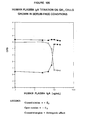
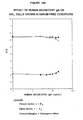
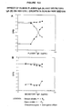
- the steroid hormone responsive culture cells are MTW9/PL2 (rat mammary cancer), T47D (human breast carcinoma), MCF-7 (human breast carcinoma), MCF-7A (human breast carcinoma), MCF-7K (human breast carcinoma), LNCaP (human prostatic carcinoma), ZR-75-1 (human prostatic carcinoma), H-301 (Syrian hamster kidney tumor), GH 1 or GH 3 (rat pituitary tumor), GH 4 C 1 (rat pituitary tumor), or HT-29 (human colonic cancer).
- an isolated estrogen receptor gamma is provided.
- the (ER ⁇ ) has an estradiol binding affmity greater than that of estrogen receptor alpha (ER ⁇ ) or estrogen receptor beta (ER ⁇ ), preferably having a K d for E 2 on the order of >10 -9 M.
- a mediator of estrogen responsive cell growth comprises ER ⁇ , and in certain embodiments a mediator of estrogen reversal of immunoglobulin inhibition of estrogen responsive cell growth comprises ER ⁇ .
- the method comprises (a) maintaining a predetermined population of estrogen responsive cancer cells in a steroid hormone-free nutrient medium comprising a quantity of immunoglobulin inhibitor sufficient to inhibit cancer cell growth in the absence of an inhibition-reversing amount of estrogen, the cells also being estrogen responsive for proliferation in vivo when implanted into a suitable host; (b) adding a defmed amount of the substance of interest to the cells and medium, to yield a test culture; (c) incubating the test culture for a predetermined period of time under cell growth promoting conditions; and (d) determining the cell population in the test culture after the predetermined period of time, a measurable increase in the cell population indicating an estrogen-like cell growth stimulating effect by the substance of interest.
- the method also includes testing the substance of interest for binding to estrogen receptor gamma and/or testing for cytotoxic effects.
- the method includes selecting estrogen responsive cancer cells containing estrogen receptor gamma
- the method comprises (a) maintaining a predetermined population of estrogen responsive cancer cells in a nutrient medium comprising a quantity of immunoglobulin inhibitor sufficient to inhibit cell growth in the absence of an inhibition-reversing amount of estrogen, the cells being capable of growing in vivo; (b) adding a defined amount of the substance of interest to the cells and medium; (c) adding a defined amount of an estrogen sufficient to stimulate cell growth in the presence of the inhibitor and in the absence of the substance of interest to the cells and medium, to yield a test culture; (d) incubating the test culture for a predetermined period of time under cell growth promoting conditions; (e) testing the substance of interest for cytotoxic effects on the cells; and (f) determining the cell population in the test culture after the predetermined period of time, a lack of measurable increase in the cell population not attributable to cytotoxic effects of the substance indicating a steroid hormone antagonistic effect by
- Also provided in accordance with the present invention are methods of identifying an estrogen responsive cell that is capable of being inhibited or prevented from proliferating by an estrogen reversible inhibitor of estrogen responsive cell growth.
- the method comprises detecting estrogen receptor gamma in the cell.
- the method comprises (a) maintaining a predetermined population of cancer cells in an above-described nutrient medium; (b) adding an effective amount of an iron compound to the medium, to provide an incubation mixture comprising unbound Fe (III), preferably at least about 1 ⁇ M Fe (III); (c) incubating the incubation mixture for a predetermined period of time under cell growth promoting conditions; and (d) determining the cell population in the incubation mixture after the predetermined period of time, an increase in cell population indicating lack of inhibition by the Fe (III), and the absence of an increase in cell population indicating inhibition of cell growth by the Fe (III).
- a method of killing cancer cells in vitro is provided.
- a concentration of at least about 10 ⁇ M unbound Fe (III) is maintained in the nutrient medium.
- extended arrest of cancer cell growth by an immunoglobulin inhibitor can also serve to kill steroid hormone responsive cancer cells in culture.
- a method of killing steroid hormone responsive cancer cells in culture comprises (a) combining a predetermined population of steroid hormone responsive cancer cells with a nutrient medium comprising an above-described cell culture medium and a quantity of steroid hormone irreversible immunoglobulin inhibitor sufficient to inhibit cell growth of steroid hormone responsive cancer cells, to provide an incubation mixture, the steroid hormone responsive cells also being steroid hormone responsive for proliferation in vivo when implanted into a suitable host; (b) incubating the incubation mixture for a predetermined period of time under cell growth promoting conditions; and (c) optionally, determining the cell population in the reaction mixture after the incubation for the predetermined period of time.
- the immunoglobulin inhibitor is irreversibly, or permanently inhibitory (i.e., the inhibitor is active with respect to the ability to inhibit steroid hormone-responsive cell proliferation and inactive with respect to steroid hormone reversibility of the inhibition.)
- Another embodiment of the present invention provides a method of killing a mixed population of steroid hormone responsive cancer cells and autonomous cancer cells.
- the method comprises contacting the mixed population of cells with an amount of an iron depleting substance sufficient to substantially deprive the autonomous cells of Fe (III), and then maintaining the cells in an iron depleted environment for a sufficient period of time for the autonomous cells to die.
- the method also includes contacting the mixed population of cells with an amount of a Fe (III) containing substance sufficient to inhibit cell growth and/or kill the steroid hormone responsive cells, and then maintaining the cells in a Fe (III)-enhanced environment for a predetermined period of time sufficient to inhibit cell growth and/or kill the steroid hormone responsive cancer cells.
- the method also includes contacting the mixed population of cells with an amount of immunoglobulin inhibitor sufficient to inhibit proliferation of the steroid hormone responsive cells.
- Still other embodiments provided by the present invention are methods of determining the concentration of a steroid hormone in a defmed amount of a body fluid.
- the method comprises assaying the body fluid for binding of steroid hormone to an immunoglobulin inhibitor of steroid hormone responsive cancer cell growth.
- Example begins with a short summary of that Example, which is intended merely to facilitate review and is not limiting on the disclosure contained in the full Example, and ends with a Discussion of some conclusions that may be drawn from that Example.
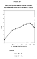
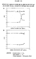
- Example 12 Differential Effects of Fe (III) on the Growth of Hormone Responsive and Autonomous Human Breast and Human Prostate Cancer Cells 105
- Example 13 Growth in Serum-free Defined Medium versus Growth in CDE-Serum ⁇ E2 108
- Example 14 Action of DES on Human AR + LNCaP Prostate Cancer Cells 111
- Example 15 Preparation of Inhibitor Depleted Serum for Control Studies and Stability Properties of the Inhibitor 112
- Example 16 Effects of Conventional Purification Methods and Properties of the Estrogen Reversible Serum-borne Inhibitor 114
- Example 17 Calcium Stabilization and Correlation with 3H-DHT Binding and Immunoprecipitation by Antibodies Raised to Human SHBG 120
- Example 18 Cortisol Affinity and Phenyl Sepharose Isolation of the "SHBG-like" Estrogen Reversible Inhibit
- Example 22 Effect of Tamoxifen Antiestrogen in Serum-free Defined Medium 144
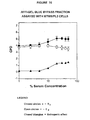
- Example 23 IgG1 and IgG2 as an Immunoglobulin Regulators of Estrogen and Androgen Responsive Cancer Cell Growth 145
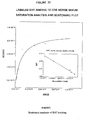
- Example 24 Mediation of IgA/IgM Effects by the Poly-Ig Receptor 147
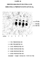
- Example 25 Mediation of IgG1 ⁇ Effects by a Fc-like Receptor 151
- Example 26 Example 26.
- Example 27 Conceptual Model for Cascading Loss of Cell Growth Inhibition in Cancer Cells. 157
- Example 28 IgA/IgM Based Test to Detect Lowered Levels of Steroid Hormone Reversible Cell Growth Inhibitors in Plasma or Body Secretions 161
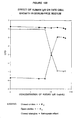
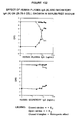
- Extracellular negative regulation is a key control mechanism of cell proliferation in steroid hormone responsive cancer cells.
- Sex steroid hormones both estrogens and androgens
- Moreno-Cuevas JE and Sirbasku DA (2000) In Vitro Cell Dev Biol 36, 410-427 ; Sirbasku DA and Moreno-Cuevas JE (2000) In Vitro Cell Dev Biol 36, 428-446 ; Moreno-Cuevas JE and Sirbasku DA (2000) In Vitro Cell Dev Biol 36, 447-464 , incorporated herein by reference).
- the prior art fails to adequately address the issues of (i) whether there are one or more of the serum-derived inhibitors, (ii) what is/are the exact chemical composition of the inhibitor(s), and (iii) what conditions were required to yield the long term stable product(s) necessary for the commercial application of the testing methodology described.
- Methods and compositions are presented herein that are useful for testing and assessment of compounds and mixtures for estrogenic or androgenic activity as well as others possessing antiestrogenic and antiandrogenic activities.
- cell culture methodology and compositions are described that permit testing at concentrations lower than was previously possible using existing methodologies.
- the new in vitro model assay systems obviate the need to conduct animal testing to predict in vivo responses.
- Some practical applications for the model include protecting the human population from unrecognized exposure to hormone-like compounds that present health hazards as well as developing new antihormone compounds to counterbalance these hazards.
- the testing of these compounds and mixtures are preferably conducted in serum-containing medium to mimic the conditions encountered by a blood borne agent. Testing can additionally be done under completely serum-free defmed medium conditions to determine direct actions on cells without serum or non-essential proteins present.
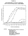
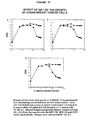
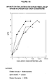
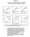
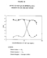
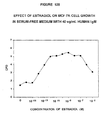
- the negative regulators of steroid hormone responsive cancer cell growth (estrogen reversible inhibitors) in serum are products from the secretory immune system, i.e., the immunologobulins A (IgA), M (IgM) and IgG1. These "immunoglobulin inhibitors" act as steroid hormone and thyroid hormone reversible inhibitors of mucosal cell growth.
- secretory immune system immunoglobulins as regulators of epithelial (mucosal) cell growth, and this discovery is unique in the cell growth regulation field.
- Application of certain of the compositions and methods is expected to relate to 80% of all human cancers because this high incidence rate arises from mucosal tissues. There is no previously reported evidence directly linking the secretory immunoglobulins with regulation of mucosal cell growth.
- breast and prostate cancers are very similar diseases. Aside from tissue specific epidemiological and social factors, breast and prostate cancers have remarkable parallels ( Grody WW et al. (1994) Am J Clin Pathol 102, S1-S67 ).
- the secretory immune system acts as a sex steroid hormone reversible inhibitor with target tumor cells from both of these cancers. Both are adenocarcinomas arising from sexually differentiate tissues.
- Both cancers are very common in North America and northern Europe compared to the rest of the world. Both are strongly influenced by steroid hormones. Both increase in incidence with age. Both are thought to have at least some genetic component. Finally, both have very similar patterns of development when examined histologically.
- the water used to prepare culture media and all other solutions was purified first by reverse osmosis followed by passage through a U. S. Filter Corporation system with a charcoal filter and two mixed bed ion exchangers.
- the effluent was distilled using a Bellco glass apparatus with quartz heating elements.
- the distilled water was stored in airflow restricted glass containers. No metal fittings are allowed in contact with the final purified water. This necessary precaution minimizes recontamination with metal ions.
- Standard phenol red containing Ham's F12-Dulbecco's modified Eagle's medium (D-MEM/F-12), phenol red-free standard D-MEM/F-12 and a custom-prepared "low-Fe" D-MEM/F-12 medium were supplied by Gibco-BRL (Catalog No. 11330-032) or Bio ⁇ Whittacker (Catalog No. 12-719, liquid).
- the "low-Fe" medium was standard phenol red containing D-MEM/F-12 from which the usual additions of ferric nitrate and ferrous sulfate had been omitted ( Eby JE et al. (1992) Anal Biochem 203, 317-325 ; Eby JE et al.
- This medium was a special formulation purchased from Gibco-BRL as a powder and prepared in the highly purified water before 0.2 ⁇ m pore filter membrane sterilization. A number of other stock solutions are required for cell culture in either serum containing or serum-free defined medium. Descriptions of each preparation are provided along with specific instructions for their use. The solutions used were designed to minimize the exogenous content of steroid hormone and to minimize the Fe (III) content of the water. Steps are taken for the exclusion of all extraneous sources of steroid hormones and Fe (III). Exclusion of Fe (III) is highly preferred, and in most of the totally serum-free applications, it is considered essential.
- Dialysis was done with Spectropor 1 membranes (Spectrum Medical Industries, molecular weight cut-off 6,000 to 8,000). The clotted material was removed by centrifugation. This preparation is termed plasma-derived serum. The serum or plasma was not heat pre-treated, or heat inactivated prior to use in the methods described below.
- STI General Cell Culture - Soybean Trypsin Inhibitor
- This trypsin preparation was used to harvest the cells for determining cell numbers. The cells are typically grown in 35-mm diameter dishes.
- This enzyme was purchased from ICN Biochemicals as the 1-300 porcine pancreatic trypsin preparation (Catalog No. 103140).
- a stock solution is typically prepared by adding the contents of a preweighed bottle of 1X Dulbecco's modified PBS medium without calcium or magnesium to 800 mL of water. This solution dissolves very gradually with adjustment to pH 7.3 using NaOH. After the solution was clear, 20 g of crude trypsin was added and this mixture stirred for 30 minutes at room temperature.
- the somewhat cloudy solution was diluted to 1000 mL with water and this volume was stored frozen in bulk overnight at -20°C to induce cold related precipitation that typically occurs when this preparation was frozen and thawed. After thawing at 37°C in a water bath, the preparation was filtered through 0.45 ⁇ m pore membranes. This preparation was stored at -20°C in useable portions.
- the EDTA used is the disodium and dihydrate salt (Sigma Catalog No. E1644).
- a 0.29 M solution is prepared by adding 107.9 g to 800 mL of water with stirring and adjustment to pH 7.2 with NaOH. The volume is brought to one liter with water and the solution stored at room temperature. Because this solution is used only at the end of the experiments, it does not require sterilization.
- Estrogen responsive T47D tumors in vivo Leung CKH and Shiu RPC (1981) Cancer Res 41, 546-55 1 ).
- 3 Estrogen responsive ZR-75-1 tumors in vivo Osborne CK et al. (1985) Cancer Res 45, 584-589 ).
- 4 Estrogen responsive GH 4 C 1 tumors in vivo Riss TL and Sirbasku DA (1989) In Vitro Cell Dev Biol 25, 136-142 ).
- 5 Estrogen responsive GH 3 tumors in vivo Sorrentino JM et al. (1976) J Natl Cancer Inst 56, 1149-1154 ).
- Estrogen responsive MTW9/PL2 tumors in vivo ( Sirbasku DA (1978) Cancer Res 38, 1154-1165 ; Danielpour D and Sirbasku DA (1984) In Vitro 20, 975-980 ).
- 7 Estrogen responsive H301 tumors in vivo ( Sirbasku DA and Kirkland WL (1976) Endocrinology 98, 1260-1272 ; Liehr JG et al. (1986) J Steroid Biochem 24, 353-356 ).
- 8 Androgen responsive LNCaP tumors in vivo Sato N et al.
- the medium was removed and the dishes washed with 10 mL of saline.
- the cells were dissociated by incubation at room temperature or at 37°C for 3 to 10 minutes with 1.5 mL of trypsin/EDTA.
- the action of the trypsin was stopped by addition of 8 mL of D-MEM/F-12 containing 10% (v/v) FBS or 8 mL of the horse serum/FBS combination.
- the cells were collected by centrifugation at 1000x g for 5 minutes and suspended in 10 mL of fresh serum containing medium. Aliquots were diluted into Isoton II (Coulter Diagnostics) and cell numbers determined with a Model ZBI or Z1 Coulter Particle Counter.
- the new dishes (100-mm diameter with 15 to 20 mL of fresh medium) were seeded with 2.0 x 10 5 to 1.0 x 10 6 cells on an alternating three-four day schedule or weekly as dictated by cell line growth rate. Cultures were used for growth assays between three and six days after passage. Acidic (yellow medium indicator color) cultures are not used for growth assays.
- Fe (III) content of this medium was ⁇ 0.2 ⁇ M ( Eby JE et al. (1992) Anal Biochem 203, 317-325 ). Fe (III) levels of ⁇ 1.0 ⁇ M interfere with thyroid hormone and estrogen responsive rat pituitary tumor cell growth in culture ( Eby JE et al. (1992) Anal Biochem 203, 317-325 ; Eby JE et al. (1993) J Cell Physiol 156, 588-600 ; Sato H et al. (1991) In Vitro Cell Dev Biol 27A, 599-602 ; Sato H et al (1992) Mol Cell Endocrinol 83, 239-251 ).
- Fe (III) might prevent estrogen responsiveness from being identified in culture with MTW9/PL2 cells, as shown herein and reported ( Sirbasku DA and Moreno-Cuevas JE (2000) In Vitro Cell Dev Biol 36, 428-446 ; Moreno-Cuevas JE and Sirbasku DA (2000) In Vitro Cell Dev Biol 36, 447-464 ), this is not the case when serum is present.
- Standard Fe (III)/Fe (II) containing D-MEM/F-12 was as effective as the low-Fe medium. It is clear that the apotransferrin in the serum effectively reduced the free Fe (III) in the medium to less than cytotoxic levels.
- apotransferrin binds Fe (III) with very high affinity at pH 7.4 in plasma.
- the total concentration of transferrin in serum is about 3 mg/mL. Usually, two-thirds of the total is apotransferrin. This amount is more than adequate to chelate Fe (III) in culture medium ( Eby JE et al. (1992) Anal Biochem 203, 317-325 ).
- a Fe (III) chelator e.g . apotransferrin or DFX
- a Fe (III) chelator e.g . apotransferrin or DFX
- the initial seed densities ranged from 5,000 to 12,000 cells per 35-mm diameter dish.
- each plate received 0.4 mL of crude pancreatic trypsin dissolved in phosphate buffered saline was added along with 0.3 mL of 0.29 M EDTA. After 4 to 40 minutes incubation at room temperature or at 37°C, the action of the trypsin was stopped by addition of 0.6 mL of horse serum. The cell clumps were dissociated further by one passage through a 201 ⁇ 2 or 23-gauge needle and syringe. This suspension was then diluted to 10 mL with Isoton II and cell numbers determined with a Coulter Counter. The results are presented as the average of triplicate dishes for each test medium. To determine day zero cell numbers, at least triplicate 1.0 mL aliquots of the inoculum were collected for counting during the seeding of the test dishes. Coulter Counter standardization and monitoring were performed by the manufacturer.
- CPD Cell population doublings
- the mitogenic response to sex steroid hormones is designated the "steroidogenic effect.”
- the "estrogenic effect” is calculated as the difference between CPD measured in the presence of an estrogen minus CPD in the absence of the steroid. These values equal cell number increases of 2 CPD .
- the term "androgenic effect” has the same meaning except that it describes growth caused by androgens such as DHT and T.
- CPD is used herein as a measure of growth because it is a direct calculation of the number of times a cell population undergoes cell division. Furthermore, CPD use permits a direct measure of ED 50 and ED 100 Concentrations in different and in replicate assays. The significance of differences between test dishes and controls was evaluated by the student's t test. Values of p ⁇ 0.05 were accepted as significant. Standard deviations ( ⁇ SD) are included when appropriate.
- the rat breast cancer cell line designated as MTW9/PL2 was deposited with the American Type Culture Collection, Rockville, Md. on , 2000.
- the hamster kidney tumor cell line designated as H301 was deposited with the American Type Culture Collection, Rockville, Md. on , 2000.
- the subject cultures have been deposited under conditions that assure that access to the cultures will be available during the pendency of this patent application to one determined by the Commissioner of Patents and Trademarks to be entitled thereto under 37 CFR 1.14 and 35 U.S.C. 122.
- the deposits will be available as required by foreign patent laws in countries wherein counterparts of the subject application, or its progeny, are filed. However, it should be understood that the availability of a deposit does not constitute a license to practice the subject invention in derogation of patent rights granted by governmental action.
- the subject culture deposits will be stored and made available to the public in accord with the provisions of the Budapest Treaty for the Deposit of Microorganisms, i.e., they will be stored with all the care necessary to keep them viable and uncontaminated for a period of at least five years after the most recent request for the furnishing of a sample of each deposit, and in any case, for a period of at least thirty (30) years after the date of deposit or for the enforceable life of any patent which may issue disclosing the cultures.
- the depositor acknowledges the duty to replace the deposits should the depositor be unable to furnish a sample when requested, due to the condition of the deposit. All restrictions on the availability to the public of the subject culture deposits will be irrevocably removed upon the granting of a patent disclosing it.
- the cells were washed with saline, resuspended in 20 mL of saline and treated with 2.0 mL of trypsin/EDTA at room temperature for one minute. The trypsin action was stopped with 6.0 mL of 0.5% (w/v) soybean trypsin inhibitor in saline. The dispersed cells were collected and suspended in phenol red-free D-MEM/F-12 to a density of 0.5 to 1.0 x 10 6 per mL.
- the rationale for measuring steroid hormone binding with whole cells rests with the intent to replicate cell culture conditions. To derive complete information, the use of whole cells avoids the possible loss of a new receptor that might not withstand the cell extraction process or otherwise not be recovered.
- the steroid hormone binding incubations were done in phenol red free D-MEM/F-12 in a total volume of 1160 ⁇ L containing 1000 ⁇ L, of cells, 100 ⁇ L of labeled steroid and 60 ⁇ L of unlabeled steroid hormone or medium.
- the incubations were done in glass tubes for two hours at 37°C with gentle agitation in an orbital shaker water bath followed by cooling to 0°C for 15 to 30 minutes.
- the cells were collected by centrifugation at 7°C as described above and washed three times with 2 mL portions of ice-cold phenol red-free D-MEM/F-12.
- Affinity Bioreagents a rabbit polyclonal antibody against a bacterial fusion protein containing the N-terminal domain of the human androgen receptor and a mouse monoclonal antibody against the amino acid sequence 533 through 547 of the DNA binding domain of the progesterone receptor.
- An affinity-purified rabbit polyclonal antibody corresponding to the amino acid sequence 580 through 599 of the mouse estrogen receptor was obtained from Santa Cruz Biotechnology.
- cytosolic supernatants 20 x 10 6 cells were washed with serum-free D-MEM/F-12 and resuspended in 5 mL of 0.01 M Tris-HCl, pH 7.4, containing 0.15 M NaCl and 1 mM EDTA (Tris/EDTA). After cooling to ice-bath temperature, the cells were disrupted by three treatments for ten seconds with a Tekmar Polytron homogenizer. The homogenates were centrifuged at 800x g for 10 minutes followed by centrifugation at 150,000x g for one hour to obtain the cytosolic supernatants.
- the pellets from the 800x g centrifugation were homogenized again three times with 1.5 mL Tris/EDTA as described above.
- the centrifugation supernatants from the three homogenizations were combined to give the nuclear extract from each cell line.
- Protein concentrations were determined using the BCA kit from Pierce Chemical ® kit with bovine serum albumin as standard. When required, the samples were concentrated by precipitation with 20% (w/v) trichloroacetic acid. The precipitates were washed once with 500 ⁇ L of 70% (v/v) ethanol and twice with 500 ⁇ L of water.
- the membranes were incubated at room temperature with a 1:100 dilution of the primary antiserum for one hour and a 1:10000 dilution of second antibody for one hour.
- the primary antiserum was used at a 1:5000 dilution with incubation at room temperature for one hour followed by one hour with a 1:10000 dilution of second antibody.
- the second antibody was an affinity purified anti-rabbit immunoglobulin conjugated to alkaline phosphatase.
- the incubations were done with 5 ⁇ g/mL primary antibody for 24 hours at 7°C, followed by incubation with a 1:1000 dilution of second antibody for eight hours at room temperature.
- the second antibody was an affinity purified anti-mouse immunoglobulin conjugated to alkaline phosphatase.
- the incubations contained either 3 H-E 2 [2,4,6,7- 3 H-17 ⁇ -estradiol] at specific activity 96 Ci/mmole or 3 H-E 2 plus a 100-fold molar excess of DES.
- Glucocorticoid specific binding was assayed with [1,2- 3 H (N)] hydrocortisone at specific activity 53 Ci/mmole and 3 H-hydrocortisone plus a 100-fold molar excess of unlabeled DEX.
- Progesterone specific binding was assayed with 3 H-progesterone [1,2- 3 H (n) progesterone] alone at specific activity 92 Ci/mmole or 3 H-progesterone plus a 100-fold molar excess of unlabeled synthetic progestin R5020.
- the use of 100-fold unlabeled steroids to determine nonspecific binding has been discussed ( Chamness GC and McGuire WL (1975) Steroids 26, 538-542 ).
- Each assay contained 0.01 M Tris-HCl, pH 7.4, with 10 mM CaCl 2 . The binding conditions were optimized for time and temperature.
- the incubations were done in glass tubes at 34 C in a total volume of 660 ⁇ L that included 50 ⁇ g/mL of the phenyl Sepharose pools and labeled and unlabeled steroid competitor. After two hours, the incubations were cooled to ice bath temperature and 50 to 200 ⁇ L aliquots applied to each DEAE-cellulose (DE-81) ion exchange filter (2.3-cm, Fisher) positioned in a Millipore Vacuum Filter Manifold ®. Thereafter, the filters were washed with ten one mL portions of ice-cold Tris/CaCl 2 buffer. With control incubations minus protein, ⁇ 3% of the label was retained.
- DEAE-cellulose DE-811
- ion exchange filter 2.3-cm, Fisher
- Protein Assay and Quantification Methods For cell growth assays, the protein sample volumes added to the culture medium were ⁇ 20%. As required, chromatography samples were concentrated using Amicon Ultrafiltration with YM-10 (molecular weight cut-off 10,000) low protein binding membranes and nitrogen gas pressure ( Sirbasku DA et al. (1991) Biochemistry 30, 295-304 ). Before assay, all fractions were dialyzed against 0.05 M Tris-HCl, pH 7.4, with 0.15 M NaCl using Spectropor 1 membranes (molecular weight cutoff 6,000 to 8,000). They were sterilized with 0.2 ⁇ m membrane filtration units.
- the protein concentrations of serum, the ammonium sulfate precipitation and all of the conventional chromatography fractions were estimated as one A 280nM equal to one mg/mL.
- concentrations were estimated either by the Pierce BCA® method according to the instructions supplied or the dye binding method of Bradford ( Bradford MM (1976) Anal Biochem 72, 248-254 ).
- Trichloroacetic acid (TCA) precipitation was used with BCA to eliminate the interfering cortisol and DHT.
- TCA deoxycholate was used to co-precipitate the protein and ethanol/water washes to eliminate the steroid.
- Human IgG was used as standard for the colorimetric protein determinations.
- Example 1 Identification of Steroid Hormone Receptors in MTW9/PL2 Cells.
- TABLE 3 presents only a fraction of the estrogen binding data available in the literature. However, the K d values presented are representative and do show a discernable pattern. The lowest K d from a literature search was in the range 5 x 10 -11 M to 1.0 x 10 -10 M for the ER ⁇ and 7 x 10 -11 M to 1.1 x 10 -10 M for the ER ⁇ . In general, the binding affinities as estimated by K d are lower for receptors from human cells than those from rodent lines. It is important to note that the results presented in TABLE 3 indicate that the lower limit of measuring estrogen receptor affinities most likely has been reached. The use of the highest specific activity tritium labeled steroids has been optimized and simply cannot be used to measure 10 to 100-fold lower K d concentrations. This opens the possibility of an as yet unrecognized ER that mediates growth effects at lower concentrations of estrogen than either the ER ⁇ or the ER ⁇ .
- Estrogen effects on Progesterone Receptor Expression in MTW9/PL2 Cells Estrogens induction of progesterone receptors in target cells is generally taken as strong evidence of sex steroid responsiveness by the criteria of regulation of gene expression ( Leavitt WW et al.
- the bands migrating at 79 kDa and 21 kDa may be degradation products, non-specific reactions between the antibody and unrelated proteins, or may represent other forms of the androgen receptor ( Prins GS et al. (1991) Endocrinology 129, 3187-3199 ).
- the T47D cells showed the same androgen receptors as LNCaP cells. In contrast, there was little androgen receptor in the nuclear or cytosolic extracts of MTW9/PL2 cells. The faint band identified was the same intensity as an equivalent component seen in the extracts of fibroblasts ( Fig. 8 , top panel).
- Fibroblasts have been reported to have low levels of androgen receptors ( Eil C et al. (1983) Clin Endocrinol 19, 223-230 ; Eil C et al. (1980) Steroids 35, 389-404 ).
- the forms in MTW9/PL2 cells also compared closely to the 85.6 kDa and 109.6 kDa masses reported for progesterone receptors of rat uterus ( Ilenchuk TT et al. (1987) Endocrinology 120, 1449-1456 ). Furthermore, the 79kDa component (A form) was the more abundant of the two receptors in MTW9/PL2 cells. This was also the case for rat uterus ( Ilenchuk TT et al. (1987) Endocrinology 120, 1449-1456 ). The T47D positive controls showed the same molecular masses of progesterone receptors although there was greater immunostaining with the cytosolic extracts than with the nuclear preparations.
- T47D progesterone receptor masses observed in the present study were similar to those reported by others studying human breast cancer cells ( Horwitz KB and Alexander PS (1983) Endocrinology 113, 2195-2201 ; Lessey BA et al. (1983) Endocrinology 112, 1267-1274 ; Horwitz KB et al. (1985) Recent Prog Hormone Res 41, 249-316 ).
- the LNCaP cells showed intense nuclear extract staining for the same forms of progesterone receptors seen in extracts of MTW9/PL2 and T47D cells. No progesterone receptors were identified in the negative control rat or human fibroblasts.
- the band identified at 50 kDa was thought to be a degradation product of the 67kDa intact form of ER ⁇ ( Moreno-Cuevas JE and Sirbasku DA (2000) In Vitro Cell Dev Biol 36, 410-427 ); however, it may be an ER ⁇ because that new receptor has a mass of 54 kDa ( Enmark E et al. (1997) J Clin Endocrinol Metab 82, 4258-4265 ).
- the 50 kDa band may also represent a variant form of ER ⁇ ( Gustafsson J- ⁇ and Warner M (2000) J Steroid Biochem Mol Biol 74, 245-248 ). Alternately, this band may represent a new estrogen receptor that regulates growth, hereby designated as estrogen receptor gamma (ER ⁇ ).
- prolactin mRNA production is activated by higher concentrations of estrogen.
- This same biphasic action of estrogen on the growth of T47D human breast cancers cells has also been noted ( Crudos D et al. (1982) J Clin Endocrinol Metab 55, 276-283 ). Low concentrations promoted growth, whereas higher levels were inhibitory. Indeed, a biphasic effect also was noted with the MCF-7 cell line ( Soto AM and Sonnenschein C (1985) J Steroid Biochem 23, 87-94 ). When this observation is coupled with the clear statements of Soto et al ( Soto AM et al.
- Variant estrogen receptors have been identified previously by others. For example from the estrogen growth responsive T47D human breast cancer cell line, there have been three isoforms of the ER ⁇ identified in one study ( Wang Y and Miksicek RJ (1991) Mol Endocrinol 5, 1707-1715 ) and another three in a different study ( Graham ML et al (1990) Cancer Res 50, 6208-6217 ). With another two estrogen growth responsive human breast cancer cell lines, the MCF-7 and ZR-75-1, another ER ⁇ variant was identified that lacked the entire exon 4 of the receptor ( Pfeffer U et al. (1993) Cancer Res 53, 741-743 ).
- Variant receptors have also been identified from human breast cancer biopsy specimens ( Murphy LC and Dotzlaw H (1989) Mol Endocrinol 3, 687-693 ).
- Another truncated variant of ER ⁇ acts as a natural inhibitor of the action of the wild-type ER ⁇ ( i.e . unchanged receptor) ( Fuqua SA et al. (1992) Cancer Res 52, 483-486 ).
- Another type of variant has received wide attention because it has constitutive transcriptional activity without the steroid hormone ligand bound ( Fuqua SA et al. (1991) Cancer Res 51, 105-109 ).
- Even normal human breast epithelial cells show several natural variants of ER ⁇ ( Yang J et al. (2000) Endocrine 12, 243-247 ).
- Example 1 The evidence presented verifies that the MTW9/PL2 cells are estrogen receptor positive (ER + ) with a significant number of progesterone receptors and possibly low levels of androgen receptors.
- the estrogen receptor content and affinity characteristics of the MTW9/PL2 cells indicate appropriate stability for use as a testing standard and for commercial applications.
- the long-term stability of this cell line in culture, without alteration of its cell properties, is further discussed in Example 3.
- the mixture was then washed with about 6-8 liters of distilled water in a sintered glass funnel (2000 mL, ASTM 40-60, C#36060). This wash removes impurities as well as fine particles of charcoal that cannot be separated from serum by centrifugation.
- the charcoal-dextran retentate was suspended in a final volume of 300 mL of distilled water to yield a suspension of 100 mg/mL charcoal and 10 mg/mL dextran.
- the mixture is stirred vigorously for about an hour, and then stored at room temperature for no more than about 2-3 weeks prior to use.
- the temperature of the mixture gradually warmed to about 40°C during centrifugation.
- the supernatants were pooled in a two-liter beaker and 275 mL portions of the supernatant (serum) transferred to fresh one-liter Erlenmeyer flasks. These were then incubated in the orbital shaker water bath at 34°C for 20-30 minutes (preferably 30 min) to re-equilibrate the temperature.
- a second extraction was done by addition of a fresh aliquot (about 14 mL) of the charcoal-dextran suspension. This re-extraction mixture was incubated with shaking for another 2 hours at 34°C. The final charcoal concentration during this extraction was about 5 mg/mL.
- the bulk of the charcoal was removed by centrifugation, as before. In some preparations the temperature of the mixture reached about 41°C, without harming the quality of the serum.
- the supernatants were collected into a two-liter beaker and filtered through 5 ⁇ m pore diameter filters to remove residual charcoal. If it was considered necessary for particular preparations (for example, due to charcoal darkening serum) the serum was also filtered with 0.45 ⁇ m Millipore filters. These filtrations were done with plastic reusable filter holders and light vacuum. The steroid hormone depleted serum was then sterilized using 0.2 ⁇ m pore diameter filters.
- the charcoal method described above is readily applicable to one to five liter volumes of serum per preparation. With use of ⁇ 50 mL per test substance, this is an adequate supply.
- the charcoal-dextran methods will preferably employ industrial filtration or other separation equipment to remove the carbon after each extraction.
- the XAD-4 resin method as presented is adaptable to one to five liters for testing purposes. For industrial applications, where ⁇ 20 to 100 liter batches are customarily required, the resin method is preferred because of the need for only one separation after extraction. However, where "foaming" of the serum protein is to be avoided completely, charcoal extraction is superior.
- charcoal-dextran has an advantage when economy is a major consideration. It is less expensive than XAD-4 resin on a per liter basis, although the resin is commercially available at low cost when purchased in large amounts ( i.e . ⁇ 50-100 kilograms).
- Example 2 Each of the methods presented have advantages, depending on the particular needs and desires of the user.
- the scale procedures described are useful to prepared sufficient serum for testing of hormone activities or antihormone activities or evaluation of toxicity of compounds in cell culture assays.
- This 34°C method has been used to prepare CDE human serum, porcine serum, rat serum, hamster serum, ovine serum, fetal bovine serum, new born bovine serum (0 to 10 days old), young donor bovine serum (10 days to 6 moths old) young adult bovine serum (300 to 900 lbs), fetal horse serum, chicken serum, turkey serum, dog serum, goat serum, rabbit serum and monkey serum. Subsequent Examples demonstrate how these stripped sera are preferably employed.
- CDE-horse serum refers to the 34°C charcoal-dextran extraction process described in above.
- the MTW9/PL2 cells were assayed for E 2 responsiveness in cultures supplemented with increasing concentrations of CDE-horse serum ( Fig. 9A ). Concentrations ⁇ 5% (v/v) promoted growth. Typically within seven days cell numbers increased from 6,000 per dish to more than 200,000 in 2 to 5% serum.
- the cells grew in clusters in suspension and had the same morphology reported earlier for the parent MTW9/PL line grown in medium containing 10% (v/v) fetal bovine serum ( Sirbasku DA (1978) Cancer Res 38, 1154-1165 ).
- concentration of CDE-horse serum was increased to 50% (v/v) without steroid, many fewer cells were present.
- the morphology of the cells was the same as in rapidly growing cultures. Estrogen addition to this same medium caused substantial growth.
- the morphology of the estrogen-stimulated cultures in 50% serum was equivalent to that seen with or without estrogen in 2.5% serum.
- the inhibitor had no effect on the microscopic morphology of MTW9/PL2 cultures nor did it affect cell-cell adhesion or cell-surface adhesion.
- CDE-horse Serum TABLE 4 shows the effect of storage temperature on the estrogen mediating activity of CDE-horse serum.
- the assays were done with MTW9/PL2 cells as shown in Figs. 9A and 9B . Stability was assessed by four criteria: (i) the concentration of serum needed to give an estrogenic effect of 2.5 CPD ( i.e. ED 2.5 ), (ii) the percent serum needed for the maximum estrogenic effect, and the magnitude of the estrogenic effects (CPD) at (iii) 20% and (iv) 30% serum.
- CDE-horse serum was stable at 23°C for three weeks without loss of activity as assessed by all four criteria.
- E 2 Induced CPDs (% serum, v/v, for the maximum) CPD at 20% (v/v) serum CPD at 30% (v/v) serum Storage at 23°C 1 2.1 4.9 (10%) 5.0 3.2 3 5.2 5.4 (20%) 6.2 5.2 6 5.0 4.2 (10%) 3.5 0.9 14 2.9 6.0 (10%) 4.3 2.6 23 4.0 6.3 (10%) 3.9 2.5 Storage at 4°C 1 1.8 5.9 (10%) 4.9 4.0 7 6.8 5.7 (20%) 6.4 5.4 15 3.8 4.1 (30%) 5.5 4.2 24 5.3 5.3 (10%) 1.0 2.8 44 3.0 4.8 (5%) 0.04 0.26 55 2.2 5.0 (5%) 0.00 0.24 90 >50 2.1 (5%) 0.30 0.40 Storage at -17°C 1 2.6 5.2 (10%) 5.0 3.1 7 4.0 5.8 (30%) 6.8 5.8 44 3.3 5.8 (20%) 6.0 5.4 90 6.1 5.2 (30%) 6.2 5.9
- the cell numbers in dishes containing 1.0 x 10 -13 M E 2 were 32-fold (p ⁇ 0.01) higher than in dishes without the hormone.
- Concentrations of 1.0 x 10 -12 to 1.0 x 10 -11 M E 2 promoted 6.73 CPD that was a 110-fold estrogenic effect in seven days.
- the ED 50 of E 2 was about 0.5 to 1.0 x 10 -12 M.
- E 1 and E 3 optimum growth was achieved at 1.0 x 10 -9 and 1.0 x 10 -8 M, respectively.
- the mitogenic potency of the synthetic estrogen DES was assessed. At 1.0 x 10 -8 M, it caused the same growth as saturating concentrations of the natural estrogens.
- the DES effect was 6.98 CPD in seven days that was a 126-fold (2 6.98 ) increase in cell number.
- the next most potent hormone was DHT. It caused significant (p ⁇ 0.05) growth at super physiologic concentrations ⁇ 1.0 x 10 -8 M.
- Progesterone also caused significant growth, but only at supraphysiological concentrations ⁇ 1.0 x 10 -7 M. Cortisol was ineffective at concentrations up to 1.0 x 10 -5 M.
- Estrogen Mitogenic Effects with MTW9/PL2 cells in CDE-serum from Several Species. Serum from species other than horse were examined to determine they also possessed estrogen reversible inhibitory activity with rat MTW9/PL2 cells. These experiments are shown in Fig. 12 . All of the sera tested were charcoal dextran extracted at 34°C. CDE-porcine ( Fig. 12A ), and CDE-human serum ( Fig. 12B ) showed patterns nearly identical to that of CDE-horse serum. The maximum estrogenic effects with both sera were six to seven CPD (N 3). CDE-rat serum also showed the same pattern of estrogen reversible growth inhibition ( Fig. 12C ).
- CDE-bovine serum adult donor herd
- the estrogenic effects reached three to four CPD in 50% (v/v) CDE-serum. Even with this variability, fetal bovine serum has less activity than the serum from the adults of this species.
- the MTW9/PL2 cells showed the same hormone responsiveness expected of rat and human breast epithelial cells, as shown herein and subsequently reported ( Moreno-Cuevas JE and Sirbasku DA (2000) In Vitro Cell Dev Biol 36, 410-427 ; Sirbasku DA and Moreno-Cuevas JE (2000) In Vitro Cell Dev Biol 36, 428-446 ; Moreno-Cuevas JE and Sirbasku DA (2000) In Vitro Cell Dev Biol 36, 447-464 ).
- the effects of the steroid hormones in culture were the same as described for the growth of the original MT-W9A tumor in W/Fu rats ( MacLeod et al. (1964) Endocrinology 75, 249-25 8 ) and tumor formation by the parental MTW9/PL cell line in this same strain of rats ( Sirbasku DA (1978) Cancer Res 38, 1154-1165 ).
- the present embodiment is the first established cell line derived from a carcinogen induced rat mammary tumor that continues to show large magnitude growth responses to estrogens, progesterone and androgens even after extended periods in culture, preferably when cultured under the conditions disclosed herein.
- Thyroid hormone responsiveness has also been demonstrated for MTW9/PL cells ( Leland FE et al. (1981) In: Functionally Differentiated Cell Lines, Sato G, ed, Alan Liss, New York, pp 1-46 ).
- prolactin is not mitogenic for the parental MTW9/PL cells under serum-free defmed conditions ( Danielpour D et al. (1988) In Vitro Cell Dev Biol 24, 42-52 ).
- prolactin also reverses the effects of the serum-borne inhibitor or otherwise acts as a cytokine to influence the production of immunoglobulins in breast and other mucosal tissues.
- the development of this cell line now permits not only sensitive steroid hormone growth analysis in culture, but also direct comparisons to the effectiveness of the same test substances in animals. No other such rat mammary system is currently available.
- the MTW9/PL2 line was also significantly estrogen responsive by this criterion.
- the notion that long-term culture necessarily leads to loss of functional estrogen receptors is laid to rest.
- a major advantage of the MTW9/PL2 line is its long-term stability permitting series analyses over long periods of time without concern for cell property changes.
- Steroid Hormone Conjugates are Non-problematic . While charcoal treatment can be expected to remove the major classes of steroid hormones from serum, there is a question about its effect on the more soluble and potentially active conjugates. It has been reported that hydrolysis of estrogen sulfates provided free estrogens in human breast cancer cell cultures ( Vignon F et al. (1980) Endocrinology 106, 1079-1086 ). This abrogated the effects of exogenous E 2 .
- estradiol sulfate Although the previous investigations did not address estradiol sulfate, it was shown that more than 95% of estrone sulfate and estradiol glucuronide were removed from serum by a single 56°C charcoal extraction ( Sirbasku DA and Kirkland WL (1976) Endocrinology 98, 1260-1272 ). Additionally, in previous studies MTW9/PL cells were incubated with tritium labeled estradiol glucuronide for up to 24 hours under cell culture conditions and found no organic solvent extractable free steroid. Both past and current results indicate that the impact of the estrogen conjugates has been overestimated. In the present study, no precautions were taken to remove the conjugated forms of estrogens from any of the sera tested.
- Plastic Product Use for Cell Culture The present studies were done with plastic ware made of polystyrene. Plastic is manufactured using alkylphenols ( Platt AE (1978) In: Encyclopedia of Chemical Technology, Kirk RE, Othmer DF, eds, 3rd Edition, Volume 26, Wiley, New York, pp 801-847 ). One of these compounds, p-nonyl-phenol, has been reported to be estrogenic for MCF-7 cells in culture ( Soto AM et al. (1991) Environ Health Perspect 92, 167-173 ). This xenobiotic most likely is present in the cultures used in these studies. No precautions were taken to exclude compounds leaching from plastic.
- bioassay procedures herein are done with polystyrene plastic ware and culture dishes almost exclusively. If there had been a significant contamination of the medium by such compounds, the estrogenic effects reported in this study should not have been seen or should have been markedly attenuated.
- An advantage of the assay systems described herein is that they have no need for expensive and or exotic substitutes for the common plastic ware used in cell culture laboratories to conduct bioassays. Also, the CDE-serum can be stored and shipped for commercial use in plastic containers without concern for creation of plastic-induced artifacts.
- estrogen responsiveness can be demonstrated in rat tumor cells, where no such responsiveness had previously been demonstrated. Further, because estrogen responsiveness and binding affinity can now be compared, data indicating the existence of an heretofore unknown estrogen receptor have been generated.
- the ZR-75-1 cells showed the same CDE-serum caused estrogen reversible inhibition as seen with rodent cell lines in this serum.
- Fig. 14 there was a 3.65 CPD (i.e. 12.5-fold) estrogenic effect ( Fig. 14 ).
- the MCF-7A was the least estrogen responsive even in 50% CDE-horse ( Fig. 15 ).
- the estrogenic effect was 2.65 CPD in 10-12 days. This was still significant (p ⁇ 0.01) as a 2 2.65 or 6.3-fold increase in cell number caused by estrogen.
- the present serum-derived inhibitor exhibited biological activity exactly opposite the estrogen reversible inhibitors described by M Tanji et al. ( Tanji M et al. (2000) Anticancer Res. 20, 2779-2783 ; Tanji M et al. (2000) Anticancer Res. 20, 2785-2789 ).
- MCF-7K, T47D, LNCaP, and H301cells are highly sex steroid hormone responsive in CDE-horse serum.
- T47D growth was monitored daily in D-MEM/F-12 supplemented with 10% (v/v) fetal bovine serum ( Fig. 16A ). This concentration of fetal bovine serum shows no inhibitor ( Moreno-Cuevas JE and Sirbasku DA (2000) In Vitro Cell Dev Biol 36, 410-427 ). Cell growth in medium with fetal bovine serum showed at most a one or two day lag period.
- CDE-human Serum Effect of CDE-human Serum on Estrogen Responsive Cell Growth .
- human serum was a source of inhibitor for steroid hormone sensitive cell lines from different species and tissues.
- the results confirm that CDE-human serum contains approximately the same level of inhibitor as CDE-horse serum. Results are shown with T47D human breast cancer cells ( Fig. 17A ), LNCaP human prostatic carcinoma cells ( Fig. 17B ), MTW9/PL2 rat mammary tumor cells ( Fig. 17C ), two GH rat pituitary tumor cell lines ( Figs. 17D and 17E ), and the Syrian hamster H301 kidney tumor cells ( Fig. 17F ). All lines showed the same biphasic response to CDE-human serum.
- Tissue concentrations are generally conceded to be lower due to SHBG that reduces the "free” or “active” form of sex steroid hormones ( Rosner W (1990) Endocr Rev 11, 80-91 ).
- the next studies with T47D cells determined the effective concentration ranges for the three most common estrogens and compared these to non-estrogen steroid hormones.
- Fig. 18 shows an analysis with T47D cells in D-MEM/F-12 containing 50% (v/v) CDE horse serum for 14 days. Estrogens were the only physiologically relevant activators of T47D growth. As expected from previous studies with breast cancer cells ( Lippman ME et al. (1977) Cancer Res 37, 1901-1907 ; Jozan S et al.
- E 2 caused significant (p ⁇ 0.05) growth when present at 1.0 x 10 -14 M and optimum growth at 1.0 x 10 -10 M. Higher concentrations were not inhibitory.
- the ED 50 concentration of E 2 wash 1.0 x 10 -13 M. It is noteworthy that even E 3 was remarkably potent. Others also had commented that E 3 was more potent than expected ( Lippman ME et al.
- the GH 4 C 1 strain was selected as an example for this next study because of its marked E 2 responsiveness in culture ( Amara JF and Dannies PS (1983) Endocrinology 112, 1141-1143 ; Sirbasku DA and Moreno-Cuevas JE (2000) In Vitro Cell Dev Biol 36, 428-446 ; Sato H et al. (1991) In Vitro Cell Dev Biol 27A, 599-602 ) and estrogen requirement for tumor formation in rats ( Riss TL and Sirbasku DA (1989) In Vitro Cell Dev Biol 25, 136-142 ).
- Fig. 19 shows the results of these experiments. All three major estrogens promoted growth. The potencies of E 2 and E 1 were equivalent whereas E 3 was substantially less effective. Even at supraphysiologic concentrations, E 3 did not promote the saturation densities seen with E 2 and E 1 . The lowest concentration of E 2 and E 1 that gave significant (p ⁇ 0.05) growth was 1.0 x 10 -12 M. The ED 50 of E 2 was ⁇ 1.0 x 10 -11 M. Optimum growth required supraphysiological concentrations ( i.e .
- E 3 was markedly less effective than E 2 or E 1 .
- E 2 and E 1 required 1.0 x 10 -11 M and 1.0 x 10 -10 M, respectively, to achieve significant (p ⁇ 0.05) growth.
- the ED 50 concentration of E 2 is about 5 to 9 x 10 -11 M.
- this concentration was higher than for the rat pituitary tumor cells ( Fig. 19 ) or rat mammary tumor cells ( Fig. 11 ). In fact, they were as much as 100 to 1000-fold higher than for human breast cancer cells ( Fig. 18 ). In other tests shown in Fig.
- progesterone, cortisol, T and DHT were all inactive.
- TABLE 5 summarizes the ED 50 concentrations required for a one-half maximum growth and the corresponding lowest K d measured for the same or closely related cell lines: TABLE 5 Comparisons of ED 50 and K d as Evidence Supporting a New ER Designated ER ⁇ Cell Line ED 50 for E 2 Induced Growth K d for E 2 Fold-higher K d Concentration Compared to ED 50 for Growth MTW9/PL2 1 x 10 -12 M 1.8 x 10 -9 M 1.8 x 10 3 T47D 1 x 10 -12 M 0.11 x 10 -9 M 1.1 x 10 3 GH 4 C 1 1x10 -11 M 0.25 x 10 -9 M 25 H301 9x10 -11 M 0.87 x 10 -9 M 10 Clearly, to seek the new ER ⁇ , the rat mammary or human breast cells will be the best sources based on the differences between the ED 50 growth concentrations and the K d values for ER ⁇ or ER ⁇ . Because the ER ⁇ was first obtained from ER
- ER ⁇ for diagnosing and/or screening for breast cancer. Measurement of the ER ⁇ specifically will provide a more accurate determination of estrogen receptor status and therefore permit more precise modeling of the therapy for each patient. ER ⁇ will be identified by immunohistochemical methods, labeled ligand binding with very high specific activity isotopes, and by PCR and other molecular biology analyzes. Other methods will also be applied. Similar analyses are expected to be applicable to other estrogen receptor related or estrogen receptor containing mucosal cancers including ovarian, uterine, vaginal, cervical, colon, lung, stomach, pituitary, liver, pancreas, skin and kidney, as described in co-owned, concurrently filed U.S. Patent App. No.
- Figs. 11 , and 18 through 21 demonstrate the usefulness of the extracted sera, assays and cell lines with regard to assessment of estrogenic activity or androgenic activity in industrial, commercial, environmental, medicinal, or other medical samples where activity measurement is required at concentrations below the usual levels detectable by radioimmunoassay.
- the sensitivity of this bioassay is unique.
- Benign prostatic hypertrophy is among the most common afflictions of older men. About 50% of 60-year old men have BPH. At 85 years about 90% of men have BPH ( Berry SJ et al. (1984) J Urol 132, 474-479 ). There is a general view that estrogens may be important in BPH ( Henderson D et al. (1987) Steroids 50, 219-229 ; Nakhla AM et al. (1994) Proc Natl Acad Sci USA 91, 5402-5405 ). The paradox involved is that as men age androgen levels fall and SHBG rises. These work in concert to further limit available androgen ( Davidson JM et al.
- Example 4 demonstrates the utility of assays using 34°C CDE serum for demonstrating estrogen responsive cell growth in a variety of tissues.
- This Example demonstrates that not only steroid hormone but also thyroid hormone growth effects can be demonstrated in cell growth assays using the present 34°C CDE serum.
- GH rat pituitary tumor cells are highly thyroid hormone responsive in serum-free defined medium ( Eby JE et al. (1992) Anal Biochem 203, 317-325 ; Eby JE et al. (1992) J Cell Physiol 156, 588-600 ; Sato H et al. (1991) In Vitro Cell Dev Biol 27A, 599-602 ).
- An example of this responsiveness with the GH 3 line is shown in Fig. 22 .
- these cells are not E 2 responsive when T 3 is omitted from the medium ( Fig. 23 ).
- thyroid hormone receptor is a member of a superfamily of receptors that also includes the steroid hormone receptors ( Evans RM (1988) Science (Wash DC) 240:889-895 ). Testing of substances expected to have thyroid hormone like activity can be performed with the GH cell lines in the presence of low concentrations of CDE-serum.
- Example 5 The removal of thyroid hormones from serum has been described before using the Bio-Rad TM AG-1 X8 ion exchange resin ( Samuels HH et al. (1979) Endocrinology 105, 80-85 ). Removal of T 3 /T 4 by the AG-1 X8 method relies on their negative carboxylic acid charge at neutral pH. However, ion exchange does not remove the uncharged/hydrophobic steroid hormones. This Example demonstrates that the 34°C CDE method described herein is more effective than the AG-1 X8 method previously known.
- Example 6 makes clear some major differences between the serum-borne inhibitor presently disclosed and those previously described. Specifically, exposure to heat can inactivate or alter the effect of inhibitors.
- U.S. Patent Nos. 4,859,585 (Sonnenschein ) and 5,135,849 (Soto ) describe an inhibitor that was derived from heat inactivated ( i.e . 56°C treated) serum and thereafter depleted of its endogenous estrogens and androgens by a 37.5°C single step charcoal-dextran procedure.
- the facts of that method are also stated in a publication ( Soto AM and Sonnenschein C (1984) Biochem Biophys Res Commun 122, 1097-1103 ).
- Soto AM and Sonnenschein C (1984) Biochem Biophys Res Commun 122, 1097-1103 In light of the results presented in Fig. 26 , it is likely that the inhibitor described by Sonnenschein and Soto is a different molecular entity, as further illustrated in Examples which follow.
- Fig. 27 shows the effects of XAD-4 treated horse serum ⁇ 10 nM E 2 with the MTW9/PL2 cell line. Unmistakably, the pattern of cell response was the same as seen with CDE-horse serum prepared as described in Example 2.
- A.. At 50% XAD-4 serum (v/v), an estrogenic effect of 5.2 CPD was observed in 7 days.
- Fig. 28 shows a similar experiment with T47D cells after 14 days.
- Example 7 There is no previous report of the preparation of steroid depleted serum by this resin treatment method. As indicated in Example 2, the XAD-4TM treatment method has particular applicability for the industrial preparation of large volumes of steroid hormone depleted serum, and will allow the commercial supply of steroid depleted serum at reasonable cost. A preferred application for this steroid hormone stripped serum is in the biotechnology industry, in which cell culture is used to produce medically and otherwise commercially significant proteins and cellular products. Steroid hormone depleted serum has applicability beyond the ER + and AR + cells described in this report. For example, hybridoma cells are the sources of many important monoclonal antibodies.
- Phenol Red as an Estrogen . It is widely believed that the phenol red indicator in tissue culture medium acts is a weak estrogen ( Berthois Y et al. (1986) Proc Natl Acad Sci USA 83,2496-2500 ). In the first report describing the phenol red problem, the indicator itself was thought to act as an estrogen ( Berthois Y et al. (1986) Proc Natl Acad Sci USA 83, 2496-2500 ). At the concentration in standard culture media (e.g. D-MEM/F-12 is 8.1 mg/mL or 22.9 ⁇ M), it was believed to stimulate ER + cell growth nearly as well as natural estrogens.
- standard culture media e.g. D-MEM/F-12 is 8.1 mg/mL or 22.9 ⁇ M
- Phenol Red Indicator is a "Red Herring" . Phenol red was further evaluated. Head-on comparisons of the growth of nine different ER + cell lines representing four target tissues and three species in medium with and without phenol red were performed ( Moreno-Cuevas JE and Sirbasku DA (2000) In Vitro Cell Dev Biol 36, 447-464 ). These studies were designed to specifically test various aspects of published reports that phenol red is estrogenic. Considering the results of these head-on comparisons, new conclusions have been reached about the effects of phenol red in culture, especially as they are relevant to experimental conditions available today to most investigators. Even more important, the test assays show the methods that can be used to determine if any commercial preparation or other source material possesses estrogenic activity.
- Fig 29A growth was measured in the presence of increasing concentrations of CDE-horse serum with and without phenol red in the medium and ⁇ E 2 .
- Concentrations of ⁇ 10% (v/v) CDE-horse serum supported more than 5 CPD. Higher concentrations progressively inhibited in both indicator containing and indicator free medium. In both types of medium, E 2 was required to reverse the serum inhibition.
- the estrogenic effects shown in Fig. 29A were compared in both types of medium and at each serum concentration. The results of this analysis are presented in Fig. 29 B.
- the maximum estrogenic effect at 50% (v/v) serum was 2.38 CPD (i.e.
- the MCF-7K strain was routinely more estrogen responsive than the MCF-7A line ( Sirbasku DA and Moreno-Cuevas JE (2000) In Vitro Cell Dev Biol 36, 428-446 ).
- the MCF-7K cells also showed a serum concentration dependent growth inhibition ( Fig. 29C ).
- the final degree of inhibition at 50% (v/v) serum was independent of phenol red. Only in the presence of 2.5, 5, 10 and 20% (v/v) CDE-horse serum were the estrogenic effects significantly greater in phenol red free ( Fig. 29D ).
- Phenol Red and T47D Cells were grown in medium with CDE-horse serum both with and without phenol red ( Fig. 30A ). Low concentrations of serum (i.e. ⁇ 2%) promoted growth. Higher concentrations progressively inhibited growth irrespective of indicator content. In both media, E 2 was required to reverse the inhibition ( Fig. 30A ). In 50% (v/v) CDE-horse serum, the maximum E 2 responses were 2 5.35 (41-fold) and 2 5.29 (39-fold) in phenol red containing and indicator free medium, respectively ( Fig. 30B ). Only at low serum concentrations were phenol red effects observed in any experiment. In some replicates, the phenol red effect was opposite to that expected.
- ZR-75-1 cells showed similar results as the T47D line. Serum caused an inhibition of growth that was undoubtedly unrelated to phenol red ( Fig. 30C ). In both types of medium, and at every serum concentration tested, E 2 was required to reverse the inhibition ( Fig. 30C ) . In 50% (v/v) serum, ZR-75-1 cells showed maximum estrogenic effects of 2 3.39 (10.5-fold) and 2 3.49 (11.2-fold) in medium with and without indicator, respectively ( Fig. 30D ). As seen with T47D cells, the ZR-75-1 line showed greater estrogenic effects in medium with phenol red than in medium without indicator when the serum was 0.5, 5 or 10% (v/v) ( Fig . 30D ).
- the MTW9/PL2 cells were chosen as an example of a highly estrogen responsive rodent origin line ( Moreno-Cuevas JE and Sirbasku DA (2000) In Vitro Cell Dev Biol 36, 410-427 ; Sirbasku DA and Moreno-Cuevas JE (2000) In Vitro Cell Dev Biol 36, 428-446 ).
- the assays were done in phenol red free D-MEM/F-12 supplemented with 30% CDE-HS. This concentration was chosen even though it is not as inhibitory as 50% (v/v) serum. This selection was made to reduce possible interactions of the phenol red/contaminant with serum proteins while still retaining a significant inhibitory effect. Phenol red concentrations of up to 16 mg/L were added to this medium.
- E 2 induced a four-fold increase in progesterone receptors in four days.
- E 2 induced a time and concentration dependent increase in the progesterone receptors with T47D cells.
- Phenol red induced a small increase in progesterone receptors at 8 and 16 mg/L after four days. This induction was about the same as caused by 1.0 x 10 -14 M E 2 ( Fig. 33A ).
- Example 8 The studies of the effects of phenol red or its lipophilic impurities demonstrate the usefulness of the presently disclosed methods for the assessment of estrogenic and androgenic activity of commercially prepared materials, substances present or extracted from environmental or food sources or other sources that are thought to contain such activities. The testing can be approached by three separate methods as shown by examples with phenol red.
- TGF ⁇ , TGF ⁇ 1, EGF, IGF-I, IGF-II and insulin were tested for inhibitor-like acitivity, using the cell growth assay described in the General Materials and Methods section,and in the foregoing Examples, substituting those proteins for the serum-borne inhibitor contained in the preferred CDE serum.
- TGF ⁇ 1 as a Substitute for the Serum-borne Estrogen Reversible Inhibitor.
- Normal mouse mammary ( Silberstein GB and Daniel CW (1987) Science (Wash DC) 237, 291-293 ; Silberstein GB et al. (1992) Dev Biol 152, 354-362 ) and normal human breast epithelial cell growth is inhibited by TGF ⁇ ( Bronzert DA et al. (1990) Mol Endocrinol 4, 981-989 ).
- human breast cancer cells are inhibited by TGF ⁇ ( Knabbe C et al. (1987) Cell 48, 417-428 ; Arteaga CL et al.
- TGF ⁇ also inhibits the GH 4 C 1 rat pituitary tumor cells ( Ramsdell JS (1991) Endocrinology 128, 1981-1990 ) and the LNCaP human prostatic carcinoma cells ( Schuurmans AL et al. (1988) The Prostate 12, 55-64 ; Wilding G et al.
- TGF ⁇ 1 was chosen for the instant study. Without a doubt, a number of the key cell lines used throughout the Examples were inhibited by TGF ⁇ . It was therefore considered essential to ask if TGF ⁇ was the estrogen reversible inhibitor.
- TGF ⁇ 1 and MCF-7 Cells are probably the most studied human breast cancer line today, this next work began with those cells.
- TGF ⁇ has been described as a hormone regulated autocrine inhibitor of the ER + MCF-7 human breast cancer cell growth ( Knabbe C et al. (1987) Cell 48, 417-428 ).
- TGF ⁇ 1 substituted for the serum-borne inhibitor with these cells they were grown in D-MEM/F-12 containing 2.5 % (v/v) CDE-horse serum plus increasing concentrations of transforming growth factor and ⁇ E 2 .
- TGF ⁇ 1 and MTW9/PL2 Cells are the only known estrogen growth responsive rat cell line derived from a hormone responsive carcinogen induced tumor.
- TGF ⁇ 1 reduced cell numbers from 350,000 to 100,000 per dish. This was significant (p ⁇ 0.05). However, the presentation of cell number results only tends to exaggerate the effects of TGF ⁇ 1.
- the results were converted to CPD ( Fig. 34B , insert), the actual inhibition was 1.5 CPD. This was at most a 25% decrease in growth rate. As shown, there was no estrogen reversal of the TGF ⁇ 1 inhibition with MTW9/PL2 cells.
- TGF ⁇ 1 and other ER + Cell Lines The effects of TGF ⁇ 1 at 50 ng/mL ⁇ E 2 were also investigated with the other cell lines used in this study.
- the MCF-7A, T47D and ZR-75-1 human breast cancer cells were inhibited by TGF ⁇ 1 ( Fig. 35A ). From these results, and those in Fig. 34A , it was clear that the MCF-7 cells were the most sensitive of the ER + human breast cancer lines tested. Irrespective of the line, E 2 had no influence on the TGF ⁇ 1 mediated inhibitions ( Fig. 35A ).
- the same experiments were done with the LNCaP cells and the GH 4 C 1 pituitary line ( Fig. 35A ).
- TGF ⁇ 1 did not substitute for the estrogen reversible inhibitor(s) in CDE serum with any of the sex steroid sensitive ER + cell lines tested.
- TGF ⁇ and EGF as Substitutes for the Estrogen Reversible Inhibitor in CDE Serum.
- the EGF family of mitogens and receptors has been linked to breast cancer proliferation, invasion and progression ( Dickson RB and Lippman ME (1987) Endocr Rev 8, 29-43 ; Normanno N et al. (1994) Breast Cancer Res Treat 29, 11-27 ; Ether SP (1995) J Natl Cancer Inst 87, 964-973 ; de Jung JS et al. (1998) J Pathol 184, 44-52 and 53-57 ).
- TGF ⁇ EGF analogue
- Estrogen induced secretion of TGF ⁇ is thought to create an autocrine loop that promotes breast cancer cell growth ( Dickson RB et al. (1985) Endocrinology 118, 138-142 ; Dickson RB et al. (1986) Cancer Res 46, 1707-1713 ; Dickson RB et al.
- TGF ⁇ is also thought to potentiate estrogen action in uterus ( Nelson KG et al. (1992) Endocrinology 131, 1657-1664 ) as well as to regulate the EGF receptor in this tissue ( DiAugustine RP et al. (1988) Endocrinology 122, 2355-2363 ; Huet-Hudson YM et al. (1990) Mol Endocrinol 4, 510-523 ; Mukku VR and Stancel GM (1985) J Biol Chem 260, 9820-9824 ).
- the culture conditions described herein offer a new opportunity to test the autocrine growth model under conditions not previously available. Application of the new cell growth assays allowed a direct test to determine if an autocrine/intacrine growth factor loop explains the estrogen reversal of the serum inhibition.
- EGF and TGF ⁇ as Substitutes for E 2 .
- Growth of the MCF-7A, MCF-7K, T47D and ZR-75-1 cells was measured in D-MEM/F-12 containing increasing concentrations of CDE horse serum with and without exogenous EGF or TGF ⁇ .
- the results with the four cell lines are shown in Figs. 36 A, 36B, 36C, and 36D, respectively.
- CDE horse serum was progressively inhibitory at concentrations > 5% (v/v).
- the addition of growth saturating concentrations ( Karey KP and Sirbasku DA (1988) Cancer Res 48. 4083-4092 ) of EGF or TGF ⁇ did not reverse the effects of the serum-borne inhibitor.
- IGF-I, IGF-II and Insulin as Substitutes for Estrogen Action.
- Insulin-like growth factors I and II (IGF-I and IGF-II) promote breast cancer cell growth ( Furlanetto RW and DiCarlo JN (1984) Cancer Res 44, 2122-2128 ; Myal Y et al. (1984) Cancer Res 44, 5486-5490 ; Dickson RB and Lippman ME (1987) Endocr Rev 8, 29-43 ; Karey KP and Sirbasku DA (1988) Cancer Res 48, 4083-4092 ; Ogasawara M and Sirbasku DA (1988) In Vitro Cell Dev Biol 24, 911-920 ; Stewart AJ et al.
- IGF-I related proteins Huff KK et al. (1986) Cancer Res 46, 4613-4619 ; Huff KK et al. (1988) Mol Endocrinol 2, 200-208 ; Dickson RB and Lippman ME (1987) Endocr Rev 8, 29-43 ; Minute F et al. (1987) Mol Cell Endocrinol 54, 17-184 , as well IGF-II ( Yee D et al. (1988) Cancer Res 48, 6691-6696 ; Osborne CK et al. (1989) Mol Endocrinol 3, 1701-1709 ), are thought of as possible autocrine/paracrine mitogens.
- IGF-I is active with breast cancer cells even in the presence of serum ( Furlanetto RW and DiCarlo JN (1984) Cancer Res 44, 2122-2128 ; Myal Y et al. (1984) Cancer Res 44, 5486-5490 ; Osborne CK et al.
- Estromedins were proposed to be secreted by target tissues such as uterus, kidney and pituitary, and to act in concert to simultaneously promote the growth of all ER + target tissues ( Sirbasku DA (1978) Proc Natl Acad Sci USA 75, 3786-3790 ; Sirbasku DA (1981) Banbury Report 8, 425-443 ; Ikeda T et al. (1982) In Vitro 18, 961-979 ).
- the estromedin hypothesis arose from the observation that reproducible in vitro direct estrogen mitogenic effects were not identifiable ( Sirbasku DA (1978) Proc Natl Acad Sci USA 75, 3786-3790 ; Sirbasku DA (1981) Banbury Report 8, 425-443 ; Ikeda T et al.
- the estrocolyone model ( Soto AM and Sonnenschein C (1987) Endocr Rev 8, 44-52 ) is an indirect negative mechanism based on regulation of sex steroid hormone dependent cells via a serum-borne inhibitor. The inhibitor blocks growth promoted by non-steroidal mitogens such as growth factors and diferric transferrin.
- Sonnenschein and Soto first proposed that estrocolyone acted at the cell surface via specific receptors. The effects of sex steroid hormones were to bind estrocolyone and prevent it from associating with the cells. Only low physiologic concentrations of sex steroid hormones were needed for this function.
- the Positive Direct Model - Steroid Hormone Receptor Mediation The one mechanism most widely accepted regarding steroid hormones and growth involves the nuclear located DNA binding ER ⁇ receptor ( Gorski J and Hansen JC (1987) Steroids 49, 461-475 ). Growth is thought to be mediation by specific cytosolic and/or nuclear located receptors that ultimately alter DNA transcription to regulate gene activity. Results from many laboratories support this mechanism ( Jensen EV and Jacobson HI (1962) Recent Prog Horm Res 18, 387-414 ; Gorski J et al. (1968) Recent Prog Horm Res 24, 45-80 ; Jensen EV et al.
- Serum Proteins with Estrocolyone Steroid Binding Characteristics If the estrocolyone mechanism is in fact correct, one must be able to identify at least one serum protein with very high affinity binding (i.e. K d picomolar) for sex steroids. There is, however, a major unresolved problem with that hypothesis. Other than sex hormone binding globulin (SHBG), additional high affinity estrogen binding in CDE human serum has not been found. SHBG has K d of 1.7 x 10 -9 M for E 2 at 37°C ( Rosner W and Smith RN (1975) Biochemistry 14, 4813-4820 ). This affinity does not qualify as the high binding expected of estrocolyone.
- SHBG sex hormone binding globulin
- a New Model of Steroid Hormone Responsive Cell Growth A new model best fits the available data. It brings together aspects of both the direct positive mechanism and indirect negative control. According to this model, regulation of steroid hormone target tumor cell growth is a balance between positive and negative control signals. This balance dictates either growth (i.e. cell division) or quiescence (i.e. cell metabolism and tissue specific function but without cell division).
- the positive mediators are the steroid hormones acting mediated by a high sensitivity intracellular DNA binding sex steroid receptor that ultimately activates gene expression via intracellular located receptors; whereas negative regulation is exerted by a serum-borne inhibitor that acts at the cell surface.
- the results disclosed herein support the view that growth is controlled directly by both negative and positive mediators.
- this model of negative and positive response control mechanisms is further described and the mediators are shown to be the secretory immunoglobulins acting on cell surface (membrane) receptors.
- TGF ⁇ and Relevant Inhibition The results presented further define the molecular properties of the serum-borne inhibitor by eliminating EGF ⁇ 1 as a candidate. This is an important issue because of the well-known effects of TGF ⁇ on normal breast epithelial cells ( Hosobuchi M and Stampfer MR (1989) In Vitro Cell Dev Biol 25, 705-713 ) and ER - estrogen insensitive breast cancer cells ( Arteaga CL et al. (1988) Cancer Res 48, 3898-3904 ). The results herein continue to confirm a previously unrecognized entity that serves as the estrogen reversible inhibitor in serum. Inhibitors that lack estrogen reversibility can be eliminated from consideration.
- Example 9 Discussion of Example 9. From this series of experiments, it can be readily appreciated that any other natural or synthetic protein or other substance can be similarly tested for cancer cell growth inhibiting activity akin to the serum-derived inhibitor in the CDE horse serum. Also, the same XADTM-4 and CDE extraction protocols may also be applied to body fluids and secretions other than serum, and the extracted fluids may be assayed as described for inhibitor activity. Such fluids or secretions include plasma, urine, seminal fluid, milk, colostrum, mucus and stool. An XADTM-4 column is especially suited for preparing a steroid hormone depleted specimen from a small sample of body fluid.
- Example 10 Serum-free Defined Culture Medium Formulations.
- serum-free defined medium was used to identify IgA, IgM and IgGs as estrogen and/or androgen reversible inhibitors of target tumor cell growth in culture, as demonstrated in subsequent Examples.
- IgA, IgM and IgGs were used to identify estrogen and/or androgen reversible inhibitors of target tumor cell growth in culture, as demonstrated in subsequent Examples.
- Another issue had to be resolved.
- the growth of hormone responsive cancer cell types in serum-free medium based on standard preparations of D-MEM/F-12 was not as vigorous as expected.
- Apotransferrin is a M r 80,000 bilobular serum protein that binds one Fe (III) in each lobe, albeit with slightly different affinities ( Aisen P and Liebman A (1972) Biochim Biophys Acta 257, 314-323 ; Chasteen ND (1983) Trends Biochem Sci 8, 272-275 ; Evans RW and Williams J (1978) Biochem J 173, 543-552 ).
- Fe (III) saturated the protein is called diferric transferrin. This form of transferrin is the major iron delivery system for the body ( Young SP and Aisen P (1981) Hepatology 1, 114-119 ; Ciechanover A et al. (1983) J Biol Chem 258, 9681-9689 ).
- Deferoxamine mesylate is a U.S. FDA approved drug used to treat iron overload and iron toxicity in humans. It is marketed by Novartis Pharmaceuticals, East Hanover, NJ, under the trade name DESFERAL®. Deferoxamine mesylate is sold to researchers by Sigma Chemicals (St. Louis, MO).
- the use of serum-free defined medium to grow diverse cell types in culture gained national and international recognition with the publication by Hayashi and Sato ( Hayashi I and Sato GH (1976) Nature (Lond) 259, 132-134 ). They demonstrated a breakthrough.
- the serum supplement commonly used in cell culture medium could be replaceable entirely by mixtures of nutrients and hormones in serum-free medium.
- This observation was expanded to include cell types from many mammalian tissues ( Barnes D and Sato G (1980) Anal Biochem 102, 255-270 ; Barnes D and Sato G (1980) Cell 22, 649-655 ; Bottenstein J et al.
- Serum-free Defined Culture - Nutrient Additions A number of nutrient additions to D-MEM/F-12 are needed to grow the cells used in the presently described studies.
- the formulations of serum-free defined medium employed are specific optimizations, modifications, or necessary changes of earlier media that have been described ( Riss TL and Sirbasku DA (1987) Cancer Res 47, 3776-3782 ; Danielpour D et al. (1988) In Vitro Cell Dev Biol 24, 42-52 ; Ogasawara M and Sirbasku DA (1988) In Vitro Cell Dev Biol 24, 911-920 ; Karey KP and Sirbasku DA (1988) Cancer Res 48, 4083-4092 ; Riss TL et al.
- Bovine serum albumin (Sigma Catalog No. A3912) was made by "initial fractionation by heat shock and Fraction V", minimum purity 98% (electrophoresis), according to the supplier.
- a 50mg/mL stock solution of BSA was prepared in normal saline and was sterilized using 0.2 ⁇ m pore membrane filters. Aliquots are stored at -20°C in plastic tubes.
- the "heat shock” step that was used in most albumin preparation methods inactivates the estrogen reversible inhibitor disclosed herein.
- ETN Serum-free Defined Medium Nutrient Supplements - Ethanolamine
- ETN was purchased from Sigma (Catalog No. A5629) (FW 61). This liquid has a density of 1.0117 grams/mL. Using 0.610 mL in 100 mL of water, a 100 mM stock solution was prepared which was sterilized using the 0.2 um pore membrane filters. The ETN was stored at -20°C in polystyrene tubes. This nutrient is required to sustain phospholipid metabolism required for all membrane biosynthesis.
- PETvN Serum-free Defined Medium Nutrient Supplements - Phosphoethanolamine
- This solid material was purchased as o-phosphoryl-ethanolamine (FW 141) (Sigma Catalog No. P0503).
- a 10 mM stock of PETN was prepared by dissolving 141 mg in 100 mL of water and sterilizing with 0.2 ⁇ m pore membrane filters. Aliquots were stored at -20°C in polystyrene tubes. This component is an adjunct to ETN.
- Serum-free Defined Medium Nutrient Supplements - Glutamine (GLUT). This essential amino acid was purchased from Sigma (Catalog No. G5763). It is "cell culture tested” according to the manufacturer. Addition of glutamine (FW 146.1) to the culture media is necessary because of its relatively short half-life (i.e. about 80% is lost in 20 days at 35°C). See the Sigma product information for the decay curves at different temperatures and pH. Purchased D-MEM/F-12 stored in the refrigerator for about three weeks lost most of the original glutamine present. For serum-free applications, additional supplementation is required to sustain growth. For a preparation, 11.7 g was dissolved in 400 mL of water to give 200 mM glutamine.
- Serum-free Defined Medium Nutrient Supplements - Selenium (Se).
- a powder of sodium selenite (100 mg/vial) is obtained from Collaborative Research or Sigma (Catalog No. S5261). It has been sterilized by irradiation. The contents of a single vial are dissolved in 100 mL of sterile water to give final stock of 1.0 mg/mL. This preparation should not be filter sterilized because Se binds to filters. The final volume was diluted to 100 mL with sterile saline. Aliquots are stored at - 20°C in polystyrene tubes.
- Selenium is an important cofactor for enzyme systems that protect the cells from oxidation effects.
- EGF Epidermal Growth Factor
- EGF Epidermal Growth Factor
- EGF EGF prepared from mouse submaxillary gland (tissue culture grade) was purchased from Collaborative Research (Catalog No. 40001) as 100 ⁇ g in a sterile vial or from Sigma (Catalog No. E4127). The original vials are stored at 4°C according to the manufacturer's instructions. To prepare a stock solution, 5.0 mL of sterile saline was added to a vial to yield a 20 ⁇ g/mL EGF solution. Aliquots are stored frozen at -20°C polystyrene tubes. Repeated freeze-thaw must be avoided. This growth factor is useful because of its very broad cell specificity range.
- Acidic FGF is purchased from Sigma (Catalog No. F5542). It is the human recombinant product from E. coli. This product has very specific handling requirements. It is provided sterilized in 25 ⁇ g vials lyophilized from PBS containing 1.25 mg of BSA. The contents of each vial are reconstituted in 25 mL of sterile PBS containing 1.0 mg/mL of BSA and 10 ⁇ g/mL of heparin. Filtration of this product at this concentration must absolutely be avoided. This solution is stored at -20°C in polystyrene tubes.
- Keratinocyte growth factor can substitute for aFGF.
- the fibroblast growth factor family is very important in growth of urogenitial tissues including prostate.
- Heparin is used to stabilize FGF in cell culture ( Gospodarowitz D and Cheng J (1986) J Cell Physiol 128, 475-484 ). Heparin is obtained from Sigma (Catalog No. H3149) as the sodium salt, Grade 1-A, from porcine intestinal mucosa. A solution of 1.0 mg/mL is made in saline and sterilized with 0.2 ⁇ m pore membrane filters. An aliquot of 250 ⁇ L is added to the 25 mL of aFGF reconstitution solution used above. Sterile heparin is stored at 4°C.
- Serum-free Defined Medium Adhesion Protein Supplement - Fibronectin Fbn
- Human plasma derived Fbn can be purchased from many commercial sources. Bovine Fbn is also available and is effective. Fbn is prepared from units of fresh human plasma (unfrozen) or fresh bovine (unfrozen) plasma by two methods ( Retta SF et al. (1999) Methods in Molecular Biology 96, 119-124 ; Smith RL and Griffin CA (1985) Thrombosis Res 37, 91-101 ). Purity is evaluated by SDS-PAGE with Coomassie Brilliant Blue staining or silver staining (Pierce Chemicals ® kits). Adhesion activity is confirmed with cells in serum-free defined medium. Vitronectin can substitute for fibronectin.
- DFX Serum-free Defined Medium Iron (Fe (III) Chelator Supplements - Deferoxamine mesylate (DFX).
- DFX (FW 656.8) is purchased from Sigma (Catalog No. D9533). The stock solution is made at 10 mM by adding 131 mg to 20 mL of highly purified water as described above. The solution is sterilized by filtration with 0.2 ⁇ M pore membranes. Aliquots are stored at -20°C in polystyrene tubes.
- Human serum ApoTf can be purchased from Sigma (Catalog No. T4382). It is minimum 98% iron-free.
- ApoTf is also prepared as described previously ( Sirbasku DA et al. (1991) Biochemistry 30, 295-304 ; Sirbasku DA et al. (1991) Biochemistry 30, 7466-7477 ). ApoTf is prepared by dialysis against citrate buffer pH 5.0- 5.5 with 1 ⁇ g/mL DFX present to chelate > 98% of the iron. Handling and storage were as described for diferric transferrin but with great care to avoid contact with iron sources.
- the final stock solution of 10mg/mL of insulin is filter sterilized using 0.2 ⁇ m pore diameter membranes. Aliquots are stored at 4°C in polystyrene tubes. Care was taken not to freeze-thaw the aliquots of stock solution. Insulin is a very broad range cell growth-stimulating factor as well as a regulator of specific metabolic processes.
- Serum-free Defined Medium Nutrient Supplements - Thyroid Hormones The preferred thyroid hormone is T 3 (3', 5-Triiodothyronine, FW 673, purchased from Sigma as Catalog No. T2752). It is stored desiccated at -20 °C. To prepare stocks, 0.5 N NaOH was made by addition of 20 grams of pellets to one liter of water. Then, 67.3 mg of T 3 was added. After dissolving the T 3 with stirring for a few minutes, 25 mL of this stock was diluted up to 250 mL with water, for a final concentration of 0.05 N NaOH. This dilution was sterilized using the 0.2 ⁇ m pore diameter filter.
- T 3 5-Triiodothyronine
- T 4 The second thyroid hormone, thyroxin (T 4 , sodium salt, pentahydrate FW 888.9), is prepared by the same procedure. For this stock solution, 88.9 mg of T 4 are used. T 4 is purchased from Sigma (Catalog No. T2501). T 4 is used at 10 to 20 times higher concentrations than T 3 . Care is taken not to freeze-thaw these preparations. Thyroid hormones have a very broad range of biological effects on metabolism and growth. Many cells in culture require these for growth.
- compositions of Serum-free Defined Media TABLE 7 presents the formulations of the preferred serum-free defined media developed for use in detecting high-level steroid hormone reversible inhibition by steroid hormone-stripped serum fractions and by purified inhibitors in serum-free cell growth assays. As indicated in the footnotes to the table, when a particular component is included in one of the formulations, the concentration that provides a suitable cell growth medium can fall within the indicated range.
- 1 INS range 100 ng/mL to 10 ⁇ g/mL 2
- EGF range 1 ng/mL to 50 ng/mL 3
- aFGF range 0.2 ng/mL to 20 ng/mL 4
- T 3 range 0.3 nM to 10
- nM 5 2FeTf range 2 ⁇ g/mL to 50 ⁇ g/mL 6
- ETN range 5 ⁇ M to 100 ⁇ M 7 PET range 5 ⁇ M to 50 ⁇ M 8
- BSA range 0.2 mg/mL to 5.0 mg/mL 9
- Lin-Alb range 50 ⁇ g/mL to 500 ⁇ g/mL 10 Se range 5 ng/mL to 20 ng/mL 11
- GSH range 1 ⁇ g/mL to 50 ⁇ g/mL 12
- Glut range 0.5 mM to 2.0 mM 13
- Heparin range 1 ⁇ g/mL to 10 ⁇ g/mL 14
- DFX range 2 ⁇ M
- Standard phenol red-containing Gibco-BRL D-MEM/F-12 is a preferred basal medium to which the defined media components are added. It contains 0.6 mM to 1.0 M CaCl 2 .
- D-MEM/F-12 can be purchased from Gibco-BRL in the liquid form or can be prepared from the powder formulation using only highly purified water. Alternatively, another suitable basal medium could be used as long as it provides at least the required minimum amounts of necessary nutrients, vitamins and minerals to maintain cell viability of the desired cell line.
- the calcium concentration range preferred is 0.6 to 10 mM. Calcium stabilizes the inhibitor in cell culture without impairing cell growth.
- the human breast cancer cell medium, DDM-2MF was a modification of the original DDM-2 medium ( Danielpour D et al. (1988) In Vitro Cell Dev Biol 24, 42-52 ) and MOM-1 ( Ogasawara M and Sirbasku DA (1988) In Vitro Cell Dev Biol 24, 911-920 ) and contained modified hormone concentrations, deferoxamine (DFX) and fibronectin.
- Aqueous salt solutions such as tissue culture medium contain hydrolytic polymeric forms of Fe (III) ( Spiro TG et al. (1966) J Am Chem Soc 88, 2721-2726 ).
- DFX binds this form of Fe (III) with very high affinity ( Schubert J (1964) In; Iron Metabolism: The Chemical Basis of Chelation, Springer, Berlin, pp 466-498 ). If not removed, Fe (III) inhibits hormone-responsive growth in serum-free defmed medium ( Sirbasku DA et al. (1991) Mol Cell Endocrinol 77, C47-C55 ; Sato H et al. (1992) Mol Cell Endocrinol 83, 239-251 ; Eby JE et al. (1993) J Cell Physiol 156, 588-600 ; Eby JE et al. (1992) Anal Biochem 203, 317-325 ).
- Fibronectin was used with DDM-2MF to promote cell attachment.
- the 35-mm diameter assay dishes were pre-coated by incubation with the designated amount of fibronectin (TABLE 7) for 16 to 48 hours at 37°C in 2.0 mL of D-MEM/F-12.
- CAPM human prostatic cancer cell medium was developed to support the growth of tumor cells from this tissue. The composition of CAPM is described in TABLE 7.
- CAPM also supports the growth of the H301 Syrian hamster kidney tumor cells.
- DDM-2A which is a modified form of DDM-2 ( Danielpour D et al. (1988) In Vitro Cell Dev Biol 24, 42-52 ), was preferred for growing MTW9/PL2 cells.
- PCM-9 defined medium was developed for growing the rat pituitary cell lines. This medium differs from previous PCM formulations ( Sirbasku DA et al. (1991) Mol Cell Endocrinol 77, C47-C55 ; Sato H et al. (1992) Mol Cell Endocrinol 83, 239-251 ; Eby JE et al. (1993) J Cell Physiol 156, 588-600 ; EbyJE et al. (1992) Anal Biochem 203, 317-325 ) in that DFX was substituted for apotransferrin and the triiodothyronine concentration was increased to 1.0 nM.
- DFX and apotransferrin (2 to 50 ⁇ g/mL) are the preferred chelators based on their very high specificity and affinities for Fe (III), EDTA at 1 to 10 ⁇ M or sodium citrate at 10 to 1000 ⁇ M also effectively neutralize the cytotoxic effects of Fe (III) ( Eby JE et al. (1993) J Cell Physiol 156, 588-600 ). Ascorbic acid (vitamin C) also chelates Fe (III), but is used less often because it is unstable in cell culture medium at 37°C in an oxygen environment in the presence of salts and metals in the medium.
- apo-ovotransferrin and apo-lactoferrin also were effective Fe (III) chelators in serum-free defined medium ( Eby JE et al. (1993) J Cell Physiol 156, 588-600 ).
- EGF, aFGF and insulin are the preferred growth factors, several other human recombinant proteins are effective. They have either been purchased or obtained as gifts from Gibco-BRL, Sigma or IMCERA Bioproducts.
- IGF-I and IGF-II Insulin-like growth factors I and II
- TGF ⁇ transforming growth factor ⁇
- bFGF basic fibroblast growth factor
- Insulin can be used to replaced IGF-I and IGF-II. All of these protein growth factors are dissolved under sterile conditions according to manufacturers' instructions and stored as indicated.
- Example 10 The preferred serum-free media described above provide an ideal scenario for the study of growth responses of hormone responsive cancers without the myriad of potential interactions accompanying the presence of serum with its 5000+ proteins and other compounds.
- the formulations presented permit dissection of growth into its individual parts caused by different stimulators.
- a combination of a few factors can be investigated to achieve an understanding of growth promoter/inhibitor interactions (i.e . cross-talk). This is exceptionally difficult to achieve in the presence of full serum.
- the serum-free medium described herein provided a tool for the assessment of growth inhibitor(s) isolated from CDE-horse serum, whose actions are reversed by sex-steroid hormones, as mentioned at the beginning of this Example and described in more detail in subsequent Examples.
- the preferred serum-free media of the present invention raise hope for the provision of new insight that could help to clarify the mechanisms involved in the control of breast, prostatic and other mucosal cancers under conditions not previously available.
- the new media compositions fill that need.
- the new serum-free media can be used not only for assays but also for large scale testing purposes and industrial uses such as cell culture production of a desirable protein.
- an antigen for vaccine production, or a monoclonal antibody can be prepared without fear of contamination by a serum-derived infectious agent. They are also useful for producing a quantity of virus for vaccine manufacture or for producing recombinant viruses for gene therapy. Basic cell culture methods for producing quantities of proteins or viruses are well known in the art and have been described in the literature.
- Example 11 Serum-free Defined Medium that Supports Hormone Sensitive and Autonomous Cancer Cell Growth
- the medium When purchased without these salts, the medium was designated “low-Fe” D-MEM/F-12.
- the iron concentrations in standard and “low-Fe” D-MEM/F-12 were 1.0 ⁇ M and 0.15 ⁇ M, respectively ( Eby JE et al (1992) Anal Biochem 203, 317-325 ).
- iron is present as a contaminant in the chemicals used to make the formulation, the 2.2 g/L NaHCO 3 added as a metabolic requirement and buffer, and the 15 mM HEPES buffer necessary for stabilizing the pH under serum-free conditions ( Eby JE et al (1992) Anal Biochem 203, 317-325 ).
- the cells to be used in the assays were washed three times in either "low-Fe” medium or "standard” medium depending upon the experimental protocol. These washes were done with the same care as described above in General Materials and Methods . Each dish received 1.0 mL of cells in the appropriate medium. At this point, the components final concentrations were (lX) as summarized in TABLE 7. Also, TABLE 7 describes medium containing deferoxamine as the Fe (III) chelator. Although less preferred, due in part to cost considerations, specificity, and affinity for Fe (III), as noted above, apotransferrin is also effective, especially at the preferred apotransferrin concentration of 50 ⁇ g/mL.
- Fig. 42 shows the same analysis with DU145 and PC3 cells in CAPM and in D-MEM/F-12 with 10% fetal bovine serum. As the cell number data show, growth was logarithmic. After 12 days, growth in the serum-free medium was identical to that in 10% fetal bovine serum for both cell lines. Growth of PC3 in 10% serum reached 6.98 ( ⁇ 0.71) CPD or a 112-fold increase in cell number versus 6.97 ( ⁇ 0.44) CPD or the same fold increase for cell numbers in serum-free medium.
- deletion of deferoxamine had only a small (i.e . ⁇ 20%) effect on growth of the DU145 and PC3 cells.
- the DU145 and PC3 cell lines also were T 3 , insulin, EGF, fibronectin and deferoxamine independent.
- ⁇ DHT had no significant effect on DU145 or PC3.
- LNCaP growth was significantly (p ⁇ 0.01) reduced or arrested completely by deletion of fibronectin, T3, diferric - transferrin or deferoxamine.
- LNCaP growth also was inhibited by deletion of EGF or insulin, but these effects were pronounced only in the absence of DHT.
- Example 11 Discussion of Example 11 .
- the media described in TABLE 7 were optimized for the specific cell types designated. Additionally, they were optimized to permit direct comparison of the growth properties of ER + and AR + steroid hormone sensitive tumor cell lines to their ER - and AR - steroid hormone insensitive (also called autonomous) counterparts. This careful optimization was done originally to study rat mammary tumor cells of both types in DDM-2A defined media. The appropriate cell lines for this approach have been developed from the MTW9/PL2 population and described ( Danielpour D and Sirbasku DA (1984) In Vitro 20, 975-980 ). The medium DDM-2MF has been developed for the same purpose only for comparisons of ER + and ER - forms of these cancers.
- CAPM has been applied to the ER + estrogen growth stimulated H301 Syrian hamster kidney cells ( Sirbasku DA and Moreno JE (2000) In Vitro Cell Dev Biol 36, 428-446 ) and its autonomous derivative cell line A195. As has been reviewed ( Evans RM (1988) Science (Wash DC) 240, 889-895 ), steroid hormones and thyroid hormones belong to the same superfamily of receptors. Both are important in growth. Therefore, it was expected that some tissues might be thyroid hormone positive regulated, while others might be positive regulated by steroid hormones. CAPM has also been applied to the study of thyroid hormone reversal of purified inhibitors with the human colonic carcinoma cell line HT-29.
- CAPM replaces a different defined medium prepared for MDCK cells ( Taub M et al. (1979) Proc Natl Acad Sci USA 76, 3338-3342 ). It is likely that the prostaglandins in that earlier medium interfere with the action of the thyroid hormones. In any case, that medium was not useful for demonstration of thyroid hormone reversal of purified MDCK cell growth inhibitors. All of these observations support the view that a series of uniquely optimized media have been formulated to defme the growth requirements of epithelial cells from several of the very prominent cancers of humans. Furthermore, the technology developed promises application to the optimization of growth of other types of epithelial cells from a variety of target tissues.
- This Example demonstrates that iron has an inhibiting effect on steroid responsive cell growth, independent of the above-described immunoglobulin effects, and which is distinguishable from its effect on autonomous cells.
- Fig. 49 shows the relative effects of addition of these three chelators to T47D serum-free defmed medium cultures. The order of effectiveness was as expected from the affinities and specificities of these chelators. Clearly, addition of Fe (III) chelators restored growth.
- Fig. 50 shows a similar study with LNCaP cells in CAPM defined medium from which the deferoxamine also was removed and 1.5 ⁇ M Fe (III) added. It was clear that chelation of the Fe (III) restored growth. It should be noted that this conclusion is reasonable based on the fact that deferoxamine has near absolute specificity for Fe (III). Concentrations as low as 0.5 ⁇ M of deferoxamine were sufficient to induce 3.5 CPD with LNCaP cells.
- Example 12 Discussion of Example 12. Together with the studies presented above, it appears that AR + cells are sensitive to the serum-borne inhibitor, sensitive to the positive effects of steroid hormone and sensitive to Fe (III) inhibition. In contrast, the DU145 and PC3 cells are insensitive to the serum-borne inhibitor, insensitive to the positive effects of androgen, and insensitive to Fe (III).
- the results presented in this example continue to demonstrate the requirement for the action of a serum-borne mediator to demonstrate steroid hormone responsive cell growth in culture.
- the use of CDE-serum was essential for the demonstration of androgen and other steroid hormone responsiveness in culture, but its use limits the understanding of stimulatory or inhibitory roles of hormones or factors on prostate and other cancer cells because of the inclusion of an undetermined amount of undefined components.
- a serum-free medium circumvents this problem, as shown in subsequent Examples.
- autonomy may be the loss of the receptor for the serum factor and/or the loss of the intracellular steroid hormone receptor. If this hypothesis is correct it should be possible to identify cells that possess steroid receptors but still have lost "sensitivity" to the hormone by virtue of the lack of the effect of the inhibitor. Most notably, this is the case with DU145 and ALVA-41 cells. As defmed by immunohistochemistry, the DU145 cells are definitely AR + ( Brolin J et al. (1992) The Prostate 20, 281-295 ). As defined by a number of criteria, the ALVA-41cells are AR + ( Nakhla AM and Rosner W (1994) Steroids 59, 586-589 ). A new concept explaining the progression of normal tissue cells to hormone autonomous cancers is discussed in more detail in an Example below.
- iron in the Fe (III) form and compounds containing it will be effective anti-prostate cancer treatments, and that direct injection (or painting) of localized prostate tumors or metastasis at other sites (e.g. bone) might effectively kill these cancers without concomitant systemic effects.
- This therapy potentially could replace such protocols as systemic chemotherapy (physically damaging), radiotherapy (damage to collateral tissues) or the use of locally acting radioactive gold chips that are complex to handle in the surgical environment and must be implanted and removed surgically.
- iron therapies can be repeated frequently by application via transrectal or transurethral access, using conventional techniques. This approach is unique and has not been discussed or suggested anywhere else in the literature. Such iron treatments may be a useful therapy for benign prostatic hypertrophy (BPH).
- BPH benign prostatic hypertrophy
- Example 10 The defined media described in Example 10 were used to verify the presence of a serum-borne inhibitor.
- the growth of six different ER + cell lines was compared in serum-free defined media (TABLE 7) to the effects seen in cultures supplemented with CDE-horse serum. These studies are shown in Figs. 56 and 57 . Estrogenic effects are recorded for each set of conditions with each cell line.
- Fig. 56A shows MCF-7K cell growth in serum-free DDM-2MF ⁇ 10 nM E 2 .
- the population replicated logarithmically for 12 days.
- E 2 had no effect on growth rate or saturation density.
- FIG. 56C shows the growth of T47D cells in serum-free defmed DDM-2MF ⁇ 10 nM E 2 .
- a small effect of estrogen was observed on growth rate, the most significant effect was an increase in stationary densities by 0.5 to 1.0 CPD.
- the effect of E 2 was much greater in medium containing CDE horse serum ( Fig. 56D ).
- Fig. 56D At 50% (v/v) CDE-serum, growth was completely inhibited.
- the estrogenic effect under these conditions was > 5 CPD. This was more than a 2 5 or 32-fold hormone effect on cell number.
- ZR-75-1 cells showed no effect of E 2 in serum-free defined DDM-2MF. This line grows more slowly than MCF-7 or T47D cells in defined medium and in serum-supplemented cultures ( Ogasawara M and Sirbasku DA (1988) In Vitro Cell Dev Biol 24, 911-920 ).
- the maximum estrogenic effects of the preferred embodiment recorded with ZR-75-1 cells in D-MEM/F-12 with 50% (v/v) CDE-horse serum ranged between 3 and 4 CPD after 14 days. This was greater than reported by others in serum containing ( Darbre P et al. (1983) Cancer Res 43, 349-355 ; Kenney NJ et al.
- LNCaP Cells in Serum-free and Serum Containing Medium ⁇ E 2 .
- E 2 the effects of E 2 on the growth of the LNCaP human prostatic carcinoma cell lines in defined medium and in serum-supplemented culture were compared.
- This cell line bears a point mutation in the AR that permits high affinity binding of estrogens to the altered receptor ( Veldscholte J et al. (1990) Biochem Biophys Res Commun 173, 534-540 ; Veldscholte J et al. (1990) Biochim Biophys Acta 1052, 187-194 ).
- estrogens cause LNCaP growth via a separate functional ER ( Castagnetta LA and Carruba G (1995) Ciba Found Symp 191, 269-286 ).
- estrogens are known to promote LNCaP growth ( Bélanger C et al. (1990) Ann NY Acad Sci 595, 399-402 ; Veldscholte J et al. (1990) Biochem Biophys Res Commun 173, 534-540 ; Veldscholte J et al. (1990) Biochim Biophys Acta 1052, 187-194 ; Castagnetta LA and Carruba G (1995) Ciba Found Symp 191, 269-286 ).
- Fig. 58 An example experiment is shown in Fig. 58 .
- the LNCaP cells were grown in serum-free CAPM supplemented with increasing concentrations of CDE-horse serum without steroids and in assay dishes with the CDE-serum plus 10 nM E 2 or 10 nM DHT. Without steroid, the CDE-horse serum showed the expected progressive inhibition. Both the estrogen and androgen reversed this inhibition completely at every serum concentration.
- the inhibitor in serum possesses a very special quality that blocks the action of the many mitogenic agents present in defined media.
- GH 4 C 1 Cells in Serum-free and Serum Containing Medium ⁇ E 2 were compared.
- the study was with the GH 4 C 1 rat pituitary tumor cell line.
- E 2 had no effect on growth rate or saturation density ( Fig. 57A ).
- the cells were highly estrogen responsive in CDE-horse serum ( Fig. 57B ).
- the estrogenic effect was > 4.5 CPD (i.e. > 22-fold cell number increase).
- the GH 4 C 1 response obtained was substantially greater than that previously reported in cultures containing serum from a gelded horse ( Amara JF and Dannies PS (1983) Endocrinology 112, 1141-1143 ).
- Replicate studies with the GH 1 and GH 3 rat pituitary tumor cells gave results equivalent to those shown in Figs. 57A and 57B (results not shown).
- Fig. 57C shows the effect of F 2 on growth of the MTW9/PL2 rat mammary tumor cells in serum-free DDM-2A. There was a small effect on growth rate and a ⁇ 1.0 CPD effect on saturation density. When the same cells were assayed in D-MEM/F-12 containing CDE horse serum, the effect of E 2 was remarkable ( Fig. 57D ). Cell number differences of 2 6 ( i.e . 64-fold) were recorded in 50% (v/v) serum in a seven-day assay. This result agrees with those presented above in this disclosure.
- the new serum-free defined medium serves as part of a model system for identifying physiologically relevant new molecules.
- completely serum-free defined conditions were employed in the past, the effects of estrogens were either marginal or insignificant as has been discussed above.
- the earlier observations in completely serum-free defined culture medium have been extended in the present investigation.
- Direct comparisons were made between estrogenic effects in serum-free defmed culture and estrogenic effects in medium containing CDE serum. The results were unequivocal. With every cell line tested, CDE serum was required to demonstrate significant estrogenic effects on logarithmic cell growth rates.
- a major advance provided was the clear demonstration that high concentrations of serum are required to observe large magnitude estrogenic effects.
- the inhibitory effects of serum are dose dependent even in the presence of the components used to formulate serum-free medium.
- DES des not inhibit steroidogenic cell growth and may be suitable for use in cancer therapies, including but not limited to other therapies disclosed herein.
- the AR + LNCaP cells were used as a model for these tests ( Fig. 59 ).
- 10 nM DHT effectively reversed the inhibition caused by higher concentrations of CDE-horse serum in D-MEM/F-12.
- 10 nM E 2 also reversed the CDE-serum caused inhibition completely ( Fig. 59B ).
- the same concentration of DES was entirely ineffective ( Fig. 59C ).
- DES did not reverse the serum caused inhibition.
- the synthetic estrogen had no direct positive effect on LNCaP cell growth.
- DES addition to medium containing DHT or E 2 did not affect the reversal caused by these two natural steroids ( Fig. 59D ).
- DES is not a direct inhibitor of androgen or estrogen promoted LNCaP cell growth.
- the view that DES acts indirectly to cause chemical castration is consistent with the present results. These results are supported by other studies indicating that DES does not bind to the AR of LNCaP cells ( Montgomery BT et al. (1992) The Prostate 21, 63-73 ).
- Example 14 Discussion of Example 14. The fact that DES is a major treatment for prostate cancer but does not act directly on the tissue has therapeutic implications. For prostate cancer localized to the organ, or specific metastases in other locations (e.g . bone, liver or lung), direct application of Fe (III) offers a therapy with a different mode of action. It is also possible that local Fe (III) therapy (as described in Example 12) can be used in conjunction with conventional systemic DES treatment to increase effectiveness above that with either treatment alone. There is another potential advantage of local Fe (III) treatment over systemic DES treatment. DES has many side-effects in males. Some present considerable discomfort or medical problems.
- CDE-horse serum was dialyzed at 4°C against 0.05M Tris-HCl, pH 7.4, for up to 72 hours with buffer changes every 24 hours using a Spectropor dialysis membrane.
- the resulting serum was tested for estrogenic effects with MWT9/PL2 cells as shown in Fig. 60 .
- Tris dialysis results described above came from passage of the inhibitor through the membrane.
- the ultrafiltration results confirm a molecular weight > 30,000 daltons.
- the combined results of dialysis and ultrafiltration suggest a lower molecular weight cofactor that might help stabilize the estrogen reversible inhibitor.
- Affi-Gel Blue Extraction of CDE-Serum An aliquot of CDE-horse serum was passed through a 5 mL Affi-Gel BlueTM affinity chromatography column (Bio-Rad, Inc.), prepared according to the manufacturer's instructions. The flow through fraction was tested in the assay for estrogen mitogenic activity at 0 to 50% (v/v). The results are shown in Fig. 66 .
- the 5 mL Affi-Gel BlueTM column removed more than 80% of the inhibitory activity in the serum. Increasing the column bed to 10 mL resulted in removal of more than 90% of the inhibitory activity in the serum.
- CDE-serum Acid Treatment of CDE-serum.
- CDE-horse serum was adjusted to pH 4.5 with HCl and incubated for 16 hrs at 4°C. The resulting serum readjusted to pH 7.4 and tested as previously done with MTW9/PL2 cells. Acid treated CDE-serum promoted only limited MTW9/PL2 cell growth and an estrogenic effect of ⁇ 0.5 CPD. Not only was the inhibitor acid labile, but the serum components that support growth at ⁇ 10% (v/v) were also adversely affected (data not shown).
- CDE-horse serum was dialyzed against 0.05 M Tris-HCl, pH 7.4, with 50 mM calcium chloride and 6 M urea for 16 hours at 4°C. The urea was removed by dialysis against the buffer without urea. The addition of CaCl 2 to the Tris buffer protects the activity (see results below). The resulting serum was tested as previously described. As shown in Fig. 67 , the inhibitory activity was inactivated. Also, the growth promoting activity of ⁇ 10% (v/v) was also adversely affected.
- Example 15 The preparation of inhibitor-depleted serum has applications with regard to testing compounds that might possess cytotoxic activity independent of any steroid hormone-like cell growth stimulating ("steroidogenic") effects or other hormone-like properties.
- the methods outlined will permit assays of commercial, environmental, industrial and medical compounds, substances and mixtures for inhibitor-like activity and/or cytotoxic activity in the same preparations.
- Affi-Gel Blue treatment is effective, but only with small volumes of serum (e.g . 1 to 2 liters). Affi-Gel Blue is more expensive and time consuming than the heating methods. Tris dialysis, acid pH treatment and urea treatment are not as satisfactory but can be applied as required under special circumstances.
- estrocolyone 1 Soto AM et al. (1992) J Steroid Biochem Mol Biol 43, 703-712 ).
- Estrocolyone 1 is stable to treatment with 6 M urea, stable at 60°C for 2 hours, and stable in 2 M acetic acid.
- estrocolyone does not bind to Affi-Gel Blue.
- the serum-borne inhibitor described herein does not share any of these properties.
- This Example demonstrates the mainly adverse effects of conventional purification techniques on the desired properties of the present inhibitors. This Example also illustrates that the conventional purification techniques can be used to produce certain desired effects on the inhibitors.
- Fig. 68 present examples of how estimates of the ED 50 concentrations protein required for half-maximum estrogenic effects were determined. Two representative commonly studied estrogen sensitive human cell lines and two established rodent lines were selected for presentation. Figs. 68A, 68B, 68C and 68D show assay results with the MCF-7K and ZR-75-1 human breast cancer cells, the MTW9/PL2 rat mammary tumor cells, and the GH 4 C 1 rat pituitary tumor cells, respectively.
- estrogen reversed cell number increases ranged from 2 3.1 to 2 5.5 ( i.e. 2 CPD ) or 8-fold with MCF-7K cells to 45-fold with MTW9/PL2 cells.
- Serum concentrations of 8 to 16% (v/v) supported ED 50 effects. This corresponded to protein concentrations of 2.4 to 4.8 mg/mL (TABLE 8).
- One unit of activity is the amount that achieves ED 50 . To achieve maximum inhibition, 15 ⁇ 2.5 mg/mL of protein were required (i.e. 50% serum).
- DEAE-Sepharose 0.05 M Tris-HCl, pH 8.6, with NaCl elution steps Pool I 133 ⁇ g/ml 278% ⁇ 14 days II 30 ⁇ g/ml 111% ⁇ 14 days III 2.2 mg/ml 23% ND IV 390 ⁇ g/ml 0% ND V 2.9 mg/ml 3% ND VI 223 ⁇ g/ml 1042 % ⁇ 14 days VII 1.7 mg/ml 19% ND 4. Phenyl Sepharose 0.05 M Tris-HCl, pH 7.4, with a 3.0 M NaCl to buffer gradient Pooled fractions 100 -130 94 ⁇ g/ml 33% ⁇ 14 days 5.
- Bio-Gel HTP 0.01 M sodium phosphate, pH 7.2, with a linear gradient of the buffer Pool I 224 ⁇ g/ml 18% ND II 70 ⁇ g/ml 0.7% ⁇ 14 days III 421 ⁇ g/ml 20% ND IV 260 ⁇ g/ml 10.2% ND V 36 ⁇ g/ml 2.7% ⁇ 14 days
- Ammonium Sulfate Precipitation Ammonium Sulfate Precipitation .
- Ammonium sulfate precipitation was studied both to increase specific activity and to decease the volume of the serum. The activity precipitated over a broad concentration range.
- the precipitate from 0 to 40% saturated ammonium sulfate contained ⁇ 30% of the protein and activity of whole CDE-serum.
- Replicate 40 to 75% saturation precipitates contained approximately 80% of the total activity units and 60% of the serum protein.
- the ED 50 of this fraction ranged between 3.0 to 6.7 mg/ml ( Fig. 69A ).
- the ammonium sulfate results are summarized in TABLE 8. The ammonium sulfate fractions were not stable to freezing and thawing nor were they stable during storage at 4 C or 23 C.
- Proteinase Inhibitors In replicate studies, the addition of the proteinase inhibitors 4-amidinophenylmethanesulfonyl fluoride (0.01 mg/ml), phenylmethylsulfonyl flouride (0.10 mg/ml), N-tosyl-L-phenylalanine chloromethyl ketone (0.1 mg/ml), and leupeptin (0.01 mg/ml) to the serum before precipitation, or to the 40 to 75% precipitated fraction, was not beneficial. Although effective as a metaloproteinase inhibitor, EDTA was not used because it was expected to remove stabilizing calcium.
- Affi-Gel Blue Chromatography General Considerations. Affi-Gel Blue fractionation of CDE-horse serum was done as described by the manufacturer for the isolation of albumin (Bio-Rad Affi-Gel® BlueGel, 50 to 100 mesh, Instruction Manual, Catalog Numbers 153-7301 and 153-7302). This study addressed two issues. First, it was important to establish that the inhibitor localized as a single protein, or at most only a few proteins. This issue has not been addressed directly before. Affi-Gel BlueGel is a mild method that effectively separates functionally active serum proteins ( Sirbasku DA et al. (1991) Biochemistry 30, 295-304 ; Travis J et al.
- Affi-Gel Blue Chromatography - Technical Applications A one-liter column (5 cm x 51 cm) was equilibrated with 0.05 M Tris-HCl, pH 7.4. The 40 to 75% ammonium sulfate precipitated material (438 mL, with 23.5 grams of protein) was dialyzed against this buffer and applied to the column. After washing with equilibration buffer, elution was done with increasing step concentrations of 0.15, 1.0 and 3.0 M NaCl in the buffer. The four pools contained 3.7, 3.2, 27.3 contain albumin, as expected ( Travis J et al.
- 69B shows an example assay of an albumin rich pool.
- the same results were obtained with whole CDE-serum applied to the same column (results not presented).
- the same four pools shown in TABLE 8 also showed no recovery of the estrogen reversible inhibitory activity.
- the Affi-Gel BlueGel results in TABLE 8 suggested another use for this matrix. Passage of CDE-horse serum through Affi-Gel Blue removed the majority of the estrogen reversible inhibitory activity for MTW9/PL2 cells ( Fig. 70 ). This was effective even though the volume of serum applied was more than five times the volume of the resin.
- the maximum estrogenic effect seen with Affi-Gel BlueGel treated serum as 1.5 CPD ( Fig.
- DEAE Sepharose Chromatography General Considerations. We next applied DEAE Sepharose chromatography. Because ammonium sulfate precipitation provided no benefit beyond sample concentration (TABLE 8), we instead used whole CDE-serum. The DEAE Sepharose column was eluted with both step increases in NaCl concentration and with linear gradients of NaCl. Eight permutations of pH, NaCl elution concentrations, and gradient protocols were analyzed. The results presented in Fig. 71 were the optimum conditions identified.
- Phenyl Sepharose Chromatography Phenyl Sepharose Chromatography. Phenyl Sepharose chromatography has been previously reported to effectively enrich human serum-derived estrocolyone 1 ( Soto AM et al. (1992) J Steroid Biochem Mol Biol 43, 703-712 ). When samples of that estrogen reversible activity were applied under high salt conditions, and the elution done with decreasing salt concentrations, a single inhibitory fraction was separated from the bulk of the proteins ( Soto AM et al. (1992) J Steroid Biochem Mol Biol 43, 703-712 ). In the present studies, further purification of DEAE Sepharose pool VI was investigated using phenyl Sepharose. Fig. 72 presents the results of the optimum of four elution protocols investigated.
- Example 16 All of the methods described in this Example are conventional protein purification methods in general use today. They commonly yield high specific activity or high purity protein preparations. Because they were carried out under what are considered non-denaturating conditions and non-reducing conditions, the expected outcome was isolation of an active estrogen reversible inhibitor. It is clear that a spectrum of the usual methods will not yield the estrogen reversible inhibitor in an active, stable form.
- Affi-Gel BlueGel has usefulness as a method of preparation of inhibitor depleted serum. It may be preferred under circumstances where heating (the other very effective method) might destroy some component in serum needed for a specialized mucosal or other origin cancer cells.
- the results of the DEAE chromatography indicate that there is more than one inhibitor. The number cannot be established by that method, but elution localization suggests at least two and possibly more. Further, in light of the results shown in Example 20, the DEAE elution pattern shown in Fig . 69D is consistent with the two or more inactivated inhibitors being denatured forms of immunoglobulins IgA/IgM/IgG1/IgG2 may be very potent antitumor agents and worthy of consideration as new treatment modalities. It should be noted that these fractions were very freeze-thaw sensitive, especially in the absence of calcium. In every case where activity was localized to a chromatographic pool, the activity decayed within 21 to 28 days to an estrogen irreversible agent.
- CDE-horse serum was dialyzed against 0.05M Tris-HCl, pH 7.4, with 50 mM CaCl 2 for 72 hrs, buffer changes every 24 hrs at 4°C using a 6000-8000 molecular weight cut-off dialysis membrane.
- the resulting serum was assayed with MTW9/PL2 cells ⁇ 10 nM E 2 .
- Fig. 74 the usual large magnitude estrogenic effects were identified.
- the presence of calcium in the buffer completely prevented the inactivation found when dialysis was done in buffer without calcium (compare to Fig . 60 ).
- ChelexTM Treatment and Protective Effects of Calcium Ions Against Heat Inactivation CDE-horse serum was treated with ChelexTM resin beads to remove free metal ions including calcium. This was done to continue the evaluation of calcium as a stabilizer.
- the serum was incubated with 10% (w/v) prewashed ChelexTM 100 resin (100-200 mesh, sodium form) (Bio-Rad) for 2 hrs at room temperature. At the end of the incubation, the serum was separated from the Chelex beads by 0.2 ⁇ m pore filtration. Calcium concentrations were determined with a calcium-detecting probe. They were ⁇ 10 nM.
- ChelexTM Treatment and Protective Effects of Metal Ions against Heat Inactivation CDE-horse serum was treated with ChelexTM resin beads to remove free metal ions including calcium. This was a continuation of the study above but with other metal ions to determine if they substituted for calcium. The experiment was conducted as described in the paragraph above but with the change that the incubation temperature was lowered to 37°C to permit more accurate estimation of the early kinetics of inactivation. Chelex treated serum lost 80% activity in 30 hours at this temperature ( Fig . 76 ). With 10 mM CaCl 2 , protection was nearly complete at this temperature for 30 hours. However, zinc, magnesium and manganese ions offered no protection. Because these are expected to be common substitutes for calcium, it is likely that stabilization by calcium is quite specific.
- Example 17 The effect of calcium on both the estrogenic activity and the binding of 3 H- DHT to CDE-serum was remarkably similar to data presented by others concerning the stability of human SHBG ( Rosner W et al. (1974) Biochim Biophys Acta 351, 92-98 ). Other investigators have raised the issue of classical SHBG as the sex hormone reversible inhibitor of target cell growth. This seems highly unlikely, however, in light of the results presented above. Both CDE horse serum and CDE rat serum contain concentrations of inhibitor about equal to any of the other serum types investigated.
- the flow rates were maintained with a peristaltic pump.
- the effluent was collected and a sample and adjusted to pH 7.2 for post-column assessment of estrogen reversible inhibitory activity. After all of the serum had been applied, the column was washed for 7 days at the same flow rate with the equilibration buffer until the A 280nm of the effluent was ⁇ 0.06 versus water.
- the cortisol-agarose column was eluted with a 500 mL linear gradient formed with 250 mL of the piperazine/NaCl buffer and 250 mL of the buffer with 1.0 mg/mL cortisol and 10% (v/v) methanol. After completion of the gradient, the column was washed with one volume of the cortisol/methanol buffer. A total volume of 600 mL was collected as 10 mL fractions.
- a 28 mL bed volume phenyl-Sepharose (1.5 cm x 16 cm) was equilibrated with 0.05 M Tris-HCl, pH 7.5, containing 0.5 M NaCl.
- the concentrated cortisol gradient volume was applied at a flow rate of 60 mL/hour (10 mL fractions).
- the first A 280nm peak observed was a mixture of cortisol and CBG ( Fernlund P and Laurell C -B (1981) J Steroid Biochem 14, 545-552 ). These fractions were combined as cortisol affinity-phenyl Sepharose pool I (CA-PS-pool I).
- the column was then washed with equilibration buffer until the A 280nm was reduced to 0.002 versus water.
- the next buffer applied was 0.05 M Tris-HCl, pH 7.5 (60%, v/v) containing 40% (v/v) ethylene glycol.
- the A 280nm peak observed with this wash was combined to form CA-PS-pool II that corresponded to SHBG from human serum ( Fernlund P and Laurell C -B (1981) J Steroid Biochem 14, 545-552 ).
- the two pools were separately concentrated to approximately 40 mL each and dialyzed separately against storage buffer which was 0.05 M Tris-HCl, pH 7.5, containing 0.15 NaCl, 0.05 M CaCl 2 and 60% (v/v) glycerol. The dialysis further concentrated each sample.
- CA-PS-pool II As last additions, 0.1 mM cortisol was added to CA-PS-pool I and 0.1 mM DHT to CS-PS-pool II. The pools were stored unfrozen at - 20 C. Six replicate isolations were done. The protein yields ranged from 22.8 to 37.7 for CA-PS-pool I and 5.82 to 12.2 mg for Ca-PS-pool II. Based on an average of 60 grams of protein per two liters of CDE-horse serum ( i.e . 30 mg/mL), CA-PS-pool II represented about 0.013% of the total protein in serum.
- Cortisol affinity and phenyl Sepharose Isolation Results and SDS-PAGE Molecular Weight Estimation .
- the chromatography profiles from the two-step cortisol affinity and phenyl Sepharose isolation of the inhibitor(s) activity from CDE-horse serum are shown in Fig. 80 .
- the elution from phenyl Sepharose gave the CA-PS-pools I and II.
- CA-PS-pool I contained predominantly 58 kDa CBG ( Rosner W and Bradlow HL (1971) J Clin Endocrinol Metab 33, 193-198 ) as confirmed by SDS-PAGE and Western immunoblotting with rabbit anti-horse CBG as well as by partial N ⁇ amino acid sequencing of the first 10 to 20 residues (results not presented).
- SDS-PAGE analyses of three example preparations of CA-PS-pool II are shown in Fig. 81A . Components of 67, 58, 54, and 29 kDa were identified. These were compared to the 48 and 46 kDa units identified for purified human SHBG ( Khan MS et al. (1985) Steroids 45, 463-472 ) ( Fig. 81A ).
- the cassettes were transferred to the same buffer containing 20% (v/v) of a charcoal-dextran mixture prepared as described above. After 18 hours at 37°C, the cassettes were transferred to another two-liter volume of the same buffer containing 10% (v/v) of the charcoal-dextran mixture and dialysis continued with stirring for another 6 to 8 hours. Finally, the cassettes were rinsed lightly with water and the dialyzed material recovered according to manufacturers instructions. The contents were sterilized by 0.2- ⁇ m-pore membrane filtration and stored at 4°C. These preparations were usually used within a few weeks.
- CA-PS-pool I Estrogen Reversible Inhibitory Activity with MTW9/PL2 Cells.
- CA-PS-pool I contained 20 to 25% of the units of estrogen reversible inhibitory activity recovered from the phenyl Sepharose column (data not shown).
- the cortisol gradient pool shown in Fig. 80 was made 1.5 M NaCl before application to the phenyl Sepharose column equilibrated at the same higher salt concentration.
- the CA-PS-pool I contained > 90% CBG, as estimated by SDS-PAGE, but showed either no estrogen reversible activity or only traces (results not presented). Irrespective of the ionic strength or pH of the cortisol affmity pool applied to phenyl Sepharose, ethylene glycol was required to elute the majority of the activity.
- CA-PS-pool II Estrogen Reversible Inhibitory Activity with Several ER + Cell Lines .
- CA-PS-pool II always contained ⁇ 75% of the activity recovered.
- CA-PS-pool II was assayed to determine if it replaced the effects of CDE-serum with eight different ER + cell lines. The results are shown in Fig. 82 .
- the estrogen reversible inhibitory effects of CA-PS-pool II were investigated with five rodent tumor cell lines derived from three different estrogen target tissue tumors, and three separate estrogen sensitive human breast cancer cell lines.
- the cells were added to medium with 2.5% (v/v) CDE-horse serum plus increasing concentrations of CA-PS-pool II ⁇ 10 nM E 2 .
- the first lines evaluated were the GH 1 , GH 3 , and GH 4 C 1 rat pituitary tumor cells ( Fig. 82A, 82B and 82C , respectively).
- the ED 50 with the GH cells was 6 to 8 ⁇ g/mL which was a 300 to 800-fold specific activity increase compared to full serum.
- E 2 reversed the effects of the CA-PS-pool II at every inhibitory concentration.
- CA-PS-pool II replaced the effects of full CDE-serum with these cells.
- Figs. 82D and 82E show similar experiments with the estrogen sensitive H301 hamster kidney tumor cells and the MTW9/PL2 rat mammary cells, respectively.
- CA-PS-pool II was most inhibitory at 15 ⁇ g/mL with both lines.
- the ED 50 were in the range of 5 to 10 ⁇ g/mL.
- Figs. 83A and 83B show the effect of a single column passage on the inhibitory activity for T47D human breast cells.
- the ED 50 of the pre-column CDE-serum was 7% (v/v).
- Post column, even 50% (v/v) serum did not achieve ED 50 .
- Figs. 83C and 83D show the same studies with the GH 3 rat pituitary cells. In this case, a single column passage completely depleted the activity. Complete depletion was also observed with the H-301 hamster kidney cell line ( Figs. 83E and 83F ).
- CA-PS-pool I was determined to contain CBG by criteria cited above. Additionally, this pool was examined by Scatchard analysis for binding of tritium labeled steroid hormones. The results are summarized in TABLE 9.
- the association constants (K a ) of the labeled hormones showed the order cortisol > progesterone >>> sex steroid hormones.
- the K a of cortisol binding at 34°C was 1.41 x 10 9 M -1 that was equal to that of native rat CBG when analyzed at 4°C ( Rosner W (1990) Endocr Rev 11, 80-91 ).
- the K a order was DHT > E 2 >>>cortisol > progesterone.
- the K a for sex steroid hormone binding to the CA-PS-pool II was similar to whole CDE-serum but 20 to 50-fold lower than human SHBG.
- Example 18 There has been one very critical problem with the estrocolyone hypothesis. Estrocolyone has never been purified and shown to act as described ( Soto AM and Sonnenschein C (1987) Endocr Rev 8, 44-52 ). The active pool isolated from the two-step procedure (i.e. CA-PS-pool II) certainly does not bind steroid hormones with sufficient affinity to act as estrocolyones (TABLE 9). Growth is activated at picomolar concentrations while the affinity (Kd) of E2 with CA-Pool II is about 10 -8 M.
- the results of steroid hormone binding may however be germane to the use of high dose treatments of breast cancer. Care must be taken when considering that high doses of estrogen, androgen, progesterone and cortisol all have the potential for binding the active agent in CA-PS-pool II and therefore may reduce the effective concentration of inhibitor.
- the assays described in this Example can be applied to biological fluids and plasma to determine if steroid concentrations are excessive and to evaluate proper levels with changes in treatment regimes.
- a highly enriched fraction of serum protein was prepared whose estrogen reversible inhibitory activity is stable and whose effects replicate those seen with full serum with a variety of sex steroid hormone target tumor cell types in culture. Because early studies mistakenly indicated that the inhibitor shared various properties with SHBG, a two-step cortisol-agarose affinity and phenyl-Sepharose chromatography protocol was applied. A highly enriched "SHBG-like" preparation was obtained. At 10 to 15 ⁇ g/mL, it replicated the E 2 reversible inhibition caused by 30 to 50% (v/v) serum with steroid responsive human breast cancer cells, and responsive rat mammary, rat pituitary and Syrian hamster kidney tumor cells in culture.
- the active inhibitor preparation obtained in this study appeared to have multiple components present. The separation and identification of these components, as discussed in Example 20, would yield additional assays and preferred reagents and methodologies for testing new hormone-like and anti-hormone like substances.
- the active serum-derived inhibitor fraction can be used directly in tests of new compounds, substances, mixtures and preparations from natural and synthetic sources to estimate both estrogenic and androgenic activity in culture. Large-scale preparation of this purified serum fraction is possible by using larger affinity columns and proportionately increased serum volumes, similar to existing technology employed for purifying other biological products. It is advantageous that only small quantities of the purified serum fraction are needed for cell growth assays.
- Example 19 Serum-free Assay Systems for Measuring Large Magnitude Steroid Hormone Mitogenic Responses with the Two-Step Purified Inhibitor.
- Fig. 84B the GH 4 C 1 rat pituitary tumor cells ( Fig. 84C ), and the H301 Syrian hamster kidney tumor cells ( Fig. 84D ). All four-cell lines grew logarithmically for several days in defmed and reached densities of 0.5 to 1.0 x 10 6 cells per 35-mm dish.
- the media formulations were based on standard D-MEM/F-12 as described in TABLE 7. Growth rates were optimized to 70% or more of D-MEM/F-12 containing 10% (v/v) fetal bovine serum.
- the results presented in Fig. 84 show little or no E 2 effect on growth in defmed medium.
- the estrogen reversible inhibitory effects of CA-PS-pool II were examined with eight ER + cell lines growing in different serum-free defined media ( Fig. 86 ).
- the cell lines were the MCF-7K cells ( Fig. 86A ), the T47D cells ( Fig. 86B ), the ZR-75-1 human breast cancer cells (ATCC) ( Fig. 86C ), the GH 1 (ATCC) ( Fig. 86D ), GH 3 (ATCC) ( Fig. 86E ), and GH 4 C 1 ( Fig.
- rat pituitary tumor cells the MTW9/PL2 rat mammary tumor cells ( Fig. 86G ), and the H301 Syrian hamster kidney tumor cells ( Fig. 86H ).
- this fraction completely inhibited growth.
- the inhibition was totally reversed by 10 nM E 2 .
- the activity was not replaced by serum albumin at 5 mg/mL (data not shown).
- Example 19 Discussion of Example 19.
- the studies presented in Fig. 86 and TABLE 10 summarized unequivocally, and for the very first time, that large magnitude estrogen mitogenic responses can be observed in completely serum-free defmed media containing 2 mg/mL total protein. Furthermore, the responses shown in Fig. 86 either equal or exceed others previously observed in partially serum-free media with ZR-75-1 human breast cancer cells ( Allegra JC and Lippman ME (1978) Cancer Res 38, 3823-3829 ; Darbe PD et al.
- the active fraction isolated from horse serum represented only 0.01 to 0.04% (w/w) of the total protein. Nonetheless, it effectively regulated eight ER + cell lines derived from three species and three different target tissues. These observations are evidence that a broadly applicable serum fraction has been identified. Furthermore, the serum-free medium results suggest that a common agent(s) may coordinately regulate estrogen responsive tissue growth in vivo and that the concept of estrogen reversible negative control may be far-reaching. The results support the conclusion that in vitro studies can be used to identify important new aspects of in vivo endocrine physiology.
- the inhibitor preparation used herein has the properties of a family of tissue regulators first named "chalones".
- These proposed cell regulators are water-soluble and tissue specific (but not species specific) proliferation inhibitors that are reversible by physiologic stimuli including hormones ( Bullough WS (1975) Life Sci 16, 323-330 ; Finkler N and Acker P (1978) Mt Sinai J Med 45, 258-264 ).
- the studies presented here support this classic concept as it applies to sex steroid hormone target tissues.
- the molecular identification of the serum inhibitor(s) promises not only to further support the role of estrogens as "necessary", but also to establish that "chalone-like” entities likely are the missing "sufficient” components that account for estrogen regulation of tissue growth.
- the application of serum-free defined medium conditions along with the use of a high specific activity fraction to demonstrate estrogen responsiveness in culture is unique.
- This Example describes chemical and physical confirmation that the sought-after serum-borne cancer cell growth inhibitor(s) include at least IgA and IgM.
- Fig. 91 demonstrates that at concentrations up to 59 ⁇ g/mL, horse IgG did not cause inhibition of MTW9/PL2 cell growth in 2.5% CDE-horse serum. There was no significant estrogenic effect caused by IgG.
- Fig. 92 shows very clearly that commercially prepared horse serum derived IgM (Custom Monoclonals), was very active. At concentrations of 20 to 50 ⁇ g/mL, IgM completely inhibited the growth of the MTW9/PL2 cells (i.e. ⁇ 1.0 CPD).
- IgG can be purified using a Hytrap matrix, which is a mixture of immobilized Protein A and Protein G, employing a technique described by others ( Lindmark R et al. (1983) J. Immunol Meth 62, 1-13 ; Kronvall G et al. (1969) J Immunol 103, 828-833 ; Akerstrom B et al. (1986) J Biol chem 261, 10240-10247 ).
- IgM can be obtained using a mannan binding protein isolation method normally applied with human serum ( Nevens JR et al. (1992) J Chrom 597, 247-256 ).
- Rat Serum Immunoglobulins Three isolations of the estrogen reversible inhibitor from separate one-liter batches of adult rat serum were conducted. This was done for two important reasons. First, the estrogen reversible activity in all types of adult serum, including rat, were assayed with a highly estrogen sensitive MTW9/PL2 rat mammary tumor cell line. It was useful to confirm the horse serum purification results with a homologous experimental system. Second, the confirmation that rat IgA and IgM regulated rat mammary tumor cell growth would open the possibility of combined testing of new therapeutic substances both in vitro and in vivo.
- a Western analysis was performed with rabbit anti-rat IgG (Fig. 94B).
- the antibody did not recognize the Coomassie stained bands but did react with control IgG.
- the Western analysis was clear ( Fig. 95 ).
- Both the commercially purified rat immunoglobulins (purchased from Zymed) and the two-step purified pools showed cross-reaction with anti-IgA (weakly), anti-IgG1 subtype (strong reaction) and anti-IgM (moderate reaction) ( Figs. 95A, 95B and 95C , respectively).
- Rat and Horse Serum Active Pools Isolated by the Two-Step Procedure of Fernlund and Laurell have the same Classes of Immunoglobulins.
- the same classes of immunoglobulins obtained by the two-step procedure of Fernlund and Laurell ( Fernlund P and Laurell C -B (1981) J Steroid Biochem 14, 545-552 ) with horse serum were found when rat serum was the starting material. This was considered to be further confirmation that binding to the agarose matrix was more important than to the immobilized cortisol. It should be noted that in the original Fernlund and Laurell report using human cord serum does not address possible immunoglobulin contamination, however ( Fernlund P and Laurell C -B (1981) J Steroid Biochem 14, 545-552 ). This is particularly curious because human immunoglobulins bind to agarose ( Smith RL and Griffin CA (1985) Thombosis Res 37, 91-101 ).
- Rat IgG, IgA and IgM for Estrogen Reversible Inhibitory Activity with MTW9/PL2 Rat Mammary Tumor Cells .
- All of the rat immunoglobulins described in this section were purchased from Zymed as the highest quality available. Their activity was assessed with MTW9/PL2 cells in 2.5% (v/v) CDE-rat serum, as described above. The activity of rat IgG (all subclasses combined) was assessed ( Fig. 98 ). There was no inhibitory effect at up to 50 ⁇ g/mL.
- Rat IgA was a potent estrogen reversible inhibitor ( Fig. 99 ). At 20 to 50 ⁇ g/mL, it completely inhibited growth.
- Example 20 Discussion of Example 20 .
- the identification of IgA and IgM as serum-borne inhibitors fully separates these inhibitors from the teachings of U.S. Patent Nos. 4,859,585 (Sonnenschein ) and 5,135,849 (Soto ), which arrived at no molecular identification of the inhibitor.
- the series of investigations presented above demonstrate that a very longstanding problem has been solved. While the solution is significant, an even more an important consequence of this knowledge is the fact that for the very first time, mucosal cell hormone dependent growth has been linked to a natural immune regulation. Moreover, this information has direct application to the diagnosis, genetic screening, prevention and therapy of breast and prostate cancer and a high likelihood of applications to other mucosal cancers, as described in more detail in U.S. Patent App. No.
- a serum composition might contain steroid hormone free serum, such as a standard, commercially available fetal bovine serum preparation, and a predetermined amount of an immunoglobulin inhibitor, i.e., one or more of IgA, IgM or IgG. Testing under these conditions, with a known amount of inhibitor in the serum, may be desirable or required when the substance has potential for inactivation/activation by a serum component or when it has lipophilic properties that require a minimum protein concentration in the medium to prevent loss.
- steroid hormone free serum such as a standard, commercially available fetal bovine serum preparation
- an immunoglobulin inhibitor i.e., one or more of IgA, IgM or IgG.
- immunoglobulin inhibitors Another valuable application of the immunoglobulin inhibitors will be in identifying substances that may have direct effects on the action of the immunoglobulins to cause inactivation.
- An assay of this nature is unique in the sense that incubation of substances with the immunoglobulin can be done before the assay to determine effects on natural immune responses. Changes in environmental/chemical factors that affect the body's immune system are of major medical concern. They also are of great concern to veterinary medicine. Chemicals/nutritional supplements may affect immune function of domestic animals and thereby affect human food supplies.
- Example 21 Regulation of Steroid Hormone-responsive and Thyroid Hormone-responsive Cancer Cell Growth in Serum-free Defined Medium by Secretory and Plasma Forms of IgA and Plasma and Cell Culture Derived IgM
- This Example demonstrates that the determination of whether purified IgA and IgM from several species mimic the sex steroid hormone reversible inhibitors isolated from horse in serum was sought.
- Fig. 101 shows SDS-PAGE and Coomassie Blue Staining with two preparations of human plasma IgM prepared.
- Human IgA1 and IgA2 were purified using immobilized Jacalin ( Roque-Barreira MC and Campos-Neto A (1985) J Immunol 134, 1740-1743 ; Kondoh H et al.
- Rat IgA and IgM were purchased from Zymed. The effectiveness of the Jacalin method with human plasma is shown in Fig. 102 .
- Horse IgA and IgM were purchased from Accurate, Sigma and Custom Monoclonals. IgA and IgM from other species or as products from cell culture are purchased from Sigma or Accurate. Human IgA and IgM were bought also from Sigma and Accurate. Human secretory (milk) IgA (sIgA) was purchased from Sigma or Accurate.
- Fig. 137 presents a summary of the effects of IgM derived from different species with a variety of ER + cell lines. This summary presents the maximum estrogenic effects recorded under conditions described above in serum-free defined medium with each cell line ⁇ 10 nM E 2 . Estrogenic effects ranged from 4 to > 7 CPD. Comparison of the results in Fig. 137 with those in TABLE 10 show in general that the results achieved in completely defined medium are equal to or greater than those seen in CDE-serum cultures.
- Example 21 Discussion of Example 21. These methods will permit evaluation of industrial, environmental, biological, medical, veterinary medicine and other potential sources of estrogenic or androgenic activity under the most sensitive conditions yet developed. Estrogenic activity is measurable at ⁇ 1.0 picomolar concentrations.
- Serum-free defined medium assays can be used to search for new compounds that mimic the action of immunoglobulins to block cancer cell growth in its early stages. This screening can be done under conditions in which serum proteins might interfere. Compounds so-identified can next be evaluated by addition of CDE-serum or XAD-4 treated serum to determine if serum proteins interfere and to determine drug efficacy in vitro under both serum-free defined medium conditions and serum supplemented conditions. Serum-free defined medium method can be used for screening of compounds that may either enhance or inhibit immune function at the epithelial cell level. Compounds with these activities may have utility as immune enhancers to help reduce the risk of cancer development. These assay methods offer a screening tool for such compounds that has not been available before. Larger magnitude effects permit greater accuracy with the new assay methods when estimating effects of substances that are less potent than natural estrogens.
- This Example illustrates the use of one of the present assays to detect the estrogenic or anti-estrogenic effect of a substance.
- the classical antiestrogen was assayed as a demonstration of the usefulness of the present assay system.
- tamoxifen has high affinity binding sites and actions distinct from the estrogen receptor ( Sutherland RL et al. (1980) Nature (Lond) 288, 273-275 ; Phaneuf S et al. (1995) J Reprod Fertil 103, 121-126 ). Despite its complex actions, tamoxifen has widespread use as a treatment for breast cancer ( Fisher B et al. (1998) J Natl Cancer Inst 90, 1371-1388 ; Jaiyesimi IA et al (1995) J Clin Oncol 13, 513-529 ; Clinical Trial Report (1997) J Clin Oncol 15, 1385-1394 ; Clinical Trial Report (1987) Lancet 2(8552), 171-175 ; Forrest AP et al.
- Serum-free Medium Effects of Tamoxifen The effects of tamoxifen (TAM) were reexamined under completely serum-free defined conditions. It is very important to note that throughout the Examples herein, data is presented showing that estrogens have either had no effect on growth in defined medium or at most a 1.0 CPD effect that was related to saturation density. This was true no matter if phenol red was present or absent from the medium, as shown in Example 8 and reported ( Moreno-Cuevas JE and Sirbasku DA (2000) In Vitro Cell Dev Biol 36, 447-464 ). In similar assays, 1.0 x 10 -7 M tamoxifen was completely inhibitory with T47D cells in culture, as shown in Fig. 138 .
- Fig. 138 examined the concentrations of tamoxifen needed to fully inhibit T47D cell growth in the preferred formulation of DDM-2MF serum-free defined medium without any source of estrogens.
- the expected outcome was no tamoxifen inhibition.
- estrogen alone had only a 1.0 CPD effect in serum-free defined medium.
- tamoxifen had unexpected effects revealed by the use of serum-free defined medium. Tamoxifen effectively arrested growth at 1.0 x 10 -7 M. Higher concentrations were cytotoxic.
- tamoxifen had the same effect as immunoglobulins IgA and IgM.
- the experiment in Fig. 139 shows that estrogens completely reverse the effect of 1.0 x 10 -7 M tamoxifen. This sequence of experiments shows the same results as that shown above with plasma IgA and IgM and ER + cell lines.
- Example 22 Discussion of Example 22.
- SERMs i.e . Selective Estrogen Receptor Modulators
- the serum-free technology disclosed herein has particularly useful applications. Specific types of SERMS can be sought for different cell types. Those SERMs that do not cause breast cancer cell growth can be readily identified by this technology. Those SERMs with multiple activities can be identified before conducting expensive animal testing.
- the serum-free defmed medium technology presented herein has direct application to the assay of a great variety of drugs now in use by women either before the onset of breast cancer or after the onset.
- Drugs or preparations such as antidepressants, herbal extracts, soy products, other food, plant or microorganism extracts, estrogenic creams and cosmetic preparations can be assessed for anti-estrogenic or estrogenic activity.
- Serum-free assay methods are also applicable to exploration of additional antiandrogenic compounds. Furthermore, in view of the possible role of estrogens as well as androgens in prostate growth, this technology can be used to search for compounds with both activities.
- Example 23 IgG1 and IgG2 as Immunoglobulin Regulators of Estrogen and Androgen Responsive Cancer Cell Growth
- This Example investigates and discusses the relative effectiveness of certain IgGs as inhibitors of steroid hormone responsive cancer cell growth.
- the IgG Subclasses and the Importance of Assessing Each for Activity As reviewed above, in the Background of the Invention, three classes of immunoglobulins are secreted by mucosal tissues. The IgG class is lowest in concentration in secretions, but still physiologically important because of its capacity to neutralize pathogens by different mechanisms. Additionally, the studies above showed that bulk purified mixtures of all subclasses of horse and rat IgG were not estrogen reversible inhibitors for MTW9/PL2 rat mammary tumor cells. The human clinical importance of understanding and measuring IgG subclasses has been growing steadily. From a few clinical reports per year in 1970, the literature now exceeds four hundred reports a year. These assays are significant for several reasons.
- the IgG subclasses of rat are IgG1, IgG2A, IgG2B and IgG2C. These IgGs, obtained from commercial sources previously identified herein, were tested at 15 ⁇ g/mL with MTW9/PL2 cells in DDM-2A serum-free defined medium ( Fig. 140 ). All four IgG subclasses were compared to rat pIgA and rat pIgM. The latter two were estrogen reversible inhibitors, as expected ( Fig. 140 ).
- IgG are not effective steroid hormone modulators in rat.
- IgG Subclasses as Estrogen Reversible Inhibitors of Breast and Prostate Cancer Cell Growth .
- the subclasses of human IgG are IgG1, IgG2, IgG 3 and IgG4. They are formed with both ⁇ and ⁇ light chains.
- IgG1 ⁇ was a significant estrogen reversible inhibitor.
- Fig. 141 shows a comparison of its activity to human pIgA and pIgM. At 40 ⁇ g/mL, it was 37% as effective as pIgM.
- Example 23 The effect of IgG1 ⁇ raises an issue not encountered with IgA or IgM.
- the preference for the ⁇ light chain implies that a different receptor mediates the effects of this immunoglobulin.
- This immunoglobulin may have greater effect on normal breast or prostate cells as an inhibitor. It is also believed that part of the transformation/progression process leading to hormone responsive cancers is an attenuation of the effectiveness of IgG1 ⁇ as an inhibitor.
- the present IgG1 observations have other applications, as well, including the measurement of the IgG1 ⁇ subclass in different populations such as black American, Asian, white, Native American and Hispanic with contrasting susceptibilities to breast and prostate cancer, or individuals within any one ethnic group, may provide additional information and confirmation of the usefulness of such measurements. These measurements can be made in bodily fluids or plasma. Measurement in milk and breast fluid may provide an indication of susceptibility to the development of breast cancer.
- this receptor will be a candidate for the missing transcytosis receptor for IgG. Its molecular identification has utility in diagnostic specimens of breast, prostate and other cancers and can be used to determine new uses of the immune system for therapeutic applications. Once it is completely identified, the receptor that mediates the IgG1 growth inhibition effect will provide another target for development of compounds that mimic the immune system inhibition of cancer cell growth.
- a poly-Ig receptor or a poly-Ig like receptor mediates the inhibition of cell growth by IgA and IgM.
- the negative response to IgA and IgM is mediated by the mucosal poly-Ig receptor or a very similar structure with the same immunoglobulins specificity as well as the same immunological and M r properties.
- the known poly-Ig receptor is a M r 100,000 transmembrane protein with several properties that place it in the Ig superfamily of receptors (Kraj i P et al. (1992) Eur J Immunol 22 , 2309-2315; Williams AF and Barclay AN (1988) Annu Rev Immunol 6, 381-405 ).
- Poly-Ig Receptor Genetic Properties of the Poly-Ig Receptor.
- the complete genomic and cDNA sequences of the poly-Ig receptor have been determined (Kraj i P et al. (1991) Hum Genet 87, 642-648; Kraj i P et al. (1992) Eur J Immunol 22, 2309-2315).
- Poly-Ig receptor gene has been localized to chromosome 1 at 1q31-q42 locus Kraj i P et al. (1991) Hum Genet 87, 642-648; Kraj i P et al. (1992) Eur J Immunol 22, 2309-2315; Kraj i P et al. (1995) Adv ExpMed Biol 371A , 617-623).
- the long arm of chromosome 1 had initially been described as the location of the most frequent ctyogenetic abnormalities found in human breast carcinoma ( Bieche I et al. (1995), Clin Cancer Res 1, 123-127 ). More recently this conclusion was modified to state that distal alterations of the short arm of chromosome 1 are the most frequent cytogenetic abnormalities in human breast carcinoma ( Bieche I et al. (1999) Genes Chromosomes Cancer 24, 255-263 ).
- the gene encoding the poly-Ig receptor is linked to D1S58 on the long arm of chromosome 1 (Kraj i P et al. (1992) Hum Genet 90, 215-219). This locus ( i.e .
- D1S58 is a known site for "allelic imbalances" in a remarkable 75% of all breast cancers ( Loupart M-L et al. (1995) Genes Chromosomes Cancer 12, 16-23 ). Allelic imbalances include "Allelic Loss, Allelic Gain, and Imbalances". Loss of heterozygosity (LOH) is consistently high along the length of the long arm of chromosome 1 at D1S58 (i.e. 46%) in breast cancers ( Loupart M-L et al. (1995) Genes, Chromosomes & Cancer 12, 16-23 ). LOH is strongly associated with development of cancer. Viewed in light of the present invention, these published published reports gain new meaning and significance.
- LOH heterozygosity
- D1 is the binding site for the Fc domains of IgA and IgM ( Frutiger S et al. (1986) J Biol Chem 262, 1712-1715 ; Bakos M-A et al. (1993) J Immunol 151, 1346-1352 ; R ⁇ e M et al. (1999) J Immunol 162, 6046-6052 ).
- the presence of a "J" chain in the immunoglobulins appears essential for receptor binding and secretion ( Vaerman J-P et al. (1998) Eur J Immunol 28, 171-182 ).
- D1 is highly conserved among species.
- the amino acid sequence of the D1 loop responsible for IgA/IgM binding has been established as residues 15 ⁇ 37 ( Bakos M-A et al. (1991) J Immunol 147, 3419-3426 ; Bakos M-A et al. (1993) J Immunol 151, 1346-1352 ; Bakos M-A et al. (1994) Mol Immunol 31, 165-168 ).
- a monoclonal antibody recognizing this sequence blocks the binding of the immunoglobulins.
- anti-J chain blocks binding ( Vaerman J-P et al. (1998) Eur J Immunol 28, 171-182 ).
- the D5 domain forms a covalent disulfide bond with IgA to form secretory sIgA.
- Exon 8 codes for the membrane spanning sequence/domain.
- Exons 9, 10 and 11 code for the three cytoplasmic domains that regulate various aspects of transcytosis ( Breitfeld PP et al. (1990) J Biol Chem 265, 13750-13757 ; Reich V et al. (1996) J Cell Sci 109, 2133-2139 ; Kraj i P et al. (1992) Eur J Immunol 22 , 2309-2315). These domains are highly conserved ( Banting G et al. (1989) FEBS Lett 254, 177-183 ). One serine residue is particularly important for transcytosis ( Hirt RP et al. (1993) Cell 74, 245-255 ).
- the levels of secretory component have been measured by others in breast tumors from 95 patients with primary or metastatic disease ( Stem JE et al. (1985) Cancer Immunol Immunother 19, 226-230 ). These authors proposed that low levels of secretory component (SC) were found in metastatic lesions and that this "could indicate a potential for SC involvement in immune regulation of tumor growth,"referring to conventional antigen-antibody recognition immune effects. However, it was not undertaken to identify growth effects related to either the immunoglobulins IgA/IgM or to identify a role of the poly-Ig receptor directly. Furthermore, this study was incomplete. There was no attempt made to determine the estrogen receptor status of the primary or metastatic disease.
- IgA/IgM mediating receptor has the properties of the poly-Ig receptor. Supporting facts include the following: (1) The source of the active IgA is not the deciding factor. Plasma or myeloma derived IgA are equally effective. Also, species makes little or no apparent difference in activity. IgA isolated from various species has major sequence homology in the ⁇ heavy chains but differences in the variable chains. This is consistent with mediation by an Fc superfamily receptor. (2) IgA purchased commercially from myeloma cell sources contains predominantly dimeric and polymeric immunoglobulin. It is highly active as an inhibitor.
- IgM is at least as active, or more active than IgA on a molar basis.
- the source of the IgM can be from plasma or myeloma cells. They are equally effective. This is also expected of the poly-Ig receptor.
- Anti-secretory component antibodies completely blocked the inhibitory effects of IgA and IgM. This not only indicates poly-Ig receptor mediation, it supports the view that IgA and IgM act via the same receptor.
- the poly-Ig receptor is known to conduct transcytosis of both of these immunoglobulins.
- Secretory IgA is invariably inactive as an inhibitor. It has the five extracellular domains of poly-Ig receptor attached. By contrast, plasma derived IgA is fully active. To prove that pIgA does not have the secretory component whereas sIgA contains the 80kDa receptor fragment, the Western analysis in Fig. 143 was done. Secretory IgA shows an 80kDa cross-reaction band with anti-secretory component whereas pIgA shows no reaction. This was the expected result and provides additional support for the view that the poly-Ig receptor is the mediator. Because secretory component is isolated from milk sIgA, these results show that the secretory component used for immunization of the rabbits was free of the other subunits in IgA. This was a good control for the next experiments.
- Fig. 144 shows the results with the T47D cells in serum-free defined medium DDM-2MF with human plasma IgM alone and with a series of dilutions of the antiserum. As shown, 10 nM E 2 completely reversed the IgM inhibition. Dilutions of 1:500 to 1:5000 also blocked the inhibition. In the insert in Fig. 144 , a control study with pre-immune rabbit serum demonstrated it had no inhibitor blocking activity. A similar study was done with LNCaP cells in serum-free defined CAPM with human pIgA ( Fig. 145 ).
- Fig. 146 To determine if IgA/IgM responsive cells expressed 100kDa poly-Ig receptor, the Western analysis shown in Fig. 146 was done. Amounts of extracts of the designated cell types were analyzed with a 1:1000 dilution of rabbit anti-human secretory component. As expected MDCK cells were positive. This cell line has been studied for several years as a model of poly-Ig receptor sorting and function. LNCaP cells showed the same receptor ( Fig. 146 ). Cell lines that were negative were ALVA-41, DU145, human fibroblasts, and PC3 cells ( Fig. 146 ). As shown in multiple experiments in preceding Examples, LNCaP cells are IgA/IgM inhibited. The results of the Western analyses show that they express the poly-Ig receptor.
- pIgA was tested with two of the cell lines that were poly-Ig receptor negative by the Western analysis shown in Fig. 146 .
- the results with DU145 cells are shown in Fig. 147 .
- Plasma IgA was not an inhibitor.
- a similar study with PC3 cells is shown in Fig. 148 .
- pIgA was not an inhibitor even at 50 ⁇ g/mL.
- the HT-29 colon cancer cells are known to express only the authentic form of the poly-Ig receptor. They are also negatively growth regulated by IgM.
- Example 24 Discussion of Example 24.
- This receptor has in the past been studied only from the perspective of a transcytosis receptor. In view of all these results, the poly-Ig receptor very likely has more function than transcytosis only.
- Ongoing investigations are directed to identifying gene changes in the authentic poly-Ig receptor gene, which may include point mutations, deletions, insertions, and premature termination.
- the receptor mediating the effects of IgA/IgM may be an alternate splicing form of the original transcytosis receptor. Alternatively, changes in the regulation of expression may influence the presence or absence of this receptor.
- the positive correlation between the presence of ER and AR, and expression of the growth regulating poly-Ig receptor indicates regulation or positive influence by steroid hormones.
- the poly-Ig receptor has a "tumor suppressor” function. It is present in cells that are regulated by IgA/IgM and absent in cells that are insensitive to immune inhibitors. This is a new aspect of cancer immunology not recognized before the present invention.
- the poly-Ig receptor is connected to the D1S58 linked locus that is a "hot spot" for genetic changes in breast cancer. It is now proposed that this locus or near neighbors contain the growth regulating form of authentic transcytosis poly-Ig receptor or a very similar immunoglobulin superfamily receptor.
- the 1q31-q41 region of chromosome 1 contains several other genes of immunological interest that include the receptor for IgA/IgM. Those genes can be employed as screens for breast and other mucosal cell cancers. They are expected to indicate susceptibility and may be useful in prognosis and other diagnostic applications with human tissue and cancer samples. Analyses of allelic imbalances in the receptor gene are also foreseen as a new tool to determine susceptibility and prognosis for development of breast and other mucosal cancers, as will be the detection of mutations in the growth regulating intracellular domains of the receptor.
- the known amino acid sequence of the poly-Ig receptor does not contain the immunoreceptor tyrosine-based inhibitory motif (ITIM) common to a new family of inhibitory motif receptors ( Cambier JC (1997) Proc Natl Acad Sci USA 94, 5993-5995 ). Other amino acid sequences may serve this same function.
- ITIM immunoreceptor tyrosine-based inhibitory motif
- the receptor binds monomeric IgG1 via the Fc domain of the immunoglobulin with some participation of the ⁇ light chain.
- the receptor has inhibitory activity akin to a new family of Fc receptors.
- the amino acid sequence of the new IgG1 ⁇ receptor is expected to have an immunoreceptor tyrosine-based inhibitory motif (ITIM) (VxYxxL) common to the new family of inhibitory motif receptors ( Cambier JC (1997) Proc Natl Acad Sci USA 94, 5993-5995 ).
- ITIM immunoreceptor tyrosine-based inhibitory motif
- Other amino acid sequences may serve this same function.
- the amino acid sequence of a new Fc family receptor may include immunoreceptor tyrosine-based inhibitory motif (ITIM) common to a new family of inhibitory motif receptors ( Cambier JC (1997) Proc Natl Acad Sci USA 94, 5993-5995 ).
- ITIM immunoreceptor tyrosine-based inhibitory motif
- Cambier JC 1997) Proc Natl Acad Sci USA 94, 5993-5995 .
- the results obtained in the present studies support the involvement of Fc receptors of mucosal cells that include one of the known members of the family of ITIMs or other amino acid sequences that serve this same function.
- Ongoing work includes genetic mapping to a specific chromosome number and locus. The genomic DNA sequence of the new receptor (or existing receptor if already known), introns and exons, will be obtained.
- This receptor may be used for diagnostic and clinical proposes, and as a screen for genetic susceptibility to breast and prostate and other mucosal cancers, as described in more detail in U.S. Patent App. No. _ (Atty. Dkt. No. 1944-00800)/PCT/US2001/_ (Atty. Dkt. No. 1944-00801) entitled "Compositions and Methods for the Diagnosis, Treatment and Prevention of Steroid Hormone Responsive Cancers,” which is hereby incorporated herein by reference. Identification of mutations and changes associated with progression from normal cells to autonomous cancer cells is also expected, and, along with detection of changes in regulation of expression and allelic imbalances in the receptor gene, will have very useful research, diagnostic and clinical applications.
- the present Immunoglobulin Inhibitors can be used as reagents for identifying their mediating receptors.
- the Mediating Receptors - Inhibitory Function As discussed above, serum contains a great variety of mitogenic agents. On this point the present results in 50% (v/v) serum were especially relevant. This concentration of serum is a rich source of mitogens including insulin and the insulin-like growth factors. Nutrients and other serum components also have growth-promoting effects. Examples include diferric transferrin, unsaturated fatty acids bound to albumin, complex lipids and ethanolamine. Clearly, the inhibitor(s) also blocks their growth effects, which lends support to the conclusion that the mediating receptor for the serum-borne agent must have special properties.
- the immunoreceptor tyrosine-based inhibitory motif (ITIM) class of Fc receptors is of particular interest with respect to identifying the mediator(s) of immunoglobulin inhibition of cancer cell growth, because the hallmark of the ITIM receptors is that they have an intracellular amino acid sequence motif I/VxYxxL that signals cell growth shutdown after ligand binding, and therefore shuts off growth factor dependent growth.
- ITIM immunoreceptor tyrosine-based inhibitory motif
- serum contains an inhibitor(s) that has a dominant role in the regulation of proliferation of steroid hormone target cells.
- the isolated IgA and IgM blocked growth factor dependent growth in serum-free defined medium. Because of its "master switch" character, the newly identified immunoglobulin inhibitors have many important and useful applications. Moreover, the results of the present investigations support the view that the inhibitor(s) will have biological implications extending well beyond estrogen and androgen target tissues.
- the Receptor Mediating IgA/IgM/IgG Inhibitory Effects strongly indicate that the IgA/IgM growth inhibition is mediated either by the poly-Ig receptor or a very closely related receptor. Establishing a growth regulating function for this "transcytosis" receptor will open new directions in medical diagnosis, treatment and prevention of cancers of mucosal epithelial tissues. It will be determined whether the poly-Ig receptor, or a poly-Ig like receptor mediates the growth regulating effects of IgA on human breast and prostate cancer cells in culture. For this study, the poly-Ig receptor in these cancer cells will be identified using well-known PCR cloning technology, 125 I-labeled IgA chemical cross-linking and Western and immunohistochemistry methods described in the literature.
- blocking polyclonal antibodies or blocking monoclonal antibodies will be employed to show that the poly-Ig receptor mediates the growth response.
- the antibodies will be raised against the poly-Ig receptor using known techniques. Reversal of the inhibitory effect of IgA and IgM by blocking the poly-Ig receptor will suggest that the poly-Ig receptor is not just a simple transport receptor, but that it has a central role in breast and prostate cancer cell growth regulation. There is no existing paradigm for breast or prostate cell growth regulation that involves the poly-Ig receptor or for that matter any receptor specific for the IgA class of immunoglobulins including Fc ⁇ receptors ( Fridman WH (1991) FASEB J 5, 2684-2690 ).
- IgG, IgA and IgM that act as inhibitors of normal prostate and breast and other mucosal epithelial cell growth and the hormone responsive and hormone autonomous forms of these cancers in serum-free defined culture medium will be determined and used as tools to evidence or confirm the identity of the receptor(s) responsible for mediating the growth regulatory effect.
- the properties of the ligand that elicits a response will be evidence supporting the identity of the receptor.
- Poly-Ig receptor is activated by Fc-domains as are Fc ⁇ receptors.
- Normal cells are likely to be most inhibited by IgG, IgA and IgM, whereas the ER + and AR + cells will likely be inhibited primarily by IgA/IgM, and ER - and AR - cells will likely not be inhibited by any of the three classes of immunoglobulins, as predicted by the conceptual model described below.
- the methods employed will include direct tests of the activity of IgG, IgA and IgM on cell growth as well as assessment of the activity of specific size forms and Fc versus Fab fragments. Antibodies such as anti-J chain and anti-Fc will be used to extend these studies to demonstrate that the Fc is the active domain and that Fc binding receptors are involved.
- AR + LNCaP cells, the AR - PC3 and DU145 cells, and the AR + ALVA-41 cells will be studied.
- Normal human prostate and breast epithelial cells will be obtained from Clonetics. Growth assays will be done in completely serum-free CAPM (prostate) and DDM-2MF (breast), as described above.
- IgA1 and IgA2 will be purified from human serum and colostrum, using techniques that are well known and have been described in the literature. Initial small samples will be obtained from a commercial supplier such as The Binding Site (San Diego, CA). The monomeric, dimeric and polymeric forms of IgA will be separated using techniques that are well known and have been described in the literature.
- IgA2 has activity, it will be further separated into the A2(m)1 and A2(m)2 allotypes, using well-known techniques that have been described in the literature. Because the initial IgA/IgM inhibitor preparations evaluated were mostly dimeric and monomeric, those forms are expected to be the most active in these series of tests. Confirmation that the most active forms are dimeric/polymeric IgA/IgM will be strong evidence for poly-Ig receptor mediation. Should the monomers be revealed as the only active inhibitor forms, however, it would favor Fc or Fc superfamily receptors, in which case the Fc ⁇ will be investigated as a possible mediator.
- IgA will be fragmented with a specific protease to yield Fc and Fab fragments from IgA, using techniques that are well known and have been described in the literature.
- the Fab and Fc fragments of TgM will be obtained using a Pierce Chemicals kit based on immobilized trypsin.
- Fab and Fc fragments of IgG1 will be obtained using another Pierce kit. If only Fc fragments of IgA and IgM are active, mediation by the poly-Ig receptor is likely. If the Fc of IgG1 is active, it will indicate an Fc receptor as the mediator.
- the immunoglobulin inhibitors will also be used as tools or biological reagents to confirm whether IgG acts via a receptor different than IgA/IgM. Based on the results reported above, identification of Fc ⁇ like receptors and the poly-Ig receptor (or related receptor) with normal cells, ER + cells and AR + cells is expected, and no functional receptors are expected in ER - cells or AR - cells.
- 125 I-labeled IgG1, IgA and IgM will be prepared using chloramine T or Iodogen beads or coated tube (Pierce Chemicals kits).
- Binding parameters, binding constants, analyses of the effects of reciprocal additions of labeled and unlabeled immunoglobulins to identify separate or similar binding sites, and determination of the effects of addition of purified secretory component on IgA and IgM binding will be performed as previously described or using well known published techniques. Specific binding will be as total binding minus binding in a 100-fold excess of unlabeled protein. For each form with activity, time, concentration and temperature dependence of binding will be assessed. Scatchard analysis will be used to estimate the number of sites per cell and the association constants (K a ). Reciprocal competitions with unlabeled and labeled immunoglobulins will be used to define interaction with the same or different receptors.
- PCR cloning methods will additionally be used to determine the cDNA sequences of the poly-Ig receptor and Fc ⁇ receptors from normal, AR + and, if indicated, from AR - cells. This method will provide clear answers to the question of the relationship of the human poly-Ig receptor and Fc ⁇ receptors to immune system negative regulation. It is expected that the receptors will be found to be either identical to known sequences or altered in sequence to convert them to "inhibitory motif" receptors.
- PCR cloning technology will be applied to obtain a full-length clone from the LNCaP and T47D cells.
- Ongoing investigations are directed to comparing receptor sequences from normal prostate and breast cells to identify any changes.
- Based on the known sequence of the Fc ⁇ RIIB1 receptor these same studies will be repeated.
- the receptors identified by cloning will be examined for the immunoreceptor tyrosine-based inhibitory motif (ITIM) amino acid sequence I/VxYxxL or related sequences. Concomitantly, the cells will be examined by Western analysis for SHP-1 and SHP-2 phosphatase mediators of the inhibition of growth factor activity.
- ITIM immunoreceptor tyrosine-based inhibitory motif
- markers are not only associated with the inhibitory motif but also other inhibitory receptors. More specifically, an LNCaP and T47D full-length poly-Ig receptor clone will be prepared and compared to the reported sequence of the poly-Ig receptor. The same technology will be applied to the poly-Ig receptor from normal prostate cells, and, if indicated, from the AR + lines. Because these cell lines are expected to express the known poly-Ig receptor, or a related form, the PCR approach is applicable. The same approach will be used with the Fc ⁇ like receptor. However, in this case, because these receptors are predominantly lymphoid origin, the form in epithelial cells may be substantially different.
- Standard cloning methods will be employed to obtain the complete cDNA sequence of the Fc ⁇ like receptor from normal and LNCaP cells.
- Total RNA will be extracted and mRNA purified by oligo dT cellulose chromatography (also for Northern analysis).
- cDNA synthesis will be done with oligo dT primers and AMV reverse transcriptase followed by Rnase H to remove RNA.
- Second strand synthesis will be done with hexameric random primers and DNA pol. I. Treatment with T4 DNA pol, Rnase H and Rnase A creates blunt ends. EcoR1 methylation is followed by EcoR1 linkers and ligation into a cloning vector.
- Example 26 Without wishing to be bound by a particular theory, it is proposed that the inhibitory effect of IgG1 is more marked with normal cells than with ER + or AR + cancer cell lines and an early step in the pathway to malignancy involves loss by the cell of IgG1 regulation. From preliminary investigations, it is suggested that the IgA and IgM receptors are likely to be a common poly-Ig receptor or poly-Ig like receptor, which in normal cells is expected to be the same as in steroid hormone receptor positive cell lines. In contrast, the IgG1 receptor, likely an Fc gamma type receptor, is expected to either be altered or its expression greatly reduced in ER + and AR + cell lines.
- IgG1 has a major growth inhibiting effect on normal cells may lead to immunization against breast cancer by administering or enhancing IgG1 in at-risk tissues. Characterization of an inhibitory role for IgG1 via an Fc ⁇ -like receptor is expected to lead to important innovations in medical diagnosis, treatment and prevention of cancers of mucus epithelial tissues.
- the isolated inhibitors now identified as IgA, IgM and IgG1, controlled breast and prostate cell growth by acting as a steroid hormone reversible inhibitor even when tested under the very rigorous conditions of serum-free defined culture.
- These active natural inhibitors are present in blood, bodily secretions and mucosal epithelial tissues.
- the isolated inhibitors readily prevented the growth of these types of cancer cells when they were still in the early (i.e., hormone responsive) stage, but not in the late, non-hormone responsive stage.
- IgG1 is present in serum during childhood, when breast tissue growth is precisely regulated to body size (isometric growth).
- the other inhibitors, IgA and IgM are very low at this time, but increase in serum at puberty. Because adult women have increased positive stimuli for breast cell proliferation due to estrogen production, the presence of IgA and IgM may provide additional protection.
- TABLE 13 Model for Progression of Steroid Hormone Dependent Cancers from Normal Growth Regulation by the Immune System to Steroid Responsive Cancers and on to Fully Hormone Autonomous Cancers
- Normal epithelial cells • Inhibitory receptors for IgG1, IgA and IgM • Inhibition caused by all Igs ⁇ ER + breast cancers or AR + prostate cancers (hormone responsive) • Inhibitory receptors for IgA and IgM • Inhibition caused primarily by IgA and IgM ⁇ ER - breast cancers or AR - prostate cancers (autonomous) • No functional receptors for IgA, IgM and IgG1 • No inhibition by Ig
- Inhibitory Motif Receptors The receptors mediating the immune response regulation must be at or very near the beginning of the onset of breast cancer. Using the tools developed in the present series of investigations, it is expected that inhibitory motif receptors for these immunoglobulins will be identified. It is now proposed that the mediating receptors are members of the Ig superfamily, which includes Fc receptors and a new class of Ig inhibitory motif receptors. This new class of receptors has emerging importance because of the increasing recognition of the role of negative regulation of cell growth. These receptors have both common and unique properties. They bind immunoglobulins via the Fc domains and hence can be classified as Fc receptors.
- Fc ⁇ RIIB that binds IgG1 (TABLE 12) and causes inhibition of antigen activation of B cells.
- Cambier JC (1997) Proc Natl Acad Sci USA 94, 5993-5995 ).
- SIRPs Signal-Regulatory Proteins
- ITIM containing SIRPs One of the most marked characteristics of the ITIM containing SIRPs is that this motif recruits two phosphatases (SHP-1 and SHP-2) to result in the inhibition of all growth factor dependent proliferation. This is similar to what was observed with IgG1, IgA and IgM and ER + breast cancer cells and AR + prostate cancer cells serum-free defined medium. This work is expected to aid in the identification of the missing genes for sporadic breast cancers and a more complete understanding of the cascade of gene changes that lead to complete loss of immune control of breast cell growth.
- THR thyroid hormone receptor
- the ER - cell lines to be studied are the MDA-MB-231, BT-20, MDA-MB-330 the non-tumorus HBL-100, and the Hs578t and Hs578Bst. Each will be evaluated for growth in serum-free medium ⁇ IgA and ⁇ E 2 .
- This study will determine if autonomous cells have lost immune system negative regulation.
- the S1 nuclease protection assays will be used to seek its mRNA.
- a kit from AMBION will be used.
- 125 I-I labeled IgA will be used to determine specific binding characteristics as described above. Immunohistochemistry will be employed to confirm and/or extend the binding data. If the receptor mRNA and protein are absent, these methods should confirm that fact. If they are present but nonfunctional, these methods should confirm that fact as well.
- Example 27 Discussion of Example 27.
- the proposed model for progression of mucosal cancers from normal cells to fully autonomous cancers is based on the experimental results presented and is unique. No previous recognition has been published of the roles of IgA, IgM and IgG1 in breast, prostate, or other mucosal cancers. Application of this model has diagnostic implications.
- Breast, prostate and other cancers can be examined for content of the receptors for IgA, IgM and IgG1 to determine stage of the cancer. This information can be compared to the determination of estrogen receptor and progesterone receptor status to aid in decisions regarding immunotherapy with immune modulators or the immunoglobulins or the use of combined anti-hormone and immune therapy modalities.
- Tumors that are negative for all of the immunoglobulin receptors are prime candidates for gene therapy to replace the receptors and thereby reestablish immune surveillance, as further described in U.S. Patent App. No. (Atty. Dkt. No. 1944-00800)/PCT/US2001/_ (Atty. Dkt. No. 1944-00801) entitled "Compositions and Methods for the Diagnosis, Treatment and Prevention of Steroid Hormone Responsive Cancers,” which is incorporated herein by reference.
- Example 28 IgA/IgM Based Test to Detect Lowered Levels of Steroid Hormone Reversible Cell Growth Inhibitors in Plasma or Body Secretions
- the level of the inhibitory form of IgA (i.e., IgA dimer) will be measured in an individual's plasma, or the secretory IgA and polymeric IgM will be measured in a bodily secretion. Decreases in plasma levels of IgA or decreased secretory capacity into milk or structural alterations in IgA may confer greater susceptibility to breast cancer. Levels are expected to be low in susceptible individuals and to fall with increasing age in normal individuals, substantially mirroring the age distribution pattern associated with breast and prostate cancer incidence. An antibody raised against the D5 domain disulfide regions, with IgA attached, is an example of an assay for the dimeric/polymeric IgA.
- direct measure of sIgA can be done along with a measure of secretory component by radioimmunoassay or other methods using ELISA or biotin-avidin technology.
- the levels of IgM can be measured directly although their levels are more subject to wide variations.
- "J" chain can be measured, but only in samples treated to remove the free (unbound) form known to be in plasma.
- Another useful test process is use of rectal or nasal passage antigen challenge and measurement of the appearance of the specific antibody against the antigen in plasma and secretory fluids by standard high capacity clinical test methods. This will directly measure the immune status of the individual. Those with optimum capacity can be separated from individuals with impaired secretory immune system function. Impaired function of the secretory immune system may indicate susceptibility to cancer.
- the testing is carried out by first treating a plasma specimen to deplete or substantially remove the steroid hormone content without inactivating or removing the endogenous poly IgA dimer and poly IgM molecules.
- the hormone depleted specimen is then tested for cell growth inhibitory activity in the presence of added steroid hormone in an in vitro assay employing cultured tumor cells incubated in a defined serum-free medium. Procedures for preparing the steroid hormone depleted plasma or serum and for conducting the assay are described in the preceding examples.
- XADTM-4 is particularly suited for treating small biological specimens.
- the results are compared to similar tests using positive and negative control plasmas or serums, which have defined levels of IgA dimer and poly IgM. In this way the tumor cell growth inhibitory activity of the individual's plasma is measured. Because the in vitro assay system employs a cell line that forms breast or prostate tumors when implanted in vivo , the in vitro assay results are believed to be suggestive of the in vivo condition of the individual.
- plasma and bodily fluids may be monitored for autoimmune antibodies that block the inhibitory action of IgA and IgM.
- An expected increase in autoimmune antibodies with increasing age is expected to coincide with increased cancer incidence, or the incidence of cancer may be high in individuals with early onset disease.
Landscapes
- Health & Medical Sciences (AREA)
- Life Sciences & Earth Sciences (AREA)
- Chemical & Material Sciences (AREA)
- Engineering & Computer Science (AREA)
- Biomedical Technology (AREA)
- Immunology (AREA)
- Organic Chemistry (AREA)
- General Health & Medical Sciences (AREA)
- Molecular Biology (AREA)
- Medicinal Chemistry (AREA)
- Biochemistry (AREA)
- Hematology (AREA)
- Biotechnology (AREA)
- Cell Biology (AREA)
- Genetics & Genomics (AREA)
- Urology & Nephrology (AREA)
- Microbiology (AREA)
- Zoology (AREA)
- Bioinformatics & Cheminformatics (AREA)
- Proteomics, Peptides & Aminoacids (AREA)
- Wood Science & Technology (AREA)
- Physics & Mathematics (AREA)
- General Physics & Mathematics (AREA)
- Pathology (AREA)
- Analytical Chemistry (AREA)
- Food Science & Technology (AREA)
- Biophysics (AREA)
- Veterinary Medicine (AREA)
- Pharmacology & Pharmacy (AREA)
- Public Health (AREA)
- Animal Behavior & Ethology (AREA)
- Epidemiology (AREA)
- General Engineering & Computer Science (AREA)
- Toxicology (AREA)
- Endocrinology (AREA)
- Dermatology (AREA)
- Oncology (AREA)
- Virology (AREA)
- Mycology (AREA)
- Gastroenterology & Hepatology (AREA)
Applications Claiming Priority (6)
| Application Number | Priority Date | Filing Date | Title |
|---|---|---|---|
| US20331400P | 2000-05-10 | 2000-05-10 | |
| US20834800P | 2000-05-31 | 2000-05-31 | |
| US20811100P | 2000-05-31 | 2000-05-31 | |
| US22907100P | 2000-08-30 | 2000-08-30 | |
| US23127300P | 2000-09-08 | 2000-09-08 | |
| EP01948216A EP1328293B1 (fr) | 2000-05-10 | 2001-05-10 | Compositions et procedes permettant de demontrer la regulation du systeme immunitaire secretoire de la croissance de cellules cancereuses sensibles aux hormones steroides |
Related Parent Applications (2)
| Application Number | Title | Priority Date | Filing Date |
|---|---|---|---|
| EP01948216A Division EP1328293B1 (fr) | 2000-05-10 | 2001-05-10 | Compositions et procedes permettant de demontrer la regulation du systeme immunitaire secretoire de la croissance de cellules cancereuses sensibles aux hormones steroides |
| EP01948216.5 Division | 2001-05-10 |
Publications (2)
| Publication Number | Publication Date |
|---|---|
| EP2394659A1 true EP2394659A1 (fr) | 2011-12-14 |
| EP2394659B1 EP2394659B1 (fr) | 2014-01-22 |
Family
ID=27539481
Family Applications (5)
| Application Number | Title | Priority Date | Filing Date |
|---|---|---|---|
| EP10185003A Ceased EP2394660A1 (fr) | 2000-05-10 | 2001-05-10 | Compositions et procédés pour le diagnostic, le traitement et la prévention des cancers répondant à l'hormone de croissance |
| EP01952116A Expired - Lifetime EP1366366B1 (fr) | 2000-05-10 | 2001-05-10 | Compositions et methodes de diagnostic, de traitement et de prevention de cancers sensibles aux hormones steroides |
| EP10184776A Withdrawn EP2359857A3 (fr) | 2000-05-10 | 2001-05-10 | Compositions et procédés pour le diagnostic, le traitement et la prévention des cancers répondant à l'hormone de croissance |
| EP10184646.7A Expired - Lifetime EP2394659B1 (fr) | 2000-05-10 | 2001-05-10 | Compositions et procédés pour le diagnostic, le traitement et la prévention des cancers répondant à l'hormone de croissance |
| EP01948216A Expired - Lifetime EP1328293B1 (fr) | 2000-05-10 | 2001-05-10 | Compositions et procedes permettant de demontrer la regulation du systeme immunitaire secretoire de la croissance de cellules cancereuses sensibles aux hormones steroides |
Family Applications Before (3)
| Application Number | Title | Priority Date | Filing Date |
|---|---|---|---|
| EP10185003A Ceased EP2394660A1 (fr) | 2000-05-10 | 2001-05-10 | Compositions et procédés pour le diagnostic, le traitement et la prévention des cancers répondant à l'hormone de croissance |
| EP01952116A Expired - Lifetime EP1366366B1 (fr) | 2000-05-10 | 2001-05-10 | Compositions et methodes de diagnostic, de traitement et de prevention de cancers sensibles aux hormones steroides |
| EP10184776A Withdrawn EP2359857A3 (fr) | 2000-05-10 | 2001-05-10 | Compositions et procédés pour le diagnostic, le traitement et la prévention des cancers répondant à l'hormone de croissance |
Family Applications After (1)
| Application Number | Title | Priority Date | Filing Date |
|---|---|---|---|
| EP01948216A Expired - Lifetime EP1328293B1 (fr) | 2000-05-10 | 2001-05-10 | Compositions et procedes permettant de demontrer la regulation du systeme immunitaire secretoire de la croissance de cellules cancereuses sensibles aux hormones steroides |
Country Status (8)
| Country | Link |
|---|---|
| US (4) | US7947275B2 (fr) |
| EP (5) | EP2394660A1 (fr) |
| AT (2) | ATE485520T1 (fr) |
| AU (2) | AU2001269690A1 (fr) |
| CA (2) | CA2409765A1 (fr) |
| DE (1) | DE60143320D1 (fr) |
| MX (2) | MXPA02011093A (fr) |
| WO (2) | WO2001086307A2 (fr) |
Families Citing this family (41)
| Publication number | Priority date | Publication date | Assignee | Title |
|---|---|---|---|---|
| US7863011B2 (en) * | 2000-05-10 | 2011-01-04 | Signe Biopharma, Inc. | Screening method for predicting susceptibility to breast cancer |
| US20030072812A1 (en) * | 2001-05-10 | 2003-04-17 | Sirbasku David A | Breast cancer eradication program |
| US8119138B2 (en) * | 2000-05-10 | 2012-02-21 | Signe Biopharma Inc. | Anti-estrogen and immune modulator combinations for treating breast cancer |
| MXPA02011093A (es) | 2000-05-10 | 2004-08-19 | David A Sirbasku | Composiciones y metodos para el diagnostico, tratamiento y prevencion de canceres que responden a la hormona esteroidea. |
| CA2451770A1 (fr) * | 2001-06-28 | 2003-01-09 | Entelos, Inc. | Methode et systeme de modelisation informatique d'une reponse immunitaire adaptative |
| AU2002343714A1 (en) | 2001-11-13 | 2003-05-26 | David A. Sirbasku | Breast cancer eradication program |
| EP1515748A4 (fr) * | 2001-11-14 | 2006-04-12 | David A Sirbasku | Combinaisons d'antioestrogenes et d'immunomudulateurs pour traiter le cancer du sein |
| EP1490502B1 (fr) * | 2001-11-14 | 2013-01-09 | Signe BioPharma Inc. | Methode de depistage permettant d'evaluer une predisposition au cancer du sein |
| US7364846B2 (en) * | 2002-10-11 | 2008-04-29 | Molecular Devices Corporation | Gene expression profiling from FFPE samples |
| EP2471924A1 (fr) * | 2004-05-28 | 2012-07-04 | Asuragen, INC. | Procédés et compositions impliquant du microARN |
| EP2302055B1 (fr) | 2004-11-12 | 2014-08-27 | Asuragen, Inc. | Procédés et compositions impliquant l'ARNmi et des molécules inhibitrices de l'ARNmi |
| WO2008073922A2 (fr) * | 2006-12-08 | 2008-06-19 | Asuragen, Inc. | Fonctions et cibles de microarn let-7 |
| ATE552334T1 (de) * | 2005-09-13 | 2012-04-15 | Takara Bio Inc | Serumfreies medium zur herstellung von retroviralen vektoren |
| AU2007299748A1 (en) * | 2006-09-19 | 2008-03-27 | Asuragen, Inc. | miR-15, miR-26, miR -31,miR -145, miR-147, miR-188, miR-215, miR-216 miR-331, mmu-miR-292-3p regulated genes and pathways as targets for therapeutic intervention |
| EP2487240B1 (fr) * | 2006-09-19 | 2016-11-16 | Interpace Diagnostics, LLC | Micro ARN différemment exprimés dans des maladies pancréatiques et leurs utilisations |
| WO2008073915A2 (fr) * | 2006-12-08 | 2008-06-19 | Asuragen, Inc. | Microarn exprimés de manière différentielle en cas de leucémie et leurs utilisations |
| EP2104735A2 (fr) * | 2006-12-08 | 2009-09-30 | Asuragen, INC. | Gènes et voies génétiques régulés par mir-21 utilisés en tant que cibles pour une intervention thérapeutique |
| US8709413B2 (en) * | 2006-12-13 | 2014-04-29 | Michael R. Simon | Treatment of celiac disease with IgA |
| CA2671270A1 (fr) * | 2006-12-29 | 2008-07-17 | Asuragen, Inc. | Genes et voies regules par mir-16 utiles comme cibles pour intervention therapeutique |
| US20090131354A1 (en) * | 2007-05-22 | 2009-05-21 | Bader Andreas G | miR-126 REGULATED GENES AND PATHWAYS AS TARGETS FOR THERAPEUTIC INTERVENTION |
| US20090232893A1 (en) * | 2007-05-22 | 2009-09-17 | Bader Andreas G | miR-143 REGULATED GENES AND PATHWAYS AS TARGETS FOR THERAPEUTIC INTERVENTION |
| US8015145B2 (en) * | 2007-06-05 | 2011-09-06 | Siemens Medical Solutions Usa, Inc. | System for providing healthcare operation specific user interface display images |
| WO2008154333A2 (fr) * | 2007-06-08 | 2008-12-18 | Asuragen, Inc. | Gènes et chemins régulés par mir-34 en tant que cibles pour une intervention thérapeutique |
| EP2198050A1 (fr) | 2007-09-14 | 2010-06-23 | Asuragen, INC. | Microarn exprimés de manière différentielle dans le cancer du col de l'utérus et leurs utilisations |
| WO2009052386A1 (fr) * | 2007-10-18 | 2009-04-23 | Asuragen, Inc. | Micro arn exprimés différentiellement dans des maladies pulmonaires et leurs utilisations |
| WO2009070805A2 (fr) * | 2007-12-01 | 2009-06-04 | Asuragen, Inc. | Gènes régulés par le mir-124 et cheminements servant de cibles pour une intervention thérapeutique |
| US20090192114A1 (en) * | 2007-12-21 | 2009-07-30 | Dmitriy Ovcharenko | miR-10 Regulated Genes and Pathways as Targets for Therapeutic Intervention |
| US20090193338A1 (en) * | 2008-01-28 | 2009-07-30 | Trevor Fiatal | Reducing network and battery consumption during content delivery and playback |
| EP2260110B1 (fr) * | 2008-02-08 | 2014-11-12 | Asuragen, INC. | Micro arn (mirna) exprimés différentiellement dans des noeuds lymphoïdes prélevés chez des patients atteints d'un cancer |
| EP2268832A2 (fr) * | 2008-03-06 | 2011-01-05 | Asuragen, INC. | Marqueurs microrna pour la récurrence d un cancer colorectal |
| WO2009154835A2 (fr) * | 2008-03-26 | 2009-12-23 | Asuragen, Inc. | Compositions et procédés liés à mir-16 et à la thérapie contre le cancer de la prostate |
| US8258111B2 (en) | 2008-05-08 | 2012-09-04 | The Johns Hopkins University | Compositions and methods related to miRNA modulation of neovascularization or angiogenesis |
| US20100179213A1 (en) * | 2008-11-11 | 2010-07-15 | Mirna Therapeutics, Inc. | Methods and Compositions Involving miRNAs In Cancer Stem Cells |
| JP2012530133A (ja) * | 2009-06-16 | 2012-11-29 | ザ トラスティーズ オブ コロンビア ユニバーシティ イン ザ シティ オブ ニューヨーク | 時間の経った赤血球の輸血に伴う副作用を改善するための方法 |
| US8993347B2 (en) | 2009-12-17 | 2015-03-31 | Cornell University | Methods for detecting antibodies in mucosal samples and device for sampling mucosal material |
| WO2013040251A2 (fr) | 2011-09-13 | 2013-03-21 | Asurgen, Inc. | Méthodes et compositions incluant mir-135b, permettant de faire la distinction entre un cancer du pancréas et une maladie pancréatique bénigne |
| EP3049514B1 (fr) * | 2013-09-24 | 2019-11-20 | University Of Miami | Milieux de culture cellulaire |
| DK3126383T3 (en) | 2014-04-03 | 2019-04-01 | Igm Biosciences Inc | MODIFIED J-CHAIN |
| DK3265575T3 (da) | 2015-03-04 | 2021-05-31 | Igm Biosciences Inc | Cd20-bindende molekyler og anvendelser deraf |
| DK3356401T3 (da) * | 2015-09-30 | 2020-09-07 | Igm Biosciences Inc | Bindingsmolekyler med modificeret j-kæde |
| AU2016329197B2 (en) | 2015-09-30 | 2021-01-21 | Igm Biosciences, Inc. | Binding molecules with modified J-chain |
Citations (6)
| Publication number | Priority date | Publication date | Assignee | Title |
|---|---|---|---|---|
| EP0269405A2 (fr) * | 1986-11-25 | 1988-06-01 | Connaught Laboratories Limited | Pasteurisation de solutions d'immunoglobulines |
| US4859585A (en) | 1986-04-17 | 1989-08-22 | Trustees Of Tufts College | In-vitro methods for identifying compositions which are agonists and antagonists of estrogens |
| US5075425A (en) * | 1989-08-17 | 1991-12-24 | Biotest Pharma Gmbh | Process for the preparation of a pharmaceutical which contains igg, iga and igm and can be administered intravenously |
| US5135849A (en) | 1986-04-17 | 1992-08-04 | Trustees Of Tufts College | In-vitro methods for identifying compositions which are agonists and antagonists of androgens |
| EP0702960A1 (fr) * | 1994-02-28 | 1996-03-27 | The Green Cross Corporation | Composition liquide de globuline pour injections intraveineuses, procede de production correspondant et procede empechant l'augmentation des dimeres de globuline dans ladite composition |
| WO2001085210A2 (fr) | 2000-05-10 | 2001-11-15 | Sirbasku David A | Compositions et procedes permettant de demontrer la regulation du systeme immunitaire secretoire de la croissance de cellules cancereuses sensibles aux hormones steroides |
Family Cites Families (32)
| Publication number | Priority date | Publication date | Assignee | Title |
|---|---|---|---|---|
| US4393071A (en) | 1975-03-17 | 1983-07-12 | Naoharu Fujii | Method of treating gastric, mammary, lung and uterus tumors |
| FR2558373B1 (fr) | 1984-01-20 | 1987-07-03 | Mauvais Jarvis Pierre | Medicament antioestrogene a base de 4-hydroxytamoxifene pour administration percutanee |
| US4683200A (en) | 1984-05-17 | 1987-07-28 | Setsuo Hirohashi | Monoclonal antibody to human cancer antigen and method for producing same |
| GB8607137D0 (en) | 1986-03-21 | 1986-04-30 | Efamol Ltd | Compositions |
| US5980896A (en) | 1989-06-30 | 1999-11-09 | Bristol-Myers Squibb Company | Antibodies reactive with human carcinomas |
| CN1057785A (zh) * | 1990-04-16 | 1992-01-15 | 哈佛大学校长及研究员协会 | 合成多聚免疫球蛋白受体,受体-抗体复合物,生产及应用 |
| RU1837891C (ru) | 1991-01-31 | 1993-08-30 | Keshelava Viktor V | Способ получени противоопухолевого средства |
| WO1992013500A1 (fr) | 1991-02-08 | 1992-08-20 | Surgical Innovations, Inc. | Appareil et procede de reparation d'hernies inguinales |
| US6200547B1 (en) | 1994-01-26 | 2001-03-13 | Ferx Incorporated | Magnetically responsive compositions for carrying biologically active substances and methods of production and use |
| US5405772A (en) * | 1993-06-18 | 1995-04-11 | Amgen Inc. | Medium for long-term proliferation and development of cells |
| US5641863A (en) | 1993-09-30 | 1997-06-24 | University Of Pennsylvania | Chimeric IgG Fc receptors |
| GB9320820D0 (en) * | 1993-10-08 | 1993-12-01 | Biokine Tech Ltd | Compounds for medicinal use |
| US5772995A (en) | 1994-07-18 | 1998-06-30 | Sidney Kimmel Cancer Center | Compositions and methods for enhanced tumor cell immunity in vivo |
| US5866323A (en) * | 1995-04-07 | 1999-02-02 | Case Western Reserve University | Cancer diagnosis prognosis and therapy based on mutation of receptors for transforming growth factor β and homologous growth controlling factors |
| US6902743B1 (en) | 1995-05-22 | 2005-06-07 | The United States Of America As Represented By The Secretary Of The Army | Therapeutic treatment and prevention of infections with a bioactive material(s) encapuslated within a biodegradable-bio-compatable polymeric matrix |
| CA2224115A1 (fr) | 1995-06-07 | 1996-12-19 | Washington University | Complexes metalliques polydentes et leurs procedes de preparation et d'utilisation |
| WO1997046715A1 (fr) * | 1996-06-03 | 1997-12-11 | New York Society For The Ruptured And Crippled Maintaining The Hospital For Special Surgery | Polymorphisme du recepteur fc |
| EP0920490A2 (fr) | 1996-07-25 | 1999-06-09 | Genzyme Corporation | Formulations de milieux de culture de chondrocytes et procedes de culture |
| EP2221361A3 (fr) * | 1996-08-30 | 2011-02-09 | Life Technologies Corporation | Méthode de production d'un polypeptide in vitro dans une cellule de mammifère dans un milieu de culture sans sérum and sans protéines |
| JP2001510690A (ja) * | 1997-07-28 | 2001-08-07 | エール ユニヴァーシティ | ヒトエストロゲン受容体β |
| US6020161A (en) | 1997-08-19 | 2000-02-01 | Smithkline Beecham Corporation | PIGR-1, a member of immunoglobulin gene superfamily |
| JP2002507387A (ja) | 1997-12-24 | 2002-03-12 | コリクサ コーポレイション | 乳癌の免疫療法および診断のための化合物ならびにそれらの使用のための方法 |
| WO2000005403A1 (fr) * | 1998-07-24 | 2000-02-03 | Uab Research Foundation | POLYMORPHISME GENETIQUE DANS LE RECEPTEUR DE IgA |
| IL125608A0 (en) * | 1998-07-30 | 1999-03-12 | Yeda Res & Dev | Tumor associated antigen peptides and use of same as anti-tumor vaccines |
| US6844189B1 (en) | 1998-11-06 | 2005-01-18 | Myriad Genetics, Inc. | Chromosome 17P-linked prostate cancer susceptibility gene |
| JP2003532378A (ja) | 1999-11-29 | 2003-11-05 | ザ・トラスティーズ・オブ・コランビア・ユニバーシティー・イン・ザ・シティー・オブ・ニューヨーク | リンパ腫/黒色腫の発症に関与する新規Fc受容体型黒色腫をコードする5つの新規遺伝子の単離 |
| EP1261371A4 (fr) | 2000-02-25 | 2005-04-13 | Univ California | Therapie dirigee sur le recepteur membranaire des oestrogenes, dans le cancer du sein |
| US6763307B2 (en) | 2000-03-06 | 2004-07-13 | Bioseek, Inc. | Patient classification |
| EP1268555A2 (fr) | 2000-03-27 | 2003-01-02 | The Regents of The University of California | Ligands diriges contre le composant non secreteur et la region du filament d'un pigr et procedes d'utilisation associes |
| US8119138B2 (en) | 2000-05-10 | 2012-02-21 | Signe Biopharma Inc. | Anti-estrogen and immune modulator combinations for treating breast cancer |
| AU2002343714A1 (en) | 2001-11-13 | 2003-05-26 | David A. Sirbasku | Breast cancer eradication program |
| EP1490502B1 (fr) | 2001-11-14 | 2013-01-09 | Signe BioPharma Inc. | Methode de depistage permettant d'evaluer une predisposition au cancer du sein |
-
2001
- 2001-05-10 MX MXPA02011093A patent/MXPA02011093A/es active IP Right Grant
- 2001-05-10 AT AT01952116T patent/ATE485520T1/de not_active IP Right Cessation
- 2001-05-10 AU AU2001269690A patent/AU2001269690A1/en not_active Abandoned
- 2001-05-10 DE DE60143320T patent/DE60143320D1/de not_active Expired - Lifetime
- 2001-05-10 AT AT01948216T patent/ATE545433T1/de active
- 2001-05-10 EP EP10185003A patent/EP2394660A1/fr not_active Ceased
- 2001-05-10 WO PCT/US2001/015171 patent/WO2001086307A2/fr active Application Filing
- 2001-05-10 AU AU2001272908A patent/AU2001272908A1/en not_active Abandoned
- 2001-05-10 EP EP01952116A patent/EP1366366B1/fr not_active Expired - Lifetime
- 2001-05-10 MX MXPA02011092A patent/MXPA02011092A/es active IP Right Grant
- 2001-05-10 CA CA002409765A patent/CA2409765A1/fr not_active Abandoned
- 2001-05-10 EP EP10184776A patent/EP2359857A3/fr not_active Withdrawn
- 2001-05-10 EP EP10184646.7A patent/EP2394659B1/fr not_active Expired - Lifetime
- 2001-05-10 WO PCT/US2001/015183 patent/WO2001085210A2/fr active Application Filing
- 2001-05-10 CA CA002409150A patent/CA2409150A1/fr not_active Abandoned
- 2001-05-10 US US09/852,958 patent/US7947275B2/en not_active Expired - Fee Related
- 2001-05-10 EP EP01948216A patent/EP1328293B1/fr not_active Expired - Lifetime
- 2001-05-10 US US09/852,547 patent/US7947463B2/en not_active Expired - Fee Related
-
2011
- 2011-05-20 US US13/112,444 patent/US8563249B2/en not_active Expired - Fee Related
- 2011-05-20 US US13/112,391 patent/US20120115161A1/en not_active Abandoned
Patent Citations (6)
| Publication number | Priority date | Publication date | Assignee | Title |
|---|---|---|---|---|
| US4859585A (en) | 1986-04-17 | 1989-08-22 | Trustees Of Tufts College | In-vitro methods for identifying compositions which are agonists and antagonists of estrogens |
| US5135849A (en) | 1986-04-17 | 1992-08-04 | Trustees Of Tufts College | In-vitro methods for identifying compositions which are agonists and antagonists of androgens |
| EP0269405A2 (fr) * | 1986-11-25 | 1988-06-01 | Connaught Laboratories Limited | Pasteurisation de solutions d'immunoglobulines |
| US5075425A (en) * | 1989-08-17 | 1991-12-24 | Biotest Pharma Gmbh | Process for the preparation of a pharmaceutical which contains igg, iga and igm and can be administered intravenously |
| EP0702960A1 (fr) * | 1994-02-28 | 1996-03-27 | The Green Cross Corporation | Composition liquide de globuline pour injections intraveineuses, procede de production correspondant et procede empechant l'augmentation des dimeres de globuline dans ladite composition |
| WO2001085210A2 (fr) | 2000-05-10 | 2001-11-15 | Sirbasku David A | Compositions et procedes permettant de demontrer la regulation du systeme immunitaire secretoire de la croissance de cellules cancereuses sensibles aux hormones steroides |
Non-Patent Citations (529)
| Title |
|---|
| "Clinical Trial Report", J CLIN ONCOL, vol. 15, 1997, pages 1385 - 1394 |
| "Clinical Trial Report", LANCET, vol. 2, no. 8552, 1987, pages 171 - 175 |
| A.M. NAKHLA ET AL., BIOCLIEM. BIOPHYS. RES. COMMUN., vol. 153, 1988, pages 1012 - 1018 |
| AISEN P; LIEBMAN A, BIOCHIIN BIOPHYS ACTA, vol. 257, 1972, pages 314 - 323 |
| AKERSTROM B ET AL., J BIOL CHEM, vol. 261, 1986, pages 10240 - 10247 |
| ALBERTS B ET AL.: "Molecular Biology of The Cell", 1994, GARLAND PUBLISHING, pages: 729 - 731 |
| ALEXANDER IE ET AL., MOL ENDOCRINOL, vol. 3, 1989, pages 1377 - 1386 |
| ALLEGRA JC; LIPPMAN ME, CANCER RES, vol. 38, 1978, pages 3823 - 3829 |
| AMARA JF; ANNIES PS, ENDOCRINOLOGY, vol. 112, 1983, pages 1141 - 1143 |
| AMARA JF; DANNIES PS, ENDOCRINOLOGY, vol. 112, 1983, pages 1141 - 1143 |
| AMMANN AJ ET AL., SOC EXP BIOL MED, vol. 122, 1966, pages 1098 - 1113 |
| ANDERSON JN ET AL., ENDOCRINOLOGY, vol. 95, 1974, pages 174 - 178 |
| ARRACK BA ET AL., CANCER RES, vol. 50, 1990, pages 299 - 303 |
| ARTEAGA CL ET AL., CANCER RES, vol. 48, 1988, pages 3898 - 3904 |
| ARTEAGA CL ET AL., CELL GROWTH DIFF, vol. 1, 1990, pages 367 - 374 |
| ARTEAGA CL ET AL., J CLIN INVEST, vol. 84, 1989, pages 1418 - 1423 |
| ARTEAGA CL ET AL., MOL ENDOCRINOL, vol. 2, 1988, pages 1064 - 1069 |
| BAKOS M-A ET AL., J IMMUNOL, vol. 147, 1991, pages 3419 - 3426 |
| BAKOS M-A ET AL., J IMMUNOL, vol. 151, 1993, pages 1346 - 1352 |
| BAKOS M-A ET AL., MOL IMMUNOL, vol. 31, 1994, pages 165 - 168 |
| BANTING G ET AL., FEBS LETT, vol. 254, 1989, pages 177 - 183 |
| BARDON S ET AL., MOL CELL ENDOCRINOL, vol. 35, 1984, pages 89 - 96 |
| BARNES D A; SATO G, ANAL BIOCHEM, vol. 102, 1980, pages 255 - 270 |
| BARNES D; SATO G, CELL, vol. 22, 1980, pages 649 - 655 |
| BARNES D; SATO G, NATURE, vol. 281, 1980, pages 388 - 389 |
| BARNES DW; SIRBASKU DA; SATO GH: "Cell Culture Methods for Molecular Biology and Cell Biology, Volume 1: Methods for Preparation of Media, Supplements, and Substrata for Serum-free Animal Cell Culture; Volume 2: Methods for Serum-free Culture of Cells of the Endocrine System; Volume 3: Methods for Serum-free Culture of Epithelial an", vol. 1, 2, 3, 1984, ALLAN R. LISS/JOHN WILEY |
| BAXTER JD ET AL., METHODS ENZYMOL, vol. 36, 1975, pages 240 - 248 |
| BEATO M, CELL, vol. 56, 1989, pages 335 - 344 |
| BEATSON GT, LANCET, 11 July 1986 (1986-07-11), pages 104 - 107 |
| BÉLANGER C ET AL., ANN NY ACAD SCI, vol. 595, 1990, pages 399 - 402 |
| BENNETT DC ET AL., CELL, vol. 15, 1978, pages 283 - 298 |
| BERREVOETS CA ET AL., J STEROID BIOCHEM MOL BIOL, vol. 46, 1993, pages 731 - 736 |
| BERRY SJ ET AL., J UROL, vol. 132, 1984, pages 474 - 479 |
| BERTHOIS Y ET AL., PROC NATL ACAD SCI USA, vol. 83, 1986, pages 2496 - 2500 |
| BIECHE I ET AL., CLIN CANCER RES, vol. 1, 1995, pages 123 - 127 |
| BIECHE I ET AL., GENES CHROMOSOMES CANCER, vol. 24, 1999, pages 255 - 263 |
| BINDAL RD ET AL., J STEROID BIOCHEM, vol. 31, 1988, pages 287 - 293 |
| BINDAL RD ET AL., J STEROID BIOCHENI, vol. 31, 1988, pages 287 - 293 |
| BINDAL RD ET AL., JMED CHEM, vol. 31, 1988, pages 1978 - 1983 |
| BINDAL RD ET AL., JSTEROID BIOCHEM, vol. 31, 1988, pages 287 - 293 |
| BINDAL RD; KATZENELLENBOGEN JA, J MED CFIEM, vol. 31, 1988, pages 1978 - 1983 |
| BINDAL RD; KATZENELLENBOGEN JA, J MED CHERN, vol. 31, 1988, pages 1978 - 1983 |
| BINDAL RD; KATZENELLENBOGEN JA, JMED CHEM, vol. 31, 1988, pages 1978 - 1983 |
| BIRNBAUMER M ET AL., JBIOL CHEM, vol. 258, 1983, pages 1637 - 1644 |
| BLAKE MS; EASTBY C, J IMMUNOL METHODS, vol. 144, 1991, pages 215 - 221 |
| BLAKE MS; SWANSON J ET AL., INFECT IMMUN, vol. 22, 1978, pages 350 - 358 |
| BLUM WF ET AL., ENDOCRINOLOGY, vol. 125, 1989, pages 766 - 772 |
| BOCCCHINFUSO WP; KORACH KS, J MAMMARY GLAND BIOL NEOPLASIA, vol. 2, 1997, pages 323 - 334 |
| BOTTENSTEIN J ET AL., METHODS ENZYMOL, vol. 58, 1979, pages 94 - 109 |
| BRADFORD MM, ANAL BIOCHEM, vol. 72, 1976, pages 248 - 254 |
| BRANDTZAEG P, ACTA PATH MICROBIOL SCAND, vol. 79, 1971, pages 189 - 203 |
| BRANDTZAEG P, ANN NYACAD SCI, vol. 409, 1983, pages 353 - 382 |
| BRANDTZAEG P, CLIN EXP IMMUNOL, vol. 44, 1985, pages 221 - 232 |
| BRANDTZAEG P, IMMUNOLOGY, vol. 29, 1975, pages 559 - 570 |
| BRANDTZAEG P, J REPROD IMMUNOL, vol. 36, 1997, pages 23 - 50 |
| BRANDTZAEG P, SCAN J IMMUNOL, vol. 22, 1985, pages 111 - 146 |
| BRANDTZAEG P, SCAND J IMMUNOL, vol. 22, 1985, pages 111 - 146 |
| BRANDTZAEG P; BAKLIEN K, CIBA FOUNDATION SYMPOSIUM, vol. 46, 1977, pages 77 - 113 |
| BRANDTZAEG P; PRYDZ H, NATURE, vol. 311, 1984, pages 71 - 73 |
| BRASCH K; OCHS RL, INT REV CYTO, vol. 159, 1995, pages 161 - 194 |
| BREITFELD PP ET AL., JBIOL CHEM, vol. 265, 1990, pages 13750 - 13757 |
| BREWER JW ET AL., JBIOL CHEM, vol. 269, 1994, pages 17338 - 17348 |
| BRIAND P; LYKKESFELDT AE, ANTICANCER RES, vol. 6, 1986, pages 85 - 90 |
| BROLIN J ET AL., THE PROSTATE, vol. 20, 1992, pages 281 - 295 |
| BRONZERT DA ET AL., MOL ENDOCRINOL, vol. 4, 1990, pages 981 - 989 |
| BRUNS GAP; SHERMAN SL, CYTOGENET CELL GENET, vol. 51, 1989, pages 67 - 77 |
| BULLOUGH WS, LIFE SCI, vol. 16, 1975, pages 323 - 330 |
| BURTON J ET AL., J MED CHEM, vol. 31, 1988, pages 1647 - 1651 |
| BUTLER WB ET AL., CANCER RES, vol. 41, 1983, pages 82 - 88 |
| CAILLEAU R ET AL., IN VITRO, vol. 14, 1978, pages 911 - 915 |
| CAILLEAU R ET AL., JNATL CANCER INST, vol. 53, 1974, pages 661 - 674 |
| CAMBIER JC, PROC NATL ACAD SCI USA, vol. 94, 1997, pages 5993 - 5995 |
| CANCER RES, vol. 44, 1984, pages 5486 - 5490 |
| CANN GM ET AL., PROC NATL ACAD SCI USA, vol. 79, 1982, pages 6656 - 6660 |
| CARRUBA G ET AL., STEROIDS, vol. 59, 1994, pages 412 - 420 |
| CARSON-JURICA MA ET AL., ENDOCR REV, vol. 11, 1990, pages 201 - 220 |
| CASTAGNETTA L ET AL., ENDOCRINOLOGY, vol. 136, 1995, pages 2309 - 2319 |
| CASTAGNETTA LA; CARRUBA G, CIBA FOUND SYMP, vol. 191, 1995, pages 269 - 286 |
| CAVAILLES V ET AL., PROC NATL ACAD (SCI USA, vol. 91, 1994, pages 10009 - 10013 |
| CHALBOS D ET AL., J CLIN ENDOCRINOL METAB, vol. 55, 1982, pages 276 - 283 |
| CHAMNESS GC; MCGUIRE WL, STEROIDS, vol. 26, 1975, pages 538 - 542 |
| CHAN P-C ET AL., PROC SOC EXP BIOL MED, vol. 151, 1976, pages 362 - 365 |
| CHAPUS RM; KOSHLAND ME, PROC NATL ACAD SCI USA, vol. 71, 1974, pages 657 - 661 |
| CHASTEEN ND, TRENDS BIOCHEM SCI, vol. 8, 1983, pages 272 - 275 |
| CHINTALACHARUVU KR; MORRISON SL, J IMMUNOL, vol. 157, 1996, pages 3443 - 3449 |
| CIECHANOVER A ET AL., J BIOL CHEM, vol. 258, 1983, pages 9681 - 9689 |
| CLARK DA; COKER R, INT JBIOCHEM CELL BIOL, vol. 30, 1998, pages 293 - 298 |
| CLARK JH ET AL.: "Williams Textbook of Endocrinology", 1992, SAUNDERS, pages: 35 - 90 |
| CLARK JH ET AL.: "Williams Textbook of Endocrinology", 1992, WF3 SAUNDERS, pages: 35 - 90 |
| CLARK JH; MARKAVERICH BM, PHARM THER, vol. 21, 1983, pages 429 - 453 |
| CLARK JH; MARKAVERICH BM, PHARMACOL THER, vol. 21, 1983, pages 429 - 453 |
| COEZY E ET AL., CANCER RES, vol. 42, 1982, pages 317 - 323 |
| CONLEY ME ET AL., ANN INTERN MED, vol. 106, 1987, pages 892 - 899 |
| CORVOL P; BARDIN CW, BIOL REPROD, vol. 8, 1973, pages 277 - 282 |
| COUSE JF; KORACH KS, ENDOCR REV, vol. 20, 1999, pages 358 - 417 |
| CULLEN KJ ET AL., CANCER RES, vol. 53, 1990, pages 48 - 53 |
| DAERON M, ANNU REV IMMUNOL, vol. 15, 1997, pages 203 - 234 |
| DAMASSA DA ET AL., ENDOCRINOLOGY, vol. 129, 1991, pages 75 - 84 |
| DANIELPOUR D ET AL., IH VITRO CELL DEV BIOL, vol. 24, 1988, pages 42 - 52 |
| DANIELPOUR D ET AL., IN VITRO CELL DEV BIOL, vol. 24, 1988, pages 42 - 52 |
| DANIELPOUR D; SIRBASKU DA, IN VITRO, vol. 20, 1984, pages 975 - 980 |
| DARBE PD ET AL., CANCER RES, vol. 44, 1984, pages 2790 - 2793 |
| DARBRE P ET AL., CANCER RES, vol. 43, 1983, pages 349 - 355 |
| DARBRE PD ET AL., CANCER RES, vol. 44, 1984, pages 2790 - 2793 |
| DAVIDSON JM ET AL., J CLIN ENDOCRINOL METAB, vol. 57, 1983, pages 71 - 77 |
| DE JONG JS ET AL., J PATHOL, vol. 184, 1998, pages 44 - 52 |
| DE JUNG JS ET AL., JPATHOL, vol. 184, 1998, pages 44 - 52,53-57 |
| DE LAUNOIT Y ET AL., CANCER RES, vol. 51, 1991, pages 5165 - 5170 |
| DE MELLOW JS ET AL., BIOCHEM BIOPHYS RES COMMUN, vol. 156, 1988, pages 199 - 204 |
| DEITCHER DL A; MOSTOV KE, MOL CELL BIOL, vol. 6, 1986, pages 2712 - 2715 |
| DELL'AQUILA ML; GAFFNEY EV, J NATL CANCER INST, vol. 73, 1984, pages 397 - 403 |
| DERRICK R, CELL, vol. 54, 1988, pages 593 - 595 |
| DIAUGUSTINE RP ET AL., ENDOCRINOLOGY, vol. 122, 1988, pages 2355 - 2363 |
| DICKSON RB ET AL., CANCER RES, vol. 46, 1986, pages 1707 - 1713 |
| DICKSON RB ET AL., ENDOCRINOLOGY, vol. 118, 1985, pages 138 - 142 |
| DICKSON RB ET AL., PROC NATL ACAD SCI USA, vol. 84, 1987, pages 837 - 841 |
| DICKSON RB ET AL., SCIENCE, vol. 232, 1986, pages 1542 - 1543 |
| DICKSON RB; LIPPMAN ME, ENDOCR REV, vol. 8, 1987, pages 29 - 43 |
| DICKSON RB; STANCEL GM, J NATL CANCER INST MONOGR NO 27, 2000, pages 135 - 145 |
| DICKSON RB; STANCEL GM, J NATL CANCER INST MONOGR NO. 27, 2000, pages 135 - 145 |
| DURE LS ET AL., NATURE, vol. 238, 1980, pages 784 - 786 |
| EBY J E ET AL: "PREPARATION OF IRON-DEFICIENT TISSUE CULTURE MEDIUM BY DEFEROXAMINE-SEPHAROSE TREATMENT AND APPLICATION TO THE DIFFERENTIAL ACTIONS OF APOTRANSFERRIN AND DIFERRIC TRANSFERRIN", ANALYTICAL BIOCHEMISTRY, ACADEMIC PRESS INC, NEW YORK, vol. 203, no. 2, 1 June 1992 (1992-06-01), pages 317 - 325, XP001117925, ISSN: 0003-2697, DOI: 10.1016/0003-2697(92)90319-3 * |
| EBY JE ET AL., ANAL BIOCHEM, vol. 203, 1992, pages 317 - 325 |
| EBY JE ET AL., J CELL PHYSIOL, vol. 156, 1992, pages 588 - 600 |
| EBY JE ET AL., J CELL PHYSIOL, vol. 156, 1993, pages 588 - 600 |
| EDWARDS DP ET AL., BIOCHEM BIOPHYS RES COMMUN, vol. 93, 1980, pages 804 - 812 |
| EIFFERT H ET AL., HOPPE-SEYLERS Z PHYSIOL CHEM, vol. 365, 1984, pages 1489 - 1495 |
| EIL C ET AL., CLIN ENDOCRINOL, vol. 19, 1983, pages 223 - 230 |
| EIL C ET AL., STEROIDS, vol. 35, 1980, pages 389 - 404 |
| ELGIN R ET AL., PROC NATL ACAD SCI USA, vol. 84, 1987, pages 3254 - 3258 |
| ENGEL LW ET AL., CANCER RES, vol. 38, 1978, pages 3352 - 3364 |
| ENMARK E ET AL., J CLIN ENDOCRINOL METAB, vol. 82, 1997, pages 4258 - 4265 |
| ETHER SP, JNATL CANCERLNST, vol. 87, 1995, pages 964 - 973 |
| EVANS RE, SCIENCE, vol. 240, 1988, pages 889 - 895 |
| EVANS RM, SCIENCE, vol. 240, 1988, pages 889 - 895 |
| EVANS RW; WILLIAMS J, BIOCHEM J, vol. 173, 1978, pages 543 - 552 |
| FABER PW ET AL., MOL CELL ENDOCRINOL, vol. 61, 1989, pages 257 - 262 |
| FAHEY JV ET AL., IMMUNOL INVEST, vol. 27, 1998, pages 167 - 180 |
| FAIVRE-BAUMAN A ET AL., BIOCHEM BIOPHYS RES COMMUN, vol. 67, 1975, pages 50 - 57 |
| FALLGREEN-GEBAUER E ET AL., BIOL CHEM HOPPE-SEYLER, vol. 374, 1993, pages 1023 - 1028 |
| FEMLUND P; LAURELL C -B, J STEROID BIOCHEM, vol. 14, 1981, pages 545 - 552 |
| FEMLUND P; LAURELL C-B, J STEROID BIOCHEM, vol. 14, 1981, pages 545 - 552 |
| FERNLUND P; LARELL C -B, J STEROID BIOCHEM, vol. 14, 1981, pages 545 - 552 |
| FERNLUND P; LAURELL C -B, J STEROID BIOCHEM, vol. 14, 1981, pages 545 - 552 |
| FERNLUND P; LAURELL C -B, JSTEROID BIOCHEM, vol. 14, 1981, pages 545 - 552 |
| FERNLUND P; LAURELL C-B, J STEROID BIOCHEM, vol. 14, 1981, pages 545 - 552 |
| FINKLER N; ACKER P, MT SINAI JMED, vol. 45, 1978, pages 258 - 264 |
| FISHER B ET AL., J NATL CANCER INST, vol. 90, 1998, pages 1371 - 1388 |
| FISSORE F ET AL., STEROIDS, vol. 59, 1994, pages 661 - 667 |
| FORREST AP, LANCET, vol. 348, no. 9029, 1996, pages 708 - 713 |
| FORTUNATI N ET AL., ENDOCRINOLOGY, vol. 137, 1996, pages 686 - 692 |
| FORTUNATI N ET AL., J STEROID BIOCHEM MOL BIOL, vol. 45, 1993, pages 435 - 444 |
| FRIDMAN WH, FASEB J, vol. 5, 1991, pages 2684 - 2690 |
| FRUTIGER S ET AL., J BIOL CHEM, vol. 262, 1986, pages 1712 - 1715 |
| FRUTIGER S, JBIOL CHEM, vol. 262, 1987, pages 1712 - 1715 |
| FUQUA SA ET AL., CANCER RES, vol. 51, 1991, pages 105 - 109 |
| FUQUA SA ET AL., CANCER RES, vol. 52, 1992, pages 483 - 486 |
| FURLANETTO RW; DICARLO JN, CANCER RES, vol. 44, 1984, pages 2122 - 2128 |
| G. ALLEN: "Sequencing of Proteins and Peptides", vol. 9, 1981, ELSEVIER/NORTH-HOLLAND, article "Laboratory Techniques in Biochemistry and Molecular Biology", pages: 43 - 71 |
| GAFFNEY EV, CELL TISSUE RES, vol. 227, 1982, pages 563 - 568 |
| GARCIA-PARDO A ET AL., J BIOL CHEM, vol. 256, 1981, pages 11734 - 11738 |
| GELBER RD ET AL., LANCET, vol. 347, no. 9008, 1996, pages 1066 - 1071 |
| GIANAZZA E; ARNAUD P, BIOCHEM J, vol. 201, 1982, pages 129 - 136 |
| GIANAZZA E; ARNAUD P, BIOCHEM, vol. J201, 1982, pages 129 - 136 |
| GIGUERE V ET AL., NATURE, vol. 331, 1988, pages 91 - 94 |
| GIGUERE V, GENETIC ENG, vol. 12, 1990, pages 183 - 200 |
| GILBERT JV ET AL., INFECT 1MMUN, vol. 56, 1988, pages 1961 - 1966 |
| GOLDBLUM RM ET AL.: "Immunological Disorders in Infants and Children", 1996, SAUNDERS, pages: 159 - 199 |
| GOODMAN JW: "Basic and Clinical Immunology", 1987, APPLETON AND LANGE |
| GORSKI J ET AL., RECENT PROG HORM RES, vol. 24, 1968, pages 45 - 80 |
| GORSKI J; HANSEN JC, STEROIDS, vol. 49, 1987, pages 461 - 475 |
| GOSPODAROWITZ D; CHENG J, J CELL PHYSIOL, vol. 128, 1986, pages 475 - 484 |
| GOSPODAROWITZ D; MORAN JS, ANNU REV BIOCHEM, vol. 45, 1976, pages 531 - 558 |
| GOSPODAROWITZ D; MORAN JS, ANNU REV BIOCHEM, vol. 45, pages 531 - 558 |
| GOUSTIN AS ET AL., CANCER RES, vol. 46, 1986, pages 1015 - 1029 |
| GRAHAM ML ET AL., CANCER RES, vol. 50, 1990, pages 6208 - 6217 |
| GREEN S; CHAMBON P: "Nuclear Hormone Receptors", 1991, ACADEMIC PRESS, pages: 15 - 38 |
| GRIFFIN PR ET AL., J BIOL CHEM, vol. 264, 1989, pages 19066 - 19075 |
| GRODY WW ET AL., AM J CLIN PATHOL, vol. 102, 1994, pages S1 - S67 |
| GUNDERSEN S ET AL., BREAST CANCER RES TREAT, vol. 36, 1995, pages 49 - 53 |
| GUSTAFSSON J-A, J ENDOCRINOL, vol. 163, 1999, pages 379 - 383 |
| GUSTAFSSON J-Á, J ENDOCRINOL, vol. 163, 1999, pages 379 - 383 |
| GUSTAFSSON J-A, SCIENCE, vol. 284, 1999, pages 1285 - 1286 |
| GUSTAFSSON J-A; WARNER M, J STEROID BIOCHEM MOL BIOL, vol. 74, 2000, pages 245 - 248 |
| GUSTAFSSON J-A; WARNER M, J STEROID BIOCHEM MOL BIOL, vol. 74, 2000, pages 254 - 248 |
| HACKETT AJ ET AL., J NATL CANCER INST, vol. 58, 1977, pages 1795 - 1806 |
| HALACHMI S ET AL., SCIENCE, vol. 264, 1994, pages 1455 - 1458 |
| HALL JM; MCDONNELL DP, ENDOCRINOLOGY, vol. 140, 1999, pages 5566 - 5578 |
| HAMILTON RG ET AL.: "Handbook of Human Immunology", 1997, CRC PRESS, article "Human Immunoglobulins" |
| HANEBERG B, SCAND J IMMUNOL, vol. 3, 1974, pages 191 - 197 |
| HANEBERG B, SCAND J IMMUNOL, vol. 3, 1974, pages 71 - 76 |
| HANSON L A; BRANDTZAEG P: "Immunological Disorders in Infants and Children", 1989, SAUNDERS, pages: 169 - 172 |
| HARD T; GUSTAFSSON J-A, ACC CHEM RES, vol. 26, 1993, pages 644 - 650 |
| HARRIS J; GORSKI J, ENDOCRINOLOGY, vol. 103, 1978, pages 240 - 245 |
| HASLAM SZ; SHYAMALA G, BIOCHEM J, vol. 182, 1979, pages 127 - 131 |
| HASLAM SZ; SHYAMALA G, ENDOCRINOLOGY, vol. 105, 1979, pages 786 - 795 |
| HAUG E, ENDOCRINOLOGY, vol. 104, 1976, pages 429 - 437 |
| HAUG E, ENDOCRINOLOGY, vol. 104, 1979, pages 429 - 437 |
| HAUG E; GAUTVIK KM, ENDOCRINOLOGY, vol. 99, 1976, pages 1482 - 1489 |
| HAYASHI I ET AL., IN VITRO, vol. 14, 1978, pages 23 - 30 |
| HAYASHI I; SATO GH, NATURE, vol. 259, 1976, pages 132 - 134 |
| HEDLUND TE; MILLER GJ, THE PROSTATE, vol. 24, 1994, pages 221 - 228 |
| HENCHUK TT ET AL., ENDOCRINOLOGY, vol. 120, 1987, pages 1449 - 1456 |
| HENDERSON D ET AL., STEROIDS, vol. 50, 1987, pages 219 - 229 |
| HEREMANS JF: "Immunoglobulins, Biological Aspects and Clinical Uses", 1970, NATIONAL ACADEMY OF SCIENCES, pages: 52 - 73 |
| HEREMANS JF: "The Antigens", vol. 2, 1974, ACADEMIC PRESS, pages: 365 - 522 |
| HIRT RP ET AL., CELL, vol. 74, 1993, pages 245 - 255 |
| HORWITZ KB ET AL., CANCER RES, vol. 38, 1978, pages 2434 - 2437 |
| HORWITZ KB ET AL., RECENT PROG HORMONE RES, vol. 41, 1985, pages 249 - 316 |
| HORWITZ KB; ALEXANDER PS, ENDOCRINOLOGY, vol. 113, 1983, pages 2195 - 2201 |
| HORWITZ KB; MCGUIRE WL, J BIOL CHEM, vol. 253, 1978, pages 8185 - 8191 |
| HORWITZ KB; MCGUIRE WL, JBIOL CHEM, vol. 253, 1978, pages 2223 - 2228 |
| HORWITZ KB; MCGUIRE WL, JBIOL CHEM, vol. 253, 1978, pages 8185 - 8191 |
| HORWITZ KB; MCGUIRE, JBIOL CHEM, vol. 253, 1978, pages 2223 - 2228 |
| HOSOBUCHI M; STAMPFER MR, IN VITRO CELL DEV BIOL, vol. 25, 1989, pages 705 - 713 |
| HRYB DJ ET AL., J BIOL CHEM, vol. 265, 1990, pages 6048 - 6054 |
| HRYB DJ ET AL., PROC NATL ACAD SCI USA, vol. 83, 1986, pages 3253 - 3256 |
| HUET-HUDSON YM ET AL., MOL ENDOCRINOL, vol. 4, 1990, pages 510 - 523 |
| HUFF KK ET AL., CANCER RES, vol. 46, 1986, pages 4613 - 4619 |
| HUFF KK ET AL., MOL ENDOCRINOL, vol. 2, 1988, pages 200 - 208 |
| HUGGINS CB; HODGES CV, CANCER RES, vol. 1, 1941, pages 293 - 297 |
| HUGGINS ET AL., ARCH SURG, vol. 43, 1941, pages 209 - 223 |
| HUSEBY RA ET AL., CANCER RES, vol. 44, 1984, pages 2654 - 2659 |
| IKEDA T ET AL., IN VITRO, vol. 18, 1982, pages 961 - 979 |
| ILENCHUK TT ET AL., ENDOCRINOLOGY, vol. 120, 1987, pages 1449 - 1456 |
| IQBAL MJ; JOHNSON MW ET AL., J STEROID BIOCHEM, vol. 8, 1977, pages 977 - 983 |
| JAIYESIMI IA ET AL., J CLIN ONCOL, vol. 13, 1995, pages 513 - 529 |
| JANEWAY CA JR ET AL.: "Immunobiology, The Immune System in Health and Disease", 1996, GARLAND PUBLISHING, pages: 3 - 32,8-19 |
| JANEWAY CA JR ET AL.: "Immunobiology, The Immune System in Health and Disease", 1999, GARLAND PUBLISHING, pages: 326 - 327 |
| JENSEN EV ET AL., PROC NATL ACAD SCI USA, vol. 59, 1968, pages 632 - 638 |
| JENSEN EV; DESOMBRE ER, SCIENCE, vol. 182, 1973, pages 126 - 134 |
| JENSEN EV; JACOBSON HI, RECENT PROG HORM RES, vol. 18, 1962, pages 387 - 414 |
| JOHANSEN FE ET AL., SCAND J 1MMUNOL, vol. 52, 2000, pages 240 - 248 |
| JOHANSEN FE ET AL., SCAND J INIMUNOL, vol. 52, 2000, pages 240 - 248 |
| JOZAN S ET AL., J STEROID BIOCHEM, vol. 10, 1979, pages 341 - 342 |
| JUNG JS ET AL., JPATHOL, vol. 184, 1998, pages 44 - 52,53-57 |
| KAIGHN ME ET AL., INVEST UROL, vol. 17, 1979, pages 16 - 23 |
| KAIGHN ME ET AL., PROC NATL ACAD SCI USA, vol. 78, 1981, pages 5673 - 5676 |
| KAREY KP; SIRBASKU DA, CANCER RES, vol. 48, 1988, pages 4083 - 4092 |
| KATENELLENBOGEN BS, ANNU REV PHYSIOL, vol. 42, 1980, pages 17 - 35 |
| KATZENELLENBOGEN BS ET AL., CANCER RES, vol. 47, 1987, pages 4355 - 4360 |
| KATZENELLENBOGEN BS, ANNU REV PHYSIOL, vol. 42, 1980, pages 17 - 35 |
| KATZENELLENBOGEN BS, J STEROID BIOCHEM, vol. 20, 1984, pages 1033 - 1037 |
| KEMLER R ET AL., EUR J IMMUNOL, vol. 5, 1975, pages 603 - 608 |
| KENNEY NJ ET AL., J CELL PHYSIOL, vol. 156, 1993, pages 497 - 514 |
| KENNEY NJ ET AL., JCELLPHYSIOL, vol. 156, 1993, pages 497 - 514 |
| KERBEL RS ET AL., INT J CANCER, vol. 20, 1997, pages 673 - 679 |
| KEYDAR I ET AL., EUR J CANCER, vol. 15, 1979, pages 659 - 670 |
| KHAN MS ET AL., STEROIDS, vol. 45, 1985, pages 463 - 472 |
| KIM I ET AL., ENDOCRINOLOGY, vol. 137, 1996, pages 991 - 999 |
| KIM IY ET AL., ENDOCRINOLOGY, vol. 137, 1996, pages 991 - 999 |
| KIM K ET AL., ACTA PAEDIATR, vol. 81, 1992, pages 113 - 118 |
| KIRKLAND WL ET AL., J NATL CANCER INST, vol. 56, 1976, pages 1159 - 1164 |
| KIRKLAND WL ET AL., JNATL CANCER INST, vol. 56, 1976, pages 1159 - 1164 |
| KIRKMAN H; ROBBINS M: "National Cancer Institute Monograph No. 1", 1959, NATIONAL INSTITUTES OF HEALTH |
| KNABBE C ET AL., CELL, vol. 48, 1987, pages 417 - 428 |
| KNABBE ET AL., CELL, vol. 48, 1987, pages 417 - 428 |
| KOIKE S ET AL., NUCLEIC ACID RES, vol. 15, 1987, pages 2499 - 2513 |
| KONDOH H ET AL., J IMMUNOL METHODS, vol. 88, 1986, pages 171 - 173 |
| KRA]ËI P ET AL., EUR JIMMUNOL, vol. 22, 1992, pages 2309 - 2315 |
| KRAJCI P ET AL., ADV EXP MED BIOL, vol. 371A, 1995, pages 617 - 623 |
| KRAJCI P ET AL., ADV EXPMED BIOL, vol. 371A, 1995, pages 617 - 623 |
| KRAJCI P ET AL., BIOCHEM BIOPHYS RES COMMUN, vol. 237, 1989, pages 9 - 20 |
| KRAJCI P ET AL., BR J CANCER, vol. 73, 1996, pages 1503 - 1510 |
| KRAJCI P ET AL., EUR J IMMUNOL, vol. 22, 1992, pages 2309 - 2315 |
| KRAJCI P ET AL., EUR JIMMUNOL, vol. 22, 1992, pages 2309 - 2315 |
| KRAJCI P ET AL., HUM GENET, vol. 87, 1991, pages 642 - 648 |
| KRAJCI P ET AL., HUM GENET, vol. 90, 1992, pages 215 - 219 |
| KRATZIN H ET AL., HOPPE-SEYLERS Z PHYSIOL CHEM, vol. 359, 1978, pages 1717 - 1745 |
| KRONVALL G ET AL., JLMMUNOL, vol. 103, 1969, pages 828 - 833 |
| KUIPER GG ET AL., ENDOCRINOLOGY, vol. 139, 1998, pages 4252 - 4263 |
| KUIPER GG ET AL., PROC NATL ACAD SCI USA, vol. 93, 1996, pages 5925 - 5930 |
| KULSETH MA ET AL., DNA CELL BIOL, vol. 14, 1995, pages 251 - 256 |
| KUMAR R; THOMPSON EB, STEROIDS, vol. 64, 1999, pages 310 - 319 |
| KUMAR V ET AL., CELL, vol. 51, 1987, pages 941 - 951 |
| KUSHIRO A; SATO T, GENE, vol. 204, 1997, pages 277 - 282 |
| LABIB RS ET AL., BIOCHIM BIOPHYS ACTA, vol. 526, 1978, pages 547 - 559 |
| LAEMMLI UK, NATURE, vol. 227, 1970, pages 680 - 685 |
| LAEMMLI UK, NATURE, vol. 227, 1976, pages 680 - 685 |
| LANCET, pages 162 - 165 |
| LANDEL CC ET AL., MOL ENDOCRINOL, vol. 8, 1994, pages 1407 - 1419 |
| LANDERS JP; SPELSBERG TC, CRIT REV EUKARY GENE EXP, vol. 2, 1992, pages 19 - 63 |
| LASFARGUES EY; OZZELLO L, J NATL CANCER INST, vol. 21, 1958, pages 1131 - 1147 |
| LAURSEN I ET AL., ANTICANCER RES, vol. 10, 1990, pages 343 - 352 |
| LAURSEN I ET AL., ANTICANCERRES, vol. 10, 1990, pages 703 - 712 |
| LEAVITT WW ET AL., ANN NY ACAD SCI, vol. 286, 1977, pages 210 - 255 |
| LEE C ET AL., ENDOCRINOLOGY, vol. 136, 1995, pages 796 - 803 |
| LEIGHTON J ET AL., SCIENCE, vol. 158, pages 472 - 473 |
| LELAND FE ET AL.: "Cold Spring Harbor Conferences on Cell Proliferation, Volume 9, Books A and B, Growth of Cells in Hormonally Defined Media", vol. 9, COLD SPRING HARBOR, pages: 741 - 750 |
| LELAND FE ET AL.: "Cold Spring Harbor Conferences on Cell Proliferation, Volume 9, Growth of Cells in Hormonally Defined Media", vol. 9, 1982, COLD SPRING HARBOR, pages: 741 - 750 |
| LELAND FE ET AL.: "Functionally Differentiated Cell Lines", 1981, ALAN LISS, pages: 1 - 46 |
| LESSEY BA ET AL., ENDOCRINOLOGY, vol. 112, 1983, pages 1267 - 1274 |
| LEWIS D; HALLOWES RC, JENDOCRINOL, vol. 62, 1974, pages 225 - 240 |
| LICHTNER RB ET AL., CANCER RES, vol. 51, pages 5943 - 5950 |
| LICHTNER RB ET AL., ONCOGENE, vol. 10, 1995, pages 1823 - 1832 |
| LIEHR JG; SIRBASKU DA: "Tissue Culture of Epithelial Cells", 1985, PLENUM, pages: 205 - 234 |
| LINDH E, J IMMUNOL, vol. 114, 1975, pages 284 - 286 |
| LINDH E, JLRNMUNOL, vol. 114, 1975, pages 284 - 286 |
| LINDH E; BJORK I, EUR J BIOCHEM, vol. 62, 1976, pages 271 - 278 |
| LINDMARK R ET AL., J. IMMUNOL METH, vol. 62, 1983, pages 1 - 13 |
| LIPPMAN ME ET AL., CANCER RES, vol. 37, 1977, pages 1901 - 1907 |
| LIPPMAN ME, CANCER RES, vol. 37, 1977, pages 1901 - 1907 |
| LIU SC ET AL., MOL ENDOCRINOL, vol. 1, 1987, pages 683 - 692 |
| LOMAN S ET AL., AM JPHYSIOL, vol. 272, 1997, pages L951 - L958 |
| LOOMES LM ET AL., J IMMUNOL METHODS, vol. 141, 1991, pages 209 - 218 |
| LOUPART M-L ET AL., GENES CHROMOSOMES CANCER, vol. 12, 1995, pages 16 - 23 |
| LOUPART M-L ET AL., GENES, CHROMOSOMES & CANCER, vol. 12, 1995, pages 16 - 23 |
| LÜLLAU E ET AL., J BIOL CHEM, vol. 271, 1996, pages 16300 - 16309 |
| LYNCH HT ET AL., CANCER, vol. 41, 1978, pages 1543 - 1549 |
| MACLEOD ET AL., ENDOCRINOLOGY, vol. 75, 1964, pages 249 - 25 8 |
| MACLEOD RM ET AL., CANCER RES, vol. 75, 1964, pages 249 - 258 |
| MACLNDOE ET AL., STEROIDS, vol. 39, 1982, pages 245 - 258 |
| MACLNDOE JH ET AL., STEROIDS, vol. 39, 1982, pages 245 - 258 |
| MARKAVERICH BM; CLARK JH, ENDOCRINOLOGY, vol. 105, 1979, pages 1458 - 1462 |
| MASSAGU6 J, ANNU REV BIOCHEM, vol. 67, 1998, pages 753 - 791 |
| MCDONNELL DP ET AL., PROC NATL ACAD SCI USA, vol. 89, 1992, pages 10563 - 10567 |
| MCKEEHAN WL ET AL., IN VITRO CELL DEV BIOL, vol. 26, 1990, pages 9 - 23 |
| MESTECKY J ET AL., ADV IMMUNOL, vol. 40, 1987, pages 153 - 245 |
| MESTECKY J ET AL., AM J MED, vol. 88, 1990, pages 411 - 416 |
| MESTECKY J; MCGHEE JR, ADV 1MMUNOL, vol. 40, 1987, pages 153 - 245 |
| MESTECKY J; MCGHEE JR, ADV IMMUNOL, vol. 40, 1987, pages 153 - 245 |
| MESTECKY J; RUSSELL MW, MONOGR ALLERGY, vol. 19, 1986, pages 277 - 301 |
| MICKELSON KE; PETRA PH, FEBS LETT, vol. 44, 1974, pages 34 - 38 |
| MINUTE F ET AL., MOL CELL ENDOCRINOL, vol. 54, 1987, pages 17 - 184 |
| MONTGOMERY BT ET AL., THE PROSTATE, vol. 21, 1992, pages 63 - 73 |
| MOREL A ET AL., CLIN EXP IMMUNOL, vol. 13, 1973, pages 521 - 528 |
| MORENO-CUEVAS JE; SIRBASKU DA, IN VITRO CELL DEV BIOL, vol. 36, 2000, pages 410 - 427 |
| MORENO-CUEVAS JE; SIRBASKU DA, IN VITRO CELL DEV BIOL, vol. 36, 2000, pages 447 - 464 |
| MORENO-CUEVAS JORGE E ET AL: "Estrogen mitogenic action. I. Demonstration of estrogen-dependent MTW9/PL2 carcinogen-induced rat mammary tumor cell growth in serum-supplemented culture and technical implications.", IN VITRO CELLULAR & DEVELOPMENTAL BIOLOGY ANIMAL, vol. 36, no. 7, July 2000 (2000-07-01), pages 410 - 427, XP001094473, ISSN: 1071-2690 * |
| MORTENSEN SB; KILIAN M, INFECT 1MMUN, vol. 45, 1984, pages 550 - 557 |
| MORTON HC ET AL., IMMUNOGENETICS, vol. 43, 1996, pages 246 - 247 |
| MOSSELMAN S ET AL., FEBS LETT, vol. 392, 1996, pages 49 - 53 |
| MOSTOV KE ET AL., COLD SPRING HARBOR SYMP QUANT BIOL, vol. 60, 1995, pages 775 - 781 |
| MUKKU VR; STANCEL GM, J BIOL CHEM, vol. 260, 1985, pages 9820 - 9824 |
| MURPHY LC; DOTZLAW H, MOL ENDOCRINOL, vol. 3, 1989, pages 687 - 693 |
| MYAL Y ET AL., CANCER RES, vol. 44, 1984, pages 5486 - 5490 |
| NAKHLA AM ET AL., PROC NATL ACAD SCI USA, vol. 91, 1994, pages 5402 - 5405 |
| NAKHLA AM; ROSNER W, STEROIDS, vol. 59, 1994, pages 586 - 589 |
| NATOLI C ET AL., BREAST CANCER RES TREAT, vol. 3, 1983, pages 23 - 32 |
| NELSON CL ET AL., GENET EPIDEMIOL, vol. 10, 1993, pages 235 - 244 |
| NELSON KG ET AL., ENDOCRINOLOGY, vol. 131, 1992, pages 1657 - 1664 |
| NELSON-REES WA; FLADERMEYER RR, SCIENCE, vol. 195, 1977, pages 134 - 136 |
| NEVENS JR ET AL., J CHROM, vol. 597, 1992, pages 247 - 256 |
| NEVENS JR ET AL., J CHROMATOGRAPHY, vol. 597, 1992, pages 247 - 256 |
| NILES MJ ET AL., PROC NATL ACAD SCI USA, vol. 92, 1995, pages 2884 - 2888 |
| NORDERHAUG IN ET AL., CRIT REV IMMUNOL, vol. 19, 1999, pages 481 - 508 |
| NORMANNO N ET AL., BREAST CANCER RES TREAT, vol. 29, 1994, pages 11 - 27 |
| OGASAWARA M ET AL., BIOCHEMISTRY, vol. 28, 1989, pages 2710 - 2721 |
| OGASAWARA M; SIRBASKU DA, IN VITRO CELL DEV BIOL, vol. 24, 1988, pages 911 - 920 |
| OHNO T ET AL., JEXP MED, vol. 172, 1990, pages 1165 - 1175 |
| OLCESE L ET AL., J IMMUNOL, vol. 156, 1996, pages 4531 - 4534 |
| O'MALLEY BW, MOL ENDOCRINOL, vol. 4, 1990, pages 363 - 369 |
| O'MALLEY BW; MEANS AR, SCIENCE, vol. 183, 1974, pages 610 - 620 |
| OSBORNE CK ET AL., MOL ENDOCRINOL, vol. 3, 1989, pages 1701 - 1709 |
| PACK TD, AMERICAN BIOTECHNOLOGY LABORATORY, vol. 17, 1999, pages 16 - 19 |
| PAGE MJ ET AL., CANCER RES, vol. 43, 1983, pages 1244 - 1250 |
| PASTEELS J-L ET AL., CANCER RES, vol. 36, 1976, pages 2162 - 2170 |
| PEITERSEN B ET AL., ACTA PAEDIATR SCAND, vol. 64, 1975, pages 709 - 717 |
| PEMRICK SM ET AL., LEUKEMIA, vol. 8, 1994, pages 1797 - 806 |
| PFEFFER U ET AL., CANCER RES, vol. 53, 1993, pages 741 - 743 |
| PHANEUF S ET AL., JREPROD FERTIL, vol. 103, 1995, pages 121 - 126 |
| PLATT AE: "Encyclopedia of Chemical Technology", vol. 26, 1978, WILEY, pages: 801 - 847 |
| PLEASS RJ ET AL., BIOCHEM J, vol. 318, 1996, pages 771 - 777 |
| PLYMATE SR ET AL., J STEROID BIOCHEM MOL BIOL, vol. 40, 1991, pages 833 - 839 |
| POULSEN K ET AL., INFECT IMMUN, vol. 57, 1989, pages 3097 - 3105 |
| PRINS GS ET AL., ENDOCRINOLOGY, vol. 129, 1991, pages 3187 - 3199 |
| RAABE NK ET AL., ACTA ONCOL, vol. 36, 1997, pages 255 - 260 |
| RAGHAVAN M; BJORKMAN PJ, ANNU REV CELL DEV BIOL, vol. 12, 1996, pages 181 - 220 |
| RAMSDELL JS, ENDOCRINOLOGY, vol. 128, 1991, pages 1981 - 1990 |
| RECHLER M ET AL., ENDOCRINOLOGY, vol. 107, 1980, pages 1451 - 1459 |
| REDDEL RR ET AL., CANCER RES, vol. 45, 1985, pages 1525 - 1531 |
| REESE CC ET AL., ANN NY ACAD SCI, vol. 538, 1988, pages 112 - 121 |
| REESE CC ET AL., ANN NYACAD SCI, vol. 538, 1988, pages 112 - 121 |
| REICH V ET AL., J CELL SCI, vol. 109, 1996, pages 2133 - 2139 |
| REINHOLDT J ET AL., INFECT 1MMUN, vol. 61, 1993, pages 3998 - 4000 |
| RENIOR J-M ET AL., PROC NATL ACAD SCI USA, vol. 77, 1980, pages 4578 - 4582 |
| RENY J-C; SOTO AM, J CLIN ENDOCRINOL METAB, vol. 68, 1992, pages 938 - 945 |
| RENY J-L; SOTO AM, J CLIN ENDOCRINOL METAB, vol. 68, 1989, pages 938 - 945 |
| RETTA SF ET AL., METHODS IN MOLECULAR BIOLOGY, vol. 96, 1999, pages 119 - 124 |
| RISS TL ET AL., IN VITRO CELL DEV BIOL 25, vol. 25, pages 127 - 135 |
| RISS TL ET AL., IN VITRO CELL DEV BIOL, vol. 24, 1988, pages 1099 - 1106 |
| RISS TL ET AL., J TISSUE CULTURE METHODS, vol. 10, 1986, pages 133 - 150 |
| RISS TL; SIRBASKU DA, CANCER RES, vol. 47, 1987, pages 3776 - 3782 |
| RISS TL; SIRBASKU DA, IN VITRO CELL DEV BIOL, vol. 23, 1987, pages 841 - 849 |
| RISS TL; SIRBASKU DA, IN VITRO CELL DEV BIOL, vol. 25, 1989, pages 136 - 142 |
| RIZZINO A ET AL., NUTR REV, vol. 37, 1979, pages 369 - 378 |
| ROE M ET AL., JLMMUNOL, vol. 162, 1999, pages 6046 - 6052 |
| ROQUE-BARREIRA MC ET AL., BRAZ J MED BIOL RES, vol. 19, 1986, pages 149 - 157 |
| ROQUE-BARREIRA MC; CAMPOS-NETO A, JLMMUNOL, vol. 134, 1985, pages 1740 - 1743 |
| ROSNER W ET AL., BIOCHIM BIOPHYS ACTA, vol. 351, 1974, pages 92 - 98 |
| ROSNER W ET AL., J STEROID BIOCHEM MOL BIOL, vol. 40, 1991, pages 813 - 820 |
| ROSNER W, ENDOCR REV, vol. 11, 1990, pages 80 - 91 |
| ROSNER W, ENDOCRINE REV, vol. 11, 1990, pages 80 - 91 |
| ROSNER W, JANDROLOGY, vol. 13, 1992, pages 101 - 106 |
| ROSNER W, JSTEROID BIOCHEM, vol. 3, 1972, pages 531 - 542 |
| ROSNER W; BRADLOW HL, J CLIN ENDOCRINOL METAB, vol. 33, 1971, pages 193 - 198 |
| ROSNER W; SMITH RN, BIOCHEMISTRY, vol. 14, 1975, pages 4813 - 4820 |
| ROZEN P ET AL., CANCER LETT, vol. 55, 1990, pages 189 - 194 |
| ROZEN P ET AL., CANCER, vol. 57, 1986, pages 1235 - 1239 |
| RUDLAND PS, CANCER METAST REV, vol. 6, 1987, pages 55 - 83 |
| RUEDL C ET AL., J STEROID BIOCHEM MOL BIOL, vol. 37, 1990, pages 195 - 200 |
| SAGER R, PROC NATL ACAD SCI USA, vol. 94, 1997, pages 952 - 955 |
| SALOMON DS ET AL., CANCER RES, vol. 44, 1984, pages 4069 - 4077 |
| SAMUELS HH ET AL., ENDOCRINOLOGY, vol. 105, 1979, pages 80 - 85 |
| SATO GH; PARDEE AB; SIRBASKU DA: "Cold Spring Harbor Conferences on Cell Proliferation, Volume 9, Books A and B, Growth of Cells in Hormonally Defined Media", vol. 9, 1982, COLD SPRING HARBOR |
| SATO H ET AL., IN VITRO CELL DEV BIOL, vol. 27A, 1991, pages 599 - 602 |
| SATO H ET AL., MOL CELL ENDOCRINOL, vol. 83, 1992, pages 239 - 251 |
| SCATCHARD G, ANN NYACAD SCI, vol. 51, 1949, pages 660 - 672 |
| SCHAERER E ET AL., J CELL BIOL, vol. 110, 1990, pages 987 - 998 |
| SCHATZ RW ET AL., J CELL PHYSIOL, vol. 124, 1985, pages 386 - 390 |
| SCHATZ RW, J CELL PHYSIOL, vol. 124, 1985, pages 386 - 390 |
| SCHONBRUNN A ET AL., J CELL BIOL, vol. 85, 1980, pages 786 - 797 |
| SCHUBERT J: "Iron Metabolism", 1963, SPRINGER-VERLAG, pages: 466 - 496 |
| SCHUBERT J: "Iron Metabolism: The Chemical Basis of Chelation", 1964, SPRINGER, pages: 466 - 498 |
| SCHUURMANS AL ET AL., INT J CANCER, vol. 42, 1988, pages 917 - 922 |
| SCHUURMANS AL ET AL., THE PROSTATE, vol. 12, 1988, pages 55 - 64 |
| SEGALOFF A, BANBURY REPORT, vol. 8, 1981, pages 229 - 239 |
| SEIBERT K ET AL., CANCER RES, vol. 43, 1983, pages 2223 - 2239 |
| SEIDENFELD J ET AL., ANN INTERN MED, vol. 132, 2000, pages 566 - 577 |
| SHAFIE SM, SCIENCE, vol. 209, 1980, pages 701 - 702 |
| SILBERSTEIN GB ET AL., DEV BIOL, vol. 152, 1992, pages 354 - 362 |
| SILBERSTEIN GB; DANIEL CW, SCIENCE, vol. 237, 1987, pages 291 - 293 |
| SIMPSON DA ET AL., J BACTERIOL, vol. 170, 1988, pages 1866 - 1873 |
| SIRBASKU D A ET AL: "ESTROGEN MITOGENIC ACTION. II. NEGATIVE REGULATION OF THE STEROID HORMONE-RESPONSIVE GROWTH OF CELL LINES DERIVED FROM HUMAN AND RODENT TARGET TISSUE TUMORS AND CONCEPTUAL IMPLICATIONS", IN VITRO CELLULAR & DEVELOPMENTAL BIOLOGY - ANIMAL, SPRINGER, GERMANY, UNITED STATES, vol. 36, no. 7, 1 July 2000 (2000-07-01), pages 428 - 446, XP001094475, ISSN: 1071-2690, DOI: 10.1290/1071-2690(2000)036<0428:EMAINR>2.0.CO;2 * |
| SIRBASKU DA ET AL., BIOCHEMISTRY, vol. 30, 1991, pages 295 - 304 |
| SIRBASKU DA ET AL., BIOCHEMISTRY, vol. 30, 1991, pages 7466 - 7477 |
| SIRBASKU DA ET AL., IN VITRO CELL DEV BIOL, vol. 28A, 1992, pages 67 - 71 |
| SIRBASKU DA ET AL., MOL CELL ENDOCRINOL, vol. 77, 1991, pages C47 - C55 |
| SIRBASKU DA ET AL.: "Serum factor regulation of estrogen responsive mammary tumor cell growth", PROCEEDINGS OF THE 1997 MEETING OF THE "DEPARTMENT OF DEFENSE BREAST CANCER RESEARCH PROGRAM: AN ERA OF HOPE", 31 October 1997 (1997-10-31), pages 739 - 740 |
| SIRBASKU DA ET AL.: "Serum factor regulation of estrogen responsive mammary tumor cell growth", PROCEEDINGS OF THE 1997 MEETING OF THE "DEPARTMENT OF DEFENSE BREAST CANCER RESEARCH PROGRAM: AN ERA OF HOPE, 1 December 1997 (1997-12-01), pages 739 - 740 |
| SIRBASKU DA, BANBURY REPORT, vol. 8, 1981, pages 425 - 443 |
| SIRBASKU DA, CANCER RES, vol. 38, 1978, pages 1154 - 1165 |
| SIRBASKU DA, PROC NATL ACAD SCI USA, vol. 75, 1978, pages 3786 - 3790 |
| SIRBASKU DA; KIRKLAND WL, ENDOCRINOLOGY, vol. 98, 1976, pages 1260 - 1272 |
| SIRBASKU DA; LELAND FE, BIOCHEMICAL ACTION OF HORMONES, vol. 9, 1982, pages 115 - 140 |
| SIRBASKU DA; MORENO JE, IN VITRO CELL DEV BIOL, vol. 36, 2000, pages 428 - 446 |
| SIRBASKU DA; MORENO-CUEVAS JE, IN VITRO CELL DEV BIOL, vol. 36, 2000, pages 428 - 446 |
| SIRBASKU DA; MORENO-CUEVAS JE, LIT VITRO CELL DEV BIOL, vol. 36, 2000, pages 428 - 446 |
| SIRBASKU DA; MORENO-CUEVAS, IN VITRO CELL DEV BIOL, vol. 36, 2000, pages 428 - 446 |
| SMITH EP ET AL., NENG JMED, vol. 331, pages 1056 - 1061 |
| SMITH RL; GRIFFIN CA, THOMBOSIS RES, vol. 37, 1985, pages 91 - 101 |
| SMITH RL; GRIFFIN CA, THROMBOSIS RES, vol. 37, 1985, pages 91 - 101 |
| SONNENSCHEIN C ET AL., CANCER RES, vol. 49, 1989, pages 3474 - 3481 |
| SONNENSCHEIN C ET AL., J STEROID BIOCHEM MOL BIOL, vol. 59, 1996, pages 147 - 154 |
| SONNENSCHEIN C ET AL., JNATL CANCER INST, vol. 64, 1980, pages 1141 - 1146 |
| SONNENSCHEIN C ET AL., JNATL CANCER INST, vol. 64, 1980, pages 1147 - 1152 |
| SONNENSCHEIN C ET AL., JSTEROID BIOCHEM MOL BIOL, vol. 59, 1996, pages 147 - 154 |
| SONNENSCHEIN C, J STEROID BIOCHEM MOL BIOL, vol. 59, 1996, pages 147 - 154 |
| SONNENSCHEIN C; SOTO AM, J STEROID BIOCHEM, vol. 6, 1978, pages 533 - 537 |
| SONNENSCHEIN C; SOTO AM, JNATL CANCER INST, vol. 64, 1980, pages 211 - 215 |
| SONNENSCHEIN C; SOTO AM, MOL CARCINOG, vol. 29, 2000, pages 205 - 211 |
| SORRENTINO JM ET AL., J NATL CANCER INST, vol. 56, 1976, pages 1149 - 1154 |
| SORRENTINO JM ET AL., J NATL CANCER INST, vol. 56, 1976, pages 1159 - 1164 |
| SORRENTINO JM ET AL., JNATL CANCER INST, vol. 56, 1976, pages 1149 - 1154 |
| SOTO AM ET AL., CANCER RES, vol. 46, 1986, pages 2271 - 2275 |
| SOTO AM ET AL., CANCER RES, vol. 48, 1988, pages 3676 - 3680 |
| SOTO AM ET AL., ENVIRON HEALTH PERSPECT, vol. 92, 1991, pages 167 - 173 |
| SOTO AM ET AL., J STEROID BIOCHEM MOL BIOL, vol. 43, 1992, pages 703 - 712 |
| SOTO AM ET AL., J STEROID BIOCHENT MOL BIOL, vol. 43, 1992, pages 703 - 712 |
| SOTO AM; SONNENSCHEIN C, BIOCHEM BIOPHYS RES COMMUN, vol. 122, 1984, pages 1097 - 1103 |
| SOTO AM; SONNENSCHEIN C, ENDOCR REV, vol. 8, 1987, pages 44 - 52 |
| SOTO AM; SONNENSCHEIN C, J STEROID BIOCHEIN, vol. 23, 1985, pages 87 - 94 |
| SOTO AM; SONNENSCHEIN C, J STEROID BIOCHEM, vol. 23, 1985, pages 87 - 94 |
| SOTO AM; SONNENSCHEIN C, PROC NATL ACAD SCI USA, vol. 77, 1980, pages 2084 - 2087 |
| SPIEGELBERG HL, ADV 1MMUNOL, vol. 19, 1974, pages 259 - 294 |
| SPIEGELBERG HL, ADV IMMUNOL, vol. 19, 1974, pages 259 - 294 |
| SPIRO TG ET AL., JAM CHEM SOC, vol. 88, 1966, pages 2721 - 2726 |
| STERN JE ET AL., CANCER 1MMUNOL IMMUNOTHER, vol. 19, 1985, pages 226 - 230 |
| STEWART AJ ET AL., J BIOL CHEM, vol. 265, 1990, pages 2172 - 2178 |
| STEWART AJ ET AL., JBIOL CHEM, vol. 265, 1990, pages 2172 - 2178 |
| STONE KR ET AL., INT J CANCER, vol. 21, 1978, pages 274 - 281 |
| STROBL JS; LIPPMAN ME, CANCER RES, vol. 39, 1979, pages 3319 - 3327 |
| SUTHERLAND RL ET AL., NATURE, vol. 288, 1980, pages 273 - 275 |
| TANJI M ET AL., ANTICANCER RES., vol. 20, 2000, pages 2779 - 2783 |
| TANJI M ET AL., ANTICANCER RES., vol. 20, 2000, pages 2785 - 2789 |
| TASHJIAN AH JR ET AL., J CELL BIOL, vol. 47, 1970, pages 61 - 70 |
| TASHJIAN AH JR, METHODS ENZYMOL, vol. 58, 1979, pages 527 - 535 |
| TASHJIAN AH JR, METHODS ENZYRNOL, vol. 58, 1979, pages 527 - 535 |
| TAUB M ET AL., PROC NATL ACAD SCI USA, vol. 76, 1979, pages 3338 - 3342 |
| TENOVER JS ET AL., J CLIN ENDOCRINOL METAB, vol. 65, 1987, pages 1118 - 1126 |
| THARAKAN J: "Antibody Techniques", ACADEMIC PRESS |
| TOFT DO; O'MALLEY BW, ENDOCRINOLOGY, vol. 90, 1972, pages 1041 - 1045 |
| TOMASI TB ET AL., J EXP MED, vol. 121, 1965, pages 101 - 124 |
| TOMASI TB JR: "Immunology, Current Knowledge of Basic Concepts in Immunology and their Clinical Applications", 1971, SINAUER ASSOCIATES, pages: 76 |
| TOMASI TB, ANN REV MED, vol. 21, 1970, pages 281 - 298 |
| TOMASI TB: "The Immune System of Secretions", 1976, PRENTICE-HALL |
| TONDER O ET AL., ACTA PATLIOL MICROBIOL SCAND, vol. 84, 1976, pages 105 - 111 |
| TORMEY DC ET AL., JNATL CANCER INST, vol. 88, 1996, pages 1828 - 1833 |
| TRAPMAN J ET AL., BIOCHEM BIOPHYS RES COMMUN, vol. 153, 1988, pages 241 - 248 |
| TRAVIS J ET AL., BIOCHEM J, vol. 157, 1976, pages 301 - 306 |
| TREMBLAY GB ET AL., MOL ENDOCINOL, vol. 11, 1997, pages 353 - 365 |
| TREMBLAY GB ET AL., MOL ENDOCRINOL, vol. 11, 1997, pages 353 - 365 |
| TSAI M-J; O'MALLEY BW, ANNU REV BIOCHEM, vol. 63, 1994, pages 451 - 486 |
| TSAI MJ; O'MALLEY BW, ANNU REV BIOCHEM, vol. 63, 1994, pages 451 - 486 |
| VAERMAN JP ET AL., EUR J 1MMUNOL, vol. 28, 1998, pages 171 - 182 |
| VAERMAN J-P ET AL., EUR J IMMUNOL, vol. 28, 1998, pages 171 - 182 |
| VALERIUS T ET AL., BLOOD, vol. 90, 1997, pages 4485 - 4492 |
| VAN DIJK TB ET AL., BLOOD, vol. 88, 1996, pages 4229 - 4238 |
| VELDSCHOLTE J ET AL., BIOCHEM BIOPHYS RES COMMUN, vol. 173, 1990, pages 534 - 540 |
| VELDSCHOLTE J ET AL., BIOCHIM BIOPHYS ACTA, vol. 1052, 1990, pages 187 - 194 |
| VERBEET MP ET AL., GENE, vol. 164, 1995, pages 329 - 333 |
| VIGNON F ET AL., BIOCHEM BIOPHYS RES COMMUN, vol. 146, 1987, pages 1502 - 1508 |
| VIGNON F ET AL., ENDOCRINOLOGY, vol. 106, 1980, pages 1079 - 1086 |
| WALSH KA ET AL., BIOCHEMISTRY, vol. 25, 1986, pages 7584 - 7590 |
| WANG Y; MIKSICEK RJ, MOL ENDOCRINOL, vol. 5, 1991, pages 1707 - 1715 |
| WANN JH ET AL., INFECT IMMUN, vol. 64, 1996, pages 3967 - 3974 |
| WEBSTER MK ET AL., J BIOL CHEM, vol. 265, 1990, pages 4831 - 4838 |
| WELSCH CW, CANCER RES, vol. 45, 1985, pages 3415 - 3443 |
| WELSCH CW; RIVERA EM, PROC SOC EXP BIOL MED, vol. 139, pages 623 - 626 |
| WENN RV ET AL., ENDOKRINOLOGIE, vol. 69, 1977, pages 151 - 156 |
| WIESE TE ET AL., IN VITRO CELL DEV BIOL 28A, 1992, pages 595 - 602 |
| WIESE TE ET AL., IN VITRO CELL DEV BIOL, vol. 28A, 1992, pages 595 - 602 |
| WILDING G ET AL., MOL CELL ENDOCRINOL, vol. 62, 1989, pages 79 - 87 |
| WILLIAMS AF; BARCLAY AN, ANNU REV IMMUNOL, vol. 6, 1988, pages 381 - 405 |
| WILLIAMS GR; FRANKLYN JA, BAILLIERES CLIN ENDOCINOL METAB, vol. 8, 1994, pages 241 - 266 |
| WILLIAMS GR; FRANKLYN JA, BAILLIERES CLIN ENDOCRINOL METAB, vol. 8, 1994, pages 241 - 266 |
| WOODHOUSE L ET AL., NUTR RES, vol. 8, 1988, pages 853 - 864 |
| YAMAMOTO KR, ANNU REV GENET, vol. 19, 1985, pages 209 - 252 |
| YANG C ET AL., HOPPE-SEYLERS Z PHYSIOL CHEM, vol. 360, 1979, pages 1919 - 1940 |
| YANG J ET AL., ENDOCRINE, vol. 12, 2000, pages 243 - 247 |
| YEE D ET AL., BREAST CANCER TREAT RES, vol. 18, 1991, pages 3 - 10 |
| YEE D ET AL., CANCER RES, vol. 48, 1988, pages 6691 - 6696 |
| YOUNG SP; AISEN P, HEPATOLOGY, vol. 1, 1981, pages 114 - 119 |
| ZAJCHOWSKI DA ET AL., CANCER RES, vol. 53, 1993, pages 5004 - 5011 |
| ZAPF J ET AL., J CLIN INVEST, vol. 63, 1978, pages 1077 - 1084 |
| ZAVA DT; MCGUIRE WL, SCIENCE, vol. 199, 1978, pages 787 - 788 |
| ZUGMAIER G ET AL., J CELL PHYSIOL, vol. 141, 1991, pages 353 - 361 |
| ZUGMAIER G ET AL., J CELL PHYSOL, vol. 141, 1989, pages 353 - 361 |
| ZUGMAIER G ET AL., J STEROID BIOCHEM MOL BIOL, vol. 39, 1991, pages 681 - 685 |
Also Published As
| Publication number | Publication date |
|---|---|
| US20120028258A1 (en) | 2012-02-02 |
| DE60143320D1 (de) | 2010-12-02 |
| EP1366366A2 (fr) | 2003-12-03 |
| ATE545433T1 (de) | 2012-03-15 |
| US20020012954A1 (en) | 2002-01-31 |
| EP2359857A2 (fr) | 2011-08-24 |
| EP1328293A2 (fr) | 2003-07-23 |
| CA2409150A1 (fr) | 2001-11-15 |
| US7947463B2 (en) | 2011-05-24 |
| MXPA02011092A (es) | 2004-08-19 |
| EP1328293B1 (fr) | 2012-02-15 |
| EP2394659B1 (fr) | 2014-01-22 |
| ATE485520T1 (de) | 2010-11-15 |
| MXPA02011093A (es) | 2004-08-19 |
| CA2409765A1 (fr) | 2001-11-15 |
| WO2001086307A2 (fr) | 2001-11-15 |
| EP1366366B1 (fr) | 2010-10-20 |
| AU2001272908A1 (en) | 2001-11-20 |
| EP2394660A1 (fr) | 2011-12-14 |
| WO2001085210A3 (fr) | 2003-04-17 |
| US20120115161A1 (en) | 2012-05-10 |
| US8563249B2 (en) | 2013-10-22 |
| EP2359857A3 (fr) | 2012-02-22 |
| AU2001269690A1 (en) | 2001-11-20 |
| WO2001086307A3 (fr) | 2003-10-02 |
| US7947275B2 (en) | 2011-05-24 |
| US20020006630A1 (en) | 2002-01-17 |
| WO2001085210A2 (fr) | 2001-11-15 |
Similar Documents
| Publication | Publication Date | Title |
|---|---|---|
| EP2394659B1 (fr) | Compositions et procédés pour le diagnostic, le traitement et la prévention des cancers répondant à l'hormone de croissance | |
| Kumpel et al. | Placental immunology and maternal alloimmune responses | |
| Gomi et al. | Glucocorticoid effects on myeloma cells in culture: correlation of growth inhibition with induction of glucocorticoid receptor messenger RNA | |
| US20120121620A1 (en) | Anti-estrogen and immune modulator combinations for treating breast cancer | |
| Mor et al. | Role of the Fas/Fas ligand system in female reproductive organs: survival and apoptosis | |
| BRPI0707529A2 (pt) | métodos para modulação do teor de manose de proteìnas recombinantes | |
| CA2265516C (fr) | Methode pour detecter les effets secondaires des contraceptifs et des substituts ou supplements d'oestrogenes et/ou de progesterone | |
| WO2008054716A2 (fr) | Procédés d'identification, d'isolement et d'utilisation de cellules progénitrices endocrines provenant de pancréas humain adulte | |
| Chrousos et al. | The squirrel monkey: receptor-mediated end-organ resistance to progesterone? | |
| Pietras et al. | Estrogen-induced membrane alterations and growth associated with proteinase activity in endometrial cells. | |
| CA2804977A1 (fr) | Compositions et procedes permettant de demontrer la regulation du systeme immunitaire secretoire de la croissance de cellules cancereuses sensibles aux hormones steroides | |
| Schuffler et al. | In vitro immunoglobulin allotype synthesis by spleen cells of heterozygous rabbits: Specific suppression by anti-allotype antibody | |
| Spicer et al. | Concentrations of insulin-like growth factor-I in follicular fluid and blood plasma of mares during early and late oestrus | |
| Fehl et al. | Further indications of the multicomponent nature of the acrosome reaction‐inducing substance of human follicular fluid | |
| US20070218059A1 (en) | Cell Surface Marker | |
| Foong et al. | Diminished intrafollicular estradiol levels in in vitro fertilization cycles from women with reduced ovarian response to recombinant human follicle-stimulating hormone | |
| Kumpel et al. | Transcytosis of IgG anti‐D by human term trophoblast cells in culture | |
| Yamada et al. | A monoclonal antibody, HCL-2, raised against human luteal cells reacts with apolipoprotein-B and detects the uptake of low density lipoprotein by luteinizing granulosa cells. | |
| Mayer et al. | Glucocorticoid binding in cytosol of cultured rat myoblasts | |
| Giusti et al. | Relevance of Maternal Thyroid Autoantibodies on the Development of Congenital Hypothyroidism | |
| EP1515748A2 (fr) | Combinaisons d'antioestrogenes et d'immunomudulateurs pour traiter le cancer du sein | |
| Kumar et al. | Immunohistochemical analysis of the stage-specific expression of Ia antigens in the rat mammary gland during pregnancy and lactation | |
| Messenger | Glucocorticoid Effects on Myeloma Cells in Culture: Correlation | |
| Ahmed | Studies on the regulation of chorionic gonadotropin production in explant cultures of human placenta | |
| Tanos et al. | Progesterone/RANKL ls a Major Regulatory Axis |
Legal Events
| Date | Code | Title | Description |
|---|---|---|---|
| AC | Divisional application: reference to earlier application |
Ref document number: 1328293 Country of ref document: EP Kind code of ref document: P |
|
| AK | Designated contracting states |
Kind code of ref document: A1 Designated state(s): AT BE CH CY DE DK ES FI FR GB GR IE IT LI LU MC NL PT SE TR |
|
| PUAI | Public reference made under article 153(3) epc to a published international application that has entered the european phase |
Free format text: ORIGINAL CODE: 0009012 |
|
| 17P | Request for examination filed |
Effective date: 20120424 |
|
| 17Q | First examination report despatched |
Effective date: 20120625 |
|
| REG | Reference to a national code |
Ref country code: HK Ref legal event code: DE Ref document number: 1162333 Country of ref document: HK |
|
| GRAP | Despatch of communication of intention to grant a patent |
Free format text: ORIGINAL CODE: EPIDOSNIGR1 |
|
| INTG | Intention to grant announced |
Effective date: 20131007 |
|
| GRAS | Grant fee paid |
Free format text: ORIGINAL CODE: EPIDOSNIGR3 |
|
| GRAA | (expected) grant |
Free format text: ORIGINAL CODE: 0009210 |
|
| AC | Divisional application: reference to earlier application |
Ref document number: 1328293 Country of ref document: EP Kind code of ref document: P |
|
| AK | Designated contracting states |
Kind code of ref document: B1 Designated state(s): AT BE CH CY DE DK ES FI FR GB GR IE IT LI LU MC NL PT SE TR |
|
| REG | Reference to a national code |
Ref country code: GB Ref legal event code: FG4D |
|
| REG | Reference to a national code |
Ref country code: CH Ref legal event code: EP |
|
| REG | Reference to a national code |
Ref country code: AT Ref legal event code: REF Ref document number: 650445 Country of ref document: AT Kind code of ref document: T Effective date: 20140215 |
|
| REG | Reference to a national code |
Ref country code: IE Ref legal event code: FG4D |
|
| REG | Reference to a national code |
Ref country code: DE Ref legal event code: R096 Ref document number: 60148577 Country of ref document: DE Effective date: 20140306 |
|
| REG | Reference to a national code |
Ref country code: NL Ref legal event code: VDEP Effective date: 20140122 |
|
| REG | Reference to a national code |
Ref country code: AT Ref legal event code: MK05 Ref document number: 650445 Country of ref document: AT Kind code of ref document: T Effective date: 20140122 |
|
| PG25 | Lapsed in a contracting state [announced via postgrant information from national office to epo] |
Ref country code: FI Free format text: LAPSE BECAUSE OF FAILURE TO SUBMIT A TRANSLATION OF THE DESCRIPTION OR TO PAY THE FEE WITHIN THE PRESCRIBED TIME-LIMIT Effective date: 20140122 Ref country code: SE Free format text: LAPSE BECAUSE OF FAILURE TO SUBMIT A TRANSLATION OF THE DESCRIPTION OR TO PAY THE FEE WITHIN THE PRESCRIBED TIME-LIMIT Effective date: 20140122 Ref country code: CY Free format text: LAPSE BECAUSE OF FAILURE TO SUBMIT A TRANSLATION OF THE DESCRIPTION OR TO PAY THE FEE WITHIN THE PRESCRIBED TIME-LIMIT Effective date: 20140122 Ref country code: NL Free format text: LAPSE BECAUSE OF FAILURE TO SUBMIT A TRANSLATION OF THE DESCRIPTION OR TO PAY THE FEE WITHIN THE PRESCRIBED TIME-LIMIT Effective date: 20140122 Ref country code: PT Free format text: LAPSE BECAUSE OF FAILURE TO SUBMIT A TRANSLATION OF THE DESCRIPTION OR TO PAY THE FEE WITHIN THE PRESCRIBED TIME-LIMIT Effective date: 20140522 Ref country code: ES Free format text: LAPSE BECAUSE OF FAILURE TO SUBMIT A TRANSLATION OF THE DESCRIPTION OR TO PAY THE FEE WITHIN THE PRESCRIBED TIME-LIMIT Effective date: 20140122 Ref country code: AT Free format text: LAPSE BECAUSE OF FAILURE TO SUBMIT A TRANSLATION OF THE DESCRIPTION OR TO PAY THE FEE WITHIN THE PRESCRIBED TIME-LIMIT Effective date: 20140122 |
|
| PG25 | Lapsed in a contracting state [announced via postgrant information from national office to epo] |
Ref country code: BE Free format text: LAPSE BECAUSE OF FAILURE TO SUBMIT A TRANSLATION OF THE DESCRIPTION OR TO PAY THE FEE WITHIN THE PRESCRIBED TIME-LIMIT Effective date: 20140122 |
|
| REG | Reference to a national code |
Ref country code: DE Ref legal event code: R097 Ref document number: 60148577 Country of ref document: DE |
|
| PG25 | Lapsed in a contracting state [announced via postgrant information from national office to epo] |
Ref country code: DK Free format text: LAPSE BECAUSE OF FAILURE TO SUBMIT A TRANSLATION OF THE DESCRIPTION OR TO PAY THE FEE WITHIN THE PRESCRIBED TIME-LIMIT Effective date: 20140122 |
|
| PLBE | No opposition filed within time limit |
Free format text: ORIGINAL CODE: 0009261 |
|
| STAA | Information on the status of an ep patent application or granted ep patent |
Free format text: STATUS: NO OPPOSITION FILED WITHIN TIME LIMIT |
|
| REG | Reference to a national code |
Ref country code: DE Ref legal event code: R119 Ref document number: 60148577 Country of ref document: DE |
|
| 26N | No opposition filed |
Effective date: 20141023 |
|
| PG25 | Lapsed in a contracting state [announced via postgrant information from national office to epo] |
Ref country code: LU Free format text: LAPSE BECAUSE OF FAILURE TO SUBMIT A TRANSLATION OF THE DESCRIPTION OR TO PAY THE FEE WITHIN THE PRESCRIBED TIME-LIMIT Effective date: 20140510 |
|
| REG | Reference to a national code |
Ref country code: CH Ref legal event code: PL |
|
| GBPC | Gb: european patent ceased through non-payment of renewal fee |
Effective date: 20140510 |
|
| PG25 | Lapsed in a contracting state [announced via postgrant information from national office to epo] |
Ref country code: CH Free format text: LAPSE BECAUSE OF NON-PAYMENT OF DUE FEES Effective date: 20140531 Ref country code: LI Free format text: LAPSE BECAUSE OF NON-PAYMENT OF DUE FEES Effective date: 20140531 Ref country code: MC Free format text: LAPSE BECAUSE OF FAILURE TO SUBMIT A TRANSLATION OF THE DESCRIPTION OR TO PAY THE FEE WITHIN THE PRESCRIBED TIME-LIMIT Effective date: 20140122 |
|
| REG | Reference to a national code |
Ref country code: DE Ref legal event code: R097 Ref document number: 60148577 Country of ref document: DE Effective date: 20141023 |
|
| REG | Reference to a national code |
Ref country code: IE Ref legal event code: MM4A |
|
| REG | Reference to a national code |
Ref country code: FR Ref legal event code: ST Effective date: 20150130 |
|
| REG | Reference to a national code |
Ref country code: DE Ref legal event code: R119 Ref document number: 60148577 Country of ref document: DE Effective date: 20141202 |
|
| PG25 | Lapsed in a contracting state [announced via postgrant information from national office to epo] |
Ref country code: IE Free format text: LAPSE BECAUSE OF NON-PAYMENT OF DUE FEES Effective date: 20140510 Ref country code: DE Free format text: LAPSE BECAUSE OF NON-PAYMENT OF DUE FEES Effective date: 20141202 |
|
| PG25 | Lapsed in a contracting state [announced via postgrant information from national office to epo] |
Ref country code: GB Free format text: LAPSE BECAUSE OF NON-PAYMENT OF DUE FEES Effective date: 20140510 Ref country code: FR Free format text: LAPSE BECAUSE OF NON-PAYMENT OF DUE FEES Effective date: 20140602 |
|
| PG25 | Lapsed in a contracting state [announced via postgrant information from national office to epo] |
Ref country code: GR Free format text: LAPSE BECAUSE OF FAILURE TO SUBMIT A TRANSLATION OF THE DESCRIPTION OR TO PAY THE FEE WITHIN THE PRESCRIBED TIME-LIMIT Effective date: 20140423 Ref country code: IT Free format text: LAPSE BECAUSE OF FAILURE TO SUBMIT A TRANSLATION OF THE DESCRIPTION OR TO PAY THE FEE WITHIN THE PRESCRIBED TIME-LIMIT Effective date: 20140122 |
|
| PG25 | Lapsed in a contracting state [announced via postgrant information from national office to epo] |
Ref country code: TR Free format text: LAPSE BECAUSE OF FAILURE TO SUBMIT A TRANSLATION OF THE DESCRIPTION OR TO PAY THE FEE WITHIN THE PRESCRIBED TIME-LIMIT Effective date: 20140122 |
|
| REG | Reference to a national code |
Ref country code: HK Ref legal event code: WD Ref document number: 1162333 Country of ref document: HK |