EP2363136A1 - Von Erwachsenenstammzellen abgeleitete Mikrovesikel zur Verwendung bei der therapeutischen Behandlung von Tumorkrankheiten - Google Patents
Von Erwachsenenstammzellen abgeleitete Mikrovesikel zur Verwendung bei der therapeutischen Behandlung von Tumorkrankheiten Download PDFInfo
- Publication number
- EP2363136A1 EP2363136A1 EP10425052A EP10425052A EP2363136A1 EP 2363136 A1 EP2363136 A1 EP 2363136A1 EP 10425052 A EP10425052 A EP 10425052A EP 10425052 A EP10425052 A EP 10425052A EP 2363136 A1 EP2363136 A1 EP 2363136A1
- Authority
- EP
- European Patent Office
- Prior art keywords
- tumour
- cells
- mvs
- stem cell
- derived
- Prior art date
- Legal status (The legal status is an assumption and is not a legal conclusion. Google has not performed a legal analysis and makes no representation as to the accuracy of the status listed.)
- Withdrawn
Links
Images


Classifications
-
- A—HUMAN NECESSITIES
- A61—MEDICAL OR VETERINARY SCIENCE; HYGIENE
- A61K—PREPARATIONS FOR MEDICAL, DENTAL OR TOILETRY PURPOSES
- A61K35/00—Medicinal preparations containing materials or reaction products thereof with undetermined constitution
- A61K35/12—Materials from mammals; Compositions comprising non-specified tissues or cells; Compositions comprising non-embryonic stem cells; Genetically modified cells
- A61K35/28—Bone marrow; Haematopoietic stem cells; Mesenchymal stem cells of any origin, e.g. adipose-derived stem cells
-
- A—HUMAN NECESSITIES
- A61—MEDICAL OR VETERINARY SCIENCE; HYGIENE
- A61P—SPECIFIC THERAPEUTIC ACTIVITY OF CHEMICAL COMPOUNDS OR MEDICINAL PREPARATIONS
- A61P35/00—Antineoplastic agents
-
- A—HUMAN NECESSITIES
- A61—MEDICAL OR VETERINARY SCIENCE; HYGIENE
- A61P—SPECIFIC THERAPEUTIC ACTIVITY OF CHEMICAL COMPOUNDS OR MEDICINAL PREPARATIONS
- A61P43/00—Drugs for specific purposes, not provided for in groups A61P1/00-A61P41/00
Definitions
- the present invention relates to the therapeutic treatment of a tumour disease.
- Hematopoietic stem cells transplantation is known to exert tumor inhibitory effects in patients with solid tumor, as well as anti-tumor effects in metastatic breast, kidney, ovarian, prostate and pancreatic cancer.
- BM-MSCs Human bone marrow mesenchymal stem cells
- the stem cells microenvironment seems to have an essential role in preventing carcinogenesis by providing signals to inhibit proliferation and to promote differentiation.
- stem cells in therapeutic indications is less advisable given that a potential tumorigenic risk of such stem cell therapy has been reported ( Amariglio N et al. Donor-derived brain tumor following neural stem cell transplantation in an ataxia telangiectasia patient. PLoS Med. 2009 Feb 17;6(2 ))
- MVs Cell-derived microvesicles
- EPCs human endothelial progenitor cells
- BM-MSCs BM-MSCs
- hepatic liver stem cells may serve as a vehicle for transfer of genetic material (mRNA) that can reprogram target differentiated cells, as endothelial cells, tubular epithelial cells and hepatocytes ( Deregibus MC et al.
- Endothelial progenitor cell derived microvesicles activate an angiogenic program in endothelial cells by a horizontal transfer of mRNA.
- microvesicles derived from adult stem cells, preferably from bone marrow or glomerular mesenchymal stem cells or from non-oval liver stem cells, show remarkable anti-tumour activities both in vitro and in vivo, thereby representing an advantageous alternative over the corresponding whole stem cells for the therapeutic treatment of cancer.
- the anti-tumour activity of microvesicles derived from adult stem cells according to the present invention was demonstrated in vitro by measuring the effect of the microvesicles on the proliferation and apoptosis of a variety of human cancer cell lines as well as their effect on the formation of capillary-like structures.
- the anti-tumour activity of the microvesicles was also confirmed in an in vivo mice model by measuring the effect of MVs-treatment on tumour growth.
- microvesicles derived from adult stem cells are quite unexpected over the prior art.
- Preparations of mesenchymal stem cells (MSCs) are in fact known to exert a regenerative effect on some tissues.
- MSCs mesenchymal stem cells
- bone marrow-derived MSCs are known to naturally support hematopoiesis by secreting a number of trophic molecules, including soluble extracellular matrix glycoproteins, cytokines and growth factors.
- microvesicles derived from endothelial stem cells were shown in WO2009/05742 to promote angiogenesis and resistance to apoptosis, both in vitro and in vivo.
- a first aspect of the present invention is a microvescicle derived from an adult stem cell for use in the therapeutic treatment of a tumour disease.
- the adult stem cell is a human mesenchymal stem cell or a human liver stem cell.
- a preferred human liver stem cell is the human non-oval liver stem cell (HLSC) expressing both mesenchymal and embryonic stem cell markers disclosed in WO 2006/126219 .
- HLSC human non-oval liver stem cell
- This cell line is in particular characterised in that it is a non-oval human liver pluripotent progenitor cell line isolated from adult tissue which expresses hepatic cell markers and which is capable of differentiating into mature liver cells, insulin producing cells, osteogenic cells and epithelial cells and preferably express markers selected from the group comprising albumin, ⁇ -fetoprotein, CK18, CD44, CD29, CD73, CD146, CD105, CD90 and preferably do not express markers selected from the group comprising CD133, CD117, CK19, CD34, cytochrome P450.
- the human mesenchymal stem cell is derived from human adult bone marrow (BM-MSC). In another preferred embodiment, the human mesenchymal stem cell is derived from human adult decapsulated glomeruli (Gl-MSC), as disclosed in European patent application no. 08425708.8 .
- BM-MSC human adult bone marrow
- Gl-MSC human adult decapsulated glomeruli
- These cells are furthermore characterised in that they are CD133 negative, CD146 positive and CD34 negative and that they are capable of differentiating into podocytes, endothelial cells and mesangial cells and they also preferably express markers selected from the group comprising CD24, Pax-2, CD31, CD29, CD44, CD73, CD90, CD105, CD166, nanog, musashi, vimentin, nestin and preferably do not express markers selected from the group comprising ⁇ -SMA, Oct-4, CD45, cytokeratin, CD80,CD86, CD40.
- the tumour disease is selected from the group consisting of liver tumour (e.g. hepatoma), epithelial tumour (e.g. Kaposi's sarcoma), breast tumour (e.g. breast adenocarcinoma), lung tumour, prostate tumour, gastric tumour, colon tumour and ovarian tumour.
- liver tumour e.g. hepatoma
- epithelial tumour e.g. Kaposi's sarcoma
- breast tumour e.g. breast adenocarcinoma
- lung tumour e.g. breast adenocarcinoma
- prostate tumour e.g. breast adenocarcinoma
- gastric tumour e.g. adenocarcinoma
- Another aspect of the present invention is the use of a microvescicle derived from an adult stem cell as defined above, for preparing a medicament for the therapeutic treatment of a tumour disease as defined above.
- the therapeutic treatment comprises the administration of one or more cytotoxic or cytostatic agents.
- Suitable cytotoxic and cytostatic agents include for example Paclitaxel, Lenalidomide, Pomalidomide, Epirubicin, 5FU, Sunitinib, La- patinib, Canertinib, cyclophosphamide, doxorubicin, Lenalidomiden/Dexamethason, Pomalidomide/Dexamethasone, Carboplatin, Rapamycin, mitoxantron, oxaliplatin, docetaxel, vinorelbin, vincristine and any combination thereof.
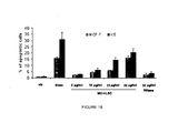
- the adminsitration of a combination of doxorubicin and/or vincristine with MVs derived from an adult stem cell is highly preferred, since such a combination has been shown to exert a synergistic effect (see figure 13 ).
- the microvescicle is administered to a patient in need thereof either locally or systemically.
- a pharmaceutical dosage form suitable for both local and systemic administration is e.g. an injectable dosage form.
- the microvescicle is administered by local intra-tumour (i.t.) injection in a solid tumour, or by i.v. injection or infusion, both in the case of a solid tumour and in the case of metastasis.
- a suitable MV dose to be administered depends on a plurality of factors, but it is generally comprised between 0.1 to 200 micrograms/kg body weight of the recipient, preferably 1-150 micrograms/ kg body weight of the recipient, even more preferably 3-120 micrograms/ kg body weight of the recipient.
- microvescicle (MV) derived from an adult stem cell refers to a membrane particle which is at least in part derived from an adult stem cell.
- adult stem cell includes any undifferentiated or partially undifferentiated cell which is capable of proliferating (self-renewal) and differentiating (plasticity), thereby replacing the mature cells of a specialized cell lineages which have reached the end of their life cycle.
- adult stem cell as used in the present description includes both stem cells having unlimited self-renewal ability and pluripotent plasticity, and progenitor cells with multipotent or unipotent plasticity and, in some instances, a limited self-renewal ability.
- the "adult stem cells” have pluripotent or multipotent plasticity meaning that they are capable of differentiating into at least two, more preferably at least three, distinct types of specialised, fully differentiated, mature cells.
- adult stem cell is intended to mean a stem cell that is isolated from an adult tissue, in contrast with an “embryonic stem cell” which is isolated from the inner cell mass of a blastocyst.
- embryonic stem cell which is isolated from the inner cell mass of a blastocyst.
- embryonic stem cells are also known as “somatic stem cells”.
- microvesicles derived from adult stem cells used in the present invention are generally spheroid in shape and have a diameter within the range of 100 nm to 5 ⁇ m, more typically of between 0.2 and 1 ⁇ m. If the particle is not spheroid in shape, the above mentioned values are referred to the largest dimension of the particle.
- the stem cells from which the microvesicles used in the invention are obtained may be isolated as described in the experimental section of the description.
- the microvesicles (MVs) may then be obtained from the supernatants of the isolated stem cells, e.g. by ultracentrifugation as disclosed in the experimental section of the description.
- the isolated MVs may then be stored until use by freezing at very low temperature, e.g. at -80°C, in a suspension with one or more cryoprotecting agent.
- Suitable cryoprotecting agents are e.g. dimethylsulphoxide (DMSO) and glycerol.
- DMSO dimethylsulphoxide
- glycerol glycerol
- the use of DMSO at a concentration of 1% of the volume of the cell suspension guarantees good preservation of the cells and a limited toxic effect on re-infused patients.
- Other substances which may be mentioned as cryoprotecting agents are extracellular cryoprotecting agents, that is to say high molecular weight
- MICROVESICLES (MVs) FROM MESENCHYMAL STEM CELLS (MSCs)
- Bone marrow cells were layered on a Ficoll gradient (density: 1022 g/ml; Sigma-Aldrich, St. Louis, MO) and centrifuged at 1500 rpm for 30 minutes.
- the mononucleated cells were cultured in the presence of Mesenchymal Stem Cells Basal Medium (MSCBM, Lonza). After 5 days of culture, the medium was changed.
- MSCBM Mesenchymal Stem Cells Basal Medium
- the adherent monolayer was detached by trypsin treatment for 5 minutes at 37° C, on day 15 for the first passage and every 7 days for the subsequent passages.
- the cells were seeded at a density of 10,000 cells/cm 2 and used not later than passage 6.
- MSC populations from glomeruli were obtained from the normal portion of cortex from surgically removed kidneys, as described in Bruno S et al. Isolation and characterization of resident mesenchymal stem cells in human glomeruli. Stem Cells Dev. 2009;18:867-880 . After dissection of the cortex, the glomerular suspension was collected using a standard established technique: after passing through a graded series of meshes (60 and 120 meshes), glomeruli were recovered at the top of the 120 meshes sieve.
- Glomeruli were then collected at the bottom of a conical tube by spontaneous precipitation (10 minutes at room temperature) and were deprived of the visceral layer of the Bowman's capsule both mechanically, by several rounds of aspiration/expulsion using a 10 ml pipette, and enzymatically, by digestion for 2 minutes with Collagenase I (Sigma, St. Louis, MO). Glomeruli were then collected at the bottom of a conical tube by spontaneous precipitation in order to remove cells and Bowman capsules, and were transferred to fibronectin coated T25 flasks (Falcon, BD Bioscience, Two Oak Park, Bedford, MA).
- Glomeruli were cultured in the presence of Mesenchymal Stem Cells Basal Medium (MSCBM, Lonza). Cells were left to reach confluence prior to passaging; the interval between passages varied (3-7 days) until passage 4, and from then on it was established at around 7 days.
- MSCBM Mesenchymal Stem Cells Basal Medium
- Cytofluorimetric analysis was performed with the following antibodies, all phycoerythrin (PE) or fluorescein isothiocyanate (FITC) conjugated: anti-CD105, -CD29, -CD31, -CD146, -CD44, -CD90 (Dakocytomation, Copenhagen, Denmark); -CD73, -CD34, -CD45, -CD80, -CD86,-CD166, HLA-I (Becton Dickinson Biosciences Pharmingen, San Jose, CA); -CD133 (Miltenyi Biotec, Auburn); KDR (R&D Systems, Abington, U.K.); -HLA-II (Chemicon International Temecula, CA), -CD40 (Immunotech, Beckman Coulter), -CD154 (Serotec, Raleigh, NC USA) monoclonal
- Mouse IgG isotypic controls were from Dakocytomation. All incubations were performed in 100 ⁇ l of phosphate buffered saline (PBS) containing 0.1 % bovine serum albumin and 0.1 % sodium azide, at 4° C. For each sample, 10,000 cells were analysed on FACSCalibur cytometer (BD Biosciences Pharmingen). Gating was constructed based on negative controls and compensation controls were included in all analyses performed. Population percentages and numbers were generated for gated populations from each experiment using Cell Quest software (BD Biosciences Pharmingen).
- PBS phosphate buffered saline
- FACSCalibur cytometer BD Biosciences Pharmingen
- Indirect immunofluorescence was performed on MSCs cultured on chamber slides (Nalgen Nunc International, Rochester, NY, USA), fixed in 4% paraformaldehyde containing 2 % sucrose and, if required, permeabilized with Hepes-Triton X 100 buffer (Sigma, St. Louis, MO).
- the following antibodies were used: mouse monoclonal anti-vimentin (Sigma) and rabbit policlonal anti-von Willebrand factor (Dakocytomation). Omission of the primary antibodies or substitution with non immune rabbit or mouse IgG were used as controls where appropriated.
- Alexa Fluor 488 anti-rabbit and anti-mouse Texas Red were used as secondary Abs. Confocal microscopy analysis was performed using a Zeiss LSM 5 Pascal Model Confocal Microscope (Carl Zeiss International, Germany). Hoechst 33258 dye (Sigma) was added for nuclear staining.
- BM-MSC and GL-MSC preparations did not express hematopoietic markers such as CD45, CD14 and CD34. They neither expressed the co-stimulatory molecules (CD80, CD86 and CD40) and the endothelial markers (CD31, von Willebrand Factor, KDR). All the cell preparations at different passages of culture expressed the typical MSC markers: CD105, CD73, CD44, CD90, CD166 and CD146. They also expressed HLA class I.
- MSCs The adipogenic, osteogenic and chondrogenic differentiation abilities of MSC were determined as described in Pittenger MF, Martin BJ. Mesenchymal stem cells and their potential as cardiac therapeutics. Circ. Res 2004;95:9-20 .
- MSCs were cultured with Adipogenic Medium (Lonza) for 3 weeks. To evaluate the differentiation, the cells were fixed with 4% paraformaldehyde for 20 minutes at room temperature and stained with 0.5% Oil Red O (Sigma) in methanol (Sigma) for 20 minutes at room temperature.
- Osteogenic differentiation was assessed by culturing MSCs in Osteogenic Medium (Lonza). The medium was changed twice per week for 3 weeks. To evaluate the differentiation, cells were fixed with 4% paraformaldehyde for 20 minutes and stained with Alizarin Red, pH 4.1 (Lonza) for 20 minutes at room temperature.
- chondrogenic differentiation 2.5 x 10 5 MSCs were centrifuged in a 15-ml conical polypropylene tube (Falcon BD Bioscience) at 150g for 5 minutes and washed twice with DMEM. The pellets were cultured in Chondrogenic medium (Lonza) supplemented with 10 ng/ml of Transforming Growth Factor ⁇ 3 (Lonza). The medium was changed every 3 days for 28 days. Pellets were fixed in 4% paraformaldehyde overnight, and the paraffin-embedded sections were stained for glycosaminoglycans using 0.1% safranin O (Sigma) and for sulfated proteoglycans using 1% alcian blue.
- the microvesicles were obtained from supernatants of BM-MSCs or GL-MSCs obtained as describe above cultured in RPMI deprived of FCS and supplemented with 0.5% of BSA (Sigma). After centrifugation at 2,000 g for 20 minutes to remove debris, cell-free supernatants were centrifuged at 100,000 g (Beckman Coulter Optima L-90K ultracentrifuge) for 1 hour at 4 °C, washed in serum-free medium 199 containing N-2-hydroxyethylpiperazine-N'-2-ethanesulfonic acid (HEPES) 25mM (Sigma) and subjected to a second ultracentrifugation under the same conditions. Endotoxin contamination of MVs was excluded by Limulus test according to the manufacturer's instructions (Charles River Laboratories, Inc., Wilmington, MA, USA) and MVs were stored at -80°C.
- MVs were treated with 1U/ml RNase (Ambion Inc., Austin, TX, USA) for 1 hour at 37°C, the reaction was stopped by addition of 10 U/ml RNase inhibitor (Ambion Inc.) and MVs were washed by ultracentrifugation.
- Human liver cancer cell line (HepG2), human breast adenocarcinoma cell line (MCF-7) and human ovarian cancer cell line (SKOV-3) were cultured in low glucose DMEM (Euroclone) containing 10% of Fetal Calf Serum (FCS, Euroclone), 100U/ml penicillin, 100mg/ml streptomycin and 1% glutamine (all from Sigma) and maintained in an incubator with a humidified atmosphere of 5% CO 2 at 37°C.
- DMEM Euroclone
- FCS Fetal Calf Serum
- KS cells Kaposi's sarcoma cells
- TECs Tumor endothelial cells isolation and culture. TECs were isolated from specimens of clear-cell type renal cell carcinomas using anti-CD105 Ab coupled to magnetic beads by magnetic cell sorting using the MACS system (Miltenyi Biotec, Auburn, CA, USA). TEC cell lines were established and maintained in culture in endothelial basal complete medium (EBM) supplemented with epidermal growth factor (10 ng/ml), hydrocortisone (1 mg/ml), bovine brain extract (all from Lonza) and 10% FCS.
- EBM endothelial basal complete medium
- EBM epidermal growth factor
- hydrocortisone 1 mg/ml
- bovine brain extract all from Lonza
- TECs were characterized as endothelial cells by morphology, positive staining for vWF antigen, CD105, CD146, and vascular endothelial-cadherin and negative staining for cytokeratin and desmin ( Bussolati B et al. Altered angiogenesis and survival in endothelial cells derived from renal carcinoma. FASEB J 2003;17:1159 ⁇ 1161 ).
- HUVECs Humbelical Vein Endothelial Cells
- isolation and culture Human umbilical vein endothelial cells (HUVECs) were obtained from the umbilical vein, as described previously ( Bussolati B et al. Vascular endothelial growth factor receptor-1 modulates vascular endothelial growth factor-mediated angiogenesis via nitric oxide. Am J Pathol. 2001Sep;159(3):993-1008 ) and maintained in EBM and 10% FCS. Experiments were performed on second or third passage HUVECs.
- HepG2, MCF-7, SKOV-3, KS cells or HUVECs were seeded at 2,000 or 4,000 cells/well into 96-well plates in DMEM (Sigma) deprived of FCS with different concentrations of microvesicles pre-treated or not pre-treated with RNase. DNA synthesis was detected as incorporation of 5-bromo-2'-deoxy-uridine (BrdU) into the cellular DNA after 48 hours of culture. Cells were then fixed with 0.5 M ethanol/HCl and incubated with nuclease to digest the DNA.
- PrdU 5-bromo-2'-deoxy-uridine
- BrdU incorporation into the DNA was detected using an anti-BrdU peroxidase-conjugated mAb and visualized with a soluble chromogenic substrate (Roche Applied Science, Mannheim, Germany). Optical density was measured with an ELISA reader at 405 nm.
- HepG2, MCF-7, SKOV-3, KS cells, HUVECs or TECs were seeded at 8,000 cells/well into 96-well plated in DMEM (Sigma) with 10% FCS and in the presence of doxorubicin (100 ng/ml, Sigma) or different concentrations of MVs (10 and 30 ⁇ g/ml) pre-treated or not with RNase. Apoptosis was assessed by TUNEL assay (ApopTag Oncor, Gaithersburg, MD, USA).
- the cells were washed with PBS, fixed in 1% paraformaldehyde pH 7.4 for 15 minutes at 4°C, washed twice in PBS and then post-fixed in pre-cooled ethanol-acetic acid 2:1 for 5 minutes at ⁇ 20°C.
- the samples were treated with the enzyme terminal deoxynucleotidyl transferase (TdT).
- TdT enzyme terminal deoxynucleotidyl transferase
- the cells were then treated with heated anti-digoxigenin conjugate with fluorescein and incubated for 30 minutes at room temperature.
- the samples were mounted in medium containing 1 ⁇ g/ml of propidium iodide and the cells analyzed by immunofluorescence. The results are expressed as the percentage of green fluorescence emitting cells (apoptotic cells) versus red fluorescence emitting cells (total cells).
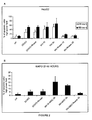
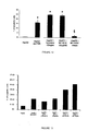
- the anti-tumour activity of MVs derived from human BM-MSCs was assessed in vitro by measuring their ability to inhibit proliferation and to induce apoptosis on HepG2, MCF-7, SKOV-3 and KS cell lines.
- Figure 1 shows that incubation of HepG2 cells ( Figure 1A ) and KS cells ( Figure 1B ) with different doses of MVs for 48 hours significantly inhibited proliferation compared to control cells incubated with vehicle alone.
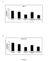
- Figure 2 shows that incubation of HepG2 cells ( Figure 2A ) and KS cells ( Figure 2B ) with MVs for 24 and 48 hours significantly promoted apoptosis compared to control cells incubated with vehicle alone and in the same way of doxorubicin stimulation.
- the inventors have also tested the effects of MVs derived from Gl-MSCs on the proliferation and apoptosis of tumor cell lines.
- Gl-MSCs did not affect proliferation and apoptosis of the HepG2 cell line.
- MVs derived from Gl-MSCs inhibited proliferation and induced apoptosis of KS cells ( Figures 1 and 2 ).
- incubation of MCF-7 and SKOV-3 cells with 30 ⁇ g/ml of MVs derived from Gl-MSCs for 48 hours inhibited proliferation compared to control cells incubated with vehicle alone ( Figure 3 ), but did not promote apoptosis.
- MVs derived from human fibroblasts did not inhibit proliferation and did not induce apoptosis of different cancer cell lines (data not shown).
- the inventors have also studied the in vitro effects of MVs derived from BM-MSCs on the proliferation, apoptosis and capillary-like formation of HUVECs and tumour endothelial cells (TECs).
- MV-treatment did not affect the proliferation (data not shown) and the capillary-like formation ability of HUVECs ( Figure 5A ).
- incubation of HUVECs with different doses of MVs for 48 hours did not induce apoptosis ( Figure 6 ).
- SCID mice (Charles River, Jackson Laboratories, Bar Harbor, ME). Cultured cells, harvested using trypsin-EDTA, were washed with PBS, counted in a microcytometer chamber and resuspended in 100 ⁇ l of DMEM and 100 ⁇ l of Matrigel Matrix (Becton Dickinson). The cells were chilled on ice, and injected subcutaneously into the left back of SCID mice via a 26-gauge needle using a 1-ml syringe. The animals were monitored for activity and physical conditions every day, and the determination of body weight and measurement of tumour mass was made every 3 days.
- Tumour mass was determined by calliper measurement in two perpendicular diameters of the implant and calculated using the formula 1 / 2a x b 2 , where a is the long diameter and b is the short diameter ( Hou J et al. Experimental therapy of hepatoma with artemisin and its derivatives: in vitro and in vivo activity, chemosensitization and mechanism of action. Clin Cancer Research. 2008;14:5519-5530 )). After 1 week, when the implanted tumours reached the volume of approximately 15 mm 3 , the inventors started the weekly intra-tumour injection of MVs.
- the first treatment was with 100 ⁇ g of MVs (treated or not with RNase) for a maximum volume of 20 ⁇ l; the subsequent intra-tumour injections were of 50 ⁇ g of MVs (treated or not with RNase), for a maximum of 20 ⁇ l.
- the inventors injected intra-tumour the same volume of vehicle alone.
- mice Tumor formation and growth are inhibited by MVs derived form BM-MSCs in SCID mice.
- SCID mice were injected subcutaneously with HepG2 in the presence of Matrigel.
- the inventors started to weekly inject the mice intra-tumour with MVs (treated or not with RNase), with a maximum volume of 20 ⁇ l.
- the first treatment was with 100 ⁇ g of MVs; the subsequent intra-tumour injections were with 50 ⁇ g of MVs.
- the inventors inject 20 ⁇ l of vehicle alone intra-tumour.
- MV intra-tumor injection showed a inhibitor effect on tumor growth ( Figure 8 ).
- Tumor size and volume were significantly smaller in SCID mice treated with MVs ( Figure 9A and B ) and histological analyses showed areas of necrosis in HepG2 tumours treated with MVs ( Figure 9C ).
- Tumours injected with MVs pre-treated with RNase did not differ in size and histology from control tumours ( Figure 10A and B ).
- HLSCs were isolated from human cryopreserved normal hepatocytes obtained from Cambrex Bio Science Verviers S.p.r.l. (Verviers, Belgium) cultured in minimum essential medium/endothelial cell basal medium-1 ( ⁇ -MEM/EBM) (3:1) (Gibco/Cambrex) supplemented with L-glutamine (5 mM), Hepes (12 mM, pH 7.4), penicillin (50 IU/ml), streptomycin (50 ⁇ g/ml) (all from Sigma, St. Louis), FCS (10%). The expanded cells were transferred to a T-75 flask and analyzed when they approached confluence.
- ⁇ -MEM/EBM minimum essential medium/endothelial cell basal medium-1
- the hepatoma cell line HepG2 were cultured in Dulbecco's modified Eagle's medium (DMEM) (low glucose) containing 10% fetal bovine serum (FBS).
- DMEM Dulbecco's modified Eagle's medium
- FBS fetal bovine serum
- MVs were obtained from supernatants of HLSCs cultured in MEM-alpha supplemented with 2% of Fetal Bovine Serum (FBS). The viability of cells incubated overnight without serum was detected by trypan blue exclusion. After centrifugation at 2000 g for 20 minutes to remove debris, cell-free supernatants were centrifuged at 100,000 g (Beckman Coulter Optima L-90K ultracentrifuge) for 1 h at 4 °C, washed in serum-free medium 199 containing N-2-hydroxyethylpiperazine-N'-2-ethanesulfonic acid (HEPES) 25mM (Sigma) and subjected to a second ultracentrifugation under the same conditions.
- FBS Fetal Bovine Serum
- MVs by fluorescence microscopy or FACS analysis, MVs from stem cells were labelled with the red fluorescence aliphatic chromophore intercalating into lipid bilayers PKH26 dye (Sigma). After labelling, MVs were washed and ultracentrifuged at 100,000 g for 1 h at 4 °C. MV pellets were suspended in medium 199, and the protein content was quantified by the Bradford method (BioRad, Hercules, CA). Endotoxin contamination of MVs was excluded by Limulus testing according to the manufacturer's instructions (Charles River Laboratories, Inc., Wilmington, MA), and MVs were stored at ⁇ 80 °C. The morphologic analysis performed on MV suspension after staining with propidium iodide did not show the presence of apoptotic bodies.
- MVs from HLSCs were treated with 1 U/ml RNase (Ambion Inc., Austin, TX) for 1 h at 37 °C, the reaction was stopped by the addition of 10 U/ml RNase inhibitor (Ambion Inc.) and MVs were washed by ultracentrifugation.
- RNase treatment was evaluated after RNA extraction using TRIZOL reagent (Invitrogen, Carlsbad, CA) by spectrophotometer analysis of total extracted RNA (untreated: 1.3 ⁇ 0.2 ⁇ g RNA/ mg protein MV; RNase treated: ⁇ 0.2 ⁇ g RNA/mg protein MV).
- RNA extracted from RNase-treated and untreated MVs was labelled by oligo dT driven retrotranscription and analyzed on 0.6% agarose gel to show the complete degradation of RNA by RNase treatment.
- MVs were treated with 1 U/ml DNase (Ambion Inc.) for 1 h at 37°C.
- Apoptosis assay was evaluated using the terminal dUTP nickend labeling assay (ApoTag; Oncor, Gaithersburg, MD).
- Cells (8x10 3 /well) were cultured in 96-well plate, suspended in phosphate-buffered saline (PBS) and fixed in 1% paraformaldehyde in PBS, pH 7.4, for 15 minutes at 4°C followed by pre-cooled ethanol/acetic acid (2:1) for 5 minutes at -20°C.
- PBS phosphate-buffered saline
- Cells were treated with terminal deoxynucleotide transferase enzyme and incubated in a humidified chamber for 1 hour at 37°C and then treated with warmed fluorescein isothiocyanate-conjugated antidigoxigenin for 30 minutes at room temperature. After washing, samples were mounted in a medium containing 1 g/ml of propidium iodide and the cells were analyzed by immunofluorescence.
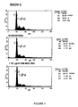
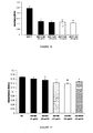
- HepG2 The ability of HLSC-derived MVs to induce apoptosis on HepG2 was evaluated. Briefly, HepG2 were seeded at a density of 8,000 cells/well into 96-well plates in DMEM with 10% FCS and apoptosis was induced by culture in the absence of FCS, by treatment with vincristine (100 ng/ml), or doxorubicin (50 ng/ml), two mitotic inhibitors used in cancer chemotherapy, or by MV treatment (30 ⁇ g/ml).
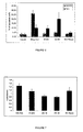
- MVs were also treated with 1 U/ml RNase18 (Ambion, Austin, TX) for 1 hour at 37°C to assess whether the contribution to the inhibition of cancer cells growth is dependent to an horizontal transfer of mRNA delivered by MV to the cancer cells. Apoptosis was evaluated using the TUNEL assay analysis at 24 and 72 hours. As shown in Figure 12 , MVs derived from HLSCs were able to induce HepG2 apoptosis comparable to that induced by vincristine. On the contrary, the RNase treatment failed to induce apoptosis. In addition, treatment of HepG2 with vincristine plus MV-HLSCs or doxorubicin plus MV-HLSCs results in an additive effect as shown in Figure 13 .
- Human hepatoma cells HepG2 were cultured in DMEM supplemented with 10% fetal bovine serum, 100 ⁇ g/ml penicillin, and 100 ⁇ g/ml streptomycin and maintained in an incubator with a humidified atmosphere of 5% CO 2 at 37°C.
- HLSCs Human liver stem cells
- ⁇ -MEM/EBM 3:1
- EBM Human liver stem cells
- GA gentamicin
- BBE Brain Bovine Extract
- MVs microvesicles
- MVs were obtained from supernatants of HLSCs cultured in ⁇ -MEM medium supplemented with 2% fetal bovine serum. After centrifugation at 2,000 g for 20 minutes to remove debris, cell-free supernatants were centrifuged at 100,000g for 1 hour at 4°C, washed in serum-free medium 199 containing N-2-hydroxyethylpiperazine-N'-2-ethanesulfonic acid (HEPES) 25mM and submitted to a second ultracentrifugation under the same conditions. MV pellets were suspended in medium 199 in 0.1 % of DMSO and the protein content was quantified using the Bradford method.
- HEPES N-2-hydroxyethylpiperazine-N'-2-ethanesulfonic acid
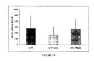
- tumours On day 7 post-tumour cell transplantation treatment started. Tumours became palpable as from day 7; 50 or 100 ⁇ g of MV, suspended in M199 supplemented with 0,1% of DMSO, were injected 7, 12, 14 and 18 days after tumour transplantation. Treatment started when tumours rinse the volume of approximately 15 mm3. The animals were monitored for activity and physical condition everyday, and the determination of body weight and measurement of tumour mass were done every 3 days.
- Tumours were measured with callipers. Tumour mass was determined by calliper, measurement in two perpendicular diameters of the implant and calculated using the formula 1 / 2a x b 2 , where a is the long diameter and b is the short diameter. The animals were sacrificed on day 28, and tumours were collected for further analysis.
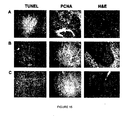
- Tumours were fixed in 10% buffered neutral formalin, routinely processed, embedded in paraffm, sectioned at 5 ⁇ m, and stained with H&E for microscopic examination. Immunohistochemistry for detection of proliferation was performed using the anti-PCNA monoclonal antibody. Sections were blocked and labeled with anti-mouse HRP secondary antibody (1:300 dilution). Omission of the primary antibodies or substitution with non immune mouse IgG was used as controls. Apoptosis was evaluated in paraffin-embedded tumor sections by TUNEL. Ten non consecutive sections were counted for apoptotic- positive tumor cells at 630X magnification. Hoechst 33258 dye was added for nuclear staining.
- mice were subcutaneously transplanted with the human hepatocarcinoma cell line HepG2.
- mice were treated with intra-tumour injection of MVs (50 or 100 ⁇ g), for a maximum of 20 ⁇ l of volume.
- tumour were injected with 20 ⁇ l of vehicle alone.
- all tumours were recovered and analyzed.
- intra-tumor injection of MVs ( Figure 14 ) showed a inhibitor effect on tumour growth.
- histological analyses showed areas of necrosis in tumours treated with MVs ( Figure 15B ) and anti-proliferative effect was observed using PCNA staining ( Figure 15B ).
- HLSC Human non oval liver stem cells
- EBM Human non oval liver stem cells
- GA human Epithelial Growth Factor
- GA gentamicin
- BBE Brain Bovine Extract
- MCF-7 breast adenocarcinoma cell lines were obtained from American Type Culture Collection (Manassas, VA) and were cultured in DMEM supplemented with 10% of FCS, 100 ⁇ g/ml penicillin and 100 ⁇ g/ml streptomycin and maintained in an incubator with a humidified atmosphere of 5% CO 2 at 37°C.
- KS cells Kaposi's sarcoma cells
- MVs were obtained from supernatants of HLSCs cultured in MEM-alpha supplemented with 2% of Fetal Bovine Serum (FBS) for 18 hours. In selected experiments, MVs were collected in the absence of FBS. The viability of cells incubated overnight at 2 % of FBS and without serum was detected by trypan blue exclusion (more than 90%, data not shown).
- FBS Fetal Bovine Serum
- cell-free supernatants were centrifuged at 100,000 g (Beckman Coulter Optima L-90K ultracentrifuge) for 1 h at 4 °C, washed in serum-free medium 199 containing N-2-hydroxyethylpiperazine-N'-2-ethanesulfonic acid (HEPES) 25mM and subjected to a second ultracentrifugation under the same conditions.
- MV pellets were suspended in medium 199, and the protein content was measured with the Bradford method. MVs were stored at ⁇ 80 °C. The morphologic analysis performed on a MVs suspension after staining with propidium iodide did not show the presence of apoptotic bodies.
- MVs from HLSCs were treated with I U/ml RNase for 1 h at 37°C. The reaction was stopped by addition of 10 U/ml RNase inhibitor and MVs were washed by ultracentrifugation.
- MCF-7 breast adenocarcinoma cells and Kaposi's sarcoma cells were seeded at 8,000 cells/well into 96-well plates in DMEM and RPMI, respectively, with different concentrations of MVs (2; 10; 15; and 30 ⁇ g/ml and 30 ⁇ g/ml of RNase treated-MVs). DNA synthesis was detected as incorporation of 5-bromo-2'-deoxy-uridine (BrdU) into the cellular DNA after 48 hours of culture.
- PrdU 5-bromo-2'-deoxy-uridine
- MCF-7 and KS cells were seeded at 8,000 cells/well into 96-well plated in low glucose DMEM with 10% FCS and in the presence of Doxorubicin (100 ng/ml) or different concentrations of MVs (2 ; 10; 15; and 30 ⁇ g/ml and 30 ⁇ g/ml of RNase treated MVs). Apoptosis was evaluated using the TUNEL assay.
Landscapes
- Health & Medical Sciences (AREA)
- Life Sciences & Earth Sciences (AREA)
- Cell Biology (AREA)
- Immunology (AREA)
- Developmental Biology & Embryology (AREA)
- Medicinal Chemistry (AREA)
- Pharmacology & Pharmacy (AREA)
- Chemical & Material Sciences (AREA)
- Animal Behavior & Ethology (AREA)
- General Health & Medical Sciences (AREA)
- Public Health (AREA)
- Veterinary Medicine (AREA)
- Engineering & Computer Science (AREA)
- Virology (AREA)
- Biotechnology (AREA)
- Epidemiology (AREA)
- Zoology (AREA)
- Hematology (AREA)
- Biomedical Technology (AREA)
- General Chemical & Material Sciences (AREA)
- Chemical Kinetics & Catalysis (AREA)
- Nuclear Medicine, Radiotherapy & Molecular Imaging (AREA)
- Organic Chemistry (AREA)
- Bioinformatics & Cheminformatics (AREA)
- Medicines Containing Material From Animals Or Micro-Organisms (AREA)
- Pharmaceuticals Containing Other Organic And Inorganic Compounds (AREA)
- Medicines That Contain Protein Lipid Enzymes And Other Medicines (AREA)
- Micro-Organisms Or Cultivation Processes Thereof (AREA)
Priority Applications (14)
| Application Number | Priority Date | Filing Date | Title |
|---|---|---|---|
| EP10425052A EP2363136A1 (de) | 2010-03-02 | 2010-03-02 | Von Erwachsenenstammzellen abgeleitete Mikrovesikel zur Verwendung bei der therapeutischen Behandlung von Tumorkrankheiten |
| JP2012555384A JP5841952B2 (ja) | 2010-03-02 | 2011-02-28 | 腫瘍性疾患の治療的処置にて用いるための成体幹細胞由来の微細小胞(mv) |
| PL11705585T PL2542250T3 (pl) | 2010-03-02 | 2011-02-28 | Mikropęcherzyki (mvs) pochodzące z dorosłych komórek macierzystych do zastosowania w terapeutycznym leczeniu choroby nowotworowej |
| HUE11705585A HUE026292T2 (en) | 2010-03-02 | 2011-02-28 | Microvesicles (MV) derived from an adult cell for use in the therapeutic treatment of tumor disease |
| PCT/EP2011/052945 WO2011107437A1 (en) | 2010-03-02 | 2011-02-28 | Microvesicles (mvs) derived from adult stem cells for use in the therapeutic treatment of a tumor disease |
| DK11705585.5T DK2542250T3 (en) | 2010-03-02 | 2011-02-28 | Microvesicles (MVS) derived from adult stem cells for use in the therapeutic treatment of a tumor disease |
| ES11705585.5T ES2554492T3 (es) | 2010-03-02 | 2011-02-28 | Microvesículas (MVs) derivadas de citoblastos de etapa adulta para uso en el tratamiento terapéutico de una enfermedad neoplásica. |
| PT117055855T PT2542250E (pt) | 2010-03-02 | 2011-02-28 | Microvesículas (mvs) derivadas de células estaminais adultas para uso no tratamento terapêutico de uma doença tumoral |
| EP11705585.5A EP2542250B1 (de) | 2010-03-02 | 2011-02-28 | Von Erwachsenenstammzellen abgeleitete Mikrovesikel zur Verwendung bei der therapeutischen Behandlung von Tumorkrankheiten |
| CN201180011925.0A CN102781454B (zh) | 2010-03-02 | 2011-02-28 | 用于治疗性治疗肿瘤疾病的来源于成体干细胞的微泡(mv) |
| US13/581,537 US9717760B2 (en) | 2010-03-02 | 2011-02-28 | Microvesicles (MVS) derived from adult stem cells for use in the therapeutic treatment of a tumor disease |
| US15/620,164 US10105395B2 (en) | 2010-03-02 | 2017-06-12 | Microvesicles (MVs) derived from adult stem cells for use in the therapeutic treatment of a tumor disease |
| US16/126,364 US20190060369A1 (en) | 2010-03-02 | 2018-09-10 | Microvesicles (mvs) derived from adult stem cells for use in the therapeutic treatment of a tumor disease |
| US18/135,243 US20230248774A1 (en) | 2010-03-02 | 2023-04-17 | Microvesicles (mvs) derived from adult stem cells for use in the therapeutic treatment of a tumor disease |
Applications Claiming Priority (1)
| Application Number | Priority Date | Filing Date | Title |
|---|---|---|---|
| EP10425052A EP2363136A1 (de) | 2010-03-02 | 2010-03-02 | Von Erwachsenenstammzellen abgeleitete Mikrovesikel zur Verwendung bei der therapeutischen Behandlung von Tumorkrankheiten |
Publications (1)
| Publication Number | Publication Date |
|---|---|
| EP2363136A1 true EP2363136A1 (de) | 2011-09-07 |
Family
ID=42306621
Family Applications (2)
| Application Number | Title | Priority Date | Filing Date |
|---|---|---|---|
| EP10425052A Withdrawn EP2363136A1 (de) | 2010-03-02 | 2010-03-02 | Von Erwachsenenstammzellen abgeleitete Mikrovesikel zur Verwendung bei der therapeutischen Behandlung von Tumorkrankheiten |
| EP11705585.5A Active EP2542250B1 (de) | 2010-03-02 | 2011-02-28 | Von Erwachsenenstammzellen abgeleitete Mikrovesikel zur Verwendung bei der therapeutischen Behandlung von Tumorkrankheiten |
Family Applications After (1)
| Application Number | Title | Priority Date | Filing Date |
|---|---|---|---|
| EP11705585.5A Active EP2542250B1 (de) | 2010-03-02 | 2011-02-28 | Von Erwachsenenstammzellen abgeleitete Mikrovesikel zur Verwendung bei der therapeutischen Behandlung von Tumorkrankheiten |
Country Status (10)
| Country | Link |
|---|---|
| US (4) | US9717760B2 (de) |
| EP (2) | EP2363136A1 (de) |
| JP (1) | JP5841952B2 (de) |
| CN (1) | CN102781454B (de) |
| DK (1) | DK2542250T3 (de) |
| ES (1) | ES2554492T3 (de) |
| HU (1) | HUE026292T2 (de) |
| PL (1) | PL2542250T3 (de) |
| PT (1) | PT2542250E (de) |
| WO (1) | WO2011107437A1 (de) |
Cited By (2)
| Publication number | Priority date | Publication date | Assignee | Title |
|---|---|---|---|---|
| WO2019115748A1 (en) * | 2017-12-14 | 2019-06-20 | Unicyte Ev Ag | PHARMACEUTICAL CARRIERS CONTAINING miRNAs FOR USE IN THE TREATMENT OF RENAL CANCER |
| WO2019197442A1 (en) * | 2018-04-12 | 2019-10-17 | Unicyte Ev Ag | A combination of active ingredients for the treatment of tumor |
Families Citing this family (15)
| Publication number | Priority date | Publication date | Assignee | Title |
|---|---|---|---|---|
| EP2363136A1 (de) * | 2010-03-02 | 2011-09-07 | Fresenius Medical Care Deutschland GmbH | Von Erwachsenenstammzellen abgeleitete Mikrovesikel zur Verwendung bei der therapeutischen Behandlung von Tumorkrankheiten |
| TWI840787B (zh) | 2010-05-12 | 2024-05-01 | 開曼群島商普羅基德尼公司 | 生物活性腎細胞 |
| ITRM20110403A1 (it) * | 2011-07-28 | 2013-01-29 | Ospedale Pediatrico Bambino Gesu | Microvescicole isolate da cellule mesenchimali come agenti immunosoppressori. |
| CN102357071B (zh) * | 2011-10-26 | 2013-10-23 | 重庆医科大学附属儿童医院 | 载紫杉醇纳米微泡及其制备方法 |
| ES2690212T3 (es) * | 2011-11-30 | 2018-11-19 | Astellas Institute For Regenerative Medicine | Células estromales mesenquimales y usos relacionados con las mismas |
| US11286463B2 (en) | 2012-03-08 | 2022-03-29 | Advanced ReGen Medical Technologies, LLC | Reprogramming of aged adult stem cells |
| CR20160307A (es) | 2013-12-20 | 2016-11-08 | Advanced Regen Medical Tech Llc | Composiciones para la restauración de células y métodos para la preparación y utilización de las mismas |
| US10772911B2 (en) | 2013-12-20 | 2020-09-15 | Advanced ReGen Medical Technologies, LLC | Cell free compositions for cellular restoration and methods of making and using same |
| EP3122333A4 (de) * | 2014-03-24 | 2017-11-29 | Advanced Regen Medical Technologies, LLC | Zellfreie zusammensetzungen zur zellwiederherstellung sowie verfahren zur herstellung und verwendung davon |
| WO2015179301A1 (en) * | 2014-05-19 | 2015-11-26 | Eleftherios Papoutsakis | Megakaryocytic particles and microparticles for cell therapy & fate modification of stem and progenitor cells |
| BR112018072198A2 (pt) | 2016-04-29 | 2019-02-12 | Advanced ReGen Medical Technologies, LLC | composição compreendendo micrornas, método de preparação de uma composição de célula-tronco restaurada, formulação farmacêutica e kit |
| EP3740576A4 (de) | 2018-01-18 | 2021-10-20 | Advanced Regen Medical Technologies, LLC | Therapeutische zusammensetzungen und verfahren zur herstellung und verwendung davon |
| CN111494417B (zh) * | 2020-02-10 | 2024-04-09 | 寇晓星 | 诱导性细胞外囊泡在制备治疗肿瘤药物中的应用 |
| EP4150054A1 (de) | 2020-05-11 | 2023-03-22 | Sterm.Bio Incorporated | Zusammensetzungen und verfahren im zusammenhang mit extrazellulären vesikeln aus megakaryozyten |
| KR102601831B1 (ko) * | 2020-09-23 | 2023-11-14 | 대한민국 | 고형암 증식 억제 효과를 갖는 개과동물의 엑소좀 |
Citations (5)
| Publication number | Priority date | Publication date | Assignee | Title |
|---|---|---|---|---|
| WO2006126219A1 (en) | 2005-05-26 | 2006-11-30 | Fresenius Medical Care Deutschland G.M.B.H. | Liver progenitor cells |
| WO2009005742A1 (en) | 2007-06-28 | 2009-01-08 | Applied Materials, Inc. | Methods and apparatus for cleaning deposition chamber parts using selective spray etch |
| WO2009057165A1 (en) * | 2007-10-29 | 2009-05-07 | Fresenius Medical Care Deutschland G.M.B.H. | Use of microvesicles (mvs) derived from stem cells for preparing a medicament for endo/epithelial regeneration of damaged or injured tissues or organs, and related in vitro and in vivo methods |
| WO2009087361A1 (en) * | 2008-01-04 | 2009-07-16 | Lydac Neuroscience Limited | Microvesicles |
| WO2009105044A1 (en) * | 2008-02-22 | 2009-08-27 | Agency For Science, Technology And Research (A*Star) | Mesenchymal stem cell particles |
Family Cites Families (12)
| Publication number | Priority date | Publication date | Assignee | Title |
|---|---|---|---|---|
| US6088613A (en) * | 1989-12-22 | 2000-07-11 | Imarx Pharmaceutical Corp. | Method of magnetic resonance focused surgical and therapeutic ultrasound |
| JP2001511775A (ja) * | 1997-01-30 | 2001-08-14 | クワッドラント・ヘルスケア・(ユーケイ)・リミテッド | 微粒子および癌処置での使用 |
| ATE296113T1 (de) * | 1998-02-09 | 2005-06-15 | Bracco Research Sa | Zielgerichtete abgabe von biologische-aktive medien |
| US20020177551A1 (en) * | 2000-05-31 | 2002-11-28 | Terman David S. | Compositions and methods for treatment of neoplastic disease |
| US7128903B2 (en) * | 2001-10-03 | 2006-10-31 | Innovative Pharmaceutical Concepts (Ipc) Inc. | Pharmaceutical preparations useful for treating tumors and lesions of the skin and the mucous membranes and methods and kits using same |
| JP2008501336A (ja) * | 2004-06-02 | 2008-01-24 | ソースフアーム・インコーポレイテツド | Rnaを含有する微小胞およびそのための方法 |
| US8148335B2 (en) * | 2004-06-23 | 2012-04-03 | Children's Hospital & Research Center Oakland | De-N-acetyl sialic acid antigens, antibodies thereto, and methods of use in cancer therapy |
| GB0501129D0 (en) * | 2005-01-19 | 2005-02-23 | Ribostem Ltd | Method of treatment by administration of RNA |
| WO2009036236A1 (en) * | 2007-09-14 | 2009-03-19 | The Ohio State University Research Foundation | Mirna expression in human peripheral blood microvesicles and uses thereof |
| ES2409180T3 (es) | 2007-10-15 | 2013-06-25 | Fresenius Medical Care Deutschland Gmbh | Uso de microvesículas (MVS), para preparar un medicamento que tiene actividad de adyuvante en el trasplante de células endoteliales, particularmente en el tratamiento de la diabetes mediante el trasplante de islotes pancreáticos,y método relacionado |
| EP2186883A1 (de) | 2008-11-04 | 2010-05-19 | Fresenius Medical Care | Isolierte multipotente Mesenchymstammzelle aus humanen adulten Glomeruli (hGL-MSC), Verfahren zu deren Herstellung und deren Anwendungen in der Medizin zur Nierenregeneration |
| EP2363136A1 (de) * | 2010-03-02 | 2011-09-07 | Fresenius Medical Care Deutschland GmbH | Von Erwachsenenstammzellen abgeleitete Mikrovesikel zur Verwendung bei der therapeutischen Behandlung von Tumorkrankheiten |
-
2010
- 2010-03-02 EP EP10425052A patent/EP2363136A1/de not_active Withdrawn
-
2011
- 2011-02-28 PT PT117055855T patent/PT2542250E/pt unknown
- 2011-02-28 PL PL11705585T patent/PL2542250T3/pl unknown
- 2011-02-28 WO PCT/EP2011/052945 patent/WO2011107437A1/en active Application Filing
- 2011-02-28 CN CN201180011925.0A patent/CN102781454B/zh active Active
- 2011-02-28 DK DK11705585.5T patent/DK2542250T3/en active
- 2011-02-28 HU HUE11705585A patent/HUE026292T2/en unknown
- 2011-02-28 JP JP2012555384A patent/JP5841952B2/ja active Active
- 2011-02-28 US US13/581,537 patent/US9717760B2/en active Active
- 2011-02-28 ES ES11705585.5T patent/ES2554492T3/es active Active
- 2011-02-28 EP EP11705585.5A patent/EP2542250B1/de active Active
-
2017
- 2017-06-12 US US15/620,164 patent/US10105395B2/en active Active
-
2018
- 2018-09-10 US US16/126,364 patent/US20190060369A1/en not_active Abandoned
-
2023
- 2023-04-17 US US18/135,243 patent/US20230248774A1/en active Pending
Patent Citations (5)
| Publication number | Priority date | Publication date | Assignee | Title |
|---|---|---|---|---|
| WO2006126219A1 (en) | 2005-05-26 | 2006-11-30 | Fresenius Medical Care Deutschland G.M.B.H. | Liver progenitor cells |
| WO2009005742A1 (en) | 2007-06-28 | 2009-01-08 | Applied Materials, Inc. | Methods and apparatus for cleaning deposition chamber parts using selective spray etch |
| WO2009057165A1 (en) * | 2007-10-29 | 2009-05-07 | Fresenius Medical Care Deutschland G.M.B.H. | Use of microvesicles (mvs) derived from stem cells for preparing a medicament for endo/epithelial regeneration of damaged or injured tissues or organs, and related in vitro and in vivo methods |
| WO2009087361A1 (en) * | 2008-01-04 | 2009-07-16 | Lydac Neuroscience Limited | Microvesicles |
| WO2009105044A1 (en) * | 2008-02-22 | 2009-08-27 | Agency For Science, Technology And Research (A*Star) | Mesenchymal stem cell particles |
Non-Patent Citations (11)
| Title |
|---|
| AMARIGLIO N ET AL.: "Donor-derived brain tumor following neural stem cell transplantation in an ataxia telangiectasia patient", PLOS MED., vol. 6, no. 2, 17 February 2009 (2009-02-17) |
| BRUNO S ET AL.: "Isolation and characterization of resident mesenchymal stem cells in human glomeruli", STEM CELLS DEV., vol. 18, 2009, pages 867 - 880 |
| BRUNO S ET AL.: "Mesenchymal stem cell-derived microvesicles protect against acute tubular injury", J AM SOC NEPHROL., vol. 20, no. 5, May 2009 (2009-05-01), pages 1053 - 67 |
| BUSSOLATI B ET AL.: "Altered angiogenesis and survival in endothelial cells derived from renal carcinoma", FASEB J, vol. 17, 2003, pages 1159 - 1161 |
| BUSSOLATI B ET AL.: "Vascular endothelial growth factor receptor-1 modulates vascular endothelial growth factor-mediated angiogenesis via nitric oxide", AM J PATHOL., vol. 159, no. 3, September 2001 (2001-09-01), pages 993 - 1008 |
| DATABASE CA [online] CHEMICAL ABSTRACTS SERVICE, COLUMBUS, OHIO, US; 23 February 2010 (2010-02-23), AOKI, NAOHITO ET AL: "A secreted microvesicle as a novel intercellular communication tool", XP002590906, retrieved from STN Database accession no. 2010:226439 * |
| DEREGIBUS MC ET AL.: "Endothelial progenitor cell derived microvesicles activate an angiogenic program in endothelial cells by a horizontal transfer of mRNA", BLOOD, vol. 110, no. 7, 1 October 2007 (2007-10-01), pages 2440 - 8 |
| HERRERA MB ET AL.: "Human liver stem cell-derived microvesicles accelerate hepatic regeneration in hepatectomized rats", J CELL MOL MED., 24 July 2009 (2009-07-24) |
| HOU J ET AL.: "Experimental therapy of hepatoma with artemisin and its derivatives: in vitro and in vivo activity, chemosensitization and mechanism of action", CLIN CANCER RESEARCH, vol. 14, 2008, pages 5519 - 5530 |
| KAGAKU TO SEIBUTSU , 48(2), 107-113 CODEN: KASEAA; ISSN: 0453-073X, 2010 * |
| PITTENGER MF; MARTIN BJ: "Mesenchymal stem cells and their potential as cardiac therapeutics", CIRC. RES, vol. 95, 2004, pages 9 - 20 |
Cited By (3)
| Publication number | Priority date | Publication date | Assignee | Title |
|---|---|---|---|---|
| WO2019115748A1 (en) * | 2017-12-14 | 2019-06-20 | Unicyte Ev Ag | PHARMACEUTICAL CARRIERS CONTAINING miRNAs FOR USE IN THE TREATMENT OF RENAL CANCER |
| US11395832B2 (en) | 2017-12-14 | 2022-07-26 | Unicyte Ev Ag | Pharmaceutical carriers containing miRNAs for use in the treatment of renal cancer |
| WO2019197442A1 (en) * | 2018-04-12 | 2019-10-17 | Unicyte Ev Ag | A combination of active ingredients for the treatment of tumor |
Also Published As
| Publication number | Publication date |
|---|---|
| WO2011107437A1 (en) | 2011-09-09 |
| US20190060369A1 (en) | 2019-02-28 |
| US10105395B2 (en) | 2018-10-23 |
| CN102781454A (zh) | 2012-11-14 |
| US9717760B2 (en) | 2017-08-01 |
| JP5841952B2 (ja) | 2016-01-13 |
| CN102781454B (zh) | 2015-12-16 |
| PT2542250E (pt) | 2015-12-09 |
| HUE026292T2 (en) | 2016-05-30 |
| PL2542250T3 (pl) | 2016-04-29 |
| US20170348356A1 (en) | 2017-12-07 |
| ES2554492T3 (es) | 2015-12-21 |
| US20230248774A1 (en) | 2023-08-10 |
| DK2542250T3 (en) | 2015-12-07 |
| EP2542250A1 (de) | 2013-01-09 |
| EP2542250B1 (de) | 2015-08-26 |
| US20120321723A1 (en) | 2012-12-20 |
| JP2013520971A (ja) | 2013-06-10 |
Similar Documents
| Publication | Publication Date | Title |
|---|---|---|
| US20230248774A1 (en) | Microvesicles (mvs) derived from adult stem cells for use in the therapeutic treatment of a tumor disease | |
| JP5261494B2 (ja) | 損傷しまたは傷害を受けた組織または臓器の内皮/上皮再生用医薬の製造のための、幹細胞由来微小胞(mv)の使用ならびにインビトロおよびインビボでの関連した方法 | |
| EP2739292B1 (de) | Mittel zur leberregeneration | |
| Poltavtseva et al. | Myths, reality and future of mesenchymal stem cell therapy | |
| Zhou et al. | Regulatory T cells enhance mesenchymal stem cell survival and proliferation following autologous cotransplantation in ischemic myocardium | |
| Navaei-Nigjeh et al. | Reduction of marginal mass required for successful islet transplantation in a diabetic rat model using adipose tissue–derived mesenchymal stromal cells | |
| JP5339533B2 (ja) | 血管内皮前駆細胞の移植による抗がん療法 | |
| Pessina et al. | Human skin-derived fibroblasts acquire in vitro anti-tumor potential after priming with Paclitaxel | |
| Wu et al. | Adhesive stem cell coatings for enhanced retention in the heart tissue | |
| de Oliveira et al. | Priming mesenchymal stem cells with endothelial growth medium boosts stem cell therapy for systemic arterial hypertension | |
| JP2009508511A (ja) | ヒト平滑筋細胞の獲得方法およびヒト平滑筋細胞の適用 | |
| US20220056418A1 (en) | Method of culturing cell population and use thereof | |
| Qingqing et al. | Bone marrow mesenchymal stem cells altered the immunoregulatory activities of hepatic natural killer cells | |
| KR101798981B1 (ko) | 췌도세포 이식용 세포이식물 및 이의 용도 | |
| JP5686841B2 (ja) | 損傷しまたは傷害を受けた組織または臓器の内皮/上皮再生用医薬の製造のための、幹細胞由来微小胞(mv)の使用ならびにインビトロおよびインビボでの関連した方法 | |
| WO2011154505A2 (en) | Complex product comprising a cellular carrier and a cytoxic chemotherapeutic drug | |
| Lutsenko et al. | RA Poltavtseva, AV Poltavtsev | |
| Jameel et al. | Postnatal Stem Cells for Myocardial Repair | |
| Aikawa et al. | BioMed Research International |
Legal Events
| Date | Code | Title | Description |
|---|---|---|---|
| PUAI | Public reference made under article 153(3) epc to a published international application that has entered the european phase |
Free format text: ORIGINAL CODE: 0009012 |
|
| AK | Designated contracting states |
Kind code of ref document: A1 Designated state(s): AT BE BG CH CY CZ DE DK EE ES FI FR GB GR HR HU IE IS IT LI LT LU LV MC MK MT NL NO PL PT RO SE SI SK SM TR |
|
| AX | Request for extension of the european patent |
Extension state: AL BA ME RS |
|
| STAA | Information on the status of an ep patent application or granted ep patent |
Free format text: STATUS: THE APPLICATION IS DEEMED TO BE WITHDRAWN |
|
| 18D | Application deemed to be withdrawn |
Effective date: 20120308 |