EP2324123B1 - Abbildung einzelner mrna-moleküle mithilfe individuell markierter sonden - Google Patents
Abbildung einzelner mrna-moleküle mithilfe individuell markierter sonden Download PDFInfo
- Publication number
- EP2324123B1 EP2324123B1 EP09813630.2A EP09813630A EP2324123B1 EP 2324123 B1 EP2324123 B1 EP 2324123B1 EP 09813630 A EP09813630 A EP 09813630A EP 2324123 B1 EP2324123 B1 EP 2324123B1
- Authority
- EP
- European Patent Office
- Prior art keywords
- probes
- cell
- spots
- mrna
- labeled
- Prior art date
- Legal status (The legal status is an assumption and is not a legal conclusion. Google has not performed a legal analysis and makes no representation as to the accuracy of the status listed.)
- Active
Links
- 239000000523 sample Substances 0.000 title claims description 131
- 108020004999 messenger RNA Proteins 0.000 title claims description 118
- 238000003384 imaging method Methods 0.000 title claims description 13
- 238000000034 method Methods 0.000 claims description 70
- 230000014509 gene expression Effects 0.000 claims description 21
- 239000002773 nucleotide Substances 0.000 claims description 21
- 125000003729 nucleotide group Chemical group 0.000 claims description 20
- 238000009826 distribution Methods 0.000 claims description 17
- 238000009396 hybridization Methods 0.000 claims description 17
- 150000001875 compounds Chemical class 0.000 claims description 13
- 230000000295 complement effect Effects 0.000 claims description 11
- 238000012360 testing method Methods 0.000 claims description 10
- 238000004458 analytical method Methods 0.000 claims description 8
- 238000007899 nucleic acid hybridization Methods 0.000 claims description 8
- 238000005406 washing Methods 0.000 claims description 5
- 238000001914 filtration Methods 0.000 claims description 4
- 230000004044 response Effects 0.000 claims description 4
- 238000012545 processing Methods 0.000 claims description 3
- 210000004027 cell Anatomy 0.000 description 57
- 239000002245 particle Substances 0.000 description 33
- 238000001514 detection method Methods 0.000 description 31
- 108091034117 Oligonucleotide Proteins 0.000 description 19
- WGTODYJZXSJIAG-UHFFFAOYSA-N tetramethylrhodamine chloride Chemical compound [Cl-].C=12C=CC(N(C)C)=CC2=[O+]C2=CC(N(C)C)=CC=C2C=1C1=CC=CC=C1C(O)=O WGTODYJZXSJIAG-UHFFFAOYSA-N 0.000 description 18
- 108090000623 proteins and genes Proteins 0.000 description 17
- JLCPHMBAVCMARE-UHFFFAOYSA-N [3-[[3-[[3-[[3-[[3-[[3-[[3-[[3-[[3-[[3-[[3-[[5-(2-amino-6-oxo-1H-purin-9-yl)-3-[[3-[[3-[[3-[[3-[[3-[[5-(2-amino-6-oxo-1H-purin-9-yl)-3-[[5-(2-amino-6-oxo-1H-purin-9-yl)-3-hydroxyoxolan-2-yl]methoxy-hydroxyphosphoryl]oxyoxolan-2-yl]methoxy-hydroxyphosphoryl]oxy-5-(5-methyl-2,4-dioxopyrimidin-1-yl)oxolan-2-yl]methoxy-hydroxyphosphoryl]oxy-5-(6-aminopurin-9-yl)oxolan-2-yl]methoxy-hydroxyphosphoryl]oxy-5-(6-aminopurin-9-yl)oxolan-2-yl]methoxy-hydroxyphosphoryl]oxy-5-(6-aminopurin-9-yl)oxolan-2-yl]methoxy-hydroxyphosphoryl]oxy-5-(6-aminopurin-9-yl)oxolan-2-yl]methoxy-hydroxyphosphoryl]oxyoxolan-2-yl]methoxy-hydroxyphosphoryl]oxy-5-(5-methyl-2,4-dioxopyrimidin-1-yl)oxolan-2-yl]methoxy-hydroxyphosphoryl]oxy-5-(4-amino-2-oxopyrimidin-1-yl)oxolan-2-yl]methoxy-hydroxyphosphoryl]oxy-5-(5-methyl-2,4-dioxopyrimidin-1-yl)oxolan-2-yl]methoxy-hydroxyphosphoryl]oxy-5-(5-methyl-2,4-dioxopyrimidin-1-yl)oxolan-2-yl]methoxy-hydroxyphosphoryl]oxy-5-(6-aminopurin-9-yl)oxolan-2-yl]methoxy-hydroxyphosphoryl]oxy-5-(6-aminopurin-9-yl)oxolan-2-yl]methoxy-hydroxyphosphoryl]oxy-5-(4-amino-2-oxopyrimidin-1-yl)oxolan-2-yl]methoxy-hydroxyphosphoryl]oxy-5-(4-amino-2-oxopyrimidin-1-yl)oxolan-2-yl]methoxy-hydroxyphosphoryl]oxy-5-(4-amino-2-oxopyrimidin-1-yl)oxolan-2-yl]methoxy-hydroxyphosphoryl]oxy-5-(6-aminopurin-9-yl)oxolan-2-yl]methoxy-hydroxyphosphoryl]oxy-5-(4-amino-2-oxopyrimidin-1-yl)oxolan-2-yl]methyl [5-(6-aminopurin-9-yl)-2-(hydroxymethyl)oxolan-3-yl] hydrogen phosphate Polymers Cc1cn(C2CC(OP(O)(=O)OCC3OC(CC3OP(O)(=O)OCC3OC(CC3O)n3cnc4c3nc(N)[nH]c4=O)n3cnc4c3nc(N)[nH]c4=O)C(COP(O)(=O)OC3CC(OC3COP(O)(=O)OC3CC(OC3COP(O)(=O)OC3CC(OC3COP(O)(=O)OC3CC(OC3COP(O)(=O)OC3CC(OC3COP(O)(=O)OC3CC(OC3COP(O)(=O)OC3CC(OC3COP(O)(=O)OC3CC(OC3COP(O)(=O)OC3CC(OC3COP(O)(=O)OC3CC(OC3COP(O)(=O)OC3CC(OC3COP(O)(=O)OC3CC(OC3COP(O)(=O)OC3CC(OC3COP(O)(=O)OC3CC(OC3COP(O)(=O)OC3CC(OC3COP(O)(=O)OC3CC(OC3COP(O)(=O)OC3CC(OC3CO)n3cnc4c(N)ncnc34)n3ccc(N)nc3=O)n3cnc4c(N)ncnc34)n3ccc(N)nc3=O)n3ccc(N)nc3=O)n3ccc(N)nc3=O)n3cnc4c(N)ncnc34)n3cnc4c(N)ncnc34)n3cc(C)c(=O)[nH]c3=O)n3cc(C)c(=O)[nH]c3=O)n3ccc(N)nc3=O)n3cc(C)c(=O)[nH]c3=O)n3cnc4c3nc(N)[nH]c4=O)n3cnc4c(N)ncnc34)n3cnc4c(N)ncnc34)n3cnc4c(N)ncnc34)n3cnc4c(N)ncnc34)O2)c(=O)[nH]c1=O JLCPHMBAVCMARE-UHFFFAOYSA-N 0.000 description 13
- 238000004422 calculation algorithm Methods 0.000 description 12
- 238000007901 in situ hybridization Methods 0.000 description 11
- 102100037026 Peptidyl-prolyl cis-trans isomerase FKBP5 Human genes 0.000 description 10
- 108010067507 tacrolimus binding protein 5 Proteins 0.000 description 10
- 230000015572 biosynthetic process Effects 0.000 description 8
- 239000005090 green fluorescent protein Substances 0.000 description 8
- 101150071146 COX2 gene Proteins 0.000 description 7
- 101100114534 Caenorhabditis elegans ctc-2 gene Proteins 0.000 description 7
- 101150000187 PTGS2 gene Proteins 0.000 description 7
- 230000027455 binding Effects 0.000 description 7
- 241000894007 species Species 0.000 description 7
- 238000003786 synthesis reaction Methods 0.000 description 7
- 101000721401 Homo sapiens Inactive ubiquitin thioesterase OTULINL Proteins 0.000 description 6
- 102100025186 Inactive ubiquitin thioesterase OTULINL Human genes 0.000 description 6
- 150000003384 small molecules Chemical class 0.000 description 6
- 108020005345 3' Untranslated Regions Proteins 0.000 description 5
- 101100445050 Caenorhabditis elegans elt-2 gene Proteins 0.000 description 5
- 108091026890 Coding region Proteins 0.000 description 5
- 101000771163 Homo sapiens Collagen alpha-1(II) chain Proteins 0.000 description 5
- 101150009249 MAP2 gene Proteins 0.000 description 5
- 210000004978 chinese hamster ovary cell Anatomy 0.000 description 5
- 230000001965 increasing effect Effects 0.000 description 5
- 238000005259 measurement Methods 0.000 description 5
- 230000009871 nonspecific binding Effects 0.000 description 5
- 102100029136 Collagen alpha-1(II) chain Human genes 0.000 description 4
- 108020004414 DNA Proteins 0.000 description 4
- 241000244206 Nematoda Species 0.000 description 4
- 238000006243 chemical reaction Methods 0.000 description 4
- 210000001787 dendrite Anatomy 0.000 description 4
- UREBDLICKHMUKA-CXSFZGCWSA-N dexamethasone Chemical compound C1CC2=CC(=O)C=C[C@]2(C)[C@]2(F)[C@@H]1[C@@H]1C[C@@H](C)[C@@](C(=O)CO)(O)[C@@]1(C)C[C@@H]2O UREBDLICKHMUKA-CXSFZGCWSA-N 0.000 description 4
- 229960003957 dexamethasone Drugs 0.000 description 4
- 238000010166 immunofluorescence Methods 0.000 description 4
- 239000002609 medium Substances 0.000 description 4
- 210000002569 neuron Anatomy 0.000 description 4
- 230000035945 sensitivity Effects 0.000 description 4
- FWBHETKCLVMNFS-UHFFFAOYSA-N 4',6-Diamino-2-phenylindol Chemical compound C1=CC(C(=N)N)=CC=C1C1=CC2=CC=C(C(N)=N)C=C2N1 FWBHETKCLVMNFS-UHFFFAOYSA-N 0.000 description 3
- 102000007469 Actins Human genes 0.000 description 3
- 108010085238 Actins Proteins 0.000 description 3
- LFQSCWFLJHTTHZ-UHFFFAOYSA-N Ethanol Chemical compound CCO LFQSCWFLJHTTHZ-UHFFFAOYSA-N 0.000 description 3
- 241000027355 Ferocactus setispinus Species 0.000 description 3
- WSFSSNUMVMOOMR-UHFFFAOYSA-N Formaldehyde Chemical compound O=C WSFSSNUMVMOOMR-UHFFFAOYSA-N 0.000 description 3
- 108020005187 Oligonucleotide Probes Proteins 0.000 description 3
- 241000255588 Tephritidae Species 0.000 description 3
- 125000003277 amino group Chemical group 0.000 description 3
- 238000013459 approach Methods 0.000 description 3
- 230000008901 benefit Effects 0.000 description 3
- 239000007844 bleaching agent Substances 0.000 description 3
- 238000004590 computer program Methods 0.000 description 3
- 238000005859 coupling reaction Methods 0.000 description 3
- 238000013461 design Methods 0.000 description 3
- 210000002257 embryonic structure Anatomy 0.000 description 3
- 210000001035 gastrointestinal tract Anatomy 0.000 description 3
- 238000010191 image analysis Methods 0.000 description 3
- 230000004807 localization Effects 0.000 description 3
- 239000000203 mixture Substances 0.000 description 3
- 239000002751 oligonucleotide probe Substances 0.000 description 3
- 102000004169 proteins and genes Human genes 0.000 description 3
- 238000000746 purification Methods 0.000 description 3
- 238000003757 reverse transcription PCR Methods 0.000 description 3
- 108091032973 (ribonucleotides)n+m Proteins 0.000 description 2
- 239000003298 DNA probe Substances 0.000 description 2
- 230000006820 DNA synthesis Effects 0.000 description 2
- 102000004190 Enzymes Human genes 0.000 description 2
- 108090000790 Enzymes Proteins 0.000 description 2
- ZHNUHDYFZUAESO-UHFFFAOYSA-N Formamide Chemical compound NC=O ZHNUHDYFZUAESO-UHFFFAOYSA-N 0.000 description 2
- 241001599018 Melanogaster Species 0.000 description 2
- 108091028043 Nucleic acid sequence Proteins 0.000 description 2
- 108020004518 RNA Probes Proteins 0.000 description 2
- 238000012228 RNA interference-mediated gene silencing Methods 0.000 description 2
- 239000003391 RNA probe Substances 0.000 description 2
- 108700008625 Reporter Genes Proteins 0.000 description 2
- 240000004808 Saccharomyces cerevisiae Species 0.000 description 2
- 238000003556 assay Methods 0.000 description 2
- QVGXLLKOCUKJST-UHFFFAOYSA-N atomic oxygen Chemical compound [O] QVGXLLKOCUKJST-UHFFFAOYSA-N 0.000 description 2
- 230000008045 co-localization Effects 0.000 description 2
- 230000008878 coupling Effects 0.000 description 2
- 238000010168 coupling process Methods 0.000 description 2
- 230000003247 decreasing effect Effects 0.000 description 2
- 238000011161 development Methods 0.000 description 2
- 230000018109 developmental process Effects 0.000 description 2
- 229960003722 doxycycline Drugs 0.000 description 2
- XQTWDDCIUJNLTR-CVHRZJFOSA-N doxycycline monohydrate Chemical compound O.O=C1C2=C(O)C=CC=C2[C@H](C)[C@@H]2C1=C(O)[C@]1(O)C(=O)C(C(N)=O)=C(O)[C@@H](N(C)C)[C@@H]1[C@H]2O XQTWDDCIUJNLTR-CVHRZJFOSA-N 0.000 description 2
- 239000003814 drug Substances 0.000 description 2
- 229940088598 enzyme Drugs 0.000 description 2
- 239000007850 fluorescent dye Substances 0.000 description 2
- 238000011223 gene expression profiling Methods 0.000 description 2
- 230000009368 gene silencing by RNA Effects 0.000 description 2
- 230000006698 induction Effects 0.000 description 2
- 210000001161 mammalian embryo Anatomy 0.000 description 2
- 238000012986 modification Methods 0.000 description 2
- 230000004048 modification Effects 0.000 description 2
- 150000007523 nucleic acids Chemical group 0.000 description 2
- 229910052760 oxygen Inorganic materials 0.000 description 2
- 239000001301 oxygen Substances 0.000 description 2
- 238000003753 real-time PCR Methods 0.000 description 2
- 150000003839 salts Chemical class 0.000 description 2
- 238000007423 screening assay Methods 0.000 description 2
- 230000035939 shock Effects 0.000 description 2
- 238000001228 spectrum Methods 0.000 description 2
- 241000244203 Caenorhabditis elegans Species 0.000 description 1
- 201000009030 Carcinoma Diseases 0.000 description 1
- 102100035882 Catalase Human genes 0.000 description 1
- 108010053835 Catalase Proteins 0.000 description 1
- MYMOFIZGZYHOMD-UHFFFAOYSA-N Dioxygen Chemical compound O=O MYMOFIZGZYHOMD-UHFFFAOYSA-N 0.000 description 1
- 241000255601 Drosophila melanogaster Species 0.000 description 1
- WQZGKKKJIJFFOK-GASJEMHNSA-N Glucose Natural products OC[C@H]1OC(O)[C@H](O)[C@@H](O)[C@@H]1O WQZGKKKJIJFFOK-GASJEMHNSA-N 0.000 description 1
- 108010015776 Glucose oxidase Proteins 0.000 description 1
- 239000004366 Glucose oxidase Substances 0.000 description 1
- 108010043121 Green Fluorescent Proteins Proteins 0.000 description 1
- 102000004144 Green Fluorescent Proteins Human genes 0.000 description 1
- 206010028980 Neoplasm Diseases 0.000 description 1
- 229940123973 Oxygen scavenger Drugs 0.000 description 1
- 230000014632 RNA localization Effects 0.000 description 1
- 108091081062 Repeated sequence (DNA) Proteins 0.000 description 1
- 108091028664 Ribonucleotide Proteins 0.000 description 1
- 241000235070 Saccharomyces Species 0.000 description 1
- 235000014680 Saccharomyces cerevisiae Nutrition 0.000 description 1
- 108010003723 Single-Domain Antibodies Proteins 0.000 description 1
- 108091027967 Small hairpin RNA Proteins 0.000 description 1
- 108020004459 Small interfering RNA Proteins 0.000 description 1
- -1 Small molecule compounds Chemical class 0.000 description 1
- FAPWRFPIFSIZLT-UHFFFAOYSA-M Sodium chloride Chemical class [Na+].[Cl-] FAPWRFPIFSIZLT-UHFFFAOYSA-M 0.000 description 1
- 108091023040 Transcription factor Proteins 0.000 description 1
- 102000040945 Transcription factor Human genes 0.000 description 1
- 108700019146 Transgenes Proteins 0.000 description 1
- 230000002411 adverse Effects 0.000 description 1
- 150000001412 amines Chemical class 0.000 description 1
- 230000003321 amplification Effects 0.000 description 1
- 239000000872 buffer Substances 0.000 description 1
- 201000011510 cancer Diseases 0.000 description 1
- 230000015556 catabolic process Effects 0.000 description 1
- 230000001413 cellular effect Effects 0.000 description 1
- 239000003795 chemical substances by application Substances 0.000 description 1
- 239000003086 colorant Substances 0.000 description 1
- 238000007405 data analysis Methods 0.000 description 1
- 230000007423 decrease Effects 0.000 description 1
- 238000006731 degradation reaction Methods 0.000 description 1
- 239000000412 dendrimer Substances 0.000 description 1
- 229920000736 dendritic polymer Polymers 0.000 description 1
- 230000001419 dependent effect Effects 0.000 description 1
- 229910001882 dioxygen Inorganic materials 0.000 description 1
- 230000002222 downregulating effect Effects 0.000 description 1
- 238000007876 drug discovery Methods 0.000 description 1
- 230000000694 effects Effects 0.000 description 1
- 230000002708 enhancing effect Effects 0.000 description 1
- 230000001747 exhibiting effect Effects 0.000 description 1
- 238000010195 expression analysis Methods 0.000 description 1
- 238000000799 fluorescence microscopy Methods 0.000 description 1
- 239000003862 glucocorticoid Substances 0.000 description 1
- 239000008103 glucose Substances 0.000 description 1
- 229940116332 glucose oxidase Drugs 0.000 description 1
- 235000019420 glucose oxidase Nutrition 0.000 description 1
- 239000001963 growth medium Substances 0.000 description 1
- 238000004128 high performance liquid chromatography Methods 0.000 description 1
- 210000004295 hippocampal neuron Anatomy 0.000 description 1
- 210000001320 hippocampus Anatomy 0.000 description 1
- 210000004408 hybridoma Anatomy 0.000 description 1
- 230000034775 imaginal disc development Effects 0.000 description 1
- 230000006872 improvement Effects 0.000 description 1
- 238000011065 in-situ storage Methods 0.000 description 1
- 230000002401 inhibitory effect Effects 0.000 description 1
- 238000013101 initial test Methods 0.000 description 1
- 230000000968 intestinal effect Effects 0.000 description 1
- 230000003834 intracellular effect Effects 0.000 description 1
- 238000002898 library design Methods 0.000 description 1
- 210000004962 mammalian cell Anatomy 0.000 description 1
- 239000000463 material Substances 0.000 description 1
- 108091070501 miRNA Proteins 0.000 description 1
- 239000002679 microRNA Substances 0.000 description 1
- 238000007431 microscopic evaluation Methods 0.000 description 1
- 238000000386 microscopy Methods 0.000 description 1
- 239000003607 modifier Substances 0.000 description 1
- 239000012120 mounting media Substances 0.000 description 1
- 229930014626 natural product Natural products 0.000 description 1
- 238000003199 nucleic acid amplification method Methods 0.000 description 1
- 210000004940 nucleus Anatomy 0.000 description 1
- 244000052769 pathogen Species 0.000 description 1
- 230000001717 pathogenic effect Effects 0.000 description 1
- 230000037361 pathway Effects 0.000 description 1
- 238000002823 phage display Methods 0.000 description 1
- 239000002244 precipitate Substances 0.000 description 1
- 238000002360 preparation method Methods 0.000 description 1
- 125000002924 primary amino group Chemical group [H]N([H])* 0.000 description 1
- 230000008569 process Effects 0.000 description 1
- 108090000765 processed proteins & peptides Proteins 0.000 description 1
- 239000000047 product Substances 0.000 description 1
- 230000002035 prolonged effect Effects 0.000 description 1
- 238000011002 quantification Methods 0.000 description 1
- 238000010791 quenching Methods 0.000 description 1
- 230000002285 radioactive effect Effects 0.000 description 1
- 230000001105 regulatory effect Effects 0.000 description 1
- 239000002336 ribonucleotide Substances 0.000 description 1
- 229920002477 rna polymer Polymers 0.000 description 1
- 230000002000 scavenging effect Effects 0.000 description 1
- 238000012216 screening Methods 0.000 description 1
- 238000007086 side reaction Methods 0.000 description 1
- 239000002924 silencing RNA Substances 0.000 description 1
- 239000004055 small Interfering RNA Substances 0.000 description 1
- 238000010561 standard procedure Methods 0.000 description 1
- 210000001519 tissue Anatomy 0.000 description 1
- 238000007056 transamidation reaction Methods 0.000 description 1
- 230000000007 visual effect Effects 0.000 description 1
Images


Classifications
-
- C—CHEMISTRY; METALLURGY
- C12—BIOCHEMISTRY; BEER; SPIRITS; WINE; VINEGAR; MICROBIOLOGY; ENZYMOLOGY; MUTATION OR GENETIC ENGINEERING
- C12Q—MEASURING OR TESTING PROCESSES INVOLVING ENZYMES, NUCLEIC ACIDS OR MICROORGANISMS; COMPOSITIONS OR TEST PAPERS THEREFOR; PROCESSES OF PREPARING SUCH COMPOSITIONS; CONDITION-RESPONSIVE CONTROL IN MICROBIOLOGICAL OR ENZYMOLOGICAL PROCESSES
- C12Q1/00—Measuring or testing processes involving enzymes, nucleic acids or microorganisms; Compositions therefor; Processes of preparing such compositions
- C12Q1/68—Measuring or testing processes involving enzymes, nucleic acids or microorganisms; Compositions therefor; Processes of preparing such compositions involving nucleic acids
- C12Q1/6813—Hybridisation assays
- C12Q1/6827—Hybridisation assays for detection of mutation or polymorphism
-
- C—CHEMISTRY; METALLURGY
- C12—BIOCHEMISTRY; BEER; SPIRITS; WINE; VINEGAR; MICROBIOLOGY; ENZYMOLOGY; MUTATION OR GENETIC ENGINEERING
- C12Q—MEASURING OR TESTING PROCESSES INVOLVING ENZYMES, NUCLEIC ACIDS OR MICROORGANISMS; COMPOSITIONS OR TEST PAPERS THEREFOR; PROCESSES OF PREPARING SUCH COMPOSITIONS; CONDITION-RESPONSIVE CONTROL IN MICROBIOLOGICAL OR ENZYMOLOGICAL PROCESSES
- C12Q1/00—Measuring or testing processes involving enzymes, nucleic acids or microorganisms; Compositions therefor; Processes of preparing such compositions
- C12Q1/68—Measuring or testing processes involving enzymes, nucleic acids or microorganisms; Compositions therefor; Processes of preparing such compositions involving nucleic acids
- C12Q1/6813—Hybridisation assays
- C12Q1/6841—In situ hybridisation
Definitions
- This invention generally relates to methods of nucleic acid sequence detection.
- In situ hybridization followed by microscopic analysis, is a well-established means of studying gene expression.
- the first generation of in situ hybridizations was performed with radioactive probes. Early improvements involved linking the probes to enzymes that catalyze chromogenic or fluorogenic reactions. However, because the products of these reactions were small molecules or precipitates that diffuse away from the probe, the location of the target molecules could not be precisely determined. Conversely, probes labeled directly with a few fluorophores maintained spatial resolution, but the sensitivity that can be achieved is relatively poor.
- Robert Singer and colleagues developed an in situ hybridization procedure that was not only sensitive enough to permit the detection of single mRNA molecules, but also restricted the signals to close proximity of the targets. They hybridized five oligonucleotide probes simultaneously to each mRNA target, each of which was about 50-nucleotides in length and each of which was labeled with five fluorophore moieties. Although the authors convincingly demonstrated single molecule sensitivity and other groups have successfully used these probes, the system has not been widely adopted. One reason for this is difficulty in the synthesis and purification of heavily labeled oligonucleotides. Usually, flurophore moieties are introduced via primary amino groups that are incorporated into oligonucleotides during their synthesis.
- This invention provides a method for detecting individual mRNA molecules in fixed, permeabilized cells using a plurality of nucleic acid hybridization probes that are singly fluorescently labeled, as with the same fluorophore.
- the inventors have surprisingly discovered that if at least 30, preferably 40-60, and very preferably 48 different probes, all labeled with the same fluorophore, are hybridized simultaneously to a target sequence of an mRNA molecule, a fluorescent spot is created that can be detected from the combined fluorescences of the multiple probes.
- the probes are non-overlapping; that is, the region of the target sequence to which each probe hybridizes is unique (or non-overlapping).
- Probes in a set of 30 or more for a selected target sequence can be designed to hybridize adjacently to one another or to hybridize non-adjacently, with stretches of the target sequence, from one nucleotide to a hundred nucleotides or more, not complementary to any of the probes.
- the invention provides a method for detecting a first target sequence of individual mRNA molecules in a fixed, permeabilized cell, comprising immersing the fixed, permeabilized cell in a hybridization solution containing an excess of a first set of at least 30 non-overlapping nucleic acid hybridization probes that have sequences complementary to said first target sequence that are 7-30 nucleotides in length and that are singly labeled with the same first fluorophore label of a first color, washing the fixed cell to remove unbound probes; and detecting spots of said first fluorophore in the washed fixed cell.
- Probes useful in this invention may be DNA, RNA or mixtures of DNA and RNA. They may include non-natural nucleotides, and they may include non-natural internucleotide linkages. Non-natural nucleotides that increase the binding affinity of probes include 2'-O-methyl ribonucleotides.
- the lengths of probes useful in this invention are 7-30 nucleotides for typical DNA or RNA probes of average binding affinity. Preferred lengths of DNA probes and RNA probes are in the range of 15-30 nucleotides, more preferably 17-25 nucleotides and even more preferably 17-22 nucleotides. The inventors have constructed the probes to be about 20 nucleotides long.
- the probe can be shorter, as short as seven nucleotides, as persons in the art will appreciate.
- a fluorophore can be attached to a probe at any position, including, without limitation, attaching a fluorophore to one end of a probe, preferably to the 3' end.
- the probes may be included in a hybridization solution that contains the multiple probes in excess, commonly in the range of 0.2-1 nanograms per microliter. Sufficient solution is added to cover and wet the cell so that the cell is immersed in the probe-containing solution.
- a single cell can be probed simultaneously for multiple mRNA target sequences, either more than one target sequence of one mRNA molecule, or one or more sequences of different mRNA molecules.
- one target sequence of an mRNA molecule can be probed with more than one set of probes, wherein each set is labeled with a distinguishable fluorophore, and the fluorophores are distinguishable.
- at least 30 green-labeled probes can be used to probe one portion of the gene sequence as its target sequence
- at least 30 red-labeled probes can be used to probe a different portion of the gene sequence as its target sequence.
- Using more than one color for each of multiple targets permits use of color-coding schemes in highly multiplexed probing methods according to this invention.
- Methods of this invention may include simply looking to see if one or more spots representing a target sequence are present. Methods according to this invention also include counting spots of a given color corresponding to a given mRNA species. When it is desired to detect more than one species of mRNA, different sets of probes labeled with distinct fluorophores can be used in the same hybridization mixture. A gene expression profile for each species ofmRNA is constructed by counting spots of different colors.
- the detection comprises filtering images with a three-dimensional linear Laplacian ofGaussian filter and applying a detection threshold. If one plots the number of spots in three dimensions for all thresholds ranging from zero to the maximum pixel intensity in the filtered image, there is a wide plateau, indicative of a region in which the number of spots detected is insensitive to threshold.
- the method further comprises plotting the number of spots, determining the boundaries of a plateau region, and selecting the threshold preferably within that region.
- this invention includes sets of probes for in situ hybridization that enable detection of individual mRNA molecules in cells.
- the probes render each molecule so intensely fluorescent that it can be seen as a fine fluorescent spot in fluorescence microscopy.
- a computer program can be used to identify and count all the mRNA molecules in the cell from the microscopic image.
- in situ hybridizations performed with the sets of probes described above allow accurate and simple gene expression analysis, detection of pathogens and pathogenic states such as cancer.
- a method for determining whether a test compound affects an amount or distribution ofa first target sequence of mRNA molecules in a cell comprising (a) incubating the cell with the test compound for a time sufficient to elicit a response; (b) permeabilizing the cell; (c) immersing said permeabilized cell in a hybridization solution containing an excess of first set of at least 30 non-overlapping nucleic acid hybridization probes that have sequences complementary to said first target sequence that are 7-30 nucleotides in length and that are singly labeled with the same first fluorophore label of a first color; (d) washing said cell to remove unbound probes; (e) detecting an amount or a distribution of spots of said first fluotophbore; and (i) comparing said amount or said distribution with that obtained from a control similarly treated but without the test compound.
- a computer readable medium comprising instructions for: obtaining a 3-D stack of 2-D fluorescent images; filtering said 3-D stack using a 3-D filter; counting a total number of 3-D spots in said filtered 3- D stack for each of a plurality of intensity thresholds; obtaining an optimum intensity threshold representative of a plateau region in a plot of said total number of 3-D spots verses the intensity threshold at which said total number was counted; and using the total number of 3-D spots obtained at said optimum threshold as representative of a number of fluorescing particles detected in said 3-D stack.
- a kit may be used, generally comprising the set of probes and the computer - readable media as described above.
- This invention relates in part to the development of an image analysis algorithm that utilizes a principled thresholding strategy and shows that we can accurately and unambiguously identify and count all the target mRNA molecules present in the cell.
- the simplicity and robustness of this approach permits reliable detection of three different mRNA species within the same cells. Using a rigorous set of criteria the inventors have demonstrated that the method allows extremely specific single mRNA imaging across a wide spectrum of cell types and model organisms.
- the inventors have taken advantage of the availability of 96 well DNA synthesizers to synthesize many different terminally labeled smaller probes for the same target.
- the obtained results show that when a set of at least 30, preferably at least 40, more preferably, about 48 (half of a 96-well plate that is used for high throughput DNA synthesis) or more singly labeled probes bind to the same mRNA molecule, they render it sufficiently fluorescent that it becomes visible as a diffraction-limited spot in wide-field microscopy.
- the non-specific sites only associate with one or a few probes, yielding diffused signals, whereas the legitimate targets bind to all or most of the probes yielding a clearly detectable spot for each mRNA molecule.
- the inventors have also developed an image analysis algorithm that utilizes a principled thresholding strategy and shows that it is possible to accurately and unambiguously identify and count the all target mRNA molecules present in the cell.
- the simplicity and robustness of this approach permits reliable detection of three different mRNA species within the same cells.
- Using a rigorous set of criteria the inventors demonstrate that the method allows extremely specific single mRNA imaging across a wide spectrum of cell types and model organisms.
- oligonucleotide probes allow the detection of individual mRNA molecules.
- the mRNA molecules were visualized as diffraction limited spots that can be easily detected in a standard wide-field microscopic set up. The spots were bright enough to be accurately counted with the spot detection image processing algorithm disclosed herein.
- the inventors obtained quantitative counts of three different species of mRNA molecules within individual cells. Such analysis facilitates accurate multiplex gene expression profiling of even lowly expressed genes across a host of model organisms.
- a potential factor in the design of the probe set is uniformity in hybridization affinities. Since oligonucleotide affinity is largely dominated by its relative GC content, the inventors have created a computer program to design a set of probes with optimally uniform overall GC content. This computer program is publicly available.
- the instantly claimed method also yields significant benefits over previous single molecule mRNA FISH method both in terms of time and cost. Due to advances in synthesis, researchers can easily and cheaply purchase large numbers of oligonucleotides with 3' amine modifiers. These can then be pooled, coupled, and purified en-masse, significantly reducing the labor associated with the multiple couplings and purifications required to generate multiply labeled probe. The resulting simplicity and cost-effectiveness of the instant method will facilitate genomics-scale studies involving the detection of many different mRNAs. Furthermore, the flexibility of the hybridization procedure allows for it to be combined with other standard techniques, such as immunofluorescence.
- the fluorophores can be incorporated into the probes during automated DNA synthesis.
- this method may be utilized for multiple assays, including, without limitation a screening assay.
- the screening assay determines whether a test compound affects an amount or a distribution of a target sequence of messenger ribonucleic acid molecules (mRNA's) said target sequence including at least 30 non-overlapping probe binding regions of 7-30 nucleotides in a cell.
- mRNA's messenger ribonucleic acid molecules
- the assay generally comprises the following steps: incubating a cell with a test compound four a period of time sufficient to elicit a response; permeabilizing the cell; immersing said cell in an excess of at least 30 nucleic acid hybridization probes, each singly labeled with the same fluorescent label and each containing a nucleic acid sequence that is complementary to a different probe binding region of said target sequence; washing said fixed cell to remove unbound probes detecting an amount or a distribution of fluorescence from said probes, comparing said amount or said distribution with an amount or a distribution, respectively, obtained from a control cell, treated as described above, but with the exception of being incubated with the test compound.
- Suitable test compound candidates include, without limitation, peptide-based compounds (e.g., antibodies or nanobodies), RNA interference agents (i.e., siRNA, shRNA, miRNA etc), and small molecules. All these compounds may be made according to the methods known in the art. For example Naito ( US 20080113351 ) and Khvorova ( US 20070031844 ) provide methods of selecting active RNA interference compounds. Antibodies may also be prepared by known techniques including the use of hybridomas, selection of monoclonal antibodies, use of phage display libraries, antibody humanization and the like.
- Small molecule compounds may be selected from screening of the appropriate libraries.
- small molecule libraries are synthesized according to methods well known and routinely practiced in the art. See, for example, Thompson and Ellman, Chem. Rev. 1996, 96, 555-600 , Shipps, et al., Proc. Natl. Acad. Sci. USA, Vol. 94, pp. 11833-11838, October 1997 , and Combinatorial Library Design and Evaluation-- Principles, Software Tools and Applications in Drug Discovery, Ghose and Viswanadhan (eds), Marcel Dekker 2001 .
- small libraries are obtained from any of a number of sources including, for example, the NIH Molecular Libraries Small Molecule Repository.
- a computer readable medium may comprise instructions for: obtaining a 3-D stack of 2-D fluorescent images; filtering said 3-D stack using a 3-D filter; counting a total number of 3-D spots in said filtered. 3-D stack for each of a plurality of intensity thresholds; obtaining an optimum intensity threshold representative of a plateau region in a plot of said total number of 3-D spots verses the intensity threshold at which said total number was counted; and using the total number of 3-D spots obtained at said optimum threshold as representative of a number of fluorescing particles detected in said 3-D stack.
- the thresholding may be accomplished using three dimensional linear Laplacian of Gaussian filter.
- a kit may comprise a computer-readable media implementing the thresholding algorithm, as described above, and a set of probes against a pre-selected target sequence.
- the probes described in connection with the claimed method are also suitable for the instant kit.
- the sets of probes were designed to consist of at least 48 oligonucleotides each with lengths varying from 17 to 22 nucleotides long with a 3'-amine modification (FKBP5, FLJ11127, and Map2 mRNAs were probed using 63, 53 and 72 oligonucleotides respectively). Additionally, the GC content of the oligonucleotides was kept close to 45% when possible. The oligonucleotides were pooled and coupled to a fluorophore in a single reaction, after which the uncoupled oligonucleotides and remaining free fluorophores were removed by HPLC purification.
- Example 2 Probing repeated and unique sequences present in the same mRNA molecule
- the inventors Utilizing small oligonucleotide probes labeled with a single fluorophore moiety, the inventors have shown that individual mRNA molecules that were engineered to contain 32-96 tandem copies of a probe-binding sequence can be detected by in situ hybridization. The inventors also demonstrated that the individual spots in the image represent single mRNA molecules, utilizing a number of different approaches, including correlating the average mRNA copy number obtained by directly counting the diffraction-limited spots to a measurement of the number of target molecules obtained by real-time RT-PCR. Thus, if many different probes are utilized, each targeted to a distinct region of a natural mRNA, it would be possible to obtain single-molecule sensitivity without resorting to the use of engineered genes.
- the inventors constructed a doxycycline-controlled gene that produced an mRNA encoding green fluorescent protein and possessed 32 tandemly repeated 80 nucleotide-long sequences in its 3'-UTR; and then this engineered gene was stably integrated into the genome of a Chinese hamster ovary cell line.
- the mRNA expressed from this gene was probed simultaneously with 48 different oligonucleotides, each complementary to a unique region in the coding sequence, and a set of four oligonucleotides, each having a complementary sequence in the repeated motif (a total of 128 probes bound) ( FIG. 1A ).
- Each oligonucleotide in the probe set that was specific for the coding sequence was labeled with a single Alexa-594 fluorophore, and each oligonucleotide in the set specific for the repeat sequence was labeled with a single tetramethylrhodamine (TMR) fluorophore.
- TMR tetramethylrhodamine
- the inventors After performing FISH with these probes, the inventors have found that many "particles" with a diameter of about 0.25 micrometers were visible in both the TMR and Alexa-594 channels ( FIG. 1B ).
- the particles were identified computationally using an image processing program (described in the next section) that categorizes the particles as being labeled with either the GFP-coding-sequence probes (TMR), the UTR-specific probes (Alexa-594), or both ( FIG. 1C ).
- TMR GFP-coding-sequence probes
- Alexa-594 the UTR-specific probes
- FIG. 1C Upon identifying and localizing particles in four fields of view similar to the ones shown in Figure 1c , a total of 599 particles corresponding to GFP-coding sequence-specific probes and 565 particles corresponding to the UTR-specific probes were counted.
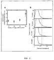
- the inventors also analyzed the fluorescent intensity of the co-localized spots in both the TMR and Alexa-594 channel and found that the spot intensities displayed a unimodal distribution ( FIG. 2 ), arguing that the particles detected are not clumps of many mRNAs but rather individual molecules.
- the spot intensities displayed a strong correlation between the two channels ( FIG. 3 ). Since there is no cross talk between the two channels, this indicates that the variability in spot intensity was not primarily due to random variability in probe hybridization (which would be uncorrelated between different probe sets) but rather other factors, such as mRNA integrity or accessibility, that affect both probes equally.
- the inventors also explored how the signal intensity would vary with the number of probes by performing in situ hybridization using either first 12, 24, 36 probes or all 48 probes in the set. For this particular target mRNA, it was found that particles could be detected with fewer numbers of probes, albeit with decreased intensity ( FIG. 3A ). However, the automatic spot detection algorithm (described in details below) performed particularly well with 48 probes, detecting the same number of spots over a broad range of thresholds ( FIG. 3B , see further discussion below). The number of probes required for robust signal is likely to depend on the target sequence, though, as the inventors have obtained clear mRNA signals using as few as 30 probes. When the instant method was compared to the method of Femino et al.
- CHO cells lacking the reporter gene yielded no signals while CHO cells having the reporter gene that was turned off by addition of doxycycline, yielded mRNA particles in only a few cells, indicating that the signals observed were specific.
- the inventors developed a semiautomated computational algorithm for finding spots in a three-dimensional stack of fluorescent images.
- One of the difficulties associated with spot detection is the nonuniform background arising from cellular autofluoresence and low levels of non-specific probe hybridization.
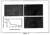
- the inventors filtered image stacks using a three dimensional linear Laplacian of Gaussian filter designed to enhance spot-like signals of the correct size and shape ( FIG . 5A and FIG . 5B ) while removing the slowly varying background.
- the inventors applied a threshold to the filtered image in order to define the spots.
- the number of spots in three dimensions for all thresholds ranging from zero to the maximum pixel intensity in the filtered image was counted.
- the inventors plotted the number of particles as a function of the threshold a wide plateau was found, indicating that there is a region over which the number of particles detected is fairly insensitive to the particular threshold chosen ( FIG. 5C ).
- the spots detected correspond very well with those identified by eye, demonstrating the efficacy of the spot detection algorithm ( FIG . 5D ).
- a potential use of the instantly claimed method is the simultaneous detection of single molecules of multiple mRNAs in individual cells.
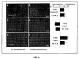
- the inventors designed probes specific to three mRNAs encoding FK506 binding protein 5 (FKBP5), Cox-2 and FLJ11127 in the human carcinoma cell line A549. These probes were coupled to the spectrally distinct fluorophores Cy5, Alexa 594 and TMR, respectively.
- FISH FK506 binding protein 5
- FIG. 6A - FIG. 6F an intensity analysis showed that fluorescent spots did not bleed through into other channels ( FIG. 7 ).
- the cells were incubated with the cell-permeable glucocorticoid dexamethasone, thus upregulating the expression of FKBP5 and FLJ1127 while mildly downregulating the expression of Cox-2 in this particular cell-line.
- the inventors found that the mean number of FKBP5 and FLJ1127 mRNAs measured by combining FISH with the instantly disclosed spot detection algorithm increased while the mean number of Cox-2 mRNAs decreased (compare FIG. 6A - FIG. 6C to FIG. 6D - FIG. 6F ) .
- Example 5 mRNA detection in model organisms and cell types
- RNA localization has been for the detection of RNA localization during development.
- the nematode the inventors constructed probes to detect mRNA molecules from the gene elt-2, a transcription factor that is expressed only in the nematode gut, and only after the nematode embryo has developed to the 45-cell stage. After hybridization of the probe set to both embryos and larvae, it was found that elt-2 mRNA molecules were present only within the gut region ( FIG.
- FIG. 9D shows a full image, in which the locations of the algorithmically identified mRNA molecules are presented as blue circles; and
- FIG. 9E shows an enlarged portion of the image with enhanced mRNA signals. The images show that mRNA molecules were found only at the anterior edge of the area of Engrailed expression, again confirming the specificity of detection.
- the inventors also tested the instantly claimed method in Saccharomyces cerevisae by designing a set of probe to target transcripts from the gene STL1.
- STL1 is one among a number of yeast genes whose expression is significantly up-regulated by the addition of salt to the growth medium. It was found that non-shocked cells contain virtually no STL1 mRNA molecules ( FIG. 10A ), while cells subjected to a ten minute 0.4 M salt shock possessed a large numbers of STL1 mRNA molecules ( FIG. 10B ).
- FIG. 10C shows that a ß-action probe set (labeled with TMR) and a differently colored Map2 probe set (labeled with Alexa-594) can be used to image and distinguish their targets with a single molecule resolution.
- a fraction of these mRNAs migrate to distant reaches of dendrites ( FIG. 10D ).
- Particle counts indicated that 14% of the 791 ß-actin mRNA molecules were located in dendrites, whereas 37% of the 140 Map2 mRNA molecules were located in the dendrites, which is similar to the previously reported distributions.
Claims (13)
- Verfahren zum Nachweis einer ersten Zielsequenz von einzelnen mRNA-Molekülen in einer fixierten, permeabilisierten Zelle, das Folgendes umfasst:Eintauchen der fixierten, permeabilisierten Zelle in einer Hybridisierungslösung, die einen Überschuss einer ersten Gruppe an mindestens 30 nicht überlappenden Nukleinsäure-Hybridisierungssonden enthält, die Sequenzen, die zu der ersten Zielsequenz komplementär sind, aufweisen, die 7-30 Nukleotide in der Länge darstellen und die mit der gleichen ersten Fluorophormarkierung einer ersten Farbe einzeln markiert sind;Waschen der fixierten Zelle, um ungebundene Sonden zu entfernen; undNachweisen von Flecken des ersten Fluorophors in der gewaschenen, fixierten Zelle.
- Verfahren nach Anspruch 1, wobei die Sonden in der Gruppe Ziel-komplementäre Sequenzen von 15-30 Nukleotiden in der Länge aufweisen.
- Verfahren nach Anspruch 1 oder Anspruch 2, wobei alle Sonden in der Gruppe mit einem Fluorophor an ihrem 3'-Ende markiert sind.
- Verfahren nach einem vorstehenden Anspruch, wobei der Schritt des Nachweisens von Flecken die Bildgebung mit einem Fluoreszenzmikroskop einschließt.
- Verfahren nach einem vorstehenden Anspruch, wobei der Schritt des Nachweisens von Flecken Folgendes einschließt: Bildgebung der gewaschenen, fixierten Zelle, um Flecken der ersten Fluorophormarkierung zu zeigen, Bearbeiten des Bilds, um die Flecken zu verstärken, und Analysieren der verstärkten Flecken unter Nutzung eines Intensitätsschwellwerts, bei dem die Anzahl an Flecken gegenüber dem Schwellenwert unempfindlich ist.
- Verfahren nach Anspruch 5, wobei das Bild durch Filtern des Bilds unter Verwendung eines dreidimensionalen linearen Laplacian-of-Gaussian-Filters bearbeitet wird.
- Verfahren nach Anspruch 1, wobei die Hybridisierungslösung einen Überschuss einer zweiten Gruppe an mindestens 30 nicht überlappenden Nukleinsäure-Hybridisierungssonden enthält, die Sequenzen, die zu einer zweiten Zielsequenz komplementär sind, aufweisen, die 7-30 Nukleotide in der Länge darstellen und die mit der gleichen zweiten nachweisbaren Markierung einer zweiten Farbe markiert sind, die von der ersten Fluorophormarkierung unterscheidbar ist.
- Verfahren zur Bestimmung, ob eine Testverbindung eine Menge oder Verteilung einer ersten Zielsequenz von mRNA-Molekülen in einer Zelle beeinflusst, das Folgendes umfasst:(a) Inkubieren der Zelle mit der Testverbindung für eine ausreichende Zeitdauer, um eine Reaktion hervorzurufen;(b) Permeabilisieren der Zelle;(c) Eintauchen der permeabilisierten Zelle in einer Hybridisierungslösung, die einen Überschuss einer ersten Gruppe an mindestens 30 nicht überlappenden Nukleinsäure-Hybridisierungssonden enthält, die Sequenzen, die zu der ersten Zielsequenz komplementär sind, aufweisen, die 7-30 Nukleotide in der Länge darstellen und die mit der gleichen ersten Fluorophormarkierung einer ersten Farbe einzeln markiert sind;(d) Waschen der Zelle, um ungebundene Sonden zu entfernen;(e) Nachweisen einer Menge oder einer Verteilung von Flecken des ersten Fluorophors; und(f) Vergleichen der Menge oder der Verteilung mit denen aus einer Kontrolle erhaltenen, die ähnlich, doch ohne die Testverbindung, behandelt wurde.
- Verfahren nach Anspruch 8, wobei die Sonden in der Gruppe Ziel-komplementäre Sequenzen von 15-30 Nukleotiden in der Länge aufweisen.
- Verfahren nach Anspruch 8 oder Anspruch 9, wobei alle Sonden in der Gruppe mit einem Fluorophor an ihrem 3'-Ende markiert sind.
- Verfahren nach einem der Ansprüche 8 bis 10, wobei der Schritt des Nachweisens Folgendes einschließt: Bearbeiten zur Verstärkung der Verteilung und Analysieren der verstärkten Verteilung unter Nutzung eines Intensitätsschwellwerts, bei dem die Analyse gegenüber dem Schwellenwert unempfindlich ist.
- Verfahren nach Anspruch 8, wobei die Hybridisierungslösung einen Überschuss einer zweiten Gruppe von mindestens 30 nicht überlappenden Nukleinsäure-Hybridisierungssonden enthält, die Sequenzen, die zu einer zweiten Zielsequenz komplementär sind, aufweisen, die 7-30 Nukleotide in der Länge darstellen und die mit der gleichen zweiten nachweisbaren Markierung einer zweiten Farbe markiert sind, die von der ersten nachweisbaren Markierung unterscheidbar ist.
- Verfahren nach Anspruch 8, wobei der Schritt des Nachweisens das Zählen von Flecken, die einzelnen Molekülen von messenger-RNA entsprechen, einschließt, um ein Genexpressionsprofil zu erhalten.
Priority Applications (1)
| Application Number | Priority Date | Filing Date | Title |
|---|---|---|---|
| EP16180033.9A EP3133170B1 (de) | 2008-09-10 | 2009-09-10 | Abbildung einzelner mrna-moleküle mithilfe mehrerer individuell markierter sonden |
Applications Claiming Priority (2)
| Application Number | Priority Date | Filing Date | Title |
|---|---|---|---|
| US19172408P | 2008-09-10 | 2008-09-10 | |
| PCT/US2009/056564 WO2010030818A2 (en) | 2008-09-10 | 2009-09-10 | IMAGING INDIVIDUAL mRNA MOLECULES USING MULTIPLE SINGLY LABELED PROBES |
Related Child Applications (1)
| Application Number | Title | Priority Date | Filing Date |
|---|---|---|---|
| EP16180033.9A Division EP3133170B1 (de) | 2008-09-10 | 2009-09-10 | Abbildung einzelner mrna-moleküle mithilfe mehrerer individuell markierter sonden |
Publications (3)
| Publication Number | Publication Date |
|---|---|
| EP2324123A2 EP2324123A2 (de) | 2011-05-25 |
| EP2324123A4 EP2324123A4 (de) | 2012-12-05 |
| EP2324123B1 true EP2324123B1 (de) | 2016-07-20 |
Family
ID=42005749
Family Applications (2)
| Application Number | Title | Priority Date | Filing Date |
|---|---|---|---|
| EP16180033.9A Active EP3133170B1 (de) | 2008-09-10 | 2009-09-10 | Abbildung einzelner mrna-moleküle mithilfe mehrerer individuell markierter sonden |
| EP09813630.2A Active EP2324123B1 (de) | 2008-09-10 | 2009-09-10 | Abbildung einzelner mrna-moleküle mithilfe individuell markierter sonden |
Family Applications Before (1)
| Application Number | Title | Priority Date | Filing Date |
|---|---|---|---|
| EP16180033.9A Active EP3133170B1 (de) | 2008-09-10 | 2009-09-10 | Abbildung einzelner mrna-moleküle mithilfe mehrerer individuell markierter sonden |
Country Status (8)
| Country | Link |
|---|---|
| US (2) | US9896720B2 (de) |
| EP (2) | EP3133170B1 (de) |
| JP (1) | JP5764730B2 (de) |
| CN (2) | CN108342454A (de) |
| AU (1) | AU2009291752B2 (de) |
| CA (1) | CA2735197C (de) |
| ES (2) | ES2624562T3 (de) |
| WO (1) | WO2010030818A2 (de) |
Families Citing this family (37)
| Publication number | Priority date | Publication date | Assignee | Title |
|---|---|---|---|---|
| EP2620762B1 (de) * | 2010-09-21 | 2017-02-01 | Olympus Corporation | Optisches analyseverfahren zur erkennung einzelner lichtemittierender partikel |
| WO2012106711A2 (en) | 2011-02-04 | 2012-08-09 | The Trustees Of The University Of Pennsylvania | A method for detecting chromosome structure and gene expression simultaneously in single cells |
| JP5885738B2 (ja) * | 2011-04-13 | 2016-03-15 | オリンパス株式会社 | 単一発光粒子検出を用いた光分析装置、光分析方法及び光分析用コンピュータプログラム |
| CN103649334A (zh) | 2011-05-12 | 2014-03-19 | 特拉斯雷神诺癌症医药有限公司 | Kiaa1456表达在结肠癌患者中预测生存 |
| MX2013015286A (es) | 2011-06-20 | 2014-09-25 | Traslational Cancer Drugs Pharma S L | Metodo para predecir la respuesta clinica a quimioterapia en un sujeto con cancer. |
| JP2014531908A (ja) | 2011-10-14 | 2014-12-04 | プレジデント アンド フェローズ オブ ハーバード カレッジ | 構造アッセンブリによる配列決定 |
| US10227639B2 (en) | 2011-12-22 | 2019-03-12 | President And Fellows Of Harvard College | Compositions and methods for analyte detection |
| US11021737B2 (en) | 2011-12-22 | 2021-06-01 | President And Fellows Of Harvard College | Compositions and methods for analyte detection |
| US20130217589A1 (en) * | 2012-02-22 | 2013-08-22 | Jun Xu | Methods for identifying agents with desired biological activity |
| US9914967B2 (en) | 2012-06-05 | 2018-03-13 | President And Fellows Of Harvard College | Spatial sequencing of nucleic acids using DNA origami probes |
| WO2013190081A1 (en) | 2012-06-22 | 2013-12-27 | Proyecto De Biomedicina Cima, S.L. | Methods and reagents for the prognosis of cancer |
| EP2733205B1 (de) | 2012-11-20 | 2018-01-10 | Centro De Investigacíon Biomédica En Red De Enfermedades Neurodegenerativas Ciberned | Obere Pyramidenmotoneuronen, Verfahren und Zusammensetzungen zur Differenzierung neuraler Stammzellen durch Modulation der CB1-Cannabinoidrezeptor-Signalisierung und Verwendungen davon |
| US9042631B2 (en) | 2013-01-24 | 2015-05-26 | General Electric Company | Method and systems for cell-level fish dot counting |
| JP6822765B2 (ja) * | 2013-03-06 | 2021-01-27 | オプティカル バイオシステムズ ホールディング インコーポレイテッド | 分子イメージングおよび関連する方法 |
| US10138509B2 (en) | 2013-03-12 | 2018-11-27 | President And Fellows Of Harvard College | Method for generating a three-dimensional nucleic acid containing matrix |
| EP2784163A1 (de) | 2013-03-25 | 2014-10-01 | Centro De Investigación Biomédica En Red De Enfermedades Neurodegenerativas | Verfahrn zur Prognose und Diagnose von neurodegenerativen Erkrankungen |
| EP2813571A1 (de) | 2013-06-13 | 2014-12-17 | Institut d'Investigació Biomèdica de Bellvitge (IDIBELL) | Verfahren zur Behandlung von Lungenkrebs |
| US20160258005A1 (en) * | 2013-07-02 | 2016-09-08 | The Trustees Of The University Of Pennsylvania | Methods for rapid ribonucleic acid fluorescence in situ hybridization |
| EP3174993B1 (de) | 2014-07-30 | 2023-12-06 | President and Fellows of Harvard College | Konstruktion einer sondenbibliothek |
| WO2016196229A1 (en) * | 2015-06-01 | 2016-12-08 | Cellular Research, Inc. | Methods for rna quantification |
| JP6882282B2 (ja) | 2015-11-03 | 2021-06-02 | プレジデント アンド フェローズ オブ ハーバード カレッジ | 三次元核酸含有マトリックスの立体撮像のための方法と装置 |
| EP3449016A4 (de) | 2016-04-25 | 2019-10-02 | President and Fellows of Harvard College | Hybridisierungskettenreaktionsverfahren für in-situ-moleküldetektion |
| EP3246415A1 (de) | 2016-05-18 | 2017-11-22 | Asociación Centro de Investigación Cooperativa en Biociencias - CIC bioGUNE | Verfahren zur prognose von prostatakrebs |
| US11352667B2 (en) | 2016-06-21 | 2022-06-07 | 10X Genomics, Inc. | Nucleic acid sequencing |
| JP7057348B2 (ja) | 2016-08-31 | 2022-04-19 | プレジデント アンド フェローズ オブ ハーバード カレッジ | 蛍光in situ配列決定を用いた単一アッセイに生体分子の検出を組み合わせる方法 |
| EP3538867B1 (de) * | 2016-11-08 | 2024-01-03 | President and Fellows of Harvard College | Multiplex-bildgebung mittels merfish, expansionsmikroskopie und zugehörige technologien |
| WO2018218150A1 (en) | 2017-05-26 | 2018-11-29 | President And Fellows Of Harvard College | Systems and methods for high-throughput image-based screening |
| US11426075B1 (en) * | 2017-08-23 | 2022-08-30 | Lumicell, Inc. | System and method for residual cancer cell detection |
| EP3728636A1 (de) | 2017-12-19 | 2020-10-28 | Becton, Dickinson and Company | Mit oligonukleotiden assoziierte teilchen |
| EP3674421A1 (de) | 2018-12-28 | 2020-07-01 | Asociación Centro de Investigación Cooperativa en Biociencias - CIC bioGUNE | Verfahren zur prognose von prostatakrebs |
| US11371076B2 (en) | 2019-01-16 | 2022-06-28 | Becton, Dickinson And Company | Polymerase chain reaction normalization through primer titration |
| WO2020214642A1 (en) | 2019-04-19 | 2020-10-22 | Becton, Dickinson And Company | Methods of associating phenotypical data and single cell sequencing data |
| WO2022003146A1 (en) | 2020-07-03 | 2022-01-06 | Fundació Institut D'investigació Biomèdica De Bellvitge (Idibell) | Methods for predicting the risk of local invasion and/or metastasis induced by an antiangiogenic treatment |
| CN116490620A (zh) * | 2020-11-06 | 2023-07-25 | 沃特世科技公司 | 可用于核酸分析的方法和组合物 |
| DE102020131047A1 (de) | 2020-11-24 | 2022-06-09 | Abberior Instruments Gmbh | Verfahren und Vorrichtung zur Aufnahme nanoskopischer Bilder mehrfach gefärbter Proben |
| WO2023283631A2 (en) * | 2021-07-08 | 2023-01-12 | The Broad Institute, Inc. | Methods for differentiating and screening stem cells |
| CN113584133B (zh) * | 2021-08-30 | 2024-03-26 | 中国药科大学 | 基于颜色编码与可编程性荧光探针的多重靶标原位检测方法 |
Family Cites Families (17)
| Publication number | Priority date | Publication date | Assignee | Title |
|---|---|---|---|---|
| US5985549A (en) | 1985-10-22 | 1999-11-16 | University Of Massachusetts | Non-isotopic in-situ hybridization method for detection of nucleic acids |
| US6242184B1 (en) * | 1988-10-18 | 2001-06-05 | University Of Massachusetts | In-situ hybridization of single-copy and multiple-copy nucleic acid sequences |
| US5962332A (en) * | 1994-03-17 | 1999-10-05 | University Of Massachusetts | Detection of trinucleotide repeats by in situ hybridization |
| US5866331A (en) * | 1995-10-20 | 1999-02-02 | University Of Massachusetts | Single molecule detection by in situ hybridization |
| US6203986B1 (en) * | 1998-10-22 | 2001-03-20 | Robert H. Singer | Visualization of RNA in living cells |
| WO2001081632A1 (en) * | 2000-04-25 | 2001-11-01 | Affymetrix, Inc. | Methods for monitoring the expression of alternatively spliced genes |
| JP4986367B2 (ja) * | 2000-09-15 | 2012-07-25 | ベンタナ・メデイカル・システムズ・インコーポレーテツド | イン・サイチュー解析のためのオリゴヌクレオチドプローブおよびタンパク質を標識するためのオリゴヌクレオチド配列式 |
| US6329152B1 (en) * | 2000-11-30 | 2001-12-11 | Bruce K. Patterson | Process for detecting low abundance RNA in intact cells |
| US20020177157A1 (en) * | 2001-05-24 | 2002-11-28 | Yuling Luo | Pairs of nucleic acid probes with interactive signaling moieties and nucleic acid probes with enhanced hybridization efficiency and specificity |
| EP2314691A3 (de) | 2002-11-14 | 2012-01-18 | Dharmacon, Inc. | Funktionale und hyperfunktionale siRNA |
| KR101202974B1 (ko) * | 2003-06-13 | 2012-11-21 | 유니버시티 오브 메디신 앤드 덴티스트리 오브 뉴 저지 | Rna 인터페라제 및 이의 사용 방법 |
| US8062897B2 (en) * | 2003-07-21 | 2011-11-22 | Aureon Laboratories, Inc. | Diagnostic histopathology using multiplex gene expression FISH |
| US20080113344A1 (en) * | 2003-10-28 | 2008-05-15 | Ralph Wirtz | Methods and Compositions for the Response Prediction of Malignant Neoplasia to Treatment |
| EP1752536A4 (de) | 2004-05-11 | 2008-04-16 | Alphagen Co Ltd | Rna-störung verursachendes polynukleotid sowie verfahren zur regulation der genexpression unter verwendung davon |
| JP2006166911A (ja) * | 2004-11-19 | 2006-06-29 | Wakunaga Pharmaceut Co Ltd | 核酸検出用核酸断片および核酸検出方法 |
| US20070099196A1 (en) * | 2004-12-29 | 2007-05-03 | Sakari Kauppinen | Novel oligonucleotide compositions and probe sequences useful for detection and analysis of micrornas and their target mRNAs |
| DK2500439T4 (da) * | 2005-06-20 | 2017-11-13 | Advanced Cell Diagnostics Inc | Kits og produkter til detektering af nukleinsyrer i individuelle celler og til identifikation af sjældne celler fra store heterogene cellepopulationer |
-
2009
- 2009-09-10 CN CN201810154172.2A patent/CN108342454A/zh active Pending
- 2009-09-10 US US13/062,975 patent/US9896720B2/en active Active
- 2009-09-10 WO PCT/US2009/056564 patent/WO2010030818A2/en active Application Filing
- 2009-09-10 ES ES09813630.2T patent/ES2624562T3/es active Active
- 2009-09-10 EP EP16180033.9A patent/EP3133170B1/de active Active
- 2009-09-10 AU AU2009291752A patent/AU2009291752B2/en active Active
- 2009-09-10 CA CA2735197A patent/CA2735197C/en active Active
- 2009-09-10 JP JP2011526977A patent/JP5764730B2/ja active Active
- 2009-09-10 CN CN2009801353648A patent/CN102149829A/zh active Pending
- 2009-09-10 ES ES16180033T patent/ES2799507T3/es active Active
- 2009-09-10 EP EP09813630.2A patent/EP2324123B1/de active Active
-
2018
- 2018-01-30 US US15/883,955 patent/US20180171396A1/en not_active Abandoned
Also Published As
| Publication number | Publication date |
|---|---|
| WO2010030818A2 (en) | 2010-03-18 |
| CA2735197C (en) | 2017-05-09 |
| EP3133170A1 (de) | 2017-02-22 |
| EP2324123A4 (de) | 2012-12-05 |
| EP3133170B1 (de) | 2020-03-18 |
| JP5764730B2 (ja) | 2015-08-19 |
| US9896720B2 (en) | 2018-02-20 |
| EP2324123A2 (de) | 2011-05-25 |
| CN102149829A (zh) | 2011-08-10 |
| CA2735197A1 (en) | 2010-03-18 |
| WO2010030818A3 (en) | 2010-07-22 |
| US20180171396A1 (en) | 2018-06-21 |
| CN108342454A (zh) | 2018-07-31 |
| US20120129165A1 (en) | 2012-05-24 |
| AU2009291752A1 (en) | 2010-03-18 |
| ES2624562T3 (es) | 2017-07-14 |
| AU2009291752B2 (en) | 2015-12-17 |
| ES2799507T3 (es) | 2020-12-18 |
| JP2012501679A (ja) | 2012-01-26 |
Similar Documents
| Publication | Publication Date | Title |
|---|---|---|
| US20180171396A1 (en) | Imaging individual mrna molecules using multiple singly labeled probes | |
| US20230212658A1 (en) | Multiplex labeling of molecules by sequential hybridization barcoding | |
| US10266879B2 (en) | Detection of nucleic acids | |
| US20160369329A1 (en) | Multiplex labeling of molecules by sequential hybridization barcoding using probes with cleavable linkers | |
| Kwon | Single-molecule fluorescence in situ hybridization: quantitative imaging of single RNA molecules | |
| Chan et al. | Direct quantification of single-molecules of microRNA by total internal reflection fluorescence microscopy | |
| Gaspar et al. | Strength in numbers: quantitative single‐molecule RNA detection assays | |
| WO2005079462A2 (en) | Methods and materials using signaling probes | |
| Wang et al. | A quick and simple FISH protocol with hybridization-sensitive fluorescent linear oligodeoxynucleotide probes | |
| US20140220574A1 (en) | Methods for fixing and detecting rna | |
| Markey et al. | Methods for spatial and temporal imaging of the different steps involved in RNA processing at single‐molecule resolution | |
| JP2020516278A (ja) | 多種多様なライゲーションによるオリゴヌクレオチドプローブの標識 |
Legal Events
| Date | Code | Title | Description |
|---|---|---|---|
| PUAI | Public reference made under article 153(3) epc to a published international application that has entered the european phase |
Free format text: ORIGINAL CODE: 0009012 |
|
| 17P | Request for examination filed |
Effective date: 20110224 |
|
| AK | Designated contracting states |
Kind code of ref document: A2 Designated state(s): AT BE BG CH CY CZ DE DK EE ES FI FR GB GR HR HU IE IS IT LI LT LU LV MC MK MT NL NO PL PT RO SE SI SK SM TR |
|
| AX | Request for extension of the european patent |
Extension state: AL BA RS |
|
| RIN1 | Information on inventor provided before grant (corrected) |
Inventor name: TYAGI, SANJAY Inventor name: RAJ, ARJUN |
|
| DAX | Request for extension of the european patent (deleted) | ||
| RIN1 | Information on inventor provided before grant (corrected) |
Inventor name: RAJ, ARJUN Inventor name: TYAGI, SANJAY |
|
| RIC1 | Information provided on ipc code assigned before grant |
Ipc: C12Q 1/68 20060101AFI20121024BHEP Ipc: C07H 21/00 20060101ALI20121024BHEP |
|
| A4 | Supplementary search report drawn up and despatched |
Effective date: 20121105 |
|
| 17Q | First examination report despatched |
Effective date: 20131121 |
|
| RAP1 | Party data changed (applicant data changed or rights of an application transferred) |
Owner name: RUTGERS, THE STATE UNIVERSITY OF NEW JERSEY |
|
| GRAP | Despatch of communication of intention to grant a patent |
Free format text: ORIGINAL CODE: EPIDOSNIGR1 |
|
| INTG | Intention to grant announced |
Effective date: 20151019 |
|
| GRAS | Grant fee paid |
Free format text: ORIGINAL CODE: EPIDOSNIGR3 |
|
| GRAP | Despatch of communication of intention to grant a patent |
Free format text: ORIGINAL CODE: EPIDOSNIGR1 |
|
| INTG | Intention to grant announced |
Effective date: 20160329 |
|
| GRAA | (expected) grant |
Free format text: ORIGINAL CODE: 0009210 |
|
| AK | Designated contracting states |
Kind code of ref document: B1 Designated state(s): AT BE BG CH CY CZ DE DK EE ES FI FR GB GR HR HU IE IS IT LI LT LU LV MC MK MT NL NO PL PT RO SE SI SK SM TR |
|
| REG | Reference to a national code |
Ref country code: GB Ref legal event code: FG4D |
|
| REG | Reference to a national code |
Ref country code: CH Ref legal event code: EP |
|
| REG | Reference to a national code |
Ref country code: IE Ref legal event code: FG4D |
|
| REG | Reference to a national code |
Ref country code: AT Ref legal event code: REF Ref document number: 814160 Country of ref document: AT Kind code of ref document: T Effective date: 20160815 |
|
| REG | Reference to a national code |
Ref country code: DE Ref legal event code: R096 Ref document number: 602009039886 Country of ref document: DE |
|
| REG | Reference to a national code |
Ref country code: LT Ref legal event code: MG4D |
|
| REG | Reference to a national code |
Ref country code: NL Ref legal event code: MP Effective date: 20160720 |
|
| REG | Reference to a national code |
Ref country code: AT Ref legal event code: MK05 Ref document number: 814160 Country of ref document: AT Kind code of ref document: T Effective date: 20160720 |
|
| PG25 | Lapsed in a contracting state [announced via postgrant information from national office to epo] |
Ref country code: NO Free format text: LAPSE BECAUSE OF FAILURE TO SUBMIT A TRANSLATION OF THE DESCRIPTION OR TO PAY THE FEE WITHIN THE PRESCRIBED TIME-LIMIT Effective date: 20161020 Ref country code: HR Free format text: LAPSE BECAUSE OF FAILURE TO SUBMIT A TRANSLATION OF THE DESCRIPTION OR TO PAY THE FEE WITHIN THE PRESCRIBED TIME-LIMIT Effective date: 20160720 Ref country code: LT Free format text: LAPSE BECAUSE OF FAILURE TO SUBMIT A TRANSLATION OF THE DESCRIPTION OR TO PAY THE FEE WITHIN THE PRESCRIBED TIME-LIMIT Effective date: 20160720 Ref country code: IS Free format text: LAPSE BECAUSE OF FAILURE TO SUBMIT A TRANSLATION OF THE DESCRIPTION OR TO PAY THE FEE WITHIN THE PRESCRIBED TIME-LIMIT Effective date: 20161120 Ref country code: NL Free format text: LAPSE BECAUSE OF FAILURE TO SUBMIT A TRANSLATION OF THE DESCRIPTION OR TO PAY THE FEE WITHIN THE PRESCRIBED TIME-LIMIT Effective date: 20160720 Ref country code: FI Free format text: LAPSE BECAUSE OF FAILURE TO SUBMIT A TRANSLATION OF THE DESCRIPTION OR TO PAY THE FEE WITHIN THE PRESCRIBED TIME-LIMIT Effective date: 20160720 |
|
| PG25 | Lapsed in a contracting state [announced via postgrant information from national office to epo] |
Ref country code: SE Free format text: LAPSE BECAUSE OF FAILURE TO SUBMIT A TRANSLATION OF THE DESCRIPTION OR TO PAY THE FEE WITHIN THE PRESCRIBED TIME-LIMIT Effective date: 20160720 Ref country code: PL Free format text: LAPSE BECAUSE OF FAILURE TO SUBMIT A TRANSLATION OF THE DESCRIPTION OR TO PAY THE FEE WITHIN THE PRESCRIBED TIME-LIMIT Effective date: 20160720 Ref country code: PT Free format text: LAPSE BECAUSE OF FAILURE TO SUBMIT A TRANSLATION OF THE DESCRIPTION OR TO PAY THE FEE WITHIN THE PRESCRIBED TIME-LIMIT Effective date: 20161121 Ref country code: GR Free format text: LAPSE BECAUSE OF FAILURE TO SUBMIT A TRANSLATION OF THE DESCRIPTION OR TO PAY THE FEE WITHIN THE PRESCRIBED TIME-LIMIT Effective date: 20161021 Ref country code: LV Free format text: LAPSE BECAUSE OF FAILURE TO SUBMIT A TRANSLATION OF THE DESCRIPTION OR TO PAY THE FEE WITHIN THE PRESCRIBED TIME-LIMIT Effective date: 20160720 Ref country code: BE Free format text: LAPSE BECAUSE OF NON-PAYMENT OF DUE FEES Effective date: 20160720 Ref country code: AT Free format text: LAPSE BECAUSE OF FAILURE TO SUBMIT A TRANSLATION OF THE DESCRIPTION OR TO PAY THE FEE WITHIN THE PRESCRIBED TIME-LIMIT Effective date: 20160720 |
|
| REG | Reference to a national code |
Ref country code: DE Ref legal event code: R119 Ref document number: 602009039886 Country of ref document: DE |
|
| REG | Reference to a national code |
Ref country code: DE Ref legal event code: R097 Ref document number: 602009039886 Country of ref document: DE |
|
| PG25 | Lapsed in a contracting state [announced via postgrant information from national office to epo] |
Ref country code: MC Free format text: LAPSE BECAUSE OF FAILURE TO SUBMIT A TRANSLATION OF THE DESCRIPTION OR TO PAY THE FEE WITHIN THE PRESCRIBED TIME-LIMIT Effective date: 20160720 Ref country code: RO Free format text: LAPSE BECAUSE OF FAILURE TO SUBMIT A TRANSLATION OF THE DESCRIPTION OR TO PAY THE FEE WITHIN THE PRESCRIBED TIME-LIMIT Effective date: 20160720 Ref country code: EE Free format text: LAPSE BECAUSE OF FAILURE TO SUBMIT A TRANSLATION OF THE DESCRIPTION OR TO PAY THE FEE WITHIN THE PRESCRIBED TIME-LIMIT Effective date: 20160720 |
|
| REG | Reference to a national code |
Ref country code: CH Ref legal event code: PL |
|
| PLBE | No opposition filed within time limit |
Free format text: ORIGINAL CODE: 0009261 |
|
| STAA | Information on the status of an ep patent application or granted ep patent |
Free format text: STATUS: NO OPPOSITION FILED WITHIN TIME LIMIT |
|
| PG25 | Lapsed in a contracting state [announced via postgrant information from national office to epo] |
Ref country code: BG Free format text: LAPSE BECAUSE OF FAILURE TO SUBMIT A TRANSLATION OF THE DESCRIPTION OR TO PAY THE FEE WITHIN THE PRESCRIBED TIME-LIMIT Effective date: 20161020 Ref country code: CZ Free format text: LAPSE BECAUSE OF FAILURE TO SUBMIT A TRANSLATION OF THE DESCRIPTION OR TO PAY THE FEE WITHIN THE PRESCRIBED TIME-LIMIT Effective date: 20160720 Ref country code: DK Free format text: LAPSE BECAUSE OF FAILURE TO SUBMIT A TRANSLATION OF THE DESCRIPTION OR TO PAY THE FEE WITHIN THE PRESCRIBED TIME-LIMIT Effective date: 20160720 Ref country code: SM Free format text: LAPSE BECAUSE OF FAILURE TO SUBMIT A TRANSLATION OF THE DESCRIPTION OR TO PAY THE FEE WITHIN THE PRESCRIBED TIME-LIMIT Effective date: 20160720 Ref country code: SK Free format text: LAPSE BECAUSE OF FAILURE TO SUBMIT A TRANSLATION OF THE DESCRIPTION OR TO PAY THE FEE WITHIN THE PRESCRIBED TIME-LIMIT Effective date: 20160720 |
|
| 26N | No opposition filed |
Effective date: 20170421 |
|
| GBPC | Gb: european patent ceased through non-payment of renewal fee |
Effective date: 20161020 |
|
| REG | Reference to a national code |
Ref country code: IE Ref legal event code: MM4A |
|
| REG | Reference to a national code |
Ref country code: FR Ref legal event code: ST Effective date: 20170531 |
|
| REG | Reference to a national code |
Ref country code: GB Ref legal event code: S28 Free format text: APPLICATION FILED |
|
| REG | Reference to a national code |
Ref country code: ES Ref legal event code: FG2A Ref document number: 2624562 Country of ref document: ES Kind code of ref document: T3 Effective date: 20170714 |
|
| REG | Reference to a national code |
Ref country code: FR Ref legal event code: PLFP Year of fee payment: 8 |
|
| PG25 | Lapsed in a contracting state [announced via postgrant information from national office to epo] |
Ref country code: LI Free format text: LAPSE BECAUSE OF NON-PAYMENT OF DUE FEES Effective date: 20160930 Ref country code: FR Free format text: LAPSE BECAUSE OF NON-PAYMENT OF DUE FEES Effective date: 20160930 Ref country code: DE Free format text: LAPSE BECAUSE OF NON-PAYMENT OF DUE FEES Effective date: 20170401 Ref country code: GB Free format text: LAPSE BECAUSE OF NON-PAYMENT OF DUE FEES Effective date: 20161020 Ref country code: IE Free format text: LAPSE BECAUSE OF NON-PAYMENT OF DUE FEES Effective date: 20160910 Ref country code: CH Free format text: LAPSE BECAUSE OF NON-PAYMENT OF DUE FEES Effective date: 20160930 |
|
| REG | Reference to a national code |
Ref country code: DE Ref legal event code: R073 Ref document number: 602009039886 Country of ref document: DE |
|
| REG | Reference to a national code |
Ref country code: DE Ref legal event code: R082 Ref document number: 602009039886 Country of ref document: DE Representative=s name: PATENTANWAELTE RUFF, WILHELM, BEIER, DAUSTER &, DE |
|
| REG | Reference to a national code |
Ref country code: FR Ref legal event code: RN Effective date: 20170724 |
|
| PG25 | Lapsed in a contracting state [announced via postgrant information from national office to epo] |
Ref country code: LU Free format text: LAPSE BECAUSE OF NON-PAYMENT OF DUE FEES Effective date: 20160910 Ref country code: IT Free format text: LAPSE BECAUSE OF NON-PAYMENT OF DUE FEES Effective date: 20160910 Ref country code: SI Free format text: LAPSE BECAUSE OF FAILURE TO SUBMIT A TRANSLATION OF THE DESCRIPTION OR TO PAY THE FEE WITHIN THE PRESCRIBED TIME-LIMIT Effective date: 20160720 |
|
| REG | Reference to a national code |
Ref country code: GB Ref legal event code: S28 Free format text: RESTORATION ALLOWED Effective date: 20170817 |
|
| REG | Reference to a national code |
Ref country code: DE Ref legal event code: R074 Ref document number: 602009039886 Country of ref document: DE |
|
| REG | Reference to a national code |
Ref country code: FR Ref legal event code: PLFP Year of fee payment: 9 |
|
| REG | Reference to a national code |
Ref country code: FR Ref legal event code: FC Effective date: 20170829 |
|
| PG25 | Lapsed in a contracting state [announced via postgrant information from national office to epo] |
Ref country code: FR Free format text: LAPSE BECAUSE OF NON-PAYMENT OF DUE FEES Effective date: 20160930 Ref country code: DE Free format text: LAPSE BECAUSE OF NON-PAYMENT OF DUE FEES Effective date: 20170401 |
|
| PGRI | Patent reinstated in contracting state [announced from national office to epo] |
Ref country code: DE Effective date: 20170921 Ref country code: FR Effective date: 20170830 |
|
| PG25 | Lapsed in a contracting state [announced via postgrant information from national office to epo] |
Ref country code: HU Free format text: LAPSE BECAUSE OF FAILURE TO SUBMIT A TRANSLATION OF THE DESCRIPTION OR TO PAY THE FEE WITHIN THE PRESCRIBED TIME-LIMIT; INVALID AB INITIO Effective date: 20090910 Ref country code: IT Free format text: LAPSE BECAUSE OF NON-PAYMENT OF DUE FEES Effective date: 20160910 Ref country code: CY Free format text: LAPSE BECAUSE OF FAILURE TO SUBMIT A TRANSLATION OF THE DESCRIPTION OR TO PAY THE FEE WITHIN THE PRESCRIBED TIME-LIMIT Effective date: 20160720 |
|
| PGRI | Patent reinstated in contracting state [announced from national office to epo] |
Ref country code: IT Effective date: 20180219 |
|
| REG | Reference to a national code |
Ref country code: FR Ref legal event code: PLFP Year of fee payment: 10 |
|
| PG25 | Lapsed in a contracting state [announced via postgrant information from national office to epo] |
Ref country code: MT Free format text: LAPSE BECAUSE OF NON-PAYMENT OF DUE FEES Effective date: 20160930 Ref country code: MK Free format text: LAPSE BECAUSE OF FAILURE TO SUBMIT A TRANSLATION OF THE DESCRIPTION OR TO PAY THE FEE WITHIN THE PRESCRIBED TIME-LIMIT Effective date: 20160720 Ref country code: TR Free format text: LAPSE BECAUSE OF FAILURE TO SUBMIT A TRANSLATION OF THE DESCRIPTION OR TO PAY THE FEE WITHIN THE PRESCRIBED TIME-LIMIT Effective date: 20160720 |
|
| PGFP | Annual fee paid to national office [announced via postgrant information from national office to epo] |
Ref country code: IT Payment date: 20200904 Year of fee payment: 12 |
|
| PG25 | Lapsed in a contracting state [announced via postgrant information from national office to epo] |
Ref country code: IT Free format text: LAPSE BECAUSE OF NON-PAYMENT OF DUE FEES Effective date: 20210930 |
|
| P01 | Opt-out of the competence of the unified patent court (upc) registered |
Effective date: 20230530 |
|
| PGFP | Annual fee paid to national office [announced via postgrant information from national office to epo] |
Ref country code: GB Payment date: 20230927 Year of fee payment: 15 |
|
| PGFP | Annual fee paid to national office [announced via postgrant information from national office to epo] |
Ref country code: FR Payment date: 20230925 Year of fee payment: 15 Ref country code: DE Payment date: 20230927 Year of fee payment: 15 |
|
| PGFP | Annual fee paid to national office [announced via postgrant information from national office to epo] |
Ref country code: ES Payment date: 20231002 Year of fee payment: 15 |