EP2288690B1 - Methods for producing enucleated erythroid cells derived from pluripotent stem cells - Google Patents
Methods for producing enucleated erythroid cells derived from pluripotent stem cells Download PDFInfo
- Publication number
- EP2288690B1 EP2288690B1 EP09743615.8A EP09743615A EP2288690B1 EP 2288690 B1 EP2288690 B1 EP 2288690B1 EP 09743615 A EP09743615 A EP 09743615A EP 2288690 B1 EP2288690 B1 EP 2288690B1
- Authority
- EP
- European Patent Office
- Prior art keywords
- cells
- cell
- human
- pluripotent stem
- engrafting hemangio
- Prior art date
- Legal status (The legal status is an assumption and is not a legal conclusion. Google has not performed a legal analysis and makes no representation as to the accuracy of the status listed.)
- Active
Links
- 238000000034 method Methods 0.000 title claims description 325
- 210000001778 pluripotent stem cell Anatomy 0.000 title claims description 196
- 210000000267 erythroid cell Anatomy 0.000 title claims description 93
- 210000004027 cell Anatomy 0.000 claims description 1276
- 210000003566 hemangioblast Anatomy 0.000 claims description 433
- 241000282414 Homo sapiens Species 0.000 claims description 406
- 108090000623 proteins and genes Proteins 0.000 claims description 123
- 101000839788 Homo sapiens Homeobox protein Hox-B4 Proteins 0.000 claims description 101
- 102100028404 Homeobox protein Hox-B4 Human genes 0.000 claims description 96
- 210000002242 embryoid body Anatomy 0.000 claims description 96
- 239000003102 growth factor Substances 0.000 claims description 87
- 210000003969 blast cell Anatomy 0.000 claims description 77
- 102000004169 proteins and genes Human genes 0.000 claims description 76
- 210000001671 embryonic stem cell Anatomy 0.000 claims description 68
- 230000004069 differentiation Effects 0.000 claims description 67
- 238000000338 in vitro Methods 0.000 claims description 58
- 210000000130 stem cell Anatomy 0.000 claims description 56
- 229940105423 erythropoietin Drugs 0.000 claims description 55
- 108090000394 Erythropoietin Proteins 0.000 claims description 54
- 102000003951 Erythropoietin Human genes 0.000 claims description 54
- 239000002609 medium Substances 0.000 claims description 54
- 239000004017 serum-free culture medium Substances 0.000 claims description 54
- OXCMYAYHXIHQOA-UHFFFAOYSA-N potassium;[2-butyl-5-chloro-3-[[4-[2-(1,2,4-triaza-3-azanidacyclopenta-1,4-dien-5-yl)phenyl]phenyl]methyl]imidazol-4-yl]methanol Chemical compound [K+].CCCCC1=NC(Cl)=C(CO)N1CC1=CC=C(C=2C(=CC=CC=2)C2=N[N-]N=N2)C=C1 OXCMYAYHXIHQOA-UHFFFAOYSA-N 0.000 claims description 51
- 108010073929 Vascular Endothelial Growth Factor A Proteins 0.000 claims description 50
- 102000005789 Vascular Endothelial Growth Factors Human genes 0.000 claims description 50
- 108010019530 Vascular Endothelial Growth Factors Proteins 0.000 claims description 50
- 108010041111 Thrombopoietin Proteins 0.000 claims description 46
- 102000036693 Thrombopoietin Human genes 0.000 claims description 46
- -1 SCF Chemical compound 0.000 claims description 38
- 238000012258 culturing Methods 0.000 claims description 36
- 108010049955 Bone Morphogenetic Protein 4 Proteins 0.000 claims description 32
- 102000003974 Fibroblast growth factor 2 Human genes 0.000 claims description 28
- 108090000379 Fibroblast growth factor 2 Proteins 0.000 claims description 28
- 238000004113 cell culture Methods 0.000 claims description 28
- NOESYZHRGYRDHS-UHFFFAOYSA-N insulin Chemical compound N1C(=O)C(NC(=O)C(CCC(N)=O)NC(=O)C(CCC(O)=O)NC(=O)C(C(C)C)NC(=O)C(NC(=O)CN)C(C)CC)CSSCC(C(NC(CO)C(=O)NC(CC(C)C)C(=O)NC(CC=2C=CC(O)=CC=2)C(=O)NC(CCC(N)=O)C(=O)NC(CC(C)C)C(=O)NC(CCC(O)=O)C(=O)NC(CC(N)=O)C(=O)NC(CC=2C=CC(O)=CC=2)C(=O)NC(CSSCC(NC(=O)C(C(C)C)NC(=O)C(CC(C)C)NC(=O)C(CC=2C=CC(O)=CC=2)NC(=O)C(CC(C)C)NC(=O)C(C)NC(=O)C(CCC(O)=O)NC(=O)C(C(C)C)NC(=O)C(CC(C)C)NC(=O)C(CC=2NC=NC=2)NC(=O)C(CO)NC(=O)CNC2=O)C(=O)NCC(=O)NC(CCC(O)=O)C(=O)NC(CCCNC(N)=N)C(=O)NCC(=O)NC(CC=3C=CC=CC=3)C(=O)NC(CC=3C=CC=CC=3)C(=O)NC(CC=3C=CC(O)=CC=3)C(=O)NC(C(C)O)C(=O)N3C(CCC3)C(=O)NC(CCCCN)C(=O)NC(C)C(O)=O)C(=O)NC(CC(N)=O)C(O)=O)=O)NC(=O)C(C(C)CC)NC(=O)C(CO)NC(=O)C(C(C)O)NC(=O)C1CSSCC2NC(=O)C(CC(C)C)NC(=O)C(NC(=O)C(CCC(N)=O)NC(=O)C(CC(N)=O)NC(=O)C(NC(=O)C(N)CC=1C=CC=CC=1)C(C)C)CC1=CN=CN1 NOESYZHRGYRDHS-UHFFFAOYSA-N 0.000 claims description 21
- 102000037865 fusion proteins Human genes 0.000 claims description 19
- 108020001507 fusion proteins Proteins 0.000 claims description 19
- 239000001963 growth medium Substances 0.000 claims description 19
- 210000005260 human cell Anatomy 0.000 claims description 19
- JYGXADMDTFJGBT-VWUMJDOOSA-N hydrocortisone Chemical compound O=C1CC[C@]2(C)[C@H]3[C@@H](O)C[C@](C)([C@@](CC4)(O)C(=O)CO)[C@@H]4[C@@H]3CCC2=C1 JYGXADMDTFJGBT-VWUMJDOOSA-N 0.000 claims description 18
- 108010002386 Interleukin-3 Proteins 0.000 claims description 16
- 210000004263 induced pluripotent stem cell Anatomy 0.000 claims description 15
- 238000010361 transduction Methods 0.000 claims description 14
- 230000026683 transduction Effects 0.000 claims description 14
- 239000003795 chemical substances by application Substances 0.000 claims description 13
- 230000000921 morphogenic effect Effects 0.000 claims description 13
- 210000002536 stromal cell Anatomy 0.000 claims description 13
- 239000013589 supplement Substances 0.000 claims description 13
- 210000000988 bone and bone Anatomy 0.000 claims description 12
- OVBPIULPVIDEAO-LBPRGKRZSA-N folic acid Chemical compound C=1N=C2NC(N)=NC(=O)C2=NC=1CNC1=CC=C(C(=O)N[C@@H](CCC(O)=O)C(O)=O)C=C1 OVBPIULPVIDEAO-LBPRGKRZSA-N 0.000 claims description 12
- 229940125396 insulin Drugs 0.000 claims description 12
- 210000002901 mesenchymal stem cell Anatomy 0.000 claims description 12
- 101100018264 Mus musculus Hoxb4 gene Proteins 0.000 claims description 11
- 102000004877 Insulin Human genes 0.000 claims description 9
- 108090001061 Insulin Proteins 0.000 claims description 9
- 229960000890 hydrocortisone Drugs 0.000 claims description 9
- 239000012581 transferrin Substances 0.000 claims description 9
- 102000004338 Transferrin Human genes 0.000 claims description 8
- 108090000901 Transferrin Proteins 0.000 claims description 8
- ZDXPYRJPNDTMRX-VKHMYHEASA-N L-glutamine Chemical compound OC(=O)[C@@H](N)CCC(N)=O ZDXPYRJPNDTMRX-VKHMYHEASA-N 0.000 claims description 7
- 229930182816 L-glutamine Natural products 0.000 claims description 7
- 239000012679 serum free medium Substances 0.000 claims description 7
- CWYNVVGOOAEACU-UHFFFAOYSA-N Fe2+ Chemical compound [Fe+2] CWYNVVGOOAEACU-UHFFFAOYSA-N 0.000 claims description 6
- SQUHHTBVTRBESD-UHFFFAOYSA-N Hexa-Ac-myo-Inositol Natural products CC(=O)OC1C(OC(C)=O)C(OC(C)=O)C(OC(C)=O)C(OC(C)=O)C1OC(C)=O SQUHHTBVTRBESD-UHFFFAOYSA-N 0.000 claims description 6
- OVBPIULPVIDEAO-UHFFFAOYSA-N N-Pteroyl-L-glutaminsaeure Natural products C=1N=C2NC(N)=NC(=O)C2=NC=1CNC1=CC=C(C(=O)NC(CCC(O)=O)C(O)=O)C=C1 OVBPIULPVIDEAO-UHFFFAOYSA-N 0.000 claims description 6
- 229910002651 NO3 Inorganic materials 0.000 claims description 6
- NHNBFGGVMKEFGY-UHFFFAOYSA-N Nitrate Chemical compound [O-][N+]([O-])=O NHNBFGGVMKEFGY-UHFFFAOYSA-N 0.000 claims description 6
- 235000003891 ferrous sulphate Nutrition 0.000 claims description 6
- 239000011790 ferrous sulphate Substances 0.000 claims description 6
- 229960000304 folic acid Drugs 0.000 claims description 6
- 235000019152 folic acid Nutrition 0.000 claims description 6
- 239000011724 folic acid Substances 0.000 claims description 6
- 229960000367 inositol Drugs 0.000 claims description 6
- CDAISMWEOUEBRE-GPIVLXJGSA-N inositol Chemical compound O[C@H]1[C@H](O)[C@@H](O)[C@H](O)[C@H](O)[C@@H]1O CDAISMWEOUEBRE-GPIVLXJGSA-N 0.000 claims description 6
- BAUYGSIQEAFULO-UHFFFAOYSA-L iron(2+) sulfate (anhydrous) Chemical compound [Fe+2].[O-]S([O-])(=O)=O BAUYGSIQEAFULO-UHFFFAOYSA-L 0.000 claims description 6
- 229910000359 iron(II) sulfate Inorganic materials 0.000 claims description 6
- PJUIMOJAAPLTRJ-UHFFFAOYSA-N monothioglycerol Chemical compound OCC(O)CS PJUIMOJAAPLTRJ-UHFFFAOYSA-N 0.000 claims description 6
- CDAISMWEOUEBRE-UHFFFAOYSA-N scyllo-inosotol Natural products OC1C(O)C(O)C(O)C(O)C1O CDAISMWEOUEBRE-UHFFFAOYSA-N 0.000 claims description 6
- 102000054609 human HOXB4 Human genes 0.000 claims description 5
- 229960005322 streptomycin Drugs 0.000 claims description 5
- 102000008137 Bone Morphogenetic Protein 4 Human genes 0.000 claims 7
- 230000001419 dependent effect Effects 0.000 claims 3
- 210000003958 hematopoietic stem cell Anatomy 0.000 description 150
- 210000003743 erythrocyte Anatomy 0.000 description 142
- 238000002360 preparation method Methods 0.000 description 105
- 210000004369 blood Anatomy 0.000 description 94
- 239000008280 blood Substances 0.000 description 94
- 210000002889 endothelial cell Anatomy 0.000 description 78
- 230000003394 haemopoietic effect Effects 0.000 description 69
- 238000010494 dissociation reaction Methods 0.000 description 67
- 230000005593 dissociations Effects 0.000 description 67
- 238000011282 treatment Methods 0.000 description 61
- 230000014509 gene expression Effects 0.000 description 56
- 239000000203 mixture Substances 0.000 description 53
- 210000001519 tissue Anatomy 0.000 description 53
- 230000001464 adherent effect Effects 0.000 description 47
- 102100031573 Hematopoietic progenitor cell antigen CD34 Human genes 0.000 description 38
- 101000777663 Homo sapiens Hematopoietic progenitor cell antigen CD34 Proteins 0.000 description 38
- 210000001185 bone marrow Anatomy 0.000 description 37
- 230000002255 enzymatic effect Effects 0.000 description 34
- 210000001772 blood platelet Anatomy 0.000 description 33
- 102100024505 Bone morphogenetic protein 4 Human genes 0.000 description 32
- 210000003593 megakaryocyte Anatomy 0.000 description 32
- 230000001225 therapeutic effect Effects 0.000 description 30
- 208000037265 diseases, disorders, signs and symptoms Diseases 0.000 description 29
- 210000001161 mammalian embryo Anatomy 0.000 description 29
- 108091003079 Bovine Serum Albumin Proteins 0.000 description 28
- 108700028369 Alleles Proteins 0.000 description 26
- 238000011161 development Methods 0.000 description 26
- 230000018109 developmental process Effects 0.000 description 26
- 201000010099 disease Diseases 0.000 description 26
- 230000007159 enucleation Effects 0.000 description 26
- 239000000243 solution Substances 0.000 description 26
- 239000000825 pharmaceutical preparation Substances 0.000 description 25
- 108091007433 antigens Proteins 0.000 description 24
- 230000012010 growth Effects 0.000 description 24
- 108090000765 processed proteins & peptides Proteins 0.000 description 24
- 241000699666 Mus <mouse, genus> Species 0.000 description 23
- 239000000427 antigen Substances 0.000 description 23
- 102000036639 antigens Human genes 0.000 description 23
- 101000610551 Homo sapiens Prominin-1 Proteins 0.000 description 22
- 102100040120 Prominin-1 Human genes 0.000 description 22
- 229940098773 bovine serum albumin Drugs 0.000 description 22
- 238000004519 manufacturing process Methods 0.000 description 22
- 102100024616 Platelet endothelial cell adhesion molecule Human genes 0.000 description 21
- 102100033177 Vascular endothelial growth factor receptor 2 Human genes 0.000 description 21
- 102100039064 Interleukin-3 Human genes 0.000 description 19
- 102100023876 Rhombotin-2 Human genes 0.000 description 19
- 210000004204 blood vessel Anatomy 0.000 description 19
- 230000035800 maturation Effects 0.000 description 19
- 210000004700 fetal blood Anatomy 0.000 description 18
- 210000001082 somatic cell Anatomy 0.000 description 18
- 241000894007 species Species 0.000 description 18
- 101001111742 Homo sapiens Rhombotin-2 Proteins 0.000 description 17
- 102100031785 Endothelial transcription factor GATA-2 Human genes 0.000 description 16
- 108010054147 Hemoglobins Proteins 0.000 description 16
- 102000001554 Hemoglobins Human genes 0.000 description 16
- 101001066265 Homo sapiens Endothelial transcription factor GATA-2 Proteins 0.000 description 16
- 230000015572 biosynthetic process Effects 0.000 description 16
- 239000012634 fragment Substances 0.000 description 16
- 229910052760 oxygen Inorganic materials 0.000 description 16
- 102000004196 processed proteins & peptides Human genes 0.000 description 16
- 102000004127 Cytokines Human genes 0.000 description 15
- 108090000695 Cytokines Proteins 0.000 description 15
- 108020004414 DNA Proteins 0.000 description 15
- 210000002459 blastocyst Anatomy 0.000 description 15
- 238000001727 in vivo Methods 0.000 description 15
- 239000000047 product Substances 0.000 description 15
- 108091005904 Hemoglobin subunit beta Proteins 0.000 description 14
- 239000003153 chemical reaction reagent Substances 0.000 description 14
- 230000003511 endothelial effect Effects 0.000 description 14
- 230000001605 fetal effect Effects 0.000 description 14
- 229920001184 polypeptide Polymers 0.000 description 14
- 102000005962 receptors Human genes 0.000 description 14
- 108020003175 receptors Proteins 0.000 description 14
- 102100021519 Hemoglobin subunit beta Human genes 0.000 description 13
- 208000014674 injury Diseases 0.000 description 13
- 238000010374 somatic cell nuclear transfer Methods 0.000 description 13
- 150000001413 amino acids Chemical group 0.000 description 12
- 210000001109 blastomere Anatomy 0.000 description 12
- 108060003196 globin Proteins 0.000 description 12
- 230000001939 inductive effect Effects 0.000 description 12
- 230000000302 ischemic effect Effects 0.000 description 12
- 150000007523 nucleic acids Chemical group 0.000 description 12
- 230000008439 repair process Effects 0.000 description 12
- 230000008672 reprogramming Effects 0.000 description 12
- 230000035899 viability Effects 0.000 description 12
- 101000835093 Homo sapiens Transferrin receptor protein 1 Proteins 0.000 description 11
- 210000001744 T-lymphocyte Anatomy 0.000 description 11
- 102100026144 Transferrin receptor protein 1 Human genes 0.000 description 11
- 238000004458 analytical method Methods 0.000 description 11
- 102000018146 globin Human genes 0.000 description 11
- 229920000609 methyl cellulose Polymers 0.000 description 11
- 239000001923 methylcellulose Substances 0.000 description 11
- 208000007056 sickle cell anemia Diseases 0.000 description 11
- 108010044495 Fetal Hemoglobin Proteins 0.000 description 10
- 102100035716 Glycophorin-A Human genes 0.000 description 10
- 241000282412 Homo Species 0.000 description 10
- 241001465754 Metazoa Species 0.000 description 10
- QVGXLLKOCUKJST-UHFFFAOYSA-N atomic oxygen Chemical compound [O] QVGXLLKOCUKJST-UHFFFAOYSA-N 0.000 description 10
- 238000012512 characterization method Methods 0.000 description 10
- 230000001332 colony forming effect Effects 0.000 description 10
- 239000003814 drug Substances 0.000 description 10
- 230000000694 effects Effects 0.000 description 10
- 238000005516 engineering process Methods 0.000 description 10
- 239000001301 oxygen Substances 0.000 description 10
- 210000005259 peripheral blood Anatomy 0.000 description 10
- 239000011886 peripheral blood Substances 0.000 description 10
- 108091005880 Hemoglobin F Proteins 0.000 description 9
- 230000001900 immune effect Effects 0.000 description 9
- 238000001356 surgical procedure Methods 0.000 description 9
- 238000002560 therapeutic procedure Methods 0.000 description 9
- 230000008733 trauma Effects 0.000 description 9
- 206010068051 Chimerism Diseases 0.000 description 8
- 230000005779 cell damage Effects 0.000 description 8
- 230000010261 cell growth Effects 0.000 description 8
- 208000037887 cell injury Diseases 0.000 description 8
- 230000000925 erythroid effect Effects 0.000 description 8
- 238000003205 genotyping method Methods 0.000 description 8
- 230000036541 health Effects 0.000 description 8
- 230000008186 parthenogenesis Effects 0.000 description 8
- 210000002826 placenta Anatomy 0.000 description 8
- 210000002966 serum Anatomy 0.000 description 8
- 210000002993 trophoblast Anatomy 0.000 description 8
- 239000013598 vector Substances 0.000 description 8
- 108091032973 (ribonucleotides)n+m Proteins 0.000 description 7
- 102000009331 Homeodomain Proteins Human genes 0.000 description 7
- 108010048671 Homeodomain Proteins Proteins 0.000 description 7
- 101000762379 Homo sapiens Bone morphogenetic protein 4 Proteins 0.000 description 7
- 101001074244 Homo sapiens Glycophorin-A Proteins 0.000 description 7
- 108091028043 Nucleic acid sequence Proteins 0.000 description 7
- 238000002659 cell therapy Methods 0.000 description 7
- 210000002257 embryonic structure Anatomy 0.000 description 7
- 230000004720 fertilization Effects 0.000 description 7
- 210000002950 fibroblast Anatomy 0.000 description 7
- 238000001943 fluorescence-activated cell sorting Methods 0.000 description 7
- 238000001415 gene therapy Methods 0.000 description 7
- 230000001965 increasing effect Effects 0.000 description 7
- 239000003112 inhibitor Substances 0.000 description 7
- 239000003550 marker Substances 0.000 description 7
- 108010082117 matrigel Proteins 0.000 description 7
- 238000007747 plating Methods 0.000 description 7
- 230000001105 regulatory effect Effects 0.000 description 7
- 102100024506 Bone morphogenetic protein 2 Human genes 0.000 description 6
- 102100022544 Bone morphogenetic protein 7 Human genes 0.000 description 6
- 102100031650 C-X-C chemokine receptor type 4 Human genes 0.000 description 6
- 102100037362 Fibronectin Human genes 0.000 description 6
- 108010067306 Fibronectins Proteins 0.000 description 6
- 102100035423 POU domain, class 5, transcription factor 1 Human genes 0.000 description 6
- 101710126211 POU domain, class 5, transcription factor 1 Proteins 0.000 description 6
- 241000700159 Rattus Species 0.000 description 6
- 208000027418 Wounds and injury Diseases 0.000 description 6
- 208000007502 anemia Diseases 0.000 description 6
- 210000002798 bone marrow cell Anatomy 0.000 description 6
- 230000030833 cell death Effects 0.000 description 6
- 238000005119 centrifugation Methods 0.000 description 6
- 230000000295 complement effect Effects 0.000 description 6
- 230000007423 decrease Effects 0.000 description 6
- 238000002474 experimental method Methods 0.000 description 6
- 108700014844 flt3 ligand Proteins 0.000 description 6
- 208000014951 hematologic disease Diseases 0.000 description 6
- 230000002949 hemolytic effect Effects 0.000 description 6
- 208000028867 ischemia Diseases 0.000 description 6
- 210000004185 liver Anatomy 0.000 description 6
- 230000001404 mediated effect Effects 0.000 description 6
- 210000003924 normoblast Anatomy 0.000 description 6
- 108020001580 protein domains Proteins 0.000 description 6
- 238000010186 staining Methods 0.000 description 6
- 238000003860 storage Methods 0.000 description 6
- 238000012546 transfer Methods 0.000 description 6
- 238000002054 transplantation Methods 0.000 description 6
- DGVVWUTYPXICAM-UHFFFAOYSA-N β‐Mercaptoethanol Chemical compound OCCS DGVVWUTYPXICAM-UHFFFAOYSA-N 0.000 description 6
- 241000283690 Bos taurus Species 0.000 description 5
- 241000282472 Canis lupus familiaris Species 0.000 description 5
- 241000283073 Equus caballus Species 0.000 description 5
- 241000282326 Felis catus Species 0.000 description 5
- 238000012413 Fluorescence activated cell sorting analysis Methods 0.000 description 5
- 108010009202 Growth Factor Receptors Proteins 0.000 description 5
- 102000009465 Growth Factor Receptors Human genes 0.000 description 5
- 108091005879 Hemoglobin subunit epsilon Proteins 0.000 description 5
- 108091005886 Hemoglobin subunit gamma Proteins 0.000 description 5
- 102100038617 Hemoglobin subunit gamma-2 Human genes 0.000 description 5
- 101001078143 Homo sapiens Integrin alpha-IIb Proteins 0.000 description 5
- 101000738771 Homo sapiens Receptor-type tyrosine-protein phosphatase C Proteins 0.000 description 5
- 102100025306 Integrin alpha-IIb Human genes 0.000 description 5
- 102000043129 MHC class I family Human genes 0.000 description 5
- 108091054437 MHC class I family Proteins 0.000 description 5
- 102000043131 MHC class II family Human genes 0.000 description 5
- 108091054438 MHC class II family Proteins 0.000 description 5
- 241000699670 Mus sp. Species 0.000 description 5
- 102100037422 Receptor-type tyrosine-protein phosphatase C Human genes 0.000 description 5
- 238000011579 SCID mouse model Methods 0.000 description 5
- 238000003556 assay Methods 0.000 description 5
- 230000008901 benefit Effects 0.000 description 5
- 239000010836 blood and blood product Substances 0.000 description 5
- 229940125691 blood product Drugs 0.000 description 5
- 210000004271 bone marrow stromal cell Anatomy 0.000 description 5
- 210000004413 cardiac myocyte Anatomy 0.000 description 5
- 239000006285 cell suspension Substances 0.000 description 5
- 230000003750 conditioning effect Effects 0.000 description 5
- 238000010363 gene targeting Methods 0.000 description 5
- 210000004602 germ cell Anatomy 0.000 description 5
- 210000003714 granulocyte Anatomy 0.000 description 5
- 210000000987 immune system Anatomy 0.000 description 5
- 210000004072 lung Anatomy 0.000 description 5
- 239000000463 material Substances 0.000 description 5
- 230000004048 modification Effects 0.000 description 5
- 238000012986 modification Methods 0.000 description 5
- 210000004940 nucleus Anatomy 0.000 description 5
- 210000000056 organ Anatomy 0.000 description 5
- 230000009984 peri-natal effect Effects 0.000 description 5
- 239000000546 pharmaceutical excipient Substances 0.000 description 5
- 230000008569 process Effects 0.000 description 5
- 238000011160 research Methods 0.000 description 5
- 108010041788 rho-Associated Kinases Proteins 0.000 description 5
- 102000000568 rho-Associated Kinases Human genes 0.000 description 5
- 238000007423 screening assay Methods 0.000 description 5
- 238000005204 segregation Methods 0.000 description 5
- 230000001568 sexual effect Effects 0.000 description 5
- 230000004083 survival effect Effects 0.000 description 5
- 238000001890 transfection Methods 0.000 description 5
- XOHUEYCVLUUEJJ-UHFFFAOYSA-N 2,3-Bisphosphoglyceric acid Chemical compound OP(=O)(O)OC(C(=O)O)COP(O)(O)=O XOHUEYCVLUUEJJ-UHFFFAOYSA-N 0.000 description 4
- FWMNVWWHGCHHJJ-SKKKGAJSSA-N 4-amino-1-[(2r)-6-amino-2-[[(2r)-2-[[(2r)-2-[[(2r)-2-amino-3-phenylpropanoyl]amino]-3-phenylpropanoyl]amino]-4-methylpentanoyl]amino]hexanoyl]piperidine-4-carboxylic acid Chemical compound C([C@H](C(=O)N[C@H](CC(C)C)C(=O)N[C@H](CCCCN)C(=O)N1CCC(N)(CC1)C(O)=O)NC(=O)[C@H](N)CC=1C=CC=CC=1)C1=CC=CC=C1 FWMNVWWHGCHHJJ-SKKKGAJSSA-N 0.000 description 4
- 102100035248 Alpha-(1,3)-fucosyltransferase 4 Human genes 0.000 description 4
- IJGRMHOSHXDMSA-UHFFFAOYSA-N Atomic nitrogen Chemical compound N#N IJGRMHOSHXDMSA-UHFFFAOYSA-N 0.000 description 4
- 102100024504 Bone morphogenetic protein 3 Human genes 0.000 description 4
- 241000283153 Cetacea Species 0.000 description 4
- 101001022185 Homo sapiens Alpha-(1,3)-fucosyltransferase 4 Proteins 0.000 description 4
- 101000762366 Homo sapiens Bone morphogenetic protein 2 Proteins 0.000 description 4
- 101000946889 Homo sapiens Monocyte differentiation antigen CD14 Proteins 0.000 description 4
- 108090000723 Insulin-Like Growth Factor I Proteins 0.000 description 4
- 101000844802 Lacticaseibacillus rhamnosus Teichoic acid D-alanyltransferase Proteins 0.000 description 4
- 102100035877 Monocyte differentiation antigen CD14 Human genes 0.000 description 4
- NWIBSHFKIJFRCO-WUDYKRTCSA-N Mytomycin Chemical compound C1N2C(C(C(C)=C(N)C3=O)=O)=C3[C@@H](COC(N)=O)[C@@]2(OC)[C@@H]2[C@H]1N2 NWIBSHFKIJFRCO-WUDYKRTCSA-N 0.000 description 4
- 241000283973 Oryctolagus cuniculus Species 0.000 description 4
- 108090000631 Trypsin Proteins 0.000 description 4
- 102000004142 Trypsin Human genes 0.000 description 4
- 208000024248 Vascular System injury Diseases 0.000 description 4
- 208000012339 Vascular injury Diseases 0.000 description 4
- 210000000170 cell membrane Anatomy 0.000 description 4
- 230000001684 chronic effect Effects 0.000 description 4
- 210000002808 connective tissue Anatomy 0.000 description 4
- 230000000875 corresponding effect Effects 0.000 description 4
- 210000000805 cytoplasm Anatomy 0.000 description 4
- 230000006378 damage Effects 0.000 description 4
- 230000003247 decreasing effect Effects 0.000 description 4
- 239000003937 drug carrier Substances 0.000 description 4
- 239000000975 dye Substances 0.000 description 4
- 230000009762 endothelial cell differentiation Effects 0.000 description 4
- 229940116977 epidermal growth factor Drugs 0.000 description 4
- 230000004927 fusion Effects 0.000 description 4
- 210000002064 heart cell Anatomy 0.000 description 4
- 230000011132 hemopoiesis Effects 0.000 description 4
- 230000005764 inhibitory process Effects 0.000 description 4
- 210000004698 lymphocyte Anatomy 0.000 description 4
- 210000002540 macrophage Anatomy 0.000 description 4
- 230000003211 malignant effect Effects 0.000 description 4
- 239000008188 pellet Substances 0.000 description 4
- 210000001539 phagocyte Anatomy 0.000 description 4
- 230000035755 proliferation Effects 0.000 description 4
- 238000000746 purification Methods 0.000 description 4
- 230000002829 reductive effect Effects 0.000 description 4
- 238000000926 separation method Methods 0.000 description 4
- 210000003491 skin Anatomy 0.000 description 4
- 210000000329 smooth muscle myocyte Anatomy 0.000 description 4
- 238000012360 testing method Methods 0.000 description 4
- 230000002992 thymic effect Effects 0.000 description 4
- 230000001131 transforming effect Effects 0.000 description 4
- 210000000143 trophectoderm cell Anatomy 0.000 description 4
- 239000012588 trypsin Substances 0.000 description 4
- 108010047303 von Willebrand Factor Proteins 0.000 description 4
- 102100036537 von Willebrand factor Human genes 0.000 description 4
- 229960001134 von willebrand factor Drugs 0.000 description 4
- INGWEZCOABYORO-UHFFFAOYSA-N 2-(furan-2-yl)-7-methyl-1h-1,8-naphthyridin-4-one Chemical compound N=1C2=NC(C)=CC=C2C(O)=CC=1C1=CC=CO1 INGWEZCOABYORO-UHFFFAOYSA-N 0.000 description 3
- WEVYAHXRMPXWCK-UHFFFAOYSA-N Acetonitrile Chemical compound CC#N WEVYAHXRMPXWCK-UHFFFAOYSA-N 0.000 description 3
- 208000032467 Aplastic anaemia Diseases 0.000 description 3
- 108010049870 Bone Morphogenetic Protein 7 Proteins 0.000 description 3
- 101710082513 C-X-C chemokine receptor type 4 Proteins 0.000 description 3
- 102000049320 CD36 Human genes 0.000 description 3
- 108010045374 CD36 Antigens Proteins 0.000 description 3
- 102000029816 Collagenase Human genes 0.000 description 3
- 108060005980 Collagenase Proteins 0.000 description 3
- 102100030886 Complement receptor type 1 Human genes 0.000 description 3
- 101710100588 Erythroid transcription factor Proteins 0.000 description 3
- 102100031690 Erythroid transcription factor Human genes 0.000 description 3
- 101710082961 GATA-binding factor 2 Proteins 0.000 description 3
- HTTJABKRGRZYRN-UHFFFAOYSA-N Heparin Chemical compound OC1C(NC(=O)C)C(O)OC(COS(O)(=O)=O)C1OC1C(OS(O)(=O)=O)C(O)C(OC2C(C(OS(O)(=O)=O)C(OC3C(C(O)C(O)C(O3)C(O)=O)OS(O)(=O)=O)C(CO)O2)NS(O)(=O)=O)C(C(O)=O)O1 HTTJABKRGRZYRN-UHFFFAOYSA-N 0.000 description 3
- 101000762375 Homo sapiens Bone morphogenetic protein 3 Proteins 0.000 description 3
- 101000899361 Homo sapiens Bone morphogenetic protein 7 Proteins 0.000 description 3
- 101000922348 Homo sapiens C-X-C chemokine receptor type 4 Proteins 0.000 description 3
- 101000727061 Homo sapiens Complement receptor type 1 Proteins 0.000 description 3
- 101000899111 Homo sapiens Hemoglobin subunit beta Proteins 0.000 description 3
- 241000725303 Human immunodeficiency virus Species 0.000 description 3
- 102000004889 Interleukin-6 Human genes 0.000 description 3
- 108090001005 Interleukin-6 Proteins 0.000 description 3
- OKKJLVBELUTLKV-UHFFFAOYSA-N Methanol Chemical compound OC OKKJLVBELUTLKV-UHFFFAOYSA-N 0.000 description 3
- 229930040373 Paraformaldehyde Natural products 0.000 description 3
- 241001494479 Pecora Species 0.000 description 3
- 102100033237 Pro-epidermal growth factor Human genes 0.000 description 3
- 208000012322 Raynaud phenomenon Diseases 0.000 description 3
- 206010063837 Reperfusion injury Diseases 0.000 description 3
- 101000874347 Streptococcus agalactiae IgA FC receptor Proteins 0.000 description 3
- 241000282887 Suidae Species 0.000 description 3
- 210000004381 amniotic fluid Anatomy 0.000 description 3
- 210000003663 amniotic stem cell Anatomy 0.000 description 3
- 230000003321 amplification Effects 0.000 description 3
- 229940046836 anti-estrogen Drugs 0.000 description 3
- 230000001833 anti-estrogenic effect Effects 0.000 description 3
- 230000000702 anti-platelet effect Effects 0.000 description 3
- 239000003146 anticoagulant agent Substances 0.000 description 3
- HFACYLZERDEVSX-UHFFFAOYSA-N benzidine Chemical compound C1=CC(N)=CC=C1C1=CC=C(N)C=C1 HFACYLZERDEVSX-UHFFFAOYSA-N 0.000 description 3
- 230000000903 blocking effect Effects 0.000 description 3
- 210000000601 blood cell Anatomy 0.000 description 3
- 210000004899 c-terminal region Anatomy 0.000 description 3
- 230000001413 cellular effect Effects 0.000 description 3
- 238000004587 chromatography analysis Methods 0.000 description 3
- 229960002424 collagenase Drugs 0.000 description 3
- 230000000139 costimulatory effect Effects 0.000 description 3
- 208000035475 disorder Diseases 0.000 description 3
- 238000009826 distribution Methods 0.000 description 3
- 229940079593 drug Drugs 0.000 description 3
- 241001493065 dsRNA viruses Species 0.000 description 3
- 230000008030 elimination Effects 0.000 description 3
- 238000003379 elimination reaction Methods 0.000 description 3
- 239000000328 estrogen antagonist Substances 0.000 description 3
- 239000012091 fetal bovine serum Substances 0.000 description 3
- 239000012894 fetal calf serum Substances 0.000 description 3
- 229940126864 fibroblast growth factor Drugs 0.000 description 3
- 230000006870 function Effects 0.000 description 3
- 229940044627 gamma-interferon Drugs 0.000 description 3
- 230000002068 genetic effect Effects 0.000 description 3
- 210000001654 germ layer Anatomy 0.000 description 3
- 229960002897 heparin Drugs 0.000 description 3
- 229920000669 heparin Polymers 0.000 description 3
- 230000028993 immune response Effects 0.000 description 3
- 238000011534 incubation Methods 0.000 description 3
- 230000006698 induction Effects 0.000 description 3
- 238000002347 injection Methods 0.000 description 3
- 239000007924 injection Substances 0.000 description 3
- 230000003993 interaction Effects 0.000 description 3
- 229940039781 leptin Drugs 0.000 description 3
- 231100001231 less toxic Toxicity 0.000 description 3
- 230000000670 limiting effect Effects 0.000 description 3
- 230000007774 longterm Effects 0.000 description 3
- 239000011159 matrix material Substances 0.000 description 3
- 210000000440 neutrophil Anatomy 0.000 description 3
- 238000003199 nucleic acid amplification method Methods 0.000 description 3
- 229920002866 paraformaldehyde Polymers 0.000 description 3
- 230000000750 progressive effect Effects 0.000 description 3
- 230000001737 promoting effect Effects 0.000 description 3
- XNSAINXGIQZQOO-SRVKXCTJSA-N protirelin Chemical compound NC(=O)[C@@H]1CCCN1C(=O)[C@@H](NC(=O)[C@H]1NC(=O)CC1)CC1=CN=CN1 XNSAINXGIQZQOO-SRVKXCTJSA-N 0.000 description 3
- 210000001525 retina Anatomy 0.000 description 3
- 230000001177 retroviral effect Effects 0.000 description 3
- 238000003757 reverse transcription PCR Methods 0.000 description 3
- 229910052703 rhodium Inorganic materials 0.000 description 3
- PCMORTLOPMLEFB-ONEGZZNKSA-N sinapic acid Chemical compound COC1=CC(\C=C\C(O)=O)=CC(OC)=C1O PCMORTLOPMLEFB-ONEGZZNKSA-N 0.000 description 3
- PCMORTLOPMLEFB-UHFFFAOYSA-N sinapinic acid Natural products COC1=CC(C=CC(O)=O)=CC(OC)=C1O PCMORTLOPMLEFB-UHFFFAOYSA-N 0.000 description 3
- 230000005945 translocation Effects 0.000 description 3
- 210000003954 umbilical cord Anatomy 0.000 description 3
- FWBHETKCLVMNFS-UHFFFAOYSA-N 4',6-Diamino-2-phenylindol Chemical compound C1=CC(C(=N)N)=CC=C1C1=CC2=CC=C(C(N)=N)C=C2N1 FWBHETKCLVMNFS-UHFFFAOYSA-N 0.000 description 2
- 241001455214 Acinonyx jubatus Species 0.000 description 2
- 241000282452 Ailuropoda melanoleuca Species 0.000 description 2
- 102000002260 Alkaline Phosphatase Human genes 0.000 description 2
- 108020004774 Alkaline Phosphatase Proteins 0.000 description 2
- 101150019028 Antp gene Proteins 0.000 description 2
- 201000001320 Atherosclerosis Diseases 0.000 description 2
- 108010049931 Bone Morphogenetic Protein 2 Proteins 0.000 description 2
- 101100284398 Bos taurus BoLA-DQB gene Proteins 0.000 description 2
- 102100032912 CD44 antigen Human genes 0.000 description 2
- 241000283707 Capra Species 0.000 description 2
- 102000009410 Chemokine receptor Human genes 0.000 description 2
- 108050000299 Chemokine receptor Proteins 0.000 description 2
- 108010077544 Chromatin Proteins 0.000 description 2
- 208000035473 Communicable disease Diseases 0.000 description 2
- 241001481833 Coryphaena hippurus Species 0.000 description 2
- 241000699800 Cricetinae Species 0.000 description 2
- KCXVZYZYPLLWCC-UHFFFAOYSA-N EDTA Chemical compound OC(=O)CN(CC(O)=O)CCN(CC(O)=O)CC(O)=O KCXVZYZYPLLWCC-UHFFFAOYSA-N 0.000 description 2
- 102000004190 Enzymes Human genes 0.000 description 2
- 108090000790 Enzymes Proteins 0.000 description 2
- 241000283070 Equus zebra Species 0.000 description 2
- 108091005250 Glycophorins Proteins 0.000 description 2
- 102100039620 Granulocyte-macrophage colony-stimulating factor Human genes 0.000 description 2
- 108010051696 Growth Hormone Proteins 0.000 description 2
- 102100028972 HLA class I histocompatibility antigen, A alpha chain Human genes 0.000 description 2
- 108010075704 HLA-A Antigens Proteins 0.000 description 2
- 208000002250 Hematologic Neoplasms Diseases 0.000 description 2
- 101000868273 Homo sapiens CD44 antigen Proteins 0.000 description 2
- 101000917858 Homo sapiens Low affinity immunoglobulin gamma Fc region receptor III-A Proteins 0.000 description 2
- 101000917839 Homo sapiens Low affinity immunoglobulin gamma Fc region receptor III-B Proteins 0.000 description 2
- 241000700588 Human alphaherpesvirus 1 Species 0.000 description 2
- 206010061216 Infarction Diseases 0.000 description 2
- 102000004218 Insulin-Like Growth Factor I Human genes 0.000 description 2
- 102000048143 Insulin-Like Growth Factor II Human genes 0.000 description 2
- 108090001117 Insulin-Like Growth Factor II Proteins 0.000 description 2
- 102100040018 Interferon alpha-2 Human genes 0.000 description 2
- 108010078049 Interferon alpha-2 Proteins 0.000 description 2
- 102000008070 Interferon-gamma Human genes 0.000 description 2
- 108010074328 Interferon-gamma Proteins 0.000 description 2
- 102000000589 Interleukin-1 Human genes 0.000 description 2
- 108010002352 Interleukin-1 Proteins 0.000 description 2
- 102000016267 Leptin Human genes 0.000 description 2
- 108010092277 Leptin Proteins 0.000 description 2
- 102100029185 Low affinity immunoglobulin gamma Fc region receptor III-B Human genes 0.000 description 2
- 102000007651 Macrophage Colony-Stimulating Factor Human genes 0.000 description 2
- 108010046938 Macrophage Colony-Stimulating Factor Proteins 0.000 description 2
- 102000009571 Macrophage Inflammatory Proteins Human genes 0.000 description 2
- 108010009474 Macrophage Inflammatory Proteins Proteins 0.000 description 2
- 101100335081 Mus musculus Flt3 gene Proteins 0.000 description 2
- 102000008052 Nitric Oxide Synthase Type III Human genes 0.000 description 2
- 108010075520 Nitric Oxide Synthase Type III Proteins 0.000 description 2
- 208000025174 PANDAS Diseases 0.000 description 2
- 238000010222 PCR analysis Methods 0.000 description 2
- 208000021155 Paediatric autoimmune neuropsychiatric disorders associated with streptococcal infection Diseases 0.000 description 2
- 240000004718 Panda Species 0.000 description 2
- 235000016496 Panda oleosa Nutrition 0.000 description 2
- 241000282320 Panthera leo Species 0.000 description 2
- 241000282376 Panthera tigris Species 0.000 description 2
- 208000000733 Paroxysmal Hemoglobinuria Diseases 0.000 description 2
- 102100036050 Phosphatidylinositol N-acetylglucosaminyltransferase subunit A Human genes 0.000 description 2
- 208000020584 Polyploidy Diseases 0.000 description 2
- 241000283080 Proboscidea <mammal> Species 0.000 description 2
- 241000282330 Procyon lotor Species 0.000 description 2
- RJKFOVLPORLFTN-LEKSSAKUSA-N Progesterone Chemical compound C1CC2=CC(=O)CC[C@]2(C)[C@@H]2[C@@H]1[C@@H]1CC[C@H](C(=O)C)[C@@]1(C)CC2 RJKFOVLPORLFTN-LEKSSAKUSA-N 0.000 description 2
- 238000010240 RT-PCR analysis Methods 0.000 description 2
- 208000019155 Radiation injury Diseases 0.000 description 2
- 101100247004 Rattus norvegicus Qsox1 gene Proteins 0.000 description 2
- 101710151245 Receptor-type tyrosine-protein kinase FLT3 Proteins 0.000 description 2
- 102100020718 Receptor-type tyrosine-protein kinase FLT3 Human genes 0.000 description 2
- 206010063897 Renal ischaemia Diseases 0.000 description 2
- 101710096632 Rhombotin-2 Proteins 0.000 description 2
- 241000283984 Rodentia Species 0.000 description 2
- 235000013290 Sagittaria latifolia Nutrition 0.000 description 2
- 206010072170 Skin wound Diseases 0.000 description 2
- FAPWRFPIFSIZLT-UHFFFAOYSA-M Sodium chloride Chemical compound [Na+].[Cl-] FAPWRFPIFSIZLT-UHFFFAOYSA-M 0.000 description 2
- 102000013275 Somatomedins Human genes 0.000 description 2
- 102100038803 Somatotropin Human genes 0.000 description 2
- 201000009594 Systemic Scleroderma Diseases 0.000 description 2
- 206010042953 Systemic sclerosis Diseases 0.000 description 2
- 101710192266 Tegument protein VP22 Proteins 0.000 description 2
- 206010043276 Teratoma Diseases 0.000 description 2
- 108091023040 Transcription factor Proteins 0.000 description 2
- 102000040945 Transcription factor Human genes 0.000 description 2
- 206010052428 Wound Diseases 0.000 description 2
- 230000001154 acute effect Effects 0.000 description 2
- 230000000735 allogeneic effect Effects 0.000 description 2
- 238000000540 analysis of variance Methods 0.000 description 2
- 230000002491 angiogenic effect Effects 0.000 description 2
- 238000002399 angioplasty Methods 0.000 description 2
- 230000003208 anti-thyroid effect Effects 0.000 description 2
- 230000000417 anti-transforming effect Effects 0.000 description 2
- 239000003418 antiprogestin Substances 0.000 description 2
- 229940043671 antithyroid preparations Drugs 0.000 description 2
- 210000003995 blood forming stem cell Anatomy 0.000 description 2
- 238000010322 bone marrow transplantation Methods 0.000 description 2
- 210000004556 brain Anatomy 0.000 description 2
- 230000024245 cell differentiation Effects 0.000 description 2
- 230000008619 cell matrix interaction Effects 0.000 description 2
- 230000004663 cell proliferation Effects 0.000 description 2
- 210000002791 cfu-m Anatomy 0.000 description 2
- 238000002512 chemotherapy Methods 0.000 description 2
- 210000003483 chromatin Anatomy 0.000 description 2
- 210000000349 chromosome Anatomy 0.000 description 2
- 238000003501 co-culture Methods 0.000 description 2
- 235000015246 common arrowhead Nutrition 0.000 description 2
- 239000002299 complementary DNA Substances 0.000 description 2
- 238000007796 conventional method Methods 0.000 description 2
- 231100000433 cytotoxic Toxicity 0.000 description 2
- 230000001472 cytotoxic effect Effects 0.000 description 2
- 230000002950 deficient Effects 0.000 description 2
- 238000012217 deletion Methods 0.000 description 2
- 230000037430 deletion Effects 0.000 description 2
- 238000009795 derivation Methods 0.000 description 2
- 206010012601 diabetes mellitus Diseases 0.000 description 2
- 230000006806 disease prevention Effects 0.000 description 2
- 231100000673 dose–response relationship Toxicity 0.000 description 2
- 229940088598 enzyme Drugs 0.000 description 2
- 210000003013 erythroid precursor cell Anatomy 0.000 description 2
- 239000000284 extract Substances 0.000 description 2
- 238000000684 flow cytometry Methods 0.000 description 2
- MHMNJMPURVTYEJ-UHFFFAOYSA-N fluorescein-5-isothiocyanate Chemical compound O1C(=O)C2=CC(N=C=S)=CC=C2C21C1=CC=C(O)C=C1OC1=CC(O)=CC=C21 MHMNJMPURVTYEJ-UHFFFAOYSA-N 0.000 description 2
- 238000005194 fractionation Methods 0.000 description 2
- 238000010230 functional analysis Methods 0.000 description 2
- 239000000122 growth hormone Substances 0.000 description 2
- 210000002216 heart Anatomy 0.000 description 2
- 210000001131 hemangioblast precursor Anatomy 0.000 description 2
- 230000001401 hemangioblastic effect Effects 0.000 description 2
- 229940088597 hormone Drugs 0.000 description 2
- 239000005556 hormone Substances 0.000 description 2
- 230000006058 immune tolerance Effects 0.000 description 2
- 238000003125 immunofluorescent labeling Methods 0.000 description 2
- 230000001506 immunosuppresive effect Effects 0.000 description 2
- 230000002637 immunotoxin Effects 0.000 description 2
- 239000002596 immunotoxin Substances 0.000 description 2
- 229940051026 immunotoxin Drugs 0.000 description 2
- 231100000608 immunotoxin Toxicity 0.000 description 2
- 230000001976 improved effect Effects 0.000 description 2
- 230000000415 inactivating effect Effects 0.000 description 2
- 230000002779 inactivation Effects 0.000 description 2
- 238000010348 incorporation Methods 0.000 description 2
- 230000007574 infarction Effects 0.000 description 2
- 208000015181 infectious disease Diseases 0.000 description 2
- 230000002401 inhibitory effect Effects 0.000 description 2
- 230000008611 intercellular interaction Effects 0.000 description 2
- 229940079322 interferon Drugs 0.000 description 2
- 229940100601 interleukin-6 Drugs 0.000 description 2
- 238000010253 intravenous injection Methods 0.000 description 2
- XEEYBQQBJWHFJM-UHFFFAOYSA-N iron Substances [Fe] XEEYBQQBJWHFJM-UHFFFAOYSA-N 0.000 description 2
- 229910052742 iron Inorganic materials 0.000 description 2
- 230000000366 juvenile effect Effects 0.000 description 2
- 210000003734 kidney Anatomy 0.000 description 2
- NRYBAZVQPHGZNS-ZSOCWYAHSA-N leptin Chemical compound O=C([C@H](CO)NC(=O)[C@H](CC(C)C)NC(=O)[C@H](CC(O)=O)NC(=O)[C@H](CC(C)C)NC(=O)[C@H](CCC(N)=O)NC(=O)[C@H](CC=1C2=CC=CC=C2NC=1)NC(=O)[C@H](CC(C)C)NC(=O)[C@@H](NC(=O)[C@H](CC(O)=O)NC(=O)[C@H](CCC(N)=O)NC(=O)[C@H](CC(C)C)NC(=O)[C@H](CO)NC(=O)CNC(=O)[C@H](CCC(N)=O)NC(=O)[C@@H](N)CC(C)C)CCSC)N1CCC[C@H]1C(=O)NCC(=O)N[C@@H](CS)C(O)=O NRYBAZVQPHGZNS-ZSOCWYAHSA-N 0.000 description 2
- 239000007788 liquid Substances 0.000 description 2
- 230000007246 mechanism Effects 0.000 description 2
- 230000003278 mimic effect Effects 0.000 description 2
- 229960004857 mitomycin Drugs 0.000 description 2
- 238000010369 molecular cloning Methods 0.000 description 2
- 210000004165 myocardium Anatomy 0.000 description 2
- 239000013642 negative control Substances 0.000 description 2
- 229940053128 nerve growth factor Drugs 0.000 description 2
- 229910052757 nitrogen Inorganic materials 0.000 description 2
- 102000039446 nucleic acids Human genes 0.000 description 2
- 108020004707 nucleic acids Proteins 0.000 description 2
- 230000008520 organization Effects 0.000 description 2
- 238000004091 panning Methods 0.000 description 2
- 201000003045 paroxysmal nocturnal hemoglobinuria Diseases 0.000 description 2
- 230000002093 peripheral effect Effects 0.000 description 2
- 239000012071 phase Substances 0.000 description 2
- 230000001817 pituitary effect Effects 0.000 description 2
- 210000004991 placental stem cell Anatomy 0.000 description 2
- 239000002243 precursor Substances 0.000 description 2
- 230000002265 prevention Effects 0.000 description 2
- 230000003623 progesteronic effect Effects 0.000 description 2
- 230000002685 pulmonary effect Effects 0.000 description 2
- 238000001959 radiotherapy Methods 0.000 description 2
- 239000011347 resin Substances 0.000 description 2
- 229920005989 resin Polymers 0.000 description 2
- 230000004044 response Effects 0.000 description 2
- PYWVYCXTNDRMGF-UHFFFAOYSA-N rhodamine B Chemical compound [Cl-].C=12C=CC(=[N+](CC)CC)C=C2OC2=CC(N(CC)CC)=CC=C2C=1C1=CC=CC=C1C(O)=O PYWVYCXTNDRMGF-UHFFFAOYSA-N 0.000 description 2
- 238000013341 scale-up Methods 0.000 description 2
- 238000012216 screening Methods 0.000 description 2
- 238000010187 selection method Methods 0.000 description 2
- 239000000126 substance Substances 0.000 description 2
- 210000001541 thymus gland Anatomy 0.000 description 2
- 239000005495 thyroid hormone Substances 0.000 description 2
- 231100000331 toxic Toxicity 0.000 description 2
- 230000002588 toxic effect Effects 0.000 description 2
- 230000002792 vascular Effects 0.000 description 2
- 210000005166 vasculature Anatomy 0.000 description 2
- 230000004862 vasculogenesis Effects 0.000 description 2
- 210000003462 vein Anatomy 0.000 description 2
- 230000007998 vessel formation Effects 0.000 description 2
- 230000003612 virological effect Effects 0.000 description 2
- 210000001835 viscera Anatomy 0.000 description 2
- 239000011534 wash buffer Substances 0.000 description 2
- XLYOFNOQVPJJNP-UHFFFAOYSA-N water Substances O XLYOFNOQVPJJNP-UHFFFAOYSA-N 0.000 description 2
- PROQIPRRNZUXQM-UHFFFAOYSA-N (16alpha,17betaOH)-Estra-1,3,5(10)-triene-3,16,17-triol Natural products OC1=CC=C2C3CCC(C)(C(C(O)C4)O)C4C3CCC2=C1 PROQIPRRNZUXQM-UHFFFAOYSA-N 0.000 description 1
- RAVVEEJGALCVIN-AGVBWZICSA-N (2s)-2-[[(2s)-2-[[(2s)-2-[[(2s)-5-amino-2-[[(2s)-2-[[(2s)-2-[[(2s)-6-amino-2-[[(2s)-6-amino-2-[[(2s)-2-[[2-[[(2s)-2-amino-3-(4-hydroxyphenyl)propanoyl]amino]acetyl]amino]-5-(diaminomethylideneamino)pentanoyl]amino]hexanoyl]amino]hexanoyl]amino]-5-(diamino Chemical compound NC(N)=NCCC[C@@H](C(O)=O)NC(=O)[C@H](CCCN=C(N)N)NC(=O)[C@H](CCCN=C(N)N)NC(=O)[C@H](CCC(N)=O)NC(=O)[C@H](CCCN=C(N)N)NC(=O)[C@H](CCCN=C(N)N)NC(=O)[C@H](CCCCN)NC(=O)[C@H](CCCCN)NC(=O)[C@H](CCCN=C(N)N)NC(=O)CNC(=O)[C@@H](N)CC1=CC=C(O)C=C1 RAVVEEJGALCVIN-AGVBWZICSA-N 0.000 description 1
- BNIFSVVAHBLNTN-XKKUQSFHSA-N (2s)-4-amino-2-[[(2s)-2-[[(2s)-2-[[(2s)-2-[[(2s)-1-[(2s)-4-amino-2-[[2-[[(2s)-2-[[(2s)-2-[[(2s)-1-[(2s)-6-amino-2-[[(2s)-2-[[(2s)-2-[[(2s,3r)-2-amino-3-hydroxybutanoyl]amino]-4-methylsulfanylbutanoyl]amino]-5-(diaminomethylideneamino)pentanoyl]amino]hexan Chemical compound C[C@@H](O)[C@H](N)C(=O)N[C@@H](CCSC)C(=O)N[C@@H](CCCN=C(N)N)C(=O)N[C@@H](CCCCN)C(=O)N1CCC[C@H]1C(=O)N[C@@H](CCCN=C(N)N)C(=O)N[C@@H](CO)C(=O)NCC(=O)N[C@@H](CC(N)=O)C(=O)N1[C@H](C(=O)N[C@@H](CC(O)=O)C(=O)N[C@@H](C(C)C)C(=O)N[C@@H](C)C(=O)N[C@@H](CC(N)=O)C(O)=O)CCC1 BNIFSVVAHBLNTN-XKKUQSFHSA-N 0.000 description 1
- NMWKYTGJWUAZPZ-WWHBDHEGSA-N (4S)-4-[[(4R,7S,10S,16S,19S,25S,28S,31R)-31-[[(2S)-2-[[(1R,6R,9S,12S,18S,21S,24S,27S,30S,33S,36S,39S,42R,47R,53S,56S,59S,62S,65S,68S,71S,76S,79S,85S)-47-[[(2S)-2-[[(2S)-4-amino-2-[[(2S)-2-[[(2S)-2-[[(2S)-2-[[(2S)-2-[[(2S)-2-amino-3-methylbutanoyl]amino]-3-methylbutanoyl]amino]-3-hydroxypropanoyl]amino]-3-(1H-imidazol-4-yl)propanoyl]amino]-3-phenylpropanoyl]amino]-4-oxobutanoyl]amino]-3-carboxypropanoyl]amino]-18-(4-aminobutyl)-27,68-bis(3-amino-3-oxopropyl)-36,71,76-tribenzyl-39-(3-carbamimidamidopropyl)-24-(2-carboxyethyl)-21,56-bis(carboxymethyl)-65,85-bis[(1R)-1-hydroxyethyl]-59-(hydroxymethyl)-62,79-bis(1H-imidazol-4-ylmethyl)-9-methyl-33-(2-methylpropyl)-8,11,17,20,23,26,29,32,35,38,41,48,54,57,60,63,66,69,72,74,77,80,83,86-tetracosaoxo-30-propan-2-yl-3,4,44,45-tetrathia-7,10,16,19,22,25,28,31,34,37,40,49,55,58,61,64,67,70,73,75,78,81,84,87-tetracosazatetracyclo[40.31.14.012,16.049,53]heptaoctacontane-6-carbonyl]amino]-3-methylbutanoyl]amino]-7-(3-carbamimidamidopropyl)-25-(hydroxymethyl)-19-[(4-hydroxyphenyl)methyl]-28-(1H-imidazol-4-ylmethyl)-10-methyl-6,9,12,15,18,21,24,27,30-nonaoxo-16-propan-2-yl-1,2-dithia-5,8,11,14,17,20,23,26,29-nonazacyclodotriacontane-4-carbonyl]amino]-5-[[(2S)-1-[[(2S)-1-[[(2S)-3-carboxy-1-[[(2S)-1-[[(2S)-1-[[(1S)-1-carboxyethyl]amino]-4-methyl-1-oxopentan-2-yl]amino]-4-methyl-1-oxopentan-2-yl]amino]-1-oxopropan-2-yl]amino]-1-oxopropan-2-yl]amino]-3-(1H-imidazol-4-yl)-1-oxopropan-2-yl]amino]-5-oxopentanoic acid Chemical compound CC(C)C[C@H](NC(=O)[C@H](CC(C)C)NC(=O)[C@H](CC(O)=O)NC(=O)[C@H](C)NC(=O)[C@H](Cc1c[nH]cn1)NC(=O)[C@H](CCC(O)=O)NC(=O)[C@@H]1CSSC[C@H](NC(=O)[C@@H](NC(=O)[C@@H]2CSSC[C@@H]3NC(=O)[C@H](Cc4ccccc4)NC(=O)[C@H](CCC(N)=O)NC(=O)[C@@H](NC(=O)[C@H](Cc4c[nH]cn4)NC(=O)[C@H](CO)NC(=O)[C@H](CC(O)=O)NC(=O)[C@@H]4CCCN4C(=O)[C@H](CSSC[C@H](NC(=O)[C@@H](NC(=O)CNC(=O)[C@H](Cc4c[nH]cn4)NC(=O)[C@H](Cc4ccccc4)NC3=O)[C@@H](C)O)C(=O)N[C@@H](CCCNC(N)=N)C(=O)N[C@@H](Cc3ccccc3)C(=O)N[C@@H](CC(C)C)C(=O)N[C@@H](C(C)C)C(=O)N[C@@H](CCC(N)=O)C(=O)N[C@@H](CCC(O)=O)C(=O)N[C@@H](CC(O)=O)C(=O)N[C@@H](CCCCN)C(=O)N3CCC[C@H]3C(=O)N[C@@H](C)C(=O)N2)NC(=O)[C@H](CC(O)=O)NC(=O)[C@H](CC(N)=O)NC(=O)[C@H](Cc2ccccc2)NC(=O)[C@H](Cc2c[nH]cn2)NC(=O)[C@H](CO)NC(=O)[C@@H](NC(=O)[C@@H](N)C(C)C)C(C)C)[C@@H](C)O)C(C)C)C(=O)N[C@@H](Cc2c[nH]cn2)C(=O)N[C@@H](CO)C(=O)NCC(=O)N[C@@H](Cc2ccc(O)cc2)C(=O)N[C@@H](C(C)C)C(=O)NCC(=O)N[C@@H](C)C(=O)N[C@@H](CCCNC(N)=N)C(=O)N1)C(=O)N[C@@H](C)C(O)=O NMWKYTGJWUAZPZ-WWHBDHEGSA-N 0.000 description 1
- VOXZDWNPVJITMN-ZBRFXRBCSA-N 17β-estradiol Chemical compound OC1=CC=C2[C@H]3CC[C@](C)([C@H](CC4)O)[C@@H]4[C@@H]3CCC2=C1 VOXZDWNPVJITMN-ZBRFXRBCSA-N 0.000 description 1
- XOHUEYCVLUUEJJ-UHFFFAOYSA-I 2,3-Diphosphoglycerate Chemical compound [O-]P(=O)([O-])OC(C(=O)[O-])COP([O-])([O-])=O XOHUEYCVLUUEJJ-UHFFFAOYSA-I 0.000 description 1
- FSPQCTGGIANIJZ-UHFFFAOYSA-N 2-[[(3,4-dimethoxyphenyl)-oxomethyl]amino]-4,5,6,7-tetrahydro-1-benzothiophene-3-carboxamide Chemical compound C1=C(OC)C(OC)=CC=C1C(=O)NC1=C(C(N)=O)C(CCCC2)=C2S1 FSPQCTGGIANIJZ-UHFFFAOYSA-N 0.000 description 1
- 108010023155 2.5S nerve growth factor Proteins 0.000 description 1
- HZCBWYNLGPIQRK-LBPRGKRZSA-N 3,3',5'-triiodo-L-thyronine Chemical compound IC1=CC(C[C@H]([NH3+])C([O-])=O)=CC=C1OC1=CC(I)=C(O)C(I)=C1 HZCBWYNLGPIQRK-LBPRGKRZSA-N 0.000 description 1
- 102100022900 Actin, cytoplasmic 1 Human genes 0.000 description 1
- 108010085238 Actins Proteins 0.000 description 1
- 206010000830 Acute leukaemia Diseases 0.000 description 1
- 239000000275 Adrenocorticotropic Hormone Substances 0.000 description 1
- 102400001318 Adrenomedullin Human genes 0.000 description 1
- 101800004616 Adrenomedullin Proteins 0.000 description 1
- 102100036601 Aggrecan core protein Human genes 0.000 description 1
- 108010067219 Aggrecans Proteins 0.000 description 1
- 206010060935 Alloimmunisation Diseases 0.000 description 1
- 102100038778 Amphiregulin Human genes 0.000 description 1
- 108010033760 Amphiregulin Proteins 0.000 description 1
- 206010002329 Aneurysm Diseases 0.000 description 1
- 102100022987 Angiogenin Human genes 0.000 description 1
- 206010002961 Aplasia Diseases 0.000 description 1
- 208000023275 Autoimmune disease Diseases 0.000 description 1
- 102100024222 B-lymphocyte antigen CD19 Human genes 0.000 description 1
- 102100022005 B-lymphocyte antigen CD20 Human genes 0.000 description 1
- 102400001242 Betacellulin Human genes 0.000 description 1
- 101800001382 Betacellulin Proteins 0.000 description 1
- 108010039209 Blood Coagulation Factors Proteins 0.000 description 1
- 102000015081 Blood Coagulation Factors Human genes 0.000 description 1
- 108010049951 Bone Morphogenetic Protein 3 Proteins 0.000 description 1
- 206010065553 Bone marrow failure Diseases 0.000 description 1
- 101001027327 Bos taurus Growth-regulated protein homolog alpha Proteins 0.000 description 1
- 101001069912 Bos taurus Growth-regulated protein homolog gamma Proteins 0.000 description 1
- 108700031361 Brachyury Proteins 0.000 description 1
- 102000004219 Brain-derived neurotrophic factor Human genes 0.000 description 1
- 108090000715 Brain-derived neurotrophic factor Proteins 0.000 description 1
- 102100023705 C-C motif chemokine 14 Human genes 0.000 description 1
- 102100021943 C-C motif chemokine 2 Human genes 0.000 description 1
- 101710155857 C-C motif chemokine 2 Proteins 0.000 description 1
- 102100036848 C-C motif chemokine 20 Human genes 0.000 description 1
- 102100036846 C-C motif chemokine 21 Human genes 0.000 description 1
- 101710155856 C-C motif chemokine 3 Proteins 0.000 description 1
- 102100031092 C-C motif chemokine 3 Human genes 0.000 description 1
- 102100032366 C-C motif chemokine 7 Human genes 0.000 description 1
- 102100036189 C-X-C motif chemokine 3 Human genes 0.000 description 1
- 102000017420 CD3 protein, epsilon/gamma/delta subunit Human genes 0.000 description 1
- 108050005493 CD3 protein, epsilon/gamma/delta subunit Proteins 0.000 description 1
- 102100028892 Cardiotrophin-1 Human genes 0.000 description 1
- 241000700199 Cavia porcellus Species 0.000 description 1
- 108010083700 Chemokine CCL20 Proteins 0.000 description 1
- 108010083702 Chemokine CCL21 Proteins 0.000 description 1
- 108010055166 Chemokine CCL5 Proteins 0.000 description 1
- 102000001327 Chemokine CCL5 Human genes 0.000 description 1
- 108010055124 Chemokine CCL7 Proteins 0.000 description 1
- 102000000012 Chemokine CCL8 Human genes 0.000 description 1
- 108010055204 Chemokine CCL8 Proteins 0.000 description 1
- 102000016950 Chemokine CXCL1 Human genes 0.000 description 1
- 108010008951 Chemokine CXCL12 Proteins 0.000 description 1
- 102000016951 Chemokine CXCL2 Human genes 0.000 description 1
- 108010014414 Chemokine CXCL2 Proteins 0.000 description 1
- 102000011022 Chorionic Gonadotropin Human genes 0.000 description 1
- 108010062540 Chorionic Gonadotropin Proteins 0.000 description 1
- 108010005939 Ciliary Neurotrophic Factor Proteins 0.000 description 1
- 102100031614 Ciliary neurotrophic factor Human genes 0.000 description 1
- 108010035532 Collagen Proteins 0.000 description 1
- 102000008186 Collagen Human genes 0.000 description 1
- 102000004626 Colony-Stimulating Factor Receptors Human genes 0.000 description 1
- 108010003384 Colony-Stimulating Factor Receptors Proteins 0.000 description 1
- 108010071942 Colony-Stimulating Factors Proteins 0.000 description 1
- 206010010099 Combined immunodeficiency Diseases 0.000 description 1
- 206010010356 Congenital anomaly Diseases 0.000 description 1
- OMFXVFTZEKFJBZ-UHFFFAOYSA-N Corticosterone Natural products O=C1CCC2(C)C3C(O)CC(C)(C(CC4)C(=O)CO)C4C3CCC2=C1 OMFXVFTZEKFJBZ-UHFFFAOYSA-N 0.000 description 1
- 102400000739 Corticotropin Human genes 0.000 description 1
- 101800000414 Corticotropin Proteins 0.000 description 1
- 102100030497 Cytochrome c Human genes 0.000 description 1
- 108010075031 Cytochromes c Proteins 0.000 description 1
- 241000450599 DNA viruses Species 0.000 description 1
- 102000052510 DNA-Binding Proteins Human genes 0.000 description 1
- 101710096438 DNA-binding protein Proteins 0.000 description 1
- 108700006830 Drosophila Antp Proteins 0.000 description 1
- 108010041308 Endothelial Growth Factors Proteins 0.000 description 1
- 102100029727 Enteropeptidase Human genes 0.000 description 1
- 108010013369 Enteropeptidase Proteins 0.000 description 1
- 101710088235 Envelope glycoprotein C homolog Proteins 0.000 description 1
- 102100023688 Eotaxin Human genes 0.000 description 1
- 101710139422 Eotaxin Proteins 0.000 description 1
- 101800003838 Epidermal growth factor Proteins 0.000 description 1
- 108010007005 Estrogen Receptor alpha Proteins 0.000 description 1
- 102100038595 Estrogen receptor Human genes 0.000 description 1
- 108700024394 Exon Proteins 0.000 description 1
- 108010037362 Extracellular Matrix Proteins Proteins 0.000 description 1
- 102000010834 Extracellular Matrix Proteins Human genes 0.000 description 1
- 102000018233 Fibroblast Growth Factor Human genes 0.000 description 1
- 108050007372 Fibroblast Growth Factor Proteins 0.000 description 1
- 108090000385 Fibroblast growth factor 7 Proteins 0.000 description 1
- 102000003972 Fibroblast growth factor 7 Human genes 0.000 description 1
- 108090000368 Fibroblast growth factor 8 Proteins 0.000 description 1
- 102000012673 Follicle Stimulating Hormone Human genes 0.000 description 1
- 108010079345 Follicle Stimulating Hormone Proteins 0.000 description 1
- 241000287828 Gallus gallus Species 0.000 description 1
- 241000699694 Gerbillinae Species 0.000 description 1
- 102000034615 Glial cell line-derived neurotrophic factor Human genes 0.000 description 1
- 108091010837 Glial cell line-derived neurotrophic factor Proteins 0.000 description 1
- 102400000321 Glucagon Human genes 0.000 description 1
- 108060003199 Glucagon Proteins 0.000 description 1
- SXRSQZLOMIGNAQ-UHFFFAOYSA-N Glutaraldehyde Chemical compound O=CCCCC=O SXRSQZLOMIGNAQ-UHFFFAOYSA-N 0.000 description 1
- MVORZMQFXBLMHM-QWRGUYRKSA-N Gly-His-Lys Chemical compound NCCCC[C@@H](C(O)=O)NC(=O)[C@@H](NC(=O)CN)CC1=CN=CN1 MVORZMQFXBLMHM-QWRGUYRKSA-N 0.000 description 1
- 108700023372 Glycosyltransferases Proteins 0.000 description 1
- 102000051366 Glycosyltransferases Human genes 0.000 description 1
- 108010086677 Gonadotropins Proteins 0.000 description 1
- 102000006771 Gonadotropins Human genes 0.000 description 1
- 102000004269 Granulocyte Colony-Stimulating Factor Human genes 0.000 description 1
- 108010017080 Granulocyte Colony-Stimulating Factor Proteins 0.000 description 1
- 108010017213 Granulocyte-Macrophage Colony-Stimulating Factor Proteins 0.000 description 1
- 102100034221 Growth-regulated alpha protein Human genes 0.000 description 1
- 102100028976 HLA class I histocompatibility antigen, B alpha chain Human genes 0.000 description 1
- 102100028971 HLA class I histocompatibility antigen, C alpha chain Human genes 0.000 description 1
- 102100040485 HLA class II histocompatibility antigen, DRB1 beta chain Human genes 0.000 description 1
- 108010058607 HLA-B Antigens Proteins 0.000 description 1
- 108010052199 HLA-C Antigens Proteins 0.000 description 1
- 108010039343 HLA-DRB1 Chains Proteins 0.000 description 1
- 102100030826 Hemoglobin subunit epsilon Human genes 0.000 description 1
- 102100030387 Hemoglobin subunit zeta Human genes 0.000 description 1
- 108091005905 Hemoglobin subunit zeta Proteins 0.000 description 1
- 102000003745 Hepatocyte Growth Factor Human genes 0.000 description 1
- 108090000100 Hepatocyte Growth Factor Proteins 0.000 description 1
- 101000980825 Homo sapiens B-lymphocyte antigen CD19 Proteins 0.000 description 1
- 101000897405 Homo sapiens B-lymphocyte antigen CD20 Proteins 0.000 description 1
- 101000978381 Homo sapiens C-C motif chemokine 14 Proteins 0.000 description 1
- 101000947193 Homo sapiens C-X-C motif chemokine 3 Proteins 0.000 description 1
- 101001069921 Homo sapiens Growth-regulated alpha protein Proteins 0.000 description 1
- 101001019745 Homo sapiens Homeobox protein Hox-B1 Proteins 0.000 description 1
- 101001019752 Homo sapiens Homeobox protein Hox-B2 Proteins 0.000 description 1
- 101000839775 Homo sapiens Homeobox protein Hox-B3 Proteins 0.000 description 1
- 101000840553 Homo sapiens Homeobox protein Hox-B5 Proteins 0.000 description 1
- 101001077542 Homo sapiens Homeobox protein Hox-B6 Proteins 0.000 description 1
- 101001077539 Homo sapiens Homeobox protein Hox-B7 Proteins 0.000 description 1
- 101000988994 Homo sapiens Homeobox protein Hox-B8 Proteins 0.000 description 1
- 101000989000 Homo sapiens Homeobox protein Hox-B9 Proteins 0.000 description 1
- 101000976075 Homo sapiens Insulin Proteins 0.000 description 1
- 101000599951 Homo sapiens Insulin-like growth factor I Proteins 0.000 description 1
- 101001046686 Homo sapiens Integrin alpha-M Proteins 0.000 description 1
- 101000716729 Homo sapiens Kit ligand Proteins 0.000 description 1
- 101000868279 Homo sapiens Leukocyte surface antigen CD47 Proteins 0.000 description 1
- 101000917826 Homo sapiens Low affinity immunoglobulin gamma Fc region receptor II-a Proteins 0.000 description 1
- 101000917824 Homo sapiens Low affinity immunoglobulin gamma Fc region receptor II-b Proteins 0.000 description 1
- 101001070790 Homo sapiens Platelet glycoprotein Ib alpha chain Proteins 0.000 description 1
- 101000871708 Homo sapiens Proheparin-binding EGF-like growth factor Proteins 0.000 description 1
- 101001100327 Homo sapiens RNA-binding protein 45 Proteins 0.000 description 1
- 101000766306 Homo sapiens Serotransferrin Proteins 0.000 description 1
- 101001023770 Homo sapiens Transcription factor NF-E2 45 kDa subunit Proteins 0.000 description 1
- 101000687905 Homo sapiens Transcription factor SOX-2 Proteins 0.000 description 1
- 101000808011 Homo sapiens Vascular endothelial growth factor A Proteins 0.000 description 1
- 101000976622 Homo sapiens Zinc finger protein 42 homolog Proteins 0.000 description 1
- 108010070875 Human Immunodeficiency Virus tat Gene Products Proteins 0.000 description 1
- 108700000788 Human immunodeficiency virus 1 tat peptide (47-57) Proteins 0.000 description 1
- 108700003968 Human immunodeficiency virus 1 tat peptide (49-57) Proteins 0.000 description 1
- 206010021074 Hypoplastic anaemia Diseases 0.000 description 1
- 235000003332 Ilex aquifolium Nutrition 0.000 description 1
- 241000209027 Ilex aquifolium Species 0.000 description 1
- 206010021460 Immunodeficiency syndromes Diseases 0.000 description 1
- 206010062016 Immunosuppression Diseases 0.000 description 1
- 102100037852 Insulin-like growth factor I Human genes 0.000 description 1
- 102100027636 Insulin-like growth factor-binding protein 1 Human genes 0.000 description 1
- 108090000957 Insulin-like growth factor-binding protein 1 Proteins 0.000 description 1
- 102100022338 Integrin alpha-M Human genes 0.000 description 1
- 101710148793 Intercellular adhesion molecule 4 Proteins 0.000 description 1
- 102100037874 Intercellular adhesion molecule 4 Human genes 0.000 description 1
- 102000001617 Interferon Receptors Human genes 0.000 description 1
- 108010054267 Interferon Receptors Proteins 0.000 description 1
- 102100040019 Interferon alpha-1/13 Human genes 0.000 description 1
- 101710106107 Interferon alpha-D Proteins 0.000 description 1
- 108010047761 Interferon-alpha Proteins 0.000 description 1
- 102000006992 Interferon-alpha Human genes 0.000 description 1
- 108090000174 Interleukin-10 Proteins 0.000 description 1
- 102000003814 Interleukin-10 Human genes 0.000 description 1
- 102000013462 Interleukin-12 Human genes 0.000 description 1
- 108010065805 Interleukin-12 Proteins 0.000 description 1
- 108090000176 Interleukin-13 Proteins 0.000 description 1
- 102000003816 Interleukin-13 Human genes 0.000 description 1
- 108090000172 Interleukin-15 Proteins 0.000 description 1
- 102000003812 Interleukin-15 Human genes 0.000 description 1
- 102000013691 Interleukin-17 Human genes 0.000 description 1
- 108050003558 Interleukin-17 Proteins 0.000 description 1
- 108010002350 Interleukin-2 Proteins 0.000 description 1
- 102100020873 Interleukin-2 Human genes 0.000 description 1
- 108090000978 Interleukin-4 Proteins 0.000 description 1
- 102000004388 Interleukin-4 Human genes 0.000 description 1
- 108010002616 Interleukin-5 Proteins 0.000 description 1
- 102100039897 Interleukin-5 Human genes 0.000 description 1
- 108010002586 Interleukin-7 Proteins 0.000 description 1
- 102100021592 Interleukin-7 Human genes 0.000 description 1
- 102000004890 Interleukin-8 Human genes 0.000 description 1
- 108090001007 Interleukin-8 Proteins 0.000 description 1
- 108010002335 Interleukin-9 Proteins 0.000 description 1
- 102100026871 Interleukin-9 Human genes 0.000 description 1
- 206010048858 Ischaemic cardiomyopathy Diseases 0.000 description 1
- 102100020880 Kit ligand Human genes 0.000 description 1
- 102100022248 Krueppel-like factor 1 Human genes 0.000 description 1
- 108700021430 Kruppel-Like Factor 4 Proteins 0.000 description 1
- XUIIKFGFIJCVMT-LBPRGKRZSA-N L-thyroxine Chemical compound IC1=CC(C[C@H]([NH3+])C([O-])=O)=CC(I)=C1OC1=CC(I)=C(O)C(I)=C1 XUIIKFGFIJCVMT-LBPRGKRZSA-N 0.000 description 1
- 108010085895 Laminin Proteins 0.000 description 1
- 102000007547 Laminin Human genes 0.000 description 1
- 108090000581 Leukemia inhibitory factor Proteins 0.000 description 1
- 102000004058 Leukemia inhibitory factor Human genes 0.000 description 1
- 102100032913 Leukocyte surface antigen CD47 Human genes 0.000 description 1
- 102000011965 Lipoprotein Receptors Human genes 0.000 description 1
- 108010061306 Lipoprotein Receptors Proteins 0.000 description 1
- 102100029204 Low affinity immunoglobulin gamma Fc region receptor II-a Human genes 0.000 description 1
- 101800001751 Melanocyte-stimulating hormone alpha Proteins 0.000 description 1
- YJPIGAIKUZMOQA-UHFFFAOYSA-N Melatonin Natural products COC1=CC=C2N(C(C)=O)C=C(CCN)C2=C1 YJPIGAIKUZMOQA-UHFFFAOYSA-N 0.000 description 1
- 241001529936 Murinae Species 0.000 description 1
- 102100038895 Myc proto-oncogene protein Human genes 0.000 description 1
- 101710135898 Myc proto-oncogene protein Proteins 0.000 description 1
- 108091061960 Naked DNA Proteins 0.000 description 1
- 102000003729 Neprilysin Human genes 0.000 description 1
- 108090000028 Neprilysin Proteins 0.000 description 1
- 108010025020 Nerve Growth Factor Proteins 0.000 description 1
- 102000015336 Nerve Growth Factor Human genes 0.000 description 1
- 102000004230 Neurotrophin 3 Human genes 0.000 description 1
- 108090000742 Neurotrophin 3 Proteins 0.000 description 1
- 102000003683 Neurotrophin-4 Human genes 0.000 description 1
- 108090000099 Neurotrophin-4 Proteins 0.000 description 1
- 102000007399 Nuclear hormone receptor Human genes 0.000 description 1
- 108020005497 Nuclear hormone receptor Proteins 0.000 description 1
- 102000004140 Oncostatin M Human genes 0.000 description 1
- 108090000630 Oncostatin M Proteins 0.000 description 1
- 102400000050 Oxytocin Human genes 0.000 description 1
- 101800000989 Oxytocin Proteins 0.000 description 1
- XNOPRXBHLZRZKH-UHFFFAOYSA-N Oxytocin Natural products N1C(=O)C(N)CSSCC(C(=O)N2C(CCC2)C(=O)NC(CC(C)C)C(=O)NCC(N)=O)NC(=O)C(CC(N)=O)NC(=O)C(CCC(N)=O)NC(=O)C(C(C)CC)NC(=O)C1CC1=CC=C(O)C=C1 XNOPRXBHLZRZKH-UHFFFAOYSA-N 0.000 description 1
- 238000012408 PCR amplification Methods 0.000 description 1
- 108010070641 PEC-60 polypeptide Proteins 0.000 description 1
- 206010033661 Pancytopenia Diseases 0.000 description 1
- 102000003982 Parathyroid hormone Human genes 0.000 description 1
- 108090000445 Parathyroid hormone Proteins 0.000 description 1
- 108010082093 Placenta Growth Factor Proteins 0.000 description 1
- 102100035194 Placenta growth factor Human genes 0.000 description 1
- 208000013544 Platelet disease Diseases 0.000 description 1
- 102100034173 Platelet glycoprotein Ib alpha chain Human genes 0.000 description 1
- 108010038512 Platelet-Derived Growth Factor Proteins 0.000 description 1
- 102000010780 Platelet-Derived Growth Factor Human genes 0.000 description 1
- 102100039277 Pleiotrophin Human genes 0.000 description 1
- 102100027467 Pro-opiomelanocortin Human genes 0.000 description 1
- 102100033762 Proheparin-binding EGF-like growth factor Human genes 0.000 description 1
- 102000003946 Prolactin Human genes 0.000 description 1
- 108010057464 Prolactin Proteins 0.000 description 1
- 101800001092 Protein 3B Proteins 0.000 description 1
- 108010076504 Protein Sorting Signals Proteins 0.000 description 1
- 101710149951 Protein Tat Proteins 0.000 description 1
- 102100038823 RNA-binding protein 45 Human genes 0.000 description 1
- 238000011529 RT qPCR Methods 0.000 description 1
- 101000827729 Rattus norvegicus Fibroblast growth factor-binding protein 1 Proteins 0.000 description 1
- 101001069900 Rattus norvegicus Growth-regulated alpha protein Proteins 0.000 description 1
- 101100451912 Rattus norvegicus Hoxa5 gene Proteins 0.000 description 1
- 108010039491 Ricin Proteins 0.000 description 1
- 241000555745 Sciuridae Species 0.000 description 1
- 108010086019 Secretin Proteins 0.000 description 1
- 102100037505 Secretin Human genes 0.000 description 1
- 102100025416 Serine protease inhibitor Kazal-type 4 Human genes 0.000 description 1
- 102000034755 Sex Hormone-Binding Globulin Human genes 0.000 description 1
- 108010089417 Sex Hormone-Binding Globulin Proteins 0.000 description 1
- 108010039445 Stem Cell Factor Proteins 0.000 description 1
- 102100021669 Stromal cell-derived factor 1 Human genes 0.000 description 1
- 102100040365 T-cell acute lymphocytic leukemia protein 1 Human genes 0.000 description 1
- 208000002903 Thalassemia Diseases 0.000 description 1
- 108060008245 Thrombospondin Proteins 0.000 description 1
- 102000002938 Thrombospondin Human genes 0.000 description 1
- 102100031372 Thymidine phosphorylase Human genes 0.000 description 1
- 108700023160 Thymidine phosphorylases Proteins 0.000 description 1
- AUYYCJSJGJYCDS-LBPRGKRZSA-N Thyrolar Chemical compound IC1=CC(C[C@H](N)C(O)=O)=CC(I)=C1OC1=CC=C(O)C(I)=C1 AUYYCJSJGJYCDS-LBPRGKRZSA-N 0.000 description 1
- 102000011923 Thyrotropin Human genes 0.000 description 1
- 108010061174 Thyrotropin Proteins 0.000 description 1
- 239000000627 Thyrotropin-Releasing Hormone Substances 0.000 description 1
- 102400000336 Thyrotropin-releasing hormone Human genes 0.000 description 1
- 101800004623 Thyrotropin-releasing hormone Proteins 0.000 description 1
- 102000002248 Thyroxine-Binding Globulin Human genes 0.000 description 1
- 108010000259 Thyroxine-Binding Globulin Proteins 0.000 description 1
- 102000014034 Transcortin Human genes 0.000 description 1
- 108010011095 Transcortin Proteins 0.000 description 1
- 102100035412 Transcription factor NF-E2 45 kDa subunit Human genes 0.000 description 1
- 102100024270 Transcription factor SOX-2 Human genes 0.000 description 1
- 101710150448 Transcriptional regulator Myc Proteins 0.000 description 1
- 102400001320 Transforming growth factor alpha Human genes 0.000 description 1
- 101800004564 Transforming growth factor alpha Proteins 0.000 description 1
- 206010052779 Transplant rejections Diseases 0.000 description 1
- 108060008682 Tumor Necrosis Factor Proteins 0.000 description 1
- 102000000852 Tumor Necrosis Factor-alpha Human genes 0.000 description 1
- 206010054094 Tumour necrosis Diseases 0.000 description 1
- 102000008790 VE-cadherin Human genes 0.000 description 1
- 101800003106 VPg Proteins 0.000 description 1
- 108010053096 Vascular Endothelial Growth Factor Receptor-1 Proteins 0.000 description 1
- 102100033178 Vascular endothelial growth factor receptor 1 Human genes 0.000 description 1
- GXBMIBRIOWHPDT-UHFFFAOYSA-N Vasopressin Natural products N1C(=O)C(CC=2C=C(O)C=CC=2)NC(=O)C(N)CSSCC(C(=O)N2C(CCC2)C(=O)NC(CCCN=C(N)N)C(=O)NCC(N)=O)NC(=O)C(CC(N)=O)NC(=O)C(CCC(N)=O)NC(=O)C1CC1=CC=CC=C1 GXBMIBRIOWHPDT-UHFFFAOYSA-N 0.000 description 1
- 108010004977 Vasopressins Proteins 0.000 description 1
- 102000002852 Vasopressins Human genes 0.000 description 1
- 108010067390 Viral Proteins Proteins 0.000 description 1
- 101800001133 Viral protein genome-linked Proteins 0.000 description 1
- 241000700605 Viruses Species 0.000 description 1
- 208000024538 War-Related injury Diseases 0.000 description 1
- 238000010817 Wright-Giemsa staining Methods 0.000 description 1
- 102100023550 Zinc finger protein 42 homolog Human genes 0.000 description 1
- 238000009825 accumulation Methods 0.000 description 1
- 230000002378 acidificating effect Effects 0.000 description 1
- 210000004093 acidophilic erythroblast Anatomy 0.000 description 1
- 230000003213 activating effect Effects 0.000 description 1
- 108010023082 activin A Proteins 0.000 description 1
- ULCUCJFASIJEOE-NPECTJMMSA-N adrenomedullin Chemical compound C([C@@H](C(=O)N[C@@H](CCC(N)=O)C(=O)NCC(=O)N[C@@H]([C@@H](C)CC)C(=O)N[C@@H](CCCNC(N)=N)C(=O)N[C@@H](CO)C(=O)N[C@@H](CC=1C=CC=CC=1)C(=O)NCC(=O)N[C@@H]1C(N[C@@H](CCCNC(N)=N)C(=O)N[C@@H](CC=2C=CC=CC=2)C(=O)NCC(=O)N[C@H](C(=O)N[C@@H](CSSC1)C(=O)N[C@@H]([C@@H](C)O)C(=O)N[C@@H](C(C)C)C(=O)N[C@@H](CCC(N)=O)C(=O)N[C@@H](CCCCN)C(=O)N[C@@H](CC(C)C)C(=O)N[C@@H](C)C(=O)N[C@@H](CC=1NC=NC=1)C(=O)N[C@@H](CCC(N)=O)C(=O)N[C@@H]([C@@H](C)CC)C(=O)N[C@@H](CC=1C=CC(O)=CC=1)C(=O)N[C@@H](CCC(N)=O)C(=O)N[C@@H](CC=1C=CC=CC=1)C(=O)N[C@@H]([C@@H](C)O)C(=O)N[C@@H](CC(O)=O)C(=O)N[C@@H](CCCCN)C(=O)N[C@@H](CC(O)=O)C(=O)N[C@@H](CCCCN)C(=O)N[C@@H](CC(O)=O)C(=O)N[C@@H](CC(N)=O)C(=O)N[C@@H](C(C)C)C(=O)N[C@@H](C)C(=O)N1[C@@H](CCC1)C(=O)N[C@@H](CCCNC(N)=N)C(=O)N[C@@H](CO)C(=O)N[C@@H](CCCCN)C(=O)N[C@@H]([C@@H](C)CC)C(=O)N[C@@H](CO)C(=O)N1[C@@H](CCC1)C(=O)N[C@@H](CCC(N)=O)C(=O)NCC(=O)N[C@@H](CC=1C=CC(O)=CC=1)C(N)=O)[C@@H](C)O)=O)NC(=O)[C@H](CC(N)=O)NC(=O)[C@H](CC(N)=O)NC(=O)[C@H](CCSC)NC(=O)[C@H](CO)NC(=O)[C@H](CCC(N)=O)NC(=O)[C@H](CCCNC(N)=N)NC(=O)[C@@H](N)CC=1C=CC(O)=CC=1)C1=CC=CC=C1 ULCUCJFASIJEOE-NPECTJMMSA-N 0.000 description 1
- 238000001042 affinity chromatography Methods 0.000 description 1
- 239000011543 agarose gel Substances 0.000 description 1
- 230000004931 aggregating effect Effects 0.000 description 1
- 230000033115 angiogenesis Effects 0.000 description 1
- 239000002870 angiogenesis inducing agent Substances 0.000 description 1
- 108010072788 angiogenin Proteins 0.000 description 1
- 230000001772 anti-angiogenic effect Effects 0.000 description 1
- 230000003466 anti-cipated effect Effects 0.000 description 1
- 230000000340 anti-metabolite Effects 0.000 description 1
- 230000000259 anti-tumor effect Effects 0.000 description 1
- 230000002137 anti-vascular effect Effects 0.000 description 1
- 230000000890 antigenic effect Effects 0.000 description 1
- 229940100197 antimetabolite Drugs 0.000 description 1
- 239000002256 antimetabolite Substances 0.000 description 1
- 108010062636 apomyoglobin Proteins 0.000 description 1
- 239000007864 aqueous solution Substances 0.000 description 1
- KBZOIRJILGZLEJ-LGYYRGKSSA-N argipressin Chemical compound C([C@H]1C(=O)N[C@@H](CCC(N)=O)C(=O)N[C@@H](CC(N)=O)C(=O)N[C@@H](CSSC[C@@H](C(N[C@@H](CC=2C=CC(O)=CC=2)C(=O)N1)=O)N)C(=O)N1[C@@H](CCC1)C(=O)N[C@@H](CCCN=C(N)N)C(=O)NCC(N)=O)C1=CC=CC=C1 KBZOIRJILGZLEJ-LGYYRGKSSA-N 0.000 description 1
- 238000003491 array Methods 0.000 description 1
- 239000003855 balanced salt solution Substances 0.000 description 1
- 210000003651 basophil Anatomy 0.000 description 1
- 210000004103 basophilic normoblast Anatomy 0.000 description 1
- 239000011324 bead Substances 0.000 description 1
- 230000009286 beneficial effect Effects 0.000 description 1
- 238000001574 biopsy Methods 0.000 description 1
- 230000002051 biphasic effect Effects 0.000 description 1
- 230000000740 bleeding effect Effects 0.000 description 1
- 239000003114 blood coagulation factor Substances 0.000 description 1
- 238000009582 blood typing Methods 0.000 description 1
- 210000002449 bone cell Anatomy 0.000 description 1
- 229940077737 brain-derived neurotrophic factor Drugs 0.000 description 1
- 239000007975 buffered saline Substances 0.000 description 1
- 238000010805 cDNA synthesis kit Methods 0.000 description 1
- 108010018828 cadherin 5 Proteins 0.000 description 1
- 239000001506 calcium phosphate Substances 0.000 description 1
- 229910000389 calcium phosphate Inorganic materials 0.000 description 1
- 235000011010 calcium phosphates Nutrition 0.000 description 1
- 239000002775 capsule Substances 0.000 description 1
- 108010041776 cardiotrophin 1 Proteins 0.000 description 1
- 238000013354 cell banking Methods 0.000 description 1
- 230000003915 cell function Effects 0.000 description 1
- 239000002771 cell marker Substances 0.000 description 1
- 230000036755 cellular response Effects 0.000 description 1
- 210000003850 cellular structure Anatomy 0.000 description 1
- 229920002301 cellulose acetate Polymers 0.000 description 1
- BHONFOAYRQZPKZ-LCLOTLQISA-N chembl269478 Chemical compound C([C@H](NC(=O)[C@H](CC=1C2=CC=CC=C2NC=1)NC(=O)[C@H]([C@@H](C)CC)NC(=O)[C@H](CCCCN)NC(=O)[C@@H](NC(=O)[C@H](CCC(N)=O)NC(=O)[C@@H](N)CCCNC(N)=N)[C@@H](C)CC)C(=O)N[C@@H](CCC(N)=O)C(=O)N[C@@H](CC(N)=O)C(=O)N[C@@H](CCCNC(N)=N)C(=O)N[C@@H](CCCNC(N)=N)C(=O)N[C@@H](CCSC)C(=O)N[C@@H](CCCCN)C(=O)N[C@@H](CC=1C2=CC=CC=C2NC=1)C(=O)N[C@@H](CCCCN)C(=O)N[C@@H](CCCCN)C(O)=O)C1=CC=CC=C1 BHONFOAYRQZPKZ-LCLOTLQISA-N 0.000 description 1
- 238000006243 chemical reaction Methods 0.000 description 1
- 229940015047 chorionic gonadotropin Drugs 0.000 description 1
- 208000024207 chronic leukemia Diseases 0.000 description 1
- 238000003776 cleavage reaction Methods 0.000 description 1
- 238000011281 clinical therapy Methods 0.000 description 1
- 229920001436 collagen Polymers 0.000 description 1
- 238000004440 column chromatography Methods 0.000 description 1
- 238000009833 condensation Methods 0.000 description 1
- 230000005494 condensation Effects 0.000 description 1
- OMFXVFTZEKFJBZ-HJTSIMOOSA-N corticosterone Chemical compound O=C1CC[C@]2(C)[C@H]3[C@@H](O)C[C@](C)([C@H](CC4)C(=O)CO)[C@@H]4[C@@H]3CCC2=C1 OMFXVFTZEKFJBZ-HJTSIMOOSA-N 0.000 description 1
- IDLFZVILOHSSID-OVLDLUHVSA-N corticotropin Chemical compound C([C@@H](C(=O)N[C@@H](CO)C(=O)N[C@@H](CCSC)C(=O)N[C@@H](CCC(O)=O)C(=O)N[C@@H](CC=1NC=NC=1)C(=O)N[C@@H](CC=1C=CC=CC=1)C(=O)N[C@@H](CCCNC(N)=N)C(=O)N[C@@H](CC=1C2=CC=CC=C2NC=1)C(=O)NCC(=O)N[C@@H](CCCCN)C(=O)N1[C@@H](CCC1)C(=O)N[C@@H](C(C)C)C(=O)NCC(=O)N[C@@H](CCCCN)C(=O)N[C@@H](CCCCN)C(=O)N[C@@H](CCCNC(N)=N)C(=O)N[C@@H](CCCNC(N)=N)C(=O)N1[C@@H](CCC1)C(=O)N[C@@H](C(C)C)C(=O)N[C@@H](CCCCN)C(=O)N[C@@H](C(C)C)C(=O)N[C@@H](CC=1C=CC(O)=CC=1)C(=O)N1[C@@H](CCC1)C(=O)N[C@@H](CC(N)=O)C(=O)NCC(=O)N[C@@H](C)C(=O)N[C@@H](CCC(O)=O)C(=O)N[C@@H](CC(O)=O)C(=O)N[C@@H](CCC(O)=O)C(=O)N[C@@H](CO)C(=O)N[C@@H](C)C(=O)N[C@@H](CCC(O)=O)C(=O)N[C@@H](C)C(=O)N[C@@H](CC=1C=CC=CC=1)C(=O)N1[C@@H](CCC1)C(=O)N[C@@H](CC(C)C)C(=O)N[C@@H](CCC(O)=O)C(=O)N[C@@H](CC=1C=CC=CC=1)C(O)=O)NC(=O)[C@@H](N)CO)C1=CC=C(O)C=C1 IDLFZVILOHSSID-OVLDLUHVSA-N 0.000 description 1
- 229960000258 corticotropin Drugs 0.000 description 1
- 210000004748 cultured cell Anatomy 0.000 description 1
- 108091000099 cysteine desulfurase Proteins 0.000 description 1
- 208000024389 cytopenia Diseases 0.000 description 1
- 230000001086 cytosolic effect Effects 0.000 description 1
- 230000007812 deficiency Effects 0.000 description 1
- 238000003795 desorption Methods 0.000 description 1
- 238000001514 detection method Methods 0.000 description 1
- 229960003957 dexamethasone Drugs 0.000 description 1
- UREBDLICKHMUKA-CXSFZGCWSA-N dexamethasone Chemical compound C1CC2=CC(=O)C=C[C@]2(C)[C@]2(F)[C@@H]1[C@@H]1C[C@@H](C)[C@@](C(=O)CO)(O)[C@@]1(C)C[C@@H]2O UREBDLICKHMUKA-CXSFZGCWSA-N 0.000 description 1
- 238000010790 dilution Methods 0.000 description 1
- 239000012895 dilution Substances 0.000 description 1
- 239000012153 distilled water Substances 0.000 description 1
- 230000002222 downregulating effect Effects 0.000 description 1
- 238000007876 drug discovery Methods 0.000 description 1
- 230000009977 dual effect Effects 0.000 description 1
- 230000002500 effect on skin Effects 0.000 description 1
- 238000001962 electrophoresis Methods 0.000 description 1
- 238000004520 electroporation Methods 0.000 description 1
- 238000000119 electrospray ionisation mass spectrum Methods 0.000 description 1
- 210000002308 embryonic cell Anatomy 0.000 description 1
- 230000002708 enhancing effect Effects 0.000 description 1
- 210000003979 eosinophil Anatomy 0.000 description 1
- 108010089558 erythroid Kruppel-like factor Proteins 0.000 description 1
- 229960001348 estriol Drugs 0.000 description 1
- PROQIPRRNZUXQM-ZXXIGWHRSA-N estriol Chemical compound OC1=CC=C2[C@H]3CC[C@](C)([C@H]([C@H](O)C4)O)[C@@H]4[C@@H]3CCC2=C1 PROQIPRRNZUXQM-ZXXIGWHRSA-N 0.000 description 1
- 229940011871 estrogen Drugs 0.000 description 1
- 239000000262 estrogen Substances 0.000 description 1
- ZMMJGEGLRURXTF-UHFFFAOYSA-N ethidium bromide Chemical compound [Br-].C12=CC(N)=CC=C2C2=CC=C(N)C=C2[N+](CC)=C1C1=CC=CC=C1 ZMMJGEGLRURXTF-UHFFFAOYSA-N 0.000 description 1
- 229960005542 ethidium bromide Drugs 0.000 description 1
- 230000003203 everyday effect Effects 0.000 description 1
- 230000007717 exclusion Effects 0.000 description 1
- 230000001747 exhibiting effect Effects 0.000 description 1
- 238000010195 expression analysis Methods 0.000 description 1
- 210000002744 extracellular matrix Anatomy 0.000 description 1
- 230000002349 favourable effect Effects 0.000 description 1
- 210000003754 fetus Anatomy 0.000 description 1
- 238000001914 filtration Methods 0.000 description 1
- 238000000799 fluorescence microscopy Methods 0.000 description 1
- 229940028334 follicle stimulating hormone Drugs 0.000 description 1
- 210000003953 foreskin Anatomy 0.000 description 1
- 238000009472 formulation Methods 0.000 description 1
- 239000007789 gas Substances 0.000 description 1
- GKDWRERMBNGKCZ-RNXBIMIWSA-N gastrin-17 Chemical compound C([C@@H](C(=O)NCC(=O)N[C@@H](CC=1C2=CC=CC=C2NC=1)C(=O)N[C@@H](CCSC)C(=O)N[C@@H](CC(O)=O)C(=O)N[C@@H](CC=1C=CC=CC=1)C(N)=O)NC(=O)[C@H](C)NC(=O)[C@H](CCC(O)=O)NC(=O)[C@H](CCC(O)=O)NC(=O)[C@H](CCC(O)=O)NC(=O)[C@H](CCC(O)=O)NC(=O)[C@H](CCC(O)=O)NC(=O)[C@H](CC(C)C)NC(=O)[C@H](CC=1C2=CC=CC=C2NC=1)NC(=O)[C@H]1N(CCC1)C(=O)CNC(=O)[C@H]1NC(=O)CC1)C1=CC=C(O)C=C1 GKDWRERMBNGKCZ-RNXBIMIWSA-N 0.000 description 1
- 238000012239 gene modification Methods 0.000 description 1
- 239000011521 glass Substances 0.000 description 1
- MASNOZXLGMXCHN-ZLPAWPGGSA-N glucagon Chemical compound C([C@@H](C(=O)N[C@H](C(=O)N[C@@H](CCC(N)=O)C(=O)N[C@@H](CC=1C2=CC=CC=C2NC=1)C(=O)N[C@@H](CC(C)C)C(=O)N[C@@H](CCSC)C(=O)N[C@@H](CC(N)=O)C(=O)N[C@@H]([C@@H](C)O)C(O)=O)C(C)C)NC(=O)[C@H](CC(O)=O)NC(=O)[C@H](CCC(N)=O)NC(=O)[C@H](C)NC(=O)[C@H](CCCNC(N)=N)NC(=O)[C@H](CCCNC(N)=N)NC(=O)[C@H](CO)NC(=O)[C@H](CC(O)=O)NC(=O)[C@H](CC(C)C)NC(=O)[C@H](CC=1C=CC(O)=CC=1)NC(=O)[C@H](CCCCN)NC(=O)[C@H](CO)NC(=O)[C@H](CC=1C=CC(O)=CC=1)NC(=O)[C@H](CC(O)=O)NC(=O)[C@H](CO)NC(=O)[C@@H](NC(=O)[C@H](CC=1C=CC=CC=1)NC(=O)[C@@H](NC(=O)CNC(=O)[C@H](CCC(N)=O)NC(=O)[C@H](CO)NC(=O)[C@@H](N)CC=1NC=NC=1)[C@@H](C)O)[C@@H](C)O)C1=CC=CC=C1 MASNOZXLGMXCHN-ZLPAWPGGSA-N 0.000 description 1
- 229960004666 glucagon Drugs 0.000 description 1
- 150000002333 glycines Chemical class 0.000 description 1
- 108010038983 glycyl-histidyl-lysine Proteins 0.000 description 1
- 239000002622 gonadotropin Substances 0.000 description 1
- 230000010005 growth-factor like effect Effects 0.000 description 1
- 238000005534 hematocrit Methods 0.000 description 1
- 230000009033 hematopoietic malignancy Effects 0.000 description 1
- 208000018706 hematopoietic system disease Diseases 0.000 description 1
- 208000006454 hepatitis Diseases 0.000 description 1
- 231100000283 hepatitis Toxicity 0.000 description 1
- 210000003494 hepatocyte Anatomy 0.000 description 1
- 108010034429 heregulin alpha Proteins 0.000 description 1
- 210000003630 histaminocyte Anatomy 0.000 description 1
- 230000003054 hormonal effect Effects 0.000 description 1
- 239000003667 hormone antagonist Substances 0.000 description 1
- 101150115057 hoxb4 gene Proteins 0.000 description 1
- 102000044241 human GYPA Human genes 0.000 description 1
- 102000055151 human KITLG Human genes 0.000 description 1
- 102000058223 human VEGFA Human genes 0.000 description 1
- 230000008348 humoral response Effects 0.000 description 1
- 230000002209 hydrophobic effect Effects 0.000 description 1
- 230000036039 immunity Effects 0.000 description 1
- 238000003119 immunoblot Methods 0.000 description 1
- 238000003364 immunohistochemistry Methods 0.000 description 1
- 229960003444 immunosuppressant agent Drugs 0.000 description 1
- 239000003018 immunosuppressive agent Substances 0.000 description 1
- 238000002513 implantation Methods 0.000 description 1
- 239000012678 infectious agent Substances 0.000 description 1
- 238000001802 infusion Methods 0.000 description 1
- PBGKTOXHQIOBKM-FHFVDXKLSA-N insulin (human) Chemical compound C([C@@H](C(=O)N[C@@H](CC(C)C)C(=O)N[C@H]1CSSC[C@H]2C(=O)N[C@H](C(=O)N[C@@H](CO)C(=O)N[C@H](C(=O)N[C@H](C(N[C@@H](CO)C(=O)N[C@@H](CC(C)C)C(=O)N[C@@H](CC=3C=CC(O)=CC=3)C(=O)N[C@@H](CCC(N)=O)C(=O)N[C@@H](CC(C)C)C(=O)N[C@@H](CCC(O)=O)C(=O)N[C@@H](CC(N)=O)C(=O)N[C@@H](CC=3C=CC(O)=CC=3)C(=O)N[C@@H](CSSC[C@H](NC(=O)[C@H](C(C)C)NC(=O)[C@H](CC(C)C)NC(=O)[C@H](CC=3C=CC(O)=CC=3)NC(=O)[C@H](CC(C)C)NC(=O)[C@H](C)NC(=O)[C@H](CCC(O)=O)NC(=O)[C@H](C(C)C)NC(=O)[C@H](CC(C)C)NC(=O)[C@H](CC=3NC=NC=3)NC(=O)[C@H](CO)NC(=O)CNC1=O)C(=O)NCC(=O)N[C@@H](CCC(O)=O)C(=O)N[C@@H](CCCNC(N)=N)C(=O)NCC(=O)N[C@@H](CC=1C=CC=CC=1)C(=O)N[C@@H](CC=1C=CC=CC=1)C(=O)N[C@@H](CC=1C=CC(O)=CC=1)C(=O)N[C@@H]([C@@H](C)O)C(=O)N1[C@@H](CCC1)C(=O)N[C@@H](CCCCN)C(=O)N[C@@H]([C@@H](C)O)C(O)=O)C(=O)N[C@@H](CC(N)=O)C(O)=O)=O)CSSC[C@@H](C(N2)=O)NC(=O)[C@H](CCC(N)=O)NC(=O)[C@H](CCC(O)=O)NC(=O)[C@H](C(C)C)NC(=O)[C@@H](NC(=O)CN)[C@@H](C)CC)[C@@H](C)CC)[C@@H](C)O)NC(=O)[C@H](CCC(N)=O)NC(=O)[C@H](CC(N)=O)NC(=O)[C@@H](NC(=O)[C@@H](N)CC=1C=CC=CC=1)C(C)C)C1=CN=CN1 PBGKTOXHQIOBKM-FHFVDXKLSA-N 0.000 description 1
- 102000028416 insulin-like growth factor binding Human genes 0.000 description 1
- 108091022911 insulin-like growth factor binding Proteins 0.000 description 1
- 229960003130 interferon gamma Drugs 0.000 description 1
- 229940076144 interleukin-10 Drugs 0.000 description 1
- 229940117681 interleukin-12 Drugs 0.000 description 1
- 229940076264 interleukin-3 Drugs 0.000 description 1
- 229940028885 interleukin-4 Drugs 0.000 description 1
- 229940100602 interleukin-5 Drugs 0.000 description 1
- 229940100994 interleukin-7 Drugs 0.000 description 1
- 229940096397 interleukin-8 Drugs 0.000 description 1
- XKTZWUACRZHVAN-VADRZIEHSA-N interleukin-8 Chemical compound C([C@H](NC(=O)[C@H](CC(O)=O)NC(=O)[C@H](CC=1C2=CC=CC=C2NC=1)NC(=O)[C@@H](NC(C)=O)CCSC)C(=O)N[C@@H](CC(O)=O)C(=O)N[C@@H](CC(O)=O)C(=O)N[C@@H](CC(C)C)C(=O)N[C@@H](CC(N)=O)C(=O)N[C@@H](CC=1C=CC=CC=1)C(=O)N[C@@H]([C@@H](C)O)C(=O)NCC(=O)N[C@@H](CCSC)C(=O)N1[C@H](CCC1)C(=O)N1[C@H](CCC1)C(=O)N[C@@H](C)C(=O)N[C@H](CC(O)=O)C(=O)N[C@H](CCC(O)=O)C(=O)N[C@H](CC(O)=O)C(=O)N[C@H](CC=1C=CC(O)=CC=1)C(=O)N[C@H](CO)C(=O)N1[C@H](CCC1)C(N)=O)C1=CC=CC=C1 XKTZWUACRZHVAN-VADRZIEHSA-N 0.000 description 1
- 229940118526 interleukin-9 Drugs 0.000 description 1
- 210000004005 intermediate erythroblast Anatomy 0.000 description 1
- 238000001990 intravenous administration Methods 0.000 description 1
- 230000006799 invasive growth in response to glucose limitation Effects 0.000 description 1
- 238000005304 joining Methods 0.000 description 1
- 238000011031 large-scale manufacturing process Methods 0.000 description 1
- 208000032839 leukemia Diseases 0.000 description 1
- 210000000265 leukocyte Anatomy 0.000 description 1
- 229950008325 levothyroxine Drugs 0.000 description 1
- 239000003446 ligand Substances 0.000 description 1
- 101150111214 lin-28 gene Proteins 0.000 description 1
- 150000002632 lipids Chemical class 0.000 description 1
- 101150033420 lmo2 gene Proteins 0.000 description 1
- 239000006249 magnetic particle Substances 0.000 description 1
- 238000002826 magnetic-activated cell sorting Methods 0.000 description 1
- 238000012423 maintenance Methods 0.000 description 1
- 230000014759 maintenance of location Effects 0.000 description 1
- 238000001819 mass spectrum Methods 0.000 description 1
- 229960003987 melatonin Drugs 0.000 description 1
- DRLFMBDRBRZALE-UHFFFAOYSA-N melatonin Chemical compound COC1=CC=C2NC=C(CCNC(C)=O)C2=C1 DRLFMBDRBRZALE-UHFFFAOYSA-N 0.000 description 1
- 210000004379 membrane Anatomy 0.000 description 1
- 239000012528 membrane Substances 0.000 description 1
- 210000003716 mesoderm Anatomy 0.000 description 1
- 108020004999 messenger RNA Proteins 0.000 description 1
- 229910052751 metal Inorganic materials 0.000 description 1
- 239000002184 metal Substances 0.000 description 1
- 238000000520 microinjection Methods 0.000 description 1
- 238000000386 microscopy Methods 0.000 description 1
- 229960003248 mifepristone Drugs 0.000 description 1
- VKHAHZOOUSRJNA-GCNJZUOMSA-N mifepristone Chemical compound C1([C@@H]2C3=C4CCC(=O)C=C4CC[C@H]3[C@@H]3CC[C@@]([C@]3(C2)C)(O)C#CC)=CC=C(N(C)C)C=C1 VKHAHZOOUSRJNA-GCNJZUOMSA-N 0.000 description 1
- 210000001616 monocyte Anatomy 0.000 description 1
- 230000000877 morphologic effect Effects 0.000 description 1
- 230000004660 morphological change Effects 0.000 description 1
- 210000000472 morula Anatomy 0.000 description 1
- 210000003205 muscle Anatomy 0.000 description 1
- 210000002464 muscle smooth vascular Anatomy 0.000 description 1
- 230000001400 myeloablative effect Effects 0.000 description 1
- 210000000066 myeloid cell Anatomy 0.000 description 1
- 210000003643 myeloid progenitor cell Anatomy 0.000 description 1
- 210000003061 neural cell Anatomy 0.000 description 1
- 229940032018 neurotrophin 3 Drugs 0.000 description 1
- 229940097998 neurotrophin 4 Drugs 0.000 description 1
- 108020004017 nuclear receptors Proteins 0.000 description 1
- 229920002113 octoxynol Polymers 0.000 description 1
- 210000004248 oligodendroglia Anatomy 0.000 description 1
- 210000000963 osteoblast Anatomy 0.000 description 1
- XNOPRXBHLZRZKH-DSZYJQQASA-N oxytocin Chemical compound C([C@H]1C(=O)N[C@H](C(N[C@@H](CCC(N)=O)C(=O)N[C@@H](CC(N)=O)C(=O)N[C@@H](CSSC[C@H](N)C(=O)N1)C(=O)N1[C@@H](CCC1)C(=O)N[C@@H](CC(C)C)C(=O)NCC(N)=O)=O)[C@@H](C)CC)C1=CC=C(O)C=C1 XNOPRXBHLZRZKH-DSZYJQQASA-N 0.000 description 1
- 229960001723 oxytocin Drugs 0.000 description 1
- 239000000199 parathyroid hormone Substances 0.000 description 1
- 229960001319 parathyroid hormone Drugs 0.000 description 1
- 239000002245 particle Substances 0.000 description 1
- 230000037361 pathway Effects 0.000 description 1
- 108010012038 peptide 78 Proteins 0.000 description 1
- 229940125863 peptide 78 Drugs 0.000 description 1
- 230000008823 permeabilization Effects 0.000 description 1
- 239000008194 pharmaceutical composition Substances 0.000 description 1
- 238000003752 polymerase chain reaction Methods 0.000 description 1
- 239000013641 positive control Substances 0.000 description 1
- 210000002996 primitive erythroblast Anatomy 0.000 description 1
- 238000012545 processing Methods 0.000 description 1
- 229960003387 progesterone Drugs 0.000 description 1
- 239000000186 progesterone Substances 0.000 description 1
- 230000007425 progressive decline Effects 0.000 description 1
- 229940097325 prolactin Drugs 0.000 description 1
- 238000012342 propidium iodide staining Methods 0.000 description 1
- 239000003223 protective agent Substances 0.000 description 1
- 230000007398 protein translocation Effects 0.000 description 1
- 108010030416 proteoliposomes Proteins 0.000 description 1
- 230000002797 proteolythic effect Effects 0.000 description 1
- 239000001397 quillaja saponaria molina bark Substances 0.000 description 1
- 238000011536 re-plating Methods 0.000 description 1
- 239000000376 reactant Substances 0.000 description 1
- 238000009877 rendering Methods 0.000 description 1
- 108091008146 restriction endonucleases Proteins 0.000 description 1
- 238000011808 rodent model Methods 0.000 description 1
- 210000000468 rubriblast Anatomy 0.000 description 1
- 239000000523 sample Substances 0.000 description 1
- 229930182490 saponin Natural products 0.000 description 1
- 150000007949 saponins Chemical class 0.000 description 1
- 230000007017 scission Effects 0.000 description 1
- 238000007790 scraping Methods 0.000 description 1
- 229960002101 secretin Drugs 0.000 description 1
- OWMZNFCDEHGFEP-NFBCVYDUSA-N secretin human Chemical compound C([C@@H](C(=O)N[C@H](C(=O)N[C@@H](CO)C(=O)N[C@@H](CCC(O)=O)C(=O)N[C@@H](CC(C)C)C(=O)N[C@@H](CO)C(=O)N[C@@H](CCCNC(N)=N)C(=O)N[C@@H](CC(C)C)C(=O)N[C@@H](CCCNC(N)=N)C(=O)N[C@@H](CCC(O)=O)C(=O)NCC(=O)N[C@@H](C)C(=O)N[C@@H](CCCNC(N)=N)C(=O)N[C@@H](CC(C)C)C(=O)N[C@@H](CCC(N)=O)C(=O)N[C@@H](CCCNC(N)=N)C(=O)N[C@@H](CC(C)C)C(=O)N[C@@H](CC(C)C)C(=O)N[C@@H](CCC(N)=O)C(=O)NCC(=O)N[C@@H](CC(C)C)C(=O)N[C@@H](C(C)C)C(N)=O)[C@@H](C)O)NC(=O)[C@@H](NC(=O)CNC(=O)[C@H](CC(O)=O)NC(=O)[C@H](CO)NC(=O)[C@@H](N)CC=1NC=NC=1)[C@@H](C)O)C1=CC=CC=C1 OWMZNFCDEHGFEP-NFBCVYDUSA-N 0.000 description 1
- 230000028327 secretion Effects 0.000 description 1
- 230000000405 serological effect Effects 0.000 description 1
- 208000002491 severe combined immunodeficiency Diseases 0.000 description 1
- 239000007787 solid Substances 0.000 description 1
- 239000007790 solid phase Substances 0.000 description 1
- 238000010183 spectrum analysis Methods 0.000 description 1
- 238000010561 standard procedure Methods 0.000 description 1
- 239000007858 starting material Substances 0.000 description 1
- 238000007619 statistical method Methods 0.000 description 1
- 210000001562 sternum Anatomy 0.000 description 1
- 230000004936 stimulating effect Effects 0.000 description 1
- 238000006467 substitution reaction Methods 0.000 description 1
- 239000000758 substrate Substances 0.000 description 1
- 239000000725 suspension Substances 0.000 description 1
- 210000005222 synovial tissue Anatomy 0.000 description 1
- 210000002437 synoviocyte Anatomy 0.000 description 1
- 238000003786 synthesis reaction Methods 0.000 description 1
- 238000010257 thawing Methods 0.000 description 1
- XUIIKFGFIJCVMT-UHFFFAOYSA-N thyroxine-binding globulin Natural products IC1=CC(CC([NH3+])C([O-])=O)=CC(I)=C1OC1=CC(I)=C(O)C(I)=C1 XUIIKFGFIJCVMT-UHFFFAOYSA-N 0.000 description 1
- 238000002179 total cell area Methods 0.000 description 1
- 239000003440 toxic substance Substances 0.000 description 1
- 230000002103 transcriptional effect Effects 0.000 description 1
- 230000007704 transition Effects 0.000 description 1
- 230000000472 traumatic effect Effects 0.000 description 1
- 229960001727 tretinoin Drugs 0.000 description 1
- QORWJWZARLRLPR-UHFFFAOYSA-H tricalcium bis(phosphate) Chemical compound [Ca+2].[Ca+2].[Ca+2].[O-]P([O-])([O-])=O.[O-]P([O-])([O-])=O QORWJWZARLRLPR-UHFFFAOYSA-H 0.000 description 1
- VBEQCZHXXJYVRD-GACYYNSASA-N uroanthelone Chemical compound C([C@@H](C(=O)N[C@H](C(=O)N[C@@H](CS)C(=O)N[C@@H](CC(N)=O)C(=O)N[C@@H](CS)C(=O)N[C@H](C(=O)N[C@@H]([C@@H](C)CC)C(=O)NCC(=O)N[C@@H](CC=1C=CC(O)=CC=1)C(=O)N[C@@H](CO)C(=O)NCC(=O)N[C@@H](CC(O)=O)C(=O)N[C@@H](CCCNC(N)=N)C(=O)N[C@@H](CS)C(=O)N[C@@H](CCC(N)=O)C(=O)N[C@@H]([C@@H](C)O)C(=O)N[C@@H](CCCNC(N)=N)C(=O)N[C@@H](CC(O)=O)C(=O)N[C@@H](CC(C)C)C(=O)N[C@@H](CCCNC(N)=N)C(=O)N[C@@H](CC=1C2=CC=CC=C2NC=1)C(=O)N[C@@H](CC=1C2=CC=CC=C2NC=1)C(=O)N[C@@H](CCC(O)=O)C(=O)N[C@@H](CC(C)C)C(=O)N[C@@H](CCCNC(N)=N)C(O)=O)C(C)C)[C@@H](C)O)NC(=O)[C@H](CO)NC(=O)[C@H](CC(O)=O)NC(=O)[C@H](CC(C)C)NC(=O)[C@H](CO)NC(=O)[C@H](CCC(O)=O)NC(=O)[C@@H](NC(=O)[C@H](CC=1NC=NC=1)NC(=O)[C@H](CCSC)NC(=O)[C@H](CS)NC(=O)[C@@H](NC(=O)CNC(=O)CNC(=O)[C@H](CC(N)=O)NC(=O)[C@H](CC(C)C)NC(=O)[C@H](CS)NC(=O)[C@H](CC=1C=CC(O)=CC=1)NC(=O)CNC(=O)[C@H](CC(O)=O)NC(=O)[C@H](CC=1C=CC(O)=CC=1)NC(=O)[C@H](CO)NC(=O)[C@H](CO)NC(=O)[C@H]1N(CCC1)C(=O)[C@H](CS)NC(=O)CNC(=O)[C@H]1N(CCC1)C(=O)[C@H](CC=1C=CC(O)=CC=1)NC(=O)[C@H](CO)NC(=O)[C@@H](N)CC(N)=O)C(C)C)[C@@H](C)CC)C1=CC=C(O)C=C1 VBEQCZHXXJYVRD-GACYYNSASA-N 0.000 description 1
- 229960003726 vasopressin Drugs 0.000 description 1
- 239000013603 viral vector Substances 0.000 description 1
- 238000005406 washing Methods 0.000 description 1
- AFVLVVWMAFSXCK-UHFFFAOYSA-N α-cyano-4-hydroxycinnamic acid Chemical compound OC(=O)C(C#N)=CC1=CC=C(O)C=C1 AFVLVVWMAFSXCK-UHFFFAOYSA-N 0.000 description 1
- WHNFPRLDDSXQCL-UAZQEYIDSA-N α-msh Chemical compound C([C@@H](C(=O)N[C@@H](CO)C(=O)N[C@@H](CCSC)C(=O)N[C@@H](CCC(O)=O)C(=O)N[C@@H](CC=1NC=NC=1)C(=O)N[C@@H](CC=1C=CC=CC=1)C(=O)N[C@@H](CCCNC(N)=N)C(=O)N[C@@H](CC=1C2=CC=CC=C2NC=1)C(=O)NCC(=O)N[C@@H](CCCCN)C(=O)N1[C@@H](CCC1)C(=O)N[C@@H](C(C)C)C(N)=O)NC(=O)[C@H](CO)NC(C)=O)C1=CC=C(O)C=C1 WHNFPRLDDSXQCL-UAZQEYIDSA-N 0.000 description 1
Images

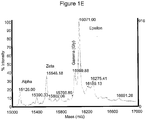
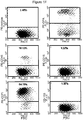
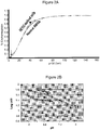
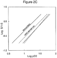
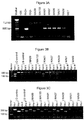

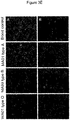
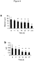
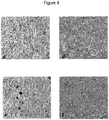
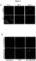
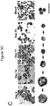

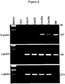
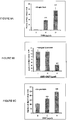
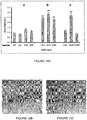
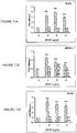

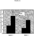
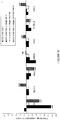
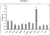
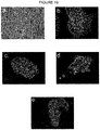


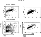
Classifications
-
- C—CHEMISTRY; METALLURGY
- C12—BIOCHEMISTRY; BEER; SPIRITS; WINE; VINEGAR; MICROBIOLOGY; ENZYMOLOGY; MUTATION OR GENETIC ENGINEERING
- C12N—MICROORGANISMS OR ENZYMES; COMPOSITIONS THEREOF; PROPAGATING, PRESERVING, OR MAINTAINING MICROORGANISMS; MUTATION OR GENETIC ENGINEERING; CULTURE MEDIA
- C12N5/00—Undifferentiated human, animal or plant cells, e.g. cell lines; Tissues; Cultivation or maintenance thereof; Culture media therefor
- C12N5/06—Animal cells or tissues; Human cells or tissues
- C12N5/0602—Vertebrate cells
- C12N5/0634—Cells from the blood or the immune system
- C12N5/0641—Erythrocytes
-
- C—CHEMISTRY; METALLURGY
- C07—ORGANIC CHEMISTRY
- C07K—PEPTIDES
- C07K14/00—Peptides having more than 20 amino acids; Gastrins; Somatostatins; Melanotropins; Derivatives thereof
- C07K14/435—Peptides having more than 20 amino acids; Gastrins; Somatostatins; Melanotropins; Derivatives thereof from animals; from humans
- C07K14/475—Growth factors; Growth regulators
-
- A—HUMAN NECESSITIES
- A61—MEDICAL OR VETERINARY SCIENCE; HYGIENE
- A61P—SPECIFIC THERAPEUTIC ACTIVITY OF CHEMICAL COMPOUNDS OR MEDICINAL PREPARATIONS
- A61P7/00—Drugs for disorders of the blood or the extracellular fluid
-
- A—HUMAN NECESSITIES
- A61—MEDICAL OR VETERINARY SCIENCE; HYGIENE
- A61P—SPECIFIC THERAPEUTIC ACTIVITY OF CHEMICAL COMPOUNDS OR MEDICINAL PREPARATIONS
- A61P7/00—Drugs for disorders of the blood or the extracellular fluid
- A61P7/06—Antianaemics
-
- C—CHEMISTRY; METALLURGY
- C12—BIOCHEMISTRY; BEER; SPIRITS; WINE; VINEGAR; MICROBIOLOGY; ENZYMOLOGY; MUTATION OR GENETIC ENGINEERING
- C12N—MICROORGANISMS OR ENZYMES; COMPOSITIONS THEREOF; PROPAGATING, PRESERVING, OR MAINTAINING MICROORGANISMS; MUTATION OR GENETIC ENGINEERING; CULTURE MEDIA
- C12N5/00—Undifferentiated human, animal or plant cells, e.g. cell lines; Tissues; Cultivation or maintenance thereof; Culture media therefor
- C12N5/06—Animal cells or tissues; Human cells or tissues
- C12N5/0602—Vertebrate cells
- C12N5/0634—Cells from the blood or the immune system
- C12N5/0644—Platelets; Megakaryocytes
-
- C—CHEMISTRY; METALLURGY
- C12—BIOCHEMISTRY; BEER; SPIRITS; WINE; VINEGAR; MICROBIOLOGY; ENZYMOLOGY; MUTATION OR GENETIC ENGINEERING
- C12N—MICROORGANISMS OR ENZYMES; COMPOSITIONS THEREOF; PROPAGATING, PRESERVING, OR MAINTAINING MICROORGANISMS; MUTATION OR GENETIC ENGINEERING; CULTURE MEDIA
- C12N5/00—Undifferentiated human, animal or plant cells, e.g. cell lines; Tissues; Cultivation or maintenance thereof; Culture media therefor
- C12N5/06—Animal cells or tissues; Human cells or tissues
- C12N5/0602—Vertebrate cells
- C12N5/0634—Cells from the blood or the immune system
- C12N5/0647—Haematopoietic stem cells; Uncommitted or multipotent progenitors
-
- C—CHEMISTRY; METALLURGY
- C12—BIOCHEMISTRY; BEER; SPIRITS; WINE; VINEGAR; MICROBIOLOGY; ENZYMOLOGY; MUTATION OR GENETIC ENGINEERING
- C12N—MICROORGANISMS OR ENZYMES; COMPOSITIONS THEREOF; PROPAGATING, PRESERVING, OR MAINTAINING MICROORGANISMS; MUTATION OR GENETIC ENGINEERING; CULTURE MEDIA
- C12N5/00—Undifferentiated human, animal or plant cells, e.g. cell lines; Tissues; Cultivation or maintenance thereof; Culture media therefor
- C12N5/06—Animal cells or tissues; Human cells or tissues
- C12N5/0602—Vertebrate cells
- C12N5/0652—Cells of skeletal and connective tissues; Mesenchyme
- C12N5/0662—Stem cells
- C12N5/0668—Mesenchymal stem cells from other natural sources
-
- C—CHEMISTRY; METALLURGY
- C12—BIOCHEMISTRY; BEER; SPIRITS; WINE; VINEGAR; MICROBIOLOGY; ENZYMOLOGY; MUTATION OR GENETIC ENGINEERING
- C12N—MICROORGANISMS OR ENZYMES; COMPOSITIONS THEREOF; PROPAGATING, PRESERVING, OR MAINTAINING MICROORGANISMS; MUTATION OR GENETIC ENGINEERING; CULTURE MEDIA
- C12N5/00—Undifferentiated human, animal or plant cells, e.g. cell lines; Tissues; Cultivation or maintenance thereof; Culture media therefor
- C12N5/06—Animal cells or tissues; Human cells or tissues
- C12N5/0602—Vertebrate cells
- C12N5/069—Vascular Endothelial cells
- C12N5/0691—Vascular smooth muscle cells; 3D culture thereof, e.g. models of blood vessels
-
- C—CHEMISTRY; METALLURGY
- C12—BIOCHEMISTRY; BEER; SPIRITS; WINE; VINEGAR; MICROBIOLOGY; ENZYMOLOGY; MUTATION OR GENETIC ENGINEERING
- C12N—MICROORGANISMS OR ENZYMES; COMPOSITIONS THEREOF; PROPAGATING, PRESERVING, OR MAINTAINING MICROORGANISMS; MUTATION OR GENETIC ENGINEERING; CULTURE MEDIA
- C12N5/00—Undifferentiated human, animal or plant cells, e.g. cell lines; Tissues; Cultivation or maintenance thereof; Culture media therefor
- C12N5/06—Animal cells or tissues; Human cells or tissues
- C12N5/0602—Vertebrate cells
- C12N5/069—Vascular Endothelial cells
- C12N5/0692—Stem cells; Progenitor cells; Precursor cells
-
- A—HUMAN NECESSITIES
- A61—MEDICAL OR VETERINARY SCIENCE; HYGIENE
- A61K—PREPARATIONS FOR MEDICAL, DENTAL OR TOILETRY PURPOSES
- A61K35/00—Medicinal preparations containing materials or reaction products thereof with undetermined constitution
- A61K35/12—Materials from mammals; Compositions comprising non-specified tissues or cells; Compositions comprising non-embryonic stem cells; Genetically modified cells
-
- C—CHEMISTRY; METALLURGY
- C12—BIOCHEMISTRY; BEER; SPIRITS; WINE; VINEGAR; MICROBIOLOGY; ENZYMOLOGY; MUTATION OR GENETIC ENGINEERING
- C12N—MICROORGANISMS OR ENZYMES; COMPOSITIONS THEREOF; PROPAGATING, PRESERVING, OR MAINTAINING MICROORGANISMS; MUTATION OR GENETIC ENGINEERING; CULTURE MEDIA
- C12N2500/00—Specific components of cell culture medium
- C12N2500/05—Inorganic components
- C12N2500/10—Metals; Metal chelators
- C12N2500/20—Transition metals
- C12N2500/24—Iron; Fe chelators; Transferrin
- C12N2500/25—Insulin-transferrin; Insulin-transferrin-selenium
-
- C—CHEMISTRY; METALLURGY
- C12—BIOCHEMISTRY; BEER; SPIRITS; WINE; VINEGAR; MICROBIOLOGY; ENZYMOLOGY; MUTATION OR GENETIC ENGINEERING
- C12N—MICROORGANISMS OR ENZYMES; COMPOSITIONS THEREOF; PROPAGATING, PRESERVING, OR MAINTAINING MICROORGANISMS; MUTATION OR GENETIC ENGINEERING; CULTURE MEDIA
- C12N2500/00—Specific components of cell culture medium
- C12N2500/30—Organic components
- C12N2500/38—Vitamins
-
- C—CHEMISTRY; METALLURGY
- C12—BIOCHEMISTRY; BEER; SPIRITS; WINE; VINEGAR; MICROBIOLOGY; ENZYMOLOGY; MUTATION OR GENETIC ENGINEERING
- C12N—MICROORGANISMS OR ENZYMES; COMPOSITIONS THEREOF; PROPAGATING, PRESERVING, OR MAINTAINING MICROORGANISMS; MUTATION OR GENETIC ENGINEERING; CULTURE MEDIA
- C12N2501/00—Active agents used in cell culture processes, e.g. differentation
- C12N2501/10—Growth factors
- C12N2501/115—Basic fibroblast growth factor (bFGF, FGF-2)
-
- C—CHEMISTRY; METALLURGY
- C12—BIOCHEMISTRY; BEER; SPIRITS; WINE; VINEGAR; MICROBIOLOGY; ENZYMOLOGY; MUTATION OR GENETIC ENGINEERING
- C12N—MICROORGANISMS OR ENZYMES; COMPOSITIONS THEREOF; PROPAGATING, PRESERVING, OR MAINTAINING MICROORGANISMS; MUTATION OR GENETIC ENGINEERING; CULTURE MEDIA
- C12N2501/00—Active agents used in cell culture processes, e.g. differentation
- C12N2501/10—Growth factors
- C12N2501/125—Stem cell factor [SCF], c-kit ligand [KL]
-
- C—CHEMISTRY; METALLURGY
- C12—BIOCHEMISTRY; BEER; SPIRITS; WINE; VINEGAR; MICROBIOLOGY; ENZYMOLOGY; MUTATION OR GENETIC ENGINEERING
- C12N—MICROORGANISMS OR ENZYMES; COMPOSITIONS THEREOF; PROPAGATING, PRESERVING, OR MAINTAINING MICROORGANISMS; MUTATION OR GENETIC ENGINEERING; CULTURE MEDIA
- C12N2501/00—Active agents used in cell culture processes, e.g. differentation
- C12N2501/10—Growth factors
- C12N2501/14—Erythropoietin [EPO]
-
- C—CHEMISTRY; METALLURGY
- C12—BIOCHEMISTRY; BEER; SPIRITS; WINE; VINEGAR; MICROBIOLOGY; ENZYMOLOGY; MUTATION OR GENETIC ENGINEERING
- C12N—MICROORGANISMS OR ENZYMES; COMPOSITIONS THEREOF; PROPAGATING, PRESERVING, OR MAINTAINING MICROORGANISMS; MUTATION OR GENETIC ENGINEERING; CULTURE MEDIA
- C12N2501/00—Active agents used in cell culture processes, e.g. differentation
- C12N2501/10—Growth factors
- C12N2501/145—Thrombopoietin [TPO]
-
- C—CHEMISTRY; METALLURGY
- C12—BIOCHEMISTRY; BEER; SPIRITS; WINE; VINEGAR; MICROBIOLOGY; ENZYMOLOGY; MUTATION OR GENETIC ENGINEERING
- C12N—MICROORGANISMS OR ENZYMES; COMPOSITIONS THEREOF; PROPAGATING, PRESERVING, OR MAINTAINING MICROORGANISMS; MUTATION OR GENETIC ENGINEERING; CULTURE MEDIA
- C12N2501/00—Active agents used in cell culture processes, e.g. differentation
- C12N2501/10—Growth factors
- C12N2501/155—Bone morphogenic proteins [BMP]; Osteogenins; Osteogenic factor; Bone inducing factor
-
- C—CHEMISTRY; METALLURGY
- C12—BIOCHEMISTRY; BEER; SPIRITS; WINE; VINEGAR; MICROBIOLOGY; ENZYMOLOGY; MUTATION OR GENETIC ENGINEERING
- C12N—MICROORGANISMS OR ENZYMES; COMPOSITIONS THEREOF; PROPAGATING, PRESERVING, OR MAINTAINING MICROORGANISMS; MUTATION OR GENETIC ENGINEERING; CULTURE MEDIA
- C12N2501/00—Active agents used in cell culture processes, e.g. differentation
- C12N2501/10—Growth factors
- C12N2501/165—Vascular endothelial growth factor [VEGF]
-
- C—CHEMISTRY; METALLURGY
- C12—BIOCHEMISTRY; BEER; SPIRITS; WINE; VINEGAR; MICROBIOLOGY; ENZYMOLOGY; MUTATION OR GENETIC ENGINEERING
- C12N—MICROORGANISMS OR ENZYMES; COMPOSITIONS THEREOF; PROPAGATING, PRESERVING, OR MAINTAINING MICROORGANISMS; MUTATION OR GENETIC ENGINEERING; CULTURE MEDIA
- C12N2501/00—Active agents used in cell culture processes, e.g. differentation
- C12N2501/20—Cytokines; Chemokines
- C12N2501/23—Interleukins [IL]
-
- C—CHEMISTRY; METALLURGY
- C12—BIOCHEMISTRY; BEER; SPIRITS; WINE; VINEGAR; MICROBIOLOGY; ENZYMOLOGY; MUTATION OR GENETIC ENGINEERING
- C12N—MICROORGANISMS OR ENZYMES; COMPOSITIONS THEREOF; PROPAGATING, PRESERVING, OR MAINTAINING MICROORGANISMS; MUTATION OR GENETIC ENGINEERING; CULTURE MEDIA
- C12N2501/00—Active agents used in cell culture processes, e.g. differentation
- C12N2501/20—Cytokines; Chemokines
- C12N2501/26—Flt-3 ligand (CD135L, flk-2 ligand)
-
- C—CHEMISTRY; METALLURGY
- C12—BIOCHEMISTRY; BEER; SPIRITS; WINE; VINEGAR; MICROBIOLOGY; ENZYMOLOGY; MUTATION OR GENETIC ENGINEERING
- C12N—MICROORGANISMS OR ENZYMES; COMPOSITIONS THEREOF; PROPAGATING, PRESERVING, OR MAINTAINING MICROORGANISMS; MUTATION OR GENETIC ENGINEERING; CULTURE MEDIA
- C12N2501/00—Active agents used in cell culture processes, e.g. differentation
- C12N2501/60—Transcription factors
-
- C—CHEMISTRY; METALLURGY
- C12—BIOCHEMISTRY; BEER; SPIRITS; WINE; VINEGAR; MICROBIOLOGY; ENZYMOLOGY; MUTATION OR GENETIC ENGINEERING
- C12N—MICROORGANISMS OR ENZYMES; COMPOSITIONS THEREOF; PROPAGATING, PRESERVING, OR MAINTAINING MICROORGANISMS; MUTATION OR GENETIC ENGINEERING; CULTURE MEDIA
- C12N2502/00—Coculture with; Conditioned medium produced by
- C12N2502/13—Coculture with; Conditioned medium produced by connective tissue cells; generic mesenchyme cells, e.g. so-called "embryonic fibroblasts"
- C12N2502/1352—Mesenchymal stem cells
- C12N2502/1358—Bone marrow mesenchymal stem cells (BM-MSC)
-
- C—CHEMISTRY; METALLURGY
- C12—BIOCHEMISTRY; BEER; SPIRITS; WINE; VINEGAR; MICROBIOLOGY; ENZYMOLOGY; MUTATION OR GENETIC ENGINEERING
- C12N—MICROORGANISMS OR ENZYMES; COMPOSITIONS THEREOF; PROPAGATING, PRESERVING, OR MAINTAINING MICROORGANISMS; MUTATION OR GENETIC ENGINEERING; CULTURE MEDIA
- C12N2502/00—Coculture with; Conditioned medium produced by
- C12N2502/13—Coculture with; Conditioned medium produced by connective tissue cells; generic mesenchyme cells, e.g. so-called "embryonic fibroblasts"
- C12N2502/1394—Bone marrow stromal cells; whole marrow
-
- C—CHEMISTRY; METALLURGY
- C12—BIOCHEMISTRY; BEER; SPIRITS; WINE; VINEGAR; MICROBIOLOGY; ENZYMOLOGY; MUTATION OR GENETIC ENGINEERING
- C12N—MICROORGANISMS OR ENZYMES; COMPOSITIONS THEREOF; PROPAGATING, PRESERVING, OR MAINTAINING MICROORGANISMS; MUTATION OR GENETIC ENGINEERING; CULTURE MEDIA
- C12N2506/00—Differentiation of animal cells from one lineage to another; Differentiation of pluripotent cells
- C12N2506/02—Differentiation of animal cells from one lineage to another; Differentiation of pluripotent cells from embryonic cells
-
- C—CHEMISTRY; METALLURGY
- C12—BIOCHEMISTRY; BEER; SPIRITS; WINE; VINEGAR; MICROBIOLOGY; ENZYMOLOGY; MUTATION OR GENETIC ENGINEERING
- C12N—MICROORGANISMS OR ENZYMES; COMPOSITIONS THEREOF; PROPAGATING, PRESERVING, OR MAINTAINING MICROORGANISMS; MUTATION OR GENETIC ENGINEERING; CULTURE MEDIA
- C12N2506/00—Differentiation of animal cells from one lineage to another; Differentiation of pluripotent cells
- C12N2506/45—Differentiation of animal cells from one lineage to another; Differentiation of pluripotent cells from artificially induced pluripotent stem cells
-
- C—CHEMISTRY; METALLURGY
- C12—BIOCHEMISTRY; BEER; SPIRITS; WINE; VINEGAR; MICROBIOLOGY; ENZYMOLOGY; MUTATION OR GENETIC ENGINEERING
- C12N—MICROORGANISMS OR ENZYMES; COMPOSITIONS THEREOF; PROPAGATING, PRESERVING, OR MAINTAINING MICROORGANISMS; MUTATION OR GENETIC ENGINEERING; CULTURE MEDIA
- C12N2533/00—Supports or coatings for cell culture, characterised by material
- C12N2533/70—Polysaccharides
- C12N2533/78—Cellulose
Definitions
- the present invention relates to producing human enucleated erythroid cells from pluripotent stem cells.
- the Red Cross and other suppliers of blood report a near constant shortage of blood. This is especially true for patients with unique blood types, patients who are Rh+, or following accidents or disasters resulting in mass casualties. Additionally, in times of war, the military has an acute need for available blood for use in the treatment of traumatic war-related injuries.
- the present invention provides improved methods for use in blood banking and transfusion. The methods of the present invention will provide a safe and reliable advance beyond the traditional reliance on blood donations, and will help prevent critical shortages in available blood.
- the present invention provides methods for making enucleated erythroid cells derived from pluripotent stem cells.
- the invention provides an in vitro method of producing a pluripotent stem cell line-derived enucleated erythroid cell, comprising: providing a cell of a pluripotent stem cell (pSC) line; differentiating said pSC into a hemangioblast, non engrafting hemangio cell, or blast cell by (a) culture in a serum free medium comprising at least bone morphogenetic protein 4 (BMP-4) and vascular endothelial growth factor (VEGF) in an amount sufficient to induce the differentiation of said cells of a pluripotent stem cell line into embryoid bodies, said differentiating pSC into a hemangioblast, non engrafting hemangio cell, or blast cell optionally further comprising (b) expanding said hemangioblast, non engrafting hemangio cell, or blast cell by further culturing in the presence of BMP-4, VEGF, and basic fibroblast growth factor (bFGF); and differentiating said hemangio
- the invention provides an in vitro method of producing a pluripotent stem cell line-derived enucleated erythroid cell, comprising: providing a cell of a pluripotent stem cell (pSC) line; differentiating said pSC into a hemangioblast, non engrafting hemangio cell, or blast cell by (a) culture in a serum free medium comprising at least bone morphogenetic protein 4 (BMP-4) and vascular endothelial growth factor (VEGF) in an amount sufficient to induce the differentiation of said cells of a pluripotent stem cell line into embryoid bodies; said differentiating pSC into a hemangioblast, non engrafting hemangio cell, or blast cell optionally further comprising
- BMP-4 bone morphogenetic protein 4
- VEGF vascular endothelial growth factor
- said cell of a pluripotent stem cell line is selected from the group consisting of: a cell of an embryonic stem cell line, and an induced pluripotent stem cell. In certain embodiments, said cell of a pluripotent stem cell line is a human cell.
- said cell of a pluripotent stem cell line is genetically manipulated prior to differentiation.
- said hemangioblast, non-engrafting hemangio cell, or blast cell is expanded prior to being differentiated into said enucleated erythroid cell.
- said hemangioblasts, non-engrafting hemangio cells, or blast cells are expanded in medium with Epo, IL-3, and SCF.
- differentiating said cell of a human pluripotent stem cell line into said hemangioblast further comprises:
- said cell of a pluripotent stem cell line is a human cell of a pluripotent stem cell line and differentiating into said non-engrafting hemangio cell comprises culturing said embryoid bodies for 10-13 days, and wherein said human cell of a pluripotent stem cell line, embryoid bodies and non-engrafting hemangio cells are grown in serum-free media throughout steps (a) and (b) of said method.
- differentiating said cell of a pluripotent stem cell line into said non-engrafting hemangio cell further comprises:
- one of said at least two additional growth factors is a fusion protein that comprises HOXB4, which may be mammalian HOXB4 such as mouse or human HOXB4, and a protein transduction domain (PTD).
- HOXB4 may be mammalian HOXB4 such as mouse or human HOXB4, and a protein transduction domain (PTD).
- PTD protein transduction domain
- vascular endothelial growth factor VEGF
- BMP-4 bone morphogenic protein-4
- said basic fibroblast growth factor (bFGF), stem cell factor (SCF), Flt-3L (FL) or thrombopoietin (TPO), or any combination thereof are added to said culture within 48 - 72 hours from the start of step (a).
- the method further comprises the step of adding erythropoietin (EPO) to step (a).
- EPO erythropoietin
- the method further comprises the step of adding erythropoietin (EPO) to step (a), or step (d).
- EPO erythropoietin
- differentiating said cell of a pluripotent stem cell line into said enucleated erythroid cell further comprises culturing said cell of a pluripotent stem cell line in a culture medium comprising EPO.
- differentiating said cell of a pluripotent stem cell line into said enucleated erythroid cell further comprises:
- the present disclosure provides for a method of producing a pluripotent stem cell-derived enucleated erythroid cell, comprising: providing a pluripotent stem cell; and differentiating said pluripotent stem cell into an enucleated erythroid cell by culturing said pluripotent stem cell with OP9 mouse stromal cells or human mesenchymal stem cells (MSCs).
- a method of producing a pluripotent stem cell-derived enucleated erythroid cell comprising: providing a pluripotent stem cell; and differentiating said pluripotent stem cell into an enucleated erythroid cell by culturing said pluripotent stem cell with OP9 mouse stromal cells or human mesenchymal stem cells (MSCs).
- Differentiating said pluripotent stem cell into an enucleated erythroid cell comprises differentiating said pluripotent stem cell into a hemangioblast, non-engrafting hemangio cell or blast cell.
- said hemangioblast, non-engrafting hemangio cell, or blast cell is expanded prior to being differentiated into said enucleated erythroid cell.
- said hemangioblasts, non-engrafting hemangio cells, or blast cells are expanded in Stemline II medium with Epo, IL-3, and SCF.
- said pluripotent stem cell is a human pluripotent stem cell and differentiating said human pluripotent stem cell into said hemangioblast is done in vitro by a method comprising: (a) culturing a cell culture comprising human pluripotent stem cell in serum-free media in the presence of at least one growth factor in an amount sufficient to induce the differentiation of said human pluripotent stem cell into embryoid bodies; and (b) adding at least two growth factors to said culture comprising embryoid bodies and continuing to culture said culture in serum-free media, wherein said growth factor is in an amount sufficient to expand said human hemangioblast in said embryoid bodies culture, wherein said human pluripotent stem cells, embryoid bodies and hemangioblasts are grown in serum-free media throughout steps (a) and (b) of said method, and wherein said at least two growth factors in step (b) comprise BMP4 and VEGF.
- differentiating said human pluripotent stem cell into said hemangioblast further comprises (c) disaggregating said embryoid bodies into single cells; and (d) adding at least one growth factor to said culture comprising said single cells and continuing to culture said culture in serum-free media, wherein said growth factor is in an amount sufficient to expand human hemangioblasts in said culture comprising said single cells, and wherein said human pluripotent stem cells, embryoid bodies and hemangio-colony forming cells are grown in serum-free media throughout steps (a)- (d) of said method.
- said pluripotent stem cell is a human pluripotent stem cell and differentiating said human pluripotent stem cell into said non-engrafting hemangio cell is done in vitro by a method comprising: (a) culturing a cell culture comprising said human pluripotent stem cell in serum-free media in the presence of at least one growth factor in an amount sufficient to induce the differentiation of said human pluripotent stem cell into embryoid bodies; and (b) adding at least one growth factor to said culture comprising embryoid bodies and continuing to culture said culture in serum-free media, wherein said growth factor is in an amount sufficient to expand said human non-engrafting hemangio cell in said embryoid bodies culture, wherein said embryoid bodies are cultured for 10-13 days, and wherein said human pluripotent stem cell, embryoid bodies and non-engrafting hemangio cells are grown in serum-free media throughout steps (a) and (b) of said method.
- differentiating said pluripotent stem cell into said non-engrafting hemangio cell further comprises (c) disaggregating said embryoid bodies into single cells; and (d) adding at least one growth factor to said culture comprising said single cells and continuing to culture said culture in serum-free media, wherein said growth factor is in an amount sufficient to expand said human non-engrafting hemangio cell in said culture comprising said single cells, wherein said embryo-derived cells, embryoid bodies and non-engrafting hemangio cells are grown in serum-free media throughout steps (a)-(d) of said method.
- differentiating said pluripotent stem cell into said enucleated erythroid cell further comprises culturing said pluripotent stem cell in the culture medium comprising EPO. In certain embodiments, differentiating said pluripotent stem cell into said enucleated erythroid cell further comprises: culturing said pluripotent stem cell in a culture medium comprising a supplement selected from the group consisting of inositol, folic acid, monothioglycerol, transferrin, insulin, ferrous nitrate, ferrous sulfate, BSA, L-glutamine, penicillin-streptomycin and combinations thereof; and culturing said pluripotent stem cell in said culture medium wherein said culture medium further comprises an agent selected from the group consisting of hydrocortisone, SCF, IL3, Epo and combinations thereof.
- said pluripotent stem cell used in the present disclosure is an embryonic stem cell or embryo-derived cell.
- said pluripotent stem cell is an induced pluripotent stem cell.
- said pluripotent stem cell is a human cell.
- said pluripotent stem cell is genetically manipulated prior to differentiation.
- said growth factor used in the present invention is a fusion protein that comprises HOXB4 and a protein transduction domain (PTD).
- said HOXB4 is mammalian HOXB4.
- said mammalian HOXB4 is mouse or human HOXB4.
- said growth factor used in the present invention is selected from the group consisting of vascular endothelial growth factor (VEGF), bone morphogenic proteins (BMP), stem cell factor (SCF), Flt-3L(FL) thrombopoietin (TPO) and erythropoietin (EPO).
- VEGF vascular endothelial growth factor
- BMP bone morphogenic proteins
- SCF stem cell factor
- TPO erythropoietin
- EPO erythropoietin
- said vascular endothelial growth factor (VEGF), bone morphogenic protein (BMP), or both are added to step (a) within 0 - 48 hours of cell culture.
- said stem cell factor (SCF), Flt-3L (FL) or thrombopoietin (TPO), or any combination thereof are added to said culture within 48 - 72 hours from the start of step (a).
- the methods further comprise the step of adding erythropoietin (EPO) to step (a) or further comprises the step of adding erythropoietin (EPO) to step (a) or (d).
- EPO erythropoietin
- the present disclosure provides enucleated erythroid cells produced by methods as described above.
- a method of producing a pluripotent stem cell-derived erythroid cell comprising: providing a pluripotent stem cell; and differentiating said pluripotent stem cell into an erythroid cell by culturing said pluripotent stem cell in a medium comprising EPO.
- differentiating said pluripotent stem cell into an erythroid cell comprises differentiating said pluripotent stem cell into a hemangioblast, non-engrafting hemangio cell, or blast cell.
- said hemangioblast, non-engrafting hemangio cell, or blast cell is expanded prior being differentiated into said erythroid cell.
- said hemangioblasts, non-engrafting hemangio cells, or blast cells are expanded in Stemline II medium with Epo, IL-3 and SCF.
- said pluripotent stem cell is a human pluripotent stem cell and differentiating said human pluripotent stem cell into said hemangioblast is done in vitro by a method comprising: (a) culturing a cell culture comprising said human pluripotent stem cell in serum-free media in the presence of at least one growth factor in an amount sufficient to induce the differentiation of said human pluripotent stem cell into embryoid bodies; and (b) adding at least two growth factors to said culture comprising embryoid bodies and continuing to culture said culture in serum-free media, wherein said growth factor is in an amount sufficient to expand said human hemangioblast in said embryoid bodies culture, wherein said human pluripotent stem cells, embryoid bodies and hemangioblasts are grown in serum-free media throughout steps (a) and (b) of said method, and wherein said at least two growth factors in step (b) comprise BMP4 and VEGF.
- differentiating said human pluripotent stem cell into said hemangioblast further comprises (c) disaggregating said embryoid bodies into single cells; and (d) adding at least one growth factor to said culture comprising said single cells and continuing to culture said culture in serum-free media, wherein said growth factor is in an amount sufficient to expand human hemangioblasts in said culture comprising said single cells, and wherein said pluripotent stem cells, embryoid bodies and hemangio-colony forming cells are grown in serum-free media throughout steps (a)-(d) of said method.
- said pluripotent stem cell is a human pluripotent stem cell and differentiating said pluripotent stem cell into said non-engrafting hemangio cell is done in vitro by a method comprising: (a) culturing a cell culture comprising human pluripotent stem cell in serum-free media in the presence of at least one growth factor in an amount sufficient to induce the differentiation of said human pluripotent stem cell into embryoid bodies; and (b) adding at least one growth factor to said culture comprising embryoid bodies and continuing to culture said culture in serum-free media, wherein said growth factor is in an amount sufficient to expand said human non-engrafting hemangio cells in said embryoid bodies culture, wherein said embryoid bodies are cultured for 10-13 days, and wherein said human pluripotent stem cell, embryoid bodies and non-engrafting hemangio cells are grown in serum-free media throughout steps (a) and (b) of said method.
- differentiating said pluripotent stem cell into said non-engrafting hemangio cell further comprises (c) disaggregating said embryoid bodies into single cells; and (d) adding at least one growth factor to said culture comprising said single cells and continuing to culture said culture in serum-free media, wherein said growth factor is in an amount sufficient to expand human non-engrafting hemangio cells in said culture comprising said single cells, wherein said human pluripotent stem cell, embryoid bodies and non-engrafting hemangio cells are grown in serum-free media throughout steps (a)-(d) of said method.
- said pluripotent stem cell used in the present disclosure is an embryonic stem cell or embryo-derived cell.
- said pluripotent stem cell is an induced pluripotent stem cell.
- said pluripotent stem cell is a human cell.
- said pluripotent stem cell is genetically manipulated prior to differentiation.
- said growth factor used in the present invention is a fusion protein that comprises HOXB4 and a protein transduction domain (PTD).
- said HOXB4 is mammalian HOXB4.
- said mammalian HOXB4 is mouse or human HOXB4.
- said growth factor used in the present invention is selected from the group consisting of vascular endothelial growth factor (VEGF), bone morphogenic proteins (BMP), stem cell factor (SCF), Flt-3L (FL) thrombopoietin (TPO) and erythropoietin (EPO).
- VEGF vascular endothelial growth factor
- BMP bone morphogenic proteins
- SCF stem cell factor
- Flt-3L FL
- thrombopoietin TPO
- EPO erythropoietin
- said vascular endothelial growth factor (VEGF), bone morphogenic protein (BMP), or both are added to step (a) within 0 - 48 hours of cell culture.
- said stem cell factor (SCF), Flt-3L (FL) or thrombopoietin (TPO), or any combination thereof are added to said culture within 48 - 72 hours from the start of step (a).
- the methods further comprise the step of adding erythropoietin (EPO) to step (a) or further comprises the step of adding erythropoietin (EPO) to step (a) or (d).
- EPO erythropoietin
- the present disclosure provides erythroid cells produced by methods as described above.
- Still other instances of the present disclosure provide methods of producing a megakaryocyte or a platelet, comprising: providing a pluripotent stem cell; differentiating said pluripotent stem cell into a hemangioblast, non-engrafting hemangio cell, or blast cell; and differentiating said hemangioblast, non-engrafting hemangio cell, or blast cell into said megakaryocyte or said platelet by culturing in megakaryocyte (MK) culture medium comprising TPO.
- MK megakaryocyte
- said pluripotent stem cell used in the present disclosure is an embryonic stem cell or embryo-derived cell.
- said pluripotent stem cell is an induced pluripotent stem cell.
- said pluripotent stem cell is a human cell.
- said pluripotent stem cell is genetically manipulated prior to differentiation.
- said hemangioblast, non-engrafting hemangio cell, or blast cell is expanded prior to being differentiated into said megakaryocyte or said platelet.
- said hemangioblasts, non-engrafting hemangio cells, or blast cells are expanded in Stemline II medium with Epo, IL-3, and SCF.
- said pluripotent stem cell is a human pluripotent stem cell and differentiating said human pluripotent stem cell into said hemangioblast is done in vitro by a method comprising: (a) culturing a cell culture comprising human pluripotent stem cell in serum-free media in the presence of at least one growth factor in an amount sufficient to induce the differentiation of said human pluripotent stem cell into embryoid bodies; and (b) adding at least two growth factors to said culture comprising embryoid bodies and continuing to culture said culture in serum-free media, wherein said growth factor is in an amount sufficient to expand said human hemangioblast in said embryoid bodies culture, wherein said human pluripotent stem cells, embryoid bodies and hemangioblasts are grown in serum-free media throughout steps (a) and (b) of said method, and wherein said at least two growth factors in step (b) comprise BMP4 and VEGF.
- differentiating said human pluripotent stem cell into said hemangioblast further comprises: (c) disaggregating said embryoid bodies into single cells; and (d) adding at least one growth factor to said culture comprising said single cells and continuing to culture said culture in serum-free media, wherein said growth factor is in an amount sufficient to expand human hemangioblasts in said culture comprising said single cells, and wherein said human pluripotent stem cells, embryoid bodies and hemangio-colony forming cells are grown in serum-free media throughout steps (a)-(d) of said method.
- differentiating said hemangioblast, non-engrafting hemangio cell, or blast cell into said megakaryocyte or said platelet is done after about 6 to 8 days of hemangioblast, non-engrafting hemangio cell, or blast cell culture.
- said pluripotent stem cell is a human pluripotent stem cell and differentiating said human pluripotent stem cell into said non-engrafting hemangio cell is done in vitro by a method comprising: (a) culturing a cell culture comprising said human pluripotent stem cell in serum-free media in the presence of at least one growth factor in an amount sufficient to induce the differentiation of said human pluripotent stem cell into embryoid bodies; and (b) adding at least one growth factor to said culture comprising embryoid bodies and continuing to culture said culture in serum-free media, wherein said growth factor is in an amount sufficient to expand said human non-engrafting hemangio cell in said embryoid bodies culture, wherein said embryoid bodies are cultured for 10 - 13 days and wherein said human pluripotent stem cell, embryoid bodies and non-engrafting hemangio cells are grown in serum-free media throughout steps (a) and (b) of said method.
- differentiating said pluripotent stem cell into said non-engrafting hemangio cell further comprises: (c) disaggregating said embryoid bodies into single cells; and (d) adding at least one growth factor to said culture comprising said single cells and continuing to culture said culture in serum-free media, wherein said growth factor is in an amount sufficient to expand said human non-engrafting hemangio cell in said culture comprising said single cells, wherein said embryo-derived cells, embryoid bodies and non-engrafting hemangio cells are grown in serum-free media throughout steps (a)-(d) of said method.
- said growth factor used in the present invention is a fusion protein that comprises HOXB4 and a protein transduction domain (PTD).
- said HOXB4 is mammalian HOXB4.
- said mammalian HOXB4 is mouse or human HOXB4.
- said growth factor used in the present invention is selected from the group consisting of vascular endothelial growth factor (VEGF), bone morphogenic proteins (BMP), stem cell factor (SCF), Flt-3L (FL) thrombopoietin (TPO) and erythropoietin (EPO).
- VEGF vascular endothelial growth factor
- BMP bone morphogenic proteins
- SCF stem cell factor
- Flt-3L FL
- thrombopoietin TPO
- EPO erythropoietin
- said vascular endothelial growth factor (VEGF), bone morphogenic protein (BMP), or both are added to step (a) within 0 - 48 hours of cell culture.
- said stem cell factor (SCF), Flt-3L (FL) or thrombopoietin (TPO), or any combination thereof are added to said culture within 48 - 72 hours from the start of step (a).
- the present disclosure also provides a megakaryocyte or a platelet produced by any one of the method as described above.
- the invention provides a method of producing an enucleated erythroid cell comprising the steps of (a) providing a pluripotent stem cell; and (b) differentiating said pluripotent stem cell into enucleated erythroid cells.
- said pluripotent stem cell is an embryonic stem cell or embryo-derived cell.
- said pluripotent stem cell is an induced pluripotent stem cell.
- said pluripotent stem cell is a human cell.
- said pluripotent stem cell is genetically manipulated prior to differentiation.
- said pluripotent stem cell is differentiated into hemangioblasts (e.g ., hemangioblasts, hemangio colony forming cells, hemangio cells, non-engrafting hemangio cells, or blast cells) prior to step (b).
- said hemangioblasts or blast cells are expanded prior to step (b).
- hemangioblasts, non-engrafting hemangio cells, or blast cells are expanded about 5, 6, 7, 8, 9, 10, 11, 12, 13, 14, or 15 days.
- hemangioblasts, non-engrafting hemangio cells, or blast cells are expanded from about day 3.5 to about day 10.
- said hemangioblasts, non-engrafting hemangio cells, or blast cells are expanded in Stemline II medium with Epo, IL-3, and SCF.
- hemangioblasts or blast cells are differentiated for about 5, 6, 7, 8, 9, 10, 11, 12, 13, 14, or 15 days.
- hemangioblasts, non-engrafting hemangio cells, or blast cells are differentiated from about day 11 to about day 20.
- said enucleated erythroid cells are cultured with OP9 or MSC cells.
- said culture is supplemented with Epo.
- the present disclosure provides enucleated erythroid cells produced by methods as described above.
- embryonic stem cells refers to embryo-derived cells and is used herein as it is used in the art. This term includes cells derived from the inner cell mass of human blastocysts or morulae, including those that have been serially passaged as cell lines. When used to refer to cells from humans, the term human embryonic stem cell (hES) cell is used.
- the ES cells may be derived from fertilization of an egg cell with sperm, as well as using DNA, nuclear transfer, parthenogenesis, or by means to generate ES cells with homozygosity in the HLA region.
- ES cells are also cells derived from a zygote, blastomeres, or blastocyst-staged mammalian embryo produced by the fusion of a sperm and egg cell, nuclear transfer, parthenogenesis, androgenesis, or the reprogramming of chromatin and subsequent incorporation of the reprogrammed chromatin into a plasma membrane to produce a cell.
- Embryonic stem cells regardless of their source or the particular method use to produce them, can be identified based on (i) the ability to differentiate into cells of all three germ layers, (ii) expression of at least Oct-4 and alkaline phosphatase, and (iii) ability to produce teratomas when transplanted into immunodeficient animals.
- pluripotent stem cells includes embryonic stem cells, embryo-derived stem cells, and induced pluripotent stem cells, regardless of the method by which the pluripotent stem cells are derived.
- Pluripotent stem cells are defined functionally as stem cells that are: (a) capable of inducing teratomas when transplanted in immunodeficient (SCID) mice; (b) capable of differentiating to cell types of all three germ layers ( e.g ., can differentiate to ectodermal, mesodermal, and endodermal cell types); and (c) express one or more markers of embryonic stem cells (e.g ., express Oct 4, alkaline phosphatase, SSEA-3 surface antigen, SSEA-4 surface antigen, nanog, TRA-1-60, TRA-1-81, SOX2, REX1, etc).
- SCID immunodeficient
- Exemplary pluripotent stem cells can be generated using, for example, methods known in the art.
- Exemplary pluripotent stem cells include embryonic stem cells derived from the ICM of blastocyst stage embryos, as well as embryonic stem cells derived from one or more blastomeres of a cleavage stage or morula stage embryo (optionally without destroying the remainder of the embryo).
- embryonic stem cells can be generated from embryonic material produced by fertilization or by asexual means, including somatic cell nuclear transfer (SCNT), parthenogenesis, and androgenesis.
- SCNT somatic cell nuclear transfer
- pluripotent stem cells include induced pluripotent stem cells (iPS cells) generated by reprogramming a somatic cell by expressing a combination of factors (herein referred to as reprogramming factors).
- iPS cells can be generated using fetal, postnatal, newborn, juvenile, or adult somatic cells.
- factors that can be used to reprogram somatic cells to pluripotent stem cells include, for example, a combination of Oct4 (sometimes referred to as Oct 3/4), Sox2, c-Myc, and Klf4.
- factors that can be used to reprogram somatic cells to pluripotent stem cells include, for example, a combination of Oct 4, Sox2, Nanog, and Lin28.
- somatic cells are reprogrammed by expressing at least 2 reprogramming factors, at least three reprogramming factors, or four reprogramming factors.
- additional reprogramming factors are identified and used alone or in combination with one or more known reprogramming factors to reprogram a somatic cell to a pluripotent stem cell.
- Induced pluripotent stem cells are defined functionally and include cells that are reprogrammed using any of a variety of methods (integrative vectors, non-integrative vectors, chemical means, etc).
- the pluripotent stem cells can be from any species. Embryonic stem cells have been successfully derived in, for example, mice, multiple species of non-human primates, and humans, and embryonic stem-like cells have been generated from numerous additional species. Thus, one of skill in the art can generate embryonic stem cells and embryo-derived stem cells from any species, including but not limited to, human, non-human primates, rodents (mice, rats), ungulates (cows, sheep, etc), dogs (domestic and wild dogs), cats (domestic and wild cats such as lions, tigers, cheetahs), rabbits, hamsters, gerbils, squirrel, guinea pig, goats, elephants, panda (including giant panda), pigs, raccoon, horse, zebra, marine mammals (dolphin, whales, etc.) and the like. In certain embodiments, the species is an endangered species. In certain embodiments, the species is a currently extinct species.
- iPS cells can be from any species. iPS cells have been successfully generated using mouse and human cells. iPS cells have been successfully generated using embryonic, fetal, newborn, and adult tissue. Accordingly, one can readily generate iPS cells using a donor cell from any species.
- the species is an endangered species.
- the species is a currently extinct species.
- Induced pluripotent stem cells can be generated using, as a starting point, virtually any somatic cell of any developmental stage.
- the cell can be from an embryo, fetus, neonate, juvenile, or adult donor.
- Exemplary somatic cells that can be used include fibroblasts, such as dermal fibroblasts obtained by a skin sample or biopsy, synoviocytes from synovial tissue, foreskin cells, cheek cells, or lung fibroblasts. Although skin and cheek provide a readily available and easily attainable source of appropriate cells, virtually any cell can be used.
- the somatic cell is not a fibroblast.
- the pluripotent stem cells can be, for example, ES cells or induced pluripotent stem cells.
- Induced pluripotent stem cells can be produced by expressing a combination of reprogramming factors in a somatic cell.
- at least two reprogramming factors are expressed in a somatic cell to successfully reprogram the somatic cell.
- at least three reprogramming factors are expressed in a somatic cell to successfully reprogram the somatic cell.
- at least four reprogramming factors are expressed in a somatic cell to successfully reprogram the somatic cell.
- protein transduction domain refers to any amino acid sequence that translocates across a cell membrane into cells or confers or increases the rate of, for example, another molecule (such as, for example, a protein domain) to which the PTD is attached, to translocate across a cell membrane into cells.
- the protein transduction domain may be a domain or sequence that occurs naturally as part of a larger protein (e.g ., a PTD of a viral protein such as HIV TAT) or may be a synthetic or artificial amino acid sequence.
- hemangioblast and "hemangio-colony forming cell” will be used interchangeably throughout this application.
- the cells have numerous structural and functional characteristics. Amongst the characteristics of these cells is the ability to engraft into the bone marrow when administered to a host. These cells can be described based on numerous structural and functional properties including, but not limited to, expression (RNA or protein) or lack of expression (RNA or protein) of one or more markers.
- Hemangio-colony forming cells are capable of differentiating to give rise to at least hematopoietic cell types or endothelial cell types.
- Hemangio-colony forming cells are preferably bi-potential and capable of differentiating to give rise to at least hematopoietic cell types and endothelial cell types.
- hemangio-colony forming cells of the present invention are at least uni-potential, and preferably bi-potential. Additionally however, hemangio-colony forming cells may have a greater degree of developmental potential and can, in certain embodiments, differentiate to give rise to cell types of other lineages.
- the hemangio-colony forming cells are capable of differentiating to give rise to other mesodermal derivatives such as cardiac cells (for example, cardiomyocytes) and/or smooth muscle cells.
- non-engrafting hemangio cells is used throughout this application to refer to a novel population of cells that share some of the characteristics of hemangio-colony forming cells. However, the non-engrafting hemangio cells are distinguishable in that they do not engraft into the bone marrow when administered to an immunodeficient host. Despite this difference, non-engrafting hemangio cells may share one or more than one (2, 3, 4, 5, 6, 7, 8, 9, 10) of the functional or structural characteristics/properties of hemangio-colony forming cells. For example, in certain embodiments, the non-engrafting hemangio cells are loosely adherent to each other.
- non-engrafting hemangio cells do not express one or more than one (2, 3, 4) of the following proteins: CD34, KDR, CD133, CD31.
- non-engrafting hemangio cells may provide a distinct stem cell population that is somewhat more committed than hemangio-colony forming cells, and yet still capable of producing a range of hematopoietic cell types.
- Embodiments of present invention generally relates to methods for differentiating human pluripotent stem cells into enucleated erythroid cells.
- Erythroid cells of the invention have a variety of uses in vitro and in vivo. Red blood cells of the invention will be useful in various therapeutic applications. Furthermore, the expanded numbers of red blood cells derived by the present invention may be utilized in novel therapeutic strategies in the treatment of hematopoietic disorders or in blood banking.
- pluripotent stem cells are hemangioblasts (e.g ., hemangioblasts, hemangio colony forming cells, non-engrafting hemangio cells, hemangio cells, or blast cells, see e.g ., US Patent Application 2008/0014180 , herein incorporated by reference in its entirety).
- hemangioblasts e.g ., hemangioblasts, hemangio colony forming cells, non-engrafting hemangio cells, hemangio cells, or blast cells, see e.g ., US Patent Application 2008/0014180 , herein incorporated by reference in its entirety).
- the red blood cells of the application may be used in transfusions.
- the ability to generate large numbers of cells for transfusion will alleviate the chronic shortage of blood experienced in blood banks and hospitals across the country.
- the methods of the invention allow for the production of universal cells for transfusion. Specifically, red blood cells that are type O and Rh- can be readily generated and will serve as a universal blood source for transfusion.
- the methods of this invention allow for the in vitro expansion of pluripotent stem cells to large quantities useful for a variety of commercial and clinical applications.
- the cell preparations comprise at least 1 X 10 6 cells.
- the cell preparations comprise at least 2 X 10 6 human pluripotent stem cells and in further embodiments at least 3 X 10 6 human pluripotent stem cells.
- the cell preparations comprise at least 4 X 10 6 human pluripotent stem cells.
- the present invention relates to a solution, a preparation, and a composition comprising between 10,000 and 4 million or more mammalian (such as human) hemangioblast cells.
- the number of hemangioblast cells in such a solution, a preparation, and a composition may be any number between the range of 10,000 to 4 million, or more. This number could be, for example, 20,000, 50,000, 100,000, 500,000, 1 million, etc.
- the invention relates to preparations of red blood cells.
- the invention further relates to methods of producing, storing, and distributing pluripotent stem cells and/or red blood cells.
- the invention also provides methods and solutions suitable for transfusion into human or animal patients.
- the invention provides methods of making red blood cells.
- the invention is suitable for use in blood banks and hospitals to provide blood for transfusion following trauma, or in the treatment of a blood-related disease or disorder.
- the invention provides red blood cells that are universal donor cells.
- the red blood cells are functional and express hemoglobin F prior to transfusion.
- red blood cells are transfused to treat trauma, blood loss during surgery, or blood diseases such as anemia, Sickle cell anemia, or hemolytic disease.
- differentiated red blood cells are transfused to treat trauma, blood loss during surgery, blood diseases such as anemia, Sickle cell anemia, or hemolytic diseases, or malignant disease.
- a mixed population of red blood cells is transfused. It should be noted that many differentiated hematopoietic cell types, particularly red blood cells, typically exist in vivo as a mixed population. Specifically, circulating red blood cells of varying levels of age and differentiation are found in vivo. Additionally, red blood cells mature overtime so as to express less fetal hemoglobin and more adult hemoglobin.
- the present invention contemplates transfusion of either purified populations of red blood cells or of a mixed population of red blood cells having varying levels of age and levels of differentiation.
- the invention contemplates transfusion of red blood cells expressing fetal hemoglobin (hemoglobin F).
- Transfusion of red blood cells that express fetal hemoglobin may be especially useful in the treatment of Sickle cell anemia.
- the ability to generate large numbers of cells for transfusion will alleviate the chronic shortage of blood experienced in blood banks and hospitals across the country.
- the methods of the invention allow for the production of universal cells for transfusion.
- red blood cells that are type O and Rh-can be readily generated and will serve as a universal blood source for transfusion.
- the red blood cells produced from the methods of the application are functional.
- the red blood cells express hemoglobin F prior to transfusion.
- the red blood cells carry oxygen.
- the red blood cells have a lifespan equal to naturally derived red blood cells.
- the red blood cells have a lifespan that is 75% of that of naturally derived red blood cells.
- the red blood cells have a lifespan that is 50% of that of naturally derived red blood cells.
- the red blood cells have a lifespan that is 25% of that of naturally derived red blood cells.
- Embryonic stem cells are a potential consistent and reliable source of red blood cells, with the benefits of unlimited supply of O-universal blood, and avoiding the additional cost of disease screening and blood typing with each donation.
- the hallmark of mature red blood cells is loss of the nucleus, as well as production of mature hemoglobin.
- Many researchers, including our laboratory, have achieved differentiation of ESCs into erythroblasts, which still contain their nuclei, and express immature hemoglobin. To date, enucleation has not been achieved with human embryonic stem cells.
- enucleation has been achieved with CD34+ bone marrow and cord blood stem cells, which are further along in development, thus probably aiding in their enucleation capability.
- Malik achieved 10-40% enucleation after 19 days of treatment of CD34+ bone marrow cells with Epo (Malik 1998).
- Miharada achieved a rate of 77% enucleation from CD34+ cord blood stem cells with a 20-day treatment of growth factors and cytokines including SCF, Epo, IL-3, VEGF, IGF-II, and mifepristone (Miharada 2006).
- the present inventive method uses OP9 cells to induce differentiation in human ESCs in a completely in vitro system, which is relevant to clinical therapies.
- the first step consists of differentiating ESCs into hemangioblasts, hemangio colony forming cells, non-engrafting hemangio cells, or blast cells.
- the second step is expansion of these cells in Stemline II medium (Sigma) with Epo, IL-3, SCF and various supplements used by Douay for cord blood enucleation (Douay 2005).
- the third step introduces the OP9 cells to the ESC-derived erythroblasts, as well as the addition of Epo.
- differentiating ESCs into the hemangioblasts, hemangio colony forming cells, and non-engrafting hemangio cells are produced and expanded in accordance to methods described herein.
- blast cells are cultured as described in Lu 2006.
- day 6-8 blast cells from Day 3.25-Day 4.25 embryoid bodies are picked or filtered and plated in Stemline II medium with Epo, IL-3, SCF, hydrocortisone, inositol, folic acid, mono-thioglycerol, transferrin, insulin, ferrous nitrate, ferrous sulfate and bovine serum albumin for 12-30 days.
- blast cells are then co-cultured with OP9 mouse stromal cells or human mesenchymal stem cells (MSCs) in the same media listed above, without hydrocortisone.
- cells begin co-culturing between day 12 and 29 days.
- cells are further cultured for 12-18 days before enucleation occurs.
- enucleation initiated by OP9 cells can occur in as little as 3 days after stromal growth.
- enucleation is induced in Stempro34 medium with hydrocortisone, inositol, folic acid, mono-thioglycerol, transferrin, insulin, ferrous nitrate, ferrous sulfate and bovine serum albumin.
- cells are fed every 3-4 days and cultured on a new stromal layer every week.
- the present invention also provides methods of producing a megakaryocyte or a platelet, comprising: providing a pluripotent stem cell; differentiating said pluripotent stem cell into a hemangioblast, non-engrafting hemangio cell, or blast cell; and differentiating said hemangioblast, non-engrafting hemangio cell, or blast cell into said megakaryocyte or said platelet by culturing in megakaryocyte (MK) culture medium comprising TPO.
- MK megakaryocyte
- the present invention also provides methods of producing a megakaryocyte or a platelet, comprising: providing a hemangioblast, non-engrafting hemangio cell, or blast cell; and differentiating said hemangioblast, non-engrafting hemangio cell, or blast cell into said megakaryocyte or said platelet by culturing in megakaryocyte (MK) culture medium comprising TPO.
- MK megakaryocyte
- the hemangioblast, non-engrafting hemangio cell, or blast cell may be obtained or produced by methods described herein.
- said pluripotent stem cell used in the present invention is an embryonic stem cell or embryo-derived cell. In certain embodiments, said pluripotent stem cell is an induced pluripotent stem cell. In certain embodiments, said pluripotent stem cell is a human cell. In certain embodiments, said pluripotent stem cell is genetically manipulated prior to differentiation.
- said hemangioblast, non-engrafting hemangio cell, or blast cell is expanded prior to being differentiated into said megakaryocyte or said platelet.
- said hemangioblasts, non-engrafting hemangio cells, or blast cells are expanded in Stemline II medium with Epo, IL-3, and SCF.
- differentiating said hemangioblast, non-engrafting hemangio cell, or blast cell into said megakaryocyte or said platelet is done after about 6 to 8 days of hemangioblast, non-engrafting hemangio cell, or blast cell culture.
- the present invention also provides a megakaryocyte or a platelet produced by any one of the method as described herein.
- This invention provides a method for generating and expanding human hemangio-colony forming cells from human pluripotent stem cells, preparations and compositions comprising human hemangio-colony forming cells, methods of producing various cell types partially or terminally differentiated from hemangio-colony forming cells, methods of using hemangio-colony forming cells therapeutically, and methods of therapeutically using various cell types partially or terminally differentiated from hemangio-colony forming cells.
- BMP-4 bone morphogenetic protein-4
- VEGF vascular endothelial growth factor
- BMP-4 and VEGF significantly up-regulate T-brachyury, KDR, CD31 and LMO2 gene expression, while dramatically down-regulating Oct-4 expression.
- bFGF basic fibroblast growth factor
- This invention also provides a method for expanding mammalian hemangio-colony forming cells obtained from any source, including ES cells, blastocysts or blastomeres, cord blood from placenta or umbilical tissue, peripheral blood, bone marrow, or other tissue or by any other means known in the art.
- Human hemangio-colony forming cells can also be generated from human pluripotent stem cells.
- Human pluripotent stem cells may be a substantially homogeneous population of cells, a heterogeneous population of cells, or all or a portion of an embryonic tissue.
- pluripotent stem cells that can be used in the methods of the present invention, human hemangio-colony forming cells can be generated from human embryonic stem cells.
- embryonic stem cells include embryonic stem cells derived from or using, for example, blastocysts, plated ICMs, one or more blastomeres, or other portions of a pre-implantation-stage embryo or embryo-like structure, regardless of whether produced by fertilization, somatic cell nuclear transfer (SCNT), parthenogenesis, androgenesis, or other sexual or asexual means.
- SCNT somatic cell nuclear transfer
- hemangioblasts can be further differentiated to hematopoietic cells including, but not limited to, platelets and red blood cells. Such cells may be used in transfusions. The ability to generate large numbers of cells for transfusion will alleviate the chronic shortage of blood experienced in blood banks and hospitals across the country.
- the methods of the invention allow for the production of universal cells for transfusion. Specifically, red blood cells that are type O and Rh- can be readily generated and will serve as a universal blood source for transfusion.
- the methods of this invention allow for the in vitro expansion of hemangioblasts to large quantities useful for a variety of commercial and clinical applications. Expansion of hemangioblasts in vitro refers to the proliferation of hemangioblasts. While the methods of the invention enable the expansion of human hemangioblast cells to reach commercially useful quantities, the present invention also relates to large numbers of hemangioblast cells and to cell preparations comprising large numbers of human hemangioblast cells (for example, at least 10,000, 100,000, or 500,000 cells). In certain embodiments, the cell preparations comprise at least 1 X 10 6 cells.
- the cell preparations comprise at least 2 X 10 6 human hemangioblast cells and in further embodiments at least 3 X 10 6 human hemangioblast cells. In still other embodiments, the cell preparations comprise at least 4 X 10 6 human hemangioblast cells.
- the present invention relates to a solution, a preparation, and a composition comprising between 10,000 and 4 million or more mammalian (such as human) hemangioblast cells.
- the number of hemangioblast cells in such a solution, a preparation, and a composition may be any number between the range of 10,000 to 4 million, or more. This number could be, for example, 20,000, 50,000, 100,000, 500,000, 1 million, etc.
- the invention relates to preparations of human hemangioblast progeny cells (e.g ., human hematopoietic cells including human hematopoietic stem cells, and endothelial cells).
- human hemangioblast progeny cells e.g ., human hematopoietic cells including human hematopoietic stem cells, and endothelial cells.
- the invention further relates to methods of producing, storing, and distributing hemangioblast cells and/or hemangioblast lineage cells.
- the invention also provides methods and solutions suitable for transfusion into human or animal patients.
- the invention provides methods of making red blood cells and/or platelets, and/or other hematopoietic cell types for transfusion.
- the invention is suitable for use in blood banks and hospitals to provide blood for transfusion following trauma, or in the treatment of a blood-related disease or disorder.
- the invention provides red blood cells that are universal donor cells.
- the red blood cells are functional and express hemoglobin F prior to transfusion.
- the invention also provides for human hemangio-colony forming cells, cell cultures comprising a substantially purified population of human hemangio-colony forming cells, pharmaceutical preparations comprising human hemangio-colony forming cells and cryopreserved preparations of the hemangio-colony forming cells.
- the invention provides for the use of the human hemangio-colony forming cells in the manufacture of a medicament to treat a condition in a patient in need thereof.
- the invention provides the use of the cell cultures in the manufacture of a medicament to treat a condition in a patient in need thereof.
- the invention also provides the use of the pharmaceutical preparations in the manufacture of a medicament to treat a condition in a patient in need thereof.
- the hemangio-colony forming cells can be identified and characterized based on their structural properties. Specifically, and in certain embodiments, these cells are unique in that they are only loosely adherent to each other (loosely adherent to other hemangio-colony forming cells). Because these cells are only loosely adherent to each other, cultures or colonies of hemangio-colony forming cells can be dissociated to single cells using only mechanical dissociation techniques and without the need for enzymatic dissociation techniques.
- the cells are sufficiently loosely adherent to each other that mechanical dissociation alone, rather than enzymatic dissociation or a combination of mechanical and enzymatic dissociation, is sufficient to disaggregate the cultures or colonies without substantially impairing the viability of the cells.
- mechanical dissociation does not require so much force as to cause substantial cell injury or death when compared to that observed subsequent to enzymatic dissociation of cell aggregates.
- hemangio-colony forming cells can be identified or characterized based on the expression or lack of expression (as assessed at the level of the gene or the level of the protein) of one or more markers.
- hemangio-colony forming cells can be identified or characterized based on lack of expression of one or more (e.g ., the cells can be characterized based on lack of expression of at least one, at least two, at least three or at least four of the following markers) of the following cell surface markers: CD34, KDR, CD133, or CD31.
- hemangio-colony forming cells can be identified or characterized based on expression of GATA2 and/or LMO2. Additionally or alternatively, hemangio-colony forming cells can be identified or characterized based on expression or lack of expression markers.
- Hemangio-colony forming cells of the present invention can be identified or characterized based on one or any combination of these structural or functional characteristics. Note that although these cells can be derived from any of a number of sources, for example, embryonic tissue, prenatal tissue, or perinatal tissue, the term "hemangio-colony forming cells" applies to cells, regardless of source, that are capable of differentiating to give rise to at least hematopoietic cell types and/or endothelial cell types and that have one or more of the foregoing structural or functional properties.
- marker(s) for the progenitor of BCs can be used to select BCs after initial culturing.
- hemangio-colonies are produced from pluripotent cells without forming embryoid bodies.
- the present invention provides a method for generating and expanding human hemangioblasts derived from human pluripotent stem cells, or from human blastocysts or blastomeres.
- the hemangioblasts so produced may be purified and/or isolated.
- Human hemangio-colony forming cells can also be generated from human pluripotent stem cells.
- Human pluripotent stem cells may be a substantially homogeneous population of cells, a heterogeneous population of cells, or all or a portion of an embryonic tissue.
- pluripotent stem cells that can be used in the methods of the present invention, human hemangio-colony forming cells can be generated from human embryonic stem cells.
- embryonic stem cells include embryonic stem cells derived from or using, for example, blastocysts, plated ICMs, one or more blastomeres, or other portions of a pre-implantation-stage embryo or embryo-like structure, regardless of whether produced by fertilization, somatic cell nuclear transfer (SCNT), parthenogenesis, androgenesis, or other sexual or asexual means.
- SCNT somatic cell nuclear transfer
- hemangio-colony forming cells can be generated from other pluripotent stem cells.
- hemangio-colony forming cells can be generated (without necessarily going through a step of embryonic stem cell derivation) from or using plated embryos, ICMs, blastocysts, trophoblast/trophectoderm cells, one or more blastomeres, trophoblast stem cells, embryonic germ cells, or other portions of a pre-implantation-stage embryo or embryo-like structure, regardless of whether produced by fertilization, somatic cell nuclear transfer (SCNT), parthenogenesis, androgenesis, or other sexual or asexual means.
- SCNT somatic cell nuclear transfer
- hemangio-colony forming cells can be generated using cells or cell lines partially differentiated from pluripotent stem cells. For example, if a human embryonic stem cell line is used to produce cells that are more developmentally primitive than hemangio-colony forming cells, in terms of development potential and plasticity, such pluripotent stem cells could then be used to generate hemangio-colony forming cells.
- hemangio-colony forming cells can be generated from other pre-natal or peri-natal sources including, without limitation, umbilical cord, umbilical cord blood, amniotic fluid, amniotic stem cells, and placenta.
- hemangio-colony forming cells are generated from human embryonic tissue a step of embryoid body formation may be needed.
- embryoid body formation serves, at least in part, to help recapitulate the three dimensional interaction of the germ layers that occurs during early development, such a step is not necessarily required when the pluripotent stem cells already have a structure or organization that serves substantially the same purpose as embryoid body formation.
- a step of embryoid body formation is not necessarily required to provide intercellular signals, inductive cues, or three dimensional architecture.
- the methods and uses of the present invention can be used to generate hemangio-colony forming cells from pluripotent stem cells or embryo-derived cells.
- the embryo-derived cells are embryonic stem cells.
- the embryo-derived cells are plated embryos, ICMs, blastocysts, trophoblast/trophectoderm cells, one or more blastomeres, trophoblast stem cells, or other portions of an early pre-implantation embryo.
- the embryo-derived cells may be from embryos produced by fertilization, somatic cell nuclear transfer (SCNT), parthenogenesis, androgenesis, or other sexual or asexual means.
- SCNT somatic cell nuclear transfer
- the invention similarly contemplates generating hemangio-colony forming cells from or using other pluripotent stem cells or embryonic-derived cells, and using the generated cells for any of the same therapeutic applications.
- the human embryonic stem cells may be the starting material of this method.
- the embryonic stem cells may be cultured in any way known in the art, such as in the presence or absence of feeder cells.
- Embryonic stem cells may form embryoid bodies ("EBs") in suspension in medium containing serum ( Wang et al. 2005 J Exp Med (201):1603-1814 ; Wang et al. 2004 Immunity (21): 31-41 ; Chadwick et al. 2003 Blood (102): 906 - 915 ).
- serum Wang et al. 2005 J Exp Med (201):1603-1814 ; Wang et al. 2004 Immunity (21): 31-41 ; Chadwick et al. 2003 Blood (102): 906 - 915 .
- serum presents certain challenges, including variability in experiments, cost, potential for infectious agents, and limited supply. Further, for clinical and certain commercial applications, use of serum necessitates additional U.S. and international regulatory compliance issues that govern biological products.
- the present invention provides methods of generating and expanding human hemangioblasts from pluripotent stem cells in which no serum is used.
- the serum-free conditions are more conducive to scale-up production under good manufacturing process (GMP) guidelines than are conditions which require serum.
- GMP good manufacturing process
- serum-free conditions extend the half-life of certain factors added to the medium (for example, the half-life of proteins including growth factors, cytokines, and HOXB4 in media is increased when no serum is present).
- the media is supplemented with BMP4 and VEGF.
- serum-free media is used throughout the method of this invention for generating and expanding human hemangioblasts.
- human stem cells are grown in serum-free media and are induced to differentiate into embryoid bodies.
- embryonic stem cells may be pelleted and resuspended in serum-free medium (e.g ., in Stemline I or II media (SigmaTM)) supplemented with one or more morphogenic factors and cytokines and then plated on low attachment (e.g ., ultra-low attachment) culture dishes.
- morphogenic factors and cytokines may include, but are not limited to, bone morphogenic proteins (e.g ., BMP2, BMP-4, BMP-7, but not BMP-3) and VEGF, SCF and FL.
- Bone morphogenic proteins and VEGF may be used alone or in combination with other factors.
- the morphogenic factors and cytokines may be added to the media from 0-48 hours of cell culture. Following incubation under these conditions, incubation in the presence of early hematopoietic expansion cytokines, including, but not limited to, thrombopoietin (TPO), Flt-3 ligand, and stem cell factor (SCF), allows the plated ES cells to form EBs.
- TPO thrombopoietin
- Flt-3 ligand Flt-3 ligand
- SCF stem cell factor
- VEGF vascular endothelial growth factor
- human ES cells are first grown in the presence of BMP-4 and VEGF 165 (e.g ., 25-100 ng/ml), followed by growing in the presence of BMP-4, VEGF 165 , SCF, TPO, and FLT3 ligand ( e.g ., 10-50 ng/ml) and HoxB4 ( e.g ., 1.5-5 ⁇ g/ml of a triple protein transduction domain-HoxB4 fusion protein as disclosed herein).
- BMP-4 and VEGF 165 e.g ., 25-100 ng/ml
- SCF vascular endothelial growth factor
- TPO thelial growth protein
- FLT3 ligand e.g ., 10-50 ng/ml
- HoxB4 e.g ., 1.5-5 ⁇ g/ml of a triple protein transduction domain-HoxB4 fusion protein as disclosed herein.
- the additional factors may be added 48-72 hours after plating.
- human hemangioblast cells are isolated from early embryoid bodies ("EBs"). Isolating hemangioblast cells from early EBs supports the expansion of the cells in vitro.
- hemangioblast cells may be obtained from EBs grown for less than 10 days.
- hemangioblast cells arise in human EBs grown for 2-6 days.
- hemangioblast cells are identified and may be isolated from human EBs grown for 4-6 days.
- human EBs are grown for 2-5 days before hemangioblast cells are isolated.
- human EBs are grown for 3-4.5 days before hemangioblast cells are isolated.
- early EBs are washed and dissociated (e.g ., by Trypsin/EDTA or collagenase B).
- a select number of cells e.g ., 2-5 X10 5 cells
- serum-free methylcellulose medium optimized for hemangioblast cell growth e.g ., BL-CFU medium, for example Stem Cell Technologies Catalogue H4436, or hemangioblast cell expansion medium (HGM), or any medium containing 1.0% methylcellulose in MDM, 1-2% Bovine serum albumin, 0.1 mM 2-mercaptoethanol, 10 ⁇ g/ml rh-Insulin, 200 ⁇ g/ml iron saturated human transferrin, 20 ng/ml rh-GM-CSF, 20 ng/ml rh-IL-3, 20 ng/ml rh-IL-6, 20 ng/ml rh-G-CSF)(*rh” stands for "recombinant human"
- This medium may be supplemented with early stage cytokines (including, but not limited to, EPO, TPO, SCF, FL, FLt-3, VEGF, BMPs such as BMP2, BMP4 and BMP7, but not BMP3) and HOXB4 (or another homeobox protein).
- EPO erythropoietin
- EPO, SCF, VEGF, BMP-4 and HoxB4 are added to the media.
- the cells are grown in the presence of EPO, TPO and FL.
- H9 is the starting human ES cell line
- EPO, TPO and FL are added to the media.
- media for cells derived from H9 or other ES cells may further comprise VEGF, BMP-4, and HoxB4.
- the cells so obtained by this method are plated onto ultra-low attachment culture dishes and incubated in a CO 2 incubator to grow hemangioblast colonies. Some cells may be able to form secondary EBs. Following approximately 3-6 days, and in some instances 3-4.5 days, hemangioblast colonies are observed. Hemangioblast colonies may be distinguished from other cells such as secondary EBs by their distinctive grape-like morphology and/or by their small size.
- hemangioblasts may be identified by the expression of certain markers (e.g ., the expression of both early hematopoietic and endothelial cell markers) as well as their ability to differentiate into at least both hematopoietic and endothelial cells (see below, Deriving hemangioblast lineage cells).
- certain markers e.g ., the expression of both early hematopoietic and endothelial cell markers
- these cells may be identified by the presence of certain markers (such as, for example, CD71+) and the absence of other markers (for example, CD34-).
- Hemangioblasts may also express GATA-1 and GATA-2 proteins, CXCR-4, and TPO and EPO receptors.
- hemangioblasts may be characterized by the absence or low expression of other markers (e.g ., CD31, CD34, KDR, or other adhesion molecules). Further, hemangioblasts may be characterized by the expression of certain genes, (e.g ., genes associated with hemangioblasts and early primitive erythroblast development, such as, for example, SCL, LMO2, FLT-1, embryonic fetal globin genes, NF-E2, GATA-1, EKLF, ICAM-4, glycophoriuns, and EPO receptor).
- genes associated with hemangioblasts and early primitive erythroblast development such as, for example, SCL, LMO2, FLT-1, embryonic fetal globin genes, NF-E2, GATA-1, EKLF, ICAM-4, glycophoriuns, and EPO receptor.
- hemangioblasts may be isolated by size (being smaller than the other cells) or purified with an anti-CD71+ antibody, such as by immunoaffinity column chromatography.
- the hemangioblast cells may be isolated by size and/or morphology by the following procedure. After 6 to 7 days of growth, the cell mixture contains EBs, which are round and represent a clump of multiple cells, and hemangioblasts, which are grape-like, smaller than the EBs, and are single cells. Accordingly, hemangioblasts may be isolated based on their morphology and size. The hemangioblast cells may be manually picked, for example, when observing the cell mixture under a microscope. The cells may subsequently grow into colonies, each colony having between 100 -150 cells.
- Human hemangioblast colonies derived as described above may be picked and replated onto methylcellulose CFU-medium to form hematopoietic CFUs.
- CFU-medium comprises StemCell Technologies H4436.
- hemangioblasts are plated in Stemline II media supplemented with cytokines and other factors.
- individual BL-CFC colonies may be handpicked and transferred to a fibronectin-coated plate containing Stemline II with recombinant human SCF (e.g ., 20 ng/ml), TPO ( e.g ., 20ng/ml), FL ( e.g ., 20 ng/ml), IL-3 ( e.g ., 20 ng/ml) VEGF ( e.g ., 20 ng/ml), G-CSF ( e.g ., 20n ng/ml), BMP-4 ( e.g ., 15 ng/ml), IL-6 ( e.g ., 10 ng/ml), IGF-1 ( e.g ., 10 ng/ml), endothelial cell growth supplement (ECGS, e.g ., 100 Ng/ml), Epo ( e.g ., 3 U/ml).
- SCF e.g ., 20 ng/m
- non-adherent hematopoietic cells may be removed by gentle pipetting and used directly for hematopoietic CFU assay.
- the adherent populations may be grown for one more week in EGM-2 endothelial cell medium (CambrexTM), and then examined for the expression of vWF.
- hemangioblasts expanded by the methods of the invention are obtained from early embryoid bodies derived from human embryonic stem cells as described above.
- hemangioblasts to be expanded may also be isolated from other mammalian sources, such as mammalian embryos ( Ogawa et al. 2001 Int Rev Immunol (20):21-44 , US patent publication no. 2004/0052771 ), cord blood from placenta and umbilical tissues ( Pelosi, et al. 2002 Blood (100): 3203-3208 ; Cogle et al. 2004 Blood (103):133-5 ), peripheral blood and bone marrow ( Pelosi et al. 2002 Hematopoiesis (100): 3203-3208 ).
- mammalian embryos Ogawa et al. 2001 Int Rev Immunol (20):21-44 , US patent publication no. 2004/0052771
- cord blood from placenta and umbilical tissues Pelosi, et al. 2002 Blood (100): 3203-3208 ; Cogle et al. 2004 Blood (103):133-5
- peripheral blood and bone marrow Pelosi
- non-human hemangioblasts to be expanded may be generated from non-human (such as mouse and non-human primates) embryonic stem cells.
- hemangioblasts are obtained from umbilical cord blood (UCB) or bone marrow by methods such as, for example, magnetic bead positive selection or purification techniques (e.g . MACS column). Cells may be selected based on their CD71 + status and may be confirmed as CD34-. Further, the isolated hemangioblasts may be tested for their potential to give rise to both hematopoietic and endothelial cell lineages.
- hemangioblasts isolated or purified and optionally enriched from embryos, cord blood, peripheral blood, bone marrow, or other tissue are more than 95% pure.
- Bone marrow-derived cells may be obtained from any stage of development of the donor individual, including prenatal (e.g ., embryonic or fetal), infant (e.g ., from birth to approximately three years of age in humans), child ( e.g ., from about three years of age to about 13 years of age in humans), adolescent (e. g., from about 13 years of age to about 18 years of age in humans), young adult ( e.g ., from about 18 years of age to about 35 years of age in humans), adult (from about 35 years of age to about 55 years of age in humans) or elderly ( e.g . from about 55 years and beyond of age in humans).
- prenatal e.g ., embryonic or fetal
- infant e.g ., from birth to approximately three years of age in humans
- child e.g ., from about three years of age to about 13 years of age in humans
- adolescent e. g., from about 13 years of age to
- Human bone marrow may be harvested by scraping from the split sternum of a patient undergoing surgery, for example. Bone marrow may then be preserved in tissue clumps of 0.1 to 1 mm 3 in volume and then grown on a mouse embryonic feeder layer (e.g ., a mitomycin C-treated or irradiated feeder layer). The bone marrow cells will attach to the plates and over a period of 1-2 weeks of culture, hemangioblast cells may be identified based on morphological features and/or cell markers and isolated (see US patent publication no. 2004/0052771 ). The cells may then be subsequently grown and expanded in serum-free conditions according to the methods disclosed herein.
- a mouse embryonic feeder layer e.g ., a mitomycin C-treated or irradiated feeder layer.
- the bone marrow cells will attach to the plates and over a period of 1-2 weeks of culture, hemangioblast cells may be identified based on morphological features and/or cell markers and isolated (
- bone marrow cells and cells from blood or other tissue may be fractionated to obtain hemangioblasts cells.
- Methods of fractionation are well known in the art, and generally involve both positive selection (i.e ., retention of cells based on a particular property) and negative selection (i.e ., elimination of cells based on a particular property).
- Positive selection i.e ., retention of cells based on a particular property
- negative selection i.e ., elimination of cells based on a particular property.
- Methods for fractionation and enrichment of bone marrow-derived cells are best characterized for human and mouse cells.
- Positive selection methods such as enriching for cells expressing CD71 may be used.
- negative selection methods which remove or reduce cells expressing CD3, CD10, CD11b, CD14, CD16, CD15, CD16, CD19, CD20, CD32, CD45, CD45R/B220 or Ly6G may also be used alone or in combination with positive selection techniques.
- negative selection may be performed on the cell preparation to reduce or eliminate differentiated T cells.
- Immunoaffinity technology may take a variety of forms, as is well known in the art, but generally utilizes an antibody or antibody derivative in combination with some type of segregation technology.
- the segregation technology generally results in physical segregation of cells bound by the antibody and cells not bound by the antibody, although in some instances the segregation technology which kills the cells bound by the antibody may be used for negative selection.
- any suitable immunoaffinity technology may be utilized for selection/enrichment of hemangioblasts from bone marrow-derived, blood, or other cells, including fluorescence-activated cell sorting (FACS), panning, immunomagnetic separation, immunoaffinity chromatography, antibody-mediated complement fixation, immunotoxin, density gradient segregation, and the like.
- FACS fluorescence-activated cell sorting
- the desired cells the cells bound by the immunoaffinity reagent in the case of positive selection, and cells not bound by the immunoaffinity reagent in the case of negative selection
- Immunoaffinity selection/enrichment is typically carried out by incubating a preparation of cells comprising bone marrow-derived cells with an antibody or antibody-derived affinity reagent (e.g ., an antibody specific for a given surface marker), then utilizing the bound affinity reagent to select either for or against the cells to which the antibody is bound.
- the selection process generally involves a physical separation, such as can be accomplished by directing droplets containing single cells into different containers depending on the presence or absence of bound affinity reagent (FACS), by utilizing an antibody bound (directly or indirectly) to a solid phase substrate (panning, immunoaffinity chromatography), or by utilizing a magnetic field to collect the cells which are bound to magnetic particles via the affinity reagent (immunomagnetic separation).
- undesirable cells may be eliminated from the bone marrow-derived cell preparation using an affinity reagent which directs a cytotoxic insult to the cells bound by the affinity reagent.
- the cytotoxic insult may be activated by the affinity reagent (e.g ., complement fixation), or may be localized to the target cells by the affinity reagent (e.g ., immunotoxin, such as ricin B chain).
- hemangioblasts expanded by the methods of the invention are obtained from early embryoid bodies derived from human embryonic stem cells as described above.
- the hemangioblasts are isolated or enriched from human tissue (e.g ., placenta or cord blood, peripheral blood, bone marrow, etc.)
- the hemangioblasts are expanded in the presence of a homeodomain protein (also referred to herein as a homeobox protein).
- a homeodomain protein also referred to herein as a homeobox protein
- the hemangioblasts are expanded in the presence of HOXB4.
- HOXB4 is added to the hemangioblast cells throughout the method for expanding hemangioblast cells.
- HOXB4 is a homeodomain transcription factor (also called HOX2F, HOX2, HOX-2.6, and in the rat HOXA5) that is expressed in vivo in the stem cell fraction of the bone marrow and that is subsequently down-regulated during differentiation. Expression of the HOXB4 gene is associated with the maintenance of primitive stem cell phenotypes ( Sauvageau et al. 1995 Genes Dev 9: 1753-1765 ; Buske et al. 2002 Blood 100: 862-868 ; Thorsteinsdottir et al. 1999 Blood 94: 2605-2612 ; Antonchuk et al. 2001 Exp Hematol 29: 1125-1134 ).
- HOXB4 used in the methods of the present invention to generate and expand hemangioblasts includes, but is not limited to, full length HOXB4 (e.g ., HOXB4 polypeptides specified by public accession numbers Gl:13273315 ( Figure 17 ), Gl:29351568 ( Figure 18 ), as well as any functional variants and active fragments thereof.
- the wild-type HOXB4 protein may be encoded by the amino acid sequence of SEQ ID NO: 1, SEQ ID NO: 3 or any other alternative allelic forms of such protein. Such sequences may be accessed via publicly available databases, such as Genbank.
- HOXB4 may be ectopically expressed within the cell or may be provided in the media. HOXB4 expressed ectopically may be operably linked to an inducible promoter.
- HOXB4 provided in the media may be excreted by another cell type (e.g ., a feeder layer) or added directly to the media.
- the present invention also relates to fusion proteins comprising HOXB4 (including fusion proteins comprising full length HOXB4, or HOXB4 functional variants or active fragments of HOXB4).
- this fusion protein may also comprise any additional proteins, protein domains or peptides.
- HOXB4 may be joined to a protein transduction domain (PTD) to allow translocation of the protein from the medium into the cells and subsequently into nuclear compartments. Fusion proteins may or may not comprise one or more linker sequences located in between the protein domains.
- Functional variants of HOXB4 include mutants of HOXB4 and allelic variants, and active fragments thereof.
- Functional variants of HOXB4 include any HOXB4 polypeptides and active fragments thereof that are capable of expanding hemangioblasts according to the methods of the present invention.
- HOXB4 functional variants also include HOXB4 polypeptides that exhibit greater transcriptional activity compared to the native HOXB4 protein.
- HOXB4 variants include proteins with one or more amino acid substitution, addition, and/or deletion in relation to a wild-type HOXB4.
- HOXB4 variants also include, but are not limited to, polypeptides that are at least 75% similar to the sequence provided in SEQ ID NO: 1 or SEQ ID NO: 3. Accordingly, HOXB4 variants include polypeptides that are 80%, 85%, 90%, 95%, and 99% similar to the amino acid sequence provided in SEQ ID NO: 1 or SEQ ID NO: 3.
- HOXB4 variants also include polypeptides encoded by nucleic acid sequences that are at least 80% identical to a nucleic acid sequence encoding its complement (e. g., the wild-type HOXB4 protein may be encoded by nucleic acid sequences of SEQ ID NO: 2 (Gl:85376187; Figure 15 ) or SEQ ID NO: 4 (Gl:29351567; Figure 16 )).
- HOXB4 variants include HOXB4 polypeptides that are encoded by nucleic acid sequences that are 85%, 90%, 95%, and 99% identical to the sequence provided in SEQ ID NO: 2 or SEQ ID NO: 4 or complement thereto.
- Nucleic acid sequences encoding HOXB4 also include, but are not limited to, any nucleic acid sequence that hybridizes under stringent conditions to a nucleic acid sequence of SEQ ID NO: 2 or 4, complement thereto, or fragment thereof. Similarly, nucleic acids which differ from the nucleic acids as set forth in SEQ ID NO: 2 or 4 due to degeneracy in the genetic code are also within the scope of the invention.
- HOXB4 variant polypeptides also include splice variants or other naturally occurring HOXB4 proteins or nucleic acid sequences.
- HOXB4 Active fragments of HOXB4 include, but are not limited to, any fragment of full length HOXB4 polypeptide that is capable of maintaining hemangioblasts according to the methods of the present invention. Accordingly, in one embodiment, a HOXB4 protein of the present invention is a HOXB4 protein that lacks part of the N-terminus, such as, for example, the N-terminal 31, 32, or 33 amino acids of full length HOXB4.
- HOXB4 proteins may be fused with additional proteins or protein domains.
- HOXB4 may be joined to a protein transduction domain (PTD).
- PTD protein transduction domain
- Protein transduction domains covalently or non-covalently linked to HOXB4, allow the translocation of HOXB4 across the cell membranes so the protein may ultimately reach the nuclear compartments of the cells.
- PTDs that may be fused with a HOXB4 protein include the PTD of the HIV transactivating protein (TAT) (Tat 47-57) ( Schwarze and Dowdy 2000 Trends Pharmacol. Sci. 21: 45-48 ; Krosl et al. 2003 Nature Medicine (9): 1428-1432 ).
- TAT HIV transactivating protein
- the amino acid sequence conferring membrane translocation activity corresponds to residues 47-57 (YGRKKRRQRRR, SEQ ID NO: 5) ( Ho et al., 2001, Cancer Research 61: 473-477 ; Vives et al., 1997, J. Biol. Chem. 272: 16010-16017 ). This sequence alone can confer protein translocation activity.
- the TAT PTD may also be the nine amino acids peptide sequence RKKRRQRRR (SEQ ID NO: 6) ( Park et al. Mol Cells 2002 (30):202-8 ).
- the TAT PTD sequences may be any of the peptide sequences disclosed in Ho et al., 2001, Cancer Research 61: 473-477 (the disclosure of which is hereby incorporated by reference herein), including YARKARRQARR (SEQ ID NO: 7), YARAAARQARA (SEQ ID NO: 8), YARAARRAARR (SEQ ID NO: 9) and RARAARRAARA (SEQ ID NO: 10).
- HSV-1 herpes simplex virus 1
- Amintp Drosophila Antennapedia homeotic transcription factor
- Antp amino acids 43-58 (RQIKIWFQNRRMKWKK, SEQ ID NO: 11) represent the protein transduction domain
- HSV VP22 the PTD is represented by the residues DAATATRGRSAASRPTERPRAPARSASRPRRPVE (SEQ ID NO: 12).
- HeptaARG RRRRRRR, SEQ ID NO: 13
- artificial peptides that confer transduction activity may be used as a PTD of the present invention.
- the PTD may be a PTD peptide that is duplicated or multimerized.
- the PTD is one or more of the TAT PTD peptide YARAAARQARA (SEQ ID NO: 14).
- the PTD is a multimer consisting of three of the TAT PTD peptide YARAAARQARA (SEQ ID NO: 15).
- a HOXB4 protein that is fused or linked to a multimeric PTD such as, for example, a triplicated synthetic protein transduction domain (tPTD), may exhibit reduced lability and increased stability in cells. Such a HOXB4 construct may also be stable in serum-free medium and in the presence of hES cells.
- fusion genes encoding fusion proteins are well known in the art. Essentially, the joining of various DNA fragments coding for different polypeptide sequences is performed in accordance with conventional techniques.
- the fusion gene can be synthesized by conventional techniques including automated DNA synthesizers.
- PCR amplification of gene fragments can be carried out using anchor primers which give rise to complementary overhangs between two consecutive gene fragments which can subsequently be annealed to generate a chimeric gene sequence (see, for example, Current Protocols in Molecular Biology, eds. Ausubel et al., John Wiley & Sons: 1992 ).
- a fusion gene coding for a purification leader sequence may be linked to the N-terminus of the desired portion of the HOXB4 polypeptide or HOXB4-fusion protein, allowing the fusion protein be purified by affinity chromatography using a Ni 2+ metal resin.
- the purification leader sequence can then be subsequently removed by treatment with enterokinase to provide the purified HOXB4 polypeptide ( e.g ., see Hochuli et al., (1987) J. Chromatography 411:177 ; and Janknecht et al., PNAS USA 88:8972 ).
- a HOXB4 protein or functional variant or active domain of it is linked to the C-terminus or the N-terminus of a second protein or protein domain (e.g ., a PTD) with or without an intervening linker sequence.
- the exact length and sequence of the linker and its orientation relative to the linked sequences may vary.
- the linker may comprise, for example, 2, 10, 20, 30, or more amino acids and may be selected based on desired properties such as solubility, length, steric separation, etc.
- the linker may comprise a functional sequence useful for the purification, detection, or modification, for example, of the fusion protein.
- the linker comprises a polypeptide of two or more glycines.
- the protein domains and/or the linker by which the domains are fused may be modified to alter the effectiveness, stability and/or functional characteristics of HOXB4.
- HOXB4 is ectopically expressed within the hemangioblast cell or is provided in the media.
- HOXB4 expressed ectopically may be operably linked to a regulatory sequence. Regulatory sequences are art-recognized and are selected to direct expression of the HOXB4 polypeptide.
- HOXB4 provided in the media may be excreted by another cell type.
- the other cell type may be a feeder layer, such as a mouse stromal cell layer transduced to express excretable HOXB4.
- HOXB4 may be fused to or engineered to comprise a signal peptide, or a hydrophobic sequence that facilitates export and secretion of the protein.
- HOXB4 such as a fusion protein covalently or non-covalently linked to a PTD, may be added directly to the media.
- HOXB4 may be borne on a viral vector, such as a retroviral vector or an adenoviral vector. Such a vector could transduce either the hemangioblasts or other cells in their culture.
- HOXB4 is added to the media at selected times during the expansion of the hemangioblasts. Because the hemangioblasts are expanded in serum-free medium, HOXB4 is relatively stable. Accordingly, in certain embodiments, a HOXB4 protein or fusion protein is added every day to the human hemangioblasts. In other embodiments, a HOXB4 protein or fusion protein is added every other day, and in still other embodiments, a HOXB4 protein or fusion protein is added every 2 days. In one embodiment, a HOXB4 fusion protein, HOXB4-PTD, is added every 2 days to the media.
- the hemangioblasts can be expanded in the presence of any other growth factors or proteins that are present in an amount sufficient to expand such cells.
- Hemangioblasts obtained from any source including human or non-human ES cells, bone marrow, placenta or umbilical cord blood, peripheral blood, or other tissue may be expanded according to the methods described above. Accordingly, in certain embodiments, a select number of purified hemangioblasts or enriched cells are mixed with serum-free methylcellulose medium optimized for hemangioblast growth (e.g ., BL-CFU medium,). This medium may be supplemented with early stage cytokines (including, but not limited to, EPO, TPO, FL, VGF, BMPs like BMP2, BMP4 and BMP7, but not BMP3) and HOXB4.
- serum-free methylcellulose medium optimized for hemangioblast growth e.g ., BL-CFU medium,
- This medium may be supplemented with early stage cytokines (including, but not limited to, EPO, TPO, FL, VGF, BMPs like BMP2, BMP4 and BMP7, but not BMP3)
- EPO erythropoietin
- TPO erythropoietin
- FL erythropoietin
- the cells are then plated onto ultra-low attachment culture dishes and grown in a CO 2 incubator.
- hemangioblast colonies exhibit a distinctive grape-like morphology and are comparatively smaller than other cells and may consequently be distinguished from other cell types.
- the hemangioblasts may also be tested for markers as well as for their ability to differentiate further into either hematopoietic or endothelial cell lineages. The hemangioblasts are subsequently isolated and expanded in vitro .
- Media that may be used for expansion includes serum-free methylcellulose medium optimized for hemangioblasts growth (e.g ., BL-CFU) supplemented with early stage cytokines and HOXB4.
- Early stage cytokines include, but are not limited to, EPO, TPO, FL, VEGF, BMPs like BMP2, BMP4 and BMP7, but not BMP3.
- EPO erythropoietin
- EPO erythropoietin
- a medium for expanding hemangioblasts may comprise VEGF, SCF, EPO, BMP-4, and HoxB4; in certain embodiments the medium may further comprise TPO and FL.
- HGM hemangioblast expansion media
- ml hemangioblast expansion media containing 1.0% methylcellulose in Iscove's MDM, 1-2% Bovine serum albumin, 0.1 mM 2-mercaptoethanol, 10 ⁇ g/ml rh-Insulin, 200 ⁇ g/ml iron saturated human transferrin, 20 ng/ml rh-GM-CSF, 20 ng/ml rh-IL-3, 20 ng/ml rh-IL-6, 20 ng/ml rh-G-CSF, 3 to 6 units/ml rh-Epo, 50 ng/ml rh-SCF, 50ng/ml rh-VEGF and 50 ng/ml rh-BMP-4, and 1.5 ⁇ g/ml of tPTD-HoxB4, with/without 50 ng/ml of Tpo and FL.
- HGM hemangioblast expansion media
- tPTD-HoxB4 1.5
- hemangioblasts In certain situations it may be desirable to obtain hemangioblasts from a patient or patient relative and expand said hemangioblasts in vitro . Such situations include, for example, a patient scheduled to begin chemotherapy or radiation therapy, or other situations wherein an autologous HSC transplantation (using the patient's own stem cells) may be used.
- the present invention provides methods of treating patients in need of cell-based therapy (for example, patients in need of hematopoietic reconstitution or treatment, or blood vessel growth or treatment of vascular injuries including ischemia, see below) using the expanded hemangioblasts or hemangioblast lineage cells of the invention, wherein the hemangioblasts are obtained from the bone marrow, blood, or other tissue of the patient or a patient relative.
- methods of treating a patient in need of hemangioblasts may comprise a step of isolating hemangioblasts from the patient or a patient relative.
- Hemangioblasts isolated from the patient or patient relative may be expanded in vitro according to the methods of the present invention and subsequently administered to the patient.
- the expanded hemangioblasts may be grown further to give rise to hematopoietic cells or endothelial cells before patient treatment.
- Hemangioblasts of that patient may then be generated and expanded from his own ES cells using a method of this invention. Those hemangioblasts or lineage derivatives thereof may be administered to that patient or to his relatives.
- human hemangioblasts are expanded to reach commercially large quantities which can be subsequently used in various therapeutic and clinical applications. Furthermore, the hemangioblasts obtained by the methods disclosed herein may be differentiated further to give rise to either hematopoietic or endothelial cell lineages for use in clinical applications.
- the hemangioblasts obtained from the method of this invention for generating and expanding human hemangioblasts from human ES cells have the potential to differentiate into at least endothelial cells or hematopoietic cells ( i.e ., they are at least bi-potential). Other hemangioblasts may be bi-potential as well. Yet other hemangioblasts may be able to differentiate into cells other than hematopoietic and endothelial cells, i.e ., they are multi- or pluri-potential).
- the human embryonic stem cells used as the starting point for the method of generating and expanding human hemangioblast cells of this invention may also be derived from a library of human embryonic stem cells, each of which is hemizygous or homozygous for at least one MHC allele present in a human population.
- each member of said library of stem cells is hemizygous or homozygous for a different set of MHC alleles relative to the remaining members of the library.
- the library of stem cells is hemizygous or homozygous for all MHC alleles that are present in a human population.
- stem cells that are homozygous for one or more histocompatibility antigen genes include cells that are nullizygous for one or more (and in some embodiments, all) such genes.
- Nullizygous for a genetic locus means that the gene is null at that locus, i.e ., both alleles of that gene are deleted or inactivated.
- Stem cells that are nullizygous for all MHC genes may be produced by standard methods known in the art, such as, for example, gene targeting and/or loss of heterozygocity (LOH). See, for example, United States patent publications US 20040091936 , US 20030217374 and US 20030232430 , and US provisional application number 60/729,173 , the disclosures of all of which are hereby incorporated by reference herein.
- the present invention relates to methods of obtaining hemangioblasts, including a library of hemangioblasts, with reduced MHC complexity.
- Hemangioblasts and hemangioblast lineage cells with reduced MHC complexity will increase the supply of available cells for therapeutic applications as it will eliminate the difficulties associated with patient matching.
- Such cells may be derived from stem cells that are engineered to be hemizygous or homozygous for genes of the MHC complex.
- a human ES cell may comprise modifications to one of the alleles of sister chromosomes in the cell's MHC complex.
- a variety of methods for generating gene modifications such as gene targeting, may be used to modify the genes in the MHC complex.
- the modified alleles of the MHC complex in the cells may be subsequently engineered to be homozygous so that identical alleles are present on sister chromosomes. Methods such as loss of heterozygosity (LOH) may be utilized to engineer cells to have homozygous alleles in the MHC complex.
- LHO loss of heterozygosity
- one or more genes in a set of MHC genes from a parental allele can be targeted to generate hemizygous cells.
- the other set of MHC genes can be removed by gene targeting or LOH to make a null line.
- This null line can be used further as the embryonic cell line in which to drop arrays of the HLA genes, or individual genes, to make a hemizygous or homozygous bank with an otherwise uniform genetic background.
- a library of ES cell lines wherein each member of the library is homozygous for at least one HLA gene, is used to derive hemangioblasts according to the methods of the present invention.
- the invention provides a library of hemangioblasts (and/or hemangioblast lineage cells), wherein several lines of ES cells are selected and differentiated into hemangioblasts. These hemangioblasts and/or hemangioblast lineage cells may be used for a patient in need of a cell-based therapy.
- certain embodiments of this invention pertain to a method of administering human hemangioblasts, hematopoietic stem cells, or human endothelial cells that have been derived from reduced-complexity embryonic stem cells to a patient in need thereof.
- this method comprises the steps of: (a) identifying a patient that needs treatment involving administering human hemangioblasts, hematopoietic stem cells, or human endothelial cells to him or her; (b) identifying MHC proteins expressed on the surface of the patient's cells; (c) providing a library of human hemangioblasts of reduced MHC complexity made by the method for generating and expanding human hemangioblast cells in vitro of the present invention; (d) selecting the human hemangioblast cells from the library that match this patient's MHC proteins on his or her cells; (e) optionally differentiating the human hemangioblast cells identified in step (d) into human hematopoietic stem cells, endothelial cells or both, or cells that are further differentiated in either or both of these two lineages, depending on need; (f) administering any of the cells from step (d) and/or (e) to said patient.
- This method may be performed in a regional center, such as, for example, a hospital, a clinic, a physician's office, and other health care facilities. Further, the hemangioblasts selected as a match for the patient, if stored in small cell numbers, may be expanded prior to patient treatment.
- the present invention provides human hemangio-colony forming cells. These cells are a unique, primitive cell type with a variety of therapeutic and other uses. Furthermore, this cell type provides an important tool for studying development of at least the hematopoietic and/or endothelial lineages. As such, the invention contemplates various preparations (including pharmaceutical preparations) and compositions comprising human hemangio-colony forming cells, as well as preparations (including pharmaceutical preparations) and compositions comprising one or more cell types partially or terminally differentiated from hemangio-colony forming cells.
- Human hemangio-colony forming cells of the present invention have at least one of the following structural characteristics: (a) can differentiate to give rise to at least hematopoietic cell types or endothelial cell types; (b) can differentiate to give rise to at least hematopoietic cell types and endothelial cell types; (c) are loosely adherent to each other (to other human hemangio-colony forming cells; (d) do not express CD34 protein; (e) do not express CD31 protein; (f) do not express KDR protein; (g) do not express CD133 protein; (h) express GATA2 protein; (i) express LMO2 protein.
- human hemangio-colony forming cells have at least two, at least three, at least four, at least five, at least six, at least seven, at least eight, or at least nine of the structural or functional characteristics detailed herein.
- the invention provides for human hemangio-colony forming cells.
- Such cells can differentiate to produce at least hematopoietic and/or endothelial cell types.
- the cells are characterized as being loosely adherent to other human hemangio-colony forming cells.
- these cells may also be described based on expression or lack of expression of certain markers. For example, these cells may also be described based on lack of expression of at least one of the following proteins: CD34, KDR, CD133, and CD31.
- hemangio-colony forming cells are loosely adherent to each other. Because these cells are only loosely adherent to each other, cultures or colonies of hemangio-colony forming cells can be dissociated to single cells using only mechanical dissociation techniques and without the need for enzymatic dissociation techniques. The cells are sufficiently loosely adherent to each other that mechanical dissociation alone, rather than enzymatic dissociation or a combination thereof, is sufficient to disaggregate the cultures or colonies without substantially impairing the viability of the cells. In other words, mechanical dissociation does not require so much force as to cause substantial cell injury or death.
- loosely adherent is described qualitatively above and refers to behavior of the human hemangio-colony forming cells with respect to each other.
- Cultures or colonies of hemangio-colony forming cells can be dissociated to single cells using only mechanical dissociation techniques and without the need for enzymatic dissociation techniques.
- the cells are sufficiently loosely adherent to each other that mechanical dissociation alone, rather than enzymatic dissociation or a combination thereof, is sufficient to disaggregate the cultures or colonies without substantially impairing the viability of the cells. In other words, mechanical dissociation does not require so much force as to cause substantial cell injury or death.
- the term “loosely adherent” is used to refer to cultures or colonies of hemangio-colony forming cells wherein at least 50% of the cells in the culture can be dissociated to single cells using only mechanical dissociation techniques and without the need for enzymatic dissociation techniques.
- the term refers to cultures in which at least 60%, 65%, 70%, or 75% of the cells in the culture can be dissociated to single cells using only mechanical dissociation techniques and without the need for enzymatic dissociation techniques.
- the term refers to cultures in which at least 80%, 85%, 90%, 95%, 96%, 97%, 98%, 99%, or even 100% of the cells in the culture can be dissociated to single cells using only mechanical dissociation techniques and without the need for enzymatic dissociation techniques.
- the ability to dissociate the hemangio-colony forming cells using only mechanical dissociation techniques and without the need for enzymatic dissociation techniques can be further quantitated based on the health and viability of the cells following mechanical dissociation. In other words, if dissociation without enzymatic techniques requires so much mechanical force that a significant number of the cells are damaged or killed, the cells are not loosely adherent, as defined herein.
- the term "loosely adherent" refers to cultures of cells that can be dissociated to single cells using only mechanical dissociation techniques and without the need for enzymatic dissociation techniques, without substantially impairing the health or viability or the cells in comparison to that observed when the same cells are dissociated using enzymatic dissociation techniques.
- the health or viability of the cells is decreased by less than 15%, 10%, 9%, 8%, 7%, 6%, 5%, 4%, 3%, 2%, or even less than 1% in comparison to that observed when a culture of the same cells are dissociated using enzymatic dissociation techniques.
- Exemplary enzymatic dissociation techniques include, but are not limited to, treatment with trypsin, collagenase, or other enzymes that disrupt cell-cell or cell-matrix interactions.
- Exemplary mechanical dissociation techniques include, but are not limited to, one or more passages through a pipette.
- Human hemangio-colony forming cells are defined structurally and functionally. Such cells can be generated from any of a number of sources including from embryonic tissue, prenatal tissue, perinatal tissue, and even from adult tissue.
- human hemangio-colony forming cells can be generated from human embryonic stem cells, other embryo-derived cells (blastocysts, blastomeres, ICMs, embryos, trophoblasts/trophectoderm cells, trophoblast stem cells, primordial germ cells, embryonic germ cells, etc.), amniotic fluid, amniotic stem cells, placenta, placental stem cells, and umbilical cord.
- the invention provides human hemangio-colony forming cells, compositions comprising human hemangio-colony forming cells, and preparations (including pharmaceutical preparations) comprising human hemangio-colony forming cells. Certain features of these aspects of the invention are described in detail below. The invention contemplates combinations of any of the following aspects and embodiments of the invention.
- the invention provides a human hemangio-colony forming cell.
- the cell can differentiate to produce at least hematopoietic and/or endothelial cell types.
- the cell is loosely adherent to other human hemangio-colony forming cells.
- the cell does not express CD34 protein.
- the cell does not express one or more of (e.g., the cell does not express at least one, at least two, at least three, or at least four of the following proteins) the following proteins: CD34, CD31, CD133, KDR.
- the cell does express GATA2 and/or LMO2 protein.
- the invention provides a human hemangio-colony forming cell.
- the cell which cell can differentiate to produce at least hematopoietic and/or endothelial cell types, and the cell does not express any of the following proteins: CD34, CD31, KDR, and CD133.
- the cell is loosely adherent to other human hemangio-colony forming cells.
- the cell does express GATA2 and/or LMO2 protein.
- the invention provides a cell culture comprising a substantially purified population of human hemangio-colony forming cells.
- the cells can differentiate to produce at least hematopoietic and endothelial cell types, and the cells are loosely adherent to each other.
- the cell does not express CD34 protein.
- the cell does not express one or more of (e.g., the cell does not express at least one, at least two, at least three, or at least four of the following proteins) the following proteins: CD34, CD31, CD133, KDR.
- the cell does express GATA2 and/or LMO2 protein.
- the invention provides a cell culture comprising human hemangio-colony forming cells differentiated from embryonic tissue.
- the hemangio-colony forming cells are loosely adherent to each other.
- the cells can differentiate to produce at least hematopoietic and/or endothelial cell types, and the cells are loosely adherent to each other.
- the cell does not express CD34 protein.
- the cell does not express one or more of (e.g ., the cell does not express at least one, at least two, at least three, or at least four of the following proteins) the following proteins: CD34, CD31, CD133, KDR.
- the cell does express GATA2 and/or LMO2 protein.
- the invention provides a cell culture comprising human hemangio-colony forming cells, which cells can differentiate to produce at least hematopoietic and/or endothelial cell types.
- the cells are loosely adherent to each other.
- the cell does not express CD34 protein.
- the cell does not express one or more of (e.g ., the cell does not express at least one, at least two, at least three, or at least four of the following proteins) the following proteins: CD34, CD31, CD133, KDR.
- the cell does express GATA2 and/or LMO2 protein.
- the invention provides a pharmaceutical preparation comprising human hemangio-colony forming cells, which cells can differentiate to produce at least hematopoietic and/or endothelial cell types.
- the hemangio-colony forming cells are loosely adherent to each other.
- the cell does not express CD34 protein.
- the cell does not express one or more of (e.g ., the cell does not express at least one, at least two, at least three, or at least four of the following proteins) the following proteins: CD34, CD31, CD133, KDR.
- the cell does express GATA2 and/or LMO2 protein.
- the pharmaceutical preparation can be prepared using any pharmaceutically acceptable carrier or excipient.
- the invention provides a pharmaceutical preparation comprising human hemangio-colony forming cells, wherein the hemangio-colony forming cells do not express any of the following proteins: CD34, CD31, KDR, and CD133.
- the hemangio-colony forming cells can differentiate to produce at least hematopoietic and/or endothelial cell types.
- the hemangio-colony forming cells are loosely adherent to each other.
- the cell does express GATA2 and/or LMO2 protein.
- the pharmaceutical preparation can be prepared using any pharmaceutically acceptable carrier or excipient.
- the composition or pharmaceutical preparation comprises at least 1x10 5 human hemangio-colony forming cells. In certain other embodiment, of any of the foregoing, the composition or pharmaceutical preparation comprises at least 1x10 6 , at least 5x10 6 , at least 1x10 7 , or greater than 1x10 7 human hemangio-colony forming cells.
- compositions, and preparations include cells partially or terminally differentiated from human hemangio-colony forming cells.
- the invention contemplates compositions and preparations comprising one or more hematopoietic and/or endothelial cell type differentiated from a hemangio-colony forming cell.
- Exemplary hematopoietic cell types include hematopoietic stem cells, platelets, RBCs, lymphocytes, megakaryocytes, and the like.
- the invention contemplates compositions and preparations comprising one or more other cell type, such as one or more partially or terminally differentiated mesodermal cell type, differentiated from hemangio-colony forming cells.
- the invention provides a cryopreserved preparation of human hemangio-colony cells or cells partially or terminally differentiated therefrom.
- the invention provides for the therapeutic use of human hemangio-colony forming cells, or compositions or preparations of human hemangio-colony forming cells.
- Such cells and preparations can be used in the treatment of any of the conditions or diseases detailed throughout the specification, as well as in the blood banking industry.
- cells differentiated from human hemangio-colony forming cells, or compositions or preparations of human hemangio-colony forming cells can be used therapeutically in the treatment of any of the conditions or diseases detailed throughout the specification, as well as in the blood banking industry.
- human hemangio-colony forming cells of the invention are can be used therapeutically. Additionally or alternatively, human hemangio-colony forming cells can be used to study development of endothelial and hematopoietic lineages or in screening assays to identify factors that can be used, for example, to (i) maintain human hemangio-colony forming cells or (ii) to promote differentiation of human hemangio-colony forming cells to one or more partially or terminally differentiated cell types. Furthermore, human hemangio-colony forming cells can be used to generate one or more partially or terminally differentiated cell types for in vitro or in vivo use.
- the human hemangio-colony forming cells of the invention can be used in any of the methods or application described in the present application including, but not limited to, in the treatment of any of the diseases or conditions described herein.
- mammalian (including human) hemangioblasts are expanded to reach commercial quantities and are used in various therapeutic and clinical applications.
- hemangioblasts are expanded to reach cell numbers on the order of 10,000 to 4 million (or more). These cell numbers may be reached within 3 -4 days of starting the initial preparations.
- the present invention relates to preparations comprising large numbers of hemangioblasts, said preparations comprising at least 10,000, 50,000, 100,000, 500,000, a million, 2 million, 3 million or 4 million cells.
- This invention also provides for a solution, a composition, and a preparation comprising large numbers of hemangioblasts, said solution, said composition, and said preparation comprising at least 10,000, 50,000, 100,000, 500,000, a million, 2 million, 3 million or 4 million cells.
- the hemangioblasts could be human.
- the present invention relate to differentiating the hemangioblasts obtained by the methods disclosed herein into either hematopoietic or endothelial cell lineages, or both, that are subsequently used in clinical applications.
- the present invention also relates to cell preparations comprising large numbers of hematopoietic or endothelial cells.
- the invention also relates to differentiating the hemangioblasts obtained by the methods disclosed herein into other cell lineages, other than hematopoietic and endothelial cells.
- the present invention also relates to cell preparations comprising large numbers of other hemangioblast-derived cells.
- compositions and preparations comprising large numbers (e.g ., thousands or millions) of hemangioblasts may be obtained by expanding hemangioblasts that are obtained as described above. Accordingly, the invention pertains to compositions and preparations comprising large numbers of hemangioblasts achieved by expanding ES cells (such as human ES cells) or hemangioblasts obtained from cord blood, peripheral blood or bone marrow. Further, as the methods of expansion may be applied to hemangioblasts of mouse, rat, bovine, or non-human primate origin, for example, the present invention also relates to compositions and preparations comprising large numbers of hemangioblasts of other species in addition to human.
- the hemangioblasts to be expanded by the methods of this invention may be bi-potential, i.e ., can differentiate into either endothelial cells or hematopoietic stem cells.
- the human hemangioblasts generated and expanded from human ES cells are bi-potential.
- Hemangio-colony forming cells are capable of differentiating to give rise to at least hematopoietic cell types or endothelial cell types.
- Hemangio-colony forming cells are preferably bi-potential and capable of differentiating to give rise to at least hematopoietic cell types and endothelial cell types.
- hemangio-colony forming cells of the present invention are at least uni-potential, and preferably bi-potential. Additionally however, hemangio-colony forming cells may have a greater degree of developmental potential and can, in certain embodiments, differentiate to give rise to cell types of other lineages. In certain embodiments, the hemangio-colony forming cells are capable of differentiating to give rise to other mesodermal derivatives such as cardiac cells (for example, cardiomyocytes) and/or smooth muscle cells.
- the hemangio-colony forming cells lack certain features characteristic of mature endothelial or hematopoietic cells. These hemangio-colony forming cells or hemangioblasts, however, may be identified by various markers such as, for example, CD71+, GATA-1 and GATA-2 proteins, CXCR-4, and TPO and EPO receptors. In additional embodiments, the hemangioblasts express LMO-2. Hemangioblasts may additionally be characterized by the absence or low expression of other markers. Accordingly, hemangioblasts may be CD34- CD31-, and KDR-. In further embodiments, the hemangioblasts may be CD34-, CD31-, KDR-, and CD133-.
- the hemangioblasts generated and expanded by the methods of present invention are characterized by the presence or absence of any one or more of the markers listed in Table 2 of WO2007/120811 , incorporated herein by reference in its entirety.
- the hemangioblasts may test negative for expression of any one or more of the markers listed in Table 2 that is denoted as "-" under "BL-CFC".
- the hemangioblasts may be negative for CD34 expression.
- the cells may additionally or alternatively be negative for CD31, CD133, and/or KDR expression.
- the hemangioblasts may express any of the markers denoted in Table 2 with "+".
- the cells may express one or more of the markers LMO-2 and GATA-2.
- Expression of a marker may be assessed by any method, such as, for example, immunohistochemistry or immunoblotting to test for protein expression, or mRNA analysis to test for expression at the RNA level.
- the methods and cell preparations of the present invention also relate to hemangioblast derivative cells.
- Human hemangioblasts generated and expanded by this invention and mammalian hemangioblasts expanded by the methods of the invention may be differentiated in vitro to obtain hematopoietic cells (including hematopoietic stem cells (HSCs)) or endothelial cells, as well as cells that are further differentiated in these two lineages. These cells may subsequently be used in the therapeutic and commercial applications described below.
- HSCs hematopoietic stem cells
- hematopoietic cells are derived by growing the hemangioblasts in serum-free BL-CFU for 3-10 days.
- single-cell suspensions of hES-derived BL-CFC cells are grown for 10-14 days. Maintaining serum-free conditions is optimal insofar as serum-free conditions facilitate scale-up production and compliance with regulatory guidelines as well as reduce cost.
- Hemangioblasts of the present invention may also be grown in serum-free Hem-culture ( Bhatia et al.
- BSA e.g ., 1% BSA
- insulin e.g ., 5 ⁇ g/ml human insulin
- transferrin media or transferrin e.g ., 100 ⁇ g/ml human transferrin
- L-glutamine e.g ., beta-mercaptoethanol
- beta-mercaptoethanol e.g ., 10 -4 M
- growth factors may comprise SCF (e.g.
- G-CSF granulocytic-colony-stimulating factor
- Flt-3 e.g ., 300 ng/ml
- IL-3 e.g ., 10 ng/ml
- IL-6 e.g ., 10 ng/ml
- TPO thrombopoietin
- VEGF see, for example, Wang et al. 2005 Ann NY Acad Sci (1044): 29-40
- BMP-4 BMP-4.
- the hemangioblasts may also be grown in serum-free methylcellulose medium supplemented with a multilineage hematopoietic growth factor cocktail.
- the hemangioblasts may be grown in methylcellulose in Iscove modified Dulbecco medium (IMDM) comprising BSA, saturated human transferrin, human LDL, supplemented with early acting growth factors (e.g ., c- kit ligand, flt3 ligand), multilineage growth factors (e.g.
- IMDM Iscove modified Dulbecco medium
- hemangioblasts may be grown in medium comprising unilineage growth factors to support the growth of one type of hematopoietic cell (e.g ., red blood cells, macrophages, or granulocytes).
- one type of hematopoietic cell e.g ., red blood cells, macrophages, or granulocytes.
- hemangioblast colonies are resuspended in Stemline I media.
- Cells are then mixed with 1 ml of serum-free hematopoietic CFU media (H4436, Stem Cell TechnologiesTM) plus 1.5 ⁇ g/ml of tPTD-HoxB4 and 0.5% EX-CYTE (Serologicals Proteins Inc.TM).
- H4436 serum-free hematopoietic CFU media
- tPTD-HoxB4 1.5 ⁇ g/ml of tPTD-HoxB4 and 0.5% EX-CYTE (Serologicals Proteins Inc.TM).
- EX-CYTE SeX-CYTE
- Hematopoietic CFUs arising following 10-14 days after initial plating may be characterized morphologically, such as by staining with Wright-Giemsa dye.
- Hematopoietic cells may also be derived from the hemangioblast using other conditions known in the art (e.g ., in media comprising IMDM, 30% fetal calf serum (FCS), 1 % bovine serum albumin (BSA), 10 -4 M beta-mercaptoethanol, and 2 mM L-glutamine). Further, in other embodiments basic fibroblast growth factor may be used to promote both BL-CFC frequency within EBs and promote hematopoietic differentiation ( Faloon et al. 2000 Development (127): 1931-1941 ).
- the growth factor hemangiopoietin is used to promote growth and hematopoietic differentiation of the hemangioblasts ( Liu et al. 2004 Blood (103): 4449-4456 ).
- the differentiation into hematopoietic cells may be assessed by CD45 status (CD45+) and the CFU assay, for example.
- human hemangioblasts may be grown for 3-10 days, or optionally for longer periods of time (e.g ., 10-14 days) in CFU-medium.
- Human hemangioblasts of the present invention are able to form CFUs comprising granulocytes, erythrocytes, macrophages, and megakaryocytes (CFU-GEMM/mix) as well as colony forming units containing only one of the latter cell types (e.g ., CFU-G, CFU-E, CFU-M, and CFU-GM).
- single-cell suspensions of hES-derived BL-CFC cells are grown for 10-14 days to derive hematopoietic cells such as, for example, erythroid, myeloid, macrophage, and multilineage hematopoietic cells.
- hemangioblasts derived from the human hemangioblasts obtained and expanded or mammalian hemangioblasts expanded by the methods described herein.
- the hemangioblasts may be grown in conditions favorable to endothelial maturation.
- hemangioblasts are first plated onto a fibronectin-coated surface and following 3-5 days (or in other embodiments 3-7 days), are replated onto a thick layer of Matrigel to support differentiation into endothelial cells. These conditions maintain the serum-free conditions established during hemangioblast development.
- hemangioblasts may be grown in media known to support differentiation into endothelial cells. Such conditions include, for example, Endo-culture comprising 20% fetal bovine serum (FBS), 50 ng/ml endothelial cell growth supplement (i.e.
- VEGF-A 165 Terramani et al. 2000 In vitro Cell Dev Biol Anim (36): 125-132 ).
- Other conditions known in the art include medium supplemented with 25% FCS/horse serum, and in some embodiments heparin ( e.g ., 10 U/ml), insulin like growth factor (IGF1 ( e.g ., 2 ng), and EC growth supplement (ECGS, e.g ., 100 ⁇ g).
- the growth factors VEGF and EGF may also be used in combination with HAPO to support endothelial differentiation (Liu et al . 2004).
- the hemangioblasts may also be seeded onto dishes coated with collagen and fibronectin, for example, to promote differentiation into endothelial cells.
- Cells may be analyzed for von Willebrand factor (vWF) and endothelial nitric oxide synthase (eNOS) and the ability to form an endothelial network in vitro .
- vWF von Willebrand factor
- eNOS endothelial nitric oxide synthase
- endothelial cells hemangioblast colonies derived by the methods described above are picked and replated onto fibronectin-coated culture plates optimized for the first step towards endothelial differentiation.
- the cells may be plated in EGM-2 or EGM-2MV complete media (CambrexTM). Following 3 to 5 days, and in alternative embodiments 3 to 7 days, the cells are re-plated on a surface that supports endothelial differentiation, such as on a layer of Matrigel. Following 16-24 hours of incubation, the formation of branched tube-cords suggests typical endothelial cell behavior. Endothelial-specific assays such as LDL-uptake may also be used to confirm that these cells are of endothelial nature.
- human hemangioblasts generated and expanded by this invention and mammalian hemangioblasts expanded by the methods of the invention may be differentiated in vitro to obtain other cells, as well as cells that are further differentiated from these cell lineages.
- additional cell lineages may be derived from the hemangioblasts generated and expanded by this invention and mammalian hemangioblasts expanded by the methods of the invention because the hemangioblast cells may have an even greater degree of developmental potential beyond differentiating into hematopoietic and endothelial cells.
- the present invention provides a novel cell population that shares some characteristics of previously identified hemangioblasts and hemangio-colony forming cells.
- the novel cell population described herein is distinct in that it does not engraft into the bone marrow when administered to immunodeficient animals.
- This novel progenitor cell population is useful for the study of basic developmental and stem cell biology, is useful to generate partially and terminally differentiated cell type in vitro and in vivo , and is useful for the development of therapeutics.
- these cells can be used in screening assays to identify, for example, (i) factors or conditions that promote the expansion of non-engrafting hemangio cells and (ii) factors or conditions that promote the generation of one or more differentiated cell type from non-engrafting hemangio cells.
- Identified factors and conditions can be used in the production of cell-based and cell free therapies, in the production of mediums and formulations, and in the study of developmental and stem cell biology.
- the present invention provides non-engrafting hemangio cells, compositions and preparations comprising non-engrafting hemangio cells, methods of producing and expanding non-engrafting hemangio cells, methods of producing differentiated cell types from non-engrafting hemangio cells, and methods of using non-engrafting hemangio cells or cells derived there from therapeutically.
- cells can be obtained from other species including, but not limited to, mice, rats, rabbits, cows, dogs, cats, sheep, pigs, and non-human primates.
- This invention provides a method for expanding mammalian non-engrafting hemangio cells obtained from any source, including ES cells, blastocysts or blastomeres, cord blood from placenta or umbilical tissue, peripheral blood, bone marrow, or other tissue or by any other means known in the art.
- human non-engrafting hemangio cells are generated from embryonic stem cells or other pluripotent stem cells.
- human non-engrafting hemangio cells can be generated from embryonic stem cells, as well as from iPS cells.
- non-engrafting hemangio cells are generated from human embryo-derived cells.
- Human embryo-derived cells may be a substantially homogeneous population of cells, a heterogeneous population of cells, or all or a portion of an embryonic tissue.
- embryo-derived cells that can be used in the methods of the present invention, human non-engrafting hemangio cells can be generated from human embryonic stem cells.
- embryonic stem cells include embryonic stem cells derived from or using, for example, blastocysts, plated ICMs, one or more blastomeres, or other portions of a pre-implantation-stage embryo or embryo-like structure, regardless of whether produced by fertilization, somatic cell nuclear transfer (SCNT), parthenogenesis, androgenesis, or other sexual or asexual means.
- SCNT somatic cell nuclear transfer
- non-engrafting hemangio cells are generated from pluripotent stem cells.
- pluripotent stem cells include, but are not limited to, embryonic stem cells and iPS cells.
- human non-engrafting hemangio cells are generated from non-pluripotent cells.
- Non-pluripotent cells may include somatic cells, such as cells derived from skin, bone, blood, connective tissue, heart, kidney, lung, liver, or any other internal organ.
- the non-pluripotent cells may be cells derived from connective tissue, such as fibroblasts.
- the non-pluripotent cells are cells derived from an adult tissue.
- non-engrafting hemangio cells can be further differentiated to hematopoietic stem cells and/or hematopoietic cell types including, but not limited to, platelets and red blood cells.
- hematopoietic stem cells and/or hematopoietic cell types including, but not limited to, platelets and red blood cells.
- Such cells may be used in transfusions or in other therapies. Although such cells have numerous uses, a particularly important use would be in improving the availability of blood for transfusions.
- the invention provides red blood cells differentiated from non-engrafting hemangio cells. Such differentiated red blood cells could be used for transfusions.
- differentiated hematopoietic cells are transfused to treat trauma, blood loss during surgery, blood diseases such as anemia, Sickle cell anemia, or hemolytic diseases, or malignant disease.
- red blood cells are transfused to treat trauma, blood loss during surgery, or blood diseases such as anemia, Sickle cell anemia, or hemolytic disease.
- a mixed population of red blood cells is transfused. It should be noted that many differentiated hematopoietic cell types, particularly red blood cells, typically exist in vivo as a mixed population.
- red blood cells of varying levels of age and differentiation are found in vivo . Additionally, red blood cells mature over time so as to express less fetal hemoglobin and more adult hemoglobin.
- the present invention contemplates transfusion of either purified populations of red blood cells or of a mixed population of red blood cells having varying levels of age and levels of differentiation.
- the invention contemplates transfusion of red blood cells expressing fetal hemoglobin (hemoglobin F). Transfusion of red blood cells that express fetal hemoglobin may be especially useful in the treatment of Sickle cell anemia. The ability to generate large numbers of cells for transfusion will alleviate the chronic shortage of blood experienced in blood banks and hospitals across the country.
- the methods of the invention allow for the production of universal cells for transfusion.
- red blood cells that are type O and Rh-can be readily generated and will serve as a universal blood source for transfusion.
- the red blood cells produced from the methods of the application are functional.
- the red blood cells express hemoglobin F prior to transfusion.
- the red blood cells carry oxygen.
- the red blood cells have a lifespan equal to naturally derived red blood cells.
- the red blood cells have a lifespan that is 75% of that of naturally derived red blood cells.
- the red blood cells have a lifespan that is 50% of that of naturally derived red blood cells.
- the red blood cells have a lifespan that is 25% of that of naturally derived red blood cells.
- non-engrafting hemangio cells may have a greater developmental potential, and may differentiate to produce endothelial cell types, smooth muscle cell types, or cardiac cell types.
- the methods of this invention allow for the in vitro expansion of non-engrafting hemangio cells to large quantities useful for a variety of commercial and clinical applications.
- Expansion of non-engrafting hemangio cells in vifro refers to the proliferation of non-engrafting hemangio cells.
- the methods of the invention enable the expansion of human non-engrafting hemangio cells to reach commercially useful quantities
- the present invention also relates to large numbers of non-engrafting hemangio cells and to cell preparations comprising large numbers of human non-engrafting hemangio cells (for example, at least 10,000, 100,000, or 500,000 cells).
- the cell preparations comprise at least 1 X 10 6 cells.
- the cell preparations comprise at least 2 X 10 6 human non-engrafting hemangio cells and in further embodiments at least 3 X 10 6 human non-engrafting hemangio cells. In still other embodiments, the cell preparations comprise at least 4 X 10 6 human non-engrafting hemangio cells. Note that these cell preparations may be purified or substantially purified. However, in certain embodiments, suitable cell preparations comprise a mixture of non-engrafting hemangio cells and hemangio-colony forming cells. The mixture may be any ratio, including mixtures comprising a greater proportion of non-engrafting hemangio cells and mixtures comprising a greater proportion of hemangio-colony forming cells.
- the present invention relates to a solution, a preparation, or a composition comprising between 10,000 and 4 million or more mammalian (such as human) non-engrafting hemangio cells.
- the number of non-engrafting hemangio cells in such a solution, a preparation, and a composition may be any number between the range of 10,000 to 4 million, or more. This number could be, for example, 20,000, 50,000, 100,000, 500,000, 1 million, etc.
- the invention relates to preparations of human non-engrafting hemangio progeny cells (e.g ., human hematopoietic cells including human hematopoietic stem cells).
- the invention further relates to methods of producing, storing, and distributing non-engrafting hemangio cells and/or non-engrafting hemangio cell progeny.
- the invention also provides methods and solutions suitable for transfusion into human or animal patients.
- the invention provides methods of making red blood cells and/or platelets, and/or other hematopoietic cell types for transfusion.
- the invention is suitable for use in blood banks and hospitals to provide blood for transfusion following trauma, or in the treatment of a blood-related disease or disorder.
- the invention provides red blood cells that are universal donor cells.
- the red blood cells are functional and express hemoglobin F prior to transfusion.
- the invention also provides for human non-engrafting hemangio cells, cell cultures comprising a substantially purified population of human non-engrafting hemangio cells, pharmaceutical preparations comprising human non-engrafting hemangio cells and cryopreserved preparations of the non-engrafting hemangio cells.
- the invention provides for the use of the human non-engrafting hemangio cells in the manufacture of a medicament to treat a condition in a patient in need thereof.
- the invention provides the use of the cell cultures in the manufacture of a medicament to treat a condition in a patient in need thereof.
- the invention also provides the use of the pharmaceutical preparations in the manufacture of a medicament to treat a condition in a patient in need thereof.
- the non-engrafting hemangio cells can be identified and characterized based on their structural properties and/or function properties. These progenitor cells do not engraft when administered to an immunodeficient host. In certain embodiments, these cells are unique in that they are only loosely adherent to each other (loosely adherent to other non-engrafting hemangio cells). In embodiments in which the cells are loosely adherent, cultures or colonies of non-engrafting hemangio cells can be dissociated to single cells using only mechanical dissociation techniques and without the need for enzymatic dissociation techniques.
- the cells are sufficiently loosely adherent to each other that mechanical dissociation alone, rather than enzymatic dissociation or a combination of mechanical and enzymatic dissociation, is sufficient to disaggregate the cultures or colonies without substantially impairing the viability of the cells.
- mechanical dissociation does not require so much force as to cause substantial cell injury or death when compared to that observed subsequent to enzymatic dissociation of cell aggregates.
- the non-engrafting hemangio cells can be further identified or characterized based on the expression or lack of expression (as assessed at the level of the gene or the level of the protein) of one or more markers.
- the non-engrafting hemangio cells have one or more of the characteristics of human hemangio-colony forming cells.
- non-engrafting hemangio cells can be identified or characterized based on lack of expression of one or more (e.g ., the cells can be characterized based on lack of expression of at least one, at least two, at least three or at least four of the following markers) of the following cell surface markers: CD34, KDR, CD133, or CD31.
- non-engrafting hemangio cells can be identified or characterized based on expression of GATA2 and/or LMO2.
- the present invention provides human non-engrafting hemangio cells. These cells are a unique, primitive cell type with a variety of therapeutic and other uses. Furthermore, this cell type provides an important tool for studying development of at least the hematopoietic lineages. As such, the invention contemplates various preparations (including pharmaceutical preparations) and compositions comprising human non-engrafting hemangio cells, as well as preparations (including pharmaceutical preparations) and compositions comprising one or more cell types partially or terminally differentiated from non-engrafting hemangio cells. Without being bound by any particular theory, these cells represent a distinct, somewhat more committed (than hemangio-colony forming cells) stem cell population that retain the ability to generate numerous hematopoietic cell types.
- Non-engrafting hemangio cells of the present invention can be identified or characterized based on one or any combination of the structural or functional characteristics described for hemangio-colony forming cells. Note that although these cells can be derived from any of a number of sources, for example, embryonic tissue, prenatal tissue, or perinatal tissue, the term "non-engrafting hemangio cells" applies to cells, regardless of source, that do not engraft and that are capable of differentiating to give rise to at least one hematopoietic cell type, and optionally have one or more of the foregoing structural or functional properties.
- human non-engrafting hemangio cells of the present invention do not engraft when administered to a immunodeficient host and have at least one of the following structural characteristics: (a) can differentiate to give rise to at least one hematopoietic cell type; (b) can differentiate to give rise to at least hematopoietic cell types and endothelial cell types; (c) are loosely adherent to each other (to other non-engrafting hemangio cells); (d) do not express CD34 protein; (e) do not express CD31 protein; (f) do not express KDR protein; (g) do not express CD133 protein; (h) express GATA2 protein; (i) express LMO2 protein.
- human non-engrafting hemangio cells have at least two, at least three, at least four, at least five, at least six, at least seven, at least eight, or at least nine of the structural or functional characteristics detailed herein.
- non-engrafting hemangio cells are loosely adherent to each other. Because these cells are only loosely adherent to each other, cultures or colonies of non-engrafting hemangio cells can be dissociated to single cells using only mechanical dissociation techniques and without the need for enzymatic dissociation techniques. The cells are sufficiently loosely adherent to each other that mechanical dissociation alone, rather than enzymatic dissociation or a combination thereof, is sufficient to disaggregate the cultures or colonies without substantially impairing the viability of the cells. In other words, mechanical dissociation does not require so much force as to cause substantial cell injury or death.
- loosely adherent is described qualitatively above and refers to behavior of the human non-engrafting hemangio cells with respect to each other.
- Cultures or colonies of non-engrafting hemangio cells can be dissociated to single cells using only mechanical dissociation techniques and without the need for enzymatic dissociation techniques.
- the cells are sufficiently loosely adherent to each other that mechanical dissociation alone, rather than enzymatic dissociation or a combination thereof, is sufficient to disaggregate the cultures or colonies without substantially impairing the viability of the cells. In other words, mechanical dissociation does not require so much force as to cause substantial cell injury or death.
- the term “loosely adherent” is used to refer to cultures or colonies of non-engrafting hemangio cells wherein at least 50% of the cells in the culture can be dissociated to single cells using only mechanical dissociation techniques and without the need for enzymatic dissociation techniques.
- the term refers to cultures in which at least 60%, 65%, 70%, or 75% of the cells in the culture can be dissociated to single cells using only mechanical dissociation techniques and without the need for enzymatic dissociation techniques.
- the term refers to cultures in which at least 80%, 85%, 90%, 95%, 96%, 97%, 98%, 99%, or even 100% of the cells in the culture can be dissociated to single cells using only mechanical dissociation techniques and without the need for enzymatic dissociation techniques.
- the ability to dissociate the non-engrafting hemangio cells using only mechanical dissociation techniques and without the need for enzymatic dissociation techniques can be further quantitated based on the health and viability of the cells following mechanical dissociation. In other words, if dissociation without enzymatic techniques requires so much mechanical force that a significant number of the cells are damaged or killed, the cells are not loosely adherent, as defined herein.
- the term "loosely adherent" refers to cultures of cells that can be dissociated to single cells using only mechanical dissociation techniques and without the need for enzymatic dissociation techniques, without substantially impairing the health or viability or the cells in comparison to that observed when the same cells are dissociated using enzymatic dissociation techniques.
- the health or viability of the cells is decreased by less than 15%, 10%, 9%, 8%, 7%, 6%, 5%, 4%, 3%, 2%, or even less than 1% in comparison to that observed when a culture of the same cells are dissociated using enzymatic dissociation techniques.
- Exemplary enzymatic dissociation techniques include, but are not limited to, treatment with trypsin, collagenase, or other enzymes that disrupt cell-cell or cell-matrix interactions.
- Exemplary mechanical dissociation techniques include, but are not limited to, one or more passages through a pipette.
- Human non-engrafting hemangio cells are defined structurally and functionally. Such cells can be generated from any of a number of sources including from embryonic tissue, prenatal tissue, perinatal tissue, and even from adult tissue.
- human non-engrafting hemangio cells can be generated from human embryonic stem cells, other embryo-derived cells (blastocysts, blastomeres, ICMs, embryos, trophoblasts/trophectoderm cells, trophoblast stem cells, primordial germ cells, embryonic germ cells, etc.), amniotic fluid, amniotic stem cells, placenta, placental stem cells, and umbilical cord.
- non-engrafting hemangio cells can be generated from pluripotent cells, such as embryonic stem cells or pluripotent stem cells.
- pluripotent stem cells include, but are not limited to, embryonic stem cells and induced pluripotent stem cells (iPS cells).
- Human non-engrafting hemangio cells can also be generated from non-pluripotent cells, such as somatic cells, including but not limited to, cells derived from skin, bone, blood, connective tissue, heart, kidney, lung, liver, or any other internal organ.
- the non-pluripotent cells may be cells derived from connective tissue, such as fibroblasts.
- the non-pluripotent cells are cells derived from an adult tissue.
- the invention provides non-engrafting hemangio cells (such as human cells), compositions comprising human non-engrafting hemangio cells, and preparations (including pharmaceutical preparations) comprising human non-engrafting hemangio cells. Certain features of these aspects of the invention are described in detail below. The invention contemplates combinations of any of the following aspects and embodiments of the invention, as well as combinations with the disclosure provided at US Application Serial No. 11/787,262 , which is incorporated by reference in its entirety.
- hemangio-colony forming cells and/or non-engrafting hemangio cells can be produced from a variety of cells including, but not limited to, pluripotent cells (embryonic stem cells, embryo-derived cells, and induced pluripotent stem cells).
- pluripotent cells embryo-derived cells, and induced pluripotent stem cells.
- the invention provides a non-engrafting hemangio cells (such as human cells).
- the cell can differentiate to produce at least one hematopoietic cell types.
- the cell is loosely adherent to other human non-engrafting hemangio cells.
- the cell does not express CD34 protein.
- the cell does not express one or more of ( e.g ., the cell does not express at least one, at least two, at least three, or at least four of the following proteins) the following proteins: CD34, CD31, CD133, KDR.
- the cell does express GATA2 and/or LMO2 protein.
- the cell shares one or more than one (2, 3, 4, 5, 6, 7, 8, 9, 10) of the functional or structural characteristics of human hemangio colony forming cells.
- the invention provides a cell culture comprising a substantially purified population of non-engrafting hemangio cells (such as human cells).
- the cells can differentiate to produce at least hematopoietic cell types.
- the cells are loosely adherent to each other.
- the cell does not express CD34 protein.
- the cell does not express one or more of (e.g ., the cell does not express at least one, at least two, at least three, or at least four of the following proteins) the following proteins: CD34, CD31, CD133, KDR.
- the cell does express GATA2 and/or LMO2 protein.
- the cell shares one or more than one (2, 3, 4, 5, 6, 7, 8, 9, 10) of the functional or structural characteristics of human hemangio colony forming cells.
- the invention provides a cell culture comprising non-engrafting hemangio cells differentiated from embryonic tissue.
- the invention provides a cell culture comprising non-engrafting hemangio cells differentiated from pluripotent cells (pluripotent stem cells).
- the non-engrafting hemangio cells are loosely adherent to each other.
- the cells can differentiate to produce at least hematopoietic cell types, and the cells are loosely adherent to each other.
- the cell does not express CD34 protein.
- the cell does not express one or more of (e.g ., the cell does not express at least one, at least two, at least three, or at least four of the following proteins) the following proteins: CD34, CD31, CD133, KDR.
- the cell does express GATA2 and/or LMO2 protein.
- the cell shares one or more than one (2, 3, 4, 5, 6, 7, 8, 9, 10) of the functional or structural characteristics of human hemangio colony forming cells.
- the invention provides a cell culture comprising human non-engrafting hemangio cells, which cells can differentiate to produce at least hematopoietic cell types.
- the cells are loosely adherent to each other.
- the cell does not express CD34 protein.
- the cell does not express one or more of (e.g. , the cell does not express at least one, at least two, at least three, or at least four of the following proteins) the following proteins: CD34, CD31, CD133, KDR.
- the cell does express GATA2 and/or LMO2 protein.
- the cell shares one or more than one (2, 3, 4, 5, 6, 7, 8, 9, 10) of the functional or structural characteristics of human hemangio colony forming cells.
- the invention provides a pharmaceutical preparation comprising human non-engrafting hemangio cells, which cells can differentiate to produce at least hematopoietic cell types.
- the non-engrafting hemangio cells are loosely adherent to each other.
- the cell does not express CD34 protein.
- the cell does not express one or more of (e.g ., the cell does not express at least one, at least two, at least three, or at least four of the following proteins) the following proteins: CD34, CD31, CD133, KDR.
- the cell does express GATA2 and/or LMO2 protein.
- the cell shares one or more than one (2, 3, 4, 5, 6, 7, 8, 9, 10) of the functional or structural characteristics of human hemangio colony forming cells.
- the pharmaceutical preparation can be prepared using any pharmaceutically acceptable carrier or excipient.
- the invention provides a pharmaceutical preparation comprising human non-engrafting hemangio cells.
- the pharmaceutical preparation can be prepared using any pharmaceutically acceptable carrier or excipient.
- the composition or pharmaceutical preparation comprises at least 1x10 5 human non-engrafting hemangio cells. In certain other embodiment, of any of the foregoing, the composition or pharmaceutical preparation comprises at least 1x10 6 , at least 5x10 6 , at least 1x10 7 , or greater than 1x10 7 human non-engrafting hemangio cells. In certain embodiments, the preparation is a purified or substantially purified preparation. In other embodiments, the preparation comprises a mixture of non-engrafting hemangio cells and other cell types. For example, a mixture of non-engrafting hemangio cells and hemangio-colony forming cells.
- compositions, and preparations include cells partially or terminally differentiated from human non-engrafting hemangio cells.
- the invention contemplates compositions and preparations comprising one or more hematopoietic and/or endothelial cell type differentiated from a non-engrafting hemangio cells.
- Exemplary hematopoietic cell types include hematopoietic stem cells, platelets, RBCs, lymphocytes, megakaryocytes, and the like.
- the invention contemplates compositions and preparations comprising one or more other cell type, such as one or more partially or terminally differentiated mesodermal cell type, differentiated from non-engrafting hemangio cells.
- the invention provides a cryopreserved preparation of human non-engrafting hemangio cells or cells partially or terminally differentiated therefrom.
- the invention provides for the therapeutic use of human non-engrafting hemangio cells, or compositions or preparations of human non-engrafting hemangio cells.
- Such cells and preparations can be used in the treatment of any of the conditions or diseases detailed throughout the specification, as well as in the blood banking industry.
- cells differentiated from human non-engrafting hemangio cells, or compositions or preparations of human non-engrafting hemangio cells can be used therapeutically in the treatment of any of the conditions or diseases detailed throughout the specification.
- human non-engrafting hemangio cells of the invention can be used therapeutically. Additionally or alternatively, human non-engrafting hemangio cells can be used to study development of endothelial and hematopoietic lineages or in screening assays to identify factors that can be used, for example, to (i) maintain human non-engrafting hemangio cells or (ii) to promote differentiation of human non-engrafting hemangio cells to one or more partially or terminally differentiated cell types. Furthermore, human non-engrafting hemangio cells can be used to generate one or more partially or terminally differentiated cell types for in vitro or in vivo use.
- human non-engrafting hemangio cells of the invention can be used in any of the methods or application described in the present application including, but not limited to, in the treatment of any of the diseases or conditions described herein. Exemplary diseases and conditions are further discussed in US application serial number 11/787,262 , which is incorporated by reference in its entirety. Further, human hemangio-colony forming cells and non-engrafting hemangio cells can be used to produce differentiated hematopoietic cell types, including functional red blood cells.
- mammalian (including human) non-engrafting hemangio cells are expanded to reach commercial quantities and are used in various therapeutic and clinical applications.
- non-engrafting hemangio cells are expanded to reach cell numbers on the order of 10,000 to 4 million (or more). These cell numbers may be reached within 3-4 days of starting the initial preparations.
- the present invention relates to preparations comprising large numbers of non-engrafting hemangio cells, said preparations comprising at least 10,000, 50,000, 100,000, 500,000, a million, 2 million, 3 million or 4 million cells.
- This invention also provides for a solution, a composition, and a preparation comprising large numbers of non-engrafting hemangio cells, said solution, said composition, and said preparation comprising at least 10,000, 50,000, 100,000, 500,000, a million, 2 million, 3 million or 4 million cells.
- the non-engrafting hemangio cells could be human.
- the solutions can be purified, substantially purified, or mixtures with other progenitor cells types including, but not limited to hemangio-colony forming cells.
- the present invention relate to differentiating the non-engrafting hemangio cells obtained by the methods disclosed herein into hematopoietic or endothelial cell lineages, or both, that are subsequently used in clinical applications.
- the present invention also relates to cell preparations comprising large numbers of partially or terminally differentiated cell types.
- compositions and preparations comprising large numbers (e.g ., thousands or millions) of non-engrafting hemangio cells may be obtained by expanding non-engrafting hemangio cells that are obtained as described above. Accordingly, the invention pertains to compositions and preparations comprising large numbers of non-engrafting hemangio cells achieved by expanding ES cells (such as human ES cells) or non-engrafting hemangio cells obtained from cord blood, peripheral blood or bone marrow.
- ES cells such as human ES cells
- the present invention also relates to compositions and preparations comprising large numbers of non-engrafting hemangio cells of other species in addition to human.
- the non-engrafting hemangio cells to be expanded by the methods of this invention may be bi-potential, i.e. , can differentiate into either endothelial cells or hematopoietic stem cells.
- the human non-engrafting hemangio cells generated and expanded from human ES cells are bi-potential.
- Non-engrafting hemangio cells are capable of differentiating to give rise to at least hematopoietic cell types.
- Non-engrafting hemangio cells are, in certain embodiments, bi-potential and capable of differentiating to give rise to at least hematopoietic cell types and endothelial cell types.
- non-engrafting hemangio cells of the present invention are at least uni-potential, and may be bi-potential. Additionally however, non-engrafting hemangio cells may have a greater degree of developmental potential and can, in certain embodiments, differentiate to give rise to cell types of other lineages.
- the non-engrafting hemangio cells are capable of differentiating to give rise to other mesodermal derivatives such as cardiac cells (for example, cardiomyocytes) and/or smooth muscle cells.
- non-engrafting hemangio cells can be used in screening assays to identify agents that, for example, (i) promote differentiation of the cells to one or more hematopoietic cell type or (ii) promote proliferation and/or survival of the cells to facilitate cell banking and storage.
- the non-engrafting hemangio cells can also be used to study basic developmental biology or can be compared to hemangio-colony forming cells to ascertain the developmental differences between the two related stem cell populations.
- human hemangioblast cells and non-engrafting hemangio cells have the potential to differentiate in vivo into either hematopoietic or endothelial cells, they can be used in cell-based treatments in which either of these two cell types are needed or would improve treatment. Further, a patient may be treated with any therapy or treatment comprising hemangioblast lineage cells or and non-engrafting hemangio lineage cells ( i.e. , hematopoietic cells and/or endothelial cells).
- hemangioblast lineage cells or and non-engrafting hemangio lineage cells i.e. , hematopoietic cells and/or endothelial cells.
- the present invention relates to methods and compositions for treating a patient in need of hematopoietic cells or blood vessel growth or repair.
- the hemangioblasts or non-engrafting hemangio cells may be injected into the blood vessel of a subject or be administered to the blood vessel of a subject through operation.
- the patient or the subject may be human.
- human hemangioblast cells or non-engrafting hemangio cells are used in transplantation, where HSC transplantation would otherwise be used.
- transplantation may be used, for example, in hematopoietic reconstitution for the treatment of patients with acute or chronic leukemia, aplastic anemia and various immunodeficiency syndromes, as well as various non-hematological malignancies and auto-immune disorders, and to rescue patients from treatment-induced aplasia following high-dose chemotherapy and/or radiotherapy.
- Such transplantation may be achieved in vivo or ex vivo (such as in bone marrow transplant).
- human hemangioblast cells or non-engrafting hemangio cells are used to treat patients in need of hematopoietic reconstitution or hematopoietic treatment.
- patients in include, for example, patients with thalassemias, sickle cell anemia, aplastic anemia (also called hypoplastic anemia), cytopenia, marrow hypoplasia, platelet deficiency, hematopoietic malignancies such as leukemias, paroxysmal nocturnal hemoglobinuria (PNH), and ADA (e.g ., deaminase (ADA)-deficient severe combined immunodeficiency (SCID)).
- aplastic anemia also called hypoplastic anemia
- cytopenia cytopenia
- marrow hypoplasia cytopenia
- platelet deficiency hematopoietic malignancies
- leukemias paroxysmal nocturnal hemoglobinuria (PNH)
- ADA e.
- the invention relates to methods of treating a patient in need of hematopoietic reconstitution or treatment using the hemangioblasts of the invention. Accordingly, the invention relates to methods of treating a patient in need of hematopoietic reconstitution or treatment comprising selecting a patient in need thereof, generating and expanding or expanding human hemangioblasts or non-engrafting hemangio cells according to the methods of the present invention, and administering the human hemangioblasts or the non-engrafting hemangio cells into the patient.
- the method may comprise differentiating the generated and expanded or expanded human hemangioblasts or non-engrafting hemangio cells into human hematopoietic cells and subsequently administering the hematopoietic cells to the patient.
- Alternative embodiments include methods in which human hemangioblasts or non-engrafting hemangio cells are produced on a large scale and stored prior to the selection of a patient in need thereof.
- other embodiments of the invention relate to methods of treating a patient in need of hematopoietic reconstitution or treatment comprising selecting a patient in need thereof, placing an order for human hemangioblasts or non-engrafting hemangio cells already isolated and expanded according to the methods described above, and administering said human hemangioblasts or non-engrafting hemangio cells to the patient.
- the method may comprise differentiating said human hemangioblasts or non-engrafting hemangio cells into human hematopoietic cells and administering said hematopoietic cells to the patient.
- hemangioblasts or non-engrafting hemangio cells hemizygous or homozygous for at least one MHC allele are grown, optionally grown to commercial quantities, and optionally stored by a business entity.
- a clinician or hospital will place an order with the business for such cells.
- the human hemangioblast cells and non-engrafting hemangio cells of the invention will proliferate and differentiate into endothelial cells under an angiogenic microenvironment
- the human hemangioblast cells may be used in a therapeutic manner to provide new blood vessels or to induce repair of damaged blood vessels at a site of injury in a patient.
- the present invention relates to methods of promoting new blood vessel growth or repairing injured vasculature.
- the human hemangioblasts or non-engrafting hemangio cells of the present invention may be used to treat endothelial injury, such as myocardium infarction, stroke and ischemic brain, ischemic limbs and skin wounds including ischemic limbs and wounds that occur in diabetic animals or patients, and ischemic reperfusion injury in the retina.
- endothelial injury such as myocardium infarction, stroke and ischemic brain, ischemic limbs and skin wounds including ischemic limbs and wounds that occur in diabetic animals or patients, and ischemic reperfusion injury in the retina.
- Other ischemic conditions that may be treated with the hemangioblasts or non-engrafting hemangio cells of the present invention include renal ischemia, pulmonary ischemia, and ischemic cardiomyopathy.
- Hemangioblasts may also be used to help repair injured blood vessels following balloon angioplasty or deployment of an endovascular stent.
- Hemangioblasts or non-engrafting hemangio cells may additionally be used in tissue grafting, surgery and following radiation injury. Further, the hemangioblasts or non-engrafting hemangio cells may be used to treat and/or prevent progression of atherosclerosis as well as to repair endothelial cell damage that occurs in systemic sclerosis and Raynaud's phenomenon (RP) ( Blann et al. 1993 J Rheumatol.(20):1325-30 ).
- RP Raynaud's phenomenon
- the invention provides various methods involved in providing blood vessel growth or repair to a patient in need thereof.
- the invention provides for a method for inducing formation of new blood vessels in an ischemic tissue in a patient in need thereof, comprising administering to said patient an effective amount of the purified preparation of human hemangioblast cells or non-engrafting hemangio cells described above to induce new blood vessel formation in said ischemic tissue.
- certain aspects of the present invention provide a method of enhancing blood vessel formation in a patient in need thereof, comprising selecting the patient in need thereof, isolating human hemangioblast cells or non-engrafting hemangio cells as described above, and administering the hemangioblast cells or non-engrafting hemangio cells to the patient.
- the present invention provides a method for treating an injured blood vessel in a patient in need thereof, comprising selecting the patient in need thereof, expanding or generating and expanding human hemangioblast cells or non-engrafting hemangio cells as described above, and administering the hemangioblast cells or non-engrafting hemangio cells to the patient.
- the hemangioblasts or non-engrafting hemangio cells may be produced on a large scale and stored prior to the selection of patient in need of hemangioblasts.
- hemangioblasts hemizygous or homozygous for at least one MHC allele are grown, optionally grown to commercial quantities, and optionally stored before a patient is selected for hemangioblast or non-engrafting hemangio cell treatment.
- Any of the aforementioned hemangioblasts, non-engrafting hemangio cells, or cell preparations of these cells may be administered directly into the circulation (intravenously).
- the hemangioblast cells, non-engrafting hemangio cells, or cell preparations of these cells may be administered by intra-vitreous injection.
- Administration of the solutions or preparations of hemangioblasts, non-engrafting hemangio cells, and derivative cells thereof may be accomplished by any route and may be determined on a case by case basis. Also, an effective amount to be administered of these solutions or preparations of hemangioblasts or derivative cells thereof is an amount that is therapeutically effective and may be determined on a case by case basis.
- hemangioblast lineage cells or non-engrafting hemangio lineage cells are used in therapeutic applications, including in the treatment of the indications described above, for example. Accordingly, hemangioblasts or non-engrafting hemangio cells generated and expanded or expanded by the methods described herein are differentiated in vitro first to obtain hematopoietic and/or endothelial cells, and then to obtain cells that are further differentiated in these two lineages. These cells may be subsequently administered to a subject or patient to treat hematopoietic conditions or for hematopoietic reconstitution, or for the treatment of ischemia or vascular injury, for example.
- HSCs derived from the human hemangioblasts or non-engrafting hemangio cells obtained by the methods disclosed herein are grown further to expand the HSCs and/or to derive other hematopoietic lineage cell types.
- Certain aspects of the present invention relate to the use of HSCs derived from the hemangioblasts or non-engrafting hemangio cells in transplantation.
- differentiated hematopoietic cells such as, for example, granulocytes, erythrocytes, myeloid cells, megakaryocytes, platelets, macrophages, mast cells and neutrophils ( Wiles and Keller 1991 Development (111): 259 )
- are used in various treatments such as transfusion therapy or for the treatment of infections.
- other embodiments of the present invention relate to methods of treating a patient in need of hematopoietic reconstitution or treatment using the HSCs or hematopoietic lineage cells derived from hemangioblasts of the invention.
- the present invention relates to methods of treating a patient in need of hematopoietic cells or treatment comprising selecting a patient in need thereof, expanding or isolating and expanding human hemangioblasts or non-engrafting hemangio cells according to the methods of the present invention, differentiating said hemangioblast cells or non-engrafting hemangio cells into hematopoietic stem cells and/or mature hematopoietic cells, and administering the hematopoietic cells to the patient.
- the hemangioblasts or non-engrafting hemangio cells are grown to give rise to endothelial cells according to the methods disclosed herein.
- the endothelial may subsequently be used to provide new blood vessels or to induce repair of damaged blood vessels at a site of injury in a patient.
- the present invention relates to methods of promoting new blood vessel growth or repairing injured vasculature in which endothelial cells derived from hemangioblasts or non-engrafting hemangio cells are used as a therapy.
- the endothelial cells may be used to treat endothelial injury, such as myocardium infarction and pulmonary ischemia, stroke and ischemic brain, ischemic limbs and skin wounds including ischemic limbs and wounds that occur in diabetic animals or patients, ischemic reperfusion injury in the retina, renal ischemia.
- the endothelial cells may also be used to help repair injured blood vessels following balloon angioplasty or deployment of an endovascular stent as well as in grafting, surgery and following radiation injury. Further, the endothelial cells may be used to treat and/or prevent progression of atherosclerosis as well as to repair endothelial cell damage that occurs in systemic sclerosis and Raynaud's phenomenon.
- the endothelial cell may be further differentiated and those cells, as appropriate, may be used in treating one or more of the "endothelial cell” disease or conditions, such as those listed in the preceding paragraph.
- certain aspects of the invention relate to methods of treating a patient with endothelial or vascular injury or in need of blood vessel growth or repair comprising selecting a patient in need thereof, expanding or isolating and expanding human hemangioblasts or non-engrafting hemangio cells according to the methods of the present invention, differentiating said hemangioblast cells or non-engrafting hemangio cells into endothelial cells, and administering the endothelial cells to the patient.
- Another aspect of the present invention provides methods of producing hematopoietic cells suitable for transfusion. Although such cells and methods have numerous uses, a particularly important use would be in improving the availability of blood for transfusions.
- the invention provides red blood cells differentiated from hemangioblaststhemangio-colony forming units or non-engrafting hemangio cells. Such differentiated red blood cells could be used for transfusions.
- differentiated hematopoietic cells are transfused to treat trauma, blood loss during surgery, blood diseases such as anemia, Sickle cell anemia, or hemolytic diseases, or malignant disease.
- red blood cells are transfused to treat trauma, blood loss during surgery, or blood diseases such as anemia, Sickle cell anemia, or hemolytic disease.
- platelets are transfused to treat congenital platelet disorders or malignant disease.
- a mixed population of red blood cells and platelets are transfused.
- red blood cells typically exist in vivo as a mixed population. Specifically, circulating red blood cells of varying levels of age and differentiation are found in vivo. Additionally, red blood cells mature over time so as to express less fetal hemoglobulin and more adult hemoglobin.
- the present invention contemplates transfusion of either purified populations of red blood cells or of a mixed population of red blood cells having varying levels of age and levels of differentiation. In particular embodiments, the invention contemplates transfusion of red blood cells expressing fetal hemoglobin (hemoglobin F).
- hemoglobin F fetal hemoglobin
- This invention provides a method for producing differentiated hematopoietic cells from human hemangio-colony forming cells and non-engrafting hemangio cells in vitro, said method comprising the steps of:
- This invention also provides a method for performing blood transfusions using hematopoietic cells that were differentiated in vitro from human hemangio-colony forming cells or non-engrafting hemangio cells, said method comprising the steps of:
- This invention also provides a method for performing blood transfusions using hematopoietic cells that had been differentiated in vitro from human hemangio-colony forming cells, said method comprising the steps of:
- said stem cells, embryoid bodies and hemangio-colony forming are grown in serum-free media throughout steps (a) and (b) of said method.
- This invention also provides a method for performing blood transfusions using hematopoietic cells that had been differentiated in vitro from human hemangio-colony forming cells, said method comprising the steps of:
- said pluripotent stem cells, embryoid bodies, hemangio-colony forming cells, non-engrafting hemangio cells and single cells are grown in serum-free media throughout steps (a)-(d) of said method.
- the pluripotent stem cell is an embryonic stem cell.
- the growth factor is a protein that comprises a homeobox protein, or a functional variant or an active fragment thereof.
- the homeobox protein comprises a HOXB4 protein, or a functional variant or an active fragment thereof.
- the differentiated hematopoietic cells are produced as a single cell type such as red blood cells, platelets, and phagocytes.
- the cell type may be heterogeneous in terms of the level of maturity or differentiation of the particular cell type.
- differentiated red blood cells may be heterogeneous in terms of level of maturity and cellular age. Without being bound by theory, such heterogeneity of erythrocytic cells may be beneficial because it mimics the way in which red blood cells are found in vivo.
- the single cell types are mixed to equal the proportion of differentiated cell types that is found in blood.
- multiple differentiated hematopoietic cell types are produced in the same step.
- the phagocyte is selected from: granulocytes, neutrophils, basophils, eosinophils, lymphocytes or monocytes.
- the hematopoietic cell types are produced in a proportion approximately equal to the proportion of differentiated hematopoietic cell types found in blood, 96% red blood cells, 1% platelets, and 3% phagocytes.
- plasma is added to the differentiated hematopoietic cells before transfusion.
- packed cells for example packed red blood cells, are transfused in the absence or substantial absence of plasma.
- the differentiated hematopoietic cells produced from the methods of the application are functional.
- the platelets produced from the methods of the application are functional.
- the phagocytes produced from the methods of the application are functional.
- the red blood cells produced from the methods of the application are functional.
- the red blood cells express hemoglobin F prior to transfusion.
- the red blood cells carry oxygen.
- the red blood cells have a lifespan equal to naturally derived red blood cells.
- the red blood cells have a lifespan that is 75% of that of naturally derived red blood cells.
- the red blood cells have a lifespan that is 50% of that of naturally derived red blood cells.
- the red blood cells have a lifespan that is 25% of that of naturally derived red blood cells.
- the methods of the application produce 1 x 10 6 cells per 100 mm dish. In certain embodiments, 2 x 10 6 cells are produced per 100 mm dish. In certain embodiments, 3 x 10 6 cells are produced per 100 mm dish. In certain embodiments, 4x10 6 cells are produced per 100 mm dish. In certain embodiments, 5 x 10 6 cells are produced per 100 mm dish. In certain embodiments, 6 x 10 6 cells are produced per 100 mm dish. In certain embodiments, 7 x 10 6 cells are produced per 100 mm dish. In certain embodiments, 8 x 10 6 cells are produced per 100 mm dish. In certain embodiments, 9 x 10 6 cells are produced per 100 mm dish. In certain embodiments, 1 x 10 7 cells are produced per 100 mm dish. In certain embodiments, 5 x 10 7 cells are produced per 100 mm dish. In certain embodiments, 1 x 10 8 cells are produced per 100 mm dish.
- the differentiation step is performed using conditions known to one of skill in the art as discussed above. In certain embodiments, the differentiation step is performed using methods specific to differentiate cells into red blood cells (see WO2005/118780 , herein incorporated by reference). In certain embodiments, the differentiation step is performed using methods specific to differentiate cells into platelets. In certain embodiments, the differentiation step is performed using methods specific to differentiate cells into leukocytes.
- Differentiation agents which can be used according to the present invention include cytokines such as interferon-alpha A, interferon-alpha A/D, interferon-.beta., interferon-gamma, interferon-gamma-inducible protein-10, interleukin-1, interleukin-2, interleukin-3, interleukin-4, interleukin-5, interleukin-6, interleukin-7, interleukin-8, interleukin-9, interleukin-10, interleukin-1, interleukin-12, interleukin-13, interleukin-15, interleukin-17, keratinocyte growth factor, leptin, leukemia inhibitory factor, macrophage colony-stimulating factor, and macrophage inflammatory protein-1 alpha.
- cytokines such as interferon-alpha A, interferon-alpha A/D, interferon-.beta., interferon-gamma, interferon-gamma-
- Differentiation agents according to the invention also include growth factors such as 6Ckine (recombinant), activin A, AlphaA-interferon, alpha-interferon, amphiregulin, angiogenin, B-endothelial cell growth factor, beta cellulin, B-interferon, brain derived neurotrophic factor, Cl0 (recombinant), cardiotrophin-1, ciliary neurotrophic factor, cytokine-induced neutrophil chemoattractant-1, endothelial cell growth supplement, eotaxin, epidermal growth factor, epithelial neutrophil activating peptide-78, erythropoietin, estrogen receptor-alpha, estrogen receptor-B, fibroblast growth factor (acidic/basic, heparin stabilized, recombinant), FLT-3/FLK-2 ligand (FLT-3 ligand), gamma-interferon, glial cell line-derived neurotrophic factor, Gly-His-Lys, granulocyte
- Differentiation agents according to the invention also include hormones and hormone antagonists, such as 17B-estradiol, adrenocorticotropic hormone, adrenomedullin, alpha-melanocyte stimulating hormone, chorionic gonadotropin, corticosteroid-binding globulin, corticosterone, dexamethasone, estriol, follicle stimulating hormone, gastrin 1, glucagon, gonadotropin, hydrocortisone, insulin, insulin-like growth factor binding protein, L-3,3',5'-triiodothyronine, L-3,3',5-triiodothyronine, leptin, leutinizing hormone, L-thyroxine, melatonin, MZ-4, oxytocin, parathyroid hormone, PEC-60, pituitary growth hormone, progesterone, prolactin, secretin, sex hormone binding globulin, thyroid stimulating hormone, thyrotropin
- differentiation agents according to the invention include extracellular matrix components such as fibronectin, proteolytic fragments of fibronectin, laminin, thrombospondin, aggrecan, and syndezan.
- Differentiation agents according to the invention also include antibodies to various factors, such as anti-low density lipoprotein receptor antibody, anti-progesterone receptor, internal antibody, anti-alpha interferon receptor chain 2 antibody, anti-c-c chemokine receptor 1 antibody, anti-CD 118 antibody, anti-CD 119 antibody, anti-colony stimulating factor-1 antibody, anti-CSF-1 receptor/c-fins antibody, anti-epidermal growth factor (AB-3) antibody, anti-epidermal growth factor receptor antibody, anti-epidermal growth factor receptor, phospho-specific antibody, anti-epidernal growth factor (AB-1) antibody, anti-erythropoietin receptor antibody, anti-estrogen receptor antibody, anti-estrogen receptor, C-terminal antibody, anti-estrogen receptor-B antibody, anti-fibroblast growth factor receptor antibody, anti-fibroblast growth factor, basic antibody, anti-gamma-interferon receptor chain antibody, anti-gamma-interferon human recombinant antibody, anti-GFR
- the invention provides a method of conducting a pharmaceutical business, comprising the step of providing differentiated hematopoietic cell preparations that are homozygous for at least one histocompatibility antigen, wherein cells are chosen from a bank of such cells comprising a library of human hemangio-colony forming cells or non-engrafting hemangio cells that can be expanded by the methods disclosed herein, wherein each hemangio-colony forming cell or non-engrafting hemangio cells preparation is hemizygous or homozygous for at least one MHC allele present in the human population, and wherein said bank of hemangio-colony forming cells or non-engrafting hemangio cells comprises cells that are each hemizygous or homozygous for a different set of MHC
- gene targeting or loss of heterozygosity may be used to generate the hemizygous or homozygous MHC allele stem cells used to derive the hemangio-colony forming cells or non-engrafting hemangio cells.
- hemangio-colony forming cells or non-engrafting hemangio cells of all blood types are included in the bank.
- hemangio-colony forming cells or non-engrafting hemangio cells are matched to a patient to ensure that differentiated hematopoietic cells of the patient's own blood type are produced.
- hemangio-colony forming cells or non-engrafting hemangio cells are negative for antigenic factors A, B, Rh, or any combination thereof.
- the differentiated hematopoietic cells are universal donor cells.
- hematopoietic cells that are type O and Rh negative can be universally used for blood transfusion.
- the invention provides methods for producing type O, Rh negative red blood cells for universal transfusion.
- red blood cells differentiated from hemangio-colony forming cells or non-engrafting hemangio cells express fetal hemoglobin. Transfusion of red blood cells that express fetal hemoglobin may be especially useful in the treatment of Sickle cell anemia. As such, the present invention provides improved methods for treating Sickle cell anemia.
- Methods of conducting a pharmaceutical business may also comprise establishing a distribution system for distributing the preparation for sale or may include establishing a sales group for marketing the pharmaceutical preparation.
- hemangio-colony forming cells or non-engrafting hemangio cells can be directly differentiated or hemangio-colony forming cells or non-engrafting hemangio cells can be frozen for later use.
- the invention provides a frozen culture of hemangio-colony forming cells or non-engrafting hemangio cells suitable for later thawing and expansion, and also suitable for differentiation to hematopoietic or endothelial lineages.
- Human hemangio-colony forming cells or non-engrafting hemangio cells can be used to generate substantial numbers of hematopoietic cell types that can be used in blood transfusions.
- substantial numbers of homogeneous or heterogeneous populations RBCs and/or platelets can be generated from human hemangio-colony forming cells.
- Hemangio-colony forming cells, non-engrafting hemangio cells and hematopoietic cell types differentiated therefrom can be banked, as is currently done with donated blood products, and used in transfusions and other treatments. Banking of these products will help alleviate the critical shortage of donated blood products.
- hemangio-colony forming cells, non-engrafting hemangio cells and derivative products can be genetically manipulated in vitro to provide universal donor blood products.
- the invention provides a method of conducting a blood banking business.
- the subject banking business involves the derivation and storage (long or short term) of hemangio-colony forming cells, non-engrafting hemangio cells and/or hematopoietic cell types (e.g ., RBCs, platelets, lymphocytes, etc.) generated therefrom.
- Cells can be cryopreserved for long term storage, or maintained in culture for relatively short term storage.
- Cells can be typed and cross-matched in much the same way the currently available blood products are typed, and the cells can be stored based on type.
- cells can be modified to specifically generate cells that are A negative and/or B negative and/or Rh negative to produce cells that are universally or nearly universally suitable for transfusion into any patient.
- hemangio-colony forming cells non-engrafting hemangio cells and/or differentiated hematopoietic cell types can be generated using any of the methods of the invention detailed through the specification.
- the cells are generated and stored at one or more central facilities. Cells can then be transferred to, for example, hospitals or treatment facilities for use in patient care. In certain other embodiments, cells are maintained in a cryopreserved state and specifically thawed and prepared for transfusion based on orders from hospitals or other treatment facilities. Such orders may be a standing order ( e.g ., generate and provide a certain quantity of cells of a certain number of units
- the method includes a system for billing hospitals or insurance companies for the costs associated with the banked products.
- the cells can be allocated based on cell number, volume, or any unit that permits the user to quantify the dose being administered to patients and/or to compare these doses to that administered during a standard blood transfusion.
- the cells are generated, stored, and administered as a mixed population of cells.
- the preparation may include cells of varying developmental stages, as well as distinct cell types.
- the cells are generated, stored, and/or administered as a substantially purified preparation of a single cell type.
- the preparations of cells are screened for one or more infectious diseases. Screening may occur prior to or subsequent to generation or storage. For example, the preparations of cells may be screened to identify hepatitis, HIV, or other blood-borne infectious disease that could be transmitted to recipients of these products.
- the human hemangioblast cells generated and expanded by the methods of this invention, or expanded by the methods of this invention, may be used to induce immunological tolerance.
- Immunological tolerance refers to the inhibition of a graft recipient's immune response which would otherwise occur, e.g ., in response to the introduction of a nonself MHC antigen (e.g ., an antigen shared with the graft and the tolerizing hemangioblasts) into the recipient.
- a nonself MHC antigen e.g ., an antigen shared with the graft and the tolerizing hemangioblasts
- tolerance refers to inhibition of the immune response induced by a specific donor antigen as opposed to the broad spectrum immune inhibition that may be elicited using immunosuppressants.
- Tolerance may involve humoral, cellular, or both humoral and cellular responses. Tolerance may include the elimination and/or inactivation of preexisting mature donor-reactive T cells as well as long-term ( e.g .
- the methods described in the present invention of generating and expanding human hemangioblasts offer several advantages for inducing tolerance.
- the methods of the present invention result in the generation of large, previously unobtainable numbers of human hemangioblasts. Large numbers of human hemangioblasts allow induction of tolerance in graft recipients with less toxic preconditioning protocols.
- the methods of the present invention provide for the generation of a library of human hemangioblasts, each of which is hemizygous or homozygous for at least one MHC allele present in the human population, wherein each member of said library of hemangioblast cells is hemizygous or homozygous for a different set of MHC alleles relative to the other members in the library.
- Such a library of human hemangioblasts can be used in the selection of tolerizing human hemangioblast cells such that cells can be selected to match any available donor graft.
- Hematopoietic or mixed chimerism refers to the production in a recipient of hematopoietic cells derived from both donor and recipient stem cells. Hence, if a recipient achieves hematopoietic chimerism, the recipient will be tolerant to donor-specific antigens. In many protocols for inducing tolerance, the tolerizing donor cells that are administered to the recipient engraft into the bone marrow of the recipient.
- hematopoietic space in the recipient bone marrow for the donor cells some protocols require a step of creating hematopoietic space (e.g ., by whole body irradiation), and such a step is typically toxic or harmful to the recipient.
- a step of creating hematopoietic space e.g ., by whole body irradiation
- such a step is typically toxic or harmful to the recipient.
- irradiation can be completely eliminated, thereby achieving hematopoietic or mixed chimerism with the advantage of less toxic pre-conditioning regimens.
- mixed chimerism can be achieved, for example, with specific, nonmyeloablative recipient conditioning.
- the present invention offers the advantage of inducing immune tolerance with less rigorous or less toxic conditioning protocols.
- the hematopoietic space-creating step may be eliminated if a sufficient number of tolerizing donor cells are used.
- human hemangioblast cells generated and expanded or expanded by the methods described herein may be used to induce immunological tolerance. While not wishing to be bound by any theory on the mechanism, the human hemangioblast cells may induce immunological tolerance by homing to the recipient's bone marrow and engrafting into the recipient's bone marrow in order to produce mixed chimerism.
- donor human hemangioblast cells are administered to a recipient patient ( e.g ., by intravenous injection) prior to implanting a graft or transplanting an organ, tissue, or cells from the donor into the recipient patient.
- human hemangioblasts are administered to induce tolerance in patients in need thereof ( e.g ., graft or transplant recipients).
- the method of inducing tolerance in a human recipient patient comprises the steps of: (a) selecting a patient in need of a transplant or cellular therapy; (b) administering to said patient human hemangioblast cells derived from a donor or that are matched to the donor, wherein said hemangioblast cells are generated and expanded or expanded according to the methods of this invention, and (c) implanting a donor organ, tissue, or cell graft into the recipient patient, wherein said hemangioblast cells induce tolerance to donor antigens.
- the patient will receive an organ, tissue, or cell therapy, wherein the organ, tissue, or cells are obtained from the donor or a donor cell source.
- hemangioblast cells from a donor can be (1) expanded according to the methods described herein to generate a large number of donor tolerizing cells, and (2) expanded and differentiated in vitro to obtain hematopoietic or endothelial cells or tissues, which can be subsequently implanted into the recipient patient.
- the organ, tissue, or cell therapy is not derived from donor hemangioblast cells but is matched to the donor hemangioblasts.
- the term "matched” relates to how similar the HLA typing is between the donor and the recipient (e.g ., graft).
- the term “matched” with respect to donor hemangioblast cells and graft refers to a degree of match t the MHC class I and/or at the MHC class II alleles such that rejection does not occur.
- the term “matched” with respect to donor hemangioblasts and graft refers to a degree of match at the MHC class I and/or at the MHC class II alleles such that the donor graft is tolerized by its matching donor hemangioblast cells.
- the term "matched" with respect to donor hemangioblast and graft refers to a degree of match at the MHC class I and/or at the MHC class II alleles such that immunosuppression is not required.
- the methods described herein for inducing tolerance to an allogeneic antigen or allogeneic graft may be used where, as between the donor and recipient, there is degree of mismatch at MHC loci or other loci, such that graft rejection results. Accordingly, for example, in certain embodiments, there may be a mismatch at least one MHC locus or at least one other locus that mediates recognition and rejection, e.g ., a minor antigen locus. In some embodiments, for example, the HLA alleles of the recipient and donor are mismatched and result in one or more mismatched antigens.
- the donor and recipient may be, for example: matched at class I and mismatched at class II; mismatched at class I and matched at class II; mismatched at class I and mismatched at class II; matched at class I, matched at class II.
- other loci which control recognition and rejection e.g ., minor antigen loci, may be matched or mismatched.
- Mismatched at MHC class I means mismatched for one or more MHC class I loci, e.g ., mismatched at one or more of HLA-A, HLA-B, or HLA-C.
- Mismatched at MHC class II means mismatched at one or more MHC class II loci, e.g ., mismatched at one or more of a DPA, a DPB, a DQA, a DQB, a DRA, or a DRB.
- the hemangioblasts and the graft may be matched at class II HLA-DRB1 and DQB1 alleles.
- the hemangioblasts and graft may further be matched at two or more class I HLA-A, B, or C, alleles (in addition to having matched DRB1 and DQB1 alleles).
- the tolerizing donor cells are cells derived from the hemangioblasts generated and expanded or expanded by the methods described herein.
- donor human hemangioblasts are differentiated in vitro to give rise to donor hematopoietic stem cells, and the donor hematopoietic stem cells are then administered to the recipient patient to induce tolerance.
- the donor hemangioblasts or hematopoietic stem cells derived therefrom and administered to said recipient prepare the recipient patient for the matched (with respect to the donor tolerizing cells) transplant or graft by inducing tolerance in said recipient.
- the method of inducing tolerance further comprises the step(s) of creating hematopoietic space (to promote engraftment of hemangioblasts or hematopoietic stem cells derived therefrom).
- the method of inducing tolerance further comprises the step(s) of temporarily inhibiting rejection of donor hemangioblast cells or hematopoietic stem cells derived therefrom by, for example, eliminating and/or inactivating preexisting donor-reactive T cells.
- the method may include irradiation (e.g ., whole body, lymphoid, or selective thymic irradiation).
- the method may further comprise the administration of drugs or antibodies (e.g ., inhibitors of cell proliferation, anti-metabolites, or anti-T cell or anti-CD8 or anti-CD4 antibodies), and/or other treatments that promote survival and engraftment of the donor cells and the formation of mixed chimerism (e.g ., the administration of stromal cells or growth factors, cytokines, etc. to said recipient, or other agents that deplete or inactive the recipient's natural antibodies).
- the irradiation, antibodies, drugs, and/or other agents administered to create hematopoietic space and/or promote survival of donor cells in the recipient is sufficient to inactivate thymocytes and/or T cells in the recipient.
- Such a step of creating hematopoietic space and/or temporarily inhibiting rejection of donor cells may be performed, for example, before the introduction of the donor hemangioblast cells to said recipient.
- the patient may receive an agent or method for blocking, eliminating, or inactivating T cells concurrently with the administration of the donor tolerizing cells.
- a combination of hematopoietic space-creating and immunosuppressive methods is used.
- a recipient may receive an anti-T cell antibody in combination with low dose whole body irradiation and/or thymic irradiation.
- the recipient may receive anti-CD4 and anti-CD8 antibodies, followed by a mild, nonmyeloablative dose of whole body irradiation (e.g ., a dose that eliminates a fraction of the recipient's bone marrow without rendering the bone marrow unrecoverable) and selective thymic irradiation or alternatively, an additional dose of T cell-inactivating antibodies or costimulatory blocking reagents (e.g ., CTLA4-Ig and/or anti-CD40L antibody).
- donor hemangioblast cells, or hematopoietic stem cells derived therefrom may be administered to the recipient ( e.g ., by intravenous injection).
- whole body irradiation to promote engraftment of donor cells may be replaced by administering a large number of donor human hemangioblasts or hematopoietic stem cells derived therefrom. Obtaining such large numbers of donor human cells can be achieved according to the methods described herein.
- treatments to deplete or inactivate recipient T cells may help to prevent inhibition of engraftment or promote survival of the administered donor tolerizing human hemangioblast cells.
- the method may include clonal deletion of donor-reactive cells in the recipient patient. For example, a patient may receive a mild dose of whole body irradiation, followed by administration of donor human hemangioblasts and T cell costimulatory blockade. Alternatively, a patient may receive T cell costimulatory blockade and administration of large numbers of donor human hemangioblast cells without receiving irradiation.
- tolerance may be achieved without myeloablative conditioning of the recipient.
- a recipient may receive donor human hemangioblasts in combination with anti-CD40L to facilitate engraftment of donor hemangioblasts.
- a recipient may receive large numbers of donor hemangioblasts, along with anti-CD40L monoclonal antibody, followed within a few days by a dose of CTLA4-Ig.
- Such a protocol may delete donor-reactive T cells and block the CD40-CD40L interaction.
- donor tolerizing human hemangioblasts generated by the methods of the present invention are administered to the recipient.
- Donor human hemangioblasts may be derived from hemangioblasts obtained from a tissue or cell source from the donor. Alternatively, donor human hemangioblasts may be obtained from a different non-donor source that is matched to the donor.
- tolerance is induced in a recipient patient by administering donor human hemangioblasts in multiple administrations (e.g ., by two, three, four, or more administrations of the donor cells). Accordingly, tolerance may be induced by a method comprising multiple administrations of donor tolerizing cells, wherein the multiple administrations are given to the recipient within a timeframe of a week or less.
- the ability of the human hemangioblast cells of this invention to induce immunological tolerance may be evaluated using different experimental model systems.
- the ability to establish a human immune system in a SCID mouse has been used to study the human immune response in an experimental model. It has been previously shown that human fetal liver and thymus tissue may be used to reconstitute a functional human immune system in an immuno-incompetent mouse recipient.
- the functional capacity of the human hemangioblast cells of this invention can be assessed using a similar experimental model system. For example, the ability of human hemangioblasts to replace human fetal liver in establishing a functional human immune system in the mouse can be evaluated using the above-described experimental model.
- human "donor" hemangioblasts may be administered to the hu-SCID mouse, according to any of the methods described above, in order to achieve mixed chimerism.
- Tolerance to donor antigen can be subsequently tested upon implantation of an allograft matched with respect to the donor hemangioblasts into these animals.
- the present invention relates to cell combinations.
- Effective cell combinations comprise two components: a first cell type to induce immunological tolerance, and a second cell type that regenerates the needed function. Both cell types may be produced by the methods of the present invention and obtained from the same donor. For example, human hemangioblast cells from a donor may be used as the tolerizing donor cells.
- Cells from the donor may also be used to generate, for example, hematopoietic cells or endothelial cells (as described herein), neural cells such as oligodendrocytes, hepatocytes, cardiomyocytes or cardiomyocyte precursors, or osteoblasts and their progenitors.
- the donor human hemangioblasts may be used to induce tolerance in a recipient such that the recipient is tolerant to cells or tissues derived from said donor hemangioblast cells or from said donor embryonic or pluripotent stem cells.
- the two cell components of the cell combinations of the present invention may be obtained from different sources or donors, wherein the two sources or donors are matched.
- hemangioblasts may be generated from an embryonic stem cell source
- the graft cells or tissues may be obtained from a source that is different from the embryonic stem cell source used to generate the human hemangioblasts.
- the two sources are matched.
- human hemangioblast or hematopoietic cells derived therefrom for immunotolerance may be supplied in the form of a pharmaceutical composition, comprising an isotonic excipient prepared under sufficiently sterile conditions for human administration.
- hemangioblast cells non-engrafting hemangio cells, or hematopoietic or endothelial cells differentiated therefrom, or in turn cells further differentiated from these cells, in gene therapy.
- the preparation of mammalian hemangioblast cells or non-engrafting hemangio cells of the invention may be used to deliver a therapeutic gene to a patient that has a condition that is amenable to treatment by the gene product of the therapeutic gene.
- the hemangioblasts and non-engrafting hemangio cells are particularly useful to deliver therapeutic genes that are involved in or influence angiogenesis ( e.g .
- VEGF to induce formation of collaterals in ischemic tissue
- hematopoiesis e.g . erythropoietin to induce red cell production
- blood vessel function e.g . growth factors to induce proliferation of vascular smooth muscles to repair aneurysm
- blood cell function e.g . clotting factors to reduce bleeding
- code for secreted proteins e.g . growth hormone.
- the therapeutic gene can be any gene having clinical usefulness, such as a gene encoding a gene product or protein that is involved in disease prevention or treatment, or a gene having a cell regulatory effect that is involved in disease prevention or treatment.
- the gene products may substitute a defective or missing gene product, protein, or cell regulatory effect in the patient, thereby enabling prevention or treatment of a disease or condition in the patient.
- the invention further provides a method of delivering a therapeutic gene to a patient having a condition amenable to gene therapy comprising, selecting the patient in need thereof, modifying the preparation of hemangioblasts or non-engrafting hemangio cells so that the cells carry a therapeutic gene, and administering the modified preparation to the patient.
- the preparation may be modified by techniques that are generally known in the art. The modification may involve inserting a DNA or RNA segment encoding a gene product into the mammalian hemangioblast cells, where the gene enhances the therapeutic effects of the hemangioblast cells or the non-engrafting hemangio cells.
- the genes are inserted in such a manner that the modified hemangioblast cell will produce the therapeutic gene product or have the desired therapeutic effect in the patient's body.
- the hemangioblasts or non-engrafting hemangio cells are prepared from a cell source originally acquired from the patient, such as bone marrow.
- the gene may be inserted into the hemangioblast cells or non-engrafting hemangio cells using any gene transfer procedure, for example, naked DNA incorporation, direct injection of DNA, receptor-mediated DNA uptake, retroviral-mediated transfection, viral-mediated transfection, non-viral transfection, lipid-mediated transfection, electrotransfer, electroporation, calcium phosphate-mediated transfection, microinjection or proteoliposomes, all of which may involve the use of gene therapy vectors.
- Other vectors can be used besides retroviral vectors, including those derived from DNA viruses and other RNA viruses.
- RNA virus includes RNA that encodes the desired agent so that the hemangioblast cells that are transfected with such RNA virus are therefore provided with DNA encoding a therapeutic gene product.
- Methods for accomplishing introduction of genes into cells are well known in the art (see, for example, Ausubel, id .).
- a purified preparation of human hemangioblast cells or non-engrafting hemangio cells, in which the cells have been modified to carry a therapeutic gene may be provided in containers or commercial packages that further comprise instructions for use of the preparation in gene therapy to prevent and/or treat a disease by delivery of the therapeutic gene.
- the invention further provides a commercial package (i.e ., a kit) comprising a preparation of mammalian hemangioblast cells or non-engrafting hemangio cells of the invention, wherein the preparation has been modified so that the cells of the preparation carry a therapeutic gene, and instructions for treating a patient having a condition amenable to treatment with gene therapy.
- Certain aspects of the present invention pertain to the expansion of human hemangioblasts and non-engrafting hemangio cells to reach commercial quantities.
- human hemangioblasts and non-engrafting hemangio cells are produced on a large scale, stored if necessary, and supplied to hospitals, clinicians or other healthcare facilities. Once a patient presents with an indication such as, for example, ischemia or vascular injury, or is in need of hematopoietic reconstitution, human hemangioblasts or non-engrafting hemangio cells can be ordered and provided in a timely manner.
- the present invention relates to methods of generating and expanding human hemangioblasts and non-engrafting hemangio cells to attain cells on a commercial scale, cell preparations comprising human hemangioblasts or non-engrafting hemangio cells derived from said methods, as well as methods of providing ( i.e ., producing, optionally storing, and selling) human hemangioblasts or non-engrafting hemangio cells to hospitals and clinicians. Further, hemangioblast lineage cells or non-engrafting hemangio lineage cells may be produced in vitro and optionally stored and sold to hospitals and clinicians.
- certain aspects of the present invention relate to methods of production, storage, and distribution of hemangioblasts or non-engrafting hemangio cells expanded by the methods disclosed herein. Following human hemangioblast or non-engrafting hemangio cells generation and expansion in vitro, human hemangioblasts or non-engrafting hemangio cells may be harvested, purified and optionally stored prior to a patient's treatment.
- the present invention provides methods of supplying hemangioblasts or non-engrafting hemangio cells to hospitals, healthcare centers, and clinicians, whereby hemangioblasts, non-engrafting hemangio cells, hemangioblast lineage cells, or non-engrafting hemangio lineage cells produced by the methods disclosed herein are stored, ordered on demand by a hospital, healthcare center, or clinician, and administered to a patient in need of hemangioblast, non-engrafting hemangio cells, hemangioblast lineage, or non-engrafting hemangio lineage therapy.
- a hospital, healthcare center, or clinician orders human hemangioblasts or non-engrafting hemangio cells based on patient specific data, human hemangioblasts or non-engrafting hemangio cells are produced according to the patient's specifications and subsequently supplied to the hospital or clinician placing the order.
- FIG. 1 Further aspects of the invention relate to a library of hemangioblasts, non-engrafting hemangio cells, hemangioblast lineage cells, and/or non-engrafting hemangio lineage cells that can provide matched cells to potential patient recipients.
- the invention provides a method of conducting a pharmaceutical business, comprising the step of providing hemangioblast or non-engrafting hemangio cell preparations that are homozygous for at least one histocompatibility antigen, wherein cells are chosen from a bank of such cells comprising a library of human hemangioblasts or non-engrafting hemangio cells that can be expanded by the methods disclosed herein, wherein each hemangioblast or non-engrafting hemangio cell preparation is hemizygous or homozygous for at least one MHC allele present in the human population, and wherein said bank of hemangioblast cells or non-engrafting hemangio cells comprises cells that are each hemizygous or homozygous for a different set of MHC alleles relative to the other members in the bank of cells.
- gene targeting or loss of heterozygosity may be used to generate the hemizygous or homozygous MHC allele stem cells used to derive the hemangioblasts.
- a particular hemangioblast or non-engrafting hemangio cell preparation is chosen to be suitable for a patient, it is thereafter expanded to reach appropriate quantities for patient treatment.
- Such methods may further comprise the step of differentiating the hemangioblasts or non-engrafting hemangio cells to obtain hematopoietic and/or endothelial cells prior to administering cells to the recipient.
- Methods of conducting a pharmaceutical business may also comprise establishing a distribution system for distributing the preparation for sale or may include establishing a sales group for marketing the pharmaceutical preparation.
- human hemangioblasts and non-engrafting hemangio cells of the present invention relate to the use of the human hemangioblasts and non-engrafting hemangio cells of the present invention as a research tool in settings such as a pharmaceutical, chemical, or biotechnology company, a hospital, or an academic or research institution.
- human hemangioblasts, non-engrafting hemangio cells and derivative cells thereof e.g ., endothelial cells
- endothelial cells may be used to screen and evaluate angiogenic and anti-angiogenic factors or may be used in tissue engineering.
- the hemangioblasts and non-engrafting hemangio cells obtained and expanded by the methods disclosed herein have dual potential to differentiate into hematopoietic and endothelial cells, they may be used for the cellular and molecular biology of hematopoiesis and vasculogenesis. Further, the human hemangioblasts and non-engrafting hemangio cells may be used for the discovery of novel markers of these cells, genes, growth factors, and differentiation factors that play a role in hematopoiesis and vasculogenesis, or for drug discovery and the development of screening assays for potentially toxic or protective agents.
- hemangioblast and non-engrafting hemangio lineage cells are also used commercially.
- Hematopoietic cells may be used to generate blood products, such as hemoglobin and growth factors, that may be used for clinical and research applications.
- the present invention also includes methods of obtaining human ES cells from a patient and then generating and expanding human hemangioblasts or non-engrafting hemangio cells derived from the ES cells. These hemangioblasts and non-engrafting hemangio cells may be stored. In addition, these hemangioblasts and non-engrafting hemangio cells may be used to treat the patient from which the ES were obtained or a relative of that patient.
- the present invention also relates to solutions of hemangioblasts and non-engrafting hemangio cells that are suitable for such applications.
- the present invention accordingly relates to solutions of hemangioblasts and non-engrafting hemangio cells that are suitable for injection into a patient.
- solutions may comprise cells formulated in a physiologically acceptable liquid (e.g ., normal saline, buffered saline, or a balanced salt solution).
- a solution may optionally comprise factors that facilitate cell differentiation in vivo .
- a solution may be administered to a patient by vascular administration (e.g., intravenous infusion), in accordance with art accepted methods utilized for bone marrow transplantation.
- the cell solution is administered into a peripheral vein, a superficial peripheral vein, or alternatively, by central venous administration ( e.g ., through a central venous catheter).
- the number of cells in the solution may be at least about 10 2 and less than about 10 9 cells.
- the number of cells in the solution may range from about 10 1 , 10 2 , 5x10 2 , 10 3 , 5x10 3 , 10 4 , 10 5 , 10 6 , 10 7 , or 10 8 to about 5x10 2 , 10 3 , 5x10 3 , 10 4 , 10 5 , 10 6 , 10 7 , 10 8 , or 10 9 , where the upper and lower limits are selected independently, except that the lower limit is always less than the upper limit.
- the cells may be administered in a single or in multiple administrations.
- H1 National Institutes of Health registered as WA01
- MA01 and MA99 derived at Advanced Cell Technology
- HuES-3 established by Cowan et al. (N.Engl.J.Med. 2004;350:1353-1356 ) and obtained from the Harvard Stem Cell Institute).
- hESCs were grown on mitomycin C-treated mouse embryonic fibroblast (MEF) in complete hESC media until they reached 80% confluence.
- MEF mouse embryonic fibroblast
- Step 1 EB formation and hemangioblast precursor induction (Day [-] 3.5-0): To induce hemangioblast precursor (mesoderm) formation, EBs were formed by plating one well of hESCs per EB culture well (ultra-low six-well plates, Corning) in 3 - 4 ml serum free Stemline media (Sigma) with BMP-4, VEGF165 (50 ng/ml each, R&D Systems) and basic FGF (20 ng/ml, Invitrogen). Half of the media was refreshed 48 hours later with the addition of SCF, Tpo and FLT3 ligand (20 ng/ml each R&D Systems).
- Step 2 Hemangioblast expansion (Day 0-10): After 3.5 days, EBs were collected and dissociated with trypsin. A single cell suspension was obtained by passing the cells through a G21 needle three times and filtering through a 40 ⁇ m filter. After resuspending in Stemline II medium, the cells were mixed with blast-colony growth media (BGM)(5x10 5 cells/ml) and plated in 100mm ultra low dishes (10 ml/dish). The cultures were expanded for 9-10 days in BGM. The addition of 20 ng/ml of bFGF and 2 ug/ml of the recombinant tPTD-HOXB4 fusion protein to BGM was found to significantly enhance hematopoietic cell proliferation.
- BGM blast-colony growth media
- HOXB4 protein has been shown to promote hematopoietic development in both mouse and human ESC differentiation systems ( Helgason et al., Blood 1996;87:2740-2749 ; Kyba et al., Cell 2002;109:29-37 ; Wang et al., Proc.Natl.Acad.Sci.U.S.A 2005;102:19081-19086 ; Bowles et al., Stem Cells 2006;24:1359-1369 ; Pilat et al., Proc.Natl.Acad.Sci.U.S.A 2005;102:12101-12106 ; Lu et al., Stem Cells Dev. 2007;16:547-560 ).
- the grape-like blast colonies were usually visible by microscopy after 4-6 days, and expanded rapidly outward. Additional BGM was added to keep the density of blast cells at 1-2 x 10 6 cells/ml.
- Step 3 Erythroid cell differentiation and expansion (Day 11-20): At the end of step 2, the cell density was often very high ( ⁇ 2x10 6 /ml). Equal volumes of BGM, containing 3 units/ml of Epo (total Epo is 6 units/ml) without HOXB4, were added to supplement the existing BGM. The blast cells were further expanded and differentiated into erythroid cells for an additional 5 days. For further expansion, the erythroid cells were transferred into 150mm Petri dishes and Stemline II-based medium containing SCF (100 ng/ml), Epo (3 unit/ml) and 0.5% methylcellulose added every 2-3 days. (When the cells reached confluence, it was very important to split the cells at a ratio of 1:3 to allow maximum expansion for an additional 7 days [cell density 2-4 x10 6 /ml]).
- Step 4 Enrichment of erythroid cells (Day 21): Erythroid cells obtained from step 3 were diluted in 5 volumes of IMDM plus 0.5% BSA medium and collected by centrifugation at 1000 rpm for 5 minutes. The cell pellets were washed twice with IMDM medium containing 0.5% BSA, and plated in tissue culture flasks overnight to allow nonerythroid cells (usually the larger cells) to attach. The non-adherent cells were then collected by brief centrifugation.
- Plating in BGM after the 3.5 day EB dissociation step was denoted as day 0 of erythroid culture.
- the time period for the entire procedure was 19-21 days from the plating of EB cells in BGM medium, with a final culture volume of 3-4 liters for 5-6 x 10 6 MA01 hESCs. It was observed that the efficiency of RBC generation from MA99, H1 and HuES-3 was approximately 5-6 times less than from MA01 hESCs (with a correspondingly lower final culture volume).
- RBCs obtained from this procedure (before put into culture for further maturation and enucleation) were used for functional characterization, flow cytometry and hemoglobin analyses.
- step 3 cells collected at day 18-19 (step 3) were diluted with IMDM containing 0.5% BSA (1:5 dilution) and centrifuged at 450g for 10 min.
- the top white portion of cell pellet was removed using a pipette with a long fine tip.
- the RBCs were then plated in StemPro-34 SCF (Invitrogen) medium containing SCF (100 ng/ml) and Epo (3 unit/ml) at a density of 2x10 6 cells/ml.
- the cells were cultured 6 days with media changes every 2 days, and then switched to StemPro-34 containing Epo (3 unit/ml) for 4-5 more days. These cells were used for ⁇ -globin chain and benzidine stain analyses.
- conjugated antibodies and the corresponding isotype controls were purchased from Pharmingen/BD Biosciences except for the RhD and HbF assay (ComDF) purchased from Chemicon.
- the antibodies used were HLAabc, Duffy group, CD14, CD15, CD34, CD35, CD36, CD41, CD44, CD45, CD71, CD133, CD184 (CXCR4), GPA, RhD and HbF.
- Erythroid cells were collected at 19-21 days and washed 2x in PBS with 0.1 % BSA and stained in accordance with the manufacturer's suggested concentration of conjugated antibody for 30min at 4°C.
- the stained cells were then washed 2x in PBS + 0.1% BSA and fixed with the wash buffer supplemented with 1% paraformaldehyde.
- the RhD and HbF assay was performed per manufacturer's protocol that included a 0.5% glutaraldehyde / 0.1 % BSA in PBS prefixing treatment and a 0.1 % Triton X/ 0.1 % BSA in PBS permeabilization step prior to staining.
- Globin mass spectra were obtained using a Voyager-DE Pro MALDI-TOF mass spectrometer (Applied Biosystems, Foster City, CA) as described by Lee et al. (Rapid Commun.Mass Spectrom. 2005;19:2629-2635 ).
- ZipTips (Millipore, Billerica, MA) packed with C18 and C4 resin were used to prepare the solution for MS analysis of peptide and protein, respectively.
- Cyano-4-hydroxycinnamic acid (CHCA) and sinapinic acid (SA) were used as the matrix for peptide and protein, respectively.
- RhD genotyping of hES cell lines by PCR was reported by Arce et al. (Blood 1993;82:651-655 ) and Simsek et al. (Blood 1995;85:2975-2980 ) with minor modifications. Since all hES cells were maintained on MEF, the inventors designed a pair of human DNA specific PCR primers that only amplified human DNA sequences. Genotyping of ABO blood group was developed based on the polymorphism of glycosyltransferase among ABO blood group individuals ( Yamamoto et al., Nature 1990;345:229-233 ).
- erythroid cells were collected at 19-21 days, cytospun on glass slides and stained with monoclonal anti-human blood group A and B antibodies (Virogen, MA) overnight at 4°C. Slides were then incubated with corresponding secondary antibodies labeled with Rhodamine or FITC (Jackson ImmunoResearch Lab) for 30-60 min. After a final wash, the cells were checked by fluorescence microscopy.
- RNAeasy Micro Kit Qiagen
- cDNA pools were constructed using the SMART cDNA synthesis kit (Clontech) as previously reported ( Lu et al., Blood 2004;103:4134-4141 ).
- Primers specific for ⁇ -, ⁇ - and ⁇ -globin genes were used to amplify corresponding messages.
- PCR products were separated on a 2.5% agarose gel and visualized by ethidium bromide fluorescence.
- Blast cells were cultured as described above up until day 7.
- the area of cells and nuclei on cytospun Wright- Giemsa stained slides were measured during the enucleation protocol using Scion Image.
- the area of the cytoplasm was calculated as the difference between the total cell area and nuclear area and nuclear to cytoplasmic ratio (N/C). Diameter was calculated from the area of the nucleus. Differences between diameter and N/C at each time point were measured by an analysis of variance (ANOVA), followed by the Holm's test. Data was presented as mean +/- standard deviation with significance of at least P ⁇ 0.05.
- BCs Blast cells
- hESCs Blast cells
- a four-step protocol was employed to differentiate the BCs toward the erythroid lineage, which included [1] EB formation from undifferentiated hESCs, [2] BC formation and expansion, [3] erythroid differentiation and amplification into a mass population of red blood cells and [4] enrichment of red blood cells.
- Early-stage EBs were generated from hESCs cultured in serum-free media supplemented with a combination of morphogens and early hematopoietic cytokines.
- the EBs were then dissociated and individual cells were plated in serum-free semi-solid blast-colony growth medium (BGM) for the growth and expansion of BCs. Grape-like blast colonies appeared at the beginning of 3 days, and rapidly expanded from 4 days.
- BGM serum-free semi-solid blast-colony growth medium
- the BCs were then induced to proliferate and differentiate into erythrocytes by adding BGM and Epo for several days.
- BGM and Epo serum-free semi-solid blast-colony growth medium
- Stemline II-based media containing SCF, Epo, and methylcellulose was added every 2 or 3 days for one week. Cells were then diluted in IMDM with added BSA, collected by brief centrifugation and plated in tissue culture flasks overnight to allow the non-erythroid cells to attach.
- the remaining non-adherent cells were collected (representing greater than 95% erythroid cells) ( Figure 1A, 1B, 1C and 1D ).
- bFGF 20ng/ml
- HOXB4 protein 2 ⁇ g/ml
- RBCs were generated from one 6-well plate of MA01 hESCs ( ⁇ 1.2 x 10 7 cells).
- hESCs The inventors found that the quality of hESCs is one of the most important factors for high-efficient generation of RBCs; high quality hESCs (i.e ., hESC culture should be composed of colonies with tight borders with minimal signs of differentiation as seen under microscope at about 80% confluent but not touching each other; grown at moderated rate: 1:3 split getting confluent in 3-5 days; stained positive with markers of pluripotency for almost every cells; and formed uniform EBs 24 hours after replating) usually generate a high number of EB cells (e.g ., 2 x 10 6 high quality hESCs will generate ⁇ 2-3 x 10 6 EB cells after 3.5 days). It was also noted that the presence of 0.2-0.5% methylcellulose in the differentiation and expansion medium prevents cells from aggregating, resulting in enhanced expansion.
- high quality hESCs i.e ., hESC culture should be composed of colonies with tight borders with minimal signs of differentiation as seen under microscope at about 80% confluent but not touching each
- the RBCs obtained using the above (19-21 day) protocol were nucleated (>95%) and substantially larger than definitive erythrocytes with an average diameter of approximately 10 ⁇ m. Giemsa-Wright staining showed an abundance of hemoglobin in the cytoplasm ( Figure 1C and 1D ). The identity of the cells was confirmed by immunological characterization (Table 1 and Figure 1F ).
- the oxygen equilibrium curves of the hESC-derived erythroid cells were either very similar to ( Figure 2A ) or somewhat rightward shifted, relative to that of normal adult RBC's.
- the oxygen equilibrium curve illustrated in Fig. 2A has a biphasic appearance. At the low end of the oxygen saturation, its curve is to the left of the normal, and it is hyperbolic in shape (arrow). At their midpoint, the two curves are virtually identical, and at higher saturation levels, the curve of ESC-derived erythroid cells is again displaced slightly to the left of the normal (arrow head). Hill's n coefficient was also similar to that of the normal control ( Figure 2C ).
- the ESC-derived erythroid cells showed a comparable Bohr effect at physiological and higher pH values, but a lesser shift at lower pH ( Figure 2B ).
- the response to 2,3-diphosphoglycerate (2,3-DPG) depletion of these cells was significantly less than in the normal control ( Figure 2C ), consistent with the known lack of interaction between Hb F and 2,3-DPG ( Maurer et al., Nature 1970;227:388-390 ).
- RhD(-) RBCs Generation of RhD(-) RBCs from hESCs
- O/RhD(-) RBCs would substantially aid in the prevention of alloimmunization when transfused into RhD(-) mismatched patients.
- the anticipated need for universal donor RBCs (O-) in Western countries is greater than in Asian countries such as Korea, Japan and China, where the RhD(-) type is less prevalent ( ⁇ 0.5% vs 15%, respectively).
- Genotype analysis by PCR showed that only two out of twenty hESC lines studied, MA99 and MA133, were RhD(-) ( Figure 3A ). Erythroid cells from 19-21 day cultures were used for FACS and immunological analyses.
- hESC-derived erythroid cells can be matured in vitro to generate enucleated erythrocytes.
- several different strategies and culture conditions were studied. It was found that hematopoietic stem cell expansion medium Stemline II plus supplements and cytokines reported by Giarratana et al. (Nat.Biotechnol. 2005;23:69-74 ) supported the growth, expansion, maturation and enucleation of hESC-derived erythroid cells with significantly higher efficiency than other tested conditions.
- Blast cells cultured in this condition without stromal layers resulted in 10-30% enucleation, while culturing on MSC stromal cells resulted in approximately 30% enucleation and OP9 stromal cell layers further enhanced the enucleation process.
- the enucleated erythrocytes show similar staining pattern and size as mature RBCs from normal human blood ( Figure 4D and 4F ).
- erythroblasts were derived from hESCs grown without MEFs using the BD Matrigel system.
- Globin chain specific immunofluorescent analysis showed that the cells increased expression of the adult ⁇ -globin chain (0% at day 17, Figure 5B ) to about 16.37% after 28 days of in vitro culture (some cells expressed the ⁇ -globin chain at very high levels, Figure 5B and Figure 7 ).
- the expression of ⁇ -globin chain gene in these cells was confirmed by globin chain specific RT-PCR analysis ( Qiu et al., Blood 2008;111:2400-2408 ) ( Figure 8 ). Consistent with a recent report ( Zambidis et al., [abstract].
- RhD genotyping of hES cell lines by PCR was reported by Arce et al. and Simsek et al. ( Arce et al., Molecular cloning of RhD cDNA derived from a gene present in RhD-positive, but not RhD-negative individuals. Blood 1993;82:651-655 ; Simsek et al. Rapid Rh D genotyping by polymerase chain reaction-based amplification of DNA. Blood 1995;85:2975-2980 ) with minor modifications.
- PCR primers that only amplified human DNA sequences PCR primers were: RhD-F, 5'- tgaccctgagatggctgtcacc-3' (SEQ ID NO: 34) and RhD-R, 5'-agcaacgatacccagtttgtct -3' (SEQ ID NO: 35), which amplify intron 4 between exons 4 and 5, and generate only a 1,200 bp fragment with DNA from RhD negative individuals, whereas in RhD positive individuals, 100 bp and 1,200 bp (which is weak due to the fragment size of amplification) are generated.
- genomic DNA was isolated from hES cells using a QIAamp DNA Mini Kit (Qiagen, Valencia, CA), and 200 ng DNA per reaction in 50 ⁇ l was used for PCR amplification.
- PCR conditions 94°C for 45 sec, 60°C for 1.5 min, and 72°C for 2.0 min for 35 cycles with final extension at 72°C for 7 min.
- PCR products were separated on a 1.2% agarose gel and visualized by ethidium bromide staining. DNA from mononuclear cells of normal human blood with RhD positive and negative individuals was used as positive and negative controls.
- Genotyping of ABO blood group was developed based on the polymorphism of glycosyltransferase among ABO blood group individuals ( Yamamoto et al., Molecular genetic basis of the histo-blood group ABO system. Nature 1990;345:229-233 .).
- human specific PCR primers were designed to amplify a DNA fragment surrounding nucleotide 258, in which O allele contains one nucleotide (G) deletion at this site and generates a cutting site for restriction enzyme Kpn I, but eliminates a cutting site of restriction enzyme Bst EII.
- PCR products were then subjected to restriction digestion by Kpn I and Bst EII: PCR product from O/O genotype can only be digested by Kpn I to generate two new shorter fragments, but is resistant to the digestion of Bst EII; while PCR product from A/A, B/B and A/B genotypes is resistant to Kpn I digestion, and is only cut by Bst EII; whereas PCR product from genotypes of A/O or B/O can be digested partially by both enzymes. Therefore, the first PCR amplification and restriction digestion is able to distinguish O blood type and non-O blood type.
- the second set of PCR primers were designed to amplify the region of nucleotide 700, where both A and O alleles contain a G nucleotide that can be digested by Msp I, while the B allele has an A nucleotide at this position that generates an Alu I cutting site.
- the combination of two separate PCR amplification at two diagnostic positions of the glycosyltransferase, and four restriction enzyme digestions can clearly distinguish A, B or O alleles.
- the PCR reaction was carried out with a set of primers amplifying the region of nucleotide 258 (primers: O-type-F, 5'-gccgtgtgccagaggcgcatgt-3' (SEQ ID NO: 36), O-Type-R, 5'-aatgtccacagtcactcgccac-3' (SEQ ID NO: 37), PCR product, 268 bp), the PCR product was purified by a Qiagen Kit, digested by Kpn I and Bst Ell, and separated on a 2% agarose gel and visualized by ethidium bromide staining.
- Kpn I generates 174 bp and 93 bp fragments, and Bst EII does not cut the PCR product; for the A/A, B/B and A/B genotypes, Kpn I does not cut the PCR product, Bst EII generates 174 bp and 93 bp fragments; for A/O or B/O genotypes, both Kpn I and Bst EII partially cuts the PCR product and generates 267 bp (original), 174 bp and 93 bp fragments.
- Second PCR amplification using primers amplifying the region of nucleotide 700 was carried out (primers: AB-Type-F, 5'-tgctggaggtgcgcgcctacaag-3' (SEQ ID NO: 38), AB-Type-R, 5'-gtagaaatcgccctcgtccttg-3' (SEQ ID NO: 39), PCR product, 278 bp), PCR product was purified, digested by Alu I and Msp I and separated as above. For the B/B genotype, Alu I digestion generates 187 bp + 91 bp fragments, and Msp I digestion generates 206 bp + 47 bp.
- Alu I does not cut the PCR product, Msp I generates 187 bp + 47 bp fragments.
- Alu I generates 278 bp (no cut) + 187 bp + 91 bp fragments; and Msp I generates 206 bp and 187 bp + 47 bp fragments.
- hESC lines WA01(H1), HUES3, and MA01 were used and maintained as previously described (6) . Briefly, hESCs were grown on mitomycin C-treated mouse embryonic fibroblast (MEF) in complete hESC media. The hESCs were passaged every 3-5 days before reaching confluence using 0.05% trypsin-0.53mM EDTA. For feeder-free culture, the cells were then grown on hESC-qualified Matrigel matrix (BD Biosciences) in complete Modified TeSR TM 1 (mTeSR TM 1) medium (Stem Cell Technologies, Inc), which is based on the formulation of Ludwig et al . (7,8) . Cells were maintained according to manufacture's suggested instructions.
- MEF mouse embryonic fibroblast
- cells were passaged when they reached approximately 90% confluence, usually every 5-7 days with split ratios ranging from 1:3 to 1:6.
- Cells were treated with dispase (1mg/ml BD, Biosciences) and incubated for 3-5 minutes at 37 °C to begin dislodging the colonies. Colonies were washed with DMEM/F12 (Mediatech) to remove dispase solution. To extricate the colonies from the tissue culture plastic, the wells were coated with DMEM/F12 and gently scraped until all of the colonies had been displaced. The colonies were transferred to conical tubes, the wells were washed with DMEM/F12 and the cells pooled to collect any remaining in the wells.
- Feeder-free hESC colonies were assayed for Oct-4 and Tra-1-60 expression using immunofluorescence.
- the cells were fixed with 4% paraformaldhyde (PFA), washed with PBS, and blocked with 5% Normal Goat Serum (Vector Labs), 1% BSA (Sigma) and 0.2% Triton-X-100 (Sigma) in PBS for 30 minutes at room temperature.
- Cells were incubated with primary antibodies against Oct-4 (Santa Cruz Biotechnology) or Tra-1-60 (Millipore/Chemicon), in blocking solution, overnight at 4 °C, washed with PBS and incubated with a biotin conjugated secondary antibody (Jackson ImmunoResearch Labs), in blocking solution, for 45 minutes at room temp.
- BGM blast colony growth medium
- BC precursor surface markers CD31, CD34, KDR, CXCR-4, CD133, ACE, PCLP1, PDGFR ⁇ , Tie-2, Nrp-2, Tpo-R and bFGFR-1 were selected for cell enrichment. All antibodies are mouse monoclonal IgG isotype and they are: CD31 and CD34 (Dako Cytomation), KDR and Tpo-R (R&D Systems, Inc.), CXCR-4 (Abcam Inc.), Nrp-2, ACE, PCLP1 and PDGFR ⁇ (Santa Cruz Biotechnology), Tie-2 (Cell Signaling Technology, Inc.), bFGFR-1 (Zymed Laboratories), and CD133 (Miltenyi Biotech).
- Antibody cocktail assembly was performed by EasySep "Do-it-Yourself" Selection Kit (Stem Cell Technologies). Cell suspensions derived from EBs were centrifuged at 1200 rpm for 4 min and resuspended in PBS with 2% FBS/1 mM EDTA buffer at a concentration of 1-2 x 10 6 cells/100 ⁇ l. The cells were mixed with different antibody cocktails for 15 min at RT and then incubated with EasySep Nanoparticle at RT for 10 additional minutes. Positive selected cells were separated after pouring off supernatant when placing tube with cells in a Magnet holder. Antibody selected positive cells (1x 10 5 ) were mix with 2.5ml of BGM and plated for blast colony development.
- Amplification and real-time data acquisition were performed in a Stratagene Mx3005P with MxPro version 3.0 software.
- the following cycle conditions were used: one cycle of 95 °C for ten minutes followed by forty cycles of 95 °C for 30 seconds, 55 °C for 1 minute, 72 °C for 30 seconds followed by a final cycle of 95 °C for 1 minute, 55 °C for 30 seconds and 95 °C for 30 seconds.
- Relative quantification of each target gene was performed based on cycle threshold (C T ) normalization to ⁇ -actin ( ⁇ C T ) using the ⁇ C T method (9) .
- Both BMP-4 and VEGFs are required for hemangioblast development
- a serum free system to induce hESC differentiation toward the hemangioblastic and hematopoietic lineages was previously described (2,10) .
- BMP-4, VEGF, and a cocktail of early hematopoietic cytokines were used, the absolute requirement and optimal concentrations of the individual factors were not examined.
- the inventors specifically examined the minimal requirements and effects of VEGFs, BMPs, and three early hematopoietic cytokines (TPO, FL and SCF) on the efficient development of blast colonies from hESCs. It was found that BMP-4 is absolutely required for the development of blast colonies under serum-free conditions.
- BGM blast colony growth medium
- VEGF 121 an isoform of VEGF members that can only bind to KDR and FLT1 receptors (11) , can be used as a substitute of VEGF 165 in promoting the development of blast colonies from hESCs; almost identical numbers of blast colonies (68 ⁇ 5 vs. 67 ⁇ 12) were developed when 50 ng/ml of either VEGF 165 or VEGF 121 , which is the optimal dose under serum-free condition, was added in EB medium. However, in contrast to BMP-4, no blast colonies were obtained if VEGF was absent in BGM, demonstrating that VEGF plays a critical role both in early stage of mesoderm/hemangioblastic specification and in the growth and expansion of BCs.
- TPO, FL and SCF were added 48 hours after plating hESCs in EB medium in an effort to further promote early hematopoietic progenitor growth and expansion.
- TPO, FL, and SCF played any role in the specification of hESCs toward the mesoderm/hemangioblast lineage.
- EBs were formed by plating hESCs in Stemline II medium with 50 ng/ml of BMP-4 and VEGF, and divided into two wells after 48 hours: to one well, 20 ng/ml of TPO, FL and SCF was added, to the other well, no additional factor was added, and the EBs were incubated for another 36 hours.
- bFGF promotes the growth, but not commitment, of hemangioblasts from hESCs
- BCs For endothelial lineage differentiation, more BCs (62 ⁇ 3%) from BGM with bFGF (20 ng/ml) differentiated into endothelial cells than BCs (55 ⁇ 3%) derived from BGM without bFGF supplement. Endothelial cells from both sources formed capillary-vascular like structures efficiently after plating on Matrigel ( Figure 10B and 2C ). These results suggest that bFGF promotes the growth of BCs, but does not cause preferential lineage differentiation.
- hESCs maintained on MEF feeders contain the nonhuman sialic acid N -glycolylneuraminic acid (Neu5Gc) ( 15,7,8 ), and that animal sources of Neu5Gc can cause a potential immunogenic reaction with human complement.
- N -glycolylneuraminic acid N -glycolylneuraminic acid
- the culturing of hESCs on MEF feeder layers prevents complete elimination of animal Neu5Gc, and raises concerns for the potential clinical applications of hemangioblasts generated from hESC lines maintained under these conditions. Therefore, we have taken steps to determine whether hemangioblasts can be generated from hESCs maintained without MEF feeders.
- the inventors compared the expression of genes associated with the development of hemangioblasts in 3.5 day-old EBs that were formed in Stemline II medium both with and without each factor, as well as with a combination of BMP-4 and VEGF. Gene expression was analyzed by real-time RT-PCR (qRT-PCR) and compared with their levels in undifferentiated hESCs. EBs formed without any factor expressed higher levels of OCT-4, a marker for hESCs, than undifferentiated hESCs.
- qRT-PCR real-time RT-PCR
- KDR one of the most studied VEGF receptor, has been shown to be expressed in all hESC lines (4,5) ; its expression was dramatically down regulated in EBs derived from media with no addition of exogenous factor, and with supplement of BMP-4 or VEGF alone.
- a moderate but significant increase in KDR expression was observed in EBs formed in the presence of BMP-4 and VEGF (p ⁇ 0.002), a condition that promoted efficient development of hemangioblasts from hESCs.
- BCs were generated by replating day 3.5 EBs cells in 1 % methylcellulose supplemented with defined factors. This strategy is important when identifying BCs that possess the potential to form hematopoietic and endothelial cells, and it is also reproducible when generating BCs from hESCs.
- this approach utilizes dishes in standard tissue culture incubators, and thus cannot be adapted to rotary bioreactors for scale-up.
- This limitation is mainly due to the fact that cells from day3.5 EBs are heterogeneous and include undifferentiated hESCs (only a portion of the cells are BC progenitors).
- KDR+ cells from 3.5 day EBs generated three times more blast colonies than the unfractioned control cells (p ⁇ 0.01), which is consistent with previous studies (5) .
- the unbound (flow through) cells of all antibodies tested also formed similar numbers of blast colonies as the unfractioned cells, suggesting that even KDR+, CD34+ and CD31 + cells represent a very limited portion of the cells that are capable of forming blast colonies. Table 1.
- Human ES cell culture Human ES cell culture.
- the hES cell lines used in this study were previously described HI and H9 (NIH-registered as WA01 and WA09) and four lines (MA01, MA03, MA40, and MA09) derived at Advanced Cell Technology.
- Undifferentiated human ES cells were cultured on inactivated (mitomycin C-treated) mouse embryonic fibroblast (MEF) cells in complete hES media until they reach 80% confluence (Klimanskaya & McMahon; Approaches of derivation and maintenance of human ES cells: Detailed procedures and alternatives, in Handbook of Stem Cells. Volume 1: Embryonic Stem Cells, ed. Lanza, R. et al.
- hES cells were dissociated by 0.05% trypsin-0.53 mM EDTA (Invitrogen) for 2-5 min and collected by centrifugation at 1,000 rpm for 5 minutes.
- EB formation To induce hemangioblast precursor (mesoderm) formation, hES cells (2 to 5 X 10 5 cells/ml) were plated on ultra-low attachment dishes (Corning) in serum-free Stemline media (for e . g ., Stemline I or II, SigmaTM) with the addition of BMP-4 and VEGF 165 (50 ng/ml, R&D Systems) and cultured in 5% CO2. Approximately 48 hours later, the EB medium was replenished and supplemented with a cocktail of early hematopoietic/endothelial growth factors.
- serum-free Stemline media for e . g ., Stemline I or II, SigmaTM
- BMP-4 and VEGF 165 50 ng/ml, R&D Systems
- EBs were collected and dissociated by 0.05% trypsin-0.53 mM EDTA (Invitrogen) for 2-5 min, and a single cell suspension was prepared by passing through 22G needle 3-5 times. Cells were collected by centrifugation at 1,000 rpm for 5 minutes and counted. Cell pellets were resuspended in 50-200 ⁇ l of serum-free Stemline media.
- BGM BL-CFC/hemangioblast expansion media
- the cell mixtures were plated on ultra-low attachment dishes and incubated at 37°C in 5% CO 2 for 4-7 days. After 4-6 days, grape-like hemangioblast blast colonies (referred to as BL-CFCs or BCs) were visible by microscopy. Cytospin preparation and Wright-Giemsa staining of the hES-derived blast colonies confirmed morphologic features of immature blast cells. To extend these results to other hES cell lines (WA09 [H9], MA01, MA03, MA40 and MA09, supplements of FL and Tpo were necessary for sustained growth of the BC colonies (without FL and Tpo, small (10-20 cell hES-BCs were obtained which died after 4-8 days).
- WA09 [H9] MA01, MA03, MA40 and MA09
- supplements of FL and Tpo were necessary for sustained growth of the BC colonies (without FL and Tpo, small (10-20 cell hES-BCs were obtained which died after 4-8 days).
- Epo was also essential for BC formation and growth in all hES cell lines tested. These cells could be readily expanded (one 6-well plate of hES generated approximately 6.1 ⁇ 0.66 [mean ⁇ SD] million hemangioblasts) under the well-defined and reproducible conditions described above.
- BL-CFC immunocytochemical analysis For BL-CFC immunocytochemical analysis, purified BL-CFCs were cytospun onto polylysine treated glass slides and fixed in 4% paraformaldehyde. For examining the expression of most genes, primary antibodies were incubated at 4° C overnight, followed by fluorescent dye labeled secondary antibodies, and finally examined under fluorescent microscope. Normal human BM cells, K562 cells and HUVEC were used as controls.
- hemangio-colony forming cells were generated following expansion for approximately 4-7 days. Under certain conditions, further culture of EBs beyond 7 days produced large numbers of a distinct cell type. As described throughout, this distinct progenitor cell type is referred to as a non-engrafting hemangio cell.
- EBs were cultured as described above. On day 7 of the expansion protocol, following formation of grape-like clusters indicative of hemangio-colony forming cells, 5 ml of BL-medium was added on top of the these cultures of grape-like clusters of cells. The cultures are semi-solid and contain 10 mL of methylcellulose medium. Following addition of fresh medium, the cells are cultured an additional 3-6 days, for a total of 10-13 days in culture post-EB formation.
- the cells identified after 10-13 days of expansion culture are similar, in many respects, to the previously identified hemangio-colony forming cells. For example, the cells are typically loosely adherent to each other (like hemangio-colony forming cells). Additionally, cells identified after 10-13 days of expansion culture differentiated in vitro to produce hematopoietic cell types. Specifically, non-engrafting hemangio cells retain the capacity to form hematopoietic CFUs. Cells were separated from the grape-like clusters after 10-13 days in culture and plated in semi-solid methylcellulose medium containing cytokines that support growth of hematopoietic CFUs. After 10-12 days in culture, erythrocyte CFUs, granulocyte CFUs, macrophage CFUs, and mixed hematopoietic CFUs were observed, thus demonstrating the potential to produce hematopoietic cell types.
- the non-engrafting hemangio cells may represent a developmentally distinct cell type that, in contrast to hemangio-colony forming cells, are no longer capable of engrafting into the bone marrow upon in vivo delivery to an immunodeficient animal.
- 1-5 million human non-engrafting hemangio cells e . g ., cells cultured for 10-13 days post-EB formation
- mice failed to reveal engraftment of human cells into the bone marrow or spleen.
- human hemangio-colony forming cells e . g ., cells cultured for 6-8 days
- non-engrafting hemangio cells provided herein may have one or more of the properties of the cells described in US Application Serial No. 11/787,262 , (ii) may be formulated as compositions, preparations, cryopreserved preparations, or purified or mixed solutions as described in US Application Serial No. 11/787,262 , (iii) may be used therapeutically and in blood banking as described in US Application Serial No.
- 11/787,262 may be used to generate partially and terminally differentiated cell types for in vitro or in vivo use as described in US Application Serial No. 11/787,262 .
- the non-engrafting hemangio cells can be derived from ES cells, ED cells, pluripotent stem cells (including iPS cells) etc. using any of the methodologies described herein and in US Application Serial No. 11/787,262 .
- the inventors Based on the method to efficiently and reproducibly generate large numbers of hemangioblasts from multiple hESC lines described herein (see also Lu et al. Nat Methods 2007;4:501-509 ; Lu et al. Regen Med 2008;3:693-704 ), the inventors further used the hemangioblast platform to differentiate hESCs through hemangioblastic progenitors into erythroid cells on a large scale (approximately 10 10 to 10 11 cells/six-well plate hESCs), which is over a thousand-fold more efficient than previously reported.
- the cells possess oxygen-transporting capacity comparable to normal RBCs and respond to changes in pH (Bohr effect) and 2,3-diphosphoglyerate (DPG) (see also, Lu et al. Blood 2008;112:4475-4484 ).
- DPG 2,3-diphosphoglyerate
- the erythroid cells underwent multiple maturation events in vitro, including a progressive decrease in size and increase in glycophorin A expression, chromatin and nuclear condensation, and increased expression of definitive adult ⁇ -globin chain.
- Globin chain specific immunofluorescent analysis showed that the cells (0% at 17 days) increased expression of the adult ⁇ -globin chain to 16.37% after 28 days of in vitro culture.
- iPSCs share a number of characteristics with hESCs, and represent an important new source of stem cells.
- the identification of an iPSC line with a O(-) genotype would permit the production of ABO and RhD compatible (and pathogen-free) "universal donor" RBCs, and using a patient's specific iPSC lines would allow the generation of patient's own platelets in vitro for transfusion.
- hESCs grow at a moderate rate: 1:3 split passaged hESCs will reach confluence in 3-5 days with positive staining of pluripotency markers in almost every cell.
- High quality hESCs usually generate a high number of EB cells (e.g. 2 x 10 6 high quality hESCs will generate ⁇ 2-3 x 10 6 EB cells after 3.5 days).
- the critical steps for obtaining high quality iPSCs include: (1) passaging with trypsin vs. collagenase: The inventors have demonstrated that hESCs can be routinely passaged by trypsin/EDTA after the initial adaptation from mechanically passaged cultures has been performed (Klimanskaya et al.
- trypsin/EDTA passaging allows the ability to scale up the culture 3-4 times faster than collagenase and to get a homogenous cell population. These observations may also be valid for human iPSCs.
- the inventors experiments showed that human iPSCs can be adapted to trypsin digestion, and these cells maintain undifferentiated status after more than 20 passages; (2) Maintaining with mouse embryonic fibroblasts (MEF feeder) or feeder-free: long term maintenance of hESCs and iPSCs required MEF feeders.
- MEF feeder mouse embryonic fibroblasts
- the culturing of hESCs and iPSCs on MEF feeder layers prevents complete elimination of animal components, and raises concerns for the potential clinical applications of derivatives generated from hESCs and iPSCs maintained under these conditions.
- the first step has been takent to determine whether hemangioblasts can be generated from hESCs maintained on Matrigel matrix in mTeSR medium.
- the inventors have demonstrated that a significantly higher number (3-fold increase) of hemangioblasts were generated with feeder-free hESCs as compared to hESCs cultured on MEF feeders when identical numbers of EB cells were plated (p ⁇ 0.05) for all three tested hESC lines WA01, MA01 and HuES-3 ( Lu et al. Regen Med 2008;3:693-704 .).
- the inventors then initiated the experiments of culturing human iPSCs in the above feeder-free system, and human iPSCs maintained in feeder-free condition expressed the pluoripotency markers of Nanog, Oct-4, SSEA-4, and Tra-1-60 ( Figure 19 ). Whether human iPSCs from feeder-free condition will differentiate to hemangioblasts with high efficiency will be tested.
- iPSCs show poor survival ability after cell dissociation and during EB formation, a phenomenon also observed for hESCs. It has been shown that addition of a selective Rho-associated kinase (ROCK) inhibitor. Y-27632, to serum-free EB formation medium prevents hESCs from apoptosis, enhances EB formation, and promotes differentiation ( Watanabe et al. Nat Biotechnol 2007;25:681-686 ).
- ROCK Rho-associated kinase
- insulin a component in almost all cell culture media including StemLine II medium used in the EB formation system described herein, is a potent inhibitor of hESC mesoderm differentiation, possibly through PI3K/Akt signaling pathway. Inhibition of PI3K/Akt signaling pathway enhanced mesoderm differentiation of hESCs in serum-free conditions ( Freund et al. Stem Cells 2008;26:724-733 .). The results showed that supplemenation with a PI3K/Akt signaling pathway inhibitor in EB formation and differentiation medium substantially increased the formation of hemangioblasts from MA09 hESCs.
- a >2.5 fold increase of hemangioblasts was obtained from dishes treated with PI3K/Akt inhibitor as compared with dishes from controls.
- supplementation with the PI3K/Akt signaling pathway inhibitor alone or plus Y-27632 during EB formation also resulted in more and healthier blast colonies from iPS(IMR90)-1 cells than controls ( Figure 20e ), producing 2.1-fold and 2.6 fold more hemangioblasts for PI3K/Art inhibitor treated EBs and PI3K/Art inhibitor plus ROCK inhibitor treated EBs, respectively.
- the hemangioblasts were then purified and plated under conditions for hematopoietic or endothelial cell differentiation. As shown in Figure 20f-20j , these cells differentiated into both hematopoietic and endothelial cells after replating under appropriate conditions.
- Pluripotent human embryonic stem cells and iPS cells are potential alternative sources for blood cells used in transfusion therapies.
- directed hESC differentiation into blood can provide a useful tool to study the ontogeny of hematopoiesis.
- Efficient and directed differentiation of hESCs into transfusible megakaryocytes/platelets is of great clinical significances.
- previously reported methods for generating megakaryocytes and platelets from human ESCs are problematic for potential clinical applications, because 1) the yield of megakaryocytes/platelets from hESCs are too low, 2) they require undefined animal stromal cells ( e . g ., OP9) and 3) these methods will be difficult to scale up for massive production ( Gaur et al.
- Serum-free hES cells ⁇ Embryoid Body Day 3.5-4 ⁇ Blast Culture Day 6 ⁇ Megakaryocyte culture Day 7
- hESC lines Three hESC lines are tested so far for MK generation: H1, H7 and HuES-3. Standard protocol was used to generate hemangioblasts (see also Lu et al. Nat Methods 2007;4:501-509 ; Lu et al. Regen Med 2008;3:693-704 ). Briefly, human ES cells were cultured in serum free media and harvested for embryoid body (EB) culture. Day 3 to 4 EB cells were collected and prepared as single cell suspension. 5x10 5 EB cells were resuspended in 1ml blast growth media for the production of hemangioblasts. Cells from day 8 hemangioblast culture were harvested for setting up MK culture suspension in suspension.
- EB embryoid body
- the inventors can now routinely produce 10 million blast cells from one million hESCs after 6 to 8 days of hemagioblast culture (see also Lu et al. Regen Med 2008;3:693-704 ).
- these blast cells are harvested and plated in liquid megakaryocyte maturation culture in serum-free media supplemented with defined growth factors including TPO. 1.5 to 2 times increase in cell number at the early stage of this culture is usually obtained.
- CD41 a+ megakaryocytes By day 4 of liquid maturation culture, greater than 90% CD41 a+ megakaryocytes can be achieved without the need of purification ( Figure 21A ). Majority of these CD41a+ megakaryocytes are co-expressing CD42b, an additional marker for megakaryocytes. As a result, 8 to 9 million CD41+ megakaryocytes can be produced from one million hESCs in 14 to15 days. In comparison, the most recent article by Takayama et al.
- hemangioblast system described herein represents a significant improvement for in vitro generation of megakaryocytes from hESCs.
Landscapes
- Health & Medical Sciences (AREA)
- Engineering & Computer Science (AREA)
- Life Sciences & Earth Sciences (AREA)
- Biomedical Technology (AREA)
- Chemical & Material Sciences (AREA)
- Organic Chemistry (AREA)
- Bioinformatics & Cheminformatics (AREA)
- Genetics & Genomics (AREA)
- Zoology (AREA)
- Wood Science & Technology (AREA)
- Biotechnology (AREA)
- General Health & Medical Sciences (AREA)
- Hematology (AREA)
- Biochemistry (AREA)
- Microbiology (AREA)
- Cell Biology (AREA)
- General Engineering & Computer Science (AREA)
- Immunology (AREA)
- Developmental Biology & Embryology (AREA)
- Vascular Medicine (AREA)
- Medicinal Chemistry (AREA)
- Nuclear Medicine, Radiotherapy & Molecular Imaging (AREA)
- Pharmacology & Pharmacy (AREA)
- Animal Behavior & Ethology (AREA)
- Public Health (AREA)
- General Chemical & Material Sciences (AREA)
- Chemical Kinetics & Catalysis (AREA)
- Diabetes (AREA)
- Veterinary Medicine (AREA)
- Biophysics (AREA)
- Gastroenterology & Hepatology (AREA)
- Molecular Biology (AREA)
- Proteomics, Peptides & Aminoacids (AREA)
- Toxicology (AREA)
- Rheumatology (AREA)
- Micro-Organisms Or Cultivation Processes Thereof (AREA)
- Medicines Containing Material From Animals Or Micro-Organisms (AREA)
- Peptides Or Proteins (AREA)
- Measuring Or Testing Involving Enzymes Or Micro-Organisms (AREA)
Priority Applications (1)
| Application Number | Priority Date | Filing Date | Title |
|---|---|---|---|
| EP17174456.8A EP3282010A1 (en) | 2008-05-06 | 2009-05-06 | Methods for producing enucleated erythroid cells derived from pluripotent stem cells |
Applications Claiming Priority (4)
| Application Number | Priority Date | Filing Date | Title |
|---|---|---|---|
| US12680308P | 2008-05-06 | 2008-05-06 | |
| US18949108P | 2008-08-19 | 2008-08-19 | |
| US19028208P | 2008-08-26 | 2008-08-26 | |
| PCT/US2009/043050 WO2009137629A2 (en) | 2008-05-06 | 2009-05-06 | Methods for producing enucleated erythroid cells derived from pluripotent stem cells |
Related Child Applications (2)
| Application Number | Title | Priority Date | Filing Date |
|---|---|---|---|
| EP17174456.8A Division EP3282010A1 (en) | 2008-05-06 | 2009-05-06 | Methods for producing enucleated erythroid cells derived from pluripotent stem cells |
| EP17174456.8A Division-Into EP3282010A1 (en) | 2008-05-06 | 2009-05-06 | Methods for producing enucleated erythroid cells derived from pluripotent stem cells |
Publications (3)
| Publication Number | Publication Date |
|---|---|
| EP2288690A2 EP2288690A2 (en) | 2011-03-02 |
| EP2288690A4 EP2288690A4 (en) | 2012-02-29 |
| EP2288690B1 true EP2288690B1 (en) | 2017-07-12 |
Family
ID=41265390
Family Applications (2)
| Application Number | Title | Priority Date | Filing Date |
|---|---|---|---|
| EP17174456.8A Withdrawn EP3282010A1 (en) | 2008-05-06 | 2009-05-06 | Methods for producing enucleated erythroid cells derived from pluripotent stem cells |
| EP09743615.8A Active EP2288690B1 (en) | 2008-05-06 | 2009-05-06 | Methods for producing enucleated erythroid cells derived from pluripotent stem cells |
Family Applications Before (1)
| Application Number | Title | Priority Date | Filing Date |
|---|---|---|---|
| EP17174456.8A Withdrawn EP3282010A1 (en) | 2008-05-06 | 2009-05-06 | Methods for producing enucleated erythroid cells derived from pluripotent stem cells |
Country Status (10)
| Country | Link |
|---|---|
| US (4) | US20110086424A1 (ko) |
| EP (2) | EP3282010A1 (ko) |
| JP (4) | JP5748654B2 (ko) |
| KR (4) | KR20170102048A (ko) |
| CN (3) | CN112280739A (ko) |
| AU (1) | AU2009244236B2 (ko) |
| BR (1) | BRPI0912517A2 (ko) |
| CA (2) | CA3150872A1 (ko) |
| MX (1) | MX2010012089A (ko) |
| WO (1) | WO2009137629A2 (ko) |
Families Citing this family (44)
| Publication number | Priority date | Publication date | Assignee | Title |
|---|---|---|---|---|
| EP2377925A1 (en) | 2006-04-14 | 2011-10-19 | Advanced Cell Technology, Inc. | Hemangio-colony forming cells |
| MX2010012088A (es) | 2008-05-06 | 2011-07-28 | Advanced Cell Tech Inc | Celulas que forman colonias de hemangiomas y celulas de hemangioma sin injertar. |
| AU2009244236B2 (en) | 2008-05-06 | 2015-03-12 | Astellas Institute For Regenerative Medicine | Methods for producing enucleated erythroid cells derived from pluripotent stem cells |
| US8986702B2 (en) | 2008-05-16 | 2015-03-24 | Taiga Biotechnologies, Inc. | Antibodies and processes for preparing the same |
| WO2011068792A2 (en) * | 2009-12-01 | 2011-06-09 | The Regents Of The University Of California | Compositions and methods for making and using bone marrow mesenchymal stem cells and erythroid progenitor cells |
| CN102822332A (zh) * | 2009-12-04 | 2012-12-12 | 干细胞及再生医学国际股份有限公司 | 从源于人胚胎干细胞的成血管细胞产生自然杀伤细胞和树突细胞的方法 |
| KR20180086301A (ko) | 2009-12-04 | 2018-07-30 | 스템 셀 앤드 리제너러티브 메디신 인터내셔날, 인크. | 기질 없는 조건 하에서 인간 배아줄기세포로부터 기능적 거핵구 및 혈소판의 대규모 생성 |
| EP2539441B1 (en) * | 2010-02-22 | 2016-09-28 | Université Pierre et Marie Curie (Paris 6) | Cell culture medium for the growth and differentiation of cells of the hematopoietic lineage |
| DK2785359T3 (en) * | 2011-11-30 | 2018-10-29 | Astellas Inst For Regenerative Medicine | MESENKYMAL STROMACELLES AND APPLICATIONS RELATED |
| GB201200458D0 (en) * | 2012-01-11 | 2012-02-22 | Nhs Blood & Transplant | Methods of preparing cells and compositions |
| GB201210857D0 (en) | 2012-06-19 | 2012-08-01 | Cambridge Entpr Ltd | Transcription factor mediated programming towards megakaryocytes |
| GB201212960D0 (en) * | 2012-07-20 | 2012-09-05 | Common Services Agency | Erythroid production |
| US8975072B2 (en) | 2012-07-20 | 2015-03-10 | Riken | Human erythroid progenitor cell line comprising HPV E6/E7 operably linked to an inducible promoter and method for producing human enucleated red blood cells |
| US9664671B2 (en) | 2012-07-24 | 2017-05-30 | Nissan Chemical Industries, Ltd. | Culture medium composition and method of culturing cell or tissue using thereof |
| US10017805B2 (en) | 2012-08-23 | 2018-07-10 | Nissan Chemical Industries, Ltd. | Enhancing ingredients for protein production from various cells |
| CA2896053A1 (en) | 2012-12-21 | 2014-06-26 | Ocata Therapeutics, Inc. | Methods for production of platelets from pluripotent stem cells and compositions thereof |
| US10272115B2 (en) * | 2013-03-11 | 2019-04-30 | Taiga Biotechnologies, Inc. | Production and use of red blood cells |
| CA3141731A1 (en) * | 2013-03-13 | 2014-10-09 | Wisconsin Alumni Research Foundation | Methods and materials for hematoendothelial differentiation of human pluripotent stem cells under defined conditions |
| JP6319304B2 (ja) | 2013-04-17 | 2018-05-09 | 日産化学工業株式会社 | 培地組成物及び当該培地組成物を用いた赤血球の製造方法 |
| EP2997135B1 (en) * | 2013-05-15 | 2020-04-29 | University Of Rochester | Human extensively self-renewing erythroblasts (esre) |
| EP3027739B1 (en) * | 2013-09-06 | 2019-06-12 | Innovative Cellular Therapeutics Co., Ltd. | Modified cells for production of blood cells |
| CN103740642A (zh) * | 2014-01-28 | 2014-04-23 | 娄彩玲 | 一种生产人红细胞的方法 |
| CN104694471A (zh) * | 2015-03-25 | 2015-06-10 | 奥思达干细胞有限公司 | 体外诱导胚胎干细胞分化为红系细胞的方法 |
| EP3277820A4 (en) * | 2015-03-30 | 2018-08-22 | Jeffrey Thomas Loh | Methods for producing modified red blood cell compositions, compositions and uses thereof |
| EP3322800A4 (en) * | 2015-07-13 | 2019-05-01 | University of Utah Research Foundation | PROCESS FOR THE PRODUCTION OF RED BLOOD BODIES AND BLOOD PLATES IN VITRO AND USES THEREOF |
| EP3334415A1 (en) | 2015-08-18 | 2018-06-20 | Astellas Institute for Regenerative Medicine | Clinical formulations |
| WO2017123312A2 (en) | 2015-09-02 | 2017-07-20 | Regeneration Worldwide Company, Inc. | Composition and methods of using umbilical cord lining stem cells |
| KR102632490B1 (ko) | 2015-10-07 | 2024-02-02 | 생귀 바이오 피티와이. 엘티디 | 혈액 제제 및 프로파일링 |
| EP3393482A4 (en) * | 2015-12-22 | 2019-08-21 | Sangui Bio Pty. Ltd | THERAPEUTIC PROCEDURES USING ERYTHROCYTES |
| CN106834224A (zh) * | 2016-11-18 | 2017-06-13 | 西北农林科技大学 | 一种建立人多能干细胞诱导分化为成熟血细胞的方法 |
| US11693006B2 (en) | 2016-12-20 | 2023-07-04 | Sangui Bio Pty. Ltd | Blood profiling with protease inhibitors |
| BR112019016951A2 (pt) * | 2017-02-17 | 2020-05-26 | Rubius Therapeutics, Inc. | Células eritroides funcionalizadas |
| JP2020099202A (ja) * | 2017-04-12 | 2020-07-02 | Agc株式会社 | 分化細胞スフェロイドの製造方法 |
| CN107254437A (zh) * | 2017-08-08 | 2017-10-17 | 安徽惠恩生物科技股份有限公司 | 一种人工诱导多能干细胞分化为红细胞的方法 |
| US20210054336A1 (en) * | 2018-01-05 | 2021-02-25 | Platelet Biogenesis, Inc. | Compositions and Methods for Producing Megakaryocytes |
| GB201818045D0 (en) * | 2018-11-05 | 2018-12-19 | Nhs Blood & Transplant | Method for producing erythroid cells |
| EP3901253A4 (en) * | 2018-12-20 | 2022-09-07 | Sumitomo Chemical Company Limited | EMBRYONIC ERYTHROBLAST CONTAINING CELL POPULATION AND METHODS FOR THEIR PRODUCTION, CELL CULTURE COMPOSITION AND COMBINATION TESTING METHODS |
| CN110093314B (zh) * | 2019-02-28 | 2021-06-15 | 中国人民解放军军事科学院军事医学研究院 | 红细胞脱核培养基及试剂盒 |
| CA3134782A1 (en) * | 2019-03-28 | 2020-10-01 | The Hospital For Sick Children | Method of generating hemangioblasts |
| KR20220005028A (ko) * | 2019-04-26 | 2022-01-12 | 루비우스 테라퓨틱스, 아이엔씨. | 탈핵 적혈구를 포함하는 조성물 |
| KR20220093196A (ko) * | 2019-11-04 | 2022-07-05 | 루비우스 테라퓨틱스, 아이엔씨. | 타우린 또는 하이포타우린을 사용하여 제핵 적혈구 세포를 생성하는 방법 |
| CN115038784A (zh) * | 2019-11-04 | 2022-09-09 | 鲁比厄斯治疗法股份有限公司 | 使用肌-肌醇产生去核红系细胞的方法 |
| CN116426472A (zh) * | 2023-06-08 | 2023-07-14 | 呈诺再生医学科技(北京)有限公司 | 一种促进造血干细胞或造血祖细胞分化为红细胞的诱导分化体系及其应用 |
| CN116836920A (zh) * | 2023-08-21 | 2023-10-03 | 佛山市生物医学工程学会 | 一种无血清培养基及其制备间充质干细胞的方法 |
Family Cites Families (61)
| Publication number | Priority date | Publication date | Assignee | Title |
|---|---|---|---|---|
| US5399346A (en) | 1989-06-14 | 1995-03-21 | The United States Of America As Represented By The Department Of Health And Human Services | Gene therapy |
| US5128259A (en) * | 1989-10-27 | 1992-07-07 | Hahnemann University | Factor-dependent hematopoietic cell line exhibiting epo-induced erythrocyte maturation |
| AU5686294A (en) * | 1992-12-11 | 1994-07-04 | Systemix, Inc. | Megakaryocyte and platelet growth, production and composition |
| AU7568094A (en) | 1993-08-12 | 1995-03-14 | Cytotherapeutics, Inc. | Improved compositions and methods for the delivery of biologically active molecules using genetically altered cells contained in biocompatible immunoisolatory capsules |
| US5599705A (en) * | 1993-11-16 | 1997-02-04 | Cameron; Robert B. | In vitro method for producing differentiated universally compatible mature human blood cells |
| EP1149898A2 (en) | 1993-12-23 | 2001-10-31 | Infigen, Inc. | Embryonic stem cells as nuclear donors and nuclear transfer techniques to produce chimeric and transgenic animals |
| US5914268A (en) * | 1994-11-21 | 1999-06-22 | National Jewish Center For Immunology & Respiratory Medicine | Embryonic cell populations and methods to isolate such populations |
| US5874301A (en) * | 1994-11-21 | 1999-02-23 | National Jewish Center For Immunology And Respiratory Medicine | Embryonic cell populations and methods to isolate such populations |
| US6063593A (en) | 1996-11-12 | 2000-05-16 | University Of Southern California University Park Campus | TGFβ1 responsive bone marrow derived cells to express a recombinant protein |
| US6429012B1 (en) | 1997-10-06 | 2002-08-06 | Viacell, Inc. | Cell population containing non-fetal hemangioblasts and method for producing same |
| WO1999067360A2 (en) | 1998-06-25 | 1999-12-29 | Hemosol Inc. | Media and methods for expansion of erythroid stem cells for the production of hemoglobin |
| EP1105463A1 (en) | 1998-08-24 | 2001-06-13 | t. Breeders, Inc. | Cell population containing non-fetal hemangioblasts and method for producing same |
| JP2003508079A (ja) * | 1999-09-07 | 2003-03-04 | アドバンスド セル テクノロジー、インコーポレイテッド | 老年期体細胞からクローン化された動物中の細胞寿命のテロメア回復と延長 |
| CN1413250A (zh) | 1999-10-15 | 2003-04-23 | 先进细胞技术公司 | 用于生成分化祖细胞和谱系缺陷型胚胎干细胞的方法 |
| EP1248517A2 (en) * | 2000-01-07 | 2002-10-16 | Oregon Health and Science University | Clonal propagation of primate offspring by embryo splitting |
| WO2001053465A1 (en) * | 2000-01-21 | 2001-07-26 | The Johns Hopkins University School Of Medicine | Human embryoid body-derived cells |
| US6602711B1 (en) * | 2000-02-21 | 2003-08-05 | Wisconsin Alumni Research Foundation | Method of making embryoid bodies from primate embryonic stem cells |
| US7249081B2 (en) * | 2000-02-23 | 2007-07-24 | Financial Engines, Inc. | Load aware optimization |
| IL154159A0 (en) | 2000-08-01 | 2003-07-31 | Yissum Res Dev Co | Directed differentiation of ebryonic cells |
| WO2002078449A2 (en) * | 2001-04-02 | 2002-10-10 | Advanced Cell Technology, Inc. | Method for facilitating the production of differentiated cell types and tissues from embryonic and adult pluripotent and multipotent cells |
| WO2002092756A2 (en) | 2001-05-15 | 2002-11-21 | Rappaport Family Institute For Research In The Medical Sciences | Insulin producing cells derived from human embryonic stem cells |
| BR0213815A (pt) | 2001-11-02 | 2004-10-19 | Wisconsin Alumni Res Found | Células endoteliais derivadas de células tronco embriÈnicas de primatas |
| CA2468292A1 (en) | 2001-11-26 | 2003-06-05 | Advanced Cell Technology, Inc. | Methods for making and using reprogrammed human somatic cell nuclei and autologous and isogenic human stem cells |
| WO2003050251A2 (en) | 2001-12-07 | 2003-06-19 | Geron Corporation | Hematopoietic cells from human embryonic stem cells |
| AU2003235652A1 (en) | 2002-01-15 | 2003-07-30 | Advanced Cell Technology, Inc. | Cloning b and t lymphocytes |
| US20040091936A1 (en) | 2002-05-24 | 2004-05-13 | Michael West | Bank of stem cells for producing cells for transplantation having HLA antigens matching those of transplant recipients, and methods for making and using such a stem cell bank |
| WO2004007698A1 (en) | 2002-07-12 | 2004-01-22 | National University Of Singapore | Hemangioblast progenitor cells |
| US20040052771A1 (en) | 2002-07-12 | 2004-03-18 | Lim Sai Kiang | Hemangioblast progenitor cells |
| US7427603B2 (en) * | 2002-09-26 | 2008-09-23 | The Children's Medical Center Corporation | Method of enhancing proliferation and/or hematopoietic differentiation of stem cells |
| WO2004029231A1 (en) | 2002-09-26 | 2004-04-08 | Institut National De La Sante Et De La Recherche Medicale | Compositions and methods for amplification of human stem cells |
| US20040229350A1 (en) * | 2003-05-12 | 2004-11-18 | Nikolai Strelchenko | Morula derived embryonic stem cells |
| AU2004291559B2 (en) * | 2003-11-19 | 2008-10-16 | Australian Stem Cell Centre Limited | Methods for producing blood products from pluripotent cells in cell culture |
| CN1302102C (zh) * | 2003-12-29 | 2007-02-28 | 中国医学科学院血液学研究所 | 通过扩增巨核祖细胞和成熟巨核细胞制备巨核细胞制剂的方法及用途 |
| CN1286970C (zh) * | 2003-12-30 | 2006-11-29 | 上海伯瑞生物技术发展有限公司 | 由脐血cd34+细胞体外诱导生成巨核细胞的方法 |
| KR20060114366A (ko) | 2004-02-09 | 2006-11-06 | 인디애나 유니버시티 리서치 앤드 테크놀로지 코포레이션 | 클론원성 내피 전구 세포의 분리, 팽창 및 용도 |
| EP1735429A4 (en) * | 2004-03-31 | 2008-06-11 | Newlink Genetics Corp | METHOD AND COMPOSITIONS FOR OBTAINING EMBRYONAL STEM CELLS OF ABSTINENT HEMATOPOETIC STEM CELLS AND USES THEREOF |
| US8206979B2 (en) * | 2004-06-04 | 2012-06-26 | Universite Pierre et Marie Curie — Paris VI | Method for producing red blood cells |
| AU2005302258B2 (en) | 2004-11-01 | 2010-10-21 | Wisconsin Alumni Research Foundation | Platelets from stem cells |
| US7893315B2 (en) * | 2004-11-04 | 2011-02-22 | Advanced Cell Technology, Inc. | Derivation of embryonic stem cells and embryo-derived cells |
| WO2006090882A1 (ja) | 2005-02-23 | 2006-08-31 | Foundation For Biomedical Research And Innovation | 血管内皮前駆細胞の生体外増幅方法 |
| GB2440494B (en) | 2005-06-01 | 2010-07-28 | Wisconsin Alumni Res Found | Method of forming dendritic from embryonic stem cells |
| JP2007089432A (ja) * | 2005-09-27 | 2007-04-12 | Reprocell Inc | 幹細胞由来血小板産生増加法 |
| CA2641967C (en) * | 2006-02-09 | 2019-03-19 | Wisconsin Alumni Research Foundation | Erythroid cells producing adult-type .beta.-hemoglobin generated from human embryonic stem cells |
| CN101045914A (zh) * | 2006-03-29 | 2007-10-03 | 中国人民解放军军事医学科学院野战输血研究所 | 体外诱导造血干/祖细胞分化为成熟红细胞的方法与应用 |
| CN101045915A (zh) | 2006-03-29 | 2007-10-03 | 旭日干 | 一种卵母细胞生发泡移植技术 |
| AU2013201444B2 (en) | 2006-04-14 | 2015-01-22 | Astellas Institute For Regenerative Medicine | Hemangio-colony forming cells |
| EP2377925A1 (en) | 2006-04-14 | 2011-10-19 | Advanced Cell Technology, Inc. | Hemangio-colony forming cells |
| US20070298496A1 (en) * | 2006-06-23 | 2007-12-27 | Hung-Chih Kuo | Method of deriving pluripotent stem cells from a single blastomere |
| US20080108044A1 (en) * | 2006-11-08 | 2008-05-08 | Deepika Rajesh | In vitro differentiation of hematopoietic cells from primate embryonic stem cells |
| JP2008307007A (ja) * | 2007-06-15 | 2008-12-25 | Bayer Schering Pharma Ag | 出生後のヒト組織由来未分化幹細胞から誘導したヒト多能性幹細胞 |
| WO2008151386A1 (en) | 2007-06-15 | 2008-12-18 | Australian Stem Cell Centre Ltd | Megakaryocyte differentiation |
| WO2009104825A1 (en) | 2008-02-18 | 2009-08-27 | Kaist | Method for inducing the defferentiation of embryonic stem cells into hemangioblast |
| JP5198116B2 (ja) * | 2008-03-28 | 2013-05-15 | 富士フイルム株式会社 | インクジェットプリンタ、およびインクジェット記録方法 |
| AU2009244236B2 (en) | 2008-05-06 | 2015-03-12 | Astellas Institute For Regenerative Medicine | Methods for producing enucleated erythroid cells derived from pluripotent stem cells |
| MX2010012088A (es) * | 2008-05-06 | 2011-07-28 | Advanced Cell Tech Inc | Celulas que forman colonias de hemangiomas y celulas de hemangioma sin injertar. |
| DK2785359T3 (en) | 2011-11-30 | 2018-10-29 | Astellas Inst For Regenerative Medicine | MESENKYMAL STROMACELLES AND APPLICATIONS RELATED |
| CA2896053A1 (en) * | 2012-12-21 | 2014-06-26 | Ocata Therapeutics, Inc. | Methods for production of platelets from pluripotent stem cells and compositions thereof |
| JP6459257B2 (ja) | 2014-07-08 | 2019-01-30 | 富士ゼロックス株式会社 | 画像形成装置 |
| CN106834224A (zh) * | 2016-11-18 | 2017-06-13 | 西北农林科技大学 | 一种建立人多能干细胞诱导分化为成熟血细胞的方法 |
| US11788065B2 (en) * | 2019-12-23 | 2023-10-17 | Boston Medical Center Corporation | Human iPSC-based derivation of NK and T-cells using early Notch induction |
| CN116731967B (zh) * | 2023-08-16 | 2023-11-17 | 南京大学 | 从多能干细胞通过诱导分化制备巨噬细胞的方法 |
-
2009
- 2009-05-06 AU AU2009244236A patent/AU2009244236B2/en not_active Ceased
- 2009-05-06 BR BRPI0912517A patent/BRPI0912517A2/pt not_active IP Right Cessation
- 2009-05-06 EP EP17174456.8A patent/EP3282010A1/en not_active Withdrawn
- 2009-05-06 KR KR1020177023856A patent/KR20170102048A/ko not_active Application Discontinuation
- 2009-05-06 KR KR1020107027292A patent/KR101803562B1/ko active IP Right Grant
- 2009-05-06 KR KR1020187037621A patent/KR20190000928A/ko active Search and Examination
- 2009-05-06 MX MX2010012089A patent/MX2010012089A/es not_active Application Discontinuation
- 2009-05-06 JP JP2011508646A patent/JP5748654B2/ja not_active Expired - Fee Related
- 2009-05-06 CN CN202010783311.5A patent/CN112280739A/zh active Pending
- 2009-05-06 EP EP09743615.8A patent/EP2288690B1/en active Active
- 2009-05-06 CA CA3150872A patent/CA3150872A1/en active Pending
- 2009-05-06 WO PCT/US2009/043050 patent/WO2009137629A2/en active Application Filing
- 2009-05-06 US US12/991,111 patent/US20110086424A1/en not_active Abandoned
- 2009-05-06 KR KR1020207037706A patent/KR20210003301A/ko not_active Application Discontinuation
- 2009-05-06 CN CN201410596811.2A patent/CN104328087A/zh active Pending
- 2009-05-06 CN CN200980125862.4A patent/CN102083960B/zh not_active Expired - Fee Related
- 2009-05-06 CA CA2722766A patent/CA2722766A1/en not_active Abandoned
-
2014
- 2014-12-25 JP JP2014261935A patent/JP2015061539A/ja active Pending
-
2016
- 2016-03-24 US US15/080,486 patent/US9988602B2/en active Active
-
2017
- 2017-10-26 JP JP2017206725A patent/JP2018027095A/ja active Pending
-
2018
- 2018-04-25 US US15/962,989 patent/US20190002828A1/en not_active Abandoned
-
2020
- 2020-02-25 US US16/800,139 patent/US20200263132A1/en not_active Abandoned
- 2020-03-18 JP JP2020047981A patent/JP2020108397A/ja active Pending
Non-Patent Citations (1)
| Title |
|---|
| VODYANIK MAXIM A ET AL: "Human embryonic stem cell-derived CD34+ cells: efficient production in the coculture with OP9 stromal cells and analysis of lymphohematopoietic potential", BLOOD, AMERICAN SOCIETY OF HEMATOLOGY, US, vol. 105, no. 2, 15 January 2005 (2005-01-15), pages 617 - 626, XP002405844, ISSN: 0006-4971, DOI: 10.1182/BLOOD-2004-04-1649 * |
Also Published As
| Publication number | Publication date |
|---|---|
| JP5748654B2 (ja) | 2015-07-15 |
| CN104328087A (zh) | 2015-02-04 |
| EP2288690A4 (en) | 2012-02-29 |
| US9988602B2 (en) | 2018-06-05 |
| JP2011519577A (ja) | 2011-07-14 |
| KR101803562B1 (ko) | 2017-12-01 |
| KR20190000928A (ko) | 2019-01-03 |
| CA2722766A1 (en) | 2009-11-12 |
| CN102083960A (zh) | 2011-06-01 |
| JP2020108397A (ja) | 2020-07-16 |
| US20110086424A1 (en) | 2011-04-14 |
| EP3282010A1 (en) | 2018-02-14 |
| AU2009244236A1 (en) | 2009-11-12 |
| AU2009244236B2 (en) | 2015-03-12 |
| MX2010012089A (es) | 2011-07-28 |
| CN112280739A (zh) | 2021-01-29 |
| KR20170102048A (ko) | 2017-09-06 |
| WO2009137629A3 (en) | 2010-01-21 |
| US20200263132A1 (en) | 2020-08-20 |
| US20190002828A1 (en) | 2019-01-03 |
| JP2018027095A (ja) | 2018-02-22 |
| KR20210003301A (ko) | 2021-01-11 |
| JP2015061539A (ja) | 2015-04-02 |
| CA3150872A1 (en) | 2009-11-12 |
| US20170121681A1 (en) | 2017-05-04 |
| CN102083960B (zh) | 2014-12-03 |
| EP2288690A2 (en) | 2011-03-02 |
| KR20110006705A (ko) | 2011-01-20 |
| WO2009137629A2 (en) | 2009-11-12 |
| BRPI0912517A2 (pt) | 2019-09-24 |
Similar Documents
| Publication | Publication Date | Title |
|---|---|---|
| US20200263132A1 (en) | Methods for producing enucleated erythroid cells derived from pluripotent stem cells | |
| JP7138134B2 (ja) | 血管コロニー形成細胞および非生着血管細胞 |
Legal Events
| Date | Code | Title | Description |
|---|---|---|---|
| PUAI | Public reference made under article 153(3) epc to a published international application that has entered the european phase |
Free format text: ORIGINAL CODE: 0009012 |
|
| 17P | Request for examination filed |
Effective date: 20101203 |
|
| AK | Designated contracting states |
Kind code of ref document: A2 Designated state(s): AT BE BG CH CY CZ DE DK EE ES FI FR GB GR HR HU IE IS IT LI LT LU LV MC MK MT NL NO PL PT RO SE SI SK TR |
|
| AX | Request for extension of the european patent |
Extension state: AL BA RS |
|
| DAX | Request for extension of the european patent (deleted) | ||
| A4 | Supplementary search report drawn up and despatched |
Effective date: 20120126 |
|
| RIC1 | Information provided on ipc code assigned before grant |
Ipc: C12N 5/00 20060101AFI20120120BHEP |
|
| REG | Reference to a national code |
Ref country code: HK Ref legal event code: DE Ref document number: 1153774 Country of ref document: HK |
|
| 17Q | First examination report despatched |
Effective date: 20130312 |
|
| 17Q | First examination report despatched |
Effective date: 20130321 |
|
| REG | Reference to a national code |
Ref country code: DE Ref legal event code: R079 Ref document number: 602009047089 Country of ref document: DE Free format text: PREVIOUS MAIN CLASS: C12N0005000000 Ipc: C12N0005078000 |
|
| RIC1 | Information provided on ipc code assigned before grant |
Ipc: A61K 35/12 20060101ALI20161021BHEP Ipc: C12N 5/0789 20100101ALI20161021BHEP Ipc: C12N 5/071 20100101ALI20161021BHEP Ipc: C12N 5/078 20100101AFI20161021BHEP |
|
| GRAP | Despatch of communication of intention to grant a patent |
Free format text: ORIGINAL CODE: EPIDOSNIGR1 |
|
| INTG | Intention to grant announced |
Effective date: 20170102 |
|
| GRAS | Grant fee paid |
Free format text: ORIGINAL CODE: EPIDOSNIGR3 |
|
| GRAA | (expected) grant |
Free format text: ORIGINAL CODE: 0009210 |
|
| RAP1 | Party data changed (applicant data changed or rights of an application transferred) |
Owner name: ASTELLAS INSTITUTE FOR REGENERATIVE MEDICINE |
|
| AK | Designated contracting states |
Kind code of ref document: B1 Designated state(s): AT BE BG CH CY CZ DE DK EE ES FI FR GB GR HR HU IE IS IT LI LT LU LV MC MK MT NL NO PL PT RO SE SI SK TR |
|
| REG | Reference to a national code |
Ref country code: GB Ref legal event code: FG4D |
|
| REG | Reference to a national code |
Ref country code: CH Ref legal event code: EP |
|
| REG | Reference to a national code |
Ref country code: AT Ref legal event code: REF Ref document number: 908335 Country of ref document: AT Kind code of ref document: T Effective date: 20170715 |
|
| REG | Reference to a national code |
Ref country code: IE Ref legal event code: FG4D |
|
| REG | Reference to a national code |
Ref country code: DE Ref legal event code: R096 Ref document number: 602009047089 Country of ref document: DE |
|
| REG | Reference to a national code |
Ref country code: CH Ref legal event code: NV Representative=s name: MICHELI AND CIE SA, CH |
|
| REG | Reference to a national code |
Ref country code: NL Ref legal event code: MP Effective date: 20170712 |
|
| REG | Reference to a national code |
Ref country code: LT Ref legal event code: MG4D |
|
| REG | Reference to a national code |
Ref country code: HK Ref legal event code: WD Ref document number: 1153774 Country of ref document: HK |
|
| REG | Reference to a national code |
Ref country code: AT Ref legal event code: MK05 Ref document number: 908335 Country of ref document: AT Kind code of ref document: T Effective date: 20170712 |
|
| PG25 | Lapsed in a contracting state [announced via postgrant information from national office to epo] |
Ref country code: AT Free format text: LAPSE BECAUSE OF FAILURE TO SUBMIT A TRANSLATION OF THE DESCRIPTION OR TO PAY THE FEE WITHIN THE PRESCRIBED TIME-LIMIT Effective date: 20170712 Ref country code: NL Free format text: LAPSE BECAUSE OF FAILURE TO SUBMIT A TRANSLATION OF THE DESCRIPTION OR TO PAY THE FEE WITHIN THE PRESCRIBED TIME-LIMIT Effective date: 20170712 Ref country code: LT Free format text: LAPSE BECAUSE OF FAILURE TO SUBMIT A TRANSLATION OF THE DESCRIPTION OR TO PAY THE FEE WITHIN THE PRESCRIBED TIME-LIMIT Effective date: 20170712 Ref country code: HR Free format text: LAPSE BECAUSE OF FAILURE TO SUBMIT A TRANSLATION OF THE DESCRIPTION OR TO PAY THE FEE WITHIN THE PRESCRIBED TIME-LIMIT Effective date: 20170712 Ref country code: FI Free format text: LAPSE BECAUSE OF FAILURE TO SUBMIT A TRANSLATION OF THE DESCRIPTION OR TO PAY THE FEE WITHIN THE PRESCRIBED TIME-LIMIT Effective date: 20170712 Ref country code: NO Free format text: LAPSE BECAUSE OF FAILURE TO SUBMIT A TRANSLATION OF THE DESCRIPTION OR TO PAY THE FEE WITHIN THE PRESCRIBED TIME-LIMIT Effective date: 20171012 Ref country code: SE Free format text: LAPSE BECAUSE OF FAILURE TO SUBMIT A TRANSLATION OF THE DESCRIPTION OR TO PAY THE FEE WITHIN THE PRESCRIBED TIME-LIMIT Effective date: 20170712 |
|
| PG25 | Lapsed in a contracting state [announced via postgrant information from national office to epo] |
Ref country code: GR Free format text: LAPSE BECAUSE OF FAILURE TO SUBMIT A TRANSLATION OF THE DESCRIPTION OR TO PAY THE FEE WITHIN THE PRESCRIBED TIME-LIMIT Effective date: 20171013 Ref country code: BG Free format text: LAPSE BECAUSE OF FAILURE TO SUBMIT A TRANSLATION OF THE DESCRIPTION OR TO PAY THE FEE WITHIN THE PRESCRIBED TIME-LIMIT Effective date: 20171012 Ref country code: ES Free format text: LAPSE BECAUSE OF FAILURE TO SUBMIT A TRANSLATION OF THE DESCRIPTION OR TO PAY THE FEE WITHIN THE PRESCRIBED TIME-LIMIT Effective date: 20170712 Ref country code: PL Free format text: LAPSE BECAUSE OF FAILURE TO SUBMIT A TRANSLATION OF THE DESCRIPTION OR TO PAY THE FEE WITHIN THE PRESCRIBED TIME-LIMIT Effective date: 20170712 Ref country code: LV Free format text: LAPSE BECAUSE OF FAILURE TO SUBMIT A TRANSLATION OF THE DESCRIPTION OR TO PAY THE FEE WITHIN THE PRESCRIBED TIME-LIMIT Effective date: 20170712 Ref country code: IS Free format text: LAPSE BECAUSE OF FAILURE TO SUBMIT A TRANSLATION OF THE DESCRIPTION OR TO PAY THE FEE WITHIN THE PRESCRIBED TIME-LIMIT Effective date: 20171112 |
|
| REG | Reference to a national code |
Ref country code: DE Ref legal event code: R097 Ref document number: 602009047089 Country of ref document: DE |
|
| PG25 | Lapsed in a contracting state [announced via postgrant information from national office to epo] |
Ref country code: RO Free format text: LAPSE BECAUSE OF FAILURE TO SUBMIT A TRANSLATION OF THE DESCRIPTION OR TO PAY THE FEE WITHIN THE PRESCRIBED TIME-LIMIT Effective date: 20170712 Ref country code: CZ Free format text: LAPSE BECAUSE OF FAILURE TO SUBMIT A TRANSLATION OF THE DESCRIPTION OR TO PAY THE FEE WITHIN THE PRESCRIBED TIME-LIMIT Effective date: 20170712 Ref country code: DK Free format text: LAPSE BECAUSE OF FAILURE TO SUBMIT A TRANSLATION OF THE DESCRIPTION OR TO PAY THE FEE WITHIN THE PRESCRIBED TIME-LIMIT Effective date: 20170712 |
|
| PLBE | No opposition filed within time limit |
Free format text: ORIGINAL CODE: 0009261 |
|
| STAA | Information on the status of an ep patent application or granted ep patent |
Free format text: STATUS: NO OPPOSITION FILED WITHIN TIME LIMIT |
|
| REG | Reference to a national code |
Ref country code: FR Ref legal event code: PLFP Year of fee payment: 10 |
|
| PG25 | Lapsed in a contracting state [announced via postgrant information from national office to epo] |
Ref country code: SK Free format text: LAPSE BECAUSE OF FAILURE TO SUBMIT A TRANSLATION OF THE DESCRIPTION OR TO PAY THE FEE WITHIN THE PRESCRIBED TIME-LIMIT Effective date: 20170712 Ref country code: EE Free format text: LAPSE BECAUSE OF FAILURE TO SUBMIT A TRANSLATION OF THE DESCRIPTION OR TO PAY THE FEE WITHIN THE PRESCRIBED TIME-LIMIT Effective date: 20170712 Ref country code: IT Free format text: LAPSE BECAUSE OF FAILURE TO SUBMIT A TRANSLATION OF THE DESCRIPTION OR TO PAY THE FEE WITHIN THE PRESCRIBED TIME-LIMIT Effective date: 20170712 |
|
| 26N | No opposition filed |
Effective date: 20180413 |
|
| PG25 | Lapsed in a contracting state [announced via postgrant information from national office to epo] |
Ref country code: SI Free format text: LAPSE BECAUSE OF FAILURE TO SUBMIT A TRANSLATION OF THE DESCRIPTION OR TO PAY THE FEE WITHIN THE PRESCRIBED TIME-LIMIT Effective date: 20170712 |
|
| REG | Reference to a national code |
Ref country code: BE Ref legal event code: MM Effective date: 20180531 |
|
| PG25 | Lapsed in a contracting state [announced via postgrant information from national office to epo] |
Ref country code: MC Free format text: LAPSE BECAUSE OF FAILURE TO SUBMIT A TRANSLATION OF THE DESCRIPTION OR TO PAY THE FEE WITHIN THE PRESCRIBED TIME-LIMIT Effective date: 20170712 |
|
| PG25 | Lapsed in a contracting state [announced via postgrant information from national office to epo] |
Ref country code: LU Free format text: LAPSE BECAUSE OF NON-PAYMENT OF DUE FEES Effective date: 20180506 |
|
| PG25 | Lapsed in a contracting state [announced via postgrant information from national office to epo] |
Ref country code: BE Free format text: LAPSE BECAUSE OF NON-PAYMENT OF DUE FEES Effective date: 20180531 |
|
| PG25 | Lapsed in a contracting state [announced via postgrant information from national office to epo] |
Ref country code: MT Free format text: LAPSE BECAUSE OF NON-PAYMENT OF DUE FEES Effective date: 20180506 |
|
| PG25 | Lapsed in a contracting state [announced via postgrant information from national office to epo] |
Ref country code: TR Free format text: LAPSE BECAUSE OF FAILURE TO SUBMIT A TRANSLATION OF THE DESCRIPTION OR TO PAY THE FEE WITHIN THE PRESCRIBED TIME-LIMIT Effective date: 20170712 |
|
| PG25 | Lapsed in a contracting state [announced via postgrant information from national office to epo] |
Ref country code: PT Free format text: LAPSE BECAUSE OF FAILURE TO SUBMIT A TRANSLATION OF THE DESCRIPTION OR TO PAY THE FEE WITHIN THE PRESCRIBED TIME-LIMIT Effective date: 20170712 Ref country code: HU Free format text: LAPSE BECAUSE OF FAILURE TO SUBMIT A TRANSLATION OF THE DESCRIPTION OR TO PAY THE FEE WITHIN THE PRESCRIBED TIME-LIMIT; INVALID AB INITIO Effective date: 20090506 |
|
| PG25 | Lapsed in a contracting state [announced via postgrant information from national office to epo] |
Ref country code: CY Free format text: LAPSE BECAUSE OF FAILURE TO SUBMIT A TRANSLATION OF THE DESCRIPTION OR TO PAY THE FEE WITHIN THE PRESCRIBED TIME-LIMIT Effective date: 20170712 Ref country code: MK Free format text: LAPSE BECAUSE OF NON-PAYMENT OF DUE FEES Effective date: 20170712 |
|
| PGFP | Annual fee paid to national office [announced via postgrant information from national office to epo] |
Ref country code: IE Payment date: 20220527 Year of fee payment: 14 Ref country code: GB Payment date: 20220527 Year of fee payment: 14 Ref country code: FR Payment date: 20220525 Year of fee payment: 14 Ref country code: DE Payment date: 20220527 Year of fee payment: 14 |
|
| PGFP | Annual fee paid to national office [announced via postgrant information from national office to epo] |
Ref country code: CH Payment date: 20220602 Year of fee payment: 14 |
|
| REG | Reference to a national code |
Ref country code: DE Ref legal event code: R119 Ref document number: 602009047089 Country of ref document: DE |
|
| REG | Reference to a national code |
Ref country code: CH Ref legal event code: PL |
|
| GBPC | Gb: european patent ceased through non-payment of renewal fee |
Effective date: 20230506 |
|
| PG25 | Lapsed in a contracting state [announced via postgrant information from national office to epo] |
Ref country code: LI Free format text: LAPSE BECAUSE OF NON-PAYMENT OF DUE FEES Effective date: 20230531 Ref country code: CH Free format text: LAPSE BECAUSE OF NON-PAYMENT OF DUE FEES Effective date: 20230531 |
|
| REG | Reference to a national code |
Ref country code: IE Ref legal event code: MM4A |
|
| PG25 | Lapsed in a contracting state [announced via postgrant information from national office to epo] |
Ref country code: IE Free format text: LAPSE BECAUSE OF NON-PAYMENT OF DUE FEES Effective date: 20230506 |
|
| PG25 | Lapsed in a contracting state [announced via postgrant information from national office to epo] |
Ref country code: IE Free format text: LAPSE BECAUSE OF NON-PAYMENT OF DUE FEES Effective date: 20230506 Ref country code: DE Free format text: LAPSE BECAUSE OF NON-PAYMENT OF DUE FEES Effective date: 20231201 Ref country code: GB Free format text: LAPSE BECAUSE OF NON-PAYMENT OF DUE FEES Effective date: 20230506 |