EP2236624B1 - Verfahren zur identifizierung des genotyps und subtyps von hepatitis-c-virus auf einem biologischen mikrochip - Google Patents
Verfahren zur identifizierung des genotyps und subtyps von hepatitis-c-virus auf einem biologischen mikrochip Download PDFInfo
- Publication number
- EP2236624B1 EP2236624B1 EP07870602A EP07870602A EP2236624B1 EP 2236624 B1 EP2236624 B1 EP 2236624B1 EP 07870602 A EP07870602 A EP 07870602A EP 07870602 A EP07870602 A EP 07870602A EP 2236624 B1 EP2236624 B1 EP 2236624B1
- Authority
- EP
- European Patent Office
- Prior art keywords
- biochip
- subtype
- genotype
- immobilized
- hcv
- Prior art date
- Legal status (The legal status is an assumption and is not a legal conclusion. Google has not performed a legal analysis and makes no representation as to the accuracy of the status listed.)
- Not-in-force
Links
- 241000711549 Hepacivirus C Species 0.000 title claims description 138
- 238000000034 method Methods 0.000 title claims description 86
- 238000000018 DNA microarray Methods 0.000 claims description 263
- 108091034117 Oligonucleotide Proteins 0.000 claims description 178
- 101800001554 RNA-directed RNA polymerase Proteins 0.000 claims description 91
- 238000004458 analytical method Methods 0.000 claims description 91
- 238000009396 hybridization Methods 0.000 claims description 75
- 239000012634 fragment Substances 0.000 claims description 74
- 239000000523 sample Substances 0.000 claims description 73
- JLCPHMBAVCMARE-UHFFFAOYSA-N [3-[[3-[[3-[[3-[[3-[[3-[[3-[[3-[[3-[[3-[[3-[[5-(2-amino-6-oxo-1H-purin-9-yl)-3-[[3-[[3-[[3-[[3-[[3-[[5-(2-amino-6-oxo-1H-purin-9-yl)-3-[[5-(2-amino-6-oxo-1H-purin-9-yl)-3-hydroxyoxolan-2-yl]methoxy-hydroxyphosphoryl]oxyoxolan-2-yl]methoxy-hydroxyphosphoryl]oxy-5-(5-methyl-2,4-dioxopyrimidin-1-yl)oxolan-2-yl]methoxy-hydroxyphosphoryl]oxy-5-(6-aminopurin-9-yl)oxolan-2-yl]methoxy-hydroxyphosphoryl]oxy-5-(6-aminopurin-9-yl)oxolan-2-yl]methoxy-hydroxyphosphoryl]oxy-5-(6-aminopurin-9-yl)oxolan-2-yl]methoxy-hydroxyphosphoryl]oxy-5-(6-aminopurin-9-yl)oxolan-2-yl]methoxy-hydroxyphosphoryl]oxyoxolan-2-yl]methoxy-hydroxyphosphoryl]oxy-5-(5-methyl-2,4-dioxopyrimidin-1-yl)oxolan-2-yl]methoxy-hydroxyphosphoryl]oxy-5-(4-amino-2-oxopyrimidin-1-yl)oxolan-2-yl]methoxy-hydroxyphosphoryl]oxy-5-(5-methyl-2,4-dioxopyrimidin-1-yl)oxolan-2-yl]methoxy-hydroxyphosphoryl]oxy-5-(5-methyl-2,4-dioxopyrimidin-1-yl)oxolan-2-yl]methoxy-hydroxyphosphoryl]oxy-5-(6-aminopurin-9-yl)oxolan-2-yl]methoxy-hydroxyphosphoryl]oxy-5-(6-aminopurin-9-yl)oxolan-2-yl]methoxy-hydroxyphosphoryl]oxy-5-(4-amino-2-oxopyrimidin-1-yl)oxolan-2-yl]methoxy-hydroxyphosphoryl]oxy-5-(4-amino-2-oxopyrimidin-1-yl)oxolan-2-yl]methoxy-hydroxyphosphoryl]oxy-5-(4-amino-2-oxopyrimidin-1-yl)oxolan-2-yl]methoxy-hydroxyphosphoryl]oxy-5-(6-aminopurin-9-yl)oxolan-2-yl]methoxy-hydroxyphosphoryl]oxy-5-(4-amino-2-oxopyrimidin-1-yl)oxolan-2-yl]methyl [5-(6-aminopurin-9-yl)-2-(hydroxymethyl)oxolan-3-yl] hydrogen phosphate Polymers Cc1cn(C2CC(OP(O)(=O)OCC3OC(CC3OP(O)(=O)OCC3OC(CC3O)n3cnc4c3nc(N)[nH]c4=O)n3cnc4c3nc(N)[nH]c4=O)C(COP(O)(=O)OC3CC(OC3COP(O)(=O)OC3CC(OC3COP(O)(=O)OC3CC(OC3COP(O)(=O)OC3CC(OC3COP(O)(=O)OC3CC(OC3COP(O)(=O)OC3CC(OC3COP(O)(=O)OC3CC(OC3COP(O)(=O)OC3CC(OC3COP(O)(=O)OC3CC(OC3COP(O)(=O)OC3CC(OC3COP(O)(=O)OC3CC(OC3COP(O)(=O)OC3CC(OC3COP(O)(=O)OC3CC(OC3COP(O)(=O)OC3CC(OC3COP(O)(=O)OC3CC(OC3COP(O)(=O)OC3CC(OC3COP(O)(=O)OC3CC(OC3CO)n3cnc4c(N)ncnc34)n3ccc(N)nc3=O)n3cnc4c(N)ncnc34)n3ccc(N)nc3=O)n3ccc(N)nc3=O)n3ccc(N)nc3=O)n3cnc4c(N)ncnc34)n3cnc4c(N)ncnc34)n3cc(C)c(=O)[nH]c3=O)n3cc(C)c(=O)[nH]c3=O)n3ccc(N)nc3=O)n3cc(C)c(=O)[nH]c3=O)n3cnc4c3nc(N)[nH]c4=O)n3cnc4c(N)ncnc34)n3cnc4c(N)ncnc34)n3cnc4c(N)ncnc34)n3cnc4c(N)ncnc34)O2)c(=O)[nH]c1=O JLCPHMBAVCMARE-UHFFFAOYSA-N 0.000 claims description 53
- 230000003321 amplification Effects 0.000 claims description 20
- 238000003199 nucleic acid amplification method Methods 0.000 claims description 20
- 108020005187 Oligonucleotide Probes Proteins 0.000 claims description 13
- 238000003205 genotyping method Methods 0.000 claims description 13
- 239000002751 oligonucleotide probe Substances 0.000 claims description 13
- 238000002560 therapeutic procedure Methods 0.000 claims description 13
- 239000002773 nucleotide Substances 0.000 claims description 12
- 235000011178 triphosphate Nutrition 0.000 claims description 12
- 239000001226 triphosphate Substances 0.000 claims description 12
- 239000000203 mixture Substances 0.000 claims description 11
- AHCYMLUZIRLXAA-SHYZEUOFSA-N Deoxyuridine 5'-triphosphate Chemical compound O1[C@H](COP(O)(=O)OP(O)(=O)OP(O)(O)=O)[C@@H](O)C[C@@H]1N1C(=O)NC(=O)C=C1 AHCYMLUZIRLXAA-SHYZEUOFSA-N 0.000 claims description 10
- 230000000295 complement effect Effects 0.000 claims description 10
- 239000003814 drug Substances 0.000 claims description 10
- 239000000017 hydrogel Substances 0.000 claims description 9
- 125000002264 triphosphate group Chemical class [H]OP(=O)(O[H])OP(=O)(O[H])OP(=O)(O[H])O* 0.000 claims description 9
- 238000007334 copolymerization reaction Methods 0.000 claims description 7
- 238000010839 reverse transcription Methods 0.000 claims description 7
- 239000000758 substrate Substances 0.000 claims description 7
- 241000700605 Viruses Species 0.000 claims description 6
- 201000010099 disease Diseases 0.000 claims description 6
- 208000037265 diseases, disorders, signs and symptoms Diseases 0.000 claims description 6
- 230000001225 therapeutic effect Effects 0.000 claims description 5
- 230000015572 biosynthetic process Effects 0.000 claims description 4
- UNXRWKVEANCORM-UHFFFAOYSA-N triphosphoric acid Chemical compound OP(O)(=O)OP(O)(=O)OP(O)(O)=O UNXRWKVEANCORM-UHFFFAOYSA-N 0.000 claims description 3
- 238000012545 processing Methods 0.000 claims description 2
- 108020004414 DNA Proteins 0.000 description 140
- 238000012163 sequencing technique Methods 0.000 description 34
- 239000000047 product Substances 0.000 description 30
- 108091032973 (ribonucleotides)n+m Proteins 0.000 description 25
- 108020000999 Viral RNA Proteins 0.000 description 25
- 238000009826 distribution Methods 0.000 description 22
- 238000006243 chemical reaction Methods 0.000 description 18
- 239000008280 blood Substances 0.000 description 14
- 210000004369 blood Anatomy 0.000 description 14
- 238000000746 purification Methods 0.000 description 12
- 125000003729 nucleotide group Chemical group 0.000 description 11
- 239000011543 agarose gel Substances 0.000 description 10
- 239000003795 chemical substances by application Substances 0.000 description 10
- 238000001962 electrophoresis Methods 0.000 description 10
- 239000000499 gel Substances 0.000 description 10
- 238000005406 washing Methods 0.000 description 10
- 208000015181 infectious disease Diseases 0.000 description 9
- 238000013081 phylogenetic analysis Methods 0.000 description 9
- 102000014150 Interferons Human genes 0.000 description 7
- 108010050904 Interferons Proteins 0.000 description 7
- 238000003556 assay Methods 0.000 description 7
- 229940079322 interferon Drugs 0.000 description 7
- 238000002844 melting Methods 0.000 description 7
- 230000008018 melting Effects 0.000 description 7
- 108020004707 nucleic acids Proteins 0.000 description 7
- 102000039446 nucleic acids Human genes 0.000 description 7
- 150000007523 nucleic acids Chemical class 0.000 description 7
- 239000000975 dye Substances 0.000 description 6
- 238000011160 research Methods 0.000 description 6
- 206010016654 Fibrosis Diseases 0.000 description 5
- IWUCXVSUMQZMFG-AFCXAGJDSA-N Ribavirin Chemical compound N1=C(C(=O)N)N=CN1[C@H]1[C@H](O)[C@H](O)[C@@H](CO)O1 IWUCXVSUMQZMFG-AFCXAGJDSA-N 0.000 description 5
- 230000000840 anti-viral effect Effects 0.000 description 5
- 239000000872 buffer Substances 0.000 description 5
- 230000007882 cirrhosis Effects 0.000 description 5
- 208000019425 cirrhosis of liver Diseases 0.000 description 5
- 229940079593 drug Drugs 0.000 description 5
- 239000000463 material Substances 0.000 description 5
- 229960000329 ribavirin Drugs 0.000 description 5
- HZCAHMRRMINHDJ-DBRKOABJSA-N ribavirin Natural products O[C@@H]1[C@H](O)[C@@H](CO)O[C@H]1N1N=CN=C1 HZCAHMRRMINHDJ-DBRKOABJSA-N 0.000 description 5
- 108091035707 Consensus sequence Proteins 0.000 description 4
- 208000005176 Hepatitis C Diseases 0.000 description 4
- 241001466980 Hepatitis C virus genotype 4 Species 0.000 description 4
- TWRXJAOTZQYOKJ-UHFFFAOYSA-L Magnesium chloride Chemical compound [Mg+2].[Cl-].[Cl-] TWRXJAOTZQYOKJ-UHFFFAOYSA-L 0.000 description 4
- 108091028043 Nucleic acid sequence Proteins 0.000 description 4
- 238000010586 diagram Methods 0.000 description 4
- 238000011156 evaluation Methods 0.000 description 4
- 239000007850 fluorescent dye Substances 0.000 description 4
- 108700008776 hepatitis C virus NS-5 Proteins 0.000 description 4
- 238000004949 mass spectrometry Methods 0.000 description 4
- 108090000623 proteins and genes Proteins 0.000 description 4
- 238000012408 PCR amplification Methods 0.000 description 3
- 230000001154 acute effect Effects 0.000 description 3
- 238000005251 capillar electrophoresis Methods 0.000 description 3
- 230000003196 chaotropic effect Effects 0.000 description 3
- 239000003153 chemical reaction reagent Substances 0.000 description 3
- 230000001684 chronic effect Effects 0.000 description 3
- SUYVUBYJARFZHO-RRKCRQDMSA-N dATP Chemical compound C1=NC=2C(N)=NC=NC=2N1[C@H]1C[C@H](O)[C@@H](COP(O)(=O)OP(O)(=O)OP(O)(O)=O)O1 SUYVUBYJARFZHO-RRKCRQDMSA-N 0.000 description 3
- SUYVUBYJARFZHO-UHFFFAOYSA-N dATP Natural products C1=NC=2C(N)=NC=NC=2N1C1CC(O)C(COP(O)(=O)OP(O)(=O)OP(O)(O)=O)O1 SUYVUBYJARFZHO-UHFFFAOYSA-N 0.000 description 3
- RGWHQCVHVJXOKC-SHYZEUOFSA-J dCTP(4-) Chemical compound O=C1N=C(N)C=CN1[C@@H]1O[C@H](COP([O-])(=O)OP([O-])(=O)OP([O-])([O-])=O)[C@@H](O)C1 RGWHQCVHVJXOKC-SHYZEUOFSA-J 0.000 description 3
- HAAZLUGHYHWQIW-KVQBGUIXSA-N dGTP Chemical compound C1=NC=2C(=O)NC(N)=NC=2N1[C@H]1C[C@H](O)[C@@H](COP(O)(=O)OP(O)(=O)OP(O)(O)=O)O1 HAAZLUGHYHWQIW-KVQBGUIXSA-N 0.000 description 3
- 230000000368 destabilizing effect Effects 0.000 description 3
- 238000011161 development Methods 0.000 description 3
- 238000001914 filtration Methods 0.000 description 3
- 230000002068 genetic effect Effects 0.000 description 3
- 239000011521 glass Substances 0.000 description 3
- 238000004128 high performance liquid chromatography Methods 0.000 description 3
- 239000003112 inhibitor Substances 0.000 description 3
- 238000011031 large-scale manufacturing process Methods 0.000 description 3
- 201000007270 liver cancer Diseases 0.000 description 3
- 208000014018 liver neoplasm Diseases 0.000 description 3
- 238000004519 manufacturing process Methods 0.000 description 3
- 239000000178 monomer Substances 0.000 description 3
- 238000003752 polymerase chain reaction Methods 0.000 description 3
- 230000004044 response Effects 0.000 description 3
- 239000000243 solution Substances 0.000 description 3
- 108090000790 Enzymes Proteins 0.000 description 2
- 102000004190 Enzymes Human genes 0.000 description 2
- ZHNUHDYFZUAESO-UHFFFAOYSA-N Formamide Chemical compound NC=O ZHNUHDYFZUAESO-UHFFFAOYSA-N 0.000 description 2
- 108091027305 Heteroduplex Proteins 0.000 description 2
- 102000006943 Uracil-DNA Glycosidase Human genes 0.000 description 2
- 108010072685 Uracil-DNA Glycosidase Proteins 0.000 description 2
- 238000000137 annealing Methods 0.000 description 2
- 230000000692 anti-sense effect Effects 0.000 description 2
- 238000007846 asymmetric PCR Methods 0.000 description 2
- 210000003050 axon Anatomy 0.000 description 2
- 230000008901 benefit Effects 0.000 description 2
- 238000012512 characterization method Methods 0.000 description 2
- NHVNXKFIZYSCEB-XLPZGREQSA-N dTTP Chemical compound O=C1NC(=O)C(C)=CN1[C@@H]1O[C@H](COP(O)(=O)OP(O)(=O)OP(O)(O)=O)[C@@H](O)C1 NHVNXKFIZYSCEB-XLPZGREQSA-N 0.000 description 2
- 230000004069 differentiation Effects 0.000 description 2
- 230000002349 favourable effect Effects 0.000 description 2
- ZJYYHGLJYGJLLN-UHFFFAOYSA-N guanidinium thiocyanate Chemical compound SC#N.NC(N)=N ZJYYHGLJYGJLLN-UHFFFAOYSA-N 0.000 description 2
- 208000006454 hepatitis Diseases 0.000 description 2
- 238000003018 immunoassay Methods 0.000 description 2
- 238000002955 isolation Methods 0.000 description 2
- 238000012317 liver biopsy Methods 0.000 description 2
- 229910001629 magnesium chloride Inorganic materials 0.000 description 2
- 238000001840 matrix-assisted laser desorption--ionisation time-of-flight mass spectrometry Methods 0.000 description 2
- 238000007857 nested PCR Methods 0.000 description 2
- 210000002381 plasma Anatomy 0.000 description 2
- 239000004033 plastic Substances 0.000 description 2
- 229920003023 plastic Polymers 0.000 description 2
- 229920000642 polymer Polymers 0.000 description 2
- 238000004393 prognosis Methods 0.000 description 2
- 239000011541 reaction mixture Substances 0.000 description 2
- 230000002441 reversible effect Effects 0.000 description 2
- 239000007790 solid phase Substances 0.000 description 2
- 230000003612 virological effect Effects 0.000 description 2
- 235000012431 wafers Nutrition 0.000 description 2
- JFLSOKIMYBSASW-UHFFFAOYSA-N 1-chloro-2-[chloro(diphenyl)methyl]benzene Chemical compound ClC1=CC=CC=C1C(Cl)(C=1C=CC=CC=1)C1=CC=CC=C1 JFLSOKIMYBSASW-UHFFFAOYSA-N 0.000 description 1
- JKMHFZQWWAIEOD-UHFFFAOYSA-N 2-[4-(2-hydroxyethyl)piperazin-1-yl]ethanesulfonic acid Chemical compound OCC[NH+]1CCN(CCS([O-])(=O)=O)CC1 JKMHFZQWWAIEOD-UHFFFAOYSA-N 0.000 description 1
- QKNYBSVHEMOAJP-UHFFFAOYSA-N 2-amino-2-(hydroxymethyl)propane-1,3-diol;hydron;chloride Chemical compound Cl.OCC(N)(CO)CO QKNYBSVHEMOAJP-UHFFFAOYSA-N 0.000 description 1
- 108020003589 5' Untranslated Regions Proteins 0.000 description 1
- 208000030507 AIDS Diseases 0.000 description 1
- HRPVXLWXLXDGHG-UHFFFAOYSA-N Acrylamide Chemical compound NC(=O)C=C HRPVXLWXLXDGHG-UHFFFAOYSA-N 0.000 description 1
- 108091029845 Aminoallyl nucleotide Proteins 0.000 description 1
- 208000017667 Chronic Disease Diseases 0.000 description 1
- 206010008909 Chronic Hepatitis Diseases 0.000 description 1
- 208000006154 Chronic hepatitis C Diseases 0.000 description 1
- 102000053602 DNA Human genes 0.000 description 1
- 230000004544 DNA amplification Effects 0.000 description 1
- 108010014303 DNA-directed DNA polymerase Proteins 0.000 description 1
- 102000016928 DNA-directed DNA polymerase Human genes 0.000 description 1
- 102000004163 DNA-directed RNA polymerases Human genes 0.000 description 1
- 108090000626 DNA-directed RNA polymerases Proteins 0.000 description 1
- KCXVZYZYPLLWCC-UHFFFAOYSA-N EDTA Chemical compound OC(=O)CN(CC(O)=O)CCN(CC(O)=O)CC(O)=O KCXVZYZYPLLWCC-UHFFFAOYSA-N 0.000 description 1
- 241000710781 Flaviviridae Species 0.000 description 1
- 239000007995 HEPES buffer Substances 0.000 description 1
- 101000595467 Homo sapiens T-complex protein 1 subunit gamma Proteins 0.000 description 1
- 241001529936 Murinae Species 0.000 description 1
- 108091092724 Noncoding DNA Proteins 0.000 description 1
- 108091005804 Peptidases Proteins 0.000 description 1
- 229940123066 Polymerase inhibitor Drugs 0.000 description 1
- 239000004743 Polypropylene Substances 0.000 description 1
- 239000004365 Protease Substances 0.000 description 1
- 238000002123 RNA extraction Methods 0.000 description 1
- 238000010802 RNA extraction kit Methods 0.000 description 1
- 102100037486 Reverse transcriptase/ribonuclease H Human genes 0.000 description 1
- 238000012300 Sequence Analysis Methods 0.000 description 1
- 102100036049 T-complex protein 1 subunit gamma Human genes 0.000 description 1
- 108010006785 Taq Polymerase Proteins 0.000 description 1
- XSQUKJJJFZCRTK-UHFFFAOYSA-N Urea Chemical compound NC(N)=O XSQUKJJJFZCRTK-UHFFFAOYSA-N 0.000 description 1
- XENHXZMAOSTXGD-DSMKLBDQSA-N [(2r,3r,4r,5r)-5-(4-amino-2-oxopyrimidin-1-yl)-4-hydroxy-2-(hydroxymethyl)-4-methyloxolan-3-yl] (2s)-2-amino-3-methylbutanoate;dihydrochloride Chemical compound Cl.Cl.C[C@@]1(O)[C@H](OC(=O)[C@@H](N)C(C)C)[C@@H](CO)O[C@H]1N1C(=O)N=C(N)C=C1 XENHXZMAOSTXGD-DSMKLBDQSA-N 0.000 description 1
- 238000009825 accumulation Methods 0.000 description 1
- 229920006243 acrylic copolymer Polymers 0.000 description 1
- 230000003044 adaptive effect Effects 0.000 description 1
- 229910052784 alkaline earth metal Inorganic materials 0.000 description 1
- 238000010976 amide bond formation reaction Methods 0.000 description 1
- 125000003277 amino group Chemical group 0.000 description 1
- BFNBIHQBYMNNAN-UHFFFAOYSA-N ammonium sulfate Chemical compound N.N.OS(O)(=O)=O BFNBIHQBYMNNAN-UHFFFAOYSA-N 0.000 description 1
- 229910052921 ammonium sulfate Inorganic materials 0.000 description 1
- 238000013459 approach Methods 0.000 description 1
- 229920001222 biopolymer Polymers 0.000 description 1
- 238000006664 bond formation reaction Methods 0.000 description 1
- 239000004202 carbamide Substances 0.000 description 1
- 125000003739 carbamimidoyl group Chemical group C(N)(=N)* 0.000 description 1
- 239000000969 carrier Substances 0.000 description 1
- 239000007795 chemical reaction product Substances 0.000 description 1
- 238000002648 combination therapy Methods 0.000 description 1
- 239000002299 complementary DNA Substances 0.000 description 1
- 150000001875 compounds Chemical class 0.000 description 1
- 239000006059 cover glass Substances 0.000 description 1
- 238000012864 cross contamination Methods 0.000 description 1
- 238000013461 design Methods 0.000 description 1
- 238000001514 detection method Methods 0.000 description 1
- 238000009792 diffusion process Methods 0.000 description 1
- 239000012153 distilled water Substances 0.000 description 1
- 230000000694 effects Effects 0.000 description 1
- 238000005516 engineering process Methods 0.000 description 1
- 230000002708 enhancing effect Effects 0.000 description 1
- 230000002255 enzymatic effect Effects 0.000 description 1
- 230000005284 excitation Effects 0.000 description 1
- 238000000695 excitation spectrum Methods 0.000 description 1
- 238000000605 extraction Methods 0.000 description 1
- GNBHRKFJIUUOQI-UHFFFAOYSA-N fluorescein Chemical compound O1C(=O)C2=CC=CC=C2C21C1=CC=C(O)C=C1OC1=CC(O)=CC=C21 GNBHRKFJIUUOQI-UHFFFAOYSA-N 0.000 description 1
- 125000000524 functional group Chemical group 0.000 description 1
- 238000012252 genetic analysis Methods 0.000 description 1
- 238000012268 genome sequencing Methods 0.000 description 1
- 238000001631 haemodialysis Methods 0.000 description 1
- 230000036541 health Effects 0.000 description 1
- 230000000322 hemodialysis Effects 0.000 description 1
- 231100000283 hepatitis Toxicity 0.000 description 1
- 108700012707 hepatitis C virus NS3 Proteins 0.000 description 1
- 208000010710 hepatitis C virus infection Diseases 0.000 description 1
- 229920001519 homopolymer Polymers 0.000 description 1
- 239000001257 hydrogen Substances 0.000 description 1
- 229910052739 hydrogen Inorganic materials 0.000 description 1
- 125000004435 hydrogen atom Chemical class [H]* 0.000 description 1
- 230000003100 immobilizing effect Effects 0.000 description 1
- 230000007124 immune defense Effects 0.000 description 1
- 238000010348 incorporation Methods 0.000 description 1
- 230000002458 infectious effect Effects 0.000 description 1
- 238000002347 injection Methods 0.000 description 1
- 239000007924 injection Substances 0.000 description 1
- 238000001990 intravenous administration Methods 0.000 description 1
- 238000011835 investigation Methods 0.000 description 1
- 239000003550 marker Substances 0.000 description 1
- 238000000816 matrix-assisted laser desorption--ionisation Methods 0.000 description 1
- 230000007246 mechanism Effects 0.000 description 1
- 230000001404 mediated effect Effects 0.000 description 1
- 229910052751 metal Inorganic materials 0.000 description 1
- 239000002184 metal Substances 0.000 description 1
- 150000002739 metals Chemical class 0.000 description 1
- 238000002493 microarray Methods 0.000 description 1
- 239000003607 modifier Substances 0.000 description 1
- 239000003068 molecular probe Substances 0.000 description 1
- 238000002887 multiple sequence alignment Methods 0.000 description 1
- 230000035772 mutation Effects 0.000 description 1
- 239000002105 nanoparticle Substances 0.000 description 1
- 238000010606 normalization Methods 0.000 description 1
- 238000002966 oligonucleotide array Methods 0.000 description 1
- 230000008520 organization Effects 0.000 description 1
- 230000000379 polymerizing effect Effects 0.000 description 1
- 102000054765 polymorphisms of proteins Human genes 0.000 description 1
- 108091033319 polynucleotide Proteins 0.000 description 1
- 102000040430 polynucleotide Human genes 0.000 description 1
- 239000002157 polynucleotide Substances 0.000 description 1
- -1 polypropylene Polymers 0.000 description 1
- 229920001155 polypropylene Polymers 0.000 description 1
- 238000002360 preparation method Methods 0.000 description 1
- 230000008569 process Effects 0.000 description 1
- 230000002035 prolonged effect Effects 0.000 description 1
- 239000010453 quartz Substances 0.000 description 1
- 230000005855 radiation Effects 0.000 description 1
- 238000003753 real-time PCR Methods 0.000 description 1
- 238000011084 recovery Methods 0.000 description 1
- 230000010076 replication Effects 0.000 description 1
- PYWVYCXTNDRMGF-UHFFFAOYSA-N rhodamine B Chemical compound [Cl-].C=12C=CC(=[N+](CC)CC)C=C2OC2=CC(N(CC)CC)=CC=C2C=1C1=CC=CC=C1C(O)=O PYWVYCXTNDRMGF-UHFFFAOYSA-N 0.000 description 1
- 239000003161 ribonuclease inhibitor Substances 0.000 description 1
- 150000003839 salts Chemical class 0.000 description 1
- 239000004065 semiconductor Substances 0.000 description 1
- VYPSYNLAJGMNEJ-UHFFFAOYSA-N silicon dioxide Inorganic materials O=[Si]=O VYPSYNLAJGMNEJ-UHFFFAOYSA-N 0.000 description 1
- 239000007787 solid Substances 0.000 description 1
- 238000010532 solid phase synthesis reaction Methods 0.000 description 1
- 125000006850 spacer group Chemical group 0.000 description 1
- 238000010186 staining Methods 0.000 description 1
- 238000003786 synthesis reaction Methods 0.000 description 1
- 230000009897 systematic effect Effects 0.000 description 1
- ABZLKHKQJHEPAX-UHFFFAOYSA-N tetramethylrhodamine Chemical compound C=12C=CC(N(C)C)=CC2=[O+]C2=CC(N(C)C)=CC=C2C=1C1=CC=CC=C1C([O-])=O ABZLKHKQJHEPAX-UHFFFAOYSA-N 0.000 description 1
- MPLHNVLQVRSVEE-UHFFFAOYSA-N texas red Chemical compound [O-]S(=O)(=O)C1=CC(S(Cl)(=O)=O)=CC=C1C(C1=CC=2CCCN3CCCC(C=23)=C1O1)=C2C1=C(CCC1)C3=[N+]1CCCC3=C2 MPLHNVLQVRSVEE-UHFFFAOYSA-N 0.000 description 1
- 125000003396 thiol group Chemical group [H]S* 0.000 description 1
- 238000013518 transcription Methods 0.000 description 1
- 230000035897 transcription Effects 0.000 description 1
- 238000012546 transfer Methods 0.000 description 1
- XLYOFNOQVPJJNP-UHFFFAOYSA-N water Chemical compound O XLYOFNOQVPJJNP-UHFFFAOYSA-N 0.000 description 1
Images


Classifications
-
- C—CHEMISTRY; METALLURGY
- C12—BIOCHEMISTRY; BEER; SPIRITS; WINE; VINEGAR; MICROBIOLOGY; ENZYMOLOGY; MUTATION OR GENETIC ENGINEERING
- C12Q—MEASURING OR TESTING PROCESSES INVOLVING ENZYMES, NUCLEIC ACIDS OR MICROORGANISMS; COMPOSITIONS OR TEST PAPERS THEREFOR; PROCESSES OF PREPARING SUCH COMPOSITIONS; CONDITION-RESPONSIVE CONTROL IN MICROBIOLOGICAL OR ENZYMOLOGICAL PROCESSES
- C12Q1/00—Measuring or testing processes involving enzymes, nucleic acids or microorganisms; Compositions therefor; Processes of preparing such compositions
- C12Q1/70—Measuring or testing processes involving enzymes, nucleic acids or microorganisms; Compositions therefor; Processes of preparing such compositions involving virus or bacteriophage
- C12Q1/701—Specific hybridization probes
- C12Q1/706—Specific hybridization probes for hepatitis
- C12Q1/707—Specific hybridization probes for hepatitis non-A, non-B Hepatitis, excluding hepatitis D
Definitions
- the present invention relates to molecular biology, virology and medicine and deals with a method of identification of a genotype and subtype of Hepatitis C virus (HCV) on the basis of the analysis of an HCV genome NS5B region using a differentiating biochip.
- HCV Hepatitis C virus
- HCV is related to the Flaviviridae RNA-containing virus family and causes an infectious process with the most frequent complication of cirrhosis and hepatocarcinoma (Surveillance. Hepatitis. CDC Report No61; Younossi Z, Kallman J, Kincaid J. The effects of HCV infection and management on health-related quality of life. Hepatology. 2007 Mar; 45(3): 806-16 ). More than 170 million people on the planet are afflicted by this disease, and the number of the affected is on the increase. There are about 1.5 million hepatocarcinoma cases world-wide caused by HCV infection. Said disease-related loss in the USA alone are 200 mln $ -- an yearly estimate.
- a contemporary wide-spread trend in HCV treatment is the use of combination therapy comprising co-injection of megadoses of interferon with a cocktail containing both common antiviral preparations and one or two inhibitors of HCV replication (specific protease-helicase and/or RNA-polymerase inhibitors) ( Toniutto P, Fabris C, Bitetto D, Fomasiere E, Rapetti R, Pirisi M. Valopicitabine dihydrochloride; a specific polymerase inhibitor of Hepatitis C virus. Curr Opin Investig Drugs. 2007 Feb; 8(2):150-8 ; Johnson CL, Owen DM, Gale M Jr. Functional and therapeutic analysis of Hepatitis C virus NS3. 4A protease control of antiviral immune defense. J Biol Chem. 2007 Apr; 282(14): 10792-803 ). These cocktails increase the percentage of recovery, however inevitably leading to the formation of adaptive, inhibitor-resistant HCV mutants.
- the identification of a genotype and subtype of an HCV specimen is of substantive importance for the purpose of detecting treatment response, evaluating duration and efficacy of antiviral therapy and establishing a route of virus propagation.
- Mass-spectrometry methods (Matrix-assisted laser desorption ionization-time of flight (MALDI mass spectrometry)):
- HCV Hepatitis C virus
- XII Dirfect determination of a nucleotide sequence (Sequencing) of an HCV NS5B region followed by the constructing a phylogenetic tree and defining a genotype and subtype of the specimen assayed, on the basis of localization of the sequence analyzed in one of the clusters of the tree derived (NS5B sequencing followed by phylogenetic analysis):
- Methods (I-IV, VI-XI) are based on the analysis of genotype - and subtype specific sequences of an HCV 5'-noncoding region (5' NC).
- the analysis of the 5' NC region makes it possible to clearly identify all the six HCV genotypes, albeit showing low efficiency (less than 70%) with reference to differentiation of subtypes belonging to genotype 1, specifically a subtype 1b that is most virulent and resistant to ribavirin/interferon treatment ( K.Sandres-Saune, P. Deny, C.Pasquier, V.Thibaut, G. Duverlie, J. Izopet. 2003. Determining hepatitis C genotype by analyzing the sequence of the NS5B region.
- NS5B region permits identifying a subtype 1b with the specificity approaching 100%. Moreover, the investigation of sequences of the given region enables detection of a number of subtypes much greater than those in the analysis of the 5' NC sequences ( Thomas F, Nicot F, Sandres-Saune K, Dubois M, Legrand-Abravanel F, Alric L, Peron JM, Pasquier C, Izopet J. 2007. Genetic diversity of HCV genotype 2 strains in south western France. J Med Viol.
- NS5B region sequence is essential for standardization of genotype determinations in multicenter epidemiological studies.
- a method for sequencing an HCV NS5B region followed by a phylogenetic assay (X11) calls for amplification and sequencing reactions, a further purification of reaction products subsequent to each of the above-mentioned steps and the following automatic sequencer analysis. More, the following analysis of chromatograms, constructing the multiple alignment and building phylogenetic trees exact the highest requirements for the skill of laboratory personnel, a factor that is a bar to the comprehensive use of the given approach for the analysis of a current of clinical specimens in the conditions of an ordinary diagnostic laboratory.
- a method for detecting serotypes through the use of variants of an enzyme immunoassay (V) permits indentifying only a restricted number of genotypes and subtypes (1a, 1b, 2a, 2b, 3a, and 4a) and calls for the presence of highly purified monoclonal antibodies for each and every serotype.
- a method for the identification of a genotype and a subtype of Hepatitis C virus (HCV) on the basis of the analysis of an HCV genome NS5B region on biochips is advantageously distinguished from methods known from state of the art adapted to detect all six genotypes (1-6) and 36 subtypes of Hepatitis C virus (1a-1e, 2a, 2b, 2c, 2d, 2i, 2j, 2k, 21, 2m, 3a, 3b, 3k, 4a, 4c, 4d, 4f, 4h, 4i, 4k, 4n, 4o, 4p, 4r, 4t, 5a, 6a, 6b, 6d, 6g, 6h, 6k) in clinical specimens showing a specificity approximating 100% due to the analysis of the NS5B region sequence; and also low cost and little time required for obtaining results.
- the method does not call for expensive equipment and highly skilled personnel.
- Data provided by a method of hybridization on the biochips can be used for evaluating and predicting severity of a disease (acute/chronic cirrhosis, a likelihood of liver cancer development), determining a therapeutic dosage for medicaments and duration of a course of therapy as well as for epidemiological genotyping.
- the present invention provides for a method of identification of an HCV genotype and subtype on the basis of the analysis of an HCV genome NS5B region using an oligonucleotide biochip.
- the method of the present invention is based on a two-stage PCR for obtaining a fluorescent labeled, predominately single-stranded, fragment of the NS5B region followed by hybridization of this fragment on the biochip comprising a set of specific discriminating oligonucleotides complementary to the genotype- and subtype-sequences of NS5B region.
- the method includes the following steps:
- a method is characterized in that in step (a) a first pair of specific primers is used whose sequences are set forth in SEQ ID NO: 121 and 122.
- a method is characterized in that in step (b) a second pair of specific primers is used whose sequences are set forth in SEQ ID NO: 121 and 123.
- a method is characterized in that in step (b) one of the primers of the second pair is used in at least tenfold molar excess relative to a second primer.
- a method is characterized in that in step (b) the fluorescent labeled deoxynucleoside triphosphate used corresponds to the fluorescent labeled deoxyuridine triphosphate.
- a method is characterized in that the biochip is a hydrogel elements-based biochip obtained by the method of chemically or photoinduced copolymerization.
- a method is characterized in that a biochip comprises a set of immobilized oligonucleotides whose sequences are set forth in SEQ ID NO: 1-120.
- a method is characterized in that registration of the results of step (d) is performed through the use of a portable analyzer of fluorescence and software, which permits using the software-based processing of signal intensities with the subsequent interpretation of results.
- a method is characterized in that interpretation of the registered results of step (d) is performed in two steps: in a first step, signals are analyzed in biochip elements comprising oligonucleotide probes specific for HCV genotypes thereby to identify the genotype of a specimen; analyzed are in case of a genotype being identified in a second step, only biochip elements comprising oligonucleotide probes specific for the subtypes of an identifiable genotype, regardless of the presence of signals in the elements comprising probes specific for the subtypes of other genotypes.
- a method further comprises evaluating and predicting severity of a disease (acute/chronic cirrhosis, a likelihood of liver cancer development), determining a therapeutic dosage of medicaments and duration of therapy and/or epidemiological genotyping on the basis of interpretation of hybridization results.
- the present invention relates to a biochip for the identification of an HCV genotype and subtype, on the basis of NS5B region analysis that represents a support comprising a set of discrete elements, with a unique oligonucleotide probe immobilized in each of them, and the probe sequences are set forth in SEQ ID NO: 1-120.
- a biochip is characterized in that it represents a biochip based on hydrogel elements that is obtained by a method of chemically or photoinduced copolymerization.
- the following aspect of the present invention is a set of oligonucleotide probes for obtaining a biochip to indentify an HCV genotype and subtype on the basis of NS5B region analysis having the sequences of SEQ ID NO: 1-120.
- And last but not least still another aspect of the present invention is a method for designing a set of oligonucleotide probes usable for constructing a biochip of the type used for identifying an HCV genotype and subtype on the basis of analysis of an NS5B region that provides for a separate selection of several discriminating probes for each and every genotype and subtype whose sequences are complementary to the sequences of different segments of an NS5B region fragment as assayed.
- the object of the present invention is to develop a method of identifying an HCV genotype and subtype, on the basis of the analysis of NS5B region using biological microchip.
- the method envisages the following steps: a reverse transcription procedure combined with a polymerase chain reaction (RT - PCR) for the amplification of NS5B region fragment with the use of viral RNA isolated from clinical sample such as blood, plasma or liver biopsy material, accumulation of single-stranded fluorescent labeled NS5B fragment with the use of cDNA fragment obtained on RT-PCR step.
- RT - PCR polymerase chain reaction
- the method as claimed provides for using an original oligonucleotide biochip with immobilized specific probes, procedures of hybridization, registration and interpretation of results.
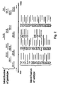
- Fig. 1 The analysis diagram of the NS5B region fragment for the identification of HCV genotype and subtype using biochip is shown in Fig. 1 .
- HCV RNA isolation is carried out through the use of methods known in the given field (for example, Hourfar MK, Michelsen U, Schmidt M, Berger A, Seifried E, Roth WK. High-throughput purification of viral RNA based on novel aqueous chemistry for nucleic acid isolation. Clin Chem. 2005 Jul; 51(7): 1217-22 ) or any specialized commercially available kit of reagents for isolating RNA from blood, plasma or liver biopsy material, for example, QIAamp DSP Virus Kit (Cat No 60704, Qiagen, Germany), MagMAX TM AI/ND Viral RNA Isolation Kits (Cat. No AM1939, Ambion, USA) or "Kit of reagents for RNA isolation Cat. No 05-013, ZAO "DNA technology, Ltd, Russia).
- methods known in the given field for example, Hourfar MK, Michelsen U, Schmidt M, Berger A, Seifried E, Roth WK. High-throughput purification of viral RNA based on
- RT-PCR reverse transcription reaction combined with PCR
- various systems can be used, as shown and described, for example, in Casabianca A., Orlandi C., Fraternale A., Magnani M. A new one-step RT-PCR method for virus quantitation in murine AIDS. 2003 Journal of Virological Methods Vol 110(1), pp. 81-90 , and commercially produced kits, e.g., OneStep RT-PCR Kit (Cat. No 210210, Qiagen, Germany), Accuscript® High-Fidelity RT-PCR Kit (Cat. No 600180, Stratagene, USA) etc.
- Primers for performing a first amplification are selected in such a way as to flank the most polymorphic fragment of an NS5B region that allows to differentiate the existing HCV genotypes and subtypes.
- the NS5B region fragment being amplified is preferred to include HCV genome positions 8256 to 8645 according to Choo, Q.L., K.H. Richman, J.H. Han, K.Berger, C.Lee, C.Dong, C. Gallegos, D. Coit, R. Medina-Selby, P.J.Barr, et al. 1991. Genetic organization and diversity of the Hepatitis C virus. Proc. Natl. Acad. Sci. USA 88: 2451-2455 .
- Primer sequences are selected in such a way as to perform the effective RNA amplification of the analyzed NS5B region fragment, of any HCV genotype and, accordingly, subtype.
- the multiple sequence alignment may be constructed using available databases of NS5B region sequences, such as http://www.ncbi.nlm.nih.gov/Genbank/index.html and htttp://hcv.lanl.gov/content/hcv-db.
- the next step includes the location of the most conservative segments within the analyzed fragment of NS5B region for all HCV genotypes and selection of primers specific to segments concerned. Using the specialized software, for example, Oligo v.
- melting temperatures of primers are calculated and the lengths of primers are varied, thus providing for a spread of the annealing temperatures of the primers inside a pair of not greater than 3-4°C. Also, the sequences are to be avoided which are able to form secondary structures of a hairpin loop type with high melting temperatures. Each and every selected primer should show a unique specificity to the analyzed NS5B region fragment.
- primers The specificity of primers is verified with the help of software using a search in the bases of nucleotide sequences by the BLAST algorithm (www.ncbi.nlm.nih.gov/BLAST/).
- BLAST algorithm www.ncbi.nlm.nih.gov/BLAST/.
- a single-stranded fluorescent labeled product is predominantly obtained by an asymmetric PCR using with the use of deoxynucleoside triphosphates mix as the substrate wherein one of said deoxynucleoside triphosphates is fluorescent labeled.
- the deoxynucleoside triphosphates mix consists of dATP, dGTP, dCTP dTTP, and the latter can be replaced with dUTP or a mixture of dTTP/dUTP in any molar proportion. Any one of said deoxynucleoside triphosphates may be fluorescent labeled.
- a fluorescent labeled deoxyuridine triphosphate is most preferable, which, on the one hand, necessitates the efficient incorporation of the present substrate in the newly synthesized DNA strand during PCR.
- the application of dUTP-fluorescent labeled conjugates makes it possible to prevent cross-contamination using uracil-DNA-glycosylase enzyme. The latter condition is important for the routine analyses in the clinical laboratory.
- the fluorescent dye used can be represented by any fluorescent dye which may chemically be included in the deoxynucleoside triphosphate molecule in such a way as the final conjugate does not hamper the nucleic acids amplification and the subsequent hybridization of the polynucleotide molecule comprising such fluorescent labeled nucleotide residues with immobilized oligonucleotide probes.
- the fluorescent dye can be attached at the 5'-terminal of a dUTP aminoallyl derivative.
- fluorescent dyes examples include fluorescein (TAMRA ® , ROX ® , JOE ® ), rhodamine (Texas Red ® ), polymethine (Cy3 ® , Cy5 ® , Cy5.5 ® , Cy7 ® ) dyes ( Ranasinghe R and Brown T). Fluorescence based strategies for genetic analysis. Chem. Commun, 2005, 5487-5502 ).
- the fluorescent dyes are commercially available, particularly from the Molecular probes company, USA.
- the dyes whose excitation spectrum is within a long-wave(Fed) region are most preferable, which permits using inexpensive semiconductor laser as exciting radiation sources for fluorescence excitation.
- Fluorescent labeled deoxynucleoside triphosphates can be obtained in laboratory conditions using known methods, such as, for example ( Kuwahara M, Nagashima J, Hasegawa M, Tamura T, Kitagata R, Hanawa K, Hososhima S, Kasamatsu T, Ozaki H, Sawai H. Systematic characterization of 2'-deoxynucleoside- 5'-triphosphate analogs as substrates for DNA polymerases by polymerase chain reaction and kinetic studies on enzymatic production of modified DNA. Nucleic Acids Res. 2006 34(19): 5383-94 and are also commercially available, for example, CyDye Fluorescent Nucleotides (Cat. No PA55021, PA55032, PA55026, GE Healthcare, USA).
- Primers for the second step of amplification are selected with the requirements set forth above, with the only difference that at least one of the primers is selected inside a PCR fragment from the first step, to enhance reaction specificity. It is hence only logical to see that the resulted PCR fragment will be a product of semi-nested or nested amplification reaction.
- the length of a second-step amplified fragment is not especially restricted until this enables the efficient hybridization of a fragment with biochip-immobilized probes.
- the primers for the second amplification step are selected such that the length of an amplified fragment should not exceed 800 nucleotides.
- PCR product from the second step makes difficult the efficient diffusion of a PCR product as assayed in biochip gel elements during the hybridization, which may result in reducing the number of hybridization duplexes and, consequently, a fluorescent signal fall.
- primers design account should be taken of the fact that single-stranded fluorescent labeled fragment yielded from a second PCR step should be complementary to oligonucleotides immobilized on the biochip. Therefore in each and every pair, the primer added in an excessive amount (“leader primer”) is selected from a chain whose sequence is complementary to the sequences of the biochip-immobilized oligonucleotides.
- a leader primer is added in an excessive molar amount in relation to a second primer.
- the molar excess is at least tenfold.
- discriminating oligonucleotides for immobilization on a biochip On selection of discriminating oligonucleotides for immobilization on a biochip to take account of the size and complexity of a sequence as assayed and, in particular, the presence of replicas and extended homopolymeric sequences, there is determined a length of the discriminating oligonucleotides that provides their specificity relative to the sequence as assayed.
- discriminating oligonucleotides for genotypes and subtypes is carried out in the following manner. Using constructed multiple alignment of NS5B region sequences, the special consensus sequence is generated for each genotype on whose basis are selected unique probes permitting uniquely identifying each and every genotype. Owing to the high variability of an HCV genome, for enhancing the reliability of a method, several discriminating probes for each of the genotypes are selected, if possible, complementary to various segments of the NS5B region fragment as assayed. The consensus sequence is also generated for each subtype followed by the location of segments of the NS5B region fragment which enable to differentiate the maximum number of subtypes inside one genotype. The number of such segments should be enough for providing the reliable identification of each of the subtypes.
- probes are constructed for the identification of subtypes, and the sequence of one probe may conform to two or more subtypes simultaneously in the separate differentiating segment of the NS5B region fragment as assayed.
- the strategy of probes selection for biochip immobilization is schematically shown in Fig. 2 .
- oligonucleotides are calculated and the lengths of probes are varied thereby to provide for a variation of melting temperatures of the oligonucleotides ranging between 2 and 3°C. Oligonucleotides are avoided which are capable of forming secondary structures of a hairpin loop type with high melting temperatures.
- Discriminating oligonucleotides are immobilized on a biochip support.
- the suitable support that might be used to produce the biochip are represented by an activated, say, aminated surface of glass slides ( Adessi C., Matton G., Ayala G., Turcatti G., Mermod J., Mayer P., Kawashima E. Solid phase DNA amplification: characterization of primer attachment and amplification mechanisms Nucleic Acids Research. 2000. V.51. 28(20).: E87 ), plastic wafers ( Nikiforov T., Rendle R. Goelet P., Rogers Y., Kotewicz M., Anderson S., Trainor G., Knapp Michael R.
- biochip based on hydrogel elements.
- Methods for producing such biochips comprise polymerizing amino-modified oligonucleotides to create a covalent bond with gel monomers in suitable conditions (pH, temperature, composition of polymers and so on) ( Rubina AY, Pan'kov SV, Dementieva EI et al. Hydrogel drop microchips with immobilized DNA: properties and methods for large-scale production. Anal Biochem 2004; 325: 92-106 ).
- Use of biochips comprising gel elements are most preferable, which are applied to a support dropwise with a dia.
- the support used can be represented by a glass substrate (glass slides or cover glass) as well as more available materials, such as plastic materials.
- the support used can be represented by a glass substrate (glass slides or cover glass) as well as more available materials, such as plastic materials.
- the concentration of immobilized oligonucleotide probes can be judged by staining the gel elements of the biochip with a dye showing a low specificity to a DNA nucleotide sequence ( A.L. Mikheikin, A.V. Choudinov, A.I. Yaroschuk, A.Yu. Roubina, S.V. Pan'kov, A.S. Krylov, A.S. Zasesammlungv, A.D. Mirzabekov.
- the dye showing a low specificity to the DNA nucleodie sequence use for evaluating the number of oligonucleotides immobilized in the elements of biological microchips. Molecular biology 2003; 37(6): 1061-70 ).
- PCR-products from a second amplification step are hybridized on a biochip with immobilized differentiating oligonucleotides complementary to the consensus sequences of genotypes and subtypes of an NS5B region fragment.
- Hybridization is carried out in a solution containing a buffer component for maintaining a pH value, a salt for creating ionic strength and a chaotropic (hydrogen-bond destabilizing) agent in a hermetically sealed hybridization chamber at a temperature depending on the melting temperatures of immobilized discriminating oligonucleotides.
- the hydrogen-bond destabilizing agent that might be used can be represented by, for example, guanidine thiocyanate, urea or formamide.
- the discriminating oligonucleotides of the present invention have melting temperatures ranging between 42 and 44°C, which fact allows one to carry out hybridization at 37°C using said chaotropic agent.
- the temperature of 37°C is suitable in that a majority of clinical laboratories are equipped with thermostats maintaining this temperature.
- the DNA fragment forms perfect hybridization duplexes only with adequate (fully complementary) oligonucleotides. With all the remaining oligonucleotides said DNA fragment provides an imperfect duplex. Said perfect and imperfect duplexes are discriminated by comparing the fluorescence intensities of biochip elements wherein the duplexes have formed. The signal strength in the element with the perfect hybridization duplex formed therein (I perf.) is higher than in the element where imperfect duplex (I imperf.) has been formed. Hybridization performed in the most favourable conditions (temperature, the concentration of a chaotropic (hydrogen - bond - destabilizing) agent and hybridization buffer ionic strength) provides a I perf ⁇ . /I imperf. ⁇ 1.5 ratio between two elements comprising probes belonging to one group and differing by one nucleotide.
- Registration of hybridization results on biochips can be performed with the aid of commercial scanning devices - analyzers of biochip fluorescence, for example, GenePix 4000B (Axon Instruments, USA) equipped with the adequate software for calculating the strength of fluorescent signals of the discrete elements of a biochip and their subsequent normalization for a background value, for example, 'GenePix Pro', 'Acuity' (Axon Instruments, USA).
- Hybridization results can be performed visually through the correlation of the registered fluorescence pattern of a biochip and/or distribution of the signal strength of biochip elements thus obtained to the arrangement of specific discriminating probes in the biochip elements (cf. Fig. 3 ). Given the distributed signal strength in biochip elements, a maximum signal is detected from among the elements comprising genotype-specific oligonucleotides. A genotype can be identified by providing a biochip element having a maximum fluorescence intensity among the probe-containing elements to determine an HCV genotype.
- Identification of the subtype of an HCV specimen as assayed can be realized by determining the maximum signals in the elements containing subtype-specific oligonucleotides corresponding to the genotype, as determined, in case of the maximum signals being registered in at least two different elements which contain unique subtype-specific probes.
- First step extraction of valid signals, more exactly the signals in elements, wherein perfect duplexes might be formed, for which purpose the normalized fluorescent strength signals of all biochip elements are classified as to increase and compared with an average signal (I ref ) in the elements devoid of any oligonucleotides.
- the valid signals are those exceeding I ref at least 1.5 times.
- Second step starting with an analysis of filtered valid signals G i in the groups of elements containing genotype-specific oligonucleotides (i - genotype number). A maximum signal is extracted inside each group of elements G imax and compared with each other.
- a conclusion is drawn on a specimen, as assayed, belonging to the given genotype. If a ratio of signals among G imax does not exceed the threshold value, a conclusion is made on the impossibility to clearly identify a genotype and on the possible presence in the specimen, as assayed, of a mixture of two and more HCV variants with various genotypes. If the signals in genotype-specific- oligonucleotides-containing groups do not undergo primary filtration in relation to the I ref , a conclusion is drawn on low signal strength and the possible absence of an HCV RNA in the specimen as assayed. And no subtype identification is performed whatever.
- the oligonucleotides are combined in groups according to the selected segments of an NTS5B fragment as assayed that permits differentiating the maximum number of the subtypes.
- the number of groups is varied from one to four in relation to the degree of homology of the consensus sequences of the NS5B fragment for various subtypes and is dictated by the need for a reliable differentiation of subtypes inside the genotype.
- This signal is designated as S ixj (i - genotype number, 'x'-symbol of a subtype according to HCV subtype classification, j - group number). Should two or more elements in the group have signals differing from one another less than 1.5 times, then all such signals-S ixj , S iyj , to mention only few, are picked out. The result: a set of elements from various groups ix1, iy1, ix2, iz2, ix3, etc. whose signals exceed the rest of signals in their groups no less than 1.5 times.
- the elements of different groups in the set so obtained conform to different genotypes, for example, ix1, iy2, iz3 or ix1, iyz3, then the signals of the given elements are compared with each other. If the signal of a element conforming to the subtype 'x' of one group exceeds the signals of the elements of other groups 3 times or more, a conclusion is drawn on the assayed specimen belonging to the subtype 'x'.
- duration of antiviral therapy and prognosis can be made on the basis of data on genotype/subtype identification.
- duration of pegylated interferon/ribavirin therapy is no less than 24 weeks for subtypes 1a, 1c, 1d, 1e and in case of subtype 1 b being detected that is interferon-resistant - no less than 48 weeks ( Weck K. Molecular methods of hepatitis C genotyping. Expert Rev Mol Diagn. 2005 Jul; 5(4): 507-20 ).
- the infection caused by HCV with a genotype 4 provides a clinical picture similar to virus genotype 1 infection ( Legrand-Abravanel F, Nicot F, Boulestin A, Sandres-Sauné K, Vinel JP, Alric L, Izopet J. Pegylated interferon and ribavirin therapy for chronic Hepatitis C virus genotype 4 infection. J Med Virol. 2005 Sep; 77(1):66-9 ).
- the genotype 4 is distinguished for splitting into a considerably greater number of subtypes ( Nicot F, Legrand-Abravanel F, Sandres-Saune K, Boulestin A, Dubois M, Alric L, Vinel JP, Pasquier C, Izopet J. 2005. Heterogeneity of Hepatitis C virus genotype 4 strains circulating in south-western France. J Gen Virol Jan; 86(Pt 1): 107-14 ).
- 4a and 4d are resistant to interferon and calls for a prolonged course of treatment (no less than 48 weeks) ( Roulot D, Bourcier V, Grando V, Epidemiological characteristics and response to peginterferon plus ribavirin treatment of Hepatitis C virus genotype 4 infection (J Viral Hepat. 2007 Jul; 14(7): 460-7 ).
- HCV genotypes 2 and 3 are responsive to therapy with drugs and lead to chronic disease in significantly lesser amount of cases. Duration of ribavirin/interferon therapy for HCV infected patients with the given genotypes is 6 to 12 weeks.
- genotype and subtype provides information in terms of etiology of infection.
- Subtypes 1a, 3a, 4a, 4d are most commonly associated with intravenous drug users, whereas a genotype 2 and a subtype 1 are linked with a blood transfusion transfer route ( Simmonds P, Bukh J, Combet C. Consensus proposals for a unified system of nomenclature of Hepatitis C virus genotypes. Hepatology. 2005 Oct; 42(4): 962-73 ).
- Example 1 Biochip for identifying HCV genotype and subtype on the basis of NS5B region assay.
- Oligonucleotides for immobilization on a biochip and primers for amplification were synthesized on an automatic synthesizer-394 DNA/RNA synthesizer (Applied Biosystems, USA) and contained a spacer with a free amino group 3'-Amino-Modifier C7 CPG 500 (Glen Research, USA) for the following immobilization to a gel.
- Biochips were produced according to the procedure thus far described (( Rubina AY, Pan'kov SV, Dementieva EI et al. Hydrogel drop microchips with immobilized DNA: properties and methods for large-scale production. Anal Biochem 2004; 325: 92-106 ).
- the biochips contained hemispherical elements, 100 mcm in dia., through a distance of 300 mcm. Uniformity of the application of elements and their dia. were assessed with the help of the software 'Test-chip' (Biochip-IMB, Russia). The qualitative control of microchips was made by measuring the concentrations of immobilized oligonucleotides. The biochips were stained with a fluorescent dye-ImD-310 (Biochip-IMB), the concentration of immobilized probes was assessed as described above ( A.L. Mikheikin, A.V. Choudinov, A.I. Yaroschuk, A.Yu. Roubina, S.V. Pan'kov, A.S. Krylov, A.S.
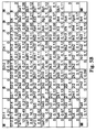
- a biochip comprises 120 immobilized oligonucleotides whose list is presented in Table I, four marker points (M) for accurate positioning (image acquisition), performed by a software, and four elements of an empty gel (O) necessary for computing a reference (background) value of fluorescence intensity I ref .
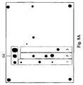
- Arrangement of oligonucleotides immobilized on a microchip is shown in Fig. 3 .
- oligonucleotides are immobilized with a 'G' index permitting identifying a HCV genotype.
- a biochip identifies all six HCV genotypes.
- oligonucleotides allowing for identifying subtypes: - inside a genotype 1: 1a, 1b, 1c, 1d, 1e.
- genotype 1 1a, 1b, 1c, 1d, 1e.
- each group is conforming to a separate segment within an NS5B region fragment as assayed: - inside a genotype 2: 2a, 2b, 2c, 2d, 2i, 2j, 2k, 21, 2m.
- each group is conforming to a separate segment within an NS5B region fragment as assayed; - inside a genotype 3: 3a, 3b, 3k.
- each group is conforming to a separate segment within an NS5B region fragment as assayed; - inside a genotype 4: 4a, 4c, 4d, 4f, 4h, 4i, 4k, 4n, 4o, 4p, 4r, 4t.
- each group is conforming to a separate segment within an NS5B region fragment as assayed; - inside a genotype 5: 5a.
- the genotype 5 is a single subtype 5a, with the genotype-identifying probes thus identifying the subtype 5a; - inside a genotype 6: 6a, 6b, 6d, 6g, 6h, 6k.
- each group is conforming to a separate segment inside an NS5B region fragment as assayed. Table 1.
- First step reverse transcription combined with the PCR (RT-PCR) to obtain a 418 b.p. NS5B region fragment.
- a 10 mcl of isolated viral RNA was added to 40 mcl RT-PCR mix (final volume of 50 mcl).
- thermocycler PTC-200 Dyad MJ Research, USA: reverse transcription at 50°C-30 min, followed by 50 cycles of PCR: 95°C-30 s, 63°C-30 s, 72°C- 30 s; final elongation at 72°C - 10 min.
- a 1 mcl reaction mix obtained at first step of amplification was used as template for second step.
- a second PCR step was carried out in a semi-nested variant with P3_f/Pr5_r primers flanking a 382 b.p. NS5B region.
- Primer sequences are given in Table 2.
- the reaction chamber of the biochip was filled with the 32 mcl of resulted hybridization mixture and sealed. Hybridization was performed at 37°C for 12-18 hours. On completion of hybridization, the biochip was washed thrice with distilled water at 37°C for 30 s and dried.
- Example 5 Analysis of NS5B region of HCV sample belonging to subtype 1a using hybridization on biochip
- HCV viral RNA was isolated from patient's blood specimen using Qiamp Viral RNA mini kit (Qiagen, Germany) under the protocol of manufacturer. The isolated RNA was used in RT - PCR as described (Example 2). The presence of an amplified NS5B 418 b.p. long fragment was tested by electrophoresis in agarose gel whereupon a RT-PCR product was divided into two portions of which one was treated and assayed according to the methods as described in Examples 2-4 (a second PCR step followed by hybridization on a biochip, washing, registration and interpretation of the fluorescent pattern of the biochip).
- the second portion of a first step product was used upon additional purification in sequencing reaction, followed by analysis on an automatic sequencer, correcting a chromatogram and obtaining the sequence of NS5B region fragment, constructing a multiple alignment and a phylogenetic tree on whose basis a genotype and a subtype were determined.
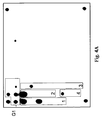
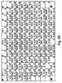
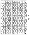
- Fig. 4A shows a biochip hybridization pattern.
- Fig. 4B demonstrates the distribution of the normalized fluorescence signals of biochip elements.
- analysis starts off with computing a mean signal (I ref ) in empty elements, in which particular case the I ref is 0.62.
- the signals are filtered into genotype and subtype-specific elements, with the result that the signals in group G1 containing genotype 1-specific probes exceed the I ref 1.5 times or more.
- G4-2 (1.37).
- the signals in other groups containing genotype-specific probes were close to background ones.
- the maximum signal is detected from group G1, G1-2 (5.7).
- the signal in the given element exceeds a signal G4-2 more than 1.5 times. So the sequence of the analyzed HCV sample, as assayed, relates to a genotype 1.
- a sequencing method with subsequent phylogenetic analysis showed that the sequence being assayed falls within a cluster of subtype 1a sequences.
- an HCV RNA specimen as assayed, has a genotype 1 and a subtype 1a, which coincides with sequencing results in full.
- HCV viral RNA was isolated from patient's blood specimen using Qiamp Viral RNA mini kit (Qiagen, Germany) under the protocol of manufacturer. The isolated RNA was used in RT - PCR as described (Example 2). The presence of an amplified NS5B 418 b.p. long fragment was tested by electrophoresis in agarose gel whereupon a RT-PCR product was divided into two portions of which one was treated and assayed according to the methods as described in Examples 2-4 (a second PCR step followed by hybridization on a biochip, washing, registration and interpretation of the fluorescent pattern of the biochip).
- the second portion of a first step product was used upon additional purification in sequencing reaction, followed by analysis on an automatic sequencer, correcting a chromatogram and obtaining the sequence of NS5B region fragment, constructing a multiple alignment and a phylogenetic tree on whose basis a genotype and a subtype were determined.
- Fig. 5A shows a biochip hybridization pattern.
- Fig. 5B demonstrates the distribution of the normalized fluorescence signals of biochip elements.
- analysis starts off with computing a mean signal (I ref ) in empty elements, in which particular case the I ref is 0.67.
- the signals are filtered in genotype-and subtype - specific elements.
- the result is that the signals in G 1 group containing genotype 1 - specific probes exceed the I ref 1.5 times or more (the maximum signal is characteristic of a G1-3 element (5.69)).
- the signals in other groups containing the genotype-specific probes were close to background ones. So the sequence of the analyzed HCV sample, as assayed, relates to a genotype 1.
- a sequencing method with subsequent phylogenetic analysis showed that the sequence being assayed falls within a cluster of subtype 1b sequences.
- an HCV RNA specimen as assayed, has a genotype 1 and a subtype 1 b which fully coincides with sequencing results.
- Example 7 Analysis of NS5B region of HCV sample belonging to subtype 1e using hybridization on biochip
- HCV viral RNA was isolated from patient's blood specimen using Qiamp Viral RNA mini kit (Qiagen, Germany) under the protocol of manufacturer. The isolated RNA was used in RT - PCR as described (Example 2). The presence of an amplified NS5B 418 b.p. long fragment was tested by electrophoresis in agarose gel whereupon a RT-PCR product was divided into two portions of which one was treated and assayed according to the methods as described in Examples 2-4 (a second PCR step followed by hybridization on a biochip, washing, registration and interpretation of the fluorescent pattern of the biochip).
- the second portion of a first step product was used upon additional purification in sequencing reaction, followed by analysis on an automatic sequencer, correcting a chromatogram and obtaining the sequence of NS5B region fragment, constructing a multiple alignment and a phylogenetic tree on whose basis a genotype and a subtype were determined.
- Fig. 6A shows a biochip hybridization pattern.
- Fig. 6B demonstrates the distribution of the normalized fluorescence signals of biochip elements.
- analysis begins with the computation of a mean signal (I ref ) in empty elements.
- I ref the value of I ref is 0.39.
- the threshold value of 1.5 the signals are filtered in genotype-and subtype-specific elements.
- the signals in other groups of elements containing the genotype-specific probes were close to background ones.
- the maximum signal in the group G1 is characteristic of a G1-3 element (4.82). So the sequence of the analyzed HCV sample relates to a genotype 1.
- a sequencing method with subsequent phylogenetic analysis showed that the sequence being assayed falls within a cluster of subtype le sequences.
- an HCV RNA specimen as assayed has a genotype I and a subtype 1e, which fully coincides with sequencing results.
- Example 8 Analysis of NS5B region of HCV sample belonging to subtype 2a using hybridization on biochip
- HCV viral RNA was isolated from patient's blood specimen using Qiamp Viral RNA mini kit (Qiagen, Germany) under the protocol of manufacturer. The isolated RNA was used in RT - PCR as described (Example 2). The presence of an amplified NS5B 418 b.p. long fragment was tested by electrophoresis in agarose gel whereupon a RT-PCR product was divided into two portions of which one was treated and assayed according to the methods as described in Examples 2-4 (a second PCR step followed by hybridization on a biochip, washing, registration and interpretation of the fluorescent pattern of the biochip).
- the second portion of a first step product was used upon additional purification in sequencing reaction, followed by analysis on an automatic sequencer, correcting a chromatogram and obtaining the sequence of NS5B region fragment, constructing a multiple alignment and a phylogenetic tree on whose basis a genotype and a subtype were determined.
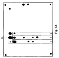
- Fig. 7A shows a biochip hybridization pattern.
- Fig. 7B demonstrates the distribution of the normalized fluorescence signals of biochip elements.
- analysis begins with the computation of a mean signal (I ref ) in empty elements.
- I ref the value of I ref is 0.25.
- filtration of the signals is carried out in genotype- and subtype-specific elements.
- the signals in group 2 elements comprising probes showing specificity to a genotype 2 exceed the I ref 1.5 times or more.
- the signal in a G6 element (2.01) is likewise valid relative to the I ref
- the maximum signal in the group 2 is characteristic of an element G2-2 (15.6) exceeding the signal in a G6 element more than 1.5 times. It follows that the sequence of the analyzed HCV sample relates to a genotype 2.
- Assaying in the groups of elements comprising the subtype-specific probes of genotype 2 has revealed: in group 1 the maximum (i.e. exceeding a 1.5 threshold value in relation to other elements) signal has 2ad1 (15.2). In group 2-2a2 (6.62). In group 3 - 2a31 (2.33). In all three groups, the maximum signal is characteristic of the elements containing probes specific for a subtype 2a. So, the specimen as assayed refers to the subtype 2a.
- a sequencing method with subsequent phylogenetic analysis showed that the sequence being assayed falls within a cluster of subtype 2a sequences.
- HCV viral RNA was isolated from patient's blood specimen using Qiamp Viral RNA mini kit (Qiagen, Germany) under the protocol of manufacturer. The isolated RNA was used in RT - PCR as described (Example 2). The presence of an amplified NS5B 418 b.p. long fragment was tested by electrophoresis in agarose gel whereupon a RT-PCR product was divided into two portions of which one was treated and assayed according to the methods as described in Examples 2-4 (a second PCR step followed by hybridization on a biochip, washing, registration and interpretation of the fluorescent pattern of the biochip).
- the second portion of a first step product was used upon additional purification in sequencing reaction, followed by analysis on an automatic sequencer, correcting a chromatogram and obtaining the sequence of NS5B region fragment, constructing a multiple alignment and a phylogenetic tree on whose basis a genotype and a subtype were determined.
- Fig. 8A shows a biochip hybridization pattern.
- Fig. 8B demonstrates the distribution of the normalized fluorescence signals of biochip elements.
- analysis begins with the computation of a mean signal (I ref ) in empty elements.
- I ref the value of I ref is 0.44.
- the signals are filtered in genotype- and subtype-specific elements with the result that only the signals in elements of group G2 with genotype 2-specific probes exceed the I ref 1.5 times or more.
- the maximum signal in the group G2 is characteristic of an element C2-3 (15.9). It follows that the sequence of the analyzed HCV sample relates to a genotype 2.
- the maximum (i.e. exceeding a 1.5 threshold value relative to other elements) signal has 2i1 (2.3).
- the perfect duplexes are provided with an assayed DNA with an oligonucleotide whose sequence is universal for subtypes 2c and 2k.
- the maximum signal belongs to the elements containing unique oligonucleotides showing a specificity to a subtype 2i.
- the specimen as assayed refers to the subtype 2i.
- a sequencing method with subsequent phylogenetic analysis showed that the sequence being assayed falls within a cluster of subtype 2i sequences.
- an HCV RNA specimen as assayed has a genotype 2 and a subtype 2i, which fully coincides with sequencing results.
- Example 10 Analysis of NS5B region of HCV sample belonging to subtype 3a using hybridization on biochip
- HCV viral RNA was isolated from patient's blood specimen using Qiamp Viral RNA mini kit (Qiagen, Germany) under the protocol of manufacturer. The isolated RNA was used in RT - PCR as described (Example 2). The presence of an amplified NS5B 418 b.p. long fragment was tested by electrophoresis in agarose gel whereupon a RT-PCR product was divided into two portions of which one was treated and assayed according to the methods as described in Examples 2-4 (a second PCR step followed by hybridization on a biochip, washing, registration and interpretation of the fluorescent pattern of the biochip).
- the second portion of a first step product was used upon additional purification in sequencing reaction, followed by analysis on an automatic sequencer, correcting a chromatogram and obtaining the sequence of NS5B region fragment, constructing a multiple alignment and a phylogenetic tree on whose basis a genotype and a subtype were determined.
- Fig. 9A shows a biochip hybridization pattern.
- Fig. 9B demonstrates the distribution of the normalized fluorescence signals of biochip elements.
- analysis begins with the computation of a mean signal (I ref ) in empty elements.
- I ref the value of I ref is 0.52.
- the signals are filtered in genotype- and subtype-specific elements.
- the signals in group G3 containing probes specific for a genotype 3 exceed the I ref 1.5 times or more.
- the signal in a G4-2 element (0.85) likewise exceeds the I ref 1.5 times.
- the signals in other groups of elements containing genotype-specific probes are close to background ones.
- the maximum signal in the group G3 is characteristic of a G3-1 element (15.4) whose signal exceeds that of G4-2 more than 1.5 times. It follows that the sequence of the analyzed HCV sample relates to a genotype 3.
- Assaying in the groups of elements containing subtype-specific probes of genotype 3 reveals the following: in group I the maximum (i.e. exceeding a 1.5 threshold value relative to other elements) signal is characteristic of a 3a1 element (16.2). In group 2 - 3a2 (4.69). In group 3 - 3a3 (1.74). And, as so, in all the groups, the maximum signal is featured by probe-containing elements showing a specificity to a subtype 3a. Consequently the specimen as assayed is related to the subtype 3a.
- a sequencing method with subsequent phylogenetic analysis showed that the sequence being assayed falls within a cluster of subtype 3a sequences.
- an HCV RNA specimen as assayed has a genotype 3 and a subtype 3a, which is in full coincidence with sequencing results.
- HCV viral RNA was isolated from patient's blood specimen using Qiamp Viral RNA mini kit (Qiagen, Germany) under the protocol of manufacturer. The isolated RNA was used in RT - PCR as described (Example 2). The presence of an amplified NS5B 418 b.p. long fragment was tested by electrophoresis in agarose gel whereupon a RT-PCR product was divided into two portions of which one was treated and assayed according to the methods as described in Examples 2-4 (a second PCR step followed by hybridization on a biochip, washing, registration and interpretation of the fluorescent pattern of the biochip).
- the second portion of a first step product was used upon additional purification in sequencing reaction, followed by analysis on an automatic sequencer, correcting a chromatogram and obtaining the sequence of NS5B region fragment, constructing a multiple alignment and a phylogenetic tree on whose basis a genotype and a subtype were determined.
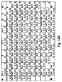
- Fig. 10A shows a biochip hybridization pattern.
- Fig. 10B demonstrates the distribution of the normalized fluorescence signals of biochip elements.
- analysis begins with the computation of a mean signal (I ref ) in empty elements.
- I ref the value of I ref is 0.55.
- the signals are filtered in genotype-and subtype-specific elements.
- the signals in G4 group elements containing probes specific for a genotype 4 exceed the I ref 1.5 times or more.
- the signals in the remaining elements containing genotype-specific oligonucleotides were close to background ones.
- the maximum signal in the G4 group is registered in a G4-3 element (17.9). It follows that the RNA sequence of a specimen as assayed is related to the genotype 4.
- Assaying in groups containing subtype-specific probes of genotype 4 reveals the following points: in three groups of probes specific for a genotype 4, the maximum signals belong to the elements containing probes for detecting a subtype 4a: 4ac1 (16.3), 4a21 (1.08), 4a4 (14.1). In group 3, the maximum signal belongs to a 4on3 element (1.21); however, in accordance with the algorithm, as shown and described, the specimen as assayed refers to the subtype 4a.
- a sequencing method with subsequent phylogenetic analysis showed that the sequence being assayed falls within a cluster of subtype 4a sequences.
- an HCV RNA specimen as assayed has a genotype 4 and a subtype 4a, which is in full coincidence with sequencing results.
- HCV viral RNA was isolated from patient's blood specimen using Qiamp Viral RNA mini kit (Qiagen, Germany) under the protocol of manufacturer. The isolated RNA was used in RT - PCR as described (Example 2). The presence of an amplified NS5B 418 b.p. long fragment was tested by electrophoresis in agarose gel whereupon a RT-PCR product was divided into two portions of which one was treated and assayed according to the methods as described in Examples 2-4 (a second PCR step followed by hybridization on a biochip, washing, registration and interpretation of the fluorescent pattern of the biochip).
- the second portion of a first step product was used upon additional purification in sequencing reaction, followed by analysis on an automatic sequencer, correcting a chromatogram and obtaining the sequence of NS5B region fragment, constructing a multiple alignment and a phylogenetic tree on whose basis a genotype and a subtype were determined.
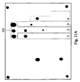
- Fig. 11A shows a biochip hybridization pattern.
- Fig. 11B demonstrates the distribution of the normalized fluorescence signals of biochip elements.
- analysis begins with the computation of a mean signal (I ref ) in empty elements.
- I ref the value of I ref is 0.35.
- the signals are filtered in genotype- and subtype-specific elements.
- the signals in G4 group elements containing probes specific for a genotype 4 exceed the I ref 1.5 times or more.
- the signals in the rest of elements containing genotype-specific oligonucleotides were close to background ones.
- the maximum signal in the G4 group belongs to a G4-1 element (17.4). It follows that the RNA sequence of a specimen as assayed is related to the genotype 4.
- Assaying in the groups of subtype-specific probes of genotype 4 reveals the following points: in group I, the maximum signal is featured by a 4df1 element, (11.6) in group 2 - 4dp2 element (3.16), in group 3 - 4d3 element (1.95), in group 4 - 4d4 (7.35). In all groups, the maximum signal is characteristic of probe-containing elements specific for a subtype 4d. Consequently a specimen as assayed is related to the subtype 4d.
- a sequencing method with subsequent phylogenetic analysis showed that the sequence being assayed falls within a cluster of subtype 4d sequences.
- an HCV RNA specimen as assayed has a genotype 4 and a subtype 4d, which fully coincides with sequencing results.
- HCV viral RNA was isolated from patient's blood specimen using Qiamp Viral RNA mini kit (Qiagen, Germany) under the protocol of manufacturer. The isolated RNA was used in RT - PCR as described (Example 2). The presence of an amplified NS5B 418 b.p. long fragment was tested by electrophoresis in agarose gel whereupon a RT-PCR product was divided into two portions of which one was treated and assayed according to the methods as described in Examples 2-4 (a second PCR step followed by hybridization on a biochip, washing, registration and interpretation of the fluorescent pattern of the biochip).
- the second portion of a first step product was used upon additional purification in sequencing reaction, followed by analysis on an automatic sequencer, correcting a chromatogram and obtaining the sequence of NS5B region fragment, constructing a multiple alignment and a phylogenetic tree on whose basis a genotype and a subtype were determined.
- Fig. 12A shows a biochip hybridization pattern.
- Fig. 12B demonstrates the distribution of the normalized fluorescence signals of biochip elements.
- analysis begins with the computation of a mean signal (I ref ) in empty elements.
- I ref the value of I ref is 0.28.
- the signals are filtered in genotype- and subtype-specific elements, with the result that only the signals in group G5 elements containing probes specific for a genotype 5 exceed the I ref 1.5 times or more.
- the maximum signal in the G5 group is characteristic of a G5-1 element (6.58). It follows that the RNA sequence of a specimen as assayed is related to the genotype 5. Inasmuch as the latter has only one subtype, 5a, and the signal in a 5a2 element showing a specificity to the given subtype is actual, a conclusion might be drawn that the specimen as assayed has the subtype 5a.
- an HCV RNA specimen as assayed has a genotype 5 and a subtype 5a, which is in full coincidence with sequencing results.
- Example 14 Analysis of NS5B region of HCV sample belonging to genotype 6 using hybridization on biochip
- HCV viral RNA was isolated from patient's blood specimen using Qiamp Viral RNA mini kit (Qiagen, Germany) under the protocol of manufacturer. The isolated RNA was used in RT - PCR as described (Example 2). The presence of an amplified NS5B 418 b.p. long fragment was tested by electrophoresis in agarose gel whereupon a RT-PCR product was divided into two portions of which one was treated and assayed according to the methods as described in Examples 2-4 (a second PCR step followed by hybridization on a biochip, washing, registration and interpretation of the fluorescent pattern of the biochip).
- the second portion of a first step product was used upon additional purification in sequencing reaction, followed by analysis on an automatic sequencer, correcting a chromatogram and obtaining the sequence of NS5B region fragment, constructing a multiple alignment and a phylogenetic tree on whose basis a genotype and a subtype were determined.
- Fig. 13A shows a biochip hybridization pattern.
- Fig. 13B demonstrates the distribution of the normalized fluorescence signals of biochip elements.
- analysis begins with the computation of a mean signal (I ref ) in empty elements.
- I ref the value of I ref is 0.72.
- signals are filtered in genotype - and subtype-specific elements.
- the signals in group G6 element (18.6) comprising a probe showing a specificity to a genotype 6 exceed the I ref 1.5 times or more.
- the signal in a G3-1 element (5.2) is also to be considered valid.
- the G6 element exceeds the signal in the G3-1 element more than 1.5 times, which means the identity of analyzed HCV specimen to the genotype 6.
- a sequencing method with subsequent phylogenetic assay goes to show that the sequence, as assayed, falls within none on the clusters of genotype 6 subtypes and forms a separate branch of a phylogenetic tree, or - to be more exact - it is unclassified. It is hence only logical to see that the results obtained in both methods confirm the impossibility to clearly define the subtype of the given specimen.
- the invention as submitted permits identifying the genotype and subtype of Hepatitis C virus, on the basis of the analysis of an NS5B region using a biological microchip.
- a method permits identifying all HCV 6 genotypes and 36 subtypes, with the most virulent and drug resistant forms included.
- the method of the present invention advantageously differs from the existing analogs in high specificity as to the identification of genotype 1 subtypes, more exactly, a 1b subtype, and also in simplicity of execution and low cost.
- Data obtained through the use of a method of hybridization on biochips of the invention, as being claimed and as set forth in the application, can be used for estimation and prognosis of disease severity (acute/chronic cirrhosis, a likelihood of development of liver cancer), determining a therapeutic dosage of medicaments and duration of a course of therapy as well as for epidemiologic genotyping.
Landscapes
- Health & Medical Sciences (AREA)
- Chemical & Material Sciences (AREA)
- Life Sciences & Earth Sciences (AREA)
- Organic Chemistry (AREA)
- Wood Science & Technology (AREA)
- Engineering & Computer Science (AREA)
- Zoology (AREA)
- Communicable Diseases (AREA)
- Immunology (AREA)
- Proteomics, Peptides & Aminoacids (AREA)
- Molecular Biology (AREA)
- Biotechnology (AREA)
- Microbiology (AREA)
- Physics & Mathematics (AREA)
- Biophysics (AREA)
- Analytical Chemistry (AREA)
- Virology (AREA)
- Biochemistry (AREA)
- Bioinformatics & Cheminformatics (AREA)
- General Engineering & Computer Science (AREA)
- General Health & Medical Sciences (AREA)
- Genetics & Genomics (AREA)
- Measuring Or Testing Involving Enzymes Or Micro-Organisms (AREA)
Claims (13)
- Verfahren zum Identifizieren eines Genotyps und eines Subtyps des Hepatitis C-Virus auf Basis der Analyse einer HCV-Genom-NS5B-Region, umfassend:(a) - Reverse-Transkription, kombiniert mit PCR (RT-PCR), unter Verwendung einer Virus-RNA als Vorlage und eines ersten Paares an Primern, die eine Spezifizität gegenüber einem NS5B-Regionfragment zeigen;(b) - asymmetrische Amplifizierung des NS5B-Regionfragments unter Verwendung eines in (a) erhaltenen RT-PCR-Produkts als Vorlage, eines zweiten Paares an spezifischen Primern und eines Gemischs aus vier Desoxynukleosidtriphosphaten, wobei eines der vier Desoxynukleosidtriphosphaten fluoreszenzmarkiert ist, als Substrat, um ein im Wesentlichen einsträngiges fluoreszenzmarkiertes Fragment bereitzustellen;(c) - Bereitstellen eines Biochips für die Identifizierung des HCV-Genotyps und - Subtyps, der einen Träger darstellt, der einen Satz eigenständiger Elemente umfasst, wobei eine einzigartige Oligonukleotidsonde auf jedem dieser immobilisiert ist, mit einer Sequenz komplementär zur Sequenz eines in Schritt (b) erhaltenen einzelsträngigen Fragments und ausgewählt aus der Gruppe umfassend: a) die NS5B-Regionfragment-Sequenzen, die für jeden der HCV-Genotypen spezifisch sind (genotypspezifisch); und b) NS5B-Regionfragment-Sequenzen, die für jeden der HCV-Subtypen spezifisch sind (subtypspezifisch);(d) - Hybridisieren des amplifizierten markierten Produkts, das in Schritt (b) bereitgestellt wurde, auf dem Biochip unter Bildung von Duplexen mit immobilisierten Sonden unter Bedingungen, die eine Einzelnukleotidauflösung zwischen den perfekten und nicht-perfekten Duplexen vorsieht;(e) - Registrieren und Interpretieren von Hybridisierungsergebnissen.
- Verfahren nach Anspruch 1, dadurch gekennzeichnet, dass in Schritt (a) ein erstes Paar an spezifischen Primern verwendet wird, deren Sequenzen in SEQ ID Nr. 121 und 122 angeführt sind.
- Verfahren nach Anspruch 1, dadurch gekennzeichnet, dass in Schritt (b) ein zweites Paar an spezifischen Primern verwendet wird, deren Sequenzen in SEQ ID Nr. 121 und 123 angeführt sind.
- Verfahren nach Anspruch 1, dadurch gekennzeichnet, dass in Schritt (b) einer der Primer des zweiten Paares in einem zumindest zehnfachen molaren Überschuss in Bezug auf den zweiten Primer verwendet wird.
- Verfahren nach Anspruch 1, dadurch gekennzeichnet, dass in Schritt (b) das verwendete fluoreszenzmarkierte Desoxynukleosidtriphosphat dem fluoreszenzmarkierten Desoxyuridintriphosphat entspricht.
- Verfahren nach Anspruch 1, dadurch gekennzeichnet, dass der Biochip ein Biochip auf Hydrogelelementbasis ist, der durch ein Verfahren der chemischen oder lichtinduzierten Copolymerisation erhalten wird.
- Verfahren nach Anspruch 1, dadurch gekennzeichnet, dass der Biochip einen Satz immobilisierter Oligonukleotide umfasst, deren Sequenzen in SEQ ID Nr. 1-120 definiert sind.
- Verfahren nach Anspruch 1, dadurch gekennzeichnet, dass das Registrieren der Ergebnissen in Schritt (e) mithilfe eines tragbaren Fluoreszenzanalysators und Software erfolgt, die die Verwendung der softwarebasierten Verarbeitung von Signalintensitäten mit der darauffolgenden Interpretation der Ergebnisse ermöglicht.
- Verfahren nach Anspruch 1, wobei die Interpretation der registrierten Ergebnisse in Schritt (e) in zwei Schritten erfolgt:1) Untersuchen der Signale in Biochipelementen, die die für HCV-Genotypen spezifischen Oligonukleotidsonden umfassen, um dadurch den Genotyp eines untersuchten Prüflings zu identifizieren; 2) im Fall der Identifizierung eines Genotyps Untersuchen von ausschließlich Biochipelementen, die für die Subtypen eines identifizierbaren Genotyps spezifisch sind, unabhängig vom Vorhandensein der Signale in den Elementen, die die für die Subtypen anderer Genotypen spezifischen Sonden enthalten.
- Verfahren nach Anspruch 1, ferner umfassend das Evaluieren und Prognostizieren des Schweregrads einer Krankheit, das Bestimmen einer therapeutischen Dosis von Medikamenten und den Verlauf einer Therapie und/oder epidemiologisches Genotypisieren auf Basis der Interpretation von Hybridisierungsergebnissen.
- Biochip zum Identifizieren eines HCV-Genotyps und -Subtyps auf Basis der Analyse einer NS5B-Region, umfassend einen Träger, der einen Satz an eigenständigen Elementen enthält, wobei eine einzigartige Oligonukleotidsonde auf jedem dieser immobilisiert ist, und wobei die Sondensequenzen in SEQ ID Nr. 1-120 definiert sind.
- Biochip nach Anspruch 12, dadurch gekennzeichnet, dass er ein Biochip auf Hydrogelelementbasis ist, der durch ein Verfahren der chemischen oder lichtinduzierten Copolymerisation erhalten wird.
- Satz Oligonukleotidsonden zum Produzieren eines Biochips für die Identifizierung eines HCV-Genotyps und -Subtyps auf Basis der Analyse einer NS5B-Region, deren Sequenzen in SEQ ID Nr. 1-120 definiert sind.
Applications Claiming Priority (1)
| Application Number | Priority Date | Filing Date | Title |
|---|---|---|---|
| PCT/RU2007/000438 WO2009022939A1 (en) | 2007-08-09 | 2007-08-09 | Method for identifying the genotype and subtype of hepatitis c virus on a biological microchip |
Publications (3)
| Publication Number | Publication Date |
|---|---|
| EP2236624A1 EP2236624A1 (de) | 2010-10-06 |
| EP2236624A4 EP2236624A4 (de) | 2011-05-18 |
| EP2236624B1 true EP2236624B1 (de) | 2012-11-07 |
Family
ID=40350888
Family Applications (1)
| Application Number | Title | Priority Date | Filing Date |
|---|---|---|---|
| EP07870602A Not-in-force EP2236624B1 (de) | 2007-08-09 | 2007-08-09 | Verfahren zur identifizierung des genotyps und subtyps von hepatitis-c-virus auf einem biologischen mikrochip |
Country Status (4)
| Country | Link |
|---|---|
| US (1) | US20100227314A1 (de) |
| EP (1) | EP2236624B1 (de) |
| EA (1) | EA018102B1 (de) |
| WO (1) | WO2009022939A1 (de) |
Families Citing this family (3)
| Publication number | Priority date | Publication date | Assignee | Title |
|---|---|---|---|---|
| AU2013205064B2 (en) * | 2012-06-04 | 2015-07-30 | Gen-Probe Incorporated | Compositions and Methods for Amplifying and Characterizing HCV Nucleic Acid |
| GB2520021A (en) * | 2013-11-06 | 2015-05-13 | Vela Operations Pte Ltd | HCV genotyping algorithm |
| SG11202105276TA (en) | 2018-12-03 | 2021-06-29 | Univ Texas | Oligo-benzamide analogs and their use in cancer treatment |
Family Cites Families (11)
| Publication number | Priority date | Publication date | Assignee | Title |
|---|---|---|---|---|
| JP4251407B2 (ja) * | 1992-11-27 | 2009-04-08 | イノゲネテイクス・エヌ・ブイ | Hcv単離物のタイピング法 |
| CA2139100C (en) * | 1993-04-27 | 2009-06-23 | Geert Maertens | New sequences of hepatitis c virus genotypes and their use as therapeutic and diagnostic agents |
| ES2176342T5 (es) * | 1994-10-21 | 2010-02-09 | Innogenetics N.V. | Secuencias del genotipo 7 del virus de la hepatitis c y su utilizacion como agentes profilacticos, terapeuticos y de diagnosticos. |
| AU768177B2 (en) * | 1998-04-02 | 2003-12-04 | Viropharma Incorporated | Hepatitis C virus NS5B compositions and methods of use thereof |
| US7179600B2 (en) * | 2001-04-12 | 2007-02-20 | Biocore Co., Ltd. | Oligonucleotide chip composition and a method for analyzing a hepatitis C virus genotype using the composition |
| US20030099949A1 (en) * | 2001-10-05 | 2003-05-29 | Surmodics, Inc. | Arrays having clustered arrangements and methods of making and using |
| US20030148267A1 (en) * | 2001-11-09 | 2003-08-07 | Schmidt Emmett Vance | Screening assay for hepatitis C virus antiviral agents |
| ES2596753T3 (es) * | 2003-08-29 | 2017-01-11 | Fujirebio Europe N.V. | Nuevo clado del virus de la hepatitis C y secuencias prototipo del mismo |
| US8067208B2 (en) * | 2005-06-30 | 2011-11-29 | Roche Molecular Systems, Inc. | Probes and methods for hepatitis C virus typing using multidimensional probe analysis |
| EP1963493B1 (de) | 2005-12-23 | 2017-02-22 | Siemens Healthcare Diagnostics Inc. | Verfahren und reagenzien für die hcv-genotypisierung |
| US20100173795A1 (en) * | 2006-11-17 | 2010-07-08 | Yale University | HIV and Hepatitis C Microarray to Detect Drug Resistance |
-
2007
- 2007-08-09 US US12/733,548 patent/US20100227314A1/en not_active Abandoned
- 2007-08-09 WO PCT/RU2007/000438 patent/WO2009022939A1/ru not_active Ceased
- 2007-08-09 EP EP07870602A patent/EP2236624B1/de not_active Not-in-force
- 2007-08-09 EA EA201000306A patent/EA018102B1/ru not_active IP Right Cessation
Also Published As
| Publication number | Publication date |
|---|---|
| EP2236624A1 (de) | 2010-10-06 |
| EA018102B1 (ru) | 2013-05-30 |
| US20100227314A1 (en) | 2010-09-09 |
| EP2236624A4 (de) | 2011-05-18 |
| EA201000306A1 (ru) | 2011-04-29 |
| WO2009022939A1 (en) | 2009-02-19 |
Similar Documents
| Publication | Publication Date | Title |
|---|---|---|
| EP1513958B1 (de) | Zusammensetzungen zum nachweis von hepatitis-b-virus | |
| EP2366803B1 (de) | Zusammensetzungen und Verfahren zum Nachweis des West Nil Viruses | |
| US8679740B2 (en) | Methods for amplifying hepatitis C virus nucleic acids | |
| US20230357872A1 (en) | Compositions and methods for detecting hev nucleic acid | |
| EP2236624B1 (de) | Verfahren zur identifizierung des genotyps und subtyps von hepatitis-c-virus auf einem biologischen mikrochip | |
| WO2011074181A1 (ja) | 複数の遺伝子型を有するHIV、HCV、HBV、PvB19及びWNVの5種類のウイルスの網羅的な検出方法、ウイルス検出用プライマーセット、マイクロアレイ及びウイルス検出用キット | |
| Gryadunov et al. | Hepatitis C virus genotyping using an oligonucleotide microarray based on the NS5B sequence | |
| EP1794328B1 (de) | Verfahren zum nachweis und zur quantifizierung von hiv-1 | |
| RU2396355C2 (ru) | Способ идентификации генотипа и подтипа вируса гепатита с | |
| CN108660252A (zh) | 一种基于焦磷酸测序的人类免疫缺陷病毒耐药性分析方法 | |
| JP4965793B2 (ja) | 薬剤耐性突然変異と関連する配列検出のためのhiv−1配列の増幅 | |
| EP3017068A1 (de) | Primer und verfahren zur erkennung menschlicher hepatitis-c-virus-varianten in einer isolierten probe | |
| EP2855710B1 (de) | Zusammensetzungen und verfahren zur amplifizierung und charakterisierung von hcv-nukleinsäure | |
| Anyovi et al. | Genetic Variability of NS5B Region of Hepatitis C Virus in Togo | |
| HK40102397A (en) | Compositions and methods for detecting hev nucleic acid | |
| KR101948816B1 (ko) | B형 간염 바이러스(hbv) 및 라미부딘 내성 hbv의 동시 검출용 조성물 | |
| WO2004031729A2 (en) | Diagnostic for long term response of hbv carrier to 3tc therapy | |
| JP5461460B2 (ja) | 薬剤耐性突然変異と関連する配列検出のためのhiv−1配列の増幅 | |
| JP5461459B2 (ja) | 薬剤耐性突然変異と関連する配列検出のためのhiv−1配列の増幅 | |
| JP2012105648A (ja) | 薬剤耐性突然変異と関連する配列検出のためのhiv−1配列の増幅 |
Legal Events
| Date | Code | Title | Description |
|---|---|---|---|
| PUAI | Public reference made under article 153(3) epc to a published international application that has entered the european phase |
Free format text: ORIGINAL CODE: 0009012 |
|
| 17P | Request for examination filed |
Effective date: 20100312 |
|
| AK | Designated contracting states |
Kind code of ref document: A1 Designated state(s): AT BE BG CH CY CZ DE DK EE ES FI FR GB GR HU IE IS IT LI LT LU LV MC MT NL PL PT RO SE SI SK TR |
|
| AX | Request for extension of the european patent |
Extension state: AL BA HR MK RS |
|
| DAX | Request for extension of the european patent (deleted) | ||
| A4 | Supplementary search report drawn up and despatched |
Effective date: 20110414 |
|
| GRAP | Despatch of communication of intention to grant a patent |
Free format text: ORIGINAL CODE: EPIDOSNIGR1 |
|
| GRAS | Grant fee paid |
Free format text: ORIGINAL CODE: EPIDOSNIGR3 |
|
| GRAA | (expected) grant |
Free format text: ORIGINAL CODE: 0009210 |
|
| RAP1 | Party data changed (applicant data changed or rights of an application transferred) |
Owner name: FEDERALNOE GOSUDARSTVENNOE BYUDZHETNOE UCHREZHDENI Owner name: CENTRE HOSPITALIER UNIVERSITAIRE DE TOULOUSE Owner name: UNIVERSITE PAUL SABATIER (TOULOUSE III) |
|
| AK | Designated contracting states |
Kind code of ref document: B1 Designated state(s): AT BE BG CH CY CZ DE DK EE ES FI FR GB GR HU IE IS IT LI LT LU LV MC MT NL PL PT RO SE SI SK TR |
|
| REG | Reference to a national code |
Ref country code: GB Ref legal event code: FG4D |
|
| REG | Reference to a national code |
Ref country code: AT Ref legal event code: REF Ref document number: 583045 Country of ref document: AT Kind code of ref document: T Effective date: 20121115 Ref country code: CH Ref legal event code: EP |
|
| REG | Reference to a national code |
Ref country code: IE Ref legal event code: FG4D |
|
| REG | Reference to a national code |
Ref country code: DE Ref legal event code: R096 Ref document number: 602007026620 Country of ref document: DE Effective date: 20130103 |
|
| REG | Reference to a national code |
Ref country code: AT Ref legal event code: MK05 Ref document number: 583045 Country of ref document: AT Kind code of ref document: T Effective date: 20121107 |
|
| REG | Reference to a national code |
Ref country code: NL Ref legal event code: VDEP Effective date: 20121107 |
|
| REG | Reference to a national code |
Ref country code: LT Ref legal event code: MG4D |
|
| PG25 | Lapsed in a contracting state [announced via postgrant information from national office to epo] |
Ref country code: IS Free format text: LAPSE BECAUSE OF FAILURE TO SUBMIT A TRANSLATION OF THE DESCRIPTION OR TO PAY THE FEE WITHIN THE PRESCRIBED TIME-LIMIT Effective date: 20130307 Ref country code: FI Free format text: LAPSE BECAUSE OF FAILURE TO SUBMIT A TRANSLATION OF THE DESCRIPTION OR TO PAY THE FEE WITHIN THE PRESCRIBED TIME-LIMIT Effective date: 20121107 Ref country code: ES Free format text: LAPSE BECAUSE OF FAILURE TO SUBMIT A TRANSLATION OF THE DESCRIPTION OR TO PAY THE FEE WITHIN THE PRESCRIBED TIME-LIMIT Effective date: 20130218 Ref country code: LT Free format text: LAPSE BECAUSE OF FAILURE TO SUBMIT A TRANSLATION OF THE DESCRIPTION OR TO PAY THE FEE WITHIN THE PRESCRIBED TIME-LIMIT Effective date: 20121107 Ref country code: NL Free format text: LAPSE BECAUSE OF FAILURE TO SUBMIT A TRANSLATION OF THE DESCRIPTION OR TO PAY THE FEE WITHIN THE PRESCRIBED TIME-LIMIT Effective date: 20121107 Ref country code: SE Free format text: LAPSE BECAUSE OF FAILURE TO SUBMIT A TRANSLATION OF THE DESCRIPTION OR TO PAY THE FEE WITHIN THE PRESCRIBED TIME-LIMIT Effective date: 20121107 |
|
| REG | Reference to a national code |
Ref country code: DE Ref legal event code: R082 Ref document number: 602007026620 Country of ref document: DE Representative=s name: GLAWE DELFS MOLL - PARTNERSCHAFT VON PATENT- U, DE |
|
| PG25 | Lapsed in a contracting state [announced via postgrant information from national office to epo] |
Ref country code: SI Free format text: LAPSE BECAUSE OF FAILURE TO SUBMIT A TRANSLATION OF THE DESCRIPTION OR TO PAY THE FEE WITHIN THE PRESCRIBED TIME-LIMIT Effective date: 20121107 Ref country code: GR Free format text: LAPSE BECAUSE OF FAILURE TO SUBMIT A TRANSLATION OF THE DESCRIPTION OR TO PAY THE FEE WITHIN THE PRESCRIBED TIME-LIMIT Effective date: 20130208 Ref country code: LV Free format text: LAPSE BECAUSE OF FAILURE TO SUBMIT A TRANSLATION OF THE DESCRIPTION OR TO PAY THE FEE WITHIN THE PRESCRIBED TIME-LIMIT Effective date: 20121107 Ref country code: PT Free format text: LAPSE BECAUSE OF FAILURE TO SUBMIT A TRANSLATION OF THE DESCRIPTION OR TO PAY THE FEE WITHIN THE PRESCRIBED TIME-LIMIT Effective date: 20130307 Ref country code: PL Free format text: LAPSE BECAUSE OF FAILURE TO SUBMIT A TRANSLATION OF THE DESCRIPTION OR TO PAY THE FEE WITHIN THE PRESCRIBED TIME-LIMIT Effective date: 20121107 Ref country code: BE Free format text: LAPSE BECAUSE OF FAILURE TO SUBMIT A TRANSLATION OF THE DESCRIPTION OR TO PAY THE FEE WITHIN THE PRESCRIBED TIME-LIMIT Effective date: 20121107 |
|
| PG25 | Lapsed in a contracting state [announced via postgrant information from national office to epo] |
Ref country code: AT Free format text: LAPSE BECAUSE OF FAILURE TO SUBMIT A TRANSLATION OF THE DESCRIPTION OR TO PAY THE FEE WITHIN THE PRESCRIBED TIME-LIMIT Effective date: 20121107 |
|
| REG | Reference to a national code |
Ref country code: DE Ref legal event code: R081 Ref document number: 602007026620 Country of ref document: DE Owner name: CENTRE HOSPITALIER UNIVERSITAIRE DE TOULOUSE, , FR Free format text: FORMER OWNER: CENTRE HOSPITALIER UNIVERSITAIR, FEDERALNOE GOSUDARSTVENNOE BYUD, UNIVERSITE PAUL SABATIER (TOULO, , FR Effective date: 20130528 Ref country code: DE Ref legal event code: R081 Ref document number: 602007026620 Country of ref document: DE Owner name: UNIVERSITE PAUL SABATIER (TOULOUSE III), FR Free format text: FORMER OWNER: CENTRE HOSPITALIER UNIVERSITAIR, FEDERALNOE GOSUDARSTVENNOE BYUD, UNIVERSITE PAUL SABATIER (TOULO, , FR Effective date: 20130528 Ref country code: DE Ref legal event code: R082 Ref document number: 602007026620 Country of ref document: DE Representative=s name: GLAWE DELFS MOLL - PARTNERSCHAFT VON PATENT- U, DE Effective date: 20130528 Ref country code: DE Ref legal event code: R082 Ref document number: 602007026620 Country of ref document: DE Representative=s name: GLAWE DELFS MOLL PARTNERSCHAFT MBB VON PATENT-, DE Effective date: 20130528 Ref country code: DE Ref legal event code: R081 Ref document number: 602007026620 Country of ref document: DE Owner name: UNIVERSITE PAUL SABATIER (TOULOUSE III), FR Free format text: FORMER OWNERS: CENTRE HOSPITALIER UNIVERSITAIRE DE TOULOUSE, ETABLISSEMENT PUBLIC DE SANTE, TOULOUSE, FR; FEDERALNOE GOSUDARSTVENNOE BYUDZHETNOE UCHREZHDENIE NAUKI INSTITUT MOLEKULYARNOI BIOLOGI IM. V.A. ENGELGARDTA ROSSIISKOI AKADEMII NAUK, 119991 MOSCAW, RU; UNIVERSITE PAUL SABATIER (TOULOUSE III), TOULOUSE, FR Effective date: 20130528 Ref country code: DE Ref legal event code: R081 Ref document number: 602007026620 Country of ref document: DE Owner name: CENTRE HOSPITALIER UNIVERSITAIRE DE TOULOUSE, , FR Free format text: FORMER OWNERS: CENTRE HOSPITALIER UNIVERSITAIRE DE TOULOUSE, ETABLISSEMENT PUBLIC DE SANTE, TOULOUSE, FR; FEDERALNOE GOSUDARSTVENNOE BYUDZHETNOE UCHREZHDENIE NAUKI INSTITUT MOLEKULYARNOI BIOLOGI IM. V.A. ENGELGARDTA ROSSIISKOI AKADEMII NAUK, 119991 MOSCAW, RU; UNIVERSITE PAUL SABATIER (TOULOUSE III), TOULOUSE, FR Effective date: 20130528 |
|
| PG25 | Lapsed in a contracting state [announced via postgrant information from national office to epo] |
Ref country code: CZ Free format text: LAPSE BECAUSE OF FAILURE TO SUBMIT A TRANSLATION OF THE DESCRIPTION OR TO PAY THE FEE WITHIN THE PRESCRIBED TIME-LIMIT Effective date: 20121107 Ref country code: EE Free format text: LAPSE BECAUSE OF FAILURE TO SUBMIT A TRANSLATION OF THE DESCRIPTION OR TO PAY THE FEE WITHIN THE PRESCRIBED TIME-LIMIT Effective date: 20121107 Ref country code: BG Free format text: LAPSE BECAUSE OF FAILURE TO SUBMIT A TRANSLATION OF THE DESCRIPTION OR TO PAY THE FEE WITHIN THE PRESCRIBED TIME-LIMIT Effective date: 20130207 Ref country code: SK Free format text: LAPSE BECAUSE OF FAILURE TO SUBMIT A TRANSLATION OF THE DESCRIPTION OR TO PAY THE FEE WITHIN THE PRESCRIBED TIME-LIMIT Effective date: 20121107 Ref country code: DK Free format text: LAPSE BECAUSE OF FAILURE TO SUBMIT A TRANSLATION OF THE DESCRIPTION OR TO PAY THE FEE WITHIN THE PRESCRIBED TIME-LIMIT Effective date: 20121107 |
|
| PG25 | Lapsed in a contracting state [announced via postgrant information from national office to epo] |
Ref country code: RO Free format text: LAPSE BECAUSE OF FAILURE TO SUBMIT A TRANSLATION OF THE DESCRIPTION OR TO PAY THE FEE WITHIN THE PRESCRIBED TIME-LIMIT Effective date: 20121107 Ref country code: IT Free format text: LAPSE BECAUSE OF FAILURE TO SUBMIT A TRANSLATION OF THE DESCRIPTION OR TO PAY THE FEE WITHIN THE PRESCRIBED TIME-LIMIT Effective date: 20121107 |
|
| PLBE | No opposition filed within time limit |
Free format text: ORIGINAL CODE: 0009261 |
|
| STAA | Information on the status of an ep patent application or granted ep patent |
Free format text: STATUS: NO OPPOSITION FILED WITHIN TIME LIMIT |
|
| PGFP | Annual fee paid to national office [announced via postgrant information from national office to epo] |
Ref country code: LU Payment date: 20130801 Year of fee payment: 7 |
|
| 26N | No opposition filed |
Effective date: 20130808 |
|
| PGFP | Annual fee paid to national office [announced via postgrant information from national office to epo] |
Ref country code: IE Payment date: 20130730 Year of fee payment: 7 Ref country code: CH Payment date: 20130827 Year of fee payment: 7 |
|
| PG25 | Lapsed in a contracting state [announced via postgrant information from national office to epo] |
Ref country code: CY Free format text: LAPSE BECAUSE OF FAILURE TO SUBMIT A TRANSLATION OF THE DESCRIPTION OR TO PAY THE FEE WITHIN THE PRESCRIBED TIME-LIMIT Effective date: 20121107 |
|
| PGFP | Annual fee paid to national office [announced via postgrant information from national office to epo] |
Ref country code: GB Payment date: 20130807 Year of fee payment: 7 |
|
| REG | Reference to a national code |
Ref country code: DE Ref legal event code: R097 Ref document number: 602007026620 Country of ref document: DE Effective date: 20130808 |
|
| PGFP | Annual fee paid to national office [announced via postgrant information from national office to epo] |
Ref country code: DE Payment date: 20131022 Year of fee payment: 7 |
|
| REG | Reference to a national code |
Ref country code: CH Ref legal event code: NV Representative=s name: TROESCH SCHEIDEGGER WERNER AG, CH Ref country code: CH Ref legal event code: PUEA Owner name: UNIVERSITE PAUL SABATIER (TOULOUSE III), FR Free format text: FORMER OWNER: UNIVERSITE PAUL SABATIER (TOULOUSE III), RU |
|
| REG | Reference to a national code |
Ref country code: GB Ref legal event code: 732E Free format text: REGISTERED BETWEEN 20140227 AND 20140305 |
|
| PG25 | Lapsed in a contracting state [announced via postgrant information from national office to epo] |
Ref country code: MC Free format text: LAPSE BECAUSE OF FAILURE TO SUBMIT A TRANSLATION OF THE DESCRIPTION OR TO PAY THE FEE WITHIN THE PRESCRIBED TIME-LIMIT Effective date: 20121107 |
|
| REG | Reference to a national code |
Ref country code: DE Ref legal event code: R119 Ref document number: 602007026620 Country of ref document: DE |
|
| PG25 | Lapsed in a contracting state [announced via postgrant information from national office to epo] |
Ref country code: LU Free format text: LAPSE BECAUSE OF NON-PAYMENT OF DUE FEES Effective date: 20140809 |
|
| REG | Reference to a national code |
Ref country code: CH Ref legal event code: PL |
|
| GBPC | Gb: european patent ceased through non-payment of renewal fee |
Effective date: 20140809 |
|
| PG25 | Lapsed in a contracting state [announced via postgrant information from national office to epo] |
Ref country code: LI Free format text: LAPSE BECAUSE OF NON-PAYMENT OF DUE FEES Effective date: 20140831 Ref country code: CH Free format text: LAPSE BECAUSE OF NON-PAYMENT OF DUE FEES Effective date: 20140831 |
|
| REG | Reference to a national code |
Ref country code: IE Ref legal event code: MM4A |
|
| REG | Reference to a national code |
Ref country code: DE Ref legal event code: R119 Ref document number: 602007026620 Country of ref document: DE Effective date: 20150303 |
|
| PG25 | Lapsed in a contracting state [announced via postgrant information from national office to epo] |
Ref country code: TR Free format text: LAPSE BECAUSE OF FAILURE TO SUBMIT A TRANSLATION OF THE DESCRIPTION OR TO PAY THE FEE WITHIN THE PRESCRIBED TIME-LIMIT Effective date: 20121107 Ref country code: MT Free format text: LAPSE BECAUSE OF FAILURE TO SUBMIT A TRANSLATION OF THE DESCRIPTION OR TO PAY THE FEE WITHIN THE PRESCRIBED TIME-LIMIT Effective date: 20121107 |
|
| PG25 | Lapsed in a contracting state [announced via postgrant information from national office to epo] |
Ref country code: HU Free format text: LAPSE BECAUSE OF FAILURE TO SUBMIT A TRANSLATION OF THE DESCRIPTION OR TO PAY THE FEE WITHIN THE PRESCRIBED TIME-LIMIT; INVALID AB INITIO Effective date: 20070809 Ref country code: GB Free format text: LAPSE BECAUSE OF NON-PAYMENT OF DUE FEES Effective date: 20140809 Ref country code: DE Free format text: LAPSE BECAUSE OF NON-PAYMENT OF DUE FEES Effective date: 20150303 |
|
| PG25 | Lapsed in a contracting state [announced via postgrant information from national office to epo] |
Ref country code: IE Free format text: LAPSE BECAUSE OF NON-PAYMENT OF DUE FEES Effective date: 20140809 |
|
| REG | Reference to a national code |
Ref country code: FR Ref legal event code: PLFP Year of fee payment: 10 |
|
| REG | Reference to a national code |
Ref country code: FR Ref legal event code: PLFP Year of fee payment: 11 |
|
| REG | Reference to a national code |
Ref country code: FR Ref legal event code: PLFP Year of fee payment: 12 |
|
| PGFP | Annual fee paid to national office [announced via postgrant information from national office to epo] |
Ref country code: FR Payment date: 20190815 Year of fee payment: 13 |
|
| PG25 | Lapsed in a contracting state [announced via postgrant information from national office to epo] |
Ref country code: FR Free format text: LAPSE BECAUSE OF NON-PAYMENT OF DUE FEES Effective date: 20200831 |