EP2188306B1 - Immunoglobulinspaltfragmente als krankheitsindikatoren und zusammensetzungen zum nachweis und zur bindung davon - Google Patents
Immunoglobulinspaltfragmente als krankheitsindikatoren und zusammensetzungen zum nachweis und zur bindung davon Download PDFInfo
- Publication number
- EP2188306B1 EP2188306B1 EP08782610.3A EP08782610A EP2188306B1 EP 2188306 B1 EP2188306 B1 EP 2188306B1 EP 08782610 A EP08782610 A EP 08782610A EP 2188306 B1 EP2188306 B1 EP 2188306B1
- Authority
- EP
- European Patent Office
- Prior art keywords
- igg
- antibody
- disease
- human
- cleavage
- Prior art date
- Legal status (The legal status is an assumption and is not a legal conclusion. Google has not performed a legal analysis and makes no representation as to the accuracy of the status listed.)
- Not-in-force
Links
Images


Classifications
-
- C—CHEMISTRY; METALLURGY
- C07—ORGANIC CHEMISTRY
- C07K—PEPTIDES
- C07K16/00—Immunoglobulins [IGs], e.g. monoclonal or polyclonal antibodies
- C07K16/06—Immunoglobulins [IGs], e.g. monoclonal or polyclonal antibodies from serum
- C07K16/065—Purification, fragmentation
-
- A—HUMAN NECESSITIES
- A61—MEDICAL OR VETERINARY SCIENCE; HYGIENE
- A61P—SPECIFIC THERAPEUTIC ACTIVITY OF CHEMICAL COMPOUNDS OR MEDICINAL PREPARATIONS
- A61P19/00—Drugs for skeletal disorders
- A61P19/02—Drugs for skeletal disorders for joint disorders, e.g. arthritis, arthrosis
-
- A—HUMAN NECESSITIES
- A61—MEDICAL OR VETERINARY SCIENCE; HYGIENE
- A61P—SPECIFIC THERAPEUTIC ACTIVITY OF CHEMICAL COMPOUNDS OR MEDICINAL PREPARATIONS
- A61P31/00—Antiinfectives, i.e. antibiotics, antiseptics, chemotherapeutics
-
- A—HUMAN NECESSITIES
- A61—MEDICAL OR VETERINARY SCIENCE; HYGIENE
- A61P—SPECIFIC THERAPEUTIC ACTIVITY OF CHEMICAL COMPOUNDS OR MEDICINAL PREPARATIONS
- A61P35/00—Antineoplastic agents
-
- A—HUMAN NECESSITIES
- A61—MEDICAL OR VETERINARY SCIENCE; HYGIENE
- A61P—SPECIFIC THERAPEUTIC ACTIVITY OF CHEMICAL COMPOUNDS OR MEDICINAL PREPARATIONS
- A61P43/00—Drugs for specific purposes, not provided for in groups A61P1/00-A61P41/00
-
- A—HUMAN NECESSITIES
- A61—MEDICAL OR VETERINARY SCIENCE; HYGIENE
- A61P—SPECIFIC THERAPEUTIC ACTIVITY OF CHEMICAL COMPOUNDS OR MEDICINAL PREPARATIONS
- A61P9/00—Drugs for disorders of the cardiovascular system
-
- C—CHEMISTRY; METALLURGY
- C07—ORGANIC CHEMISTRY
- C07K—PEPTIDES
- C07K16/00—Immunoglobulins [IGs], e.g. monoclonal or polyclonal antibodies
- C07K16/42—Immunoglobulins [IGs], e.g. monoclonal or polyclonal antibodies against immunoglobulins
-
- G—PHYSICS
- G01—MEASURING; TESTING
- G01N—INVESTIGATING OR ANALYSING MATERIALS BY DETERMINING THEIR CHEMICAL OR PHYSICAL PROPERTIES
- G01N33/00—Investigating or analysing materials by specific methods not covered by groups G01N1/00 - G01N31/00
- G01N33/48—Biological material, e.g. blood, urine; Haemocytometers
- G01N33/50—Chemical analysis of biological material, e.g. blood, urine; Testing involving biospecific ligand binding methods; Immunological testing
- G01N33/68—Chemical analysis of biological material, e.g. blood, urine; Testing involving biospecific ligand binding methods; Immunological testing involving proteins, peptides or amino acids
- G01N33/6854—Immunoglobulins
-
- C—CHEMISTRY; METALLURGY
- C07—ORGANIC CHEMISTRY
- C07K—PEPTIDES
- C07K2317/00—Immunoglobulins specific features
- C07K2317/50—Immunoglobulins specific features characterized by immunoglobulin fragments
-
- C—CHEMISTRY; METALLURGY
- C07—ORGANIC CHEMISTRY
- C07K—PEPTIDES
- C07K2317/00—Immunoglobulins specific features
- C07K2317/70—Immunoglobulins specific features characterized by effect upon binding to a cell or to an antigen
- C07K2317/73—Inducing cell death, e.g. apoptosis, necrosis or inhibition of cell proliferation
- C07K2317/734—Complement-dependent cytotoxicity [CDC]
Definitions
- the invention relates to diagnostic and prognostic indicators and methods and reagents for their detection.
- the invention further relates to methods of monitoring the natural history of disease in a patient.
- a biomarker is a biochemical substance that can be used to measure the progress of disease or the effects of treatment, that is, a diagnostic or prognostic indicator.
- One biomarker which effectively reflects the natural history of disease and disease control is hemoglobin A1c for glycemic control in diabetic patients. Due to the long half-life of HbA1c in serum, it serves as a recent record of the excursion of blood glucose away from ideal levels as well as the duration of such excursions.
- a currently used biomarker of systemic inflammatory conditions is C-reactive protein (CRP) ( Pepys MB et al. J. Clin. Invest. 111; 1805-1812, 2003 ).
- CRP is an acute phase reactant that is produced in response to a wide variety of acute inflammatory conditions.
- CRP is synthesized in the liver in response to cytokine signals including TNF and IL-6 which themselves migrate from the distant site of inflammation.
- TNF and IL-6 which themselves migrate from the distant site of inflammation.
- An increase in serum of CRP occurs in infection, stroke, vascular disease, myocardial infarction and several other acute inflammatory disorders.
- Circulating immunoglobulins and specifically those antibodies of the IgG class, are major serum proteins. It is well-known that human proteases are associated with inflammatory, proliferative, metastatic, and infectious diseases. Human proteases such as matrix metalloproteinases (MMPs) and neutrophil elastase cleave the IgGs heavy chain polypeptide at a residue unique to each protesase as do bacterial proteases such as glutamyl endopeptidase ( Staph. aureus ) or immunoglobulin degrading enzyme of streptococcus ( Strep. pyogenes ).
- MMPs matrix metalloproteinases
- neutrophil elastase cleave the IgGs heavy chain polypeptide at a residue unique to each protesase as do bacterial proteases such as glutamyl endopeptidase ( Staph. aureus ) or immunoglobulin degrading enzyme of streptoc
- the cleavage sites in the heavy chain are clustered around the region termed the hinge domain, where the interchain disulfide linkage of the two heavy chains occurs.
- the region below the hinge constitutes the Fc region and comprises binding sites responsible for the effector functions of IgG.
- protease expression is a potential adjunctive virulence pathway allowing organisms to avoid opsonization ( Rooijakkers et al. Microbes and Infection 7: 476-484, 2005 ) in so far as the proteolytic release of the Fc domain by cleavage below the hinge effectively neutralizes functions that would otherwise lead to the targeting and killing of that pathological cell.
- the elaboration of specific proteases may be representative of a myriad of diseases states including cancer, inflammation and infectious diseases.
- IgG degradation is enhanced in pathologic in vivo environments as evidenced by the presence of natural IgG autoantibodies that bind to the cleaved hinge domain (Knight et al., 1995; Nasu et al., 1980; Persselin and Stevens, 1985, Terness, et al. 1995 J Imunol. 154: 6446-6452 ).
- These autoantibodies also bind the Fab and F(ab') 2 fragments generated by several proteinases (including papain and pepsin), with particularly strong reactivity to the lower hinge domain remaining as C-terminal residues in F(ab')2 molecules (Terness et al., 1995).
- the ability to assess the type and amount of IgG cleavage product(s) in the bodily fluids or blood of subjects could be used as a biomarker of specific disease activity.
- Specific reagents and methods for such determinations would provide useful tools for diagnostic and prognostic medical assays.
- the invention provides an antibody composition that comprises at least one antibody that specifically binds to an IgG protease cleavage product characterized by, a) having molecular weight which is comparable to an intact mammalian IgG under non-denaturing conditions and b) being separable into two fragments which comprise an antigen binding fragment of 135 kDa and a CH2-containing fragment under denaturing but non-reducing conditions and c) wherein the reagent does not react with intact IgG, and wherein said antibody specifically binds to a protease specific cleavage site in human IgG1 produced by IdeS, TCPPCPAPELLG.
- the invention further provides a human IgG protease cleavage site peptide analog consisting of TCPPCPAPELLG covalently attached to keyhole limpet hemocyanin via the N-terminus.
- the invention further provides a method of preparing of the antibody of the invention using a non-human animal immunized with the peptide analog of the invention, wherein the reagent is purified from the serum of said animal by pre-absorption on a human IgG affinity matrix.
- the invention further provides a method of detecting a disease process in a subject by analysis of a tissue sample from the subject, wherein said method comprises the detection by the antibody composition of the invention of an IgG proteolytically cleaved by IdeS characterized by a) having a molecular weight which is comparable to an intact mammalian IgG under physiological conditions and b) being separable into two fragments which comprise an antigen binding fragment of 135 kDa and a CH2-containing fragment under denaturing but non-reducing conditions.
- the invention further provides a kit including a reagent for detection of a disease marker in tissue of a subject, which reagent comprises at least one antibody that specifically binds to a cleaved IgG, which antibody is capable of detecting an IgG cleavage product characterized by a) having molecular weight which is comparable to an intact mammalian IgG under non-denaturing conditions and b) being separable into two fragments which comprise an antigen binding fragment of 135 kDa and a CH2-containing fragment under denaturing but non-reducing conditions and c) wherein the reagent does not react with intact IgG, and wherein said antibody specifically binds to a protease specific cleavage site in human IgG1 produced by IdeS, TCPPCPAPELLG.
- a reagent for detection of a disease marker in tissue of a subject which reagent comprises at least one antibody that specifically binds to a cleaved IgG, which antibody is capable of
- the invention further provides the antibody composition of the invention for use in therapy.
- the invention relates to reagents and use of the reagents to detect a disease process associated with elaboration of proteases, which proteases are manifestations of the disease pathology as well as agents which limit host immunological defenses.
- the reagents and use of the reagents in an assay detects anti-disease antibodies, which antibodies are specific for targets related to the disease pathology.
- the reagents are directed to assessing an IgG breakdown product that is the result of such proteolytic cleavage.
- the methods of the disclosure are directed to detection of an IgG cleavage product which is characterized by 1) having a molecular weight which is comparable to an intact mammalian IgG under physiological conditions and 2) being separable into two fragments which comprise an antigen binding fragment and a 32 kDa fragment under denaturing but non-reducing conditions and 3) does not exhibit ADCC activity in an in vitro assay.
- a specific reagent capable of detecting the cleavage product is provided, which reagent is at least one antibody capable of binding to said cleavage product.
- sequences for generating the reagents useful for detection of the IgG cleavage product are provided which are useful for immunizing, panning, and selection of the anti-IgG cleavage product reagent of the disclosure.
- the sequence is selected from the group consisting of at least 5 contiguous amino acids selected from the human IgG hinge region sequences of SEQ ID NO: 1, 2, 3, or 4 that are on the amino terminal side of a protease cleavage site.
- the sequences are selected from those of SEQ ID NOs. 5-11 and N terminal truncations thereof.
- a method of designing a peptide immunogen based on the proteolytic cleavage site of a human IgG molecule is provided.
- an anti-IgG cleavage product antibody of the disclosure including nucleic acid sequences, vectors, and host cells for the recombinant production of anti-IgG cleavage product antibodies.
- immune host animals are disclosed which animals' serum is a source of the antibodies of the disclosure from which the reagent is prepared by methods described or known in the art.
- kits for detection of anti-IgG cleavage product comprising anti-IgG cleavage product antibodies of the invention.
- Antibody fragments; Fab, F(ab') 2 , and Fc are terms describing proteolytic cleavage products of IgG antibodies which may be further dissociated by reduction of the disulfide bonds between the heavy chains (the core hinge region).
- Classic proteolytically generated antibody fragments include: Fab (e.g., by papain digestion), Fab' (e.g., by pepsin digestion and partial reduction) and F(ab') 2 (e.g., by pepsin digestion), facb (e.g., by plasmin digestion), pFc' (e.g., by pepsin or plasmin digestion), Fd (e.g., by pepsin digestion, partial reduction and reaggregation), where reduction removes the disulfide linkage between cysteine residues forming interchain linkages (refer to Fig.
- Fc refers to the dimeric structure formed by association of the heavy chain CH2-CH3 segments whether covalently bound or not. It will be understood that the non-covalently associated Fc may be distinguished from the disulfide linked Fc by its ability to undergo dissociation into CH2-CH3 monomers in the presence of a denaturant such as a detergent.
- proteolytic proteolytic cleavage
- proteolytic enzyme an agent, e.g. enzyme, which is able to cleave a polypeptide chain producing two or more fragments, where the enzyme acts under normal temperature and under physiological conditions or physiologically compatible conditions.
- Physiological conditions include any temperature, buffer, cation, anion, substrate, catalyst, pH, cofactor, or the like which is a naturally found in the body of a living mammal whether in health or disease.
- the protease may be derived from a non-mammalian source such as from a pathogen which may be of any type of life form.
- scIgG single cleaved IgG
- scIgG any immunoglobulin class G molecules having a heterodimeric structure comprising two heavy chains and two light chains, where one of the heavy chains has been subjected to proteolytic cleavage on a single heavy chain while the second heavy chain remains intact.
- upstream relative to an amino acid sequence written from the N-terminal to the C-terminal residue is meant the residues in the sequence towards the N-terminus from a given residue.
- downstream relative to an amino acid sequence is meant the residues in the sequence towards the C-terminus from a given residue.
- immunoglobulins In general, immunoglobulins, antibodies, consist of regions of continuous polypeptide chain comprising approximately 100 amino acids which show a characteristically folded globular domain and represent different elements of the structure. Each 100 of the amino acid regions represents a globular domain which is about 10-11 kDa.
- immunogammaglobulins IgGs
- these domains are grouped together into segments; the Fab segment is comprised of a light chain variable joined to a light chain constant region in a single chain linked through a disulfide bond to the heavy chain first constant region (CH1) which is contiguous with the heavy chain variable region; Fc is comprised of two contiguous heavy chain constant regions (CH2 and CH3) linked through two or three disulfide bonds in the hinge region.
- proteases such as papain and pepsin
- Two identical Fab segments connected via the hinge region to one Fc segment thus form a Y-shaped conformation of the 150 kDa structure (see Fig. 1 ).
- Fab segments generated using papain typically have a molecular weight of 46 kDa
- nonreduced F(ab') 2 typically have a molecular weight of 90-100 kDa
- nonglycosylated, nonreduced Fc will have an apparent molecular weight of approximately 50-60 kDa.
- the exact nature and location of the cleavage and cleavage products are variant.
- Antigen binds to antibodies via an antigen binding site in the variable domains of each pair of light and heavy chains ( Fig. 1 ).
- Other molecules known as effector molecules or cells, bind to other sites in the remainder of the molecule, ie other than the antigen binding sites, and this portion of antibody includes the more invariant immunoglobulin sequences, "the constant portion" of an antibody, such sites being located particularly in the Fc region constituted by the portions of the heavy chains extending beyond the ends of the light chains.
- Antibodies have several effector functions mediated by binding of effector molecules. For example, binding of the C1 component of complement to antibodies activates the complement system. Activation of complement is important in the opsonisation and lysis of cell pathogens (a process called complement-mediated cytotoxicity or CDC). The activation of complement stimulates the inflammatory response and may also be involved in autoimmune hypersensitivity. Further, antibodies bind to cells via the Fc region, with a Fc receptor site on the antibody Fc region binding to a Fc receptor (FcR) on a cell.
- FcR Fc receptor
- Fc receptors which are specific for different classes of antibody, including IgG (gamma receptors), IgE (eta receptors), IgA (alpha receptors) and IgM (mu receptors). Binding of antibody to Fc receptors on cell surfaces triggers a number of important and diverse biological responses including engulfment and destruction of antibody- coated particles, clearance of immune complexes, lysis of antibody-coated target cells by killer cells (called antibody-dependent cell-mediated cytotoxicity, or ADCC), release of inflammatory mediators, placental transfer and control of immunoglobulin production.
- ADCC antibody-dependent cell-mediated cytotoxicity
- the sequences around the hinge domain are conserved among IgG isotypes (SEQ ID NO: 1-4) and among mammalian species generally.
- the IgG1 (SEQ ID NO: 1) and IgG3 (SEQ ID NO: 1) isotype comprise a Leu-Leu pair that is a structural motif for binding to Fc ⁇ receptor(s) and for Fc effector functions.
- Other residues downstream of the "hinge core" which typically comprises at least cysteine separated by two non-cysteine residues, are also conserved.
- scIgG a cleavage product, of human IgG1 is formed by human and bacterial proteases when proteolysis occurs on one of the two heavy chain polypeptides that comprise an IgG, while not disrupting the overall composition of the heterodimeric molecule.
- applicants have determined through kinetic analysis of proteolytic attack on human heavy chain constant region-containing IgG molecules, that scIgG is likely the more abundant product of in vivo proteolysis.
- the existence of scIgG as a proteolytic intermediate leading to F(ab') 2 during proteolysis of IgG has been noted previously for the MMP-3 enzyme ( Gearing AJH et al, Immunol. Lett.
- scIgG exhibits a serum half-life compatible with assessment of disease activity over a period of several days to months, thereby enabling the use of scIgG as a marker of latent or suppressed disease processes, or could be used to understand the recent natural history and response or recovery from a disease.
- reagents suitable for the detection of proteolytic cleavage products including F(ab') 2 and scIgG.
- the reagents of the invention generated using cleavage site analog peptides of the invention, recognize human IgG1 cleavage products but do not recognize intact IgG.
- scIgGs similar in size to those generated with in vitro enzyme panel, are detectable in inflammatory exudates such as synovial fluid from patients with rheumatoid arthritis. Further, scIgG can be detected in the serum of patients with a number of diseases in which localized proteolytic activity is a known characteristic of the pathology. The scIgG in these disease states is at higher concentrations than in healthy normal volunteers and is also higher than in the serum of patients with less severely inflammatory disease.
- scIgG The detection of scIgG was enabled by the generation of affinity-purified polyclonal antibodies (rabbit) that specifically bind to newly exposed epitopes in the cleaved heavy chain at or around the hinge disulfides, but do not react with the intact, non-cleaved IgG molecule. Confirmatory support for the detection of scIgG in serum is its prolonged circulating lifespan similar to intact IgG. The ability to detect scIgG in the bodily fluids or blood of diseased individuals is a potentially novel biomarker strategy.
- affinity-purified polyclonal antibodies (rabbit) that specifically bind to newly exposed epitopes in the cleaved heavy chain at or around the hinge disulfides, but do not react with the intact, non-cleaved IgG molecule.
- Confirmatory support for the detection of scIgG in serum is its prolonged circulating lifespan similar to intact IgG.
- antibody besides rabbit (such as mouse, rat, and camel) may be used and monoclonal antibodies produced, for example, by cloning of a antibody gene coding for a specified antibody binding region sequence which polyclonal or monoclonal antibody retains the ability to bind human IgG1 cleavage products but that do not recognize intact IgG are encompassed as reagents of the invention.
- Other methods of producing antibodies e.g. by selection from antibody domain libraries, are well known to those skilled in the art and may be used as a source of the antibodies of the invention.
- the antibodies of this invention can be prepared in several ways well known in the art using criteria and immunogens designed by applicants to raise or select antibodies useful in the practice of the invention.
- the antibodies are conveniently obtained from hybridomas prepared by immunizing an animal with the observed cleavage fragments or cleavage site analog peptides derived therefrom.
- the antibodies can be obtained by immunizing animals or screening antibody libraries with antibody cleavage fragments including F(ab') 2 and scIgG, or N-terminal truncations or structural analogs thereof.
- the peptides used for generating the antibodies are selected from the 14-mer peptides fragments of IgG1 shown in SEQ ID NO: 5-11, where the C-terminal residue of the polypeptide or peptide represents the residue upstream (N-terminal side) of the cleavage site as shown in Table 1 of the residue cleavage pairs. Fragments comprising the hinge motif, e.g. -T-C-P-P-C- of IgG1 (residues 7-11 of SEQ ID NO: 1), will be multimeric due to disulfide bond formation, unless the cysteine residues (C) have been replaced with e.g. alanine (A) residues.
- C cysteine residues
- the antibody is generated using an 8-mer peptide corresponding to the sequence of amino acids on the amino terminal side of the MMP-3 cleavage site (TCPPCPAP, residues 7-14 of SEQ ID NO: 1), or extended peptides corresponding to the glutamyl endopeptidase site (TCPPCPAPE, residues 7-15 of SEQ ID NO: 1); or the IdeS site (TCPPCPAPELLG, residues 7-18 of SEQ ID NO: 1).
- the peptides can conveniently be covalently attached to keyhole limpet hemocyanin (KLH) via the N-terminus or through an added linker residue or peptide.
- KLH keyhole limpet hemocyanin
- the antibodies can thus be obtained using any of the hybridoma techniques well known in the art, see, e.g., Ausubel, et al., ed., Current Protocols in Molecular Biology, John Wiley & Sons, Inc., NY, NY (1987-2001 ); Sambrook, et al., Molecular Cloning: A Laboratory Manual, 2nd Edition, Cold Spring Harbor, NY (1989 ); Harlow and Lane, antibodies, a Laboratory Manual, Cold Spring Harbor, NY (1989 ); Colligan, et al., eds., Current Protocols in Immunology, John Wiley & Sons, Inc., NY (1994-2001 ); Colligan et al., Current Protocols in Protein Science, John Wiley & Sons, NY, NY, (1997-2001 ).
- An antibody of the invention can include or be derived from any mammal, such as but not limited to a human, a mouse, a rabbit, a rat, a rodent, a primate, or any combination thereof and includes isolated human, primate, rodent, mammalian, chimeric, humanized and/or CDR-grafted anti-integrin antibodies, immunoglobulins, cleavage products and other specified portions and variants thereof.
- Phage-displayed antibody libraries may also be used to identify novel binding domains with the desired specificity to scIgG and other antibody fragments.
- the specific reagents used for this purpose are a further aspect of the invention.
- the specific immunogens or test reagents developed for this purpose are characterized as comprising residues around the hinge core of the IgG1, including but not limited to the residues SCDKTHTCPP CPAPELLGGP SVFLFP (SEQ ID NO: 1) as shown in Fig 3 .
- Hinge regions of other human isotype antibodies that produce antibody fragments upon contact with proteolytic enzymes may also serve as sources of analog for the purposes of creating, selecting or testing antibodies or other binding molecules to enzymatic cleavage products.
- an analogous region of the human IgG4 heavy chain includes residues TCNVDHKPSN TKVDKRVESK YGPPCPSCPA PEFLGGPSVF LF (SEQ ID NO: 2) and for IgG2 and IgG3 as shown in SEQ ID NOS: 3 and 4, respectively.
- the peptides consist of at least 5 contiguous amino acids selected from the human IgG hinge region sequences of SEQ ID NO: 1, 2, 3, or 4 that are on the amino terminal side of a protease cleavage site.
- the specific immunogen or peptide used for generating the antibodies comprise at least the hinge core of the IgG1, defined as the residues -T-C-P-P-C-.
- the peptide is a 12-mer peptide analog of the human IgG1 lower hinge and adjoining CH2 domain having the sequence TCPPCPAPELLG (residues 7-18 of SEQ ID NO: 1).
- a general method for creating peptide fragments useful in creating, selecting or testing antibodies or other binding molecules to enzymatic cleavage products is to a) identify the N-terminal residue of a pair of residues of an antibody heavy chain cleaved by a protease as exemplified by specific proteases in Example 1 and shown in Table 1, b) define from 5-14 or more upstream residues from that cleavage site where the N-terminal residue will become the C-terminus of the defined sequence and c) produce the peptide in sufficient amounts for the desired purpose(s).
- Peptides such as those described are those selected from SEQ ID NO: 5-11 or N-terminal truncations thereof.
- the peptides may be labeled, conjugated or cross-linked or used in admixture one with another or with adjuvants for the purposes of testing binding or as immunogens or panning targets for use e.g. in selecting binders from a phage display library.
- isolated nucleic acid molecules comprising, complementary, or hybridizing to, a polynucleotide encoding the aforementioned specific peptides or antibodies thereto, comprising at least one specified sequence, domain, portion or variant thereof.
- the present disclosure encompasses isolated nucleic acids encoding at least one isolated monoclonal antibody having specificity for the scIgG as described herein and a nucleic acid vector comprising the isolated nucleic acid, and/or a prokaryotic or eukaryotic host cell comprising the isolated nucleic acid.
- the host cell can optionally be at least one selected from E.
- Coli COS-1, COS-7, HEK293, BHK21, CHO, BSC-1, Hep G2, 653, SP2/0, 293, HeLa, myeloma, lymphoma, yeast, insect or plant cells, or any derivative, immortalized or transformed cell thereof.
- a method for producing at least one antibody of the disclosure comprising translating the antibody encoding nucleic acid under conditions in vitro, in vivo or in situ, such that the peptide or antibody is expressed in detectable or recoverable amounts.
- the reagents of the invention are useful in detecting disease pathology when the disease process invokes, is a result of, causes, or is otherwise associated with proteolytic activity and proteolytic enzymes, proteases.
- diseases and processes include those precipitating or aggravating, produced by, or resulting from infection, stroke, vascular disease, myocardial infarction and several other acute and chronic inflammatory disorders.
- Applicants have demonstrated that one particularly useful biomarker of the proteolytic activity is scIgG, which is detected at increased levels in some of the aforementioned disorders. As scIgG is generated locally at the site of the pathology or pathological process or infection, scIgG provides a unique and specific marker of such processes as a gauge of the involvement of specific tissues or cell types at the disease site.
- a sample is obtained from a subject suspected of having, having had, or having been treated for a disease characterized by elevated levels of proteases.
- the sample is contacted with a binding agent, such as an antibody preparation, having specificity for IgG cleavage fragments known to result from contact between the disease stimulated protease and a population of serum IgG.
- the method of the invention can be used to assess whether patients previously diagnosed with a disease or condition are at risk for advanced disease (e.g. cancer metastases, aggressive tumor growth, persistent infection, etc.).
- advanced disease e.g. cancer metastases, aggressive tumor growth, persistent infection, etc.
- the detection of scIgG may be useful to indicate advanced disease progression involving metastatic spread which is known to involve elaboration of proteolytic enzymes, especially MMPs.
- neoplastic disease shares these mechanisms generally with inflammatory processes, tissue repair, and healing ( Coussens, L.M. and Werb, Z. 2002. Nature 420 (19): 860-867 ).
- Other studies have shown that, for example, lipid lowering correlates both with a reduction in the risk for cardiac and vascular events, e.g.
- the methods of the invention are particularly applicable but not limited to patients with severe arthritic syndromes (RA, ankylosing spondylitis), certain cancers (especially inflammatory breast cancer), severe coronary arterial settings (myocardial infarction and congestive heart failure) and other diseases like asthma.
- RA severe arthritic syndromes
- cancers especially inflammatory breast cancer
- severe coronary arterial settings myocardial infarction and congestive heart failure
- the method of the invention may be used to distinguish those diseases and conditions in which the pathophysiology involves or induces protease capable of acting upon IgG from other pathologies not characterized by enhanced elevated levels of secreted proteases or wherein the proteases do not cleave IgG.
- cleaved fragments could include an analysis of the binding specificity of the variable regions of the cleaved antibody.
- a solid phase assay which combines antigen binding selectivity with fragmented antibody detection could be used to determine whether certain antigens and proteases are co-localized in a subject thereby providing information about the nature of the tissue, disease, or pathology at the site of proteolytic activity.
- Drawing blood is the most frequently practiced form of tissue sampling from subjects, human or animal, healthy or ill.
- scIgG is found systemically, and is not restricted to the site of formation, that is, the site of the protease activity, it is a reporter marker for disease activity which may localized in specific compartments.
- One such compartment is the synovial fluid.
- blood or serum collection provides a convenient and feasible source for detection of early disease using the reagents and methods provided by the present invention.
- sampling of local settings like RA synovial fluid, lung exudates, biopsies, and the like could also be applied to patients at any stage including diagnosis or in patients with advanced disease.
- Cleaved antibody fragments may be detected in such tissue samples by direct staining (immunohistochemical methods) or in fractioned samples derived from the samples.
- Tissue samples should be treated so as to inhibit any residual active proteases. Chelation of metals (e.g. EDTA) effectively inhibits MMPs. Iodoacetamide blocks cysteine proteases (e.g. IdeS), serine proteases can be blocked with DFP and similar compounds. Active proteases are present in synovial fluid and should be processed accordingly. Samples may also be maintained frozen until the time of assay. Once the samples have been appropriately processed, the scIgG specific reagents of the invention may be used in any antibody-based techniques such as ELISA, bead-based formats, RIAs, known to those skilled in the art or yet to be developed.
- the anti-IgG proteolytic cleavage fragment reagents of the invention may be packaged in a kit for research or diagnostic use and for commercial sale along with other reagents such as buffers and standards such as intact human IgG and known quantitities of cleaved IgG along with instructions for the measurement and, if desired, quantitation of IgG proteolytic cleavage fragments in tissue samples harvest from subjects.
- Antibodies of the invention immunospecific for hinge peptide cleavage fragments are capable of binding the remnants of enzymatically cleaved IgG which retain antigen binding domains, e.g. Fab, F(ab') 2 , scIgG , and thus restore the Fc-related binding characteristics and attendant effector functions by providing an intact Fc-region.
- the antibodies created by the methods taught herein or having the property of binding to enzymatically created antibody fragments in vivo may be useful as therapeutic molecules.
- the anti-IgG cleavage fragment antibodies of the present invention can be used to treat patients in which a disease characterized by disease induced proteolytic cleavage of IgG.
- the anti-IgG cleavage fragment antibodies may be used to restore effector functions to antibody fragments which retain target specific binding capability.
- EXAMPLE 1 CLEAVAGE ANALYSIS OF HUMAN IgG HEAVY CHAIN
- a purified monoclonal antibody comprising a human IgG heavy chain was contacted with the proteases described and sampling was conducted over various durations of contact. Fragmentation in the samples was evaluated using the Agilent micro fluidics "lab-on-a-chip" technology for in vitro biosizing ( Goetz H et al. Biochemical and Biophysical Methods 60; 281-293, 2004 ).
- Monoclonal antibodies were either fully human, recombinant humanized murine antibodies or human/murine chimeric antibodies possessing human constant domains and hinge regions of the IgGlkappa class/subclass and species:
- Mab1 is a human IgG1 which binds a pathogen
- Mab2 anti-cytokine
- Mab3 is a CDR-grafted humanized IgG1. All of the antibodies contain a kappa light chain.
- Human pro-MMP-2, MMP-7 and pro-MMP-9 were obtained from Chemicon International (Temecula, CA) and were activated by incubation with 1 mM p -aminophenylmercuric acetate (APMA; CalBiochem, San Diego, CA) for 16 hr at 37°C prior to use (Marcy et al., 1991).
- Recombinant human active MMP-12 was obtained from R&D Systems.
- Recombinant MMP-1 was a generous gift from Dr. Hideaki Nagase.
- Human pro-MMP-3 was transiently expressed in HEK cells with a histidine tag in place of the hinge and hemopexin domains.
- the pro-MMP-3 variant was activated by incubation at 55 °C for 25 minutes as described (Koklitis et al., 1991).
- Cathepsins B, D, G, S and proteinase 3 were obtained from Athens Research & Technology (Athens, GA).
- the coagulation enzymes thrombin, F.Xa, F.IXa, F.XIIa and kallekrein, as well as plasmin and plasminogen, were purchased from Enzyme Research Laboratories (South Bend, IN).
- Tissue plasminogen activator (Activase) was a product of Genentech (South San Francisco, CA). Streptokinase and activated protein C were obtained from Sigma (St. Louis, MO).
- Staphylokinase was obtained from Affinity BioReagents (Golden, CO). Staph. aureus V8 glutamyl endopeptidase I was obtained from Pierce Biotechnology (Rockville, IL). Recombinant immunoglobulin degrading enzyme of Streptococcus pyogenes (IdeS) was provided by Dr. Lars Bjorck of the University of Lund (Lund, Sweden).
- Proteinase digestions of purified IgGs were carried out at pH 7.5 in phosphate-buffered saline (PBS) or, for the metalloproteinases, in Tris-buffered saline buffer at 37 °C. Calcium chloride was included in the metalloproteinase reactions at 1mM for MMP-12 and 10 mM for MMP-3; otherwise no additives were used. Antibody concentrations were typically 1 or 2 mg/mL and reactions were initiated by addition of enzyme to a 1% (w/w) ratio to IgG.
- PBS phosphate-buffered saline
- Tris-buffered saline buffer at 37 °C.
- Calcium chloride was included in the metalloproteinase reactions at 1mM for MMP-12 and 10 mM for MMP-3; otherwise no additives were used.
- Antibody concentrations were typically 1 or 2 mg/mL and reactions were initiated by addition of enzyme to a 1% (w/w) ratio to I
- the major proteolytic cleavage positions in the IgG1 hinge were identified for enzyme-generated fragments by N-terminal sequencing of the purified Fc fragment (MMP-3 and MMP-12) and/or high resolution mass spectrometric analyses of the purified Fab (neutrophil elastase, plasmin) and F(ab') 2 (cathepsin G, glutamyl endopeptidase and IdeS) fragments. Fragmentation was evaluated using the Agilent microfluidics "lab-on-a-chip" technology.
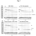
- FIG. 2 depicts biosizing analyses of IgG before and during proteolytic enzyme treatment.
- MMP-3, gluatamyl endopeptidase I and IdeS were each observed to cleave IgG1 in a stepwise fashion ( Figures 2A, 2B and 2C , respectively).
- an early intermediate of approximately 135,000 Da was generated which was subsequently converted to a species of approximately 100,000 Da.
- the molecular weight (35 kDa) gauged by gel migration is larger than that predicted by the heavy chain fragment amino acid sequence which would be 211 to 215 residues (between residue 232 and 237 to the 447 th residue at the C-terminus of the heavy chain) but consistent with the fragment containing the glycosylation site in the CH2 domain.
- the disappearance of intact IgG (160,000 Da) occurred over a period of several hours with MMP-3 and glutamyl endopeptidase I, and within a minute or less with IdeS under these conditions. All digestions were carried out under comparable conditions as described.
- the 135 kDa intermediate was found to result from a single proteolytic cleavage in one of the heavy chains in the lower hinge domain. Under non-denaturing conditions, the intermediate is indistinguishable from intact IgG in certain physical properties, such as migration in size exclusion chromatography (data not shown). However in SDS gels, and the present microcapillary electrophoresis system, the cleavage fragment of the Fc region (CH2-CH3 domains of the heavy chain) separates from the rest of the structure to reveal the reduced size intermediate (135 kDa). The size of this species is consistent with a singly cleaved IgG as reported by Gearing (2002 supra ). Extended incubation of IgG1 with the three enzymes resulted in conversion of the scIgG intermediate to the F(ab') 2 fragment and Fc.
- HNE human neutrophil elastase
- the major proteolytic cleavage positions in the IgG1 hinge were identified for the purified Fc fragment (MMP-3 and MMP-12) and/or high resolution mass spectrometric analyses of the purified Fab (neutrophil elastase, plasmin) and F(ab') 2 (cathepsin G, glutamyl endopeptidase and IdeS) fragments.
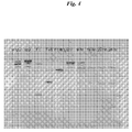
- the amino acid sequence of the human IgG1 hinge region is presented in Figure 3 , with the identified positions of enzymatic cleavages indicated. Extended digestion with proteinases that cleaved in the upper hinge yielded two Fab fragments; enzymes that cleaved in the lower hinge (below the core hinge disulfide bonds) yielded F(ab') 2 S.
- the dominant site of enzymatic cleavage were identified or confirmed for each enzyme including human MMP-3 and -12, human cathepsin G, human HNE, staphylococcal glutamyl endopeptidase I and streptococcal IdeS based on analysis of both the carboxy- and amino-terminal residue in the F(ab') 2 or Fc fragment, respectively (Table 1). Secondary cleavage sites were observed in some cases during extended incubations (e.g. cathepsin G and HNE), and it was uncertain if these are alternative cleavage sites for the indicated proteinase or the result of minor, proteinase contaminants in these enzyme preparations. The MMP-12 and HNE cleavage sites in IgG have not been previously reported.
- the cleavage positions differ slightly among the enzymes; with proteolysis occurring after proline-245, glutamic acid-246 and glycine-249 for MMP-3, V8 and IdeS, respectively. These differences in cleavage position do not impact molecular weight as detectable using the micro-capillary electrophoretic biosizing system (Agilent Technologies). Longer incubation times with MMP-3, V8 and IdeS allow the complete conversion to F(ab') 2 fragments.
- the digestion of IgG1 by HNE differs from the other proteases as it cleaves before the core hinge disulfides (cysteines 238 and 241) at histidine 236 to yield a Fab product and disulfide linked Fc (see Fig. 2D ).
- the cleavage sites are based on EU numbering and relate to the residues shown in Fig. 3 and SEQ ID NO: 1 which encompasses from Ser 219 through Phe 243 of a human IgG1 class antibody.
- Several proteinases cleaved IgG1 below the hinge domain, and yielded F(ab') 2 fragments of slightly different lengths (spanning Ala 231 to Gly 237 ).
- IgG-degrading proteinases characterized in this study have been reported to be expressed, or to be abundant, at sites of inflammation (HNE, cathepsin G, MMP-12), in the tumor or wound-healing environment (MMP-2, MMP-3, MMP-7, MMP-9, plasmin), and at sites of infection (glutamyl endopeptidase, IdeS) (Dollery et al., 2003; Kilian et al., 1996; Rooijakkers et al., 2005; Schönbeck et al., 1999; Shapiro, 1999; Sukhova et al., 1998; van Kempen et al., 2006; Vincents et al., 2004)).
- the single-cleaved intermediate of IgG1 was previously proposed as a possible intermediate during digestion of IgGs with MMP-3 (Gearing et al., 2002) and with IdeS (Vincents et al., 2004). In the present studies, it was consistently observed that the first cleavage to the single-cleaved intermediate occurred relatively rapidly compared to the second, slower cleavage that yields F(ab') 2 . The studies reported here focused on IgG1 because it is the predominant isotype of IgG in human circulation. A limited number of other human isotype experiments were carried out to determine relative susceptibilities to MMP-3 and IdeS.
- IgG4 was comparable in susceptibility to IgG1, whereas IgG2 and IgG3 were more resistant under these conditions (data not shown). Comparable investigations of IgA, IgM, IgE, IgD degradation were not done.
- Coagulation proteinases included F.XIIa, FIXa, F.Xa, thrombin and activated protein C; plasmin was plasminogen co-incubated with plasminogen activators; tPA, streptokinase and staphylokinase; "plasminogen activators alone” are without plasminogen; and the MMPs were recombinant proteinases obtained either as the active form or the pro-enzyme as detailed in the Materials; and "None” denotes no detectable cleavage in 24 hours. Except where indicated all enzymes where human.
- the residue designations are for the EU numbering system for the IgG1 antibody heavy chain where the 25 residues of SEQ ID NO: 1 correspond to residues 219 through 243 of the complete mature heavy chain.
- Table 1 Enzyme Source Proteinase Type Disease Association (Ref) Cleaved Site Major Product Cathepsin G Human Neutrophil granules Serine endopeptidase Emphysema, IPF, RA (2,3) Glu 233 -leu 234 F(ab') 2 + Fc Cathepsin B " " None Cathepsin D " " None Neutrophil elastase (HNE, leukocyte elastase, PMN elastase) " " Amyloidosis, lung emphysema, cystic fibrosis, ARDS, RA, tumor invasion (2,3) Thr 223 -his 224 Fab + Fc neutrophils Pancreatic elastase Pancreatititis (3) Protein
- Plasminogen is a critical host pathogenicity factor for group A streptococcal infection. Science. 305, 1283-1286 .
- Inflammatory exudates and other such fluids are expected to possess proteolytic enzymes associated with the inflammation and wound healing.
- samples of wound fluid were obtained from Ethicon Inc.
- an antibody substrate which comprises human heavy chain constant domains was randomly biotinylated. Ten microliters of the biotinylated substrate antibody was added to 190 microL of the wound fluid and incubated at 37°C for 8-24 hours. At specified times, samples were removed. The starting IgG and samples from the various times were applied in separate wells to a 4-12% Bis-Tris gel and subjected to SDS PAGE.
- the separated bands were transferred to a nitrocellulose membrane and, following blocking with 0.1M Tris buffered saline containing 0.1% Tween 20 and 10% blocking grade milk ("Blotto"), the blot was developed using AVIDIN-D -horseradish peroxidase reagent followed by TMB (membrane) substrate.
- the determination of the presence of host (patient) antibody fragments produced by endogenous proteases requires a reagent which selectively binds to the cleaved IgG but not intact IgG. Both identification of the cleaved component and a quantitative difference between fragment content in samples from patients with disease as compared to the normal population should be able to be assessed using the reagent.
- New Zealand rabbits (two per immunogen) were immunized by subcutaneous injection of 0.2 mg conjugated peptide in complete Freund's adjuvant and re-boosted three additional times with 0.1 mg antigen in incomplete Freund's adjuvant on days 14, 42 and 56. Serum was collected at 4, 8 and 10 weeks and pooled per immunogen for antibody purification. The immune titers were monitored by an ELISA based on solid phase antigen peptide.
- Affinity purification of antibodies employed the respective peptide antigens immobilized on an activated support.
- the antiserum from the two rabbits immunized with the same antigen was pooled and passed through the antigen column after which the column was extensively washed.
- Specific antibodies were eluted as low affinity and high affinity pools using 3M KSCN and 0.1M glycine, pH 2.5, respectively.
- the two pools yielded indistinguishable binding characteristics and were used interchangeably and/or pooled.
- the three separately eluted pools of bound antibodies were next subjected to a second affinity adsorption step, this time on a column containing an intact antibody comprising human IgG1 heavy chain constant regions (Mab3).
- the intent of the second affinity chromatography step was to remove undesirable antibodies that might recognize intact IgG. However, little or no rabbit antibody was adsorbed to the IgG column suggesting that the population of antibodies was reactive only with the "cleaved" sequence with its exposed carboxy terminus.
- the individual affinity-purified rabbit anti-peptide antibodies were tested for their ability to bind to enzymatically-generated fragments of human IgG as well as intact IgG by ELISA ( Fig. 5 ).
- the purified antibodies from the rabbits immunized with KLH conjugated to residues 7-14 of SEQ ID NO: 1 did not bind intact IgG and were highly specific for scIgG and F(ab') 2 produced by digestion of IgG with MMP-3.
- This antibody preparation showed minimal reactivity to scIgG and F(ab') 2 produced with V8 protease or IdeS.
- the antibodies obtained from rabbits immunized with the V8-cleavage site hinge peptide analog (residues 7-15 of SEQ ID NO: 1) and the IdeS-cleavage site hinge peptide analog (residues 7-18 of SEQ ID NO: 1) showed a cross-reactive binding profile for scIgGs and F(ab') 2 produced by either of these two enzymes.
- these preparations showed minimal reactivity for the MMP-3 digested products. None of the antibody preparations bound to intact IgG and none of the antibody preparations was comparably reactive with fragments, including F(ab') 2 and scIgG, produced by three different enzymes as shown in Figure 6 .
- the intended use of the anti-hinge reagent is the detection of scIgGs and F(ab') 2 (and other potential fragments) that are produced in complex in vivo settings by enzymes present in disease specific tissues or produced by disease specific cell types or cell populations, e.g. infiltrating macrophages or neutrophils.
- scIgGs and F(ab') 2 and other potential fragments
- the novel assay employed for detecting scIgG in serum using, as a capture reagent, the RAH-1 capable of binding cleaved human IgG but not intact human IgG is described in detail as follows.
- the disease serum samples were added at a 1:50 dilution in the same buffer. Plates were washed and a 1:6000 dilution of Jackson Immuno Research's HRP conjugated AffiniPure F(ab') 2 Fragment Donkey anti-human IgG (H+L) which has minimal cross reactivity to various animals including rabbit was added to all wells. This was added in a dilution of PBS with 1%Casein and 3%BSA and incubated at 37°C for 1 hour. The plates were washed thoroughly and 100 ml/well of HRP substrate (Roche's BM Chemiluminescence POD, 582 950) was added seconds before plate was read on the luminescent reader.
- the average luminescence from the 0 ng/ml scIgG wells on the standard curve was subtracted from all wells that were exposed to the RAH-1 coat. This subtraction controls for any non-specific reactivity of the secondary with the RAH-1. Then, the value for each donor on non-RAH coated wells was subtracted from the previously-adjusted value of the RAH coated wells. This accounts for any non-specific reactivity in the serum to the plate.
- EXAMPLE 5 USE OF REAGENT TO DETECT DISEASE ASSOCIATED PROTEOLYTIC ACTIVITY
- the RAH-1 reagent was tested for its ability to detect IgG fragments in another inflammatory fluid, the synovial fluid of a patient with rheumatoid arthritis (RA).
- RA rheumatoid arthritis
- a collection of synovial fluid samples from RA patients were purchased commercially from Bioreclamation. Samples were diluted 1:5 in LDS sample buffer and 10 mciroL of each sample loaded onto a 4-12% Bis-Tris gel. As a control for the reactivity of RAH-1, intact IgG (Mab3), or protease digested IgG (partial digests of Mab3 with MMP-3, glutamyl endopeptidase and IdeS) was loaded onto the gel as well. Following SDS PAGE, the gel was transferred to nitrocellulose membrane and blocked with Blotto.
- the membrane was then incubated with a 1:2,500 dilution of the RAH-1 in blotto, washed with 0.1M tris buffered saline, pH 7.5 containing 0.1% Tween 20 and incubated with a 1:5,000 dilution of goat anti-rabbit IgG (H&L) horseradish peroxidase conjugate.
- the blot was then developed using TMB membrane.
- the RAH-1 preparation did not react with the intact IgG , but detected scIgG, F(ab') 2 possibly Fab' from all 3 protease digests.
- Blood plasma or serum is a more convenient for biomarker testing than biological fluids or tissue extracts, such as synovial fluid.
- biological fluids or tissue extracts such as synovial fluid.
- synovial fluid is that it is a self-contained and local environment in which the proteases are active and in which the IgG fragments might be expected to accumulate as described in Example 2.
- serum for testing makes it a considerably more likely sample tissue for biomarkers, including IgG breakdown products.
- Fractionated proteolysis products Mab2 IgG1, and the scIgG and F(ab') 2 generated with MMP-3, were prepared as follows.
- a 20 milligram quantity of Mab2 IgG was digested with heat-activated MMP-3 as described in Example 1. The digestion was initiated by addition of enzyme to a 4 mg/mL solution of Mab2 in tris-buffered saline containing 10mM CaCl 2 , pH 7.5 at 37°C. The reaction was terminated by the addition of EDTA to a final concentration of 20mM at 48 hours.
- the terminated digest was subjected to a two-step purification to remove the Fc fragment and to separate purified scIgG and F(ab) 2 .
- the digest was subjected to chromatography using protein A-Sepharose.
- the unbound material from the column contained the F(ab) 2 fragment and no detectable intact IgG or scIgG.
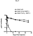
- a group of twenty-one female Balb/c mice (Ace Animals) were used for the pK study. Terminal bleeds were taken via cardiac puncture from three randomly selected mice prior to the experiment to serve as baseline controls. The remaining eighteen female Balb/c mice were weighed and placed into six equal groups. Two groups were injected with intact Mab2 IgG1, two groups with Mab2 scIgG1 produced with MMP-3 and two groups with Mab2 F(ab')2 produced with MMP-3. All injections were i.p. at a constant dose volume of 10 ml/kg based on individual animal weight at 0.19 mg/ml. Therefore, each animal received a 1.9 mg/kg dose at day zero.
- the IgG and IgG fragment concentrations of the collected serum was quantified via enzyme-linked immunosorbent assay (ELISA).
- ELISA enzyme-linked immunosorbent assay
- the scIgG In order for the scIgG to be a useful disease biomarker, it must exhibit differential quantity in samples obtained from patients in defined disease categories as compared to healthy people.
- a commercial source of serum from diseased individuals was identified as Genomics Collaborative (now SeraCare Life Sciences Inc.). Small volumes (300 microL) of serum from 10 different individuals within each of 8 diseases were purchased. The disease categories were rheumatoid arthritis, osteoarthritis, asthma, type-1 diabetes, breast cancer, lung cancer, myocardial infarction, and congestive heart failure. In addition, serum from 28 age-matched and gender-matched normal healthy volunteers were obtained from this vendor as controls.
- the samples were analyzed and the results shown in Fig. 9 .
- the assay based on the selectivity of the RAH-1 reagent, demonstrated that IgG cleavage products comparable to those generated by known specific proteases are clearly detectable and above levels maintained in healthy or normal donors for an inflammatory autoimmune disease, rheumatoid arthritis. In contrast, patients with osteoarthritis showed levels which were similar and in the range of those for the normal individual's samples.
- EXAMPLE 8 MODIFIED SINGLE CHAIN CLEAVED IgG ASSAY
- Example 4 A solid phase assay, ELISA, using reagent RAH-1 for detection of scIgG in serum was described in Example 4. In order to optimize the detection range for scIgG concentrations serum samples specific changes were made.
- the plates used were Immulon 4 HBX plates (VWR) coated with rabbit polyclonal antibodies (RAH-1) at a concentration of 5 ⁇ g/mL in PBS pH 7.2 (100 ⁇ L per well) by sealing and incubating the plate for 1 hour at room temperature. Thereafter, the plate is washed 3X with PBS, 0.05% Tween (Sigma) on automatic plate washer. All samples and standards are diluted with PBS containing 1%BSA, 0.05% Tween.
- Anti-IgG Fc-Biotin USBIologicals, Swampscott, MA is the means of detection of scIgG standards or scIgG unknowns in serum dilutions.
- the plate is blocked using 200 ⁇ L of SuperBlock (Pierce) for 15 minutes at room temperature (RT) with shaking and then washed plate 3X with PBS, 0.05% Tween on an automatic plate washer.
- IgG Fc Biotin dilution of 1:20,000 (dilute appropriately in assay buffer), is added to all wells (100 ⁇ L per well and incubated for one hour at RT on a shaker followed by 3X washing with PBS, 0.05% Tween on an automatic plate washer.
- SA-HRP Streptavidin conjugated to horseradish peroxidase, Sigma, used at a 1:30,000 dilution in PBS, 0.05% Tween, 1% BSA
- SA-HRP Streptavidin conjugated to horseradish peroxidase, Sigma, used at a 1:30,000 dilution in PBS, 0.05% Tween, 1% BSA
- TMB (3,3',5,5'-Tetramethylbenzidine a peroxidase substrate) as provided by the manufacturer (Sigma) is added to each well and allowed to incubate for about 10 minutes for color development. The reaction is stopped with 75 ⁇ L of 1 N H 2 SO4 and read plate at 450 nm.
- the assay demonstrated greatly improved linearity and spike recoveries of scIgG in normal, healthy serum.
- the dilution linearity of the assay was determined in two serum pools, diluted 1:100 then spiked with Mab1 at concentrations of 150 ng/mL and 300 ng/mL, and further diluted to concentrations of 0, 25, 75, and 100% serum. Each sample dilution was assayed in triplicate and mean analyte recoveries were calculated. Linearity was assessed by calculation of an R2 correlation coefficient from a plot of the observed (y-axis) versus expected (x-axis) analyte recovery results for each sample pool. The R2 values were: Sample 1 Low 0.9983; Sample 1 High 0.9913; Sample 2 Low 0.9852; Sample 2 High 0.973; and dilution linearity was 100% for all dilutions.
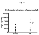
- Serum from RA patients was used exclusively in order to further study the results in Example 4 where some serum samples from this group of patients had notably higher scIgG compared to controls.
- Serum samples from 10 subjects with rheumatoid arthritis (RA) and from 10 age- and gender-matched healthy individuals were obtained from Genomics Collaborative. Using the modified assay described in Example 8, the samples were analyzed and the results shown in Fig. 10 . The results indicated that 4 of the 10 subjects with RA demonstrated serum scIgG concentrations >60 ⁇ g/mL. In the healthy control group, scIgG concentrations ranged from ⁇ 8.2 ⁇ g/mL to 52.7 ⁇ g/mL. The samples for this comparison were not rigorously selected for stage of disease, treatment regimens, etc.
- a 12-mer peptide analog of the human IgG1 lower hinge and adjoining CH2 domain was the immunogen: TCPPCPAPELLG (residues 7-18 of SEQ ID NO: 1).
- the naturally occurring cysteines were replaced by alanines to give the variant TAPPAPAPELLG.
- An N-terminal cysteine was added to allow for conjugation to keyhole limpet hemocyanin (KLH) by standard chemical methods for reaction to free sulhydryls.
- KLH-CTAPPAPAPELLG The final immunogen was KLH-CTAPPAPAPELLG.
- New Zealand white rabbits (3) were immunized with 0.5 mg KLH peptide in complete Freund's adjuvant using multiple subcutaneous sites (5). The animals were boosted with the 0.25 mg immunogen in incomplete Freund's adjuvant at three-week intervals for a total of 4 additional immunizations.
- the serum antibody titers to a BSA-conjugated version of the same peptide were monitored during the course of the immunization by standard ELISA methods.
- Animals (2) were chosen for splenectomy based on the titer data.
- Rabbit hybridomas were generated from spleen-derived lymphocytes fused with a rabbit fusion partner cells ( Spieker-Polet, 1995 PNAS USA 92: 9348 - 9352 ). Cell growth was examined 2-3 weeks after fusion in multiple plates.
- Positive hybridomas were screened via ELISA on plates coated with the BSA-immunogen peptide conjugate. Multiple positive clones from each fusion were identified. Further screening involved binding to intact IgG1 and various enzymatically-generated F(ab')2 fragments of IgG1. From these screening and counter-screening tactics, 3 clones were chosen based on strong selectivity of binding to the immunogen peptide and to F(ab')2 fragments with termini at or near the terminus of the immunogen peptide and with minimal binding to intact IgG1. The positive hybridomas were subcloned and expanded.
- Rabbit IgG was purified from individual cell supernatants by standard methods including chromatography on immobilized protein A. The specificity of the purified rabbit IgGs for binding to peptide analogs of the human IgG1 hinge, as well as intact IgG and purified IgG F(ab')2 fragments created using single or doubly cleaved Mabs using IdeS and MMP-3 enzymes were tested in standard ELISA protocols. Briefly, the peptides which were synthesized by standard peptide chemistry and were N-terminally biotinylated were captured on streptavidin-coated wells. The IgG and fragments were directly coated at 10 ⁇ g/mL. Binding of rabbit mAbs was detected by well-characterized goat anti-rabbit IgG Fc-horseradish peroxidase and OPD substrate systems.
- ELISA results for rabbit mAb 91-2 are shown in Fig. 11 .
- WIL2-S cells a lymphoblastoid B-cell line expressing CD20 (ATCC) were used as target cells for CDC assays.
- 50 ⁇ l of cells were added to the wells of 96-well plates for a final concentration of 8 X 10 4 cells per well in RPMI, 5% heat-inactivated FBS, 0.1mM nonessential amino acids, 1mM sodium pyruvate, penicillin (500 U/ml), streptomycin (500 U/ml), 2mM L-glutamine.
- An additional 50 ⁇ l was added to the wells with or without antibodies of various concentrations and the plates were incubated at room temperature for 2 hours.
- Figure 12 shows that the 3 rabbit anti-hinge mAbs were able to restore complement dependent cell lysis to the target cells when titrated in the presence of a fixed concentration of the F(ab')2 fragment of an antibody that binds CD20.
- the rabbit mAbs were more effective, and, at lower concentrations than a polyclonal rabbit anti-hinge mAb preparation (a component of the same detection system for serum scIgG described earlier). Intact antibody to CD20 was active, as expected, but its F(ab')2 fragment and scIgG version were not active alone.
- the rabbit anti-hinge mAbs were not able to direct cell lysis in the absence of cell-binding F(ab')2 fragment.
Landscapes
- Health & Medical Sciences (AREA)
- Chemical & Material Sciences (AREA)
- Life Sciences & Earth Sciences (AREA)
- Immunology (AREA)
- Organic Chemistry (AREA)
- Medicinal Chemistry (AREA)
- General Health & Medical Sciences (AREA)
- Engineering & Computer Science (AREA)
- Molecular Biology (AREA)
- Biochemistry (AREA)
- Proteomics, Peptides & Aminoacids (AREA)
- Animal Behavior & Ethology (AREA)
- Veterinary Medicine (AREA)
- General Chemical & Material Sciences (AREA)
- Public Health (AREA)
- Chemical Kinetics & Catalysis (AREA)
- Pharmacology & Pharmacy (AREA)
- Nuclear Medicine, Radiotherapy & Molecular Imaging (AREA)
- Urology & Nephrology (AREA)
- Hematology (AREA)
- Biomedical Technology (AREA)
- Genetics & Genomics (AREA)
- Biophysics (AREA)
- Bioinformatics & Cheminformatics (AREA)
- Microbiology (AREA)
- Pathology (AREA)
- General Physics & Mathematics (AREA)
- Analytical Chemistry (AREA)
- Physics & Mathematics (AREA)
- Food Science & Technology (AREA)
- Biotechnology (AREA)
- Cell Biology (AREA)
- Cardiology (AREA)
- Oncology (AREA)
- Communicable Diseases (AREA)
- Physical Education & Sports Medicine (AREA)
- Rheumatology (AREA)
- Orthopedic Medicine & Surgery (AREA)
- Heart & Thoracic Surgery (AREA)
- Peptides Or Proteins (AREA)
Claims (11)
- Antikörperzusammensetzung, die wenigstens einen Antikörper umfasst, der spezifisch an ein IgG-Proteasespaltprodukt bindet, das dadurch gekennzeichnet ist, dass es a) ein Molekulargewicht aufweist, das mit einem intakten Säuger-IgG unter nicht denaturierenden Bedingungen vergleichbar ist, und b) in zwei Fragmente, die ein antigenbindendes Fragment von 135 kDa und ein CH2 enthaltendes Fragment umfassen, unter denaturierenden, aber nicht reduzierenden Bedingungen getrennt werden kann, und c) wobei das Reagens nicht mit intaktem IgG reagiert,
und wobei der Antikörper spezifisch an eine proteasespezifische Spaltstelle in menschlichem IgG1, erzeugt durch IdeS, TCPPCPAPELLG, bindet. - Antikörperzusammensetzung nach Anspruch 1, wobei es sich bei dem Antikörper um:i) ein polyklonales Antiserum; oderii) wenigstens einen monoklonalen Antikörper handelt.
- Menschliches-IgG-Proteasespaltstelle-Peptidanalog, bestehend aus TCPPCPAPELLG, kovalent gebunden an KLH (Keyhole Limpet Hemocyanin) über den N-Terminus.
- Verfahren zur Herstellung der Antikörperzusammensetzung nach Anspruch 1 unter Verwendung eines mit dem Peptidanalog gemäß Anspruch 3 immunisierten nichtmenschlichen Tiers, wobei das Reagens aus dem Serum des Tiers durch Vorabsorption an einer Menschliches-IgG-Affinitätsmatrix aufgereinigt wird.
- Verfahren zum Nachweisen eines Krankheitsprozesses bei einem Individuum anhand einer Analyse einer Gewebeprobe von dem Individuum, wobei das Verfahren den Nachweis eines durch IdeS proteolytisch gespaltenen IgG, das dadurch gekennzeichnet ist, dass es a) ein Molekulargewicht aufweist, das mit einem intakten Säuger-IgG unter physiologischen Bedingungen vergleichbar ist, und b) in zwei Fragmente, die ein antigenbindendes Fragment von 135 kDa und ein CH2 enthaltendes Fragment umfassen, unter denaturierenden, aber nicht reduzierenden Bedingungen getrennt werden kann, mittels der Antikörperzusammensetzung nach Anspruch 1 umfasst.
- Verfahren nach Anspruch 5, wobei die Krankheit aus einer arthritischen Krankheit, einer bösartigen Krankheit, einer Infektionskrankheit und einer Gefäßkrankheit ausgewählt ist, etwa wobei es sich bei der Krankheit um rheumatoide Arthritis handelt, beispielsweise wobei es sich bei der Krankheit um rheumatoide Arthritis und bei der Probe um Synovia handelt.
- Verfahren nach Anspruch 5, wobei es sich bei der Probe um Blut oder ein Fraktionierungsprodukt davon handelt.
- Verfahren nach Anspruch 5, wobei der Nachweis an anderen Gewebeproben als einer Blutfraktion durchgeführt wird.
- Verfahren nach Anspruch 5, wobei das Nachweisverfahren aus der aus ELISA, immunhistochemischer Färbung und Western-Blotting bestehenden Gruppe ausgewählt ist.
- Kit, enthaltend ein Reagens zum Nachweis eines Krankheitsmarkers in Gewebe eines Individuums, wobei das Reagens wenigstens einen Antikörper umfasst, der spezifisch an ein gespaltenes IgG bindet, wobei mit dem Antikörper ein IgG-Spaltprodukt nachgewiesen werden kann, das dadurch gekennzeichnet ist, dass es a) ein Molekulargewicht aufweist, das mit einem intakten Säuger-IgG unter nicht denaturierenden Bedingungen vergleichbar ist, und b) in zwei Fragmente, die ein antigenbindendes Fragment von 135 kDa und ein CH2 enthaltendes Fragment umfassen, unter denaturierenden, aber nicht reduzierenden Bedingungen getrennt werden kann, und c) wobei das Reagens nicht mit intaktem IgG reagiert,
und wobei der Antikörper spezifisch an eine proteasespezifische Spaltstelle in menschlichem IgG1, erzeugt durch IdeS, TCPPCPAPELLG, bindet. - Antikörperzusammensetzung nach Anspruch 1 oder Anspruch 2 zur Verwendung in der Therapie.
Applications Claiming Priority (2)
| Application Number | Priority Date | Filing Date | Title |
|---|---|---|---|
| US95516207P | 2007-08-10 | 2007-08-10 | |
| PCT/US2008/072083 WO2009023457A1 (en) | 2007-08-10 | 2008-08-04 | Immunoglobulin cleavage fragments as disease indicators and compositions for detecting and binding such |
Publications (3)
| Publication Number | Publication Date |
|---|---|
| EP2188306A1 EP2188306A1 (de) | 2010-05-26 |
| EP2188306A4 EP2188306A4 (de) | 2011-03-02 |
| EP2188306B1 true EP2188306B1 (de) | 2016-06-29 |
Family
ID=40351057
Family Applications (1)
| Application Number | Title | Priority Date | Filing Date |
|---|---|---|---|
| EP08782610.3A Not-in-force EP2188306B1 (de) | 2007-08-10 | 2008-08-04 | Immunoglobulinspaltfragmente als krankheitsindikatoren und zusammensetzungen zum nachweis und zur bindung davon |
Country Status (20)
| Country | Link |
|---|---|
| US (1) | US20090155280A1 (de) |
| EP (1) | EP2188306B1 (de) |
| JP (2) | JP5530356B2 (de) |
| KR (2) | KR20150059813A (de) |
| CN (2) | CN101889021B (de) |
| AU (1) | AU2008287128B2 (de) |
| BR (1) | BRPI0814355A2 (de) |
| CA (1) | CA2696011A1 (de) |
| DK (1) | DK2188306T3 (de) |
| EA (1) | EA025220B1 (de) |
| ES (1) | ES2593791T3 (de) |
| HU (1) | HUE029649T2 (de) |
| IL (1) | IL203544A (de) |
| MX (1) | MX2010001616A (de) |
| NZ (1) | NZ582916A (de) |
| PL (1) | PL2188306T3 (de) |
| PT (1) | PT2188306T (de) |
| SG (1) | SG183701A1 (de) |
| WO (1) | WO2009023457A1 (de) |
| ZA (1) | ZA201001689B (de) |
Families Citing this family (15)
| Publication number | Priority date | Publication date | Assignee | Title |
|---|---|---|---|---|
| US8501907B2 (en) * | 2007-08-10 | 2013-08-06 | Janssen Biotech, Inc. | Immunoglobulin cleavage fragments as disease indicators and compositions for detecting and binding such |
| KR101834890B1 (ko) | 2009-10-23 | 2018-04-20 | 밀레니엄 파머슈티컬스 인코퍼레이티드 | 항gcc 항체 분자와 관련 조성물 및 방법 |
| WO2011059684A1 (en) | 2009-10-29 | 2011-05-19 | Centocor Ortho Biotech Inc. | Antibody glycosylation variants |
| US8041225B2 (en) * | 2009-12-21 | 2011-10-18 | General Electric Company | Contactless infrared data transmission for wind turbines |
| BR112013012389A2 (pt) * | 2010-11-19 | 2019-09-24 | Janssen Biotech Inc | composições de vacina de fragmentos de clivagem de imunoglobina |
| WO2012085845A1 (en) | 2010-12-23 | 2012-06-28 | Eulitha A.G. | System and method for production of nanostructures over large areas |
| MX347077B (es) * | 2010-12-23 | 2017-04-10 | Janssen Biotech Inc | Mutantes fc de anticuerpos resistentes a proteasas activas. |
| LT2841575T (lt) * | 2012-04-27 | 2019-10-10 | Millennium Pharmaceuticals, Inc. | Antikūno molekulės, nukreiptos prieš gcc, ir jų panaudojimas nustatant jautrumą į gcc nukreiptai terapijai |
| US10654933B2 (en) * | 2013-12-27 | 2020-05-19 | Chugai Seiyaku Kabushiki Kaisha | Method for purifying antibody having low isoelectric point |
| JP6759104B2 (ja) | 2014-04-04 | 2020-09-23 | メイヨ・ファウンデーション・フォー・メディカル・エデュケーション・アンド・リサーチ | 精密分子質量を用いた免疫グロブリンのアイソタイピング |
| US11209439B2 (en) | 2015-09-24 | 2021-12-28 | Mayo Foundation For Medical Education And Research | Identification of immunoglobulin free light chains by mass spectrometry |
| CN109863395B (zh) * | 2016-09-07 | 2023-05-23 | 梅约医学教育与研究基金会 | 分子量法鉴定和监测裂解免疫球蛋白 |
| EP3681528A4 (de) | 2017-09-13 | 2021-07-21 | Mayo Foundation for Medical Education and Research | Identifizierung und überwachung des apoptosehemmers von makrophagen |
| JP7425725B2 (ja) * | 2017-12-07 | 2024-01-31 | 中外製薬株式会社 | 試料中のポリペプチドの検出または捕捉において使用するための抗体、組成物、および試料中のポリペプチドの検出または捕捉のための方法 |
| US20210325380A1 (en) * | 2020-04-20 | 2021-10-21 | EnLiSense, LLC | Disease diagnostics using a multi-configurable sensing array |
Family Cites Families (4)
| Publication number | Priority date | Publication date | Assignee | Title |
|---|---|---|---|---|
| DK0698097T3 (da) * | 1993-04-29 | 2001-10-08 | Unilever Nv | Produktion af antistoffer eller (funktionaliserede) fragmenter deraf afledt af Camelidae-immunoglobuliner med tung kæde |
| AUPM322393A0 (en) * | 1993-12-24 | 1994-01-27 | Austin Research Institute, The | Mucin carbohydrate compounds and their use in immunotherapy |
| US7129331B2 (en) * | 2000-05-31 | 2006-10-31 | Pestka Biomedical Laboratories, Inc. | Phosphorylated polypeptides and uses related thereto |
| GB0130228D0 (en) * | 2001-12-18 | 2002-02-06 | Hansa Medica Ab | Protein |
-
2008
- 2008-08-04 BR BRPI0814355-2A2A patent/BRPI0814355A2/pt not_active IP Right Cessation
- 2008-08-04 SG SG2012058665A patent/SG183701A1/en unknown
- 2008-08-04 ES ES08782610.3T patent/ES2593791T3/es active Active
- 2008-08-04 EP EP08782610.3A patent/EP2188306B1/de not_active Not-in-force
- 2008-08-04 PT PT87826103T patent/PT2188306T/pt unknown
- 2008-08-04 WO PCT/US2008/072083 patent/WO2009023457A1/en active Application Filing
- 2008-08-04 KR KR1020157013122A patent/KR20150059813A/ko not_active Application Discontinuation
- 2008-08-04 CN CN200880102840.1A patent/CN101889021B/zh not_active Expired - Fee Related
- 2008-08-04 HU HUE08782610A patent/HUE029649T2/en unknown
- 2008-08-04 CN CN201310054803.0A patent/CN103497254B/zh not_active Expired - Fee Related
- 2008-08-04 KR KR1020107005265A patent/KR101588276B1/ko not_active IP Right Cessation
- 2008-08-04 CA CA2696011A patent/CA2696011A1/en not_active Abandoned
- 2008-08-04 DK DK08782610.3T patent/DK2188306T3/en active
- 2008-08-04 EA EA201070236A patent/EA025220B1/ru not_active IP Right Cessation
- 2008-08-04 NZ NZ582916A patent/NZ582916A/xx not_active IP Right Cessation
- 2008-08-04 JP JP2010520241A patent/JP5530356B2/ja not_active Expired - Fee Related
- 2008-08-04 PL PL08782610T patent/PL2188306T3/pl unknown
- 2008-08-04 AU AU2008287128A patent/AU2008287128B2/en not_active Ceased
- 2008-08-04 US US12/185,333 patent/US20090155280A1/en not_active Abandoned
- 2008-08-04 MX MX2010001616A patent/MX2010001616A/es active IP Right Grant
-
2010
- 2010-01-27 IL IL203544A patent/IL203544A/en not_active IP Right Cessation
- 2010-03-09 ZA ZA2010/01689A patent/ZA201001689B/en unknown
-
2013
- 2013-10-30 JP JP2013225205A patent/JP2014058535A/ja active Pending
Non-Patent Citations (1)
| Title |
|---|
| RYAN ET AL: "Proteolysis of purified IgGs by human and bacterial enzymes in vitro and the detection of specific proteolytic fragments of endogenous IgG in rheumatoid synovial fluid", MOLECULAR IMMUNOLOGY, PERGAMON, GB, vol. 45, no. 7, 21 December 2007 (2007-12-21), pages 1837 - 1846, XP022478946, ISSN: 0161-5890, DOI: 10.1016/J.MOLIMM.2007.10.043 * |
Also Published As
| Publication number | Publication date |
|---|---|
| ZA201001689B (en) | 2011-06-29 |
| EP2188306A4 (de) | 2011-03-02 |
| SG183701A1 (en) | 2012-09-27 |
| PT2188306T (pt) | 2016-09-13 |
| CA2696011A1 (en) | 2009-02-19 |
| WO2009023457A1 (en) | 2009-02-19 |
| CN101889021A (zh) | 2010-11-17 |
| KR20100059850A (ko) | 2010-06-04 |
| EA201070236A1 (ru) | 2010-08-30 |
| ES2593791T3 (es) | 2016-12-13 |
| CN103497254B (zh) | 2016-12-28 |
| US20090155280A1 (en) | 2009-06-18 |
| NZ582916A (en) | 2012-08-31 |
| AU2008287128A1 (en) | 2009-02-19 |
| JP2010536037A (ja) | 2010-11-25 |
| BRPI0814355A2 (pt) | 2015-01-27 |
| PL2188306T3 (pl) | 2017-01-31 |
| KR20150059813A (ko) | 2015-06-02 |
| CN101889021B (zh) | 2016-06-08 |
| JP5530356B2 (ja) | 2014-06-25 |
| IL203544A (en) | 2015-07-30 |
| EP2188306A1 (de) | 2010-05-26 |
| JP2014058535A (ja) | 2014-04-03 |
| AU2008287128B2 (en) | 2013-07-18 |
| KR101588276B1 (ko) | 2016-01-26 |
| CN103497254A (zh) | 2014-01-08 |
| MX2010001616A (es) | 2010-03-15 |
| EA025220B1 (ru) | 2016-12-30 |
| HUE029649T2 (en) | 2017-02-28 |
| DK2188306T3 (en) | 2016-08-22 |
Similar Documents
| Publication | Publication Date | Title |
|---|---|---|
| EP2188306B1 (de) | Immunoglobulinspaltfragmente als krankheitsindikatoren und zusammensetzungen zum nachweis und zur bindung davon | |
| Ryan et al. | Proteolysis of purified IgGs by human and bacterial enzymes in vitro and the detection of specific proteolytic fragments of endogenous IgG in rheumatoid synovial fluid | |
| KR102662387B1 (ko) | B7-h3 항체, 이의 항원-결합 단편 및 이의 의학적 용도 | |
| EP2985296A1 (de) | Für MMP9 spezifische Antikörper | |
| US9481734B2 (en) | Immunoglobulin cleavage fragments and disease indicators and compositions for detecting and binding such | |
| US20150376294A1 (en) | Anti-pad2 antibodies and treatment of autoimmune diseases | |
| CN110177810B (zh) | 抗pcsk9抗体及其用途 | |
| Brezski et al. | Human anti-IgG1 hinge autoantibodies reconstitute the effector functions of proteolytically inactivated IgGs | |
| US11021530B2 (en) | Antibody preparation | |
| EP2985295A1 (de) | MMP9-spezifische Antikörper | |
| US20150355199A1 (en) | Agents, kits and methods for complement factor h-related protein 1 detection | |
| JP7437511B2 (ja) | 腫瘍免疫抑制因子に不応性の操作されたモノクローナル抗体の組成物及び使用 | |
| AU2010363981A1 (en) | Immunoglobulin cleavage fragments vaccine compositions | |
| WO2019056991A1 (zh) | 单克隆抗体或其抗原结合片段及其用途 | |
| KR100842121B1 (ko) | D-다이머에 대한 단일클론 항체와 이를 이용한 d-다이머및 교차결합성 피브린 또는 이를 포함하는 피브린 분해물검사용 진단시약 | |
| KR20240063177A (ko) | B7-h3 항체, 이의 항원-결합 단편 및 이의 의학적 용도 |
Legal Events
| Date | Code | Title | Description |
|---|---|---|---|
| PUAI | Public reference made under article 153(3) epc to a published international application that has entered the european phase |
Free format text: ORIGINAL CODE: 0009012 |
|
| 17P | Request for examination filed |
Effective date: 20100310 |
|
| AK | Designated contracting states |
Kind code of ref document: A1 Designated state(s): AT BE BG CH CY CZ DE DK EE ES FI FR GB GR HR HU IE IS IT LI LT LU LV MC MT NL NO PL PT RO SE SI SK TR |
|
| AX | Request for extension of the european patent |
Extension state: AL BA MK RS |
|
| DAX | Request for extension of the european patent (deleted) | ||
| REG | Reference to a national code |
Ref country code: HK Ref legal event code: DE Ref document number: 1142915 Country of ref document: HK |
|
| A4 | Supplementary search report drawn up and despatched |
Effective date: 20110131 |
|
| RAP1 | Party data changed (applicant data changed or rights of an application transferred) |
Owner name: JANSSEN BIOTECH, INC. |
|
| 17Q | First examination report despatched |
Effective date: 20120601 |
|
| REG | Reference to a national code |
Ref country code: DE Ref legal event code: R079 Ref document number: 602008044910 Country of ref document: DE Free format text: PREVIOUS MAIN CLASS: C07K0014435000 Ipc: C07K0016060000 |
|
| GRAP | Despatch of communication of intention to grant a patent |
Free format text: ORIGINAL CODE: EPIDOSNIGR1 |
|
| RIC1 | Information provided on ipc code assigned before grant |
Ipc: C07K 16/06 20060101AFI20160104BHEP Ipc: C07K 16/42 20060101ALI20160104BHEP Ipc: G01N 33/68 20060101ALI20160104BHEP |
|
| INTG | Intention to grant announced |
Effective date: 20160121 |
|
| GRAS | Grant fee paid |
Free format text: ORIGINAL CODE: EPIDOSNIGR3 |
|
| GRAA | (expected) grant |
Free format text: ORIGINAL CODE: 0009210 |
|
| AK | Designated contracting states |
Kind code of ref document: B1 Designated state(s): AT BE BG CH CY CZ DE DK EE ES FI FR GB GR HR HU IE IS IT LI LT LU LV MC MT NL NO PL PT RO SE SI SK TR |
|
| REG | Reference to a national code |
Ref country code: GB Ref legal event code: FG4D |
|
| REG | Reference to a national code |
Ref country code: CH Ref legal event code: NV Representative=s name: E. BLUM AND CO. AG PATENT- UND MARKENANWAELTE , CH Ref country code: CH Ref legal event code: EP |
|
| REG | Reference to a national code |
Ref country code: FR Ref legal event code: PLFP Year of fee payment: 9 |
|
| REG | Reference to a national code |
Ref country code: AT Ref legal event code: REF Ref document number: 809041 Country of ref document: AT Kind code of ref document: T Effective date: 20160715 |
|
| REG | Reference to a national code |
Ref country code: IE Ref legal event code: FG4D |
|
| REG | Reference to a national code |
Ref country code: DE Ref legal event code: R096 Ref document number: 602008044910 Country of ref document: DE |
|
| REG | Reference to a national code |
Ref country code: DK Ref legal event code: T3 Effective date: 20160815 |
|
| REG | Reference to a national code |
Ref country code: NL Ref legal event code: FP |
|
| REG | Reference to a national code |
Ref country code: PT Ref legal event code: SC4A Ref document number: 2188306 Country of ref document: PT Date of ref document: 20160913 Kind code of ref document: T Free format text: AVAILABILITY OF NATIONAL TRANSLATION Effective date: 20160907 |
|
| REG | Reference to a national code |
Ref country code: SE Ref legal event code: TRGR |
|
| REG | Reference to a national code |
Ref country code: LT Ref legal event code: MG4D |
|
| PG25 | Lapsed in a contracting state [announced via postgrant information from national office to epo] |
Ref country code: LT Free format text: LAPSE BECAUSE OF FAILURE TO SUBMIT A TRANSLATION OF THE DESCRIPTION OR TO PAY THE FEE WITHIN THE PRESCRIBED TIME-LIMIT Effective date: 20160629 |
|
| REG | Reference to a national code |
Ref country code: NO Ref legal event code: T2 Effective date: 20160629 |
|
| PG25 | Lapsed in a contracting state [announced via postgrant information from national office to epo] |
Ref country code: LV Free format text: LAPSE BECAUSE OF FAILURE TO SUBMIT A TRANSLATION OF THE DESCRIPTION OR TO PAY THE FEE WITHIN THE PRESCRIBED TIME-LIMIT Effective date: 20160629 Ref country code: HR Free format text: LAPSE BECAUSE OF FAILURE TO SUBMIT A TRANSLATION OF THE DESCRIPTION OR TO PAY THE FEE WITHIN THE PRESCRIBED TIME-LIMIT Effective date: 20160629 |
|
| REG | Reference to a national code |
Ref country code: ES Ref legal event code: FG2A Ref document number: 2593791 Country of ref document: ES Kind code of ref document: T3 Effective date: 20161213 |
|
| PG25 | Lapsed in a contracting state [announced via postgrant information from national office to epo] |
Ref country code: RO Free format text: LAPSE BECAUSE OF FAILURE TO SUBMIT A TRANSLATION OF THE DESCRIPTION OR TO PAY THE FEE WITHIN THE PRESCRIBED TIME-LIMIT Effective date: 20160629 Ref country code: EE Free format text: LAPSE BECAUSE OF FAILURE TO SUBMIT A TRANSLATION OF THE DESCRIPTION OR TO PAY THE FEE WITHIN THE PRESCRIBED TIME-LIMIT Effective date: 20160629 |
|
| REG | Reference to a national code |
Ref country code: SK Ref legal event code: T3 Ref document number: E 22209 Country of ref document: SK |
|
| REG | Reference to a national code |
Ref country code: HU Ref legal event code: AG4A Ref document number: E029649 Country of ref document: HU |
|
| REG | Reference to a national code |
Ref country code: GR Ref legal event code: EP Ref document number: 20160402312 Country of ref document: GR Effective date: 20170117 |
|
| REG | Reference to a national code |
Ref country code: DE Ref legal event code: R097 Ref document number: 602008044910 Country of ref document: DE |
|
| PG25 | Lapsed in a contracting state [announced via postgrant information from national office to epo] |
Ref country code: MC Free format text: LAPSE BECAUSE OF FAILURE TO SUBMIT A TRANSLATION OF THE DESCRIPTION OR TO PAY THE FEE WITHIN THE PRESCRIBED TIME-LIMIT Effective date: 20160629 |
|
| PLBE | No opposition filed within time limit |
Free format text: ORIGINAL CODE: 0009261 |
|
| STAA | Information on the status of an ep patent application or granted ep patent |
Free format text: STATUS: NO OPPOSITION FILED WITHIN TIME LIMIT |
|
| 26N | No opposition filed |
Effective date: 20170330 |
|
| STAA | Information on the status of an ep patent application or granted ep patent |
Free format text: STATUS: NO OPPOSITION FILED WITHIN TIME LIMIT |
|
| REG | Reference to a national code |
Ref country code: FR Ref legal event code: PLFP Year of fee payment: 10 |
|
| PG25 | Lapsed in a contracting state [announced via postgrant information from national office to epo] |
Ref country code: BG Free format text: LAPSE BECAUSE OF FAILURE TO SUBMIT A TRANSLATION OF THE DESCRIPTION OR TO PAY THE FEE WITHIN THE PRESCRIBED TIME-LIMIT Effective date: 20160929 Ref country code: SI Free format text: LAPSE BECAUSE OF FAILURE TO SUBMIT A TRANSLATION OF THE DESCRIPTION OR TO PAY THE FEE WITHIN THE PRESCRIBED TIME-LIMIT Effective date: 20160629 Ref country code: LU Free format text: LAPSE BECAUSE OF NON-PAYMENT OF DUE FEES Effective date: 20160804 |
|
| REG | Reference to a national code |
Ref country code: HK Ref legal event code: GR Ref document number: 1142915 Country of ref document: HK |
|
| PG25 | Lapsed in a contracting state [announced via postgrant information from national office to epo] |
Ref country code: CY Free format text: LAPSE BECAUSE OF FAILURE TO SUBMIT A TRANSLATION OF THE DESCRIPTION OR TO PAY THE FEE WITHIN THE PRESCRIBED TIME-LIMIT Effective date: 20160629 |
|
| PG25 | Lapsed in a contracting state [announced via postgrant information from national office to epo] |
Ref country code: MT Free format text: LAPSE BECAUSE OF NON-PAYMENT OF DUE FEES Effective date: 20160831 Ref country code: TR Free format text: LAPSE BECAUSE OF FAILURE TO SUBMIT A TRANSLATION OF THE DESCRIPTION OR TO PAY THE FEE WITHIN THE PRESCRIBED TIME-LIMIT Effective date: 20160629 |
|
| REG | Reference to a national code |
Ref country code: FR Ref legal event code: PLFP Year of fee payment: 11 |
|
| PGFP | Annual fee paid to national office [announced via postgrant information from national office to epo] |
Ref country code: IE Payment date: 20180809 Year of fee payment: 11 Ref country code: NO Payment date: 20180809 Year of fee payment: 11 Ref country code: DE Payment date: 20180724 Year of fee payment: 11 Ref country code: NL Payment date: 20180712 Year of fee payment: 11 Ref country code: ES Payment date: 20180903 Year of fee payment: 11 Ref country code: FR Payment date: 20180712 Year of fee payment: 11 Ref country code: IT Payment date: 20180823 Year of fee payment: 11 |
|
| PGFP | Annual fee paid to national office [announced via postgrant information from national office to epo] |
Ref country code: BE Payment date: 20180717 Year of fee payment: 11 Ref country code: SE Payment date: 20180810 Year of fee payment: 11 Ref country code: FI Payment date: 20180809 Year of fee payment: 11 Ref country code: SK Payment date: 20180712 Year of fee payment: 11 Ref country code: CH Payment date: 20180817 Year of fee payment: 11 Ref country code: PL Payment date: 20180711 Year of fee payment: 11 Ref country code: GB Payment date: 20180801 Year of fee payment: 11 Ref country code: HU Payment date: 20180724 Year of fee payment: 11 Ref country code: IS Payment date: 20180706 Year of fee payment: 11 Ref country code: DK Payment date: 20180810 Year of fee payment: 11 Ref country code: AT Payment date: 20180725 Year of fee payment: 11 Ref country code: CZ Payment date: 20180713 Year of fee payment: 11 Ref country code: GR Payment date: 20180716 Year of fee payment: 11 |
|
| PGFP | Annual fee paid to national office [announced via postgrant information from national office to epo] |
Ref country code: PT Payment date: 20180803 Year of fee payment: 11 |
|
| REG | Reference to a national code |
Ref country code: AT Ref legal event code: UEP Ref document number: 809041 Country of ref document: AT Kind code of ref document: T Effective date: 20160629 |
|
| REG | Reference to a national code |
Ref country code: DE Ref legal event code: R119 Ref document number: 602008044910 Country of ref document: DE |
|
| REG | Reference to a national code |
Ref country code: NO Ref legal event code: MMEP |
|
| REG | Reference to a national code |
Ref country code: FI Ref legal event code: MAE |
|
| REG | Reference to a national code |
Ref country code: DK Ref legal event code: EBP Effective date: 20190831 |
|
| REG | Reference to a national code |
Ref country code: NL Ref legal event code: MM Effective date: 20190901 |
|
| REG | Reference to a national code |
Ref country code: AT Ref legal event code: MM01 Ref document number: 809041 Country of ref document: AT Kind code of ref document: T Effective date: 20190804 |
|
| REG | Reference to a national code |
Ref country code: SE Ref legal event code: EUG |
|
| GBPC | Gb: european patent ceased through non-payment of renewal fee |
Effective date: 20190804 |
|
| PG25 | Lapsed in a contracting state [announced via postgrant information from national office to epo] |
Ref country code: AT Free format text: LAPSE BECAUSE OF NON-PAYMENT OF DUE FEES Effective date: 20190804 Ref country code: PT Free format text: LAPSE BECAUSE OF NON-PAYMENT OF DUE FEES Effective date: 20200204 Ref country code: HU Free format text: LAPSE BECAUSE OF NON-PAYMENT OF DUE FEES Effective date: 20190805 Ref country code: SE Free format text: LAPSE BECAUSE OF NON-PAYMENT OF DUE FEES Effective date: 20190805 Ref country code: GR Free format text: LAPSE BECAUSE OF NON-PAYMENT OF DUE FEES Effective date: 20200305 Ref country code: FI Free format text: LAPSE BECAUSE OF NON-PAYMENT OF DUE FEES Effective date: 20190804 Ref country code: NO Free format text: LAPSE BECAUSE OF NON-PAYMENT OF DUE FEES Effective date: 20190831 |
|
| REG | Reference to a national code |
Ref country code: SK Ref legal event code: MM4A Ref document number: E 22209 Country of ref document: SK Effective date: 20190804 |
|
| PG25 | Lapsed in a contracting state [announced via postgrant information from national office to epo] |
Ref country code: SK Free format text: LAPSE BECAUSE OF NON-PAYMENT OF DUE FEES Effective date: 20190804 Ref country code: CZ Free format text: LAPSE BECAUSE OF NON-PAYMENT OF DUE FEES Effective date: 20190804 Ref country code: CH Free format text: LAPSE BECAUSE OF NON-PAYMENT OF DUE FEES Effective date: 20190831 Ref country code: LI Free format text: LAPSE BECAUSE OF NON-PAYMENT OF DUE FEES Effective date: 20190831 |
|
| REG | Reference to a national code |
Ref country code: BE Ref legal event code: MM Effective date: 20190831 |
|
| PG25 | Lapsed in a contracting state [announced via postgrant information from national office to epo] |
Ref country code: DE Free format text: LAPSE BECAUSE OF NON-PAYMENT OF DUE FEES Effective date: 20200303 Ref country code: NL Free format text: LAPSE BECAUSE OF NON-PAYMENT OF DUE FEES Effective date: 20190901 Ref country code: IE Free format text: LAPSE BECAUSE OF NON-PAYMENT OF DUE FEES Effective date: 20190804 Ref country code: DK Free format text: LAPSE BECAUSE OF NON-PAYMENT OF DUE FEES Effective date: 20190831 Ref country code: FR Free format text: LAPSE BECAUSE OF NON-PAYMENT OF DUE FEES Effective date: 20190831 |
|
| PG25 | Lapsed in a contracting state [announced via postgrant information from national office to epo] |
Ref country code: BE Free format text: LAPSE BECAUSE OF NON-PAYMENT OF DUE FEES Effective date: 20190831 Ref country code: GB Free format text: LAPSE BECAUSE OF NON-PAYMENT OF DUE FEES Effective date: 20190804 Ref country code: IT Free format text: LAPSE BECAUSE OF NON-PAYMENT OF DUE FEES Effective date: 20190804 |
|
| REG | Reference to a national code |
Ref country code: ES Ref legal event code: FD2A Effective date: 20210105 |
|
| PG25 | Lapsed in a contracting state [announced via postgrant information from national office to epo] |
Ref country code: ES Free format text: LAPSE BECAUSE OF NON-PAYMENT OF DUE FEES Effective date: 20190805 |
|
| PG25 | Lapsed in a contracting state [announced via postgrant information from national office to epo] |
Ref country code: IS Free format text: LAPSE BECAUSE OF NON-PAYMENT OF DUE FEES Effective date: 20190901 |
|
| PG25 | Lapsed in a contracting state [announced via postgrant information from national office to epo] |
Ref country code: PL Free format text: LAPSE BECAUSE OF NON-PAYMENT OF DUE FEES Effective date: 20190804 |