EP2092333B1 - Assay membrane and method of use thereof - Google Patents
Assay membrane and method of use thereof Download PDFInfo
- Publication number
- EP2092333B1 EP2092333B1 EP07863577.8A EP07863577A EP2092333B1 EP 2092333 B1 EP2092333 B1 EP 2092333B1 EP 07863577 A EP07863577 A EP 07863577A EP 2092333 B1 EP2092333 B1 EP 2092333B1
- Authority
- EP
- European Patent Office
- Prior art keywords
- antibody
- membrane
- fragment
- protein
- control
- Prior art date
- Legal status (The legal status is an assumption and is not a legal conclusion. Google has not performed a legal analysis and makes no representation as to the accuracy of the status listed.)
- Active
Links
Images


Classifications
-
- G—PHYSICS
- G01—MEASURING; TESTING
- G01N—INVESTIGATING OR ANALYSING MATERIALS BY DETERMINING THEIR CHEMICAL OR PHYSICAL PROPERTIES
- G01N33/00—Investigating or analysing materials by specific methods not covered by groups G01N1/00 - G01N31/00
- G01N33/48—Biological material, e.g. blood, urine; Haemocytometers
- G01N33/50—Chemical analysis of biological material, e.g. blood, urine; Testing involving biospecific ligand binding methods; Immunological testing
- G01N33/53—Immunoassay; Biospecific binding assay; Materials therefor
- G01N33/543—Immunoassay; Biospecific binding assay; Materials therefor with an insoluble carrier for immobilising immunochemicals
- G01N33/54306—Solid-phase reaction mechanisms
-
- B—PERFORMING OPERATIONS; TRANSPORTING
- B01—PHYSICAL OR CHEMICAL PROCESSES OR APPARATUS IN GENERAL
- B01L—CHEMICAL OR PHYSICAL LABORATORY APPARATUS FOR GENERAL USE
- B01L3/00—Containers or dishes for laboratory use, e.g. laboratory glassware; Droppers
- B01L3/50—Containers for the purpose of retaining a material to be analysed, e.g. test tubes
- B01L3/502—Containers for the purpose of retaining a material to be analysed, e.g. test tubes with fluid transport, e.g. in multi-compartment structures
- B01L3/5025—Containers for the purpose of retaining a material to be analysed, e.g. test tubes with fluid transport, e.g. in multi-compartment structures for parallel transport of multiple samples
-
- B—PERFORMING OPERATIONS; TRANSPORTING
- B01—PHYSICAL OR CHEMICAL PROCESSES OR APPARATUS IN GENERAL
- B01L—CHEMICAL OR PHYSICAL LABORATORY APPARATUS FOR GENERAL USE
- B01L3/00—Containers or dishes for laboratory use, e.g. laboratory glassware; Droppers
- B01L3/50—Containers for the purpose of retaining a material to be analysed, e.g. test tubes
- B01L3/508—Rigid containers without fluid transport within
- B01L3/5085—Rigid containers without fluid transport within for multiple samples, e.g. microtitration plates
- B01L3/50855—Rigid containers without fluid transport within for multiple samples, e.g. microtitration plates using modular assemblies of strips or of individual wells
-
- G—PHYSICS
- G01—MEASURING; TESTING
- G01N—INVESTIGATING OR ANALYSING MATERIALS BY DETERMINING THEIR CHEMICAL OR PHYSICAL PROPERTIES
- G01N33/00—Investigating or analysing materials by specific methods not covered by groups G01N1/00 - G01N31/00
- G01N33/48—Biological material, e.g. blood, urine; Haemocytometers
- G01N33/50—Chemical analysis of biological material, e.g. blood, urine; Testing involving biospecific ligand binding methods; Immunological testing
- G01N33/53—Immunoassay; Biospecific binding assay; Materials therefor
- G01N33/543—Immunoassay; Biospecific binding assay; Materials therefor with an insoluble carrier for immobilising immunochemicals
-
- G—PHYSICS
- G01—MEASURING; TESTING
- G01N—INVESTIGATING OR ANALYSING MATERIALS BY DETERMINING THEIR CHEMICAL OR PHYSICAL PROPERTIES
- G01N2800/00—Detection or diagnosis of diseases
- G01N2800/52—Predicting or monitoring the response to treatment, e.g. for selection of therapy based on assay results in personalised medicine; Prognosis
Definitions
- the present invention relates to a method for the colorimetric detection of at least one analyte in a sample, preferably multiple analytes in a sample, and membranes for use in the method.
- Biomarkers can identify disease prior to exhibition of clinical symptoms by a subject, and therefore provide the ability to treat the molecular basis of disease using targeted therapies. Thus, biomarkers allow the pharmacodynamic effects of targeted therapeutics to be evaluated before clinical signs and symptoms become evident.
- biomarkers are being discovered by means of a large number of proteomic and genomic studies using low throughput, labour intensive technologies such as mass spectrometry and high performance liquid chromatography. These technologies are great discovery tools for identifying novel biomarkers in small numbers of samples from subjects. However, biomarkers must be qualified by testing large numbers of samples from subjects before being accepted as a clinically valid biomarker. Currently available technologies for screening large numbers of samples from subjects are expensive and cumbersome.
- Colorimetric immunoassays are often considered the accepted standard for single protein measurement. These assays typically involve a primary antigen-specific antibody to bind the target antigen from the sample, with antigen binding detected using a secondary antibody linked to a colorimetric detection system.
- the most widely used format is enzyme-linked immunosorbent assays (ELISA), having well-established protocols for the measurement of single proteins in solutions.
- One aspect of the invention relates to a microporous membrane for detecting at least one target analyte in a sample, the membrane comprising an array that comprises at least one capture element and optionally a plurality of control elements spotted, printed or the like, onto the membrane surface, the at least one capture element corresponding to and being able to bind a target analyte.
- the plurality of control elements when optionally included, may comprise:
- Another aspect of the invention relates to a microporous membrane for detecting a plurality of target analytes in a sample, the membrane comprising an array of capture elements printed on the membrane surface, each capture element corresponding to and being able to bind a target analyte.
- the array may further include a plurality of control elements comprising
- the array of capture elements comprises a plurality of groups of capture elements, each capture element within a group being able to bind the same target analyte, and each group of capture elements being able to bind a different target analyte than any other group of capture elements.
- the array of capture elements comprises a plurality of pairs of capture elements, each capture element in a pair being able to bind the same target analyte, each pair of capture elements being able to bind a different target analyte than any other pair of capture elements.
- the plurality of groups of capture elements are able to bind a plurality of target analytes, wherein the plurality of target analytes comprises one or more panels of target analytes indicative of one or more human diseases or conditions as set forth in Table 1.
- the plurality of groups of capture elements are able to bind a plurality of target analytes, wherein the plurality of target analytes comprises one or more panels of target analytes determines the efficacy of one or more treatments as set forth in Table 2.
- the plurality of groups of capture elements are able to bind a plurality of target analytes, wherein the plurality of target analytes comprises one or more panels of target analytes for animal testing as set forth in Table 3.
- the plurality of target analytes represent a combination of any two or more panels as set forth in Tables 1-3.
- the target analyte is selected from a protein, a protein fragment, a peptide, a polypeptide, a polypeptide fragment, an antibody, an antibody fragment, an antibody binding domain, an antigen, an antigen fragment, an antigenic determinant, an epitope, a hapten, an immunogen, an immunogen fragment, a metal ion, a metal ion-coated molecule, biotin, avidin, streptavidin, an inhibitor, a co-factor, a substrate, an enzyme, a receptor, a receptor fragment, a receptor subunit, a receptor subunit fragment, a ligand, a receptor ligand, a receptor agonist, a receptor antagonist, a signalling molecule, a signalling protein, a signalling protein fragment, a growth factor, a growth factor fragment, a transcription factor, a transcription factor fragment, an inhibitor, a monosaccharide, an oligosaccharide, a polysaccharide, a glycol,
- the capture element is selected from a protein, a protein fragment, a binding protein (BP), a binding protein fragment, an antibody, an antibody fragment, an antibody heavy chain, an antibody light chain, a single chain antibody, a single-domain antibody (a VHH for example), a Fab antibody fragment, an Fc antibody fragment, an Fv antibody fragment, a F(ab')2 antibody fragment, a Fab' antibody fragment, a single-chain Fv (scFv) antibody fragment, an antibody binding domain, an antigen, an antigenic determinant, an epitope, a hapten, an immunogen, an immunogen fragment, a binding domain; a metal ion, a metal ion-coated molecule; biotin, avidin, streptavidin; a substrate, an enzyme, an abzyme, a co-factor, a receptor, a receptor fragment, a receptor subunit, a receptor subunit fragment, a ligand, an inhibitor, a hormone, a binding site, a VHH for
- the capture element is an antibody or antibody fragment and the target analyte is an antigen. In another embodiment the capture element is an antigen and the target analyte is an antibody or antibody fragment.
- the target analyte is an antigen associated with a disease or disorder, such as an infectious disease, allergic disease, autoimmune disease, cardiac disease, cancer or graft versus host disease.
- a disease or disorder such as an infectious disease, allergic disease, autoimmune disease, cardiac disease, cancer or graft versus host disease.
- the target analyte is a blood contaminant for testing blood bank samples, a compatibility determinant for assessing transplant rejection, an analyte indicative of pregnancy (such as human chorionic gonadotropin, hCG) or fertility, a drug or hormone present in a body fluid, a cell activation marker such as a growth factor, a cytokine or a chemokine.
- the target analyte is an antibody associated with a disease or disorder, such as an infectious disease, allergic disease, autoimmune disease, cardiac disease, cancer, graft versus host disease, or organ transplant rejection.
- a disease or disorder such as an infectious disease, allergic disease, autoimmune disease, cardiac disease, cancer, graft versus host disease, or organ transplant rejection.
- the membrane is a nitrocellulose, nylon, polyvinylidene difluoride, polyester, polystyrene, polyethersulfone, cellulose acetate, mixed cellulose ester or polycarbonate membrane.
- the membrane is removably attached to a bottomless microtiter plate.
- the fiduciary marker is a dye, dye-conjugated protein or chromogenic protein, hapten-conjugated protein or enzyme-conjugated protein; for example, Coomassie Blue, colloidal gold, Ponceau S, a peroxidase enzyme such as horseradish peroxidase (HRP), or a dyed molecular weight marker.
- the fiduciary marker permits orientation and gridding of the array.
- the array contained on the membrane of the invention typically comprises a plurality of controls including, for example, at least one negative control to monitor background signal, at least one negative control to monitor assay specificity, at least one positive colourimetric control, and at least one positive control to monitor assay performance.
- the negative control to monitor background signal is print buffer.
- the negative control to monitor assay specificity comprises one or more antibody isotypes, a corresponding antibody or antibody isotype from a different animal species or a closely related ligand.
- the positive colourimetric control is an enzyme capable of reacting with a substrate to generate a detectable result.
- the enzyme label comprises horseradish peroxidase, alkaline phosphatases, ß-D-galactosidase or glucose oxidase.
- the colourimetric control comprises the same colourimetry system used to resolve positive capture element-target analyte binding.
- the positive control to monitor assay performance comprises one binding partner of a complementary binding pair, wherein the other binding partner is a sample component or an assay reagent.
- the assay performance control is preferably selected from a target analyte, a binding partner corresponding to and able to bind a non-target analyte that will be present in the sample, a binding partner corresponding to and able to bind an assay reagent, and a colourimetric enzyme label, or any combination of any two or more thereof.
- the array comprises 1, 2, 3 or 4 positive controls to monitor assay performance. In a preferred embodiment the array comprises at least 3 positive controls to monitor assay performance.
- each element on the array is printed as a discrete area of between 100 ⁇ m to 500 ⁇ m in diameter.
- each discrete area is between 350 ⁇ m to 400 ⁇ m in diameter.
- the discrete areas of the array are printed in a 5X5 grid but any array format is useful within the scope of the invention. It is not necessary that the array be symmetrical.
- each capture element is printed in the array. In another embodiment, at least two replicates of each capture element are printed in the array.
- Another aspect of the invention relates to a microporous membrane for detecting a plurality of target antigens or antibody ligands in a sample, the membrane comprising an array of capture elements printed on the membrane surface, each capture element corresponding to and being able to bind a target antigen or antibody ligand, the capture elements comprising an antibody or antibody fragment.
- the array may further comprise a plurality of control elements including
- a microporous membrane for detecting a plurality of target antibodies in a sample, the membrane comprising an array of capture elements printed on the membrane surface, each capture element corresponding to and being able to bind a target antibody, the capture elements comprising an antigen or antibody ligand.
- the array may further include a plurality of control elements including:
- Another aspect of the invention relates to a microporous membrane for detecting a plurality of target ligands in a sample, the membrane comprising an array of capture elements printed on the membrane surface, each capture element corresponding to and being able to bind a target ligand, the capture elements comprising a receptor or a receptor subunit.
- the array optionally further includes a plurality of control elements including
- Another aspect of the invention relates to a microporous membrane for detecting a plurality of target receptors or receptor subunits in a sample, the membrane comprising an array of capture elements printed on the membrane surface, each capture element being able to bind a target receptor or receptor subunit, the capture elements comprising a receptor ligand or receptor subunit ligand.
- the array optionally further includes a plurality of control elements including:
- Another aspect of the invention relates to a kit for detecting of a plurality of target analytes in a sample including
- the background reducing agent is a protein blocking agent selected from the group comprising skim milk, casein, bovine serum albumin, gelatins from fish, pigs or other species and dextran.
- the blocking agent may be supplemented with a detergent such as Tween 20, Triton X-100 and CHAPS.
- the colourimetric detection system comprises an enzyme label selected from the group comprising horseradish peroxidase, alkaline phosphatases, ß-D-galactosidase or glucose oxidase and a substrate selected from the list comprising 3, 3', 5, 5'-tetramethylbenzidine, diaminobenzidine, metal-enhanced diaminobenzidine, 4-chloro-1-naphthol, colloidal gold, nitro-blue tetrazolium chloride, 5-bromo-4-chloro-3'-indolylphosphate p-toluidine salt and naphthol AS-MX phosphate + Fast Red TR Salt.
- an enzyme label selected from the group comprising horseradish peroxidase, alkaline phosphatases, ß-D-galactosidase or glucose oxidase and a substrate selected from the list comprising 3, 3', 5, 5'-tetramethylbenzidine, diaminobenzidine, metal-
- the invention in another aspect relates to a method of processing a microarray or detecting an analyte in a sample comprising
- the step of processing the membrane comprises a blocking step during which available protein binding sites on the membrane are blocked, an optional wash step, contacting the membrane with the sample containing one or more analytes to be measured, a wash step to remove non-bound material from the membrane, contacting the membrane with one or more secondary antibodies that correspond to and will bind one or more target analytes and non-target analyte that is bound to an assay performance control, an optional wash step, , and contacting the membrane with one or both of an enzyme conjugate or an enzyme substrate to generate a detectable result.
- the invention may also be said broadly to consist in the parts, elements and features referred to or indicated in the specification of the application, individually or collectively, in any or all combinations of two or more of said parts, elements or features, and where specific integers are mentioned herein that have known equivalents in the art to which the invention relates, such known equivalents are deemed to be incorporated herein as if individually set forth.
- Biomaterials 26(10), 2005 pages 1081-1085 protein arrays are described containing three different controls.
- Figures 1A-1C are pictorial diagrams summarizing (1A) the processing of an antibody array of the invention for antigen detection, (1B) an antigen array of the invention for antibody detection, and (1C) the steps for processing a printed array of the invention.
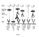
- Figure 2 is a pictorial diagram summarizing the function and processing of the controls spots present in an array printed on an assay membrane of the invention.
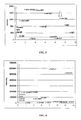
- Figure 3 is a graphical diagram summarizing the results of Example 5 obtained from processing human serum spiked with cytokines.
- the hash line represents the threshold signal above which the result is considered positive.
- the threshold is set at two times the signal intensity of the negative control spot (the Buffer spot).
- Figure 4 is a graphical diagram summarizing the results Example 6 obtained from processing human serum sample spiked with antibodies to Hepatitis B surface antigens.
- the hash line represents the threshold signal above which the result is considered positive.
- the threshold is set at two times the signal intensity of the negative control spot (the Buffer spot).
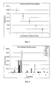
- Figure 5 is a graphical diagram summarizing the results of Example 7 that assesses the efficacy of a fiduciary marker of one embodiment of the invention.
- the x-axis units identify the tube number and thus the dilution factor.
- Figure 6 is a graphical diagram showing the results of Example 10 that demonstrates upper respiratory viral pathogen arrays for detection of antibodies in serum samples.
- Six human serum samples were tested at a dilution of 1 in 800 for presence of antibodies to each of the six viral antigens on arrays.
- a signal intensity threshold of 100000 was set for a positive test. Based on this threshold the results shown in Table 20 were obtained.
- Figure 7 is a graphical diagram showing the results of Example I showing the results from ten human serum samples that were tested at a dilution of 1 in 4000 for presence of antibodies to each of the four Hepatitis B antigens on arrays.
- the present invention relates to an assay membrane for detecting of at least one target analyte or a plurality of target analytes in a sample, as well as kits for detecting said target analytes and a method of processing the assay membrane.
- Biomarkers can identify disease prior to exhibition of clinical symptoms by a subject, and therefore provide the ability to treat the molecular basis of disease using targeted therapies.
- the basis for subject stratification lies in correlation of molecular heterogeneity of disease with heterogeneity of response to therapy.
- the target of the drug must be present and have a role in maintaining or worsening the disease state of the subject in order for the drug to be effective. This target would therefore serve as a biomarker to determine whether the subject is a candidate for treatment with that particular therapy.
- the presence of Her2/neu in tumors is required for effective treatment with anti-Her2 antibodies, such as Herceptin.
- Biomarkers can also be used for retrospective sample analysis after clinical trials have been completed or after post-marketing analysis of new drugs to perform subgroup analysis to identify covariates that were expected to account for differences in response.
- the present invention contemplates a plurality of capture agents arranged to detect one or more (i.e., a panel) target analytes (i.e., biomarkers) that may be used for a variety of assays.
- a panel of biomarkers may be monitored during clinical trials to determine effectiveness of therapy while simultaneously ensuring lack of side effects or any other adverse events.
- a panel of biomarkers may be used to test a variety of conditions and/or to further validate one or more potential biomarkers.
- Exemplary conditions include, but are not limited to, human diseases or allergies, pregnancy detection, animal diseases, and animal testing performed prior to export.
- biomarkers can be used during all phases of clinical trials to obtain a greater understanding of drug mechanism in a population prior to approval and general administration of the drug. Furthermore, biomarkers can support clinical outcome results from efficacy studies and help to measure real clinical benefit to the subject.
- Biomarkers can also be used to determine which subjects are likely to respond to a particular therapeutic. Biomarkers can also be used to monitor disease progression and treatment efficacy by measuring levels of various disease parameters simultaneously, thereby increasing the benefit of treatment to the subject. These biomarker panels aim to identify the right drug for the right subject at the right time.
- Biomarker validation for prediction of a particular disease, disorder, or condition refers to the confirmation of accuracy, reproducibility and effectiveness of biomarkers in detecting the disease, disorder or condition.
- the major challenge for biomarker validation is the high level of variability of biomarker levels across the human population and the considerable molecular heterogeneity of specific diseases, even from a single tissue.
- biomarker makes the transition from the research setting to the clinical diagnostic laboratory, it should progress through defined stages of confirmation.
- the first task of biomarker validation is evaluation of research technology, performance, and specifications (analytical validation). However, the ultimate goal is initial validation of the biomarker to identify early stage diseases, disorders, or conditions (clinical validation).
- biomarker refers to any substance used as an indicator of a biologic state.
- a biomarker can be any substance whose detection indicates a particular disease state (for example, the presence of an antibody may indicate an infection).
- a biomarker can be indicative of a change in expression or state of a protein that correlates with the risk or progression of a disease, or with the susceptibility of the disease to a given treatment.
- a biomarker may be used as a surrogate for a natural endpoint such as survival or irreversible morbidity. If a treatment alters the biomarker, which has a direct connection to improved health, the biomarker serves as a "surrogate endpoint" for evaluating clinical benefit.
- assay element refers to any of a number of different elements for use in an array of the invention.
- exemplary assay elements include, but are not limited to, capture elements and control elements.
- capture element refers to a molecule that is able to bind to a target analyte.
- useful capture elements include proteins, protein fragments, binding proteins, binding protein fragments, antibodies (polyclonal, monoclonal, or chimeric), antibody fragments, antibody heavy chains, antibody light chains, single chain antibodies, single-domain antibodies (a VHH for example), Fab antibody fragments, Fc antibody fragments, Fv antibody fragments, F(ab')2 antibody fragments, Fab' antibody fragments, single-chain Fv (scFv) antibody fragments, antibody binding domains, antigens, antigenic determinant, epitopes, haptens, immunogens, immunogen fragments, binding domains; a metal ion, a metal ion-coated molecule, biotin, avidins, streptavidins; substrates, enzymes, abzymes, co-factors, receptors, receptor fragments, receptor subunits, receptor subunit fragments, ligands
- control capture element refers to a capture element that functions as a control, either a negative control that should not bind any analyte or a positive control that will bind a non-target analyte.
- control element refers to an element that is used to provide information on the function of the assay, for example binding specificity, the level of non-specific background binding, the degree of binding cross-reactivity, and the performance of assay reagents and the detection system.
- Preferred controls useful herein include at least one negative control to monitor background signal, at least one negative control to monitor assay specificity, at least one positive colourimetric control, and at least one positive control to monitor assay performance.
- control to monitor assay performance refers to an element that forms one part of a complementary binding interaction during an assay and is intended to provide information on the accuracy of the assay result.
- the positive control to monitor assay performance comprises one binding partner of a complementary binding pair, where the other binding partner is a sample component or an assay reagent.
- the assay performance control is preferably selected from a target analyte, a binding partner corresponding to and able to bind a non-target analyte that will be present in the sample, a binding partner corresponding to and able to bind an assay reagent, and a colourimetric enzyme label, or any combination of any two or more thereof.
- binding partner corresponding to and able to bind a non-target analyte that will be present in the sample is an anti-Ig antibody that will bind an immunoglobulin present in a serum sample, therefore confirming a sample has been added.
- An example of a binding partner corresponding to and able to bind an assay reagent is an anti-Ig antibody that will bind a secondary immunoglobulin that is used to process the assay, such as biotinylated anti-target analyte antibody.
- Another example of a binding partner corresponding to and able to bind an assay reagent is a biotinylated antibody that will bind a streptavidin-peroxidase conjugate that is used to process the assay.
- control to monitor assay specificity refers to an element that is closely related to at least one binding partner of a complementary binding pair present in the assay and is intended to provide information of the specificity of the complementary binding. This control is a negative control that is not expected to generate a detectable result during normal assay processing.
- the assay specificity control would comprise an antibody that should not bind any antigen in the sample.
- the assay specificity control would comprise an antigen that should not bind any antibody in the sample.
- fiduciary marker refers to a coloured marker or label that will always be detectable on the membrane, preferably irrespective of the performance of the assay or processing of the membrane.
- the fiduciary marker acts therefore as a "true" positive control.
- microporous membrane refers to a membrane with protein binding characteristics and a narrow pore-size distribution.
- porosity of the membrane may determine the exposure time of reagents with membrane bound components by controlling the flow rate through the membrane.
- Microporous membranes for use in the present invention comprise nitrocellulose, nylon, polyvinylidene difluoride, polyester, polystyrene, polyethersulfone, cellulose acetate, mixed cellulose esters and polycarbonate.
- negative control refers to an element comprising print buffer or an unrelated protein to which no complementary binding partner is intended to be present in the assay. Any detectable signal from the negative control can be used to determine the background threshold of the assay and the accuracy of any positive results.
- the negative control to monitor background signal is print buffer.
- the print buffer is a solution used to carry and print the capture elements and control elements onto the membrane and may comprise buffered saline, glycerol and a surfactant, preferably a polysorbate surfactant such as Tween 20.
- the blocking solution is used to reduce non-specific protein biding to the membrane surface and preferably comprises skim milk, casein, bovine serum albumin, gelatins from fish, pigs or other species, dextran or any mixture of any two or more thereof, preferably in a solution of phosphate buffered saline and a surfactant such as Tween 20.
- positive colourimetric control refers to an enzyme or enzyme conjugate that provides a detectable signal upon addition of the enzyme substrate.
- printing refers to the placement of the assay elements (control and capture elements) on the membrane surface, with or without an adapter molecule between the membrane and the element.
- the assay elements bind to the membrane by covalent or non-covalent interaction.
- methods of placing assay elements on the membrane include printing, spotting or other techniques known in the art.
- the term “printing” can be used to include any of the methods for placing the assay elements on the membrane.
- sample and “specimen” as used herein are used in their broadest sense to include any composition that is obtained and/or derived from biological or environmental source, as well as sampling devices (e.g., swabs) which are brought into contact with biological or environmental samples.
- Biological samples include those obtained from an animal (including humans, domestic animals, as well as feral or wild animals, such as ungulates, bear, fish, lagamorphs, rodents, etc.), body fluids such as urine, blood, plasma, fecal matter, milk, nipple exudate, cerebrospinal fluid (CSF), semen, sputum, and saliva, as well as solid tissue.
- CSF cerebrospinal fluid
- Biological samples also include a cell (such as cell lines, cells isolated from tissue whether or not the isolated cells are cultured after isolation from tissue, fixed cells such as cells fixed for histological and/or immunohistochemical analysis), tissue (such as biopsy material), cell extract, tissue extract, and nucleic acid (e.g., DNA and RNA) isolated from a cell and/or tissue, and the like. Also included are materials obtained from food products and food ingredients such as dairy items, vegetables, meat, meat byproducts, and waste. “Environmental samples” include environmental material such as surface matter, soil, water, and industrial materials, as well as material obtained from food and dairy processing instruments, apparatus, equipment, disposable, and non-disposable items.
- the biological sample is a cell, tissue, and or fluid obtained from a mammal, including from the upper respiratory tissues (such as nasopharyngeal wash, nasopharyngeal aspirate, nasopharyngeal swab, and oropharyngeal swab), from the lower respiratory tissues (such as bronchiolar lavage, tracheal aspirate, pleural tap, sputum), blood, plasma, serum, stool, milk, nipple exudate, and tissue from any organ such as, without limitation, lung, heart, spleen, liver, brain, kidney, and adrenal glands.
- the upper respiratory tissues such as nasopharyngeal wash, nasopharyngeal aspirate, nasopharyngeal swab, and oropharyngeal swab
- the lower respiratory tissues such as bronchiolar lavage, tracheal aspirate, pleural tap, s
- antibody as used herein includes naturally occurring antibodies as well as non-naturally occurring antibodies, including, for example, single chain antibodies, chimeric, bifunctional and humanized antibodies, as well as antigen-binding fragments thereof.
- non-naturally occurring antibodies can be constructed using solid phase peptide synthesis, can be produced recombinantly or can be obtained, for example, by screening combinatorial libraries consisting of variable heavy chains and variable light chains (see Huse et al., Science 246:1275-1281, 1989 , which is incorporated herein by reference).
- These and other methods of making, for example, chimeric, humanized, CDR-grafted, single chain, and bifunctional antibodies are well known ( Winter and Harris, Immunol.
- modified or derivatized antibodies, or antigen binding fragments of antibodies can be useful for the present methods.
- pegylated antibodies can be useful for the present methods.
- Fab, F(ab') 2 , Fd and Fv fragments of an antibody that retain specific binding activity are included within the definition of an antibody.
- second antibody refers to an antibody that will bind a target analyte and that is conjugated with either an adaptor molecule such as biotin or an enzyme label such as horseradish peroxidase (HRP).
- Antibody-adaptor conjugates are processed to give a detectable result by contacting the antibody-adaptor conjugate with an adaptor-enzyme conjugate and then the enzyme substrate; for example, antibody-biotin conjugates will bind streptavidin-HRP conjugates.
- Antibody-enzyme label conjugates include antibody-HRP conjugates. Use of secondary antibodies is discussed and exemplified below.
- binding specifically or “specific binding activity” or the like, means that two molecules form a complex that is relatively stable under physiologic conditions.
- the term is also applicable where, e.g ., an antigen-binding domain is specific for a particular epitope, which is carried by a number of antigens, in which case the antibody carrying the antigen-binding domain will be able to bind to the various antigens carrying the epitope.
- Specific binding is characterized by a high affinity and a low to moderate capacity.
- the binding is considered specific when the affinity constant is about 1 x 10 -6 M, generally at least about 1 x 10 -7 M, usually at least about 1 x 10 -8 M, and particularly at least about 1 x 10 -9 M or 1 x 10 -10 M or less.
- one aspect of the invention relates to a microporous membrane for detecting a plurality (i.e., a panel) of target analytes (e.g ., biomarkers) in a sample, the membrane comprising an array that comprises at least one capture element and a plurality of control elements printed on the membrane surface, the at least one capture element corresponding to and being able to bind a target analyte.
- the plurality of control elements include
- membrane The choice of membrane is dependent on three main membrane characteristics: protein-binding capacity, porosity, and strength.
- the ability of the membrane to immobilize macromolecules, in particular proteins is crucial as the membrane serves as the solid phase used in the assay. However, this ability must be balanced with the availability of appropriate reagents (i.e., blockers) for blocking non-specific interactions on the membrane.
- the porosity of the membrane may determine the exposure time of reagents with membrane bound components by controlling their flow rate through the membrane.
- porosity must be balanced with the degree of array spot spreading during array manufacture, which can result in decreased signal intensity or cross contamination between adjacent spots.
- the strength of the membrane is important for the manufacture and eventual use of a device. A wide range of membranes are available with differing characteristics, allowing a particular membrane to be chosen depending on the requirements of an assay.
- microporous membranes for use in the present invention comprise nitrocellulose, nylon, polyvinylidene difluoride, polyester, polystyrene, polyethersulfone, cellulose acetate, mixed cellulose esters and polycarbonate.
- membranes such as cellulose acetate may have insufficient binding capacities for diagnostic immunoassays, the characteristics of such membranes may be applicable for assays where lower levels of accuracy or sensitivity are sufficient.
- the microporous membrane is preferably removably attachable to a bottomless microtiter plate. Accordingly, the membrane can be divided into individual microtiter wells that are separated from each other by a physical barrier, to prevent sample mixing between wells. Moreover, different assays can be conducted in separate wells, requiring smaller volumes of assay reagents.
- Capture elements specific for a target analyte are used to detect the presence or absence of the analyte in a sample.
- a wide range of complementary binding or coupling partners are known, with the choice of capture elements determined by the analytes to be detected, the requirement for adapter molecules and the level of specificity required for the assay.
- the target analyte is selected from a protein, a protein fragment, a peptide, a polypeptide, a polypeptide fragment, an antibody, an antibody fragment, an antibody binding domain, an antigen, an antigen fragment, an antigenic determinant, an epitope, a hapten, an immunogen, an immunogen fragment, a metal ion, a metal ion-coated molecule, biotin, avidin, streptavidin, an inhibitor, a co-factor, a substrate, an enzyme, a receptor, a receptor fragment, a receptor subunit, a receptor subunit fragment, a ligand, a receptor ligand, a receptor agonist, a receptor antagonist, a signalling molecule, a signalling protein, a signalling protein fragment, a growth factor, a growth factor fragment, a transcription factor, a transcription factor fragment, an inhibitor, a monosaccharide, an oligosaccharide, a polysaccharide, a glycol,
- the capture element is selected from a protein, a protein fragment, a binding protein, a binding protein fragment, an antibody, an antibody fragment, an antibody heavy chain, an antibody light chain, a single chain antibody, a single-domain antibody (a VHH for example), a Fab antibody fragment, an Fc antibody fragment, an Fv antibody fragment, a F(ab')2 antibody fragment, a Fab' antibody fragment, a single-chain Fv (scFv) antibody fragment, an antibody binding domain, an antigen, an antigenic determinant, an epitope, a hapten, an immunogen, an immunogen fragment, a binding domain; metal ion, or metal ion-coated molecule, biotin, avidin, streptavidin; a substrate, an enzyme, an abzyme, a co-factor, a receptor, a receptor fragment, a receptor subunit, a receptor subunit fragment, a ligand, an inhibitor, a hormone, a binding site, a lectin, a VHH
- the complementary binding partners comprise antibody-antigen interactions or antibody-ligand interactions.
- the capture elements may comprise antibodies or fragments thereof that are immobilised on the membrane surface and are specific for different antigens or ligands that may be present in a sample.
- the capture elements may comprise antigens or ligands and the assay involves the detection of specific antibodies that may be present in a sample.
- the capture elements may comprise of a receptor or a subunit of a receptor that binds a specific ligand.
- the target analyte is associated with an infectious disease, allergic disease, autoimmune disease, cardiac disease, cancer or graft versus host disease.
- the target analyte is selected from the list comprising angiogenesis factors such as Ang-2, FGF basic, HB-EGF, HGF, KGF, PDGF-BB, TIMP-1, TIMP-2, TPO and VEGF; Biomarkers such as A-SAA, Acrp-30 (Adiponectin), AR (Amphiregulin), Apo A-1, Apo B-100, C-peptide, sCD14, sCD30 (TNFRSF8), CD40L, CRP (C-reactive protein), ErbB2, FasL, Fibrinogen, Fibronectin, IGFBP-1, IGFBP-3, Leptin, LIF, MPO (Myeloperoxidase), NT-proBNP, OPG (Osteoprotegrin), OPN (Osteopontin), PAI-I Active, PAI-1 Total, PAPP-A, P1GF (Placental Growth Factor), Prolactin, RANK, RANKL, Resist
- the target analyte is an antigen from a family, genus, species, subtype or individual microorganism.
- exemplary microorganisms include, but are not limited to, Mycobacterium, Brucella, Bacillus, Treponema, Clostridium, Staphylococcus, Enterococcus, Streptococcus, Haemolyticus, Pseudomonas, Campylobacter, Enterobacter, Neisseria, Proteus, Salmonella, Simonsiella, Riemerella, Escherichia, Neisseria, Meningococcus, Moraxella, Kingella, Chromobacterium and Branhamella, or from a virus such as adenovirus, influenza, cytomegalovirus, hepatitis, human immunodeficiency virus, avian influenza virus, respiratory syncytial virus, herpex simplex virus, parainfluenza virus, pestivirus, porcine parvovirus, peudora
- target analytes include, but are not limited to, human chorionic gonadotropin, growth hormone, insulin, glucagon, adrenocorticotropic hormone, thyroid stimulating hormone, a-fetoprotein, human placental lactogen, leptin, inhibin A, activin A, pregnancy-associated plasma protein A, placenta growth factor, pregnancy-specific beta-1 glycoprotein; steroids such as testosterone, oestriol, cortisol, progesterone, corticosterone, aldosterone; thyroid hormones such as thyroxine, triiodothyronine; thyroid binding globulin (TBG); active peptides such as bradykinin, gastrin, angiotensin, thyroid hormone-releasing hormone, luteinising hormone-releasing hormone; physiologically active amines such as epinephrine, norepinephrine, histamine, serotonin; prostaglandins, such as PGF2a, PGE,
- the target analyte is an allergen.
- allergens include, but are not limited to, indoor allergens such as Mites, Tyr. put, Lep. dest. or mayrei, Felis, Bos, Albumine, Pen. cit., Pen. not., Asp. fumigatus, Alt.
- one or more capture agents are arranged to detect one or more (i.e., a panel) target analytes (i.e., biomarkers) that would be indicative of particular human conditions or diseases.
- a panel i.e., a panel
- target analytes i.e., biomarkers
- Table 1 Human Condition/Disease Panels Infectious disease screening for epidemiological studies in developing countries Human immunodeficiency virus (HIV)-1, HIV2, Hepatitis A virus, Hepatitis B virus, Hepatitis C virus, Herpes simplex virus (HSV)-1, HSV-2, Treponema pallidum, Mycobacterium tuberculosis, Neisseria gonorrhoeae, Plasmodium Upper respiratory viral infections Adenovirus, Cytomegalovirus (CMV), Influenza A, Influenza B, Parainfluenza 1, Parainfluenza 2, Parainfluenza 3 and Respiratory Syncytial Virus (RSV), Group A Streptococci.
- HSV Human immunodeficiency virus
- HIV2 Hepatitis A virus
- Hepatitis B virus Hepatitis C virus
- HSV-2 Herpes simplex virus
- Treponema pallidum Mycobacterium tuberculosis
- Sexually transmitted disease panel Human immunodeficiency virus (HIV)-1, HIV2, Treponema pallidum, Neisseria gonorrhoeae, Chlamydia trachomatis.
- Plasma borne disease panel Plasmodium falciparum (malaria), Trypanosoma cruzi (Chagas disease), Brucella spp (Brucellosis), Human immunodeficiency virus (HIV)-1, HIV2, Hepatitis A virus, Hepatitis B virus.
- Biosecurity panel Bacillus anthracis (anthrax), Clostridium botulinum (botulism), Clostridium perfringens, Yersinia pestis (plague), Coxiella burnetii (Q fever), Staphylococcal enterotoxin B, Vibrio cholerae (Cholera). Fertility panel Estradiol, follicle stimulating hormone, human chorionic gonadotrophin, lutenizing hormone, progesterone, prolactin, testosterone, parathyroid hormone. Drugs of abuse panel Acetaminophen, Amphetamines, Barbiturates, Cannabinoids, cocaine metabolites, methadone, opiates, salicylate and tricyclic anti-depressants.
- Panel for cardiovascular disease testing Brain natriuretic pepetide (BNP), N-terminal proBNP (Nt-proBNP), creatine kinase (CK)-MB, myoglobin, cardiac Troponin I, cardiac Troponin T, High-sensitivity C-reactive protein.
- Panel for autoimmune disease testing Rheumatoid factor, C-reactive protein, soluble human leukocyte antigen (HLA)-DR, antibodies against double stranded DNA, citrullinated peptides, small nuclear ribonucleoproteins, neutrophil cytoplasme (ANCA) and nuclear antigens (ANA).
- BNP Brain natriuretic pepetide
- Nt-proBNP N-terminal proBNP
- CK creatine kinase
- myoglobin myoglobin
- cardiac Troponin I cardiac Troponin I
- cardiac Troponin T High-sensitivity C-reactive protein.
- Panel for autoimmune disease testing Rheumatoid factor, C
- PSA Free and total Prostate specific antigen
- CEA Carcinoembryonic antigen
- CA125 CA15-3, CA19-9, CA24-2, CA72-4, alpha fetoprotein (AFP).
- IL-1 ⁇ and ⁇ Markers of Inflammation Interleukin (IL)-1 ⁇ and ⁇ , IL1 receptor antagonist, IL2, IL4, IL6, IL8, IL10, IL12, IL13, IFN ⁇ , TNF ⁇ , MIP1 ⁇ and ⁇ , MCP1, RANTES, soluble VCAM, C-reactive protein, soluble TNF ⁇ receptor I and II.
- Allergen panel to screen for serum IgE binding Allergens obtained by recombinant methods or derived from dust mites, grass and tree pollen, animal dander, moulds, insect venoms and foods such as soy protein, milk proteins, proteins derived from varieties of nuts, cereals and legumes, proteins from seafood such as shrimp, abalone and lobsters.
- one or more capture agents are arranged to detect one or more (i.e., a panel) target analytes (i.e., biomarkers) that would be useful in determining treatment efficacy for the indicated diseases.
- a panel target analytes
- biomarkers i.e., biomarkers
- Such tests could consist of any combination of the panels listed in Table 2, depending upon local requirements.
- Table 2 Panels to Determine Efficacy of Treatments Autoimmune diseases Cytokines and chemokines including CTLA4, tumor necrosis factor alpha (TNF ⁇ ), bLyS (BAFF), interferon gamma (IFN ⁇ ), eotaxin, CXCL10 or IP 10, osteopontin, osteoprotegerin and RANKL.
- biomolecules such as pyridinoline, deoxypyridinoline, cartilage oligomeric matrix protein.
- Antibodies against double stranded DNA small nuclear ribonucleoproteins, nuclear proteins such as Sjogren's Syndrome antigen (SS)-A and SS-B, nuclear antigen, Sm antigen, ribosomal P proteins, cardiolipin and topoisomerase I.
- Antibodies against therapeutic proteins such as abatacept (an immunoglobulin fused to the ectodomain of CTLA4), rituximab (a chimeric anti-CD20 antibody), toclizumab (anti-IL6 receptor antibody), etanercept (recombinant soluble human TNF ⁇ fused to IgG), infliximab (a chimeric anti-TNF ⁇ antibody), adalimumab (a humanized anti-TNF ⁇ antibody), anakinra (a human IL6 receptor antagonist protein) and others.
- abatacept an immunoglobulin fused to the ectodomain of CTLA4
- rituximab a chimeric anti-CD20 antibody
- toclizumab anti-IL6 receptor antibody
- etanercept recombinant soluble human TNF ⁇ fused to IgG
- infliximab a chimeric anti-TNF ⁇ antibody
- adalimumab a
- Antibodies against therapeutic proteins such as rituximab (a chimeric anti-CD20 antibody), cetuximab (a chimeric anti-EGF antibody), trastuzumab (humanized anti-Her2/ neu antibody), tositumomab (mouse monoclonal anti-CD20 antibody), gemtuzumab (humanized anti-CD33 antibody), bevacimumab (a humanized anti-VEGF antibody), alemtuzumab (a humanized anti-CD52 antibody) and Ibritumomab tiuxetan (mouse anti-CD20 antibodv).
- rituximab a chimeric anti-CD20 antibody
- cetuximab a chimeric anti-EGF antibody
- trastuzumab humanized anti-Her2/ neu antibody
- tositumomab mouse monoclonal anti-CD20 antibody
- gemtuzumab humanized anti-CD33 antibody
- bevacimumab
- IL-1 Interleukin
- IL-6 IL-6
- IL-10 ischemia-modified albumin
- MCP monocyte chemoattractant protein
- plasminogen activator-1 TNF ⁇
- von Willebrand factor plasminogen activator-1
- TNF ⁇ TNF ⁇
- von Willebrand factor plasminogen activator-1
- plasminogen activator-1 TNF ⁇
- von Willebrand factor plasminogen activator-1
- TNF ⁇ TNF ⁇
- von Willebrand factor soluble CD40 ligand
- myeloperoxidase myeloperoxidase
- placental growth factor fibrinogen
- fibrinogen and heart-type fatty acid binding protein (H-FABP)
- MMP matrix metalloproteinase
- BNGF B-type neurotrophic growth factor
- serum amyloid A fibrinogen
- sICAM S-100b.
- one or more capture agents are arranged to detect one or more (i.e ., a panel) target analytes (i.e., biomarkers) that would be useful in animal testing.
- a panel i.e., a target analytes
- Such tests could consist of any combination of the panels listed in Table 3, depending upon local requirements.
- Table 3 Animal Testing Panels Avian Avian influenza virus, Avian pneumovirus, Avian reovirus, avian rhinotracheitis virus, Chicken anemia virus.
- Bovine Bovine Adenovirus Bovine Coronavirus, Leptospira spp, Bovine leukosis Virus, Bovine respiratory syncytial virus, bovine spongiform encephalopathy, bovine viral diarrhoea virus, Brucella abortus, Neospora caninum, Mycoplasma bovis, Bovine babesiosis, Rotavirus, contagious bovine pleuropneumonia, bovine Herpes Virus Type I and II, bovine parainfluenza 3.
- Canine Canine distemper virus canine coronavirus, canine herpes virus, canine parvovirus, Borrelia burgdorferii, Rickettsia rickettsii, Ehrlichia canis, Rickettsia conori, canine rheumatoid factor, dog erythrocyte antigen, canine Hepatitis virus 1 and 2, canine parainfluenza 1, Borrelia afzelii, Leishmania donovani, Ehrlichia equi, Rickettsia conorii.
- Equine Equine Equine arteritis virus equine infectious anemia virus, equine herpesvirus Type I, equine adenovirus, equine influenza virus, Babesia equi, Babesia caballi, Borrelia burgdorferii, Borrelia afzelii, Ehrlichia equi, Leishmania donovani.
- Feline Feline coronavirus feline calicivirus, feline leukemia virus, feline herpesvirus, Feline immunodeficiency virus, feline infectious peritonitis virus, feline panleukopaenia virus, feline viral rhinotracheitis virus, Feline Enteric Corona Virus.
- Porcine pathogens Porcine influenza A virus, porcine parvovirus, porcine reproductive and respiratory syndrome virus, Pseudorabies virus, porcine rotavirus, porcine Brucella suis, transmissible gastroenteritis (TGE) virus, classical swine fever virus, porcine respiratory coronavirus.
- Ovine pathogens Ovine Herpes virus, Brucella ovis, pseudorabies virus (Aujesky's). Protein and endocrine panel for all species Estrone sulfate, progesterone, growth hormone, serum cortisol, testosterone, thyroxine (T)-3, T-4, Serum albumin, serum globulin, insulin, parathyroid hormone, thyroid stimulating hormone, leutenizing hormone.
- Bovine viral diarrhoea virus enzootic bovine leucosis virus, bovine Herpes Virus Type I, Maedi visna virus, Brucella ovis, Mycobacterium paratuberculosis (Johne's disease), Campylobacter fetus, Trichomonas foetus, Leptospira spp, Streptococcus equi, Infectious bovine rhinotracheitis virus.
- IL-1 ⁇ and ⁇ Markers of Inflammation Interleukin (IL)-1 ⁇ and ⁇ , IL1 receptor antagonist, IL2, IL4, IL6, IL8, IL10, IL12, IL13, IFN ⁇ , TNF ⁇ , MIP1 ⁇ and ⁇ , MCP1, RANTES, soluble VCAM, C-reactive protein, soluble TNF ⁇ receptor I and II.
- Blockers serve to decrease or at best eliminate non-specific protein binding from the sample on the membrane surface thereby decreasing overall background signal. This increases the ratio of signal to noise, thereby increasing the overall sensitivity of the assay. Blockers play no active part in the subsequent reactions between the sample and other assay reagents and the immobilized proteins on the membrane.
- blockers include, but are not limited to, bovine serum albumin, casein, non-fat dry milk, gelatin derived from fish, pigs and other sources, dextran, serum derived from sources other than the sample being analysed such as from steelhead salmon, guinea pigs, hamsters, rabbit and other sources, polyethylene glycol, polyvinyl pyrrollidone, and commercial preparations including HeteroBlock (Omega Biologicals, Bozeman, MT), SuperBlock, StartingBlock, SEA BLOCK (Pierce, Rockford, IL).
- blockers are made up in buffer solutions such as, for example, phosphate buffer, phosphate buffered saline, Tris buffer, acetate buffer and others.
- the blockers may also be supplemented with detergents such as, for example, Tween 20, Tween 80, Nonidet P40, sodium dodecyl sulfate and others.
- the membrane of the invention comprises at least one fiduciary marker that will always be detectable on the membrane, preferably detectable irrespective of the performance of the assay or processing of the membrane.
- the fiduciary marker is a dye, dye-conjugated protein or a chromogenic protein such as haemoglobin.
- fiduciary marker will obviate the necessity of this element being detected based on successful array processing, in comparison to the positive colourimetric controls.
- the fiduciary marker is therefore a "true" positive control that would always be detectable regardless of array processing, and can be used to orient and help to grid the array.
- the membrane of the invention also comprises at least one control to monitor assay specificity.
- the control is intended to provide information of the specificity of binding between the capture element and the target analyte, or between the binding partners of the assay detection steps.
- the assay specificity control comprises one or more antibody isotypes, a corresponding antibody or antibody isotype from a different animal species or a closely related ligand.
- human IgM and anti-human IgM can be used as controls to monitor assay specificity.
- the membrane of the invention also comprises at least one control to monitor assay performance.
- the control is intended to provide information of the efficiency of the complementary binding interactions or the quality or performance of the reagents used.
- the assay performance control comprises one binding partner of a complementary binding pair, wherein the other binding partner is an assay reagent.
- the assay performance control is preferably selected from the list comprising the target analyte, a non-specific binding partner or a colourimetric enzyme label.
- the positive colourimetric control is an enzyme label conjugate capable of reacting with a colourimetric substrate, comprising an enzyme selected from the list comprising horseradish peroxidase, alkaline phosphatases, ß-D-galactosidase or glucose oxidase.
- the identity of the assay controls will be dependent on the type of array, the identity of the target analyte, and the type of sample to be analyzed.
- anti-human IgG-HRP or anti-mouse IgG-HRP may be used in arrays printed with antigens and antibodies, respectively.
- the final detection antibody in antigen arrays will often be anti-human IgG-HRP, while for antibody arrays it will often be a biotinylated mouse IgG.
- These controls can provide a positive control in addition to providing information on the performance or quality of the HRP substrate.
- Mouse IgG, human IgG and anti-human IgG present on antigen or antibody arrays can act either as positive or negative controls depending on the array format, in addition to providing information of assay specificity.
- mouse IgG should provide the positive signal in antibody arrays, while the latter two should provide a positive signal in antigen arrays.
- human IgM and anti-human IgM may be replaced as controls with human IgE and anti-human IgE. These controls can also serve as controls for overall assay performance.
- the elements on the array are printed in discrete areas of between 100 ⁇ m to 500 ⁇ m in diameter. More preferably, the discrete areas are between 350 ⁇ m to 400 ⁇ m in diameter.
- the discrete areas of the array are printed in a 5 x 5 grid.
- the array comprises up to nine control elements and two replicates of each of eight different capture elements.
- the capture elements are printed in two or more replicates of four different capture elements and multiples thereof.
- the assay techniques used in conjunction with the membranes of the present invention include any of a number of well known colourimetric enzyme-linked assays. Examples of such systems are well known in the art.
- the assay techniques are based upon the formation of a complex between a complementary binding pair, followed by detection with a colourimetric detection system comprising an enzyme-conjugate label and a colourimetric substrate.
- the solid phase carrier or substrate is a microporous membrane.
- the detection system will be described with reference to enzyme-linked immunosorbent assays (ELISA), though a skilled person would appreciate that such techniques are not restricted to the use of antibodies but are equally applicable to any colourimetric assay.
- Figure 1 shows a schematic representation of assay formats and sample processing flow.
- Panel (A) shows the processing steps of an antibody array for detection of antigens or ligands from biological test samples.
- Panel (B) shows the processing of an antigen or ligand array for detection of antibodies in biological test samples.
- Panel (C) shows the general sample processing flow in which each of the reagents described below and in the Examples are added to the array printed according to Example 1.
- Figure 2 shows a schematic representation of function of control elements and their binding to various reagents added during processing of an antibody assay for antigen detection. The addition of various reagents is shown on the left of the Figure with sequential additions being made from bottom to top. Color is developed only if the appropriate functional reagent binds to the control element.
- the ELISA is in the "sandwich” assay format. In this format the target analyte to be measured is bound between two antibodies - the capture antibody and the detection antibody. In another embodiment the ELISA is a non-competitive assay, in which an antibody binds to the capture antigen and the amount of bound antibody is determined by a secondary detection antibody.
- the ELISA is a competitive assay, where a labelled antigen is used instead of a labelled antibody. Unlabelled antigen and the labelled antigen compete for binding to the capture antibody and the amount of target analyte bound can be determined by the proportion of labelled antigen detected.
- Either monoclonal or polyclonal antibodies may be used as the capture and detection antibodies in sandwich ELISA systems.
- Monoclonal antibodies have an inherent monospecificity toward a single epitope that allows fine detection and quantitation of small differences in antigen.
- a polyclonal antibody can also be used as the capture antibody to bind as much of the antigen as possible, followed by the use of a monoclonal antibody as the detecting antibody in the sandwich assay to provide improved specificity.
- a monoclonal antibody can also be used as the capture antibody to provide specific analyte capture, followed by the use of a polyclonal antibody as the detecting antibody in the sandwich assay.
- multiplexed assays it is also important that there is no overlap between each of the binding pairs to eliminate crossreactivity.
- a number of multiplexed ELISAs have been developed and it is anticipated other combinations of binding pairs could be configured through testing.
- the enzyme-conjugate label comprising an enzyme selected from the list comprising horseradish peroxidase, alkaline phosphatase, ß-D-galactosidase or glucose oxidase.
- the enzyme label may be conjugated directly to a primary antibody or introduced through a secondary antibody that recognises the primary antibody. It may also be conjugated to a protein such as streptavidin if the primary antibody is biotin labelled.
- the assay detection system comprises a detection colourimetric substrate selected from the list comprising 3, 3', 5, 5'-tetramethylbenzidine, diaminobenzidine, metal-enhanced diaminobenzidine, 4-chloro-1-naphthol, colloidal gold, nitro-blue tetrazolium chloride, 5-bromo-4-chloro-3'-indolylphosphate p-toluidine salt and naphthol AS-MX phosphate + Fast Red TR Salt.
- a detection colourimetric substrate selected from the list comprising 3, 3', 5, 5'-tetramethylbenzidine, diaminobenzidine, metal-enhanced diaminobenzidine, 4-chloro-1-naphthol, colloidal gold, nitro-blue tetrazolium chloride, 5-bromo-4-chloro-3'-indolylphosphate p-toluidine salt and naphthol AS-MX phosphate + Fast Red TR
- the colourimetric reaction can be detected and optionally quantified and analysed using an image capture device such as a digital camera or a desktop scanner attached to a computer.
- image capture device such as a digital camera or a desktop scanner attached to a computer.
- known methods for image analysis may be used.
- the density values of known standard elements can be used to generate standard curves.
- Density values for unknown analytes can be analysed using the standard curve for each analyte to calculate actual concentrations. Values for each analyte can be identified based on the spotting position of each capture element within the array.
- Membranes of the present invention are particularly amenable to use in kits for the detection of target analytes. Such kits may comprise the membranes together with instructions and any assay consumables required. Different kits are envisaged for different target analytes and types of array. Accordingly, in one aspect the invention relates to a kit comprising a membrane of the invention and optionally, one or more processing reagents. For example, a kit of the invention optionally includes one or more of, or any combination of any two or more of
- the invention also relates to a method of processing a membrane of the invention.
- a method of processing a membrane of the invention comprises
- the step of processing the membrane comprises a blocking step during which available protein-binding sites on the membrane are blocked with a blocker, an optional wash step, contacting the membrane with the sample containing the one or more analytes to be measured, a wash step to remove non-bound material from the membrane, contacting the membrane with one or more secondary antibodies that correspond to and will bind one or more target analytes and non-target analyte that is bound to an assay performance control, a wash step, and contacting the membrane with one or both of an enzyme conjugate or an enzyme substrate to generate a detectable result. Examples of processing a membrane of the invention are described below.
- the microporous membranes of the invention can be used for the simultaneous detection of at least one target analyte in a sample, and preferably a plurality of different target analytes in a sample, and have utility in diagnostic and screening assays.
- the microporous membranes of the invention provide the advantage that they can be adapted to high throughput (or ultra high throughput) analysis and, therefore, any number of samples (e.g., 96, 1024, 10,000, 100,000, or more) can be examined in parallel, depending on the particular support used.
- a particular advantage of adapting the microporous membranes to high throughput analysis is that an automated system can be used for adding or removing reagents from one or more of the samples at various times, for adding different reagents to particular samples, or for subjecting the samples to various heating cycles.
- the automated system may consist of one or more temperature-controlled chambers and one or more robotic arms mounted on a deck that has platforms configured to hold 96-well plates.
- the movement of the robotic arms and the temperature in the chambers are controlled by a central computer unit.
- the array plates are stacked on the deck of the instrument.
- the plates containing samples to be analysed are stacked in a chamber with temperature of 4°C.
- One robotic arm then sequentially transfers each individual array plate on one platform while the other arm sequentially transfers each individual sample plate on the second platform.
- a nozzle containing 96 disposable tips then aspirates a predetermined volume of sample from each well of the sample plate and transfers the sample to the corresponding wells of the array plate.
- the array plate containing the sample is then transferred to a chamber with temperature of 37°C. This process is repeated until sample has been added to all the array plates stacked on the deck.
- the array plates are incubated for a predetermined time followed by transfer of each plate to the platform for addition of wash buffer with the nozzle containing 96 disposable tips.
- the wash buffer is aspirated after a predetermined time and this wash process is repeated multiple ( i.e. , two or more) times.
- Each array plate then receives the secondary antibody followed by transfer to a chamber with temperature of 37°C.
- the array plates are incubated for a predetermined time followed by transfer of each plate to the platform for addition of wash buffer with the nozzle containing 96 disposable tips.
- the wash buffer is aspirated after a predetermined time and this wash process is repeated multiple (i.e., two or more) times. Each array plate then receives the detection reagent followed by incubation for a predetermined time followed by transfer of each plate to the platform for addition of wash buffer with the nozzle containing 96 disposable tips.
- the wash buffer is aspirated after a predetermined time and the plate transferred to the 37°C chamber for drying. The plates are transferred back to the deck after a predetermined period and manually processed for analyses of data.
- such high throughput assays provide a means for examining duplicate, triplicate, or more aliquots of a single sample, thus increasing the validity of the results obtained, and for examining control samples under the same conditions as the test samples, thus providing an internal standard for comparing results from different assays.
- Membranes or films attached to a bottomless 96-well polystyrene plate (such as nylon, Nalge Nunc International, USA,) were used for printing microarrays.
- Various methods may be used to attach the membranes to the 96-well bottomless plates.
- a rubber sheet with dimensions of 128mm length and 86mm and 1 mm thickness was used.
- 96 round holes were stamped on the sheet with a diameter of 6.35mm and a center to center distance of 9mm.
- the sheet was then coated with adhesive on both sides and glued to one side of the 96-well bottomless plate such that the second adhesive layer was still available for binding to a membrane.
- a nylon membrane was then cut to the dimensions of 128mm length and 86mm width and attached to the other side of the rubber sheet to create a gasket that creates leak proof wells.
- Another method for attaching the membranes to the 96-well bottomless plates involves use of an adhesive that is applied to one side of the plate. Thereafter, a nylon membrane cut to the dimensions of 128mm length and 86mm width is attached using pressure such that there is no leakage between the wells.
- Microarrays were printed by mixing proteins in a print buffer solution containing phosphate buffered saline, glycerol and Tween 20 to give a final concentration of 10% glycerol and 0.005% Tween 20.
- Arrays were printed by using a benchtop contact microarrayer (LabNext Inc, USA) using quill pins designed to give uniform spots with an average diameter of 350 ⁇ m (ChipMaker II, Telechem International Inc, USA). Arrays were printed at ambient temperature and humidity of 60% ( ⁇ 10%).
- Each array had up to 25 spots printed in 5X5 grids (Number of Columns X Number of Rows). Arrays with less than 25 spots were printed such that they contained 5, 10, 15 or 20 spots in patterns of 5X1 , 5X2, 5X3 and 5X4 spots.
- Each array had a series of control spots that were printed in Column I and Row 5. These control spots included a fiduciary marker (a dye-conjugated protein such as BlueRanger Prestained Protein Molecular Weight Marker, Pierce Biotechnology Inc, USA, Catalog Number 26681), negative control (phosphate buffered saline containing 20% glycerol and 0.005% Tween 20 and a non-specific antibody), positive controls (enzyme-conjugated protein such as streptavidin conjugated horseradish peroxidase, Pierce, Catalog Number 21126) and sample specific controls to monitor the overall performance of the assay.
- a fiduciary marker a dye-conjugated protein such as BlueRanger Prestained Protein Molecular Weight Marker, Pierce Biotechnology Inc, USA, Catalog Number 26681
- negative control phosphate buffered saline containing 20% glycerol and 0.005% Tween 20 and a non-specific antibody
- positive controls enzyme-conjugated protein such as streptavidin conjugated horse
- Test proteins were printed at concentrations ranging from 0.05 mg/ ml to 1.0 mg/ml, usually 0.5 mg/ml, determined by the binding affinity of the specific protein to the analyte being measured.
- the arrays were incubated at 37°C for 60 min after adding 100 ⁇ l of Blocker [1% casein (Vector Labs, USA) in phosphate buffered saline containing 0.1% Tween 20 (PBS-T)] to each well. The Blocker was then aspirated off.
- Blocker 1% casein (Vector Labs, USA) in phosphate buffered saline containing 0.1% Tween 20 (PBS-T)
- Samples containing analytes were added by diluting in Blocker at a volume of 50 ⁇ l in each well and the membrane incubated at 37°C for 60 min. The membrane was washed 3X with PBS-T to remove excess non-bound analytes.
- adapter-conjugated detection antibodies were added to the wells at concentrations either recommended by the manufacturer or empirically determined by experimentation.
- adapter-conjugated antibodies include hapten-conjugated antibodies such as biotin-conjugated antibodies.
- the membrane was incubated at 37°C for 60 min and washed 3X with PBS-T.
- the membrane was then incubated with an anti-adapter antibody (such as an anti-biotin antibody) conjugated to an enzyme or an adapter-conjugated enzyme (such as a streptavidin-conjugated enzyme) for 37°C for 60 min.
- an enzyme is horseradish peroxidase.
- the membrane was washed 3X with PBS-T.
- the membrane was incubated with anti-immunoglobulin antibodies conjugated with an enzyme such as horseradish peroxidase.
- an enzyme such as horseradish peroxidase.
- the membrane was incubated for 37°C for 60 min.
- the membrane was washed 3X with PBS-T.
- the bound enzyme was detected and measured using an enzyme substrate that results in a colored precipitate deposited on the protein spot.
- An example of a substrate used is metal enhanced diaminobenzidine (Pierce, USA) that gives a brown precipitate with horseradish peroxidase.
- the bound anti-analyte antibodies can be detected using a second antibody or adapter such as streptavidin conjugated to colloidal gold.
- the membrane was dried for 60 min at ambient temperature and scanned at 600 dpi resolution or photographed using a digital camera with a resolution of at least 4 megapixels and the image saved in the TIFF format.
- the color intensity at each spot was determined using gridding software that placed grids on all the arrays using the fiduciary marker to align the grid at the appropriate position.
- the intensity values were obtained in a Microsoft ExcelTM spreadsheet file, which can then be used for analysis of results.
- Arrays for detection of antigens such as protein markers of autoimmune diseases, cardiovascular diseases, cancer and infectious agents, or ligands such as growth factors, hormones, cytokines and chemokines are created by printing panels of antibodies as capture elements for specific capture of the antigen.
- a series of control antibodies and control proteins are also printed. These controls serve a variety of functions including controls for monitoring assay performance including performance of individual reagents, controls for monitoring the specificity of the capture antibodies and fiduciary markers for gridding the arrays after sample processing for determination of signal intensity at each spot in the array.
- Table 4 summarises the reagents that may be used to print and process antibody arrays.
- the assay performance control numbering relates to the numbering in Figure 2 .
- biotin Control to monitor the function of enzyme-conjugated Streptavidin or any other biotin-binding protein(BP) (assay performance (3)) Anti-mouse IgG conjugated to biotin Hapten binding protein- enzyme conjugate Positive colourimetric control to monitor the performance of the enzyme substrate Streptavidin-horseradish peroxidase Anti-IgG antibody Control to demonstrate the addition of sample (assay performance (1) ) Addition of serum will result in binding of serum IgG to this spot; If non-human sample is to be tested the antibody will be replaced with one appropriate for capture of IgG from the species being tested IgG for capture of detection antibodies conjugated to hapten, Control to demonstrate the addition of secondary detection antibody mix (assay performance (2) ) Anti-mouse IgG The secondary biotinylated monoclonal antibodies will bind to the anti-mouse IgG Non-specific antibody Negative control for determining antigen capture specificity (assay specificity) Hamster IgG IgG
- Test and control antibodies are printed at concentrations ranging from 0.1 mg/ml to 1mg/ml depending upon the affinity of the antibody for its antigen and the signal obtained from the control antibodies.
- Table 5 Antibody array design for antigen detection Fiduciary marker Test antibody 1 Test antibody 1 Test antibody 2 Test antibody 2 Print Buffer (negative control) Test antibody 3 Test antibody 3 Test antibody 4 Test antibody 4 Hapten-conjugated antibody (assay performance (3)) Test antibody 5 Test antibody 5 Test antibody 6 Test antibody 6 Print Buffer (negative control) Test antibody 7 Test antibody 7 Test antibody 8 Test antibody 8 HRP-hapten BP conjugate (colourimetrie control) Anti-IgG antibody (assay performance (1)) Anti-mouse antibody (assay performance (2)) Non-specific antibody (assay specificity) Fiduciary marker
- Printed arrays are used for measuring the presence of marker proteins by initially incubating with Blocker at 37°C for 60 min.
- samples to be tested such as serum, plasma or any other biological material are added to their own well. Samples may be added without dilution or may be diluted in Blocker prior to addition to the test well. The membrane is incubated at 37°C for 60 min, and non-bound material is washed off with PBS-T.
- Antigens or ligands bound to arrayed antibodies are detected by sequential incubations with biotinylated secondary antibodies and a biotin-binding protein-conjugated to an enzyme. The amount of enzyme at each spot is then measured by using a substrate that results in a colored precipitate deposited at the spot.
- positive controls are processed and detected as follows.
- the colourimetric control is processed to generate a colour result by addition of the enzyme substrate.
- the assay performance controls are processed as follows.
- Assay performance control (1) an anti-IgG antibody, will bind IgG (a non-target analyte) present in the serum sample. IgG binding will be detected using a secondary antibody, either an antibody-adapter conjugate (e.g. an anti-IgG antibody-biotin conjugate) or an antibody-enzyme conjugate (e.g. an antibody-HRP conjugate).
- Assay performance control (2) an anti-mouse antibody, will bind the biotinylated secondary antibody.
- biotin binding protein-enzyme conjugate and enzyme substrate or a biotin-binding molecule conjugated to a colored moiety such as colloidal gold.
- Assay performance control (3) an antibody-biotin conjugate, will bind the biotin binding protein-enzyme conjugate and this interaction will be detected by addition of the enzyme substrate.
- Arrays for detection of antibodies to antigens of interest such as protein markers of autoimmune diseases, cardiovascular diseases, cancer and infectious agents, or ligands such as growth factors, hormones, cytokines and chemokines are created by printing panels of antigens or ligands as capture elements for specific capture of antibodies.
- a series of control antibodies and control proteins are also printed. These controls serve a variety of functions including controls for monitoring assay performance, including the performance of individual reagents, controls for monitoring the specificity of the assay and fiduciary markers for gridding the arrays after sample processing for determination of signal intensity at each spot in the array.
- Table 6 summarizes the reagents that may be used to print and process antigen arrays.
- Control antibodies are printed at concentrations ranging from 0.1 mg/ml to 1 mg/ml depending upon the signal obtained from the control antibodies.
- Test antigens or ligands are printed at concentrations ranging from 0.05 mg/ml to 1 mg/ml depending on the affinity of the antigen to test antibodies from positive control biological samples.
- Table 7 Antigen array design for antibody detection Fiduciary marker Test antigen 1 Test antigen 1 Test antigen 2 Test antigen 2 Print Buffer (negative control) Test antigen 3 Test antigen 3 Test antigen 4 Test antigen 4 Anti-IgM antibody (assay performance (1)) Test antigen 5 Test antigen 5 Test antigen 6 Test antigen 6 Print Buffer (negative control) Test antigen 7 Test antigen 8 Test antigen 8 HRP-conjugated protein (colourimetric control) Anti-IgG antibody (assay performance (1)) IgG (assay performance (2)) Antibody from non-crossreactive species (assay specificity) Fiduciary marker
- Printed arrays are used for measuring the presence of antibodies by initially incubating with Blocker at 37°C for 60 min.
- samples to be tested such as serum, plasma or any other biological material are added to their own well. Samples may be added without dilution or may be diluted in Blocker prior to addition to the test well. The membrane is incubated at 37°C for 60 min, and non-bound material is washed off with PBS-T.
- Antibodies bound to arrayed antigens are detected by incubations with enzyme conjugated secondary antibodies or secondary antibodies conjugated to a colored molecule such as colloidal gold. The amount of enzyme at each spot is then measured by using a substrate that results in a colored precipitate deposited at the spot.
- Positive controls are detected as follows.
- the colourimetric control was detected by addition of enzyme substrate.
- the assay performance (1) control will bind IgG in the serum sample and is detected by addition of a secondary antibody-enzyme conjugate and the enzyme substrate.
- the assay performance (2) control will bind the secondary antibody-enzyme conjugate and is detected by addition of the enzyme substrate.
- the assay performance (1) control will bind IgM in the serum sample and is detected by addition of a secondary antibody-enzyme conjugate and the enzyme substrate.
- Table 8 Cytokine array reagents Reagent Vendor Catalog No. Function Mouse anti-human IFN ⁇ antibody BioLegend 507501 Capture elements Mouse anti-human TUF ⁇ antibody BioLegend 502801 Mouse anti-human IL4 antibody BioLegend 500701 Goat anti-human IgG antibody Pierce 31119 Detect sample addition.
- Anti-human IgG-peroxidase 50 ⁇ g/ml) (fiduciary marker) Anti-human IgG antibody (assay performance (1) ) (200 ⁇ g/ml) Anti-human IgG antibody (assay performance (1)) (200 ⁇ g/ml) Anti-human IgM antibody (assay performance (1) (200 ⁇ g/ml) Anti-human IgM antibody (assay performance (1) (200 ⁇ g/ml) Print Buffer (negative control) Anti-human IFN ⁇ antibody (capture element) (200 ⁇ g/ml) Anti-human IFN ⁇ antibody (capture element) (200 ⁇ g/ml) Anti-human TNF ⁇ antibody (capture element) (
- Printed arrays were used for measuring the amount of cytokines by initially incubating with Blocker at 37°C for 60 min.
- Cytokines bound to arrayed antibodies were detected by sequential incubations with biotinylated anti-cytokine antibodies and streptavidin-conjugated horseradish peroxidase. Positive controls were detected by incubating with biotinylated anti-humanIgG and anti-humanIgM antibodies before streptavidin-HRP was added. The amount of peroxidase in each spot was then measured by using metal enhanced diaminobenzidine.
- assay performance (2) antibodies on the array detected the secondary antibodies for resolving the assay performance (1) control (i.e. the biotinylated anti-human IgG and anti-human IgM antibodies), rather than being antibodies that detected the biotinylated anti-human cytokine antibodies preferred in Example 3.
- FIG. 3 A representative example of the results obtained from processing a human serum sample spiked with TNF ⁇ at 1.25 ⁇ g/ml and IFN ⁇ at 0.32 ng/ml is shown in Figure 3 .
- the hash line represents the threshold signal above which the result is considered positive. The threshold is based on the signal intensity of negative control spots and is two times the signal.
- the data is also shown in Table 10 for the samples tested. Table 10: Results of Example 5 Sample Number Test Result Comment 1 IgG Positive Sample was serum IgM Positive Sample was serum IFN ⁇ Positive Cytokine present TNF ⁇ Positive Cytokine present IL4 Negative Not present
- Printed arrays were initially incubated with Blocker at 37°C for 60 min.
- Antibodies bound to arrayed antigens were detected by incubation with a mixture of horseradish peroxidase-conjugated anti-mouse IgG antibody, and anti-human IgG and IgM antibodies. The amount of peroxidase in each spot was then measured by using metal enhanced diaminobenzidine.
- the assay returned a positive signal for the presence of anti-Hepatitis B antibodies reactive to ad and ay subtypes.
- the signal from the other antigens was below the threshold for a positive tests result, highlighting the specificity of the array.
- the prestained protein was diluted 2-fold from tubes 1 to 5 and used for printing arrays.
- the array configuration is shown in Table 13.
- Table 13 Array configuration Undiluted prestained protein Undiluted prestained protein 1 in 2 dilution of protein 1 in 2 dilution of protein 1 in 4 dilution of protein 1 in 4 dilution of protein 1 in 8 dilution of protein 1 in 8 dilution of protein 1 in 16 dilution of protein 1 in 16 dilution of protein
- Arrays for detection of IgE antibodies to allergens of interest such as those obtained by recombinant methods or derived from, e.g., dust mites, grass and tree pollen, animal dander, moulds, insect venoms and foods such as soy protein, milk proteins, proteins derived from varieties of nuts, cereals and legumes, proteins from seafood such as shrimp, abalone and lobsters and others are created by printing panels of allergens as capture elements for specific capture of IgE antibodies. A series of control antibodies and control proteins are also printed.
- controls serve a variety of functions including controls for monitoring assay performance, including the performance of individual reagents, controls for monitoring the specificity of the assay and fiduciary markers for gridding the arrays after sample processing for determination of signal intensity at each spot in the array.
- Table 14 summarizes exemplary reagents that may be used to print and process allergen arrays.
- Table 14 Allergen array reagents Reagent Function Example Comments Protein or antibody conjugated with a chromogenic dye or enzyme Fiduciary marker to enable the gridding software to locate and place grids on each spot of the array BlueRanger dye conjugated protein marker or antibody-horse radish peroxidase conjugate Print Buffer Negative control for determining the background signal in the array Anti-IgE antibody (Assay performance control 1) Determine presence of total IgE in sample Mouse anti-human IgE antibody Presence of IgE in serum will result in binding of IgE to this spot; If non-human sample is to be tested the antibody will be replaced with one appropriate for capture of IgE from the species being tested Hapten binding protein- enzyme conjugate Positive colorimetric control to monitor the performance of the enzyme substrate Streptavidin-horseradish peroxidase Anti- human IgG antibody (assay performance control 1) Control to demonstrate the addition of sample Addition of serum will result in binding of serum IgG to this spot; If non-human
- Control antibodies are printed at concentrations ranging from 0.1 mg/ml to 1.0 mg/ml depending upon the signal obtained from the control antibodies.
- Test allergens are printed at concentrations ranging from 0.05 mg/ml to 1.0 mg/ml depending on the affinity of the antigen to test antibodies from positive control biological samples.
- Table 15 Allergen array design for IgE antibody detection Fiduciary marker Test allergen I Test allergen 1 Test allergen 2 Test allergen 2 Print Buffer (negative control) Test allergen 3 Test allergen 3 Test allergen 4 Test allergen 4 Anti-IgE antibody (assay performance (1)) Test allergen 5 Test allergen 5 Test allergen 6 Test allergen 6 Print Buffer (negative control) Test allergen 7 Test allergen 7 Test allergen 8 Test allergen 8 HRP-conjugated protein (colorimetric control) Anti-human IgG antibody (assay performance (1)) IgE (assay performance (2)) Antibody from non-crossreactive species (assay specificity) Fiduciary marker
- Printed arrays are used for measuring the presence of antibodies by initially incubating with Blocker at 37°C for 60 min.
- samples to be tested may be added to the individual wells. Samples may be added without dilution or may be diluted in Blocker prior to addition to the test well. The membrane is incubated at 37°C for 60 min, and non-bound material is washed off with PBS-T.
- Antibodies bound to arrayed antigens are detected by incubations with enzyme conjugated secondary antibodies or secondary antibodies conjugated to a colored molecule such as colloidal gold. The amount of enzyme at each spot is then measured by using a substrate that results in a colored precipitate deposited at the spot. Positive controls are detected as follows: The colorimetric control was detected by addition of enzyme substrate.
- the assay performance control (1) will bind IgG in the serum sample and is detected by addition of a secondary antibody-enzyme conjugate and the enzyme substrate.
- the assay performance (2) control will bind the secondary antibody-enzyme conjugate and is detected by addition of the enzyme substrate.
- the assay performance (1) control will bind IgE in the serum sample and is detected by addition of a secondary antibody-enzyme conjugate and the enzyme substrate.
- Arrays for simultaneous detection of presence of antibodies and antigens such as cytokines and other protein biomarkers in serum are created by printing panels of corresponding cognate antigens and antibodies as capture elements for specific capture of analytes.
- a series of control antibodies and control proteins are also printed. These controls serve a variety of functions, such as monitoring assay performance, including the performance of individual reagents, monitoring the specificity of the assay, and fiduciary markers for gridding the arrays after sample processing for determination of signal intensity at each spot in the array.
- Table 16 summarizes exemplary reagents that may be used to print and process these composite arrays.
- Table 16 Composite Antibody and Antigen Array Reagents Reagent Function Example Comments Protein or antibody conjugated with a chromogenic dye or enzyme Fiduciary marker to enable the gridding software to locate and place grids on each spot of the array BlueRanger dye conjugated protein marker or antibody-horse radish peroxidase conjugate Print Buffer Negative control for determining the background signal in the array Anti-secondary antibody IgG (Assay performance control 3) Monitor performance of secondary detection antibodies Goat anti-mouse IgG antibody Addition of secondary antibody will result in binding of IgG to this spot; the antibody spotted will vary depending upon the contents of the secondary antibody cocktail.
- Hapten binding protein- enzyme conjugate Positive colorimetric control to monitor the performance of the enzyme substrate Streptavidin-horseradish peroxidase Anti- human IgG antibody (assay performance control 1) Control to demonstrate the addition of sample Addition of serum will result in binding of serum IgG to this spot; If non-human sample is to be tested the antibody will be replaced with one appropriate for capture of IgG from the species being tested IgG (Assay performance control 2) Control to demonstrate the addition of secondary detection antibody Human IgG The secondary antibody will bind the IgG; If non-human sample is to be tested the antibody will be replaced with one appropriate for capture of IgG from the species being tested Non-specific antibody Negative control for detennining antigen capture specificity (assay specificity) Hamster IgG IgG from a species not represented in the array panel and detection reagents Panel of test antigens for capture of antibody Diagnostic tests to determine specificity of patient IgG or IgM to antigen (capture elements) anti-nuclear
- the arrays are printed in 5X5 grids as shown in Table 17.
- Control antibodies are printed at concentrations ranging from 0.1 mg/ml to 1.0 mg/ml depending upon the signal obtained from the control antibodies.
- Test antigens and antibodies are printed at concentrations ranging from 0.05 mg/ml to 1.0 mg/ml depending on the affinity of the antigen to test antibodies from positive control biological samples.
- Table 17 Composite antibody and antigen arrays Fiduciary marker Test antigen 1 Test antigen 1 Test antigen 2 Test antigen 2 Print Buffer (negative control) Test antigen 3 Test antigen 3 Test antigen 4 Test antigen 4 Anti-secondary antibody IgG (assay performance (3)) Test antibody 1 Test antibody 1 Test antibody 2 Test antibody 2 Print Buffer (negative control) Test antibody 3 Test antibody 4 Test antibody 4 HRP-conjugated protein (colorimetric control) Anti-human IgG antibody (assay performance (1)) IgG (assay performance (2)) Antibody from non-crossreactive species (assay specificity) Fiduciary marker
- Printed arrays are used for measuring the presence of antigens and antibodies by initially incubating with Blocker at 37°C for 60 min.
- Samples may be added without dilution or may be diluted in Blocker prior to addition to the test well.
- the membrane is incubated at 37°C for 60 min, and non-bound material is washed off with PBS-T.
- Antigens and Antibodies bound to arrayed reagents are detected by incubations with enzyme conjugated secondary antibodies or secondary antibodies conjugated to a colored molecule such as colloidal gold. The amount of enzyme at each spot is then measured by using a substrate that results in a colored precipitate deposited at the spot. Positive controls are detected as follows. The colorimetric control was detected by addition of enzyme substrate.
- the assay performance control (1) will bind IgG in the serum sample and is detected by addition of a secondary antibody-enzyme conjugate and the enzyme substrate.
- the assay performance (2) control will bind the secondary antibody-enzyme conjugate and is detected by addition of the enzyme substrate.
- the assay performance (3) control will bind secondary IgG (detection antibodies) and is detected by addition of a detection antibody-enzyme conjugate and the enzyme substrate.
- Table 18 Antibody detection assay reagents Reagent Vendor Catalog No. Function Anti-mouse antibody peroxidase Pierce 31430 Colour reaction Adenovirus antigen Virion 1121 Capture elements Cytomegalovirus CF antigen Virion 1130 Influenza A antigen Microbix EL-13-02 Influenza B antigen Microbix EL-14-02 Parainfluenza 3 antigen Microbix EL-10-02 Respiratory Syncytial Virus antigen Virion 1124 Cytomegalovirus IgG antigen BiosPacific J43010230 Influenza A antigen BiosPacific J43610149 Goat anti-human IgG horseradish peroxidase Pierce 31412 Fiduciary marker and colour reaction Goat anti-human IgG antibody Pierce 31119 Detect sample addition.
- test performance (1) Detect IgG and IgM binding to the assay performance (1) control Mouse anti-human IgM antibody BioLegend 314501 Human IgG Pierce 31154 Detect detection antibody addition.
- assay performance (2) Mouse IgG Pierce 31202 Assay specificity Goat anti-mouse IgG Pierce 31164 Assay specificity
- Printed arrays were initially incubated with Blocker at 37°C for 60 min. Human serum samples were added at a 1 in 100 dilution to Row A of the plate containing the arrays and diluted 2-fold in Blocker from Row A to G. Row H contained only the Blocker solution. The membrane was incubated at 37°C for 60 min, and non-bound antibodies were washed off with PBS-T.
- Antibodies bound to arrayed antigens were detected by incubation with horseradish peroxidase-conjugated anti-mouse IgG antibody. The amount of peroxidase in each spot was then measured by using metal enhanced diaminobenzidine.
- Figure 6 shows the results of six human serum samples at a dilution of 1 in 800 for presence of antibodies to each of the six viral antigens on arrays.
- a signal intensity threshold of 100000 was set for a positive test.
- the assay returned a positive signal for some of the antigens on the array.
- the signal from the other antigens was below the threshold for a positive tests result, highlighting the ability of the arrays to determine the presence of serum antibodies to the arrayed antigens. Based on this threshold the results shown in Table 20 were obtained.
- Table 21 Anti-Hepatitis B antibody detection assay reagents Reagent Vendor Catalog No. Function Anti-mouse antibody peroxidase Pierce 31430 Colour reaction Hepatitis B surface antigen "ad” BiosPacific J44010031 Capture elements Hepatitis B surface antigen "ay” Bios Pacific J44020031 Hepatitis B "E” antigen BiosPacific J44200352 Hepatitis B core antigen Bios Pacific J44400352 Goat anti-human IgG horseradish peroxidase Pierce 31412 Fiduciary marker and colour reaction Goat anti-human IgG antibody Pierce 31119 Detect sample addition. (assay performance (1)) Detect IgG and IgM binding to the assay performance (1) control Human IgG Pierce 31154 Detect detection antibody addition. (assay performance (2)) Goat anti-mouse IgG Pierce 31164 Assay specificity
- Printed arrays were initially incubated with Blocker at 37°C for 60 min. Human serum samples were added at a 1 in 100 dilution to Row A of the plate containing the arrays and diluted 2-fold in Blocker from Row A to G. Row H contained only the Blocker solution. The membrane was incubated at 37°C for 60 min, and non-bound antibodies were washed off with PBS-T.
Landscapes
- Health & Medical Sciences (AREA)
- Chemical & Material Sciences (AREA)
- Immunology (AREA)
- Life Sciences & Earth Sciences (AREA)
- Hematology (AREA)
- Engineering & Computer Science (AREA)
- General Health & Medical Sciences (AREA)
- Analytical Chemistry (AREA)
- Biomedical Technology (AREA)
- Molecular Biology (AREA)
- Urology & Nephrology (AREA)
- Chemical Kinetics & Catalysis (AREA)
- Clinical Laboratory Science (AREA)
- Food Science & Technology (AREA)
- Pathology (AREA)
- Physics & Mathematics (AREA)
- Biotechnology (AREA)
- Biochemistry (AREA)
- Cell Biology (AREA)
- General Physics & Mathematics (AREA)
- Medicinal Chemistry (AREA)
- Microbiology (AREA)
- Investigating Or Analysing Biological Materials (AREA)
- Apparatus Associated With Microorganisms And Enzymes (AREA)
- Measuring Or Testing Involving Enzymes Or Micro-Organisms (AREA)
- Investigating Or Analysing Materials By The Use Of Chemical Reactions (AREA)
- Investigating Or Analyzing Non-Biological Materials By The Use Of Chemical Means (AREA)
Applications Claiming Priority (2)
| Application Number | Priority Date | Filing Date | Title |
|---|---|---|---|
| US86177106P | 2006-11-28 | 2006-11-28 | |
| PCT/US2007/082732 WO2008067091A2 (en) | 2006-11-28 | 2007-10-26 | Assay membrane and method of use thereof |
Publications (3)
| Publication Number | Publication Date |
|---|---|
| EP2092333A2 EP2092333A2 (en) | 2009-08-26 |
| EP2092333A4 EP2092333A4 (en) | 2010-01-20 |
| EP2092333B1 true EP2092333B1 (en) | 2013-05-01 |
Family
ID=39468589
Family Applications (1)
| Application Number | Title | Priority Date | Filing Date |
|---|---|---|---|
| EP07863577.8A Active EP2092333B1 (en) | 2006-11-28 | 2007-10-26 | Assay membrane and method of use thereof |
Country Status (9)
| Country | Link |
|---|---|
| US (2) | US9625453B2 (enExample) |
| EP (1) | EP2092333B1 (enExample) |
| JP (1) | JP5240945B2 (enExample) |
| AU (1) | AU2007325465B2 (enExample) |
| CA (1) | CA2670615C (enExample) |
| ES (1) | ES2422597T3 (enExample) |
| NZ (1) | NZ577992A (enExample) |
| WO (1) | WO2008067091A2 (enExample) |
| ZA (1) | ZA200903411B (enExample) |
Families Citing this family (33)
| Publication number | Priority date | Publication date | Assignee | Title |
|---|---|---|---|---|
| JP5240945B2 (ja) * | 2006-11-28 | 2013-07-17 | ピクター リミテッド | アッセイ膜およびその使用法 |
| KR20140063499A (ko) * | 2010-11-17 | 2014-05-27 | 오숀 바이오시스템즈 | 웰내 교정 특징부를 프린팅하기 위한 방법 및 시스템 |
| WO2013074643A2 (en) | 2011-11-14 | 2013-05-23 | Aushon Biosystems, Inc. | Systems and methods to enhance consistency of assay performance |
| WO2014003580A1 (en) | 2012-06-28 | 2014-01-03 | Caldera Health Ltd | Targeted rna-seq methods and materials for the diagnosis of prostate cancer |
| US9427707B2 (en) | 2012-08-10 | 2016-08-30 | Jean I. Montagu | Filtering blood |
| US10534003B2 (en) | 2013-07-17 | 2020-01-14 | The Johns Hopkins University | Multi-protein biomarker assay for brain injury detection and outcome |
| GB201314452D0 (en) * | 2013-08-13 | 2013-09-25 | Ostara Biomedical Ltd | Embryo implantation |
| WO2015120382A1 (en) | 2014-02-07 | 2015-08-13 | The Johns Hopkins University | Predicting response to epigenetic drug therapy |
| FR3019654B1 (fr) | 2014-04-04 | 2020-10-30 | Bio Rad Innovations | Controles pour la mise en oeuvre de procedes d'analyse multiplexe |
| US9903868B2 (en) | 2014-05-16 | 2018-02-27 | The United States Of America, As Represented By The Secretary, Department Of Health And Human Services | Method for the detection and quantitation of biomarkers |
| WO2016044697A1 (en) | 2014-09-19 | 2016-03-24 | The Johns Hopkins University | Biomarkers of cognitive dysfunction |
| GB201501302D0 (en) | 2015-01-27 | 2015-03-11 | Ostara Biomedical Ltd | Embryo implantation |
| US11041865B2 (en) | 2015-02-20 | 2021-06-22 | The Johns Hopkins University | Biomarkers of myocardial injury |
| GB201517523D0 (en) | 2015-10-05 | 2015-11-18 | Ostara Biomedical Ltd | Methods and compositions for managing reproduction |
| US10973679B2 (en) * | 2016-02-12 | 2021-04-13 | Adam Arreola | STD detecting condom |
| CN110612447B (zh) | 2017-02-24 | 2024-02-06 | 德克萨斯州立大学董事会 | 用于检测早期胰腺癌的测定 |
| US11117128B2 (en) * | 2017-08-25 | 2021-09-14 | Vanadis Diagnostics Ab | Filtration device |
| WO2019067592A1 (en) | 2017-09-27 | 2019-04-04 | University Of Georgia Research Foundation | TREATMENT AND DETECTION OF INFECTION AND DISEASE ASSOCIATED WITH DIFFERENT FUNGAL PATHOGENS |
| JP7347849B2 (ja) * | 2018-04-19 | 2023-09-20 | ファースト ライト ダイアグノスティクス, インコーポレイテッド | 標的の検出 |
| CN113811325A (zh) | 2019-01-24 | 2021-12-17 | 乔治亚大学研究基金会 | 与不同真菌病原体相关的感染和疾病的治疗和检测 |
| WO2020264156A1 (en) | 2019-06-25 | 2020-12-30 | Musc Foundation For Research Development | Asymmetric dimethylarginine (adma) as a marker for vascular pathologies |
| US20210325380A1 (en) * | 2020-04-20 | 2021-10-21 | EnLiSense, LLC | Disease diagnostics using a multi-configurable sensing array |
| CN111796101B (zh) * | 2020-07-14 | 2022-08-26 | 汕头市中心医院 | Notch3蛋白表达量检测剂联合cypa蛋白表达量检测剂在预测癌症治疗疗效的用途 |
| WO2022025773A1 (en) * | 2020-07-29 | 2022-02-03 | Pictor Limited | Immunoassay for sars-cov-2 antibodies |
| CN116547537A (zh) | 2020-07-29 | 2023-08-04 | 皮克托有限公司 | SARS-CoV-2抗体免疫测定 |
| US20240295549A1 (en) * | 2020-08-07 | 2024-09-05 | Vital Biosciences Inc. | Multiplexed analyte detection |
| WO2022039604A1 (en) * | 2020-08-21 | 2022-02-24 | Pictor Limited | Multiplex assay for johne's disease and pregnancy |
| EP4222498A4 (en) | 2020-09-29 | 2024-10-16 | The Johns Hopkins University | INTEGRATED PROTEOMIC BIOMARKERS FOR THE DETECTION OF AGGRESSIVE PROSTATE CANCER |
| WO2022119459A1 (en) * | 2020-12-03 | 2022-06-09 | Pictor Limited | Device and method of analyte detection |
| US20220299431A1 (en) * | 2021-03-19 | 2022-09-22 | Kenota Inc. | Assay membrane test region localization |
| CN113070113B (zh) * | 2021-06-03 | 2021-08-20 | 成都齐碳科技有限公司 | 芯片结构、成膜方法、纳米孔测序装置及应用 |
| CN113791210B (zh) * | 2021-09-17 | 2024-10-29 | 石家庄洹众生物科技有限公司 | 一种同时检测基质金属蛋白酶-8和白细胞介素-6的试剂条、装置及其应用 |
| CN116519956A (zh) * | 2023-04-19 | 2023-08-01 | 三诺生物传感股份有限公司 | 一种降钙素原校准品基质液 |
Family Cites Families (19)
| Publication number | Priority date | Publication date | Assignee | Title |
|---|---|---|---|---|
| US6417324B1 (en) * | 2000-04-21 | 2002-07-09 | Tripep Ab | Synthetic peptides that bind to the hepatitis B virus core and e antigens |
| US7347972B1 (en) * | 1998-07-22 | 2008-03-25 | Jin Po Lee | Multiple analyte assay device |
| US6942771B1 (en) * | 1999-04-21 | 2005-09-13 | Clinical Micro Sensors, Inc. | Microfluidic systems in the electrochemical detection of target analytes |
| US6753143B2 (en) * | 2000-05-01 | 2004-06-22 | Clinical Micro Sensors, Inc. | Target analyte detection using asymmetrical self-assembled monolayers |
| US6699665B1 (en) * | 2000-11-08 | 2004-03-02 | Surface Logix, Inc. | Multiple array system for integrating bioarrays |
| EP1322957B1 (en) * | 2001-05-04 | 2009-08-12 | Biosite Incorporated | Diagnostic markers of acute coronary syndromes and methods of use thereof |
| US7063979B2 (en) * | 2001-06-13 | 2006-06-20 | Grace Bio Labs., Inc. | Interface between substrates having microarrays and microtiter plates |
| US20050153381A1 (en) * | 2002-02-14 | 2005-07-14 | Marusich Michael F. | Immunocapture of mitochondrial protein complexes |
| US20030153013A1 (en) * | 2002-11-07 | 2003-08-14 | Ruo-Pan Huang | Antibody-based protein array system |
| EP1962096B1 (de) * | 2002-11-16 | 2012-07-18 | Siemens Healthcare Diagnostics Products GmbH | SCD40L, PAPP-A und plazentaler-Wachstumsfaktor (PIGF) als biochemische Markerkombination bei kardiovaskulären Erkrankungen |
| US20040191810A1 (en) * | 2002-12-17 | 2004-09-30 | Affymetrix, Inc. | Immersed microarrays in conical wells |
| US7074194B2 (en) * | 2003-05-19 | 2006-07-11 | Ischemia Technologies, Inc. | Apparatus and method for risk stratification of patients with chest pain of suspected cardiac origin |
| CN100357738C (zh) | 2004-03-26 | 2007-12-26 | 博奥生物有限公司 | 一种检测小分子化合物的方法 |
| US20060024703A1 (en) * | 2004-06-01 | 2006-02-02 | The Regents Of The University Of Michigan | Library on a slide and the use thereof |
| WO2006023412A2 (en) * | 2004-08-18 | 2006-03-02 | The Government Of The United States Of America As Represented By The Secretary Of The Department Of Health And Human Services | Biomarkers for osteoarthritis |
| US9734282B2 (en) * | 2005-03-28 | 2017-08-15 | Discoverx Corporation | Biological dataset profiling of cardiovascular disease and cardiovascular inflammation |
| ATE496297T1 (de) | 2005-05-02 | 2011-02-15 | Anp Technologies Inc | Polymerkonjugat-verbesserte bioassays |
| NZ575827A (en) | 2006-09-21 | 2012-02-24 | Prometheus Lab Inc | Antibody-based arrays for detecting multiple signal transducers in rare circulating cells |
| JP5240945B2 (ja) * | 2006-11-28 | 2013-07-17 | ピクター リミテッド | アッセイ膜およびその使用法 |
-
2007
- 2007-10-26 JP JP2009539389A patent/JP5240945B2/ja not_active Expired - Fee Related
- 2007-10-26 CA CA2670615A patent/CA2670615C/en active Active
- 2007-10-26 ES ES07863577T patent/ES2422597T3/es active Active
- 2007-10-26 US US12/513,659 patent/US9625453B2/en active Active
- 2007-10-26 NZ NZ577992A patent/NZ577992A/xx unknown
- 2007-10-26 AU AU2007325465A patent/AU2007325465B2/en not_active Ceased
- 2007-10-26 EP EP07863577.8A patent/EP2092333B1/en active Active
- 2007-10-26 WO PCT/US2007/082732 patent/WO2008067091A2/en not_active Ceased
-
2009
- 2009-05-18 ZA ZA200903411A patent/ZA200903411B/xx unknown
-
2017
- 2017-04-18 US US15/490,795 patent/US10948486B2/en active Active
Non-Patent Citations (1)
| Title |
|---|
| FUJIYAMA K ET AL: "In Planta production of immunogenic poliovirus peptide using tobacco mosaic virus-based vector system", JOURNAL OF BIOSCIENCE AND BIOENGINEERING, ELSEVIER, AMSTERDAM, NL, vol. 101, no. 5, 1 May 2006 (2006-05-01), pages 398 - 402, XP025182937, ISSN: 1389-1723, [retrieved on 20060501] * |
Also Published As
| Publication number | Publication date |
|---|---|
| CA2670615A1 (en) | 2008-06-05 |
| WO2008067091A2 (en) | 2008-06-05 |
| CA2670615C (en) | 2018-05-01 |
| AU2007325465A1 (en) | 2008-06-05 |
| ES2422597T3 (es) | 2013-09-12 |
| US9625453B2 (en) | 2017-04-18 |
| EP2092333A2 (en) | 2009-08-26 |
| AU2007325465B2 (en) | 2013-09-05 |
| US20180067109A1 (en) | 2018-03-08 |
| JP5240945B2 (ja) | 2013-07-17 |
| EP2092333A4 (en) | 2010-01-20 |
| ZA200903411B (en) | 2010-03-31 |
| US10948486B2 (en) | 2021-03-16 |
| NZ577992A (en) | 2012-09-28 |
| US20100093557A1 (en) | 2010-04-15 |
| WO2008067091A3 (en) | 2008-08-28 |
| JP2010511177A (ja) | 2010-04-08 |
Similar Documents
| Publication | Publication Date | Title |
|---|---|---|
| US10948486B2 (en) | Assay membrane and method of use thereof | |
| EP3475702B1 (en) | Measuring trail by lateral flow immunoassay | |
| US20060177873A1 (en) | Method of adjusting the working range of a multi-analyte assay | |
| JP2024084754A (ja) | 高濃度の分析物を検出するための側方流動アッセイ及び方法 | |
| JP2024057619A (ja) | ウイルス感染と細菌感染を区別するためのマルチプレックスラテラルフローアッセイ | |
| CN110366677A (zh) | 为数字测定使用横向照明的光学成像系统 | |
| CN103823058B (zh) | 血清中抗原类蛋白的化学发光蛋白芯片方法和试剂盒 | |
| KR101866844B1 (ko) | 면역분석에서 시약의 반응성을 조절하기 위한 공동 결합 | |
| JP2024061719A (ja) | ウイルス感染と細菌感染を区別するためのマルチプレックスラテラルフローアッセイ | |
| CN119522367A (zh) | 免疫学测定方法、非特异反应抑制方法、免疫学测定试剂、免疫学测定试剂盒、组合物、非特异反应抑制剂和用途 | |
| CN109781972B (zh) | 一种免疫定量检测方法和应用 | |
| KR102047517B1 (ko) | 막 기반 다중튜브를 포함하는 생체 물질 분석 장치 | |
| JP2002340897A (ja) | 複数項目及び総含有量の同時分析可能なイムノクロマトグラフ法及びイムノクロマトグラフ用ストリップ | |
| US20210318311A1 (en) | Simultaneous detection of humoral and inflammatory biomarkers | |
| Gannot et al. | Layered peptide arrays: high-throughput antibody screening of clinical samples | |
| Matson | Lateral Flow Microarray-Based ELISA for Cytokines | |
| JP5714791B2 (ja) | 非特異反応抑制剤 | |
| CN109313190A (zh) | 通过流体样品再利用而使流体样品多重化的设备、方法和试剂盒 | |
| JP2007147494A (ja) | 同時物質測定方法およびそれに使用する測定用支持体 | |
| WO2005119258A1 (ja) | 複数物質同時測定方法およびそれに使用する測定用デバイス | |
| CN120121848B (zh) | 一种用于检测难溶性蛋白的胶体金试剂盒及检测方法 | |
| Urbanowska | Development of a protein microarray platform for the multiplex analysis of biomarkers associated with Rheumatoid arthritis |
Legal Events
| Date | Code | Title | Description |
|---|---|---|---|
| PUAI | Public reference made under article 153(3) epc to a published international application that has entered the european phase |
Free format text: ORIGINAL CODE: 0009012 |
|
| 17P | Request for examination filed |
Effective date: 20090604 |
|
| AK | Designated contracting states |
Kind code of ref document: A2 Designated state(s): AT BE BG CH CY CZ DE DK EE ES FI FR GB GR HU IE IS IT LI LT LU LV MC MT NL PL PT RO SE SI SK TR |
|
| A4 | Supplementary search report drawn up and despatched |
Effective date: 20091222 |
|
| DAX | Request for extension of the european patent (deleted) | ||
| 17Q | First examination report despatched |
Effective date: 20100331 |
|
| GRAP | Despatch of communication of intention to grant a patent |
Free format text: ORIGINAL CODE: EPIDOSNIGR1 |
|
| GRAS | Grant fee paid |
Free format text: ORIGINAL CODE: EPIDOSNIGR3 |
|
| GRAA | (expected) grant |
Free format text: ORIGINAL CODE: 0009210 |
|
| AK | Designated contracting states |
Kind code of ref document: B1 Designated state(s): AT BE BG CH CY CZ DE DK EE ES FI FR GB GR HU IE IS IT LI LT LU LV MC MT NL PL PT RO SE SI SK TR |
|
| REG | Reference to a national code |
Ref country code: GB Ref legal event code: FG4D |
|
| REG | Reference to a national code |
Ref country code: CH Ref legal event code: EP Ref country code: CH Ref legal event code: NV Representative=s name: FIAMMENGHI-FIAMMENGHI, CH Ref country code: AT Ref legal event code: REF Ref document number: 610238 Country of ref document: AT Kind code of ref document: T Effective date: 20130515 |
|
| REG | Reference to a national code |
Ref country code: IE Ref legal event code: FG4D |
|
| REG | Reference to a national code |
Ref country code: DE Ref legal event code: R096 Ref document number: 602007030245 Country of ref document: DE Effective date: 20130627 |
|
| REG | Reference to a national code |
Ref country code: SE Ref legal event code: TRGR |
|
| REG | Reference to a national code |
Ref country code: NL Ref legal event code: T3 |
|
| REG | Reference to a national code |
Ref country code: ES Ref legal event code: FG2A Ref document number: 2422597 Country of ref document: ES Kind code of ref document: T3 Effective date: 20130912 |
|
| REG | Reference to a national code |
Ref country code: AT Ref legal event code: MK05 Ref document number: 610238 Country of ref document: AT Kind code of ref document: T Effective date: 20130501 |
|
| REG | Reference to a national code |
Ref country code: LT Ref legal event code: MG4D |
|
| PG25 | Lapsed in a contracting state [announced via postgrant information from national office to epo] |
Ref country code: FI Free format text: LAPSE BECAUSE OF FAILURE TO SUBMIT A TRANSLATION OF THE DESCRIPTION OR TO PAY THE FEE WITHIN THE PRESCRIBED TIME-LIMIT Effective date: 20130501 Ref country code: SI Free format text: LAPSE BECAUSE OF FAILURE TO SUBMIT A TRANSLATION OF THE DESCRIPTION OR TO PAY THE FEE WITHIN THE PRESCRIBED TIME-LIMIT Effective date: 20130501 Ref country code: AT Free format text: LAPSE BECAUSE OF FAILURE TO SUBMIT A TRANSLATION OF THE DESCRIPTION OR TO PAY THE FEE WITHIN THE PRESCRIBED TIME-LIMIT Effective date: 20130501 Ref country code: IS Free format text: LAPSE BECAUSE OF FAILURE TO SUBMIT A TRANSLATION OF THE DESCRIPTION OR TO PAY THE FEE WITHIN THE PRESCRIBED TIME-LIMIT Effective date: 20130901 Ref country code: LT Free format text: LAPSE BECAUSE OF FAILURE TO SUBMIT A TRANSLATION OF THE DESCRIPTION OR TO PAY THE FEE WITHIN THE PRESCRIBED TIME-LIMIT Effective date: 20130501 Ref country code: GR Free format text: LAPSE BECAUSE OF FAILURE TO SUBMIT A TRANSLATION OF THE DESCRIPTION OR TO PAY THE FEE WITHIN THE PRESCRIBED TIME-LIMIT Effective date: 20130802 Ref country code: PT Free format text: LAPSE BECAUSE OF FAILURE TO SUBMIT A TRANSLATION OF THE DESCRIPTION OR TO PAY THE FEE WITHIN THE PRESCRIBED TIME-LIMIT Effective date: 20130902 |
|
| PG25 | Lapsed in a contracting state [announced via postgrant information from national office to epo] |
Ref country code: PL Free format text: LAPSE BECAUSE OF FAILURE TO SUBMIT A TRANSLATION OF THE DESCRIPTION OR TO PAY THE FEE WITHIN THE PRESCRIBED TIME-LIMIT Effective date: 20130501 Ref country code: BG Free format text: LAPSE BECAUSE OF FAILURE TO SUBMIT A TRANSLATION OF THE DESCRIPTION OR TO PAY THE FEE WITHIN THE PRESCRIBED TIME-LIMIT Effective date: 20130801 Ref country code: CY Free format text: LAPSE BECAUSE OF FAILURE TO SUBMIT A TRANSLATION OF THE DESCRIPTION OR TO PAY THE FEE WITHIN THE PRESCRIBED TIME-LIMIT Effective date: 20130501 |
|
| PG25 | Lapsed in a contracting state [announced via postgrant information from national office to epo] |
Ref country code: LV Free format text: LAPSE BECAUSE OF FAILURE TO SUBMIT A TRANSLATION OF THE DESCRIPTION OR TO PAY THE FEE WITHIN THE PRESCRIBED TIME-LIMIT Effective date: 20130501 |
|
| PG25 | Lapsed in a contracting state [announced via postgrant information from national office to epo] |
Ref country code: CZ Free format text: LAPSE BECAUSE OF FAILURE TO SUBMIT A TRANSLATION OF THE DESCRIPTION OR TO PAY THE FEE WITHIN THE PRESCRIBED TIME-LIMIT Effective date: 20130501 Ref country code: SK Free format text: LAPSE BECAUSE OF FAILURE TO SUBMIT A TRANSLATION OF THE DESCRIPTION OR TO PAY THE FEE WITHIN THE PRESCRIBED TIME-LIMIT Effective date: 20130501 Ref country code: BE Free format text: LAPSE BECAUSE OF FAILURE TO SUBMIT A TRANSLATION OF THE DESCRIPTION OR TO PAY THE FEE WITHIN THE PRESCRIBED TIME-LIMIT Effective date: 20130501 Ref country code: DK Free format text: LAPSE BECAUSE OF FAILURE TO SUBMIT A TRANSLATION OF THE DESCRIPTION OR TO PAY THE FEE WITHIN THE PRESCRIBED TIME-LIMIT Effective date: 20130501 Ref country code: EE Free format text: LAPSE BECAUSE OF FAILURE TO SUBMIT A TRANSLATION OF THE DESCRIPTION OR TO PAY THE FEE WITHIN THE PRESCRIBED TIME-LIMIT Effective date: 20130501 |
|
| PG25 | Lapsed in a contracting state [announced via postgrant information from national office to epo] |
Ref country code: RO Free format text: LAPSE BECAUSE OF FAILURE TO SUBMIT A TRANSLATION OF THE DESCRIPTION OR TO PAY THE FEE WITHIN THE PRESCRIBED TIME-LIMIT Effective date: 20130501 Ref country code: IT Free format text: LAPSE BECAUSE OF FAILURE TO SUBMIT A TRANSLATION OF THE DESCRIPTION OR TO PAY THE FEE WITHIN THE PRESCRIBED TIME-LIMIT Effective date: 20130501 |
|
| PLBE | No opposition filed within time limit |
Free format text: ORIGINAL CODE: 0009261 |
|
| STAA | Information on the status of an ep patent application or granted ep patent |
Free format text: STATUS: NO OPPOSITION FILED WITHIN TIME LIMIT |
|
| 26N | No opposition filed |
Effective date: 20140204 |
|
| REG | Reference to a national code |
Ref country code: DE Ref legal event code: R097 Ref document number: 602007030245 Country of ref document: DE Effective date: 20140204 |
|
| PG25 | Lapsed in a contracting state [announced via postgrant information from national office to epo] |
Ref country code: MC Free format text: LAPSE BECAUSE OF FAILURE TO SUBMIT A TRANSLATION OF THE DESCRIPTION OR TO PAY THE FEE WITHIN THE PRESCRIBED TIME-LIMIT Effective date: 20130501 |
|
| PG25 | Lapsed in a contracting state [announced via postgrant information from national office to epo] |
Ref country code: TR Free format text: LAPSE BECAUSE OF FAILURE TO SUBMIT A TRANSLATION OF THE DESCRIPTION OR TO PAY THE FEE WITHIN THE PRESCRIBED TIME-LIMIT Effective date: 20130501 |
|
| PG25 | Lapsed in a contracting state [announced via postgrant information from national office to epo] |
Ref country code: HU Free format text: LAPSE BECAUSE OF FAILURE TO SUBMIT A TRANSLATION OF THE DESCRIPTION OR TO PAY THE FEE WITHIN THE PRESCRIBED TIME-LIMIT; INVALID AB INITIO Effective date: 20071026 Ref country code: LU Free format text: LAPSE BECAUSE OF NON-PAYMENT OF DUE FEES Effective date: 20131026 |
|
| PG25 | Lapsed in a contracting state [announced via postgrant information from national office to epo] |
Ref country code: MT Free format text: LAPSE BECAUSE OF FAILURE TO SUBMIT A TRANSLATION OF THE DESCRIPTION OR TO PAY THE FEE WITHIN THE PRESCRIBED TIME-LIMIT Effective date: 20130501 |
|
| REG | Reference to a national code |
Ref country code: FR Ref legal event code: PLFP Year of fee payment: 9 |
|
| REG | Reference to a national code |
Ref country code: FR Ref legal event code: PLFP Year of fee payment: 10 |
|
| REG | Reference to a national code |
Ref country code: FR Ref legal event code: PLFP Year of fee payment: 11 |
|
| REG | Reference to a national code |
Ref country code: FR Ref legal event code: PLFP Year of fee payment: 12 |
|
| PGFP | Annual fee paid to national office [announced via postgrant information from national office to epo] |
Ref country code: NL Payment date: 20231023 Year of fee payment: 17 |
|
| PGFP | Annual fee paid to national office [announced via postgrant information from national office to epo] |
Ref country code: GB Payment date: 20231016 Year of fee payment: 17 |
|
| PGFP | Annual fee paid to national office [announced via postgrant information from national office to epo] |
Ref country code: ES Payment date: 20231117 Year of fee payment: 17 |
|
| PGFP | Annual fee paid to national office [announced via postgrant information from national office to epo] |
Ref country code: SE Payment date: 20231025 Year of fee payment: 17 Ref country code: IE Payment date: 20231019 Year of fee payment: 17 Ref country code: FR Payment date: 20231023 Year of fee payment: 17 Ref country code: DE Payment date: 20231018 Year of fee payment: 17 Ref country code: CH Payment date: 20231102 Year of fee payment: 17 |
|
| REG | Reference to a national code |
Ref country code: DE Ref legal event code: R119 Ref document number: 602007030245 Country of ref document: DE |
|
| REG | Reference to a national code |
Ref country code: CH Ref legal event code: PL |
|
| REG | Reference to a national code |
Ref country code: SE Ref legal event code: EUG |
|
| REG | Reference to a national code |
Ref country code: NL Ref legal event code: MM Effective date: 20241101 |
|
| GBPC | Gb: european patent ceased through non-payment of renewal fee |
Effective date: 20241026 |
|
| PG25 | Lapsed in a contracting state [announced via postgrant information from national office to epo] |
Ref country code: DE Free format text: LAPSE BECAUSE OF NON-PAYMENT OF DUE FEES Effective date: 20250501 |
|
| PG25 | Lapsed in a contracting state [announced via postgrant information from national office to epo] |
Ref country code: GB Free format text: LAPSE BECAUSE OF NON-PAYMENT OF DUE FEES Effective date: 20241026 |
|
| PG25 | Lapsed in a contracting state [announced via postgrant information from national office to epo] |
Ref country code: NL Free format text: LAPSE BECAUSE OF NON-PAYMENT OF DUE FEES Effective date: 20241101 |
|
| PG25 | Lapsed in a contracting state [announced via postgrant information from national office to epo] |
Ref country code: FR Free format text: LAPSE BECAUSE OF NON-PAYMENT OF DUE FEES Effective date: 20241031 |
|
| PG25 | Lapsed in a contracting state [announced via postgrant information from national office to epo] |
Ref country code: CH Free format text: LAPSE BECAUSE OF NON-PAYMENT OF DUE FEES Effective date: 20241031 |
|
| PG25 | Lapsed in a contracting state [announced via postgrant information from national office to epo] |
Ref country code: SE Free format text: LAPSE BECAUSE OF NON-PAYMENT OF DUE FEES Effective date: 20241027 |
|
| PG25 | Lapsed in a contracting state [announced via postgrant information from national office to epo] |
Ref country code: IE Free format text: LAPSE BECAUSE OF NON-PAYMENT OF DUE FEES Effective date: 20241026 |
|
| REG | Reference to a national code |
Ref country code: ES Ref legal event code: FD2A Effective date: 20251204 |
|
| PG25 | Lapsed in a contracting state [announced via postgrant information from national office to epo] |
Ref country code: ES Free format text: LAPSE BECAUSE OF NON-PAYMENT OF DUE FEES Effective date: 20241027 |