EP1982652A1 - Procédé pour dériver des informations sur la forme - Google Patents
Procédé pour dériver des informations sur la forme Download PDFInfo
- Publication number
- EP1982652A1 EP1982652A1 EP07106650A EP07106650A EP1982652A1 EP 1982652 A1 EP1982652 A1 EP 1982652A1 EP 07106650 A EP07106650 A EP 07106650A EP 07106650 A EP07106650 A EP 07106650A EP 1982652 A1 EP1982652 A1 EP 1982652A1
- Authority
- EP
- European Patent Office
- Prior art keywords
- impression
- shape information
- scan
- taking
- deriving
- Prior art date
- Legal status (The legal status is an assumption and is not a legal conclusion. Google has not performed a legal analysis and makes no representation as to the accuracy of the status listed.)
- Withdrawn
Links
- 238000000034 method Methods 0.000 title claims abstract description 86
- 210000004513 dentition Anatomy 0.000 claims abstract description 28
- 230000036346 tooth eruption Effects 0.000 claims abstract description 28
- 210000003625 skull Anatomy 0.000 claims abstract description 9
- 210000000988 bone and bone Anatomy 0.000 claims description 21
- 239000012634 fragment Substances 0.000 claims description 19
- 239000000463 material Substances 0.000 claims description 9
- 230000002980 postoperative effect Effects 0.000 claims description 9
- 230000008921 facial expression Effects 0.000 claims description 6
- FHVDTGUDJYJELY-UHFFFAOYSA-N 6-{[2-carboxy-4,5-dihydroxy-6-(phosphanyloxy)oxan-3-yl]oxy}-4,5-dihydroxy-3-phosphanyloxane-2-carboxylic acid Chemical compound O1C(C(O)=O)C(P)C(O)C(O)C1OC1C(C(O)=O)OC(OP)C(O)C1O FHVDTGUDJYJELY-UHFFFAOYSA-N 0.000 claims description 3
- 229940072056 alginate Drugs 0.000 claims description 3
- 235000010443 alginic acid Nutrition 0.000 claims description 3
- 229920000615 alginic acid Polymers 0.000 claims description 3
- 229920001296 polysiloxane Polymers 0.000 claims description 3
- 230000014759 maintenance of location Effects 0.000 claims description 2
- 210000004872 soft tissue Anatomy 0.000 description 18
- 238000001356 surgical procedure Methods 0.000 description 16
- 230000003190 augmentative effect Effects 0.000 description 9
- 239000011505 plaster Substances 0.000 description 8
- 238000012800 visualization Methods 0.000 description 7
- 238000013461 design Methods 0.000 description 5
- 210000003128 head Anatomy 0.000 description 5
- 238000013459 approach Methods 0.000 description 4
- 210000002455 dental arch Anatomy 0.000 description 4
- 230000001815 facial effect Effects 0.000 description 4
- 230000004927 fusion Effects 0.000 description 4
- 210000002050 maxilla Anatomy 0.000 description 3
- 238000004088 simulation Methods 0.000 description 3
- 238000012546 transfer Methods 0.000 description 3
- TZCXTZWJZNENPQ-UHFFFAOYSA-L barium sulfate Chemical compound [Ba+2].[O-]S([O-])(=O)=O TZCXTZWJZNENPQ-UHFFFAOYSA-L 0.000 description 2
- 238000002591 computed tomography Methods 0.000 description 2
- 238000005094 computer simulation Methods 0.000 description 2
- 210000003464 cuspid Anatomy 0.000 description 2
- 238000003384 imaging method Methods 0.000 description 2
- 239000007943 implant Substances 0.000 description 2
- 210000004373 mandible Anatomy 0.000 description 2
- 238000004519 manufacturing process Methods 0.000 description 2
- 238000005259 measurement Methods 0.000 description 2
- 238000004321 preservation Methods 0.000 description 2
- 238000013179 statistical model Methods 0.000 description 2
- 206010061274 Malocclusion Diseases 0.000 description 1
- NIXOWILDQLNWCW-UHFFFAOYSA-N acrylic acid group Chemical group C(C=C)(=O)O NIXOWILDQLNWCW-UHFFFAOYSA-N 0.000 description 1
- 238000004458 analytical method Methods 0.000 description 1
- 230000001174 ascending effect Effects 0.000 description 1
- 238000010990 cephalometric method Methods 0.000 description 1
- 238000013170 computed tomography imaging Methods 0.000 description 1
- 239000004053 dental implant Substances 0.000 description 1
- 239000002978 dental impression material Substances 0.000 description 1
- 238000002059 diagnostic imaging Methods 0.000 description 1
- 238000005516 engineering process Methods 0.000 description 1
- 238000011156 evaluation Methods 0.000 description 1
- 238000003780 insertion Methods 0.000 description 1
- 230000037431 insertion Effects 0.000 description 1
- 210000001847 jaw Anatomy 0.000 description 1
- 238000003801 milling Methods 0.000 description 1
- 230000007935 neutral effect Effects 0.000 description 1
- 238000005457 optimization Methods 0.000 description 1
- 230000011218 segmentation Effects 0.000 description 1
- 210000001154 skull base Anatomy 0.000 description 1
- 239000007787 solid Substances 0.000 description 1
- 238000011477 surgical intervention Methods 0.000 description 1
- 210000001738 temporomandibular joint Anatomy 0.000 description 1
- 210000001519 tissue Anatomy 0.000 description 1
Images


Classifications
-
- A61B6/51—
-
- A—HUMAN NECESSITIES
- A61—MEDICAL OR VETERINARY SCIENCE; HYGIENE
- A61B—DIAGNOSIS; SURGERY; IDENTIFICATION
- A61B5/00—Measuring for diagnostic purposes; Identification of persons
- A61B5/103—Detecting, measuring or recording devices for testing the shape, pattern, colour, size or movement of the body or parts thereof, for diagnostic purposes
- A61B5/107—Measuring physical dimensions, e.g. size of the entire body or parts thereof
- A61B5/1077—Measuring of profiles
-
- A—HUMAN NECESSITIES
- A61—MEDICAL OR VETERINARY SCIENCE; HYGIENE
- A61B—DIAGNOSIS; SURGERY; IDENTIFICATION
- A61B5/00—Measuring for diagnostic purposes; Identification of persons
- A61B5/45—For evaluating or diagnosing the musculoskeletal system or teeth
- A61B5/4538—Evaluating a particular part of the muscoloskeletal system or a particular medical condition
- A61B5/4542—Evaluating the mouth, e.g. the jaw
- A61B5/4547—Evaluating teeth
-
- A—HUMAN NECESSITIES
- A61—MEDICAL OR VETERINARY SCIENCE; HYGIENE
- A61C—DENTISTRY; APPARATUS OR METHODS FOR ORAL OR DENTAL HYGIENE
- A61C19/00—Dental auxiliary appliances
- A61C19/04—Measuring instruments specially adapted for dentistry
- A61C19/05—Measuring instruments specially adapted for dentistry for determining occlusion
-
- A—HUMAN NECESSITIES
- A61—MEDICAL OR VETERINARY SCIENCE; HYGIENE
- A61C—DENTISTRY; APPARATUS OR METHODS FOR ORAL OR DENTAL HYGIENE
- A61C9/00—Impression cups, i.e. impression trays; Impression methods
- A61C9/004—Means or methods for taking digitized impressions
- A61C9/0046—Data acquisition means or methods
-
- A—HUMAN NECESSITIES
- A61—MEDICAL OR VETERINARY SCIENCE; HYGIENE
- A61B—DIAGNOSIS; SURGERY; IDENTIFICATION
- A61B17/00—Surgical instruments, devices or methods, e.g. tourniquets
- A61B17/56—Surgical instruments or methods for treatment of bones or joints; Devices specially adapted therefor
- A61B17/58—Surgical instruments or methods for treatment of bones or joints; Devices specially adapted therefor for osteosynthesis, e.g. bone plates, screws, setting implements or the like
- A61B17/60—Surgical instruments or methods for treatment of bones or joints; Devices specially adapted therefor for osteosynthesis, e.g. bone plates, screws, setting implements or the like for external osteosynthesis, e.g. distractors, contractors
- A61B17/66—Alignment, compression or distraction mechanisms
- A61B17/663—Alignment, compression or distraction mechanisms for jaw bones, e.g. subcutaneous distractors with external access
- A61B17/666—Alignment, compression or distraction mechanisms for jaw bones, e.g. subcutaneous distractors with external access for alveolar distraction
-
- A—HUMAN NECESSITIES
- A61—MEDICAL OR VETERINARY SCIENCE; HYGIENE
- A61B—DIAGNOSIS; SURGERY; IDENTIFICATION
- A61B17/00—Surgical instruments, devices or methods, e.g. tourniquets
- A61B17/56—Surgical instruments or methods for treatment of bones or joints; Devices specially adapted therefor
- A61B17/58—Surgical instruments or methods for treatment of bones or joints; Devices specially adapted therefor for osteosynthesis, e.g. bone plates, screws, setting implements or the like
- A61B17/68—Internal fixation devices, including fasteners and spinal fixators, even if a part thereof projects from the skin
- A61B17/80—Cortical plates, i.e. bone plates; Instruments for holding or positioning cortical plates, or for compressing bones attached to cortical plates
-
- A—HUMAN NECESSITIES
- A61—MEDICAL OR VETERINARY SCIENCE; HYGIENE
- A61B—DIAGNOSIS; SURGERY; IDENTIFICATION
- A61B34/00—Computer-aided surgery; Manipulators or robots specially adapted for use in surgery
- A61B34/10—Computer-aided planning, simulation or modelling of surgical operations
-
- A—HUMAN NECESSITIES
- A61—MEDICAL OR VETERINARY SCIENCE; HYGIENE
- A61B—DIAGNOSIS; SURGERY; IDENTIFICATION
- A61B5/00—Measuring for diagnostic purposes; Identification of persons
- A61B5/45—For evaluating or diagnosing the musculoskeletal system or teeth
- A61B5/4504—Bones
Definitions
- the present invention relates to methods for assessing the shape of the skull and dentition that are applicable in the field of orthognathic surgery.
- orthognathic surgery In maxillofacial surgery, the skull and dentition is surgically remodelled or restored.
- This surgical discipline encompasses surgical interventions of repair, in particular, of a mis-positioning of the jaws with respect to one another, called orthognathic surgery.
- orthognathic surgery involves osteotomies of the maxilla and/or mandible to reposition these bone fragments correctly with respect to the rest of the skull and to create a good occlusion.
- Osteotomies are surgical operations whereby a bone is cut to shorten, lengthen or change its alignments. With 'occlusion' is meant the manner in which the teeth from upper and lower arches come together when the mouth is closed.
- W02006/000063 describes a method to perform a 3D cephalometric analysis of hard and soft tissues and to derive anatomically relevant movements to reposition bone fragments. It also mentions the possibility to enhance the visualization by fusing a scan of plaster models on the basis of a 3D splint with equipped with at least 4 markers. An important drawback of said approach is that the 3D splint always disturbs the facial profile.
- WO03/028577 describes a method to generate a surgical splint.
- the key component in this method is the usage of some markers relative to the patient's dentition identifiable in both the digital dental computer model and the computed tomography computer model to visualize the patient's dentition in detail.
- the registration method is based on point-based matching.
- this method has as fundamental drawback that the markers are disturbing the natural facial expression during the patient scan.
- This method to create a visualization of the dentition can be seen as a straightforward extension of the work in the field of dental implant planning (see e.g. 'An image-guided planning system for endosseous oral implants', Verstreken et al., IEEE Trans Med Imaging 1998, 17, pp. 842-852 ).
- the present invention aims to provide a method to derive information from an augmented skull model that offers a detailed visualization of the dentition.
- the invention aims to provide a method for deriving orthognathic planning information.
- a further aim of the invention is to provide methods for producing intra- and postoperative tools wherein said method to derive information is applied.
- the present invention relates to a method for deriving shape information of a person's skull and dentition, comprising the steps of :
- the step of taking the impression is performed with a predefined occlusion.
- the method further comprises the step of taking a third scan of the person's head alone without the impression.
- said step of taking the first scan is performed with a dose less than 45 ⁇ Sv.
- the third scan comprises the person's maxillofacial complex.
- the third scan is taken with a predefined occlusion and facial expression.
- the second scan is taken with said impression being positioned on a foam-like material.
- the step of taking the impression is preferably performed with alginate or silicone as impression material.
- the impression may be taken by means of a wax-bite.
- the impression of the person's dentition is a double-sided impression.
- the step of taking the impression is performed in a CT scanner.
- the invention in a second aspect relates to a method for deriving orthognathic planning information for repositioning a bone fragment wherein information of a person's skull and dentition is derived with the method as previously described.
- the invention further relates to a method for designing and producing an intraoperative splint for transferring a planned occlusion to the surgical field, wherein the splint is designed according to shape information derived from the method as set out above.
- the invention also relates to a method for designing and producing a fixation structure for connecting upper and lower dentition, such that a planned occlusion is can be transferred, whereby said fixation structure is designed according to shape information derived from the method for deriving shape information as described.
- Dental brackets obtained with this method also are an aspect of the present invention.
- the invention relates to a method for designing and producing customised osteosynthesis plates, designed according to shape information derived from the method as previously described.
- the invention further presents a method for designing and producing a postoperative retention tool for preserving a surgically achieved setting, whereby shape information is applied, derived with the method as above described.
- the invention also relates to a software program, executable on a programmable device containing instructions, which when executed, perform any of the methods as described.
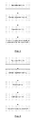
- Fig. 1 represents the data flow of the double scan procedure.
- Fig. 2 represents the data flow of the triple scan procedure.
- Fig. 3 represents an example of a double sided impression.
- Fig. 4 represents the step of taking a scan of the patient's dentition while he is wearing the impression.
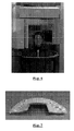
- Fig. 5 represents a scan setup for the high resolution scan of the impression.
- Fig. 6 represents a scan setup for the optional scan of the patient's maxillofacial complex with the occlusion that the doctor wants to examine.
- Fig. 7 represents an example of a wax-bite.
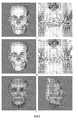
- Fig. 8 represents the result of an augmented model.
- Fig.8a shows the data of the patient scan.
- Fig.8b shows the data of the patient scan with the detailed surface of the dentition.
- Fig.8c shows the model of Fig. 8b with the textured skin surface.
- Fig. 9 represents the method to optimize the occlusion by repositioning of the jawbones.
- Fig. 10 represents the digital design of a splint based on the planning on the augmented model.
- An intermediate splint can be designed in a similar way.
- Fig. 11 represents the produced splint for the planning shown in Fig. 10 .
- Fig. 12 represents the produced splint for the planning shown in Fig. 10 .
- the proposed image acquisition protocol supports the generation of an augmented model possibly without the soft tissue deformation, without the need to position markers, in a fairly easy and realistic clinical setting.
- the usage of plaster casts of the patient's dentition is not necessary anymore.
- the planning protocol extends the anatomically relevant movements of bone fragments with occlusal planning optimization based on the augmented model.
- it includes soft tissue simulation and the design of an ideal 3D soft tissue profile.
- the patient can be imaged in two ways: without preservation of the soft tissues in natural rest position (the so-called double-scan procedure) (see Fig.1 ), or with preservation of the soft tissues in natural rest position (the so-called triple-scan procedure) (see Fig.2 ).
- an impression of the patient's dentition is produced (see (1) in Fig.1 ).
- this impression contains a double-sided impression. It contains the shape information of upper and lower dental arches.
- all dental impression materials such as alginate, silicone, ... among others
- wax-bites possibly mixed with barium-sulphate to increase the CT-number (i.e. the gray value)
- the impression materials can be applied in a double impression tray (see Fig.3 ). Wax-bites can be modelled on their own (see Fig.7 ).
- the gray values of the impression material are different from those of soft tissues so that the impression can be differentiated from the soft and hard tissues.
- it is important that the occlusion of the impression is well controlled and corresponds to the clinically desired occlusion for orthognathic surgery planning.
- step 1 in Fig.1 the step of impression taking is performed in the CT-scanner.
- the patient does not need to bite twice in the same impression (as it would be difficult to bite a second time in exactly the same way in the impression). It should be carefully checked that the impression completely covers the cuspids of all teeth.
- a second high-resolution scan of the impression alone is acquired (see step (4) in Fig.1 and the illustration in Fig.5 ).
- the impression should ideally show itself on the CT image data as if it was flying in the air.
- the impression can for example be positioned on a foam-like material, such as a sponge.
- the method as set out above comprises an additional step, typically carried out between the steps (2) and (4) of Fig.1 .
- Step (2) is thereby slightly modified.
- the data flow scheme of Fig.2 is then obtained. This is called the triple-scan procedure.
- step (1) the occlusion while biting into the impression material is not important and can be randomly chosen.
- step (2) also single sided impressions could be applied. However this would require an extra patient scan in a further step, which needs to be avoided as much as possible from a clinical point of view.
- Step 2 is a scan of the patient wearing the impression (see (2) in Fig.2 ).
- a very low dose scan of the dentition is enough (see Fig.4 ) given that this scan only yields intermediate results. This very low dose can be understood as less than 45 ⁇ Sv.
- the step of impression taking (Step 1 in Fig.2 ) is performed in the CT-scanner. In this case, the patient does not need to bite twice in the same impression. It should be carefully checked that the impression completely covers the cuspids of all teeth.
- the patient is scanned again, but now without the impression (see (3) in Fig.2 ).
- the region of interest of the head is scanned. This is typically the maxillofacial complex.
- Special attention is paid to the occlusion of the patient and to the facial expression.
- the occlusion needs to be the occlusion that the doctor wants to examine. This might be controlled by a physician directly during the scan. Alternatively, it can be controlled by a tiny wax bite guiding the patient into the right occlusion.
- the facial expression needs to be the expression that the doctor wants to analyse. Typically this is a central occlusion and a neutral, relaxed facial expression.
- the ideal skin surface based on the current skin surface of the patient can be designed.
- a method to design the ideal skin surface is based on clinical relevant parameters, including functional as well as aesthetic parameters, such as the degree of malocclusion or cephalometric measures, and/or body property parameters (such as body mass index, age, racial properties, gender, etc%) .
- Another method to design the ideal skin surface is based on the repositioning of points of the skin surface. These points can be important anatomical landmarks. Based on the new positions of these points, a new skin surface is computed.
- a statistical model based on an extensive database of skin surfaces on a large group of persons, is built.
- These skin surfaces can be acquired by 3D photography, or extracted from CT or MR imaging. The latter allows also including volumetric data into the statistical model. In this case the relation to the bony structures can also be included in the model.
- the model is parameterized according to said parameters.
- the initial skin surface is altered according to the changes of the parameter values.
- a statistical relevant skin surface is derived from the initial skin surface adapted according to the repositioned points.
- the ideal skin surface can be set as a target for surgery planning. In order to evaluate how close the surgical plan meets the ideal skin surface, the differences between both surfaces need to be visualised. This can be done by computing a distance map between both surfaces. Ideally, this distance map is based on the distance between anatomically related points. When the differences between both the ideal skin surface and the expected skin surface are minimal, a pleasing surgical plan is obtained.
- Bone fragments After virtual osteotomies the bone fragments need to be moved to the correct position. Bone fragments are moved with respect to the other skeletal features, to match a desirable occlusion and to obtain an acceptable facial skin surface. Typically, a bone fragment (e.g. the maxilla) or a group of bone fragments is moved to the appropriate position using anatomical relevant directions, reference planes and anatomical landmarks. Next, bone fragments are moved to optimize the dental occlusion. In the approach according to the present invention, the bone fragments are moved together by spring forces, taking into account the collisions. This technique is called solid body simulation. Finally, further adjustments of the position of bone fragments or groups thereof can be performed. While doing so, the soft tissue deformation can be simulated and compared with the ideal skin surface.
- a bone fragment e.g. the maxilla
- a group of bone fragments is moved to the appropriate position using anatomical relevant directions, reference planes and anatomical landmarks.
- bone fragments are moved to optimize the dental
- the planned occlusion can be transferred to the patient.
- the correct position of the bone fragments (like the maxilla) with respect to the skull base and the ascending ramus of the mandible with respect to the TMJ fossa can be transferred.
- a first method is to produce a surgical splint based on the digital planning data.
- the splint is designed from the planning data and produced by rapid prototyping techniques or milling techniques (see Figs. 10 , 11 and 12 ). This method transfers the correct occlusion.
- a second method is to produce a fixation structure that connects the upper and lower brackets or the archbars that are wired to the teeth, in order to create the planned occlusion. This method transfers the correct occlusion.
- a third method is to produce personalized osteosynthesis plates.
- the osteosynthesis plates are in the correct position and the bone fragments are fixed against these plates, the planned bone fragment positions are obtained during surgery.
- the plates are entered into the correct position based on anatomical features or based on bone anchors that are positioned before patient scanning and segmented from image data obtained from the patient+impression scan in the double-scan method or from the patient scan in the triple-scan method.
- this personalized part can be produced from the results of the planning software.
- this improved visualization of the teeth enables accurate occlusion planning and the production of intra- and postoperative tools.
Priority Applications (10)
| Application Number | Priority Date | Filing Date | Title |
|---|---|---|---|
| EP07106650A EP1982652A1 (fr) | 2007-04-20 | 2007-04-20 | Procédé pour dériver des informations sur la forme |
| KR1020097022978A KR101590330B1 (ko) | 2007-04-20 | 2008-04-18 | 형상 정보를 얻기 위한 방법 |
| PCT/EP2008/003135 WO2008128720A2 (fr) | 2007-04-20 | 2008-04-18 | Procédé de déduction d'information de forme |
| US12/596,829 US9439608B2 (en) | 2007-04-20 | 2008-04-18 | Method for deriving shape information |
| BRPI0810478A BRPI0810478B8 (pt) | 2007-04-20 | 2008-04-18 | método para a derivação de informações de formato e meio legível por computador |
| JP2010503417A JP2010524530A (ja) | 2007-04-20 | 2008-04-18 | 形状情報を引き出すための方法 |
| AU2008240993A AU2008240993B2 (en) | 2007-04-20 | 2008-04-18 | Method for deriving shape information |
| CN2008800205502A CN101720204B (zh) | 2007-04-20 | 2008-04-18 | 用于导出形状信息的方法 |
| EP08748990A EP2142094A2 (fr) | 2007-04-20 | 2008-04-18 | Procédé pour dériver des informations sur la forme |
| JP2014148747A JP5976730B2 (ja) | 2007-04-20 | 2014-07-22 | 形状情報を引き出すための方法 |
Applications Claiming Priority (1)
| Application Number | Priority Date | Filing Date | Title |
|---|---|---|---|
| EP07106650A EP1982652A1 (fr) | 2007-04-20 | 2007-04-20 | Procédé pour dériver des informations sur la forme |
Publications (1)
| Publication Number | Publication Date |
|---|---|
| EP1982652A1 true EP1982652A1 (fr) | 2008-10-22 |
Family
ID=38596062
Family Applications (2)
| Application Number | Title | Priority Date | Filing Date |
|---|---|---|---|
| EP07106650A Withdrawn EP1982652A1 (fr) | 2007-04-20 | 2007-04-20 | Procédé pour dériver des informations sur la forme |
| EP08748990A Ceased EP2142094A2 (fr) | 2007-04-20 | 2008-04-18 | Procédé pour dériver des informations sur la forme |
Family Applications After (1)
| Application Number | Title | Priority Date | Filing Date |
|---|---|---|---|
| EP08748990A Ceased EP2142094A2 (fr) | 2007-04-20 | 2008-04-18 | Procédé pour dériver des informations sur la forme |
Country Status (8)
| Country | Link |
|---|---|
| US (1) | US9439608B2 (fr) |
| EP (2) | EP1982652A1 (fr) |
| JP (2) | JP2010524530A (fr) |
| KR (1) | KR101590330B1 (fr) |
| CN (1) | CN101720204B (fr) |
| AU (1) | AU2008240993B2 (fr) |
| BR (1) | BRPI0810478B8 (fr) |
| WO (1) | WO2008128720A2 (fr) |
Cited By (6)
| Publication number | Priority date | Publication date | Assignee | Title |
|---|---|---|---|---|
| WO2012016635A2 (fr) | 2010-08-04 | 2012-02-09 | Charité - Universitätsmedizin Berlin | Procédé pour générer une topologie dentaire numérique pour une structure dentaire ainsi que procédé de mesure |
| CN102988114A (zh) * | 2011-09-15 | 2013-03-27 | 浙江大学医学院附属口腔医院 | 一种基于锥束ct数据的国产数字化手术导板制作的方法 |
| WO2013173100A1 (fr) * | 2012-05-16 | 2013-11-21 | 3M Innovative Properties Company | Dispositif dentaire pour administrer une substance de soin buccal à des surfaces buccales |
| WO2015169910A1 (fr) * | 2014-05-09 | 2015-11-12 | 3Shape A/S | Balayage de patients edentes |
| US9380986B2 (en) | 2012-06-11 | 2016-07-05 | Planmeca Oy | Dental surface models |
| WO2020180763A1 (fr) * | 2019-03-04 | 2020-09-10 | Open Technologies S.R.L. | Système de balayage et de visualisation dentaire |
Families Citing this family (39)
| Publication number | Priority date | Publication date | Assignee | Title |
|---|---|---|---|---|
| US10022202B2 (en) | 2013-03-15 | 2018-07-17 | Triagenics, Llc | Therapeutic tooth bud ablation |
| WO2010132368A1 (fr) | 2009-05-11 | 2010-11-18 | Colby Leigh E | Ablation thérapeutique de bourgeon dentaire |
| WO2014143014A1 (fr) | 2013-03-15 | 2014-09-18 | Triagenics, Llc | Ablation thérapeutique de bourgeon dentaire |
| EP2306400B1 (fr) | 2009-09-04 | 2015-02-11 | Medicim NV | Procédé de numérisation d'objets dento-maxillo-facial |
| EP2368498A1 (fr) | 2010-03-26 | 2011-09-28 | Stichting voor de Technische Wetenschappen | Procédé pour dériver des informations sur la forme de la dentition d'une personne |
| DE102011012460A1 (de) * | 2011-02-25 | 2012-08-30 | Hicat Gmbh | Chirurgisches Instrument mit integrierter Navigationskontrolle |
| FI20110106L (fi) * | 2011-03-21 | 2012-04-13 | Planmeca Oy | Hammaslääketieteellinen kuvauslaitteisto |
| GB201115265D0 (en) | 2011-09-05 | 2011-10-19 | Materialise Dental Nv | A method and system for 3d root canal treatment planning |
| KR101299456B1 (ko) * | 2012-01-03 | 2013-08-29 | 서울대학교산학협력단 | 무치악 또는 편측무치악 파노라마 바이트 |
| GB201216230D0 (en) | 2012-09-12 | 2012-10-24 | Nobel Biocare Services Ag | An improved surgical template |
| GB201216224D0 (en) | 2012-09-12 | 2012-10-24 | Nobel Biocare Services Ag | An improved virtual splint |
| GB201216214D0 (en) | 2012-09-12 | 2012-10-24 | Nobel Biocare Services Ag | A digital splint |
| US9438264B1 (en) | 2015-09-10 | 2016-09-06 | Realtek Semiconductor Corp. | High-speed capacitive digital-to-analog converter and method thereof |
| EP3009097A1 (fr) * | 2014-10-17 | 2016-04-20 | Imactis | Procédé de navigation d'un instrument chirurgical |
| US9629698B2 (en) | 2014-11-04 | 2017-04-25 | James R. Glidewell Dental Ceramics, Inc. | Method and apparatus for generation of 3D models with applications in dental restoration design |
| US10521969B2 (en) * | 2015-02-23 | 2019-12-31 | Osstemimplant Co., Ltd. | Method for simulating mandibular movement, device for same and recording medium for recording same |
| WO2016197326A1 (fr) * | 2015-06-09 | 2016-12-15 | 佘承鑫 | Système et méthode de conception et de correction d'image pour une chirurgie orale et maxillo-faciale |
| DE102015118853A1 (de) * | 2015-11-03 | 2017-05-04 | Technische Universität Dresden | Verfahren zur Herstellung eines Distraktors zur skelettalen Befestigung an einem Kiefer |
| CN106806030B (zh) * | 2015-11-30 | 2018-08-10 | 北京大学口腔医学院 | 一种冠根三维模型融合方法 |
| EP3487439A4 (fr) * | 2016-07-22 | 2020-04-08 | Prosomnus Sleep Technologies, Inc. | Matrice de conception assistée par ordinateur pour la fabrication de dispositifs dentaires |
| US11559378B2 (en) | 2016-11-17 | 2023-01-24 | James R. Glidewell Dental Ceramics, Inc. | Scanning dental impressions |
| ES2683085B2 (es) * | 2017-02-24 | 2019-04-25 | Dmr Dental S L | Procedimiento de escaneo de un rostro humano para una alineacion entre el rostro y los dientes de una persona y conjunto de marcadores para su ejecucion |
| KR101901646B1 (ko) * | 2017-03-07 | 2018-10-01 | 주식회사 메가젠임플란트 | 3차원 하이브리드 영상 구축 프로그램을 이용한 악교정 시스템 |
| USD843417S1 (en) | 2017-04-19 | 2019-03-19 | Navix International Limited | Display screen or portion thereof with icon |
| GB201708520D0 (en) | 2017-05-27 | 2017-07-12 | Dawood Andrew | A method for reducing artefact in intra oral scans |
| DE102017115750A1 (de) * | 2017-07-13 | 2019-01-17 | Karl Leibinger Medizintechnik Gmbh & Co. Kg | Knochenpositioniervorrichtung für die Dysgnathie mit Trennwerkzeugführungsvorrichtung / -abschnitt |
| AU2018323052A1 (en) * | 2017-08-30 | 2020-03-19 | Zst Holdings, Inc. | Apparatus and method for registration of a digital dental bite |
| KR102061644B1 (ko) * | 2017-12-19 | 2020-02-11 | 주식회사 키스톤 | 임플란트 진단용 영상 생성 시스템 및 그 생성방법 |
| US10779917B2 (en) * | 2018-02-20 | 2020-09-22 | Ivoclar Vivadent Ag | Computer implemented method for modifying a digital three-dimensional model of a dentition |
| EP3547262A1 (fr) * | 2018-03-28 | 2019-10-02 | Koninklijke Philips N.V. | Reconstruction d'images tomographiques à rayons x |
| JP7374193B2 (ja) | 2018-12-20 | 2023-11-06 | メディシム ナームロゼ ベンノートチャップ | 表面メッシュの自動トリミング |
| JP2022515432A (ja) * | 2018-12-26 | 2022-02-18 | スリーエム イノベイティブ プロパティズ カンパニー | デジタルメッシュオブジェクト間の衝突を自動的に除去し、メッシュオブジェクトを空間的配置間で滑らかに移動させる方法 |
| EP3979938A4 (fr) | 2019-06-06 | 2023-06-28 | TriAgenics, Inc. | Systèmes de sonde d'ablation |
| US11534271B2 (en) | 2019-06-25 | 2022-12-27 | James R. Glidewell Dental Ceramics, Inc. | Processing CT scan of dental impression |
| US11540906B2 (en) | 2019-06-25 | 2023-01-03 | James R. Glidewell Dental Ceramics, Inc. | Processing digital dental impression |
| US11622843B2 (en) | 2019-06-25 | 2023-04-11 | James R. Glidewell Dental Ceramics, Inc. | Processing digital dental impression |
| US10898298B1 (en) | 2020-04-08 | 2021-01-26 | Oxilio Ltd | Systems and methods for determining orthodontic treatment |
| KR102388411B1 (ko) * | 2020-04-27 | 2022-04-19 | 오스템임플란트 주식회사 | 트레이 제조방법, 데이터 이전 방법 및 이를 수행하는 시뮬레이션 장치 |
| US11544846B2 (en) | 2020-08-27 | 2023-01-03 | James R. Glidewell Dental Ceramics, Inc. | Out-of-view CT scan detection |
Citations (7)
| Publication number | Priority date | Publication date | Assignee | Title |
|---|---|---|---|---|
| DE3522196A1 (de) * | 1985-06-21 | 1986-02-20 | Martin Wilhelm Dr. Dr. 6072 Dreieich Happel | Verfahren zur fertigung von gelenkendoprothesen und osteosynthesematerial oder anderem knochenersatz nach den knochendimensionen des patienten |
| WO1999059106A1 (fr) * | 1998-05-13 | 1999-11-18 | Acuscape International, Inc. | Procede pour generer des modeles en 3d a partir d'images medicales |
| US6120289A (en) * | 1999-03-24 | 2000-09-19 | 3M Innovative Properties Company | Orthodontic attachment device for interarch appliances |
| WO2003028577A2 (fr) | 2001-10-03 | 2003-04-10 | Board Of Regents, The University Of Texas System | Appareil et procede pour fabriquer des gouttieres chirurgicales orthodontiques |
| US20040170941A1 (en) * | 2000-04-25 | 2004-09-02 | Align Technology, Inc. | Embedded features and methods of a dental appliance |
| US20040197740A1 (en) * | 2001-05-07 | 2004-10-07 | Benaddi Amar | Method for obtaining an impression for producing a dental prosthesis and instrumentation architecture therefor |
| WO2006000063A1 (fr) | 2004-06-25 | 2006-01-05 | Medicim Nv | Procede permettant d'obtenir un plan de traitement de chirurgie orthognatique les dispositifs a cet effet |
Family Cites Families (121)
| Publication number | Priority date | Publication date | Assignee | Title |
|---|---|---|---|---|
| US4155163A (en) * | 1977-01-13 | 1979-05-22 | Robert Schwartz | Relator assembly |
| US4836778A (en) | 1987-05-26 | 1989-06-06 | Vexcel Corporation | Mandibular motion monitoring system |
| US5066231A (en) | 1990-02-23 | 1991-11-19 | Minnesota Mining And Manufacturing Company | Dental impression process using polycaprolactone molding composition |
| WO1994010935A1 (fr) | 1992-11-09 | 1994-05-26 | Ormco Corporation | Procede et appareil de formation d'appareils orthodondiques personnalises |
| US5338198A (en) | 1993-11-22 | 1994-08-16 | Dacim Laboratory Inc. | Dental modeling simulator |
| SE502035C2 (sv) | 1993-12-06 | 1995-07-24 | Nobelpharma Ab | Metod och och anordning för framtagning av information för framställning av artifiella stödorgan eller ersättningsdelar till människokroppen |
| SE502427C2 (sv) | 1994-02-18 | 1995-10-16 | Nobelpharma Ab | Metod och anordning utnyttjande artikulator och datorutrustning |
| JP3727660B2 (ja) | 1995-07-21 | 2005-12-14 | カデント・リミテッド | 3次元の歯の像を入手するための方法 |
| US6551611B2 (en) | 1995-09-28 | 2003-04-22 | Schering Aktiengesellschaft | Hormone replacement therapy method |
| JPH1075963A (ja) | 1996-09-06 | 1998-03-24 | Nikon Corp | 歯科補綴物モデルの設計方法およびこの方法を実行するプログラムを記録した媒体 |
| US5690631A (en) * | 1996-09-11 | 1997-11-25 | Walter Lorenz Surgical, Inc. | Multi-configurable plating system |
| AUPO280996A0 (en) | 1996-10-04 | 1996-10-31 | Dentech Investments Pty Ltd | Creation and utilization of 3D teeth models |
| US6217334B1 (en) | 1997-01-28 | 2001-04-17 | Iris Development Corporation | Dental scanning method and apparatus |
| SE509142C2 (sv) | 1997-04-10 | 1998-12-07 | Nobel Biocare Ab | Anordning och förfarande för att återskapa en modell för dental produkt eller verktyg till produkten |
| US6450807B1 (en) | 1997-06-20 | 2002-09-17 | Align Technology, Inc. | System and method for positioning teeth |
| US5975893A (en) | 1997-06-20 | 1999-11-02 | Align Technology, Inc. | Method and system for incrementally moving teeth |
| US7247021B2 (en) | 1997-06-20 | 2007-07-24 | Align Technology, Inc. | Subdividing a digital dentition model |
| US8496474B2 (en) | 1997-06-20 | 2013-07-30 | Align Technology, Inc. | Computer automated development of an orthodontic treatment plan and appliance |
| AU744385B2 (en) | 1997-06-20 | 2002-02-21 | Align Technology, Inc. | Method and system for incrementally moving teeth |
| US6471511B1 (en) | 1997-06-20 | 2002-10-29 | Align Technology, Inc. | Defining tooth-moving appliances computationally |
| US6152731A (en) | 1997-09-22 | 2000-11-28 | 3M Innovative Properties Company | Methods for use in dental articulation |
| US9084653B2 (en) | 1998-01-14 | 2015-07-21 | Cadent, Ltd. | Methods for use in dental articulation |
| SE512083C2 (sv) | 1998-05-29 | 2000-01-24 | Nobel Biocare Ab | Metod att producera dental första påbyggnadsdel till implantat eller annan påbyggnadsdel samt hållare för modell av den första påbyggnadsdelen |
| IL125659A (en) | 1998-08-05 | 2002-09-12 | Cadent Ltd | Method and device for three-dimensional simulation of a structure |
| US11026768B2 (en) | 1998-10-08 | 2021-06-08 | Align Technology, Inc. | Dental appliance reinforcement |
| US6802713B1 (en) | 1998-10-08 | 2004-10-12 | Align Technology, Inc. | Defining tooth-moving appliances computationally |
| US6227850B1 (en) | 1999-05-13 | 2001-05-08 | Align Technology, Inc. | Teeth viewing system |
| EP1119309B1 (fr) | 1998-10-08 | 2016-06-01 | Align Technology, Inc. | Developpement informatique automatise d'un plan de traitement orthodontique et appareil prevu a cet effet |
| US6406292B1 (en) | 1999-05-13 | 2002-06-18 | Align Technology, Inc. | System for determining final position of teeth |
| AU3954200A (en) * | 1999-05-03 | 2000-11-17 | Medartis Ag | Blockable bone plate |
| US6318994B1 (en) | 1999-05-13 | 2001-11-20 | Align Technology, Inc | Tooth path treatment plan |
| US6602070B2 (en) | 1999-05-13 | 2003-08-05 | Align Technology, Inc. | Systems and methods for dental treatment planning |
| DE29909025U1 (de) * | 1999-05-25 | 1999-11-04 | Lipat Consulting Ag Basel | Osteosynthetische Knochenplatte |
| WO2001001854A2 (fr) * | 1999-07-02 | 2001-01-11 | Hypermed Imaging, Inc. | Dispositif d'imagerie a modalite integree |
| CA2324090A1 (fr) | 1999-10-25 | 2001-04-25 | Leviton Manufacturing Co., Inc. | Dispositif de connexion avec differents types de raccord de fils |
| US7160110B2 (en) | 1999-11-30 | 2007-01-09 | Orametrix, Inc. | Three-dimensional occlusal and interproximal contact detection and display using virtual tooth models |
| US6463344B1 (en) | 2000-02-17 | 2002-10-08 | Align Technology, Inc. | Efficient data representation of teeth model |
| US7373286B2 (en) | 2000-02-17 | 2008-05-13 | Align Technology, Inc. | Efficient data representation of teeth model |
| US6633789B1 (en) | 2000-02-17 | 2003-10-14 | Align Technology, Inc. | Effiicient data representation of teeth model |
| US6371761B1 (en) | 2000-03-30 | 2002-04-16 | Align Technology, Inc. | Flexible plane for separating teeth models |
| WO2001074268A1 (fr) | 2000-03-30 | 2001-10-11 | Align Technology, Inc. | Systeme et procede de separation de modeles tridimensionnels |
| US6582229B1 (en) | 2000-04-25 | 2003-06-24 | Align Technology, Inc. | Methods for modeling bite registration |
| US7092784B1 (en) | 2000-07-28 | 2006-08-15 | Align Technology | Systems and methods for forming an object |
| US7040896B2 (en) | 2000-08-16 | 2006-05-09 | Align Technology, Inc. | Systems and methods for removing gingiva from computer tooth models |
| US6386878B1 (en) | 2000-08-16 | 2002-05-14 | Align Technology, Inc. | Systems and methods for removing gingiva from teeth |
| US6726478B1 (en) | 2000-10-30 | 2004-04-27 | Align Technology, Inc. | Systems and methods for bite-setting teeth models |
| US7736147B2 (en) | 2000-10-30 | 2010-06-15 | Align Technology, Inc. | Systems and methods for bite-setting teeth models |
| US20020094509A1 (en) | 2000-11-30 | 2002-07-18 | Duane Durbin | Method and system for digital occlusal determination |
| US6783360B2 (en) | 2000-12-13 | 2004-08-31 | Align Technology, Inc. | Systems and methods for positioning teeth |
| US6579095B2 (en) | 2000-12-22 | 2003-06-17 | Geodigm Corporation | Mating parts scanning and registration methods |
| US7074038B1 (en) | 2000-12-29 | 2006-07-11 | Align Technology, Inc. | Methods and systems for treating teeth |
| KR100419380B1 (ko) * | 2001-03-08 | 2004-02-19 | 김정만 | 컴퓨터를 이용한 치열 교정기의 제조 방법 |
| US7362890B2 (en) | 2001-05-24 | 2008-04-22 | Astra Tech Inc. | Registration of 3-D imaging of 3-D objects |
| CA2350849A1 (fr) | 2001-06-15 | 2002-12-15 | Dentalmatic Technologies Inc. | Articulateur virtuel |
| DE60219031T2 (de) | 2001-08-31 | 2007-12-13 | Cynovad, Inc., St. Laurent | Verfahren zur herstellung von giessformen |
| WO2003049635A2 (fr) | 2001-12-07 | 2003-06-19 | Diesso Michael | Procede et dispositif d'impression dentaire |
| SE520765C2 (sv) | 2001-12-28 | 2003-08-19 | Nobel Biocare Ab | Anordning och arrangemang för att medelst mall ta upp hål till implantat i ben, företrädesvis käkben |
| US6767208B2 (en) | 2002-01-10 | 2004-07-27 | Align Technology, Inc. | System and method for positioning teeth |
| US7252507B2 (en) | 2002-03-15 | 2007-08-07 | Tesini David A | Thermoplastic wafer for a dental impression for identification purposes |
| US20030207227A1 (en) | 2002-05-02 | 2003-11-06 | Align Technology, Inc. | Systems and methods for treating patients |
| US7778686B2 (en) | 2002-06-04 | 2010-08-17 | General Electric Company | Method and apparatus for medical intervention procedure planning and location and navigation of an intervention tool |
| US7255558B2 (en) | 2002-06-18 | 2007-08-14 | Cadent, Ltd. | Dental imaging instrument having air stream auxiliary |
| US6979196B2 (en) | 2002-06-21 | 2005-12-27 | Align Technology, Inc. | Systems and methods for automated bite-setting of tooth models |
| US20040243361A1 (en) | 2002-08-19 | 2004-12-02 | Align Technology, Inc. | Systems and methods for providing mass customization |
| US7156661B2 (en) | 2002-08-22 | 2007-01-02 | Align Technology, Inc. | Systems and methods for treatment analysis by teeth matching |
| AU2003274467A1 (en) | 2002-11-18 | 2004-06-15 | Koninklijke Philips Electronics N.V. | Method and device for image registration |
| DE20221002U1 (de) * | 2002-12-12 | 2004-12-16 | Kettenbach Gmbh & Co. Kg | Additionsvernetzende Zweikomponenten-Siliconmaterialien mit hoher Shore D-Härte |
| US7778490B2 (en) | 2003-01-13 | 2010-08-17 | Koninklijke Philips Electronics N.V. | Method of image registration and medical image data processing apparatus |
| US7030383B2 (en) | 2003-08-04 | 2006-04-18 | Cadent Ltd. | Speckle reduction method and apparatus |
| US7202466B2 (en) | 2003-08-25 | 2007-04-10 | Cadent Ltd. | Apparatus and method for providing high intensity non-coherent light and for speckle reduction |
| US20050058602A1 (en) * | 2003-09-11 | 2005-03-17 | The Procter & Gamble Company | Dye composition and method for detection of demineralized lesions in teeth |
| US20050106529A1 (en) | 2003-11-19 | 2005-05-19 | Align Technology, Inc. | Dental impression tray with detachable portions |
| US7361020B2 (en) | 2003-11-19 | 2008-04-22 | Align Technology, Inc. | Dental tray containing radiopaque materials |
| FR2863086B1 (fr) | 2003-11-28 | 2006-03-10 | Ge Med Sys Global Tech Co Llc | Procede de production d'une sequence d'images volumiques d'une zone d'un organe d'un etre vivant. |
| US7145661B2 (en) | 2003-12-31 | 2006-12-05 | Carl Zeiss Meditec, Inc. | Efficient optical coherence tomography (OCT) system and method for rapid imaging in three dimensions |
| US7118375B2 (en) | 2004-01-08 | 2006-10-10 | Duane Milford Durbin | Method and system for dental model occlusal determination using a replicate bite registration impression |
| US7536234B2 (en) | 2004-02-09 | 2009-05-19 | Cadent Ltd. | Method and system for manufacturing a dental prosthesis |
| JP2005261440A (ja) | 2004-02-19 | 2005-09-29 | Aze Ltd | 医療用画像の被造影領域抽出方法 |
| US7333874B2 (en) | 2004-02-24 | 2008-02-19 | Cadent Ltd. | Method and system for designing and producing dental prostheses and appliances |
| US20050186524A1 (en) | 2004-02-24 | 2005-08-25 | Align Technology, Inc. | Arch expander |
| US7970627B2 (en) | 2004-02-27 | 2011-06-28 | Align Technology, Inc. | Method and system for providing dynamic orthodontic assessment and treatment profiles |
| US9492245B2 (en) | 2004-02-27 | 2016-11-15 | Align Technology, Inc. | Method and system for providing dynamic orthodontic assessment and treatment profiles |
| US7970628B2 (en) | 2004-02-27 | 2011-06-28 | Align Technology, Inc. | Method and system for providing dynamic orthodontic assessment and treatment profiles |
| US7930189B2 (en) | 2004-02-27 | 2011-04-19 | Align Technology, Inc. | Method and system for providing dynamic orthodontic assessment and treatment profiles |
| US8874452B2 (en) | 2004-02-27 | 2014-10-28 | Align Technology, Inc. | Method and system for providing dynamic orthodontic assessment and treatment profiles |
| US8126726B2 (en) * | 2004-02-27 | 2012-02-28 | Align Technology, Inc. | System and method for facilitating automated dental measurements and diagnostics |
| US7637740B2 (en) | 2004-02-27 | 2009-12-29 | Align Technology, Inc. | Systems and methods for temporally staging teeth |
| US7987099B2 (en) | 2004-02-27 | 2011-07-26 | Align Technology, Inc. | Dental data mining |
| US7880751B2 (en) | 2004-02-27 | 2011-02-01 | Align Technology, Inc. | Method and system for providing dynamic orthodontic assessment and treatment profiles |
| US7241142B2 (en) | 2004-03-19 | 2007-07-10 | Align Technology, Inc. | Root-based tooth moving sequencing |
| WO2005115266A2 (fr) | 2004-05-24 | 2005-12-08 | Great Lakes Orthodontics, Ltd. | Fabrication numerique de dispositifs oraux amovibles |
| DE602005009432D1 (de) | 2004-06-17 | 2008-10-16 | Cadent Ltd | Verfahren und Gerät zur Farbbildformung einer dreidimensionalen Struktur |
| SE527666C2 (sv) | 2004-09-30 | 2006-05-02 | Nobel Biocare Ab | Anordning vid skanner |
| US7357634B2 (en) | 2004-11-05 | 2008-04-15 | Align Technology, Inc. | Systems and methods for substituting virtual dental appliances |
| US20060127852A1 (en) | 2004-12-14 | 2006-06-15 | Huafeng Wen | Image based orthodontic treatment viewing system |
| US7442040B2 (en) | 2005-01-13 | 2008-10-28 | Align Technology, Inc. | Template for veneer application |
| US20080038684A1 (en) | 2005-01-27 | 2008-02-14 | Scott Keating | Systems and Processes for Computationally Setting Bite Alignment |
| EP1869403B1 (fr) | 2005-03-03 | 2017-06-14 | Align Technology, Inc. | Systeme et procede pour l'exploration d'une cavite intrabuccale |
| WO2007011306A2 (fr) | 2005-07-20 | 2007-01-25 | Bracco Imaging S.P.A. | Procede et appareil destines a mapper un modele virtuel d'un objet sur l'objet |
| GB0507204D0 (en) | 2005-04-08 | 2005-05-18 | Leuven K U Res & Dev | Maxillofacial and plastic surgery |
| US20060275736A1 (en) | 2005-04-22 | 2006-12-07 | Orthoclear Holdings, Inc. | Computer aided orthodontic treatment planning |
| US20060275731A1 (en) | 2005-04-29 | 2006-12-07 | Orthoclear Holdings, Inc. | Treatment of teeth by aligners |
| US7476100B2 (en) | 2005-05-17 | 2009-01-13 | Align Technology, Inc. | Guide apparatus and methods for making tooth positioning appliances |
| US7555403B2 (en) | 2005-07-15 | 2009-06-30 | Cadent Ltd. | Method for manipulating a dental virtual model, method for creating physical entities based on a dental virtual model thus manipulated, and dental models thus created |
| US20070031791A1 (en) | 2005-08-03 | 2007-02-08 | 3M Innovative Properties Company | Scanning models for digital orthodontics |
| JP2007061592A (ja) * | 2005-08-04 | 2007-03-15 | Gc Corp | 歯顎の三次元形状データの作成方法及び歯科用cad/cam用三次元形状データの作成方法 |
| WO2007026598A1 (fr) | 2005-08-31 | 2007-03-08 | Gifu University | Processeur d’images médicales et procédé de traitement d’images |
| US7698014B2 (en) | 2006-01-20 | 2010-04-13 | 3M Innovative Properties Company | Local enforcement of accuracy in fabricated models |
| US8366442B2 (en) * | 2006-02-15 | 2013-02-05 | Bankruptcy Estate Of Voxelogix Corporation | Dental apparatus for radiographic and non-radiographic imaging |
| WO2008030965A2 (fr) * | 2006-09-06 | 2008-03-13 | Voxelogix Corporation | Procédé pour la conception virtuelle et la fabrication informatique de dispositifs intra-buccaux |
| US7916911B2 (en) | 2007-02-26 | 2011-03-29 | Align Technology, Inc. | System and method for digital tooth imaging |
| US20080288289A1 (en) | 2007-05-14 | 2008-11-20 | Align Technology, Inc. | Method and system for efficient orthodontic treatment information management |
| US20080306724A1 (en) | 2007-06-08 | 2008-12-11 | Align Technology, Inc. | Treatment planning and progress tracking systems and methods |
| US9060829B2 (en) | 2007-06-08 | 2015-06-23 | Align Technology, Inc. | Systems and method for management and delivery of orthodontic treatment |
| US8562338B2 (en) | 2007-06-08 | 2013-10-22 | Align Technology, Inc. | Treatment progress tracking and recalibration |
| US7865259B2 (en) | 2007-12-06 | 2011-01-04 | Align Technology, Inc. | System and method for improved dental geometry representation |
| US8899977B2 (en) | 2008-01-29 | 2014-12-02 | Align Technology, Inc. | Orthodontic repositioning appliances having improved geometry, methods and systems |
| US8439672B2 (en) | 2008-01-29 | 2013-05-14 | Align Technology, Inc. | Method and system for optimizing dental aligner geometry |
| US8108189B2 (en) | 2008-03-25 | 2012-01-31 | Align Technologies, Inc. | Reconstruction of non-visible part of tooth |
| WO2009140582A2 (fr) * | 2008-05-16 | 2009-11-19 | Geodigm Corporation | Procédé et appareil permettant de combiner des images de scanner dentaire 3d avec d'autres ensembles de données 3d |
| EP2368498A1 (fr) * | 2010-03-26 | 2011-09-28 | Stichting voor de Technische Wetenschappen | Procédé pour dériver des informations sur la forme de la dentition d'une personne |
-
2007
- 2007-04-20 EP EP07106650A patent/EP1982652A1/fr not_active Withdrawn
-
2008
- 2008-04-18 EP EP08748990A patent/EP2142094A2/fr not_active Ceased
- 2008-04-18 AU AU2008240993A patent/AU2008240993B2/en not_active Ceased
- 2008-04-18 US US12/596,829 patent/US9439608B2/en active Active
- 2008-04-18 BR BRPI0810478A patent/BRPI0810478B8/pt not_active IP Right Cessation
- 2008-04-18 KR KR1020097022978A patent/KR101590330B1/ko active IP Right Grant
- 2008-04-18 JP JP2010503417A patent/JP2010524530A/ja active Pending
- 2008-04-18 WO PCT/EP2008/003135 patent/WO2008128720A2/fr active Application Filing
- 2008-04-18 CN CN2008800205502A patent/CN101720204B/zh active Active
-
2014
- 2014-07-22 JP JP2014148747A patent/JP5976730B2/ja active Active
Patent Citations (7)
| Publication number | Priority date | Publication date | Assignee | Title |
|---|---|---|---|---|
| DE3522196A1 (de) * | 1985-06-21 | 1986-02-20 | Martin Wilhelm Dr. Dr. 6072 Dreieich Happel | Verfahren zur fertigung von gelenkendoprothesen und osteosynthesematerial oder anderem knochenersatz nach den knochendimensionen des patienten |
| WO1999059106A1 (fr) * | 1998-05-13 | 1999-11-18 | Acuscape International, Inc. | Procede pour generer des modeles en 3d a partir d'images medicales |
| US6120289A (en) * | 1999-03-24 | 2000-09-19 | 3M Innovative Properties Company | Orthodontic attachment device for interarch appliances |
| US20040170941A1 (en) * | 2000-04-25 | 2004-09-02 | Align Technology, Inc. | Embedded features and methods of a dental appliance |
| US20040197740A1 (en) * | 2001-05-07 | 2004-10-07 | Benaddi Amar | Method for obtaining an impression for producing a dental prosthesis and instrumentation architecture therefor |
| WO2003028577A2 (fr) | 2001-10-03 | 2003-04-10 | Board Of Regents, The University Of Texas System | Appareil et procede pour fabriquer des gouttieres chirurgicales orthodontiques |
| WO2006000063A1 (fr) | 2004-06-25 | 2006-01-05 | Medicim Nv | Procede permettant d'obtenir un plan de traitement de chirurgie orthognatique les dispositifs a cet effet |
Non-Patent Citations (2)
| Title |
|---|
| FILIP SCHUTYSER ET AL: "Robust Visualization of the Dental Occlusion by a Double Scan Procedure", MEDICAL IMAGE COMPUTING AND COMPUTER-ASSISTED INTERVENTION - MICCAI 2005 LECTURE NOTES IN COMPUTER SCIENCE;;LNCS, SPRINGER-VERLAG, BE, vol. 3749, 2005, pages 368 - 374, XP019021655, ISBN: 3-540-29327-2 * |
| VERSTREKEN E A: "A DOUBLE SCANNING PROCEDURE FOR VISUALISATION OF RADIOLUCENT OBJECTS I SOFT TISSUES: APPLICATION TO ORAL IMPLANT SURGERY PLANNING", MEDICAL IMAGE COMPUTING AND COMPUTER-ASSISTED INTERVENTION. MICCAI. INTERNATIONAL CONFERENCE. PROCEEDINGS, 11 October 1998 (1998-10-11), pages 985 - 995, XP002103535 * |
Cited By (9)
| Publication number | Priority date | Publication date | Assignee | Title |
|---|---|---|---|---|
| WO2012016635A2 (fr) | 2010-08-04 | 2012-02-09 | Charité - Universitätsmedizin Berlin | Procédé pour générer une topologie dentaire numérique pour une structure dentaire ainsi que procédé de mesure |
| DE102010036841A1 (de) | 2010-08-04 | 2012-02-09 | Charité - Universitätsmedizin Berlin | Verfahren zum Erzeugen einer digitalen Zahntopologie für eine Zahnstruktur sowie Messverfahren |
| CN102988114A (zh) * | 2011-09-15 | 2013-03-27 | 浙江大学医学院附属口腔医院 | 一种基于锥束ct数据的国产数字化手术导板制作的方法 |
| WO2013173100A1 (fr) * | 2012-05-16 | 2013-11-21 | 3M Innovative Properties Company | Dispositif dentaire pour administrer une substance de soin buccal à des surfaces buccales |
| US9380986B2 (en) | 2012-06-11 | 2016-07-05 | Planmeca Oy | Dental surface models |
| WO2015169910A1 (fr) * | 2014-05-09 | 2015-11-12 | 3Shape A/S | Balayage de patients edentes |
| US10456229B2 (en) | 2014-05-09 | 2019-10-29 | 3Shape A/S | Scanning of edentulous patients |
| US11478344B2 (en) | 2014-05-09 | 2022-10-25 | 3Shape A/S | Scanning of edentulous patients |
| WO2020180763A1 (fr) * | 2019-03-04 | 2020-09-10 | Open Technologies S.R.L. | Système de balayage et de visualisation dentaire |
Also Published As
| Publication number | Publication date |
|---|---|
| AU2008240993B2 (en) | 2013-10-03 |
| CN101720204B (zh) | 2013-11-06 |
| JP5976730B2 (ja) | 2016-08-24 |
| BRPI0810478B8 (pt) | 2021-06-22 |
| JP2014237005A (ja) | 2014-12-18 |
| BRPI0810478B1 (pt) | 2019-11-26 |
| EP2142094A2 (fr) | 2010-01-13 |
| WO2008128720A3 (fr) | 2009-03-19 |
| US20110059413A1 (en) | 2011-03-10 |
| BRPI0810478A2 (pt) | 2014-11-11 |
| US9439608B2 (en) | 2016-09-13 |
| KR101590330B1 (ko) | 2016-02-01 |
| AU2008240993A1 (en) | 2008-10-30 |
| KR20100016180A (ko) | 2010-02-12 |
| JP2010524530A (ja) | 2010-07-22 |
| CN101720204A (zh) | 2010-06-02 |
| WO2008128720A2 (fr) | 2008-10-30 |
Similar Documents
| Publication | Publication Date | Title |
|---|---|---|
| AU2008240993B2 (en) | Method for deriving shape information | |
| Xia et al. | Algorithm for planning a double-jaw orthognathic surgery using a computer-aided surgical simulation (CASS) protocol. Part 1: planning sequence | |
| Bobek et al. | Virtual surgical planning for orthognathic surgery using digital data transfer and an intraoral fiducial marker: the charlotte method | |
| Zinser et al. | Computer-assisted orthognathic surgery: feasibility study using multiple CAD/CAM surgical splints | |
| Vercruyssen et al. | Computer‐supported implant planning and guided surgery: a narrative review | |
| Uechi et al. | A novel method for the 3-dimensional simulation of orthognathic surgery by using a multimodal image-fusion technique | |
| Hsu et al. | Accuracy of a computer-aided surgical simulation protocol for orthognathic surgery: a prospective multicenter study | |
| JP5020816B2 (ja) | 頬顎異常矯正学的手術のための治療計画を導出する方法及びそのための装置 | |
| Wu et al. | Postoperative outcomes of two-and three-dimensional planning in orthognathic surgery: a comparative study | |
| AU2008256518B2 (en) | Method and system for dental planning | |
| Centenero et al. | 3D planning in orthognathic surgery: CAD/CAM surgical splints and prediction of the soft and hard tissues results–our experience in 16 cases | |
| Kang et al. | Verification of the usability of a navigation method in dental implant surgery: in vitro comparison with the stereolithographic surgical guide template method | |
| Chang et al. | Accuracy assessment of computer-aided three-dimensional simulation and navigation in orthognathic surgery (CASNOS) | |
| Pektas et al. | The accuracy of computer‐assisted surgical planning in soft tissue prediction following orthognathic surgery | |
| Wang et al. | Design and manufacture of dental-supported surgical guide for genioplasty | |
| Di Blasio et al. | Virtual planning of a complex three-part bimaxillary osteotomy | |
| Patel et al. | Surgical planning: 2D to 3D | |
| Edwards et al. | Applications of Cone Beam Computed Tomography to Orthognathic Surgery Treatment Planning | |
| Orhan et al. | Digital Craniofacial and Orthognathic Surgery | |
| Paoli et al. | A CAD-based methodology for dental implant surgery | |
| Wang et al. | The application of digital model surgery in the treatment of dento-maxillofacial deformities | |
| Hungate | The creation of a novel full-coverage orthognathic surgical splint utilizing 3D printing & virtual surgical planning | |
| Di Blasio et al. | Case Report Virtual Planning of a Complex Three-Part Bimaxillary Osteotomy | |
| Nichelini | Virtual Orthognathic Surgery: CAD/CAM Splint Generation and Analysis | |
| Tetteh-Martey | Maxillary Protraction: An Alternative to Surgery in the Growing Cleft Patient |
Legal Events
| Date | Code | Title | Description |
|---|---|---|---|
| PUAI | Public reference made under article 153(3) epc to a published international application that has entered the european phase |
Free format text: ORIGINAL CODE: 0009012 |
|
| AK | Designated contracting states |
Kind code of ref document: A1 Designated state(s): AT BE BG CH CY CZ DE DK EE ES FI FR GB GR HU IE IS IT LI LT LU LV MC MT NL PL PT RO SE SI SK TR |
|
| AX | Request for extension of the european patent |
Extension state: AL BA HR MK RS |
|
| AKX | Designation fees paid | ||
| REG | Reference to a national code |
Ref country code: DE Ref legal event code: 8566 |
|
| STAA | Information on the status of an ep patent application or granted ep patent |
Free format text: STATUS: THE APPLICATION IS DEEMED TO BE WITHDRAWN |
|
| 18D | Application deemed to be withdrawn |
Effective date: 20090423 |