EP1908405A2 - Appareil de mammographie de type à double radiation et procédé d'imagerie des seins utilisant la mammographie - Google Patents
Appareil de mammographie de type à double radiation et procédé d'imagerie des seins utilisant la mammographie Download PDFInfo
- Publication number
- EP1908405A2 EP1908405A2 EP07253884A EP07253884A EP1908405A2 EP 1908405 A2 EP1908405 A2 EP 1908405A2 EP 07253884 A EP07253884 A EP 07253884A EP 07253884 A EP07253884 A EP 07253884A EP 1908405 A2 EP1908405 A2 EP 1908405A2
- Authority
- EP
- European Patent Office
- Prior art keywords
- breast
- beams
- image
- ray
- collimator
- Prior art date
- Legal status (The legal status is an assumption and is not a legal conclusion. Google has not performed a legal analysis and makes no representation as to the accuracy of the status listed.)
- Withdrawn
Links
- 210000000481 breast Anatomy 0.000 title claims abstract description 122
- 238000009607 mammography Methods 0.000 title claims abstract description 33
- 238000003384 imaging method Methods 0.000 title claims description 10
- 239000007787 solid Substances 0.000 claims abstract description 16
- 230000005540 biological transmission Effects 0.000 claims abstract description 9
- 238000002407 reforming Methods 0.000 claims abstract description 9
- 238000001914 filtration Methods 0.000 claims description 8
- 238000003825 pressing Methods 0.000 claims description 5
- 230000003902 lesion Effects 0.000 description 28
- 230000005855 radiation Effects 0.000 description 10
- 230000009977 dual effect Effects 0.000 description 9
- ZOKXTWBITQBERF-UHFFFAOYSA-N Molybdenum Chemical compound [Mo] ZOKXTWBITQBERF-UHFFFAOYSA-N 0.000 description 4
- 238000000034 method Methods 0.000 description 4
- 229910052750 molybdenum Inorganic materials 0.000 description 4
- 239000011733 molybdenum Substances 0.000 description 4
- 229910052703 rhodium Inorganic materials 0.000 description 4
- 239000010948 rhodium Substances 0.000 description 4
- MHOVAHRLVXNVSD-UHFFFAOYSA-N rhodium atom Chemical compound [Rh] MHOVAHRLVXNVSD-UHFFFAOYSA-N 0.000 description 4
- 206010006187 Breast cancer Diseases 0.000 description 2
- 208000026310 Breast neoplasm Diseases 0.000 description 2
- XAGFODPZIPBFFR-UHFFFAOYSA-N aluminium Chemical compound [Al] XAGFODPZIPBFFR-UHFFFAOYSA-N 0.000 description 2
- 229910052782 aluminium Inorganic materials 0.000 description 2
- 239000003575 carbonaceous material Substances 0.000 description 2
- 238000013480 data collection Methods 0.000 description 2
- 239000000463 material Substances 0.000 description 2
- 239000013077 target material Substances 0.000 description 2
- NIXOWILDQLNWCW-UHFFFAOYSA-M Acrylate Chemical compound [O-]C(=O)C=C NIXOWILDQLNWCW-UHFFFAOYSA-M 0.000 description 1
- 241001481828 Glyptocephalus cynoglossus Species 0.000 description 1
- 206010028980 Neoplasm Diseases 0.000 description 1
- XUIMIQQOPSSXEZ-UHFFFAOYSA-N Silicon Chemical group [Si] XUIMIQQOPSSXEZ-UHFFFAOYSA-N 0.000 description 1
- 238000001574 biopsy Methods 0.000 description 1
- 230000000295 complement effect Effects 0.000 description 1
- 239000013078 crystal Substances 0.000 description 1
- 238000010894 electron beam technology Methods 0.000 description 1
- 238000000295 emission spectrum Methods 0.000 description 1
- 230000004907 flux Effects 0.000 description 1
- 230000001939 inductive effect Effects 0.000 description 1
- 229910044991 metal oxide Inorganic materials 0.000 description 1
- 150000004706 metal oxides Chemical class 0.000 description 1
- 125000002496 methyl group Chemical group [H]C([H])([H])* 0.000 description 1
- 239000004065 semiconductor Substances 0.000 description 1
- 229910052710 silicon Inorganic materials 0.000 description 1
- 239000010703 silicon Substances 0.000 description 1
- 210000004872 soft tissue Anatomy 0.000 description 1
- 210000001519 tissue Anatomy 0.000 description 1
- WFKWXMTUELFFGS-UHFFFAOYSA-N tungsten Chemical compound [W] WFKWXMTUELFFGS-UHFFFAOYSA-N 0.000 description 1
- 229910052721 tungsten Inorganic materials 0.000 description 1
- 239000010937 tungsten Substances 0.000 description 1
Images

Classifications
-
- A—HUMAN NECESSITIES
- A61—MEDICAL OR VETERINARY SCIENCE; HYGIENE
- A61B—DIAGNOSIS; SURGERY; IDENTIFICATION
- A61B6/00—Apparatus or devices for radiation diagnosis; Apparatus or devices for radiation diagnosis combined with radiation therapy equipment
-
- A—HUMAN NECESSITIES
- A61—MEDICAL OR VETERINARY SCIENCE; HYGIENE
- A61B—DIAGNOSIS; SURGERY; IDENTIFICATION
- A61B6/00—Apparatus or devices for radiation diagnosis; Apparatus or devices for radiation diagnosis combined with radiation therapy equipment
- A61B6/48—Diagnostic techniques
- A61B6/482—Diagnostic techniques involving multiple energy imaging
-
- A—HUMAN NECESSITIES
- A61—MEDICAL OR VETERINARY SCIENCE; HYGIENE
- A61B—DIAGNOSIS; SURGERY; IDENTIFICATION
- A61B6/00—Apparatus or devices for radiation diagnosis; Apparatus or devices for radiation diagnosis combined with radiation therapy equipment
- A61B6/50—Apparatus or devices for radiation diagnosis; Apparatus or devices for radiation diagnosis combined with radiation therapy equipment specially adapted for specific body parts; specially adapted for specific clinical applications
- A61B6/502—Apparatus or devices for radiation diagnosis; Apparatus or devices for radiation diagnosis combined with radiation therapy equipment specially adapted for specific body parts; specially adapted for specific clinical applications for diagnosis of breast, i.e. mammography
-
- A—HUMAN NECESSITIES
- A61—MEDICAL OR VETERINARY SCIENCE; HYGIENE
- A61B—DIAGNOSIS; SURGERY; IDENTIFICATION
- A61B6/00—Apparatus or devices for radiation diagnosis; Apparatus or devices for radiation diagnosis combined with radiation therapy equipment
- A61B6/40—Arrangements for generating radiation specially adapted for radiation diagnosis
- A61B6/4035—Arrangements for generating radiation specially adapted for radiation diagnosis the source being combined with a filter or grating
Definitions
- the present invention relates to a dual-radiation type mammography.
- Embodiments relate to a dual-radiation type mammography apparatus that can easily examine the breast by taking a breast image by scanning the breast using a plurality of fan beams that are formed from beams radiated from an x-ray generating unit and radiated at different angles and by reforming the image as a three-dimensional breast image and that can accurately determine a location and size of lesion.
- a mammography apparatus uses a rotating target type x-ray generating device. Molybdenum or rhodium is usually used as a target material. A maximum tube voltage applied to an anode of the x-ray generating device is 40kVp. Electron beams radiated from a filament of an x-ray generating unit collide with the anode, by which x-ray beams are generated. The breast image for examining is captured using the x-ray light.
- beams in a specific energy band except for an energy band of 15-20keV within which a contrast between a soft tissue and a tumor in an x-ray image of the breast is excellent does not affect on the actual image.
- beams in a relatively low energy band of 8-15keV are mostly absorbed in the breast and thus increases the x-ray exposure dose to the patient.
- Beams in a high energy band above 20keV causes blurring of the image due to Compton scattering.
- a filter such as an aluminum filter, a molybdenum filter, or a rhodium filter is used to filter off beams in a specific energy band.
- the filter may partly filter x-rays, e.g., 17.48 keV(Mo Target), which is effective for the image, the x-ray radiation time must be increased and the quality of the image is deteriorated.
- a latest mammography apparatus takes a breast image by radiating the solid angle x-ray from an x-ray generating unit to an entire region of the breast using a collimator or takes the breast image by scanning the breast using a single pan beam formed by a special collimator.
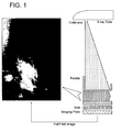
- FIG. 1 is a schematic view of a prior art mammography apparatus.
- a sold angle x-ray is used to take an image of the overall breast through one x-ray scanning. This has an advantage of reducing the scanning time.
- the x-rays of the lower energy band (8-15keV) are absorbed in the breast, the exposure dose increases and the resolution of the image is deteriorated.
- a mammography apparatus using fan beams has been proposed.
- the x-rays generated from an x-ray generating unit pass through a collimator so that only the x-rays required for taking the image can be directed to the breast. Therefore, the x-ray dose of this mammography apparatus is less than that of the solid angle mammography apparatus and the resolution of the image can be improved.
- the mammography apparatus using the fan beams the x-ray generating unit and the image detecting unit are located on a vertical line. Therefore, when lesions overlap on the vertical line, the image is taken as having one lesion. Therefore, it is difficult to accurately identify the locations and sizes of the lesions. Therefore, the mammography apparatus using the fan beams is simply used to identify if there is the breast cancer.
- the image of the breast is further taken after turning the x-ray generating unit and the image detecting unit leftward or rightward by 15-30°. By doing this, the locations of the lesions overlapping on the vertical line can be identified.
- the images are independently taken and combined to each other to look for the locations of the lesions.
- the combining process of the images is complicated.
- the photographing in order to take a biopsy of the lesions by extracting the tissues of the lesions, the photographing must be done at least three times and thus the exposure dose increases. In addition, the photographing time increases. Further, the locations of the lesions are usually identified by feeling from the two-dimensional image or by additionally taking an image while inserting a lesion extracting needle little by little.
- the patient's breast are pressed for a long time. This exacerbates the patient's pain and increases the exposure dose, thereby inducing a secondary accident. Furthermore, since the two-dimensional image is used to identify the locations and sizes of the lesions, this method cannot assist the user to accurately identify the locations and sizes of the lesions.
- Embodiments provide a dual-radiation type mammography apparatus that can easily examine the breast by taking an image by scanning the breast using a plurality of fan beams that are formed from beams radiated from an x-ray generating unit and radiated at different angles and reforming the image as a three-dimensional breast image and can accurately determine a location and size of lesion, thereby enabling the data collection for maximizing treatment efficiency, minimizing the exposure dose to a patient by reducing the image taking time, and minimizing the patient's pain.
- the dual-radiation type mammography apparatus may further include a second collimator that uniformly maintaining a width of each of the beams when the beams deflected by the multi-layered filter units scan the breast.
- the breast fixing unit may include a pressure sensor for uniformly controlling fixing pressure pressing the breast.
- a breast imaging method for examining a breast by taking a breast image using x-ray beams includes fixing the breast by applying predetermined pressure to the breast; forming a plurality of beams by allowing beams in a specific energy band among the solid angle beam generated from an x-ray generating unit to pass and allowing rest beams to be blocked; filtering some of the beams passing through the holes of the first collimator and deflecting only beams in a specific energy band at different angles; scanning the breast using the beams deflected at different angles; and capturing two-dimensional scan images formed by the beams deflected at the different angles and reforming the two-dimensional scan images into a three-dimensional image through a geometrical calculation.
- the breast imaging method may further include maintaining uniformly a width of each of the beams deflected at the different angles when the beams scan the breast.
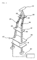
- FIG. 2 is a schematic perspective view of a dual radiation type mammography apparatus according to an embodiment of the present invention
- FIG. 3 is a conception view of the dual radiation type mammography apparatus of FIG. 2.
- a mammography apparatus includes a first collimator 20 radiating a plurality of beams from solid angle beams generated from an x-ray generating unit 10, a plurality of multi-layered filter units 30 radiating the plurality of beams from the first collimator 20 at different angles, a second collimator 40 for uniformly maintaining a width of the beams radiated at different angles through the multi-layered filter units 30, a breast fixing unit 50 for fixing the breast B, and an image output unit 60 for reforming breast images taken at the different angles into a three dimensional by scanning the breast B using the beams radiated at the different angles through the second collimator 40.
- the x-ray generating unit 10 uses molybdenum or rhodium as a target material.
- the emission spectrum of the x-ray generating unit 10 is identical to that of the prior art x-ray generating unit 10.
- the first collimator 20 is provided with a plurality of transmission holes 22 to transmit beams each having a predetermined size among the solid angle beams radiated from the x-ray generating unit 10.
- the transmission holes 22 may be formed in a fan beam shape so that the beams passing through the transmission holes 22 have the fan beam shape.
- the first collimator 20 is formed of lead or tungsten. Some of the solid angle beams pass through the transmission holes 22 (two transmission holes in this embodiment) and the rest is blocked not to be directed to the patient.
- the multi-layered filter units 30 filter off some of the beams passing through the first collimator 20 so that beams in a first specific energy band can be deflected at a predetermined angle.
- the first specific energy band may be 15-23keV.
- the multi-layered filter units 30 may be formed of molybdenum, rhodium, or aluminum. The multi-layered filter units 30 filter off the beams in the low energy band to reduce the exposure dose to the patient.
- one of the beams (two beams) radiated at different angles from the multi-layered filter units 30 at different angles may be vertically incident on the breast and another (the other) of the beams (two beams) may be incident on the breast at an inclined angle with respect to the vertical line.
- the second collimator 40 serves to uniformly maintain a width of the beams that are radiated from the multi-layered filter units 30 at the different angles and emitted to the breast B.
- the second collimator 40 is formed of a material same as that of the first collimator 20.
- the second collimator 40 is provided with filtering holes 42 each having a size identical to that of each pixel of the image detecting unit 60. Therefore, beams in a specific energy band can pass through the filtering holes 42 and the rest is blocked. Therefore the exposure dose to the patient can be significantly reduced.
- a width of the filtering hole may be 40-60 ⁇ m, preferably 50 ⁇ m.
- the fan beams passing through the multi-layered filter units 30 are not straightly directed but incident on the breast B while being diffused in a solid angle shape, thereby preventing the breast B is exposed to ineffective beams and obtaining a high definition image.
- the breast fixing unit 50 includes a paddle that holds the breast through which the beams in the second specific energy band are incident.
- the breast fixing unit 50 is identically structured to the prior art.
- the breast fixing unit 50 may be formed in a plate shape of a carbon-based material.
- a pressing plate pressing the breast may be formed of poly methyl meta acrylate or carbon-based material.
- the present invention is not limited to this. Any material that can effectively transmit the beams and minimize an image loss may be used for the breast fixing unit 50.
- a pressure sensor 52 is provided to the breast fixing unit 50 so as to prevent the breast fixing unit 50 from pressing the breast B with a pressure higher than a predetermined value, thereby minimizing the patient's pain.
- the image output unit 60 includes an image capturing unit, an image scanning unit, and an image processing unit.
- the image capturing unit is arranged in series as long as the width of the beam radiated from the second collimator 40 to form a complete breast image by combining an image obtained before scanning the breast and an image obtained after scanning the breast.
- the image capturing unit may be a digital detector using a charge coupled device (CCD) or a complementary metal-oxide semiconductor (CMOS) cameral. Therefore, the costs for the components can be minimized and the utilization of the exposure index can be maximized.
- CCD charge coupled device
- CMOS complementary metal-oxide semiconductor
- the image capturing unit is provided for each of the plurality of the beams. At this point, the image capturing unit may be disposed to be perpendicular to the corresponding beam to increase the resolution of the image.
- the image scanning unit is mounted on a motor-driven stage so that the plurality of the fan beams can scan the entire region of the breast. Alternatively, the entire region of the breast may be scanned while moving in a state where the x-rays are fixed.
- the image processing unit analyzes numerically the images obtained from the beams radiated vertically or at a predetermined angle through a geometrical reverse and reforms the images into a three-dimensional image so that the examiner can accurately identify the location of the lesion and thus judgment of the examiner can be maximized.
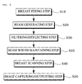
- a breast fixing step S10 the breast B that will be examined is fixed.
- the breast B is pressed and fixed by the paddle of the breast fixing unit 50.
- the pressure sensor 52 is provided to the breast fixing unit 50.
- the pressure applied to the breast B is uniformly maintained by the control of the pressure sensor 52 and thus the patient's pain is minimized.
- the pressure sensor 52 may be controlled such that the pressure applied to the breast B can vary.
- the x-ray generating unit 10, the first and second collimators 20 and 40, the multi-layered filter units 30, the breast fixing unit 50, and the image output unit 60 are set to be initialized by the motor-driven stage before the breast B is fixed.
- a beam generating step S20 the plurality of beams each having a predetermined size are generated from a solid angle beam generated through the x-ray generating unit.
- the x-ray generated from the x-ray generating unit 10 is generally the solid angle beam.
- the plurality of beams having a predetermined size are formed.
- a width of each of the beams generated in the beam generating step S30 is adjusted in response to the breast region.
- beams in a specific band are incident and deflected at a predetermined angle. That is, the beams passing through the first collimator 20 are incident on the corresponding multi-layered filter units 30.
- the multi-layered filter units 30 adjust the width of each of the beams so that the beams can be irradiated to the breast region, and specifically, deflect only the beams in the specific energy band (15-23keV) while filtering the beams in the lower energy band, thereby reducing the exposure dose to the patient. At this point, the multi-layered filter units 30 deflect the beams at different predetermined angles.
- one of the beams deflected by the multi-layered filter units 30 is incident on the breast in the vertical direction and the other is incident on the breast B at a predetermined inclined angle with respect to the vertical direction. Therefore, scanned images of the breast B can be taken by the beams incident on the breast B.
- a beam width maintaining step S32 the beams deflected and incident through the filtering/deflecting step S30 are maintained with predetermined widths each corresponding to an image obtainable pixel. That is, the beams deflected by the multi-layered filter units 30 are incident on the second collimator 40 and sized to correspond to a pixel size of the image output unit. In addition, a width of each of the beams directed to the breast B is uniformly maintained.
- a breast scanning step S40 the beams whose widths are uniformly maintained through the beam width maintaining step S32 are incident on the breast to scan the breast. That is, the beams whose widths are uniformly maintained through the second collimator 40 are incident on the breast B fixed by the breast fixing unit 50 to scan the entire region of the breast B.
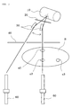
- FIG. 3 is a conception view of the dual radiation type mammography apparatus of FIG. 2.
- the beams radiated through the second collimator 40 scan the breast B by a width of each fan beam while moving from a side to the other side.
- the beam emitted along the vertical line scans the location and vertical section of the lesion a1 and further scans the location and vertical section of the lesion a3 while moving in a direction.
- the beam radiated at an inclined angle with respect to the vertical line scans the section of the breast, which is inclined at a predetermined angle and thus scans the locations and sizes of the lesions a1, a2, and a3.
- an image capturing/outputting step S50 two-dimensional scan images formed by the beams scanning the breast in the breast scanning step S40 are reformed in a three-dimension image to examine if the breast has a lesion. If the breast has the lesion, the size and location of the lesion are output.
- the image output unit 60 is disposed on each of the beams scanning the breast B fixed y the breast fixing unit 50 and analyzes numerically the two-dimensional images formed by the respective beams scanning the breast B through the geometrical reverse, thereby reforming the two-dimensional images into the three-dimensional image.
- FIG. 4 is a schematic view illustrating an image of lesion, which is taken and reformed by a dual radiation type mammography apparatus of an embodiment of the present invention.
- a two-dimensional image for the lesions a1 and a2 are captured by the fan beam emitted along the vertical line and a two-dimensional image for the a1, a2, and a3 is captured by the fan beam emitted at an inclined angle.
- the exposure dose to the patient can be reduced and the patient's pain can be minimized.
- the three dimensional image is formed from two-dimensional images that are captured by the fan beams formed by beams radiated from the-ray generating unit and emitted at different angles the geometrical reverse, the locations and sizes of the lesions can be accurately identified. Therefore, the data collection for maximizing the treatment of the patient becomes possible. In addition, since the breast image taking time is reduced, the x-ray exposure dose to the patient and the patient's pain can be minimized.
Landscapes
- Health & Medical Sciences (AREA)
- Life Sciences & Earth Sciences (AREA)
- Medical Informatics (AREA)
- Engineering & Computer Science (AREA)
- Radiology & Medical Imaging (AREA)
- Molecular Biology (AREA)
- Biophysics (AREA)
- Nuclear Medicine, Radiotherapy & Molecular Imaging (AREA)
- Optics & Photonics (AREA)
- Pathology (AREA)
- Physics & Mathematics (AREA)
- Biomedical Technology (AREA)
- Heart & Thoracic Surgery (AREA)
- High Energy & Nuclear Physics (AREA)
- Surgery (AREA)
- Animal Behavior & Ethology (AREA)
- General Health & Medical Sciences (AREA)
- Public Health (AREA)
- Veterinary Medicine (AREA)
- Dentistry (AREA)
- Oral & Maxillofacial Surgery (AREA)
- Apparatus For Radiation Diagnosis (AREA)
Applications Claiming Priority (1)
| Application Number | Priority Date | Filing Date | Title |
|---|---|---|---|
| KR1020060096980A KR100830549B1 (ko) | 2006-10-02 | 2006-10-02 | 이중 조사방식의 유방촬영장치 및 그 장치를 이용한유방촬영방법 |
Publications (1)
| Publication Number | Publication Date |
|---|---|
| EP1908405A2 true EP1908405A2 (fr) | 2008-04-09 |
Family
ID=38878991
Family Applications (1)
| Application Number | Title | Priority Date | Filing Date |
|---|---|---|---|
| EP07253884A Withdrawn EP1908405A2 (fr) | 2006-10-02 | 2007-10-01 | Appareil de mammographie de type à double radiation et procédé d'imagerie des seins utilisant la mammographie |
Country Status (5)
| Country | Link |
|---|---|
| US (1) | US20080089472A1 (fr) |
| EP (1) | EP1908405A2 (fr) |
| JP (1) | JP2008086760A (fr) |
| KR (1) | KR100830549B1 (fr) |
| CN (1) | CN101156781A (fr) |
Families Citing this family (21)
| Publication number | Priority date | Publication date | Assignee | Title |
|---|---|---|---|---|
| KR101095955B1 (ko) | 2010-04-05 | 2011-12-19 | 주식회사 나노포커스레이 | 모노크로매틱 엑스선 발생기용 다층박막거울 정렬장치 및 이를 이용한 엑스선 영상획득방법 |
| WO2011149146A1 (fr) * | 2010-05-25 | 2011-12-01 | 주식회사 나노포커스레이 | Dispositif d'alignement de miroirs à film multicouche pour un générateur de rayons x monochromatiques, et procédé pour l'acquisition d'une image de rayons x mettant en œuvre le dispositif |
| KR20110138803A (ko) | 2010-06-22 | 2011-12-28 | 삼성전자주식회사 | X-ray를 이용한 영상 진단 장치 및 방법 |
| KR101228911B1 (ko) * | 2010-08-06 | 2013-02-15 | 라드텍주식회사 | 이중 에너지 x-선 흡광분석을 이용한 x-선 영상장치 |
| US8311184B2 (en) * | 2010-08-30 | 2012-11-13 | General Electric Company | Fan-shaped X-ray beam imaging systems employing graded multilayer optic devices |
| KR101678664B1 (ko) * | 2010-09-07 | 2016-11-23 | 삼성전자주식회사 | 유방 촬영 장치 및 방법 |
| KR20130012297A (ko) | 2011-07-25 | 2013-02-04 | 삼성전자주식회사 | 병변 검출 장치, 병변 검출 방법 및 병변 진단 장치 |
| KR101384601B1 (ko) * | 2011-12-22 | 2014-04-15 | (주)제노레이 | 디지털 엑스선 유방암 진단장치 및 진단 방법 |
| KR101981708B1 (ko) | 2012-01-03 | 2019-05-23 | 삼성전자주식회사 | 병변 모양 규칙성 판단이 가능한 병변 진단 장치 및 이의 병변 모양 규칙성 판단 방법 |
| WO2014041675A1 (fr) * | 2012-09-14 | 2014-03-20 | 株式会社日立製作所 | Dispositif d'imagerie par rayons x et procédé d'imagerie par rayons x |
| JP6307590B2 (ja) * | 2013-03-29 | 2018-04-04 | ゼネラル・エレクトリック・カンパニイ | マンモグラフィ装置 |
| KR101437125B1 (ko) * | 2013-05-03 | 2014-09-02 | (주)시스트 | X선을 이용한 회로소자 검사 시스템 및 방법 |
| JP2016187364A (ja) | 2013-09-20 | 2016-11-04 | 第一高周波工業株式会社 | 磁束照射装置 |
| US10342989B2 (en) | 2013-09-20 | 2019-07-09 | Dai-Ichi High Frequency Co., Ltd. | Magnetic flux irradiation devices and components |
| CN104586415B (zh) * | 2013-10-31 | 2019-10-08 | Ge医疗系统环球技术有限公司 | 准直器对准偏差确定方法及计算机化断层成像系统 |
| JP6716591B2 (ja) | 2015-03-02 | 2020-07-01 | カイオ セラピー,エルエルシー | 交番磁界治療を提供するためのシステム及び方法 |
| CN105726049B (zh) * | 2016-01-14 | 2018-10-26 | 深圳安科高技术股份有限公司 | 一种数字乳腺x射线机及其自动曝光图像优化方法 |
| KR101770282B1 (ko) * | 2016-04-26 | 2017-08-23 | 서울대학교병원 | 실시간 입체시를 위한 x선 투시 장치 |
| JP6707048B2 (ja) * | 2017-03-22 | 2020-06-10 | 富士フイルム株式会社 | マンモグラフィ装置 |
| CN108805933B (zh) * | 2018-08-27 | 2021-01-12 | 上海联影医疗科技股份有限公司 | 确定目标点的方法及乳腺x射线摄影系统的定位系统 |
| EP3820371A4 (fr) | 2018-08-27 | 2021-08-11 | Shanghai United Imaging Healthcare Co., Ltd. | Système et procédé de détermination d'un point cible pour une biopsie par aspiration |
Family Cites Families (5)
| Publication number | Priority date | Publication date | Assignee | Title |
|---|---|---|---|---|
| US4969175A (en) * | 1986-08-15 | 1990-11-06 | Nelson Robert S | Apparatus for narrow bandwidth and multiple energy x-ray imaging |
| US6175117B1 (en) * | 1998-01-23 | 2001-01-16 | Quanta Vision, Inc. | Tissue analysis apparatus |
| US6583420B1 (en) * | 2000-06-07 | 2003-06-24 | Robert S. Nelson | Device and system for improved imaging in nuclear medicine and mammography |
| US6611575B1 (en) * | 2001-07-27 | 2003-08-26 | General Electric Company | Method and system for high resolution 3D visualization of mammography images |
| US7120224B2 (en) | 2004-11-02 | 2006-10-10 | Advanced X-Ray Technology, Inc. | X-ray imaging apparatus and method for mammography and computed tomography |
-
2006
- 2006-10-02 KR KR1020060096980A patent/KR100830549B1/ko not_active Expired - Fee Related
-
2007
- 2007-09-25 JP JP2007247001A patent/JP2008086760A/ja active Pending
- 2007-09-25 US US11/902,756 patent/US20080089472A1/en not_active Abandoned
- 2007-09-26 CN CNA2007101543838A patent/CN101156781A/zh active Pending
- 2007-10-01 EP EP07253884A patent/EP1908405A2/fr not_active Withdrawn
Also Published As
| Publication number | Publication date |
|---|---|
| JP2008086760A (ja) | 2008-04-17 |
| CN101156781A (zh) | 2008-04-09 |
| US20080089472A1 (en) | 2008-04-17 |
| KR20080030745A (ko) | 2008-04-07 |
| KR100830549B1 (ko) | 2008-05-21 |
Similar Documents
| Publication | Publication Date | Title |
|---|---|---|
| EP1908405A2 (fr) | Appareil de mammographie de type à double radiation et procédé d'imagerie des seins utilisant la mammographie | |
| JP5346654B2 (ja) | 放射線撮影装置及びその制御方法 | |
| KR101639374B1 (ko) | 토모신테시스 및 마모그래피 영상 촬영을 위한 x선 초점의 특성을 제어하기 위한 시스템 및 방법 | |
| US20070183585A1 (en) | X-ray device that emits an x-ray beam with a scanning-like movement | |
| US20130294582A1 (en) | X-ray imaging apparatus | |
| JP2010075338A (ja) | X線治療機能を備える乳房用画像撮影及び治療装置 | |
| US20120224664A1 (en) | Tomosynthesis mammography system with enlarged field of view | |
| JP2009512502A (ja) | 画像化システム及び関連する技術 | |
| JP5677534B2 (ja) | 放射線撮影装置及びその制御方法 | |
| JP2012110719A (ja) | 小型マンモグラフィ装置、及び関連するマンモグラフィ方法 | |
| JP2008237631A (ja) | 放射線画像撮像装置 | |
| JP2003180670A (ja) | デジタル位相コントラストx線画像撮影システム | |
| KR20140060432A (ko) | 엑스선 촬영 장치 및 엑스선 촬영 방법 | |
| CN110946598A (zh) | 层析摄影装置及其工作方法、存储介质 | |
| JP5914625B2 (ja) | 放射線撮影装置及びその制御方法 | |
| EP2194876B1 (fr) | Appareil de tomographie calculée par ordinateur | |
| CN110946606A (zh) | 层析摄影装置及其工作方法 | |
| JP5538734B2 (ja) | 放射線撮影装置及びその処理方法 | |
| JP2022046946A (ja) | X線ct装置 | |
| JP4208271B2 (ja) | グリッド制御回転陽極型x線管 | |
| JP2004202119A (ja) | 乳房画像撮影装置 | |
| JP2004248945A (ja) | 画像処理装置、画像処理方法、プログラム及び記憶媒体 | |
| EP4268724B1 (fr) | Dispositif radiographique et procédé de régulation de flux de rayonnement | |
| JP2002136511A (ja) | X線撮影装置 | |
| JPH09220224A (ja) | X線診断装置 |
Legal Events
| Date | Code | Title | Description |
|---|---|---|---|
| PUAI | Public reference made under article 153(3) epc to a published international application that has entered the european phase |
Free format text: ORIGINAL CODE: 0009012 |
|
| AK | Designated contracting states |
Kind code of ref document: A2 Designated state(s): AT BE BG CH CY CZ DE DK EE ES FI FR GB GR HU IE IS IT LI LT LU LV MC MT NL PL PT RO SE SI SK TR |
|
| AX | Request for extension of the european patent |
Extension state: AL BA HR MK RS |
|
| STAA | Information on the status of an ep patent application or granted ep patent |
Free format text: STATUS: THE APPLICATION IS DEEMED TO BE WITHDRAWN |
|
| 18D | Application deemed to be withdrawn |
Effective date: 20100504 |