EP1887859B1 - Tiermodell für das humane immunsystem, sowie verfahren zu dessen herstellung - Google Patents
Tiermodell für das humane immunsystem, sowie verfahren zu dessen herstellung Download PDFInfo
- Publication number
- EP1887859B1 EP1887859B1 EP06742372A EP06742372A EP1887859B1 EP 1887859 B1 EP1887859 B1 EP 1887859B1 EP 06742372 A EP06742372 A EP 06742372A EP 06742372 A EP06742372 A EP 06742372A EP 1887859 B1 EP1887859 B1 EP 1887859B1
- Authority
- EP
- European Patent Office
- Prior art keywords
- human
- cells
- cell
- transplantation
- animals
- Prior art date
- Legal status (The legal status is an assumption and is not a legal conclusion. Google has not performed a legal analysis and makes no representation as to the accuracy of the status listed.)
- Not-in-force
Links
- 238000010171 animal model Methods 0.000 title claims description 25
- 210000000987 immune system Anatomy 0.000 title claims description 20
- 238000004519 manufacturing process Methods 0.000 title description 16
- 210000004027 cell Anatomy 0.000 claims description 118
- 238000002054 transplantation Methods 0.000 claims description 42
- 210000000130 stem cell Anatomy 0.000 claims description 36
- 238000000034 method Methods 0.000 claims description 33
- 239000012228 culture supernatant Substances 0.000 claims description 31
- 238000004113 cell culture Methods 0.000 claims description 28
- 210000001185 bone marrow Anatomy 0.000 claims description 25
- 210000005260 human cell Anatomy 0.000 claims description 23
- 238000011579 SCID mouse model Methods 0.000 claims description 21
- 230000003750 conditioning effect Effects 0.000 claims description 18
- 210000005259 peripheral blood Anatomy 0.000 claims description 17
- 239000011886 peripheral blood Substances 0.000 claims description 17
- 102000004889 Interleukin-6 Human genes 0.000 claims description 14
- 108090001005 Interleukin-6 Proteins 0.000 claims description 14
- 230000003394 haemopoietic effect Effects 0.000 claims description 14
- 210000004700 fetal blood Anatomy 0.000 claims description 13
- 210000003958 hematopoietic stem cell Anatomy 0.000 claims description 13
- 108090001007 Interleukin-8 Proteins 0.000 claims description 11
- 108010073929 Vascular Endothelial Growth Factor A Proteins 0.000 claims description 11
- 102000005789 Vascular Endothelial Growth Factors Human genes 0.000 claims description 11
- 108010019530 Vascular Endothelial Growth Factors Proteins 0.000 claims description 11
- 102100022987 Angiogenin Human genes 0.000 claims description 9
- 101710155857 C-C motif chemokine 2 Proteins 0.000 claims description 9
- 108090000623 proteins and genes Proteins 0.000 claims description 8
- 210000001519 tissue Anatomy 0.000 claims description 6
- 108090000373 Tissue Plasminogen Activator Proteins 0.000 claims description 5
- 238000007912 intraperitoneal administration Methods 0.000 claims description 5
- 102000004169 proteins and genes Human genes 0.000 claims description 5
- 229940100601 interleukin-6 Drugs 0.000 claims description 4
- 108010072788 angiogenin Proteins 0.000 claims description 3
- 229940096397 interleukin-8 Drugs 0.000 claims description 3
- XKTZWUACRZHVAN-VADRZIEHSA-N interleukin-8 Chemical compound C([C@H](NC(=O)[C@H](CC(O)=O)NC(=O)[C@H](CC=1C2=CC=CC=C2NC=1)NC(=O)[C@@H](NC(C)=O)CCSC)C(=O)N[C@@H](CC(O)=O)C(=O)N[C@@H](CC(O)=O)C(=O)N[C@@H](CC(C)C)C(=O)N[C@@H](CC(N)=O)C(=O)N[C@@H](CC=1C=CC=CC=1)C(=O)N[C@@H]([C@@H](C)O)C(=O)NCC(=O)N[C@@H](CCSC)C(=O)N1[C@H](CCC1)C(=O)N1[C@H](CCC1)C(=O)N[C@@H](C)C(=O)N[C@H](CC(O)=O)C(=O)N[C@H](CCC(O)=O)C(=O)N[C@H](CC(O)=O)C(=O)N[C@H](CC=1C=CC(O)=CC=1)C(=O)N[C@H](CO)C(=O)N1[C@H](CCC1)C(N)=O)C1=CC=CC=C1 XKTZWUACRZHVAN-VADRZIEHSA-N 0.000 claims description 3
- 238000001990 intravenous administration Methods 0.000 claims description 3
- 210000003954 umbilical cord Anatomy 0.000 claims description 3
- 102000000018 Chemokine CCL2 Human genes 0.000 claims 1
- 102100026236 Interleukin-8 Human genes 0.000 claims 1
- 102000003978 Tissue Plasminogen Activator Human genes 0.000 claims 1
- 241001465754 Metazoa Species 0.000 description 79
- 101000738771 Homo sapiens Receptor-type tyrosine-protein phosphatase C Proteins 0.000 description 34
- 102100037422 Receptor-type tyrosine-protein phosphatase C Human genes 0.000 description 34
- 210000001744 T-lymphocyte Anatomy 0.000 description 34
- 230000001143 conditioned effect Effects 0.000 description 26
- 210000003719 b-lymphocyte Anatomy 0.000 description 23
- 241000699670 Mus sp. Species 0.000 description 22
- 210000004443 dendritic cell Anatomy 0.000 description 20
- 210000005087 mononuclear cell Anatomy 0.000 description 20
- 102000004127 Cytokines Human genes 0.000 description 19
- 108090000695 Cytokines Proteins 0.000 description 19
- 210000004369 blood Anatomy 0.000 description 18
- 239000008280 blood Substances 0.000 description 18
- 206010068051 Chimerism Diseases 0.000 description 17
- 238000011161 development Methods 0.000 description 15
- 230000018109 developmental process Effects 0.000 description 15
- 241000699666 Mus <mouse, genus> Species 0.000 description 13
- 239000000427 antigen Substances 0.000 description 13
- 108091007433 antigens Proteins 0.000 description 13
- 102000036639 antigens Human genes 0.000 description 13
- 210000000822 natural killer cell Anatomy 0.000 description 13
- 239000006228 supernatant Substances 0.000 description 13
- 102100031573 Hematopoietic progenitor cell antigen CD34 Human genes 0.000 description 12
- 101000777663 Homo sapiens Hematopoietic progenitor cell antigen CD34 Proteins 0.000 description 12
- 108060008682 Tumor Necrosis Factor Proteins 0.000 description 12
- 102000000852 Tumor Necrosis Factor-alpha Human genes 0.000 description 12
- 229920006008 lipopolysaccharide Polymers 0.000 description 12
- 210000000952 spleen Anatomy 0.000 description 12
- 239000002158 endotoxin Substances 0.000 description 11
- 210000003714 granulocyte Anatomy 0.000 description 11
- 210000001616 monocyte Anatomy 0.000 description 11
- 102000004890 Interleukin-8 Human genes 0.000 description 10
- 230000000638 stimulation Effects 0.000 description 10
- 230000014509 gene expression Effects 0.000 description 9
- 102100021943 C-C motif chemokine 2 Human genes 0.000 description 8
- 238000012360 testing method Methods 0.000 description 8
- 229960000814 tetanus toxoid Drugs 0.000 description 8
- 230000001225 therapeutic effect Effects 0.000 description 8
- 102000003814 Interleukin-10 Human genes 0.000 description 7
- 108090000174 Interleukin-10 Proteins 0.000 description 7
- 210000005006 adaptive immune system Anatomy 0.000 description 7
- 230000000694 effects Effects 0.000 description 7
- 230000003053 immunization Effects 0.000 description 7
- 238000002649 immunization Methods 0.000 description 7
- 229940027941 immunoglobulin g Drugs 0.000 description 7
- 230000009257 reactivity Effects 0.000 description 7
- -1 sVCAM-1 Proteins 0.000 description 7
- MZOFCQQQCNRIBI-VMXHOPILSA-N (3s)-4-[[(2s)-1-[[(2s)-1-[[(1s)-1-carboxy-2-hydroxyethyl]amino]-4-methyl-1-oxopentan-2-yl]amino]-5-(diaminomethylideneamino)-1-oxopentan-2-yl]amino]-3-[[2-[[(2s)-2,6-diaminohexanoyl]amino]acetyl]amino]-4-oxobutanoic acid Chemical compound OC[C@@H](C(O)=O)NC(=O)[C@H](CC(C)C)NC(=O)[C@H](CCCN=C(N)N)NC(=O)[C@H](CC(O)=O)NC(=O)CNC(=O)[C@@H](N)CCCCN MZOFCQQQCNRIBI-VMXHOPILSA-N 0.000 description 6
- 102100025248 C-X-C motif chemokine 10 Human genes 0.000 description 6
- 101710098275 C-X-C motif chemokine 10 Proteins 0.000 description 6
- 102000007644 Colony-Stimulating Factors Human genes 0.000 description 6
- 108010071942 Colony-Stimulating Factors Proteins 0.000 description 6
- 102100020880 Kit ligand Human genes 0.000 description 6
- 241001529936 Murinae Species 0.000 description 6
- 229940047120 colony stimulating factors Drugs 0.000 description 6
- 238000001514 detection method Methods 0.000 description 6
- 230000004069 differentiation Effects 0.000 description 6
- 238000009826 distribution Methods 0.000 description 6
- 230000009885 systemic effect Effects 0.000 description 6
- 102100024222 B-lymphocyte antigen CD19 Human genes 0.000 description 5
- 238000002965 ELISA Methods 0.000 description 5
- 102000006354 HLA-DR Antigens Human genes 0.000 description 5
- 108010058597 HLA-DR Antigens Proteins 0.000 description 5
- 101000980825 Homo sapiens B-lymphocyte antigen CD19 Proteins 0.000 description 5
- 108010002386 Interleukin-3 Proteins 0.000 description 5
- 102000000646 Interleukin-3 Human genes 0.000 description 5
- 230000015572 biosynthetic process Effects 0.000 description 5
- 238000006243 chemical reaction Methods 0.000 description 5
- 239000003814 drug Substances 0.000 description 5
- 230000006870 function Effects 0.000 description 5
- 210000000777 hematopoietic system Anatomy 0.000 description 5
- 230000011132 hemopoiesis Effects 0.000 description 5
- 238000001727 in vivo Methods 0.000 description 5
- 208000015181 infectious disease Diseases 0.000 description 5
- 229940076264 interleukin-3 Drugs 0.000 description 5
- 210000000265 leukocyte Anatomy 0.000 description 5
- 238000011160 research Methods 0.000 description 5
- 241000894007 species Species 0.000 description 5
- 102100032367 C-C motif chemokine 5 Human genes 0.000 description 4
- 102100036170 C-X-C motif chemokine 9 Human genes 0.000 description 4
- 101710085500 C-X-C motif chemokine 9 Proteins 0.000 description 4
- 101800000267 CD40 ligand, soluble form Proteins 0.000 description 4
- 102400000432 CD40 ligand, soluble form Human genes 0.000 description 4
- 108010055166 Chemokine CCL5 Proteins 0.000 description 4
- 108090000379 Fibroblast growth factor 2 Proteins 0.000 description 4
- 102100024785 Fibroblast growth factor 2 Human genes 0.000 description 4
- 108060003951 Immunoglobulin Proteins 0.000 description 4
- 101710177504 Kit ligand Proteins 0.000 description 4
- 102000019400 Tissue-type plasminogen activator Human genes 0.000 description 4
- 230000004913 activation Effects 0.000 description 4
- 210000002798 bone marrow cell Anatomy 0.000 description 4
- 239000002771 cell marker Substances 0.000 description 4
- 230000001419 dependent effect Effects 0.000 description 4
- 206010012601 diabetes mellitus Diseases 0.000 description 4
- 238000005516 engineering process Methods 0.000 description 4
- 230000028993 immune response Effects 0.000 description 4
- 102000018358 immunoglobulin Human genes 0.000 description 4
- 238000000338 in vitro Methods 0.000 description 4
- 210000002540 macrophage Anatomy 0.000 description 4
- 210000000056 organ Anatomy 0.000 description 4
- 238000002360 preparation method Methods 0.000 description 4
- 239000000047 product Substances 0.000 description 4
- 210000003462 vein Anatomy 0.000 description 4
- 101000917858 Homo sapiens Low affinity immunoglobulin gamma Fc region receptor III-A Proteins 0.000 description 3
- 101000917839 Homo sapiens Low affinity immunoglobulin gamma Fc region receptor III-B Proteins 0.000 description 3
- 101000946889 Homo sapiens Monocyte differentiation antigen CD14 Proteins 0.000 description 3
- 241000725303 Human immunodeficiency virus Species 0.000 description 3
- 102100022297 Integrin alpha-X Human genes 0.000 description 3
- 102000008070 Interferon-gamma Human genes 0.000 description 3
- 108010074328 Interferon-gamma Proteins 0.000 description 3
- 102100029185 Low affinity immunoglobulin gamma Fc region receptor III-B Human genes 0.000 description 3
- 102100035877 Monocyte differentiation antigen CD14 Human genes 0.000 description 3
- 230000033289 adaptive immune response Effects 0.000 description 3
- 238000004458 analytical method Methods 0.000 description 3
- 210000000612 antigen-presenting cell Anatomy 0.000 description 3
- 239000006285 cell suspension Substances 0.000 description 3
- 230000001413 cellular effect Effects 0.000 description 3
- 230000007547 defect Effects 0.000 description 3
- 230000002950 deficient Effects 0.000 description 3
- 229940079593 drug Drugs 0.000 description 3
- 230000001605 fetal effect Effects 0.000 description 3
- 238000011577 humanized mouse model Methods 0.000 description 3
- 230000008105 immune reaction Effects 0.000 description 3
- 230000002458 infectious effect Effects 0.000 description 3
- 229960003130 interferon gamma Drugs 0.000 description 3
- 230000007774 longterm Effects 0.000 description 3
- 210000005210 lymphoid organ Anatomy 0.000 description 3
- 239000003550 marker Substances 0.000 description 3
- 230000001105 regulatory effect Effects 0.000 description 3
- 208000002491 severe combined immunodeficiency Diseases 0.000 description 3
- 238000011476 stem cell transplantation Methods 0.000 description 3
- 238000002560 therapeutic procedure Methods 0.000 description 3
- KZMAWJRXKGLWGS-UHFFFAOYSA-N 2-chloro-n-[4-(4-methoxyphenyl)-1,3-thiazol-2-yl]-n-(3-methoxypropyl)acetamide Chemical compound S1C(N(C(=O)CCl)CCCOC)=NC(C=2C=CC(OC)=CC=2)=C1 KZMAWJRXKGLWGS-UHFFFAOYSA-N 0.000 description 2
- 102100031585 ADP-ribosyl cyclase/cyclic ADP-ribose hydrolase 1 Human genes 0.000 description 2
- 208000031261 Acute myeloid leukaemia Diseases 0.000 description 2
- NLXLAEXVIDQMFP-UHFFFAOYSA-N Ammonia chloride Chemical compound [NH4+].[Cl-] NLXLAEXVIDQMFP-UHFFFAOYSA-N 0.000 description 2
- 208000035143 Bacterial infection Diseases 0.000 description 2
- 108010029697 CD40 Ligand Proteins 0.000 description 2
- 102100032937 CD40 ligand Human genes 0.000 description 2
- RTZKZFJDLAIYFH-UHFFFAOYSA-N Diethyl ether Chemical compound CCOCC RTZKZFJDLAIYFH-UHFFFAOYSA-N 0.000 description 2
- 102100031939 Erythropoietin Human genes 0.000 description 2
- 208000009329 Graft vs Host Disease Diseases 0.000 description 2
- 241000282412 Homo Species 0.000 description 2
- 101000777636 Homo sapiens ADP-ribosyl cyclase/cyclic ADP-ribose hydrolase 1 Proteins 0.000 description 2
- 101000757236 Homo sapiens Angiogenin Proteins 0.000 description 2
- 101000581981 Homo sapiens Neural cell adhesion molecule 1 Proteins 0.000 description 2
- 241000701044 Human gammaherpesvirus 4 Species 0.000 description 2
- 206010061598 Immunodeficiency Diseases 0.000 description 2
- 208000029462 Immunodeficiency disease Diseases 0.000 description 2
- 102000015696 Interleukins Human genes 0.000 description 2
- 108010063738 Interleukins Proteins 0.000 description 2
- 206010028980 Neoplasm Diseases 0.000 description 2
- 102100027347 Neural cell adhesion molecule 1 Human genes 0.000 description 2
- 108010053210 Phycocyanin Proteins 0.000 description 2
- 108010004729 Phycoerythrin Proteins 0.000 description 2
- 241000283984 Rodentia Species 0.000 description 2
- 102000003800 Selectins Human genes 0.000 description 2
- 108090000184 Selectins Proteins 0.000 description 2
- 108010039445 Stem Cell Factor Proteins 0.000 description 2
- 241000700605 Viruses Species 0.000 description 2
- UYRDHEJRPVSJFM-VSWVFQEASA-N [(1s,3r)-3-hydroxy-4-[(3e,5e,7e,9e,11z)-11-[4-[(e)-2-[(1r,3s,6s)-3-hydroxy-1,5,5-trimethyl-7-oxabicyclo[4.1.0]heptan-6-yl]ethenyl]-5-oxofuran-2-ylidene]-3,10-dimethylundeca-1,3,5,7,9-pentaenylidene]-3,5,5-trimethylcyclohexyl] acetate Chemical compound C[C@@]1(O)C[C@@H](OC(=O)C)CC(C)(C)C1=C=C\C(C)=C\C=C\C=C\C=C(/C)\C=C/1C=C(\C=C\[C@]23[C@@](O2)(C)C[C@@H](O)CC3(C)C)C(=O)O\1 UYRDHEJRPVSJFM-VSWVFQEASA-N 0.000 description 2
- 108010004469 allophycocyanin Proteins 0.000 description 2
- 210000002588 alveolar type II cell Anatomy 0.000 description 2
- 230000003321 amplification Effects 0.000 description 2
- 208000022362 bacterial infectious disease Diseases 0.000 description 2
- 210000000988 bone and bone Anatomy 0.000 description 2
- 238000012512 characterization method Methods 0.000 description 2
- 229930002875 chlorophyll Natural products 0.000 description 2
- 235000019804 chlorophyll Nutrition 0.000 description 2
- ATNHDLDRLWWWCB-AENOIHSZSA-M chlorophyll a Chemical compound C1([C@@H](C(=O)OC)C(=O)C2=C3C)=C2N2C3=CC(C(CC)=C3C)=[N+]4C3=CC3=C(C=C)C(C)=C5N3[Mg-2]42[N+]2=C1[C@@H](CCC(=O)OC\C=C(/C)CCC[C@H](C)CCC[C@H](C)CCCC(C)C)[C@H](C)C2=C5 ATNHDLDRLWWWCB-AENOIHSZSA-M 0.000 description 2
- 230000009260 cross reactivity Effects 0.000 description 2
- 239000012645 endogenous antigen Substances 0.000 description 2
- 210000003743 erythrocyte Anatomy 0.000 description 2
- 238000002474 experimental method Methods 0.000 description 2
- 238000000684 flow cytometry Methods 0.000 description 2
- 108020001507 fusion proteins Proteins 0.000 description 2
- 102000037865 fusion proteins Human genes 0.000 description 2
- 208000024908 graft versus host disease Diseases 0.000 description 2
- 229940094342 human immunoglobulin m Drugs 0.000 description 2
- 230000002519 immonomodulatory effect Effects 0.000 description 2
- 230000006058 immune tolerance Effects 0.000 description 2
- 230000007813 immunodeficiency Effects 0.000 description 2
- 230000016784 immunoglobulin production Effects 0.000 description 2
- 229940072221 immunoglobulins Drugs 0.000 description 2
- 230000001939 inductive effect Effects 0.000 description 2
- 238000011835 investigation Methods 0.000 description 2
- 230000035800 maturation Effects 0.000 description 2
- 239000002609 medium Substances 0.000 description 2
- 210000002901 mesenchymal stem cell Anatomy 0.000 description 2
- 244000005700 microbiome Species 0.000 description 2
- 239000000203 mixture Substances 0.000 description 2
- 238000003199 nucleic acid amplification method Methods 0.000 description 2
- 230000008506 pathogenesis Effects 0.000 description 2
- UTIQDNPUHSAVDN-UHFFFAOYSA-N peridinin Natural products CC(=O)OC1CC(C)(C)C(=C=CC(=CC=CC=CC=C2/OC(=O)C(=C2)C=CC34OC3(C)CC(O)CC4(C)C)C)C(C)(O)C1 UTIQDNPUHSAVDN-UHFFFAOYSA-N 0.000 description 2
- 230000000144 pharmacologic effect Effects 0.000 description 2
- OXCMYAYHXIHQOA-UHFFFAOYSA-N potassium;[2-butyl-5-chloro-3-[[4-[2-(1,2,4-triaza-3-azanidacyclopenta-1,4-dien-5-yl)phenyl]phenyl]methyl]imidazol-4-yl]methanol Chemical compound [K+].CCCCC1=NC(Cl)=C(CO)N1CC1=CC=C(C=2C(=CC=CC=2)C2=N[N-]N=N2)C=C1 OXCMYAYHXIHQOA-UHFFFAOYSA-N 0.000 description 2
- 239000002243 precursor Substances 0.000 description 2
- 230000008569 process Effects 0.000 description 2
- AAEVYOVXGOFMJO-UHFFFAOYSA-N prometryn Chemical compound CSC1=NC(NC(C)C)=NC(NC(C)C)=N1 AAEVYOVXGOFMJO-UHFFFAOYSA-N 0.000 description 2
- 230000002285 radioactive effect Effects 0.000 description 2
- 230000001172 regenerating effect Effects 0.000 description 2
- 230000004044 response Effects 0.000 description 2
- 230000028327 secretion Effects 0.000 description 2
- 208000003265 stomatitis Diseases 0.000 description 2
- 230000009261 transgenic effect Effects 0.000 description 2
- 230000002792 vascular Effects 0.000 description 2
- 208000030507 AIDS Diseases 0.000 description 1
- 208000024893 Acute lymphoblastic leukemia Diseases 0.000 description 1
- 208000014697 Acute lymphocytic leukaemia Diseases 0.000 description 1
- 102100022005 B-lymphocyte antigen CD20 Human genes 0.000 description 1
- 108091003079 Bovine Serum Albumin Proteins 0.000 description 1
- 102000017420 CD3 protein, epsilon/gamma/delta subunit Human genes 0.000 description 1
- 108050005493 CD3 protein, epsilon/gamma/delta subunit Proteins 0.000 description 1
- 201000009030 Carcinoma Diseases 0.000 description 1
- 108010077544 Chromatin Proteins 0.000 description 1
- 230000033616 DNA repair Effects 0.000 description 1
- 101100193633 Danio rerio rag2 gene Proteins 0.000 description 1
- 108090000394 Erythropoietin Proteins 0.000 description 1
- 102100020715 Fms-related tyrosine kinase 3 ligand protein Human genes 0.000 description 1
- 101710162577 Fms-related tyrosine kinase 3 ligand protein Proteins 0.000 description 1
- 101000897405 Homo sapiens B-lymphocyte antigen CD20 Proteins 0.000 description 1
- 101001033249 Homo sapiens Interleukin-1 beta Proteins 0.000 description 1
- 101001076408 Homo sapiens Interleukin-6 Proteins 0.000 description 1
- 101001055222 Homo sapiens Interleukin-8 Proteins 0.000 description 1
- 101000716729 Homo sapiens Kit ligand Proteins 0.000 description 1
- 101000832103 Homo sapiens Signal transducer and activator of transcription 5A Proteins 0.000 description 1
- 108700005091 Immunoglobulin Genes Proteins 0.000 description 1
- 102000000588 Interleukin-2 Human genes 0.000 description 1
- 108010002350 Interleukin-2 Proteins 0.000 description 1
- 102000010789 Interleukin-2 Receptors Human genes 0.000 description 1
- 108010038453 Interleukin-2 Receptors Proteins 0.000 description 1
- 108010002586 Interleukin-7 Proteins 0.000 description 1
- 108090000581 Leukemia inhibitory factor Proteins 0.000 description 1
- 241000124008 Mammalia Species 0.000 description 1
- 101100193635 Mus musculus Rag2 gene Proteins 0.000 description 1
- 208000033776 Myeloid Acute Leukemia Diseases 0.000 description 1
- 239000004677 Nylon Substances 0.000 description 1
- 208000006664 Precursor Cell Lymphoblastic Leukemia-Lymphoma Diseases 0.000 description 1
- 238000012356 Product development Methods 0.000 description 1
- 239000012980 RPMI-1640 medium Substances 0.000 description 1
- 206010039491 Sarcoma Diseases 0.000 description 1
- 206010039897 Sedation Diseases 0.000 description 1
- 108091008874 T cell receptors Proteins 0.000 description 1
- 102000016266 T-Cell Antigen Receptors Human genes 0.000 description 1
- 108700019146 Transgenes Proteins 0.000 description 1
- 230000001154 acute effect Effects 0.000 description 1
- 230000003044 adaptive effect Effects 0.000 description 1
- 230000004721 adaptive immunity Effects 0.000 description 1
- 239000002671 adjuvant Substances 0.000 description 1
- 230000000172 allergic effect Effects 0.000 description 1
- 235000019270 ammonium chloride Nutrition 0.000 description 1
- 230000003698 anagen phase Effects 0.000 description 1
- 238000013459 approach Methods 0.000 description 1
- 238000003556 assay Methods 0.000 description 1
- 208000010668 atopic eczema Diseases 0.000 description 1
- 230000001363 autoimmune Effects 0.000 description 1
- 239000011324 bead Substances 0.000 description 1
- 230000008901 benefit Effects 0.000 description 1
- 230000000903 blocking effect Effects 0.000 description 1
- 238000009395 breeding Methods 0.000 description 1
- 230000001488 breeding effect Effects 0.000 description 1
- 230000011712 cell development Effects 0.000 description 1
- 239000003795 chemical substances by application Substances 0.000 description 1
- 210000003483 chromatin Anatomy 0.000 description 1
- 238000005345 coagulation Methods 0.000 description 1
- 230000015271 coagulation Effects 0.000 description 1
- 230000000295 complement effect Effects 0.000 description 1
- 239000002131 composite material Substances 0.000 description 1
- 239000003636 conditioned culture medium Substances 0.000 description 1
- 210000002808 connective tissue Anatomy 0.000 description 1
- 230000016396 cytokine production Effects 0.000 description 1
- 230000003111 delayed effect Effects 0.000 description 1
- 238000000432 density-gradient centrifugation Methods 0.000 description 1
- 239000010432 diamond Substances 0.000 description 1
- 201000010099 disease Diseases 0.000 description 1
- 208000037265 diseases, disorders, signs and symptoms Diseases 0.000 description 1
- 230000036267 drug metabolism Effects 0.000 description 1
- 239000000975 dye Substances 0.000 description 1
- 239000012636 effector Substances 0.000 description 1
- 210000001671 embryonic stem cell Anatomy 0.000 description 1
- 210000003979 eosinophil Anatomy 0.000 description 1
- 229940105423 erythropoietin Drugs 0.000 description 1
- 238000000605 extraction Methods 0.000 description 1
- 239000012894 fetal calf serum Substances 0.000 description 1
- 238000001914 filtration Methods 0.000 description 1
- 238000001943 fluorescence-activated cell sorting Methods 0.000 description 1
- 238000010230 functional analysis Methods 0.000 description 1
- 238000012239 gene modification Methods 0.000 description 1
- 230000005017 genetic modification Effects 0.000 description 1
- 235000013617 genetically modified food Nutrition 0.000 description 1
- 210000004602 germ cell Anatomy 0.000 description 1
- 230000013595 glycosylation Effects 0.000 description 1
- 238000006206 glycosylation reaction Methods 0.000 description 1
- 239000003102 growth factor Substances 0.000 description 1
- 239000001963 growth medium Substances 0.000 description 1
- 229920000669 heparin Polymers 0.000 description 1
- ZFGMDIBRIDKWMY-PASTXAENSA-N heparin Chemical compound CC(O)=N[C@@H]1[C@@H](O)[C@H](O)[C@@H](COS(O)(=O)=O)O[C@@H]1O[C@@H]1[C@@H](C(O)=O)O[C@@H](O[C@H]2[C@@H]([C@@H](OS(O)(=O)=O)[C@@H](O[C@@H]3[C@@H](OC(O)[C@H](OS(O)(=O)=O)[C@H]3O)C(O)=O)O[C@@H]2O)CS(O)(=O)=O)[C@H](O)[C@H]1O ZFGMDIBRIDKWMY-PASTXAENSA-N 0.000 description 1
- 102000052624 human CXCL8 Human genes 0.000 description 1
- 102000052934 human STAT5A Human genes 0.000 description 1
- 229940098197 human immunoglobulin g Drugs 0.000 description 1
- 229940116886 human interleukin-6 Drugs 0.000 description 1
- 230000037189 immune system physiology Effects 0.000 description 1
- 230000036039 immunity Effects 0.000 description 1
- 239000003547 immunosorbent Substances 0.000 description 1
- 230000003308 immunostimulating effect Effects 0.000 description 1
- 230000001024 immunotherapeutic effect Effects 0.000 description 1
- 238000009169 immunotherapy Methods 0.000 description 1
- 238000002513 implantation Methods 0.000 description 1
- 230000002779 inactivation Effects 0.000 description 1
- 238000011534 incubation Methods 0.000 description 1
- 230000006698 induction Effects 0.000 description 1
- 208000027866 inflammatory disease Diseases 0.000 description 1
- 230000002757 inflammatory effect Effects 0.000 description 1
- 238000002347 injection Methods 0.000 description 1
- 239000007924 injection Substances 0.000 description 1
- 238000010253 intravenous injection Methods 0.000 description 1
- 230000005865 ionizing radiation Effects 0.000 description 1
- 238000002372 labelling Methods 0.000 description 1
- 230000002045 lasting effect Effects 0.000 description 1
- 210000004185 liver Anatomy 0.000 description 1
- 210000005265 lung cell Anatomy 0.000 description 1
- 210000004698 lymphocyte Anatomy 0.000 description 1
- 230000007246 mechanism Effects 0.000 description 1
- 238000013160 medical therapy Methods 0.000 description 1
- 239000003607 modifier Substances 0.000 description 1
- 238000012544 monitoring process Methods 0.000 description 1
- 238000010172 mouse model Methods 0.000 description 1
- 230000035772 mutation Effects 0.000 description 1
- 210000000066 myeloid cell Anatomy 0.000 description 1
- 230000001537 neural effect Effects 0.000 description 1
- 210000000440 neutrophil Anatomy 0.000 description 1
- 230000003448 neutrophilic effect Effects 0.000 description 1
- 229920001778 nylon Polymers 0.000 description 1
- 231100000590 oncogenic Toxicity 0.000 description 1
- 230000002246 oncogenic effect Effects 0.000 description 1
- 238000005457 optimization Methods 0.000 description 1
- 210000003200 peritoneal cavity Anatomy 0.000 description 1
- 239000002831 pharmacologic agent Substances 0.000 description 1
- 239000013612 plasmid Substances 0.000 description 1
- 210000004043 pneumocyte Anatomy 0.000 description 1
- 239000011148 porous material Substances 0.000 description 1
- 230000003389 potentiating effect Effects 0.000 description 1
- 238000002203 pretreatment Methods 0.000 description 1
- 230000035755 proliferation Effects 0.000 description 1
- 230000005855 radiation Effects 0.000 description 1
- 230000008707 rearrangement Effects 0.000 description 1
- 238000011084 recovery Methods 0.000 description 1
- 230000009467 reduction Effects 0.000 description 1
- 210000003289 regulatory T cell Anatomy 0.000 description 1
- 230000004043 responsiveness Effects 0.000 description 1
- 201000005404 rubella Diseases 0.000 description 1
- 230000003248 secreting effect Effects 0.000 description 1
- 230000036280 sedation Effects 0.000 description 1
- 238000000926 separation method Methods 0.000 description 1
- 210000002966 serum Anatomy 0.000 description 1
- 150000003384 small molecules Chemical class 0.000 description 1
- 239000007787 solid Substances 0.000 description 1
- 239000000243 solution Substances 0.000 description 1
- 210000001082 somatic cell Anatomy 0.000 description 1
- 230000000392 somatic effect Effects 0.000 description 1
- 230000010473 stable expression Effects 0.000 description 1
- 238000010186 staining Methods 0.000 description 1
- 230000005477 standard model Effects 0.000 description 1
- 239000000126 substance Substances 0.000 description 1
- 230000004083 survival effect Effects 0.000 description 1
- 230000002123 temporal effect Effects 0.000 description 1
- 229940124597 therapeutic agent Drugs 0.000 description 1
- 230000001988 toxicity Effects 0.000 description 1
- 231100000419 toxicity Toxicity 0.000 description 1
- 238000012549 training Methods 0.000 description 1
- 238000011282 treatment Methods 0.000 description 1
- 238000002255 vaccination Methods 0.000 description 1
- 229960005486 vaccine Drugs 0.000 description 1
- 238000012795 verification Methods 0.000 description 1
- 238000002689 xenotransplantation Methods 0.000 description 1
Images




Classifications
-
- A—HUMAN NECESSITIES
- A01—AGRICULTURE; FORESTRY; ANIMAL HUSBANDRY; HUNTING; TRAPPING; FISHING
- A01K—ANIMAL HUSBANDRY; AVICULTURE; APICULTURE; PISCICULTURE; FISHING; REARING OR BREEDING ANIMALS, NOT OTHERWISE PROVIDED FOR; NEW BREEDS OF ANIMALS
- A01K67/00—Rearing or breeding animals, not otherwise provided for; New or modified breeds of animals
- A01K67/027—New or modified breeds of vertebrates
- A01K67/0271—Chimeric vertebrates, e.g. comprising exogenous cells
-
- A—HUMAN NECESSITIES
- A01—AGRICULTURE; FORESTRY; ANIMAL HUSBANDRY; HUNTING; TRAPPING; FISHING
- A01K—ANIMAL HUSBANDRY; AVICULTURE; APICULTURE; PISCICULTURE; FISHING; REARING OR BREEDING ANIMALS, NOT OTHERWISE PROVIDED FOR; NEW BREEDS OF ANIMALS
- A01K2267/00—Animals characterised by purpose
- A01K2267/03—Animal model, e.g. for test or diseases
- A01K2267/035—Animal model for multifactorial diseases
- A01K2267/0381—Animal model for diseases of the hematopoietic system
Definitions
- the present invention relates to a method for establishing a functional adaptive human immune system in a suitable animal model.
- the animal model is suitable z.
- For this immuno-deficient mice are used, which develop little immunological reactions against the transmitted cells.
- the first strategy uses the technology to produce genetically modified ( transgenic, knock-out, knock-in ) animals by introducing human genes, inducing specific mutations or replacing a murine gene with the human homolog in somatic or germline cells for genetic modification or to express human antigens or targets.
- the limits of this method are next to the high ones Cost, low success rates, especially in the limited production, usually individual, human properties and thus their reactivity with mostly animal partner molecules and usually not identical gene expression control mechanisms. Therefore, this method is mostly used only to characterize the drug's potential in terms of drug metabolism and the determination of toxicity or to study the activity of a single or a set of targets (1, 2).
- the second and more complex approach is the generation of chimeras by xenotransplantation (3).
- This includes the application of human cells or tissues in mostly immunodeficient animals.
- Outstanding host animals for generating a human immune system are mouse strains that exhibit multiple defects in adaptive immunity, such as Rag2 - / - ⁇ - / - (4), BNX or NOD / SCID B2m null (5).
- various lines of the NOD / SCID non-obese-diabetic / severe-combined-immuno-deficiency
- mice are characterized essentially by the following immunodeficient properties: complete loss of B and T lymphocytes, reduced number of NK cells, defects in the differentiation and function of antigen-presenting cells and the absence of circulating complement.
- These mice are more susceptible to ionizing radiation than the wild type and have defects in the DNA repair system.
- the formation of human single or multiple lines of hematopoiesis in an immunodeficient animal is possible after transplantation of human hematopoietic stem cells, differentiated hematopoietic cells and lymphoid organs.
- the development of 0.1 to 90% of human CD45 + cells in the peripheral blood or spleen and bone marrow of the animals is possible.
- the disadvantage so far is the extremely high variability of the reconstitution of hematopoiesis.
- individual human cell types are represented differently, depending on the mouse strain and the conditioning of the animals.
- Characteristic of the NOD / SCID mouse is that it grows preferentially and almost exclusively B cells.
- the human B lymphocytes show the typical patterns of immunoglobulin gene rearrangement (6) and generate a diverse immunoglobulin repertoire after application of human fetal, umbilical cord or adult lymphoid progenitors (7). Circulating T cells are missing or are formed only to a very small extent and also delayed.
- T lymphocytes Only after pre-treatment of the animals with recombinant tumor necrosis factor and the application of high cell counts mononuclear cells is a rapid and strong development of T lymphocytes visible (8).
- human engraftment has very high variances within the animal group.
- the phenotyping of the human T cells shows, despite the variability, a CD4 / CD8 ratio of 1: 1 or 1: 2, depending on the stem cell source used.
- the cells have a diverse T cell receptor repertoire. Preliminary functional analysis of these T cells in in vitro proliferation studies with PHA or IL-2 shows that they can be activated, but only at a very low level.
- US 6,627,792 B2 utilizes sections of ribbed bone tissue from operated patients, which is subcutaneously administered into the peritoneal cavity, for successful reconstitution of T cells. Their availability and ethical concerns limit the use. After intravenous injection of single cell suspensions of this bone tissue, human chimerism is very low and T cells were difficult to detect.
- US 6,060,643 uses human hematopoietic cells from G-CSF-mobilized peripheral blood (PB), fetal and adult bone marrow (FKM, AKM) in BNX mice for reconstitution. Only 17% and 4.7%, respectively, of the mice were chimeric with an extremely high variability after PB or AKM transplantation after 6-8 weeks. No chimerism was achieved after application of FKM.
- PB peripheral blood
- FKM fetal and adult bone marrow
- the cells respond to LPS stimulation in vivo with high cytokine secretion of IL-8, TNF ⁇ , IL-10 and IL-12p70. Relatively low is the release of IL-1ß, IL-6 and IFN ⁇ . These cytokines however, are characteristic of an acute or systemic infection in the human system. Further developments are therefore necessary in which a cytokine profile comparable to the human system must be sought.
- stem cell factor also called “steel factor” - SCF.
- Cashman JD et al (Blood, 1999, 93 (2): 481-7 ) also describe a human umbilical cord blood stem cell transplantation model in NOD / SCID mice.
- colony-stimulating factors such as stem cell factor (SCF or "steel factor - SF), erythropoietin (EP or EPO), G-CSF, GM-CSF, IL-3, and interleukin-6 (IL -6) and Flt3 ligand.
- Vormoor et al. (Blood, 1994. 83 (9): 2489-97 ) describe a successful engraftment in 80% of the SCID mice after injection of umbilical cord blood cells, which is independent of the application of hematopoietic cytokines.
- the multilineage engraftment of an average of 10% of human cells in the bone marrow is considered by the authors to be high, which corresponded to the state of the art at the time.
- Kollmann et al. show a significant engraftment of human cells in the bone marrow and the periphery after transplantation of pre-cultured fetal bone marrow cells. They detect B cells and myeloid cells (monocytes), no T cells grow out.
- Kerre et al. (Blood, 2002 Mar 1; 99 (5): 1620-6 ) optimize the humanization of the NOD / SCID mice by blocking the murine NK cell activity by intraperitoneal application of 200 ⁇ g of the antibody TM- ⁇ 1.
- Shultz et al. (J Immunol., 2005. 174 (10): 6477-89 ) generate a new NOD / SCID mouse strain, in which the ⁇ chain of the IL-2 receptor is deficient and thus the NK cell development is blocked.
- the application 20 ⁇ g / animal of a Fc-IL-7 fusion protein was monitored.
- WO 01/15521 A1 discloses a method of generating transgenic mammals that facilitates the production of a donor-specific immunity.
- the core of the method is the introduction of species-specific exclusively hematopoietic growth factors and cytokines (IL-7, SCF, LIF, GM-CSF, G-CSF, IL-6) as transgene and the inactivation of recipient-specific genes that cause immunodeficiency Ensure the recipient.
- DE 4337007 C1 discloses a mouse model for testing AIDS vaccines, in which the immune system of mice is destroyed by irradiation, and then embryonic stem cells are transplanted and the mice are treated with IL-3.
- the problem to be solved by the invention is therefore to provide a novel process for the permanent production of human-specific chimeras, which does not have the disadvantages known from the prior art.
- the object of the invention is increasing the safety of growing a graft with cells having hematopoietic potential in animals and minimizing the interindividual variability of reconstitution success. This creates the prerequisite of homogeneous experimental groups with comparable reaction patterns and strength.
- the invention is intended to enable the cooperation of human cells in their typical manner in an animal and thus represent a functional human, in particular adaptive immune system.
- the chimeras are said to have no or minimal evidence of a graft-related response to the host organism.
- the procedural, sometimes very high mortality of animals (15-65%) should be minimized.
- a method is provided by the invention, which overcomes by a new conditioning method of the host animals, the previous disadvantages of the known solutions.
- the invention is characterized by the use of "conditioned cell culture supernatants” which contain combinations (cocktails) of messengers, in particular of human origin. These are introduced into the immunodeficient NOD / SCID mice in connection with the administration of human stem cells for the production of the animal model according to the invention (immune system chimeras).
- the inventive method advantageously allows the formation of both B and T lymphocytes, NK cells, monocytes, granulocytes, hematopoietic stem cells and dendritic cells and the expression of cell type-specific reactions of these cells.
- a model is available that reflects the human, in particular the adaptive immune system in essential features of its functionality.
- the method is useful in all in vivo or in vivo derived applications where human cell immunological processes are needed for diagnostic, research, development and testing of therapeutic applications or for the development of products.
- Exemplary here are the production and use of monoclonal human antibodies, development of vaccination strategies, animal experimental disease models, testing of pharmacological agents, development of strategies for the induction of immune tolerance after organ transplantation, in particular using human, humanized or murine antibodies.
- the conditioning is evaluated with "conditioned cell culture supernatant" in sole or combined use with supporting cells.
- Cell culture supernatant is defined to be a cell-free, soluble phase separated from cells that exist singly or in tissue composite after various molecules have been secreted into the cell culture supernatant during an incubation phase.
- the secreting cells are from the same species as the stem cell source used to produce a chimera.
- Human cell lines that produce soluble factors such as cytokines permanently or after appropriate stimulation or differentiation are used Preparation of cell culture supernatant derived from cell lines 358/8 (derived from NCI-H358, European Collection of Cell Cultures, Salisbury, Wiltshire, UK, no.
- subclone SC25 human type II pneumocytes, LGC, Cell Biology Collection, ATCC ® No. CCL-185 TM, London, UK
- VA -ES-BJ human epitheloid sarcoma line, German Collection for Microorganisms and Cell Cultures GmbH, Braunschweig, DE, ACC 328
- the cell culture supernatant contains the factors contained in the cell culture supernatant of a cell line selected from 358/8, A-549 and VA-ES-BJ or in a mixed cell culture supernatant of two or three of said cell lines.
- IL-8, IL-6, VEGF, IP-10, MCP-1, ANG and tPA are contained in the above-mentioned culture supernatants in concentration ranges of 20pg / ml to 10ng / ml, respectively.
- the cytokines are included in the culture supernatants mentioned in the following concentration ranges: IL-8 (500 pg / ml to 10 ng / ml), IL-6 (50 pg / ml to 1 ng / ml), VEGF (500 pg / ml to 10 ng / ml), IP-10 (20 pg / ml to 1 ng / ml), MCP-1 (500 pg / ml to 10 ng / ml), ANG (20 pg / ml to 1 ng / ml ), and tPA (500 pg / ml to 15 ng / ml).
- IL-8 500 pg / ml to 10 ng / ml
- IL-6 50 pg / ml to 1 ng / ml
- VEGF 500 pg / ml to 10 ng / ml
- IP-10
- a content of TNF- ⁇ , IL-10, MIG, RANTES, sVCAM-1, sCD40L, bFGF, sP-selectin, or IGF is not necessary or not necessary in higher concentrations ( ⁇ 20 ⁇ g / ml).
- the time of this conditioning with cell culture supernatant can take place both before and after the day of transplantation on one or more days.
- the amount of application should not interfere with the general recommendations contained in guidelines for the treatment of laboratory animals.
- the conditioned culture supernatant is preferably administered immediately after cellular transplantation and then administered every other day for a period of 14 days.
- mice an intraperitoneal application with an amount of about 200 .mu.l, preferably 150 .mu.l to 250 .mu.l per application should be sought. It is convenient to check the compatibility of each selected "conditioned cell culture supernatant" prior to transplantation in the animal model.
- support cells without an independent hematopoietic potential are applied.
- These may be, for example, stem cell-depleted (maximum stem cell content ⁇ 0.1%) or negative cell populations (maximum stem cell content ⁇ 0.01%) of a stem cell source as well as mesenchymal stem cells.
- the use of "supporting cells” with oncogenic potential should be avoided.
- CD34-negative or CD133-negative mononuclear cells of the umbilical cord or of the bone marrow can be used as the support cells.
- the support cells are preferably used in concentration ranges of 1 ⁇ 10 4 to 5 ⁇ 10 7 cells per cell transplant.
- the application of the support cells is preferably carried out together with the stem cell-containing population.
- NOD / SCID mice serve as host animals for producing chimerism.
- the sex of the animals is secondary.
- the age should be more than 6 weeks, especially in mice.
- the procedure is facilitated because the recipient is from an inbred line that has multiple immunodeficiencies, especially in the adaptive immune response.
- Preferred here is the NOD / SCID mouse strain of the Jackson Laboratory, Bar Harbor, ME, USA.
- the immunodeficient animals should be kept under specific pathogen-free conditions.
- Stem cells required to produce chimerism have a hematopoietic (hematopoietic) potential.
- hematopoietic stem cells are cells that can multiply and are capable of generating all the cells of the hematopoietic system, in particular the immune system.
- Such stem cells can be found mainly in preparations from human bone marrow, mobilized and non-mobilized peripheral blood, umbilical cord blood and tissue.
- the stem cells used according to the invention are able to form cells of the human immune system.
- the stem cells can in B and T lymphocytes, NK cells, monocytes, granulocytes, and differentiate dendritic cells.
- the age and gender of the donors are irrelevant. An enrichment of the stem cells is possible, but not generally necessary. Preferably, a mononuclear single cell fraction should be provided. The removal of erythrocytes and granulocytes by common methods is recommended. Both freshly-made and cryopreserved preparations can be used. Cell staining with selected vital dyes such as CFSE does not hinder the success of the procedure.
- animals in which chimaerism is to be produced will be conditioned by conditioning, i.a. followed by removal of residual animal immunocompetent cells, made more susceptible to subsequent stem cell transplantation. This is preferably done by radioactive irradiation of the recipient organism. Radioactive isotopes as well as x-ray equipment can be used as a radiation source. Irradiation should not exceed the respective animal-specific sub-lethal doses. This dose is determined individually for each breeding strain. As a rule, a single and no multiple irradiation should be sought. The single-dose sub-lethal dose is usually 250-350 cGy for NOD / SCID mice.
- the application of the transplant can be done shortly or long after conditioning by irradiation.
- the short term implies immediately after the irradiation, in the long term can enclose several days. In general, a time window between 3 to 24 hours after the irradiation is considered.
- the site of application of the transplant is not mandatory.
- intravenous or intraperitoneal administration should be preferred.
- the animals can be anesthetized if necessary.
- 1x10 3 to 5 ⁇ 10 7 mononuclear cells containing the stem cells are transplanted per mouse.
- the content of CD34-positive stem cells is preferably in the range of 5 to 5 ⁇ 10 5 , preferably 1 ⁇ 10 3 to 1 ⁇ 10 4 .
- the animals After transplantation, the animals are kept under standard conditions or specific pathogen-free conditions.
- the expression of the generated chimerism is determined by controls, at regular intervals, from samples of the peripheral blood of the transplanted animals. In mice, the extraction of blood from the retrobulbar eye plexus or the tail vein is particularly suitable.
- the proportion of human cells is preferably determined by means of flow cytometric investigations using human-specific antibodies (line markers, differentiation antigens, activation markers). With a small sample volume, a specific species-specific pan-leukocyte marker such as CD45 is recommended, if possible in direct comparison of donor and recipient species.
- the present invention overcomes the problems and disadvantages of the prior art for producing human chimeric animals by providing a new standardizable, highly reproducible method using a novel conditioning method of the animals in conjunction with human stem cells.
- the method allows for the provision of equivalent chimeras and thus provides a reduction of the high interindividual variability previously known in the art. Under the conditions of the invention only a mortality rate of 10% is recorded.
- a new aspect of the invention is the reliable realization of chimeras that form human immunocompetent cells of the hematopoietic system.
- the animal model for the human immune system in an immunodeficient NOD / SCID mouse containing human immunocompetent cells of the hematopoietic system includes in particular T and B cells, monocytes, dendritic cells, NK cells, granulocytes and CD34 + stem cells. All animals exhibit human engraftment in the peripheral blood, spleen and bone marrow under the conditions of the invention. Advantageous is the permanent reconstitution of hematopoiesis.
- the expression as well as composition of the human blood picture, which were detected on day 56 after transplantation in a NOD / SCID mouse treated according to the invention, could advantageously be detected over a period of 5 months.
- the animal model for the human immune system produced according to the invention is therefore very stable.
- the invention advantageously provides mice which express a human equivalent (equivalent) immune system, with particular focus on a reactive adaptive immune system.
- the human immunocompetent cells advantageously have a comparable functionality as in the human body, in particular a comparable response to antigens and a cytokine profile that is comparable to the human one.
- the human dendritic cells in the animal model are capable of constitutively adding human interleukin-8 and human interleukin-6, human interleukin-1 ⁇ and human tumor necrosis factor-alpha (TNF- ⁇ ) after immunostimulation (e.g., with LPS) to produce.
- human interleukin-8 and human interleukin-6, human interleukin-1 ⁇ and human tumor necrosis factor-alpha (TNF- ⁇ ) e.g., with LPS
- mice show no visible signs of graft-versus-host disease. Due to the new, stable expression of human hematopoietic stem cells in the recipient organism may advantageously be carried out a further transplantation of cells derived from these into one or more secondary recipients and thus a further amplification of chimeric organisms.
- mice The conditioned, transplanted mice have in the spleen within the human cells to a balanced extent T cells (median: 29%) and B cells (median: 20%). Furthermore, human NK cells (median: 0.36%), monocytes (median: 1.1%), granulocytes (median: 4%) and CD34-positive stem cells (median: 0.7%) can be detected. The median results from studies with 21 animals.
- the high reactivity of dendritic cells (0.2% - 2.4% of human cells in the bone marrow) was demonstrated by cytokine production after LPS stimulation in vitro .
- the cells produce cytokines such as IL-1 ⁇ (over 1000) both 24 and 48 hours after stimulation pg / ml), IL-6 (above 5000 pg / ml), TNF- ⁇ (above 90 pg / ml) and IL-10 (above 100 pg / ml).
- the cytokine IL-8 is already constitutively secreted. This cytokine pattern is comparable to an infectious event in humans.
- dendritic cells As potent antigen-presenting cells (exogenous as well as endogenous antigens), which occupy a key position in the activation and expression of an adaptive immune response, in the simultaneous presence of reactive human T- as well as B-cells, is the given to human immune system equivalent functionality of the (chimeric) animal model.
- the functionality of the human adaptive immune system in the mouse could be clearly demonstrated by the detection of induced T cell-dependent antigen-specific antibody production (immunization). Maturation of the immune response in the form of antigen-specific antibodies of the immunoglobulin G isotype has been demonstrated.
- the animal model produced according to the invention thus advantageously represents a model system for the human immune system, which is suitable for product development and research, in particular pharmacological research, preclinical studies, pathogenesis research, development of new diagnostic or therapeutic strategies.
- the present method for the production of human reactive chimeras and the animal model produced thereby advantageously allows the production of human monoclonal as well as polyclonal antibodies.
- human monoclonal as well as polyclonal antibodies.
- human monoclonal is highly valued in the pharmaceutical, therapeutic and diagnostic markets.
- the human cells generated according to the invention in the animal model advantageously have the ability to recognize both exogenous and endogenous antigens and then to form an immune response.
- possibilities for the production of antigen-specific dendritic cells eg using human, humanized or murine therapeutic antibodies, characterization of immunomodulatory substances (chromatin modifiers, "small molecules") or immunomodulatory effective cell populations (mesenchymal stem cells, regulatory T cells, tissue engineered products).
- This model may advantageously be used as a platform technology for the further development of models for infectious (such as HSV, EBV, HIV, rubella), autoimmune, tumor, transplantation-associated or inflammatory diseases and therapeutic strategies.
- infectious such as HSV, EBV, HIV, rubella
- Human lung cell carcinoma (here the type II pneumocyte line 358/8 [derived from NCI-H358] subclone SC25, obtained from Institute of Zoology, University of Leipzig) is cultured under sterile conditions in RPMI 1640 with the addition of 5% fetal calf serum, until it reaches the logarithmic growth phase.
- RPMI 1640 fetal calf serum
- the cytokines are contained in the following concentration ranges: IL-8 (500 pg / ml to 10 ng / ml), IL-6 (50 pg / ml to 1 ng / ml), VEGF (500 pg / ml to 10 ng / ml), IP-10 (20 pg / ml to 1 ng / ml), MCP-1 (500 pg / ml to 10 ng / ml), ANG (20 pg / ml to 1 ng / ml), and tPA (500 pg / ml to 15 ng / ml).
- IL-8 500 pg / ml to 10 ng / ml
- IL-6 50 pg / ml to 1 ng / ml
- VEGF 500 pg / ml to 10 ng / ml
- IP-10 (20 pg / ml
- a content of TNF- ⁇ , IL-10, MIG, RANTES, sVCAM-1, sCD40L, bFGF, sP-selectin, or IGF is not detectable or can not be detected in higher concentrations ( ⁇ 20 ⁇ g / ml).
- At least six week old male and female NOD / SCID mice (Jackson Laboratories, USA) were maintained under specific pathogen-free conditions and served as recipients.
- the whole-body irradiation of the animals was carried out with 350cGy by means of an X-ray therapy device.
- 3-5 hours after the irradiation the application of 1-10 ⁇ 10 6 mononuclear cells, isolated from human umbilical cord blood, into the tail vein of an animal anesthetized by ether. Both cells of a single blood of a donor and cells of several mixed blood samples were used.
- the mononuclear cells were isolated by density gradient centrifugation using lymphocyte separation medium LSM 1077 (PAA Laboratories, Pasching, AT) according to the method of Boyum (1976).
- the mononuclear cell fraction was stored for at least 24 hours at -196 ° C prior to transplantation and was thawed promptly on the day of transplantation. Until transplantation, the cells were stored in medium or PBS at 4 ° C. After the application The cells were immediately given the first dose of 200 ⁇ l of the conditioned cell culture supernatant of the human pneumocyte cell line obtained intraperitoneally as described above. Additional supernatants were given every 2 days until day 14 after transplantation without sedation of the animals.
- control groups were given the respective cell fractions without application of the cell culture supernatant.
- mice To control the human hematopoietic reconstitution of the mice, blood samples of about 75 ⁇ l were obtained every 2 weeks beginning on day 14 post transplantation from each mouse by tail vein puncture or slight scoring of the tail vein with a scalpel. To avoid coagulation, sodium heparin was added to the samples, and then the samples were mixed well. Eight weeks after transplantation, the mice were sacrificed to determine human long-term engraftment, and the peripheral blood, spleen, bone marrow, and liver of the animals were isolated. Single-cell suspensions were mechanically prepared from the solid organs. If necessary, passage was via a nylon filter to separate connective tissue from the cells. Erythrocytes were optionally by means of dist.
- human-specific antibodies were used, which were tested for potential cross-reactivity with murine antigens prior to analysis.
- the following human-specific antibodies were used: CD45, CD3, CD19, CD56, CD14, CD16, CD34, HLA-DR, anti-lineage cocktail, CD11c, CD123 (Becton Dickinson, Heidelberg, DE).
- labeling was carried out with a murine-specific CD45 antibody (IQ Products, Oldenburg, DE). The individual lines are given as a relative proportion of human CD45-positive cells.
- the labeled samples were analyzed by FACS-Calibur (Becton Dickinson). Recipients showed a human chimerism, if they had a content of human CD45-positive cells of ⁇ 0, 1%.
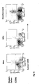
- Fig. 1 shows the results of a flow cytometric analysis of peripheral blood, spleen and bone marrow 8 weeks after transplantation of transplanted NOD / SCID animals with application of the conditioned supernatant.
- the dot plots show living cells characterized by human-specific antibodies such as CD19 (general B cell marker), CD3 (general T cell marker), CD34 (stem cell marker, mainly hematopoietic stem cells and lineage-marked progenitor cells) coated against CD45 (general leukocyte marker ).
- Fig. 1 both human T lymphocytes and human B lymphocytes and human hematopoietic stem cells were generated. Furthermore, human monocytes and human NK cells as well as human granulocytes were detected. The mentioned cell populations can be detected in all hematopoietic organs such as peripheral blood, spleen and bone marrow.
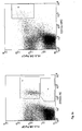
- FIG. 2a shows a flow cytometric analysis of bone marrow (representative of spleen and peripheral blood) 8 weeks after transplantation of transplanted NOD / SCID animals with application of the conditioned supernatant.
- the dot plots show human dendritic cells characterized by the expression of human-specific antigens such as HLA-DR, CD11c (right blot, region R5) or CD123 (left blot, region R4) as well as the absence of lineage-type antigens such as CD3 (general T).
- CD16 markers of neutrophilic granulocytes, subpopulation of NK cells and monocytes, macrophages
- CD19 and CD20 common B cell markers
- CD14 monoocyte marker, detection of macrophages, neutrophils, eosinophils
- CD56 generally NK cell marker , Subpopulation of T cells contained in an anti-lineage cocktail antibody mixture.
- Fig. 2a live cells are shown which show little or no expression of line-type antigens. These cells contain two human dendritic cell populations.
- the plasmoid dendritic cell population is characterized by the expression of CD123 as well as HLA-DR ( Fig. 2a , left blot, region R4).
- HLA-DR HLA-DR
- 0.21% of the cells are median plasmoid dendritic cells.
- LPS 10 ⁇ g / mouse
- the proportion of plasmid dendritic cells increases to 2.4%.
- the myeloid dendritic cell population expresses on the cell surface CD11c as well as HLA-DR ( Fig. 2a , right blot, region R5).
- HLA-DR HLA-DR
- a median of 0.17% of the cells are myeloid dendritic cells.
- LPS 10 ⁇ g / mouse
- the proportion of myeloid dendritic cells increases to 2.1%.
- Dendritic cells are an essential member of the antigen-presenting cells and form the basis for the development of an adaptive immune response. Furthermore, they are significantly involved in the process of developing immune tolerance. The functional reactivity of the cells is derived from the increase in the proportion of dendritic cells after systemic stimulation with LPS.
- human basophilic cells effector cells of allergic type I reactions
- Fig. 2a left blot, region R3
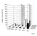
- Fig. 2b compares cytokine release from isolated bone marrow cells after LPS stimulation (10 ⁇ g / ml, white bars) from, using the conditioned supernatant, humanized NOD / SCID mice compared to unstimulated control (black bars).
- bone marrow cells of a reconstituted NOD / SCID mouse were stimulated in vitro with LPS (10 ⁇ g / ml) as an analog of infection. Controls without stimulation were included. 24 and 48 hours after stimulation, the determination of human-specific cytokines such as IL-1 ⁇ , IL-6, IL-8, IL-10, IL-12p70 and TNF- ⁇ in the culture supernatants was carried out by BD Cytometric Bead Array Analysis at the FACS. Scan (Becton Dickinson).
- the reactivity of the human dendritic cells could be clearly demonstrated on the basis of a strong inducible cytokine release ( Fig. 2b ).
- the cells produce cytokines such as IL-1 ⁇ (above 1000 pg / ml), IL-6 (above 5000 pg / ml), TNV- ⁇ (above 90 pg / ml) and IL-10 both after 24 and 48 hours after stimulation (above 100 pg / ml).
- the cytokine IL-8 is already constitutively secreted. This cytokine pattern is similar to a cytokine pattern in an infectious human event.
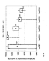
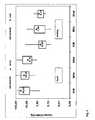
- Fig. 3a and 3b show the influence of the application of the conditioned cell culture supernatant on the distribution of human T and B lymphocytes in the spleen of transplanted mice.
- Fig. 3a shows the control without and Fig. 3b with application of the conditioned supernatant.
- the individual cell lines (median, black triangles and numbers to the right of bars) are given as a relative proportion of human CD45-positive cells.
- the bars represent the distribution (2 quartiles and the two extreme values) within the experimental animals.
- T cells (29% CD3-positive cells) and B cells (20% CD19-positive cells) form in a balanced manner.
- T cells (29% CD3-positive cells) and B cells (20% CD19-positive cells) form in a balanced manner.
- These animals have a median distribution of 1% monocytes / macrophages (CD14-positive cells), 4% CD16-positive cells (NK cells, granulocytes, subpopulation of monocytes) and 0.7% CD34-positive stem cells or imprinted progenitor cells . Due to the balanced distribution of B and T lymphocytes, the prerequisite is given to form specific immune reaction in particular T cell-dependent immune reactions. All chimeric animals formed a CD34-positive stem cell population after application of the conditioned cell culture supernatant, which can be localized especially in the bone marrow. This ensures permanent hematopoiesis in the animals. Thus, by transplanting the bone marrow of selected chimeric animals into other recipients, amplification under selective conditions can be made.
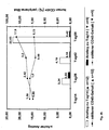
- Fig. 4a and 4b show the kinetics of human chimerism formation after transplantation of human mononuclear cells using the conditioned supernatant. Recipients which show a content of human CD45-positive cells of ⁇ 0.1% of all cells show a human chimerism.
- Fig. 4a the median content of human CD45-positive cells in the blood of all animals transplanted with 5x10 6 MNC (black line with diamonds) and the increase in the content of human CD45-positive cells relative to day 14 (white bars).
- Fig. 4b shows the mean level of human CD45-positive cells in the blood of the animals transplanted with 5x10 6 MNC, depending on the development of human engraftment.
- the data for the "low” responders are shown in the black line with circles and the black bars.
- the data for the "high” responders are shown in the black line with squares and the white bars.
- the surface molecule CD45 is expressed by all leukocytes.
- the content of human CD45-positive cells represents the development of the human hematopoietic system in the host.
- a value greater than 1 defines "high” responder, a value less than 1 "low” responder.
- "High” responders have an average of 18% human CD45-positive cells in blood on day 56 after transplantation, while low-level responders averaged only 0.7%.
- Fig. 5 Figure 4 shows a representation of the median relative content of human CD45 positive cells in lymphoid organs (day 56 post-transplantation) of humanized mice after application of the conditioned supernatant as a function of low and high engraftment.
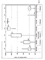
- FIG. 6 Figure 8 shows a representation of the median relative content of human CD45 positive cells in bone marrow of humanized mice 8 weeks after transplantation, as a function of the applied number of MNC and thus also of stem cells contained therein, the use of the conditioned supernatant and the use of supporting cells.
- the median is represented by a rhombus and the associated numerical values to the right of the bars.
- support cells AC133-negative cell fractions of the mononuclear cells of the umbilical cord blood were used.
- AC133 is a surface molecule which in particular hematopoietic stem cells (major part of these cells in turn carries CD34) as well as neuronal stem cells and precursors of pancreatic cells is expressed. This cell fraction did not show its own hematopoietic engraftment in control experiments.
- MNC mononuclear cells
- Ig immunoglobulins
- T cell-dependent immunization of the humanized NOD / SCID animals with tetanus toxoid was performed.
- the immunoreactivity was detected with a human-specific, TT-specific antibody detection (IgG, IgM) in an ELISA ( Fig. 7 ).
- the immunization was carried out intraperitoneally on day 0 (day 0 of the immunization corresponds to day 56 after transplantation), day 14, day 28, day 42 with 50 ⁇ g tetanus toxoid in GERBU adjuvant 10 (Gerbu, Gaiberg).
- Antibody production was measured on day 42 (T 42) after immunization (equivalent to day 112 post-transplantation).
- Fig. 7 shows that after immunization with tetanus toxoid (TT) in the mice with an established human immune system, an antigen-specific immune response in the form of IgM or IgG antibodies.
- TT tetanus toxoid
- animals with a high T-cell content T-cell group -white bars
- tetanus toxoid-specific human IgM hu-TT-IgM
- IgG antibodies hu-TT-IgG
- None TT-specific antibodies were formed in the chimeric animals with a low relative T cell content (B cell group - black bars).
- the human immune system generated in the mice is stable and remains functional for well over 3 months after transplantation, as evidenced by a T cell-dependent immune response in the form of human antibodies ( Fig. 7 ).
Landscapes
- Life Sciences & Earth Sciences (AREA)
- Environmental Sciences (AREA)
- Cell Biology (AREA)
- Animal Behavior & Ethology (AREA)
- Zoology (AREA)
- Animal Husbandry (AREA)
- Biodiversity & Conservation Biology (AREA)
- Micro-Organisms Or Cultivation Processes Thereof (AREA)
- Medicines Containing Material From Animals Or Micro-Organisms (AREA)
Description
- Die vorliegende Erfindung betrifft ein Verfahren zur Etablierung eines funktionsfähigen adaptiven humanen Immunsystems in einem geeignetem Tiermodell. Das Tiermodell eignet sich z. B. für präklinische pharmakologische Studien und die Herstellung von humanen Antikörpern. Hierfür werden immundefiziente Mäuse benutzt, die kaum immunologische Reaktionen gegen die übertragenen Zellen entwickeln.
- Grundlegende Strategien und Technologien einer medizinischen Therapie insbesondere aus dem Bereich der Regenerativen Medizin werden oft in tierischen Modellen (z.B. unter Verwendung von Nagern oder Huftieren) charakterisiert. Bei der Überführung so gewonnener Prinzipien in die klinische Anwendung am Menschen, ist es oft schwierig abzugrenzen, ob ein therapeutisches Produkt oder eine therapeutische Strategie für ein menschliches Target optimal ist oder nicht. So gibt es zwei Möglichkeiten: a) das humane Target ist in der klinischen Anwendung invalid oder b) das therapeutische Agens hat nicht dieselben funktionellen Eigenschaften. Dies ist speziell für Tumor- und Transplantationstherapien (u.a. Stammzelltransplantationen) und mit besonderem Augenmerk bei Immuntherapien, die speziesspezifische monoklonale oder polyklonale Antikörperpräparationen nutzen, relevant. Pharmazeutische Unternehmen sehen sich mit der Herausforderung konfrontiert, dass nur aus ca. 10% der in klinischen Studien getesteten Wirkstoffe ein neues zugelassenes Medikament hervorgeht. Zur Absicherung sowie der Akzeptanz einer neuen Technologie oder Arbeitsstrategie sind präklinische Modelle und Werkzeuge erforderlich, bei denen menschliche Zellen und Gewebe in einen möglichst komplexen systembiologischen Zusammenhang untersucht werden können. Immundefiziente Mäuse, in denen menschliche Zellen des Immunsystems miteinander reagieren ohne abgestoßen zu werden, können dafür eine Antwort darstellen, da sie den Vorteil eines Kleintiermodells mit einer besseren Korrelation zu den klinischen Gegebenheiten verknüpfen. Zwei Strategien zur Entwicklung entsprechender Tiermodelle bei Nagern wurden bisher dargestellt.
- Die erste Strategie nutzt die Technologie zur Herstellung genetisch veränderter (transgener, knock-out, knock-in) Tiere, mittels der Einführung menschlicher Gene, durch Induktion spezifischer Mutationen oder den Ersatz eines murinen Gens durch das humane Homolog in somatischen oder Keimbahnzellen um genetische Veränderungen nachzuahmen bzw. um humane Antigene oder Targets zu exprimieren. Die Grenzen dieser Methode liegen neben den hohen Kosten, den geringen Erfolgsraten insbesondere in der limitierten Herstellung, meist einzelner, humaner Eigenschaften und somit deren Reaktivität mit meist tierischen Partnermolekülen sowie meist nicht identischen Genexpressionskontrollmechanismen. Daher wird diese Methode meist ausschließlich zur Charakterisierung des Potenzials von Wirkstoffen bezüglich des Wirkstoffmetabolismus und der Bestimmung der Toxizität oder zur Untersuchung der Aktivität eines einzelnen oder eines Sets von Targets genutzt (1;2).
- Der zweite und komplexere Weg ist die Erzeugung von Chimären durch Xenotransplantation (3). Dies umfasst die Applikation von humanen Zellen oder Geweben in meist immundefiziente Tiere. Herausragende Wirtstiere zur Erzeugung eines humanen Immunsystem sind Mäusestämme, die mehrfache Defekte in der adaptiven Immunität aufweisen wie Rag2-/-γ-/- (4), BNX oder NOD/SCID B2mnull (5). Als ein Standardmodell zur Humanisierung dienen verschiedene Linien der NOD/SCID- (non-obese-diabetic/severe-combined-immuno-deficiency) Maus. Diese sind im Wesentlichen durch folgende immundefiziente Eigenschaften charakterisiert: kompletter Verlust von B- und T-Lymphozyten, reduzierte Anzahl von NK-Zellen, Defekte in der Differenzierung und Funktion von Antigen-präsentierenden Zellen und der Abwesenheit von zirkulierendem Komplement. Diese Mäuse sind empfänglicher für ionisierende Strahlung als der Wildtyp und weisen Defekte im DNA-Reparatursystem auf. Die Ausbildung von humanen einzelnen bzw. mehreren Linien der Hämatopoese in einem immundefizienten Tier ist nach Transplantation von humanen hämatopoetischen Stammzellen, differenzierten hämatopoetischen Zellen sowie lymphoiden Organen möglich. In Abhängigkeit vom genutzten Mausstamm und dessen Konditionierung sowie der Anzahl und der Quelle von hämatopoetsichen Stammzellen ist die Entwicklung von 0,1 bis 90% humaner CD45+ Zellen im peripheren Blut oder der Milz und dem Knochenmark der Tiere möglich.
- Nachteilig ist bisher die extrem hohe Variabilität der Rekonstitution der Hämatopoese. Zudem werden einzelne humane Zelltypen unterschiedlich repräsentiert, abhängig vom Mausstamm und der Konditionierung der Tiere. Kennzeichnend ist für die NOD/SCID-Maus: Es wachsen bevorzugt und fast ausschließlich B-Zellen. Die humanen B-Lymphozyten zeigen die typischen Muster des Immunglobulin-Genrearrangements (6) und generieren ein diverses Immunglobulin-Repertoire nach Applikation von humanem fötalen, Nabelschnurblut oder adulten lymphoiden Progenitoren (7). Zirkulierende T-Zellen fehlen oder werden nur in äußerst geringem Umfang und zudem noch verzögert gebildet. Nur nach einer Vorbehandlung der Tiere mit rekombinanten Tumor-Nekrosis-Faktor und der Applikation von hohen Zellzahlen mononukleärer Zellen ist eine schnelle und starke Entwicklung von T-Lymphozyten sichtbar (8). Das humane Engraftment weist jedoch sehr hohe Varianzen innerhalb der Tiergruppe auf. Die Phänotypisierung der humanen T-Zellen zeigt, trotz der Variabilität, einen CD4/CD8-Quotienten von 1:1 bzw. 1:2 in Abhängigkeit von der verwendeten Stammzellquelle. Die Zellen weisen ein diverses T-Zell-Rezeptor-Repertoire auf. Vorläufige Funktionsanalysen dieser T-Zellen zeigen in In-vitro-Proliferationsstudien mit PHA oder IL-2, dass sie aktiviert werden können, allerdings nur auf einem sehr niedrigem Niveau.
US 6,627,792 B2 nutzt Sektionen von Rippen-Knochengewebe von operierten Patienten, welches subkutan in den Peritonealraum appliziert wird, zur erfolgreichen Rekonstitution von T-Zellen. Deren Verfügbarkeit und ethische Bedenken limitieren den Einsatz. Nach intravenöser Injektion von Einzelzellsuspensionen dieses Knochengewebes ist der humane Chimärismus sehr niedrig und T-Zellen konnten nur schwerlich nachgewiesen werden.US 6,060,643 nutzt zur Rekonstitution in BNX-Mäusen humane hämatopoetische Zellen von G-CSF-mobilisiertem peripheren Blut (PB), fötales und adultes Knochenmark (FKM, AKM). Nur 17% bzw. 4,7% der Mäuse waren mit einer extrem hohen Variabilität nach Transplantation von PB bzw. AKM nach 6-8 Wochen chimär. Kein Chimärismus wurde nach Applikation von FKM erzielt. Weniger als 3% bzw. 15% der mit PB oder AKM bzw. FKM transplantierten Tiere zeigten ein Langzeitengraftment. Tiere, die PB erhielten, haben eine mittlere Überlebensrate von 25% nach 30 Tagen. Dies könnte darauf zurückzuführen sein, dass autoreaktive Zellen gebildet wurden oder eine regulatorisch aktive Zellpopulation nicht ausgeprägt wird. Klare Zeichen einer Graft-versus-Host-Disease konnten nur nach Applikation von peripherem Blut adulter Spender gefunden werden (9). Dies zeigt die bisher unbefriedigende funktionelle Potenz von menschlichen Zellen im Kontext des Tiermodells an. - Humane Zellen der Makrophagen-Dendritischen Zellreihe konnten bisher nur als unreife CD34- HLA-DR+ CD4+ Dendritische Zellvorläufer (DC) nach Implantation von humanen mononukleären Zellen des Nabelschnurblutes nachgewiesen werden. In den lymphoiden Organen der NOD/SCID-Maus konnten keine reife DC's detektiert werden (10). Obgleich die Differenzierung der DC zu reifen Zellen im Tiermodell geblockt war, zeigten sich die unter In-vitro-Bedingungen gereiften DC als effiziente Stimulatoren in einer gemischten Leukozyten-Reaktion. Cravens et al. (11) demonstrierte die Entwicklung von humanen reifen und reaktionsfähigen DC in der NOD/SCID-Maus ohne zusätzliche Zytokingabe. Die Zellen reagieren auf eine LPS-Stimulation in-vivo mit einer hohen Zytokinsekretion von IL-8, TNFα, IL-10 sowie IL-12p70. Relativ niedrig liegt die Freisetzung von IL-1ß, IL-6 sowie IFNγ. Diese Zytokine sind jedoch im humanen System charakteristisch für eine akute oder systemische Infektion. Weitere Entwicklungen sind daher nötig, in denen ein dem humanem System vergleichbares Zytokinprofil angestrebt werden muss.
- Bonnet D et al. (Bone Marrow Transplant. 1999. 23(3):203-9) beschreiben die Transplantation von Lin-CD34+CD38--Stammzellen aus dem Nabelschnurblut ("cord blood") oder "acute myeloid leukemia"-Stammzellen (AML) in NOD/SCID-Mäuse. Die Zugabe der Koloniestimulierende Faktoren oder eine Kotransplantation von Lin+CD34- oder Lin-CD34+CD38+-Zellen als "accessory cells" ist notwendig für die erfolgreiche Transplantation der Stammzellen. Die verwendeten Hämatopoese- und Kolonie-stimulierenden Faktoren (CSF) sind Interleukin 3 (IL-3), Granulozyten/Makrophagen-CSF (GM-CSF), Granulozyten-CSF (G-CSF) u. der Stammzellen-Faktor (auch "Steel-Faktor" - SCF). Cashman J D et al (Blood. 1999. 93(2):481-7) beschreiben ebenfalls ein Transplantationsmodell für humane Stammzellen aus dem Nabelschnurblut in NOD/SCID-Mäuse. Hier erfolgt wieder die Zugabe von Kolonie-stimulierenden Faktoren, wie Stammzellen-Faktor (SCF oder "steel factor - SF), Erythropoetin (EP oder EPO), G-CSF, GM-CSF, IL-3, sowie Interleukin-6 (IL-6) und Flt3Ligand. Lapidot et al. (Science. 1992 255(5048):1137-41) beschreibt ebenfalls ein Transplantationsmodell für humane Knochenmarkzellen in SCID-Mäuse, welches nach Aussage der Autoren als Tiermodell der humanen akuten lymphoblastischen Leukämie dient. Nach der Transplantation erfolgt eine Stimulation der Mäuse durch die Applikation von humanen MGF (eine veraltete Bezeichnung des Stammzellen-Faktors SCF) und einem Fusionsprotein bestehend aus IL-3 und GM-CSF.
- Vormoor et al. (Blood. 1994. 83(9):2489-97) beschreiben ein erfolgreiches Engraftment in 80% der SCID-Mäuse nach Injektion von Nabelschnurblutzellen, welches unabhängig von der Applikation von hämatopoetischen Zytokinen ist. Das multilineage Engraftment von durchschnittlich 10% humanen Zellen im Knochenmark wird von den Autoren als hoch eingeschätzt, welches dem damaligen Stand der Technik entsprach.
- Kollmann et al. (Proc Natl Acad Sci USA. 1994.91(17):8032-6) zeigen ein signifikantes Engraftment humaner Zellen im Knochenmark und in der Peripherie nach Transplantation von vorkultivierten fötalen Knochenmarkzellen. Sie weisen B-Zellen und myeloide Zellen (Monozyten) nach, es wachsen keine T-Zellen aus.
- Zwei jüngere Arbeiten konstatieren jeweils, dass die Funktion der (humanen) hämatopoetischen Stammzellen in NOD/SCID-Mäusen durch die noch vorhandene NK-Zellaktivität limitiert ist und verfolgen unterschiedliche Wege um diese Einschränkung zu überwinden. Kerre et al. (Blood. 2002 Mar 1;99(5):1620-6) optimieren die Humanisierung der NOD/SCID-Mäuse durch Blockierung der murinen NK-Zellaktivität mittels intraperitonealer Applikation von 200µg des Antikörpers TM-ß1. Shultz et al. (J Immunol. 2005. 174(10):6477-89) generieren einen neuen NOD/SCID-Mausstamm, in dem die γ-Kette des IL-2-Rezeptors defizient und somit die NK-Zellentwicklung blockiert ist. Als zusätzliche Optimierungsstrategie wurde die Applikation 20µg/Tier eines Fc-IL-7-Fusionsprotein verfolgt.
-
WO 01/15521 A1
DE 4337007 C1 offenbart ein Mausmodell zur Testung von AIDS-Impfstoffen, bei dem das Immunsystem von Mäusen durch Bestrahlung zerstört wird und anschließend embryonale Stammzellen transplantiert werden und die Mäuse mit IL-3 behandelt werden. - Zusammenfassend wird beurteilt, dass bisher existierende Verfahren und Modelle partiell die Entwicklung und Reifung von humanen hämatopoetischen Zellen und deren Reaktionsvermögen in einem Tier widerspiegeln. Bisher ist jedoch kein Verfahren etabliert, welches ein standardisiertes, reproduzierbares Modell generiert, dass die Gesamtheit eines humanen, insbesondere adaptiven Immunsystems repräsentiert. Dieses ist jedoch eine unbedingte Notwendigkeit um Testungen in vorklinischen Phasen effizienter zu machen und Therapieausfälle zu minimieren. Die Herstellung sowie die Verwendung von humanspezifischen therapeutischen oder regenerativen Agenzien setzt eine gemeinsame funktionelle Präsenz von humanen Dendritischen Zellen, T- sowie B-Zellen, NK-Zellen, Monozyten und Granulozyten voraus.
- Die durch die Erfindung zu lösende Problemstellung ist daher, ein neues Verfahren zur dauerhaften Herstellung von human-spezifischen Chimären bereitzustellen, das nicht die aus dem Stand der Technik bekannten Nachteile aufweist. Insbesondere ist die Aufgabe der Erfindung, die Sicherheit des Anwachsens eines Transplantates mit Zellen, die ein hämatopoetisches Potential besitzen, in Tieren zu erhöhen und die interindividuelle Variabilität des Rekonstitutionserfolges zu minimieren. Damit wird die Voraussetzung von homogenen Versuchsgruppen mit vergleichbaren Reaktionsmuster und -stärke geschaffen.
- Des Weiteren ist es Aufgabe der Erfindung, eine dauerhafte Ausbildung von allen Zellen des blutbildenden Systems in einem ausgewogenen Maße sowie die Ausprägung jeweils zelltypischer Reaktionsmuster in einem Tier zu erzielen. Die Erfindung soll die Kooperation der humanen Zellen in ihrer typischen Weise in einem Tier ermöglichen und so ein funktionsfähiges humanes, insbesondere adaptives Immunsystem darstellen. Die Chimären sollen keine oder nur minimale Anzeichen einer Transplantat-bedingten Reaktion gegen den Wirtsorganismus aufweisen. Zusätzlich soll die verfahrensbedingte, teilweise sehr hohe Sterblichkeit von Tieren (15-65%) minimiert werden.
- Zur Lösung der Aufgabe wird mittels der Erfindung ein Verfahren bereitgestellt, das durch ein neues Konditionierungsverfahren der Wirtstiere, die bisherigen Nachteile der bekannten Lösungen überwindet.
- Erfindungsgemäß wird die Aufgabe gelöst durch ein Verfahren zur Herstellung eines Tiermodells für das humane Immunsystem in einer immunodefizienten Maus, mit den Schritten:
- a) Transplantation von humanen Stammzellen mit hämatopoetischen Potential in eine NOD/SCID-Maus und
- b) Konditionieren der Maus mit Zellkulturüberstand einer Kultur von Zellen einer humanen Zelllinie ausgewählt aus 358/8 [abgeleitet aus NCI-H358] und daraus abgeleitete Subclonen, sowie A-549, wobei der Zellkulturüberstand jeweils 20 pg/ml bis 10 ng/ml der humanen Faktoren Interleukin-8, Interleukin-6, und vaskulo-endothelialen Wachstumsfaktor (VEGF), Interferon-γ-Induced Protein 10, Monocyte Chemoattractant Protein-1, Angiogenin und Tissue-type Plasminogen Activator enthält, und gegebenenfalls
- c) Applikation von Stützzellen ohne eigenständiges hämatopoetisches Potential.
- Die Erfindung wird charakterisiert durch die Nutzung von "konditionierten Zellkulturüberständen", die Kombinationen (Cocktails) von Botenstoffen insbesondere humanen Ursprungs enthalten. Diese werden in Verbindung mit der Applikation von humanen Stammzellen zur Herstellung des erfindungsgemäßen Tiermodells (Immunsystem-Chimären) in die immundefizienten NOD/SCID-Mäuse eingebracht.
- Das erfindungsgemäße Verfahren ermöglicht vorteilhaft die Ausbildung sowohl von B- und T-Lymphozyten, NK-Zellen, Monozyten, Granulozyten, hämatopoetischen Stammzellen als auch von Dendritischen Zellen und die Ausprägung Zelltyp-spezifischer Reaktionen dieser Zellen. Damit steht ein Modell zur Verfügung, welches das humane, insbesondere das adaptive Immunsystem in wesentlichen Zügen seiner Funktionalität widerspiegelt. Das Verfahren ist für alle In-vivo- oder abgeleiteten In-vitro-Anwendungen nutzbringend, bei denen immunologische Prozesse mit menschlichen Zellen für diagnostische Zwecke, für Forschungszwecke, für die Entwicklung und Testung von Heilverfahren oder zur Entwicklung von Produkten benötigt werden. Beispielgebend sind hier die Herstellung und Verwendung von monoklonalen humanen Antikörpern, Entwicklung von Vakzinierungsstrategien, tierexperimenteller Krankheitsmodelle, Testung pharmakologischer Wirkstoffe, Entwicklung von Strategien zur Induktion von Immuntoleranz nach Organtransplantation insbesondere unter Verwendung humaner, humanisierter oder muriner Antikörper.
- Die erfindungswesentlichen Merkmale des in den Patentansprüchen dargestellten Verfahrens sowie die in den weiteren Patentansprüchen geltend gemachten Ansprüche werden im Folgenden in verallgemeinerter Form erläutert.
- Als erfindungswesentlich für die Erzeugung eines Chimärismus wird die Konditionierung mit "konditioniertem Zellkulturüberstand" in alleiniger oder kombinierter Verwendung mit Stützzellen bewertet.
- Als "konditionierter Zellkulturüberstand" wird eine zellfreie, lösliche Phase definiert, die von Zellen abgetrennt wurde, die einzeln oder im Gewebeverbund vorliegen, nachdem während einer Inkubationsphase verschiedene Moleküle in den Zellkulturüberstand sezerniert wurden. Die sezernierenden Zellen entstammen derselben Spezies, wie die zur Erzeugung einer Chimäre genutzte Stammzellquelle. Es werden humane Zelllinien, die permanent oder nach geeigneter Stimulation bzw. Differenzierung lösliche Faktoren wie Zytokine produzieren, zur Herstellung des Zellkulturüberstands benutzt, welche aus den Zelllinien 358/8 (abgeleitet aus NCI-H358, European Collection of Cell Cultures, Salisbury, Wiltshire,
UK, No. 95111733 - Der Zellkulturüberstand enthält die Faktoren, die im Zellkulturüberstand einer Zelllinie ausgewählt aus 358/8, A-549 und VA-ES-BJ oder in einem gemischten Zellkulturüberstand von zwei oder drei der genannten Zelllinien enthalten sind.
- Als vorteilhaft hat sich erwiesen, wenn im Zellkulturüberstand die Faktoren IL-8, IL-6, VEGF, IP-10, MCP-1, ANG und tPA vorhanden sind.
- IL-8, IL-6, VEGF, IP-10, MCP-1, ANG und tPA sind in den erwähnten Kulturüberständen in Konzentrationsbereichen von jeweils 20pg/ml bis 10ng/ml enthalten.
- Besonders bevorzugt sind die Zytokine in den erwähnten Kulturüberständen in den nachfolgend ausgeführten Konzentrationsbereichen enthalten: IL-8 (500 pg/ml bis 10 ng/ml), IL-6 (50 pg/ml bis 1 ng/ml), VEGF (500 pg/ml bis 10 ng/ml), IP-10 (20 pg/ml bis 1 ng/ml), MCP-1 (500 pg/ml bis 10 ng/ml), ANG (20 pg/ml bis 1 ng/ml), und tPA (500 pg/ml bis 15 ng/ml).
- Ein Gehalt an TNF-α, IL-10, MIG, RANTES, sVCAM-1, sCD40L, bFGF, sP-Selectin, oder IGF ist hingegen nicht oder nicht in höheren Konzentrationen (≤20pg/ml) notwendig.
- Der Zeitpunkt dieser Konditionierung mit Zellkulturüberstand kann sowohl vor als auch nach dem Transplantationstag an einem oder mehreren Tagen erfolgen. Die Applikationsmenge sollte nicht den allgemeinen Empfehlungen, die in Richtlinien zur Behandlung von Versuchstieren verankert sind, zuwiderlaufen.
- Der konditionierte Kulturüberstand wird bevorzugt unmittelbar nach der zellulären Transplantation appliziert und anschließend über einen Zeitraum von 14 Tagen jeden zweiten Tag verabreicht.
- Der Applikationsort ist nicht beschränkt. Es empfiehlt sich jedoch eine intravenöse oder intraperitoneale Applikation. Für Mäuse sollte eine intraperitoneale Applikation mit einer Menge von ca. 200µl, bevorzugt 150 µl bis 250 µl pro Applikation, angestrebt werden. Es ist zweckmäßig, die Verträglichkeit jedes gewählten "konditionierten Zellkulturüberstandes" vor der Transplantation im Tiermodell zu überprüfen.
- Zusätzlich zur Applikation der stammzellhaltigen Quelle werden in einer speziellen Variante des erfindungsgemäßen Verfahrens sogenannte "Stützzellen" ohne ein eigenständiges hämatopoetisches Potential appliziert werden. Dabei kann es sich z.B. um Stammzell-abgereicherte (maximaler Stammzellgehalt ≤ 0,1%) oder -negative Zellpopulationen (maximaler Stammzellgehalt ≤ 0,01%) einer Stammzell-Quelle als auch um mesenchymale Stammzellen handeln. Die Verwendung von "Stützzellen" mit onkogenem Potential sollte vermieden werden. Als Stützzellen können beispielsweise CD34-negative oder CD133-negative mononukleäre Zellen des Nabelschnurblutes oder des Knochenmark verwendet werden. Die Stützzellen werden bevorzugt in Konzentrationsbereichen von 1x10⁴ bis 5x10⁷ Zellen pro Zelltransplantat eingesetzt. Die Applikation der Stützzellen erfolgt bevorzugt gemeinsam mit der stammzellhaltigen Population.
Für das Tiermodell dienen NOD/SCID-Mäuseals Wirtstier zur Erzeugung des Chimärismus. Das Geschlecht der Tiere ist nebensächlich. Das Alter sollte insbesondere bei Mäusen mehr als 6 Wochen betragen. Das Verfahren wird erleichtert, da der Rezipient aus einer Inzuchtlinie stammt, die mehrere Immundefekte aufweist, vor allem in der adaptiven Immunantwort. Als bevorzugt wird hier der NOD/SCID Mausstamm des Jackson Laboratory, Bar Harbor, ME, USA benannt. Die immundefizienten Tiere sollten unter spezifisch-pathogenfreien Konditionen gehalten werden. - Zur Erzeugung des Chimärismus benötigte Stammzellen weisen ein hämatopoetisches (blutbildendes) Potential auf. Derartige hämatopoetische Stammzellen sind Zellen, die sich vermehren können und befähigt sind alle Zellen des blutbildenden Systems, insbesondere des Immunsystems, zu generieren. Solche Stammzellen lassen sich vor allem in Präparaten aus humanem Knochenmark, mobilisiertem sowie nicht-mobilisiertem peripherem Blut, Nabelschnurblut sowie -gewebe finden. Die erfindungsgemäß verwendeten Stammzellen sind in der Lage Zellen des humanen Immunsystems auszubilden. Insbesondere können die Stammzellen in B- und T-Lymphozyten, NK-Zellen, Monozyten, Granulozyten, sowie Dendritische Zellen differenzieren.
- Das Alter und das Geschlecht der Spender sind unerheblich. Eine Anreicherung der Stammzellen ist möglich, aber nicht generell notwendig. Vorzugsweise sollte eine mononukleäre Einzelzellfraktion zur Verfügung gestellt werden. Die Entfernung der Erythrozyten sowie der Granulozyten durch gängige Methoden ist empfehlenswert. Sowohl frisch gewonnene als auch kryokonservierte Präparate können genutzt werden. Eine Zellfärbung mit ausgewählten Vitalfarbstoffen wie CFSE behindert nicht den Erfolg des Verfahrens.
- Im Allgemeinen werden Tiere, in denen ein Chimärismus erzeugt werden soll, durch eine Konditionierung, die u.a. die Entfernung restlicher tierischer immunkompetenter Zellen verfolgt, für eine nachfolgende Transplantation der Stammzellen empfänglicher gemacht. Bevorzugt erfolgt dies durch eine radioaktive Bestrahlung des Empfängerorganismus. Als Strahlenquelle können radioaktive Isotope als auch Röntgengeräte genutzt werden. Die Bestrahlung sollte die jeweils tier-spezifische sub-lethale Dosen nicht überschreiten. Diese Dosis wird für jeden Zuchtstamm individuell ermittelt. In der Regel sollte eine einmalige und keine multiple Bestrahlung angestrebt werden. Die sub-lethale Dosis bei einer einmaligen Bestrahlung beträgt für NOD/SCID-Mäuse in der Regel 250-350 cGy.
- Die Applikation des Transplantates kann kurz- oder langfristig nach der Konditionierung durch Bestrahlung erfolgen. Kurzfristig impliziert sofort nach der Bestrahlung, langfristig können mehrere Tage umschließen. Im Allgemeinen wird ein Zeitfenster zwischen 3 bis 24 Stunden nach der Bestrahlung berücksichtigt. Der Applikationsort des Transplantates ist nicht zwingend vorgeschrieben. Es sollte bei Einzelzellsuspensionen eine intravenöse oder intraperitoneale Gabe bevorzugt werden. Die Tiere können bei Bedarf anästhesiert werden. Bevorzugt werden pro Maus von 1x10³ bis 5x10⁷ mononukleäre Zellen, die die Stammzellen enthalten, transplantiert. Der Gehalt an CD34-positiven Stammzellen liegt vorzugsweise im Bereich von 5 bis 5x10⁵, bevorzugt 1x10³ bis 1x10⁴.
- Nach der Transplantation werden die Tiere unter Standardbedingungen oder spezifisch-pathogenfreien Konditionen gehalten.
- Die Ausprägung des erzeugten Chimärismus wird durch Kontrollen, in regelmäßigen Abständen, anhand von Proben des peripheren Blutes der transplantierten Tiere bestimmt. Bei Mäusen eignet sich besonders die Gewinnung von Blut aus dem retrobulbären Augenplexus oder aus der Schwanzvene. Der Anteil der humanen Zellen wird bevorzugt mittels durchflusszytometrischer Untersuchungen unter Verwendung von human-spezifischen Antikörpern (Linienmarker, Differenzierungsantigene, Aktivierungsmarker) ermittelt. Bei einem geringen Probenvolumen empfiehlt sich ein jeweils spezies-spezifischer pan-Leukozytenmarker wie CD45, möglichst im direkten Vergleich von Spender- und Empfängerspezies.
- Vorteilhaft ist nach Anwendung des erfindungsgemäßen Verfahrens bereits 14 Tage nach Transplantation ein sicherer Nachweis von humanen Zellen in der Peripherie möglich. Diese Form des Monitoring erlaubt unter den Bedingungen der Erfmdung eine zuverlässige Aussage über den zeitlichen Rekonstitutionserfolg. Des Weiteren korreliert das Auftreten von humanen Zellen in der Peripherie mit einem Engraftment in der Milz und dem Knochenmark.
- Die vorliegende Erfindung überwindet die Probleme und Nachteile des bisherigen Standes der Technik zur Herstellung human-chimärer Tiere, indem sie ein neues standardisierbares, hoch reproduzierbares Verfahren unter Verwendung eines neuen Konditionierungsverfahrens der Tiere in Verbindung mit humanen Stammzellen bereitstellt.
- Das Verfahren erlaubt die Bereitstellung von gleichwertigen Chimären und schafft somit eine Reduktion der bisher nach dem Stand der Technik bekannten hohen interindividuellen Variabilität. Unter den Bedingungen der Erfindung ist nur eine Mortalitätsrate von 10% zu verzeichnen.
- Ein neuer erfindungsgemäßer Aspekt ist die verlässliche Verwirklichung von Chimären, die humane immunkompetente Zellen des hämatopoetischen Systems bilden.
- Das Tiermodell für das humane Immunsystem in einer immunodefizienten NOD/SCID-Maus, welches humane immunkompetente Zellen des hämatopoetischen Systems enthält, umfasst insbesondere T- und B-Zellen, Monozyten, Dendritische Zellen, NK-Zellen, Granulozyten sowie CD34+-Stammzellen. Alle Tiere weisen unter den Bedingungen der Erfindung ein humanes Engraftment im peripherem Blut, der Milz und dem Knochenmark auf. Vorteilhaft ist die dauerhafte Rekonstitution der Hämatopoese. Die Ausprägung sowie Zusammensetzung des menschlichen Blutbildes, die am Tag 56 nach der Transplantation in einer erfindungsgemäß behandelten NOD/SCID-Maus detektiert wurden, konnten vorteilhaft über einen Zeitraum von 5 Monaten nachgewiesen werden. Das erfindungsgemäß hergestellte Tiermodell für das humane Immunsystem ist daher sehr stabil.
- Die Erfindung stellt vorteilhaft Mäuse zur Verfügung, die ein dem Menschen vergleichbares (äquivalentes) Immunsystem ausprägen, mit besonderem Fokus auf ein reaktives adaptives Immunsystem. Im erfindungsgemäß hergestellten Tiermodell haben die humanen immunkompetenten Zellen vorteilhaft eine vergleichbare Funktionalität, wie im menschlichen Körper, insbesondere eine vergleichbare Antwort auf Antigene und ein Zytokinprofil, welches dem humanen vergleichbar ist.
- Die humanen dendritische Zellen in dem Tiermodell sind fähig, humanes Interleukin-8 konstitutiv und humanes Interleukin-6, humanes Interleukin-1ß und humanen Tumor-Necrose-Faktor-alpha (TNF-α) nach Immunstimulation (z. B. mit LPS) zu produzieren.
- Erstaunlicherweise zeigen die Mäuse keine sichtbaren Zeichen einer Graft-versus-Host-Disease. Auf Grund der neuen, stabilen Ausprägung von humanen hämatopoetischen Stammzellen im Empfängerorganismus kann vorteilhaft eine weitere Transplantation von Zellen, die aus diesen stammen in einen oder mehrere sekundäre Rezipienten vorgenommen werden und somit eine weitere Vervielfältigung von chimären Organismen vorgenommen werden.
- Die konditionierten, transplantierten Mäuse weisen in der Milz innerhalb der humanen Zellen in einem ausgewogenem Maße T-Zellen (Median: 29%) und B-Zellen (Median: 20%) auf. Des Weiteren können humane NK-Zellen (Median: 0,36%), Monozyten (Median: 1,1%), Granulozyten (Median: 4%) sowie CD34-positive Stammzellen (Median: 0,7%) detektiert werden. Der Median resultiert aus Untersuchungen mit 21 Tieren.
- Die Bildung von humanem Immunglobulin M sowie Immunglobulin G in vivo als Beleg für die Aktivität der humanen B-Zellen konnte nachgewiesen werden.
- Die hohe Reaktivität von Dendritischen Zellen (0,2% - 2,4% der humanen Zellen im Knochenmark) wurde durch Zytokinproduktion nach LPS-Stimulation in vitro gezeigt. Die Zellen produzieren sowohl 24 als auch 48 Stunden nach Stimulation Zytokine wie IL-1β (über 1000 pg/ml), IL-6 (über 5000 pg/ml), TNF-α (über 90 pg/ml) sowie IL-10 (über 100 pg/ml). Das Zytokin IL-8 wird bereits konstitutiv sezerniert. Dieses Zytokinmuster ist vergleichbar mit einem infektiösen Geschehen im Menschen. Mit der Reaktivität der Dendritischen Zellen als potente antigen-präsentierende Zellen (exogene als auch endogene Antigene), die eine Schlüsselposition bei der Aktivierung und Ausprägung einer adaptiven Immunantwort einnehmen, bei gleichzeitigem Vorhandensein von reaktiven humanen T- wie auch B-Zellen, ist eine dem menschlichen Immunsystem äquivalente Funktionalität des (chimären) Tiermodells gegeben.
- Die Funktionalität des menschlichen adaptiven Immunsystems in der Maus konnte eindeutig mit dem Nachweis einer induzierten, T-Zellabhängigen, Antigen-spezifischen Antikörperproduktion (Immunisierung) gezeigt werden. Eine Reifung der Immunantwort in Form einer Bildung von Antigen-spezifischen Antikörpern des Immunglobulin G-Isotyps ist nachgewiesen.
- Das erfindungsgemäß hergestellte Tiermodell stellt damit vorteilhaft ein Modellsystem für das humane Immunsystem dar, welches sich für die Produktentwicklung und Forschung, insbesondere pharmakologische Forschung, präklinische Studien, Pathogeneseforschung, Entwicklung von neuen diagnostischen oder therapeutischen Strategien eignet.
- Das vorliegende Verfahren zur Herstellung von humanen reaktiven Chimären und das damit hergestellte Tiermodell erlaubt vorteilhaft die Herstellung humaner monoklonaler als auch polyklonaler Antikörper. Besonderer Stellenwert kommt der Herstellung kompletter humaner Antikörper ohne tierischen Fremdanteil und physiologischer vollständiger Glykosilierung zu. Diese humanen Antikörper haben einen hohen Stellenwert auf dem pharmazeutischen, therapeutischen sowie diagnostischen Markt.
- Die erfindungsgemäß im Tiermodell generierten humanen Zellen haben vorteilhaft die Fähigkeit sowohl exogene als auch endogene Antigene zu erkennen und daraufhin eine Immunantwort auszubilden. Daraus leiten sich u.a. Möglichkeiten zur Herstellung von Antigen-spezifischen Dendritischen Zellen, Überprüfung immuntherapeutischer Protokolle z.B. unter Verwendung von humanen, humanisierten oder murinen therapeutischen Antikörpern, Charakterisierung von immunmodulatorischen Substanzen (Chromatinmodifikatoren, "small molecules") oder immunmodulatorisch wirksamer Zellpopulationen (mesenchymale Stammzellen, regulatorische T-Zellen, tissue engineered products) ab.
- Dieses Modell kann vorteilhaft als eine Plattformtechnologie für die weitere Entwicklung von Modellen für Infektions- (wie HSV, EBV, HIV, Röteln), Autoimmun-, Tumor-, Transplantations-assoziierte oder entzündliche Erkrankungen und therapeutischer Strategien genutzt werden.
- Im Folgenden wird das erfindungsgemäße Verfahren anhand eines Ausführungsbeispiels und den zugehörigen Abbildungen erläutert:
- Eine humane Lungenzellkarzinom- (hier die TypII-Pneumozyten-Linie 358/8 [abgeleitet aus NCI-H358] Subklon SC25, erhalten von Institut für Zoologie, Universität Leipzig) wird unter sterilen Bedingungen in RPMI 1640 unter Zusatz von 5% fötalem Kälberserum kultiviert, bis sie die logarithmische Wachstumsphase erreicht. Zur Herstellung des konditionierten Mediums werden 2-4x10⁵ Zellen/ml ausgesät. Das Kulturmedium wird nach 3 Tagen geerntet, gesammelt, sterilfiltriert (Porengröße 0,2µm) und portioniert bei mindestens -20°C bis zur Verwendung gelagert.
- In dem konditionierten Kulturüberstand sind die Zytokine in dem nachfolgend ausgeführten Konzentrationsbereichen enthalten: IL-8 (500 pg/ml bis 10 ng/ml), IL-6 (50 pg/ml bis 1 ng/ml), VEGF (500 pg/ml bis 10 ng/ml), IP-10 (20 pg/ml bis 1 ng/ml), MCP-1 (500 pg/ml bis 10 ng/ml), ANG (20 pg/ml bis 1 ng/ml), und tPA (500 pg/ml bis 15 ng/ml).
- Ein Gehalt an TNF-α, IL-10, MIG, RANTES, sVCAM-1, sCD40L, bFGF, sP-Selectin, oder IGF ist hingegen nicht oder nicht in höheren Konzentrationen (≤20pg/ml) nachweisbar.
- Mindestens 6 Wochen alte, männliche als auch weibliche NOD/SCID-Mäuse (Jackson Laboratories, USA) wurden unter spezifisch-pathogenfreien Bedingungen gehalten und dienten als Rezipienten. Die Ganzkörper-Bestrahlung der Tiere erfolgte mit 350cGy mittels eines Röntgentherapiegerätes. 3-5 Stunden nach der Bestrahlung erfolgte die Applikation von 1-10x106 mononukleären Zellen, isoliert aus humanem Nabelschnurblut, in die Schwanzvene eines mittels Ether anästhesierten Tieres. Es wurden sowohl Zellen eines einzelnen Blutes eines Spenders als auch Zellen mehrerer, gemischter Blutproben genutzt. Die mononukleären Zellen wurden mittels einer Dichtegradientenzentrifugation unter Verwendung von Lymphozytentrennmedium LSM 1077 (PAA Laboratories, Pasching, AT) nach der Methode von Boyum (1976) isoliert. Die mononukleäre Zellfraktion wurde vor der Transplantation mindestens 24h bei -196°C gelagert und wurde am Tag der Transplantation zeitnah aufgetaut. Bis zur Transplantation erfolgte die Aufbewahrung der Zellen in Medium oder PBS bei 4°C. Nach der Applikation der Zellen erfolgte sofort die erste Gabe von 200µl des, wie oben beschrieben erhaltenen, konditioniertem Zellkulturüberstand der humanen Pneumozytenzelllinie intraperitoneal. Weitere Gaben des Überstandes erfolgten aller 2 Tage bis zum Tag 14 nach Transplantation, ohne eine Sedierung der Tiere vorzunehmen.
- Um den Effekt der zusätzlichen Konditionierung zu demonstrieren, erhielten Vergleichsgruppen die jeweiligen Zellfraktionen ohne Applikation des Zellkulturüberstandes.
- Zur Kontrolle der humanen hämatopoetischen Rekonstitution der Mäuse wurden aller 2 Wochen, beginnend mit Tag 14 nach Transplantation, von jeder Maus durch Schwanzvenenpunktion oder leichtes Anritzen der Schwanzvene mit einem Skalpell, Blutproben von ca. 75µl gewonnen. Zur Vermeidung der Gerinnung wurde den Proben Natrium-Heparin zugefügt und anschließend wurden die Proben gut gemischt. 8 Wochen nach Transplantation wurden die Mäuse zur Bestimmung des humanen Langzeit-Engraftments getötet und das periphere Blut, die Milz, das Knochenmark sowie die Leber der Tiere isoliert. Von den soliden Organen wurden mechanisch Einzelzellsuspensionen hergestellt. Bei Bedarf erfolgte eine Passage über einen Nylonfilter zur Trennung von Bindegewebe von den Zellen. Erythrozyten wurden wahlweise mittels aqua dest. oder mit Ammoniumchlorid-haltigem Puffer lysiert. Zur Charakterisierung des Engraftments wurden direkt-konjugierte human-spezifische Antikörper verwendet, die vor der Analyse auf mögliche Kreuzreaktivität mit murinen Antigenen getestet wurden. Folgende human-spezifische Antikörper wurden genutzt: CD45, CD3, CD19, CD 56, CD14, CD16, CD34, HLA-DR, anti-lineage-cocktail, CD11c, CD123 (Becton Dickinson, Heidelberg, DE). Des Weiteren erfolgte zur Spezifitätskontrolle die Markierung mit einem murin-spezifischen CD45 Antikörper (IQ Products, Oldenburg, DE). Die Angabe der einzelnen Linien erfolgt als relativer Anteil von humanen CD45-positiven Zellen. Die markierten Proben wurden mittels einem FACS-Calibur (Becton Dickinson) analysiert. Rezipienten zeigten einen humanen Chimärismus, wenn sie einen Gehalt an humanen CD45-positiven Zellen von ≥ 0, 1 % aufwiesen.
- Nach Transplantation von 5x10⁶ sowie 10x106 mononukleären Zellen des Nabelschnurblutes unter Verwendung von konditioniertem Zellkulturüberstand zeigten alle Tiere einen humanen Chimärismus. Die Sterblichkeitsrate der Tiere lag bei 10%.
-
Fig. 1 zeigt die Ergebnisse einer durchflußzytometrische Analyse von peripherem Blut, Milz und Knochenmark 8 Wochen nach Transplantation von transplantierten NOD/SCID-Tieren mit Applikation des konditionierten Überstandes. Die Dot Plots zeigen lebende Zellen charakterisiert durch human-spezifische Antikörper wie CD19 (allgemeiner B-Zellmarker), CD3 (allgemeiner T-Zellmarker), CD34 (Stammzellmarker, umfasst vor allem hämatopoetische Stammzellen sowie linien-geprägte Vorläuferzellen) aufgetragen gegen CD45 (allgemeiner Leukozytenmarker). - Wie in
Fig. 1 dargestellt, konnten sowohl humane T-Lymphozyten als auch humane B-Lymphozyten und humane hämatopoetische Stammzellen generiert werden. Des Weiteren wurden humane Monozyten sowie humane NK-Zellen, als auch humane Granulozyten nachgewiesen. Die genannten Zellpopulationen lassen sich in allen hämatopoetischen Organen wie peripheres Blut, Milz und Knochenmark nachweisen. -
Fig. 2a zeigt eine durchflußzytometrische Analyse von Knochenmark (repräsentativ für Milz und peripheres Blut) 8 Wochen nach Transplantation von transplantierten NOD/SCID-Tieren mit Applikation des konditionierten Überstandes. Die Dot Plots zeigen humane Dendritische Zellen charakterisiert durch die Expression von human-spezifischen Antigenen wie HLA-DR, CD11c (rechter Blot, Region R5) oder CD123 (linker Blot, Region R4) sowie der Abwesenheit von linientypischen Antigenen wie CD3 (allgemeiner T-Zellmarker), CD16 (Marker von neutrophilen Granulozyten, Subpopulation von NK-Zellen und Monozyten, Makrophagen), CD19 und CD20 (allgemeine B-Zellmarker), CD14 (Monozytenmarker, Nachweis von Makrophagen, Neutrophilen, Eosinophilen) sowie CD56 (allgemeiner NK-Zellmarker, Subpopulation von T-Zellen) enthalten in einem anti-lineage-cocktail-Antikörper-Gemisch. - In
Fig. 2a sind lebende Zellen dargestellt, die keine oder nur eine geringe Expression von linientypischen Antigenen zeigen. Diese Zellen enthalten zwei humane Dendritische Zellpopulationen. - Die plasmoide Dendritische Zellpopulation ist charakterisiert durch die Expression von CD123 sowie HLA-DR (
Fig. 2a , linker Blot, Region R4). Im Knochenmark chimärer Tiere sind im Median 0,21% der Zellen plasmoide Dendritische Zellen. Nach einer systemischen Applikation von LPS (10µg/Maus), als Analogon einer systemischen bakteriellen Infektion, erhöht sich der Anteil der plasmoiden Dendritischen Zellen auf 2,4%. - Die myeloische Dendritische Zellpopulation exprimiert auf der Zelloberfläche CD11c sowie HLA-DR (
Fig. 2a , rechter Blot, Region R5). Im Knochenmark der chimären Tiere sind im Median 0,17% der Zellen myeloische Dendritische Zellen. Nach einer systemischen Applikation von LPS (10µg/Maus), als Analogon einer systemischen bakteriellen Infektion, erhöht sich der Anteil der myeloischen Dendritischen Zellen auf 2,1%. - Dendritische Zellen sind ein wesentlicher Vertreter der Antigen-präsentierenden Zellen und bilden die Grundlage für die Ausbildung einer adaptiven Immunantwort. Des Weiteren sind sie wesentlich in den Prozess zur Entwicklung einer Immuntoleranz eingebunden. Die funktionelle Reaktivität der Zellen leitet sich aus der Erhöhung des Anteils Dendritischer Zellen nach einer systemischen Stimulation mit LPS ab.
- Des Weiteren konnten humane basophile Zellen (Effektorzellen von allergischen TypI-Reaktionen) nachgewiesen werden (
Fig. 2a , linker Blot, Region R3). -
Fig. 2b vergleicht die Zytokinfreisetzung von isolierten Knochenmarkzellen nach LPS-Stimulation (10µg/ml, weiße Balken) von, unter Applikation des konditionierten Überstandes, humanisierten NOD/SCID-Mäusen im Vergleich zur unstimulierten Kontrolle (schwarze Balken). - Zur Aktivitätsprüfung der humanen Zellen wurden Knochenmarkzellen einer rekonstituierten NOD/SCID-Maus in vitro mit LPS (10µg/ml) als Analogon zu einer Infektion stimuliert. Kontrollen ohne Stimulation wurden mitgeführt. 24 bzw. 48 Stunden nach Stimulation erfolgte die Bestimmung human-spezifischer Zytokine wie IL-1β, IL-6, IL-8, IL-10, IL-12p70 sowie TNF-α in den Kulturüberständen mittels BD Cytometric Bead Array Analysis am FACS-Scan (Becton Dickinson).
- Es konnte anhand von einer starken induzierbaren Zytokinfreisetzung klar die Reaktivität der humanen Dendritischen Zellen gezeigt werden (
Fig. 2b ). Die Zellen produzieren sowohl nach 24 als auch 48 Stunden nach Stimulation Zytokine wie IL-1β (über 1000 pg/ml), IL-6 (über 5000 pg/ml), TNV-α (über 90 pg/ml) sowie IL-10 (über 100 pg/ml). Das Zytokin IL-8 wird bereits konstitutiv sezerniert. Dieses Zytokinmuster ist vergleichbar mit einem Zytokinmuster bei einem infektiösen Geschehen im Menschen. Zudem kann auf Grund der fehlenden oder geringen Zytokinsekretion von IL-1β, IL-6, IL-10, TNF-α sowie IL-12p70 in den Kontrollkulturen konstatiert werden, dass die Zellen des Knochenmark keine Vorstimulation, die auf ein entzündliches Geschehen in der rekonstituierten Maus hinweist, aufzeigen. -
Fig. 3a und3b zeigen den Einfluss der Applikation des konditionierten Zellkulturüberstandes auf die Verteilung von humanen T-und B-Lymphozyten in der Milz von transplantierten Mäusen.Fig. 3a zeigt die Kontrolle ohne undFig. 3b mit Applikation des konditionierten Überstandes. Die Angabe der einzelnen Zelllinien (Median, schwarze Dreiecke und Zahlen rechts neben Balken) erfolgt als relativer Anteil von humanen CD45-positiven Zellen. Die Balken stellen die Verteilung (2 Quartile sowie die beiden Extremwerte) innerhalb der Versuchstiere dar. - Vergleichend zur Kontrolle wird der Effekt der Konditionierung auf die Verteilung der T-sowie B-Lymphozyten in
Fig. 3 verdeutlicht. Ohne Applikation des konditionierten Zellkulturüberstandes (Fig. 3a ) bilden die humanisierten Tiere in überwiegendem Maße B-Zellen (Median: 62% CD19-positive Zellen) aus, die sich sowohl im Blut, als auch in der Milz und im Knochenmark nachweisen lassen. Im Median weisen diese Tiere nur 1,6% T-Zellen (CD3-positive Zellen) auf. - Nach Applikation des konditionierten Zellkulturüberstandes bilden (
Fig. 3b ) hingegen bilden sich in ausgewogenem Maße sowohl T-Zellen (29% CD3-positive Zellen) als auch B-Zellen (20% CD19-positive Zellen). Diese Tiere weisen eine mediane Verteilung von 1% Monozyten/Makrophagen (CD14-positive Zellen), 4% CD16-positive Zellen (NK-Zellen, Granulozyten, Subpopulation von Monozyten) sowie 0,7% CD34-positive Stammzellen bzw. geprägte Vorläuferzellen auf. Auf Grund der ausgewogenen Verteilung von B- sowie T-Lymphozyten ist die Voraussetzung gegeben, spezifische Immunreaktion insbesondere T-Zellabhängige Immunreaktionen auszubilden. Alle chimären Tiere bildeten nach Applikation des konditionierten Zellkulturüberstandes eine CD34-positive Stammzellpopulation aus, die insbesondere im Knochenmark lokalisiert werden kann. Damit ist eine dauerhafte Hämatopoese in den Tieren gewährleistet. Somit kann durch eine Transplantation des Knochenmarkes von ausgewählten chimären Tieren in weitere Rezipienten eine Vervielfältigung unter selektiven Bedingungen vorgenommen werden. - Mit dem Nachweis der einzelnen humanen Zelltypen (
Fig. 1 ,2 ,3 ) sind die Voraussetzungen für die funktionelle Reaktivität insbesondere eines adaptiven Immunsystems gegeben. -
Fig. 4a und4b zeigen die Kinetik der Ausbildung eines humanen Chimärismus nach Transplantation von humanen mononukleären Zellen unter Verwendung des konditionierten Überstandes. Rezipienten, die einen Gehalt an humanen CD45-positiven Zellen von ≥ 0,1% aller Zellen zeigen, weisen einen humanen Chimärismus auf. - Dabei zeigt
Fig. 4a den medianen Gehalt humaner CD45-positiver Zellen im Blut aller Tiere, die mit 5x10⁶ MNC transplantiert wurden (schwarze Linie mit Rauten), sowie den Anstieg des Gehaltes humaner CD45-positiver Zellen bezogen auf den Tag 14 (weiße Balken). -
Fig. 4b zeigt den mittleren Gehalt humaner CD45-positiver Zellen im Blut der Tiere, die mit 5x10⁶ MNC transplantiert wurden, in Abhängigkeit von der Entwicklung des humanen Engraftments. Die Gruppierung der Tiere erfolgte in "low" Responder (Anstieg des relativen Anteils CD45-positiver Zellen <1 bezogen auf den Tag 14, n = 9 [9 Tiere]) sowie in "high" Responder (Anstieg des relativen Anteils CD45-positiver Zellen >1 bezogen auf den Tag 14, n = 12 [12 Tiere]). - Die Daten für die "low" Responder (Dreiecke mit Spitze nach unten) sind in der schwarzen Linie mit Kreisen und die schwarzen Balken angegeben. Die Daten für die "high" Responder (Dreiecke mit Spitze nach oben) sind in der schwarzen Linie mit Quadraten und die weißen Balken angegeben.
- Das Oberflächenmolekül CD45 wird von allen Leukozyten exprimiert. Somit repräsentiert der Gehalt an humanen CD45-positiven Zellen die Entwicklung des humanen hämatopoetischen Systems im Wirt.
- Alle Tiere zeigten nach Applikation von 5x10⁶ Zellen ein zu jedem Zeitpunkt nachweisbares humanes Engraftment (Gehalt an humanen CD45-positiven Zellen ≥ 0,1% aller Zellen). Wie in
Fig. 4a dargestellt, steigt der Anteil CD45-positiver Zellen im Blut kontinuierlich vom Tag 14 nach der Transplantation bis zum Tag 56 an. Am Tag 56 ist im Mittel ein 13facher Anstieg zu verzeichnen. Der mittlere Gehalt humaner CD45-positiver Zellen im Blut aller transplantierten Tiere beträgt 10,5%. Es lässt sich jedoch innerhalb der Versuchstiergruppe eine Unterteilung in gute und schlechte Responder finden, die schon frühzeitig mittels der Bestimmung des CD45-Zellgehaltes festgelegt werden kann. Die Unterteilung erfolgte durch Bestimmung des Anstiegs des Gehaltes humaner CD45-positiver Zellen im Blut an allen weiteren Analysetagen im Verhältnis zum Tag 14. Ein Wert größer 1 defmiert "high" Responder, ein Wert kleiner 1 "low" Responder. So kann bereits am Tag 28, mit größerer Sicherheit am Tag 42 nach Transplantation eine klare Aussage über das Rekonstitutionsvermögen der einzelnen Tiere gemacht werden (Fig. 4b ). "High" Responder weisen am Tag 56 nach der Transplantation im Mittel 18% humane CD45-positive Zellen im Blut auf, "low" Responder hingegen im Mittel nur 0,7%. -
Fig. 5 zeigt eine Darstellung des medianen relativen Gehaltes humaner CD45-positiver Zellen in lymphatischen Organen (Tag 56 nach Transplantation) von humanisierten Mäusen nach Applikation des konditionierten Überstandes in Abhängigkeit von einem niedrigen und hohen Engraftment. Die Gruppierung der Tiere erfolgte wie unterFig. 4b beschrieben in "high"-Resonder (linke Seite inFig. 5 - "hoch", n = 12 [12 Tiere]) und "low"-Responder (rechte Seite inFig. 5 - "niedrig", n = 9 [9 Tiere]). - Die Bestimmung des relativen Gehaltes humaner CD45-positiver Zellen im peripheren Blut korreliert in hohem Maße mit dem Gehalt der humanen Zellen in der Milz sowie im Knochenmark (KM) (
Fig. 5 ). So weisen "high" Responder im Median (schwarze Dreiecke InFig. 5 ) 36,5% humane CD45-positive Zellen im Knochenmark auf, "low" Responder hingegen nur 0,6%. Vergleichbare Verhältnisse spiegeln sich in der Verteilung der Zellen in der Milz wider (18,4% versus 1,3%). - Somit lassen sich mit der Bestimmung des Anstiegs des Gehaltes humaner CD45-positiver Zellen im peripheren Blut frühzeitig homogene Versuchstiergruppen mit niedrigen interindividuellen Abweichungen für weiterführende Untersuchungen bilden. Die dargestellten Ergebnisse verdeutlichen zudem, dass alle hämatopoetischen Organe mit humanen Zellen besiedelt werden.
-
Fig. 6 zeigt eine Darstellung des medianen relativen Gehaltes humaner CD45-positiver Zellen im Knochenmark von humanisierten Mäusen 8 Wochen nach Transplantation in Abhängigkeit von der applizierten Zahl von MNC und somit auch von darin enthaltenen Stammzellen, der Verwendung des konditionierten Überstandes sowie der Verwendung von Stützzellen. Der Median ist jeweils durch eine Raute und die zugehörigen Zahlenwerte rechts neben den Balken dargestellt. Als Stützzellen wurden AC133-negative Zellfraktionen der mononukleären Zellen des Nabelschnurblutes verwendet. AC133 ist ein Oberflächenmolekül, welches insbesondere von hämatopoetischen Stammzellen (Hauptanteil dieser Zellen trägt wiederum CD34) sowie von neuronalen Stammzellen und Vorläufern von Pankreaszellen exprimiert wird. Diese Zellfraktion wies in Kontrollexperimenten kein eigenes hämatopoetisches Engraftment auf. - Die Höhe des Chimärismus ist von der applizierten Zellzahl abhängig (
Fig. 6 ). Nach Applikation von 1x10⁶ mononukleären Zellen (MNC) des Nabelschnurblutes ohne zusätzliche Konditionierung der Rezipienten mit konditioniertem Überstand prägen nur einige Tiere einen auf äußerst niedrigem Niveau liegenden Chimärismus aus (n = 7 [7 Tiere], linke Spalte). Im Median beträgt der relative Anteil humaner CD45-positiven Zellen 0,01% und liegt damit unterhalb der Definition eines erfolgreichen Engraftments. - Unter Verwendung des konditionierten Zellkulturüberstandes (n = 4 [4 Tiere], 2. Spalte von links) prägen die Tiere im Median 0,02% humane CD45-positive Zellen aus.
- Erfolgt hingegen die Applikation der 1x10⁶ MNC mit 4x10⁶ Stützzellen (AC133-negative MNC des Nabelschnurblutes) und zusätzlicher Anwendung des konditionierten Überstandes können die Tiere erfolgreich einen humanen Chimärismus ausprägen (n = 5 [5 Tiere], 3. Spalte von links). Der mediane relative Anteil der CD45-positiven Zellen beträgt 9,8%.
- Kontrolltiere, die 5x10⁶ Stützzellen (AC133-negative MNC des Nabelschnurblutes) erhielten, zeigten kein humanes Engraftment (n = 5 [5 Tiere], 2. Spalte von rechts). Eine zusätzliche Anwendung des konditionierten Überstandes unterstützte nicht die Ausbildung von humanen CD45-positiven Zellen (0,01% CD45-positive Zellen, n = 2 [2 Tiere,], rechte Spalte).
- Bemerkenswert ist, dass nach Applikation von 10x106 Zellen ohne Konditionierung mit Überstand ein deutlicher Chimärismus in den Tieren erzeugt werden konnte (43,4% CD45-positive Zellen, n = 9 [9 Tiere], mittlere Spalte).
- Auffällig ist hingegen die extrem niedrige Variabilität des auf einem sehr hohen Niveau erzeugten Chimärismus nach Applikation von 10x106 Zellen unter Konditionierung der Rezipienten mit Zellkulturüberstand (46,4% CD45-positive Zellen, n = 5 [5 Tiere], 3. Spalte von rechts). Diese niedrige Variablität in Verbindung mit einem stabilen, auf einem sehr hohen Niveau befindlichen humanen Chimärismus ist eine ideale Voraussetzung für weiterführende Anwendungen der humanisierten Maus (z.B. Pathogeneseforschung).
- Zum Nachweis der Funktionsfähigkeit der gebildeten humanen B-Zellen in den Mäusen erfolgte die Bestimmung von humanem Immunglobulin M (IgM) bzw. Immunglobulin G (IgG) im Serum mittels ELISA. Der Test zeigt keine Kreuzreaktivitäten mit Maus-Immunglobulinen. Das Detektionslimit liegt bei 20ng/ml Immunglobulin.
- Mittels human-spezifischer Bestimmung von Immunglobulinen (Ig) konnte eine In-vivo-Reaktivität von B-Zellen in den Chimären nach Transplantation von 5x10⁶ Zellen gezeigt werden. Eine IgG-Produktion konnte ab Tag 49 mit 752 ng/ml mit einem kontinuierlichen Anstieg bis auf 6728 ng/ml am Tag 56, begleitet von einer IgM-Produktion, detektiert werden.
- Zur Funktionsprüfung des humanen adaptiven Immunsystems wurde eine T-Zellabhängige Immunisierung der humanisierten NOD/SCID-Tiere mit Tetanustoxoid (TT) durchgeführt. Die Immunreaktivität wurde mit einem humanspezifischen, TT-spezifischen Antikörpernachweis (IgG, IgM) in einem ELISA, nachgewiesen (
Fig. 7 ). - Das Experiment wurde in Tieren durchgeführt, die nach der dargelegten Methode ein komplettes menschliches Immunsystem mit einem relativen T-Zellanteil >20% (relativer Anteil CD3-positiver Zellen im Blut am Tag 56 nach Transplantation, n = 2) entwickelt hatten (T-Zellgruppe, links in
Fig. 7 ). Als Kontrollgruppe dienten Tiere mit einem relativen humanen T-Zellanteil im Blut <2% (B-Zellgruppe, rechts inFig. 7 , n=6). Die Immunisierung erfolgte jeweils intra peritoneal am Tag 0 (Tag 0 der Immunisierung entspricht Tag 56 nach Transplantation), Tag 14, Tag 28, Tag 42 mit je 50 µg Tetanustoxoid in GERBU-Adjuvans 10 (Gerbu, Gaiberg). Die Antikörperbildung wurde am Tag 42 (T 42) nach der Immunisierung (entspricht Tag 112 nach der Transplantation) gemessen. -
Fig. 7 zeigt, dass nach Immunisierung mit Tetanustoxoid (TT) in den Mäusen mit einem etablierten humanen Immunsystem eine Antigen-spezifische Immunantwort in Form von IgM- bzw. IgG-Antikörpern erfolgt. Bei den Tieren mit hohem T-Zellanteil (T-Zellgruppe -weiße Balken) lassen sich am Tag 42 (T42) nach der Immunisierung (Imm) Tetanustoxoidspezifische humane IgM (hu-TT-IgM) und IgG-Antikörper (hu-TT-IgG) nachweisen. Keine TT-spezifischen Antikörper wurden in den chimären Tieren mit einem niedrigen relativen T-Zellanteil (B-Zellgruppe - schwarze Balken) gebildet. - Das in den Mäusen erzeugte humane Immunsystem ist stabil und ist noch weit über 3 Monate nach Transplantation funktionsfähig, wie anhand einer T-Zell-abhängigen Immunantwort in Form von menschlichen Antikörpern nachgewiesen werden konnte (
Fig. 7 ). -
- (1) Tornell J, Snaith M. Transgenic systems in drug discovery: from target identification to humanized mice. Drug Discovery Today 2002; 7(8):461-470.
- (2) Bolon B. Genetically engineered animals in drug discovery and development: A maturing resource for toxicologic research. Basic & Clinical Pharmacology & Toxicology 2004; 95(4):154-161.
- (3) Igney FH, Asadullah K, Zollner TM. Techniques: Species' finest blend - humanized mouse models in inflammatory skin disease research. Trends in Pharmacological Sciences 2004; 25(10):543-549.
- (4) Traggiai E, Chicha L, Mazzucchelli L, Bronz L, Piffaretti JC, Lanzavecchia A et al. Development of a human adaptive immune system in cord blood cell-transplanted mice. Science 2004; 304(5667):104-107.
- (5) Kollet O, Peled A, Byk T, Ben Hur H, Greiner D, Shultz L et al. beta 2 microglobulindeficient (B2m(null)) NOD/SCID mice are excellent recipients for studying human stem cell function. Blood 2000; 95(10):3102-3105.
- (6) Rossi MID, Medina KL, Garrett K, Kolar G, Comp PC, Shultz LD et al. Relatively normal human lymphopoiesis but rapid turnover of newly formed B cells in transplanted nonobese diabetic/SCID mice. Journal of Immunology 2001; 167(6):3033-3042.
- (7) Kolar GR, Yokota T, Rossi MID, Nath SK, Capra JD. Human fetal, cord blood, and adult lymphocyte progenitors have similar potential for generating B cells with a diverse immunoglobulin repertoire. Blood 2004; 104(9):2981-2987.
- (8) Samira S, Ferrand C, Peled A, Nagler A, Tovbin Y, Ben Hur H et al. Tumor Necrosis Factor Promotes Human T-Cell Development in Nonobese Diabetic/Severe Combined Immunodeficient Mice. Stem Cells 2004; 22(6):1085-1100.
- (9) Vallet V, Mauray S, Kindler V, Aubry D, Ruegg M, Cherpillod J et al. Human tonsil implants xenotransplanted in SCID mice display broad lymphocytic diversity and cellular activation profile similar to those in the original lymphoid organ. Xenotransplantation 2005; 12(1):38-48.
- (10) Nobuyoshi M, Kusunoki Y, Seyama T, Kodama K, Kimura A, Kyoizumi S. Arrest of human dendritic cells at the CD34(-)/CD4(+)/HLA-DR+ stage in the bone marrow of NOD/SCID-human chimeric mice. Blood 2001; 97(11):3655-3657.
- (11) Cravens PD, Melkus MW, Padgett-Thomas A, Islas-Ohlmayer M, del PM, Garcia JV. Development and Activation of Human Dendritic Cells In Vivo in a Xenograft Model of Human Hematopoiesis. Stem Cells 2005; 23(2):264-278.
- In der Erfindungsbeschreibung und in den Figuren werden folgende Abkürzungen verwendet:
- ANG
- Angiogenin
- APC
- Allophycocyanin
- bFGF
- basic fibroblast growth factor
- CD
- cluster of differentiation (engl.)
- CFSE
- carboxyfluorescin succinimidyl ester (engl.)
- DSZM
- Deutsche Sammlung von Mikroorganismen und Zellkulturen GmbH
- EBV
- Epstein-Barr-Virus
- ELISA
- enzym linked immuno sorbent assay (engl.)
- HLA
- human leukocyte antigen
- HIV
- human immunodeficiency virus
- HSV
- human stomatitis virus
- hu
- human
- IGF
- insulin-like growth factor
- IL
- Interleukin
- IP-10
- interferon-γ-induced protein 10
- KM
- Knochenmark
- LPS
- Lipopolysaccharid
- MCP-1
- monocyte chemoattractant protein-1
- MIG
- monokine-induced by interferon-γ
- MNC
- mononuclear cell (engl.)
- NOD
- non-obese diabetic
- NK
- Natürliche Killer (natural killer, engl.)
- PC7
- Phycocyanin 7
- PE
- Phycoerythrin
- PerCP
- Peridinin chlorophyll protein
- RANTES
- regulated upon activation, normal T-cell expressed and secreted
- sCD40L
- soluble CD40-Ligand
- SCID
- severe-combined-immuno-deficiency (engl.)
- sP-Selectin
- Soluble platelet selectin
- sVCAM-1
- vascular cellular adhesion molecule-1
- TNF-α
- tumor necrosis factor-alpha
- tPA
- tissue-type plasminogen activator
- VEGF
- vascular endothelial growth factor
Claims (7)
- Verfahren zur Herstellung eines Tiermodells für das humane Immunsystem in einer immunodefizienten Maus mit den Schritten:a) Transplantation von humanen hämatopoetischen Stammzellen in eine NOD/SCID-Maus
undb) Konditionieren der Maus mit Zellkulturüberstand einer Kultur von Zellen einer humanen Zelllinie ausgewählt aus 358/8, welche aus NCI-H358 abgeleitet ist, und daraus abgeleitete Subclonen, sowie A-549, wobei der Zellkulturüberstand jeweils 20 pg/ml bis 10 ng/ml der humanen Faktoren Interleukin-8, Interleukin-6, und vaskulo-endothelialen Wachstumsfaktor (VEGF), Interferon-γ-Induced Protein 10, Monocyte Chemoattractant Protein-1, Angiogenin und Tissue-type Plasminogen Activator enthält. - Verfahren nach Anspruch 1, dadurch gekennzeichnet, dass die humanen Stammzellen mit hämatopoetischen Potenzial aus humanem Knochenmark, mobilisiertem sowie nicht-mobilisiertem peripherem Blut, dem Nabelschnurblut oder Nabelschnurgewebe stammen.
- Verfahren nach Anspruch 1 oder 2, dadurch gekennzeichnet, dass die Transplantation 3 bis 24 Stunden nach einer Bestrahlung der Maus erfolgt.
- Verfahren nach einem der Ansprüche 1 bis 3, dadurch gekennzeichnet, dass die Konditionierung durch intravenöse oder intraperitoneale Applikation des Zellkulturüberstandes erfolgt.
- Verfahren nach einem der Ansprüche 1 bis 4, dadurch gekennzeichnet, dass das Konditionieren in einem Zeitraum von 4 Wochen nach Transplantation, beginnend mit dem Tag der Transplantation, durchgeführt wird.
- Verfahren nach einem der Ansprüche 1 bis 5, dadurch gekennzeichnet, dass zusätzlich zu dem Konditionieren mit Zellkulturüberstand eine Applikation von Stützzellen ohne eigenständiges hämatopoetisches Potenzial in die Maus erfolgt.
- Verfahren nach einem der Ansprüche 1 bis 6, dadurch gekennzeichnet, dass, das Alter der Maus mehr als 6 Wochen beträgt.
Applications Claiming Priority (2)
| Application Number | Priority Date | Filing Date | Title |
|---|---|---|---|
| DE102005023342 | 2005-05-17 | ||
| PCT/DE2006/000893 WO2006122545A1 (de) | 2005-05-17 | 2006-05-17 | Tiermodell für das humane immunsystem, sowie verfahren zu dessen herstellung |
Publications (2)
| Publication Number | Publication Date |
|---|---|
| EP1887859A1 EP1887859A1 (de) | 2008-02-20 |
| EP1887859B1 true EP1887859B1 (de) | 2012-12-05 |
Family
ID=36729360
Family Applications (1)
| Application Number | Title | Priority Date | Filing Date |
|---|---|---|---|
| EP06742372A Not-in-force EP1887859B1 (de) | 2005-05-17 | 2006-05-17 | Tiermodell für das humane immunsystem, sowie verfahren zu dessen herstellung |
Country Status (5)
| Country | Link |
|---|---|
| US (1) | US8986743B2 (de) |
| EP (1) | EP1887859B1 (de) |
| JP (1) | JP4403200B2 (de) |
| CN (1) | CN101179931B (de) |
| WO (1) | WO2006122545A1 (de) |
Families Citing this family (10)
| Publication number | Priority date | Publication date | Assignee | Title |
|---|---|---|---|---|
| JP5773207B2 (ja) * | 2009-10-21 | 2015-09-02 | 学校法人日本大学 | ヒト関節リウマチ様ビラン性関節炎モデル非ヒト哺乳動物 |
| KR101551926B1 (ko) * | 2013-09-06 | 2015-09-10 | 가톨릭대학교 산학협력단 | 인간 유래 역분화 줄기세포 및 이를 이용한 인간의 면역계가 발현된 동물 제조 방법 |
| JP6474549B2 (ja) * | 2014-03-03 | 2019-02-27 | 国立大学法人徳島大学 | 幹細胞の培養産物の評価指標及びその利用 |
| CN104531615B (zh) * | 2015-01-05 | 2018-05-01 | 山东中医药大学 | 一种通过大鼠应力骨折模型募集间充质干细胞的方法 |
| KR102693802B1 (ko) | 2016-02-15 | 2024-08-08 | 프라운호퍼-게젤샤프트 츄어 푀르더룽 데어 안게반텐 포르슝에.파우. | 변형된 이식편의 사전 사용에 의한 이식편 거부의 방지 |
| RU2639404C1 (ru) * | 2016-08-24 | 2017-12-21 | Государственное автономное учреждение здравоохранения Свердловской области "Центр специализированных видов медицинской помощи "Институт медицинских клеточных технологий" | Способ восстановления лимфоидной ткани селезенки лабораторных животных |
| WO2019098362A1 (ja) * | 2017-11-17 | 2019-05-23 | 国立大学法人筑波大学 | 非ヒト動物及びその製造方法 |
| WO2020157997A1 (ja) * | 2019-01-28 | 2020-08-06 | 公益財団法人実験動物中央研究所 | ヒトil-34を有する非ヒト動物及びその使用 |
| CN113588934B (zh) * | 2021-06-18 | 2024-11-15 | 创模生物科技(北京)有限公司 | Pbmc的免疫系统重建能力的检测方法及应用 |
| CN119497573A (zh) * | 2022-07-29 | 2025-02-21 | 金凤实验室 | 一种表达il-8的非人哺乳动物模型及其用途 |
Family Cites Families (6)
| Publication number | Priority date | Publication date | Assignee | Title |
|---|---|---|---|---|
| GB111733A (en) | 1916-12-21 | 1917-12-13 | James Henry Morris | Improvements in and relating to Spanners. |
| DE4337007C1 (de) * | 1993-10-29 | 1994-12-15 | Andreas Dr Med Nickel | Modifiziertes Tiermodell zur Testung von AIDS-Impfstoffen |
| US6060643A (en) | 1995-08-04 | 2000-05-09 | Emory University | Mouse model with human immune system |
| JP2000312544A (ja) | 1999-04-28 | 2000-11-14 | Hiromitsu Nakauchi | ヒトt細胞が生着したマウス、その作出方法およびその用途 |
| WO2001015521A1 (en) | 1999-08-31 | 2001-03-08 | Genencor International, Inc. | Transgenic mammal capable of facilitating production of donor-specific functional immunity |
| US7311905B2 (en) * | 2002-02-13 | 2007-12-25 | Anthrogenesis Corporation | Embryonic-like stem cells derived from post-partum mammalian placenta, and uses and methods of treatment using said cells |
-
2006
- 2006-05-17 CN CN200680017310.8A patent/CN101179931B/zh not_active Expired - Fee Related
- 2006-05-17 WO PCT/DE2006/000893 patent/WO2006122545A1/de not_active Ceased
- 2006-05-17 JP JP2008511550A patent/JP4403200B2/ja not_active Expired - Fee Related
- 2006-05-17 US US11/914,649 patent/US8986743B2/en not_active Expired - Fee Related
- 2006-05-17 EP EP06742372A patent/EP1887859B1/de not_active Not-in-force
Non-Patent Citations (4)
| Title |
|---|
| KERRE T ET AL.: "Adapted NOD/SCID model supports development of phenotypically and functionally mature T cells from human umbilical cord blood CD34(+) cells.", vol. 99, 2002, pages 1620 - 1626, XP003013993 * |
| KOLLMANN ET AL.: "Reconstitution of SCID mice with human lymphoid and myeloid cells after transplantation with human fetal bone marrow without the requirement for exogenous human cytokines", PNAS, vol. 91, 1994, pages 8032 - 8036 * |
| SHULTZ LEONARD D ET AL.: "Human lymphoid and myeloid cell development in NOD/LtSz-scid IL2R gamma null mice engrafted with mobilized human hemopoietic stem cells.", JOURNAL OF IMMUNOLOGY, vol. 174, 15 May 2005 (2005-05-15), pages 6477 - 6489 * |
| VORMOOR J ET AL.: "Immature human cord blood progenitors engraft and proliferate to high levels in severe combined immunodeficient mice.", vol. 83, 1994, pages 2489 - 2497, XP000574284 * |
Also Published As
| Publication number | Publication date |
|---|---|
| US8986743B2 (en) | 2015-03-24 |
| JP4403200B2 (ja) | 2010-01-20 |
| US20080216182A1 (en) | 2008-09-04 |
| CN101179931A (zh) | 2008-05-14 |
| EP1887859A1 (de) | 2008-02-20 |
| WO2006122545A1 (de) | 2006-11-23 |
| CN101179931B (zh) | 2013-06-05 |
| JP2008539771A (ja) | 2008-11-20 |
Similar Documents
| Publication | Publication Date | Title |
|---|---|---|
| DE60131676T2 (de) | Verfahren zur herstellung einer maus, die für die aufnahme, differenzierung und proliferation von heterogenen zellen geeignet ist, die hierdurch hergestellte maus und ihre verwendung | |
| DE3853201T2 (de) | Schimäre immunkompromittierende Säugetiere und ihre Verwendung. | |
| Georgopoulos et al. | The Ikaros gene is required for the development of all lymphoid lineages | |
| DE69431107T2 (de) | Die erzeugung von toleranz gegen fremdtransplantate durch tolerogenese mit hilfe von ersatzlebewesen | |
| US5652373A (en) | Engraftment and development of xenogeneic cells in normal mammals having reconstituted hematopoetic deficient immune systems | |
| DE69333433T2 (de) | Verfahren zur in vitro kultivierung dendritischer vorläuferzellen und deren verwendung zur immunogen herstellung | |
| DE69434986T2 (de) | Homologe rekombination für universelle donorzellen und chimerische säugetierzellen | |
| DE69832402T2 (de) | Erzeugung von hämatopoietischen zellen aus multipotenten neuronalen stammzellen | |
| Baersch et al. | Good engraftment of B-cell precursor ALL in NOD-SCID mice | |
| EP1887859B1 (de) | Tiermodell für das humane immunsystem, sowie verfahren zu dessen herstellung | |
| US5866757A (en) | Engraftment and development of xenogeneic cells in normal mammals having reconstituted hematopoetic deficient immune systems | |
| JP4609855B2 (ja) | ヒト由来免疫担当細胞の製造方法 | |
| DE69232427T2 (de) | Suppressorzellen | |
| DE69433399T2 (de) | Detektion und behandlung von mutationen in einem gen für einen cd40-liganden | |
| DE102006024359B4 (de) | Tiermodell für das humane Immunsystem, sowie Verfahren zu dessen Herstellung | |
| US6930222B2 (en) | In vivo animal model of human leukemia | |
| US5434341A (en) | Xenogeneic lymph node in mammary fat pad | |
| Flake et al. | In utero transplantation of hematopoietic stem cells | |
| Yurasov et al. | Severe combined immunodeficiency mice engrafted with human T cells, B cells, and myeloid cells after transplantation with human fetal bone marrow or liver cells and implanted with human fetal thymus: a model for studying human gene therapy | |
| Boermans et al. | Engraftment of severe combined immune deficient/beige mice with bovine foetal lymphoid tissues | |
| CN113614238B (zh) | 免疫缺陷小鼠 | |
| WO1998058963A1 (de) | Verfahren zur herstellung polyklonaler und monoklonaler antikörper | |
| US20260007123A1 (en) | Thymocyte humanized animals, methods of making and methods of use thereof | |
| WO2003045428A2 (de) | Verwendung einer technisch veränderten zelle als vakzine zur behandlung einer tumorerkrankung | |
| Mossmann | The SCID mouse: description, husbandry and experimental use |
Legal Events
| Date | Code | Title | Description |
|---|---|---|---|
| PUAI | Public reference made under article 153(3) epc to a published international application that has entered the european phase |
Free format text: ORIGINAL CODE: 0009012 |
|
| 17P | Request for examination filed |
Effective date: 20071129 |
|
| AK | Designated contracting states |
Kind code of ref document: A1 Designated state(s): AT BE BG CH CY CZ DE DK EE ES FI FR GB GR HU IE IS IT LI LT LU LV MC NL PL PT RO SE SI SK TR |
|
| RIN1 | Information on inventor provided before grant (corrected) |
Inventor name: ACKERMANN, MANUELA Inventor name: EMMRICH, FRANK Inventor name: KAMPRAD, MANJA |
|
| DAX | Request for extension of the european patent (deleted) | ||
| 17Q | First examination report despatched |
Effective date: 20081031 |
|
| GRAP | Despatch of communication of intention to grant a patent |
Free format text: ORIGINAL CODE: EPIDOSNIGR1 |
|
| GRAS | Grant fee paid |
Free format text: ORIGINAL CODE: EPIDOSNIGR3 |
|
| GRAA | (expected) grant |
Free format text: ORIGINAL CODE: 0009210 |
|
| AK | Designated contracting states |
Kind code of ref document: B1 Designated state(s): AT BE BG CH CY CZ DE DK EE ES FI FR GB GR HU IE IS IT LI LT LU LV MC NL PL PT RO SE SI SK TR |
|
| REG | Reference to a national code |
Ref country code: GB Ref legal event code: FG4D Free format text: NOT ENGLISH |
|
| REG | Reference to a national code |
Ref country code: CH Ref legal event code: EP |
|
| REG | Reference to a national code |
Ref country code: AT Ref legal event code: REF Ref document number: 586745 Country of ref document: AT Kind code of ref document: T Effective date: 20121215 |
|
| REG | Reference to a national code |
Ref country code: IE Ref legal event code: FG4D Free format text: LANGUAGE OF EP DOCUMENT: GERMAN |
|
| REG | Reference to a national code |
Ref country code: DE Ref legal event code: R096 Ref document number: 502006012286 Country of ref document: DE Effective date: 20130131 |
|
| PG25 | Lapsed in a contracting state [announced via postgrant information from national office to epo] |
Ref country code: SE Free format text: LAPSE BECAUSE OF FAILURE TO SUBMIT A TRANSLATION OF THE DESCRIPTION OR TO PAY THE FEE WITHIN THE PRESCRIBED TIME-LIMIT Effective date: 20121205 Ref country code: FI Free format text: LAPSE BECAUSE OF FAILURE TO SUBMIT A TRANSLATION OF THE DESCRIPTION OR TO PAY THE FEE WITHIN THE PRESCRIBED TIME-LIMIT Effective date: 20121205 Ref country code: ES Free format text: LAPSE BECAUSE OF FAILURE TO SUBMIT A TRANSLATION OF THE DESCRIPTION OR TO PAY THE FEE WITHIN THE PRESCRIBED TIME-LIMIT Effective date: 20130316 Ref country code: LT Free format text: LAPSE BECAUSE OF FAILURE TO SUBMIT A TRANSLATION OF THE DESCRIPTION OR TO PAY THE FEE WITHIN THE PRESCRIBED TIME-LIMIT Effective date: 20121205 |
|
| REG | Reference to a national code |
Ref country code: NL Ref legal event code: VDEP Effective date: 20121205 |
|
| REG | Reference to a national code |
Ref country code: LT Ref legal event code: MG4D |
|
| PG25 | Lapsed in a contracting state [announced via postgrant information from national office to epo] |
Ref country code: LV Free format text: LAPSE BECAUSE OF FAILURE TO SUBMIT A TRANSLATION OF THE DESCRIPTION OR TO PAY THE FEE WITHIN THE PRESCRIBED TIME-LIMIT Effective date: 20121205 Ref country code: GR Free format text: LAPSE BECAUSE OF FAILURE TO SUBMIT A TRANSLATION OF THE DESCRIPTION OR TO PAY THE FEE WITHIN THE PRESCRIBED TIME-LIMIT Effective date: 20130306 Ref country code: SI Free format text: LAPSE BECAUSE OF FAILURE TO SUBMIT A TRANSLATION OF THE DESCRIPTION OR TO PAY THE FEE WITHIN THE PRESCRIBED TIME-LIMIT Effective date: 20121205 Ref country code: PL Free format text: LAPSE BECAUSE OF FAILURE TO SUBMIT A TRANSLATION OF THE DESCRIPTION OR TO PAY THE FEE WITHIN THE PRESCRIBED TIME-LIMIT Effective date: 20121205 |
|
| PG25 | Lapsed in a contracting state [announced via postgrant information from national office to epo] |
Ref country code: BG Free format text: LAPSE BECAUSE OF FAILURE TO SUBMIT A TRANSLATION OF THE DESCRIPTION OR TO PAY THE FEE WITHIN THE PRESCRIBED TIME-LIMIT Effective date: 20130305 Ref country code: CY Free format text: LAPSE BECAUSE OF FAILURE TO SUBMIT A TRANSLATION OF THE DESCRIPTION OR TO PAY THE FEE WITHIN THE PRESCRIBED TIME-LIMIT Effective date: 20121205 Ref country code: IS Free format text: LAPSE BECAUSE OF FAILURE TO SUBMIT A TRANSLATION OF THE DESCRIPTION OR TO PAY THE FEE WITHIN THE PRESCRIBED TIME-LIMIT Effective date: 20130405 Ref country code: EE Free format text: LAPSE BECAUSE OF FAILURE TO SUBMIT A TRANSLATION OF THE DESCRIPTION OR TO PAY THE FEE WITHIN THE PRESCRIBED TIME-LIMIT Effective date: 20121205 Ref country code: CZ Free format text: LAPSE BECAUSE OF FAILURE TO SUBMIT A TRANSLATION OF THE DESCRIPTION OR TO PAY THE FEE WITHIN THE PRESCRIBED TIME-LIMIT Effective date: 20121205 Ref country code: SK Free format text: LAPSE BECAUSE OF FAILURE TO SUBMIT A TRANSLATION OF THE DESCRIPTION OR TO PAY THE FEE WITHIN THE PRESCRIBED TIME-LIMIT Effective date: 20121205 |
|
| PG25 | Lapsed in a contracting state [announced via postgrant information from national office to epo] |
Ref country code: NL Free format text: LAPSE BECAUSE OF FAILURE TO SUBMIT A TRANSLATION OF THE DESCRIPTION OR TO PAY THE FEE WITHIN THE PRESCRIBED TIME-LIMIT Effective date: 20121205 Ref country code: PT Free format text: LAPSE BECAUSE OF FAILURE TO SUBMIT A TRANSLATION OF THE DESCRIPTION OR TO PAY THE FEE WITHIN THE PRESCRIBED TIME-LIMIT Effective date: 20130405 Ref country code: RO Free format text: LAPSE BECAUSE OF FAILURE TO SUBMIT A TRANSLATION OF THE DESCRIPTION OR TO PAY THE FEE WITHIN THE PRESCRIBED TIME-LIMIT Effective date: 20121205 |
|
| PLBE | No opposition filed within time limit |
Free format text: ORIGINAL CODE: 0009261 |
|
| STAA | Information on the status of an ep patent application or granted ep patent |
Free format text: STATUS: NO OPPOSITION FILED WITHIN TIME LIMIT |
|
| PG25 | Lapsed in a contracting state [announced via postgrant information from national office to epo] |
Ref country code: DK Free format text: LAPSE BECAUSE OF FAILURE TO SUBMIT A TRANSLATION OF THE DESCRIPTION OR TO PAY THE FEE WITHIN THE PRESCRIBED TIME-LIMIT Effective date: 20121205 |
|
| 26N | No opposition filed |
Effective date: 20130906 |
|
| BERE | Be: lapsed |
Owner name: UNIVERSITAT LEIPZIG Effective date: 20130531 |
|
| PG25 | Lapsed in a contracting state [announced via postgrant information from national office to epo] |
Ref country code: IT Free format text: LAPSE BECAUSE OF FAILURE TO SUBMIT A TRANSLATION OF THE DESCRIPTION OR TO PAY THE FEE WITHIN THE PRESCRIBED TIME-LIMIT Effective date: 20121205 Ref country code: MC Free format text: LAPSE BECAUSE OF FAILURE TO SUBMIT A TRANSLATION OF THE DESCRIPTION OR TO PAY THE FEE WITHIN THE PRESCRIBED TIME-LIMIT Effective date: 20121205 |
|
| REG | Reference to a national code |
Ref country code: CH Ref legal event code: PL |
|
| REG | Reference to a national code |
Ref country code: DE Ref legal event code: R097 Ref document number: 502006012286 Country of ref document: DE Effective date: 20130906 |
|
| GBPC | Gb: european patent ceased through non-payment of renewal fee |
Effective date: 20130517 |
|
| PG25 | Lapsed in a contracting state [announced via postgrant information from national office to epo] |
Ref country code: CH Free format text: LAPSE BECAUSE OF NON-PAYMENT OF DUE FEES Effective date: 20130531 Ref country code: LI Free format text: LAPSE BECAUSE OF NON-PAYMENT OF DUE FEES Effective date: 20130531 |
|
| REG | Reference to a national code |
Ref country code: IE Ref legal event code: MM4A |
|
| PG25 | Lapsed in a contracting state [announced via postgrant information from national office to epo] |
Ref country code: BE Free format text: LAPSE BECAUSE OF NON-PAYMENT OF DUE FEES Effective date: 20130531 |
|
| REG | Reference to a national code |
Ref country code: FR Ref legal event code: ST Effective date: 20140131 |
|
| PG25 | Lapsed in a contracting state [announced via postgrant information from national office to epo] |
Ref country code: GB Free format text: LAPSE BECAUSE OF NON-PAYMENT OF DUE FEES Effective date: 20130517 Ref country code: IE Free format text: LAPSE BECAUSE OF NON-PAYMENT OF DUE FEES Effective date: 20130517 |
|
| PG25 | Lapsed in a contracting state [announced via postgrant information from national office to epo] |
Ref country code: FR Free format text: LAPSE BECAUSE OF NON-PAYMENT OF DUE FEES Effective date: 20130531 |
|
| REG | Reference to a national code |
Ref country code: AT Ref legal event code: MM01 Ref document number: 586745 Country of ref document: AT Kind code of ref document: T Effective date: 20130517 |
|
| PG25 | Lapsed in a contracting state [announced via postgrant information from national office to epo] |
Ref country code: AT Free format text: LAPSE BECAUSE OF NON-PAYMENT OF DUE FEES Effective date: 20130517 |
|
| PGFP | Annual fee paid to national office [announced via postgrant information from national office to epo] |
Ref country code: DE Payment date: 20140516 Year of fee payment: 9 |
|
| PG25 | Lapsed in a contracting state [announced via postgrant information from national office to epo] |
Ref country code: TR Free format text: LAPSE BECAUSE OF FAILURE TO SUBMIT A TRANSLATION OF THE DESCRIPTION OR TO PAY THE FEE WITHIN THE PRESCRIBED TIME-LIMIT Effective date: 20121205 |
|
| PG25 | Lapsed in a contracting state [announced via postgrant information from national office to epo] |
Ref country code: LU Free format text: LAPSE BECAUSE OF NON-PAYMENT OF DUE FEES Effective date: 20130517 Ref country code: HU Free format text: LAPSE BECAUSE OF FAILURE TO SUBMIT A TRANSLATION OF THE DESCRIPTION OR TO PAY THE FEE WITHIN THE PRESCRIBED TIME-LIMIT; INVALID AB INITIO Effective date: 20060517 |
|
| REG | Reference to a national code |
Ref country code: DE Ref legal event code: R119 Ref document number: 502006012286 Country of ref document: DE |
|
| PG25 | Lapsed in a contracting state [announced via postgrant information from national office to epo] |
Ref country code: DE Free format text: LAPSE BECAUSE OF NON-PAYMENT OF DUE FEES Effective date: 20151201 |