EP1869450B1 - Verfahren zur verhinderung der zeitabhängigen rna-expression in biologischen zellen - Google Patents
Verfahren zur verhinderung der zeitabhängigen rna-expression in biologischen zellen Download PDFInfo
- Publication number
- EP1869450B1 EP1869450B1 EP06723634A EP06723634A EP1869450B1 EP 1869450 B1 EP1869450 B1 EP 1869450B1 EP 06723634 A EP06723634 A EP 06723634A EP 06723634 A EP06723634 A EP 06723634A EP 1869450 B1 EP1869450 B1 EP 1869450B1
- Authority
- EP
- European Patent Office
- Prior art keywords
- sample
- expression
- time
- cells
- dependent
- Prior art date
- Legal status (The legal status is an assumption and is not a legal conclusion. Google has not performed a legal analysis and makes no representation as to the accuracy of the status listed.)
- Expired - Lifetime
Links
Images


Classifications
-
- C—CHEMISTRY; METALLURGY
- C12—BIOCHEMISTRY; BEER; SPIRITS; WINE; VINEGAR; MICROBIOLOGY; ENZYMOLOGY; MUTATION OR GENETIC ENGINEERING
- C12Q—MEASURING OR TESTING PROCESSES INVOLVING ENZYMES, NUCLEIC ACIDS OR MICROORGANISMS; COMPOSITIONS OR TEST PAPERS THEREFOR; PROCESSES OF PREPARING SUCH COMPOSITIONS; CONDITION-RESPONSIVE CONTROL IN MICROBIOLOGICAL OR ENZYMOLOGICAL PROCESSES
- C12Q1/00—Measuring or testing processes involving enzymes, nucleic acids or microorganisms; Compositions therefor; Processes of preparing such compositions
- C12Q1/68—Measuring or testing processes involving enzymes, nucleic acids or microorganisms; Compositions therefor; Processes of preparing such compositions involving nucleic acids
- C12Q1/6806—Preparing nucleic acids for analysis, e.g. for polymerase chain reaction [PCR] assay
-
- C—CHEMISTRY; METALLURGY
- C12—BIOCHEMISTRY; BEER; SPIRITS; WINE; VINEGAR; MICROBIOLOGY; ENZYMOLOGY; MUTATION OR GENETIC ENGINEERING
- C12Q—MEASURING OR TESTING PROCESSES INVOLVING ENZYMES, NUCLEIC ACIDS OR MICROORGANISMS; COMPOSITIONS OR TEST PAPERS THEREFOR; PROCESSES OF PREPARING SUCH COMPOSITIONS; CONDITION-RESPONSIVE CONTROL IN MICROBIOLOGICAL OR ENZYMOLOGICAL PROCESSES
- C12Q2600/00—Oligonucleotides characterized by their use
- C12Q2600/158—Expression markers
Definitions
- the present invention relates to a reagent for preventing time-dependent induced expression in biological cells in a sample. It also relates to a method for this purpose and cell preparations treated in this way.
- Such reagents and such methods are needed in particular in the field of molecular biological analysis and medical diagnostics, in particular tumor diagnostics.
- the present invention thus has for its object to provide a reagent and a method for preventing and reducing the time-dependent induced expression in biological cells in a sample and their treated cell preparations available.
- the present invention successfully deals with the suppression of mRNA synthesis (time-dependent, induced expression). This has at least as great importance when using highly sensitive, modern analysis methods as the previously considered stabilization of the surface antigens or the mRNA of the target cells. Only in this way is it possible to unambiguously characterize the target cells based on their mRNA expression.
- Imidazolidinyl urea IDU
- diazolidinyl urea DU
- dimethylol-5,5-dimethylhydantoin dimethylol urea
- 2-bromo-2-nitropropane-1,3-diol Bronopol
- DMDM-hydantoin 1.3 -bis (hydroxymethyl) -5,5-dimethylimidazolidine-2,4-diones
- N5-methyl-tetrahydrofolic acid N10-methyl-tetrahydrofolic acid or a mixture thereof.
- the reagent according to the invention and the method according to the invention it is therefore possible for the first time to prevent the time-dependent induced expression in biological cells in a sample ex vivo and to enable the preservation of disseminated tumor cells in blood samples from cancer patients.
- the reagent optionally dissolved in phosphate buffered saline (PBS), and with the addition of the anticoagulant EDTA in the indicated final concentration immediately mixed with blood after blood sampling.
- PBS phosphate buffered saline
- the sample may then be stored for a period of several days, for example up to at least 72 hours at 4 ° C, without causing illegitimate expression within the sample.
- the sample may be further cell stabilizing substances, e.g. Polyethylene glycol (PEG) and / or protease inhibitors are added.
- the effect of the additives according to the invention on the specificity of a tumor cell detection by means of analysis of blood samples from healthy donors and tumor patients was investigated in the following examples.
- the effect of the additives according to the invention on the sensitivity of tumor cell detection was analyzed by analysis on blood samples of healthy donors with inoculated tumor cells. The analysis is carried out by means of upstream cell selection and subsequent expression profile analysis, as described in the European patent application EP 02 732 726 are described. This European patent application is incorporated in full in the present application with respect to the analysis method disclosed therein.
- the clinical sensitivity was determined by analysis of stabilized blood samples from breast and colorectal cancer patients. All patients studied were in advanced disease (M1) and treated with palliative therapy.
- the additives according to the invention were mixed with the blood samples immediately after taking the blood samples and immediately, after 24 h, 48 h and after 72 h storage analyzed at 4 ° C.
- Cell suspensions may be body fluids or suspended tissue cells, e.g. Tissue pieces, tissue sections and the like.
- the magnetic particles were used in a concentration of 4 ⁇ 10 8 beads / ml (CELLection TM Pan Mouse IgG Kit, Dynal). The ratios between the antibody concentration and the antibodies coupled thereto are shown in Table 2.
- the prepared magnetic particles were mixed in equal parts and added to the blood (EDTA) in 100 ⁇ l PBS per 5 ml sample.
- the magnetic particles optionally present as cell-antibody-magnetic particle complexes were means of a magnetic (MPC ® -S, Dynal) washed 3 times with PBS and the adherent cells are then treated according to the method described in the following mRNA Isolation Protocol ,
- the mRNA was isolated by oligo (dT) -coupled magnetic particles Dynabeads ® mRNA Direct TM Micro Kit (Dynal). This isolation was carried out according to the manufacturer's instructions given in the kit.
- the cDNA synthesis (Table 3) was carried out at 37 ° C for one hour with subsequent inactivation of the reverse transcriptase by heating for 5 minutes 95 ° C and subsequent cooling on ice.
- a Sensiskript reverse transcriptase kit company Qiagen, Hilden was used according to protocols specified therein.
- PCR polymerase chain reaction
- oligonucleotides listed in Table 4 were used as PCR primers for the amplification of cDNA.
- Table 4 List of PCR primers ⁇ / b> primer name Sequence 5 '->3' PCR product tumor markers GA733.2 sense AATCGTCAATGCCAGTGTACTTCA 383 bp GA733.2 sense TAACGCGTTGTGATCTCCTTCTGA CA15-3 sense TCAGCTTCTACTCTGGTGCACAAC 293 bp CA-15-3 antisense TGGTAGTAGTCGGTGCTGGGATCT Her-2 sense CCCAGTGTGTCAACTGCAGCCAGT 270 bp Her-2 antisense CAGATGGGCATGTAGGAGAGGTCA Internal control Actin sense CTGGAGAAGAGCTACGAGCTGCCT 114 bp Actin antisense ACAGGACTCCATGCCCAGGAAGGA
- PCR was carried out using the approach given in Table 5.
- Table 5 PCR approach ⁇ / b> The PCR synthesis was carried out in a 50 ⁇ l reaction mixture components volume cDNA 8 ⁇ l primer 4 ⁇ l HotStarTaq 25 ⁇ l H 2 O 13 ⁇ l
- PCR conditions (number of cycles, cycling, etc.) are given in Table 6; Temperature changes took place at 2 ° C / s.
- the amplificates of the cDNA thus produced were electrophoretically separated by means of a Bioanalyzer 2100 (Agilent).
- a Bioanalyzer 2100 Analog to Physical Component
- 1 .mu.l of the PCR product in the bioanalyzer was separated on a DNA chip 1000 and the separation result was documented electronically.
- the analysis result is found to be positive if the band intensity of at least one tumor marker > 6,7 is.
- the band intensity corresponds to the unit "peak height" of the agile. Peaks with a height of ⁇ 4.0 are rated negative. Peaks ranging from 4.0 to 6.7 can not be rated as positive or negative. In this way, the FIGS. 1 to 6 generated and evaluated.
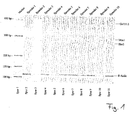
- FIG. 1 shows the detection of time-dependent induced transcription in IDU-treated blood samples.
- Lane 1 shows a ladder with size markers
- lanes 2 to 11 show the amplified DNA fragments of the tumor-associated transcripts to be detected in ten different healthy donors and an internal control of ⁇ -actin.
- the position of the respective bands for ⁇ -actin (actin) and the three tumor-associated transcripts GA733-2 (384 bp), MUC-1 (292 bp) and Her-2 (265 bp) are in Fig. 1 designated.
- the samples of the ten healthy donors were mixed with 0.045% (w / v) IDU and then stored at 4 ° C for 24 h.
- the amplified DNA fragments were detected after analysis of the blood samples by high voltage capillary gel electrophoresis in a Bioanalyzer 2100 (Agilent Technologies). It can be seen that the analysis was negative for all ten healthy donors (donor 1 to donor 10 in lanes 2 to 11) (peak levels below threshold). That no illegitimate time-dependent induced expression can be detected.
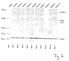
- FIG. 2 shows the same study, but now after storage of the samples of the ten healthy donors for 48 h. Again, no time-dependent expression of tumor-associated transcripts could be detected.
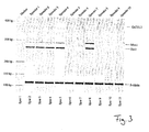
- FIG. 3 shows the same blood samples as in FIG. 1 and in FIG. 2 but without the addition of 0.025% (w / v) IDU. It can be seen that illegitimate expression of the tumor-associated transcripts occurred in these untreated blood samples.
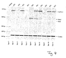
- FIG. 4 In contrast, detection of breast tumor cells in 0.045% (w / v) IDU-treated blood samples from ten healthy donors (lanes 2 to 10). Into these samples, 5 ml of blood were inoculated into two breast cancer cells (MCF7). The study on the expression pattern of the tumor-associated transcripts Her-2, MUC-1 and GA733.2 was carried out after storage of the blood samples at 4 ° C. for 48 h. All other analysis parameters correspond to those in FIGS. 1 to 3 , It can be seen that the tumor-associated transcripts Her-2, MUC-1 and GA733.2, which were introduced into the blood samples via the inoculation of MCF7 breast cancer cells, are clearly detected. Thus, FIG.
- FIG. 5 shows comparative experiments with the commercially available stabilization solution "CellSave” from Immunicon.
- 5 breast cancer cells HCC1954.
- Two of the samples were treated with 0.025% (w / v) IDU according to the invention and stored at 4 ° C (lanes 2 and 3).
- Two further samples were treated in a similar manner, but stored at 25 ° C (lanes 6 and 7).
- Two additional samples were stabilized with the product CellSave according to the manufacturer's instructions and stored at 4 ° C. (lanes 4 and 5).
- the last two samples in FIG. 5 (Lanes 8 and 9) were also stabilized according to the manufacturer's instructions with CellSave and stored at 25 ° C.
- the expression of the tumor markers Her-2, MUC-1 and GA733.2 is detected in the inoculated breast cancer cells and the marker ⁇ -actin in the samples treated according to the invention (lanes 2, 3, 6, 7).
- the commercially available stabilized solution CellSave stabilized samples (lanes 4, 5, 8, 9), a sensitive detection of neither the marker ⁇ -actin nor the tumor markers Her-2, MUC-1 and Ga733.2 was possible.
- FIG. 6 shows the analysis of blood samples from breast cancer patients immediately after blood sampling, after 24 h and after 48 h. All of these samples were spiked with 0.025% (w / v) IDU immediately after blood sampling and stored at 4 ° C.
- FIG. 6 shows in the tracks 2 to 4 the examination results of the blood sample of a patient immediately after the blood collection, after 24 h and 48 h after the blood collection. It turns out that the expression pattern remains largely the same over time and consequently the addition of IDU leads to a preservation of the expression pattern. It is crucial that no illegitimate time-dependent induced expression occurs. This is also the case with the second patient sample tracked in lanes 5-7 over time. This apparently has no circulating tumor cells in the blood sample. Time-dependent induced expression was also not detected. Also, the sample of patient 3 in lanes 8-10 shows that treatment with 0.025% (w / v) IDU maintained the expression pattern for 48 hours.
- tumor cell detection in breast cancer patient blood samples treated with 0.025% (w / v) IDU immediately after collection, at 24 h, and at 48 h shows consistent results, i. Blood samples in which tumor cells could be detected at the time of the decrease show a positive tumor cell detection even after 24 h and 48 h, while samples without tumor cells also remained negative after 24 and 48 h, i. no false-positive results arise, which could be generated by a time-dependent expression ex vivo. Thus, the blood samples examined show a constant pattern of expression over a period of 48 hours, i. Samples that were initially negative remained negative and positive samples remained positive.
- FIG. 7 shows the detection of time-dependent induced transcription in DU-treated blood samples. Of the Detection was carried out according to the protocols described for IDU.
- Lane 1 shows a ladder with size markers
- lanes 2 to 4 show the amplified DNA fragments of the tumor-associated transcripts to be detected in a healthy donor and an internal control of ⁇ -actin.
- Lanes 5 to 7 show the transcripts of samples with 2 inoculated cells of the tumor cell line Calu-3 at the times 0 h, 24 h and 48 h.
- the position of the respective bands for ⁇ -actin (actin) and the three tumor-associated transcripts GA733-2 (384 bp), MUC-1 (292 bp) and Her-2 (265 bp) are in Fig. 7 designated.
- the samples were treated with 2 inoculated Calu-3 cells with 0.01% (w / v) DU and then stored at 4 ° C for a maximum of 48 h.
- the amplified DNA fragments were detected after analysis of the blood samples by high voltage capillary gel electrophoresis in a Bioanalyzer 2100 (Agilent Technologies). It can be seen that the analysis was negative for all samples without inoculated cells (lanes 2 to 4) (peak levels below threshold). That no illegitimate time-dependent induced expression can be detected.
- FIG. 7 clearly shows that even small numbers of cells can still be detected after 48 h if the blood sample was treated according to the invention without hindering the detection by illegitimate time-dependent induced expression. A loss of sensitivity of the targeted tumor cell detection is thus by the inventive Procedure also prevented.
- FIG. 8 shows the detection of time-dependent induced transcription in bronopol-treated blood samples (2-bromo-2-nitropropane-1,3-diol).
- Lanes 1, 4 and 7 show a ladder with size markers
- lanes 2, 5 and 8 show the amplified DNA fragments of the tumor-associated transcripts to be detected in a healthy donor and an internal control of ⁇ -actin.
- Lanes 3, 6 and 9 show the transcripts of samples with 2 inoculated cells of the tumor cell line Calu-3.
- the position of the respective bands for ⁇ -actin (actin) and the three tumor-associated transcripts GA733-2 (384 bp), MUC-1 (292 bp) and Her-2 (265 bp) are in Fig. 8 designated.
- Samples from a healthy donor were spiked with 0.025% (lanes 2 and 3), 0.05% (lanes 5 and 6) and 0.075% (w / v) (lanes 8 and 9) bronopol and then at 4 ° C for Stored for 24 h.
- the amplified DNA fragments were detected after analysis of the blood samples by high voltage capillary gel electrophoresis in a Bioanalyzer 2100 (Agilent Technologies). It can be seen that the analysis was negative at all three Bronopol concentrations (lanes 2, 5 and 8) (peak levels below threshold). That is, no illegitimate time-dependent induced expression can be detected.
- Lanes 3, 6 and 9 show that tumor-associated transcripts introduced into the blood samples via inoculation of Calu-3 cancer cells are clearly detected.
- FIG. 8 thus clearly shows that even cell numbers as low as 2 cells can still be detected after 24 h if the blood sample was treated according to the invention without detection by illegitimate to hinder time-dependent induced expression. A loss of sensitivity of the targeted tumor cell detection is therefore also prevented by the inventive method.
- the present experimental results thus show that a prevention of a time-dependent induced expression in biological cells in a sample is achieved ex vivo. Furthermore, disseminated tumor cells in peripheral blood of breast and colorectal cancer patients could be detected by the addition of the reagents of the invention. No time-dependent induced transcription was detected for 48 h. In non-treated blood samples, however, a very strong time-dependent induced expression was already observed after 24 h.
- the reagents according to the invention and the method according to the invention therefore make it possible to prevent a time-dependent induced expression in biological cells in a sample (especially ex vivo) and at the same time to store disseminated tumor cells in peripheral blood of cancer patients. They therefore have a high logistical and diagnostic value, since this also allows a longer transport and a greater time interval between sampling and sample analysis.
- the reagents according to the invention and the inventive Methods are therefore suitable for use in blood collection systems.
Landscapes
- Chemical & Material Sciences (AREA)
- Life Sciences & Earth Sciences (AREA)
- Organic Chemistry (AREA)
- Analytical Chemistry (AREA)
- Zoology (AREA)
- Wood Science & Technology (AREA)
- Proteomics, Peptides & Aminoacids (AREA)
- Health & Medical Sciences (AREA)
- Engineering & Computer Science (AREA)
- Molecular Biology (AREA)
- Biochemistry (AREA)
- Microbiology (AREA)
- Biotechnology (AREA)
- Biophysics (AREA)
- Physics & Mathematics (AREA)
- Chemical Kinetics & Catalysis (AREA)
- Immunology (AREA)
- Bioinformatics & Cheminformatics (AREA)
- General Engineering & Computer Science (AREA)
- General Health & Medical Sciences (AREA)
- Genetics & Genomics (AREA)
- Measuring Or Testing Involving Enzymes Or Micro-Organisms (AREA)
- Investigating Or Analysing Biological Materials (AREA)
- Immobilizing And Processing Of Enzymes And Microorganisms (AREA)
- Micro-Organisms Or Cultivation Processes Thereof (AREA)
Description
- Die vorliegende Erfindung betrifft ein Reagenz zur Verhinderung der zeitabhängigen, induzierten Expression in biologischen Zellen in einer Probe. Sie betrifft gleichermaßen ein Verfahren hierzu sowie derart behandelte Zellpräparate.
- Derartige Reagenzien und derartige Verfahren werden insbesondere im Bereich der molekular-biologischen Analyse und der medizinischen Diagnostik, insbesondere der Tumordiagnostik, benötigt.
- Viele Nachweisverfahren zielen darauf, die Genexpression auf mRNA-Ebene zu analysieren. Dabei können exprimierte Gene als Nachweis spezieller Zellen gewertet werden. Es kann aber auch das Expressionsmuster aller untersuchten Gene genutzt werden, um Rückschlüsse über den Zustand einer Zelle zu ziehen oder um im Vergleich von zwei biologischen Proben deren Unterschiede zu charakterisieren.
- Es wurde beschrieben, dass es zwischen der Probennahme und der Probeanalyse zum Abbau transkribierter mRNA in der Probe aufgrund von Änderungen in den Umgebungsbedingungen der Probe im Anschluss an die Probeentnahme kommen kann (Becker S. et al., 2004 Clin Chem 50 (4), 785-786).
- Überraschenderweise tritt aber ein anderes schwerwiegendes Problem bei den in der Anmeldung beschriebenen Verfahren zum Nachweis von zellassoziierter mRNA auf, und zwar eine zeitabhängige illegitime Expression von mRNA ex vivo (ex vivo = außerhalb des menschlichen oder tierischen Körpers). Es kann nach der Probennahme die Expression neuer mRNA induziert werden. Diese zeitabhängig induzierte Expression verfälscht das Analyseergebnis. Die vorliegende Erfindung beschäftigt sich mit diesem Problem.
- Eine derartige illegitime, zeitabhängig induzierte Expression wird bereits in einer Vielzahl von Publikationen beschrieben, beispielsweise in Baechler et al., Genes Immun. Band 5, Seite 347 bis 353 (2004).
- Neuerdings sind hochsensitive Analyseverfahren entwickelt worden, beispielsweise wie in der
EP 02 732 726 A1 EP 02 732 726 EP 02 732 726 Figur 3 ) werden jedoch ohne spezielle Behandlung der Blutproben Transkripte nachgewiesen, die zum Zeitpunkt der Blutentnahme nicht vorhanden waren. Diese illegitime Transkription führt zu einem falsch-positiven Ergebnis. - Beschriebene Verfahren, die darauf abzielen, lediglich den Abbau vorhandener mRNA zu verhindern sowie die Morphologie und Antigenstruktur von Zellen zu stabilisieren, sind nicht geeignet, um mit dem Verfahren der
EP 02 732 726 EP 02 732 726 Figur 5 ). Das Problem der zeitabhängig induzierten Expression ist demnach mit den bekannten Verfahren nicht gelöst. - Die vorliegende Erfindung macht es sich also zur Aufgabe, ein Reagenz sowie ein Verfahren zur Verhinderung und Reduzierung der zeitabhängig induzierten Expression in biologischen Zellen in einer Probe sowie deren behandelte Zellpräparate zur Verfügung zu stellen.
- Diese Aufgabe wird durch das Reagenz nach Anspruch 1, das Zellpräparat nach Anspruch 9 sowie das Verfahren nach Anspruch 15 gelöst. Vorteilhafte Weiterbildungen des Reagenzes, des Zellpräparates sowie des Verfahrens zur Verhinderung der zeitabhängig induzierten Expression werden in den jeweiligen abhängigen Ansprüchen gegeben.
- Die vorliegende Erfindung beschäftigt sich also erstmals erfolgreich mit der Unterbindung der mRNA-Neusynthese (zeitabhängige, induzierte Expression). Diese besitzt bei Einsatz hochsensitiver, moderner Analysemethoden eine mindestens ebenso große Bedeutung wie die bisher betrachtete Stabilisierung der Oberflächenantigene oder der mRNS der Zielzellen. Nur so ist es möglich, die Zielzellen anhand ihrer mRNA-Expression unzweideutig zu charakterisieren.
- Überraschenderweise wurde gefunden, dass die Zugabe eines Formaldehyddonors in sehr geringer Konzentration zu einer biologischen Probe, nämlich in einer Konzentration ≤ 0,075 % (w/v), vorteilhafterweise < 0,045 % (w/v), vorteilhafterweise ≤ 0,025 % (w/v) zu einer Verhinderung der zeitabhängigen illegitimen Expression führt. Als Formaldehyddonor kann dabei Imidazolidinyl-Harnstoff (IDU), Diazolidinyl-Harnstoff (DU), Dimethylol-5,5-Dimethylhydantoin, Dimethylol-Harnstoff, 2-Brom-2-Nitropropan-1,3-Diol (Bronopol), 1,3-bis(hydroxymethyl)-5,5-dimethylimidazolidine-2,4-dione (DMDM-Hydantoin), N5-Methyl-Tetrahydrofolsäure, N10-Methyl-Tetra-hydrofolsäure oder eine Mischung hiervon eingesetzt werden.
- Mit dem erfindungsgemäßen Reagenz sowie dem erfindungsgemäßen Verfahren ist es daher erstmals möglich, die zeitabhängig induzierte Expression in biologischen Zellen in einer Probe ex vivo zu verhindern sowie eine Konservierung disseminierter Tumorzellen in Blutproben von Krebspatienten zu ermöglichen.
- Hierzu wird nach Blutabnahme das Reagenz, gegebenenfalls gelöst in phosphatgepufferter Kochsalzlösung (PBS), sowie unter Zugabe des Gerinnungshemmers EDTA in der angegebenen Endkonzentration sofort mit dem Blut gemischt. Die Probe kann dann über einen Zeitraum von mehreren Tagen, beispielsweise bis zu mindestens 72 h bei 4 °C gelagert werden, ohne dass illegitime Expression innerhalb der Probe auftritt. Weiterhin können der Probe weitere zellstabilisierende Substanzen, wie z.B. Polyethylenglycol (PEG) und/oder Proteaseinhibitoren zugesetzt werden.
- Um die erfindungsgemäße Wirkung zu demonstrieren, wurde in den folgenden Beispielen der Effekt der erfindungsgemäßen Zusätze auf die Spezifität eines Tumorzellennachweises mittels Analyse von Blutproben gesunder Spender sowie von Tumorpatienten untersucht. Außerdem wurde der Effekt der erfindungsgemäßen Zusätze auf die Sensitivität des Tumorzellnachweises mittels Analyse an Blutproben gesunder Spender mit inokulierten Tumorzellen untersucht. Die Analyse erfolgt dabei mittels vorgeschalteter Zellselektionierung und anschließender Expressionsprofilanalyse, wie sie in der europäischen Patentanmeldung
EP 02 732 726 - Desweiteren wurde die klinische Sensitivität mittels Analyse stabilisierter Blutproben von Brust- und Colorektal-Krebspatienten ermittelt. Alle untersuchten Patienten befanden sich in einem fortgeschrittenen Krankheitsstadium (M1) und wurden mit palliativer Therapie behandelt. Die erfindungsgemäßen Zusätze wurden unmittelbar nach Abnahme der Blutproben mit den Blutproben vermischt und sofort, nach 24 h, 48 h und nach 72 h Lagerung bei 4°C analysiert.
- Es ist dabei zu erwähnen, dass sich die folgenden Beispiele sämtlich zwar auf Blutproben beziehen, jedoch das erfindungsgemäße Verfahren auch geeignet ist, um andere Zellsuspensionen oder Gewebsproben zu untersuchen. Zellsuspensionen können dabei Körperflüssigkeiten oder auch in Suspension gebrachte Gewebezellen, z.B. Gewebestücke, Gewebeschnitte und dergleichen, sein.
- Es zeigen
- Figur 1
- den Nachweis zeitabhängig induzierter Transkription in IDU behandelten Blutproben 24 h nach Abnehmen;
- Figur 2
- den Nachweis zeitabhängig induzierter Transkription in IDU behandelten Blutproben 48 h nach Abnahme;
- Figur 3
- den Nachweis zeitabhängig induzierter Transkription in nicht-behandelten Blutproben;
- Figur 4
- die Erfassung von 2 Brusttumorzellen in mit IDU behandelten Blutproben;
- Figur 5
- die Erfassung von Brusttumorzellen in mit verschiedenen Systemen behandelten Blutproben;
- Figur 6
- typische Beispiele für den Nachweis disseminierter Tumorzellen in IDU behandelten Blutproben von Brustkrebspatientinnen;
- Figur 7
- den Nachweis zeitabhängig induzierter Transkription in mit DU behandelten Blutproben;
- Figur 8
- den Nachweis zeitabhängig induzierter Transkription in mit Bronopol behandelten Blutproben.
- Bei sämtlichen im Folgenden dargestellten Beispielen wurde eine Analyse, wie sie in der
EP 02 732 726 Tabelle 1 Antigen Klon Firma Epitheliales Membran Antigen GP1.4 Novocastra Epitheliales Antigen Ber-EP4 DAKO Muc 1 HMPV Pharmingen - Die Magnetpartikel wurden dabei in einer Konzentration von 4 x 10⁸ Beads/ml (CELLection™ Pan Mouse IgG Kit, Firma Dynal) verwendet. Die Verhältnisse zwischen der Antikörperkonzentration und den daran gekoppelten Antikörpern sind in Tabelle 2 wiedergegeben.
Tabelle 2 Klon Antikörperkonzentration µl Antkörper/25µl
PartikelBerEP4 0,32 mg/ml 4 µl HMPV 0,5 mg/ml 4 µl GP1.4 0,2 mg/ml 0,3 µl - Die so vorbereiteten Magnetpartikel wurden zu gleichen Teilen gemischt und in 100 µl PBS je 5 ml Probe dem Blut (EDTA) zugegeben.
- Nach 30 min Inkubation im Überkopfschüttler wurden die Magnetpartikel, die gegebenenfalls als Zell-Antikörper-Magnetpartikelkomplexe vorlagen, mittels eines Magnetpartikelkonzentrators (MPC®-S, Firma Dynal) 3mal mit PBS gewaschen und die anhaftenden Zellen anschließend entsprechend des im folgenden beschriebenen mRNS-Isolierungsprotokolls behandelt.
- Die mRNS-Isolierung erfolgte mittels Oligo(dT)-gekoppelter Magnetpartikel, Dynabeads® mRNA Direct™ Micro Kit (Firma Dynal). Diese Isolierung erfolgte entsprechend der in dem Kit angegebenen Herstellerangaben.
- An die Isolierung der mRNS schließt sich eine reverse Transkription an, bei der die mRNS in cDNS umgeschrieben wird.
Tabelle 3: Komponenten der cDNS-Synthese Die cDNS-Synthese erfolgt in einem 20 µl-Reaktionsansatz Komponenten Volumen mRNS 29,5 µl 10 x RT-Puffer 4 µl dNTP-Mix 4 µl RNase-Inhibitor (RNAsin, Promega) 0,5 µl Reverse Transkriptase 2 µl - Die cDNS-Synthese (Tabelle 3) erfolgte bei 37 °C für eine Stunde mit nachfolgender Inaktivierung der reversen Transkriptase durch Erhitzen für 5 Minuten bei 95 °C und anschließender Abkühlung auf Eis. Hierzu wurde ein Sensiskript Reverse Transkriptase Kit, Firma Qiagen, Hilden nach dort angegebenen Protokollen verwendet.
- Anschließend an die Umschreibung der mRNS in cDNS erfolgte eine Polymerase-Kettenreaktion (PCR) mit ß-Aktin als interner Kontrolle.
- Dabei wurden die in Tabelle 4 aufgeführten Oligonukleotide als PCR-Primer zur Amplifikation von cDNS verwendet.
Tabelle 4: Liste der PCR-Primer Primername Sequenz 5' -> 3' PCR-Produkt Tumormarker GA733.2 sense AATCGTCAATGCCAGTGTACTTCA 383 bp GA733.2 sense TAACGCGTTGTGATCTCCTTCTGA CA15-3 sense TCAGCTTCTACTCTGGTGCACAAC 293 bp CA-15-3 antisense TGGTAGTAGTCGGTGCTGGGATCT Her-2 sense CCCAGTGTGTCAACTGCAGCCAGT 270 bp Her-2 antisense CAGATGGGCATGTAGGAGAGGTCA Interne Kontrolle Aktin sense CTGGAGAAGAGCTACGAGCTGCCT 114 bp Aktin antisense ACAGGACTCCATGCCCAGGAAGGA - Die PCR wurde mit dem in Tabelle 5 angegebenen Ansatz durchgeführt.
Tabelle 5: PCR-Ansatz Die PCR-Synthese erfolgte in einem 50 µl-Reaktionsansatz Komponenten Volumen cDNS 8 µl Primer 4 µl HotStarTaq 25 µl H2O 13 µl - Die PCR-Bedingungen (Zyklenzahl, Zyklenführung etc.) sind in der Tabelle 6 gegeben; Temperaturänderungen erfolgten mit 2 °C/s.

- Die so erzeugten Amplifikate der cDNS wurden elektrophoretisch aufgetrennt mittels eines Bioanalyzer 2100 (Firma Agilent). Hierzu wurde 1 µl des PCR-Produktes in dem Bioanalyzer auf einem DNS-Chip 1000 aufgetrennt und das Trennergebnis elektronisch dokumentiert. Das Analyseergebnis wird für positiv befunden, wenn die Bandenintensität mindestens eines Tumormarkers > 6,7 ist. Der Bandenintensität entspricht die Einheit "Peakhöhe" des Agilents. Peaks mit einer Höhe von < 4,0 werden negativ bewertet. Peaks, deren Höhe von 4,0 bis 6,7 liegt, können weder als positiv noch als negativ bewertet werden. Auf diese Weise wurden die
Figuren 1 bis 6 erzeugt und ausgewertet. -
Figur 1 zeigt den Nachweis zeitabhängig induzierter Transkription in mit IDU behandelten Blutproben. - Spur 1 zeigt dabei eine Leiter mit Größenmarkern, Spuren 2 bis 11 zeigen die bei zehn verschiedenen gesunden Spendern nachzuweisenden amplifizierten DNS-Fragmente der tumorassoziierten Transkripte und einer internen Kontrolle von ß-Aktin. Die Position der jeweiligen Banden für ß-Aktin (Actin) und die drei tumorassoziierten Transkripte GA733-2 (384 bp), MUC-1 (292 bp) und Her-2 (265 bp) sind in
Fig. 1 bezeichnet. - Die Proben der zehn gesunden Spender wurden dabei mit 0,045 % (w/v) IDU versetzt und anschließend bei 4 °C für 24 h gelagert. Die amplifizierten DNA-Fragmente wurden nach Analyse der Blutproben mittels Hochspannungskapillar-Gelelektrophorese in einem Bioanalyzer 2100 (Agilent Technologies) nachgewiesen. Es ist zu erkennen, dass die Analyse bei allen zehn gesunden Spendern (Spender 1 bis Spender 10 in Spuren 2 bis 11) negativ ausgefallen ist (Peakhöhen unter Schwellenwert). D.h. es kann keinerlei illegitime zeitabhängig induzierte Expression nachgewiesen werden.
-
Figur 2 zeigt dieselbe Untersuchung, nun jedoch nach Lagerung der Proben der zehn gesunden Spender für 48 h. Auch hier konnte noch immer keine zeitabhängige Expression tumorassoziierter Transkripte nachgewiesen werden. -
Figur 3 zeigt demgegenüber dieselben Blutproben wie inFigur 1 und inFigur 2 , jedoch ohne Zugabe von 0,025 % (w/v) IDU. Es ist zu erkennen, dass bei diesen nicht behandelten Blutproben illegitime Expression der tumorassoziierten Transkripte auftrat. - Mit den Untersuchungen gemäß den
Figuren 1 bis 3 wurde eindeutig nachgewiesen, dass mit den angegebenen Konzentrationen eines Formaldehyddonors wirksam die illegitime zeitabhängig induzierte Expression in Zellen innerhalb eines Zellpräparats unterdrückt werden kann. Die erfindungsgemäßen Zusätze erhalten eine hohe Spezifität. -
Figur 4 zeigt demgegenüber die Erfassung von Brusttumorzellen in mit 0,045 % (w/v) IDU behandelten Blutproben zehn gesunder Spender (Spuren 2 bis 10). In diese Proben wurden je 5 ml Blut zwei Brustkrebszellen (MCF7) inokuliert. Die dargestellte Untersuchung auf das Expressionsmuster der tumorassoziierten Transkripte Her-2, MUC-1 und GA733.2 wurde nach einer Lagerung der Blutproben bei 4°C über 48 h durchgeführt. Alle weiteren Analyseparameter entsprechen denjenigen inFiguren 1 bis 3 . Es ist zu erkennen, dass die tumorassoziierten Transkripte Her-2, MUC-1 und GA733.2, die über die Inokulierung von MCF7-Brustkrebszellen in die Blutproben eingebracht wurden, eindeutig nachgewiesen werden. Figur 4 zeigt damit eindeutig, dass auch so geringe Zellzahlen, wie 2 Zellen/5 ml Blut noch nach 48 h nachgewiesen werden können, wenn die Blutprobe erfindungsgemäß behandelt wurde, ohne den Nachweis durch illegitime zeitabhängig induzierte Expression zu behindern. Ein Verlust an Sensitivität des angestrebten Tumorzellennachweises wird also durch das erfindungsgemäße Verfahren ebenfalls verhindert. -
Figur 5 zeigt Vergleichsversuche mit der marktüblichen Stabilisierungslösung "CellSave" von Immunicon. Hierzu wurden in je 5 ml Blut gesunder Spender jeweils 5 Brustkrebszellen (HCC1954) inokuliert. Zwei der Proben wurden mit 0,025 % (w/v) IDU erfindungsgemäß behandelt und bei 4 °C gelagert (Spuren 2 und 3). Zwei weitere Proben wurden in entsprechender Weise behandelt, jedoch bei 25 °C gelagert (Spuren 6 und 7). Zwei weitere Proben wurden gemäß den Herstellerangaben mit dem Produkt CellSave stabilisiert und bei 4°C gelagert (Spuren 4 und 5). Die letzten beiden Proben inFigur 5 (Spuren 8 und 9) wurden ebenfalls gemäß den Herstellerangaben mit CellSave stabilisiert und bei 25 °C gelagert. - Wie zu erkennen ist, wird die Expression der Tumormarker Her-2, MUC-1 und GA733.2 in den inokulierten Brustkrebszellen sowie des Markers ß-Aktin in den erfindungsgemäß behandelten Proben (Spuren 2, 3, 6, 7) erfasst. Mit der käuflichen Stabilisierungslösung CellSave stabilisierte Proben (Spuren 4, 5, 8, 9) war ein sensitiver Nachweis weder des Markers ß-Aktin noch der Tumormarker Her-2, MUC-1 und Ga733.2 möglich.
- Dies zeigt, dass im Vergleich zum Stand der Technik durch die vorliegende erfindungsgemäße Behandlung eine sehr effektive Erhaltung des Expressionsmusters in einer biologischen Probe erzielt wird, die erlaubt, die nachgeschaltete Analyse hoch selektiv und sensitiv durchzuführen.
-
Figur 6 zeigt die Analyse von Blutproben von Brustkrebspatienten unmittelbar nach der Blutprobenentnahme, nach 24 h sowie nach 48 h. Sämtliche dieser Proben wurden unmittelbar nach Blutabnahme mit 0,025 % (w/v) IDU versetzt und bei 4 °C gelagert. -
Figur 6 zeigt in den Spuren 2 bis 4 die Untersuchungsergebnisse der Blutprobe eines Patienten unmittelbar nach der Blutentnahme, nach 24 h und 48 h nach der Blutentnahme. Es zeigt sich, dass das Expressionsmuster über die Zeit weitgehend gleich bleibt und folglich die IDU-Zugabe zu einer Bewahrung des Expressionsmusters führt. Entscheidend ist dabei, dass auch keinerlei illegitime zeitabhängig induzierte Expression auftritt. Dies ist auch bei der Probe des zweiten Patienten, die in den Spuren 5 bis 7 über die Zeit verfolgt wurde, der Fall. Dieser weist offensichtlich keine zirkulierenden Tumorzellen in der Blutprobe auf. Eine zeitabhängig induzierte Expression wurde ebenfalls nicht festgestellt. Auch die Probe des Patienten 3 in den Spuren 8 bis 10 zeigt, dass mit der Behandlung durch 0,025 % (w/v) IDU eine Bewahrung des Expressionsmusters über 48 h erzielt werden konnte. - Zusammenfassend lässt sich feststellen, dass der Tumorzellennachweis in mit 0,025 % (w/v) IDU behandelten Blutproben von Brustkrebspatientinnen sofort nach Abnahme, nach 24 h und nach 48 h einheitliche Ergebnisse zeigt, d.h. Blutproben, in denen zum Zeitpunkt der Abnahme Tumorzellen nachgewiesen werden konnten, zeigen auch nach 24 h und 48 h einen positiven Tumorzellennachweis, während Proben ohne Tumorzellen auch nach 24 und 48 h negativ blieben, d.h. keine falsch-positiven Resultate entstehen, die durch eine zeitabhängige Expression ex vivo erzeugt sein könnten. Die untersuchten Blutproben zeigen also über einen Zeitraum von 48 h hinweg ein konstantes Expressionsmuster, d.h. Proben, die anfangs negativ waren, blieben negativ und positive Proben blieben positiv.
-
Figur 7 zeigt den Nachweis zeitabhängig induzierter Transkription in mit DU behandelten Blutproben. Der Nachweis erfolgte nach den zu IDU beschriebenen Protokollen. - Spur 1 zeigt dabei eine Leiter mit Größenmarkern, Spuren 2 bis 4 zeigen die bei einem gesunden Spender nachzuweisenden amplifizierten DNS-Fragmente der tumorassoziierten Transkripte und einer internen Kontrolle von ß-Aktin. Spuren 5 bis 7 zeigen die Transkripte von Proben mit 2 inokulierten Zellen der Tumorzelllinie Calu-3 zu den Zeitpunkten 0 h, 24 h und 48 h. Die Position der jeweiligen Banden für ß-Aktin (Actin) und die drei tumorassoziierten Transkripte GA733-2 (384 bp), MUC-1 (292 bp) und Her-2 (265 bp) sind in
Fig. 7 bezeichnet. - Die Proben wurden dabei mit 2 inokulierten Calu-3-Zellen mit 0,01 % (w/v) DU versetzt und anschließend bei 4 °C für maximal 48 h gelagert. Die amplifizierten DNA-Fragmente wurden nach Analyse der Blutproben mittels Hochspannungskapillar-Gelelektrophorese in einem Bioanalyzer 2100 (Agilent Technologies) nachgewiesen. Es ist zu erkennen, dass die Analyse bei allen Proben ohne inokulierte Zellen (Spuren 2 bis 4) negativ ausgefallen ist (Peakhöhen unter Schwellenwert). D.h. es kann keinerlei illegitime zeitabhängig induzierte Expression nachgewiesen werden.
- An den Spuren 5 bis 7 ist zu erkennen, dass tumorassoziierte Transkripte, die über die Inokulierung von Calu-3-Krebszellen in die Blutproben eingebracht wurden, eindeutig nachgewiesen werden.
Figur 7 zeigt damit eindeutig, dass auch geringe Zellzahlen noch nach 48 h nachgewiesen werden können, wenn die Blutprobe erfindungsgemäß behandelt wurde, ohne den Nachweis durch illegitime zeitabhängig induzierte Expression zu behindern. Ein Verlust an Sensitivität des angestrebten Tumorzellennachweises wird also durch das erfindungsgemäße Verfahren ebenfalls verhindert. -
Figur 8 zeigt den Nachweis zeitabhängig induzierter Transkription in mit Bronopol behandelten Blutproben (2-Bromo-2-Nitropropan-1,3-Diol). - Spuren 1, 4 und 7 zeigen dabei eine Leiter mit Größenmarkern, Spuren 2, 5 und 8 zeigen die bei einem gesunden Spender nachzuweisenden amplifizierten DNS-Fragmente der tumorassoziierten Transkripte und einer internen Kontrolle von ß-Aktin. Die Spuren 3, 6 und 9 zeigen die Transkripte von Proben mit 2 inokulierten Zellen der Tumorzelllinie Calu-3. Die Position der jeweiligen Banden für ß-Aktin (Actin) und die drei tumorassoziierten Transkripte GA733-2 (384 bp), MUC-1 (292 bp) und Her-2 (265 bp) sind in
Fig. 8 bezeichnet. - Die Proben eines gesunden Spenders wurden dabei mit 0,025 % (Spuren 2 und 3), 0,05 % (Spuren 5 und 6) und 0,075 % (w/v) (Spuren 8 und 9) Bronopol versetzt und anschließend bei 4 °C für 24 h gelagert. Die amplifizierten DNA-Fragmente wurden nach Analyse der Blutproben mittels Hochspannungskapillar-Gelelektrophorese in einem Bioanalyzer 2100 (Agilent Technologies) nachgewiesen. Es ist zu erkennen, dass die Analyse bei allen drei Konzentrationen Bronopol (Spuren 2, 5 und 8) negativ ausgefallen ist (Peakhöhen unter Schwellenwert). D.h. es kann keinerlei illegitime zeitabhängig induzierte Expression nachgewiesen werden. Die Spuren 3, 6 und 9 zeigen, dass tumorassoziierte Transkripte, die über die Inokulierung von Calu-3-Krebszellen in die Blutproben eingebracht wurden, eindeutig nachgewiesen werden.
Figur 8 zeigt damit eindeutig, dass auch so geringe Zellzahlen, wie 2 Zellen, noch nach 24 h nachgewiesen werden können, wenn die Blutprobe erfindungsgemäß behandelt wurde, ohne den Nachweis durch illegitime zeitabhängig induzierte Expression zu behindern. Ein Verlust an Sensitivität des angestrebten Tumorzellennachweises wird also durch das erfindungsgemäße Verfahren ebenfalls verhindert. - Die oben dargestellten Versuchsergebnisse zeigen eindeutig sowohl eine hohe Effektivität als auch Verlässlichkeit des erfindungsgemäßen Reagenzes und des erfindungsgemäßen Verfahrens bei der Behandlung von Zellpräparaten, insbesondere von disseminierten Tumorzellen im Blut von Brustkrebspatientinnen. Bei Colorektal-Krebspatienten wurden vergleichbare Ergebnisse erzielt.
- Die vorliegenden Versuchsergebnisse zeigen also, dass eine Verhinderung einer zeitabhängig induzierten Expression in biologischen Zellen in einer Probe ex vivo erzielt wird. Weiterhin konnten disseminierte Tumorzellen in peripherem Blut von Brust- und Colorektal-Krebspatienten durch die Zugabe der erfindungsgemäßen Reagenzien nachgewiesen werden. Es wurde keine zeitabhängig induzierte Transkription über 48 h nachgewiesen. In nicht-behandelten Blutproben wurde jedoch eine sehr starke zeitabhängig induzierte Expression schon nach 24 h beobachtet.
- Die erfindungsgemäßen Reagenzien und das erfindungsgemäße Verfahren ermöglichen also eine Verhinderung einer zeitabhängig induzierten Expression in biologischen Zellen in einer Probe (insbesondere ex vivo) und gleichzeitig eine Aufbewahrung disseminierter Tumorzellen in peripherem Blut von Krebspatienten. Sie besitzen daher einen hohen logistischen und diagnostischen Wert, da hiermit auch ein längerer Transport und ein größerer zeitlicher Abstand zwischen Probenentnahme und Probenanalyse ermöglicht wird. Die erfindungsgemäßen Reagenzien und das erfindungsgemäße Verfahren eignen sich daher zum Einsatz in Blutabnahmesystemen.
-
- <110> Adnagen AG
- <120> Reagenz und Verfahren zur Verhinderung der zeitabhängigen Expression in biologischen Zellen
- <130> Adnagen 060431
- <140> 060431
<141> 2006-03-22 - <150>
DE 10 2005 013 261.8
<151> 2006-03-22 - <160> 8
- <170> PatentIn Ver. 2.1
- <210> 1
<211> 24
<212> DNA
<213> Künstliche Sequenz - <220>
<223> Beschreibung der künstlichen Sequenz: Oligonukleotid - <400> 1
aatcgtcaat gccagtgtac ttca 24 - <210> 2
<211> 24
<212> DNA
<213> Künstliche Sequenz - <220>
<223> Beschreibung der künstlichen Sequenz: Oligonukleotid - <400> 2
taacgcgttg tgatctcctt ctga 24 - <210> 3
<211> 24
<212> DNA
<213> Künstliche Sequenz - <220>
<223> Beschreibung der künstlichen Sequenz: Oligonukleotid - <400> 3
tcagcttcta ctctggtgca caac 24 - <210> 4
<211> 24
<212> DNA
<213> Künstliche Sequenz - <220>
<223> Beschreibung der künstlichen Sequenz: Oligonukleotid - <400> 4
tggtagtagt cggtgctggg atct 24 - <210> 5
<211> 24
<212> DNA
<213> Künstliche Sequenz - <220>
<223> Beschreibung der künstlichen Sequenz: Oligonukleotid - <400> 5
cccagtgtgt caactgcagc cagt 24 - <210> 6
<211> 24
<212> DNA
<213> Künstliche Sequenz - <220>
<223> Beschreibung der künstlichen Sequenz: Oligonukleotid - <400> 6
cagatgggca tgtaggagag gtca 24 - <210> 7
<211> 24
<212> DNA
<213> Künstliche Sequenz - <220>
<223> Beschreibung der künstlichen Sequenz: Oligonukleotid - <400> 7
ctggagaaga gctacgagct gcct 24 - <210> 8
<211> 24
<212> DNA
<213> Künstliche Sequenz - <220>
<223> Beschreibung der künstlichen Sequenz: Oligonukleotid - <400> 8
acaggactcc atgcccagga agga 24
Claims (7)
- Verfahren zur Verhinderung der zeitabhängigen illegitimen Expression von mRNA in biologischen Zellen in einer Probe ex vivo,
dadurch gekennzeichnet, dass die Probe mit einem Reagenz, das einen Formaldehyddonor enthält, sowie gegebenenfalls einer Flüssigkeit vermischt wird. - Verfahren nach dem vorhergehenden Anspruch, dadurch gekennzeichnet, dass die Mischung des Reagenzes mit der Probe den Formaldehyddonor in einer Konzentration ≤ 0,075 % (w/v) enthält.
- Verfahren nach dem vorhergehenden Anspruch, dadurch gekennzeichnet, dass die Konzentration des Formaldehyddonors in der Mischung des Reagenzes mit der Probe ≤ 0,045 % (w/v), vorteilhafterweise ≤ 0,025 % (w/v) beträgt.
- Verfahren nach einem der drei vorhergehenden Ansprüche, dadurch gekennzeichnet, dass der Formaldehyddonor, Imidazolidinyl-Harnstoff, Diazolidinyl-Harnstoff, Dimethylol-5,5-Dimethylhydantoin, Dimethylol-Harnstoff, 2-Brom-2-Nitropropan-1,3-Diol, 1,3-bis(hydroxymethyl)-5,5-dimethylimidazolidine-2,4-dione (DMDM-Hydantoin), N5-Methyl-Tetrahydrofolsäure, N10-Methyl-Tetrahydrofolsäure oder eine Mischung hiervon enthält oder daraus besteht.
- Verfahren nach einem der Ansprüche 1 bis 4, dadurch gekennzeichnet, dass die Flüssigkeit ein Puffer, physiologische Kochsalzlösung oder dergleichen ist.
- Verfahren nach einem der Ansprüche 1 bis 5, dadurch gekennzeichnet, dass die damit behandelten biologischen Zellen antikörpervermittelt oder über andere gebräuchliche Verfahren separiert werden.
- Verwendung eines Verfahrens nach einem der vorhergehenden Ansprüche, um die zeitabhängige Expression von mRNA in einer Probe mit biologischen Zellen zu verhindern, insbesondere in einer Körperflüssigkeit, Blut, Liquor, Aszites, Urin oder in einem Gewebestück oder einer Suspension, die Zellen aus Gewebeproben enthält, insbesondere zum Nachweis von hierin enthaltenen Tumorzellen.
Applications Claiming Priority (2)
| Application Number | Priority Date | Filing Date | Title |
|---|---|---|---|
| DE102005013261A DE102005013261A1 (de) | 2005-03-22 | 2005-03-22 | Reagenz und Verfahren zur Verhinderung der zeitabhängigen Expression in biologischen Zellen |
| PCT/EP2006/002642 WO2006100063A2 (de) | 2005-03-22 | 2006-03-22 | Reagenz und verfahren zur verhinderung der zeitabhängigen rna-expression in biologischen zellen |
Publications (2)
| Publication Number | Publication Date |
|---|---|
| EP1869450A2 EP1869450A2 (de) | 2007-12-26 |
| EP1869450B1 true EP1869450B1 (de) | 2008-11-12 |
Family
ID=36685775
Family Applications (1)
| Application Number | Title | Priority Date | Filing Date |
|---|---|---|---|
| EP06723634A Expired - Lifetime EP1869450B1 (de) | 2005-03-22 | 2006-03-22 | Verfahren zur verhinderung der zeitabhängigen rna-expression in biologischen zellen |
Country Status (7)
| Country | Link |
|---|---|
| US (1) | US20080206866A1 (de) |
| EP (1) | EP1869450B1 (de) |
| JP (1) | JP2008533979A (de) |
| AT (1) | ATE414277T1 (de) |
| DE (2) | DE102005013261A1 (de) |
| ES (1) | ES2314895T3 (de) |
| WO (1) | WO2006100063A2 (de) |
Families Citing this family (11)
| Publication number | Priority date | Publication date | Assignee | Title |
|---|---|---|---|---|
| ES2784011T3 (es) | 2002-10-16 | 2020-09-21 | Streck Inc | Procedimiento y dispositivo para recoger y preservar las células para su análisis |
| ATE483160T1 (de) * | 2006-03-13 | 2010-10-15 | Siemens Healthcare Diagnostics | Reduktion von blutplättcheninterferenzen bei plasmatestproben |
| US11634747B2 (en) | 2009-01-21 | 2023-04-25 | Streck Llc | Preservation of fetal nucleic acids in maternal plasma |
| EP3290530B1 (de) | 2009-02-18 | 2020-09-02 | Streck Inc. | Konservierung von zellfreien nukleinsäuren |
| CA2780536C (en) * | 2009-11-09 | 2018-01-02 | Streck, Inc. | Stabilization of rna in and extracting from intact cells within a blood sample |
| WO2012151391A2 (en) | 2011-05-04 | 2012-11-08 | Streck, Inc. | Inactivated virus compositions and methods of preparing such compositions |
| EP4136972A1 (de) | 2013-07-24 | 2023-02-22 | Streck, Inc. | Zusammensetzungen und verfahren zur stabilisierung zirkulierender tumorzellen |
| US11168351B2 (en) | 2015-03-05 | 2021-11-09 | Streck, Inc. | Stabilization of nucleic acids in urine |
| US20170145475A1 (en) | 2015-11-20 | 2017-05-25 | Streck, Inc. | Single spin process for blood plasma separation and plasma composition including preservative |
| WO2018022991A1 (en) | 2016-07-29 | 2018-02-01 | Streck, Inc. | Suspension composition for hematology analysis control |
| US12378543B2 (en) | 2017-10-19 | 2025-08-05 | Streck Llc | Compositions for hemolysis and coagulation regulation and stabilization of extracellular vesicles |
Family Cites Families (12)
| Publication number | Priority date | Publication date | Assignee | Title |
|---|---|---|---|---|
| US4664806A (en) * | 1985-01-22 | 1987-05-12 | Ciba-Geigy Corporation | Anionically modified polysaccharides chromatography agents |
| US5021594A (en) * | 1990-03-12 | 1991-06-04 | Pennwalt France S.A. | Enhanced reduction and inhibition of ETU content in alkylenebisdithiocarbamates |
| US5849517A (en) * | 1991-05-08 | 1998-12-15 | Streck Laboratories, Inc. | Method and composition for preserving antigens and nucleic acids and process for utilizing cytological material produced by same |
| EP0639985B1 (de) * | 1992-09-10 | 2004-08-11 | Streck Laboratories, Inc. | Verfahren und zusammensetzung zur konservierung von antigenen und verfahren zur verwendung von durch selbiges verfahren hergestellten zytologischem material |
| DE69434942T2 (de) * | 1993-02-25 | 2007-11-29 | Abbott Laboratories, Abbott Park | Mehrzweckreagenzsystem zur schnellen lysierung von vollblutproben |
| US6204375B1 (en) * | 1998-07-31 | 2001-03-20 | Ambion, Inc. | Methods and reagents for preserving RNA in cell and tissue samples |
| JP5192631B2 (ja) * | 2000-11-08 | 2013-05-08 | ベクトン・ディキンソン・アンド・カンパニー | 生物学的試料を収集し安定化させるための方法および装置 |
| US6602718B1 (en) * | 2000-11-08 | 2003-08-05 | Becton, Dickinson And Company | Method and device for collecting and stabilizing a biological sample |
| KR20040047971A (ko) * | 2001-10-26 | 2004-06-05 | 이뮤니베스트 코포레이션 | 동일 샘플상에서의 광범위 핵산 및 이의 형태학적 특질에대한 다중 매개변수 분석법 |
| US20060008807A1 (en) * | 2002-08-23 | 2006-01-12 | O'hara Shawn M | Multiparameter analysis of comprehensive nucleic acids and morphological features on the same sample |
| ES2784011T3 (es) * | 2002-10-16 | 2020-09-21 | Streck Inc | Procedimiento y dispositivo para recoger y preservar las células para su análisis |
| US7727718B2 (en) * | 2005-01-04 | 2010-06-01 | Molecular Research Center, Inc. | Reagents for storage and preparation of samples for DNA analysis |
-
2005
- 2005-03-22 DE DE102005013261A patent/DE102005013261A1/de not_active Ceased
-
2006
- 2006-03-22 AT AT06723634T patent/ATE414277T1/de active
- 2006-03-22 EP EP06723634A patent/EP1869450B1/de not_active Expired - Lifetime
- 2006-03-22 DE DE502006002073T patent/DE502006002073D1/de not_active Expired - Lifetime
- 2006-03-22 JP JP2008502321A patent/JP2008533979A/ja active Pending
- 2006-03-22 WO PCT/EP2006/002642 patent/WO2006100063A2/de not_active Ceased
- 2006-03-22 US US11/909,142 patent/US20080206866A1/en not_active Abandoned
- 2006-03-22 ES ES06723634T patent/ES2314895T3/es not_active Expired - Lifetime
Also Published As
| Publication number | Publication date |
|---|---|
| US20080206866A1 (en) | 2008-08-28 |
| ES2314895T3 (es) | 2009-03-16 |
| DE102005013261A1 (de) | 2006-09-28 |
| JP2008533979A (ja) | 2008-08-28 |
| WO2006100063A3 (de) | 2006-11-16 |
| DE502006002073D1 (de) | 2008-12-24 |
| EP1869450A2 (de) | 2007-12-26 |
| ATE414277T1 (de) | 2008-11-15 |
| WO2006100063A2 (de) | 2006-09-28 |
Similar Documents
| Publication | Publication Date | Title |
|---|---|---|
| DE19530132C2 (de) | Verfahren zur Reinigung, Stabilisierung oder Isolierung von Nukleinsäuren aus biologischen Materialien | |
| DE69424797T2 (de) | Telomerase-aktivitätsversuche | |
| DE19904267A1 (de) | Verfahren zur Anreicherung von Tumorzellen aus einer Körperflüssigkeit und dazu geeigneter Kit | |
| EP3344781B1 (de) | Verwendung von im blutserum oder blutplasma zirkulierenden micrornas zur identifikation biopsiepflichtiger patienten und als marker zur differentialdiagnose einzelner nicht-ischämischer kardiomyopathien oder speichererkrankungen des herzens | |
| WO2006100063A2 (de) | Reagenz und verfahren zur verhinderung der zeitabhängigen rna-expression in biologischen zellen | |
| DE3050802C2 (de) | Verwendung von Rifamycin SV Natriumsalz bei der intraartikulären Bekämpfung von rheumatoider Arthritis | |
| EP3662080B1 (de) | Verfahren und zusammensetzung zur stabilisierung zellfreier nukleinsäuren und zellen | |
| EP3198011B1 (de) | Verfahren zum nachweis von nukleinsäuren in proben mit biologischem material | |
| EP0602686A2 (de) | Lektinkonzentrate aus Mistelextrakten und entsprechende standardisierte, stabilisierte Mistellektinpräparate, Verfahren zu ihrer Herstellung sowie diese enthaltende Arzneimittel und deren Verwendung zur Erhöhung der natürlichen Immunresistenz und/oder in der Tumor-Therapie | |
| DE69321454T2 (de) | Zytodiagnostisches verfahren unter verwendung von alstonin als selektivem marker und diesen marker enthaltender diagnostischer kit | |
| DE69528731T2 (de) | Verwendung eines Inhibitors für Serin/Threonin Protein Phosphatasen zur Induzierung der Kondensation unreifer Chromosomen | |
| EP0370032A1 (de) | Verfahren zur untersuchung einer biologischen flüssigkeit auf zellulare onkogen-dna bzw. deren fragmente | |
| EP3322817B1 (de) | Oligonucleotide und deren verwendung | |
| WO1997042344A1 (de) | Verfahren zur dns-analytik von blut und mittel zur durchführung des verfahrens | |
| EP2576820A1 (de) | Stabilisierung von nukleinsäuren in zellmaterial-haltigen biologischen proben | |
| DE3852004T2 (de) | Verfahren zur feststellung der expression bioregulatorischer gene in mikrokulturen eukariontischer zellen. | |
| EP3894858A1 (de) | Verfahren für die analyse einer blutprobe eines menschen auf eine tuberkuloseerkrankung durch detektion von tb-antigen stimulierter cd154-expression in kombination mit cd38, ki-67 oder hla-dr | |
| DE102023206168A1 (de) | Verfahren und Vorrichtung zur Demaskierung biologischer Zellen, insbesondere zirkulierender Tumorzellen | |
| DE102005031910B4 (de) | Verfahren zum Stabilisieren von Nukleinsäuren | |
| EP2402456A1 (de) | Verfahren zur Ermittlung von Organismen in Wasser | |
| CH719022B1 (de) | Verfahren zum laden von exosomen mit kleinen nichtcodierenden rnas | |
| DE10300861A1 (de) | Verwendung von an FABP4 bindenden Substanzen zur Diagnose und Behandlung des Harnblasenkarzinoms | |
| DE102018222357A1 (de) | In-vitro-Verfahren zum Nachweis von mindestens einer Nukleinsäure, die sich bei einem Lebewesen im Vollblut außerhalb der Blutzellen befindet und Vorrichtung und Kit hierzu | |
| DE102009054098A1 (de) | Nachweis von Mykobakterien und deren Resistenzgenen | |
| DE10153584A1 (de) | Verfahren zur Indentifizierung Tumor-spezifischer Antigene auf Krebszellen |
Legal Events
| Date | Code | Title | Description |
|---|---|---|---|
| PUAI | Public reference made under article 153(3) epc to a published international application that has entered the european phase |
Free format text: ORIGINAL CODE: 0009012 |
|
| 17P | Request for examination filed |
Effective date: 20071022 |
|
| AK | Designated contracting states |
Kind code of ref document: A2 Designated state(s): AT BE BG CH CY CZ DE DK EE ES FI FR GB GR HU IE IS IT LI LT LU LV MC NL PL PT RO SE SI SK TR |
|
| RIN1 | Information on inventor provided before grant (corrected) |
Inventor name: BOECHER, OLIVER Inventor name: ZIEGLSCHMID, VEIT Inventor name: ALBERT, WINFRIED |
|
| RTI1 | Title (correction) |
Free format text: METHOD FOR PREVENTING TIME-DEPENDENT RNA-EXPRESSION IN BIOLOGICAL CELLS |
|
| DAX | Request for extension of the european patent (deleted) | ||
| GRAP | Despatch of communication of intention to grant a patent |
Free format text: ORIGINAL CODE: EPIDOSNIGR1 |
|
| GRAS | Grant fee paid |
Free format text: ORIGINAL CODE: EPIDOSNIGR3 |
|
| GRAA | (expected) grant |
Free format text: ORIGINAL CODE: 0009210 |
|
| AK | Designated contracting states |
Kind code of ref document: B1 Designated state(s): AT BE BG CH CY CZ DE DK EE ES FI FR GB GR HU IE IS IT LI LT LU LV MC NL PL PT RO SE SI SK TR |
|
| REG | Reference to a national code |
Ref country code: GB Ref legal event code: FG4D Free format text: NOT ENGLISH |
|
| REG | Reference to a national code |
Ref country code: CH Ref legal event code: NV Representative=s name: TROESCH SCHEIDEGGER WERNER AG Ref country code: CH Ref legal event code: EP |
|
| REG | Reference to a national code |
Ref country code: IE Ref legal event code: FG4D Free format text: LANGUAGE OF EP DOCUMENT: GERMAN |
|
| REF | Corresponds to: |
Ref document number: 502006002073 Country of ref document: DE Date of ref document: 20081224 Kind code of ref document: P |
|
| REG | Reference to a national code |
Ref country code: ES Ref legal event code: FG2A Ref document number: 2314895 Country of ref document: ES Kind code of ref document: T3 |
|
| LTIE | Lt: invalidation of european patent or patent extension |
Effective date: 20081112 |
|
| PG25 | Lapsed in a contracting state [announced via postgrant information from national office to epo] |
Ref country code: LT Free format text: LAPSE BECAUSE OF FAILURE TO SUBMIT A TRANSLATION OF THE DESCRIPTION OR TO PAY THE FEE WITHIN THE PRESCRIBED TIME-LIMIT Effective date: 20081112 |
|
| NLV1 | Nl: lapsed or annulled due to failure to fulfill the requirements of art. 29p and 29m of the patents act | ||
| PG25 | Lapsed in a contracting state [announced via postgrant information from national office to epo] |
Ref country code: NL Free format text: LAPSE BECAUSE OF FAILURE TO SUBMIT A TRANSLATION OF THE DESCRIPTION OR TO PAY THE FEE WITHIN THE PRESCRIBED TIME-LIMIT Effective date: 20081112 Ref country code: PL Free format text: LAPSE BECAUSE OF FAILURE TO SUBMIT A TRANSLATION OF THE DESCRIPTION OR TO PAY THE FEE WITHIN THE PRESCRIBED TIME-LIMIT Effective date: 20081112 Ref country code: SI Free format text: LAPSE BECAUSE OF FAILURE TO SUBMIT A TRANSLATION OF THE DESCRIPTION OR TO PAY THE FEE WITHIN THE PRESCRIBED TIME-LIMIT Effective date: 20081112 Ref country code: FI Free format text: LAPSE BECAUSE OF FAILURE TO SUBMIT A TRANSLATION OF THE DESCRIPTION OR TO PAY THE FEE WITHIN THE PRESCRIBED TIME-LIMIT Effective date: 20081112 Ref country code: IS Free format text: LAPSE BECAUSE OF FAILURE TO SUBMIT A TRANSLATION OF THE DESCRIPTION OR TO PAY THE FEE WITHIN THE PRESCRIBED TIME-LIMIT Effective date: 20090312 Ref country code: LV Free format text: LAPSE BECAUSE OF FAILURE TO SUBMIT A TRANSLATION OF THE DESCRIPTION OR TO PAY THE FEE WITHIN THE PRESCRIBED TIME-LIMIT Effective date: 20081112 |
|
| PG25 | Lapsed in a contracting state [announced via postgrant information from national office to epo] |
Ref country code: BG Free format text: LAPSE BECAUSE OF FAILURE TO SUBMIT A TRANSLATION OF THE DESCRIPTION OR TO PAY THE FEE WITHIN THE PRESCRIBED TIME-LIMIT Effective date: 20090212 Ref country code: DK Free format text: LAPSE BECAUSE OF FAILURE TO SUBMIT A TRANSLATION OF THE DESCRIPTION OR TO PAY THE FEE WITHIN THE PRESCRIBED TIME-LIMIT Effective date: 20081112 Ref country code: RO Free format text: LAPSE BECAUSE OF FAILURE TO SUBMIT A TRANSLATION OF THE DESCRIPTION OR TO PAY THE FEE WITHIN THE PRESCRIBED TIME-LIMIT Effective date: 20081112 Ref country code: EE Free format text: LAPSE BECAUSE OF FAILURE TO SUBMIT A TRANSLATION OF THE DESCRIPTION OR TO PAY THE FEE WITHIN THE PRESCRIBED TIME-LIMIT Effective date: 20081112 |
|
| PG25 | Lapsed in a contracting state [announced via postgrant information from national office to epo] |
Ref country code: CZ Free format text: LAPSE BECAUSE OF FAILURE TO SUBMIT A TRANSLATION OF THE DESCRIPTION OR TO PAY THE FEE WITHIN THE PRESCRIBED TIME-LIMIT Effective date: 20081112 Ref country code: SE Free format text: LAPSE BECAUSE OF FAILURE TO SUBMIT A TRANSLATION OF THE DESCRIPTION OR TO PAY THE FEE WITHIN THE PRESCRIBED TIME-LIMIT Effective date: 20090212 Ref country code: PT Free format text: LAPSE BECAUSE OF FAILURE TO SUBMIT A TRANSLATION OF THE DESCRIPTION OR TO PAY THE FEE WITHIN THE PRESCRIBED TIME-LIMIT Effective date: 20090413 |
|
| PLBE | No opposition filed within time limit |
Free format text: ORIGINAL CODE: 0009261 |
|
| STAA | Information on the status of an ep patent application or granted ep patent |
Free format text: STATUS: NO OPPOSITION FILED WITHIN TIME LIMIT |
|
| PG25 | Lapsed in a contracting state [announced via postgrant information from national office to epo] |
Ref country code: SK Free format text: LAPSE BECAUSE OF FAILURE TO SUBMIT A TRANSLATION OF THE DESCRIPTION OR TO PAY THE FEE WITHIN THE PRESCRIBED TIME-LIMIT Effective date: 20081112 |
|
| 26N | No opposition filed |
Effective date: 20090813 |
|
| PG25 | Lapsed in a contracting state [announced via postgrant information from national office to epo] |
Ref country code: MC Free format text: LAPSE BECAUSE OF NON-PAYMENT OF DUE FEES Effective date: 20090331 |
|
| PG25 | Lapsed in a contracting state [announced via postgrant information from national office to epo] |
Ref country code: GR Free format text: LAPSE BECAUSE OF FAILURE TO SUBMIT A TRANSLATION OF THE DESCRIPTION OR TO PAY THE FEE WITHIN THE PRESCRIBED TIME-LIMIT Effective date: 20090213 |
|
| PG25 | Lapsed in a contracting state [announced via postgrant information from national office to epo] |
Ref country code: HU Free format text: LAPSE BECAUSE OF FAILURE TO SUBMIT A TRANSLATION OF THE DESCRIPTION OR TO PAY THE FEE WITHIN THE PRESCRIBED TIME-LIMIT Effective date: 20090513 |
|
| PG25 | Lapsed in a contracting state [announced via postgrant information from national office to epo] |
Ref country code: TR Free format text: LAPSE BECAUSE OF FAILURE TO SUBMIT A TRANSLATION OF THE DESCRIPTION OR TO PAY THE FEE WITHIN THE PRESCRIBED TIME-LIMIT Effective date: 20081112 |
|
| PG25 | Lapsed in a contracting state [announced via postgrant information from national office to epo] |
Ref country code: CY Free format text: LAPSE BECAUSE OF FAILURE TO SUBMIT A TRANSLATION OF THE DESCRIPTION OR TO PAY THE FEE WITHIN THE PRESCRIBED TIME-LIMIT Effective date: 20081112 |
|
| REG | Reference to a national code |
Ref country code: FR Ref legal event code: PLFP Year of fee payment: 11 |
|
| REG | Reference to a national code |
Ref country code: FR Ref legal event code: PLFP Year of fee payment: 12 |
|
| REG | Reference to a national code |
Ref country code: FR Ref legal event code: PLFP Year of fee payment: 13 |
|
| PGFP | Annual fee paid to national office [announced via postgrant information from national office to epo] |
Ref country code: DE Payment date: 20250319 Year of fee payment: 20 |
|
| PGFP | Annual fee paid to national office [announced via postgrant information from national office to epo] |
Ref country code: LU Payment date: 20250320 Year of fee payment: 20 |
|
| PGFP | Annual fee paid to national office [announced via postgrant information from national office to epo] |
Ref country code: IE Payment date: 20250319 Year of fee payment: 20 |
|
| PGFP | Annual fee paid to national office [announced via postgrant information from national office to epo] |
Ref country code: AT Payment date: 20250320 Year of fee payment: 20 Ref country code: BE Payment date: 20250319 Year of fee payment: 20 |
|
| PGFP | Annual fee paid to national office [announced via postgrant information from national office to epo] |
Ref country code: FR Payment date: 20250325 Year of fee payment: 20 |
|
| PGFP | Annual fee paid to national office [announced via postgrant information from national office to epo] |
Ref country code: IT Payment date: 20250325 Year of fee payment: 20 Ref country code: GB Payment date: 20250321 Year of fee payment: 20 |
|
| PGFP | Annual fee paid to national office [announced via postgrant information from national office to epo] |
Ref country code: ES Payment date: 20250425 Year of fee payment: 20 |
|
| PGFP | Annual fee paid to national office [announced via postgrant information from national office to epo] |
Ref country code: CH Payment date: 20250401 Year of fee payment: 20 |
|
| REG | Reference to a national code |
Ref country code: CH Ref legal event code: H14 Free format text: ST27 STATUS EVENT CODE: U-0-0-H10-H14 (AS PROVIDED BY THE NATIONAL OFFICE) Effective date: 20260322 Ref country code: DE Ref legal event code: R071 Ref document number: 502006002073 Country of ref document: DE |
|
| REG | Reference to a national code |
Ref country code: ES Ref legal event code: FD2A Effective date: 20260327 |
|
| PG25 | Lapsed in a contracting state [announced via postgrant information from national office to epo] |
Ref country code: ES Free format text: LAPSE BECAUSE OF EXPIRATION OF PROTECTION Effective date: 20260323 |
|
| REG | Reference to a national code |
Ref country code: BE Ref legal event code: MK Effective date: 20260322 |
|
| REG | Reference to a national code |
Ref country code: GB Ref legal event code: PE20 Expiry date: 20260321 |