EP1749210B1 - Isolation allergen-spezifischer immunoglobulin-gene aus humanen b-zellen von atopikern - Google Patents
Isolation allergen-spezifischer immunoglobulin-gene aus humanen b-zellen von atopikern Download PDFInfo
- Publication number
- EP1749210B1 EP1749210B1 EP05753990A EP05753990A EP1749210B1 EP 1749210 B1 EP1749210 B1 EP 1749210B1 EP 05753990 A EP05753990 A EP 05753990A EP 05753990 A EP05753990 A EP 05753990A EP 1749210 B1 EP1749210 B1 EP 1749210B1
- Authority
- EP
- European Patent Office
- Prior art keywords
- cells
- antigen
- specific
- process according
- recombinant
- Prior art date
- Legal status (The legal status is an assumption and is not a legal conclusion. Google has not performed a legal analysis and makes no representation as to the accuracy of the status listed.)
- Expired - Lifetime
Links
Images



Classifications
-
- C—CHEMISTRY; METALLURGY
- C12—BIOCHEMISTRY; BEER; SPIRITS; WINE; VINEGAR; MICROBIOLOGY; ENZYMOLOGY; MUTATION OR GENETIC ENGINEERING
- C12N—MICROORGANISMS OR ENZYMES; COMPOSITIONS THEREOF; PROPAGATING, PRESERVING, OR MAINTAINING MICROORGANISMS; MUTATION OR GENETIC ENGINEERING; CULTURE MEDIA
- C12N15/00—Mutation or genetic engineering; DNA or RNA concerning genetic engineering, vectors, e.g. plasmids, or their isolation, preparation or purification; Use of hosts therefor
- C12N15/09—Recombinant DNA-technology
- C12N15/10—Processes for the isolation, preparation or purification of DNA or RNA
- C12N15/1034—Isolating an individual clone by screening libraries
-
- C—CHEMISTRY; METALLURGY
- C07—ORGANIC CHEMISTRY
- C07K—PEPTIDES
- C07K16/00—Immunoglobulins [IG], e.g. monoclonal or polyclonal antibodies
- C07K16/16—Immunoglobulins [IG], e.g. monoclonal or polyclonal antibodies against material from plants
-
- C—CHEMISTRY; METALLURGY
- C07—ORGANIC CHEMISTRY
- C07K—PEPTIDES
- C07K2317/00—Immunoglobulins specific features
- C07K2317/60—Immunoglobulins specific features characterized by non-natural combinations of immunoglobulin fragments
- C07K2317/62—Immunoglobulins specific features characterized by non-natural combinations of immunoglobulin fragments comprising only variable region components
- C07K2317/622—Single chain antibody (scFv)
Definitions
- the invention relates to a method for determining the immunological reaction profile of an organism.
- Another component of the invention are the RT and PCR products obtainable by the above-mentioned method, as well as the vectors containing these RT and PCR products.
- the invention further relates to the use of these vectors for the production of recombinant antibodies, as well as the recombinant antibodies, as well as the use of these recombinant antibodies in the above-mentioned method for analyzing their binding activity.
- Hypersensitivity reactions of type I are characteristic for atopic diseases . Pathophysiologically the disease is attributed to disturbances of immunological tolerance mechanisms, which are not yet clarified in detail. The underlying immune reactions are usually directed against harmless antigens that are tolerated by healthy people. These inflammatory reactions are mediated by class E immunoglobulins. The IgE molecules are bound by mast cells and basophilic granulocytes via surface receptors. If cross-linking of IgE / IgE receptor complexes occurs on antigen contact, processes leading to secretion of inflammatory mediators are induced in the affected cells. This is the starting point for the diverse symptoms of atopic diseases.
- a humoral immune response begins with an IgM-dominated primary antibody response.
- the antibody repertoire specifies itself by a switch recombination to the antibody isotypes IgG, IgA or IgE.
- These Antibody classes will later also be used for "memory antibodies”. It is characteristic of the antibodies of the secondary immune response that they have a higher affinity for a particular antigen. The phenomenon underlying this development is called “affinity maturation of the antibody repertoire”. It is achieved by somatic mutations in the hypervariable regions of the variable regions of the heavy and light antibody chains and takes place in the germinal centers of lymphatic organs.
- the producers of immunoglobulins are B lymphocytes whose maturation, differentiation and clonal expansion are largely determined by the mutual contact with antigen-presenting cells and T helper cells (Th cells).
- Th cells T helper cells
- Each B-cell clone expresses only one, genetically unique variant of an immunoglobulin.
- the variability of different binding specificities is thus determined by the repertoire of different clonal cells of origin.
- the ratio of the size of the individual clones to each other is characterized by the dynamic adaptation to the current requirements.
- the specific binding activity to antigens is determined by the constitution of the variable regions of the light and heavy chains of an antibody.
- type 2 Th cells have a significant influence.
- the alignment of immunological processes to a specific antigen is ensured by highly specific surface receptors (B cell receptor, T cell receptor) on the B and Th lymphocytes, which allows the initial contact of both cell types only in the presence of an antigen, for both need to have specific receptor.
- B cell receptor T cell receptor
- This antigen-specific orientation of the immune reaction can be lost at high IgE titers, when B cells of very different specificity bind via the low-affinity receptor for IgE (CD23) soluble immunoglobulins of these classes and, in addition to their original specificity, which is mediated by the membrane-bound B Cell receptor is -IgE-specific antigens recognize, internalize and access via MHC II molecules with Th2 cells in antigen-specific contact. As a result, more IgEsezernierende arise Plasma cells that can extremely expand the hypersensitive antigen spectrum [1].
- Antigen-specific V-gene profiles based on single-cell analyzes are also described [3], [4]. These approaches are designed to provide a representative representation of the antigen-specific V-gene repertoire, but have not been used in this form for a comprehensive isotype-specific analysis of V-gene repertoires.
- the present invention offers a solution to this problem.
- the subject of the present application allows the determination of the antigen-specific reaction profile of an organism at the genetic level. Surprisingly, it is thus possible to detect all B cell-mediated antigen-specific reactions, especially those in which the secreting activity of B cells is inhibited by suppressive influences.
- the aim of the invention is to comprehensively represent the individual repertoire of Ig-mediated binding activities of organisms, in particular of humans with atopic diseases and / or autoimmune diseases, using recombinant Ig-specific V genes. All immunoglobulin classes should be taken into consideration since, in addition to the IgE-specific reactions, binding of antibodies of other immunoglobulin classes to the same antigen can be important regulators of the inflammatory process.
- Organism refers to all living beings that may be affected by immune dysfunctions, such as atopic diseases (allergies) and / or autoimmune diseases. In particular, these are mammals, including humans.
- Immunological Response Profile This term describes the ability of an organism to produce antibodies specific to an antigen.
- an individual antigen-specific V-gene repertoire can be represented in a short time and the Ig isotype specifically differentiated, without burdening the organism. This results in the possibility to record and evaluate disease processes - especially those that are caused by disorders of B-cell-mediated functions - using antigen and isotype-specific reaction profiles:
- the subject of the invention is a method for determining the immunological response profile of an organism comprising the following steps: (a) isolation, once or several times, of a fraction of body fluids and / or tissues containing B cells; (b) isolating individual B cells or their genetic material and storing one or more B cells, or their genetic material, in a reaction vessel; (c) amplifying the genetic material encoding the variable regions of an antibody expressed by an isolated B cell; (d) production of recombinant antibodies by expression of the cloned amplification products and (e) determination of the binding of the recombinant antibody to recombinant and / or native antigens.
- This method makes it possible to determine the immunological reaction profile of organisms, in particular persons whose immune system is not obviously disturbed, in particular for determining the potential of a predisposition to atopic diseases or antibody-mediated autoimmune diseases. Likewise, this method is useful for determining the immunological response profile of organisms such as individuals with immune dysfunction.
- the method according to the invention is suitable for determining the immunological reaction profile in the case of immune disorders which include the following diseases: allergies, atopic diseases, antibody-mediated autoimmune diseases such as antiglomerular basement membrane disease; Arthritis, rheumatoid; Autoimmune diseases of the nervous system, eg multiple sclerosis; Diabetes mellitus, insulin-dependent; Lupus erythematosus, systemic; pemphigus; Addison's disease; Anemia, autoimmune hemolytic; Antiphospholipid syndrome; Dermatitis, herpetiformis Duhring; Glomerulonephritis, IgA; Glomerulonephritis, membranous; Goodpasture's syndrome; Grave's disease; Lambert-Eaton myasthenic syndrome; Ophthalmia, sympathetic; Pemphigoid, bullous; Polyendocrinopathies, autoimmune; Purpura, thrombocytopenic, idiopathic; Reiter's disease; Thyroidit
- the immunological reaction profile is determined on the basis of the genetic material of the B cells and quantitatively and qualitatively compared with the reaction profile from the serum.
- the B cell-containing fraction is isolated from peripheral mononuclear cells by density centrifugation from blood samples.
- the isolation of individual B cells preferably comprises the following steps: (a) the labeling of B cells by means of fluorochrome-coupled antibodies, preferably with antibodies specific for CD19, CD85 and CD138, CD19, CD85, CD138, CD10, CD20, CD21, CD22, CD23, CD24, CD27, CD37, CD38, CD39, CD40, CD72, CD73, CD74, CDw75, CDw76, CD77, CDw78, CD79, CD80, CD81, CD82, CD83, CDw84, CD85 and / or CD86, etc. ; (b) flow cytometric selection of labeled B cells; and (c) the deposition of one or more B cells into a reaction vessel.
- the B cells are immortalized by EBV transformation after the isolation step and the single cell deposition. It is also possible to fuse the B cells with myeloma cells after the isolation step and single cell deposition.
- the method according to the invention for determining the immunological reaction profile comprises the following amplification steps: (a) cDNA-generating RT reactions; (b) PCR reactions with the cDNA as template and (c) nested PCR reactions with the amplification products from the PCR reaction as template.
- the RT reactions use primers which are sequence-complementary to the light and heavy chain constant regions, and cDNAs are obtained as the amplification product.
- the cDNA is used as a template and degenerate primers that are sequence-complementary to the variable regions in the framework 1 region of the 6 heavy chain gene families, and / or the cDNA is used as a template in the PCR reactions and degenerate primers for the 4 gene families of the ⁇ chains, and / or the cDNA is used as a template in the PCR reactions, and degenerate primers for the constant regions of ⁇ - ⁇ and ⁇ chains.
- the amplification products from the PCR reaction are used as template and 5 'primers which are sequence-complementary to the first 6 codons of the V H or V L regions, and 3' primers which are sequence-complementary to the " nested "constant regions of the ⁇ , ⁇ , ⁇ 1, ⁇ 2, ⁇ 1, ⁇ 2, ⁇ 3, ⁇ 4, ⁇ and ⁇ chains are.
- the amplification products from the reaction mixtures which contain amplificates of the V L and the V H regions are cloned into a vector and recombinant antibodies are obtained by expression of the vectors in bacterial and / or eukaryotic expression systems.
- the vectors of the method of the invention containing the amplification products.
- the use of the amplification products according to the invention for the production of expression vectors is claimed.
- the use of the vectors for the production of recombinant antibodies is claimed, as well as the recombinant antibodies obtainable in this way.
- the method according to the invention also comprises the determination of the binding of the recombinant antibodies to antigens by automated methods, in particular by solid-phase-coupled binding assays, flow cytometry, resonance spectrometry and / or chip arrays of recombinant and native antigens in native or denatured form. It also claims the use of the recombinant antibodies to determine their binding activities for recombinant antigens.
- the present invention for determining the immunological reaction profile of an organism comprises the following steps: (a) the isolation, once or several times, of B cells and their sorting and single cell deposition and the immortalization of the B cells by EBV transformation or the Heteromylomfusion of the B cells with the B lymphoma cell line KGH6 / B5; (b) the isolation of the genetic material of the single or multiple B cells; (c) the specific amplification of the variable regions of light In and ⁇ chains and of the variable regions of the heavy IgA1, IgO2, Ig ⁇ 1, Ig ⁇ 2a, Ig ⁇ 2b, Ig3, Ig ⁇ 4, Ig ⁇ and Ig ⁇ -chain by means of different PCR reactions, including a single cell (d) the functional expression of the amplicons in a bacterial expression system for the recombinant production of antibodies; and (e) the automated qualitative and quantitative analysis of the binding activities of recombinant antibodies by solid phase coupled binding assays, flow cytometry, resonance spect
- the present invention is a method of determining the immunological response profile of an organism comprising several steps.
- the allergen-specific plasma and memory B cells are enriched from a body fluid and / or tissues containing B cells.
- the accumulation of these cells is, for example, by density centrifugation of peripheral mononuclear Cells from blood samples possible.
- Dead cell populations are excreted by means of propidium iodide.
- the allergen-specific memory cell population is 1.2% on average in the FACS analysis.
- the memory B cell population are sorted as single cells as described in the Methods section.
- Isolation comprises three steps, namely (a) B cell labeling using fluorochrome coupled antibodies, with antibodies specific for CD19, CD138, CD10, CD20, CD21, CD22, CD23, CD24, CD27, CD37, CD38, CD39, CD40, CD72, CD73, CD74, CDw75, CDw76, CD77, CDw78, CD79, CD80, CD81, CD82, CD83, CDw84, CD85 and / or CD86, etc.; (b) flow cytometric selection of labeled B cells; and (c) the deposition of one or more cells into a reaction vessel.
- B cell labeling using fluorochrome coupled antibodies with antibodies specific for CD19, CD138, CD10, CD20, CD21, CD22, CD23, CD24, CD27, CD37, CD38, CD39, CD40, CD72, CD73, CD74, CDw75, CDw76, CD77, CDw78, CD79, CD80, CD81, CD82, CD83, CDw84
- the deposited cells can be immortalized by transformation with Eppstein Barr Virus (EBV); it is also possible to fuse the isolated / deposited / sorted B cells with myeloma cells.
- Eppstein Barr Virus Eppstein Barr Virus
- the second step is followed by RNA isolation from the sorted cells.
- the third step is the amplification of the genetic material encoding the variable regions of the antibody expressed by the B cell.
- an RT-PCR is performed to amplify the immunoglobulin heavy and light chains.
- primers are used to obtain the cDNA amplification products, which are sequence-complementary to the constant regions of the light and heavy chains.
- the following primers are used for the RT reactions: (a) light chain ⁇ : 5 'AAC AGA GGC AGT TCC AGA; (b) light chain ⁇ : 5 'TGTGGC CTT GTT GGC TTG; (c) heavy chain ⁇ : 5 'CTTGCA GAC ACT TGG TGT TCG TGC; (d) heavy chain ⁇ 1 and ⁇ 3: 5 'AGG GYG CCA GGG GGA A; (e) heavy chain ⁇ 2: 5 'TTT ACC CRG AGA CAG GGA GAG GC; (f) heavy chain ⁇ 4: 5 'TTT ACC CRG AGA CAG GGA GAG GC; (g) heavy chain ⁇ : 5'GCA GGA GAC GAG GGG GA; and (h) heavy chain ⁇ : 5 'TTT ACC GGG ATT TAC AGA CAC.
- the heavy chains are analyzed for all isotypes. So far, allergen-specific variants of various isotypes have been amplified and expressed. Naturally, however, the Ig class gamma 1 am found frequently. It should be noted that the same variable genes are not automatically used for all isotypes (see [14]). Rather, an isotype-specific use of V gene classes is also found here.
- the cDNA fragments obtained by RT-PCR are used as templates in PCR reactions containing degenerate primers that are sequence complementary to the variable regions in the framework 1 region of the six heavy chain gene families, and / or sequence complementary for the four gene families of ⁇ Chains and / or are sequence-complementary for the three gene families of the ⁇ chains, and / or with degenerate primers for the constant regions of the ⁇ - ⁇ and ⁇ chains.
- following sense primers are used for the variable region of the ⁇ light chains: hVkI Back 5 'GAC MTC VWG HTS ACC CAG TCT CC; hVkII Back 5 'GAC CTC CAG HTG ACC CAR WSY CC; hVkIII Back 5 'GAC CTC CAR HTS ASK CAG TCT CC; hVkiv Back 5 'GAK VTY GTG ATG ACY CAG WCT CC; hVkV Back 5 'GAC ATC SWG ATG ACC MAG TCT CC; hVkVI Back 5 'GAC CTC GTG HTG ACB CAG DSY CC; hVkVII Back 5 'GAC ATS VWG CTC ACS CAG TCT CC.
- hVkI For 5'TCG TTT GAT CTC CAS YYK KGT CC
- hVkII For 5 'TCG TTT RAT YAG T'AC CYK KGT CC
- hVkIII For 5 'TCG TTT GAY HTS CAS CTT KGT CC.
- hV ⁇ I Back 5 'CAG TCT SWG CTG ACK CAG CCR CC For the ⁇ light chain variable regions, following sense primers are used: hV ⁇ I Back 5 'CAG TCT SWG CTG ACK CAG CCR CC; hV ⁇ II Back 5 'CAG TCT SMG CTG ACT CAG CCW SS; HV ⁇ II Back 5'CAS GYT WTA YTG ACT CAA YCG CC; hV ⁇ IV Back 5 'TCC TMT GWG CTG ACW CAR CCA CC; HV ⁇ V Back 5'TCK TMT GAR CTG ACT CAR GAC CC; hV ⁇ VI Back 5 'CAG TCT GTG CTG ACT CAG SMD SS.
- hV ⁇ I For 5 'ACCKAG RAC GGT SAS CTB GGT CC
- hV ⁇ II For 5'ACY TAR GAC GGT SAV YTT GGT CC
- hV ⁇ III For 5'ACC TAR RAC GGT SAV CTK GGT CC
- they were used as immunoglobulin-specific primers (sense) for the heavy chains: hVHI 5'CAG GTG CAG CTG SWG SAR TCK GG; hVHII sense 5 'CAG STG CAK CTG CAG GAG TCS GG; HvhIII sense 5'CAG GTG SAG CTG SWG SAG TCH GG; HvhIV sense 5'GAG GTG CAG CTR CAK SAG TSG GG; HvhV sense 5'GAG GTG CAR CTG KTG SAG TCN GS; HvhVI sense 5'GAG GTG CGR CTG GTG S
- anti-sense immunoglobulin-specific heavy chain primers are used: IgA1 5'GAA GAC CTT GGG GCT GGT CGG GGA TG; IgA2 5 'ATG CGA CGA CCA CGT TCC CAT CTT G; IgE 5'GTT CTG GAT CAG GCA GGC GAG GGT GC; IgG1 5'CTC GAT GGGGGC TGG GAG GGC TTT G; IgG2 5'CCT GGT GCA CAA CGG TGA GGA CG; IgG3 5 'GTC CGG GAA ATC ATA AGG GTA TC; IgG4 5 'GAG GAC GGG AGG CCT TTG YTG GAG; IgM 5'CAG GAG ACG AGG GGG AAA AG.
- Class-specific differentiation is achieved by reamplification of the heavy chains in a nested PCR, again recovering amplification products.
- the amplification products from the PCR reaction are template and 5 'primers that are sequence complementary to the first 6 codons of the V H region or the V L regions, and 3' primers that are sequence complementary to the one are constant regions of the ⁇ -, ⁇ -, ⁇ 1-, ⁇ 2-, ⁇ 1-, ⁇ 2-, ⁇ 3-, ⁇ 4-, ⁇ - and ⁇ - chains, are used, and thus an amplification product is obtained.
- the "sense" primer used is the "sense" primer of the PCR reaction.
- immunoglobulin class-specific oligonucleotides are used as anti- "sense" primers: IgA1 5'GAAAAC CCA GCT CAG CCC AAA CTC CAT; IgA2 5 'CCT TGG GGC TGG TCG GGG ATG; IgE 5 'CAG GAC GAC TGT AAG ATC TTC ACG; IgG1 5 'CCC CAG AGG TGC TCT TGG AGG AGG GT; IgG2 5 'CCG CTG TGC TCT CAG AGG TGC TCC TG; IgG3 5 'GGC CGC TGT GCC CCC AGA GGT GCT C; IgG4 5 'GCA GCC CAG GGC GGC TGT GCT CTC G; IgM 5'GAA AAG GGT TGG GGC GGA TGC.
- the amplification reactions are carried out with Taq polymerase and the resulting fragments containing V L and V H regions are cloned in vectors.
- the TA cloning system is used.
- the fourth step of the method for determining the immunological reaction profile of an organism is the recombinant production of antibodies by expression of the amplificates.
- the vectors cloned in fragments are expressed in bacterial and / or eukaryotic expression systems.
- the vectors are used for the production of recombinant antibodies.
- the resulting immunoglobulin fragments are first expressed as "single-chain antibodies" or as "Fab fragments" in phages.
- the antibodies thus obtained are used to analyze their binding activity to allergens.
- the binding affinity of the "single-chain antibodies” is analyzed by means of "biosensor chip” technology.
- the expression system described under Example 1 is used for the expression of the Fab fragments as soluble proteins.
- betvl was covalently coupled as an antigen to a CM5 sensor chip. Further, two sequenced heavy and light chains as well as a heavy chain were cloned alone into a suitable phage expression system. Subsequently, two Fab-expressing phages were injected. The experiment shows two Fab fragments of different affinity (Fab. High, Fab low). The specificity is ensured by the two controls (F-heavy and vector). The experiment clearly shows that the method described can be used to isolate affine antigen-specific B cells.
- the method is thus suitable for the determination of the potential for atopic diseases or antibody-mediated autoimmune diseases, as well as for the determination of the immunological reaction profile of persons with already obvious immune function disorders.
- the immune disorders that can be analyzed by the present invention, the following diseases are counted: antiglomerular basement membrane disease; Arthritis, rheumatoid; Autoimmune diseases of the nervous system, eg multiple sclerosis; Diabetes mellitus, insulin-dependent; Lupus erythematosus, systemic; pemphigus; Addison's disease; Anemia, autoimmune hemolytic; Antiphospholipid syndrome; Dermatitis, herpetiformis Duhring; Glomerulonephritis, IgA; Glomerulonephritis, membranous; Goodpasture's syndrome; Graves's disease; Lambert-Eaton myasthenic syndrome; Ophthalmia, sympathetic; Pemphigoid, bullous; Polyendocrinopathies, autoimmune; Purpura
- the binding properties of the cultured antigen-specifically selected B cells were analyzed by flow cytometry.
- a recombinant fusion protein was used (PV 2 1/2 Ang II); consisting of the actual antigen PV 2 1/2 a fragment of the timothy grass pollen allergen rPhl pVb coupled to human angiogenin (Ang II).
- Another antigen used was the fusion protein LPV-ETA ', a fusion of the full-length allergen with the toxin domain ETA'.
- the antigen (LPV) was present as a fusion protein coupled to the toxin component (ETA ').
- ETA toxin component
- control experiments were carried out with the aid of a fusion construct (HAI-ETA ') in which the antigen domain is replaced by recombinant EGF (epidermal growth factor) was.
- the bonds were directed exclusively against the antigen domain (LPV).
- the fluorescence intensity of the control antigen corresponded to that of the background (secondary and tertiary detection components).
- antigen-specifically-selected B cells were compared with unselected cell lines.
- the unselected cells represent a clonal B-cell repertoire of a "buffy coat" that has been immortalized and cultured by EBV transformation.
- the experimental procedure corresponds to that described above, with the exception of the control antigen.
- 4.1 times higher binding activity was determined in the antigen-specifically selected cells.
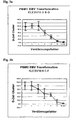
- Example 1.2 For immortalization of primary B cells leukocyte preparations were subjected to the procedure described in Example 1.2. To determine the optimum transformation parameters, the transformation frequency of EBV-containing supernatants with unselected PBMCs was determined. At a constant cell count (1x10 5 ), transformation approaches were carried out with continuous 2-fold dilutions of the EBV-containing supernatant in two different PBMC preparations. After a culture period of 17 days, transformed B cells could be observed as cluster-shaped cell aggregates. For simplicity, each cluster was scored as a single clone. The number of clusters was set in relation to the starting cell number and to the respective dilution level of the EBV-containing supernatants ( FIG. 3 ). The transformation frequencies for the PBMC preparation A were 3.1 ⁇ 10 -3 ⁇ 1.7 ⁇ 10 -3 and for the preparation B 3.5 ⁇ 10 -3 ⁇ 2.4 ⁇ 10 -3 .
- the separation of antigen-specific B cells is preceded by a leucocyte preparation by Ficollgradentrenzentrifugation (see also Example 1).
- Starting material for the preparation is peripheral whole blood from patients. Allergy sufferers are taken 10 ml of peripheral whole blood in heparin-coated blood tubes. This is followed by a density gradient centrifugation. The supernatant contains approximately 1: 3 diluted serum and can be stored for ELISA at -20 ° C.
- the solid blood components erythrocytes, platelets, etc.
- the lymphocytes are incubated with an anti-human CD19-MACS antibody (Milteny) (see also Example 1). After washing twice, up to 2x10 8 total cells are loaded on the MS-MACS Separation column (Miltenyi). Beforehand, the column is equilibrated with 500 ⁇ l of FACS buffer and then washed 3 times with 500 ⁇ l of FACS buffer. The column is then removed from the magnet, overlaid with 1 ml of FACS buffer and the enriched cells are gently pressed with the syringe punch. The purity can be increased to 99% by passing the positive, CD19 + enriched fraction through a second, equilibrated column.
- High affinity antibodies are produced by antigen-specific B cells in "germinal centers" of secondary lymphatic organs by somatic mutation after antigenic contact. Therefore, the further enrichment of CD19 + populations focuses on the isolation of memory cells.
- published anti-CD antibodies surface markers
- the specificity of the B cell population for the antigen is achieved by providing the antigen with a fluorescent marker (eg FITC).
- the fluorescence of the cells is shown after staining in the FACS-Vantage flow cytometer. Cells whose signals exceed the maximum fluorescence intensity of the control batch by a power of ten are recorded via a selection gate and stored in the 96-well plate format via the single-cell sorting mode.
- RNA isolation is carried out according to the Quiagen standard protocol.
- RT reverse transcription
- antisense-specific primers from the 3 'region are derived from the kappa and lambda light chain constant region, as well as the isotype-specific heavy chain (epsilon ( ⁇ ), alpha ( ⁇ ), mu ( ⁇ ) and gamma ( ⁇ ) subclasses) (the sequences are fully contained in the sequence database).
- PCR amplification step in which an aliquot of the RT batch is added with the respective sense primers from the kappa and lambda light chain 5 'variable region, as well as the variable heavy chain. Standard primers for this are published V-gene mixes of heavy and light chains used.
- the PCR conditions were optimized for the following times and temperatures (45 s 95 ° C, 45 s 72 ° C, 1 min 59 ° C, 35 repeats). To increase the specificity of the PCR, a semi-nested PCR protocol can be used.
- the resulting PCR products are sequenced directly (ABI). Due to the sequence obtained, the PCR product is recloned into suitable expression vectors.
- the separation of antigen-specific B cells was carried out by means of MACS methods ( Fig. 1 ).
- the antigen was biotin-conjugated timothy grass pollen allergen (rPhl pVb).
- the labeling of immortalized cells was carried out with 2 ⁇ g of antigen / 10 6 cells, a washing step and a subsequent incubation with streptavidin-coupled paramagnetic microbeads. The marked Lymphocytes were separated from non-labeled cells by retention in the magnetic field and culture was taken.
- specific B cells were selected by flow cytometry against Phl pVb (FACSVantage SE, Becton Dickinson).
- PhI pVb Pseudomonas exotoxin A
- LPV-ETA Pseudomonas exotoxin A
- the fusion protein is detectable via a poly histidine tag.
- the detection antibodies used were monoclonal anti-His antibodies (working concentration 1: 2000) and goat anti-mouse FITC conjugate (1: 1000).
- the fluorescence of the cells was shown after staining in the FACSVantage SE flow cytometer in the FL 1 / FL2. Cells whose signals exceeded the maximum fluorescence intensity of the control mixture by a power of ten were recorded via a selection "gate" and placed in a 96-well plate format in a cell culture plate via the single-cell sorting mode. The selected cells were taken in permanent culture and tested for specific binding activity
- cryopreserved B-95-8 cells (CATCC CLR 1612) at a concentration of 1 ⁇ 10 6 cells in 10 ml medium (RPMI 1640 10% FCS, 50 ⁇ g / ml streptomycin, 50 U / ml penicillin , 2 mM L-glutamine) in culture (37 ° C, 5% CO 2 ).
- 10 ml medium RPMI 1640 10% FCS, 50 ⁇ g / ml streptomycin, 50 U / ml penicillin , 2 mM L-glutamine
- the cell culture supernatants were filtered through a 0.2 micron filter, aliquoted and frozen at -196 ° C.
- peripheral mononuclear cells were used which were obtained by density gradient centrifugation from whole blood or buffy coats' and then subjected to a selection procedure described under 1.1.
- the transformation mixture was composed in each case of 100 ⁇ l of cell suspension (10 5 cells), 10 ⁇ l of cyclosporin A (2.6 ⁇ g / ml final) and 100 ⁇ l of EBV supernatant. After 17 days Cultivation (37 ° C, 5% CO 2 ) without change of medium, immortalized B cells were observed on the basis of the resulting cell aggregates and taken in culture. The cell culture supernatant was assayed for secreted immunoglobulins by ELISA ( Fig. 3 ).
- Detection was carried out by means of specific antibodies against human immunoglobulin classes (IgA, IgG1, IgG4, IgE and IgM), as well as biotin-conjugated anti-goat or anti-mouse antibodies and alkaline phosphatase-coupled streptavidin.
- IgA human immunoglobulin classes
- IgG1, IgG4, IgE and IgM biotin-conjugated anti-goat or anti-mouse antibodies and alkaline phosphatase-coupled streptavidin.
- the culture medium RPMI 1640, 10% FCS
- the supernatants tested were from four-month-old culture after previous subculturing of immunoglobulin-secreting cells via limiting dilution (125 cells / well).
- Binding single-stranded variable fragments were cloned by standard methods [6] into a derivative of the phagemid pCANTAB or the bacterial expression vector pBM1.1 (7) and thereby fused with a deletion mutant of Pseudomonas exotoxin A.
- the ligation mixtures were phenol extracted, ethanol precipitated and resuspended in 10 ⁇ l H2O. 2.5 ⁇ l of this DNA solution was introduced by electroporation into E. coli BL21 (DE3) according to a protocol described [7].
- Transformed bacteria were cultured on kanamycin-containing medium (2xLB with 50 ⁇ g kanamycin / ml and 2% glucose). Positive clones were verified by restriction analysis.
- E. coli BL21 (DE3) were transformed into Terrific broth medium [TB] [6]; 1.2% Bacto-Tryptone, 2.4% Bacto-Yeast Extract, 0.4%, 0.17M KH 2 PO 4 , 0.72 MK 2 HPO 4 ) supplemented with 50 ⁇ g / ml kanamycin and 0.5 mM ZnCl 2 for 12-15 h or incubated overnight at 26 ° C and 200 rpm. Subsequently, the cultures were transferred to 0.5 l Erlenmeyer flasks with baffles, in a volume of 200 ml TB medium and grown to an OD 600 of 2.
- the osmotic value of the medium was increased with 0.5 M sorbitol, 4% NaCl and added as compatible solute 10 mM betaine or ectoine. Both substances have comparable effects, although ectoine can also be synthesized directly from the cells via co-transformation with plasmids of the pOSM series or by additional coding on the pBM1.1 derivative.
- the cultures were incubated for 15-60 min at 26 ° C / 200 rpm and then expression of the target protein was initiated by addition of 2mM IPTG. After reaching an OD 600 of 2.3 - 2.4, the bacteria were harvested by a centrifugation step (3,700 g, 10 min, 4 ° C). All further steps were done on ice.
- the batch is centrifuged at 4,000 rpm for 10 minutes and the pellet resuspended in 10 ml 2xTY, 100 ⁇ g / ml ampicillin, 1 mM ITPG. With continuous shaking at 30 ° C, the batch is incubated for at least 5 more hours.
- the construct can be harvested from the periplasm after 5 hours of induction. For this, the culture is cooled for 10 to 20 minutes on ice and then centrifuged in a Microfuge at 6,000 rpm for 5 minutes. The pellets are taken up in 10% of the original volume in PBS, 1 M NaCl, 1 mM EDTA 4 ° C and then incubated for 30 minutes on ice. This is followed by another centrifugation step in the microfuge for 10 minutes at 6,000 rpm to remove the cells. The supernatant is transferred to a fresh tube and centrifuged at 14,000 rpm, 4 ° C for 10 minutes to sediment debris. The supernatant contains the periplasmic protein fraction.
- the bacterial pellet was resuspended in ultrasound buffer (75 mM Tris / HCl pH 8, 300 mM NaCl, 1 tablet of protease inhibitors [Boeringer] / 50 ml, 5 mM DTT, 10 mM EDTA, 10% glycerol), and homogenized (15 min) and then sonicated for 6 x 30 s at 200 watts. Between the individual Incubation phases were followed by an equally long cooling phase on ice (30 s).
- the samples were rebuffered at 4 ° C by Hitrap desalting column on an FPLC system (75 mM Tris / HCl pH 8, 300 mM NaCl, 10% glycerol).
- the protein was applied in a first step by metal chracter chromatography over a Ni-NTA column (Qiagen).
- the separation of antigen-specific B cells is preceded by a leucocyte preparation by Ficollgradentrenzentrifugation (see also Example 1).
- Starting material for the preparation is peripheral whole blood from patients. Allergy sufferers are taken 10 ml of peripheral whole blood in heparin-coated blood tubes. This is followed by a density gradient centrifugation. The supernatant contains approximately 1: 3 diluted serum and can be stored for ELISA at -20 ° C.
- the solid blood components erythrocytes, platelets, etc.
- the lymphocytes are incubated with an anti-human CD19-MACS antibody (Milteny) (see also Example 1). After washing twice, up to 2x10 8 total cells are loaded on the MS-MACS Separation column (Miltenyi). Beforehand, the column is equilibrated with 500 ⁇ l of FACS buffer and then washed 3 times with 500 ⁇ l of FACS buffer. The column is then removed from the magnet, overlaid with 1 ml of FACS buffer and the enriched cells are gently pressed with the syringe punch. The purity can be increased to 99% by passing the positive, CD19 + enriched fraction through a second, equilibrated column.
- High affinity antibodies are produced by antigen-specific B cells in "germinal centers" of secondary lymphatic organs by somatic mutation after antigenic contact. Therefore, the further enrichment of CD19 + populations focuses on the isolation of memory cells.
- published anti-CD antibodies surface markers
- the specificity of the B cell population for the antigen is achieved by providing the antigen with a fluorescent marker (eg FITC).
- the fluorescence of the cells is shown after staining in the FACS-Vantage flow cytometer. Cells whose signals exceed the maximum fluorescence intensity of the control batch by a power of ten are recorded via a selection gate and stored in the 96-well plate format via the single-cell sorting mode.
- RNA isolation is carried out according to the Quiagen standard protocol.
- RT reverse transcription
- antisense-specific primers from the 3 'region from the kappa and lambda light chain constant region, as well as the isotype-specific heavy chain (epsilon ( ⁇ ), alpha ( ⁇ ), mu ( ⁇ ) and gamma ( ⁇ ) subclasses) (the sequences are fully contained in the sequence database).
- PCR amplification step in which an aliquot of the RT batch is added with the respective sense primers from the kappa and lambda light chain 5 'variable region, as well as the variable heavy chain.
- primers for this standard published V-gene mixes of heavy and light chains are used.
- the PCR conditions were optimized for the following times and temperatures (45 s 95 ° C, 45 s 72 ° C, 1 min 59 ° C, 35 repeats).
- a semi-nested PCR protocol can be used.
- the resulting PCR products are sequenced directly (ABI). Due to the sequence obtained, the PCR product is recloned into suitable expression vectors.
Landscapes
- Chemical & Material Sciences (AREA)
- Health & Medical Sciences (AREA)
- Life Sciences & Earth Sciences (AREA)
- Genetics & Genomics (AREA)
- Organic Chemistry (AREA)
- Engineering & Computer Science (AREA)
- Biotechnology (AREA)
- Biophysics (AREA)
- Zoology (AREA)
- General Engineering & Computer Science (AREA)
- Biomedical Technology (AREA)
- Molecular Biology (AREA)
- Biochemistry (AREA)
- Bioinformatics & Cheminformatics (AREA)
- General Health & Medical Sciences (AREA)
- Wood Science & Technology (AREA)
- Microbiology (AREA)
- Plant Pathology (AREA)
- Physics & Mathematics (AREA)
- Crystallography & Structural Chemistry (AREA)
- Botany (AREA)
- Immunology (AREA)
- Medicinal Chemistry (AREA)
- Proteomics, Peptides & Aminoacids (AREA)
- Peptides Or Proteins (AREA)
- Preparation Of Compounds By Using Micro-Organisms (AREA)
- Micro-Organisms Or Cultivation Processes Thereof (AREA)
- Medicines Containing Antibodies Or Antigens For Use As Internal Diagnostic Agents (AREA)
Description
- Die Erfindung betrifft ein Verfahren zur Bestimmung des immunologischen Reaktionsprofils eines Organismus. Weiterer Bestandteil der Erfindung sind die RT- und PCR Produkte erhältlich durch das oben genannte Verfahren, wie auch die Vektoren, die diese RT- und PCR Produkte enthalten. Die Erfindung betrifft darüber hinaus die Verwendung dieser Vektoren zur Herstellung von rekombinanten Antikörpern, sowie die rekombinanten Antikörper, als auch die Verwendung dieser rekombinanten Antikörper in dem oben genannten Verfahren zur Analyse ihrer Bindungsaktivität.
- Charakteristisch für atopische Erkrankungen sind Überempfindlichkeitsreaktionen vom Typ I. Pathopysiologisch wird die Erkrankung auf Störungen immunlogischer Toleranzmechanismen zurückgeführt, die im einzelnen noch nicht geklärt sind. Dabei richten sich die zu Grunde liegende Immunreaktionen zumeist gegen harmlose Antigene, die von Gesunden toleriert werden. Diese Entzündungsreaktionen werden durch Immunglobuline der Klasse E vermittelt. Die IgE Moleküle werden durch Mastzellen und basophile Granulozyten über Oberflächenrezeptoren gebunden. Kommt es bei Antigenkontakt zu Kreuzvernetzungen von IgE/IgE Rezeptorkomplexen, so werden bei den betreffenden Zellen Prozesse induziert, die zu einer Sekretion von Entzündungsmediatoren führen. Dies ist der Ausgangspunkt für die vielfältige Symptomatik atopischer Erkrankungen.
- Eine humorale Immunantwort beginnt mit einer IgM dominierten primären Antikörperantwort. Später spezifiziert sich das Antikörperrepertoire, durch eine Switch-Rekombination zu den Antikörper Isotypen IgG, IgA oder IgE. Diese Antikörperklassen werden später auch für "Memory-Antikörper" herangezogen. Charakteristisch für die Antikörper der sekundären Immunantwort ist, dass sie eine höhere Affinität zu einem bestimmten Antigen aufweisen. Das Phänomen das dieser Entwicklung zugrunde liegt bezeichnet man als "Affinitätsreifung des Antikörper Repertoires". Es wird durch somatische Mutationen in den hypervariablen Regionen der variablen Bereiche der schweren und leichten Antikörperketten erreicht und findet in den Keimzentren von lymphatischen Organen statt. Die Produzenten der Immunglobuline sind B-Lymphozyten, deren Reifung, Differenzierung und klonale Expansion wesentlich durch den wechselseitigen Kontakt mit antigenpräsentierenden Zellen und T- Helferzellen (Th-Zellen) bestimmt werden. Dabei exprimiert jeder B-Zellklon nur eine, genetisch einmalige Variante eines Immunglobulins. Die Variabilität unterschiedlicher Bindungsspezifitäten ist somit durch das Repertoire unterschiedlicher klonaler Ursprungszellen determiniert. Das Verhältnis der Größe der einzelnen Klone zueinander ist durch die dynamische Anpassung an die aktuellen Erfordernisse geprägt. Die spezifische Bindungsaktivität gegenüber Antigenen ist über die Konstitution der variablen Regionen der leichten und schweren Ketten eines Antikörpers festgelegt.
- An der Entwicklung von IgE-sezernierenden Plasmazellen haben Th-Zellen des Typs 2 bedeutenden Einfluss. Die Ausrichtung immunologischer Prozesse auf ein bestimmtes Antigen wird durch hoch spezifische Oberflächenrezeptoren (B-Zellrezeptor, T -Zellrezeptor) auf den B- und Th-Lymphozyten gewährleistet, die den initialen Kontakt beider Zelltypen nur in Gegenwart eines Antigens ermöglicht, für das beide über einen spezifischen Rezeptor verfügen müssen. Diese Antigen-spezifische Orientierung der Immunreaktion kann bei hohen IgE-Titern verloren gehen, wenn B-Zellen unterschiedlichster Spezifität über den niedrig affinen Rezeptor für IgE (CD23) lösliche Immunglobuline dieser Klassen binden und damit neben ihrer ursprünglichen Spezifität, die durch den membranständigen B-Zellrezeptor repräsentiert wird -IgE-spezifische Antigene erkennen, internalisieren und über MHC II-Moleküle mit Th2- Zellen in antigenspezifischen Kontakt zu treten. In der Folge entstehen so weitere IgEsezernierende Plasmazellen, die das hypersensitive Antigenspektrum extrem erweitern können [1].
- Die Beteiligung einzelner Immunglobulinklassen am atopischen Geschehen ist zwar beschrieben [2], die funktionellen Bezüge unterschiedlicher Antikörperklassen innerhalb des Immunsystems, ihre Einflüsse auf das Gesamtgefüge z.B. im Rahmen physiologischer und pathophysiologischer Prozesse (wie den atopischen Erkrankungen, darüber hinaus aber auch autoreaktive Reaktionen, Sepsis, Toleranz, und Anergie steuernde Vorgänge) sind dagegen nur unzureichend verstanden. Tiefergehende Erkenntnisse dieser Prozesse lassen sich aus differenzierten Reaktionsprofilen der am Geschehen beteiligten B-Zellklone ableiten, die den Einfluss auf das Spektrum des antigenspezifischen V-Gen- bzw. Antikörperrepertoirs und Isotypmusters im funktionellen Bezug zum immunphysiologischen bzw. pathophysiologischen Status abbilden können. Bislang stellt die enorme Vielfalt des Antikörperrepertoires eine entscheidende Hürde für die Erstellung zeitlich eng auflösender, repräsentativer Reaktionsprofile dar.
- Begrenzte Antigenspezifische Reaktionsprofile von Antikörpern werden auf der Basis von Serumanalysen durchgeführt. Dabei kann das Blutserum auf Reaktivität gegen ca. 600 verschiedene Allergene hin untersucht werden. Diese Methodik erlaubt aber keine Aussagen über nicht sezernierende B-Zellen, die durch suppressive Einflüsse an der Sezernierung von Antikörpern gehemmt sind. Es werden somit nicht alle potentiellen reaktiven Zellen erfasst, und es ist mit dieser Methode nicht möglich die Prädisposition zu Immunerkrankungen zu bestimmen.
- Weiterhin ist es möglich das Antigen-spezifische Reaktionsprofil direkt am Patienten, in vivo, durch Haut-Prick-, Intrakutan- und Provokationstests zu bestimmen. Nachteile dieser Methoden sind, dass sie den Patienten belasten und bei hochgradigen Allergikern, nur beschränkt anwendbar sind. Antigenspezifische V-Gen Profile auf Basis von Einzelzellanalysen sind ebenfalls beschrieben [3], [4]. Diese Ansätze sind auf eine repräsentative Darstellung des Antigen-spezifischen V-Gen Repertoires ausgelegt, aber in dieser Form nicht für eine umfassende isotypenspezifische Analyse der V-Gen Repertoires genutzt worden.
- In der letzten Zeit gelang es auch im Modellsystem Maus, B-Zellen zu isolieren, die dem Affinitätsreifungsprozess (somatische Hypermutation) unterliegen. Ein Protokoll wurde entwickelt, das die nachfolgende Isolierung von B-Zellen mittels FACS ermöglichte. Aus den Hapten- bzw. Allergenspezifischen Zellen wurde RNA isoliert und in Folge in cDNA revers transkribiert. Aus der cDNA konnten anschließend Allergen/Hapten spezifische schwere Ketten von Immunglobulinen mittels RT-PCR isolieren werden. Die Sequenzen wurden dann einer intensiven Computeranalyse unterzogen. Folgende Ergebnisse wurden erzielt:
- Ohne jeden Zweifel kann die IgE-Antwort "reifen".
- Die somatische Diversität ist von Isotyp zu Isotyp unterschiedlich.
- Der Antigenrezeptor ist für die Selektion von hoch affinen Antikörpern notwendig.
- Der Antigenrezeptor hat für die Regulation des somatischen Mutationsprozesses keine Funktion.
- Je kürzer die CDR3 Region, je affiner der Antikörper gegen das Hapten.
- IgE bildet keine "long lived"-Plasmazellen.
- Die IgE-Antwort ist kurzlebig.
- Der zytoplasmatische "tail" scheint in geeignetes Target für eine Allergietherapie zu sein.
- Die Ergebnisse dieses Teilprojektes wurden im "European Journal of Immunology" publiziert [5].
- Bedingt durch die enorme Vielfalt des Antikörperrepertoires war es bislang somit problematisch, bzw. unmöglich, ein umfassendes Antigen-spezifischen Reaktionsprofil eines Organismus zu bestimmen. Insbesondere fehlte ein für den Patienten wenig belastendes und risikoarmes Verfahren zur Beurteilung seines allergischen (antigen-spezifischen) Reaktionsprofils. Überraschenderweise bietet die vorliegenden Erfindung eine Lösung dieses Problems an. Der Erfindungsgegenstand der vorliegenden Anmeldung erlaubt auf genetischer Ebene die Bestimmung des Antigen-spezifischen Reaktionsprofils eines Organismus. Überrachsenderweise ist es so möglich, alle B-Zell-vermittelten Antigen-spezifische Reaktionen zu erfassen, insbesondere auch solche bei denen die Sezernierungsaktivität der B-Zellen durch suppressive Einflüsse gehemmt ist.
-
-
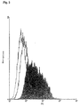
Figur 1 : FACS Analyse EBV transformierter humaner B-Zellen, die gegen das Lieschgras-Allergen Phl pVb selektioniert wurden. Gezeigt ist die spezifische Bindung an LPV-ETA' durch ein EBV transformierte B-Zelllinie (gefüllte Kurve), die aus einem Antigen spezifischen Selektionsverfahren hervorgegangen ist. LPV-ETA' stellt ein Fusionsproteinaus dem rekombinanten Allergen Phl p5b und dem modifizierten Enterotoxin A aus Staphylococcus aureus dar. Bindungsaktivitäten, die sich gegen das Toxinfragment richten, wurden mit einem Kontrollantigen HAI-ETA' untersucht, einem Fusionsprotein aus einem Antikörperfragment und der oben erwähnten Enterotoxin A Domäne (offene Kurve). Unspezifische Reaktionen die als Hintergrund der Messungen durch den Einsatz sekundärer Nachweiskomponenten auftreten, sind mit der offenen Kurve dargestellt. -
Figur 2 : Die Darstellung zeigt mit einer FACS Analyse den Vergleich der Bindungsaktivität der Phl p5b spezifischen EBV transformierten Zelllinie (gefüllte Kurve) mit einer EBV transformierten Zelllinie ohne Bezug zu Phlp5b (offene Kurve). Unspezifische Reaktionen die als Hintergrund der Messungen durch den Einsatz sekundärer Nachweiskomponenten auftreten, sind mit der offenen Kurve dargestellt. -
Figur 3: (a) und (b) zeigen die Anzahl der B-Zellcluster nach EBV-Transformation in Bezug zur Verdünnungsreihe der EBV-Überstände. Berechnete Korrelationskoeffizienten: (a) = -0,95; (b) = -0,96, n=3. -
Figur 4 : Immunklassen-spezifischer ELISA mit Überständen EBV transformierter B-Zellen. Der Nachweis wurde für 10 Subkulturen geführt. Die Werte beruhen auf kontrollbereinigten Doppelbestimmungen mit Ansätzen ohne primäre Nachweiskomponente. Als Negativkontrolle diente ein Ansatz gegen Kulturmedium. -
Figur 5 : Affinitätsmessung reklonierter und exprimierter anti Betvl spezifischer Fab-Fragmente mittels der BIACORE- Methode - Ziel der Erfindung ist es, das individuelle Repertoire Ig-vermittelter Bindungsaktivitäten von Organismen, insbesondere von Menschen mit atopischen Erkrankungen und/oder Autoimmunerkrankungen, anhand rekombinanter Ig-spezifischer V Gene umfassend darzustellen. Dabei sollen alle Immunglobulinklassen berücksichtigt werden, da neben den IgEspezifischen Reaktionen Bindungen von Antikörpern anderer ImmunglobulinKlassen am selben Antigen wichtige Regulative für das entzündliche Geschehen darstellen können.
- Nachstehende Definitionen sind auf die gesamte vorliegenden Anmeldung anzuwenden.
- Organismus: unter diesem Begriff werden alle Lebewesen verstanden, die von Immunfunktionsstörungen, wie atopischen Erkrankungen (Allergien) und/oder Autoimmunerkrankungen betroffen sein können. Insbesondere sind dies Säugetiere, einschließlich des Menschen.
- Immunologisches Reaktionsprofil: dieser Begriff beschreibt die Fähigkeit eines Organismus gegen ein Antigen spezifische Antikörper zu bilden.
- Mit dem vorliegenden in vitro Verfahren zur Bestimmung des immunologischen Reaktionsprofils eines Organismus kann in kurzer Zeit ein individuelles Antigen spezifisches V-Gen Repertoire dargestellt und des Ig Isotyp spezifisch differenziert werden, ohne dabei den Organismus zu belasten. Damit ergibt sich die Möglichkeit Krankheitsverläufe - vor allem solche, die durch Störungen B-Zell vermittelter Funktionen beruhen - anhand von Antigen und Isotyp spezifischen Reaktionsprofilen zu erfassen und zu bewerten:
- Der Erfindungsgegenstand ist ein Verfahren zur Bestimmung des immunologischen Reaktionsprofils eines Organismus, der die folgenden Schritte umfasst: (a) Isolation, einmalig oder mehrmalig, einer Fraktion aus Körperflüssigkeiten und/oder Geweben, die B-Zellen enthält; (b) Isolation von einzelnen B-Zellen oder deren genetischen Materials und Ablage von einer oder mehreren B-Zellen, oder deren genetischen Materials, in ein Reaktionsgefäß; (c) Amplifikation des die variablen Regionen eines von einer isolierten B-Zelle exprimierten Antikörpers codierenden genetischen Materials; (d) Herstellung von rekombinanten Antikörpern durch Exprimierung der klonierten Amplifikationsprodukte und (e) Bestimmung der Bindung des rekombinanten Antikörpers an rekombinante und/oder native Antigene. Dieses Verfahren ermöglicht die Bestimmung des immunologischen Reaktionsprofils von Organismen, insbesondere Personen, deren Immunsystem nicht offensichtlich gestört ist, insbesondere zur Bestimmung des Potentials einer Prädisposition für atopische Erkrankungen oder Antikörper vermittelte Autoimmunerkrankungen. Ebenso eignet sich dieses Verfahren zur Bestimmung des immunologischen Reaktionsprofils von Organismen, wie Personen, mit Immunfunktionsstörungen. Insbesondere eignet sich das erfindungsgemäße Verfahren zur Bestimmung des immunologischen Reaktionsprofils bei Immunfunktionsstörungen die folgende Erkrankungen umfassen: Allergien, atopische Erkrankungen, Antikörper-vermittelte Autoimmunerkrankungen wie Antiglomeruläre Basalmembrankrankheit; Arthritis, rheumatoide; Autoimmunkrankheiten des Nervensystems, z.B. Multiple Sklerose; Diabetes mellitus, insulinabhängiger; Lupus erythematodes, systemischer; Pemphigus; Addison-Krankheit; Anämie, autoimmunhämolytische; Antiphospholipid-Syndrom; Dermatitis, herpetiformis Duhring; Glomerulonephritis, IgA-; Glomerulonephritis, membranöse; Goodpasture-Syndrom; Graves-Krankheit; Lambert-Eaton-Myasthenie-Syndrom; Ophthalmie, sympathische; Pemphigoid, bullöses; Polyendokrinopathien, Autoimmun-; Purpura, thrombozytopenische, idiopathische; Reiter-Krankheit; Thyreoiditis, Autoimmun; Hashimoto Thyreoiditis; primäres Myxödem; Thyreotoxikose (Basedow Krankheit); Myasthenia gravis; Anämie, perniziöse; Leukopenie, autoimmune; Thrombozytopenie, idiopathische; Zirrhose, primäre, biliäre; Autoimmunhepatitis; Colitis ulcerosa; Sjörgen Syndrom; rheumatisches Fieber; Dermatomyositis Polymyositis; Sklerose, progressive, systemische; Granulomatose, Wegner; Panateriitis nodosa; Periateriitis nodosa; und Hypersensitivitätsangiitis etc. Bei dem erfindungsgemäßen Verfahren wird anhand des genetischen Materials der B-Zellen das immunologische Reaktionsprofil ermittelt und quantitativ und qualitativ verglichen mit dem Reaktionsprofil aus dem Serum. Bei dem erfindungsgemäßen Verfahren wird die B-Zell-haltige Fraktion aus peripheren mononukleären Zellen über Dichtezentrifugation aus Blutproben isoliert. Die Isolation einzelner B-Zellen umfasst bevorzugt die folgenden Schritte: (a) die Markierung der B-Zellen mittels Fluorochrom gekoppelter Antikörper, bevorzugt mit Antikörpern spezifisch für CD19, CD85 und CD138, CD19, CD85, CD 138, CD10, CD20, CD21, CD22, CD23, CD24, CD27, CD37, CD38, CD39, CD40, CD72, CD73, CD74, CDw75, CDw76, CD77, CDw78, CD79, CD80, CD81, CD82, CD83, CDw84, CD85 und/oder CD86, usw.; (b) die durchflusszytometrische Selektion der markierten B-Zellen; und (c) die Ablage von einer oder mehreren B-Zellen in ein Reaktionsgefäß. Bei dem erfindungsgemäßen Verfahren werden die B-Zellen nach dem Isolierungsschritt und der Einzelzellablage durch EBV Transformation immortalisiert. Es ist auch möglich die B-Zellen nach dem Isolierungsschritt und der Einzellzellablage mit Myelomzellen zu fusionieren.
- Das erfindungsgemäße Verfahren zur Bestimmung des immunologischen Reaktionsprofils umfasst folgende Amplifikationsschritte: (a) cDNA generierende RT Reaktionen; (b) PCR Reaktionen mit der cDNA als Matrize und (c) nested PCR Reaktionen mit den Amplifikationsprodukten aus der PCR Reaktion als Matrize. Dabei werden in den RT Reaktionen Primer eingesetzt, die sequenzkomplementär zu den konstanten Regionen der leichten und schweren Ketten sind, und es werden als Amplifikationsprodukt cDNAs gewonnen. In den PCR Reaktionen wird die cDNA als Matrize eingesetzt und degenerierte Primer, die sequenzkomplementär zu den variablen Regionen im framework 1 Bereich der 6 Genfamilien der schweren Ketten sind, und/oder es werden in den PCR Reaktionen die cDNA als Matrize eingesetzt und degenerierte Primer für die 4 Genfamilien der κ- Ketten, und/oder es werden in den PCR Reaktionen die cDNA als Matrize eingesetzt, und degenerierte Primer für die konstanten Regionen der ε- γ- und der κ- Ketten. In den "nested" PCR Reaktionen werden die Amplifikationsprodukte aus der PCR Reaktion als Matrize eingesetzt und 5' Primer, die sequenzkomplementär zu den ersten 6 Codons der VH- oder der VL Regionen sind, und 3' Primern, die sequenzkomplementär zu den "genesteten" konstanten Regionen der κ-, λ-, α1-, α2-, γ1-, γ2-, γ3-, γ4-, µ- und ε- Ketten sind. In dem erfindungsgemäßen Verfahren werden die Amplifikationsprodukte aus den Reaktionsgemischen, die Amplifikate der VL und der VH Regionen enthalten, in einen Vektor kloniert und durch Exprimierung der Vektoren in bakteriellen und/oder eukaryontischen Expressionssystemen rekombinante Antikörper gewonnen. Es werden auch die Vektoren des erfindungsgemäßen Verfahren, die die Amplifikationsprodukte enthalten, beansprucht. Ebenso wird die Verwendung der erfindungsgemäßen Amplifikationsprodukte zur Herstellung von Expressionsvektoren beansprucht. Weiterhin wird die Verwendung der Vektoren zur Herstellung von rekombinanten Antikörpern beansprucht, sowie die so erhältlichen rekombinanten Antikörper. Das erfindungsgemäße Verfahren umfasst auch die Bestimmung der Bindung der rekombinanten Antikörper an Antigene durch automatisierte Verfahren, insbesondere durch festphasengekoppelte Bindungsassays, Durchflußzytometrie, Resonanzspektrometrie und/oder Chip Arrays mit rekombinanten und nativem Antigenen in nativer oder denaturierter Form. Es wird auch die Verwendung der rekombinanten Antikörper zur Bestimmung ihrer Bindungsaktivitäten für rekombinante Antigene beansprucht.
- Ausgehend von peripheren Blutproben umfasst die vorliegende Erfindung zur Bestimmung des immunologischen Reaktionsprofils eines Organismus nachfolgende Schritte: (a) die Isolation, einmalig oder mehrmalig, von B-Zellen sowie deren Sortierung und Einzelzellablage und die Immortalisierung der B-Zellen durch EBV Transformation bzw. die Heteromylomfusion von der B-Zellen mit der B-Lymphom Zelllinie KGH6/B5; (b) die Isolation des genetischen Materials der einzelnen oder mehrerer B-Zellen; (c) die spezifische Amplifikation der variablen Regionen leichter In- und λ-ketten sowie der variablen Regionen der schweren Iga1, Igo2, Igγ1, Igγ2a, Igγ2b, Ig3, Igγ4, Igµ und Igε-Kette mittels verschiedener PCR Reaktionen, einschließlich einer Einzelzell "nested" RT- PCR, (d) die funktionelle Expression der Amplifikate in einem bakteriellen Expressionssystem zur rekombinanten Herstellung von Antikörpern und (e) die automatisierte qualitative und quantitative Analyse der Bindungsaktivitäten rekombinanter Antikörper durch festphasengekoppelte Bindungsassays, Durchflußzytometrie, Resonanzspektrometrie, Chip Arrays mit rekombinanten und nativem Antigenen in nativer oder rekombinanter Form, um so auch nichtproteinogene, aus post-translationalen Modifikationen herrührende Epitopstrukturen erfassen zu können.
- Bei der vorliegenden Erfindung handelt es sich um ein Verfahren zur Bestimmung des immunologischen Reaktionsprofils eines Organismus, das mehrere Schritte umfasst. Als erstes werden die allergenspezifischen Plasmaund Gedächtnis-B-Zellen aus einer Körperflüssigkeit und/oder Geweben, die B-Zellen enthalten, angereichert. Die Anreicherung dieser Zellen ist beispielsweise durch Dichtezentrifugation von peripheren mononukleären Zellen aus Blutproben möglich. Tote Zellpopulationen werden mittels Propidiumiodid ausgeschieden. Die allergenspezifische Gedächtniszellpopulation beträgt in der FACS Analyse im Durchschnitt 1,2%. Die Gedächtnis-B-Zellpopulation werden, wie auch im Methodenteil beschrieben, als Einzelzellen sortiert. Die Isolation umfasst drei Schritte, nämlich (a) die Markierung der B-Zellen mittels Fluorochrom gekoppelter Antikörper, wobei Antikörper spezifisch für CD19, CD 138, CD10, CD20, CD21, CD22, CD23, CD24, CD27, CD37, CD38, CD39, CD40, CD72, CD73, CD74, CDw75, CDw76, CD77, CDw78, CD79, CD80, CD81, CD82, CD83, CDw84, CD85 und/oder CD86, usw., verwendet werden können; (b) die durchflusszytometrische Selektion der markierten B-Zellen; und (c) die Ablage von einer oder mehreren Zellen in eine Reaktionsgefäß. Die abgelegten Zellen können durch Transformation mit dem Eppstein Barr Virus (EBV) immortalisiert werden; es ist auch möglich, die isolierten/abgelegten/sortierten B-Zellen mit Myelomzellen zu fusionieren. Als zweiter Schritt schließt sich die RNA-Isolierung aus den sortierten Zellen an. Als dritter Schritt erfolgt die Amplifikation des genetischen Materials, das die variablen Regionen des von der B-Zelle exprimierten Antikörpers codiert. Es wird zunächst eine RT-PCR zur Amplifizierung der Immunglobulin schweren und leichten Ketten durchgeführt. Dabei werden zur Gewinnung der cDNA-Amplifikationsprodukte Primer eingesetzt, die sequenzkomplementär zu den konstanten Regionen der leichten und schweren Ketten sind. Insbesondere werden für die RT Reaktionen folgende Primer eingesetzt: (a) leichte Kette κ:5' AAC AGA GGC AGT TCC AGA; (b) leichte Kette λ: 5' TGTGGC CTT GTT GGC TTG; (c) schwere Kette α: 5' CTTGCA GAC ACT TGG TGT TCG TGC; (d) schwere Kette γ1 und γ3: 5' AGG GYG CCA GGG GGA A; (e) schwere Kette γ2: 5' TTT ACC CRG AGA CAG GGA GAG GC; (f) schwere Kette γ4: 5' TTT ACC CRG AGA CAG GGA GAG GC; (g) schwere Kette µ: 5'GCA GGA GAC GAG GGG GA; und (h) schwere Kette ε: 5' TTT ACC GGG ATT TAC AGA CAC. Die schweren Ketten werden auf alle Isotypen hin analysiert. Bisher konnten allergenspezifische Varianten diverser Isotypen amplifiziert und exprimiert werden. Naturgemäß findet sich allerdings die Ig-Klasse gamma 1 am häufigsten. Es wird darauf hingewiesen, dass nicht automatisch dieselben variablen Gene für alle Isotypen verwendet werden (siehe [14]). Vielmehr findet sich auch hier eine Isotypen spezifische Verwendung von V- Genklassen. Die durch die RT-PCR gewonnen cDNA Fragmente werden als Matrizen in PCR Reaktionen eingesetzt, die degenerierte Primer enthalten, die sequenzkomplementär zu den variablen Regionen im framework 1 Bereich der sechs Genfamilien der schweren Ketten sind, und/oder sequenzkomplementär für die vier Genfamilien der κ-Ketten sind, und/oder sequenzkomplementär für die drei Genfamilien der λ-Ketten sind, und/oder mit degenerierten Primern für die konstanten Regionen der ε- γ- und der κ- Ketten. Insbesondere werden für die variable Region der leichten κ-Ketten nachfolgende "sense" Primer eingesetzt: hVkI Back 5' GAC MTC VWG HTS ACC CAG TCT CC; hVkII Back 5' GAC CTC CAG HTG ACC CAR WSY CC; hVkIII Back 5' GAC CTC CAR HTS ASK CAG TCT CC; hVkIV Back 5' GAK VTY GTG ATG ACY CAG WCT CC; hVkV Back 5' GAC ATC SWG ATG ACC MAG TCT CC; hVkVI Back 5' GAC CTC GTG HTG ACB CAG DSY CC; hVkVII Back 5' GAC ATS VWG CTC ACS CAG TCT CC. Als anti-"sense" Primer werden für die variable Region der leichten κ-Ketten nachfolgende Primer eingesetzt: hVkI For 5'TCG TTT GAT CTC CAS YYK KGT CC; hVkII For 5' TCG TTT RAT YAG T'AC CYK KGT CC; hVkIII For 5' TCG TTT GAY HTS CAS CTT KGT CC. Für die variablen Regionen der leichten λ-Kette werden nachfolgende "sense" Primer eingesetzt: hVλI Back 5' CAG TCT SWG CTG ACK CAG CCR CC; hVλII Back 5' CAG TCT SMG CTG ACT CAG CCW SS; HVλII Back 5'CAS GYT WTA YTG ACT CAA YCG CC; hVλIV Back 5' TCC TMT GWG CTG ACW CAR CCA CC; HVλV Back 5'TCK TMT GAR CTG ACT CAR GAC CC; hVλVI Back 5' CAG TCT GTG CTG ACT CAG SMD SS. Als anti-`sense" Primer für die variablen Regionen der λ-Kette werden eingesetzt: hVλI For 5' ACCKAG RAC GGT SAS CTB GGT CC; hVλII For 5'ACY TAR GAC GGT SAV YTT GGT CC; hVλIII For 5'ACC TAR RAC GGT SAV CTK GGT CC. Weiterhin wurden als Immunglobulin-spezifischen Primer ("sense") für die schweren Ketten eingesetzt: hVHI 5'CAG GTG CAG CTG SWG SAR TCK GG; hVHII sense 5' CAG STG CAK CTG CAG GAG TCS GG; HvhIII sense 5'CAG GTG SAG CTG SWG SAG TCH GG; HvhIV sense 5'GAG GTG CAG CTR CAK SAG TSG GG; HvhV sense 5'GAG GTG CAR CTG KTG SAG TCN GS; HvhVI sense 5'GAG GTG CGR CTG GTG SAG WSK GG; HvhVII sense 5' CAG GTS AAS YTA AGG GAG TCT GG. Als anti-"sense" Immunglobulin-spezifische Primer für schweren Ketten werden eingesetzt: IgA1 5'GAA GAC CTT GGG GCT GGT CGG GGA TG; IgA2 5' ATG CGA CGA CCA CGT TCC CAT CTT G; IgE 5'GTT CTG GAT CAG GCA GGC GAG GGT GC; IgG1 5'CTC GAT GGGGGC TGG GAG GGC TTT G; IgG2 5'CCT GGT GCA CAA CGG TGA GGA CG; IgG3 5' GTC CGG GAA ATC ATA AGG GTA TC; IgG4 5' GAG GAC GGG AGG CCT TTG YTG GAG; IgM 5'CAG GAG ACG AGG GGG AAA AG. Die klassenspezifische Differenzierung wird durch eine Reamplifikation der schweren Ketten in einer "nested" PCR erreicht, wobei erneut Amplifikationsprodukte gewonnen werden. In den "nested" PCR Reaktionen werden die Amplifikationsprodukte aus der PCR Reaktion als Matrize und 5' Primern, die sequenzkomplementär zu den ersten 6 Codons der VH Region oder der VL Regionen sind, und 3' Primern, die sequenzkomplementär zu den "genesteten" konstanten Regionen der κ-, λ-, α1-, α2-, γ1-, γ2-, γ3-, γ4-, µ- und ε- Ketten sind, zum Einsatz kommen, und so ein Amplifikationsprodukt gewonnen wird. Als "sense" primer werden die "sense" primer der PCR Reaktion verwendet. Als anti-"sense" Primer werden dabei nachfolgende Immunglobulin-klassenspezifische Oligonukleotide eingesetzt: IgA1 5'GAAAAC CCA GCT CAG CCC AAA CTC CAT; IgA2 5' CCT TGG GGC TGG TCG GGG ATG; IgE 5' CAG GAC GAC TGT AAG ATC TTC ACG; IgG1 5' CCC CAG AGG TGC TCT TGG AGG AGG GT; IgG2 5' CCG CTG TGC TCT CAG AGG TGC TCC TG; IgG3 5' GGC CGC TGT GCC CCC AGA GGT GCT C; IgG4 5' GCA GCC CAG GGC GGC TGT GCT CTC G; IgM 5'GAA AAG GGT TGG GGC GGA TGC. Die Amplifikationsreaktionen werden mit Taq Polymerase durchgeführt und die erhaltenen Fragmente, die VL und VH Regionen enthalten, werden in Vektoren kloniert. Insbesondere wird dabei das TA Klonierungssystems verwendet. Der vierte Schritt des Verfahrens zur Bestimmung des immunologischen Reaktionsprofils eines Organismus ist die rekombinante Herstellung von Antikörpern durch Exprimierung der Amplifikate. Dabei werden die in Vektoren klonierten Fragmente in bakteriellen und/oder eukaryontischen Expressionssystemen exprimiert. Die Vektoren werden zur Herstellung von rekombinanten Antikörpern verwendet. Insbesondere werden die resultierenden Immunglobulinfragmente als "single-chain Antikörper" oder als "Fab- Fragmente" zunächst in Phagen exprimiert. Die so erhaltenen Antikörper werden verwendet um ihre Bindungsaktivität zu Allergenen zu analysieren. Dabei wird die Bindungsaffinität der "single-chain Antikörper" mittels "biosensor chip" Technologie analysiert. Für die Expression der Fab-Fragmente als lösliche Proteine wird das unter Beispiel 1 beschriebene Expressionssystem verwendet.
- Für die Messung der Bindungsaffinität wurde als Antigen rekombinantes Betvl an einen CM5 sensor chip kovalent gekoppelt. Weiter wurden zwei sequenzierte schwere und leichte Ketten, sowie eine schwere Kette allein in ein geeignetes Phagenexpressionssystem kloniert. Anschließend wurden zwei Fab-exprimierende Phagen injiziert. Das Experiment zeigt zwei Fab-Fragmente unterschiedlicher Affinität (Fab. high, Fab low). Die Spezifität ist durch die beiden Kontrollen (F-heavy und vector) gewährleistet. Das Experiment zeigt eindeutig, dass mit der beschriebenen Methode affine antigenspezifische B-Zellen isoliert werden können.
- Das Verfahren eignet sich somit für die Bestimmung des Potentials für atopische Erkrankungen oder Antikörper vermittelte Autoimmunerkrankungen, sowie auch zur Bestimmung des immunologischen Reaktionsprofils von Personen mit bereits offensichtlichen Immunfunktionsstörungen. Zu den Immunfunktionsstörungen, die mittels vorliegender Erfindung analysierbar sind, werden nachfolgende Erkrankungen gerechnet: Antiglomeruläre Basalmembrankrankheit; Arthritis, rheumatoide; Autoimmunkrankheiten des Nervensystems, z.B. Multiple Sklerose; Diabetes mellitus, insulinabhängiger; Lupus erythematodes, systemischer; Pemphigus; Addison-Krankheit; Anämie, autoimmunhämolytische; Antiphospholipid-Syndrom; Dermatitis, herpetiformis Duhring; Glomerulonephritis, IgA-; Glomerulonephritis, membranöse; Goodpasture-Syndrom; Graves-Krankheit; Lambert-Eaton-Myasthenie-Syndrom; Ophthalmie, sympathische; Pemphigoid, bullöses; Polyendokrinopathien, Autoimmun-; Purpura, thrombozytopenische, idiopathische; Reiter-Krankheit; Thyreoiditis, Autoimmun; Hashimoto Thyreoiditis; primäres Myxödem; Thyreotoxikose (Basedow Krankheit); Myasthenia gravis; Anämie, perniziöse; Leukopenie, autoimmune; Thrombozytopenie, idiopathische; Zirrhose, primäre, biliäre; Autoimmunhepatitis; Colitis ulcerosa; Sjörgen Syndrom; rheumatisches Fieber; Dermatomyositisl Polymyositis; Sklerose, progressive, systemische; Granulomatose, Wegner; Panateriitis nodosa; Periateriitis nodosa; und Hypersensitivitätsangiitis usw. Das Verfahren erlaubt, das genetisch ermittelte Reaktionsprofil mit Reaktionsprofilen aus dem Serum qualitativ und quantitativ zu vergleichen. Nachstehend werden Ergebnisse diskutiert, die sich auf die in den Beispielen beschriebenen Experimente beziehen.
- Ausgangspunkt für die Selektionssschritte stellen Leukozytenpräparationen aus Ficollgradientenzentrifugationen dar, auf der Basis von "buffy coats" bzw. peripherem Vollblut. Der Anteil CD19+ Zellen betrug 10,6 ± 0,6% bei einer Gesamtzellzahl von 3,4 ± 0,7 x 10⁷ PBMC.
- Die Separation antigenspezifischer B-Zellen wurden mittels MACS-Verfahren (Miltenyi) durchgeführt. In einem zweifach gestaffelten Selektionsverfahren wurden zunächst CD19+-Zellen isoliert und aus dieser Fraktion antigen-spezifisch markierte Zellen. Deren Anteil variierte zwischen 0,01 und 0,05%. Die Zellen wurden im Anschluss EBV-transformiert.
- Die Bindungseigenschaften der kultivierten antigenspezifisch selektionierten B-Zellen wurden durchflusszytometrisch analysiert. Als Antigen diente ein rekombinantes Fusionsprotein (PV 21/2 Ang II); bestehend aus dem eigentlichen Antigen PV 21/2, einem Fragment des Lieschgras-Allergens rPhl pVb gekoppelt an humanes Angiogenin (Ang II). Als weiteres Antigen wurde das Fusionsprotein LPV-ETA' eingesetzt, eine Fusion des Vollängen-Allergens mit der Toxindomäne ETA'. Um nicht-Antigen-spezifische Reaktionen - im Sinne der vorangegangen Selektion gegen das Allergen - von Reaktionen unterscheiden zu können, die sich gegen Epitope der Toxindomänen richten, wurden Versuche mit rekombinanten Proteinfusionen durchgeführt, bei denen die Allergendomäne gegen rekombinante scFv - spezifisch für die Oberflächenantigene GD2 (NAng GDII) bzw. den Epidermalen Wachstumsfaktor- Rezeptor (HAI-ETA') -ausgetauscht waren. Im Gegensatz zu PV 21/2 Ang II und LPV-ETA' zeigte der Einsatz beider Kontrollantigene in der FACS-Analyse Fluoreszenzintensitäten, die dem Hintergrundniveau der sekundären Nachweiskomponenten entsprachen (
Figur 1 a und Figur 2 a). Mit einem weiteren Versuchsansatz sollte abgeklärt werden, ob die eingesetzten Proteine generell B-Zellen binden oder auf Eigenschaften spezifischer B-Zellen zurückzuführen sind. Dazu wurde derselbe Versuchsansatz vor dem Hintergrund eines großen B-Zell Rezeptorrepertoires mit frisch präparierten PBMCs durchgeführt und die Bindungsaktivität der CD19+ Fraktion untersucht (Figur 1 b). Für alle Allergen- und Kontrollvarianten ergeben sich identische Fluoreszenzsignalerkennungen und damit keinerlei Hinweise auf unspezifische Bindungen durch B-Zellen. - Zur durchflusszytometrischen Selektion Antigen-spezifischer B-Zellen wurden 4x10⁶ EBV-transformierte Zellen eingesetzt. Die Zellen waren ursprünglich als Antigen-spezifische Zellinie aus einer MACS-Selektion hervorgegangen. Mit zunehmenden Alter der Kultur wuchs der Anteil nicht bindender Zellen, vermutlich genetisch unstabile Klone, die keine funktionellen Immunglobuline oder B-Zellrezeptoren exprimieren konnten. Die verbliebenen Antigen-spezifischen Zellen ließen sich im Durchflusszytometer klar von unspezifischen unterscheiden und konnten über ein entsprechendes Selektions-"Gate" isoliert werden. Aus der Ursprungspopulation wurden 3x10⁵ Zellen, mit einer Reinheit von 97% isoliert und in Dichten von 1.000 Zellen pro Kavität in Kultur genommen. Nach 2 Wochen wurden die Antigen-spezifischen Bindungseigenschaften überprüft (
Figur 2 ). Das Antigen (LPV) lag als Fusionsprotein vor, gekoppelt an die Toxinkomponente (ETA'). Um Reaktionen gegen die Toxindomäne erfassen zu können, wurden Kontrollversuche mit Hilfe eines Fusionskonstruktes (HAI-ETA') durchgeführt, bei dem die Antigendomäne durch rekombinantes EGF (epidermal growth factor) ersetzt war. Die Bindungen waren ausschließlich gegen die Antigendomäne (LPV) gerichtet. Die Fluoreszenzintensität des Kontrollantigens entsprach dem des Hintergrundes (sekundäre und tertiäre Nachweiskomponenten). - Um zu überprüfen, ob die Interaktionen zwischen Antigen und B-Zellen Eigenschaften spezifischer Klone im Sinne einer Antikörper/ Anitgenreaktion darstellen, wurden Antigen-spezifisch-selektioniert B-Zellen unselektionierten Zellinien gegenübergestellt. Die unselektionierten Zellen repräsentieren ein klonales B- Zellrepertoire aus einem "buffy coat", die durch EBV-Transformation immortalisiert und kultiviert wurden. Der Versuchsansatz entspricht dem oben beschriebenen, mit Ausnahme des Kontrollantigens. Gegenüber nicht-selektionierten Zellen B-Zellen wurden bei den Antigen-spezifisch selektionierter Zellen 4,1 fach höhere Bindungsaktivitäten (mittleren Fluoreszenzintensität) ermittelt.
- Zur Immortalisierung von primären B-Zellen wurden Leukozytenpräparationen dem unter Beispiel 1.2 beschrieben Verfahren unterzogen. Zur Bestimmung deroptimaler Transformationsparameter wurde Transformationsfrequenz EBV-haltiger Oberstände mit nicht selektionierten PBMCs ermittelt. Bei konstanter Zellzahl (1x10⁵) wurden Transformationsansätze mit kontinuierlichen 2-fach Verdünnungen des EBV-haltigen Oberstands bei zwei verschiedenen PBMC Präparationen durchgeführt. Nach 17 tägiger Kultivierungphase konnten transformierte B-Zellen als cluster-förmig wachsenden Zellaggregate beobachtet werden. Zur Vereinfachung wurde jeder Cluster als einzelner Klon gewertet. Die Anzahl der Cluster wurde in Bezug zur Ausgangszellzahl und zur jeweiligen Verdünnungsstufe der EBV-haltigen Oberstände gesetzt (
Figur 3 ). Die Transformationsfrequenzen betrugen für die PBMC Präparation A 3,1 x 10⁻³ ± 1,7x 10⁻³ und für die Präparation B 3,5 x 10⁻³ ± 2,4 x 10⁻³. - Um zu klären welche Immunglobulinklassen die in Dauerkultur befindlichen Selektionskulturen sezernieren wurden Kulturüberstände mittels Festphasen ELISA getestet. Die Untersuchung umfasste humane Antikörper der Klassen IgA, IgE, IgG1, IgG4 und IgM. Bei allen getesteten Kulturen konnten sezernierte Anitkörper nachgewiesen werden, die ausschließlichen den Klassen IgG1 und IgM zuzuordnen waren (
Figur 4 ). - Der Separation Antigen-spezifischer B-Zellen geht eine Leukozytenpräparation mittels Ficollgradientenzentrifugation voran (siehe auch Beispiel 1). Ausgangsmaterial für die Präparation stellt peripheres Vollblut aus Patienten dar. Allergikern wird 10 ml periphäres Vollblut in Heparin-beschichtete Blut-Röhrchen abgenommen. Es folgt eine Dichtegradienten Zentrifugation. Der Überstand enthält das ca. 1:3 verdünnte Serum und kann für ELISA auf -20°C gelagert werden. Die festen Blutbestandteile (Erythrozyten, Thrombozyten etc.) sinken bis zum Boden des Probenröhrchens, die Lymphozyten erscheinen als Ring zwischen Serum und Ficoll Lösung und werden mittels Pipette abgenommen. Man wäscht die Lymphozyten in einem Volumen von 30ml FACS Puffer (1 X PBS, 0.5% BSA, 4mM EDTA) bei 4°C für 20 min bei 1500 rpm mit Bremse und resuspendiert das Lymphozyten-Pellet in 1 ml FACS Puffer.
- Die Lymphozyten werden mit einem anti-human CD19-MACS-Antikörper (Milteny) inkubiert (siehe ebenfalls Beispiel 1). Nach zweimaligem Waschen werden bis zu 2 x 10⁸ Gesamt-Zellen auf die MS-MACS Separations Säule (Miltenyi) geladen. Zuvor wird die Säule mit 500 µl FACS Puffer äquilibriert und anschließend 3 x mit 500 µl FACS Puffer gewaschen. Die Säule wird danach vom Magnet entfernt, mit 1 ml FACS Puffer überschichtet und die angereicherten Zellen werden vorsichtig mit dem Spritzenstempel durchgedrückt. Die Reinheit kann auf 99% erhöht werden, wenn man die positive, CD19+-angereicherte Fraktion über eine zweite, äquilibrierte Säule laufen lässt.
- Hochaffine Antikörper werden von Antigenspezifischen B-Zellen in "Reifungszentren (germinal centers)" sekundär lymphatischer Organe mittels somatischer Mutation nach Antigenkontakt gebildet. Deshalb konzentriert sich die weitere Anreichung von CD19+-Populationen auf die Isolierung von Gedächtniszellen (memory cells). Für die Anreicherung von Gedächtniszellen werden standardmäßig publizierte anti-CD-Antikörper (Oberflächenmarker) verwendet. Die Spezifität der B-Zellpopulation für das Antigen wird dadurch erreicht, dass das Antigen mit einem Fluoreszenzmarker versehen wird (z.B. FITC). Die Fluoreszenz der Zellen wird nach der Färbung im FACS-Vantage Durchflußzytometer dargestellt. Zellen deren Signale die maximale Fluoreszenzintensität des Kontrollansatzes um eine Zehnerpotenz übertreffen, werden über ein Selektions-"Gate" erfasst und über den Einzelzell-Sortierungsmodus im 96 Weil Plattenformat abgelegt.
- Die positiven Zellen werden sofort in eine 96-well Platte sortiert, in die 20 µl 1x RT-PCR Puffer (QIAGEN oneStep RT-PCR Kit) vorgelegt wurden. Die RNA Isolierung erfolgt nach dem Quiagen Standardprotokoll. Für die reverse Transkription (RT) werden antisense-spezifische Primer aus der 3'-Region von der konstanten Region der kappa- und lambda-leichten Kette, sowie der isotypspezifischen schweren Kette (epsilon (ε), alpha (α), mü (µ) und gamma (γ)-Subklassen) verwendet (die Sequenzen sind vollständig in der Sequenzdatenbank vorhanden). Es folgt der PCR-Amplifikationsschritt, bei dem ein Aliquot des RT- Ansatzes mit den jeweiligen sense primern aus der 5'-variablen Region von der kappa- und lambda leichten Kette, sowie der variablen schweren Kette zugefügt wird. Als Primer werden hierfür standardmäßig publizierte V-Gen Mixe von schweren und leichten Ketten verwendet. Die PCR- Bedingungen wurden für folgende Zeiten und Temperaturen optimiert (45 s 95°C, 45 s 72°C, 1 min 59°C; 35 repeats). Um die Spezifität der PCR zu erhöhen kann ein semi-nested PCR-Protokoll angewendet werden. Die resultierenden PCR-Produkte werden direkt sequenziert (ABI). Aufgrund der erhaltenen Sequenz wird das PCR-Produkt in geeignete Expressionsvektoren umkloniert.
- Die Expression der schweren und leichten Ketten als scFv oder Fab-Fragmente erfolgt wie in Beispiel 1 beschrieben.
- Die Immortalisierung wird mittels EBV- Transformation erreicht. Die Methode ist in Beispiel 1 genau erläutert.
- Die Separation Antigen-spezifischer B-Zellen wurde mittels MACS-Verfahren durchgeführt (
Abb. 1 ). Als Antigen diente Biotin-konjugiertes Lieschgraspollen-Allergen (rPhl pVb). Die Markierung immortalisierter Zellen erfolgte mit 2 µg Antigen/106 Zellen, einem Waschschritt und einer anschließenden Inkubation mit Strepavidin-gekoppelten paramagnetischen Mikrobeads. Die markierten Lymphozyten wurden durch Retention im Magnetfeld von nicht markierten Zellen getrennt und Kultur genommen. - Alternativ zur MACS Isolation wurden spezifische B-Zellen mittels Durchflusszytometrie gegen Phl-pVb (FACSVantage SE, Becton Dickinson) selektioniert. Zur Markierung spezifischer Zellen wurde eine Fusion des rekombinanten Allergens PhI pVb mit der Toxindomäne des Pseudomonas Exotoxins A (ETA), LPV-ETA; eingesetzt (2 µg LPV-ETA' auf 1x10⁶ Zellen in 300 µl, 30'/4°C). Das Fusionsprotein ist über ein Poly-Histidintag detektierbar. Als Nachweisantikörper dienten monoklonale anti-His Antikörper (Arbeitskonzentration 1: 2000) und Ziege anti-Maus FITC Konjugat (1:1000). Die Fluorezenz der Zellen wurde nach der Färbung im FACSVantage SE Durchflusszytometer im FL 1/ FL2 dargestellt. Zellen deren Signale die maximale Fluoreszenzintensität des Kontrollanssatzes um eine Zehnerpotenz übertrafen wurden über ein Selektions-"Gate" erfasst und über den Einzelzell-Sortierungsmodus im 96 Well Plattenformat in eine Zellkulturplatte abgelegt. Die selektionierten Zellen wurden in Dauerkultur genommen und auf spezifische Bindungsaktivität getestet
- Zur Gewinnung EBV-haltiger Überstände wurden kryokonservierte B-95-8 Zellen (CATCC CLR 1612) in einer Konzentration von 1 x 10⁶ Zellen in 10 ml Medium (RPMI 1640 10% FKS, 50 µg/ml Streptomycin, 50 U/ml Penicillin, 2 mM L-Glutamin) in Kultur genommen (37 °C, 5% CO2). Nach Zugabe von 5 ml Kulturmedium am folgenden Tag und weiterer 10-tägiger Kultivierung ohne Mediumzugabe wurden die Zellkulturüberstände über einen 0,2 µm-Filter filtriert, aliquotiert und bei -196°C eingefroren.
- Zur Transformation wurden periphere mononukleäre Zellen (PBMC) eingesetzt, die über Dichtegradienten- Zentrifugation aus Vollblut oder buffy coats' gewonnen wurden und anschließend einem, der unter 1.1 beschriebenen, Selektionsverfahren unterzogen wurden. Der Transformationsansatz setzte sich aus jeweils 100 µl Zellsuspension (105 Zellen), 10 µl Cyclosporin A (2,6 µg/ml final) und 100 µl EBV-Überstand zusammen. Nach 17-tägiger Kultivierung (37 °C, 5% CO2) ohne Mediumwechsel konnten immortalisierte B-Zellen anhand der entstandenen Zellaggregate beobachtet und in Kultur genommen werden. Der Zellkulturüberstand wurde mit Hilfe eines ELISA auf sezernierte Immunglobuline untersucht (
Abb. 3 ). Die Detektion erfolgte mittels spezifischer Antikörper gegen humane Immunglobulinklassen (lgA, IgG1, IgG4, IgE und IgM), sowie Biotin-konjugierter anti Ziege-, bzw. anti Maus-Antikörper und alkalische Phosphatase-gekoppeltem Streptavidin. Als Negativkontrolle wurde das Kulturmedium (RPMI 1640, 10% FKS) eingesetzt und jeweils ein Ansatz ohne primäre Nachweiskomponente. Die untersuchten Überstände entstammten vier Monate alten Kulturansätzen nach vorausgegangener Subkultivierung Immunglobulin-sezernierender Zellen via "limiting dilution" (125 Zellen / Kavität). - Bindungsfähige, einzelsträngige variable Fragmente (scFv) wurden mittels Standardverfahren [6] in ein Derivat des Phagemids pCANTAB oder den bakteriellen Expressionsvektor pBM1.1 (7) kloniert und dadurch mit einer Deletionsmutante des Pseudomonas Exotoxin A fusioniert. Die Ligationsansätze wurden phenolextrahiert, ethanolpräzipitiert und in 10 µl H2O resuspendiert. 2.5 µl dieser DNA-Lösung wurde nach einem beschriebenen Protokoll [7] durch Elektroporation in E.coli BL21 (DE3) eingebracht. Transformierte Bakterien wurden auf Kanamycin-haltigem Medium (2xLB mit 50 µg Kanamycin/ml und 2% Glukose) kultiviert. Positive Klone wurden durch Restriktionsanalyse verifiziert.
- Die Verfahren zur Klonierung und Expression der scFV sind bereits beschrieben unter
Scfv Zielantigen Erkrankung Referenzen Ki-4 CD30-Rezeptor Hodgkin-Lymphom [8, 9] Ki-3 CD30-Rezeptor Hodgkin-Lymphom [10, 11]] hHAK30 CD30-Rezeptor Hodgkin-Lymphom [12] RFT5 CD25-Rezeptor Hodgkin-Lymphom [13-15] 14.18 GD2-Antigen Neuroblastom [13, 14] - Transformierte E.coli BL21 (DE3) wurden in Terrific Broth-Medium [TB] [6]; 1,2% Bacto-Trypton, 2,4% Bacto-Yeast Extract, 0,4%, 0,17 M KH2PO4, 0,72 M K2HPO4) supplementiert mit 50 µg/ml Kanamycin und 0,5 mM ZnCl2 für 12-15 h bzw. über Nacht bei 26°C und 200 rpm inkubiert. Anschließend wurden die Kulturen in 0,5 1-Erlenmeyerkolben mit Schikanen, in einem Volumen von 200 ml TB-Medium überführt und bis zu einer OD600 von 2 angezogen. Anschließend wurde der osmotische Wert des Mediums mit 0,5 M Sorbitol, 4% NaCl heraufgesetzt und als kompatibles Solut 10 mM Betain oder Ectoin hinzugefügt. Beide Substanzen haben vergleichbare Wirkung, wobei allerdings Ectoin über Kotransformation mit Plasmiden der pOSM-Reihe oder durch zusätzliche Kodierung auf dem pBM1.1- Derivat auch direkt von den Zellen synthetisiert werden kann.
- Die Kulturen wurden für 15 - 60 min bei 26°C/200 rpm inkubiert und dann wurde durch Zugabe von 2 mM IPTG die Expression des Targetproteins eingeleitet. Nach Erreichen einer OD600 von 2,3 - 2,4 wurden die Bakterien durch einen Zentrifugationsschritt (3.700 g, 10 min, 4°C) geerntet. Alle weiteren Schritte wurden auf Eis durchgeführt. Nach einem Waschschritt (Pellet wurde in 75 mM Tris/HCl pH 8, 4% NaCl, 10% Glycerin resupendiert und homogenisiert) mit anschließender Zentrifugation (3.700 g, 10 min, 4°C) wurde das Gewicht des Naßpellets bestimmt und das Bakterienpellet vor dem Aufschluss bei -80°C bzw. bei - 196°C für 10 min schockgefroren.
- Ein alternativer Ansatz zur Expression rekombinanter scFv wird nach folgenden Verfahren durchgeführt: Transformierte E.coli TG1 werden gepickt und in 2 x TY, 100 µg/ml Ampicillin, 2% Glukose aufgenommen und über Nacht bei 37°C unter Schütteln inkubiert. Anschließend wird die über Nacht inkubierte Kultur 1:100 in 50 ml 2 x YT Broth-Medium, 100 µg/ml Ampicillin, 2% Glukose verdünnt und bei 37°C unter Schütteln auf einen Dichte von 0,9 OD600nm angezogen. Der Ansatz wird bei 4.000 rpm 10 Minuten zentrifugiert und das Pellet in 10 ml 2xTY, 100 µg/ml Ampicillin, 1 mM ITPG resuspendiert. Unter kontinuierlichem Schütteln bei 30°C wird der Ansatz für mindestens 5 weitere Stunden inkubiert.
- Das Konstrukt kann nach 5 Stunden Induktion aus dem Periplasma abgeerntet werden. Dazu wird die Kultur für 10 bis 20 Minuten auf Eis gekühlt und im Anschluss in einer Microfuge bei 6.000 rpm für 5 Minuten zentrifugiert. Die Pellets werden in 10% des ursprünglichen Volumens in PBS, 1 M NaCl, 1 mM EDTA 4°C aufgenommen und danach für 30 Minuten auf Eis inkubiert. Es folgt ein weiterer Zentrifugationsschritt in der Mikrofuge für 10 Minuten bei 6.000 rpm um die Zellen zu entfernen. Der Überstand wird in ein frisches Tube überführt und bei 14.000 rpm, 4°C für 10 Minuten zentrifugiert um Debris zu sedimentieren. Im Überstand befindet sich die periplasmatische Protein Fraktion.
- Im Anschluss an die Einfrierphase wurde das Bakterienpellet in Ultraschallpuffer (75 mM Tris/HCl pH 8, 300 mM NaCl, 1 Tablette Proteaseinhibitoren [Boeringer]/50 ml, 5 mM DTT, 10 mM EDTA, 10% Glycerol) auf Eis resuspendiert, homogenisiert (15 min) und anschließend 6 x 30 s bei 200 Watt Ultraschall- behandelt. Zwischen den einzelnen Inkubationsphasen erfolgte eine gleich lange Abkühlungsphase auf Eis (30 s). Um die scFv-Fusionen (Immuntoxine) quantitativ zu gewinnen, war es nötig, das Pellet nochmals bei -80°C bzw. -196°C einzufrieren und die soeben beschriebenen Arbeitsschritte maximal 3x zu wiederholen. Die gewonnenen Fraktionen wurden vereinigt und sterilfiltriert. Nach sequentieller 50% Ammoniumsulfat (AS)-Fällung wurden die Proben in 30% AS für 4-6 h inkubiert, anschließend in 50 % AS angereichert und schließlich bei 4°C über Nacht präzipitiert. Zur Gewinnung der Proben werden diese bei 25.000 g für 45 min zentrifugiert. Das Proteinpellet wurde in Probenpuffer (75 mM Tris/HCl pH 8, 300 mM NaCl, 10% Glycerol) aufgenommen. Danach wurden die Proben bei 4°C mittels Hitrap-Desalting Säule auf einer FPLC Anlage umgepuffert (75 mM Tris/HCl pH 8, 300 mM NaCl, 10% Glycerol). Im Anschluss an die Entsalzungssäule wurde das Protein in einem ersten Schritt metallchelatchromatographisch über eine Ni-NTA-Säule (Qiagen) aufgetragen.
- Der Separation Antigen-spezifischer B-Zellen geht eine Leukozytenpräparation mittels Ficollgradientenzentrifugation voran (siehe auch Beispiel 1). Ausgangsmaterial für die Präparation stellt peripheres Vollblut aus Patienten dar. Allergikern wird 10 ml periphäres Vollblut in Heparin-beschichtete Blut-Röhrchen abgenommen. Es folgt eine Dichtegradienten Zentrifugation. Der Überstand enthält das ca. 1:3 verdünnte Serum und kann für ELISA auf -20°C gelagert werden. Die festen Blutbestandteile (Erythrozyten, Thrombozyten etc.) sinken bis zum Boden des Probenröhrchens, die Lymphozyten erscheinen als Ring zwischen Serum und Ficoll Lösung und werden mittels Pipette abgenommen. Man wäscht die Lymphozyten in einem Volumen von 30ml FACS Puffer (1 X PBS, 0.5% BSA, 4mM EDTA) bei 4°C für 20 min bei 1500 rpm mit Bremse und resuspendiert das Lymphozyten-Pellet in 1 ml FACS Puffer.
- Die Lymphozyten werden mit einem anti-human CD19-MACS-Antikörper (Milteny) inkubiert (siehe ebenfalls Beispiel 1). Nach zweimaligem Waschen werden bis zu 2 x 10⁸ Gesamt-Zellen auf die MS-MACS Separations Säule (Miltenyi) geladen. Zuvor wird die Säule mit 500 µl FACS Puffer äquilibriert und anschließend 3 x mit 500 µl FACS Puffer gewaschen. Die Säule wird danach vom Magnet entfernt, mit 1 ml FACS Puffer überschichtet und die angereicherten Zellen werden vorsichtig mit dem Spritzenstempel durchgedrückt. Die Reinheit kann auf 99% erhöht werden, wenn man die positive, CD19+-angereicherte Fraktion über eine zweite, äquilibrierte Säule laufen lässt.
- Hochaffine Antikörper werden von Antigenspezifischen B-Zellen in "Reifungszentren (germinal centers)" sekundär lymphatischer Organe mittels somatischer Mutation nach Antigenkontakt gebildet. Deshalb konzentriert sich die weitere Anreichung von CD19+-Populationen auf die Isolierung von Gedächtniszellen (memory cells). Für die Anreicherung von Gedächtniszellen werden standardmäßig publizierte anti-CD-Antikörper (Oberflächenmarker) verwendet. Die Spezifität der B-Zellpopulation für das Antigen wird dadurch erreicht, dass das Antigen mit einem Fluoreszenzmarker versehen wird (z.B. FITC). Die Fluoreszenz der Zellen wird nach der Färbung im FACS-Vantage Durchflußzytometer dargestellt. Zellen deren Signale die maximale Fluoreszenzintensität des Kontrollansatzes um eine Zehnerpotenz übertreffen, werden über ein Selektions-"Gate" erfasst und über den Einzelzell-Sortierungsmodus im 96 Weil Plattenformat abgelegt.
- Die positiven Zellen werden sofort in eine 96-well Platte sortiert, in die 20 µl 1x RT-PCR Puffer (QIAGEN oneStep RT-PCR Kit) vorgelegt wurden. Die RNA Isolierung erfolgt nach dem Quiagen Standardprotokoll. Für die reverse Transkription (RT) werden antisense-spezifische Primer aus der 3'-Region von der konstanten Region der kappa- und lambda-leichten Kette, sowie der isotypspezifischen schweren Kette (epsilon (ε), alpha (α), mü (µ) und gamma (γ)-Subklassen) verwendet (die Sequenzen sind vollständig in der Sequenzdatenbank vorhanden). Es folgt der PCR-Amplifikationsschritt, bei dem ein Aliquot des RT- Ansatzes mit den jeweiligen sense primern aus der 5'-variablen Region von der kappa- und lambda leichten Kette, sowie der variablen schweren Kette zugefügt wird. Als Primer werden hierfür standardmäßig publizierte V-Gen Mixe von schweren und leichten Ketten verwendet. Die PCR- Bedingungen wurden für folgende Zeiten und Temperaturen optimiert (45 s 95°C, 45 s 72°C, 1 min 59°C; 35 repeats). Um die Spezifität der PCR zu erhöhen kann ein semi-nested PCR-Protokoll angewendet werden. Die resultierenden PCR-Produkte werden direkt sequenziert (ABI). Aufgrund der erhaltenen Sequenz wird das PCR-Produkt in geeignete Expressionsvektoren umkloniert.
- Die Expression der schweren und leichten Ketten als scFv oder Fab-Fragmente erfolgt wie in Beispiel 1 beschrieben.
- Die Immortalisierung wird mittels EBV- Transformation erreicht. Die Methode ist in Beispiel 1 genau erläutert.
-
- 1. Mudde, G.C., et al., Antigen presentation in allergic sensitization. Immunol Cell Biol, 1996. 74(2) : p. 167-173.
- 2. Vercelli, D., et al., To E or not to E? Can an IL-4-indiuced B cell choose between IgE and IgG4? Int Arch Allergy Immunol, 1998. 116(1): p. 1-4.
- 3. Lieby, P., et al., The clonal analysis of anticardiolipin antibodies in a single patient with primary antiphospholipid syndrome reveals an extreme antibody heterogeneity. Blood , 2001. 97(12): p. 3820-3828.
- 4. Wang, X. and B.D. Stollar, Human immunoglobulin variable region gene analysis by single cell RT-PCR. J Immunol Methods, 2000. 244(1-2): p. 217-225.
- 5. Luger E., Lamers M., Achatz-Straussberger G., Geisberger R., Inführe D., Breitenbach M., Crameri R. and Achatz G. Somatic diversity of the immunoglobulin repertoire is controlled in an isotype-specific manner. (2001) Eur. J. Immunol. 31: 2319-2330
- 6. Sambrook J., Fritsch E.F. and Maniatis T. (1989) Molecular Cloning, Alaoboartory Manual, second edition CSH Cold Spring Harbor laboratory Press, ISBN 0-87969-309-6
- 7. Dower WJ, Miller JF, Ragsdale CW. High efficiency transformation of E. coli by high voltage electroporation. Nucleic Acids Res. 1988 Jul 11;16(13):6127-45.
- 8. Klimka, A., et al., An anti-CD30 single chain Fv selected by phage display and fused to Pseudomonas exotoxin A (Ki-4(scFv)-ETA') is a potent immunotoxin against Hodkin-derived cell line- Br J Cancer, 1999. 80(8): p. 1214-1422.
- 9. Barth, S., et al., (Ki-4(scFv)-ETA'), a new recombinant anti-CD30 immuntoxin with highly specific cytotoxic activity against disseminated Hodkin tumors in SCID mice. Blood, 2000. 95(12): p. 3909-3914.
- 10. Matthey, B., et al., Recombinant immunotoxins for the treatment of Hodkin's disease (Review). Int J Mol Med, 2000. 6(5): p. 509-514.
- 11. Hansen, H.P., et al., Inhibition of metalloproteinases enhances the internalization of anti-CD30 antibody Ki-3 and the cytotoxic activity of Ki-3 immunotoxin. Int J Cancer, 2002. 98(2): p. 210-215.
- 12. Klimka, A., et al., Human anti-CD30 recombinnat antibodies by guided phage antibody selection using cell panning. Br J Cancer, 2000. 83(2): p. 252-260.
- 13. Tur, M.K., et al, Selection of scFv phages on intact cells under low pH conditions leads to a significant loss of insert-free phages. Biotechniques, 2001. 30(2): p. 410, 412-413.
- 14. Barth, S., et al., Construction and in vitro evaluation of RFT5(scFv)-ETA', a new recombinant single-chain immunotoxin with specific cytotoxicity towards CD25+-Hodkins-derived cell lines. Int J Mol med, 1998. 1(1): p. 249-256.
- 15. Barth, S., et al., Recombinnat anti-CD25 immunotoxin RFT5(scFv)-ETA' demonstrates successful elimination of disseminated human Hodkin lymphoma in SCID mice. Int J Cancer, 2000. 86(5): p. 718-724.
- 13. Luger, E., et al., Somatic diversity of the immunoglobuline repertoire is controlled in an isotype-specific manner. Eur J Immunol, 2001. 31(8) : p. 2319-2330.
Claims (10)
- Verfahren zur Bestimmung des antigenspezifischen V-Gen Repertoires aller Immunglobulinklassen eines Organismus in vitro, umfassend die folgenden Schritte:(a) Isolation, einmalig oder mehrmalig, einer Fraktion aus Körperflüssigkeiten und/oder Geweben, die B-Zellen enthält;(b) Isolation von einzelnen B-Zellen oder deren genetischen Materials und Ablage von einer oder mehreren B-Zellen oder deren genetischen Materials in ein Reaktionsgefäß wobei die Isolation einzelner B-Zellen die folgenden Schritte umfasst:(i) die Markierung der B-Zellen mittels Fluorochrom-gekoppelter Antikörper spezifisch für CD19, CD85 und CD138, CD19, CD85, CD 138, CD10, CD20, CD21, CD22, CD23, CD24, CD27, CD37, CD38, CD39, CD40, CD72, CD73, CD74, CDw75, CDw76, CD77, CDw78, CD79, CD80, CD81, CD82, CD83, CDw84, CD85 und/oder CD86,(ii) die durchflusszytometrische Selektion der markierten B-Zellen und(iii) die Ablage einer oder mehrerer B-Zellen in ein Reaktionsgefäß.(c) Amplifikation des die variablen Regionen eines von einer isolierten B-Zelle exprimierten Antikörpers codierenden genetischen Materials unter Berücksichtigung aller Immunglobulinklassen, umfassend die folgenden Schritte:(i) cDNA generierende RT Reaktionen, wobei Primer eingesetzt werden, die sequenzkomplementär zu den konstanten Regionen der leichten und schweren Ketten sind, und als Amplifikationsprodukt ein cDNA Produkt gewonnen wird,(ii) PCR Reaktionen mit der cDNA als Matrize, wobei degenerierte Primer, die sequenzkomplementär zu den variablen Regionen im framework 1 Bereich der 6 Genfamilien der schweren Ketten sind und/oder PCR Reaktionen mit degenerierten Primern für die 4 Genfamilien der κ-Ketten und/oder PCR Reaktionen mit degenerierten Primern für die 3 Genfamilien der λ-ketten und/oder PCR Reaktionen mit degenerierten Primern für die konstanten Regionen der ε- γ- und der κ-Ketten eingesetzt werden, wobei Amplifikationsprodukte gewonnen werden, und(iii) nested PCR Reaktionen mit den Amplifikaten aus der PCR Reaktion als Matrize und 5' Primern, die sequenzkomplementär zu den ersten 6 Codons der VH Region oder der VL Regionen sind, und 3' Primern, die sequenzkomplementär zu den "genesteten" konstanten Regionen der κ-, λ-, α1-, α2-, γ1-, γ2-, γ3-, γ4-, µ- und ε- Ketten sind, wobei Amplifikationsprodukte gewonnen werden;(d) Klonierung der Amplifikationsprodukte in einen Vektor;(e) Herstellung von rekombinanten Antikörpern durch Exprimierung der klonierten Amplifikationsprodukte; und(f) Bestimmung der Bindung des rekombinanten Antikörpers an rekombinante und/oder native Antigene.
- Verfahren nach Anspruch 1 zur Bestimmung des Immunologischen Reaktionsprofils eines Organismus, dessen . Immunsystem nicht offensichtlich gestört ist.
- Verfahren nach Anspruch 2, wobei es sich bei dem Organismus um eine Person handelt.
- Verfahren nach Anspruch 2 und/oder 3, wobei das Potential einer Prädisposition für atopische Erkrankungen oder Antikörper vermittelte Autoimmunerkrankungen bestimmt wird.
- Verfahren nach Anspruch 1 zur Bestimmung des immunologischen. Reaktionsprofils eines Organismus mit Immunfunktionsstörungen.
- Verfahren nach Anspruch 5, wobei es sich bei dem Organismus um eine Person handelt.
- Verfahren nach Anspruch 1, wobei die B-Zellen nach dem Isollerungsschritt und der Einzeizellablage durch EBV-Transformation immortalisiert werden.
- Verfahren nach Anspruch 1, wobei die B-Zellen nach dem Isolierungsschritt und der Einzellzellablage mit Myelomzellen fusioniert werden.
- Verfahren nach Anspruch 1, wobei die Bestimmung der Bindung der rekombinanten Antikörper an Antigene durch automatisierte Verfahren erfolgt.
- Verfahren nach Anspruch 9, wobei die Bestimmung der Bindung der rekombinanten Antikörper an Antigene durch festphasengekoppelte Bindungsassays, Durchflußzytometrie, Resonanzspektrometrie und/oder Chip Arrays mit rekombinanten und nativen Antigenen erfolgt.
Priority Applications (1)
| Application Number | Priority Date | Filing Date | Title |
|---|---|---|---|
| EP05753990A EP1749210B1 (de) | 2004-05-26 | 2005-05-25 | Isolation allergen-spezifischer immunoglobulin-gene aus humanen b-zellen von atopikern |
Applications Claiming Priority (3)
| Application Number | Priority Date | Filing Date | Title |
|---|---|---|---|
| EP04012408 | 2004-05-26 | ||
| PCT/EP2005/052398 WO2005116645A2 (de) | 2004-05-26 | 2005-05-25 | Isolation allergen-spezifischer immunoglobulin-gene aus humanen b-zellen von atopikern |
| EP05753990A EP1749210B1 (de) | 2004-05-26 | 2005-05-25 | Isolation allergen-spezifischer immunoglobulin-gene aus humanen b-zellen von atopikern |
Publications (2)
| Publication Number | Publication Date |
|---|---|
| EP1749210A2 EP1749210A2 (de) | 2007-02-07 |
| EP1749210B1 true EP1749210B1 (de) | 2011-04-13 |
Family
ID=34925126
Family Applications (1)
| Application Number | Title | Priority Date | Filing Date |
|---|---|---|---|
| EP05753990A Expired - Lifetime EP1749210B1 (de) | 2004-05-26 | 2005-05-25 | Isolation allergen-spezifischer immunoglobulin-gene aus humanen b-zellen von atopikern |
Country Status (6)
| Country | Link |
|---|---|
| US (1) | US8399200B2 (de) |
| EP (1) | EP1749210B1 (de) |
| AT (1) | ATE505727T1 (de) |
| CA (1) | CA2567623C (de) |
| DE (1) | DE502005011243D1 (de) |
| WO (1) | WO2005116645A2 (de) |
Families Citing this family (6)
| Publication number | Priority date | Publication date | Assignee | Title |
|---|---|---|---|---|
| ES2523194T3 (es) * | 2007-03-13 | 2014-11-21 | University Of Zurich | Anticuerpo monoclonal humano específico para cada tumor |
| EP2088432A1 (de) * | 2008-02-11 | 2009-08-12 | MorphoSys AG | Verfahren zur Identifikation eines Antikörpers oder eines Targets |
| CN102272158B (zh) * | 2008-11-12 | 2014-05-07 | 乔治·K·刘易斯 | 源自记忆b细胞的人单克隆抗体的快速表达克隆 |
| SG173669A1 (en) * | 2009-02-20 | 2011-09-29 | Hoffmann La Roche | Method for obtaining immunoglobulin encoding nucleic acid |
| WO2010136598A1 (en) | 2009-05-29 | 2010-12-02 | Morphosys Ag | A collection and methods for its use |
| HRP20181811T1 (hr) | 2010-11-19 | 2018-12-28 | Morphosys Ag | Zbirka sljedova protutijela i njezina upotreba |
Family Cites Families (2)
| Publication number | Priority date | Publication date | Assignee | Title |
|---|---|---|---|---|
| GB9015198D0 (en) | 1990-07-10 | 1990-08-29 | Brien Caroline J O | Binding substance |
| US20040119176A1 (en) * | 2002-12-23 | 2004-06-24 | Bausch & Lomb Incorporated | Method for manufacturing lenses |
-
2005
- 2005-05-25 US US11/597,492 patent/US8399200B2/en not_active Expired - Fee Related
- 2005-05-25 CA CA2567623A patent/CA2567623C/en not_active Expired - Lifetime
- 2005-05-25 EP EP05753990A patent/EP1749210B1/de not_active Expired - Lifetime
- 2005-05-25 WO PCT/EP2005/052398 patent/WO2005116645A2/de not_active Ceased
- 2005-05-25 DE DE502005011243T patent/DE502005011243D1/de not_active Expired - Lifetime
- 2005-05-25 AT AT05753990T patent/ATE505727T1/de active
Also Published As
| Publication number | Publication date |
|---|---|
| CA2567623C (en) | 2012-04-03 |
| US8399200B2 (en) | 2013-03-19 |
| WO2005116645A2 (de) | 2005-12-08 |
| DE502005011243D1 (de) | 2011-05-26 |
| CA2567623A1 (en) | 2005-12-08 |
| US20100035241A1 (en) | 2010-02-11 |
| ATE505727T1 (de) | 2011-04-15 |
| EP1749210A2 (de) | 2007-02-07 |
| WO2005116645A3 (de) | 2006-03-16 |
Similar Documents
| Publication | Publication Date | Title |
|---|---|---|
| DE60202877T2 (de) | Gezieltes antikörper-verfahren | |
| DE69126281T2 (de) | Verfahren zur Herstellung von Antikörpern | |
| DE69718341T2 (de) | Verfahren und mittel zur auswahl von peptiden und proteinen mit spezifischer affinität zu einem zielmolekül | |
| DE602004012299T2 (de) | Verfahren zur verknüpfung interessierender sequenzen | |
| Gorman et al. | Reshaping a therapeutic CD4 antibody. | |
| DE69130563T2 (de) | Screening-verfahren für genbanken (recombinant libraries) | |
| DE69626026T2 (de) | Differentialtest für ulzerative kolitis, primäre sklerosierende cholangitis und autoimmun-hepatitis vom type 1. | |
| DE69922261T2 (de) | Monoklonale antikörper, antigene sowie diagnose und therapie von bösartigen krankheiten | |
| JP3012326B2 (ja) | 抗原/抗抗原の開裂 | |
| EP1206555B1 (de) | Fv-antikörper-konstrukte mit bindungsstellen für einen cd16-rezeptor und ein cd30-oberflächenprotein | |
| AT503889A1 (de) | Neue multivalente immunglobuline | |
| AU2003285578A1 (en) | Assay for identifying antibody producing cells | |
| CA2595682A1 (en) | Method for generating variable domain sequences of heavy chain antibodies | |
| JPH01501200A (ja) | 抗体 | |
| EP0980390A2 (de) | Human-cd28 spezifische monoklonale antikörper zur antigenunspezifischen aktivierung von t-lymphozyten | |
| JP2011505810A (ja) | ヒト化抗ヒトcd34抗体、その調製方法および使用 | |
| EP1032660A1 (de) | Methode zur identifikation von bindungsstellendomänen die ihre fähigkeit zur bindung an epitope beibehalten | |
| MX2007000104A (es) | Anticuerpos anti-tnf-? de gran afinidad y metodo. | |
| DE69915320T2 (de) | Immunoglobulin-bindende proteine | |
| AU2016247213A1 (en) | Identification of antigen-specific adaptive immune responses using arm-pcr and high-throughput sequencing | |
| CN108383908A (zh) | 识别人源ctla-4并阻断其功能的全人源单克隆抗体或抗体片段及其制备方法和用途 | |
| WO2020127827A1 (de) | Verfahren zur selektion biologischer bindemoleküle | |
| WO2005116645A2 (de) | Isolation allergen-spezifischer immunoglobulin-gene aus humanen b-zellen von atopikern | |
| DE69736239T2 (de) | Magnetisch aktivierte zellsortierung zur herstellung von proteinen | |
| DE69931776T2 (de) | Verfahren zur klonierung von genen |
Legal Events
| Date | Code | Title | Description |
|---|---|---|---|
| PUAI | Public reference made under article 153(3) epc to a published international application that has entered the european phase |
Free format text: ORIGINAL CODE: 0009012 |
|
| 17P | Request for examination filed |
Effective date: 20061124 |
|
| AK | Designated contracting states |
Kind code of ref document: A2 Designated state(s): AT BE BG CH CY CZ DE DK EE ES FI FR GB GR HU IE IS IT LI LT LU MC NL PL PT RO SE SI SK TR |
|
| RIN1 | Information on inventor provided before grant (corrected) |
Inventor name: STOECKER, MICHAEL Inventor name: KLOCKENBRING, TORSTEN Inventor name: FERREIRA, FATIMA Inventor name: ACHATZ, GERNOT Inventor name: FISCHER, RAINER Inventor name: BARTH, STEFAN Inventor name: HUHN, MICHAEL Inventor name: LUGER, ELKE |
|
| DAX | Request for extension of the european patent (deleted) | ||
| 17Q | First examination report despatched |
Effective date: 20070927 |
|
| GRAP | Despatch of communication of intention to grant a patent |
Free format text: ORIGINAL CODE: EPIDOSNIGR1 |
|
| GRAS | Grant fee paid |
Free format text: ORIGINAL CODE: EPIDOSNIGR3 |
|
| GRAA | (expected) grant |
Free format text: ORIGINAL CODE: 0009210 |
|
| AK | Designated contracting states |
Kind code of ref document: B1 Designated state(s): AT BE BG CH CY CZ DE DK EE ES FI FR GB GR HU IE IS IT LI LT LU MC NL PL PT RO SE SI SK TR |
|
| REG | Reference to a national code |
Ref country code: GB Ref legal event code: FG4D Free format text: NOT ENGLISH |
|
| REG | Reference to a national code |
Ref country code: CH Ref legal event code: EP |
|
| REG | Reference to a national code |
Ref country code: CH Ref legal event code: NV Representative=s name: SCHMAUDER & PARTNER AG PATENT- UND MARKENANWAELTE |
|
| REG | Reference to a national code |
Ref country code: IE Ref legal event code: FG4D Free format text: LANGUAGE OF EP DOCUMENT: GERMAN |
|
| REF | Corresponds to: |
Ref document number: 502005011243 Country of ref document: DE Date of ref document: 20110526 Kind code of ref document: P |
|
| REG | Reference to a national code |
Ref country code: DE Ref legal event code: R096 Ref document number: 502005011243 Country of ref document: DE Effective date: 20110526 |
|
| REG | Reference to a national code |
Ref country code: NL Ref legal event code: VDEP Effective date: 20110413 |
|
| LTIE | Lt: invalidation of european patent or patent extension |
Effective date: 20110413 |
|
| PG25 | Lapsed in a contracting state [announced via postgrant information from national office to epo] |
Ref country code: SE Free format text: LAPSE BECAUSE OF FAILURE TO SUBMIT A TRANSLATION OF THE DESCRIPTION OR TO PAY THE FEE WITHIN THE PRESCRIBED TIME-LIMIT Effective date: 20110413 Ref country code: LT Free format text: LAPSE BECAUSE OF FAILURE TO SUBMIT A TRANSLATION OF THE DESCRIPTION OR TO PAY THE FEE WITHIN THE PRESCRIBED TIME-LIMIT Effective date: 20110413 Ref country code: PT Free format text: LAPSE BECAUSE OF FAILURE TO SUBMIT A TRANSLATION OF THE DESCRIPTION OR TO PAY THE FEE WITHIN THE PRESCRIBED TIME-LIMIT Effective date: 20110816 |
|
| BERE | Be: lapsed |
Owner name: FRAUNHOFER-GESELLSCHAFT ZUR FORDERUNG DER ANGEWAN Effective date: 20110531 |
|
| PG25 | Lapsed in a contracting state [announced via postgrant information from national office to epo] |
Ref country code: CY Free format text: LAPSE BECAUSE OF FAILURE TO SUBMIT A TRANSLATION OF THE DESCRIPTION OR TO PAY THE FEE WITHIN THE PRESCRIBED TIME-LIMIT Effective date: 20110413 Ref country code: GR Free format text: LAPSE BECAUSE OF FAILURE TO SUBMIT A TRANSLATION OF THE DESCRIPTION OR TO PAY THE FEE WITHIN THE PRESCRIBED TIME-LIMIT Effective date: 20110714 Ref country code: SI Free format text: LAPSE BECAUSE OF FAILURE TO SUBMIT A TRANSLATION OF THE DESCRIPTION OR TO PAY THE FEE WITHIN THE PRESCRIBED TIME-LIMIT Effective date: 20110413 Ref country code: FI Free format text: LAPSE BECAUSE OF FAILURE TO SUBMIT A TRANSLATION OF THE DESCRIPTION OR TO PAY THE FEE WITHIN THE PRESCRIBED TIME-LIMIT Effective date: 20110413 Ref country code: ES Free format text: LAPSE BECAUSE OF FAILURE TO SUBMIT A TRANSLATION OF THE DESCRIPTION OR TO PAY THE FEE WITHIN THE PRESCRIBED TIME-LIMIT Effective date: 20110724 Ref country code: IS Free format text: LAPSE BECAUSE OF FAILURE TO SUBMIT A TRANSLATION OF THE DESCRIPTION OR TO PAY THE FEE WITHIN THE PRESCRIBED TIME-LIMIT Effective date: 20110813 |
|
| PG25 | Lapsed in a contracting state [announced via postgrant information from national office to epo] |
Ref country code: MC Free format text: LAPSE BECAUSE OF NON-PAYMENT OF DUE FEES Effective date: 20110531 Ref country code: NL Free format text: LAPSE BECAUSE OF FAILURE TO SUBMIT A TRANSLATION OF THE DESCRIPTION OR TO PAY THE FEE WITHIN THE PRESCRIBED TIME-LIMIT Effective date: 20110413 |
|
| PG25 | Lapsed in a contracting state [announced via postgrant information from national office to epo] |
Ref country code: EE Free format text: LAPSE BECAUSE OF FAILURE TO SUBMIT A TRANSLATION OF THE DESCRIPTION OR TO PAY THE FEE WITHIN THE PRESCRIBED TIME-LIMIT Effective date: 20110413 Ref country code: CZ Free format text: LAPSE BECAUSE OF FAILURE TO SUBMIT A TRANSLATION OF THE DESCRIPTION OR TO PAY THE FEE WITHIN THE PRESCRIBED TIME-LIMIT Effective date: 20110413 |
|
| PLBE | No opposition filed within time limit |
Free format text: ORIGINAL CODE: 0009261 |
|
| STAA | Information on the status of an ep patent application or granted ep patent |
Free format text: STATUS: NO OPPOSITION FILED WITHIN TIME LIMIT |
|
| PG25 | Lapsed in a contracting state [announced via postgrant information from national office to epo] |
Ref country code: PL Free format text: LAPSE BECAUSE OF FAILURE TO SUBMIT A TRANSLATION OF THE DESCRIPTION OR TO PAY THE FEE WITHIN THE PRESCRIBED TIME-LIMIT Effective date: 20110413 Ref country code: RO Free format text: LAPSE BECAUSE OF FAILURE TO SUBMIT A TRANSLATION OF THE DESCRIPTION OR TO PAY THE FEE WITHIN THE PRESCRIBED TIME-LIMIT Effective date: 20110413 Ref country code: SK Free format text: LAPSE BECAUSE OF FAILURE TO SUBMIT A TRANSLATION OF THE DESCRIPTION OR TO PAY THE FEE WITHIN THE PRESCRIBED TIME-LIMIT Effective date: 20110413 Ref country code: DK Free format text: LAPSE BECAUSE OF FAILURE TO SUBMIT A TRANSLATION OF THE DESCRIPTION OR TO PAY THE FEE WITHIN THE PRESCRIBED TIME-LIMIT Effective date: 20110413 |
|
| 26N | No opposition filed |
Effective date: 20120116 |
|
| PG25 | Lapsed in a contracting state [announced via postgrant information from national office to epo] |
Ref country code: BE Free format text: LAPSE BECAUSE OF NON-PAYMENT OF DUE FEES Effective date: 20110531 |
|
| REG | Reference to a national code |
Ref country code: DE Ref legal event code: R097 Ref document number: 502005011243 Country of ref document: DE Effective date: 20120116 |
|
| PG25 | Lapsed in a contracting state [announced via postgrant information from national office to epo] |
Ref country code: IT Free format text: LAPSE BECAUSE OF FAILURE TO SUBMIT A TRANSLATION OF THE DESCRIPTION OR TO PAY THE FEE WITHIN THE PRESCRIBED TIME-LIMIT Effective date: 20110413 |
|
| PGFP | Annual fee paid to national office [announced via postgrant information from national office to epo] |
Ref country code: IE Payment date: 20120522 Year of fee payment: 8 |
|
| PG25 | Lapsed in a contracting state [announced via postgrant information from national office to epo] |
Ref country code: LU Free format text: LAPSE BECAUSE OF NON-PAYMENT OF DUE FEES Effective date: 20110525 |
|
| PG25 | Lapsed in a contracting state [announced via postgrant information from national office to epo] |
Ref country code: BG Free format text: LAPSE BECAUSE OF FAILURE TO SUBMIT A TRANSLATION OF THE DESCRIPTION OR TO PAY THE FEE WITHIN THE PRESCRIBED TIME-LIMIT Effective date: 20110713 |
|
| PG25 | Lapsed in a contracting state [announced via postgrant information from national office to epo] |
Ref country code: TR Free format text: LAPSE BECAUSE OF FAILURE TO SUBMIT A TRANSLATION OF THE DESCRIPTION OR TO PAY THE FEE WITHIN THE PRESCRIBED TIME-LIMIT Effective date: 20110413 |
|
| PG25 | Lapsed in a contracting state [announced via postgrant information from national office to epo] |
Ref country code: HU Free format text: LAPSE BECAUSE OF FAILURE TO SUBMIT A TRANSLATION OF THE DESCRIPTION OR TO PAY THE FEE WITHIN THE PRESCRIBED TIME-LIMIT Effective date: 20110413 |
|
| REG | Reference to a national code |
Ref country code: IE Ref legal event code: MM4A |
|
| PG25 | Lapsed in a contracting state [announced via postgrant information from national office to epo] |
Ref country code: IE Free format text: LAPSE BECAUSE OF NON-PAYMENT OF DUE FEES Effective date: 20130525 |
|
| REG | Reference to a national code |
Ref country code: FR Ref legal event code: PLFP Year of fee payment: 11 |
|
| REG | Reference to a national code |
Ref country code: FR Ref legal event code: PLFP Year of fee payment: 12 |
|
| REG | Reference to a national code |
Ref country code: FR Ref legal event code: PLFP Year of fee payment: 13 |
|
| REG | Reference to a national code |
Ref country code: FR Ref legal event code: PLFP Year of fee payment: 14 |
|
| PGFP | Annual fee paid to national office [announced via postgrant information from national office to epo] |
Ref country code: GB Payment date: 20220523 Year of fee payment: 18 Ref country code: FR Payment date: 20220523 Year of fee payment: 18 Ref country code: DE Payment date: 20220519 Year of fee payment: 18 |
|
| PGFP | Annual fee paid to national office [announced via postgrant information from national office to epo] |
Ref country code: CH Payment date: 20220523 Year of fee payment: 18 Ref country code: AT Payment date: 20220517 Year of fee payment: 18 |
|
| P01 | Opt-out of the competence of the unified patent court (upc) registered |
Effective date: 20230524 |
|
| REG | Reference to a national code |
Ref country code: DE Ref legal event code: R119 Ref document number: 502005011243 Country of ref document: DE |
|
| REG | Reference to a national code |
Ref country code: CH Ref legal event code: PL |
|
| REG | Reference to a national code |
Ref country code: AT Ref legal event code: MM01 Ref document number: 505727 Country of ref document: AT Kind code of ref document: T Effective date: 20230525 |
|
| GBPC | Gb: european patent ceased through non-payment of renewal fee |
Effective date: 20230525 |
|
| PG25 | Lapsed in a contracting state [announced via postgrant information from national office to epo] |
Ref country code: LI Free format text: LAPSE BECAUSE OF NON-PAYMENT OF DUE FEES Effective date: 20230531 Ref country code: CH Free format text: LAPSE BECAUSE OF NON-PAYMENT OF DUE FEES Effective date: 20230531 Ref country code: AT Free format text: LAPSE BECAUSE OF NON-PAYMENT OF DUE FEES Effective date: 20230525 |
|
| PG25 | Lapsed in a contracting state [announced via postgrant information from national office to epo] |
Ref country code: DE Free format text: LAPSE BECAUSE OF NON-PAYMENT OF DUE FEES Effective date: 20231201 Ref country code: GB Free format text: LAPSE BECAUSE OF NON-PAYMENT OF DUE FEES Effective date: 20230525 |
|
| PG25 | Lapsed in a contracting state [announced via postgrant information from national office to epo] |
Ref country code: FR Free format text: LAPSE BECAUSE OF NON-PAYMENT OF DUE FEES Effective date: 20230531 |